characterization of mycobacteria spp. and antimycobacterial ...
298
CHARACTERIZATION OF MYCOBACTERIA SPP. AND ANTIMYCOBACTERIAL ACTIVITIES OF PLANT DERIVED COMPOUNDS FROM ANACARDIACEAE FAMILY by PRUDENCE NGALULA KAYOKA Submitted in accordance with the requirements for the degree of DOCTOR OF PHILOSOPHY in the subject ENVIRONMENTAL SCIENCE at the UNIVERSITY OF SOUTH AFRICA SUPERVISOR: Prof L J MCGAW SUPERVISORS: Prof J N ELOFF AND PROF C L OBI NOVEMBER 2016
-
Upload
khangminh22 -
Category
Documents
-
view
1 -
download
0
Transcript of characterization of mycobacteria spp. and antimycobacterial ...

CHARACTERIZATION OF MYCOBACTERIA SPP. AND ANTIMYCOBACTERIAL ACTIVITIES OF PLANT DERIVED
COMPOUNDS FROM ANACARDIACEAE FAMILY
by
PRUDENCE NGALULA KAYOKA
Submitted in accordance with the requirements
for the degree of
DOCTOR OF PHILOSOPHY
in the subject
ENVIRONMENTAL SCIENCE
at the
UNIVERSITY OF SOUTH AFRICA
SUPERVISOR: Prof L J MCGAW
SUPERVISORS: Prof J N ELOFF AND PROF C L OBI
NOVEMBER 2016
ii

iii
DECLARATION
Name: Dr P N PRUDENCE KAYOKA
Student number: 44414021
Degree: PhD in Environmental Science
Exact wording of the title of the thesis as appearing on the copies submitted for
examination:
CHARACTERIZATION OF MYCOBACTERIA SPP. AND ANTIMYCOBACTERIAL ACTIVITIES OF PLANTS DERIVED COMPOUNDS FROM ANACARDIACEAE FAMILY
I declare that the above thesis is my own work and that all the sources that I have used
or quoted have been indicated and acknowledged by means of complete references.
Signed 8/02/2017
_______________________ _______________________
SIGNATURE DATE

iv
DEDICATION
This research is dedicated to all tuberculosis sufferers. I also dedicate this work to my
late father, Raphael Kabongo for teaching me resilience and work ethics, to my mother,
Therese Kabongo for teaching me the values that are leading my life and to my son,
Axel Kayoka, and daughter, Benissa Kayoka, my best friends; you have been part of my
life’s goals motivation from the time you were born up to date. May this work inspire
you to persevere in reaching your goals and live your dreams to the mutual benefit of
communities.
Happy is the man who finds wisdom and the man who gains understanding: Proverbs
3:13.

v
ACKNOWLEDGEMENTS
There is no stand-alone individual in the universe as we are the result of so many
interactions at each stage of our lives. I will not be able to remember each factor that
has contributed to this PhD. Nevertheless, I wish to express my appreciation to the
following individuals and/organizations:
Promoter, Prof L.J. McGaw: for your unconditional availability, your guidance, technical
inputs and overall support throughout the course of this study.
Co-promoter, Prof J.N. Eloff: for the advice, technical input, mentoring and guidance.
Your open door policy and guidance step by step through some of the experiments
were very helpful in the understanding of the assays.
Co-promoter, Prof C.L. Obi: for believing in me and my research proposal from the initial
stage and encouraging me to pursue my degree despite all the difficulties encountered
and for your support, guidance and technical inputs.
Dr Matt Ekron: for organizing sample collection from infected herds.
Drs Johann Kotze, Jacoba Wessels and Melinda Hansen: for providing samples.
Ms Emmerentia Mkhize and Mrs Tharien de Winnaar: for carrying out administrative
work in the Phytomedicine Programme.
My fellow postgraduate students: for maintaining a team spirit and an environment
conducive to learning in the Phytomedicine Programme.
Dr Ahmed Aroke Shahid: for assisting during the last round with the laborious
separation of compounds.
Prof Vinesh Maharaj, Dr Mamoalosi Selepe from University of Pretoria and Dr Chris Van
der Westhuyzen from CSIR: for assisting with the identification and structural
elucidation of the isolated compounds.

vi
Mr Reckson Ramuageli and Mr William Mokgojane: for providing clean glassware and
always ready to assist with relevant items when requested.
Ms Annette Venter: for making sure that work in the tissue culture laboratory ran
smoothly in maintaining the tissue culture collection.
National Research Foundation (NRF) for financial support.
Department of Agriculture, Forestry and Fisheries (DAFF): for giving the authorization
for my work to be conducted.
National Health Laboratory Services (NHLS): for access to their laboratory facilities and
providing ATCC strains of Mycobacterium tuberculosis H37Ra, ATCC strain of
Mycobacterium avium and clinical isolates of Mycobacterium tuberculosis.
Prof Nontombi Mbelle, Ms Kathy Lindeque and Mrs Omowunmi Onwuegbuna: for
assisting with all items needed to perform my work in the laboratory.
Onderstepoort Veterinary Institute: for access to laboratory facilities and providing BCG
vaccine strain of Mycobacterium bovis.
Research Center for Zoonosis Control, Hokkaido University, Japan: for access to
laboratory facilities. I am grateful to Professors Chie Nakajima and Suzuki who assisted
with the gene sequencing and spoligotyping of the Mycobacterium isolates.
Tohoku University, Graduate School of Medicine, Department of Emerging Infectious
Diseases, Japan: for access to the relevant laboratory facilities and financial support. I
am grateful to Prof Toshio Hattori for organizing financial support during my stay in
Japan and for being a reliable mentor in all activities while in Japan.
University of Pretoria: for access to laboratory facilities in the Department of Paraclinical
Sciences, Phytomedicine Programme and access to my promotors.
University of South Africa: for allowing me time out to complete my studies and financial
support.

vii
Lastly, my two best friends, Axel and Benissa Kayoka, my children, my motivators for
your encouragement, support and love during this journey.

ii
ABSTRACT
The treatment of tuberculosis (TB) is currently a challenge due to multi- and
extensively drug resistant strains of Mycobacterium tuberculosis. Mycobacterium
bovis and M. tuberculosis cause clinically indistinguishable tuberculosis in humans.
Both M. bovis and M. tuberculosis have been isolated from humans and animals.
Plant species contain antimicrobial compounds that may lead to new anti-TB drugs.
To conduct in vitro antimycobacterial assays, it is important to include current clinical
isolates as new strains of bacteria might be circulating under the ongoing climate
change environment. The overall goal and objectives of this study were to isolate
and characterize mycobacteria species from South Africa, to test some selected
plant species of the Anacardiaceae family for antimycobacterial activity using some
of the newly isolated and reference strains of mycobacteria followed by cytotoxicity
evaluation of the most active plant species, and finally the isolation and
characterization of at least one compound from the most active and least toxic plant.
This study led to the discovery of a new isolate of Mycobacterium Avium Complex
species from black wildebeest. Other non-tuberculous mycobacteria and M. bovis
isolates were identified from other animal species. Five out of 15 plant species
screened showed good activity against Mycobacterium species. Five
antimycobacterial compounds were isolated from Searsia undulata, the most active
plant species. Two out of the five compounds were identified, and one compound
appears to be novel, but both compounds have been isolated for the first time from
Searsia undulata. An incidental finding was the potential anticancer property of
extracts of Searsia undulata.
Recommended future activities include isolation and identification of more active
compounds from Searsia undulata which were visible in bioautography analysis, as
well as synergy evaluation of antimycobacterial activities of the different compounds
with current anti-tubercular drugs.
Key words: Characterization, Antimycobacterial, NTM mycobacteria, Black wildebeest, MDR-M. bovis, MDR-M. tuberculosis, Anacardiaceae, Searsia undulata, Betulonic acid.

iii
The project involved the following steps:
Isolation and characterization of Mycobacterium species A total of 80 samples from 44 animals were processed, and included samples from
12 black wildebeest (Connochaetus gnou) (n=26); 10 cattle (Bos taurus) (n=31), 1
impala (Aepyceros melampus) (n=1); 1 rabbit (Oryctolagus cuniculus) (n=2) and 20
warthog (Phacochoerus africanus) (n=20). These samples namely lymph nodes,
liver, lung and kidney were obtained from slaughtered animals showing lesions
suggestive of tuberculosis and positive reactors to tuberculin test (cattle only). The
methods used were isolation in liquid medium using the BACTECTM MGITTM 960
system, solid media and Löwenstein Jensen slants, with glycerol and pyruvate. The
isolates were further identified using the commercial kit GenoType CM/AS reverse
line blot assay and DNA strip Mycobacterium identification species (Hain Life
Science, Gmbh Nehren, Germany). The isolates were further characterized by
multiplex PCR, spoligotyping, Mycobacterial Interspersed Repetitive Units-Variable
Number of Tandem Repeats (MIRU-VNTR), gene sequencing, phylogenetic analysis
and antimicrobial susceptibility test of cattle isolates (M. bovis) against first line TB
drugs using a Genotype MTBDRplus kit (Hain Life Science GmbH, Nehren,
Germany).
The samples from cattle yielded 15 isolates of M. bovis; 8 out of the 15 isolates
(53%) were resistant to isoniazid (INH) and rifampin (MDR-M. bovis), whereas 7 out
of 15 (47%) were sensitive to both drugs. The presence of MDR-M. bovis is of
concern as M. bovis causes tuberculosis in humans which is clinically
indistinguishable from TB caused by M. tuberculosis. Added to this, there are very
few data available in South Africa reporting human tuberculosis caused by M. bovis.
The isolates from cattle were genotyped and yielded two spoligotypes, namely
SB0121 (67%) and SB 1235 (33%). The VNTR was type 1. These isolates were
from the same origin and most likely belonged to the Kruger National Park cluster as
reported by Hlokwe et al. 2014.
The first batch of samples, received in February 2009, from black wildebeest yielded
non-tuberculous mycobacteria: a novel Mycobacterium Avium Complex species was
confirmed by gene sequencing and phylogenetic analysis and the second batch,
received in August 2010, yielded Mycobacterium avium subspecies hominissuis (M.
iv
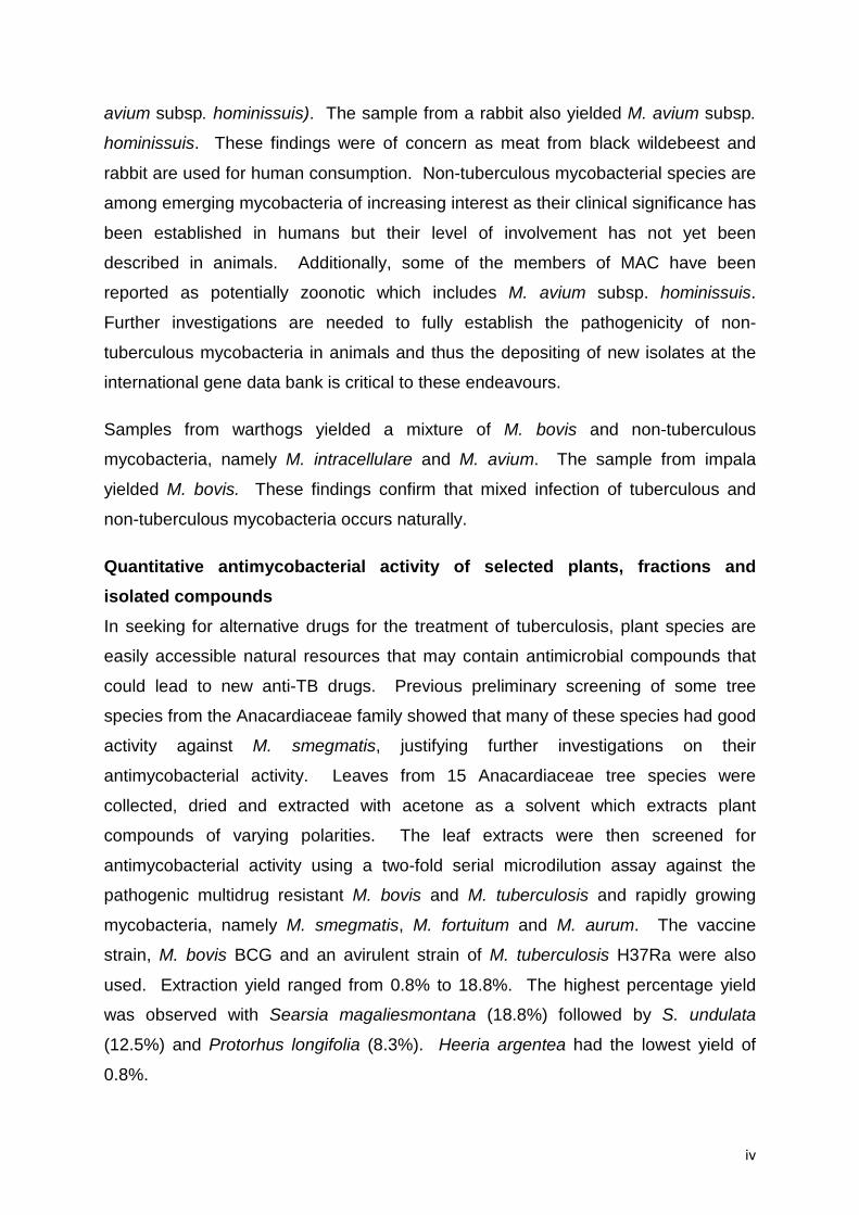
avium subsp. hominissuis). The sample from a rabbit also yielded M. avium subsp.
hominissuis. These findings were of concern as meat from black wildebeest and
rabbit are used for human consumption. Non-tuberculous mycobacterial species are
among emerging mycobacteria of increasing interest as their clinical significance has
been established in humans but their level of involvement has not yet been
described in animals. Additionally, some of the members of MAC have been
reported as potentially zoonotic which includes M. avium subsp. hominissuis.
Further investigations are needed to fully establish the pathogenicity of non-
tuberculous mycobacteria in animals and thus the depositing of new isolates at the
international gene data bank is critical to these endeavours.
Samples from warthogs yielded a mixture of M. bovis and non-tuberculous
mycobacteria, namely M. intracellulare and M. avium. The sample from impala
yielded M. bovis. These findings confirm that mixed infection of tuberculous and
non-tuberculous mycobacteria occurs naturally.
Quantitative antimycobacterial activity of selected plants, fractions and isolated compounds In seeking for alternative drugs for the treatment of tuberculosis, plant species are
easily accessible natural resources that may contain antimicrobial compounds that
could lead to new anti-TB drugs. Previous preliminary screening of some tree
species from the Anacardiaceae family showed that many of these species had good
activity against M. smegmatis, justifying further investigations on their
antimycobacterial activity. Leaves from 15 Anacardiaceae tree species were
collected, dried and extracted with acetone as a solvent which extracts plant
compounds of varying polarities. The leaf extracts were then screened for
antimycobacterial activity using a two-fold serial microdilution assay against the
pathogenic multidrug resistant M. bovis and M. tuberculosis and rapidly growing
mycobacteria, namely M. smegmatis, M. fortuitum and M. aurum. The vaccine
strain, M. bovis BCG and an avirulent strain of M. tuberculosis H37Ra were also
used. Extraction yield ranged from 0.8% to 18.8%. The highest percentage yield
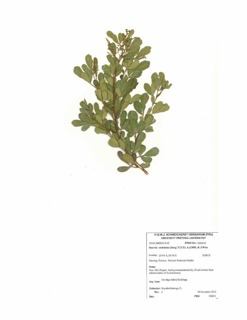
was observed with Searsia magaliesmontana (18.8%) followed by S. undulata
(12.5%) and Protorhus longifolia (8.3%). Heeria argentea had the lowest yield of
0.8%.

v
Four out of 15 crude acetone extracts (Harpephyllum caffrum, Searsia undulata,
Sclerocarya birrea and Protorhus longifolia) had significant antimycobacterial activity
with minimum inhibitory concentration (MIC) values varying from 50-100 µg/mL.
Searsia undulata had the highest activity against most mycobacteria, followed by P.
longifolia. Mycobacterium fortuitum was a reasonably good predictor of activity
against MDR-TB isolates (correlation coefficient =0.65). Searsia undulata extracts
had significant antimycobacterial activity with the lowest MIC value of 70 µg/mL for
M. aurum and M. fortuitum followed by M. smegmatis with MIC = 90 µg/mL.
Protorhus longifolia also had significant antimycobacterial activity against all three
non-tuberculous mycobacteria with MIC values of 110 µg/mL for both M. aurum and
M. fortuitum and 70 µg/mL for M. smegmatis. Searsia lancea, S. birrea and H.
caffrum had moderate activity of MIC = 420, 520 and 590 µg/mL against M. aurum,
respectively, whereas the same plant species (S. lancea, H. caffrum and S. birrea)
had moderate activity with low MIC values of 110 to 210 µg/mL for M. smegmatis
and M. fortuitum, respectively. Searsia undulata had significant activity against all
mycobacteria including MDR - M. bovis and M. tuberculosis with MIC values ranging
from 50 to 110 µg/mL and the lowest MIC of 50 µg/mL against M. tuberculosis ATCC
strain H37Ra.
Bioautography using M. aurum and M. fortuitum worked well as indicators of the Rf
values of active compounds yielding strong zones of inhibition. The leaf extracts of
S. undulata and P. longifolia had more than 10 different antimycobacterial
compounds, whereas the other plant species showed none or only one to two active
compounds depending on the non-tuberculous Mycobacterium spp. involved.
Based on good antimycobacterial activity and low cytotoxicity, Searsia undulata was
further processed to isolate the active antimycobacterial compounds. The amount of
1.5 kg of dried fine powder of S. undulata leaves was processed for acetone bulk
extraction, followed by solvent to solvent fractionation and separation by silica gel
column chromatography. The isolation was bioassay-guided; fractions collected at
different stages were tested using two-fold serial dilution to determine the minimum
inhibitory concentration using the previous isolates of pathogenic MDR - M. bovis, M.
tuberculosis and rapidly growing mycobacteria, M. aurum, M. fortuitum and M.
smegmatis. The active compounds, visible on biautography, were targeted during

vi
the bioassay-guided isolation using thin layer chromatography as fingerprints to
locate those active compounds.
Solvent to solvent fractionation yielded five fractions, namely chloroform, hexane,
butanol, 35% H2O-methanol and water fraction. Chloroform, butanol and 35% H2O-
methanol fractions had clear zones of inhibition on biautography whereas the water
fraction did not show any clear zones of activity.
The chloroform fraction was further separated and yielded 55 fractions that were
combined into 12 fractions (F1-F12), based on similar patterns on TLC fingerprints.
Three out of the 12 fractions showed similar compounds using bioautography.
These fractions were tested as already mentioned above under “quantitative
antimycobacterial of fractions” and F5-F7 had good MIC values. These three semi-
purified fractions were combined and separated further.
Fractions and compounds collected during different stages of isolation had good
antimycobacterial activity. Some of the main fractions obtained during solvent-
solvent fractionation, namely chloroform, butanol and 35% H2O methanol, showed
similar compounds inhibiting the growth of non-tuberculous Mycobacterium spp. on
bioautography although TLC fingerprints of the butanol fraction did not show visible
compounds. The chloroform fraction revealed the presence of several potential
active fractions on the bioautogram plate sprayed with M. aurum whereas the plate
sprayed with M. fortuitum had fewer active fractions which corresponded to a lower
total activity observed with the combined sub-fractions F5-F7 derived from the
chloroform fraction. The variation of activity observed on bioautography could be
explained by the different sensitivity of mycobacterial species towards the potential
antimycobacterial phytochemicals present in the fractions.
Sub-fractions F1-F12 derived from the chloroform fraction had MIC values from 58 to
468 µg/mL. Sub-fractions F5, F6 and F7 had significant antimycobacterial activity
and were selected for further fractionation towards isolation of compounds with MIC
values of 117 µg/mL for M. aurum, 234 µg/mL for M. fortuitum and 58 µg/mL for M.
smegmatis for all fractions except F7 with MIC value of 117 µg/mL against M.
smegmatis. Fractions F5 and F6 showed the lowest value of 58 µg/mL against M.
smegmatis. Fractions F5, F6 and F7 were active against all three non-tuberculous

vii
Mycobacterium spp. The total activity of each fraction (Mass/MIC) was calculated as
this indicates which fraction could be the best candidate for organic production of the
compound. Among all the fractions, F3 showed the highest total activity of 19 765
mL/g on its own for M. aurum and less for M. fortuitum and M. smegmatis whereas
F5, F6 and F7 had a combined total activity of 44 181 mL/g against M. smegmatis
with F6 and F5 showing the highest activity of 19 793 and 17 154 mL/g, respectively,
against M. smegmatis. F7 had the highest activity of 7 262 mL/g for both M. aurum
and M. smegmatis and 3 627 mL/g for M. fortuitum. The total activity of the
combined fractions F5-F7 was the highest with M. smegmatis followed by total
combined activity of 25 716 mL/g with M. aurum.
Three compounds were isolated from the leaves of S. undulata and these had good
antimycobacterial activity against rapidly growing Mycobacterium spp. with MIC
values ranging from 23.44 to 250 µg/mL. Two of the compounds (SLN1 and PK-B)
had MIC values of 23.44 µg/mL, the lowest value for M. fortuitum, and MIC = 31.25
µg/mL for M. aurum and M. smegmatis. In addition, compound 3, had activity
against all three mycobacteria with MIC = 32.25 µg/mL against M. aurum and M.
fortuitum with the highest value of 46.88 µg/mL against M. smegmatis.
Two of the isolated compounds were subjected to MS and NMR spectroscopy. The
first compound (SLN1) was identified as betulonic acid. The second compound (PK-
B) appears to be a novel compound as it did not match with any published records
using Scifinder search tool. This compound will be further identified using
crystallization, melting point determination and Fourier Transform Infrared (FTIR)
spectroscopy. This is the first report of isolation of betulonic acid from S. undulata.
This compound has been reported to have anti-proliferative (anticancer) activity
which could relate to the high cytotoxicity of the crude extract observed on human
liver cancer cells. Other properties reported include antiviral, anti-inflammatory and
antimicrobial. Besides the above three compounds, the isolation of the other active
compounds is still underway, forming the research focus subsequent to this PhD
study.
Cytotoxicity of the 15 acetone crude extracts and compounds of S. undulata Cytotoxicity was assessed using a tetrazolium colorimetric assay (MTT) against Vero
monkey kidney, human hepatoma (C3A) and murine macrophage (RAW 264.7) cell

viii
lines. Plants extracts of H. caffrum, L. discolor, P. longifolia, S. undulata and S.
birrea with good antimycobacterial activity (significant to moderate) and showing one
or more visible zones of inhibition on bioautograms were selected for cytotoxicity
testing. All crude extracts from the above-mentioned plant species had low
cytotoxicity against the three cell lines except S. undulata which had high toxicity
against C3A cells and relatively low cytotoxicity on Vero kidney cells. This finding
deserves further investigation as it may indicate that S. undulata extracts have good
anticancer activity. Searsia undulata had moderate cytotoxicity with LC50 = 34 µg/mL
for C3A and LC50 of 50 and 120 µg/mL for Vero kidney and RAW cells respectively
with good selectivity indexes of 7.08 on Vero and more than 10 on RAW cells for
non-tuberculous and MDR-TB mycobacteria. Protorhus longifolia had LC50 of 620
µg/mL on C3A cells, 880 µg/mL on Vero cells and more than 1 000 µg/mL on RAW
cells with the highest selectivity index on Vero cells of 12.6 for M. smegmatis
followed by 8.02 for M. aurum, M. fortuitum and MDR-TB. Extracts with SI > 1 may
indicate better safety as they are less toxic to the mammalian cells but more toxic to
pathogens but this will need to be confirmed with in vivo tests.
Only three compounds out of the five which were isolated in sufficient quantity were
tested for cytotoxicity using Vero cells. PK-B was the least toxic with LC50 of 210
µg/mL whereas compound 3 and SLN1 showed moderate cytotoxicity of 31.3 µg/mL
and 47.7 µg/mL, respectively.

ix
TABLE OF CONTENTS
Declaration…………………………………………………………………………………iii Dedication…….. ....................................................................................................... iv
Acknowledgements .................................................................................................. v
Abstract……….. ........................................................................................................ ii List of abbreviations and symbols ....................................................................... xv
List of figures… ..................................................................................................... xix
List of tables…. ................................................................................................... xxiii List of publications ............................................................................................. xxiv
CHAPTER 1:GENERAL INTRODUCTION AND LITERATURE REVIEW ................ 1
1.1 General introduction ...................................................................................... 1
1.2 Literature review ............................................................................................ 6
1.2.1 Tuberculosis epidemiology ..................................................................... 6
1.2.1.1 Tuberculosis in humans .................................................................... 6
1.2.1.2 Tuberculosis in animals .................................................................... 9
1.2.2 Mycobacteria ........................................................................................ 12
1.2.2.1 Mycobacterium tuberculosis ........................................................... 15
1.2.2.2 Mycobacterium bovis ...................................................................... 17
1.2.2.3 Non-tuberculous mycobacteria ....................................................... 19
1.2.3 Pathogenesis of tuberculosis ............................................................... 22
1.2.4 Clinical signs of tuberculosis ................................................................ 26
1.2.5 Diagnostic tests .................................................................................... 26
1.2.6 Challenges in the treatment of tuberculosis ......................................... 30
1.2.7 The use of traditional medicine as an alternative or complementary medicine ............................................................................................... 31
1.2.8 New lead compounds for drug discovery and development ................. 32
1.2.9 Antimycobacterial activity of South African medicinal plants and bioactive compounds ........................................................................... 35
1.2.10 Selection of potential medicinal plants for drug development............... 36
1.3 Problem statement ....................................................................................... 38
1.4 Aims and objectives ..................................................................................... 40
1.5 Hypothesis ................................................................................................... 42

x
1.6 Scope of the thesis ...................................................................................... 42
1.6.1 Isolation and characterization of mycobacteria .................................... 42
1.6.2 Antimycobacterial activity of plant extracts ........................................... 42
1.6.3 Cytotoxicity assay of plant extracts ...................................................... 42
1.6.4 Isolation, purification and identification of the active compound(s) from Searsia undulata .................................................................................. 43
1.7 Structure of the thesis .................................................................................. 43
CHAPTER 2:MOLECULAR PROFILE OF MYCOBACTERIUM SPP. ISOLATES FROM CATTLE AND OTHER ANIMAL SPECIES .................................................. 46
Abstract ................................................................................................................. 46
2.1 Introduction .................................................................................................. 47
2.2 Materials and methods ................................................................................ 50
2.2.1 Study area ............................................................................................ 50
2.2.2 Study design and sampling .................................................................. 51
2.2.3 Sources of Samples ............................................................................. 52
2.2.4 Mycobacterial isolation ......................................................................... 54
2.2.5 Mycobacterial identification .................................................................. 55
2.2.5.1 DNA extraction and primary molecular identification ...................... 55
2.2.5.2 Antimicrobial susceptibility test ....................................................... 56
2.2.5.3 MTC discrimination by multiplex PCR ............................................ 57
2.2.5.4 Spoligotyping .................................................................................. 57
2.2.5.5 Variable number of tandem repeat (VNTR) typing ......................... 58
2.3 Results and discussion ................................................................................ 62
2.3.1 Mycobacterial isolation ......................................................................... 62
2.3.2 Primary molecular identification ........................................................... 62
2.3.3 MTC discrimination by multiplex PCR .................................................. 63
2.3.4 Antimicrobial susceptibility test ............................................................. 63
2.3.5 Spoligotyping ........................................................................................ 64
2.3.6 Variable number of tandem repeat (VNTR) typing ............................... 67
2.4 Conclusion ................................................................................................... 67
CHAPTER 3:NOVEL MYCOBACTERIUM AVIUM SPECIES ISOLATED FROM BLACK WILDEBEEST (CONNOCHAETES GNOU) IN SOUTH AFRICA .............. 70
Abstract ................................................................................................................. 70
3.1 Background ................................................................................................. 70

xi
3.2 Materials and methods ................................................................................ 73
3.2.1 Study area ............................................................................................ 73
3.2.2 Study design and sampling .................................................................. 73
3.2.3 Sources of samples .............................................................................. 74
3.2.4 Mycobacterial isolation ......................................................................... 75
3.2.5 Mycobacterial identification .................................................................. 76
3.2.5.1 Biochemical profile ......................................................................... 76
3.2.5.2 DNA extract and primary molecular identification ........................... 76
3.2.6 Gene sequencing ................................................................................. 77
3.2.6.1 16S ribosomal RNA gene and ITS ................................................. 77
3.2.6.2 rpoB ................................................................................................ 77
3.2.6.3 hsp65 .............................................................................................. 78
3.2.7 Phylogenetic analyses ......................................................................... 78
3.2.8 Statistical analyses ............................................................................... 79
3.2.9 GenBank accession numbers .............................................................. 79
3.3 Results ......................................................................................................... 79
3.3.1 Mycobacterial isolation ......................................................................... 79
3.3.2 Biochemical characteristics .................................................................. 80
3.3.3 Primary molecular identification ........................................................... 80
3.3.4 Phylogenetic analyses ......................................................................... 80
3.3.4.1 ITS sequences ............................................................................... 80
3.3.4.2 16S rRNA, hsp65 and rpoB analyses. ............................................ 81
3.4 Discussion and conclusion .......................................................................... 82
CHAPTER 4:ANTIMYCOBACTERIAL ACTIVITY AND CYTOTOXICITY OF LEAF EXTRACTS OF SOME AFRICAN ANACARDIACEAE TREE SPECIES ................ 85
Abstract ................................................................................................................. 85
4.1 INTRODUCTION ......................................................................................... 85
4.2 Materials and methods ................................................................................ 88
4.2.1 Source of plant materials and extraction .............................................. 88
4.2.2 Antimycobacterial activity ..................................................................... 95
4.2.2.1 Pathogenic mycobacteria ............................................................... 96
4.2.2.2 Rapidly growing mycobacteria ........................................................ 96
4.2.2.3 Maintenance of cultures ................................................................. 96
4.2.2.4 Minimum inhibitory concentration (MIC) determination .................. 97

xii
4.2.3 Thin Layer Chromatography (TLC) analysis and Bioautography .......... 98
4.2.4 In vitro cytotoxicity assay and selectivity index ................................... 100
4.2.5 Statistical analysis .............................................................................. 102
4.3 Results and discussion .............................................................................. 102
4.3.1 Acetone extract plant yield ................................................................. 102
4.3.2 Biological activity of extracts .............................................................. 104
4.3.3 Bioautography and thin layer chromatography analysis ..................... 106
4.3.4 Cytotoxicity assay .............................................................................. 106
4.4 Conclusion ................................................................................................. 110
CHAPTER 5:BIOASSAY-GUIDED ISOLATION OF FRACTIONS AND COMPOUNDS FROM SEARSIA UNDULATA ...................................................... 111
ABSTRACT ......................................................................................................... 111
5.1 Background ............................................................................................... 112
5.1.1 Description and taxonomy .................................................................. 112
5.1.2 Traditional use of Searsia/Rhus species ............................................ 113
5.1.3 In vitro biological activities .................................................................. 114
5.1.4 Isolation of compounds ...................................................................... 115
5.1.5 In vivo experiments ............................................................................ 116
5.2 Materials and methods .............................................................................. 117
5.2.1 Plant collection and storage ............................................................... 117
5.2.2 Fractionation of bioactive fractions ..................................................... 118
5.2.2.1 Bulk extraction and solvent to solvent fractionation ...................... 118
5.2.2.2 Column chromatography (CC) ...................................................... 120
5.2.3 Thin layer chromatography (TLC) fingerprinting and bioautography of fractions and compounds ................................................................... 121
5.2.4 Minimum inhibitory concentration (MIC) of Searsia undulata fractions and compounds .................................................................................. 123
5.2.5 Cytotoxicity of compounds ................................................................. 123
5.2.6 13C and 1H nuclear magnetic resonance (NMR)-procedures and structural elucidation of isolated compounds...................................... 123
5.2.6.1 Isolation and characterization of compounds ............................... 123
5.2.6.2 Sample preparation of compounds ............................................... 124
5.2.6.3 NMR spectrometers and structure elucidation .............................. 124
5.3 Results and discussion .............................................................................. 125

xiii
5.3.1 Bulk extraction, fractionation, thin layer chromatography and bioautography .................................................................................... 125
5.3.1.1 Extraction yield, thin layer chromatography and bioautography ... 125
5.3.2 Minimum inhibitory concentration of fractions and compounds .......... 154
5.3.2.1 Minimum Inhibitory concentration of fractions .............................. 154
5.3.2.2 Minimum inhibitory concentration of compounds .......................... 160
5.3.3 Cytotoxicity ......................................................................................... 161
5.3.3.1 Crude extracts .............................................................................. 161
5.3.3.2 Compounds .................................................................................. 161
5.3.4 Structure elucidation and identification of compounds ....................... 163
5.3.4.1 SLN1 ............................................................................................ 163
5.3.4.2 Compound-PKB ........................................................................... 170
5.4 Conclusion ................................................................................................. 172
CHAPTER 6:SUMMARY AND CONCLUSIONS ................................................... 173
6.1 Isolation and characterization of mycobacterium species .......................... 173
6.2 Antimycobacterial activity of acetone leaf extracts of plant species from anacardiaceae family ................................................................................. 174
6.2.1 Antimycobacterial activity of fractions and compounds of S. undulata 176
6.2.1.1 Minimum Inhibitory concentration of fractions .............................. 176
6.2.1.2 Minimum inhibitory concentration of isolated compounds ............ 176
6.3 Cytotoxicity of acetone leaf extracts of plant species from anacardiaceae family ......................................................................................................... 177
6.3.1 Cytotoxicity of compounds isolated from Searsia undulata ................ 178
6.4 Isolation of bioactive fractions and compounds from the leaf of searsia undulata ..................................................................................................... 178
6.5 Structure elucidation and identification of compounds ............................... 180
6.5.1 Compound SLN1 ................................................................................ 180
6.5.2 Compound PK-B ................................................................................ 181
6.6 Research challenges ................................................................................. 181
6.7 General conclusion, recommendations and future perspectives ............... 182
6.7.1 Isolation and characterization of mycobacterial species .................... 182
6.7.2 Antimycobacterial activities of leaf extracts, fractions and compounds isolated from plant species from the Anacardiaceae family................ 183
6.7.3 In vitro cytotoxicity activity of Searsia undulata leaf extracts and isolated compounds against Vero and human hepatoma cell lines ................. 184

xiv
6.7.4 Future perspectives ............................................................................ 185
6.7.4.1 Isolation and identification of compounds from Searsia undulata . 185
6.7.4.2 In vitro safety of all compounds isolated from S. undulata............ 185
6.7.4.3 Synergistic evaluation of compounds isolated from S. undulata ... 185
6.7.4.4 Determine the stability of each compound and fractions of S. undulata ........................................................................................ 185
6.7.4.5 Determine antioxidant, anti-inflammatory and inhibition of nitric oxide production by fractions and compounds of S. undulata ................ 186
6.7.4.6 Determine the in vivo safety of pure compounds .......................... 186
6.7.4.7 Develop an herbal product from S. undulata ................................ 186
6.7.4.8 Understanding the mechanism of actions of compounds ............. 186
6.7.4.9 Develop network with pharmaceutical companies ........................ 187
6.7.4.10 Testing of four other plant species with good antimycobacterial activity .......................................................................................... 187
REFERENCES……………………………………………………………………………188
APPENDICES…………………………………………………………………………….225
Appendix A: Structure elucidation of compound SLN-1 ...................................... 225
Appendix B: Structure elucidation of compound PK-B ........................................ 262
Appendix C: Published articles ........................................................................... 266

xv
LIST OF ABBREVIATIONS AND SYMBOLS
Ac Acetone
ANOVA Analysis of variance
ATCC American Type Culture Collection
BCG Bacille Calmette Guerin (M. bovis attenuated vaccine strain)
BEA Benzene/ethanol/ammonia (18:2:0.2)
BI Bayesian inference
BLAST Basic alignment search tool
C Carbon
C3A Human liver hepatoma cells 13CNMR Carbon 13 NMR
CEF Chloroform/ethyl acetate/formic acid (10:8:2)
CFU Colony forming units
CHCL3 Chloroform
CP Compound
1D One dimensional
2D Two dimensional
DAFF Department of Agriculture, Forestry and Fisheries
DMSO Dimethyl sulphoxide
DNA Deoxyribonucleic acid
DMEM Dulbecco's Modified Eagles' Medium
DPPH 2, 2-Diphenyl-1-picrylhydrazyl
DR Direct repeat
EDTA Ethylene-diamine-tetra-acetic acid
ELISA Enzyme-linked immuno-sorbent assay
EMB Ethambutol
EMEM Minimal essential medium eagle with L- glutamine
EMW Ethyl acetate/methanol/water (10:1.35:1)
EtOAc Ethyl acetate
FAO Food and Agriculture Organization
FBS Foetal bovine serum
FTIR Fourier transform infra-red spectroscopy
xvi
GC-MS Gas chromatography-mass spectrometry 1HNMR Proton NMR
H37Ra Mycobacterium tuberculosis attenuated strain
Ha Harpehyllum caffrum
HCl Hydrochloric acid
HEX Hexane
HIV Human immunodeficiency virus
H2O Water
IFN Interferon
IGRA Interferon - gamma release blood assays
INH Isoniazid
IL Interleukin
INT p-Iodonitrotetrazolium
LAM Lipoarabinomannan
LC50 50% Lethal concentration
LC-MS Liquid chromatography mass spectrometry LJ Löwenstein - Jensen
M7H9 Middlebrook 7H9
M7H10 Middlebrook 7H10
MA Mycobacterium aurum
MAC Mycobacterium avium complex
MAFFT Multiple alignments by fast Fourier transformation
MALDI – TOF Matrix assisted laser desorption ionization time of flight
MB Mycobacterium bovis
MDR Multidrug-resistant
MEGA 5 Molecular evolutionary genetic analysis
MEM Minimum Essential Medium
MeOH Methanol
MF Mycobacterium fortuitum
MIC Minimum inhibitory concentration
MIRUs Mycobacterial interspersed repetitive units
MIT Multi - inter - transdisciplinary
MGIT Mycobacteria growth indicator tube
MH Mueller Hinton

xvii
ML Maximum likelihood
MOTT Mycobacteria other than tuberculosis
MP Maximum parsimony
MS Mycobacterium smegmatis
MSH Mycothiol
MTB Mycobacterium tuberculosis complex
MTT 3-[4, 5-dimethylthiazol-2-yl]-2, 5 diphenyl tetrazolium bromide
NALC – NaOH N - acetyl - L - cystein - sodium
NCTC National Collection Type Cultures
NHLS National Health Laboratory Service
NJ Neighbor - joining
NMR Nuclear magnetic resonance
NTM Non - tuberculous mycobacteria
OADC Oleate albumin dextrose catalase
OD Optical density
OIE Office International des Epizooties
OP Onderstepoort
p probability
PA Pyrazinamide
PACT Polymyxin B, amphotericin B, carbenicillin and trimethoprim
PAMPs Pathogen - associated molecular patterns
PBS Phosphate buffered saline
PCR Polymerase chain reaction
PL Protorhus longifolia
PPEM Potentially pathogenic environmental mycobacteria
PPD Purified protein derivative
PPM Potentially pathogenic mycobacteria
PRRs Pattern recognition receptors
r Pearson's correlation coefficient
RAPD Random amplified polymorphic DNA
RAW 264.7 Murine macrophage cells
RCBH Reverse cross blot hybridisation
RIDOM Ribosomal differentiation of medical microorganisms
RFLP Restriction fragment length polymorphism

xviii
RNA Ribonucleic acid
rRNA Ribosomal ribonucleic acid
SANBI South African National Botanical Institute
SI Selectivity index
Rf Retardation factor or retention factor
RIF Rifampin
TA Total activity
TB Tuberculosis
TBST Tris buffered saline with Tween
TLC Thin layer chromatography
TMB 3,3,5,5 – Tetra methyl - benzidine
TNF Tumour necrosis factor
VNTR Variable number of tandem repeat
WHO World Health Organization
XDR Extensively drug resistant
ZN Ziehl - Neelsen

xix
LIST OF FIGURES
Figure 1.1: Macrophage phacocytosis and evasion of tubercle bacilli (Inderlied, 2004). ........ 4
Figure 1.2: Map showing the estimated new TB cases in the world (WHO, 2015). ................ 8
Figure 1.3: Mycobacterial cell envelopes (Riley, 2006) ........................................................ 14
Figure 1.4: Mycobacterium tuberculosis stained by Ziehl-Neelsen method, appearing
as dark pink straight and curved rods (Brehar, 2015) .......................................................... 15
Figure 1.5: Mycobacterium tuberculosis cord factor ............................................................ 16
Figure 1.6: Colonies of M. tuberculosis on Löwenstein-Jensen agar slope tubes ................ 17
Figure 1.7: Mechanism of activity of antiTB drugs ............................................................... 31
Figure 2.1: South Africa Map with the red triangle showing Mpumalanga Province where
samples were obtained. ...................................................................................................... 51
Source: courtesy of www.sa.venues.com ............................................................................ 51
Figure 2.2: Samples processed in a biosafety cabinet (a) and incubated in BACTECTM
MGITTM 960 system (b) ....................................................................................................... 55
Figure 2.3: WT probes and mutation genes targeted to determine the susceptibility of an
isolate towards first line drugs ............................................................................................. 64
Figure 2.4: Spoligotype SB1235 (A) and spoligotype SB0121 (B), deleted spacers that are
common to all M. bovis are 39 to 43. SB1235 lack spacers 3, 6, 8, 9, 10, 11, 12, 16, 37-43
whereas SB0121 lacks spacers 3, 9, 16, 21, 39-43 –The deleted spacers are marked with
Figure 2.5). schematic representation of the spoligotype pattern as presented on the
membranes above .............................................................................................................. 65
Figure 2.5: Schematic representation of spoligotype patterns of M. bovis cattle isolates from
South Africa ........................................................................................................................ 66
Figure 3.1: Phylogenetic tree obtained by NJ analysis of concatenated nucleotide sequences
of 16S rRNA, 16S-23S rRNA, hsp65 and rpoB. Bootstrap values (1000 replicates) are given
above the branches for NJ/ML and below for MP. Branch support values on the nodes are
shown in percentage and the values less than 60% are not shown. The tree is rooted with
M. fortuitum and M. insubricum. .......................................................................................... 82
Figure 4.1: Air drying of leaves suspended in bags (a), filtration of the acetone extracts for
preliminary assays (b) ......................................................................................................... 89
Figure 4.2: Leaves, flowers and fruits of Anacardiacea occidentalis (Cashew tree) (a) and
Lannea discolor (b) ............................................................................................................. 93
Figure 4.3: Leaves and fruits of Mangifera indica (c) and Ozoroa paniculosa (d) ................ 93
Figure 4.4: Leaves of Ozoroa mucronata (e) and leaves and fruits of Heeria argentea (f) ... 94
Figure 4.5: Leaves and fruits of Searsia chirendensis (g) and leaves of Searsia lancea (h) 94
xx
Figure 4.6: Leaves of Searsia magaliesmontana (i) and Smodingium argutum (j) ............... 95
Figure 4.7: Leaves and flowers of Searsia pyroides (k) and Protorhus longifolia (l) ............. 95
Figure 4.8: Bioautograms (A) of the three plants extracts with low MIC values and thin layer
chromatograms eluted in chloroform/ethyl acetate/formic (CEF) solvent system sprayed with
vanillin sulphuric acid (B) showing varied chemical constituents. Bioautogram of the extracts
against Mycobacterium aurum. ........................................................................................... 99
Figure 5.1: Shrub (a) and trifoliate leaves and greenish flowers (b) of Searsia undulata ... 113
Figure 5.2: Plant collection (a) and air drying of leaves (b) ................................................ 117
Figure 5.3: Grinding processes of dried leaves of Searsia undulata using a Macsalab Mill
......................................................................................................................................... 118
Figure 5.4: Crude extract acetone-bulk extraction processes (left) and drying process with a
rotary evaporator (right) .................................................................................................... 119
Figure 5.5: Column chromatography used for fractionation ............................................... 121
Figure 5.6: Normal Vero monkey kidney cells viewed under the microscope at magnifications
of 10X (a) and 40X (b) ....................................................................................................... 123
Figure 5.7: Schematic extraction and fractionation results towards compound isolation from
Searsia undulata ............................................................................................................... 127
Figure 5.8: Mass (g) of the five solvent-solvent fractions obtained from 80 g of S. undulata
leaf extracts ...................................................................................................................... 128
Figure 5.9: Chloroform and butanol fractions- TLC fingerprinting and bioautograms of plates
eluted in BEA and sprayed with rapidly growing mycobacteria spp. CHL: Chloroform, BUT:
Butanol, HE: Hexane, E: Ethyl acetate, MA: M aurum, MF: M fortuitum, MS: M smegmatis
......................................................................................................................................... 129
Figure 5.10-A: Bioautogram of M. aurum eluted in BEA (Benzene/ethanol/ammonia at ratio
18:2:0.2) separated in triplicate (CHL) and sprayed with 2 mg/mL of INT
(Iodonitrotetrazolium violet) showing solvent- solvent fractions of chloroform (CHL), butanol
(BUT) and 35% water (H2O) of SU .................................................................................... 131
Figure 5.11-A1: TLC fingerprint of acetone crude extract and solvent- solvent fractions
(Butanol, chloroform, 35% water, hexane and water) ........................................................ 132
Figure 5.11-B: Bioautogram plate eluted in BEA (Benzene/ethanol/ammonia at ratio
18:1:0.2) and sprayed with M. smegmatis followed by INT indicating antimycobacterial
compounds of solvent-solvent fractions CHL: chloroform, BUT: butanol and 35% H2O: 35%
water in methanol of SU. Clear zones indicate mycobacterial growth inhibition ................ 133
Figure 5.11-C: Bioautogram plate eluted in BEA (Benzene/ethanol/ammonia at ratio
18:2:0.2) and sprayed with M. fortuitum and followed by INT indicating antimycobacterial
compounds of solvent-solvent fractions CHL: chloroform, BUT: n-butanol and 35%H2O: 35%
water in methanol of SU. Clear zones indicate mycobacterial growth inhibition ................ 134

xxi
Figure 5.12: Fractions (n=55) collected from the chloroform fraction were combined, tested
and based on clear zone of inhibition pattern against M. smegmatis, combined into 12 semi-
purified fractions (F1-F12) that were separated further for compound isolation ................. 135
Figure 5.13: Fractions (n=55) collected from the chloroform fraction were combined, tested
and based on clear zone of inhibition pattern against M. aurum, combined into 12 semi-
purified fractions (F1-F12) that were separated further for compound isolation ................. 136
Figure 5.14-A: TLC fingerprints of the 12 pooled fractions obtained from the chloroform
fraction; plate was eluted in BEA (Benzene/ethanol/ammonia at ratio 18:2:0.2). F5 to F7
fractions targeted for compound isolation. ......................................................................... 138
Figure 5.14-A1 Bioautogram eluted in BEA sprayed with M. aurum followed by INT indicating
consistent clear zone of inhibition with focus on fractions F5, F6 and F7. .......................... 139
Figure 5.14-A2 Vanillin sprayed TLC plate and bioautogram plate eluted in BEA
(Benzene/ethanol/ammonia at ratio 18:2:02) sprayed with M. aurum followed by INT
indicating the three pooled fractions (F5, F6 and F7) that were separated further for isolation
of compounds indicating the closeness of the active compounds ...................................... 140
Figure 5.14-B: Bioautogram eluted in CEF (Chloroform/ethyl acetate/formic acid at ratio
10:8:2) sprayed with M. aurum followed by INT indicating 12 pooled fractions from the
chloroform with consistent clear zone of inhibition focusing on fractions F5, F6 and F7. The
clear zones indicate mycobacterial inhibition growth ......................................................... 142
Figure 5.15-A Bioautogram eluted in BEA sprayed with M. fortuitum followed by INT
indicating 12 pooled fractions from chloroform with zones of growth inhibition focusing on
fractions F5, F6 and F7 ..................................................................................................... 143
Figure 5.15-B Bioautogram eluted in CEF and sprayed with M. fortuitum followed by INT
indicating 12 pooled fractions from chloroform with zones of growth inhibition focusing on
fractions F5, F6 and F7 ..................................................................................................... 145
Figure 5.16-A Bioautogram eluted in BEA and sprayed with M. smegmatis followed by INT
indicating 12 pooled fractions from chloroform with zones of growth focusing on fractions
F5, F6 and F7 ................................................................................................................... 146
Figure 5.16-B Bioautogram eluted in CEF and sprayed with M. smegmatis followed by INT
indicating zones of growth inhibition focusing on fractions F5, F6 and F7 ......................... 148
Figure 5.17: TLC fingerprints and bioautograms showing the different stages of isolation
from the acetone crude extract to fractions of Searsia undulata. Plates were eluted in BEA,
TLC plates were sprayed with vanillin and bioautograms with Mycobacterium aurum (which
was one of the best indicators of mycobacterial activity) followed by INT. ACE: Acetone
crude extract; CHL: Chloroform fraction; F5-F7: sub-fractions obtained from 55 fractions from
the chloroform fraction ...................................................................................................... 149

xxii
Figure 5.18: TLC fingerprints and bioautograms indicating targeted fractions F5-F7 to
compounds isolated from Searsia undulata. Bioautogram were eluted in BEA solvent
systems and sprayed with Mycobacterium aurum which was one of the good indicators of
mycobacterial activity. TLC plates for compounds were eluted in different ratios of hexane:
ethyl acetate and sprayed with vanillin .............................................................................. 152
Figure 5.19: TLC plate visualized under UV light (366 nm) showing the separation of
compound B from compound C during the purification process. ........................................ 153
Figure 5.20: The MIC value (mg/mL) of each fraction against the different mycobacteria.
MA: M. aurum, MF: M. fortuitum, MS: M. smegmatis......................................................... 154
Figure 5.21: The 1/MIC values (mg/mL) of each fraction against the different mycobacteria
species. MA: M. aurum, MF: M. fortuitum, MS: M. smegmatis ........................................... 155
Figure 5.22 The average 1/MIC values (mg/mL) of each fraction against all the different
mycobacteria species. Fractions F5, F6 and F7 show consistent high activity ................. 156
Figure 5.23: Total activity of different fractions (mL/g); Fractions F3 and F6 had the highest
total activity for M. aurum and M. smegmatis respectively followed by fractions F5, F6 and
F7 that had better total activity against all the mycobacteria which also correlated with the
intensity of active fractions indicated by the clear zone of inhibition on bioautograms ....... 158
Figure 5.24: The average total activity (mL/g) per fraction ................................................. 159
Figure 5.25: LC50 of compound 1 where Y was 0.955 (A) and 0.997 (B) ........................... 162
Figure 5.26: LC50 of compound PKB where Y was 0.955 (A) and 0.997 (B) ...................... 162
Figure 5.27: LC50 of compound 1 where Y was 0.955 (A) and 0.997 (B) ........................... 162
Figure 5.28: Interactions between protons on C29 and methyl group of C30 and other COSY
scalar couplings ................................................................................................................ 164
Figure 5.29: Atomic interactions within a network .............................................................. 165
Figure 5.30: The structure of betulonic acid (SLN1) .......................................................... 168
Figure 5.31: Proposed structure for sample PK-B ............................................................. 172

xxiii
LIST OF TABLES
Table 2.1: Types of samples from different animal species ................................................. 53
Table 2.2: Variable number of tandem repeat (VNTR) loci and forward and reverse primers
(5'-3') sequences used for the typing of M. bovis isolates (Le Fleche et al., 2002) .............. 59
Table 2.3: Band sizes (bp) and corresponding tandem repeat numbers .............................. 61
Table 2.4: Mycobacterium species isolated from the different animals ................................ 63
Table 3.1: Type of tissue samples from Black Wildebeest processed at NHLS ................... 75
Table 3.2: Growth and biochemical characteristics of the isolate ........................................ 80
Table 4.1: Ethnomedicinal use and known isolated compounds of selected species of the
Anacardiaceae family .......................................................................................................... 90
Table 4.2: Minimal inhibitory concentration (MIC in mg/mL) and total activity (TA in mL/g) of
acetone leaf extract from 15 plants of the Anacardiaceae family against M. bovis and M.
tuberculosis ....................................................................................................................... 103
Table 4.3: Pearson’s correlation coefficient (r) between MIC values of tested Mycobacteria
......................................................................................................................................... 105
Table 4.4: Cytotoxicity (LC50 in mg/mL) of extracts and selectivity index against C3A liver
cells, Vero kidney cells and RAW 264.7 murine macrophage cells.................................... 109
Table 5.1: The intensity of the zones of inhibition on a five point scale and their Rf values as
observed in Figure 5.14-B ................................................................................................. 141
Table 5.2: The intensity of the zones of inhibition on a five point scale and their Rf values as
observed in Figure 5.15-B ................................................................................................. 144
Table 5.3: The intensity of the zones of inhibition on a five point scale and their Rf values as
observed in Figure 5.16-B ................................................................................................. 147
Table 5.4 The MIC values and total activity of each fraction .............................................. 157
Table 5.5 Rf values of different phytochemical bands observed on the chloroform fraction in
BEA .................................................................................................................................. 160
Table 5.6 MIC values of compounds isolated from Searsia undulata ................................ 161
Table 5.7 Cytotoxicity of compounds on Vero Monkey Kidney cells .................................. 163
Table 5.8 1H and 13C NMR of SLN1 (betulonic acid) showing the chemical shift similarities
with those obtained by Letedi et al. (2014) ........................................................................ 166
Table 5.9: 1 D and 2 D NMR data of compound PK-B in DMSO-d6 .................................. 171
xxiv
LIST OF PUBLICATIONS
Research articles already published from this thesis:
Kabongo-Kayoka PN, Obi CL, Nakajima C, Suzuki Y, Hattori T, Eloff JN, Wright J,
Mbelle N and McGaw LJ. 2015. Novel Mycobacterium avium complex isolated from
Black Wildebeest (Connochaetes gnou) in South Africa. Transboundary Emerging
Diseases. Doi: 10.1111/TBED.12460
Kabongo-Kayoka PN, Obi CL, Eloff JN and McGaw LJ. 2016. Antimycobacterial
activity and low cytotoxicity of leaf extracts of some Anacardiaceae tree species.
Phytotherapy Research. Doi: 10.1111/PTR-0292
Published conference abstracts from this thesis:
Kabongo PN, Obi CL, Eloff JN and McGaw LJ. 2015. Fractions of Searsia undulata
(Anacardiaceae) have excellent antimycobacterial activity. South African Journal of
Botany 98, 182. Doi:10.1016/j.sajb.2015.03.056
Conference presentations from the thesis:
Local conferences
• Walter Sisulu University (WSU) International Research Conference. 2010. Isolation, antibiogram profile and molecular characterization of
Mycobacterium species from cattle and black wildebeest in Mpumalanga
Province. WSU, Eastern Cape, 18-20th August 2010, an oral presentation.
• World Veterinary Congress. 2011. New Mycobacterium species isolated
from Black Wildebeest, South Africa. World Vet Congress, Cape Town
Conventional Centre, 10-14th October 2011, an oral presentation.
• Faculty Day. 2014. Leaf extract of selected Anacardiaceae trees have
excellent antimycobacterial activity. Faculty of Veterinary Science, University
of Pretoria, 4th September 2014, an oral presentation.

xxv
• South African Association of Botanists, 41St Annual Conference. 2015. Fractions of Searsia undulata (Anacardiaceae) have excellent
antimycobacterial activity. Tshipise Forever Resort, Limpopo, 11-15th January
2015, an oral presentation.
International conferences:
• International Infectious Diseases Conference. 2010. Isolation and
molecular characterization of M. avium, M. bovis and M. tuberculosis in this
era of HIV/AIDS in South Africa. University of Tohoku, Japan, Sendai, 13-16th
January 2010, oral presentation.
• International Journal of Arts and Sciences Conference. 2016. High
antimycobacterial activity and low cytotoxicity of Searsia undulata
(Anacardiaceae) - An Overview. University of London, United Kingdom 8-11th
November 2016, an oral presentation.
Papers prepared for publication: Kabongo-Kayoka PN, Obi CL, Eloff JN and McGaw LJ. 2017. Fractions and
compounds with low cellular damage isolated from Searsia undulata have good
antimycobacterial activity. Phytotherapy Research
Kabongo-Kayoka PN, Obi CL, Eloff JN and McGaw LJ. 2017. Isolation of a novel
antimycobacterial compound from Searsia undulata. Planta Medica
Kabongo-Kayoka PN, Obi CL, Eloff JN and McGaw LJ. 2017. Antioxidant, anti-
inflammatory and inhibition of nitric oxide production of the potential
antimycobacterial plants, Protorhus longifolia and Searsia undulata. BMC
Complementary and Alternative Medicine
1
CHAPTER 1: GENERAL INTRODUCTION AND LITERATURE REVIEW
1.1 GENERAL INTRODUCTION Many human diseases that are new, emerging or re-emerging at present, are caused
by pathogens that originate from animals or products of animal origin (Müller et al.,
2013). Tuberculosis can be foodborne or airborne and is one of the most important
human infections in the world with more than 2 million new cases occurring each
year, mostly in developing countries (WHO, 2015).
The emergence of multi-drug resistant (MDR) strains of Mycobacterium tuberculosis
associated with human immunodeficiency virus (HIV) / acquired immunodeficiency
syndrome (AIDS) is of great epidemiological concern. Species belonging to the
Mycobacterium tuberculosis (MTB) complex include M. tuberculosis, M. bovis, M.
africanum, M. canetti, M. pinnipedii, M. caprae, M. microti, M. mungi, Dassie bacillus,
Oryx bacillus (M. orygis) and M. surricatae (Alexander et al., 2010; Azé et al., 2015;
Clarke et al., 2016; Helden et al., 2009). There are other pathogenic non-
tuberculous mycobacterium (NTM) species for example Mycobacterium avium
complex (MAC) which consists of two closely related species, M. avium and M.
intracellulare (Legrand et al., 2000). The importance of NTM has received much
attention during the past decade, especially in humans. They are found widely in
soil, water, near human settlements and aerosols and can be associated with
colonization, serious infection or pseudo-outbreaks with a wide variety of
presentations (Biet et al., 2005; Kankya et al., 2011; Katale et al., 2014).
The isolation of NTM from human clinical samples of patients (HIV positive or HIV
negative) with pulmonary symptoms as suspected cases of tuberculosis has
increased over the years and has been observed in different countries in Africa,
America and Europe (Kankya et al., 2011; Katale et al., 2014; Mirsaeidi et al., 2014a;
Moore et al., 2010). In animals, the clinical significance of NTM has yet to be
elucidated in the disease-causing process (Chege et al., 2008; Kankya et al., 2011;
Katale et al., 2014).

2
The management, treatment and infection control measures differ significantly
between M. tuberculosis and NTM infections. One hundred and sixty species of
NTM have been reported worldwide, of which more than 60% are pathogenic to
animals or humans (BoRam et al., 2014; Tortoli, 2014). In South Africa, reports on
the isolation of NTM in animals, humans and the environment and their effects in
disease-causing processes are limited (Gcebe et al., 2013; Kabongo-Kayoka et al.,
2015; Michel et al., 2007; Müller et al., 2011).
As far as tuberculosis is concerned in humans, there are several underlying medical
conditions that are associated with an increased risk of progressing to tuberculosis
such as HIV infection, diabetes mellitus, renal failure, malnutrition or advanced
malignancy but the disease can develop in people who do not have any of the
mentioned risk factors, most likely due to genetic susceptibility (Gopathi et al., 2015).
South Africa has one of the highest tuberculosis incidence rates in the world (WHO,
2015). Twenty one million people are infected with TB of which 5-10% may develop
active tuberculosis whereas the majority of individuals (90-95%) infected with M.
tuberculosis will never develop the disease (Lienhardt et al., 2016). The clinical
manifestation of tuberculosis is worsened when associated with HIV/AIDS due to the
concomitant immunosuppression (Swaminathan and Chandrasekaran, 2014).
The most common route of infection (95%) is inhalation of infectious droplet nuclei
through the air-borne route but the foodborne/alimentary route also plays an
important role in the dissemination of the disease as does the handling of
contaminated fomites (Inderlied, 2004; Wani, 2013). The exposure to M.
tuberculosis bacilli does not always lead to infection and not all the patients are
infectious. Only individuals with active pulmonary or laryngeal tuberculosis are
infectious. Infectious individuals propel TB bacilli in the air by coughing, sneezing,
spitting, talking or singing. The risk of infection is directly related to the number and
distribution of tubercle bacilli in the inhaled and respired air (Inderlied, 2004).
Coughing and talking for 5 minutes can produce 3 000 infectious droplet nuclei and
sneezing can produce over a million particles (Alland et al., 1994). Inhaled droplet
nuclei that are less than 100 nm in diameter usually travel through the airway until
they reach the alveoli whereas the larger particles that are deposited on the way are
removed through the normal mechanism of airway clearance (Dannenberg Jr,

3
1989). The first step of possible infection is the contact between the bacteria and
lung alveolar epithelial mucosal cells and attending macrophages. The inhaled bacilli
are phagocytosed, processed, and presented by alveolar macrophages to the T-
lymphocytes (Gopathi et al., 2015). The probability of an infection depends on the
ability of the mycobacterium to survive within the macrophages, in this way evading
many host defence mechanisms (Figure 1.1). During this phase, unless a patient
receives prophylactic treatment, symptomatic disease will eventually occur in 5-10%
of infected patients (Inderlied, 2004; Lin and Flynn, 2010).
Several methods have been described for identifying Mycobacterium species.
Conventional methods continue to be used such as smears, mycobacterial cultures
and chest radiography but other technologies have emerged that include nucleic acid
amplification tests, immune based assays, skin path test and rapid culture systems
(Barnard et al., 2008; Pai et al., 2006). Mycobacterial cultures and histopathology
are essential for confirmation of TB infection; molecular characterization is used for
identification and studying the spatial spread of mycobacteria whereas phylogenetic
analysis has been used for analysis of morphological and molecular data to define
relationships of mycobacteria isolates (Barnard et al., 2008; Pai et al., 2006).
Control measures of TB in animals depend on the primary objectives for the specific
ecosystem, being wild animals or domestic animals. Currently, control and
monitoring of infected animals are done using the intradermal tuberculin test. In
humans, vaccination remains the ultimate control measure but it has been reported
that the vaccination is no longer as effective as it was in the past. There are 15
vaccines that are currently on trial in different countries (WHO, 2015).
In general, TB is not treated in animals - the testing and slaughter policy is followed
whereas in humans, the treatment of tuberculosis has become a challenge due to
emerging multi-drug resistant strains (MDR). The molecular basis of MDR
tuberculosis has been well documented (Bosne-David et al., 2000; Cingolani et al.,
1999; Musser, 1995; Sreevatsan et al., 1997). Mutations that occur in the target
genes of M. tuberculosis and M. bovis strains are the most common mechanisms of
resistance encountered for primary antimycobacterial agents such as rifampin,
isoniazid and streptomycin. The World Health Organization estimates that up to 50
million persons worldwide may be infected with drug resistant strains of M.

4
tuberculosis (WHO, 2015). As far as genetic assessment is concerned, there are
limited data available on drug resistant M. bovis strains and this species is
transmissible to humans (Blazquez et al., 1997).
Figure 1.1: Macrophage phacocytosis and evasion of tubercle bacilli (Inderlied, 2004)
(1). The tubercle bacilli bind via lipoarabinomannan (LAM) or complement receptors (2); phagocytosis occurs (3) with the activation of an oxidative burst with superoxide dismutase (SOD) (4), glycolipids (GL), sulfatides (ST), thiols and LAM downregulate the oxidative burst (5), reactive nitrogen intermediates may play a role in antimycobacterial activity (6), similar role is played by the acidic pH of the phagolysosome (7). The production of ammonia by tubercles may diminish the effect of reactive nitrogen intermediates (8) and contribute to the failure to form a phagolysosome fusion (9). The tubercle bacilli may evade the antimycobacterial activities of the phagolysosome by producing a hemolysin that releases the
bacilli into the cytoplasm (10).
Plants have been used as resources for many traditional medicine systems
throughout the world for centuries and continue to provide people with new
remedies. Numerous useful drugs were developed from compounds isolated from
medicinal plants. Up to this day, this strategy remains an essential route to new
pharmaceuticals (Balunas and Kinghorn, 2005; Jachak and Saklani, 2007). There

5
are also few publications showing activity of certain plants against the rapidly
growing mycobacteria, namely M. aurum, M. bovis BCG, M. smegmatis and M.
fortuitum (Chimponda and Mukanganyama, 2010; Ghaemi et al., 2011; Mariita et al.,
2010; Nguta et al., 2016). Further investigation is needed to evaluate activity against
the pathogenic mycobacteria, namely M. bovis and M. tuberculosis.
A large proportion of the populations in developing countries use traditional
medicines for primary health care. It is estimated that 80% of the world’s population
utilize plants as their primary source of medicinal agents (Cowan, 1999; Fabricant
and Farnsworth, 2001). In South Africa up to 60% of the population consult
traditional healers as an alternative to or in addition to Western health care providers
(Van Vuuren, 2008). The reliance on traditional medicine can be attributed to
availability, accessibility, affordability, extensive traditional knowledge and expertise
within communities (Gurib-Fakim, 2006).
Much therapeutics used daily in South Africa is still derived from plants. This
presents a valuable resource for research into the development of new
pharmaceutical drugs and there has been a growing interest in natural and traditional
medicines as sources of new medicinal products of commercial value
(Mahomoodally et al., 2010; Rybicki et al., 2012). Approximately 30 000 plant
species of South Africa form a useful potential pool for the screening of new
therapeutic compounds. Conventional medicine has been accepting the use of
traditional medicine once scientifically validated (Mukherjee et al., 2010). Some
traditional healers use the different parts of the whole plant depending on the
disease (Street et al., 2008; Van Wyk, 2008a).
It is often preferable to screen leaves rather than roots or bark for potential biological
activity as part of promoting sustainability of the environment. Traditional health
practitioners predominantly use water as an extractant but it has been reported that
some compounds including antimicrobials are not commonly extracted by water
(Kotzé and Eloff, 2002). Among other organic solvents, acetone has proven not to
be bactericidal at the concentration used in the bioassay (Eloff et al., 2007) and
therefore, it is a solvent of choice as extractant in the investigation of antimicrobial
activities of plant extracts including antimycobacterial activities (Eloff, 1998a).

6
Isolation and identification of compounds from plants have been explored using
different research techniques, among others bioassay-guided fractionation, which
involves repetitive fractionation of extracts in order to determine the biological activity
of each fraction against a specific organism (Brusotti et al., 2014). This method is
time-consuming and labour intensive but provides a rational means to isolate
bioactive compounds from a complex mixture (Bero et al., 2011).
Other methods that have been used comprise thin layer chromatography (TLC), gas
or liquid chromatography, mass spectrometry, open column chromatography on
silica gel, nuclear magnetic resonance (NMR) spectroscopy and Fourier Transform
Infra-Red (FTIR) spectroscopy (Harborne, 1998).
Nuclear magnetic resonance is the most frequently used technique by chemist and
biochemists to identify the structure of organic molecules. Samples may be
analysed with 1-dimensional proton and 2-dimensional carbon 13 NMR spectroscopy
(Exarchou et al., 2003; Ward et al., 2007).
1.2 LITERATURE REVIEW
1.2.1 Tuberculosis epidemiology
1.2.1.1 Tuberculosis in humans Tuberculosis (TB) is one of the major causes of illness and death in the world,
especially in Asia and Africa (Figure 1.2). The incidence of this disease may still be
under-reported in countries with limited laboratory skills. According to the World
Health Organization report (2015), 9.6 million of TB cases occurred in 2014, 1.5
million people died of TB, and among these mortalities, 1.1 million were HIV negative
and 0.4 million HIV positive. While life expectancy has been increasing for HIV/AIDS
individuals due to antiretroviral drugs, the battle against TB is still a challenge.
Tuberculosis remains one of the world’s biggest threats alongside HIV as a leading
cause of death. It has been reported that the ratio of patients under treatment to
patients newly notified as having MDR-TB or rifampicin resistant TB was 90%
globally (WHO, 2015). Only 50% of MDR-TB patients were successfully treated
globally. An estimated 9.7% of people with MDR-TB have extensively drug resistant
(XDR) TB (WHO, 2015). South Africa has the highest incidence rate of TB per

7
capita of 500 per 100 000 population (Figure 1.2). South Africa is on the list of 30
high burden TB countries in the world according to the World Health Organization
(2015). A large proportion (59%) of people living with co-infection of HIV and TB has
been reported in South Africa (WHO, 2015).

8
Figure 1.2: Map showing the estimated new TB cases in the world (WHO, 2015)
Source: courtesy of www.who.int

9
Most of the TB drugs currently in use were introduced more than 50 years ago. New
drugs approved for treatment of TB, namely rifabutin and rifapentine, are not yet
widely available (León-Díaz et al., 2013; Svensson et al., 2014). After a pilot trial
conducted in South Africa (Diacon et al., 2012), a treatment programme was
introduced in March 2014 for XDR-TB patients using the first new drug after more
than 50 years, bedaquiline, initially known as diarylquinolone TMC 207. This drug
has a new mechanism of action which consists of inhibiting ATP synthase
(Ganihigama et al., 2015). Another new MDR-TB treatment using delamanid has
also been tested (Xavier and Lakshmanan, 2014). Linezolid, an antibiotic
discovered during the late 1990’s, has been used in combination with bedaquiline
(Sotgiu et al., 2013; Zumla et al., 2013). Antibiotic resistance is a continuous threat;
therefore, research for alternative drugs is ongoing to broaden the range of effective
and safe potential antimycobacterial drugs. Indeed, studies conducted in countries
such as Europe, Africa, America and Canada have led to isolation of different
chemical compounds, namely allicin, alkaloids, arjunic acid, anthraquinone,
anthocyanidin, beta-sitosterol, benzophenanthridine alkaloids, crinine, decarine,
mellagitannin, neolignans, elligitannin, ellagic acids, friedelin, gallic acid,
galanthimine, glucopyranosides, glycosides, hydroxybenzoic acids, iridoids,
phenylpropanoids, 1-epicatechol, leucopelargonidol, punicalagin, taraxerol and
termilignan B (Chinsembu, 2016; Mukanganyama et al., 2015). Some of these
compounds may provide leads to the development of new and less toxic drugs to
lessen the global burden of TB and drug resistant M. tuberculosis strains.
1.2.1.2 Tuberculosis in animals Tuberculosis in cattle is caused mainly by M. bovis and cattle are considered the true
host; in other words they are the main maintenance host of M. bovis in domestic
animals. In South Africa, M. bovis infection has been confirmed in the following wild
species: African buffalo (Syncerus caffer), Greater Kudu (Tragelaphus strepsiceros),
Lion (Panthera leo), Eland (Taurotragus oryx), Warthog (Phacochoerus aethiopicus),
Bushpig (Potamochoerus porcus), Large spotted genet (Genetta tigrina), Leopard
(Panthera pardus), Spotted hyena (Crocuta crocuta), Cheetah (Acinonyx jubatus),
Chacma baboon (Papio ursinus), Impala (Aepyceros melampus) and Honey badger
(Mellivora capensis) (Bengis et al., 2002, 1996; Hang’ombe et al., 2012; Hlokwe et
al., 2013; Palmer et al., 2012). Mycobacterium bovis has also been isolated from

10
bison, sheep, goats, equines, camels, pigs, wild boars, deer, antelopes, dogs, cats,
foxes, mink, badgers, ferrets, rats, primates, llamas, elands, tapirs, elks, elephants,
sitatungas, oryxes, addaxes, rhinoceroses, possums, ground squirrels, otters, seals,
hares, moles, racoons, coyotes and several predatory felines including tigers and
lynx (O’Brien et al., 2002 and OIE, 2014).
Tuberculosis is a zoonotic disease as it occurs in both humans and animals with
similar clinical features (Cousins, 2001; De la Rua-Domenech, 2006a; Grange,
2001). It is primarily a respiratory disease and transmission of infection is mainly by
the airborne route although the food-borne route is also encountered. It can also be
systemic and affect different organs in the body (De la Rua-Domenech, 2006;
Fanning, 1999).
Many susceptible species including man are considered spill-over hosts as they
harbour Mycobacterium bovis as long as the maintenance host is present but still
can spread the disease. In some instances, spill overs can become maintenance
hosts and spill back to the initial hosts. The African buffalo (Syncerus caffer) in
South Africa is considered a maintenance host for M. bovis (Bengis et al., 2002;
Michel et al., 2010; Vos et al., 2001) while there is spill-over to other species (Vos et
al., 2001). In Canada, the wapiti (Cervus elaphus) and bison (Bison bison) are
reservoirs (Nishi et al., 2006); in USA, the white-tailed deer population is the wildlife
reservoir for M. bovis (de Lisle et al., 2002; O’Brien et al., 2002; Schmitt et al., 2002);
European badger (Meles meles) are reservoir hosts in Ireland, United Kingdom
(Corner, 2006); brushtail possums (Trichosurus vulpecula) are the primary
maintenance host in New Zealand (Nugent, 2011). Tuberculosis in captive deer or
wild cervids has been observed in Europe and America; wild boars (Sus scrofa) are
maintenance hosts for other wildlife and domestic animals in Europe (Naranjo et al.,
2008). Wildlife may contaminate cattle by direct or indirect contact at the wildlife-
livestock interface (Michel et al., 2010).
In South Africa, recent studies have also revealed that interspecies spreading of M.
bovis is extending to different animal species in different provinces; spoligotyping
has revealed that some strains are from the same origin which implies that there is
indeed inter- and intra-species spreading of the disease (Hlokwe et al., 2014). This
situation is of concern as it makes the control of TB difficult. In this study, adding to

11
the animal species mentioned in the first paragraph, M. bovis was also isolated from
cattle (Bos taurus), bushbuck (Tragelaphus scriptus and Tragelaphus sylvaticus)
nyala (Tragelaphus angasii), waterbuck (Kobus ellipsiprymnus) and wildebeest
(Connochaetes gnou and Connochaetes taurinus) (Hlokwe et al., 2014). Human
infections with M. bovis have been reported (Cosivi et al., 1998; de Kantor et al.,
2008; De Kantor et al., 2010; De la Rua-Domenech, 2006; Thoen et al., 2009). A
study conducted in California confirmed cases of M. bovis in children and adults
(Gallivan et al., 2015). Mycobacterium bovis in adults was associated with HIV or
other immunosuppressive disease such as diabetes, whereas in children the clinical
picture was usually mainly extra-pulmonary TB. Human to human transmission of M.
bovis in immunocompetent patients has also been reported in France (Sunder et al.,
2009).
Tuberculosis is not treated in animals; the test and slaughter approach is used to
control the spread of the disease (Cousins, 2001). In South Africa, eradication of
tuberculosis in livestock is sometimes compromised due to the lack of tuberculin
stock (Matt Ekron, personal communication 2010). Adding to this, the
implementation of the test and slaughter policy is either non-existent or compromised
due to lack of farmers’ compensation by the government subsidies and the increase
of emerging farmers (Personal communication and observation during the course of
this research 2009-2012; Department of Agriculture, Forestry and Fisheries; Musoke
et al., 2015). In developing countries, human tuberculosis caused by M. bovis is
probably under-reported as there are limitations of laboratories in identifying and
differentiating M. bovis from M. tuberculosis. In countries where there is
transmission of infection from endemically infected wildlife populations to cattle or
other farmed animals, eradication is not feasible as wild animals play the role of
maintenance hosts and control measures must be applied indefinitely, and this is
also the case in South Africa (Keet et al., 2001; Michel et al., 2010).
Bovine TB has been reduced or eliminated in many developed countries but there
are pockets of infected reservoirs in wildlife in Canada, the United Kingdom, the
United States of America and New Zealand (Corner et al., 2009; de Lisle et al., 2002;
Naranjo et al., 2008; Nugent, 2011). The prevalence of bovine TB has been found to
be high in certain populations of animals. In South Africa, the highest prevalence of

12
70% was reported in buffaloes (Kriek et al., 1996) whereas in Zambia, the
prevalence of 50% has been reported in Kafue where lechwe antelopes are the
maintenance hosts (Munyeme et al., 2010). Infected wild animals are a source of
infections for other animal species through ingestion of infected meat or carcasses
by lions, cheetah and baboons. Sharing of extensive grazing land is also a factor in
the spreading of the disease among animals (Munyeme et al., 2010). A similar
situation has been reported in New Zealand where maintenance hosts are mainly
ferrets (Mustela furo) and stoats (Mustela erminea); ferrets could also infect cattle
and farmed deers.
The zoonotic transmission of TB infection in humans is likely to occur by inhalation of
infected droplets which are expelled from the lungs during coughing of sick animals.
Calves and humans can also become infected by ingesting raw milk from infected
cows or by handling contaminated fomites on the farm or premises where infected
animals are housed or post-mortem in the laboratory (Cosivi et al., 1998). China
among others has been identified as a high burden TB country (WHO, 2015). It has
been reported that cattle maintain both M. bovis and M. tuberculosis. Although M.
tuberculosis was thought to be non-pathogenic to cattle, there has been evidence of
M. tuberculosis isolated from cattle tissues with and without tuberculous lesions
(Prasad et al., 2005; Une and Mori, 2007).
The course of the disease is slow; taking months or years to kill an infected animal,
thus, an animal can spread the disease to many other animals in the herd before it
begins to manifest clinical signs. Therefore, movement of undetected infected
domestic animals and contact with infected wild animals are the major ways of
spreading the disease (Cosivi et al., 1998; Hlokwe et al., 2014; Michel et al., 2008).
1.2.2 Mycobacteria Bacterial ecology is broad and complex, therefore our knowledge is still limited as
most bacteria are still unknown, with new enzymatic functions or new products to be
isolated (Busse et al., 1996). Mycobacteria belong to the family Mycobacteriaceae
and the genus Mycobacterium. The members of the genus Mycobacterium are
genetically closer to each other than the microorganisms belonging to other genera,
making identification a difficult and challenging task. This genus includes species

13
that cause tuberculosis and those that cause disease conditions other than
tuberculosis called “non-tuberculous mycobacteria” or NTM.
The genus Mycobacterium contains more than 170 species
(http://www.bacterio.net/mycobacterium.html) most of which are classified as NTM or
potentially pathogenic mycobacteria (PPM); (BoRam et al., 2014; Chege et al., 2008;
Malama et al., 2014; Tortoli, 2014) and mycobacteria belonging to the
Mycobacterium tuberculosis complex (MTC). The MTC comprises M. tuberculosis,
M. bovis, M. africanum, M. canetti, M. pinnipedii, M. caprae, M. microti, M. mungi,
Dassie bacillus, Oryx bacillus (M. orygis), M. surricatae and the attenuated M. bovis
Bacille Calmette–Guerin (BCG) vaccine strain. With the exception of BCG, these
species are obligate parasites, pathogenic and can cause tuberculosis (TB) in
mammalian hosts (Alexander et al., 2010; Azé et al., 2015; Pittius et al., 2012; Vos et
al., 2001). The prominent pathogens in the MTC include M. tuberculosis and M.
bovis; both species are incriminated in cases of human and bovine tuberculosis
because M. bovis is transmitted zoonotically.
Mycobacteria are aerobic, non-motile single cell, non-sporeforming and are straight
or slightly curved rods. They are considered Gram-positive although they are not
stained easily by Gram’s stain. They are grouped in the suprageneric rank of
actinomycetes that, unusually, have a high content of guanine (61-71%) and
cytosine (G+C) in the genomic deoxyribonucleic acid (DNA). The most prominent
feature of mycobacteria that is uniformly present and distinctive to the genus is the
complex, lipid-rich cell envelope (Figure 1.3). The structure of the mycobacterial cell
envelope comprises a plasma membrane, a peptidoglycan layer, an arabinogalactan
layer esterified to an uneven mycolate layer, and a glycolipid layer.
Lipoarabinomannan and a small number of porins traverse the width of the
mycobacterial envelope (Daffé and Reyrat, 2008). A common feature of the
mycobacterial envelope is the lipid shell which is made of a thick, hydrophobic waxy
cell wall rich in mycolic acids up to 50% (Chatterjee, 1997; Riley, 2006). This is one
of the main characteristics of the genus responsible for acid fastness, extreme
hydrophobicity, and resistance to injury such as desiccation and most disinfectants
including the drugs (antibiotics) as well as distinctive immunological properties. It
also contributes to the slow growth rate of some species by restricting the uptake of

14
nutrients (Hett and Rubin, 2008; Niederweis, 2003). Based on the growth rate,
mycobacteria are divided into two groups: rapid growers and slow growers. Rapid
growers are separated from slowly growing mycobacteria according to the time
required to produce clearly visible colonies which is about 7 days or less for rapid
growers (Inderlied, 2004) whereas most pathogenic mycobacteria are slow growing
organisms. Mycobacterium smegmatis (rapidly growing) has a generation time of 2-
4 h whereas M. tuberculosis (slow grower) has a generation time of 16-18 h
(Wayne, 1994). Adding to the lipids, peptidoglycan is another major component of
the mycobacterial cell wall.
Figure 1.3: Mycobacterial cell envelopes (Riley, 2006)
Source: courtesy of www.jci.org
When processing samples for isolation, the resistance of the cell wall due to the high
lipid content allows treatment of samples with acid or alkali to kill microbial
contaminants without destroying the mycobacteria. Mycobacteria are acid-fast
organisms. Ziehl-Neelsen is the stain of choice for their microscopic visualization;
once stained with carbol fuchsin, they cannot be decolourized by acid-alcohol and

15
hence they are referred to as “acid fast” (Figure 1.4). With Ziehl-Neelsen stain, the
bacilli appear as stained dark pink rods against a deep sky-blue background.
Mycobacterium and other closely related genera namely Corynebacterium, Gordona,
Tsukamurella, Nocardia, Rhodococcus and Dietzia have similar cell wall compounds
and structure and show some phenotypic resemblance.
Figure 1.4: Mycobacterium tuberculosis stained by Ziehl-Neelsen method, appearing as dark pink straight and curved rods (Brehar, 2015)
Source: courtesy of www.slideshare.net
1.2.2.1 Mycobacterium tuberculosis In the 1700s and early 1800s, tuberculosis prevalence reached its peak in Western
Europe and the United States and was the most common cause of death. A
hundred years later, it spread to Eastern Europe, Asia, Africa and South America
(Bloom and Murray, 1992). Today, tuberculosis has re-emerged as one of the
prevalent causes of death worldwide. Mycobacterium tuberculosis can also infect
animals (Maslow and Mikota, 2015; Michel et al., 2010; Mikota et al., 2001). It is the
main cause of human tuberculosis and was first described by Robert Koch in 1882.
He was able to reproduce the disease in guinea pigs and rabbits and isolated the
bacillus in pure culture (Mikota et al., 2001). This microorganism is also known as
Koch’s bacillus. As previously mentioned, it belongs to the M. tuberculosis complex.
In 1891, Koch developed tuberculin, a purified sterile protein extracted from culture

16
of M. tuberculosis, which was used for immunity or treatment of the disease. It is still
a valuable diagnostic and control tool for the detection of the disease caused by M.
tuberculosis or M. bovis; it is used in cattle to identify infected animals that react
positively on the tuberculin test. These animals are either slaughtered or isolated in
control programmes. Purified protein obtained from cultures of M. bovis or M. avium
can also be used in the comparative tuberculin test (Corner, 2007; Musoke et al.,
2015; Tweddle and Livingstone, 1994).
Microscopically, M. tuberculosis appears as a fairly large, slender, non-motile,
slightly curved or straight, rod shaped bacterium. The rods are 1.0 to 4.0 µm in
length and 0.2 to 0.5 µm in width surrounded by a multi-layered cell wall of about 20
nm thicknesses. One of the features of this mycobacterium is formation of cords
(Figure 1.5), which is due to the presence of a glycolipid loosely bound in the outer
layer of the cell wall (Chan et al., 1991; Glickman et al., 2000). The cord factor is
thought to be important as a virulent factor because it inhibits secretion of tumour
necrosis factor alpha (TNF) by macrophages (Clay et al., 2008; Ryll et al., 2001).
Figure 1.5: Mycobacterium tuberculosis cord factor.
Source: courtesy of www.intranet.tdmu.edu.ua
Macroscopically, M. tuberculosis grows on medium containing egg yolk (Löwenstein-
Jensen) and colonies appear as raised, dry, cream-yellowish coloured colonies. The

17
colonies are tough and difficult to pick up with a loop from the surface of the agar
(Figure 1.6).
Figure 1.6: Colonies of M. tuberculosis on Löwenstein-Jensen agar slope tubes
Source: courtesy of www.microbiologyinfo.com
People with compromised immunity due to factors such as malnutrition, physical
stress, poor access to health services, overcrowded conditions, and other diseases
namely HIV/AIDS, diabetes etc. are more susceptible to TB. In general, the natural
body defences of an individual can prevent the infection from developing into active
disease. Besides human infection, M. tuberculosis and M. tuberculosis-like
organisms have been reported in a wide range of species that include non-human
primates, elephants and other exotic ungulates, carnivores, marine mammals and
psittacine birds, and transmission from some of these species namely non-human
primates, Asian elephants and psittacine birds to humans has been reported (Maslow and Mikota, 2015; Ocepek et al., 2005).
1.2.2.2 Mycobacterium bovis Bovine tuberculosis existed in the Mediterranean littoral before classical times. From
Northern Italy, it spread to Western Europe and Great Britain then, cattle imported
from Holland and Great Britain spread the disease to other parts of the world that
were colonized by those countries (Myers and Steele, 1969).

18
The first reference to animal tuberculosis in South Africa was made by Hutcheon in
1880 after importation of European breeds; in 1902, many animals imported from
Australia, Argentina, Madagascar and England were destroyed in Durban after a
positive reaction in the tuberculin test (Henning, 1956). The spread in South Africa
was originally confined to dairy farms but more beef herds have been found to be
infected (Hlokwe et al., 2011; Musoke et al., 2015). Bovine tuberculosis was first
recorded in Zimbabwe in 1908 in a heifer imported from the Cape Peninsula. With
movement of cattle and animals across the world, Mycobacterium bovis has since
been isolated in domestic and wild animals and humans in different African countries
(Ayele et al., 2004; Cosivi et al., 1998; Hlokwe et al., 2014; Marcotty et al., 2013;
Michel et al., 2010; Munyeme et al., 2010a; Munyeme et al., 2010b; Musoke et al.,
2015; Thoen et al., 2009).
Bovine tuberculosis has been a notifiable and controlled disease in South Africa
since 1911. Under the Animal Diseases Act No 27 of 1903, individual animals and
herds which had been in contact with cases that tested positive and with infected
animals based on lesions observed at the abattoir were subjected to compulsory
tuberculin testing. Reactors were slaughtered and their owners compensated. Later
on, this act was replaced by voluntary accreditation of the herd meaning that farm
owners voluntarily joined the bovine tuberculosis eradication scheme. The scheme
objective of total eradication through a test and slaughter policy was introduced in
1969 and it has since been in operation to date under the Animal Diseases Act No
35 of 1984 (www.nda.agric.za).
Although Mycobacterium bovis is of greatest concern in cattle as the primary host, it
can occur in humans and many other domesticated and wild animal species. Goats
and swine are highly susceptible to infection, while sheep and horses are more
resistant (Pavlik et al., 2003). Besides known maintenance reservoir hosts
previously mentioned, some authors believe that there are species that could be
spill-over hosts that maintain the organism only when its population density is high.
Species reported to be spill-over hosts include sheep, goats, horses, pigs, dogs,
cats, ferrets, camels, Llamas, deer, elk, elephants, rhinoceroses, foxes, coyotes,
mink, primates, opossums, otters, seals, sea lions, hares, raccoons, bears,
warthogs, large cats (lions, tigers, leopards, cheetahs and lynx) and several species
19
of rodents (Miller, 2015; Musoke et al., 2015; Nugent, 2011; Palmer et al., 2012;
Thoen et al., 2014).
Little is known about the susceptibility of birds to M. bovis, although they are
generally thought to be resistant. Experimental infections have been reported in
pigeon after oral or intra-tracheal inoculation and in crows after intraperitoneal
inoculation (Fitzgerald et al., 2003). Some avian species, including mallard ducks,
appear to be resistant to experimental infection (Clarke et al., 2006; Fitzgerald et al.,
2003).
Cattle usually infect human beings, but other sources of infection have also been
reported such as direct contact with a wound during hunting of infected game, and
also from person to person when a person with pulmonary TB coughs or sneezes.
Indeed, several cases of human tuberculosis caused by M. bovis have been
described (Biet et al., 2005; CDC, 2005; Evans et al., 2007; Hlavsa et al., 2008) and
there are also reports on human to human infection of M. bovis (Gallivan et al., 2015;
Müller et al., 2013; Sunder et al. 2009). Human tuberculosis caused by M. bovis is
clinically, radiologically and pathologically undistinguishable from the disease caused
by M. tuberculosis (Cosivi et al., 1998; De la Rua-Domenech, 2006; Grange, 2001).
The disease caused by M. bovis is prevalent in most of Africa, parts of Asia, America
and Middle Eastern countries. Nations classified as TB-free in domestic animals
include Australia, Iceland, Denmark, Sweden, Norway, Finland, Austria, Switzerland,
Luxembourg, Latvia, Slovakia, Lithuania, Estonia, the Czech Republic, Canada,
Singapore, Jamaica, Barbados and Israel (OIE, 2014). Eradication programmes are
in progress in Japan, New Zealand, United States of America, Mexico and some
countries of central and South America (OIE, 2014). In South Africa, the existence
of tuberculosis infection at the wildlife-livestock interface has complicated the control
of bovine tuberculosis due to spill-over from wild animals to livestock (Michel et al.,
2010) but spill-back from cattle to wild life is also a possibility.
1.2.2.3 Non-tuberculous mycobacteria The non-tuberculous mycobacteria (NTM) or opportunistic mycobacteria are also
referred to as atypical mycobacteria, mycobacteria other than tuberculosis (MOTT)
or potentially pathogenic environmental mycobacteria (PPEM). They can survive in

20
water and have been isolated in Europe, New Zealand and South Africa (Gcebe et
al., 2013; van der Werf et al., 2014). NTM can complicate the interpretation of the
tuberculin skin test in cattle as they can be responsible for non-specific sensitization
of cattle to purified protein derivative (PPD) of bovine and avian tuberculin (Gcebe et
al., 2013; Wagner and Young, 2004; Zeng et al., 2013). In humans, both M.
tuberculosis and NTM can induce pulmonary infection with similar symptoms and
pulmonary radiographic findings; these similarities have led to difficulty in
distinguishing the infections clinically (Dailloux et al., 2006; Koh et al., 2002; McGrath
et al., 2008). Adding to this, in primary health care with limited facilities using Ziehl-
Neelsen as the main bacteriological identification of M. tuberculosis, NTM can easily
be misdiagnosed as M. tuberculosis, therefore, relying on Ziehl-Neelsen staining
alone can be misleading (Laifangbam et al., 2009; Padilla et al., 2005).
Mycobacteria are classified based on growth rate, pigmentation and clinical
significance. Diseases caused by the non-tuberculous mycobacteria are named
mycobacterioses (Han et al., 2007; Shitaye et al., 2009). They were considered as
opportunistic pathogens often resistant to chemotherapeutic agents and not
contagious (Pavlik et al., 2005; Slany et al., 2010; Tortoli, 2009). Over the past
years, reports on isolation of the non-tuberculous mycobacteria from clinical samples
from humans and animals have been increasing (Kabongo-Kayoka et al., 2015;
Kankya et al., 2011; Malama et al., 2014; Muwonge et al., 2012; Tortoli, 2014).
NTM have been incriminated in the non-specific reactions observed on cattle
reactors to tuberculin test during the interpretation process (De la Rua-Domenech et
al., 2006; Kazda and Cook, 1988; Pavlik et al., 2005). In South Africa, they have
been isolated from organs and lymph nodes of animals with lesions suggestive of
tuberculosis (Gcebe et al., 2013; Kabongo-Kayoka et al., 2015). They have also
been isolated from humans presenting clinical conditions suggestive of tuberculosis
(Tortoli, 2014; Tortoli et al., 2009) in different countries such as the Czech Republic
(Pavlik et al., 2005); China (Zeng et al., 2013), Zambia (Malama et al., 2014) and
Uganda (Kankya et al., 2011). There are reports on the clinical importance of some
NTM, namely M. fortuitum isolated from non-healing wounds in dogs and cats (Baral
et al., 2006; Jang and Hirsh, 2002) and diseased fish (Zeng et al., 2013); M.
marinum from snakes and bullfrogs (Maslow and Mikota, 2015). Mycobacterium

21
aurum has been reported as the cause of infection in catheter-related blood-stream
infections in humans (El Helou et al., 2013; Escalonilla et al., 1998; Koranyi and
Ranalli, 2003). Mycobacterium smegmatis can also be found in normal human
genital secretions (Best and Best, 2009). It was first isolated in 1884 by Lustgarden
from syphilitic chancres but was found to be pathogenic for the first time by Vonmoos
et al. (1986). Mycobacterium smegmatis has been identified as the cause of
disseminated infection (Pierre-Audigier et al., 1997), pneumonia and infection of soft
tissues (Best and Best, 2009; Ergan et al., 2004). In animals, M. smegmatis and
other mycobacterial species were isolated from mastitis cases in cattle (Pardo et al.,
2001; Thomson et al., 1988) and panniculitis pyogranuloma in cats (Ålander-
Damsten et al., 2003). Different species of NTM have been isolated during
experimental conditions and natural disease cases (Bercovier and Vincent, 2001).
These NTM are sometimes referred to as non-pathogenic but it is preferable to refer
to them as rapidly growing mycobacteria. The most important group widely spread
in the environment and causing clinical diseases in various animal species apart
from the obligate pathogens (M. tuberculosis complex) is the M. avium complex, also
referred as M. avium-intracellulare. The M. avium complex includes M.
intracellulare, M. avium, M. paratuberculosis, M. lepraremurium (pathogenic for
rodents) and M. avium subsp. silvaticum subsp. Nov, known as wood pigeon
bacillus.
Some members of the M. avium complex are saprophytes and others are pathogens
of birds and mammals, including humans (Biet et al., 2005). They cause
opportunistic infection in AIDS sufferers. There are different serovars involved in the
disease process: serovars 1, 2 and 3 produce natural progressive disease in poultry
and birds. In Europe, “Fowl tuberculosis” has been important and serovars 2 and 3
have been isolated from pig with lymphadenitis that were in close contact with fowls
(Bono et al., 1995). This condition has also been reported in the USA. In South
Africa, serovars 1, 4 and 8 are mainly responsible for pig lymphadenitis and serovars
4 and 8 occur frequently in the environment, in animal feed and bedding and on
plants. In the Czech Republic, during a survey study conducted over 4 years by
Pavlik et al. (2005), it was found that M. avium subsp. avium prevailed in cattle
whereas M. avium subsp. hominissuis occurred in pigs.

22
Members of the M. avium complex produce visible growth in culture after two to
three weeks. They grow at temperatures ranging from 20 to 45°C and form colonies
that are pearly-grey, lemon-yellow or sometimes bright yellow and which emulsify
easily (Biet et al., 2005). Methods to determine the relationship within the
mycobacteria include immunological techniques, comparison of cell wall
components, and comparison of homologous enzyme sequences, DNA/DNA
homology, plasmid profiles, and restriction endonuclease analyses (BumJoon et al.,
2004; Cousins et al., 2003; Roth et al., 2000; Stahl and Urbance, 1990).
Comparative rRNA sequencing supports the classification of slow and rapid growers
(Roth et al., 2000; Stahl and Urbance, 1990).
1.2.3 Pathogenesis of tuberculosis Tuberculosis is not treated in animals and therefore, the focus of the pathogenesis
will be on humans. Mycobacterium tuberculosis is a slow growing organism which is
primary transmitted via the respiratory route but other routes can allow the entry of
this bacterium, namely the oral route when using contaminated items or ingestion of
contaminated food (gastro intestinal tract). Generally, the primary focus of infection
is the lower zones of the lung (respiratory tract). The bacilli are taken up by lung
macrophages but they can survive within the macrophages and grow to form the
primary focus of infection and from there, enter the local lymphatic system and move
within the body via the blood and lymphatic system. This phase of the disease is
clinically silent and it is called “latent infection”; sometimes there is mild fever but in
most cases, immunity develops within a few weeks and the patient becomes
tuberculin positive (Leung et al., 2011).
Mycobacterium tuberculosis has a particular characteristic of being able to multiply
intracellularly in the macrophages, evading many host defence mechanisms
(Sakamoto, 2012). Acute inflammation is initiated by immune cells, macrophages
which possess specialized receptors known as pattern recognition receptors (PRRs).
Upon infection, macrophages are activated which enhances the ability of PRRs to
recognize pathogen-associated molecular patterns (PAMPs) preserved on most
pathogens (Walsh et al., 2013). When PRRs bind to PAMPs, the immune cells are
activated to release inflammatory mediators (cytokines, plasma proteins and growth
factors) to sustain the inflammatory process and recruit more cells to the site of injury

23
or infection (Castellheim et al., 2009). Upon successful removal of the causative
agents, the inflammatory response is quickly stopped and the repair mechanism is
initiated by an active, coordinated process of resolution of the inflammatory episode
(Walsh et al., 2013). In chronic inflammation, the inflammatory response is
sustained, prolonged and further propagated causing damage to adjacent tissues
and organs (Walsh et al., 2013).
Humans are as susceptible to M. bovis as to M. tuberculosis. Once a tuberculous
lesion develops in an organ, the appearance and the course of the disease are the
same irrespective of the causative agent (Cousins, 2001; Grange, 2001). Some
mycobacterial infections remain localized, while others cause widely disseminated
lesions. In general, depending on the route of infection, primary foci of tuberculosis
occur in either the respiratory tract (95%) or gastro-intestinal tract. Other routes of
infection for M. bovis in humans are percutaneous via damaged skin (for example, a
meat inspector at an abattoir). Some factors do influence the length of time required
for the disease to develop namely immune status, age, route of infection, strain of
mycobacterium and the dose of mycobacteria (Shanmuganathan and
Shanmuganathan, 2015).
The majority of individuals (90-95%) infected with M. tuberculosis, the main cause of
TB, will never develop any clinical disease. There are risk predisposing factors that
may lead to the development of clinical tuberculosis namely HIV infection, diabetes
mellitus, gastrectomy, jejunoileal bypass surgery, dialysis dependent chronic kidney
disease, renal failure, malnutrition, immunosuppression after solid organ
transplantation, long-term corticosteroid treatment, tumour necrosis factor (TNF)
inhibitors, advanced malignancy and to some extent genetic susceptibility among
some individuals (Ferrara et al., 2012).
The most common form of tuberculosis is the chronic pulmonary form that is
classified into three phases which include primary, post-primary dissemination or
miliary and cell mediated immunity (Jacob et al., 2009; Sharma et al., 2005). To
initiate infection, M. tuberculosis bacilli must be ingested by the macrophages.
Bacilli that are not killed by the macrophages replicate inside them, killing the host
macrophages with the help of CD8 lymphocytes; inflammatory cells are attracted to
the area causing pneumonitis that coalesces into the characteristic tubercles

24
observed histologically. The ability of the macrophages to kill mycobacteria is
enhanced only after the development of delayed type hypersensitivity. The rapid
development of the immune response following infection inhibits the growth of
mycobacteria in lesions while they are still very small. Macrophages at the site of
infection may be activated by direct contact with mycobacteria or as result of
lymphokines.
The debris of necrotic macrophages sensitizes local lymphocytes. After
sensitization, lymphocytes release lymphokines that attract, activate and increase
the number of mononuclear cells at the site of infection. They also contribute to
death of cells leading to caseous (cheese-like appearance) necrosis. Macrophages
also play an integral part in the processing of mycobacterial antigens and the
initiation of the immune response. Cell death caused by the delayed hypersensitivity
reaction may be the result of lymphokines, toxic components of the bacteria,
ischaemia, antigen-antibody reactions, hydrolytic enzymes and reactive oxygen
intermediates released from necrotic macrophages and neutrophils, and tumour
necrosis factor (Ahmad, 2010; Lee et al., 2011; Leung et al., 2011; Parandhaman
and Narayanan, 2014; Young, 1993).
In the early weeks of infection, some infected macrophages can migrate to regional
lymph nodes where they access the bloodstream. Organisms may then spread to
any part of the body. In the pulmonary form, the lesions occur primarily in the
lymphoid tissues whereas both the lung parenchyma and the lymph nodes are
affected in the respiratory system. Although hematogenous spread of tubercle bacilli
from the lung is seldom, hematogenous dissemination accounts for the occurrence
of extrapulmonary involvement of lymph nodes, kidneys, reproductive organs, bones
and the gastrointestinal tract. Local features vary according to the organ involved.
Hematogenous dissemination is less likely in patients with partial immunity due to
vaccination or to prior natural infection with M. tuberculosis or environmental
mycobacteria (non-tuberculous). In 95% of cases, after three weeks of uninhibited
growth, the immune system suppresses bacillary replication, usually before
symptoms develop (Leung et al., 2011; Young, 1993). Foci of bacilli in the lungs or
other sites resolve into epithelioid cell granulomas, which may have caseous and
necrotic centres. Tubercle bacilli can survive in this state for years; the balance

25
between the host’s resistance and microbial virulence determines whether the
infection ultimately resolves without treatment, remains dormant, or becomes active
(Delogu et al., 2013; Leung et al., 2011; Sakamoto, 2012; Young, 1993). Infectious
foci may leave fibro-nodular scars in the apices of one or both lungs (Simon foci,
which usually result from hematogenous seeding from another site of infection) or
small areas of consolidation (Ghon foci).
The initial possible infection is the contact between the inhaled bacteria and lung
alveolar epithelial mucosal cells followed by reaching macrophages. When inhaled,
droplet nuclei that are less than 100 nm usually travel through the airway until they
reach the alveoli (Inderlied, 2004; Leung et al., 2011). Once the bacteria enter the
macrophage, they locate in the mycobacterial phagosome (Inderlied, 2004;
Sakamoto, 2012). Unlike normal phagocytosis, during which the phagosomal
content is degraded upon fusion with lysosomes, the mycobacteria block this
process (Inderlied, 2004; Sakamoto, 2012). The inhaled bacilli are phagocytosed,
processed and presented by alveolar macrophages to the lymphocytes (Sakamoto,
2012). The probability of infection depends on the ability of the mycobacterium to
survive within the macrophages, in this way evading many host defence mechanism
(Figure 1.1). At this stage unless the patient receives prophylaxis, symptomatic
disease eventually occurs in 5-10% of infected patients.
The maturation of mycobacterial phagosomes is stopped and therefore, these cells
are unable to eliminate bacilli by fusion of phagosomes with lysosomes. The
inhibition of phagosome maturation by mycobacteria may be reverted by cytokines,
such as interferon-gamma (IFN-γ) and tumour necrosis alpha (TNF-α) which also
stimulate microbicidal mechanisms, including the production of reactive oxygen and
nitrogen intermediates (Chan et al., 1992). Nitrogen intermediates play a protective
role to some extent (Nicholson et al., 1996); reactive oxygen intermediates namely
hydrogen peroxide produced by the macrophages activated by cytokines, have a
mycobactericidal activity (Walker and Lowrie, 1981). In addition, the tubercles of
bacilli present other molecules such as lipoarabinomannan (LAM), phenolic
glycolipid I and thiols such as mycothiol (MSH) which work as oxygen radical
scavenger molecules (Chan et al., 1991).
26
A Ghon focus with lymph node involvement is a Ghon complex, which, if calcified, is
called a Ranke complex. The tuberculin skin test and interferon-gamma release
blood assays (IGRA) become positive during the latent stage of infection. Sites of
latent infection are dynamic processes. Extra-pulmonary TB at any site can
sometimes manifest without evidence of lung involvement. Lymphadenopathy TB is
the most common extra pulmonary presentation; however, meningitis is the most
feared because of its high mortality in the very young and very old (Donald et al.,
2005; Marx and Chan, 2011).
1.2.4 Clinical signs of tuberculosis Clinical signs observed may include a persistent cough, night sweats, lassitude,
weight loss, malaise, fever and anorexia, dyspneoa, chest pain, signs of chest
disease (Churchyard et al., 2014; Inderlied, 2004).
At every stage during the course of the tuberculous process, the character of the
lesions depends on the fluctuation between cell mediated immunity and the delayed
type hypersensitivity. Adding to this, the development of either an exudative or a
proliferative response depends on the concentration of bacterial antigen, with low
concentrations causing proliferative lesions, whereas high concentrations result in an
exudative response (Delogu et al., 2013; Inderlied, 2004).
1.2.5 Diagnostic tests There are different traditional methods for the diagnosis of tuberculosis which include
the tuberculin skin test, culture and smear microscopy. Each of these methods has
limitations. In humans, one of the challenges of the tuberculin test is that prior
vaccination with BCG may result in a false positive result even many years later.
The culture method, although sensitive, is time-consuming, and takes 6-8 weeks to
obtain results (Cosivi et al., 1998; Cousins, 2001). In laboratories that only process
human samples, M. bovis is often missed as the routine growth medium used (LJ
with glycerol) does not contain pyruvate which is a substrate that enhances the
growth of M. bovis (de Kantor et al., 2008; Grange, 2001). Furthermore, the
suspected cases might be missed due to delay. Staining of infective organisms by
the Ziehl-Neelsen (ZN) technique in sputum has both the problems of sensitivity and
specificity as there are many other organisms that are also acid-fast and therefore
one cannot rely on ZN staining as the only method of identification. Although, time-

27
consuming, the culture method and ZN staining are still used for the confirmation of
acid-fast organisms and diagnosis of tuberculosis (Bhalla et al., 2015; Kabongo-
Kayoka et al., 2015; Ryu, 2015).
Other time-consuming methods include phenotypic tests based on biochemical
substrates, mycolic acid analysis and narrow spectrum nucleic acid probes. Bacteria
are identified based on biochemical and physiological features. These methods are
sometimes not able to differentiate between different Mycobacterium species.
Puttinaowarat et al. (2002) reported on combining PCR followed by reverse cross
blot hybridisation (RCBH) which was highly specific for Mycobacterium spp. and
identified bacteria to the species level with a detection limit of 100 fg which is
equivalent to 20 mycobacterial cfu. For an improved differentiation of
Mycobacterium tuberculosis strains, Yokoyama et al. (2007) proposed the use of a
combination of variable number of tandem repeat loci.
Chemotaxonomic methods include analysis of the quinone system, fatty acid
profiles, polar lipid patterns, polyamine patterns, whole cell sugars, peptidoglycan
diaminoacids, analytical fingerprints methods and cellular protein patterning (Butler
and Guthertz, 2001; Layre et al., 2011). Other approaches include PCR and
sequencing technologies (Cloud et al., 2002). The microSeq 500 system is a
commercial kit based on the sequencing and analysis of the gene 16S rRNA; it has
limitations as it requires the use of additional databases such as the Ribosomal
Differentiation of Medical Microorganisms (RIDOM) to precisely identify subtypes of
strains not currently in the MicroSeq library (Fontana et al., 2005). Gene sequencing
allows the analysis of phylogenetic relationships of bacteria, nucleic acids
hybridization, G+C content determination, restriction fragment length polymorphism
(RFLP), macrorestriction analysis and random amplified polymorphic DNA (RAPD)
(Jansen et al., 2002; Laurent et al., 1999).
Advances in the molecular field and understanding of the genetic structure of
mycobacteria has led to the development of techniques and tools that can be used
to confirm the identity of isolates from clinical samples and study of the epidemiology
of tuberculosis. PCR-based methods are available as diagnostic and confirmatory
tests for tuberculosis and are expected to detect as few as 1-10 organisms, but its
use in surveillance and epidemiological studies might not be possible (Ryu, 2015).

28
Molecular typing methods such as IS6110-RFLP and spoligotyping can be used for
identification of outbreaks and can facilitate contact tracing of tuberculosis. IS6110-
RFLP is considered a gold standard for the molecular typing of M. tuberculosis but
this has provided limited discrimination among M. bovis populations where the
majority of the isolates harbour only one or a few IS copies (Haddad et al., 2004).
IS6110-RFLP has a high discriminative power and reproducibility but the technique
requires a large amount of DNA and is therefore restricted to the mycobacterial
cultures which take around 20-40 days to obtain sufficient DNA needed. Moreover,
this technique is also demanding technically, is expensive and requires sophisticated
software for result analysis. For M. bovis, PCR-based spoligotyping (Kamerbeek et
al., 1997) has been widely used to genotype isolates; it is highly reproducible and
represents the first universally recognized typing system for M. bovis but other
studies have showed a limited discrimination power of this method (Hilty et al., 2005;
Roring et al., 2002; Skuce et al., 2002).
Spoligotyping (spacer oligonucleotide typing) is a method that can be simultaneously
used for detection as well as typing of the M. tuberculosis complex. It is a PCR-
based method, which relies on the amplification of the spacers using the primers
DRa and DRb, which allow the amplification of the highly polymorphic Direct Repeat
(DR) locus, the whole DR genome (Haddad et al., 2001). The DR region in M. bovis
BCG contains direct repeat sequences of 36 bp, which is interspersed by the non-
repetitive DNA spacers of 35-41 bp in length. In other M. tuberculosis complex
strains, the number of DR elements varies significantly and the majority of the M.
tuberculosis strains contain one or more IS6110 elements in the DR region. A main
feature for M. bovis is the lack of spacers 39 to 43 (Aranaz et al., 1996).
Genetic typing of M. bovis has contributed to a greatly improved knowledge of inter-
bovine and inter-species transmission of bovine tuberculosis (Durr et al., 2000). The
understanding of genetic typing is important for the effective management of bovine
tuberculosis control schemes and the wildlife–livestock interface in countries where
wildlife reservoirs for M. bovis have been identified, including South Africa (Haddad
et al., 2004; Vos et al., 2001). The most widely used DNA typing techniques for M.
bovis include IS6110 and PGRS restriction fragment length polymorphism (RFLP)
typing, spoligotyping and variable number of tandem repeat (VNTR) typing (Sola et
29
al., 2003; Wirth et al., 2008). In South Africa, spoligotyping and VNTR typing have
been used (Hlokwe et al., 2014; Michel et al., 2008).
Other methods for rapid molecular epidemiology investigation include six VNTR loci,
A through F (ETR-A,-B, -C, -D, -E and –F), which were reported to be more
discriminative than spoligotyping (Frothingham and Meeker-O’Connell, 1998). A
method with more genetic markers, MIRU-VNTR was described and comprised
ETRS, mycobacterial interspersed repetitive units (MIRUs) and VNTRS (Roring et
al., 2002; Skuce et al., 2002). Combining methods can reduce their respective
limitations and improve the identification of mycobacteria. A combination of
spoligotyping and MIRU-VNTR provides a discrimination power greater than 90%
(Roring et al., 2002).
The similarity of mycobacteria can be based on the 16S rDNA most conserved
region but further differentiation is obtained with the concatenation of all four genes
namely ITS, 16S rDNA, rpoB and hsp65 gene sequences that require high
identification resolution (Cloud et al., 2002).
The whole cell MALDI-TOF (Matrix Assisted Laser Desorption Ionization Time Of
Flight) aided with diagnostic proteins is robust for Mycobacterium spp. phenotypic
characterization (Buckwalter et al., 2016; Wang et al., 2012) whereas for genotypic
characterization, the multiple-locus sequence analysis of all four genes are required
for the complete identification at the species level of any Mycobacterium spp. (Cloud
et al., 2002; Kabongo-Kayoka et al., 2015). Mycobacteria can also be differentiated
or identified based on the composition of the longer chain alpha mycolic acids of
their cell wall (C80). Each mycobacterial species possesses a characteristic profile
of alpha-mycolic composition and that the use of normal phase liquid
chromatography with thermally assisted hydrolysis and methylation followed by gas
chromatography-mass spectrometry could give rapid and very precise information for
the identification of pathogenic and non-pathogenic mycobacterial species (Mourão
et al., 2016).
Phylogenetic analysis is conducted to find similarities between organisms (analysis
of changes that have occurred in different organisms during evolution), to
understand or find relationships between an ancestral sequence and its descendant

30
and estimate time of divergence between a group of organisms from a common
ancestor. This evolution is illustrated as a phylogenetic tree. There are different
methods to build phylogenetic trees, namely neighbour joining, the parsimony
method and maximum likelihood (Tamura et al., 2011).
1.2.6 Challenges in the treatment of tuberculosis Tuberculosis treatment is based on the use of multi-drug treatment where usually
three to four anti-TB drugs are combined. The cocktail of rifampin (RIF), isoniazid
(INH), pyrazinamide (PA) and ethambutol (EMB) are the recommended first line
drugs. Second line drugs, in case of organism resistance or heavy side effects,
include fluoroquinolones, aminoglycosides or streptomycin (CDC and Society, 2003;
Warner and Mizrahi, 2006). The emergence of TB strains resistant to more than one
drug is a concern. Factors that may lead to the development of resistance include
patients failing to adhere to the regular administration of the correct dosage due to
the long duration and toxic effects of the drugs; other factors such as
immunosuppressed patients may contribute to the development of resistant strains.
The molecular basis of MDR TB has been well documented (Bosne-David et al.,
2000; Cingolani et al., 1999; Johnson et al., 2007; Musser, 1995; Sreevatsan et al.,
1997). Mutations that occur in the target genes of M. tuberculosis and M. bovis
strains are the most common mechanisms of resistance encountered for primary
antimycobacterial agents such as RIF and INH.
MDR strains of TB do not respond to treatment with rifampin and isoniazid which are
the first line drugs in the initial treatment regime of TB whereas XDR-TB strains are
not only resistant to both first line drugs, rifampin and isoniazid, but also to one of the
three second line drugs, for example fluoroquinolones. The normal mechanism of
activity is either hampered or inhibited (Figure 1.7). MDR-TB and XDR-TB infections
are difficult to treat and require a long duration of treatment. As the disease is
chronic and develops slowly, lesions might already be established when the disease
is clinically diagnosed and surgical removal of necrotic tissues is sometimes required
as permeability of drugs into dead tissues is sometimes not possible (Kaplan et al.,
2003). The cost to treat such cases is high and requires long courses of antibiotic
treatment with possible adverse consequences due to toxic side effects of more
expensive drugs. According to the World Health Organization (2015), it is estimated

31
worldwide that 3.3% of new TB cases and 20% of previously treated cases are
MDR-TB. Eastern European and central Asian countries continue to have the
highest levels of MDR-TB. South Africa was among the six countries with the
highest incident cases in 2014 (WHO, 2015) and it is still on the list of the countries
with a high TB burden. Other countries are India, Indonesia, China, Nigeria and
Pakistan. There is a need for alternative treatment with fewer toxic effects, possibly
using natural resources.
Figure 1.7: Mechanism of activity of antiTB drugs
Source: courtesy of www.slideshare.net
1.2.7 The use of traditional medicine as an alternative or complementary medicine
Alternative medicine is a term referring to a broad spectrum of healthcare practices,
namely acupuncture, chiropractic, herbalism, homeopathy and naturopathy, whereas
complementary medicine refers to the use of treatments drawn from alternative
medicine to complement allopathic medicine (Cleary-Guida et al., 2001; Ernst and

32
White, 2000). This is an unfortunate clumping of practices because herbalism is
based on scientifically understood pharmacological activities of compounds present
in plants (Craker and Gardner, 2003; Cushman and Hoffman, 2004; Hoffmann,
2003).
Despite the advances made in the treatment of many microbial infections caused by
microorganisms and other diseases with conventional drugs, there has been a global
cultural revival towards more natural methods of healing. The use of traditional
plant-based medicine remains ingrained in the healing practices of many developing
countries (Makunga et al., 2008). It has been reported that about 80% of
communities of African origin rely on herbal medicine for primary health care
purposes (Dubey et al., 2004; Mahomoodally et al., 2010). The huge reliance of
African people on traditional medicine can be attributed to availability, accessibility,
affordability, extensive traditional knowledge and expertise within local people
(Gurib-Fakim, 2006). Conventional medicine has been accepting the use of
traditional medicine once it has been scientifically validated (Gilani, 2005).
Prior to modern medicine, people have been using medicinal plants for the treatment
of various ailments for centuries (Briskin, 2000). Plant-derived medicines are used
for primary health care to treat diseases such as cancer, diabetes, infectious
diseases and many others (Zahin et al., 2010). In South Africa, both Western and
traditional systems of medicines co-exist. Three hundred years ago, the Western
system was introduced by European settlers and traditional medicine was
established from Palaeolithic times (Van Wyk et al., 1997). It is estimated that 27
million South Africans rely on traditional medicine for their primary health care needs
(Mander et al., 2007; Van Vuuren, 2008).
1.2.8 New lead compounds for drug discovery and development There has been an increased interest in investigating medicinal plants for the
discovery of new lead compounds for potential drug development (Eloff and McGaw,
2014; Rout et al., 2009). Indeed, extracts derived from medicinal plants have been
used for a very long time for the management and prevention of many diseases.
Despite the rise of synthetic chemistry as an integral part of lead discovery process,
natural products still play a major role as starting material for drug discovery (Katiyar
et al., 2012; Saleem et al., 2010). According to Newman and Cragg (2007), of the

33
974 small molecules used as therapeutic drugs, 63% were natural product-derived or
semi-synthetic derivatives of natural products. The number continues to increase
annually, such that 48.6% of anti-cancer drugs are either natural products or plant-
derived entities (Newman and Cragg, 2010). Veeresham (2012) stated that 11% of
the 252 drugs presented by WHO originates from flowering plants.
Emergence of drug-resistant organisms, ease of accessibility of traditional medicines
and high cost of conventional health care systems as well as traditional beliefs are
among the factors that continue to promote and sustain interest in alternative forms
of medical care in non-industrialized countries (Cerella et al., 2013; Gurib-Fakim,
2006) including developed countries (WHO, 2002). It is further stated that 75% of
people living with HIV/AIDS use traditional medicine for therapeutic purposes in
South Africa (WHO, 2002). Plant-derived substances are used as therapeutics in
many communities in developed countries for primary health care needs. As much
as 50% of the populations of Europe and North America use plant-derived
substances as supplements or primary therapeutics (WHO, 2002). Though the pace
of discovery of potential lead compounds and their development remains
exceedingly slow, plants are still good sources because of their ability to synthesize
secondary metabolites in response to microbial infection (Cowan, 1999). Although
the efficacy of plant-derived extracts for disease treatment has been known for
centuries, the first isolation of a bioactive compound from a medicinal plant species
was only achieved in the 19th century (Phillipson, 2001). Despite the implied
potential of natural products, only a fraction of medicinal plant species has been
tested for bioactivity (Gautam et al., 2007). The rich diversity of compounds with
novel structures and biological activity make them a source of different types of
drugs.
Plant extracts contain multiple component mixtures of active, partially active and
inactive substances. This includes primary metabolites, such as sugars and amino
acids, found in all plants and secondary metabolites, while some are found only in a
particular genus or species (Hussain et al., 2012; Vanisree et al., 2004). This
explains the variation in isolating a compound from a specific species within a genus
or family while it may be absent in the rest.

34
The secondary metabolites and pigments that have potential therapeutic actions in
humans can be refined to produce drugs (Pandey et al., 2011). The activity of the
plant extract is the sum of the activities of the individual phytochemicals as well as
the interaction between them. This approach has led not only to the introduction of
many important drugs but still forms the basis of studying lead compounds from
plants (Efferth and Koch, 2011; Habib-ur-Rehman et al., 2007). Some of the drugs
that are currently used originated from the diverse structures and the intricate carbon
skeletons of natural products (Rout et al., 2009). Innovations in phytochemical
analysis and the ability to synthesize them have allowed scientists to modify
structures in order to enhance certain characteristics such as solubility, efficiency
and stability in the human body (Ji et al., 2009). Out of many families of secondary
metabolites, the alkaloids have contributed the largest number of drugs to the
modern pharmacopoeia. The terpenoids (including steroids) have made equally
important contributions to human health (Raskin et al., 2002; Wang et al., 2005).
Tannins, polyphenols, saponins, flavonoids and phytates have been isolated and
studied at the cellular and molecular levels but in general, these compounds were
not useful in treating antimicrobial infections (Singh et al., 2003).
Interesting compounds have been isolated from plant species, bacteria and fungi
and have a broad spectrum of biological actions. Among many others,
naphthoquinones have been reported for their antibiotic, antiviral, anti-inflammatory,
antipyretic and anti-proliferative properties (Babula et al., 2009).
Plant species still serve as a rich source of many novel biologically active
compounds, as very few plant species have been thoroughly investigated for their
medicinal properties (Gautam et al., 2007; Jiménez-Arellanes et al., 2013; Jimenez‐
Arellanes et al., 2003; Newman et al., 2003). Several plants such as Buddleja
saligna have been screened for their antimycobacterial activity (Bamuamba et al.,
2008). Several plant extracts have been found to be active against M. tuberculosis
and other model mycobacteria such as M. aurum, M. smegmatis and M. fortuitum
(Aro et al., 2015; Gautam et al., 2007; Green et al., 2010; Kabongo‐Kayoka et al.,
2016).
Ethnobotanical information has also shown that plants claimed to treat diseases
such as TB inhibit the growth of Mycobacterium tuberculosis in culture (Tabuti et al.,
35
2010). Mycobacterium aurum has been reported as a good model for M.
tuberculosis due to its low pathogenicity potential and rapid growth rate.
Mycobacterium aurum has the same sensitivity profile as M. tuberculosis as their
fatty acid chains in the cell wall are of similar length which plays an important role in
susceptibility of mycobacteria (Chung et al., 1995). Mycobacterium aurum,
therefore, has been used frequently in drug screening where the actual pathogens
cannot be grown without risk of contamination and infection (Chung et al., 1995;
Mukanganyama et al., 2015; Shawar et al., 1997). Diospyrin (from Diospyros
montana Roxb) has been explored as a potential drug efflux inhibitor for the
treatment of tuberculosis (Mukanganyama et al., 2015). Diospyrin and some of its
derivatives were found to inhibit mycobacterial growth and enhanced accumulation
of drugs inside the bacteria (Mukanganyama et al., 2015). Other secondary
metabolites such as alkaloids have been reported to have antimycobacterial activity
(Camacho-Corona et al., 2009).
1.2.9 Antimycobacterial activity of South African medicinal plants and bioactive compounds
In South Africa, many people still rely on traditional medicine for their primary health
care and due to socio-economic challenges some of those medicines are sold in
informal markets (McKean and Mander, 2007). Therefore, the scientific validation of
the biological activities of South African plants is still justified though a considerable
amount of work has been reported on the antimicrobial activity to verify some of the
traditional claims (Fennell et al., 2004; Light et al., 2005). Very few have reported
antimicrobial activity against Mycobacterium spp. causing tuberculosis.
The Phytomedicine Programme at the Faculty of Veterinary Science at the University
of Pretoria is a multidisciplinary and collaborative research programme that
investigates the use of medicinal plants growing in Africa for the benefit of its people
and animals. Acetone leaf extracts of more than 500 southern African plant species
have been evaluated for activity against eight important bacterial and fungal
pathogens for general antimicrobial activity; investigating the possibility that extracts
could be used in the primary health care of rural people (Pauw and Eloff, 2014).
Some leaf extracts of different plant families were also evaluated specifically against
Mycobacterium spp. for antimycobacterial activity aiming at finding potential leads for

36
the treatment of tuberculosis. These acetone leaf extracts included Euclea species
and plants from the Rubiaceae and Anacardiaceae families (Aro et al., 2015;
Kabongo‐Kayoka et al., 2016; McGaw et al., 2008a). Other studies have also
reported potential antimycobacterial activity for plants belonging to families such as
Scutia buxifolia (Rhamnaceae); Terminalia sericea (Combretaceae); Warburgia
salutaris (Canellaceae); Asclepias fruticosa (Apocynaceae) and others (Boligon et
al., 2012; Green et al., 2010; Mariita et al., 2010).
Essential oils, alkaloids, terpenoids, phenolic acids, peptides, flavonoids, tannins and
coumarins represent the principal groups of compounds extracted from plants with
antimycobacterial properties reported in the literature (Andrade-Ochoa et al., 2015;
Kishore et al., 2009; McGaw et al., 2008a; Santhosh and Suriyanarayanan, 2014).
Many bioactive compounds have been isolated from plant species which had
promising activities during the preliminary phase, from different plant families (Eloff
et al., 2008). The bioactive compounds isolated to date were relatively non-polar.
Therefore, the tendency is to focus on non-polar compounds due to complexity of
phytochemical compounds in a plant. Although isolation of most compounds has
been reported elsewhere, some of the compounds have been isolated from leaves
tested for antimicrobial activity and cytotoxicity for the first time in the Phytomedicine
Programme. For example, two novel terpenoid compounds have been isolated from
Combretum imberbe and Combretum padoides (Katerere et al., 2003).
1.2.10 Selection of potential medicinal plants for drug development There are several strategies for selecting medicinal plants suitable for
pharmacological studies. These include traditional use (ethnobotany or
ethnopharmacology), chemical content, toxicity, randomized selection or a
combination of several criteria (Blondeau et al., 2010; Rates, 2001; Santhosh and
Suriyanarayanan, 2014; Schwikkard and Mulholland, 2014; Soejarto et al., 2012;
Ramesha et al., 2011; Williamson et al., 1996). The ethno pharmacological
approach is often the preferred method because of the rich information on how
medicinal plants are used by different ethnic groups. The preparation procedure by
the traditional practitioners provides vital information or indication of the best
extraction method and the mode of administration when the chosen plant species
has promising in vitro pharmacological activity that warrants further investigation

37
using animal models. The best way of selecting potential medicinal plants is to use a
combination of several approaches (Rates, 2001).
Another imperative demand imposed on scientific investigation is that the result
should be repeatable (Hedberg, 1993). Medicinal plants harvested from the wild
generally vary in quality and consistency of the active secondary metabolites due to
genetic, ecological and environmental differences (Bopana and Saxena, 2007).
Factors such as plant age, seasonal variation and geographical deviation contribute
towards variation in pharmacological activity. Medicinal plants are in great demand
in developing countries and at times mature trees or plants cannot be found, then the
young ones are used, which results in inconsistency of plant material of the same
species and conditions (Street et al., 2008). In order to develop an antimicrobial
drug, it is important to be able to achieve the same level of activity in the source
material. For sustainability, plant leaves are a more viable option compared to the
use of other parts of the plants such as root, bark, stem or fruits, especially if the
bioactive phytochemicals are the same. The extinction of vulnerable species is also
a danger when using vital parts such as roots, bark and stem. This explains why
botanical gardens have become sources of collection of plant material used in many
scientific investigations.
Despite many publications on antimicrobial activity of plants extract, it has not yet led
to the development of a new commercial antimicrobial compound (Gertsch, 2009).
One of the reasons for this failure could be that scientists selected plant species for
research based on traditional knowledge. Traditional healers have mainly water
available as an extractant while it has been established that aqueous extracts
generally have little to no antimicrobial activity (Eloff et al., 2005; Kotzé and Eloff,
2002). Therefore, random screening of antimicrobial activities of acetone leaf
extracts of more than 500 representative tree species belonging to different families
was undertaken in the Phytomedicine Programme against eight microbial pathogens
including Mycobacterium smegmatis as leads for in-depth research (Pauw and Eloff,
2014). Based on the good antimicrobial activity of plant species from some families
including the Anacardiaceae, the selection was extended to other members of the
same family to evaluate them for antimycobacterial activity (Kabongo‐Kayoka et al.,
2016).

38
1.3 PROBLEM STATEMENT Tuberculosis has increased worldwide during the last century and the incidence of
this contagious and deadly disease reached a peak in 2005 while the total number of
new TB cases is still increasing. The positive case numbers of human
immunodeficiency virus (HIV) and multidrug resistant patients also increased
(Dorman and Chaisson, 2007; WHO, 2007; Shah et al., 2007). Besides TB, other
species of non-tuberculous mycobacteria causing disease have also been isolated
from non-HIV and HIV-affected patients including M. avium (Biet et al., 2005; Katale
et al., 2014).
In humans, childhood acquired immunity for TB has been obtained through
vaccination using the Bacille Calmette Guerin (BCG) vaccine which is an attenuated
bovine strain. But, studies have reported that the vaccine is only partially effective
(Andersen and Doherty, 2005; WHO, 2015; Trunz et al., 2006; Young and Dye,
2006). To explore possibilities of drug development and vaccines, it is essential to
make accurate diagnosis and conduct experiments using not only reference strain
cultures from known culture collection databases such as the American Type
Collection Culture (ATCC) or National Collection Type Cultures (NCTC) but also
using current isolates as mutations might take place in this ever changing
environment. The continuous monitoring of Mycobacterium spp. infections in
humans as well as in animals for any change in antimicrobial patterns is therefore
necessary to evaluate the development of resistance against existing antimicrobial
drugs, bearing in mind that tuberculosis is a zoonotic disease. In 2010, the WHO
stated that managing and responding to risks related to zoonoses and some high
impact diseases require multi-sectoral and multi-institutional cooperation.
Strengthening collaboration in human and animal health is essential. The Food and
Agriculture Organization (FAO), Office International des Epizooties (OIE) and WHO
(2010) suggested among others the development of a joint framework to address
gaps and strengthen collaboration in human and animal health laboratory activities.
Therefore, the approach in this research project has been multi-inter-
transdisciplinary (MIT).
For more than 50 years, there has not been a new molecule discovery of antibiotics.
Most new drugs are modifications of the existing antibiotics except the newly
39
reported drug bedaquiline (Biuković et al., 2013; Diacon et al., 2012, 2009;
Sacksteder et al., 2012). The excessive use of antibiotics in treatment, the food
industry and improper administration has led to an increase of antibiotic resistance in
the market (Burnet et al., 2000). Treatment of tuberculosis has become a challenge
due to emerging multi-drug resistant strains (Tomioka, 2006).
The long duration (6-9 months) of treatment and the adverse effects of
chemotherapy are some of the reasons for lack of adherence to the completion of
the treatment regime. This situation has led to the increased emergence of MDR-TB
and XDR-TB. In South Africa, MDR-TB has also been identified (Wilkinson et al.,
1996) and tuberculosis was declared an emergency after an outbreak due to an
extremely deadly resistant strain (XDR-TB) that was responsible for morbidity and
mortality in the KwaZulu-Natal Province (Andrews et al., 2008; Basu et al., 2007). It
is therefore imperative to conduct periodic studies on characterization of
mycobacterial species, antibiogram profiles and identification of other potential
antibiotic drugs from natural resources.
During the first global XDR-TB task force that WHO held in Geneva from 8-9 October
2006, the following areas were identified as urgent research priorities:
epidemiological characterization, relationship between strains and resistance,
virulence and drug resistance, full understanding of the current disaster and
Mycobacterium tuberculosis identification and drug susceptibility testing as well as
needs to identify alternative treatments besides current available drugs (WHO,
2007). Plants have formed the basis of many traditional medicine systems
throughout the world for centuries and continue to provide mankind with new
remedies. Numerous useful drugs were developed from lead compounds
discovered from medicinal plants. This strategy remains an essential route to new
pharmaceuticals (Newman et al., 2003). There are few publications showing activity
of certain plants against the pathogenic mycobacteria, non-pathogenic M. bovis BCG
and rapidly growing or non-tuberculous Mycobacteria namely M. aurum, M.
smegmatis and M. fortuitum. Further investigation with emphasis on pathogenic
mycobacteria, namely M. bovis and M. tuberculosis, is still needed to provide a
broader database of potential plant species as candidates for possible
antimycobacterial drug development.

40
1.4 AIMS AND OBJECTIVES This study comprised two phases: phase I included the isolation of Mycobacterium
spp. from samples of animals suspected of being infected with TB after
tuberculination or showing lesions suggestive of tuberculosis and characterization of
some of the isolates. During phase II, selected plant species of the family
Anacardiaceae were tested for antimycobacterial activity using some isolates from
phase I and strains of non-tuberculous mycobacteria.
The aims of this research project were:
• Phase 1: To isolate and characterize M. bovis and M. avium strains from
animal samples;
• Phase 2: To test some first-line antimicrobial drugs and selected plant extracts
against isolates from phase I and strains of non-tuberculous mycobacteria for
antimicrobial sensitivity.
The abovementioned aims were achieved through the following objectives:
• To process samples from animals suspected of tuberculosis and isolate
Mycobacterium species;
• To identify and characterize the above isolates;
• To test some of these isolates for antimycobacterial sensitivity using first-line
drugs;
• To select 15 plant species from a list of previously screened plants (in the
family Anacardiaceae) with good activity against Mycobacterium smegmatis.
• To collect the leaves from the selected plants and prepare crude acetone
extracts from dried leaf materials;
• To determine in vitro antimycobacterial activities and cytotoxicity of crude
acetone extracts of the selected plant species;
• To select the most active extract for bioassay-guided isolation of
antimycobacterial active compounds;
• To perform solvent-solvent fractionation of the most active crude acetone
extract and run a second screening on the fractions for antimycobacterial
activity;
• To select the most active fraction/s for compound isolation;

41
• To isolate and determine the molecular structure of at least one compound
responsible for antimycobacterial activity;
• To determine the antimycobacterial activity and cytotoxicity of the isolated
compounds.

42
1.5 HYPOTHESIS New Mycobacterium species can still be isolated in the current ever-changing
environment. The selected plant extracts may contain chemical compounds with in
vitro antimycobacterial activity.
1.6 SCOPE OF THE THESIS
1.6.1 Isolation and characterization of mycobacteria To explore possibilities of drug development and vaccines, it is essential to make
accurate diagnosis and conduct experiments using not only reference strain cultures
but also using the current clinical isolates as mycobacterial mutations might have
occurred in this ever-changing environment. The treatment of tuberculosis is still a
challenge due to emergence of MDR and XDR strains. Therefore, there is a need to
characterize and establish the genetic diversity of the current isolates used in
bioassays while exploring the development of alternative drugs for tuberculosis. The
continuous monitoring of Mycobacterium spp. infections in humans as well as in
animals for any change in antimicrobial patterns is therefore necessary to evaluate
the development of resistance against existing antimicrobial drugs, bearing in mind
that tuberculosis is a zoonotic disease. Samples from slaughtered animals
suspected of being infected with tuberculosis or presenting lesions suggestive of
tuberculosis were processed for Mycobacterium spp. isolation and characterization.
1.6.2 Antimycobacterial activity of plant extracts Previous screening of some tree species from the Anacardiaceae family revealed the
presence of good antimicrobial activity against Mycobacterium smegmatis (Pauw,
2014). Leaf extracts of 15 Anacardiaceae tree species were screened for
antimycobacterial activity against the pathogenic Mycobacterium bovis, multidrug
resistant M. tuberculosis, drug sensitive M. tuberculosis (H37Ra), M. bovis BCG
vaccine strain and strains of non-tuberculous mycobacteria, namely M. aurum, M.
fortuitum and M. smegmatis.
1.6.3 Cytotoxicity assay of plant extracts For a plant extract or compound to be considered for drug development or as an
alternative for therapeutic use it should be more toxic to the target organism than the
patient’s cells. The cytotoxicity of the active crude extract, its fractions and pure

43
active compounds was investigated and the selectivity index was calculated to
achieve this aim.
Once the antimycobacterial activity of the 15 plant extracts was established, isolation
of the pure active compounds from one of the most active and least toxic plant
species on normal cells was carried out. Cytotoxicity was evaluated using Vero
monkey kidney cells, human hepatoma liver cells and murine macrophages.
1.6.4 Isolation, purification and identification of the active compound(s) from Searsia undulata
Out of the 15 plant species tested for antimycobacterial activity, 5 had moderate to
good activity. Of these five plant species; Searsia undulata had good
antimycobacterial activity and low cytotoxicity to normal cells (but was toxic to the
C3A hepatoma cells). According to the literature review, leaves of Searsia undulata
are chewed by the Nama (Khoisan) for chest colds and leaf decoctions are
administered for post partem problems. The plant is a shrub widely distributed along
the west coast of the Cape and occurs in a range of different habitats (Van Wyk et
al., 1997). This information supported the decision to focus on this plant species
first.
The objective was to isolate active compound(s) and evaluate the minimum inhibitory
concentration with the different fractions and finally with the pure compound(s)
against pathogenic mycobacteria and non-tuberculous mycobacteria. Through
bioassay-guided fractionation, ten compounds active against mycobacteria were
observed using bioautography but only three compounds could be isolated in
sufficient quantity within the time frame allocated to the completion of this research
for the degree purpose as compounds were closely related to each other and difficult
to separate. Due to time constraints the three compounds were only tested against
the non-tuberculous mycobacteria.
1.7 STRUCTURE OF THE THESIS Chapter 1 provides an overview related to the general aspects of the disease
caused by mycobacteria spp. and the use of plants as alternative medicine and
potential leads for drug development. Epidemiology, causes, pathogenesis, clinical

44
signs, diagnosis, control, treatment and zoonotic aspects of the diseases are
covered.
Chapter 2 deals with samples from different animal species suspected of being
infected with tuberculosis. Isolation of mycobacteria spp. using solid medium,
Löwenstein-Jensen agar and liquid medium tubes using the BACTECTM MGITTM is
described. Isolates were identified and characterized using Ziehl-Neelsen staining,
the line probe assay from the GenoType CM/AS kit, multiplex PCR, spoligotyping,
variable number tandem repeat. Antimycobacterial susceptibility testing using a
commercial kit, Genotype® MTBDRplus against first line drugs was also conducted
on some isolates. Results of mycobacteria spp. identified and antimycobacterial
activity of drugs against some isolates are reported.
Chapter 3 describes the novel Mycobacterium avium complex isolated from black
wildebeest samples suspected of tuberculosis. Isolation using solid medium,
Löwenstein-Jensen slants, and liquid medium tubes using the BACTECTM MGITTM
are reported. Identification using biochemical tests, the line probe assay from the
GenoType CM/AS, gene sequencing of all four genes 16S rRNA, 16S-23S rRNA
(ITS), rpoB and hsp65 and phylogenetic analyses are discussed.
Chapter 4 analyses results obtained from the screening of 15 plant species against
pathogenic and rapidly growing mycobacteria. Cytotoxicity evaluation of selected
plants with good antimycobacterial using a colorimetric assay against Vero monkey
kidney, human hepatoma and murine macrophage cells is discussed. Results
justifying the selection of Searsia undulata as the plant of interest for further studies
are presented. An incidental finding related to the potential anticancer property of S.
undulata is reported.
Chapter 5 provides results of the bioassay-guided fractionation of the bulk plant
extract leading to the isolation of compounds. Results of antimycobacterial activity,
cytotoxicity assays and structure elucidation of isolated compounds using MS and
NMR spectroscopy are reported. The physio-biological properties of one of the
compounds are provided.
Chapter 6 covers the highlights of results obtained from each previous chapter in a
coherent manner. Assessment of each objective is discussed; challenges

45
encountered during the course of this research are provided, and future perspectives
and recommendations in terms of work subsequent to this PhD research are also
provided.

46
CHAPTER 2: MOLECULAR PROFILE OF MYCOBACTERIUM SPP. ISOLATES
FROM CATTLE AND OTHER ANIMAL SPECIES
ABSTRACT Mycobacterium bovis causes tuberculosis in cattle and other animals and its
incrimination in human tuberculosis has been recognized since the beginning of the
twentieth century. Tuberculosis caused by M. bovis or M. tuberculosis presents the
same clinical picture. The treatment of tuberculosis is currently a challenge due to
emergence of multi-drug and extensively drug resistant strains. Therefore, there is a
need to characterize and establish the genetic diversity of the current isolates used
in antimycobacterial bioassays while exploring the development of alternative drugs
for tuberculosis. Consequently, a purposive sampling was conducted and a total
number of samples (n=80) were processed for Mycobacterium spp. isolation that
included 31 samples from 10 cattle; 26 samples from 12 black wildebeest
(Connochaetes gnou) which were received in two different batches, 20 samples from
20 warthogs (Phacochoerus africanus), 1 sample from an impala (Aepyceros
melampus) and 2 samples from 1 rabbit (Oryctolagus cuniculus). The isolation,
multiplex PCR, spoligotyping, variable number of tandem repeat (VNTR) and
antimicrobial susceptibility test of some of the isolates against first-line TB drugs
using Genotype® MTBDRplus kit (Hain Life Science GmbH, Nehren, Germany) were
investigated.
In this chapter, samples (n=64) are described, which includes the second batch of
samples (n=10) from 3 black wildebeest. The findings from the first sample batch
(n=16) from black wildebeest are reported in Chapter 3. The 64 samples which
yielded 36 isolates of Mycobacterium spp. included samples (n=10) from 3 black
wildebeest that yielded M. avium subsp. hominissuis, samples (n=20) from 20
warthog that yielded M. bovis (n=14), M. avium (n=1) and M. intracellulare (n=1),
samples (n=2) from a rabbit that yielded M. avium subsp. hominissuis (n=1), sample
(n=1) from 1 impala yielded M. bovis (n=1) and samples (n=31) from 10 cattle that
yielded M. bovis (n=15).

47
The non-tuberculous mycobacteria isolated from the rabbit, black wildebeest
samples and warthogs (M. intracellulare and M. avium) are potentially zoonotic as
they have been incriminated in diseases in humans.
The isolates from cattle were further analysed by spoligotyping and yielded two
spoligotypes. Ten out of 15 M. bovis isolates were typed as SB0121 (67%) and 5
out of 15 were typed as SB 1235 (33%). SB0121 was obtained from different
animals and it has been isolated from several countries including South Africa
whereas SB1235 seemed to relate to UK strains and was recently reported in South
Africa. A spoligotype strain is an indication of the scope of the movement of cattle
and it is a useful epidemiological tool. All isolates had an identical VNTR type 1
which meant that the isolates were from the same origin. The antimicrobial
sensitivity test of M bovis isolates revealed that 8 out of 15 (53%) isolates were multi-
drug resistant (MDR) as they were resistant to the first-line drugs INH and RIF used
routinely in the tuberculosis regimen in humans whereas 7 out of 15 (47%) were
sensitive to both drugs. The presence of MDR of M. bovis in cattle is of concern
considering the emergence of MDR-TB strains in humans and the lack of data on the
prevalence and incidence of tuberculosis caused by M. bovis in humans in South
Africa.
2.1 INTRODUCTION Tuberculosis (TB) is the most common cause of death in this century. In 1993, the
World Health Organization (WHO) declared TB to be a global emergency.
Mycobacterium tuberculosis and Mycobacterium bovis are the main causative
agents associated with the disease in humans. They belong to the Mycobacterium
tuberculosis complex (MTC) which includes other species, namely M. africanum, M.
canetti, M. pinnipedii, M. caprae, M. microti, M. mungi, Dassie bacillus, M. orygis
(Oryx bacillus) and M. surricatae. These species are pathogenic and can cause
tuberculosis in mammalian hosts (Alexander et al., 2010; Helden et al., 2009; Pittius
et al., 2012; ; Vos et al., 2001). In industrialized countries, human infection with M.
bovis has been largely controlled through pasteurization of cow’s milk, abattoir
inspection and culling of cattle positive reactors to the compulsory tuberculin test.
Despite these control measures, cases of human tuberculosis caused by M. bovis
associated with or without HIV/AIDS have been reported (CDC, 2005; De Kantor et

48
al., 2010; Evans et al., 2007; Gallivan et al., 2015; Hlavsa et al., 2008; Olea-Popelka
et al., 2016) as well as cases of co-infection with both M. bovis and M. tuberculosis
(Silva et al., 2013). Cases of animal tuberculosis caused by M. tuberculosis have
also been reported (Michel et al., 2013; Ocepek et al., 2005). The incidence of
tuberculosis caused by M. bovis in humans is underestimated in most cases due to
the difficulty of differentiating M. tuberculosis from M. bovis with limited diagnostic
tools such as culture medium and Ziehl-Neelsen stained smears. Adding to this,
human tuberculosis caused by either M. tuberculosis or M. bovis is clinically
undistinguishable (Cousins, 2001; De la Rua-Domenech, 2006; Grange, 2001).
Airborne human to human transmission of M. bovis has been reported in the UK
(Evans et al., 2007).
It was estimated that between 2002 and 2020, one billion people would be newly
infected, 200 million people would get sick, and 35 million would die of TB if proper
control measures were not instituted (WHO, 2002). The treatment of tuberculosis is
still a challenge due to emergence of multi-drug and extensively resistant strains
(Bateman, 2015; Hoang et al., 2015; WHO, 2015). Several authors have reported
outbreaks of multi-drug resistant (MDR) M. bovis and M. tuberculosis strains
(Bateman, 2015; Samper et al., 1997).
These outbreaks highlight the importance of accurate diagnosis of Mycobacterium
spp. for effective management of the disease. Besides the solid culture medium,
Löwenstein-Jensen (LJ) with glycerol that is routinely used in medical laboratories, it
is recommended to include LJ with pyruvate which will promote the growth of M.
bovis and prevent missing cases of M. bovis (Silva et al., 2013). Beyond culture
isolation and Ziehl-Neelsen staining, other methods of differentiation are used to
identify Mycobacterium spp. namely MIRU-VNTR (Mycobacterial Interspersed
Repetitive Units-Variable Number of Tandem Repeat), Restriction Fragment Length
Polymorphism (RFLP) typing, gene sequencing, spoligotyping and antimicrobial
susceptibility (Laniado-Laborín et al., 2014; Michel et al., 2013; Ocepek et al., 2005).
Antimicrobial sensitivity profile can assist in the differentiation between M.
tuberculosis and M. bovis while providing the susceptibility pattern against
antimycobacterial drugs for an effective drug choice. For example M. bovis is
known to be resistant to pyrazinamide; such isolates should further be analysed to
49
rule out M. tuberculosis. A combination of techniques is required for an accurate
identification of the isolates. Spoligotyping, RFLP typing, gene sequencing and
VNTR are important as epidemiological tools to trace the origin of the infection,
identify the strain involved and assist in disease control especially in eradication
programmes aiming at eliminating the sources of infection (Hang’ombe et al., 2012;
Hlokwe et al., 2013; Michel et al., 2013).
Other challenges include adverse drug side effects, high loss of follow ups of
patients (suboptimal treatment adherence), inadequate treatment of MDR-TB using
fewer drugs, leading to XDR-TB, inadequate hospital capacity, poor infection control
and limited availability of appropriate second-line drugs and inadequate national
funds from government (Bateman, 2015; Hoang et al., 2015; WHO, 2015). South
Africa is ranked among the 30 countries with a high burden of multi-drug resistant
tuberculosis (Churchyard et al., 2014; WHO, 2015). The current antimycobacterial
drugs include isoniazid (INH) and rifampin (RIF) as most common first-line drugs and
fluoroquinolones as second-line drugs; patients diagnosed with MDR-TB do not
respond to the treatment with first-line drugs (INH and RIF) and therefore they will
have to be treated with second-line drugs. The current recommendation from WHO
for MDR and XDR is a minimum of 20 months therapy using 3 or 4 drugs in
combination (WHO, 2015) but lower cure rates of MDR and XDR-TB have been
observed, ranging from 50 to 70% which have led to high morbidity and mortality
rates (Gandhi et al., 2012).
In the absence of a preventative vaccine, more effective diagnostic tools and novel
therapeutic interventions are needed. An understanding of the relationship between
strains and resistance, virulence and drug resistance, is necessary to fully
understand the current disaster as well as needs to identify alternative treatments
besides current available drugs (WHO, 2010b).
There are different approaches to explore alternative treatment for TB namely,
modification of current molecules of drugs already on the market and exploration of
natural resources such as plants (Jimenez‐Arellanes et al., 2003; Newman et al.,
2003). Plants have been the basis of many traditional medicine systems throughout
the world for centuries and continue to provide mankind with new remedies. Besides
synthetic chemistry, plants are still an important natural source of pharmacologically

50
active compounds with many drugs derived directly or indirectly from plants
(Newman et al., 2003; Veeresham, 2012).
When conducting the different bioassays exploring alternative treatments for
tuberculosis, it is relevant to make use of new isolates of Mycobacterium spp. from
the changing environment as well as reference cultures sourced for example from
the American Type Culture Collection (ATCC) strains and National Collection of
Type Cultures (NCTC). Therefore, there is a need to characterize and establish the
genetic diversity of the current isolates that can be used in bioassays exploring the
development of alternative drugs for tuberculosis. This will also allow detection of
any changes in circulating strains and epidemiological traceability of Mycobacterium
spp. causing disease.
2.2 MATERIALS AND METHODS
2.2.1 Study area In South Africa, nine provinces exist but due to the vast land mass of the country, it
was necessary to select some study areas based on reported cases and
accessibility to samples within the time frame allocated to this research.
Mpumalanga Province was selected as the area of study based on previous
publications reporting TB in wildlife and livestock (Bengis et al., 1996; Michel et al.,
2007; Vos et al., 2001) and availability of veterinary staff members experienced in
conducting the comparative tuberculin skin test. Mpumalanga is one of the nine
provinces in South Africa as indicated by a red triangle on the map (Figure 2.1) and it
is located in the north-eastern part of the country, bordering Swaziland and
Mozambique to the East. It embraces the southern half of the Kruger National Park,
a vast nature reserve with abundant wildlife including big game. It has a subtropical
foliage supporting about 1 439 000 cattle according to the Trends in the Agricultural
Sector (2013).

51
Figure 2.1: South Africa Map with the red triangle showing Mpumalanga Province where samples were obtained
Source: courtesy of www.sa.venues.com
2.2.2 Study design and sampling The study was designed as a cross sectional study and specimens were collected
from animals in the designated areas from January 2009 to January 2011. The local
municipalities were selected based on the number of commercial farms, proximity to
abattoirs, and location at the human-wildlife interface and also the movement of
animals. The municipalities selected comprised Malelane, Nelspruit, Lydenburg,
Ermelo, Witbank and Standerton. The target population comprised cattle carcasses
showing gross tuberculous-like lesions at meat inspection from positive reactors to
tuberculin skin test at the municipality abattoirs during the study period. Samples
from any other animal species showing gross tuberculous-like lesions were also

52
included as convenience samples. The sampling was purposive to increase the
chances of isolating mycobacteria and only cattle were selected based on positive
reaction on tuberculin skin test and pathological lesions.
2.2.3 Sources of Samples Thirty-one samples randomly selected from 10 cattle (Bos taurus) from 3 different
farms and one abattoir located in Mpumalanga were screened for Mycobacterium
spp. isolation and identification. This work was conducted at the National Health
Laboratory Service (NHLS) in Pretoria, South Africa. Samples from other animal
species were collected based on observed tuberculosis-like lesions. They
comprised two samples from a rabbit (Oryctolagus cuniculus), 20 samples from 20
warthogs (Phacochoerus africanus), a different batch of 10 samples from 3 black
wildebeest and 1 sample from an impala (Aepyceros melampus). Different organs
and lymph nodes on ice were received (Table 2.1).
All samples were accompanied by a sample submission form that contained
information about the animal, owner and location. The history of each animal was
provided by the state veterinarian in charge. Cattle samples were from positive
reactors to tuberculin test and that had lesions suggestive of tuberculosis at
slaughter. Yellowish granulomatous lesions of different sizes with calcification and
gritty sensation when cutting through the granulomas were observed.
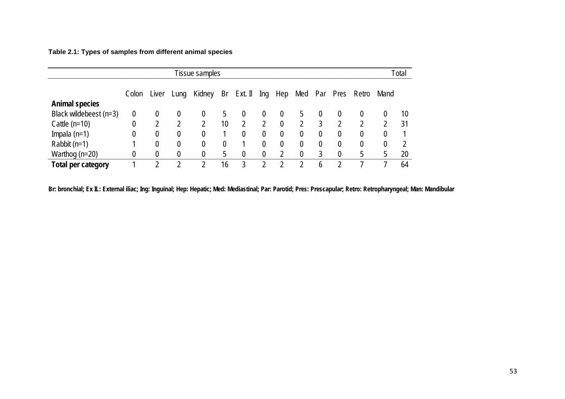
53
Table 2.1: Types of samples from different animal species
Tissue samples Total Colon Liver Lung Kidney Br Ext. Il Ing Hep Med Par Pres Retro Mand Animal species Black wildebeest (n=3) 0 0 0 0 5 0 0 0 5 0 0 0 0 10 Cattle (n=10) 0 2 2 2 10 2 2 0 2 3 2 2 2 31 Impala (n=1) 0 0 0 0 1 0 0 0 0 0 0 0 0 1 Rabbit (n=1) 1 0 0 0 0 1 0 0 0 0 0 0 0 2 Warthog (n=20) 0 0 0 0 5 0 0 2 0 3 0 5 5 20 Total per category 1 2 2 2 16 3 2 2 2 6 2 7 7 64
Br: bronchial; Ex IL: External iliac; Ing: Inguinal; Hep: Hepatic; Med: Mediastinal; Par: Parotid; Pres: Prescapular; Retro: Retropharyngeal; Man: Mandibular

54
2.2.4 Mycobacterial isolation Samples were kept frozen at -20°C until processing at NHLS under biosafety
measures (Figure 2.2). Direct impression smears were made from lesions and were
stained using the Ziehl-Neelsen method. Tissue samples taken in a sterile manner
from the inside of granulomatous lesions at the border between healthy and fibrous
tissues were finely cut using a sterile scalpel blade and homogenized using sterile
glass beads by vortexing as described by Bengis et al. (1996) and Warren et al.
(2006) with some modifications. To maximize the mycobacterial yield, specimens
were subjected to a digestion and decontamination procedure using N-acetyl-L-
cysteine-sodium hydroxide (NALC-NaOH) solution with NaOH at a final
concentration of 2% (Chatterjee et al., 2013). The specimens were left at room
temperature for 15 min during the decontamination process and thereafter
neutralized with phosphate buffer pH 6.8, centrifuged (Beckman Coulter) at 3000 x g
for 15 min at 4°C, and the supernatant decanted and pellet suspended into 1 mL of
phosphate buffer. The sediment was inoculated onto two LJ slants, supplemented
with pyruvate and glycerol and an antibiotic mixture of polymyxin B, amphotericin B,
carbenicillin and trimethoprim (PACT) (National Health Laboratory Service, South
Africa, and Becton Dickinson, Germany) using a 0.01 mL calibrated loop. A further
0.5 mL of the sediment was inoculated with a graduated Pasteur pipette into a
prepared liquid medium tube (BBLTM MGITTM Mycobacterium Growth indicator tubes)
enriched with OADC and containing 800 µl of PANTATM antibiotic mixture (BDTM).
This was incubated in the BACTECTM MGITTM 960 (Figure 2.2) mycobacterial
detection system at 37°C (Warren et al., 2006). The system was monitored for a
maximum period of 42 days for bacterial growth whereas LJ slants were observed
for colony growth and any other contaminants at two week intervals for 10 weeks.
Tubes detected as positive within that period were further processed using Ziehl-
Neelsen staining and examined microscopically for the presence of acid-fast
organisms and morphology, thereafter subcultured on LJ slant supplemented with
glycerol and pyruvate. For identification purposes, a single colony was subcultured
on a fresh LJ slant to obtain pure colonies. The same was repeated with two other
colonies on different LJ slants to rule out the possibility of missing a different
organism. Reference cultures of M. avium (ATCC 25291), M. bovis BCG and M.
tuberculosis (ATCC 25177) were used as positive controls. Stored DNA extracts
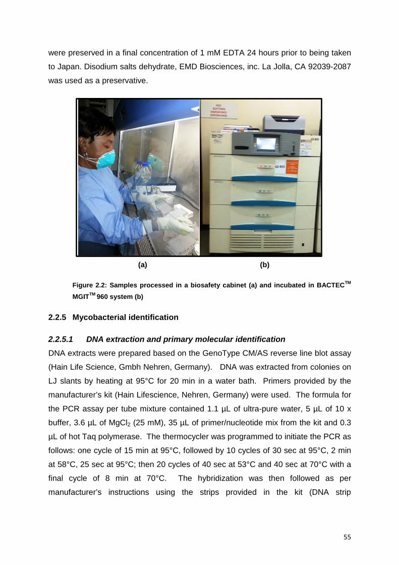
55
were preserved in a final concentration of 1 mM EDTA 24 hours prior to being taken
to Japan. Disodium salts dehydrate, EMD Biosciences, inc. La Jolla, CA 92039-2087
was used as a preservative.
(a) (b)
Figure 2.2: Samples processed in a biosafety cabinet (a) and incubated in BACTECTM MGITTM 960 system (b)
2.2.5 Mycobacterial identification
2.2.5.1 DNA extraction and primary molecular identification DNA extracts were prepared based on the GenoType CM/AS reverse line blot assay
(Hain Life Science, Gmbh Nehren, Germany). DNA was extracted from colonies on
LJ slants by heating at 95°C for 20 min in a water bath. Primers provided by the
manufacturer’s kit (Hain Lifescience, Nehren, Germany) were used. The formula for
the PCR assay per tube mixture contained 1.1 µL of ultra-pure water, 5 µL of 10 x
buffer, 3.6 µL of MgCl2 (25 mM), 35 µL of primer/nucleotide mix from the kit and 0.3
µL of hot Taq polymerase. The thermocycler was programmed to initiate the PCR as
follows: one cycle of 15 min at 95°C, followed by 10 cycles of 30 sec at 95°C, 2 min
at 58°C, 25 sec at 95°C; then 20 cycles of 40 sec at 53°C and 40 sec at 70°C with a
final cycle of 8 min at 70°C. The hybridization was then followed as per
manufacturer’s instructions using the strips provided in the kit (DNA strip

56
Mycobacterium identification species). Part of each DNA extract was also stored at -
20°C prior to further investigations in Japan.
2.2.5.2 Antimicrobial susceptibility test The antimicrobial susceptibility test and identification of mutations responsible for
resistance against rifampin (RIF) and isoniazid (INH) were conducted at NHLS
according to in house standard operating procedure using a commercial kit,
Genotype® MTBDRplus kit (Hain Life Science, Gmbh Nehren, Germany), following
the manufacturer’s instructions. Biotinylated primers provided with the kit were used
to amplify 5 µl of DNA with hot-start Taq DNA polymerase (Qiagen, Pretoria, South
Africa). Amplification was performed using a thermal cycler MyCyclerTM (Bio-Rad,
Cape Town, South Africa). The thermocycler was programmed as described under
section 2.2.5.1. Then hybridization steps were followed using hybridization trays
(Hain Life Science, Gmbh Nehren, Germany) according to the manufacturer’s
instructions. The following genes were targeted for detecting the sensitivity of the
isolates to a specific antimycobacterial drug namely eight rpoB wild-type probes
named WT1-WT8 and four mutant probes named MUT1, MUT2A, MUT2B and
MUT3 were used to detect resistance against RIF. One katG wild type (katG WT)
and two mutant probes (MUT1 and MUT2); plus two inhA wild type (WT1 and WT2)
and four mutant probes (MUT1, MUT2, MUT3A and MUT3B) for the detection of
resistance against INH. According to the manufacturer, when all WT probes stained
positive and no mutation band formed, the result was interpreted as susceptible to
the specific anti-TB drug (Figure 2.3). The resistance towards a specific antibiotic
was indicated by the absence of a band for at least one of the WT probes. Each
pattern deviating from the wild type pattern indicates a resistance of the tested strain.
The banding pattern obtained with rpoB probes allows a conclusion to be drawn
about a rifampicin resistance of the strain tested whereas the banding pattern
obtained with the katG probes allows a conclusion to be made about a high level
isoniazid resistance; the banding pattern obtained with the inhA probes allows a
conclusion to be drawn about a low level of isoniazid resistance of the strain tested
respectively.

57
2.2.5.3 MTC discrimination by multiplex PCR The Mycobacterium tuberculosis complex-discriminating multiplex PCR (MTCD-
MPCR) targeting genetic regions cfp32 (a specific gene for MTC), RD4 (region of
difference 4 which is present in M. tuberculosis and M. africanum but absent in M.
bovis was used for species differentiation according to the method described by
(Parsons et al., 2002) with slight modifications. The reaction mixture consisted of
7.4 μL H20, 2 μL 10 x Taq buffer, 2 μL dNTPs (2.5 mM each), 0.2 μL Taq (Takara
Bio Inc, Shiga, Japan), 1 μL of DNA sample, 2.2 μL of 10 μM cfp32 primers, 0.7 μL
of 5 μM RD4 primers. The PCR was performed using the following program:
denaturation for 1 min at 98°C followed by 35 cycles of 5 sec at 98°C, 20 sec at 58°C
and 1 min at 68°C with final elongation at 72°C for 5 min in a thermal cycler (iCycler,
Bio-Rad Laboratories Inc., Hercules, CA). Mycobacterium bovis BCG Pasteur
vaccine, M. bovis BCG Tokyo and M. bovis AF2122/97 were used as positive
controls and distilled water as negative control. The PCR products were separated
by electrophoresis on a 2% agarose gel in TAE buffer, and then visualized after
staining with ethidium bromide. RD4 Flank F and RD4 flank R gave a product of
about 446 bp.
2.2.5.4 Spoligotyping Spoligotyping of M. bovis isolates using microarray was performed according to the
procedure by Kamerbeek et al. (1997) with slight modifications. Forty-three spacer-
sequence probes were covalently bound to the membrane (Pall Co., NY, USA).
PCR products were generated using primers DRa (GGTTTTGGGTCTGACGAC) and
DRb (CCGAGAGGGGACGGAAAC). The PCR mixture included 7.3 µL of deionized
water, 5 X 3 µL colourless GoTaq buffer (Promega TM, Fitchburg, WI), 1.5 µL of
PCR DIG Labelling Mix (Roche), 1 µL of 10 µM Fw primer, 11 µL of 10 µM Rv
primer, 0.2 µL of GoTaq DNA Polymerase (5 units/µL, Promega) and 1 µL of DNA
sample in a total volume of 15 µL of mixture per tube. The thermocycler was
programmed to initiate denaturation at 98°C for 60 sec, followed by 40 cycles of 5
sec at 98°C, 10 sec at 55°C, 10 sec at 55°C, 30 sec at 72°C and a post run for 60
sec at 72°C. The PCR product that was diluted 500 times in hybridization buffer was
heat denatured at 95°C for 5 min and immediately cooled on ice to obtain the single
stranded DNA. The probe solutions for spotting were prepared by adjusting the
concentration with TE buffer to 50 µM of spacer probe and 10 µM of DR probe

58
(positive control). Then the membrane was cut to an appropriate size and marked to
distinguish the side to be spotted. The membrane was held with forceps to avoid
binding of protein, which could interfere with probe DNA attachment on the
membrane. Then 0.5 µL of each probe solution was spotted on the membrane and
heated at 70°C for 30 min, followed by pouring of the blocking buffer on the
membrane. It was then allowed to stand at room temperature for 1 hour with
intermittent gentle shaking.
The hybridization was carried out by immersing the membrane in the hybridization
solution at 60°C for 1 hour. Then, the membrane was washed in warm TBST-E
(0.1% Tween-20 and 1 mM EDTA-2Na in tris-buffered saline) and incubated for 10
min at 60°C then washed for the third time in warm TBST-E. The membrane was
then dried at room temperature. The POD and TMB solutions were prepared as per
manufacturer’s instructions. The nitrocellulose membranes were covered with a
1000 times diluted Ant-Digoxigenin-POD [poly], Fab fragments (Roche) in a dish and
incubated at room temperature for 30 min. Thereafter, the membranes were
sequentially washed three times in TBST-E at room temperature for 1 min, 10 min
and 1 min as per protocol (Hang’ombe et al., 2012). Then, POD on the membranes
was detected through colour development by adding TMB solution (TMB Peroxidase
Substrate Kit, Vector Labs Inc. TM, Burlingame, CA) according to the manufacturer’s
protocol. The membrane was then washed with deionized water (DIW) and scanned
(Figure 2.4). The spoligotype patterns observed were compared to the patterns
available in the spoligotype M. bovis database (www.mbovis.org)
2.2.5.5 Variable number of tandem repeat (VNTR) typing The M. bovis isolates were further genotyped by PCR amplification using primers
targeting a wide range of tandem repeat loci (Table 2.2). Two different PCR reaction
mixtures were conducted according to the loci and the PCR primer sequences used
are outlined in Table 2.2. Locus MIRU10, 16, 24, 26, 27, 39, 40, ETR-B, F, VNTR-
424, 3690 were done under betaine 1.0 M, whilst locus MIRU2, 4, 20, 23, 31, ETR-A,
C, QUB- 11a, 11b, 26, 3232, 3336, VNTR-1955, 2401, 4156 were performed under
GCII buffer (Takara). The PCR reaction mixture under 1.0 M betaine buffer was
conducted in a mixture consisted of 6.3 μL H2O, 0.4 μL of dNTP (10 mM each), 3.0
μL of 5x colourless Go Taq buffer, 3.0 μL of betaine (5 M), 0.6 μL of Primer 1 and 0.6

59
μL of Primer 2, 0.1 μL of Go Taq (5 units/ μL) DNA Polymerase, and 1 μL of DNA
sample. An initial denaturation step of 95°C for 5 min was followed by 32 cycles at
95°C for 15 sec, 58°C for 20 sec and 72°C for 1 min with a final elongation step at
72°C for 1 min in a thermal cycler. The PCR reaction mixture under the GCII buffer
consisted of 4.8 μL H2O, 0.4 μL of dNTP (10 mM each), 7.5 μL of 2 x GCII buffer, 0.6
μL of primers, 0.1 μL of Go Taq (5 units/μL) DNA Polymerase (Promega), and 1 μL
of DNA sample. All the PCR samples were electrophoresed on a 2% agarose gel to
identify the repeat numbers; a 100 bp ladder PLUS (Thermo Scientific, USA) was
included for the estimation of the size (Hang’ombe et al., 2012) (Table 2.3).
Table 2.2: Variable number of tandem repeat (VNTR) loci and forward and reverse primers (5'-3') sequences used for the typing of M. bovis isolates (Le Fleche et al., 2002)
ON-No Locus No Common names Primer sequence Mer 749
13 MIRU4F GCGCGAGAGCCCGAACTGC 19
750 MIRU4R GCGCGAGCAGAAACGCCAGC 20 751
1 MIRU10F GTTCTTGACCAACTGCAGTCGTCC 24
752 MIRU10R GCCACCTTGGTGATCAGCTACCT 23 753
2 MIRU16F TCGGTGATCGGGTCCAGTCCAAGT 24
754 MIRU16R CCCGTCGTGCAGCCCTGGTAC 21 755
3 MIRU26F CCCGCCTTCGAAACGTCGCT 20
756 MIRU26R TGACATAGGCGACCAGGCGAATA 24 757
4 MIRU31F ACTGATTGGCTTCATACGGCTTTA 24
758 MIRU31R GTGCCGACGTGGTCTTGAT 19 759
5 MIRU40F GGGTTGCTGGATGACAACGTGT 22
760 MIRU40R GGGTGATCTCGGCGAAATCAGATA 24 761
9 ETR-A1 AAATCGGTCCCATCACCTTCTTAT 24
762 ETR-A2 CGAAGCCTGGGGTGCCCGCGATTT 24 763
10 ETR-C1 GTGAGTCGCTGCAGAACCTGCAG 23
764 ETR-C2 GGCGTCTTGACCTCCACGAGTG 22 765
11 QUB11b1 CGTAAGGGGGATGCGGGAAATAGG 24
766 QUB11b2 CGAAGTGAATGGTGGCAT 18 767
12 QUB26-1 GGCCAGGTCCTTCCCGAT 18
768 QUB26-2 AACGCTCAGCTGTCGGAT 18 769
6 VN424F CTTGGCCGGCATCAAGCGCATTATT 25
770 VN424R GGCAGCAGAGCCCGGGATTCTTC 23 771
14 VN2401F CTTGAAGCCCCGGTCTCATCTGT 23
772 VN2401R ACTTGAACCCCCACGCCCATTAGTA 25 773
8 VN3690F CGGTGGAGGCGATGAACGTCTTC 23
774 VN3690R TAGAGCGGCACGGGGGAAAGCTTAG 25 775
15 VN4156F TGACCACGGATTGCTCTAGT 20
776 VN4156R GCCGGCGTCCATGTT 15

60
777 7
VN1955F AGATCCCAGTTGTCGTCGTC 20 778 VN1955R CAACATCGCCTGGTTCTGTA 20 779
16 MIRU2F TGGACTTGCAGCAATGGACCAACT 24
780 MIRU2R TACTCGGACGCCGGCTCAAAAT 22 781
17 MIRU20F TCGGAGAGATGCCCTTCGAGTTAG 24
782 MIRU20R GGAGACCGCGACCAGGTACTTGTA 24 783
18 MIRU23F CAGCGAAACGAACTGTGCTATCAC 24
784 MIRU23R CGTGTCCGAGCAGAAAAGGGTAT 23 785
19 MIRU24F CGACCAAGATGTGCAGGAATACAT 24
786 MIRU24R GGGCGAGTTGAGCTCACAGAA 21 787
20 MIRU27F TCGAAAGCCTCTGCGTGCCAGTAA 24
788 MIRU27R GCGATGTGAGCGTGCCACTCAA 22 789
21 MIRU39F CGCATCGACAAACTGGAGCCAAAC 24
790 MIRU39R CGGAAACGTCTACGCCCCACACAT 24 791
22 ETR-B1 GCGAACACCAGGACAGCATCATG 23
792 ETR-B2 GGCATGCCGGTGATCGAGTGG 21 793
23 ETR-F1 CTCGGTGATGGTCCGGCCGGTCAC 24
794 ETR-F2 GGAAGTGCTCGACAACGCCATGCC 24 795
24 QUB11a1 TTCAGGGGGGATCCGGGA 18
796 QUB11a2 CCCATCCCGCTTAGCACATTCGTA 24 797
25 QUB3232-1 CAGACCCGGCGTCATCAAC 19
798 QUB3232-2 CCAAGGGCGGCATTGTGTT 19 799
26 QUB3336-1 ATCCCCGCGGTACCCATC 18
800 QUB3336-2 GCCAGCGGTGTCGACTATCC 20

61
Table 2.3: Band sizes (bp) and corresponding tandem repeat numbers
Tandem repeat number for M. bovis No Locus name Unit (bp) 0 1 2 3 4 5 6 7 8 9 10 1 MIRU10 53 482 537 590 643 696 749 802 855 908 2 MIRU16 53 565 618 671 724 777 830 883 936 989 3 MIRU26 51 461 512 563 614 665 716 767 818 869 4 MIRU31 (ETR-E) 53 492 545 598 651 704 757 810 863 916 5 MIRU40 54 354 408 462 516 570 624 678 732 786 6 VN424 (Mtub04) 51 538 589 640 691 742 793 844 895 946 7 VN1955 (Mtub21) 57 149 206 263 320 377 434 491 548 605 8 VN3690 (Mtub39) 58 330 388 446 504 562 620 678 736 9 ETR-A(VN2165) 75 195 270 345 420 495 570 645 720 795 870 945
10 ETR-C(VN577) 58 44 102 160 218 276 334 392 450 508 11 QUB11b(VN2163b) 69 136 205 274 343 412 481 550 619 12 QUB26(VN4052) 111 264 375 486 597 708 819 930 1041 13 MIRU04 (ETR-D) 77 or 53 252 329 406 483 560 637 14 VN2401 (Mtub30) 58 247 305 363 421 479 537 595 653 711 15 VN4156n(QUB4156) 59 563 622 681 740 799 858 917 976 16 MIRU02 53 402 455 508 561 614 667 720 773 826 17 MIRU20 77 514 591 668 18 MIRU23 53 558 608 661 714 767 820 873 926 979 19 MIRU24 54 395 447 501 555 609 663 717 771 825 20 MIRU27 53 551 604 657 710 763 816 869 922 21 MIRU39 53 536 591 646 701 756 811 866 921 976 22 ETR-B 57 121 178 235 292 349 406 463 520 577 23 ETR-F 79 263 342 421 500 579 658 737 816 895 24 QUB11a(VN2163a) 69 374 443 512 581 650 719 788 857 926 995 25 QUB3232 56 423 479 535 591 647 703 759 815 871 26 QUB3336 59 171 230 289 348 407 466 525 584 643 702

62
2.3 RESULTS AND DISCUSSION
2.3.1 Mycobacterial isolation Out of the 20 samples received from 20 warthogs, 16 different types of lymph nodes
namely bronchial, mandibular, parotid and retropharyngeal yielded either rough or
smooth colonies on LJ slants with pyruvate inoculated with the sediment from
positive MGIT tubes. One out of the 2 samples (external iliac lymph node) from a
rabbit yielded smooth colonies as well as 3 out of 10 samples from 3 black
wildebeest, 15 samples out of 31 samples from 10 cattle yielded rough colonies. All
isolates were slow-growing organisms and acid-fast rods under the microscope.
Isolates were further identified as Ziehl-Neelsen staining alone cannot differentiate
Mycobacterium species. In medical laboratories, it was observed that LJ with
glycerol is routinely used; introducing LJ with pyruvate in the routine diagnostic will
increase the opportunity to isolate M. bovis.
2.3.2 Primary molecular identification The isolates from 3 black wildebeests were identified as M. avium subsp.
hominissuis (n=3) out of the 10 samples; 16 isolates from the 20 warthogs were
identified as M. bovis (n=14), M. avium (n=1) and M. intracellulare (n=1), one isolate
from rabbit (n=1) was also identified as M. avium subsp. hominissuis, the isolate
from an impala (n=1) was identified as M. bovis and 15 isolates from 10 cattle were
identified as M. bovis (Table 2.4) using commercial kits, the GenoType
Mycobacterium CM/AS (Hain Lifescience GmbH, Nehren, Germany). Cattle isolates
were further processed as species of interest for the next phase of the research
project whereas those from other animal species were stored at -80°C for use in
future studies.
The isolation of non-tuberculous mycobacteria namely M. avium subsp. hominissuis,
M. avium and M. intracellulare are of concern as these species are potentially
zoonotic and have been reported as causes of disease in humans. In South Africa,
the level of involvement of non-tuberculous mycobacteria in disease-causing
conditions in animals and humans should be fully investigated. In the medical
laboratory where the study was conducted, these mycobacteria were referred as
“MOTT” and are not given much attention.
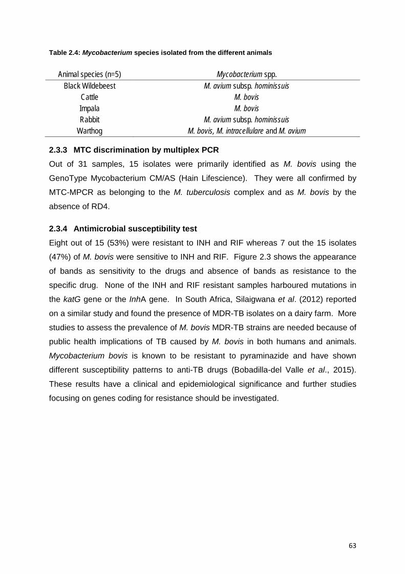
63
Table 2.4: Mycobacterium species isolated from the different animals
Animal species (n=5) Mycobacterium spp. Black Wildebeest M. avium subsp. hominissuis
Cattle M. bovis Impala M. bovis Rabbit M. avium subsp. hominissuis
Warthog M. bovis, M. intracellulare and M. avium
2.3.3 MTC discrimination by multiplex PCR Out of 31 samples, 15 isolates were primarily identified as M. bovis using the
GenoType Mycobacterium CM/AS (Hain Lifescience). They were all confirmed by
MTC-MPCR as belonging to the M. tuberculosis complex and as M. bovis by the
absence of RD4.
2.3.4 Antimicrobial susceptibility test Eight out of 15 (53%) were resistant to INH and RIF whereas 7 out the 15 isolates
(47%) of M. bovis were sensitive to INH and RIF. Figure 2.3 shows the appearance
of bands as sensitivity to the drugs and absence of bands as resistance to the
specific drug. None of the INH and RIF resistant samples harboured mutations in
the katG gene or the InhA gene. In South Africa, Silaigwana et al. (2012) reported
on a similar study and found the presence of MDR-TB isolates on a dairy farm. More
studies to assess the prevalence of M. bovis MDR-TB strains are needed because of
public health implications of TB caused by M. bovis in both humans and animals.
Mycobacterium bovis is known to be resistant to pyraminazide and have shown
different susceptibility patterns to anti-TB drugs (Bobadilla-del Valle et al., 2015).
These results have a clinical and epidemiological significance and further studies
focusing on genes coding for resistance should be investigated.

64
Figure 2.3: WT probes and mutation genes targeted to determine the susceptibility of an isolate towards first line drugs
2.3.5 Spoligotyping The 15 isolates of M. bovis from cattle consisted of two strains, namely SB0121 and
SB1235. Most isolates, 10 out of 15 were SB0121 (67 %) and 5 out of 15 (33%)
were SB1235. SB1235 lacked spacers 3, 6, 8, 9, 10, 11, 12, 16, 37-43 (Figure 2.4
A) whereas SB0121 was characterized by lack of spacers 3, 9, 16, 21, 39-43 (Figure
2.4 B). SB0121 is the most frequently isolated spoligotype. Spoligotyping is used as
an epidemiological tool to trace the origin of the strain causing the disease and
hence can facilitate implementation of appropriate control measures. A consistent
characteristic of M. bovis is the lack of spacers 39-43 (Figures 2.4 and 2.5).
Isolates from cattle were confirmed and further characterized using spoligotyping
and VNTR. The spoligotype patterns revealed the presence of a strain recently
reported in South Africa, SB1235 (Hlokwe et al., 2014), whereas SB0121 is known to
be the most common type which has been reported in several other countries,
namely Argentina, Belgium, Brazil, Spain, Great Britain, Italy, Mexico, Sweden, USA
and South Africa (Hlokwe et al., 2014; Musoke et al., 2015; Parreiras et al., 2012;
Rodriguez-Campos et al., 2013). They were all VNTR type 1 which means they

65
have the same origin. Samples were from one province, Mpumalanga, although
from different farms.
SB1235 seems to relate to UK strains, and the importation by breeders of cattle from
Europe could explain the introduction of this strain (http://www.sussex.co.za;
http://www.afrikanerbees.com). Spoligotype strains are an indication of how wide is
the movement of cattle worldwide. SB1235 was originally isolated in Northern
Ireland whereas SB0121 seems to be the most common spoligotype in South Africa
as reported by Hlokwe et al., (2014). It has also been isolated worldwide from
Argentina, Belgium, Brazil, Spain, Great Britain, Italy, Mexico, Sweden, USA, New
Zealand and Sweden (Rodriguez-Campos et al., 2013; Sahraoui et al., 2009).
Figure 2.4: Spoligotype SB1235 (A) and spoligotype SB0121 (B), deleted spacers that are common to all M. bovis are 39 to 43. SB1235 lack spacers 3, 6, 8, 9, 10, 11, 12, 16, 37-43 whereas SB0121 lacks spacers 3, 9, 16, 21, 39-43 –The deleted spacers are marked with red stars
A B
3
6 8 10
9
11 12
16
37 38
39 40
41 42 43
3
9
16
21
39 40
41 42 43

66
Figure 2.5: Schematic representation of spoligotype patterns of M. bovis cattle isolates from South Africa
1 2 3 4 5 6 7 8 9 10 11 12 13 14 15 16 17 18 19 20 21 22 23 24 25 26 27 28 29 30 31 32 33 34 35 36 37 38 39 40 41 42 43 Spoligotype patternsSB0121 (67% )SB1235 (33% )

67
2.3.6 Variable number of tandem repeat (VNTR) typing The typing of the 15 isolates was based on the 13 VNTR loci and yielded only one
type, VNTR-1. This meant that the isolates were all from the same origin.
2.4 CONCLUSION The presence of MDR in M. bovis in cattle is of concern taking into consideration the
emergence of MDR tuberculosis strains in humans. In medical laboratories, the
tendency is to rule out M. tuberculosis, other mycobacteria are considered as MOTT
and not given much attention. Introducing LJ with pyruvate in the routine diagnostic
will increase the opportunity to isolate M. bovis. Attention should also be given to the
non-tuberculous mycobacteria referred to as MOTT as they have been reported as
the main cause of the disease in some cases. The identification of MDR-M. bovis
isolates is of concern due to public health implications. Mycobacterium bovis causes
tuberculosis in humans which is clinically undistinguishable from M. tuberculosis.
The presence of MDR-MTB complex in dairy cattle in South Africa has been reported
by Silaigwana et al. (2012). Mycobacterium bovis is known to be resistant to
pyraminazide and has shown different susceptibility patterns to anti-TB drugs
(Bobadilla-del Valle et al., 2015). Drug susceptibility and accurate identification of
isolates in both animals and humans need attention as there are no current data on
the prevalence and incidence of human tuberculosis caused by M. bovis in South
Africa (Bobadilla-del Valle et al., 2015).
Genetic typing of M. bovis has contributed greatly to the improved knowledge of
inter-bovine and interspecies transmission of bovine tuberculosis (Durr et al., 2000).
The understanding of genetic typing is important for the effective management of
bovine tuberculosis control schemes and the wildlife–livestock interface in countries
where wildlife reservoirs for M. bovis have been identified, including South Africa
(Haddad et al., 2004; Vos et al., 2001). The most widely used DNA typing
techniques for M. bovis include IS6110 and PGRS restriction fragment length
polymorphism (RFLP) typing, spoligotyping and variable number of tandem repeat
(VNTR) typing (Reddington et al., 2011). In South Africa, spoligotyping and variable
number of tandem repeat typing have been used (Hlokwe et al., 2014; Michel et al.,
2007). The two methods had a high discriminatory power of over 90% (Kremer et
al., 2005).

68
Another finding relates to non-tuberculous mycobacteria. Their importance has been
emphasized during the last decade and it was interesting to isolate M. avium subsp.
hominissuis (MAH) from the rabbit, pig samples and other samples from black
wildebeest. This is the most prevalent MAC involved in human infection. Rónai et
al. (2016) reported that M. avium subsp. hominissuis was an important member of
the M. avium complex with a high diversity among M. avium subsp. avium and
hominissuis and they are both potentially zoonotic. The evidence for MAH as
causative agent of lymphadenitis and sometimes disseminated granulomatous
inflammation in animals is supportive of virulence of the organism (Despierres et al.,
2012). This will need some attention as M. avium infection is a severe condition in
humans whereas in pigs it causes a subclinical infection. MAH has been isolated
from swine gastrointestinal samples, from lymph nodes of tuberculin positive swine
(Agdestein et al., 2012, 2011; Kriz et al., 2014; Yoav, 2012). It has also been
incriminated as a cause of mesenteric lymphadenitis and sometimes disseminated
granulomatous lesions in different species namely pigs, cattle, dogs and parrots
(Campora et al., 2011; Kriz et al., 2014).
Arrazuria et al. (2015) stated that rabbits are susceptible to infection by different
species of the genus Mycobacterium, namely Mycobacterium avium subsp. avium
and Mycobacterium avium subsp. paratuberculosis but microbiological data on rabbit
meat is lacking and more specifically reports of mycobacterial presence in
industrially reared rabbit for human consumption have not been published.
In this research, colonic granulomatous lesions from a rabbit yielded colonies that
were characterized as M. avium subsp. hominissuis (MAH) and this species is the
most prevalent subtype among human MAC infection. A recent study has reported
on the presence of MAH in rabbit (Arrazuria et al., 2015). This isolate has been
stored for further investigation.
The isolates from pigs were not further characterized but have been stored for other
studies especially M. intracellulare and M. avium isolates.
Some of the isolates will be used in testing of plants for antimycobacterial activity in
other chapters.

69
Post script The next chapter (Chapter 3) has been published in the Journal Transboundary and
Emerging Diseases.

70
CHAPTER 3: NOVEL MYCOBACTERIUM AVIUM SPECIES ISOLATED FROM
BLACK WILDEBEEST (CONNOCHAETES GNOU) IN SOUTH AFRICA
ABSTRACT A study was undertaken to isolate and characterize Mycobacterium species from
black wildebeest suspected of being infected with tuberculosis in South Africa. This
led to the discovery of a new Mycobacterium avium species, provisionally referred to
as “Gnou isolate” from black wildebeest (Connochaetus gnou). Sixteen samples
from nine black wildebeest were processed for Mycobacterium isolation. Following
decontamination; samples were incubated in an ordinary incubator at 37°C on
Löwenstein-Jensen slants and in liquid medium tubes using the BACTECTM MGITTM
960 system respectively. Identification of the isolate was done by standard
biochemical tests and using the line probe assay from the GenoType® CM/AS kit
(Hain Life Science GmbH, Nehren, Germany). The DNA extract was also analysed
using gene sequencing. Partial gene sequencing and analysis of 16S rRNA gene,
16S-23S rRNA (ITS), rpoB and hsp65 and phylogenetic analyses by searching
GenBank using the BLAST algorithm were conducted. Phylogenetic trees were
constructed using four methods, namely Bayesian inference, maximum likelihood,
maximum parsimony and neighbor-joining methods. The isolate was identified as
Mycobacterium intracellulare using the GenoType® CM/AS kit and as Mycobacterium
avium complex (MAC) by gene sequencing. The gene sequence targeting all the
genes, ITS, 16S rRNA, rpoB and hsp65 and phylogenetic analyses indicated that
this isolate presented a nucleotide sequence different from all currently published
sequences, and its position was far enough from other MAC species to suggest that
it might be a new species.
3.1 BACKGROUND In late 2006, animals from a commercial game farm reserve in Mpumalanga
Province in South Africa were harvested for game meat exportation. During meat
inspection, the animal carcasses showed lesions suspicious of tuberculosis which
was supported by histopathological results. The exact cause of the disease was not

71
determined and the farm was put under quarantine for suspected bovine
tuberculosis.
In February 2009, 158 animals were harvested. A high number of animals (n = 135)
showed gross-visible tuberculosis-like lesions and lesions from 6 animals processed
for mycobacterial cultures yielded non-tuberculous mycobacteria.
Samples (n = 16) from 9 animals were submitted to the National Health Laboratory
Service (NHLS, Pretoria, South Africa) for mycobacterial isolation and a non-
tuberculous mycobacterium (NTM) was isolated and identified as Mycobacterium
intracellulare using a commercial kit and as Mycobacterium avium complex (MAC)
by gene sequencing. The results were submitted to the Department of Agriculture,
Forestry and Fisheries (DAFF, South Africa) as M. intracellulare, and
characterization of the isolate was then done in Japan.
The complete history of the herd including the pathology report described well-
developed encapsulated granulomatous lesions observed on the different samples of
organs as very suspicious for bovine tuberculosis (BTB). Other lesions observed
which were not typical of M. bovis (pseudotuberculosis) comprised lack of caseous
necrosis and liquefaction in the granulomas. The inspissated material from within
the capsules could almost be squeezed out in total, leaving behind an empty “shell”.
There were also several smaller granulomas with a typical onion ring appearance,
but absence of calcification and liquefaction with no gritty sensation on cut section of
these capsules. .
The genus Mycobacterium contains more than 170 species
(http://www.bacterio.net/mycobacterium.html), most of which are classified as NTM
or potentially pathogenic mycobacteria (PPM) (Chege et al., 2008; Kim et al., 2014;
Malama et al., 2014; Tortoli, 2014) and mycobacteria belonging to the
Mycobacterium tuberculosis complex (MTC). MTC comprises M. tuberculosis, M.
bovis, M. africanum, M. canetti, M. pinnipedii, M. caprae, M. microti, M. mungi,
Dassie bacillus, M. orygis (Oryx bacillus), M. surricatae and the attenuated M. bovis
Bacille-Calmette-Guerin (BCG) vaccine strain. With the exception of BCG, these
species are pathogenic and can cause tuberculosis (TB) in mammalian hosts
(Alexander et al., 2010; Helden et al., 2009; Pittius et al., 2012; Vos et al., 2001).

72
The M. avium-intracellulare complex is the most commonly encountered group of
NTM, and the clinically most important members are M. intracellulare and M. avium
(Biet et al., 2005). Mycobacterium intracellulare has not been subdivided into
subspecies whereas M. avium consists of four subspecies, namely M. avium subsp.
avium, M. avium subsp. hominissuis, M. avium subsp. silvaticum and M. avium
subsp. paratuberculosis. Mycobacterium Avium Complex (MAC) includes 10
different species, namely M. avium, M. intracellulare, M. colombiense, M.
bouchedurhonense, M. timonense, M. arosiense, M. chimaera, M. vulneris, M.
yongonense and M. marseillense (Tortoli, 2014).
The importance of NTM has received attention during the past decade, especially in
humans. NTM are found in environmental systems (such as various soil and water
systems) near human settlements and can be associated with colonization, serious
infection or pseudo-outbreaks with a wide variety of presentations (Biet et al., 2005;
Kankya et al., 2011; Katale et al., 2014). Indeed in humans, the isolation of NTM
from clinical samples of patients presenting with pulmonary symptoms as suspected
cases of tuberculosis has increased over the years and has been observed in
different countries in Africa, America and Europe (Kankya et al., 2011; Katale et al.,
2014; Mirsaeidi et al., 2014a, 2014b; Moore et al., 2010) whereas in animals the
clinical significance of NTM has yet to be elucidated in the disease causing process
(Chege et al., 2008; Kankya et al., 2011; Katale et al., 2014). The members of the
genus Mycobacterium are genetically closer to each other than the microorganisms
belonging to other genera, making identification a difficult and challenging task. The
management, treatment and infection control measures differ significantly between
M. tuberculosis and NTM infections.
More than one hundred and fifty species of NTM have been reported worldwide, of
which more than 60% are pathogenic to animals or humans (Kim et al., 2014; Tortoli,
2014). In South Africa, reports on the isolation of NTM in animals, humans and
environment and their effects in disease-causing processes are limited (Gcebe et al.,
2013; Kabongo-Kayoka et al., 2015; Michel et al., 2007; Müller et al., 2011).
Black wildebeest (Connochaetus gnou), known in Afrikaans as “Swartwildebees” and
in German as “Weisschwanzgnu”, have been hunted in South Africa for meat and
hides. The overall research project was mainly on tuberculosis, so samples from

73
black wildebeest suspected of being infected with tuberculosis were processed as
part of phase I of the project focusing on prevalence and molecular studies of
mycobacteria. The emergence of multi-drug and extremely drug resistant
Mycobacterium tuberculosis strains was one of the main justifications of the project.
This work resulted in the reporting of a novel Mycobacterium avium complex species
from wildebeest in South Africa, which is expected to add to the corpus of knowledge
and extend the frontiers of research on NTM.
3.2 MATERIALS AND METHODS
3.2.1 Study area Mpumalanga Province was selected as the area of study based on previous
publications reporting TB in wildlife and livestock (Bengis et al., 1996; Michel et al.,
2007; Vos et al., 2001) and availability of veterinary staff members experienced in
conducting the comparative tuberculin skin test. Mpumalanga is one of the nine
provinces in South Africa which is located in the north-eastern part of the country,
bordering Swaziland and Mozambique to the East. It embraces the southern half of
the Kruger National Park, a vast nature reserve with abundant wildlife including big
game. It has a subtropical foliage supporting about 1 439 000 cattle according to the
Trends in the Agricultural Sector (2013).
3.2.2 Study design and sampling The study was designed as a cross-sectional study sampling animals in the
designated area from January 2009 to January 2011. The local municipalities were
selected based on the number of commercial farms, proximity to abattoirs, and
location at the human-wildlife interface and the movement of animals as well. The
municipalities selected comprised Malelane, Nelspruit, Lydenburg, Ermelo, Witbank
and Standerton. The target population comprised cattle carcasses showing gross
tuberculous-like lesions at meat inspection from positive reactors to tuberculin skin
test at the municipality abattoirs during the study period. Samples from any other
animal species showing gross tuberculous-like lesions were also included as
convenience samples. The sampling was purposive to increase the chances of
isolating mycobacteria and animals were selected based on positive reaction to the
tuberculin skin test and suggestive lesions at the abattoir upon meat inspection.

74
3.2.3 Sources of samples Samples from black wildebeest were received as part of phase I of a research
project related to “Prevalence and molecular studies of Mycobacteria”. The samples
were processed at the National Health Laboratory Service (NHLS) as part of a joint
collaboration between University of Pretoria and NHLS. The history of the case was
provided by the state veterinarian in charge. During a hunting period in February
2009 on a commercial game reserve located in Mpumalanga (South Africa), game
animals (n=158) were randomly harvested and processed in the local abattoir
according to standard operating procedures. These animals comprised black
wildebeest (Connochaetes gnou) (n=137), blesbok (Damaliscus dorcas phillipsi)
(n=15), blue wildebeest (Connochaetes taurinus) (n=2), red hartebeest (Alcelaphus
buselaphus caama) (n=2) and springbok (Antidorcas marsupialis) (n=2). The
animals lagging behind were the main target as well as females. During routine
meat inspection of these animals, a high number of black wildebeest (n=135)
showed granulomatous lesions in one or more lymph nodes or organs and this is the
reason why samples were selected from this antelope species. Sixteen samples
randomly selected from nine black wildebeest showing fresh lesions suggestive of
tuberculosis infection were submitted to NHLS for isolation and identification of
Mycobacterium spp. The samples included different organs and lymph nodes
transported on ice (Table 3.1).

75
Table 3.1: Type of tissue samples from Black Wildebeest processed at NHLS
Animals Tissue samples Liver Lung Med Total ID* BWB 1 1 0 0 1 BWB 2 0 0 1 1 BWB 3 1 1 0 2 BWB 4 0 1 1 2 BWB 5 1 1 0 2 BWB 6 0 1 0 1 BWB 7 1 1 0 2 BWB 8 0 1 1 2 BWB 9 1 1 1 3 Tot 5 7 4 16
*ID: identity, BWB: Black Wildebeest, Med: Mediastinal lymph nodes
3.2.4 Mycobacterial isolation Samples were frozen at -20°C until processing at NHLS. Direct impression smears
were made from lesions and smears were stained using the Ziehl-Neelsen method.
Tissue samples taken in a sterile manner from the inside of granulomatous lesions at
the border between healthy and pathological tissues were finely cut using a sterile
scalpel blade and homogenized using sterile glass beads by vortexing as described
by Bengis et al. (1996) and Warren et al. (2006) with some modifications. To
maximize the mycobacterial yield, specimens were subjected to a digestion and
decontamination procedure using N-acetyl-L-cysteine-sodium hydroxide (NALC-
NaOH) solution with NaOH at a final concentration of 2% (Chatterjee et al., 2013).
The specimens were left at room temperature for 15 min during the decontamination
process and thereafter neutralized with phosphate buffer, centrifuged (Beckman
Coulter) at 3000 x g for 15 min at 4°C, and the supernatant decanted and pellet
suspended into 1 mL of phosphate buffer. The sediment was inoculated onto two LJ
slants supplemented with pyruvate and glycerol and an antibiotic mixture of
polymyxin B, amphotericin B, carbenicillin and Trimethoprim (PACT) (National Health
Laboratory Service, South Africa, and Becton Dickinson, Germany) using a 0.01 mL
calibrated loop. A further 0.5 mL of the sediment was inoculated with a graduated

76
Pasteur pipette into a prepared liquid medium tube (BBLTM MGITTM Mycobacterium
Growth indicator tubes) enriched with OADC and containing 800 µL of PANTATM
antibiotic mixture (BDTM). This was incubated in the BACTECTM MGITTM 960
mycobacterial detection system at 37°C (Warren et al., 2006). The system was
monitored for a maximum period of 42 days for bacterial growth whereas LJ slants
were observed for colony growth and any other contaminant at two week intervals for
10 weeks. Tubes detected as positive within that period were further processed
using Ziehl-Neelsen staining and examined microscopically for the presence of acid
fast organisms and morphology. Thereafter, they were subcultured on LJ slant
supplemented with glycerol and pyruvate. For identification purposes, a single
colony was subcultured on a fresh LJ slant to obtain pure colonies. The same was
repeated with two other colonies on different LJ slants to rule out the possibility of
missing a different organism. Reference cultures of M. avium (ATCC 25291), M.
bovis BCG and M. tuberculosis (ATCC 25177) were used as positive controls.
3.2.5 Mycobacterial identification
3.2.5.1 Biochemical profile Colonies were suspended in Middlebrook 7H9 (M7H9) broth enriched with OADC
and inoculated into the different biochemical substrates according to the
manufacturer’s instructions (Mycobacterium identification kit, Kyokuto
Pharmaceutical Industrial Co., Ltd., Japan).
3.2.5.2 DNA extract and primary molecular identification DNA extracts were prepared based on the GenoType CM/AS reverse line blot assay
(Hain Life Science, Gmbh Nehren, Germany). DNA was extracted from colonies on
LJ slants by heating at 95°C for 20 min in a water bath. Primers provided by the
manufacturer’s kit (Hain Life Science, Gmbh Nehren, Germany) were used. The
formula for the PCR assay per tube mixture contained 1.1 µL of ultra-pure water, 5
µL of 10 x buffer, 3.6 µL of MgCl2 (25 mM), 35 µL of primer/nucleotide mix from the
kit and 0.3 µL of hot Taq polymerase. The thermocycler was programmed to initiate
the PCR as follows: one cycle of 15 min at 95°C, followed by 10 cycles of 30 sec at
95°C, 2 min at 58°C, 25 sec at 95°C; then 20 cycles of 40 sec at 53°C and 40 sec at
70°C with a final cycle of 8 min at 70°C. The hybridization was then followed as per
manufacturer’s instructions using the strips provided in the kit (DNA strip

77
Mycobacterium identification species). Part of each DNA extract was also stored at -
20°C prior to further investigations in Japan.
3.2.6 Gene sequencing The DNA extract obtained during the primary identification was processed further by
sequencing analysis; targeting hsp65, rpoB, 16S rRNA genes and 16S-23S rRNA
internal transcribed spacer (ITS) for identification of the bacterial species.
3.2.6.1 16S ribosomal RNA gene and ITS The 16S ribosomal RNA gene and flanking 16S–23S rRNA ITS region was amplified
with a primer pair of Bact-rrs-F: 5’-AGAGTTTGATCCTGGCTCAG- 3’ and myco ITS-
23S-Rv: 5’-CGGTTGACAGCTCCCCGAGGC- 3’. Amplification reaction mixture
consisted of 0.5 μM of each primer pair, 1× ExTaq buffer (Takara Bio Inc., Japan),
0.5 M betaine, 0.25 mM each of dNTPs, 1 U of ExTaq DNA polymerase (Takara Bio
Inc.), 1 μL of target DNA solution and milli-Q water to adjust the final volume to 20
μL. Amplification was performed in a thermal cycler with a pre-heat step at 98°C for 1
min, 38 cycles of denaturation at 98°C for 10 sec, annealing at 55°C for 10 sec and
extension at 72°C for 110 sec followed by a final extension at 72°C for 5 min. The
amplicon was electrophoresed on a 1% agarose gel and a band of approximately 1.9
Kbp was excised and purified with Wizard SV Gel and PCR Clean-Up System
(Promega Corp., USA). The sequence of the 5’ region of the 16S ribosomal RNA
gene and ITS were read with primers Bact-rrs-F and myco ITS-23S-Rv, respectively,
by ABI PRISM 3130xl Genetic Analyzer (Life Technologies Corp., CA, U.S.A.)
according to the manufacturer’s instructions.
3.2.6.2 rpoB Partial rpoB gene was amplified and sequenced with primers Myco-F and Myco-R
designed by Ben Salah et al. (2008) PCR reaction mixture was comprised of 0.5 μM
of each primers, 1× GoTaq buffer (Promega Corp., USA), 0.5 M betaine, 0.25 mM
each of dNTPs, 1 U of GoTaq DNA polymerase (Promega Corp.), 1 μL of target DNA
solution and milli-Q water to adjust the final volume to 20 μL. Amplification was
performed with an initial denaturation at 96°C for 1 min, 38 cycles of denaturation at
96°C for 10 sec, annealing at 60°C for 10 sec and extension at 72°C for 45 seconds
followed by a final extension at 72°C for 5 min. The amplicon was electrophoresed
on a 1% agarose gel and a band with a size of 761 bp was excised and purified. The

78
sequence was read by ABI PRISM 3130xl Genetic Analyzer (Life Technologies
Corp.) according to the manufacturer’s instructions.
3.2.6.3 hsp65 Partial hsp65 gene was amplified and sequenced with primers Myco-hsp65F (565-
585): 5’- AGGGTATGCGGTTCGACAAG- 3’ and MAC hsp65R (Turenne et al.,
2006). Amplification was performed with an initial denaturation at 96°C for 1 min, 38
cycles of denaturation at 96°C for 10 sec, annealing at 53°C for 10 sec and
extension at 72°C for 45 sec followed by a final extension at 72°C for 5 min in the
same reaction mixture content described in section 3.2.6.2. The band with a size of
1067 bp was excised and sequencing was performed by the same procedure as in
section 3.2.6.2.
The obtained sequences were compared with the GenBank nucleotide database by
the blastn program (National Center for Biotechnology Information:
http://www.ncbi.nlm.nih.gov).
3.2.7 Phylogenetic analyses The datasets for the different genes (ITS, 16S, hsp65 and rpoB) were collated by
searching GenBank (http://www.ncbi.nlm.nih.gov) using the BLAST algorithm, an
acronym for the Basic Local Alignment Search Tool (Altschul et al., 1990). BLAST
searches various databases, as specified by the user, to look for similar sequences,
and then uses a similarity matrix to measure the similarity between sequences and
the possibility that the similarity could be due to chance based on the nucleotide
sequence of the query versus its target. There are several types of searches
available but for these analyses “blastn” was used which searches nucleotide
databases with a nucleotide query. These sequences were then downloaded into a
local database using BioEdit version 7.2.1 (Hall, 1999). Sequences were aligned
using MAFFT version 7 (Katoh et al., 2005, 2002)
http://www.mafft.cbrc.jp/alignment/server/) with default parameters. Multiple
Alignments by Fast Fourier Transformation (MAFFT) utilizes an iterative algorithm,
unlike previous progressive alignment methods. Where necessary, small
adjustments were made to the alignments manually to minimize hypothesized
insertion/deletion events, again using BioEdit software (Hall, 1999). The programme
jModelTest 2.1.6 (Darriba et al., 2012) was run for each gene separately, as well as
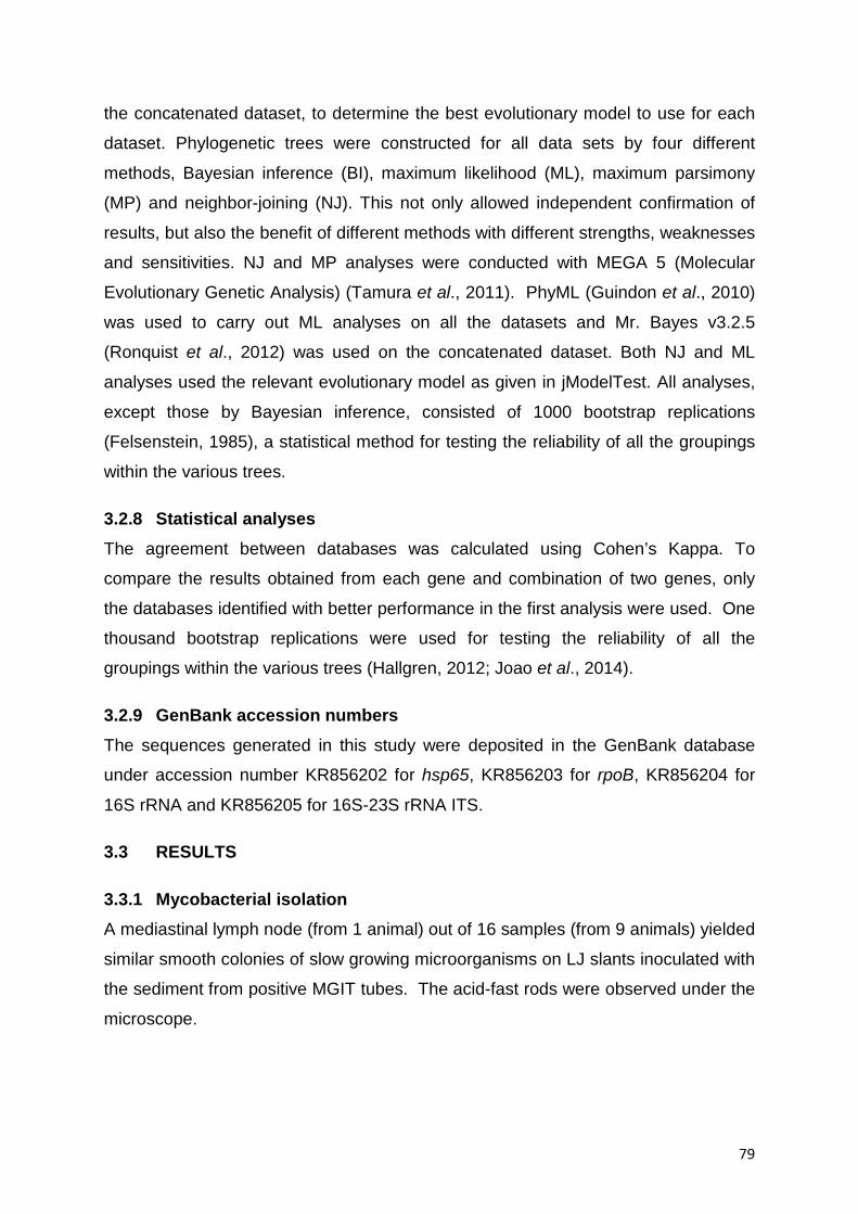
79
the concatenated dataset, to determine the best evolutionary model to use for each
dataset. Phylogenetic trees were constructed for all data sets by four different
methods, Bayesian inference (BI), maximum likelihood (ML), maximum parsimony
(MP) and neighbor-joining (NJ). This not only allowed independent confirmation of
results, but also the benefit of different methods with different strengths, weaknesses
and sensitivities. NJ and MP analyses were conducted with MEGA 5 (Molecular
Evolutionary Genetic Analysis) (Tamura et al., 2011). PhyML (Guindon et al., 2010)
was used to carry out ML analyses on all the datasets and Mr. Bayes v3.2.5
(Ronquist et al., 2012) was used on the concatenated dataset. Both NJ and ML
analyses used the relevant evolutionary model as given in jModelTest. All analyses,
except those by Bayesian inference, consisted of 1000 bootstrap replications
(Felsenstein, 1985), a statistical method for testing the reliability of all the groupings
within the various trees.
3.2.8 Statistical analyses The agreement between databases was calculated using Cohen’s Kappa. To
compare the results obtained from each gene and combination of two genes, only
the databases identified with better performance in the first analysis were used. One
thousand bootstrap replications were used for testing the reliability of all the
groupings within the various trees (Hallgren, 2012; Joao et al., 2014).
3.2.9 GenBank accession numbers The sequences generated in this study were deposited in the GenBank database
under accession number KR856202 for hsp65, KR856203 for rpoB, KR856204 for
16S rRNA and KR856205 for 16S-23S rRNA ITS.
3.3 RESULTS
3.3.1 Mycobacterial isolation A mediastinal lymph node (from 1 animal) out of 16 samples (from 9 animals) yielded
similar smooth colonies of slow growing microorganisms on LJ slants inoculated with
the sediment from positive MGIT tubes. The acid-fast rods were observed under the
microscope.

80
3.3.2 Biochemical characteristics The phenotypic characteristics, including growth rate (fast/slow), production of
pigment, growth at different temperatures and biochemical reactions with relevant
substrates were evaluated. Colonies were identified as MAC slow growers with no
pigment on specific substrates. They were positive for tellurite reduction, stimulation
by pyruvate and heat-stable catalase but negative for niacin production, nitrate
reduction, urease, acid phosphatase activity, Tween 80 hydrolysis (7 and 14 days)
and urease (Table 3.2); (Cook et al., 2003).
Table 3.2: Growth and biochemical characteristics of the isolate
Growth rate 27°C Positive 37°C Positive (Slow growth) 45°C Negative Stimulation by pyruvate Positive Pigmentation No pigmentation Niacin production Negative Nitrate reduction Negative Catalase production Positive Tellurite reduction Positive Urease Negative Acid phosphatase activity Negative Tween 80 hydrolysis (7 and 14 days) Negative
3.3.3 Primary molecular identification The isolate was identified as Mycobacterium intracellulare using a commercial kit
routinely used at NHLS, the GenoType Mycobacterium CM/AS (Hain Life Science,
Gmbh Nehren, Germany).
3.3.4 Phylogenetic analyses
3.3.4.1 ITS sequences Alignment of the 16S-23S rRNA ITS of Mycobacterium “Gnou isolate” with the other
sequevars of the MAC show that the Gnou isolate is most closely related to the
MAC-T sequevar with only three nucleotide differences.
81
Phylogenetic analyses of these data by NJ, MP and ML, placed the Gnou isolate
within the MAC and more closely related to MAC-T, with MAC-M, MAC-I and MAC-L
forming a sister group. All three methods placed the Gnou isolate in the same place
with varying degrees of bootstrap support.
3.3.4.2 16S rRNA, hsp65 and rpoB analyses. All three datasets by every method grouped the Gnou isolate with M. vulneris, M.
bouchedurhonense, and M. colombiense with M. arosiense included in the group by
just the MP analysis of hsp65. However, all other analyses placed M. arosiense
close to the Gnou isolate grouping. With the similarity between the various trees the
four genes were combined into one dataset to give a concatenated tree (Figure 3.1).
Generally the groupings all have good bootstrap support. All the species above M.
mantenii in this phylogeny are members of the MAC.

82
Figure 3.1: Phylogenetic tree obtained by NJ analysis of concatenated nucleotide sequences of 16S rRNA, 16S-23S rRNA, hsp65 and rpoB. Bootstrap values (1000 replicates) are given above the branches for NJ/ML and below for MP. Branch support values on the nodes are shown in percentage and the values less than 60% are not shown. The tree is rooted with M. fortuitum and M. insubricum.
3.4 DISCUSSION AND CONCLUSION This is the first report in South Africa of the discovery of a novel Mycobacterium
avium complex species from black wildebeest which has been named “Gnou isolate”
to reflect the species name of the wildebeest from which it was isolated. Our isolate
was identified using analysis of 16S rRNA gene, hsp65, rpoB and ITS. A number of
studies suggest that 16S rRNA gene sequencing provides genus identification in
most cases (>90%) but less so with regard to species (65 to 83%) (Janda and

83
Abbott, 2007; Joao et al., 2014). Although 16S rRNA gene sequencing is highly
useful in regards to bacterial classification, it has low phylogenetic power at the
species level and poor discriminatory power for some genera (Janda and Abbott,
2007). It has also been reported that analysis of 16S rRNA gene alone is insufficient
for the accurate identification of NTM (Joao et al., 2014); it was thus proposed that a
stepwise algorithm combining 16S rRNA and hsp65 gene analysis by multiple public
databases could be used to identify NTM at the species level. In some cases, 16S
rRNA gene sequence data cannot provide a definitive answer since it cannot
distinguish between recently diverged species. While it is impossible to be
completely accurate when modelling all evolution that has occurred between a set of
sequences, several parameters appear to be particularly important. These are
corrections for substitution patterns (nucleotide substitution matrices) and correction
for different evolutionary rates at different sites, most accurately corrected using a
gamma distribution model, the shape parameter α of which is calculated by
jModelTest. It has been established that NTM cause disease (Kim et al., 2014), but
the significance of NTM in the disease processes in animals should be investigated
further as the presence of a small number of a specific pathogen does not correlate
with the virulence of the pathogen nor its economic importance. Organisms act in
synergy, potentiating the colonization by other bacteria. Although mixed infection
with NTM has been reported, attention should be given to NTM in the future as
several studies have recognized the significance of NTM as a major public health
issue around the world (Kankya et al., 2011; Kim et al., 2014; Malama et al., 2014;
Moore et al., 2010; Temmerman et al., 2014). Furthermore, NTM have been found
to interfere with the diagnosis of TB in cattle; indeed, some cross reactions between
the antigens of NTM with those used for diagnostic purposes such as M. avium and
M. fortuitum may compromise the diagnosis and control of bovine tuberculosis (De la
Rua-Domenech et al., 2006; Gcebe et al., 2013).
To our knowledge, this is the first report of this new isolate of NTM, “Gnou isolate”,
sampled from black wildebeest (Connochaetes gnou) in South Africa. The diagnosis
of NTM should be standardized as the isolate was identified as Mycobacterium
intracellulare using DNA strip according to Hain’s method at National Health
Laboratory Service and confirmed as a new species of MAC by gene sequencing at
Research Center for Zoonosis Control at Hokkaido University in Japan. Tortoli et al.

84
(2010) reported the misidentification of NTM using commercial kit assays as we have
also experienced with this study. The partial sequencing of 16S rRNA gene alone is
not sufficient to fully identify NTM to species level; algorithm analysis combining all
four genes should be considered as well as biochemical identification. One of the
characteristics of this isolate was slow growth. The role of NTM, in particular slow
growing MAC, should be further investigated in the disease-causing process as they
could potentiate colonization by other rapidly growing microorganisms. The isolation
and identification at species level of some NTM should be performed to establish
their clinical relevance in animals and humans. Countries should also be
encouraged to register new species on the international data bank for purposes of
adding to the epidemiological knowledge of this genus.
Postscript The next chapter comprises the start of phase 2 of the project whereby extracts of
plant species from the family Anacardiaceae were tested for antimycobacterial
activity against a panel of Mycobacterium species, both rapidly growing saprophytic
and slow-growing pathogenic species. Cytotoxicity of the extracts was also tested
against various cell lines. The chapter has been published in the Journal
Phytotherapy Research. The article is attached in Appendix C.

85
CHAPTER 4: ANTIMYCOBACTERIAL ACTIVITY AND CYTOTOXICITY OF LEAF
EXTRACTS OF SOME AFRICAN ANACARDIACEAE TREE SPECIES
ABSTRACT Treatment of tuberculosis (TB) is a challenge due to multi- and extremely drug
resistant strains of Mycobacterium tuberculosis. Plant species contain antimicrobial
compounds that may lead to new anti-TB drugs. Previous screening of some tree
species from the Anacardiaceae family revealed the presence of antimicrobial
activity, justifying further investigations. Leaf extracts of 15 Anacardiaceae tree
species were screened for antimycobacterial activity using a twofold serial
microdilution assay against the pathogenic Mycobacterium bovis and multidrug
resistant M. tuberculosis and rapidly growing mycobacteria, M. smegmatis, M.
fortuitum and M. aurum. The vaccine strain, M. bovis and an avirulent strain, M.
tuberculosis H37Ra, were also used. Cytotoxicity was assessed using a colorimetric
assay against Vero kidney, human hepatoma and murine macrophage cells. Four
out of 15 crude acetone extracts showed significant antimycobacterial activity with
minimum inhibitory concentration (MIC) values varying from 50 to100 µg/mL.
Searsia undulata had the highest activity against most mycobacteria, followed by
Protorhus longifolia. Mycobacterium fortuitum was the strongest predictor of activity
against MDR-TB (correlation coefficient = 0.65). Bioautography against M. aurum
and M. fortuitum worked well as indicators of the Rf values of active compounds
yielding strong zones of inhibition. The leaf extracts of S. undulata and P. longifolia
each had more than ten different antimycobacterial compounds and had low
cytotoxicity with LC50 values above 100 µg/mL.
4.1 INTRODUCTION
Tuberculosis occurs in humans, cattle, wild animals and many other domesticated
species. The Mycobacterium tuberculosis complex (MTC) comprises M.
tuberculosis, M. bovis, M. africanum, M. canetti, M. pinnipedii, M. caprae, M. microti,
M. mungi, Dassie bacillus, M. orygis (Oryx bacillus), M. surricatae and the attenuated
M. bovis Bacille-Calmette-Guerin (BCG) vaccine strain. With the exception of BCG,
these species are pathogenic and can cause tuberculosis (TB) in mammalian hosts

86
(Alexander et al., 2010; Kabongo-Kayoka et al., 2015; Pittius et al., 2012; Vos et al.,
2001).
Despite the fact that TB can be cured, this disease remains a worldwide public
health threat due to the emergence of multi- and extremely drug resistant strains and
subsequent poor response to current antibiotics. One third of the world’s population
is infected with Mycobacterium tuberculosis (MTB) and is hence at risk of developing
active TB (WHO, 2015). In the order of 9.6 million people every year develop TB
and 1.5 million die from the disease. This includes 0.4 million TB deaths among HIV
positive people (WHO, 2015). From the 9.6 million, only 12% were HIV positive and
it was also estimated that 480 000 cases of multidrug-resistant TB (MDR-TB) have
occurred in 2014 (WHO, 2015). This confirms that TB alone is still a major
challenge.
There is currently no effective vaccine to protect against TB. In humans, control of
the disease relies heavily on detecting infectious cases and treating them for at least
six months with a combination of antibiotics. The course of antibiotics involves the
administration of first-line anti-TB drugs, which are combinations of isoniazid (INH),
rifampicin (RIF), pyrazinamide and ethambutol for 2 months to kill the rapidly growing
bacteria. The treatment is continued in the next 4 months with a combination of INH
and RIF because of their sterilizing activity to eliminate bacilli which are dormant in
the macrophages, or slow growers (Rivers and Mancera, 2008). Toxicity associated
with these first-line drugs and the long duration of treatment has led to low patient
compliance, giving rise to drug-resistant strains. Multidrug-resistant mycobacteria
are resistant to INH and RIF whereas extensively drug-resistant mycobacteria are
resistant to second-line drugs such as fluoroquinolones and to at least one injectable
drug in addition to INH and RIF. Hence, there is a need to find new anti-tubercular
agents with novel modes of actions (Chinsembu, 2016).
It is most likely that the emergence of resistant Mycobacterium isolates will continue
in the future, exhausting the current range of effective antibiotics available.
Therefore, new classes of anti-TB agents are urgently needed, and research projects
seeking for alternative therapeutics have been encouraged. Natural resources have
been explored to develop new antibiotics. Substances that can either inhibit the
growth of pathogens or kill them while having little or no toxicity to host cells are
87
considered good candidates for developing new antimicrobial drugs (Nguta et al.,
2016).
Medicinal plants are used in many parts of the world to treat different ailments
including TB-related symptoms such as chest complaints, respiratory ailments, fever
and coughing (McGaw et al., 2008a, 2008b). Relevant reviews of traditional
medicinal use of plants in Africa are available (Hutchings et al., 1996; Kuete, 2010;
Maroyi, 2013; Nguta et al., 2016; Stark et al., 2013; Van Wyk et al., 1997).
South Africa possesses the richest temperate flora in the world, with an estimated 24
000 species and intraspecific taxa in 368 families more than 10% of the world’s
vascular plant flora (Germishuizen and Meyer, 2003). There is much potential for
discovery of structurally interesting metabolites with activity against Mycobacterium
species from these plants. The existence of a strong cultural reliance on traditional
medicines within South African communities justifies the importance of studying their
safety and efficacy (Fennell et al., 2004).
In a study conducted on Indian medicinal plants, Gautam et al. (2007) reported that
with many of the plant species examined, there was a strong positive correlation
between antimycobacterial activity and ethnomedicinal use for TB and TB-related
diseases. Although this provides support for investigating plants customarily used in
other cultures to treat symptoms relating to TB, other plants tested for
antimycobacterial activity using indicator organisms such as M. smegmatis and M.
bovis BCG should also be investigated (McGaw et al., 2008a). Testing plant
material not only against ATCC strains but against current isolates is crucial as these
microorganisms are part of the changing environment harbouring emerging resistant
strains.
The Anacardiaceae, cashew or Sumac is a tropical family that comprises flowering
plant bearing fruits that are drupes and in some cases producing urushiol which is an
irritant substance (Mabberley, 1997). The Anacardiaceae comprises several genera
and species of economic importance as shown in Figure 4.1 (a-l) namely Cashew
(Anacardium occidentale), Mango (Mangifera indica), Poison Ivy (Toxicodendron
radican, Smodingium argutum), Sumac (Searsia species), Smoke tree (Cotinus
coggygria, Cotinus obovatus), Marula (Sclerocarya birrea), Yellow mombin

88
(Spondias mombin, Spondias purpurea var. lutea), Cuachalalate (Amphipterygium
adstringens) and Pistacia (Pistacia lentiscus, Pistacia integerrima). This family,
which belongs to the order Sapindales, is represented by 77 genera and 600 species
mostly distributed in tropical, subtropical, and temperate areas (Mabberley, 1997).
Very few plant species of the Anacardiaceae family have been investigated for
antimycobacterial activity. One of the objectives of this study was to investigate in
vitro antimycobacterial activities as well as cytotoxicity of selected plant species from
the Anacardiaceae family. These plants were selected mainly based on a
preliminary broad antimicrobial screening (Pauw, 2014).
A summary on ethnobotanical use of the selected plant species from the
Anacardiaceae family is presented in Table 4.1. Eight out of the 15 plants, namely
Lannea discolor, Loxostylis alata, Ozoroa mucronata, Ozoroa paniculosa, Protorhus
longifolia, Searsia chirindensis, Searsia lancea and Sclerocarya birrea, have
antimicrobial properties mainly against Gram-negative and Gram-positive organisms
(Ahmed et al., 2014; Hutchings et al., 1996; Mosa et al., 2014; Stark et al., 2013;
Van Wyk et al., 1997). Three species (Searsia chirindensis, Searsia undulata and
Smodingium argutum) have good anti-inflammatory activity (Hutchings et al., 1996;
Ojewole, 2008; Stark et al., 2013). It would be useful to identify plant species with
both antimycobacterial and anti-inflammatory efficacy as both acute and chronic
inflammation are induced during the course of tuberculosis disease progression
(Volpe et al., 2006).
4.2 MATERIALS AND METHODS
4.2.1 Source of plant materials and extraction Leaves of the 15 plant species were collected in November 2009 from the Lowveld
National Botanical Gardens in Nelspruit, the University of Pretoria Botanical Gardens
and the Pretoria National Botanical Garden, South Africa in terms of a signed
material transfer agreement. Leaves were kept in labelled bags with full
identification and collection dates. Voucher specimens were kept in the Lowveld
National Botanical Gardens (Nelspruit), HGWJ Schweickerdt Herbarium of the
University of Pretoria and Phytomedicine Programme, University of Pretoria (Table
4.1). The leaves were air-dried at room temperature suspended in bags of net
material in a ventilated room (refers Figure 4.1a), ground to a fine powder in a
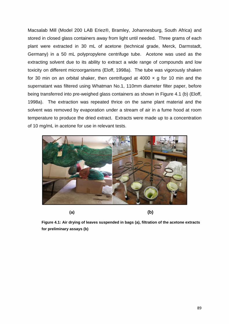
89
Macsalab Mill (Model 200 LAB Eriez®, Bramley, Johannesburg, South Africa) and
stored in closed glass containers away from light until needed. Three grams of each
plant were extracted in 30 mL of acetone (technical grade, Merck, Darmstadt,
Germany) in a 50 mL polypropylene centrifuge tube. Acetone was used as the
extracting solvent due to its ability to extract a wide range of compounds and low
toxicity on different microorganisms (Eloff, 1998a). The tube was vigorously shaken
for 30 min on an orbital shaker, then centrifuged at 4000 × g for 10 min and the
supernatant was filtered using Whatman No.1, 110mm diameter filter paper, before
being transferred into pre-weighed glass containers as shown in Figure 4.1 (b) (Eloff,
1998a). The extraction was repeated thrice on the same plant material and the
solvent was removed by evaporation under a stream of air in a fume hood at room
temperature to produce the dried extract. Extracts were made up to a concentration
of 10 mg/mL in acetone for use in relevant tests.
(a) (b)
Figure 4.1: Air drying of leaves suspended in bags (a), filtration of the acetone extracts for preliminary assays (b)
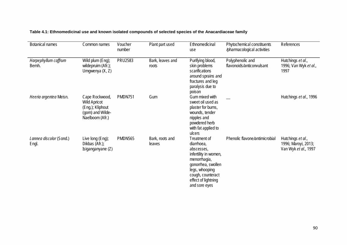
90
Table 4.1: Ethnomedicinal use and known isolated compounds of selected species of the Anacardiaceae family
Botanical names Common names Voucher number
Plant part used Ethnomedicinal use
Phytochemical constituents /pharmacological activities
References
Harpephyllum caffrum Bernh.
Wild plum (Eng); wildepruim (Afr.); Umgwenya (X, Z)
PRU2583 Bark, leaves and roots
Purifying blood, skin problems scarifications around sprains and fractures and leg paralysis due to poison
Polyphenolic and flavonoids/anticonvulsant
Hutchings et al., 1996; Van Wyk et al., 1997
Heeria argentea Meisn. Cape Rockwood, Wild Apricot (Eng.); Kliphout (gom) and Wilde-Naelboom (Afr.)
PMDN751 Gum Gum mixed with sweet oil used as plaster for burns, wounds, tender nipples and powdered herb with fat applied to ulcers
__ Hutchings et al., 1996
Lannea discolor (Sond.) Engl.
Live long (Eng); Dikbas (Afr.); Isiganganyane (Z)
PMDN565 Bark, roots and leaves
Treatment of diarrhoea, abscesses, infertility in women, menorrhagia, gonorrhea, swollen legs, whooping cough, counteract effect of lightning and sore eyes
Phenolic flavone/antimicrobial Hutchings et al., 1996; Maroyi, 2013; Van Wyk et al., 1997

91
Loxostylis alata A. Spreng. ex Rchb.
Wild pepper tree, tarwood (Eng); teerhout, tierhout (Afr); Ufutho, ufuthu (Z)
PRU3526 Bark and leaves Used at childbirth boost immunity
Phenolic lipids, cardanol and ginkgolic acid; lupeol 1 and B-sitosterol 2; antimicrobials
Ahmed et al., 2014
Ozoroa mucronata (Bernh.) R. Fern. & A. Fern.
No common names associated with this taxon yet. Mvunda-Jukunu in Tanzania
NEL71/1993 Leaves and roots Diarrhoea, dysentery, stomach troubles, gonorrhea, bilharzia and antidote for snake bite
Anacardic acid; flavonoids, phenols, tannins/antimicrobial
Ahmed et al., 2014
Ozoroa paniculosa (Sond.) R. Fern. & A. Fern.
Red resin tree (Eng.)
PMDN568 Bark and roots Diarrhoea and abdominal pain
Flavonoids, phenols, tannins Anacardic acid, ginkgoic acid and triterpenes/antimicrobial
Ahmed et al., 2014; Stark et al., 2013
Protorhus longifolia (Bernh.) Engl.
Red beech (Eng), rooiboekenhout (Afr.); umkhomizo, unhlangothi (Z)
PRU120030 Leaves, stem bark and seeds
Heartburn, bleeding from stomach, depilatories, Heartwater, dysentery and diarrhoea in cattle
Triterpenes, lanosteryl triterpen/antimicrobial, Antihyperlipidemic and antihyperglycemic
Mosa et al., 2014
Searsia chirindensis (Baker f.) Moffett
Red currant (Eng.); Bostaaibos (Afr.), umHlabamvubu (Z)
PMDN333 Stem Bark Heart, stomach ailments, diarrhoea, strengthen the body, stimulate circulation, rheumatism and mental disturbances
Saponins, tannins triterpenoids and flavonoids/ Inflammatory, analgesic and anticonvulsant
Ojewole, 2008
Searsia lancea (L.f.) F.A.Barkley
Karee (Eng.), umHlakotshane (X)
PMDN750 Bark and roots Diarrhoea, gallsickness
Flavonoids and tannins/antimicrobial
Stark et al., 2013

92
Searsia leptodictya (Diels.) T.S.Yi., A.J.Mill.& J.Wen
Mountain karee (Eng.), bergkaree (Afr.) Motshotlwane (Tsw)
NEL94/1969 Bark, leaves and roots
Diarrhoea and pain Phenols, tannins and flavonoids
Ahmed et al., 2014; Stark et al., 2013
Searsia magaliesmontana (Sond.) Moffett
NEL68/1997 __ __ __ __
Searsia pyroides (Burch.) Moffett
Wildcurrant (Eng.); Brandtaaibos, (Afr.); Inhlokoshiyane (Z)
PMDN570 Leaves, roots and stem bark
Treatment of epilepsy and cough medicine
Flavonoids bichalcones Maroyi, 2013
Searsia undulata (Jacq). T.S.Yi, A.J.Mill. & J.Wen
T’kuni (khoi); Kuni bush, (Eng.); Koenibos (Afr.)
PRU120031 Bark, leaves and roots
Post-parturient problems and treatment for chest colds
Apigenin 7,4´-dimethyl ether, flavone/ Anti-inflammatory
Fourie and Snyckers, 1984; Hutchings et al., 1996)
Sclerocarya birrea subsp. caffra (Sond.) Kokwaro
Cider tree, marula (Eng.); Maroela (Afr.); Umganu (Z)
NEL103/1967 Bark, leaves and roots
Diarrhoea, dysentery, pain, stomach problems, fever, cough, malaria, tonic, diabetes and sore eyes
Gallotannins, Flavonoids, epicatechin, alkaloids, triterpenoids and vitamin c /Anticonvulsants; antidiarrhoeal, antidiabetic, anti-inflammatory, antimicrobial and antioxidant
Maroyi, 2013; Stark et al., 2013; Van Wyk et al., 1997
Smodingium argutum E. Mey. ex Sond.
African poison ivy (Eng.); Pynbos (Afr.); Umthomvane (Z)
PMDN188 Bark, leaves and roots
Diarrhoea and diabetes
Catechols, phenolic lipids (cardanol and ginkgolic acid) /Antidiarrhoeal, hypoglycaemic effects and anti-inflammatory
Hutchings et al., 1996

93
(a) (b)
Figure 4.2: Leaves, flowers and fruits of Anacardiacea occidentalis (Cashew tree) (a) and Lannea discolor (b)
Source: courtesy of www.zipcodezoo.com and www.tropical.theferns
(c) (d)
Figure 4.3: Leaves and fruits of Mangifera indica (c) and Ozoroa paniculosa (d)
Source: courtesy of www.alamy.com

94
(e) (f)
Figure 4.4: Leaves of Ozoroa mucronata (e) and leaves and fruits of Heeria argentea (f)
Source: courtesy of www.ispotnature.org
(g) (h)
Figure 4.5: Leaves and fruits of Searsia chirendensis (g) and leaves of Searsia lancea (h)
Source: courtesy of www.plantzafrica.com and www.redlist.sanbi.org

95
(i) (j)
Figure 4.6: Leaves of Searsia magaliesmontana (i) and Smodingium argutum (j).
Source: courtesy of www.operationwildflower.org.za
(k) (l)
Figure 4.7: Leaves and flowers of Searsia pyroides (k) and Protorhus longifolia (l).
Source: courtesy of www.bidorbuy.co.za
4.2.2 Antimycobacterial activity The antimycobacterial activity of the acetone crude extracts of leaf powder was
determined using bioautography and the two-fold serial microdilution method. The
pathogenic and rapidly growing mycobacteria were test microorganisms used for
antimycobacterial activity.

96
4.2.2.1 Pathogenic mycobacteria
4.2.2.1.1 Mycobacterium tuberculosis multi-drug resistant isolates (MDR-TB)
Two clinical isolates of multi-drug resistant Mycobacterium tuberculosis (MDR-TB)
were used. Both isolates were obtained from patients admitted to the MDR-TB ward
at Tshepong Hospital in Klerksdorp, North West Province of South Africa in
December 2012. The patients were a 34 year old female and an 18 year old male.
Samples of sputum had been submitted to the National Health Laboratory Service
(NHLS) in Pretoria for culture in liquid medium and PCR/Line Probe Assay. Both
isolates were resistant to isoniazid and rifampicin.
4.2.2.1.2 Mycobacterium bovis
Mycobacterium bovis cultures were isolated from field samples of cattle from herds
that tested positive on tuberculination skin test during the period January 2009 to
January 2011(Kabongo-Kayoka et al., 2015) as described in Chapter 2 under section
2.3.2.
4.2.2.2 Rapidly growing mycobacteria Mycobacterium bovis BCG (Pasteur strain P1172) was obtained from the
Tuberculosis Laboratory at the ARC-Onderstepoort Veterinary Institute and M.
tuberculosis H37Ra, which is routinely used as reference material at NHLS, was
obtained from the American Type Culture Collection (ATCC) number 25177. Other
reference strains used included a M. aurum strain from the National Collection of
Type Cultures (NCTC), a culture collection of Public Health England, M. aurum
(NCTC 10437), M. smegmatis (ATCC 1441) and M. fortuitum (ATCC 6841).
4.2.2.3 Maintenance of cultures Fresh cultures were used in the relevant assays. The pathogenic isolates of
Mycobacterium spp. were kept at room temperature on LJ slants supplemented with
glycerol, or pyruvate in the case of the M. bovis BCG cultures, and were used within
a month. Prior to each assay, cultures were reactivated in liquid medium,
Middlebrook 7H9, using MGIT 960 tubes which were incubated at 37°C in the
BACTEC MGIT 960 instrument, in which they were automatically monitored each
hour for fluorescence development for 42 days or until a positive signal developed.
Bacterial suspensions from MGIT tubes were then subcultured on solid medium LJ

97
slants with pyruvate for M. bovis and LJ slants with glycerol for M. tuberculosis. LJ
tubes were then incubated in a walk-in incubator at 37°C for 4 to 6 weeks. A stained
Ziehl-Neelsen smear was made from the sediment of the MGIT tube and the slant of
LJ medium. Reference cultures of M. avium (ATCC 25291), M. bovis BCG and M.
tuberculosis H37Ra (ATCC 25177) were used as positive controls.
The rapidly growing Mycobacterium species were maintained on Middlebrook 7H10
agar prepared according to the manufacturer instructions (Merck) and kept in a
fridge at 4°C until used within a month. Mycobacterium aurum and M. fortuitum were
revived using Middlebrook 7H9 broth with glycerol and tween and supplemented with
OADC (Oleic acid Albumin Dextrose Catalase) whereas M. smegmatis was revived
using MH broth (Merck). Bacterial suspensions were inoculated on MH agar plates
for M. smegmatis and the rest on Middlebrook 7H9 agar. All agar plates were then
incubated at 37°C until colony growth was visible within 24 to 72 hours depending on
the growth rate of the specific mycobacterial species. The final density of 1 x 10⁵
CFU/mL used was confirmed after spreading an inoculum of 100 μL of tenfold serial
dilutions of the bacterial suspensions at McFarland No 1 onto Middlebrook 7H10
(M7H10) agar plates incubated at 37°C and counting colonies.
4.2.2.4 Minimum inhibitory concentration (MIC) determination The MIC values were determined using the serial microplate method developed by
Eloff (1998b) and slightly modified for mycobacteria by McGaw et al. (2008b). The
MIC value was the lowest concentration of the tested extract or sample inhibiting the
growth of the Mycobacterium species under investigation.
Mycobacterial suspensions were prepared from a pure culture of fresh colonies from
solid medium and suspended in Middlebrook 7H9 (M7H9) liquid medium
supplemented with 10% OADC. These colonies were transferred into a sterile
screw-capped tube containing 3 mL of M7H9 broth and homogenized by placing the
tube on a Vortex mixer for 5 minutes. After the larger particles had settled, the
mycobacterial suspension was adjusted to McFarland no.1 turbidity standard by
adding more broth (Lall and Meyer, 1999).
The assay was performed using sterile 96-well microplates with rounded bottoms.
The sample to be tested (plant extract, drug or fraction) was prepared at a

98
concentration of 10 mg/mL before serial dilution. One hundred microliters of M7H9
broth or MH broth was added to all the wells then, 100 µl of the sample to be tested were added in three adjacent column wells in the first row whereas other wells
included standard drug positive controls to assess bacterial growth, solvent control,
broth to be inoculated and clean broth for sterility check. Two samples of crude
extract and one standard drug were tested per plate. A two fold serial dilution was carried out leaving 100 µl of different concentrations of diluted tested samples in
each well starting with a concentration of 2.5 mg/mL in the first wells. Then 100 µl of
the test bacterial suspension were added to all the wells except column 12 (negative
control) containing pure broth to make sure that there was no contamination of the broth. Each dose was run in triplicate and the experiment repeated at least six
times. The standard antibiotics (reference drugs) included ciprofloxacin, rifampicin,
isoniazid and streptomycin starting with a concentration of 100 µg/mL. The
microplates were covered and sealed in plastic bags, placed in humid chambers to minimize the evaporation of the culture medium and incubated at 37°C for 24 hours (M. smegmatis), 48 hours (M. fortuitum) and 72 hours (M. aurum). For the
pathogenic mycobacteria, microplates were incubated for a period of 7 to 15 days.
At the end of incubation, a volume of 40 µl of 0.2 mg/mL of iodonitrotetrazolium chloride (INT) was added to each well, plates were incubated for 30 minutes or
longer at 37°C and the development of colour observed. A coloured red-purple
formazan or pink colour indicated the reduction of INT by metabolizing organisms whereas a yellow colour or decrease in colour indicated the inhibition of bacterial
growth Eloff (1998b). If the colour development was not strong enough for slow
growing organisms, plates were incubated much longer and monitored. The
correlation assessing the relationship between the MIC values of the 7 mycobacterial species used (Table 4.3) was calculated using Microsoft Excel 2010 software
(Microsoft, Redmond, WA, USA).
4.2.3 Thin Layer Chromatography (TLC) analysis and Bioautography Ten microliter from 10 mg/mL of relevant extracts (containing 100 μg of extract) was loaded on aluminium-backed TLC plates (Merck silica gel F254) in lines of about 1
cm wide. The TLC plates were eluted in three different mobile solvent systems that
included ethyl acetate/methanol/water (EMW) 10:1.35:1, chloroform/ethyl
acetate/formic acid (CEF) 10:8:2 and benzene/ethanol/ammonia (BEA) 18:2:0.2

99
(Kotzé and Eloff, 2002). Chromatograms were examined under ultraviolet light at
wavelengths of 254 and 366 nm to visualize UV active compounds that fluoresce and might not be visible after vanillin spray; the plates were then sprayed with a
solution made of 0.1 g vanillin in 28 mL of methanol and 1 mL of sulphuric acid
(vanillin–methanol-sulphuric acid) and heated at 110°C with a heat gun for colour
development and visualization of UV inactive compounds present in the plant extract. More than 10 compounds of different polarities were observed. These
compounds were very close to each other in both the solvent systems BEA and
CEF. In general, the active plants had different activities against different
Mycobacteria. Active compounds included both polar and non-polar compounds based on the Rf values determined in bioautography. In the non-polar solvent BEA,
the non-polar compounds travelled some distance up the plate with clear zones of inhibition on plates sprayed with different Mycobacterium species whereas in the
polar solvent system EMW, non-polar compounds were pushed towards the solvent front and polar compounds off the baseline. The CEF solvent system led to a better
separation of some compounds (B in Figure 4.3).
PL (A) PL (B) HC (A) HC (B) SU (A) SU (B)
PL: Protorhus longifolia, HC: Harpephyllum caffrum, SU: Searsia undulata
Figure 4.8: Bioautograms (A) of the three plants extracts with low MIC values and thin layer chromatograms eluted in chloroform/ethyl acetate/formic (CEF) solvent system sprayed with vanillin sulphuric acid (B) showing varied chemical constituents. Bioautogram of the extracts against Mycobacterium aurum.

100
For bioautography, only the rapidly growing organisms were used, namely M. aurum,
M. fortuitum and M. smegmatis, because the pathogenic mycobacteria present a
high biohazard risk if handled in a spray format. Aerosols of pathogenic species
increase risk of possible contamination of the environment and human exposure.
Duplicate chromatograms prepared as described above were left uncovered in a
dark place and air dried until solvents were completely evaporated for at least 24
hours or overnight. A saturated bacterial culture was centrifuged at 3000 x g for 10
min and the pellet resuspended in fresh appropriate liquid growth medium and
prepared at McFarland No 1. The TLC plates were then sprayed with active
bacterial suspension (18 to 72 h old cultures depending on the growth rate of the
particular Mycobacterium species) until completely moist using a glass spray gun
linked to a vacuum pump. The moist plates were incubated at 37°C in a humidified
chamber under 100% relative humidity for 24 h for M. smegmatis, 48 h for M.
fortuitum and 72 h for M. aurum. The plates were then sprayed with 2 mg/mL of INT
(iodonitrotetrazolium violet) (Sigma, Johannesburg, South Africa) in distilled water
and incubated for a further 30 min to few hours until the development of clear zones
against a purple-red background. The emergence of purple-red colour resulting from
the reduction of INT into formazan indicated the presence of viable microorganisms
whereas clear zones against the purple-red background indicated inhibition of
mycobacterial growth indicating the Rf values of antimycobacterial compounds
separated on the thin layer chromatography plates (Hamburger and Cordell, 1987).
4.2.4 In vitro cytotoxicity assay and selectivity index The cytotoxicity of crude acetone extracts from plants with good MIC values and
clear zones of inhibition on bioautography was determined using a tetrazolium-based
colorimetric assay, 3-(4, 5-dimethylthiazol)-2, 5-diphenyl tetrazolium bromide (MTT
assay) described by Mosmann (1983) and slightly modified by McGaw et al. (2007).
Vero African monkey kidney cells were obtained from the cell culture collection of the
Department of Tropical Diseases (University of Pretoria), cancer liver cells
(hepatoma C3A) were purchased from the American Type Culture Collection
(ATCC), Manassas, USA, CRL-10741 and murine macrophage (RAW 264.7) cells
purchased from ATCC, ATCC®TIB71TM were used. Cells were cultured in Minimal
Essential Medium Eagle with L-Glutamine (EMEM) (BioWhittaker, Lonza, Belgium)
supplemented with 0.1% gentamicin (Genta50) (Virbac, Centurion, South Africa) and

101
5% foetal calf serum (Highveld Biological, Sandton, Modderfontein, South Africa) for
Vero cells and 5% sodium pyruvate (Adcock Ingram, Midrand, South Africa) and
10% of foetal calf serum were added for C3A cells. For C3A and Vero cells, a 5 day
old confluent culture in 75 cm2 flasks were harvested and centrifuged at 200 × g for 2
min, and the pellet resuspended in growth medium to a density of 0.1 ×10⁶ cells/mL
whereas RAW 264.7 cells were mechanically scraped, plated at a density of 4 × 10⁵
cells/mL onto 96-well plates containing 100 μL of Dulbecco’s Modified Eagle Medium
(DMEM) (BioWhittaker, Lonza, Belgium) and incubated in a 37°C, 5% CO2 incubator
for 24 h prior to exposure. The following procedure was the same for all the different
types of cells. A sterile 96-well microplate was used; cell suspension (100 µL) was
added into each well of columns 2–11. Columns 1 and 12 were used as blanks with
200 µL of growth medium to minimize the “edge effect” and maintain humidity. The
plates were incubated for 24 h at 37 °C in a 5% CO2 incubator and cells were
observed until they reached the exponential phase of growth. The cells were then
exposed to different concentrations of the extract sample as well as to doxorubicin
hydrochloride (Adriblastina CSV, Pfizer, Johannesburg, South Africa) as positive
control and acetone as negative control. From a concentration of 100 mg/mL of the
extract sample, different concentrations using growth medium as a diluent were
prepared, namely 1, 0.75, 0.5, 0.25, 0.1, 0.075, 0.05 and 0.025 mg/mL. The
microplates were then incubated for 48 h. After this period of incubation, the
medium was removed from the wells containing the cells using a multichannel
pipette attached to a pump via a plastic tube; cells were rinsed with 200 µL
phosphate buffered saline (PBS, Sigma-Aldrich, Johannesburg, South Africa) and
replaced with 200 µL of fresh medium. Then thirty microliters (30 µL) of a 5 mg/mL
solution of 3-(4, 5-dimethylthiazol)-2, 5-diphenyl tetrazolium bromide (Sigma-Aldrich,
Johannesburg, South Africa) in PBS was added to each well. The plates were
incubated for a further 4 h at 37˚C in the CO2 incubator. After incubation, the
medium was carefully removed using the same multichannel pipette connected to a
pump without disturbing the MTT formazan crystals in each well. Then 50 µL of
dimethyl sulfoxide (DMSO) was added to each well to dissolve the MTT crystals.
Absorbance values were read immediately using a microplate reader (BioTek
Synergy HT, Analytical and Diagnostic Products, Johannesburg, South Africa) at a
wavelength of 570 nm. Columns 1 and 12 were used as blank. The different
concentrations were tested in quadruplicate and the assay was repeated at least

102
three times. The LC50 values were calculated as the concentration of plants extracts
where 50% of cells were still viable compared to untreated cells in columns 2 and 11.
In Table 4.4, the selectivity index of the active plants was calculated using LC50
values divided by MIC values (LC50/MIC).
4.2.5 Statistical analysis All experiments were conducted in triplicate and values expressed as mean ±
standard deviation. For the MIC values, re-evaluation of the growth inhibition was
conducted where a wide variation within the three experiments was noticed. The
relationship between the different mycobacterial MIC values was calculated using
the Pearson’s correlation coefficient (r).
4.3 RESULTS AND DISCUSSION
4.3.1 Acetone extract plant yield The extraction yield of the plant species ranged from 0.8 to 18.8% (Table 4.2). The
highest percentage was observed with Searsia magaliesmontana (18.8%) followed
by Searsia undulata (12.5%) and Protorhus longifolia (8.3%). Heeria argentea had
the lowest yield of 0.8%. The extraction yield and bioactivities of extracts using
different extractants vary strongly (Kotzé and Eloff, 2002), hence the choice of
acetone in this study.

103
Table 4.2: Minimal inhibitory concentration (MIC in mg/mL) and total activity (TA in mL/g) of acetone leaf extract from 15 plants of the Anacardiaceae family against M. bovis and M. tuberculosis
MBCG H37Ra MBfield MDR-Tb
Plant species Voucher number
Extraction yield Mass yield (mg) MIC ± SD TA MIC ± SD TA MIC ± SD TA MIC ± SD TA
Harpephyllum caffrum PRU2583 4.6 138 0.11 ± 0.05 1255 0.16 ± 0 863 0.13 ± 0.05 1062 0.21 ± 0.09 657 Heeria argentea PMDN 751 0.8 24 0.26 ± 0.09 92 0.52 ± 0.18 46 0.26 ± 0.09 92 0.21 ± 0.09 114 Lannea discolour PMDN 565 5.7 171 0.11 ± 0.05 1555 0.11 ± 0.05 1555 0.21 ± 0.09 814 0.52 ± 0.18 329 Loxostylis alata PRU3526 4.9 147 0.21 ± 0.08 700 0.24 ± 0.08 613 0.52 ± 0.18 283 0.52 ± 0.18 283 Ozoroa mucronata NEL71/1993 1.7 51 0.625 ± 0 82 0.625 ± 0 82 0.51 ± 0.17 100 0.51 ± 0.17 100 Ozoroa paniculata PMDN 568 7.7 231 0.52 ± 0.18 444 0.52 ± 0.18 444 0.51 ± 0.17 453 0.51 ± 0.17 453 Protorhus longifolia PRU120030 8.3 249 0.11 ± 0.05 2264 0.07 ± 0.02 1186 0.11 ± 0.05 1915 0.11 ± 0.05 2264 Searsia Chirindensis PMDN 333 3.1 93 0.16 ± 0 581 0.11 ± 0.05 845 0.31 ± 0.01 300 0.26 ± 0.09 358 Searsia leptodictya NEL94/1969 6.1 183 0.13 ± 0.05 1408 0.42 ± 0.18 436 0.22 ± 0.0.8 832 0.26 ± 0.09 704 Searsia magaliesmontana NEL68/1997 18.8 564 0.17 ± 0.02 3318 0.17 ± 0.02 3318 0.52 ± 0.18 1085 0.52 ± 0.18 1085 Searsia pyroides PMDN 570 3.8 114 0.13 ±0.05 877 0.13 ± 0.05 877 0.21 ± 0.09 543 0.21 ± 0.09 543 Searsia undulata PRU120031 12.5 375 0.07 ± 0.02 3409 0.05 ± 0.02 3409 0.11 ± 0.05 3409 0.11 ± 0.05 2885 Sclerocarya birrea NEL103/1967 6 180 0.11 ± 0.05 1636 0.11 ± 0.05 1636 0.22 ± 0.08 818 0.22 ± 0.08 818
Smodingium argutum PMDN 188 3.7 111 0.26 ± 0.09 427 0.26 ± 0.09 427 0.13 ± 0.05 854 0.13 ± 0.05 854
Searsia lancea L PMDN 750 10.6 318 0.26 ± 0.08 1223 0.26 ± 0.08 1223 0.26 ± 0.08 1223 0.26 ± 0.08 1223
CIP (µg/mL)
0.013 ± 0.006
0.10 ± 0.05
0.013 ± 0.006
>10
INH (µg/mL
10.42 ± 3.61
2.08 ± 0.90
10.42 ± 3.61
>100 RIF µg/mL
1.04 ± 0.45
0.26 ± 0.11
1.04 ± 0.45
>100
Strep µg/mL
>100
0.33 ± 0.11
>100
>100 Acetone mg/mL >2.5 >2.5 >2.5 >2.5

104
4.3.2 Biological activity of extracts Bioautography of extracts using two (M. aurum and M. fortuitum) out of the three
rapidly growing mycobacteria led to strong clear zones of inhibition. This also
corresponded with low MIC values obtained ranging from 0.05 to 0.1 mg/mL.
Extracts with MIC values ≤ 0.1mg/mL were considered to have significant activity;
moderate activity was between 0.1 and 0.625 mg /mL and weak or poor activity with
MICs >0.625 mg/mL (Kuete, 2010). All 15 plant extracts had good to moderate
antimycobacterial activity against rapidly growing and pathogenic mycobacteria
ranging from 0.05 to 0.590 mg/mL. Searsia undulata extracts had significant
antimycobacterial activity with the lowest MIC value of 0.07 mg/mL for M. aurum and
M. fortuitum followed by M. smegmatis with MIC of 0.09 mg/mL. Protorhus longifolia
extracts also had significant antimycobacterial activity against all three non-
tuberculous mycobacteria with MIC values of 0.11 mg/mL for both M. aurum and M.
fortuitum and 0.07 mg/mL for M. smegmatis. Searsia lancea, Sclerocarya birrea and
Harpehyllum caffrum had moderate activity of 0.420 mg/mL, 0.520 mg/mL and 0.590
mg/mL against M. aurum respectively whereas the same plant species had
moderate activity with low MIC values of 0.21 and 0.11 mg/mL; 0.13 and 0.21 mg/mL
and 0.21 and 0.11 mg/mL against M. fortuitum and M. smegmatis, respectively.
Searsia undulata had significant activity against all mycobacteria including M. bovis
and MDR-TB isolates with MIC ranging from 0.05 to 0.11 mg/mL, and the lowest
value of 0.05 mg/mL against M. tuberculosis ATCC strain H37Ra.
Positive correlation was observed between the pathogenic isolates, M. bovis and
MDR-TB, with M. fortuitum with (r) coefficient correlation values of 0.62 and 0.65
respectively whereas negative correlation with value of -0.005 was observed with M.
aurum. Good correlation was observed between the ATCC strain, H37Ra and
rapidly growing mycobacteria with values of 0.92, 0.87 and 0.37 for M. fortuitum, M.
aurum and M. smegmatis respectively and the highest coefficient correlation value of
0.98 between MDR-TB isolate with the pathogenic M. bovis isolate (Table 4.3).

105
Table 4.3: Pearson’s correlation coefficient (r) between MIC values of tested Mycobacteria
Ma Mf Ms H37Ra Mb MDR-TB
Ma 1 Mf 0.62 1
Ms 0.62 0.20 1 H37Ra 0.87 0.92 0.37 1
Mb -0.005 0.62 0.26 0.34 1 MDR-
TB -0.06 0.65 0.10 0.35 0.98 1
Ma, Mycobacterium aurum; Mf, Mycobacterium fortuitum; Ms, Mycobacterium smegmatis; H37Ra, Mycobacterium tuberculosis ATCC strain 25177; Mb, Mycobacterium bovis; MDR-TB, Multi-drug resistant Mycobacterium tuberculosis. The values in bold represent the correlation between rapidly growing mycobacteria and pathogenic mycobacteria.
The total activity value of an extract from a plant indicates which species could be the candidates for organic production of active extracts for use by communities
because it takes into account not only the MIC of the extract but also the yield of the
extract. By dividing the extraction yield in mg/g by the MIC in mg/mL the total activity
is calculated in mL/g (Eloff, 2000). This provides the volume in mL that the extract from 1 g of dry material can be diluted and still kills the pathogen. Searsia undulata
had the lowest MIC values (significant activity) with the highest % yield and highest total activity of 5 357 mL/g on M. fortuitum and 2 885 mL/g on MDR-TB followed by Protorhus longifolia showing the highest total activity of 3 557 mL/g on M. smegmatis
and 2 264 mL/g on MDR-TB. The results of total activity using rapidly growing
mycobacteria are not shown due to limitation on number of tables. These two plants
are the best candidates for further investigation due to their low cytotoxicity and more than 30 antimycobacterial compounds observed using bioautography. In addition, (Mosa et al., 2014) reported that Protorhus longifolia had antibacterial activity
towards resistant clinical isolates of Gram negative (Pseudomonas aeruginosa) and
Gram positive (Staphylococcus aureus) whereas Searsia undulata has been used to treat chest cold (Hutchings et al., 1996). Specific antimicrobials targeting Gram-
negative and Gram-positive bacteria are useful but this does not eliminate the need
for broader spectrum antimicrobials. However, other plants with moderate activity
are worthy of investigation as studies have shown that there is synergy among different compounds within an extract and once fractionated, different

106
fractions/compounds might be more active than the crude extract (Awouafack et al.,
2013).
4.3.3 Bioautography and thin layer chromatography analysis Although all selected plant species had significant to moderate activity against all Mycobacterium species tested, plants showing at least MIC below 0.1 mg/mL or
close to 0.1 mg/mL for any of the mycobacteria tested were selected to be investigated by bioautography. Five plants were selected namely Heeria argentea,
Lannea discolor, Protorhus longifolia, Searsia undulata and Sclerocarya birrea. Only
three plant species, Harpephyllum caffrum, Protorhus longifolia and Searsia
undulata, had one or more clear zones of inhibition after spraying with M. aurum or M. fortuitum. Searsia undulata was the only species to show clear zones of inhibition
with both M. aurum and M. fortuitum, with more compounds visible with M. aurum.
Using bioautography, M. aurum was the best indicator of antimycobacterial activity
followed by M. fortuitum. Low MIC values might not always correlate with
visualization of clear zones of inhibition due to the nature of separated compounds
on the TLC plate, which may be volatile or experience loss of activity due to the
separation of compounds that were acting in synergy in the crude extract. The crude extract of Searsia undulata had several active compounds against M. aurum –
potentially more than ten compounds as some compounds were very close to each
other in terms of Rf values ranging from 0.05 to 0.98 in the different solvent systems
BEA, CEF and EMW (results not shown). Based on the MIC values and bioautogram patterns, the same plant extracts have shown different activities towards different mycobacteria species. Mycobacterium smegmatis also had some
low MIC values with few or no visible zones of inhibition. The BEA and CEF solvent
systems gave the best separation of compounds at this initial stage (Figure 4.3).
4.3.4 Cytotoxicity assay Most drugs or other substances are metabolised in the liver and excreted by the
kidney. Mycobacterial species causing tuberculosis multiply and hide in
macrophages. Therefore, three cell lines were selected to represent these organs namely Vero monkey kidney, human liver hepatoma (C3A) and mouse macrophage
cells (RAW 264.7). Cancer cells are abnormal cells, and substances showing
cytotoxicity to C3A cells and low cytotoxicity to the Vero cells may also be good candidates for further investigation as anticancer preparations. Cell-based in vitro

107
toxicity assays are performed at an early stage of the drug development process in
order to remove high-risk materials. Plants extracts showing sensitivity to cell lines with LC50 values >0.1 mg/mL are considered not cytotoxic in terms of searching for
anticancer compounds (Kuete, 2010).
Crude extracts (Heeria argentea, Lannea discolor, Protorhus longifolia, Searsia
undulata and Sclerocarya birrea) with good antimycobacterial activity (from
significant to moderate) and those that showed visible zones of inhibition on
bioautograms were selected for cytotoxicity testing (Table 4.4). All crude extracts
tested showed low toxicity against all three cell lines (C3A, Vero cells and RAW (264.7) except Searsia undulata that had moderate toxicity to C3A cells with LC50 of
0.034 mg/mL. This deserves further investigation as source of anticancer substance
as C3A cells are cancer cells with abnormal metabolic activity.
The Searsia undulata extract had LC50 of 0.50 and 0.12 mg/mL on Vero cells and
RAW cells respectively with good selectivity indexes of 7.08 on Vero cells and more than 1 on RAW cells for non-tuberculous and MDR-TB mycobacteria. Protorhus
longifolia had an LC50 of 0.62 mg/mL on C3A cells, 0.88 mg/mL on Vero cells and >1
mg/mL for RAW cells with highest selectivity index on Vero cells of 12.6 for M.
smegmatis followed by 8.02 for M. aurum, M. fortuitum and MDR-TB. Plant extracts
with SI values less than1 mean that the extracts are relatively less toxic to the
bacteria and more toxic to the mammalian cells (Eloff, 2000). Therefore, extracts with SI >1 may be relatively safer to use in vivo (not accounting for pharmacokinetic
parameters) as they are less toxic to mammalian cells but more toxic to the
pathogens.
The selectivity indexes, especially of P. longifolia with SI = 12.6 could be considered
as very promising as a good therapeutic index for a remedy or drug should be ≥10 (Caamal-Fuentes et al., 2011). It is also worth noting that efficacy “in vitro” might
differ from efficacy ‘in vivo” due to different parameters influencing
pharmacodynamics and pharmacokinetic of drugs administered by different routes to
humans and animals.
In this study, Protorhus longifolia and Searsia undulata had the highest selectivity
index against the three rapidly growing mycobacteria and pathogenic mycobacteria
and the highest total activity (Table 4.4). These rapidly growing species had the

108
highest total activity of 5357 mL/g for S. undulata and 3557 mL/g for P. longifolia
which indicates the volume to which the extract from 1 g can be diluted and still inhibit the growth of mycobacteria. It is also worth noting that the leaves of S.
undulata are chewed by Khoi people to treat chest cold, indicating a potential low
toxicity. The mode of administration and interaction with other factors in vivo must
be considered when assessing toxicity.

109
Table 4.4: Cytotoxicity (LC50 in mg/mL) of extracts and selectivity index against C3A liver cells, Vero kidney cells and RAW 264.7 murine macrophage cells
SI C3 A SI Vero cells SI RAW cells
Plant species LC50 MA MF MS MDR-TB LC50 MA MF MS MDR-TB LC50 MA MF MS MDR-TB Heeria argentea
0.314 0.627 1.045 1.206 1.493 0.499 0.998 1.663 1.919 2.375 NT NT NT NT NT
Lannea discolor
0.408 2.148 2.148 3.139 0.785 NT NT NT NT NT >1mg/mL Protorhus longifolia
0.621 5.642 5.642 8.866 5.642 0.883 8.024 8.024 12.609 8.024 >1mg/mL
Searsia undulata
0.034 0.488 0.488 0.380 0.311 0.496 7.083 7.083 5.509 4.507 0.123 1.751 1.751 1.362 1.115 Sclerocarya birrea
0.169 0.326 1.304 0.807 0.770 0.499 0.959 3.836 2.375 2.267 NT
LC50: Lethal concentration 50, MA: Mycobacterium aurum, MF: Mycobacterium fortuitum, MS: Mycobacterium smegmatis, MDR-TB: Multiple resistant tuberculosis strain, SI: Selectivity index
110
4.4 CONCLUSION This is the first report on antimycobacterial activity of these tree species from the
Anacardiaceae family in Africa. Acetone leaf crude extracts of all the selected plants
have shown significant to moderate antimycobacterial activity. Harpephyllum caffrum,
Lannea discolor, Protorhus longifolia, Searsia undulata and Sclerocarya birrea subsp.
caffra had good antimycobacterial activity and low cytotoxicity. Some rapidly growing
mycobacteria may be used as indicators for detecting good antimycobacterial activity of
plant extracts against pathogenic mycobacteria based on the correlation of MIC values.
The best indicators of antimycobacterial activity on MIC and bioautography were M.
aurum followed by M. fortuitum. Protorhus longifolia and S. undulata had good
antimycobacterial activity against all mycobacterial species. Low cytotoxicity was
observed on Vero cells and RAW cells. Searsia undulata was cytotoxic to hepatoma
cells and could be investigated further as a source of potential anticancer substances.
Adding to results obtained from the bioautography, MIC values and cytotoxicity as well
as ethnobotanical use, P. longifolia and S. undulata had good potential
antimycobacterial activity worthy of further investigation. The ethnobotanical use of
chewing S. undulata leaves by Khoi people to treat chest colds is interesting and the
antimicrobial activity of leaf extracts of this species supports this use although further
study is necessary to validate this supposition. Isolation and characterization of
compounds from these highly promising species is underway and bioassay-guided
isolation of fractions and compounds from Searsia undulata forms the topic of the next
chapter.
Postscript
In the next chapter 5, preliminary results of two out of the 10 antimycobacterial
compounds present in these extracts are provided and spectra obtained are presented
in appendix B.

111
CHAPTER 5: BIOASSAY-GUIDED ISOLATION OF FRACTIONS AND COMPOUNDS
FROM SEARSIA UNDULATA
ABSTRACT The treatment of tuberculosis has been a challenge over the past decade due to the
emergence of multi- and extremely drug resistant strains (MDR and XDR). There is an
urgent need to find alternative treatments for this deadly and highly contagious disease.
Searsia (Rhus) undulata (Anacardiaceae) is a medicinal plant; its root has been used in
South Africa by the Khoi as an anti-inflammatory. Leaves are chewed to treat chest
colds and decoction for post parturient problems. Its crude extracts have shown good
antimycobacterial activity in a previous study. Therefore, the aim of this work was to
isolate and identify active antimycobacterial compounds. Isolated compounds from
Searsia undulata were screened for antimycobacterial activity using a twofold serial
microdilution assay against fast-growing saprophytic mycobacteria that included M.
smegmatis, M. fortuitum and M. aurum. Cytotoxicity (LC50) was assessed using a
colorimetric (MTT) assay against Vero monkey kidney cells. Different fractions, sub-
fractions of Searsia undulata were prepared using column chromatography and
analysed using thin layer chromatography and bioautography to visualize the
antimycobacterial activity and isolated compounds. Compounds were identified using
MS and NMR spectroscopy. Unknown compound was further processed by melting
point and FITR. The chloroform fraction showed more than 5 compounds but only two
compounds (SLN1 and PKB) were fully identified respectively as Betulonic acid and a
novel compound which did not match with any published data base using SciFinder
search tool. Mycobacterium aurum and M. fortuitum were good indicators of
antimycobacterial activity with strong clear zone of inhibition; M. aurum showed more
than 7 active zones of inhibitions. Chloroform fraction showed more than 5 compounds
on TLC, the butanol fraction extracted the least compounds. Betulonic acid and
compound PKB had an average MIC of 31.25µg/ml for M. aurum and M. smegmatis and
23.44µg/ml and 31.25µg/ml for M. fortuitum respectively. The highest concentration of

112
compound tested for cytotoxicity was 200µg/ml and compound PKB was the least toxic
out of the two compounds. Several active compounds were observed and correlated
with low MIC values. This is the first report on Searsia undulata related to
antimycobacterial activity and first isolation of the two elucidated compounds. This plant
has being investigated further for isolation of the rest of active compounds.
5.1 BACKGROUND
5.1.1 Description and taxonomy Searsia (Rhus) undulata plant belongs to the old Rhus genus also referred to as
“Sumac” that was changed to “Searsia” (Moffett, 2007). Searsia undulata is also called
t’kuni (Khoi); Kuni-bush (English) and koeniebos, garrabos (Afrikaans). The genus
Searsia is heterogeneous in the Anacardiaceae family and comprises more than 200
species (Moffett, 2007). They are found in temperate and tropical regions worldwide.
The Anacardiaceae family includes plants that produce gums, resins or latex from which
antimicrobial compounds may be isolated.
Searsia undulata is a dense, evergreen shrub or small tree of up to three meters in
height native to South Africa (Figure 5.1.a). The leaves are trifoliate (Figure 5.1.b), dark
green with a shiny lacquered look arising from a narrow winged leaf stalk. Each leaflet
is variable in size and shape, with a pointed tip and a distinctly wavy margin hence the
species epithet “undulata”. Young leaves are obovate, usually shiny and resinous (very
sticky). The inconspicuous greenish flowers (Figure 5.1.b) are followed by small, thinly
fleshy, green or slightly reddish berries. Searsia undulata has been confused with R.
burchellii as they are closely related (Palgrave et al., 2002). The shrub is widely
distributed along the west coast of the Cape and occurs in a wide range of different
habitats (Moffett, 1993).
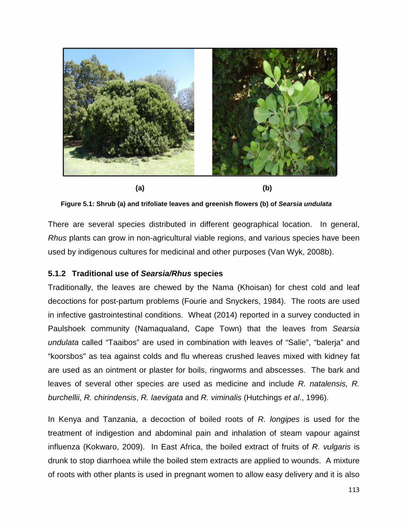
113
(a) (b)
Figure 5.1: Shrub (a) and trifoliate leaves and greenish flowers (b) of Searsia undulata
There are several species distributed in different geographical location. In general,
Rhus plants can grow in non-agricultural viable regions, and various species have been
used by indigenous cultures for medicinal and other purposes (Van Wyk, 2008b).
5.1.2 Traditional use of Searsia/Rhus species Traditionally, the leaves are chewed by the Nama (Khoisan) for chest cold and leaf
decoctions for post-partum problems (Fourie and Snyckers, 1984). The roots are used
in infective gastrointestinal conditions. Wheat (2014) reported in a survey conducted in
Paulshoek community (Namaqualand, Cape Town) that the leaves from Searsia
undulata called “Taaibos” are used in combination with leaves of “Salie”, “balerja” and
“koorsbos” as tea against colds and flu whereas crushed leaves mixed with kidney fat
are used as an ointment or plaster for boils, ringworms and abscesses. The bark and
leaves of several other species are used as medicine and include R. natalensis, R.
burchellii, R. chirindensis, R. laevigata and R. viminalis (Hutchings et al., 1996).
In Kenya and Tanzania, a decoction of boiled roots of R. longipes is used for the
treatment of indigestion and abdominal pain and inhalation of steam vapour against
influenza (Kokwaro, 2009). In East Africa, the boiled extract of fruits of R. vulgaris is
drunk to stop diarrhoea while the boiled stem extracts are applied to wounds. A mixture
of roots with other plants is used in pregnant women to allow easy delivery and it is also

114
used against infertility (Kokwaro, 2009) In China, the decoction of all parts of R.
chinensis is used for treatment of diabetes mellitus; a decoction from seeds is used for
malaria and rheumatism (Djakpo and Yao, 2010; Duke, 1992). Dried fruits of R.
chinensis are used as infusion to treat diarrhoea, aching gums, toothaches and swollen
legs whereas its boiled leaves are used in peptic ulcers, kidney stones, eruptions and
bruises (Maina, 2011).
In Turkey, the leaf extract of R. chinensis is used in the treatment of animal bites. In
North America and India, R. glabra is used for the treatment of bacterial diseases such
as gonorrhea, urinary tract infections, dysentery, internal and external ulcers and
sunburn (Borchardt et al., 2008; Hostettmann, 1999; Winston, 2001). This plant is also
used to alleviate hot flashes symptoms (Winston, 2001); the American Indians smoke
sumac leaves for asthma (Hutton, 2010). The dried fruits ground with salt of R. coriaria
are used in the Mediterranean and Middle East as a spice and for wound healing
(Rayne and Mazza, 2007; Shabbir, 2012). In Tanzania the extract of pounded roots of
R. natalensis steeped in hot or cold water are used for influenza, abdominal pains,
hookworm infestation and gonorrhea; the leaves are used against cough; steam from
leaves macerated in hot water are inhaled to fight colds (Maina, 2011).
5.1.3 In vitro biological activities Research undertaken on Rhus extracts to date indicates a promising potential for this
genus to provide renewable bioproducts with beneficial bioactivities such as
antimicrobial, antifungal, antiviral, antimalarial, antioxidant, antifibrogenic, anti-
inflammatory, antimutagenic, antithrombin, antitumorigenic, cytotoxic, hypoglycaemic
and leukopenic (Maina, 2011; Rayne and Mazza, 2007)
Much work has been done on the fruits of R. coriaria, a spice, which have demonstrated
potential antimicrobial and antifungal properties (Ali-Shtayeh et al., 2013; Bonjar, 2004;
Gabr et al., 2014; Gabr and Alghadir, 2015). These properties appear to be ascribed to
polar compounds extractable with protic solvents. The stem bark methanol extract of R
quartiniana was found to have antibacterial activity against gonorrhoea (Neisseria
gonorrhea) and Staphylococcus aureus; anthocyanins, coumarins, essential oils,

115
flavonoids, quinines, saponins, sterols and tannins have been isolated from this plant
(Chhabra et al., 1987). Biological activities of other Searsia species have been reported
(Table 4.1 previous chapter).
5.1.4 Isolation of compounds In Africa, there is no report on isolation of compounds from Searsia undulata with
antimycobacterial activity, but in general, antimicrobial properties from other species of
the genus Searsia (Rhus) have been reported elsewhere. The favourable worldwide
distribution of Searsia also suggests that desirable bio-products may be obtained locally
with minimal transportation requirements from processing to consumer. However, not
all the species studied to date have been fully characterized for isolation of potential
antimycobacterial activity. The genus Searsia (formerly Rhus) is of great interest since
the members have been found to possess chemical compounds that are toxic to micro-
organisms. These compounds include flavonoids, bioflavonoids, isoquinolines and
urushiols among others (Ahmed et al., 2001; Chhabra et al., 1987; Johns et al., 1990;
Lin et al., 1999; Sebothoma, 2009; Songca et al., 2012; Wang et al., 2008).
Bioassay-guided fractionation has been used for many years to isolate bioactive natural
products. The procedure involves repetitive fractionation of extracts and determining
the biological activity of all the fractions (Brusotti et al., 2014). The method provides a
rational means to isolate bioactive compounds from a complex mixture even though it is
consuming and labour intensive (Bero et al., 2011; Manvar et al., 2012). It has also
been found that the bioactive components can be extracted from Searsia using
environmentally benign solvents that allow for both food and industrial end-uses.
Indeed, antimicrobial compounds active against Gram-positive and Gram–negative
bacteria have been isolated from the methanol extract of Rhus glabra, a species used in
folk medicine in North America. Bioassay-guided fractionation led to isolation of three
antibacterial compounds namely methyl ester of 3, 4, 5-trihydroxybenzoic acid (methyl
gallate), 4-methoxy-3,5-dihydroxybenzoic acid and gallic acid (Saxena et al., 1994). A
flavone, apigenin dimethylether extracted from the roots of S. undulata, was reported to
have anti-inflammatory properties (Fourie and Snyckers, 1984); the root methanol
116
extract from Rhus succedonea has been reported to contain compounds such as
agathisflavone, robustaflavone, hinokiflavone, volkensiflavone, rhusflavone,
succedaneflavone and their methylethers and acetates which were evaluated for
antiviral activities. Robustaflavone demonstrated strong inhibitory effects against
influenza A and B viruses (Lin et al., 1999); the same activity was detected with
agathisflavone and amentoflavone which also exhibited moderate anti-Herpes Simplex
virus (HSV)-1 and anti-Herpes Simplex Virus (HSV)-2 activities. Anti HIV-1 reverse
transcriptase activity has been observed with robustaflavone, hinokiflavone and
morelloflavone (Lin et al., 1999).
Maina (2011) has reported on bioassay-guided antibacterial activity (using ATCC
cultures and clinical isolates of Gram-positive and Gram-negative bacteria) of extracts
from root bark, stem bark and leaves of Rhus natalensis from which the following
compounds were isolated: rhuschromone, chamaechromone, lophirone A, calodenone,
rhuschalone II and catechol.
5.1.5 In vivo experiments The National Institute of Health of the United States under the screening programme for
new antitubercular drugs recommends that all compounds, potentially antitubercular, be
evaluated in both in vitro and in vivo models. Animal models for in vivo evaluation of
antimycobacterial activity and toxicity of compounds have been reported (Jiménez-
Arellanes et al., 2013; Nguta et al., 2016; Villar et al., 2008). There is no report on the
in vivo evaluation of antimycobacterial activity/toxicity of Searsia undulata.

117
5.2 MATERIALS AND METHODS
5.2.1 Plant collection and storage The leaves of Searsia undulata were collected during summer (Figure 5.1.a) in
November 2013 at the National Botanical Garden in Pretoria, South Africa.
Coordinates: 25°44´S, 28°16´E. The tree was identified and labelled in the botanical
garden. A voucher number was issued by the University of Pretoria herbarium (PRU)
HGWJ Schweickerdt Herbarium, Department of Plant Sciences, where all research
specimens are kept. The leaves were air dried at room temperature suspended in bags
of net material in a ventilated room (Figure 5.2.b), ground to a fine powder in a
Macsalab Mill (Model 2000 LAB Eriez®) (Figure 5.3) and stored in closed jars away
from light in the cupboards until used.
(a) (b)
Figure 5.2: Plant collection (a) and air drying of leaves (b)

118
Figure 5.3: Grinding processes of dried leaves of Searsia undulata using a Macsalab Mill
5.2.2 Fractionation of bioactive fractions
5.2.2.1 Bulk extraction and solvent to solvent fractionation Powder of Searsia undulata leaves (1.5 kg) was serially extracted (3 × 4.5 L each) with
acetone (Ac). In the first round, the content was left to stand overnight and mixed
vigorously on a Labotec shaking machine for 3 h. This process was repeated five times
until the colour of the extract decreased. Each time the mixture was vigorously shaken
for 2 h on the shaking machine and the extract was filtered using Whatman No. 1 filter
paper. For each round, the collected filtrate of the extract was added into a round-
bottomed flask and dried using a Büchi R-114 rotary evaporator (Labotec) under
reduced pressure (Figure 5.4). The concentrated extract was then poured into a pre-
weighed glass beaker and left to dry under a stream of cold air from a wall fan.

119
Figure 5.4: Crude extract acetone-bulk extraction processes (left) and drying process with a rotary evaporator (right)
A total amount of crude acetone extract (bulk extraction) was obtained (216.06 g).
Eighty grams out of 216.06 g extract was taken aside and the rest stored in the fridge.
The extract was further processed by fractionating components based on polarity as
described (Suffness and Douros, 1979) with minor modifications. The extract (80 g)
was dissolved in a 1:1 mixture of chloroform and water and the two phases were
separated in a separatory funnel to obtain the chloroform and water fractions. The
water fraction was then mixed with an equal volume of n-butanol in a separatory funnel
to yield the water and n-butanol fractions (polar).
On the other hand, the non-polar part included the chloroform fraction which was dried
in a round-bottomed flask in a vacuum rotary evaporator and extracted with an equal
volume of hexane and 35% of water: methanol mixture to yield the n-hexane fraction.
Then the chloroform fraction was mixed with 35% water: methanol to yield chloroform
and 35% water: methanol fractions.
A bioassay-guided fractionation procedure was followed; collected main fractions (from
solvent-solvent fractionation) were tested against rapidly growing mycobacteria and
slow growing mycobacteria (pathogenic) to identify the active fractions that will be used
for further fractionation. The chloroform fraction, one of the active main fractions, was
further processed for fractionation using silica gel columns (Figure 5.5).

120
5.2.2.2 Column chromatography (CC) The isolation of the bioactive compounds was carried out using the chloroform fraction
with open mobile column chromatography and silica gel as the stationary phase (Figure
5.5). The silica gel powder (90.6 g) was mixed with an appropriate volume of n-hexane
in a glass beaker in a sonicator and stirred until all bubbles disappeared. The slurry
mixture was then poured into a column (50 cm height, 4.5 cm diameter). Extract
sample was prepared the previous day: the 9.06 g of chloroform fraction was dissolved
in 30 mL of acetone and mixed with 20 g of silica gel and the mixture was allowed to dry
under cold air overnight. A filter paper (Whatman no. 1) was cut to fit the inside
diameter of the column and was put on top of the silica gel followed by the dried
chloroform fraction sample then covered with another filter paper (Figure 5.5).
The solvent system used to elute the different fractions was hexane/ethyl acetate, ethyl
acetate/methanol. The sample loaded was adsorbed and eluted starting with 100% n-
hexane with an increasing amount of ethyl acetate at an interval of 10% until 100% ethyl
acetate at the following ratios (90:10), (80:20), (70:30), (60:40), (50:50), (40:60),
(30:70), (20:80), (10:90) and finally 100% ethyl acetate; this was followed by 250 mL
volumes of EtOAc-MeOH mixtures using similar ratios until (50:50), then small amounts
of MeOH were added in the mixture with EtOAc up to 15% Me(OH) (98.5:2.5), (95:5),
92.5:7.5, 90:10, 95:15). The column was washed with 100% MeOH and the rest of the
fractions collected, dried and kept in the fridge. This was done gradually to yield
different fractions which were collected into honey jars (250 mL). All the fractions were
left overnight to dry in a fume cabinet under a stream of cold air.

121
Figure 5.5: Column chromatography used for fractionation
The sub-fractions (F5, F6 and F7) from the chloroform fraction showing the presence of
antimycobacterial phytochemical compounds were combined (Figure 5.13-A) and
fractionated on a smaller column by gravity elution using hexane and ethyl acetate as
solvent system as described above but using a much smaller gradient of 2% to separate
compounds of interest. Fractions collected were analysed on TLC and three
compounds were isolated. Later on, more of the chloroform fraction stock (33 g and
140 g) was processed as described above; targeting the same fractions based on Rf
values, bioautography and TLC fingerprint patterns with the aim of collecting a larger
amount of the compounds of interest for further analysis.
5.2.3 Thin layer chromatography (TLC) fingerprinting and bioautography of fractions and compounds
The composition of fractions and compounds was determined by TLC using aluminium-
backed sheets of 20 X 20 cm (TLC Silica gel 60 F254, Merck, Germany). Ten microliters
from 10 mg/mL of selected fractions dissolved in acetone (containing 100 μg of extract)
were loaded on TLC plates in lines of about 1 cm wide using a micropipette. The lines
were allowed to dry at room temperature in the dark. Mobile phases of varying
polarities were used to develop the plates, namely hexane/ethyl acetate at different

122
ratios [6:4], [7:3], [8:2], [8.5:1.5], [8.8:1.2], chloroform/ethyl acetate/formic acid (CEF)
[10:8:2] and benzene/ethanol/ammonia (BEA) [18:2:0.2] (Kotzé and Eloff, 2002). The
above procedure, using the crude extracts of Searsia undulata, has been reported in
Chapter 4 in section 4.2.3. More than ten compounds of different polarities were
observed. These compounds were very close to each other in both the solvent systems
BEA and CEF. Active compounds included both polar and non-polar compounds based
on the Rf values determined on bioautograms. In the non-polar solvent BEA, the non-
polar compounds travelled some distance up the plate with clear zone of inhibition on
plates sprayed with different mycobacteria species. The intermediate solvent system
CEF led to a better separation of some compounds (Figure 4.3 previous chapter).
For bioautography, only the rapidly growing organisms were used, namely M. aurum, M.
fortuitum and M. smegmatis, because the pathogenic mycobacteria present a high
biohazard risk if handled as a spray. Mycobacteria used were cultured and maintained
as described in Chapter 4 under section 4.2.2.3. Duplicate chromatograms prepared as
described above were left uncovered in a dark place and air-dried until solvents were
completely evaporated for 24 h or overnight. A saturated bacterial culture was
centrifuged at 3000 x g for 10 min and the pellet resuspended in fresh appropriate liquid
growth medium and diluted to a McFarland No. 1. The chromatograms were then
sprayed with active bacterial suspension (18 to 72 h old cultures depending on the
growth rate of the particular Mycobacterium species) until completely moist using a
glass spray gun linked to a membrane pressure pump. The moist plates were
incubated at 37°C in a humidified chamber under 100% relative humidity for 24 h for
Mycobacterium smegmatis, 48 h for M. fortuitum and 72 h for M. aurum. The plates
were then sprayed with 2 mg/mL of INT (Sigma-Aldrich, Johannesburg, South Africa) in
distilled water and incubated for a further 30 min to few h until the development of a
purple-red background. The emergence of purple-red colour resulting from the
reduction of INT (Iodonitrotetrazolium violet) into formazan indicated the presence of
viable microorganisms whereas clear zones against the purple-red background
indicated inhibition of mycobacterial growth indicating the Rf values of antimycobacterial
compounds separated on the TLC plates (Hamburger and Cordell, 1987).

123
5.2.4 Minimum inhibitory concentration (MIC) of Searsia undulata fractions and compounds
The method was carried out as described in Chapter 4, section 4.2.2.4. All the fractions
comprised a mixture of phytochemical constituents and were tested at 10 mg /mL
whereas the concentration for the purified compounds was 1 mg/mL. The MIC of
extracts, fractions and compounds was determined at each fractionation stage.
5.2.5 Cytotoxicity of compounds The cytotoxicity assay was carried out as described in Chapter 4, section 4.2.4 and
Vero monkey kidney cells were used (Figure 5.6).
(a) (b)
Figure 5.6: Normal Vero monkey kidney cells viewed under the microscope at magnifications of 10X (a) and 40X (b)
5.2.6 13C and 1H nuclear magnetic resonance (NMR)-procedures and structural elucidation of isolated compounds
5.2.6.1 Isolation and characterization of compounds Glass columns (20-25 mm diameter) wet packed with silica gel 60 (0.040-0.063 mm)
230-400 Mesh ASTM, Merck) were used for column chromatography, and elution was
done with hexane, ethyl acetate, MeOH and chloroform (CHCl3). Isolation of
compounds from the combined semi-purified fractions, F5, F6 and F7 (Figures 5.14-A to
5.15-B) was carried out using a small column packed with silica gel with 100% n-hexane
124
and fractions were collected into honey jars. Hexane is a non-polar solvent that extracts
mostly non-polar alkaloids, triterpenes, phenolics, flavonoids, steroids and volatile oils
(Adeshina et al., 2012).
5.2.6.2 Sample preparation of compounds Samples of compounds were submitted to the Department of Chemistry at the Council
for Scientific and Industrial Research (CSIR) and the University of Pretoria, South Africa
for structure elucidation.
5.2.6.2.1 Compound SLN1
The sample SLN1 was dissolved in 200 µL d6-acetone (500 µL ampoule, 99.99%
deuterated acetone, C3D6O, Merck) and immediately transferred to a new 7″ Norell
Select Series 3 mm i.d. NMR tube and sealed for analysis.
5.2.6.2.2 Compound PK-B
The sample was partially soluble in acetone; but soluble in DMSO-d6 which was then
used as a solvent. DMSO (200 µL) was used to dissolve the sample from the vial and
transferred immediately to a new 7″ Norell Select Series 3 mm i.d. NMR tube and
sealed for analysis.
5.2.6.3 NMR spectrometers and structure elucidation
5.2.6.3.1 Compound SLN1
Two spectrometers were used to accomplish the acquisitions, namely a Varian
Premium Shield VNMRS triple resonance system (Direct Digital Receiver 1) operating
at 600 MHz, and a Varian INOVA dual band system operating at 400 MHz. The 600
MHz system was equipped with a 5 mm room temperature H{CN}probe, and was used
to acquire the proton spectrum of the material as well as all two dimensional spectra.
The 400 MHz system was equipped with a 3 mm room temperature dual resonance
indirect. Both systems were operated using Agilent VNMRJ 4.2A software.
Compounds at CSIR were measured on a Varian-600 NMR machine (Agilent
Technologies, California). Chemical shifts were recorded in σ (ppm). All spectra were
processed on ACD Labs Structure Elucidator 2015 software. All signals in both one-

125
and two dimensional spectra were individually hand-selected to ensure signal purity and
alignment.
5.2.6.3.2 Compound PKB
The mass spectrum was acquired by Synapt G2 high definition mass spectrometry in
positive ESI mode. The NMR spectra were recorded on a 400 MHz Bruker AVANCEIII
NMR spectrometer at 25oC. The chemical shifts from 1H NMR and 13C NMR spectra are
reported in parts per million relative to the solvent residual peak. The chemical shifts
used for referencing 1H and 13C Spectra were 2.50 ppm and 39.5 ppm, respectively.
The coupling constants (J) were measured in Hz. Abbreviations used for the multiplicity
of signals are as follows: singlet (s), doublet (d) and broadened (br).
5.3 RESULTS AND DISCUSSION
5.3.1 Bulk extraction, fractionation, thin layer chromatography and bioautography
5.3.1.1 Extraction yield, thin layer chromatography and bioautography The 1.5 kg finely ground S. undulata leaves yielded 216.06 g of crude acetone extract
(Figure 5.7). Eighty grams out of 216.06 g was processed using solvent-solvent
fractionation and yielded five fractions, namely chloroform, hexane, 35% water:
methanol, butanol and water based on polarity which varied widely (Figure 5.8). The
highest mass was obtained in the intermediate polarity chloroform fraction (53.27 g) with
only 0.34 g in the non-polar hexane fraction. The butanol fraction contained 9.2 g, the
water fraction 3.75 g and the 35% H2O in methanol 0.34 g. more polar (intermediate
polarity). The yield of each fraction obtained after solvent-solvent fractionation of the
bulk acetone extract (80 g) is presented in Figure 5.8.
From the 80 g of acetone crude extract processed, the total mass recovered was 66.9 g
which means about 84% recovery. The 16% loss might have been due to the presence
of pellicles that could not be separated and to the sticky, oily nature of the extract which
was not easy to recover during solvent-solvent fractionation; the oily supernatant
observed when separating hexane from chloroform fractions was filtered out. The

126
percentage of recovery for each fraction was respectively 79.6% for chloroform, 13.75%
for butanol, 5.6% for water and 0.51% for hexane and 35%H2O/methanol. With 0.5 %
of recovery for the non-polar fraction (hexane) and 80% of intermediate polarity
(chloroform), the results from the different solvent systems showed compounds of
intermediate polarity and variable active compounds from non-polar to polar. The TLC
fingerprint of the butanol fraction did not contain visible compounds after vanillin spray
and colour development but some compounds were visible under UV light and those
compounds were more visible on bioautograms (Figures 5.9-5.11-C).
Initially 55 fractions were collected and analysed on TLC plates using hexane: ethyl-
acetate at ratio 7:3 and 6:4, fractions with similar profiles were combined into 12 sub
fractions (F1-F12) (Figures 5.7, 5.12-5.13). Fractions collected with 100% methanol are
not included in figure 5.7 as they had no antimycobacterial activity.

127
Figure 5.7: Schematic extraction and fractionation results towards compound isolation from Searsia undulata
1.5 Kg Acetone bulk extraction
216.06 g
Solvent to solvent fractionation
80g
Chloroform 53.27 g
Hexane 0.34 g
35% H2O 0.34 g
n-butanol 9.2 g
H2O 3.75 g
Column bed silica gel 9060 mg
F1 417 mg
F2 187 mg
F3 421.3 mg
F4 479.4 mg
F5 295.2 mg
F6 280.8 mg
F7 443.7 mg
F8 504 mg
F9 1204 mg
F10 524 mg
F11 360 mg
F12 962 mg
55 fractions were initially collected and based on TLC profiles; they were combined into 12 sub fractions (F1-F12)

128
Figure 5.8: Mass (g) of the five solvent-solvent fractions obtained from 80 g of S. undulata leaf extracts
0
10
20
30
40
50
60
Butanol Chloroform Hexane Water
Mass (g) of five solvent fractions of SU
35% H2O

129
CHL BUT-BEA CHL_HE: E 60:40 MA-CHL; MA-BUT MF-CHL; MF-BUT MS-CHL; MS-BUT
Figure 5.9: Chloroform and butanol fractions- TLC fingerprinting and bioautograms of plates eluted in BEA and sprayed with rapidly growing mycobacteria spp. CHL: Chloroform, BUT: Butanol, HE: Hexane, E: Ethyl acetate, MA: M aurum, MF: M fortuitum, MS: M smegmatis
The solvent-solvent fractions with high yield, good MIC values and presence of
antimycobacterial compounds in bioautograms were selected (chloroform, butanol and
35% water fractions) and tested further to determine antimycobacterial activity against
M. aurum, M. fortuitum and M. smegmatis (Figures 5.10 A - 5.11 C). Antimycobacterial
activity was observed for all three mycobacteria although MIC values were different.
The difference in MIC may be due to the difference in concentration of inactive
compounds in different fractions or to synergism. Based on the bioautograms the most
active compounds observed were non-polar and did not separate well. The chloroform
fraction was selected for further work and fractionated by silica gel column
chromatography then sequentially eluted with increasing polarity of n-hexane/ethyl
acetate mixture to yield 55 fractions. The chemical composition was determined by TLC
and fractions were combined based on similar composition (Figures 5.10 - A, 5.11 - A.1)
and bioautograms (Figures 5.12-5.13). The results indicated that most compounds
were non polar; indeed a better compound separation was observed in the BEA solvent
separation system.

130
5.3.1.2 TLC fingerprinting and bioautography of fractions and compounds
5.3.1.2.1 Solvent-solvent fractions
The five fractions were eluted in BEA solvent system and showed the presence of
similar compounds. The acetone crude extracts (ACE) showed similar compounds
when compared to n-hexane (HE) and chloroform (CHA, CHB) fraction. The chloroform
and hexane fraction profiles were more similar showing a better separation. The more
polar water fraction, 35% water fraction in methanol (35% H2O), did not separate well in
the non-polar eluent system as expected. But, this fraction showed two compounds
similar to those present in the n-hexane and chloroform fractions. The n-butanol and
water fractions did not show any visible compounds. The water fraction was not further
processed as no compounds were observed on vanillin-sprayed TLC plates and
bioautogram; the hexane fraction was similar to chloroform fraction. The butanol
fraction was tested and showed clear zones of inhibitions on bioautograms although
there were almost no visible compounds on vanillin-sprayed TLC plates eluted in the
same solvent system BEA (Figures 5.9 - 5.11-A1).

131
Figure 5.10-A: Bioautogram of M. aurum eluted in BEA (Benzene/ethanol/ammonia at ratio 18:2:0.2) separated in triplicate (CHL) and sprayed with 2 mg/mL of INT (Iodonitrotetrazolium violet) showing solvent- solvent fractions of chloroform (CHL), butanol (BUT) and 35% water (H2O) of SU
M. aurum-BEA
CHL CHL CHL BUT BUT 35%H2O
Chloroform fraction selected for compound
isolation
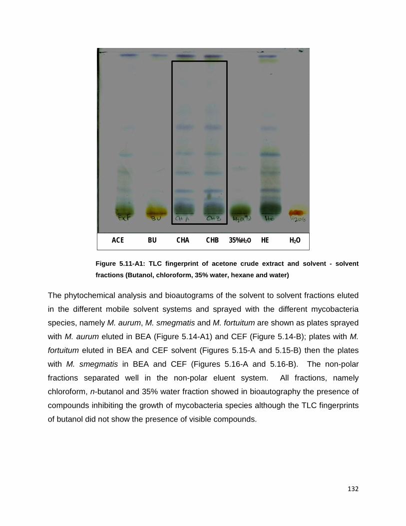
132
Figure 5.11-A1: TLC fingerprint of acetone crude extract and solvent - solvent fractions (Butanol, chloroform, 35% water, hexane and water)
The phytochemical analysis and bioautograms of the solvent to solvent fractions eluted
in the different mobile solvent systems and sprayed with the different mycobacteria
species, namely M. aurum, M. smegmatis and M. fortuitum are shown as plates sprayed
with M. aurum eluted in BEA (Figure 5.14-A1) and CEF (Figure 5.14-B); plates with M.
fortuitum eluted in BEA and CEF solvent (Figures 5.15-A and 5.15-B) then the plates
with M. smegmatis in BEA and CEF (Figures 5.16-A and 5.16-B). The non-polar
fractions separated well in the non-polar eluent system. All fractions, namely
chloroform, n-butanol and 35% water fraction showed in bioautography the presence of
compounds inhibiting the growth of mycobacteria species although the TLC fingerprints
of butanol did not show the presence of visible compounds.
ACE BU CHA CHB 35%H2O HE H2O

133
Figure 5.11-B: Bioautogram plate eluted in BEA (Benzene/ethanol/ammonia at ratio 18:1:0.2) and sprayed with M. smegmatis followed by INT indicating antimycobacterial compounds of solvent-solvent fractions CHL: chloroform, BUT: butanol and 35% H2O: 35% water in methanol of SU. Clear zones indicate mycobacterial growth inhibition
M. smegmatis-BEA
CHL CHL CHL BUT BUT 35%H2O

134
Figure 5.11-C: Bioautogram plate eluted in BEA (Benzene/ethanol/ammonia at ratio 18:2:0.2) and sprayed with M. fortuitum and followed by INT indicating antimycobacterial compounds of solvent-solvent fractions CHL: chloroform, BUT: n-butanol and 35%H2O: 35% water in methanol of SU. Clear zones indicate mycobacterial growth inhibition
The isolation of the compound was bioassay-guided based on the presence of
antimycobacterial activity observed as clear, white zones of inhibition on the
bioautograms at each fractionation stage. Most compounds were non-polar and close
to each other which made the process of separation laborious (Figures 5.10 A – 5.11
C).
Based on the high yield of the chloroform fraction (53.27 g) and strong
antimycobacterial activity observed on the bioautography and MIC values, the
chloroform fraction was selected. An amount of 9.06 g was used for further
fractionation towards isolation of the compound in an open column on silica gel. Fifty
five fractions were collected in different jars at the initial fractionation; these fractions
were combined into 12 fractions (F1-F12) and tested as previously described with M.
aurum and M. smegmatis (Figures 5.12, 5.13, 5.14 and 5.16).
M. fortuitum-BEA
CHL CHL CHL BUT BUT 35%H2O

135
Figure 5.12: Fractions (n=55) collected from the chloroform fraction were combined, tested and based on clear zone of inhibition pattern against M. smegmatis, combined into 12 semi-purified fractions (F1-F12) that were separated further for compound isolation
1-6 7-12 17-19 20 22 24 26 28 30 31-33 34-39 45-49 51-55
M. smegmatis-BEA
Initial combined 55 fractions into 12 fractions

136
M. aurum-BEA
1-6 7-12 17-19 20 22 24 26 28 30 31-33 34-39 45-49 51-55
Initial combined 55 fractions into 12 fractions
Figure 5.13: Fractions (n=55) collected from the chloroform fraction were combined, tested and based on clear zone of inhibition pattern against M. aurum, combined into 12 semi-purified fractions (F1-F12) that
were separated further for compound isolation

137
5.3.1.2.2 TLC fingerprinting and bioautograms of sub-fractions
The chloroform fraction (9.06 g) yielded 12 fractions amounting to 6.0784 g which is
about 67% recovery. The percentage of recovery below 80% represented the portion of
fractions containing most of the targeted antimycobacterial compounds as shown on the
different figures, Rf and MIC values. Three out of the 12 sub-fractions, F5, F6 and F7,
represented 16% of the collected sub-fractions (1.0197 g) (Figure 5.7). The sub-
fractions F5 to F7 showed similar consistent patterns of strong clear zones of inhibition
against all mycobacteria species tested - M. aurum, M. smegmatis and M. fortuitum - on
plates eluted in BEA and CEF (Figures 5.14 to 5.16). Adding to good MIC values
obtained, they were combined and processed further for the preliminary isolation of
some of the compounds (Figures 5-17, 5.18-5.19).

138
Figure 5.14-A: TLC fingerprints of the 12 pooled fractions obtained from the chloroform fraction; plate was eluted in BEA (Benzene/ethanol/ammonia at ratio 18:2:0.2). F5 to F7 fractions targeted for compound isolation
F1 F2 F3 F4 F5 F6 F7 F8 F9 F10 F11 F12
Combined fractions targeted for compound isolation
BEA
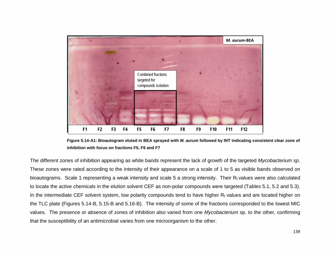
139
Figure 5.14-A1: Bioautogram eluted in BEA sprayed with M. aurum followed by INT indicating consistent clear zone of inhibition with focus on fractions F5, F6 and F7
The different zones of inhibition appearing as white bands represent the lack of growth of the targeted Mycobacterium sp.
These zones were rated according to the intensity of their appearance on a scale of 1 to 5 as visible bands observed on
bioautograms. Scale 1 representing a weak intensity and scale 5 a strong intensity. Their Rf values were also calculated
to locate the active chemicals in the elution solvent CEF as non-polar compounds were targeted (Tables 5.1, 5.2 and 5.3).
In the intermediate CEF solvent system, low polarity compounds tend to have higher Rf values and are located higher on
the TLC plate (Figures 5.14-B, 5.15-B and 5.16-B). The intensity of some of the fractions corresponded to the lowest MIC
values. The presence or absence of zones of inhibition also varied from one Mycobacterium sp. to the other, confirming
that the susceptibility of an antimicrobial varies from one microorganism to the other.
F1 F2 F3 F4 F5 F6 F7 F8 F9 F10 F11 F12
M. aurum-BEA
Combined fractions targeted for compounds isolation

140
(a) (b)
F5 F6 F7 F5 F6 F7
Figure 5.14-A2: Vanillin sprayed TLC plate and bioautogram plate eluted in BEA (Benzene/ethanol/ammonia at ratio 18:2:02) sprayed with M. aurum followed by INT indicating the three pooled fractions (F5, F6 and F7) that were separated further for isolation of compounds indicating the closeness of the active compounds

141
Table 5.1: The intensity of the zones of inhibition on a five point scale and their Rf values as observed in Figure 5.14-B
Rated Intensity of zone of inhibition against M. aurum on bioautography and Rf values in CEF solvent Rf values F4 F5 F6 F7 F8 F9 F10 F11 F12
0.95 3 2 2 2 0.90-0.92 5 5 4 5 5 5 2
0.88-0.89
3 2 5
4 2 0.85-0.87
5 5 4 4
0.81-0.83
4 3 2 3 5 4 5 5 0.8
0.71-0.73
5 0.68-0.69 1 2 2 1
4 5 5
0.64-0.67
5 5
5 0.61

142
Figure 5.14-B: Bioautogram eluted in CEF (Chloroform/ethyl acetate/formic acid at ratio 10:8:2) sprayed with M. aurum followed by INT indicating 12 pooled fractions from the chloroform with consistent clear zone of inhibition focusing on fractions F5, F6 and F7. The clear zones indicate mycobacterial inhibition growth
M. aurum-CEF
F1 F2 F3 F4 F5 F6 F7 F8 F9 F10 F11 F12

143
Figure 5.15-A: Bioautogram eluted in BEA sprayed with M. fortuitum followed by INT indicating 12 pooled fractions from chloroform with zones of growth inhibition focusing on fractions F5, F6 and F7
F1 F2 F3 F4 F5 F6 F7 F8 F9 F10 F11 F12
M. fortuitum-BEA
Combined fractions targeted for compounds isolation

144
Table 5.2: The intensity of the zones of inhibition on a five point scale and their Rf values as observed in Figure 5.15-B
Rated Intensity of zone of inhibition against M. fortuitum on bioautography and Rf values in CEF solvent
Rf values F4 F5 F6 F7 F8 F9 F10 F11 F12 0.95
0.90-0.92 1 1
5 0.88-0.89 2 5 5 5 5 5
0.85-0.87
5 0.81-0.83 2 4 3
3
0.8
5 0.71-0.73
2
4 1 1
0.68-0.69
1 2 0.64-0.67 2 3 2
5 4
0.61

145
Figure 5.15-B: Bioautogram eluted in CEF and sprayed with M. fortuitum followed by INT indicating 12 pooled fractions from chloroform with zones of growth inhibition focusing on fractions F5, F6 and F7
M. fortuitum-CEF
F1 F2 F3 F4 F5 F6 F7 F8 F9 F10 F11 F12

146
Figure 5.16-A: Bioautogram eluted in BEA and sprayed with M. smegmatis followed by INT indicating 12 pooled fractions from chloroform with zones of growth focusing on fractions F5, F6 and F7
F1 F2 F3 F4 F5 F6 F7 F8 F9 F10 F11 F12
M. smegmatis-BEA
Combined fractions targeted for compounds isolation

147
Table 5.3: The intensity of the zones of inhibition on a five point scale and their Rf values as observed in Figure 5.16-B
Rated Intensity of zone of inhibition against M. smegmatis on bioautography and Rf values in CEF solvent
Rf values F4 F5 F6 F7 F8 F9 F10 F11 F12 0.95
0.90-0.92 3 1 0.88-0.89
0.85-0.87
5 5 5 5 0.81-0.83 4
2 2
0.8
2 0.71-0.73
1
0.68-0.69 0.64-0.67
2 2 1 0.61 4 4 3

148
Figure 5.16-B: Bioautogram eluted in CEF and sprayed with M. smegmatis followed by INT indicating zones of growth inhibition focusing on fractions F5, F6 and F7
Two plates were eluted in the BEA solvent for each fraction and sprayed, one with vanillin and the other with the
mycobacterial suspension broth of Mycobacterium aurum. The plates were placed next to each other to visualize the
compounds inhibiting the growth of mycobacteria appearing as white zones against a reddish background indicating the
growth of the mycobacteria. Prior to spraying the TLC plates with either vanillin or mycobacteria, they were observed
under UV light at 254 nm and 366 nm to visualize any fluorescent compounds invisible to the naked eye. Two fluorescent
compounds were observed (Figure 5.17). Fungal contamination during the period of this research was also a challenge
which affected the quality of some plates as dark pinpoints or stains, but without jeopardizing the results.
F1 F2 F3 F4 F5 F6 F7 F8 F9 F10 F11 F12
Combined fractions targeted for compounds isolation
M. smegmatis-CEF

149
Figure 5.17: TLC fingerprints and bioautograms showing the different stages of isolation from the acetone crude extract to fractions of Searsia undulata. Plates were eluted in BEA, TLC plates were sprayed with vanillin and bioautograms with Mycobacterium aurum (which was one of the best indicators of mycobacterial activity) followed by INT. ACE: Acetone crude extract; CHL: Chloroform fraction; F5-F7: sub-fractions obtained from 55 fractions from the chloroform fraction
Visible under UV
ACE ACE CHL CHL F5 F6 F7 F5 F6 F7

150
5.3.1.2.3 Sub-fractions and compounds
Based on the MIC, TLC fingerprints, bioautograms and total activity of each fraction,
three fractions were selected (F5, F6 and F7) to target compounds with the best
antimycobacterial activity (Figures 5.14 - 5.16). Two fluorescing compounds were
observed under UV light at 366 nm wavelength (Figure 5.17) but did not show activity
on bioautography and were not targeted for isolation during the period of this study.
The three sub-fractions (F5-F7) showed strong clear zones of inhibition and they were
compared to crude extracts, solvent to solvent fractions and compounds. Observation
on the separation of the fractions of interest from crude extract to isolation of
compounds is easy to follow (Figures 5.17-5.18). The bioautogram plate was sprayed
with M. aurum which was one of the best indicators of antimycobacterial activity. The
chloroform fraction showed a clear zone of inhibition of a compound that is more polar
and this compound seemed not to show inhibition when fractionated further. This could
be explained that further separation did take place and the fraction lost the active
portion as the synergistic interaction of compounds within the fractions might be lost
during the separation process. Based on Rf values, the same compounds were present
in all the fractions.
The TLC fingerprint of the chloroform fraction indicated the presence of more than 20
potential fractions whereas the crude extract showed about 10 visible fractions, which
confirms that the process of fractionating was indeed taking place. In general, active
fractions were close to each other as shown on all the profiles (Figures 5.9 - 5.11). The
isolated compounds were only tested using the MIC assay and for cytotoxicity. More
work is still to be done which includes testing against pathogenic mycobacteria, synergy
with current standard anti-TB drugs and isolation and identification of more compounds
while assessing the stability of fractions and compounds in different environmental
conditions. Some of the fractions deposited crystals on standing while others remained
in the form of an oily paste. The bioactivity of the crude acetone extract of Searsia
undulata has been reported in Chapter 4, but results of the crude extract are shown and
used to discuss results obtained from the fractions and compounds (Figure 5.18). The
55 fractions obtained from the chloroform fraction of SU were tested against M. aurum,

151
M. fortuitum and M. smegmatis and their antimycobacterial activity determined on
bioautography observed as clear zones of inhibition (white zones). The results revealed
that fractions 5, 6, and 7 (F5, F6 and F7) had the consistent inhibitory activity against all
three mycobacteria species and indicated similar compounds. Other fractions
contained one or two active compounds that were not consistent with the different
intensity of the clear zones of inhibition for all three tested mycobacteria. Observations
made on bioautograms eluted in BEA and sprayed with M. aurum revealed that F3 had
3 visible clear zones of inhibition which were also present in F4 but F4 showed two
visible active phytochemicals of which neither was present in F3 or F5, F6 and F7.
Fractions F8 to F12 showed a similar active phytochemical but did not show the same
compounds as observed in F5 to F7. F5, F6 and F7 were therefore selected for further
isolation based on consistency observed (Figures 5.14 – 5.18).
Compounds were eluted with 100% hexane with increasing polarity with 2% ethyl
acetate resulting in sequential elution of SLN1 (26.2 mg); PK-A1 (15.4 mg), PK-B (22.4
mg), PK-C (15.9 mg) and PK-D (8.4 mg). PK-B was obtained by eluting the compound
with a mixture of chloroform and methanol (Figures 5-18-5.19).

152
F5 F6 F7 F5 F6 F7 C1 C2 C3 C4 C5 C6
Compounds Sub-fractions from chloroform fraction that were combined and further separated for isolation of
compounds
Figure 5.18: TLC fingerprints and bioautograms indicating targeted fractions F5-F7 to compounds isolated from Searsia undulata. Bioautogram were eluted in BEA solvent systems and sprayed with Mycobacterium aurum which was one of the good indicators of mycobacterial activity. TLC plates for compounds were eluted in different ratios of hexane: ethyl acetate and sprayed with vanillin

153
Figure 5.19: TLC plate visualized under UV light (366 nm) showing the separation of compound B from compound C during the purification process
PKB
PKC
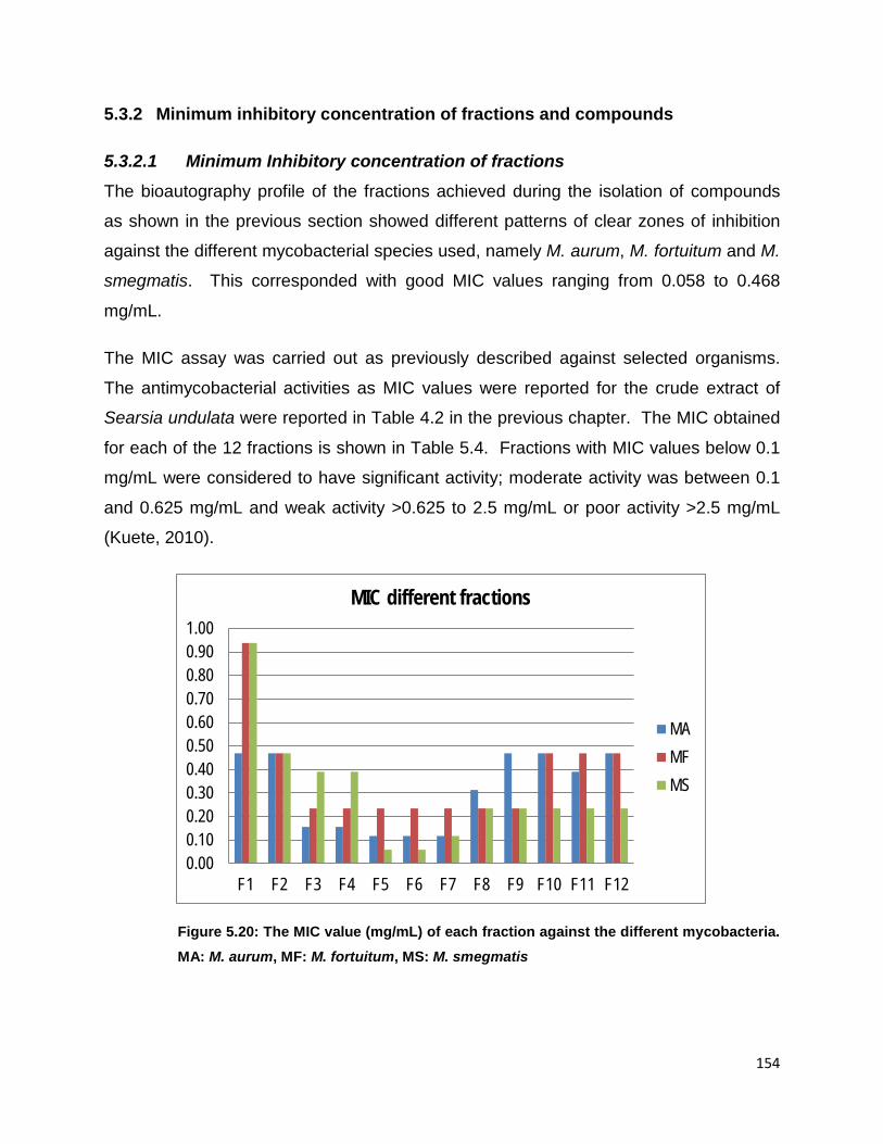
154
5.3.2 Minimum inhibitory concentration of fractions and compounds
5.3.2.1 Minimum Inhibitory concentration of fractions The bioautography profile of the fractions achieved during the isolation of compounds
as shown in the previous section showed different patterns of clear zones of inhibition
against the different mycobacterial species used, namely M. aurum, M. fortuitum and M.
smegmatis. This corresponded with good MIC values ranging from 0.058 to 0.468
mg/mL.
The MIC assay was carried out as previously described against selected organisms.
The antimycobacterial activities as MIC values were reported for the crude extract of
Searsia undulata were reported in Table 4.2 in the previous chapter. The MIC obtained
for each of the 12 fractions is shown in Table 5.4. Fractions with MIC values below 0.1
mg/mL were considered to have significant activity; moderate activity was between 0.1
and 0.625 mg/mL and weak activity >0.625 to 2.5 mg/mL or poor activity >2.5 mg/mL
(Kuete, 2010).
Figure 5.20: The MIC value (mg/mL) of each fraction against the different mycobacteria. MA: M. aurum, MF: M. fortuitum, MS: M. smegmatis
0.000.100.200.300.400.500.600.700.800.901.00
F1 F2 F3 F4 F5 F6 F7 F8 F9 F10 F11 F12
MIC different fractions
MAMFMS

155
Fractions that included main fractions obtained from solvent to solvent fractionation and
sub-fractions collected after elution of the chloroform fraction in the open column silica
gel chromatography had good to moderate activity that did not always correlate with the
presence of clear zones of inhibition on bioautograms.
The fractions F5, F6 and F7 had significant antimycobacterial activity showing similar
compounds and hence were selected for further column chromatography with MIC
values of 0.117 mg/mL for M. aurum, 0.234 mg/mL for M. fortuitum and 0.058 mg/mL for
M. smegmatis for all fractions except F7 with MIC value of 0.117 mg/mL for M.
smegmatis. Fraction F5 and F6 had the lowest MIC of 0.058 mg/mL observed against
M. smegmatis. The lowest value of MIC, the more effective is the fraction at a lower
dosage whereas 1/MIC represent the sensitivity of the organism tested, the highest the
value of 1/MIC, the more sensitive is the organism tested against the specific fraction.
The MIC values of each fraction against all three mycobacteria species, sensitivity of
each mycobacteria and the overall view of each fraction’s average activity is presented
in Figures 5.20, 5.21 and 5.22. Fractions F5, F6 and F7 were active against all three
mycobacteria species.
Figure 5.21: The 1/MIC values (mg/mL) of each fraction against the different mycobacteria species. MA: M. aurum, MF: M. fortuitum, MS: M. smegmatis
02468
1012141618
F1 F2 F3 F4 F5 F6 F7 F8 F9 F10 F11 F12
1/MIC different fractions
MA MF MS

156
Figure 5.22: The average 1/MIC values (mg/mL) of each fraction against all the different mycobacteria species. Fractions F5, F6 and F7 show consistent high activity
The total activity value of a fraction indicates which species could be candidates for organic production of active extracts for use by communities because it takes into account not only the MIC of the extract but also the yield of the fraction. By dividing the extraction yield in mg/g by the MIC in mg/mL the total activity is calculated in mL/g (Eloff, 2000) as represented in Table 5.4. Among all 12 fractions, F3 had the highest total activity on its own of 19 765 mL/g for M aurum followed by 13 162 mL/g for M. fortuitum and 7 896 mL/g for M. smegmatis. F3 also had active compounds and good MIC values but was not selected at this initial stage as it did not have a consistent similar pattern of phytochemical active compounds on bioautography when compared to F5 to F7 (Figures 5.14-5.18). In future investigations, the isolation of compounds focusing only on fraction F3 will be undertaken. The next fraction showing the highest activity was F6 with a total activity of 19 793 mL/g against M smegmatis followed by 9 897 mL/g for M. aurum and the lowest 4 943 mL/g for M. fortuitum then F5 showing the highest total activity of 17154 mL/g against M. smegmatis, followed by 8 577 mL/g for M. aurum and 4 284 mL/g for M. fortuitum then the last fraction with lower total activity F7 showing 7 262 mL/g for both M. aurum and M. smegmatis and 3 627 mL/g for M. fortuitum (Figure 5.24). The average total activity of the combined fractions was 27 599 mL/g which is higher than F3 alone at 19 765 mL/g (Figure 5.23). At this preliminary
0.01.02.03.04.05.06.07.08.0
0 1 2 3 4 5 6 7 8 9 10 11 12
1/MIC
mg/
mL
Fraction number
Average activity against all mycobacteria

157
stage, this determined the focus on the combination of these three fractions based on their MIC values against all the mycobacterial species.
Table 5.4 The MIC values and total activity of each fraction
Fractions Mass (mg) MIC (mg/mL)
Total activity (mL/g)
MA Ave MF Ave MS Ave MA MF MS average F1 2 309 0.47 0.94 0.94 4 926 2 463 2 463 3 284 F2 509 0.47 0.47 0.47 1,086 1 086 1 086 1 086 F3 3 083.3 0.16 0.23 0.39 19 765 13 162 7 896 13 608 F4 1 122.9 0.16 0.23 0.39 7 198 4 794 2 876 4 956 F5 1 003.5 0.12 0.23 0.06 8 577 4 284 17 154 10 005 F6 1 157.9 0.12 0.23 0.065 9 897 4 943 19 793 11 544 F7 849.6 0.12 0.23 0.12 7 262 3 627 7 262 6 050 F8 1 980.8 0.31 0.23 0.23 6 339 8 456 8 456 7 750 F9 1 165.7 0.47 0.23 0.23 2 487 4 976 4 976 4 146 F10 1 768.6 0.47 0.47 0.23 3 773 3 773 7 550 5 032 F11 858.5 0.39 0.47 0.23 2 198 1 831 3 665 2 565 F12 557 0.47 0.47 0.23 1 188 1 188 2 378 1 585

158
Figure 5.23: Total activity of different fractions (mL/g); Fractions F3 and F6 had the highest total activity for M. aurum and M. smegmatis respectively followed by fractions F5, F6 and F7 that had better total activity against all the mycobacteria which also correlated with the intensity of active fractions indicated by the clear zone of inhibition on bioautograms
0
5,000
10,000
15,000
20,000
25,000
F1 F2 F3 F4 F5 F6 F7 F8 F9 F10 F11 F12
Total activity per fraction mL
MAMFMS

159
Figure 5.24: The average total activity (mL/g) per fraction
The chloroform fraction revealed the presence of several potential active fractions on
the bioautogram sprayed with M. aurum whereas on the one sprayed with M. fortuitum
there were fewer active fractions. This is explained by the different sensitivity of
mycobacteria species towards the potential antimycobacterial phytochemical present in
the fractions. The Rf value, also called retardation factor, was used as a fingerprint to
trace and identify targeted fractions that had antimycobacterial activity at different
stages of the fractionation process. Rf values of the chloroform fraction were calculated
by dividing the distance travelled by either the extract or fraction or compound spotted
on the vanillin sprayed TLC plate or bioautogram eluted in a specific mobile phase
solvent system by the distance travelled by the solvent front. Rf values were calculated
for the plates eluted in BEA and sprayed with M. aurum or M. fortuitum as they were
good indicators of antimycobacterial activity in general (Figures 5.9-5.11). In Table 5.5,
the different values showed the closeness of some of the active fractions such as Rf =
0.21, 0.26 and 0.34 and 0.36. Different fractions that have travelled the same distance
0
2,000
4,000
6,000
8,000
10,000
12,000
14,000
16,000
F1 F2 F3 F4 F5 F6 F7 F8 F9 F10 F11 F12

160
could have the same Rf values, namely fractions with Rf values 0.34. The same applied
to the different fractions F1 to F12 that had the same Rf values.
Table 5.5 Rf values of different phytochemical bands observed on the chloroform fraction in BEA
M. aurum M. fortuitum Rf values
1 0.05 0.34
2 0.17 0.45 3 0.21 0.52 4 0.26 0.38 5 0.34
6 0.36 7 0.47 8 0.52 9 0.61
5.3.2.2 Minimum inhibitory concentration of compounds The antimycobacterial activity of the five compounds initially isolated yielded a variety of
results. Table 5.6 shows the MIC values of S. undulata compounds against different
organisms. Each value is the mean of three replications measured in µg/mL (test
samples of compounds were prepared at an initial concentration of 1 mg/mL). It is
worth noting that some of the compounds formed a jelly like substance when inoculated
in the growth medium. Therefore, the growth medium was diluted with ethyl acetate or
DMSO at different ratios and organisms’ growth was tested prior to carrying out the
process. All 5 compounds showed activity against rapidly growing mycobacteria with
MIC values ranging from 23.44 to 250 µg/mL. The first compounds 1 and 2 showed
activity with the highest MIC values varying from 62.5 to > 250 µg/mL. The MIC values
were 62.5 µg/mL for M. aurum and M. fortuitum and 125 µg/mL for M. smegmatis
whereas C2 showed the highest MIC value >250 µg/mL against M. fortuitum and M.
smegmatis. Compound 3 (SLN1) had the lowest MIC value of 23.44 µg/mL for M.
fortuitum; 31.25 µg/mL for M. aurum and M. smegmatis. Compound PKB and
compound 5 (C5 or PKC) both showed activity against all three mycobacteria with the
highest MIC of 46.88 µg/mL for C4 against M. smegmatis.

161
Table 5.6 MIC values of compounds isolated from Searsia undulata
MIC (µg/mL) and Mycobacterium species tested
M. aurum
M. fortuitum M. smegmatis
Compounds (µg/mL) C1 62.5
125
125
C2 62.5
>250
>250
C3-SLN1 31.25
23.44
31.25
C4-PKB 31.25
31.25
46.88 C5-PKC 31.25
31.25
31.25
5.3.3 Cytotoxicity
5.3.3.1 Crude extracts Reported in Chapter 4, under section 4.3.4
5.3.3.2 Compounds Only three compounds were evaluated at the time as there were in sufficient purified
quantity for further testing and structure elucidation - all three compounds had low
cytotoxicity with CP2 being the least toxic 210 µg/mL (Table 5.7). The 50% lethal
concentration (LC50) of the compound was determined by calculating the concentration
of compound that will keep half the cells viable relative to the untreated control. For
each compound a duplicate assay was conducted (Table 5.7). For the first compound 1
(C1), it was found that Y was 0.955 and 0.997 and LC50 27.50 and 35.23 µg/mL for
assays 1 and 2, respectively; for C2, Y was the same as for C1 with LC50 172.74 and
248.33 µg/mL for assays 1 and 2 and C3 LC50 48.59 and 46.94 for each of the assays.

162
Figure 5.25: LC50 of compound 1 where Y was 0.955 (A) and 0.997 (B)
Figure 5.26: LC50 of compound PKB where Y was 0.955 (A) and 0.997 (B)
Figure 5.27: LC50 of compound 1 where Y was 0.955 (A) and 0.997 (B)
y = -1.3089x + 2.839 R² = 0.9212
-0.20
0.20.40.60.8
11.21.41.61.8
0 1 2 3
Abs
570
nm
Log conc compound
C1-SLN-1 A y = -1.6586x + 3.5628
R² = 0.9586
-0.5
0
0.5
1
1.5
2
2.5
0 1 2 3
Abs
570
nm
Log conc compound
C1-SLN-1 B
y = -0.6142x + 2.3292 R² = 0.8555
00.20.40.60.8
11.21.41.61.8
2
0 1 2 3
Abs
570
nm
Log conc compound
C2-PKB y = -0.4706x + 2.1241
R² = 0.7678
00.20.40.60.8
11.21.41.61.8
0 1 2 3
Abs
570
nm
Log conc compound
C2-PKB
y = -1.4598x + 3.4371 R² = 0.8581
0
0.5
1
1.5
2
2.5
0 1 2 3
Abs
570
nm
Log conc compound
C3-PKC B y = -1.336x + 3.2082
R² = 0.8759
00.20.40.60.8
11.21.41.61.8
2
0 1 2 3
Abs
570
nm
Log conc compound
C3-PKC A

163
Table 5.7 Cytotoxicity of compounds on Vero Monkey Kidney cells
Assay 1 Assay 2 Average SD Compound 1 (µg/mL) 27.50 35.23 31.37 5.47 Compound 2 (µg/mL) 172.74 248.33 210.53 53.45 Compound 3 (µg/mL) 48.60 46.94 47.76 1.16 It is worth noting that the highest concentration of compound tested was 200 µg/mL and
compound 2 was the least toxic out of the three compounds (Table 5.7).
5.3.4 Structure elucidation and identification of compounds
5.3.4.1 SLN1 The initial inspection of the proton spectrum of the SLN1 compound revealed that it was
triterpenoid in nature, based on the presence of at least six methyl groups in the
spectrum. The compound was approximately 70% pure. Additionally, a pair of triplet-
like signals at δ 4.72 ppm and δ 4.59 ppm, integrating to a single proton each, pointed
to the presence of a terminal methylene system, likely a 1, 1-disubstituted alkene typical
of the lupane pentacyclic triterpenoids. The absence of one of the seven methyl signals
typically seen in the lupine systems indicated that one of these had been oxidized or
modified, typical of the betulin-type lupane triterpenoids. Direct integration of the
material was complicated by the presence of approximately 30% of another
compound(s), and the exact number of protons could not be deduced directly from this
spectrum.
Inspection of the carbon spectrum indicated the presence of a keto moiety (δ 216.57
ppm), a carboxylate (δ 177.65 ppm) and a typical terminal 1,1-disubstituted alkene
(δ 151.70 ppm and δ 110.10 ppm; cf. δ 146 ppm and δ 117 ppm typical of an internal
alkene). The absence of signals between δ 65 – 90 ppm indicated that no further
oxygenation was present in the system, indicating three oxygen atoms as part of the
chemical formula only. Taking into account the purity of the material, 30 distinct carbon
signals could be deduced from these data, indicating a chemical formula of C30H46O3.

164
The initial library searches of the selected carbon signals within Structure Elucidator
indicated that sections of the typical lupane system were present, with a 3-keto group,
17-COOH group and 19-propylene group likely, as suspected.
gHSQC_PS in multiplicity-edited form allowed unambiguous assignment of the
multiplicities (C, CH, CH2 or CH3) of each signal, allowing direct determination of the
integration needed in the proton spectrum. Based on the above, it was deduced that
there were six methyl groups, eleven methylene groups, five methine groups and eight
quaternary carbons, indicating a chemical formula of C30H45O3 but the presence of a
carboxylate indicates that the formula should be C30H46O3, to account for the
exchangeable –OH proton of the carboxylate.
gDQCOSY (showing direct proton-proton scalar coupling in most cases) showed a clear
cross-correlation between the methylene protons at δ 4.72 ppm and δ 4.59 ppm and a
broad signal at δ 1.70 ppm corresponding to a methyl group, typical of the interaction
seen between the protons on C29 and the methyl group of C30 in a lupane system.
Other COSY scalar couplings are indicated in the diagram (Figure 5.28). The above
findings are summarized in Table 5.8.
Figure 5.28: Interactions between protons on C29 and methyl group of C30 and other COSY scalar couplings
O
CO2H
H
H
H
12
34
5
67
8
9
10
1112
13
1415
1617
18
19
2021
22
23 24
25 26
27
28
29
30

165
Due to serious signal overlap in the sub-3 ppm area, many of the correlations could not
be distinguished, accounting for the seeming dearth of correlations for the saturated
alkane regions of some parts of the molecule. gHMBC correlation spectroscopy
enabled direct correlation of protons to carbon atoms three or more bonds away. In this
way, the following correlation “map” could be built from the data:
Figure 5.29: Atomic interactions within a network
Each colour in Figure 5.29 refers to a specific atom interacting in a network (shown as
either solid bonds or arrows). Significantly, the presence of the exocyclic propylene
system C20/C29/C30 is clearly shown by correlation to C19, while the carboxylate C28
is established as attached to C17 indirectly by correlations from H15, H16, H18, H21
and H22. Similarly, the keto moiety at C3 is unambiguously shown from correlations to
H1, H2, H23 and H24. The remaining systems confirm the lupane skeleton. Compound
SLN1 was identified as betulonic acid with formula C30H46O3 (Figure 5.30).
O
CO2H
H
H
H
12
34
5
67
8
9
10
1112
13
1415
1617
18
19
2021
22
23 24
25 26
27
28
29
30

166
Table 5.8 1H and 13C NMR of SLN1 (betulonic acid) showing the chemical shift similarities with those obtained by Letedi et al. (2014)
C C mult (HSQC) δx / ppm H δH / ppm H mult (J / Hz) gDQCOSY gHMBCAD
1 CH2 40.311 1a 1.44-1.47 m - - 1b 1.86-1.93 m -
2 CH2 34.599 2a 2.34-2.42 m H1b C1, C10 2b 2.458 ddd (16.52, 9.78, 7.15) H1a, H1b C1, C3
3 C 216.665 - - - - H1b, H23 4 C 48.037 - - - - H1b, H29a, H29b 5 CH 55.608 5 1.36-1.45 m - C4, C7, C24
6 CH2 20.459 6a 1.45-1.50 m - C1, C5, C8, C10, C25 6b 1.530 d (1.81) H6a C7
7 CH2 34.541 7 1.44-1.48 m - C7 8 C 41.563 - - - - H7, H26 9 CH 50.006 9 1.661 t (11.18) H29a C4, C5
10 C 37.755 - - - - H1b, H6b
11 CH2 22.341 11a 1.27-1.33 m H11b, H12b C9, C12 11b 1.44-1.47 m H1b, H2a, H13
12 CH2 26.537 12a 1.06-1.14 m H11b, H12b, H13 C18 12b 1.72-1.76 m H1a, H11a, H12a
13 CH 39.257 13 2.34-2.42 m H12a, H12b C12, C14, C18 14 C 43.394 - - - - H26, H27
15 CH2 30.557 15a 1.206 dt (13.42, 3.11) H15b C13, C16, C28 15b 1.558 H15a, H16a, H18 C16
16 CH2 32.871 16a 2.249 dt (12.80, 3.30) H15b, H16b C15 16b 1.466 H16a C15
17 C 56.882 - - - - H21a 18 CH 50.761 18 1.42-1.55 m - C28 19 CH 47.788 19 3.049 br td (11.00, 5.22) H21a, H30 C13, C18, C20, C21, C29, C30 20 C 151.698 - - - - H30
21 CH2 31.450 21a 1.36-1.41 m H19, H21b C17 21b 1.90-1.95 m H15a C28
22 CH2 37.624 22a 1.42-1.55 m H22b C17 22b 1.90-1.95 m H21a, H22a C18, C19
23 CH3 27.086 23 1.034 s - C6 24 CH3 21.396 24 0.992 s - C3, C4, C5 25 CH3 16.380 25 0.997 s - C2, C8. C9 26 CH3 16.446 26 0.950 s - C9, C13, C26

167
d = doublet, ddd = doublet of doublets of doublets, dt = doublet of triplets, br = broad, t = triplet, td = triplet of doublets, s = singlet, m = multiplet, mult = multiplicity, J = scalar coupling constant in Hertz, δX = chemical shift of nucleus X in ppm.
27 CH3 15.040 27 1.035 s - C15, C20, C24, C26, C7, C29, C30 28 C 177.65 - - - - H16b, H22b
29 CH2 110.097 29a 4.592 br t (1.70) H29b, H30 C19, C30 29b 4.724 br d (2.20) H29a C19, C30
30 CH3 19.595 30 1.701 br s H13, H29b C19, C20, C29

168
O
CO2H
H
H
H
12
34
5
67
8
9
10
1112
13
1415
1617
18
19
2021
22
23 24
25 26
27
28
29
30
Figure 5.30: The structure of betulonic acid (SLN1)
5.3.4.1.1 Physico-chemical properties and reported biological activities of betulonic
acid and derivatives
Betulonic acid can be obtained by oxidation of the following compounds: betulin,
betulinic acid, betulonal or betulinal, but generally as starting material betulin is used,
mainly because is highly accessible as a naturally-occurring compound (Bastos et al.,
2007; Tolstikov et al., 2005). During this study, betulonic acid [lup-20(29)-en-3-oxo-28-
oic] was obtained as a yellow powder, highly non-polar with a formula of C30H46O3
(Ledeţi et al., 2015; Melnikova et al., 2012). Until the 2000s, interest in betulinic acid
was primarily due to its role as the precursor for synthesis of betulonic acid, which is an
effective drug against human melanoma and as an anti-inflammatory and anti-viral
(Bastos et al., 2007; Laavola et al., 2016; Melnikova et al., 2012; Moldovan et al., 2013;
Periasamy et al., 2014). According to ChemFaces material safety data sheet (2015),
betulonic acid amide isolated from the branch of Eucalyptus globulus Labill. stimulates
the regenerative response in hepatocytes under conditions of combined toxic exposure
characterized by a significant decrease of the degree of severity of liver fibrosis and the
absence of cirrhotic transformation of the liver. It was also observed that betulonic acid
derivatives have promising cytostatic activity in vitro and could be used as potential
leads for the development of new type of anti-cancer agents (Bastos et al., 2007;
Gheorgheosu et al., 2014).

169
The mechanism of antiproliferative action of betulonic acid is thought to occur via
induction of apoptosis through the mitochondrial intrinsic pathway. Other biological
properties are anticholestatic effects in mice, antiviral, anti-inflammatory, antimicrobial,
hepatoprotective and immunostimulant activities (Dehelean et al., 2012; Flekhter et al.,
2000; Melnikova et al., 2012; Periasamy et al., 2014; Tolstikov et al., 2005; Yang et al.,
2015).
Betulonic acid can be synthesized; it can be obtained by oxidation of betulin by Jones’
reagent made of Cr(VI) compounds in aqueous acetone (CrO3/H2SO4/acetone) on solid
supports such as alumina, zeolites and silica gel at room temperature. Selective
oxidation (100%) during 30 min of betulin up to betulonic aldehyde was determined
when silica gel support was used. It was found that the selective oxidation to betulonic
acid is due to the influence of Al3+ ions. The synthesis of betulonic acid is long, not
very profitable and requires challenging purification steps including column
chromatography, multiple recrystallizations and extraction using very large volumes of
solvents, making it unsuitable for extensive scale industrial application (Bastos et al.,
2007; Tolstikova et al., 2006).
Betulinic and betulonic acid are naturally occurring triterpenes found in many plants but
betulonic acid can be obtained from oxidation of betulinic acid with Jones’ reagent.
Betulonic acid is a derivative of betulinic acid. Betulinic acid is a naturally occurring
pentacyclic triterpenoid which has antiretroviral, antimalarial and anti-inflammatory
properties as well as anticancer activity. It is found in the bark of several species of
plants, principally the white birch (Betula pubescens) from which it gets its name, but
also from the ber tree (Ziziphus mauritiana), selfheal (Prunella vulgaris), the tropical
carnivorous plants Triphyophyllum peltatum and Ancistrocladus heyneanus, Diospyros
leucomelas, jambul (Syzygium formosanum), flowering quince (Pseudocydonia
sinensis, formerly Chaenomeles sinensis (koehne), rosemary and Pulsatilla chinensis.
The pure compound of betulinic acid (oxidation product of betulin) appears as a white
crystalline solid, melting at 295-297°C. It was isolated from Melaleuca cajuput and was
chromatographed on a silica gel column using chloroform as eluent (Abd Rahman et al.,
2012; Bastos et al., 2007; Cragg and Newman, 2005; Faujan et al., 2010; Mahajan and

170
Chopda, 2009; Newman and Cragg, 2007). It exhibits limited solubility in organic
alcohols such as methanol and ethanol, chloroform and ether and has low solubility in
water, petroleum ether, dimethylformamide, dimethyl sulfoxide, and benzene. It is
highly soluble in pyridine and acetic acid (Cichewicz and Kouzi, 2004). It is not readily
visible on thin layer chromatography plates under UV (254 and 365 nm) (Cheng et al.,
2011; Jäger et al., 2007) but it is easily detected following exposure to iodine vapors,
anisaldehyde-sulphuric acid, or vanillin (Periasamy et al., 2014).
5.3.4.2 Compound-PKB The sample was analysed by NMR spectroscopy and high resolution mass
spectrometry. The proposed structure is given in Figure 5.30. The structure was
assigned on the basis of 1D (1H and 13C) and 2D (COSY, HSQC and HMBC) data. The
NMR data was in agreement with the proposed structure (Table 8). The molecular
formula was deduced as C30H18O10 on the basis of its positive MS data which showed
the presence of an [M+H]+ ion at m/z 539.0959 as indicated in Figure 5.31.
This compound could possibly be new as no matching structure was found from
Scifinder search. A second plant material collection will be planned to extract a larger
amount of the compound for further analysis which will include crystallization, melting
point and Fourier Transform Infrared Spectroscopy (FTIR).

171
a HMBC correlations not observed for C-8a at σC 154.0 b Assignments may be interchanged
Table 5.9: 1 D and 2 D NMR data of compound PK-B in DMSO-d6
1H COSY HSQC HMBCa
13.30 (1H, brs, OH-5) __ __ 60.0 (C, C-5), 109.2 (C, C-4a), 104.1 (CH, C-6) 12.70 (1H, s, OH-5'') __ __ 161.3 (C, C-5''), 101.9 (C, C-4''a), 99.3 (C, C-6'') 10.63 (2H, brs, 2 X OH) __ __ __ 9.69 (1H, brs, OH) __ __ __ 7.67 (2H, d, J = 8.8 Hz, H-2' & H-6') 6.82 (H-3' & H-5' ) 128.6 (2 X CH, C-2' & C-6' ) 165.0 (C, C-2), 161.6 (C, C-4'), 128.6 (2 X CH, C-2' & C-6') 7.32 (2H, d, J = 8.6 Hz, H-3''' & H-5''') 6.87 (H-2''' & H-6''') 131.7 (2 X CH, C-3''' & C-5''') 157.3 (C, C1'''), 131.7 (2 X CH, C-3''' & C-5'''), 114.3 (C, C-8) 7.04 (1H, s, H-3) __ 103.3 (CH, C-3) 183.0b (C, C-4), 165.0 (C, C-2), 120.8 (C, C-1'), 109.2 (C, C-4a) 7.02 (1H, s, H-6) __ 104.1 (CH, C-6) 160.0 (C, C-5), 152.8 (C, C-7), 114.3 (C, C-8), 109.2 (C, C-4a) 6.87 (2H, d, J = 8.6 Hz, H-2''' & H-6''') 7.32 (H-3''' & H-5''') 115.1 (2 X CH, C-2''' & C-6''') 157.4 (C, C1'''), 119.8 (C, C4''') 6.82 (2H, d, J = 8.8 Hz, H-3' & H-5') 7.67 (H-2' & H-6' ) 115.9 (2 X CH, C-3' & C-5' ) 161.6 (C, C-4'), 120.8 (C, C-1') 6.31 (1H, d, J = 2.0, H-8'')
6.19 (H-6'')
93.9 (CH, C-8'') 164.2 (C, C-7''), 154.8 (C, C-8''a), (101.9, C-4'' a), 99.3 (CH, C-6'')
6.19 (1H, d, J = 2.0, H-6'')
6.31 (H-8'')
99.3 (CH, H-6'') 164.2 (C, C-7''), 161.3 (C, C-5''), 101.9 (C, C4''a), 93.9 (CH, C-8'')
5.45 (1H, s, H-3'') __ 88.8 (CH, H-3'') 182.8b, 165.5, 101.9 (C, C4''a)

172
Compound PK-B formula C30H18O10
O
O
HO OH
OH
OO
OOH
HO1
23
4
4a
56
7
8
8a
1'
2'
3'
4'
5'
6'
1''
2''
3''4''
4''a5''
6''
7''
8''8''a 1'''
2'''
3'''
4'''
5'''
6'''
Figure 5.31: Proposed structure for sample PK-B
5.4 CONCLUSION To our knowledge, this is the first report of the isolation of betulonic acid from the
chloroform fraction of S. undulata. The NMR data of the isolated betulonic acid was
consistent with previous publications (Ledeti et al., 2014; Ledeţi et al., 2015; Melnikova
et al., 2012). Its biological properties have been reported, which include antitubercular
and antiproliferative (antitumor activity) (Bastos et al., 2007; Dehelean et al., 2012;
Periasamy et al., 2014; Yang et al., 2015). Searsia undulata was found to be toxic to
C3A (Human cancer liver cell) but less toxic to normal Vero kidney cells and this
potential anticancer activity should be further investigated.

173
CHAPTER 6: SUMMARY AND CONCLUSIONS
This study comprised two phases: phase I comprised the isolation and characterization
of Mycobacterium spp. During phase II, extracts of selected plant species of the family
Anacardiaceae were tested for antimycobacterial activity using some isolates from
phase I and strains of non-tuberculous mycobacteria. Several objectives were
formulated and reported results obtained from these objectives are summarized in this
chapter:
6.1 ISOLATION AND CHARACTERIZATION OF MYCOBACTERIUM SPECIES This objective was achieved through the processing of samples, isolation, identification,
characterization and antimycobacterial activity of some of the isolates.
The samples from slaughtered animals showing lesions suggestive of tuberculosis and
cattle positive reactors to tuberculin test were processed. Samples received included
five different animal species that included black wildebeest (Connochaetes gnou), cattle
(Bos taurus), impala (Aepyceros melampus), rabbit (Oryctolagus cuniculus) and
warthog (Phacochoerus africanus). Methods used were isolation in culture using liquid
medium BACTECTM MGITTM 960, and solid growth media on Löwenstein Jensen slants
with glycerol and pyruvate. The isolates were further identified using the commercial kit
GenoType CM/AS reverse line blot assay and DNA strip Mycobacterium identification
species (Hain Life Science, Gmbh Nehren, Germany), biochemical substrate,
characterized by multiplex PCR, spoligotyping, variable number of tandem repeat
(VNTR), gene sequencing, phylogenetic analysis and antimicrobial susceptibility test of
cattle isolates (M. bovis) against first-line TB drugs using Genotype® MTBDRplus kit
(Hain Life Science Gmbh Nehren, Germany).
Samples from cattle yielded 15 isolates of M. bovis of which 53% were resistant to the
first-line drugs INH and rifampin (MDR-M. bovis)-these findings are of concern as there
are very few data available in South Africa related to human tuberculosis caused by M.
bovis. Considering that South Africa is among the list of high burden MDR/XDR-TB

174
countries, accurate diagnostic and antimicrobial sensitivity patterns are necessary for
the implementation of effective treatment regimens. Further investigation on the
prevalence and incidence of M. bovis in humans and characterization of such isolates is
needed.
The isolates from cattle were further analysed by spoligotyping and yielded two
spoligotypes. SB0121 (67%) and SB 1235 (33%). The VNTR was type 1. These
isolates were from the same origin. Spoligotype are interesting epidemiological tools to
trace the origin of the strain for the control/management of the disease.
Samples from black wildebeest yielded non-tuberculous mycobacteria: a novel
Mycobacterium Avium Complex species and Mycobacterium avium subsp. hominissuis.
The sample from a rabbit also yielded M. avium subsp. hominissuis. These findings
were of concern as meat from black wildebeest and rabbit are used for human
consumption. Non-tuberculous mycobacteria are among emerging mycobacteria with
increased interest as their clinical significance in causing disease has been established
in humans but not yet in animals. Adding to that, some of the members of the MAC
have been reported as potentially zoonotic which includes M. avium subsp. hominissuis.
Further investigation is needed to establish the pathogenicity of non-tuberculous
mycobacteria in animals beyond any doubt and deposition of new isolates at the
international gene data bank.
Samples from warthogs yielded a mixture of M. bovis and non-tuberculous
mycobacteria, namely M. intracellulare and M. avium. The sample from an impala
yielded M. bovis.
6.2 ANTIMYCOBACTERIAL ACTIVITY OF ACETONE LEAF EXTRACTS OF PLANT SPECIES FROM ANACARDIACEAE FAMILY
This objective was achieved by selecting 15 plant species from the Anacardiaceae
family and conducting in vitro evaluation of antimycobacterial and cytotoxicity activities
of crude acetone extracts followed by selection of the most active extract for bioassay-
guided isolation of active antimycobacterial compounds. Fifteen plant species were
screened (Harpephyllum caffrum, Heeria argentea, Lannea discolor, Loxostylis alata,

175
Ozoroa mucronata, Ozoroa paniculosa, Protorhus longifolia, Searsia chirendensis,
Searsia lancea, Searsia leptodictya, Searsia magaliesmontana subsp., Searsia
pyroides, Searsia undulata, Sclerocarya birrea subsp caffra and Smodingium argutum).
The acetone extraction yield of the plant species ranged from 0.8 to 18.8%. The
highest percentage was observed with Searsia magaliesmontana (18.8%) followed by
Searsia undulata (12.5%) and Protorhus longifolia (8.3%). Heeria argentea had the
lowest yield of 0.8%.
The antimycobacterial activity of acetone leaf extracts of each plant species was
determined using a twofold serial dilution method against pathogenic (M. bovis, MDR-
TB), ATCC M. tuberculosis H37Ra and rapidly growing mycobacteria that included
ATCC and NCTC cultures, M. aurum, M. fortuitum and M. smegmatis.
The number of antimycobacterial compounds against rapidly growing mycobacteria in
each extract was determined using bioautography. The plates were developed in
duplicate, one for bioautography and the other plate for visualization of phytochemicals
present in each plant following spraying with the chromogenic reagent vanillin-sulphuric
acid. Bioautography of extracts using two (M. aurum and M. fortuitum) out of the three
rapidly growing mycobacteria led to strong, clear zones of inhibition. This also
corresponded with low MIC values obtained, ranging from 50 to 100 µg/mL.
All 15 plant species had good to moderate antimycobacterial activity against rapidly
growing and pathogenic mycobacteria ranging from 50 to 590 µg/mL. This confirms the
results obtained against M. smegmatis in the Phytomedicine Programme database.
The following five plant species MIC values were good and showed the presence of
antimycobacterial compounds on bioautograms: Searsia undulata extracts had
significant antimycobacterial activity with the lowest MIC value of 70 µg/mL for M. aurum
and M. fortuitum followed by M. smegmatis with MIC of 90 µg/mL. Protorhus longifolia
extracts also had significant antimycobacterial activity against all three non-tuberculous
mycobacteria with MIC values of 110 µg/mL for both M. aurum and M. fortuitum and 70
µg/mL for M. smegmatis. Searsia lancea, Sclerocarya birrea and Harpehyllum caffrum
had moderate activity of 420 µg/mL, 520 µg/mL and 590 µg/mL against M. aurum
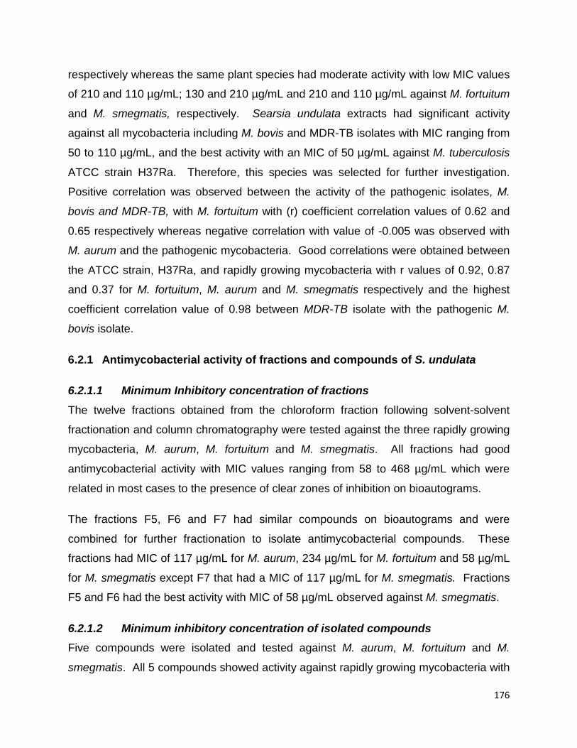
176
respectively whereas the same plant species had moderate activity with low MIC values
of 210 and 110 µg/mL; 130 and 210 µg/mL and 210 and 110 µg/mL against M. fortuitum
and M. smegmatis, respectively. Searsia undulata extracts had significant activity
against all mycobacteria including M. bovis and MDR-TB isolates with MIC ranging from
50 to 110 µg/mL, and the best activity with an MIC of 50 µg/mL against M. tuberculosis
ATCC strain H37Ra. Therefore, this species was selected for further investigation.
Positive correlation was observed between the activity of the pathogenic isolates, M.
bovis and MDR-TB, with M. fortuitum with (r) coefficient correlation values of 0.62 and
0.65 respectively whereas negative correlation with value of -0.005 was observed with
M. aurum and the pathogenic mycobacteria. Good correlations were obtained between
the ATCC strain, H37Ra, and rapidly growing mycobacteria with r values of 0.92, 0.87
and 0.37 for M. fortuitum, M. aurum and M. smegmatis respectively and the highest
coefficient correlation value of 0.98 between MDR-TB isolate with the pathogenic M.
bovis isolate.
6.2.1 Antimycobacterial activity of fractions and compounds of S. undulata
6.2.1.1 Minimum Inhibitory concentration of fractions The twelve fractions obtained from the chloroform fraction following solvent-solvent
fractionation and column chromatography were tested against the three rapidly growing
mycobacteria, M. aurum, M. fortuitum and M. smegmatis. All fractions had good
antimycobacterial activity with MIC values ranging from 58 to 468 µg/mL which were
related in most cases to the presence of clear zones of inhibition on bioautograms.
The fractions F5, F6 and F7 had similar compounds on bioautograms and were
combined for further fractionation to isolate antimycobacterial compounds. These
fractions had MIC of 117 µg/mL for M. aurum, 234 µg/mL for M. fortuitum and 58 µg/mL
for M. smegmatis except F7 that had a MIC of 117 µg/mL for M. smegmatis. Fractions
F5 and F6 had the best activity with MIC of 58 µg/mL observed against M. smegmatis.
6.2.1.2 Minimum inhibitory concentration of isolated compounds Five compounds were isolated and tested against M. aurum, M. fortuitum and M.
smegmatis. All 5 compounds showed activity against rapidly growing mycobacteria with

177
MIC values ranging from 23.44 to 250 µg/mL. The first compounds 1 and 2 had the
highest MIC values varying from 62.5 to >250 µg/mL. The MIC values of 62.5 µg/mL
were obtained against M. aurum and M. fortuitum and 125 µg/mL for M. smegmatis
whereas C2 showed the highest MIC value >250 µg/mL against M. fortuitum and M.
smegmatis. Compound 3 (C3 or SLN1) had the lowest MIC value of 23.44 µg/mL
against M. fortuitum; 31.25 µg/mL against M. aurum and M. smegmatis. Compound C4
(C4 or PKB) and compound 5 (C5 or PKC) both showed activity against all three
mycobacteria with best value of 46.88 µg/mL for C4 against M. smegmatis.
6.3 CYTOTOXICITY OF ACETONE LEAF EXTRACTS OF PLANT SPECIES FROM ANACARDIACEAE FAMILY
Most drugs or other substances are metabolised in the liver and excreted by the kidney.
Mycobacterial species causing tuberculosis multiply and hide in macrophages. This
objective was achieved by selecting three cell lines to represent these organs, namely
Vero monkey kidney, human liver hepatoma (C3A) and mouse macrophage cells (RAW
264.7). Crude extracts (Heeria argentea, Lannea discolor, Protorhus longifolia, Searsia
undulata and Sclerocarya birrea) with good antimycobacterial activity (from significant to
moderate) and those that showed visible zones of inhibition on bioautograms were
selected for cytotoxicity testing. The cytotoxicity was determined using a tetrazolium-
based colorimetric assay with 3-(4, 5-dimethylthiazol)-2, 5-diphenyl tetrazolium bromide
(MTT) as the indicator. All crude extracts tested showed low toxicity against all three
cell lines except Searsia undulata that had moderate toxicity to C3A cells with LC50 of
0.034 mg/mL. This deserves further investigation as a source of anticancer substances
as C3A cells are cancer cells with abnormal metabolic activity. The S. undulata extract
had LC50 of 0.50 and 0.12 mg/mL on Vero cells and RAW cells respectively with good
selectivity indexes of 7.08 on Vero cells and more than 1 on RAW cells for non-
tuberculous and MDR-TB mycobacteria. Protorhus longifolia had an LC50 of 0.62
mg/mL on C3A cells, 0.88 mg/mL on Vero cells and >1 mg/mL for RAW cells with the
highest selectivity index on Vero cells of 12.6 for M. smegmatis followed by 8.02 for M.
aurum, M. fortuitum and MDR-TB. Plant extracts with SI values less than 1 mean that
the extracts are relatively less toxic to the bacteria and more toxic to the mammalian
cells. Therefore, extracts with SI >1 may be relatively safer to use in vivo (not
178
accounting for pharmacokinetics parameters) as they are less toxic to mammalian cells
but more toxic to the pathogens. The selectivity indexes, especially of P. longifolia with
SI = 12. 6 could be considered as very promising, as a good therapeutic index for a
remedy or drug should be ≥10. It is also worth noting that efficacy “in vitro” might differ
from efficacy ‘in vivo” due to different parameters influencing pharmacodynamics and
pharmacokinetics of drugs administered by different routes to humans and animals.
Protorhus longifolia and Searsia undulata had the highest selectivity index against the
three rapidly growing mycobacteria and pathogenic mycobacteria and the highest total
activity. These rapidly growing species had the highest total activity of 5 357 mL/g for
S. undulata and 3 557 mL/g for P. longifolia which indicates the volume to which the
extract from 1 g can be diluted and still inhibit the growth of mycobacteria. It is also
worth noting that the leaves of S. undulata are chewed by Khoisan people to treat chest
colds, indicating a low toxicity. The mode of administration and interaction with other
factors ”in vivo” must be considered when assessing toxicity. Plant extracts showing
sensitivity to cell lines with LC50 values >0.1 mg/mL are considered not cytotoxic in
terms of searching for anticancer compounds. Searsia undulata was selected as the
preferred plant species for compound isolation based on its high cytotoxicity on
abnormal cells (C3A) (potential candidate for anticancer agent) and low cytotoxicity on
the normal Vero kidney cells and its good antimycobacterial activity.
6.3.1 Cytotoxicity of compounds isolated from Searsia undulata The crude extract of Searsia undulata at a concentration of 100 µg/mL had low
cytotoxicity (LC50 = 500 µg/mL) on Vero cells (Section 6.3). Three of the compounds
isolated from S. undulata had moderate toxicity with compound 2 (SLN1) being the least
toxic (LC50 = 211 µg/mL) followed by compound 3 with LC50 = 48 µg/mL (PK-B) and
compound 1 (LC50 = 31 µg/mL). The other two compounds were not tested because
they were not pure and not in sufficient quantity.
6.4 ISOLATION OF BIOACTIVE FRACTIONS AND COMPOUNDS FROM THE LEAF OF SEARSIA UNDULATA
The five fractions from solvent-solvent fractionation (chloroform, butanol, hexane, H2O-
35% methanol and H2O) were eluted in BEA solvent system and tested for

179
antimycobacterial activity against M. aurum, M. fortuitum and M. smegmatis and MDR-
TB and H37Ra. Thin layer chromatography fingerprints eluted in hexane: ethyl acetate
at different ratios was used to locate the active phytochemical compounds as mentioned
under section 6.2.
The chloroform fraction was selected as the fraction with the best antimycobacterial
activity based on good MIC values and presence of more than 10 potential active
compounds as discussed under section 6.2.1.1. It was further fractionated using
column chromatography packed with silica gel into 55 fractions that were combined into
12 sub-fractions (F1-F12). Three (F5-F7) out of the 12 fractions showing good
antimycobacterial activity and presence of active phytochemical compounds on
bioautography were combined and were further fractionated using column silica gel
chromatography for isolation of compounds.
Among all 12 fractions, F3 had the highest total activity on its own of 19 765 mL/g for M
aurum followed by 13 162 mL/g for M. fortuitum and 7 896 mL/g for M. smegmatis. It
also had active compounds and good MIC values but was not selected at this initial
stage as it did not have the consistent similar pattern of phytochemical active
compounds on bioautography and TLC when compared to F5 to F7 (Figures 5.14-5.18).
In future investigations, the isolation of compounds from fraction F3 will be undertaken.
The next fraction showing the highest activity was F6 with a total activity of 19 793 mL/g
against M smegmatis then F5 showing the highest total activity of 17154 mL against M.
smegmatis then the last fraction with lower total activity F7 showing 7 262 mL for both
M. aurum and M. smegmatis. The average total activity of the combined fractions was
27 599 which is higher than F3 alone 19 765 mL. At this preliminary stage, this
determined the focus on the combination of these three fractions based on their MIC
values against all the mycobacteria species.
The Rf value, or retardation factor, was used to trace and identify targeted compounds
with antimycobacterial activity at different stages of the fractionation process. Rf values
of the chloroform fraction were calculated by dividing the distance travelled by either the
extract or fraction or compound spotted on the vanillin sprayed TLC plate or
180
bioautogram eluted in a specific mobile phase solvent system. Rf values were
calculated for the plates eluted in BEA solvent and sprayed with M. aurum and M.
fortuitum as they were good indicators of antimycobacterial activity in general.
Isolation of compounds from the combined semi-purified fractions, F5, F6 and F7
(1019.7mg) was carried out using a small column packed with silica gel with 100% n-
hexane and fractions collected into honey jars. Compounds were eluted with 100% n-
hexane with increasing polarity with 2% ethyl acetate resulting in sequential elution of
SLN1 (26.2 mg), PKA1 (15.4 mg), PKB (22.4 mg), PKC (15.9 mg) and PKD (8.4 mg).
The purified PK B was obtained by using a mixture of chloroform and methanol as
eluent on a silica gel column at the second run.
6.5 STRUCTURE ELUCIDATION AND IDENTIFICATION OF COMPOUNDS
6.5.1 Compound SLN1 This compound was collected and dried as a light yellow powder. The initial inspection
of the proton spectrum revealed that the material was triterpenoid in nature, based on
the presence of at least six methyl groups in the spectrum. The material consisted of
about 70% of the major product. Additionally, a pair of triplet-like signals at δ 4.72 ppm
and δ 4.59 ppm, integrating to a single proton each, pointed to the presence of a
terminal methylene system, likely a 1, 1-disubstituted alkene typical of the lupane
pentacyclic triterpenoids. The compound was identified as betulonic acid, which has
been reported previously as having antimicrobial activity.
During the process of the isolation of compounds, it is worth noting that the work was
carried out at room temperature and samples were exposed to oxygen and light.
Changes observed on TLC plates indicated that certain compounds were unstable and
decomposed. This could imply that the initial compound was betulinic acid which
underwent oxidation to its derivative betulonic acid. In either case, both compounds
were reported to have similar biological activity properties.
Betulinic and betulonic acid are naturally occurring triterpenes found in many plants but
betulonic acid can be obtained from oxidation of betulinic acid with Jones’ reagent.

181
Betulonic acid is a derivative of betulinic acid as described above. Betulinic acid is a
naturally occurring pentacyclic triterpenoid which has antiretroviral, antimalarial and
anti-inflammatory properties as well as anticancer (Dehelean et al., 2012). This
appears to be the first report of isolation of betulonic acid from S. undulata.
6.5.2 Compound PK-B This compound was collected and dried as a slightly yellow powder. Its putative
structure did not match any chemical structure on Scifinder. This compound seems to
be novel. More of the compound needs to be isolated to enable crystallization, melting
point and Fourier Transform Infrared Spectroscopy (FTIR) for full elucidation of the
chemical structure. The proposed structure is provided in section 5.3.4.2 (Figure 5.30).
6.6 RESEARCH CHALLENGES
• All bioassays were conducted in vitro. There is still a need to validate the
efficacy as well as safety in vivo. Several parameters and interactions need to be
considered during the in vivo metabolism of a drug which leads to its effective
bioavailability.
• The isolation of compounds was a laborious and difficult process due to the
complexity of antimycobacterial active phytochemical compounds that were
closely related. This made the separation and purification processes challenging.
• Delay encountered in the appointment of a phytochemist on site for guidance in
the isolation of compounds.
• No easy access to NMR instrument and very limited expert staff available
therefore the high demand from researchers making use of this technique could
not be met within the required time.
• NMR broken probes were sent for repair in Germany (no technical local
representation)
• Minimum inhibitory concentration (MIC): the main challenge encountered was the
formation of a jelly-like substance by some compounds when inoculated in the
growth medium, preventing tested compounds from dissolving. Therefore, the
growth medium was diluted with solvents such as ethyl acetate or DMSO at
different ratios and organisms’ growth was tested prior to carrying out the

182
bioactivity assay. Bioautography: the use of tested mycobacteria was only limited
to the rapidly growing organisms and not extended to the pathogenic
mycobacteria. This is due to the technique as a mycobacterial suspension is
sprayed on the TLC plates. Pathogenic mycobacteria are highly infectious and
air borne infection is one of the main routes of contamination/infection. The slow-
growing mycobacteria would also not be able to survive on the TLC plate for the
extended period of time necessary for adequate growth.
6.7 GENERAL CONCLUSION, RECOMMENDATIONS AND FUTURE PERSPECTIVES
6.7.1 Isolation and characterization of mycobacterial species The involvement of some zoonotic Mycobacterium spp. in the disease-causing process
has been established. There is evidence that new strains of bacteria are circulating. It
has also been reported that clinical signs of disease caused by M. bovis and M.
tuberculosis are indistinguishable. Other findings mention that radiological clinical
pictures of some pulmonary diseases caused by non-tuberculous mycobacteria in
humans were confused with those of tuberculosis. Therefore, methods used in routine
laboratory diagnostics of tuberculosis should be mixed and include culture, Ziehl-
Neelsen staining and at least one molecular technique. For culture isolation, growth
media that will promote the growth of both M. bovis and M. tuberculosis should be used.
The so called MOTT, referring to mycobacteria spp. other than the targeted known
pathogenic M. bovis and M. tuberculosis, should no longer be disregarded and should
be identified fully as they might be primary causative agents of some tuberculosis-like
lesions or clinical conditions/cases in both animals and humans. Although the
involvement of non-tuberculous mycobacteria in the disease-causing process has been
established in humans and some animal species as well as their zoonotic aspects, this
involvement still remains to be established in animals. Laboratories and researchers
should also be encouraged to deposit newly identified mycobacterial spp. in the
international gene data bank as there have been reports on unpublished data of new
mycobacteria spp. in South Africa (Gcebe et al., 2013; Helden et al., 2009).

183
The emergence of MDR/XDR TB strains is of concern, and adding to the target of
accurate identification of mycobacterial isolates, reference laboratories should
implement a routine antimycobacterial sensitivity test for accurately identified isolates
and perhaps also partially identified mycobacteria to allow a rapid implementation of
effective treatment regimens. Annual reports may be compiled of antimycobacterial
pattern sensitivity observed in the laboratory. As a long term goal, creating a national
database on antimycobacterial sensitivity of mycobacterial isolates from humans and
animals will be relevant as spill-over of M. tuberculosis from humans to animals has
been reported and spill-back could also be a possibility (Michel et al., 2013; Ocepek et
al., 2005). Medical and veterinary laboratories should collaborate on generating
databases for antimycobacterial sensitivity of M. bovis and M. tuberculosis isolates.
This task could be conducted in medical laboratories as tuberculosis in animals is not
treated.
6.7.2 Antimycobacterial activities of leaf extracts, fractions and compounds isolated from plant species from the Anacardiaceae family
Besides the above, an ultimate goal is to continue to seek for alternative treatments
exploring natural resources (plants) at our disposal for potential leads to anti-TB drug
development. Natural products such as plant extracts, either as pure compounds or as
standardized extracts, provide unlimited opportunities for new discoveries because of
the unavailability of a chemical diversity database. A national database should be
created and voucher samples of isolated anti-TB compounds deposited. This would
allow at some stage easy traceability when networking with pharmaceutical companies
in exploring the development of new drugs from targeted compounds.
Extraction is a crucial step because it is necessary to extract the desired chemical
components from the plant material. The solvent used is equally important depending
on the assay conducted. In the case of biological activity one must make sure that the
solvent is not harmful to the microorganisms tested. Many solvents inhibit the growth of
microorganisms. When evaluating the biological activity of extracts against
microorganisms, acetone was proven not to inhibit the growth of tested microorganisms
(Eloff, 1998a).

184
During this study, five plant species, namely Harpephyllum caffrum, Protorhus longifolia,
Searsia lancea, Searsia undulata and Sclerocarya birrea had good antimycobacterial
activity, and they will be further analysed. For the purpose of this study, one of the
plants, Searsia undulata, contained about ten antimycobacterial compounds visible in
bioautography. Of these, three compounds were isolated and two identified.
Bioassay-guided isolation of compounds is laborious and demanding. It needs a lot of
patience and bioassays at each stage should be conducted scrupulously to identify the
fractions in which active phytochemicals are present and to continue until isolation of
the target compound(s). Several challenges were encountered during this study.
Among others some compounds were unstable as demonstrated by changing TLC
fingerprints. Compounds also appeared to have closely related structures making them
difficult to separate. Adhering to certain environmental conditions from the extraction to
isolation of compounds must be observed as some compounds are sensitive to heat;
oxygen, temperature or light which might change their structure, therefore, avoiding
exposure to these conditions will minimize structural changes.
6.7.3 In vitro cytotoxicity activity of Searsia undulata leaf extracts and isolated compounds against Vero and human hepatoma cell lines
In vitro cytotoxicity evaluation is important as compounds with fewer adverse effects are
needed, considering that some of the current anti-TB drugs have undesirable side
effects. When evaluating plants, the use of normal and abnormal cells (cancer cells) is
relevant as cancer is a major cause of mortality and a cure is yet to be found for
different types of cancer. Identifying potential antimicrobial and anticancer compounds
can also be concurrently explored. This is the case during this study where an
incidental finding revealed that Searsia undulata had high cytotoxicity to cancerous liver
cells (C3A hepatoma cells) but low cytotoxicity to normal cells (Vero monkey kidney
cells). This was supported by the compound isolated and identified as betulonic acid,
which, together with its derivatives, has been reported to have anticancer, anti-
inflammatory, antiviral and antimicrobial activities.

185
6.7.4 Future perspectives As part of continued research relating to this project, there are many activities to be
carried out in the future deriving from this study for publication purposes, and eventually
drug development. The different activities recommended will be planned as short term,
medium and long term goals.
6.7.4.1 Isolation and identification of compounds from Searsia undulata Ten antimycobacterial compounds were observed on bioautography and two
compounds were identified. One of the compounds, PK-B, requires further analysis to
confirm beyond any doubt that it is indeed a novel compound. Crystallization, melting
point and Fourier Transform Infrared spectroscopy are among the techniques needing
to be done. The isolation and identification of the remainder of compounds will be
carried out.
6.7.4.2 In vitro safety of all compounds isolated from S. undulata The evaluation of the cytotoxicity of all isolated antimycobacterial compounds against at
least three cell lines will be conducted.
6.7.4.3 Synergistic evaluation of compounds isolated from S. undulata The synergy of antimycobacterial activity among the compounds and mixed with
standard anti-TB drugs will be investigated.
6.7.4.4 Determine the stability of each compound and fractions of S. undulata Observations made during this study indicate that some compounds might be unstable;
therefore, it is important to evaluate the stability of each compound as a labile
compound may not be useful. The stability in solution and as powder under different
conditions of temperature, moisture, light, and presence or absence of oxygen needs to
be determined.

186
6.7.4.5 Determine antioxidant, anti-inflammatory and inhibition of nitric oxide production by fractions and compounds of S. undulata
Testing for antioxidant activity and anti-inflammatory activities should be conducted.
Inflammation, which evolves into chronic inflammation, as well as the production of
oxidative radicals, takes place during the pathogenesis of acute or chronic tuberculosis.
6.7.4.6 Determine the in vivo safety of pure compounds Identifying an animal model for in vivo evaluation of efficacy is an area that requires
attention in the future. Clinical trials are also necessary to demonstrate the effectiveness
and safety of the bioactive compounds. In the course of drug development, determining
acute and chronic toxicity is required.
6.7.4.7 Develop an herbal product from S. undulata Considering that the development of drugs is expensive, one could first investigate in
the local communities the current ethnobotanical use of this plant species. The
selection of species in this study was extended to the Anacardiaceae family based on
few members of the family that had good antibacterial activities on Gram-positive and
Gram-negative bacteria as well as M. smegmatis in a previous study (Pauw, 2004). It
was subsequently discovered in the literature that leaves are chewed by the Khoi San to
treat chest colds and this needs further investigation. A survey in this population to find
out about any other information on the ethnobotanical use and observations about this
plant species could be initiated.
6.7.4.8 Understanding the mechanism of actions of compounds In the case of S. undulata, leaves are chewed to treat chest colds. Each group of
currently-used antibiotics has a different mechanism of action - bactericidal or
bacteriostatic on microorganisms. The emergence of MDR-TB drugs is of concern and
therefore focusing on mechanisms of action to prevent rapid development of resistance
is crucial. There is evidence that the development of resistance may be minimized
when using plant extracts, as a combination of different compounds may exert a range
of different activities. In the case of tuberculosis, mycobacteria might be present in the
latent “dormant” phase, and can be reactivated when immunity is compromised.
187
Therefore the best drug or combination of drugs should be bactericidal towards the
mycobacteria, particularly those inside macrophages. Drugs with additional positive
effects on the immune system are also desirable.
6.7.4.9 Develop network with pharmaceutical companies Establishing network with companies interested in tuberculosis drugs will be part of the
long term goal of this research.
6.7.4.10 Testing of four other plant species with good antimycobacterial activity The other plant species (Harpephyllum caffrum, Protorhus longifolia, Searsia lancea
and Sclerocarya birrea) that have good antimycobacterial activity will be further
processed as mentioned under all the above sections.

188
REFERENCES
Abd Rahman, M., Yeap, S.K., Ali, A.M., Ahmad, F., Ng, M.K., Lam, H.Y., Alitheen, M., Banu, N. 2012. Immunomodulatory effects of betulinic acid isolation from the bark of Melaleuca cajuputi. Pertanika Journal of Tropical Agriculture and Science 35: 293–305.
Adeshina, G.O., Kunle, O.F., Onaolapo, J.A., Ehinmidu, J.O., Odama, L.E. 2012. Antimicrobial activity of the aqueous and ethyl acetate sub-fractions of Alchornea cordifolia Leaf. European Journal of Medicinal Plants 2: 31.
Agdestein, A., Johansen, T.B., Kolbjørnsen, Ø., Jørgensen, A., Djønne, B., Olsen, I. 2012. A comparative study of Mycobacterium avium subsp. avium and Mycobacterium avium subsp. hominissuis in experimentally infected pigs. Biomedical Veterinary Research 8: 1.
Agdestein, A., Johansen, T.B., Polaček, V., Lium, B., Holstad, G., Vidanović, D., Aleksić-Kovačević, S., Jørgensen, A., Žultauskas, J., Nilsen, S.F. 2011. Investigation of an outbreak of mycobacteriosis in pigs. Biomedical Veterinary Research 7:1.
Ahmad, S. 2010. Pathogenesis, immunology, and diagnosis of latent Mycobacterium tuberculosis infection. Clinical and Developmental Immunology 20:11.
Ahmed, A.S., McGaw, L.J., Moodley, N., Naidoo, V., Eloff, J.N. 2014. Cytotoxic, antimicrobial, antioxidant, antilipoxygenase activities and phenolic composition of Ozoroa and Searsia species (Anacardiaceae) used in South African traditional medicine for treating diarrhoea. South African Journal of Botany 95: 9–18.
Ahmed, M.S., Galal, A.M., Ross, S.A., Ferreira, D., ElSohly, M.A., Ibrahim, A.-R.S., Mossa, J.S., El-Feraly, F.S. 2001. A weakly antimalarial biflavanone from Rhus retinorrhoea. Phytochemistry 58: 599–602.
Ålander-Damsten, Y.K., Brander, E.E., Paulin, L.G. 2003. Panniculitis, due to Mycobacterium smegmatis, in two Finnish cats. Journal of Feline Medicine and Surgery 5: 19–26.
Alexander, K.A., Laver, P.N., Michel, A.L., Williams, M., Helden, P.D., Warren, R.M., Pittius, N.C.G. 2010. Novel Mycobacterium tuberculosis complex pathogen, M. mungi. Emerging Infectious Diseases 16: 1296–1299.
Ali-Shtayeh, M.S., Al-Assali, A.A., Jamous, R.M. 2013. Antimicrobial activity of Palestinian medicinal plants against acne-inducing bacteria. African Journal of Microbiology Research 7: 2560–2573.
Alland, D., Kalkut, G.E., Moss, A.R., McAdam, R.A., Hahn, J.A., Bosworth, W., Drucker,

189
E., Bloom, B.R. 1994. Transmission of tuberculosis in New York City--an analysis by DNA fingerprinting and conventional epidemiologic methods. New England Journal of Medicine 330: 1710–1716.
Altschul, S.F., Gish, W., Miller, W., Myers, E.W., Lipman, D.J. 1990. Basic local alignment search tool. Journal of Molecular Biology 215: 403–410 doi.org/10.1016/S0022-2836(05)80360-2.
Andersen, P., Doherty, T.M. 2005. The success and failure of BCG—implications for a novel tuberculosis vaccine. Nature Reviews Microbiology 3: 656–662.
Andrade-Ochoa, S., Nevárez-Moorillón, G.V., Sánchez-Torres, L.E., Villanueva-García, M., Sánchez-Ramírez, B.E., Rodríguez-Valdez, L.M., Rivera-Chavira, B.E. 2015. Quantitative structure-activity relationship of molecules constituent of different essential oils with antimycobacterial activity against Mycobacterium tuberculosis and Mycobacterium bovis. Biomedical Complementary Alternative Medicine 15: 1.
Andrews, J.R., Gandhi, N.R., Moodley, P., Shah, N.S., Bohlken, L., Moll, A.P., Pillay, M., Friedland, G., Sturm, A.W. 2008. Exogenous reinfection as a cause of multidrug-resistant and extensively drug-resistant tuberculosis in rural South Africa. Journal of Infectious Diseases 198: 1582–1589.
Aranaz, A., Liebana, E., Mateos, A., Dominguez, L., Vidal, D., Domingo, M., Gonzolez, O., Rodriguez-Ferri, E.F., Bunschoten, A.E., Van Embden, J.D. 1996. Spacer oligonucleotide typing of Mycobacterium bovis strains from cattle and other animals: a tool for studying epidemiology of tuberculosis. Journal of Clinical Microbiology 34: 2734–2740.
Aro, A.O., Dzoyem, J.P., Hlokwe, T.M., Madoroba, E., Eloff, J.N., McGaw, L.J., 2015. Some South African rubiaceae tree leaf extracts have antimycobacterial activity against pathogenic and Non-pathogenic Mycobacterium Species. Phytotherapy Research 29: 1004–1010 doi:10.1002/ptr.5338.
Arrazuria, R., Sevilla, I.A., Molina, E., Pérez, V., Garrido, J.M., Juste, R.A., Elguezabal, N. 2015. Detection of Mycobacterium avium subspecies in the gut associated lymphoid tissue of slaughtered rabbits. Biomedical Veterinary Research 11: 1.
Awouafack, M.D., McGaw, L.J., Gottfried, S., Mbouangouere, R., Tane, P., Spiteller, M., Eloff, J.N. 2013. Antimicrobial activity and cytotoxicity of the ethanol extract, fractions and eight compounds isolated from Eriosema robustum (Fabaceae). Biomedical Complementary and Alternative Medicine 13:1.
Ayele, W.Y., Neill, S.D., Zinsstag, J., Weiss, M.G., Pavlik, I. 2004. Bovine tuberculosis: an old disease but a new threat to Africa. International Journal of Tuberculosis and Lung Disease 8: 924–937.

190
Azé, J., Sola, C., Zhang, J., Lafosse-Marin, F., Yasmin, M., Siddiqui, R., Kremer, K., van Soolingen, D., Refrégier, G. 2015. Genomics and machine learning for taxonomy consensus: The Mycobacterium tuberculosis Complex Paradigm. PLoS One 10: e0130912.
Babula, P., Adam, V., Havel, L., Kizek, R. 2009. Noteworthy secondary metabolites naphthoquinones-their occurrence, pharmacological properties and analysis. Current Pharmaceutical Analysis 5: 47–68.
Balunas, M.J., Kinghorn, A.D. 2005. Drug discovery from medicinal plants. Life Sciences 78: 431–441.
Bamuamba, K., Gammon, D.W., Meyers, P., Dijoux-Franca, M.-G., Scott, G. 2008. Anti-mycobacterial activity of five plant species used as traditional medicines in the Western Cape Province (South Africa). Journal of Ethnopharmacology 117: 385–390.
Baral, R.M., Metcalfe, S.S., Krockenberger, M.B., Catt, M.J., Barrs, V.R., McWhirter, C., Hutson, C.A., Wigney, D.I., Martin, P., Chen, S.C.A. 2006. Disseminated Mycobacterium avium infection in young cats: overrepresentation of Abyssinian cats. Journal of Feline Medicine and Surgery 8: 23–44.
Barnard, M., Albert, H., Coetzee, G., O’Brien, R., Bosman, M.E. 2008. Rapid molecular screening for multidrug-resistant tuberculosis in a high-volume public health laboratory in South Africa. American Journal of Respiratory and Critical Care Medicine 177: 787–792.
Bastos, D.Z.L., Pimentel, I.C., de Jesus, D.A., de Oliveira, B.H. 2007. Biotransformation of betulinic and betulonic acids by fungi. Phytochemistry 68: 834–839.
Basu, S., Andrews, J.R., Poolman, E.M., Gandhi, N.R., Shah, N.S., Moll, A., Moodley, P., Galvani, A.P., Friedland, G.H. 2007. Prevention of nosocomial transmission of extensively drug-resistant tuberculosis in rural South African district hospitals: an epidemiological modelling study. Lancet 370: 1500–1507.
Bateman, C. 2015. Eastern Cape treatment dysfunction boosts virulent new XDR-TB strain. South African Medical Journal 105: 165–167.
Ben Salah, I., Adekambi, T., Raoult, D., Drancourt, M. 2008. rpoB sequence-based identification of Mycobacterium avium complex species. Microbiology 154: 3715–3723 doi:10.1099/mic.0.2008/020164-0.
Bengis, R.G., Kock, R.A., Fischer, J. 2002. Infectious animal diseases: the wildlife/livestock interface. Revue Scientifique, Technique Internationale des épizooties 21: 53–66.
191
Bengis, R.G., Kriek, N.P.J., Keet, D.F., Raath, J.P., Vos, V., Huchzermeyer, H.F.A.K. 1996. An outbreak of bovine tuberculosis in a free-living African buffalo (Syncerus caffer-Sparrman) population in the Kruger National Park: a preliminary report. Onderstepoort Journal of Veterinary Research 63, 15–18.
Bercovier, H., Vincent, V. 2001. Mycobacterial infections in domestic and wild animals due to Mycobacterium marinum, M. fortuitum, M. chelonae, M. porcinum, M. farcinogenes, M. smegmatis, M. scrofulaceum, M. xenopi, M. kansasii, M. simiae and M. genavense. Revue Scientifique et Technique 20: 265–290.
Bero, J., Hannaert, V., Chataigné, G., Hérent, M.-F., Quetin-Leclercq, J. 2011. In vitro antitrypanosomal and anti-leishmanial activity of plants used in Benin in traditional medicine and bio-guided fractionation of the most active extract. Journal of Ethnopharmacology 137: 998–1002.
Best, C.A., Best, T.J. 2009. Mycobacterium smegmatis infection of the hand. Journal of Clinical Microbiology 4: 165–166.
Bhalla, P., Forrest G.N., Gershon, A.A. 2015. An official publication of the Infectious Diseases Society of America. Clinical Infectious Diseases: 61(1):143 doi:10.1093/cid/civ338.
Biet, F., Boschiroli, M.L., Thorel, M.F., Guilloteau, L.A. 2005. Zoonotic aspects of Mycobacterium bovis and Mycobacterium avium-intracellulare complex (MAC). Veterinary Research 36: 411–436.
Biuković, G., Basak, S., Manimekalai, M.S.S., Rishikesan, S., Roessle, M., Dick, T., Rao, S.P.S., Hunke, C., Grüber, G. 2013. Variations of subunit ε of the Mycobacterium tuberculosis F1Fo ATP synthase and a novel model for mechanism of action of the tuberculosis drug TMC207. Antimicrobial Agents Chemotherapy 57: 168–176.
Blazquez, J., de Los Monteros, L.E.E., Samper, S., Martin, C., Guerrero, A., Cobo, J., Van Embden, J., Baquero, F., Gómez-Mampaso, E. 1997. Genetic characterization of multidrug-resistant Mycobacterium bovis strains from a hospital outbreak involving human immunodeficiency virus-positive patients. Journal of Clinical Microbiology 35: 1390–1393.
Blondeau, S., Do, Q.T., Scior, T., Bernard, P., Morin-Allory, L. 2010. Reverse pharmacognosy: another way to harness the generosity of nature. Current Pharmaceutical Design 16: 1682–1696.
Bloom, B.R., Murray, C.J. 1992. Tuberculosis: commentary on a reemergent killer. Science 257: 1055–1064.
Bobadilla-del Valle, M., Torres-González, P., Cervera-Hernández, M.E., Martínez-Gamboa, A., Crabtree-Ramirez, B., Chávez-Mazari, B., Ortiz-Conchi, N., Rodríguez-Cruz, L., Cervantes-Sánchez, A., Gudiño-Enríquez, T. 2015. Trends of Mycobacterium bovis isolation and first-line anti-tuberculosis drug susceptibility

192
profile: A Fifteen-Year Laboratory-Based Surveillance. PLoS Neglected Tropical Diseases Journal 9: e0004124.
Boligon, A.A., Agertt, V., Janovik, V., Cruz, R.C., Campos, M., Guillaume, D., Athayde, M.L., dos Santos, A.R.S. 2012. Antimycobacterial activity of the fractions and compounds from Scutia buxifolia. Revista Brasileira de Farmacognosia 22: 45–52.
Bonjar, G.H.S. 2004. Evaluation of antibacterial properties of Iranian medicinal plants against Micrococcus luteus, Serratia marcescens, Klebsiella pneumoniae and Bordetella bronchoseptica. Asian Journal of Plant Sciences 3: 82–86.
Bono, M., Jemmi, T., Bernasconi, C., Burki, D., Telenti, A., Bodmer, T. 1995. Genotypic characterization of Mycobacterium avium strains recovered from animals and their comparison to human strains. Applied and Environmental Microbiology 61: 371–373.
Bopana, N., Saxena, S. 2007. Asparagus racemosus—Ethnopharmacological evaluation and conservation needs. Journal of Ethnopharmacology 110: 1–15.
BoRam, K., JaeMyung, K., ByoungJun, K., YunHo, J., SoYoon, R., YoonHoh, K., BumJoon, K. 2014. Identification of nontuberculous mycobacteria isolated from Hanwoo (Bos taurus coreanae) in South Korea by sequencing analysis targeting hsp65, rpob and 16S rRNA genes. Veterinary Microbiology 173: 385–389.
Borchardt, J.R., Wyse, D.L., Sheaffer, C.C., Kauppi, K.L., Ehlke, R.G.F.N.J., Biesboer, D.D., Bey, R.F. 2008. Antimicrobial activity of native and naturalized plants of Minnesota and Wisconsin. Journal of Medicinal Plants Research 2: 98–110.
Bosne-David, S., Barros, V., Verde, S.C., Portugal, C., David, H.L. 2000. Intrinsic resistance of Mycobacterium tuberculosis to clarithromycin is effectively reversed by subinhibitory concentrations of cell wall inhibitors. Journal of Antimicrobial Chemotherapy 46: 391–395.
Briskin, D.P. 2000. Medicinal plants and phytomedicines. Linking plant biochemistry and physiology to human health. Plant Physiology 124: 507–514.
Brusotti, G., Cesari, I., Dentamaro, A., Caccialanza, G., Massolini, G. 2014. Isolation and characterization of bioactive compounds from plant resources: The role of analysis in the ethnopharmacological approach. Journal of Pharmaceutical and Biomedical Analysis 87: 218–228.
Buckwalter, S.P., Olson, S.L., Connelly, B.J., Lucas, B.C., Rodning, A.A., Walchak, R.C., Deml, S.M., Wohlfiel, S.L., Wengenack, N.L. 2016. Evaluation of Matrix-Assisted Laser Desorption Ionization-Time of Flight Mass Spectrometry for identification of Mycobacterium species, Nocardia species, and other aerobic Actinomycetes. Journal of Clinical Microbiology 54: 376–384.
BumJoon, K., SeongKarp, H., KeunHwa, L., YeoJun, Y., EuiChong, K., YoungGil, P., GilHan, B., YoonHoh, K. 2004. Differential identification of Mycobacterium
193
tuberculosis complex and nontuberculous mycobacteria by duplex PCR assay using the RNA polymerase gene (rpoB). Journal of Clinical Microbiology 42: 1308–1312.
Burnet, N.G., Benson, R.J., Williams, M. V, Peacock, J.H. 2000. The rise in bacterial resistance. Bristish Medical Journal 320: 199–200.
Busse, H.J., Denner, E.B.M., Lubitz, W. 1996. Classification and identification of bacteria: current approaches to an old problem. Overview of methods used in bacterial systematics. Journal of Biotechnology 47: 3–38.
Butler, W.R., Guthertz, L.S. 2001. Mycolic acid analysis by high-performance liquid chromatography for identification of Mycobacterium species. Clinical Microbiology Reviews 14: 704–726.
Caamal-Fuentes, E., Torres-Tapia, L., Simá-Polanco, P., Peraza-Sánchez, S.R., Moo-Puc, R. 2011. Screening of plants used in Mayan traditional medicine to treat cancer-like symptoms. Journal of Ethnopharmacology 135: 719–724.
Camacho-Corona, M. del R., Favela-Hernández, J.M. de J., González-Santiago, O., Garza-González, E., Molina-Salinas, G.M., Said-Fernández, S., Delgado, G., Luna-Herrera, J. 2009. Evaluation of some plant-derived secondary metabolites against sensitive and multidrug-resistant Mycobacterium tuberculosis. Journal of the Mexican Chemical Society 53: 71–75.
Campora, L., Corazza, M., Zullino, C., Ebani, V. V, Abramo, F. 2011. Mycobacterium avium subspecies hominissuis disseminated infection in a Basset Hound dog. Journal of Veterinary Diagnostic Investigation 23: 1083–1087.
Castellheim, A., Brekke, O.L., Espevik, T., Harboe, M., Mollnes, T.E. 2009. Innate immune responses to danger signals in systemic inflammatory response syndrome and sepsis. Scandinavian Journal of Immunology 69: 479–491.
Centers for Disease Control and Prevention. 2005. Human tuberculosis caused by Mycobacterium bovis--New York City, 2001-2004. Morbidity and Mortality Weekly Report 54, 605.
Centers for Disease Control and Prevention. 2003. Update: adverse event data and revised American Thoracic Society/CDC recommendations against the use of rifampin and pyrazinamide for treatment of latent tuberculosis infection--United States, 2003. Morbidity and Mortality Weekly Report 52, 735.
Cerella, C., Radogna, F., Dicato, M., Diederich, M. 2013. Natural compounds as regulators of the cancer cell metabolism. International Journal of Cell Biology doi:10.1155/2013/639401.

194
Chan, J., Fan, X.D., Hunter, S.W., Brennan, P.J., Bloom, B.R. 1991. Lipoarabinomannan, a possible virulence factor involved in persistence of Mycobacterium tuberculosis within macrophages. Infection and Immunity 59: 1755–1761.
Chan, J., Xing, Y., Magliozzo, R.S., Bloom, B.R. 1992. Killing of virulent Mycobacterium tuberculosis by reactive nitrogen intermediates produced by activated murine macrophages. Journal of Experimental Medicine 175: 1111–1122.
Chatterjee, D. 1997. The mycobacterial cell wall: structure, biosynthesis and sites of drug action. Current Opinion in Chemical Biology 1: 579–588.
Chatterjee, M., Bhattacharya, S., Karak, K., Dastidar, S.G. 2013. Effects of different methods of decontamination for successful cultivation of Mycobacterium tuberculosis. Indian Journal of Medical Research 138: 541–548 doi:IJMR-138-541.
Chege, G.K., Warren, R.M., Pittius, N.C.G., Burgers, W.A., Wilkinson, R.J., Shephard, E.G., Williamson, A.L. 2008. Detection of natural infection with Mycobacterium intracellulare in healthy wild-caught Chacma baboons (Papio ursinus) by ESAT-6 and CFP-10 IFN-γ ELISPOT tests following a tuberculosis outbreak. Biomedical Microbiology 8.
Cheng, Y., Shao, Y., Yan, W. 2011. Solubilities of betulinic acid in thirteen organic solvents at different temperatures. Journal of Chemical and Engineering Data 56: 4587–4591.
Chhabra, S.C., Mahunnah, B.L.A., Mshiu, E.N. 1987. Plants used in traditional medicine in eastern Tanzania. I. Pteridophytes and Angiosperms (Acanthaceae to Canellaceae). Journal of Ethnopharmacology 21: 253–277.
Chimponda, T., Mukanganyama, S. 2010. Antimycobacterial activities of selected medicinal plants from Zimbabwe against Mycobacterium aurum and Corynebacterium glutamicum. Tropical Biomedicine 27: 595–610.
Chinsembu, K.C. 2016. Tuberculosis and nature’s pharmacy of putative anti-tuberculosis agents. Acta Tropica 153: 46–56.
Chung, G.A., Aktar, Z., Jackson, S., Duncan, K. 1995. High-throughput screen for detecting antimycobacterial agents. Antimicrobial Agents and Chemotherapy 39: 2235–2238.
Churchyard, G.J., Mametja, L.D., Mvusi, L., Ndjeka, N., Hesseling, A.C., Reid, A., Babatunde, S., Pillay, Y. 2014. Tuberculosis control in South Africa: Successes, challenges and recommendations. South African Medical Journal 104: 234–248.
Cichewicz, R.H., Kouzi, S.A. 2004. Chemistry, biological activity, and chemotherapeutic potential of betulinic acid for the prevention and treatment of cancer and HIV infection. Medicinal Research Reviews 24: 90–114.
195
Cingolani, A., Antinori, A., Sanguinetti, M., Gillini, L., De Luca, A., Posteraro, B., Ardito, F., Fadda, G., Ortona, L. 1999. Application of molecular methods for detection and transmission analysis of Mycobacterium tuberculosis drug resistance in patients attending a reference hospital in Italy. Journal of Infectious Diseases 179: 1025–1029.
Clarke, C., Van Helden, P., Miller, M., Parsons, S. 2016. Animal-adapted members of the Mycobacterium tuberculosis complex endemic to the southern African subregion. Journal of South African Veterinary Association 87: doi:10.4102/jsava.v87i1.1322.
Clarke, K.R., Fitzgerald, S.D., Hattey, J.A., Bolin, C.A., Berry, D.E., Church, S. V, Reed, W.M. 2006. Experimental inoculation of wild turkeys (Meleagris gallopavo) with Mycobacterium bovis. Avian Diseases 50: 131–134.
Clay, H., Volkman, H.E., Ramakrishnan, L. 2008. Tumor necrosis factor signaling mediates resistance to mycobacteria by inhibiting bacterial growth and macrophage death. Immunity 29: 283–294.
Cleary-Guida, M.B., Okvat, H.A., Oz, M.C., Ting, W. 2001. A regional survey of health insurance coverage for complementary and alternative medicine: current status and future ramifications. Journal of Alternative Complementary Medicine 7: 269–273.
Cloud, J.L., Neal, H., Rosenberry, R., Turenne, C.Y., Jama, M., Hillyard, D.R., Carroll, K.C. 2002. Identification of Mycobacterium spp. by using a commercial 16S ribosomal DNA sequencing kit and additional sequencing libraries. Journal Clinical Microbiology 40: 400–406.
Cook, V.J., Turenne, C.Y., Wolfe, J., Pauls, R., Kabani, A. 2003. Conventional methods versus 16S ribosomal DNA sequencing for identification of nontuberculous mycobacteria: cost analysis. Journal of Clinical Microbiology 41: 1010–1015.
Corner, L.A.L. 2007. Bovine tuberculosis control in Australia, New Zealand and Ireland: wild animals and the epidemiology of tuberculosis in domestic animals. Cattle Practice 15: 3–12.
Corner, L.A.L. 2006. The role of wild animal populations in the epidemiology of tuberculosis in domestic animals: how to assess the risk. Veterinary Microbiology 112: 303–312.
Corner, L.A.L., Murphy, D., Costello, E., Gormley, E. 2009. Tuberculosis in European Badgers (Meles meles) and the control of infection with bacille Calmette-Guérin vaccination. Journal of Wildlife Diseases 45: 1042–1047.
Cosivi, O., Grange, J.M., Daborn, C.J., Raviglione, M.C., Fujikura, T., Cousins, D., Robinson, R.A., Huchzermeyer, H.F., De Kantor, I., Meslin, F.X. 1998. Zoonotic tuberculosis due to Mycobacterium bovis in developing countries. Emerging Infectious Diseases 4: 59.

196
Cousins, D. V, 2001. Mycobacterium bovis infection and control in domestic livestock. Mycobacterial infections in domestic and wild animals. Revue Scientifique et Technique (Office International des Épizooties) 20: 71-85.
Cousins, D. V, Bastida, R., Cataldi, A., Quse, V., Redrobe, S., Dow, S., Duignan, P., Murray, A., Dupont, C., Ahmed, N. 2003. Tuberculosis in seals caused by a novel member of the Mycobacterium tuberculosis complex: Mycobacterium pinnipedii sp. nov. International Journal of Systematic and Evolutionary Microbiology 53: 1305–1314.
Cowan, M.M. 1999. Plant products as antimicrobial agents. Clinical Microbiology Reviews 12: 564–582.
Cragg, G.M., Newman, D.J. 2005. Plants as a source of anti-cancer agents. Journal of Ethnopharmacology 100, 72–79.
Craker, L.E., Gardner, Z. 2003. Dietary supplements: The new herbalism. HortTechnology 13: 239–242.
Cushman, M.J., Hoffman, M.J. 2004. Complementary and alternative health care and the home care population. Home Health Care Management and Practice 16: 360–373.
Daffé, M., Reyrat, J.M., 2008. The mycobacterial cell envelope. Cold Spring Harbour Perspectives Medicine 4.a021105.
Dailloux, M., Abalain, M.L., Laurain, C., Lebrun, L., Loos-Ayav, C., Lozniewski, A., Maugein, J., Group, F.M.S. 2006. Respiratory infections associated with nontuberculous mycobacteria in non-HIV patients. European Respiratory Journal 28: 1211–1215.
Dannenberg Jr, A.M. 1989. Immune mechanisms in the pathogenesis of pulmonary tuberculosis. Reviews of Infectious Diseases 11 Suppl 2: S369–S378.
Darriba, D., Taboada, G.L., Doallo, R., Posada, D. 2012. jModelTest 2: more models, new heuristics and parallel computing. Nature Methods 9: 772.
de Kantor, I.N., Ambroggi, M., Poggi, S., Morcillo, N., Telles, M.A.D.S., Ribeiro, M.O., Torres, M.C.G., Polo, C.Ll., Ribón, W., García, V. 2008. Human Mycobacterium bovis infection in ten Latin American countries. Tuberculosis 88, 358–365.
De Kantor, I.N., LoBue, P.A., Thoen, C.O. 2010. Human tuberculosis caused by Mycobacterium bovis in the United States, Latin America and the Caribbean. International Journal of Tuberculosis and Lung Disease 14: 1369–1373.
De la Rua-Domenech, R. 2006. Human Mycobacterium bovis infection in the United Kingdom: incidence, risks, control measures and review of the zoonotic aspects of bovine tuberculosis. Tuberculosis 86: 77–109.

197
De la Rua-Domenech, R., Goodchild, T., Vordermeier, M., Clifton-Hadley, R. 2006. Ante mortem diagnosis of bovine tuberculosis: the significance of unconfirmed test reactors. Government Veterinary Journal 16: 65–71.
de Lisle, G.W., Bengis, R.G., Schmitt, S.M., O’Brien, D.J. 2002. Tuberculosis in free-ranging wildlife: detection, diagnosis and management. Revue Scientifique et Technique 21: 317–334.
Dehelean, C.A., Şoica, C., Ledeţi, I., Aluaş, M., Zupko, I., Gǎluşcan, A., Cinta-Pinzaru, S., Munteanu, M. 2012. Study of the betulin enriched birch bark extracts effects on human carcinoma cells and ear inflammation. Chemistry Central Journal 6: 1.
Delogu, G., Sali, M., Fadda, G. 2013. The biology of Mycobacterium tuberculosis infection. Mediterranean Journal of Hematology and Infectious Diseases. 5: 2013070.
Despierres, L., Cohen-Bacrie, S., Richet, H., Drancourt, M. 2012. Diversity of Mycobacterium avium subsp. hominissuis and mycobacteria causing lymphadenitis, France. European Journal of Clinical Microbiology and Infectious Diseases 31: 1373–1379.
Diacon, A.H., Donald, P.R., Pym, A., Grobusch, M., Patientia, R.F., Mahanyele, R., Bantubani, N., Narasimooloo, R., De Marez, T., Van Heeswijk, R. 2012. Randomized pilot trial of eight weeks of bedaquiline (TMC207) treatment for multidrug-resistant tuberculosis: long-term outcome, tolerability and effect on emergence of drug resistance. Antimicrobial Agents and Chemotherapy 56: 3271–3276.
Diacon, A.H., Pym, A., Grobusch, M., Patientia, R., Rustomjee, R., Page-Shipp, L., Pistorius, C., Krause, R., Bogoshi, M., Churchyard, G. 2009. The diarylquinoline TMC207 for multidrug-resistant tuberculosis. New England Journal of Medicine 360: 2397–2405.
Djakpo, O., Yao, W., 2010. Rhus chinensis and Galla Chinensis–folklore to modern evidence: review. Phytotherapy Research 24: 1739–1747.
Donald, P.R., Schaaf, H.S., Schoeman, J.F. 2005. Tuberculous meningitis and miliary tuberculosis: the Rich focus revisited. Journal of Infection 50: 193–195.
Dorman, S.E., Chaisson, R.E. 2007. From magic bullets back to the magic mountain: the rise of extensively drug-resistant tuberculosis. Nature Medicine 13: 295–298.
Dubey, N.K., Kumar, R., Tripathi, P. 2004. Global promotion of herbal medicine: India’s opportunity. Current Science 86, 37–41.
Duke, J.A. 1992. Handbook of phytochemical constituent grass, herbs and other economic plants. CRC press publications.

198
Durr, P.A., Clifton-Hadley, R.S., Hewinson, R.G. 2000. Molecular epidemiology of bovine tuberculosis II. Applications of genotyping. Revue Scientifique et Technique International des Epizooties 19: 689–701.
Efferth, T., Koch, E. 2011. Complex interactions between phytochemicals. The multi-target therapeutic concept of phytotherapy. Current Drug Targets 12: 122–132.
El Helou, G., Viola, G.M., Hachem, R., Han, X.Y., Raad, I.I. 2013. Rapidly growing mycobacterial bloodstream infections. Lancet Infectious Diseases 13: 166–174.
Eloff, J.N. 2000. On expressing the antibacterial activity of plant extracts - a small first step in applying scientific knowledge to rural primary health care. South African Journal of Sciences 96: 116–118.
Eloff, J.N. 1998a. Which extractant should be used for the screening and isolation of antimicrobial components from plants? Journal of Ethnopharmacology 60: 1–8.
Eloff, J.N. 1998b. A sensitive and quick microplate method to determine the minimal inhibitory concentration of plant extracts for bacteria. Planta Medica 64: 711–713.
Eloff, J.N., Famakin, J.O., Katerere, D.R.P. 2005. Combretum woodii (Combretaceae) leaf extracts have high activity against Gram-negative and Gram-positive bacteria. African Journal of Biotechnology 4 (10): 1161-1166.
Eloff, J.N., Katerere, D.R., McGaw, L.J. 2008. The biological activity and chemistry of the southern African Combretaceae. Journal of Ethnopharmacology 119: 686–699.
Eloff, J.N., Masoko, P., Picard, J. 2007. Resistance of animal fungal pathogens to solvents used in bioassays. South African Journal of Botany 73: 667–669.
Eloff, J.N., McGaw, L.J. 2014. Using African Plant Biodiversity to Combat Microbial Infections, in: Novel Plant Bioresources. John Wiley & Sons publishers, 163–173 doi:10.1002/9781118460566.ch12.
Ergan, B., Çoplu, L., Alp, A., Artvinli, M. 2004. Mycobacterium smegmatis pneumonia. Respirology 9: 283–285.
Ernst, E., White, A. 2000. The BBC survey of complementary medicine use in the UK. Complement Therapies in Medicine 8: 32–36.
Escalonilla, P., Esteban, J., Soriano, L., Fari a, C., Pique, E., Grilli, R., Ramirez, J.R., Barat, A., Martin, L., Requena, L. 1998. Cutaneous manifestations of infection by nontuberculous mycobacteria. Clinical and Experimental Dermatology 23: 214–221.
Evans, J.T., Smith, E.G., Banerjee, A., Smith, R.M.M., Dale, J., Innes, J.A., Hunt, D., Tweddell, A., Wood, A., Anderson, C. 2007. Cluster of human tuberculosis caused by Mycobacterium bovis: evidence for person-to-person transmission in the UK. Lancet 369: 1270–1276.

199
Exarchou, V., Godejohann, M., van Beek, T.A., Gerothanassis, I.P., Vervoort, J. 2003. LC-UV-solid-phase extraction-NMR-MS combined with a cryogenic flow probe and its application to the identification of compounds present in Greek oregano. Analytical Chemistry 75: 6288–6294.
Fabricant, D.S., Farnsworth, N.R. 2001. The value of plants used in traditional medicine for drug discovery. Environmental Health Perspectives 109: 69.
Fanning, A. 1999. Tuberculosis: 6. Extrapulmonary disease. Canadian Medical Association Journal 160: 1597.
Faujan, N.H., Alitheen, N.B., Yeap, S.K., Ali, A.M., Muhajir, A.H., Ahmad, F.B.H., 2010. Cytotoxic effect of betulinic acid and betulinic acid acetate isolated from Melaleuca cajuput on human myeloid leukemia (HL-60) cell line. African Journal of Biotechnology 9: 6387–6396.
Felsenstein, J., 1985. Phylogenies and the Comparative Method. American Naturalist 125: 1–15.
Fennell, C.W., Lindsey, K.L., McGaw, L.J., Sparg, S.G., Stafford, G.I., Elgorashi, E.E., Grace, O.M., Staden, J. 2004. Assessing African medicinal plants for efficacy and safety: pharmacological screening and toxicology. Journal of Ethnopharmacology 94: 205–217.
Ferrara, G., Murray, M., Winthrop, K., Centis, R., Sotgiu, G., Migliori, G.B., Maeurer, M., Zumla, A. 2012. Risk factors associated with pulmonary tuberculosis: smoking, diabetes and anti-TNFα drugs. Current Opinion in Pulmonary Medicine 18: 233–240.
Fitzgerald, S.D., Zwick, L.S., Berry, D.E., Church, S. V, Kaneene, J.B., Reed, W.M., 2003. Experimental inoculation of pigeons (Columba livia) with Mycobacterium bovis. Avian Diseases 47: 470–475.
Flekhter, O.B., Karachurina, L.T., Poroikov, V. V, Nigmatullina, L.P., Baltina, L.A., Zarudii, F.S., Davydova, V.A., Spirikhin, L. V, Baikova, I.P., Galin, F.Z. 2000. The synthesis and hepatoprotective activity of esters of the lupane group triterpenoids. Russian Journal of Bioorganic Chemistry 26: 192–200.
Fontana, C., Favaro, M., Pelliccioni, M., Pistoia, E.S., Favalli, C. 2005. Use of the MicroSeq 500 16S rRNA gene-based sequencing for identification of bacterial isolates that commercial automated systems failed to identify correctly. Journal of Clinical Microbiology 43, 615–619.
Fourie, T.G., Snyckers, F.O. 1984. A flavone with antiinflammatory activity from the roots of Rhus undulata. Journal of Natural Products 47: 1057–1058.
Frothingham, R., Meeker-O’Connell, W.A. 1998. Genetic diversity in the Mycobacterium tuberculosis complex based on variable numbers of tandem DNA repeats. Microbiology 144: 1189–1196.

200
Gabr, S.A., Al-Ghadir, A.H., 2015. Phytochemical analysis and in vitro antifungal activities of bioactive fractions from leaves of Rhus coriaria (SUMAC). Journal of Pure and Applied Microbiology 9: 559-565.
Gabr, S.A., El-Metwally, M.M., Al-Ghadir, A.H. 2014. Antioxidant and antibacterial active constituents of Rhus coriaria. Biotechnology 13: 37.
Gallivan, M., Shah, N., Flood, J. 2015. Epidemiology of human Mycobacterium bovis disease, California, USA, 2003-2011. Emerging Infectious Diseases 21: 435–443 doi:10.3201/eid2103.141539.
Gandhi, N.R., Andrews, J.R., Brust, J.C.M., Montreuil, R., Weissman, D., Heo, M., Moll, A.P., Friedland, G.H., Shah, N.S. 2012. Risk factors for mortality among MDR and XDR-TB patients in a high HIV prevalence setting. International Journal of Tuberculosis and Lung Disease 16: 90–97.
Ganihigama, D.U., Sureram, S., Sangher, S., Hongmanee, P., Aree, T., Mahidol, C., Ruchirawat, S., Kittakoop, P. 2015. Antimycobacterial activity of natural products and synthetic agents: pyrrolodiquinolines and vermelhotin as anti-tubercular leads against clinical multidrug resistant isolates of Mycobacterium tuberculosis. European Journal of Medicinal Chemistry 89: 1–12.
Gautam, R., Saklani, A., Jachak, S.M. 2007. Indian medicinal plants as a source of antimycobacterial agents. Journal of Ethnopharmacology 110: 200–234.
Gcebe, N., Rutten, V., Pittius, N.C.G., Michel, A. 2013. Prevalence and distribution of non-tuberculous mycobacteria (NTM) in cattle, African buffaloes (Syncerus caffer) and their environments in South Africa. Transboundary Emerging Diseases 60, 74–84.
Germishuizen, G., Meyer, N.L. 2003. Plants of southern Africa: an annotated checklist. Strelitzia 14.
Gertsch, J. 2009. How scientific is the science in ethnopharmacology? Historical perspectives and epistemological problems. Journal of Ethnopharmacology 122: 177–183.
Ghaemi, E.A., Mazandarani, M., Mansourian, A.R., Babaii Kochaksaraeii, M. 2011. Antimycobacterial activity of some medicinal plants used in traditional medicine in North of Iran. International Proceedings of Chemical, Biological and Environmental Engineering 3: 26–28.
Gheorgheosu, D., Duicu, O., Dehelean, C., Soica, C., Muntean, D. 2014. Betulinic acid as a potent and complex antitumor phytochemical: a minireview. Anti-Cancer Agents Medicinal Chemistry 14: 936–945.
Gilani, A.H. 2005. Trends in ethnopharmacology. Journal of Ethnopharmacology 100: 43–49.
Glickman, M.S., Cox, J.S., Jacobs Jr, W.R. 2000. A novel mycolic acid cyclopropane

201
synthetase is required for cording, persistence, and virulence of Mycobacterium tuberculosis. Molecular Cell 5: 717–727.
Gopathi, N.R., Mandava, V., Makala, S. 2015. Tuberculosis and diabetes: the deadly duo. International Journal of Advance in Medical Science 2: 241–245.
Grange, J.M. 2001. Mycobacterium bovis infection in human beings. Tuberculosis 81: 71–77.
Green, E., Samie, A., Obi, C.L., Bessong, P.O., Ndip, R.N. 2010. Inhibitory properties of selected South African medicinal plants against Mycobacterium tuberculosis. Journal of Ethnopharmacology 130: 151–157.
Guindon, S., Dufayard, J.-F., Lefort, V., Anisimova, M., Hordijk, W., Gascuel, O. 2010. New algorithms and methods to estimate maximum-likelihood phylogenies: assessing the performance of PhyML 3.0. Systematic Biology 59: 307–321 doi:10.1093/sysbio/syq010.
Gurib-Fakim, A. 2006. Medicinal plants: traditions of yesterday and drugs of tomorrow. Molecular Aspects Medicine 27: 1–93.
Habib-ur-Rehman, Yasin, K.A., Choudhary, M.A., Khaliq, N., Atta-ur-Rahman, Choudhary, M.I., Malik, S. 2007. Studies on the chemical constituents of Phyllanthus emblica. Natural Product Research 21: 775–781.
Haddad, N., Masselot, M., Durand, B. 2004. Molecular differentiation of Mycobacterium bovis isolates. Review of main techniques and applications. Research in Veterinary Science 76: 1–18.
Haddad, N., Ostyn, A., Karoui, C., Masselot, M., Thorel, M.F., Hughes, S.L., Inwald, J., Hewinson, R.G., Durand, B. 2001. Spoligotype diversity of Mycobacterium bovis strains isolated in France from 1979 to 2000. Journal of Clinical Microbiology 39: 3623–3632.
Hall, T.A. 1999. BioEdit: a user-friendly biological sequence alignment editor and analysis program for Windows 95/98/NT, in: Nucleic Acids Symposium Series 95–98.
Hallgren, K.A. 2012. Computing inter-rater reliability for observational data: An overview and tutorial. Tutorials in Quantitative Methods for Psychology 8: 23–34.
Hamburger, M.O., Cordell, G.A. 1987. A direct bioautographic TLC assay for compounds possessing antibacterial activity. Journal of Natural Products 50: 19–22.
Han, X.Y., Dé, I., Jacobson, K.L. 2007. Rapidly growing mycobacteria: clinical and microbiologic studies of 115 cases. American Journal of Clinical Pathology 128: 612–621.
Hang’ombe, M.B., Munyeme, M., Nakajima, C., Fukushima, Y., Suzuki, H., Matandiko,

202
W., Ishii, A., Mweene, A.S., Suzuki, Y. 2012. Mycobacterium bovis infection at the interface between domestic and wild animals in Zambia. Biomedical Veterinary Research 8: 1.
Harborne, A.J. 1998. Phytochemical methods a guide to modern techniques of plant analysis. Springer Science & Business Media publishers.
Hedberg, I. 1993. Botanical methods in ethnopharmacology and the need for conservation of medicinal plants. Journal of Ethnopharmacology 38: 113–119.
Helden, P.D. van, Parsons, S.D.C., Pittius, N.C.G. van. 2009. Emerging mycobacteria in South Africa. Journal of South African Veterinary Association 80: 210–214.
Henning, M.W. 1956. Animal diseases in South Africa: being an account of the infectious diseases of domestic animals. 3rd edn. Pretoria: Central News Agency.
Hett, E.C., Rubin, E.J. 2008. Bacterial growth and cell division: a mycobacterial perspective. Microbiology and Molecular Biology Reviews 72: 126–156.
Hilty, M., Diguimbaye, C., Schelling, E., Baggi, F., Tanner, M., Zinsstag, J. 2005. Evaluation of the discriminatory power of variable number tandem repeat (VNTR) typing of Mycobacterium bovis strains. Veterinary Microbiology 109: 217–222.
Hlavsa, M.C., Moonan, P.K., Cowan, L.S., Navin, T.R., Kammerer, J.S., Morlock, G.P., Crawford, J.T., LoBue, P.A. 2008. Human tuberculosis due to Mycobacterium bovis in the United States, 1995-2005. Clinical Infectious Diseases 47: 168–175.
Hlokwe, T.M., Helden, P. van, Michel, A. 2013. Evaluation of the discriminatory power of variable number of tandem repeat typing of Mycobacterium bovis isolates from southern Africa. Transboundary Emerging Diseases 60: 111–120.
Hlokwe, T.M., Jenkins, A.O., Streicher, E.M., Venter, E.H., Cooper, D., Godfroid, J., Michel, A.L. 2011. Molecular characterisation of Mycobacterium bovis isolated from African buffaloes (Syncerus caffer) in Hluhluwe-iMfolozi Park in KwaZulu-Natal, South Africa. Onderstepoort Journal of Veterinary Research 78: 39–44.
Hlokwe, T.M., Van Helden, P., Michel, A.L. 2014. Evidence of increasing intra and inter-species transmission of Mycobacterium bovis in South Africa: Are we losing the battle? Preventive Veterinary Medicine 115: 10–17.
Hoang, T.T.T., Nguyen, N.V., Dinh, S.N., Nguyen, H.B., Cobelens, F., Thwaites, G., Nguyen, H.T., Nguyen, A.T., Wright, P., Wertheim, H.F.L. 2015. Challenges in detection and treatment of multidrug resistant tuberculosis patients in Vietnam. Biomedical Public Health 15: 980 doi:10.1186/s12889-015-2338-5.
Hoffmann, D. 2003. Medical herbalism: the science and practice of herbal medicine. Inner Traditions/Bear & Co publishers.
Hostettmann, K. 1999. Chemistry, biological and pharmacological properties of medicinal plants from the Americas. Taylor & Francis publishers. CRC press

203
http://www.sussex.co.za [Accessed 2016-08-08]
http;//www.afrikanerbees.com [Accessed 2016-08-08]
http://www.bacterio.net/mycobacterium.html [Accessed 2016-10-26]
http://www.ncbi.nlm.nih.gov [Accessed 2013-06-17].
http://www.mafft.cbrc.jp/alignment/server/ [Accessed 2009-12-13]
Hussain, M.S., Fareed, S., Ansari, S., Rahman, M.A., Ahmad, I.Z., Saeed, M. 2012. Current approaches toward production of secondary plant metabolites. Journal of Pharmacy and Bioallied Sciences 4: 10.
Hutchings, A., Scott, A.H., Lewis, G. 1996. Zulu Medicinal Plants: An Inventory. University of Natal Press.
Hutton, K. 2010. A comparative study of the plants used for medicinal purposes by the Creek and Seminole tribes. PhD Thesis, University of South Florida, USA.
Inderlied, C.B. 2004. Mycobacteria. Infectious Diseases (Auckl). 2: 21–22.
Jachak, S.M., Saklani, A. 2007. Challenges and opportunities in drug discovery from plants. Current Science 92: 1251.
Jacob, J.T., Mehta, A.K., Leonard, M.K. 2009. Acute forms of tuberculosis in adults. American Journal of Medicine 122: 12–17.
Jäger, S., Winkler, K., Pfüller, U., Scheffler, A. 2007. Solubility studies of oleanolic acid and betulinic acid in aqueous solutions and plant extracts of Viscum album L. Planta Medica 73: 157–162.
Janda, J.M., Abbott, S.L. 2007. 16S rRNA gene sequencing for bacterial identification in the diagnostic laboratory: pluses, perils, and pitfalls. Journal of Clinical Microbiology 45: 2761–2764.
Jang, S.S., Hirsh, D.C. 2002. Rapidly growing members of the genus Mycobacterium affecting dogs and cats. Journal of the American Animal Hospital Association 38: 217–220.
Jansen, R., Embden, J., Gaastra, W., Schouls, L. 2002. Identification of genes that are associated with DNA repeats in prokaryotes. Molecular Microbiology 43, 1565–1575.
Ji, H., Li, X., Zhang, H., 2009. Natural products and drug discovery. EMBO Reports 10: 194–200.
Jiménez-Arellanes, A., Luna-Herrera, J., Ruiz-Nicolás, R., Cornejo-Garrido, J., Tapia, A., Yépez-Mulia, L. 2013. Antiprotozoal and antimycobacterial activities of Persea americana seeds. Biomedical Complementary Alternative Medicine 13: 1.

204
Jimenez‐Arellanes, A., Meckes, M., Ramirez, R., Torres, J., Luna‐Herrera, J. 2003. Activity against multidrug‐resistant Mycobacterium tuberculosis in Mexican plants used to treat respiratory diseases. Phytotherapy Research 17: 903–908.
Joao, I., Cristovao, P., Antunes, L., Nunes, B., Jordao, L. 2014. Identification of non-tuberculous mycobacteria by partial gene sequencing and public databases. Int ernational Journal of Mycobacteriology 3: 144–151 doi.org/10.1016/j.ijmyco.2014.04.001.
Joffe, P. 2001. Creative Gardening with Indigenous Plants: A South African Guide, South African Guide. Briza publications, Pretoria.
Johns, T., Kokwaro, J.O., Kimanani, E.K. 1990. Herbal remedies of the Luo of Siaya District, Kenya: establishing quantitative criteria for consensus. Economic Botany 44: 369–381.
Johnson, M.M., Odell, J.A., 2014. Non-tuberculous mycobacterial pulmonary infections. Journal of Thoracic Diseases 6: 210–220 doi:jtd-06-03-210.
Johnson, R., Streicher, E.M., Louw, G.E., Warren, R.M., Van Helden, P.D., Victor, T.C., 2007. Drug resistance in M. tuberculosis. Understanding the mechanisms of . drug Resistance in enhancing rapid molecular detection of drug resistance in Mycobacterium Tuberculosis 7.
Kabongo-Kayoka, P., Obi, C.L., Nakajima, C., Suzuki, Y., Hattori, T., Eloff, J.N., Wright, J., Mbelle, N., McGaw, L.J., 2015. Novel Mycobacterium avium Complex Species Isolated From Black Wildebeest (Connochaetes gnou) in South Africa. Transboundary Emerging Diseases doi:10.1111/tbed.12460.
Kabongo‐Kayoka, P.N., Eloff, J.N., Obi, C.L., McGaw, L.J. 2016. Antimycobacterial activity and low cytotoxicity of leaf extracts of some African Anacardiaceae tree species. Phytotherapy Research doi: 10.1002/ptr.5706.
Kamerbeek, J., Schouls, L., Kolk, A., Van Agterveld, M., Van Soolingen, D., Kuijper, S., Bunschoten, A., Molhuizen, H., Shaw, R., Goyal, M. 1997. Simultaneous detection and strain differentiation of Mycobacterium tuberculosis for diagnosis and epidemiology. Journal of Clinical Microbiology 35: 907–914.
Kankya, C., Muwonge, A., Djonne, B., Munyeme, M., Opuda-Asibo, J., Skjerve, E., Oloya, J., Edvardsen, V., Johansen, T.B. 2011. Isolation of non-tuberculous mycobacteria from pastoral ecosystems of Uganda: Public Health significance. Biomedical Public Health 11: 320 doi:1471-2458-11-320.
Kaplan, G., Post, F.A., Moreira, A.L., Wainwright, H., Kreiswirth, B.N., Tanverdi, M., Mathema, B., Ramaswamy, S. V, Walther, G., Steyn, L.M. 2003. Mycobacterium tuberculosis growth at the cavity surface: a microenvironment with failed immunity. Infection and Immunity 71: 7099–7108.
Katale, B.Z., Mbugi, E. V, Botha, L., Keyyu, J.D., Kendall, S., Dockrell, H.M., Michel,

205
A.L., Kazwala, R.R., Rweyemamu, M.M., van Helden, P., Matee, M.I. 2014. Species diversity of non-tuberculous mycobacteria isolated from humans, livestock and wildlife in the Serengeti ecosystem, Tanzania. Biomedical Infectious Diseases 14:10 doi: 1186/s12879-014-0616-y.
Katerere, D.R., Gray, A.I., Nash, R.J., Waigh, R.D. 2003. Antimicrobial activity of pentacyclic triterpenes isolated from African Combretaceae. Phytochemistry 63: 81–88.
Katiyar, C., Gupta, A., Kanjilal, S., Katiyar, S. 2012. Drug discovery from plant sources: An integrated approach. International Journal of Research in Ayurveda and Pharmacy 33: 10.
Katoh, K., Kuma, K., Toh, H., Miyata, T. 2005. MAFFT version 5: improvement in accuracy of multiple sequence alignment. Nucleic Acids Research 33: 511–518 doi:10.1093/nar/gki198.
Katoh, K., Misawa, K., Kuma, K., Miyata, T. 2002. MAFFT: a novel method for rapid multiple sequence alignment based on fast Fourier transform. Nucleic Acids Research 30: 3059–3066 doi:gkf436.
Kazda, J., Cook, B.R. 1988. Mycobacteria in pond waters as a source of non-specific reactions to bovine tuberculin in New Zealand. New Zealand Veterinary Journal 36: 184–188.
Keet, D.F., Kriek, N.P.J., Bengis, R.G., Michel, A.L. 2001. Tuberculosis in kudus (Tragelaphus strepsiceros) in the Kruger National Park. Onderstepoort Journal of Veterinary Research 68: 225.
Kim, B.R., Kim, J.M., Kim, B.J., Jang, Y., Ryoo, S., Kook, Y.H., Kim, B.J. 2014. Identification of nontuberculous mycobacteria isolated from Hanwoo (Bos taurus coreanae) in South Korea by sequencing analysis targeting hsp65, rpoB and 16S rRNA genes. Veterinary Microbiology 173: 385–389 doi:10.1016/j.vetmic.2014.07.019.
Kishore, N., Mishra, B.B., Tripathi, V., Tiwari, V.K. 2009. Alkaloids as potential anti-tubercular agents. Fitoterapia 80: 149–163.
Koh, W.J., Kwon, O.J., Lee, K.S. 2002. Nontuberculous mycobacterial pulmonary diseases in immunocompetent patients. Korean Journal of Radiology 3: 145–157.
Kokwaro, J.O. 2009. Medicinal plants of east Africa. University of Nairobi press.
Koranyi, K.I., Ranalli, M.A. 2003. Mycobacterium aurum bacteremia in an immunocompromised child. Pediatric Infectious Disease Journal 22: 1108–1109.
Kotzé, M., Eloff, J.N. 2002. Extraction of antibacterial compounds from Combretum microphyllum (Combretaceae). South African Journal of Botany (Special Issue: Ethnobotany in South Africa) 68: 62–67.

206
Kremer, L., De Chastellier, C., Dobson, G., Gibson, K.J.C., Bifani, P., Balor, S., Gorvel, J., Locht, C., Minnikin, D.E., Besra, G.S. 2005. Identification and structural characterization of an unusual mycobacterial monomeromycolyl‐diacylglycerol. Molecular Microbiology 57: 1113–1126.
Kriek, N.P.J., Keet, D.F., Raath, J.P., De Vos, V., Huchzermeyer, H.F.A.K., Bengis, R.G. 1996. An outbreak of bovine tuberculosis in a free-living African buffalo (Syncerus caffer-Sparrman) population in the Kruger National Park: a preliminary report.
Kriz, P., Kaevska, M., Slana, I., Bartejsova, I., Pavlik, I. 2014. Mycobacterium avium subsp. avium in lymph nodes and diaphragms of pigs from one infected herd in the Czech Republic. Journal of Food Protection 77: 141–144.
Kuete, V., 2010. Potential of Cameroonian plants and derived products against microbial infections: a review. Planta Medica 76:1479–1491 doi:10.1055/s-0030-1250027
Laavola, M., Haavikko, R., Hämäläinen, M., Leppänen, T., Nieminen, R., Alakurtti, S., Moreira, V.M., Yli-Kauhaluoma, J., Moilanen, E. 2016. Betulin derivatives effectively suppress inflammation in vitro and in vivo. Journal of Natural Products 79: 274–280.
Laifangbam, S., Singh, H.L., Singh, N.B., Devi, K.M., Singh, N.T. 2009. A comparative study of fluorescent microscopy with Ziehl-Neelsen staining and culture for the diagnosis of pulmonary tuberculosis. Kathmandu University Medical Journal 7: 226–230.
Lall, N., Meyer, J.J. 1999. In vitro inhibition of drug-resistant and drug-sensitive strains of Mycobacterium tuberculosis by ethnobotanically selected South African plants. Journal of Ethnopharmacology 66: 347–354.
Laniado-Laborín, R., Muñiz-Salazar, R., García-Ortiz, R.A., Vargas-Ojeda, A.C., Villa-Rosas, C., Oceguera-Palao, L. 2014. Molecular characterization of Mycobacterium bovis isolates from patients with tuberculosis in Baja California, Mexico. Infection, Genetics and Evolution 27: 1–5.
Laurent, F.J., Provost, F., Boiron, P. 1999. Rapid identification of clinically relevant Nocardia species to genus level by 16S rRNA gene PCR. Journal of Clinical Microbiology 37: 99–102.
Layre, E., Sweet, L., Hong, S., Madigan, C.A., Desjardins, D., Young, D.C., Cheng, T.-Y., Annand, J.W., Kim, K., Shamputa, I.C. 2011. A comparative lipidomics platform for chemotaxonomic analysis of Mycobacterium tuberculosis. Chemistry and Biology 18: 1537–1549.
Ledeţi, I., Avram, Ş., Bercean, V., Vlase, G., Vlase, T., Ledeţi, A., Zupko, I., Mioc, M., Şuta, L.-M., Şoica, C. 2015. Solid-state characterization and biological activity of betulonic acid derivatives. Molecules 20: 22691–22702.

207
Ledeti, I., Bosca, C.S., Cosma, C., Badea, V., Todea, A., Bercean, V.-N. 2014. Study on obtaining 3-oxolup-20 (29)-en-28-oic acid (betulonic acid) from (3β)-lup-20 (29)-en-3, 28-diol (betulin). Revista de Chimie Bucharest 65: 1289–1293.
Lee, J., Repasy, T., Papavinasasundaram, K., Sassetti, C., Kornfeld, H. 2011. Mycobacterium tuberculosis induces an atypical cell death mode to escape from infected macrophages. PLoS One 6: e18367.
Le Fleche, P., Fabre, M., Denoeud, F., Koeck, J.L., Vergnaud, J.L. 2002. High resolution, on-line identification of strains from the Mycobacterium tuberculosis complex based on tandem repeat typing. BMC Microbiology 2: 37.
Legrand, E., Sola, C., Rastogi, N., 2000. Mycobacterium avium-intracellulare complex: phenotypic and genotypic markers and molecular basis of inter-species transmission. Le complexe Mycobacterium avium-intracellulare: marqueurs phénotypiques et génotypiques et les bases moléculaires de la trans. Bulletin de la Société de Pathologie Exotique 93: 182–192.
León-Díaz, R., Meckes-Fischer, M., Valdovinos-Martínez, L., Campos, M.G., Hernández-Pando, R., Jiménez-Arellanes, M.A. 2013. Antitubercular activity and the subacute toxicity of (−)-Licarin A in BALB/c mice: a neolignan isolated from Aristolochia taliscana. Archives of Medical Research 44: 99–104.
Leung, C.C., Rieder, H.L., Lange, C., Yew, W.W. 2011. Treatment of latent infection with Mycobacterium tuberculosis: update 2010. European Respiratory Journal 37: 690–711.
Lienhardt, C., Lönnroth, K., Menzies, D., Balasegaram, M., Chakaya, J., Cobelens, F., Cohn, J., Denkinger, C.M., Evans, T.G., Källenius, G., Kaplan, G., Kumar, A.M. V, Matthiessen, L., Mgone, C.S., Mizrahi, V., Mukadi, Y., Nguyen, V.N., Nordström, A., Sizemore, C.F., Spigelman, M., Squire, S.B., Swaminathan, S., Helden, P.D. van, Zumla, A., Weyer, K., Weily, D. 2016. Translational research for tuberculosis elimination: priorities, challenges, and actions. PLoS Medicine 13: e1001965.
Light, M.E., Sparg, S.G., Stafford, G.I., Van Staden, J. 2005. Riding the wave: South Africa’s contribution to ethnopharmacological research over the last 25 years. Journal of Ethnopharmacology 100: 127–130.
Lin, P.L., Flynn, J.L. 2010. Understanding latent tuberculosis: a moving target. Journal of Immunology 185: 15–22.
Lin, Y.-M., Flavin, M.T., Schure, R., Chen, F.-C., Sidwell, R., Barnard, D.I., Huffmann, J.H., Kern, E.R. 1999. Antiviral activities of biflavonoids. Planta Medica 65: 120–125.
Mabberley, D.J. 1997. The Plant-Book: A portable dictionary of the vascular plants. Cambridge University Press.

208
Mahajan, R.T., Chopda, M.Z. 2009. Phyto-pharmacology of Ziziphus jujuba Mill-A plant review. Pharmacognosy Reviews 3: 320.
Mahomoodally, M.F., Gurib‐Fakim, A., Subratty, A.H. 2010. Screening for alternative antibiotics: an investigation into the antimicrobial activities of medicinal food plants of Mauritius. Journal of Food Science 75: M173–M177.
Maina, M.H. 2011. Phytochemical and antimicrobial studies on Rhus natalensis. MSc Dissertation, University of the Western Cape, Western Cape.
Makunga, N.P., Philander, L.E., Smith, M. 2008. Current perspectives on an emerging formal natural products sector in South Africa. Journal of Ethnopharmacology 119: 365–375.
Malama, S., Munyeme, M., Mwanza, S., Muma, J.B. 2014. Isolation and characterization of non-tuberculous mycobacteria from humans and animals in Namwala District of Zambia. Biomedical Research Notes 7 doi: 10.1186/1756-0500-7-622.
Mander, M., Ntuli, L., Diedericks, N., Mavundla, K. 2007. South Africa’s traditional medicines industry. Available at http://www.hst.org.za/uploads/files/chapter13_07 [Accessed 2016-08-13].
Manvar, D., Mishra, M., Kumar, S., Pandey, V.N. 2012. Identification and evaluation of anti Hepatitis C virus phytochemicals from Eclipta alba. Journal of Ethnopharmacology 144: 545–554.
Marcotty, T., Matthys, F., Godfroid, J., Rigouts, L., Ameni, G., Van Pittius, N.G., Kazwala, R., Muma, J., Van Helden, P., Walravens, K. 2013. Zoonotic tuberculosis and brucellosis in Africa: neglected zoonoses or minor public-health issues? The outcomes of a multi-disciplinary workshop. Annals of Tropical Medicine and Parasitology 103 (5): 401-11 doi:10.1179/136485909X451771.
Mariita, R.M., Orodho, J.A., Okemo, P.O., Mbugua, P.K. 2010. Antifungal, antibacterial and antimycobacterial activity of Entada abysinnica Steudel ex A. Rich (Fabaceae) methanol extract. Pharmacognosy Research 2: 163.
Maroyi, A. 2013. Traditional use of medicinal plants in south-central Zimbabwe: review and perspectives. Journal of Ethnobiology and Ethnomedicine 9:31doi: 10.1186/1746-4269-9-31.
Marx, G.E., Chan, E.D. 2011. Tuberculous meningitis: diagnosis and treatment overview. Tuberculosis Research Treatment doi:10.1155/2011/798764.
Maslow, J.N., Mikota, S.K. 2015. Tuberculosis in elephants-a reemergent disease: diagnostic dilemmas, the natural history of infection, and new immunological tools. Veterinary Pathology 52: 437–440 doi:10.1177/0300985814568357
McGaw, L.J., Lall, N., Hlokwe, T.M., Michel, A.L., Meyer, J.J.M., Eloff, J.N. 2008a. Purified compounds and extracts from Euclea species with antimycobacterial
209
activity against Mycobacterium bovis and fast-growing mycobacteria. Biological and Pharmacological Bulletin 31: 1429–1433.
McGaw, L.J., Lall, N., Meyer, J.J.M., Eloff, J.N. 2008b. The potential of South African plants against Mycobacterium infections. Journal of Ethnopharmacology 119: 482–500. doi:10.1016/j.jep.2008.08.022
McGaw, L.J., Steenkamp, V., Eloff, J.N. 2007. Evaluation of Athrixia bush tea for cytotoxicity, antioxidant activity, caffeine content and presence of pyrrolizidine alkaloids. Journal of Ethnopharmacology 110: 16–22 doi.org/10.1016/j.jep.2006.08.029.
McGrath, E.E., McCabe, J., Anderson, P.B. 2008. Guidelines on the diagnosis and treatment of pulmonary non‐tuberculous mycobacteria infection. International Journal of Clinical Practice 62: 1947–1955.
McKean, S., Mander, M. 2007. Traditional medicine and the vulture trade: a case study. South African Health Review 197–199.
Melnikova, N., Burlova, I., Kiseleva, T., Klabukova, I., Gulenova, M., Kislitsin, А., Vasin, V., Tanaseichuk, B. 2012. A practical synthesis of betulonic acid using selective oxidation of betulin on aluminium solid support. Molecules 17: 11849–11863.
Michel, A.L., de Klerk, L.M., van Pittius, N.C.G., Warren, R.M., van Helden, P.D. 2007. Bovine tuberculosis in African buffaloes: observations regarding Mycobacterium bovis shedding into water and exposure to environmental mycobacteria. Biomedical Veterinary Research 3: 23 doi:1746-6148-3-23.
Michel, A.L., Hlokwe, T.M., Coetzee, M.L., Mare, L., Connoway, L., Rutten, V., Kremer, K. 2008. High Mycobacterium bovis genetic diversity in a low prevalence setting. Veterinary Microbiology 126: 151–159.
Michel, A.L., Hlokwe, T.M., Espie, I.W., Zijll Langhout, M., Koeppel, K., Lane, E. 2013. Mycobacterium tuberculosis at the human/wildlife interface in a high TB burden country. Transboundary Emerging Diseases 60: 46–52.
Michel, A.L., Müller, B., Van Helden, P.D. 2010. Mycobacterium bovis at the animal–human interface: A problem, or not? Veterinary Microbiology 140: 371–381.
Mikota, S.K., Peddie, L., Peddie, J., Isaza, R., Dunker, F., West, G., Lindsay, W., Larsen, R.S., Chatterjee, D., Payeur, J. 2001. Epidemiology and diagnosis of Mycobacterium tuberculosis in captive Asian elephants (Elephas maximus). Journal of Zoo and Wildlife Medicine 32: 1–16.
Miller, M.A. 2015. Tuberculosis in South African wildlife: Why is it important? Inaugural lecture, Stellenbosch University, Cape Town.
Mirsaeidi, M., Farshidpour, M., Allen, M.B., Ebrahimi, G., Falkinham, J.O. 2014a. Highlight on advances in nontuberculous mycobacterial disease in North America. Biomedical Research International doi:10.1155/2014/919474.

210
Mirsaeidi, M., Farshidpour, M., Ebrahimi, G., Aliberti, S., Falkinham, J.O. 2014b. Management of non-tuberculous mycobacterial infection in the elderly. European Journal of Internal Medicine 25: 356–363 doi:10.1016/j.ejim.2014.03.008.
Mittal, A.K., Chisti, Y., Banerjee, U.C. 2013. Synthesis of metallic nanoparticles using plant extracts. Biotechnology Advances 31: 346–356 doi:10.1016/j.biotechadv.2013.01.003.
Moffett, R.O., 1993. Rhus ciliata and Rhus tridactyla, two hitherto confused species of southern African Anacardiaceae. Botanical Journal of the Linnean Society 112: 33–42.
Moffett, R.O. 2007. Name changes in the old world Rhus and recognition of Searsia (Anacardiaceae). Bothalia 37, 165–175.
Moldovan, H., Albulescu, M., Vlase, G., Bercean, V. 2013. Synthesis of betulonic acid by oxidation of betulin. Annals of West University Timisoara. 22: 89.
Moore, J.E., Kruijshaar, M.E., Ormerod, L.P., Drobniewski, F., Abubakar, I. 2010. Increasing reports of non-tuberculous mycobacteria in England, Wales and Northern Ireland, 1995-2006. Biomedical Public Health 10: 612. doi:1471-2458-10-612.
Mosa, R.A., Nhleko, M.L., Dladla, T. V, Opoku, A.R. 2014. Antibacterial activity of two triterpenes from stem bark of Protorhus longifolia. Journal of Medicinal Plants Research 8: 686–702.
Mosmann, T. 1983. Rapid colorimetric assay for cellular growth and survival: application to proliferation and cytotoxicity assays. Journal of Immunological Methods 65: 55–63.
Mourão, M.P.B., Kuijper, S., Dang, N.A., Walters, E., Janssen, H.-G., Kolk, A.H.J. 2016. Direct detection of Mycobacterium tuberculosis in sputum: A validation study using solid phase extraction-gas chromatography–mass spectrometry. Journal of Chromatograpphy B 1012: 50–54.
Mukanganyama, S., Chirisa, E., Hazra, B. 2015. Antimycobacterial activity of diospyrin and its derivatives against Mycobacterium aurum. Research Pharmacy 2(1): 1-13.
Mukherjee, P.K., Venkatesh, P., Ponnusankar, S. 2010. Ethnopharmacology and integrative medicine – Let the history tell the future. Journal of Ayurveda and Integrative Medicine 1: 100–109 doi:10.4103/0975-9476.65077
Müller, B., Dürr, S., Alonso, S., Hattendorf, J., Laisse, C.J.M., Parsons, S.D.C., Van Helden, P.D., Zinsstag, J. 2013. Zoonotic Mycobacterium bovis-induced tuberculosis in humans. Emerging Infectious Diseases 19: 899–908.
Müller, B., Klerk-Lorist, L., Henton, M.M., Lane, E., Parsons, S., Pittius, N.C.G., Kotze, A., Helden, P.D., Tanner, M. 2011. Mixed infections of Corynebacterium pseudotuberculosis and non-tuberculous mycobacteria in South African antelopes

211
presenting with tuberculosis-like lesions. Veterinary Microbiology 147: 340–345.
Munyeme, M., Muma, J.B., Munang’andu, H.M., Kankya, C., Skjerve, E., Tryland, M. 2010. Cattle owners’ awareness of bovine tuberculosis in high and low prevalence settings of the wildlife-livestock interface areas in Zambia. Biomedical Veterinary Research 6: 1.
Munyeme, M., Muma, J.B., Siamudaala, V.M., Skjerve, E., Munang’andu, H.M., Tryland, M. 2010. Tuberculosis in Kafue lechwe antelopes (Kobus leche Kafuensis) of the Kafue Basin in Zambia. Preventive Veterinary Medicine 95: 305–308.
Musoke, J., Hlokwe, T.M., Marcotty, T., Du Plessis, B.J.A., Michel, A.L. 2015. Spillover of Mycobacterium bovis from wildlife to livestock, South Africa. Emerging Infectious Diseases 21 (3): 448-451 doi:10 320/eid 2103.131690.
Musser, J.M., 1995. Antimicrobial agent resistance in mycobacteria: molecular genetic insights. Clinical Microbiology Reviews 8: 496–514.
Muwonge, A., Kankya, C., Johansen, T.B., Djønne, B., Godfroid, J., Biffa, D., Edvardsen, V., Skjerve, E. 2012. Non-tuberculous mycobacteria isolated from slaughter pigs in Mubende district, Uganda. Biomedical Veterinary Research 8: 52 doi:1746-6148-8-52.
Myers, J.A., Steele, J.H. 1969. Bovine tuberculosis. Control in man and animals. St Louis, Missouri: Warren H. Green, Inc.
Naranjo, V., Gortazar, C., Vicente, J., De La Fuente, J. 2008. Evidence of the role of European wild boar as a reservoir of Mycobacterium tuberculosis complex. Veterinary Microbiology 127: 1–9.
Newman, D.J., Cragg, G.M. 2010. Natural products as drugs and leads to drugs: the historical perspective. Natural Product Chemistry for Drug Discovery. Cambridge, UK RSC Publ. 23–24.
Newman, D.J., Cragg, G.M. 2007. Natural products as sources of new drugs over the last 25 years. Journal of Natural Products 70: 461–477.
Newman, D.J., Cragg, G.M., Snader, K.M. 2003. Natural products as sources of new drugs over the period 1981-2002. Journal of Natural Products 66: 1022–1037.
Nguta, J.M., Appiah-Opong, R., Nyarko, A.K., Yeboah-Manu, D., Addo, P.G.A., Otchere, I., Kissi-Twum, A. 2016. Antimycobacterial and cytotoxic activity of selected medicinal plant extracts. Journal of Ethnopharmacology doi:10.1016/j.jep.2016.02.010.
Nicholson, S., da G Bonecini-Almeida, M., e Silva, J.R.L., Nathan, C., Xie, Q.W., Mumford, R., Weidner, J.R., Calaycay, J., Geng, J., Boechat, N. 1996. Inducible nitric oxide synthase in pulmonary alveolar macrophages from patients with tuberculosis. Journal of Experimental Medicine 183: 2293–2302.

212
Niederweis, M. 2003. Mycobacterial porins – new channel proteins in unique outer membranes. Molecular Microbiology 49: 1167–1177.
Nishi, J.S., Shury, T., Elkin, B.T. 2006. Wildlife reservoirs for bovine tuberculosis (Mycobacterium bovis) in Canada: strategies for management and research. Veterinary Microbiology 112: 325–338.
Nugent, G. 2011. Maintenance, spillover and spillback transmission of bovine tuberculosis in multi-host wildlife complexes: a New Zealand case study. Veterinary Microbiology 151: 34–42.
O’Brien, D.J., Schmitt, S.M., Fierke, J.S., Hogle, S.A., Winterstein, S.R., Cooley, T.M., Moritz, W.E., Diegel, K.L., Fitzgerald, S.D., Berry, D.E. 2002. Epidemiology of Mycobacterium bovis in free-ranging white-tailed deer, Michigan, USA, 1995–2000. Preventive Veterinary Medicine 54: 47–63.
Ocepek, M., Pate, M., Zolnir-Dovc, M., Poljak, M. 2005. Transmission of Mycobacterium tuberculosis from human to cattle. Journal of Clinical Microbiology 43: 3555–3557.
Ojewole, J.A.O. 2008. Anticonvulsant effect of Rhus chirindensis (Baker F.) (Anacardiaceae) stem-bark aqueous extract in mice. Journal of Ethnopharmacology 117: 130–135.
Office International des Epizooties (OIE). 2014. Bovine tuberculosis. Available at (http://www.oie.int.pdf/BOVINE-TB-EN [Accessed 2016-10-13].
Olea-Popelka, F., Muwonge, A., Perera, A., Dean, A.S., Mumford, E., Erlacher-Vindel, E., Forcella, S., Silk, B.J., Ditiu, L., El Idrissi, A., Raviglione, M., Cosivi, O., LoBue, P., Fujiwara, P.I. 2016. Zoonotic tuberculosis in human beings caused by Mycobacterium bovis -a call for action. The Lancet Infectious Diseases doi:10.1016/S1473-3099(16): 301-396.
Padilla, E., Manterola, J.M., González, V., Thornton, C.G., Quesada, M.D., Sánchez, M.D., Pérez, M., Ausina, V. 2005. Comparison of the sodium hydroxide specimen processing method with the C18-carboxypropylbetaine specimen processing method using independent specimens with auramine smear, the MB/BacT liquid culture system, and the COBAS AMPLICOR MTB test. Journal of Clinical Microbiology 43: 6091–6097.
Pai, M., Kalantri, S., Dheda, K. 2006. New tools and emerging technologies for the diagnosis of tuberculosis: part II. Active tuberculosis and drug resistance. Expert Review of Molecular Diagnostics 6: 423–432.
Palgrave, K.C., Drummond, R.B., Moll, E.J., Palgrave, M.C. 2002. Trees of Southern Africa. Struik Publishers.
Palmer, M. V, Thacker, T.C., Waters, W., Gortázar, C., Corner, L.A.L. 2012. Mycobacterium bovis: a model pathogen at the interface of livestock, wildlife, and

213
humans. Veterinary Medicine International doi.org/10.1155/2012/236205 (17 pages).
Pandey, M., Debnath, M., Gupta, S., Chikara, S.K. 2011. Phytomedicine: an ancient approach turning into future potential source of therapeutics. Journal of Pharmacognosy and Phytotherapy 3: 113–117.
Parandhaman, D.K., Narayanan, S. 2014. Cell death paradigms in the pathogenesis of Mycobacterium tuberculosis infection. Front. Cellular and Infection Microbiology 4: 31 doi 10.3389/fcimb.2014.00031.
PARDO, R.B., LANGONI, H., MENDONÇA, L.J.P., CHI, K.D. 2001. Isolation of Mycobacterium spp. in milk from cows suspected or positive to tuberculosis. Brazilian Journal of Veterinary Research and Animal Science 38(6): 284-287.
Parreiras, P.M., Andrade, G.I., Nascimento, T. de F. do, Oelemann, M.C., Gomes, H.M., Alencar, A.P. de, Assis, R.A. de, Mota, P.M.P.C., Pereira, M.A. da S., Lobato, F.C.F. 2012. Spoligotyping and variable number tandem repeat analysis of Mycobacterium bovis isolates from cattle in Brazil. Memorias do Instituto Oswaldo Cruz 107: 64–73.
Parsons, L.M., Brosch, R., Cole, S.T., Somoskövi, Á., Loder, A., Bretzel, G., Van Soolingen, D., Hale, Y.M., Salfinger, M. 2002. Rapid and simple approach for identification of Mycobacterium tuberculosis complex isolates by PCR-based genomic deletion analysis. Journal of Clinical Microbiology 40: 2339–2345.
Pauw, E.L. 2014. Evaluating the predictive value of a database of antimicrobial activities of leaf extracts of 537 southern African tree species against six important bacterial and fungal pathogens. PhD Thesis, University of Pretoria, Pretoria.
Pauw, E., Eloff, J.N. 2014. Which tree orders in southern Africa have the highest antimicrobial activity and selectivity against bacterial and fungal pathogens of animals? Biomedical Complementary and Alternative Medicine 14: 317 doi:10.1186/1472-6882-14-317 (8 pages).
Pavlik, I., Matlova, L., Dvorska, L., Bartl, J., Oktabcova, L., Docekal, J. 2003. Tuberculous lesions in pigs in the Czech Republic during 1990.1999: occurrence, causal factors and economic losses. Veterinarni Medicina, 55 (2): 55-59.
Pavlik, I., Matlova, L., Dvorska, L., Shitaye, J.E., Parmova, I. 2005. Mycobacterial infections in cattle and pigs caused by Mycobacterium avium complex members and atypical mycobacteria in the Czech Republic during 2000-2004. Veterinary Medicine 50: 281.
Periasamy, G., Teketelew, G., Gebrelibanos, M., Sintayehu, B., Gebrehiwot, M., Karim, A., Geremedhin, G. 2014. Betulinic acid and its derivatives as anti-cancer agent: a review. Archives of Applied Science Research 6: 47–58.
Phillipson, J.D. 2001. Phytochemistry and medicinal plants. Phytochemistry 56: 237–

214
243.
Pierre-Audigier, C., Jouanguy, E., Lamhamedi, S., Altare, F., Rauzier, J., Vincent, V., Canioni, D., Emile, J.-F., Fischer, A., Blanche, S. 1997. Fatal disseminated Mycobacterium smegmatis infection in a child with inherited interferon γ receptor deficiency. Clinical Infectious Diseases 24: 982–984.
Pittius, N.C.G., Perrett, K.D., Michel, A.L., Keet, D.F., Hlokwe, T., Streicher, E.M., Warren, R.M., Helden, P.D. 2012. Infection of African buffalo (Syncerus caffer) by oryx bacillus, a rare member of the antelope clade of the Mycobacterium tuberculosis complex. Journal of Wildlife Diseases 48: 849–857.
Prasad, H.K., Singhal, A., Mishra, A., Shah, N.P., Katoch, V.M., Thakral, S.S., Singh, D. V, Chumber, S., Bal, S., Aggarwal, S. 2005. Bovine tuberculosis in India: potential basis for zoonosis. Tuberculosis 85: 421–428.
Puttinaowarat, S., Thompson, K.D., Kolk, A., Adams, A. 2002. Identification of Mycobacterium spp. isolated from snakehead, Channa striata (Fowler), and Siamese fighting fish, Betta splendens (Regan), using polymerase chain reaction-reverse cross blot hybridization (PCR-RCBH). Journal of Fish Diseases 25: 235–243.
Qulu, L., Daniels, W.M.U., Russell, V., Mabandla, M. V. 2015. Searsia chirindensis reverses the potentiating effect of prenatal stress on the development of febrile seizures and decreased plasma interleukin-1ß levels. Neuroscience Research doi:10.1016/j.neures.2015.08.004
Raskin, I., Ribnicky, D.M., Komarnytsky, S., Ilic, N., Poulev, A., Borisjuk, N., Brinker, A., Moreno, D.A., Ripoll, C., Yakoby, N. 2002. Plants and human health in the twenty-first century. Trends in Biotechnology 20: 522–531.
Rates, S.M.K. 2001. Plants as source of drugs. Toxicon 39: 603–613.
Rayne, S., Mazza, G. 2007. Biological activities of extracts from sumac (Rhus spp.): a review. Plant Foods for Human Nutrition 62: 165–175.
Reddington, K., O’Grady, J., Dorai-Raj, S., Niemann, S., van Soolingen, D., Barry, T. 2011. A novel multiplex real-time PCR for the identification of Mycobacteria associated with zoonotic tuberculosis. PLoS One 6 doi:10.1371/journal.pone.0023481.
Riley, L.W. 2006. Of mice, men, and elephants: Mycobacterium tuberculosis cell envelope lipids and pathogenesis. Journal of Clinical Investigation 116: 1475.
Rivers, E.C., Mancera, R.L., 2008. New anti-tuberculosis drugs in clinical trials with novel mechanisms of action. Drug Discovery Today 13: 1090–1098 doi:10.1016/j.drudis.2008.09.004
Rodriguez-Campos, S., Navarro, Y., Romero, B., de Juan, L., Bezos, J., Mateos, A., Golby, P., Smith, N.H., Hewinson, G.R., Domínguez, L. 2013. Splitting of a

215
prevalent Mycobacterium bovis spoligotype by variable-number tandem-repeat typing reveals high heterogeneity in an evolving clonal group. Journal of Clinical Microbiolology 51: 3658–3665.
Rónai, Z., Csivincsik, Á., Dán, Á., Gyuranecz, M. 2016. Molecular analysis and MIRU-VNTR typing of Mycobacterium avium subsp. avium,‘hominissuis’ and silvaticum strains of veterinary origin. Infection, Genetics and Evolution 40: 192–199.
Ronquist, F., Teslenko, M., van der Mark, P., Ayres, D.L., Darling, A., Hohna, S., Larget, B., Liu, L., Suchard, M.A., Huelsenbeck, J.P. 2012. MrBayes 3.2: Efficient Bayesian phylogenetic inference and model choice across a large model space. Systematic Biology 61: 539–542 doi:10.1093/sysbio/sys029.
Roring, S., Scott, A., Brittain, D., Walker, I., Hewinson, G., Neill, S., Skuce, R. 2002. Development of variable-number tandem repeat typing of Mycobacterium bovis: comparison of results with those obtained by using existing exact tandem repeats and spoligotyping. Journal of Clinical Microbiology 40: 2126–2133.
Roth, A., Reischl, U., Schönfeld, N., Naumann, L., Emler, S., Fischer, M., Mauch, H., Loddenkemper, R., Kroppenstedt, R.M. 2000. Mycobacterium heckeshornense sp. nov., a new pathogenic slowly growing Mycobacterium sp. causing cavitary lung disease in an immunocompetent patient. Journal of Clinical Microbiology 38: 4102–4107.
Rout, S.P., Choudary, K.A., Kar, D.M., Das, L., Jain, A. 2009. Plants in traditional medicinal system - future source of new drugs. International Journal of Pharmacy and Pharmaceutical Sciences 1: 1–23.
Rybicki, E.P., Chikwamba, R., Koch, M., Rhodes, J.I., Groenewald, J.-H. 2012. Plant-made therapeutics: an emerging platform in South Africa. Biotechnology Advances 30: 449–459.
Ryll, R., Kumazawa, Y., Yano, I. 2001. Immunological properties of trehalose dimycolate (cord factor) and other mycotic acid‐containing glycolipids. A Review of Microbiology and Immunology 45: 801–811.
Ryu, Y.J. 2015. Diagnosis of pulmonary tuberculosis: recent advances and diagnostic algorithms. Tuberculosis and Respiratory Diseases 78: 64–71.
Sacksteder, K.A., Protopopova, M., Barry, C.E., Andries, K., Nacy, C.A. 2012. Discovery and development of SQ109: a new antitubercular drug with a novel mechanism of action. Future Microbiology 7: 823–837.
Sahraoui, N., Müller, B., Guetarni, D., Boulahbal, F., Yala, D., Ouzrout, R., Berg, S., Smith, N.H., Zinsstag, J. 2009. Molecular characterization of Mycobacterium bovis strains isolated from cattle slaughtered at two abattoirs in Algeria. Biomedical Veterinary Research 5: 4.
Sakamoto, K. 2012. The pathology of Mycobacterium tuberculosis infection. Veterinary

216
Pathology 49: 423–439.
Saleem, M., Nazir, M., Ali, M.S., Hussain, H., Lee, Y.S., Riaz, N., Jabbar, A. 2010. Antimicrobial natural products: an update on future antibiotic drug candidates. Natural Product Reports 27: 238–254.
Samper, S., Martín, C., Pinedo, A., Rivero, A., Blázquez, J., Baquero, F., Van Soolingen, D., van Embden, J. 1997. Transmission between HIV‐infected patients of multidrug‐resistant tuberculosis caused by Mycobacterium bovis. Aids 11: 1237–1242.
Santhosh, R.S., Suriyanarayanan, B. 2014. Plants: a source for new antimycobacterial drugs. Planta Medica 80: 9–21.
Saxena, G., McCutcheon, A.R., Farmer, S., Towers, G.H.N., Hancock, R.E.W. 1994. Antimicrobial constituents of Rhus glabra. Journal of Ethnopharmacology 42: 95–99.
Schmitt, S.M., O’brien, D.J., BRUNING‐FANN, C.S., Fitzgerald, S.D. 2002. Bovine tuberculosis in Michigan wildlife and livestock. Annals of the New York Academy of Sciences 969: 262–268.
Schwikkard, S.L., Mulholland, D.A. 2014. Useful methods for targeted plant selection in the discovery of potential new drug candidates. Planta Medica 80: 1154–1160.
Sebothoma, C. 2009. Isolation and characterization of antibacterial compounds from Rhus leptodictya. MSc Dissertation, University of Limpopo, Limpopo.
Shabbir, A. 2012. Rhus coriaria Linn, a plant of medicinal, nutritional and industrial importance: a review. Journal of Animal and Plant Sciences 22: 505–512.
Shah, N.S., Wright, A., Bai, G.-H., Barrera, L., Boulahbal, F., Drobniewski, F., Gilpin, C., Havelkov, M., Lepe, R., Lumb, R. 2007. Worldwide emergence of extensively drug-resistant tuberculosis. Emerging infectious Diseases 13 (3): 380-387.
Shanmuganathan, R., Shanmuganathan, I.D. 2015. Clinical manifestation and risk factors of tuberculosis infection in Malaysia: case study of a community clinic. Global Journal of Health Science 7: 110.
Sharma, S.K., Mohan, A., Sharma, A., Mitra, D.K. 2005. Miliary tuberculosis: new insights into an old disease. Lancet Infectious Diseases 5: 415–430.
Shawar, R.M., Humble, D.J., Van Dalfsen, J.M., Stover, C.K., Hickey, M.J., Steele, S., Mitscher, L.A., Baker, W. 1997. Rapid screening of natural products for antimycobacterial activity by using luciferase-expressing strains of Mycobacterium bovis BCG and Mycobacterium intracellulare. Antimicrobial Agents and Chemotherapy 41: 570–574.
Shitaye, E.J., Beran, V., Svobodová, J., Morávková, M., Babák, V., Pavlík, I. 2009. Comparison of the conventional culture, the manual fluorescent MGIT system and

217
the automated fluorescent MGIT 960 culture system for the detection of Mycobacterium avium ssp. avium in tissues of naturally infected hens. Folia Microbiologica 54: 137–141 doi:10.1007/s12223-009-0020-y.
Silaigwana, B., Green, E., Ndip, R.N. 2012. Molecular detection and drug resistance of Mycobacterium tuberculosis complex from cattle at a dairy farm in the Nkonkobe region of South Africa: A pilot study. Internationa Journal of Environmental Research and Public Health 9: 2045–2056.
Silva, M.R., Rocha, A. da S., Costa, R.R. da, Alencar, A.P. de, Oliveira, V.M. de, Fonseca Junior, A.A., Sales, M.L., Issa, M. de A., Soares Filho, P.M., Pereira, O.T.V. 2013. Tuberculosis patients co-infected with Mycobacterium bovis and Mycobacterium tuberculosis in an urban area of Brazil. Memorias do Instituto Oswaldo Cruz 108: 321–327.
Singh, B., Bhat, T.K., Singh, B. 2003. Potential therapeutic applications of some antinutritional plant secondary metabolites. Journal of Agricultural and Food Chemistry 51: 5579–5597.
Skuce, R.A., McCorry, T.P., McCarroll, J.F., Roring, S.M.M., Scott, A.N., Brittain, D., Hughes, S.L., Hewinson, R.G., Neill, S.D. 2002. Discrimination of Mycobacterium tuberculosis complex bacteria using novel VNTR-PCR targets. Microbiology 148: 519–528.
Slany, M., Mrlik, V., Kriz, P., Pavlik, I. 2010. First isolation of a newly described Mycobacterium insubricum from freshwater fish. Veterinary Microbiology 144: 254–255 doi:10.1016/j.vetmic.
Soejarto, D.D., Gyllenhaal, C., Kadushin, M.R., Southavong, B., Sydara, K., Bouamanivong, S., Xaiveu, M., Zhang, H.-J., Franzblau, S.G., Tan, G.T. 2012. An ethnobotanical survey of medicinal plants of Laos toward the discovery of bioactive compounds as potential candidates for pharmaceutical development. Pharmaceutical Biology 50: 42–60.
Sola, C., Filliol, I., Legrand, E., Lesjean, S., Locht, C., Supply, P., Rastogi, N. 2003. Genotyping of the Mycobacterium tuberculosis complex using MIRUs: association with VNTR and spoligotyping for molecular epidemiology and evolutionary genetics. Infection, Genetics and Evolution 3: 125–133.
Songca, S.P., Sebothoma, C., Samuel, B.B., Eloff, J.N. 2012. A biflavonoid and a carotenoid from Rhus leptodictya: isolation, characterization and antibacterial properties. African Journal of Biochemistry Research 6: 172–178.
Sotgiu, G., Centis, R., D’Ambrosio, L., Spanevello, A., Migliori, G.B. 2013. Linezolid to treat extensively drug-resistant TB: retrospective data are confirmed by experimental evidence. European Respiratory Journal 42: 288–290.
Sreevatsan, S., Stockbauer, K.E., Pan, X.I., Kreiswirth, B.N., Moghazeh, S.L., Jacobs, W.R., Telenti, A., Musser, J.M. 1997. Ethambutol resistance in Mycobacterium

218
tuberculosis: critical role of embB mutations. Antimicrobial Agents and Chemotherapy 41: 1677–1681.

219
Stahl, D.A., Urbance, J.W. 1990. The division between fast-and slow-growing species corresponds to natural relationships among the mycobacteria. Journal of Bacteriology 172: 116–124.
Stark, T.D., Mtui, D.J., Balemba, O.B. 2013. Ethnopharmacological survey of plants used in the traditional treatment of gastrointestinal pain, inflammation and diarrhea in Africa: future perspectives for integration into modern medicine. Animals 3: 158–227.
Street, R.A., Stirk, W.A., Van Staden, J. 2008. South African traditional medicinal plant trade - challenges in regulating quality, safety and efficacy. Journal of Ethnopharmacology 119: 705–710.
Suffness, M., Douros, J. 1979. Drugs of plant origin. Methods cancer Research 16: 73–126.
Sunder, S., Lanotte, P., Godreuil, S., Martin, C., Boschiroli, M.L., Besnier, J.M. 2009. Human-to-human transmission of tuberculosis caused by Mycobacterium bovis in immunocompetent patients. Journal of Clinical Microbiology 47:1249–1251 doi:10.1128/JCM.02042-08.
Svensson, E.M., Murray, S., Karlsson, M.O., Dooley, K.E. 2014. Rifampicin and rifapentine significantly reduce concentrations of bedaquiline, a new anti-TB drug. Journal of Antimicrobial Chemotherapy doi: 10.1093/jac/dku504.
Swaminathan, S., Chandrasekaran, P. 2014. Tuberculosis screening and isoniazid preventive therapy implementation: a Brazilian experience. Expert Review of Anti - Infective Therapy 12: 289–292.
Tabuti, J.R.S., Kukunda, C.B., Waako, P.J. 2010. Medicinal plants used by traditional medicine practitioners in the treatment of tuberculosis and related ailments in Uganda. Journal of Ethnopharmacology 127: 130–136.
Tamura, K., Peterson, D., Peterson, N., Stecher, G., Nei, M., Kumar, S. 2011. MEGA5: Molecular evolutionary genetics analysis using Maximum Likelihood, Evolutionary Distance, and Maximum Parsimony Methods. Molecular Biology and Evolution 28: 2731–2739. doi:10.1093/molbev/msr121.
T Ramesha, B., Gertsch, J., Ravikanth, G., Priti, V., N Ganeshaiah, K., Uma Shaanker, R. 2011. Biodiversity and chemodiversity: future perspectives in bioprospecting. Current Drug Targets 12: 1515–1530.
Temmerman, S., Vandekerckhove, L., Sermijn, E., Vogelaers, D., Claeys, G., Vaneechoutte, M., Cools, P., Callens, S. 2014. Disseminated infection with Mycobacterium tilburgii in a male immunocompromised patient. Journal of Clinical Microbiology 52: 1777–1779. doi:03148-13.

220
Thoen, C.O., LoBue, P.A., Enarson, D.A. 2014. Tuberculosis in animals and humans - Zoonotic tuberculosis. Mycobacterium bovis and other pathogenic Mycobacteria, Third Ed. John Wiley & Sons: 3–7.
Thoen, C.O., LoBue, P.A., Enarson, D.A., Kaneene, J.B., de Kantor, I.N. 2009. Tuberculosis: a re-emerging disease in animals and humans. Veternaria Italiana, 45 (1): 135-181.
Thomson, J.R., Mollison, N., Matthews, K.P. 1988. An investigation of mastitis due to S agalactiae, S. uberis and M. smegmatis in a dairy herd. Veterinary Record 122: 271–274.
Tolstikov, G.A., Flekhter, O.B., Shultz, E.E., Baltina, L.A., Tolstikov, A.G. 2005. Betulin and its derivatives: Chemistry and biological activity. Chemistry for Sustainable Development 13: 1–29.
Tolstikova, T.G., Sorokina, I. V, Tolstikov, G.A., Tolstikov, A.G., Flekhter, O.B. 2006. Biological activity and pharmacological prospects of lupane terpenoids: I. Natural lupane derivatives. Russian Journal of Bioorganic Chemistry 32: 37–49.
Tomioka, H. 2006. Current status of some antituberculosis drugs and the development of new antituberculous agents with special reference to their in vitro and in vivo antimicrobial activities. Current Pharmaceutical Design 12, 4047–4070.
Tortoli, E. 2014. Microbiological features and clinical relevance of new species of the genus Mycobacterium. Clinical Microbiology Reviews 27: 727–752.
Tortoli, E. 2009. Clinical manifestations of nontuberculous mycobacteria infections. Clinical Microbiology and Infection 15: 906–910.
Tortoli, E., Adriani, B., Baruzzo, S., Degl’Innocenti, R., Galanti, I., Lauria, S., Mariottini, A., Pascarella, M. 2009. Pulmonary disease due to Mycobacterium arosiense, an easily misidentified pathogenic novel Mycobacterium. Journal of Clinical Microbiology 47: 1947–1949 doi:2449-08.
Tortoli, E., Pecorari, M., Fabio, G., Messin, M., Fabio, A. 2010. Commercial DNA Probes for mycobacteria incorrectly identify a number of less frequently encountered species. Journal of Clinical Microbiology 48: 307–310 doi:1536-09.
Trunz, B.B., Fine, P.E.M., Dye, C. 2006. Effect of BCG vaccination on childhood tuberculous meningitis and miliary tuberculosis worldwide: a meta-analysis and assessment of cost-effectiveness. Lancet 367: 1173–1180.
Turenne, C.Y., Semret, M., Cousins, D. V, Collins, D.M., Behr, M.A., 2006. Sequencing of hsp65 distinguishes among subsets of the Mycobacterium avium Complex. Journal of Clinical Microbiology 44: 433–440 doi:1878-05.
Tweddle, N.E., Livingstone, P. 1994. Bovine tuberculosis control and eradication programs in Australia and New Zealand. Veterinary Microbiology 40: 23–39.

221
Une, Y., Mori, T. 2007. Tuberculosis as a zoonosis from a veterinary perspective. Comparative Immunology, Microbiology and Infectious Diseases 30: 415–425.
van der Werf, M.J., Ködoman, C., Katalinic-Janković, V., Kummik, T., Soini, H., Richter, E., Papaventsis, D., Tortoli, E., Perrin, M., van Soolingen, D., Zolnir-Dovć, M., Thomsen, V.O. 2014. Inventory study of non-tuberculous mycobacteria in the European Union. Biomedical Infectious Diseases 14 (1): 1 doi:1471-2334-14-62.
Van Vuuren, S.F. 2008. Antimicrobial activity of South African medicinal plants. Journal of Ethnopharmacology 119: 462–472.
Van Wyk, B.-E. 2008a. A broad review of commercially important southern African medicinal plants. Journal of Ethnopharmacology 119: 342–355.
Van Wyk, B.-E., 2008b. A review of Khoisan and Cape Dutch medical ethnobotany. Journal of Ethnopharmacology 119: 331–341.
Van Wyk, B.E., Van Oudtshoorn, B., Gericke, N. 1997. Medicinal Plants of South Africa. Briza Publications.
Vanisree, M., Lee, C.-Y., Lo, S.-F., Nalawade, S.M., Lin, C.Y., Tsay, H.-S. 2004. Studies on the production of some important secondary metabolites from medicinal plants by plant tissue cultures. Botanical Bulletin of Academia Sinica 45: 1–22.
Veeresham, C. 2012. Natural products derived from plants as a source of drugs. Journal of Advanced Pharmaceutical Technology and Research 3: 200.
Villar, R., Vicente, E., Solano, B., Pérez-Silanes, S., Aldana, I., Maddry, J.A., Lenaerts, A.J., Franzblau, S.G., Cho, S.-H., Monge, A. 2008. In vitro and in vivo antimycobacterial activities of ketone and amide derivatives of quinoxaline 1, 4-di-N-oxide. Journal of Antimicrobial Chemotherapy 62: 547–554.
Volpe, E., Cappelli, G., Grassi, M., Martino, A., Serafino, A., Colizzi, V., Sanarico, N., Mariani, F. 2006. Gene expression profiling of human macrophages at late time of infection with Mycobacterium tuberculosis. Immunology 118: 449–460.
Vonmoos, S., Leuenberger, P., Beer, V., De Haller, R. 1986. Pleuropulmonary infection caused by Mycobacterium smegmatis. Case description and literature review. Schweizerische Medizinische Wochenschrift 116: 1852–1856.
Vos, V., Bengis, R.G., Kriek, N.P.J., Michel, A., Keet, D.F., Raath, J.P., Huchzermeyer, H.F.K.A. 2001. The epidemiology of tuberculosis in free-ranging African buffalo (Syncerus caffer) in the Kruger National Park, South Africa. Onderstepoort Journal of Veterinary Research 68: 119–130.
Wagner, D., Young, L.S. 2004. Nontuberculous mycobacterial infections: a clinical review. Infection 32: 257–270.
Walker, L., Lowrie, D.B. 1981. Killing of Mycobacterium microti by immunologically activated macrophages. Nature 293: 69-70 doi:10.1038/293069a0.

222
Walsh, D., McCarthy, J., O’Driscoll, C., Melgar, S. 2013. Pattern recognition receptors - molecular orchestrators of inflammation in inflammatory bowel disease. Cytokine and Growth Factor Reviews 24: 91–104.
Wang, G., Tang, W., Bidigare, R.R. 2005. Terpenoids as therapeutic drugs and pharmaceutical agents, in: Natural Products. Springer: 197–227.
Wang, R.-R., Gu, Q., Wang, Y.-H., Zhang, X.-M., Yang, L.-M., Zhou, J., Chen, J.-J., Zheng, Y.-T. 2008. Anti-HIV-1 activities of compounds isolated from the medicinal plant Rhus chinensis. Journal of Ethnopharmacology 117: 249–256.
Wang, Y., Zhou, Q., Li, B., Liu, B., Wu, G., Ibrahim, M., Xie, G., Li, H., Sun, G. 2012. Differentiation in MALDI-TOF MS and FTIR spectra between two closely related species Acidovorax oryzae and Acidovorax citrulli. BioMed Central Microbiology 12: 1.
Wani, R.L.S. 2013. Tuberculosis 2: pathophysiology and microbiology of pulmonary tuberculosis. South Sudan Medical Journal 6: 10–12.
Ward, J.L., Baker, J.M., Beale, M.H. 2007. Recent applications of NMR spectroscopy in plant metabolomics. Federation of European Biochemical Societies Journal 274: 1126–1131.
Warner, D.F., Mizrahi, V. 2006. Tuberculosis chemotherapy: the influence of bacillary stress and damage response pathways on drug efficacy. Clinical Microbiology Reviews 19: 558–570.
Warren, R.M., Pittius, N.C.G., Barnard, M., Hesseling, A., Engelke, E., Kock, M., Gutierrez, M.C., Chege, G.K., Victor, T.C., Hoal, E.G., Helden, P.D. 2006. Differentiation of Mycobacterium tuberculosis complex by PCR amplification of genomic regions of difference. International Journal of Tuberculosis and Lung Disease 10: 818–822.
Wayne, L.G. 1994. Dormancy of Mycobacterium tuberculosis and latency of disease. European Journal of Clinical Microbiology and Infectious Diseases 13: 908–914.
Wheat, N.M. 2014. An ethnobotanical, phytochemical and metabolomics investigstion of plants from the Paulshoek Communal Area, Namaqualand. PhD Thesis, University of Cape Town, Cape Town.
Wilkinson, D., Davies, G.R., Connolly, C. 1996. Directly observed therapy for tuberculosis in rural South Africa, 1991 through 1994. American Journal Public Health 86: 1094–1097.
Williamson, E.M., Okpako, D.T., Evans, F.J. 1996. Selection, preparation and pharmacological evaluation of plant material. John Wiley & Sons.
Winston, D. 2001. Nwoti—Cherokee Medicine and Ethnobotany. Journal of the American Herbalists Guild 2: 45–49.

223
Wirth, T., Hildebrand, F., Allix-Béguec, C., Wölbeling, F., Kubica, T., Kremer, K., van Soolingen, D., Rüsch-Gerdes, S., Locht, C., Brisse, S. 2008. Origin, spread and demography of the Mycobacterium tuberculosis complex. PLoS Pathogens 4 (9): e1000160.doi:10.1371.
World Health Organization. 2015. Global tuberculosis report 2015. Geneva, World Health Organization.
World Health Organization. 2010a. Global tuberculosis control: WHO report 2010. Geneva, World Health Organization.
World Health Organization (WHO). 2010b. Treatment of tuberculosis: guidelines. Geneva, World Health Organization.
World Health Organization (WHO). 2007. Guidelines for assessing quality of herbal medicines with reference to contaminants and residues. Geneva, World Health Organization.
World Health Organization (WHO). 2002. Traditional medicine strategy 2002-2005. Geneva, World Health Organization.
www.who.int information available at (http://www.who.int/iris/9789241565059_eng [Accessed 2016-05-13].
www.intranet.tdmu.edu.ua information available at (http://www.intranet.tdmu.edu.ua/data/kafedra/internal/micbio/classes_stu/en/nurse/Associate) [Accessed 2016-09-16].
www.microbiologyinfo.com information available at (http://www.microbiologyinfo.com/lowenstein-jensen-lj-medium) [Accessed 2016-08-12]
www.sa.venues.com information available at (http://www.sa.venues.com) [Accessed 2016-07-13].
www.zipcodezoo.com information available at (http://www.zipcodezoo.com) [Accessed 2016-08-13].
www.tropical.theferns information available at (http://www.tropical.theferns) [Accessed 2016-08-13].
www.alamy.com information avalailable at (http://www.alamy.com) [Accessed 2016-08-13].
www.ispotnature.org information available at (http://www.ispotnature.org) [Accessed 2016-08-13)
www.plantazafrica.com information available at (http://www.plantzafrica.com) [Accessed 2016-08-13]

224
www.redlist.sanbi.org information available at (http://www.redlist.sanbi.org) [Accessed 2016-08-13].
www.operationwildflower.org.za information available at (http://www.operationwildflower.org.za) [Accessed 2016-08-13].
www.bidorbuy.co.za information available at (http://www.bidorbuy.co.za) [Accessed 2016-08-13].
www.nda.agric.za information available at (http:www.nda.agric.za) [Accessed 2016-03-12].
www.mbovis.org information available at (http://www.mbovis.org) [Accessed 2010-12-13].
Xavier, A.S., Lakshmanan, M. 2014. Delamanid: A new armor in combating drug-resistant tuberculosis. Journal of Pharmacology and Pharmacotherapeutics. 5: 222.
Yang, S.-J., Liu, M.-C., Xiang, H.-M., Zhao, Q., Xue, W., Yang, S. 2015. Synthesis and in vitro antitumor evaluation of betulin acid ester derivatives as novel apoptosis inducers. European Journal of Medicinal Chemistry 102: 249–255.
Yoav, K. 2012. Diarrhea and colonic ulcers of unusual etiology. Mycobacterial Diseases 2:112 doi:10.4172/2161-1068.1000112.
Yokoyama, E., Kishida, K., Uchimura, M., Ichinohe, S. 2007. Improved differentiation of Mycobacterium tuberculosis strains, including many Beijing genotype strains, using a new combination of variable number of tandem repeats loci. Journal of Molecular Epidemiology and Evolutionary Genetics of Infectious Diseases 7: 499–508.
Young, D., Dye, C. 2006. The development and impact of tuberculosis vaccines. Cell 124: 683–687.
Young, L.S. 1993. Mycobacterial diseases and the compromised host. Clinical Infectious Diseases: An Official publication of the Infectious Diseases Society of America 17 Suppl 2: S436–S441.
Zahin, M., Hasan, S., Aqil, F., Khan, M.S.A., Husain, F.M., Ahmad, I. 2010. Screening of certain medicinal plants from India for their anti-quorum sensing activity. Indian Journal of Experimental Biology 48 (12): 1219-24.
Zeng, W., Zhang, Y., Zhao, X., Huang, G., Jiang, Y., Dong, H., Li, X., Wan, K., He, C. 2013. Occurrence of non-tuberculous mycobacteria species in livestock from northern China and first isolation of Mycobacterium caprae. Epidemiology and Infection, Special Issue: TB in wildlife. 141: 1545–1551.
Zumla, A., Nahid, P., Cole, S.T. 2013. Advances in the development of new tuberculosis drugs and treatment regimens. Nature Reviews Drug Discovery 12: 388–404.

225
APPENDICES Appendix A: Structure elucidation of compound SLN-1

226

227

228
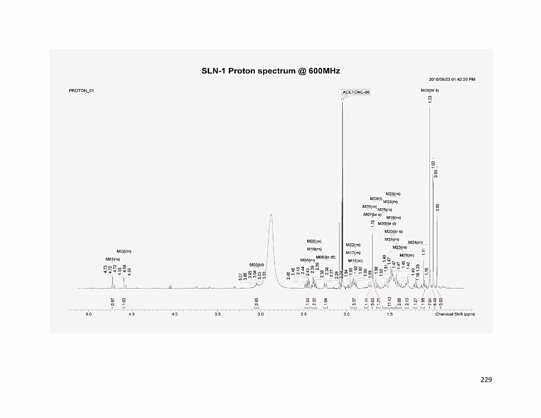
229

230

231

232
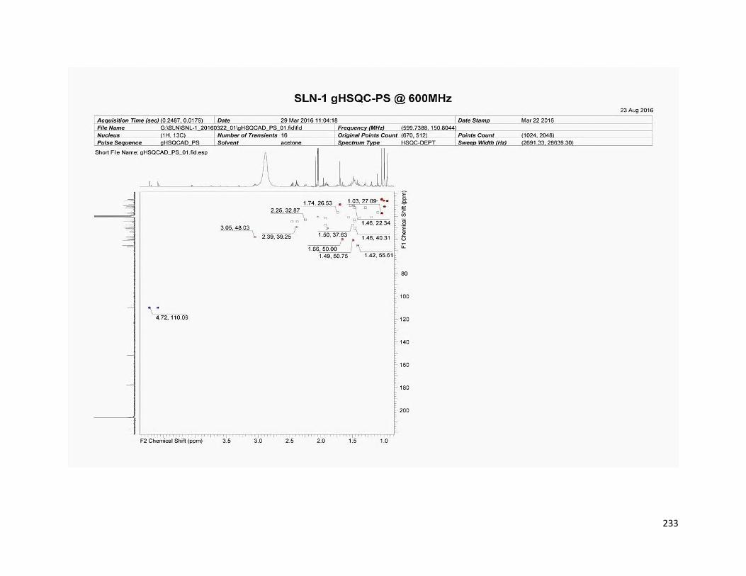
233

234

235

236

237

238

239

240

241

242

243

244

245

246

247

248

249

250

251

252

253

254
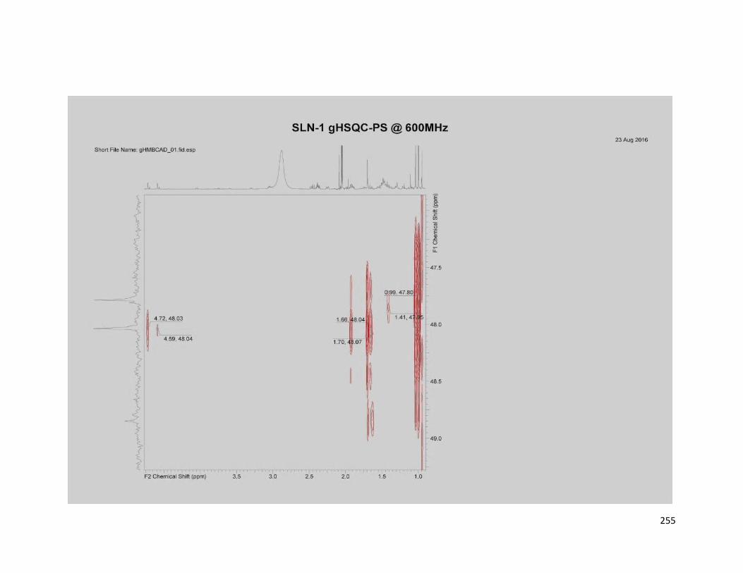
255

256

257

258

259

260

261

262
Appendix B: Structure elucidation of compound PK-B

263
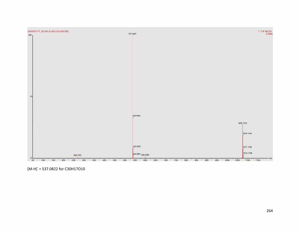
264
[M-H]- = 537.0822 for C30H17O10

265
[M-H]+ = 539.0959 for C30H19O10

266
Appendix C: Published articles

277