Isolation and identification of Pandoraea spp. From ...
8
RESEARCH Open Access Isolation and identification of Pandoraea spp. From bronchoalveolar lavage of cystic fibrosis patients in Iran Mohammad Tabatabaei 1* , Mahdi Dastbarsar 1 and Mohammad Ashkan Moslehi 2 Abstract Background: Pandoraea species are gram negative, motile, non-spore forming, rod shaped and oxidase positive, obligate aerobes bacteria, and have one polar flagellum. Most of Pandoraea species are associated with lung infections in cystic fibrosis patients. Cystic fibrosis is the most prevalent autosomal recessive hereditary disease in the world that affects various organs of the body. The main important cause of death in these patients is lung involvement. This study was conducted to isolate and identify Pandoraea bacterium from bronchoalveolar lavage and sputum samples of cystic fibrosis patients in Shiraz, Iran. Methods: In this research 31 samples of bronchoalveolar lavage and sputum were examined by culture and PCR method. Then confirmed isolates were evaluated for susceptibility to different antibiotics and ability to produce biofilm. Results: The results of this study after cultivation, purification and DNA extraction led to the isolation of 4 Pandoraea bacterium by PCR using specific primers. Antibiotic susceptibility test were indicated all isolates were resistant to gentamicin, amikacin and imipenem and susceptible to ciprofloxacin, trimethoprim-sulfumethoxazole, piperacillin and tetracycline. Ability to create biofilm was indicated by some of Pandoraea isolates. According to findings of this study, ability to synthesis biofilm by Pandoraea isolates and resistance to some antibiotics are very important. Conclusions: Our study notes the role of P. pnomenusa as an emerging pathogen that can cause chronic lung colonization in CF patients. Identification tools need to be accurate and must be based on molecular techniques. Also our findings should raise awareness about antibiotic resistance in cystic fibrosis patients in Iran and ability of including bacterial agents to produce biofilm is an alarm for public health. Thus clinicians should exercise caution about finding of clinical relevance of this pathogen to the infection and prescribing antibiotics, especially in cases of children infections. Keywords: Pandoraea spp., Cystic fibrosis, Bronchoalveolar lavage, PCR, Iran Introduction The genus Pandoraea is composed of aerobic, non-spore- forming, non-nitrate reducing, non-lactose fermenting, and gram negative rods shaped bacteria, with polar flagella. They have been grown at 30–37 °C in 0.5 and 1.5% NaCl or on Drigalski agar, assimilate D-gluconate, L-malate, and phenylacetate, and have catalase, acid and alkaline phos- phatase and leucine arylamidase activity [1]. Different spe- cies of Pandoraea have been described: P. apista, P. norimbergensis, P. pnomenusa, P. pulmonicola, P. spu- torum, P. fibrosis, P. terrae, P. oxalativorance, P. faecigallinarum, P. vervacti, P. thiooxidance and four un- named genomospecies. The majority of isolates have been isolated from respiratory samples from patients with cystic fibrosis (CF) or other underlying chronic lung disease but can also be isolated from other clinical samples (including blood) and from the environmental samples such as soil, food, sea, and drinking water [1–5]. Its closest phylogenetic relative is the genus Burkholderia and, like members of the genus Burkholderia, the Pandoraea spp. are emerging im- portant respiratory pathogens, particularly in people with cystic fibrosis (CF). Species in this genus are often misiden- tified as Burkholderia cepacia complex (Bcc) or Ralstonia species owing to overlapping biochemical profiles without differences that reliably distinguish between species [6]. It © The Author(s). 2019 Open Access This article is distributed under the terms of the Creative Commons Attribution 4.0 International License (http://creativecommons.org/licenses/by/4.0/), which permits unrestricted use, distribution, and reproduction in any medium, provided you give appropriate credit to the original author(s) and the source, provide a link to the Creative Commons license, and indicate if changes were made. The Creative Commons Public Domain Dedication waiver (http://creativecommons.org/publicdomain/zero/1.0/) applies to the data made available in this article, unless otherwise stated. * Correspondence: [email protected] 1 Faculty of Veterinary Medicine, Shiraz University, Shiraz, Iran Full list of author information is available at the end of the article Tabatabaei et al. Italian Journal of Pediatrics (2019) 45:118 https://doi.org/10.1186/s13052-019-0687-x
-
Upload
khangminh22 -
Category
Documents
-
view
1 -
download
0
Transcript of Isolation and identification of Pandoraea spp. From ...

RESEARCH Open Access
Isolation and identification ofPandoraea spp. From bronchoalveolarlavage of cystic fibrosis patients in IranMohammad Tabatabaei1*, Mahdi Dastbarsar1 and Mohammad Ashkan Moslehi2
Abstract
Background: Pandoraea species are gram negative, motile, non-spore forming, rod shaped and oxidase positive,obligate aerobes bacteria, and have one polar flagellum. Most of Pandoraea species are associated with lunginfections in cystic fibrosis patients. Cystic fibrosis is the most prevalent autosomal recessive hereditary disease inthe world that affects various organs of the body. The main important cause of death in these patients is lunginvolvement. This study was conducted to isolate and identify Pandoraea bacterium from bronchoalveolar lavageand sputum samples of cystic fibrosis patients in Shiraz, Iran.
Methods: In this research 31 samples of bronchoalveolar lavage and sputum were examined by culture and PCRmethod. Then confirmed isolates were evaluated for susceptibility to different antibiotics and ability to produce biofilm.
Results: The results of this study after cultivation, purification and DNA extraction led to the isolation of4 Pandoraea bacterium by PCR using specific primers. Antibiotic susceptibility test were indicated all isolates wereresistant to gentamicin, amikacin and imipenem and susceptible to ciprofloxacin, trimethoprim-sulfumethoxazole,piperacillin and tetracycline. Ability to create biofilm was indicated by some of Pandoraea isolates. According to findingsof this study, ability to synthesis biofilm by Pandoraea isolates and resistance to some antibiotics are very important.
Conclusions: Our study notes the role of P. pnomenusa as an emerging pathogen that can cause chronic lungcolonization in CF patients. Identification tools need to be accurate and must be based on molecular techniques. Alsoour findings should raise awareness about antibiotic resistance in cystic fibrosis patients in Iran and ability of includingbacterial agents to produce biofilm is an alarm for public health. Thus clinicians should exercise caution about finding ofclinical relevance of this pathogen to the infection and prescribing antibiotics, especially in cases of children infections.
Keywords: Pandoraea spp., Cystic fibrosis, Bronchoalveolar lavage, PCR, Iran
IntroductionThe genus Pandoraea is composed of aerobic, non-spore-forming, non-nitrate reducing, non-lactose fermenting, andgram negative rods shaped bacteria, with polar flagella.They have been grown at 30–37 °C in 0.5 and 1.5% NaClor on Drigalski agar, assimilate D-gluconate, L-malate, andphenylacetate, and have catalase, acid and alkaline phos-phatase and leucine arylamidase activity [1]. Different spe-cies of Pandoraea have been described: P. apista, P.norimbergensis, P. pnomenusa, P. pulmonicola, P. spu-torum, P. fibrosis, P. terrae, P. oxalativorance, P.
faecigallinarum, P. vervacti, P. thiooxidance and four un-named genomospecies. The majority of isolates have beenisolated from respiratory samples from patients with cysticfibrosis (CF) or other underlying chronic lung disease butcan also be isolated from other clinical samples (includingblood) and from the environmental samples such as soil,food, sea, and drinking water [1–5]. Its closest phylogeneticrelative is the genus Burkholderia and, like members of thegenus Burkholderia, the Pandoraea spp. are emerging im-portant respiratory pathogens, particularly in people withcystic fibrosis (CF). Species in this genus are often misiden-tified as Burkholderia cepacia complex (Bcc) or Ralstoniaspecies owing to overlapping biochemical profiles withoutdifferences that reliably distinguish between species [6]. It
© The Author(s). 2019 Open Access This article is distributed under the terms of the Creative Commons Attribution 4.0International License (http://creativecommons.org/licenses/by/4.0/), which permits unrestricted use, distribution, andreproduction in any medium, provided you give appropriate credit to the original author(s) and the source, provide a link tothe Creative Commons license, and indicate if changes were made. The Creative Commons Public Domain Dedication waiver(http://creativecommons.org/publicdomain/zero/1.0/) applies to the data made available in this article, unless otherwise stated.
* Correspondence: [email protected] of Veterinary Medicine, Shiraz University, Shiraz, IranFull list of author information is available at the end of the article
Tabatabaei et al. Italian Journal of Pediatrics (2019) 45:118 https://doi.org/10.1186/s13052-019-0687-x

is because of these limitations that reliable identification re-quires 16S ribosomal DNA sequence analysis.Cystic fibrosis is caused by mutations in the CFTR (cys-
tic fibrosis transmembrane conductance regulator) gene[7]. The commonest mutation is the deletion of phenyl-alanine at codon 508 (phe508del). This occurs in about70% of patients with cystic fibrosis. The primary functionof the CFTR protein is as an ion channel that regulates li-quid volume on epithelial surfaces through chloride secre-tion and inhibition of sodium absorption. The commonlyaccepted explanation for airway disease in cystic fibrosis isthe “low volume” hypothesis. A reduced volume of airwaysurface liquid causes failure of mucociliary clearance, thelungs’ innate defense mechanism [8]. The mucociliary dys-function means that a patient with CF cannot effectivelyclear inhaled bacteria. In addition, there is an excessive in-flammatory response to pathogens. For a given bacterialload, a person with CF will have up to 10 times more in-flammation than a person with a lower respiratory tractinfection but without the disease. The reasons for the ex-cessive inflammatory response to pathogens are not fullyunderstood. The abnormal composition and secretion ofmucus may also be important. At birth, the airway is unin-fected and probably uninflamed [9], but the end result ofthe abnormalities described above is irreversible airwaydamage with bronchiectasis and respiratory failure in mostpatients. Ion and water abnormalities may also cause dis-ease in other epithelia-lined organs.The main source of morbidity and mortality in CF pa-
tients, is the decline in the pulmonary function subsequentto pathogenic colonization with non-fermenting Gramnegative bacteria (NFGNB) that they encounter throughouttheir lives.CF patients are particularly susceptible to infec-tions caused by specific bacterial pathogens such as Pseudo-monas aeruginosa, Staphylococcus aureus and Haemophilusinfluenzae [10].Although Pandoraea species have also been isolated
from sputum samples of CF patients, there is still verylittle known about their mechanisms of pathogenicity ortheir roles in CF lung disease [11]. In addition, Pandor-aea isolates have been recovered from both CF and non-CF patients from a variety of clinical samples includingblood, sputum, urine, the upper airways and lung tissue[12]. The recovery of Pandoraea isolates from the bloodof patients indicates that this organism is capable of in-vading tissue [13, 14]. Antibiotic therapy for treatmentof infection is difficult due to the limited number ofantibiotics to which these species are susceptible:tetracycline, imipenem and trimethoprim–sulfameth-oxazole [15].However, the clinical significance of colonization with
these organisms remains unclear [13] and there are limitedand conflicting data available on the clinical outcome ofpatients colonized with Pandoraea.
Whether Pandoraea spp. is truly pathogenic is not yetfully understood, there is evidence that infection with Pan-doraea spp. invokes a host inflammatory response. A num-ber of virulence factors have been associated with clinicalvirulence of Pandoraea spp. and Bcc, including stimulationof pro-inflammatory cytokine secretion known to causelung tissue damage (IL-6 and IL-8), biofilm formation and,in the case of Bcc, the ability to invade lung epithelial cells,which may contribute to the persistence of the strains inthe CF lung [16–20]. Indeed, biofilm formation by Pandor-aea spp. and Bcc has been specifically associated with in-creased resistance to antibiotics and maintenance of thebacteria in the lung [21–24]. Here, we report the results ofisolation and identification of Pandoraea spp. and theirability to form biofilms in vitro, in order to gain insight intotheir virulence potential in CF lung infections.In the clinical microbiology laboratory, identification to
the species level and differentiation of Pandoraea speciesfrom organisms belonging to the Bcc, R. pickettii, or R.paucula may be problematic [1, 6, 25]. To aid in the iden-tification of these organisms, we used PCR-based identifi-cation strategies based on the 16S rRNA gene (rDNA).
Materials and methodSample collectionFrom November 2017 to August 2018, following the sed-ation of CF patients in Namazi hospital (Shiraz, Iran), 31samples of bronchoalveolar lavage and sputum were col-lected. Then the samples were transferred to the bacteri-ology laboratory, faculty of veterinary medicine of ShirazUniversity, in cold condition. The study was approved bythe Ethics Committee of Shiraz University of MedicalSciences.
Culture and isolationTo obtain strains and to compare culture and PCR re-sults, cultures were performed initially as enrichmentcultures. Upon arrival, bronchoalveolar lavage and spu-tum were cultured in BHI broth and incubated aerobic-ally at 37 °C for 24 h. Initial enriched cultures weretransferred to the enrichment culture plates by serialstreaking the cultures on sheep blood agar and incu-bated aerobically at 37 °C for 24–48 h.Then, a loop full of the identified colonies were streaked
on a new blood agar and incubated aerobically at 37 °C for24 h. Next, from culture positive plates, typical colonieswere subjected to gram’s staining to study staining reac-tions and cellular morphology under light microscope.Mixed and gram-negative bacteria were further sub cul-tured with due care, on both blood agar and MacConkeyagar plates for final identification. The growth of typicalcolonies on both blood agar and MacConkey agar wascharacterized using blood agar for the presence and typeof haemolysis, and the general appearance of colonies
Tabatabaei et al. Italian Journal of Pediatrics (2019) 45:118 Page 2 of 8

(morphology, color, shape, size and consistency) and theMacConkey agar for the ability to ferment lactose. Purecultures of single colony type were further analyzed bycatalase and oxidase tests. Confirmation of bacteria to spe-cies level was aided by using the biochemical testsincluding, metabolism of sugars such as glucose, fructose,lactose and tests for metabolic end products such asnitrate reduction, unease and citrate activity, growth oncetrimide agarat 42 °C and O/F medium following stand-ard procedures. For final confirmation, isolates evaluatedby two PCR reactions. A first PCR assay was designed toidentify all Pandoraea spp. Subsequent PCR assays weredesigned to identify individual Pandoraea species.
DNA extractionA few colonies from the phenotypically characterized purecultures of Pandoraea from 24 to 48 h growth on bloodagar plates were transferred into 1.5 ml Eppendorf tubes.Total DNA was prepared using the Gram negative DNAextraction kit (Cinagen, Iran). The protocol for Gramnegative bacteria as described in the kit was followed forextractions. The extracted DNA were determined to be ofgood quality and DNA concentration was measured usingNanodrop (10,000 V 3.52). DNA concentrations wereadjusted to 15 ng μL− 1 before PCR amplification. Finally,extracted DNA stored at −20 °C for further use.
Master mixture of PCRThe oligonucleotide primers used in this study were synthe-sized by Cinagen Company (Iran). The forward and the re-verse primers sequences are as PanF (5´GGGCTYAACCTGGGAACTGCATTC3´), and PanR (5´CGRYTTGGCRRCCCTCTGTACCG3´) [26]. Also PCR evaluations havebeen done with two universal primers for amplification of16S rRNA gene. The reaction mixture solution for PCR wasprepared using 2 μl of 10x PCR buffer, 1.5 μl (1.5mM)MgCl2, 1.2 μl (200mM) dNTPs, 1.2 μl (100 nmol) each pri-mer of Pandoraea, 0.2 U of Taq DNA polymerase, 14.7 μlH2O, and 3 μl of DNA template to have a final volume of25 μl. Using 0.2ml thin wall microtubes. After initial de-naturation (at 94 °C for 5min), amplification conditionswere, denaturation at 94 °C for 1min, annealing at 60 °Cfor 45 s, and extension at 72 °C for 1min. This was re-peated for 30 cycles in a Block assembly 96G thermocyclerwith a hot top assembly (Analytic Jena, Germany), with afinal extension of 72 °C for 10min and the PCR productsremained in the thermal cycler at 4 °C until they were col-lected. All DNA isolation procedures were carried out in aClass II biological safety cabinet in a room geographicallyseparate from that used to set up reaction mixes and alsofrom the ‘post PCR’ room, in order to prevent crossovercontamination by extraneous nucleic acids and in accord-ance with good molecular diagnostic practice. All reactionmixes were set up in a PCR hood in a room separate from
that used to extract DNA, and from the amplification andpost-PCR room, in order to minimize contamination. Anegative control consisting of all component of reactionmixture except the DNA template was included in all PCR.Positive controls were included in the PCR from coloniesthat were confirmed according to NCBI gene bank sequen-cing. In order to evaluate the PCR results electrophoresis inagarose gel was carried out. PCR amplicons were assessedby loading 8 μl of the PCR product plus 2 μl of loading buf-fer into separate wells of a 1% (w/v) agarose gel (Agarose I;Cinnagen, Iran) containing safe stain. A molecular weightmarker (50 bp; Cinnagen, Iran) was loaded into the firstwell to determine the size of the amplified fragments. Thegel was immersed in TBE buffer and subjected to a voltagedifference of 100 V that led to separation of the fragments.Visualization was undertaken using an ultraviolet transillu-minator (BTS-20, Japan), and the resulting image was cap-tured by a computer software program (Alpha Ease; AlphaInnotech).For the final confirmation of isolates of Pandoraea,
PCR products of Pan primers for some samples accord-ing to the Macrogene Company’s instructions were pre-pared at 50 μl volume and were transferred forsequencing. In addition, 16S rRNA gene sequencing forspecies identification was applied for some of the Pan-doraea isolates. The sequences were compared with pre-viously published 16SrRNA (GenBank accession no.NR-152002), Pandoraea gene sequences.
Antimicrobial susceptibility testingAntimicrobial susceptibility testing for Pandoraea spp. iso-lates was performed by disk diffusion method according tothe Clinical and Laboratory Standards Institute (CLSI) [27].Disk diffusion method was performed on Muller-Hintonagar (Merck, Germany), using an inoculum of 105 CFU/ml.Antibiotic disks (Padtan teb, Iran) including amikacin, cip-rofloxacin, trimethoprim- sulfumethoxazole, gentamicin,piperacillin tetracycline and imipenem. These antibioticdisks were then placed on agar plates and incubated at 37°C for 24 h. Escherichia coli (ATCC 25922) and Pseudo-monas aeruginosa (ATCC 27853) were used as qualitycontrols strain in each susceptibility determination. Thediameter of inhibition zone was measured in millimetersand isolates were scored as susceptible or resistant by com-paring results with values recommended on standardcharts.
Biofilm assayIn order to evaluate the ability to create biofilms in Pan-doraea spp. isolates used the method of Merritt et al.and Wakimoto model [28, 29]. Briefly, 100 μl of each di-luted culture was relocate into individual wells of micro-titer dish and grown at 37 °C overnight. All wells werestained with 125 μl of 0.2% (w/v) crystal violet solution
Tabatabaei et al. Italian Journal of Pediatrics (2019) 45:118 Page 3 of 8
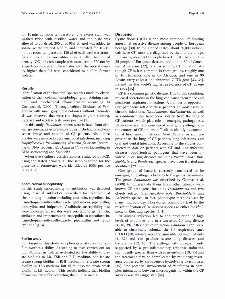
for 10 min at room temperature. The excess stain waswashed twice with distilled water and the plate wasallowed to air dried. 200 μl of 95% ethanol was added tosolubilize the stained biofilm and incubated for 10–15min at room temperature. 125 μl of each well was trans-ferred into a new microtiter dish. Finally, the opticaldensity (OD) of each sample was measured at 570 nm bya spectrophotometer. The isolates with the optical dens-ity higher than 0.2 were considered as biofilm formerisolates.
ResultsIdentification of the bacterial species was made by obser-vation of their colonial morphology, gram staining reac-tion and biochemical characteristics according toCoenyeet al. (2000). Through culture 8isolates of Pan-doraea with small gray circle colonies without haemoly-sis was observed that were rod shapes in gram staining.Catalase and oxidase tests were positive [1].In this study, Pandoraea strains were isolated from clin-
ical specimens, as in previous studies including bronchoal-veolar lavage and sputum of CF patients. Also, mostisolates were involved in polymicrobial infections, includingStaphylococcus, Pseudomonas, Neisseria flavorans (accord-ing to DNA sequencing), Delftia acidovorans (according toDNA sequencing) and Klebsiella.When these culture positive isolates evaluated by PCR,
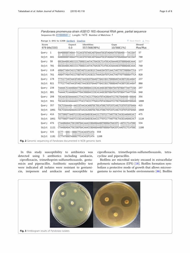
using the stated primers, all the samples tested for thepresence of Pandoraea were identified as 100% positive(Figs. 1, 2).
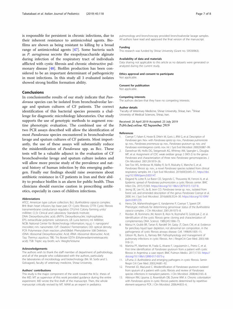
Antimicrobial susceptibilityIn this study susceptibility to antibiotics was detectedusing 7 usual antibiotics prescribed for treatment ofchronic lung infection including amikacin, ciprofloxacin,trimethoprim-sulfumethoxazole, gentamicin, piperacillin,tetracyline and imipenem. Antibiotic susceptibility testwere indicated all isolates were resistant to gentamicin,amikacin and imipenem and susceptible to ciprofloxacin,trimethoprim-sulfumethoxazole, piperacillin and tetra-cycline (Fig. 3).
Biofilm assayOur target in this study was phenotypical survey of bio-film synthesis ability. According to tests carried out onfour Pandoraea isolates evaluated for the ability to cre-ate biofilms in LB, TSB and BHI medium, one isolatecreate strong biofilm in BHI medium, one create strongbiofilm in TSB medium, but all four isolate create weakbiofilm in LB medium. This results indicate that biofilmformation can differ according the culture media.
DiscussionCystic fibrosis (CF) is the most common life-limitingautosomal recessive disease among people of Europeanheritage [30]. In the United States, about 30,000 individ-uals have CF; most are diagnosed by six months of age.In Canada, about 4000 people have CF [31]. Around 1 in25 people of European descent, and one in 30 of Cauca-sian Americans [32], is a carrier of a CF mutation. Al-though CF is less common in these groups, roughly onein 46 Hispanics, one in 65 Africans, and one in 90Asians carry at least one abnormal CFTR gene [33, 34].Ireland has the world’s highest prevalence of CF, at onein 1353 [35].CF is a common genetic disease. Due to this condition,
mucosal secretions in the lung can cause recurrence andpersistent respiratory infections. A number of opportun-istic pathogens settle in these patients. In most cases, inchronic infections, Pseudomonas, Burkholderia, as wellas Pandoraea spp. have been isolated from the lung ofCF patients, which play role in emerging pathogenesis.Pandoraea spp. are considered emerging pathogens inthe context of CF and are difficult to identify by conven-tional biochemical methods. Most Pandoraea spp. arepresent in the lung of CF patients, lung infections, andoral and dental infections. According to the studies con-ducted to data on patients with CF and lung infectiondiseases, opportunistic pathogens that have been in-volved in causing diseases including Pseudomonas, Bur-kholderia and Pandoraea species, have been isolated andidentified [29, 36–38].One group of bacteria currently considered to be
emerging CF pathogens belongs to the genus Pandoraea.The genus Pandoraea was described by Coenye et al.(2000) to differentiate them from other already well-known CF pathogens, including Pseudomonas and twoclosely related Gram-negative rods, Burkholderia andRalstonia species. In fact, phenotypic methods used bymany microbiology laboratories commonly lead to themisidentification of Pandoraea species as either Burkhol-deria or Ralstonia species [2, 4].Pandoraea infection led to the production of high
levels of antibodies, and to a worsened CF lung disease[4, 10, 39]. After first colonization, Pandoraea spp. wereable to chronically colonize the CF respiratory tract(CFRT) [10, 40–42], were transmissible between patients[4, 37] and can produce severe lung diseases andbacteremia [12, 43]. The pathogenicity appears mainlysupported by a pro-inflammatory response inductionsignificantly greater than with P. aeruginosa [19, 44] andthe treatment may be complicated by multidrug resist-ance conferred by carbapenem hydrolyzing oxacillinases[19]. The potential involvement of Pandoraea in com-plex interactions between microorganisms within the CFairways was also suggested [44].
Tabatabaei et al. Italian Journal of Pediatrics (2019) 45:118 Page 4 of 8

In current study from 31 samples of bronchoalveo-lar lavage and sputum, by culture method eight Pan-doraea bacteria were isolated and identified. Thesebacteria were isolated and purified using conventionaldiagnostic methods such as culture and biochemicaltests. But finally by PCR assay just 4 isolates con-firmed as Pandoraea. Also, in addition to Pandoraeaspp. other bacteria including Staphylococcus, Pseudo-monas, Neisseria flavorans, Delftia acidovorans andKlebsiella were also isolated.In 2001, Coenye et al. identified and investigated spe-
cies of Pandoraea bacteria. In this study, from 123 sam-ples, Pandoraea (69), Burkholderia (30), Ralstonia (9)and Pseudomonas aeruginosa (5) were isolated and iden-tified. For the first time, PCR testing was performed toidentify the member of genus Pandoraea, which wereable to detect P. apista, P. pnomenusa, P. sputorum, P.pulmonicola and P. norimbergensis [26].Daneshvar and colleagues in 2001 examined the cel-
lular effects of Pandoraea spp. on lung cells in CFpatients. The study also found that it was possible toisolate the Pandoraea from the cultures of patient’sblood who did not have cystic fibrosis [2].In the study of Spreet et al., conducted in Canada in
2002, from a total of 447 CF patients in 8 provinces in-cluded 5 Pandoraea (1.1%), 412 Burkholderia (92.6%)and 5 Ralstonia (1.1%) bacteria, using culture tech-niques, biochemical tests and molecular techniques in-cluding PFGE, RAPD and RFLP were identified andisolated [45].
In 2003, Jorgensen et al., examined the epidemic of P.apista in patients with cystic fibrosis. According to the re-sults of this study, it has been shown that P. apista shouldbe added to the ever increasing list of pathogens that cancause chronic lung infections in CF patients [43].In 2008, Karher et al. examined the pathogenic and
genetic characteristics of Pandoraea species in lungepithelial cells. In this study, 17 Pandoraea bacteriawere isolated including 5 species: P. apista, P. pnome-nusa, P. sputorum, P. pulmonicola and P. norimber-gensis [38].In a study conducted by Panickar and David (2015)
from October 2012 to September 2014 on outpatient,and lavage samples from 182 CF children treated at theRoyal Manchester Hospital, 5(3%) were Burkholderia,17(19%) Exophiala, 32(18) Achromobacter and 32 (18%)Rhodotorula, 18 (10%) Achromobacter, 1 Ralstonia and 6(3%) Pandoraea were identified. This study showed thatthe number of Pandoraea and Achromobacter bacteriain CF children is increasing [39].Antibiotic treatment of infections caused by Pandor-
aea species is difficult because it has been demon-strated resistant against a wide range of antibioticssuch as ampicillin, cefazolin, piperacillin, azithroman,broad- spectrum cephalosporins and aminoglycosides,and show a different response to quinolones, tri-methoprim-sulfumethoxazole, colistin and carbapen-ems. Pandoraea species are resistant to mostantibiotics, including beta-lactams and aminoglyco-sides. The bacteria in the subjects evaluated moderatesensitivity to imipenem, doxycycline and ceftriaxone,and an unusual antibiotic susceptibility pattern to car-bapenem was also found in Pandoraea bacteria andresistance to meropenem and sensitivity to imipenemhas been reported.The result shows that for antibiotic treatment of
Pandoraea infection, antibiogram test for each personhas been done individually. Then if a person is allergicto imipenem they can use carbapenem. All Pandoraeaspecies except P. apista, G9278 are resistant to amika-cin and cefazolin antibiotics, most of which are resist-ant to broad-spectrum cephalosporins, azithroman andpiperacillin. Also, these organisms are resistant toaminoglycosides such as gentamicin, tobramycin andamikacin, but their susceptibility to fluroquinolones isdifferent [40].Puges et al. (2015), reported that P. sputorum was
susceptible to imipenem and resistant to meropenem.This discrepancy has already been described by severalauthors and is because of meropenem-hydrolyzing β-lactamases. Some strains are resistant to both imipe-nem and meropenem, and an imipenem-hydrolyzingoxacillinase named OXA-62 has been identified in P.pnomenusa [45].
Fig. 1 PCR amplification profile of Pandoraea spp. from DNA isolateddirectly from samples with PAN primers. Lane a: 50 bp DNA marker,Lane b: Negative control, Lane C: Positive control and Lanes D-G:Positive samples (645 bp amplicon size). It should be noted that 4samples were absolutely homologues with documented genomicsequencing of Pandoraea in NCBI genomic bank
Tabatabaei et al. Italian Journal of Pediatrics (2019) 45:118 Page 5 of 8
In this study susceptibility to antibiotics wasdetected using 5 antibiotics including amikacin,ciprofloxacin, trimethoprim-sulfumethoxazole, genta-
micin and piperacillin. Antibiotic susceptibility testwere indicated all isolates were resistant to gentami-cin, imipenem and amikacin and susceptible to
ciprofloxacin, trimethoprim-sulfumethoxazole, tetra-cycline and piperacillin.Biofilms are microbial society encased in extracellular
polymeric substances (EPS) [18]. Biofilm formation sym-bolizes a protective mode of growth that allows microor-ganisms to survive in hostile environments [46]. Biofilm
Fig. 2 Genomic sequencing of Pandoraea documented in NCBI genomic bank
Fig. 3 Antibiogram results of Pandoraea isolates
Tabatabaei et al. Italian Journal of Pediatrics (2019) 45:118 Page 6 of 8
is responsible for persistent in chronic infections, due totheir inherent resistance to antimicrobial agents. Bio-films are shown as being resistant to killing by a broadrange of antimicrobial agents [47]. Some bacteria suchas P. aeruginosa secrete the exopolysaccharide alginateduring infection of the respiratory tract of individualsafflicted with cystic fibrosis and chronic obstructive pul-monary disease [48]. Biofilm production has been con-sidered to be an important determinant of pathogenicityin most infections. In this study all 3 evaluated isolatesshowed strong biofilm formation ability.
ConclusionsIn conclusionsthe results of our study indicate that Pan-doraea species can be isolated from bronchoalveolar lav-age and sputum cultures of CF patients. The correctidentification of this bacterial species presents a chal-lenge for diagnostic microbiology laboratories. Our studysupports the use of genotypic methods to augment rou-tine phenotypic evaluation. The combined use of thetwo PCR assays described will allow the identification ofmost Pandoraea species encountered in bronchoalveolarlavage and sputum cultures of CF patients. Most import-antly, the use of these assays will substantially reducethe misidentification of Pandoraea spp. as Bcc. Thesetests will be a valuable adjunct in the evaluation of CFbronchoalveolar lavage and sputum culture isolates andwill allow more precise study of the prevalence and nat-ural history of human infection by this emerging patho-gen. Finally our findings should raise awareness aboutantibiotic resistance in CF patients in Iran and their abil-ity to produce biofilm is an alarm for public health. Thusclinicians should exercise caution in prescribing antibi-otics, especially in cases of children infections.
AbbreviationsATCC: American type culture collection; Bcc: Burkholderia cepacia complex;BHI: Brain heart infusion; bp: base pair; CF: Cystic fibrosis; CFTR: Cystic fibrosistransmembrane conductance regulator; CFU/ml: Colony forming units/milliliter; CLSI: Clinical and Laboratory Standards Institute;DNA: Deoxyribonucleic acid; dNTPs: Deoxyribonucleic triphosphates;EPS: extracellular polymeric substances; LB: Luria bertoni; ml: Milliliter;NCBI: National Center for Biotechnology Information; ng μL-1: nanogram/microliter; nm: nanometer; O/F: Oxiation/ Fermentation; OD: optical density;PCR: Polymerase chain reaction; phe508del: Phenylalanine 508 Deletion;rDNA: ribosomal Deoxyribonucleic Acid; rRNA: ribosomal ribonucleic Acid;Taq: Thermus aquiticus; TBE: Tris Borate EDTA (Ethylenediaminetetraaceticacid); TSB: Triptic soy broth; w/v: Weight/Volume
AcknowledgementsThe authors wish to thank the staff member of department of pathobiology,and all of the people who collaborated with the authors, particularlythe laboratories of microbiology and biotechnology (Mr. M. Sorbi and S.Golvagoei), faculty of veterinary medicine, Shiraz University.
Authors’ contributionsThis study is the major component of the work toward the M.Sc. thesis ofthe MD. MT as supervisor of this work provided guidance during the entireexperiment. MD wrote the first draft of the manuscript. Then, the wholemanuscript critically revised by MT. MAM as an expert in pediatrics
pulmonology and bronchoscopy provided bronchoalveolar lavage samples.All authors have read and approved the final version of the manuscript.
FundingThis research was funded by Shiraz University (Grant no. S9530963).
Availability of data and materialsData sharing not applicable to this article as no datasets were generated oranalyzed during the current study.
Ethics approval and consent to participateNot applicable.
Consent for publicationNot applicable.
Competing interestsThe authors declare that they have no competing interests.
Author details1Faculty of Veterinary Medicine, Shiraz University, Shiraz, Iran. 2ShirazUniversity of Medical Sciences, Shiraz, Iran.
Received: 20 April 2019 Accepted: 23 July 2019
References1. Coenye T, Falsen E, Hoste B, Ohle’n M, Goris J, JRW G, et al. Description of
Pandoraea gen. Nov. with Pandoraea apista sp. nov., Pandoraea pulmonicolasp. nov., Pandoraea pnomenusa sp. nov., Pandoraea sputorum sp. nov. andPandoraea norimbergensis comb. nov. Int J Syst Evol Microbiol. 2000;50:887–99.
2. Daneshvar MI, Hollis DG, Steigerwalt AG, Whitney AM, Spangler L, DouglasMP, et al. Assignment of CDC weak oxidizer group 2 (WO-2) to the genusPandoraea and characterization of three new Pandoraea genomospecies. JClin Microbiol. 2001;39:1819–26.
3. See-Too WS, Ambrose M, Malley R, Ee R, Mulcahy E, Manche E, et al.Pandoraea fibrosis sp. nov., a novel Pandoraea species isolated from clinicalrespiratory samples. Int J Syst Evol Microbiol. 2019;69(3):645–51. https://doi.org/10.1099/ijsem.0.003147.
4. Degand N, Lotte R, Le Butor CD, Segonds C, Thouverez M, Ferroni A, et al.Epidemic spread of Pandoraea pulmonicolain a cystic fibrosis center. BMCInfect Dis. 2015;15:583. https://doi.org/10.1186/s12879-015-1327-8.
5. Jeong SE, Lee HJ, Jia B, Jeon CO. Pandoraea terrae sp. nov., isolated fromforest soil, and emended description of the genus Pandoraea Coenye et al.2000. Int J Syst Evol Microbiol. 2016;66(9):3524–30. https://doi.org/10.1099/ijsem.0.001229.
6. Henry DA, Mahenthiralingam E, Vandamme P, Coenye T, Speert DP.Phenotypic methods for determining genomovar status of the Burkholderiacepacia complex. J Clin Microbiol. 2001;39:1073–8.
7. Riordan JR, Rommens JM, Kerem B, Alon N, Rozmahel R, Grzelczak Z, et al.Identification of the cystic fibrosis gene: cloning and characterization ofcomplementary DNA. Science. 1989;245:1066–73.
8. Matsui H, Grubb BR, Tarran R, Randell SH, Gatzy JT, Davis CW, et al. Evidencefor periciliary liquid layer depletion, not abnormal ion composition, in thepathogenesis of cystic fibrosis airways disease. Cell. 1998;95:1005–15.
9. Gibson RL, Burns JL, Ramsey BW. Pathophysiology and management ofpulmonary infections in cystic fibrosis. Am J RespirCrit Care Med. 2003;168:918–51.
10. Martina PF, Martínez M, Frada G, Alvarez F, Leguizamón L, Prieto C, et al.First time identification of Pandoraea sputorum from a patient with cysticfibrosis in Argentina: a case report. BMC Pulmon Medici. 2017;17:33. https://doi.org/10.1186/s12890-017-0373-y.
11. LiPuma JJ. Burkholderia and emerging pathogens in cystic fibrosis. SeminRespir Crit Care Med. 2003;24:681–92.
12. Pimentel JD, MacLeod C. Misidentification of Pandoraea sputorum isolatedfrom sputum of a patient with cystic fibrosis and review of Pandoraeaspecies infections in transplant patients. J Clin Microbiol. 2008;46:3165–8.
13. Atkinson RM, Lipuma JJ, Rosenbluth DB, Dunne WM Jr. Chronic colonizationwith Pandoraea apista in cystic fibrosis patients determined by repetitive-element-sequence PCR. J Clin Microbiol. 2006;44:833–6.
Tabatabaei et al. Italian Journal of Pediatrics (2019) 45:118 Page 7 of 8

14. Stryjewski ME, LiPuma JJ, Messier RH Jr, Reller LB, Alexander BD. Sepsis,multiple organ failure, and death due to Pandoraea pnomenusa infectionafter lung transplantation. J Clin Microbiol. 2003;41:2255–7.
15. Schneider I, Queenan AM, Bauernfeind A. Novel carbapenem-hydrolyzingoxacillinase OXA-62 from Pandoraea pnomenusa. Antimicrob AgentsChemother. 2006;50:1330–5.
16. Burns JL, Jonas M, Chi EY, Clark DK, Berger A, Griffith A. Invasion ofrespiratory epithelial cells by Burkholderia (Pseudomonas) cepacia. InfectImmun. 1996;64:4054–9.
17. Cieri MV, Mayer-Hamblett N, Griffith A, Burns JL. Correlation between an invitro invasion assay and a murine model of Burkholderia cepacia lunginfection. Infect Immun. 2002;70:108–6.
18. Duff C, Murphy PG, Callaghan M, McClean S. Differences in invasion andtranslocation of Burkholderia cepacia complex species in polarized lungepithelial cells in vitro. Microb Pathog. 2006;4:183–92.
19. Caraher E, Collins J, Herbert G, Philip G, Murphy PG, Gallagher CG, CroweMJ, et al. Evaluation of in vitro virulence characteristics of the genusPandoraea in lung epithelial cells. J Med Microbiol. 2008;57(Pt 1):15–20.
20. Green H, Jones AM. Emerging gram-negative bacteria: pathogenic orinnocent bystanders. Curr Opin Pulm Med, 2018; 24:000–000. https://doi.org/10.1097/MCP.0000000000000517.
21. Caraher E, Duff C, Mullen T, Mc Keon S, Murphy P, Callaghan M, McClean S.Invasion and biofilm formation of Burkholderia dolosa is comparable withBurkholderia cenocepacia and Burkholderia multivorans. J Cyst Fibros. 2007;6:49–56.
22. Chernish RN, Aaron SD. Approach to resistant gram negative bacterialpulmonary infections in patients with cystic fibrosis. Curr Opin Pulm Med.2003;9:509–15.
23. Hoiby N, Krogh Johansen H, Moser C, Song Z, Ciofu O, Kharazmi A.Pseudomonas aeruginosa and the in vitro and in vivo biofilm mode ofgrowth. Microbes Infect. 2001;3:23–35.
24. Yu H, Head NE. Persistent infections and immunity in cystic fibrosis. FrontBiosci. 2002;7:d442–57.
25. Moore JE, Coenye T, Vandamme P, Elborn JS. First report of Pandoraeanorimbergensis isolated from food-potential clinical significance. FoodMicrobiol. 2001;18:113–4.
26. Coenye T, Liu L, Vandamme P, LiPuma JJ. Identification of Pandoraea speciesby 16S ribosomal DNA-based PCR assays. J Clin Microbiol. 2001;39:4452–5.
27. CLSI. M100-S25. Performance standards for antimicrobial susceptibilitytesting; Twenty-fifth informational supplement; 2015.
28. Merritt J, Kadour D, O'Toole G. Growing and analyzing static biofilms, CurrProtoc Microbiol. 01 2005; Unite1B.1.
29. Wakimoto N, Nishi J, Sheikh J, Nataro J, Sarantuya J, Iwashita M, Manago K,Tokuda K, Yoshinag M, Kawano Y. Quantitative biofilm assay using amicrotiter plate to screen for enteroaggregative Escherichia coli. Am J TroMed Hyg. 2004;71:687e690.
30. Edward T. Essential Medical Genetics, 2011. John Wiley & Sons. p. 312. ISBN1-118-29370-3. Archived from the original on 2016-04-17.
31. The canadian facts & figures on cystic fibrosis. Archived from the original on2013-06-16.
32. "Genetic Carrier Testing". Cystic Fibrosis Foundation. 2007. Archived fromthe original on 2010-03-23.
33. Rosenstein BJ, Cutting GR. The diagnosis of cystic fibrosis: a consensusstatement. Cystic Fibrosis Foundation consensus panel. J Pediatr. 1998;132(4):58995. https://doi.org/10.1016/S0022-3476(98)70344-0.PMID9580754.
34. Hamosh A, FitzSimmons SC, Macek M, Knowles MR, Rosenstein BJ, CuttingGR. Comparison of the clinical manifestations of cystic fibrosis in black andwhite patients. J Pediatr. 1998;132(2):255–9. https://doi.org/10.1016/S0022-3476(98)70441-X.PMID9506637.
35. Farrell P, Joffe S, Foley L, Canny GJ, Mayne P, Rosenberg M. Diagnosis of cysticfibrosis in the Republic of Ireland: epidemiology and costs. Ir Med J 2007;100(8): 557–560. PMID 17955689. Archived from the original on 2013-12-03.
36. Karatan E, Watnick P. Signals, regulatory networks, and materials that buildand break bacterial biofilms. Microbiol Mol Biol Rev. 2009;73:310–47.
37. Panickar J, David T. Prevalence of emerging pathogens in a large paediatricCF centre. J Cyst Fibros. 2015;14:S68.
38. Ambrose M, Malley RC, Warren SJC, Beggs SA, Swallow OFE, McEwan B, et al.Pandoraea pnomenusa isolated from an Australian patient with cystic fibrosis.Front Microbiol. 2016;7:692. https://doi.org/10.3389/fmicb.2016.00692.
39. Mahenthiralingam E. Emerging cystic fibrosis pathogens and themicrobiome. Paediatr Respir Rev. 2014;15:13–5. https://doi.org/10.1016/j.prrv.2014.04.006.
40. Fernández-Olmos A, Morosini MI, Lamas A, García-Castillo M, García-García L,Cantón R, et al. Clinical and microbiological features of a cystic fibrosispatient chronically colonized with Pandoraea sputorum identified bycombining 16S rRNA sequencing and matrix-assisted laser desorptionionization-time of flight mass spectrometry. J Clin Microbiol. 2012;50:1096–8.https://doi.org/10.1128/JCM.05730-11.
41. Kokcha S, Bittar F, Reynaud-Gaubert M, Mely L, Gomez C, Gaubert JY, et al.Pandoraeapulmonicola chronic colonization in a cystic fibrosis patient,France. New Microbes New Infect. 2013;1:27–9. https://doi.org/10.1002/2052-2975.16.
42. Puges M, Debelleix S, Fayon M, Megraud F, Fayon M, Megraud F, et al.Persistent infection because of Pandoraea sputorum in a young cysticfibrosis patient resistant to antimicrobial treatment. Pediatr Infect Dis J.2015;34(10):1135–7. https://doi.org/10.1097/INF.0000000000000843.
43. Jørgensen IM, Johansen HK, Frederiksen B, Pressler T, Hansen A, Vandamme P,Koch C. Epidemic spread of Pandoraea apista, a new pathogen causing severelung disease in cystic fibrosis patients. Pediatr Pulmonol. 2003;36(5):439–46.
44. Costello A, Herbert G, Fabunmi L, Schaffer K, Kavanagh KA, Caraher EM, etal. Virulence of an emerging respiratory pathogen, genus Pandoraea, in vivoand its interactions with lung epithelial cells. J Med Microbiol. 2011;60:289–99. https://doi.org/10.1099/jmm.0.022657-0.
45. Speert DP, Henry D, Vandamme P, Corey M, Mahenthiralingam E.Epidemiology of Burkholderia cepacia complex in patients with cysticfibrosis, Canada. Emerg Infect Dis. 2002;8(2):181.
46. Caraher E, Collins J, Herbert G, Murphy PG, Gallagher CG, Crowe MJ,McClean S. Evaluation of in vitro virulence characteristics of the genusPandoraea in lung epithelial cells. J Med Microbiol. 2008;57(1):15–20.
47. Davies D, Parsek M, Pearson J, Iglewski B, Costerton J, Greenberg E. Theinvolvement of cell-to-cell signals in the development of a bacterial biofilm.Science. 1998;280:295–8.
48. Costerton JW, Stewart PS, Greenberg EP. Bacterial biofilms: a common causeof persistent infections. Science. 1999;284:1318e1322.
Publisher’s NoteSpringer Nature remains neutral with regard to jurisdictional claims inpublished maps and institutional affiliations.
Tabatabaei et al. Italian Journal of Pediatrics (2019) 45:118 Page 8 of 8