DNA microarray-based detection and identification of Burkholderia mallei, Burkholderia pseudomallei...
11
JOURNAL OF CLINICAL MICROBIOLOGY, Nov. 2007, p. 3743–3753 Vol. 45, No. 11 0095-1137/07/$08.000 doi:10.1128/JCM.00942-07 Copyright © 2007, American Society for Microbiology. All Rights Reserved. DNA Microarray-Based Detection and Identification of Fungal Pathogens in Clinical Samples from Neutropenic Patients Birgit Spiess, Wolfgang Seifarth, Margit Hummel,* Oliver Frank, Alice Fabarius, Chun Zheng, Handan Mo ¨rz, Ru ¨diger Hehlmann, and Dieter Buchheidt III. Medizinische Universita ¨tsklinik, Medizinische Fakulta ¨t Mannheim der Universita ¨t Heidelberg, 68305 Mannheim, Germany Received 7 May 2007/Returned for modification 28 June 2007/Accepted 18 August 2007 The increasing incidence of invasive fungal infections (IFI) in immunocompromised patients emphasizes the need to improve diagnostic tools. We established a DNA microarray to detect and identify DNA from 14 fungal pathogens (Aspergillus fumigatus, Aspergillus flavus, Aspergillus terreus, Candida albicans, Candida dubliniensis, Candida glabrata, Candida lusitaniae, Candida tropicalis, Fusarium oxysporum, Fusarium solani, Mucor racemosus, Rhizopus microsporus, Scedosporium prolificans, and Trichosporon asahii) in blood, bronchoalveolar lavage, and tissue samples from high-risk patients. The assay combines multiplex PCR and consecutive DNA microarray hybridization. PCR primers and capture probes were derived from unique sequences of the 18S, 5.8S, and internal transcribed spacer 1 regions of the fungal rRNA genes. Hybridization with genomic DNA of fungal species resulted in species-specific hybridization patterns. By testing clinical samples from 46 neutropenic patients with proven, probable, or possible IFI or without IFI, we detected A. flavus, A. fumigatus, C. albicans, C. dubliniensis, C. glabrata, F. oxysporum, F. solani, R. microsporus, S. prolificans, and T. asahii. For 22 of 22 patients (5 without IFI and 17 with possible IFI), negative diagnostic results corresponded with negative microarray data. For 11 patients with proven (n 4), probable (n 2), and possible IFI (n 5), data for results positive by microarray were validated by other diagnostic findings. For 11 of 11 patients with possible IFI, the microarray results provided additional information. For two patients with proven and probable invasive aspergillosis, respectively, microarray results were negative. The assay detected genomic DNA from 14 fungal pathogens from the clinical samples, pointing to a high significance for improving the diagnosis of IFI. Systemic fungal infections are increasing and cause severe morbidity and high mortality rates for immunocompromised patients, especially patients with acute leukemia after intensive chemotherapy or allogeneic stem cell transplantation. Besides the increasing incidence, the epidemiology of invasive fungal infections (IFI) is changing, and during the last decade, both uncommon and resistant fungal pathogens, e.g., Zygomycetes, Candida krusei, or Aspergillus terreus, emerged (3, 4, 7, 22, 25, 26). Concerning the outcome of patients with IFI, early initi- ation of antifungal treatment is crucial; since conventional microbiological diagnostic procedures are time-consuming and lack sensitivity and/or specificity, antifungal therapy—in most cases—is started empirically or preemptively based on surrogate marker findings (serologic tests and computed tomography of the chest) (11, 12, 24). In recent years, molecular diagnostic tools, such as PCR, have been established for early detection of fungal pathogens (espe- cially Aspergillus and Candida species) in clinical samples (5, 6, 10, 14, 18, 19, 21, 27, 29, 30) by sensitive and specific methods. To facilitate early diagnosis of IFI caused by common and less common clinically relevant fungi, we established a sensitive and specific DNA microarray combining multiplex PCR and consecutive DNA chip hybridization to detect fungal genomic DNA in clinical samples and we evaluated this assay by testing blood, bronchoalveolar lavage (BAL), and tissue samples from neutropenic patients at high risk for invasive fungal infections. MATERIALS AND METHODS Strains and growth conditions. Fungal test strains were obtained from the Deutsche Sammlung von Mikroorganismen und Zellkulturen GmbH, Braun- schweig, Germany, from the Centraalbureau voor Schimmelcultures, Utrecht, The Netherlands, or from the Institute for Medical Microbiology and Hygiene, Universita ¨tsklinikum Mannheim, University of Heidelberg, Germany. DNA extraction. Prior to DNA extraction, fungal cultures were grown in Sabouraud agar for 72 h at 30°C. DNA extraction from fungal cultures was performed (29) with the QIAGEN DNeasy plant mini kit (QIAGEN, Hilden, Germany) as described previously. DNA from bacterial cultures was obtained from the Deutsche Sammlung von Mikroorganismen und Zellkulturen GmbH, Braunschweig. DNA extraction both for blood and BAL samples was performed according to a protocol described previously (29). Tissue samples were processed additionally in liquid nitrogen for disruption. Primers for multiplex PCRs. The first multiplex primer mix was for amplifi- cation of the internal transcribed spacer 1 (ITS1) regions of fungal rRNA genes. Nine sense primers derived from the highly conserved 18S and three antisense primers from the highly conserved 5.8S regions of the rRNA genes were selected (Table 1). The first primer mix also contained a primer pair selected from the ITS1 region of the human rRNA gene as a positive control. All antisense primers were 5 modified with carbocyanine 3 (Cy3) fluorochrome. Therefore, the am- plification products were Cy3 labeled at the 5 ends. The second multiplex primer set was used for a separate PCR as an internal standard for the quality of DNA and as a positive and a negative control for the hybridization reaction. The primer mix contained primers for human housekeep- ing genes corresponding to glucose-6-phosphate dehydrogenase (G6PD), human ribosomal protein 19 (RPL19), and two plant-specific genes (Arabidopsis thaliana ribosomal protein L23a [AtrpL23a] and A. thaliana actin 2 [ACT2]) as negative controls. The melting temperatures of the primers and possible secondary structures were calculated with Oligo software (Oligo 6.0; MedProbe AS, Oslo, Norway). * Corresponding author. Mailing address: III. Medizinische Universi- ta ¨tsklinik, Medizinische Fakulta ¨t Mannheim der Universita ¨t Heidelberg, Theodor-Kutzer-Ufer 1-3, D-68167 Mannheim, Germany. Phone: 49- 621-383-4115. Fax: 49-621-383-4201. E-mail: margit.hummel@med3 .ma.uni-heidelberg.de. Published ahead of print on 22 August 2007. 3743 on February 18, 2016 by guest http://jcm.asm.org/ Downloaded from
-
Upload
independent -
Category
Documents
-
view
3 -
download
0
Transcript of DNA microarray-based detection and identification of Burkholderia mallei, Burkholderia pseudomallei...
JOURNAL OF CLINICAL MICROBIOLOGY, Nov. 2007, p. 3743–3753 Vol. 45, No. 110095-1137/07/$08.00�0 doi:10.1128/JCM.00942-07Copyright © 2007, American Society for Microbiology. All Rights Reserved.
DNA Microarray-Based Detection and Identification of FungalPathogens in Clinical Samples from Neutropenic Patients�
Birgit Spiess, Wolfgang Seifarth, Margit Hummel,* Oliver Frank, Alice Fabarius, Chun Zheng,Handan Morz, Rudiger Hehlmann, and Dieter Buchheidt
III. Medizinische Universitatsklinik, Medizinische Fakultat Mannheim der Universitat Heidelberg, 68305 Mannheim, Germany
Received 7 May 2007/Returned for modification 28 June 2007/Accepted 18 August 2007
The increasing incidence of invasive fungal infections (IFI) in immunocompromised patients emphasizes theneed to improve diagnostic tools. We established a DNA microarray to detect and identify DNA from 14 fungalpathogens (Aspergillus fumigatus, Aspergillus flavus, Aspergillus terreus, Candida albicans, Candida dubliniensis,Candida glabrata, Candida lusitaniae, Candida tropicalis, Fusarium oxysporum, Fusarium solani, Mucor racemosus,Rhizopus microsporus, Scedosporium prolificans, and Trichosporon asahii) in blood, bronchoalveolar lavage, andtissue samples from high-risk patients. The assay combines multiplex PCR and consecutive DNA microarrayhybridization. PCR primers and capture probes were derived from unique sequences of the 18S, 5.8S, andinternal transcribed spacer 1 regions of the fungal rRNA genes. Hybridization with genomic DNA of fungalspecies resulted in species-specific hybridization patterns. By testing clinical samples from 46 neutropenicpatients with proven, probable, or possible IFI or without IFI, we detected A. flavus, A. fumigatus, C. albicans,C. dubliniensis, C. glabrata, F. oxysporum, F. solani, R. microsporus, S. prolificans, and T. asahii. For 22 of 22patients (5 without IFI and 17 with possible IFI), negative diagnostic results corresponded with negativemicroarray data. For 11 patients with proven (n � 4), probable (n � 2), and possible IFI (n � 5), data forresults positive by microarray were validated by other diagnostic findings. For 11 of 11 patients with possibleIFI, the microarray results provided additional information. For two patients with proven and probableinvasive aspergillosis, respectively, microarray results were negative. The assay detected genomic DNA from 14fungal pathogens from the clinical samples, pointing to a high significance for improving the diagnosis of IFI.
Systemic fungal infections are increasing and cause severemorbidity and high mortality rates for immunocompromisedpatients, especially patients with acute leukemia after intensivechemotherapy or allogeneic stem cell transplantation. Besidesthe increasing incidence, the epidemiology of invasive fungalinfections (IFI) is changing, and during the last decade, bothuncommon and resistant fungal pathogens, e.g., Zygomycetes,Candida krusei, or Aspergillus terreus, emerged (3, 4, 7, 22,25, 26).
Concerning the outcome of patients with IFI, early initi-ation of antifungal treatment is crucial; since conventionalmicrobiological diagnostic procedures are time-consumingand lack sensitivity and/or specificity, antifungal therapy—inmost cases—is started empirically or preemptively based onsurrogate marker findings (serologic tests and computedtomography of the chest) (11, 12, 24).
In recent years, molecular diagnostic tools, such as PCR, havebeen established for early detection of fungal pathogens (espe-cially Aspergillus and Candida species) in clinical samples (5, 6, 10,14, 18, 19, 21, 27, 29, 30) by sensitive and specific methods.
To facilitate early diagnosis of IFI caused by common andless common clinically relevant fungi, we established a sensitiveand specific DNA microarray combining multiplex PCR andconsecutive DNA chip hybridization to detect fungal genomic
DNA in clinical samples and we evaluated this assay by testingblood, bronchoalveolar lavage (BAL), and tissue samples fromneutropenic patients at high risk for invasive fungal infections.
MATERIALS AND METHODS
Strains and growth conditions. Fungal test strains were obtained from theDeutsche Sammlung von Mikroorganismen und Zellkulturen GmbH, Braun-schweig, Germany, from the Centraalbureau voor Schimmelcultures, Utrecht,The Netherlands, or from the Institute for Medical Microbiology and Hygiene,Universitatsklinikum Mannheim, University of Heidelberg, Germany.
DNA extraction. Prior to DNA extraction, fungal cultures were grown inSabouraud agar for 72 h at 30°C. DNA extraction from fungal cultures wasperformed (29) with the QIAGEN DNeasy plant mini kit (QIAGEN, Hilden,Germany) as described previously. DNA from bacterial cultures was obtainedfrom the Deutsche Sammlung von Mikroorganismen und Zellkulturen GmbH,Braunschweig.
DNA extraction both for blood and BAL samples was performed according toa protocol described previously (29). Tissue samples were processed additionallyin liquid nitrogen for disruption.
Primers for multiplex PCRs. The first multiplex primer mix was for amplifi-cation of the internal transcribed spacer 1 (ITS1) regions of fungal rRNA genes.Nine sense primers derived from the highly conserved 18S and three antisenseprimers from the highly conserved 5.8S regions of the rRNA genes were selected(Table 1). The first primer mix also contained a primer pair selected from theITS1 region of the human rRNA gene as a positive control. All antisense primerswere 5� modified with carbocyanine 3 (Cy3) fluorochrome. Therefore, the am-plification products were Cy3 labeled at the 5� ends.
The second multiplex primer set was used for a separate PCR as an internalstandard for the quality of DNA and as a positive and a negative control for thehybridization reaction. The primer mix contained primers for human housekeep-ing genes corresponding to glucose-6-phosphate dehydrogenase (G6PD), humanribosomal protein 19 (RPL19), and two plant-specific genes (Arabidopsis thalianaribosomal protein L23a [AtrpL23a] and A. thaliana actin 2 [ACT2]) as negativecontrols.
The melting temperatures of the primers and possible secondary structureswere calculated with Oligo software (Oligo 6.0; MedProbe AS, Oslo, Norway).
* Corresponding author. Mailing address: III. Medizinische Universi-tatsklinik, Medizinische Fakultat Mannheim der Universitat Heidelberg,Theodor-Kutzer-Ufer 1-3, D-68167 Mannheim, Germany. Phone: 49-621-383-4115. Fax: 49-621-383-4201. E-mail: [email protected].
� Published ahead of print on 22 August 2007.
3743
on February 18, 2016 by guest
http://jcm.asm
.org/D
ownloaded from

Design of capture probes and microarray preparation. The fungal oligonu-cleotide probes and two positive control oligonucleotides from human DNAwere designed from the ITS1 sequences available in the GenBank database(www.ncbi.nlm.nih.gov/). Sequence alignments were performed using the Geno-matix DiAlign (www.genomatix.de/cgi-bin/dialign/dialign.pl). By comparing thesequences of the ITS1 regions, oligonucleotides were generated from regionswith a high level of variability between the different fungal species. One to threecapture probes of various lengths (22 to 70 bases) per fungal species and twohuman ITS1 oligonucleotides as positive controls were designed (24 and 55bases). As additional positive controls, we designed four capture probes (26 to 55bases) based on the human glucose-6-phosphate dehydrogenase (G6PD) geneand two oligonucleotides (26 and 55 bases) based on the human ribosomalprotein L19 (RPL19) gene.
Two plant capture probes of 55 bases from the A. thaliana genes AtrpL23a andACT2 served as negative controls.
The ITS1 region of Candida parapsilosis, the third yeast responsible for can-didemia, showed a high level of homology and cross-reactivity to other Candidaspecies so it was not included.
To predict the potential cross-reactivities of the capture probes, additionaldatabase searches were performed by using the BLASTN program (www.ncbi.nlm.nih.gov/). Sequences of the capture probes and sequence sources used forprimers and capture probes design are given in Table 2.
All oligonucleotides had a C12 spacer and were NH2 modified at their 5� endsfor covalent coupling.
The synthetic oligonucleotides were diluted to 100 �M in 3� SSC (1� SSC is0.15 M NaCl plus 0.015 M sodium citrate). Microarrays were prepared andtreated as described previously (28). The microarray design is shown in Fig. 1.
For hybridization data redundancy, two replicates were spotted onto the sameslide.
PCR, labeling, and chip hybridization. Cy3-labeled DNA probes were synthe-sized by PCR in a total volume of 50 �l containing 2 �l template DNA (1 pg to5 ng fungal DNA, 200 ng human DNA), 3 mM MgCl2, 0.25 mM of eachdeoxynucleoside triphosphate, 1 U of Taq polymerase (Invitrogen GmbH,Karlsruhe, Germany), and 200 pmol primer mixtures.
For amplification of the ITS1 regions and human housekeeping/A. thalianacontrol genes, two separate PCRs under the same reaction conditions werecarried out. Amplification was performed in a DNA thermal cycler (Mastercy-cler; Eppendorf AG, Hamburg, Germany) and started with 2 min of initialdenaturation at 94°C, followed by 45 cycles of DNA denaturation at 94°C for30 s, primer annealing at 56°C for 1 min, elongation at 72°C for 3 min, and a finalextension step at 72°C for 10 min. Before microarray hybridization, the Cy3-labeled DNA was ethanol precipitated, air dried, and redissolved in 23 �l hy-bridization buffer containing 3.0� SSC, 0.5% sodium dodecyl sulfate, 50%formamide, and 50 mM sodium phosphate buffer, pH 7.4.
Hybridization was performed under standardized conditions described else-where (28).
Chip evaluation. Hybridized glass slides were scanned with an AffymetrixGMS 418 array scanner (Affymetrix) by using the recommended settings for Cy3fluorochrome. High-resolution images (10 �m/pixel) were saved in the TIF fileformat (16-bit) and further evaluated by the ImaGene 4.0 software tool package(BioDiscovery, Inc., Los Angeles, CA). The original signal and the signals of thetwo replicates from one hybridization were evaluated separately. The meanvalues and standard deviations were calculated. After background subtraction foreach signal, false color mapping (BMP file format, 24-bit) was used to display
TABLE 1. Composition of fungal and human ITS1 genes, housekeeping genes, and Arabidopsis thaliana-specific primer sets used foramplification of the hybridization probes
Primer namea Orientation 5�Modified Nucleotide sequence (5�–3�) Targeted organism(s)
Fungal rRNA genesITS1-s1 Sense CCTGCGGAAGGATCATTACC A. flavus, A. fumigatus, A. terreus,
C. albicans, and C. tropicalisITS1-s2 Sense CTGCGAATGGCTCATTAAATC C. glabrataITS1-s3 Sense CAGCGGAGGGATCATTACC F. oxysporumITS1-s4 Sense CTGCGGAGGGATCATTACC F. solaniITS1-s5 Sense GTGAACCTGCGGAAGGATC M. racemosusITS1-s6 Sense GTGAACCTGCGGAAGGTTC R. microsporusITS1-s7 Sense GTGAACCTGCGGAAGGATC C. lusitaniae and C. dubliniensisITS1-s8 Sense GTGAACCAGCGGAGGGATC S. prolificansITS1-s9 Sense GTGAACCTGCGGAAGGATC T. asahiiITS1-as1OP Antisense Cy3 CCAAGAGATCCATTGTTGAAAG A. flavus, A. terreus, and
R. microsporusITS1-as2OP Antisense Cy3 CAAGAGATCCGTTGTTGAAAG A. fumigatus, C. albicans,
C. dubliniensis, C. lusitaniae,C. tropicalis, F. oxysporum,F. solani, M. racemosus,S. prolificans, and T. asahii
ITS1-as3OP Antisense Cy3 AGGCCATGCGATTCGAAAAG C. glabrata
Human rRNA genesHumITS1-sA Sense CCTCCCCGTGGTGTGAAACHumITS1-asB Antisense Cy3 GTTCTTCATCGACGCACGAGC
Arabidopsis thaliana genesACT2-s1 Sense TGTATAATTGATTCAGAATCACT2-as1 Antisense Cy3 CTCCGTTTTTTTAGTGGTTAACAtrpL23a-s1 Sense AAAGGCTGTGAAGTCTGGTCAtrpL23a-as1 Antisense Cy3 GGAGTAGCGCTGATTTTTGG
Human housekeepinggenes
G6PD-sA Sense AATCAGGTGTCACCCTGGG6PD-asBOP Antisense Cy3 CAGGTAGAGCCGGGATGATCRPL19-sA Sense AGGTGAAGGGGAATGTGTTCRPL19-asBOP Antisense Cy3 CAACTAACCTCTAGTCCCTAC
a s, sense; as, antisense; Hum, human.
3744 SPIESS ET AL. J. CLIN. MICROBIOL.
on February 18, 2016 by guest
http://jcm.asm
.org/D
ownloaded from

TABLE 2. Capture probe sequences derived from the ITS1 regions of the corresponding organisms and sequence sources
Organism Probe namea Length (bases) Probe sequence (5�–3�)b Dot code Accession no.
C. albicans Calb1Sc 21 AACTTGTCACACCAGATTATTACT A2 AJ551313C. dubliniensis Cdub1Sc 23 AACTTGTCACGAGATTATTTTTA A3 AJ865083C. dubliniensis Cdubl2 55 AAACTTACAACCAAATTTTTTATAAACTTGTCACGAG
ATATTTTTAATAGTCAAA4
C. lusitaniae Clusi1 55 AAAAATACATTACACATTGTTTTTGCGAACAAAAAAATAAATTTTTTTATTCGAA
A5 AF172262
C. lusitaniae Clusi2 68 AAAAATACATTACACATTGTTTTTGCGAACAAAAAAATAAATTTTTTTATTCGAATTTCTTAATATCA
A6
C. glabrata Cglab1 70 GATAGTTCCTTTACTACATGGTATAACTGTGGTAATTCTAGAGCTAANTCATGCTTAAAATCTCGACCTC
A7 M60311
C. glabrata Cglab2 55 AAGAGATGTATTTATTAGATAAAAAATCAATGTCTTCGGACTTTTTGATGATTCA
A8
C. glabrata Cglab3 55 GATAGTTCCTTTACTACATGGTATAACTGTGGTAATTCTAGAGCTAAATCATGCT
A9
C. tropicalis Ctrop1 55 GTGTTTTTTATTGAACAAATTTCTTTGGTGGCGGGAGCAATCCTACCACCAGAGG
A10 AF321539
C. tropicalis Ctrop2 70 GTGTTTTTTATTGAACAAATTTCTTTGGTGGCGGGAGCAATCCTACCACCAGAGGTTATAACTAAACCAA
A11
R. microsporus var.rhizopodiformis
Rmicr1 70 ACTAATGTATTGGCACTTTACTGGGATTTACTTCTCAGTATTGTTTGCTTCTATACTGTGAACCTCTGGC
B1 AY243958
R. microsporus var.rhizopodiformis
Rmicr2 55 GATTACCTTTCTTCCTTTGGGAAGGAAGGTGCCTGGTACCCTTTACCATATACCA
B2
R. microsporus var.rhizopodiformis
Rmicr3 55 TACTGGGATTTACTTCTCAGTATTGTTTGCTTCTATACTGTGAACCTCTGGCGAT
B3
R. microsporus var.rhizopodiformis
Rmicr4 55 ACTAATGTATTGGCACTTTACTGGGATTTACTTCTCAGTATTGTTTGCTTCTATACTGTGAACCTCTGGC
B4
R. microsporus var.rhizopodiformis
Rmicr5 55 ACTAATGTATTGGCACTTTACTGGGATTTACTTCTCAGTATTGTTTGCTTCTATA
B5
F. oxysporum Foxys1 70 CCGAGTTTACAACTCCCAAACCCCTGTGAACATACCACTTGTTGCCTCGGCGGATCAGCCCGCTCCCGGT
B9 X94173
F. oxysporum Foxys2 55 CAGCCCGCTCCCGGTAAAACGGGACGGCCCGCCAGAGGACCCCTAAACTCTGTTT
B10
F. solani Fsola1 55 CCGAGTTATACAACTCATCAACCCTGTGAACATACCTAAAACGTTGCTTCGGCGG
B11 AJ608989
F. solani Fsola2 55 CCTAAAACGTTGCTTCGGCGGGAACAGACGGCCCTGTAACAACGGGCCGCCCCCG
B12
M. racemosus Mrace1 55 AAATAATCAATAATTTTGGCTTGTCCATCATTATCTATTTACTGTGAAACGTATT
C1 AJ271061
M. racemosus Mrace2 70 TGTTCCACTGCTATAAGGATAGGCAGCGGAAATGTTAACCGAGTCATAATCAAGCTTAGGCTTGGTATCC
C2
S. prolificans Sprol1 55 TGTTCTGTTGCCTCGGCGGGGAGGAAGACCCCTTAAAAAGCCCAGCCCCCGCCGG
C3 AY228120
S. prolificans Sprol2 24 GAGGAAGACCCCTTAAAAAGCCCA C4T. asahii Tasah1 55 TCGAGTATTTTTACAAACAATGTGTAATGAACGTCG
TTTTATTATAACAAAATAAC5 AF322110
T. asahii Tasah2 30 ACAATGTGTAATGAACGTCGTTTTATTATA C6A. flavus Aflav1 55 GGGCCCGCGCCCGCCGGAGACACCACGAACTCTGTC
TGATCTAGTGAAGTCTGAGC7 L76747
A. flavus Aflav2 27 CACCACGAACTCTGTCTGATCTAGTGA C8A. terreus Aterr1 55 GTTGCTTCGGCGGGCCCGCCAGCGTTGCTGGCCGCC
GGGGGGCGACTCGCCCCCGC9 AY373871
A. terreus Aterr2 27 AGCGTTGCTGGCCGCCGGGGGGCGACT C10A. fumigatus Afumi1 55 GGGCCCGCGCCCGCCGAAGACCCCAACATGAACGCT
GTTCTGAAAGTATGCAGTCC11 AY373851
A. fumigatus Afumi2 28 CCCCAACATGAACGCTGTTCTGAAAGTA C12M. racemosus Mrace3 55 TGGTATCCTATTATTATTTACCAAAAGAATTCAGAAT
TAATATTGTAACATAGACD2
HumanPositive controls HumITS1_1 24 AGCGCCCTCGCCAAATCGACCTCG D3 U13369
HumITS1_2 55 CCTCCCGGTGCGTCGTCGGGAGCGCCCTCGCCAAATCGACCTCGTACGACTCTTA
D4
A. thalianaNegative controls Atrpl23aA 55 AAGCCTTCAAGAAGAAGGACAAAAAGATTAGGACC
AAGGTCACCTTCCACAGGCCD5 AF034694
ACT2A 55 TTGACGAGTTCGGATGTAGTAGTAGCCATTATTTAATGTACATACTAATCGTGAA
D6 U41998
HumanPositive controls RPL19_1 55 ACAAGCGGATTCTCATGGAACACATCCACAAGCTGA
AGGCAGACAAGGCCCGCAAD7 AC004408
RPL19_2 26 CACATCCACAAGCTGAAGGCAGACAA D8G6PD-2_1 55 CTTCCATCAGTCGGATACACACATATTCATCATCATG
GGTGCATCGGTGAGTATCD9 X55448
G6PD-2_2 28 CAGTCGGATACACACATATTCATCATCA D10G6PD-1_1 55 CCCTGAGCCGGACCCAGGTGTGCGGGATCCTGCGG
GAAGAGCTTTTCCAGGGCGAD11
G6PD-1_2 26 TGCGGGATCCTGCGGGAAGAGCTTTT D12
a Names of the fungal capture probes are derived from the first letter of genus name and the first four letters of the species name and are consecutively numbered.Hum, human.
b All capture probes have a C12 spacer and a NH2 linker at the 5� ends.c Capture probe sequences are from Leinberger et al. (20).
3745
on February 18, 2016 by guest
http://jcm.asm
.org/D
ownloaded from

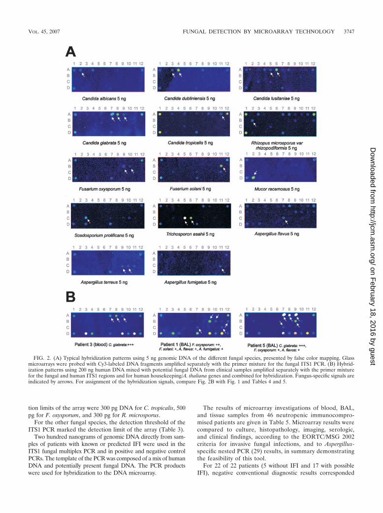
resultant images as shown in Fig. 2. Dots with at least twice the signal intensitiesof the background intensities were evaluated as positive signals. Backgroundintensities were calculated by the ImaGene 4.0 program.
Patients. Blood, BAL, and tissue samples from a total of 46 immunocompro-mised neutropenic patients with mainly hematological malignancies were furtherprocessed for diagnostic purposes after the patients gave informed consent.Primary diseases were acute myeloid leukemia (AML) (n � 21), acute lympho-blastic leukemia (ALL) (n � 7), non-Hodgkin’s lymphoma (NHL) (n � 11),myelodysplastic syndromes (MDS) (n � 2), severe aplastic anemia (n � 1),osteomyelofibrosis (n � 1), metastatic rhabdomyosarcoma (n � 1), medulloblas-toma (n � 1), and metastatic germ cell tumor (n � 1).
For clinical evaluation, we investigated 91 samples (66 blood samples, 18 BALsamples, and 7 tissue samples) from 46 neutropenic patients with proven (n � 5),probable (n � 3), or possible (n � 33) IFI, according to the European Organi-zation for Research and Treatment of Cancer/Mycoses Study Group (EORTC/MSG) 2002 criteria (1). For five patients, there was no evidence of IFI.
Clinical samples. Blood samples were obtained by venipuncture under sterileconditions in a sterile vessel to a final concentration of 1.6 mg EDTA per mlblood. The sample volume was 5 to 7 ml.
BAL was performed as described previously (29), and BAL samples wereobtained in a sterile vessel without conservation medium. The mean samplevolume was 10 ml.
Tissue samples were obtained by needle biopsies (liver and kidney) or surgicalprocedures (other samples) under sterile conditions.
RESULTS
The multiplex PCR for amplification of the ITS1 regions(137 to 245 bp) of the 14 fungal pathogens showed differentsensitivity thresholds for the genomic DNAs of the fungalspecies (Table 3). Even large amounts of genomic bacterialDNAs (5 ng) used as negative controls (Enterococcus faecalisDSM 2570, Escherichia coli DSM 787, Klebsiella pneumoniaeDSM 4617, Proteus mirabilis DSM 788, Pseudomonas aerugi-nosa DSM 50071, Staphylococcus aureus DSM 799, and Strep-tococcus pneumoniae DSM 20566) were not amplified in theITS1 multiplex PCR and did not give any signals after hybrid-ization to the fungal DNA microarray. The genomic DNA ofPenicillium notatum, a fungal organism that we did not aim todetect, was amplified by PCR and gave hybridization signalswith one of three capture probes of Candida glabrata and oneof two capture probes of Scedosporium prolificans. Addition-
ally, for a human, ITS1-specific, 226-bp fragment, few nonspe-cific amplification products were visible when large amounts ofhuman genomic DNA (200 ng) were used in the ITS1 multi-plex PCR. After the hybridization of the human PCR productsto the DNA microarray, there was no cross-reactivity betweenhuman DNA and fungal capture probes detected. There werealso no cross-reactivities between any fungal or human PCRproducts and the two spotted A. thaliana negative control oli-gonucleotides. Hybridization with two Cy3-labeled oligonucle-otides (50 �M), which were complementary to the two spottedA. thaliana capture probes, resulted in intense signals (data notshown).
The fungal and human capture probes were located on theglass microarray as shown schematically in Fig. 1. To establishthe fungal DNA chip, 5-ng amounts of each of 14 genomicfungal DNAs were amplified separately by ITS1 multiplexPCR and the products were then hybridized. Every fungalorganism showed a specific hybridization pattern after evalu-ation of the hybridization signals (Fig. 2A). Details about thecorrelation of the hybridization patterns and the fungal organ-isms are described in Table 4. In spite of single cross-reactiv-ities, each species has a unique signature hybridization pattern.
Different hybridization conditions were tested and com-pared. The best hybridization results and an appropriate ratiobetween specific binding, signal intensity, and cross-reactivitywere achieved by using the following hybridization and wash-ing conditions: hybridization at 42°C using 50% formamide for12 h following a washing step for 10 min in 1�, 0.5�, and 0.1�SSC at room temperature, respectively.
For Candida tropicalis, Fusarium oxysporum, and Rhizopusmicrosporus DNA, the detection limit of the array is above thedetection limit of the hybridization preceding fungal ITS1PCR. Additional serial 10-fold dilutions of the three DNAswere used to determine the detection threshold of the array.The ITS1 PCR detection limits were 1 pg for C. tropicalis, 10 pgfor F. oxysporum, and 8 pg for R. microsporus, and the detec-
FIG. 1. Schematic composition of glass microarray and grid location of fungal, human, and A. thaliana-specific oligonucleotides (captureprobes). Grid locators (A1, A12, and D1 for Cy3) represented by a spotted Cy3-labeled arbitrary oligonucleotide are depicted as black boxes.Names of the fungal capture probes are derived from the first letter of the genus name and the first four letters of the species name and arenumbered consecutively as follows: Calb1S (20), Candida albicans; Cdub1S (20) and Cdubl, Candida dubliniensis; Clusi, Candida lusitaniae; Cglab,Candida glabrata; Ctrop, Candida tropicalis; Rmicr, Rhizopus microsporus; Foxys, Fusarium oxysporum; Fsola, Fusarium solani; Mrace, Mucorracemosus; Sprol, Scedosporium prolificans; Tasah, Trichosporon asahii; Aflav, Aspergillus flavus; Aterr, Aspergillus terreus; and Afumi, Aspergillusfumigatus.
3746 SPIESS ET AL. J. CLIN. MICROBIOL.
on February 18, 2016 by guest
http://jcm.asm
.org/D
ownloaded from
tion limits of the array were 300 pg DNA for C. tropicalis, 500pg for F. oxysporum, and 300 pg for R. microsporus.
For the other fungal species, the detection threshold of theITS1 PCR marked the detection limit of the array (Table 3).
Two hundred nanograms of genomic DNA directly from sam-ples of patients with known or predicted IFI were used in theITS1 fungal multiplex PCR and in positive and negative controlPCRs. The template of the PCR was composed of a mix of humanDNA and potentially present fungal DNA. The PCR productswere used for hybridization to the DNA microarray.
The results of microarray investigations of blood, BAL,and tissue samples from 46 neutropenic immunocompro-mised patients are given in Table 5. Microarray results werecompared to culture, histopathology, imaging, serologic,and clinical findings, according to the EORTC/MSG 2002criteria for invasive fungal infections, and to Aspergillus-specific nested PCR (29) results, in summary demonstratingthe feasibility of this tool.
For 22 of 22 patients (5 without IFI and 17 with possibleIFI), negative conventional diagnostic results corresponded
FIG. 2. (A) Typical hybridization patterns using 5 ng genomic DNA of the different fungal species, presented by false color mapping. Glassmicroarrays were probed with Cy3-labeled DNA fragments amplified separately with the primer mixture for the fungal ITS1 PCR. (B) Hybrid-ization patterns using 200 ng human DNA mixed with potential fungal DNA from clinical samples amplified separately with the primer mixturefor the fungal and human ITS1 regions and for human housekeeping/A. thaliana genes and combined for hybridization. Fungus-specific signals areindicated by arrows. For assignment of the hybridization signals, compare Fig. 2B with Fig. 1 and Tables 4 and 5.
VOL. 45, 2007 FUNGAL DETECTION BY MICROARRAY TECHNOLOGY 3747
on February 18, 2016 by guest
http://jcm.asm
.org/D
ownloaded from

with negative microarray data. For eight patients with proven(n � 5) or probable (n � 3) IFI, results for four or two patientswere confirmed by microarray testing, respectively. For pa-tients with possible IFI (n � 5), positive microarray data werevalidated by other diagnostic findings. For 11 of 11 patientswith possible IFI, the microarray results provided additionalinformation about fungal pathogens. For one patient withproven invasive aspergillosis (determined from a brain abscesssample), DNA microarray results from blood samples werenegative under antifungal conditions; for one patient withprobable invasive aspergillosis, DNA microarray results from aBAL aliquot were negative; this patient also received antifun-gal treatment.
Genomic DNA and specific hybridization patterns of Can-dida albicans, Candida dubliniensis, Candida glabrata, Aspergil-lus fumigatus, Aspergillus flavus, Fusarium solani, Rhizopusmicrosporus, Scedosporium prolificans, and Trichosporon asahiiwere detected. One fungal pathogen was identified in 11 pa-tients with IFI, and two or more fungal pathogens were like-wise identified in 11 patients with IFI.
We also compared microarray findings to the results of apreviously described and clinically validated Aspergillus-specificnested PCR assay (6, 29). For one patient, the Aspergillusnested PCR assay was positive for Aspergillus DNA, whereasthe DNA microarray results were negative. For three patients,the Aspergillus PCR results for blood and tissue samples werepositive, whereas the DNA microarray results were negativefor Aspergillus DNA but positive for additional fungal species(C. albicans, C. dubliniensis, and C. glabrata). Differences be-tween nested Aspergillus PCR (29) findings and the microarrayresults are caused by the higher sensitivity of the nested PCRassay for Aspergillus DNA. For five patients, positive Aspergil-lus PCR findings were in line with positive DNA microarrayresults for Aspergillus DNA.
Three representative hybridizations of DNA from a totalof 46 patients (Table 5) isolated from clinical samples areshown in Fig. 2B. For the blood sample from patient 3,hybridization resulted in distinct hybridization signals for allthree C. glabrata capture probes without any cross-reactivi-ties. A corresponding blood culture was positive for Candidaspecies that could not be further differentiated. A second
TABLE 4. Hybridization patterns for the 14 fungal species
Fungal speciesHybridization
signal(dot code)
Signaturehybridization
patterna
C. albicans A2 Calb1S (20)A4 Cdubl2
C. dubliniensis A3 Cdub1S (20)A4 Cdubl2A8 Cglab2
C. lusitaniae A4 Cdubl2A5 Clusi1A6 Clusi2A8 Cglab2C3 Sprol1C5 Tasah1D7 RPL19_1
C. glabrata A7 Cglab1A8 Cglab2A9 Cglab3D7 RPL19_1
C. tropicalis A9 Cglab2A11 Ctrop2D7 RPL19_1
R. microsporus var. A3 Cdub1Srhizopodiformis A7 Cglab1
A8 Cglab2A11 Ctrop2B2 Rmicr2D7 RPL19_1
F. oxysporum B9 Foxys1B10 Foxys2D7 RPL19_1
F. solani B11 Fsola1C11 Afumi1D7 RPL19_1
M. racemosus D2 Mrace3
S. prolificans C3 Sprol1D3 HumITS1_2D7 RPL19_1
T. asahii A8 Cglab2C5 Tasah1C6 Tasah2D7 RPL19_1
A. flavus A7 Cglab1A8 Cglab2B9 Foxys1B10 Foxys2C7 Aflav1D3 HumITS1_2
A. terreus C9 Aterr1C10 Aterr2C11 Afumi1D7 RPL19_s1
A. fumigatus C11 Afumi1
a Names of the fungal capture probes are derived from the first letter of the genusname and the first four letters of the species name and are consecutively numbered.Specific capture probes are marked in bold letters. The unique signature hybridiza-tion pattern for each organism is shown in column 3. Hum, human.
TABLE 3. Lowest detectable amount of DNA from fungal strainsby PCR
Fungal strain or samplea DNA amt(pg)
Aspergillus flavus DSM 1959............................................................. 20Aspergillus fumigatus DSM 819 ........................................................ 50Aspergillus terreus DSM 826 ............................................................. 10Candida albicans DSM 11949 .......................................................... 1Candida dubliniensis DSM 75921 .................................................... 1Candida glabrata DSM 11226 .......................................................... 1Candida lusitaniae, CS ...................................................................... 1Candida tropicalis DSM 1346........................................................... 1Fusarium oxysporum DSM 12646 .................................................... 10Fusarium solani DSM 1164 ..............................................................100Mucor racemosus DSM 62760.......................................................... 10Rhizopus microsporus var. rhizopodiformis DSM 2196.................. 8Scedosporium prolificans CBS 100390............................................. 50Trichosporon asahii, CS..................................................................... 10
a CS, clinical sample.
3748 SPIESS ET AL. J. CLIN. MICROBIOL.
on February 18, 2016 by guest
http://jcm.asm
.org/D
ownloaded from

TABLE 5. Results from patient blood, BAL, and tissue samplesa
Patient Sample Diagnosis Diagnosticsignificanceb
Result(s) from diagnosticwork-up DNA microarray result(s)c
NestedPCR
resultd
1 BAL ALL Proven Culture positive for aspergillosis Positive for A. flavus,A. fumigatus, F. oxysporum,and F. solani
�
2 Blood AML Proven Histology positive, culturenegative
Positive for A. flavus andC. tropicalis
�
3 Blood AML Proven Culture positive for Candida,histology positive
Positive for C. glabrata �
3A Brain abscess Positive for C. glabrata �3B Spleen abscess Positive for C. glabrata and
S. prolificans�
3C Spleen tissue Positive for S. prolificans �4 Blood AML Proven Candida sepsis, blood culture
positive for C. albicansPositive for C. dubliniensis �
5 BAL OMF, HSCT Probable Positive for Aspergillus Ag(galactomannan)
Positive for C. glabrata, A. flavus,and F. oxysporum
�
6 Blood ALL Probable Positive for Aspergillus Ag(galactomannan)
Positive for C. tropicalis �
Positive LI Positive for C. glabrata, A. flavus,A. fumigatus, and T. asahii
6A BAL Positive for C. glabrata andT. asahii
�
6B Blood Positive for C. glabrata �6C Blood Negative �7 Blood AML Possible PCR positive for Candida Positive for C. dubliniensis and
C. glabrata�
7A Blood Positive for C. glabrata (�)7B Spleen Positive for C. dubliniensis and
C. glabrata�
7C Needle biopsy,kidney
Positive for C. dubliniensis andC. glabrata
(�)
8 Blood Germ cell tumor Possible Serology positive for Candida,e
negative for Candida Ag,negative for Aspergillus Ag(galactomannan)
Positive for C. tropicalis andC. glabrata
�
9 Blood ALL Possible Positive for Aspergillus Ag Positive for A. flavus �10 Blood ALL Possible Negative for Aspergillus Ag
(galactomannan), positivehepatic nodule
Positive for C. tropicalis (�)
10A Blood Positive for C. tropicalis (�)10B Blood Positive for C. tropicalis �10C Blood Negative (�)11 Blood AML Possible Positive LI Negative �11A Blood Positive for A. fumigatus �12 Blood Plasmocytoma No IFI Negative LI Negative �13 Blood Mantle cell
lymphomaNo IFI Positive for Mycobacterium
tuberculosisNegative �
13A BAL Negative �14 BAL AML No IFI Negative �15 BAL MDS No IFI No microbiological proof Negative �16 Blood AML No IFI Negative for Aspergillus Ag
(galactomannan), fungalculture negative
Negative �
16A Blood Negative �16B Blood Negative �17 Blood AML Possible Positive LI, negative for
Aspergillus Ag(galactomannan)
Negative �
17A Blood Negative �17B Blood Negative �18 Blood AML Possible Positive LI Negative �18A Blood Negative �18B Blood Negative �19 BAL AML Possible No microbiological proof Negative �20 Blood CLL Possible Histology negative Negative �20A BAL Negative �21 BAL MDS Possible Negative for Aspergillus Ag
(galactomannan), positive LINegative �
22 Blood AML Possible Negative for Aspergillus Ag(galactomannan), bloodculture negative, BALnegative for Aspergillus Ag(galactomannan), negative forCandida Ag
Negative �
23 Blood AML Possible Positive LI, negative forAspergillus Ag(galactomannan)
Negative �
23A Blood Negative �
Continued on following page
VOL. 45, 2007 FUNGAL DETECTION BY MICROARRAY TECHNOLOGY 3749
on February 18, 2016 by guest
http://jcm.asm
.org/D
ownloaded from

example (patient 1) shows the hybridization results usingDNA from a BAL sample. A blood culture from this patientidentified A. fumigatus. The hybridization gave positive sig-nals for A. flavus, A. fumigatus, F. oxysporum, and F. solani.
The third example (patient 5) showed signals with the C.glabrata capture probes and the A. flavus probe. Addition-ally, the sample was positive by Aspergillus antigen testing(galactomannan enzyme-linked immunosorbent assay). The
TABLE 5—Continued
Patient Sample Diagnosis Diagnosticsignificanceb
Result(s) from diagnosticwork-up DNA microarray result(s)c
NestedPCR
resultd
24 BAL CLL Possible Positive LI Negative �25 Blood AML Possible Negative LI Negative �25A Blood Negative �26 BAL Hairy cell
leukemiaPossible Fungal culture negative Negative �
27 Blood AML Possible Negative for Aspergillus Ag(galactomannan)
Negative �
27A Blood Negative �27B Blood Negative �28 Blood AML Possible Positive for LI, negative for
Aspergillus Ag(galactomannan)
Negative �
28A Blood Negative �28B Blood Negative �28C Blood Negative �28D Blood Negative �28E Blood Negative �29 Blood AML Possible Negative for Aspergillus Ag
(galactomannan)Negative �
29A Blood Negative �29B Blood Negative �29C Blood Negative �30 Blood AML Possible Positive LI Negative �30A Blood Negative �30B Blood Negative �30C Blood Negative �31 Blood AML relapse Possible Positive LI, negative for
Aspergillus Ag(galactomannan)
Negative �
31A Blood Negative �31B Blood Negative32 Blood AML Possible Positive LI Negative �32A Blood Negative �32C Blood Negative �33 BAL ALL Possible Negative for Aspergillus Ag
(galactomannan)Negative �
34 Blood Multiple myeloma Possible No microbiological proof Positive for C. glabrata andC. tropicalis
�
35 Blood SAA Possible Negative �35A Blood Positive for C. tropicalis �36 BAL Mantle cell
lymphomaPossible No microbiological proof Positive for C. albicans �
37 BAL ALL, mucositis Possible No microbiological proof Positive for C. glabrata andC. tropicalis
�
38 Blood ALL Possible No microbiological proof Positive for R. microsporus �39 Liver biopsy Rhabdomyosarcoma Possible No microbiological proof Positive for C. glabrata �40 Liver biopsy Medulloblastoma Possible No microbiological proof Positive for C. tropicalis and
S. prolificans�
41 Liver biopsy B-NHL Possible Negative for Aspergillus Ag(galactomannan), fungalculture negative, positivehepatic nodule
Positive for C. glabrata �
41A Blood Negative �42 Blood Lymphoma Possible Negative for Aspergillus Ag
(galactomannan), fungalculture negative
Positive for C. glabrata �
42A BAL Positive for C. glabrata andC. tropicalis
�
43 BAL AML Possible Positive LI, CT positive Positive for C. glabrata �44 BAL T-NHL Possible Positive LI Positive for C. glabrata �45 Blood NHL Proven Aspergillosis, histology from
brain abscessNegative �
45A Blood Negative �45B Blood Negative (�)46 BAL B-CLL Probable Positive for Aspergillus Ag
(galactomannan)Negative �
a �, negative; �, positive; (�), weakly positive; Ag, antigen; CLL, chronic lymphocytic leukemia; CT, computed tomography scan of the chest; HSCT, hematologicstem cell transplantation; LI, lung infiltrate; OMF, osteomyelofibrosis; SAA, severe aplastic anemia.
b See reference 1.c Results from three capture probe replicates.d The nested PCR assay is specific for Aspergillus species. See reference 29.e Status after fungal sepsis.
3750 SPIESS ET AL. J. CLIN. MICROBIOL.
on February 18, 2016 by guest
http://jcm.asm
.org/D
ownloaded from

result of the nested PCR assay for Aspergillus species wasnegative for this patient.
Hybridization of the DNA from a blood sample of a healthyvolunteer did not give any signal with the fungal captureprobes.
DISCUSSION
In immunocompromised patients, the spectrum of clinicallyrelevant fungal pathogens causing systemic infections with con-siderable morbidity and high mortality rates is changing (25).Therefore, we set out to establish a rapid, specific, and sensi-tive molecular method to detect and identify the most emerg-ing fungal pathogens (26) from noncultured clinical samples inone assay.
Our DNA microarray is able to detect clinically relevantfungal pathogens at low detection thresholds. Results usingclinical samples from immunocompromised patients with he-matological malignancies show the usefulness of the microar-ray in the clinical context. The benefit of the fungal microarrayis the potential to detect the most important 14 fungal patho-gens in a specific IFI high-risk setting with one test; due tofeasibility concerns, other fungi were not included. In view ofthe changing spectrum of emerging clinically relevant fungalpathogens causing IFI and the advent of novel antifungals, thisnew diagnostic approach meets urgent clinical needs, at leastconcerning the group of patients treated for hematologicalmalignancies.
For our assay, sensitive and specific detection and identifi-cation of the fungal pathogens were achieved by designingprimer pairs complementary to the highly conserved 18S and5.8S regions of the fungal rRNA genes and oligonucleotidecapture probes complementary to the more variable ITS1 re-gions which enabled a differentiation of fungal species (13, 14,15, 16, 20). As part of the rRNA genes, the ITS1 regions werechosen as targets because they are present in numerous copiesin the fungal genome. The design of the primer pairs fromhighly conserved regions of a multicopy gene lead to a highsensitivity of the PCR and to an amplification of the targetsequences with only a few primer combinations. Reasons forthe different detection thresholds of ITS1 PCR for the fungalDNAs could be different primer binding capacities and possi-ble secondary structures of single primers. The existence of twoto three oligonucleotides per fungal organism (2) resulted inspecies-specific hybridization patterns.
As a proof of principle and for clinical validation, hybridiza-tion of DNA of blood, BAL, and tissue samples from a total of46 patients demonstrated the applicability and feasibility of ourdiagnostic assay (Table 5).
For 22 patients, negative microarray data were confirmed bynegative conventional diagnostic results. On the other hand,positive microarray data were confirmed by other diagnosticresults for 11 patients with proven (n � 4), probable (n � 2),or possible (n � 5) IFI.
For 11 of 11 patients with possible IFI, the group of patientswith the highest diagnostic uncertainty, the microarray resultsprovided additional information about the pathogens, whichwere mainly Candida species. Among the clinical samples ofthis patient group were five BAL samples positive for Candidaspecies; from a clinical point of view, the detection of Candida
species in BAL is not accepted as proof for IFI. The rate ofsample contamination with Candida species is still unclear.
For one patient with proven invasive aspergillosis (deter-mined from a brain abscess sample), DNA microarray resultsfrom blood samples were negative and possibly caused by in-tensive antifungal treatment (18); for one patient with proba-ble invasive aspergillosis, DNA microarray results from a BALaliquot were also negative under antifungal treatment and/ordue to a nonrepresentative sample.
Genomic DNA and specific hybridization patterns of Can-dida albicans, Candida dubliniensis, Candida glabrata, Aspergil-lus fumigatus, Aspergillus flavus, Fusarium solani, Rhizopus mi-crosporus, Scedosporium prolificans, and Trichosporon asahiiwere detected. One fungal pathogen was identified in 11 pa-tients with IFI, and two or more fungal pathogens were like-wise identified in 11 patients with IFI. The microarray findingsof these patients show the potential of the assay to detect morethan one fungal organism as a cause of infection in the samepatient. From a clinical point of view, our knowledge aboutfungal infections caused by multiple pathogens is small.
Comparing results of the microarray analysis to the As-pergillus-specific nested PCR assay, positive Aspergillus PCRfindings for five patients were in line with positive DNAmicroarray results for Aspergillus DNA. For one patient, theAspergillus nested PCR assay was positive, whereas the DNAmicroarray results were negative. For three patients, As-pergillus PCR results from blood and tissue samples werepositive, whereas the DNA microarray results were negativefor Aspergillus DNA but positive for additional fungal spe-cies (C. albicans, C. dubliniensis, and C. glabrata). Differ-ences between nested Aspergillus PCR (29) findings and themicroarray results are caused by the higher sensitivity of thenested PCR assay for Aspergillus DNA; generally, nestedPCR assays are more sensitive than multiplex PCRs becauseof an additional partial amplification of the PCR product inthe second step.
Three other groups previously described the applicationof the DNA microarray technology for the detection offungal pathogens from cultured clinical isolates. In the studyof Hsiao et al. (16), the capture probes were designedagainst the ITS1 and ITS2 regions of the fungal rRNA genesof filamentous fungi but not of yeasts. The group deter-mined the detection threshold of the assay by using twoserially diluted fungal DNA samples for all pathogens. Incontrast, our assay defined different species-specific detec-tion thresholds of both PCR and microarray hybridizationfor all 14 fungal species. The second study describing thedetection of fungal pathogens from cultured clinical isolateswas published by Leinberger et al. (20). In this study, cap-ture probes complementary to the ITS1 and ITS2 regions ofthe fungal rRNA genes were spotted on epoxy-coated glassslides. This DNA microarray contained capture probes from12 Candida and Aspergillus species, but not from other clin-ically relevant fungal pathogens. Although universal fungalprimers exist (31), we decided to design primer pairs ampli-fying the ITS1 regions only, because of the good bindingcapacity of shorter DNA fragments of up to 300 bases dur-ing hybridization, leading to a high sensitivity of the assay.In the study by Leinberger et al., the question of PCR andhybridization detection thresholds was not addressed. In
VOL. 45, 2007 FUNGAL DETECTION BY MICROARRAY TECHNOLOGY 3751
on February 18, 2016 by guest
http://jcm.asm
.org/D
ownloaded from

contrast, we focused our interest on a high sensitivity for ourassay because we were investigating noncultured clinicalsamples containing only small amounts of fungal DNA.Huang et al. (17) described another application of the DNAmicroarray technology that uses capture probes from thefungal ITS2 regions of the rRNA genes to identify patho-genic fungi from standard strains and cultured clinical iso-lates. The detection of the fungal DNA was carried outusing isolates and additionally spiked blood samples of 16patients by negative culture, microscopy, or PCR. TheirDNA microarray contains capture probes of 20 fungal spe-cies. Other pathogens are less relevant than Candida, As-pergillus, and Mucor species for the clinical investigation ofimmunocompromised patients with hematologic malignan-cies.
In addition to those of DNA chip technology tests, theresults of a small number of multifungal PCR-based assaysfor the detection and identification of fungal pathogens inclinical samples have been published (8–10, 14, 19, 21, 27).These assays cover a different spectrum of targeted fungalorganisms using diverse methodical detection proceduresafter an initial PCR step, in order to answer distinct clinicalquestions. However, a molecular diagnosis gold standard ofIFI has not yet been defined.
The established multifungal DNA microarray detectsclinically relevant fungal pathogens specifically and at lowdetection thresholds from noncultured clinical samples, de-tecting different fungal pathogens with one test. In view ofthe changing spectrum of clinically relevant and emergingfungal pathogens causing IFI, this new diagnostic approachmeets urgent clinical needs, at least concerning the high-riskgroup of patients with hematologic malignancies. This eval-uation by a prospective multicenter study, testing blood,BAL, and tissue samples from immunocompromised pa-tients, especially patients with acute leukemia, and, compar-ing microarray results with findings from conventional diag-nostic studies, is ongoing.
ACKNOWLEDGMENTS
This work was supported by a grant of the Deutsche Krebshilfe/Dr.Mildred Scheel Stiftung fur Krebsforschung (project number 70-3255Bu 1).
We are indebted to H. Hof, C. Mosbach, and A. Dietz, Institute ofClinical Microbiology and Hygiene, University Hospital of Mannheim,for excellent microbiological support.
REFERENCES
1. Ascioglu, S., J. H. Rex, B. de Pauw, J. E. Bennett, J. Bille, F. Crokaert, D. W.Denning, J. P. Donnelly, J. E. Edwards, Z. Erjavec, D. Fiere, O. Lortholary,J. Maertens, J. F. Meis, T. F. Patterson, J. Ritter, D. Selleslag, P. M. Shah,D. A. Stevens, and T. J. Walsh. 2002. Defining opportunistic invasive fungalinfections in immunocompromised patients with cancer and hematopoi-etic stem cell transplants: an international consensus. Clin. Infect. Dis.34:7–14.
2. Behr, T., C. Koob, M. Schedl, A. Mehlen, H. Meier, D. Knopp, E. Frahm, U.Obst, K. Schleifer, R. Niessner, and W. Ludwig. 2000. A nested array ofrRNA targeted probes for the detection and identification of enterococci byreverse hybridization. Syst. Appl. Microbiol. 23:563–572.
3. Bethge, W. A., M. Schmalzing, G. Stuhler, U. Schumacher, S. M. Krober, M.Horger, H. Einsele, L. Kanz, and H. Hebart. 2005. Mucormycoses in patientswith hematologic malignancies: an emerging fungal infection. Haemato-logica 90:ECR22.
4. Bille, J., O. Marchetti, and T. Calandra. 2005. Changing face of health-careassociated fungal infections. Curr. Opin. Infect. Dis. 18:314–319.
5. Buchheidt, D., M. Hummel, D. Schleiermacher, B. Spiess, and R. Hehlmann.2004. Current molecular diagnostic approaches to systemic infections with
aspergillus species in patients with hematological malignancies. Leuk. Lym-phoma 45:463–468.
6. Buchheidt, D., and M. Hummel. 2005. Aspergillus polymerase chain reaction(PCR) diagnosis. Med. Mycol. 43:139–145.
7. Castagnola, E., S. Cesaro, M. Giacchino, S. Livadiotti, F. Tucci, G. Zanazzo,D. Caselli, I. Caviglia, S. Parodi, R. Rondelli, P. E. Cornelli, R. Mura, N.Santoro, G. Russo, R. De Santis, S. Buffardi, C. Viscoli, R. Haupt, and M. R.Rossi. 2006. Fungal infections in children with cancer: a prospective, multi-center surveillance study. Pediatr. Infect. Dis. J. 25:634–639.
8. Diaz, M. R., and J. W. Fell. 2004. High-throughput detection of pathogenicyeasts of the genus Trichosporon. J. Clin. Microbiol. 42:3696–3706.
9. Diaz, M. R., T. Boekhout, B. Theelen, M. Bovers, F. J. Cabanes, and J. W.Fell. 2006. Microcoding and flow cytometry as a high-throughput fungalidentification system for Malassezia species. J. Med. Microbiol. 55:1197–1209.
10. Einsele, H., H. Hebart, G. Roller, J. Loffler, I. Rothenhofer, C. A. Muller,R. A. Bowden, J. van Burik, D. Engelhard, L. Kanz, and U. Schumacher.1997. Detection and identification of fungal pathogens in blood by usingmolecular probes. J. Clin. Microbiol. 35:1353–1360.
11. Girmenia, C., M. Nucci, and P. Martino. 2001. Clinical significance of As-pergillus fungaemia in patients with haematological malignancies and inva-sive aspergillosis. Br. J. Haematol. 114:93–98.
12. Guiot, H. F., W. E. Fibbe, and J. W. van’t Wout. 1994. Risk factors for fungalinfection in patients with malignant hematologic disorders: implications forempirical therapy and prophylaxis. Clin. Infect. Dis. 18:525–532.
13. Healy, M., K. Reece, D. Walton, J. Huong, K. Shah, and D. P. Kontoyiannis.2004. Identification to the species level and differentiation between strains ofAspergillus clinical isolates by automated repetitive-sequence-based PCR.J. Clin. Microbiol. 42:4016–4024.
14. Hendolin, P. H., L. Paulin, P. Koukila-Kahkola, V. J. Anttila, H. Malmberg,M. Richardson, and J. Ylikoski. 2000. Panfungal PCR and multiplex liquidhybridization for detection of fungi in tissue specimens. J. Clin. Microbiol.38:4186–4192.
15. Henry, T., P. C. Iwen, and S. H. Hinrichs. 2000. Identification of Aspergillusspecies using internal transcribed spacer regions 1 and 2. J. Clin. Microbiol.38:1510–1515.
16. Hsiao, C. R., L. Huang, J. P. Bouchara, R. Barton, H. C. Li, and T. C. Chang.2005. Identification of medically important molds by an oligonucleotidearray. J. Clin. Microbiol. 43:3760–3768.
17. Huang, A., J. W. Li, Z. Q. Shen, X. W. Wang, and M. Jin. 2006. High-throughput identification of clinical pathogenic fungi by hybridization to anoligonucleotide microarray. J. Clin. Microbiol. 44:3299–3305.
18. Lass-Florl, C., E. Gunsilius, G. Gastl, H. Bonatti, M. C. Freund, A.Gschwendter, G. Kropshofer, M. P. Dietrich, and A. Petzer. 2004. Diag-nosing invasive aspergillosis during antifungal therapy by PCR analysis ofblood samples. J. Clin. Microbiol. 42:4154–4157.
19. Lau, A., S. Chen, T. Sorell, D. Carter, R. Malik, P. Martin, and C. Halliday.2007. Development and clinical application of a panfungal PCR assay todetect and identify fungal DNA in tissue specimens. J. Clin. Microbiol.45:380–385.
20. Leinberger, D. M., U. Schumacher, I. B. Autenrieth, and T. T. Bachmann.2005. Development of a DNA microarray for detection and identification offungal pathogens involved in invasive mycoses. J. Clin. Microbiol. 43:4943–4953.
21. Loeffler, J., N. Henke, H. Hebart, D. Schmidt, L. Hagmeyer, U. Schumacher,and H. Einsele. 2000. Quantification of fungal DNA by using fluorescenceresonance energy transfer and the Light Cycler system. J. Clin. Microbiol.38:586–590.
22. Marchetti, O., J. Bille, U. Fluckiger, P. Eggimann, C. Ruef, J. Garbino, T.Calandra, M. P. Glauser, M. G. Tauber, and D. Pittet. 2004. Epidemiologyof candidemia in Swiss tertiary care hospitals: secular trends, 1991–2000.Clin. Infect. Dis. 38:311–320.
23. Reference deleted.24. Martino, R., and C. Viscoli. 2006. Empirical antifungal therapy in patients
with neutropenia and persistent or recurrent fever of unknown origin. Br. J.Haematol. 132:138–154.
25. Maschmeyer, G. 2006. The changing epidemiology of invasive fungal infec-tions: new threats. Int. J. Antimicrob. Agents 27:3–6.
26. Pfaller, M. A., and D. J. Diekema. 2004. Rare and emerging opportunisticfungal pathogens: concern for resistance beyond Candida albicans and As-pergillus fumigatus. J. Clin. Microbiol. 42:4419–4431.
27. Schabereiter-Gurtner, C., B. Selitsch, M. L. Rotter, A. M. Hirschl, and B.Willinger. 2007. Development of novel real-time PCR assays for detectionand differentiation of eleven medically important Aspergillus and Candidaspecies in clinical specimens. J. Clin. Microbiol. 45:906–914.
28. Seifarth, W., B. Spiess, U. Zeilfelder, C. Speth, R. Hehlmann, and C. Leib-Mosch. 2003. Assessment of retroviral activity using a universal retrovirus chip.J. Virol. Methods 112:79–91.
3752 SPIESS ET AL. J. CLIN. MICROBIOL.
on February 18, 2016 by guest
http://jcm.asm
.org/D
ownloaded from

29. Skladny, H., D. Buchheidt, C. Baust, F. Krieg-Schneider, W. Seifarth, C.Leib-Mosch, and R. Hehlmann. 1999. Specific detection of Aspergillus spe-cies in blood and bronchoalveolar lavage samples of immunocompromisedpatients by two-step PCR. J. Clin. Microbiol. 37:3865–3871.
30. Spiess, B., D. Buchheidt, C. Baust, H. Skladny, W. Seifarth, U. Zeilfelder, C.Leib-Mosch, H. Morz, and R. Hehlmann. 2003. Development of a Light-Cycler PCR assay for detection and quantification of Aspergillus fumigatus
DNA in clinical samples from neutropenic patients. J. Clin. Microbiol. 41:1811–1818.
31. White, T. J., T. Bruns, S. Lee, and J. Taylor. 1990. Amplification anddirect sequencing of fungal ribosomal RNA genes for phylogenetics, p.315–322. In M. Innis, J. Gelfand, J. Sninsky, and T. White (ed.), PCRprotocols: a guide to methods and application. Academic Press, Inc., SanDiego, CA.
VOL. 45, 2007 FUNGAL DETECTION BY MICROARRAY TECHNOLOGY 3753
on February 18, 2016 by guest
http://jcm.asm
.org/D
ownloaded from