Blood Pressure Regulation Evolved from Basic Homeostatic ...
Upload
independentCategory
view
0download
0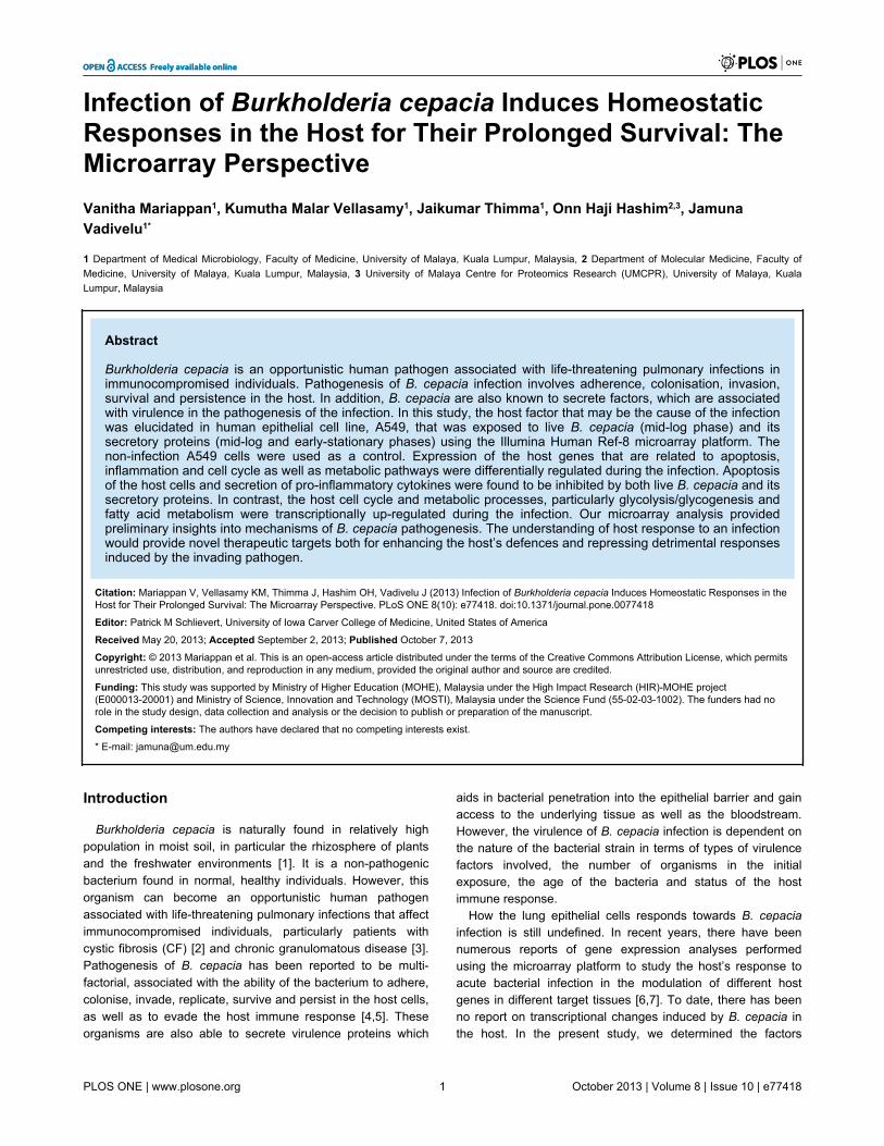
Infection of Burkholderia cepacia Induces HomeostaticResponses in the Host for Their Prolonged Survival: TheMicroarray PerspectiveVanitha Mariappan1, Kumutha Malar Vellasamy1, Jaikumar Thimma1, Onn Haji Hashim2,3, JamunaVadivelu1*
1 Department of Medical Microbiology, Faculty of Medicine, University of Malaya, Kuala Lumpur, Malaysia, 2 Department of Molecular Medicine, Faculty ofMedicine, University of Malaya, Kuala Lumpur, Malaysia, 3 University of Malaya Centre for Proteomics Research (UMCPR), University of Malaya, KualaLumpur, Malaysia
Abstract
Burkholderia cepacia is an opportunistic human pathogen associated with life-threatening pulmonary infections inimmunocompromised individuals. Pathogenesis of B. cepacia infection involves adherence, colonisation, invasion,survival and persistence in the host. In addition, B. cepacia are also known to secrete factors, which are associatedwith virulence in the pathogenesis of the infection. In this study, the host factor that may be the cause of the infectionwas elucidated in human epithelial cell line, A549, that was exposed to live B. cepacia (mid-log phase) and itssecretory proteins (mid-log and early-stationary phases) using the Illumina Human Ref-8 microarray platform. Thenon-infection A549 cells were used as a control. Expression of the host genes that are related to apoptosis,inflammation and cell cycle as well as metabolic pathways were differentially regulated during the infection. Apoptosisof the host cells and secretion of pro-inflammatory cytokines were found to be inhibited by both live B. cepacia and itssecretory proteins. In contrast, the host cell cycle and metabolic processes, particularly glycolysis/glycogenesis andfatty acid metabolism were transcriptionally up-regulated during the infection. Our microarray analysis providedpreliminary insights into mechanisms of B. cepacia pathogenesis. The understanding of host response to an infectionwould provide novel therapeutic targets both for enhancing the host’s defences and repressing detrimental responsesinduced by the invading pathogen.
Citation: Mariappan V, Vellasamy KM, Thimma J, Hashim OH, Vadivelu J (2013) Infection of Burkholderia cepacia Induces Homeostatic Responses in theHost for Their Prolonged Survival: The Microarray Perspective. PLoS ONE 8(10): e77418. doi:10.1371/journal.pone.0077418
Editor: Patrick M Schlievert, University of Iowa Carver College of Medicine, United States of America
Received May 20, 2013; Accepted September 2, 2013; Published October 7, 2013
Copyright: © 2013 Mariappan et al. This is an open-access article distributed under the terms of the Creative Commons Attribution License, which permitsunrestricted use, distribution, and reproduction in any medium, provided the original author and source are credited.
Funding: This study was supported by Ministry of Higher Education (MOHE), Malaysia under the High Impact Research (HIR)-MOHE project(E000013-20001) and Ministry of Science, Innovation and Technology (MOSTI), Malaysia under the Science Fund (55-02-03-1002). The funders had norole in the study design, data collection and analysis or the decision to publish or preparation of the manuscript.
Competing interests: The authors have declared that no competing interests exist.
* E-mail: [email protected]
Introduction
Burkholderia cepacia is naturally found in relatively highpopulation in moist soil, in particular the rhizosphere of plantsand the freshwater environments [1]. It is a non-pathogenicbacterium found in normal, healthy individuals. However, thisorganism can become an opportunistic human pathogenassociated with life-threatening pulmonary infections that affectimmunocompromised individuals, particularly patients withcystic fibrosis (CF) [2] and chronic granulomatous disease [3].Pathogenesis of B. cepacia has been reported to be multi-factorial, associated with the ability of the bacterium to adhere,colonise, invade, replicate, survive and persist in the host cells,as well as to evade the host immune response [4,5]. Theseorganisms are also able to secrete virulence proteins which
aids in bacterial penetration into the epithelial barrier and gainaccess to the underlying tissue as well as the bloodstream.However, the virulence of B. cepacia infection is dependent onthe nature of the bacterial strain in terms of types of virulencefactors involved, the number of organisms in the initialexposure, the age of the bacteria and status of the hostimmune response.
How the lung epithelial cells responds towards B. cepaciainfection is still undefined. In recent years, there have beennumerous reports of gene expression analyses performedusing the microarray platform to study the host’s response toacute bacterial infection in the modulation of different hostgenes in different target tissues [6,7]. To date, there has beenno report on transcriptional changes induced by B. cepacia inthe host. In the present study, we determined the factors
PLOS ONE | www.plosone.org 1 October 2013 | Volume 8 | Issue 10 | e77418
involved in virulence of B. cepacia i.e., release of theextracellular enzymes and effect of the bacterial multiplicity ofinfection (MOI) at different stages of growth on invasion andintracellular survival. Investigation was also performed on thetranscriptional changes in the host response towards exposureto B. cepacia live bacteria (mid-log phase) and secretoryproteins (mid-log and stationary phases) that has previouslybeen identified using 2-DE and MS analyses [5].
The aim of this study was to develop a comprehensive profileof the host transcriptional responses that occur in the lungepithelial cells during an infection with B. cepacia live bacteriaor exposure to its secretory proteins. This improves ourunderstanding of the immediate host responses at the earlystage of B. cepacia infection.
Materials and Methods
Bacterial strain, growth and culture conditionsB. cepacia CQK, isolated from a non-CF patient at the
University Malaya Medical Centre (UMMC), Kuala Lumpur,Malaysia, was cultured on nutrient agar. The bacterial culturewas prepared using Luria-Bertani (LB) broth according toprotocols described by Mariappan et al. (2011) [5]. Bacterialculture was sampled at every 2 hours interval (0 to 24 hours)and centrifuged at 20,000g for 40 mins at 4°C, after which thesupernatant was collected and filtered through a 0.22 µm poresize membrane filter to remove residual bacteria [8].
Exoenzyme profile of B. cepacia secretory proteinsThe bacterial free culture supernatant was concentrated 50-
fold using ultrafiltration employing 10 kDa centricon ultra-freecentrifugal filter units (Millipore, USA). The 10kDa cut-off wasselected based on data obtained from our previous studies [5].Total protein concentrations were determined using Bradfordmethod (1976) [9]; protease was assayed using azacollaccording to Chavira et al. (1984) [10]; phospholipase C (PLC)was assayed using p-nitrophenyl phosphorylcholine (NPPC),( [11]; peroxidase activity was assayed using o-dianisidine [12];acid and alkaline phosphatase activities were assayed bymeasuring the release of p-nitrophenol from p-nitrophenolphosphate (p-NPP) at OD410nm [13]; lipase activity was assayedusing Tween 80 [14]. The quantification of isocitratedehydrogenase (ICD) was performed according to Anderson etal. (1991) [15] to determine bacterial cell lysis. In all theexperiments, fresh LB was used as control and the assayswere performed in triplicates.
Invasion and intracellular survival assaysApproximately 5 x 10⁵ A549 cells were seeded into each well
of a 24-well tissue culture plate containing 1 ml of Roswell ParkMemorial Institute (RPMI) growth medium containing 10%foetal calf serum and incubated at 37°C overnight. Theinvasion and intracellular survival assays were performed asdescribed by Mariappan et al. (2011) [5]. In brief, the A549cells were infected with B. cepacia grown to three differenttime-points of growth (mid-log, early stationary and stationary)at different MOI -1:10, 1:50 or 1:100 and were incubated for 1,
3, 6, 12, 18 and 24 hours respectively, at 37°C in 5% CO2 toallow bacterial invasion. For the intracellular survival assay,following three hours of invasion, the extracellular bacteriawere killed using antibiotics (1 mg/ml ceftazidime andgentamicin, respectively) for 2 hours. The cells were furtherincubated in the antibiotic-free RPMI medium for 1, 3, 6, 12, 18and 24 hours. Serial dilutions of the lysate was prepared andplated on nutrient agar to determine the bacterial counts [16].Non-invasive Escherichia coli was used as a negative control.This experiment was performed in triplicates and the resultswere averaged.
A549 epithelial cell viability testApproximately 1×10⁶ A549 cells were seeded into each well
of a 6-well tissue culture plate containing 1 ml of RPMI growthmedium and incubated at 37°C in 5% CO2 until approximately80% confluency. The confluent monolayer of A549 cells wereexposed to B. cepacia live bacteria cultured to mid-log phasewith ratio of 1:10 to 1:200 and the filter-sterilised mid-log andearly-stationary phase secretory proteins at differentconcentrations ranging from 0-100 µg/ml, for 3 hours at 37°C in5% CO2. Following 3 hours, 0.1% trypsin was added to detachthe cells, after which the cell suspension was centrifuged at300g for 5 minutes. The pelleted cells were then resuspendedin 1 ml of phosphate buffered saline (PBS) and the number ofviable cells was enumerated on a haemocytometer using thetryphan blue exclusion method.
Gene expression analysesThree biological replicates of total cellular RNA from A549
cells were extracted subsequent to 3 hours exposure of B.cepacia culture supernatant (mid-log (ML) and early-stationary(ES) phases) and live bacteria (LBC) using the Qiagen RNeasyMini Kit with on column DNase treatment according to themanufacturer’s instructions (Qiagen, USA). The integrity of theextracted RNA was assessed using the Agilent 2100Bioanalyser (Agilent Technologies, USA) and a total of 500 ngRNA was amplified in a single-round of in vitro transcriptionamplification that allowed incorporation of biotin-labellednucleotides using the Illumina TotalPrep RNA Amplification Kitaccording to the manufacturer’s instructions (Ambion, USA).Microarray experiments were performed using the IlluminaHumanRef-8 BeadChip, (containing 24,526 distinct genes)according to the instructions provided by the manufacturer(Illumina, USA).
Microarray data analysisThe data was analysed using two different softwares;
Illumina’s BeadStudio version 1.0 (Illumina, USA) andGeneSpring GX version 10 (Agilent, California, USA) software.In brief, the BeadStudio was used to generate signal intensityvalues from the scans, followed by standard normalisationusing the GeneSpring (substitution of 0.01 to expression values<0.01, per chip normalisation to 50th percentile, per genenormalisation to median). The normalised data were groupedon the basis of the experimental conditions (treated and non-treated) and filtered using the Scatter Plot. A Venn diagramwas generated from the one-way ANOVA results in order to
Burkholderia Cepacia, Host-Pathogen Interaction
PLOS ONE | www.plosone.org 2 October 2013 | Volume 8 | Issue 10 | e77418
compare the number of genes significantly changed in each set(p value of ≤ 0.05 and an absolute change greater than 2-foldfor B. cepacia infected cells relative to the un-infected controlcells). The pathways of the differentially expressed genes wasanalysed using the Kyoto Encyclopaedia of Genes andGenomes (KEGG) mapper database (http://www.genome.jp/kegg/). The web-based software GOTerm Finder (http://go.princeton.edu/cgi-bin/GOTermFinder) and GeneTrail (http://genetrail.bioinf.uni-sb.de/) were used to analyse significantpathways. Selected data were organised by a hierarchicalclustering with the web-based software Cluster 3.0. Theclustering algorithm is based on an uncentred correlationmetric, with average linkage clustering and visualised usingJava Treeview V1.1.3.
Quantitative real-time PCR (qRT-PCR)qRT-PCR was performed in the IQ5 real-time PCR (BioRad
Laboratories, USA), to verify and quantify the expression ofVAM 8, RGS2, IL-1β, NFKB1A, AKR1B10, PADI2, TNF andLTB. All primers used for the targeted genes including β-actinand GAPDH were obtained from PrimerBank (http://pga.mgh.harvard.edu/primerbank/) (Table S1). All the primerswere synthesised at Nano Life Quest Laboratories (Malaysia).Briefly, 25 µl reactions were performed using the iScript™One-Step RT-PCR kit with SYBR green according to themanufacturer’s instruction (BioRad Laboratories, USA), primersat a final concentration of 1 µM and a data acquisitiontemperature of 56°C. In order to control for variation in RNAconcentration, cycle threshold (Ct) values were normalised toβ-actin and GAPDH that does not change with infection. TheIQ5 real-time PCR software (Biorad, California, USA) was usedto generate the quality control of the replicates, data extractionand initial analysis with a manual threshold of 0.6 and an autobaseline applied in order to obtain the threshold cycle (Ct)value for each measurement taken.
Results
Determination of the optimal parameters for hostinteraction
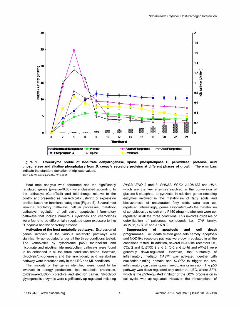
The secretion profiles of protease, peroxidase, lipase, PLCand acid and alkaline phosphatases were different for each ofthe enzymes studied at the different time points of bacterialculture (Figure 1). An intracellular enzyme assay, which detectsICD activity as a result of bacterial lysis was performed andinsignificant levels of activity (0-0.21 Units/ml) were detected.Maximum enzyme peak activities were observed at 4 hours ofgrowth (mid-log phase) for peroxidase, lipase, acidphosphatase and alkaline phosphatase with 10.96 Units/ml,12.53 Units/ml, 18.92 Units/ml and 24.81 Units/ml, respectively.In contrast, the activities of protease and PLC increased totheir highest levels at 8 hrs of growth (early-stationary phase)and with activities of 6.06 Units/ml and 11.73 Units/ml,respectively.
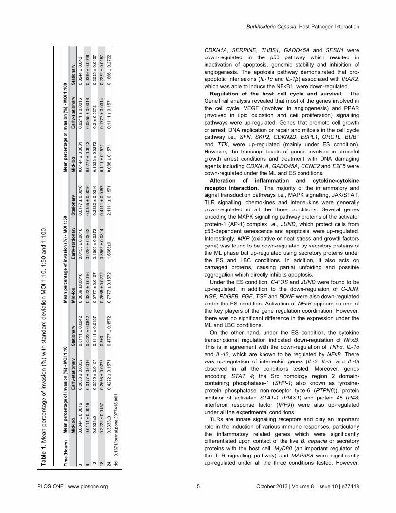
The ability of B. cepacia CQK strain to invade the epithelialcells was measured and compared at three different phases ofbacterial growth (Table 1). In general, the trend of the invasionwas similar for all the phases of growth at MOI 1:10 and 1:50.
However, the invasion capacity at MOI 1:50 werecomparatively higher than MOI 1:10 at all times of exposureand growth phases. The highest percentage of invasion(2.11%) was observed at MOI 1:50, stationary phase, 24 hourspost-infection. At MOI 1:100, the percentage of invasion wasmoderately higher than MOI 1:50 at 3-12 hours post-infectionat all phases of growth. However, the rate of invasion wasfound to decrease distinctly following longer post-infection timeat the respective growth phases.
In general, the intracellular survival profiles were similar at allthe MOIs (Figure 2). The intracellular survival of B. cepacia atMOI 1:50 showed slow replication of B. cepacia in the epithelialcells from the first hour to 12 hours of incubation. A similarpattern was also observed with the intracellular survival of B.cepacia at MOI of 1:100. The number of bacteria replicatedintracellularly peaked following six hours of incubation at allthree phases. Nevertheless, it was noticeable that the numberof cells living intracellularly decreased at longer time ofreplication (18 and 24 hours) at all the MOIs, suggesting thatintracellular survival in the epithelial cells was saturable.
Determination of epithelial cells viability upon infectionof B. cepacia
The reduction in the percentage of the A549 epithelial cellviability was demonstrated using trypan blue (Figure 3, panelsA and B). The cell survival was 100% at a concentration of 0.5µg/ml culture supernatant harvested at the mid-log and early-stationary phases and decreased gradually as theconcentration increased from 1-5 µg/ml. Using 50 µg/ml of themid-log phase culture supernatant, viability of the host cellswere found to be reduced to 55%. Conversely, none of the hostcells survived when they were exposed to 50 µg/ml of theearly-stationary phase culture supernatant (Figure 3, panel A).In addition, exposure of live B. cepacia to the A549 cellsshowed 99.2% and 99.05% of cell survival at MOI ratios of 1:10and 1:50, respectively. However, the percentage of cell survivalreduced to 83.02% at an MOI ratio of 1:100. Only 52.07% ofthe A549 cells were found to further survive at an MOI ratio of1:200 (Figure 3, panel B).
Host transcriptional responses to early exposure of B.cepacia live bacteria and the culture supernatant
In this study, a total 20,496 of 24,526 genes, (83.57%) werefiltered with cut-off values equivalent to Present and Marginal.Using the one-way ANOVA (asymptotic computed p-value<0.01) and Benjamini Hochberg (multiple testing corrections),9,029 of 20,496 genes (44.02%) were identified as significantlyexpressed. Additionally, based on the results obtained from thefold change analysis, Venn diagrams were generated todetermine common genes of the host that were differentiallyexpressed (Figure 4, panels A and B). From the experiments,422 genes were identified to be commonly up-regulated underall three experimental conditions (LBC, ML and ES).Conversely, only 312 genes were found to be commonly down-regulated among LBC, ML and ES. Scatter plots were alsoused to observe the relative distribution of genes that weresignificantly differentiated between each treated group and thecontrol group.
Burkholderia Cepacia, Host-Pathogen Interaction
PLOS ONE | www.plosone.org 3 October 2013 | Volume 8 | Issue 10 | e77418
Heat map analysis was performed and the significantlyregulated genes (p-value<0.05) were classified according tothe pathways (GeneTrail) and fold-change relative to thecontrol and presented as hierarchical clustering of expressionprofiles based on functional categories (Figure 5). Several hostimmune regulatory pathways, cellular processes, metabolicpathways, regulation of cell cycle, apoptosis, inflammatorypathways that include numerous cytokines and chemokineswere found to be differentially regulated upon exposure to liveB. cepacia and the secretory proteins.
Activation of the host metabolic pathways. Expression ofgenes involved in the various metabolic pathways wassignificantly up-regulated under all the three conditions tested.The xenobiotics by cytochrome p450 metabolism andnicotinate and nicotinamide metabolism pathways were foundto be enhanced in all the three conditions tested. However,glycolysis/glycogenesis and the arachidonic acid metabolismpathway were increased only in the LBC and ML conditions.
The majority of the genes identified were found to beinvolved in energy production, lipid metabolic processes,oxidation-reduction, cofactors and electron carrier. Glycolytic/glycogenesis enzymes were significantly up-regulated including
PYGB, ENO 2 and 3, PHKA2, PCK2, ALDH1A3 and HK1,which are the key enzymes involved in the conversion ofglucose-6-phosphate to pyruvate. In addition, genes encodingenzymes involved in the metabolism of fatty acids andbiosysnthesis of unsaturated fatty acids were also up-regulated. Interestingly, genes associated with the metabolismof xenobiotics by cytochrome P450 (drug metabolism) were up-regulated in all the three conditions. This involves oxidases ordetoxification of poisonous compounds i.e., CYP family,MGST2, GSTO2 and AKR1C2.
Suppression of apoptosis and cell deathprogrammes. Cell death related gene sets namely; apoptosisand NOD-like receptors pathway were down-regulated in all theconditions tested. In addition, several NOD-like receptors i.e.,CCL 2 and 5, BIRC 2 and 3, IL-6 and IL-1β and NFκB1 weregenerally down-regulated. However, the subfamily ofinflammatory mediator CASP1 was activated together withnucleotide-binding domain and NLRP2 to trigger the pro-inflammatory caspases upon injury, toxins or invasion. The p53pathway was down-regulated only under the LBC, where SFN,which is the p53-regulated inhibitor of the G2/M progression incell cycle, was up-regulated. However, the transcriptional of
Figure 1. Exoenzyme profile of isocitrate dehydrogenase, lipase, phospholipase C, peroxidase, protease, acidphosphatase and alkaline phosphatase from B. cepacia secretory proteins at different phases of growth. The error barsindicate the standard deviation of triplicate values.doi: 10.1371/journal.pone.0077418.g001
Burkholderia Cepacia, Host-Pathogen Interaction
PLOS ONE | www.plosone.org 4 October 2013 | Volume 8 | Issue 10 | e77418
Tabl
e 1.
Mea
n pe
rcen
tage
of i
nvas
ion
(%) w
ith s
tand
ard
devi
atio
n M
OI 1
:10,
1:5
0 an
d 1:
100.
Tim
e (H
ours
)M
ean
perc
enta
ge o
f inv
asio
n (%
) - M
OI 1
:10
Mea
n pe
rcen
tage
of i
nvas
ion
(%) -
MO
I 1:5
0M
ean
perc
enta
ge o
f inv
asio
n (%
) - M
OI 1
:100
M
id-lo
gEa
rly-s
tatio
nary
Stat
iona
ryM
id-lo
gEa
rly-s
tatio
nary
Stat
iona
ryM
id-lo
gEa
rly-s
tatio
nary
Stat
iona
ry3
0.00
44 ±
0.0
016
0.00
88 ±
0.0
032
0.01
11 ±
0.0
042
0.00
89 ±
0.00
160.
0155
± 0
.001
60.
0177
± 0
.001
60.
0144
± 0
.003
10.
0211
± 0
.001
60.
0244
± 0
.042
60.
0111
± 0
.001
60.
0177
± 0
.001
60.
0222
± 0
.064
20.
0222
± 0
.001
60.
0289
± 0
.004
20.
0355
± 0
.001
60.
0277
± 0
.004
20.
0355
± 0
.001
60.
0389
± 0
.001
612
0.03
33±0
0.05
55 ±
0.0
157
0.11
11 ±
0.0
157
0.07
77 ±
0.0
157
0.16
66 ±
0.0
272
0.22
22 ±
0.0
314
0.13
33 ±
0.0
272
0.2
± 0.
0272
0.25
55 ±
0.0
157
180.
2222
± 0
.015
70.
2666
± 0
.027
20.
3±0
0.26
66 ±
0.0
272
0.35
55 ±
0.0
314
0.41
11 ±
0.0
157
0.11
1 ±
0.15
710.
1777
± 0
.031
40.
2222
± 0
.015
724
0.33
33±0
0.42
22 ±
0.1
571
0.47
77 ±
0.1
572
0.77
77 ±
0.1
572
1.66
66±0
2.11
11 ±
0.1
571
0.08
8 ±
0.15
710.
1111
± 0
.157
10.
1666
± 0
.272
2
doi:
10.1
371/
jour
nal.p
one.
0077
418.
t001
CDKN1A, SERPINE, THBS1, GADD45A and SESN1 weredown-regulated in the p53 pathway which resulted ininactivation of apoptosis, genomic stability and inhibition ofangiogenesis. The apotosis pathway demonstrated that pro-apoptotic interleukins (IL-1α and IL-1β) associated with IRAK2,which was able to induce the NFκB1, were down-regulated.
Regulation of the host cell cycle and survival. TheGeneTrail analysis revealed that most of the genes involved inthe cell cycle, VEGF (involved in angiogenesis) and PPAR(involved in lipid oxidation and cell proliferation) signallingpathways were up-regulated. Genes that promote cell growthor arrest, DNA replication or repair and mitosis in the cell cyclepathway i.e., SFN, SKP2, CDKN2D, ESPL1, ORC1L, BUB1and TTK, were up-regulated (mainly under ES condition).However, the transcript levels of genes involved in stressfulgrowth arrest conditions and treatment with DNA damagingagents including CDKN1A, GADD45A, CCNE2 and E2F5 weredown-regulated under the ML and ES conditions.
Alteration of inflammation and cytokine-cytokinereceptor interaction. The majority of the inflammatory andsignal transduction pathways i.e., MAPK signalling, JAK/STAT,TLR signalling, chemokines and interleukins were generallydown-regulated in all the three conditions. Several genesencoding the MAPK signalling pathway proteins of the activatorprotein-1 (AP-1) complex i.e., JUND, which protect cells fromp53-dependent senescence and apoptosis, were up-regulated.Interestingly, MKP (oxidative or heat stress and growth factorsgene) was found to be down-regulated by secretory proteins ofthe ML phase but up-regulated using secretory proteins underthe ES and LBC conditions. In addition, it also acts ondamaged proteins, causing partial unfolding and possibleaggregation which directly inhibits apoptosis.
Under the ES condition, C-FOS and JUND were found to beup-regulated, in addition to the down-regulation of C-JUN.NGF, PDGFB, FGF, TGF and BDNF were also down-regulatedunder the ES condition. Activation of NFκB appears as one ofthe key players of the gene regulation coordination. However,there was no significant difference in the expression under theML and LBC conditions.
On the other hand, under the ES condition, the cytokinetranscriptional regulation indicated down-regulation of NFκB.This is in agreement with the down-regulation of TNFα, IL-1αand IL-1β, which are known to be regulated by NFκB. Therewas up-regulation of interleukin genes (IL-2, IL-3, and IL-6)observed in all the conditions tested. Moreover, genesencoding STAT 4; the Src homology region 2 domain-containing phosphatase-1 (SHP-1; also known as tyrosine-protein phosphatase non-receptor type-6 (PTPN6)), proteininhibitor of activated STAT-1 (PIAS1) and protein 48 (P48;interferon response factor (IRF9)) were also up-regulatedunder all the experimental conditions.
TLRs are innate signalling receptors and play an importantrole in the induction of various immune responses, particularlythe inflammatory related genes which were significantlydifferentiated upon contact of the live B. cepacia or secretoryproteins with the host cell. MyD88 (an important regulator ofthe TLR signalling pathway) and MAP3K8 were significantlyup-regulated under all the three conditions tested. However,
Burkholderia Cepacia, Host-Pathogen Interaction
PLOS ONE | www.plosone.org 5 October 2013 | Volume 8 | Issue 10 | e77418
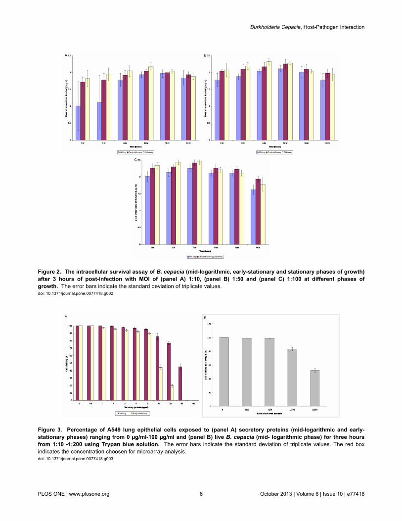
Figure 2. The intracellular survival assay of B. cepacia (mid-logarithmic, early-stationary and stationary phases of growth)after 3 hours of post-infection with MOI of (panel A) 1:10, (panel B) 1:50 and (panel C) 1:100 at different phases ofgrowth. The error bars indicate the standard deviation of triplicate values.doi: 10.1371/journal.pone.0077418.g002
Figure 3. Percentage of A549 lung epithelial cells exposed to (panel A) secretory proteins (mid-logarithmic and early-stationary phases) ranging from 0 µg/ml-100 µg/ml and (panel B) live B. cepacia (mid- logarithmic phase) for three hoursfrom 1:10 -1:200 using Trypan blue solution. The error bars indicate the standard deviation of triplicate values. The red boxindicates the concentration choosen for microarray analysis.doi: 10.1371/journal.pone.0077418.g003
Burkholderia Cepacia, Host-Pathogen Interaction
PLOS ONE | www.plosone.org 6 October 2013 | Volume 8 | Issue 10 | e77418

there is also an apparent decrease in some cytokine endproducts of the TLR signalling observed under the ESsecretory proteins, but no significant difference in expressionwas observed under the LBC and ML conditions. Theexpression of the COX-2 isoform was induced after interactionwith live B. cepacia and secretory proteins (ML and ES).
Genes of the chemokines C-X-C motif chemokine 5 (CXCL5)as well as IL-11 of the host were up-regulated after interactionwith live B. cepacia and secretory proteins (mid-log and early-stationary phases). However, CCL26 and IL-8 were found to beup-regulated only under the LBC condition. On the other hand,CCL2, CCL5 and CCL20 genes along with IL-1β, IL-1α, IL11RAand IL27RA were down-regulated under the ML and ESconditions. In addition, IL10RB was down-regulated under theLBC and ML conditions whereas IL-6 and LIF were down-regulated under the ES condition.
Validation of the microarray dataThe qRT-PCR assay was used for validation of genes
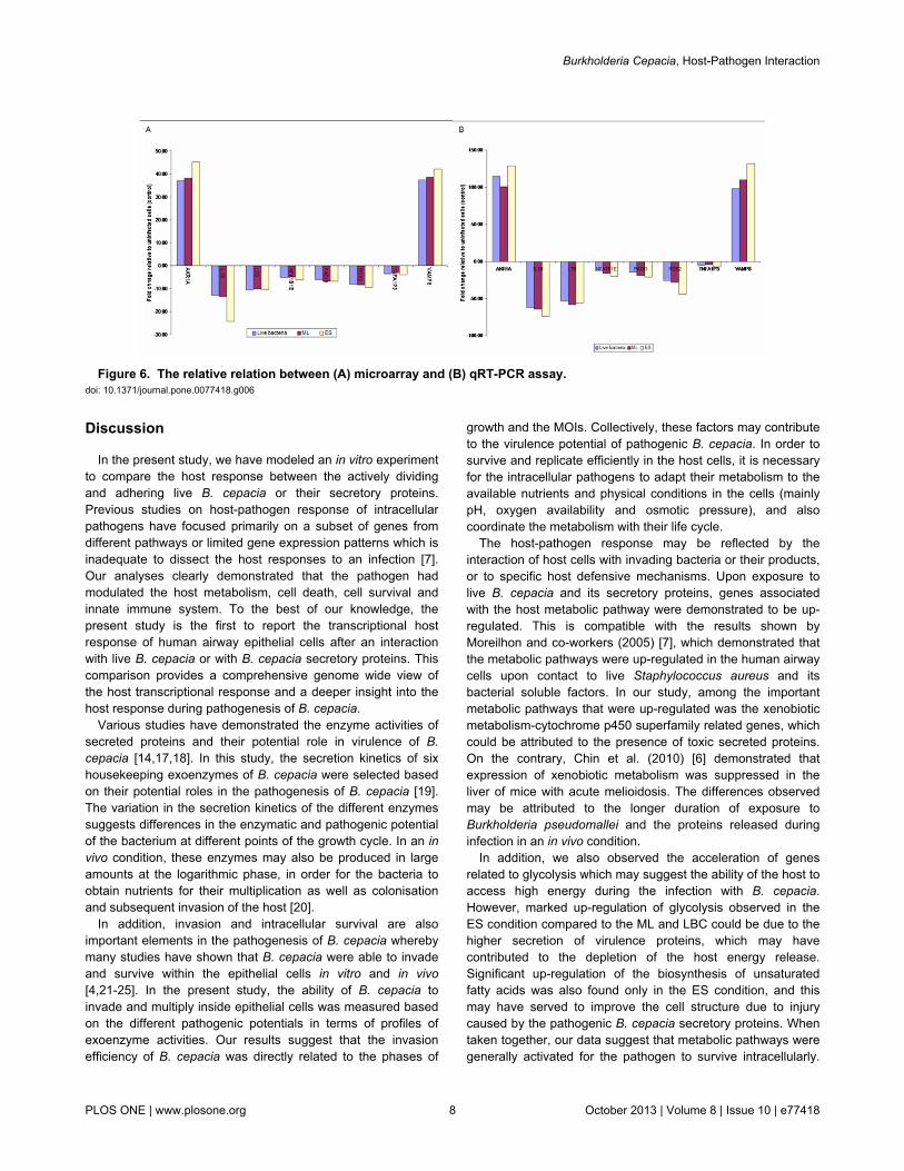
involved in adhesion, cell growth, apoptosis, inflammation, andcorresponding to cytokines, receptors, adapters, kinases,phosphatases, and transcription factors that were differentiallyexpressed in the microarray analysis. The data obtained,together with those determined by the microarray experimentsare shown in Figure 6 (panels A and B). The gene expressionsobtained using the two techniques were comparable. However,differences were observed in the fold-change values wherebythe fold-change detected in qRT-PCR assay was higher thanthe microarray.
Figure 4. Venn diagrams showing the number of genes that were up-regulated (panel A) and down-regulated (panel B)with fold changes of ≥2.0-fold. doi: 10.1371/journal.pone.0077418.g004
Figure 5. Heat map analysis. doi: 10.1371/journal.pone.0077418.g005
Burkholderia Cepacia, Host-Pathogen Interaction
PLOS ONE | www.plosone.org 7 October 2013 | Volume 8 | Issue 10 | e77418
Discussion
In the present study, we have modeled an in vitro experimentto compare the host response between the actively dividingand adhering live B. cepacia or their secretory proteins.Previous studies on host-pathogen response of intracellularpathogens have focused primarily on a subset of genes fromdifferent pathways or limited gene expression patterns which isinadequate to dissect the host responses to an infection [7].Our analyses clearly demonstrated that the pathogen hadmodulated the host metabolism, cell death, cell survival andinnate immune system. To the best of our knowledge, thepresent study is the first to report the transcriptional hostresponse of human airway epithelial cells after an interactionwith live B. cepacia or with B. cepacia secretory proteins. Thiscomparison provides a comprehensive genome wide view ofthe host transcriptional response and a deeper insight into thehost response during pathogenesis of B. cepacia.
Various studies have demonstrated the enzyme activities ofsecreted proteins and their potential role in virulence of B.cepacia [14,17,18]. In this study, the secretion kinetics of sixhousekeeping exoenzymes of B. cepacia were selected basedon their potential roles in the pathogenesis of B. cepacia [19].The variation in the secretion kinetics of the different enzymessuggests differences in the enzymatic and pathogenic potentialof the bacterium at different points of the growth cycle. In an invivo condition, these enzymes may also be produced in largeamounts at the logarithmic phase, in order for the bacteria toobtain nutrients for their multiplication as well as colonisationand subsequent invasion of the host [20].
In addition, invasion and intracellular survival are alsoimportant elements in the pathogenesis of B. cepacia wherebymany studies have shown that B. cepacia were able to invadeand survive within the epithelial cells in vitro and in vivo[4,21-25]. In the present study, the ability of B. cepacia toinvade and multiply inside epithelial cells was measured basedon the different pathogenic potentials in terms of profiles ofexoenzyme activities. Our results suggest that the invasionefficiency of B. cepacia was directly related to the phases of
growth and the MOIs. Collectively, these factors may contributeto the virulence potential of pathogenic B. cepacia. In order tosurvive and replicate efficiently in the host cells, it is necessaryfor the intracellular pathogens to adapt their metabolism to theavailable nutrients and physical conditions in the cells (mainlypH, oxygen availability and osmotic pressure), and alsocoordinate the metabolism with their life cycle.
The host-pathogen response may be reflected by theinteraction of host cells with invading bacteria or their products,or to specific host defensive mechanisms. Upon exposure tolive B. cepacia and its secretory proteins, genes associatedwith the host metabolic pathway were demonstrated to be up-regulated. This is compatible with the results shown byMoreilhon and co-workers (2005) [7], which demonstrated thatthe metabolic pathways were up-regulated in the human airwaycells upon contact to live Staphylococcus aureus and itsbacterial soluble factors. In our study, among the importantmetabolic pathways that were up-regulated was the xenobioticmetabolism-cytochrome p450 superfamily related genes, whichcould be attributed to the presence of toxic secreted proteins.On the contrary, Chin et al. (2010) [6] demonstrated thatexpression of xenobiotic metabolism was suppressed in theliver of mice with acute melioidosis. The differences observedmay be attributed to the longer duration of exposure toBurkholderia pseudomallei and the proteins released duringinfection in an in vivo condition.
In addition, we also observed the acceleration of genesrelated to glycolysis which may suggest the ability of the host toaccess high energy during the infection with B. cepacia.However, marked up-regulation of glycolysis observed in theES condition compared to the ML and LBC could be due to thehigher secretion of virulence proteins, which may havecontributed to the depletion of the host energy release.Significant up-regulation of the biosynthesis of unsaturatedfatty acids was also found only in the ES condition, and thismay have served to improve the cell structure due to injurycaused by the pathogenic B. cepacia secretory proteins. Whentaken together, our data suggest that metabolic pathways weregenerally activated for the pathogen to survive intracellularly.
Figure 6. The relative relation between (A) microarray and (B) qRT-PCR assay. doi: 10.1371/journal.pone.0077418.g006
Burkholderia Cepacia, Host-Pathogen Interaction
PLOS ONE | www.plosone.org 8 October 2013 | Volume 8 | Issue 10 | e77418
These metabolic alterations are presumed for the maintenanceof host homeostasis, which is beneficial to the host as well asthe pathogen.
In this study, down-regulation of the host apoptotic pathwaywas detected under all the conditions investigated, with themaximum effect observed under the ES condition, whichcorrelates with the findings by Moreilhon and co-workers(2005) [7]. We believe that this could be due to an urge for thehost to survive under the ES condition, which was morepathogenic compared to the LBC and ML conditions. Inhibitionof apoptosis of the A549 cells would ease replication of thebacteria, prolongs their intracellular survival and favoursbacterial persistence.
Activation of the p53 signalling pathway can lead to eitherthe cell cycle arrest or apoptosis [26]. In the present study, thesignificant down-regulation of p53 was only found in the LBCcondition. Interaction of the live B. cepacia with the host cellsmight have caused minimal DNA damage. This was supportedby our cell viability test results which showed minimalpercentage of cell death. However, an in vitro model for thehuman meninges has provided evidence that secreted proteinsfrom Neisseria meningitidis also play a role in the induction ofthe cell cycle and resistance to apoptosis [27].
Cell cycle progression is a highly complex process whichrequires coordinated activation of several kinases, some ofwhich are activated upon binding of a cyclin subunit [28]. Ouranalysis consistently demonstrated that the cell cycle was up-regulated. GADD45, through it’s association with CDC2,appears to disrupt interaction between CDC2 and Cyclin B1and hence induces G2/M arrest. Similar transcriptional profileswere also seen by Zhang et al. (1999) [29], thus suggestingthat B. cepacia induces G2/M arrest in response to DNAdamage to allow time for DNA repair. In addition, activation ofthe VEGF signaling pathway promotes angiogenesis, which isinvolved in the normal processes of wound healing. Therefore,exposure of the host to live B. cepacia and secretory proteinsmay have caused injuries or cell damage.
Additionally, cyclooxygenase (COX), an inflammationenzyme involved in release of excessive amounts of PGE2(prostaglandin E2) was also highly up-regulated by the hostupon exposure to B. cepacia secretory proteins, suggestingthat COX may be an important mediator of B. cepaciapathogenesis. The enhanced expression of COX leads tofurther proliferation, reduction in apoptosis and angiogenesis[30,31]. Ejima et al. (2003) [32] suggested that COX is animportant mediator of the response to bacterial sepsis whenthey showed that COX deficient mice had increasedsusceptibly to bacterial peritonitis and toxaemia. Thus, in thisstudy, the induction of COX upon exposure to secretedproducts may partially explain the resistance of the epithelialcells to apoptosis.
Many researchers have demonstrated that infections byintracellular pathogens alter the expression of genes encodingpro-inflammatory cytokines and chemokines, which have beenimplicated as principal mediators during infections of the host inboth in vitro and in vivo systems [6,7,33]. These cytokines andchemokines also function as central mediators in stimulatingvarious host defences systems such as the cytokine-cytokine
receptor interactions pathway, signalling pathways andapoptosis and eventually elicit appropriate adaptive immunesystem. Moreover, C-JUN, in combination with C-FOS, formsthe AP-1 early response transcription factor, which up-regulates transcription of a diverse range of genes involved inproliferation and differentiation to defense against invasion andcell damage. In our study, the pro-inflammatory moleculeswere found to be down-regulated upon infection, and similarevidences have been observed from in vitro [33] and in vivo[34] studies. However, this is in contrast to the earlier report byMoreilhon and co-workers (2004) [7], which described up-regulation of the interleukins in the human airway cells when incontact with S. aureus supernatant.
Our microarray data demonstrated that B. cepacia interferedwith the transcription of genes involved in the production of pro-inflammatory cytokines. Inhibition of these genes by B. cepaciawould theoretically result in a reduction of recruitment of theinnate immune cells to the site of infection, which in turn couldinfluence the degree of host inflammatory response andbacterial elimination. B. cepacia has been shown to modulatethe epithelial bactericidal response in favour of its intracellularsurvival and persistence in the human host, and this processmay be associated with disease relapse. Feezor et al. (2002)[35] demonstrated that the release of TNFα, IL-10 and IL-6 intobactericidal-stimulated whole blood obtained from patients withsevere sepsis was significantly decreased compared with thecontrol group. Moreover, Ertel and colleagues (1995) [36] hadhypothesised the role of systemic inflammation and aneffectively functioning immune system of the host in eliminatinginvading pathogens. They speculated that during an infection,the pathogens may be made compromised differentlydepending on the time of exposure. In addition, they have alsoreported the excessive down-regulation of the synthesis of pro-inflammatory cytokines and their release in the early periodafter contact with pathogens. This may be an evolutionaryprocess to reduce the incidence of tissue necrosis and theconsequential multiple organ failure.
Likewise, Michie et al. (1988) [37] have demonstrated thatinfusion of high doses of pro-inflammatory cytokines may resultin severe host tissue damage, organ failure and death in an invivo condition. We presume that the response of the host cellsto B. cepacia secretory proteins mimics the physiological actionof the host towards toxins. This phenomenon may be referredto as ‘toxin tolerance,’ which is due to a reduced capacity of thecells to synthesise and/or secrete pro-inflammatory cytokines.Granowitz et al. (1993) [38] hypothesised that the phenomenonof toxin tolerance may be due to a defect in transcriptionaland/or translational regulation of the host. These indicate thatthe control of the immune response of the host to toxins that ismediated by TNFα and IL-16 may represent a potent protectivemechanism against infection with invasive microorganisms. Inthis light, the reduced capability of the host cells from septicshock to produce and release adequate amounts of pro-inflammatory cytokines after exposure to toxin may indicate theinability of septic patients to adequately respond to repeated orpersisting invasion of microorganisms and to maintain aneffective defence system.
Burkholderia Cepacia, Host-Pathogen Interaction
PLOS ONE | www.plosone.org 9 October 2013 | Volume 8 | Issue 10 | e77418
In summary, our data is consistent with the interpretation thatB. cepacia can manipulate A549 cells for its own advantage,allowing prolonged survival inside the harmless environmentalniche of the non-phagocytic cells. The data presented clearlyindicate that early exposure of the host cells to either live B.cepacia or their secretory proteins are similar. Contrary topopular belief, we postulate that B. cepacia inducedhomeostatic responses in the host cell which in turn aid thebacteria to establish persistent infection. Using this knowledge,strategies interfering with essential interactions between B.cepacia and the host cell, like apoptosis inhibition, can beexploited in the future to develop an innovative arsenal oftherapeutic compounds.
Supporting Information
Table S1. (DOC)
Acknowledgements
We are grateful to the Malaysia Genome Institute (MGI) forallowing us to use the microarray equipments and facilities andthe Tropical Infectious Disease Research and EducationCenter (TIDREC), Faculty of Medicine, University of Malaya forallowing us to use the GeneSpring software.
Author Contributions
Conceived and designed the experiments: VM KMV JV.Performed the experiments: VM KMV. Analyzed the data: VMKMV JT. Contributed reagents/materials/analysis tools: OHHJV. Wrote the manuscript: VM.
References
1. Holt JG, Krieg NR, Sneath PHA, Stately JT, Williams St (1994)Bergey’s Manual of determinative bacteriology, 9th ed. Baltimore:Williams and Wilkins: USA
2. Jones AM, Dodd ME, Govan JRW, Barcus V, Doherty CJ et al. (2004)Burkholderia cenocepacia and Burkholderia multivorans: influence onsurvival in cystic fibrosis. Thorax 59: 948–951. doi:10.1136/thx.2003.017210. PubMed: 15516469.
3. Govan JRW, Hughes JE, Vandamme P (1996) Burkholderia cepacia:medical, taxonomic and ecological issues. J Med Microbiol 45:395-407. doi:10.1099/00222615-45-6-395. PubMed: 8958242.
4. Martin DW, Mohr CD (2000) Invasion and intracellular survival ofBurkholderia cepacia. Infect Immun 68: 24-29. doi:10.1128/IAI.68.1.24-29.2000. PubMed: 10603364.
5. Mariappan V, Vellasamy KM, Hashim OH, Vadivelu J (2011) Profiling ofBurkholderia cepacia secretome at mid-logarithmic and early-stationaryphases of growth. PLOS ONE 6(10): e26518. doi:10.1371/journal.pone.0026518. PubMed: 22046299.
6. Chin CY, Monack DM, Nathan S (2010) Genome wide transcriptomeprofiling of a murine acute melioidosis model reveals new insights intohow Burkholderia pseudomallei overcomes host innate immunity. BMCGenomics 11: 672-680. doi:10.1186/1471-2164-11-672. PubMed:21110886.
7. Moreilhon C, Gras D, Hologne C, Bajolet O, Cottrez F et al. (2005) LiveStaphylococcus aureus and bacteria soluble factors induce differenttranscriptional responses in human airway cells. Physiol Genomics. 20:244-255. PubMed: 15598879.
8. Mariappan V, Vellasamy KV, Thimma JS, Hashim OH, Vadivelu J(2010) Identification of immunogenic proteins from Burkholderiacepacia secretome using proteomic analysis. Vaccine 13: 1318-1324.PubMed: 19944788.
9. Bradford MM (1976) A rapid and sensitive method for quantitation ofmicrogram quantities of protein utilizing the principle of protein-dyebinding. Anal Biochem 72: 248-225. doi:10.1016/0003-2697(76)90527-3. PubMed: 942051.
10. Chavira R Jr., Burnett TJ, Hageman JH (1984) Assaying proteinaseswith azocoll. Anal Biochem 136: 446-450. doi:10.1016/0003-2697(84)90242-2. PubMed: 6426343.
11. Geoffroy C, Raveneau J, Beretti JL, Lecroisey A et al. (1991)Purification and characterization of an extracellular 29-kilodaltonphospholipase C from Listeria monocytogenes. Infect Immun 59:2382-2388. PubMed: 1904842.
12. Pine L, Hoffman PS, Malcolm GB, Benson RF, Gorman GW (1984)Whole-cell peroxidase test for identification of Legionella pneumophila.J Clin Microbiol 19: 286-290. PubMed: 6365966.
13. Domenech CE, Lisa TA, Salvano MA, Garrido MN (1992)Pseudomonas aeruginosa acid phosphatase. Activation by divalentcations and inhibition by aluminium ion. FEBS Lett 299: 96–98. doi:10.1016/0014-5793(92)80108-S. PubMed: 1544481.
14. Lonon MK, Woods DE, Straus DC (1988) Production of lipase byclinical isolates of Pseudomonas cepacia. J Clin Microbiol 26: 979-984.PubMed: 3384918.
15. Andersen P, Askgaard D, Ljungqvist L, Bennedsen J, Heron I (1991)Proteins released from Mycobacterium tuberculosis during growth.Infect Immun 59: 1905-1910. PubMed: 1903768.
16. Miles AA, Misra SS (1938) Estimation of the bactericidal power of theblood. J Hyg (Camb.) 38: 732-749. doi:10.1017/S002217240001158X.
17. McKenney D, Brown KE, Allison DG (1995) Influence of Pseudomonasaeruginosa exoproducts on virulence factor production in Burkholderiacepacia: evidence of interspecies communication. J Bacteriol 177:6989-6992. PubMed: 7592496.
18. McKevitt AI, Bajaksouzian S, Klinger JD, Woods DE (1989) Purificationand characterization of an extracellular protease from Pseudomonascepacia. Infect Immun 57: 771–778. PubMed: 2645209.
19. Govan JRW, Vandamme P (1998) Agricultural and medicalmicrobiology: a time for bridging gaps. Microbiol 144: 2373–2375. doi:10.1099/00221287-144-9-2373. PubMed: 9782485.
20. Lefebre MD, Valvano MA (2001) In vitro resistance of Burkholderiacepacia complex isolates to reactive oxygen species in relation tocatalase and superoxide dismutase production. Microbiol 147: 97-109.
21. Caraher E, Duff C, Mullen T, Mc Keon S, Murphy P et al. (2007)Invasion and biofilm formation of Burkholderia dolosa is comparablewith Burkholderia cenocepacia and Burkholderia multivorans. J CystFibros 6: 49–56. doi:10.1016/S1569-1993(07)60184-2. PubMed:16781896.
22. Duff C, Murphy PG, Callaghan M, McClean S (2006) Differences ininvasion and translocation of Burkholderia cepacia complex species inpolarised lung epithelial cells in vitro. Microb Pathog 41: 183–192. doi:10.1016/j.micpath.2006.07.005. PubMed: 16938423.
23. Cieri MV, Mayer-Hamblett N, Griffith A, Burns JL (2002) Correlationbetween an in vitro invasion assay and a murine model of Burkholderiacepacia lung infection. Infect Immun 70: 1081–1086. doi:10.1128/IAI.70.3.1081-1086.2002. PubMed: 11854186.
24. Chiu CH, Wong S, Hancock REW, Speert DP (2001) Adherence ofBurkholderia cepacia to respiratory tract epithelial cells and inhibitionwith dextrans. Microbiol 147: 2651–2658. PubMed: 11577144.
25. Burns JL, Jonas M, Chi EY, Clark DK, Berger A et al. (1996) Invasionof respiratory epithelial cells by Burkholderia (Pseudomonas) cepacia.Infect Immun 64: 4054–4059. PubMed: 8926068.
26. Vogelstein B, Lane D, Levine AJ (2000) Surfing the p53 network.Nature 408: 307-310. doi:10.1038/35042675. PubMed: 11099028.
27. Wells DB, Tighe PJ, Wooldridge KG, Robinson K, Aldeen DAA (2001)Differential gene expression during meningeal-meningococcalinteraction: evidence for self-defense and early release of cytokinesand chemokines. Infect Immun 69: 2718–2722. doi:10.1128/IAI.69.4.2718-2722.2001. PubMed: 11254640.
28. Malumbres M, Sotillo R, Santamaría D, Galán J, Cerezo A et al. (2004)Mammalian cells cycle without the D-type cyclin-dependent kinasesCdk4 and Cdk6. Cell 118: 493–504. doi:10.1016/j.cell.2004.08.002.PubMed: 15315761.
29. Zhang JM, Zhao X, Wei Q, Paterson BM (1999) Direct inhibition ofG1 cdk kinase activity by MyoD promotes myoblast cell cycle
Burkholderia Cepacia, Host-Pathogen Interaction
PLOS ONE | www.plosone.org 10 October 2013 | Volume 8 | Issue 10 | e77418
withdrawal and terminal differentiation. EMBO J 18: 6983-6993. doi:10.1093/emboj/18.24.6983. PubMed: 10601020.
30. Walduck A, Schmitt A, Lucas B, Aebischer T, Meyer TF (2004)Transcription profiling analysis of the mechanisms of vaccine-inducedprotection against Helicobacter pylori. FASEB J 18: 1955–1957.PubMed: 15456742.
31. Oshima K, Kakizawa S, Nishigawa H, Jung HY, Wei W et al. (2004)Reductive evolution suggested from the complete genome sequence ofa plant-pathogenic Phytoplasma. Nat Genet, 36: 27-29. doi:10.1038/ng1277. PubMed: 14661021.
32. Ejima K, Layne MD, Carvajal IM, Kritek PA, Baron RM et al. (2003)Cyclooxygenase-2-deficient mice are resistant to endotoxin-inducedinflammation and death. FASEB J 17: 1325–1327. PubMed: 12738799.
33. Wongprompitak P, Sirisinha S, Chaiyaroj CS (2008) Differential GeneExpression Profiles of lung epithelial cells exposed to Burkholderiapseudomallei and Burkholderia thailandensis during the initial phase ofinfection. Asia. Pac. J. Immuno: Allergy 26: 59-70. PubMed: 19548631.
34. Dinarello CA (1991) The proinflammatory cytokines interleukin- 1 andtumour necrosis factor and treatment of septic shock.syndrome. J InfectDis 163: 1177-1184. doi:10.1093/infdis/163.6.1177. PubMed: 2037782.
35. Feezor RJ, Oberholzer C, Baker HV, Novick D, Rubinstein M et al.(2002) Molecular characterization of the acute inflammatory responseto infections with Gram-negative versus Gram-positive bacteria. InfectImmun 71: 5803-5813. PubMed: 14500502.
36. Ertel W, Kremer JP, Kenney J, Steckholzer U, Jarrar D (1995) Downregulation of proinflammatory cytokine release in whole blood fromseptic patients. Blood 85: 1341-1347. PubMed: 7858264.
37. Michie HR, Manogue KR, Spriggs DR, Revhaug A, O’Dwyer S et al.(1988) Detection of circulating tumor necrosis factor after endotoxinadministration. N Engl J Med 318: 1481-1489. doi:10.1056/NEJM198806093182301. PubMed: 2835680.
38. Granowitz EV, Porat R, Mier JW, Orencole SF, Kaplanski G et al.(1993) Intravenous endotoxin suppresses the cytokine response ofperipheral blood mononuclear cells of healthy humans. J Immunol 151:1637-1645. PubMed: 7687636.
Burkholderia Cepacia, Host-Pathogen Interaction
PLOS ONE | www.plosone.org 11 October 2013 | Volume 8 | Issue 10 | e77418
Copyright © 2022 FDOKUMEN