The Genetic and Molecular Basis of O-Antigenic Diversity in Burkholderia pseudomallei...
9
The Genetic and Molecular Basis of O-Antigenic Diversity in Burkholderia pseudomallei Lipopolysaccharide Apichai Tuanyok 1 *, Joshua K. Stone 1 , Mark Mayo 2 , Mirjam Kaestli 2 , Jeffrey Gruendike 1 , Shalamar Georgia 1 , Stephanie Warrington 1 , Travis Mullins 1 , Christopher J. Allender 1 , David M. Wagner 1 , Narisara Chantratita 3 , Sharon J. Peacock 3 , Bart J. Currie 2 , Paul Keim 1,4 1 Department of Biological Sciences, Northern Arizona University, Flagstaff, Arizona, United States of America, 2 Menzies School of Health Research and Northern Territory Clinical School, Darwin, Australia, 3 Faculty of Tropical Medicine, Mahidol University, Bangkok, Thailand, 4 The Translational Genomics Research Institute, Flagstaff, Arizona, United States of America Abstract Lipopolysaccharide (LPS) is one of the most important virulence and antigenic components of Burkholderia pseudomallei, the causative agent of melioidosis. LPS diversity in B. pseudomallei has been described as typical, atypical or rough, based upon banding patterns on SDS-PAGE. Here, we studied the genetic and molecular basis of these phenotypic differences. Bioinformatics was used to determine the diversity of genes known or predicted to be involved in biosynthesis of the O- antigenic moiety of LPS in B. pseudomallei and its near-relative species. Multiplex-PCR assays were developed to target diversity of the O-antigen biosynthesis gene patterns or LPS genotypes in B. pseudomallei populations. We found that the typical LPS genotype (LPS genotype A) was highly prevalent in strains from Thailand and other countries in Southeast Asia, whereas the atypical LPS genotype (LPS genotype B) was most often detected in Australian strains (,13.8%). In addition, we report a novel LPS ladder pattern, a derivative of the atypical LPS phenotype, associated with an uncommon O-antigen biosynthesis gene cluster that is found in only a small B. pseudomallei sub-population. This new LPS group was designated as genotype B2. We also report natural mutations in the O-antigen biosynthesis genes that potentially cause the rough LPS phenotype. We postulate that the diversity of LPS may correlate with differential immunopathogenicity and virulence among B. pseudomallei strains. Citation: Tuanyok A, Stone JK, Mayo M, Kaestli M, Gruendike J, et al. (2012) The Genetic and Molecular Basis of O-Antigenic Diversity in Burkholderia pseudomallei Lipopolysaccharide. PLoS Negl Trop Dis 6(1): e1453. doi:10.1371/journal.pntd.0001453 Editor: Craig R. Roy, Yale University School of Medicine, United States of America Received June 17, 2011; Accepted November 11, 2011; Published January 3, 2012 Copyright: ß 2012 Tuanyok et al. This is an open-access article distributed under the terms of the Creative Commons Attribution License, which permits unrestricted use, distribution, and reproduction in any medium, provided the original author and source are credited. Funding: This project was funded by National Institutes of Health-National Institute of Allergy and Infectious Diseases Grant U54 AI-065359 and U01 AI-075568 to PK and United States Department of Homeland Security award no. HSHQDC-10-C-00135 to AT. The funders had no role in study design, data collection and analysis, decision to publish, or preparation of the manuscript. Competing Interests: The authors have declared that no competing interests exist. * E-mail: [email protected] Introduction Lipopolysaccharide (LPS) is a major component of the outer membrane of Gram-negative bacteria, playing an important role in cell integrity and in signaling host innate immune response [1]. Structurally, LPS is composed of three major components: lipid A, the bacterial endotoxin that is embedded in the phospholipid bilayer of the outer membrane; core-oligosaccharide; and O- antigen. These three components are linked together as a part of the bacterial outer membrane. In a highly pathogenic bacterial species, such as Burkholderia pseudomallei, LPS has a major role in stimulating host innate immune response during infection [2]. B. pseudomallei LPS has been classified as a type II O-polysaccharide (O-PS) and is one of 4 different surface polysaccharides produced by this pathogen [3]. Previous studies have shown that B. pseudomallei LPS is required for serum resistance and virulence [4]. It has been well established in many bacterial diseases that overstimulation of the host cells by LPS can lead to the features of septic shock [5]. Likewise for B. pseudomallei, septicemia is a major cause of death. In Northeast Thailand especially in Ubon Ratchathani Province where melioidosis is highly endemic, the average incidence rate of melioidosis is 12.7 cases per 100,000 people per year with the average of 42.6% of mortality rate [6]. Cellular recognition of LPS by the innate immune system triggers the proinflammatory cytokines by host cells, which aids in the clearance of the pathogen. Previous studies have supported a potential role for B. pseudomallei LPS in protective immunity, with high concentrations of antibodies to LPS associated with survival in severe melioidosis [7,8]. As a result, LPS has been used in vaccine development and provided protective immunity in a murine model of melioidosis [2]. In addition, it was demonstrated that LPS had an important role in bacterial virulence because the LPS mutant B. pseudomallei strain SRM117, which lacked the O- antigenic polysaccharide moiety was more susceptible to macro- phage killing during the early phase of infection than its parental strain 1026b [9]. A previous study [10] identified LPS diversity based upon electrophoretic mobility with SDS-PAGE and detection using immunoblot analysis. This diversity included two serotypes (A and B) possessing different electrophoretic ladder profiles and a rough type that did not contain the ladder patterns; all were antigenically distinct [10]. Molecular structure of O-antigen serotype A or typical type has been described as the unbranched heteropolymers consisting of disaccharides repeats of -3)-b-D-glucopyranose-(1-3) www.plosntds.org 1 January 2012 | Volume 6 | Issue 1 | e1453
-
Upload
manoa-hawaii -
Category
Documents
-
view
0 -
download
0
Transcript of The Genetic and Molecular Basis of O-Antigenic Diversity in Burkholderia pseudomallei...
The Genetic and Molecular Basis of O-Antigenic Diversityin Burkholderia pseudomallei LipopolysaccharideApichai Tuanyok1*, Joshua K. Stone1, Mark Mayo2, Mirjam Kaestli2, Jeffrey Gruendike1, Shalamar
Georgia1, Stephanie Warrington1, Travis Mullins1, Christopher J. Allender1, David M. Wagner1, Narisara
Chantratita3, Sharon J. Peacock3, Bart J. Currie2, Paul Keim1,4
1 Department of Biological Sciences, Northern Arizona University, Flagstaff, Arizona, United States of America, 2 Menzies School of Health Research and Northern Territory
Clinical School, Darwin, Australia, 3 Faculty of Tropical Medicine, Mahidol University, Bangkok, Thailand, 4 The Translational Genomics Research Institute, Flagstaff, Arizona,
United States of America
Abstract
Lipopolysaccharide (LPS) is one of the most important virulence and antigenic components of Burkholderia pseudomallei,the causative agent of melioidosis. LPS diversity in B. pseudomallei has been described as typical, atypical or rough, basedupon banding patterns on SDS-PAGE. Here, we studied the genetic and molecular basis of these phenotypic differences.Bioinformatics was used to determine the diversity of genes known or predicted to be involved in biosynthesis of the O-antigenic moiety of LPS in B. pseudomallei and its near-relative species. Multiplex-PCR assays were developed to targetdiversity of the O-antigen biosynthesis gene patterns or LPS genotypes in B. pseudomallei populations. We found that thetypical LPS genotype (LPS genotype A) was highly prevalent in strains from Thailand and other countries in Southeast Asia,whereas the atypical LPS genotype (LPS genotype B) was most often detected in Australian strains (,13.8%). In addition, wereport a novel LPS ladder pattern, a derivative of the atypical LPS phenotype, associated with an uncommon O-antigenbiosynthesis gene cluster that is found in only a small B. pseudomallei sub-population. This new LPS group was designatedas genotype B2. We also report natural mutations in the O-antigen biosynthesis genes that potentially cause the rough LPSphenotype. We postulate that the diversity of LPS may correlate with differential immunopathogenicity and virulenceamong B. pseudomallei strains.
Citation: Tuanyok A, Stone JK, Mayo M, Kaestli M, Gruendike J, et al. (2012) The Genetic and Molecular Basis of O-Antigenic Diversity in Burkholderia pseudomalleiLipopolysaccharide. PLoS Negl Trop Dis 6(1): e1453. doi:10.1371/journal.pntd.0001453
Editor: Craig R. Roy, Yale University School of Medicine, United States of America
Received June 17, 2011; Accepted November 11, 2011; Published January 3, 2012
Copyright: � 2012 Tuanyok et al. This is an open-access article distributed under the terms of the Creative Commons Attribution License, which permitsunrestricted use, distribution, and reproduction in any medium, provided the original author and source are credited.
Funding: This project was funded by National Institutes of Health-National Institute of Allergy and Infectious Diseases Grant U54 AI-065359 and U01 AI-075568 toPK and United States Department of Homeland Security award no. HSHQDC-10-C-00135 to AT. The funders had no role in study design, data collection andanalysis, decision to publish, or preparation of the manuscript.
Competing Interests: The authors have declared that no competing interests exist.
* E-mail: [email protected]
Introduction
Lipopolysaccharide (LPS) is a major component of the outer
membrane of Gram-negative bacteria, playing an important role
in cell integrity and in signaling host innate immune response [1].
Structurally, LPS is composed of three major components: lipid A,
the bacterial endotoxin that is embedded in the phospholipid
bilayer of the outer membrane; core-oligosaccharide; and O-
antigen. These three components are linked together as a part of
the bacterial outer membrane. In a highly pathogenic bacterial
species, such as Burkholderia pseudomallei, LPS has a major role in
stimulating host innate immune response during infection [2]. B.
pseudomallei LPS has been classified as a type II O-polysaccharide
(O-PS) and is one of 4 different surface polysaccharides produced
by this pathogen [3]. Previous studies have shown that B.
pseudomallei LPS is required for serum resistance and virulence
[4]. It has been well established in many bacterial diseases that
overstimulation of the host cells by LPS can lead to the features of
septic shock [5]. Likewise for B. pseudomallei, septicemia is a major
cause of death. In Northeast Thailand especially in Ubon
Ratchathani Province where melioidosis is highly endemic, the
average incidence rate of melioidosis is 12.7 cases per 100,000
people per year with the average of 42.6% of mortality rate [6].
Cellular recognition of LPS by the innate immune system triggers
the proinflammatory cytokines by host cells, which aids in the
clearance of the pathogen. Previous studies have supported a
potential role for B. pseudomallei LPS in protective immunity, with
high concentrations of antibodies to LPS associated with survival
in severe melioidosis [7,8]. As a result, LPS has been used in
vaccine development and provided protective immunity in a
murine model of melioidosis [2]. In addition, it was demonstrated
that LPS had an important role in bacterial virulence because the
LPS mutant B. pseudomallei strain SRM117, which lacked the O-
antigenic polysaccharide moiety was more susceptible to macro-
phage killing during the early phase of infection than its parental
strain 1026b [9].
A previous study [10] identified LPS diversity based upon
electrophoretic mobility with SDS-PAGE and detection using
immunoblot analysis. This diversity included two serotypes (A and
B) possessing different electrophoretic ladder profiles and a rough
type that did not contain the ladder patterns; all were antigenically
distinct [10]. Molecular structure of O-antigen serotype A or
typical type has been described as the unbranched heteropolymers
consisting of disaccharides repeats of -3)-b-D-glucopyranose-(1-3)
www.plosntds.org 1 January 2012 | Volume 6 | Issue 1 | e1453

-6-deoxy-a-L-talopyranose-(1- in which approx. 33% of the L-
6dTalp residues bear 2-O-methyl and 4-O-acetyl substituents
whereas the other L-6dTalp residues carry only 2-O-acetyl
substituents [11]. We note that the structures are not known for
any of the other B. pseudomallei O-antigens. B. thailandensis, a
genetically related non-pathogenic species, has LPS that is cross-
reactive to sera obtained from B. pseudomallei and B. mallei
infections, and this has led to the development of a vaccine for
melioidosis using LPS from B. thailandensis [12]. B. mallei, the
causative agent of glanders, has O-antigen structure similar to
those found in B. pseudomallei and B. thailandensis, except that it has
different side-group modifications at the L-6dTalp residues which
lack the acetylation at the O-4 position [13]. These structural
differences are associated with the absence of oacA gene in B. mallei.
oacA encodes for O-antigen acetylase A in B. thailandensis and its
homolog in B. pseudomallei K96243 is identified as BPSL1936 [14].
B. pseudomallei genomes are very diverse due to horizontal gene
transfer events [15,16] and dynamic changes in repeated
sequences [17]. This results in diverse phenotypic characteristics
such as bacterial colony morphotypes [18], and importantly, may
be implicated in the diverse clinical manifestations observed
among melioidosis patients. The latter range from asymptomatic
cases, to localized infections, to whole body sepsis, along with
differential seroreactivities [19], all of which may be correlated
with the great genomic diversity in this species [15,17].
Nevertheless, the specific roles of genetic diversity in B. pseudomallei
in differential clinical presentations of melioidosis requires further
analysis, as clinical studies suggest host risk factors are the major
determinant of disease severity [20]. Because LPS phenotypic
diversity is important for serology and diagnostics, we investigated
the genetic and molecular basis of differential LPS phenotypes in a
large B. pseudomallei population. Bioinformatics, phenotypic
characterization, as well as, population genetics approach were
used in this study to better understand this important trait.
Methods
Comparative genomics of LPS biosynthesis genesArtemis and Artemis Comparison Tool (ACT) software [21]
was used to display and compare multiple B. pseudomallei genomes.
Genomes and nucleotide sequences used in this study are listed in
Table 1. Mutations in O-antigen biosynthesis genes were identified
using basic homologous gene based alignments.
PCR AnalysisMultiplex-SYBR-Green PCR assays were designed to target 3
different LPS genotypes. Gene wbiE of B. pseudomallei K96243,
gene BUC_3396 of B. pseudomallei 576, and gene BURP840_
LPSb16 of B. pseudomallei MSHR840 were used as the PCR
markers to investigate frequency of LPS genotypes A, B, and B2,
respectively (Figures 1&2). PCR primers used in this study are as
follows: wbiE_F, 59-TCAAACCTATCCGCGTGTCGAAGT-39;
wbiE_R, 59-TCGTCGTCAAGAAATCCCAGCCAT-39; BUC-
3396_ F, 59-AATCTTTTTCTGATTCCGTCC-39; BUC3396
_R, 59 -ACCAGAAGACAAGGAGAAAGGCCA-39; BURP840
_LPSb16_F, 59-AACCGGGTAGTTCGCGATTAC-39; and BU-
RP840_LPSb16_R, 59-ATACGCCGGTGTAGAACAGTA-39.
The PCR assay was conducted in 10-mL reaction mixtures
containing 16 SYBR-Green master mix (Applied Biosystems,
USA), 0.3 mM of each PCR primer, and 0.1 to 1.0 ng of DNA
template. Most tested DNA samples were made in collaborative
laboratories in Thailand and Australia using various DNA
extraction techniques. The reactions were performed on an ABI
7900HT Sequence Detection System (Applied Biosystems, USA)
utilizing 40 cycles. Each cycle contained two steps: denaturation at
95uC for 15 s and annealing at 60uC for 30 s. The PCR products
were further analyzed by melting them continuously from 60uC to
95uC to generate a dissociation curve. The melting temperatures
of PCR amplicons for genes wbiE, BUC_3396, and BURP-
840_LPSb16 were constant at 87.0uC, 83uC, and 88.5uC,
respectively. We used this assay to analyze DNA templates from
999 diverse B. pseudomallei strains isolated from clinical, animal,
and environmental samples from Australia (n = 600), Thailand
(n = 349), Malaysia (n = 27), Vietnam (n = 7), Papua New Guinea
(n = 2), and unknown countries in Southeast Asia (n = 14), as well
as 77 B. thailandensis strains, 2 B. thailandensis-like spp. strains, and
37 strains of unknown soil bacteria.
DNA sequencing and analysisWhole genome sequencing was performed using 454 sequenc-
ing technology (Roche, USA) by US Army Edgewood Chemical
Biological Center (ECBC), MD, USA. Artemis –based analysis
and BLAST were used to annotate the O-antigen biosynthesis
genes of B. pseudomallei strains MSHR840, MSHR139, and
MSHR1950. DNA sequencing for wbiI and oacA genes was
performed using ABI 31306l Genetic Analyzer (Applied Biosys-
tems, USA).
LPS identification and characterization: Techniques for
LPS extraction and SDS-PAGE analysis followed a previous study
[10]. Immunoblot analysis was performed using sera from
melioidosis patients with known infection with B. pseudomallei
LPS genotype A or B strains as the primary antibodies. Horse
radish peroxidase (HRP) – conjugated anti-human IgG was used
as the secondary antibody in a standard immunoblot analysis.
Monoclonal antibody 3D11, the B. mallei LPS-specific mAb
(Research Diagnostics Inc., USA), was used as a primary antibody
in the immunoblot analysis of the oacA mutant strains.
Serum susceptibility testsSelect B. pseudomallei strains were tested for growth, multiplica-
tion, and survival in the presence of 30% normal human serum
(NHS) as previously described [4] with some modifications.
Briefly, each B. pseudomallei strain was inoculated in a 2 mL of
TSBDC media and incubated overnight at 37uC and 250 rpm in
Author Summary
Burkholderia pseudomallei is an environmental Gram-negative bacterium and the cause of melioidosis, an oftenlife-threatening disease affecting people in Southeast Asiaand northern Australia. Melioidosis is usually contracted bybacterial inoculation, ingestion or inhalation. Effectivevaccines for melioidosis are currently unavailable. Thisorganism contains a large genome, which varies greatlyamong strains due to a high frequency of geneticrecombination. We report here on diversity of lipopoly-saccharides (LPS) in this species, a major component of thebacterial outer membrane and a known immunogenicvirulence factor. We developed LPS genotyping techniquesto study frequency of two major LPS types, known astypical and atypical LPS, in B. pseudomallei strains collectedfrom two endemic regions: Southeast Asia and NorthernAustralia. LPS genotype variation differed among B.pseudomallei populations. During the investigation, wediscovered a new LPS genotype in a sub-population groupof B. pseudomallei in Australia. We postulate that suchdifferences are likely to be associated with variableimmunopathogenicity and clinical presentation in thehuman host.
Genetics of B. pseudomallei Lipopolysaccharide
www.plosntds.org 2 January 2012 | Volume 6 | Issue 1 | e1453

an orbital shaker. The overnight culture (100 mL) was used to
inoculate 3 mL of TSBDC media and then incubated at the same
conditions for 4 hr to reach mid exponential growth phase. Serum
susceptibility tests were performed in 1.5 mL microfuge tubes
containing 100 mL of bacterial culture, 300 mL of NHS (Lonza
Inc., USA), and 600 mL PBS. The mixture was incubated at 37uCfor 2 hr, and then the number of viable bacterial cells was
determined using plate counting. B. pseudomallei 1026b and E. coli
HB101 were used as positive and negative controls in this study,
respectively.
Table 1. LPS genotype identification and mutations in the four closely related Burkholderia species.
Species Strain Source GenBank Accession Number LPS Genotype oacA
B. pseudomallei K96243 Human NC_006350 A Intact
B. pseudomallei 1026b Human AF064070 A Intact
B. pseudomallei 1106a Human NC_009076 A Intact
B. pseudomallei 1106b Human NZ_CM000774 A Intact
B. pseudomallei 1710a Human NZ_CM000832 A Intact
B. pseudomallei 1710b Human NC_007434 A Intact
B. pseudomallei MSHR668 Human NC_009074 A Intact
B. pseudomallei MSHR1655 Human NZ_CH899712 A* Frame-shift
B. pseudomallei MSHR305 Human NZ_AAYX01000005 A Intact
B. pseudomallei MSHR346 Human NC_012695 A Intact
B. pseudomallei MSHR840 Human GU574442 B2 Intact
B. pseudomallei MSHR139 Human HM852063 B2 Intact
B. pseudomallei MSHR1950 Human HM852062 B2 Intact
B. pseudomallei 112 Human NZ_ABBP01000549 A Frame-shift
B. pseudomallei 14 Animal NZ_ABBJ01000798 A{ Intact
B. pseudomallei 406e Human NZ_CH899732 A Intact
B. pseudomallei 576 Human NZ_ACCE01000003 B Intact
B. pseudomallei 7894 Human NZ_ABBO01000695 A Intact
B. pseudomallei 9 Human NZ_ABBL01000749 A Intact
B. pseudomallei 91 Animal NZ_ABBK01000735 A Intact
B. pseudomallei B7210 Human NZ_ABBN01000620 A{ Intact
B. pseudomallei BCC215 Human NZ_ABBR01000422 A Intact
B. pseudomallei DM98 Human NZ_ABBI01002075 A Intact
B. pseudomallei NCTC13177 Human NZ_ABBQ01000469 B Intact
B. pseudomallei Pakistan 9 Human NZ_ACKA01000012 A Intact
B. pseudomallei Pasteur 52237 Human NZ_CH899755 A Intact
B. pseudomallei S13 Human NZ_CH899770 A Intact
B. mallei ATCC23344 Human NC_006348 A Absent
B. mallei NCTC10229 Animal NC_008836 A Absent
B. mallei NCTC10247 Unknown NC_009080 A Absent
B. mallei SAVP1 Animal NC_008785 A Absent
B. mallei 2002721280 Unknown NZ_CH899691 A Absent
B. mallei ATCC10399 Animal NZ_CH899681 A Absent
B. mallei FMH Human NZ_DS264097 A Absent
B. mallei GB horse4 Animal NZ_AAHO01000001 A Absent
B. mallei JHU Human NZ_DS264109 A Absent
B. mallei PRL-20 Animal NZ_AAZP01000025 A Absent
B. thailandensis E264 Environment NC_007651 A Intact
B. thailandensis Bt4 Environment NZ_ABBH01000548 A Intact
B. thailandensis TXDOH Human NZ_ABBD01000533 A{ 59truncation
B. oklahomensis C6786 Human NZ_ABBG01000257, NZ_ABBG01000258 N/A Intact
B. oklahomensis EO147 Human NZ_ABBF01000376 A{, y Intact
Note: LPS genotypes and mutations were identified in the main O-antigen biosynthesis gene locus and the oacA homologs. Frame-shifted mutations of the O-antigenbiosynthesis genes were found in their wbiI(*), wbiF({),wbiE ({), and wbiD (y) genes. We did not test the effects of wbiF mutant in B. pseudomallei 14, and wbiE mutant inB. pseudomallei B7210, due to unavailability of live bacterial cultures. The listed GenBank accession numbers are associated with the LPS genotype identification, not foroacA analysis. Details of the oacA mutations are demonstrated in Figure S1.doi:10.1371/journal.pntd.0001453.t001
Genetics of B. pseudomallei Lipopolysaccharide
www.plosntds.org 3 January 2012 | Volume 6 | Issue 1 | e1453
GenBank AccessionsNucleotide sequences and annotations of the O-antigen
biosynthesis genes in B. pseudomallei strains MSHR840, MSHR139,
and MSHR1950LPS were submitted to GenBank under accession
nos. GU574442, HM852063, and HM852062, respectively.
Results
To better understand the diversity of genes responsible for the
biosynthesis of O-antigen moiety of the LPS in B. pseudomallei, we
first used a comparative analysis of all publicly available B.
pseudomallei genomes to identify differences within LPS biosynthetic
genes. Three different O-antigen biosynthesis gene categories, or
genotypes, were identified. Secondly, we examined the genotype
frequencies in B. pseudomallei populations using PCR assays
targeting each of these genetic types. Thirdly, we correlated LPS
genotypes with their differential phenotypes (serotypes). This led to
our discovery of a natural mutation in an O-antigen biosynthesis
gene in a clonal panel of B. pseudomallei strains isolated from a
single human host. The adaptability of B. pseudomallei strains
through LPS variation, even within a single human host,
represents an important aspect of pathogen biology and a
complication for melioidosis host response.
Diversity of O-antigen biosynthesis genes in B.pseudomallei and its near-relative species
We compared 27 B. pseudomallei, 10 B. mallei, 3 B. thailandensis,
and 2 B. oklahomensis genomes (Table 1) to identify the LPS O-
antigen biosynthesis genes. Assuming synteny and common
genomic locations, along with known or predicted function, B.
pseudomallei O-antigen biosynthesis genes were assigned to two
major groups. Group A (LPS genotype A) was identical or very
similar to the O-antigen biosynthesis operon observed in B.
pseudomallei 1026b [4], whereas group B (LPS genotype B) was
found in an atypical LPS strain 576 and also in the species type
strain, NCTC13177. LPS genotype A was found in most B.
pseudomallei and all B. mallei and B. thailandensis genomes examined.
Surprisingly, the more distantly related B. oklahomensis strain
EO147 also had LPS genotype A, which was different from the
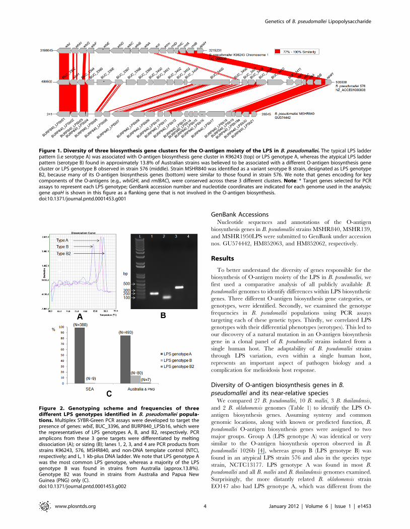
Figure 1. Diversity of three biosynthesis gene clusters for the O-antigen moiety of the LPS in B. pseudomallei. The typical LPS ladderpattern (i.e serotype A) was associated with O-antigen biosynthesis gene cluster in K96243 (top) or LPS genotype A, whereas the atypical LPS ladderpattern (serotype B) found in approximately 13.8% of Australian strains was believed to be associated with a different O-antigen biosynthesis genecluster or LPS genotype B observed in strain 576 (middle). Strain MSHR840 was identified as a variant serotype B strain, designated as LPS genotypeB2, because many of its O-antigen biosynthesis genes (bottom) were similar to those found in strain 576. We note that genes encoding for keycomponents of the O-antigens (e.g., wbiGHI, and rmlBAC), were conserved across these 3 different clusters. Note: * Target genes selected for PCRassays to represent each LPS genotype; GenBank accession number and nucleotide coordinates are indicated for each genome used in the analysis;gene apaH is shown in this figure as a flanking gene that is not involved in the O-antigen biosynthesis.doi:10.1371/journal.pntd.0001453.g001
Figure 2. Genotyping scheme and frequencies of threedifferent LPS genotypes identified in B. pseudomallei popula-tions. Multiplex SYBR-Green PCR assays were developed to target thepresence of genes: wbiE, BUC_3396, and BURP840_LPSb16, which werethe representatives of LPS genotypes A, B, and B2, respectively. PCRamplicons from these 3 gene targets were differentiated by meltingdissociation (A); or sizing (B); lanes 1, 2, 3, and 4 are PCR products fromstrains K96243, 576, MSHR840, and non-DNA template control (NTC),respectively; and L, 1 kb-plus DNA ladder. We note that LPS genotype Awas the most common LPS genotype, whereas a majority of the LPSgenotype B was found in strains from Australia (approx.13.8%).Genotype B2 was found in strains from Australia and Papua NewGuinea (PNG) only (C).doi:10.1371/journal.pntd.0001453.g002
Genetics of B. pseudomallei Lipopolysaccharide
www.plosntds.org 4 January 2012 | Volume 6 | Issue 1 | e1453

predicted O-antigen biosynthesis gene cluster in other B.
oklahomensis strains (C6786, C7532, and C7533; data not shown).
This may represent a lateral gene transfer event into EO147 and is
deserving of additional study. Furthermore, regions within the two
clusters had different levels of sequence conservation. Genes located
at the ends of these two clusters (e.g., wbiGHI, and rmlBAC; Figure 1)
had higher sequence similarity than most of the genes in the core of
the clusters. Indeed, many of the cluster cores contain distinct gene
composition. The conserved genes include those important for
oligosaccharide synthesis and O-antigen biosynthesis [4].
LPS genotype frequencies were analyzed across a large strain
collection using PCR-based assays. Multiplex-SYBR-Green PCR
assays were designed to target a specific gene unique for each
genotype. Gene wbiE (BPSL2676) of B. pseudomallei strain K96243
and gene BUC_3396 of strain 576 were used to represent the
presence of LPS genotypes A and B, respectively (Figures 1&2). A
total of 999 B. pseudomallei strains from different geographic
locations and epidemiological origins (e.g., clinical, animal, and
environmental strains) were tested for their LPS genotypes. We
noted that 23 B. pseudomallei strains were collected from one
melioidosis patient. We found that LPS genotype A was the most
common genotype in both Australian and Southeast Asian strain
populations (Figure 2). LPS genotype B was relatively rare in
Southeast Asian strains (,2.3%), but was found in 13.8% of
Australian strains. Five strains from Australia and two strains from
Papua New Guinea were non-typeable using these two PCR gene
markers. Three of these strains, MSHR840, MSHR1950, and
MSHR139 were further analyzed for O-antigen biosynthesis gene
identification using whole genome sequencing. The O-antigen
biosynthesis gene clusters from these strains were identified and
annotated (GenBank accession nos. GU574442, HM852062,
HM852063). Comparative genomics demonstrated that many
genes in this new cluster were similar to those of the LPS genotype
B genes of B. pseudomallei 576 and were distinct from the K96243
LPS genotype A genes. Hence, these newly identified O-antigen
biosynthesis gene clusters represent a variant of the LPS genotype
B and, consequentially, were designated as LPS genotype B2
(Table 1). Figure 1 shows the genomic comparison of these three
different O-antigen biosynthesis gene clusters: A, B, and B2 (from
B. pseudomallei strains K96243, 576, and MSHR840, respectively).
We note that %G+C content of the core of these 3 different
clusters is relatively low (,59–60%) compared to the conserved
parts of the O-antigen biosynthesis operon (,68%). This supports
the hypothesis that these genomic differences are due to genetic
recombination e.g., horizontal gene transfer, which is common in
B. pseudomallei [15,16]. Comparative genomics of these three
different clusters using homologous-based alignment are summa-
rized in Table S1. Again, we note that genes wbiGHI, and rmlBAC
are conserved among these three different clusters. Furthermore,
gene BURP840_LPSb16 from strain MSHR840 was selected for
use as a PCR marker to represent the LPS genotype B2. PCR
genotype analysis (Figure 2) revealed that all seven of the
previously non-typeable strains were positive for the LPS genotype
B2. The LPS B2 genotype was found only in strains from Australia
and Papua New Guinea. It is important to note that there is no
known clonal relationship among these seven strains. The LPS B2
genotype genes were also found in a B. thailandensis-like spp. strain
MSMB121, which was isolated in Australia (unpublished data).
Complete LPS genotypic data are reported in Table S2.
A novel LPS electrophoretic pattern - a type B variantLPS genotyping results were further examined by direct
comparison to LPS electrophoretic phenotypes [10]. Due to the
difficulty of international Select Agent transfer and BSL3
handling, we phenotyped only , 24% of the isolates that were
genotyped. We note that this is a limitation of our study. That said,
all examined LPS A or B phenotypes were perfectly matched with
their LPS A or B genotypes. In addition, 22 strains producing the
rough LPS phenotype were all identified as LPS genotype A
(Table S2). The genetic basis of the rough phenotype and its
derivation from the A phenotype is known for only 16 of these
strains (see below). SDS-PAGE revealed that LPS genotype B2
strains produced a distinct ladder pattern, though they were all
detectable with type B sera using immunoblot hybridization. The
B2 phenotype had a wider range of molecular weights (40–
120 kDa) than the LPS types A and B. In total, three LPS banding
patterns plus the rough LPS type (no ladder) can be detected
(Figure 3).
Natural mutations in O-antigen genes and changes inbacterial phenotypes
A frame-shift mutation observed in the O-antigen biosynthesis
wbiI gene of B. pseudomallei strain MSHR1655 was correlated with
its rough phenotype. This is one of nearly 100 strains that were
isolated over 8 years from a patient with severe bronchiectasis
associated with melioidosis. The mutation was an extra guanine
inserted after nucleotide position 815 of the wbiI gene (Figure 4).
The wbiI gene encodes an oligosaccharide epimerase/dehydratase
and is conserved in all O-antigen biosynthesis gene clusters of B.
pseudomallei. A mutation in this gene probably impacts on the
synthesis of the O-antigen in this bacterial strain. There were 23
serial B. pseudomallei isolates observed from the chronically infected
patient and the wbiI gene sequences were determined in all of
them to detect frame shift mutations. The frame-shift mutation
occurred in 16 isolates, all of which were collected on or after day
550 of the infection. The wild type sequence was present in the
other seven isolates from earlier in the infection (Figure 4).
Moreover, phenotypic characterization revealed that LPS samples
extracted from the 16 wbiI mutated strains did not have the O-
antigen ladder pattern (i.e the rough phenotype) based upon SDS-
PAGE and silver straining (Figure 5A). Thus, it seems likely the
frame-shift mutation in the wbiI gene blocks synthesis of the O-
antigen. A recent study has reported that oacA gene, known to be
involved in the acetylation at the O-4 position of the L-6dTalp
residues of B. thailandensis O-antigen [14], is mutated in B.
pseudomallei MSHR1655. Since MSHR1655 was isolated from the
same patient above, we then sequenced the oacA gene in all of
these clonal strains. We found that the oacA mutation occurred in
the same 16 strains that had the wbiI mutation (Figure 4C).
Additional study of the oacA gene in other whole genome
sequenced strains determined that B. pseudomallei 112 and B.
thailandensis TXDOH also had point mutation in their oacA genes
(Table 1; Figure S1). To determine if the oacA gene plays only a
single role in the side group modification of the L-6dTalp residues,
or a dual role in combination with the synthesis of the O-antigen,
both strains were tested for O-antigen production and immuno-
genic specificity. We found that B. pseudomallei 112 and B.
thailandensis TXDOH expressed O-antigen type A ladder pattern
and their O-antigen bands were strongly positive with the B. mallei
LPS-specific mAb 3D11 (Figure 6) that recognized the lack of 4-O
acetylation of the L-6dTalp residues [14]. This suggests the oacA
gene in B. pseudomallei and B. thailandensis has a role in the
acetylation at the O-4 position of the O-antigen L-6dTalp residues
but is not involved in the synthesis of the O-antigen. Thus, we
determined that the rough LPS phenotype observed in the 16
clonal chronic lung strains was due to the mutation of their wbiI
gene, but not from the effect of the oacA mutation. In this study, we
also identified six other independent rough LPS strains, but
Genetics of B. pseudomallei Lipopolysaccharide
www.plosntds.org 5 January 2012 | Volume 6 | Issue 1 | e1453

mutations did not occur in their wbiI or oacA genes. Searching for
mutations in other genes of these strains warrants a follow up study
to understand alternate mechanisms that generate the rough
phenotype.
Because LPS is essential for outer membrane integrity and
serum resistance, four B. pseudomallei strains from this chronic lung
patient were further tested in serum bactericidal assays. Two of the
wbiI mutant strains that expressed the rough LPS phenotype
(MSHR1655 and MSHR3042) were unable to grow in the
presence of 30% normal human serum (NHS). In contrast, two
early infection isolates from the same patient expressing the typical
LPS A phenotype (MSHR1043 and MSHR1048) were able to
Figure 3. Diversity of B. pseudomallei LPS banding patterns and their serological specificity. Panel A is silver strained SDS-PAGE of fourdifferent LPS phenotypes; panels B and C are immunoblotting analysis of the same LPS samples using sera from melioidosis patients with knowninfection by LPS genotype A, or B strains, respectively. Lanes 1–4 are typical (type A), atypical (type B), a novel atypical (type B variant or type B2), andrough LPS types, respectively; lane L is a pre-stained protein standard ladder. We note that the typical LPS was specifically seroreactive to theantibody from patient who was infected by LPS genotype A strain, whereas, the atypical LPS types (lanes 2 and 3) were seroreactive with theantibody from the LPS genotype B infected patient only. Rough LPS or no-banding LPS appearance (lane 4) was seronegative to both sera.doi:10.1371/journal.pntd.0001453.g003
Figure 4. Point mutations found in wbiI and oacA genes in clonal B. pseudomallei strains. These strains were collected chronologically froma single chronic lung patient who had severe bronchiectasis associated with melioidosis over almost 8 years. Panel A is the chronological order ofthese B. pseudomallei strains. Panel B demonstrates an extra base (‘‘G’’) that was found to cause frame-shift mutation in wbiI gene of all B.pseudomallei strains collected from day 550 onward. Panel C demonstrates the insertion of two extra bases ‘‘TC’’ in BPSL1936, the oacA homolog, inthe same strains that had the wbiI mutation. Note: the wbiI gene of B. pseudomallei K96243 and oacA gene of B. thailandensis E264 [14] were used ascomparisons.doi:10.1371/journal.pntd.0001453.g004
Genetics of B. pseudomallei Lipopolysaccharide
www.plosntds.org 6 January 2012 | Volume 6 | Issue 1 | e1453

resist the inhibitory human serum effect and grow (Figure 5B).
Furthermore, we also confirmed that the LPS genotype B2 strains
were killed in growth media containing 30% NHS, whereas the
LPS genotype B strains were resistant (Figure S2). We believe that
this finding of serum susceptibility in LPS genotype B2 is
important and deserves further investigation.
Discussion
Two major (A, B) and one minor (B2) LPS genotypes existunequally in B. pseudomallei populations
Despite the fact that genes responsible for the O-antigen
biosynthesis in B. pseudomallei 1026b were identified many years
ago [4], diversity of these genes across multiple B. pseudomallei
strains has not been well studied until now. Advances in genome
sequencing and comparative genomics have provided insights into
the complexity and diversity of B. pseudomallei genomes. B.
pseudomallei genomic studies can now strive for correlations
between genomic diversity and differential phenotypes; perhaps
the clinical outcomes of individual strains of B. pseudomallei may be
predicted using basic genomic analysis. In our current study, we
were able to establish a correlation between differential LPS
phenotypes and diversity of O-antigen biosynthesis genes or
known as LPS genotypes. Three different major LPS genotypes
have been identified so far. LPS genotype A was designated to the
strains that contained the O-antigen biosynthesis genes that were
identical or very similar to those found in a reference strain 1026b
[4], whereas the LPS genotype B category is represented by the
atypical LPS strain 576. Finally, LPS B2 genotype was identified as
a variant of the LPS genotype B because many of its O-antigen
biosynthesis genes were similar to those of LPS genotype B, and
both groups were serotype B positive. LPS genotype A was the
most common genotype in both geographic locations: Southeast
Asia and Australia where it accounted for 97.7% and 85.3% of the
populations, respectively. Interestingly, the frequency of LPS
genotype B was relatively high (approx. 13.8%) in Australian
strains, whereas they accounted for only 2.3% of the strains from
Southeast Asia. LPS genotype B2 was found in only 7 strains, 5 of
which were from Australia, and the other 2 strains were from
Papua New Guinea. In addition, LPS genotype B2 was also found
in a member of B. thailandensis-like species which was recently
discovered in Australia [22]. This would suggest that the LPS
genotype B2 genes in B. pseudomallei may be acquired by horizontal
gene transfer from a common soil bacterial species in Australia, or
vice versa. Comparative genomics and phenotypic characteriza-
tion of this LPS genotype B2 in B. pseudomallei and its near-relative
species warrants further investigation.
Because the LPS genotypes B and B2 were frequently found in
Australia but not in Southeast Asia, it is possible that this finding
may be due to different therapies used for clinical cases in these 2
endemic locations. We have investigated this and found that the
majority of these isolates were obtained before any exposure to
antibiotics or treatment therapy. In addition, some of the LPS
genotype B strains were collected from soil in Australia, and 2
strains of the LPS genotype B2 were found in animal cases. This
confirms that the occurrence of LPS types B and B2 in Australia is
not associated with the exposure to antibiotics or treatment
therapy. Although, we phenotyped only 24% of the isolates that
were genotyped, most tested strains were perfectly matched
between their genotypes and phenotypes, except those 16 rough
Figure 5. Differential LPS phenotypes and serum susceptibilityof the chronic lung strains. Panel A demonstrates LPS phenotypesbased upon SDS-PAGE analysis of select chronic lung strains; lanes 1–9,LPS samples from the chronic lung strains MSHR1043, MSHR1048,MSHR1218, MSHR1288, MSHR1290, MSHR1418, MSHR1459, MSHR1655,and MSHR3042, respectively; L, protein standard ladder. Panel B showsdifferential serum susceptibility in four select chronic lung B. pseu-domallei strains grown in 30% of normal human serum (NHS); a well-known serum resistant B. pseudomallei strain 1026b, and a laboratory E.coli strain HB101 were used as the positive and negative controls in thisstudy, respectively. We note that strains MSHR1655 and MSHR3042, therough LPS strains that had mutation in their wbiI genes were unable tomultiply in the presence of 30% NHS, whereas, the typical LPS strainsMSHR1043 and MSHR1048 from the same patient were able to utilizethe NHS as nutrients.doi:10.1371/journal.pntd.0001453.g005
Figure 6. Phenotypic effects of the oacA mutation in B.pseudomallei 112 and B. thailandensis TXDOH revealed byimmunoblot analysis. LPS samples from B. pseudomallei K96243and 112, B. mallei ATCC23344, and B. thailandensis E264 and TXDOH,Lanes 1–5, respectively, hybridized against serotype A patient’s serum(panel A), and B. mallei LPS-specific mAb 3D11 (panel B). As predicted,LPS samples from B. pseudomallei 112 (lane 2) and B. thailandensisTXDOH (lane 5) were strongly positive to the mAb 3D11 due to themutation of their oacA genes. Lane L is a pre-stained protein standardladder.doi:10.1371/journal.pntd.0001453.g006
Genetics of B. pseudomallei Lipopolysaccharide
www.plosntds.org 7 January 2012 | Volume 6 | Issue 1 | e1453

LPS genotype A strains from a single chronic case that had
mutations in their wbiI genes (Figure 4). In this current study, we
were unable to identify the genetic basis or mutations in 6
independent LPS genotype A strains that did not produce the O-
antigen (Table S2).
Because the typical LPS was also found in B. thailandensis, the use
of anti-LPS antibody based latex agglutination for the identifica-
tion of B. pseudomallei in environmental specimens was not
successful in an early study [23]. B. thailandensis LPS has also been
shown to cross-react with rabbit and mouse sera obtained from
inoculation with B. pseudomallei or B. mallei suggesting that LPS
molecules from B. thailandensis, a non-pathogenic bacterium, may
be useful in ongoing efforts to develop novel vaccines and/or
diagnostic reagents [24]. This has brought to our attention
whether low-grade B. thailandensis infections might naturally
provide protection against melioidosis. Although the O-antigen
biosynthesis genes in B. pseudomallei and B. thailandensis are similar,
a recent study by a Singaporean group has revealed that lipid A
components of the LPS from both B. pseudomallei and B. thailandensis
must be different; the murine and human macrophages produced
lower levels of tumor necrosis factor alpha, interleukin-6 (IL-6),
and IL-10 in response to B. pseudomallei LPS than in response to B.
thailandensis LPS in vitro [25]. In our current study, the typical LPS
was also found in B. oklahomensis strain EO147, formerly known as
an American B. pseudomallei strain [26], suggesting that the typical
LPS is widely spread in multiple Burkholderia species. This group
includes highly pathogenic species such as B. pseudomallei and B.
mallei, but also non-pathogenic species: B. thailandensis, B.
thailandensis-like species, and B. oklahomensis. The evolution of LPS
diversity across these closely related species is likely a function of
differential selection and horizontal transfer of genetic elements.
This diversity could play a role in frequency and distribution of
disease in humans. However, without understanding molecular
structures of these O-antigen types, it is difficult to access the
phenotypic effects of this genetic diversity. Structural analysis of
the O-antigen types B and B2 deserves further investigations. In
addition, we have found that the LPS genotype B2 strains were
sensitive to 30% NHS, whereas the LPS type B strains were
resistant (Figure S2). This finding demonstrates a level of
phenotypic differences between these two serologically related
groups. We believe that the consequences for case treatment
associated with these differential serum susceptibilities also warrant
further investigations.
The rough LPS phenotype – Adaptation to survival andpersistence in a host?
A previous study has shown that the two less common LPS
phenotypes (smooth type B and rough type) were more prevalent
in clinical than environmental isolates and more prevalent in
Australian isolates than Thai isolates [10]. In our current study,
LPS genotype B was found in both clinical and environmental
strains from Australia, whereas the rough LPS was still found only
in clinical strains. Based on our description of the molecular basis
for LPS phenotypes, it is unlikely that B. pseudomallei will readily
switch its LPS phenotype from A to B, or vice versa, as has been
suggested previously [10]. The gene compositions of LPS
genotypes A and B are very different and a simple switching
mechanism is difficult to envision. In addition, we have found that
at least some rough LPS strains have mutations in their O-antigen
biosynthesis genes. These include 16 clonally related isolates from
a single chronic lung infected patient (Table S2). All of these
strains were identified as LPS genotype A with mutations in their
O-antigen biosynthesis genes. Using Tn5-OT182 mutagenesis,
DeShazer and colleagues identified at least seven genes in the
O-antigen biosynthesis operon of B. pseudomallei 1026b that were
responsible for O-antigen biosynthesis and serum resistance; these
included rmlB, rmlD, wbiA, wbiC, wbiE, wbiG, and wbiI [4]. In our
current study, we found point mutations in wbiI and oacA genes of
B. pseudomallei isolates that were collected from a chronic lung
patient (Figure 4). We hypothesize that the frame-shift mutation in
the wbiI genes blocks O-antigen biosynthesis in all mutant strains,
but not from the effect of the oacA mutation. This is because we
observed the oacA mutations in B. pseudomallei 112 and B.
thailandensis TXDOH that had normal O-antigen biosynthesis
gene cluster (Table 1 and Figure S1). Our study has demonstrated
that these two oacA mutant strains expressed O-antigens identical
to those found in B. mallei due to lack of the 4-O acetylation of the
L-6dTalp residues of the O-antigen. The lack of the 4-O
acetylation of the L-6dTalp residues has recently been described
in the oacA knock-out mutant B. thailandensis ZT0715 and a wild-
type B. mallei ATCC23344 [14].
We have demonstrated that these wbiI mutant strains produced
rough LPS and were sensitive to normal human serum suggesting
that the wbiI gene encoding for epimerase, or dehydratase, was
essential for the biosynthesis of B. pseudomallei O-antigen. Although
loss of the O-antigen might compromise serum survival it might
also be adaptive in particular niches. B. pseudomallei survival or
persistence in the host might be enhanced without the surface
presentation of the O-antigenic moiety of the LPS, as it would not
be recognized by host immune systems and would, therefore,
avoid being killed by antibodies. The O-antigenic polysaccharide
of B. pseudomallei modulates the host cell response, which in turn
controls the intracellular fate of B. pseudomallei inside macrophage.
This was concluded from the observation that the O-antigen
mutant B. pseudomallei strain SRM117 was more susceptible to
macrophage killing during the early phase of infection than the
parental wild-type strain 1026b [27]. This was also confirmed by
the same group when they demonstrated the importance of
intracellular killing by the human polymorphonuclear cells
(PMNs), macrophages (MQs), and susceptibility to killing by 30%
normal human serum [28].
LPS and CPS (capsular polysaccharide) have been used as
subunits in immunizing BALB/c mice against B. pseudomallei
infection [2]. Mice vaccinated with LPS developed predominantly
IgM and IgG3 responses, whereas the mice vaccinated with the
CPS developed a predominantly IgG2b response. Furthermore,
immunization with the LPS provided an optimal protective
response, and the immunized mice challenged by the aerosol
route showed a small increase in the mean time to death compared
with the unvaccinated controls [2]. Previously, it was shown that
B. pseudomallei LPS from strain 1026b signaled through Toll-like
receptor (TLR) 2 and not through TLR4 [29]. This was observed
in the TLR2 knock-out mutant mice that displayed a markedly
improved host defense, but it was not observed in TLR4 knock-out
mice [29]. In contrast, a study in HEK293 cells demonstrated that
heat-killed B. pseudomallei strains K96243 or BP-1 activated TLR2
and TLR4, and in the presence of MD-2, LPS and lipid A from
BP-1 are TLR4 ligands [30]. We note that B. pseudomallei 1026b
and K96243 expressed the typical O-antigen type A, but the O-
antigen type of BP-1 was not reported in that study. Although
there was no report of association between the LPS types and
disease severity (e.g., fatal versus non-fatal, and septicemia versus
localized), clinical manifestations (neurologic versus non-neuro-
logic), or underlying risk factors (diabetic versus non-diabetic)
observed in a previous study [10], full phenotypic characterization
including virulence in animal models, innate immune response, etc
of these different LPS types warrants further investigations given
the LPS diversity that we have described.
Genetics of B. pseudomallei Lipopolysaccharide
www.plosntds.org 8 January 2012 | Volume 6 | Issue 1 | e1453
Supporting Information
Figure S1 Point mutations found in gene BPSL1936(oacA homolog) of B. pseudomallei MSHR1655 and 112,and B. thailandensis TXDOH. These point mutations (panel
A): in strain MSHR1655, the mutation was associated with 2 extra
bases, ‘‘TC’’, inserted right after nucleotide no. 298 of this gene; in
strain 112, it was associated with a deletion of one base, ‘‘T’’, at
nucleotide no. 112; and in B. thailandensis TXDOH, it was
associated with the 59 truncation mutation. Amino acid sequence
analysis (panel B) has demonstrated that the point mutations in
MSHR1655 and 112 potentially caused frame-shift mutations in
their BPSL1936 genes, and then split the gene into 2 separated
open reading frames (ORFs). Two known amino acid motifs,
VXXFFXXSG and WXLXXEXXXY, were present in both
ORFs of MSHR1655, whereas only the latter motif was present in
strain 112. We noted that both amino acid motifs were absent in B.
thailandensis TXDOH.
(PPT)
Figure S2 O-antigen types and differential serumsusceptibility. O-antigen type A and B strains including B.
pseudomallei 1026b, NCTC13178, and NCTC13179, MSHR367b,
MSHR98, respectively, were resistant to 30% normal human
serum (NHS); whereas the O-antigen type B2 strains : B.
pseudomallei MSHR840, MSHR454, and MSHR1950, and B.
thailandensis-like sp. strain MSMB121 were sensitive to the 30%
NHS. A rough O-antigen type strain MSHR3042, a member of
the chronic lung strains (see text), was also sensitive. We noted that
B. thailandensis E264 was able to survive, but unable to multiple in
the presence of 30% NHS. E. coli HB101 was used as a control
serum sensitive strain.
(PPT)
Table S1 Comparison of LPS genotype A, B, and B2gene clusters.
(DOC)
Table S2 List of bacterial strains used in this study andtheir LPS genotyping PCR results.
(XLS)
Acknowledgments
We thank Judy Lee, Stephanie Grasso, Dawn Birdsell, Amanda Pettus,
Eleanor Woolveridge, and Yuwana Podin for their technical assistance.
Author Contributions
Conceived and designed the experiments: AT. Performed the experiments:
AT JKS MM JG SG SW TM CJA. Analyzed the data: AT JKS MK.
Contributed reagents/materials/analysis tools: NC SJP. Wrote the paper:
AT PK DMW SJP BJC.
References
1. Caroff M, Karibian D (2003) Structure of bacterial lipopolysaccharides.Carbohyd Res 338: 2431–2447.
2. Nelson M, Prior JL, Lever MS, Jones HE, Atkins TP, et al. (2004) Evaluation oflipopolysaccharide and capsular polysaccharide as subunit vaccines against
experimental melioidosis. Journal of Medical Microbiology 53: 1177–1182.3. Sarkar-Tyson M, Thwaite JE, Harding SV, Smither SJ, Oyston PCF, et al.
(2007) Polysaccharides and virulence of Burkholderia pseudomallei. Journal of
Medical Microbiology 56: 1005–1010.4. DeShazer D, Brett PJ, Woods DE (1998) The type II O-antigenic polysaccharide
moiety of Burkholderia pseudomallei lipopolysaccharide is required for serumresistance and virulence. Molecular Microbiology 30: 1081–1100.
5. Leon CG, Tory R, Jia J, Sivak O, Wasan KM (2008) Discovery and
development of Toll-like receptor 4 (TLR4) antagonists: a new paradigm fortreating sepsis and other diseases. Pharmaceutical Research 25: 1751–1761.
6. Limmathurotsakul D, Wongratanacheewin S, Teerawattanasook N, Wong-suvan G, Chaisuksant S, et al. (2010) Increasing incidence of human melioidosis
in Northeast Thailand. Am J Trop Med Hyg 82: 1113–1117.
7. Charuchaimontri C, Suputtamongkol Y, Nilakul C, Chaowagul W,Chetchotisakd P, et al. (1999) Antilipopolysaccharide II: An antibody protective
against fatal melioidosis. Clinical Infectious Diseases 29: 813–818.8. Ho M, Schollaardt T, Smith MD, Perry MB, Brett PJ, et al. (1997) Specificity
and functional activity of anti-Burkholderia pseudomallei polysaccharide antibodies.Infection and Immunity 65: 3648–3653.
9. Arjcharoen S, Wikraiphat C, Pudla M, Limposuwan K, Woods DE, et al. (2007)
Fate of a Burkholderia pseudomallei lipopolysaccharide mutant in the mousemacrophage cell line RAW 264.7: Possible role for the O-antigenic
polysaccharide moiety of lipopolysaccharide in internalization and intracellularsurvival. Infection and Immunity 75: 4298–4304.
10. Anuntagool N, Wuthiekanun V, White NJ, Currie BJ, Sermswan RW, et al.
(2006) Short report: Lipopolysaccharide heterogeneity among Burkholderia
pseudomallei from different geographic and clinical origins. American Journal of
Tropical Medicine and Hygiene 74: 348–352.11. Perry MB, MacLean LL, Schollaardt T, Bryan LE, Ho M (1995) Structural
characterization of the lipopolysaccharide O antigens of Burkholderia pseudomallei.Infection and Immunity 63: 3348–3352.
12. Ngugi SA, Ventura VV, Qazi O, Harding SV, Kitto GB, et al. (2010)
Lipopolysaccharide from Burkholderia thailandensis E264 provides protection in amurine model of melioidosis. Vaccine 28: 7551–7555.
13. Burtnick MN, Deshazer D, Woods DE (2002) Molecular and physicalcharacterization of Burkholderia mallei O antigens. J Bacteriol 184: 849–852.
14. Brett PJ, Burtnick MN, Heiss C, Azadi P, DeShazer D, et al. (2011) Burkholderia
thailandensis oacA mutants facilitate the expression of Burkholderia mallei-like Opolysaccharides. Infection and Immunity 79: 961–969.
15. Tuanyok A, Leadem BR, Auerbach RK, Beckstrom-Sternberg SM, Beckstrom-Sternberg JS, et al. (2008) Genomic islands from five strains of Burkholderia
pseudomallei. BMC Genomics 9: 566.
16. Tuanyok A, Auerbach RK, Brettin TS, Bruce DC, Munk AC, et al. (2007) A
horizontal gene transfer event defines two distinct groups within Burkholderia
pseudomallei that have dissimilar geographic distributions. J Bacteriol 189:
9044–9049.
17. U’ Ren JM, Schupp JM, Pearson T, Hornstra H, Clark CL, et al. (2007) Large
tanden repeat regions within Burkholderia pseudomallei genome and their
application for high resolution genotyping. BMC Microbiology 7: 23.
18. Chantratita N, Wuthiekanun V, Boonbumrung K, Tiyawisutsri R, Vesarat-
chavest M, et al. (2007) Biological relevance of colony morphology and
phenotypic switching by Burkholderia pseudomallei. J Bacteriol 189: 807–817.
19. Cheng AC, Currie BJ (2005) Melioidosis: epidemiology, pathophysiology, and
management. Clin Microbiol Rev 18: 383–416.
20. Currie BJ, Ward L, Cheng AC (2010) The epidemiology and clinical spectrum of
melioidosis: 540 cases from the 20 year Darwin prospective study. PLoS Negl
Trop Dis 4: e900.
21. Carver TJ, Rutherford KM, Berriman M, Rajandream MA, Barrell BG, et al.
(2005) ACT: the Artemis Comparison Tool. Bioinformatics 21: 3422–3423.
22. Gee JE, Glass MB, Novak RT, Gal D, Mayo MJ, et al. (2008) Recovery of a
Burkholderia thailandensis-like isolate from an Australian water source. BMC
Microbiol 8: 54.
23. Wuthiekanun V, Anuntagool N, White NJ, Sirisinha S (2002) Short report: A
rapid method for the differentiation of Burkholderia pseudomallei and Burkholderia
thailandensis. Am J Trop Med Hyg 66: 759–761.
24. Qazi O, Prior JL, Judy BM, Whitlock GC, Kitto GB, et al. (2008) Sero-
characterization of lipopolysaccharide from Burkholderia thailandensis. Trans R Soc
Trop Med Hyg 102: S58–60.
25. Novem V, Shui GH, Wang DL, Bendt AK, Sim SH, et al. (2009) Structural and
biological diversity of lipopolysaccharides from Burkholderia pseudomallei and
Burkholderia thailandensis. Clinical and Vaccine Immunology 16: 1420–1428.
26. Nussbaum JJ, Hull DS, Carter MJ (1980) Pseudomonas pseudomallei in an
anopthalmic orbit. Arch Opthalmol 98: 1224–1225.
27. Arjcharoen S, Wikraiphat C, Pudla M, Limposuwan K, Woods DE, et al. (2007)
Fate of a Burkholderia pseudomallei mouse macrophage cell line lipopolysaccharide
mutant in the RAW 264.7: Possible role for the o-antigenic polysaccharide
moiety of lipopolysaccharide in internalization and intracellular survival.
Infection and Immunity 75: 4298–4304.
28. Wikraiphat C, Charoensap J, Utaisincharoen P, Wongratanacheewin S,
Taweechaisupapong S, et al. (2009) Comparative in vivo and in vitro analyses
of putative virulence factors of Burkholderia pseudomallei using lipopolysaccharide,
capsule and flagellin mutants. FEMS Immunol Med Microbiol 56: 253–259.
29. Wiersinga WJ, Wieland CW, Dessing MC, Chantratita N, Cheng AC, et al.
(2007) Toll-like receptor 2 impairs host defense in Gram-negative sepsis caused
by Burkholderia pseudomallei (melioidosis). PLoS Medicine 4: e248.
30. West TE, Ernst RK, Jansson-Hutson MJ, Skerrett SJ (2008) Activation of Toll-
like receptors by Burkholderia pseudomallei. BMC Immunology 9.
Genetics of B. pseudomallei Lipopolysaccharide
www.plosntds.org 9 January 2012 | Volume 6 | Issue 1 | e1453