Proteome analysis ofSaccharomyces cerevisiae: A methodological outline
Upload
independentCategory
view
1download
0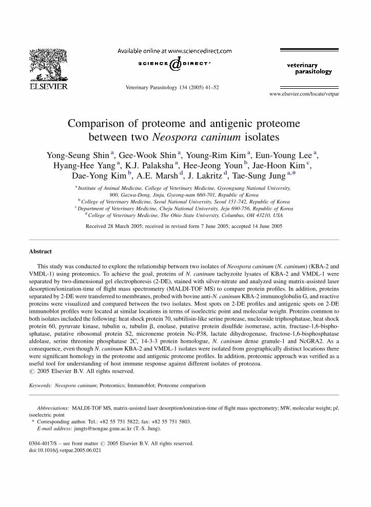
Comparison of proteome and antigenic proteome
between two Neospora caninum isolates
Yong-Seung Shin a, Gee-Wook Shin a, Young-Rim Kim a, Eun-Young Lee a,Hyang-Hee Yang a, K.J. Palaksha a, Hee-Jeong Youn b, Jae-Hoon Kim c,
Dae-Yong Kim b, A.E. Marsh d, J. Lakritz d, Tae-Sung Jung a,*
a Institute of Animal Medicine, College of Veterinary Medicine, Gyeongsang National University,
900, Gazwa-Dong, Jinju, Gyeong-nam 660-701, Republic of Koreab College of Veterinary Medicine, Seoul National University, Seoul 151-742, Republic of Korea
c Department of Veterinary Medicine, Cheju National University, Jeju 690-756, Republic of Koread College of Veterinary Medicine, The Ohio State University, Columbus, OH 43210, USA
Received 28 March 2005; received in revised form 7 June 2005; accepted 14 June 2005
Abstract
This study was conducted to explore the relationship between two isolates of Neospora caninum (N. caninum) (KBA-2 and
VMDL-1) using proteomics. To achieve the goal, proteins of N. caninum tachyzoite lysates of KBA-2 and VMDL-1 were
separated by two-dimensional gel electrophoresis (2-DE), stained with silver-nitrate and analyzed using matrix-assisted laser
desorption/ionization-time of flight mass spectrometry (MALDI-TOF MS) to compare protein profiles. In addition, proteins
separated by 2-DE were transferred to membranes, probed with bovine anti-N. caninum KBA-2 immunoglobulin G, and reactive
proteins were visualized and compared between the two isolates. Most spots on 2-DE profiles and antigenic spots on 2-DE
immunoblot profiles were located at similar locations in terms of isoelectric point and molecular weight. Proteins common to
both isolates included the following: heat shock protein 70, subtilisin-like serine protease, nucleoside triphosphatase, heat shock
protein 60, pyruvate kinase, tubulin a, tubulin b, enolase, putative protein disulfide isomerase, actin, fructase-1,6-bispho-
sphatase, putative ribosomal protein S2, microneme protein Nc-P38, lactate dihydrogenase, fructose-1,6-bisphosphatase
aldolase, serine threonine phosphatase 2C, 14-3-3 protein homologue, N. caninum dense granule-1 and NcGRA2. As a
consequence, even though N. caninum KBA-2 and VMDL-1 isolates were isolated from geographically distinct locations there
were significant homology in the proteome and antigenic proteome profiles. In addition, proteomic approach was verified as a
useful tool for understanding of host immune response against different isolates of protozoa.
# 2005 Elsevier B.V. All rights reserved.
Keywords: Neospora caninum; Proteomics; Immunoblot; Proteome comparison
www.elsevier.com/locate/vetpar
Veterinary Parasitology 134 (2005) 41–52
Abbreviations: MALDI-TOF MS, matrix-assisted laser desorption/ionization-time of flight mass spectrometry; MW, molecular weight; pI,
isoelectric point
* Corresponding author. Tel.: +82 55 751 5822; fax: +82 55 751 5803.
E-mail address: [email protected] (T.-S. Jung).
0304-4017/$ – see front matter # 2005 Elsevier B.V. All rights reserved.
doi:10.1016/j.vetpar.2005.06.021
Y.-S. Shin et al. / Veterinary Parasitology 134 (2005) 41–5242
1. Introduction
Neospora caninum (N. caninum) has become an
important pathogen of the livestock industry, especially
cattle (Dubey, 2003). Pregnant cattle with neosporosis
suffer from abortion and stillbirths. On the other hand,
no clinical signs have been reported in non-pregnant
adult cattle with neosporosis (Anderson et al., 2000;
Dubey, 2003). In Korea, bovine neosporosis was first
reported in 1997 (Kim et al., 1997). The parasites were
isolated in congenitally infected calf and aborted fetus
in Korea, and the pathogens were named KBA-1 and
KBA-2 isolates, respectively (Kim et al., 1998, 2000).
With the evolution of whole organism genomic
sequencing projects, proteomics has become an
important area of research (Liebler, 2002). Among
its usefulness are characterizing genes and proteins
function, understanding functional linkages between
protein molecules, thus providing valuable informa-
tions regarding the mechanisms of biological pro-
cesses (Jungblut et al., 1999; Zhu et al., 2003).
Investigation of immunodominant antigens as
vaccine candidates may improve efficacy of vaccines.
To achieve this goal, understanding host immune
response against pathogen through the analysis of
pathogen proteins is a critical step (Hanash, 2003). A
large number of investigations have used proteomics
combinedwith two-dimensional gel electrophoresis (2-
DE) with immunoblot analysis of antigenic proteins,
which are useful tools for understanding immunologi-
cal processes of host against pathogens’ proteins
(Klade, 2002).
A number of reports have been published about the
biological, the morphological, and the molecular
features of isolates of N. caninum originated from a
variety of worldwide-distributed hosts, especially dogs
and cattle (Schock et al., 2001; Gondim et al., 2004;
Dubey et al., 2004). These reports indicate that N.
caninum isolates have similarities as well as differ-
ences. However, there is limited analysis of the parasite
proteome to evaluate the relationships between isolates,
but improved methods for protein characterization
using 2-DE followed by matrix-assisted laser deso-
rption/ionization-time of flight mass spectrometry
(MALDI-TOF MS) provide additional means of
characterizing these isolates. In this study, proteomic
analysis was carried out to evaluate the relationship
between N. caninum KBA-2 and VMDL-1 isolates to
address the presence of potential differences in protein
expression by the different isolates.
2. Materials and methods
If not stated otherwise, all the reagents were
purchased from Sigma (St. Louis, MO, USA).
2.1. Parasite
N. caninum isolates KBA-2 (Kim et al., 2000) and
VMDL-1 (Hyun et al., 2003) tachyzoites were
maintained according to Lee et al. (2003). Tachyzoites
of each isolate were purified (Yamane et al., 1998) and
stored at �70 8C until use.
2.2. Tachyzoites preparation for 2-DE
Purified tachyzoites (approximately 1 � 108) were
dissolved in 40 mMTris-base, disrupted by freeze-thaw
cycles, and then sonicated (XL-2020, Misonix Inc.
Farmingdale, NY, USA). The dissolved tachyzoites
were lysed in lysis buffer (2.2 M thiourea, 7.7 M urea,
40 mM Tris-base, 4% (w/v) 3-[(3-cholamidopropyl)-
dimethylammonio]-1-propanesulfonate (CHAPS), 1%
(w/v) dithiothreitol (DTT), 0.5% (v/v)TritonX-100and
0.5% (v/v) immobilized pH gradient (IPG)-buffer pH
4–7 (Amersham Bioscience, Uppsala, Sweden)) and
the lysates weremaintained in ice slurry for 1 h. Protein
concentrations of the 2-DE samples were estimated
using the Bradford protein assay kit (Biorad, Hercules,
CA, USA).
2.3. Preparation of anti-N. caninum KBA-2 serum
from cow
A cow (Holstein, female) was intra-venously
inoculated with 1 � 108 live tachyzoites of the
KBA-2 isolate to produce anti-N. caninum KBA-2
polyclonal antibodies. Prior to inoculation, the donor
was confirmed as N. caninum and Toxoplasma gondii
negative by IFAT (Baszler et al., 2001; Osawa et al.,
1998). The cow was provided care according to the
guidelines for the care and use of laboratory animals
developed by Gyeongsang National University,
Republic of Korea. Serum was collected 13 weeks
post-inoculation and stored at�20 8C until use. Serum

Y.-S. Shin et al. / Veterinary Parasitology 134 (2005) 41–52 43
antibody titers to N. caninum and T. gondii were
evaluated as 1:800 and <1:100 by indirect fluorescent
antibody test, respectively.
2.4. 2-DE and its immunoblot
Isoelectric focusing (IEF) was performed using an
IPGphorTM system (Amersham Bioscience) according
to Gorg et al. (2000), with IPG strips (Immobiline
DryStripTM, pH 4–7, 0.5 mm � 3 mm � 130 mm;
Amersham Bioscience). The prepared tachyzoites 2-
DE samples were mixed with a rehydration buffer (2 M
thiourea, 7 Murea, 2% (w/v)CHAPS, 0.4% (w/v)DTT,
Fig. 1. Two-dimensional gel electrophoresis (2-DE) and its immunoblot pr
performed at 86.1 kVh using pH 4–7 immobilized pH gradient strips (13 c
performed on a 10% gel then stained with silver-nitrate. Each N. caninum
fluoride (PVDF) membrane and incubated with anti-KBA-2 serum. Anti
solution kit. A, 2-DE profile of N. caninum KBA-2; B, 2-DE immunoblot pr
D, 2-DE immunoblot profile of N. caninum VMDL-1; , Each profile w
0.5% IPG buffer, 0.002% (w/v) bromophenol blue) and
then focused for a total of 86.1 kVh. After IEF, the IPG
strips were subjected to 10% sodium dodecyl sulfate-
polyacrylamide gels (160 mm � 160 mm � 1 mm)
and run under 10 mA/gel. Immunoblot was performed
according to Mansfield (1995). Briefly, each N.
caninum isolate lysate separated by 2-DE was
transferred onto polyvinyldene fluoride (PVDF) mem-
brane (Immobilon-PTM, 0.45 mm; Millipore, Billerica,
MA,USA) and incubatedwith anti-KBA-2 serumat 1:1
dilution ratio with 5% (w/v) skim milk in phosphate
buffered saline. The dilution ratio between serum and
horseradish peroxidase (HRP)-conjugated anti-bovine
ofiles of N. caninum KBA-2 and VMDL-1. Isoelectric focusing was
m) with 60 mg of lysate from N. caninum isolates. SDS-PAGE was
isolate lysate separated by 2-DE was transferred onto polyvinyldene
body binding was determined using enhanced chemiluminescence
ofile of N. caninum KBA-2; C, 2-DE profile of N. caninum VMDL-1;
as divided into four different parts for further analysis (Figs. 2–5).

Y.-S. Shin et al. / Veterinary Parasitology 134 (2005) 41–5244
immunoglobulin G (IgG) as reagents were basically
titrated by the Checker-Board method and established
the proper ratio (Crowther, 1995). HRP-conjugated
anti-bovine IgG (Jackson ImmunoResearch Labora-
tories,West Grove, PA, USA) at 1:2000 was used as the
secondary antibody. Antibody binding was determined
using enhanced chemiluminescence solution (ECLTM:
Amersham Bioscience) for 1 min, exposed to X-ray
film (Fuji, Tokyo, Japan), and developed.
2.5. Protein visualization and identification
Silver-nitrate staining of gels, image analysis, in-
gel digestion of protein spots on gels, matrix-assisted
Fig. 2. Proteome and antigenic proteome profiles of part I on Fig. 1 ranged
stained with silver-nitrate were found to locate in similar places betwe
immunoreactive also showed high similar locations between isolates. A, pro
N. caninum KBA-2; C, proteome profile of N. caninum VMDL-1; D, ant
laser desorption/ionization-time of flight mass spec-
trometry (MALDI-TOF MS) analysis, and database
searches (Ms-Fit; The Mass Spectrometry Facility, the
University of California San Francisco) were per-
formed according to Lee et al. (2003). Gel staining,
image analysis, and protein identification were each
performed a minimum of three times, and several
minor spots were not analyzed. Spots with pI and MW
values within 5–10% of each other were regarded as
having the same pI and MW (Jungblut and Thiede,
1997). In addition, only sequences that contained at
least five matching peptides that comprised at least
15% of the protein sequence were selected (Mann
et al., 2001).
pIs between 4.3 and 5.2 and MWs from 48 to 100 kDa. Most spots
en the isolates. In the immunoblot profiles, spots recognized as
teome profile ofN. caninumKBA-2; B, antigenic proteome profile of
igenic proteome profile of N. caninum VMDL-1.

Y.-S. Shin et al. / Veterinary Parasitology 134 (2005) 41–52 45
3. Results
For ease and detailed analysis, the overall profiles of
N. caninum KBA-2 and VMDL-1 (Fig. 1) were divided
into four different parts (Figs. 2–4), and potentially
important proteins were indicated using circular or
cubic marks. Specifically, circles and cubics indicated
the existence and the non-existence of spots between
the two isolate profiles, respectively.Alphabet letters on
the profiles indicate spots that were identified by
MALDI-TOF MS that correspond to a known protein.
3.1. Proteome profiles of N. caninum isolate KBA-
2 and VMDL-1
Approximately 540 and 520 spots were observed
on the silver-nitrate stained gel using pH 4–7, 13 cm
IPG strip of N. caninum isolate KBA-2 and VMDL-1,
respectively. To identify their respective protein
names, some spots indicated on the profiles were
examined by MALDI-TOF MS, and 28 protein spots
corresponding to 19 different proteins were success-
fully identified by peptide mass fingerprinting (PMF)
analysis. The identified protein spots (alphabet letters
A–S) were marked onto the gel image as shown in
Fig. 3. Proteome and antigenic proteome profiles of part II on Fig. 1. The
ranging from 50 to 90 kDa. Spots were observed at the same locations in
were also demonstrated in the immunoblot profiles. A, proteome profile o
KBA-2; C, proteome profile of N. caninum VMDL-1; D, antigenic prote
Figs. 2–5 and listed in Table 1. Table 2 lists spots’
identification numbers exclusively observed for one
isolate but not both via 2-DE analysis.
3.2. Antigenic proteome profiles of N. caninum
isolate KBA-2 and VMDL-1
Approximately 53 and 46 spots were detected as
antigenic spots on 2-DE immunoblot profiles of KBA-2
and VMDL-1, respectively. Among these, 13 spots
corresponding to 7 different proteins, such as heat shock
protein 70 (HSP70), subtilisin-like serine protease,
nucleoside triphosphatase (NTPase), HSP60, enolase,
actin and N. caninum dense granule-1 (NCDG-1), were
successfully identified and corresponded with proteins
in the MALDI-TOF MS analysis. The antigenic spots
weremarked onto the profiles as shown in Figs. 2–5 and
listed in Table 3.
3.3. Comparison of N. caninum isolate KBA-2
and VMDL-1 profiles
Fig. 2 shows proteome and antigenic proteome
profiles of part I setting in the range of pIs 4.3 and 5.2,
with MWs ranging from 48 to 100 kDa. In the 2-DE
range of the map was setting between pIs 5.3 and 6.7 with the MWs
both isolates of silver-nitrate staining profiles and the same patterns
f N. caninum KBA-2; B, antigenic proteome profile of N. caninum
ome profile of N. caninum VMDL-1.

Y.-S. Shin et al. / Veterinary Parasitology 134 (2005) 41–5246
Fig. 4. Proteome and antigenic proteome profiles of part III on Fig. 1. The mapwas located on the range of pIs between 4.1 and 5.2 with theMWs
ranging from 20 to 48 kDa. Most spots on 2-DE and the corresponding immunoblot profiles were exhibited to have the same location (pI and
MW) between isolates. A, proteome profile ofN. caninumKBA-2; B, antigenic proteome profile of N. caninumKBA-2; C, proteome profile ofN.
caninum VMDL-1; D, antigenic proteome profile of N. caninum VMDL-1.
profiles (Fig. 2A and C), most spots including HSP 70
(spot A), tubulin a-chain (spot G), tubulin b-chain
(spot F) and putative protein disulfide isomerase (spot
I) were found to locate in similar places between the
isolates. However, only spot no. 101 was present on 2-
DE profile of KBA-2. In the immunoblot profiles
(Fig. 2B and D), spots recognized as immunoreactive
(spot nos. 1–5) also showed high similar locations
between isolates but differences were noticed, such as
spot no. 6 was only exhibited on immunoblot profile of
KBA-2. Spot nos. 4 and 5 on KBA-2 were more
strongly reacted as antigenic spots probed with bovine
anti-N. caninum KBA-2 IgG.
Proteins in between pIs 5.3 and 6.7 with MWs in
the range 50–90 kDa (Fig. 3) were identified as
follows: subtilisin-like serine protease (spot B),
NTPase (spot C), HSP60 (spot D), pyruvate kinase
(spot E) and enolase (spot H). Other unidentified spots

Y.-S. Shin et al. / Veterinary Parasitology 134 (2005) 41–52 47
Fig. 5. Proteome and antigenic proteome profiles of part IVon Fig. 1. At the range of pIs between 5.3 and 6.6 with MWs between 21 and 50 kDa
were focused. The locations of the spots presented also showed high similarities between KBA-2 and VMDL-1. A, proteome profile of N.
caninum KBA-2; B, antigenic proteome profile of N. caninum KBA-2; C, proteome profile of N. caninum VMDL-1; D, antigenic proteome
profile of N. caninum VMDL-1.
were observed at the same locations in both isolates
(Fig. 3A and C), and the same patterns were also
demonstrated in the immunoblot profiles (Fig. 3B and
D). Of the spots on 2-DE profiles, spot nos. 102–106
were only detectable with KBA-2 2-DE profile,
whereas spot no. 107 was present in VMDL-1 isolate
but was not present in KBA-2. In the immunoblot
profiles, there were similarities and differences
between isolates. Spot nos. 7, 19, 23, 24 and 26 were
only detected in KBA-2, whereas spot no. 22 was only
detected in VMDL-1. Spot nos. 8–11, 16–18, 29 and
31 were present in both isolates.
As seen inFig. 4, in the range betweenpIs 4.1 and5.2
with the MWs ranging from 20 to 48 kDa (part III),
most spots on 2-DE and the corresponding immunoblot
profileswere exhibited to have the same location (pI and
MW) between isolates. Of the spots on 2-DE profiles,
spot nos. 108, 109 and 115–121 were only detected in
KBA-2, and spot nos. 110–114 and 122 were only
detected in VMDL-1. Of the spots on the immunoblot

Y.-S. Shin et al. / Veterinary Parasitology 134 (2005) 41–5248
Table 1
Protein spots identified on the two-dimensional gel electrophoresis (2-DE) profiles of Neopora caninum (N. caninum) KBA-2 and VMDL-1 after
2-DE followed by matrix-assisted laser desorption/ionization-time of flight mass spectrometry (MALDI-TOF MS) analysis
Spot no. Protein name No. of matched
peptides
Sequence
coverage (%)
Theoreticala Measureda Accession
no.bSpecies
MW (Da) pI MW (Da) pI
A Heat shock protein 70 (HSP70) 13 22 70,627 5.2 69,998 4.81 11277111 T. gondii
A HSP70 12 23 70,627 5.2 69,716 4.88 11277111 T. gondii
A HSP70 11 23 70,627 5.2 69,364 4.91 3850197 T. gondii
B Subtilisin like serine protease 9 13 93,650 5.4 67,713 5.76 6119851 N. caninum
C Nucleoside triphosphatase
(NTPase)
13 20 68,688 5.4 64,123 5.52 3298332 N. caninum
C NTPase 13 23 68,688 5.4 64,076 5.61 3298332 N. caninum
D HSP60 15 30 61,014 5.8 61,336 5.42 5052052 T. gondii
E Pyruvate kinase 9 22 57,530 6.0 61,100 5.94 13928580 T. gondii
E Pyruvate kinase 9 19 57,530 6.0 60,036 5.76 13928580 T. gondii
F Tubulin b 20 32 50,060 4.7 54,290 4.69 135499 T. gondii
G Tubulin a 10 27 50,114 5.0 53,301 4.99 135439 T. gondii
H Enolase 7 19 48,291 5.7 53,081 5.63 12619316 T. gondii
H Enolase 7 24 48,291 5.7 53,027 5.52 12619316 T. gondii
H Enolase 7 20 48,291 5.7 52,697 5.37 12619316 T. gondii
I Putative protein disulfide
isomerase
9 27 52,802 5.1 52,807 4.99 14494995 T. gondii
J Actin 18 51 41,908 5.0 46,324 4.88 1703160 T. gondii
J Actin 19 47 41,908 5.0 45,940 4.83 1703160 T. gondii
J Actin 14 40 41,908 5.0 45,830 4.94 1703160 T. gondii
K Fructase-1,6-bisphosphatase 10 34 42,398 6.1 43,753 5.02 21715907 T. gondii
L Putative ribosomal protein S2 9 39 31,512 5.2 41,165 4.85 22035888 T. gondii
M microneme protein Nc-P38 17 44 38,084 5.4 39,700 5.02 6606507 N. caninum
N Lactate dihydrogenase 4 24 35,549 6.0 38,331 5.67 1695772 T. gondii
N Lactate dihydrogenase 13 51 35,549 6.0 38,331 5.85 1695772 T. gondii
O Fructose-1,6-bisphosphatase
aldolase
15 36 39,097 7.6 37,946 6.42 25989716 T. gondii
P Serine threonine
phosphatase 2C
9 37 36,791 5.4 35,891 5.38 27817640 T. gondii
Q 14-3-3 Protein homologue 11 32 30,650 4.8 34,167 4.46 3023191 N. caninum
Q 14-3-4 Protein homologue 9 34 30,650 4.8 34,147 4.50 3023191 N. caninum
R N. caninum dense
granule-1 (NCDG-1)
7 33 22,495 4.6 33,182 4.33 3023896 N. caninum
S NcGRA2 8 33 22,418 8.6 28,600 5.64 11066176 N. caninuma Molecular weights (MW) and iso-electric points (pI) were calculated as an average value (n = 3).b No. of protein sequence database hosted by the national center for biotechnology information (NCBI), USA.
profiles, spot nos. 32, 36–39 were present in both,
whereas spot no. 35 was only present in VMDL-1
(Fig. 4B and D).
In the range of pIs between 5.3 and 6.6 with MWs
ranging from 21 to 50 kDa as shown in part IV (Fig. 5),
the locations of the spots presented also showed high
similarities between KBA-2 and VMDL-1. In this
figure, spot nos. 123 and 126 were only shown with
KBA-2, and spot nos. 124 and 125 were only found
with VMDL-1. Antigenic spot no. 41 was expressed
on KBA-2 immunoblot profile. There were fewer
protein spots recognized by bovine antiserum in this
section of the immunoblot (Fig. 5B and D) as
compared with other immunoblot profiles.
4. Discussion
Identifying molecular, structural and antigenic
proteins between pathogens is very important in
epidemiological studies as well as developing
diagnostic methods and vaccines. Proteomic analysis

Y.-S. Shin et al. / Veterinary Parasitology 134 (2005) 41–52 49
Table 2
Spots only observed two-dimensional gel electrophoresis (2-DE) profiles either Neopora caninum (N. caninum) KBA-2 or VMDL-1
Spot no. MW (Da)a pIa Spot no. MW (Da) pI Spot no. MW (Da) pI
101 52,642 5.06 110 39,554 4.72 119 28,292 4.86
102 71,478 6.12 111 39,554 4.77 120 26,736 4.17
103 71,760 6.19 112 39,554 4.81 121 26,720 4.22
104 64,360 6.09 113 40,l417 5.14 122 25,272 4.87
105 54,588 6.23 114 39,235 5.14 123 39,501 5.96
106 52,697 6.21 115 38,225 4.84 124 38,890 4.09
107 49,785 5.78 116 36,794 4.82 125 28,261 5.49
108 39,620 4.52 117 28,804 4.83 126 27,999 5.77
109 39,076 4.51 118 28,896 4.92a Molecular weights (MW) and iso-electric points (pI) were calculated as an average value (n = 3).
using 2-DE, 2-DE with immunoblot analysis com-
bined with MALDI-TOF MS analysis provides
powerful, highly specific and sensitive method for
proteins and antigenic proteins analysis (Klade, 2002;
Seliger and Kellner, 2002). In this study, these
techniques were used to evaluate differences between
N. caninum isolates, KBA-2 and VMDL-1 isolated
from geographically distinct regions.
According to several reports, there have been little
biological or genetic differences detected among
isolates of N. caninum (Atkinson et al., 1999; Schock
et al., 2001). This conservation between N. caninum
Table 3
Antigenic spots found on the two-dimensional gel electrophoresis (2-DE) im
or VMDL-1, and both
Spot no. MW (Da)a pIa Spot no. MW
1 83,034 4.99 10 82,25
1 83,105 4.97 11 77,67
1 83,175 4.94 12 85,36
2 78,242 4.99 12 85,28
2 78,172 5.01 13 76,76
2 78,172 5.04 14 76,69
2 78,102 5.07 15 74,29
3 72,605 4.87 16 74,15
3 72,253 4.93 17 69,36
4 69,998 4.81 17 67,71
4 69,716 4.88 17 67,57
4 69,646 4.85 18 69,78
4 69,364 4.91 19 69,64
4 69,364 4.96 20 66,53
5 66,013 4.92 21 64,12
6 65,635 5.03 22 61,33
7 84,655 5.70 23 54,71
8 83,739 5.66 24 53,08
9 83,246 8.76 25 61,66a Molecular weights (MW) and iso-electric points (pI) were calculated
isolates suggests this parasite is not as diverse as other
related parasites such as T. gondii (Howe and Sibley,
1999; Innes et al., 2000). However, recently published
result found small differences among N. caninum
isolates specifically within the internal transcribed
spacer 1 (ITS1) region sequence among isolates
derived from several regions: Brazil, North America
and Europe (Gondim et al., 2004). This report indicate
the need for comparisons of additional N. caninum
isolates in terms of biological, genetic and antigenic
studies using methods with extremely fine resolution
capabilities.
munoblot profiles of Neopora caninum (N. caninum) either KBA-2
(Da) pI Spot no. MW (Da) pI
9 5.82 26 50,225 5.80
9 5.84 27 61,761 5.91
0 6.03 28 57,218 6.03
9 6.00 29 62,328 6.07
3 6.09 30 59,785 6.20
2 6.16 31 51,378 6.42
6 6.10 32 46,324 4.88
5 6.18 32 45,940 4.83
4 5.79 32 45,830 4.94
3 5.76 33 37,636 5.13
2 5.71 34 37,718 5.31
7 5.66 35 36,692 5.21
6 5.55 36 33,182 4.33
3 5.52 37 31,452 4.45
3 5.52 38 30,119 4.42
6 5.42 39 28,730 4.41
3 5.35 40 40,110 6.32
1 5.63 41 40,656 6.41
7 5.79 42 37,390 5.42
as an average value (n = 3).

Y.-S. Shin et al. / Veterinary Parasitology 134 (2005) 41–5250
In the comparison of the two isolates, most spots on
2-DE profiles and antigenic spots on 2-DE immuno-
blot profiles were found at similar pI andMW between
the two isolates. In contrast, there were limited
numbers of proteins observed solely in either KBA-2
or VMDL-1 by 2-DE and the companion immunoblot.
These protein spots were regarded as isolate-specific
spots. Approximately, 17 spots and 8 immunoreactive
spots were found in KBA-2, and 6 spots and 1
immunoreactive isolate-specific spots were found in
VMDL-1, respectively, but these spots were not
identified to proteins within the existing database.
Unfortunately, present study used only anti-KBA-2
serum as a probe against isolates, since we could not
obtain anti-VMDL-1 serum. Previously, Lee et al.
(2005) reported that N. caninum isolate KBA-2 and
JPA-1 (Yamane et al., 1997) showed high protein
similarity between isolates, but the proteome and
antigenic proteome analysis showed clear differences
when T. gondii was included in the comparison. The N.
caninum isolates, KBA-2 and VMDL-1, were isolated
from geographically distinct regions, Republic of
Korea and the USA, respectively. Nonetheless, this
study was able to suggest that there were similarity
between N. caninum isolates at the proteome and
antigenic proteome level but potentially some
individual differences.
A number of examinations have been conducted to
explore immunodominant proteins in neosporosis.
These reports mentioned that the immunodominant
proteins could be utilized for efficient diagnostic
markers (Lally et al., 1996; Louie et al., 1997;
Nishikawa et al., 2001a; Howe et al., 2002; Ahn et al.,
2003) and vaccine candidates (Nishikawa et al.,
2001b; Cannas et al., 2003a,b; Liddell et al., 2003). In
this study, spot nos. 8–11, 16–18, 29, and 32 of the
spots unidentified were commonly recognized as
immunodominant antigens on the immunoblot profiles
in both isolates. In the case of the spots identified, such
as HSP70 (spot A), subtilisin like serine protease (spot
B), and actin (spot J), they were exhibited as
immunodominat antigens on the immunoblot profiles
of both isolates. Except for the unidentified spots,
spots recognized as immunodominant antigens from
both isolates were associated with a process of
invasion, proliferation and egression of apicomplexian
parasites (Dobbin et al., 2002; Blackman et al., 1998;
Morrissette and Sibley, 2002). Thus, it might be
supposed that the host immune response was activated
vigorously by these tachyzoite proteins related to the
survival strategy of apicomplexans, and they could be
potential candidates for vaccine and diagnostic
markers.
It would be desirable to target antigens that show a
high similarity between N. caninum KBA-2 and
VMDL-1 isolates. The tools of studying proteome and
antigenic proteome profiling were proven to be highly
useful for exploring the relationship between isolates.
Moreover, the study methodology offers a better
resolution to specifically identify common and distinct
antigens between isolates as compared to conventional
sodium dodecyl sulfate-polyacrylamide gel electro-
phoresis (SDS-PAGE) protein separation or whole
parasite staining techniques. These results contribute
to additional information needed to pursue a better
understanding of proteins present in N. caninum and
vaccine development.
Acknowledgement
This work was supported by grant No. (R01-2001-
000-00242-0) from the Basic Research Program of the
Korea Science & Engineering Foundation.
Reference
Ahn, H.J., Kim, S., Kim, D.Y., Nam, H.W., 2003. ELISA detection
of IgG antibody against a recombinant major surface antigen
(Nc-p43) fragment of Neospora caninum in bovine sera. Korean
J. Parasitol. 41, 175–177.
Anderson, M.L., Andrianarivo, A.G., Conrad, P.A., 2000. Neos-
porosis in cattle. Anim. Reprod. Sci. 60–61, 417–431.
Atkinson, R., Harper, P.A., Ryce, C., Morrison, D.A., Ellis, J.T.,
1999. Comparison of the biological characteristics of two iso-
lates of Neospora caninum. Parasitology 118, 363–370.
Baszler, T.V., Adams, S., Vander-Schalie, J., Mathison, B.A., Kos-
tovic, M., 2001. Validation of a commercially available mono-
clonal antibody-based competitive-inhibition enzyme-linked
immunosorbent assay for detection of serum antibodies to
Neospora caninum in cattle. J. Clin. Microbiol. 39, 3851–3857.
Blackman, M.J., Fujioka, H., Stafford, W.H., Sajid, M., Clough, B.,
Fleck, S.L., Aikawa, M., Grainger, M., Hackett, F., 1998. A
subtilisin-like protein in secretory organelles of Plasmodium
falciparum merozoites. J. Biol. Chem. 273, 23398–23409.
Cannas, A., Naguleswaran, A., Muller, N., Eperon, S., Gottstein, B.,
Hemphill, A., 2003a. Vaccination of mice against experimental
Neospora caninum infection using NcSAG1- and NcSRS2-

Y.-S. Shin et al. / Veterinary Parasitology 134 (2005) 41–52 51
based recombinant antigens and DNA vaccines. Parasitology
126, 303–312.
Cannas, A., Naguleswaran, A., Muller, N., Gottstein, B., Hemphill,
A., 2003b. Reduced cerebral infection of Neospora caninum-
infected mice after vaccination with recombinant microneme
protein NcMIC3 and ribi adjuvant. J. Parasitol. 89, 44–50.
Crowther, J. (Ed.), .1995. ELISA; Theory and Practice. Humana
Press, New Jersey, pp. 63–98.
Dobbin, C.A., Smith, N.C., Johnson, A.M., 2002. Heat shock protein
70 is a potential virulence factor in murine toxoplasma infection
via immunomodulation of host NF-kappa B and nitric oxide. J.
Immunol. 169, 958–965.
Dubey, J.P., 2003. Review of Neospora caninum and neosporosis in
animals. Korean J. Parasitol. 41, 1–16.
Dubey, J.P., Sreekumar, C., Knickman, E., Miska, K.B., Vianna,
M.C., Kwok, O.C., Hill, D.E., Jenkins, M.C., Lindsay, D.S.,
Greene, C.E., 2004. Biologic, morphologic, and molecular
characterisation of Neospora caninum isolates from littermate
dogs. Int. J. Parasitol. 34, 1157–1167.
Gondim, L.F., Laski, P., Gao, L., McAllister, M.M., 2004. Variation
of the internal transcribed spacer 1 sequence within individual
strains and among different strains of Neospora caninum. J.
Parasitol. 90, 119–122.
Gorg, A., Obermaier, C., Boguth, G., Harder, A., Scheibe, B.,
Wildgruber, R., Weiss, W., 2000. The current state of two-
dimensional electrophoresis with immobilized pH gradients.
Electrophoresis 21, 1037–1053.
Hanash, S., 2003. Disease proteomics. Nature 422, 226–232.
Howe, D.K., Sibley, L.D., 1999. Comparison of the major antigens
of Neospora caninum and Toxoplasma gondii. Int. J. Parasitol.
29, 1489–1496.
Howe, D.K., Tang, K., Conrad, P.A., Sverlow, K., Dubey, J.P.,
Sibley, L.D., 2002. Sensitive and specific identification of
Neospora caninum infection of cattle based on detection of
serum antibodies to recombinant Ncp29. Clin. Diagn. Lab.
Immunol. 9, 611–615.
Hyun, C., Gupta, G.D., Marsh, A.E., 2003. Sequence comparison of
Sarcocystis neurona surface antigen from multiple isolates. Vet.
Parasitol. 112, 11–20.
Innes, E.A., Buxton, D., Maley, S., Wright, S., Marks, J., Esteban, I.,
Rae, A., Schock, A., Wastling, J., 2000. Neosporosis. Aspects of
epidemiology and host immune response. Ann. N.Y. Acad. Sci.
916, 93–101.
Jungblut, P., Thiede, B., 1997. Protein identification from 2-DE gels
by MALDI mass spectrometry. Mass Spectrom. Rev. 16, 145–
162.
Jungblut, P.R., Zimny-Arndt, U., Zeindl-Eberhart, E., Stulik, J.,
Koupilova, K., Pleissner, K.P., Otto, A., Muller, E.C., Soko-
lowska-Kohler, W., Grabher, G., Stoffler, G., 1999. Proteomics
in human disease: cancer, heart and infectious diseases. Elec-
trophoresis 20, 2100–2110.
Kim, D.Y., Hwang, W.S., Kim, J.H., 1997. Bovine abortion asso-
ciated with Neospora in Korea. Korean J. Vet. Res. 37, 607–612.
Kim, J.H., Sohn, H.J., Hwang, E.K., Hwang, W.S., Hur, K., Jean,
Y.H., Lee, B.C., Rhee, J.C., Kang, Y.B., Yamane, I., Kim, D.Y.,
1998. In vitro isolation of a bovine Neospora in Korea. Korean J.
Vet. Res. 38, 139–145.
Kim, J.H., Sohn, H.J., Hwang, W.S., Hwang, E.K., Jean, Y.H.,
Yamane, I., Kim, D.Y., 2000. In vitro isolation and character-
ization of bovine Neospora caninum in Korea. Vet. Parasitol. 90,
147–154.
Klade, C.S., 2002. Proteomics approaches towards antigen discov-
ery and vaccine development. Curr. Opin. Mol. Ther. 4, 216–
223.
Lally, N.C., Jenkins, M.C., Dubey, J.P., 1996. Evaluation of two
Neospora caninum recombinant antigens for use in an enzyme-
linked immunosorbent assay for the diagnosis of bovine neos-
porosis. Clin. Diagn. Lab. Immunol. 3, 275–279.
Lee, E.-G., Kim, J.-H., Shin, Y.-S., Shin, G.-W., Kim, Y.-R.,
Palaksha, K.J., Kim, D.-Y., Yamane, I., Kim, Y.-H., Kim, G.-
S., Suh, M.-D., Jung, T.-S., 2005. Application of proteomics for
comparison of proteome of Neospora caninum and Toxoplasma
gondii tachyzoites. J. Chromatogr. B. 815, 305–314.
Lee, E.G., Kim, J.H., Shin, Y.S., Shin, G.W., Suh, M.D., Kim, D.Y.,
Kim, Y.H., Kim, G.S., Jung, T.S., 2003. Establishment of a two-
dimensional electrophoresis map for Neospora caninum tachy-
zoites by proteomics. Proteomics 3, 2339–2350.
Liddell, S., Parker, C., Vinyard, B., Jenkins, M., Dubey, J.P.,
2003. Immunization of mice with plasmid DNA coding for
NcGRA7 or NcsHSP33 confers partial protection against ver-
tical transmission of Neospora caninum. J. Parasitol. 89, 496–
500.
Liebler, D.C., 2002. Introduction to Proteomics: Tools for the New
Biology. Humana Press Inc., New Jersey, USA, pp. 3–13.
Louie, K., Sverlow, K.W., Barr, B.C., Anderson, M.L., Conrad, P.A.,
1997. Cloning and characterization of two recombinant Neos-
pora protein fragments and their use in serodiagnosis of bovine
neosporosis. Clin. Diagn. Lab. Immunol. 4, 692–699.
Mann, M., Hendrickson, R.C., Pandey, A., 2001. Analysis of
proteins and proteomes by mass spectrometry. Annu. Rev.
Biochem. 70, 437–473.
Mansfield, M.A., 1995. Rapid immunodetection on polyvinylidene
fluoride membrane blots without blocking. Anal. Biochem. 229,
140–143.
Morrissette, N.S., Sibley, L.D., 2002. Cytoskeleton of apicomplexan
parasites. Microbiol. Mol. Biol. Rev. 66, 21–38.
Nishikawa, Y., Kousaka, Y., Tragoolpua, K., Xuan, X., Makala, L.,
Fujisaki, K., Mikami, T., Nagasawa, H., 2001a. Characterization
of Neospora caninum surface protein NcSRS2 based on
baculovirus expression system and its application for serodiag-
nosis of Neospora infection. J. Clin. Microbiol. 39, 3987–
3991.
Nishikawa, Y., Xuan, X., Nagasawa, H., Igarashi, I., Fujisaki, K.,
Otsuka, H., Mikami, T., 2001b. Prevention of vertical transmis-
sion of Neospora caninum in BALB/c mice by recombinant
vaccinia virus carrying NcSRS2 gene. Vaccine 19, 1710–
1716.
Osawa, T., Wastling, J., Maley, S., Buxton, D., Innes, E.A., 1998. A
multiple antigen ELISA to detect Neospora-specific antibodies
in bovine sera, bovine foetal fluids, ovine and caprine sera. Vet.
Parasitol. 79, 19–34.
Schock, A., Innes, E.A., Yamane, I., Latham, S.M., Wastling, J.M.,
2001. Genetic and biological diversity among isolates of Neos-
pora caninum. Parasitology 123, 13–23.

Y.-S. Shin et al. / Veterinary Parasitology 134 (2005) 41–5252
Seliger, B., Kellner, R., 2002. Design of proteome-based studies in
combination with serology for the identification of biomarkers
and novel targets. Proteomics 2, 1641–1651.
Yamane, I., Kokuho, T., Shimura, K., Eto, M., Shibahara, T.,
Haritani, M., Ouchi, Y., Sverlow, K., Conrad, P.A., 1997. In
vitro isolation and characterisation of a bovine Neospora species
in Japan. Res. Vet. Sci. 63, 77–80.
Yamane, I., Shibahara, T., Kokuho, T., Shimura, K., Hamaoka, T.,
Haritani, M., Conrad, P.A., Park, C.H., Sawada, M., Umemura,
T., 1998. An improved isolation technique for bovine Neospora
species. J. Vet. Diagn. Invest. 10, 364–368.
Zhu, H., Bilgin, M., Snyder, M., 2003. Proteomics Annu. Rev.
Biochem. 72, 783–812.
Copyright © 2022 FDOKUMEN