Proteome analysis in the assessment of ageing
12
Please cite this article in press as: Nkuipou-Kenfack, E., et al., Proteome analysis in the assessment of ageing. Ageing Res. Rev. (2014), http://dx.doi.org/10.1016/j.arr.2014.09.002 ARTICLE IN PRESS G Model ARR 533 1–12 Ageing Research Reviews xxx (2014) xxx–xxx Contents lists available at ScienceDirect Ageing Research Reviews jou rn al hom epage: www.elsevier.com/locate/arr Review Proteome analysis in the assessment of ageing Esther Nkuipou-Kenfack a,b,∗ , Thomas Koeck a , Harald Mischak a,e , Andreas Pich b , Q1 Joost P. Schanstra c,d , Petra Zürbig a , Björn Schumacher f a Mosaiques Diagnostics GmbH, Hannover, Germany b Hannover Medical School, Core Facility Proteomics, Carl-Neuberg-Str. 1, 30625 Hannover, Germany c Institut National de la Santé et de la Recherche Médicale (INSERM), U1048, Institut of Cardiovascular and Metabolic Disease, Toulouse, France Q2 d Université Toulouse III Paul-Sabatier, Toulouse, France e BHF Glasgow Cardiovascular Research Centre, University of Glasgow, Glasgow, United Kingdom f Institute for Genome Stability in Ageing and Disease and Cologne Excellence Cluster for Cellular Stress Responses in Aging-Associated Diseases (CECAD) Research Center, University of Cologne, Joseph-Stelzmann-Str. 26, 50931 Cologne, Germany a r t i c l e i n f o Article history: Received 6 June 2014 Received in revised form 5 September 2014 Accepted 15 September 2014 Available online xxx Keywords: Ageing Proteomics Proteostais Redox homeoatasis Energy homeostasis Infammation and extracellular matrix remodelling a b s t r a c t Based on demographic trends, the societies in many developed countries are facing an increasing num- ber and proportion of people over the age of 65. The raise in elderly populations along with improved health-care will be concomitant with an increased prevalence of ageing-associated chronic conditions like cardiovascular, renal, and respiratory diseases, arthritis, dementia, and diabetes mellitus. This is expected to pose unprecedented challenges both for individuals and societies and their health care sys- tems. An ultimate goal of ageing research is therefore the understanding of physiological ageing and the achievement of ‘healthy’ ageing by decreasing age-related pathologies. However, on a molecular level, ageing is a complex multi-mechanistic process whose contributing factors may vary individually, partly overlap with pathological alterations, and are often poorly understood. Proteome analysis potentially allows modelling of these multifactorial processes. This review summarises recent proteomic research on age-related changes identified in animal models and human studies. We combined this information with pathway analysis to identify molecular mechanisms associated with ageing. We identified some molecular pathways that are affected in most or even all organs and others that are organ-specific. How- ever, appropriately powered studies are needed to confirm these findings based in in silico evaluation. © 2014 Published by Elsevier B.V. Contents 1. Introduction . . . . . . . . . . . . . . . . . . . . . . . . . . . . . . . . . . . . . . . . . . . . . . . . . . . . . . . . . . . . . . . . . . . . . . . . . . . . . . . . . . . . . . . . . . . . . . . . . . . . . . . . . . . . . . . . . . . . . . . . . . . . . . . . . . . . . . . . . . 00 2. Proteomics approaches in ageing studies . . . . . . . . . . . . . . . . . . . . . . . . . . . . . . . . . . . . . . . . . . . . . . . . . . . . . . . . . . . . . . . . . . . . . . . . . . . . . . . . . . . . . . . . . . . . . . . . . . . . . . . . . . . . 00 2.1. Technical aspects . . . . . . . . . . . . . . . . . . . . . . . . . . . . . . . . . . . . . . . . . . . . . . . . . . . . . . . . . . . . . . . . . . . . . . . . . . . . . . . . . . . . . . . . . . . . . . . . . . . . . . . . . . . . . . . . . . . . . . . . . . . . . . 00 2.1.1. Two dimensional gel electrophoresis . . . . . . . . . . . . . . . . . . . . . . . . . . . . . . . . . . . . . . . . . . . . . . . . . . . . . . . . . . . . . . . . . . . . . . . . . . . . . . . . . . . . . . . . . . . . . . . 00 2.1.2. Liquid chromatography . . . . . . . . . . . . . . . . . . . . . . . . . . . . . . . . . . . . . . . . . . . . . . . . . . . . . . . . . . . . . . . . . . . . . . . . . . . . . . . . . . . . . . . . . . . . . . . . . . . . . . . . . . . . . 00 2.1.3. Capillary electrophoresis . . . . . . . . . . . . . . . . . . . . . . . . . . . . . . . . . . . . . . . . . . . . . . . . . . . . . . . . . . . . . . . . . . . . . . . . . . . . . . . . . . . . . . . . . . . . . . . . . . . . . . . . . . . . 00 2.1.4. Overview of mass spectrometry . . . . . . . . . . . . . . . . . . . . . . . . . . . . . . . . . . . . . . . . . . . . . . . . . . . . . . . . . . . . . . . . . . . . . . . . . . . . . . . . . . . . . . . . . . . . . . . . . . . . 00 3. Proteomic findings in ageing studies . . . . . . . . . . . . . . . . . . . . . . . . . . . . . . . . . . . . . . . . . . . . . . . . . . . . . . . . . . . . . . . . . . . . . . . . . . . . . . . . . . . . . . . . . . . . . . . . . . . . . . . . . . . . . . . . . 00 3.1. Organ-specific alterations in ageing . . . . . . . . . . . . . . . . . . . . . . . . . . . . . . . . . . . . . . . . . . . . . . . . . . . . . . . . . . . . . . . . . . . . . . . . . . . . . . . . . . . . . . . . . . . . . . . . . . . . . . . . . . 00 3.1.1. Heart . . . . . . . . . . . . . . . . . . . . . . . . . . . . . . . . . . . . . . . . . . . . . . . . . . . . . . . . . . . . . . . . . . . . . . . . . . . . . . . . . . . . . . . . . . . . . . . . . . . . . . . . . . . . . . . . . . . . . . . . . . . . . . . . . 00 3.1.2. Musculoskeletal system . . . . . . . . . . . . . . . . . . . . . . . . . . . . . . . . . . . . . . . . . . . . . . . . . . . . . . . . . . . . . . . . . . . . . . . . . . . . . . . . . . . . . . . . . . . . . . . . . . . . . . . . . . . . . 00 ∗ Corresponding author at: Mosaiques Diagnostics GmbH, Mellendorfer Str. 7-9, D-30625 Hannover, Niedersachsen, Germany. Tel.: +49 511 554744 12; fax: +49 511 554744 31. E-mail addresses: [email protected], [email protected] (E. Nkuipou-Kenfack). http://dx.doi.org/10.1016/j.arr.2014.09.002 1568-1637/© 2014 Published by Elsevier B.V. 1 2 3 4 5 6 7 8 9 10 11 12 13 14 15 16 17 18 19 20 21 22 23 24 25 26 27 28 29 30 31 32 33 34 35 36 37 38 39 40 41
-
Upload
independent -
Category
Documents
-
view
2 -
download
0
Transcript of Proteome analysis in the assessment of ageing

A
R
P
EQ1
Ja
b
cQ2d
e
f
R
a
ARRAA
KAPPREIr
C
f
e
h1
1
2
3
4
5
6
7
8
9
10
11
12
13
14
15
16
17
18
19
20
21
22
23
24
25
26
27
28
29
30
31
32
33
34
35
36
37
38
39
40
41
ARTICLE IN PRESSG ModelRR 533 1–12
Ageing Research Reviews xxx (2014) xxx–xxx
Contents lists available at ScienceDirect
Ageing Research Reviews
jou rn al hom epage: www.elsev ier .com/ locate /ar r
eview
roteome analysis in the assessment of ageing
sther Nkuipou-Kenfacka,b,∗, Thomas Koecka, Harald Mischaka,e, Andreas Pichb,oost P. Schanstrac,d, Petra Zürbiga, Björn Schumacher f
Mosaiques Diagnostics GmbH, Hannover, GermanyHannover Medical School, Core Facility Proteomics, Carl-Neuberg-Str. 1, 30625 Hannover, GermanyInstitut National de la Santé et de la Recherche Médicale (INSERM), U1048, Institut of Cardiovascular and Metabolic Disease, Toulouse, FranceUniversité Toulouse III Paul-Sabatier, Toulouse, FranceBHF Glasgow Cardiovascular Research Centre, University of Glasgow, Glasgow, United KingdomInstitute for Genome Stability in Ageing and Disease and Cologne Excellence Cluster for Cellular Stress Responses in Aging-Associated Diseases (CECAD)esearch Center, University of Cologne, Joseph-Stelzmann-Str. 26, 50931 Cologne, Germany
r t i c l e i n f o
rticle history:eceived 6 June 2014eceived in revised form 5 September 2014ccepted 15 September 2014vailable online xxx
eywords:geingroteomicsroteostaisedox homeoatasis
a b s t r a c t
Based on demographic trends, the societies in many developed countries are facing an increasing num-ber and proportion of people over the age of 65. The raise in elderly populations along with improvedhealth-care will be concomitant with an increased prevalence of ageing-associated chronic conditionslike cardiovascular, renal, and respiratory diseases, arthritis, dementia, and diabetes mellitus. This isexpected to pose unprecedented challenges both for individuals and societies and their health care sys-tems. An ultimate goal of ageing research is therefore the understanding of physiological ageing and theachievement of ‘healthy’ ageing by decreasing age-related pathologies. However, on a molecular level,ageing is a complex multi-mechanistic process whose contributing factors may vary individually, partlyoverlap with pathological alterations, and are often poorly understood. Proteome analysis potentiallyallows modelling of these multifactorial processes. This review summarises recent proteomic research
nergy homeostasisnfammation and extracellular matrixemodelling
on age-related changes identified in animal models and human studies. We combined this informationwith pathway analysis to identify molecular mechanisms associated with ageing. We identified somemolecular pathways that are affected in most or even all organs and others that are organ-specific. How-ever, appropriately powered studies are needed to confirm these findings based in in silico evaluation.
© 2014 Published by Elsevier B.V.
ontents
1. Introduction . . . . . . . . . . . . . . . . . . . . . . . . . . . . . . . . . . . . . . . . . . . . . . . . . . . . . . . . . . . . . . . . . . . . . . . . . . . . . . . . . . . . . . . . . . . . . . . . . . . . . . . . . . . . . . . . . . . . . . . . . . . . . . . . . . . . . . . . . . 002. Proteomics approaches in ageing studies . . . . . . . . . . . . . . . . . . . . . . . . . . . . . . . . . . . . . . . . . . . . . . . . . . . . . . . . . . . . . . . . . . . . . . . . . . . . . . . . . . . . . . . . . . . . . . . . . . . . . . . . . . . . 00
2.1. Technical aspects . . . . . . . . . . . . . . . . . . . . . . . . . . . . . . . . . . . . . . . . . . . . . . . . . . . . . . . . . . . . . . . . . . . . . . . . . . . . . . . . . . . . . . . . . . . . . . . . . . . . . . . . . . . . . . . . . . . . . . . . . . . . . . 002.1.1. Two dimensional gel electrophoresis . . . . . . . . . . . . . . . . . . . . . . . . . . . . . . . . . . . . . . . . . . . . . . . . . . . . . . . . . . . . . . . . . . . . . . . . . . . . . . . . . . . . . . . . . . . . . . . 002.1.2. Liquid chromatography . . . . . . . . . . . . . . . . . . . . . . . . . . . . . . . . . . . . . . . . . . . . . . . . . . . . . . . . . . . . . . . . . . . . . . . . . . . . . . . . . . . . . . . . . . . . . . . . . . . . . . . . . . . . . 002.1.3. Capillary electrophoresis . . . . . . . . . . . . . . . . . . . . . . . . . . . . . . . . . . . . . . . . . . . . . . . . . . . . . . . . . . . . . . . . . . . . . . . . . . . . . . . . . . . . . . . . . . . . . . . . . . . . . . . . . . . . 002.1.4. Overview of mass spectrometry . . . . . . . . . . . . . . . . . . . . . . . . . . . . . . . . . . . . . . . . . . . . . . . . . . . . . . . . . . . . . . . . . . . . . . . . . . . . . . . . . . . . . . . . . . . . . . . . . . . . 00
3. Proteomic findings in ageing studies . . . . . . . . . . . . . . . . . . . . . . . . . . . . . . . . . . . . . . . . . . . . . . . . . . . . . . . . . . . . . . . . . . . . . . . . . . . . . . . . . . . . . . . . . . . . . . . . . . . . . . . . . . . . . . . . . 00
Please cite this article in press as: Nkuipou-Kenfack, E., et al., Proteomhttp://dx.doi.org/10.1016/j.arr.2014.09.002
3.1. Organ-specific alterations in ageing . . . . . . . . . . . . . . . . . . . . . . . . . . . . . . . .3.1.1. Heart . . . . . . . . . . . . . . . . . . . . . . . . . . . . . . . . . . . . . . . . . . . . . . . . . . . . . . .3.1.2. Musculoskeletal system . . . . . . . . . . . . . . . . . . . . . . . . . . . . . . . . . . .
∗ Corresponding author at: Mosaiques Diagnostics GmbH, Mellendorfer Str. 7-9, D-306ax: +49 511 554744 31.
E-mail addresses: [email protected],[email protected] (E. Nkuipou-Kenfack).
ttp://dx.doi.org/10.1016/j.arr.2014.09.002568-1637/© 2014 Published by Elsevier B.V.
e analysis in the assessment of ageing. Ageing Res. Rev. (2014),
. . . . . . . . . . . . . . . . . . . . . . . . . . . . . . . . . . . . . . . . . . . . . . . . . . . . . . . . . . . . . . . . . . . . . . . . . . 00 . . . . . . . . . . . . . . . . . . . . . . . . . . . . . . . . . . . . . . . . . . . . . . . . . . . . . . . . . . . . . . . . . . . . . . . . . . 00
. . . . . . . . . . . . . . . . . . . . . . . . . . . . . . . . . . . . . . . . . . . . . . . . . . . . . . . . . . . . . . . . . . . . . . . . . . 00
25 Hannover, Niedersachsen, Germany. Tel.: +49 511 554744 12;

ARTICLE IN PRESSG ModelARR 533 1–12
2 E. Nkuipou-Kenfack et al. / Ageing Research Reviews xxx (2014) xxx–xxx
3.1.3. Brain . . . . . . . . . . . . . . . . . . . . . . . . . . . . . . . . . . . . . . . . . . . . . . . . . . . . . . . . . . . . . . . . . . . . . . . . . . . . . . . . . . . . . . . . . . . . . . . . . . . . . . . . . . . . . . . . . . . . . . . . . . . . . . . . . 003.1.4. Kidney . . . . . . . . . . . . . . . . . . . . . . . . . . . . . . . . . . . . . . . . . . . . . . . . . . . . . . . . . . . . . . . . . . . . . . . . . . . . . . . . . . . . . . . . . . . . . . . . . . . . . . . . . . . . . . . . . . . . . . . . . . . . . . . 003.1.5. Liver . . . . . . . . . . . . . . . . . . . . . . . . . . . . . . . . . . . . . . . . . . . . . . . . . . . . . . . . . . . . . . . . . . . . . . . . . . . . . . . . . . . . . . . . . . . . . . . . . . . . . . . . . . . . . . . . . . . . . . . . . . . . . . . . . 003.1.6. Colon . . . . . . . . . . . . . . . . . . . . . . . . . . . . . . . . . . . . . . . . . . . . . . . . . . . . . . . . . . . . . . . . . . . . . . . . . . . . . . . . . . . . . . . . . . . . . . . . . . . . . . . . . . . . . . . . . . . . . . . . . . . . . . . . 003.1.7. Prostate . . . . . . . . . . . . . . . . . . . . . . . . . . . . . . . . . . . . . . . . . . . . . . . . . . . . . . . . . . . . . . . . . . . . . . . . . . . . . . . . . . . . . . . . . . . . . . . . . . . . . . . . . . . . . . . . . . . . . . . . . . . . . . 00
3.2. Systemic alterations in ageing . . . . . . . . . . . . . . . . . . . . . . . . . . . . . . . . . . . . . . . . . . . . . . . . . . . . . . . . . . . . . . . . . . . . . . . . . . . . . . . . . . . . . . . . . . . . . . . . . . . . . . . . . . . . . . . . 003.2.1. Plasma . . . . . . . . . . . . . . . . . . . . . . . . . . . . . . . . . . . . . . . . . . . . . . . . . . . . . . . . . . . . . . . . . . . . . . . . . . . . . . . . . . . . . . . . . . . . . . . . . . . . . . . . . . . . . . . . . . . . . . . . . . . . . . . 003.2.2. Urinary proteome . . . . . . . . . . . . . . . . . . . . . . . . . . . . . . . . . . . . . . . . . . . . . . . . . . . . . . . . . . . . . . . . . . . . . . . . . . . . . . . . . . . . . . . . . . . . . . . . . . . . . . . . . . . . . . . . . . . 00
4. Conclusion . . . . . . . . . . . . . . . . . . . . . . . . . . . . . . . . . . . . . . . . . . . . . . . . . . . . . . . . . . . . . . . . . . . . . . . . . . . . . . . . . . . . . . . . . . . . . . . . . . . . . . . . . . . . . . . . . . . . . . . . . . . . . . . . . . . . . . . . . . . . 00Acknowledgments . . . . . . . . . . . . . . . . . . . . . . . . . . . . . . . . . . . . . . . . . . . . . . . . . . . . . . . . . . . . . . . . . . . . . . . . . . . . . . . . . . . . . . . . . . . . . . . . . . . . . . . . . . . . . . . . . . . . . . . . . . . . . . . . . . . . 00
Appendix A. Supplementary data . . . . . . . . . . . . . . . . . . . . . . . . . . . . . . . . . . . . . . . . . . . . . . . . . . . . . . . . . . . . . . . . . . . . . . . . . . . . . . . . . . . . . . . . . . . . . . . . . . . . . . . . . . . . . . . . . . . . . . . 00 . . . . . .
1
adioantcKat
i(domt
rdal2
idlsimtm
tcopoitap
2
o
42
43
44
45
46
47
48
49
50
51
52
53
54
55
56
57
58
59
60
61
62
63
64
65
66
67
68
69
70
71
72
73
74
75
76
77
78
79
80
81
82
83
84
85
86
87
88
89
90
91
92
93
94
95
96
97
98
99
100
101
102
103
104
105
106
107
108
109
110
111
112
113
114
115
116
117
118
119
120
121
122
123
124
125
126
127
128
129
130
131
132
133
References . . . . . . . . . . . . . . . . . . . . . . . . . . . . . . . . . . . . . . . . . . . . . . . . . . . . . . . . . . . .
. Introduction
Ageing is an inevitable passage of living organisms. In humans,geing is superficially characterised by the appearance of grey hair,eclining in vision and hearing, wrinkles in the skin and a decline
n physical strength of muscles and bones. While the progressionf ageing can easily be observed in individuals over the years, itppears to be one of the most complex biological events. The tur-ing point on ageing research was the remarkable discovery madehat life span could be genetically controlled by mutating spe-ific genes in the nematode Caenorhabditis elegans (Johnson, 1990;enyon et al., 1993; Klass, 1983). Since then, a plethora of researchctivities were carried out to shed more light on the mechanismshat underlie the process of ageing.
The major complication of normal “healthy” ageing is thencreasing risk for age-related diseases like cardiovascular diseasesNorth and Sinclair, 2012), diabetes mellitus (Sue et al., 2012), andementia (Corrada et al., 2010) that can adversely affect qualityf life in general, increase the risk of co-morbidities, and increaseortality. The burden caused by ageing-associated pathologies is
herefore obvious.On a molecular level ageing can be defined as a progressive dete-
ioration of physiological functions ultimately leading to systemicysfunction and death (Campisi, 2013). This might include theccumulation of senescent cells (Lopez-Otin et al., 2013) therebyimiting regenerative abilities (Collado et al., 2007; Onyema et al.,012).
Ageing is a complex systemic process and the major gap in age-ng research remains the insufficient knowledge about pathwayserailing normal “healthy” ageing to disease-associated patho-
ogical ageing. Therefore, global “omics” approaches may help totudy cellular and even molecular mechanisms and obtain detailednsights into ageing-associated processes. The proteome, being
ore close to the phenotype than transcriptome and more stablehan the metabolome (Schanstra and Mischak, 2014), might be the
ost promising “omics” field in ageing research.To date, most studies on ageing have been conducted within
he context of chronic pathologies and it appears challenging tolearly separate “healthy ageing” from pathological ageing in mostf the proteomic studies published. In this review on the use ofroteomics in studying ageing processes, we present a condensedverview of the different proteomic technologies and ageing stud-es using proteomics. Finally, we used pathway analysis to integratehe currently available proteomics data in ageing and identifydditional candidate proteins. This analysis suggested that ageingrocesses differ between organs.
Please cite this article in press as: Nkuipou-Kenfack, E., et al., Proteomhttp://dx.doi.org/10.1016/j.arr.2014.09.002
. Proteomics approaches in ageing studies
Although ageing enhances the risk for developing a hostf human ailments and thus comprises an underlying cause
. . . . . . . . . . . . . . . . . . . . . . . . . . . . . . . . . . . . . . . . . . . . . . . . . . . . . . . . . . . . . . . . . . . . . . . . . . 00
for disease, ageing in itself is not a disorder but instead anormal physiological process. As such, attempting to identifyageing-related proteomic alterations might not be of any directclinical applicability but might rather enable the assessment ofpreventive interventions. Regardless of clinical applicability, how-ever, identifying ageing-related proteomic changes will play animportant role for understanding ageing at a molecular levelwith ramifications for investigating root causes for age-relateddiseases.
2.1. Technical aspects
The complexity of tissue and body fluid proteomes callsfor a separation step ahead of mass spectrometric (MS) anal-ysis. Depending on the composition of the proteome, severalwell studied separation techniques are available including two-dimensional gel electrophoresis (2D-DE), liquid chromatography(LC) and capillary electrophoresis (CE). For a brief overview ofthe strengths and limitations of these major proteomic techniquessee Table 1. With respect to the separation and mass spectromet-ric technique selected, proteins may have to be fractionated ina controlled manner into peptides through enzymatic digestionusing e.g. trypsin up or downstream of the separation step (Chait,2006).
2.1.1. Two dimensional gel electrophoresisThe principle of two-dimensional gel electrophoresis (2-DE)
coupled to MS is based on the separation of complex proteinmixtures via a two-step protocol (Natale et al., 2012). Classi-cally, proteins are first separated according to their isoelectricpoints (Ip) in a pH gradient gel strip and second according totheir molecular weight (MW) using SDS-PAGE. Both physico-chemical properties of a protein, Ip and MW, are independentand can be altered by post-translational modifications (PTMs) suchas phosphorylation, glycosylation and oxidation. 2-DE can there-fore not only be utilised to analyse differential protein expressionbut also to detect aberrations of PTMs. For in-gel protein detec-tion, various staining methods exist (Steinberg, 2009) which alsoinclude some PTM-specific dyes (Miller et al., 2006). Staining-based relative quantification to compare the abundance of proteinsin different samples can be compromised by limitations in thelinear dynamic range of dyes. Further complications of a 2-DEprotein separation approach include labour-intensiveness, limitedseparation of hydrophobic and highly acidic or basic proteins(Magdeldin et al., 2014), and gel-to-gel variability. The latter canbe partly overcome by two-dimensional fluorescence differen-tial gel electrophoresis (2-D DIGE) where two samples can be
e analysis in the assessment of ageing. Ageing Res. Rev. (2014),
compared in one gel (Qin and Ling, 2012). Proteins of interestcan be identified, typically by tryptic digestion of the selectedprotein spots in the gel and subsequent identification by massspectrometry.
134
135
136
137

ARTICLE IN PRESSG ModelARR 533 1–12
E. Nkuipou-Kenfack et al. / Ageing Research Reviews xxx (2014) xxx–xxx 3
Table 1Comparison of strengths and limitations of proteomic techniques.
Proteomic techniques Advantages and strengths Limitations
Two dimensional gel electrophoresis (2-DE) Detection of post-translational modifications Gel-to-gel variability, labour-intensive,low-throughput and limited separation ofhighly acidic, basic and hydrophobic proteins
Liquid chromatography (LC) High resolution, high-throughput andmultidimensional separation of proteins
Sensitivity to interfering compounds andcarry-over detection
Capillary electrophoresis (CE) High resolution, fast, high-throughput and Small sample volumes and not suitable for
2
(fcmodiptcd2bwpbie
iecaticmthai(
2
sticrtsm(ssap
2
h
138
139
140
141
142
143
144
145
146
147
148
149
150
151
152
153
154
155
156
157
158
159
160
161
162
163
164
165
166
167
168
169
170
171
172
173
174
175
176
177
178
179
180
181
182
183
184
185
186
187
188
189
190
191
192
193
194
195
196
197
198
199
200
201
202
203
204
205
206
207
208
209
210
211
212
213
214
215
216
217
218
219
220
221
222
223
224
225
226
227
228
229
230
231
232
233
234
235
236
237
238
239
cost-effective
.1.2. Liquid chromatographyLiquid chromatography (LC) coupled to mass spectrometry
LC–MS) is a powerful and sensitive analytical technique to per-orm proteome analysis. LC comprises high performance liquidhromatography (HPLC) or ultra high performance liquid chro-atography (UHPLC) to achieve a high-resolution separation
f various chemically different but solvent-soluble compoundsepending on the LC column. Separation is achieved via differences
n the affinities/distribution between the stationary and mobilehase. Modern nanoLC systems achieve high resolution separa-ion of peptides and are excellent tools for shot gun proteomicsombined with data-dependent analysis (Moruz et al., 2013). Multi-imensional protein identification technology (MudPIT) based on-D LC allows the analysis of highly complex samples (tissues andody fluids) (Schirmer et al., 2003). The high sensitivity associatedith LC can also become a limitation towards interfering com-ounds. Sample carry-over is another limitation of LC characterisedy the detection of residual analytes from previous measurements
ntroducing biases into newer analyses (Maes et al., 2014; Mullent al., 2012).
The versatility of different chromatographic techniques includ-ng ion-exchange, hydrophobic interaction, affinity and size-xclusion used in LC provide a unique platform for the separation ofomplex mixtures. While the separation principle of ion-exchangend size-exclusion chromatography techniques is based respec-ively on charge and size properties, affinity and hydrophobicnteraction chromatography on the other hand, rely on specific bio-hemical or hydrophobic interactions (Saraswat et al., 2013). Theost popular one-dimensional chromatographic technique in pro-
eomics is reverse-phase liquid chromatography (RP-LC) due to itsigh resolution and suitability with MS online coupling (Fröhlichnd Arnold, 2006; Zhang et al., 2014). The online coupling of RP-LCs facilitated by the use of mobile phases that are compatible MSZhang et al., 2014).
.1.3. Capillary electrophoresisCapillary electrophoresis (CE) coupled to an electrospray mass
pectrometry (CE–MS) is a cost-effective and high-throughputechnology that enables separation of proteins via CE followed bydentification using robust MS. The separation of analytes from aomplex protein mixture is achieved in a single step and with highesolution through buffer-filled capillaries flowing in a strong elec-rical field (300–500 V/cm). Additionally, CE–MS is fast, enablingeparation of several thousand peptides in 60 min in a single runaking it an ideal technology to be used in clinical proteomics
Stalmach et al., 2013). A potential limitation is the fact that onlymall sample volumes can be applied to CE capillaries even iftacking approaches are used. In addition, the technique is notppropriate for the separation of proteins >20 kDa due to potentialrecipitation.
Please cite this article in press as: Nkuipou-Kenfack, E., et al., Proteomhttp://dx.doi.org/10.1016/j.arr.2014.09.002
.1.4. Overview of mass spectrometrySince the development of mass spectrometry (MS), there
as been a breakthrough in proteomics with improvements in
proteins >20 kDa
analysis time and resolution as well as specificity (Yates et al.,2009). There are two main types of ionisation sources used for pep-tide analysis: matrix-assisted laser desorption ionisation (MALDI)and electrospray ionisation (ESI). The ionisation process of a sam-ple using MALDI is achieved by energy transfer from a laser to theprotein through a matrix. MALDI further relies on accurate samplepreparation and crystallisation to reduce signal (ion) suppressioneffects e.g. by involatile solvents, matrix clusters or competing ana-lytes (depending on the analyte to matrix mole ratio) especially incomplex samples potentially compromising MS peptide mass fin-gerprinting and protein identification (Mischak et al., 2013). In ESI,analytes are ionised via a high voltage field (2–6 kV) leading to thegeneration of multiply charged ions (Steen and Mann, 2004; Yateset al., 2009). An important feature of the ESI technique is that itallows online coupling of the LC or CE with the mass spectrometerenabling maintenance of the resolution obtained in separation andminimising ion suppression.
Ionisation is followed by the mass determination with differentinstruments like time-of -flight (TOF), quadrupole (Q), ion traps,orbitrap or Fourier-transform ion cyclotron resonance (FTICR) thatgenerate mass to charge ratio spectrum via detectors (Yates et al.,2009). For identification of the molecular identity of peptides, theions of interest (e.g. a specific peptide) are isolated in the massspectrometer, fragmented by e.g. collision with gas molecules orby transferring an unpaired electron (Di Girolamo et al., 2013) andthe resulting fragment ions are subjected to MS/MS analysis to givefragment patterns of desired peptides in a data-dependent analy-sis. The complex spectra are typically interpreted using appropriatesoftware like Mascot and Sequest that compare the experimen-tally obtained spectra with theoretical spectra of the respectivespecies’ proteome. Quantification can be achieved using label-free approaches or labelling approaches including stable-isotopelabelling by amino acids in cell culture (SILAC) and isobaric tags forrelative and absolute quantification (iTRAQ) (Wasinger et al., 2013).SILAC is based on the quantitative replacement of a specific aminoacid in viable cells by the same amino acid labelled with stableheavy isotopes including 13C and 15N before the planned exper-iment. Due to the resulting distinct mass differences of peptidescontaining this amino acid, this allows for the comparison of twoor even three different conditions by MS-based relative quantifi-cation (Mann, 2006). Labelling by iTRAQ is implemented after cellor tissue lysis and based on the quantitative covalent modificationof N-terminal and side chain primary amines of peptides with iso-baric (same mass) stable isotope reagents. Quantification is thenfacilitated through MS analysis of the reporter groups (differentmass) that are generated upon fragmentation in the mass spec-trometer (Aggarwal et al., 2006). Quantification in the label-freeapproach is achieved by comparing the peak intensity of a peptidepresent in both the sample and its control (Zhu et al., 2010). Spec-tral counting is another label-free proteomic approach based on a
e analysis in the assessment of ageing. Ageing Res. Rev. (2014),
principle that the more abundant a protein is, the more peptides itgenerates. Protein quantification is achieved through the compari-son of the number identified MS/MS spectra derived from the sameprotein in large LC–MS/MS datasets (Zhu et al., 2010). Nowadays,
240
241
242
243
ING ModelA
4 Resea
pao
3
utOcOoadtrlafaramamsaoha
3
3
m(ccrs(Watr
v(CrwtTd(sdtloPot
244
245
246
247
248
249
250
251
252
253
254
255
256
257
258
259
260
261
262
263
264
265
266
267
268
269
270
271
272
273
274
275
276
277
278
279
280
281
282
283
284
285
286
287
288
289
290
291
292
293
294
295
296
297
298
299
300
301
302
303
304
305
306
307
308
309
310
311
312
313
314
315
316
317
318
319
320
321
322
323
324
325
326
327
328
329
330
331
332
333
334
335
336
337
338
339
340
341
342
343
344
345
346
347
348
349
350
351
352
353
354
355
356
357
358
359
360
361
362
363
364
365
366
ARTICLERR 533 1–12
E. Nkuipou-Kenfack et al. / Ageing
roteomics has evolved into a mature approach that enables thenalysis of several hundreds to thousands proteins or peptides inne experiment.
. Proteomic findings in ageing studies
To explore how modern proteome analysis contributes to thenderstanding of “healthy” or normal ageing, we have searchedhe literature using Web of Science and keywords including “ageR aging OR ageing” and “proteom*” which resulted in the identifi-ation of 558 articles published in the last decade (2003–June 2013).ut of these, 350 papers with the highest citation rate (minimumf 3 citations overall) were selected for further screening and 161rticles were excluded on the basis of the title including studies oniseases or ageing studies not within the context of proteomics andhe type of species used taking into consideration only human andodent studies. We also performed a search for the most recent pub-ications (June 2013–March 2014) without a minimum citation ratend identified 73 articles hence a total of 262 articles were selectedor further screening. The exclusion criteria for the remaining 262rticles included reviews, post-translational modifications, age-elated pathological condition i.e. neurodegenerative disorders,ge-related macular degeneration and muscular dystrophy, animalodels bearing age-related disease traits or genetically modified
nimal models, studies evaluating the effects of antioxidant treat-ent or calorie restriction, studies using genomic techniques and
tudies using cells and/or species other than human subjects andnimal models. Since ageing and not maturation was investigated,nly rats older then 3 months, mice older than 2 months andumans post puberty were included. This final screening of the 262rticles yielded 32 papers used in this review.
.1. Organ-specific alterations in ageing
.1.1. HeartIn the heart, ageing is associated with cellular (e.g. signalling,
itochondrial function, and sarcoplasmic reticulum), structurale.g. hypertrophy and fibrosis) and functional (e.g. elasticity andompliance) alterations; however, it can be challenging to dis-riminate between what might be considered as simple adaptiveesponses and early preclinical states of cardiac diseases especiallyince e.g. the related proteomic alterations are not known in detailDai et al., 2012a,b; Lakatta and Levy, 2003; Sheydina et al., 2011;
alker et al., 2006). In addition, ageing processes in the arterialnd cardiac systems greatly influence each other, shown e.g. byhe effect of ageing-associated arterial stiffening on left ventricularemodelling (Gando et al., 2010).
Various studies addressed specifically the proteome of the leftentricle. Dai et al. (2008) investigated the left ventricle in young3 month), middle-aged (15 months) and old (23 months) maleB6F1 mice using 2-D PAGE combined with MALDI-TOF MS. Age-elated increases in both left ventricular wall thickness and volumeere accompanied by significant alterations of 73 identified pro-
eins, based on normalised Coomassie-stained 2-D gel analysis.hese alterations concerned cytosolic (glycolysis) and mitochon-rial metabolism (pyruvate dehydrogenase, tricarboxylic acidTCA) cycle and fatty acid �-oxidation), OXPHOS, energy conver-ion/storage (creatine kinase), and redox homeostasis (superoxideismutases (SOD) and peroxiredoxins) in dependence of the inves-igated protein fractions based on differential solubility in the initialysis buffer. Some proteins were present in multiple protein spots
Please cite this article in press as: Nkuipou-Kenfack, E., et al., Proteomhttp://dx.doi.org/10.1016/j.arr.2014.09.002
f the 2D gels indicating potential age-dependent aberrations ofTMs and/or protein processing. Western blot analysis of proteinsf the soluble fraction confirmed 2-DE findings. The findings on pro-eins of the insoluble fraction (desmin, manganese SOD, and Cu/Zn
PRESSrch Reviews xxx (2014) xxx–xxx
SOD) could not be confirmed, possibly due to variability in proteinextraction. The authors concluded that the observed changes pre-dispose the left ventricle of old mice to poor outcome in responseto stress and support a mitochondrial contribution to heart age-ing. Age-related alterations of the proteome of the left ventriclewere also investigated in male Fisher 344 rats (4 vs. 26 month old)using iTRAQ, thereby identifying 117 differently expressed pro-teins (Grant et al., 2009). In agreement with the former study byDai et al., an impairment of mitochondrial metabolism (pyruvatedehydrogenase, TCA cycle, and fatty acid �-oxidation), OXPHOS(complex I and III, ATP synthase, electron transfer flavoprotein),energy conversion/storage (creatine kinase), and redox homeosta-sis was shown to be associated with ageing. This is pointing towardsan “energy-stressed” heart similar to a hypertrophied heart thatincreasingly relies on glycolytic ATP (Kolwicz and Tian, 2011).Additionally, differential expressions of the �-isoform of myosinheavy chain, troponins, tropomyosin, actin �2, and potentially FHL2indicate alterations in the contractility of the heart (Kobayashiet al., 2008; Moss et al., 2004; Stehle and Iorga, 2010; Wolskaand Wieczorek, 2003). Another study investigating the completeproteome and mitochondrial sub-proteome of the left ventriclein young and old male (6.3 ± 0.4 vs. 20.1 ± 0.5 years of age) andfemale (6.5 ± 0.4 vs. 21.2 ± 0.4 years of age) monkeys (Macaca fasci-cularis) was based on SYPRO Ruby dye-stained 2-D gel spot analysiscombined with MALDI-TOF-MS (Yan et al., 2004). This primatemodel, phylogenetically close to humans, revealed that key ageing-relevant alterations are sex dependent, affecting old males only.Some of these alterations, i.e. the ones emphasising a role of declinein the OXPHOS system (complex III and IV, ATP synthase), TCAcycle (2-oxoglutarate dehydrogenase, ATP-specific succinyl-CoAsynthetase), and pyruvate dehydrogenase in ageing, concur withalterations observed in male rodent (mouse and rat) ageing mod-els. Contrarily, sex-dependent age-associated declines in glycolysis(�-enolase, pyruvate kinase) and sex-independent age-associatedincreases in fatty acid �-oxidation (acyl-CoA dehydrogenase, 3-oxoacid CoA transferase) do not concur with alterations observedin male rodent ageing models. In addition, proteomic studies alsorevealed sex-dependent differences in heart ageing (Diedrich et al.,2007; Hunter and Korzick, 2005; Yan et al., 2004). The physiolog-ical relevance of the observed alterations in the OXPHOS proteinsin old males has been confirmed by reduced NADH-dependentoxygen consumption. These age-associated alterations in energyhomeostasis irrespective of the cause may diminish stress toler-ance e.g. to acute ischemia and be associated with changes in stresssignalling. Sex-dependent and ageing associated (6 month vs. 22month) alterations in stress-related intracellular signalling con-cerning protein kinase C (PKC)–extracellular regulated kinase 1/2(ERK1/2) signalling modules (SMS) have indeed been investigatedin the left ventricles of the heart of male and female Fisher 344rats (Hunter and Korzick, 2005). The authors observed sex-specificage-related changes in the levels and/or subcellular distribution ofPKC�, PKC�, and PKC� itself and their PKC-ERK1/2 SMS by Westernblotting. These results must be interpreted with great caution asthe species to species variation of PKC isozyme expression is sub-stantial and the same isozyme may mediate different protectiveand/or detrimental functions in e.g. acute vs. chronic conditions(Hausenloy and Yellon, 2006; Palaniyandi et al., 2009; van Berloet al., 2013). However, one can expect sex-specific effects on hyper-trophy, myocyte structure/growth, fibrosis, tolerance to ischemia,apoptosis, and contractility.
The ageing ventricular myocardium in male FBN rats (Fisher344 X Brown Norway hybrid; 4, 12, 24, and 34 month old)
e analysis in the assessment of ageing. Ageing Res. Rev. (2014),
was investigated in another study (Richardson et al., 2008) usingCoomassie-stained 2D gel spot analysis. The study revealed a strongpositive linear correlation between the number of differentiallyexpressed proteins and increasing age as well as a fairly abrupt
367
368
369
370

IN PRESSG ModelA
Research Reviews xxx (2014) xxx–xxx 5
atecdira(rsi1fftk
2oLpmcc
wpm
nft1fIasFanpiampdhwfti
3
(iAmppt
3
g
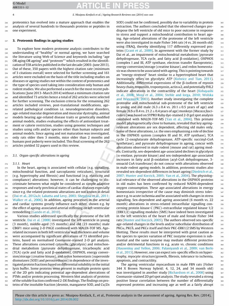
Fig. 1. Nucleic acid metabolism, small molecule biochemistry, amino acid metabolism.Ingenuity pathway analysis of proteins identified in normal heart ageing. Colouredmolecules: green; decreased, and red; increased. IPA revealed ERRA and ERRGas highly linked to proteins identified in reviewed studies during heart ageing.
Ligand-dependent nuclear receptor , enzyme , peptidase
, transcription regulator , transcription regulator , transmembrane
receptor , other , binding only , acts on
, inhibits
371
372
373
374
375
376
377
378
379
380
381
382
383
384
385
386
387
388
389
390
391
392
393
394
395
396
397
398
399
400
401
402
403
404
405
406
407
408
409
410
411
412
413
414
415
416
417
418
419
420
421
422
423
424
425
426
427
428
429
430
431
432
433
434
435
436
437
438
439
440
441
442
443
444
445
446
447
448
449
450
451
452
453
454
455
456
457
ARTICLERR 533 1–12
E. Nkuipou-Kenfack et al. / Ageing
ge-dependent (24 and 36 month of age) increase in nitrated pro-eins indicating increased oxidative stress. Of the 46 differentiallyxpressed proteins identified by MALDI-TOF-MS, 16 were asso-iated with the regulation of apoptosis, including parvalbumin,esmin, heat shock protein 27, peroxiredoxin 6, and kallikrein,
ndicating an adaptive rather than pro-apoptotic alteration. The up-egulation of parvalbumin, a calcium binding protein, may also bedaptive to diastolic dysfunction facilitating myocardial relaxationSchmidt et al., 2005), while the age-related increase in myosinegulatory light chain 2 (MLC-2) potentially contributes to dia-tolic dysfunction (Swynghedauw, 1996). The age-related increasen the level of 2-oxoglutarate 5 dioxygenase 1 (lysyl hydroxylase) may promote the formation of hydroxylysine sites essentialor the stability of the intermolecular collagen cross-links, there-ore contributing to fibrosis and diastolic dysfunction. However,he observed increases in the levels of desmin, peroxiredoxin, andallikrein may be protective against fibrosis and hypertrophy.
A proteomic analysis of the whole heart in 19–22 weeks and4 month old C57BL/6 mice (Chakravarti et al., 2008) basedn Comassie-stained 2D gel spot analysis combined with nano-C–MS–MS characterised 5 up-regulated and 27 down-regulatedrotein spots. In total 18 spots contained mitochondrial proteinsainly involved in metabolism (pyruvate dehydrogenase, TCA
ycle, and fatty acid �-oxidation) as well as OXPHOS again indi-ating an energy-stressed aged heart.
The results from most of the studies listed above are consistentith the mitochondrial theory of ageing since the cardiac muscle isarticularly vulnerable to mitochondrial dysfunction due to its highetabolic rate that needs to adapt swiftly to changes in demands.Ingenuity Pathway Analysis (IPA) software was used to generate
etwork diagrams to further elucidate the cellular context of dif-erentially expressed proteins related to heart ageing identified inhe combined studies reviewed here (Fig. 1, Supplementary Table). All data from Supplementary Table 1 including the observedold changes were imported in IPA and analysed using the defaultPA settings by IPA Core Analysis using the Build version: 261899nd Content version: 18030641 of IPA. According to this analy-is, additional proteins may likely be involved in cardiac ageing.or instance the oestrogen-related receptors alpha (ERRA or ERR�)nd gamma (ERRG or ERR�), two of the additionally identified IPAetwork nodes, were shown to directly interact with triosephos-hate isomerase 1, aconitase 1 and 2 suggesting a possible role
n energy metabolism (Fig. 1). ERRs are nuclear receptors inter-cting with PPAR� coactivator-1� and being involved in energyetabolism, mitochondrial biogenesis, gluconeogenesis and ROS
roduction (Giguere, 2008). ERRA−/− mice were characterised byepletion of phosphocreatine in the heart and pathologic cardiacypertrophy (Huss et al., 2007). Even more striking, ERRG−/− miceere reported to die shortly after birth due to severe metabolic dys-
unctions (Alaynick et al., 2007). Moreover, a recent study revealedhat overexpression of ERRG is associated with cardiac hypertrophyn humans (Kwon et al., 2013).
.1.2. Musculoskeletal systemFor a comprehensive review of skeletal muscle ageing see
Baraibar et al., 2013). Briefly, ageing in the musculoskeletal systems characterised by progressive loss of muscle mass and strength.lterations of myofibrillar and cytoskeletal networks (thin fila-ent), cytosolic and mitochondrial energy metabolism (glycolysis,
yruvate dehydrogenase, tricarboxylic acid (TCA) cycle, oxidativehosphorylation (OXPHOS)), redox homeostasis, and the secre-ome (myokines) contribute to these age-related losses.
Please cite this article in press as: Nkuipou-Kenfack, E., et al., Proteomhttp://dx.doi.org/10.1016/j.arr.2014.09.002
.1.3. BrainProteome-based mechanisms of brain ageing were investi-
ated e.g. in male Kunming mice that are characterised by high
, inhibits AND acts on , direct interaction—————, indirect interaction ------------ (For interpretation of the references tocolor in this figure legend, the reader is referred to the web version of this article.).
resistance to disease and good survival rate (Shang et al., 2009)at the age of 3 months and 15 months using 2-D PAGE com-bined with MALDI-TOF MS and peptide mass fingerprinting (Yanget al., 2008). Thirty-nine proteins were differentially expressed.The majority of these proteins, 23, were found to decrease withadvancing age, predominantly affecting protein turnover and denovo synthesis as well as neuron motility (axon formation). Inregard to protein turnover, the ubiquitin-proteasome system (UPS)is affected by a decrease of the subunits alpha type 3 and alphatype 6 as well as the regulatory subunit 14 (non-ATPase) of the26S proteasome and the valosin-containing protein (VCP), a mul-tifunctional protein binding misfolded and ubiquitinated proteinssupporting their export e.g. from the ER to the cytoplasm wherethey are degraded by the proteasome (Kloppsteck et al., 2012).The decrease in these proteins is believed to be associated withperturbations in apoptosis and cell signalling and the accumula-tion of misfolded and denatured proteins observed during ageing(Yang et al., 2008). An ageing-associated decrease of proteins com-posing the 26S proteasome was also found in C57BL/6 mice (Maoet al., 2010) highlighting the importance of the 26S proteasome inbrain ageing.
Additionally, perturbations in de novo protein synthesis in oldmice were indicated by the decrease in the eukaryotic transla-
e analysis in the assessment of ageing. Ageing Res. Rev. (2014),
tion initiation factor 5A (eIF5A) (Yang et al., 2008). eIF5A is crucialfor cell development and proliferation and the only transcriptionfactor known to date containing a hypusine residue that is essen-tial for the formation of the first peptide bond during translation
458
459
460
461

ING ModelA
6 Resea
(samad(acnt
gbQ3hees(omgmmobpi(ammvecphp(socs1pkhc
i2tgtlPPaimwaosTl
462
463
464
465
466
467
468
469
470
471
472
473
474
475
476
477
478
479
480
481
482
483
484
485
486
487
488
489
490
491
492
493
494
495
496
497
498
499
500
501
502
503
504
505
506
507
508
509
510
511
512
513
514
515
516
517
518
519
520
521
522
523
524
525
526
527
528
529
530
531
532
533
534
535
536
537
538
539
540
541
542
543
544
545
546
547
548
549
550
551
552
553
554
555
556
557
558
559
560
561
562
563
564
565
566
567
568
569
570
571
572
573
574
575
576
577
578
579
580
581
582
583
584
585
586
587
588
ARTICLERR 533 1–12
E. Nkuipou-Kenfack et al. / Ageing
Zanelli and Valentini, 2007). Luchessi et al. (2008) found eIF5Aelectively depleted in the cortex and cerebellum of aged rat brainsffecting neurogenesis and differentiation and therefore long-termotor memory (Luchessi et al., 2008). De novo protein synthesis
long with chromosomal integrity might also be affected by theecrease in VCP due to its role in DNA double strand break repairAcs et al., 2011). An increase in DNA double strand breaks haslso been observed in neurons of ageing C57/B6 male mice asso-iated with a decrease in the high mobility group B1 (HMGB1), aon-histone chromatin-associated protein with key roles in main-enance of nuclear homeostasis (Enokido et al., 2008).
Another interesting finding was the decrease in dynamin-1, auanosine triphosphatase (Yang et al., 2008) that was also observedy VanGuilder et al. (2010) in Fischer 344 × Brown Norway (F1)ybrid male rats (Chadwick et al., 2012; Freeman et al., 2009; Jiangt al., 2010) and Ottis et al. (2013) in male outbred Wistar rats (Ottist al., 2013). In the brain dynamin-1 plays a pivotal role in the fissiontage of synaptic vesicle endocytosis and thus neurotransmissionFerguson et al., 2007) as well as in the mechanical stabilisationf F-actin bundles in growth cone fillipodia and thus growth coneotility and the elongation and guidance of axons to synaptic tar-
ets (Yamada et al., 2013). Consequently, a decrease in dynamin-1ay adversely affect synaptic plasticity and therefore associativeemory. A decrease in dynamin-1 was also reported in the brain
f Alzheimer’s patients (Yao et al., 2003) suggesting a strong linketween dynamin-1 expression levels and cognitive function. Arogressive decline in cognitive function and impairments in learn-
ng and memory formation are leading features of brain ageingAlbers and Beal, 2000). Indeed, age-related proteomic alterationsssociated with cognitive impairment, assessed by the Morris wateraze experiment, were investigated in the hippocampal region ofale Fisher 344 × Brown Norway (F1) rats (10 and 27 month of age)
ia 2-D DIGE and MALDI-TOF/TOF mass spectrometry (Freemant al., 2009). The authors observed distinct proteomic changes asso-iated with either ageing or an altered cognitive status. Differentialrotein expression associated with normal ageing included carbo-ydrate metabolism (enolase 1 alpha, phosphoglycerate mutase 1,yruvate kinase, and lactate dehydrogenase A), protein turnover26S proteasome subunit alpha type 1), cell structure (coronin 1A),ignal transduction (protein phosphatase 1E), and redox home-statsis (peroxiredoxin 6, glutathione synthetase). Comparing agedognitively intact and impaired rats, differential protein expres-ion mainly affected carbohydrate metabolism (aldolase A, enolase
alpha) and protein metabolism and modification (heat shockrotein 1, heat shock protein 4, mitogen activated protein kinaseinase 1, puromycin-sensitive aminopeptidase). The increase ineat shock proteins and puromycin-sensitive aminopeptidase indi-ate increased stress and/or impaired protein processing.
Five other studies investigated age-related alterations specif-cally in rats and mice brain mitochondria (Dencher et al., 2006,007; Frenzel et al., 2010; Mao et al., 2006; Stoll et al., 2011). Inhree of these studies, the mitochondrial proteome was investi-ated under conditions preserving protein–protein interactions inhe first dimension of gel electrophoresis (Blue as well as Colour-ess Native PAGE) (Dencher et al., 2006, 2007; Frenzel et al., 2010).roteins were then further resolved by second dimension SDS-AGE and identified by MALDI-TOF MS. This approach uncoveredge-modulated differences in the abundance of multiple proteinsn cortical mitochondria of young (4–6 month) and older (28–30
onth) male Wistar rats, affecting i.e. OXPHOS complexes I to IV asell as F0F1 ATP synthase. Besides decreased monomer abundance,
geing also resulted in a destabilised supramolecular architecture
Please cite this article in press as: Nkuipou-Kenfack, E., et al., Proteomhttp://dx.doi.org/10.1016/j.arr.2014.09.002
f OXPHOS complexes, especially I1III2, and an increased oligomeri-ation of F0F1 ATP synthase that may alter cristae architecture.his may increase generation of reactive oxygen species (ROS)ike superoxide anion radicals as well as decrease ATP production.
PRESSrch Reviews xxx (2014) xxx–xxx
Age-related declines in complex I and IV subunits were alsoobserved in male and female C57BL/6 mice (Mao et al., 2006) andused to estimate mitochondrial DNA mutation rate based on amathematical model. Thus, mitochondrial dysfunction during brainageing may contribute to cognitive disabilities through detrimentalalterations in energy and redox homeostasis. The loss of mitochon-drial ATP generation could, in turn, alter glycolysis. An increasein lactate production accompanied by a decrease in ATP synthase(SILAC based MS/MS and tissue histology) and mitochondrial oxy-gen consumption was indeed observed in adult primary-culturedneural progenitor cells isolated from C57BL/6 mice of 3 and 15–18month (Stoll et al., 2011).
A closer investigation of age-related differential expression ofproteins in lipid rafts of neuronal synaptic plasma membranepreparations from Fisher 344/Brown Norway hybrid (F344/BNF1)rats (5 and 34 month of age) was carried out using 2-D DIGE MALDI-TOF/TOF and ultra Hybrid Mass Spectrometer (LTQ-FT) (Jiang et al.,2010). It revealed a total of 40 identified and significantly dimin-ished proteins in these specific cholesterol- and sphingolipid-richdomains of neurons in older rats. Almost half of these proteinswere associated with bioenergetics including 7 subunits of NADHdehydrogenase, OXPHOS complex I, 3 subunits of cytochromebc1 complex, OXPHOS complex III, and 3 subunits of probablyectopic F0F1 ATP synthase. The age-related loss in these proteinsalong with the voltage-dependent anion-selective channel protein1 (VDAC1) and its NADH:ferricyanide reductase activity may alterenergy and redox homeostasis, e.g. through altering the mainte-nance of cytosolic NAD+/NADH ratio. A potential contaminationwith mitochondrial membrane fragments was excluded based onthe detection of cytochrome C and glutamate dehydrogenase.
While all these studies demonstrated noticeable age-relatedchanges in the brain proteome, a recent study using stable isotopelabelling with amino acids in vivo combined with LC–MS/MS anal-ysis showed that only minimal proteomic changes occur duringageing of the brain (Walther and Mann, 2011). However, the find-ings of this study were based on brain tissues of only two animals.
3.1.4. KidneyTo gain insight into molecular mechanisms involved in kidney
ageing, the proteome of C57/BL6 mice kidneys was investigatedusing 2-D PAGE and LC–MS/MS (Chakravarti et al., 2009). Male micebetween 19–22 weeks and 24 months of age were used. The pro-tein expression of 59 proteins was found to be significantly alteredout of which 49 proteins increased in older mice. The majority ofsignificantly altered proteins were involved in OXPHOS, TCA cycle,propanoate metabolism, transport mechanisms (albumin, transfer-rin), unfolded protein response (UPR; glucose-regulated protein 78,VCP), and aldehyde detoxification (Chakravarti et al., 2009). Theglucose-regulated protein 78 (GRP78), also known as immunoglob-ulin heavy-chain binding protein (BiP), was found increased in thekidney of old mice. GPR78 is a multifunctional protein located in theendoplasmic reticulum (ER) and involved in the folding and assem-bly of newly synthesised proteins as well as the activation of theUPR cascade to degrade misfolded proteins via the UPS (Cybulsky,2013; Li and Lee, 2006). The observed increase in GRP78 potentiallyindicates impairments in protein folding and/or an accumulationof misfolded proteins, also called ER stress, associated with ageing(Cybulsky, 2013). Mice expressing a mutant GPR78 characterised bythe deletion of the retrieval sequence were produced to investigatethe function of GPR78 in ER stress response. These mice were foundto die shortly after birth emphasising the importance of GPR78for survival (Mimura et al., 2007). In addition, VCP was found to
e analysis in the assessment of ageing. Ageing Res. Rev. (2014),
increase in the kidney during ageing further pointing out ER stressas a factor in kidney ageing (Chakravarti et al., 2009).
Besides ER stress the increase of various aldehyde processingenzymes including aldehyde dehydrogenases, aldo-keto reductase
589
590
591
592

ING ModelA
Resea
acceapp2mdtieddiesw(teeiC
mwioha
pa
3
mb2tgadnFticoa(wpptndoir(s
i
593
594
595
596
597
598
599
600
601
602
603
604
605
606
607
608
609
610
611
612
613
614
615
616
617
618
619
620
621
622
623
624
625
626
627
628
629
630
631
632
633
634
635
636
637
638
639
640
641
642
643
644
645
646
647
648
649
650
651
652
653
654
655
656
657
658
659
660
661
662
663
664
665
666
667
668
669
670
671
672
673
674
675
676
677
678
679
680
681
682
683
684
685
686
687
688
689
690
691
692
693
694
695
696
697
698
699
700
701
702
703
704
705
706
707
708
709
710
711
712
713
714
715
716
717
ARTICLERR 533 1–12
E. Nkuipou-Kenfack et al. / Ageing
nd alcohol dehydrogenases in mice kidneys during ageing indi-ates altered metabolism and an increased formation of potentiallyytotoxic aldehydes and therefore oxidative stress (Chakravartit al., 2009). Aldehydes including 4-hydroxy-2-nonenal (4-HNE),crolein and malondialdehyde (MDA) are reactive moleculesroduced as the result of oxidative degradation of membrane phos-holipids, a process known as lipid peroxidation (Pizzimenti et al.,013). Increased MDA formation in combination with iron accu-ulation was also shown to be associated with chronic kidney
isease (CKD) (Shah et al., 2007). Levels of the iron binding proteinransferrin indeed increased in the kidney of older mice possiblyndicating iron overload (Amelina and Cristobal, 2009; Chakravartit al., 2009). While the majority of the identified aldehyde dehy-rogenases increased during ageing, the mitochondrial aldehydeehydrogenase family 6 member A1 (ALDH6A1), which is involved
n branched chain amino acid and pyrimidin metabolism (Wanderst al., 2012), decreased in this study. However, ALDH6A1 washown to increase in CD1-SWISS male mice kidneys between 28eeks and 76 weeks of age using 2-D PAGE and MALDI-TOF MS
Amelina and Cristobal, 2009). The discrepancy could be attributedo the difference between mice strain and/or the 5 months differ-nce in the age of older mice in the studies. In addition, Amelinat al. also showed gender differences in the protein expression dur-ng mouse kidney ageing, as ALDH6A1 was only identified in maleD1-SWISS mice and not females.
Kidney ageing was further investigated in the peroxisomes ofale C57BL/6J mice at 18 and 24 months, using 2D PAGE combinedith MALDI-TOF MS (Mi et al., 2007). The expression and the activ-
ty of the antioxidant enzyme catalase decreased significantly inlder mice might result in an impairment of the detoxification ofydrogen peroxide, an metabolic by-product of peroxisomal fattycid �-oxidation (Mi et al., 2007).
Overall, the findings in the kidney revealed oxidative stress anderturbations in the proteostasis, the homeostasis of the proteomes key events taking place during ageing.
.1.5. LiverThe mitochondrial proteome of the liver was investigated in
ale Wistar rats between 6 and 26 months of age using DIGE in alue-native gel system combined with MALDI-TOF MS (Dani et al.,010). Proteins involved in OXPHOS (complex I, IV and ATP syn-hase) and redox homeostasis/detoxification (catalase, microsomallutathione S-transferase and cytochrome p450) were shown to beltered in older animals depending on a normal or calorie-restrictediet. On normal diet components of complex I (NADH dehydroge-ase), complex IV (cytochrome c oxidase), and the ATP synthase0 complex were increased whereas components of the ATP syn-hase F1 complex were decreased during ageing. Hence, despite thencreases in the other OXPHOS components, the decreases in theatalytic components of ATP synthase (F1) suggested a decreasef OXPHOS-dependent ATP in the ageing liver. The detoxifyinggent cytochrome p450 was reported to decrease during ageingDani et al., 2010). An age-related loss in cytochrome p450 activityas also observed in human subjects over the age of 70 com-ared to younger subjects (Sotaniemi et al., 1997). Cytochrome450 is ubiquitously expressed in the liver and it plays a role inhe processing of drugs and environmental pollutants and, alter-atively, metabolic activation (Nebert and Dalton, 2006). Hence aecrease in cytochrome P450 might result in altered processingf these compounds. Simultaneously, antioxidant enzymes includ-ng glutathione S-transferase also decreased with age. Indeed, theatio between reduced glutathione (GSH) and oxidised glutathione
Please cite this article in press as: Nkuipou-Kenfack, E., et al., Proteomhttp://dx.doi.org/10.1016/j.arr.2014.09.002
GSSG) was found to decrease in the liver of aged rats indicating ahift towards positive reduction potential.
Another study also investigated mitochondrial liver proteomen male Fischer 344 Charles-River rats between 12 and 28 months
PRESSrch Reviews xxx (2014) xxx–xxx 7
of age using 2-D PAGE combined with ESI-MS (Musicco et al.,2011). Here proteins involved in metabolism (TCA, fatty acid �-oxidation, urea cycle), OXPHOS (complex I and ATP synthase),sulphur metabolism (rhodanese), and redox homeostasis (perox-iredoxin 3, glutathione peroxidase 1) were shown to be alteredin older animals. Some non-mitochondrial proteins (e.g. GRP78,protein-disulfide isomerase, and beta-actin) were also found tobe affected. A more than two-fold reduction in ATP synthase wasfound, which is in agreement with the study performed by Daniet al. (2010). Yet the increase in several subunits of complex I wasmore pronounced, which might result in an increased generation ofsuperoxide anion radicals through electron leakage (Jastroch et al.,2010). The observed strong age-dependent increase in the abun-dance of the antioxidant enzymes peroxiredoxin 3 and glutathioneperoxidase 1 might be a compensatory adaptation. Furthermore,the more than six-fold decrease in ornithine transcarbamylase(OTC) will affect the urea cycle and l-arginine biosynthesis inthe aged rats. In humans, severe OTC deficiency may result inhyperammonemia and in turn, without intervention, in metabolicencephalopathy, coma, and eventually death (Deignan et al., 2008;Walker, 2009).
In conclusion, ageing in the liver is mainly marked by dysfunc-tion in energy production and oxidative stress.
3.1.6. ColonThe proteome of colonic epithelial tissue was analysed in
healthy human male individuals between 25 and 65 years of ageusing 2-D PAGE in combination with peptide mass fingerprintingthrough MALDI-TOF MS (Li et al., 2007; Yi et al., 2010). Thirty-fiveproteins were significantly altered during ageing. These proteinsare involved in OXPHOS (complex I and ATP synthase), redoxhomeostasis (catalase, peroxiredoxin-1, 2 and 5, glutathione S-tranferase and glutathione peroxidase), UPR (GRP78) and sulphurmetabolism (rhodanese). Rhodanese, a mitochondrial thiosulfatesulfurtransferase, was shown to decrease in older subjects. It isimportant for the import of 5 S ribosomal RNA into mitochondriaand thereby the translation of proteins encoded by mitochondrialDNA (mtDNA) (Smirnov et al., 2010) as well as the restorationof iron-sulphur centres in Fe–S proteins, such as ferredoxin andsuccinate dehydrogenase (Bonomi et al., 1977). Translation of themtDNA-encoded proteins is further affected by the observed lossof the mitochondrial elongation factor-tu. In turn, depletion ofrhodanese may increase the formation of ROS, especially mito-chondrial superoxide anion radicals (Krueger et al., 2010). As aresult, ROS accumulation and therefore oxidative stress may occur,especially since proteins involved in redox homeostasis includ-ing catalase, glutathione S-transferase and peroxiredoxin-1, 2 and5 were also observed to decrease. This oxidative stress observedin older subjects could be a potential risk factor for developingcolorectal cancer (Perse, 2013). Additionally, the observed deple-tion in proteins associated with mitochondrial energy production,i.e. ATP synthase beta chain and electron transfer flavoproteinalpha-subunit, may further contribute to increasing mitochondrialperturbations during ageing. The nearly five fold increase observedfor GRP78, which is involve in the UPR, indicated the presence ofER stress (Li et al., 2007; Yi et al., 2010).
In conclusion, ageing of the colon seems to be marked by mito-chondrial dysfunction, disturbance in the redox homeostasis andproteostasis (ER stress).
3.1.7. ProstateWe found only one proteomic study assessing prostate ageing.
e analysis in the assessment of ageing. Ageing Res. Rev. (2014),
In this study the proteomic mechanisms of prostate ageing wereassessed in the ventral prostate of male Noble rats between 3 and16 months of age characterised by a short lifespan and a suscep-tibility to develop prostate and breast tumours (Ho et al., 2009)
718
719
720
721

ING ModelA
8 Resea
uDfdypsesTmalfDipt2(eupmbogtbtc22
tcs2toete(Lprp22
rtmp
3
3
dm2idA
722
723
724
725
726
727
728
729
730
731
732
733
734
735
736
737
738
739
740
741
742
743
744
745
746
747
748
749
750
751
752
753
754
755
756
757
758
759
760
761
762
763
764
765
766
767
768
769
770
771
772
773
774
775
776
777
778
779
780
781
782
783
784
785
786
787
788
789
790
791
792
793
794
795
796
797
798
799
800
801
802
803
804
805
806
807
808
809
810
811
812
813
814
815
816
817
818
819
820
821
822
823
824
825
826
827
828
829
830
831
832
833
834
835
836
837
838
839
840
841
842
843
844
845
846
ARTICLERR 533 1–12
E. Nkuipou-Kenfack et al. / Ageing
sing isotope-coded affinity tag (ICAT) labelling combined with 1- PAGE separation and LC–MS/MS analysis (Lam et al., 2008). Thirty
our proteins with a greater than two-fold increase or 1.7-foldecrease in expression in the aged ventral prostates versus theirounger counterparts were identified. The expression of 10 of theseroteins was verified by Western blot analysis. Notably, the expres-ion of proteins related to oxidative stress increased while thexpression of proteins involved in protein folding, protein synthe-is, and chromatin stability decreased in the aged ventral prostates.wo isoforms of glutathione-S-transferases (GST-mu1 and GST-u3) were amongst the proteins related to oxidative stress. GST
ctivity effectively detoxifies endogenous electrophilic metabolitesike lipid hydroperoxides, which are usually the consequence ofree radical (e.g. superoxide anion radical) damage to lipids andNA. GST activity has also been demonstrated to be important
n carcinogenesis through the inhibition of initiation and tumourromotion, but may also be detrimental as it is linked to resis-ance to anticancer drugs and malignant progression (Ketteler et al.,003). Regulated e.g. by testosterone, cystathionine beta-synthaseCBS) catalyses the first step in the transsulfuration pathway (Kabilt al., 2014; Prudova et al., 2007). However, an alternate reactionsing l-cysteine as substrate instead of serine forms hydrogen sul-hide (H2S). H2S has emerged as an important gaseous signallingolecule exerting effects e.g. on cellular antioxidant defence and
ioenergetics (Kabil et al., 2014; Kimura, 2014). Overexpressionf CBS has also been associated with the promotion of tumourrowth (Hellmich et al., 2014). The presence of a disturbance inhe redox homeostasis in aged ventral prostates is also supportedy the upregulation of ferritin, a protein complex binding transi-ion metal ions, particularly iron. Changes in cell iron homeostasisan result in oxidative stress (Antosiewicz et al., 2007; Cabantchik,014), especially when cellular iron increases with age (Xu et al.,010).
The observed decreases in the expression of chaperone pro-eins (PPIB/CyPB, DNAJ3/HSP40, HSPCA/HSP86, and HYOU1) mayompromise intracellular protein trafficking and increase cellulartress due to elevated protein misfolding (Behnke and Hendershot,014; Proctor et al., 2005) and perturbed removal of misfolded pro-eins (Merker and Grune, 2000). Thereby the increased expressionf T-kininogen, an inhibitor of cysteine proteinases (Greenbaumt al., 1992), may be a contributing factor. Moreover, de novo pro-ein biosynthesis will likely be affected by the decreases in thexpression of eukaryotic translation initiation factor 2 subunit �eIF-2�), glutamyl-prolyl-tRNA synthetase, and ribosomal protein36. Reduced expression of spermidine synthase in aged ventralrostate, a key enzyme in polyamine metabolism, may furtheresult in a heightened risk of chromatin instability and cancer asolyamines play a role in chromatin stability (Hobbs and Gilmour,000; Pollard et al., 1999) as well as cancer (Thomas and Thomas,003).
In conclusion, disturbances in redox homeostasis potentiallyesulting in increased susceptibility to oxidative protein modifica-ions in combination with a decline in proteostatic maintenance
ay be key factors in prostate ageing and increase the risk ofrostate cancer.
.2. Systemic alterations in ageing
.2.1. PlasmaAge-related changes in plasma were assessed in a longitu-
inal study utilising male C57BL/6J mice between 2 and 19onths of age using 2-D PAGE and LC–MS/MS (Ding and Kopchick,
Please cite this article in press as: Nkuipou-Kenfack, E., et al., Proteomhttp://dx.doi.org/10.1016/j.arr.2014.09.002
011). Transthyretin (TTR), haptoglobin isoform 2 and 3, andmmunoglobulin kappa isoform 1, 2, and 3 significantly increaseduring ageing whereas peroxiredoxin-2, serum amyloid protein-1 and albumin isoform 6, 7, 8, 9, and 18 decreased (Ding and
PRESSrch Reviews xxx (2014) xxx–xxx
Kopchick, 2011). TTR is a protein involved in the transport ofthyroxin and retinol binding protein-4 (Myron et al., 2007). Addi-tionally, TTR can induce amyloid fibril formation which couldsubsequently result in the development of amyloidosis; a disor-der characterised by the extracellular deposition of amyloid fibrilsin a systemic fashion (Ando et al., 2013). Amyloid deposition inthe peripheral nervous system is a pathological event particu-larly observed in Alzheimer’s disease (Krabbe et al., 2013), henceincreased TTR levels could point towards neurological dysfunction.Some isoforms of haptoglobin were also reported to increase andthis increase could suggest elevated inflammation and oxidativestress as haptoglobin is an acute phase protein and it acts as adefensive mechanism against oxidative stress triggered by heme(Dobryszycka, 1997). In addition, a decrease in peroxiredoxin-2revealed a dysfunction in the redox homeostasis.
The proteome of human serum was investigated using 2DPAGE and LC–MS to identify potential proteins that would predictlongevity in young (23–27 years old), old (60–67 years old) andelderly (90–91 years old) male and female subjects. A total of sevenserum spots that could classify between different age groups wereidentified and haptoglobin was demonstrated to increase in oldersubjects in agreement with the observation in mice (Byerley et al.,2010; Ding and Kopchick, 2011). In conclusion, findings revealedthat inflammation could be a plasma marker of ageing in a healthypopulation.
3.2.2. Urinary proteomeUrinary proteomics/peptidomics is a rapidly growing field dis-
playing changes not only in the kidney and urinary tract but alsosystemic changes (Rodriguez-Suarez et al., 2013).
In one study the urinary proteome was assessed in a cohort of218 healthy subjects (male and female) between the age of 19 and73 years through CE–MS analysis, to investigate changes associ-ated with ageing (Zürbig et al., 2009). A total of 49 peptides wereobserved to be significantly altered during ageing with fragments ofcollagen type 1 and 3 as well as uromodulin representing the major-ity of identified peptides. All collagen type 1 fragments were shownto be decreased in the urine of older subjects suggesting alterationsin the extracellular matrix (ECM) possibly including fibrotic pro-cesses (Schanstra and Mischak, 2014). The findings also suggestedthat kidney ageing resembles chronic kidney disease, i.e. kidneydisease speeds up kidney ageing (Zürbig et al., 2009).
A very recent study investigated the human urinary proteomein 37 healthy subjects (19 male and 18 female) between the age of19 and 90 years (Bakun et al., 2014). Urine samples were analysedusing LC–MS/MS and 19 proteins involved in tissue remodelling(e.g. secreted protein acidic and rich in cysteine and EGF-containingfibulin-like extracellular matrix protein) and inflammation (Alpha-1-acid glycoproteins) were differentially identified between youngand old subjects. The secreted protein acidic and rich in cysteine(SPARC), a collagen-binding matricellular glycoprotein regulatingcell–matrix interactions, was found to decrease in older subjects.SPARC promotes tissue repair and differentiation (Wang et al.,2006). Therefore, reduced levels of SPARC may suggest alterationsin the ECM and an impairment of tissue healing properties duringageing (Bakun et al., 2014). Levels of EGF-containing fibulin-likeextracellular matrix protein or fibulins were also reported to reducein the urine of older subjects. Fibulins are extracellular matrix pro-teins involved in the integration of elastic fibres into the ECM(Dasouki et al., 2007), hence a reduction in fibulins could leadto loss of elasticity in the tissue, an event observed during age-ing. Inflammation in older subjects was characterised by elevated
e analysis in the assessment of ageing. Ageing Res. Rev. (2014),
levels of acute phase proteins Alpha-1-acid glycoprotein 1 and 2.Increased levels of Alpha-1-acid glycoprotein were shown to bestrongly correlated with age, inflammation and high mortality riskof in-hospital elderly patients (Henry et al., 2003). This illustrates
847
848
849
850

ARTICLE IN PRESSG ModelARR 533 1–12
E. Nkuipou-Kenfack et al. / Ageing Research Reviews xxx (2014) xxx–xxx 9
e was
th
mEei2btiw2c
Ft
851
852
853
854
855
856
857
858
859
860
861
862
863
864
865
866
867
868
869
870
871
872
873
874
Fig. 2. Formation of collagen fibers cross-linking. The figur
he importance of Alpha-1-acid glycoprotein in ageing and alsoighlights urine as an adequate and informative biological source.
In conclusion, ageing in the urine is characterised by inflam-ation an event also observed in the plasma and alteration in the
CM remodelling. Alteration in the ECM remodelling is a molecularvent that could have awry ramifications during ageing consider-ng the pleiotropic roles of ECM in tissues and organs (Lu et al.,011). ECM consists of a basement membrane acting as a physicalarrier and an interstitial matrix contributing to elastic proper-ies of the ECM (Lu et al., 2012a,b). Elasticity of the ECM is an
Please cite this article in press as: Nkuipou-Kenfack, E., et al., Proteomhttp://dx.doi.org/10.1016/j.arr.2014.09.002
mportant factor enabling interaction and communication of cellsith ECM which can be altered when stiffness occurs (Lu et al.,
012a,b). In addition, decreased ECM elasticity is considered bylinicians as a potential indicator for diseases (Lu et al., 2011). The
ig. 3. Overview of molecular pathways occurring during normal ageing. (A) Organ-specific aaking place in organs or systemically listed first, followed by secondary events presented
adapted from Leeming et al. (2009). LOX: Lysyl oxidase.
dynamics of ECM (degradation and remodelling) are tightly con-trolled by degrading enzymes such as matrix metalloproteinases(MMP) during development. Reduced collagen peptides observedduring ageing could be the result of reduced MMP activity lead-ing to increase tissue remodeling and subsequently tissue stiffness(Karsdal et al., 2013). Increase in tissue stiffness was reported to bethe consequence of increase cross-linking of collagen fibres (Fig. 2).Collagen cross-linking can either be induced enzymatically via lysyloxidase enzymes or chemically as a result of advanced glycationend products formation (Langton et al., 2013). The later modifica-
e analysis in the assessment of ageing. Ageing Res. Rev. (2014),
tion was shown to trigger pathological age-related tissue stiffening(Sherratt, 2009). Therefore, alteration in the ECM remodellingcould be a key event promoting age-related pathologies duringageing.
lterations in ageing. (B) Systemic alterations in ageing. The core molecular pathway in brackets.
875
876
877
878

ING ModelA
1 Resea
4
pmtromdi
•
•
•
ttcthtu
A
Q4FQ52w
A
t
R
A
A
879
880
881
882
883
884
885
886
887
888
889
890
891
892
893
894
895
896
897
898
899
900
901
902
903
904
905
906
907
908
909
910
911
912
913
914
915
916
917
918
919
920
921
922
923
924
925
926
927
928
929
930
931
932
933
934
935
936
937
938
939
940
941
942
943
944
945
946
947
948
949
950
951
952
953
954
955
956
957
958
959
960
961
962
963
964
965
966
967
968
969
970
971
972
973
974
975
976
977
978
979
980
981
982
983
984
985
986
987
988
989
990
991
992
993
994
995
996
997
998
999
1000
1001
1002
1003
1004
1005
1006
1007
1008
1009
1010
1011
1012
1013
1014
1015
1016
ARTICLERR 533 1–12
0 E. Nkuipou-Kenfack et al. / Ageing
. Conclusion
The assessment of the ageing proteome has already revealedotential proteins involved in the ageing process and deter-ined important biological processes that are altered with ageing
hroughout tissues as well as tissue-specific changes (Fig. 3). Aecurrent drawback of the past studies is the lacking availabilityf human material for determining if the observations that areostly based on animal studies are also valid in humans. The recent
evelopments in proteomics technologies might help in solving thisssue:
Body fluid proteomics is now readily available (Schanstra andMischak, 2014). The use of non-invasive or minimally invasiveobtained body fluid could greatly facilitate studying the pro-teome of healthy ageing. Although previous proteomic studiesusing body fluids only generated limited sets of proteins andpeptides, more recent studies show that proteome technologieshave evolved sufficiently to obtain information of thousands ofproteins and peptides in non-invasively obtained fluids such asurine (Santucci et al., 2014) and in other fluids including plasma(Lu et al., 2012a,b).The obvious the drawback of body fluids is that these proteomesare mostly non-organ specific. Again progress in proteome anal-ysis might be of help since we are currently able to performin depth proteome analysis on biopsies (Aquino et al., 2014;Shipitsin et al., 2014), hence on minute amounts of tissue sam-ple. For example in kidney transplantation, declamping biopsiesare readily obtained and can yield information on the healthyaging kidney proteome as done for the ageing transcriptome in(Rodwell et al., 2004). Preliminary studies show that it is possibleto study specific kidney cell types starting with biopsies (Charoniset al., 2011).Proteomic approaches can also play a pivotal role in investigatingPTMs during ageing. For instance, glycosylation was shown to beinvolved in ageing, as high levels of N-glycans were shown to besignificantly correlated with age (Vanhooren et al., 2010).
Oxidative stress appears to be prevalent ubiquitously acrossissues and organs. However, we also discovered some intriguingissue-specific changes (Fig. 3). For instance, in the heart, ageing isharacterised by mitochondrial dysfunction leading to a dysfunc-ion in the energy metabolism whereas proteostasis is one of theallmarks of ageing in the brain. However, more studies using pro-eomics tool in its most effective way are needed to fully understandnderlying pathways involved in ageing.
cknowledgments
This work was supported by the grant GA 31354 (CodeAge)P7-PEOPLE-2011-ITN program grant and EU-MASCARA (HEALTH-011-278249). We thank Theofilos Papadopoulos for assistanceith the IPA figure.
ppendix A. Supplementary data
Supplementary material related to this article can be found, inhe online version, at http://dx.doi.org/10.1016/j.arr.2014.09.002.
eferences
Please cite this article in press as: Nkuipou-Kenfack, E., et al., Proteomhttp://dx.doi.org/10.1016/j.arr.2014.09.002
cs, K., Luijsterburg, M.S., Ackermann, L., Salomons, F.A., Hoppe, T., Dantuma, N.P.,2011. The AAA-ATPase VCP/p97 promotes 53BP1 recruitment by removingL3MBTL1 from DNA double-strand breaks. Nat. Struct. Mol. Biol. 18, 1345–1350.
ggarwal, K., Choe, L.H., Lee, K.H., 2006. Shotgun proteomics using the iTRAQ isobarictags. Brief. Funct. Genomics Proteomics 5, 112–120.
PRESSrch Reviews xxx (2014) xxx–xxx
Alaynick, W.A., Kondo, R.P., Xie, W., He, W., Dufour, C.R., Downes, M., Jonker, J.W.,Giles, W., Naviaux, R.K., Giguere, V., Evans, R.M., 2007. ERRgamma directs andmaintains the transition to oxidative metabolism in the postnatal heart. CellMetab. 6, 13–24.
Albers, D.S., Beal, M.F., 2000. Mitochondrial dysfunction and oxidative stress in agingand neurodegenerative disease. J. Neural Transm. Suppl. 59, 133–154.
Amelina, H., Cristobal, S., 2009. Proteomic study on gender differences in agingkidney of mice. Proteome Sci. 7, 16.
Ando, Y., Coelho, T., Berk, J.L., Cruz, M.W., Ericzon, B.G., Ikeda, S., Lewis, W.D.,Obici, L., Plante-Bordeneuve, V., Rapezzi, C., Said, G., Salvi, F., 2013. Guidelineof transthyretin-related hereditary amyloidosis for clinicians. Orphanet J. RareDis. 8, 31.
Antosiewicz, J., Ziolkowski, W., Kaczor, J.J., Herman-Antosiewicz, A., 2007. Tumornecrosis factor-alpha-induced reactive oxygen species formation is mediatedby JNK1-dependent ferritin degradation and elevation of labile iron pool. FreeRadical Biol. Med. 43, 265–270.
Aquino, P.F., Lima, D.B., de Saldanha da Gama Fischer, Melani, R.D., Nogueira, F.C.,Chalub, S.R., Soares, E.R., Barbosa, V.C., Domont, G.B., Carvalho, P.C., 2014. Explor-ing the proteomic landscape of a gastric cancer biopsy with the shotgun imaginganalyzer. J. Proteome Res. 13, 314–320.
Bakun, M., Senatorski, G., Rubel, T., Lukasik, A., Zielenkiewicz, P., Dadlez, M.,Paczek, L., 2014. Urine proteomes of healthy aging humans reveal extracellu-lar matrix (ECM) alterations and immune system dysfunction. Age (Dordr.) 36,299–311.
Baraibar, M.A., Gueugneau, M., Duguez, S., Butler-Browne, G., Bechet, D., Friguet, B.,2013. Expression and modification proteomics during skeletal muscle ageing.Biogerontology 14, 339–352.
Behnke, J., Hendershot, L.M., 2014. The large Hsp70 Grp170 binds to unfolded proteinsubstrates in vivo with a regulation distinct from conventional Hsp70s. J. Biol.Chem. 289, 2899–2907.
Bonomi, F., Pagani, S., Cerletti, P., 1977. Insertion of sulfide into ferredoxins catalyzedby rhodanese. FEBS Lett. 84, 149–152.
Byerley, L.O., Leamy, L., Tam, S.W., Chou, C.W., Ravussin, E., 2010. Development of aserum profile for healthy aging. Age (Dordr.) 32, 497–507.
Cabantchik, Z.I., 2014. Labile iron in cells and body fluids: physiology, pathology,and pharmacology. Front. Pharmacol. 5, 45.
Campisi, J., 2013. Aging, cellular senescence, and cancer. Annu. Rev. Physiol. 75,685–705.
Chadwick, W., Martin, B., Chapter, M.C., Park, S.S., Wang, L., Daimon, C.M., Bren-neman, R., Maudsley, S., 2012. GIT2 acts as a potential keystone proteinin functional hypothalamic networks associated with age-related phenotypicchanges in rats. PLoS One 7, e36975.
Chait, B.T., 2006. Chemistry. Mass spectrometry: bottom-up or top-down? Science314, 65–66.
Chakravarti, B., Oseguera, M., Dalal, N., Fathy, P., Mallik, B., Raval, A., Chakravarti,D.N., 2008. Proteomic profiling of aging in the mouse heart: altered expressionof mitochondrial proteins. Arch. Biochem. Biophys. 474, 22–31.
Chakravarti, B., Seshi, B., Ratanaprayul, W., Dalal, N., Lin, L., Raval, A., Chakravarti,D.N., 2009. Proteome profiling of aging in mouse models: differential expressionof proteins involved in metabolism, transport, and stress response in kidney.Proteomics 9, 580–597.
Charonis, A., Luider, T., Baumann, M., Schanstra, J.P., 2011. Is the time ripe for kidneytissue proteomics? Proteomics Clin. Appl. 5, 215–221.
Collado, M., Blasco, M.A., Serrano, M., 2007. Cellular senescence in cancer and aging.Cell 130, 223–233.
Corrada, M.M., Brookmeyer, R., Paganini-Hill, A., Berlau, D., Kawas, C.H., 2010.Dementia incidence continues to increase with age in the oldest old: the 90+study. Ann. Neurol. 67, 114–121.
Cybulsky, A.V., 2013. The intersecting roles of endoplasmic reticulum stress, ubiq-uitin–proteasome system, and autophagy in the pathogenesis of proteinurickidney disease. Kidney Int. 84, 25–33.
Dai, D.F., Chen, T., Johnson, S.C., Szeto, H., Rabinovitch, P.S., 2012a. Cardiac aging:from molecular mechanisms to significance in human health and disease.Antioxid. Redox Signal. 16, 1492–1526.
Dai, D.F., Rabinovitch, P.S., Ungvari, Z., 2012b. Mitochondria and cardiovascularaging. Circ. Res. 110, 1109–1124.
Dai, Q., Escobar, G.P., Hakala, K.W., Lambert, J.M., Weintraub, S.T., Lindsey, M.L., 2008.The left ventricle proteome differentiates middle-aged and old left ventricles inmice. J. Proteome Res. 7, 756–765.
Dani, D., Shimokawa, I., Komatsu, T., Higami, Y., Warnken, U., Schokraie, E., Schnolzer,M., Krause, F., Sugawa, M.D., Dencher, N.A., 2010. Modulation of oxidative phos-phorylation machinery signifies a prime mode of anti-ageing mechanism ofcalorie restriction in male rat liver mitochondria. Biogerontology 11, 321–334.
Dasouki, M., Markova, D., Garola, R., Sasaki, T., Charbonneau, N.L., Sakai, L.Y., Chu,M.L., 2007. Compound heterozygous mutations in fibulin-4 causing neonatallethal pulmonary artery occlusion, aortic aneurysm, arachnodactyly, and mildcutis laxa. Am. J. Med. Genet. A 143A, 2635–2641.
Deignan, J.L., Cederbaum, S.D., Grody, W.W., 2008. Contrasting features of urea cycledisorders in human patients and knockout mouse models. Mol. Genet. Metab.93, 7–14.
Dencher, N.A., Goto, S., Reifschneider, N.H., Sugawa, M., Krause, F., 2006. Unraveling
e analysis in the assessment of ageing. Ageing Res. Rev. (2014),
age-dependent variation of the mitochondrial proteome. Ann. N.Y. Acad. Sci.1067, 116–119.
Dencher, N.A., Frenzel, M., Reifschneider, N.H., Sugawa, M., Krause, F., 2007. Pro-teome alterations in rat mitochondria caused by aging. Biogeront.: Mech. Interv.1100, 291–298.
1017
1018
1019
1020
1021

ING ModelA
Resea
D
D
D
D
E
F
F
F
F
G
G
G
G
H
H
Q6H
H
H
H
H
J
J
J
K
K
K
K
K
K
K
K
Q7
1022
1023
1024
1025
1026
1027
1028
1029
1030
1031
1032
1033
1034
1035
1036
1037
1038
1039
1040
1041
1042
1043
1044
1045
1046
1047
1048
1049
1050
1051
1052
1053
1054
1055
1056
1057
1058
1059
1060
1061
1062
1063
1064
1065
1066
1067
1068
1069
1070
1071
1072
1073
1074
1075
1076
1077
1078
1079
1080
1081
1082
1083
1084
1085
1086
1087
1088
1089
1090
1091
1092
1093
1094
1095
1096
1097
1098
1099
1100
1101
1102
1103
1104
1105
1106
1107
1108
1109
1110
1111
1112
1113
1114
1115
1116
1117
1118
1119
1120
1121
1122
1123
1124
1125
1126
1127
1128
1129
1130
1131
1132
1133
1134
1135
1136
1137
1138
1139
1140
1141
1142
1143
1144
1145
1146
1147
1148
1149
1150
1151
1152
1153
1154
1155
1156
1157
1158
1159
1160
1161
1162
1163
1164
1165
1166
1167
1168
1169
1170
1171
1172
1173
1174
1175
1176
1177
1178
1179
1180
1181
1182
1183
1184
1185
1186
1187
1188
ARTICLERR 533 1–12
E. Nkuipou-Kenfack et al. / Ageing
i Girolamo, F., Lante, I., Muraca, M., Putignani, L., 2013. The role of mass spectrom-etry in the “Omics” era. Curr. Org. Chem. 17, 2891–2905.
iedrich, M., Tadic, J., Mao, L., Wacker, M.A., Nebrich, G., Hetzer, R., Regitz-Zagrosek,V., Klose, J., 2007. Heart protein expression related to age and sex in mice andhumans. Int. J. Mol. Med. 20, 865–874.
ing, J., Kopchick, J.J., 2011. Plasma biomarkers of mouse aging. Age (Dordr.) 33,291–307.
obryszycka, W., 1997. Biological functions of haptoglobin—new pieces to an oldpuzzle. Eur. J. Clin. Chem. Clin. Biochem. 35, 647–654.
nokido, Y., Yoshitake, A., Ito, H., Okazawa, H., 2008. Age-dependent change ofHMGB1 and DNA double-strand break accumulation in mouse brain. Biochem.Biophys. Res. Commun. 376, 128–133.
erguson, S.M., Brasnjo, G., Hayashi, M., Wolfel, M., Collesi, C., Giovedi, S., Rai-mondi, A., Gong, L.W., Ariel, P., Paradise, S., O’toole, E., Flavell, R., Cremona,O., Miesenbock, G., Ryan, T.A., De, C.P., 2007. A selective activity-dependentrequirement for dynamin 1 in synaptic vesicle endocytosis. Science 316,570–574.
reeman, W.M., VanGuilder, H.D., Bennett, C., Sonntag, W.E., 2009. Cognitive per-formance and age-related changes in the hippocampal proteome. Neuroscience159, 183–195.
renzel, M., Rommelspacher, H., Sugawa, M.D., Dencher, N.A., 2010. Ageing altersthe supramolecular architecture of OxPhos complexes in rat brain cortex. Exp.Gerontol. 45, 563–572.
röhlich, T., Arnold, G.J., 2006. Proteome research based on modern liquidchromatography–tandem mass spectrometry: separation, identification andquantification. J. Neural Transm. 113, 973–994.
ando, Y., Kawano, H., Yamamoto, K., Sanada, K., Tanimoto, M., Oh, T., Ohmori, Y.,Miyatani, M., Usui, C., Takahashi, E., Tabata, I., Higuchi, M., Miyachi, M., 2010.Age and cardiorespiratory fitness are associated with arterial stiffening and leftventricular remodelling. J. Hum. Hypertens. 24, 197–206.
iguere, V., 2008. Transcriptional control of energy homeostasis by the estrogen-related receptors. Endocr. Rev. 29, 677–696.
rant, J.E., Bradshaw, A.D., Schwacke, J.H., Baicu, C.F., Zile, M.R., Schey, K.L., 2009.Quantification of protein expression changes in the aging left ventricle of Rattusnorvegicus. J. Proteome Res. 8, 4252–4263.
reenbaum, L.M., Howard, E., Albus, U., Lapp, C., Gao, X.X., 1992. T-kininogen,processing and functions. Agents Actions Suppl. 38 (Pt 1), 300–306.
ausenloy, D.J., Yellon, D.M., 2006. Survival kinases in ischemic preconditioning andpostconditioning. Cardiovasc. Res. 70, 240–253.
ellmich, M., Coletta, C., Chao, C., Szabo, C., 2014. The therapeutic potential ofcystathionine-beta-synthetase/hydrogen sulfide inhibition in cancer. Antioxid.Redox Signal..
enry, O.F., Blacher, J., Verdavaine, J., Duviquet, M., Safar, M.E., 2003. Alpha 1-acidglycoprotein is an independent predictor of in-hospital death in the elderly. AgeAgeing 32, 37–42.
o, J., Lucy, M., Krokhin, O., Hayglass, K., Pascoe, E., Darroch, G., Rush, D., Nickerson,P., Rigatto, C., Reslerova, M., 2009. Mass spectrometry-based proteomic analysisof urine in acute kidney injury following cardiopulmonary bypass: a nested case-control study. Am. J. Kidney Dis. 53, 584–595.
obbs, C.A., Gilmour, S.K., 2000. High levels of intracellular polyamines promote his-tone acetyltransferase activity resulting in chromatin hyperacetylation. J. Cell.Biochem. 77, 345–360.
unter, J.C., Korzick, D.H., 2005. Age- and sex-dependent alterations in pro-tein kinase C (PKC) and extracellular regulated kinase 1/2 (ERK1/2) in ratmyocardium. Mech. Ageing Dev. 126, 535–550.
uss, J.M., Imahashi, K., Dufour, C.R., Weinheimer, C.J., Courtois, M., Kovacs, A.,Giguere, V., Murphy, E., Kelly, D.P., 2007. The nuclear receptor ERRalpha isrequired for the bioenergetic and functional adaptation to cardiac pressureoverload. Cell Metab. 6, 25–37.
astroch, M., Divakaruni, A.S., Mookerjee, S., Treberg, J.R., Brand, M.D., 2010. Mito-chondrial proton and electron leaks. Essays Biochem. 47, 53–67.
iang, L., Fang, J., Moore, D.S., Gogichaeva, N.V., Galeva, N.A., Michaelis, M.L., Zaidi,A., 2010. Age-associated changes in synaptic lipid raft proteins revealed by two-dimensional fluorescence difference gel electrophoresis. Neurobiol. Aging 31,2146–2159.
ohnson, T.E., 1990. Increased life-span of age-1 mutants in Caenorhabditis elegansand lower Gompertz rate of aging. Science 249, 908–912.
abil, O., Motl, N., Banerjee, R., 2014. HS and its role in redox signaling. Biochim.Biophys. Acta.
enyon, C., Chang, J., Gensch, E., Rudner, A., Tabtiang, R., 1993. A C. elegans mutantthat lives twice as long as wild type. Nature 366, 461–464.
etteler, M., Bongartz, P., Westenfeld, R., Wildberger, J.E., Mahnken, A.H., Bohm,R., Metzger, T., Wanner, C., Jahnen-Dechent, W., Floege, J., 2003. Association oflow fetuin-A (AHSG) concentrations in serum with cardiovascular mortality inpatients on dialysis: a cross-sectional study. Lancet 361, 827–833.
imura, H., 2014. The physiological role of hydrogen sulfide and beyond. NitricOxide.
lass, M.R., 1983. A method for the isolation of longevity mutants in the nematodeCaenorhabditis elegans and initial results. Mech. Ageing Dev. 22, 279–286.
loppsteck, P., Ewens, C.A., Forster, A., Zhang, X., Freemont, P.S., 2012. Regulationof p97 in the ubiquitin-proteasome system by the UBX protein-family. Biochim.
Please cite this article in press as: Nkuipou-Kenfack, E., et al., Proteomhttp://dx.doi.org/10.1016/j.arr.2014.09.002
Biophys. Acta 1823, 125–129.obayashi, T., Jin, L., de Tombe, P.P., 2008. Cardiac thin filament regulation. Pflugers
Arch. 457, 37–46.olwicz Jr., S.C., Tian, R., 2011. Glucose metabolism and cardiac hypertrophy. Car-
diovasc. Res. 90, 194–201.
PRESSrch Reviews xxx (2014) xxx–xxx 11
Krabbe, G., Halle, A., Matyash, V., Rinnenthal, J.L., Eom, G.D., Bernhardt, U., Miller,K.R., Prokop, S., Kettenmann, H., Heppner, F.L., 2013. Functional impairment ofmicroglia coincides with Beta-amyloid deposition in mice with Alzheimer-likepathology. PLoS One 8, e60921.
Krueger, K., Koch, K., Juhling, A., Tepel, M., Scholze, A., 2010. Low expression ofthiosulfate sulfurtransferase (rhodanese) predicts mortality in hemodialysispatients. Clin. Biochem. 43, 95–101.
Kwon, D.H., Eom, G.H., Kee, H.J., Nam, Y.S., Cho, Y.K., Kim, D.K., Koo, J.Y., Kim, H.S.,Nam, K.I., Kim, K.K., Lee, I.K., Park, S.B., Choi, H.S., Kook, H., 2013. Estrogen-relatedreceptor gamma induces cardiac hypertrophy by activating GATA4. J. Mol. Cell.Cardiol. 65, 88–97.
Lakatta, E.G., Levy, D., 2003. Arterial and cardiac aging: major shareholders in car-diovascular disease enterprises: Part II: the aging heart in health: links to heartdisease. Circulation 107, 346–354.
Lam, Y.W., Tam, N.N., Evans, J.E., Green, K.M., Zhang, X., Ho, S.M., 2008. Differentialproteomics in the aging Noble rat ventral prostate. Proteomics 8, 2750–2763.
Langton, A.K., Griffiths, C.E., Sherratt, M.J., Watson, R.E., 2013. Cross-linking of struc-tural proteins in ageing skin: an in situ assay for the detection of amine oxidaseactivity. Biogerontology 14, 89–97.
Li, J., Lee, A.S., 2006. Stress induction of GRP78/BiP and its role in cancer. Curr. Mol.Med. 6, 45–54.
Li, M., Xiao, Z.Q., Chen, Z.C., Li, J.L., Li, C., Zhang, P.F., Li, M.Y., 2007. Proteomic analysisof the aging-related proteins in human normal colon epithelial tissue. J. Biochem.Mol. Biol. 40, 72–81.
Lopez-Otin, C., Blasco, M.A., Partridge, L., Serrano, M., Kroemer, G., 2013. The hall-marks of aging. Cell 153, 1194–1217.
Lu, J., Huang, Y., Wang, Y., Li, Y., Zhang, Y., Wu, J., Zhao, F., Meng, S., Yu, X., Ma, Q.,Song, M., Chang, N., Bittles, A.H., Wang, W., 2012a. Profiling plasma peptides forthe identification of potential ageing biomarkers in Chinese Han adults. PLoSOne 7, e39726.
Lu, P., Takai, K., Weaver, V.M., Werb, Z., 2011. Extracellular matrix degradation andremodeling in development and disease. Cold Spring Harbor Perspect. Biol. 3.
Lu, P., Weaver, V.M., Werb, Z., 2012b. The extracellular matrix: a dynamic niche incancer progression. J. Cell Biol. 196, 395–406.
Luchessi, A.D., Cambiaghi, T.D., Alves, A.S., Parreiras-E-Silva, L.T., Britto, L.R., Costa-Neto, C.M., Curi, R., 2008. Insights on eukaryotic translation initiation factor 5A(eIF5A) in the brain and aging. Brain Res. 1228, 6–13.
Maes, K., Smolders, I., Michotte, Y., Van, E.A., 2014. Strategies to reduce aspecificadsorption of peptides and proteins in liquid chromatography-mass spectrom-etry based bioanalyses: an overview. J. Chromatogr. A.
Magdeldin, S., Enany, S., Yoshida, Y., Xu, B., Zhang, Y., Zureena, Z., Lokamani, I.,Yaoita, E., Yamamoto, T., 2014. Basics and recent advances of two dimensional-polyacrylamide gel electrophoresis. Clin. Proteomics 11, 16.
Mann, M., 2006. Functional and quantitative proteomics using SILAC. Nat. Rev. Mol.Cell Biol. 7, 952–958.
Mao, L., Romer, I., Nebrich, G., Klein, O., Koppelstatter, A., Hin, S.C., Hartl, D., Zabel,C., 2010. Aging in mouse brain is a cell/tissue-level phenomenon exacerbatedby proteasome loss. J. Proteome Res. 9, 3551–3560.
Mao, L., Zabel, C., Wacker, M.A., Nebrich, G., Sagi, D., Schrade, P., Bachmann, S.,Kowald, A., Klose, J., 2006. Estimation of the mtDNA mutation rate in aging miceby proteome analysis and mathematical modeling. Exp. Gerontol. 41, 11–24.
Merker, K., Grune, T., 2000. Proteolysis of oxidised proteins and cellular senescence.Exp. Gerontol. 35, 779–786.
Mi, J., Garcia-Arcos, I., Alvarez, R., Cristobal, S., 2007. Age-related subproteomic anal-ysis of mouse liver and kidney peroxisomes. Proteome Sci. 5, 19.
Miller, I., Crawford, J., Gianazza, E., 2006. Protein stains for proteomic applications:which, when, why? Proteomics 6, 5385–5408.
Mimura, N., Hamada, H., Kashio, M., Jin, H., Toyama, Y., Kimura, K., Iida, M., Goto,S., Saisho, H., Toshimori, K., Koseki, H., Aoe, T., 2007. Aberrant quality control inthe endoplasmic reticulum impairs the biosynthesis of pulmonary surfactant inmice expressing mutant BiP. Cell Death Differ. 14, 1475–1485.
Mischak, H., Vlahou, A., Ioannidis, J.P., 2013. Technical aspects and inter-laboratoryvariability in native peptide profiling: the CE–MS experience. Clin. Biochem. 46,432–443.
Moruz, L., Pichler, P., Stranzl, T., Mechtler, K., Kall, L., 2013. Optimized nonlinear gra-dients for reversed-phase liquid chromatography in shotgun proteomics. Anal.Chem. 85, 7777–7785.
Moss, R.L., Razumova, M., Fitzsimons, D.P., 2004. Myosin crossbridge activation ofcardiac thin filaments: implications for myocardial function in health and dis-ease. Circ. Res. 94, 1290–1300.
Mullen, W., Albalat, A., Gonzalez, J., Zerefos, P., Siwy, J., Franke, J., Mischak, H.,2012. Performance of different separation methods interfaced in the same MS-reflection TOF detector: a comparison of performance between CE versus HPLCfor biomarker analysis. Electrophoresis 33, 567–574.
Musicco, C., Capelli, V., Pesce, V., Timperio, A.M., Calvani, M., Mosconi, L., Cantatore,P., Gadaleta, M.N., 2011. Rat liver mitochondrial proteome: changes associatedwith aging and acetyl-l-carnitine treatment. J. Proteomics 74, 2536–2547.
Myron, J.A., Merlini, G., Sheldon, J., Ichihara, K., 2007. Clinical indications for plasmaprotein assays: transthyretin (prealbumin) in inflammation and malnutrition.Clin. Chem. Lab. Med. 45, 419–426.
Natale, M., Caiazzo, A., Bucci, E.M., Ficarra, E., 2012. A novel Gaussian extrapo-
e analysis in the assessment of ageing. Ageing Res. Rev. (2014),
lation approach for 2D gel electrophoresis saturated protein spots. GenomicsProteomics Bioinf. 10, 336–344.
Nebert, D.W., Dalton, T.P., 2006. The role of cytochrome P450 enzymes in endoge-nous signalling pathways and environmental carcinogenesis. Nat. Rev. Cancer6, 947–960.
1189
1190
1191
1192
1193

ING ModelA
1 Resea
N
O
O
P
PQ8
P
P
P
P
Q
R
R
R
S
S
S
S
S
S
S
S
S
S
S
S
S
S
1194
1195
1196
1197
1198
1199
1200
1201
1202
1203
1204
1205
1206
1207
1208
1209
1210
1211
1212
1213
1214
1215
1216
1217
1218
1219
1220
1221
1222
1223
1224
1225
1226
1227
1228
1229
1230
1231
1232
1233
1234
1235
1236
1237
1238
1239
1240
1241
1242
1243
1244
1245
1246
1247
1248
1249
1250
1251
1252
1253
1254
1255
1256
1257
1258
1259
1260
1261
1262
1263
1264
1265
1266
1267
1268
1269
1270
1271
1272
1273
1274
1275
1276
1277
1278
1279
1280
1281
1282
1283
1284
1285
1286
1287
1288
1289
1290
1291
1292
1293
1294
1295
1296
1297
1298
1299
1300
1301
1302
1303
1304
1305
1306
1307
1308
1309
1310
1311
1312
1313
1314
1315
1316
1317
1318
1319
1320
1321
1322
1323
1324
1325
1326
1327
1328
1329
1330
1331
1332
1333
1334
1335
1336
1337
1338
1339
ARTICLERR 533 1–12
2 E. Nkuipou-Kenfack et al. / Ageing
orth, B.J., Sinclair, D.A., 2012. The intersection between aging and cardiovasculardisease. Circ. Res. 110, 1097–1108.
nyema, O.O., Njemini, R., Bautmans, I., Renmans, W., De, W.M., Mets, T.,2012. Cellular aging and senescence characteristics of human T-lymphocytes.Biogerontology 13, 169–181.
ttis, P., Topic, B., Loos, M., Li, K.W., de Souza Silva, M.A., Schulz, D., Smit, A.B., Hus-ton, J.P., Korth, C., 2013. Correction: aging-induced proteostatic changes in therat hippocampus identify ARP3, NEB2 and BRAG2 as a molecular circuitry forcognitive impairment. PLoS One 8.
alaniyandi, S.S., Sun, L., Ferreira, J.C., Mochly-Rosen, D., 2009. Protein kinase C inheart failure: a therapeutic target? Cardiovasc. Res. 82, 229–239.
erse, M., 2013. Oxidative stress in the pathogenesis of colorectal cancer: cause orconsequence? Biomed. Res. Int., 725710.
izzimenti, S., Ciamporcero, E., Daga, M., Pettazzoni, P., Arcaro, A., Cetrangolo, G.,Minelli, R., Dianzani, C., Lepore, A., Gentile, F., Barrera, G., 2013. Interactionof aldehydes derived from lipid peroxidation and membrane proteins. Front.Physiol. 4, 242.
ollard, K.J., Samuels, M.L., Crowley, K.A., Hansen, J.C., Peterson, C.L., 1999. Func-tional interaction between GCN5 and polyamines: a new role for core histoneacetylation. EMBO J. 18, 5622–5633.
roctor, C.J., Soti, C., Boys, R.J., Gillespie, C.S., Shanley, D.P., Wilkinson, D.J., Kirkwood,T.B., 2005. Modelling the actions of chaperones and their role in ageing. Mech.Ageing Dev. 126, 119–131.
rudova, A., Albin, M., Bauman, Z., Lin, A., Vitvitsky, V., Banerjee, R., 2007. Testos-terone regulation of homocysteine metabolism modulates redox status inhuman prostate cancer cells. Antioxid. Redox Signal. 9, 1875–1881.
in, X.J., Ling, B.X., 2012. Proteomic studies in breast cancer (Review). Oncol. Lett. 3,735–743.
ichardson, M.R., Lai, X., Mason, S.B., Miller, S.J., Witzmann, F.A., 2008. Differentialprotein expression during aging in ventricular myocardium of Fischer 344 xBrown Norway hybrid rats. Exp. Gerontol. 43, 909–918.
odriguez-Suarez, E., Siwy, J., Zurbig, P., Mischak, H., 2013. Urine as a source for clin-ical proteome analysis: from discovery to clinical application. Biochim. Biophys.Acta.
odwell, G.E., Sonu, R., Zahn, J.M., Lund, J., Wilhelmy, J., Wang, L., Xiao, W., Mindrinos,M., Crane, E., Segal, E., Myers, B.D., Brooks, J.D., Davis, R.W., Higgins, J., Owen, A.B.,Kim, S.K., 2004. A transcriptional profile of aging in the human kidney. PLoS Biol.2, e427.
antucci, L., Candiano, G., Petretto, A., Bruschi, M., Lavarello, C., Inglese, E., Righetti,P.G., Ghiggeri, G.M., 2014. From hundreds to thousands: widening the normalhuman Urinome (1). J. Proteomics.
araswat, M., Musante, L., Ravida, A., Shortt, B., Byrne, B., Holthofer, H., 2013. Prepar-ative purification of recombinant proteins: current status and future trends.Biomed. Res. Int. 2013, 312709.
chanstra, J.P., Mischak, H., 2014. Proteomic urinary biomarker approach in renaldisease: from discovery to implementation. Pediatr. Nephrol.
chirmer, E.C., Yates III, J.R., Gerace, L., 2003. MudPIT: a powerful proteomics toolfor discovery. Discov. Med. 3, 38–39.
chmidt, U., Zhu, X., Lebeche, D., Huq, F., Guerrero, J.L., Hajjar, R.J., 2005. In vivogene transfer of parvalbumin improves diastolic function in aged rat hearts.Cardiovasc. Res. 66, 318–323.
hah, S.V., Baliga, R., Rajapurkar, M., Fonseca, V.A., 2007. Oxidants in chronic kidneydisease. J. Am. Soc. Nephrol. 18, 16–28.
hang, H., Wei, H., Yue, B., Xu, P., Huang, H., 2009. Microsatellite analysis in twopopulations of Kunming mice. Lab. Anim. 43, 34–40.
herratt, M.J., 2009. Tissue elasticity and the ageing elastic fibre. Age (Dordr.) 31,305–325.
heydina, A., Riordon, D.R., Boheler, K.R., 2011. Molecular mechanisms of cardiomy-ocyte aging. Clin. Sci. (Lond) 121, 315–329.
hipitsin, M., Small, C., Choudhury, S., Giladi, E., Friedlander, S., Nardone, J., Hus-sain, S., Hurley, A.D., Ernst, C., Huang, Y.E., Chang, H., Nifong, T.P., Rimm, D.L.,Dunyak, J., Loda, M., Berman, D.M., Blume-Jensen, P., 2014. Identification ofproteomic biomarkers predicting prostate cancer aggressiveness and lethalitydespite biopsy-sampling error. Br. J. Cancer.
mirnov, A., Comte, C., Mager-Heckel, A.M., Addis, V., Krasheninnikov, I.A., Martin,R.P., Entelis, N., Tarassov, I., 2010. Mitochondrial enzyme rhodanese is essen-tial for 5 S ribosomal RNA import into human mitochondria. J. Biol. Chem. 285,30792–30803.
otaniemi, E.A., Arranto, A.J., Pelkonen, O., Pasanen, M., 1997. Age and cytochromeP450-linked drug metabolism in humans: an analysis of 226 subjects with equalhistopathologic conditions. Clin. Pharmacol. Ther. 61, 331–339.
Please cite this article in press as: Nkuipou-Kenfack, E., et al., Proteomhttp://dx.doi.org/10.1016/j.arr.2014.09.002
talmach, A., Albalat, A., Mullen, W., Mischak, H., 2013. Recent advances in capillaryelectrophoresis coupled to mass spectrometry for clinical proteomic applica-tions. Electrophoresis 34, 1452–1464.
teen, H., Mann, M., 2004. The ABC’s (and XYZ’s) of peptide sequencing. Nat. Rev.Mol. Cell Biol. 5, 699–711.
PRESSrch Reviews xxx (2014) xxx–xxx
Stehle, R., Iorga, B., 2010. Kinetics of cardiac sarcomeric processes and rate-limitingsteps in contraction and relaxation. J. Mol. Cell. Cardiol. 48, 843–850.
Steinberg, T.H., 2009. Protein gel staining methods: an introduction and overview.Methods Enzymol. 463, 541–563.
Stoll, E.A., Cheung, W., Mikheev, A.M., Sweet, I.R., Bielas, J.H., Zhang, J., Ros-tomily, R.C., Horner, P.J., 2011. Aging neural progenitor cells have decreasedmitochondrial content and lower oxidative metabolism. J. Biol. Chem. 286,38592–38601.
Sue, K.M., Briscoe, V.J., Clark, N., Florez, H., Haas, L.B., Halter, J.B., Huang, E.S., Kory-tkowski, M.T., Munshi, M.N., Odegard, P.S., Pratley, R.E., Swift, C.S., 2012. Diabetesin older adults: a consensus report. J. Am. Geriatr. Soc. 60, 2342–2356.
Swynghedauw, B., 1996. Transgenic models of myocardial dysfunction. Heart Fail.Rev. 1, 277–290.
Thomas, T., Thomas, T.J., 2003. Polyamine metabolism and cancer. J. Cell. Mol. Med.7, 113–126.
van Berlo, J.H., Maillet, M., Molkentin, J.D., 2013. Signaling effectors underlyingpathologic growth and remodeling of the heart. J. Clin. Invest. 123, 37–45.
Vanhooren, V., Dewaele, S., Libert, C., Engelborghs, S., De Deyn, P.P., Toussaint,O., bacq-Chainiaux, F., Poulain, M., Glupczynski, Y., Franceschi, C., Jaspers, K.,van, d.P.I., Hoeijmakers, J., Chen, C.C., 2010. Serum N-glycan profile shift duringhuman ageing. Exp. Gerontol. 45, 738–743.
Walker Jr., E.M., Nillas, M.S., Mangiarua, E.I., Cansino, S., Morrison, R.G., Perdue, R.R.,Triest, W.E., Wright, G.L., Studeny, M., Wehner, P., Rice, K.M., Blough, E.R., 2006.Age-associated changes in hearts of male Fischer 344/Brown Norway F1 rats.Ann. Clin. Lab. Sci. 36, 427–438.
Walker, V., 2009. Ammonia toxicity and its prevention in inherited defects of theurea cycle. Diabetes Obes. Metab. 11, 823–835.
Walther, D.M., Mann, M., 2011. Accurate quantification of more than 4000 mousetissue proteins reveals minimal proteome changes during aging. Mol. Cell. Pro-teomics 10, M110.
Wanders, R.J., Duran, M., Loupatty, F.J., 2012. Enzymology of the branched-chainamino acid oxidation disorders: the valine pathway. J. Inherit. Metab. Dis. 35,5–12.
Wang, H., Workman, G., Chen, S., Barker, T.H., Ratner, B.D., Sage, E.H., Jiang, S., 2006.Secreted protein acidic and rich in cysteine (SPARC/osteonectin/BM-40) bindsto fibrinogen fragments D and E, but not to native fibrinogen. Matrix Biol. 25,20–26.
Wasinger, V.C., Zeng, M., Yau, Y., 2013. Current status and advances in quantitativeproteomic mass spectrometry. Int. J. Proteomics. 2013, 180605.
Wolska, B.M., Wieczorek, D.M., 2003. The role of tropomyosin in the regulation ofmyocardial contraction and relaxation. Pflugers Arch. 446, 1–8.
Xu, J., Marzetti, E., Seo, A.Y., Kim, J.S., Prolla, T.A., Leeuwenburgh, C., 2010. The emerg-ing role of iron dyshomeostasis in the mitochondrial decay of aging. Mech.Ageing Dev. 131, 487–493.
Yamada, H., Abe, T., Satoh, A., Okazaki, N., Tago, S., Kobayashi, K., Yoshida, Y., Oda,Y., Watanabe, M., Tomizawa, K., Matsui, H., Takei, K., 2013. Stabilization of actinbundles by a dynamin 1/cortactin ring complex is necessary for growth conefilopodia. J. Neurosci. 33, 4514–4526.
Yan, L., Ge, H., Li, H., Lieber, S.C., Natividad, F., Resuello, R.R.G., Kim, S.J., Akeju, S.,Sun, A., Loo, K., Peppas, A.P., Rossi, F., Lewandowski, E.D., Thomas, A.P., Vat-ner, S.F., Vatner, D.E., 2004. Gender-specific proteomic alterations in glycolyticand mitochondrial pathways in aging monkey hearts. J. Mol. Cell. Cardiol. 37,921–929.
Yang, S., Liu, T., Li, S., Zhang, X., Ding, Q., Que, H., Yan, X., Wei, K., Liu, S., 2008.Comparative proteomic analysis of brains of naturally aging mice. Neuroscience154, 1107–1120.
Yao, P.J., Zhu, M., Pyun, E.I., Brooks, A.I., Therianos, S., Meyers, V.E., Coleman, P.D.,2003. Defects in expression of genes related to synaptic vesicle trafficking infrontal cortex of Alzheimer’s disease. Neurobiol. Dis. 12, 97–109.
Yates, J.R., Ruse, C.I., Nakorchevsky, A., 2009. Proteomics by mass spectrom-etry: approaches, advances, and applications. Annu. Rev. Biomed. Eng. 11,49–79.
Yi, H., Li, X.H., Yi, B., Zheng, J., Zhu, G., Li, C., Li, M.Y., Zhang, P.F., Li, J.L., Chen, Z.C.,Xiao, Z.Q., 2010. Identification of Rack1, EF-Tu and Rhodanese as aging-relatedproteins in human colonic epithelium by proteomic analysis. J. Proteome Res. 9,1416–1423.
Zanelli, C.F., Valentini, S.R., 2007. Is there a role for eIF5A in translation? Amino Acids33, 351–358.
Zhang, Z., Wu, S., Stenoien, D.L., Pasa-Tolic, L., 2014. High-throughput proteomics.Annu. Rev. Anal. Chem. (Palo. Alto. Calif.) 7, 427–454.
Zhu, W., Smith, J.W., Huang, C.M., 2010. Mass spectrometry-based label-free quan-
e analysis in the assessment of ageing. Ageing Res. Rev. (2014),
titative proteomics. J. Biomed. Biotechnol. 2010, 840518.Zürbig, P., Decramer, S., Dakna, M., Jantos, J., Good, D.M., Coon, J.J., Bandin, F., Mis-
chak, H., Bascands, J.L., Schanstra, J., 2009. The human urinary proteome revealshigh similarity between kidney aging and chronic kidney disease. Proteomics 9,2108–2117.
1340
1341
1342
1343
1344