RENAL FUNCTION IN AN AGEING- - Enlighten: Theses
335
RENAL FUNCTION IN AN AGEING- COMMUNITY. Robert Livingstone Orchardson, M.B.E., M.B., Ch.B., B.Sc.
-
Upload
khangminh22 -
Category
Documents
-
view
0 -
download
0
Transcript of RENAL FUNCTION IN AN AGEING- - Enlighten: Theses

RENAL FUNCTION IN AN AGEING-
COMMUNITY.
Robert Livingstone Orchardson, M.B.E., M.B., Ch.B., B.Sc.

ProQuest Number: 13849082
All rights reserved
INFORMATION TO ALL USERS The quality of this reproduction is dependent upon the quality of the copy submitted.
In the unlikely event that the author did not send a com p le te manuscript and there are missing pages, these will be noted. Also, if material had to be removed,
a note will indicate the deletion.
uestProQuest 13849082
Published by ProQuest LLC(2019). Copyright of the Dissertation is held by the Author.
All rights reserved.This work is protected against unauthorized copying under Title 17, United States C ode
Microform Edition © ProQuest LLC.
ProQuest LLC.789 East Eisenhower Parkway
P.O. Box 1346 Ann Arbor, Ml 48106- 1346
CONTENTS.
Page
Introduction. ^Intention. * 2
Part I.Chapter I The Functions of the Kidney. 3
n II The Development of the Kidney - Embryology. 5M jjj ],ie Anatomy of the Kidney 10
The glomerulus HThe renal tubules 13The renal vasculature 17Renal innervation. 25
M IV The Physiology of the Kidney 25The secretion of urine 25The glomerulus _ 28The renal tubules 42The excretion of water 46Renal treatment of sodium 56Acid-base balance 59The excretion of urea. 66
,! V Renal Functional Measurements. 69M VI Renal Functions in Senescence. 75
Summary 91" VII Method Part II. 93
The Urea Concentration test of McLean 93The Concentration and Dilution test
of Calvert 97

Page
Chapter VIII Choice of Subject. 102” IX Results: method of presentation 106
1. Overall average findings. 1092. Effect of increasing age on renal
function.3. Renal function differences between
the two sexes.4 . Relationship between Renal Eunction
and Social Status.
132
1405. Relationship between the Cardio- -..c
vascular System and Renal Eunction.The heart. 146The arteries. 160The level of arterial blood pressure. 165Conclusions. 177
6 . Relationship between streptococcalinfection and Renal Eunction.
7. Relationship between Obesity and BodyStructure and Renal Eunction.
178
185
Simple Obesity. 187Sheldon’s Somatotypes. 189
8 . Effect of tobacco smoking on Renal nqoEunction.
9. Effect of consumption of alcohol on 3.98Renal Eunction.
10. Effect on Renal Eunction of Pulmonarylesions causing impaired oxygenation 2 0 2of the blood.
11. The level of Renal Eunction in the 2 0 6presence of albuminuria.

page12. Effect of Prostatic Enlargement
on Renal Function. 2101 3 . General characteristics
associated with poor Renal Function. 219
Chapter X. Conclusions. 222M XI. Summary 225
Bibliography. 231Appendices. 239
A: List of all patients surveyed.B: The incidence of various
pathological conditions encountered.
1C: List of all *'healthy" subjects.D: Causes of death, and death
rates.

- 1 -
INTRODUCTION.
In 1947 there were alive some five million persons over the age of 65 years. According to the official estimate, this figure will increase to 7.3 millions by 1977, and Sheldon comments that, if the present trend in mortality rates continues, it may well be that this figure will actually prove to be as high as 8.2 millions ( ^4)#Thus it is obvious that studies of problems relating to old age are of ever-increasing importance.
This has not always been realised, and prior to the. recent war of 1939-45, few investigations were made along such lines. On the subject of renal function in senescence there appears to have been but one paper - that of Lewis and Alving However, since the pressing nature ofthe problem came to be appreciated, there has been a resurrection of interest in subjects geriatric, and renal function in old age has been well investigated. Nevertheless, I have been unable to find any general survey of renal function among our ageing population, and I have set myself to make such a study, albeit on rather a small scale.
While the conditions of general practice militate against the carrying out of complicated laboratory investigations, they do provide the reservoir of subjects,

- 2 -
male and female, old and not-so-old, in the fulness of health or in the sorrows of morbidity, so essential for a general and unrestricted survey.
INTENTION.
My intention, then, is to ascertain the state of the renal function of a random sample of my elderly patients, using such means as are within my capabilities, and also to relate this function to pastor present abnormalities or peculiarities in the physical state or circumstances of these same persons.

- 3 -
PART I.
CHAPTER I«
THE FUNCTIONS OP THE KIDNEYS.
That the kidneys are vital to continuing healthy life was amply demonstrated by Professor Rose Bradford .
Dr. Homer W. Smith has succintly summarised therole of the kidneys in regulating fluid and electrolyte balance in the body, by saying that the composition and volume of the blood and of the body's internal environment are determined, not by what the mouth ingests, but by what the kidneys keep. He describes the normal physiological mechanism by which the kidneys maintain homeostasis. He states that at one end the heart is pumping as hard as it can to push out a large volume of fluid, and at the other end the kidneys are working as hard as they can to defeat this object and to retain 99$ of the water. According to him, life depends on neither heart nor kidneys winning this battle.
Put at greater length, and in greater detail, the functions of the kidneys may be summarised thus:-
By secreting urine, the kidney(l) helps to keep constant the plasma volume, and the

.Somite.
Pe.-ritone.al Cavity
PeritonealCavity.
'We in I o m e r u t v S .
Peritoneal
C a v ity .F u n n e l
P i g . I . C o m p o s itio n an«t O r ig in o f N e p h n c T u b u le s .
A . J h a g rc m o f On iso lated N e p h r it Tubule..
B . SA OY^IfUj th e m a n n e r- in w b icA the. Inte.-rrne.clta.te Cell (VJaSS t> , c ') g ive s origin to th eN ephric T u b u le ( a ) 7 P e rito n e a l Funnel [ b ) # And. th e Nepht-oeoele (e ) .
C . I be isolation of these p a r ts fro m tA c Som ite One/ th e ir union to form a. s y s te m .O . T h e O rig in o f a C lom evu /uS in t i e V x /e il o f th e . Fteph ro co ele C O -
^ /3f t e r Keith '5 Embryology
£ y-ace yD. -f:

- 4 -
water content of the body as a whole;(2) helps to preserve the reaction of the blood within
narrow limits;(3) eliminates waste products, especially the nitrogenous
and sulphur-containing substances derived from the metabolism of ingested protein and of the tissue cells;
(4) eliminates most of the basic, and the non-volatileacid, radicles which are ingested or are formed in the body;
(5) regulates osmotic relations in the blood and tissuesusing the mechanisms mentioned under (1) and (4) above;
(6) maintains the optimum concentration of certainindividual constituents of the plasma;
(7) eliminates toxic substances which have been introducedartificially into the body; and
(8) manufactures certain substances in the epithelial cellsof the renal tubules.
Further, there is the unproved hypothesis that, if the blood supply to the kidney is markedly decreased, an enzyme, renin, is produced by the kidney and then discharged into the circulation; by interaction with a globulin this renin is believed to form a pressor substance called hypertensin, which is responsible for so-called ischaemic hypertension

Nephtic Dud'.
TubuleNep/mc Tubule
Glomerulus C jQ iika.1 C ilcm cl
n e p h r o S to m e .
Mesentery.
F . S - S ch em a tic . Section to shovv th e S p ecia lisation o f
the. Dors*.! pa-rf" o f t f i& Coelom in to N ephric Tubules 7 Peritoneal
F un nels And G lom eVo/i . ^ M o d if ie d fro m K e ith 's Em bryology
To jace p. 5.

- 5 -
(33, 46, 47, 48, 112, 122)
At this point, I shall leave this brief survey of physiology, and shall trace the development of the kidney up to its adult form.
CHAPTER II.
EMBRYOLOGY.
In the course of evolution, man has developed from a type possessing a separate excretory tubule or kidney for each body segment. In Pig. 1A such a nephric tubule is represented diagrammatically.
At its commencement the tubule communicates with the peritoneal cavity by an open ’’peritoneal funnel”. A vascular body, a glomerulus - similar to the glomerulus of the adult kidney - projects into the dilated beginning of the tubule, which possesses a functional epithelial lining - the essential part of the apparatus. These tubules discharge fluid into a common collecting duct, named the nephric duct, which in turn leads to the cloaca.
As shown in Pigs. IB, 1C and ID, all these organs are formed from the intermediate cell mass. The nephric tubulearises by an evagination of the outer wall of the intermediate ||part of the coelom, while the glomerular chamber or j

CetvicoJ
Pronephros
W o lffia n Tubules
Renal tissue.
Dtetvric bud.
1st. Sacral.
P i g . O . Condition o9 the N ep h ric o ^
R e n a l System in a H um an E m b ry o of
w e e k s . / f l f t t r K eith 's E m b ry o lo g y .

- 6 -
nephrocoele and the peritoneal funnel are produced from the coelomic passage which originally connected the peritoneal cavity with that of a somite. The nephridial system thus can he regarded as modified parts of the wall of the original coelomic cavity. (Fig. 2).
In higher vertebrates three distinct phases of the renal system have been evolved. The human embryo in the sixth week possesses all three systems. (Fig. 3).By that time the first and most primitive of these systems, the pronephros, which appears in the last four or five cervical, and the first two or three thoracic, segments, is retrogressing. This pronephros is simple in the cervical region, where a segmental arrangement is retained. At the same time, the metanephros, which is destined to become the adult renal system, is only beginning to appear in the last lumbar segments. Concurrently, the mesonephros or Wolffian Body, found in the segments from fifth cervical to third lumbar, is coming to its peak of development.
These three systems are all of the same order, and are of increasingly advanced development. All open into the common excretory duct - the Wolffian duct. They progress from extreme simplicity in the cervical segments to extreme complexity in the sacral regions. By the third month, the permanent kidney, derived from the metanephros, is assuming its final position, and the Wolffian Body or mesonephros

-QlomeruluSTobuleS of pro- nepkros
Wolff. an Duct"
W olffiantubules.
opening into C oelom .
Wolffian
Rectumoaca.
Fig. 4. Sc,k erne o f tke W olffi’an Body
o f tke rijkf" s id e .
^F-rom Keith's E m b ry o lo g y .)
'W o lffian renal tubules.
Clomerolus.
MeSo-nepkros.
[ 7o jace. p. 7.

- 7 -
is ‘becoming a mere appendage of the genital system.In the lower vertebrates the Wolffian Body is the
functional kidney. A diagram of the tubular system of the Wolffian Body of the frog is shown in Fig. 4. In many points this corresponds to the same structure in the human embryo.
The Wolffian Body is a temporary or embryonic structure in the higher vertebrates who develop the kidney proper to take over renal function, and this permanent kidney would appear to develop from enlargement and separation of the posterior portion of the Wolffian Body. However, the presence of the mesonephros in the embryonic phases of the higher vertebrates must indicate that the higher forms are descended from ancestors of the lower.
In the human embryo, the first and simplest form, the pronephros, reaches its highest development in the fourth week, and thereafter retrogresses and is lost without trace.
The second and more complex system, the Wolffian Body, on the other hand, is retained partially in the genital system. In the female, the upper part of the Mullerian duct develops into the Fallopian tube; some remnants of the distal parts of the Mullerian duct sometimes persist, but they usually vanish. In the male, the Wolffian duct forms the tube of the epididymis, the vas deferens and the common ejaculatory duct and seminal vesicles. In both

ra '* '* /*/
I ome-toJj/ainl a I gitflo VliJ- !i»m uii
F"| fl. 5 . Four S ta g e s in t h e d e v e lo p m e n t of- a I ' fe p h n 'e T u b u le .
In th e H u m a n E m b ryo .
A. Vesreo/ar Stage of tu lo u le .
0 . T u b u le j ° ' n i W o l f f i a n D u c t .
f . ■ S c to m e s Convo l u t e d ,
D. f\ qlome-ruLuS is developed .
1 1 odi ficd ft om K.Ciith. S E-m btyotojy^
T u b u le .
(jlometulus.
ToLol<
o J-ccce p. 8%

- 8 -
sexes, the ureter and collecting tubules of the kidney are developed from the Wolffian duct.
As I have mentioned earlier, the hindmost portions of the Wolffian Body develop into the permanent kidney.
The kidney is the end result of the fusion of two separately developing systems. These are, first, a secretory system, and second, a collecting system. The collecting system is evolved from an outgrowth of the posterior end of the Wolffian duct, and forms the ureter, the pelvis of the kidney, and the collecting tubules.(Fig. 5 and 6). The secretory system comes from the hindmost end of the nephrogenic cord, behind the portion which forms the mesonephros, and at the level of the first and second sacral segments. It produces the secretory substance of the kidney- the cortex, with the glomeruli, the convoluted tubules and the loops of Henle. (Fig. 5 and 6).
The collecting system is developing by the fifth week; late in the sixth week the crude outline of the ureter, kidney pelvis and calyces is apparent, and clustered round the calyces is the substance of the nephrogenic cap. The ureter is now beginning to separate from the Wolffian duct. The position at this time is shown in Fig. 6.
During the third month, outgrowths from the primitive pelvic bud develop, and subdivide to form the collecting

i«-mc
U re iv .r
llretc-/-.B l euLd<i-
Io a c o .
tc j/nn /n ij
Tom
To face p. 9.

- 9 -
tubules of the pyramids. The rudimentary tubules, at first closed at both ends, (Pig. 5), in thenephrogenic body are likewise enlarging and elongating; one end establishes connection with the collecting tubules, while the other end develops into a glomerulus. This is achieved by expansion and invagination so developing a concavity at one side into which a capillary tuft grows to produce a Bowman's capsule. The tubules themselves develop rapidly into the adult form. These processes are demonstrated in Pig. 7, and the scheme of growth in the subcapsular zone of growth is shown in Pig. 8.
The glomeruli appear early in the third month, and during this month the surrounding mesodermal tissue provides capsules for them. Prom this time onward until birth, the formation of fresh tubular and glomerular tissue proceeds apace. This activity is in the subcapsular zone of growth of the kidney. A diagrammatic representation of this is given in Pig. 8. In the process of differentiation into the various parts of the tubule, the deeper tubules come first.
Shortly after birth new formation ceases, and the succeeding increase in size is due to pure growth. The gradual development of the kidney by a process of budding gives it a lobulated appearance; but early in the post-natal period the fissures between the lobules become filled in by

[ o r o t : e r i c )
lo itte ro l
l i t . C o n v o lu tio n
T h in lim b o f
H e n /e ’s /o o p .
Loop o f Hcnle
Collecting Tubes
ollechno tube .
UretericBud.
N e p h r ic Suet.
Collecting Tube. .
N e p h r i e To bo/e.
Collecting T o te , t U re te ric )
F i g . 7 . I l lu s t r a t i n g th e d e v e lo p m e n t o f t / ie R e n a l T is s u e .
A", grow in g en d o f collecting tubule W ith b v d o f nephric tu.be a t t a c k e d .
8 : f ir s t stage in th e develop m ent o f a nephric bud in to a n e p h r ic
tu b u le .
C.'. fully developed re n a l tubule \ the pa-rf from the ureteric bud. ispale green. 7 th a t from the nephnc tubule d a r k g re en .
(^Mod! tie d fro m K e ith 's E m bryo logy
O face p. 10.

NepKfogenit CjTowinQ Z
N ephrogenic. Z o n e .
in w h ic h
tubules se rve fa r
Q. t im e .
C o llectin g System.
V e s tig ia l Tubule.Z o n e in w b ic k tubules become ve s tig ia l.
Pelvis .
U reter.
Section of p e r t of a. Foetal Kidney sh owingtfje SobeapSuUr Z.one. o f G ro w th . The num bers
Indicate th e Successive broods of collecting tubes produced by division. ^Modified. ffo m KeitfCs Em bryology.^
7~o face. p. /O.

- 10 -
newly developing cortical tissue so that the surface of the kidney soon becomes smooth*
The blood supply to the kidney evolves in the following way. Initially, the renal buds have temporary branches from the common iliac arteries and from the aorta; when they come to be on the dorsal aspect of the Wolffian Body by the seventh week, the renal buds are invaded by the arterial network of th6 Wolffian Body tubules. Thus the kidney receives blood from the Wolffian arteries, that is, from the eleventh thoracic to the fourth lumbar segments.The definitive and final blood supply to the kidney is derived from the arteries of the second lumbar segment, although occasionally more than one pair persist.
Renal function is established very early in the human foetal kidney. Cameron and Chambers were able todemonstrate that the proximal tubules had become functional st Is’ months, and they c uote other writers as finding functioning renal tissue as early as the tenth or twelfth week.
CHAPTER III.
ANATOMY - THE STRUCTURE OP THE ADULT KIDNEY.
The human being is provided with two kidneys.

1
G L O M E R U L U S
| ~To face If.
THIN SEGMENT
Fi«. 9. Tfie. Q rtan yem en t a n d strvc io -re o f the.
N e p h t o n .
( APf'tv Hornet Smi ifc.)
PROXIMAL TUB ULE------
D ISTALTUBULE
SEEE

- 11 -
nephron, of which each kidney contains about one million, is the functional unit. Each nephron consists of a Bowman*s capsule and glomerulus, connected to an unbranched tubule which is subdivided functionally into three parts, and which drains into the collecting tubules. These in turn drain into the kidney pelvis, and thence into the ureter and urinary bladder. This arrangement isrepresented in Big. 9
THE G-LOMERUXiUS. 110^The glomerulus is a spherical capillary tuft which
is supplied with blood through a short, wide afferent arteriole. It is formed by the abrupt division of this afferent arteriole into two or four, or more rarely, ten, primary branches, which subdivide at times into as many as fifty capillary loops, each having a length equal to two or three times the diameter of the whole tuft. There are no arterio-venous shunts and no anastomoses. The capillary loops then coalesce into an efferent arteriole which, in its turn, breaks up into a second capillary system (the peritubular capillaries) around the tubules.
The capillary tuft - the glomerulus - grows into the expanded, but closed and invaginated, end of the tubule (Big. 5), so that it becomes enveloped in a spherical, double-walled capsule derived from the tubule itself. This
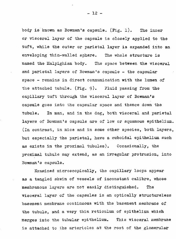
- 12 -
body is known as Bowman's capsule. (Big. l). The inner or visceral layer of the capsule is closely applied to the tuft, while the outer or parietal layer is expanded into an enveloping thin-walled sphere. The whole structure is named the Malpighian body. The space between the visceral and parietal layers of Bowman's capsule - the capsular space - remains in direct communication with the lumen of the attached tubule. (Pig. 9). Pluid passing from the capillary tuft through the visceral layer of Bowman's capsule goes into the capsular space and thence down the tubule. In man, and in the dog, both visceral and parietal layers of Bowman's capsule are of low or squamous epithelium. (In contrast, in mice and in some other species, both layers, but especially the parietal, have a cuboidal epithelium such as exists in the proximal tubules). Occasionally, the proximal tubule may extend, as an irregular protrusion, into Bowman's capsule.
Examined microscopically, the capillary loops appear as a tangled skein of vessels of inconstant calibre, whose membranous layers are not easily distinguished. The visceral layer of the capsules is an optically structureless basement membrane continuous with the basement membrane of the tubule, and a very thin reticulum of epithelium which merges into the tubular epithelium. This visceral membrane is attached to the arterioles at the root of the glomerular

- 13 -
tuft. At some points it is closely applied to the individual capillaries, while at others it envelopes capillary loops which have no basement membrane of their own. In the intercapillary space an infrequent cell, which is neither an endothelial nor epithelial cell, is present. It may be of connective tissue origin, and has been designated the mesangium by Zimmerman.
The three primary membranes of the glomerulus - capillary endothelium, basement membrane, and capsular epithelium - are generally considered to be continuous in
( o Q \having no openings or defects. Ekehorn , however, contends that the visceral epithelium is discontinuous.
THE TUBULES. (;L10'
As may be seen in Pig. 9, after extensive convolutions (the proximal convoluted tubule) near the glomerulus, the tubule descends in a more or less straight path into the medulla, of the kidney for a variable depth, where it executes a sharp hairpin turn (the loop of Henle), and returns upon its former course to the glomerulus of origin; here it undergoes a second series of convolutions (the distal convoluted tubule) and subsequently joins a collecting duct. The proximal and distal convoluted tubules are closely intertwined about the glomerulus as a result of

- 14 -
having developed in situ. , It will he remembered that the embryonic tubule is anchored at both ends, while the middle portion grows by elongation to form the loop of Henle.
The tubule, by structure and function, is, like Gaul, divided into three parts, viz.:-1. The Proximal tubule, which continues through the proximal
convoluted tubule down along the straight descending limb of Henle's loop to the point where the thin segment begins.
2. The thin segment of the loop of Henle.3. The Distal tubule, beginning where the thin segment ends,
and continuing through the distal convoluted tubule to the collecting duct.
The Proximal segment has the widest diameter of all the various parts of the tubule. It is lined by large cuboidal or truncated pyramidal cells rich in protoplasm, and having large basally placed spherical nuclei. The cells are fluted on their adjacent sides, and these flutings lock together. They are coarsely granular cells which bulge into the lumen of the tubule. On the luminal aspect they have a ’'brush’1 border.
The Thin segment consists of highly attenuated cells.The lumen of the tubule here is of lesser calibre. The

- 15 -
lining is of flattened epithelial cells, with clear protoplasm and compressed nuclei. The length of the thin segment varies the length of Henle's loop, and it is found that short loops are seven times commoner than long ones. The thin segment may he only on the descending limb, may turn the sharp corner of the loop, or may even continue some way up the ascending limb. It is interesting that this part of the tubule is found only in mammals and in a few birds; the length of the thin segment varies according to species, and is least in the more primitive forms.
The Ascending limb (the distal tubule) is lined at first by a cuboidal epithelium, but this changes later to a columnar type and acquires protrusions into the lumen of the tubule. The nuclei are spherical or slightly oval, the cells are more finely granular, and they have no "brush" border.
Fibroblasts and macrophages, whose function is obscure, are found in the interstitial spaces, mainly around the thick ascending limb of the loop of Henle.
The basement membrane is interposed between the epithelium and the capillaries; it is specially important in the glomerulus, where it lies between the capillaries and the visceral epithelium of Bowman's capsule. It must be traversed by water and solutes.

- 16 -
The final part of the nephron, the collecting tubules, evolve from the primitive pelvic bud. (Pigs. 3, 6 and 7). Union with the tubules developing from the nephrogenic mass occurs in the third month. The lining of the collecting tubule is clear cubical cells. These tubules lead into the larger ducts of Bellini which are lined by clear columnar cells. Finally, the ducts of Bellini open at the apex of the pyramid.
The cortex of the kidney contains glomeruli, the convoluted segments of the proximal and distal tubules, and the proximal and distal portions of the straight descending and ascending limbs of the loop of Henle.
The medulla of the kidney contains the remaining parts of the descending and ascending limbs of Henle's loop, together with the interposed thin segments, and all intermingled with the collecting ducts.
The disposition of the nephrons into pyramidal lobes is a feature of the size of the kidney, and thus also of the size of the animal. It is because of the limitation in the maximal length of the tubules that one solitary large kidney is replaced by a multilobular organ, which, in effect, is composed of several small ones. This multilobular development has already been referred to.

- 17 -
THE VASCULATURE OP THE KIDNEY.
The renal artery breaks into two sets of end arteries - the ventral and the dorsal, both of which subdivide into arteries of three further orders. These are the interlobar, the arcuate and the interlobular arteries.
The afferent glomerular arterioles spring from the interlobular arteries, and each one supplies a glomerulus.I have already described the formation of the capillary tuft or glomerulus. On the fusion of the capillary loops the efferent arterioles are formed, and leave the glomerulus only to break up into a second set of capillaries. It is to be noted that these sets of capillaries are differently arranged in the separate zones of the kidney: in the innerthird of the cortex (associated with the juxta-medullary glomeruli) the efferent blood is directed in a straight radial course to the medulla; while in the outer two-thirds of the cortex it .is distributed to the adjacent tubular tissue.
In both the cortical and medullary zones the peritubular capillaries converge into veins which follow the local tubular pattern, and these veins drain into the interlobular veins, which in turn empty into the arcuate veins and pass thence to the renal veins.
Pig. 10 shows a scheme of the vascular supply to the nephron, and in Pig. 11 is given the blood-vessel

Convoluted.
Jur\cttO noJTubu/e
C ort ica l
Substance.
F i , . 1 0 : A Scheme of a Nerval tubule anol its vascular Supply.
( W o d if ie .d L f r o m fray’s / { m a t o t n y C )
Boundary Z o n e .
A f fey-i'ae r e t ta e
b u t o f B e il in i
M e d u lla ry
Substance.
A ffe ren t Qxten ole.
E ffe re n t arteriole.
In te r tubular
C a p illa ries
Intralobular V e in
Intralobular artery
Henle’S / A s e e n d in q limb loop I Descending limb
lit". C o n ro lu ttdBowmcvrvt Capsule
J7o face p. /8.

- 18 -
distribution in the renal cortex.
THE JUXTA-MEDULLARY CIRCULATION.
(a) In the case of the cortical glomeruli the efferent arteriole is smaller than the afferent. It develops capillaries which follow the tubule, and which are distributed around the thick descending and ascending limbs of the loop of Henle, and then around the collecting tubules. The capillaries pass only a brief distance into the medulla with the short thin segment of Henle*s loop.
(b) But in the case of the juxta-medullary glomeruli, the vessels pass deep into the medulla with the long thin segment of the loop of Henle. The efferent arteriole in these cases is as large as the afferent.It divides into several straight parallel vessels, the vasa recta, whose calibre almost equals that of the efferent arteriole. These vessels pass down into the medulla along with the tubules, turn along with them, and return as veins into the arcuate or interlobular vein. The vasa recta anastomose freely between each other. (Pig. 10). There is no abundant capillary reticulum such as occurs in the circulation from the cortical glomeruli, but the long straight capillaries

BoWIT\£U\'s COflSule .
(qlonterulvs
E ffe ren t arterio le
etou stubule .
u uiar(ex os
Fig. II The d is t r ib u t io n o f th e h lood vessels in th e C o r te x o f
th e k id n e y . ( M o d i t ie d , f r o m G>ay,S TltiAfomy.)
7o fa.ce p . /9.

- 19 -
probably subserve the same function.It is to be noted that the afferent arterioles to
these juxta-medullary glomeruli come mainly from the proximal parts of the interlobar arteries.
Thus the picture presented in the deep medulla is that only the thin segments of Henle*s loop, and the vasa recta from the juxta-medullary glomeruli are present. On the other hand, in the outer third of the medulla the straight descending and ascending limbs of the loop of Henle are also present, and here exists a capillary circulation similar to that found in the cortex.
It can be seen that the vasa recta provide a potential pathway for blood to pass from the arterial to the venous system via the juxta-medullary glomeruli should ischaemia of the cortex be present.
ARTERIO-VENOUS ANASTOMOSES AND DIRECT BLOOD SUPPLY TO THEMEDULLA.
Bowman originally stated that all blood reaching the tubules had already passed through the glomeruli. Homer Smith mentions that this view was challenged later.Isaacs and Ludwig, independently, demonstrated that the afferent arterioles occasionally bud off small twigs which communicate directly with the peritubular capillaries
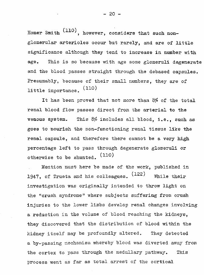
- 20 -
Homer Smith however, considers that such non-glomerular arterioles occur but rarely, and are of little significance although they tend to increase in number with age. This is so because with age some glomeruli degenerate and the blood passes straight through the debased capsules. Presumably, because of their small numbers, they are of little importance.
It has been proved that not more than 8$ of the total renal blood flow passes direct from the arterial to the venous system. This 8fo includes all blood, i.e., such as goes to nourish the non-functioning renal tissue like the renal capsule, and therefore there cannot be a very high percentage left to pass through degenerate glomeruli or otherwise to be shunted.
Mention must here be made of the work, published in(122)1947, of Trueta and his colleagues. v 7 While their
investigation was originally intended to throw light on the "crush syndrome" where subjects suffering from crush injuries to the lower limbs develop renal changes involving a reduction in the volume of blood reaching the kidneys, they discovered that the distribution of blood within the kidney itself may be profoundly altered. They detected a by-passing mechanism whereby blood was diverted away from the cortex to pass through the medullary pathway. This process went as far as total arrest of the cortical

- 21 -
circulation and the production of a complete shunting ofblood into the medullary circuit. It is noteworthy thatthe thin segments of the loop of Henle, which are concernedin the passive reabsorption of water, are supplied withblood from the juxta-medullary circuit.
Richards and Schmidt demonstrated that glomerularintermittence occurs in amphibians. Trueta believes thatit may also occur in mammals. In this he has the support
(5 )of Hayman and Starr who showed it to be present inrabbits. It has also been shown that dehydration candecrease, and hydration increase, the number of active
(52)nephrons. w 7 However, this belief in glomerular intermittence has not gone unchallenged H9)^ an^ Smith has pointed out that, almost alone among mammals, the rabbit behaves as do amphibians, so that findings applicable to the rabbit need not hold for all mammals.
Trueta and his colleagues believe that emotion plays a large part in determining the distribution of the renal blood flow, and that direct nervous control, and the action of hormones, or a combination of these two, are the agencies through which emotion acts. That emotion can produce an increase in the peripheral resistance and a rise in blood pressure, mediated through the nervous system and a conjectural humoral mechanism 33 > 47, 112, 94)^

- 22 -
can induce the posterior pituitary gland to release the(126)anti-diuretic hormone v , is well known. Emotion has
also been shown to be able to cause vasoconstriction of the glomerular arterioles, both afferent and efferent. (*^7) Thus there is some considerable support for Trueta*s views.
Trueta then cites G-oldblatt's experiment (46) where constriction of the renal arteries was shown to induce hypertension without at first causing impairment of renal function. He agrees with G-oldblatt in attributing this effect to renal ischaemia; but suggests that the essential anoxia of the cortex necessary for the production of the pressor substance which causes the hypertension, may be due to a diversion of blood to the medullary shunt. In support of his reasoning he observes that in many elderly kidneys, and those with Bright's disease, many juxta-medullary glomeruli show unusual features, generally considered to be degenerative. The reader will remember that I have already mentioned Homer Smith's comment on thissubject.
Trueta's assessment of the situation is that in the beginning diversion of blood into the medullary shunt causes no renal changes, but, if it is maintained, then one capillary in each affected juxta-medullary glomerulus becomes widely dilated. With the continuance of the shunt this
- 23 -
dilatation increases, until, in time the glomerulus, as a glomerulus, ceases to exist and blood flows straight through. (This is reminiscent of Smith's aglomerular nephrons. )Provided again that the shunting continues, this process is multiplied and more and more aglomerular nephrons are produced. This causes a reduction in the resistance to the blood flow through these areas, with consequently further deviation of blood into the shunt, and the development of relative ischaemia of the cortex. This tends to become permanent. A rise in the blood pressure follows in attempted compensation, but even this is not adequate to keep the cortex supplied with blood.
It is Trueta*s belief that this process will tend to occur in old age, or in disease where the renal vasculature architecture has been altered.
This work of Trueta's has provoked much comment, mostly(17, 62, 82, 57, 83, 101, 121) n (31)adverse. v J 9 ’ 9 * One paper w *
supporting Trueta's findings has itself been adverselycriticised by Clifford Wilson (Med. Ann. 1953). HomerSmith ^^"^has criticised Trueta in a more constructivemanner. He makes the objection that the rabbit (on whichanimal Trueta's work was performed) reacts differently fromman, and similarly to the frog, after administration ofwater. He notes that in the frog hydration is accompaniedby an increase in the glomerular filtration rate achieved

- 24 -
by increasing the number of fully active glomeruli, and helooks upon this fluctuation in the mass of active glomerulias a primitive method of regulating the urine flow, sincein the frog the thin segment of the loop of Henle is
✓
lacking, and with it the tubular reabsorption of water |induced by the antidiuretic hormone of the pituitary. This ! last mechanism is present only in mammals and in some birds, these being the only species capable of forming a concentrated urine. Since the rabbit, like the frog, reacts to hydration with an increased urine flow and an increased glomerular
jfiltration rate, he infers that in this animal the vasarecta and other glomerular by-passing mechanisms may be muchmore important than in other mammals, and therefore thatTrueta's work requires confirmation. I must mention,however, that the adverse comments to which I have alreadymade reference are all more recent than Smith's remarks.
On the other hand, a great deal of work has been donerecently on the effect of congestive and other types ofheart failure on renal function, and an indirect conclusion
(2Q)of a negative nature was made by Davies and Kilpatrick ' to the effect that, while their work did not support the existence of Trueta's shunts, it did not specifically exclude them.
The question, then, of the existence of intra-renal shunting is still sub judice. |

- 25 -
THE INNERVATION OF THE KIDNEY. ^110'
There is a rich sympathetic vasoconstrictor supply, derived from the fourth dorsal to the fourth lumbar segments. The fibres pass through the splanchnic and abdominal ganglia to the renal plexus. They then run from the aortic plexus along the renal artery and into the kidney with the renal vessels to terminate around the afferent and efferent glomerular arterioles. Some nerve fibres penetrate the basement membrane to end adjacent to the tubular cells, especially in the proximal segment, while others end among the juxtaglomerular cells in the parietal layer of Bowman's capsule and in the perivascular spaces of the capillary tuft. Neither sympathetic vasodilator nor vagal fibres have been demonstrated.
The sympathetic nerve supply acts only by modifying the local blood flow. It has no direct action on the nephron itself.
CHAPTER IV.
PHYSIOLOGY - THE SECRETION OP URINE.
It was their appreciation of the differences between the circulations of the two main parts of the nephron, and

- 26 -
between the lining membranes of the tubules and the membrane covering the glomeruli, that led Bowman in 1842 and Ludwig in 1844, to enunciate their separate theories of the secretion of urine.
Bowman suggested that the urine was formed by twoseparate processes. He believed that the glomerulifiltered water and salts, and this solution passed alongthe uriniferous tubules where, by the second process (ofsecretion) the various specific urinary constituents suchas urea, uric acid, etc., were added by active secretionon the part of the cells of the convoluted tubules.(^-4) (28)
On the other hand, it appeared to Ludwig that the wholeprocess of urine formation was one of filtration in theglomeruli, as a result of which all the constituents of theurine were already present in the glomerular fluid, whichwas, in essence, an ultra-filtrate of the plasma, and ofreabsorption of water and certain salts on the part of thetubules. This process of reabsorption was attributed byhim to the difference in protein content between thetubular urine on the one hand, and the tissue fluid andblood on the outside of the tubules on the other. (28, 114)
These two theories formed the basis of all the workdone on renal physiology for many years.
The vitally essential nature of the kidneys was shown(1 )in 1899, when Rose Bradford J demonstrated that, if about

- 27 -
three-quarters of the total kidney substance was removed, death would follow. If an appreciable amount of one kidney was excised, then there followed an increased secretion of a watery urine, without any accompanying increase in the total solids eliminated. When two-thirds of the total kidney substance was removed, there developed a permanent increase in the amount of urine of a dilute type, but no permanent increase in the amount of urea passed. He noted that the fragment of kidney which remained was capable of greatly increased excretion of urea, and of greatly increasing the volume of urine passed, but was incapable of concentrating the urine. An increase in the nitrogenous content of the blood and tissues was also noted to occur.
Bowman's theory has now been disproved; and Ludwig's, in its original form, is untenable because the difference in osmotic pressure between the blood plasma and its ultrafiltrate is too small to withdraw water from the filtrate in the tubules, sufficient to produce the concentration, even of urea, found in the urine. (-^4)
Heidenhain (^*4) considered that urine secretion occurred as Ludwig stated, but that in addition, the glomerular epithelium had a secretory function. There is no evidence to support this.
The present conception is of glomerular ultrafiltration

- 28 -
of the plasma producing a glomerular fluid which is simply- filtered plasma minus its protein content. This watery secretion passes down the tubules and is altered by the reabsorption of various substances, and addition by secretion, of others.
It is interesting to note that even the conception of the glomerular membrane as one which is impermeable to protein under normal conditions, is now being held in question. Walker (*^2)^ suggested that inmammals, the glomerular filtrate may not be entirely protein-free. Rather (-^2) noted in 1952 that the cells lining the proximal convoluted tubules were able to reabsorb protein from the glomerular filtrate. Meanwhile, Dock (^2) in 194-2, and Gilson (-^2) in 1949, labelled certain plasma protein with dye, and found that the dye became concentrated in the cells of the proximal convoluted tubules. They considered that this indicated that glomerular filtration and tubular reabsorption of protein must occur. Sellers and his colleagues confirmed these findings, usingrats, in 1954, and concluded that some 33f° of the circulating plasma proteins was filtered and reabsorbed daily.
GLOMERULAR FILTRATION.
About 1300 ml. of blood perfuse the kidneys per minute. (^O) in the glomeruli 120 to 130 ml. of fluid

- 29 -
per minute are filtered off from the 700 ml. of plasmaand passed into Bowmanfs capsule. 140) In otherwords, some 19$ of the total plasma water is filtered offin the glomeruli. » Hi) may -e mentioned in
(1 2 )passing that Mayrs and Watt v J as long ago as 1922, had estimated that 20-25$ of the total plasma water was filtered. An early objection to the filtration-reabsorption theory of renal secretion of urine was the relatively enormous quantity of fluid which would require to be filtered and reabsorbed - of the order of 170 litres each day. However, this has now been proved to occur (H4)^ so that this is no longer a valid criticism of the theory.
If we accept the conception of glomerular function as being one of filtration, whence comes the energy required to fulfil this? Starling (H4) considered that the heart was the original source of this energy, which was exerted as the blood pressure within the glomerular capillaries.If then, we accept the theory that urine secretion occurs as simple ultrafiltration through the glomerular membrane, the energy for which is supplied by the blood pressure, certain criteria must be fulfilled. These are-1. that the fluid passing from the blood into Bowman*s
capsule must be exactly the same as plasma minus its protein, and
2. that the Blood pressure in the glomerular capillaries

- 30 -
must be sufficient to produce the requisite amount of filtrate, and this amount of filtrate must vary with the glomerular capillary pressure.
Let us now consider these points.
THE COMPOSITION 0E THE GLOMERULAR TRANSUDATE.
Richards and his co-workers 98, 114, 128, 129)were able to prove that in frogs and necturus the glomerular fluid was, in fact, a simple ultra-filtrate of plasma excluding protein. This they achieved by inserting a pipette into individual Bowman’s capsules, and-analysing the fluid withdrawn. Samson Wright (^-O) mentions that a few similar successful experiments have been carried out in rats and guinea-pigs. This evidence is absolute, but there have been other earlier experiments which pointed to the same conclusion.
(6)In 1914 Bainbridge and Evans v excised a dog’s kidney and included it in parallel with a heart-lung preparation. They were able to control the level of blobd pressure, the rate of blood flow, and the temperature within the preparation. This kidney was found to be able to secrete urine. Under the influence of diuretics, which provoked urinary secretion, they found that the gaseous metabolism of the kidney was increased. This gaseous metabolism,

- 31 -
however, was not affected by changes in the rate of blood flow through the kidney.
Ekehorn , also, claimed to have proved that glomerular function was pure filtration.
It is clear that, if the glomerular fluid is closelysimilar in composition to plasma minus its protein, itmust undergo profound changes during its transit of the tubules to alter it to bladder urine. Bayliss and Lundsgaard in 1932, sought to eliminate the functionof the tubules, and so to obtain an unchanged glomerularfluid. They used a heart-lung-kidney preparation similar to that of Bainbridge and Evans, and paralysed the function of the tubule cells by perfusing the preparation with blood to which minute traces of cyanide had been added. The result was that urine secretion continued, at a slightly higher rate, but the urine changed its character, almost but not quite,to that of an ultrafiltrate of plasma less its protein. As this fluid must have been filtered by the glomeruli, it can be inferred that filtration must be the main, and probably the only, function of the glomeruli.
It is of interest to note that the use of cyanide asa paralysant of tubular function had already been employedby Starling and Verney an^ -jy Eichholtz and Starling^^in 1925. In the latter paper mention was made of the fact that cyanide increases the permeability of the

- 32 -
glomerular capillaries.It is noteworthy that, even in the normal kidney,
as was shown by Starling and Verney ra-fce 0fflow of the glomerular transudate along the tubules can vary the degree of change occurring in it. The more rapidly it flows, the less will be the alteration in its composition, and so the more nearly will it come to resemble the plasma. This extremely important finding will be referred to later. In the case of urea, the percentage of it present in the urine falls when the rate of urine flow rises; but the absolute amount of urea excreted rises with the rate of urine flow, because the glomeruli must simply filter off a certain proportion of the salts in each ml. of urine they produce. In the case of sodium chloride, however, the percentage present in the urine rises with the rate of urine flow, because of the lesser degree of reabsorption of sodium under such conditions.
FACTORS AFFECTING- THE AMOUNT OF GLOMERULAR FILTRATE.
If urine is formed by filtration, then any factor which increases the rate of blood flow through the kidneys and the pressure within the glomerular capillaries, will increase the rate of urine formation, and, conversely, any factor which impedes the flow of blood into the glomerular
- 33 -
capillaries, or which raises the intra-capsular pressure, will decrease the rate of formation of urine.
These factors may he annotated thus:-(A). Factors causing an increased pressure within the
glomerular capillaries, so inducing a greater rate of urine formation are:-___________________________(1) a rise in the arterial blood pressure.(2) division of the splanchnic nerves which causes
relaxation of the renal arterioles.(3) local readjustment of relative tonus of the
glomerular afferent and efferent arterioles in such a way that the efferent arterioles are constricted relative to the afferents.
(4) dilution of the blood.(B). Factors tending to cause a lessened secretion of urine
are:-____________________________________(1) the osmotic pressure of the plasma proteins.(2) factors causing a rise in the intracapsular
pressure which impedes the rate of filtration in the glomerulus:(a) a rise in ureter pressure, and(b) a rise in renal venous pressure.
(3) factors causing a reduced renal blood flow:-(a) vasospasm of the renal vessels, e.g.
following stimulation of the splanchnic

- 34 -
nerves, or the administration of adrenaline.(b) constriction of the glomerular afferent
arterioles relative to the efferent arterioles.
(c) vascular obliteration of the renal vessels,such as occurs, or may occur, with prolonged hypertensive disease, with atherosclerosis, or artificially as in the G-oldblatt experiment.
(d) chronic congestive (low-output) cardiacfailure.
(e) chronic high-output cardiac failure.
Let us now discuss these points.
The effective filtration pressure in the glomerulus is the resultant of the arterial blood pressure minus the sum of the colloid osmotic pressure of the plasma and the intzacapsular pressure (which is normally zero).^0’ ^ “4)It has been calculated that this effective filtration pressure must be at least 30 mm. of mercury for urine formation to occur. 115)
Consider first the level of the blood pressure. The anatomical arrangement of a short, wide afferent arteriole, followed by an extensive capillary bed leading into narrower efferent arterioles, is calculated to produce a high level

- 35 -
of pressure within the glomerular capillaries. Winton (-*-38) in 1931, estimated that the glomerular capillary pressure was about 2/3 of the arterial blood pressure. This allows an adequate margin of power for the filtration of a protein- free glomerular transudate. Since the minimum effective filtration pressure has been found to be about 30 mm. of mercury, then, if the blood pressure is lowered, urine secretion should cease when it falls to about 30 - 40 mm. of mercury. That this, in fact, does occur was shown by Verney and Starling They also noted that dilutionof the plasma with normal saline, so lowering the protein content, allowed filtration to continue when the blood pressure was as low as 18 mm. of mercury. Verney (-^5)^ Starling and Verney (-^7) an( Rivards and Plant (^-4) demonstrated also that the amount of urine secreted varied directly with the level of the blood pressure, and did not depend on the amount of blood flowing through the kidney.This finding is only true in p a r t / ^ - The rate of the renal blood flow must, and does, have a considerable effect on the rate of secretion of urine? ^3, 29, 36, 41, 47,48, 74, 75, 80, 116, 122, 124, 125, 132, 133, 134, 135, 141,*42) -{-q kg admitted that this effect is mediatedthrough the pressure in the glomerular capillaries.
However, as I have mentioned, the glomerular capillary pressure can be altered by means other than variation in the

FILTRATIONPRESSURE
F l 0 • 1 2 . F iltctin f Pressure In t/ie Glomeruli as influenced by the. Relative Callbte of Apfe/e^f" ant/ Efferent' Cilomevulat Vessels.
I f tAe. ftffertnt" Vessel is conStrieted th e glom erular pressure is markedly dimm i shed. (a ) ‘ i f the e f fe re n t vessel is constric ted th e g lo m e ru la r p ressure >s raised, in the glom erulus [ B ') a n d falls Steeply on teach in g the tu b u la r c a p illa r ie s .
[A f te r Samson \A /n g h t $ Applied. P h y s io lo g y
O S M O T IC P R ESSU RE OF
P LASM A P R O TE IN S

- 36 -
arterial blood pressure. Relaxation of the renal blood vessels brought about by the division of the splanchnic nerves, causes an increased secretion of u r i n e . ^ 9 > 114) The converse also holds, that stimulation of the splanchnic nerves reduces the formation of urine by causing vasospasm of the renal vessels. (^8, 114) Adrenaline, in moderate amount, has exactly the same effect; if, however, it is used in minute amounts, adrenaline produces a different response. Richards and Plant (* -4) foun£ that when the perfused rabbit kidney in a kidney preparation was supplied with blood at a constant rate of flow, the administration of minute quantities of adrenaline produced a rise in the perfusion pressure in the glomeruli together with swelling of the kidney. This they interpreted as being due to preferential vasoconstriction of the glomerular efferent arterioles, with consequent distension of the glomerular and preglomerular vessels, and a rise in the filtration pressure. Winton (-*-39) in 1931 ? was able to confirm these findings using the heart-lung-kidney preparation, and in 1938 Chasis and his co-workers obtained the same results in normalman. Both Winton and Chasis interpreted their results in the same way as Richards and Plant, and drew the important conclusion that the control of the renal blood flow and of the glomerular filtration rate were vested in the tonus of the glomerular efferent arterioles. These points are

- 37 -
illustrated in Pig. 12.Such a local control of glomerular capillary pressure
had been foreseen by Ludwig as early as 1856, and by Starling in 1912, who put it in these words - "A dilatation of the afferent vessels, and a slight constriction of the efferent ! vessels, would cause a considerable rise of pressure in the glomerular capillaries, and a consequent increased transudation, without necessarily altering to any marked extent the total
j
circulation of blood through the whole organ. The changes in the afferent and efferent vessels are, however, beyond our | control or powers of observation, so that it is impossible to devise at the present time any crucial experiment which Imight decide the nature of the process occurring in the glomeruli.” (-^9)
That changes in the glomerular filtration rate arerelated to the relative tonus of the afferent and efferentglomerular arterioles has also been shown more recently.(5, 1X1, 119)
Prom these various findings we are able to say that the glomerular capillary pressure is sufficient to allow filtration of plasma, and that an increase in glomerular capillary pressure will enhance the rate of glomerular filtration. According to Smith when all the glomeruliin both kidneys are taken into account, the calculated amount of plasma water which is filtered (taken as 19$) is well

- 38 -
above the maximal rate of urine excretion.And what of the factors which impede urine excretion?In the first place it is obvious that, since proteins
are present in the plasma on the blood side of the glomerular membrane, and not on the urinary side, and since the crystalloids are equally disposed on either side of the membrane, the osmotic pressure of the plasma proteins will tend to resist the process of filtration. No further comment need be made on this subject, except that Olbrich (1947) showed that the colloid osmotic pressure due to plasma proteins tended to be lowered in aged persons as compared with young, healthy subjects. (90)
Turning to the intracapsular pressure, we find that on ligation of the ureter, urine secretion continues for a short time then ceases altogether when the ureter pressure rises to a certain level, usually when the difference between arterial and urinary pressure is of the order of 40 - 50 mm. of mercury. (• -4) Winton (-^7) kas demonstrated a similar effect when the renal venous pressure is raised. However, such a rise in renal venous pressure produces two distinct and opposite effects, viz., firstly, a transmission of this venous pressure to the fluid in the distal portions of the tubules, so retarding the secretion of urine in the same way, and to the same extent, as does an equal pressure applied to the ureter; and secondly, a fraction of the

- 39 -
pressure in the veins is transmitted hack along the blood vessels to raise the glomerular capillary pressure, and so to increase the secretion of urine in the same way as does a small rise in the arterial pressure. Winton also showed that the renal blood flow is decreased more by an increase in venous pressure than by an equivalent decrease in arterial pressure, and further, that the values of venous pressure and ureter pressure, which, when applied one at a time, will reduce the urine flow equally, differ by an amount which is a measure of the increase in glomerular pressure consequent upon venous obstruction. He also noted that venous pressure has no appreciable influence on tubule function.
However, it matters nothing whether the secretion of urine is retarded by raising the ureter pressure, by raising venous pressure, or by lowering arterial pressure, because the changes in composition of the urine are exactly the same.
The other factors which cause a reduction in the renal blood flow - stimulation of the splanchnic nerves, adrenaline in more than minute amounts, a constriction of the afferent glomerular arterioles relative to the efferents, obliteration of the renal vascular bed, chronic low-output cardiac failure, and chronic high-output cardiac failure - also diminish the amount of blood flow to the kidneys, and so

- 40 -
impair the rate of secretion of urine.It can he said, therefore, that the original premiss
that glomerular function is simple ultra-filtration, the energy for which is supplied by the blood pressure, has been proved.
GLOMERULAR RESERVE.
Glomerular activity is intrinsically variable, though the degree of variation differs markedly in different species, as well as between adult and infant. (^--^ As it is obvious that the rate of urine flow varies from time to time, equally is it obvious that glomerular function can be increased either by making each glomerulus do more work, assuming that all glomeruli are functioning all the time, or by increasing the number of active glomeruli, assuming that under conditions of rest only a proportion of the glomeruli are functioning.
This problem does not appear to have been fully settled. Richards and Schmidt (^-4) definitelydemonstrated in the frog’s kidney that not all glomeruli were functioning at one time, and that therefore there was available a reserve of nephrons which could be called into action as required. In the rabbit, Hayman and Starr (53) found similar results, and Handley and his colleagues (52)

- 41 -
believed that they had obtained like results in dogs.However, when attempting to confirm these last results, Thompson, Barrett and Pitts (^9) came to the opposite conclusion, that all the glomeruli were always functioning, and that the degree of glomerular activity was varied by alterations in the relative tonus of the glomerular afferentand efferent arterioles. This view has been accepted by^ -4. (5, 111, 114)other writers. * * 7
NUTRITION: Nutrition, especially of oxygen, has to besupplied to the living cells of the glomerular membrane. Bayliss and Lundsgaard in their cyanide perfusionexperiment, found that urine which at first was protein-free, came to contain traces of protein after some minutes of perfusion with cyanide-treated blood. This is due to anoxia. In the same way, if the renal artery is occluded (^*4)^ secretion of urine ceases and remains so for some time after removal of the obstruction, and the first samples obtained after re-establishment of secretion contain protein. This, again, is caused by ischaemia and anoxia of the cells of the glomerular membrane. In like manner, protein may appear in the urine at all times when the glomeruli are damaged, as by ischaemia, anoxia, inflammation or infection of the kidney.

- 42 -
FUNCTIONS OF THE RENAL TUBULES.
It is obvious that the bladder urine is very different from the glomerular transudate, and the great changes which must have occurred to transform the latter into the former must have been brought about in the tubules. For a long time it was not known whether this was brought about by the addition (secretion), in the tubules, of a concentrated fluid, containing exactly the right proportions of the various urinary constituents, to a relatively small volume of a simple glomerular secretion, or by the reabsorption of varying amounts of the constituents of a very large volume of a glomerular transudate. In the latter case, the fully- formed urine represents the unabsorbed fraction of the glomerular transudate. The first of these views (Bowmanfs theory) is now known to be false, and' the second is the view currently accepted as representative of tubular function.
Although the primary function of the tubules is one of reabsorption, there is a certain amount of excretory activity also. In the original controversy as to whether tubular function was one of secretion or one of reabsorption, there was one incontrovertible fact which showed that some tubules, at least, were capable of secretion. This was the fact that there are at least twenty-five species of fish which possess no glomeruli and yet can produce a urine

- 43 -
essentially similar to that of fish possessing glomeruli. ^In these cases at any rate the tubules do have secretory
(71)powers. Marshall v ' describes one such case in the toadfish, and considers this state of affairs to be relatively common among marine teleosts. The probable explanation is that, since the glomerulus is of relatively recent development, the tubules originally possessed secretory powers, but in proportion as the glomeruli developed, these secretory powers diminished, and finally were lost. At the same time, the reabsorptive ability of the tubules became enhanced. Water, at any rate, cannot be excreted by the tubules, because if the glomeruli be cut off from the tubules by compression, no fluid can be jjcollected from the latter. An attempt, not verysuccessful, has been made to prove that certain substances \
are added to the glomerular transudate at certain points jalong the tubules. v ' This, however, cannot be accepted !as reliable evidence in favour of tubular secretion. j
|
It is clear that there must be a very considerablereabsorption of water by the tubules, because the volume !of the glomerular transudate is some 100 to 180 timesgreater than that of bladder urine. ^40) in mammals some I80 - 85io (about 150 litres/24 hours) of the filtered water is reabsorbed by the proximal tubules, while another, and

- 44 -
variable, quantity of water is reabsorbed in the distal tubules to produce the final concentrated bladder urine.(12, 110, 111) Bland men-fcions that there is agradient of urine flow along the nephron, the relative rates being as follows:-(a) glomerular filtration rate: 130 ml./min. or 180-200
litres in 24 hours, with Specific Gravity = 1010.(b) proximal tubule flow: 24 ml./min.(c) distal tubule flow: 15 ml./min.
Specific Gravity = 1010.(d) collecting tubule flow: 1 ml./min.
Specific Gravity= 1020-1032. Prom the urine in the lumina of the proximal tubules
are reabsorbed glucose, phosphates and bicarbonate, while chloride and sodium are reabsorbed simultaneously with water, and to such an extent that the urine in the proximal tubule remains isotonic with plasma. 12> 110t 111> 129)
The hydrogen ion concentration remains unchanged in the proximal tubule. Urea is reabsorbed in thetubules (110)? some 40fo ^124 to 57# 110 of it in the proximal tubules, and another fraction in the distal tubules. As has already been mentioned, Sellers believes thatprotein also is reabsorbed in the proximal tubules.
While these events occurring in the proximal tubules are physiologically invariable, other processes proceeding

- 45 -
in the distal tubules can be varied to suit the needs of the moment. ^12’ 110 ’ 1^0,
Here, in the distal tubules, the urine becomes more or less concentrated. Water and salt are reabsorbed or excreted according to the prevailing homeostatic requirements, and are probably under separate control. 96,11 A 1 Oil \
9 ' Potassium and sodium ions may be added, andammonia, synthesised by the distal tubule cells, will be excreted as required. 96, 110) Acidification of theurine is carried out in a sharply localised area of thedistal tubules.
(? 8)Cushny ' has classified the various solidconstituents of the urine into what he calls "threshold" and "no-threshold" substances. The former, threshold substances, are those which are of value to the body and which ought to be retained. They are only excreted if they are present in the blood in amounts so excessive as to exceed the threshold, or level, of their need, or occasionally, if the threshold is abnormally low, e.g. glucose in renal glycosuria. G-lucose, in fact, is an example of a threshold substance. "Uo-threshold" substances, on the other hand, are waste products and have to be eliminated. Urea, sulphate, creatinine and uric acid are examples.
Here, then, in the renal tubules, we find one of the vital body functions, one which regulates the water

content of the body, and maintains the acid-base equilibrium, j|
Let us consider these processes in greater detail. j
THE EXCRETION OF WATER. !
It must be appreciated that water equilibrium through- jout the body is continually being maintained at constant jlevels. Water is found in three body compartments, viz., intra-cellular water, extra-cellular, water and plasma water, and can, and does, move freely between these compartments jaccording to the prevailing electrolyte and osmotic conditions, t111)
Homer W. Smith 111) ^eiieyes that there are atleast two independent processes involved in the reabsorption of water. These are:-(1) Passive, or obligatory, water reabsorption which occurs j
in the proximal tubule and in the thin segment of !Henle's loop, and occasionally in the distal tubule; [and
(2) Active, or facultative, water reabsorption which takesplace in the distal tubules and possibly in the collecting ducts. I
j
Passive, or obligatory, water reabsorption.This process of water reabsorption in the proximal

- 47 -
tubule and thin segment is not physiologically controllable. It accounts for rather more than 80% of the water filtered . by the glomeruli. 9 110, 111) jn man ra -e 0^ u^neflow is related to the glomerular filtration rate which is usually about 130 ml./minute, and is relatively constant.(24, 48, 110, 111) Passive reabsorption of water occurs in association with reabsorption by the proximal tubules of sodium, chloride, bicarbonate, glucose, etc., at such a rate as to keep the osmotic pressures of tubular urine and plasma approximately equal. Since the reabsorptionof sodium, glucose, etc., in the proximal tubules, being an active process, will tend to leave the tubular urine hypotonic to blood, it may be presumed that so far as time permits, water will diffuse back from this hypotonic urine into the blood. This reabsorption of water, then is truly passive and is secondary to the reabsorption of other, osmotically active, substances. Further passive diffusion of water back to the blood occurs in the thin limb of the loop of Henle, whose function is considered to be that of promoting osmotic equilibrium between the tubular urine and the blood before the urine is delivered to the distal tubules. It has been shown that the proximal tubule cannot produce a concentrated hypertonic urine therefore if someosmotically active substance which is not reabsorbed entirely is added to the tubular urine, e.g. urea, it will

- 48 -
reduce the proximal reabsorption of water so that a greater load of water will be passed on to the distal tubule - an osmotic diuresis. H i ) j_s a p0in-fc 0f interestthat hypertonic urine is produced only in birds and mammals, which are the only vertebrates possessing a loop of Henlel^"^
Active, or facultative, water reabsorption.Since rather more than 80$ of the water filtered by
the glomeruli has already been reabsorbed in the proximaltubules, only 15-20$ is left to be dealt with by activereabsorption. 111) Unlike the passive process, thisone is physiologically controllable, and it is by variationin the amount of, water reabsorbed by this mechanism thatthe urine flow is adjusted to meet the normal requirementsof the body in respect of water and electrolytes, althoughit is probable that the controlling mechanism is different
(12 110 111)and independent for these substances. x 9 9 ' Activewater reabsorption is confined to the distal tubules and possibly the collecting ducts. (HO, 111) ig unciercontrol of the anti-diuretic hormone of the posterior pituitary. 110, 111) ^ reabsorption ofwater is in abeyance, the urine flow is large and the urine osmotically dilute because of the reabsorption of sodium and chloride in the distal tubules; but when facultative reabsorption of water is maximal, the urine flow is small,
- 49 -
and the urine concentrated.
The Anti-diuretic hormone (A.D.H.) of the posterior pituitary.
I have mentioned that active water reabsorption iscontrolled by the action of the anti-diuretic hormone ofthe posterior pituitary gland. let us trace the stepsby which this was proved.
In 1924 Starling and Verney foun(j that(pituitrin was able to prevent diuresis. Fee f in
1929, confirmed their findings. It was stated by Verney (1258»)J ' in 1929 that pituitary extract was capable of suspending the diuresis following the ingestion of water. Then, in 1930, Rioch called attention to the fact that after the ingestion of large quantities of water, there was a constant time-lag of the diuresis behind the changes occurring in the blood, and that these blood changes were inconstant, while, in contrast, the blood changes were constant following the ingestion of isotonic salt solution. He summarised his results following the ingestion of Locke's solution thus:-(1) there is a prolonged increase in the electrical
conductivity of serum;(2) there is a marked dilution of the total solids of
the blood;

- 50 -
(3) there is a moderate, delayed, diuresis.On the other hand, ingestion of water was followed by:-(1) marked diuresis starting 30-45 minutes after the
intake of water, reaching a peak at 90-120 minutes after ingestion of water then gradually declining to the resting rate after a total of 3 - 5 hours.
(2) slight dilution of serum electrolytes; the greatestdilution here precedes the maximum diuresis by 15-20 minutes.
(3) a variable degree of dilution of the total solids ofthe blood.
IHe believed that, since the ingestion of isotonic saline solution caused a slight increase in the electrical conductivity of the serum and a dilution of the whole blood, but only a moderate, delayed diuresis, or even no diuresis at all, the diuresis induced by ingestion of water must be due to factors other than the passage of fluid from the intestines to the blood stream, or than the dilution of whole blood or its colloidal constituents. He mentions that Priestley (1916, 1921) had found that the dilution of electrolytes of the blood was much greater than that of the total solids following ihgestion of water, and he (Rioch) suggests that the diuresis was due to the hypo- tonicity of the plasma. Because of the lag period, the

- 51 -
mechanism could not he one of simple filtration, but itcould be that the lag in time was due to the interval takenby the change to hypotonicity to affect the mechanism
(125a)regulating the posterior pituitary, which Verney v J 1 had already shown to be able to promote anti-diuresis. This
j.1 • j (110)is the view now accepted. '(l)Baldes and Smirke x,/ amplified Rioch's work in 1934.
They found that the excretion of a watery urine following the ingestion of water, was not caused by the presence of water then in the blood, but by a change in the kidneys, and that, until that change had been brought about, no diuresis could occur, despite the blood dilution. They noted that in water diuresis, water excretion was out of9 ilall proportion to the excretion of the other constituents ' !of the urine, so that the total osmotic pressure of the urine fell well below that of blood. This meant that a selective excretion of water had occurred, and that the kidneys had to perform work to do so. Since the glomeruli |are merely filters, this work must have been done by thetubules, and it must have taken some time for the process to get under way. In their view it was the change in osmotic pressure that was important, rather than the absolute level. They were able to show that when the change in osmotic pressure was made gradually the controlling mechanism adjusted itself to this lower level without calling

- 52 -
forth the normal response, and that, once it had achieved equilibrium at the new level, further demands on it were met only partially.
In the absence of water-retaining hormone (i.e. in diabetes insipidus), Findlay and White (40) in 1937, !noted that the urine flow was maximal, which is about 15-20$ of the glomerular filtrate and amounts to some 30 litres jper day. This represents the total amount of water left i
i
over after passive (obligatory) reabsorption has occurred |in the proximal tubules, and indicates that active j(facultative) reabsorption has failed to occur. The !diuresis which followed water ingestion was small in amount |and prolonged in time in subjects with diabetes insipidus. IFrom this they concluded that in normal subjects a "pitressin- i like" substance is present which allows the usual water Jdiuresis to occur, while in subjects with diabetes insipidus this substance is absent, so causing the abnormal response.
So far, then, it had been learned that pituitrin produced antidiuresis; that there was a time-lag before diuresis set in following water ingestion; that this diuresis was caused by a change in the functioning of the tubules; that the probable factor in bringing about this change was an alteration in the activity of a "pitressin-like" substance; that this change in the activity of the ’'pitressin-like" substance was most likely to be caused by

\- 53 -
variations in the osmotic pressure of the blood; and, finally, that in diabetes insipidus this "pitressin-like’’substance was lacking. Evidence was thus accumulating that the posterior-pituitary, through hormonal action, was able to influence the activity of the cells of the renal tubules andso to control water excretion.
(12)Bland v * mentions that Ranson had found that the posterior pituitary was controlled by the hypothalamic centres, and that injury to the hypothalamus, to the hypothalamic-pituitary tract, or to the pituitary gland itself, effectively suppressed the supply of water-retaining hormone with the result that diabetes insipidus ensued.
While the evidence in favour of control by a pituitary hormone, produced in response to stimuli emanating from the hypothalamus, was overwhelming, and while it was known that there must be some mechanism present by which the hypothalamus received knowledge of the conditions prevailing in the body, it was not until 1947 that Verney (-^6) actually demonstrated the existence of osmoreceptors which were able to respond to the osmotic state of the plasma. These receptor bodies were found to lie somewhere in the vascular bed supplied by the internal carotid artery, a site which guarantees to them a relatively unvarying supply of blood, so necessary to their function. The osmoreceptors are linked with the neurohypophysis. Verney was able to show that the anti-diuretic !

- 54 -
hormone is released through two distinct agencies, viz., emotional stress, or an increase in osmotic pressure of the arterial hlood. The release of anti-diuretic hormone was affected by the state of discomposure of the animals in Verney!s experiments, and he considered this effect to be due directly to adrenaline. Sympathectomy facilitated the liberation of anti-diuretic hormone through the agency of emotional stress, but had no effect on its release through osmotic pressure changes. Verney demonstrated also that it took some time for the maximum release of anti-diuretic hormone to occur. The converse holds also, that after the ingestion of water, it takes some time for anti-diuretic activity to cease, so causing the delay time already noted in water diuresis. Smith puts the peak of waterdiuresis at 37 minutes after the ingestion of water.
The evidence is now complete that active water reabsorption is controlled by the anti-diuretic hormone of the posterior pituitary. The chain of events is that variations in the osmotic pressure of the arterial blood are perceived by the osmoreceptors which influence the hypothalamus to stimulate or inhibit the release of anti-diuretic hormone from the posterior pituitary. The anti-diuretic hormone acts directly on the cells of the distal renal tubules, and by its presence or absence, induces these cells to
- 55 -
reabsorb or reject water according to the needs of the moment. This activity is confined to that moiety of the glomerular filtrate remaining after passive water reabsorption has occurred, and amounts in all to some 15 - 20fo of the total fluid filtered by the glomeruli.
Physiologically, this mechanism responds to a state of actual or relative availability of excess water, when the plasma osmotic pressure tends to be lower, by reducing the amount of anti-diuretic hormone liberated by the posterior pituitary until none is produced at all. As a result, the distal tubules reject water (and probably reabsorb salt ^6, 134) an^ water diuresis occur .'*■ ,This is equivalent to a physiological diabetes insipidus. Conversely, water deprivation causes a rise in the plasma osmotic pressure, the osmoreceptors respond by inducing the hypothalamus to stimulate the posterior pituitary to secrete
t
more anti-diuretic hormone, under whose influence the distal tubules reabsorb water and produce a more concentrated urine. 110, 111) Bland states that under thoseconditions salt is probably rejected.
In this way the kidney is able to retain or to reject water according to the homeostatic needs of the moment.
There is some evidence that the plasma volume alsoaffects the amount of anti-diuretic activity. (^*6) Leaf
(6*7)and Mamby (1951) found that in conditions where

F i g u r e 13.S o J lU f n — Active R cabsorp tton I Active R e&bsatph'on
i t d’ 1 MII |
j t t I ' | | |Ur ;i; L IvpotenL Hypoii«n~ M V _ . 1 T I J --------- =K_ Myperftpnic y*
I . \ Iw / V V
Vy y y
8 7 - 5 %
!r~ "K\, 1- 4
W ater ~ Passive Reatsorpbon
HyO
Act isc Reatsorpbon
.tAnK - Diuretic [J ormoi
z A nd
of glomerular filtra te .
Diagram a tic representation of reabsorption of Sodium and
of Water m the renal tubule. (M o d if ie d from Wesson, Anslow and S m i t h ) .
o j-a.ce p. S6.

- 56 -
extracellular fluid electrolyte depletion occurred, e.g. in Addison*s disease or in chronic congestive cardiac failure, the attempt to restore tonicity to normal levels is sacrificed in favour of water retention to restore volume. Also in 1951 > Welt and Orloff (^81) demonstrated that an uncomplicated expansion of plasma volume initiated a water diuresis, presumably by depressing posterior pituitary activity. Welt (1952) summarised theseand other findings, by stating that there was sufficient evidence to believe that the posterior pituitary may be stimulated by the level of* the plasma volume, although to a lesser degree, as well as by changes in plasma tonicity.
Nevertheless, the most important consideration in water excretion is the extent to which water is reabsorbed in the distal tubules, and this is controlled by the posterior pituitary, however it may be stimulated.
THE RENAL TREATMENT OP SODIUM.
It is believed by Wesson, Anslow and Smith ^184) that an approximately constant (about 7/8) fraction of the filtered sodium and water are reabsorbed in the proximal tubules, regardless of the filtration rate (Pig. 13). In the dog, when osmotic diuresis occurs, the fraction of water reabsorbed proximally is much reduced and large

- 57 -
quantities of fluid are delivered to the distal tubules. However, there is no commensurate reduction in the reabsorb- tion of sodium. Nevertheless, it must be realised that the filtrate reaching the distal tubules under these conditions, is all excreted. Under normal conditions, the urine delivered to the distal tubules is isosmotic with plasma, and an additional fraction of sodium, as well as of water, is reabsorbed distally (Pig. 13). They suggest that these two processes, the distal reabsorbtion of sodium and of water, are independently limited by their maximal rates of tubular transportation, just as it has been shown that there is a maximal tubular transportation rate for other similar transport systems. While the distal reabsorption of water is dependent on anti-diuretic activity, the excretion or reabsorption of sodium does not vary in the same way, indicating that these processes are independent.
The process of reabsorption and excretion of sodium is a vitally important one, and has significant repercussions. At all times, the sodium concentration of the plasma is regulated to a critical value through the supracortico- hypophyseal system by retention or rejection of water. ^84)So long as the filtered fraction of sodium remains constant, the load of sodium delivered to the distal tubule will vary as the filtration rate (184). There must be a state of exact sodium equilibrium at some critical glomerular

- 56 -
filtration rate. Should the filtration rate exceed this,too much sodium will reach the distal tubules and will be excreted; if sodium is excreted more rapidly than water, the sodium content of the plasma will fall, causing a reduction in plasma osmotic pressure* This, acting on the supracortico-hypophyseal system, leads to the distal rejection of water until the critical osmotic pressure of the plasma is reached. Conversely, if the glomerular filtration rate is reduced (as in chronic congestive cardiac failure), the distal load of sodium will be less than the critical level, and sodium reabsorption will be complete*If sodium is reabsorbed more than water, there will be a rise in plasma osmotic pressure, and distal reabsorption of water will follow in an attempt to restore the plasma osmotic pressure to its critical level. Such effects can be brought about by very slight changes in the glomerular filtration rate. It can thus be seen that a fall in the glomerular filtration rate tends to be followed by sodium retention and consequent water retention, with the production of oedema. This, in effect, is the mechanismdescribed by Merrill (1946) ^4) ±n kis ‘'forward failure"
/ j n y r gotheory, and has been confirmed by other writers/ 9 °9 ’116, 132, 133, 141, 142) j_s aqso an example of Smitl^s^"^glomerular-tubular imbalance, because it is clear that the fault lies with the glomeruli, the tubular function and

- 59 -
tubular mass being normal.
ACID-BASE BALANCE.
The process of secretion of some substances into the tubular filtrate, and of reabsorption back into the plasma water of others, is accomplished by osmosis.
The capacity of the tubular transport system for anyfiltrate component, whether this be a "shared" or "private"system, imposes a fundamental limitation on tubular function. (12) These systems are probably specific enzyme functions.Of these, the best known relates to glucose. If the enzyme phosphorylase is inhibited by phlorhizin, glucose fails to be reabsorbed, glycosuria follows, and "phlorhizin diabetes" is established. More recently, the enzyme carbonic anhydrase j has been inhibited in the cells of the distal tubules by the j drug 6063 (Diamox), a sulphonamide-type substance. The iresult of this is a suppression of hydrogen-ion formation (Eig. 14), so depressing the function of acidification of urine, and presumably stimulating secretion of potassium by the distal tubule cells.
The reabsorption of sodium and potassium in the tubules is partly under the control of the adrenal cortex. DOCA-like hormones are most potent in bringing about sodium retention and potassium excretion. Those hormones with

£ To face p. <50.

- 60 -
oxygen ions in the 11 and 17 positions are less active ini
influencing water and electrolyte balance. In adrenal insufficiency there occurs sodium excretion and potassium retention, while in adrenocortical hyperactivity the converse- sodium and chloride retention and potassium excretion -
(1 9 )holds true. v 'The maintenance of the acid-base balance, i.e., the
environment, of the body is in the hands of (l) the respiratory organs, and (2) the kidneys. ^6)
The respiratory organs keep the carbonic acid content constant within very narrow limits.
The kidneys maintain the acid-base balance in two ways:-(1) Stabilisation of concentration of bicarbonate.
This is a dual mechanism consisting of(a) the salvage of filtered bicarbonate, and(b) the return to the body of base used in
neutralising metabolic acid.For sheer magnitude the first of these
is the greater, for more than 1 lb. of sodium bicarbonate is reabsorbed by the tubules in 24 hours.
(2) Ion substitution.
Hydrogen or ammonium ions are substituted for sodium in the tubular

- 61 -
tubular urine, with, resultant reabsorption of sodium. Hydrogen is synthesised in the cells from carbon dioxide and carbonic anhydrase. Thus again base is conserved.
These processes are represented diagramatically in Fig. 14.
The bicarbonate which is recovered from the tubules is used over and over again, and the total usage represents as much as five times the bicarbonate concentration of the body as a whole. (96) - .
Such non-volatile acids as phosphates and sulphates are formed daily in large amounts from the metabolism of phospholipids and proteins. Their neutralisation for transportation is accomplished by the bicarbonate of the body base (Fig. 14). This, in time, would lead to the depletion of the body stores of bicarbonate unless prevented by two other tubular mechanisms, viz. the secretion of titratable acid and the secretion of ammonia. These processes reverse that of neutralisation and allow the excretion of acid minus the valuable base, which is returned to the body in the shape of bicarbonate. This can be represented in the reaction
Ha2HP04 + H2C03 ^ NaH2P04 + NaHCC>3(excreted) (reabsorbed)
The suppression of hydrogen ion formation by inhibitionkit
- 62 -
of carbonic anhydrase in the distal tubule cells, so reducing the ability to acidify urine (Fig. 14), leads to increased secretion of potassium ions into the tubular urine. It has been shown that secretion of potassium ion by the distal tubules is necessary to account quantitatively for the potassium ion excretion during rapid administration of potassium, and that this secretory process of potassium ion loss is one of ion exchange - sodium ions from the tubular lumen being exchanged for potassium ions from within the distal tubule cell (Pig. 14).
The normal process of secretion of hydrogen ions duringthe acidification of urine is brought about by the interactionof metabolite, oxygen and carbon dioxide under the influenceof carbonic anhydrase in the distal tubule cell (Fig. 14).There, free hydrogen ions are available for the process ofion exchange, and replace sodium ions in the urine in thedistal tubules; the displaced sodium ions pass into thetubule cell and combine with bicarbonate ions to producesodium bicarbonate. Thus, during the normal process of
(12)acidification of urine, sodium is conserved. v J
The fact that, when hydrogen ion formation is suppressed, there is an increased loss of potassium ions, suggests that potassium excretion may normally, at least in part, be carried out by ion exchange. In conditions of alkalosis potassium

FILTRATE
CLUJAMINE AMINO. AC(£>
Fig- 15.Illustrating the relationshipbetween the rate of ammonia etctetion and. the pH of the Urine.^ M o d i f ie d . fr-o n x P i tt s )
_b l oo d
o ja ce p. 63.

- 63 -
is lost, while in acidosis it is retained.It is of interest to note that the suppression of
carbonic anhydrase by Diamox is now being used to produce diuresis when this is required.
The kidney is limited in the amount of free strong acid that it can eliminate. Such acid must be neutralised, and this is achieved by combining it with ammonium ions, the displaced sodium being reabsorbed, (Pig. 1 5 ) ^ ^ . This may be shown by the reactionNa2S04 + 2(HH4)HCG3 (NH4)S04 + 2 NaECO-j
(excreted) (reabsorbed)The synthesis of ammonia is a function of the distal
tubule cell, and glutamine and other amino acids are its true precursors.(12,96,110) Titratable acid and ammonia secretion in the distal tubule restore base to the extracellular fluid, mole for mole, as b i c a r b o n a t e . A s the pH of the urine falls to acid levels, the rate of secretion of ammonia rises, indicating that a connection exists. The secretion of ammonia is best explained as a diffusion of ammonia into the acidified contents of the lumen of the distal tubule, where it combines with hydrogen producing ammonium ions. Thus the primary event in the excretion of ammonium ions is the secretion of free hydrogen ions, a process which, as we have seen, is dependent on the activity of carbonic anhydrase. Pig. 1 5 ^ ^ illustrates these points.

- 64 -
Put In words, sodium chloride enters the filtrate, and hydrogen ions are exchanged for sodium ions across the luminal margin* Thus one sodium ion is recovered, and one hydrogen and one chlorine ion remain in the filtrate. This acid is neutralised "by ammonium ions with the production of ammonium chloride for excretion in the urine. Since the increasing concentration of hydrogen ions in the tubular urine, if it contained only the salts of strong acids such as sodium chloride or sulphate, would eventually block any appreciable exchange of hydrogen for sodium ions, the secretion of ammonia ions into the urine becomes necessary to neutralise this acid by binding the hydrogen as ammonium ions, so allowing the exchange of hydrogen for base to continue. Each ammonia ion secreted into the urine binds one hydrogen ion, and allows one sodium ion to be reabsorbed. As I have mentioned, the pH of the urine must to some extent govern the rate of diffusion of free ammonia into the tubular u r i n e . T h i s is borne out by the fact that after administration of Diamox, which prevents the formation of free hydrogen ions, the formation and excretion of ammonium ion decreases.
Agreement is general that acidification of urine by thedistal tubule mechanism is a process of ion exchange rather than
(12)a direct secretion of acid.To summarise: on one side the distal tubule cell is

- 65 -
exposed to tubular urine, and on the other to tubular blood. Because of its own metabolism and of its continuous exposure to capillary blood, it has a constant source of supply of carbon dioxide. Within the cell is a liberal supply of carbonic anhydrase, so that it has no difficulty in forming the dissolved gas into carbonic acid. Dissociation of carbonic acid into hydrogen and bicarbonate ions allows the hydrogen ions to replace sodium ions in the tubular urine.The sodium, together with an equivalent quantity of bicarbonate, returns to the renal venous blood, so conserving both sodium and base. The hydrogen, with the remaining anions, is excreted into the tubular urine as titratable acid. This mechanism serves well when dealing with such weak buffer acids as phosphates, or with medium buffer acids such as B hydroxy- butyrates or aceto-acetates. However, in the case of strong acids, the titratable acid formed in the tubular lumen following the salving of base from sodium chloride and sulphate must be promptly neutralised by ammonia because of the strength of the acids (hydrochloric and sulphuric) formed.
Clinically, the acidosis of diabetic ketosis is due to a flooding of the body with such a load of metabolic acid that the kidneys cannot substitute hydrogen and ammonia ion quickly enough to prevent a depletion of base^^, and the acidosis of chronic nephritis is at least partly due to the inability of

- 66 -
the damaged tubule cells to effect the changes rapidly enough to compensate for a normal load of metabolic acid.^^’ ^
!In senescence it has been demonstrated that kidney function
deteriorates. Many studies have pointed to this. The jability to maintain the acid-base balance however, can be maintained in old age in spite of the impairment of renal function^^^. At the same time it must be pointed out that under conditions of stress, the delicate regulation of acid- base equilibrium is liable to be gravely upset in old age^1^ .
The Mechanism of Urea Excretion.Urea is a waste product of no value to the body. There
fore it must be excreted. This is carried out by the kidneys. Bradford^ (1899) demonstrated that urea increased in the blood when portions of kidney tissue were excised. Urea, in common with other filterable plasma constituents, is completely filtered off at the glomeruli; but the clearance rate for urea is only 30 - 60$ of the inulin clearance rate (rate of glomerular filtration), depending on the rate of urine flow. Therefore a certain amount of urea must disappear from the tubular urine during the transit of the tubules. Since there is no evidence for its destruction, it must have been reabsorbed^ . There is no evidence for the active reabsorption of urea in mammals, although it does occur in elasmobranch fishes, and it is probable that urea is reabsorbed
k

- 67 -
(2*5)by a passive processv This is likely to occur in consequence of a concentration gradient established by the reabsorption of water, and is a process of diffusion of urea back into the blood. Since urea is one of the most diffusible organic compounds known, this is not impossible.^25,48,110)
The concentration of urea in the urine is inversely proportional to the rate of urine 25*26,48,110,125a)^Thus, as the urine flow diminishes, the amount of urea reabsorbed increases, because there is more time for this to occur during the slower transit of the tubules; and conversely, the higher the rate of urine flow, the greater will be the clearance of urea^2'*, , 0,11°^. Smith^10 cites Dole as stating that, in man, 57$ of the filtered urea is reabsorbed in the proximal tubules, while Olbrich^0 and Van Slyke^12^ give this figure as 40$. The reabsorption of urea occurs in two distinct phases, accompanying water in its obligatory and facultative r e a b s o r p t i o n . 110 However, since the capacityto reabsorb water is impaired by disease processes, the fraction of urea reabsorbed under these conditions also decreases, because its reabsorption is purely passive and secondary to that of water.^2^’110^
The elevation of blood urea occurring in nephritis is due to decreased filtration along with actually decreased reabsorption. 2^ This latter is due to the impaired ability

- 68 -
to reabsorb water, and with it urea, obtaining in nephritis.We have mentioned that the proximal tubules cannot elaborate a concentrated urine; that being so, the presence of any unreabsorbed substance such as urea in the tubules, will, by its osmotic pressure, further reduce the proximal reabsorption of water, and will therefore increase the load passed on to the distal tubules. Under disease conditions involving a lowered glomerular filtration rate, there will be, as I have said, less urea, in common with other ingredients, filtered out of the b l o o d . i n -the tubules two separate processes may occur, viz. (l) reduced ability to reabsorb water, and therefore reduced re-diffusion of urea back into the blood, and (2) the slow urine flow will permit more time for reabsorption of water and therefore for back-diffusion of urea into the blood. The actual amounts of urea excreted or reabsorbed will depend on the algebraic sum of these factors; but, irrespective of this, the true amount of urea cleared from the blood under conditions of renal disease or renal functional impairment will be reduced, so that a tendency towards uraemia is established.

- 69 -
CHAPTER V
RENAL FUNCTIONAL MEASUREMENTS.
By using various substances and estimating their rates of excretion or reabsorption by the kidneys, and by employing various combinations of these results, it is possible to build up a picture of the various functions of the kidney.
1. The Rate of Glomerular Filtration.This is the amount of plasma filtered per unit time. It
is measured by using inulin and is expressed as Cjn .
Cjn = UY (quantity of inulin excreted per minute)(plasma concentration of inulin)
= 131 * 21.5 ml ./minuted485Inulin is a substance which is believed to be completely
filtered out by the glomeruli, but is neither reabsorbed norexcreted by the tubules. Therefore the measurement of itsexcretion divided by its concentration in the plasma, willgive a close estimate of the filtering capacity of the glomeruli.(Pig. 16).
It is of interest however, that Olbrich^^ has thuown doubt on the belief that inulin is neither reabsorbed nor excreted by the tubules. He considers that some 15# of thefiltered in is reabsorbed, and that under experimental

- 70 -
conditions the inulin clearance represents a nearly constant (85#) proportion of the glomerular filtration rate. Therefore, for purposes of comparison, when absolute figures are not so important, the inulin clearance may be accepted as at least an approximate measure of the glomerular filtration rate.
2. The Effective Renal Blood Flow.This is the amount of blood circulating through function
ing renal tissue. It is measured by using diodone (diodrast), and is expressed as C^.
Cj) » 697 ± 135*9 ml./ minute.Diodone, an organic iodine-containing substance has been
proved to be completely eliminated by the kidneys in the course of one complete circuit of the blood. Thus, it is filteredout by the glomeruli, and is also excreted by the tubules.The figure given by its excretion will therefore exceed that for inulin, and the diodone clearance thus measures the total renal blood flow (Fig. 16).
3. The Maximal Tubular Excretory Capacity.The efficiency of the tubular tissue can be estimated by
measuring the total urinary diodone when the plasma load has been increased to the level at which tubular cells excrete at
- 71 -
their maximal rate, and deducting the amount of diodonefiltered out hy the glomeruli. This last involves measuringthe inulin clearance which estimates the rate of glomerularfiltration. The resultant figure, expressed as T^, isrelated to the number of functioning tubules and their efficiency.It is regarded as an index of ’’tubular excretory mass”. Whileit is uncertain to what extent T„, for one substance willmapproximate to that for other unrelated substances, it can be said that a marked decrease in Tm represents a decrease in the number or efficiency of the renal tubules, or of both combined. (Pig. 16).
This figure for diodone is referred to as T^, where T ^ * 51*8 ± 8.73 mgm. iodine/minute.^*®^
4. Maximal Tubular Reabsorptive Capacity. i
;tThis is expressed also as Tm , and for glucose is T^.It is a measure of the reabsorptive capacity of these tubules which are supplied with glomerular filtrate. There is a maximal rate of reabsorption for any substance which is reabsorbed. (Fig. 16).
TmG = ± mgm* gluc<>se/min.^*®^
A number of ratios can be derived from the preceding measurements. These are:

F iltra tio n R a te .
( Inulin a t any plasma. Concentration).
( CJn)
v y
Renal Stood F law . M axim al Tubular l^laxima! Tubular. Bncretoty Capacity. feabsorpkiee Capacity.(KM+«t .tIm 1 — 1 — - = ---------plasma Concentration)- (l)fOt)Lta.st at ntyb (G,iucoSe a t ntyh
plasma Concentration), plasm a concentration').(c d) (7«d) (Tmt)
Fig. Schematic R epresentation of R en a l fu n c tio n a lM easurem ents. [ A f t e r (qoldrtny a n d C lu x iis).

- 72 -
5. The Filtration Fraction*This is the volume of glomerular filtrate formed from
each ml. of plasma perfusing the functioning nephrons. It is expressed as where
^In = glomerular filtration rate » 0.19 ± 0 . 0 2 ^ ^Cjj Effective renal plasma flow
6 CD , which expresses the ratio between the effectiverenal plasma flow and the maximal tubular excretory
mass, and indicates the volume of plasma which may be supposed to be cleared of a test substance per unit of functioning renal tissue.
It is given as 14 ± 2.16. (48)
7 CIn , which expresses the ratio between the glomerular filtration rate and the maximal tubular excretory
mass, and indicates the amount of glomerular filtrate per unit of functioning tubular tissue. It is given as 2.63 ± 0.34^*^
In Table I which follows, are given these various figures. (Prom Ooldring and Chasis^®)).
It will be noted that there are no significant sex differences in these figures.
In Pig. 16 (from Goldring and Chasis) is shown, diagrammatieally, the mechanisms involved in some of these measurements.

T A B L E I .Mean values of Renal Functional Measurements in normal subjects, by sex.
Males, Mean values.
Females, Mean values.
Rate glomerular filtration (Cjn ) ml./min. 131 1 21.5 117 ± 15.6Effective renal plasma flow (C-p) ml./min. 697 i 135.9 594 * 102.4(Maximal tubular excretory capacity ( (%£>) iodine/min. 51.8 1 8.73 42.6 £ 9.46(Maximal tubular reabsorptive capacity( (tm&) 215111 * Slucose/min* 375 i 79.7 303 ± 55.3Filtration Fraction (C-j-n/Cj ) 0.19 i 0.02 0.20 ± 0.03(Effective renal plasma flow ( ) ^Maximal tubular excretory capacity^T^ j
14 ± 2.16 14.2 ± 2.36
^Rate of glomerular filtration | ^In) (Maximal tubular excretory capacity(f^ )
2.63 ± 0*34 2.81 ± 0.56
(All values are corrected to 1.73 square metres.)
table I .
[To /ace. p. /'3.

- 73 -
8- Urea Concentration in Urine.This simple measure can give reliable information as to
the state of renal function. After ingesting a certain quantity of urea, the concentration of urea in the urine should rise to a percentage of at least 2.
9. Range of Urea Concentration in Urine.This elaborates the simple urea concentration measurement.
In it, the maximum and minimum concentrations of urea are recorded after ingestion of urea, followed later by water.The greater the range, the better the renal function.
10. Urea clearance.In this case the measurement gives the volume of blood
which, were it completely cleared of urea by the kidneys, would yield the amount of urea excreted in the urine in one minute•
11. Maximal Specific Gravity of Urine.Smith^110 declares that, at best, the specific gravity
is only a blunt index of the degree of concentrating ability of the kidneys, because the various solutes contribute so differently to the specific gravity. Addis and Shevsky ^ developed a concentration test and found that in young healthy

- 74 -
subjects the average maximum specific gravity was 1032 + 2.81 in a short period sample collected after 24 hours abstinence from water or liquid food. All their measurements were above 1026, and in 95$ of their subjects on ordinary diets, the specific gravity was 1028 or above. A maximum specific gravity, therefore, under the conditions of the test of Addis and Shevsky, of below 1026 may be considered abnormal. Smith^^*^ reports that Lashmet and Newburg made a similar study, and found that all normal subjects were able to concentrate their urine to 1026 or above. They found that 24-hour samples from ten normal subjects, living under moderate climatic conditions and with no dietary restrictions, gave specific gravity figures ranging between 1007.9 and 1026.5 because of the variable water intake, but after water deprivation for 12 hours, the figures rose to between 1028 and 1033.
A further point is that the specific gravity of acid urines, collected at random, is higher than that of alkaline urines.
12. Range of Specific gravity of Urine.Like the range of urea concentration, this measurement
gives an indication of the flexibility of renal function.The subject is first made to concentrate his urine by undergoing

- 75 -
a period of fluid deprivation, and then to dilute it by drinking a large volume of fluid. The concentrated specimen should have a specific gravity of 1025 or more, and the dilute one, a specific gravity of not more than 1005. The greater the difference between maximum and minimum \siues, the better is the renal function; and deterioration of renal function is indicated by a narrowing of the gap between the two readings, until in extreme cases of renal malfunction, the specific gravity tends to become fixed around 1010.
CHAPTER VI
CONSIDERATIONS OF RENAL FUNCTION IN SENESCENCE.
It can be accepted that the functional capacity of the kidneys is compromised in varying degree by the process of ageing.(12,30,7G’76,77,78,79,90,105,107,110). Smith(ll0) however, interprets the changes of senescence as meaning that the entire renal parenchyma, glomeruli and tubules, undergo senescent degeneration in a closely parallel manner, the renal plasma flow suffering a somewhat greater decrease because of arteriosclerosis or other vascular changes.
It is found that the renal circulation, glomerular filtration rate, effective renal plasma flow, tubular excretory

- 76 -
mass, tubular reabsorptive capacity, the ability to excrete urea, and to concentrate water, are usually all impaired, and the most important of these is the renal circulation. Indeed, the other aspects of renal function only fail after the circulation has become inadequate. The status of renal function can be linked directly with the efficiency of the cardio-vascular system in general, and with the renal circulation in particular, with the single exception of the case where ureteric resistance is raised, as for example when the prostate is enlarged.
Thus it can be seen that, while renal function is usuallyimpaired in old age, it need not inevitably be so. This has
(58 59)been shown in a study of autopsy material w ’ , when itwas found that grossly scarred kidneys occurred frequently below the age of 70 years, and that the kidneys of a fair percentage of more elderly subjects were healthy. A rough estimate was made of the numbers of healthy glomeruli, and it was considered that these did not necessarily diminish with age.
In the case of renal function, as in other systems, the earlier conception that age is inevitably accompanied by progressive degeneration must be amended, and it is necessary to appreciate that degenerative processes are not inevitable with age. In fact, there can be a complete absence of degeneration even in extreme old age. This,
- 77 -
This, however, is rather an ideal state of affairs, moreoften absent than not.
Howell and Piggot believe that 65 years of age is theupper limit of older maturity, and that thereafter
(59 )degeneration tends to become progressive. So far asarterial changes are concerned, it was found that they aremuch more marked after the age of 70 years. Howell and
(rq)Piggot concluded that the weak link in the renalsystem is the prostate and not the renal vessels, but this view is highly questionable.
RENAL CIRCULATION.
On the whole there is probably no great deterioration in the renal circulation. Degenerative change, however, undoubtedly exists in the majority of subjects, not only in the renal vessels, but throughout the body, so that the amount of blood reaching the kidneys is usually lessened in old a g e . ^ ^ The presence of hypertension, which willbe discussed later, modifies the picture, and produces further lessening of the kidney blood flow. Suffice it to say at present that arteriosclerotic or other vascular changes in the renal circuit tend to occur in old age, and lead to senescent degeneration of the entire renal

- 78 -
parenchyma, glomeruli and tubules, in a similar manner.Smith cites Trueta as suggesting that in
elderly normal persons, the protracted or repeated juxta- medullary diversion of the circulation leads to canalisation of glomeruli by degenerative vascular changes with a consequent reduction in the available filtering surface and filtration rate. If this occurs in fact, the blood will not be cleared of waste products since the thin segments and distal tissues do not excrete, this being a function only of the proximal tissues. Trueta, however, does not insist that this diversion of circulation from cortex to medulla occurs frequently in the quiescent state, and admits ! that normally the greater part of the blood passes through the cortex. The reader is referred back to an earlier section for a fuller account of Trueta's work, but mention must again be made of the development of aglomerular nephrons which he postulates. Homer Smith's commenton this subject is that if aglomerular nephrons do, in fact, exist in the senescent kidney, they are not sufficiently numerous to make themselves evident in the various experiments which have been stimulated by Trueta's work.
THE GLOMERULAR FILTRATION HATE.
There have been made sufficient studies of the

glomerular filtration rate in the aged to show that itdeclines with increasing age. ^5*107, 110)
Davies and Shock (30) found that the glomerularfiltration rate fell by 46$ between the ages of 20 and 90
(79)years. Mitchell and Valk obtained very similarresults in men suffering from prostatic disease, whichoccurs, they aver, in 60$ of all male subjects over theage of 65 years in the U.S.A.
Olbrieh (^0) s-tUcLie(i renal function in aged hypertensivesubjects. As compared with healthy young adults, agedpersons with a normal diastolic blood pressure had areduction in both glomerular filtration rate and effectiverenal plasma flow of about 30$, indicating that theavailable glomeruli are functioning as well in the olderas in the younger persons. However, when aged subjectssuffering from diastolic hypertension were compared with\healthy young adults, it was found that, while the glomerular filtration rate was reduced by about 40$, the effective renal plasma flow was reduced by almost 60$. Hence these aged persons with a raised diastolic pressure were producing more glomerular filtrate per minute than would be expected from their effective renal plasma flow, and therefore their glomeruli must have been functioning with enhanced

- 80 -
efficiency. This finding brings to mind the older concept of the relative tonus of the glomerular afferent and efferent arterioles enunciated by Starling in 1912 and described in the section on Physiology.
It will be remembered that the glomerular filtration- rate depends on the resultant of
(1) mean arterial blood pressure,(2) colloid osmotic pressure of the plasma,
and (3) intracapsular pressure,where (1) is opposed
by (2) and (3).In 01brich*s cases it was assumed that, since there
was no evidence of obstruction to the urinary flow, the intracapsular pressure was substantially unaltered from its normal zero. In his cases the mean arterial pressure was raised. The colloid osmotic pressure due to plasma proteins had already been shown to be lower in old persons than in young (Olbrich, 1947).^^ I*1 these cases then,while the arterial pressure changes were undoubtedly real, those in the plasma colloid osmotic pressure were not statistically significant (90) arl(j un( er those circumstances an increase in glomerular filtration would be expected.But instead of an increase, a decrease was actually found, and it became obvious that some other factors were playing

- 81 -
a dominant role.Smith (^-0) considers that glomerular filtration is
regulated by the interplay of tonus between the afferent and efferent glomerular arterioles (already mentioned (24,.114,159) and that this interplay is, within limits, independent of the mean arterial blood pressure. If then, the afferent arterioles are constricted, other things being equal, less plasma will reach the glomeruli in unit time, and less filtrate will be produced. This may be supposed to account for the proportionate reduction in diodone and inulin clearances (effective renal plasma flow and glomerular filtration rate) observed in aged subjects with normal diastolic blood pressure. When, however, the efferent arterioles also are constricted, then, other things being equal, plasma is "trapped" in the glomerular capillaries, and the amount of filtrate per unit time is increased. In aged persons with a high diastolic pressure, the afferent constriction reduces filtration, and the efferent constriction tends to increase filtration, so that the actual filtration rate found is the resultant of these two opposite trends.Thus such subjects have a filtration rate less than normal younger subjects and less than aged persons with a normal diastolic blood pressure, but greater than would be expected on the basis of their effective renal plasma flow alone.

- 82 -i
This then, is a physiological attempt at compensation which is successful up to a point.
THE EFFECTIVE RENAL PLASMA PLOW.
This aspect of renal function also has been shown to decline with increasing age.^^’ 90, 105, 107,
Davies and Shock found a decline in the effectiverenal plasma flow amounting to 53$ between the ages of 20
(79)and 90 years. Mitchell and Valk as with the glomerularfiltration rate, found that the presence of prostatic diseasehad little effect on the reduction in effective renal plasmaflow. The findings of Olbrich (90) have already beenreferred to. He reported further that, while the differencebetween young subjects and aged ones with normal diastolicpressures was hardly significant statistically, the differencewas significant when the aged subjects had a raised diastolicblood pressure.
If it be accepted that diodone is cleared completelyfrom the plasma in one circuit through the kidneys, thenchanges in diodone clearance paralleling increasing ageand blood pressure changes may be interpreted as indicatingcorresponding changes in the effective renal plasma flow.Therefore the findings of Olbrich suggest that the blood pressure changes are the dominating factors, and that age

- 83 -
is of importance only in so far as it may be accompanied by these changes. The alterations in diodone clearance indicate quantitative changes in the effective renal plasma flow rather than qualitative changes in the renal response.
In the group of cases with normal diastolic pressure the peripheral arteriolar resistance was not increased.(^8) The raised systolic pressure only means an increased resistance in the pre-glomerular parts of the arterial tree stimulating an intact myocardium; in such cases a smaller amount of blood reaches the functioning kidney epithelium and therefore less diodone is excreted per minute.According to Smith this reduction in blood flow tothe kidneys is a late phenomenon; he considers that raised mean arterial pressure merely offsets increased arteriolar resistance so that a normal blood flow results to all the organs of the body. (This interpretation tends to overlap to the stage where diastolic, as well as systolic, pressure is raised, because when peripheral arteriolar resistance is increased, and only then, the diastolic blood pressure rises. (^8)) However, with the passage of time, a weak link develops somewhere, the exact site of which cannot be pre-determined, and whose location only becomes apparent when actual organic derangement becomes an overt fact.Smith thinks that the hypertensive subject is especially

- 84 -
susceptible, because of his enhanced arteriolar tonus, and possibly because of an inadequate vasopressor mechanism, to the consequences of hypertension under circumstances that block sympathetic vasomotor tone, or impair venous return to the heart.
In Olbrich's second group, where both systolic and diastolic pressures are raised, the arteriolar resistance must be increased, and still less blood reaches the functioning kidney tissue. This has been shown to occur also in hypertensive subjects who were not old. (^8) in both of Olbrich's groups there is a distal reduction in the flow of blood to the kidneys, but there is no evidence that the physiological mechanism of the kidney itself suffers any change in cpzality.
The narrowing of the vacular bed is caused by twofactors, viz. vasoconstriction and anatomical narrowing,and is reversible up to a point so long as organic change
(12)has not progressed too far. Bland mentions that therenal vacular resistance in aged subjects has been shown to be as capable of responding to vasodilators as in the case
(r77)of younger subjects. Meilman found similar resultsusing protoveratrine.
THE FILTRATION FRACTION.Olbrich and his co-workers (90) f0und that when the

- 85 -
diastolic pressure was low in their aged subjects, thefiltration fraction was the same as in healthy young controls.This is confirmation that, under those circumstances, thereduction in glomerular filtration rate is proportional tothat in effective renal plasma flow. When, however, thediastolic blood pressure was raised, they found that thefiltration fraction was, on average, higher than in the othersubjects, and they considered this difference to be highlysignificant. As has been mentioned already, this isprobably due to relatively greater vasoconstriction of theefferent, than of the afferent, glomerular arterioles.
The increased filtration fraction in old age has beenobserved by other workers, who also attributed it to efferent
(12 ^0 76 79 105glomerular arteriolar vasoconstriction. ’ ’ 9 *9 J9
107, 110)
THE TUBULAR EXCRETORY MASS.
Shook 005) firs-t demonstrated a reduction in tubular excretory mass in the elderly in 1946. Such reduction, however, must be considered in the light of the diminished glomerular filtration rate which we have already considered, and which means, of course, that in any case a smaller volume of filtrate will be delivered to the tubules. Davies and Shock in 1950, conclusively demonstrated a reduction in

- 86 -
the tubular excretory mass of 43.5$ between the ages of 20 and 90 years, and Mitchell and Valk ^^) foun^ closely similar results, even when prostatic enlargement was present. Other writers have quoted like results -^7, HO) ^those of Davies and Shock.
Olbrich (90)^ aqso j_n 1950, found that elderly subjects with a low diastolic pressure had a tubular excretory mass 30$ lower than young controls. This difference was of the same order as that which they found in the case of the effective renal plasma flow. They considered, however, that this was not a mere reflection of the changes which had produced a reduction in plasma flow, but that it represented either a decrease in the amount of functioning tubular tissue or a diminution in the efficiency of the tubular epithelium, or a combination of both. When their subjects had a raised diastolic pressure, they found the tubular excretory mass to be lower still. This, they thought, must indicate a change in the actual amount of functioning tubular tissue, or in tubular efficiency. (G-oldring and Chasis believe thatin hypertensive disease the initial reduction in tubular excretory mass is due to diminished tubular efficiency alone, and that later on in the disease, an actual reduction in the amount of functioning tubular tissue is superadded.)It might, however, be a result of defective oxygen supply

and supply of nutriments (which Goldring and Chasis suggested), and therefore may ultimately he due to reduced renal plasma flow or to the factors causing such reduced flow, hut not he a direct consequence of it.
Smith mentions that while the tuhular excretory j
mass is impaired in the earlier stages of hypertensive disease, j the tuhular reahsorptive capacity for glucose is more or less unaffected. He states that this reduction in tuhular jexcretory mass is the most characteristic effect of hypertension on renal function; and since hypertension is so common among elderly persons (60$ it must have animportant hearing on the findings obtained from an average
' sample of elderly people. jc,! i: i
THE TUBULAR EEABSORPTION OF SLUCOSE. :II!!:
This has heen shown to decline with increasing age. j(77)Miller, McDonald and Shock / found that this decline was
parallel to that of the glomerular filtration rate. They Nfound,also, that the decrease in the tuhular reahsorption )jcapacity for glucose, expressed as percentages, agreed almost |exactly with the rate of decrease in tuhular excretory capacity for diodone. The ratio of glomerular filtration rate to the maximal tuhular reahsorption for glucose did not

- 88 -
change significantly with increasing age, These findings indicate that the nephron - glomeruli and tubules - loses its efficiency as an overall unit.
As we have mentioned previously, the maximal tubular reabsorptive capacity for glucose is not impaired in the early stages of hypertensive vascular disease, while the tubular excretory mass is.
THE EXCRETION OP UHEA.
Lewis and Alving f0un(j -that the ability of thekidneys to clear the blood of urea declined with increasing age; the decrease continued uniformly until the age of 55 years, remained steady at that level until 65 years, then accelerated from 65 years to 75 years of age. In like manner they found that the urea content of the blood rose with increasing age. Again it was seen that between the ages of 53 and 73 years there was an arrest in this rise, only to accelerate from 73 years onwards.
Olbrich also commented on this subject, findinga tendency for urea clearance to diminish with rising age. And Powweather stated that he accepted a lower figurefor normal urea clearance in elderly subjects as compared with young, thus tacitly admitting that urea excretion was impaired in old age.

- 89 -
WATER REABSORPTION, AND SPECIFIC GRAVITY OP URINE.
We have already seen that tubular function is impaired in senescence. Since the reabsorption of water is one of the principal tubular functions, it is likely to be affected, and the specific gravity of the urine will provide a rough estimate of the effectiveness of tubular reabsorption of water. Such estimates, however, are reliable only under controlled conditions of reduced water intake, or with measured fluid intake following upon a period of water deprivation, as otherwise the specific gravity of the urine varies widely according to the variations in fluid intake.
As long ago as 1922, Addis and Shevsky established that under certain conditions the average maximum specific gravity of urine was 1032, with a standard deviation of ± 2.81. They gave the odds that any normal individual, under the conditions of their test, would yield a urine specific gravity as low as, or lower than, certain levels, as follows ______ Specific G-ravity______________________ Odds___________
1026 1 normal in 471025 1 " " 1151024 1 " " 3231023 1 " 11 10001022 1 ,f 11 5000
They were not, of course, dealing with elderly subjects,
- 90 -
but with healthy young men.In 1938, Lewis and Alving studied the effect of
age on the concentrating ability of the kidney in man. They used the method of Addis and Shevsky, and took as the lower limit for maximum specific gravity indicating adequate renal function in healthy young normals the figure of 1026. In their series of 38 normal individuals, ranging in age from 40 to 89 years, they found that the average maximal specific gravity at age 40 was 1030, and at age 90 was 1023. The volume of urine during the period of fluid restriction was similar in men of different ages, and was much the same as in the case of normal young males. Their findings indicated that the maximum specific gravity does not fall below a lower limit of 1026, as it is in young normals, until the age of 65 years. In 17 subjects aged 40 - 64 years, the specific gravity was below 1026 only once, but in 21 subjects over 65 years of age, it was below 1026 fourteen times. Variations, also, were more frequent in the elderly.
They do point out, however, that the lessened concentrating ability of the senescent kidney may be due to a decline in tubular reabsorption, or to changes in glomerular filtration, or to both.
Howell and Piggot found that the average maximumspecific gravity of the urine passed by Pensioners on the

- 91 -
morning following admission to the Royal Hospital in Chelsea ranged around 1016.8. This was without any prior preparations for assessment of urinary specific gravity.
Since the specific gravity of the urine depends mainly on the amount of water reabsorbed in the tubules, let us now turn to the question of tubular response to the anti-
( i~i Q \diuretic hormone. Miller and Shock w 'in 1953 found that in old age there was an impaired response to anti-diuretic hormone. While their work made it clear that the tubular end organs had decreased responsiveness to the anti-diuretic hormone, it did not exclude the possibility that in addition the posterior pituitary or the hypothalamic end organs in elderly subjects were less sensitive to changes in the osmotic state of the extracellular fluid, so causing a diminished output of the anti-diuretic hormone itself.
SUMMARY.
( 1 o')As Bland K ' points out, there is now abundant evidence that with increasing age there is a gradual decline in kidney function as indicated by a reduction in effective renal plasma flow, in glomerular filtration, in both excretory and reabsorptive tubular mass, in the ability to excrete urea, and in the ability to concentrate the urine.

- 92 -
From this one can predict that in the aged the power to regulate the acid - base equilibrium will be compromised.In fact, the aged can hold this balance in health, but the addition of physiological stress or disease can upset the mechanism completely. ^*^7) These old people have a narrower margin on which to regulate this balance, and the prime factor involved is the decline in renal function', principally brought about by vascular degeneration.

L A D IE S ’ G U IL D O F T H E
R O Y A L M E D IC A L B E N E V O L E N T F U N D
S E V E N Y E A R C O V E N A N T SA subscriber can sign a Seven Year Covenant, by which means
the Guild can recover Income Tax on the subscription paid. This increases the income of the Guild without further cost to the subscriber, provided that the subscriber (or her husband) pay?
Income Tax at the full standard rate. Covenants are for seven years, bu t cease at death if previous. There is no charge on the Estate at death. We ask for a BANKER’S ORDER to be completed at the same time. The necessary form is overleaf.
The am ount of the subscription should be entered on the Subscriber’s Income Tax return (or that of her husband if she herself does not make a return) under the heading of “ Annuities
and A nnual Payments ” as follows : “ DEED OF COVENANT ” with the Ladies’ Guild of the Royal Medical Benevolent Fund(NET) £ ...................................
Covenants from 10/6 upwards are accepted by the Guild.
F O R M O F B E Q U E S TThe Committee earnestly appeals to members to remember
the Guild by legacy and to interest their friends to do so.
The following wording should be incorporated in the W ill:—
“ I give to The Ladies’ Guild of the Royal Medical Benevolent
Fund for the general purposes thereof free of all death duties thesum of £ ..................................... and I declare that the receipt of theHon. Treasurer for the time being of the said Ladies’ Guild shall be a sufficient discharge to my Executors/Trustees who shall not be required to see to the application thereof.”
P.T.O

FORM OF COVENANTD e e d o f A g r e e m e n t N o .:
I (full name) ........................................................................................................................
of (address)...................... ......................................................................................................hereby covenant with the Ladies’ Guild of the Royal Medical Benevolent Fund (hereinafter called the Guild) that for a period of seven years from 'the date hereof or during my lifetime (whichever shall be the shorter Deriod) I will pay annually to the said Guild such a sum as after deductionof Income Tax will amount to .............................. ......r__„__ (sum paid to theGuild) the amount being paid out of my general fund of taxed income and I UNDERTAKE that I will together with each payment as aforesaid hand to the Guild signed by me a certificate of the deduction of Income Tax on such yearly payment in the proper form (this certificate will be supplied by the Guild). IN WITNESS thereof I have hereunto set my hand this...........................................day of...................................... one thousand nine hundredand fifty- (date on which this agreement is made out).
SIGNED, SEALED AND DELIVERED * .....................................BY THE ABOVE-NAMED (Sign between #
in th e p resence o fWitness
S ig n a tu re .................................... _.................
A d d r ess ...............................................................................
O ccu p a tio n .................................... .................. ..................N.B.—The Deed must be returned to Guild Headquarters within 30 days
of the date of signature.
B A N K E R ’S O R D E R
.....................19......T o ............................................................................................. (Bankers).
P lea se pay a t o n ce a n d o n th e sam e d ate in ea ch su cceed in gyear, u n til fu r th er in stru ction s, th e su m o f £ ..................... :.......... :....to th e a cco u n t o f th e L adies’ G uild of the R oyal M edical B enevolent F und w ith th e N a tio n a l P rovincia l B ank L im ited ,
291b, O xford Street, W .l , an d charge th e sam e to m y accou nt.
(Signature of Subscriber)
(Address) .........................
£LANGHAM, HERALD PR E SS, FARNHAM
2 d .
S t a m p

- 93 -
PART II.
i
CHAPTER VII,\
METHOD.
In pursuance of my objectives I have made use of two separate methods, viz. the Urea Concentration Test of McLean, and the Concentration and Dilution Test of Calvert. With two exceptions, the subjects investigated by the two methods were different.
A. THE UREA CONCENTRATION TEST OP McLEAN.
In my first series I made use of this method. It waschosen because it could be performed readily under clinical conditions, and because Boyd in his "Pathology of Internal Diseases" (Kimpton, 4th Edition, 1944) comments in the words of the author of this test that "no information of anyimportance was ever afforded by any other test that was notequally well indicated by this simple one."
In carrying out the test, I gave my subjects written instructions to abstain from fluid for twelve hours (this was done overnight), and then to drink a draught containing 15 grams of urea dissolved in 3 ounces of suitably flavoured

- 94 -water. The urine passed thereafter was collected at one, two and three hours, and was examined for urea by the hypobromite method. The hypobromite was prepared as required by mixing equal quantities of 40$ caustic soda and liquor bromi. The hypobromite so obtained was caused to react with 1 ml. urine in a Hind's modification of the Doremus ureometer. This instrument is graduated to fractions of a gram of urea to the cubic centimetre of urine, and to obtain the percentage of urea present in the urine it is only necessary to multiply the reading obtained by 100. My results have been recorded in this way as a percentage of urea.
Heitzmann .(54) s^a -es hypobromite reactiondepends upon the fact that when urea comes in contact with an alkaline sodium hypobromite solution, it is decomposed into nitrogen, carbon dioxide and water according to the equation
C0(NH2)2 + 3 NaBrO + 3NaBr + 2H20 + C02 + Ng.
The C02 is absorbed by the sodium hydroxide, while from the volume of nitrogen liberated, can be calculated the amount of urea present in the urine.
If the kidneys are healthy, the urine must contain at least 2$ of urea. This is the figure generally accepted.
The rationale of the test is as follows.(-^O)
- 95 -The ingested urea is rapidly absorbed from the intestine and the urea content of the blood rises. No change occurs in the renal blood flow or in the glomerular filtration rate.The total excretion of the urea in the urine rises slowly, reaches a peak, then slowly declines. The excess urea is all excreted in ten hours. Urea is a low-, or no-threshold substance, reabsorbed from the tubules only in part. Some associated diuresis usually occurs. In normal cases the - urea concentration in the urine will rise above 2$ at some stage in its excretion, and may even reach 4 or 4 .5$.
The excretion of urea depends upon several factors, and is the resultant of(1) the glomerular filtration rate,(2) the quantity of water reabsorbed by the tubules in both
the obligatory and facultative phases,(3) the quantity of urea reabsorbed passively along with
the water, and(4) the osmotic pressure of the urea remaining in the tubules.
Put at greater length, since the blood urea rises, thereis a larger absolute amount of urea filtered through the glomeruli in unit time. If this increased filtration is associated with decreased urea reabsorption in the tubules, large additional amounts of urea can be excreted. The associated diuresis is brought about by the increased osmotic

- 96 -
pressure of the tuhular urine caused by the higher concentration of urea therein. This interferes with the reabsorption of water, and consequently of urea. Thus the urea retains water in the tubules to facilitate its own excretion. And the level to which the urea concentrated in the tubules rises at its maximum, is a measure of the power of the tubules to reabsorb water against the osmotic pressure resistance engendered by the urea. At this point one recalls that the amount of urea reabsorbed is associated with rate of flow of the tubular urine - the slower or smaller the flow, the greater will be the reabsorption of urea by back-diffusion. <25- 26’ 41> 48- 110’ 124> 125a’ 125c)
It is to be noted that the maximum concentration of urea in the urine is independent of that of other salts in the urine. (28)
False readings due to excessive diuresis were guarded against by discarding any one specimen which amounted to more than 120 ml.
Each specimen was tested for albumen by the salicyl- sulphonic acid method, for sugar by the Climtest method, and microscopically. Bile was assayed by the iodine test only when it appeared on visual examination to be present. Heitzmann (54) s^a .es on theoretical grounds theurine should be free from albumen and sugar before the test

- 97 -
is carried out, this is not necessary in practice since the method does not yield results of such great accuracy as to he interfered with significantly by these substances. In my subjects, therefore, I have ignored the presence of albuminuria or glycosuria.
In recording my results I have used only the maximum concentration of urea attained by each subject.
B. THE CONCENTRATION AND DILUTION TEST OF CALVERT.
This test arises from the work of Volhard and Farr, Fishberg, and Addis and Shevsky, for further details of which the reader is referred to G-radwohl. (^9)
This method was chosen for the second series because it measures the flexibility of renal function, i.e. the power to concentrate urine after fluid deprivation., and to dilute it when fluid is present in abundance.
My subjects were given written instructions to reduce their fluid intake on the day when the test was to commence. Normal food intake was allowed, but no more than 3 fl. oz. of fluid at lunch-time, and 2 fl. oz. at tea-time, were permitted. Collection of urine specimens during the day was not required, and the bladder was to be emptied at 8 p.m. and the urine discarded. Immediately thereafter the subjects had to consume a heavy, dry meal containing a relatively

- 98 -
large amount of protein, such as bacon and egg, eggs, meat or fish. The test was now "on", and all urine passed thereafter had to be collected. The concentrated specimen (fluid deprivation period) included all urine passed between 8 p.m. and 7 a.m. the following morning, at which time the bladder -had to be emptied. This sample was added to the overnight urine, and made up the total "concentrated" output. It was labelled "Specimen A".
As soon as this was done, the subjects had to drink one pint of weak tea and one pint of water. They were allowed nothing else whatever, food or drink, until the test was completed. Urine had to be passed at 8 a.m., 9 a.m. and 10 a.m., and each of these specimens was collected separately, bottled separately, and labelled respectively Specimens Bl,B2 and B3.
All the specimens were examined on the same day as they were collected. Each concentrated specimen was tested for albumen, sugar and bile as in the first series, and subjected to microscopy. If glycosuria was present, the specimens had to be discarded, and dietary measures were instituted to attempt to clear the glycosuria. If this was successful, the test was repeated and carried through. After the preliminary assessment, the maximum and minimum specific gravities were measured and recorded. The urinometer used was checked against water for accuracy.

- 99 -
This test allows an assessment to he made of tubular capacity to reabsorb water. G-lomerular activity as such is not measured directly; but the effective renal plasma flow and the glomerular filtration rate, and therefore the factors influencing these functions, are prime agents in determining the amount, and rate of delivery, of filtrate supplied to the tubules. They also determine the rate of flow of the tubular urine, so controlling the length of time available for reabsorption of water and other constituents of the tubular urine.
The reader is referred back to the section on the Excretion of Water. It will be remembered that, in conditions of relative water deprivation, the tubules, under the influence of the anti-diuretic hormone of the pituitary, will reabsorb actively a maximal quantity (one-eighth of the glomerular filtrate) of water and so produce a concentrated urine. Conversely, when water is available in abundance, anti-diuretic activity is suppressed, so that the tubules reject water, and a copious, watery, dilute urine is produced. It must be remembered that this is a measure only of the active, or facultative, reabsorptive power, because passive, or obligatory, reabsorption is invariable, amounting to about 85$ of the total glomerular filtrate, irrespective of its actual volume. 111, 134)

- 100 -
At this point it is interesting to see what Belt says of renal function tests. He mentions that it is not always possible to correlate the various different tests.The same individual, examined by several methods at the same time, may give as many different results. However, the same test repeated at intervals gives results which can be correlated. He says that the earliest indication of renal dysfunction is given by the specific gravity test, when there is seen a tendency towards fixation of the urine specific gravity. Thus, this test is very sensitive in
■N
detecting slight renal damage, but it fails to distinguish between moderately advanced and terminal stages of renal disease. Indeed, the specific gravity remains constant, once it has become "fixed", while other tests can show further progressive renal degeneration.(48) urea clearancetest, Belt states that it has the widest range; but it is not sensitive to changes less advanced than are represented by a loss of 20$ of the functional capacity of the kidneys.At the same time, it does permit readings to as low as only 5$ of functional capacity.
Isbister in recommending the creatinine clearancetest, states that the maximum specific gravity test after dehydration, is a useful screening method.
- 101 -Goldring and Chasis (48) state that the ability of the kidneys to elaborate a dilute urine is directly related to the rate of water excretion. The concentrating power is lost before diluting power, and it is probable that the final failure in dilution is due to a marked decrease in the numbers of functioning nephrons. However, they say that the extra- renal factors which affect the rate of the excretion of water are so complex, that the significance of variations in the diluting power is difficult to interpret. Thus, in their view, the diluting power of the kidneys is of little aid in the evaluation of the renal functional status.
I have, therefore, placed more emphasis on theconcentrating power of the kidneys than on the dilutionfigures, but I have used the range of specific gravity inassessing my results.
I have subdivided the concentrating ability of my( 32)subjects according to the classification of Derow v
thus:-1. Adequate concentrating ability -
Specific Gravity of 1025 or above.2. Impaired concentrating ability -
Specific Gravity of 1024 - 1018 inclusive.3. Inadequate concentrating ability -
Specific Gravity of 1017 or below.

- 102 -Since the diluting ability cannot be so accurately
assessed I have demanded simply a dilution figure ofnot more than 1003 as an indication of adequate function.
CHAPTER VIII.
CHOICE OP SUBJECTS.
There was no deliberate attempt to select cases, although, since the aim was to investigate the renal function of elderly subjects there was probably some subconscious choice or rejection of particular individuals according to personal bias. The series was commenced some years ago, and at that time I drew cases from among those consulting me for some minor or chronic complaint, or who were being attended by me regularly. Some few cases, considered to be in reasonably good health, who were not being attended at the time, were drawn at random from the practice files. In certain younger subjects who were under treatment I wished to ascertain the renal function status, and these cases also were included in the series. They do, incidentally, provide material for comparison with their elders. The whole of this series was assessed by using the urea concentration test. By 1955 > when I began using the Specific Gravity test (the second series),

- 103 -
I drew more subjects from the files at random, but I did continue to make use of individuals who consulted me for some ailment or other.
The only subjects who were definitely excluded were those suffering from frank renal disease, excluding prostatism, or those known to be in the terminal stages of some illness. This is in marked contrast to the American investigators (e.g. Shock wil0 demanded that theirsubjects fulfil these criteria:1. be free from cardio-vascular or renal disease;2. be capable of showing a clinical history free from
past cardio-vascular or renal disease;3. have a blood-pressure less than ^ ^ / 90;4 . have no albumen, sugar, red cells, leucocytes or casts
in their urine;5. produce a urine of specific gravity greater than 1020
after water restriction for 14 hours;6. have no marked prostatic hypertrophy;7. could be easily catheterised;8 . have minimal peripheral sclerosis;
(7Q)It will be remembered that Mitchell and Valk broke awayfrom these criteria to the extent of investigating cases with prostatic hypertrophy, and that Olbrich (90) jj_n countrycorrelated the renal status with hypertension and vascular change. Furthermore, with the exception of Olbrich, they

- 104 -all eschewed females, in spite of the fact that Goldring and Chasis had already shown that there was no significantdifference between the sexes in renal function.
It is right and proper that such strict criteria be applied when initial attempts are being made to establish norms, but the results cannot be accepted as a true indication of the general renal status of the aged population as a whole. Therefore,in order to attempt such a survey, all persons have to be included in a review, and this I have essayed. In my own practice, the incidence of cardio-vascular disease is disproportionately large, or appears to be so, because this area is a favourite place of retirement for elderly persons. There is some difficulty in assessing the degree of peripheral sclerosis, and as Olbrich mentions, different observers will produce different assessments, but when all the observations are made by one person, then the results, be they right or wrong, are at least uniform.
Thus it was that I made up my mind to include as broad a cross-section of the elderly population as I could, in order to make the picture as representative as possible of the local population as a whole. One defect lies in the type of practice; this is a non-industrial area, and consequently there are few persons who come into the lower social groups.

- 105 -This, however, is unavoidable. With this reservation inmind, I feel that I can claim to have presented a picture of the renal status of a cross-section of the ageing members of the community.
I must mention that at the outset I had not intended to attempt a correlation between past illnesses of an infectious nature and renal status, and when later I tried to obtain this information, where it was lacking, I failed in some cases because the subjects were untraceable. Thus in the section relating to this correlation, the numbers of individuals assessed is smaller than in the whole series.The total number of persons investigated was 74 in the urea concentration series, and 78 in the Specific Gravity series.
Two subjects were examined by both methods, but at different times. (Cases No. 61 and 67).
One subject when initially examined was suffering from marked prostatic hypertrophy. He underwent prostatectomy, and his renal status was reassessed five months later, and again a further five months later. (Case No. 132A).
Another subject on initial examination had prostatic hypertrophy, but no untoward symptoms of prostatism. Nine months later he was reassessed when the prostatic enlargement had become very troublesome, and a deterioration in renal function was found. He underwent medical treatment, improved,

and seven weeks later his kidney efficiency was re-assayed, showing some improvement. (Case No. 144A.) •
The order of presentation of the cases in the First series is alphabetical according to surname, while in the Second series it is chronological according to the time of the investigation.
APPENDICES.In Appendix A I have listed all my cases, giving
personal details and condition of health and recording the results of the tests. Should a particular subject have died, the cause of death is shown, and its time after the investigation was carried out.
Appendix B lists the incidence of various pathological conditions xoresent among my subjects. One individual, of course, may have more than one disease condition.
At Appendix C I have given the subjects whom I deem to be, for all practical purposes, free from any pathological condition.
Appendix D shows the numbers of deaths which have occurred among the subjects of the investigation, and records these figures as percentages in various ways.
CHAPTER IX.
RESULTS.
METHOD OF PRESENTATION.
A uniform method has been employed throughout. The

- 107 -
data and the results pertaining to each heading are presented in tabular form. Each table is in essence double, the first part relating to the Urea Concentration series, the second to the Specific Gravity series.
The tables, within the subdivisions of the two series, are further subdivided horizontally into the headings relating to the subject under discussion, e.g. into males and females, and underneath these figures is given, for comparison, the averages reached by the whole series. (These last figures are actually those given in Table II.)
Each Table is then subdivided vertically into various headings. First is given the number of subjects concerned both as an absolute figure and as a percentage of the total.In the third column I show the average age in years of the subjects under consideration, and in the fourth column is noted the percentage of subjects who are male. The fifth column presents the percentage incidence of prostatism among the male subjects concerned. The following three columns show the average blood pressure, the percentage incidence of atherosclerotic change and the percentage incidence of impaired heart action, among the subjects under consideration. The final two columns, which give my results, are different for the two series; in the urea concentration series the penultimate column shows the percentage of subjects within

- 108 -the group who attained an adequate concentration of urea inthe urine, i.e. 2$ or more; the final column presents theactual average percentage concentration of urea reached bythe group. In the Specific Gravity series, the last twocolumns give the same information as in the Urea Concentrationseries, but it is further subdivided. The column giving thepercentage of adequacy of renal function is first dividedinto concentrating and diluting ability; concentratingpower is then shown, as percentages, under the subheadingsof (a) good function, headed "G", i.e. specific gravity of1025 or more; (b) impaired function, headed "I", i.e.specific gravity between 1024 and 1018, both inclusive; and(c) inadequate function, headed "B", i.e. specific gravity of
( 32)1017 or less; J diluting ability meanwhile, is shown simply as the percentage of cases capable of diluting to a specific gravity of not more than 1003. The final column, showing the average results attained by the group, gives the average maximum and minimum specific gravities, and the range of specific gravity.
Certain variations are, of course, necessary according to the requirements of the particular tables. Apart from these, uniformity is preserved.
Renal function is presented in relation to the following

- 109 -
factors:-
Age,Sex, ' . " \
Social grouping,The state of the heart,Atherosclerotic change,Arterial blood pressure,Previous streptococcal infection,Obesity,Sheldon's somatotypes,Consumption of Tobacco,
\
Consumption of Alcohol,Chronic Pulmonary Lesions,Albuminuria, and Prostatic enlargement.
1. OVERALL AVERAGE FINDINGS.
In Table II is presented the overall figures obtained in both series.
It can be seen that the average age in the Specific Gravity series is six years higher than in the Urea Concentration group. There is a higher proportion of males in the Specific Gravity series. The average level of the blood pressure is

T A B L E II.Presenting overall average findings
SeriesNo.of
Av.age
(years)*
males *prostatism
Av. B.P. (mm.Hg.)
# incidence of # adequacy of renal function
Averageresultscases athero
sclerosisimpairedheartaction
UreaConcentration. 74 64 47 9 165/91 43 58 80 2.75#
SpecificGravity.
d one ent rat i on? fruition
Av .max. & min.S.C
RangeG. I. Be ’ >
78 70 60 36 175/9l 45 50 35 51 14 27 1022.3/1007.4 14.9
* G = good concentrating ability. I m impaired " MB = inadequate M w
This annotation is used throughout where applicable.
table k .
£ To fact p. H O .

- 110 -comparatively high in both series, with the diastolic level the same in both, and with the systolic higher in theSpecific Gravity series. Perera (93) gives ideal resting
120blood pressure figures as /80 mm. of mercury, and while casual readings of about *^^/90 mm. Hg. may be accepted as normal, even a single diastolic reading above 90 mm. Hg. must be viewed with caution.(94) avers that once maturity isreached, the blood pressure levels do not rise unless in the presence of underlying organic change. (93) Homer Smith (-^^ quotes Master as stating that 50$ of the males, and 60$ of the females in a sample population of 14,849 persons aged 40 years and over were hypertensive, i.e. had a blood pressure over ^^/90; while in my series it can be seen in Table XIV that 66 - 70$ of my subjects were hypertensive in this sense - a proportion considerably higher than Master's.
In both my series I find an approximately similar incidence of atherosclerosis, i.e. 43$ and 45$* As this is a matter on which observers will differ, I do not propose to attempt comparison with other findings. My assessment is based on examination of the perceptible peripheral vessels, and of the retinal arteries (although Perera (94) expresses doubts on the value of this finding), and on the cardiac and locomotor response of the individual to normal exertion.

- Ill -
Gavey (45) insists that arteriosclerotic processes must not be considered "normal11 even in old age, although they occur with great frequency.
In considering cardiac action, I have used the processof simple clinical investigation, with its symptoms andsigns, and have found that 50 - 58$ of my subjects sufferfrom some degree of cardiac insufficiency. This figure isfairly high, but quite definitely gives a true indicationof the cardiac status in such a community as this. Furthermore,it agrees well with Smith who states that heart diseasein all its forms has caused 50$ of all adult deaths in
(88)America. Olbrich shov/ed that the cardiac index decreasedwith advancing years, thus indicating a diminished cardiac output; Shock (-^^) confirmed this finding of Olbrich, and reported that he had found a reduction in cardiac output of 35 - 40$ between the ages of 20 and 90 years.
The incidence of prostatism among the males of the two series reflects the difference in their average ages. In the Urea Concentration series this age is 62 years, and prostatism is present in only 20$; but in the Specific Gravity series, where the average male age is 72 years, the incidence of prostatic enlargement reaches the high figure of 60$ #

- 112 -RESULTS:
My results indicate that these older subjects have, on average, poorer renal function than young, healthy persons. They show, furthermore, that although the subjects in the two series were drawn from the same community, and although there are no marked cardio-vascular differences in the two groups into which the subjects are subdivided, the results obtained in the Urea Concentration series are distinctly superior to those of the Specific Gravity series. However, the fact that, while the average ages of all the subjects in the two series differ by six years, those of the males range from 62 years to 72 years, associated with widely varying degrees of prostatic enlargement, must have some influence in producing poorer renal functional measurements in the Specific Gravity series. I have shown in the Section on Prostatism that this condition is accompanied by a reduction in renal efficiency, and in particular by an impaired capacity to form a dilute urine. It is because of this limitation that the ability of the subjects in the Specific Gravity series to produce a dilute urine is so poor. That this is so is made clear in Table VI, where it can be seen that females are able to dilute to a specific gravity of 1004.7, and that the males can only achieve a dilution, on average, of 1009.2.

- 113 -
Nevertheless, my results also indicate that the Specific Gravity test is a much more delicate one than the Urea Concentration method, and is capable of detecting slighter degrees of renal functional impairment. In fact, if in the Specific Gravity series we summate the figures given for good and impaired concentrating power, we find that the total is 86$, which is not very different from the 80$ given in the Urea Concentration test for adequate renal efficiency. Thisshows that the latter test, while detecting absolutely insufficient renal powers, fails to differentiate between good and not-so-good function.
Insofar as diluting power is concerned, it is interesting to see how poor this is among these elderly subjects, and even more interesting to note how much of this weakness is due to the male moiety of the group, with their tendency to a raised ureter pressure stemming from prostatic obstruction. This poor diluting ability has the effect of narrowing the range of specific gravity.
Although these results show a decline in renal function as age advances, reference to Appendix A will reveal that there are many elderly persons with very good function, just as there are younger people with poor function.
At this point I wish to make reference to the two subjects in whom both tests were carried out. The first, Case No. 35,

- 114 -
is a highly intelligent woman who could be relied upon tocarry out her instructions. Yet, in the Urea Concentrationtest, she gave poor results - 1 . 5 while in the SpecificGravity test carried out some years later, she concentratedto 1027 and diluted to 1013 (which was not good). It istrue that she had a poorish cardiac action, poorer still atthe time of the Urea Concentration test when she was nursingher husband, and that the consequently reduced cardiac outputresulted in a low renal plasma flow and glomerular filtrationrate. (74, 116) This in turn caused a slower flow ofglomerular filtrate along the tubules. The ultimateelimination of urea, and its concentration in the urine, isthe resultant of several conflicting factors. Here thereduced renal plasma flow and glomerular filtration rate willproduce a slow flow of urine in the tubules, with relativelyreduced quantity of filtrate and therefore of urea, and will
(25)favour good reabsorption of water, and with it, urea,iyC-o-rrt-
b- so facilitating the production of a high urinary urea. But this has not occurred; therefore the ability of the tubules to reabsorb water against the osmotic pressure of the urea inthe tubular urine must be impaired, and the probable causefor this is the lessened power of the heart providing a diminished renal blood flow and therefore a relative scarcity of oxygen and other nutriments. The blood urea, which was

not estimated, would almost certainly have been found to be elevated. 26, 48, 110, 125a). However, some yearslater her cardiac compensation was much improved, and so when the specific gravity test was performed, some at least of the former defects were corrected so that she was able to produce a concentrated urine. However, she did not succeed in diluting the urine well, which implies continued anti-diuretic activity at a time when this should have been reduced. The explanation of the failure to suspend antidiuretic activity probably lies in the finding that where extra-cellular fluid electrolyte depletion occurs, as it tends to do in chronic congestive cardiac failure, water retention is favoured as against the attempt to restore normal plasma tonicity, with the result that a dilute urine fails to be produced. 130) also possible thatthe renal venous pressure was elevated, so producing some resistance to the outflow of urine in exactly the same way as a rise in ureteric pressure from prostatic enlargement.(-^7) This would be associated with some loss in diluting ability such as occurs in this instance. (See Section on Prostatic Enlargement.) The nett result here, then, suggests that there is a greater degree of renal functional impairment than is apparent from the concentrating ability; although it may be more proper to say that it is not the renal system which

- 116 -
is inadequate but rather the cardio-vascular system which imposes its own deficiencies on the kidneys.
The other case, No. 58, is more or less straightforward, with fairly good results in both urea and water concentration. Once again, diluting ability was not very good.
2. THE EFFECT OP INCREASING AGE ON RENAL FUNCTION.
In Table III I have present-ed my subjects subdivided according to age. Each subdivision represents a half-decade.
(a) UREA CONCENTRATION SERIES.
My results are tabulated in Table III and are represented graphically in Figure 17A. (Found a t inside o f rear cover.)
(i) Average concentration of urea in urine.There is a gradual decline in the ability to concentrate
urea as age rises. This fall is not, however, uniform. The group up to 55 years of age gives very good results, then the succeeding two groups show a progressive fall, to be followed by improvement in the next two; thereafter there is a marked fall in the degree of concentration to the point whi'ch would be anticipated had the earlier part of the graph continued without interruption, and this fall continues in the highest age group. This is shown in the graph at Fig. 17A, where

TABLE I I I
Presenting the effect of age on Renal Function*
Series* AgeGroup(years)
No.of3ases.
ioftotal. imale.
1°of prostatism.
Av. B . P . (m m .Hg *)
$ incidei atherosclerosis
ice of impaired, cardiac
action.
i adequacy of Renal Function#
Average Results.
Urea Cone. Upto55.
U 64 nil. 159/93 ■ T T ~ 35 85 ----- 377TWSpecific Gravity.
¥oneConcentration.p u r r ~ Av.max. &
min.S.G. Range.G • I. B. ution.- - - - - -
Urea Cone.56to60
9 12 -51H nil. 142/85 - 1 77 F Tition.
' ’ "2788#--------Specific
Gravity.3 4 33 nil. 159/86 33 nil.
Concentra Dil Av ,max.& min.S.G. Range.G. I . B. ution.
~“ J3 66 nil. 33 1023 . 5/ ^1003.6 20
Urea Cone. 61to65
“IB ^5 45 11 167/89 4 0 66 83 2.615*SpecificGravity. 14 18 64 28 171/96 36 71
Concentration, : rxp Av.max.imin.S,G. Range.G. I. B. ution.
28 64 8 43 1021.971007.7 14 .2
Urea Cone.66to70
13 ’ IT" ‘ T6 " ^ T ... ■' 1 7 5 7 5 5 " ' ' 69 '' ■ ........ 7 7 ............... 2.76#
SpecificGravity. 21 27 52 28 179/89 47 52
Cone;?entration. Dil Av.max.& min.S.G. Range.G • 1. B. ution,
33 52 15 24 1 0 22 .2 /1008 .7 . 13.5
Urea Cone. 13. _ 1 T
39 "1 5 " 171789 59 tzSpecific
Gravity.71to75 26 33 58 34 180/94 45 45
Cone;entration^ Dil Av.max.A min.S .G .
Range.tr • I . B. ution,39 46 15 ' 31 1 0 2 2 .8 /
1006.9 15.9
T A B L E TFT. ( c o n t i n u e d o v e r l e c f . )
[ To f a c e p. I l f
- 117 -
the unexpected improvement in urea concentrating ability at 66 - 75 years is clearly seen. My results bring to mind the findings of Lewis and Alving j havealready referred.
The relationship between these results and the individi percentage efficiency of the subjects, the incidence of cardiac impairment, the incidence of atherosclerosis, and
the level of arterial blood pressure can be seen by using inappropriate transparent overlay in Pig. 17A.(ii) Individual percentage efficiency of the subjects.
It can be seen that the graph of individual efficiency (pictured in green) corresponds roughly with that of average urea concentration attained in the different age groups, Broadly speaking, there is the same rise and fall, although the graphs do not follow one another exactly.(iii) Incidence of impairment in cardiac action.
In the case of the status of the heart (blue in the graph) it can be seen very clearly in the graph that up to the age of 80 years, renal efficiency is inversely related to the degree of impairment of cardiac function, and thereto is directly related to the effectiveness of the heart. Aft
the age of 80 years, renal efficiency drops abruptly, as doe
the incidence of cardiac insufficiency. This reduction in the frequency of impaired heart action may be fortuitous in

T A B L E I I I . (Continued) .
Series. No.of
Cases.*of pros
tatism.Av.B .P . (mm.Hg.)
__% incidence ofAgeGroup(years)
7° of
total , male.atherosclerosis
impaired . cardiac
action.1° adequacy of
Renal Function. k\rerage Results.tJrea Sonc. 5 7 20 20 180/97 60 130 60SpecificGravity.
76 Concentration.Dil- Av.max.to80
164/85G. 1. B, ution .& min.
S.G.tenge.
9 11 67 33 44 44 4 5 22 33 11 111007.3 L4.9
Urea Cone. 2 3 50 50 150/85 TOO w * 1.85%SpecificGravity.
Over Concentration.Dil- Av.max.80
178/87G. I . B. ution & min.
S.G.tenge.
5 7 100 100 60 40 20 80 nil. nil. 1050.8/ 1008.6 L2.2
Orea Cone. 74 - 47 § 1 8 5 /5 1 45 58 80SpecificGravity.
Total Conq entratior]l. Dil Av.max.Series.
175/910 • I. B, ution & 9in.
S.G.tenge.
78 60 36 45 50 15 51 14 27 107213/“1007.4 14.9
III
TABLE ill (continuation).
£ T o f a c e p . U S .

- 118 -my series, but may also be associated with the fact that since these individuals were able to survive to the age of 80 years, they probably had fairly sound hearts to begin with, while their contemporaries with poor hearts possibly died off before that age. This is suggested in the graph by the large increase in heart insufficiency in the 76 - 80 year group, followed by the drop after 80 years.(iv) Incidence of atherosclerosis.
This is shown in yellow. Once again, with some irregularity between the ages of 60 and 80 years, there is an inverse relation between atherosclerosis and renal efficiency It is noteworthy that the incidence of atherosclerosis rises with age.(v) Level of arterial blood pressure.
I have shown this as thin black lines in the graph. There is a similarity in the shape of the graphs for systolic blood pressure and for urea concentration. Both fall after the age °f 55 years, but after 60 years of age the systolic blood pressure begins to rise, and continues to do so until the age of 80. The concentration of urea continues to fall from 55 years to 65 years, but then begins to rise, possibly partly because of the increase in systolic pressure. However, after 75 years, kidney efficiency measured by the concentration of urea declines, again possibly because of the continued

- 119 -!
icontinued hypertension, which has probably both been caused ! by, and has aggravated, the amount of arterial degeneration which has occurred. Such arterial degeneration must leave jits mark on the renal vessels, and so will cause a deteriorationIin kidney function.
There does not appear to be any positive relation between the levels of diastolic blood pressure and the concentration of urea.(vi) Incidence of prostatism.
Reference to Table III shows that the incidence of prostatism tends to rise unevenly from 55 years of age to 80. The males in the 61 - 65 year group have 25$ prostatism as against 16$ in the 66 - 70 year group, with similar degrees j
of vascular and cardiac damage. Furthermore, the latter group has a higher blo-od pressure level than the former, and yet, in spite of all this, it is the 61 - 65 year group which show the poorer renal function. This is almost certainly idue to the higher incidence of prostatism. However, when we come to the 71 - 75 year group, with the males comprising 39$ of the total, and 40$ of these showing prostatism, we findthat renal function is good. Here, without doubt, the saving |!factor is the very low (38$) incidence of cardiac impairment, jwhich offsets the effect of prostatism and other factors. In jthe succeeding groups the incidence of prostatism is 100$, and I

- 120 -
this factor must be exerting a considerable influence in bringing about the progressive deterioration in renal function,(vii) Other relationships#
If we compare the graphs of the blood pressure (systolic) and of atherosclerosis, we find that up to the age of 80 years, there is a remarkable similarity in the trends shown; but after that age, in spite of a drastic increase in atherosclerosis, the systolic (and diastolic) pressure falls. Since, if we now superimpose the graph for impairment of cardiac action, and note that after this same age of 80 years the cardiac action improves, we must assume with Euguid thatthe atherosclerosis which is present cannot be accompanied by arteriolar narrowing, presumably because of either dilatation of the vessels, shrinkage of clot, or both together. In any case, it is clear that the peripheral resistance, as measured by the diastolic pressure, has become less.
Nevertheless, in spite of these favourable factors, renal efficiency declines abruptly after the age of 75 years, and is most probably due to simple senescent degeneration.
From these results one is led to conclude that the upper limit of older maturity is about 75 years, and not 65 years as thought by Howell and Piggot.^^(b) SPECIFIC GRAVITY SERIES.
The results obtained are tabulated in Table HI, and graphed in Figures 17B and 17C.

- 121 -
(i) Average concentrating power.In all groups these results are rather poor. Even the
youngest group (56 - 60 years) could only average a specific gravity of 1023.6, and hy the time the oldest group is reached the figure is only 1020.8. Reference to Fig. 17 will show how closely the trend of concentrating power for water approximates to that for urea. However, the whole graph is on a lower plane of efficiency, and indicates that the water concentration test detects slighter degrees of renal inefficiency than does the test of urea concentration.(ii) Average diluting power.
Here the youngest group gave quite good results, averaging a specific gravity of 1003.6; but immediately afterwards there is a deterioration in the ability to compose a dilute urine. This may be due to the appearance of prostatism atthis point. Once again, in Fig. 170, the trend of the graph is roughly similar to that of the concentrating powers for water and urea.(iii) Range of specific gravity.
Again the youngest group gave good results, with a range of 20. Following on this group, the others show a shrinkage in the range to a fairly constant plateau around 14.5, with a pronounced "dip" at the ages between 66 and 70 years.However, after 80 years of age there is an even greater

- 122 -
contraction in the specific gravity range, a contraction to be expected from all the preceding results. Here, again, the effect of prostatism can be seen in bringing about this shrinkage.(iv) The power of the individual to concentrate urine,
expressed as a percentage._______________________This is most clearly seen in the graph at Fig. 17B,
where it is shown in green.I have divided my subjects into those with good, impaired
and bad concentrating powers. 11 Good” is shown as a continuous green line, "impaired” as a dotted green line, and "bad” as a crossed continuous green line.
Fully adequate concentrating power can be seen to rise - as with urea concentration - between 56 and 80 years, after which point it declines.
Thoroughly inadequate concentrating ability is found to have a similar trend.
And the graph for impaired, but not wholly inadequate, concentrating ability is seen to be almost exactly the opposite of that for good, and for bad, concentrating power.As a result of these trends it appears that at extreme ages over 80 years, there are no subjects with completely inadequate, few with fully adequate, and very many with impaired concentrating ability.

- 123 -
(v) The power of the individual to dilute urine, expressedas a percentage. (Fig. 170.)___________________________
Here we find a gradual fall in efficiency as age advances, until at over the age of 80 years there are no subjects possessing adequate diluting powers, and all males show prostatism.(vi) Incidence of impairment of cardiac action. (Fig 17B
and 170.)_____________This is graphed in blue. The picture presented is
rather different from that found in the Urea Concentration series. Here we find a low figure at the ages of 56 - 60 years, but there is an abrupt rise in cardiac impairment, to 70$, by the age of 65 years. From this age onwards, there is a gradual lessening in the incidence of cardiac impairment.
From 56 to 80 years of age there is a direct relationship between cardiac and renal efficiency, not perhaps exact and constant, but nevertheless quite definite. After 80 years of age, however, this relationship is lost.(yii) Incidence of atherosclerosis.
Here again, as in the Urea Concentration series,- the incidence of atherosclerosis rises steadily with age, and at the same time renal efficiency deteriorates.(viii) Level of the arterial blood pressure.
As in the Urea Concentration series, a rise in systolic
- 124 -
blood pressure occurs between 56 and 75 years, a drop follows at the 7 6 - 8 0 year level, to be followed by a rise again after the age of 80. This graph trends in the same way as that for the average maximum specific gravity except at the youngest and oldest age levels. At the latter stage the systolic (and diastolic) pressure has risen along with the incidence of atherosclerosis, but there is associated with it a very slight lessening in cardiac insufficiency and a big drop in renal concentrating power. There is a rather similar correspondence between the systolic pressure and the diluting power of the kidneys. The diastolic pressure does not appear to show any definite relationship with the other factors, except the incidence of atherosclerosis.(ix) Incidence of prostatism.
Here again can be seen the effects of prostatic enlargement combined with those of a weak heart muscle. In the youngest group (56 - 60 years) there is no prostatism, and the cardiac power is good, and the average figures obtained are higher than for all the other groups. While, in individual concentrating ability, this group does not give outstanding results, they do show a complete absence of failure to concentrate adequately. The 61 - 65 year group, with 44% prostatism and 71% of weakened heart action, have poorer results than the succeeding (66 - 70 year) group who

- 125 -
>n, .
>ugir
■osig
' * * * ' ^ " 5 v t n i 'RT"'ti ■^'IKs tryt -Trvv c i i ! w i r * “r '• ' ' P /7W 3 rr* rp

T A B L E I V.
Illustrating the varying degrees of urine urea concentration achieved in different Age Groups, and the incidence of diastolic hypertension and other gross pathological conditions, and tabulating the average results attained by ‘'satisfactory” subjects who show no pathological defects.
Diastolic hypertension. Wo. ofcases. io
No. of Cases.
Urine Urea Cone, io
Other organic defects Satisfactory cases. Average results for
"Satisfactory” cases.No.(l) Up to 55 years.
5________ Over 3*53*78#
Below 2*0(2) 56 - 60 years.
2 Over 3*3
NoneBelow 2.0
65 years.Over 3*5
22
(4.) 66 - 70 years.2________ Over 3*3 2.8 io16
TABLE j V . (u o n tin u e d overleaf.)fover lea
C To face p ■ !ZS~-

have 55% of prostatism and only 52% of impaired cardiac action, and they, in turn, have poorer results than the 71 - 75 year group who show 58% prostatism and but 45% of poor cardiac action. This suggests that the power of the heart is of greater importance than the effect of prostatic enlargement, although it can be noted that a high incidence of prostatism is associated with poor diluting ability, and this becomes more pronounced as age rises. In the last two groups, although cardiac power is interpreted as fairly good, prostatism becomes well-marked, and senescence makes itself felt in further reduction of renal function. However, even in the 7 6 - 8 0 year group, with only 50% of prostatism and 44% of impaired cardiac action, a surprisingly high (45%) percentage of subjects reach fully adequate concentrating power, which indicates that some elderly people remain very efficient.(x) Other factors.
In this series the graph of the incidence of atherosclerosis is similar to that for both systolic and diastolic pressures, and over the age of 80 years, they all show a rise. This is in contradistinction to the findings for the over-80 group in the Urea Concentration series. The degree of cardiac impairment is noted to be fairly constant after the age of 70 years.
7e can therefore say that with a rising incidence of

T A B L E IV. (Continued)
No. of Cases.
Urine Urea Cone. $
Diastolic hypertension. Other organic defects. MSatisfactory” cases. Average results
for"SatisfactoryH cases.
No. of cases. * No. of
cases. $ No. *(5) 71 - 75 years.
301
61 38 3.04* |1|
1 Over 3*53 3.1 - 3.5 1 26 2 . 6 - 3.6 2 32 2.0 - 2.5 1 2 -1 Below 2.6 - 1 -
(6) 76 - 80 years.
60 80 Nil
[
No result, j*ii
None Over 3*51 3.1 - 3.5 1 I -1 2.6 - 3.0 - 1 -1 2.0 - 2.5 I — —2 Below 2.0 1 2 —
(7) Over 80 years.
Nil 50 50
-■ i . ■ —■ ■■ ---------
2.0*None Over 3*5None 3.1 - 3.5 - — —None 2.6 - 3.0 - — -1 2.6 - 2.5 1 - - I1 Below 2.0 1 — 1 —
TABLE IV ( c o n t in u e d . )
£ To face p. /Z6.

- 126
atherosclerosis(though not to such high levels as in the Urea Concentration series) and blood pressure, both systolic and diastolic, there goes a reduction in renal efficiency both on average and in the case of the individual subjects.Here we see that there is no evidence whatever of a reduction in peripheral resistance such as occurred in the Urea Concentration series, and yet the renal efficiency falls.There is, however, less cardiac impairment in the over 80 year group in the Specific Gravity series.
Thus, again, the same reduction in renal efficiency occurs after the age of 75 years, whether peripheral resistance drops or not, and once again it appears as though the age of 75 years represents the upper limit of elderly maturity.
Consideration of the data presented in Table III and 'in Pig. 17 brings out several points:1. Renal function deteriorates as age increases.2. This deterioration does not proceed uniformly.3. There are numbers of elderly subjects with surprisingly
efficient renal function, just as there are many younger subjects with unsatisfactory function.
4. Up to the age of 80 years there is a fairly constantdirect relationship between cardiac and renal efficiency.
5. Renal function deteriorates rather abruptly after theage of 75 years.

%90
60
10
6o
5b
4o
3o
20
lo
]
Fig. 18.
> Ur*A tone..
cL V 3•i >• ■Do-5lr! O In P ^ O <§^ 'O fr" t-- «*>■*2 i i I | I t;in Co Co q
Urea Concentration V> 'iflBMMF ° Dilitohc. Hypertension.
Satis fie lorry Ceses. % mmmmt % Or; Ante Defects.Representing Renal efficiency of 'silt's factory casesf dnct
the incidence o f o rgan ic defects a n d of hyper tension f
according to A e Croup. { U rea Cone. S e r ie s ) .

- 127 -
6. There is a consistent low lev.el of renalefficiency at the age group 61 - 70 years, and a consistent rise in efficiency at the 71 - 75 year level.
7. Prostatic enlargement tends to cause a deteriorationin renal efficiency.
This leads one to the inevitable conclusion that man’s, if average physiological efficiency is at a low ebb in the
> i ' i l a - - ---------------
t7l1/vJL r sixties, and that after 70 his general capacity for livingactually improves. This improvement persists until after 75 years, and does not show its final senescent decline until after 80 years of age.
It appears then that the critical period of life lies in the seventh decade, when there are still many persons alive who suffer from varying diseases and deficiencies, and who are not destined to survive beyond 70 years. It is because of the presence of such individuals that the average findings are low for this decade. After the age of 70, these "unhealthy” individuals have been largely eliminated, leaving a residuum of basically healthy and robust individuals whose good renal efficiency is now revealed in all its clarity, unobscured by the curtain of deficiencies of the preceding decade. The average results

L
%r aao7>8 %d?-s? %9-7 - P-E% /£ --C£7. 9 -£ - o -h7#-»r j.**o
7.7 A*7»S%o-7 - s-27. 9 7 - 0 -£% »£ --s £7.9£ - 0 -hy„*r *• %> > O
% 7 a'°j»p7.0-7 — -5-7% 9 7 - £>•£7. >-£ — -S*£%9-e - O b 7,7 7c 77.0-7 -_S-7 %9-7 - o-£ % / • £ - - $ • £
% 9 - E - <7-Vr
% 17 o% 7 AAO,agy.oz — -S-7%9 7- Of7. / £- j;-£%9£ - 0-77.7 O</&>
%? °/a9%o-7 --s-77.9 7- 0 £°4IZ - -S-£%9 £- 0-*7.77.7 A'°;ap7.0-7- sr7.9-r- ot7. / £- -s £% 9 £ — 0--/7
rtfj -lU-X.U u Tcf'9 7]°^ !CT
£*__
(li--tf‘ >— QV)
0>
?-o<n
V-cr-»
o~>c
cnc
j-SL
c rie (nc”9 U
=> O
1

obtained are now much improved, and this occurs in spite j/ yl P* \
of a raised incidence of prostatism. Gavey writesin similar vein on the subject of the heart.
In an attempt to demonstrate that there is in fact such a "dangerous period" in the life-span, I have broken down the figures presented in Table III in such a way as to show the varying degrees of urea concentration and water concentration attained in each half-decade, and the j
incidence within these periods of hypertension (as jrepresented by a diastolic blood pressure in excess of j
90 mm. Hg.), and other severe diseases, deficiencies or !disorders. My subjects are then classified into "satisfactory", i.e. without hypertension or other defects, and "unsatisfactory", i.e. the remainder. The "satisfactory" subjects are then investigated, and their average results
j
obtained; this allows an assessment to be made of the effect j
of age per se. The incidence of "unsatisfactory" subjects allows me to ascertain whether there is a 'dangerous period" with its associated reduced efficiency. The data relating to these findings are presented in Table IV and Pigs. 18 and 19 in the case of the Urea Concentration series, and in Table V and Pigs. 20 and 21 in the case of the Specific Gravity series.
Study of these various Tables and Figures reveals some

T A B L E V .
Illustrating the varying levels of concentration of urine achieved in different Age Groups, and the incidence of diastolic hypertension and other gross pathological conditions, and tabulating the average results attained by "satisfactory” subjects who show no pathological defects.
No of Cases.
Maximum Specific Diastolic hypertension. Organic defects. "Satisfactory" cases. Average results forGravity attained. No. of
Cases. * "No. of Cases. * No. of
Cases. * "Satisfactory"cases.
(1) 56 - 60 years.1 1025 and over none 1 none 1022/10Q52 1024 - 101b none nil “T " T - 33none 1017 and below none none none
{2) 61 -■ 65 years.4 1025 and over 1 2 2 1025/l003.69 1024 - 1018 5 50 8 . 78 1 211 1017 and below 1 1 none
i3) 66 - 70 years.1020/10057 1025 and over 4 5 1
11 1024 - 1018 3 38 “T " ... ' " 56 ”2" "" 193 1017 and below 1 2 1
14)71 - ^5 years.. .. 10 1025 and over 8 8 2 1023/1005
13 1024 - 1018 4. 54 “F ” ■ 73 5 273 1017 and below 2 3 none
ki) 76 -* b6 years.4 1025 and over none 1
66 333
1026/10062 1024 - 10Tb "" ' hShS- 1 “ 33 2 ~ none3 1UI7 ana oeiow _ '3'..... “3:.:.:: none - ____________ — --i
^6) Over bO years. i_ 1 1025 and over none 1
100none
nil no re suit. j4 1024 - 101b 2 40 4 " nunsnone 1017 and below none "TT0IH5"'" " r ntrne. _____1
T A B L E V .
£ To face p. IZ9.

- 129 -
interesting facts. Consider first the average results attained by these "satisfactory” cases. In the Urea Concentration series there is a decline in their renal efficiency as age advances, much the same as that found in the overall series, (Table III and Fig. 17A) although the results obtained are on a higher level. On the other hand, in the Specific Gravity series, the figures for maximum water concentration follow a most erratic course quite unlike those found in the overall series (Table III and Fig. 17B). The level for the 56- 60 year group is low, that for the 61 - 65 year group is good, then there is a precipitous fall at the age level 66- 70 years, only to be followed by an equally dramatic recovery to first class levels at 70 - 80 years. In the case of water dilution (cf. Table III and Fig. 17C), between the ages of 56 and 75 years, the diluting ability varies directly with the power to concentrate water . so that when the concentrating power is good, so also is diluting ability.The converse also holds. Thus the range of Specific Gravity is poor in the youngest age group, increases at 61 - 65 years, shrinks drastically at 66- 70 years, and increases again at 71 - 75 years. After 75 years of age, while the concentrating power shows an increase, diluting ability lessens, although the Specific Gravity range remains

/I'vefagfeMdxi’mum F i g . 2 0 .
V7 zV >-r*. S
AverageM im a io n iS.C,.
k 2
IOZI
0vO1 tov3I
0C--1O
toe>»IO001c2
Suto
RLO.
ion.m h . Mix. S.Q. cu m m % Di*stolU ItypetUns
mmm Ar. Mm. S .q . ■ ■ %, O r*w < Defect*.
f o S z t i s f a c t o r y Cases.
Representing Rem I efficiency of satisfactory cases, anJ the
incidence of organic defects dnd o f hypertension , according
to (\ge Croup.____ (Specifi c gravity Series^
- 130 - j:i1!
good. I cannot explain the erratic course followed in ithe Specific Gravity series, but it does suggest that the years between 66 and 70 are dangerous ones, when renal !function is at a low ebb, even in cases otherwise healthy.Here also is an illustration of the improvement in diluting figures when cases with prostatic enlargement are eliminated.
Turning now to the percentage of ”satisfactory” cases j
present in the various age groups, we find that in both |series the nadir is reached at the 66 - 70 year level, again (
1 !' !suggesting the dangerousness of this period of life.Consideration of the incidence of organic defects - jj
1 jwhich includes diastolic hypertension - shows that it j
reaches its zenith at 66 - 70 years in the Urea Concentration series; but this does not hold in the Specific Gravity group of subjects. As would be expected, in both series |the incidence of satisfactory cases varies inversely with j
that of organic defects.The graph for the incidence of diastolic hypertension
follows approximately that for organic defects in both series. In the Urea Concentration series it is inversely |,related to the renal efficiency, but in the Specific Gravity |;iji|series diastolic hypertension and renal function - both iconcentrating and diluting ability - appear to be directly related. While the findings for the Urea Concentration


- 131 -
series are as would be anticipated, those in the Specific Gravity group are not, and immediately bring to mind the work of Olbrich (90) on hypertension and renal efficiency, to which reference has already been made.
Let us now turn to the histograms at Figs. 19 and 21. In the Urea Concentration series (Fig. 19), it can be seen that there is a gradual trend towards lowering of the concentration of urea in the urine as age increases; the percentage of subjects giving inadequate function is specially low in the 71 - 75 year age group, and is high at the 56 - 60 year, and 66 - 70 year, level. In the Specific Gravity series (Fig. 21) the percentage of cases showing good concentrating powers actually tends to rise steadily from 61 - 80 years, as does the percentage of subjects yielding frankly inadequate concentrating ability. Conversely, impairment of concentrating power diminishes up to the age of 80 years. Reference to the levels for diluting ability will show that, apart from the subjects aged 76 years and over, the poorest diluting power occurs at the 66 - 70 year level.
These findings bear out my contention that there is a period during the second half of the seventh decade when physiological efficiency is at a low ebb. It is interesting to note that Richardson* found a similar tendency in his
* Richardson (1956) Scot.Red.Jour. l:12:3ol.
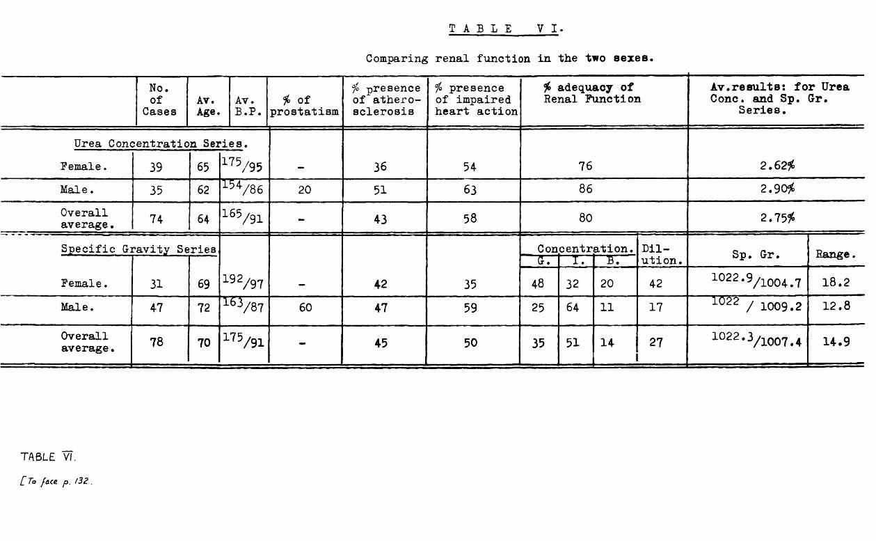
T A B L E V I .
Comparing renal function in the two sexes.
No.of
CasesAv.Age.
Av.B.P.
* of prostatism
# presence of atherosclerosis
# presence of impaired heart action
# adequacy of Renal Function
Av.results: for Urea Cone, and Sp. Gr.
Series.
Urea Concentration Series.Female. 39 65 175/95 — 36 54 76 2.62#Male. 35 62 »V86 20 51 63 86 2.90#Overall average• 74 64 165/9i - 43 58 80 2.75#
Specific Gravity Series Concentration. Dil Sp. Gr. Range.G. I. B. ution.Female. 31 69 !92/97 - 42 35 48 32 20 42 1 0 2 2‘9/1004.7 18.2Male. 47 72 l6V 8 7 60 47 59 25 64 11 17 1022 / 1009.2 12.8
Overall average• 78 70 175/91 - 45
... . . ___50 35 51 14 27 1022.3/i007 H . 9
TABLE VI.[ To fo.ce. p. /32 .

- 132 -
recent survey.
3. RENAL FUNCTION DIFFERENCES BETWEEN THE TWO SEXES.
Goldring and Chasis (483 (Table I) showed that there was no appreciable sex difference in renal function. Their figures were based on healthy subjects; mine are not. Therefore my results permit a comparison between average elderly male and female subjects, and the results obtained will bear a relation to the general state of health of the representatives of the sexes. The results are recorded in Table VI.
The functional efficiency of the kidneys as estimated in my Urea Concentration series is superior -among the males in all respects; but in the Specific Gravity series the converse holds true. Why?
Let us analyse the differences that are present among these groups of subjects.
First of all, age; the females are the older in the Urea Concentration series, the males have a higher average age in the Specific Gravity series, older by ten years than their counterparts in the Urea Concentration tests. Of themselves, these age differences do not greatly affect the renal function; but they are associated with a much higher incidence of prostatism among the older male subjects of

- 133 -
the Specific Gravity series.Hypertension, truly so because of the raised level
of diastolic pressure, is more marked among the females in both series. This finding agrees with that of Smith who quotes the incidence of hypertension as being higher among women than among men in the ratio of 6:5, and with that of Pemberton (92) w^0 ma^es a Simj_iar observation.
I have ascribed to the males in both series a higher incidence of atherosclerosis. This is interesting, because should that be so in truth, then surely it is they, and not the females, who ought to have had the higher peripheral resistance, and with it the higher level of diastolic pressure. But that is not my finding; and, since the recording of the level of the blood pressure is a definite process, and the assessment of atherosclerosis an equivocal one, it must mean that I have not estimated atherosclerosis properly. The explanation of this anomaly is, I feel sure, due to the fact that I was assessing "hardening" of the arteries, while atherosclerosis, of course, may produce no "hardening" at all, although it does cause appreciable narrowing of the vessels, and therefore raises the peripheral resistance.Indeed, Gavey (^5) mentions that Paul White has found that subjects who have palpably hardened arteries rarely have arterial narrowing, while those whose arteries are not perceptible frequently do suffer in marked degree from

- 134 -
narrowing of the arteries. Thus, in both series, it is probably true to say that the female subjects have more intrinsic damage to their arterial trees than do the males.
On the other hand, impairment of cardiac power is greater among the males of both series. Lastly, the females do not suffer the inconvenience of prostatic enlargement, and among the males this affliction is much more prevalent in the Specific Gravity subjects.
How will these differences be reflected in renalfunction? It has been well established that in the presenceof an impaired cardiac output, the renal blood flow, theglomerular filtration rate, and renal efficiency are reduced. (14, 29, 51, 74, 75, 80, 132, 133) Goldring and chasis (48)
have shown that the cardiac output is normal in hypertensive subjects. They have shown also that, while the effective renal blood flow is reduced in such subjects because of vasoconstriction of the glomerular efferent arterioles, the glomerular filtration rate is maintained at normal levels during the early stages of hypertensive disease, because the raised systemic blood pressure and the glomerular efferent arteriolar vasoconstriction allow a rise in the intra- glomerular pressure to occur, so offsetting the reduced renal blood flow.Consequentially, the filtration fraction is raised in hypertensive subjects. This has also been referred

- 135 -
to by Olbrich. These same writers, together with manyothers, have pointed out also that, as arterial degeneration progresses, so renal efficiency diminishes. I, myself, have shown that the presence of prostatic enlargement causes a lowering in renal powers.
If we now relate these observations to my findings, we can come to certain conclusions.
My female subjects have a higher degree of peripheral resistance than the males, but this is compensated for in part by their superior cardiac action. On the basis, then, of cardio-vascular status, the females will be at a disadvantage in the Urea Concentration series because their superiority in cardiac power is not pronounced. In this series, also, the males are not greatly affected by prostatic enlargement with its accompanying adverse effects on renal function. All these points are borne out by the results in the Urea Concentration series where the males have distinctly better renal efficiency than the females.
On the other hand, in the Specific Gravity series, while the females again show greater peripheral resistance than the males, they have much better cardiac action, and in this series the males suffer the additional burden of a high incidence of prostatism. The final results favour the females here. It is interesting to note that, while a

- 136 -
higher (almost double) percentage of females are able to achieve fully satisfactory concentration of urine, the percentage of women with frankly insufficient concentrating power is also almost double the number of men in the same position. In other words, the females show a tendency to the extremes, either good or bad, and the males are content with a steadier mediocrity. It is tempting to assume that the explanation of these differences lies in the varying degrees of progressive arterial degeneration present mainly among the females, so that while some show comparatively little deterioration in renal powers, others suffer a marked restriction in renal efficiency from destruction of kidney tissue. In contradistinction to this, among the males there is a considerable amount of impairment of cardiac power, and an appreciable degree of prostatism, so that while comparatively few of their numbers can scale the heights of good renal function, few of them descend to the depths of kidney insufficiency. It is also noteworthy that the males fail miserably in the fabrication of dilute urine - a finding which appears to be associated with raised ureteric pressure. Since it has been shown that diluting ability is directly related to the rate of water excretion, (48) the aforementioned observation is not unexpected. As a result one would anticipate that the female subjects would have greater

T A B L E V I I .
Comparing the Renal Function of healthy males and females*
TotalNo.
Cases.Healthy Cases Av. age
(healthy)Av. B.P. (healthy)
i° adequacy of RenalI X am f Vrnol +Vitr 1 Av. results (healthy).No. i° of total.
UULJ / t
Urea Concentrat:ion Series.Females. 39 8 20 64 144/77 87 3.0*
Males. 35 10 28 59 128/74 90 3.4*
Overall average• 74 - — 64 165/91 80 2.7!5*
Specific Gravit:j Series. C one ent rat i on • Dil Max. and Min. Range.G. I. B. ution. S.G.Females. 31 3 10 69 U 2 /75 67 nil. 33 25 1022*3/1004.6 17.7
Males. 47 3 6 68 13V 81 33 67 nil nil 1022•3/i006 16.3
Overall average• 78 - - 70 175/9i 35 51 14 27 1022 *3/i007.4 14.9
TABLE VII.
C To face. p . IZJ.
- 137 -
diluting ability than the males. They have.In order to try to assess the relation of renal
function between the sexes, unaffected by the varying degree of morbidity which afflicts my random sample, I have extracted from among my subjects those males and females who can be described as reasonably healthy (blood pressure
1 C Anot above /90 mm. Hg., without any cardiac defect, with no more than minimal atherosclerosis, and with no gross organic defects) and compared them in Table VII.
From these figures one sees exactly the same trend as shown in Table VI, although the differences between the sexes have narrowed considerably. It can be seen that in the Urea Concentration series these healthy cases produce higher results than those given in Table VI. In the case of the Specific Gravity series, very few cases indeed fulfil the criteria of health, and, rather strangely, these females who do, give slightly poorer results, with one exception, than all the females together. The exception is in the fact that a considerably higher proportion possess good concentrating ability, and none at all have impaired concentrating power. In this series the males show a slight overall improvement in concentrating power, and an appreciable overall increase in diluting ability, presumably largely due to the absence of prostatism, although none of

T A B L E V I I I .
Comparing the Renal Function of normotensive males and females.
Group TotalNo.of
Cases.
Normotensive Subjects.No .of Cases.
io of total.
Av. age (Yrs.)
%incidence of prostatism.
Av.B.P. (mm.Eg.)
% incidence ofi adequacy of Renal Function.
Averageresults.athero
sclerosis .impairedcardiacaction.
Urea Concentration Series,30 64 U 5 /78 27 45 82 2.78#Female. 39 11
Male. 35 14 40 62 21 13V 7 0 42 64 86 3.13*Overallaverages. 74 - - 64 9 165/91 43 58 80 2.75*
Specific Gravity Series.
16 70 U 4 /77 20 40
Concentration , Dilution
" " T
Av.max.and ,min. S.ft—
•o
«
Female. 31 5
Cr • I • S .
60 20 20 40 1023/1004.4 18JS
Male. 47 19 40 71 47 i37/76 47 68 21 68 11 16 1022/1010 12
Overallaverages. 78 - — 70 36 175/91 45 50 35 51 14 27 1022*3/
1007.4 M.9
I A B L E V/fl.
[ To race p. 138

- 138 -
them are able to dilute down to a specific gravity of 1003.One may conclude from this that, as G-oldring and
Chasis demonstrated, there is no appreciable differencein renal function between healthy members of the two sexes.
If we now (Table VIII) compare males and females on the basis of arterial tension alone, and include all normotensive subjects irrespective of the presence of cardiovascular or other defects, we find that the differences detected in Table VI are actually slightly exaggerated.
In the Urea Concentration series the normotensives of both sexes reach higher concentrationsof urea than do the total numbers in each sex, but not such high figures as do the fully healthy subjects. The same general tendency is found among the females in the Specific Gravity series, although in the normotensive group slightly better average results are obtained than in the healthy group. Once again in the case of the males, the best results come from the healthy group, especially in respect of diluting power, and the poorest findings were obtained from the normotensive subjects. The reason why this was found probably lies in the fact that no less than 16 of the 19 normotensive males have some gross abnormality sufficient to exclude them from the healthy subjects of Table VII. The greatest differences within these groups are the incidence of impairment of

- 139 -
cardiac action and of prostatism among the normotensive males, and this leads one to conclude that lack of cardiac power is one of the major factors in bringing about inefficient renal function, especially when abetted by prostatism. It can be seen that this proviso is necessary when one notes that the cardiac power exhibited by the normotensive males of the Urea Concentration series is almost the same as that shown by like subjects in the Specific Gravity group; and yet, in the Urea Concentration series these males reach a high level of renal efficiency.It is only in the incidence of prostatism that there is a big difference between the normotensive males in the two series, and in the Urea Concentration group, with their good kidney function, this is only 21$, as compared with 47$ in the other series. It must be admitted, also, that the normotensive males in the Specific G-ravity series are much older than those in the Urea Concentration group.
In conclusion, therefore, we may aver that among reasonably healthy subjects there is no material difference in renal function between the sexes at any age. (48)However, the picture is different in an average cross-section of the elderly population, and differences may be noted in the level of renal efficiency in the two sexes. Both are subject to cardio-vascular degeneration, but this tends to

- 140 -
be more prevalent among the males, and the males alone are liable to the obstructive effects of enlargement of the prostate gland, so that the elderly male is more,likely to have a deficient renal function than the elderly female.This statement is, however, a generalisation, and many individual exceptions can be found.
4. THE RELATIONSHIP BETWEEN RENAL FUNCTION AND SOCIAL STATUS.
To determine whether social status has any bearing on renal function, I have subdivided all the subjects examined into Social Groups, according to the classifications of the Registrar-General (Classification of Occupations, 1950).No further subdivision according to age has been made, because the numbers of subjects would then become too small in each subgroup, and in any case, I found that there was relatively little difference in age as between the individuals allotted to each group.
In Table IX I present the findings when the subjects are subdivided according to the Registrar-General1s classification. I have included all women, giving to housewives the classification which would be their husbands1.I have next attempted to consider the situation when such women are taken out from the general categories and placed in a separate one - women not gainfully employed; the findings in

T A B L E I X .Presenting the relationship between Social Status and Renal Function.
Social Group.
No.of
Cases,*oftotal.
Av.age(Yrs.)
*male.
io of pros
tatism .Av.
E.P.(mm.Hg.)
% incident atherosclerosis .
pe of impaired cardiac action.
* adequacy of Average Renal Function. Hesulte.
Urea Concentration Series.47 6 159/89 29 65 82 2.66*I 17 23 66
II 25 34 64 40 12 l66/g2 44 48 80 2.74*III 20 27 62 60 5 163/g2 50 50 85 3.11*IV 9 12 64 45 11 ^ 8 4 55 88 78 2.47*V 3 4 63 33 33 33 66 33 1.76*
Overallaverages, 74 - 64 47 9 165/91 43 58 80 2.75*
Specific Gravj.ty Series.
52 30 184/ 43 61
Concentration. Dilution.
Av. max.and min. S.G.
Range.
I 23 29 71G. I. B.35. 48. 17. 30 1022.3/1006.9 15.4
II 32 41 70 56 37 167/gg 37 44 47 44 9 16 1023.5/1007.3 16.2III 20 26 69 70 40 TTT/^- 60 60 20 60 20 45 1020/75
' 1007.6 13.1IV 3 4 74 100 33 177/98 33 nil. nil. 100 nil. nil. 1021/1011.3 9.7V none _ — — — — -
Overallaverages 78
•- 70 60 36 175/91 45 50 35 51 14 27 1022.3/1007.4 14.9
TABLE IX.
[To f.ace p. l^ -l,

- 141 -
this case are presented in Table X.It will be noted that Social Groups IV and V are
poorly represented. The distribution cannot therefore be accepted as an accurate cross-section of the community, but it is a fair sample of the persons in this practice.
The results in the Urea Concentration series place the social groups in this descending order of renal efficiency:- III, II, I, IV, and V. In the Specific Gravity series the order is II, I, IV, and III. The effect of removing the women not gainfully employed to a separate category, makes no material change in the relative positions of the groups. However, it will be noted that this causes Group I in the Specific Gravity series to have better concentrating power, but poorer diluting ability.
Broadly speaking, one may conclude that, in this small series, persons in the higher social groups tend to have better renal function than those in the lower groups, as represented by the few subjects there present, and that women not gainfully employed come into an intermediate position with comparatively good renal function, and a particularly high ability to dilute the urine in conditions of abundance of fluid.
It is to be noted, also, that Group II gives consistently better results than Group I, and this may be

T A B L E X.Presenting the relationship between Social Status and Renal Function, when women not gainfully employed are placed in a separate category.
Social No. # Av. 1° of Av. i° incidence ofGroup. of
cases.of
total.age
(Yrs.) males.pros
tatism.B.P.
(mm.Hg.)athero- scleros is.
impairedcardiacaction.
# adequacy of Renal Function.
AverageResults.
Women not Ure a Concentratior Serie*3.gainfully
employed . 29 39 66 nil. nil. 173/93 31 62 79 2.70#I 8 11 64 100 12 149/qy 62 75 75 2.67#II 15 26 62 66 20 163/ 40 33 87 2.9296
III 13 17 62 92 8 152/90 54 54 85 3.05*IV 6 8 _ 63. 66 16 "T72/^ 66 83 83 2.5896V 3 5 63 _ 13 33 is5^101 33 66 33 _ 1.7696
Overall ------ averages. 74 - 64 47 9 165/gl 43 58 30 2.7596
Women not gainfully employed.
Specific Gravity Series. Concentration. Dil Av.max.&
192/94G. I f B. ution. min.S.G. Rangea
21 27 70 nil. nil. 48 38 43 33 24 35 1022-;i004.6 17.9I u x3 17 71 92 53 175/91 46 61 31 54 15 15 13.4
II 25 _3_? 72 72 48 1657^7 36 48 44 48 8 16 1023/]_q o 7.5 15.4III 16 20 69 88 50 168/92 56 69 13 70 12 47 1021.1/ . 1008.6 12.8IV 3 4 74 100 33 33 nil. nil. 100 nil. nil. l°21/l011.3 9.7V None _ __
Overallaverag&a*.- 78 - 70 60 36 175/91 45 50 35 51 14 27■a ■■-■=■■£
1022.3^007.4 14.9
TABLE X.i To j-a.cz. p. (f-2, .

- 142 -
associated with the fact that Group I occupations usually involve the greater exercise of responsibility which infers more strain on the cardio-vascular system. This is exemplified by the higher incidence of impairment of cardiac action in Group I in both series. The fact that the higher social groups give better results than the lower is possibly a reflection of the superior constitution bred into these groups by virtue of heredity and better living conditions.
Social Group III gives interestingly different results in the two series. The individual subjects are, of course, different, but it would seem odd that mere chance should have produced such disparity in renal efficiency. Atherosclerosis is prominent in both series in this group, especially so in the Specific Gravity series, but there is a relatively low incidence of cardiac impairment in the Urea Concentration series, and a relatively high one in the Specific Gravity tests. It is probable that this chance situation of a high incidence of cardiac insufficiency is a prime factor in bringing about the poor results given by Group III ih the Specific Gravity series. However, there is another marked difference between the subjects of Group III in the two series. This is the degree of

- 143 -
prostatism present among the males of the Group. In the Urea Concentration series prostatic enlargement is present in 5# of the males, while in the Specific Gravity series this figure is 40#. There is no doubt that this brings about poorer renal function in the subjects of Group III in the Specific Gravity tests, and this adverse influence is superadded to the deleterious effects of poor cardiac action which have already been noted. This high incidence of prostatism in the Specific Gravity series also brings about a poor minimal specific gravity for the group, although there is a high percentage ability to dilute among the group members - this is achieved by those not affected by prostatism.
It can be seen in Tables IX and X that prostatism tends to be associated with age rather than with Social Classification, and it is fairly evenly distributed among the males of the different Social Groups.
We can conclude that the major factor influencing renal function is the state of the heart; if this organ is impaired, renal efficiency tends to be low. At the same time, the degree of prostatic enlargement exerts an additional effect, and if pronounced brings about a further reduction in renal powers. Comparison of Groups II and

- 144 -
III in the Urea Concentration Series demonstrates this;Group II, with a low incidence of impairment of heart action, hut with a relatively high proportion of prostatism, achieves poorer results in the overall average than Group III which has exactly opposite characteristics.
Thus it appears that persons in the higher social groups tend to have better renal function than those in the lower, while housewives tend to occupy an intermediate position. It would seem therefore, that the strains and stresses of life to which those in the higher social groups are exposed, do not cause so much damage to the renal status of the individual, as do the poorer upbringing and living conditions of persons belonging to the lower social orders.

- 145 -
5. THE RELATIONSHIP BETWEEN THE CARDIO-VASCULARSYSTEM AND RENAL FUNCTION.
The secretion of urine by mechanical ultrafiltration at the glomeruli is governed by the following factors
(1) the glomerular hydrostatic pressure (i.e. capillaryblood pressure),
(2) the colloid osmotic pressure of the plasma, and(3) the intracapsular pressure,
where factors (2) and (3) are in opposition to (1).The colloid osmotic pressure of the plasma tends to be
lower, slightly but not significantly, in aged subjects than in healthy young persons ^90). since the reduction is not great, this factor can be ignored as one bringing about age changes in glomerular filtration. Similarly, unless there is definite resistance to the renal outflow, the intracapsular pressure can be accepted as being its normal, i.e. zero.Where, however, prostatic enlargement occurs, it must be allowed for, as it will cause a resistance to renal outflow. Thus, such variations as can be demonstrated in the secretion of urine are brought about mainly by changes in the glomerular hydrostatic pressure, modified on occasion by alterations in the intracapsular pressure, as in prostatism.
The glomerular capillary pressure, being a vascular, phenomenon, depends on

- 146 -
(a) the renal "blood flow, and(h) the intra-renal adjustment of this blood flow.
These factors in turn are affected by(a) the total cardiac output,(b) the level of arterial blood pressure,(c) the relative calibre and tonus of the renal afferent
and efferent glomerular arterioles, and glomerular capillaries, and
(d) the volume and viscosity of the blood.(d) may be discounted except in special circumstances.
Smith states that the renal blood flow is about20io of the total cardiac output, and Winton (-^8) foun(j that the glomerular capillary pressure is approximately 2/3 of the arterial pressure.
It will be convenient to discuss the effect of variations in the cardio-vascular system upon renal function under these heads
(a) the state of the heart;(b) the state of the arteries, and(c) the level of arterial blood pressure.
5(a). THE RELATIONSHIP BETWEEN THE STATE OF THEHEART AND RENAL FUNCTION.
Gavey (45) kas p0j_nted out that, physiologically, the

- 147 -
heart is the last organ to atrophy. However, many are thedegenerations and troubles which may beset the cardio-vascularsystem in the earlier and middle periods of life, and thesetroubles tend to obscure the fundamental sturdiness of theheart. Gavey mentions that Howell and Piggot found healthyheart muscle in 50$ of 164 cases aged 80 - 90 years, and in
oithis same series^a group of 81 subjects 61$ showed markedcoronary atheroma and calcification, and only one case showed
( )no atheroma at all. As Duguid has shown, the importantfactor, however, is not the presence of atherosclerosis perse, but whether or not narrowing of the arterial channel hasoccurred. Paul White has made a similar comment. A recentpaper, giving support to these opinions, suggests that theimportant factor in the occurrence of myocardial infarctionis that the total coronary circulation has usually undergone
*previous and progressive narrowing. However, despite Gavey's remarks, Olbrich and Shock have demonstrated thatthe cardiac output decreases with advancing age. Shock reports a 35 - 40$ decrease in cardiac output between the ages of 20 and 90 years, and Olbrich, making a similar observation, adds that if the electrocardiographic findings indicate myocardial damage, the cardiac index (cardiac output per square metre of body surface) is further depressed; and also that
*Branwood & Montgomery (1956) Scottish M.J. 1:367.

- 148 -
frail subjects, even with normal electrocardiographic findings, have a lower cardiac index than would be expected.
We are faced then, in spite of the resilience of cardiac muscle, with a marked decrease in cardiac output in elderly subjects. What effect does this have on renal function?
( *7 A \Merrill , studying the question of oedema in chronic congestive cardiac failure, found that in such cases the renal blood flow was reduced to 1/3 to 1/5 of normal, and that the glomerular filtration rate was 1/2 to 1/3 of its normal value. At the same time the filtration fraction was markedly elevated. He also found that while the renal blood flow was unaffected by changes in the renal venous pressure, it fell as the cardiac index decreased. Thus low renal blood flow could be correlated with inadequate cardiac output; but the cardiac output is rarely reduced below one half of its normal resting value, while the renal blood flow is frequently found to be reduced to 1/5 of normal. This, he considered, can only mean that there is a specific deviation of blood away from the kidneys, possibly a protective mechanism to save scarce oxygen for other tissues, such as brain and muscle, with greater need. Merrill found further that as the glomerular filtration rate remained relatively normal until the renal blood flow had been markedly reduced, there must have been a high intraglomerular pressure from efferent glomerular arteriole vasoconstriction, possibly due to an

- 149 -
increase in the amount of renin present. Such an increase in renin was attributed hy Goldblatt (46>4?) ^ resui-fcs of renal ischaemia.
Mokotoff (60) his 00-workers confirmed Merrill’sfindings, and also noted that the renal fraction of thecardiac output fell to 7.4$ in chronic congestive cardiacfailure. Smith had estimated that the renal fraction was20$ in normal subjects, and McMichael and Sharpey-Schaferconsidered it to be 20 - 25$. The fraction of plasma waterwhich is filtered at the glomeruli is greater in congestivecardiac failure than in other conditions, and indicates markedvasoconstriction of the glomerular efferent arterioles, withan associated rise in the glomerular filtration pressure.*
Mokotoff noted also that the tubular reabsorptive mechanism for maintaining the plasma concentration of sodium was dependent on the glomerular filtration rate under normal conditions, and remained so in congestive cardiac failure.
Although they disagreed with the interpretations of Merrill and of Mokotoff, Briggs (^4) colleagues confirmedtheir general findings, but stressed the possible importance of the hypoxia present under conditions of chronic congestive cardiac failure.
(75)Carrying their studies further, Merrill, with Cargill , investigated subjects with cardiac disease who were compensated ai rest, but who developed oedema with exertion; since they

- 150-
had already shown that the renal blood flow and the glomerular filtration rate were related to the cardiac output, they inferred that the latter must have been adequate to prevent the formation of oedema while the subjects were at rest. But in many of their subjects, even a small amount of exertion produced a situation where the cardiac output could not be increased sufficiently to maintain the normal circulation in face of the greater demands made upon it, and the formation of oedema ensued. This reduction in renal blood flow seems to be due to vasoconstriction of the kidney vessels, with diversion of blood away from the kidney, as I have mentioned previously.
(29)Further work was carried out by Davies and Kilpatrick v on "low-output" cardiac failure such as occurs in rheumatic heart disease, and on "high-output" failure as seen in chronic chest disease. They found a striking lack of correlation between cardiac output and renal clearances, which indicates that the reduction in the renal circulation, which they too observed in congestive cardiac failure, is not a simple response to reduced cardiac output as Merrill had postulated. Their findings showed that a reduction in renal blood flow occurred in both types of cardiac failure, but the reduction was greater in the case of "high-output" failure. As did the workers mentioned previously, so did they find a high filtration

- 151 -
fraction under those circumstances, and they also attributed this to an increased tone in the glomerular efferent arterioles. These findings cause one to think of Trueta's (-^2) renaj_ shunts, but do not constitute actual proof of their existence.On the other hand, the findings do not exclude the possibility of such shunting.
This concept of reduced renal blood flow in congestivecardiac failure was further confirmed by Werko and his co-
(1^2 I'}'})workers ’ Like Davies and Kilpatrick, Werkofound no correlation between cardiac output and renal blood flow or the filtration fraction. He did however, elicit the interesting fact that heart disease causes a reduction in the renal blood flow even before congestive failure ensues, and does so, indeed, in cases who have never been in failure at all. In the later paper ^33) werko amplified his findings and extended them to a variety of cases who suffered from differing diseases of the Cardio-vascular system; he found that the renal blood flow was subnormal in all such cases, to an extent roughly proportional to the degree of severity of the disease process. Furthermore, this decrease appeared to be proportionate to the stroke volume of the heart, rather than to the actual cardiac output. Indeed, in some of his subjects, the renal blood flow was reduced even when the cardiac output was normal, but in such subjects the stroke
- 152 -
volume was found to be low, although the volume per minute was normal.
Werko demonstrated that the lowest renal blood flow and glomerular filtration rate occurred in constrictive pericarditis, severe mitral disease, severe aortic valvular disease, and arterial hypertension. But he also found that in primary pulmonary hypertension and in pulmonary disease, the renal blood flow was greatly reduced. Conversely, he found that in all cases who showed a marked reduction in the renal blood flow, the pressure in the pulmonary artery was moderately or markedly increased. In addition, there was always some other haemodynamic abnormality present, and it was accompanied by either a reduced cardiac output, arterial unsaturation or increased right atrial pressure. The critical factor then, appears to be the pressure in the pulmonary circuit. This in turn brings in the question of oxygen saturation and its possible effect on renal function, a matter which I have already mentioned as considered by Briggs et al.^1^
Are these changes in renal haemodynamics structural and final, or are they physiological and reversible?
It was shown (-^3) rapid infusion of isotonicsaline solution revealed the ability of these same kidneys to augment the perfusion rate to normal or even higher values;

- 153 -
and this was accompanied by an increase in cardiac output,and in pressure in the pulmonary artery. If minor,correctable, circulatory disorders were rectified surgically,then the renal blood flow returned to normal; but in thecase of major disorders such as mitral disease, valvotomycould produce only minor changes and those not consistently. (133, 51).
The mechanism by which arterial hypertension brings about a reduction in renal blood flow is not quite the same as in other cardio-vascular diseases. In the former, although the arterial blood pressure, and especially the diastolic pressure, is elevated, the total blood flow, the pressure in the pulmonary circuit, and the auricular pressure are all normal.This will be discussed further in the section on hypertension.
In our present series, we have therefore to consider the effect on renal function of
(1) the reduced cardiac output accompanying increased age,and
(2) the additional effect of cardiac lesions associatedin their own right with diminished cardiac output.
Is it the case that subjects who are aged but have no apparent cardiac lesion have a reduced renal efficiency, and do those aged persons who have some cardiac lesion have an even greater degree of renal deficiency? And what is the effect of

- 154 -
valvular lesions?In Table XI I have presented my results, but before
analysing them, let us recount some basic facts. The secretion of urine has already been shown to be dependent on the arterial pressure. Winton ^38) demonstrated that the pressure in the glomerular capillaries is about 2/3 of the arterial blood pressure. The arterial pressure is the resultant of (48, 93)
(1) Cardiac output/unit time;(2) Blood volume;(3) Blood viscosity;(4) Elasticity of larger arterial walls; and(5) Peripheral resistence, offered mainly by the arterioles.
Thus the actual renal circulation is not directly proportional to the cardiac output, but is influenced also by the other factors involved in the regulation of the blood flow. Chasis et al (^4) that under basal conditions, the renalblood flow is constant in any one individual, and that the control of the renal blood flow is vested primarily in the tone of the glomerular efferent arterioles. This has been confirmed by many other workers, and has already been referred to.
And now, here are my results.

T A B L E X IPresenting the relationship between the state of the heart and Renal Function.
CardiacStatus
No.ofcases
r>of
total
A _ _-H.V •age
(years)y°
male70
incidenceof
prostatism
Av. B .P . (mm.Hg.) 1°incidence
ofatherosclerosis
io adequacy of . renal function Average results
Urea Concentra Healthy
tion Se: 31
ries. 42 61 42 3 162/90 26 80 2.87$
LIyo cardiuia impaired 36 49 65 58 14 165/93 61 77 2.65$Impaired myocardium with endocarditis 7 9 64 14 nil 178/85 22 88 2.76$
Endocarditisalone none — — — — — — — —Overallaverages 74 - 64 47 9 165/91 43 80 2.75 $
70 50 17 174/92 26
Concentration Dilution
Av. max. and min. S . G .
Rangeuucuim; urx'civx u.y oerrea. G I. BHealthy 34 44 44 47 9 18 1023.5/1006 18.5Ivlyocardiumimpaired 29 37 69 76 51 169/89 52 28 62 1 0 31 1022.1/1008.4 13.7Impaired myocardium with endocarditis
1 0 13 76 60 50 184/91 80 30 40 30 30 1021.5/4009.9 11.6
Endocarditisalone 5 6 74 4 0 4 0 194/95 4 0 2 0 4 0 4 0 4 0 1017.4/1006.4 11.Overallaverages 7 8 - 7 0 6 0 3 6 175/91 4 5 3 5 51 14 27 1022.3/1007.4
-----14.9
T A B L E X I .
[ To face. p. /S'S.

- 155 -
(a) Urea Concentration Series (Table XI)Here we find evidence that good cardiac function and
good renal efficiency go hand in hand. Best results, therefore, were given by subjects with healthy heart muscle.
Those individuals who have an impaired myocardium without any evidence of valvular disease give the poorest results.It is noteworthy that in addition to the inadequacy of their myocardia, they show much the highest incidence of atherosclerosis (61 i°) and of prostatism (14$). As might be expected from the atherosclerotic tendency, they show the highest diastolic blood pressure (93 mm.Hg.) without a fully compensatory rise in systolic blood pressure. The elevation of diastolic pressure indicates increased peripheral resistance, and this together with the weakened heart muscle will lead to a reduction in the renal blood flow and glomerular filtration rate. The relatively high incidence of prostatism will tend to diminish the rate of urine flow. The results of these tendencies will be to lessen the amount of urea filtered at the glomeruli and to decrease the amount of water reabsorbed by the tubules, so that the urine urea concentration will be comparatively low. It is evident that in this group the incidence of prostatism exerts an appreciable effect on renal function and must be partially responsible for the poor results obtained.

- 156 -
Surprisingly, the seven subjects who present myocardial impairment associated with valvular lesions, give better results than those with straightforward myocardial weakness. However, these seven persons have a low incidence of atherosclerosis, a low diastolic pressure with a high systolic pressure, indicating an absence of increased peripheral resistance, and no prostatism. Therefore, such deficiency in renal function as exists can be attributed to the cardiac
4 condition, which, because of its nature, will tend to be associated with a low stroke volume. We have already seen that such a condition brings about a reduction in renal blood flow and in the rate of glomerular filtration,^33) j have found that the average figure for the concentration of urea in the urine attained by these subjects who suffer from myocardial plus endocardial lesions is, in fact, lower than the figure reached by subjects with healthy hearts.
(b) Specific Gravity Series. (Table XI)Here the results are more in accordance with expectations.
Those subjects with healthy heart muscle have much the best concentrating ability both as average results and in individual capacity. They also have the best average diluting power, but this is associated with the poorest individual capacity to produce a dilute urine. This group has the lowest percentage incidence of atherosclerosis and of prostatism,
- 157 -
although it must be noted that in respect of prostatism all the groups in this series have a much higher incidence than the groups in the urea concentration series.
Those subjects with impairment of cardiac muscle give the next highest results, to be followed by those whose myocardial impairment is complicated by endocarditis. This latter group however shows an appreciably higher incidence of frank renal insufficiency, and has the greatest amount of atherosclerosis among the four groups. Both these groups have a high incidence of prostatism, and both fail to produce a good average dilution of urine. At the same time a relatively high percentage of the individual members of both groups show good diluting ability.
The final group, those suffering from endocarditis alone, have distinctly the poorest results except that they do achieve a fairly good degree of dilution, and 40$ of their number can dilute to a Specific Gravity of 1003 or lower.Two of the five subjects in this group were male, and both showed prostatic enlargement.
It can be seen that as the individual ability to concentrate urine falls, so that to dilute the urine rises, which is hardly what one would expect. But in contrast with this, it may be seen that as the average concentrating power declines, so does the average ability to dilute, with the

T A B L E X I I
Presenting individual eases in both series suffering from impairment of myocardium with endocarditis,and from endocarditis alone.
■i
Cardiac Status Case No. Sex Age B.P.mm . Kg .
Presence of athero—
Concentration Result: $ urea, or max. & min. S .G .v.v u xj> w x \y
sclerosis Good Impaired Bad
Myocarditis with en Urea Cone. Series
docarditis1 P 60 130/75 1 3.1#23 F 74 200/110 1 1 - - 3-0#35 F 61 150/80 - - - 1 1.5#37 F 75 180/90 - 1 - - 2.8#47 F 65 210/120 - 1 - - 2.5# i49 F 52 230/120 1 1 - - 2.9#67 Ivl 62 150/0 — 1 - - 3.5#
Specific Gravity . 75A* M 73 200/100 1 1 - - 1027 : 1007Series 76A* M 75 18C/10C 1 - 1 - 1023 : 1027
85A F 67 170/80 1 1 - - 1027 : 100393A M 77 150/55 - - 1 - 1024 : 10159 6 A* M 73 200/105 1 - 1 - 1020 : 1003
110A F 68 170/110 - 1 - - 1029 : 1011129A F 73 240/100 1 - - 1 1015 : 1001137A F 77 210/110 1 - - 1 1017 : 1003147 A* M 79 210/110 1 - - 1 1014 : 1011
TABLE! XII. (contnueE overleaf.)
[ To jace p . /SS .
15 2 A* 7 2 //r7P 1 _ 1 _ 1 0 1 8 : 1 0 1 7
C o n t d .

- 158 -
exception of the group with pure endocarditis. This shrinkage in the range of specific gravity indicates that appreciable damage to renal efficiency has occurred.
The inadequacy of renal function among the endocarditic subjects is probably due to the diminished stroke volume of their hearts. (-*-33)
In Table XII I have recorded the subjects of both series who suffer from endocarditis, either with or without associated myocardial change. If, in the two series, we compare those who have endocarditis plus myocarditis, we find high renal efficiency in the urea concentration series, and a rather low level of efficiency in the Specific Gravity Series. The major points of difference between these subjects of the two groups are (l) the greater age, (2) the higher incidence of atherosclerosis and (3) the higher incidence of prostatism, among those of the Specific Gravity Series, and each of those factors tends to depress renal function. In comparison to all these subjects are those of the Specific Gravity Series who suffer from pure endocarditis, and who have a low incidence of atherosclerosis (?), a high one of prostatism, and a slightly raised diastolic blood pressure. They have poor results, which must stem from their valvular lesions. (The high incidence of prostatism must be qualified however, because only two males were present, both having enlarged

T A B L E X I I ( C o n t d . )
Cardiac Status Case No. Sex Age B.P.mm.Hg.
Presence of atherosclerosis
Concentration Result: # urea, ormax. & min . S .G .
Good Impaired Bad
Endocarditis alone
Specific Gravity 98a E 72 160/80 1 \ 1018 : 1001Series 1C7A E 70 240/110 1 - - 1 1013 : 1013
115A* M 72 160/90 - - - 1 1012 : 1009116A E 70 200/85 - - 1 - 1018 : 1003138A* M 84 185/80 1 _ — 1026 : 1006
* Prostate enlarged.
TABLh. X// (continued).
To ja c e p . /5"9.

- 159 -
prostates. This predominance in numbers of females probably explains the good diluting results obtained).
It may be stated in conclusion that the presence of myocardial damage does reduce renal efficiency, that the co-existence of valvular disease with myocardial damage, other factors being equal, further aggravates this reduction in efficiency, and finally that the presence of gross valvular disease, even in the absence of myocardial insufficiency, has the most serious effect of all on renal function. This last is most probably due to the rise in atrialpressure found in such cases, together with the poorer stroke volume, and the specific shunting of blood away from the renal circuit which appears to take place in these circumstances (74>75*80) and which brings about a reduction in the renal blood flow and the rate of glomerular filtration 29,74,75 >80,132,133)The additional presence of prostatism has the effect of producing a further depression in renal efficiency.
Digressing for a moment from the main line of argument, it is interesting to observe the fate of these subjects.35$ of those examined in the urea concentration series have since died, as have 9$ of those in the specific gravity group. The large difference is principally due to the fact that the urea concentration tests were carried out some years ago, the specific gravity ones but recently. The highest per-

- 160 -
centage of deaths occurred in the groups of subjects who presented myocardial or valvular damage at the time of examination, and the greatest number of those died cardiovascular deaths. These figures are presented in tabular form in Appendix D.
5(b). THE RELATIONSHIP BETWEEN THE STATE OP THE ARTERIES AND RENAL EUNGTION.
As I mentioned in the preceding section, two of the factors controlling the level of the arterial blood pressure and with it the rate of urine formation, are the degree of peripheral resistance and the state of elasticity of the larger vessels. Thus the condition of the arterial and arteriolar walls becomes a factor of great importance in determining the quality of renal function. Furthermore, by virtue of their effect on the myocardium, the condition of the coronary arteries will determine the efficiency of the heart muscle and will therefore affect the cardiac output, so exerting additional influence on the functional power of the kidneys. It must be remembered that here we are not dealing with "hypertension" in its true sense; the presence of atherosclerosis can affect the level of the blood pressure, as I have already noted, but in this instance the presence or absence of atherosclerosis is the point of interest,

- 161 -
irrespective of the level of arterial "blood pressure.As the years roll on the elasticity of the aorta and
of the great vessels diminishes, aortic volume increases, and consequently cardiac output falls.^43) jn -^is respect atherosclerotic change can have a hearing on our subject. Furthermore, as has been pointed out previously, atherosclerosis of itself need not necessarily mean narrowing of the blood vessels; initially, it is true, the formation of patches of atherosclerotic material on the arterial walls means narrowing of the vessel lumen, but shrinkage of these plaques, and finally dilatation of the vessel, can lead toquite the opposite condition when the calibre of the vessel
( )is actually increased. 1 Therefore, atherosclerosis isof greatest importance only when it causes, or is associated
( )with, narrowing of a vessel. Nevertheless, the presenceof atherosclerosis in the smaller renal arterioles can be theprecursor of malignant hypertension, or can cause loss ofrenal function by simple obliteration of these arterioles.(4®)Such ischaemic changes can produce patchy areas of infarctionin the kidney substance with degeneration of the affectednephrons as complete entities and consequent reduction in renal efficiency.(4-6,48,110,122,125b,125c) However,Goldblatt^^ showed that partial reduction in the renal Hood flow did not produce much effect on renal excretory

- 162 -
powers, unless it persisted over a long period and was sufficiently intense in degree. Similar observations have been made by other writers including G-oldring and Chasis^^ , Perera^^ and Smith^"^ , and were foreshadowed in 1899 by Rose Bradford^^^ in his kidney resection experiments.It can be accepted then, that the presence of atherosclerosis is a potential source of impairment of renal function, whether it be due to its simple obliterative effect, to its effect on the level of arterial blood pressure, to its effect on the aorta, or to its effect, through the coronary arteries, on the cardiac muscle. And atherosclerosis will also reduce the "emergency reserve" of the body, including the kidneys, to meet unwonted additional stress.(12,107>110,117)
In Table XIII will be found the figures relating to this section. The reader will remember that I have already commented on the difficulty of assessing "atherosclerosis". While another observer might find differently from me, at least the assessments here made are uniform.
RESULTS
(a) Urea Concentration Series.Here the findings are not as one would expect. The
average age is rather higher in the atherosclerotic group;

T A B L E X I I I
Presenting the relationship Between the state of the arteries and Renal Function.
State of arteries No.
of cases
/“of
totalAv. age
(years)male
o';incidence
ofprostatism
Av. B . P . (mm.Kg.) /incidence
ofcardiac
impairment
i* adequacy of renal function Average results
Urea Concentrati.011 Series.Healthy 42 57 60 40 Nil 159/87 45 74 2.74/Atherosclerotic 32 43 68 56 21 173/96 75 90 2.76/Overallaverages 74 - 64 47 9 165/91
-----------43
. . . . . . .80 2.75/
----------- -------------Concentration fit Ion Av. max. and
min. S.G.Range
Specific Gravity Series. 0 I BHealthy 42 55 70 58 41 169/90 37 35 56 9 25 1023.1/1005 17.9Atherosclerotic 36 45 71 63 48 180/92 63 34 46 20 28 1022.2/1007.1 15.Overallaverages 78 - 70 60 36 175/91 50 35 51 14
-----27
-------1022.3/1007.4 14.9
TABLE XJiLfro face p. /63.

- 163 -their average level of arterial blood pressure, both systolic and diastolic, is higher, so indicating an increase in peripheral resistance. As might be expected the incidence of myocardial impairment is much higher among the atherosclerotic group. But, unexpectedly, the average urinary urea percentage reached by the atherosclerotic group is a little higher than that achieved by the non-atherosclerotic group, and a considerably higher proportion (90$) of those with damaged arteries reach adequate urea concentration in urine than of those with healthy arteries (74$). There are two possible explanations for this anomaly.
(1) the urea concentration test is not sensitive torelatively minor degrees of renal impairment (25,47,48,49,124)
(2) the degree of arteriolar obliteration caused by theatherosclerosis has been overcome by the vasoconstriction which it has induced, particularly in the glomerular efferent arterioles with consequently increased glomerular filtration pressure, and by the generally elevated blood pressure brought about reflexly by the reduced renal blood flow.
The nett result has been better renal function measurements for the atherosclerotics than would be anticipated, better indeed than those achieved by subjects with healthier

- 164 -vessels. It may also be concluded that there cannot have been a sufficiently pronounced degree of atherosclerosis as to bring about a great enough loss in renal tissue to be detectable by the urea concentration test. Furthermore, the atherosclerotic group have an appreciably higher incidence of prostatism, and attain their superior results despite this handicap.
(b) Specific Gravity Series.In this series the results are more as one would anti
cipate. Both groups have a similar average age. As in the urea concentration test series, the atherosclerotic subjects have a rather higher systolic and diastolic pressure, and a higher incidence of myocardial defect than the group with healthy arteries.
Insofar as the actual results are concerned the atherosclerotic subjects have a concentrating and diluting ability less than the "healthy” subjects; and in the case of the actual individual ability of the subjects to concentrate urine, it will be noted that the percentage (20$) of atherosclerotics revealing frankly insufficient power was more than twice as high as that (9$) for the subjects without atherosclerosis. The percentage of individuals reaching adequate diluting power was much the same in both groups.

- 165 -In my opinion these results offer a more accurate
picture of the situation than do those presented in the urea concentration series, and this is so because of the superior delicacy of the specific gravity test in detecting minor degrees of renal insufficiency.
In conclusion it may be said that the presence of atherosclerosis does impair renal function, tends to be associated with a rise in the general level of the blood pressure, and is frequently accompanied by inferior cardiac action, and as a result compromisesi,he efficiency of the individual as a functioning organism. In my series, renal function, as determined by the urea concentration method, is largely unaffected by the degree of atherosclerosis found among my subjects, but in the specific gravity tests the ability of the tubules to produce a concentrated urine is found to be impaired. This calls to mind the picture in chronic Bright's disease given by V e r n e y ^ ^ c\ and that described by Golding and Chasis^^ in hypertension.
5(c). THE RELATIONSHIP BETWEEN THE LEVEL OFARTERIAL BLOOD PRESSURE ARE RENAL FUNCTION.
Consideration of the effect of the level of arterialblood pressure follows naturally on the two preceding sectionsa&d goes right to the heart of the whole question of urinary

- 166 -secretion. The rate of urine formation has already been shown to depend on the level of the arterial blood pressure (114i125a,127) This, of course, holds true only in so faras the renal vessels remain sufficiently potent to carry the blood flow, and within those limits a rise in arterial blood pressure will induce an increased flow of urine. There is a difference in the effect of systolic hypertension and diastolic hypertension. Only in the presence of the latter can hypertensive disease be claimed to be present(48).Systolic hypertension, usually in elderly subjects, probably merely indicates either aortic valvular disease with incompetence, hyperthyroidism, sclerosis of the aorta and great arteries with loss of their elasticity, or, more rarely, heart block with a slow heart rate^^. Such a rise will not greatly affect urinary secretion as it is merely an attempt to restore the cardiac output which would otherwise be greatly diminished. Nevertheless, should the attempted compensation be inadequate, renal function will suffer because of the reduction in renal blood flow brought about by these pre-renal factors.
If, on the other hand, the diastolic pressure is raised, this means that the peripheral resistance, which is confined to the arteriolar bed, has increased^^ . Only when the Peripheral resistance has risen, as shown by a diastolic

- 167 -pressure above 90 mm.Hg., can the condition properly be described as hypertensive disease. And at such times certain definite sequelae appear.
Digressing for a moment, it can be said that at present there is no unanimity as to how the hypertensive processesare initiated. The earliest developments in the process
beare obscure: neurogenic stimuli ma^l and probably are, themotivating forces^®), e.g. acute alarm, and it has been shown that pain near to the threshold of tolerance, or neurotic conflict, can induce renal vaso-constriction in m a n ^ ^ . S t e v e n s o n ^ a f f i r m s that such an “emergency reaction” causes an increased cardiac output, and that because of the renal vasoconstriction which is coincident with it, this increased output is diverted away from the kidney in a “shunting” manner, and, if continued over-long, or induced too frequently, may produce renal ischaemia and ultimately a hypertension irreversible in type. At any rate, the critical factor in the development of hypertension is the increased state of tonus in the arteriolar bed, and this leads to increased peripheral resistance^ . Smith^'^ isemphatic that the pathological changes found in the renal arterioles in hypertension are not the cause of the initial increase in peripheral resistance, but are rather the result, and differ in no way from the similar changes found elsewhere

- 168 -in the body.
While there is doubt as to the processes initiating hypertensive vascular disease, there is downright conflict about the factors which determine its continuing existence. The renal ischaemia resulting from the vasoconstriction of the renal arterioles has been cited as the causal factor, and is believed to cause the liberation of renin, or some similar substance, which continues the hypertensive action; or in somewhat like manner, the renal ischaemia prevents the formation by the kidney of some anti-hypertensive factor which it normally produces. >-^7) This view is contested violently by G-oldring and Chasis^^ and by Smith (110,112)^ -fcka-t; -^e renal arteriolar changes aremerely part and parcel of similar changes throughout the body.
In any case, the rise in diastolic pressure, usually with concomitant rise in systolic pressure, tends to allow the circulation to continue more or less unchanged in face of the increased peripheral resistance, and so is an attempt to maintain a normal blood flow. This compensating mechanism, however, can only hold good for a certain length of time, and against a not-too-excessive resistance.
Returning to the original argument, certain factors are clear. Firstly, the basal cardiac output is unaffected in

- 169 -hypertensive disease^48,11°). Secondly, in a majority of human subjects the effective renal plasma flow shows a reduction even in the earliest stages of hypertensive vascular disease^48’110^. At the same time, glomerular efferent arteriolar vasoconstriction allows the glomerular filtration rate to be maintained, for a period at least, despite the reduction in the renal blood flow^48 . And thirdly, the most characteristic change in the kidneys in hypertension is the progressive destruction of tubular tissue brought about by renal ischaemia following on the increase in peripheral resistance.
Considering the sequence of events in relation to renal function, Olbrich^0 mentions a very early stage, a prehypertensive interval when only the systolic pressure is elevated, and thinks that this phase represents one of vasoconstriction of the renal afferent arterioles of the glomeruli. During this period he considers that there is a slight reduction in the effective renal blood flow with but little countering rise in the intraglomerular pressure, so that the glomerular filtration rate is also reduced in parallel with the renal blood flow. There is no increase in the filtration fraction. Under these conditions, a minor impairment of renal function may be anticipated. (Mention must be made of the interpretation put upon systolic hypertension in elderly

- 170 -subjects by Yeman and Schwartz^ , who think that it is probably in truth a systolic-diastolic hypertension with the diastolic pressure modified by changes in the great vessels.)
Later, but still early in the hypertensive process, when the diastolic pressure begins to rise, vasoconstriction affects the efferent arterioles also, so that now, while the effective renal plasma flow is reduced, the intraglomerular pressure is induced to rise, as a result of which the glomerular filtration rate remains higher than the diminished renal plasma flow would lead one to expect. This effect is enhanced by the rise in the systolic pressure. The filtration fraction is, of course, increased. By this time, the reduction in the renal blood flow is exerting its effect on the tubular tissue whose function begins to deteriorate^^’ . Renal function now shows definite impairment, though still remaining better than would be anticipated, principally because the glomerular filtration rate is kept fairly high through the efferent arteriolar vasoconstriction^^.
However, by now or soon afterwards, organic, irreversible changes make their appearance, and are unremittingly progressive. The renal vessels become gradually obliterated, more and more glomeruli degenerate and are lost as functioning units, and their associated tubules become increasingly damaged, until finally they act as mere conduits. The

- 171 -effective renal plasma flow is less, the glomerular filtration rate drops, and the attempt to maintain a secretion of urine becomes more and more difficult. The body tries to correct this state of affairs by raising the blood pressure still higher and yet higher in an endeavour to force a passage for more blood. This only causes further damage to fresh glomeruli which in turn become degenerate, and with them,their tubules, so that ever more work is thrust on fewer andstill fewer functioning units, until final and inevitableexhaustion sets in, and with it, absolute renal failure (48,90,110,125c)
Finally, the procession of events in hypertensive vascular disease may be summarised thus (modified from G-oldring and
Throughout these changes progressive vascular degeneration is proceeding.
(l) First changes: a decrease in effectiverenal plasma flow and tubular excretory
Chasis^
Progressive mass is apparent. G-lomerular filtrationvasculardegeneration
rate and maximum concentrating capacityare normal. The filtration fraction iselevated.
(2) Second stage: glomerular filtration ratefalls, and blood urea rises. The tubular
- 172 -
Progressivevasculardegeneration
V
excretory mass and the effective renal plasma flow are much reduced. The maximum concentrating capacity of the tubules is lowered. The filtration bed is becoming impaired.
(3) Third stage: the tubular excretory massand the effective renal plasma flow are reduced to still lower levels. The glomerular filtration rate is further depressed, and maximum concentrating power is lost. Tubular reabsorptive capacity is beginning to lessen. The filtration bed is markedly reduced.
(4) Terminal stage: while the specific gravityremains fixed at 1010, the glomerular filtration rate, effective renal plasma flow, maximal tubular excretory capacity, and maximal tubular reabsorptive power continue to be progressively reduced.Blood urea rises. All the renal functions tend to be reduced to vestigial levels.
Now let us consider the results obtained in my investigations. These are presented in Table XIV.

T A B L E X I VPresenting one. relationship between the level of arterial blood pressure and renal function.
Level of arterialmm.rig.
i—iC\j4-3o
O
TTv»rea Concentration Series.34lpO/90 or less
Sys. over 1?0 Lias. not over 10CSys. over 150 Lias. over 105 19 26
63
6464
C QCD
rHSa
to
5232
CD2 ^£ m■ CD -Hnp P4 -P •H O d O -P•H
UOh
12
7K
Av. B.P.(mm.Hg.)
137/73167/922CC/111
CDo Ih o 02<D H -H CD 02
•H Cj O O +3 U £n S <D •H i—IO'• . O 02
36
38
63
CDO O -P£ ojCD * H Q)r& a• H Pio n3 -h ^ o•H Phaor o -h
to
6168
/ adequacy of renal function
8476
84
Average results
2.97/*2.68/
2.63/Sys. below 150 Liast. over §0 1Overall averages 74
56"ST
nilT T
nil 140/951d5/91
nil nil nil
Specific Gravity Series.150/90 or less 24 31 71 79 37 138/76
43
A2
"58" WConcentration
1.70/
G I B29 58 13
Dilution
Av. max. and min. S .G .
21 1022.3/1009.3
Range
13Sys. over 150 Liast. not over 100Sys. over 150 Liast. over 105Overall averages
42 70 61 39 179/83 42 45 39 52 30 1022.9/1006.6
21 27 71 38 28 209/110 52 47 33 43 24 28 1021.6/1006.978 70 60 36 175/91 45 50 35 51 14 27 1022.3/1007.4
16.3
14.714.9
TABLE X I V .
[To face. p. 173.

- 173 -My subjects are subdivided into three main groups, viz.
(1) Normotensives, whose B.P. is 150/90 mm.Hg. or less.(2) Systolic hypertensives, whose systolic pressure exceeds
150 mm.Hg., with a diastolic pressure not over 100 mm.Hg.
(3) Diastolic hypertensives, with a systolic pressure inexcess of 150 mm.Hg., and a diastolic pressure over 105 mm.Hg.
In this I use the same subdivision as Olbrich^0 . In the urea concentration series, there is a fourth subsidiary group with a systolic pressure below 150 mm.Hg. and a diastolic pressure above 90 mm.Hg.
RESULTS
(a) Urea Concentration Series.The normotensive subjects achieve the most efficient
results. They have the lowest incidence of atherosclerosis and of myocardial impairment. Both hypertensive groups show a lesser ability to concentrate urea; those subjects with a raised diastolic pressure have average urinary urea concentration figures not much lower than those with more normal diastolic pressures. A higher percentage (84/) of the high diastolic pressure group are able to concentrate urea in the urine above

- 174 -2/, than the hypertensives of systolic hypertension group (76/). Indeed this percentage efficiency is the same in the normotensive and diastolic-hypertension groups. It is to be noted that the solitary subject who has a blood pressure of 140/95 mm.Kg., indicating a minor degree of increased peripheral resistance without a compensating rise in the systolic pressure, could achieve a urea concentration of only 1.7/, so revealing renal insufficiency. In this casethere must be a reduced renal blood flow and probably a reduced glomerular filtration rate, even although there is probably some efferent glomerular arteriolar vasoconstriction, and the degree of diminution of the renal blood flow must be so great that the efferent arteriolar vasoconstriction cannot counterbalance it. Furthermore, there is probably an appreciable degree of tubular degeneration and dysfunction.
In the case of the diastolic hypertension group, who show much the highest (63/) incidence of atherosclerosis, there must be an appreciable rise in peripheral resistance, with a diminished renal blood flow, and vasoconstriction of the glomerular efferent arterioles. This allows the glomerular filtration rate, and the filtration fraction, to be elevated, so permitting more efficient excretion from the diminished renal blood flow, and yielding a result superior io that which would be expected^

- 175 -In this series, prostatism has exerted little effect.
(b) Specific Gravity Series.Here the normotensive group is loaded with an undue
proportion of "frail" subjects, as borne out by the high percentage (66/) of subjects showing myocardial impairment. This has had its effect on the results shown, and it can be seen that the normotensive group do not present the best results. Pride of place, in all respects, goes to the systolic hypertensive group, who, incidentally, show the lowest (45/) incidence of myocardial impairment. Second place goes to the frail normotensive group insofar as average concentrating power is concerned, and last comes the group presenting diastolic hypertension. However, further analysis of these last two groups shows that the normotensives have a smaller percentage of subjects who achieve good concentration, many more who show impaired concentration, but only half the percentage of cases presenting frankly insufficient concentrating power as compared with the diastolic hypertensives. Diluting power is poorest in the normotensive group, and largely because of this they have the narrowest range of specific gravity. It will be noted that they have a- rather high incidence of prostatism.
Once again, the diastolic hypertension group carries the

- 176 -highest incidence of atherosclerosis.
One is struck by the failure of the normotensive subjects to achieve good results; and the only definitely adverse factor noted among them is the poor myocardial action they possess. We have already noted that under such conditions renal function is compromised, and that there tends to be a high proportion of such subjects giving an impaired degree of concentrating ability. Prom such comment, one is inclined to consider that, if this normotensive group can be accepted as being truly representative, then the presence of normotension need not necessarily be to the advantage of the subject possessing it, nor may systolic hypertension always mean that renal function is gravely depressed. Thus the poor results obtained from the normotensives can only mean that their cardiac index is so markedly reduced that even intra-renal adjustment of the relative tonus of the glomerular efferent and afferent arterioles fails to keep the glomerular filtration rate within normal limits; and this serves as a reminder that even efficient compensating mechanisms cannot for ever overcome the handicap of defective cardiac output and the reduced effective renal blood flow which accompanies it.
It seems to me reasonable that one may ignore the inadequate results obtained from the normotensives in considering the whole picture; allowing for this, we may conclude that

- 177 -healthy normotensives usually give good renal efficiency, and that systolic hypertensives give a level of efficiency somewhat lower than the normotensives, but superior to that reached by the diastolic hypertensives with their elevated peripheral resistance. This once again demonstrates the primacy of the cardio-vascular system (aside from urinary lesions) in determining the efficiency of renal function, and stresses the importance of maintaining an adequate renal blood flow, and of preventing the onset of renal ischaemia with all its sequelae, e.g. the degeneration of the tubules. Prostatism can be seen to be fairly evenly distributed among the several groups and probably exerts little influence on the results.
It is of interest to note also that hypertension is more marked among the females, a fact which has been established by other writers, and to which reference has already been made.
GENERAL CONCLUSIONS ON THE RELATIONSHIP BETWEEN THE CARDIO-VASCULAR SYSTEM AND RENAL FUNCTION.
The basic requirement for adequate renal function is a sufficient and properly distributed and controlled supply of blood to the kidneys,. This is essential (a) for filtration

- 178 -in the glomeruli, and (h) for the nutrition of the nephron. This blood supply is liable to be compromised if:-
(a) the cardiac output is insufficient;(b) there is an increase in the peripheral vascular
resistance, which is due to arteriolar vasoconstriction; or
(c) there is organic vascular disease which may beatheromatous in type or may be degenerative following on long-continued vaso-constriction.
My results show that under any of those conditions, or under a combination of any of them, there is some loss of renal efficiency, and the actual degree of loss will depend on the vascular impairment present.
Since elderly persons so frequently suffer from cardiovascular degenerations, their renal efficiency suffers in proportion to their cardio-vascular status.
While apparently anomalous results appear in both series of tests, the specific gravity method appears to be more capable of detecting minor degrees of renal impairment than the urea concentration method.
6. THE RELATIONSHIP BETWEEN PREVIOUS STREPTOCOCCALINFECTIONS AND RENAL FUNCTION.
That there is a connection between streptococcal infection

- 179 -and the kidney is shown by the typical history of acute Bright’s disease, when an antecedent infection of streptococcal type can invariably be elicited. It is thought that the lesion of Bright's disease, occurring as it does in what is virtually the most vulnerable part of the vascular system, the glomerulus, is due to an exaggerated antigen-antibody reaction; but there has to be some other additional factor present before renal function becomes impaired. The hypertension which occurs so abruptly at the onset of Bright’s
(91)disease may be this other factor'^ . We have already seen how hypertension, of itself, can produce damage in the renal vasculature. It appears to be the case that an abrupt rise in the arterial blood pressure is necessary before glomerular arteriolar necrosis sets in.
It may be of interest then, to ascertain whether a history of streptococcal infection, e.g. scarlatina, tonsillitis, sore throat or erysipelas, leads to any reduction in renal function in the population as a whole.
I have therefore divided my subjects into two groups - those giving such a history, and those without - analysed their results, and presented them in Table XV. It is appreciated that the memories of the subjects are being tested, and that they may be faulty.
One point must be made clear: this section was not

T A B L E X V
Presenting the relationship Between previous streptococcal infection and present renal function.
History of streptococcal infection
No.of
Case
s i—i-po-pO Av
. ag
e (y
ears
) CQ(Di—1cda
$ in
cide
nce
ofpr
osta
tism
A v . B . P . (m m .H g .)
$ incidence of$ adequacy of renal function
Average resultsatherosclerosis
myocardialimpairment
Urea Concentration Series.40 7 164/92 66 43 93 2.88$Present 30 64 64
Ah sent 17 36 56 48 nil 169/94 30 40 59 2.54#
Overallaverages 74 - 64 47 9 165/91 43 58 80 2.75/
Specific Gravity Series.
55 27 178/91 42 45
Concentration Dilution
A v . max. andmin. S .G .
RangeG I B
Present 40 51 70 35 53 12 35 1022.6/1006.2 16.4
Absent 38 49 71 68 44 171/91 47 58 34 50 16 18 1022.2/1008.6 13.6
Overallaverages 78 - 70 60 36 175/91 45 50 35 51 14 27 1022.3/1007.4 14.9
TABLE XV.
[ To face p. ISO.

- 180 -
planned in -the original scheme, and when the past histories of streptococcal infection were sought, it was found that for one reason or another, 27 subjects of the Urea Concentration Series could not be traced, so that the information was not available. All these subjects have simply been omitted.This applies only to the Urea Concentration Series.
The trend in the results shown in Table XV is clear.The subjects who have a previous history of streptococcal infection give the better results. This tendency is much more marked in the Urea Concentration Series, but is still present in the Specific Gravity Series. In the case of the Urea Concentration Series this is all the more remarkable because the subjects with a past history of streptococcal infection (l) are, on average, 8 years older, (2) have an incidence of atherosclerosis more than double that of the other group, and (3) have a much higher incidence of prostatism. There are no such definite differences in the Specific Gravity series, although here we find an appreciably higher incidence of myocardial impairment and prostatism among those presenting no history of past streptococcal infection.
While it is true that my evidence is fragmentary, it seems too much to assume that chance alone should lead to a rather better renal function among those who have been ex-

- 181 -
posed to streptococcal infection. One is tempted to hypothesise: as I have already mentioned, some hold thatBright's disease occurs as an exaggerated antigen—antibody reaction, provided that some additional factor, possible acute hypertension, co-exists. Might it not be the case, then, that any, or even more so, repeated, streptococcal infections can create a state of (enhanced) hyperaemia throughout the body, including of course, the kidneys?And might it not be the case that many, probably the vast majority, of the individuals exposed to a streptococcal infection may develop a lesion rather similar to, but infinitely below the threshold of, a Type I nephritis? And further, may it not be the case that such persons, or an appreciable proportion of them, retain a permanently increased renal blood flow and glomerular filtration rate, so providing a superior level of renal function?
In this it must be understood that I am assuming that such changes have not proceeded to the stage where endothelial proliferation occurs in the glomerular capillaries as in Bahr's extracapillary glomerulitis which Ellis found to be present in Type I nephritis; that lesion of course progresses and causes relative ischaemia of the glomerular tuft, and is especially serious if associated with hypertensive necrosis of the afferent glomerular arterioles.
It may well be that no such stage as I have envisaged

- 182 -
takes place at all, and that no true enhancement of circulation occurs at any stage in the process of hypersensitisation. So little is known concerning the whole problem of immunity and allergy that it is probably pointless to continue this argument further.
Nevertheless, it is almost certainly true that bacterial infection can induce a state of hypersensitiveness. It seems that the capacity to sensitise, at least among streptococci, appears to vary inversely with virulence; and as a corollary to this, Mchronicity of low-grade infection appears to be an important factor in the attainment of a high degree
(qi) *of hypersensitivenessH W . Also, Rackemann asserts that allergy is a reaction which occurs only in individuals capable of developing sensitiveness. He mentions, too, that there are wide variations in the numbers of individuals who may become sensitive to differing groups of antigens.
However, it would appear from my results that exposure to streptococcal infection has some effect on renal function, hoes the effect differ when only one, or when multiple, exposures have occurred? And if so, why? In Table XVI I have taken those subjects in the Specific Gravity Series (40 in number) who gave a history of streptococcal infection, and have subdivided them into those declaring one only, and those declaring multiple, exposures to such infection. (I have
*hractitioner, 1953 > 170: 333*

T A B L E X V I
Presenting the differing effects of one, or several, exposures to streptococcal infection on renalfunction. (Specific Gravity Series only).
N o . of exposures to streptococcal
infectionNo.of
Case
s1o
of to
tal
numb
er
expo
sed
Av. ag
e (y
ears
)
0) i—la
<Do ^ £ coQ) -H T* <+H -P•iH o c\3 O -p £ CQ •H O
Sh Ph
Av • B . P .(mm.Hg.)
i° incidence of°/o adequacy of renal function
Average resultsathero
sclerosiscardiac
impairment Concentration Dilution
A v . max. and min. S.G.
RangeG I E
One only 22 55 70 68 32 173/90 50 54 41 50 9 32 1023.4/1006.2 17.2
Several 18 45 70 33 22 180/90 33 44 28 56 16 33 1021.7/1006.2 15.5
T A B L E X V I .
/To face p. /%3.

had to exclude the Urea Concentration Series because of insufficiently detailed data). I may mention that I am sensible of the fact that my subjects may have had streptococcal infections early in childhood which they may not have remembered, and which would invalidate my figures. However, that objection aside, perusal of Table XVI will show that there are quite definite differences in these two groups: that group declaring but one streptococcal episode has significantly superior results. It will be noted also, that the average age and average blood pressure levels are closely similar in both groups, but there is a considerably higher incidence of both atherosclerosis and impairment of cardiac action in the subjects who claim to have had only one streptococcal infection. This would lead one to expect from them an inferior grade of renal efficiency; but exactly the reverse is the case. Why? I do not know. The fact that they do have a lower incidence of prostatism may be contri- butary to this finding. But it does appear that one attack of a streptococcal infection is associated with improved renal function, while several attacks appear to have an adverse effect. It will be seen from reference to Tables XV and XVI that the subjects who declare more than one streptococcal infection have a rather poorer renal function than those who have not suffered such infection at all.
The conclusion to be reached then, is that one attack of

- 184 -
a streptococcal infection is associated with enhanced renal function, while repeated attacks make for diminished renal capacity.
• 7. THE RELATIONSHIP BETWEEN OBESITY AND BODYSTRUCTURE AND RENAL FUNCTION.
Some people are fat, others are lean; is there anydifference in their renal function? I have classified mysubjects in two ways; first, in the presence or absence ofsimple excess weight, and secondly in an attempt to applySheldon's^"^^ criteria of somatotypes.
The tendency to obesity is considered to be hereditary,and to affect certain races more than others. It has also
( 3 5 5 5 6 9 2been found to occur more frequently among females ’ ’ 9 9
97,120)
T i d y ^ ^ ^ holds that obesity is due to(1) the excessive intake of food beyond the needs of the
body;(2) conditions which lower the food requirements of the
body so that a normal food intake becomes relatively excessive; and
(3) constitutional factors where heredity plays a largepart and the gain in weight appears to be contrary

- 185 -
to the usual caloric standards. (This last hints at Sheldon's somatotypes).
What are the effects of obesity?Overfeeding in the young hastens the onset of maturity
and shortens the life-span, while in adults it usually leadsto an early demise Obesity is commoner among womenthan among men, and when it does occur among men it is lesslikely to be associated with increased morbidity, as itinvariably is when the sufferers are women. Obese men arelikely to belong to the upper social classes, while women who
( 3 4 5 5 5 6 9 2 )are fat most commonly come from the lower classes. ’ ’It is held that obesity and atherosclerosis are related
(63,65>108)^ an^ ‘bo-tii are associated with the habitualconsumption of a diet rich in fa-t, (35,63 > 65 >108) a
diet certainly induces obesity; it also produces a high serum cholesterol level^^ . A high serumcholesterol level, which is related not to the dietary intake of cholesterol but to the total fat i n t a k e ^ ^ ^ , is the probable cause of the development of atherosclerosis, and is brought about by an altered lipid metabolism^ ^ .Now, since atherosclerosis can occur in persons who are neither obese nor overweight, there can be an alteration in lipid metabolism such as will induce atherogenesis without necessarily causing obesity. However, since obesity and atherosclerosis are so frequently related, atherogenesis must be

- 186 -
abetted, at least in some individuals, by a diet over-rich in fat, and/or the resulting obesity^^^.
In so far as ageing is concerned, the serum cholesterollevel tends to be higher in aged persons than in young, and is higher among females than males at all ages^^ ’ >1^8) This tendency is reflected in the increasing incidence of
( Cc \atherosclerosis as age rises^Furthermore, obese subjects tend to have higher blood
pressure (both systolic and diastolic) levels than their thinner brothers and s i s t e r s a n d this degree of hypertension has been found to be appreciably greater among women
( 4 92) than among menv^ ’ ' .There are therefore ample grounds for looking upon
obesity as a factor dangerous to man’s efficiency and continuing existence, and the subjects most likely to suffer are women who are obese. A further point which should be made, is that obesity is invariably associated with some degree of fatty change in the myocardium, so that even the pov^er of the heart muscle will be diminished^ .
Let us now turn to the results which I have obtained.I shall discuss first simple obesity, and later the question of Sheldon's somatotypes.

iH A T- T- A -D 1 X V I I
Presenting the relationship "between simple obesity and renal function.
SimpleObesity
01a)CQOjOPiOo&
i—ics>-po-po Av
. ag
e (y
ears
) CDCL'1i—12sox
09o aci 03 0) *H 3 «H -P ■ri O ijj O -p £ CO •H O9'om Ph
Av. P.P. (mm.Hg.}
h incidence of$ adequacy of renal function
Average resultsatherosclerosis
impairment of cardiac
action
Urea Concentration Series.Present 28 38 63 40 14 175/96 53 68 71 2.70 $
Absent 46 62 64 52 4 159/88 37 52 85 2.78$
Overallaverages 74 — 64 47 oJ 165/91 43 58 80 2.75$
Concentration Dil A v . max. and RangeSpecific G-rav:-ty Series. 0 I E ution min. S.Gr.
Present 27 34 70 63 37 168/90 40 63 33 51 16 8 1022.1/1009.4 12.7
Absent 51 66 71 59 35 178/91 47 43 35 51 14 37 1022.5/1006.4 16.1
Overallaverages 78 - 70 60 36 175/91 45 50 35 51 14 27 1022.3/1007.4 14.9
— — =ga
T A B L E XVll.
[To J-ace. p. /i?7-

- 187 -
(l) SIMPLE OBESITY (TABLE XVII)
(a.) Urea Concentration Series.38$ of the subjects were obese, and of these 60$ were
female. The obese subjects had the higher systolic and diastolic blood pressures (both markedly so), the higher incidence of atherosclerosis and of myocardial impairment, and the lower percentage of subjects showing adequate renal function. They also had a rather poorer ability to concentrate urea in the urine.
All of which is what one has been led to expect, and is almost certainly due to the damage caused to the cardiovascular system by obesity and its sequelae. There has been some reduction, on average, in cardiac output, with an increase in peripheral resistance as shown by the higher diastolic pressure. It is probable that the glomerular efferent arterioles are in a state of vasoconstriction so that the glomerular filtration rate has been held up higher than would otherwise have occurred, and the tubules probably suffer from some degree of ischaemia. As a result, the effective renal blood flow has been reduced, as has been the glomerular filtration rate, although the latter has suffered relatively a lesser degree of diminution, and the filtration fraction has risen. The efficiency of the kidneys in these obese subjects is therefore reduced, though to a lesser extent than one might

- 188 -
have expected.
(b) Specific Gravity Series.Here the picture is not so clear-cut. 34$ of the
subjects are obese, but only 37$ of these are females; but, be it noted, there are fewer females in this series. However, the obese subjects have a lower systolic and diastolic pressure than the non-obese, and they have a rather lower incidence of atherosclerosis (40$ compared with 47$). Once again, my interpretation of "atherosclerosis" may have been faulty. Nevertheless, among the obese subjects there was a very much higher incidence of impairment of cardiac action (63$ compared with 43$)-
Insofar as the individual degree of adequacy of renal function is concerned, there is little difference between the obese and the non-obese, although the latter are more efficient in producing a dilute urine. In average results the non- obese are slightly better, both in concentration and in dilution, than the obese.
It would appear that since so many of the subjects in this series are "frail", some masking of the true effects of obesity has occurred.
In conclusion, however, we may say that obesity has been shown to be associated with a tendency towards some loss of

T A B L E 1 V I I I
Presenting the differing levels of renal function among the different forms of somatotypes. (Sheldon).
Somatotype
03<D03
OOO
I— 1CO-po-po
0 ^ tss cc cti U
• 0> >* < | w
030rH0?3
0O mlh 030 •H
-P•H O roO -ph CQ•H O
Up m Ph
Av. P.P. (mm.Hg.)
1 incidence ofathero
sclerosisimpairment of cardiac
action$ adequacy of renal function
Average results
Urea Concentration Series.A Ahnaomorphic 25 133 63 ±A 173/94 40 60 64 2.62$
Mesomorphic 29 140 63 45 10 .66/94 55 58 90 2.80$ictomorohic 20 27 65 55 154/82 30 55 85 2.82$Overallaverages 74 64 47 9 165/91 43 58 80 2.75$
ConcentrationSuecific Gravity Series. GEndomorphicmesomorphic
1631
20A O
7369
75 50 180/92 56 75 25
55 32 178/91 48 48 3962.548
12.513
Dilution
A v . max. and min. S.G.
25 1021.7/10C8.413 1022.8/1008.3
Range
13.314.5
ictonorphic 31 40 70 58 32 168/90 42 42 36 48 16 42 1022.3/1006 16.3
Overallaverages 78 70 60 36 175/91 45 50 35 51 14 27 1022.3/1007.4 14.9
T A B L E W i l l .
[T° j-ace. p. I&9.

- 189 -
efficiency of renal function.
(2) SHELDON1S SOMATOTYPES ^103 (TABLE XVIII)
In Sheldon's words this short section is something of a "P.P.J." I cannot pretend that I have achieved an accurate sorting of my subjects into his three somatotypes, but, using simple clinical observation I have made the attempt employing these criteria:-
(1) Endomorphs: these are the subjects who are roundedand soft, without muscle relief, who present a centralconcentration of mass, have short tapering limbs and weak extremities, and have large antero-posterior diameters.
(2) Mesomorphs: these are they who are "square" and hard,massively muscled and without central concentration of mass, who carry strong, heavy limbs with no tapering but with powerful extremities. Their shoulders are broad, their waists low and narrow.
(3) Ectomorphs: they present the greatest emphasis inlinearity and fragility, and have the greatest surface area in relation to mass.
On this basis therefore, and making due allowance for inaccuracies in classification, I present my results.(Table XVIII).

- 190 -
The relative proportions between these three types are different in my two series. Both series show 40$ of meso- morphy, but in the urea concentration series there are 33$ and 27$ of endomorphy and ectomorphy respectively, compared with 20$ and 40$ in the specific gravity group. The majority of females are either endo-, or meso-morphic in the urea concentration series, while in the Specific Gravity experiments females are mainly mesomorphic or ectomorphic.
In both series, endomorphs have the highest arterial blood pressure, the ectomorphs the lowest. The incidence of atherosclerosis and of cardiac impairment is lowest among the ectomorphs. To his ectomorphs, Sheldon attributes pronounced longevity, and this would appear to be borne out in the present series by their relative freedom from cardio-vascular degeneration. Both in the urea concentration and specific gravity series, the mesomorphs show less cardiac impairment than the endomorphs, but atherosclerosis occurs more frequently among the mesomorphs of the urea concentration series, and among the endomorphs of the specific gravity series.
RESULTS
(a) Urea Concentration Series.Endomorphic subjects have the poorest renal function, both

- 191 -
individually and as a group. Fewer mesomorphs than ectomorphs have inadequate function, and both are much better than endomorphs. In average results, the ectomorphs are slightly better than the mesomorphs, and again both give superior results as compared with the endomorphic subjects.
(b) Specific Gravity Series.In this series also, the endomorphs have the lowest
percentage of cases (25$) with adequate concentrating ability; but, also having the lowest percentage of frankly insufficient concentrating ability (12.5$)» they have the highest proportion of subjects giving impaired (62.5$)> as opposed to insufficient, concentrating ability. In diluting power they lie between the mesomorphs and ectomorphs. However, the average results for the endomorphic subjects are the poorest of the three groups.
The mesomorphs meanwhile, have a higher percentage (39$) of adequate concentrating power, and a lower percentage (13$) of insufficient concentrating ability, than the ectomorphs.In diluting power, the ectomorphs are supreme, and the mesomorphs are bad. On average results, the mesomorphic group have slightly better concentrating figures, and poorer dilution, than the ectomorphs. Thus the mesomorphs concentrate better, and dilute less well, than the ectomorphs.

- 192 -
It is interesting to note that in both series, prostatism is most in evidence among the endomorphic subjects.
Comparing the figures in Tables XVII and XVIII one can see a fairly close degree of similarity between the non-obese subjects and the ectomorphs, and the degree of cardiovascular degeneration is lowest in these groups. The presence of some mesomorphic subjects among the non-obese is reflected by the slightly higher level of blood pressure among them as compared with the ectomorphic group.
One may conclude then that obesity causes a degeneration in the quality of renal function.
8. THE EFFECT OF TOBACCO SMOKING ON RENAL FUNCTION
Since the smoking of tobacco is the most prevalent formof drug addiction in the world, and since its use is increasing,
(21)the effects which it produces are of great importance'The effects of smoking depend on the amount of nicotine
which is absorbed, and not on the amount of nicotine which is present in the tobacco smoked. If tobacco is damp, or if it is used under damp conditions, as in a dirty pipe, a greater amount of nicotine will be absorbed. Li^kwise, if inhalation of tobacco is practised, more nicotine gains access to the
tissues^ ).

- 193 -
And what are the pharmacological actions of nicotine?It causes an increased amount of adrenaline to he secretedand this has an effect on the circulation, stimulating theheart and causing peripheral vasoconstriction with a rise inblood p r e s s u r e . I n small doses nicotine stimulates,and in large doses paralyses, the ganglia of the autonomicnervous system, and exerts complex effects on the tissuessupplied by the autonomic nerves. Centrally, it first
( 2 7 )stimulates, then paralyses, the central nervous system' .Nicotine exerts its effect on the heart by first
stimulating then paralysing the ganglia; but usually the(2 1 )initial effect is to increase the heart rate' . Breathing
is likewise affected in a reflex m a n n e r 1140).( 2 7 )Turning now to the effects of smoking tobacco' ', we
find that in the novice the blood pressure falls, the pulseslows, nausea develops, cold sweating breaks out, pallorbecomes apparent and vomiting follows. Tolerance, however,is rapidly developed, and in a habitual smoker a strong cigarmerely produces a slight rise in blood pressure and pulse rate.
( 2 7 )Certain toxic effects are attributed to tobacco .One's "wind" becomes impaired possibly because enough carbon monoxide is inhaled to change 10$ of the haemoglobin of the blood to carboxyhaemoglobin. Palpitation, extrasystoles and paroxys mal tachycardia are believed to be aggravated or in-

- 194 -
duced by overmuch tobacco^^’^ ’ ^ . Angina pectoris appears more easily, and earlier, in the smoker than in the non-smoker, and can be ameliorated by stopping smoking^^ * > -20) Tobacco may be a factor in producing atheroma and thromboangiitis obliterans, and in aggravating intermittent c l a u d i c a t i o n ^ ^ . Levine(68) ^as Q ^ 0YtIL that tobaccocan cause a temporary depression of the T wave of the electrocardiogram, and he suggests that this is due to spasm of a large coronary artery causing local ischaemia.
L e v y ^ ^ , however, believes that the effects of tobacco depend more on the individual sensitivity of the subject than on the tobacco itself. He has demonstrated that the smoking of cigarettes does not cause any increase in the work done by the heart.
Smoking has been shown to have an anti-diuretic effect ( 1 9 2 2 )’ . This effect is brought about by nicotine actingdirectly on the nuclei of the hypothalamus, and inducing it to stimulate the posterior pituitary to liberate increased amounts of the anti-diuretic hormone. Although smokers required a greater amount of nicotine to produce this antidiuretic effect, no great tolerance can be developed against this action.
What then is the probable effect of tobacco on renal function? Except in the case of frank tobacco poisoning,

- 195 -
one would expect many of the factors operating to balance one another. On the one hand, the effective renal plasma flow and the glomerular filtration rate will tend to be reduced by certain factors such as the increase in heart rate which will probably tend to lessen the stroke volume^*^2,133) (as I have mentioned Levy has shown that there is no increase in the work done by the heart when smoking , and the raised peripheral resistance which will impede the free flow of blood; and on the other hand are different agencies which tend to have the opposite effect. Such are the elevated arterial blood pressure and the relatively greater vasoconstriction of the glomerular efferent arterioles, both of which assist the rate of glomerular filtration in spite of the reduction of renal blood flow. And while the hypernoea, said to be induced by nicotine, provides additional available oxygen which allows more work to be carried out by the body in general, and the kidneys in particular, the inhalation ofcarbon monoxide in smoking reduces the available haemoglobin
(27)by transforming some of it to carboxyhaemoglobin , so that these two factors,also, neutralise one another. One might expect, however, that the anti-diuretic action of nicotine would allow the kidneys to show greater concentrating powers, and perhaps hinder the formation of a dilute urine.
The final effect of smoking, then, will depend on a great variety of factors, largely extra-renal, which will exert

T A B h h X I X
Presenting the relationship between tobacco smoking and renal function.
CQQ)to i—i <D^ 03Q)o a£ 03
yj incidence ofTobaccoStatus
ooo&
-po-pO
£u0 03Ucti. d) > >5
d) 1—1a
Q) -HrCi <H -p •H o c6 o -p £ CQ •H OMa,
A v . B . P . (mm.Hg.)
atherosclerosis
impairedcardiacaction
°/o adequacy of renal function
Average results
Urea Concentration Series.Smokers 35 47 61 14 17 159/87 51 60 77 2 .74/0
Non-smokers 39 53 65 5 33 170/94 36 56 82 2 .77/
Overallaverages 74 — 64 9 20 165/91 43 58 80 2.75/
Concentration Dilution
A v . max. and min. S.G.
RangeSpecific G-ravity Series. 0 I BSmokers 45 58 70 51 57 170/88 51 60 27 60 13 22 1022.0/1008.6 13.4
Non-smokers 33 42 72 15 71 185/95 36 36 45 40 15 33 1023.7/1005.8 17.9
Overallaverages 78 - 70 36 60 175/91 45 50 35 51 14 27 1022.3/1007.4 14.9
TABLE XIX.£ To face. p. /g6.

- 196 -
their influence to varying degrees in different individuals.
RESULTS.
If one considers the various figures presented in Table XIX, one is struck by the fact that the smokers have poorer renal function than the non-smokers. This is more marked in the Specific Gravity series than in the Urea Concentration.The percentage of subjects using tobacco is higher in the Specific Gravity series, but this can probably be accounted for by the higher percentage of males in that group. In respect of age, the two groups are similar in the Specific Gravity series, but in the Urea Concentration tests the non- smokers are rather older.
In view of the fact that nicotine is reported to produce( 2 7 )a rise m blood pressure , it is surprising that, throughout,
I have found the blood pressure levels of the smokers to be lower than those of the non-smokers. This, however, can probably be accounted for by the much higher percentage of females found in the non-smoking groups, and as the average blood pressure levels for women are higher than for men, any rise in arterial pressure brought about by nicotine is masked.
One most interesting point is that the incidence of atherosclerosis, exactly the same in the two series, is much
higher among the smokers. This is too marked to be co-

- 197 -
incidental, and supports the view that tobacco has at least contributed to atherogenesis.(68,97,120) sm0kersthe degree of impairment of cardiac action is greater, though not appreciably so in the Urea Concentration series. In both series, the incidence of prostatism is higher among the non-smokers.
As will be seen, the average results attained by the non-smokers are uniformly superior to those reached by the smokers.
In terms of individual efficiency also, the non-smokers are the better. This is particularly marked in the Specific Gravity series, where 45^ of the non-smokers achieve good concentration figures, and 33/ good diluting power, as against 21fo and 22f° respectively for the smokers. The range of specific gravity too, is appreciably better among those who do not use tobacco.
In conclusion, we may assert that the use of tobacco impairs renal efficiency. It would seem too, that the deleterious effects produced by tobacco smoking are greater than can be fully compensated for by altered renal haemo- dynamics, so that diminution in renal function is inevitable, and is brought about primarily by the action of tobacco on the cardio-vascular system.
- 198 -
9. THE EFFECT OF THE CONSUMPTION 0? ALCOHOL ONRENAL FUNCTION.
Alcohol, when taken into the body, is disposed of in anextremely simple, hut uncontrollable, manner by diffusionthroughout the body water and by oxidation at a constant rate.Only a small amount is excreted through the kidneys, which,
(27)by the way, cannot concentrate alcoholv .The action of alcohol on the heart is detrimental, though
not greatly so. Various cardiac irregularities, and "fatty(97 )heart", are said to be caused by alcohol . The level of
the arterial blood pressure is not affected by any direct action of alcohol on the vaso-motor system, but the excitement which it produces does tend to raise the blood pressure. However, alcohol alters the distribution of blood throughout the body, because it brings about a dilatation of the surface blood vessels, so tending to reduce the proportion of bloodwhich goes to the vital organs. The development of degenerations
('97)such as atherosclerosis may be accelerated by alcohol , but in general it is remarkable how little cardio-vascular change may be present in alcoholics. The general metabolic rate is increased by alcohol.
In the state of chronic alcoholism considerable tolerance to alcohol is developed. This toleration however, is not

- 199 -
shared by the gastric mucosa or the liver, and both undergo progressive degenerative changes; in the case of the liver cirrhosis may develop. In connection with the degenerative changes in the liver, it is interesting to note the negative correlation which has been found to exist between hepatic
/ir\degeneration and essential hypertension ° .Nicholson and Taylor (1938)^^ showed that alcohol has
an effect on the renal epithelium, allowing the passage of water, but causing the retention of potassium in large amounts, and of sodium, chloride and nitrogen in smaller amounts.Luring the recovery phase, i.e. when the blood alcohol concentration has fallen, these electrolytes are lost in greater amounts than the quantities taken in, so indicating that a barrier to their excretion has been removed. Water retention, however, appears to take place after the initial diuresis.This brings to mind the view that plasma volume, as well as tonicity, exerts an influence on the anti-diuretic activity of the posterior pituitary. (87? 116,130 ,131) pr0-ba-biethat in chronic alcoholism electrolyte balance becomes upset.
RESULTS. (TABLE XX)
In this Table there are two subdivisions for users of alcohol; the first relates to those who habitually take, or

T A B L E X X
Presenting the relationship between the taking of alcohol and renal function.
Quantity of alcohol taken
No.of
Case
s i—ic\5-Po-PO Av
. ag
e (y
ears
)/
male
s%
inci
denc
e of
pros
tati
sm
Av. B.P. (m m .H g .)
yo incidence of/b adequacy of renal function
atherosclerosis
impairedcardiacaction
Average results
Urea Concentration Series.20 157/89 80 80 80 2.38/0Heavy 5 6 59 80
Moderate or Heavy 22 30 60 77 22 160/90 50 59 72 2.63/None 52 7C 65 35 4 167/91 40 58 83 2 . 84/ ;Overallaverages 74 - 64 47 9 165/91 43 58 80 2.75/
187/97 75 100
Concentration Dilution
Av. max. and min. S . G-.
RangeSpecific G-ravity Series. | G - . I r ■■ ■ —
BHeavy 4 5 67 iCO 75 50 50 nil nil 1021.5/1011.5 10.Moderate or Heavy 25 32
i72 j 96 68 174/90 44 52 20 56 24 12 1021.1/1010.6 10.5
N one 53 68 70 43 20 175/92 45 49 41 50 9 34 1023./1006 17Overallaverages 78 - 7C 60 36 175/91 45 50 35 51 14 27 1022.3/1007.j- 14.9
TABLE XX.[ To face p. ZOO.

- 200 -
have taken, large amounts of alcohol, and the second refers to all users of alcohol, including those already considered under the first sub-division. It is at once apparent that the taking of alcohol, even in moderate amounts, is associated with a reduction in renal efficiency as measured by both methods. This reduction in efficiency is greater in degree than that associated with tobacco smoking.
In both series approximately the same percentage (30fo) of subjects take alcohol; and again, the percentage of heavy drinkers is very similar (6^ and 5/ ). For the present I shall leave aside the figures relating to heavy drinkers, and shall consider only all users of alcohol compared with nonusers. In both series the takers of alcohol have the slightly lower level of blood pressure, both systolic and diastolic, a finding which conforms to the fact that a negative correlation has been established between hepatic degeneration and essential hypertension^^^. Atherosclerosis is slightly more prevalent among the alcohol-takers in the Urea Concentration series, but its incidence is much the same among both users and non-users of alcohol in the Specific G-ravity series. The figures for percentage incidence of cardiac impairment are closely similar whether alcohol is taken or not. Prostatism is found to be more prevalent among the users of alcohol.
However, those who take alcohol give demonstrably poorer

- 201 -
results in the tests of renal efficiency. In fact, in the Specific Gravity series, when one considers the individual degree of renal efficiency, one finds that, as compared with the non-users, the users of alcohol have only half as many who reach good concentrating ability, more with impaired, and almost three times as many with frankly insufficient concentrating power. Furthermore, the alcohol-takers have much less ability to elaborate a dilute urine, and their range of Specific Gravity is almost the poorest in the whole series of experiments.
how consider the figures relating to the heavy drinkers. They are not many, and therefore my findings cannot be conclusive. Insofar as arterial blood pressure is concerned, they have a low figure in the Urea Concentration series, but an unexpectedly high one in the Specific Gravity tests. It might be that in a larger series more uniformity would be found. However, the most marked finding is the very high incidence of cardio-vascular damage, especially in respect of cardiac action. Their ability to concentrate urea in the urine is poor; however, they do manage, in the specific gravity series, to achieve a very slightly better concentration of water than all the alcohol-users taken as a group. This may be a result of their higher level of arterial blood pressure. In diluting power, however, they are frankly hopeless, and the range of specific gravity which they possess is

- 202 -
very low.We may conclude then, that alcohol taken in moderate
amounts will cause an appreciable deterioration in renal function, without any considerable effect on the cardiovascular system, and that when alcohol is used habitually to excess it will bring about an even greater loss in renal powers, and this will be associated with definite cardiovascular degeneration. The most significant loss in renal function is in flexibility, as shown by the reduced range of Specific Gravity achieved.
10. THE EFFECT ON RENAL FUNCTION OF PULMONARYLESIONS CAUSING IMPAIRED OXYGENATION OF THE BLOOD.
This section is included for comparison with that relating to cardiac impairment. In the latter, with the development of congestive cardiac failure we have a type of failure described as "low-output", while in chronic pulmonary disease the failure which develops is a "high-output" type.
These two types of failure have, in common, increased fluid in the extra-cellular compartment, and this can cause venous congestion, cardiac dilatation and oedema. However, in high output failure there is
(l) increased cardiac output,

(2; increased circulatory rate, and(3) decreased art eric-venous oxygen difference,
while in. true congestive failure exactly the opposite obtains (142)^
Under conditions of high-output failure there is reduced resistance to the run-off of blood from the systemic arterial tree- In the case of chronic pulmonary disease, here considered, this drop in peripheral resistance is only in mild degree, and Youmans^^^ suggests that the mechanism may be local vasodilatation brought about by the lowered oxygen tension. fhe development of a high cardiac output occurs in response to this reduced peripheral resistance. At the same time, right atrial pressure is not increased (cf. true congestive f a i l u r e }. However, as time passes, failure begins to set in; the blood volume rises, venous pressure climbs above normal, the heart dilates, pulmonary pressure is elevated while the diastolic blood pressure remains low; but even then, the cardiac output remains high, and there is no peripheral vasoconstriction. The import.ant factor may well be the increase in blood volume .
Certain effects on fhe kidney become apparent. In both high and low output failures, fhe patterns of the renal circulation are similar ^ . Consider first the cardiac output: in normal subjects at reef, 20 - 23tj of fhe cardiac

- 204 -
output passes through the k i d n e y s ^ 9110^. In hoth typesof failure the renal fraction of the cardiac output is reduced;Davies and Kilpatrick^^ aver that this reduction is greaterin high output failure where the renal fraction may "be as lowas 7$. However, Mokotoff^0 finds that this figure can dropto 7.4$ in true congestive failure. Now consider the effectson the kidney. In high output failure, the effective renalplasma flow is reduced, as is the glomerular filtration rate;since the former is reduced relatively more than the latter,the filtration fraction is increased. This can "be ascribed
(29)to increased tone in the glomerular efferent arterioles'1 . Thus, although the extra-renal peripheral resistance is disproportionately low, the kidneys do not share in the generally increased circulation of high output failure. Renal function, of course, is affected adversely.
None of my subjects were in actual failure, but it is interesting to see whether the presence of chronic pulmonary lesions has any effect on their level of renal function.
RESULTS.
The percentage of cases showing chronic pulmonary lesions was the same in both series - 17$. The level of arterial blood pressure was higher among those with pulmonary lesions in the urea concentration series, but lower in the specific gravity series, than in those without. This higher blood pressure

T A P L
Presenting the effects of Pulmonary Lesions on Renal Function
incidence ofI—IAy . B . P. (mm.Hg.)
atherosclerosis
impairedcardiacaction
adequacy of renal function Average results
Urea Concentration Series180/96Present
162/9061
Overallaverages 165/91 2.75#
Concentration Dilution
Av. max. and min. S . G-.
RangeSpecific G-ravity Series.
160/85 16.07100Present
1022.16/1007.5 14.6178/92 16Absent
Overallaverages 14.9175/91 1460
TABLE XX).CTo /ace p. 205.

- 205 -
in the urea concentration series hardly suggests a reduced peripheral resistance such as we would expect, but it may be because of the comparatively high co-incidence of atherosclerosis which counteracts it; but in the specific gravity subjects there would seem to be a lessened degree of peripheral resistance (in this case the incidence of atherosclerosis is very low.) In the urea concentration series, the incidence of atherosclerosis and impaired cardiac action was much the same whether pulmonary lesions were present or not; on the other hand, in the specific gravity series, those subjects with pulmonary lesions had much less atherosclerosis, but much more impairment of heart action, than those without. Possibly, therefore, the cases in the specific gravity series may be the more truly representative of high-output types of failure.
The incidence of prostatism carries no pattern, and is opposite in the two series.
In terms of actual results, in the urea concentration series, a greater percentage of those with pulmonary lesions achieve adequate concentrations of urea than of those without, but the overall average level of concentration of urea is lower when pulmonary lesions are present. On the other hand, in the specific gravity series, those subjects who have pulmonary lesions fare better than those lacking such lesions, in all aspects of the tests, except in the individual ability
to dilute the urine.
- 206 -
Thus my findings are inconclusive. One notes that the level of urea concentration obtaining for subjects with pulmonary lesions, is the lowest in all my experiments, but, on the other hand, in the specific gravity series, the presence of the same type of lesion is not in any way associated with impairment of renal function. These differences cannot be reconciled; but it must be admitted that none of my subjects were actually in failure as it may have confused the issue. Thus, although some subjects, tested by one method, give poor results in the presence of pulmonary lesions, these lesions cannot be positively identified as the cause, because of the completely different results obtained in the other test.
H . THE LEVEL OF RENAL FUNCTION IN THE PRESENCEOE ALBUMINURIA.
/ t o nEkehorn (1926) J ' has stated that albuminuria in no way
implies any abnormality or deficiency in the kidney's behaviour towards the ordinary urinary constituents. Further,the degree of albuminuria bears no relation to the existence,/absence or degree of renal deficiency. Pronounced renal insufficiency may be accompanied by only very slight albuminuria, and excessive albuminuria is compatible with undisturbed renal function in respect of all ordinary urinary constituents. Albuminuria will appear as a result of passive renal congestion

- 207 -
in decompensated cardiac disease if it persists long enough; it will also appear in hypertensive vascular disease, once again if of sufficient standing. Albuminuria occurs in all forms of nephritis, and may also be present in many other conditions.
(/ft As Goldring and Cliasis (1944) ; pointed out (as wasbelieved at that time), the normal glomerular membrane is impermeable to protein, therefore the presence of albumen in the urine implies glomerular damage. Gradually progressive obstruction of the glomerular afferent arterioles, associated with vaso-constriction of the glomerular efferent vessels which causes raised intra-glomerular pressure, leads to a marked reduction in the glomerular blood flow5 with resulting anoxia in the glomerular capillaries and the development of increased permeability. Albuminuria follows. Ekehorn'3 ' had considered a like process, and he thought that the albumen leaked through pores in the glomerular membrane.
(102)This view hints at the more recent findings' ' that
under normal conditions some protein does filter through the glomerular membrane only to be reabsorbed in the proximal 'tubule. Should damage to the glomeruli (of an anoxic type) occur, protein will filter more freely, and, since the tubular epithelium will also have suffered damage, none, or at least not all, will be reabsorbed. Again, the result is albumin
uria.

XT A B L E X X I I
Presenting the relationship between Albuminuria and Renal Function.
Status withreferencetoAlbuminuria
CQ<D i— !CQ CD
-P tlQ CQO O cd U-P cd
«H • a)O <H > >>• O
OV-O^
<di—I
CDO dX-;£ CQCD •H
t J q n - p•H 0 cdO - p£ CQ
•H Ou
O' J
Av. B.P. (mm.Hg.)
A incidence of
atherosclerosis
impairedcardiacaction
A adequacy of renal function Average results
Urea Concentration Series.Trace 8 72 34 16 163/92 50 100 50 2.31/neavy 12 62 45 nil 183/95 44 33 66 2.77/TotalAlbuminuria 15 20 66 40 175/94 47 60 60 2,58/Absent 59 80 63 50 10 163/90 42 58 85 2.80/Overallaverages 74 64 47 165/91 43 58 80 2.75/
ConcentrationSpecific G-ravity Series.
Trace 9 11 73 78HeavyTotalAlbuminuriaAbsentOverallaverages
3
12
78
4
1585
72
7470
70
55 176/92100 100 180/98
83 66 176/9356
60
30 174/90
36 175/91
55 55 4466 100 nil
5842
6647
3335
45 50 35
I3367
4253
51
B2333
2512
14
Dilution
Av. max. and min. S .0.
44 1022.7/1006.6nil 1018.3/1009.
33_26
1021.6/1007.251022.5/1007.4
27 1022.3/1007.4
T A B L E XXll.[ T o Jace p . 2 . 0 S .
Range
16.19.3
14.415.1
14.9

- 208 -
Accepting, then, the fact that albuminuria usually means glomerular damage, almost certainly anoxic in character, how do my subjects respond?
RESULTS.
Reference to Table XXII shows the results attained. Comparatively few cases, 20/ in the Urea Concentration, and 15/ in the Specific Gravity series, showed albuminuria. Of the 20/, 45/> and of the 15/> 25/ showed the presence of large amounts of protein. I shall refer to these subjects later.
First, considering together all the cases showing the presence of albuminuria, it will be seen that they have, in both series, a higher average age, higher levels of blood pressure, principally in respect of the diastolic, higher incidence of atherosclerosis and cardiac impairment, and poorer standards of renal function in all the measured capacities except diluting power. Their incidence of prostatism is lower in the Urea Concentration series, and much higher in the Specific Gravity series, than that for subjects showing no albuminuria. The presence of albuminuria in these cases indicates some degree of glomerular damage, and we can conclude that albuminuria is often associated with some degree of renal insufficiency.
But we gain much more enlightenment when we consider the

- 209 -
albuminuric cases under the separate headings of "trace" or "heavy’' precipitation of albumen in the simple clinical test. Here we find that the results obtained in the two series are completely at variance. Thus, in the urea concentration series, those cases showing only a trace of albumen have very poor renal function, a finding which also holds for those with heavy albuminuria in the specific gravity series. Both these groups carry a high incidence of prostatism. And contrariwise, those with much proteinuria in the urea concentration series, and those with only a trace in the specific gravity series, both of whom have a lower incidence of prostatism, give much better results; indeed, in the latter series, those subjects showing only a trace of albuminuria have actually better results than those without any albumen in the urine, although it must be admitted that they also have a higher percentage of frankly insufficient concentrating power, as well as of good concentrating ability.
The presence or degree of albuminuria, then, gives no clue to renal efficiency. What is of great importance however, is the finding that of the albuminuric cases who show poor renal function, 100/ have impaired cardiac action, while those showing reasonable renal function have average or good cardiac power. Further, those groups giving poor results have a high incidence of prostatism and a comparatively high incidence of

- 210 -
atherosclerosis, while those in the specific gravity series also have a high level of arterial blood pressure.
However, we may conclude that the presence of albuminuria more often than not, indicates the presence of an associated impairment of renal function, either from frank renal disease, or more commonly from degenerative cardio-vascular changes, although the simple methods of measurement of renal function such as I have employed may not be capable of detecting this degree of failure to any appreciable extent. The rider must be added that albuminuria itself does not necessarily mean that renal function is, in fact, impaired.
12. THE EFFECT OF PROSTATIC ENLARGEMENT ON RENAL FUNCTION
(59)Howell and Piggot (1953) claimed that the state of the prostate was the vital link in renal function in aged males, and that prostatic enlargement caused a reduction in renal efficiency. On the other hand, Mitchell and Valk (1953)^^ > having studied a series of 28 aged males all suffering from some degree of prostatic enlargement, found that the presence of such disease did not appreciably affect renal functional efficiency. According to their figures, prostatic enlargement is present in 60/ of all males over 65 years of age in the U.S.A.
It must be obvious however, that if there be applied a resistance to the outflow of urine there must, in time, be a

- 211 -
rise in ureter pressure. This in turn will raise the resistance to filtration through the glomerular membrane, and in so doing, will exert an influence on the secretion of urine. Now, such a resistance to urinary outflow may be brought about either by a rise in ureter pressure or by a rise in renal venous pressure. Vifinton (1931)^*^ has shown that the blood flow through the kidney is reduced more by an increase in venous pressure than by a corresponding reduction in arterial pressure; and further that, if the ureter pressure is raised, then an increase in renal venous pressure to about the same value will produce an increase in urine flow and a decrease in the renal blood flow. It would appear that this is relevant, because a sustained increase in ureter pressure will surely cause some rise in renal venous pressure. Winton mentions that venous obstruction operates in two different ways producing opposite results, so that the nett effect is the algebraic sum of these two conflicting factors. These are
(1) the pressure in the vein is transmitted to the fluidin the distal portions of the tubules, so retardingthe secretion of urine in the same way and to thesame extent as does the same pressure applied to the ureter; and
(2) a fraction of the pressure in the vein is transmittedback along the blood vessels of the kidney and

- 212 -
raises the pressure in the glomerular capillaries, so accelerating the secretion of the urine in the same way as does a small increase in arterial blood pressure.
The principal difference between the effects of increasedvenous pressure and increased ureter pressure is that in thelatter tubular function is more liable to be impaired. In
(17)acute tubular necrosisv ' and in the renal tubular failure of shock and nephritis(-^4) we examples of an extremedegree of tubular dysfunction. The renal blood flow and glomerular filtration rate are reduced, and the co-existent tubular damage is reflected in the failure of all tubular functions, viz. the ability to conserve essential substances, to control the absorption or excretion of substances such as water and electrolytes, and to concentrate and excrete waste products. In a word, under such conditions, the tubular urine becomes a poorly concentrated glomerular filtrate. That, then, in lesser degree, is the picture we might expect to find when ureter pressure is increased.
In a recent study of prostatism, Olbrich (1955)^“ bas shown that old men suffering from prostatic enlargement, and without a concurrent urinary infection, have decreased renal plasma flow and glomerular filtration rate, but maintain their tubular function fairly well. If, however, urinary infection

T A B L E X X I I I
Presenting the relationship between prostatic enlargement and renal function. (Only malesubjects are considered here).
State of the prostate
No.of
Case
s IrHcd-po•p<Ho Av
. ag
e (y
ears
)
A v . B .P . (mrn.Hg.)
# incidence of# adequacy of renal function
Average resultsatherosclerosis
impairedcardiacaction
Urea Concentration Series.
73 165/89 100 86 71 2.41#enlarged 7 20
Normal 28 80 59 151/86 39 57 89 3.02#
Overall average for males 35 - 62 154/86 51 63 80 2.75#
Specific Gravity Series.
73 172/88 57 71
Concentration Dilution
Av. max. andmin. S .G .
RangeG I B
Enlarged 28 60 25 60 15 14 1021.2/1009.4 11.8
Normal 19 40 69 153/87 31 42 26 68 6 21 1023/1008.9 14.1
Overall average for males 4-7 - 72 163/87 47 59 25 64 11 17 1022/1009.2 12.8
TABLE XXIII.[ To face. p. 213.

- 213 -
co-exists, then there is a considerable impairment in tubular function. After operation, those subjects in the non-infected group, show improvement in the renal blood flow and in tubular function. He also mentions that acute retention arrests tubular function, but that the latter returns after decompression.
RESULTS (TABLE XXIII)
In my series the male subjects with enlarged prostateshave poorer renal function than those without. In my ureaconcentration series the incidence of prostatic enlargementis only 20%, but in the specific gravity series it is 60%,
(79)which approaches the incidence given by Mitchell and ValkIt will be seen that the subjects with prostatic enlarge
ment have the higher average age, higher blood pressure levels, and a considerably higher incidence of both atherosclerosis and cardiac impairment, most of which factors tend to be associated with an inferior grade of renal function.
In the urea concentration series, the subjects with prostatism have significantly poorer renal function than those with normal prostates; while in the specific gravity series the same findings hold, although not quite so markedly. Here the most definite difference between the two groups lies in "the much higher incidence of frankly insufficient concentrating

T A B L E X X I V
Renal function tests obtained from Case No. 132A, before and after prostatectomy inMay 1956.
Period Agein
Time of test
Examination of Urine Maximum Sp. Gravity
Minimum Sp. Gravity
Range of Sp. Gravityyears Alb. Sugar Bile Micro
scopy
1. Pre-operative 65 Peb. 1956 neg. neg. neg. a few W.B.C.
1014 1012 2
2. Post-operative 66 Oct. 1956 neg. neg. neg. neg. 1019 1006 13
3. Post-operative 67 April 1957 neg. neg. pos. neg. 1024 1007 17
TABLE XXIV.£ To f ace. p. 2.I4--

- 214 -
power and diluting power among the subjects with prostatic enlargement.
It must be admitted that only a general conclusion can be drawn, because of the high incidence of extra-renal factors, detrimental to adequate renal function, present among the subjects with prostatic enlargement. With this reservation in mind, it is clear that the presence of an enlarged prostatetends to be associated with impaired renal function. This
(59) (QQ)i s the view of Howell and Piggotw , and of 01brichv J , but
(79)is contrary to the findings of Mitchell and Valk '. Such deterioration in renal efficiency as the latter found, was attributed by them to the effects of age and the cardiovascular changes which tend to occur with age, and not to the prostatic enlargement. My findings show this to be false.
How can we attempt to disentangle these various factors which all tend to impair the renal function? And how can we try to determine the true effect of prostatic enlargement?The only method by which such would be possible would be one where all the other factors were either the same, or were eliminated; elimination being impossible, they must be accepted and be identical so that at least truly comparable results can be obtained. This can only be attained where serial tests are carried out on the same individual over a period during which his prostatic status varies.

F i g . 22 Case No. 132 A : Pyelogm m before prostatectomy^
showing marked hydronephrosis and hydroureter. This w as
associated with poor renal fu n ch on .
[ To face. p . TUS".

- 215 -
Therefore, it is now pertinent to discuss two of my subjects, who fulfil these requirements, and to see in what ways the state of the prostate has affected the renal efficiency.
Consider first Case No. 132A. At the time of initial assay of his renal function (February 1956) he was aged 65 years. He had an appreciable degree of prostatism with nocturia. In addition, he suffered from hypertensive vascular disease with cardiac complications including coronary failure.(In fact, electrocardiographic examination in May 1956 revealed that he had had a "silent" coronary infarction some 18 months earlier.) In spite of all this he was ambulant, and enjoyed a comparatively good toleration of exercise. The results obtained in testing his renal function are shown in Table XXIV. It will be noted that there was no appreciable degree of urinary infection, and it is very apparent that at that time his renal function was considerably depressed, in just the manner described by Olbrich^'^ . That there must have been appreciable ureteric back-pressure can be seen from the radiograph (Fig.22a), where the dilatation of both ureters and kidney pelves is clearly visible.
In May 1956, a one-stage retropubic prostatectomy was carried out. Pathological examination of the gland showed it to be benign. The patient made an excellent recovery. His cardio-vascular status was maintained virtually unchanged, and

F t g . 2 2 B . Case No. 13 2 A : P ye lo g ra m 10 m on ths after
p r o s t a t e c t o m y 7 sh o w in g fhat- the hydronephrosis a n d hydro-
u r e t e r Have r e s o h e d . At th is s t a g e ren a l function s h o w e d
C o n siderab le i m p r o v e m e n t.

- 216 -
he was able to achieve moderate exertion without distress.His renal function was re-assessed in October 1956, five months after operation (Table XXIV).
The results then obtained were infinitely superior to the preoperative findings in all respects. Finally, in April 1957, eleven months after operation, renal function was again reviewed. The results can be seen to show even further improvement. Just prior to this last investigation, intravenous pyelography was carried out and it was found that the hydronephrosis and hydro-ureter which had been noted pre- operatively, were absent, and the urinary tract showed a normal outline. This is shown in Fig. 22b.
These findings are surely conclusive proof that the earlier deterioration in renal function was mainly due to prostatic obstruction, and not to the hypertensive vascular disease and relatively poor cardiac efficiency which co-existed. Furthermore, the removal of the prostatic obstruction has been followed by undeniable improvement in renal function, even though the caraio-vascular status remains unchanged.
Turning now to the second case, No. 144A, the situation is rather different. fie was a fit old man of 82 years at the time of first testing in February 1956. At that time I found his renal function to be rather poor, as shown in Table XXV.In his case also, there was little evidence of urinary in
fection, and nocturia had been noticed only shortly before

T A B L E X X V
Renal function tests obtained from Case No. 144A, during various phases of hisprosta.tic condition.
Prostatic Agein
yearsTime
of testExamination of Urine Maximum
Sp. GravityMinimum
Sp. GravityRange of
Sp. GravityCondition Alb. Sugar Bile Microscopy
Minimal symptoms 82 Feb. 1956 neg. neg. + a few W.B.C.
1019 1010 9
Marked symptoms and much enlargement.
82 Nov. 1956 neg. neg. neg. neg. 1013 1012 1
After stilboestrol treatment 83 Jan. 1957 neg. neg. + neg. 1019 1014
!5
TABLE XXV.
To j-ace. p. 2.1 .

- 217 -
testing. The prostate, of course, was enlarged. So far as his general condition went, he also suffered from hypertensive vascular disease, hut he had practically no reduction in exercise tolerance.
However, by November 1956, nine months after the first testing, he was showing very marked prostatism. Nocturia was very troublesome, and dribbling occurred all day. The urine became infected, but was rendered clear by sulphona- mides, alkalies and fluid. At this time (after the urine had been rendered clear) his renal function was again assayed (Table XXV). It is evident that gross deterioration occurred in the kidney efficiency during the interval of nine months from the first test, and it had now reached the point of fixation of specific gravity. Further degeneration of renal efficiency could not be measured by the specific gravity tests.
He was referred for Consultant advice and the great enlargement of the prostate was confirmed, the gland being described as hard, fixed and immobile, and probably neoplastic. Stilboestrol was advised, and he was given 30 mgm. thrice daily. Some six weeks later the patient was much improved, nocturia was almost absent, and the dribbling had ceased. He was reported as still showing prostatic protrusion into the bladder with residual urine, and that he might still require operation. However, at the time of writing this is still

sub judice. After seven weeks of stilboestrol treatment a third assay of renal function was carried out with the results shown (Table XXV). It is apparent that some improvement in function has occurred, but the tubular function is still grossly affected; presumably, although it has diminished, the ureteric pressure must still be above its normal, because of the pro static protrusion into the bladder which has been noted. Again, one must admit that possibly some irreparable damage has been done by the previous phase of very high pressure. However, one must await further developments to see if renal function will recover. ^Unfortunately, further investigation in this case is impossible, as he died from coronary infarction in March 1957.)
These two illustrative cases demonstrate beyond any possible doubt, that prostatism causes gross deterioration in renal efficiency, to the point of suppression of tubular function. Further, they show that when the local prostatic condition is alleviated, renal function can recover. At the same time, it must be accepted that a degree of prostatic enlargement may be present for an appreciable time without producing more than minor renal effects.
It is now clear that we can safely conclude that in those subjects who have prostatic enlargement, and who show a depression of renal function, however much this may be due to

- 219 -
other factors, at least a large share of the blame must he borne by the prostatic lesion.
13. THE GENERAL CHARACTERISTICS ASSOCIATED WITHPOOR RENAL FUNCTION.
In order to discover which general characteristics may be associated with an impairment of renal function I have, in the Urea Concentration Series, subdivided my subjects into those with good, and those with poor, renal function. The results are recorded in Pig. 23* The relative proportions of each group are given, with the percentage of males, the average age, and the average levels of arterial blood pressure. In the histogram are shown the relative percentages of subjects, with good or with poor renal function, showing various different defects, such as impaired cardiac action, or different characteristics such as the use or non-use of tobacco, In the final column are shown the relative death-rates as between subjects with good, and subjects with poor, renal function.
The subjects of the Specific G-ravity Series are dealt with in a similar manner, except that here I have subdivided them into three groups - those with good, impaired and insufficient renal function. These findings are presented in
Pig. 24.


A study of these figures, then, reveals in a general way, the features which accompany defective renal function. While the two series do not give exactly similar results, they do indicate like trends. It must he appreciated that my figures relate to individual renal capacities, and not to the average results expressed as percentage of urea in the urine or actual average maximum and minimum specific gravities.
It can he seen that defective cardiac action is commoner among subjects with poor renal function than among those with efficient kidneys.
Atherosclerosis is such a difficult entity to recognise clinically with exactitude that I am loath to put much stress on my findings, which differ radically in the two series. I prefer to use the level of the diastolic hlood pressure as a truer index of increased peripheral resistance. Taking this stand-point, we find in the specific gravity series that there is a much higher incidence of diastolic hypertension among those with insufficient renal function than among those with good or impaired function, while in the urea concentration series the incidence of diastolic hypertension is similar in hoth classes of subject, being in fact slightly lower among those with inadequate renal function. This finding, incidentally, rather supports my findings in respect of atherosclerosis.
A previous history of streptococcal infection does not


- 221 -
appear to exert any great influence on renal efficiency.On the other hand, obesity tends to be commoner among those with poor renal function than among subjects whose kidneys are good, although in the Specific Gravity series the difference is not great.
The smoking of tobacco is shown to induce some deterioration in renal function among the subjects of the Specific Gravity series, but in the Urea Concentration series there is no marked difference in the renal function of smokers and non- smokers. In contrast, the taking of alcohol is demonstrated as being injurious to renal function in both series.
While no conclusion can be drawn from the findings relating to the presence or absence of pulmonary lesions causing impaired oxygenation, it is clear that the presence of albuminuria, and of prostatism in males, are both associated more commonly than not with poor renal function.
Finally, the death rate among my subjects since the commencement of this study, is appreciably higher for those whose renal function has been proved inadequate.

CHAPTEB X
CONCLUSIONS
The average level of renal function found among elderly subjects is comparatively high, with wide individual variations, but is appreciably inferior to the level found in young healthy adults. As age advances there is a general tendency for renal function to deteriorate, but this deterioration is far from uniform. At the younger levels of old age there are to be found many subjects with deficient renal function, while at the higher levels of old age there are not a few individuals with good renal powers. I have demonstrated that there is a particularly dangerous period of life at 65 - 70 years of age when the general level of morbidity is higher than at other times. It is not until about the age of 80 years that true senescence sets in, bringing with it a general deterioration in bodily functions.
Certain factors emerge as the principal determinants of the quality of renal function. Of these, the most important is the state of the cardio-vascular system, and within this system the dominant feature is the efficiency of the heart. There is no doubt whatever that if the heart action be impaired, renal function either already is, or is in danger of becoming, compromised. This effect is brought about by the general

- 223 -
reduction in blood flow, to which, in the case of the kidneys, is added a specific deviation of blood away from renal tissue, so that the effective renal blood flow and the glomerular filtration rate are reduced. Attempts by the body to compensate for this reduced blood flow and rate of filtration by adjustments in the intra-renal circulation can only be partially successful, and cannot be maintained indefinitely. The presence of arterial degeneration affects the general circulation, may actually directly diminish the blood flow in the renal vessels, and may impair the coronary circulation, so lessening the efficiency of the heart itself. Finally, the level of the arterial blood pressure, which reflects the degree of peripheral resistence engendered in the arteriolar bed, likewise exerts a direct effect on the amount of blood reaching functional renal tissue, and an indirect one upon the heart. Therefore should there be a marked degree of hypertension of sufficient standing, renal function suffers.
Perhaps the factor next in importance is the state of the prostate, which, if enlarged induces definite renal functional impairment. This factor, of course, can only apply in male subjects.
All other qualities which tend to produce an impairment of renal function, with two exceptions, mediate their effect through their action on these two cardinal features. Thus,

- 224 -
age itself does not necessarily impair renal function, "but it is oft associated with, cardio-vascular degeneration or with prostatic enlargement, and then through those agencies age does cause a lessening of renal efficiency. In an exactly similar manner sex affects- the ability of the kidneys to function well, but sex alone has no effect on renal powers. Obesity too, which tends to be associated with diminished renal efficiency, exerts its action through the medium of the cardiovascular system. Finally, social status has no effect on the kidneys beyond the general effects produced on the body as a whole, and principally on the cardio-vascular system.
The two exceptions which I have mentioned are tobacco smoking, and alcohol drinking. Both of these do exert a degenerative influence on the cardio-vascular system and so lead to impairment of kidney powers, but tobacco also alters the control of the anti-diuretic hormone and tends to reduce the ability to fabricate a dilute urine, while alcohol has a direct effect on the renal tubules, diminishing their normal powers. From my own results, alcohol in moderation does not produce much cardio-vascular degeneration but does bring about pronounced reduction in renal efficiency, indicating that a direct effect is being exercised upon kidney tissue; while when alcohol is used to excess, there is superadded a pronounced degree of cardio-vascular degeneration with its

attendant sequelae.There is a hint that a single streptococcal infection
brings about an enhancement of renal function, which, however is not maintained in the face of repeated streptococcal infections. The mechanism here is not at all clear.
I have found that the presence of albuminuria is not a reliable indication of defective renal function, although its presence usually indicates some renal damage.
Finally, I have been able to show conclusively that renal function is impaired by prostatic enlargement, and can recover when the obstruction is removed. By employing serial tests in two subjects I have been able to eliminate the effects of the cardio-vascular system, and have obtained a picture of the effects of prostatism unobscured by other factors.
CHAPTER XI
SUMMARY.
An outline of renal embryology, anatomy and physiology is given. The aims of this investigation are propounded, and the methods followed in pursuit of these ends are discussed. The results obtained are recorded and discussed at length, and are tabulated and illustrated as required. These findings are summarised as follows.

- 22«
(a) The Effect of Age.
Taken as an overall average, renal function displayed by elderly subjects is surprisingly good, although it is inferior to that expected from young healthy adults. Thisshrinkage in renal capacity has been noted by many authors(12,30,70,76,77,78,79,90,105,107,110) „. However, I have beenable to show that not all elderly subjects show this tendencyto degeneration of renal function, at least until the age of80 years, when almost all persons do reveal the onset ofsenescent change. There is a period in life, during theseventh decade, when physiological efficiency is at adangerously low ebb, and when there is an excessive incidenceof cardiac and other defects. Renal function also, duringthis phase, tends to be poor. The many subjects living atthis period who show such diverse pathological conditions areresponsible for the overall average decline in renal functiondetected for this age group. After they die, usually duringthe seventh decade, this fundamental cause for a low overalllevel of renal efficiency is removed, and the true averagefindings in relation to renal powers are seen to improve.This improvement persists until the onset of final senescentdegeneration.
(b) The Effect of Sex.As G-oldring and Chasis found, there is no

o o a/
appreciable difference in renal function between healthy males and females. My findings indicate that this is so even among the elderly. However, when a general cross- section of the elderly population is considered, males tend to have superior renal function during the earlier years of old age, while females tend to be the better during the later years. This difference appears to be related to the fact that females, having a higher degree of morbidity, (4,34,55, 61,92,112) eSpeciaiiy below 70 years, have the poorer renal function at this period, while in the later years males are increasingly affected by cardiac defects and by prostatism which depress their renal efficiency to levels below those of the females.
(e) The Effect of Social Status.Persons in the higher social groups tend to have better
renal function than those in the lower, and housewives are found to occupy an intermediate position. It would seem therefore that the strains of responsibility do not exert as great an adverse influence on renal function, as do the stresses of manual work. It may be also that the early up-bringing of the individual has a bearing on the level of renal efficiency.
(d) The Effect of the Cardio-vascular system.It is evident that the state of the cardio-vascular

- 228 -
system is a prime factor in the determination of the level of renal efficiency. An adequate renal blood flow is essential for proper kidney function, and any factors interfering with this blood flow will depress renal function. Thus, an impairment of cardiac action from any cause, a degeneration of the arterial system, or hypertension, all bring about a lowered level of renal efficiency.
(e) The Effect of Preceding Streptococcal Infection.It appears that, provided the information given by my
subjects is correct, one attack of a streptococcal infection is associated with an enhancement of renal function, while repeated attacks bring about a diminution in kidney efficiency.
(f) The Effect of Obesity.Obesity is associated with a lowered level of renal
efficiency. My attempted classification of my subjects into Sheldon's somatotypes also indicates that lean individuals are those likeliest to have good renal function.
(g) The Effect of Tobacco-smoking.The use of tobacco causes a lowering of kidney powers.
This effect is brought about mainly by the action of tobacco on the cardio-vascular system, but is also contributed to by its effect on the secretion of the anti-diuretic hormone of

- 229 -
the pituitary.
(h) The Effect of Alcohol.There appears to be no doubt that the use of alcohol is
accompanied by an impairment in renal power; this is brought about in "light" drinkers by direct action on the kidneys, and in heavy drinkers by this same action plus pronounced cardio-vascular degeneration.
(i) The Effect of Pulmonary Lesions which impair the Oxygenation of the Blood.None of my subjects were actually in a state of "high-
output" failure, and I am not able to demonstrate any conclusive effect induced by such pulmonary lesions on renal function. It is probably because of this absence of the failure state, that I am unable to confirm the findings of Navies and K i l p at ri ck ^^ , although the diminutiveness of my series must have contributed to this.
(j) The Effect of Albuminuria.The presence of albuminuria does not have any effect on
renal function. However, it is sometimes associated with a depressed level of kidney function. Renal powers are not necessarily depressed when albuminuria is present, and com-

- 230 -
pletely adequate renal function may be evident even when albumen is being lost through the glomerular filters.
(k) The Effect of Prostatism.I have shown that prostatism does, in fact, bring about
a reduction in renal efficiency. This agrees with the contentions of Howell and Pi g g o t ^ ^ and of Olbrich^^, but is at variance with the views expressed by Mitchell and V a l k ^ ^ .
Renal function in two of my subjects is discussed; in one the function before and after prostatectomy is described, and in the other the situation is reviewed before gross prostatic enlargement occurred, after it had developed, and finally after the prostate had been shrunken by medical treatment, and I am able to show that prostatic enlargement causes reduced renal efficiency, and that after removal of the obstruction renal powers recover.
ACKNOWLEDGEMENT
I am indebted to Mr. Alex. Lyall, M.D., Ch.M., P.R.C.S., for permission to include the radiographs at Pigs. 22 (a) and (b ).

- 231 -
1 .
2 .
3.
4.5.
6 .
7.8.
* 9.
10.11.
12.
13.14.
15.16.17.18.19.20.
BIBLIOGRAPHY
Addis and Shevsky (1922); Arch. Int. Med., 30:559.Allen (1952); Circulation (A Symposium), 5:98.Anderson (1955); "Old Age in the Modern World" p.534
(Edinburgh).Anderson and Cowan (1955); Lancet, 2:239.Ayer, Scheiss and Pitts (1947); Am. Jour. Physiol.,
151:168.Bainbridge and Evans (1914); J. Physiol., 48:278.Baldes and Smirk (1934); J. Physiol., 82:62.Barr (1955); J. Chron. Lis., 1:63*Barton, W.M. (1916); "A Manual of Vital Punction testing
Methods and their interpretation". (Boston,Richard G. Badger, 2nd Edition.)
Bayliss and Lundsgaard (1932); J. Physiol., 74:279.Ba.yliss and Walker (1930); J. Biol. Chem., 87:523.Bland (1953); Jour. Amer. Geriatrics Soc., 1:4.Bradford (1899); J. Physiol., 23:415.Briggs, Barrell, Hamilton, Remington, Wheeler and Winslow
(1948); J. Clin. Invest., 27:810.B .M .J ., Leading article, (1956), 1:618.B .M .J . , Leading article, (1956), 1:1032.Bull, Jackes and Lowe (1950); Clin. Sci., 9:379.Bull (1956); Scot. Med. Jour., 1:160 (Reports of Societies).Burn, Truelove and B u m (1945); B.M.J., 1:403*Cameron and Chambers (1938); Am. J. Physiol., 123:482.

- 232 -
21. Canny, Verney and Winton (1930); J. Physiol., 68:333,22. Cates and Garrod (1951); Clin. Sci., 10:145.23• Chapman, Henschel, Minckler, Forsgreen and Keys (1948):
J . Clin. Invest., 27:639•24. Chasis, Ranges, Goldring and Smith (1938); J. Clin.
InvestT, 17:683.25. Chasis and Smith (1938); J. Clin. Invest., 17:347.26* Chesley (1938); J. Clin. Invest., 17:119*27. Clark (1942); "Applied Pharmacology", 7th Edition,
(Churchill).28. Cushny (1926); "The Secretion of Urine", (London,
Longmans, Green).29. Davies and Kilpatrick (1951); Clin. Sci., 10:53*30. Davies and Shock (1950); J. Clin. Invest., 29:496.31. De and Sengupta (1951); Lancet, 2:1100.32. Deron (1954); Circulation, 10:114.
Dexter (1948); Amer. J. Med., 4:279.34. Downes (1950); Millhank Mem. Pd. Quart., 28:407.35. Duguid (1955); Practitioner, 175:241.36. Earle, Parker, Alexander and Pellegrino (1951); J-
Clin. Invest., 30:421.37. Eichholtz and Starling (1925); Proc. Roy. Soc., 98D:93«38. Ekehorn (1931); Ac. Med. Scand., Supplement XXXVI.39. Pee (1929); J* Physiol., 68:305*40. Pindlay and White (1937); J. Clin. Invest., 16:197.41. Pishherg (1950); M. Clin. North America, 34:641.42. Fowweather (1934); B.M.J., 2:49*

- 233 -
43* Fowweather (1955): B.M.J., 2:1419.44. Freeman, Livingston and Richards (1930); J. Biol.
Uhem., 87:467. " —45. G-avey (1936); Practitioner, 176:263.4^* Goldblatt, Lynch, Hanzal and Summerville (1934); J.
Exper. Med., 59:347.47* Goldblatt (1948); Amer. J. Med., 4:100.48. G-oldring and Chasis (1944); "Hypertension and Hyper
tensive Disease". (The Commonwealth Bund, New York).49* G-radwohl "Clinical Laboratory Methods and Diagnosis",
Vol. 1, p.169, 4th Edition (Henry Kimpton).50. G-roilman (1955); "Old Age in the Modern World", p.517,
(Edinburgh).51. Hammond, McKinnon and Whitaker (1956); B.M.J., 2:71.52. Handley, Sigafoos and la Eorge (1949); Am. J. Physiol.,
1 5 9 : 1 7 5 ' .
53- Hay man and Starr (1925); J. Exper. Med; 42:641.54. Heitzmann (1934); "Urinary Analysis and Diagnosis"
(London; Balliere, Tindal and Cox.)55. Hobson (1955); "Old Age in the Modern World", p.384.
(Edinburgh).56. Hobson and Pemberton (1956); B.M.J., 1:587.57* Houck (1951); Am. Jour. Physiol., 167:523.58. Howell and Piggot (1948); J. Gerontol., 3:124.59. Howell and Piggot (1953); Geriatrics, 8:217.60. Isbister (1953); Medical Annual.
Jackson (1950); Millbank Mem. Pd. Quart., 28:429*62. Hahn, Skeggs and Shumway (1950); Circulation, 1:445*
- 234 -
63* Katz (1952); Circulation, 5:101.64* Keith (1933); "Human Embryology and Morphology", 5th '
Edition, (London, ndward Arnold and Co.)65. Keys (1952); Circulation, 5:115.66• Keys, Anderson, Eidanza, Keys and Swahn (1955); Clin.
Chem., 1:34.67. Leaf and Mamby (1951); J. Clin. Invest., 30:654 (abstract).68* Devine (1945); "Clinical Heart Disease", 3rd Edition
(Saunders).69. Levy (1950); Med. Clin. North America, 34:697.70. Lewis and Alving (1938); Am. J. Physiol., 123:500.71. Marshall (1929); Bull. John Hopkins Hosp., 45:95*72. Mayrs and Watt (1922); J. Physiol., 56:120.73* Meilman (1951); J. Clin. Invest., 30:659 (abstract).74. Merrill (1946); J. Clin. Invest., 25:389.75. Merrill and Cargill (1948); J. Clin. Invest., 27:272.76. Miller, McDonald and Shock (1951); J. Gerontol., 6:213.77. Miller, McDonald and Shock (1952); J. Gerontol., 7:196.78. Miller and Shock (1953); J. Gerontol., 8:446.79. Mitchell and Valk (1953); Geriatrics, 8:263.80. Mokotoff, Ross and Leiter (1948); J. Clin. Invest., 27:1.81. Morris, Heady, Raffle, Roberts and Parks (1953); Lancet,
2:1053-82. Moyer, Conn, Markley and Schmidt (1950); Am. J. Physiol.,
161:250.83. Moyer and Handley (1951); Am. J. Physiol., 165:548.

- 235 -
84.85.86 .
87,88 .
89.
90.
91.
92.
93.94.95.96.97.
98.99. 100.
101.
102.
103.
EicfrQlson and Taylor (1938); J. Clin. Invest., 17:279.Occupationsi Olassification of (1950); Registrar-General.O'Connor and Conway (1922); J. Physiol., 56:190.Ogden (1948); Bull. New York Acad. Med., 24:561.Olbrich (1955); "Old Age in the Modern World", p.499. [Edinburgh).Olbrich (1955); "Old Age in the Modern World", p.545»
[Edinburgh).Olbrich, Ferguson, Robson and Stewart (1950); Edinburgh
Med. J., 57:117.Pathology, Recent Advances in (1947); 5th Edition
(Churchill).Pemberton (1955); "Old Age in the Modern World", p.364.
(Edinburgh).Perera (1948); Am. J. Med., 4:416.Perera (1955); J. Chr. Pis., 1:33*Pfizer Spectrum (1956); 1:1.Pitts (1952); Arch. Int. Med., 89:864.Price (1946); "A Textbook of the Practice of Medicine",
yth Edition (Oxford Medical Publications).Richards and Walker (1930); J. Biol. Chem., 87:479*Rioch (1930); J. Physiol., 70:45*Schemm and Camara (1954); Circulation, 10:430.Scher (1951); Am. J. Physiol., 167:539*Sellers, Griggs, Marmorston and Goodman (1954); J. Exper." Med., 100:1. "Sheldon ( 1 9 4 0 ) ; "The Varieties of Human Physique".* [Harper and Brothers).

104.105.106.
107.108.
109.110.
111.112.113.
114.
115.116.117.
118.119.
120.
121.
1 2 2 .
236
Sheldon (1954); Lancet, 2:151.Shock (1946); G-eriatrics, 1:232.Shock (1955); "Old Age in the Modern World", p.516 (Edinburgh).Shock and Yiengst (1950); Jour. Gerontol., 5:1.Sinclair (1955); "Old Age in the Modem Y/orld", p. 106.
(Edinburgh).Smirk and Alstad (1951); B.M.J., 1:1217.Smith, H.W. (1951); "The Kidney Structure and Eunction
in Health and Disease". (Hew York, Oxford Univ. Press.)
Smith, H.W. (1947); Bull. New York Acad. Med., 23:177.Smith, H.W. (1948); Amer. J. Med., 4:724.Smith, Goldring and Chasis (1938); J. Clin. Invest.,
17:263.Starling's Principles of Human Physiology (1933); 6th
Edition, CChurchill).Starling and Vemey (1925); Proc. Roy. Soc., 97B:321.Stead (1948); Bull. New York Acad. Med., 24:607.Stevenson et al. (i960); Med. Clin. North America,
34:817.Taylor (1955); Clin. Sci., 14:725.Thompson, Barrett and Pitts (1951); Am. J. Physiol.,
167:546.Tidy, Sir H.L. (1949); "A Synopsis of Medicine'', 9th
Edition. (Bristol, John Wright & Sons Ltd.)Triangle (Sandoz), (1956); Vol. II, No. 5*Trueta, Barclay, Daniel, Franklin and Prichard (1947);
"Studies of the Renal Circulation".(Blackwell, Oxford).

12^* eider and Gubner (1950); Med. Clin. Worth America,34:o05.124. Van Slyke (1954); Ann. Inter. Med.; 41:709.125a. Verne.y (1929); Lancet, 1:539.125b. Verne.y (1929); Lancet, 1:645.125c. Verney (1929); Lancet, 1:751.126. Verney (1947); Proc. Roy. Soc., sB 135:25.127. Verne.y and Starling (1922); J. Physiol., 56:353.128. Walker (1930); J. Biol. Chem., 87:499.129. Wearn and Richards (1925); J. Biol. Chem., 66:247.130. Welt (1952); Arch. Int. Med., 89:931.131. Welt and Orloff (1951); J. Clin. Invest., 30:751.132. Werko, Ek, Bucht and Eliasch (1952); Scan. J. Clin.
Lab. Invest., 4:15.133* Werko. Ek, Varnauskas, Bucht, Thomasson and Eliasch
(l955); Amer. Heart J., 49:823.134. Wesson, Anslow and Smith (1948); Bull. New York Acad.
Med., 24:586.135. Weston, Grossman, Escher, Mokotoff and Leiter (1950);
Med. Clin. North America, 34:615.136. Widdowson and McCance (1955); "Old Age in the Modern
World", p.113. (Edinburgh).137. Winton (1931); J* Physiol., 72:49*138. Winton (1931); J* Physiol., 72:361.139. Winton (1931); 1* Physiol., 73:151.140. Wright (1945); "Applied Physiology", Oxford Medical
Publications, Oxford Univ. Press.
141. Youmans (1954); Ann. Int. Med., 41:739*
- 238 -
14-2. Youmans (1954); Ann. Int. Med., 41:747.143* Zeman and Schwartz (1948); J. Gerontol., 3:40.

Appendix A :
Appendix B :
Appendix C :
Appendix D :
APPENDICES.
List of all patients surveyed.
Incidence of various pathological conditions encountered.
List of all "healthy” subjects.
The death rates among the subjects of the investigation.

- 240
APPENDIX A.
LIST OF ALL SUBJECTS SUHVEYED
Method of Presentation:Eirst is given the Case Number, and thereafter the
details in the following order:Sex, age in years, occupation, social category, level of blood pressure in mm. Hg., state of the heart, state of the arteries, other pathological conditions present, cause and time of death if this has occurred, past infections, state of the prostate in males, habits in respect of tobacco, habits in respect of alcohol, results of routine urine examination, and lastly the results of the present investigation, giving the maximum concentration of urea in the urine or the maximum and minimum specific gravities of urine, depending on the test carried out.
1* Eemale; 60 years; widow of architect; I; 130/75;rheumatic endo- and myocarditis; arteries healthy; no other pathological lesions; carcinoma of oesophagus: 6 yrs; Scarlatina; no tobacco; no alcohol; albuminuria;3.1 0./.
2. Male; 50 years; automobile engineer; II; 130/70; hearthealthy; arteries healthy; obesity; no past infectionsreported; prostate healthy; heavy smoker; moderate alcohol; trace of albumen; 4.0 (x,°/o.
3. Male; 60 years; Commercial traveller; III; 160/105; ^Myocarditis; arteries atherosclerotic; Myxoedema, obesity,

24-1 -
Coronary infarction: 2 yrs; past infections not known; prostate healthy; moderate smoker; no alcohol; albuminuria; 3.0 G.fo.
4. Male; 45 years; master mariner; II; 160/95; heart healthy; arteries healthy; no other pathological lesions; Influenza; prostate healthy; heavy smoker; no alcohol; no abnormalities in urine; 1.7 G.V.
5. Female; 74 years; widow of marine engineer; III; 150/75; heart healthy; arteries healthy; Pernicious anaemia; no past infections; no tobacco; no alcohol; no abnormalities in urine; 2.8 &./.
6. Male; 62 years; retired Headmaster; II; 160/80; heart healthy; arteries atherosclerotic; Emphysema and cholecystectomy; French fever; Influenza (1918); prostate healthy; moderate smoker; no alcohol; no abnormalities in urine; 3.2 0 ./.
7. Female; 64 years; at home; I; 160/90; heart healthy; arteries healthy; Peptic ulcer; Scarlatina; no tobacco; no alcohol; no abnormalities in urine; 2.3 0./.
8. Female; 63 years; wife of automobile engineer; II;140/90; Myocarditis; arteries atherosclerotic; hypertensive history: G-astric ulcer; sore throats; no tobacco; no alcohol; no abnormalities in urine; 2.0 G-.$.
9. Male; 61 years; Managing Pirector; I; 120/80; Myocarditis; arteries atherosclerotic; obesity, enlarged liver, chronic alcoholism; coronary infarction: 2 yrs; past infections not known; prostate healthy; heavy smoker; heavy drinker; Bilinuria; 2.4 C-.t*
10. Male; 74 years; manual worker; IV; 150/70; Myocarditis,endocarditis; arteries atherosclerotic; cerebral atherosclerosis; atherosclerosis and myocarditis: 2 yrs; past infections not known; enlarged prostate, moderate smoker; moderate alcohol; no abnormalities in urine; 2.4 G-.m.
11. Male; 68 years; manual worker; IV; 160/90; Myocarditis;arteries healthy; enlarged liver; Myocarditis: 3 yrs: past infections not known; prostate healthy; moderate smoker; no alcohol; albuminuria, pyuria; 1.6
12. Female; 66 years; housewife; IV; 150/85; Myocarditis, arteries atherosclerotic; hypertensive history, obesi y;

- 242 -
lost trace; past infections not known; no tobacco; no alcohol; no abnormalities in urine; 2.9 G.$.
13• Maie; 65 years; seaman; V; 165/100; Myocarditis;arteries atherosclerotic; Bronchitis, emphysema; Tonsillitis, scarlatina, typhoid; prostate enlarged; heavy smoker; heavy drinker; no abnormalities in urine;2.5 g.r.
14. Male; 63 years; House-painter; III; 170/85; Myocarditis; arteries atherosclerotic; Bronchitis, emphysema, intermittent claudication; Rheumatic fever, sore throats, pneumonia, bronchitis; prostate healthy; heavy smoker; heavy drinker; no abnormalities in urine;2.3 G./.
15. Female; 67 years; widow of shepherd; IV; 150/80;Myocarditis; arteries healthy; "Gastritis", rheumatoid arthritis; bcarlatina, sore throats, diphtheria; no tobacco; no alcohol; no abnormalities in urine; 1.4 G./.
16. Female; 56 years; Tailoress; III; 140/95; hearthealthy; arteries healthy; Melandrolia, obesity; pastinfections not known; no tobacco; no alcohol; noabnormalities in urine; 1.7 G.V.
17. Male; 57 years; Lawyer; I; 130/68; Myocarditis (overstrain); arteries healthy; no other pathological lesions; no past infections; prostate healthy; moderate tobacco; moderate alcohol; no abnormalities in urine; 1.8 G .7.
18. Female; 69 years; at home; I; 190/105; Myocarditis;arteries healthy; obesity; Cardiac death (not in this area); past infections not known; no tobacco; no alcohol; no abnormalities in urine; 3*2 G.p.
19. Female; 60 years; Charwoman; V; 160/80;^ Myocarditis; arteries healthy; Chronic bronchitis, obesity; Myo- carditis, bronchitis; Chronic interstitial nephritis (P.M.) li~ years; past infections not known; moderate tobacco; moderate alcohol; albuminuria; 1.6 G.7M
20. Male; 75 years; Clerk; III; 170/90; heart healuhy, arteries healthy; no other pathological lesions.Coronary infarction, 2 years; past infections no nown;prostate healthy; moderate tobacco; no alcohol, raof albumen; 3*0 G./.

243
21. Female; 71 years; widow of marine engineer; III;220/110; Myocarditis, effort angina; arteries atherosclerotic; obesity; Coronary infarction, 1 year; past infections not known; no tobacco; no alcohol; no abnormalities in urine; 3.0 G.3.
22. Female; 82 years; widow; II; 140/80; heart healthy;arteries atherosclerotic; no other pathological lesions; cerebral thrombosis, 1 year; past infections not known; no tobacco; no alcohol; no abnormalities in urire;2.0 G. /.
23. Female; 74 years; retired Headmistress; II; 200/110;Endocarditis, myocarditis; arteries atherosclerotic; no other pathological lesions; Scarlatina, sore throats;no tobacco; no alcohol; no abnormalities in urine;3*0 G-. y'o.
24. Male; 77 years; Commercial traveller; III; 130/80;Myocarditis; arteries atherosclerotic; Glycosuria, obesity; Coronary infarction, 2 years; past infections not known; prostate enlarged^ moderate tobacco; moderate alcohol; Glycosuria; 1.8 G.g.
25. Male; 72 years; Blacksmith; III; 115/80; hearthealthy; arteries healthy; mild asthma; Scarlatina, Herpes zoster; prostate healthy; moderate tobacco; moderate alcohol; no abnormalities in urine; 3*3 G./.
26. Female; 33 years; wife of Dispensing Chemist; II;160/100; heart healthy; arteries healthy; Thyroidectomy1939? Toxaemia of pregnancy 1947? Obesity; ^Diphtheria; moderate tobacco; moderate alcohol; albuminuria; 1.9 G./o.
27. Male; 59 years; Butcher; III; 135/75; Myocarditis,effort angina; arteries atherosclerotic; obesity^ lost trace; past infections not known; prostate healthy; moderate tobacco; no alcohol; no abnormalities in urine; 4.5 G r . f o .
28. Female; 50 years; Domestic; IV; 180/120;^ Myocarditis;arteries atherosclerotic; no other pathological lesions,Rheumatic fever, scarlatina, tonsillitis, diphtheria,no tobacco; no alcohol; no abnormalities in urine,2.0 G./.
29. M ale ; 69 y e a r s ; r e t i r e d ; II; 250/105; M y o c a r d i t i s ,

- 244 -
arteries atherosclerotic; Pulmonary fibrosis, obesity; died two years later but not here — coronary occlusion; past infections not known; -prostate enlarged; moderate tobacco; moderate alcohol; no abnormalities in urine;2.4 G . / .
30. Male; 51 years; Salesman; III; 170/90; Myocarditis(coronary infarct 1 year earlier); arteries atherosclerotic; Renal glycosuria; Non-fatal cerebral thrombosis 4 years later; Influenza; prostate healthy; heavy smoker; moderate alcohol; no abnormalities in urine ; 3.6 G-. /.
31. Female; 70 years; Retired schoolmistress; II; 140/80;heart healthy; arteries healthy; Hiatus hernia, Achlorhydria; Sore throats; moderate tobacco; moderate alcohol; no abnormalities in urine; 3*0 G./.
32. Male; 61 years; Company Director; I; 165/80; Myocarditis (coronary infarct earlier); arteries atherosclerotic; no other pathological lesions; Coronary infarction, 3 years; past infections not known; prostate healthy; moderate tobacco; moderate alcohol; no abnormalitie s in urine; 2.7 G ./.
33. Female; 63 years; widow; II; 150/75; heart healthy;arteries healthy; no other pathological lesions; Tonsillitis, sore throats; no tobacco; no alcohol; no abnormalities in urine; 3*4 G ./.
34. Male; 69 years; Medical Practitioner; I; 160/110;Myocarditis, coronary disease; arteries atherosclerotic;no other pathological lesions; Coronary infarction, 2years; past infections not known; prostate healthy; moderate tobacco; no alcohol; no abnormalities in urine;4.2 G./.
35. Female; 61 years; wife of Medical Practitioner; I ;150/80; Endocarditis, myocarditis^ arteries healthy; Simple anaemia, pyelitis; no past infections; ^no tobacco; no alcohol; no abnormalities in urine; 1.5 G-./c.
36. Female; 77 years; Leisured; I; 160/95; Myocarditis;arteries healthy; Anaemia; Scarlatina; no tobacco;no alcohol; no abnormalities in urine; 2.1 G./o.
37. F e m a le ; 75 y e a r s ; L e i s u r e d ; I ; 1 8 0 /9 0 ; E n d o c a r d i t i s ,

- 245 -
myocarditis; arteries healthy; Rheumatoid arthritis; Myocarditis, 4 years; past infections not known; no tobacco; no alcohol; no abnormalities in urine; 2.8 G./.
38. Male; 70 years; Manual worker (Skilled); III; 150/90; Myocarditis, effort angina; arteries atherosclerotic; Peptic ulceration; Pied - non C-V.; past infections not known; prostate healthy; no tobacco; no alcohol; no abnormalities in urine; 2.6 G./.
39. Pemale; 65 years; wife of Master Mariner; II; 170/100; Myocarditis, effort angina; arteries atherosclerotic; enlarged tonsils, obesity; Tonsillitis, sore throats, diphtheria; no tobacco; no alcohol; no abnormalities in urine; 3-1 0./.
40. Male; 65 years; Master Mariner; II; 170/120; Myocarditis, effort angina; arteries atherosclerotic;Cerebral atherosclerosis, obesity, intermittent claudication; Myocarditis and acute pulmonary oedema, 1 year; Malaria, exposure; prostate enlarged; moderate tobacco; moderate alcohol; no abnormalities in urine; 2.3 G./.
41. Pemale; 73 years; Leisured; I; 160/90; heart healthy;arteries healthy; Gastric ulcer, Varicose veins - legs; Tonsillitis, sore throats, influenza; no tobacco; no alcohol; no abnormalities in urine; 3*1 G./.
42. Pemale; 65 years; Wife of Army W.O.; III; 150/80;heart healthy; arteries healthy; Visceroptosis;Scarlatina, erysipelas, pertussis; no tobacco; no alcohol; no abnormalities in urine; 4.5 G./M
43. Male; 40 years; Toolroom fitter; III; 170/105; hearthealthy; arteries healthy; Hyperpiesis, obesity; no past infections; prostate healthy; moderate tobacco; moderate alcohol; Bilinuria; 2.6 G./».
44. Male; 49 years; Marine engineer; III; 110/75; hearthealthy; arteries healthy; no other pathological lesions; no past infections; prostate healthy; moderate smoker;no alcohol; no abnormalities in urine; 4.0 G./°.
45. Male; 48 years; Parmer; II; 170/100; heart healthy,arteries healthy; enlarged liver; no past infections, prostate healthy; no tobacco; heavy drinker; no abnormalities in urine; 3*1 G.7 0.
4 6 . F e m a le ; 61 y e a r s ; D o m e s t i c ; IV; 2 4 0 /1 1 0 ; h e a r t

- 246 -
healthy, arteries atherosclerotic; Chronic bronchitis,premature senility; Septicaemia; no tobacco; no alcohol; Albuminuria; 3.5 G./.
47. Pemale; 65 years; Widow of Valet; IV; 210/120; Pndocarditis, myocarditis; arteries healthy; Had nephritis 50 years earlier; Sore throats; no tobacco; no alcohol; no abnormalities in urine; 2.5 G./.
48. Pemale; 71 years; Schoolmistress; II; 190/90; hearthealthy; arteries atherosclerotic; no other pathologicallesions; Scarlatina, tonsillitis, septicaemia, acuterheumatism; no tobacco; no alcohol; no abnormalities in urine ; 3*0 G.;'.
49. Pemale; 52 years; Wife of Marine engineer; III;230/120; Myocarditis; arteries atherosclerotic; obesity;Tonsillitis; moderate tobacco; moderate alcohol; no abnormalities in urine; 2.9 G./.
50. Pemale; 67 years; Widow; II; 180/95; Myocarditis;arteries healthy; obesity; Carcinoma of caecum, 1 year; past infections not known; no tobacco; no alcohol; no abnormalities in urine; 3*3 G./.
51. Pemale; 63 years; Charwoman; V; 230/125; hearthealthy; arteries healthy; Hyperpiesis; no past infections; no tobacco; no alcohol; no abnormalities in urine; 1.2 G ./.
52. Pemale; 70 years; Widow; III; 230/110; heart healthy;arteries healthy; Obesity, emphysema, bronchial asthma;Appendicitis and peritonitis, 3 years; Pleurisy and pneumonia; no tobacco; no alcohol; Albuminuria; 1.9 G.y.
53. Pemale; 65 years; Widow; II; 160/80; Myocarditis;arteries healthy; Obesity, pernicious anaemia; lost trace; past infections not known; no tobacco; no alcohol; trace of albumen; 1.8 G.y.
54. Male; 65 years; Chauffeur; IV; 155/100; Myocarditis, arteries atherosclerotic; Bronchitis, pulmonary fibrosis; P.M.: orimary bronchial carcinoma, 6 months; past infections not known; prostate healthy; moderate smoker, no alcohol; no abnormalities in urine; 2.5 G.^.
55. Pemale; 73 years; Widow of shopkeeper; II; 200/110,

- 247 -
Myocarditis; arteries atherosclerotic; Obesity; pernicious anaemia; no past infections; no tobacco; no alcohol; trace of albumen; 2.2 G-./.
56. Male;> 50 years; Clergyman; I; 150/98; Myocarditis; arteries healthy; no other pathological lesions; Rheumatic fever, scarlatina, tonsillitis, diphtheria; prostate healthy; no tobacco; no alcohol; no abnormalities in urine: 2.7 0./.
57. Pemale; 48 years; wife of Clergyman; I; 180/105; heart healthy; arteries healthy; no other pathological lesions; Scarlatina, tonsillitis; no tobacco; no alcohol; no abnormalities in urine; 3.1 G-./,
58. Male; 67 years; retired Shipping Manager; II; 170/110;heart healthy; arteries healthy; Emphysema, granuloma of palate; Scarlatina; prostate healthy; moderate smoker; no alcohol; no abnormalities in urine; 2.4 G-./.
59. Male; 51 years; Accountant; I; 138/70; heart healthy;arteries healthy; no other pathological lesions; Rheumatic fever, infective hepatitis, lobar pneumonia; prostate healthy; moderate smoker; no alcohol; no abnormalities in urine; 2.9 G-./.
60. Male; 92 years; Company Eirector; I; 160/90; Effortangina, coronary narrowing, myocarditis; arteries atherosclerotic; Obesity; Coronary infarction, 1 year; past infections not known; prostate enlarged: no tobacco;no alcohol; trace of albumen; 1.7 G-. .
61. Male; 69 years; Company Eirector; I; 170/100; hearthealthy; arteries atherosclerotic; Obesity; Scarlatina;prostate healthy; moderate smoker; no alcohol; no abnormalities in urine; 3.0 G.%.
62. Male; 52 years; Shop manager; II; 140/85; Myocarditis(overstrain); arteries healthy; no other patnological lesions; Scarlatina, pleurisy, diphtheria; prostatehealthy; moderate smoker; no alcohol; no abnormalitiesin urine; 4.0 G-.f«.
63. Male; 57 years; Publican; II; 105/70; Myocarditis(overstrain); arteries healthy; no other pathological lesions; Pneumonia and pleurisy; prostate healthy,moderate tobacco; moderate alcohol; no abnormalities murine; 2.8 G-.$.

- 248 -
64. Male; 73 years; Master butcher; II; 130/60; heart healthy; arteries atherosclerotic; Cerebral atherosclerosis; Generalised arteriosclerosis, 5 years; Scarlatina, sore throate; prostate enlarged; no tobacco; no alcohol; no abnormalities in urine; 3.8 G.$.
65* Female; 70 years; Widow; II; 180/90; Myocarditis; arteries atherosclerotic; Obesity, cerebral atherosclerosis; Cerebral atherosclerosis and brain softening,6 months; past infections not known; no tobacco; no alcohol; no abnormalities in urine; 4.0 G./.
66. Female; 76 years; Widow of tradesman; III; 170/105; Myocarditis; arteries atherosclerotic; Emphysema; Cardio-vascular degeneration, asthma, lj years; past infections not known; no tobacco; no alcohol; trace of albumen; 3*5 G
67* Male; 62 years; Manual worker; IV; 150/0; Myocarditis, endocarditis; arteries healthy; Duodenal ulcer; Non -C.V. death, 4 years; past infections not known; prostate healthy; moderate smoker; no alcohol; no abnormalities in urine; 3.5 G ,fo,
68. Male; 71 years; Policeman; III; 200/100; heart healthy; arteries atherosclerotic; Diabetes mellitus, obesity; Scarlatina; Prostatectomy; moderate tobacco; moderate alcohol; Albuminuria, glycosuria; 3»3 G .$.
69. Female; 72 years; Shopkeeper; II; 160/85: hearthealthy; arteries atherosclerotic; Cerebral changes, probably vascular, with psychosis; Fracture of pelvis & brain softening, 3 years; no past infections; light tobacco; light alcohol; no abnormalities in urine;1.7 G./o.
70. Male; 59 years; Master plumber; III; 155/100; Myocarditis; arteries healthy; Emphysema; no past infections; prostate healthy; no tobacco; no alcohol; no abnormalities in urine; 4.0 G.fo.
71. Female; 76 years; Widow of Garage Proprietor; II;240/115; Myocarditis, coronary narrowing; arteries atherosclerotic: Obesity, cholecystitis; Sore throats;no tobacco; no alcohol; no abnormalities in urine;1.1 G./.

- 249 -
72. Female; 34 years; Wife of Fireman; III; 140/70*heart healthy; arteries healthy; Obesity; no pastinfections; no tobacco; no alcohol; no abnormalities m urine; 4.0 G.$.
73. Female; 60 years; Widow; II; 165/100; heart healthy;arteries healthy; Obesity; Scarlatina, chorea; no tobacco; no alcohol; no abnormalities in urine; 3.5 G.$.
74. Female; 78 years; Leisured; I; 200/90; Myocarditis;arteries healthy; Bronchitis, obesity, blindness; lost trace; past infections not known; no tobacco; no alcohol; trace of albumen; 2.7 G./.
SPECIFIC GRAVITY SERIES
75A Male; 73 years; Accountant; I; 200/100; Endocarditis,myocarditis; arteries atherosclerotic; Raynaud's phenomenon; Pneumonia, pyelitis; prostate enlarged; moderate smoker; no alcohol; trace of albumen;1027/1007.
76A Male; 75 years; Manual worker; III; 180/100; Myocarditis; arteries atherosclerotic; Parkinsonism, intermittent claudication; Myocardial degeneration with atherosclerosis, 3 months; no past infections; prostate enlarged; moderate tobacco; moderate alcohol; Bilinuria; 1023/1027.
77A Male; 68 years; Businessman; II; 190/105; Myocarditis;arteries atherosclerotic; Post-thrombotic hemiplegia, obesity; Pneumonia; prostate enlarged; moderate smoker;no alcohol; no abnormalities in urine; 1025/1009-
78A Female; 68 years; Schoolmistress; II; 198/105; hearthealthy; arteries healthy; no other pathological lesions; "Growing pains", erysipelas; no tobacco; no alcohol; no abnormalities in urine; 1020/1005.
79A Female; 63 years; Y/ife of Police Inspector;170/100; heart healthy; arteries healthy; no other pathological lesions; Tonsillitis, erysipelas, sore throats; no tobacco; no alcohol; no abnormalities m urine; 1027/1003-

- 250 -
80A Pemale; 62 years; Shop assistant; III; 190/120;Myocarditis; arteries healthy; Obesity; no past infections; no tobacco; no alcohol; no abnormalities in urine; 1021/1001.
8lA Male; 78 years; Police Inspector; II; 140/80; heart healthy; arteries healthy; Had glycosuria. Controlled by diet prior to test; Sore throats; prostate enlarged; no tobacco; moderate alcohol; trace of albumen, bilinuria; 1025/1005*
82A Pemale; 74 years; Wife of Consulting Engineer; I;230/110; Myocarditis, effort angina; arteries healthy; no other pathological lesions; Scarlatina; no tobacco; no alcohol; no abnormalities in urine; 1627/IOO3.
83A Male; 75 years; Master Mariner; II; 130/65; Myocarditis; coronary narrowing; arteries atherosclerotic; Obesity, bronchiectasis, emphysema; Myocardial failure,2 months; Scarlatina, pneumonia, malaria; prostate enlarged; moderate smoker; no alcohol; no abnormalities in urine; 1023/1005*
84A Pemale; 67 years; Welfare worker; II; 200/100; heart healthy; arteries atherosclerotic; Iron deficiency anaemia; Scarlatina, influenza; no tobacco; no alcohol; no abnormalities in urine; 1027/1005*
85A Female; 67 years; Leisured; I; 170/80; Endocarditis, myocarditis; arteries atherosclerotic; Myxoedema;Cerebral haemorrhage, ± i years; Bronchopneumonia, influenza; no tobacco; no alcohol; a few W.B.C. and R.B.C. in urine; 1027/1003*
86A Pemale; 72 years; Boarding house proprietrix; II; 180/106; heart healthy; arteries healthy; no other pathological lesions; Scarlatina, infective hepatitis; no tobacco; no alcohol; a few W.B.C. in urine;1027/1003*
87A Pemale; 70 years; Leisured; I; 230/90; Myocarditis; arteries healthy; G-laucoma; Scarlatina, herpes zoster, erysipelas; no tobacco; no alcohol; a few W.B.C. in urine; 102 3/1007 *
88A Male; 72 years; Consulting engineer; I; 130/80; Myocarditis; arteries healthy; Obesity, emphysema,

- 251 -
pulmonary fibrosis; Gassing, 1914-18 War, Herpes zoster; prostate enlarged; no tobacco; no alcohol; Albuminuria, W.B.C. present; 1022/1007.
89A Male; 71 years; Architect; I; 210/110; heart healthy; arteries healthy; Gouty diathesis - blood uric acid =6.2 mgm./; Scarlatina; prostate healthy; moderate tobacco; moderate alcohol; Bilinuria; 1024/1006.
90A. Male; 74 years;Army Sgt., Police Officer; III; 200/100; heart healthy; arteries healthy; Bronchitis; Typhoid; prostate enlarged; heavy smoker; moderate alcohol; Albuminuria, tr. Glycosuria, bilinuria, some R.B.C. and W.B.C.; 1022/1002.
91A Male; 74 years; Shipyard blacksmith; III; 150/75;Myocarditis; arteries atherosclerotic; no other pathological lesions; Sore throats, pneumonia; prostate enlarged; moderate tobacco; moderate alcohol; trace of albumen, some R.B.C. and W.B.C.; 1017/1021.
92A Male; 70 years; Research chemist; I; 200/80; Myocarditis; arteries healthy; no other pathological lesions; Sore throats, malaria, prostatitis, amoebicdysentry; prostate enlarged; moderate tobacco; moderate alcohol; no abnormalities in urine; 1022/1002.
93A Male; 77 years; Spirit Salesman; II; 150/55; Myocarditis, endocarditis; arteries healthy; Anaemia,obesity; Scarlatina, pneumonia. Prostatectomy; moderatetobacco; moderate alcohol-, no abnormalities in urine; 1024/1015.
94A Male; 67 years; Consulting engineer; I; 190/90;Myocarditis, effort angina, coronary infarct two years earlier; arteries atherosclerotic; Emphysema; Scarlatina,sore throats; prostate healthy; heavy smoker, no alcohol; no abnormalities in urine; 1023/1010.
95A Male; 65 years; Letter-press printer; III; 140/80;Myocarditis, coronary narrowing; arteries atherosclerotic; Glycosuria; Malaria; prostate healthy; heavy smoker; no alcohol; Glycosuria bilinuria, R.B.C. and W.B.C.; 1025/1002.
96A Male; 73 years; Commercial traveller; III; 200/105;Myocarditis, effort angina; arteries atherosclerotic; no other pathological lesions; Influenza; prostate

- 252 -
enlarged; heavy smoker; no alcohol; trace of albumen, R.B.Co and W.B.C.; 1020/1003.
97A Male; 61 years; Marine engineer; III; 130/88;Myocarditis; arteries healthy; Obesity; Scarlatina, pleurisy, dysentry; prostate healthy; moderate smoker; no alcohol; no abnormalities in urine; 1019/1013*
98A Pemale; 72 years; Leisured; I; 160/80; Endocarditis;arteries atherosclerotic; Calcification of aorta;Rheumatic fever; no tobacco; no alcohol; Pyuria;1018/1001.
99A Male; 67 years; Clerk; III; 138/70; Myocarditis,effort angina; arteries atherosclerotic; Left inguinal hernia; Scarlatina; prostate enlarged; moderate smoker; no alcohol; no abnormalities in urine; 1021/1002.
100A Male; 65 years; Railway signalman; III; 150/95;Myocarditis; arteries healthy; Disseminated Choroiditis; Scarlatina, malaria; prostate healthy; heavy smoker; moderate alcohol; Bilinuria; 1023/1002.
101A Male; 76 years; Shopkeeper; II; 130/80; heart healthy;arteries atherosclerotic; Cerebral artery changes with senescent symptoms; Bronchitis; prostate healthy; no tobacco; no alcohol; no abnormalities in urine;1022/1005.
102A Pemale; 71 years; Widow of Consulting engineer; I; 145/80; Myocarditis; arteries atherosclerotic; no other patholqgical lesions; Malaria, cystitis; light smoker; no alcohol; trace of albumen; 1025/1003.
103A Male; 75 years; Master butcher; II; 120/65; Myocarditis, cardiac enlargement; arteries atherosclerotic; Obesity, leg wounds causing deformities and varicosities; Scarlatina; prostate enlarged; light smoker; no alcohol; no abnormalities in urine; 1021/1016.
104A Male; 73 years; Retired Dentist; I; 155/75; hearthealthy; arteries atherosclerotic; Double inguinal hernia; Pleurisy; prostate enlarged; moderate tobacco; light alcohol; Bilinuria; 1024/1009.
105A Male; 69 years; Printer manager; II; 120/60; hearthealthy; arteries healthy; Later - bronchial carcinoma

- 253 -
discovered; Bronchial carcinoma, 3 months; Scarlatina, sore throats, C.S.M.; prostate healthy; heavy smoker; moderate alcohol; no abnormalities in urine; 1016/1005*
106A Male; 77 years; Spirit salesman; II; 190/90; hearthealthy; arteries healthy; no other pathological lesions; no past infections; prostate enlarged; heavy smoker; moderate alcohol; Bilinuria; 1025/1007.
107A Bemale; 70 years; Bore-woman, cloth-finishing; III; 240/110; Endocarditis; arteries atherosclerotic;Obesity; no past infections; light smoker; moderatealcohol; no abnormalities in urine; 1013/1013•
108A Male; 65 years; Master butcher; II; 160/80; Myocarditis; arteries healthy; Obesity, hyperacidity, bronchitis and emphysema; Scarlatina, pneumonia, bronchitis; prostate enlarged; light smoker, no alcohol; Bilinuria; 1026/1005.
109A Male; 64 years; Newspaper representative; II; 160/95;Myocarditis; arteries healthy; Obesity; Sore throats; prostate enlarged; heavy smoker; moderate alcohol; no abnormalities in urine; 1018/1021.
110A Bemale; 68 years; Shopkeeper; II; 170/110;Myocarditis; arteries healthy; no other pathological lesions; no past infections; no tobacco; no alcohol;no abnormalities in urine; 1029/1011.
111A Bemale; 62 years; Wife of Master butcher; II; 160/75;Myocarditis, effort angina; arteries soft; Obesity; Coronary occlusion, 8 months; Pleurisy; no tobacco; no alcohol; Bilinuria; 1024/1019*
112A Bemale; 62 years; Licensed grocer; II; 205/110;heart healthy; arteries healthy; Formerly obese; Sore throats, tonsillectomy; light smoker; no alcohol; Bilinuria; 1024/1005.
113A Bemale; 75 years; Wife of Ironmonger; II; 230/115;heart healthy, reduced exercise tolerance; arteries atherosclerotic; Cholecystitis; Scarlatina; no tobacco;no alcohol; no abnormalities in urine; 1029/1004.
114A Male; 69 years; Ironmonger; II; 150/80; Myocarditis, coronary occlusion 2-1 years earlier; arteries atherosclerotic; Obesity; Influenza; prostate enlarged;

- 254 -
heavy smoker; light alcohol; Bilinuria; 1023/1004.115A Male; 72 years; Farmer; II; 160/90; Endocarditis,
mitral incompetence; arteries healthy; no other pathological lesions; Prohahly rheumatic infection in childhood; prostate enlarged; moderate tobacco; moderate alcohol; Bilinuria; 1012/1009.
116A Female; 70 years; Wife of Clergyman; I; 200/85;Endocarditis; arteries healthy; no other pathological lesions; Sore throats, diphtheria; no tobacco; no alcohol; no abnormalities in urine; 1018/1003•
117A Male; 71 years; Clergyman; I; 160/98; heart healthy; arteries healthy; Obesity, bronchitic tendency;Scarlatina, bronchopneumonia; prostate healthy; moderate smoker; no alcohol; no abnormalities in urine;1027/1013.
118A Male; 58 years; Chiropodist; III; 118/82; heart healthy; arteries healthy; Bronchitis, asthma; no past infections; prostate healthy; heavy smoker; no alcohol; no abnormalities in urine; 1019/1004.
119A Male; 69 years; Research Chemist; I; 140/80; hearthealthy; arteries healthy; Had glycosuria, new controlled i by diet; no past infections; prostate healthy; moderate tobacco; moderate alcohol; no abnormalities in urine; 1023/1009.
120A Male; 65 years; Master shoemaker; III; 145/90;heart healthy; arteries atherosclerotic; Clinical duodenal ulcer; Scarlatina; prostate healthy; heavy smoker; no alcohol; no abnormalities in urine;1022/1011.
121A Female; 72 years; Widow of Silk importer; II; 200/110; heart healthy; arteries atherosclerotic; Obesity; Scarlatina; no tobacco; no alcohol: Bilinuria, pyuria;1027/1004.
122A Male; 65 years; G-rocer; II; 170/90; Coronary failure; arteries soft, probably atherosclerotic; Varicose veins, legs; Scarlatina, influenza; prostate healthy; moderate tobacco; no alcohol; Bilinuria; 1020/1003.
123A F e m a le ; 70 y e a r s ; Widow o f R a i lw a y G-oods C l e r k ; III;

- 255 -
124A
125A
126A
127A
128A
129A
130A
131A
230/110; heart healthy; arteries atherosclerotic; no other pathological lesions; Rheumatic fever, scarlatina, sore throat; no tobacco; no alcohol; no abnormalities in urine; 1020/1002.Female; 76 years; Widow of Dye-works manager; II;180/92; heart healthy; arteries healthy; no otherpathological lesions; Rheumatic fever, herpes zoster; no tobacco; no alcohol; no abnormalities in urine; 1017/1005.Male; ' 68.years; Gardener; IV; 185/110; heart healthy; arteries healthy; Obesity, bronchitis; no past infections; prostate healthy; no tobacco; no alcohol; no abnormalities in urine; 1022/1024.Male; 69 years; Engineer, fitter; III; 140/90;Myocarditis; arteries healthy; Obesity, bronchitis; Bronchial carcinoma, 6 months; French fever, malaria; prostate healthy; heavy smoker; heavy drinker;Bilinuria, a few R.B.C. and W.B.C.; 1028/1021.Female; 58 years; Wife of Clerk, accounting; II;170/85; heart healthy; arteries healthy; Obesity,hypertensive history; Sore throats; light smoker; no alcohol; Pyuria; 1022/1005.Male; 71 years; Accounting clerk; II; 175/85; hearthealthy; arteries healthy; no other pathologicallesions; no past infections; prostate normal onpalpation, probably middle lobe intrusion; light smoker; no alcohol; W.B.C. +, R.B.C. a few; 1024/1009.Female; 73 years; Widow of Civil Engineer; I;240/100; Myocarditis, endocarditis; arteries atherosclerotic; Cholecystitis; Scarlatina; no tobacco; no alcohol; A few W.B.C. in urine; 1015/1001.Female; 66years; Widow of Shipping Clerk; III; 180/80; heart healthy; arteries atherosclerotic; Obesity; Sore throats; no tobacco; no alcohol; no abnormalities inurine; 1025/1005.Male; 65 years; Locomotive Supt., (Nigeria); II;200/105; Myocarditis; arteries atherosclerotic;Obesity, bronchitis, chronic congestive cardiac failure; Malaria; prostate enlarged; heavy smoker; heavydrinker; Pyuria; 1019/1009.

- 256 -
132A Male; 65 years; Tea planter (Ceylon); I; 220/110;Myocarditis; arteries atherosclerotic; Femoral artery thrombosis, coronary infarction - E.C.G-.; no past infections; prostate enlarged; heavy smoker; heavy drinker; A few R.B.C. in urine; 1014/1012.
133A Male; 71 years; Manager, Insurance Comp.; I; 160/90;heart healthy; arteries healthy; Obesity, bronchitis; no past infections; Prostatectomy, 1948; moderate tobacco; moderate alcohol; trace of albumen, a few R.B.C. in urine; 1027/1007.
134A Male; 67; Company Director; I; 150/75; Myocarditis;arteries healthy; liver enlarged, chronic dyspepsia; no past infections; prostate enlarged; moderate smoker; no alcohol; no abnormalities in urine; 1023/1025.
135A Female; 72 years; Widow of Warehouse Director; I;220/108; heart healthy; arteries healthy; no other pathological lesions; Scarlatina, sore throats; no tobacco; no alcohol; Pyuria, a few R.B.C. in urine; 1028/1004.
136A Female; 64 years; Clerkess; II; 150/80; heart healthy;;arteries healthy; Thyroidectomy 1938; Scarlatina; no tobacco; no alcohol; no abnormalities in urine;1025/1003.
137A Female; 77 years; Widow of Police Officer; III;210/110; heart dilated, early hypertensive failure; arteries atherosclerotic; Obesity, gastro-enterostomy for peptic ulcer; Scarlatina, sore throats; no tobacco; no alcohol; trace of albumen, pyuria; 1017/1003.
138A Male; 84 years; Engineer; II; 185/80; Endocarditis,mitral incompetence; arteries healthy; no other pathological lesions; Bronchitis, enteric fever; prostate enlarged, no tobacco; no alcohol; no abnormalities in urine; 1026/1006.
139A Male; 79 years; Master baker; II; 140/88; Myocarditis; arteries atherosclerotic; Obesity, cholecystectomy; no past infections; prostate healthy; moderate smoker; no alcohol; Bilinuria; 1029/1008.
140A Female; 73 years; Widow of Shopkeeper; II; 200/110;heart healthy; arteries healthy; no other pathologicallesions; Sore throats; no tobacco; no alcohol; no

- 257 -
abnormalities in urine; 1029/1003.141A Female; 72 years; Wife of Consulting engineer; I;
240/125; Myocarditis - coronary failure; arteries' atherosclerotic; Obesity; no past infections; no tobacco; no alcohol; W.B.C. + in urine; 1013/1004.
142A Male; 69 years; Plumber; III; 190/85; Myocarditis;arteries atherosclerotic; Enlargement of liver; no past infections; prostate enlarged; moderate tobacco; heavy drinker; Bilinuria, a few W.B.C.; 1025/1004.
143A Female; 66 years; Wife of Plumber; III; 145/80;heart healthy; arteries healthy; Obesity, duodenal ulcer, arthritis of knees; no past infections; no tobacco; no alcohol; W.B.C. + in urine; 1015/1004.
144A Male; 82 years; Army W.O.; III; 210/110; hearthealthy; arteries healthy; Obesity; Scarlatina, malaria; prostate enlarged; no tobacco; no alcohol; Bilinuria, a few W.B.C.; 1019/1010.
145A Male; 82 years; Sawmill foreman; III; 160/80;Myocarditis; arteries atherosclerotic; Obesity; Influenza; prostate enlarged; no tobacco; no alcohol; Bilinuria, a few R.B.C.; 1021/1004.
146A Female; 76 years; Leisured; I; 130/65; heart healthy;arteries healthy; Obesity; no past infections; no tobacco; no alcohol; W.B.C. +, a few R.B.C. andgranular casts; 1027/1007.
147A Male; 79 years; Naval architect; I; 210/110; Myocarditis, endocarditis; arteries atherosclerotic;Neoplasm of colon, haematuria in past; Neoplasm of colon: intestinal obstruction, 5 months; Scarlatina; prostate enlarged; moderate tobacco; moderate alcohol; Albuminuria, W.B.C.+, a few R.B.C.; 1014/1011.
I48A Female; 58 years; Wife of Commercial traveller; II;188/90; heart healthy; arteries atherosclerotic; Intermittent claudication, cholecystectomy; Rheumatic fever; moderate tobacco; no alcohol; no abnormalities in urine; 1030/1004.
149A Male; 82 years; Farm servant; IV; 185/95; hearthealthy; arteries atherosclerotic; Arthritis of great

- 258 -
toe, probably gout; Pleurisy; prostate enlarged; moderate tobacco; moderate alcohol; no abnormalitiesin urine; 1020/1006.
150A Male; 71 years; Chauffeur; IV; 160/90; hearthealthy; arteries healthy; Gassing - 1914-18 War; Scarlatina; prostate healthy; moderate tobacco; moderate alcohol; Bilinuria; 1021/1004*
151A Pemale; 74 years; Widow of Company Director; I;150/82; Myocarditis; arteries healthy; no otherpathological lesions; no past infections; no tobacco; no alcohol; Bilinuria, a few W.B.C.; 1023/1005*
152A Male; 88 years; Hotel proprietor; II; 148/70; Myocarditis; arteries atherosclerotic; no other pathological lesions; Pneumonia; prostate enlarged; moderate tobacco; moderate alcohol; no abnormalitiesin urine; 1018/1017.
Cases already recorded in Urea Concentration Series, and now repeated in this.
35* Pemale; 67 years; V/idow of General Practitioner; I; 170/78; Myocarditis; arteries atherosclerotic; Pyelitis; Pyelitis; no tobacco; no alcohol; trace of albumen; 1027/1013*
58. Male; 70 years; Shipping manager; II; 170/105;heart healthy; arteries atherosclerotic; Emphysema, granuloma of palate; Scarlatina; prostate healthy; moderate tobacco; no alcohol; no abnormalities in urine; 1025/1003*

A P P E N D I X B.The Incidence of the Various Pathological Conditions encountered.
System Condition No. of Cases io of Total (152)
Cardiovascular Myocardial impairment 83 54.6Endocarditis 18 11.8Atherosclerosis 65 42.7Hypertension (B.P. over 150/90 mm.Hg.) 102 67.1
Respiratory Various 24 15.8Gastro-intestinal Various 17 11.2Haemopoetic Pernicious anaemia 3 2
Iron deficiency anaemia 4 CO•CM
G- enit o -urinary Prostatic enlargement 35 42.8 {tfo of total males)Nephritis 1 0.65
Metabolic Obesity 55 36.2Diabetes mellitus 1 0.65Gout 2 1.3
Endocrine Myxoedema 2 1.3Locomotor Rheumatoid arthritis 2 1.3Neoplasms Eronchial 3 2
Palate 1 0.65APPENDIX B. Oesophagus 1 0.65H To f-o.ce. p. ZST . Caecum 1 0.65

- 259 -
It can be seen that cardiovascular lesions account for the vast majority of the morbid conditions encountered, and among the cardiovascular defects the one most commonly met is hypertension.
There is a high incidence of obesity, and as we have already seen, this is fairly equally divided between the sexes.
Among the males prostatism ranks high on the list of pathological conditions.
Other lesions are found only in small numbers, but it is noteworthy that in this short series, neoplasms occur more frequently in the respiratory system than elsewhere, and always in males.

A P P E N D I X c .
List of Subjects adjudged to be "healthy" or nearly so.
Series Sex Serial Numbers of "healthy" subjects
Total No. of "healthy"
subjectsfo of whole sex series Overall total
of "healthy" subjects
Overall $ of
"healthy"subjects
UreaC one ent rat i on
Male 2 , 4, 6, 11, 17, 25 44, 59, 62, 63.
10 28.518 24.3
Pemale 5, 7, 22, 31, 33, 41, 42, 72.
8 20.5
SpecificGravity
Male 136A, 143A, 146A. 3 6.46 7.7
Pemale 81A, 118a , 119A. 3 9.7
APPENDIX C.
[ To face. p. 2.60.
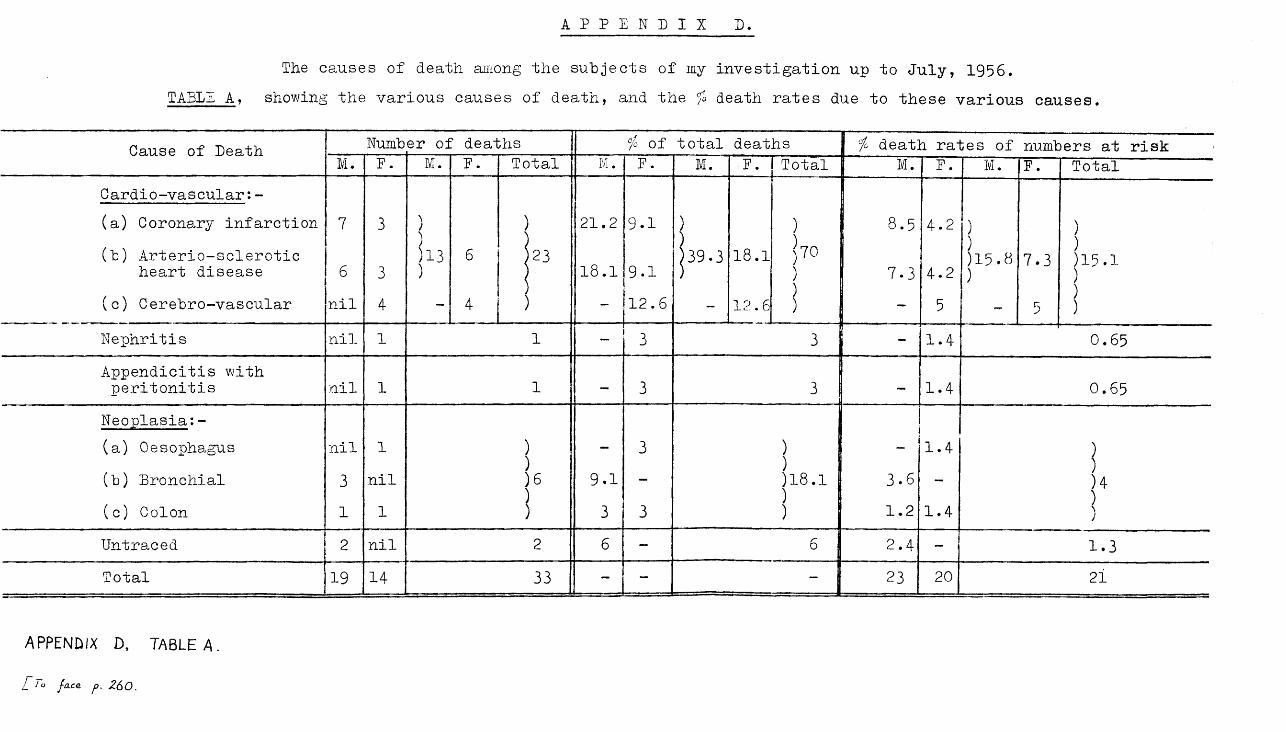
A P P E N D I X D.
The causes of death among the subjects of my investigation up to July, 1956.TABLE A , showing the various causes of death, and the fo death rates due to these various causes.
Cause of Death Numb er of deaths °/o Of total deaths [ io death rates of numbers at riskM. E. M. E. Total M . E. M. F. Total I M - F. M. E. Total
Cardio-vascular:-(a) Coronary infarction(b) Arterio-sclerotic
heart disease
7
6
3
3
)))13)
6) 21.2
18.1
9.1
9.1|39.3 18.1
))7°))
8.5
7.3
4.2
4.2
)))15.8 )
7.3)))15.1 )))(c) Cerebro-vascular nil 4 - 4 ) - 12.6 - 12,6 - 5 - 5
Nephritis nil 1 1 - 3 3 1.4 0.65Appendicitis with peritonitis nil 1 1 - 3 3 1.4 0.65
Neoplasia:- (a) Oesophagus nil 1 )
))6'S
_ 3 )))18.1))
— 1.4 j(b) Bronchial 3 nil 9*1 - 3.6 - )4
))(c) Colon 1 1 ! 3 3 1.2 1.4
Untraced 2 nil 2 6 - 6 2.4 - 1.3Total 19 14 33 - -
. . 23 20 21
APPENDIX D, TABLE A.
i To fa.ce p. 260.

- 260 - APPENDIX D.
It will be seen that by far the greatest cause of death among my subjects is cardiovascular disease. It is surprising to note how few are the other causes of death, but it is not unexpected to find that the next highest cause, after cardiovascular disease, is neoplasia.
I find that the total death rates for males and females are similar, though slightly higher among the males. It has been shown that while the mortality rates in adult life among both men and women have improved over recent years, a marked difference in the trend for the two sexes has been observed in later adult life, at which period the mortality rate for women continues to show improvement, but that for men fails to do so. The reason for this is that coronary thrombosis and bronchial carcinoma, increasingly prevalent in both sexes, affect men much more than women (Medical Annual 1956).
My figures bear this out. In my series, deaths from coronary infarction and from arteriosclerotic heart disease occur in the approximate ratio of 2 male to 1 female, and bronchial carcinoma causes no female deaths, but is responsible for the death of three males.
Smith^"^^ states that in the U.S.A. 50fo of all adult deaths are due to heart disease in all its forms. In my series the corresponding figure is 57.4$. Smith also quotes 3# of all deaths as being due to cerebral vascular catastrophes,

- 261 -
and my figure for such conditions is much higher, being12.6 fo.
In Table B I have presented the death rates among my subjects in greater detail according to the various subdivisions of my investigation. I have '..aid greater stress on cardiovascular deaths because of their h gh frequency. The total numbers of deaths from all causes are shown, and are also calculated as a percentage of the numbers at risk in the subdivision. Cardiovascular deaths are treated similarly, and in addition are shown as a percentage of the total numbers of deaths occurring in each appropriate subdivision, and as a percentage of the total numbers at risk in each complete series.
The single case of interstitial nephritis is included among the cardiovascular deaths as post-mortem examination revealed that cardiovascular degeneration, including myocarditis, was present.
Since the investigation was commenced 33 deaths have occurred, 26 of them among the subjects of the Urea Concentration Series. Thus 35$ of the subjects of this series have died; of those 26 deaths, 20 (77$) were cardiovascular in type, so that 27$ of those at risk died cardiovascular deaths. Fewer

APPENDIX D ~ T A B L E B (Contd.)
Urea Concentration Series I Specific Gravity SeriesTotal deaths Cardiovascular deaths , Total deaths Cardiovascular deaths
Subdivision
Number
of
As io
of to
tal
numbers
at ri
sk
in su
bsec
tion
<H0u<p&
As fo
of to
tal
numbers
at ri
sk
in su
bsec
tion
As i
of to
tal
numbers
at ri
sk
in whole
seri
es
As i
of to
tal
numbers
of
deaths
in the
subs
ecti
on OuQ>rg
As i
of to
tal
numbers
at ri
sk
in su
bsec
tion
1Number
of
As i
of total
1 numbers
at ris
k i
in subsection
j
As $
of to
tal
numbers
at ri
sk
in whole
seri
es
As i
of to
tal
numbers
of
deaths
in the
subs
ecti
on
9. Obesity: Present 12 43* 10 36* 13.5* 83* 3 11* 2 7.4* 2.56* 66*Absent 14 30* 10 22* 13.5* 71* 4 8* 2 4* 2.56* 50*
10.Sheldon’s SomatotypesEndomorphs 8 32$ 6 CM 8.10* 75* 1 6* 1 6* 1.28* 100*Mesomorphs 11 38* 10 34* 13-5* 90* 2 6* 1 3* 1.28* 50*Ectomorphs 7 35* 4 20* 5.4* 57* 4 13* 2 6.4* 2.56* 50*
11.Tobacco: Smokers 14 40* 12 34* 16.2* 85* 5 11* 2 4.4* 2.56* 40*Non-smokers 12 30* 8 20* 10.8* 66* 2 6* 2 6* 2.56* 100*
12.Alcohol: Users 8 36* 8 36* 10.8* 100* 4 16* 1 4* 1.28* 25*Non-users 18 34* 12 23* 16.2* 66*1 3 5.6* 3 5.6* 3". 84* 1 0 0 *
13.Chronic Pulmonary Le si ons: £r e sent 5 38* 3 23* 4.05* 60* | 3 23* 1 8* 1.28* 33*
Absent 21 34* 17 28* 22.95* 81* 4 6* 3 4.6* 3.54* 75*14.Albuminuria:
Present 8 53* 6 40* 8 . 1 * 75* 1 8 * none nil nil nilAbsent 18 30* 14 24$ 18.9*~ 77*" | 6 9*" 4 6* 5.12* 66i
A P P E N D I X d ,
T A B L E B.
( S H E E T 4 - ) .
[ T ° f-o.ce p . 2 6 2

APPENDIX D - T A B L E B (Conta.)
Subdivision
Urea Concentration Series Specific Gravity SeriesTotal deaths Cardiovascular deaths Total deaths Cardiovascular deaths
« H0u0 )&
& As i*
of to
tal
numbers
at ri
sk
in su
bsec
tion
0u< D
As in
of to
tal
numbers
at ri
sk
in su
bsec
tion
As of
tota
l numbers
at ri
sk
in whole
seri
es
As i°
of to
tal
numbers
of
deaths
in the
subs
ecti
on
Number
of..
...
...-
1As
io of
tota
l numbers
at ri
sk
in su
bsec
tion
Number
of
As of
tota
l numbers
at ri
sk
in su
bsec
tion
As i
of to
tal
numbers
at ri
sk
in whole
seri
es
As i
of to
tal
numbers
of
deaths
in the
subs
ecti
on
6. State of arteriesAtherosclerosis absent 8 19ft 4 9# 5.40# 50# 4 9# 2 5# 2.56# 50#Atherosclerosis present 18 56# 16 50# 21.60# 88# 3 8# 2 6# 2.56# 66#
7. Level of Blood Pressure
150/90 or below 9 36# 6 24# 8.10# 66# 3 12.5# 1 4# 1.28# 33#Systolic over )
150 mm.Hg. ) Diastolic not over)
100 mm.Hg. ) 9 31# 7 24# 9-45# 77# 3 9# 3 9# 3-84# 100#
Systolic over ) 150 mm.Hg. )
Diastolic 105 mm. ) Hg. or over ) 8 42# 7 37# 9.45# 87# 1 5# none nil nil
1.28#
nil
8. Previous strepto- A P P F N m x r> coccal infection:-
T A B L E B . Present Not recorded becaiuse of ineomplete data 3 7# 1 2.5# 33#(sheet 3) Absent H tl tl tl 11 »» 4 | 1096 3 8# 3-84# 75#
[ / o f a c e p . Z&2. •

/APPENDIX D T A B L E B (Con-td.)
Subdivision
Urea Concentration Series Specific Gravity SeriesTotal deaths Cardiovascular deaths Total deaths Cardiovascular dleaths
Number
of
As $
of to
tal
numbers
at ri
sk
in su
bsec
tion
ou0)£
mr j * H05 U O • P - H O - P - P -P 05 o 0>C H C O 0 9 O U fit
0 rjC O
0 3 3 dr t - H As
$ of
tota
l numbers
at ri
sk
in whole
seri
es
As $
of to
tal
numbers
of
deaths
in the
subs
ecti
on < Mof nQ )« As
$ of
tota
l numbers
at ri
sk
in subsection
j.
< Ho0
As $
of total
*
numbers
at ri
sk
in su
bsec
tion
I
As $
of to
tal
numbers
at ris
k I
in whole
seri
es
As $
of to
tal
numbers
of
deaths
in the
subs
ecti
on
4a. Contd.IV 4 44# 2 22# 2.70# 50# none
..nil .
V 1
9
33# 1 33# 1.35#
8.10#
100# Nocases - - - - -
4b.Social GroupingsWomen not gainfully
employed 31# 6 20# 66# 2 9$ 2 9# 2.56# 100#I 4 50# 4 50# 5.40# 100# 1 8$ none nil nil nilII 4 26# 4 26# 5.40# 100# 2 8 i 1 4# 1.28# 50#
III 4 30# 3 23# 4.05# 75# 2 12$ 1 6# 1.28# 50#IV 4 66# 2 33# 2.70# 50# none nil none nil nil nilV 1 33# 1 33# 1.35# 100# Nocases — - — - —
5. State of heart: Myocardium healthy 5 16# 4 13# 5.40# 80# 1 3$ none nil nil nilMyocardium impaired 17 47# 14 40# 18.90# 82# 3 10$ 1 3$ 1.28$ 33#Myocarditis +
A P P E N D I X D , endocarditis 3 3# 1 14# 1.30# 33# 3 30$ 1 10$ 1.28# 33#1 A B l L B . _
( s h e e t 2 . ) Endocarditis alone clSes -
T-l" WT'SS
- - none nil none nil nil nil
[_ to /ace p. 2 6 2 .

APPENDIX D - T A B L E B
Subdivision.
Urea Concentration Series Specific Gravity SeriesTotal deaths Cardiovascular deaths Total deaths Cardiovascular deaths
0ua>
S3
mH *H0& U 0 -P *H O -P -P -P cti O 0) a) m O P» ,Qa> 303a 1 Pi <3 PJ *H Nu
mber
of
M03H *H cS Pi O -P >H O -p -p •P Cfl O 0)<+-4 03 03 O P» ,QQ) rjro
5 I Pi As i
of total
numbers
at ri
sk
in whole
series
As $
of total
numbers
of
deaths
in the
subs
ection O
PiQ>£
As $
of total
numbers
at ri
sk
in su
bsec
tion
Number
of
As $
of total
numbers
at ri
sk
in su
bsec
tion
As i
of total
numbers
at ri
sk
in whole
series
As $
of total
numbers
of
deaths
in the
subs
ecti
on
1. Overall Series 26 35/ 20 27/ 27/ 77/ 7 9 i 4 5 .12/ 5 .12/ 57/2. Effect of age:-
(1) up to 55 yrs. none - - - — - nocases - - - - -
(2) 56 - 60 yrs. 3 33/ 2 22/. 2.7/ 66/ none nil - ~ -(3) 61 - 65 yrs. 5 28/ 3 17/ 4.05/ 60/ 1 7/ 1 7/ 1.28/ 100/(4) 66 - 70 yrs. 8 61/ 5 38/. 6.75/ 62/ 3 14/ 1 5/ 1.28/ 33/(5) 71 - 75 yrs. 6 46$ 6 46/ 8.10/ 100/ 2 8/ 2 8/ 2.56/ 100/(6) 76 - 80 yrs. 2 40/ 2 40/ 2.7/ 100/ 1 11/ none - - -(7) Over 80 yrs. 2 100/ 2 100/ 2.7/ 100/ none nil - - - -
3. Effect of sex:- Eemales 11 285S 8 20/ 10.80/ 72/ 2 6/ 2 6/ 2.56/ 100/Males 15 43/ 12 34/ 16.20/ 80/ 5 11/ 2 4/ 2.56/ 40/
4a.Social GroupingsAppcNmx ft ....... 7 41/ 6 35/ 8.10/ 85/ 2 8/ 1 4/ 1.28/ 50/
Table b. II 7 28/ 6 24/ 8.10/ 85/ 3 9/ 2 5/ 2.56/ 66/( -SHEET I.) XII 7 35/ 5 25/ 6.75/ 71/ 2 10/ 1 6/ 1.28/ 50/
[ To fa.ce. p. 2.6Z Contd.

- 262 -
deaths have occurred in the more recent Specific Gravity series, where 7 (9/) subjects have died. 4 (57/) of these were due to diseases of the cardiovascular system, showing that 5.12/ of my subjects in this series died such deaths.
Various other conclusions can be drawn from my figures: some definite and some indefinite. For example, in Subdivision 2, relating to the different age levels, it is clear that in both series, if we exclude subjects over 80 years, the highest death rate from all causes occurs between 66 and 70 years, and that for cardiovascular diseases between 71 and 75 years. I have already pointed out that the years from 65 to 70 are fraught with danger, and the high death rate which I now find for this half-decade bears this out.
As between the sexes, the findings in the Urea. Concentration Series show that males carry a higher death rate than females; both from all causes and from cardiovascular disease; but in the Specific Gravity Series, while they have a higher death rate from all causes, the males show a rather lower death rate than females from cardiovascular disease.
The death rates found in the different Social Groups do not appear to follow any definite pattern. In the Urea Concentration Series the mortality rates for Groups I - V are in the increasing order II, V, III, I, IV, and when Women not gainfully employed are grouped into a separate category

- 263 -
the order becomes II, III, Women, V, I, IV. The corresponding figures for the Specific Gravity Series are IV, I, II, III, and IV, I, II, Women, III.
The mortality rates for males in 1921-3, and for men and married women in 1930-2, were found to increase in a steady gradient from Social Group I to V. However, samples taken from the recent census show that the Standard Mortality rate in Social Group I - V increase in the following order for men - II, IV, I, III, V, and for women - II, I, III, IV, V. The reason for this change from a simple gradient increase from Group I to V is not clear, but Logan does not consider that it is due to random fluctuation (Medical Annual 1955). It can be seen that my findings follow the same general pattern as the census samples. I note also that the death rates from cardiovascular causes tend to be highest at the extremes of the Social Scale.
Among subjects with healthy hearts the death rate tends ,to be lower, but even under such conditions a very high proportion (80/) of deaths are due to cardiovascular causes.The findings in relation to the state of the arteries differ in my two series; in the Urea Concentration Series it is noted that the presence of atherosclerosis is associated with an elevation of the total death-rate from all causes, and of the cardiovascular death rate; furthermore a smaller pro

portion of deaths are due to cardiovascular causes when atherosclerosis is absent - (50$ to 88$). in the Specific Gravity Series the results are almost the same whether atherosclerosis is present or not.
Smith^11^ states that hypertensive vascular disease and its sequelae are responsible for 25$ of the total number of deaths. I find that the maintenance of an elevated blood pressure during life leads to an increased cardio-vascular mortality rate, and the higher the level of the blood pressure, the greater will be the proportion of cardio-vascular deaths. However, although the cardio-vascular mortality rises with the blood pressure levels, the death rate from all causes is high among the normotensives of both series.
Obesity is associated with a relatively mild increase in mortality, principally because of a high proportion of cardiovascular deaths. This is exactly as one would expect.
The findings for tobacco-smoking are inconclusive; in the Urea Concentration series, smokers have a higher death rate from all causes, and from cardio-vascular causes, than the non-smokers, and the percentage of cardio-vascular deaths is higher among smokers. In the Specific Gravity series, smokers have a higher total death rate than non-smokers, but the smokers fare better in respect of the cardio-vascular

- 265 -
two were heavy smokers, and one was a moderate smoker.The picture for alcohol is somewhat similar to that for
tobacco; again, we have higher mortality rates among alcohol users, both from all causes and from cardio-vascular disease. An exception is that in the Specific Gravity series, it is the non-drinkers who have the higher cardiovascular mortality rate.
In the case of chronic pulmonary lesions the total death rate from all causes is raised, but there is no significant rise in the cardio-vascular mortality rate.
The presence of albuminuria (in the Urea Concentration series) is associated with a tendency to a raised mortality rate.
It may be stated then that the presence of certain circumstances tends to elevate the mortality rate both from all causes and from cardio-vascular diseases. These aresummarised thus:-1. Age itself, particularly between 65 and 75 years, and
in extreme senescence;2. Sex - in this case maleness - in age groups after
61 years;3. Impaired heart action;4. Atherosclerosis;

- 266 -
5. Hypertension; and6. Obesity.
The use of tobacco and alcohol are suspected of bringing about increased mortality rates, and this is probably true if they are used to excess.
It will be noted how closely these factors correspondto the agencies which bring about reduced renal efficiencyand which we have already discussed.
REFERENCES.
Deeny (April 1956). Irish Jour. Med. Sci., 364:145.Medical Annual 1955.Medical Annual 1956.Registrar-General's Statistical Review of England and
Wales for the year 1955.Report of the Ministry of Health - On the State of the
Public Health for the year 1955.Smith, H.W. (1951). "The Kidney Structure and Function in
Health and Disease". (New York, Oxford University Press).

%
*>
M a x im u m S. G
1020
Minimum S.Cr.
Raise
myears:-

Maximum S. G~.I nr ipWiit-l
1020
Minimum S.G.