ROS Generation in Peroxisomes and its Role in Cell Signaling
Upload
khangminh22Category
view
2download
0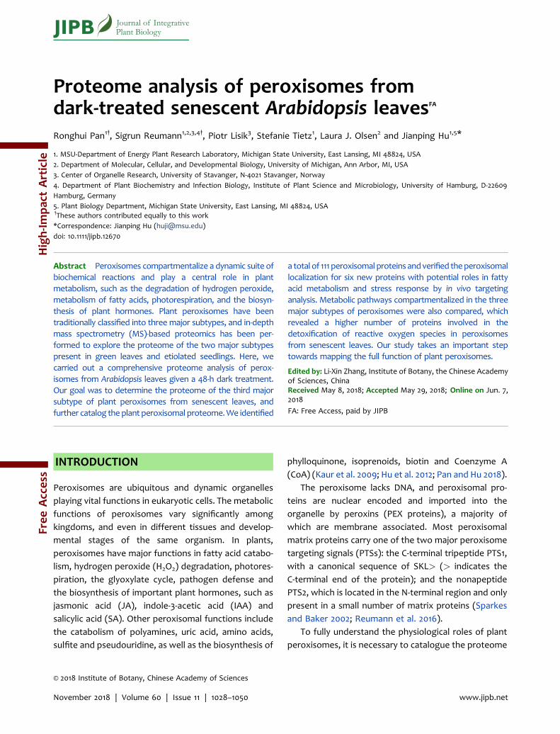
Proteome analysis of peroxisomes fromdark-treated senescent Arabidopsis leavesFA
Ronghui Pan1†, Sigrun Reumann1,2,3,4†, Piotr Lisik3, Stefanie Tietz1, Laura J. Olsen2 and Jianping Hu1,5*
1. MSU-Department of Energy Plant Research Laboratory, Michigan State University, East Lansing, MI 48824, USA2. Department of Molecular, Cellular, and Developmental Biology, University of Michigan, Ann Arbor, MI, USA3. Center of Organelle Research, University of Stavanger, N-4021 Stavanger, Norway4. Department of Plant Biochemistry and Infection Biology, Institute of Plant Science and Microbiology, University of Hamburg, D-22609Hamburg, Germany5. Plant Biology Department, Michigan State University, East Lansing, MI 48824, USA†These authors contributed equally to this work*Correspondence: Jianping Hu ([email protected])doi: 10.1111/jipb.12670
Abstract Peroxisomes compartmentalize a dynamic suite ofbiochemical reactions and play a central role in plantmetabolism, such as the degradation of hydrogen peroxide,metabolism of fatty acids, photorespiration, and the biosyn-thesis of plant hormones. Plant peroxisomes have beentraditionally classified into three major subtypes, and in-depthmass spectrometry (MS)-based proteomics has been per-formed to explore the proteome of the two major subtypespresent in green leaves and etiolated seedlings. Here, wecarried out a comprehensive proteome analysis of perox-isomes from Arabidopsis leaves given a 48-h dark treatment.Our goal was to determine the proteome of the third majorsubtype of plant peroxisomes from senescent leaves, andfurther catalog theplant peroxisomal proteome.We identified
a total of 111 peroxisomal proteins andverified theperoxisomallocalization for six new proteins with potential roles in fattyacid metabolism and stress response by in vivo targetinganalysis. Metabolic pathways compartmentalized in the threemajor subtypes of peroxisomes were also compared, whichrevealed a higher number of proteins involved in thedetoxification of reactive oxygen species in peroxisomesfrom senescent leaves. Our study takes an important steptowards mapping the full function of plant peroxisomes.
Edited by: Li-Xin Zhang, Institute of Botany, the Chinese Academyof Sciences, ChinaReceived May 8, 2018; Accepted May 29, 2018; Online on Jun. 7,2018
FA: Free Access, paid by JIPB
INTRODUCTION
Peroxisomes are ubiquitous and dynamic organellesplaying vital functions in eukaryotic cells. The metabolicfunctions of peroxisomes vary significantly amongkingdoms, and even in different tissues and develop-mental stages of the same organism. In plants,peroxisomes have major functions in fatty acid catabo-lism, hydrogen peroxide (H2O2) degradation, photores-piration, the glyoxylate cycle, pathogen defense andthe biosynthesis of important plant hormones, such asjasmonic acid (JA), indole-3-acetic acid (IAA) andsalicylic acid (SA). Other peroxisomal functions includethe catabolism of polyamines, uric acid, amino acids,sulfite and pseudouridine, as well as the biosynthesis of
phylloquinone, isoprenoids, biotin and Coenzyme A(CoA) (Kaur et al. 2009; Hu et al. 2012; Pan and Hu 2018).
The peroxisome lacks DNA, and peroxisomal pro-teins are nuclear encoded and imported into theorganelle by peroxins (PEX proteins), a majority ofwhich are membrane associated. Most peroxisomalmatrix proteins carry one of the two major peroxisometargeting signals (PTSs): the C-terminal tripeptide PTS1,with a canonical sequence of SKL> (> indicates theC-terminal end of the protein); and the nonapeptidePTS2, which is located in the N-terminal region and onlypresent in a small number of matrix proteins (Sparkesand Baker 2002; Reumann et al. 2016).
To fully understand the physiological roles of plantperoxisomes, it is necessary to catalogue the proteome
© 2018 Institute of Botany, Chinese Academy of Sciences
JIPB Journal of IntegrativePlant Biology
November 2018 | Volume 60 | Issue 11 | 1028–1050 www.jipb.net
Free
Access
High-Im
pact
Article

of these organelles. In recent years, experimentalproteomics, based on peroxisome purification andmass spectrometry (MS), have greatly accelerated therate and scale of the identification of novel plantperoxisomal proteins (Kaur and Hu 2011; Reumann 2011;Pan and Hu 2018). This unbiased and relatively large-scale approach has not only successfully identified newauxiliary and regulatory proteins involved in establishedperoxisomal processes, but also revealed numerousproteins involved in unexpected peroxisomal functions(Kaur and Hu 2011; Reumann 2011; Pan and Hu 2018).
Peroxisomes are metabolically and morphologicallydynamic. Environmental factors, including light, areknown to cause changes in peroxisome abundance(Lopez-Huertas et al. 2000; Koh et al. 2005; Desai andHu 2008; Rodr�ıguez-Serrano et al. 2016; Desai et al.2017; Fahy et al. 2017). The proteome and primaryfunctions of plant peroxisomes vary, quantitativelyand qualitatively, across developmental stages, anddistinct names were given to the three major subtypesof plant peroxisomes. Leaf peroxisomes in greenleaves are mainly involved in photorespiration (Tolbertet al. 1969; Peterhansel et al. 2010), glyoxysomes referto peroxisomes in seeds/fatty tissue, which harbor theglyoxylate cycle together with fatty acid b-oxidation(Beevers 1979), and gerontosomes refer to perox-isomes in senescent tissues, which were suggested toresemble glyoxysomes in terms of their metabolicfunctions (Vicentini and Matile 1993).
To uncover peroxisomal proteins at differentdevelopmental stages, previous proteomic studiesanalyzed peroxisomes purified from various plantorgans and species, including greening and etiolatedArabidopsis cotyledons (Fukao et al. 2002, 2003),Arabidopsis leaves (Reumann et al. 2007, 2009), non-green Arabidopsis cell suspension cultures (Eubel et al.2008), etiolated Arabidopsis seedlings (Quan et al.2013), etiolated soybean cotyledons (Arai et al. 2008),and spinach leaves (Babujee et al. 2010). These studiesrevealed common pathways, as well as proteins specificto certain developmental stages, and identified anumber of novel peroxisome proteins, such as thoseinvolved in methylglyoxal detoxification, phylloquinonebiosynthesis, pseudouridine catabolism, CoA biosynthe-sis, and putative regulatory proteins (Kaur and Hu 2011;Reumann 2011; Pan and Hu 2018).
The goal of this study was to discover additionalperoxisomal proteins and catalog the proteome of
peroxisomes from plants undergoing senescence-re-lated processes. Senescence is a genetically-controlledglobal-reprogramming of cellular activities, featured bythe cessation of photosynthesis, degradation of
chlorophyll, disintegration of organelle structures,and degradation of diverse cellular components, suchas proteins and lipids (Buchanan-Wollaston 1997). Plant
peroxisomal metabolism during leaf senescence hasbeen investigated in pea, from which increasedproteolytic activity and more active metabolism ofreactive oxygen species (ROS), nitric oxide and NADPH
was discovered (Pastori and del Rio 1997; Corpas et al.1999, 2004). However, analysis of the whole peroxi-some proteome undergoing senescence-related pro-
cesses had been lacking. Detached leaves subjected toextended (e.g. 48 h) darkness have been widely used asa system to study plant senescence (Biswal and Biswal
1984; Oh et al. 1997; Weaver et al. 1998; Weaver andAmasino 2001; Song et al. 2014; Liebsch and Keech2016). To this end, we performed proteome analysis ofperoxisomes isolated from leaves of dark-treated
detached Arabidopsis plants. This study identified 111peroxisomal proteins, a higher number compared withprevious peroxisome proteomic studies in any organ-
ism, with eight being identified for the first time by MS-based proteomics of peroxisomes. Our in vivo subcellu-lar targeting analysis validated peroxisomal localizationof six new proteins involved in fatty acid metabolism
and stress response. Our study takes an important steptowards fully deciphering the peroxisome proteomeand uncovering novel physiological roles of plant
peroxisomes.
RESULTS
Mass spectrometry-based interrogation ofperoxisomal proteinsIsolation of leaf peroxisomes requires large amounts ofleaves (approximately 60 g FW). Therefore, any abioticstress applied to Arabidopsis plants needs to reach allleaves and preferentially all mesophyll cells relativelyuniformly. A comparison of intact and harvestedArabidopsis Col-0 plants subjected to prolonged darktreatment demonstrated that only harvested plantsshowed uniform progression of leaf senescence in allleaves, consistent with previous reports of chlorophylldegradation (see Introduction). The chlorophyll content
Proteome of peroxisomes from senescent leaves 1029
www.jipb.net November 2018 | Volume 60 | Issue 11 | 1028–1050

was reduced by approximately 40% after 48 h of dark-induced senescence, and reached a residual level ofapproximately 15% after 4 d (Figure 1A). By contrast,when intact plants were subjected to dark treatment,leaf senescence became visible only slowly andprimarily in the oldest leaves, and as a result, chlorophyllcontent of the entire plant hardly declined, even after 6d (Figure S1A). As such, we chose harvested plants fordark treatment to induce uniform leaf senescence.
After 4 weeks of growth under a 16/8 h light/darkcycle, the above-ground portion of Arabidopsis plantswas harvested and subjected to 48 h dark treatment(Figure S1B), at which point peroxisomes were isolatedand purity assessed (Figure 1B), using our previouslyestablished immunological methodologies (Reumannet al. 2009; Quan et al. 2013). Two independent pools ofperoxisome preparations with maximum purity, namedTotal 1 (T1) and Total 2 (T2), were obtained. For protein
identification, 500mg each of T1 and T2 peroxisomeproteins were separated on a 1D gel, which was latersliced into eight pieces after electrophoresis. Proteins ineach slice were in-gel digested by trypsin, and subjectedto liquid chromatography-electrospray ionization-tandem mass spectrometry (LC-ESI-MS/MS). Thismethod had been used in our previous studies tosuccessfully identify a large number of peroxisomalproteins from peroxisomes of Arabidopsis etiolatedseedlings and green leaves (Reumann et al. 2007, 2009;Quan et al. 2013).
Using 99% probability of protein identification as the
cut-off, a total of 320 and 391 proteins were detected in
T1 and T2, respectively (Table S1). Among them, 91
proteins from T1 and 100 proteins from T2 (105 proteins
in total) were previously established peroxisomalproteins (Table 1). To identify putative novel peroxi-
some proteins, the remaining proteins were further
classified into ‘non-peroxisomal’ and ‘potentially new
peroxisomal’, using protein subcellular localization
information in the Arabidopsis Subcellular Proteomic
Database (SUBA4) (Hooper et al. 2017), which repre-sents compiled data from published biochemical
studies, organelle proteomics, and/or fluorescence
microscopy-based localization analyses. Also included
in the ‘potentially new peroxisomal’ category were five
proteins annotated as non-peroxisomal by SUBA, but
contained C-terminal PTS1 or PTS1-like tripeptides.These included fructose-bisphosphate aldolase 1 splic-
ing isoform 2 (FBA1.2; SFL>), glutamate-1-semialdehyde
2,1-aminomutase 1 (GSA1; SRI>), aconitate hydratase 3
(ACO3; SKQ>), TSK-associating protein 1 (TSA1; SSL>),
and 60S ribosomal protein L19-1 (RPL19A; SKK>). Intotal, 211 proteins fromT1 and 267 proteins from T2were
considered as ‘non-peroxisomal’ (Table S2), and to-
gether 30 proteins were considered to be ‘potentially
new peroxisomal’ proteins (Table S3).
Microscopic validation of peroxisomal localizationDue to possible organelle contamination, all novelproteins detected by mass spectrometry- based organ-elle proteomics need to be confirmed by an indepen-dent approach, such as in vivo subcellular targetinganalysis of fluorescently tagged candidate proteins. Touncover new peroxisomal proteins and functions, weselected 19 out of the 30 potentially new peroxisomalproteins (Table S3) for in vivo targeting analysis. Thefollowing selection criteria were applied: (i) thepresence of a PTS-like sequence; (ii) having putative
Figure 1. Assessment of dark-induced senescence inArabidopsis and purity of isolated leaf peroxisomes(A) Change in chlorophyll content (mg chl/g FW) duringdark-induced senescence of detached Arabidopsisshoots. (B) Immunoblot of different leaf peroxisomeisolates (5mg protein/each preparation) from senescentshoots using an anti-VDAC (voltage-dependent anionchannel) antibody (Reumann et al. 2009) to determinethedegreeofmitochondrial contamination. Two internalstandards (IS1-2) of moderate (mp) and high purity (hp)were analyzed, in parallel, to compare the degree ofcontamination between different isolates. Senescentleaf peroxisome isolates of highest purity (e.g., SLP1)were selected and pooledwith other high-purity isolatesfor proteome analysis by mass spectrometry.
1030 Pan et al.
November 2018 | Volume 60 | Issue 11 | 1028–1050 www.jipb.net

Table 1. Peroxisomal proteins identified in this study
Protein
identification
Pathway/annotation Symbol At locus PTS1/2 T1 T2
b-oxidation
3-Ketoacyl-CoA thiolase 1 KAT1 AT1G04710 RQx5HL X X
Acyl-CoA oxidase 3 ACX3 AT1G06290 RAx5HI/SSV X X
Acyl-activating enzyme 1 AAE1 AT1G20560 SKL X X
3-Ketoacyl-CoA thiolase 2 KAT2 AT2G33150 RQx5HL X X
Acyl-CoA oxidase ACX5 AT2G35690 AKL X X
Long-chain acyl-CoA synthetase 6 LACS6 AT3G05970 RIx5HL X X
Multifunctional protein 2 MFP2 AT3G06860 SRL X X
Hydroxybutyryl-CoA dehydrogenase HBCDH AT3G15290 PRL X X
Acyl-activating enzyme 7 AAE7 AT3G16910 SRL X X
Acyl-CoA oxidase 4 ACX4 AT3G51840 SRL X X
Acyl-CoA oxidase 1 ACX1 AT4G16760 ARL X X
Abnormal inflorescence meristem 1 AIM1 AT4G29010 SKL X X
Acyl-activating enzyme 5 AAE5 AT5G16370 SRM X X
Acyl-activating enzyme 17 AAE17 AT5G23050 SKL X X
Long-chain acyl-CoA synthetase 7 LACS7 AT5G27600 RLx5HI/SKL X X
3-Ketoacyl-CoA thiolase 5 KAT5 AT5G48880 RQx5HL X X
Acyl-CoA oxidase 2 ACX2 AT5G65110 RIx5HL X
IAA biosynthesis (b-oxidation)
Indole-3-butyric acid response 3 IBR3 AT3G06810 SKL X X
Short-chain dehydrogenase-reductasea/Indole-3-butyric acid response 1
IBR1 AT4G05530 SRL X X
JA biosynthesis (b-oxidation)
OPC-8:0 ligase 1 OPCL1 AT1G20510 SKL X X
12-oxophytodienoate reductase 3 OPR3 AT2G06050 SRL X X
4-coumarate:CoA ligase 1 4CL1 AT4G05160 SKM X X
4-coumarate:CoA ligase like 2 4CL2 AT5G63380 SKL X X
Auxiliary b-oxidation and other fatty acidcatabolism
Small thioesterase 4 st4 AT1G04290 SNL X
D3,D2-enoyl CoA isomerase 1 ECI1 AT1G65520 SKL X
Monofunctional enoyl-CoA hydratase 2 ECH2 AT1G76150 SSL X X
Peroxisomal NADþ-malate deshydrogenase 1 PMDH1 AT2G22780 RIx5HL X X
Small thioesterase 5 st5 AT2G29590 SKL X X
Short-chain dehydrogenase-reductase c SDRc AT3G01980 SYM X X
Short-chain dehydrogenase-reductaseb/ 2,4-dienoyl-CoA reductase
SDRb/ DECR AT3G12800 SKL X X
Hydroxy-acid oxidase 1 HAOX1 # $ AT3G14130 SML X
Hydroxy-acid oxidase 2 HAOX2 AT3G14150 SML X
Short-chain dehydrogenase-reductase d SDRd AT3G55290 SSL X X
Short-chain dehydrogenase-reductase e SDRe # $ AT3G55310 SSL X
(Continued)
Proteome of peroxisomes from senescent leaves 1031
www.jipb.net November 2018 | Volume 60 | Issue 11 | 1028–1050

Table 1. Continued
Protein
identification
Pathway/annotation Symbol At locus PTS1/2 T1 T2
Small thioesterase 3 st3 AT3G61200 SKL X X
Epoxide hydrolase 3 EH3 AT4G02340 ASL X X
Indole-3-butyric acid response 10/ D(3),D(2)-Enoyl-CoA isomerase 2
IBR10/ ECI2 AT4G14430 PKL X X
Monofunctional enoyl-CoA hydratase/isomerase a ECHIa AT4G16210 SKL X X
Peroxisomal NADþ-malate deshydrogenase 2 PMDH2 AT5G09660 RIx5HL X X
Sterol carrier protein 2 SCP2 AT5G42890 SKL X X
D3,5-D2,4-dienoyl-CoA-isomerase DCI1 AT5G43280 AKL X X
Photorespiration
Glutamate-glyoxylate aminotransferase 1 GGT1 AT1G23310 SKM X X
Hydroxypyruvate reductase 1 HPR1 AT1G68010 SKL X X
Glutamate-glyoxylate aminotransferase 2 GGT2 AT1G70580 SRM X X
Serine-glyoxylate aminotransferase/Alanine:glyoxylate aminotransferase 1
SGT/ AGT1 AT2G13360 SRI X X
Glycolate oxidase 1 GOX1 AT3G14415 PRL X X
Acyl-CoA hydrolysis
Acyl-CoA thioesterase 2 ACH2 AT1G01710 SKL X X
AcylCoA thioesterase family protein ACH AT4G00520 AKL X
Biogenesis
Peroxin 11c PEX11c AT1G01820 X X
Peroxin 11a PEX11a AT1G47750 X X
Peroxin 11d PEX11d AT2G45740 X X
Dynamin-related protein 5B DRP5B # AT3G19720 X
Peroxin 11b PEX11b AT3G47430 X X
Peroxin 11e PEX11e AT3G61070 X X
Peroxin 14 PEX14 AT5G62810 X X
Chaperone
a-Crystallin domain containing proteinof 31.2 kDa
Acd31.2 AT1G06460 RLx5HF/PKL X X
Methylglyoxal detoxification
Glyoxylase I homolog GLX1 AT1G11840 X
NADH/NADPH metabolism
Glyceraldehyde-3-phosphate dehydrogenase C2 GAPC2 AT1G13440 SKA X X
NADP-dependent isocitrate deshydrogenase ICDH AT1G54340 SRL X X
Phosphogluconate dehydrogenase 2 6PGDH/PGD2
AT3G02360 SKI X X
ROS detoxification
Dehydroascorbate reductase 1 DHAR1 AT1G19570 X X
Catalase 3 CAT3 AT1G20620 QKL(internal) X X
Catalase 1 CAT1 AT1G20630 QKL(internal) X X
Glutathione reductase 1 GR1 AT3G24170 TNL X
(Continued)
1032 Pan et al.
November 2018 | Volume 60 | Issue 11 | 1028–1050 www.jipb.net

Table 1. Continued
Protein
identification
Pathway/annotation Symbol At locus PTS1/2 T1 T2
Monodehydroascorbate reductase 4 MDAR4 AT3G27820 X X
Monodehydroascorbate reductase 1 MDAR1 AT3G52880 AKI X
Glutathione S-transferase lambda 2 GSTL2 # $ AT3G55040 ARL X
Ascorbate peroxidase 3 APX3 AT4G35000 X X
Catalase 2 CAT2 AT4G35090 QKL(internal) X X
Ascorbate peroxidase 5 APX5 $ AT4G35970 X
Copper/zinc superoxide dismutase 3 CSD3 AT5G18100 AKL X X
Glutathione S-transferase theta 1 GSTT1 AT5G41210 SKI X X
Glutathione S-transferase theta 3 GSTT3 # AT5G41220 SKM X
Phylloquinone synthesis
DHNA-CoA thioesterase 1 DHNAT1 AT1G48320 AKL X X
Naphthoate synthase NS AT1G60550 RLx5HL X X
Pseudouridine catabolism
Indigoidine synthase A IndA AT1G50510 RIx5HL X X
Urate degradation
Uricase Uri AT2G26230 SKL X X
Transthyretin-like protein 1 TLP1 AT5G58220 RLx5HL X X
Transporter
Peroxisomal NADþ carrier PXN AT2G39970 X X
Peroxisomal membrane proteinof 22kDa
PMP22 AT4G04470 X
Peroxisomal ABC transporter 1 PXA1 AT4G39850 X X
Peroxisomal adenine nucleotide carrier 2 PNC2 AT5G27520 X
Protease
Peptidase family M16 PM16 AT2G41790 PKL X X
Lon protease homolog 2 Lon2 AT5G47040 SKL X X
Polyamine oxidation
Copper amine oxidase CuAO3 AT2G42490 SKL X X
Betaine aldehyde dehydrogenase BADH AT3G48170 SKL X X
Glyoxylate cycle
Citrate synthase 3 CSY3 AT2G42790 RLx5HL/SSV X X
Citrate synthase 2 CSY2 AT3G58750 RLx5HL/SAL X X
Sulfite oxidation
Sulfite oxidase SO AT3G01910 SNL X X
Nucleotide homeostasis
Histidine triad family protein 3 HIT3 AT3G56490 RVx5HF X X
Nucleoside diphosphate kinase type 1 NDPK1 AT4G09320 X
Histidine triad family protein 1 HIT1 AT4G16566 SKV X
Histidine triad family protein 3 HIT2 AT5G48545 RLx5HL X X
Mevalonic acid (MVA) pathway
(Continued)
Proteome of peroxisomes from senescent leaves 1033
www.jipb.net November 2018 | Volume 60 | Issue 11 | 1028–1050

stress-related functions; and (iii) having been detectedin previous peroxisome proteomic studies but notconfirmed by an independent approach (Table 2).
The yellow fluorescent protein (YFP) was fused to
the N-terminus of each candidate protein to allow
subcellular targeting functionality of the C-terminal
PTS1. Through Agrobacterium-mediated transforma-
tion, each fluorescently-tagged candidate gene was
transiently co-expressed with a fusion between the
cyan fluorescent protein gene and the PTS1 tripeptide
SKL (CFP-PTS1) in tobacco (Nicotiana tabacum) leaves. In
some cases, we also co-expressed YFP tagged candi-
date genes with the mitochondrial marker gene COX4-
CFP for further clarification. Two days later, tobacco leaf
epidermal and/or mesophyll cells were subjected to
confocal microscopy. In total, we confirmed the
peroxisomal localization for six candidate proteins
(Figures 2–5; Table 2).
Five of the confirmed proteins, i.e., hydroxy-acid
oxidase 1 (HAOX1), glutathione S-transferase lambda 2
(GSTL2), short-chain dehydrogenase-reductase isoform
e (SDRe), harmless to ozone layer 3 (HOL3), and TSK-
associating protein 1 (TSA1), contained either a PTS1 or
PTS1-like sequence (Table 2). SML> of HAOX1, ARL> of
GSTL2, and SSL> of SDRe are well-established PTS1
tripeptides, and the YFP fusions of these proteins
showed strong and almost exclusive localization to
peroxisomes (Figure 2).Harmless to Ozone Layer 3 (HOL3) has a C-terminal
tripeptide STL> that was previously shown to be afunctional PTS1 in an EST of glycolate oxidase (GOX)from Catharanthus roseus (Lingner et al. 2011), but notidentified in any established Arabidopsis peroxisomalprotein. YFP-HOL3 exhibited strong targeting toperoxisomes, while some YFP fluorescence also re-mained in the cytosol (Figure 3A). Since HOL3 has been
Table 1. Continued
Protein
identification
Pathway/annotation Symbol At locus PTS1/2 T1 T2
Isopentenyl Diphosphate Isomerase 2 IPI2 # AT3G02780 X
Amino acid metabolism
Aspartate aminotransferase ASP3 AT5G11520 RIx5HL X X
Cobalamin-independent methionine synthase ATMS1 AT5G17920 SAK X X
3-hydroxyisobutyryl-CoA hydrolase CHY1 AT5G65940 AKL X X
Other or unknown functions
Unknown protein 6 UP6 AT1G16730 SKL X
Acetyl transferase 1 ATF1 AT1G21770 SSI X
NADH: quinone reductase NQR AT1G49670 SRL X X
TSK-associating protein 1 TSA1 $ AT1G52410 SSL X X
GAST1 protein homolog 1 GASA1 ? AT1G75750 X X
Dehydrin family protein ERD14 ? # AT1G76180 X
Acetyltransferase 2 ATF2 AT1G77540 SSI X
Glycine-rich protein 3 GRP3 ? # AT2G05520 X
Unknown protein 3 UP3 AT2G31670 SSL X X
Harmless to ozone layer 3 HOL3 $ AT2G43940 STL X X
Senescence-associated protein/ B12D-related protein B12D1 AT3G48140 X
Macrophage migration inhibitory factor 1 MIF1 AT3G51660 SKL X X
Zinc-binding dehydrogenase ZnDH AT3G56460 SKL X X
Unknown protein 5 UP5 AT5G44250 SRL X X
# indicatesproteins identified for thefirst time inperoxisomeproteomics. $ indicates proteins verified tobeperoxisomalby in vivo targeting analysis. ? indicates proteins reported to be peroxisomal by (Cutler et al. 2000), but sequenceinformation and microscopic images were not presented in this large-scale work thus need further confirmations.
1034 Pan et al.
November 2018 | Volume 60 | Issue 11 | 1028–1050 www.jipb.net

Table2.
Summaryof
invivo
targetingan
alyses
Ann
otation
Symbo
lATlocus
PTS
Previous
peroxisome
proteo
mework
YFP-protein
Protein-YF
P
Hyd
roxy-acidox
idase1
HAOX1
AT3G14130
SML
Per
–
Glutathione
S-tran
sferaselambd
a2
GST
L2AT3G5504
0ARL
Per
–
Short-chainde
hydrog
enase-redu
ctaseisoform
eSD
Re
AT3G55310
SSL
Per
–
Harmless
tooz
onelayer3
HOL3
AT2G43
940
STL
E200
8;R20
09Cy
t,pe
r–
Glutathione
S-tran
sferaseTA
U20
GST
U20
AT1G78
370
R20
09Cy
tCy
t
DEA
D-box
ATP
-dep
ende
ntRNAhe
licase37
RH37
AT2G42520
R20
07;200
9Cy
t;vesicles
Cyt;vesicles
Glycine
rich
protein2
GRP2
AT4
G38
680
Q20
13Cy
t;vesicles
–
Ascorba
tepe
roxida
se5
APX
5AT4
G3597
0E200
8Pe
r;mito;
chpt
–
Glyox
alase2-3
GLX
2-3
AT1G5358
0Cy
tMito
Ascorba
tepe
roxida
se2
APX
2AT3G09
640
RGx5HC
Cyt
Cyt
RING-H2fin
gerproteinATL65
/ATL
gene
family
mem
ber
ATL65
AT3G1893
0Cy
tVe
sicles;chpt
Alpha
/beta-Hyd
rolasessupe
rfam
ilyprotein
–AT3G47
560
Cyt
cyt
3-Oxo
acyl-[acyl-carrier-protein]redu
ctaselikeprotein1
OARP1
AT4
G20
760
Dots
Mito;
chpt
6-Ph
osph
ogluco
nolacton
ase4
PGL4
AT5
G24410
Cyt;vesicles
–
Fructose-bisph
osph
atealdo
lase
1,splicingisoform
2FB
A1.2
AT2G21330.2
SFL
Cyt
Chpt;mito
Glutamate-1-semialdeh
yde2,1-am
inom
utase1
GSA
1AT5
G63
570
SRI
Cyt
–
Aco
nitase
3ACO
3AT2G05
710
SKQ
E200
8;R20
09;Q20
13Cy
tMito;
chpt
60Sribo
somal
proteinL19-1
RPL
19A
AT1G02
780
SKK
Cyt
–
TSK-assoc
iating
protein1
TSA1
AT1G52410
SSL
R20
07;200
9Pe
r–
E200
8,Eu
bele
tal.2
008;
R20
07/200
9,Reu
man
net
al.2
007/20
09;Q
2013,Q
uanet
al.2
013.
Proteome of peroxisomes from senescent leaves 1035
www.jipb.net November 2018 | Volume 60 | Issue 11 | 1028–1050

implicated in pathogen defense, we further investi-gated whether HOL3 was also peroxisomal in standardexpression systems independent of Agrobacterium-mediated transformation. Indeed, YFP-HOL3 targetedto peroxisomes in both Arabidopsis protoplasts andonion epidermal cells (Figure 3B, C). Only few proteinswith non-canonical PTS1 tripeptides, such as STL>, areperoxisomal because their peroxisome-targeting abilitygenerally depends strongly on specific targeting-enhancing upstream residues (Lingner et al. 2011).Therefore, we also made fusions between the
C-terminal 10 aa of HOL3 and YFP and showed thatthis decapeptide was sufficient to direct YFP toperoxisomes in both protein expression systems (Figure3D, E). These results confirmed that STL> is a functionalPTS1 and established HOL3 as the first Arabidopsisperoxisomal protein identified thus far that uses STL>as a PTS1.
TSA1 had not been reported to have any functionassociated with the peroxisome despite having anestablished but non-canonical PTS1, SSL>. Interestingly,YFP-TSA1, which contained the C-terminal 259 aa of the
Figure 2. Peroxisome localization of HAOX1, GSTL2 and SDReConfocal images of the peroxisome localization of YFP-HAOX1 (A), YFP-GSTL2 (B), and YFP-SDRe (C) were taken intobacco leaf epidermal cells transiently co-expressing each YFP fusion gene and the peroxisome marker CFP-PTS1.Scale bars¼ 10mm. Predicted PTS1 tripeptides are shown in parentheses.
1036 Pan et al.
November 2018 | Volume 60 | Issue 11 | 1028–1050 www.jipb.net

Figure 3. Peroxisome localization of YFP-HOL3 in three plant protein expression systems(A) Confocal images of the peroxisome localization of YFP-HOL3 in tobacco leaf epidermal cells co-expressingYFP-HOL3 and the peroxisomal marker CFP-PTS1. (B–E) Epifluorescence images of the peroxisome localization ofYFP-HOL3 and YFP-Ct10aa (C-terminal 10 aa of HOL3) taken in Arabidopsis leaf protoplasts (B, D) and onionepidermal cells (C, E) that were transiently co-expressing each YFP fusion gene and the peroxisome marker gMDH-CFP. Scale bars¼ 10mm. Predicted PTS1 tripeptides are shown in parentheses.
Proteome of peroxisomes from senescent leaves 1037
www.jipb.net November 2018 | Volume 60 | Issue 11 | 1028–1050

759 aa protein, was detected in small punctate
structures that partially overlapped with the peroxi-
some matrix-localized marker protein, CFP-PTS1 (Figure
4A). This was not a random targeting pattern, because
the protein had no association with other organelle
markers, such as the mitochondrial marker COX4-CFP
(Figure 4B). We speculated that TSA1 may not be
imported into the peroxisome matrix, but instead is
attached to the surface of peroxisomes.
As a paralog of the peroxisomal membrane protein
APX3 (Narendra et al. 2006), APX5 also contains a
predicted transmembrane domain (TMD) near its C-
terminus, suggesting that it might be a C-terminal tail-
anchored membrane protein like APX3. Consistent with
this prediction, YFP-APX5 formed a ring structure
surrounding the peroxisomal matrix (Figure 5A, B),
suggesting that it is located on the membrane.
Interestingly, YFP-APX5 fluorescence was also detected
on the chloroplast outer-membrane (Figure 5C). Fur-
thermore, YFP-APX5 showed strong signals overlapping
with mitochondria (Figure 5D) and when examined at
higher magnification, was seen to, at times, form a ring
structure (Figure 5E), suggesting that it is also localized
to one of the two mitochondrial membranes.
Confocal microscopic analysis did not detect peroxi-
somal association for the other 13 candidate proteins
(Table 2). Among them, GSA1 (SRI>), FBA1.2 (SFL>),
RPL19A (SKK>) and ACO3 (SKQ>) contained PTS1 or
PTS1-like peptides (Table 2), yet their N-terminally
tagged YFP fusions were all observed to be cytosolic
(Figures S2A, S2B, S3A, S4A). Glutathione S-transferase
TAU 20 (GSTU20), DEAD-box ATP-dependent RNA
helicase 37 (RH37) and glycine rich protein 2 (GRP2)
do not have PTS-related peptides but were detected in
previous peroxisome proteome studies and this study
(Table 2). YFP-GSTU20 was cytosolic (Figure S4C), and
both YFP-RH37 and YFP-GRP2 were detected in the
cytosol as well as in small vesicle-like structures
that were neither peroxisomal nor mitochondrial
(Figures S5A, S6A).
Figure 4. TSA1 is associated with peroxisomesConfocal images of tobacco leaf epidermal cells transiently co-expressing YFP-TSA1 (C-terminal 259 aa) and theperoxisome marker CFP-PTS1 (A) or the mitochondrial marker COX4-CFP (B). Scale bars¼ 10mm. Predicted PTS1tripeptides are shown in parentheses. Insets in (A) are enlarged images of the boxed areas.
1038 Pan et al.
November 2018 | Volume 60 | Issue 11 | 1028–1050 www.jipb.net

Figure 5. Continued
Proteome of peroxisomes from senescent leaves 1039
www.jipb.net November 2018 | Volume 60 | Issue 11 | 1028–1050

The remaining six proteins tested lacked PTS1-related peptides, but were specifically detected in thisperoxisome proteomic study and had possible linkswithperoxisomes based on annotation or predicted func-tional domains. These included glyoxalase 2-3 (GLX2-3)presumably involved in methylglyoxal catabolism to-gether with peroxisomal GLX1 (Reumann et al. 2009;Quan et al. 2010), ascorbate peroxidase 2 (APX2) thatparticipates in ROS degradation, and the RING-H2 fingerE3 ubiquitin ligase ATL65 (an Arabidopsis T�oxicos enLevadura protein family member) implicated in proteinubiquitination/degradation. In addition, the alpha/beta-hydrolases superfamily protein AT3G47560 and 3-oxoacyl-[acyl-carrier-protein] reductase like protein 1(OARP1) were implicated in fatty acid metabolism, and6-phosphogluconolactonase 4 (PGL4) belongs to theoxidative pentose phosphate pathway. YFP-GLX2-3,YFP-APX2, YFP-ATL65 and YFP-AT3G47560 localized tothe cytosol (Figures S6B, S7A, S8A, S9A), YFP-PGL4 wasmainly cytosolic but was also localized to small vesicle-like structures that were not associated with theperoxisomal or mitochondrial marker (Figure S9C),and YFP-OARP1 localized to vesicle-like structuresthat were neither peroxisomes nor mitochondria(Figure S10A).
Out of the 13 candidates that did not showperoxisome association as N-terminal YFP fusions,APX2 contained a PTS2-like peptide RGx5HC, andGSTU20, RH37, FBA1.2, ACO3, GLX2-3, ATL65,AT3G47560 and OARP1 contained putative N-terminalchloroplast transit peptides, mitochondrial targetingsignals or signal peptides for the secretory pathway, aspredicted by the TargetP 1.1 Server (http://www.cbs.dtu.dk/services/TargetP/). Therefore, C-terminal YFPfusion proteins were also created for these nineproteins and assessed for subcellular localization (Table2); none were observed to be associated withperoxisomes. FBA1.2-YFP localized to chloroplasts inall cells and to mitochondria in some cells (Figure S2C),ACO3-YFP targeted strongly to both chloroplastsand mitochondria (Figure S3B), GLX2-3-YFP was
mitochondrial (Figure S6C), and OARP1-YFP was dual-targeted to mitochondria (in all cells) and chloroplasts(in some cells; Figure S10B). Cytosolic localization wasobserved for GSTU20-YFP (Figure S4D), APX2-YFP(Figure S7B) and AT3G47560-YFP (Figure S9B). RH37-YFP was cytosolic and in punctate structures that didnot overlap with peroxisomes or mitochondria(Figure S5B). ATL65-YFP localized to vesicle-like struc-tures that did not overlap with peroxisomes ormitochondria but appeared to be associated withchloroplasts (Figure S8B).
High coverage of peroxisomal proteinsTogether with the six newly verified peroxisomal
proteins, the number of authentic peroxisomal proteins
identified in this study was 111 (Table 1), which is higher
than those identified from any previously reported
peroxisome proteomic study. Using normalized spectral
abundance factors (NSAFs) to represent the actual
abundance of identified proteins (Paoletti et al. 2006),
we determined the enrichment of peroxisomal proteins
in the total identified proteins to be 76% in T1 and 80% in
T2 in protein abundance (Figure 6A). The enrichment of
peroxisomal proteins in this proteomic study is
comparable to those in our two previous studies using
Arabidopsis green leaves (�75%) and etiolated seedlings
(�65%) (Reumann et al. 2009; Quan et al. 2013).All identified proteins were divided into groups
based on their relative abundance (NSAF) in theisolated peroxisome fraction. Proteins with very highabundance (NSAF> 0.025) were all peroxisomal,whereas non-peroxisomal proteins became prevalentas protein abundance decreased (Figure 6B). How-ever, many peroxisomal proteins were still present inthe low abundance range. For example, out of the sixproteins validated to be peroxisomal in this study,five had very low abundance (NSAF< 0.0002), andHOL3 had relatively low abundance (NSAF< 0.0007),demonstrating the importance of analyzing the lowabundance range to discover novel peroxisomalproteins.
3Figure 5. APX5 localizes to peroxisomes, chloroplasts and mitochondriaConfocal images of tobacco leaf epidermal cells transiently co-expressing YFP-APX5 and the peroxisomemarker CFP-PTS1 (A, B), leaf mesophyll cells transiently expressing YFP-APX5 alone (C), or leaf epidermal cells transiently co-expressing YFP-APX5 and the mitochondrial marker COX4-CFP (D, E). Scale bars¼ 10mm. (B) Enlarged images of theboxed areas in (A).
1040 Pan et al.
November 2018 | Volume 60 | Issue 11 | 1028–1050 www.jipb.net

The abundance of peroxisomal proteins in majorfunctional categories was further analyzed. The relativeabundance of photorespiration-related proteins was27%–31% (Figure 6C), which was lower than theapproximately 45% in green leaves (same plant age) inour previous study (Reumann et al. 2009). In contrast,the abundance of ROS detoxification-related proteinswas 23%–24% (Figure 6C), much higher than theapproximately 15% in green leaves (Reumann et al.2009). The abundance of proteins related tob-oxidation
was comparable to green leaves (Reumann et al. 2009).
These data are consistent with the notion that during
senescence photosynthesis is reduced, whereas ROS
detoxification is increased.
Proteome comparison of peroxisomes under threedifferent conditionsPeroxisomal metabolic pathways vary both quantita-tively and qualitatively between tissue types, as shownin our previous peroxisomal proteome studies of green
Figure 6. Abundance and functions of peroxisomal proteins identified in this study(A) Relative abundance (NSAF) of peroxisomal proteins identified in this study. T1 and T2 represent the two pools ofperoxisome preparations Total 1 and Total 2. (B) Classification of peroxisome-associated proteins and other proteinsidentified in this study, based on relative abundance (NSAF). (C) Functional categorization of peroxisomal proteinsshown by percentage of relative abundance (NSAF). The category of ‘b-oxidation and related’ includes proteinsinvolved in core b-oxidation, auxiliary b-oxidation, JA biosynthesis, IBA biosynthesis, lipid catabolism and acyl-CoAhydrolysis, as shown in Table 1. The category of ‘other metabolic functions’ includes protein involved inmethylglyoxal detoxification, phylloquinone synthesis, pseudouridine catabolism, urate degradation, polyamineoxidation, sulfite oxidation, glyoxylate cycle, mevalonic acid (MVA) pathway and amino acid metabolism, as shownin Table 1. The ‘others’ category includes proteins involved in peroxisome biogenesis, NADH/NADPH metabolism,nucleotide homeostasis and transporters, proteases, chaperones, and other proteins of unknown functions, asshown in Table 1.
Proteome of peroxisomes from senescent leaves 1041
www.jipb.net November 2018 | Volume 60 | Issue 11 | 1028–1050

leaves and etiolated seedlings (Reumann et al. 2009;Quan et al. 2013). Although therewere some differencesin protein quantity and fractionation methods, andversions of the Arabidopsis database used for proteinidentification, these three datasets were developed byour group, using the same method for peroxisomeisolation and same mass-spec device and settings forprotein identification. Therefore, we used these data-sets to conduct a general comparison of proteinconstituents and metabolic pathways in peroxisomesin etiolated seedlings, green leaves and dark-inducedsenescent leaves.
Collectively, these three studies detected 125peroxisomal proteins (Figure 7; Table S4). Sixty-sevenof these proteins were shared by all three datasets,covering major peroxisomal biochemical processes,such as b-oxidation, hormone biosynthesis, photorespi-ration, and ROS detoxification (Table S4). Thirty-twoproteins were shared only between peroxisomes ofgreen leaves and dark-treated senescent leaves (Figure7; Table S4). From peroxisomes of dark-treated leaves,we identified, to our knowledge, all previously estab-lished peroxisomal proteins involved in ROS detoxifica-tion, except for one (GSTT2). These included all threecatalase isoforms (CAT1, CAT2 and CAT3), all enzymes inthe ascorbate-glutathione cycle (APX3, DHAR1, GR1,MDAR1 andMDAR4), two GSTs of the theta class (GSTT1
and GSTT3), and copper/zinc superoxide dismutaseisoform 3 (CSD3) (reviewed in Pan and Hu 2018), inaddition to the newly confirmed GSTL2 and APX5.Among them, CAT1 to 3, CSD3 and APX3–enzymesdirectly degrading ROS � were detected in all threeproteomes. In contrast, dehydroascorbate reductase 1(DHAR1), monohydroascorbate reductase 4 (MDAR4),monohydroascorbate reductase 1 (MDAR1) and gluta-thione reductase 1 (GR1) � the three reductasesinvolved in the regeneration of ascorbate and glutathi-one in the glutathione/ascorbate cycle, were onlyshared by the two leaf types (Table S4).
Members of some protein families, such as acyl-activating enzyme (AAE), acyl-CoA oxidase (ACX),3-ketoacyl-CoA thiolase (KAT), peroxin 11 (PEX11), smallthioesterase (ST), and histidine triad family protein(HIT), were shared by all three proteomes, or by the twotypes of leaves (Table S4). No proteins were shown tobe in common only between etiolated seedlings andsenescent leaves, and only GOX2 and GOX3 were foundto be in common exclusively between peroxisomesfrom etiolated seedlings and green leaves (Figure 7),consistent with the fact that etiolated seedlings differsubstantially from leaves in their physiology.
Eight proteins, including isocitrate lyase (ICL),malate synthase (MLS), a small heat shock proteinHsp15.7, acetoacetyl-CoA thiolase 1.3 (AACT1.3), ben-zoyloxyglucosinolate 1 (BZO1), response to drought 21A-like 1 (RDL1), serine carboxypeptidase-like protein 20(SCPL20), and unknown protein 9 (UP9), were onlydetected in etiolated seedlings (Table S4). This isconsistent with the fact that ICL and MLS are majorenzymes in the glyoxylate cycle, which occurs predomi-nantly in peroxisomes within seeds/fatty tissue andgerminating seedlings, and is tightly related to core b-oxidation through its generation of acetyl-CoA (Beevers1979). The higher number of proteolytic enzymes inseedling peroxisomes might reflect the fact that someglyoxysome-specific enzymes are being degraded asseedlings prepare for photorespiration.
The four proteins specifically detected in peroxi-somes of green leaves were dephospho-CoA kinase(CoAE), malonyl-CoA decarboxylase (MCD), esterase/lipase/thioesterase family 1 (ELT1) and Unknown protein7 (UP7). The physiological functions of these proteinsare not well defined, and whether or not the relatedbiochemical functions are more active in green leavesremains to be determined.
Figure 7. A Venn diagram comparing the number ofperoxisomal proteins identified from three proteomicstudies using different tissues
1042 Pan et al.
November 2018 | Volume 60 | Issue 11 | 1028–1050 www.jipb.net

The largest group of peroxisomal proteins detected
specifically in one type of tissue was the 12 proteins
uniquely detected in peroxisomes from dark-treated
senescent leaves (Table S4). These included HAOX1,SDRe, acyl CoA thioesterase family protein (ACH),
dynamin-related protein 5B (DRP5B), GSTL2, APX5,
glutathione S-transferase theta 3 (GSTT3), peroxisomal
membrane protein of 22 kDa (PMP22), peroxisomal
adenine nucleotide carrier 2 (PNC2), isopentenyldiphosphate isomerase 2 (IPI2), glycine-rich protein 3
(GRP2), and early response to dehydration 14 (ERD14),
most of which belong to protein families for which
other members were detected in the other two
proteome studies (Table S4). DRP5B, a dynamin-
related protein shared by the division of peroxisomesand chloroplasts, was detected for the first time in a
peroxisome proteome study (Table S4). According to
the online database Arabidopsis eFP browser (http://
bar.utoronto.ca/; Winter et al. 2007), seven of these 12
genes (HAOX1, SDRe, ACH, GSTT3, PMP22, PNC2 and
ERD14) exhibited various levels of increased expressionin senescent leaves in comparison with green leaves
(Figure S11), indicating that their protein products
may play a more important role under senescent
conditions.
DISCUSSION
Identification of a large number of peroxisomalproteinsThis study detected the highest number of peroxisomalproteins among all reported peroxisome proteomestudies, across various organisms. We identified 111peroxisomal proteins, including 105 previously estab-lished and six newly identified and verified. The twoprevious proteome studies of Arabidopsis peroxisomes,reported by our group, identified 80 peroxisomalproteins (including 15 new ones) from green leavesand 77 (including 11 new ones) from etiolated seedlings(Reumann et al. 2009; Quan et al. 2013). The number ofperoxisome proteins detected in other previousperoxisome proteome studies in plants ranged fromapproximately 20 to approximately 90 (Fukao et al.2002, 2003; Reumann et al. 2007; Arai et al. 2008; Eubelet al. 2008). Interestingly, peroxisome proteomestudies in non-plant systems generally identified fewerproteins than those in plants despite advanced
quantitative proteome methodologies (Mi et al. 2007;Wiederhold et al. 2010; Gronemeyer et al. 2013). Thissuggests that plant peroxisomes may exceed fungaland animal peroxisomes in terms of metabolic capabil-ity, flexibility and functional subtype specialization.
In this study, we identified six additional peroxi-somal proteins, indicating that additional novel peroxi-somal proteins will likely be identified throughexperiments conducted on plants exposed to variousenvironmental and physiological conditions. The use ofa stress condition in this study also likely increased theprobability for identifying new peroxisomal proteins.Some candidate proteins failed to localize to perox-isomes in our in vivo targeting analysis, but whether anyof these proteins display peroxisomal targeting exclu-sively under senescence and/or dark conditions needsfurther investigation. Finally, it is also possible thatsome peroxisomal proteins that function specificallyin peroxisomes during senescence were not identified,as a 48 h dark treatment may have been too shortto induce some metabolic processes involved insenescence.
New peroxisomal proteins potentially involved infatty acid catabolism and ROS detoxificationHAOX1 and its close homolog HAOX2 are members of
the L-2-hydroxy acid oxidase family, which contains
three other well-known peroxisome-localized mem-
bers, namely GOX1/2/3 (Reumann et al. 2004; Esser et al.
2014). GOX1 and GOX2 have major functions in
photorespiration, whereas GOX3 converts L-lactate to
pyruvate and possibly functions to keep L-lactate at low
levels (Reumann et al. 2004; Engqvist et al. 2015).
Recombinant HAOX1 produced in E. coli exhibited
highest activity with 2-hydroxydodecanoic acid,
whereas HAOX2 had highest activity for leucic acid,
and both HAOXs displayed low activity with glycolate,
suggesting that they likely participate in fatty acid and
protein catabolism rather than photorespiration (Esser
et al. 2014). HAOX2 was earlier identified in leaf
peroxisomes and confirmed by fluorescence micros-
copy (Reumann et al. 2009). The in vivometabolic roles
of HAOX1 and HAOX2 and their physiological impor-
tance remains to be established.As a member of the short-chain dehydrogenases/
reductases (SDR) protein family, the newly identifiedSDRe shares 95% amino acid identity with the peroxi-somal SDRd protein (At3g55290) (Reumann et al. 2007,
Proteome of peroxisomes from senescent leaves 1043
www.jipb.net November 2018 | Volume 60 | Issue 11 | 1028–1050

2009; Eubel et al. 2008; Quan et al. 2013). SDRe andSDRd share approximately 34% and approximately 29%amino acid sequence identity, respectively, with humanmitochondrial 17beta-hydroxysteroid dehydrogenasetype 8 (HSD17B8) (Ohno et al. 2008; Chen et al. 2009)and peroxisomal trans-2-enoyl-CoA reductase (PECR)(Gloerich et al. 2006). It will be interesting to study thepredicted reductase function of SDRd and SDRe towardspecific fatty acids and their derivatives and the possiblerelationship to auxiliary b-oxidation.
The finding of a higher number of proteins involved
in ROS metabolism in these peroxisomes is consistent
with previous reports that described active metabolism
of ROS, nitric oxide and NADPH in peroxisomes from
senescent pea plants (Pastori and del Rio 1997; Corpas
et al. 1999; 2004). GSTL2 belongs to the lambda class of
the Arabidopsis glutathione transferase (GST) super-
family, whose members are components of cellular
detoxification systems and have the ability to conjugate
reduced glutathione (GSH) to electrophilic substrates,
such as peroxidized lipids and xenobiotics, for break-
down (Sheehan et al. 2001).After removal of its putative chloroplast transit
peptide at the N-terminus, GFP-GSTL2 was detected inboth peroxisome-like punctate structures and thecytosol (Dixon et al. 2009). Our study provides the firstreport of GSTL2 in peroxisomes through proteomics,and further confirmed that YFP-GSTL2 (full-length)specifically targeted to peroxisomes (Figure 2B). ThreeGSTs of the theta class (GSTT1-3) were also shown totarget peroxisome-like structures (Dixon et al. 2009).Previously, only GSTT1 had been identified by peroxi-some proteome analyses (Reumann et al. 2007, 2009;Eubel et al. 2008; Quan et al. 2013). Here, we detectedthree peroxisomal GSTs (Table 1), suggesting that GSTT1is constitutive, whereas the function of the other GSTsmay be stress related. In addition to GSTL2 and APX5,we also identified all the peroxisomal proteins known tobe involved in ROS detoxification (except GSTT2; Table1), underscoring the key role of peroxisomal ROSdegradation during senescence and dark-inducedstress.
HOL3, the first Arabidopsis peroxisomal protein with aPTS1 STL>, is possibly involved in methyl halideproductionHOL1 to 3 constitute a small subfamily of methyltrans-ferases, and recombinant proteins of all three isoforms
catalyze the S-adenosyl-L-methionine (SAM)-dependent
methylation of halogenids to produce methyl halides in
vitro (Nagatoshi and Nakamura 2007). It was reported
that tropical and subtropical plants were the largestsources of methyl chloride (Yokouchl et al. 2002).
Mutant analysis showed that the production of methyl
halide is primarily controlled by HOL1 (Nagatoshi and
Nakamura 2007). Physiologically, recombinant HOL1
was shown to be highly active in methylating thiocya-nate (SCN-), a product of glucosinolate breakdown
upon tissue damage, resulted in CH3SCN production
that was correlated with a dramatic increase in
Arabidopsis defense against phytopathogens
(Nagatoshi and Nakamura 2009). HOL1 and HOL2 do
not contain PTS-related peptides, and have not beenidentified in peroxisome proteome studies. In contrast,
HOL3 contains an STL>, a rare and weak PTS1. Besides
this study, HOL3 was detected in at least two previous
peroxisome proteome studies (Eubel et al. 2008;
Reumann et al. 2009). Thus, HOL3 is a specific
peroxisomal methyl transferase that may have meth-ylation-related functions towards a substrate generated
in the peroxisomal matrix, and may have a function in
pathogen defense.
APX5, a new peroxisomal antioxidant enzyme sharedwith mitochondria and chloroplastsHaving a higher affinity to H2O2 than catalase,peroxisomal APX functions in the ascorbate-glutathionecycle, a peroxisomal membrane-associated H2O2 detox-ification system complementary to catalases (reviewedin Kaur and Hu 2009). Out of the eight APXs inArabidopsis, only APX3 was previously shown to beperoxisomal by in vivo targeting analysis (Narendraet al. 2006), and was detected in multiple peroxisomeproteome studies (Fukao et al. 2002, 2003; Reumannet al. 2007, 2009; Eubel et al. 2008; Quan et al. 2013).APX3 overexpression had various benefits, includingincreased tolerance and seed production during stress(reviewed in Kaur and Hu 2009). However, analysis of anapx3 loss-of-function mutant established that APX3 wasdispensable under normal and stress conditions,suggesting that it may be functionally redundant withother APX proteins (Narendra et al. 2006). APX5 sharesthe highest sequence similarity with APX3 among allArabidopsis APXs (Panchuk et al. 2002), and was alsodetected in a peroxisome proteome study usingArabidopsis cell suspension culture subjected to a 7-d
1044 Pan et al.
November 2018 | Volume 60 | Issue 11 | 1028–1050 www.jipb.net

dark treatment (Eubel et al. 2008), indicating that long-term darkness, or other stresses, may lead to anincrease of APX5 in peroxisomes.
APX5 was shown to be associated with the
mitochondrial outer membrane, by mitochondrial
proteomics and in vivo targeting analysis using APX5-
GFP (Duncan et al. 2011), but its chloroplast localization
was unknown until this study. Peroxisomes, mitochon-
dria and chloroplasts are three organelles with impor-
tant functions in various aspects of energy metabolism,
each hosting numerous ROS producing reactions and
operate cooperatively in some metabolic pathways,
such as photorespiration. The interesting triple localiza-
tion pattern of APX5 needs to be further clarified, as
APX5 appears to be a shared component of antioxidant
systems in all three energy organelles. Consistent with
these findings, increased APX activities were reported
during leaf senescence in pea peroxisomes, mitochon-
dria and chloroplasts (Jim�enez et al. 1998; Palma et al.
2006; Ribeiro et al. 2017).
TSA1, a new peroxisomal protein potentially linkingperoxisomes and the cytoskeletonTSA1 was first shown to interact with TONSOKU (TSK), aprotein with an important role in the maintenance ofmeristem organization and cell division (Suzuki et al.2005).When transiently expressed in tobacco BY-2 cells,TSA1-GFP localized to small cytoplasmic vesicles andwas suggested to play a role in mitosis (Suzuki et al.2005). TSA1 was also identified as an interacting partnerof CSN1, a subunit of the COP9 signalosome (CSN)involved in diverse cellular and developmental pro-cesses (Li et al. 2011). The tsa1 mutants had shorthypocotyls when germinated in darkness, suggestingthat TSA1 may be involved in seedling development (Liet al. 2011).
TSA1 was also identified as an interacting partner of~a-TuC Protein 3-Interacting Protein 1 (GIP1), which isinvolved in the organization of microtubules(Batzenschlager et al. 2013). Moreover, TSA1-GFP wasreported to partially localize to the nuclear envelope,where the interaction between TSA1 and GIP1 occurred,suggesting TSA1 might be part of the structural linkbetween the nuclear envelope and the cytoskeleton(Batzenschlager et al. 2013). In these reports, TSA1-GFPfusions were used for subcellular localization, blockingits predicted PTS1 tripeptide. Here, we discovered anovel localization of TSA1 to peroxisomes by fusing YFP
at its N-terminus. The partial overlap between YFP-TSA1and peroxisomes, together with a potential role of TSA1in microtubule organization, suggested a putative roleof TSA1 in cytoskeleton-peroxisome connection. Previ-ous peroxisome proteomic studies using green leavesof Arabidopsis also detected TSA1 (Reumann et al. 2007,2009), indicating that its peroxisome association maynot be specific to stress or senescence.
Towards a complete Arabidopsis peroxisomalproteomeMajor obstacles for establishment of a completecatalog for the peroxisome proteome, based onproteome studies, include difficulties in detectingmembrane and transient peroxisomal proteins. It ispossible that some proteins are targeted to perox-isomes only under specific conditions, for example,during senescence, in detached leaves or after darktreatment. Such proteins could not be verified asperoxisomal in our in vivo subcellular targeting analysesusing a transient expression system. In such cases, itwould be necessary to obtain stable transgenic plantsand analyze the localization of these proteins underspecific conditions.
The possibility also exists that we mistakenly
grouped some proteins into the list of contaminants
and, hence, did not analyze their peroxisome associa-
tion. Furthermore, some proteins lacking a PTSmight be
targeted to the peroxisome through the so-called
piggyback transport, via their interaction with another
peroxisomal protein that contains a PTS (reviewed in Hu
et al. 2012). In this case, peroxisome targeting of these
proteins would depend on their co-expression with a
yet unknown “partner gene”. Finally, although isoform-
specific peptides were not detected for four previously
established peroxisome proteins, namely GOX2, GOX3,
ACX6 and GSTT2, to differentiate them from close
paralogs for a positive identification (Table S1), these
four proteins might have been detected in our study.
Taken together, the actual number of peroxisomal
proteins detected in this study could be even higher
than the 111 we reported.To comprehensively map the proteomes of all plant
peroxisomal subtypes and to compare their proteinconstituents in different tissue types, other plantorgans will need to be used. In addition to darkness,senescence can be induced by other means, such asethylene treatment, and other types of stress could be
Proteome of peroxisomes from senescent leaves 1045
www.jipb.net November 2018 | Volume 60 | Issue 11 | 1028–1050
applied to the plant to discover peroxisomal proteinsspecifically involved in such stress responses.
MATERIALS AND METHODS
Plant growth, induction of senescence and isolationof peroxisomes from senescent leaves
Wild-type seeds of Arabidopsis thaliana ecotype Colum-
bia-0 were grown in soil for 4 weeks, in a growth
chamber, with a 16 h/8 h light/dark cycle and 100 to
150mmol/m2/s light. To induce uniform senescence,
4-week-old plants were detached from the primary root
by one single cut (approximately 60 g FW per isolation)
and collected in a large perforated tray housed in
a second tray filled with water to a depth of
approximately 0.5 cm. Styrofoam strips prevented the
detached plants from making direct contact with the
water. The trays were enclosed in plastic bag to
maintain high humidity and prevent leaf wilting, and
kept in the dark at room temperature for 48 h, at which
chlorophyll degradation and leaf senescence became
apparent.Peroxisomes were isolated, as previously described
(Reumann et al. 2007), with slight modifications. Briefly,twice the amount of protease inhibitors was usedcompared with green leaf peroxisome preparationbecause, in general, protease activity including that inperoxisomes, is increased during senescence (Distefanoet al. 1999). Purity analysis of isolated peroxisomes wasperformed as previously described (Reumann et al.2009).
Mass spectrometry analysis and protein identification500mg each of highly enriched peroxisomal proteinswere loaded onto a single lane of an SDS-PAGE gel. Theprotein sample was run 2 cm into the resolving gel,which was then sliced into eight pieces. Proteins in eachslice were in-gel digested by trypsin, and subjected toliquid chromatography-electrospray ionization-tandemmass spectrometry (LC-ESI-MS/MS), as previouslydescribed (Reumann et al. 2009). MS data analysisand determination of protein identification was per-formed, as previously described (Reumann et al. 2009),but with modifications in the software version (Scaffold4), genome database version (TAIR10) and filteringthreshold (1% false discovery rate for protein identifica-tion, minimal 1 peptide and 95% probability for peptide
identification). A protein was determined to beidentified if it had at least 99% probability for proteinidentification.
Gene cloning and plasmid constructionFor subcellular targeting analysis using the tobaccosystem, gene amplification by PCR and plasmidconstruction, using the Gateway cloning technique,were performed as previously described (Pan et al.2014, 2016). For gene cloning for protein targetinganalysis using Arabidopsis protoplasts and onionepidermal cells, reporter gene constructs were gener-ated as described previously (Lingner et al. 2011).Vectors and primers used in this study are given inTable S5.
Transient protein expression and in vivo targetinganalysisTransient protein expression in a tobacco leaf systemfollowed by confocal microscopy to analyze proteintargeting, was carried out as previously described (Panet al. 2014, 2016).
For transient expression in Arabidopsis protoplasts,
2 to 4-week-old plants, which did not show any signs of
stress or senescence [grown on soil, V.2.1.2, 10-15 young
leaves (approximately 1 g)] were harvested for proto-
plast isolation. Leaves were cut with a sharp blade into
small strips, and incubated overnight at room tempera-
ture in the dark with an enzyme solution [1.5% (w/v)
cellulase R10, 0.4% (w/v) Macerozyme R10, 0.4M
Mannitol, 20mmol/L KCl, 20mmol/L MES pH 5.7, and
10mmol/L CaCl2, 0.1 % (w/v) BSA] to decompose the cell
wall. On the next day, the protoplast suspension was
filtered through a 63mm mesh and protoplasts
collected by centrifugation (100 g for 3min at 18°C)
and then resuspended in Solution D (154mmol/L NaCl,
125mmol/L CaCl2, 5mmol/L KCl, and 2mmol/L MES pH
5.7), followed by protoplast counting, using a hemacy-
tometer. For transformation, protoplasts were pelleted
again and resuspended in Solution C (0.4M Mannitol,
15mmol/L MgCl2, 4mmol/L MES pH 5.7) to a final
protoplast concentration of 2� 105 cells/mL. Plasmid
DNA (2.5mg) was added to 125mL of protoplast solution
(2� 105 cells/mL), followed by addition of 137.5mL of
Solution B [61.5% (w/v) PEG 4000, 0.3M Mannitol, and
0.15M CaCl2] and then a 30min incubation at RT. After
incubation, protoplasts were collected by centrifuga-
tion and resuspended in 4mL solution D, and incubated
1046 Pan et al.
November 2018 | Volume 60 | Issue 11 | 1028–1050 www.jipb.net

overnight at RT, in the dark. The next day (18–24 h posttransformation [pt]) the protoplast solution wascentrifuged, and washed again with solution D.Microscopic analyses were carried out 18–48 h pt.
For protein transient expression in onion epidermalcells, gold particles (BioRad) were coated with recom-binant genes of interest. Healthy onions were cut intoslices, transferred to Petri dishes, and placed on a wettissue paper. Onion slices were bombarded using aBiolistic Particle Delivery System (BioRad, USA) withrupture discs (1,100 psi) under vacuum of 0.1 bars. Aftertransformation, samples were incubated overnight indarkness, and microscopic analyses were performed onthe next day.
Fluorescence microscopy analyses of Arabidopsisprotoplasts and onion epidermal cells was performedusing a Nikon TE-2000U inverted fluorescence micro-scope equipped with an illumination system Exfo X-cite120 (Excelitas Technologies, Waltham, USA). Differentfilters were used for YFP (exciter HQ500/20, emitterS535/30) and CFP (exciter D436/20, emitter D480/40).Images were captured by a Hamamatsu Orca ER 1394cooled CCD camera. NIS-Elements Basic Research(Nikon, Tokyo, Japan) and Adobe Photoshop Elementswere used for analysis and standard image acquisition.
ACKNOWLEDGEMENTS
We thank Danielle Holbrook for technical assistance inperoxisome isolation, and Doug Whitten of theMichigan State University proteomics facility forperforming the LC-MS/MS analysis. This work wassupported by grants from the National ScienceFoundation to J.H. (MCB 0618335; MCB 1330441) andL.J.O. (MCB 0618279).
AUTHOR CONTRIBUTIONS
R.P. analyzed the proteomics data, performed most ofthe in vivo targeting analyses, and drafted themanuscript. S.R. established leaf peroxisome isolationfrom senescent plants, carried out sample preparationfor mass-spec analysis, and revised the manuscript. P.L.carried out the subcellular localization study of theHOL3 protein using onion cells and Arabidopsisprotoplasts. S.T. participated in the cloning of YFP
fusions of candidate genes. L.J.O. provided advice andsupport for peroxisome isolation. J.H. supervised thestudy and co-wrote the manuscript.
REFERENCES
Arai Y, Hayashi M, Nishimura M (2008) Proteomic analysis ofhighly purified peroxisomes from etiolated soybeancotyledons. Plant Cell Physiol 49: 526–539
Babujee L, Wurtz V, Ma C, Lueder F, Soni P, Van Dorsselaer A,Reumann S (2010) The proteome map of spinach leafperoxisomes indicates partial compartmentalization ofphylloquinone (vitamin K1) biosynthesis in plant perox-isomes. J Exp Bot 61: 1441–1453
Batzenschlager M, Masoud K, Janski N, Houln�e G, Herzog E,Evrard J-L, Baumberger N, Erhardt M, Nomin�e Y, Kieffer B,Schmit A-C, Chabout�e M-E (2013) The GIP gamma-tubulincomplex-associated proteins are involved in nucleararchitecture in Arabidopsis thaliana. Front Plant Sci 4: 480
Beevers H (1979)Microbodies in higher plants.Annu Rev PlantBiol 30: 159–193
Biswal UC, Biswal B (1984) Photocontrol of leaf senescence.Photochem Photobiol 39: 875–879
Buchanan-Wollaston V (1997) The molecular biology of leafsenescence. J Exp Bot 48: 181–199
Chen Z, Kastaniotis AJ, Miinalainen IJ, Rajaram V, WierengaRK, Hiltunen JK (2009) 17-Hydroxysteroid dehydrogenasetype 8 and carbonyl reductase type 4 assemble as aketoacyl reductase of human mitochondrial FAS. FASEB J23: 3682–3691
Corpas FJ, Barroso JB, Sandalio LM, Palma JM, Lupianez JA,del R�ıo LA (1999) Peroxisomal NADP-dependent isocitratedehydrogenase. Characterization and activity regulationduring natural senescence. Plant Physiol 121: 921–928
Corpas FJ, Barroso JB, Carreras A, Quiros M, Leon AM,Romero-Puertas MC, Esteban FJ, Valderrama R, Palma JM,Sandalio LM, Gomez M, del R�ıo LA (2004) Cellular andsubcellular localization of endogenous nitric oxide inyoung and senescent pea plants. Plant Physiol 136:2722–2733
Cutler SR, Ehrhardt DW, Griffitts JS, Somerville CR (2000)Random GFP::cDNA fusions enable visualization ofsubcellular structures in cells of Arabidopsis at a highfrequency. Proc Natl Acad Sci USA 97: 3718–23
Desai M, Hu J (2008) Light induces peroxisome proliferation inArabidopsis seedlings through the photoreceptor phyto-chrome A, the transcription factor HY5 HOMOLOG, andthe peroxisomal protein PEROXIN11b. Plant Physiol 146:1117–1127
Desai M, Pan R, Hu J (2017) Arabidopsis forkhead-associateddomain protein 3 negatively regulates peroxisome divi-sion. J Integr Plant Biol 59: 454–458
Distefano S, Palma JM, McCarthy II, del Rio LA (1999)Proteolytic cleavage of plant proteins by peroxisomal
Proteome of peroxisomes from senescent leaves 1047
www.jipb.net November 2018 | Volume 60 | Issue 11 | 1028–1050

endoproteases from senescent pea leaves. Planta 209:308–13
Dixon DP, Hawkins T, Hussey PJ, Edwards R (2009) Enzymeactivities and subcellular localization of members of theArabidopsis glutathione transferase superfamily. J Exp Bot60: 1207–1218
Duncan O, Taylor NNLN, Carrie C, Eubel H, Kubiszewski-Jakubiak S, Zhang B, Narsai R, Millar AH, Whelan J (2011)Multiple lines of evidence localize signaling, morphology,and lipid biosynthesis machinery to the mitochondrialouter membrane of Arabidopsis. Plant Physiol 157:1093–1113
Engqvist MKM, Schmitz J, Gertzmann A, Florian A, Jaspert N,Arif M, Balazadeh S, Mueller-Roeber B, Fernie AR, MaurinoVG (2015) GLYCOLATE OXIDASE3, a Glycolate OxidaseHomolog of Yeast l-Lactate Cytochrome c Oxidoreduc-tase, supports l-Lactate oxidation in roots of Arabidopsis.Plant Physiol 169: 1042–1061
Esser C, Kuhn A, Groth G, Lercher MJ, Maurino VG (2014) Plantand animal glycolate oxidases have a common eukaryoticancestor and convergently duplicated to evolve long-chain2-hydroxy acid oxidases. Mol Biol Evol 31: 1089–1101
Eubel H, Meyer EH, Taylor NL, Bussell JD, O’Toole N,Heazlewood JL, Castleden I, Small ID, Smith SM, MillarAH (2008) Novel proteins, putative membrane trans-porters, and an integrated metabolic network arerevealed by quantitative proteomic analysis of Arabidopsiscell culture peroxisomes. Plant Physiol 148: 1809–1829
Fahy D, Sanad MNME, Duscha K, Lyons M, Liu F, Bozhkov P,Kunz H-H, Hu J, Neuhaus HE, Steel PG, Smertenko A (2017)Impact of salt stress, cell death, and autophagy onperoxisomes: Quantitative and morphological analysesusing small fluorescent probe N-BODIPY. Sci Rep 7: 39069
Fukao Y, Hayashi M, Hara-Nishimura I, Nishimura M (2003)Novel glyoxysomal protein kinase, GPK1, identified byproteomic analysis of glyoxysomes in etiolated cotyledonsof Arabidopsis thaliana. Plant Cell Physiol 44: 1002–1012
Fukao Y, Hayashi M, Nishimura M (2002) Proteomic analysis ofleaf peroxisomal proteins in greening cotyledons ofArabidopsis thaliana. Plant Cell Physiol 43: 689–696
Gloerich J, Ruiter JPN, van den Brink DM, Ofman R,Ferdinandusse S, Wanders RJA (2006) Peroxisomal trans-2-enoyl-CoA reductase is involved in phytol degradation.FEBS Lett 580: 2092–2096
Gronemeyer T, Wiese S, Ofman R, Bunse C, Pawlas M, HayenH, Eisenacher M, Stephan C, Meyer HE, Waterham HR,Erdmann R, Wanders RJ, Warscheid B (2013) Theproteome of human liver peroxisomes: Identification offive new peroxisomal constituents by a label-freequantitative proteomics survey. PLoS ONE 8: e57395
Hooper CM, Castleden IR, Tanz SK, Aryamanesh N, Millar AH(2017) SUBA4: The interactive data analysis centre forArabidopsis subcellular protein locations. Nucleic AcidsRes 45: D1064–D1074
Hu J, Baker A, Bartel B, Linka N, Mullen RT, Reumann S,Zolman BK (2012) Plant peroxisomes: Biogenesis andfunction. Plant Cell 24: 2279–2303
Jim�enez A, Hern�andez JA, Pastori G, del Rıo LA, Sevilla F (1998)Role of the ascorbate-glutathione cycle of mitochondriaand peroxisomes in the senescence of pea leaves. PlantPhysiol 118: 1327–1335
Kaur N, Hu J (2011) Defining the plant peroxisomal proteome:From Arabidopsis to rice. Front Plant Sci 2: 1–41
Kaur N, Hu J (2009) Dynamics of peroxisome abundance: Atale of division and proliferation. Curr Opin Plant Biol 12:781–788
Kaur N, Reumann S, Hu J (2009) Peroxisome biogenesis andfunction. In: The Arabidopsis Book 7: p e0123
Koh S, Andr�e A, Edwards H, Ehrhardt D, Somerville S (2005)Arabidopsis thaliana subcellular responses to compatibleErysiphe cichoracearum infections. Plant J 44: 516–529
Li W, Zang B, Liu C, Lu L, Wei N, Cao K, Deng XW,Wang X (2011)TSA1 interacts with CSN1/CSN and may be functionallyinvolved in Arabidopsis seedling development in darkness.J Genet Genomics 38: 539–546
Liebsch D, Keech O (2016) Dark-induced leaf senescence: Newinsights into a complex light-dependent regulatorypathway. New Phytol 212: 563–570
Lingner T, Kataya AR, Antonicelli GE, Benichou A, Nilssen K,Chen X-Y, Siemsen T, Morgenstern B, Meinicke P,Reumann S (2011) Identification of novel plant peroxi-somal targeting signals by a combination of machinelearning methods and in vivo subcellular targetinganalyses. Plant Cell 23: 1556–1572
Lopez-Huertas E, Charlton WL, Johnson B, Graham IA, Baker A(2000) Stress induces peroxisome biogenesis genes.EMBO J 19: 6770–6777
Mi J, Kirchner E, Cristobal S (2007) Quantitative proteomiccomparison of mouse peroxisomes from liver and kidney.Proteomics 7: 1916–1928
Nagatoshi Y, Nakamura T (2007) Characterization of threehalide methyltransferases in Arabidopsis thaliana. PlantBiotechnol 24: 503–506
Nagatoshi Y, Nakamura T (2009) Arabidopsis HARMLESS TOOZONE LAYER protein methylates a glucosinolate break-down product and functions in resistance to Pseudomonassyringae pv. maculicola. J Biol Chem 284: 19301–19309
Narendra S, Venkataramani S, Shen G, Wang J, Pasapula V,Lin Y, Kornyeyev D, Holaday AS, Zhang H (2006) TheArabidopsis ascorbate peroxidase 3 is a peroxisomalmembrane-bound antioxidant enzyme and is dispensablefor Arabidopsis growth and development. J Exp Bot 57:3033–3042
Oh SA, Park JH, Lee GI, Paek KH, Park SK, Nam HG (1997)Identification of three genetic loci controlling leafsenescence in Arabidopsis thaliana. Plant J 12: 527–535
Ohno S, Nishikawa K, Honda Y, Nakajin S (2008) Expression inE. coli and tissue distribution of the human homologue ofthe mouse Ke 6 gene, 17b-hydroxysteroid dehydrogenasetype 8. Mol Cell Biochem 309: 209–215
Palma JA, Jim�enez A, Sandalio LM, Corpas FJ, Lundqvist M,G�omez M, Sevilla F, del R�ıo LA (2006) Antioxidativeenzymes from chloroplasts, mitochondria, and
1048 Pan et al.
November 2018 | Volume 60 | Issue 11 | 1028–1050 www.jipb.net

peroxisomes during leaf senescence of nodulated peaplant. J Exp Bot 57: 1747–1758
Pan R, Hu J (2018) The Proteome of Plant Peroxisomes. In: delR�ıo LA, Schrader M, eds. Proteomics of Peroxisomes:Identifying Novel Functions and Regulatory Networks.Springer, in press
Pan R, Kaur N, Hu J (2014) The Arabidopsis mitochondrialmembrane-bound ubiquitin protease UBP27 contributesto mitochondrial morphogenesis. Plant J 78: 1047–1059
Pan R, Satkovich J, Hu J (2016) E3 ubiquitin ligase SP1 regulatesperoxisome biogenesis in Arabidopsis. Proc Natl Acad SciUSA 113: 201613530
Panchuk II, Volkov RA, Sch€offl F (2002) Heat stress- and heatshock transcription factor-dependent expression andactivity of ascorbate peroxidase in Arabidopsis. PlantPhysiol 129: 838–853
Paoletti AC, Parmely TJ, Tomomori-Sato C, Sato S, Zhu D,Conaway RC, Conaway JW, Florens L, Washburn MP(2006) Quantitative proteomic analysis of distinct mam-malian mediator complexes using normalized spectralabundance factors. Proc Natl Acad Sci USA 103:18928–18933
Pastori GM, del Rio LA (1997) Natural senescence of pea leaves(an activated oxygen-mediated function for peroxi-somes). Plant Physiol 113: 411–418
Peterhansel C, Horst I, Niessen M, Blume C, Kebeish R,K€urkc€uoglu S, Kreuzaler F (2010) Photorespiration. In: TheArabidopsis Book 8: p e0130
Quan S, Switzenberg R, Reumann S, Hu J (2010) In vivosubcellular targeting analysis validates a novel peroxisometargeting signal type 2 and the peroxisomal localization oftwo proteins with putative functions in defense inArabidopsis. Plant Signal Behav 5: 151–153
Quan S, Yang P, Cassin-Ross G, Kaur N, Switzenberg R, Aung K,Li J, Hu J (2013) Proteome analysis of peroxisomes frometiolated arabidopsis seedlings identifies a peroxisomalprotease involved in -oxidation and development. PlantPhysiol 163: 1518–1538
Reumann S (2011) Toward a definition of the completeproteome of plant peroxisomes: Where experimentalproteomics must be complemented by bioinformatics.Proteomics 11: 1764–1779
Reumann S, Babujee L, Ma C, Wienkoop S, Siemsen T,Antonicelli GE, Rasche N, L€uder F, Weckwerth W, Jahn O(2007) Proteome analysis of Arabidopsis leaf peroxisomesreveals novel targeting peptides, metabolic pathways, anddefense mechanisms. Plant Cell 19: 3170–3193
Reumann S, Chowdhary G, Lingner T (2016) Characterization,prediction and evolution of plant peroxisomal targetingsignals type 1 (PTS1s). Biochim Biophys Acta - Mol Cell Res1863: 790–803
Reumann S, Ma C, Lemke S, Babujee L (2004) AraPerox. Adatabase of putative Arabidopsis proteins from plantperoxisomes. Plant Physiol 136: 2587–2608
Reumann S, Quan S, Aung K, Yang P, Manandhar-Shrestha K,Holbrook D, Linka N, Switzenberg R, Wilkerson CG, Weber
APM, Olsen LJ, Hu J (2009) In-depth proteome analysis ofArabidopsis leaf peroxisomes combined with in vivosubcellular targeting verification indicates novel metabolicand regulatory functions of peroxisomes. Plant Physiol150: 125–143
Ribeiro CW, Korbes AP, Garighan JA, Jardim-Messeder D,Carvalho FEL, Sousa RHV, Caverzan A, Teixeira FK, SilveiraJAG, Margis-Pinheiro M (2017) Rice peroxisomal ascorbateperoxidase knockdown affects ROS signaling and triggersearly leaf senescence. Plant Sci 263: 55–65
Rodr�ıguez-Serrano M, Romero-Puertas MC, Sanz-Fern�andezM, Hu J, Sandalio LM (2016) Peroxisomes extendperoxules in a fast response to stress via a reactiveoxygen species-mediated induction of the peroxin PEX11a.Plant Physiol 171: 1665–1674
Sheehan D, Meade G, Foley VM, Dowd CA (2001) Structure,function and evolution of glutathione transferases:Implications for classification of non-mammalian mem-bers of an ancient enzyme superfamily. Biochem J 360:1–16
Song Y, Yang C, Gao S, Zhang W, Li L, Kuai B (2014) Age-triggered and dark-induced leaf senescence require thebHLH transcription factors PIF3, 4, and 5. Mol Plant 7:1776–1787
Sparkes IA, Baker A (2002) Peroxisome biogenesis and proteinimport in plants, animals and yeasts: Enigma andvariations? (review). Mol Membr Biol 19: 171–185
Suzuki T, Nakajima S, Morikami A, Nakamura K (2005) AnArabidopsis protein with a novel calcium-binding repeatsequence interacts with TONSOKU/MGOUN3/BRUSHY1involved in meristem maintenance. Plant Cell Physiol 46:1452–1461
Tolbert NE, Oeser A, Yamazaki RK, Hageman RH, Kisaki T(1969) A survey of plants for leaf peroxisomes. PlantPhysiol 44: 135–147
Vicentini F, Matile PM (1993) Gerontosomes, a multifunctionaltype of peroxisome in senescent leaves. J Plant Physiol142: 50–56
Weaver LM, Amasino RM (2001) Senescence is induced inindividually darkened Arabidopsis leaves, but inhibited inwhole darkened plants. Plant Physiol 127: 876–886
Weaver LM, Gan S, Quirino B, Amasino RM (1998)A comparison of the expression patterns of severalsenescence-associated genes in response to stress andhormone treatment. Plant Mol Biol 37: 455–469
Wiederhold E, Veenhoff LM, Poolman B, Slotboom DJ (2010)Proteomics of Saccharomyces cerevisiae organelles. MolCell Proteomics 9: 431–445
Winter D, Vinegar B, Nahal H, Ammar R, Wilson GV, Provart NJ(2007) An “electronic fluorescent pictograph”Browser forexploring and analyzing large-scale biological data sets.PLoS ONE 2: e718
Yokouchl Y, Ikeda M, Inuzuka Y, Yukawa T (2002) Strongemission of methyl chloride from tropical plants. Nature416: 163–165
Proteome of peroxisomes from senescent leaves 1049
www.jipb.net November 2018 | Volume 60 | Issue 11 | 1028–1050

SUPPORTING INFORMATION
Additional Supporting Information may be found onlinein the supporting information tab for this article: http://onlinelibrary.wiley.com/doi/10.1111/jipb.12670/suppinfoFigure S1. Assessment of dark-induced senescence inArabidopsisFigure S2. Subcellular targeting analysis of GSA1 andFBA1.2Figure S3. Subcellular targeting analysis of ACO3Figure S4. Subcellular targeting analysis of RPL19A andGSTU20Figure S5. Subcellular targeting analysis of RH37Figure S6. Subcellular targeting analysis of GRP2 andGLX2-3
Figure S7. Subcellular targeting analysis of APX2Figure S8. Subcellular targeting analysis of ATL65Figure S9. Subcellular targeting analysis of AT3G47560and PGL4Figure S10. Subcellular targeting analysis of OARP1Figure S11. Gene expression data from Arabidopsis eFPbrowser (bar.utoronto.ca)Table S1. All protein identified in this studyTable S2. Non-peroxisomal proteins identified in thisstudyTable S3. Potentially new peroxisome proteins identi-fied in this studyTable S4. Comparison of the proteome analyses ofthree major peroxisomal subtypes in ArabidopsisTable S5. Primers and vectors used in this study
Scan using WeChat with yoursmartphone to view JIPB online
Scan with iPhone or iPad toview JIPB online
1050 Pan et al.
November 2018 | Volume 60 | Issue 11 | 1028–1050 www.jipb.net
Copyright © 2022 FDOKUMEN