The proteome of Salmonella Typhimurium grown under in vivo-mimicking conditions
15
RESEARCH ARTICLE The proteome of Salmonella Typhimurium grown under in vivo-mimicking conditions Kathleen A. J. Sonck 1 , Gwendoline Kint 1 , Geert Schoofs 1 , Corinne Vander Wauven 2 , Jos Vanderleyden 1 and Sigrid C. J. De Keersmaecker 1 1 Centre of Microbial and Plant Genetics, K. U. Leuven, Leuven, Belgium 2 Institut de Recherches Microbiologiques JM Wiame, Brussels, Belgium To successfully infect a host, it is a prerequisite for enteric pathogens such as Salmonella enterica ser- ovar Typhimurium to adapt to their environment, in casu the gastrointestinal tract. The adoption of an appropriate lifestyle is triggered by environmental signals such as the low oxygen availability and high osmolarity prevalent in the gut. In order to gain more insight in the changes that are induced when S. Typhimurium is adapting to these particular conditions, we used 2-D DIGE technology to investigate the combined effect of low oxygen tension and high osmolarity on the proteome of S. Typhimurium SL1344 compared to standard laboratory conditions. As a validation of the 2-D DIGE technique, preferential protein labeling by the Cy-dyes was assessed and proved to be negligible. The differen- tially expressed proteins identified reflect very well the applied culture conditions. Furthermore, reported transcriptional changes and observed changes at the translational level show overlap. Among the metabolic processes that are upregulated under in vivo-mimicking conditions are anae- robic fumarate respiration and the utilization of 1,2-propanediol. We also provide evidence that S. Typhimurium expresses an arginine deiminase pathway for the catabolism of L-arginine. The increased activity of this pathway was biochemically validated. Finally, also proteins involved in quorum sensing and virulence are differentially expressed under in vivo-mimicking conditions. These conditions offer possibilities as a simplified model system for the host environment given the high overlap of identifications in our study and reported genuine in vivo studies, respectively. Received: May 21, 2007 Revised: July 29, 2008 Accepted: August 28, 2008 Keywords: 2-D DIGE / Osmolarity / Oxygen / Preferential protein labeling / Salmonella Proteomics 2009, 9, 565–579 565 1 Introduction The enteropathogenic bacterium Salmonella enterica serovar Typhimurium is still a major cause of food-borne disease worldwide. Although nontyphoid Salmonella infections do not tend to be life threatening for healthy individuals, they can be for little children, elderly and immunocompromised persons [1]. Once ingested, Salmonella infects its host largely in a two-step process. Each step is triggered by its own environ- mental cues, which activate the expression of specific effec- tor molecules. In the first step of infection, the bacterium enters the intestinal epithelial cells, a process called invasion and causing the typical symptoms of enteritis. Invasion is triggered by the prevalent environmental conditions in the intestinal tract such as high osmolarity and microaerobiosis. Once inside the epithelial cells, Salmonella prepares for sys- temic infection, a state in which it can be taken up by and survive within phagocytic cells of the host immune system [2–4]. Correspondence: Dr. Sigrid C. J. De Keersmaecker, Centre of Microbial and Plant Genetics, K. U. Leuven, Kasteelpark Arenberg 20, 3001 Leuven, Belgium E-mail: [email protected] Fax: 132-16-32-19-63 Abbreviations: ABC, ATP-binding cassette; ADI, arginine deimi- nase; AI-2, autoinducer-2; AspA, aspartate ammonia lyase; FumB, fumarase B; G3P, glycerol-3-phosphate; HQNO, 2-n-heptyl-4-hydro- xyquinoline N-oxide; iNOS, inducible nitric oxide synthase; LB, Luria-Bertani; MW, molecular weight; NO, nitric oxide; OCTase, ornithine carbamoyltransferase; PDL, 1,2-propanediol DOI 10.1002/pmic.200700476 © 2009 WILEY-VCH Verlag GmbH & Co. KGaA, Weinheim www.proteomics-journal.com
Transcript of The proteome of Salmonella Typhimurium grown under in vivo-mimicking conditions

RESEARCH ARTICLE
The proteome of Salmonella Typhimurium grown
under in vivo-mimicking conditions
Kathleen A. J. Sonck1, Gwendoline Kint1, Geert Schoofs1,Corinne Vander Wauven2, Jos Vanderleyden1 and Sigrid C. J. De Keersmaecker1
1 Centre of Microbial and Plant Genetics, K. U. Leuven, Leuven, Belgium2 Institut de Recherches Microbiologiques JM Wiame, Brussels, Belgium
To successfully infect a host, it is a prerequisite for enteric pathogens such as Salmonella enterica ser-ovar Typhimurium to adapt to their environment, in casu the gastrointestinal tract. The adoption of anappropriate lifestyle is triggered by environmental signals such as the low oxygen availability and highosmolarity prevalent in the gut. In order to gain more insight in the changes that are induced when S.Typhimurium is adapting to these particular conditions, we used 2-D DIGE technology to investigatethe combined effect of low oxygen tension and high osmolarity on the proteome of S. TyphimuriumSL1344 compared to standard laboratory conditions. As a validation of the 2-D DIGE technique,preferential protein labeling by the Cy-dyes was assessed and proved to be negligible. The differen-tially expressed proteins identified reflect very well the applied culture conditions. Furthermore,reported transcriptional changes and observed changes at the translational level show overlap.Among the metabolic processes that are upregulated under in vivo-mimicking conditions are anae-robic fumarate respiration and the utilization of 1,2-propanediol. We also provide evidence thatS. Typhimurium expresses an arginine deiminase pathway for the catabolism of L-arginine. Theincreased activity of this pathway was biochemically validated. Finally, also proteins involved inquorum sensing and virulence are differentially expressed under in vivo-mimicking conditions.These conditions offer possibilities as a simplified model system for the host environment given thehigh overlap of identifications in our study and reported genuine in vivo studies, respectively.
Received: May 21, 2007Revised: July 29, 2008
Accepted: August 28, 2008
Keywords:
2-D DIGE / Osmolarity / Oxygen / Preferential protein labeling / Salmonella
Proteomics 2009, 9, 565–579 565
1 Introduction
The enteropathogenic bacterium Salmonella enterica serovarTyphimurium is still a major cause of food-borne disease
worldwide. Although nontyphoid Salmonella infections donot tend to be life threatening for healthy individuals, theycan be for little children, elderly and immunocompromisedpersons [1].
Once ingested, Salmonella infects its host largely in atwo-step process. Each step is triggered by its own environ-mental cues, which activate the expression of specific effec-tor molecules. In the first step of infection, the bacteriumenters the intestinal epithelial cells, a process called invasionand causing the typical symptoms of enteritis. Invasion istriggered by the prevalent environmental conditions in theintestinal tract such as high osmolarity and microaerobiosis.Once inside the epithelial cells, Salmonella prepares for sys-temic infection, a state in which it can be taken up by andsurvive within phagocytic cells of the host immune system[2–4].
Correspondence: Dr. Sigrid C. J. De Keersmaecker, Centre ofMicrobial and Plant Genetics, K. U. Leuven, Kasteelpark Arenberg20, 3001 Leuven, BelgiumE-mail: [email protected]: 132-16-32-19-63
Abbreviations: ABC, ATP-binding cassette; ADI, arginine deimi-nase; AI-2, autoinducer-2; AspA, aspartate ammonia lyase; FumB,
fumarase B; G3P, glycerol-3-phosphate; HQNO, 2-n-heptyl-4-hydro-xyquinoline N-oxide; iNOS, inducible nitric oxide synthase; LB,
Luria-Bertani; MW, molecular weight; NO, nitric oxide; OCTase,
ornithine carbamoyltransferase; PDL, 1,2-propanediol
DOI 10.1002/pmic.200700476
© 2009 WILEY-VCH Verlag GmbH & Co. KGaA, Weinheim www.proteomics-journal.com
566 K. A. J. Sonck et al. Proteomics 2009, 9, 565–579
Clearly, for Salmonella to be able to initiate this infectionprocess, it is necessary that the bacterium adjusts to the en-vironment inside the host’s intestine, i.e., low oxygen con-centrations and high osmolarity conditions. There arealready numerous studies describing changes in Salmonellagene expression under these conditions. However, themajority of these studies focused on transcriptional changesof one or a few genes of interest, and mostly in relation tochanges of one environmental condition [5–8].
In this study, we investigate the combined effect of lowoxygen tension and high salt concentrations on the wild typeproteome of Salmonella Typhimurium using 2-D DIGE.Comparing our results at the proteome level with previouslyreported results reveals a good correlation between both dif-ferential gene expression at the mRNA and protein level andbetween in vivo-mimicking and genuine in vivo conditions.Among the proteins that were found to be differentiallyexpressed in the proteome are determinants of anaerobicmetabolism, proteins conferring osmoprotection, virulencefactors and type-2 quorum sensing proteins. Some of theproteins are discussed in greater detail. Additionally, we pro-vide evidence that Salmonella Typhimurium expresses an ar-ginine deiminase (ADI) pathway for catabolism of L-argi-nine.
2 Materials and methods
2.1 Bacterial strains and growth conditions
To mimic the environmental conditions of the gastro-intest-inal tract in vitro, a preculture of S. enterica serovar Typhi-murium SL1344 [9], grown overnight, nonagitated at 377C in5 mL Luria-Bertani (LB) broth (10 g tryptone, 10 g NaCl, and5 g yeast extract per liter [10]), was diluted 50-fold in 100 mLof fresh LB containing 0.4 M NaCl and grown at 377C non-agitated for 5 h (OD at 595 nm 6 0.2). In the following, thiscondition is referred to as the in vivo-mimicking condition.Lee and Falkow [11] reported that bacteria cultured in thisfashion are invasive, i.e., they express factors that permitattachment and entry into mammalian cells. The laboratorycondition refers to a 100-fold diluted agitated overnight cul-ture, grown to the same OD in 100 mL LB at 377C with agi-tation (250 rpm). Three independent biological replicates ofeach culture were prepared.
2.2 Preparation of protein samples
Cells were harvested by centrifugation at 90006g for 10 minat room temperature and were subsequently washed with100 mL of PBS. After a second centrifugation step at 47C theresulting cell pellet was resuspended in 400 mL of SDS buffer(30 mM Tris at pH 8.5; 1% SDS) and transferred to a micro-centrifuge tube. Disruption of the cells was done by sonica-tion (Labsonic U, Braun, Melsungen, Germany) until the cellsuspension became clear (9620 s). Cell debris was pelleted
down by centrifugation at 47C and the supernatant wastransferred to a fresh microcentrifuge tube. Cell lysates werestored at 2807C.
2.3 Labeling of protein samples
To eliminate any substances that could interfere with thesubsequent 2-D DIGE procedure, 10 mL aliquots of the pro-tein samples were subjected to a clean-up process, using the2-D Clean-up kit (GE Healthcare, Diegem, Belgium) accord-ing to the manufacturer’s instructions. Finally, the proteinswere dissolved in 20 mL lysis buffer (30 mM Tris; 7 M urea;2 M thiourea; 4% CHAPS). Prior to quantification using the2-D Quant kit (GE Healthcare), the pH of the samples wasadjusted to pH 8.5. Subsequent Cy-dye™ labeling was per-formed using the Cy2, Cy3, and Cy5 minimal labeling dyes ofGE Healthcare as described in the accompanying instruc-tions booklet, i.e., 50 mg of each protein lysate was labeledwith 400 pmol of dye. Each sample was reciprocally labeledwith both Cy3 and Cy5. The internal standard, consisting ofequal amounts of all samples to be analyzed, was labeledwith Cy2.
2.4 IEF and SDS-PAGE
Immobiline DryStrips (18 cm) with a 4–7 pH range (GEHealthcare) were rehydrated overnight in 330 mL reswellingbuffer (6 M urea; 2 M thiourea; 2% CHAPS; 0.002% Bromo-phenol blue; 0.3% DTT; and 2% 4–7 IPG buffer (GE Health-care)).
Reciprocally labeled standard laboratory and in vivo-mimicking protein samples were combined with the labeledinternal standard to reflect a randomized experimentalsetup. These sample mixtures were diluted two-fold withsample buffer (7 M urea; 2 M thiourea; 4% CHAPS; 2% DTT,and 2% 4–7 IPG buffer) prior to anodic cup loading on theImmobiline DryStrips.
IEF was performed on a Multiphor II instrument (GEHealthcare) according to the guidelines of the manu-facturer. For the production of preparative gels, 300 mg ofunlabeled internal standard sample was applied to theImmobiline DryStrips and was focused on an EttanIPGphor II instrument (GE Healthcare), also accordingto the manufacturer’s guidelines. Subsequently, thefocused protein samples were reduced using 5 mL equi-libration buffer (6 M urea; 30% glycerol; 2% SDS; 50 mMTris at pH 8.8, and 0.002% bromophenol blue) contain-ing 1% w/v DTT. After 15 min, this buffer was replacedwith 5 mL equilibration buffer containing 3% w/v iodo-acetamide for a second incubation period. Then, separa-tion of the proteins according to their molecular weight(MW) was performed on 15% polyacrylamide gels, usingthe EttanDALTsix instrument (GE Healthcare). After-wards, preparative gels were fixed during 2 h in a 10%methanol, 7.5% acetic acid solution on a rocker platform,and stained overnight with Sypro® Ruby protein gel stain
© 2009 WILEY-VCH Verlag GmbH & Co. KGaA, Weinheim www.proteomics-journal.com
Proteomics 2009, 9, 565–579 567
(Invitrogen, Carlsbad, USA). Scanning of both pre-parative and analytical gels was performed on a Typhoon9400 laser scanner (GE Healthcare).
2.5 Image analysis and spot picking
Analytical gel images were cropped using ImageQuant (GEHealthcare) and further analyzed with the DeCyder 6.5 soft-ware (GE Healthcare). A two-way ANOVA analysis was usedto identify spots that were differentially expressed. In thisprocedure, the p-values were corrected for a false discoveryrate according to the procedure of Benjamini and Hochberg[12]. Spots with a p-value ,0.01 and a more than two-foldchange in expression level were considered differentiallyexpressed. In order to be identified, spots of interest weremanually matched to the protein pattern in the preparativegel images and included in a pick list. Spot picking was exe-cuted automatically with the Ettan SpotPicker (GE Health-care).
2.6 In-gel tryptic digest and MALDI-TOF MS
For each protein spot to be identified, three gel plugs werepooled and subjected to in-gel tryptic digest according toShevchenko et al. [13]. Before application onto the MALDI-targets, tryptic peptide mixtures were desalted with a Zip-tipping procedure using perfectpure C18-columns (Eppen-dorf, Hamburg, Germany) according to the manufacturer’sguidelines. The peptides were eluted directly onto theMALDI-target with 1.2 mL of a saturated solution of CHCA(Sigma) in 50% ACN, 0.1% TFA containing a trace (30 fmol/mL) of fibrinopeptide-B. The spotted peptide mixtures wereanalyzed with MALDI-TOF on a 4700 Proteomics Analyzer(Applied Biosystems, Foster City, USA). Processed peptidemass fingerprints of each sample were submitted for identi-fication using MASCOT (Matrix Science, UK) as a databasesearch engine. Searches were performed in the NCBInrdatabase with taxonomy restrictions to bacteria. Queriesreturning a protein identification with a protein MOWSEscore of at least 75 were regarded as valid protein ID’s. Acomplete table of all protein identifications can be found inTable S1 of the Supporting Information. Functional classifi-cation and calculation of enrichment of functional classeswas performed as previously described [14].
2.7 Effect of fumarate respiration inhibition
Inhibition of fumarate respiration was accomplished bysupplying 2-n-heptyl-4-hydroxyquinoline N-oxide (HQNO)in different concentrations to a culture growing under labo-ratory or in vivo-mimicking conditions. Briefly, SalmonellaSL1344 overnight cultures were diluted and grown for 2–4 h(to an OD at 600 nm of 0.250), either under laboratory or invivo-mimicking conditions. Subsequently, cultures wererediluted 1:1000 in medium supplemented with HQNO(Alexis Biochemicals, Lausen, Switzerland) in final con-
centrations ranging from 0 to 500 mM, and grown underlaboratory or in vivo-mimicking conditions. The OD at600 nm was measured automatically every 20 min during48 h in a BioscreenC instrument (Labsystems Oy). Threebiological replicates were included. The generation time (g)was calculated as follows; g = [(t2 2 t1)log 2]/[log OD2 2 logOD1] with t, time; OD at 600 nm; 1 and 2 are successive timepoints in exponential growth phase.
2.8 Assessment of ADI-pathway enzyme activity
Enzyme activities were assayed at 377C on sonicated cellextracts. Ornithine carbamoyltransferase activities were cal-culated from the amount of citrulline formed from ornithineand carbamoylphosphate, measured as in Broman et al. [15].The 2-mL reaction mixture contained 50 mM EDTA–NaOHbuffer pH 7.75, 10 mM lithium carbamoylphosphate,10 mM ornithine, and cell extract (up to 30 or 150 mg of pro-teins from cells grown under in vivo-mimicking or standardlaboratory conditions, respectively). Protein concentrationswere measured by Lowry’s procedure [16]. Specific activitiesare expressed as micromoles of product formed per hour permilligram of protein.
3 Results and discussion
3.1 Assessment of preferential labeling
In 2-D DIGE all protein samples under investigation arelabeled with a fluorescent group (Cy3 or Cy5; Cy2 is used forthe internal standard, see Section 2) prior to separation. Torule out the possibility that proteins would be identified asdifferentially expressed due to differences in labeling effi-ciency of the Cy3 and Cy5 fluors (false positives), all sampleswere reciprocally labeled. Additionally, this set-up allowed usto evaluate the effect of possible preferential Cy-dye labeling.
Comparison of Cy5-labeled samples to Cy3-labeled sam-ples revealed only 26 proteins that were statistically different(p,0.01) on a total of 1278 spots that had at least enoughmatches to have a p-value calculated. In biological systems,differences in protein expression of more than 50% areusually considered as having a potential effect on cellularprocesses [17, 18]. In that respect, two out of the 26 proteinsare of concern, because it seems that detected differencesgreater than 50% can be due purely to the so-called experi-mental variation. On a closer inspection, however, thedetected difference is rather a problem of comigration of thesame spot with the three different fluorescent labels than aproblem of preferential labeling (see Fig. S1 of SupportingInformation). The remaining 24 out of the 26 preferentiallylabeled proteins, representing 1.88% of all detected proteins,showed a maximum difference in spot intensity due toexperimental variation of 1.39-fold. This is below our cut-offvalue of minimum two-fold increase or decrease in theexpression. Taken together, these results allow us to conclude
© 2009 WILEY-VCH Verlag GmbH & Co. KGaA, Weinheim www.proteomics-journal.com
568 K. A. J. Sonck et al. Proteomics 2009, 9, 565–579
that preferential labeling can be neglected when investigat-ing and analyzing the proteome of Salmonella Typhimurium,provided that the appropriate cut-off values are chosen in thedecision rule.
3.2 Comparison of in vivo-mimicking conditions
versus laboratory growth conditions
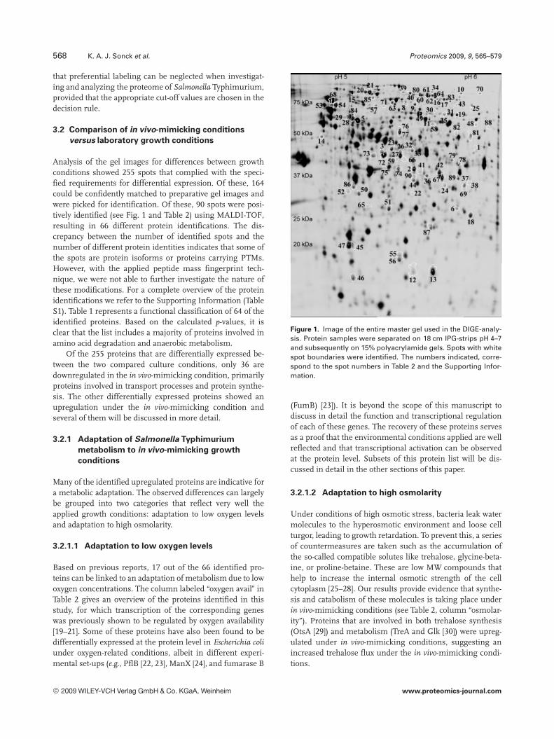
Analysis of the gel images for differences between growthconditions showed 255 spots that complied with the speci-fied requirements for differential expression. Of these, 164could be confidently matched to preparative gel images andwere picked for identification. Of these, 90 spots were posi-tively identified (see Fig. 1 and Table 2) using MALDI-TOF,resulting in 66 different protein identifications. The dis-crepancy between the number of identified spots and thenumber of different protein identities indicates that some ofthe spots are protein isoforms or proteins carrying PTMs.However, with the applied peptide mass fingerprint tech-nique, we were not able to further investigate the nature ofthese modifications. For a complete overview of the proteinidentifications we refer to the Supporting Information (TableS1). Table 1 represents a functional classification of 64 of theidentified proteins. Based on the calculated p-values, it isclear that the list includes a majority of proteins involved inamino acid degradation and anaerobic metabolism.
Of the 255 proteins that are differentially expressed be-tween the two compared culture conditions, only 36 aredownregulated in the in vivo-mimicking condition, primarilyproteins involved in transport processes and protein synthe-sis. The other differentially expressed proteins showed anupregulation under the in vivo-mimicking condition andseveral of them will be discussed in more detail.
3.2.1 Adaptation of Salmonella Typhimurium
metabolism to in vivo-mimicking growth
conditions
Many of the identified upregulated proteins are indicative fora metabolic adaptation. The observed differences can largelybe grouped into two categories that reflect very well theapplied growth conditions: adaptation to low oxygen levelsand adaptation to high osmolarity.
3.2.1.1 Adaptation to low oxygen levels
Based on previous reports, 17 out of the 66 identified pro-teins can be linked to an adaptation of metabolism due to lowoxygen concentrations. The column labeled “oxygen avail” inTable 2 gives an overview of the proteins identified in thisstudy, for which transcription of the corresponding geneswas previously shown to be regulated by oxygen availability[19–21]. Some of these proteins have also been found to bedifferentially expressed at the protein level in Escherichia coliunder oxygen-related conditions, albeit in different experi-mental set-ups (e.g., PflB [22, 23], ManX [24], and fumarase B
Figure 1. Image of the entire master gel used in the DIGE-analy-sis. Protein samples were separated on 18 cm IPG-strips pH 4–7and subsequently on 15% polyacrylamide gels. Spots with whitespot boundaries were identified. The numbers indicated, corre-spond to the spot numbers in Table 2 and the Supporting Infor-mation.
(FumB) [23]). It is beyond the scope of this manuscript todiscuss in detail the function and transcriptional regulationof each of these genes. The recovery of these proteins servesas a proof that the environmental conditions applied are wellreflected and that transcriptional activation can be observedat the protein level. Subsets of this protein list will be dis-cussed in detail in the other sections of this paper.
3.2.1.2 Adaptation to high osmolarity
Under conditions of high osmotic stress, bacteria leak watermolecules to the hyperosmotic environment and loose cellturgor, leading to growth retardation. To prevent this, a seriesof countermeasures are taken such as the accumulation ofthe so-called compatible solutes like trehalose, glycine-beta-ine, or proline-betaine. These are low MW compounds thathelp to increase the internal osmotic strength of the cellcytoplasm [25–28]. Our results provide evidence that synthe-sis and catabolism of these molecules is taking place underin vivo-mimicking conditions (see Table 2, column “osmolar-ity”). Proteins that are involved in both trehalose synthesis(OtsA [29]) and metabolism (TreA and Glk [30]) were upreg-ulated under in vivo-mimicking conditions, suggesting anincreased trehalose flux under the in vivo-mimicking condi-tions.
© 2009 WILEY-VCH Verlag GmbH & Co. KGaA, Weinheim www.proteomics-journal.com

Proteomics 2009, 9, 565–579 569
Table 1. Enrichment of functional classes
Functiona) No. inanalysisb)
p-Valuec)
1 Small molecule metabolism
1.A Degradation
1.A.1 Carbon compounds PduB, PduC, PduE, PduG, PduP, Glk, GarR 7 2.69E 2031.A.2 Amino acids AnsB, TdcB, TdcG, STM4466, STM4467 5 8.31E 205
1.B Energy metabolism
1.B.3 Tricarboxylic acid cycle FumB 1 2.10E 2011.B.5 Pentose phosphate pathway1.B.5.b Nonoxidative branch TalA, TktB 2 2.03E 2031.B.7 Respiration1.B.7.b Anaerobic DmsA, FrdA, FrdB, GlpA, HybC, PflB, TdcE 7 9.06E 205
1.C Central intermediary metabolism Agp, AspA, GlpQ, GcvT 4 2.50E 2021.C.2 Gluconeogenesis PckA 1 5.70E 202
1.D Amino acid biosynthesis1.D.1 Glutamate family STM4465 1 2.44E 2011.D.2 Aspartate family Asd 1 3.18E 2011.D.4 Aromatic amino acid family MaeB, WrbA 2 4.69E 202
1.F Purines, pyrimidines, nucleosides, and nucleotides
1.F.4 Salvage of nucleosides and nucleotides CpdB 1 2.87E 201
1.G Biosynthesis of cofactors, prosthetic groups, and carriers
1.G.13 Cobalamin CbiF 1 2.66E 2011.G.6 Pyridoxine PdxA 1 5.70E 202
2 Broad regulatory functions LsrR, PhoP, YfhA 3 7.15E 201
3 Macromolecule metabolism
3.A Synthesis and modification of macromolecules3.A.2 Ribosomal protein synthesis and modification RpsA 1 5.88E 2013.A.7 DNA replication, restriction/modification, recombination,
and repair Dps1 7.52E 201
3.A.8 Protein translation and modification FusA 1 5.29E 2013.A.9 RNA synthesis, RNA modification, and DNA transcription RsuA 1 3.08E 201
3.B Degradation of macromolecules
3.B.3 Proteins, peptides, and glycopeptides Clpb, IadA 2 7.83E 2023.C Cell envelope OmpW 1 3.57E 2013.C.2 Surface polysaccharides, lipopolysaccharides, and antigens NanA 1 7.16E 201
4 Cell processes
4.A Transport/binding proteins LsrA, LsrB, ZnuA 3 5.67E 2014.A.1 Amino acids and amines ProX, PotF 2 1.96E 2014.A.3 Carbohydrates, organic acids, and alcohols ManX 1 6.94E 2014.A.6 Other FadL, DppA 2 2.28E 201
4.G Detoxification KatE 1 2.76E 201
4.I Pathogenicity SipA, TypA 2 9.58E 201
5 Other
5.F Adaptations and atypical conditions OsmY, OtsA, TreA 3 4.79E 203
5.I Unknown GpmI, LsrF, STM1560, UcpA, YghA 5 8.31E 201
a) Functions according to the Sanger classification for S. Typhi. For two proteins, no Salmonella Typhi ortholog(FhuA) or functional class (MdoB) could be found. They were excluded from the functional class enrichmentcalculations.
b) Number of proteins identified in this study in a functional class.c) Probability of retrieving a certain number of proteins in a functional class by chance. The lower the p-value, the
more the functional class is enriched in the experiment. p-Values below E-04 are indicated in bold.
© 2009 WILEY-VCH Verlag GmbH & Co. KGaA, Weinheim www.proteomics-journal.com

570 K. A. J. Sonck et al. Proteomics 2009, 9, 565–579
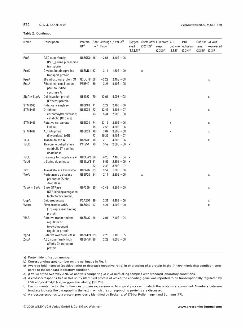
Table 2. Overview of the proteins identified in this study
Name Description ProteinIDa)
Spotno.b)
AverageRatioc)
p-valued) Oxygenavail.(3.2.1.1)e)
Osmolarity(3.2.1.2)f)
Fumarateresp.(3.2.2)f)
ADIpathway(3.2.3)f)
PDLutilization(3.2.4)f)
Quorumsens.(3.2.5)f)
In vivoexpressed(3.3)g)
Agp Glucose-1-phosphataseprecursor (G1Pase)
O33921 1 2.10 1.10E 205 x
AnsB PeriplasmicL-asparaginase II
Q7CPT7 2 3.89 3.00E 205 x x x
Asd Aspartate-semialdehydedehydrogenase (ASAdehydrogenase)(ASADH)
P0A1F8 3 2.48 1.40E 207 x
AspA Aspartate ammonia-lyase Q7CPA1 4 3.79 1.10E 204 x x x5 3.48 2.20E 204
CbiF Cobalt-precorrin-4C(11)-methyltransferase
P0A2G9 6 4.61 1.50E 209 x x x
ClpB Chaperone ClpB Q7CQ01 7 22.04 8.30E 205 xCpdB 20,30-Cyclic nucleotide 20
phosphodiesteraseprecursor
P26265 8 2.06 2.10E 206 x9 2.57 6.60E 206
DmsA Anaerobic DMSOreductase subunit A
Q8ZQD3 10 8.45 1.60E 204 x x
DppA Dipeptide transportprotein
Q8ZLA9 11 22.16 3.70E 205 x x
Dps Stress responseDNA-binding protein;starvation inducedresistance to H2O2
Q7CQV9 12 5.83 3.80E 208 x
13 6.36 7.30E 208FadL Long-chain fatty acid
transport proteinprecursor
Q8ZNA5 14 23.30 4.10E 207
FhuA Outer membrane proteinreceptor/transporter forferrichrome colicin M
Q8ZRQ2 15 22.95 4.80E 205 x
FrdA Fumarate reductase Q7CP97 16 4.28 1.10E 204 x x x17 3.73 2.60E 204
FrdB Fumarate reductase Q8ZKB4 18 3.38 1.20E 204 x x xFumB Fumarase B Q8ZKE2 19 7.96 3.70E 205 x x xFusA Elongation factor EF-2 P0A1H3 20 22.01 3.30E 206 x
21 22.16 8.40E 207GarR Tartronate semialdehyde
reductase (TSAR)Q8ZLV8 22 2.69 2.00E 206 x x
Gcvt Aminomethyltransferase(Glycine cleavagesystem T protein).
P64222 23 3.43 2.50E 207 x
Glk Glucokinase(Glucose kinase).
Q93IM5 24 2.27 1.30E 205 x x
GlpA sn-G3P dehydrogenaselarge subunit
Q8ZNG5 25 3.35 1.10E 204 x x x
GlpQ Periplasmic glycero-phosphodiesterphosphodiesterase
Q8ZNG7 26 2.56 2.10E 204 x x x27 2.36 9.90E 206
GpmI 2,3-Bisphosphoglycerate-independentphosphoglyceratemutase
Q8ZL56 28 3.58 4.10E 205 x29 2.80 3.40E 204
HybC Hydrogenase-2; largesubunit
Q8ZM16 30 22.65 1.00E 204 x x x31 4.97 2.20E 205
© 2009 WILEY-VCH Verlag GmbH & Co. KGaA, Weinheim www.proteomics-journal.com

Proteomics 2009, 9, 565–579 571
Table 2. Continued
Name Description ProteinIDa)
Spotno.b)
AverageRatioc)
p-valued) Oxygenavail.(3.2.1.1)e)
Osmolarity(3.2.1.2)f)
Fumarateresp.(3.2.2)f)
ADIpathway(3.2.3)f)
PDLutilization(3.2.4)f)
Quorumsens.(3.2.5)f)
In vivoexpressed(3.3)g)
IadA Isoaspartyl dipeptidase Q8ZJZ8 32 2.67 4.30E 205 x x33 5.26 1.60E 207
KatE Catalase HPII Q9L328 34 5.52 2.50E 208 xLsrA = Ego Putative ABC-type sugar
aldose transport systemQ8ZKQ4 35 3.28 7.50E 207 x
LsrB = YneA Putative sugar transportprotein
Q8ZKQ1 36 2.95 1.00E 206 x x
LsrF = YneB Putative fructose-1; 6-bisphosphate Aldolase
Q8ZKQ0 37 4.97 9.80E 207 x x x
LsrR = YdeW Putative transcriptionalrepressor
Q8ZKQ5 38 2.13 7.50E 205 x
MaeB NADP-dependent malicenzyme (NADP-ME)
Q9ZFV8 39 2.22 9.10E 207 x40 2.28 4.20E 206
ManX Mannose-specificenzyme IIAB
Q8ZP03 41 2.26 2.40E 204 x42 2.20 5.00E 205
MdoB = OpgB Phosphoglyceroltransferase I
Q8ZJX6 43 22.24 2.40E 206
NanA N-acetylneuraminate lyase(N-acetylneuraminicacid aldolase)
Q8ZLQ6 44 4.88 1.80E 206 x
OmpW Outer membraneprotein W precursor
Q8ZP50 45 2.08 1.20E 204 x x46 2.27 1.40E 204
OsmY Hyperosmotically inducibleperiplasmic protein
Q7CP68 47 2.96 2.30E 206 x x x
OtsA Alpha; a-trehalose-phosphate synthase[UDP-forming]
P0A1Q0 48 2.99 2.90E 206 x
PckA Phosphoenolpyruvatecarboxykinase [ATP](PEP carboxykinase)
P41033 49 2.19 1.10E 205 x
PduB Propanediol utilizationprotein
P37449 50 5.63 8.60E 209 x x x51 5.59 5.30E 20652 2.25 9.90E 206
PduC Glycerol dehydrataselarge subunit
P37450 53 2.42 2.00E 204 x x x54 6.39 2.40E 208
PduE Propanediol utilizationdehydratase; smallsubunit
O31042 55 9.71 2.40E 208 x x x56 8.43 6.30E 208
PduG Propanediol utilizationdiol dehydratasereactivation protein
O31043 57 4.06 1.10E 205 x x x
PduP Propanediol utilizationCoA-dependentpropionaldehydedehydrogenase
Q9XDN1 58 3.25 1.70E 204 x x x
PdxA 4-Hydroxythreonine-4-phosphatedehydrogenase 1
P58717 59 2.21 4.10E 205
PflB Pyruvate formate lyase I;induced anaerobically
Q7CQU1 60 2.16 2.80E 203 x x61 2.62 6.90E 20462 2.73 1.30E 20363 4.95 1.30E 20464 2.62 1.60E 203
PhoP Virulence transcriptionalregulatory protein PhoP
P14146 65 3.40 2.20E 208 x
© 2009 WILEY-VCH Verlag GmbH & Co. KGaA, Weinheim www.proteomics-journal.com
572 K. A. J. Sonck et al. Proteomics 2009, 9, 565–579
Table 2. Continued
Name Description ProteinIDa)
Spotno.b)
AverageRatioc)
p-valued) Oxygenavail.(3.2.1.1)e)
Osmolarity(3.2.1.2)f)
Fumarateresp.(3.2.2)f)
ADIpathway(3.2.3)f)
PDLutilization(3.2.4)f)
Quorumsens.(3.2.5)f)
In vivoexpressed(3.3)g)
PotF ABC superfamily(Peri_perm); putrescinetransporter
Q8ZQK0 66 22.58 8.50E 205
ProX Glycine/betaine/prolinetransport protein
Q8ZML1 67 3.14 1.50E 209 x
RpsA 30S ribosomal protein S1 Q7CQT9 68 22.23 2.40E 208 xRsuA Ribosomal small subunit
pseudouridinesynthase A
P65840 69 3.24 9.10E 205
SipA = SspA Cell invasion protein(Effector protein)
Q56027 70 13.01 5.00E 208 x
STM1560 Putative a amylase Q8ZPF0 71 2.22 2.70E 206STM4465 Ornithine
carbamoyltransferase;catabolic (OTCase)
Q8ZK35 72 12.42 4.10E 207 x x73 5.44 2.20E 208
STM4466 Putative carbamatekinase
Q8ZK34 74 27.19 3.30E 206 x x75 2.58 4.30E 206
STM4467 ADI (Argininedihydrolase) (AD)
Q8ZK33 76 7.07 3.80E 208 x x77 29.29 5.40E 207
TalA Transaldolase A Q8ZN83 78 2.19 4.30E 206 xTdcB Threonine dehydratase
catabolic (Threoninedeaminase)
P11954 79 5.52 2.00E 206 x
TdcE Pyruvate formate-lyase 4 Q8ZLW2 80 4.25 7.40E 204 xTdcG L-Serine deaminase Q8ZLW3 81 6.96 2.30E 206 x
82 2.43 3.50E 207TktB Transketolase 2 isozyme Q8ZN82 83 2.57 1.00E 206TreA Periplasmic trehalase
precursor (Alpha;-trehalase)
Q8ZP20 84 2.11 2.90E 206 x x
TypA = BipA BipA GTPase(GTP-binding elongationfactor family protein)
Q9F2E0 85 22.48 4.90E 209 x
UcpA Oxidoreductase P0A2D1 86 3.32 4.30E 206 xWrbA Flavoprotein wrbA
(Trp repressor bindingprotein)
Q8ZQ40 87 4.21 4.90E 209 x
YfhA Putative transcriptionalregulator oftwo-componentregulator protein
Q8ZN32 88 2.01 7.40E 204
YghA Putative oxidoreductase Q8ZM09 89 2.25 1.10E 205ZnuA ABC superfamily high
affinity Zn transportprotein
Q8ZNV8 90 2.22 5.00E 206
a) Protein identification number.b) Corresponding spot number on the gel image in Fig. 1.c) Average fold increase (positive ratio) or decrease (negative ratio) in expression of a protein in the in vivo-mimicking condition com-
pared to the standard laboratory condition.d) p-Value of the two-way-ANOVA analysis comparing in vivo-mimicking samples with standard laboratory conditions.e) A crosscorresponds to a in this study identified protein of which the encoding gene was reported to be transcriptionally regulated by
FNR and/or ArcA/B (i.e., oxygen availability) [19, 20].f) Environmental factor that influences protein expression or biological process in which the proteins are involved. Numbers between
brackets indicate the paragraph in the text in which the corresponding proteins are discussed.g) A crosscorresponds to a protein previously identified by Becker et al. [76] or Rollenhagen and Bumann [77].
© 2009 WILEY-VCH Verlag GmbH & Co. KGaA, Weinheim www.proteomics-journal.com

Proteomics 2009, 9, 565–579 573
Further protection against adverse osmotic conditionscan be achieved by the uptake of other osmoprotectants suchas glycine-betaine and proline-betaine. Evidence of such ac-tivity is given by the expression level of ProX, which is upre-gulated more than three-fold under in vivo-mimicking con-ditions. ProX is the periplasmic binding protein of a high-affinity ATP-binding cassette (ABC)-type transporter for gly-cine, betaine, and proline [31]. In E. coli, ProX was reported tobe expressed under anaerobic conditions at high pH [32].The other constituents of the transporter, ProV and ProW,were not retrieved in our analysis.
Finally, also the elevated expression level of OsmY indi-cates that Salmonella Typhimurium reacts against osmoticstress under in vivo-mimicking conditions. OsmY, also relat-ed to oxygen limitation (see Table 2, column labeled “oxygenavail”), is known to be induced by conditions of hyper-osmolarity, but the exact function of the protein is not clear[33, 34].
Some of the retrieved proteins, such as OsmY, ProX,KatE, TalA, and TktB, were also identified by Weber et al. [34],who studied proteomic alterations of E. coli due to osmoticstress under aerobic and anaerobic growth conditions.
3.2.2 Fumarate is a key player in metabolic processes
during in vivo-mimicking growth
Due to the lack of oxygen during growth under limiting oxy-gen concentrations, Salmonella must use alternative electronacceptors such as nitrate, DMSO, or fumarate. In case of invivo-mimicking growth, it appears that fumarate serves aselectron acceptor. Proteins that partially constitute thefumarate reductase enzyme, FrdA and FrdB, encoded by thefrdABCD operon [35], were found to be upregulated under invivo-mimicking conditions (see Table 2, column “fumarateresp.”). Interestingly, two spots with different pI were detect-ed for FrdA, suggesting a PTM affecting the iso-electric pointof the protein. FrdC and FrdD have a pI of 9.87 and 9.10,respectively, and were consequently not identified.
In a cell, a very tight hierarchical regulatory control existsconcerning the use of alternative electron acceptors. Fuma-rate is at the very bottom of this hierarchy. Indeed, whenoxygen or nitrate is present, expression of the frdABCDgenes is repressed [35, 36]. In this respect, the upregulationof FrdA and FrdB suggests that no sufficient amounts ofoxygen, nitrate, or nitrite were left in the growth medium atthe time of sampling.
E. coli can use different electron donors in combinationwith fumarate as the terminal electron acceptor. Knownsubstrates that can serve this purpose are H2 and glycerol-3-phosphate (G3P) [37, 38, www.ecosal.org]. Proteins that arepart of the corresponding dehydrogenases were found to bedifferentially expressed in the Salmonella proteome investi-gated. We identified two spots of HybC, forming the largesubunit of hydrogenase 2, that showed an opposite expres-sion pattern under in vivo-mimicking conditions: the morebasic spot is upregulated almost five-fold, while the more
acidic spot is downregulated 2.5-fold and has a differentrelative MW. This suggests that regulation of hydrogenase 2expression occurs both at the transcriptional and transla-tional level. Another dehydrogenase, GlpA, was found to beupregulated under in vivo-mimicking conditions. GlpA con-stitutes the large part of the sn-G3P dehydrogenase [38]. Incooperation with GlpB and GlpC, it oxidizes G3P to dihy-droxyacetone phosphate (DHAP) under anaerobic circum-stances. Also the GlpQ enzyme, responsible for the conver-sion of G3P-esters to G3P, was found to be upregulatedunder in vivo-mimicking conditions. The upregulation ofboth dehydrogenases, combined with the elevated FrdA andFrdB expression in our experiment, substantiates the centralrole of fumarate respiration in the metabolism of S. Typhi-murium during growth under in vivo-mimicking conditions.
Additionally, fumarate is an intermediate in the tri-carboxylic acid (TCA) cycle. Under anaerobic conditions, thispathway is altered to favor reactions in the direction of suc-cinate, from oxaloacetate via malate and fumarate, called thereductive branch of the TCA. In rich media, like LB, thispathway is fed by C4-dicarboxylates and related compoundssuch as malate and aspartate (www.ecosal.org). Both of thesecan be converted to fumarate in a single reaction step [36,39]. The enzymes responsible for these conversions, FumBand the aspartate ammonia lyase (AspA), respectively, wereboth found to be upregulated in the in vivo-mimicking con-ditions. The enzyme that catalyses the reaction from aspar-agine to aspartate, the periplasmic asparaginase II (AnsB),was upregulated as well in our conditions (see Table 2) [36].
We investigated the role of fumarate respiration in ourapplied growth conditions by adding HQNO, an inhibitor ofthe fumarate reductase enzyme [40], to Salmonella cultures.Under in vivo-mimicking, addition of HQNO indeedincreased the generation time, in a concentration dependentmanner (Table 3), suggesting that fumarate respiration isimportant for the fitness of bacterium under these condi-tions. Under laboratory conditions, no major effect of HQNOon bacterial growth was observed (Table 3).
There is some controversy on the role of fumarate utili-zation in virulence of S. Typhimurium [41]. Nevertheless, ithas been shown that the use of hydrogen as an electrondonor is linked to virulence in S. Typhimurium [42]. So, thepresumed importance of fumarate respiration might be thereflection of a need for efficient hydrogen utilization by thebacterium in vivo. In other pathogens, the involvement ofanaerobic fumarate metabolism in virulence was readilydemonstrated [43, 44].
3.2.3 S. Typhimurium possesses and expresses an
arginine deiminase pathway for anaerobic
arginine catabolism
The highest upregulated protein under in vivo-mimickingconditions appeared to be the product from a predictedORF designated STM4467. This protein, a putative ADI, isencoded by the first gene in an operon, to which also the
© 2009 WILEY-VCH Verlag GmbH & Co. KGaA, Weinheim www.proteomics-journal.com

574 K. A. J. Sonck et al. Proteomics 2009, 9, 565–579
Table 3. Generation time of Salmonella grown in the presence ofHQNO, an inhibitor of the fumarate reductase enzyme
HQNO concentration (mM)
0 100 500In vivo-mimicking 3.2 6 0.1a) 3.6 6 0.2a) 4.0 6 0.1a)
Laboratoryconditions
1.1 6 0.1a) 1.0 6 0.2a) 1.0 6 0.2a)
a) Generation time of Salmonella grown in the presence ofHQNO (concentration indicated), calculated as specified inSection 2, expressed in hours (h). The value is the mean ofthree biological replicates 6 SD.
STM4466 (encoding a carbamate kinase) and STM4465(encoding an ornithine carbamoyltransferase, OCTase)genes belong. Together, these three genes encode a pathwayfor anaerobic L-arginine catabolism, called the ADI pathway[45]. A previous study suggested the existence of such apathway in S. Typhimurium, based on the retrieval ofSTM4467 transposon mutants impaired in growth suppres-sion in vitro [46]. Protein spots corresponding to all threegenes were retrieved as being highly upregulated under invivo-mimicking conditions (see Table 2, column “ADI path-way”). Induction of the ADI pathway in S. Typhimurium wasfurther confirmed by a more than ten-fold increased OCTasespecific activity measured in extracts from cells grown underin vivo-mimicking conditions (Table 4).
The question then rises why Salmonella would need suchan ADI pathway in vivo. Several hypotheses can be made.The ADI pathway might serve an energy scavenging rolesince one molecule of ATP is produced per molecule of argi-nine. Concomitantly, L-ornithine is formed, which can bedegraded to the polyamine putrescine. Although polyaminescan be synthesized through alternative pathways, the one-step reaction from ornithine to putrescine might be moreadvantageous under in vivo-mimicking conditions. Poly-amines have diverse functions, ranging from maintainingoptimal conformation of negatively charged nucleic acids,constituting the outer membrane, and protection againstoxidative and acid stress to contributing to pathogenesis [47].Additionally, the ADI pathway might play a role in escapingthe host defense system. Indeed, in eukaryotic cells, L-argi-nine is the substrate of several enzymes, among which thenitric oxide synthases [48, 49]. Nitric oxide synthases (NOS)are responsible for the production of nitric oxide (NO), amessenger molecule that has been shown to be involved inmany eukaryotic physiological processes. One particularNOS, the inducible NOS (iNOS), is inducible by bacteriallipopolysaccharides, outer membrane proteins or cytokinesin a wide range of tissues and cells, including intestinal epi-thelial cells [49–51]. The large amounts of NO produced byiNOS in epithelial cells are usually considered to benefit thehost, as they can have a killing effect on the infecting bacte-rium [52, 53]. However, if L-arginine, the primary substrate of
Table 4. OCTase specific activity of Salmonella grown under dif-ferent conditions
OCTase specific activity
In vivo-mimicking 78.5 6 10.7 mmol/h?mg proteina)
Laboratory conditions 5.9 6 2.8 mmol/h?mg proteina)
a) OCTase activity determined as specified in Section 2, expres-sed as micromoles of product formed per hour per milligramof protein. The value is the mean of four biological replica-tes 6 SD.
the iNOS enzyme is limiting, only small amounts of NO willbe produced. Therefore, the expression of a full ADI pathwayand hence the scavenging of L-arginine from the amino acidpool in the host, could be a mechanism to avoid accumula-tion of NO and killing of Salmonella Typhimurium. Such acounteraction of NO production has already been reportedfor Helicobacter pylori [54]. Additionally, Helicobacter inducesarginase II in infected macrophages, resulting in increasedspermine production [55]. Spermine post-transcriptionallyrepresses iNOS production [56]. This is a second way ofpathogen-mediated repression of NO production by the host.Future experiments will have to reveal whether the sameholds true for Salmonella.
3.2.4 Salmonella upregulates proteins for
1,2-propanediol utilization under
in vivo-mimicking conditions
The largest set of proteins belonging to one single operonidentified in this study consists of the products of the pduoperon (see Table 2, column “PDL utilization”). Genes of thisoperon are required for the vitamin B12-dependent catabo-lism of 1,2-propanediol (PDL). The first step of PDL break-down is the conversion of PDL to propionaldehyde by thepduCDE encoded propanediol dehydratase. This enzymerequires the cofactor adenosylcobalamin (AdoCbl, alsoknown as vitamin B12) [57, 58]. The genes responsible for denovo synthesis of AdoCbl are arranged in a second operon,cob, which is adjacent to the pdu operon but is divergentlytranscribed [57]. Transcription of both operons was shown tobe upregulated under conditions of low oxygen concentra-tions [59–61]. Our experiment shows that this upregulationis also persistent at the translational level, as all identifiedPdu proteins were at least 2.25 times more expressed in thein vivo-mimicking growth condition. Also the CbiF protein,involved in de novo vitamin B12 synthesis and part of the coboperon, was found to be upregulated.
The pdu genes have been reported to be involved in S.Typhimurium pathogenesis [62–65]. Additionally, the cob-cbi-pdu gene cluster has been shown to be necessary for multi-plication of S. Typhimurium inside macrophages [66].Another interesting observation is the fact that propionate(also produced during PDL degradation), inhibits transcrip-
© 2009 WILEY-VCH Verlag GmbH & Co. KGaA, Weinheim www.proteomics-journal.com
Proteomics 2009, 9, 565–579 575
tion of the hilA gene, the master transcriptional regulator ofgenes needed for Salmonella invasion [2, 67]. Based on all thisevidence and our own results, it could be hypothesized thatexpression of the pdu operon is involved in the transitionfrom the extracellular intestinal environment to an intracel-lular phagocytic lifestyle.
3.2.5 Quorum sensing proteins are upregulated
under in vivo-mimicking conditions
Our proteomics results indicate that Salmonella Typhimur-ium SL1344 upregulates the uptake of the autoinducer-2 (AI-2) molecule under in vivo-mimicking conditions. AI-2 is asignaling molecule used for quorum sensing, which is aterm used to describe the coordinated behavior of a bacterialpopulation upon detection of a certain threshold of signalingmolecules, called AI. Different types of quorum sensingmechanisms have been reported, one of them being AI-2dependent quorum sensing [68].
Four proteins that are directly involved in type-2 quorumsensing were found to be upregulated, i.e., in order of aver-age ratio magnitude: LsrF, LsrA, LsrB, and LsrR (see Table 2,column “quorum sens.”). In Salmonella, LsrA and LsrB arepart of the AI-2 uptake transporter. Once AI-2 is taken up bythe cell and phosphorylated (AI-2-P), it binds to the tran-scriptional repressor LsrR. As such, it alleviates the repres-sion on the lsr-operon and allows increased transcription ofthe lsr-genes, resulting in an increased internalization of AI-2[69, 70]. Downstream metabolism of AI-2-P is thought to beexecuted by the LsrF, LsrE, and LsrG proteins [69–71]. Arecent paper by Xavier et al. [72] describes the enzymatic ac-tivity of LsrG. Phosphorylated AI-2 is enzymatically con-verted to phosphoglycolic acid and an unknown three-carboncompound. In view of the upregulation of pdu genes men-tioned above, one could speculate that this C3 compoundmight be PDL or a precursor of this metabolite.
3.2.6 Identification of “classical” virulence proteins
We found SipA as being 13 times more expressed under invivo-mimicking conditions. SipA is one of the known effectorproteins that are translocated to the host cell during invasion[73]. Another important pathogenicity related protein thatwas found to be upregulated more than three-fold under invivo-mimicking conditions is the transcriptional regulatorPhoP, which is a part of a two-component system that sensesextracellular Mg21 concentrations [74]. This observationmight be explained by our finding of increased LsrA andLsrB expression under in vivo-mimicking conditions (seeSection 3.2.5) and the recent report on elevated PhoP proteinexpression in the presence of AI-2 molecules [75].
Our applied conditions of low oxygen concentrations andhigh osmolarity are known to induce the expression of viru-lence genes of S. Typhimurium, necessary for invasion [11].Therefore, the fact that only two virulence proteins wereidentified in this proteomic study may have other reasons, as
explained below. Moreover, based on a paper by Becker et al.[76] it is reasonable to state that the flexibility of Salmonella toadapt and switch metabolic processes upon encounter ofnew environmental niches is at least as important in con-tributing to virulence and infection as the expression of spe-cific virulence factors.
3.3 In vivo-mimicking conditions versus genuine in
vivo conditions: A comparison
To validate our in vivo-mimicking conditions, a cross-checking of our results with published results obtained fromgenuine in vivo studies was conducted (see Table 2, column“in vivo expressed”). In vivo-expression of the Pdu proteinsand HybC was already mentioned above [42, 62].
In a recent publication by Rollenhagen and Bumann [77],results were presented from a promoter trap experimentperformed to identify Salmonella promoters that show highactivity during enteritis. Of the 21 highly active promotersidentified, 4 drive the expression of genes or operons encod-ing proteins that were also retrieved in our study (PfocA,expressing PflB, PglpT, expressing GlpQ, PsicA, expressingSipA and PfhuA, expressing FhuA). The others might be mis-sed in our analysis for reasons specified below. Furthermore,the authors mention that during enteritis, Salmonella pre-dominantly expresses genes involved in general physiologi-cal functions such as nutrient utilization and energy conver-sion, while virulence genes that are mostly associated withSalmonella Pathogenicity Island-1 account for only a minorfraction of gene expression. As proof of principle, a Salmo-nella mutant of fnr, encoding a major oxygen responsivetranscriptional regulator, was tested and showed a strongvirulence defect supporting the high relevance of anaerobicSalmonella metabolism in the enteritis model [77]. Thislargely parallels the observations in our in vivo-mimickingproteomics analysis, where most of the identified proteinsare indeed involved in metabolic processes.
Additionally, comparison of our results with the resultsof an extensive proteomic analysis of in vivo-expressed pro-teins in S. Typhimurium cells recovered from mouse spleenor caecum by Becker et al. [76], revealed some more overlaps.Two-thirds of the proteins identified in our study were alsoretrieved in the in vivo study, meaning that these proteins aregenuinely expressed in vivo during infection. Two more pro-teins that were differentially expressed in our study (PdxAand MdoB) were assumed to be expressed in vivo, althoughthe authors did not identify them in their analysis. Of the 44proteins commonly identified in both studies, 31 representmetabolic enzymes. The large amount of enzymes amongthe identified proteins in both analyses (64% in the in vivostudy, 70% in our study), emphasizes again that the ability toadapt metabolism is of importance during infection. Inter-estingly, Becker et al. also identified the enzymes of the ADIpathway as being expressed in vivo in an enteritis-type infec-tion model, further suggesting the importance of this path-way in vivo.
© 2009 WILEY-VCH Verlag GmbH & Co. KGaA, Weinheim www.proteomics-journal.com
576 K. A. J. Sonck et al. Proteomics 2009, 9, 565–579
In summary, for 49 out of the 66 identified proteins inour in vivo-mimicking study, genuine in vivo expression hasbeen demonstrated.
4 Concluding remarks
The proteins identified in this study represent very well theapplied growth conditions and allow us to gain some globalinsight in the processes occurring when Salmonella is adapt-ing to in vivo-mimicking conditions. However, some of theproteins that one would expect to be differentially expressedunder the conditions in our experiment, and were indeedidentified in similar studies, are not retrieved in our analysis.Depending on the characteristics of the missing proteins,different explanations are possible. First, every gel-basedproteomic experiment is limited by the pI and MW bound-aries of the gels. As a consequence, only a subset of the totalproteome of S. Typhimurium expressed under these condi-tions is visualized and investigated. Second, not all proteinsthat showed differential expression were picked and sub-mitted to MS, e.g., when no confident match was found be-tween analytical and preparative 2-D gels. Third, not everyspot that was analyzed by MS yielded a positive identifica-tion, e.g., due to insufficient material. A fourth possible rea-son for some missing proteins in our study can be related toour sample preparation method. We used a total cell lysatefor separation in the first and second dimensions. Mem-brane proteins are known to be underrepresented in suchprotein samples [78, 79]. This could explain why integralmembrane proteins or membrane associated proteins arealmost absent in our list of identified proteins. Moreover,most membrane proteins also possess pI values above 7. Oursample preparation method may also contribute to the factthat only a few known virulence factors were retrieved in thisanalysis. As a large part of the proteins responsible for inva-sion are being secreted, these proteins might be under-represented or absent in a total cell lysate. Therefore, whenone particularly would like to investigate virulence proteinsin a proteomics set-up, an analysis of the secretome isrequired. However, this was not the primary scope of thisstudy. Finally, many important regulatory proteins, such asFNR and HilA are missing in our list of identified proteins,while some of these were retrieved in the promoter trapstrategy discussed above [77]. This is probably due to theintrinsic nature of these proteins: the abundance of reg-ulatory proteins is often very low and these proteins maytherefore be missed during protein detection or be maskedby other more abundant proteins with similar pI and MW[80, 81]. Moreover, regulatory proteins might show only lim-ited differential expression, related to their downstreammultiplicator effect. Taken together, these arguments makeclear that with a reasonable amount of effort, the “complete”retrieval and identification of “all” proteins that are differen-tially expressed under certain environmental conditions, isnot very likely. In this respect, analyses at the transcriptome
and proteome level are complementary. Interestingly, ourstudy revealed two or more spots for a number of proteins,also pointing out the importance of complementing tran-scriptome analysis with proteome analysis. The significanceof these different isoforms or post-translationally modifiedproteins can only be understood through further analysis.
To better understand the specific adaptation of thepathogen Salmonella, it would be interesting to compare theprotein changes observed during in vivo-mimicking growthof Salmonella to previously described results obtained insimilar proteomic experiments with, e.g., the closely relatedE. coli. However, due to the intrinsic incompleteness of pro-teomic experiment results (as explained above) and the largevariation in experimental set-up in the different studies, asystematic insight in species-specific expression patterns,based on previously published results, is not feasible. Never-theless, as indicated throughout the text, some overlap be-tween Salmonella and E. coli can be found.
Interestingly, many of the identified proteins in ourstudy, using an in vivo-mimicking set-up, were also shown tobe differentially expressed in real in vivo experiments (e.g.[42, 62–64, 76]). This strengthens the relevance of our in vivo-mimicking set-up in providing meaningful informationregarding in vivo situations, without initially having to copewith the complexity and difficulties of an in vivo environ-ment. Therefore, it will be interesting to verify new resultsfrom this in vivo-mimicking experiment in a genuine in vivostudy, e.g., the function of a Salmonella ADI pathway in vivo.
We thank Dr. E. Witters for the MALDI-TOF measurementsand protein identifications. This research was supported by theGBOU-SQUAD-20160 of the IWT Vlaanderen and by theCentre of Excellence SymBioSys (Research Council K.U. LeuvenEF/05/2007). G. Kint and S. De Keersmaecker are ResearchAssistants of the Research Foundation-Flanders (FWO Vlaande-ren), aspirant and postdoctoral fellow, respectively.
The authors have declared no conflict of interest.
5 References
[1] Hohmann, E. L., Nontyphoidal salmonellosis. Clin. Infect. Dis.2001, 32, 263–269.
[2] Bajaj, V., Lucas, R. L., Hwang, C., Lee, C. A., Co-ordinate reg-ulation of Salmonella Typhimurium invasion genes by envi-ronmental and regulatory factors is mediated by control ofhilA expression. Mol. Microbiol. 1996, 22, 703–714.
[3] Altier, C., Genetic and environmental control of Salmonellainvasion. J. Microbiol. 2005, 43, 85–92.
[4] Abrahams, G. L., Hensel, M., Manipulating cellular transportand immune responses: Dynamic interactions betweenintracellular Salmonella enterica and its host cells. CellMicrobiol. 2006, 8, 728–737.
[5] Frymier, J. S., Reed, T. D., Fletcher, S. A., Csonka, L. N.,Characterization of transcriptional regulation of the kdp
© 2009 WILEY-VCH Verlag GmbH & Co. KGaA, Weinheim www.proteomics-journal.com

Proteomics 2009, 9, 565–579 577
operon of Salmonella Typhimurium. J. Bacteriol. 1997, 179,3061–3063.
[6] Fletcher, S. A., Csonka, L. N., Fine-structure deletion analysisof the transcriptional silencer of the proU operon of Salmo-nella Typhimurium. J. Bacteriol. 1995, 177, 4508–4513.
[7] Wei, Y., Miller, C. G., Characterization of a group of anaero-bically induced, fnr-dependent genes of Salmonella Typhi-murium. J. Bacteriol. 1999, 181, 6092–6097.
[8] Chen, P., Andersson, D. I., Roth, J. R., The control region ofthe pdu/cob regulon in Salmonella Typhimurium. J. Bacter-iol.1994, 176, 5474–5482.
[9] Hoiseth, S. K., Stocker, B. A., Aromatic-dependent Salmo-nella Typhimurium are nonvirulent and effective as live vac-cines. Nature 1981, 291, 238–239.
[10] Sambrook, J., Russell, D. W., Molecular Cloning a Laborato-ry Manual, Cold Spring Harbor Laboratory Press, ColdSpring Harbor 2001.
[11] Lee, C. A., Falkow, S., The ability of Salmonella to entermammalian cells is affected by bacterial growth state. Proc.Natl. Acad. Sci. USA 1990, 87, 4304–4308.
[12] Benjamini, Y., Hochberg, Y., On the adaptive control of thefalse discovery rate in multiple testing with independentstatistics. J. Educ. Behav. Stat. 2000, 25, 60–83.
[13] Shevchenko, A., Wilm, M., Vorm, O., Mann, M., Mass spec-trometric sequencing of proteins from silver-stained poly-acrylamide gels. Anal. Chem. 1996, 68, 850–858.
[14] Monsieurs, P., De Keersmaecker, S., Navarre, W. W., Bader,M. W. et al., Comparison of the PhoPQ regulon in Escher-ichia coli and Salmonella Typhimurium. J. Mol. Evol. 2005,60, 462–474.
[15] Broman, K., Stalon, V., Wiame, J. M., The duplication of ar-ginine catabolism and the meaning of the two ornithinecarbamoyltransferases in Bacillus licheniformis. Biochem.Biophys. Res. Commun. 1975, 66, 821–827.
[16] Lowry, O. H., Rosebrough, N. J., Farr, A. L., Randall, R. J.,Protein measurement with the Folin phenol reagent. J. Biol.Chem. 1951, 193, 265–275.
[17] Ward, J. E., Jr., Lutkenhaus, J., Overproduction of FtsZinduces minicell formation in E. coli. Cell 1985, 42, 941–949.
[18] Winslow, R. L., Cortassa, S., Greenstein, J. L., Using modelsof the myocyte for functional interpretation of cardiac pro-teomic data. J. Physiol. 2005, 563, 73–81.
[19] Lynch, A. S., Lin, E. C. C, in: Neidhardt, F. C. (Ed.), Responsesto Molecular Oxygen in Escherichia coli and Salmonella,ASM Press, Washington DC 1996, pp. 1526–1538.
[20] Sawers, G., The aerobic/anaerobic interface. Curr. Opin.Microbiol. 1999, 2, 181–187.
[21] Fink, R. C., Evans, M. R., Porwollik, S., Vasquez-Torres, A. etal., FNR is a global regulator of virulence and anaerobicmetabolism in Salmonella enterica serovar Typhimurium(ATCC 14028s). J. Bacteriol. 2007, 189, 2262–2273.
[22] Smith, M. W., Neidhardt, F. C., Proteins induced by anaero-biosis in Escherichia coli. J. Bacteriol. 1983, 154, 336–343.
[23] Peng, L., Shimizu, K., Global metabolic regulation analysisfor Escherichia coli K12 based on protein expression by 2-dimensional electrophoresis and enzyme activity measure-ment. Appl. Microbiol. Biotechnol. 2003, 61, 163–178.
[24] Blankenhorn, D., Phillips, J., Slonczewski, J. L., Acid- andbase-induced proteins during aerobic and anaerobic growth
of Escherichia coli revealed by two-dimensional gel electro-phoresis. J. Bacteriol. 1999, 181, 2209–2216.
[25] Wood, J. M., Bremer, E., Csonka, L. N., Kraemer, R. et al.,Osmosensing and osmoregulatory compatible solute accu-mulation by bacteria. Comp. Biochem. Physiol. A Mol.Integr. Physiol. 2001, 130, 437–460.
[26] Sleator, R. D., Hill, C., Bacterial osmoadaptation: The role ofosmolytes in bacterial stress and virulence. FEMS Microbiol.Rev. 2002, 26, 49–71.
[27] Kempf, B., Bremer, E., Uptake and synthesis of compatiblesolutes as microbial stress responses to high-osmolalityenvironments. Arch. Microbiol. 1998, 170, 319–330.
[28] Balaji, B., O’Connor, K., Lucas, J. R., Anderson, J. M.,Csonka, L. N., Timing of induction of osmotically controlledgenes in Salmonella enterica Serovar Typhimurium, deter-mined with quantitative real-time reverse transcription-PCR.Appl. Environ. Microbiol. 2005, 71, 8273–8283.
[29] Giaever, H. M., Styrvold, O. B., Kaasen, I., Strom, A. R., Bio-chemical and genetic characterization of osmoregulatorytrehalose synthesis in Escherichia coli. J. Bacteriol. 1988,170, 2841–2849.
[30] Boos, W., Ehmann, U., Bremer, E., Middendorf, A., Postma,P., Trehalase of Escherichia coli. Mapping and cloning of itsstructural gene and identification of the enzyme as a peri-plasmic protein induced under high osmolartiy growth con-ditions. J. Biol. Chem. 1987, 262, 13212–13218.
[31] Gowrishankar, J., Nucleotide sequence of the osmor-egulatory proU operon of Escherichia coli. J. Bacteriol. 1989,171, 1923–1931.
[32] Yohannes, E., Barnhart, D. M., Slonczewski, J. L., pH-de-pendent catabolic protein expression during anaerobicgrowth of Escherichia coli K-12. J. Bacteriol. 2004, 186, 192–199.
[33] Yim, H. H., Villarejo, M., osmY, a new hyperosmoticallyinducible gene, encodes a periplasmic protein in Escher-ichia coli. J. Bacteriol. 1992, 174, 3637–3644.
[34] Weber, A., Kögl, S. A., Jung, K., Time-dependent proteomealterations under osmotic stress during aerobic and anae-robic growth in Escherichia coli. J. Bacteriol. 2006, 188,7165–7175.
[35] Jones, H. M., Gunsalus, R. P., Regulation of Escherichia colifumarate reductase (frdABCD) operon expression byrespiratory electron acceptors and the fnr gene product. J.Bacteriol. 1987, 169, 3340–3349.
[36] Goh, E.-B., Bledose, P. J., Chen, L.-L., Gyaneshwar, P. et al.,Hierarchical control of anaerobic gene expression inEscherichia coli K-12: The nitrate-responsive NarX-NarLregulatory system represses synthesis of the fumarate-responsive DcuS-DcuR regulatory system. J. Bacteriol. 2005,187, 4890–4899.
[37] Sawers, R. G., Jamieson, D. J., Higgins, C. F., Boxer, D. H.,Characterization and physiological roles of membrane-bound hydrogenase isoenzymes from Salmonella Typhi-murium. J. Bacteriol. 1986, 168, 398–404.
[38] Iuchi, S., Cole, S. T., Lin, E. C. C, Multiple regulatory ele-ments for the glpA operon encoding anaerobic glycerol-3-phosphate dehydrogenase in Escherichia coli: Further char-acterization of respiratory control. J. Bacteriol. 1990, 172,179–184.
[39] Golby, P., Kelly, D. J., Guest, J. R., Andrews, S. C., Transcrip-tional regulation and organization of the dcuA and dcuB
© 2009 WILEY-VCH Verlag GmbH & Co. KGaA, Weinheim www.proteomics-journal.com
578 K. A. J. Sonck et al. Proteomics 2009, 9, 565–579
genes, encoding homologous anaerobic C4-dicarboxylatetransporters in Escherichia coli. J. Bacteriol. 1998, 180, 6586–6596.
[40] Maklashina, E., Cecchini, G., Comparison of catalytic activityand inhibitors of quinone reactions of succinate dehy-drogenase (Succinate-ubiquinone oxidoreductase) andfumarate reductase (Menaquinol-fumarate oxidoreductase)from Escherichia coli. Arch. Biochem. Biophys. 1999, 369,223–232.
[41] Yimga, M. T., Laetham, M. P., Allen, J. H., Laux, D. C. et al.,Role of gluconeogenesis and the tricarboxylic acid cycle inthe virulence of Salmonella enterica Serovar Typhimuriumin BALB/c mice. Infect. Immun. 2006, 74, 1130–1140.
[42] Maier, R. J., Olczak, A., Maier, S., Soni, S., Gunn, J.,Respiratory hydrogen use by Salmonella enterica serovarTyphimurium is essential for virulence. Infect. Immun. 2004,72, 6294–6299.
[43] Jacobsen, I., Hennig-Pauka, I., Baltes, N., Trost, M., Gerlach,G. F., Enzymes involved in anaerobic respiration appear toplay a role in Actinobacillus pleuropneumoniae virulence.Infect. Immun. 2005, 73, 226–234.
[44] Ge, Z., Feng, Y., Dangler, C. A., Xu, S. et al., Fumarate reduc-tase is essential for Helicobacter pylori colonization of themouse stomach. Microb. Pathog. 2000, 29, 279–287.
[45] Cunin, R., Glansdorff, N., Pierard, A., Stalon, V., Biosynthesisand metabolism of arginine in bacteria. Microbiol. Rev.1986, 50, 314–352.
[46] Nógrády, N., Imre, A., Rychlik, I., Barrow, P. A., Nagy, B.,Genes responsible for anaerobic fumarate and arginine me-tabolism are involved in growth suppression in Salmonellaenterica serovar Typhimurium in vitro, without influencingcolonisation inhibition in the chicken in vivo. Vet. Microbiol.2003, 97, 191–199.
[47] Shah, P., Swiatlo, E., A multifaceted role for polyamines inbacterial pathogens. Mol. Microbiol. 2008, 68, 4–16.
[48] Alderton, W. K., Cooper, C. E., Knowles, R. G., Nitric oxidesynthases: Structure, function and inhibition. Biochem. J.2001, 357, 593–615.
[49] Mori, M., Gotoh, T., Arginine metabolic enzymes, nitricoxide and infection. J. Nutr. 2004, 134, 2820S–2825S.
[50] Witthoft, T., Eckmann, L., Kim, J. M., Kagnoff, M. F., Enter-oinvasive bacteria directly activate expression of iNOS andNO production in human colon epithelial cells. Am. J. Phy-siol. 1998, 275, G564–G571.
[51] Malladi, V., Puthenedam, M., Williams, P. H., Balakrishnan,A., Enteropathogenic Escherichia coli outer membrane pro-teins induce iNOS by activation of NF-kappaB and MAPkinases. Inflammation 2004, 28, 345–353.
[52] Fang, F. C., Antimicrobial reactive oxygen and nitrogen spe-cies: Concepts and controversies. Nat. Rev. Microbiol. 2004,2, 820–832.
[53] Dijkstra, G., van Goor, H., Jansen, P. L., Moshage, H., Target-ing nitric oxide in the gastrointestinal tract. Curr. Opin.Investig. Drugs 2004, 5, 529–536.
[54] Gobert, A. P., McGee, D. J., Akhtar, M., Mendz, G. L. et al.,Helicobacter pylori arginase inhibits nitric oxide productionby eukaryotic cells: A strategy for bacterial survival. Proc.Natl. Acad. Sci. USA 2001, 98, 13844–13849.
[55] Gobert, A. P., Cheng, Y., Wang, J. Y., Boucher, J. L. et al.,Helicobacter pylori induces macrophage apoptosis by acti-vation of arginase II. J. Immunol. 2002, 168, 4692–4700.
[56] Bussière, F. I., Chaturvedi, R., Cheng, Y., Gobert, A. P. et al.,Spermine causes loss of innate immune response to Heli-cobacter pylori by inhibition of inducible nitric-oxide syn-thase translation. J. Biol. Chem. 2005, 280, 2409–2412.
[57] Jeter, R. M., Cobalamin-dependent 1,2-propanediol utiliza-tion by Salmonella Typhimurium. J. Gen. Microbiol. 1990,136, 887–896.
[58] Bobik, T. A., Xu, Y., Jeter, R. M., Otto, K. E., Roth, J. R., Pro-panediol utilization genes (pdu) of Salmonella Typhimur-ium: Three genes for the propanediol dehydratase. J. Bac-teriol. 1997, 179, 6633–6639.
[59] Jeter, R. M., Olivera, B. M., Roth, J. R., Salmonella Typhi-murium synthesizes cobalamin (vitamin B 12) de novounder anaerobic growth conditions. J. Bacteriol. 1984, 159,206–213.
[60] Ailion, M., Bobik, T. A., Roth, J. R., Two global regulatorysystems (Crp and Arc) control the cobalamin/propanediolregulon of Salmonella Typhimurium. J. Bacteriol. 1993, 175,7200–7208.
[61] Walter, D., Ailion, M., Roth, J., Genetic characterization ofthe pdu operon: Use of 1, 2-propanediol in SalmonellaTyphimurium. J. Bacteriol. 1997, 179, 1013–1022.
[62] Heithoff, D. M., Conner, C. P., Hanna, P. C., Julio, S. M. et al.,Bacterial infection as assessed by in vivo gene expression.Proc. Natl. Acad. Sci. USA 1997, 94, 934–939.
[63] Conner, C. P., Heithoff, D. M., Julio, S. M., Sinsheimer, R. L.,Mahan, M. J., Differential patterns of acquired virulencegenes distinguish Salmonella strains. Proc. Natl. Acad. Sci.USA 1998, 95, 4641–4645.
[64] Heithoff, D. M., Conner, C. P., Hentschel, U., Govantes, F. etal., Coordinate intracellular expression of Salmonella genesinduced during infection. J. Bacteriol. 1999, 181, 799–807.
[65] Adkins, J. N., Mottaz, H. M., Norbeck, A. D., Gustin, J. K. etal., Analysis of the Salmonella Typhimurium proteomethrough environmental response toward infectious condi-tions. Mol. Cell. Proteomics 2006, 5, 1450–1461.
[66] Klumpp, J., Fuchs, T. M., Identification of novel genes ingenomic islands that contribute to Salmonella Typhimuriumreplication in macrophages. Microbiology 2007, 153, 1207–1220.
[67] Nakayama, S.-I., Watanabe, H., Mechanism of hilA repres-sion by 1,2-propanediol consists of two distinct pathways,one dependent on and the other independent of catabolicproduction of propionate, in Salmonella enterica serovarTyphimurium. J. Bacteriol. 2006, 188, 3121–3125.
[68] Walters, M., Sperandio, V., Quorum sensing in Escherichiacoli and Salmonella. Int. J. Med. Microbiol. 2006, 296, 125–131.
[69] Taga, M. E., Semmelhack, J. L., Bassler, B. L., The LuxS-de-pendent autoinducer AI-2 controls the expression of an ABCtransporter that functions in AI-2 uptake in SalmonellaTyphimurium. Mol. Microbiol. 2001, 42, 777–793.
[70] Taga, M. E., Miller, S. T., Bassler, B. L., Lsr-mediated trans-port and processing of AI-2 in Salmonella Typhimurium.Mol. Microbiol. 2003, 50, 1411–1427.
© 2009 WILEY-VCH Verlag GmbH & Co. KGaA, Weinheim www.proteomics-journal.com
Proteomics 2009, 9, 565–579 579
[71] Xavier, K. B., Bassler, B. L., Regulation of uptake and pro-cessing of the quorum-sensing autoinducer AI-2 in Escher-ichia coli. J. Bacteriol. 2005, 187, 238–248.
[72] Xavier, K. B., Miller, S. T., Lu, W., Kim, J. H. et al., Phospho-rylation and processing of the quorum-sensing moleculeautoinducer-2 in enteric bacteria. ACS Chem. Biol. 2007, 2,128–136.
[73] Zhou, D., Mooseker, M. S., Galan, J. E., Role of the S. Typhi-murium actin-binding protein SipA in bacterial internaliza-tion. Science 1999, 283, 2092–2095.
[74] Groisman, E. A., The pleiotropic two-component regulatorysystem PhoP-PhoQ. J. Bacteriol. 2001, 183, 1835–1842.
[75] Soni, K. A., Jesudhasan, P. R., Cepeda, M., Williams, B. et al.,Autoinducer AI-2 is involved in regulating a variety of cel-lular processes in Salmonella Typhimurium. FoodbornePathog. Dis. 2008, 5, 147–153.
[76] Becker, D., Selbach, M., Rollenhagen, C., Ballmaier, M. et al.,Robust Salmonella metabolism limits possibilities for newantimicrobials. Nature 2006, 440, 303–307.
[77] Rollenhagen, C., Bumann, D., Salmonella enterica highlyexpressed genes are disease specific. Infect. Immun. 2006,74, 1649–1660.
[78] Cordwell, S. J., Technologies for bacterial surface proteom-ics. Curr. Opin. Microbiol. 2006, 9, 320–329.
[79] Bunai, K., Yamane, K., Effectiveness and limitation of two-dimensional gel electrophoresis in bacterial membraneprotein proteomics and perspectives. J. Chromatogr. BAnalyt. Technol. Biomed. Life Sci. 2005, 815, 227–236.
[80] Cordwell, S. J., Nouwens, A. S., Verrills, N. M., Basseal, D. J.,Walsh, B. J., Subproteomics based upon protein cellularlocation and relative solubilities in conjunction with com-posite two-dimensional electrophoresis gels. Electrophore-sis 2000, 21, 1094–1103.
[81] Stasyk, T., Huber, L. A., Zooming in: Fractionation strategiesin proteomics. Proteomics 2004, 4, 3704–3716.
© 2009 WILEY-VCH Verlag GmbH & Co. KGaA, Weinheim www.proteomics-journal.com