Subcellular localization of the yeast proteome
13
Subcellular localization of the yeast proteome Anuj Kumar, 1 Seema Agarwal, 1 John A. Heyman, 3,5 Sandra Matson, 1 Matthew Heidtman, 1 Stacy Piccirillo, 1 Lara Umansky, 1 Amar Drawid, 2 Ronald Jansen, 2 Yang Liu, 2 Kei-Hoi Cheung, 4 Perry Miller, 4 Mark Gerstein, 2 G. Shirleen Roeder, 1 and Michael Snyder 1,2,6 1 Department of Molecular, Cellular, and Developmental Biology, 2 Department of Molecular Biophysics and Biochemistry, Yale University, New Haven, Connecticut 06520, USA; 3 Invitrogen Corporation, Carlsbad, California 92008, USA; 4 Center for Medical Informatics, Department of Anesthesiology, Yale University School of Medicine, New Haven, Connecticut 06510, USA Protein localization data are a valuable information resource helpful in elucidating eukaryotic protein function. Here, we report the first proteome-scale analysis of protein localization within any eukaryote. Using directed topoisomerase I-mediated cloning strategies and genome-wide transposon mutagenesis, we have epitope-tagged 60% of the Saccharomyces cerevisiae proteome. By high-throughput immunolocalization of tagged gene products, we have determined the subcellular localization of 2744 yeast proteins. Extrapolating these data through a computational algorithm employing Bayesian formalism, we define the yeast localizome (the subcellular distribution of all 6100 yeast proteins). We estimate the yeast proteome to encompass ∼5100 soluble proteins and >1000 transmembrane proteins. Our results indicate that 47% of yeast proteins are cytoplasmic, 13% mitochondrial, 13% exocytic (including proteins of the endoplasmic reticulum and secretory vesicles), and 27% nuclear/nucleolar. A subset of nuclear proteins was further analyzed by immunolocalization using surface-spread preparations of meiotic chromosomes. Of these proteins, 38% were found associated with chromosomal DNA. As determined from phenotypic analyses of nuclear proteins, 34% are essential for spore viability—a percentage nearly twice as great as that observed for the proteome as a whole. In total, this study presents experimentally derived localization data for 955 proteins of previously unknown function: nearly half of all functionally uncharacterized proteins in yeast. To facilitate access to these data, we provide a searchable database featuring 2900 fluorescent micrographs at http://ygac.med.yale.edu. [Key Words: Protein localization; S. cerevisiae; proteomics; machine learning; epitope-tagging] Received December 18, 2001; revised version accepted February 1, 2002. A global understanding of the molecular mechanisms underpinning cell biology necessitates an understanding not only of an organism’s genome but also of the protein complement encoded within this genome (the pro- teome). In the past, data regarding an organism’s pro- teome have typically been accumulated piecemeal through studies of a single protein or cell pathway. Ge- nomic methodologies have altered this paradigm: a vari- ety of approaches are now in place by which proteins may be directly analyzed on a proteome-wide scale. Chromatography-coupled mass spectrometry (Gygi et al. 1999; Washburn et al. 2001), large-scale two-hybrid screens (Uetz et al. 2000; Ito et al. 2001; Tong et al. 2002), immunoprecipitation/mass spectrometric analysis of protein complexes (Gavin et al. 2002; Ho et al. 2002), and protein microarray technologies (MacBeath and Schreiber 2000; Zhu et al. 2000, 2001) are yielding un- precedented quantities of protein data. Recent genomic techniques combining microarray technologies with ei- ther chromatin immunoprecipitation (Ren et al. 2000; Iyer et al. 2001) or targeted DNA methylation (van Steensel et al. 2001) have been used to globally map bind- ing sites of chromosomal proteins in vivo. Initiatives are even underway to automate and industrialize processes by which protein structures may be solved, potentially providing a library of structural data from which ho- mologous proteins may be modeled (Burley 2000; Mon- telione 2001). Although these approaches promise a wealth of infor- mation, many fundamental proteomic data sets remain uncataloged. Notably, the subcellular distribution of proteins within any single eukaryotic proteome has never been extensively examined, despite the usefulness and importance of these data. Protein localization is as- sumed to be a strong indicator of gene function. Local- ization data are also useful as a means of evaluating pro- 5 Present address: Active Motif, 104 Avenue Franklin Roosevelt, Box-25, B-1330 Rixensart, Belgium. 6 Corresponding author. E-MAIL [email protected]; FAX (203) 432-6161. Article and publication are at http://www.genesdev.org/cgi/doi/10.1101/ gad.970902. GENES & DEVELOPMENT 16:707–719 © 2002 by Cold Spring Harbor Laboratory Press ISSN 0890-9369/02 $5.00; www.genesdev.org 707
-
Upload
independent -
Category
Documents
-
view
0 -
download
0
Transcript of Subcellular localization of the yeast proteome

Subcellular localization of the yeastproteomeAnuj Kumar,1 Seema Agarwal,1 John A. Heyman,3,5 Sandra Matson,1 Matthew Heidtman,1
Stacy Piccirillo,1 Lara Umansky,1 Amar Drawid,2 Ronald Jansen,2 Yang Liu,2 Kei-Hoi Cheung,4
Perry Miller,4 Mark Gerstein,2 G. Shirleen Roeder,1 and Michael Snyder1,2,6
1Department of Molecular, Cellular, and Developmental Biology, 2Department of Molecular Biophysics and Biochemistry,Yale University, New Haven, Connecticut 06520, USA; 3Invitrogen Corporation, Carlsbad, California 92008, USA; 4Centerfor Medical Informatics, Department of Anesthesiology, Yale University School of Medicine,New Haven, Connecticut 06510, USA
Protein localization data are a valuable information resource helpful in elucidating eukaryotic proteinfunction. Here, we report the first proteome-scale analysis of protein localization within any eukaryote. Usingdirected topoisomerase I-mediated cloning strategies and genome-wide transposon mutagenesis, we haveepitope-tagged 60% of the Saccharomyces cerevisiae proteome. By high-throughput immunolocalization oftagged gene products, we have determined the subcellular localization of 2744 yeast proteins. Extrapolatingthese data through a computational algorithm employing Bayesian formalism, we define the yeast localizome(the subcellular distribution of all 6100 yeast proteins). We estimate the yeast proteome to encompass ∼5100soluble proteins and >1000 transmembrane proteins. Our results indicate that 47% of yeast proteins arecytoplasmic, 13% mitochondrial, 13% exocytic (including proteins of the endoplasmic reticulum andsecretory vesicles), and 27% nuclear/nucleolar. A subset of nuclear proteins was further analyzed byimmunolocalization using surface-spread preparations of meiotic chromosomes. Of these proteins, 38% werefound associated with chromosomal DNA. As determined from phenotypic analyses of nuclear proteins, 34%are essential for spore viability—a percentage nearly twice as great as that observed for the proteome as awhole. In total, this study presents experimentally derived localization data for 955 proteins of previouslyunknown function: nearly half of all functionally uncharacterized proteins in yeast. To facilitate access tothese data, we provide a searchable database featuring 2900 fluorescent micrographs at http://ygac.med.yale.edu.
[Key Words: Protein localization; S. cerevisiae; proteomics; machine learning; epitope-tagging]
Received December 18, 2001; revised version accepted February 1, 2002.
A global understanding of the molecular mechanismsunderpinning cell biology necessitates an understandingnot only of an organism’s genome but also of the proteincomplement encoded within this genome (the pro-teome). In the past, data regarding an organism’s pro-teome have typically been accumulated piecemealthrough studies of a single protein or cell pathway. Ge-nomic methodologies have altered this paradigm: a vari-ety of approaches are now in place by which proteinsmay be directly analyzed on a proteome-wide scale.Chromatography-coupled mass spectrometry (Gygi et al.1999; Washburn et al. 2001), large-scale two-hybridscreens (Uetz et al. 2000; Ito et al. 2001; Tong et al. 2002),immunoprecipitation/mass spectrometric analysis ofprotein complexes (Gavin et al. 2002; Ho et al. 2002), and
protein microarray technologies (MacBeath andSchreiber 2000; Zhu et al. 2000, 2001) are yielding un-precedented quantities of protein data. Recent genomictechniques combining microarray technologies with ei-ther chromatin immunoprecipitation (Ren et al. 2000;Iyer et al. 2001) or targeted DNA methylation (vanSteensel et al. 2001) have been used to globally map bind-ing sites of chromosomal proteins in vivo. Initiatives areeven underway to automate and industrialize processesby which protein structures may be solved, potentiallyproviding a library of structural data from which ho-mologous proteins may be modeled (Burley 2000; Mon-telione 2001).
Although these approaches promise a wealth of infor-mation, many fundamental proteomic data sets remainuncataloged. Notably, the subcellular distribution ofproteins within any single eukaryotic proteome hasnever been extensively examined, despite the usefulnessand importance of these data. Protein localization is as-sumed to be a strong indicator of gene function. Local-ization data are also useful as a means of evaluating pro-
5Present address: Active Motif, 104 Avenue Franklin Roosevelt, Box-25,B-1330 Rixensart, Belgium.6Corresponding author.E-MAIL [email protected]; FAX (203) 432-6161.Article and publication are at http://www.genesdev.org/cgi/doi/10.1101/gad.970902.
GENES & DEVELOPMENT 16:707–719 © 2002 by Cold Spring Harbor Laboratory Press ISSN 0890-9369/02 $5.00; www.genesdev.org 707

tein information inferred from genetic data (e.g., support-ing or refuting putative protein interactions suggestedfrom two-hybrid analysis; Ito et al. 2001). Furthermore,the subcellular localization of a protein can often revealits mechanism of action.
To determine the subcellular localization of a protein,its corresponding gene is typically either fused to a re-porter or tagged with an epitope. Reporters and epitopetags are fused routinely to either the N or C termini oftarget genes, a choice that can be critical in obtainingaccurate localization data. Organelle-specific targetingsignals (e.g., mitochondrial targeting peptides andnuclear localization signals) are often located at the Nterminus (Silver 1991); N-terminal reporter fusions maydisrupt these sequences, resulting in anomalous proteinlocalizations. In other cases, C-terminal sequences maybe important for proper function and regulation, as re-cently shown from analysis of the yeast �-tubulin-likeprotein Tub4p (Vogel et al. 2001). Gene copy number canalso have an impact on the accuracy with which a pro-tein is localized; overexpressed protein products maysaturate intracellular transport mechanisms, potentiallyproducing an aberrant subcellular protein distribution.In other cases, weakly expressed single-copy genes maynot yield sufficient protein to be visualized, particularlyby fluorescence microscopy. The effects of copy numberand reporter/tag orientation on protein localization,however, have never been studied in a large data set.
To date, few studies have characterized protein local-ization on a large scale, primarily because few high-throughput methods exist by which reporter fusions orepitope-tagged proteins can be generated and subse-quently localized. Typically, systematic approaches havebeen used to construct a limited number of chimericreporter fusions applicable to pilot localization studies.For example, >100 human cDNAs have been cloned asN- and C-terminal gene fusions to spectral variants ofgreen fluorescent protein (GFP) as a means of examiningthe subcellular localization of these proteins in livingcells (Simpson et al. 2000). Thus far, the majority of lo-calization studies have been undertaken in yeast, owingprimarily to the fidelity of homologous recombination inSaccharomyces cerevisiae and the concomitant easewith which integrated reporter gene fusions can be gen-erated. As part of a pilot study in S. cerevisiae, Nieden-thal et al. (1996) constructed GFP reporter fusions tothree unknown open reading frames (ORFs) from yeastChromosome XIV and subsequently localized these chi-meric GFP-fusion proteins by fluorescence microscopy.
In addition to directed cloning methods, strains suit-able for localization analysis may be generated throughrandom approaches. Recently, a plasmid-based GFP-fu-sion library of Schizosaccharomyces pombe DNA wasconstructed by fusing random fragments of genomicDNA upstream of GFP-coding sequence. Fission yeastcells transformed with this library were subsequentlyscreened for GFP fluorescence, and 250 independentgene products were localized (Ding et al. 2000). InS. cerevisiae, transposon-based methods have been usedto generate random lacZ gene fusions (Burns et al. 1994)
and epitope-tagged alleles (Ross-MacDonald et al. 1999)for subsequent immunolocalization. Although thesetransposon-based studies have resulted in the localiza-tion of ∼300 yeast proteins, the majority of the S. cerevi-siae proteome has remained uncharacterized in regardsto its subcellular distribution.
To address this deficiency, we have undertaken thelargest analysis to date of protein localization in yeast.Employing high-throughput methods of epitope-taggingand immunofluorescence analysis, our study defines thesubcellular localization of 2744 proteins. By integratingthese localization data with those previously published,we identify the subcellular localization of >3300 yeastproteins, 55% of the proteome. Building on these data,we have applied a Bayesian system to estimate the in-tracellular distribution of all 6100 yeast proteins andhave further characterized a subset of nuclear proteinsboth by immunolocalization on surface spread chromo-somal preparations and by phenotypic analysis. In total,our findings provide a wealth of insight into proteinfunction, while formally corroborating an expected linkbetween protein function and localization. Furthermore,this study provides experimentally derived localizationdata for nearly 1000 proteins of previously unknownfunction, thereby providing, at minimum, a startingpoint for informed analysis of this previously uncharac-terized segment of the proteome.
Results
Genome-wide epitope-tagging and large-scaleimmunolocalization
Yeast proteins immunolocalized in this study were epi-tope-tagged using two approaches: directed cloning ofPCR-amplified ORFs into a yeast tagging/expression vec-tor, and random tagging by transposon mutagenesis. Bythe former approach, 2085 annotated S. cerevisiae ORFswere cloned into the yeast high-copy expression vectorpYES2/GS through topoisomerase I-mediated ligation(Fig. 1A). PCR-amplified yeast ORFs were inserted im-mediately upstream of sequence encoding the V5 epitope(from the P and V proteins of paramyxovirus SV5; Hey-man et al. 1999) and downstream of the galactose-induc-ible GAL1 promoter, such that galactose induction inyeast could be used to drive expression of each gene as afusion protein carrying the V5 epitope at its C terminus.For purposes of this study, sequence-verified plasmidsbearing yeast genes were transformed into an appropriatestrain of S. cerevisiae in a 96-well format (see Materialsand Methods). Cloned genes were expressed in yeast bygalactose induction; the induction period was kept asbrief as possible to minimize potential artifacts associ-ated with gene overexpression. Protein products weresubsequently localized by indirect immunofluorescenceusing monoclonal antibodies directed against the V5 epi-tope. To accommodate higher throughput, yeast cellswere prepared for immunofluorescence analysis in a 96-well format as described (Kumar et al. 2000b).
Yeast genes were also epitope-tagged by means of in-
Kumar et al.
708 GENES & DEVELOPMENT
sertional mutagenesis using a series of bacterial trans-posons, each modified to carry sequence encoding a re-porter gene, bacterial and yeast selectable markers, a pairof internal lox sites, and three copies of the HA epitope(Fig. 1B; Ross-Macdonald et al. 1997). By shuttle muta-genesis (Seifert et al. 1986), transposon-mutagenizedfragments of genomic DNA were introduced into a dip-loid strain of yeast; insertion alleles integrated at theircorresponding genomic loci by homologous recombina-tion. Insertions in-frame with gene-coding sequenceswere selected and subsequently modified in vivo by Cre-lox recombination such that all transposon-encoded re-porters, stop codons, and selectable markers were ex-cised. The remaining transposon insertion element en-codes 93 amino acids, primarily consisting of thetriplicate HA epitope. Proteins carrying this transposon-
encoded HA tag (HAT tag) were localized by indirectimmunofluorescence with �-HA monoclonal antibodies(see Materials and Methods). By this approach, 11,417HAT-tagged strains were generated, encompassing 2958different proteins suitable for subsequent immunolocal-ization.
In total, we have examined by indirect immunofluo-rescence >13,000 strains harboring epitope-tagged allelesof 3565 different genes (∼60% of the yeast proteome). Of2085 genes cloned into V5-tagging/expression vectors,2022 gene products showed a staining pattern abovebackground upon immunofluorescence analysis. Of 2958HAT-tagged proteins similarly examined, 1083 proteinsyielded staining patterns appreciably distinct from back-ground. As these two data sets partially overlap, we de-fine here the subcellular localization of 2744 differentproteins. The subcellular compartmentalization of theseproteins is indicated in Table 1. Example staining pat-terns resulting from indirect immunofluorescence analy-sis of HAT-tagged proteins and V5-tagged proteins arepresented in Figure 2.
Subcellular localization of 2744 yeast proteins
Tagged proteins were localized in yeast to a wide varietyof organelles and intracellular structures including thenucleus, mitochondria, endoplasmic reticulum, plasmamembrane, vacuole, cytoplasm, and cell neck (Fig. 2).The majority (48%) of proteins tested in this study werefound localized throughout the cytoplasm, typicallyshowing a finely punctate pattern of staining. In addi-tion, 68 proteins (2.5% of those tested) localized pre-dominantly in clusters within the cytoplasm, visualizedas intense areas of staining or patches occasionally over-laid on a background of general cytoplasmic staining.This patchy staining was often evident in strains carry-ing tagged alleles of known cytoskeletal or cytoskeleton-associated proteins. For example, immunofluorescenceanalysis of tagged Hsp42p revealed a patchy pattern ofcytoplasmic staining; Hsp42p is a small heat shock pro-tein functioning in reorganization of the actin cytoskel-eton following thermal stress (Gu et al. 1997). In total, 18known cytoskeletal proteins showed this staining pat-tern upon immunolocalization. Patches of cytoplasmicstaining were also observed in cells carrying tagged pro-teins identified previously as components of the Golgiapparatus or other membrane-bound vesicles of the yeastsecretory pathway. Van1p, a mannosyltransferase resid-ing in the early Golgi compartment (Cho et al. 2000),showed this patchy staining pattern upon HAT-taggingand subsequent immunolocalization.
Approximately 1200 of all 2744 localized proteinswere compartmentalized to discrete subcellular organ-elles such as the nucleus, mitochondria, or endoplasmicreticulum. Of these proteins, a significant fraction(25.2%) showed a mixed compartmentalization, localiz-ing predominantly to a single organelle but also showingappreciable cytoplasmic staining upon immunofluores-cence analysis. For example, 82 proteins were localizedto the endoplasmic reticulum and cytoplasm, including
Figure 1. Genome-wide epitope-tagging strategies. (A) YeastORFs were amplified by PCR and cloned by topoisomerase I-mediated ligation into the yeast expression vector pYES2/GS.The pYES2/GS vector carries the yeast 2µ origin of replicationfor maintenance of high copy number. Yeast genes were in-serted into pYES2 such that they are under transcriptional con-trol of the GAL1 promoter and fused at their 3� ends to sequenceencoding the V5-epitope and polyhistidine tag (HIS)6. By galac-tose induction in yeast, cloned genes were overexpressed as V5-tagged proteins for subsequent immunolocalization with �-V5antibodies (in 96-well formats). (B) Modified bacterial trans-posons were used to randomly tag yeast genes at their nativegenomic loci with sequence encoding three copies of the viralhaemagglutinin epitope (3×HA epitope). The transposon carriesa promoterless and 5�-truncated lacZ reporter enabling selec-tion of in-frame insertions by �-galactosidase assay. In-frameinsertions were subsequently modified in yeast by Cre-lox re-combination, such that the majority of the transposon sequencewas excised. The remaining HA-epitope insertion element(HAT tag) encodes no stop codons in the specified readingframe. The indicated 279-bp HAT-tag insertion includes a 5-bpduplication in target site sequence associated with Tn3 trans-position. HAT-tagged proteins were immunolocalized withmonoclonal �-HA antibodies in a 96-well format.
Large-scale protein localization in yeast
GENES & DEVELOPMENT 709

the vesicular transport protein Sec17p. Previous studieshave suggested a role for Sec17p in vesicle-mediated en-doplasmic reticulum-to-Golgi transport (Waters et al.1991); the observed cytoplasmic/endoplasmic reticulumstaining pattern resulting from immunolocalization of
tagged Sec17p is typical of secretory vesicle proteins (seeDiscussion).
Interestingly, an even greater number of proteins (207)colocalized to the cytoplasm and nucleus. The majorityof these proteins (for which functions had been assigned
Figure 2. Immunolocalization of epit-ope-tagged proteins. (A–E) Vegetative cellscontaining HAT-tagged proteins werestained with the DNA-binding dye 4�,6-diamidino-2-phenylindole (DAPI; left im-age) and monoclonal antibody against HA(center). Per row, the DAPI-stained and�-HA-stained images are shown merged inthe rightmost panel. Typical nucleolarstaining patterns can be seen in strainscontaining HAT-tagged alleles of therRNA-binding proteins Net1p (A) andSik1p (E). Staining of the cell neck is evi-dent in cells containing HAT-taggedHsl1p (B). HAT-tagging of the vacuolarATPase Vma6p is shown in row C. Stain-ing of the cell periphery can be seen uponHAT-tagging of the cell surface glycopro-tein Gas1p (D). (F–J) Vegetative cells car-rying V5-tagged proteins were stained withmonoclonal antibody directed against theV5 epitope (center). Corresponding DAPI-stained images and merged images areshown to the left and right, respectively.Nucleolar staining is apparent in cells car-rying V5-tagged Nop13p (F). Note, how-ever, that V5-tagging and mild overexpression of SIK1 (J) results in a nuclear staining pattern, as opposed to the nucleolar patternevident upon HAT-tagging of this same gene (E). Mitochondrial staining (G) can be seen in cells carrying a tagged allele of YMR293C;overlap between DAPI- and �-V5 staining is shown in the merged image. V5-tagged Gpi12p localizes to the endoplasmic reticulum (H),visible as an area of strong staining around the nuclear rim. A patchy pattern of cytoplasmic staining can be seen in cells carryingV5-tagged Bzz1p (I). Bar, 2 µm.
Table 1. Summary of localized proteins
Overexpressed, V5-tagged proteins HAT-tagged proteins Cumulative
Staining pattern # proteins Staining pattern # proteins Staining pattern # proteins (w/known functions)
Cell periphery 30 Cell periphery 43 Cell periphery 64 (51)Cuto. (patches) 265 Cyto. (patches) 51 Cyto. (patches) 68 (53)Cytoplasmic 928 Cytoplasmic 573 Cytoplasmic 1314 (760)Nuclear rim/ER 94 Nuclear rim/ER 30 Nuclear rim/ER 115 (83)Nucleus 371 Nucleus 130 Nucleus 451 (292)Mitochondria 284 Mitochondria 74 Mitochondria 332 (233)Mixed: 219 Mixed: 116 Mixed: 302 (235)
Cyto./ER 55 Cyto./ER 41 Cyto./ER 82 (61)Cyto./Nucleus 153 Cyto./Nucleus 73 Cyto./Nucleus 207 (165)
Cell neck 1 Cell neck 4 Cell neck 5 (5)Spindle pole body 4 Spindle pole body 3 Spindle pole body 5 (4)Vacuole 5 Vacuole 6 Vacuole 11 (9)Other: 60 Other: 53 Other: 77 (64)Total 2022 Total 1083 Total 2744 (1789)
A subset of yeast proteins is represented within both the V5- and HAT-tagged data sets; therefore, the cumulative totals correspondto the number of distinct proteins within the union of these two data sets. The number of functionally characterized proteins (asextracted from the MIPS CYGD) showing each respective staining pattern is indicated in parentheses beside the cumulative totals (seeFig. 6). Major subcategories within the mixed and other categories are indicated. Specific protein localization data and correspondingimmunofluorescence images may be accessed at http: //ygac.med.yale.edu (Protein Localization in Yeast link).
Kumar et al.
710 GENES & DEVELOPMENT

previously) are involved either in processes of transcrip-tion or cytoskeletal organization. In our study, manytranscription factors were localized, at least in part, tothe cytoplasm. For example, we found the transcrip-tional activator Pho4p localized predominantly to thecytoplasm, and only slightly in the nucleus, under con-ditions of vegetative growth on standard media. Thisfinding agrees with published work in which Pho4p wasfound concentrated in the nucleus only under conditionsof phosphate starvation (O’Neill et al. 1996). In somecases, however, cytoplasmic staining may be an artifactresulting from a disrupted nuclear localization signal orsaturated nuclear transporters.
To estimate the frequency with which such artifactsare present within our data, we have compared all local-izations from this study with previously published local-ization data extracted from the Yeast Protein Database(YPD; Costanzo et al. 2001), the SwissProt Database(Bairoch and Apweiler 2000), and the Munich Informa-tion Center for Protein Sequences Comprehensive YeastGenome Database (MIPS CYGD; Mewes et al. 2000).Comparison of 694 protein localizations indicated >85%agreement with data from existing literature. In particu-lar, our findings are in agreement with previously pub-lished results in 93% of cases in which we localize aprotein to the mitochondria (134 comparisons total) and90% of cases in which we localize a protein to thenucleus (230 comparisons total). We do recognize biasesin our method, as certain classes of proteins (e.g., spindlepole proteins) are underrepresented in our results. Amore detailed analysis of the accuracy and efficiency ofour methods is provided in the Discussion.
Mapping protein sorting signals by transposon-tagging
As transposition occurs nearly at random, our genome-wide methods of transposon mutagenesis often generatemultiple insertions within a single gene (see Discussion).The availability of these multiple insertion alleles can beadvantageous, providing a means by which intragenicsequences important for proper localization and functionmay be mapped. For example, from immunolocalizationof several HAT-tagged variants of the yeast peroxisomalmembrane protein Pex22p, we have identified a putativeperoxisomal membrane-targeting signal at the N termi-nus of this protein: HAT-tag insertion at residue 10 ofPex22p disrupts peroxisomal localization, whereas an in-sertion 55 residues C-terminal of this site does not. In-terestingly, a functional homolog of Pex22p in Pichiapastoris contains a known 25-amino-acid membrane-tar-geting signal at its extreme N terminus (Koller et al.1999).
Subcellular compartmentalization of the yeastproteome using an integrated Bayesian system
By integrating our results with those publicly availablein YPD, SwissProt, and MIPS CYGD, we can definitivelyassign subcellular localizations to 3343 yeast proteins. In
complement, we have employed a hydrophobicity-basedpredictive algorithm (Krogh et al. 2001) to identify allyeast proteins possessing two or more transmembranedomains—an approach estimated to identify integralmembrane proteins with 99% accuracy (Krogh et al.2001). In total, 1029 integral membrane proteins wereidentified in the yeast proteome; 387 of these predictedmembrane proteins were already assigned a subcellularcompartment from our immunolocalization data and/orpreviously published data. We have estimated the rela-tive subcellular distribution of the remaining 642 previ-ously unstudied membrane proteins by extrapolatingfrom the relative compartmentalization of membraneproteins observed in our experimentally derived localiza-tion data set. We, therefore, define a molecular environ-ment (transmembrane or soluble) and subcellular local-ization for 3985 yeast proteins.
To intelligently predict the subcellular distribution ofthe remaining 2147 soluble yeast proteins for which nolocalization data are available, we have used a Bayesiansystem (Drawid and Gerstein 2000) that extrapolatesfrom our findings and also integrates additional types ofdata potentially correlative to protein localization (seeMaterials and Methods). For purposes of this analysis, wehave used all available data describing experimentallydetermined protein localizations in yeast to calculate adefault localization probability. This initial probabilityis sequentially updated for each previously uncharacter-ized protein using Bayes’s rules and a diverse set of 30features (including motif analysis, surface composition,isoelectric point, and mRNA expression, among others),thereby generating a final localization probability foreach protein. Localization probabilities were subse-quently summed, providing an estimate as to the overallpopulation of each subcellular compartment. The esti-mated compartment populations were added to those ex-perimentally determined to arrive at the total subcellu-lar compartmentalization of the yeast proteome (Fig. 3).
By this approach, we estimate 47% of all yeast pro-teins to be cytoplasmic and an additional 27% to benuclear. Approximately equal fractions of the yeast pro-teome (13%) compartmentalize to the mitochondria andexocytic network. As expected, we find the majority ofyeast integral membrane proteins localized to the endo-plasmic reticulum or other secretory vesicles. In the cat-egorization scheme employed here, plasma membraneproteins have been incorporated into the cytoplasmiccompartment; therefore, the membrane fraction of cyto-plasmic proteins is higher than otherwise would be ex-pected. In total, the yeast proteome consists of 1029transmembrane proteins and 5103 soluble proteins.Comprehensive results from this Bayesian analysis maybe accessed at http://genecensus.org/localize.
Chromosomal association and phenotypic analysisof nuclear-localized proteins
By our analysis (Fig. 3), the yeast proteome may encom-pass in excess of 1600 nuclear proteins. The presence ofthis surprisingly large nuclear complement raises an in-
Large-scale protein localization in yeast
GENES & DEVELOPMENT 711

teresting question: what fraction of these proteins asso-ciates with chromosomes? Furthermore, how many ofthese nuclear proteins are essential for cell viability? Toaddress these questions, we have analyzed the chromo-somal localization and disruption phenotypes associatedwith a subset of yeast nuclear proteins identified in thisstudy. Transposon-tagged strains were chosen for thisanalysis, as a single transposon insertion can be used togenerate both a gene disruption as well as an epitope-tagged allele (Ross-Macdonald et al. 1997), facilitatingphenotypic study and immunofluorescence analysis, re-spectively. To assess the ability of these proteins to as-sociate with chromosomal DNA, 56 HAT-tagged nuclearproteins were immunolocalized on surface-spread prepa-rations of meiotic chromosomes isolated from late-zygo-tene-to-pachytene nuclei. A sampling of observed stain-ing patterns is presented in Figure 4; complete results areindicated in Figure 5. In addition, corresponding allelesof each gene carrying full-length transposon insertionswere assayed for their effect on spore viability (see Ma-terials and Methods). Results from this phenotypicanalysis are also presented in Figure 5.
In total, 21 nuclear proteins of 56 tested (38%) werefound localized to meiotic chromosomes. Specifically,16 proteins (including six of previously unknown func-tion) showed staining patterns indicative of general chro-mosomal binding, typically with 40 or more chromo-somal foci per nucleus. Two proteins, Orc4p (Fig. 4S–U)and Rap1p, bound telomeric DNA. Orc4p is a compo-nent of the origin recognition complex (ORC) and is in-volved in transcriptional silencing at telomeres (Bell etal. 1995); Rap1p is a transcription factor also involved intelomeric silencing as well as telomere maintenance(Ray and Runge 1998). Two additional proteins, theDNA replication factor component Rfc3p (Fig. 4A–C; Liand Burgers 1994) and the chromatin remodeling proteinRsc6p (Cairns et al. 1996), bound telomeric sequencewhile also recognizing more than 20 other chromosomalsites each. As expected, the centromere-binding factorCbf1p (Baker and Masison 1990) bound centromeric se-quence, visualized as a single staining spot in the centerof each chromosome. Nine gene products (16% of thosetested) localized predominantly, if not exclusively, to thenucleolus, including two previously uncharacterized
proteins encoded by YGR090W (Fig. 4J–L) and YHR196W(Fig. 4P–R). The majority of chromosomal and nucleolarproteins, such as these, likely bind DNA, either chromo-somal DNA or nucleolar rDNA; however, a significantfraction may only associate with chromosomes throughinteractions with other chromosomal proteins (e.g., his-tone modification proteins).
Phenotypic analysis of the 56 nuclear proteins testedhere revealed 19 genes (34%) indispensable for spore vi-ability (Fig. 5)—a fraction approximately twice as greatas that found for the genome as a whole (Winzeler et al.1999). Interestingly, 6 of these 19 essential genes encodenucleolar proteins, including the aforementionedYGR090W and YHR196W gene products. An additional13 genes produced a slow-growth phenotype upon dis-ruption. Five of these genes encode chromosomal-asso-ciated proteins; three encode nucleolar proteins. In total,all nine nucleolar proteins conferred observable pheno-types (spore inviability or slow-growth) upon disruption;52% of chromosomal proteins conferred these same phe-notypes, underscoring the fundamental importance ofthe nucleus/nucleolus and its protein complement.
Protein localization correlates stronglywith protein function
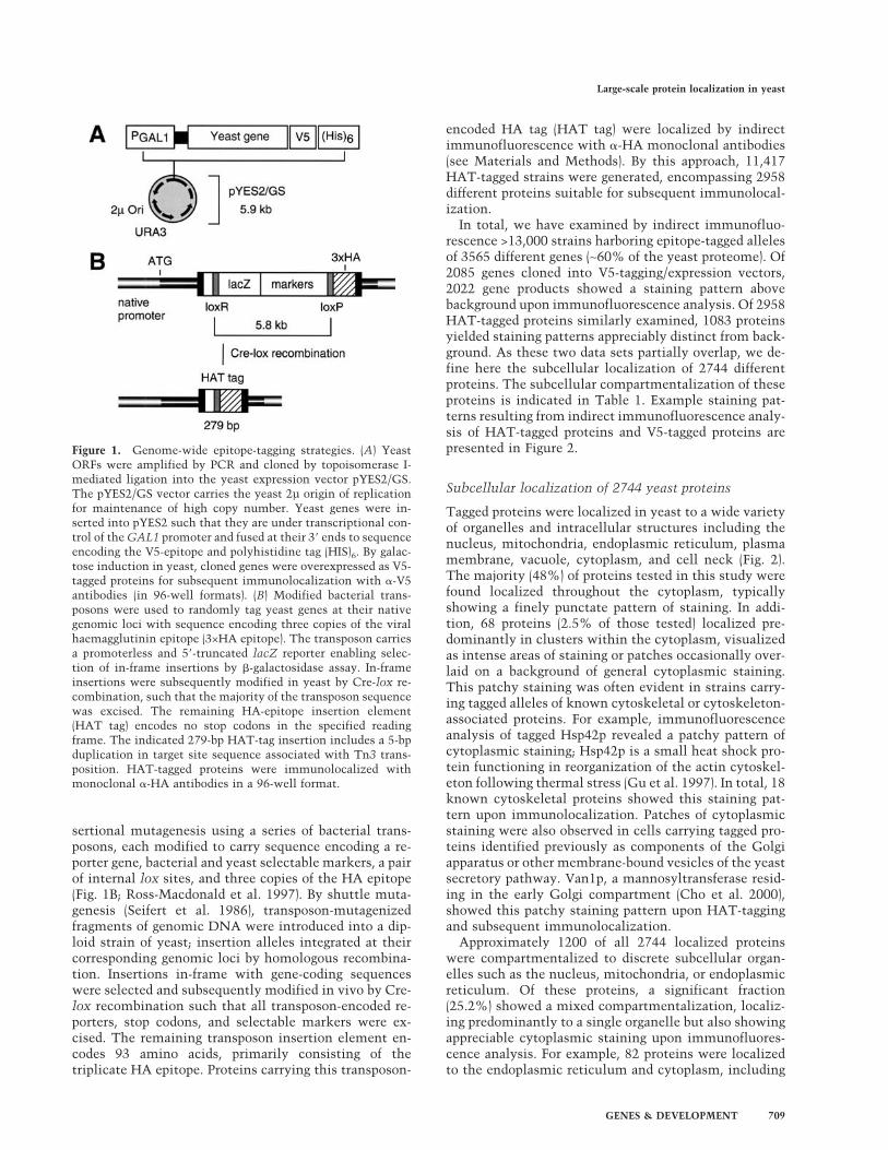
The studies presented here provide a unique opportunityto examine more rigorously the assumption that proteinfunction can be inferred from protein localization, anassumption best tested by correlating proteome-widedata sets of protein localization with corresponding datasets of protein function. Accordingly, we have tallied allmolecular functions (extracted from MIPS CYGD) asso-ciated with the 2744 yeast proteins immunolocalized inthis study. The most frequently observed functions as-sociated with each of the eight most populous compart-ments of the yeast proteome are indicated in Figure 6.
Within each organelle or compartment, a plurality ofproteins participate, at least partially, in maintainingstructural integrity. Secondary functions also correlatewell with major organelle-specific processes: for ex-ample, 34% of all nuclear-localized proteins are involvedin the process of transcription, and 26% of all mitochon-drial proteins function directly in cellular respiration.
Figure 3. Subcellular compartmentalization of theyeast proteome. (A) Cellular compartments are asfollows: cytoplasmic (Cyt.), nuclear (Nuc.), mito-chondrial (Mit.), and exocytic (Exo.). The membranefraction of each compartment is indicated in stripes.The percentage of the yeast proteome containedwithin the respective membrane and soluble frac-tions of each compartment is indicated outside thechart; the total percentage of the proteome con-tained within each of the four main compartmentsis indicated inside the chart. Plasma membrane pro-teins are included in the cytoplasmic compartmentfor purposes of this analysis. (B) The correspondingprotein population of each cellular compartmentand membrane/soluble subfraction is indicated.
Kumar et al.
712 GENES & DEVELOPMENT

Furthermore, specific functions can be correlated withsubtly distinct localization patterns. For example, 17%of the proteins that colocalized to the nucleus and cyto-plasm are cytoskeletal, whereas cytoskeletal functionsare not as strongly associated with proteins that localizeonly to the nucleus or only to the cytoplasm. Similarly,cytoskeletal proteins and Golgi proteins constitute thebulk of those proteins showing patchy patterns of cyto-plasmic staining; however, identical functions are notsignificantly represented among proteins yielding fine,
granular, or punctate cytoplasmic staining patterns. Thisstrong correlation between function and localizationsuggests that broad functional categories can now be as-cribed to the 955 proteins of previously unknown func-tion localized in this study.
An on-line database and visual library of proteinlocalization in yeast
To catalog the data presented here, we have developed anon-line database of yeast protein localization accessiblefrom our lab homepage at http://ygac.med.yale.edu (Pro-tein Localization in Yeast link). For this site, we havedeveloped a new user interface specifically accommodat-ing our V5-tagged data set; new HAT-tagged data maynow be accessed from our TRIPLES web site (Kumar etal. 2000a). In both cases, we supply search options bywhich users can access data for any gene of interest. Al-ternatively, complete data sets for all proteins localizingto a given site may be downloaded as tab-delimited text.Tabular data sets are supplemented with fluorescent mi-crographs of staining patterns observed upon immuno-fluorescence analysis of each indicated protein. In total,this new site houses >2893 micrographs, establishing itas the largest visual library of eukaryotic protein local-ization to date.
Discussion
Constituting the first proteome-wide analysis of proteinlocalization, this study is uniquely suited to address anumber of issues regarding both the methods by whichsuch a project may be undertaken as well as the utilityand applications of the end data. Here, we have used twocommon approaches by which epitope-tagged allelesmay be generated on a genomic scale: directed cloningmethods and random transposon-based approaches. Bycomparing the localization data generated from each re-spective set of tagged alleles, we can rigorously assessthe efficiency and accuracy of each approach. In particu-lar, our results may be used to consider the accuracywith which overexpressed proteins can be localized ascompared with the localization accuracy associated withendogenously expressed proteins. The resulting localiza-tion data sets correlate strongly with protein function,providing further means by which proteins may be as-cribed functions on a proteome-wide scale. This analysisalso offers specific insight into the relative distributionof functions and phenotypes associated with nuclear pro-teins, while providing data regarding nearly 1000 pro-teins of unknown function.
Two genomic epitope-tagging approaches:respective efficiencies
Directed cloning and random transposon-tagging eachpossess advantages and disadvantages as approaches forgenome-wide epitope-tagging. For large-scale directedcloning, a significant investment in labor and reagents is
Figure 4. Immunolocalization of nuclear proteins on surface-spread meiotic chromosomes. Meiotic chromosomes were sur-face spread and stained with the DNA-binding dye DAPI (left)and monoclonal anti-HA antibodies (center). Correspondingmerged images are shown to the right. A general pattern ofchromosomal binding can be seen from immunofluorescenceanalysis of cells containing HAT-tagged alleles of RFC3 (A–C),IOC2 (D–F), and PDR1 (G–I). Nine proteins localized predomi-nantly to the nucleolus; typical nucleolar staining patterns areshown here in cells containing HAT-tagged alleles of YGR090W(J–L), MPP10 (M–O), and YHR196W (P–R). Specific binding totelomeric DNA can be seen upon HAT-tagging and immunolo-calization of the origin recognition complex subunit Orc4p (S–U). Bar, 1 µm.
Large-scale protein localization in yeast
GENES & DEVELOPMENT 713

initially required; however, the final collection possesseslittle or no redundancy in gene representation. Further-more, for purposes of immunolocalization, directed ap-proaches are efficient: in this study, 93% of genes clonedinto a tagging/expression vector subsequently yieldedstaining patterns above background upon immunofluo-rescence analysis. Transposon-based methods, in con-trast, are economical but inefficient. Only 30% of trans-poson-tagged proteins showed a staining pattern distinctfrom background. In addition, owing both to the stochas-tic nature of transposition and to the insertional biasesassociated with bacterial transposons (e.g., Tn3), trans-poson-based methods can prove problematic as a meansof saturating a given genome. Small genes are less likelyto be mutagenized by transposon mutagenesis than arelarge genes. Also, insertional collections possess greaterredundancy in gene representation than do collectionsgenerated by directed methods: the collection of HAT-tagged genes generated in this study shows approxi-
mately fourfold redundancy in gene representation onaverage (11,417 HAT-tagged alleles representing 2958different genes). As shown, however, this redundancycan be beneficial in mapping domains within a givenprotein.
Respective accuracy of each approach
To estimate the accuracy of data generated by each tag-ging strategy, we have compared all protein localizationsdetermined experimentally in this study with previouslypublished localization data. Both approaches (i.e., mildoverexpression of C-terminal, V5-tagged alleles vs. en-dogenously expressed random HAT-tagged alleles)yielded data sets of similar accuracy (∼85%) when com-pared with published localization results. This internalcomparison, however, is complicated by the fact thatdifferent proteins are represented in the V5- and HAT-tagged data sets, respectively. To estimate the relative
Figure 5. Chromosomal localization and phenotypic analysis of nuclear proteins. Chromosomal localization indicates a generalpattern of chromosomal binding, typically with >40 staining foci per nucleus. Strains disrupted for each gene were assayed for sporeviability or growth defects; observed disruption mutants are categorized as viable, inviable, or slow-growth, accordingly.
Kumar et al.
714 GENES & DEVELOPMENT
accuracy of each approach more rigorously, we have lim-ited the comparison to only those 361 proteins commonto both data sets. Of these proteins, 295 yielded identicalor very similar staining patterns upon immunofluores-cence analysis regardless of the tagging approach used;the remaining 66 proteins yielded differing results. For29 of these proteins, no previously published localizationdata are available. Of the remaining 37 proteins ana-lyzed, localization data derived from V5-tagged proteinsagreed more closely with published results in 20 cases; in17 cases, HAT-tagged data proved more accurate (e.g.,Sik1p shown in Fig. 2E,J). Therefore, both approachesmay be used to generate epitope-tagged alleles for subse-quent immunolocalization with comparable degrees ofaccuracy, suggesting, furthermore, that the effects of tagsize, placement, and expression may be less severe thangenerally thought.
The yeast proteome: subcellular distributionand functional implications
Using the tagging/immunofluorescence strategies dis-cussed above, we have determined the subcellular local-
ization of 2744 different yeast proteins (Table 1). As ex-pected, large sets of proteins were localized to the cyto-plasm and nucleus (∼50% and ∼25% of the yeastproteome, respectively); however, a surprisingly largenumber of proteins showed a mixed staining pattern, lo-calizing to more than one subcellular compartment. Intotal, mixed localization patterns were evident in 11% ofall samples tested. Transcription factors and cytoskeletalproteins were frequently found distributed within boththe nucleus and cytoplasm as discussed. Also, vesicularproteins (e.g., Sec17p) often colocalized to the endoplas-mic reticulum and cytoplasm. Secretory polypeptidesthat have been either epitope-tagged or overproducedmay be processed less efficiently for export, and there-fore accumulate in the endoplasmic reticulum: ran-domly placed tags may disrupt signal peptide sequence,and overexpressed proteins may saturate mechanisms re-sponsible for membrane protein traffic. Therefore, colo-calization of secretory vesicle proteins to the endoplas-mic reticulum is expected.
The fact that a given protein may be distributed amongmore than one cellular compartment is a relevant con-
Figure 6. Prevalent functions associated with cellular compartments in yeast. Functional categorizations (compiled from publishedliterature) were extracted from the MIPS CYG database for all proteins experimentally localized in this study. In total, functions wereavailable for 1789 proteins; the number of functionally categorized proteins localized to each of the indicated cellular compartmentsis shown. Mixed localizations are also represented: 165 functionally characterized proteins were colocalized to the cytoplasm andnucleus; similarly, 61 such proteins were colocalized to the cytoplasm and endoplasmic reticulum (ER). Functions were tallied for allproteins within a given cellular compartment. The most frequently occurring functions per compartment are shown boxed. Multiplefunctions may be associated with a single protein. Therefore, the listed percentage following each function refers to the fraction ofproteins within each compartment associated with that particular cellular process, and the sum total of these percentages within agiven compartment will not equal 100%.
Large-scale protein localization in yeast
GENES & DEVELOPMENT 715

sideration in developing a computational system bywhich our data may be extrapolated over the yeast pro-teome. For this purpose, we have used a Bayesian systemby which the relative protein population of each yeastcellular compartment may be estimated without requir-ing a definitive localization for every constituent pro-tein. The accuracy of this method is dependent largelyon the availability of a large and unbiased localizationdata set from which a default series of localization prob-abilities can be calculated. In previous applications ofthis approach (Drawid and Gerstein 2000), an initial dataset was constructed from all known yeast protein local-izations cataloged in public databases (1342 localizationsin total). This data set, however, is biased toward nuclearproteins, as they have been traditionally studied ingreater detail than other protein classes. Our study pro-vides a less biased data set: the transposon-based ap-proaches used here are near-random, and the populationof yeast genes successfully cloned into pYES2 may re-flect only a marginal enrichment for short ORFs thattend to be easily amplified by PCR. By merging our ex-perimentally determined localizations with publishedyeast localization data and predicted transmembraneclassifications (Krogh et al. 2001), we define a subcellularlocalization for 3985 yeast proteins. Applying our proba-bilistic system to the remaining yeast protein comple-ment, we arrive at the proteome-wide protein compart-mentalization indicated in Figure 3. This distributionagrees well with previous theoretical estimates of pro-tein localization in yeast (Drawid and Gerstein 2000).
Because protein localization and function are tightlycorrelated (Fig. 5), our global localization analysis pro-vides a means by which gene function in yeast may beinferred on a genome-wide scale. Extrapolating from thefunctional categorizations maintained in the MIPSCYGD and our localization data, ∼45% of all yeast pro-teins function at least partly in maintaining cytoplasmicand organelle-specific organization and integrity. Fromour localization analysis, we estimate that the yeast pro-teome contains nearly 800 mitochondrial proteins—themajority of which function, as expected, in processes ofcellular respiration (Fig. 6).
Similar predictions can be made regarding the yeastnuclear protein complement. In this study, we haveidentified 457 nuclear-localized proteins for which func-tional data are currently available (including 165 pro-teins that colocalize to the nucleus and cytoplasm). Ofthese nuclear proteins, 34.8% function in transcription.Extrapolating this fraction to the total nuclear proteincompartment (1683 proteins; Fig. 3), we estimate thatnearly 10% of the yeast proteome is dedicated to pro-cesses of mRNA transcription. Consistent with this pre-diction, we have found that ∼38% of all nuclear proteins(or 10% of the yeast proteome) are associated with chro-mosomal DNA as determined by immunofluorescenceanalysis of tagged proteins on surface-spread meioticchromosomes (Fig. 5). Although caution must be exer-cised in extrapolating from a limited population of 56nuclear proteins, our phenotypic studies suggest thatroughly 34% of all nuclear proteins (>570 proteins in
total) are essential for spore viability. In contrast, overthe genome as a whole, 18% of yeast genes (∼1100) arethought to be essential as indicated from systematicanalyses of yeast deletion mutants (Winzeler et al. 1999).Therefore, slightly more than half of all essential genesin yeast are likely to be nuclear.
Integrating localizome data
Large-scale localization data sets provide a fundamentalcomplement to other existing varieties of proteomicdata. For example, our localization data may be used toscreen sets of putative protein–protein interactions, en-riching for genuine protein associations by virtue of theexpectation that two interacting proteins will share acommon cellular compartment and show similar local-ization patterns. At present, large catalogs of protein in-teractions in yeast have been generated through genome-wide applications of the two-hybrid method (Uetz et al.2000; Ito et al. 2001) and systematic, high-throughputapproaches using mass spectrometric analysis of immu-noprecipitated protein complexes (Gavin et al. 2002; Hoet al. 2002). We have correlated our localization resultswith a sampling of interaction data drawn from each ofthese studies. Of 155 randomly selected two-hybrid in-teractions identified either by Uetz et al. (2000) or Ito etal. (2001), only 73 (47%) contain a protein pair localizedto the same cellular compartment. In contrast, however,within a set of 105 two-hybrid interactions indepen-dently identified by both groups, 87 protein pairs (83%)show a shared localization pattern. Analysis of data gen-erated by Gavin et al. (2002) and Ho et al. (2002) yieldssimilar results. Of 100 sampled protein associations (en-compassing 10 different bait proteins), 67 interactionsconsist of two proteins from the same cellular compart-ment. Of these 100 protein associations, 23 were identi-fied by both groups: all 23 of these protein pairs showcompatible localization patterns. These correlations sug-gest that confidence can be placed preferentially in pro-tein interactions independently identified within morethan one study, while simultaneously demonstratingthe usefulness of localization data in distinguishing spu-rious protein–protein interactions is likely to be spuri-ous.
As illustrated by these comparisons, results from in-dependent studies may be effectively integrated to pro-vide more accurate and complete genomic findings. Theaccuracy of our own localization results may be im-proved through comparison with a set of known andestablished protein–protein interactions, a corollary ofthe analysis above. A more comprehensive representa-tion of yeast protein function may be achieved by inte-grating multiple proteomic data sets, because all suchindividual data sets are presently incomplete (i.e., en-compass <6000 yeast proteins). Collectively, this unionof diverse proteomic and genomic approaches will provemutually complementary and necessary as a means ofunderstanding global processes of eukaryotic cellularfunction.
Kumar et al.
716 GENES & DEVELOPMENT

Materials and methods
Epitope-tagging and immunolocalization
Yeast genes were amplified by polymerase chain reaction (PCR)and cloned into the yeast expression vector pYES2/GS by topoi-somerase I-mediated ligation as described previously (Heymanet al. 1999). Vector constructs carrying cloned yeast genes wereintroduced into haploid strain YNN218 [ura3-52 lys2-801 ade2-101 his3�200] by DNA transformation (Ito et al. 1983). To in-duce gene expression, yeast transformants were first grown tosaturation in synthetic medium lacking uracil (SC-Ura) withraffinose as its carbon source; cultures were then washed insterile water prior to resuspension in SC-Ura with galactose asa carbon source. Transformant cultures were incubated in ga-lactose for 1 h. Multiple incubation periods were tested to de-termine the optimum time for galactose induction such thatartifacts resulting from gene overexpression are minimized. Fol-lowing galactose induction, cells were prepared for immunoflu-orescence analysis in a 96-well format as described (Kumar et al.2000b). V5-tagged proteins were immunolocalized by indirectimmunofluorescence using anti-V5 mouse monoclonal IgG2aantibody (Invitrogen) and Cy3-conjugated goat anti-mouse IgG(Jackson Labs).
Yeast genes were HAT-tagged using transposon-based meth-ods presented previously (Ross-MacDonald et al. 1999; Kumaret al. 2000b). Tagged genes were generated in a Y800 background[MATa leu2-�98cry1R/MAT� leu2-�98CRY1 ade2-101 HIS3/ade2-101 his3-�200 ura3-52 caniR/ura3-52CAN1 lys2-801/lys2-801 CYH2/cyh2R trp1-1/TRP1 Cir0] carrying pGAL-cre (amp,ori, CEN, LEU2) (Burns et al. 1994). Asynchronous cultures ofHAT-tagged yeast strains were grown and prepared for immu-nofluorescence analysis in 96-well microtiter plates (Kumar etal. 2000b). Transposon-tagged proteins were immunolocalizedas above, except that mouse monoclonal anti-HA 16B12(MMS101R, BAbCO) was used as the primary antibody.
Computational methods
For purposes of this analysis, all yeast proteins were divided intofour localization categories: cytoplasm (Cyt), nucleus (Nuc), mi-tochondria (Mit), and exocytic network (Exo; endoplasmic re-ticulum, Golgi apparatus, vacuoles, vesicles, peroxisome, andextracellular proteins). A different localization prediction pro-cedure was applied for soluble and membrane proteins of allcategories.
We identified 1029 yeast proteins as integral membrane pro-teins. These proteins were predicted to possess two or moretransmembrane helices using TMHMM (Krogh et al. 2001);many also had a verifying match to a Pfam family of knownmembrane proteins (L. Yang and M. Gerstein, in prep.). Of theseputative membrane proteins, ∼380 had been localized to one ofthe four categories described above by transposon-tagging andsubsequent immunolocalization as described here. We believethe distribution of these proteins among the four categories tobe random and accurate. Consequently, we applied this distri-bution to the ∼650 membrane proteins of unknown localization.
The remaining 5101 proteins in the yeast genome were con-sidered to be soluble proteins. In total, experimentally derivedlocalization data are available for ∼2950 of these soluble pro-teins both from this study as well as from data previously de-posited in the MIPS, YPD, and SwissProt databases. This servedas the training set for our Bayesian method (Drawid and Ger-stein 2000). The Bayesian system integrates a large number ofdifferent features related to yeast proteins, including sequencepatterns, such as the nuclear localization signal or signal se-
quence, expression information, and many varieties of pheno-typic data (e.g., viability of corresponding null mutants). Theincorporation of expression information is particularly uniqueand is derived from the observation that cytoplasmic proteinspossess much higher levels of expression than those in othercompartments (Drawid et al. 2000).
By transposon-tagging/immunolocalization, we have definedthe localization of ∼2500 soluble proteins. Because we believethese proteins represent a random sample from the yeast ge-nome, we have used their localization proportions as our priors(Cyt, 52%; Nuc, 27%; Mit, 14%; Exo, 7%). The subcellularlocalization of the remaining 2150 soluble proteins (for whichno localization data are available) was predicted using ourBayesian method and the above prior and training data. We di-rectly integrated the proportions of these 2150 proteins to yieldan overall prediction for protein compartmentalization withinthe yeast proteome. We added together the number of solubleand membrane proteins to obtain the pie chart presented inFigure 3A.
Immunocytology and phenotypic analysis of nuclear proteins
Meiotic chromosomes from HAT-tagged strains were surfacespread and stained as described using mouse anti-HA (Covance)at 1:400 dilution and DAPI (Agarwal and Roeder 2000). To ex-amine phenotypes of strains (Y800 background) containing dis-ruptive, full-length transposon insertions within genes encod-ing nuclear proteins, diploids heterozygous for the insertionwere sporulated; tetrads were subsequently dissected and as-sayed for spore viability and transposon-encoded �-galactosi-dase activity as described previously (Burns et al. 1994).
Acknowledgments
We thank James R. Chambers, Shannon Hattier, and Jon Row-land of Invitrogen Corporation for strain organization and DNApreparation. This work was supported by NIH Grant R01-CA77808 to M.S. A.K. is supported by a postdoctoral fellowshipfrom the American Cancer Society.
The publication costs of this article were defrayed in part bypayment of page charges. This article must therefore be herebymarked “advertisement” in accordance with 18 USC section1734 solely to indicate this fact.
References
Agarwal, S. and Roeder, G.S. 2000. Zip3 provides a link betweenrecombination enzymes and synaptonemal complex pro-teins. Cell 102: 245–255.
Bairoch, A. and Apweiler, R. 2000. The SWISS-PROT proteinsequence database and its supplement TrEMBL in 2000.Nucleic Acids Res. 28: 45–48.
Baker, R.E. and Masison, D.C. 1990. Isolation of the gene en-coding the Saccharomyces cerevisiae centromere-bindingprotein CP1. Mol. Cell. Biol. 10: 2458–2467.
Bell, S.P., Mitchell, J., Leber, J., Kobayashi, R., and Stillman, B.1995. The multidomain structure of Orc1p reveals similarityto regulators of DNA replication and transcriptional silenc-ing. Cell 83: 563–568.
Burley, S.K. 2000. An overview of structural genomics. Nat.Struct. Biol. Suppl: 932–934.
Burns, N., Grimwade, B., Ross-Macdonald, P.B., Choi, E.-Y.,Finberg, K., Roeder, G.S., and Snyder, M. 1994. Large-scalecharacterization of gene expression, protein localization andgene disruption in Saccharomyces cerevisiae. Genes & Dev.
Large-scale protein localization in yeast
GENES & DEVELOPMENT 717

8: 1087–1105.Cairns, B.R., Lorch, Y., Li, Y., Zhang, M., Lacomis, L., Erdju-
ment-Bromage, H., Tempst, P., Du, J., Laurent, B., and Korn-berg, R.D. 1996. RSC, an essential, abundant chromatin-re-modeling complex. Cell 87: 1249–1260.
Cho, J.H., Noda, Y., and Yoda, K. 2000. Proteins in the earlyGolgi compartment of Saccharomyces cerevisiae immu-noisolated by Sed5p. FEBS Lett. 469: 151–154.
Costanzo, M.C., Crawford, M.E., Hirschman, J.E., Kranz, J.E.,Olsen, P., Robertson, L.S., Skrzypek, M.S., Braun, B.R., Len-non-Hopkins, K., Kondu, P., et al. 2001. YPDTM, PombeP-DTM and WormPDTM: Model organism volumes of the Bio-KnowledgeTM Library, an integrated resource for protein in-formation. Nucleic Acids Res. 29: 75–79.
Ding, D.Q., Tomita, Y., Yamamoto, A., Chikashige, Y., Hara-guchi, T., and Hiraoka, Y. 2000. Large-scale screening of in-tracellular protein localization in living fission yeast cells bythe use of a GFP-fusion genomic DNA library. Genes Cells5: 169–190.
Drawid, A. and Gerstein, M. 2000. A Bayesian system integrat-ing expression data with sequence patterns for localizingproteins: Comprehensive application to the yeast genome. J.Mol. Biol. 301: 1059–1075.
Drawid, A., Jansen, R., and Gerstein, M. 2000. Genome-wideanalysis relating expression level with protein subcellularlocalization. Trends Genet. 16: 426–430.
Gavin, A.-C., Bosche, M., Krause, R., Grandi, P., Marzioch, M.,Bauer, A., Schultz, J., Rick, J.M., Michon, A.-M., Cruciat,C.-M., et al. 2002. Functional organization of the yeast pro-teome by systematic analysis of protein complexes. Nature415: 141–147.
Gu, J., Emerman, M., and Sandmeyer, S. 1997. Small heat shockprotein suppression of Vpr-induced cytoskeletal defects inbudding yeast. Mol. Cell. Biol. 17: 4033–4042.
Gygi, S.P., Rist, B., Gerber, S.A., Tureck, F., Gelb, M.H., andAebersold, R. 1999. Quantitative analysis of complex pro-tein mixtures using isotope-coded affinity tags. Nat. Biotech.17: 994–999.
Heyman, J.A., Cornthwaite, J., Foncerrada, L., Gilmore, J.R.,Gontag, E., Hartman, K.J., Hernandez, C.L., Hood, R., Hull,H.M., Lee, W.-Y., et al. 1999. Genome-scale cloning and ex-pression of individual open reading frames using topoisom-erase I-mediated ligation. Genome Res. 9: 383–392.
Ho, Y., Gruhler, A., Heilbut, A., Bader, G., Moore, L., Adams,S.-L., Millar, A., Taylor, P., Bennett, K., Boutilier, K., et al.2002. Systematic identification of protein complexes in Sac-charomyces cerevisiae by mass spectrometry. Nature415: 180–183.
Ito, H., Fukuda, Y., Murata, K., and Kimura, A. 1983. Transfor-mation of intact yeast cells treated with alkali cations. J.Bacteriol. 153: 163–168.
Ito, T., Chiba, T., Ozawa, R., Yoshida, M., Hattori, M., andSakaki, Y. 2001. A comprehensive two-hybrid analysis toexplore the yeast protein interactome. Proc. Natl. Acad. Sci.98: 4569–4574.
Iyer, V.R., Horak, C.A., Scafe, C.S., Botstein, D., Snyder, M., andBrown, P.O. 2001. Genomic binding sites of the yeast cell-cycle transcription factors SBF and MBF. Nature 409: 533–538.
Koller, A., Snyder, W.B., Faber, K.N., Wenzel, T.J., Rangell, L.,Keller, G.A., and Subramani, S. 1999. Pex22p of Pichia pas-toris, essential for peroxisomal matrix protein import, an-chors the ubiquitin-conjugating enzyme, Pex4p, on the per-oxisomal membrane. J. Cell Biol. 146: 99–112.
Krogh, A., Larsson, B., von Heijne, G., and Sonnhammer, E.L.2001. Predicting transmembrane protein topology with a
hidden Markov model: Application to complete genomes. J.Mol. Biol. 305: 567–580.
Kumar, A., Cheung, K.-H., Ross-Macdonald, P., Coelho, P.S.R.,Miller, P., and Snyder, M. 2000a. TRIPLES: A database ofgene function in Saccharomyces cerevisiae. Nucleic AcidsRes. 28: 81–84.
Kumar, A., des Etages, S.A., Coelho, P.S.R., Roeder, G.S., andSnyder, M. 2000b. High-throughput methods for the large-scale analysis of gene function by transposon tagging. Meth-ods Enzymol. 328: 550–574.
Li, X. and Burgers, P.M. 1994. Molecular Cloning and expressionof the Saccharomyces cerevisiae RFC3 gene, an essentialcomponent of replication factor C. Proc. Natl. Acad. Sci.91: 868–872.
MacBeath, G. and Schreiber, S.L. 2000. Printing proteins as mi-croarrays for high-throughput function determination. Sci-ence 289: 1760–1763.
Mewes, H.W., Frishman, D., Gruber, C., Geier, B., Haase, D.,Kaps, A., Lemcke, K., Mannhaupt, G., Pfeiffer, F., Schuller,C., et al. 2000. MIPS: A database for genomes and proteinsequences. Nucleic Acids Res. 28: 37–40.
Montelione, G.T. 2001. Structural genomics: An approach tothe protein folding problem. Proc. Natl. Acad. Sci.98: 13488–13489.
Niedenthal, R.K., Riles, L., Johnston, M., and Hegemann, J.H.1996. Green fluorescent protein as a marker for gene expres-sion and subcellular localization in budding yeast. Yeast12: 773–786.
O’Neill, E.M., Kaffman, A., Jolly, E.R., and O’Shea, E.K. 1996.Regulation of PHO4 nuclear localization by the PHO80–PHO85 cyclin–CDK complex. Science 271: 209–212.
Ray, A. and Runge, K.W. 1998. The C terminus of the majoryeast telomere binding protein Rap1p enhances telomereformation. Mol. Cell. Biol. 18: 1284–1295.
Ren, B., Robert, F., Wyrick, J.J., Aparicio, O., Jennings, E.G.,Simon, I., Zeitlinger, J., Schreiber, J., Hannett, N., Kanin, E.,et al. 2000. Genome-wide location and function of DNAbinding proteins. Science 290: 2306–2309.
Ross-Macdonald, P., Sheehan, A., Roeder, G.S., and Snyder, M.1997. A multipurpose transposon system for analyzing pro-tein production, localization, and function in Saccharomy-ces cerevisiae. Proc. Natl. Acad. Sci. 94: 190–195.
Ross-MacDonald, P., Coelho, P.S.R., Roemer, T., Agarwal, S.,Kumar, A., Jansen, R., Cheung, K.-H., Sheehan, A., Symoni-atis, D., Umansky, L., et al. 1999. Large-scale analysis of theyeast genome by transposon tagging and gene disruption.Nature 402: 413–418.
Seifert, H.S., Chen, E.Y., So, M., and Heffron, F. 1986. Shuttlemutagenesis: A method of transposon mutagenesis for Sac-charomyces cerevisiae. Proc. Natl. Acad. Sci. 83: 735–739.
Silver, P.A. 1991. How proteins enter the nucleus. Cell 64: 489–497.
Simpson, J.C., Wellenreuther, R., Poustka, A., Pepperkok, R.,and Wiemann, S. 2000. Systematic subcellular localizationof novel proteins identified by large-scale cDNA sequencing.EMBO Reports 1: 287–292.
Tong, A.H.Y., Drees, B., Nardelli, G., Bader, G.D., Brannetti, B.,Castagnoli, L., Evangelista, M., Ferracuti, S., Nelson, B., Pa-oluzi, S., et al. 2002. A combined experimental and compu-tational strategy to define protein interaction networks forpeptide recognition modules. Science 295: 321–324.
Uetz, P., Giot, L., Cagney, G., Mansfield, T.A., Judson, R.S.,Knight, J.R., Lockshon, D., Narayan, V., Srinivasan, M., Po-chart, P., et al. 2000. A comprehensive analysis of protein–protein interactions in Saccharomyces cerevisiae. Nature403: 623–627.
Kumar et al.
718 GENES & DEVELOPMENT

van Steensel, B., Delrow, J., and Henikoff, S. 2001. Chromatinprofiling using targeted DNA adenine methyltransferase.Nat. Genet. 27: 304–308.
Vogel, J., Drapkin, B., Oomen, J., Beach, D., Bloom, K., andSnyder, M. 2001. Phosphorylation of �-tubulin regulates mi-crotubule organization in budding yeast. Dev. Cell 1: 621–631.
Washburn, M.P., Wolters, D., and Yates III, J.R. 2001. Large-scaleanalysis of the yeast proteome by multidimensional proteinidentification technology. Nat. Biotechnol. 19: 242–247.
Waters, M.G., Griff, I.C., and Rothman, J.E. 1991. Proteins in-volved in vesicular transport and membrane fusion. Curr.Opin. Cell. Biol. 3: 615–620.
Winzeler, E.A., Shoemaker, D.D., Astromoff, A., Liang, H.,Anderson, K., Andre, B., Bangham, R., Benito, R., Boeke, J.D.,Bussey, H., et al. 1999. Functional characterization of the S.cerevisiae genome by gene deletion and parallel analysis.Science 285: 901–906.
Zhu, H., Klemic, J.F., Chang, S., Bertone, P., Casamayor, A.,Klemic, K.G., Smith, D., Gerstein, M., Reed, M.A., andSnyder, M. 2000. Analysis of yeast protein kinases usingprotein chips. Nat. Genet. 26: 283–289.
Zhu, H., Bilgin, M., Bangham, R., Hall, D., Casamayor, A., Ber-tone, P., Lan, N., Jansen, R., Bidlingmaier, S., Houfek, T., etal. 2001. Global analysis of protein activities using proteomechips. Science 293: 2101–2105.
Large-scale protein localization in yeast
GENES & DEVELOPMENT 719