Nucleotide excision repair in yeast
147
Cover Page The handle http://hdl.handle.net/1887/20360 holds various files of this Leiden University dissertation. Author: Eijk, Patrick van Title: Nucleotide excision repair in yeast Date: 2012-12-13
-
Upload
independent -
Category
Documents
-
view
1 -
download
0
Transcript of Nucleotide excision repair in yeast
Cover Page
The handle http://hdl.handle.net/1887/20360 holds various files of this Leiden University dissertation. Author: Eijk, Patrick van Title: Nucleotide excision repair in yeast Date: 2012-12-13
Nucleotide Excision Repair in Yeast
Patrick van Eijk

Nucleotide Excision Repair in Yeast
PROEFSCHRIFT
ter verkrijging van
de graad van Doctor aan de Universiteit Leiden,
op gezag van Rector Magnificus prof.mr. P.F. van der Heijden,
volgens besluit van het College voor Promoties
te verdedigen op 13 December 2012
klokke 12:30 uur
door
Patrick van Eijk
geboren te Rotterdam
in 1984
Promotiecommissie
Promotor Prof. Dr. J. Brouwer
Co-promoter Dr. S.H. Reed (Cardiff University)
Overige Leden Dr. J.A. Brandsma
Dr. N. Goosen
Prof. Dr. L.H.F. Mullenders
Prof. Dr. M.H.M. Noteborn
Prof. Dr. H.P. Spaink
Prof. Dr. G.A. van der Marel
Contents
I Introduction 7
II Nucleotide Excision Repair Factors Directly Regulate dNTP
Synthesis in Response to UV Damage in Yeast 59
III The effect of Histone H on repair of rDNA in yeast 89
IV Phenotypic analysis of a rad4 mutant carrying a homologous
change that in XPC leads to the XP disorder 103
V A novel, functionally distinct, bipartite Rad4-Rad23 interaction
in Nucleotide Excision Repair 115
VI WCG4A, a commonly used Saccharomyces cerevisiae yeast
strain, contains a RAD4 mutation affecting UV resistance 127
VII Summary & Conclusions 137
VIII Samenvatting & Conclusies 141
IX Curriculum vitea 146

I Introduction

I Introduction

Introduction
8
I Introduction 7 1. General Introduction 9 2. The DNA Damage Response 10
2.1 RNR pathway and dNTP synthesis 13 3. DNA Repair 14
3.1 Post-Replication Repair 15 3.2 Mismatch Repair 16 3.3 Direct DNA damage reversal 18 3.4 Base Excision Repair 20 3.5 Double Strand Break Repair via HR & NHEJ 21
4. Nucleotide Excision Repair in eukaryotes 23 4.1 A short history of Excision repair 23 4.2 The Mechanism of Nucleotide Excision Repair 24 4.3 DNA damage recognition 26 4.4 Bubble formation and Incision 26 4.5 Oligonucleotide excision and repair synthesis 27 4.6 From in vitro to in vivo NER 28 4.7 Global Genome NER 29 4.8 Transcription Coupled NER 30
5. rDNA and repair 32 6. The NER proteins important for this thesis 34
6.1 The core NER complex Rad4-Rad23 34 6.2 Rad34 37 6.3 Rad33 38
7. Scope of this thesis 39 8. References 40
Introduction
9
1. General Introduction DeoxyiboNucleic acid (DNA) is the carrier of genetic information for most forms of life [1,
2]. DNA exists as a double helix of complementary strands that resides in the nucleus of
eukaryotic cells. Prokaryotes lack a cell nucleus and hence maintain their genome in the
cytoplasm in the form of a nucleoid structure [3]. The primary structure in DNA is the
linear arrangement of nucleotides into genes that code for proteins and a variety of
RiboNucleic Acid (RNA) molecules. Both pro- and eukaryotes maintain their genome in a
higher order structure required for compaction of the DNA to fit inside the cell or nucleus.
More importantly, this structure functions in regulation of gene expression [4-6]. The
higher order structure seen in eukaryotes contains many levels of organization of which
the basic unit is the nucleosome [7].
Historically, the stable propagation of genetic traits has been recognized for many years,
but it was hard to reconcile that a simple biological molecule could harbor such stability.
The chemical nature of DNA makes it inherently unstable and subject to many chemical
alterations under physiological conditions [8]. Moreover, our genome faces many natural
challenges altering our genetic information that would collectively compromise faithful
inheritance via any type of biomolecule. Endogenous sources of DNA damage are
deamination, depurination and importantly, Reactive Oxygen Species (ROS) produced
by cellular metabolism. Exogenous agents, on the other hand, are both chemical and
physical in nature including but not limited to UV radiation from sunlight, ionizing or
gamma radiation, smoke-related carcinogens, anti-cancer drugs and environmental
pollutants [8]. UV radiation causes formation of thymine-thymine dimers in the form of
cyclobutane pyrimidine dimers (CPD) and 6-4 photoproducts (6-4PP) [9]. Ionizing and
gamma radiation damage the DNA by inducing nicks, breaks and base modifications [10,
11]. This constant pressure of DNA insults requires an ample maintenance and repair
capacity. Indeed nature has devised a large spectrum of DNA repair and damage
response pathways with different degrees of conservation amongst the three kingdoms
of life [12]. It is thus the concerted action of a plethora of DNA repair and response
pathways that is responsible for the very stable transmission of genetic traits and not the
stability of the carrier per se.
If DNA damages are left unrepaired they can lead to mutations. The cellular response to
DNA damage regardless of the source is in essence twofold: that of the DNA Damage
Response (DDR) and of DNA repair. Defects in either of these cellular processes can
lead to genomic aberrations ranging from point mutations to gross chromosomal
rearrangements, referred to as genomic instability [13]. Failure to remove DNA damages
can be due to impaired DNA repair, a defective DDR, an extreme high damage dose or a
combination of the three. The presence of genomic instability in a majority of human
cancers indicates that genomic instability drives carcinogenesis [13-16]. Indeed, most
genetic defects of DNA repair pathways lead to predisposition to cancer in humans [17],
Introduction
10
putting into perspective the importance of DNA repair in maintaining genome stability
and preventing carcinogenesis [18-20].
Firstly, in the next sections of the introduction the importance of genome stability and the
DNA Damage Response (DDR) will be discussed (2). Secondly, the DNA repair
pathways will be described in more detail in section 3 including: Post-Replication Repair
(PRR) and Translesion DNA Synthesis (TLS) (3.1), Mismatch Repair (MMR) (3.2), Direct
DNA damage reversal (3.3), Base Excision Repair (BER) (3.4), and repair of Double
Strand Breaks (DSB) via Homologous Recombination (HR) and Non-Homologous End-
Joining (NHEJ) (3.5). In light of the research described in this thesis special emphasis is
placed on Nucleotide Excision Repair (NER) described in sections 4 to 6..
2. The DNA Damage Response During the initial response to DNA damage cells recruit the DNA Damage Response
(DDR), a pathway of damage detectors and kinase protein that signal the cell to halt cell
cycle progression and alter the expression of many gene targets [21]. Before introducing
damage detection that fuels the DDR it is critical to cover some of the basics around
replication. Elements of the replication complex are vital for DDR signaling as will
become evident in the following paragraph.
DNA replication is achieved at an appreciably low error-rate but is sensitive to aberrant
bases or other DNA damages that can block the replication machinery or lead to
misincorporation of bases [14, 22]. The replication fork is built up around origins of
replication as a multi-protein complex starting as the pre-replication complex. The double
stranded nature of DNA requires a denaturing step by the MCM (Mini Chromosome
Maintenance) helicase that individually runs ahead of the replication complex. At each
origin two replication complexes, called replisomes, are formed that synthesize DNA in
opposite direction. The Replication Factor Complex C (RFC) loads the Proliferating Cell
Nuclear Antigen (PCNA) clamp onto the DNA that stimulates the processivity of the
replisome. The replicative DNA polymerases are recruited to the replication fork by
PCNA. The presence of partial duplex DNA (stretches of ss- an dsDNA) puts the
replication complex at risk of DNA strand breaks or recombination. Moreover, the MCM
helicase activity is uncoupled from the main replication machinery and will generate large
stretches of ssDNA ahead of a stalled replication fork, creating another substrate for
DNA breaks or recombination. To illustrate, genetic mutation or deletion of genes
encoding for components of the replication complex results in the accumulation of
replication intermediates that are prone to recombination leading to genomic instability
[19]. Other endogenous sources that can impede on replication are DNA secondary
structures in repetitive DNA, protein-DNA and transcription complexes [19].
Introduction
11
More relevant to
this thesis is the
role of DNA
damage in
generating ge-
nome instability.
Any DNA damage
that will prevent
the replication
machinery from
progressing is a
potential risk for
genome stability.
This includes but
is not limited to
UV-induced py-
rimidine dimers
and chemically
induced bulky
DNA adducts [19].
The natural re-
sponse to these
types of bulky le-
sions, initially, is
the DDR dis-
cussed here fol-
lowed by DNA
repair. If however,
damages persist into S phase when replication commences, an important 'last resort'
exists, referred to as DNA damage tolerance pathways. This is Post-Replication Repair
(PRR) which includes translesion DNA synthesis (TLS). Sub-pathways of this response
can be both error-free or error-prone and will be discussed in more detail in their respec-
tive sections below. At this stage I introduce these pathways as part of the holy trinity of
DDR, DNA repair and damage tolerance all working in concert to promote cellular
survival after DNA damage. It is important to appreciate that even though these three
major processes are all highly redundant within and amongst each other, failure at any
stage is a potential hazard for genomic stability.
In order to provide cells time and to produce the resources and conditions to repair DNA
damages, the so-called DNA Damage Response is initiated. A variety of DNA lesions
can trigger the DDR in order to halt cell cycle progression and prepare the cell for DNA
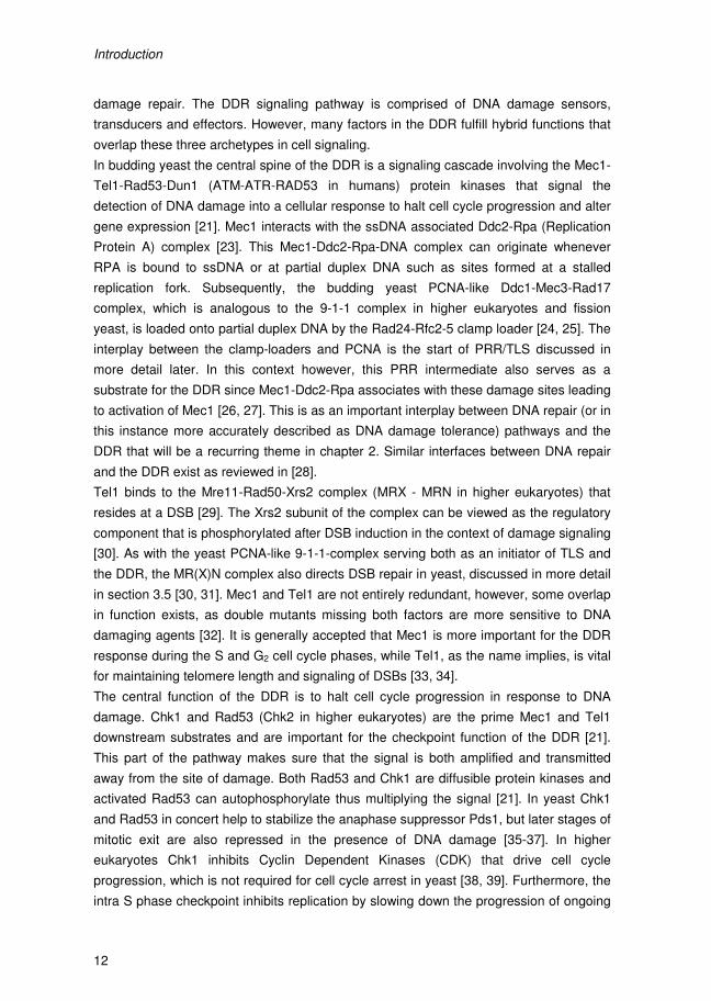
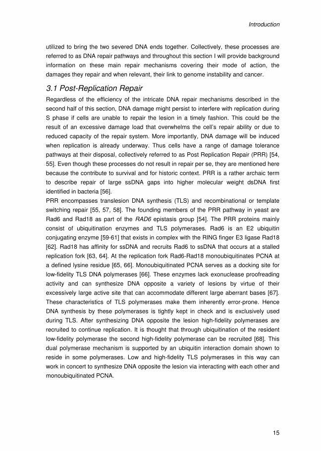
Figure 1 – A model for the DDR directed control of dNTP synthesis, DNA damage detection via the Rad17, Rad24, Mec3 and Rad9 pathways funnel into the core Mec1-Rad53-Dun1 DDR signaling cascade. The dual function of Dun1 is highlighted. Dun1 phosphorylates the RNR inhibitor Sml1 and concurrently induces transcription of the RNR2 to 4 genes. In its free form Sml1 is degraded. Taken from [50].

Introduction
10
putting into perspective the importance of DNA repair in maintaining genome stability
and preventing carcinogenesis [18-20].
Firstly, in the next sections of the introduction the importance of genome stability and the
DNA Damage Response (DDR) will be discussed (2). Secondly, the DNA repair
pathways will be described in more detail in section 3 including: Post-Replication Repair
(PRR) and Translesion DNA Synthesis (TLS) (3.1), Mismatch Repair (MMR) (3.2), Direct
DNA damage reversal (3.3), Base Excision Repair (BER) (3.4), and repair of Double
Strand Breaks (DSB) via Homologous Recombination (HR) and Non-Homologous End-
Joining (NHEJ) (3.5). In light of the research described in this thesis special emphasis is
placed on Nucleotide Excision Repair (NER) described in sections 4 to 6..
2. The DNA Damage Response During the initial response to DNA damage cells recruit the DNA Damage Response
(DDR), a pathway of damage detectors and kinase protein that signal the cell to halt cell
cycle progression and alter the expression of many gene targets [21]. Before introducing
damage detection that fuels the DDR it is critical to cover some of the basics around
replication. Elements of the replication complex are vital for DDR signaling as will
become evident in the following paragraph.
DNA replication is achieved at an appreciably low error-rate but is sensitive to aberrant
bases or other DNA damages that can block the replication machinery or lead to
misincorporation of bases [14, 22]. The replication fork is built up around origins of
replication as a multi-protein complex starting as the pre-replication complex. The double
stranded nature of DNA requires a denaturing step by the MCM (Mini Chromosome
Maintenance) helicase that individually runs ahead of the replication complex. At each
origin two replication complexes, called replisomes, are formed that synthesize DNA in
opposite direction. The Replication Factor Complex C (RFC) loads the Proliferating Cell
Nuclear Antigen (PCNA) clamp onto the DNA that stimulates the processivity of the
replisome. The replicative DNA polymerases are recruited to the replication fork by
PCNA. The presence of partial duplex DNA (stretches of ss- an dsDNA) puts the
replication complex at risk of DNA strand breaks or recombination. Moreover, the MCM
helicase activity is uncoupled from the main replication machinery and will generate large
stretches of ssDNA ahead of a stalled replication fork, creating another substrate for
DNA breaks or recombination. To illustrate, genetic mutation or deletion of genes
encoding for components of the replication complex results in the accumulation of
replication intermediates that are prone to recombination leading to genomic instability
[19]. Other endogenous sources that can impede on replication are DNA secondary
structures in repetitive DNA, protein-DNA and transcription complexes [19].
Introduction
11
More relevant to
this thesis is the
role of DNA
damage in
generating ge-
nome instability.
Any DNA damage
that will prevent
the replication
machinery from
progressing is a
potential risk for
genome stability.
This includes but
is not limited to
UV-induced py-
rimidine dimers
and chemically
induced bulky
DNA adducts [19].
The natural re-
sponse to these
types of bulky le-
sions, initially, is
the DDR dis-
cussed here fol-
lowed by DNA
repair. If however,
damages persist into S phase when replication commences, an important 'last resort'
exists, referred to as DNA damage tolerance pathways. This is Post-Replication Repair
(PRR) which includes translesion DNA synthesis (TLS). Sub-pathways of this response
can be both error-free or error-prone and will be discussed in more detail in their respec-
tive sections below. At this stage I introduce these pathways as part of the holy trinity of
DDR, DNA repair and damage tolerance all working in concert to promote cellular
survival after DNA damage. It is important to appreciate that even though these three
major processes are all highly redundant within and amongst each other, failure at any
stage is a potential hazard for genomic stability.
In order to provide cells time and to produce the resources and conditions to repair DNA
damages, the so-called DNA Damage Response is initiated. A variety of DNA lesions
can trigger the DDR in order to halt cell cycle progression and prepare the cell for DNA
Figure 1 – A model for the DDR directed control of dNTP synthesis, DNA damage detection via the Rad17, Rad24, Mec3 and Rad9 pathways funnel into the core Mec1-Rad53-Dun1 DDR signaling cascade. The dual function of Dun1 is highlighted. Dun1 phosphorylates the RNR inhibitor Sml1 and concurrently induces transcription of the RNR2 to 4 genes. In its free form Sml1 is degraded. Taken from [50].
Introduction
12
damage repair. The DDR signaling pathway is comprised of DNA damage sensors,
transducers and effectors. However, many factors in the DDR fulfill hybrid functions that
overlap these three archetypes in cell signaling.
In budding yeast the central spine of the DDR is a signaling cascade involving the Mec1-
Tel1-Rad53-Dun1 (ATM-ATR-RAD53 in humans) protein kinases that signal the
detection of DNA damage into a cellular response to halt cell cycle progression and alter
gene expression [21]. Mec1 interacts with the ssDNA associated Ddc2-Rpa (Replication
Protein A) complex [23]. This Mec1-Ddc2-Rpa-DNA complex can originate whenever
RPA is bound to ssDNA or at partial duplex DNA such as sites formed at a stalled
replication fork. Subsequently, the budding yeast PCNA-like Ddc1-Mec3-Rad17
complex, which is analogous to the 9-1-1 complex in higher eukaryotes and fission
yeast, is loaded onto partial duplex DNA by the Rad24-Rfc2-5 clamp loader [24, 25]. The
interplay between the clamp-loaders and PCNA is the start of PRR/TLS discussed in
more detail later. In this context however, this PRR intermediate also serves as a
substrate for the DDR since Mec1-Ddc2-Rpa associates with these damage sites leading
to activation of Mec1 [26, 27]. This is as an important interplay between DNA repair (or in
this instance more accurately described as DNA damage tolerance) pathways and the
DDR that will be a recurring theme in chapter 2. Similar interfaces between DNA repair
and the DDR exist as reviewed in [28].
Tel1 binds to the Mre11-Rad50-Xrs2 complex (MRX - MRN in higher eukaryotes) that
resides at a DSB [29]. The Xrs2 subunit of the complex can be viewed as the regulatory
component that is phosphorylated after DSB induction in the context of damage signaling
[30]. As with the yeast PCNA-like 9-1-1-complex serving both as an initiator of TLS and
the DDR, the MR(X)N complex also directs DSB repair in yeast, discussed in more detail
in section 3.5 [30, 31]. Mec1 and Tel1 are not entirely redundant, however, some overlap
in function exists, as double mutants missing both factors are more sensitive to DNA
damaging agents [32]. It is generally accepted that Mec1 is more important for the DDR
response during the S and G2 cell cycle phases, while Tel1, as the name implies, is vital
for maintaining telomere length and signaling of DSBs [33, 34].
The central function of the DDR is to halt cell cycle progression in response to DNA
damage. Chk1 and Rad53 (Chk2 in higher eukaryotes) are the prime Mec1 and Tel1
downstream substrates and are important for the checkpoint function of the DDR [21].
This part of the pathway makes sure that the signal is both amplified and transmitted
away from the site of damage. Both Rad53 and Chk1 are diffusible protein kinases and
activated Rad53 can autophosphorylate thus multiplying the signal [21]. In yeast Chk1
and Rad53 in concert help to stabilize the anaphase suppressor Pds1, but later stages of
mitotic exit are also repressed in the presence of DNA damage [35-37]. In higher
eukaryotes Chk1 inhibits Cyclin Dependent Kinases (CDK) that drive cell cycle
progression, which is not required for cell cycle arrest in yeast [38, 39]. Furthermore, the
intra S phase checkpoint inhibits replication by slowing down the progression of ongoing
Introduction
13
replication forks, but also by inhibiting the firing of new origins of replication. Importantly,
Rad53 phosphorylates and activates the Dun1 kinase that is responsible for the
upregulation of RiboNucleotide Reductase (RNR) genes that control cellular dNTP
synthesis [40, 41], described in more detail in the next section.
In addition to regulation of the cell cycle checkpoints and dNTP synthesis, the DDR
regulates a DNA repair, chromatin, cytoplasmic and transcriptional response in concert
to promote a coordinated response to DNA damage. For more details on these targets of
the DDR the reader is referred to [21] and references therein.
Linking back to the topic of genomic instability, DDR checkpoint mutants, like mec1,
dun1 and ddc2, show an increase in genome instability [42]. Therefore, it is thought that
the S phase replication checkpoints described here help to maintain the protein
complexes formed at stalled replication forks to allow replication to restart [18, 43].
Breakdown or reversal of replication forks is believed to be detrimental to genome
stability as a result of the partial duplex DNA being processed into a DSB and/or long
stretches of ssDNA. Thus, inability to maintain stalled forks in DDR mutants drives
genomic instability [19, 43].
The importance of an intact DDR to maintain genome stability is now widely recognized
and is underscored by the human disorder ataxia telangiectasia. This disorder is caused
by homozygous mutations in the human homologs of Tel1 and Mec1, Ataxia
Telangiectasia mutated (ATM) and Ataxia Telangiectasia and Rad3 related (ATR) [32,
44, 45]. Patients suffering from ATM display cerebral degeneration, sensitivity to
radiation and predisposition to cancer [46]. Thus, maintenance of genomic stability at the
level of regulation of the DNA repair pathways via the DDR is vital to counteract
carcinogenesis. Importantly, the coordinated events that make up the DDR facilitate and
drive DNA repair, but the DDR itself does not result in DNA damage removal or reversal.
2.1 RNR pathway and dNTP synthesis
The RiboNucleotide Reductase (RNR) family of genes and associated repressors and
activators form the hallmark example of a DDR target. The concerted action of these
factors allows for the damage induced activation of dNTP synthesis that has been shown
to facilitate survival in response to DNA damage and to protect against DNA damage
[47]. The RNR enzyme consists of 4 subunits, 2 major catalytic subunits coded by RNR1
and RNR3 and 2 regulatory proteins Rnr2 and 4 [48]. The Rnr1 subunit holds the active
site for final step in dNTP synthesis and alosteric product inhibition. This first level of
inhibition is part of a negative feedback loop, whereby increased dNTP concentrations
inhibit enzyme activity. In yeast, this mode of regulation is active but relatively insensitive
compared to that in higher eukaryotes [49]. Another mechanism of regulation exists at
the protein level by direct inhibition of the RNR complex by Sml1. Sml1 interacts directly
with the RNR complex, an interaction that is disrupted when Sml1 is phosphorylated in a
Mec1 and Rad53 dependent fashion. Specifically, Sml1 is phosphorylated by Dun1, after
Introduction
14
which it can no longer interact with the RNR enzyme complex and is subject to ubiquitin
dependent degradation [50, 51].
RNR activity is also regulated at the level of gene expression by Crt1 and Dun1. Crt1
represses the expression RNR2, RNR3 and RNR4, and this is relieved after
phosphorylation of Crt1 by Dun1 [52]. The expression of all RNR genes is DNA damage
inducible and RNR3 is most significantly upregulated after DNA damage (up to 100-fold)
[48]. Rnr3 is therefore referred to as the damage specific subunit that can form a
heterodimeric subcomplex with Rnr1. Rnr1 is present at levels in excess of Rnr3 in the
absence of DNA damage [49]. However, Rnr3 by itself does not exhibit significant
catalytic activity, it requires Rnr1 to form an enzyme that has any appreciable catalytic
activity [49]. Rnr3 is therefore thought to contribute to the regulation of dNTP synthesis in
response to DNA damage by promoting enhanced interactions with Rnr1 resulting in
increased levels of the heterodimeric catalytic subunits for the RNR enzyme complex,
Summarizing, in response to initiation of the DDR, both Sml1 and Crt1 are
phosphorylated. Sml1 releases the RNR enzyme creating a catalytically active complex
permitting dNTP synthesis to occur. This activity is reinforced since phosphorylated Crt1
no longer represses RNR gene expression, while DNA damage induced expression of
the RNR genes is mediated via the concomitant activation of Dun1. These events
culminate into an upregulation of dNTP levels in the order of 10-20-fold in response to
DNA damage. In a similar fashion, but in a different context, dNTP production can be
upregulated during replication in S phase by cycling of the Sml1 inhibitor [48, 53].
3. DNA Repair DNA repair mechanisms can be classified based on their mode of action. We can
distinguish between several strategies among the different repair pathways which are
damage bypass (MMR and PRR/TLS), DNA damage reversal (photolyase repair), DNA
damage removal (BER, NER) and DNA break repair (HR/NHEJ). The two error
avoidance pathways Post-Replication Repair (PRR) and Mismatch Repair (MMR) are
historically included in the assortment of DNA repair mechanisms, while they do not
repair DNA damage per se. Instead, they are the last line of defense for survival if DNA
damages persist to interfere with replication and to remove the resulting mismatches.
The conceptually straightforward direct damage reversal pathways include photolyase
and DNA alkyltransferases. By directly removing the chemical alteration or reversing the
structural change to the DNA, these mechanisms revert the DNA to its original state
without otherwise complex processing of the damaged DNA. DNA damage removal on
the other hand, removes the modified base or an entire oligonucleotide to repair the
DNA. In these repair pathways the complementarity of the DNA is utilized to fill the
empty ssDNA gap making both pathways inherently non-mutagenic. Finally, DNA double
strand breaks (DSB) are uniquely different from other types of DNA damage. The state
of the cell cycle and the nature of the break determine which of the repair pathways is
Introduction
15
utilized to bring the two severed DNA ends together. Collectively, these processes are
referred to as DNA repair pathways and throughout this section I will provide background
information on these main repair mechanisms covering their mode of action, the
damages they repair and when relevant, their link to genome instability and cancer.
3.1 Post-Replication Repair
Regardless of the efficiency of the intricate DNA repair mechanisms described in the
second half of this section, DNA damage might persist to interfere with replication during
S phase if cells are unable to repair the lesion in a timely fashion. This could be the
result of an excessive damage load that overwhelms the cell’s repair ability or due to
reduced capacity of the repair system. More importantly, DNA damage will be induced
when replication is already underway. Thus cells have a range of damage tolerance
pathways at their disposal, collectively referred to as Post Replication Repair (PRR) [54,
55]. Even though these processes do not result in repair per se, they are mentioned here
because the contribute to survival and for historic context. PRR is a rather archaic term
to describe repair of large ssDNA gaps into higher molecular weight dsDNA first
identified in bacteria [56].
PRR encompasses translesion DNA synthesis (TLS) and recombinational or template
switching repair [55, 57, 58]. The founding members of the PRR pathway in yeast are
Rad6 and Rad18 as part of the RAD6 epistasis group [54]. The PRR proteins mainly
consist of ubiquitination enzymes and TLS polymerases. Rad6 is an E2 ubiquitin
conjugating enzyme [59-61] that exists in complex with the RING finger E3 ligase Rad18
[62]. Rad18 has affinity for ssDNA and recruits Rad6 to ssDNA that occurs at a stalled
replication fork [63, 64]. At the replication fork Rad6-Rad18 monoubiquitinates PCNA at
a defined lysine residue [65, 66]. Monoubiquitinated PCNA serves as a docking site for
low-fidelity TLS DNA polymerases [66]. These enzymes lack exonuclease proofreading
activity and can synthesize DNA opposite a variety of lesions by virtue of their
excessively large active site that can accommodate different large aberrant bases [67].
These characteristics of TLS polymerases make them inherently error-prone. Hence
DNA synthesis by these polymerases is tightly kept in check and is exclusively used
during TLS. After synthesizing DNA opposite the lesion high-fidelity polymerases are
recruited to continue replication. It is thought that through ubiquitination of the resident
low-fidelity polymerase the second high-fidelity polymerase can be recruited [68]. This
dual polymerase mechanism is supported by an ubiquitin interaction domain shown to
reside in some polymerases. Low and high-fidelity TLS polymerases in this way can
work in concert to synthesize DNA opposite the lesion via interacting with each other and
monoubiquitinated PCNA.
Introduction
16
Interestingly, PCNA can also be polyubiquitinated by
the Rad5-Ubc13-Mms2 complex of proteins [65].
Ubc13-Mms2 forms a heterodimeric E2 conjugating
enzyme that associates with the RING finger E3 ligase
Rad5 [65]. Rad5 also holds ssDNA-dependent
ATPase and helicase activity [69]. The factors in this
Rad5 protein complex act downstream of Rad6-Rad18
and have been identified to drive error-free PRR [65,
70]. Indeed a rad5 mutant is sensitive to UV but still
displays normal UV-induced mutagenesis that is oth-
erwise reduced in rad6 and rad18 mutants [70].
Moreover rad5 and rad18 mutants are known to be
hyper-recombinant, recombination that is dependent
on HR factors Rad51 and Rad52 [55]. This mode of
error-free damage avoidance is most likely directed by
template switching using homologous recombination
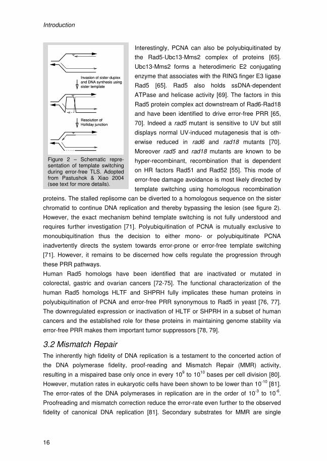
proteins. The stalled replisome can be diverted to a homologous sequence on the sister
chromatid to continue DNA replication and thereby bypassing the lesion (see figure 2).
However, the exact mechanism behind template switching is not fully understood and
requires further investigation [71]. Polyubiquitination of PCNA is mutually exclusive to
monoubiquitination thus the decision to either mono- or polyubiquitinate PCNA
inadvertently directs the system towards error-prone or error-free template switching
[71]. However, it remains to be discerned how cells regulate the progression through
these PRR pathways.
Human Rad5 homologs have been identified that are inactivated or mutated in
colorectal, gastric and ovarian cancers [72-75]. The functional characterization of the
human Rad5 homologs HLTF and SHPRH fully implicates these human proteins in
polyubiquitination of PCNA and error-free PRR synonymous to Rad5 in yeast [76, 77].
The downregulated expression or inactivation of HLTF or SHPRH in a subset of human
cancers and the established role for these proteins in maintaining genome stability via
error-free PRR makes them important tumor suppressors [78, 79].
3.2 Mismatch Repair
The inherently high fidelity of DNA replication is a testament to the concerted action of
the DNA polymerase fidelity, proof-reading and Mismatch Repair (MMR) activity,
resulting in a mispaired base only once in every 109 to 10
10 bases per cell division [80].
However, mutation rates in eukaryotic cells have been shown to be lower than 10-10
[81].
The error-rates of the DNA polymerases in replication are in the order of 10-3
to 10-6
.
Proofreading and mismatch correction reduce the error-rate even further to the observed
fidelity of canonical DNA replication [81]. Secondary substrates for MMR are single
Figure 2 – Schematic repre-sentation of template switching during error-free TLS. Adopted from Pastushok & Xiao 2004 (see text for more details).
Introduction
17
nucleotide mismatches that originate from DNA damage that persists through S phase
and is tolerated by the error-prone PRR pathway. These mismatches arise due to
misincorporation of nucleotides by the low fidelity TLS DNA polymerases. Other
important MMR targets are mismatches generated by insertion and deletion events in
microsatellites. These mismatches result from misalignment of the polymerase during
synthesis of highly repetitive DNA of these microsatellites that is prone to polymerase
slippage [82, 83]. This makes microsatellites inherently unstable and even more unstable
when MMR is deficient. Therefore, MMR is widespread in nature because it actively
contributes to replication fidelity which when solely dependent on polymerase fidelity and
proof-reading is not sufficient to replicate large genomes at an appreciably low error-rate.
MMR genes were initially described in both prokaryotes and simple eukaryotes as 'Mut'
genes due to their spontaneous and UV-induced mutator phenotype and accumulation of
replication errors when deleted [82]. Bacterial MMR has been completely reconstituted in
vitro which lead to the deciphering of the mechanism behind MMR [84]. Repair of these
lesions, as with any DNA anomaly, starts with the recognition of the mismatch [85]. In
bacteria the MutS homodimer recognizes a host of mismatches and interacts with the β-
clamp (homologous to PCNA in eukaryotes) that is believed to help deliver MutS to
newly replicated DNA [86, 87]. Next, the MutL ATPase protein is recruited to attract and
activate the MutH endonuclease [88, 89]. The concerted action of MutL and MutS is
believed to serve as a mode of licensing damage recognition for downstream processing
[90]. MutL is referred to as a 'matchmaker' as it lacks specific enzymatic activity but
instead collaborates with MutS in damage verification, recruitment and activation of
downstream MMR factors. MutL has aspecific affinity for DNA underscoring its
matchmaker function [91, 92]. Since mismatches are not damaged bases or nucleotides,
the discrimination between the parental and newly synthesized strand, containing the
mismatch, is crucial for MMR to prevent mutagenesis. It is thus not the recognition of the
mismatch per se but the selection of the newly synthesized strand that allows MMR to
actively contribute to replication fidelity. In E.coli the temporal hemimethylated state of
newly synthesized duplex DNA makes this possible due to dam DNA methylation [84].
The MutH endonuclease specifically cleaves the newly synthesized strand at the
hemimethylated GATC site [93]. Following the strand-selective nicking of the DNA, MutL
is responsible for loading a DNA helicase onto the nick [94, 95], that together with
ssDNA binding protein (SSB) generates a ssDNA stretch that can be refilled by a DNA
polymerase [84]. The basic steps of recognition, strand-selection, nicking and repair
synthesis are conserved mechanistically [82]. Similarly, genes and proteins involved in
MMR are conserved from bacteria to man [82, 83, 96].
Interestingly, higher eukaryotes maintain multiple MutS (MSH) and MutL homologs
(MLH) that consistently act as heterodimers [82, 96]. It has been shown that the
eukaryotic MMR proteins have affinity for different mismatches [82, 96] ranging from
single basepair mismatches to insertion and deletion loops [97, 98]. Similarly, specific
Introduction
16
Interestingly, PCNA can also be polyubiquitinated by
the Rad5-Ubc13-Mms2 complex of proteins [65].
Ubc13-Mms2 forms a heterodimeric E2 conjugating
enzyme that associates with the RING finger E3 ligase
Rad5 [65]. Rad5 also holds ssDNA-dependent
ATPase and helicase activity [69]. The factors in this
Rad5 protein complex act downstream of Rad6-Rad18
and have been identified to drive error-free PRR [65,
70]. Indeed a rad5 mutant is sensitive to UV but still
displays normal UV-induced mutagenesis that is oth-
erwise reduced in rad6 and rad18 mutants [70].
Moreover rad5 and rad18 mutants are known to be
hyper-recombinant, recombination that is dependent
on HR factors Rad51 and Rad52 [55]. This mode of
error-free damage avoidance is most likely directed by
template switching using homologous recombination
proteins. The stalled replisome can be diverted to a homologous sequence on the sister
chromatid to continue DNA replication and thereby bypassing the lesion (see figure 2).
However, the exact mechanism behind template switching is not fully understood and
requires further investigation [71]. Polyubiquitination of PCNA is mutually exclusive to
monoubiquitination thus the decision to either mono- or polyubiquitinate PCNA
inadvertently directs the system towards error-prone or error-free template switching
[71]. However, it remains to be discerned how cells regulate the progression through
these PRR pathways.
Human Rad5 homologs have been identified that are inactivated or mutated in
colorectal, gastric and ovarian cancers [72-75]. The functional characterization of the
human Rad5 homologs HLTF and SHPRH fully implicates these human proteins in
polyubiquitination of PCNA and error-free PRR synonymous to Rad5 in yeast [76, 77].
The downregulated expression or inactivation of HLTF or SHPRH in a subset of human
cancers and the established role for these proteins in maintaining genome stability via
error-free PRR makes them important tumor suppressors [78, 79].
3.2 Mismatch Repair
The inherently high fidelity of DNA replication is a testament to the concerted action of
the DNA polymerase fidelity, proof-reading and Mismatch Repair (MMR) activity,
resulting in a mispaired base only once in every 109 to 10
10 bases per cell division [80].
However, mutation rates in eukaryotic cells have been shown to be lower than 10-10
[81].
The error-rates of the DNA polymerases in replication are in the order of 10-3
to 10-6
.
Proofreading and mismatch correction reduce the error-rate even further to the observed
fidelity of canonical DNA replication [81]. Secondary substrates for MMR are single
Figure 2 – Schematic repre-sentation of template switching during error-free TLS. Adopted from Pastushok & Xiao 2004 (see text for more details).
Introduction
17
nucleotide mismatches that originate from DNA damage that persists through S phase
and is tolerated by the error-prone PRR pathway. These mismatches arise due to
misincorporation of nucleotides by the low fidelity TLS DNA polymerases. Other
important MMR targets are mismatches generated by insertion and deletion events in
microsatellites. These mismatches result from misalignment of the polymerase during
synthesis of highly repetitive DNA of these microsatellites that is prone to polymerase
slippage [82, 83]. This makes microsatellites inherently unstable and even more unstable
when MMR is deficient. Therefore, MMR is widespread in nature because it actively
contributes to replication fidelity which when solely dependent on polymerase fidelity and
proof-reading is not sufficient to replicate large genomes at an appreciably low error-rate.
MMR genes were initially described in both prokaryotes and simple eukaryotes as 'Mut'
genes due to their spontaneous and UV-induced mutator phenotype and accumulation of
replication errors when deleted [82]. Bacterial MMR has been completely reconstituted in
vitro which lead to the deciphering of the mechanism behind MMR [84]. Repair of these
lesions, as with any DNA anomaly, starts with the recognition of the mismatch [85]. In
bacteria the MutS homodimer recognizes a host of mismatches and interacts with the β-
clamp (homologous to PCNA in eukaryotes) that is believed to help deliver MutS to
newly replicated DNA [86, 87]. Next, the MutL ATPase protein is recruited to attract and
activate the MutH endonuclease [88, 89]. The concerted action of MutL and MutS is
believed to serve as a mode of licensing damage recognition for downstream processing
[90]. MutL is referred to as a 'matchmaker' as it lacks specific enzymatic activity but
instead collaborates with MutS in damage verification, recruitment and activation of
downstream MMR factors. MutL has aspecific affinity for DNA underscoring its
matchmaker function [91, 92]. Since mismatches are not damaged bases or nucleotides,
the discrimination between the parental and newly synthesized strand, containing the
mismatch, is crucial for MMR to prevent mutagenesis. It is thus not the recognition of the
mismatch per se but the selection of the newly synthesized strand that allows MMR to
actively contribute to replication fidelity. In E.coli the temporal hemimethylated state of
newly synthesized duplex DNA makes this possible due to dam DNA methylation [84].
The MutH endonuclease specifically cleaves the newly synthesized strand at the
hemimethylated GATC site [93]. Following the strand-selective nicking of the DNA, MutL
is responsible for loading a DNA helicase onto the nick [94, 95], that together with
ssDNA binding protein (SSB) generates a ssDNA stretch that can be refilled by a DNA
polymerase [84]. The basic steps of recognition, strand-selection, nicking and repair
synthesis are conserved mechanistically [82]. Similarly, genes and proteins involved in
MMR are conserved from bacteria to man [82, 83, 96].
Interestingly, higher eukaryotes maintain multiple MutS (MSH) and MutL homologs
(MLH) that consistently act as heterodimers [82, 96]. It has been shown that the
eukaryotic MMR proteins have affinity for different mismatches [82, 96] ranging from
single basepair mismatches to insertion and deletion loops [97, 98]. Similarly, specific
Introduction
18
MSH and MLH proteins are involved in meiotic recombination [99] and mitochondrial
MMR [100]. Intriguingly, MutH homologs have not been identified in higher eukaryotes
and endo- or exonucleases associated with MMR have only recently been described
[90]. In mammals, EXO1 and the PMS2 subunit of MutLα are responsible for the
digestion and nicking steps of MMR, respectively [90]. Importantly, the underlying
mechanism of repair is conserved despite these minor differences.
Strand selection in higher eukaryotes cannot make use of hemimethylated DNA because
dam DNA methylation is unique to a subset of bacteria [83]. MSH and MLH proteins in
eukaryotes have been shown to interact with PCNA [87], drawing on the parallel with the
bacterial system. However, for eukaryotes it is suggested that this interaction is
important for the strand specificity of MMR [82, 96, 101]. The latent endonuclease
activity of PMS2 in MutLα is activated by its interaction with PCNA and MutS [102]. The
PMS2 subunit is spatially restricted within the complex to only nick one strand by the
orientation of PCNA which in turn makes the nick specific for the mismatched strand [90].
In this way PCNA is thought to give MMR its directionality as PCNA is always loaded
onto heteroduplex DNA with the same orientation that cannot switch [58, 90]. Another
possible mechanism for strand-specificity in higher eukaryotes is thought to arise from
strand discontinuities associated with replication like Okazaki fragments, confining this
mode of strand-selection to the lagging strand [86, 103, 104].
Long stretches of simple DNA repeats like mono-, di- and trinucleotide repeats pose
difficult substrates to faithfully replicate. These microsatellites have been known to be
highly unstable in MMR deficient model organisms [82, 83, 96]. An important link
between cancer and MMR deficiency arose when it was identified that 60 to 70% of
hereditary non-polyposis colon cancer (HNPCC) patients have mutations in MLH1 and
MSH2 [82, 105]. Similarly, a minority of HNPCC cases is associated with mutations in
PMS1, PMS2 or MSH6 (reviewed in [106]). Thus, loss of MMR activity increases
mutagenesis and tumor development [82]. Increase in cancer susceptibility has also
been shown for numerous mouse MMR knockouts that suffer from increased
tumorigenesis of internal organs confirming the importance of MMR in supporting DNA
replication to maintain genome stability [82, 96].
3.3 Direct DNA damage reversal
The majority of organisms studied thus far feature one or both of the following direct
reversal pathways: photolyase and AlkylGuanine Transferase (AGT). Photolyases are
widely spread through all kingdoms of life, but most placental mammals including
humans do not have a photolyase [107]. A photolyase is a monomeric enzyme that
catalyzes the reversion of UV-induced lesions by using energy quanta of visible light
[108, 109]. Originally, the first E.coli photolyase that was described is a CPD photolyase,
only able to revert CPDs [110] and it was not until much later that the first 6-4PP
photolyase was described in Drosophila melanogaster [111, 112]. Both CPD and 6-4PP
Introduction
19
photolyases have different degrees of sequence similarity amongst species but are all
expected to have fairly similar structures and reaction mechanisms based on available
sequence and structure data [108, 112, 113]. The photolyase protein binds UV damaged
DNA due to the detection of a slight kink that is induced by the helix distortion of the UV
lesion not present in undamaged DNA [114]. Upon binding the photolesion flips out of
the DNA double helix into the photolyase active site where the reaction takes place
[115].
All photolyase enzymes carry the FAD flavin chromofore as cofactor that is pivotal in the
cyclic redox reaction mechanism described here [109, 113]. The flavin cofactor is
accompanied by a second chromophore that acts as a photoreceptor antenna commonly
in the form of MTHF (5,10-methenyltetrahydrofolate) or more rarely, 8-HDF (8-hydroxy-5-
deazariboflavin) [109, 113]. After DNA damage binding the energy of a single photon is
absorbed by the folate cofactor and transferred to the flavin chromophore, generating an
excited FADH-* molecule. The energy quantum is not sufficient to break the cyclobutane
ring by itself but it increases the redox potential of the FADH- molecule. The excited
FADH-* molecule transfers an electron to the pyrimdine dimer splitting the cyclobutane
ring. The cyclic nature of the reaction makes that the FADH• radical is also reverted back
to its groundstate FADH- by electron back-transfer. As a result the pyrimidine dimer is
completely reverted to the original DNA conformation as is the flavin cofactor [108, 109,
113].
AlkylGguanine Transferases (AGTs) are widespread in nature, but are not found in
plants and Schizosaccharomyces pombe [116]. AGTs protect cells from the effects of
both endogenous and exogenous alkylating agents. This family of damage reversal
proteins was originally characterized based on the properties of the human and E.coli
MGMT enzymes (O6-methylguanine-DNA methyltransferase) that were shown to remove
the methyl group from a O6-methylguanine [110, 117-119]. However, more recent
literature describes a more generic alkyltransferase function to this family of proteins by
showing that more bulky adducts and tobacco-induced carcinogens are also a substrate
for these enzymes [120, 121]. Therefore, these enzymes are now collectively referred to
as alkyltransferases [116]. AGTs are thought to be able to actively slide acrross the DNA
double helix in search for a damage, however, the exact nature of the force driving this
process is currently unknown [116]. Upon DNA damage binding AGTs bend the DNA
slightly, allowing it access to the minor groove. In this conformation the enzyme can flip
out the substrate into its active center where a reactive cysteine attacks the alkyl group
and covalently transfers it onto the enzyme itself [122, 123]. This restores the guanine
and leaves the enzyme catalytically dead. In this state the occupied reactive site brings
about a conformational change in the enzyme that is thought to expose a ubiquitination
site leading to ubiquitin mediated degradation of the protein in eukaryotes [124, 125].
A confounding factor of AGTs is that increased AGT activity in certain types of cancer
protects the malignant cells from alkylating agents used in cancer treatment, while at the

Introduction
18
MSH and MLH proteins are involved in meiotic recombination [99] and mitochondrial
MMR [100]. Intriguingly, MutH homologs have not been identified in higher eukaryotes
and endo- or exonucleases associated with MMR have only recently been described
[90]. In mammals, EXO1 and the PMS2 subunit of MutLα are responsible for the
digestion and nicking steps of MMR, respectively [90]. Importantly, the underlying
mechanism of repair is conserved despite these minor differences.
Strand selection in higher eukaryotes cannot make use of hemimethylated DNA because
dam DNA methylation is unique to a subset of bacteria [83]. MSH and MLH proteins in
eukaryotes have been shown to interact with PCNA [87], drawing on the parallel with the
bacterial system. However, for eukaryotes it is suggested that this interaction is
important for the strand specificity of MMR [82, 96, 101]. The latent endonuclease
activity of PMS2 in MutLα is activated by its interaction with PCNA and MutS [102]. The
PMS2 subunit is spatially restricted within the complex to only nick one strand by the
orientation of PCNA which in turn makes the nick specific for the mismatched strand [90].
In this way PCNA is thought to give MMR its directionality as PCNA is always loaded
onto heteroduplex DNA with the same orientation that cannot switch [58, 90]. Another
possible mechanism for strand-specificity in higher eukaryotes is thought to arise from
strand discontinuities associated with replication like Okazaki fragments, confining this
mode of strand-selection to the lagging strand [86, 103, 104].
Long stretches of simple DNA repeats like mono-, di- and trinucleotide repeats pose
difficult substrates to faithfully replicate. These microsatellites have been known to be
highly unstable in MMR deficient model organisms [82, 83, 96]. An important link
between cancer and MMR deficiency arose when it was identified that 60 to 70% of
hereditary non-polyposis colon cancer (HNPCC) patients have mutations in MLH1 and
MSH2 [82, 105]. Similarly, a minority of HNPCC cases is associated with mutations in
PMS1, PMS2 or MSH6 (reviewed in [106]). Thus, loss of MMR activity increases
mutagenesis and tumor development [82]. Increase in cancer susceptibility has also
been shown for numerous mouse MMR knockouts that suffer from increased
tumorigenesis of internal organs confirming the importance of MMR in supporting DNA
replication to maintain genome stability [82, 96].
3.3 Direct DNA damage reversal
The majority of organisms studied thus far feature one or both of the following direct
reversal pathways: photolyase and AlkylGuanine Transferase (AGT). Photolyases are
widely spread through all kingdoms of life, but most placental mammals including
humans do not have a photolyase [107]. A photolyase is a monomeric enzyme that
catalyzes the reversion of UV-induced lesions by using energy quanta of visible light
[108, 109]. Originally, the first E.coli photolyase that was described is a CPD photolyase,
only able to revert CPDs [110] and it was not until much later that the first 6-4PP
photolyase was described in Drosophila melanogaster [111, 112]. Both CPD and 6-4PP
Introduction
19
photolyases have different degrees of sequence similarity amongst species but are all
expected to have fairly similar structures and reaction mechanisms based on available
sequence and structure data [108, 112, 113]. The photolyase protein binds UV damaged
DNA due to the detection of a slight kink that is induced by the helix distortion of the UV
lesion not present in undamaged DNA [114]. Upon binding the photolesion flips out of
the DNA double helix into the photolyase active site where the reaction takes place
[115].
All photolyase enzymes carry the FAD flavin chromofore as cofactor that is pivotal in the
cyclic redox reaction mechanism described here [109, 113]. The flavin cofactor is
accompanied by a second chromophore that acts as a photoreceptor antenna commonly
in the form of MTHF (5,10-methenyltetrahydrofolate) or more rarely, 8-HDF (8-hydroxy-5-
deazariboflavin) [109, 113]. After DNA damage binding the energy of a single photon is
absorbed by the folate cofactor and transferred to the flavin chromophore, generating an
excited FADH-* molecule. The energy quantum is not sufficient to break the cyclobutane
ring by itself but it increases the redox potential of the FADH- molecule. The excited
FADH-* molecule transfers an electron to the pyrimdine dimer splitting the cyclobutane
ring. The cyclic nature of the reaction makes that the FADH• radical is also reverted back
to its groundstate FADH- by electron back-transfer. As a result the pyrimidine dimer is
completely reverted to the original DNA conformation as is the flavin cofactor [108, 109,
113].
AlkylGguanine Transferases (AGTs) are widespread in nature, but are not found in
plants and Schizosaccharomyces pombe [116]. AGTs protect cells from the effects of
both endogenous and exogenous alkylating agents. This family of damage reversal
proteins was originally characterized based on the properties of the human and E.coli
MGMT enzymes (O6-methylguanine-DNA methyltransferase) that were shown to remove
the methyl group from a O6-methylguanine [110, 117-119]. However, more recent
literature describes a more generic alkyltransferase function to this family of proteins by
showing that more bulky adducts and tobacco-induced carcinogens are also a substrate
for these enzymes [120, 121]. Therefore, these enzymes are now collectively referred to
as alkyltransferases [116]. AGTs are thought to be able to actively slide acrross the DNA
double helix in search for a damage, however, the exact nature of the force driving this
process is currently unknown [116]. Upon DNA damage binding AGTs bend the DNA
slightly, allowing it access to the minor groove. In this conformation the enzyme can flip
out the substrate into its active center where a reactive cysteine attacks the alkyl group
and covalently transfers it onto the enzyme itself [122, 123]. This restores the guanine
and leaves the enzyme catalytically dead. In this state the occupied reactive site brings
about a conformational change in the enzyme that is thought to expose a ubiquitination
site leading to ubiquitin mediated degradation of the protein in eukaryotes [124, 125].
A confounding factor of AGTs is that increased AGT activity in certain types of cancer
protects the malignant cells from alkylating agents used in cancer treatment, while at the
Introduction
20
same time reduced AGT activity might result in accumulation of alkylating damages that
could result in cancer in people that are exposed to exogenous alkylating agents [126,
127]. This makes AGT inhibitors prime agents for cancer therapy, specifically sensitizing
cancer cells for treatment with alkylating agents [126, 128]. Importantly, AGTs help
protect the cell from smoke derived carcinogens that bind covalently to DNA. The
smoking related nitrosamine carcinogens are a substrate for AGTs and interindividual
differences in MGMT expression can predispose those smokers with reduced MGMT
expression to lung cancer even more [126].
3.4 Base Excision Repair
BER is responsible for the repair of altered bases and including oxidative damages [129,
130]. Oxidative damage is a rather frequent event in aerobic organisms resulting in the
order of ~100-500 8-hydroxy-guanine (8-oxoG) lesions for instance in a human cell, per
day [8]. Other chemically altered bases in DNA are also substrate for BER including
uracil induced by cytosine deanimation and alkylation generated 3-methyladenine [127,
131]. Most of these damages are not bulky and will therefore not block replication.
Instead they will mispair during replication resulting in mutagenesis. The high potential
for mutation and the abundance of oxidative damages due to respiration are a likely
explanation for the strong evolutionary conservation of most genes involved in BER
[130].
Mechanistically, BER can be described by 5 separate enzymatic steps starting with the
recognition and excision of the damaged base [132]. DNA glycosylases bend DNA upon
binding [133] and flip-out the damaged base into an extra helical position to fit inside the
catalytic pocket [134]. The DNA-enzyme interaction leads to a severe kink in the DNA of
up to 70° probing the DNA for damage. Only when a damaged base is bound will it be
flipped into the active site [135, 136]. DNA damage recognition is followed by enzymatic
cleavage of the N-glycosyl bond between the base and the sugar connected to the DNA
backbone. This initial step is performed by an abundance of DNA glycosylases with
different substrate affinity. Individual enzymes can recognize a subset of lesions making
the damage recognition system in BER highly redundant. After cleavage the damaged
base is released and an apurinic-/apyrimidinic (AP) site remains [137, 138]. The resulting
AP-site is a substrate for the AP-endonuclease APE1, that generates a 5' nick at the AP-
site in the DNA backbone. Alternatively, some DNA glycosylases are bifunctional and
contain a protein domain with AP-lyase function [139]. In this way the glycosylase itself
can generate a nick after excising the damaged base. The small single nucleotide gap
and DNA nick are now substrate for DNA Polymerase β (POLB) to perform the third and
forth enzymatic steps. POLB removes the 5’-terminal deoxyribophosphate (dRP) left
over by the AP-endonuclease making it a suitable substrate to insert the correct
nucleotide into the AP-site [140, 141]. Finally, DNA Ligase I or III will ligate the nick
returning the DNA to its undamaged state [142].
Introduction
21
Depending on how the nick is generated and processed, BER can funnel into short-patch
or long-patch BER after DNA damage recognition and base excision [143]. A suitable 5'
and 3' substrate for POLB allows short-patch BER to take place according to the
mechanism described above. During long-patch BER, however, displacement DNA
synthesis by POLB or POLD occur as a result of the residual 5' terminal dRP not being
amenable for POLB induced removal. After removal of the 4 to 6 basepair flap by the
endonuclease FEN1 [144] the synthesized patch is suitable for DNA Ligase I or DNA
Ligase III mediated ligation.
Interestingly, BER is very important in sustaining life as gross defects in BER are not
found in nature. Knocking out genes essential for BER like POLB, APE1 or FEN1 in mice
results in embryonic lethality [141, 145, 146]. It is interesting to note that 30% of all
tumors described contain a mutant variant of POLB highlighting that defective BER can
drive mutagenesis leading to cancer [129]. A few examples of human disease as a result
of specific BER deficiencies have been described in literature but are beyond the scope
of this introduction [129, 130, 147].
3.5 Double Strand Break Repair via Homologous Recombination & Non-Homologous End-Joining
DNA Double strand breaks (DSBs) are uniquely different to other DNA damages as both
backbones of the DNA double helix are severed, in essence breaking a chromosome in
two. The gross chromosomal aberrations that result from their faulty repair has a large
impact on the genome opposed single nucleotide mutations described previously.
Instead severed genes can lose or gain function and the fusion of genes can drive many
different malignant processes that underlie tumorigenesis. DSBs are, therefore, highly
lethal and their repair is pivotal to maintain genome stability and prevent cancer.
DSBs can be induced by ionizing radiation [148]. Moreover, certain chemicals and ROS
induced by cellular metabolism also have the potential to block replication and result in
DSBs [17, 149]. Under high damage load, repair mechanisms can induce nicks in
opposite backbones of the DNA in close enough proximity to produce DSBs. Similarly,
ssDNA nicks can result in DSBs when the replisome attempts to synthesize DNA
opposite these lesions. Importantly, some processes require the cell to generate DSBs,
for instance to create genetic diversity during meiotic recombination [150]. Induction of
these types of DSBs is of course tightly controlled.
If a homologous template is available it can be applied for its complementarity by
Homologous Recombination (HR) which is therefore inherently limited to S phase [151].
A second mode of repair exists that results in the annealing of the two severed ends of a
DSB that is referred to as Non-Homologous End-Joining (NHEJ) [148]. A mode of repair
that is not restricted to cell cycle but is believed to be downregulated during S phase
since it can have deleterious effects during this stage of the cell cycle [151]. In the
absence of a homologous template NHEJ efficiently anneals a two-ended DSB [149].
Introduction
22
Depending on how a DSB is generated, the DNA ends caused by an exogenous damage
source are chemically altered and will not consist of a 5' and 3' terminus that is amenable
for ligation. Thus the processing of both ends of the DSB is a prerequisite for both HR
and NHEJ [30, 152]. Endogenous breaks, on the other hand, typically result in less
aberrant DNA end structures that are a suitable substrate for direct ligation [30, 148,
152]. Interestingly, end processing is suggested to play an important role in regulation of
HR and NHEJ. NHEJ can already ligate two ends with 4 or less basepair overhangs
whereas HR requires long sections of ssDNA for strand invasion [153]. The selection to
progress through the HR pathway is fixed when the DNA end is irreversibly processed
into long ssDNA stretches to facilitate strand invasion [154]. Vice versa, stabilizing short
resected NHEJ substrate ends will drive NHEJ in favor of HR [154]. Moreover, it has
been shown that end resection is tightly regulated during the cell cycle adding a level of
control to inhibit NHEJ and stimulate HR during S phase [34, 155].
NHEJ is conceptually the most straightforward way of fixing a DSB. Native damages
such as blunt-ends or short 5' or 3' complementary overhangs at a break are amenable
for in vitro repair by only the core eukaryotic NHEJ factors Ku70, Ku80, DNA-dependent
protein kinase catalytic subunit (DNA-PKcs) and DNA ligase IV [156]. The Ku70-Ku80
heterodimer has affinity for DNA ends and forms a ring structure around the DNA end
protecting it from degradation [157] and recruiting DNA-PKcs and other downstream
targets [148, 152]. DNA-PKcs in higher eukaryotes hold the two DNA ends together
during the repair process [158]. The association of Ku70-Ku80 and DNA-PKcs changes
the conformation of the DNA-protein complex exposing the end for processing by the
endonuclease Artemis [159-161]. This protein complex can process a large variety of
altered DNA structures at the DNA end, making the end available for repair [161, 162].
After a DNA polymerase is recruited that fills any ssDNA gaps, ligation can take place
[163, 164]. Finally, DNA Ligase IV is recruited to close the remaining nicks [165, 166].
As mentioned earlier HR is restricted to S phase because of the need for a homologous
DNA strand. HR is based on the exchange of homologous or semi-homologous
sequences between a broken and intact DNA strand [149, 152]. The basic steps of HR
involve end-resection, strand invasion, DNA synthesis and resolving the repair
intermediate DNA structure [30]. Depending on the order of events that result in strand
invasion HR culminates into different sub-pathways of which only the classic double-
strand break repair (DSBR) pathway is discussed here [30].
The first step in DSBR is 5' end-resection producing a 3' ssDNA stretch. The MR(X)N
complex (see section 2) is required for end-resection but the nature of the specific
exonuclease is unclear [167, 168]. Currently, redundant nuclease activity has been
described for several factors that contribute to end-resection [30]. Rad50 of the MRX
complex is thought to hold the DNA ends together during HR, as it does for NHEJ in
yeast [169, 170]. The processed ssDNA stretch is bound by RPA quickly after resection,
which prevents formation of secondary DNA structure [171]. RPA in turn interacts with
Introduction
23
Rad52, a core recombination protein that is required for most DSB repair pathways [30].
Rad52 and possibly MR(X)N actively load the Rad51 recombinase onto the ssDNA
replacing the RPA molecules resulting in a Rad51 nucleofilament [152, 172]. The Rad51-
DNA complex is ready for homology search and strand invasion resulting in D-loop
formation interlinking the broken DNA strand and the intact template. However, the exact
mechanism behind homology search is not fully understood. Recent progress has
identified chromatin remodeling activity of Rad54 to be a driving force for the search for
homology [30]. After strand invasion, replication can synthesize DNA across the
homologous template. The resulting DNA structure of interwoven dsDNA is attached via
Holliday junctions that need to be actively resolved [152]. After untangling of the intricate
DNA structures both DNA strands are restored to intact dsDNA molecules.
4. Nucleotide Excision Repair in eukaryotes
4.1 A short history of Nucleotide Excision Repair
An abundant natural exogenous source of DNA damage is UV radiation from sunlight.
UV radiation results in dimer formation between neighboring pyrimidines. These dimers
are Cyclebutane Pyrimidine Dimers (CPD) and 6-4 PhotoProducst (6-4PP) that are the
major components of UV induced DNA damage (see figure 3). The common
denominator amongst lesions repaired by NER is that they distort the DNA double helix,
unwinding it slightly. Mechanistically, NER is highly conserved from bacteria to higher
eukaryotes. Amongst eukaryotes homology is high but not perfect. Because of its nature
NER is not mutagenic and deficiency of this mode of repair due to genetic defects is
associated with the cancer prone disorder Xeroderma pigmentosum in humans. If UV
damages persist through S phase they can become lethal or mutagenic by blocking
replication and transcription. The focus of this thesis and the research described in the
following chapters is on NER. Therefore, more emphasis is put on this repair pathway.
The first mode of DNA repair that was discovered was photoreactivation: repair of UV
induced damage that is dependent on visible light, later shown to be the result of
photolyase DNA damage reversal (3.3). However, cells after UV treatment showed
Figure 3 – UV induced pyrimidine cyclobutane dimer (CPD) and pyrimidine-pyrimidone (6-4) photoproduct (6-4PP). Taken from [109].

Introduction
22
Depending on how a DSB is generated, the DNA ends caused by an exogenous damage
source are chemically altered and will not consist of a 5' and 3' terminus that is amenable
for ligation. Thus the processing of both ends of the DSB is a prerequisite for both HR
and NHEJ [30, 152]. Endogenous breaks, on the other hand, typically result in less
aberrant DNA end structures that are a suitable substrate for direct ligation [30, 148,
152]. Interestingly, end processing is suggested to play an important role in regulation of
HR and NHEJ. NHEJ can already ligate two ends with 4 or less basepair overhangs
whereas HR requires long sections of ssDNA for strand invasion [153]. The selection to
progress through the HR pathway is fixed when the DNA end is irreversibly processed
into long ssDNA stretches to facilitate strand invasion [154]. Vice versa, stabilizing short
resected NHEJ substrate ends will drive NHEJ in favor of HR [154]. Moreover, it has
been shown that end resection is tightly regulated during the cell cycle adding a level of
control to inhibit NHEJ and stimulate HR during S phase [34, 155].
NHEJ is conceptually the most straightforward way of fixing a DSB. Native damages
such as blunt-ends or short 5' or 3' complementary overhangs at a break are amenable
for in vitro repair by only the core eukaryotic NHEJ factors Ku70, Ku80, DNA-dependent
protein kinase catalytic subunit (DNA-PKcs) and DNA ligase IV [156]. The Ku70-Ku80
heterodimer has affinity for DNA ends and forms a ring structure around the DNA end
protecting it from degradation [157] and recruiting DNA-PKcs and other downstream
targets [148, 152]. DNA-PKcs in higher eukaryotes hold the two DNA ends together
during the repair process [158]. The association of Ku70-Ku80 and DNA-PKcs changes
the conformation of the DNA-protein complex exposing the end for processing by the
endonuclease Artemis [159-161]. This protein complex can process a large variety of
altered DNA structures at the DNA end, making the end available for repair [161, 162].
After a DNA polymerase is recruited that fills any ssDNA gaps, ligation can take place
[163, 164]. Finally, DNA Ligase IV is recruited to close the remaining nicks [165, 166].
As mentioned earlier HR is restricted to S phase because of the need for a homologous
DNA strand. HR is based on the exchange of homologous or semi-homologous
sequences between a broken and intact DNA strand [149, 152]. The basic steps of HR
involve end-resection, strand invasion, DNA synthesis and resolving the repair
intermediate DNA structure [30]. Depending on the order of events that result in strand
invasion HR culminates into different sub-pathways of which only the classic double-
strand break repair (DSBR) pathway is discussed here [30].
The first step in DSBR is 5' end-resection producing a 3' ssDNA stretch. The MR(X)N
complex (see section 2) is required for end-resection but the nature of the specific
exonuclease is unclear [167, 168]. Currently, redundant nuclease activity has been
described for several factors that contribute to end-resection [30]. Rad50 of the MRX
complex is thought to hold the DNA ends together during HR, as it does for NHEJ in
yeast [169, 170]. The processed ssDNA stretch is bound by RPA quickly after resection,
which prevents formation of secondary DNA structure [171]. RPA in turn interacts with
Introduction
23
Rad52, a core recombination protein that is required for most DSB repair pathways [30].
Rad52 and possibly MR(X)N actively load the Rad51 recombinase onto the ssDNA
replacing the RPA molecules resulting in a Rad51 nucleofilament [152, 172]. The Rad51-
DNA complex is ready for homology search and strand invasion resulting in D-loop
formation interlinking the broken DNA strand and the intact template. However, the exact
mechanism behind homology search is not fully understood. Recent progress has
identified chromatin remodeling activity of Rad54 to be a driving force for the search for
homology [30]. After strand invasion, replication can synthesize DNA across the
homologous template. The resulting DNA structure of interwoven dsDNA is attached via
Holliday junctions that need to be actively resolved [152]. After untangling of the intricate
DNA structures both DNA strands are restored to intact dsDNA molecules.
4. Nucleotide Excision Repair in eukaryotes
4.1 A short history of Nucleotide Excision Repair
An abundant natural exogenous source of DNA damage is UV radiation from sunlight.
UV radiation results in dimer formation between neighboring pyrimidines. These dimers
are Cyclebutane Pyrimidine Dimers (CPD) and 6-4 PhotoProducst (6-4PP) that are the
major components of UV induced DNA damage (see figure 3). The common
denominator amongst lesions repaired by NER is that they distort the DNA double helix,
unwinding it slightly. Mechanistically, NER is highly conserved from bacteria to higher
eukaryotes. Amongst eukaryotes homology is high but not perfect. Because of its nature
NER is not mutagenic and deficiency of this mode of repair due to genetic defects is
associated with the cancer prone disorder Xeroderma pigmentosum in humans. If UV
damages persist through S phase they can become lethal or mutagenic by blocking
replication and transcription. The focus of this thesis and the research described in the
following chapters is on NER. Therefore, more emphasis is put on this repair pathway.
The first mode of DNA repair that was discovered was photoreactivation: repair of UV
induced damage that is dependent on visible light, later shown to be the result of
photolyase DNA damage reversal (3.3). However, cells after UV treatment showed
Figure 3 – UV induced pyrimidine cyclobutane dimer (CPD) and pyrimidine-pyrimidone (6-4) photoproduct (6-4PP). Taken from [109].
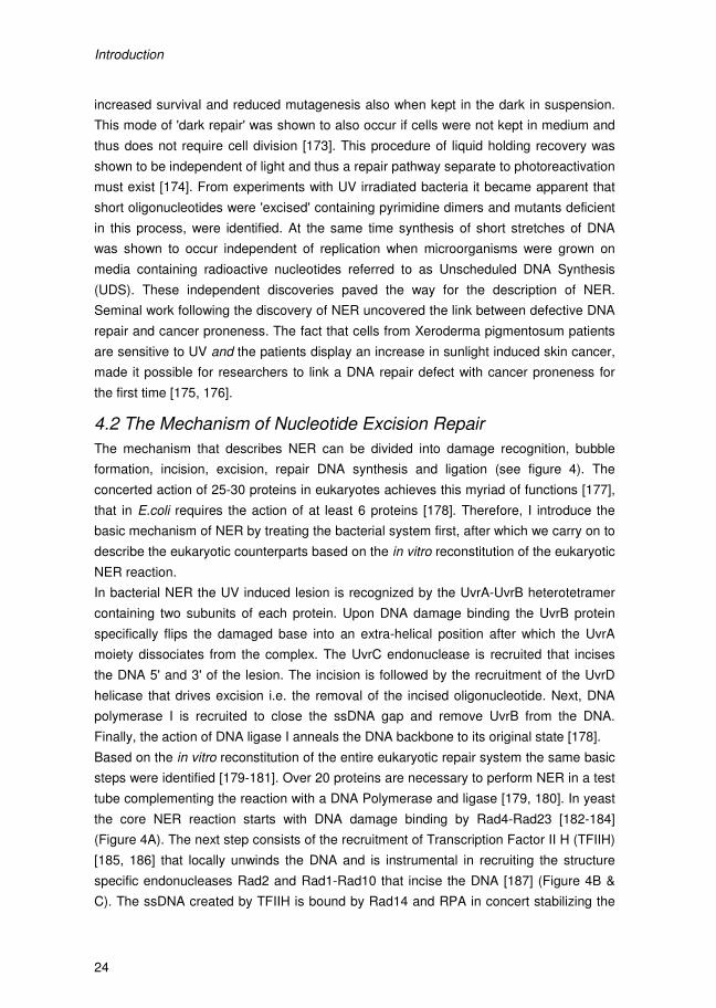
Introduction
24
increased survival and reduced mutagenesis also when kept in the dark in suspension.
This mode of 'dark repair' was shown to also occur if cells were not kept in medium and
thus does not require cell division [173]. This procedure of liquid holding recovery was
shown to be independent of light and thus a repair pathway separate to photoreactivation
must exist [174]. From experiments with UV irradiated bacteria it became apparent that
short oligonucleotides were 'excised' containing pyrimidine dimers and mutants deficient
in this process, were identified. At the same time synthesis of short stretches of DNA
was shown to occur independent of replication when microorganisms were grown on
media containing radioactive nucleotides referred to as Unscheduled DNA Synthesis
(UDS). These independent discoveries paved the way for the description of NER.
Seminal work following the discovery of NER uncovered the link between defective DNA
repair and cancer proneness. The fact that cells from Xeroderma pigmentosum patients
are sensitive to UV and the patients display an increase in sunlight induced skin cancer,
made it possible for researchers to link a DNA repair defect with cancer proneness for
the first time [175, 176].
4.2 The Mechanism of Nucleotide Excision Repair
The mechanism that describes NER can be divided into damage recognition, bubble
formation, incision, excision, repair DNA synthesis and ligation (see figure 4). The
concerted action of 25-30 proteins in eukaryotes achieves this myriad of functions [177],
that in E.coli requires the action of at least 6 proteins [178]. Therefore, I introduce the
basic mechanism of NER by treating the bacterial system first, after which we carry on to
describe the eukaryotic counterparts based on the in vitro reconstitution of the eukaryotic
NER reaction.
In bacterial NER the UV induced lesion is recognized by the UvrA-UvrB heterotetramer
containing two subunits of each protein. Upon DNA damage binding the UvrB protein
specifically flips the damaged base into an extra-helical position after which the UvrA
moiety dissociates from the complex. The UvrC endonuclease is recruited that incises
the DNA 5' and 3' of the lesion. The incision is followed by the recruitment of the UvrD
helicase that drives excision i.e. the removal of the incised oligonucleotide. Next, DNA
polymerase I is recruited to close the ssDNA gap and remove UvrB from the DNA.
Finally, the action of DNA ligase I anneals the DNA backbone to its original state [178].
Based on the in vitro reconstitution of the entire eukaryotic repair system the same basic
steps were identified [179-181]. Over 20 proteins are necessary to perform NER in a test
tube complementing the reaction with a DNA Polymerase and ligase [179, 180]. In yeast
the core NER reaction starts with DNA damage binding by Rad4-Rad23 [182-184]
(Figure 4A). The next step consists of the recruitment of Transcription Factor II H (TFIIH)
[185, 186] that locally unwinds the DNA and is instrumental in recruiting the structure
specific endonucleases Rad2 and Rad1-Rad10 that incise the DNA [187] (Figure 4B &
C). The ssDNA created by TFIIH is bound by Rad14 and RPA in concert stabilizing the
Introduction
25
pre-incision complex [188, 189]. Finally, the recruitment of a DNA polymerase that fills
the ssDNA gap [190] and a DNA ligase that rejoins the DNA backbone at the remaining
nick, completes the repair reaction (Figure 4F). Mechanistically, NER utilizes the DNA
double stranded nature for damage recognition and incision to completely remove an
oligonucleotide containing the damaged base or nucleotides. The complementarity of the
resulting ssDNA is then a perfect substrate for faithful replication of the excised DNA
changing the DNA back to its undamaged state.
Figure 4 – Cartoon of the basic mechanism of eukaryotic NER using the yeast proteins. (A) The Rad4-Rad23 het-erodimer associates with the damage and recruits important downstream targets including TFIIH. (B) TFIIH helps to verify the lesion and locally un-winds the DNA by virtue of its helicase activity. (C) Subsequent re-cruitment of Rad14, RPA and the endonu-cleases to the bubble forming complex leads to the pre-incision state. (D) With both endonu-cleases Rad2 and Rad1-Rad10 in place the first 5' restriction by Rad1-Rad10 will take place after which Rad2 will incise 3' of the damage. (E) Disas-sembly of the repair complex drives excision of a short oligonucleo-tide containing the lesion. (F) The final ssDNA gap is substrate for a DNA polymerase that will fill the gap and leave a nick that is substrate for ligation. Adopted from B. den Dulk, thesis (2008).
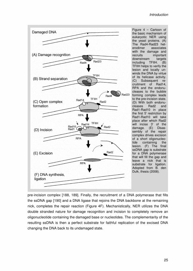
Introduction
24
increased survival and reduced mutagenesis also when kept in the dark in suspension.
This mode of 'dark repair' was shown to also occur if cells were not kept in medium and
thus does not require cell division [173]. This procedure of liquid holding recovery was
shown to be independent of light and thus a repair pathway separate to photoreactivation
must exist [174]. From experiments with UV irradiated bacteria it became apparent that
short oligonucleotides were 'excised' containing pyrimidine dimers and mutants deficient
in this process, were identified. At the same time synthesis of short stretches of DNA
was shown to occur independent of replication when microorganisms were grown on
media containing radioactive nucleotides referred to as Unscheduled DNA Synthesis
(UDS). These independent discoveries paved the way for the description of NER.
Seminal work following the discovery of NER uncovered the link between defective DNA
repair and cancer proneness. The fact that cells from Xeroderma pigmentosum patients
are sensitive to UV and the patients display an increase in sunlight induced skin cancer,
made it possible for researchers to link a DNA repair defect with cancer proneness for
the first time [175, 176].
4.2 The Mechanism of Nucleotide Excision Repair
The mechanism that describes NER can be divided into damage recognition, bubble
formation, incision, excision, repair DNA synthesis and ligation (see figure 4). The
concerted action of 25-30 proteins in eukaryotes achieves this myriad of functions [177],
that in E.coli requires the action of at least 6 proteins [178]. Therefore, I introduce the
basic mechanism of NER by treating the bacterial system first, after which we carry on to
describe the eukaryotic counterparts based on the in vitro reconstitution of the eukaryotic
NER reaction.
In bacterial NER the UV induced lesion is recognized by the UvrA-UvrB heterotetramer
containing two subunits of each protein. Upon DNA damage binding the UvrB protein
specifically flips the damaged base into an extra-helical position after which the UvrA
moiety dissociates from the complex. The UvrC endonuclease is recruited that incises
the DNA 5' and 3' of the lesion. The incision is followed by the recruitment of the UvrD
helicase that drives excision i.e. the removal of the incised oligonucleotide. Next, DNA
polymerase I is recruited to close the ssDNA gap and remove UvrB from the DNA.
Finally, the action of DNA ligase I anneals the DNA backbone to its original state [178].
Based on the in vitro reconstitution of the entire eukaryotic repair system the same basic
steps were identified [179-181]. Over 20 proteins are necessary to perform NER in a test
tube complementing the reaction with a DNA Polymerase and ligase [179, 180]. In yeast
the core NER reaction starts with DNA damage binding by Rad4-Rad23 [182-184]
(Figure 4A). The next step consists of the recruitment of Transcription Factor II H (TFIIH)
[185, 186] that locally unwinds the DNA and is instrumental in recruiting the structure
specific endonucleases Rad2 and Rad1-Rad10 that incise the DNA [187] (Figure 4B &
C). The ssDNA created by TFIIH is bound by Rad14 and RPA in concert stabilizing the
Introduction
25
pre-incision complex [188, 189]. Finally, the recruitment of a DNA polymerase that fills
the ssDNA gap [190] and a DNA ligase that rejoins the DNA backbone at the remaining
nick, completes the repair reaction (Figure 4F). Mechanistically, NER utilizes the DNA
double stranded nature for damage recognition and incision to completely remove an
oligonucleotide containing the damaged base or nucleotides. The complementarity of the
resulting ssDNA is then a perfect substrate for faithful replication of the excised DNA
changing the DNA back to its undamaged state.
Figure 4 – Cartoon of the basic mechanism of eukaryotic NER using the yeast proteins. (A) The Rad4-Rad23 het-erodimer associates with the damage and recruits important downstream targets including TFIIH. (B) TFIIH helps to verify the lesion and locally un-winds the DNA by virtue of its helicase activity. (C) Subsequent re-cruitment of Rad14, RPA and the endonu-cleases to the bubble forming complex leads to the pre-incision state. (D) With both endonu-cleases Rad2 and Rad1-Rad10 in place the first 5' restriction by Rad1-Rad10 will take place after which Rad2 will incise 3' of the damage. (E) Disas-sembly of the repair complex drives excision of a short oligonucleo-tide containing the lesion. (F) The final ssDNA gap is substrate for a DNA polymerase that will fill the gap and leave a nick that is substrate for ligation. Adopted from B. den Dulk, thesis (2008).
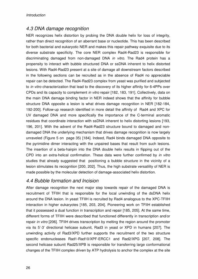
Introduction
26
4.3 DNA damage recognition
NER recognizes helix distortion by probing the DNA double helix for loss of integrity,
rather than direct recognition of an aberrant base or nucleotide. This has been described
for both bacterial and eukaryotic NER and makes this repair pathway exquisite due to its
diverse substrate specificity. The core NER complex Rad4-Rad23 is responsible for
discriminating damaged from non-damaged DNA in vitro. The Rad4 protein has a
propensity to interact with bubble structured DNA or ssDNA inherent to helix distorted
lesions. With Rad4-Rad23 present at a site of damage all downstream factors described
in the following sections can be recruited as in the absence of Rad4 no appreciable
repair can be detected. The Rad4-Rad23 complex from yeast was purified and subjected
to in vitro characterization that lead to the discovery of its higher affinity for 6-4PPs over
CPDs and its capacity to complement in vitro repair [182, 183, 191]. Collectively, data on
the main DNA damage binding factor in NER indeed shows that the affinity for bubble
structure DNA opposite a lesion is what drives damage recognition in NER [182-184,
192-200]. Follow-up research identified in more detail the affinity of Rad4 and XPC for
UV damaged DNA and more specifically the importance of the C-terminal aromatic
residues that coordinate interaction with ssDNA inherent to helix distorting lesions [193,
196, 201]. With the advent of the Rad4-Rad23 structure bound to damaged and non-
damaged DNA the underlying mechanism that drives damage recognition is now largely
unraveled (Figure 5 on page 35) [184]. Indeed, Rad4 binds damaged DNA opposite to
the pyrimidine dimer interacting with the unpaired bases that result from such lesions.
The insertion of a beta-hairpin into the DNA double helix results in flipping out of the
CPD into an extra-helical confirmation. These data were further confirmed by in vitro
studies that already suggested that positioning a bubble structure in the vicinity of a
lesion stimulates its recognition [200, 202]. Thus, the high substrate versatility of NER is
made possible by the molecular detection of damage-associated helix distortion.
4.4 Bubble formation and Incision
After damage recognition the next major step towards repair of the damaged DNA is
recruitment of TFIIH that is responsible for the local unwinding of the dsDNA helix
around the DNA lesion. In yeast TFIIH is recruited by Rad4 analogous to the XPC-TFIIH
interaction in higher eukaryotes [185, 203, 204]. Pioneering work on TFIIH established
that it possessed a dual function in transcription and repair [185, 205]. At the same time,
different forms of TFIIH were described that functioned differently in transcription and/or
repair in vitro [206]. TFIIH drives transcription by melting the region around the promoter
via its 5’-3’ directional helicase subunit, Rad3 in yeast or XPD in humans [207]. The
unwinding activity of Rad3/XPD further supports the recruitment of the two structure
specific endonucleases Rad1-Rad10/XPF-ERCC1 and Rad2/XPG [207, 208]. The
second helicase subunit Rad25/XPB is responsible for transferring large conformational
changes of the TFIIH complex driven by ATP hydrolysis to anchor the complex at the site
Introduction
27
of damage [207, 209]. Due to the local unwinding of the DNA by Rad3/XPD more ssDNA
becomes available that is bound by Rad14/XPA and RPA traditionally referred to as
damage verifiers [188]. The process of verification is not understood on a mechanistic
level, but the interaction of Rad14/XPA with the pre-incision complex is essential
because in its absence NER cannot take place making Rad14/XPA a core NER factor
similar to Rad4. ssDNA coated with RPA is thought to initiate DDR signaling when NER
does not repair the damage [23, 210]. Thus TFIIH interacts with Rad4/XPC [203, 204,
211] and supports damage verification by Rad14 [186] and the recruitment of the two
endonucleases [206].
When TFIIH is anchored to the site of damage and the local bubble is formed the
structure specific endonucleases can be recruited that incise the DNA at ss-dsDNA
junctions 5' and 3' of the damage. For the human proteins a detailed description of the
coordination of incision has been described recently [188, 212]. This tight coordination is
not trivial as nicks in the DNA double helix are damages themselves as described in
detail in the previous sections. Rad2/XPG is the first endonuclease to be recruited by
direct interaction with TFIIH. Rad2/XPG binding to the pre-incision complex initially
stabilizes the complex [188, 212]. In NER the presence of both the structure specific
endonucleases forms the first mutually exclusive condition that drives incision 5' of the
damage by XPF-ERCC1 [188]. The first incision requires the presence of XPG in the
pre-incision complex but not its catalytic activity. The structural conformation changes in
XPG that drive the 3' incision reaction do not take place before 5' incision by XPF-
ERCC1 has occurred [188, 212]. Interestingly, DNA repair synthesis can start in the
absence of the second 3' XPG-induced incision but not the protein, shown by the
recruitment of PCNA, RFC and DNA Pol δ and detection of UDS in a catalytic dead XPG
background [212]. This implication prompted the authors to suggest a cut-patch-cut-
patch mechanism [212]. This mechanism describes the crucial first 5' incision by XPF-
ERCC1 (cut) and the presence of both endonucleases as a precedent for starting DNA
repair synthesis (patch). These initial steps are followed by displacement synthesis
inducing incision of XPG 3' of the lesion (cut) to allow repair synthesis to finish filling the
gap (patch) [212]. This tight coordination prevents the creation of ssDNA and is
reminiscent of long-patch BER [130, 213]. A model for regulation of incision in yeast has
not been described to date. However, based on the extensive homology between the
yeast and human proteins and protein functions involved in incision, incision in yeast no
doubt occurs via a similar mechanism.
4.5 Oligonucleotide excision and repair synthesis
Obviously, the final stages of NER are as much crucial as damage recognition and all
the steps described before. However, excision (i.e. removal of the incised
oligonucleotide) and DNA repair synthesis and ligation did not receive ample attention as
compared to the damage recognition and early repair steps, because DNA synthesis had

Introduction
26
4.3 DNA damage recognition
NER recognizes helix distortion by probing the DNA double helix for loss of integrity,
rather than direct recognition of an aberrant base or nucleotide. This has been described
for both bacterial and eukaryotic NER and makes this repair pathway exquisite due to its
diverse substrate specificity. The core NER complex Rad4-Rad23 is responsible for
discriminating damaged from non-damaged DNA in vitro. The Rad4 protein has a
propensity to interact with bubble structured DNA or ssDNA inherent to helix distorted
lesions. With Rad4-Rad23 present at a site of damage all downstream factors described
in the following sections can be recruited as in the absence of Rad4 no appreciable
repair can be detected. The Rad4-Rad23 complex from yeast was purified and subjected
to in vitro characterization that lead to the discovery of its higher affinity for 6-4PPs over
CPDs and its capacity to complement in vitro repair [182, 183, 191]. Collectively, data on
the main DNA damage binding factor in NER indeed shows that the affinity for bubble
structure DNA opposite a lesion is what drives damage recognition in NER [182-184,
192-200]. Follow-up research identified in more detail the affinity of Rad4 and XPC for
UV damaged DNA and more specifically the importance of the C-terminal aromatic
residues that coordinate interaction with ssDNA inherent to helix distorting lesions [193,
196, 201]. With the advent of the Rad4-Rad23 structure bound to damaged and non-
damaged DNA the underlying mechanism that drives damage recognition is now largely
unraveled (Figure 5 on page 35) [184]. Indeed, Rad4 binds damaged DNA opposite to
the pyrimidine dimer interacting with the unpaired bases that result from such lesions.
The insertion of a beta-hairpin into the DNA double helix results in flipping out of the
CPD into an extra-helical confirmation. These data were further confirmed by in vitro
studies that already suggested that positioning a bubble structure in the vicinity of a
lesion stimulates its recognition [200, 202]. Thus, the high substrate versatility of NER is
made possible by the molecular detection of damage-associated helix distortion.
4.4 Bubble formation and Incision
After damage recognition the next major step towards repair of the damaged DNA is
recruitment of TFIIH that is responsible for the local unwinding of the dsDNA helix
around the DNA lesion. In yeast TFIIH is recruited by Rad4 analogous to the XPC-TFIIH
interaction in higher eukaryotes [185, 203, 204]. Pioneering work on TFIIH established
that it possessed a dual function in transcription and repair [185, 205]. At the same time,
different forms of TFIIH were described that functioned differently in transcription and/or
repair in vitro [206]. TFIIH drives transcription by melting the region around the promoter
via its 5’-3’ directional helicase subunit, Rad3 in yeast or XPD in humans [207]. The
unwinding activity of Rad3/XPD further supports the recruitment of the two structure
specific endonucleases Rad1-Rad10/XPF-ERCC1 and Rad2/XPG [207, 208]. The
second helicase subunit Rad25/XPB is responsible for transferring large conformational
changes of the TFIIH complex driven by ATP hydrolysis to anchor the complex at the site
Introduction
27
of damage [207, 209]. Due to the local unwinding of the DNA by Rad3/XPD more ssDNA
becomes available that is bound by Rad14/XPA and RPA traditionally referred to as
damage verifiers [188]. The process of verification is not understood on a mechanistic
level, but the interaction of Rad14/XPA with the pre-incision complex is essential
because in its absence NER cannot take place making Rad14/XPA a core NER factor
similar to Rad4. ssDNA coated with RPA is thought to initiate DDR signaling when NER
does not repair the damage [23, 210]. Thus TFIIH interacts with Rad4/XPC [203, 204,
211] and supports damage verification by Rad14 [186] and the recruitment of the two
endonucleases [206].
When TFIIH is anchored to the site of damage and the local bubble is formed the
structure specific endonucleases can be recruited that incise the DNA at ss-dsDNA
junctions 5' and 3' of the damage. For the human proteins a detailed description of the
coordination of incision has been described recently [188, 212]. This tight coordination is
not trivial as nicks in the DNA double helix are damages themselves as described in
detail in the previous sections. Rad2/XPG is the first endonuclease to be recruited by
direct interaction with TFIIH. Rad2/XPG binding to the pre-incision complex initially
stabilizes the complex [188, 212]. In NER the presence of both the structure specific
endonucleases forms the first mutually exclusive condition that drives incision 5' of the
damage by XPF-ERCC1 [188]. The first incision requires the presence of XPG in the
pre-incision complex but not its catalytic activity. The structural conformation changes in
XPG that drive the 3' incision reaction do not take place before 5' incision by XPF-
ERCC1 has occurred [188, 212]. Interestingly, DNA repair synthesis can start in the
absence of the second 3' XPG-induced incision but not the protein, shown by the
recruitment of PCNA, RFC and DNA Pol δ and detection of UDS in a catalytic dead XPG
background [212]. This implication prompted the authors to suggest a cut-patch-cut-
patch mechanism [212]. This mechanism describes the crucial first 5' incision by XPF-
ERCC1 (cut) and the presence of both endonucleases as a precedent for starting DNA
repair synthesis (patch). These initial steps are followed by displacement synthesis
inducing incision of XPG 3' of the lesion (cut) to allow repair synthesis to finish filling the
gap (patch) [212]. This tight coordination prevents the creation of ssDNA and is
reminiscent of long-patch BER [130, 213]. A model for regulation of incision in yeast has
not been described to date. However, based on the extensive homology between the
yeast and human proteins and protein functions involved in incision, incision in yeast no
doubt occurs via a similar mechanism.
4.5 Oligonucleotide excision and repair synthesis
Obviously, the final stages of NER are as much crucial as damage recognition and all
the steps described before. However, excision (i.e. removal of the incised
oligonucleotide) and DNA repair synthesis and ligation did not receive ample attention as
compared to the damage recognition and early repair steps, because DNA synthesis had
Introduction
28
already been described in much detail. Knowledge of DNA replication can be extended
to show that short patch UDS occurs via a similar mechanism because all of the in vitro
reconstituted systems use replicative DNA polymerases and DNA ligase I to achieve
NER of an artificial substrate [179, 180, 214, 215]. By extension it was assumed that the
replicative B-family polymerases do the job in vivo as well since NER is non-mutagenic.
However, more recent data describes the involvement of error-prone Y-family
polymerases associated with NER complexes [216]. Therefore, the regulation and active
recruitment of DNA polymerases to NER foci is less well described.
PCNA is recruited to NER foci by the concerted action of the endonucleases XPF and
XPG, and XPA [217-220]. With the PCNA platform present different DNA polymerases
are actively recruited to the NER post-excision complex supporting DNA synthesis. It
appears that, depending on the ubiquitination state of PCNA and the relevant clamp
loader at the repair site, three different polymerases can be recruited post-incision [221].
These authors propose that the nature of the incision product and the chromatin context
drives recruitment of either of the polymerases. If a repair site is swiftly incised 5' and 3'
of the lesion leading to the relatively unhampered disassembly of the repair complex and
release of the oligonucleotide. This substrate is amenable for repair synthesis by a
replicative DNA polymerase [221]. However, if the 5' and 3' incisions do not take place in
quick succession the NER intermediate structure is subject to displacement synthesis;
repair synthesis that displaces the overhanging flap structure of the ssDNA protruding
from the non-excised 3' end of the lesion. This mode of repair is what was described as
the cut-patch-cut-patch model in the previous section [189]. The different substrates not
only recruit the specific polymerase suited for the job, but the association of the different
polymerases are mutually exclusive by actively inhibiting recruitment of the other
polymerases which are less suited for the job [221]. Collectively, the final stages of NER
drive excision and repair synthesis, resulting in perfect repair of the DNA making full use
of the inherent complementarity of DNA.
4.6 From in vitro to in vivo NER
Interestingly, some NER factors in yeast and human are dispensable for the in vitro
reconstituted NER reaction but are required for efficient in vivo repair [179, 180]. These
factors in yeast include Rad7, Rad16 and Rad26. Deletion of RAD7 or RAD16 results in
moderately UV sensitivity [222]. It turns out that these NER factors are responsible for
DNA damage recognition in a chromatin context. These repair proteins detect lesions
that are located anywhere in the genome and are part of the Global Genome NER (GG-
NER) pathway.
Moreover, a bias in repair of active versus inactive genes exists elucidated by gene-
specific repair assays [223, 224]. More specifically, by using strand-specific repair
assays a strand-bias in active genes can be detected. This means that the transcribed
strand (TS) of an active gene is repaired more quickly than the non-transcribed strand
Introduction
29
(NTS) [225]. This pioneering work fully implicated transcription into NER and led to the
description of Transcription Coupled NER (TC-NER) [223-225]. Mutants were identified
that where specifically disrupted in this mode of NER, leading to the characterization of
the TC-NER factor Rad26 in yeast [226]. DNA damage that is specifically located in the
transcribed strand (TS) of active genes is detected and repaired by TC-NER. Thus
extending the in vitro data it is now evident that the in vivo chromatin context of DNA
adds a level of complexity to NER that resulted in the GG- and TC-NER pathways in
NER that contribute to DNA repair and UV survival by specifically tackling in vivo hurdles
to the core NER reactions.
4.7 Global Genome NER
In yeast, GG-NER specifically requires the Rad7-Rad16-Abf1 protein complex [227-229].
Rad16 shows homology to the family of SWI/SNF chromatin remodelers. Moreover, the
yeast GG-NER proteins have been shown to be part of an E3 ligase complex that
contributes to repair and survival by ubiquitinating Rad4 in yeast [230, 231]. Rad7 and
Rad16 were shown to be important for repair of the silent mating type locus in yeast and
the NTS of active genes, all bona fide substrates for GG-NER [232].
In higher eukaryotes, the functional homolog of Rad7-Rad16 is most probably UV-DDB
made up of DNA Damage Binding proteins 1 and 2 (DDB1 & DDB2) [233, 234].
Moreover, the XPC-hHR23B-CEN2 complex is also exclusively required for GG-NER. In
higher eukaryotes the presence of UV-DDB and XPC-hHR23B-CEN2 is most likely
warranted by the different substrate specificity of these two complexes. Whereas UV-
DDB has affinity for both 6-4PP and CPDs, XPC-hHR23-CEN2 has more affinity for 6-
4PPs. It has actually tremendous difficulty in recognizing CPDs and requires UV-DDB for
recruitment to CPD photolesions in vivo [195, 235, 236]. In yeast, this type of substrate
affinity and diversification of GG-NER factors recognizing them does not exist. Rad7-
Rad16-Abf1 is solely required for GG-NER of all NER substrates in vivo.
Detailed biochemical characterization of GG-NER in human cells provided important
insight into the mechanism of DNA damage binding and the so called 'damage handover'
between UV-DDB and XPC [237]. The interesting notion that UV-DDB [237] is involved
in an E3 ubiquitin ligase complex appeared to be crucial in this process. UV-DDB resides
in an E3 ligase complex made up of DDB1-DDB2, CUL4A and ROC1 [238]. Interestingly,
the UV-DDB E3 ligase ubiquitinates XPC and DDB2 after DNA damage induction [201,
237]. The fate of these targets is remarkably different: ubiquitinated XPC has increased
damage affinity while autoubiquitination of DDB2 leads to loss of UV-DDB at the site of
damage facilitating the so-called damage handover from UV-DDB to XPC-hHR23B-
CEN2 [237]. This dual action damage recognition could possibly underlie the GG-NER
mechanism in yeast.
No structure data for the yeast Rad7-Rad16-Abf1 complex bound to damaged DNA is
available, however, the structure of UV-DDB bound to a lesion has been solved [239].
Introduction
30
The interaction of the DDB2 subunit with a lesion is entirely opposite to what has been
described for Rad4. UV-DDB recognizes and flips out the 6-4PP into its binding pocket.
Spatial 3D modeling of the known UV-DDB structure and Rad4-based XPC model
showed that UV-DDB can expose the ssDNA opposite a lesion and accommodate XPC
binding in trans. The concerted action of two GG-NER complexes in higher eukaryotes is
thus required to cover a wider spectra of substrates that apparently in yeast is covered
by Rad7-Rad16-Abf1 alone.
4.8 Transcription Coupled NER
UV-induced lesions are potent transcription blockers and the presence of a stalled RNA
polymerase at a site of damage makes it both a damage signal as well as an obvious
block to repair. By blocking a UV lesion the RNA polymerase makes the lesion less
accessible for GG-NER to repair the lesion, explaining why a dedicated set of proteins is
required to process the RNA polymerase at lesions in the transcribed strand (TS) of
active genes. How a cell discriminates between a stalling RNA Pol II complex and a
transcription complex blocked at a DNA damage is an important question regarding TC-
NER. How a cell exactly targets blocked polymerases is still under debate but several
mechanisms for the early steps in TC-NER have been put forward.
Importantly, TC-NER is not uniform across a single gene, between genes or during
different growth phases of the cell. Before RNA polymerase II transcription becomes fully
processive, repair around the promoter region of an active gene is performed by GG-
NER and thus Rad26-independent in yeast [240]. Highly transcribed genes display
mostly Rad26-independent repair [241]. Repair of the TS becomes Rad26-dependent
30-40 bases downstream of the transcription start overlapping with the transition from
transcription initiation to elongation of the RNA polymerase [240, 242]. Thus Rad26-
dependent repair is specifically required to handle TC-NER of elongation RNA
polymerases II. In yeast, DSIF and Spt4 control this mechanism, that when absent
results in less processive transcription. By deleting SPT4, the RNA polymerase will not
transition into fully elongative modus and TC-NER thus becomes completely Rad26-
independent [243]. In hindsight SPT4 deletion was found as a suppressor of rad26 UV
sensitivity, which can now be explained in detail as described here.
In yeast, two sub-pathways of TC-NER based on the action of RNA Pol II subunits Rpb4
and Rpb9 have been described [241]. In yeast cells, Rad26-independent repair can be
detected that is dependent on Rpb9 but inhibited by Rpb4 [241]. Rpb9 is a zinc-binding
domain protein that has sequence homology to TFIIS. Since TFIIS is known to be able to
help RNA Pol II back-track when stalled, Rpb9 could have a similar function in TC-NER
also explaining why TFIIS is not required for TC-NER in yeast [241]. However, there is
no biochemical evidence available to support this claim.
During the early stages of transcription of the first 40 or so basepairs, TFIIH is still
associated with the polymerase thus eliminating the need for active recruitment of TFIIH
Introduction
31
by TC-NER factors. Moreover, early transcription complexes are known to be less
processive and hence are more easily disrupted. Both of these concepts are thought to
bypass the need for Rad26 in yeast TC-NER close to the transcription initiation site or for
highly transcribed genes. These data suggest that the rate of transcription, in part
determines the faith of the TC-NER process employed.
The early steps in TC-NER that target the RNA polymerase for removal or degradation
have been described in more detail for human cells. These data will be described here.
RNA Polymerase II stalled at a damage drives TC-NER [242, 244, 245] and the large
subunit (LS) of RNA Pol II is subject to DNA damage induced phosphorylation and
ubiquitination [242, 246-251]. The Cdk-activating kinase (CAK) subcomplex of TFIIH was
shown to phosphorylate the C-terminal tail domain (CTD) of RNA Pol II required for UV
induced degradation of the large RNA Pol II subunit [252]. The TC-NER factor Cockayne
Syndrome B (CSB) in higher eukaryotes actively associates with a damage stalled
polymerase, presumably via interaction with the phosphorylated CTD [242]. CSB then
recruits TC-NER factor Cockayne Syndrome A (CSA) and other downstream targets
[253]. CSA resides in an E3 ligase complex and associates with RNA Pol II irrespective
of UV irradiation [238, 254]. However, the interaction between the ligase and the
polymerase increases after DNA damage induction due to the hyperphosphorylated state
of Pol II [238]. The CSA complex associates with the COP9/CSN signalosome in
response to UV inhibiting the ubiquitin ligase activity. The dissociation of CSN from the
complex results in activation of the CSA E3 ligase that is required to resume transcription
after DNA repair and is not involved in the TC-NER process [238, 254, 255]. A role for
the CSA E3 ligase in RNA Pol II LS ubiquitination and degradation is plausible as the
involvement of CSA and CSB in UV induced ubiquitination of RNA Pol II LS has been
described [250, 251] but ubiquitinated RNA Pol II LS has not been directly detected in
TC-NER protein complexes [238, 253]. Thus it appears that even though ubiquitination of
RNA Pol II in response to UV damage is an early response in TC-NER, it can be
uncoupled from the repair reaction or complex [238].
Importantly, TC-NER in higher eukaryotes can take place through remodeling and/or
displacement the RNA Pol II complex without disrupting it, thus negating the use of
ubiquitin directed degradation of the complex under some conditions [242]. Interestingly,
CSB shares homology to SWI/SNF chromatin remodelers and it is thought that through
this action it can remodel the RNA Pol II DNA-RNA complex. While CSB is not able to
fully displace a stalled RNA polymerase in vitro, it can stimulate the polymerase to add
another nucleotide to the transcript aiding in exposing the damage for the core repair
factors [242]. Similarly, recruitment of TFIIS by CSB could drive the more subtle
polymerase remodeling mechanism of back-tracking [242, 253]. Remodeling might be
the heart of the initial steps during TC-NER when RNA Pol II degradation is not applied.
The multitude of data from both yeast and human cells describes the early stages of TC-
NER that act on the RNA polymerase. Importantly, however, a CSA homolog in yeast
Introduction
32
does not exist and the equivalent E3 ligase complex in yeast has not been described.
Similarly, an active role for the yeast TC-NER factor Rad26 in degradation of Rpb1 or
remodeling of the polymerase complex has not been described to date [249]. However,
phosphorylation and ubiquitination of Rpb1, the large subunit of RNA Pol II in yeast, and
the responsible ligase, Rsp5, have been shown highlighting some important parallels
between the dominant model organisms [246-248, 251]. The activity of CSB that
remodels the RNA polymerase could be performed by Rad26 in yeast as Rad26 also
resembles SWI/SNF chromatin remodelers. However, in yeast RNA Pol II remodeling is
not sufficient to bypass the need for Rad4-Rad23, which is the case for XPC-hHR23B in
humans. Thus the activity of CSB as opposed to the early steps of TC-NER in yeast
mediates damage recognition by processing the RNA Pol II complex into a suitable
substrate for NER without the need for XPC. In addition, TFIIS recruitment by CSB could
aid in this process. Importantly, TFIIS is not required for TC-NER in yeast a function that
could probably be full-filled by Rpb9.
Because the yeast genome consists of short genes that produce smaller transcripts a
yeast cell can permit to abort transcription by completely removing and degrading Rpb1
and resume transcription after DNA repair. It is known that in higher eukaryotes more
effort is put into maintaining the transcription complex intact. Indeed, TC-NER can take
place by back-tracking and remodeling of the transcription complex without the need for
disrupting the RNA Pol II complex in higher eukaryotes [242]. TC-NER defects in human
cells lead to Cockayne Syndrome and extreme UV sensitivity, while TC-NER deficient
yeast cells are not UV sensitive [226]. This underscores the different contribution of TC-
NER to cellular survival between yeast and humans.
Summarizing, the active recruitment of downstream NER factors by CSA and CSB is
able to drive TC-NER and funnel it into the core NER reaction in humans. The active role
for Rad26 and Rad28 in yeast are less well described. The faith of the RNA Pol II
complex will differ between organisms but also between different repair loci and
difference in rate of transcription or growth phase of the cell. Taken together, the process
of damage recognition and remodeling or removal of the transcription complex is at the
heart of TC-NER that will fuel NER.
5. rDNA and repair The rDNA is of interest in the context of NER and this thesis in particular because during
evolution S. cerevisiae obtained a Rad4-like protein responsible for TC-NER of this
locus. This Rad4-like protein, Rad34, is discussed in more detail in the next section and
chapter 3 [256, 257].
The higher order structure of the chromatin containing the rDNA genes results in the
formation of the nucleolus, a nuclear compartment harboring the rRNA synthesis,
processing and ribosome assembly [258-260]. The rDNA locus in yeast resides entirely
on chromosome 12. The tandem arrangement of ~150 copies of the 9.6kb rDNA
Introduction
33
cassette makes up the entire rDNA. Each cassette contains the 18S, 5.8S and 25S
rRNAs that are transcribed concurrently by RNA Pol I to form the 35S pre-rRNA
transcript. The intergenic space separating any two copies of the 35S rDNA gene
contains the 5S rDNA gene transcribed by RNA Pol III. Roughly 50% of all copies are
actively transcribed while the other half is in a heterochromatic state in yeast. The
repetitive nature of rDNA makes it a prime target for homologous recombination, a
process that is tightly kept in check [261]. Interestingly, sporadic recombination at the
rDNA locus results in the formation of Extra-chromosomal rDNA Circles (ERCs) that
have been shown to result in replicative aging in yeast [262-265]. This insight provided
the first example of a molecular model for aging, that in hindsight appeared to be specific
for yeast.
The overall size and transcriptional activity of the yeast genome makes that there is not a
lot of heterochromatin as compared to higher eukaryotes. In yeast rDNA, the silent
mating type locus HML and telomeres are the most distinct heterochromatic regions.
rDNA is therefore an excellent target for research on repair of silent versus transcribed
DNA as psoralen crosslinking allows researchers to separate active from inactive rDNA
to study their repair kinetics individually [266, 267]. Early reports on DNA repair at the
rDNA locus showed that in the active copies the TC-NER driven strand-bias is readily
detectable and GG-NER fixes lesions in the inactive fraction in yeast [267]. First and
foremost these data showed that the NER machinery has unimpeded access to the
nucleolar DNA, which is not as trivial as it seems. Furthermore, yeast appears to be the
only model organism to display rDNA TC-NER [266] and, as will be clear in section 6.2,
has a special Rad4-like protein exclusively required for this mode of repair [256, 257].
Moreover, these authors were able to show that actually photolyase repair (PR) was very
efficient in removal of CPDs at the rDNA locus [267]. Another interesting feature of TC-
NER in rDNA links back to the Rad26-dependent TC-NER described in section 4.7. Here
we described that Rad26 drives TC-NER of the elongating RNA polymerase II while
early repair around the transcription start-site and during the pre-elongating phase, when
TFIIH is still associated with the polymerase complex, is independent of Rad26
(Janssen, thesis 2002). Interestingly, data from our lab showed that rDNA repair can be
detected in the absence of Rad26, but also in the absence Rad7, Rad16 and Rad4 [268].
The absence of Rad26 in rDNA TC-NER seems unusual but if we recall that the state of
transcription modulates the use of Rad26 in a Rpb4-dependent fashion [241] an obvious
parallel surfaces. Genes that undergo Rad26-indepedent TC-NER are heavily
transcribed, similar to rDNA transcription of RNA Polymerase I that is very proficient
[258]. Thus, highly transcribed rDNA does not require Rad26 activity to drive TC-NER.
Interestingly, these data from our lab also hint at Rad7 and Rad16-independent repair of
rDNA [268]. It remains to be discerned exactly how rDNA is repaired in the absence of
GG-NER.

Introduction
32
does not exist and the equivalent E3 ligase complex in yeast has not been described.
Similarly, an active role for the yeast TC-NER factor Rad26 in degradation of Rpb1 or
remodeling of the polymerase complex has not been described to date [249]. However,
phosphorylation and ubiquitination of Rpb1, the large subunit of RNA Pol II in yeast, and
the responsible ligase, Rsp5, have been shown highlighting some important parallels
between the dominant model organisms [246-248, 251]. The activity of CSB that
remodels the RNA polymerase could be performed by Rad26 in yeast as Rad26 also
resembles SWI/SNF chromatin remodelers. However, in yeast RNA Pol II remodeling is
not sufficient to bypass the need for Rad4-Rad23, which is the case for XPC-hHR23B in
humans. Thus the activity of CSB as opposed to the early steps of TC-NER in yeast
mediates damage recognition by processing the RNA Pol II complex into a suitable
substrate for NER without the need for XPC. In addition, TFIIS recruitment by CSB could
aid in this process. Importantly, TFIIS is not required for TC-NER in yeast a function that
could probably be full-filled by Rpb9.
Because the yeast genome consists of short genes that produce smaller transcripts a
yeast cell can permit to abort transcription by completely removing and degrading Rpb1
and resume transcription after DNA repair. It is known that in higher eukaryotes more
effort is put into maintaining the transcription complex intact. Indeed, TC-NER can take
place by back-tracking and remodeling of the transcription complex without the need for
disrupting the RNA Pol II complex in higher eukaryotes [242]. TC-NER defects in human
cells lead to Cockayne Syndrome and extreme UV sensitivity, while TC-NER deficient
yeast cells are not UV sensitive [226]. This underscores the different contribution of TC-
NER to cellular survival between yeast and humans.
Summarizing, the active recruitment of downstream NER factors by CSA and CSB is
able to drive TC-NER and funnel it into the core NER reaction in humans. The active role
for Rad26 and Rad28 in yeast are less well described. The faith of the RNA Pol II
complex will differ between organisms but also between different repair loci and
difference in rate of transcription or growth phase of the cell. Taken together, the process
of damage recognition and remodeling or removal of the transcription complex is at the
heart of TC-NER that will fuel NER.
5. rDNA and repair The rDNA is of interest in the context of NER and this thesis in particular because during
evolution S. cerevisiae obtained a Rad4-like protein responsible for TC-NER of this
locus. This Rad4-like protein, Rad34, is discussed in more detail in the next section and
chapter 3 [256, 257].
The higher order structure of the chromatin containing the rDNA genes results in the
formation of the nucleolus, a nuclear compartment harboring the rRNA synthesis,
processing and ribosome assembly [258-260]. The rDNA locus in yeast resides entirely
on chromosome 12. The tandem arrangement of ~150 copies of the 9.6kb rDNA
Introduction
33
cassette makes up the entire rDNA. Each cassette contains the 18S, 5.8S and 25S
rRNAs that are transcribed concurrently by RNA Pol I to form the 35S pre-rRNA
transcript. The intergenic space separating any two copies of the 35S rDNA gene
contains the 5S rDNA gene transcribed by RNA Pol III. Roughly 50% of all copies are
actively transcribed while the other half is in a heterochromatic state in yeast. The
repetitive nature of rDNA makes it a prime target for homologous recombination, a
process that is tightly kept in check [261]. Interestingly, sporadic recombination at the
rDNA locus results in the formation of Extra-chromosomal rDNA Circles (ERCs) that
have been shown to result in replicative aging in yeast [262-265]. This insight provided
the first example of a molecular model for aging, that in hindsight appeared to be specific
for yeast.
The overall size and transcriptional activity of the yeast genome makes that there is not a
lot of heterochromatin as compared to higher eukaryotes. In yeast rDNA, the silent
mating type locus HML and telomeres are the most distinct heterochromatic regions.
rDNA is therefore an excellent target for research on repair of silent versus transcribed
DNA as psoralen crosslinking allows researchers to separate active from inactive rDNA
to study their repair kinetics individually [266, 267]. Early reports on DNA repair at the
rDNA locus showed that in the active copies the TC-NER driven strand-bias is readily
detectable and GG-NER fixes lesions in the inactive fraction in yeast [267]. First and
foremost these data showed that the NER machinery has unimpeded access to the
nucleolar DNA, which is not as trivial as it seems. Furthermore, yeast appears to be the
only model organism to display rDNA TC-NER [266] and, as will be clear in section 6.2,
has a special Rad4-like protein exclusively required for this mode of repair [256, 257].
Moreover, these authors were able to show that actually photolyase repair (PR) was very
efficient in removal of CPDs at the rDNA locus [267]. Another interesting feature of TC-
NER in rDNA links back to the Rad26-dependent TC-NER described in section 4.7. Here
we described that Rad26 drives TC-NER of the elongating RNA polymerase II while
early repair around the transcription start-site and during the pre-elongating phase, when
TFIIH is still associated with the polymerase complex, is independent of Rad26
(Janssen, thesis 2002). Interestingly, data from our lab showed that rDNA repair can be
detected in the absence of Rad26, but also in the absence Rad7, Rad16 and Rad4 [268].
The absence of Rad26 in rDNA TC-NER seems unusual but if we recall that the state of
transcription modulates the use of Rad26 in a Rpb4-dependent fashion [241] an obvious
parallel surfaces. Genes that undergo Rad26-indepedent TC-NER are heavily
transcribed, similar to rDNA transcription of RNA Polymerase I that is very proficient
[258]. Thus, highly transcribed rDNA does not require Rad26 activity to drive TC-NER.
Interestingly, these data from our lab also hint at Rad7 and Rad16-independent repair of
rDNA [268]. It remains to be discerned exactly how rDNA is repaired in the absence of
GG-NER.
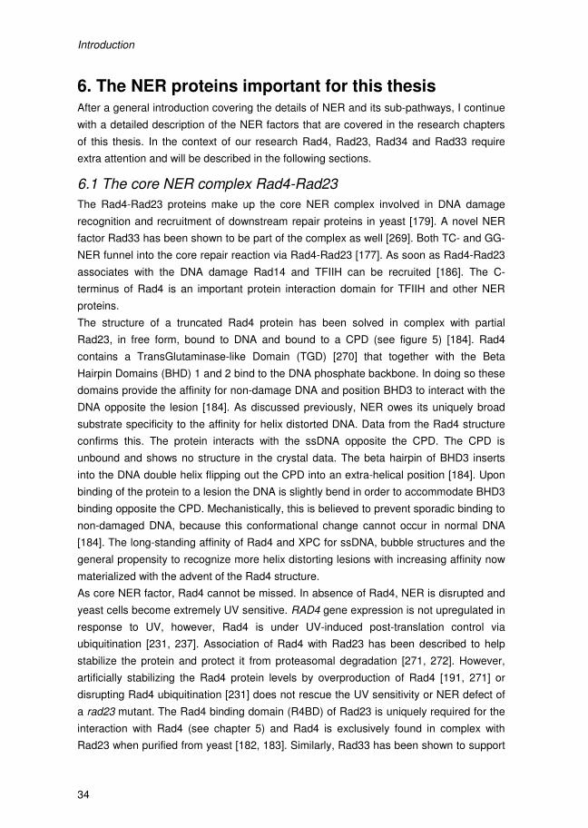
Introduction
34
6. The NER proteins important for this thesis After a general introduction covering the details of NER and its sub-pathways, I continue
with a detailed description of the NER factors that are covered in the research chapters
of this thesis. In the context of our research Rad4, Rad23, Rad34 and Rad33 require
extra attention and will be described in the following sections.
6.1 The core NER complex Rad4-Rad23
The Rad4-Rad23 proteins make up the core NER complex involved in DNA damage
recognition and recruitment of downstream repair proteins in yeast [179]. A novel NER
factor Rad33 has been shown to be part of the complex as well [269]. Both TC- and GG-
NER funnel into the core repair reaction via Rad4-Rad23 [177]. As soon as Rad4-Rad23
associates with the DNA damage Rad14 and TFIIH can be recruited [186]. The C-
terminus of Rad4 is an important protein interaction domain for TFIIH and other NER
proteins.
The structure of a truncated Rad4 protein has been solved in complex with partial
Rad23, in free form, bound to DNA and bound to a CPD (see figure 5) [184]. Rad4
contains a TransGlutaminase-like Domain (TGD) [270] that together with the Beta
Hairpin Domains (BHD) 1 and 2 bind to the DNA phosphate backbone. In doing so these
domains provide the affinity for non-damage DNA and position BHD3 to interact with the
DNA opposite the lesion [184]. As discussed previously, NER owes its uniquely broad
substrate specificity to the affinity for helix distorted DNA. Data from the Rad4 structure
confirms this. The protein interacts with the ssDNA opposite the CPD. The CPD is
unbound and shows no structure in the crystal data. The beta hairpin of BHD3 inserts
into the DNA double helix flipping out the CPD into an extra-helical position [184]. Upon
binding of the protein to a lesion the DNA is slightly bend in order to accommodate BHD3
binding opposite the CPD. Mechanistically, this is believed to prevent sporadic binding to
non-damaged DNA, because this conformational change cannot occur in normal DNA
[184]. The long-standing affinity of Rad4 and XPC for ssDNA, bubble structures and the
general propensity to recognize more helix distorting lesions with increasing affinity now
materialized with the advent of the Rad4 structure.
As core NER factor, Rad4 cannot be missed. In absence of Rad4, NER is disrupted and
yeast cells become extremely UV sensitive. RAD4 gene expression is not upregulated in
response to UV, however, Rad4 is under UV-induced post-translation control via
ubiquitination [231, 237]. Association of Rad4 with Rad23 has been described to help
stabilize the protein and protect it from proteasomal degradation [271, 272]. However,
artificially stabilizing the Rad4 protein levels by overproduction of Rad4 [191, 271] or
disrupting Rad4 ubiquitination [231] does not rescue the UV sensitivity or NER defect of
a rad23 mutant. The Rad4 binding domain (R4BD) of Rad23 is uniquely required for the
interaction with Rad4 (see chapter 5) and Rad4 is exclusively found in complex with
Rad23 when purified from yeast [182, 183]. Similarly, Rad33 has been shown to support
Introduction
35
normal Rad4 protein levels via a yet to be identified mechanism. Rad4 protein levels are
reduced in RAD33 deletion cells, but they remain constant in response to UV irradiation
[269]. However, in the absence of Rad23, Rad4 protein levels are severely reduced and
subject to UV induced degradation.
RAD23 deletion cells are moderately UV sensitive but display hardly any detectable
repair activity in vivo [273-276]. This contradiction in part underlies the discrepancy
between the colony forming ability of UV irradiated cells over the course of 2-3 days in
UV survival curve analysis and the repair efficiency of UV treated cells during the first 2
hours after DNA damage. Defects in the NER reaction can be very accurately assessed
by strand specific repair assays during the early times after UV irradiation but colony
forming ability extends far beyond the first 2 hours of repair and involves the concerted
fidelity of replication and cell cycle progression to support UV survival. This implies that
mutants displaying absence of repair but moderate UV sensitivity, perhaps have defects
in replication or cell cycle arrest, progressing through cell division uninterrupted after UV.
Rad23 is known to affect transcription of plethora of UV-inducible genes (chapter 2,
[277]), by affecting a change in expression of important DDR and cell cycle checkpoint
genes it is possible that rad23 cells display no detectable repair but still recover from UV
irradiation relatively well.
Figure 5 – Cartoon of the Rad4-Rad23 protein structure bound to a short oligonucleotide containing a CPD lesion. A sideview of the protein structure bound to DNA with the protein domains highlighted in their specific color as presented here. In yellow the TGD domain is highlighted, followed by BHD1 (pink), BHD2 (Blue) and BHD3 (red). The Rad23 Rad4 Binding Domain is shown in green bound to the Rad4 TGD. Adopted from Min & Pavletich (2007) [184].
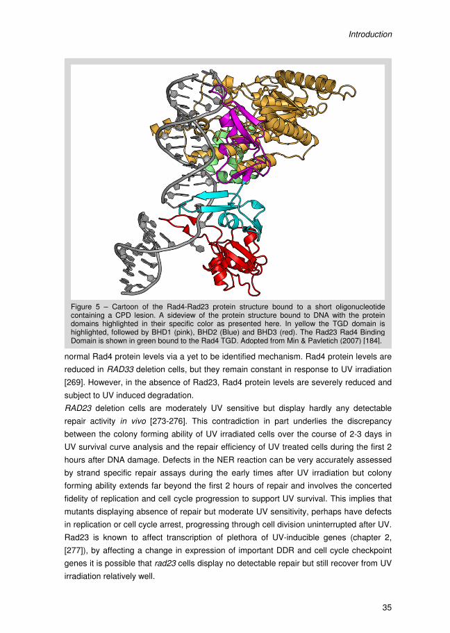
Introduction
34
6. The NER proteins important for this thesis After a general introduction covering the details of NER and its sub-pathways, I continue
with a detailed description of the NER factors that are covered in the research chapters
of this thesis. In the context of our research Rad4, Rad23, Rad34 and Rad33 require
extra attention and will be described in the following sections.
6.1 The core NER complex Rad4-Rad23
The Rad4-Rad23 proteins make up the core NER complex involved in DNA damage
recognition and recruitment of downstream repair proteins in yeast [179]. A novel NER
factor Rad33 has been shown to be part of the complex as well [269]. Both TC- and GG-
NER funnel into the core repair reaction via Rad4-Rad23 [177]. As soon as Rad4-Rad23
associates with the DNA damage Rad14 and TFIIH can be recruited [186]. The C-
terminus of Rad4 is an important protein interaction domain for TFIIH and other NER
proteins.
The structure of a truncated Rad4 protein has been solved in complex with partial
Rad23, in free form, bound to DNA and bound to a CPD (see figure 5) [184]. Rad4
contains a TransGlutaminase-like Domain (TGD) [270] that together with the Beta
Hairpin Domains (BHD) 1 and 2 bind to the DNA phosphate backbone. In doing so these
domains provide the affinity for non-damage DNA and position BHD3 to interact with the
DNA opposite the lesion [184]. As discussed previously, NER owes its uniquely broad
substrate specificity to the affinity for helix distorted DNA. Data from the Rad4 structure
confirms this. The protein interacts with the ssDNA opposite the CPD. The CPD is
unbound and shows no structure in the crystal data. The beta hairpin of BHD3 inserts
into the DNA double helix flipping out the CPD into an extra-helical position [184]. Upon
binding of the protein to a lesion the DNA is slightly bend in order to accommodate BHD3
binding opposite the CPD. Mechanistically, this is believed to prevent sporadic binding to
non-damaged DNA, because this conformational change cannot occur in normal DNA
[184]. The long-standing affinity of Rad4 and XPC for ssDNA, bubble structures and the
general propensity to recognize more helix distorting lesions with increasing affinity now
materialized with the advent of the Rad4 structure.
As core NER factor, Rad4 cannot be missed. In absence of Rad4, NER is disrupted and
yeast cells become extremely UV sensitive. RAD4 gene expression is not upregulated in
response to UV, however, Rad4 is under UV-induced post-translation control via
ubiquitination [231, 237]. Association of Rad4 with Rad23 has been described to help
stabilize the protein and protect it from proteasomal degradation [271, 272]. However,
artificially stabilizing the Rad4 protein levels by overproduction of Rad4 [191, 271] or
disrupting Rad4 ubiquitination [231] does not rescue the UV sensitivity or NER defect of
a rad23 mutant. The Rad4 binding domain (R4BD) of Rad23 is uniquely required for the
interaction with Rad4 (see chapter 5) and Rad4 is exclusively found in complex with
Rad23 when purified from yeast [182, 183]. Similarly, Rad33 has been shown to support
Introduction
35
normal Rad4 protein levels via a yet to be identified mechanism. Rad4 protein levels are
reduced in RAD33 deletion cells, but they remain constant in response to UV irradiation
[269]. However, in the absence of Rad23, Rad4 protein levels are severely reduced and
subject to UV induced degradation.
RAD23 deletion cells are moderately UV sensitive but display hardly any detectable
repair activity in vivo [273-276]. This contradiction in part underlies the discrepancy
between the colony forming ability of UV irradiated cells over the course of 2-3 days in
UV survival curve analysis and the repair efficiency of UV treated cells during the first 2
hours after DNA damage. Defects in the NER reaction can be very accurately assessed
by strand specific repair assays during the early times after UV irradiation but colony
forming ability extends far beyond the first 2 hours of repair and involves the concerted
fidelity of replication and cell cycle progression to support UV survival. This implies that
mutants displaying absence of repair but moderate UV sensitivity, perhaps have defects
in replication or cell cycle arrest, progressing through cell division uninterrupted after UV.
Rad23 is known to affect transcription of plethora of UV-inducible genes (chapter 2,
[277]), by affecting a change in expression of important DDR and cell cycle checkpoint
genes it is possible that rad23 cells display no detectable repair but still recover from UV
irradiation relatively well.
Figure 5 – Cartoon of the Rad4-Rad23 protein structure bound to a short oligonucleotide containing a CPD lesion. A sideview of the protein structure bound to DNA with the protein domains highlighted in their specific color as presented here. In yellow the TGD domain is highlighted, followed by BHD1 (pink), BHD2 (Blue) and BHD3 (red). The Rad23 Rad4 Binding Domain is shown in green bound to the Rad4 TGD. Adopted from Min & Pavletich (2007) [184].
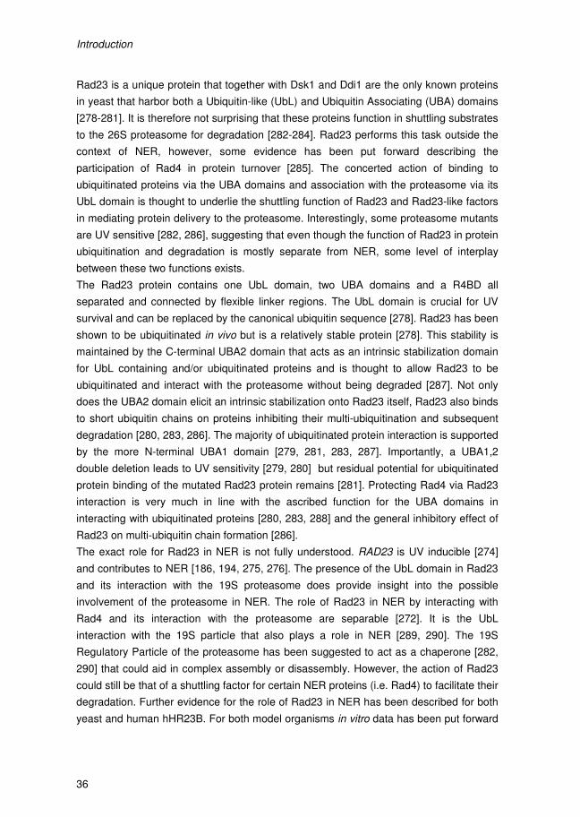
Introduction
36
Rad23 is a unique protein that together with Dsk1 and Ddi1 are the only known proteins
in yeast that harbor both a Ubiquitin-like (UbL) and Ubiquitin Associating (UBA) domains
[278-281]. It is therefore not surprising that these proteins function in shuttling substrates
to the 26S proteasome for degradation [282-284]. Rad23 performs this task outside the
context of NER, however, some evidence has been put forward describing the
participation of Rad4 in protein turnover [285]. The concerted action of binding to
ubiquitinated proteins via the UBA domains and association with the proteasome via its
UbL domain is thought to underlie the shuttling function of Rad23 and Rad23-like factors
in mediating protein delivery to the proteasome. Interestingly, some proteasome mutants
are UV sensitive [282, 286], suggesting that even though the function of Rad23 in protein
ubiquitination and degradation is mostly separate from NER, some level of interplay
between these two functions exists.
The Rad23 protein contains one UbL domain, two UBA domains and a R4BD all
separated and connected by flexible linker regions. The UbL domain is crucial for UV
survival and can be replaced by the canonical ubiquitin sequence [278]. Rad23 has been
shown to be ubiquitinated in vivo but is a relatively stable protein [278]. This stability is
maintained by the C-terminal UBA2 domain that acts as an intrinsic stabilization domain
for UbL containing and/or ubiquitinated proteins and is thought to allow Rad23 to be
ubiquitinated and interact with the proteasome without being degraded [287]. Not only
does the UBA2 domain elicit an intrinsic stabilization onto Rad23 itself, Rad23 also binds
to short ubiquitin chains on proteins inhibiting their multi-ubiquitination and subsequent
degradation [280, 283, 286]. The majority of ubiquitinated protein interaction is supported
by the more N-terminal UBA1 domain [279, 281, 283, 287]. Importantly, a UBA1,2
double deletion leads to UV sensitivity [279, 280] but residual potential for ubiquitinated
protein binding of the mutated Rad23 protein remains [281]. Protecting Rad4 via Rad23
interaction is very much in line with the ascribed function for the UBA domains in
interacting with ubiquitinated proteins [280, 283, 288] and the general inhibitory effect of
Rad23 on multi-ubiquitin chain formation [286].
The exact role for Rad23 in NER is not fully understood. RAD23 is UV inducible [274]
and contributes to NER [186, 194, 275, 276]. The presence of the UbL domain in Rad23
and its interaction with the 19S proteasome does provide insight into the possible
involvement of the proteasome in NER. The role of Rad23 in NER by interacting with
Rad4 and its interaction with the proteasome are separable [272]. It is the UbL
interaction with the 19S particle that also plays a role in NER [289, 290]. The 19S
Regulatory Particle of the proteasome has been suggested to act as a chaperone [282,
290] that could aid in complex assembly or disassembly. However, the action of Rad23
could still be that of a shuttling factor for certain NER proteins (i.e. Rad4) to facilitate their
degradation. Further evidence for the role of Rad23 in NER has been described for both
yeast and human hHR23B. For both model organisms in vitro data has been put forward
Introduction
37
that describes a stimulatory role for Rad23 in DNA damage binding of Rad4 and XPC
[191, 194].
As will become evident in chapter 2, Rad23 and Rad4 also play an important role in UV
induced gene transcriptional regulation of the RNR pathway of genes. Interestingly, the
GG-NER E3 ligase in yeast, introduced in section 4.7, plays a pivotal role in this
regulation and was shown to ubiquitinate Rad4 in a mode that is independent of active
NER [231]. A role for Rad23 in transcription has been described previously [277],
however, the implications for DDR signaling and UV induced gene expression via the
GG-NER E3 ligase have not been reported previously.
In summary, the Rad4-Rad23 protein complex facilitates NER and the interaction with
the 19S RP. At the same time Rad4-Rad23 provides a platform for recruitment of TFIIH
and Rad14. More importantly, Rad4-Rad23 also regulate UV-induced gene expression
described in more detail in chapter 2. The concerted action of Rad4-Rad23 in these
multifaceted processes is a testimony to the importance of this core NER factor in yeast.
6.2 Rad34
NER is highly conserved both genetically as well as mechanistically, however, some
exceptions exist between the dominant model organisms. Both Rad4p and XPC share
some sequence similarity [291] and reside in protein-complexes of similar makeup,
however, yeast contains a second distinct Rad4-like protein called Rad34 which is
required exclusively for TC-NER of rDNA [256, 257]. When RAD34 is knocked out in a
rad4∆ background, repair of the TS of active rDNA is completely abolished [292, 293].
Vice versa, it is already known for some time that rDNA is still promptly repaired in the
absence of Rad4 [294]. Rad34 shares C-terminal homology to Rad4 [291] and
associates with Rad23 [295]. These data corroborate the Rad34-Rad4 homology, but at
the same time unequivocally underscore the separation of function of these two yeast
NER factors.
The role for Rad4 and Rad34 in rDNA repair underscores the TC-NER specificity of
Rad34, reminiscent of the Rad26-dependent TC-NER of RNA Polymerase II transcribed
genes. TC-NER at active rDNA genes becomes Rad34-dependent ~40 nucleotide
downstream of the transcription start site while around the promoter region repair is
entirely Rad4-dependent [257]. Highly RNA Pol II transcribed genes do not require
Rad26 for TC-NER [296]. rDNA is highly transcribed and 40 basepairs away from the
transcription start site, RNA Pol I is in an elongating modus. However, conscientious
analysis of the RNA polymerase I initiation complex showed that RNA Pol I switches to
elongation around base 12 downstream from the promoter [297]. These data do not
perfectly overlap with the Rad34-dependent TC-NER transition 40 basepairs
downstream of the promoter. Either the transition from initiation to elongation does not
correlate with the transition from Rad4 to Rad34 dependent repair or technical aspects of
these study make comparison impossible.
Introduction
38
Interestingly, elongating RNA Pol I thus requires Rad34 but not Rad26 [257, 268]. Thus
the Rad26 activity suggested for handling an elongating RNA Pol II complex stalled at a
damage is not required during rDNA TC-NER but a different Rad4 protein is warranted.
This discrepancy will be discussed in further detail in chapter 3, where we try to decipher
the conditions that create the exclusive preference for Rad34 over Rad4 during rDNA
TC-NER.
6.3 Rad33
Rad33 is a novel NER factor. A RAD33 deletion results in moderate UV sensitivity and is
epistatic with the deletion core NER factors RAD4 and RAD14 [298] [299]. In depth DNA
repair analysis revealed that TC-NER is moderately affected whereas Rad33 is
absolutely required for GG-NER [298]. Similarly, Rad33 was shown to interact with
Rad34 [300].
Bioinformatic structure predictions for Rad33 show a weak resemblance to calmodulin-
like proteins [269]. Interestingly, in higher eukaryotes a trimeric complex exist around the
human Rad4-homolog, XPC consisting of hHR23 and Centrin 2 [301]. Through a series
of in vitro experiments it became apparent that the calmodulin-like protein CEN2 is a
bona fide NER factor that in cohort with hHR23B stabilizes XPC and contributes to DNA
binding and incision in vitro [302]. More detailed analysis of the XPC-CEN2 interaction
led to the identification of a conserved WLL binding motif in the C-terminus of XPC [302,
303]. By binding to a conserved motif at the C-terminus of XPC, Centrin 2 molds the
unstructured region into an α-helical fold upon binding possibly making the region more
amenable for protein-protein interactions required during NER [302-305]. The XPC-
CEN2 interaction is of functional significance in NER in vivo as XPC-CBM (Centrin
Binding Mutant) containing cells are UV-sensitive [306]. Interestingly, the WLL motifs
supports the Rad4-Rad33 interaction in yeast as well. Mutating the motif in yeast to three
alanines, creating the rad4AAA allele, perfectly phenocopies a RAD33 deletion strain
showing that Rad33, like CEN2, functions in NER exclusively through the interaction with
Rad4 [269].
The yeast centrin homologue Cdc31 was shown to give rise to low UV sensitivity in
conditional deletions and interacts with Rad4 [307]. However, the parallel between
Centrin 2 and Rad33 does not hold for Cdc31 [269]. Conversely, there is no evidence for
a Rad33-like protein in higher eukaryotes; it thus seems that the centrosome and NER
related functions of CEN2 are split between Cdc31 and Rad33 in yeast. This is further
supported by the fact that there is no obvious role for Rad33 at the centrosome and the
active role of Cdc31 in NER is still debatable. The novelty of a centrosome component at
the heart of the XPC-complex is enigmatic and a possible link between centrosome
duplication and repair seems plausible but has yet to materialize.
Introduction
39
7. Scope of this thesis In Chapter 2, we describe a novel mechanism for UV induced gene expression
controlled by the GG-NER E3 ligase and Rad4-Rad23. This process implies the
dissociation of Rad4-Rad23 from promoter regions in response to UV induced
ubiquitination of Rad4 by the GG-NER E3 ligase, triggering gene expression of a subset
of genes. In Chapter 3 the effect of Histone H1 on repair of the rDNA locus is studied in
relation to Rad34. This Rad4-like protein is specifically needed in yeast for TC-NER of
rDNA. In the absence of Histone H1, however, Rad4 can replace Rad34 in this repair
pathway. A model is presented explaining why for TC-NER of rDNA a different Rad4-like
protein is required. In Chapter 4 the homology between human and yeast NER is used
to study the phenotype of a RAD4 mutation, rad4W496S. This mutation affects a
conserved amino acid in Rad4-like proteins that when present in XPC (W690S) results in
a XP disease phenotype. We show that the Rad4W496S is mainly affected in GG-NER.
Chapter 5 is dedicated to the detailed study of the Rad4-Rad23 interaction. We provide
evidence for presence of two binding domains for Rad23 in Rad4 that are mutually
exclusive. The function of these Rad4 binding domains in the two sub-pathways of NER
is studied. Research described in Chapter 6 concerns the discovery of a RAD4 mutant
allele in a commonly used yeast background that confers conditional UV sensitivity.
Finally, the results are discussed and summarized in Chapter 7.
Introduction
40
8. References 1. Avery, O.T., C.M. Macleod, and M. McCarty, Studies on the Chemical Nature of the
Substance Inducing Transformation of Pneumococcal Types : Induction of
Transformation by a Desoxyribonucleic Acid Fraction Isolated from Pneumococcus Type
Iii. J Exp Med, 1944. 79(2): p. 137-58.
2. Hershey, A.D. and M. Chase, Independent functions of viral protein and nucleic acid in
growth of bacteriophage. J Gen Physiol, 1952. 36(1): p. 39-56.
3. Robinow, C. and E. Kellenberger, The bacterial nucleoid revisited. Microbiol Rev, 1994.
58(2): p. 211-32.
4. Ptacin, J.L. and L. Shapiro, Chromosome architecture is a key element of bacterial
cellular organization. Cell Microbiol, 2012.
5. Cremer, T. and C. Cremer, Chromosome territories, nuclear architecture and gene
regulation in mammalian cells. Nat Rev Genet, 2001. 2(4): p. 292-301.
6. Cremer, T., et al., Chromosome territories--a functional nuclear landscape. Curr Opin
Cell Biol, 2006. 18(3): p. 307-16.
7. Campos, E.I. and D. Reinberg, Histones: annotating chromatin. Annu Rev Genet, 2009.
43: p. 559-99.
8. Lindahl, T., Instability and decay of the primary structure of DNA. Nature, 1993.
362(6422): p. 709-15.
9. Moynahan, M.E. and M. Jasin, Mitotic homologous recombination maintains genomic
stability and suppresses tumorigenesis. Nat Rev Mol Cell Biol, 2010. 11(3): p. 196-207.
10. Leadon, S.A., Repair of DNA Damage Produced by Ionizing Radiation: A Minireview.
Semin Radiat Oncol, 1996. 6(4): p. 295-305.
11. Cadet, J., et al., Hydroxyl radicals and DNA base damage. Mutat Res, 1999. 424(1-2): p.
9-21.
12. Aravind, L., D.R. Walker, and E.V. Koonin, Conserved domains in DNA repair proteins
and evolution of repair systems. Nucleic Acids Res, 1999. 27(5): p. 1223-42.
13. Fishel, R., et al., The human mutator gene homolog MSH2 and its association with
hereditary nonpolyposis colon cancer. Cell, 1993. 75(5): p. 1027-38.
14. Loeb, L.A., C.F. Springgate, and N. Battula, Errors in DNA replication as a basis of
malignant changes. Cancer Res, 1974. 34(9): p. 2311-21.
15. Nowell, P.C., The clonal evolution of tumor cell populations. Science, 1976. 194(4260):
p. 23-8.
16. Negrini, S., V.G. Gorgoulis, and T.D. Halazonetis, Genomic instability--an evolving
hallmark of cancer. Nat Rev Mol Cell Biol, 2010. 11(3): p. 220-8.
17. Hoeijmakers, J.H., Genome maintenance mechanisms for preventing cancer. Nature,
2001. 411(6835): p. 366-74.
18. Kolodner, R.D., C.D. Putnam, and K. Myung, Maintenance of genome stability in
Saccharomyces cerevisiae. Science, 2002. 297(5581): p. 552-7.
19. Branzei, D. and M. Foiani, Maintaining genome stability at the replication fork. Nat Rev
Mol Cell Biol, 2010. 11(3): p. 208-19.
Introduction
41
20. Wijnhoven, S.W., et al., Tissue specific mutagenic and carcinogenic responses in NER
defective mouse models. Mutat Res, 2007. 614(1-2): p. 77-94.
21. Putnam, C.D., E.J. Jaehnig, and R.D. Kolodner, Perspectives on the DNA damage and
replication checkpoint responses in Saccharomyces cerevisiae. DNA Repair (Amst),
2009. 8(9): p. 974-82.
22. Lange, S.S., K. Takata, and R.D. Wood, DNA polymerases and cancer. Nat Rev Cancer,
2011. 11(2): p. 96-110.
23. Zou, L. and S.J. Elledge, Sensing DNA damage through ATRIP recognition of RPA-
ssDNA complexes. Science, 2003. 300(5625): p. 1542-8.
24. Melo, J.A., J. Cohen, and D.P. Toczyski, Two checkpoint complexes are independently
recruited to sites of DNA damage in vivo. Genes Dev, 2001. 15(21): p. 2809-21.
25. Kondo, T., et al., Recruitment of Mec1 and Ddc1 checkpoint proteins to double-strand
breaks through distinct mechanisms. Science, 2001. 294(5543): p. 867-70.
26. Bonilla, C.Y., J.A. Melo, and D.P. Toczyski, Colocalization of sensors is sufficient to
activate the DNA damage checkpoint in the absence of damage. Mol Cell, 2008. 30(3):
p. 267-76.
27. Majka, J., A. Niedziela-Majka, and P.M. Burgers, The checkpoint clamp activates Mec1
kinase during initiation of the DNA damage checkpoint. Mol Cell, 2006. 24(6): p. 891-
901.
28. Lazzaro, F., et al., Checkpoint mechanisms at the intersection between DNA damage
and repair. DNA Repair (Amst), 2009. 8(9): p. 1055-67.
29. Nakada, D., K. Matsumoto, and K. Sugimoto, ATM-related Tel1 associates with double-
strand breaks through an Xrs2-dependent mechanism. Genes Dev, 2003. 17(16): p.
1957-62.
30. Pardo, B., B. Gomez-Gonzalez, and A. Aguilera, DNA repair in mammalian cells: DNA
double-strand break repair: how to fix a broken relationship. Cell Mol Life Sci, 2009.
66(6): p. 1039-56.
31. Bressan, D.A., B.K. Baxter, and J.H. Petrini, The Mre11-Rad50-Xrs2 protein complex
facilitates homologous recombination-based double-strand break repair in
Saccharomyces cerevisiae. Mol Cell Biol, 1999. 19(11): p. 7681-7.
32. Morrow, D.M., et al., TEL1, an S. cerevisiae homolog of the human gene mutated in
ataxia telangiectasia, is functionally related to the yeast checkpoint gene MEC1. Cell,
1995. 82(5): p. 831-40.
33. Ritchie, K.B., J.C. Mallory, and T.D. Petes, Interactions of TLC1 (which encodes the
RNA subunit of telomerase), TEL1, and MEC1 in regulating telomere length in the yeast
Saccharomyces cerevisiae. Mol Cell Biol, 1999. 19(9): p. 6065-75.
34. Ira, G., et al., DNA end resection, homologous recombination and DNA damage
checkpoint activation require CDK1. Nature, 2004. 431(7011): p. 1011-7.
35. Sanchez, Y., et al., Control of the DNA damage checkpoint by chk1 and rad53 protein
kinases through distinct mechanisms. Science, 1999. 286(5442): p. 1166-71.
36. Agarwal, R., et al., Two distinct pathways for inhibiting pds1 ubiquitination in response to
DNA damage. J Biol Chem, 2003. 278(45): p. 45027-33.
Introduction
40
8. References 1. Avery, O.T., C.M. Macleod, and M. McCarty, Studies on the Chemical Nature of the
Substance Inducing Transformation of Pneumococcal Types : Induction of
Transformation by a Desoxyribonucleic Acid Fraction Isolated from Pneumococcus Type
Iii. J Exp Med, 1944. 79(2): p. 137-58.
2. Hershey, A.D. and M. Chase, Independent functions of viral protein and nucleic acid in
growth of bacteriophage. J Gen Physiol, 1952. 36(1): p. 39-56.
3. Robinow, C. and E. Kellenberger, The bacterial nucleoid revisited. Microbiol Rev, 1994.
58(2): p. 211-32.
4. Ptacin, J.L. and L. Shapiro, Chromosome architecture is a key element of bacterial
cellular organization. Cell Microbiol, 2012.
5. Cremer, T. and C. Cremer, Chromosome territories, nuclear architecture and gene
regulation in mammalian cells. Nat Rev Genet, 2001. 2(4): p. 292-301.
6. Cremer, T., et al., Chromosome territories--a functional nuclear landscape. Curr Opin
Cell Biol, 2006. 18(3): p. 307-16.
7. Campos, E.I. and D. Reinberg, Histones: annotating chromatin. Annu Rev Genet, 2009.
43: p. 559-99.
8. Lindahl, T., Instability and decay of the primary structure of DNA. Nature, 1993.
362(6422): p. 709-15.
9. Moynahan, M.E. and M. Jasin, Mitotic homologous recombination maintains genomic
stability and suppresses tumorigenesis. Nat Rev Mol Cell Biol, 2010. 11(3): p. 196-207.
10. Leadon, S.A., Repair of DNA Damage Produced by Ionizing Radiation: A Minireview.
Semin Radiat Oncol, 1996. 6(4): p. 295-305.
11. Cadet, J., et al., Hydroxyl radicals and DNA base damage. Mutat Res, 1999. 424(1-2): p.
9-21.
12. Aravind, L., D.R. Walker, and E.V. Koonin, Conserved domains in DNA repair proteins
and evolution of repair systems. Nucleic Acids Res, 1999. 27(5): p. 1223-42.
13. Fishel, R., et al., The human mutator gene homolog MSH2 and its association with
hereditary nonpolyposis colon cancer. Cell, 1993. 75(5): p. 1027-38.
14. Loeb, L.A., C.F. Springgate, and N. Battula, Errors in DNA replication as a basis of
malignant changes. Cancer Res, 1974. 34(9): p. 2311-21.
15. Nowell, P.C., The clonal evolution of tumor cell populations. Science, 1976. 194(4260):
p. 23-8.
16. Negrini, S., V.G. Gorgoulis, and T.D. Halazonetis, Genomic instability--an evolving
hallmark of cancer. Nat Rev Mol Cell Biol, 2010. 11(3): p. 220-8.
17. Hoeijmakers, J.H., Genome maintenance mechanisms for preventing cancer. Nature,
2001. 411(6835): p. 366-74.
18. Kolodner, R.D., C.D. Putnam, and K. Myung, Maintenance of genome stability in
Saccharomyces cerevisiae. Science, 2002. 297(5581): p. 552-7.
19. Branzei, D. and M. Foiani, Maintaining genome stability at the replication fork. Nat Rev
Mol Cell Biol, 2010. 11(3): p. 208-19.
Introduction
41
20. Wijnhoven, S.W., et al., Tissue specific mutagenic and carcinogenic responses in NER
defective mouse models. Mutat Res, 2007. 614(1-2): p. 77-94.
21. Putnam, C.D., E.J. Jaehnig, and R.D. Kolodner, Perspectives on the DNA damage and
replication checkpoint responses in Saccharomyces cerevisiae. DNA Repair (Amst),
2009. 8(9): p. 974-82.
22. Lange, S.S., K. Takata, and R.D. Wood, DNA polymerases and cancer. Nat Rev Cancer,
2011. 11(2): p. 96-110.
23. Zou, L. and S.J. Elledge, Sensing DNA damage through ATRIP recognition of RPA-
ssDNA complexes. Science, 2003. 300(5625): p. 1542-8.
24. Melo, J.A., J. Cohen, and D.P. Toczyski, Two checkpoint complexes are independently
recruited to sites of DNA damage in vivo. Genes Dev, 2001. 15(21): p. 2809-21.
25. Kondo, T., et al., Recruitment of Mec1 and Ddc1 checkpoint proteins to double-strand
breaks through distinct mechanisms. Science, 2001. 294(5543): p. 867-70.
26. Bonilla, C.Y., J.A. Melo, and D.P. Toczyski, Colocalization of sensors is sufficient to
activate the DNA damage checkpoint in the absence of damage. Mol Cell, 2008. 30(3):
p. 267-76.
27. Majka, J., A. Niedziela-Majka, and P.M. Burgers, The checkpoint clamp activates Mec1
kinase during initiation of the DNA damage checkpoint. Mol Cell, 2006. 24(6): p. 891-
901.
28. Lazzaro, F., et al., Checkpoint mechanisms at the intersection between DNA damage
and repair. DNA Repair (Amst), 2009. 8(9): p. 1055-67.
29. Nakada, D., K. Matsumoto, and K. Sugimoto, ATM-related Tel1 associates with double-
strand breaks through an Xrs2-dependent mechanism. Genes Dev, 2003. 17(16): p.
1957-62.
30. Pardo, B., B. Gomez-Gonzalez, and A. Aguilera, DNA repair in mammalian cells: DNA
double-strand break repair: how to fix a broken relationship. Cell Mol Life Sci, 2009.
66(6): p. 1039-56.
31. Bressan, D.A., B.K. Baxter, and J.H. Petrini, The Mre11-Rad50-Xrs2 protein complex
facilitates homologous recombination-based double-strand break repair in
Saccharomyces cerevisiae. Mol Cell Biol, 1999. 19(11): p. 7681-7.
32. Morrow, D.M., et al., TEL1, an S. cerevisiae homolog of the human gene mutated in
ataxia telangiectasia, is functionally related to the yeast checkpoint gene MEC1. Cell,
1995. 82(5): p. 831-40.
33. Ritchie, K.B., J.C. Mallory, and T.D. Petes, Interactions of TLC1 (which encodes the
RNA subunit of telomerase), TEL1, and MEC1 in regulating telomere length in the yeast
Saccharomyces cerevisiae. Mol Cell Biol, 1999. 19(9): p. 6065-75.
34. Ira, G., et al., DNA end resection, homologous recombination and DNA damage
checkpoint activation require CDK1. Nature, 2004. 431(7011): p. 1011-7.
35. Sanchez, Y., et al., Control of the DNA damage checkpoint by chk1 and rad53 protein
kinases through distinct mechanisms. Science, 1999. 286(5442): p. 1166-71.
36. Agarwal, R., et al., Two distinct pathways for inhibiting pds1 ubiquitination in response to
DNA damage. J Biol Chem, 2003. 278(45): p. 45027-33.

Introduction
42
37. Liang, F. and Y. Wang, DNA damage checkpoints inhibit mitotic exit by two different
mechanisms. Mol Cell Biol, 2007. 27(14): p. 5067-78.
38. Amon, A., et al., Regulation of p34CDC28 tyrosine phosphorylation is not required for
entry into mitosis in S. cerevisiae. Nature, 1992. 355(6358): p. 368-71.
39. Sorger, P.K. and A.W. Murray, S phase feedback control in budding yeast independent
of tyrosine phosphorylation of p34cdc28. Nature, 1992. 355(6358): p. 365-8.
40. Zhou, Z. and S.J. Elledge, DUN1 encodes a protein kinase that controls the DNA
damage response in yeast. Cell, 1993. 75(6): p. 1119-27.
41. Elledge, S.J., Cell cycle checkpoints: preventing an identity crisis. Science, 1996.
274(5293): p. 1664-72.
42. Myung, K., A. Datta, and R.D. Kolodner, Suppression of spontaneous chromosomal
rearrangements by S phase checkpoint functions in Saccharomyces cerevisiae. Cell,
2001. 104(3): p. 397-408.
43. Lopes, M., et al., The DNA replication checkpoint response stabilizes stalled replication
forks. Nature, 2001. 412(6846): p. 557-61.
44. Cimprich, K.A., et al., cDNA cloning and gene mapping of a candidate human cell cycle
checkpoint protein. Proc Natl Acad Sci U S A, 1996. 93(7): p. 2850-5.
45. Greenwell, P.W., et al., TEL1, a gene involved in controlling telomere length in S.
cerevisiae, is homologous to the human ataxia telangiectasia gene. Cell, 1995. 82(5): p.
823-9.
46. Ejima, Y., L. Yang, and M.S. Sasaki, Aberrant splicing of the ATM gene associated with
shortening of the intronic mononucleotide tract in human colon tumor cell lines: a novel
mutation target of microsatellite instability. Int J Cancer, 2000. 86(2): p. 262-8.
47. Chabes, A., et al., Survival of DNA damage in yeast directly depends on increased dNTP
levels allowed by relaxed feedback inhibition of ribonucleotide reductase. Cell, 2003.
112(3): p. 391-401.
48. Elledge, S.J. and R.W. Davis, Two genes differentially regulated in the cell cycle and by
DNA-damaging agents encode alternative regulatory subunits of ribonucleotide
reductase. Genes Dev, 1990. 4(5): p. 740-51.
49. Domkin, V., L. Thelander, and A. Chabes, Yeast DNA damage-inducible Rnr3 has a very
low catalytic activity strongly stimulated after the formation of a cross-talking Rnr1/Rnr3
complex. J Biol Chem, 2002. 277(21): p. 18574-8.
50. Zhao, X. and R. Rothstein, The Dun1 checkpoint kinase phosphorylates and regulates
the ribonucleotide reductase inhibitor Sml1. Proc Natl Acad Sci U S A, 2002. 99(6): p.
3746-51.
51. Andreson, B.L., et al., The ribonucleotide reductase inhibitor, Sml1, is sequentially
phosphorylated, ubiquitylated and degraded in response to DNA damage. Nucleic Acids
Res, 2010. 38(19): p. 6490-501.
52. Huang, M., Z. Zhou, and S.J. Elledge, The DNA replication and damage checkpoint
pathways induce transcription by inhibition of the Crt1 repressor. Cell, 1998. 94(5): p.
595-605.
Introduction
43
53. Zhao, X., et al., The ribonucleotide reductase inhibitor Sml1 is a new target of the
Mec1/Rad53 kinase cascade during growth and in response to DNA damage. Embo
Journal, 2001. 20(13): p. 3544-53.
54. Prakash, L., Characterization of postreplication repair in Saccharomyces cerevisiae and
effects of rad6, rad18, rev3 and rad52 mutations. Mol Gen Genet, 1981. 184(3): p. 471-
8.
55. Broomfield, S., T. Hryciw, and W. Xiao, DNA postreplication repair and mutagenesis in
Saccharomyces cerevisiae. Mutat Res, 2001. 486(3): p. 167-84.
56. Rupp, W.D., et al., Exchanges between DNA strands in ultraviolet-irradiated Escherichia
coli. J Mol Biol, 1971. 61(1): p. 25-44.
57. Friedberg, E.C., Suffering in silence: the tolerance of DNA damage. Nat Rev Mol Cell
Biol, 2005. 6(12): p. 943-53.
58. Moldovan, G.L., B. Pfander, and S. Jentsch, PCNA, the maestro of the replication fork.
Cell, 2007. 129(4): p. 665-79.
59. Sung, P., S. Prakash, and L. Prakash, The RAD6 protein of Saccharomyces cerevisiae
polyubiquitinates histones, and its acidic domain mediates this activity. Genes Dev,
1988. 2(11): p. 1476-85.
60. Dohmen, R.J., et al., The N-end rule is mediated by the UBC2(RAD6) ubiquitin-
conjugating enzyme. Proc Natl Acad Sci U S A, 1991. 88(16): p. 7351-5.
61. Sung, P., et al., Yeast RAD6 encoded ubiquitin conjugating enzyme mediates protein
degradation dependent on the N-end-recognizing E3 enzyme. EMBO J, 1991. 10(8): p.
2187-93.
62. Bailly, V., et al., Specific complex formation between yeast RAD6 and RAD18 proteins: a
potential mechanism for targeting RAD6 ubiquitin-conjugating activity to DNA damage
sites. Genes Dev, 1994. 8(7): p. 811-20.
63. Bailly, V., et al., Yeast DNA repair proteins Rad6 and Rad18 form a heterodimer that has
ubiquitin conjugating, DNA binding, and ATP hydrolytic activities. J Biol Chem, 1997.
272(37): p. 23360-5.
64. Bailly, V., S. Prakash, and L. Prakash, Domains required for dimerization of yeast Rad6
ubiquitin-conjugating enzyme and Rad18 DNA binding protein. Mol Cell Biol, 1997.
17(8): p. 4536-43.
65. Hoege, C., et al., RAD6-dependent DNA repair is linked to modification of PCNA by
ubiquitin and SUMO. Nature, 2002. 419(6903): p. 135-41.
66. Kannouche, P.L., J. Wing, and A.R. Lehmann, Interaction of human DNA polymerase eta
with monoubiquitinated PCNA: a possible mechanism for the polymerase switch in
response to DNA damage. Mol Cell, 2004. 14(4): p. 491-500.
67. McCulloch, S.D. and T.A. Kunkel, The fidelity of DNA synthesis by eukaryotic replicative
and translesion synthesis polymerases. Cell Res, 2008. 18(1): p. 148-61.
68. Shachar, S., et al., Two-polymerase mechanisms dictate error-free and error-prone
translesion DNA synthesis in mammals. Embo Journal, 2009. 28(4): p. 383-93.
69. Johnson, R.E., et al., Saccharomyces cerevisiae RAD5-encoded DNA repair protein
contains DNA helicase and zinc-binding sequence motifs and affects the stability of
simple repetitive sequences in the genome. Mol Cell Biol, 1992. 12(9): p. 3807-18.
Introduction
44
70. Xiao, W., et al., The Saccharomyces cerevisiae RAD6 group is composed of an error-
prone and two error-free postreplication repair pathways. Genetics, 2000. 155(4): p.
1633-41.
71. Lee, K.Y. and K. Myung, PCNA modifications for regulation of post-replication repair
pathways. Mol Cells, 2008. 26(1): p. 5-11.
72. Hamai, Y., et al., DNA hypermethylation and histone hypoacetylation of the HLTF gene
are associated with reduced expression in gastric carcinoma. Cancer Sci, 2003. 94(8): p.
692-8.
73. Hibi, K., et al., Methylation pattern of HLTF gene in digestive tract cancers. Int J Cancer,
2003. 104(4): p. 433-6.
74. Moinova, H.R., et al., HLTF gene silencing in human colon cancer. Proc Natl Acad Sci U
S A, 2002. 99(7): p. 4562-7.
75. Sood, R., et al., Cloning and characterization of a novel gene, SHPRH, encoding a
conserved putative protein with SNF2/helicase and PHD-finger domains from the 6q24
region. Genomics, 2003. 82(2): p. 153-61.
76. Unk, I., et al., Role of yeast Rad5 and its human orthologs, HLTF and SHPRH in DNA
damage tolerance. DNA Repair (Amst), 2010. 9(3): p. 257-67.
77. Unk, I., et al., Human SHPRH is a ubiquitin ligase for Mms2-Ubc13-dependent
polyubiquitylation of proliferating cell nuclear antigen. Proc Natl Acad Sci U S A, 2006.
103(48): p. 18107-12.
78. Motegi, A., et al., Polyubiquitination of proliferating cell nuclear antigen by HLTF and
SHPRH prevents genomic instability from stalled replication forks. Proc Natl Acad Sci U
S A, 2008. 105(34): p. 12411-6.
79. Motegi, A., et al., Human SHPRH suppresses genomic instability through proliferating
cell nuclear antigen polyubiquitination. J Cell Biol, 2006. 175(5): p. 703-8.
80. Drake, J.W., A constant rate of spontaneous mutation in DNA-based microbes. Proc Natl
Acad Sci U S A, 1991. 88(16): p. 7160-4.
81. Kunkel, T.A. and K. Bebenek, DNA replication fidelity. Annu Rev Biochem, 2000. 69: p.
497-529.
82. Harfe, B.D. and S. Jinks-Robertson, DNA mismatch repair and genetic instability. Annu
Rev Genet, 2000. 34: p. 359-399.
83. Hsieh, P. and K. Yamane, DNA mismatch repair: molecular mechanism, cancer, and
ageing. Mech Ageing Dev, 2008. 129(7-8): p. 391-407.
84. Lahue, R.S., K.G. Au, and P. Modrich, DNA mismatch correction in a defined system.
Science, 1989. 245(4914): p. 160-4.
85. Parker, B.O. and M.G. Marinus, Repair of DNA heteroduplexes containing small
heterologous sequences in Escherichia coli. Proc Natl Acad Sci U S A, 1992. 89(5): p.
1730-4.
86. Lopez de Saro, F.J. and M. O'Donnell, Interaction of the beta sliding clamp with MutS,
ligase, and DNA polymerase I. Proc Natl Acad Sci U S A, 2001. 98(15): p. 8376-80.
87. Flores-Rozas, H., D. Clark, and R.D. Kolodner, Proliferating cell nuclear antigen and
Msh2p-Msh6p interact to form an active mispair recognition complex. Nat Genet, 2000.
26(3): p. 375-8.
Introduction
45
88. Au, K.G., K. Welsh, and P. Modrich, Initiation of methyl-directed mismatch repair. J Biol
Chem, 1992. 267(17): p. 12142-8.
89. Hall, M.C. and S.W. Matson, The Escherichia coli MutL protein physically interacts with
MutH and stimulates the MutH-associated endonuclease activity. J Biol Chem, 1999.
274(3): p. 1306-12.
90. Pena-Diaz, J. and J. Jiricny, Mammalian mismatch repair: error-free or error-prone?
Trends Biochem Sci, 2012. 37(5): p. 206-14.
91. Hall, M.C., et al., High affinity cooperative DNA binding by the yeast Mlh1-Pms1
heterodimer. J Mol Biol, 2001. 312(4): p. 637-47.
92. Hall, M.C., et al., DNA binding by yeast Mlh1 and Pms1: implications for DNA mismatch
repair. Nucleic Acids Res, 2003. 31(8): p. 2025-34.
93. Ban, C. and W. Yang, Crystal structure and ATPase activity of MutL: implications for
DNA repair and mutagenesis. Cell, 1998. 95(4): p. 541-52.
94. Mechanic, L.E., B.A. Frankel, and S.W. Matson, Escherichia coli MutL loads DNA
helicase II onto DNA. J Biol Chem, 2000. 275(49): p. 38337-46.
95. Yamaguchi, M., V. Dao, and P. Modrich, MutS and MutL activate DNA helicase II in a
mismatch-dependent manner. J Biol Chem, 1998. 273(15): p. 9197-201.
96. Kunkel, T.A. and D.A. Erie, DNA mismatch repair. Annu Rev Biochem, 2005. 74: p. 681-
710.
97. Flores-Rozas, H. and R.D. Kolodner, The Saccharomyces cerevisiae MLH3 gene
functions in MSH3-dependent suppression of frameshift mutations. Proc Natl Acad Sci U
S A, 1998. 95(21): p. 12404-9.
98. Harfe, B.D., B.K. Minesinger, and S. Jinks-Robertson, Discrete in vivo roles for the MutL
homologs Mlh2p and Mlh3p in the removal of frameshift intermediates in budding yeast.
Curr Biol, 2000. 10(3): p. 145-8.
99. Santucci-Darmanin, S. and V. Paquis-Flucklinger, [Homologs of MutS and MutL during
mammalian meiosis]. Med Sci (Paris), 2003. 19(1): p. 85-91.
100. Reenan, R.A. and R.D. Kolodner, Characterization of insertion mutations in the
Saccharomyces cerevisiae MSH1 and MSH2 genes: evidence for separate mitochondrial
and nuclear functions. Genetics, 1992. 132(4): p. 975-85.
101. Johnson, R.E., et al., Evidence for involvement of yeast proliferating cell nuclear antigen
in DNA mismatch repair. J Biol Chem, 1996. 271(45): p. 27987-90.
102. Kadyrov, F.A., et al., Endonucleolytic function of MutLalpha in human mismatch repair.
Cell, 2006. 126(2): p. 297-308.
103. Umar, A., et al., Requirement for PCNA in DNA mismatch repair at a step preceding
DNA resynthesis. Cell, 1996. 87(1): p. 65-73.
104. Pavlov, Y.I., I.M. Mian, and T.A. Kunkel, Evidence for preferential mismatch repair of
lagging strand DNA replication errors in yeast. Curr Biol, 2003. 13(9): p. 744-8.
105. Soreide, K., Molecular testing for microsatellite instability and DNA mismatch repair
defects in hereditary and sporadic colorectal cancers--ready for prime time? Tumour
Biol, 2007. 28(5): p. 290-300.
106. Buermeyer, A.B., et al., Mammalian DNA mismatch repair. Annu Rev Genet, 1999. 33: p.
533-64.
Introduction
46
107. Li, Y.F., S.T. Kim, and A. Sancar, Evidence for lack of DNA photoreactivating enzyme in
humans. Proc Natl Acad Sci U S A, 1993. 90(10): p. 4389-93.
108. Sancar, A., Structure and function of DNA photolyase. Biochemistry, 1994. 33(1): p. 2-9.
109. Sancar, A., Structure and function of DNA photolyase and cryptochrome blue-light
photoreceptors. Chem Rev, 2003. 103(6): p. 2203-37.
110. Sancar, A. and C.S. Rupert, Cloning of the phr gene and amplification of photolyase in
Escherichia coli. Gene, 1978. 4(4): p. 295-308.
111. Todo, T., et al., Similarity among the Drosophila (6-4)photolyase, a human photolyase
homolog, and the DNA photolyase-blue-light photoreceptor family. Science, 1996.
272(5258): p. 109-12.
112. Maul, M.J., et al., Crystal structure and mechanism of a DNA (6-4) photolyase. Angew
Chem Int Ed Engl, 2008. 47(52): p. 10076-80.
113. Sancar, A., Structure and function of photolyase and in vivo enzymology: 50th
anniversary. J Biol Chem, 2008. 283(47): p. 32153-7.
114. Park, H., et al., Crystal structure of a DNA decamer containing a cis-syn thymine dimer.
Proc Natl Acad Sci U S A, 2002. 99(25): p. 15965-70.
115. Park, H.W., et al., Crystal structure of DNA photolyase from Escherichia coli. Science,
1995. 268(5219): p. 1866-72.
116. Pegg, A.E., Multifaceted roles of alkyltransferase and related proteins in DNA repair,
DNA damage, resistance to chemotherapy, and research tools. Chem Res Toxicol,
2011. 24(5): p. 618-39.
117. Pegg, A.E., Alkylation of rat liver DNA by dimethylnitrosamine: effect of dosage on O6-
methylguanine levels. J Natl Cancer Inst, 1977. 58(3): p. 681-7.
118. Samson, L. and J. Cairns, A new pathway for DNA repair in Escherichia coli. Nature,
1977. 267(5608): p. 281-3.
119. Pegg, A.E. and G. Hui, Removal of methylated purines from rat liver DNA after
administration of dimethylnitrosamine. Cancer Res, 1978. 38(7): p. 2011-7.
120. Goodtzova, K., et al., Repair of O6-benzylguanine by the Escherichia coli Ada and Ogt
and the human O6-alkylguanine-DNA alkyltransferases. J Biol Chem, 1997. 272(13): p.
8332-9.
121. Mijal, R.S., et al., The repair of the tobacco specific nitrosamine derived adduct O6-[4-
Oxo-4-(3-pyridyl)butyl]guanine by O6-alkylguanine-DNA alkyltransferase variants. Chem
Res Toxicol, 2004. 17(3): p. 424-34.
122. Daniels, D.S. and J.A. Tainer, Conserved structural motifs governing the stoichiometric
repair of alkylated DNA by O(6)-alkylguanine-DNA alkyltransferase. Mutat Res, 2000.
460(3-4): p. 151-63.
123. Olsson, M. and T. Lindahl, Repair of alkylated DNA in Escherichia coli. Methyl group
transfer from O6-methylguanine to a protein cysteine residue. J Biol Chem, 1980.
255(22): p. 10569-71.
124. Srivenugopal, K.S., et al., Ubiquitination-dependent proteolysis of O6-methylguanine-
DNA methyltransferase in human and murine tumor cells following inactivation with O6-
benzylguanine or 1,3-bis(2-chloroethyl)-1-nitrosourea. Biochemistry, 1996. 35(4): p.
1328-34.
Introduction
47
125. Xu-Welliver, M. and A.E. Pegg, Degradation of the alkylated form of the DNA repair
protein, O(6)-alkylguanine-DNA alkyltransferase. Carcinogenesis, 2002. 23(5): p. 823-
30.
126. Povey, A.C., G.P. Margison, and M.F. Santibanez-Koref, Lung cancer risk and variation
in MGMT activity and sequence. DNA Repair (Amst), 2007. 6(8): p. 1134-44.
127. Sedgwick, B., et al., Repair of alkylated DNA: recent advances. DNA Repair (Amst),
2007. 6(4): p. 429-42.
128. Khan, O. and M.R. Middleton, The therapeutic potential of O6-alkylguanine DNA
alkyltransferase inhibitors. Expert Opin Investig Drugs, 2007. 16(10): p. 1573-84.
129. Maynard, S., et al., Base excision repair of oxidative DNA damage and association with
cancer and aging. Carcinogenesis, 2009. 30(1): p. 2-10.
130. Robertson, A.B., et al., DNA repair in mammalian cells: Base excision repair: the long
and short of it. Cell Mol Life Sci, 2009. 66(6): p. 981-93.
131. Krokan, H.E., F. Drablos, and G. Slupphaug, Uracil in DNA--occurrence, consequences
and repair. Oncogene, 2002. 21(58): p. 8935-48.
132. Lindahl, T., An N-glycosidase from Escherichia coli that releases free uracil from DNA
containing deaminated cytosine residues. Proc Natl Acad Sci U S A, 1974. 71(9): p.
3649-53.
133. Chen, L., et al., Direct visualization of a DNA glycosylase searching for damage. Chem
Biol, 2002. 9(3): p. 345-50.
134. Huffman, J.L., O. Sundheim, and J.A. Tainer, DNA base damage recognition and
removal: new twists and grooves. Mutat Res, 2005. 577(1-2): p. 55-76.
135. Banerjee, A., et al., Structure of a repair enzyme interrogating undamaged DNA
elucidates recognition of damaged DNA. Nature, 2005. 434(7033): p. 612-8.
136. Bruner, S.D., D.P. Norman, and G.L. Verdine, Structural basis for recognition and repair
of the endogenous mutagen 8-oxoguanine in DNA. Nature, 2000. 403(6772): p. 859-66.
137. O'Connor, T.R. and J. Laval, Physical association of the 2,6-diamino-4-hydroxy-5N-
formamidopyrimidine-DNA glycosylase of Escherichia coli and an activity nicking DNA at
apurinic/apyrimidinic sites. Proc Natl Acad Sci U S A, 1989. 86(14): p. 5222-6.
138. Robson, C.N. and I.D. Hickson, Isolation of cDNA clones encoding a human
apurinic/apyrimidinic endonuclease that corrects DNA repair and mutagenesis defects in
E. coli xth (exonuclease III) mutants. Nucleic Acids Res, 1991. 19(20): p. 5519-23.
139. Boiteux, S., T.R. O'Connor, and J. Laval, Formamidopyrimidine-DNA glycosylase of
Escherichia coli: cloning and sequencing of the fpg structural gene and overproduction of
the protein. EMBO J, 1987. 6(10): p. 3177-83.
140. Singhal, R.K. and S.H. Wilson, Short gap-filling synthesis by DNA polymerase beta is
processive. J Biol Chem, 1993. 268(21): p. 15906-11.
141. Sobol, R.W., et al., Requirement of mammalian DNA polymerase-beta in base-excision
repair. Nature, 1996. 379(6561): p. 183-6.
142. Kubota, Y., et al., Reconstitution of DNA base excision-repair with purified human
proteins: interaction between DNA polymerase beta and the XRCC1 protein. EMBO J,
1996. 15(23): p. 6662-70.
Introduction
48
143. Klungland, A. and T. Lindahl, Second pathway for completion of human DNA base
excision-repair: reconstitution with purified proteins and requirement for DNase IV
(FEN1). EMBO J, 1997. 16(11): p. 3341-8.
144. Liu, Y., H.I. Kao, and R.A. Bambara, Flap endonuclease 1: a central component of DNA
metabolism. Annu Rev Biochem, 2004. 73: p. 589-615.
145. Xanthoudakis, S., et al., The redox/DNA repair protein, Ref-1, is essential for early
embryonic development in mice. Proc Natl Acad Sci U S A, 1996. 93(17): p. 8919-23.
146. Larsen, E., et al., Proliferation failure and gamma radiation sensitivity of Fen1 null mutant
mice at the blastocyst stage. Mol Cell Biol, 2003. 23(15): p. 5346-53.
147. Sampath, H., A.K. McCullough, and R.S. Lloyd, Regulation of DNA glycosylases and
their role in limiting disease. Free Radic Res, 2012. 46(4): p. 460-78.
148. Lieber, M.R., The mechanism of double-strand DNA break repair by the nonhomologous
DNA end-joining pathway. Annu Rev Biochem, 2010. 79: p. 181-211.
149. van Gent, D.C., J.H. Hoeijmakers, and R. Kanaar, Chromosomal stability and the DNA
double-stranded break connection. Nat Rev Genet, 2001. 2(3): p. 196-206.
150. Whitby, M.C., Making crossovers during meiosis. Biochem Soc Trans, 2005. 33(Pt 6): p.
1451-5.
151. Saberi, A., et al., RAD18 and poly(ADP-ribose) polymerase independently suppress the
access of nonhomologous end joining to double-strand breaks and facilitate homologous
recombination-mediated repair. Mol Cell Biol, 2007. 27(7): p. 2562-71.
152. Wyman, C. and R. Kanaar, DNA double-strand break repair: all's well that ends well.
Annu Rev Genet, 2006. 40: p. 363-83.
153. Daley, J.M. and T.E. Wilson, Rejoining of DNA double-strand breaks as a function of
overhang length. Mol Cell Biol, 2005. 25(3): p. 896-906.
154. Frank-Vaillant, M. and S. Marcand, Transient stability of DNA ends allows
nonhomologous end joining to precede homologous recombination. Mol Cell, 2002.
10(5): p. 1189-99.
155. Aylon, Y., B. Liefshitz, and M. Kupiec, The CDK regulates repair of double-strand breaks
by homologous recombination during the cell cycle. EMBO J, 2004. 23(24): p. 4868-75.
156. Baumann, P. and S.C. West, DNA end-joining catalyzed by human cell-free extracts.
Proc Natl Acad Sci U S A, 1998. 95(24): p. 14066-70.
157. Walker, J.R., R.A. Corpina, and J. Goldberg, Structure of the Ku heterodimer bound to
DNA and its implications for double-strand break repair. Nature, 2001. 412(6847): p.
607-14.
158. Spagnolo, L., et al., Three-dimensional structure of the human DNA-PKcs/Ku70/Ku80
complex assembled on DNA and its implications for DNA DSB repair. Mol Cell, 2006.
22(4): p. 511-9.
159. Ma, Y., et al., Hairpin opening and overhang processing by an Artemis/DNA-dependent
protein kinase complex in nonhomologous end joining and V(D)J recombination. Cell,
2002. 108(6): p. 781-94.
160. Niewolik, D., et al., DNA-PKcs dependence of Artemis endonucleolytic activity,
differences between hairpins and 5' or 3' overhangs. J Biol Chem, 2006. 281(45): p.
33900-9.
Introduction
49
161. Yannone, S.M., et al., Coordinate 5' and 3' endonucleolytic trimming of terminally
blocked blunt DNA double-strand break ends by Artemis nuclease and DNA-dependent
protein kinase. Nucleic Acids Res, 2008. 36(10): p. 3354-65.
162. Ma, Y., K. Schwarz, and M.R. Lieber, The Artemis:DNA-PKcs endonuclease cleaves
DNA loops, flaps, and gaps. DNA Repair (Amst), 2005. 4(7): p. 845-51.
163. Ma, Y., et al., A biochemically defined system for mammalian nonhomologous DNA end
joining. Mol Cell, 2004. 16(5): p. 701-13.
164. Yaneva, M., T. Kowalewski, and M.R. Lieber, Interaction of DNA-dependent protein
kinase with DNA and with Ku: biochemical and atomic-force microscopy studies. EMBO
J, 1997. 16(16): p. 5098-112.
165. Gu, J., et al., XRCC4:DNA ligase IV can ligate incompatible DNA ends and can ligate
across gaps. EMBO J, 2007. 26(4): p. 1010-23.
166. Gu, J., et al., Single-stranded DNA ligation and XLF-stimulated incompatible DNA end
ligation by the XRCC4-DNA ligase IV complex: influence of terminal DNA sequence.
Nucleic Acids Res, 2007. 35(17): p. 5755-62.
167. Trujillo, K.M., et al., Nuclease activities in a complex of human recombination and DNA
repair factors Rad50, Mre11, and p95. J Biol Chem, 1998. 273(34): p. 21447-50.
168. Moreau, S., J.R. Ferguson, and L.S. Symington, The nuclease activity of Mre11 is
required for meiosis but not for mating type switching, end joining, or telomere
maintenance. Mol Cell Biol, 1999. 19(1): p. 556-66.
169. Hopfner, K.P., et al., The Rad50 zinc-hook is a structure joining Mre11 complexes in
DNA recombination and repair. Nature, 2002. 418(6897): p. 562-6.
170. Lobachev, K., et al., Chromosome fragmentation after induction of a double-strand break
is an active process prevented by the RMX repair complex. Curr Biol, 2004. 14(23): p.
2107-12.
171. Alani, E., et al., Characterization of DNA-binding and strand-exchange stimulation
properties of y-RPA, a yeast single-strand-DNA-binding protein. J Mol Biol, 1992. 227(1):
p. 54-71.
172. Krogh, B.O. and L.S. Symington, Recombination proteins in yeast. Annu Rev Genet,
2004. 38: p. 233-71.
173. Hays, J.B., S.J. Martin, and K. Bhatia, Repair of nonreplicating UV-irradiated DNA:
cooperative dark repair by Escherichia coli uvr and phr functions. J Bacteriol, 1985.
161(2): p. 602-8.
174. Parry, J.M., The genetic effects of liquid holding recovery in ultra violet light sensitive
mutants of yeast. Mol Gen Genet, 1971. 111(1): p. 51-60.
175. Friedberg, E.C., A brief history of the DNA repair field. Cell Res, 2008. 18(1): p. 3-7.
176. Friedberg, E.C., Nucleotide excision repair of DNA: The very early history. DNA Repair
(Amst), 2011. 10(7): p. 668-72.
177. Prakash, S. and L. Prakash, Nucleotide excision repair in yeast. Mutat Res, 2000. 451(1-
2): p. 13-24.
178. Truglio, J.J., et al., Prokaryotic nucleotide excision repair: the UvrABC system. Chem
Rev, 2006. 106(2): p. 233-52.

Introduction
50
179. Guzder, S.N., et al., Reconstitution of yeast nucleotide excision repair with purified Rad
proteins, replication protein A, and transcription factor TFIIH. J Biol Chem, 1995.
270(22): p. 12973-6.
180. Aboussekhra, A., et al., Mammalian DNA nucleotide excision repair reconstituted with
purified protein components. Cell, 1995. 80(6): p. 859-68.
181. He, Z., et al., Assessing the requirements for nucleotide excision repair proteins of
Saccharomyces cerevisiae in an in vitro system. J Biol Chem, 1996. 271(45): p. 28243-9.
182. Guzder, S.N., et al., Affinity of yeast nucleotide excision repair factor 2, consisting of the
Rad4 and Rad23 proteins, for ultraviolet damaged DNA. J Biol Chem, 1998. 273(47): p.
31541-6.
183. Jansen, L.E., R.A. Verhage, and J. Brouwer, Preferential binding of yeast Rad4.Rad23
complex to damaged DNA. J Biol Chem, 1998. 273(50): p. 33111-4.
184. Min, J.H. and N.P. Pavletich, Recognition of DNA damage by the Rad4 nucleotide
excision repair protein. Nature, 2007. 449(7162): p. 570-5.
185. Drapkin, R., et al., Dual role of TFIIH in DNA excision repair and in transcription by RNA
polymerase II. Nature, 1994. 368(6473): p. 769-72.
186. Guzder, S.N., et al., Yeast DNA repair protein RAD23 promotes complex formation
between transcription factor TFIIH and DNA damage recognition factor RAD14. J Biol
Chem, 1995. 270(15): p. 8385-8.
187. Tomkinson, A.E., et al., Purification of Rad1 protein from Saccharomyces cerevisiae and
further characterization of the Rad1/Rad10 endonuclease complex. Biochemistry, 1994.
33(17): p. 5305-11.
188. Schärer, O.D., Structure-Specific Endonucleases in DNA Repair. CHIMIA International
Journal for Chemistry, 2009. 63(11): p. 753-757.
189. Staresincic, L., et al., Coordination of dual incision and repair synthesis in human
nucleotide excision repair. Embo Journal, 2009. 28(8): p. 1111-20.
190. Overmeer, R.M., et al., Replication factor C recruits DNA polymerase delta to sites of
nucleotide excision repair but is not required for PCNA recruitment. Mol Cell Biol, 2010.
30(20): p. 4828-39.
191. Xie, Z., et al., Roles of Rad23 protein in yeast nucleotide excision repair. Nucleic Acids
Res, 2004. 32(20): p. 5981-90.
192. Masutani, C., et al., Purification and cloning of a nucleotide excision repair complex
involving the xeroderma pigmentosum group C protein and a human homologue of yeast
RAD23. Embo Journal, 1994. 13(8): p. 1831-43.
193. Reardon, J.T., D. Mu, and A. Sancar, Overproduction, purification, and characterization
of the XPC subunit of the human DNA repair excision nuclease. J Biol Chem, 1996.
271(32): p. 19451-6.
194. Sugasawa, K., et al., HHR23B, a human Rad23 homolog, stimulates XPC protein in
nucleotide excision repair in vitro. Mol Cell Biol, 1996. 16(9): p. 4852-61.
195. Sugasawa, K., et al., A multistep damage recognition mechanism for global genomic
nucleotide excision repair. Genes Dev, 2001. 15(5): p. 507-21.
196. Maillard, O., S. Solyom, and H. Naegeli, An aromatic sensor with aversion to damaged
strands confers versatility to DNA repair. Plos Biology, 2007. 5(4): p. e79.
Introduction
51
197. Bernardes de Jesus, B.M., et al., Dissection of the molecular defects caused by
pathogenic mutations in the DNA repair factor XPC. Mol Cell Biol, 2008. 28(23): p. 7225-
35.
198. Hoogstraten, D., et al., Versatile DNA damage detection by the global genome
nucleotide excision repair protein XPC. J Cell Sci, 2008. 121(Pt 17): p. 2850-9.
199. Krasikova, Y.S., et al., Interaction of nucleotide excision repair factors XPC-HR23B,
XPA, and RPA with damaged DNA. Biochemistry (Moscow), 2008. 73(8): p. 886-896.
200. Sugasawa, K., et al., Two-step recognition of DNA damage for mammalian nucleotide
excision repair: Directional binding of the XPC complex and DNA strand scanning.
Molecular Cell, 2009. 36(4): p. 642-53.
201. Li, J., et al., DNA damage binding protein component DDB1 participates in nucleotide
excision repair through DDB2 DNA-binding and cullin 4A ubiquitin ligase activity. Cancer
Res, 2006. 66(17): p. 8590-7.
202. Sugasawa, K., [Molecular mechanism of mammalian nucleotide excision repair].
Tanpakushitsu Kakusan Koso, 2001. 46(8 Suppl): p. 893-901.
203. Yokoi, M., et al., The xeroderma pigmentosum group C protein complex XPC-HR23B
plays an important role in the recruitment of transcription factor IIH to damaged DNA. J
Biol Chem, 2000. 275(13): p. 9870-5.
204. Uchida, A., et al., The carboxy-terminal domain of the XPC protein plays a crucial role in
nucleotide excision repair through interactions with transcription factor IIH. DNA Repair
(Amst), 2002. 1(6): p. 449-61.
205. Feaver, W.J., et al., Dual roles of a multiprotein complex from S. cerevisiae in
transcription and DNA repair. Cell, 1993. 75(7): p. 1379-87.
206. Svejstrup, J.Q., et al., Different forms of TFIIH for transcription and DNA repair: holo-
TFIIH and a nucleotide excision repairosome. Cell, 1995. 80(1): p. 21-8.
207. Egly, J.M. and F. Coin, A history of TFIIH: two decades of molecular biology on a pivotal
transcription/repair factor. DNA Repair (Amst), 2011. 10(7): p. 714-21.
208. Gervais, V., et al., TFIIH contains a PH domain involved in DNA nucleotide excision
repair. Nature Structural & Molecular Biology, 2004. 11(7): p. 616-22.
209. Oksenych, V., et al., Molecular insights into the recruitment of TFIIH to sites of DNA
damage. Embo Journal, 2009. 28(19): p. 2971-80.
210. Giannattasio, M., et al., Physical and functional interactions between nucleotide excision
repair and DNA damage checkpoint. Embo Journal, 2004. 23(2): p. 429-38.
211. Bardwell, A.J., et al., Yeast nucleotide excision repair proteins Rad2 and Rad4 interact
with RNA polymerase II basal transcription factor b (TFIIH). Mol Cell Biol, 1994. 14(6): p.
3569-76.
212. Staresincic, L., et al., Coordination of dual incision and repair synthesis in human
nucleotide excision repair. EMBO J, 2009. 28(8): p. 1111-20.
213. Liu, Y. and S.H. Wilson, DNA base excision repair: a mechanism of trinucleotide repeat
expansion. Trends Biochem Sci, 2012. 37(4): p. 162-72.
214. Wood, R.D., P. Robins, and T. Lindahl, Complementation of the xeroderma
pigmentosum DNA repair defect in cell-free extracts. Cell, 1988. 53(1): p. 97-106.
Introduction
50
179. Guzder, S.N., et al., Reconstitution of yeast nucleotide excision repair with purified Rad
proteins, replication protein A, and transcription factor TFIIH. J Biol Chem, 1995.
270(22): p. 12973-6.
180. Aboussekhra, A., et al., Mammalian DNA nucleotide excision repair reconstituted with
purified protein components. Cell, 1995. 80(6): p. 859-68.
181. He, Z., et al., Assessing the requirements for nucleotide excision repair proteins of
Saccharomyces cerevisiae in an in vitro system. J Biol Chem, 1996. 271(45): p. 28243-9.
182. Guzder, S.N., et al., Affinity of yeast nucleotide excision repair factor 2, consisting of the
Rad4 and Rad23 proteins, for ultraviolet damaged DNA. J Biol Chem, 1998. 273(47): p.
31541-6.
183. Jansen, L.E., R.A. Verhage, and J. Brouwer, Preferential binding of yeast Rad4.Rad23
complex to damaged DNA. J Biol Chem, 1998. 273(50): p. 33111-4.
184. Min, J.H. and N.P. Pavletich, Recognition of DNA damage by the Rad4 nucleotide
excision repair protein. Nature, 2007. 449(7162): p. 570-5.
185. Drapkin, R., et al., Dual role of TFIIH in DNA excision repair and in transcription by RNA
polymerase II. Nature, 1994. 368(6473): p. 769-72.
186. Guzder, S.N., et al., Yeast DNA repair protein RAD23 promotes complex formation
between transcription factor TFIIH and DNA damage recognition factor RAD14. J Biol
Chem, 1995. 270(15): p. 8385-8.
187. Tomkinson, A.E., et al., Purification of Rad1 protein from Saccharomyces cerevisiae and
further characterization of the Rad1/Rad10 endonuclease complex. Biochemistry, 1994.
33(17): p. 5305-11.
188. Schärer, O.D., Structure-Specific Endonucleases in DNA Repair. CHIMIA International
Journal for Chemistry, 2009. 63(11): p. 753-757.
189. Staresincic, L., et al., Coordination of dual incision and repair synthesis in human
nucleotide excision repair. Embo Journal, 2009. 28(8): p. 1111-20.
190. Overmeer, R.M., et al., Replication factor C recruits DNA polymerase delta to sites of
nucleotide excision repair but is not required for PCNA recruitment. Mol Cell Biol, 2010.
30(20): p. 4828-39.
191. Xie, Z., et al., Roles of Rad23 protein in yeast nucleotide excision repair. Nucleic Acids
Res, 2004. 32(20): p. 5981-90.
192. Masutani, C., et al., Purification and cloning of a nucleotide excision repair complex
involving the xeroderma pigmentosum group C protein and a human homologue of yeast
RAD23. Embo Journal, 1994. 13(8): p. 1831-43.
193. Reardon, J.T., D. Mu, and A. Sancar, Overproduction, purification, and characterization
of the XPC subunit of the human DNA repair excision nuclease. J Biol Chem, 1996.
271(32): p. 19451-6.
194. Sugasawa, K., et al., HHR23B, a human Rad23 homolog, stimulates XPC protein in
nucleotide excision repair in vitro. Mol Cell Biol, 1996. 16(9): p. 4852-61.
195. Sugasawa, K., et al., A multistep damage recognition mechanism for global genomic
nucleotide excision repair. Genes Dev, 2001. 15(5): p. 507-21.
196. Maillard, O., S. Solyom, and H. Naegeli, An aromatic sensor with aversion to damaged
strands confers versatility to DNA repair. Plos Biology, 2007. 5(4): p. e79.
Introduction
51
197. Bernardes de Jesus, B.M., et al., Dissection of the molecular defects caused by
pathogenic mutations in the DNA repair factor XPC. Mol Cell Biol, 2008. 28(23): p. 7225-
35.
198. Hoogstraten, D., et al., Versatile DNA damage detection by the global genome
nucleotide excision repair protein XPC. J Cell Sci, 2008. 121(Pt 17): p. 2850-9.
199. Krasikova, Y.S., et al., Interaction of nucleotide excision repair factors XPC-HR23B,
XPA, and RPA with damaged DNA. Biochemistry (Moscow), 2008. 73(8): p. 886-896.
200. Sugasawa, K., et al., Two-step recognition of DNA damage for mammalian nucleotide
excision repair: Directional binding of the XPC complex and DNA strand scanning.
Molecular Cell, 2009. 36(4): p. 642-53.
201. Li, J., et al., DNA damage binding protein component DDB1 participates in nucleotide
excision repair through DDB2 DNA-binding and cullin 4A ubiquitin ligase activity. Cancer
Res, 2006. 66(17): p. 8590-7.
202. Sugasawa, K., [Molecular mechanism of mammalian nucleotide excision repair].
Tanpakushitsu Kakusan Koso, 2001. 46(8 Suppl): p. 893-901.
203. Yokoi, M., et al., The xeroderma pigmentosum group C protein complex XPC-HR23B
plays an important role in the recruitment of transcription factor IIH to damaged DNA. J
Biol Chem, 2000. 275(13): p. 9870-5.
204. Uchida, A., et al., The carboxy-terminal domain of the XPC protein plays a crucial role in
nucleotide excision repair through interactions with transcription factor IIH. DNA Repair
(Amst), 2002. 1(6): p. 449-61.
205. Feaver, W.J., et al., Dual roles of a multiprotein complex from S. cerevisiae in
transcription and DNA repair. Cell, 1993. 75(7): p. 1379-87.
206. Svejstrup, J.Q., et al., Different forms of TFIIH for transcription and DNA repair: holo-
TFIIH and a nucleotide excision repairosome. Cell, 1995. 80(1): p. 21-8.
207. Egly, J.M. and F. Coin, A history of TFIIH: two decades of molecular biology on a pivotal
transcription/repair factor. DNA Repair (Amst), 2011. 10(7): p. 714-21.
208. Gervais, V., et al., TFIIH contains a PH domain involved in DNA nucleotide excision
repair. Nature Structural & Molecular Biology, 2004. 11(7): p. 616-22.
209. Oksenych, V., et al., Molecular insights into the recruitment of TFIIH to sites of DNA
damage. Embo Journal, 2009. 28(19): p. 2971-80.
210. Giannattasio, M., et al., Physical and functional interactions between nucleotide excision
repair and DNA damage checkpoint. Embo Journal, 2004. 23(2): p. 429-38.
211. Bardwell, A.J., et al., Yeast nucleotide excision repair proteins Rad2 and Rad4 interact
with RNA polymerase II basal transcription factor b (TFIIH). Mol Cell Biol, 1994. 14(6): p.
3569-76.
212. Staresincic, L., et al., Coordination of dual incision and repair synthesis in human
nucleotide excision repair. EMBO J, 2009. 28(8): p. 1111-20.
213. Liu, Y. and S.H. Wilson, DNA base excision repair: a mechanism of trinucleotide repeat
expansion. Trends Biochem Sci, 2012. 37(4): p. 162-72.
214. Wood, R.D., P. Robins, and T. Lindahl, Complementation of the xeroderma
pigmentosum DNA repair defect in cell-free extracts. Cell, 1988. 53(1): p. 97-106.

Introduction
52
215. Araujo, S.J. and R.D. Wood, Protein complexes in nucleotide excision repair. Mutat Res,
1999. 435(1): p. 23-33.
216. Ogi, T. and A.R. Lehmann, The Y-family DNA polymerase kappa (pol kappa) functions in
mammalian nucleotide-excision repair. Nat Cell Biol, 2006. 8(6): p. 640-2.
217. Miura, M., et al., Induction of proliferating cell nuclear antigen (PCNA) complex formation
in quiescent fibroblasts from a xeroderma pigmentosum patient. J Cell Physiol, 1992.
150(2): p. 370-6.
218. Aboussekhra, A. and R.D. Wood, Detection of nucleotide excision repair incisions in
human fibroblasts by immunostaining for PCNA. Exp Cell Res, 1995. 221(2): p. 326-32.
219. Miura, M., et al., Roles of XPG and XPF/ERCC1 endonucleases in UV-induced
immunostaining of PCNA in fibroblasts. Exp Cell Res, 1996. 226(1): p. 126-32.
220. Miura, M. and T. Sasaki, Effect of XPA gene mutations on UV-induced immunostaining
of PCNA in fibroblasts from xeroderma pigmentosum group A patients. Mutat Res, 1996.
364(1): p. 51-6.
221. Ogi, T., et al., Three DNA polymerases, recruited by different mechanisms, carry out
NER repair synthesis in human cells. Molecular Cell, 2010. 37(5): p. 714-27.
222. Verhage, R., et al., The RAD7 and RAD16 genes, which are essential for pyrimidine
dimer removal from the silent mating type loci, are also required for repair of the
nontranscribed strand of an active gene in Saccharomyces cerevisiae. Mol Cell Biol,
1994. 14(9): p. 6135-42.
223. Bohr, V.A., et al., DNA repair in an active gene: removal of pyrimidine dimers from the
DHFR gene of CHO cells is much more efficient than in the genome overall. Cell, 1985.
40(2): p. 359-69.
224. Madhani, H.D., V.A. Bohr, and P.C. Hanawalt, Differential DNA repair in transcriptionally
active and inactive proto-oncogenes: c-abl and c-mos. Cell, 1986. 45(3): p. 417-23.
225. Mellon, I., G. Spivak, and P.C. Hanawalt, Selective removal of transcription-blocking
DNA damage from the transcribed strand of the mammalian DHFR gene. Cell, 1987.
51(2): p. 241-9.
226. Vangool, A.J., et al., Rad26, the Functional Saccharomyces-Cerevisiae Homolog of the
Cockayne-Syndrome-B Gene Ercc6. Embo Journal, 1994. 13(22): p. 5361-5369.
227. Reed, S.H., et al., Yeast autonomously replicating sequence binding factor is involved in
nucleotide excision repair. Genes Dev, 1999. 13(23): p. 3052-8.
228. Yu, S., et al., The yeast Rad7/Rad16/Abf1 complex generates superhelical torsion in
DNA that is required for nucleotide excision repair. DNA Repair (Amst), 2004. 3(3): p.
277-87.
229. Yu, S., et al., ABF1-binding sites promote efficient global genome nucleotide excision
repair. J Biol Chem, 2009. 284(2): p. 966-73.
230. Ramsey, K.L., et al., The NEF4 complex regulates Rad4 levels and utilizes Snf2/Swi2-
related ATPase activity for nucleotide excision repair. Mol Cell Biol, 2004. 24(14): p.
6362-78.
231. Gillette, T.G., et al., Distinct functions of the ubiquitin-proteasome pathway influence
nucleotide excision repair. Embo Journal, 2006. 25(11): p. 2529-38.
Introduction
53
232. Verhage, R., et al., The Rad7 and Rad16 Genes, Which Are Essential for Pyrimidine
Dimer Removal from the Silent Mating-Type Loci, Are Also Required for Repair of the
Nontranscribed Strand of an Active Gene in Saccharomyces-Cerevisiae. Molecular and
Cellular Biology, 1994. 14(9): p. 6135-6142.
233. Kataoka, H. and Y. Fujiwara, UV damage-specific DNA-binding protein in xeroderma
pigmentosum complementation group E. Biochem Biophys Res Commun, 1991. 175(3):
p. 1139-43.
234. Keeney, S., et al., Correction of the DNA repair defect in xeroderma pigmentosum group
E by injection of a DNA damage-binding protein. Proc Natl Acad Sci U S A, 1994. 91(9):
p. 4053-6.
235. Fitch, M.E., et al., In vivo recruitment of XPC to UV-induced cyclobutane pyrimidine
dimers by the DDB2 gene product. J Biol Chem, 2003. 278(47): p. 46906-10.
236. Yasuda, G., et al., In vivo destabilization and functional defects of the xeroderma
pigmentosum C protein caused by a pathogenic missense mutation. Mol Cell Biol, 2007.
27(19): p. 6606-14.
237. Sugasawa, K., et al., UV-induced ubiquitylation of XPC protein mediated by UV-DDB-
ubiquitin ligase complex. Cell, 2005. 121(3): p. 387-400.
238. Groisman, R., et al., The ubiquitin ligase activity in the DDB2 and CSA complexes is
differentially regulated by the COP9 signalosome in response to DNA damage. Cell,
2003. 113(3): p. 357-67.
239. Scrima, A., et al., Structural basis of UV DNA-damage recognition by the DDB1-DDB2
complex. Cell, 2008. 135(7): p. 1213-23.
240. Tijsterman, M., et al., Transitions in the coupling of transcription and nucleotide excision
repair within RNA polymerase II-transcribed genes of Saccharomyces cerevisiae. Proc
Natl Acad Sci U S A, 1997. 94(15): p. 8027-32.
241. Li, S. and M.J. Smerdon, Rpb4 and Rpb9 mediate sub-pathways of transcription-coupled
DNA repair in Saccharomyces cerevisiae. Embo Journal, 2002. 21(21): p. 5921-9.
242. Svejstrup, J.Q., Mechanisms of transcription-coupled DNA repair. Nat Rev Mol Cell Biol,
2002. 3(1): p. 21-9.
243. Jansen, L.E., et al., Spt4 modulates Rad26 requirement in transcription-coupled
nucleotide excision repair. Embo Journal, 2000. 19(23): p. 6498-507.
244. Tornaletti, S., D. Reines, and P.C. Hanawalt, Structural characterization of RNA
polymerase II complexes arrested by a cyclobutane pyrimidine dimer in the transcribed
strand of template DNA. J Biol Chem, 1999. 274(34): p. 24124-30.
245. Tornaletti, S. and P.C. Hanawalt, Effect of DNA lesions on transcription elongation.
Biochimie, 1999. 81(1-2): p. 139-46.
246. Huibregtse, J.M., J.C. Yang, and S.L. Beaudenon, The large subunit of RNA polymerase
II is a substrate of the Rsp5 ubiquitin-protein ligase. Proc Natl Acad Sci U S A, 1997.
94(8): p. 3656-61.
247. Beaudenon, S.L., et al., Rsp5 ubiquitin-protein ligase mediates DNA damage-induced
degradation of the large subunit of RNA polymerase II in Saccharomyces cerevisiae. Mol
Cell Biol, 1999. 19(10): p. 6972-9.
Introduction
54
248. Luo, Z., et al., Ultraviolet radiation alters the phosphorylation of RNA polymerase II large
subunit and accelerates its proteasome-dependent degradation. Mutat Res, 2001.
486(4): p. 259-74.
249. McKay, B.C., et al., UV light-induced degradation of RNA polymerase II is dependent on
the Cockayne's syndrome A and B proteins but not p53 or MLH1. Mutat Res, 2001.
485(2): p. 93-105.
250. Bregman, D.B., et al., UV-induced ubiquitination of RNA polymerase II: a novel
modification deficient in Cockayne syndrome cells. Proc Natl Acad Sci U S A, 1996.
93(21): p. 11586-90.
251. Ratner, J.N., et al., Ultraviolet radiation-induced ubiquitination and proteasomal
degradation of the large subunit of RNA polymerase II. Implications for transcription-
coupled DNA repair. J Biol Chem, 1998. 273(9): p. 5184-9.
252. Arab, H.H., et al., Dissociation of CAK from core TFIIH reveals a functional link between
XP-G/CS and the TFIIH disassembly state. PLoS One, 2010. 5(6): p. e11007.
253. Fousteri, M., et al., Cockayne syndrome A and B proteins differentially regulate
recruitment of chromatin remodeling and repair factors to stalled RNA polymerase II in
vivo. Mol Cell, 2006. 23(4): p. 471-82.
254. Groisman, R., et al., CSA-dependent degradation of CSB by the ubiquitin-proteasome
pathway establishes a link between complementation factors of the Cockayne syndrome.
Genes Dev, 2006. 20(11): p. 1429-34.
255. Lommel, L., M.E. Bucheli, and K.S. Sweder, Transcription-coupled repair in yeast is
independent from ubiquitylation of RNA pol II: implications for Cockayne's syndrome.
Proc Natl Acad Sci U S A, 2000. 97(16): p. 9088-92.
256. den Dulk, B., J.A. Brandsma, and J. Brouwer, The Rad4 homologue YDR314C is
essential for strand-specific repair of RNA polymerase I-transcribed rDNA in
Saccharomyces cerevisiae. Mol Microbiol, 2005. 56(6): p. 1518-26.
257. Tremblay, M., et al., Complementary roles of yeast Rad4p and Rad34p in nucleotide
excision repair of active and inactive rRNA gene chromatin. Mol Cell Biol, 2008. 28(24):
p. 7504-13.
258. Russell, J. and J.C. Zomerdijk, RNA-polymerase-I-directed rDNA transcription, life and
works. Trends in Biochemical Sciences, 2005. 30(2): p. 87-96.
259. Shematorova, E.K. and G.V. Shpakovskii, [Structure and function of eukaryotic nuclear
DNA-dependent RNA polymerase I]. Mol Biol (Mosk), 2002. 36(1): p. 3-26.
260. Grummt, I., Life on a planet of its own: regulation of RNA polymerase I transcription in
the nucleolus. Genes Dev, 2003. 17(14): p. 1691-702.
261. Cioci, F., et al., Silencing in Yeast rDNA Chromatin. Molecular Cell, 2003. 12(1): p. 135-
145.
262. Guarente, L., G. Ruvkun, and R. Amasino, Aging, life span, and senescence. Proc Natl
Acad Sci U S A, 1998. 95(19): p. 11034-6.
263. Sinclair, D., K. Mills, and L. Guarente, Aging in Saccharomyces cerevisiae. Annu Rev
Microbiol, 1998. 52: p. 533-60.
264. Sinclair, D.A., K. Mills, and L. Guarente, Molecular mechanisms of yeast aging. Trends in
Biochemical Sciences, 1998. 23(4): p. 131-4.
Introduction
55
265. Johnson, F.B., D.A. Sinclair, and L. Guarente, Molecular biology of aging. Cell, 1999.
96(2): p. 291-302.
266. Conconi, A., The yeast rDNA locus: a model system to study DNA repair in chromatin.
DNA Repair (Amst), 2005. 4(8): p. 897-908.
267. Meier, A., M. Livingstone-Zatchej, and F. Thoma, Repair of active and silenced rDNA in
yeast: the contributions of photolyase and transcription-couples nucleotide excision
repair. J Biol Chem, 2002. 277(14): p. 11845-52.
268. Verhage, R.A., P. Van de Putte, and J. Brouwer, Repair of rDNA in Saccharomyces
cerevisiae: RAD4-independent strand-specific nucleotide excision repair of RNA
polymerase I transcribed genes. Nucleic Acids Res, 1996. 24(6): p. 1020-5.
269. den Dulk, B., et al., The NER protein Rad33 shows functional homology to human
Centrin2 and is involved in modification of Rad4. DNA Repair (Amst), 2008. 7(6): p. 858-
68.
270. Anantharaman, V., E.V. Koonin, and L. Aravind, Peptide-N-glycanases and DNA repair
proteins, Xp-C/Rad4, are, respectively, active and inactivated enzymes sharing a
common transglutaminase fold. Hum Mol Genet, 2001. 10(16): p. 1627-30.
271. Lommel, L., et al., Proteolysis of a nucleotide excision repair protein by the 26 S
proteasome. Curr Genet, 2002. 42(1): p. 9-20.
272. Ortolan, T.G., et al., Rad23 stabilizes Rad4 from degradation by the Ub/proteasome
pathway. Nucleic Acids Res, 2004. 32(22): p. 6490-500.
273. Miller, R.D., L. Prakash, and S. Prakash, Defective excision of pyrimidine dimers and
interstrand DNA crosslinks in rad7 and rad23 mutants of Saccharomyces cerevisiae. Mol
Gen Genet, 1982. 188(2): p. 235-9.
274. Madura, K. and S. Prakash, Transcript levels of the Saccharomyes cerevisiae DNA
repair gene RAD23 increase in response to UV light and in meiosis but remain constant
in the mitotic cell cycle. Nucleic Acids Res, 1990. 18(16): p. 4737-42.
275. Mueller, J.P. and M.J. Smerdon, Rad23 is required for transcription-coupled repair and
efficient overrall repair in Saccharomyces cerevisiae. Mol Cell Biol, 1996. 16(5): p. 2361-
8.
276. Verhage, R.A., et al., Analysis of gene- and strand-specific repair in the moderately UV-
sensitive Saccharomyces cerevisiae rad23 mutant. Mutat Res, 1996. 362(2): p. 155-65.
277. Wade, S.L., et al., The Snf1 kinase and proteasome-associated Rad23 regulate UV-
responsive gene expression. Embo Journal, 2009. 28(19): p. 2919-31.
278. Watkins, J.F., et al., The Saccharomyces cerevisiae DNA repair gene RAD23 encodes a
nuclear protein containing a ubiquitin-like domain required for biological function. Mol
Cell Biol, 1993. 13(12): p. 7757-65.
279. Bertolaet, B.L., et al., UBA domains mediate protein-protein interactions between two
DNA damage-inducible proteins. Journal of Molecular Biology, 2001. 313(5): p. 955-63.
280. Bertolaet, B.L., et al., UBA domains of DNA damage-inducible proteins interact with
ubiquitin. Nat Struct Biol, 2001. 8(5): p. 417-22.
281. Chen, L., et al., Ubiquitin-associated (UBA) domains in Rad23 bind ubiquitin and
promote inhibition of multi-ubiquitin chain assembly. EMBO Rep, 2001. 2(10): p. 933-8.
Introduction
56
282. Schauber, C., et al., Rad23 links DNA repair to the ubiquitin/proteasome pathway.
Nature, 1998. 391(6668): p. 715-8.
283. Chen, L. and K. Madura, Rad23 Promotes the Targeting of Proteolytic Substrates to the
Proteasome. Molecular and Cellular Biology, 2002. 22(13): p. 4902-4913.
284. Kim, I., K. Mi, and H. Rao, Multiple interactions of rad23 suggest a mechanism for
ubiquitylated substrate delivery important in proteolysis. Mol Biol Cell, 2004. 15(7): p.
3357-65.
285. Li, Y., et al., Rad4 regulates protein turnover at a postubiquitylation step. Mol Biol Cell,
2010. 21(1): p. 177-85.
286. Ortolan, T.G., et al., The DNA repair protein rad23 is a negative regulator of multi-
ubiquitin chain assembly. Nat Cell Biol, 2000. 2(9): p. 601-8.
287. Heessen, S., M.G. Masucci, and N.P. Dantuma, The UBA2 domain functions as an
intrinsic stabilization signal that protects Rad23 from proteasomal degradation. Molecular
Cell, 2005. 18(2): p. 225-35.
288. Raasi, S., Rad23 Ubiquitin-associated Domains (UBA) Inhibit 26 S Proteasome-
catalyzed Proteolysis by Sequestering Lysine 48-linked Polyubiquitin Chains. Journal of
Biological Chemistry, 2003. 278(11): p. 8951-8959.
289. Gillette, T.G., et al., The 19S complex of the proteasome regulates nucleotide excision
repair in yeast. Genes Dev, 2001. 15(12): p. 1528-39.
290. Russell, S.J., et al., The 19S regulatory complex of the proteasome functions
independently of proteolysis in nucleotide excision repair. Molecular Cell, 1999. 3(6): p.
687-95.
291. Marti, T.M., C. Kunz, and O. Fleck, Repair of damaged and mismatched DNA by the
XPC homologues Rhp41 and Rhp42 of fission yeast. Genetics, 2003. 164(2): p. 457-67.
292. den Dulk, B., J.A. Brandsma, and J. Brouwer, The Rad4 homologue YDR314C is
essential for strand-specific repair of RNA polymerase I-transcribed rDNA in
Saccharomyces cerevisiae. Molecular microbiology, 2005. 56(6): p. 1518-1526.
293. Tremblay, M., et al., Complementary roles of yeast Rad4p and Rad34p in nucleotide
excision repair of active and inactive rRNA gene chromatin. Molecular and cellular
biology, 2008. 28(24): p. 7504-13.
294. Verhage, R.A., P. Van de Putte, and J. Brouwer, Repair of rDNA in Saccharomyces
cerevisiae: RAD4-independent strand-specific nucleotide excision repair of RNA
polymerase I transcribed genes. Nucleic acids research, 1996. 24(6): p. 1020-5.
295. Gavin, A.C., et al., Proteome survey reveals modularity of the yeast cell machinery.
Nature, 2006. 440(7084): p. 631-6.
296. Li, S., et al., Modulation of Rad26- and Rpb9-mediated DNA repair by different promoter
elements. J Biol Chem, 2006. 281(48): p. 36643-51.
297. Kahl, B.F., H. Li, and M.R. Paule, DNA melting and promoter clearance by eukaryotic
RNA polymerase I. Journal of Molecular Biology, 2000. 299(1): p. 75-89.
298. den Dulk, B., et al., Rad33, a new factor involved in nucleotide excision repair in
Saccharomyces cerevisiae. DNA Repair (Amst), 2006. 5(6): p. 683-92.
299. Hanway, D., et al., Previously uncharacterized genes in the UV- and MMS-induced DNA
damage response in yeast. Proc Natl Acad Sci U S A, 2002. 99(16): p. 10605-10.
Introduction
57
300. Ito, S., et al., MMXD, a TFIIH-independent XPD-MMS19 protein complex involved in
chromosome segregation. Molecular Cell, 2010. 39(4): p. 632-40.
301. Araki, M., et al., Centrosome protein centrin 2/caltractin 1 is part of the xeroderma
pigmentosum group C complex that initiates global genome nucleotide excision repair. J
Biol Chem, 2001. 276(22): p. 18665-72.
302. Nishi, R., et al., Centrin 2 stimulates nucleotide excision repair by interacting with
xeroderma pigmentosum group C protein. Mol Cell Biol, 2005. 25(13): p. 5664-74.
303. Popescu, A., et al., Xeroderma pigmentosum group C protein possesses a high affinity
binding site to human centrin 2 and calmodulin. J Biol Chem, 2003. 278(41): p. 40252-
61.
304. Charbonnier, J.B., et al., Structural, thermodynamic, and cellular characterization of
human centrin 2 interaction with xeroderma pigmentosum group C protein. Journal of
Molecular Biology, 2007. 373(4): p. 1032-46.
305. Thompson, J.R., et al., The structure of the human centrin 2-xeroderma pigmentosum
group C protein complex. J Biol Chem, 2006. 281(27): p. 18746-52.
306. Nishi, R., et al., Centrin 2 stimulates nucleotide excision repair by interacting with
xeroderma pigmentosum group C protein. Molecular and cellular biology, 2005. 25(13):
p. 5664-74.
307. Chen, L. and K. Madura, Centrin/Cdc31 is a novel regulator of protein degradation.
Molecular and cellular biology, 2008. 28(5): p. 1829-40.
Introduction
56
282. Schauber, C., et al., Rad23 links DNA repair to the ubiquitin/proteasome pathway.
Nature, 1998. 391(6668): p. 715-8.
283. Chen, L. and K. Madura, Rad23 Promotes the Targeting of Proteolytic Substrates to the
Proteasome. Molecular and Cellular Biology, 2002. 22(13): p. 4902-4913.
284. Kim, I., K. Mi, and H. Rao, Multiple interactions of rad23 suggest a mechanism for
ubiquitylated substrate delivery important in proteolysis. Mol Biol Cell, 2004. 15(7): p.
3357-65.
285. Li, Y., et al., Rad4 regulates protein turnover at a postubiquitylation step. Mol Biol Cell,
2010. 21(1): p. 177-85.
286. Ortolan, T.G., et al., The DNA repair protein rad23 is a negative regulator of multi-
ubiquitin chain assembly. Nat Cell Biol, 2000. 2(9): p. 601-8.
287. Heessen, S., M.G. Masucci, and N.P. Dantuma, The UBA2 domain functions as an
intrinsic stabilization signal that protects Rad23 from proteasomal degradation. Molecular
Cell, 2005. 18(2): p. 225-35.
288. Raasi, S., Rad23 Ubiquitin-associated Domains (UBA) Inhibit 26 S Proteasome-
catalyzed Proteolysis by Sequestering Lysine 48-linked Polyubiquitin Chains. Journal of
Biological Chemistry, 2003. 278(11): p. 8951-8959.
289. Gillette, T.G., et al., The 19S complex of the proteasome regulates nucleotide excision
repair in yeast. Genes Dev, 2001. 15(12): p. 1528-39.
290. Russell, S.J., et al., The 19S regulatory complex of the proteasome functions
independently of proteolysis in nucleotide excision repair. Molecular Cell, 1999. 3(6): p.
687-95.
291. Marti, T.M., C. Kunz, and O. Fleck, Repair of damaged and mismatched DNA by the
XPC homologues Rhp41 and Rhp42 of fission yeast. Genetics, 2003. 164(2): p. 457-67.
292. den Dulk, B., J.A. Brandsma, and J. Brouwer, The Rad4 homologue YDR314C is
essential for strand-specific repair of RNA polymerase I-transcribed rDNA in
Saccharomyces cerevisiae. Molecular microbiology, 2005. 56(6): p. 1518-1526.
293. Tremblay, M., et al., Complementary roles of yeast Rad4p and Rad34p in nucleotide
excision repair of active and inactive rRNA gene chromatin. Molecular and cellular
biology, 2008. 28(24): p. 7504-13.
294. Verhage, R.A., P. Van de Putte, and J. Brouwer, Repair of rDNA in Saccharomyces
cerevisiae: RAD4-independent strand-specific nucleotide excision repair of RNA
polymerase I transcribed genes. Nucleic acids research, 1996. 24(6): p. 1020-5.
295. Gavin, A.C., et al., Proteome survey reveals modularity of the yeast cell machinery.
Nature, 2006. 440(7084): p. 631-6.
296. Li, S., et al., Modulation of Rad26- and Rpb9-mediated DNA repair by different promoter
elements. J Biol Chem, 2006. 281(48): p. 36643-51.
297. Kahl, B.F., H. Li, and M.R. Paule, DNA melting and promoter clearance by eukaryotic
RNA polymerase I. Journal of Molecular Biology, 2000. 299(1): p. 75-89.
298. den Dulk, B., et al., Rad33, a new factor involved in nucleotide excision repair in
Saccharomyces cerevisiae. DNA Repair (Amst), 2006. 5(6): p. 683-92.
299. Hanway, D., et al., Previously uncharacterized genes in the UV- and MMS-induced DNA
damage response in yeast. Proc Natl Acad Sci U S A, 2002. 99(16): p. 10605-10.
Introduction
57
300. Ito, S., et al., MMXD, a TFIIH-independent XPD-MMS19 protein complex involved in
chromosome segregation. Molecular Cell, 2010. 39(4): p. 632-40.
301. Araki, M., et al., Centrosome protein centrin 2/caltractin 1 is part of the xeroderma
pigmentosum group C complex that initiates global genome nucleotide excision repair. J
Biol Chem, 2001. 276(22): p. 18665-72.
302. Nishi, R., et al., Centrin 2 stimulates nucleotide excision repair by interacting with
xeroderma pigmentosum group C protein. Mol Cell Biol, 2005. 25(13): p. 5664-74.
303. Popescu, A., et al., Xeroderma pigmentosum group C protein possesses a high affinity
binding site to human centrin 2 and calmodulin. J Biol Chem, 2003. 278(41): p. 40252-
61.
304. Charbonnier, J.B., et al., Structural, thermodynamic, and cellular characterization of
human centrin 2 interaction with xeroderma pigmentosum group C protein. Journal of
Molecular Biology, 2007. 373(4): p. 1032-46.
305. Thompson, J.R., et al., The structure of the human centrin 2-xeroderma pigmentosum
group C protein complex. J Biol Chem, 2006. 281(27): p. 18746-52.
306. Nishi, R., et al., Centrin 2 stimulates nucleotide excision repair by interacting with
xeroderma pigmentosum group C protein. Molecular and cellular biology, 2005. 25(13):
p. 5664-74.
307. Chen, L. and K. Madura, Centrin/Cdc31 is a novel regulator of protein degradation.
Molecular and cellular biology, 2008. 28(5): p. 1829-40.

Introduction
58
II Nucleotide Excision Repair Factors
Directly Regulate dNTP Synthesis in
Response to UV Damage in Yeast
Patrick van Eijk*, Zheng Zhou*+, Neil Humphryes*, Shirong Yu, Julia Smirnova, Raymond Waters and Simon Reed°
* These authors contributed equally
Department of Pathology
School of Medicine
Cardiff University
Heath Park
Cardiff CF14 4XN
UK
+ College of Biology
Hunan Univeristy
Changsha 410082
China
° To whom correspondence
should be addressed
Tel: +44 29 20745576
Fax: +44 29 20744276
Email: [email protected]
dNTP synthesis and NER
60
1. Abstract DNA damage and replication stress can activate a signal transduction pathway called the
DNA damage checkpoint. The checkpoint maintains replication fork integrity, promotes
the completion of replication and DNA repair and coordinates these processes with
progression through the cell cycle. This is achieved by regulating the transcription of
DNA repair genes and other genes including those that control the cellular pool of
dNTPs, which is necessary for cellular survival. Following DNA damage or replication
block, the Dun1 protein kinase is phosphorylated in a Rad53 and Mec1 dependent
fashion. The Dun1p controls the expression of a number of DNA damage inducible
genes including the ribonucleotide reductase encoding genes, which regulate the cellular
dNTP pools. How cellular dNTP levels are regulated in response to DNA damage is not
fully understood. Here, we demonstrate that the yeast nucleotide excision repair complex
Rad4-Rad23 binds directly to the promoters of several DNA damage response genes
including Dun1, regulating their expression in response to UV radiation. We also show
that regulation of these genes in response to damage is dependent on ubiquitination of
Rad4 by the GG-NER E3 ubiquitin ligase complex. This determines the occupancy of the
Rad4-Rad23 complex at the promoters of these genes. This subsequently controls the
expression of genes including RNR1 and SML1 that collectively regulate the dNTP levels
necessary for efficient DNA repair and cellular survival. We propose that the response of
these core nucleotide excision repair factors to UV radiation explains how dNTP levels
are regulated in response to UV induced damage and defines how the nucleotide
excision repair pathway integrates with the DNA damage checkpoint.
dNTP synthesis and NER
61
2. Introduction In living cells, genetic information is encoded within the DNA molecule. If left unchecked,
DNA damage from a variety of sources would accumulate to levels that would ultimately
result in a precipitous loss of the genetic information harnessed within the genome. Such
a rapid and continual loss of this information would be incompatible with life.
Consequently, mechanisms have evolved in cells to promote genome stability. At the
most fundamental level, the removal of genetic damage by DNA repair is central to the
maintenance of genome integrity [1]. Malfunction of genes required for the normal
processing of DNA damage by any of the major DNA repair pathways including
nucleotide excision repair (NER) results in genetic predisposition to cancer [2, 3].
Defective NER is the cause of the hereditary cancer-prone syndrome Xeroderma
pigmentosum (XP), and failure to remove DNA damage is the primary cellular phenotype
of this disease [2].
Genomic instability is one of the established hallmarks of cancer [4]. In addition to
defects in DNA repair, defective DNA replication and recombination are also linked to its
causes [5, 6]. Both DNA damage and replication stress can activate a signal transduction
pathway known as the checkpoint or DNA damage response [7-9]. In Saccharomyces
cerevisiae the central spine of this network is characterized by the Mec1/Rad53/Dun1
kinase cascade [7]. In this pathway, damage sensors detect chromatin-associated DNA
damage structures [8]. This information is transmitted via signal transducers, which
include the diffusible protein kinases mentioned above, to effectors that mediate the
physiological response of the cell to damage and promote cell survival [7]. The primary
target of the checkpoint kinases is the arrest or slowing of the cell cycle, providing time
for DNA repair to take place. However, in addition, they also mediate a transcriptional
response to DNA damage involving the regulation of specific genes, many of which
directly affect efficient DNA repair and survival of the cell [7]. The checkpoint function,
acting in combination with the DNA damage dependent transcriptional response,
ensures the integrity of the replication forks whilst promoting DNA repair during the
completion of DNA synthesis during replication. It also integrates these processes with
cell cycle transitions in coordinated fashion thus promoting genome stability [5, 7].
Failure of the DNA damage response sees progression through the cell cycle with
defective or incomplete DNA synthesis and/or the persistence of DNA damage in the
genome. The consequences of this include the accumulation of mutations, genome
aberrations and chromosomal instability, all of which are associated with many forms of
cancer. Defects in the genes that control the response are now known to cause a wide
variety of severe human disease syndromes including cancer prone disorders [10].
Mutations in checkpoint genes are also found in cancer cells [4]. Understanding the
molecular mechanisms regulating the checkpoint response, and in particular determining
how DNA repair pathways are integrated within it, is essential for our deeper
understanding of the biology of cancer.
dNTP synthesis and NER
60
1. Abstract DNA damage and replication stress can activate a signal transduction pathway called the
DNA damage checkpoint. The checkpoint maintains replication fork integrity, promotes
the completion of replication and DNA repair and coordinates these processes with
progression through the cell cycle. This is achieved by regulating the transcription of
DNA repair genes and other genes including those that control the cellular pool of
dNTPs, which is necessary for cellular survival. Following DNA damage or replication
block, the Dun1 protein kinase is phosphorylated in a Rad53 and Mec1 dependent
fashion. The Dun1p controls the expression of a number of DNA damage inducible
genes including the ribonucleotide reductase encoding genes, which regulate the cellular
dNTP pools. How cellular dNTP levels are regulated in response to DNA damage is not
fully understood. Here, we demonstrate that the yeast nucleotide excision repair complex
Rad4-Rad23 binds directly to the promoters of several DNA damage response genes
including Dun1, regulating their expression in response to UV radiation. We also show
that regulation of these genes in response to damage is dependent on ubiquitination of
Rad4 by the GG-NER E3 ubiquitin ligase complex. This determines the occupancy of the
Rad4-Rad23 complex at the promoters of these genes. This subsequently controls the
expression of genes including RNR1 and SML1 that collectively regulate the dNTP levels
necessary for efficient DNA repair and cellular survival. We propose that the response of
these core nucleotide excision repair factors to UV radiation explains how dNTP levels
are regulated in response to UV induced damage and defines how the nucleotide
excision repair pathway integrates with the DNA damage checkpoint.
dNTP synthesis and NER
61
2. Introduction In living cells, genetic information is encoded within the DNA molecule. If left unchecked,
DNA damage from a variety of sources would accumulate to levels that would ultimately
result in a precipitous loss of the genetic information harnessed within the genome. Such
a rapid and continual loss of this information would be incompatible with life.
Consequently, mechanisms have evolved in cells to promote genome stability. At the
most fundamental level, the removal of genetic damage by DNA repair is central to the
maintenance of genome integrity [1]. Malfunction of genes required for the normal
processing of DNA damage by any of the major DNA repair pathways including
nucleotide excision repair (NER) results in genetic predisposition to cancer [2, 3].
Defective NER is the cause of the hereditary cancer-prone syndrome Xeroderma
pigmentosum (XP), and failure to remove DNA damage is the primary cellular phenotype
of this disease [2].
Genomic instability is one of the established hallmarks of cancer [4]. In addition to
defects in DNA repair, defective DNA replication and recombination are also linked to its
causes [5, 6]. Both DNA damage and replication stress can activate a signal transduction
pathway known as the checkpoint or DNA damage response [7-9]. In Saccharomyces
cerevisiae the central spine of this network is characterized by the Mec1/Rad53/Dun1
kinase cascade [7]. In this pathway, damage sensors detect chromatin-associated DNA
damage structures [8]. This information is transmitted via signal transducers, which
include the diffusible protein kinases mentioned above, to effectors that mediate the
physiological response of the cell to damage and promote cell survival [7]. The primary
target of the checkpoint kinases is the arrest or slowing of the cell cycle, providing time
for DNA repair to take place. However, in addition, they also mediate a transcriptional
response to DNA damage involving the regulation of specific genes, many of which
directly affect efficient DNA repair and survival of the cell [7]. The checkpoint function,
acting in combination with the DNA damage dependent transcriptional response,
ensures the integrity of the replication forks whilst promoting DNA repair during the
completion of DNA synthesis during replication. It also integrates these processes with
cell cycle transitions in coordinated fashion thus promoting genome stability [5, 7].
Failure of the DNA damage response sees progression through the cell cycle with
defective or incomplete DNA synthesis and/or the persistence of DNA damage in the
genome. The consequences of this include the accumulation of mutations, genome
aberrations and chromosomal instability, all of which are associated with many forms of
cancer. Defects in the genes that control the response are now known to cause a wide
variety of severe human disease syndromes including cancer prone disorders [10].
Mutations in checkpoint genes are also found in cancer cells [4]. Understanding the
molecular mechanisms regulating the checkpoint response, and in particular determining
how DNA repair pathways are integrated within it, is essential for our deeper
understanding of the biology of cancer.
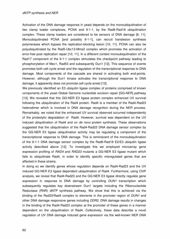
dNTP synthesis and NER
62
Activation of the DNA damage response in yeast depends on the monoubiquitination of
two clamp loader complexes, PCNA and 9-1-1, by the Rad6-Rad18 ubiquitination
complex. These clamp loaders are considered to be sensors of DNA damage [8, 11].
Monoubiquitinated PCNA [and possibly 9-1-1], can recruit translesion synthesis
polymerases which bypass the replication-blocking lesion [10, 11]. PCNA can also be
polyubiquitinated by the Rad5-Ubc13-Mms2 complex which promotes the activation of
error-free post replication repair [10, 11]. In a different context monoubiquitination of the
Rad17 component of the 9-1-1 complex stimulates the checkpoint pathway leading to
phosphorylation of Mec1, Rad53 and subsequently Dun1 [12]. This sequence of events
promotes both cell cycle arrest and the regulation of the transcriptional response to DNA
damage. Most components of the cascade are shared in activating both end-points.
However, although the Dun1 kinase activates the transcriptional response to DNA
damage, it apparently does not promote cell cycle arrest [12].
We previously identified an E3 ubiquitin ligase complex of proteins comprised of known
components of the yeast Global Genome nucleotide excision repair [GG-NER] pathway
[13]. We revealed that this GG-NER E3 ligase protein complex enhanced UV survival
following the ubiquitination of the Rad4 protein. Rad4 is a member of the Rad4-Rad23
heterodimer which is involved in DNA damage recognition during the NER process.
Remarkably, we noted that the enhanced UV survival observed occurred independently
of the proteolytic degradation of Rad4. However, survival was dependent on the UV
induced ubiquitination of Rad4 and on de novo protein synthesis. These observations
suggested that the ubiquitination of the Rad4-Rad23 DNA damage sensor complex by
the GG-NER E3 ligase ubiquitination activity may be regulating a component of the
transcriptional response to DNA damage. This is reminiscent of the monoubiquitination
of the 9-1-1 DNA damage sensor complex by the Rad6-Rad18 E2/E3 ubiquitin ligase
activity described above [12]. To investigate this we employed microarray gene
expression profiling of RAD4 and RAD23 mutants a GG-NER E3 ligase mutant which
fails to ubiquitinate Rad4, in order to identify specific misregulated genes that are
affected in these strains.
In doing so we identify genes whose regulation depends on Rad4-Rad23 and the UV
induced GG-NER E3 ligase dependent ubiquitination of Rad4. Furthermore, using ChIP
analysis, we reveal that Rad4-Rad23 and the GG-NER E3 ligase directly regulate gene
expression in response to DNA damage by controlling DUN1 transcription which
subsequently regulates key downstream Dun1 targets including the Ribonucleotide
Reductase (RNR) dNTP synthesis pathway. We show that this is achieved via the
binding of the Rad23/Rad4 complex to elements in the promoter region of DUN1 and
other DNA damage responsive genes including DDR2. DNA damage results in changes
in the binding of the Rad4-Rad23 complex at the promoter of these genes in a manner
dependent on the ubiquitination of Rad4. Collectively, these data describe a novel
regulation of UV DNA damage induced gene expression via the well-known NER DNA
dNTP synthesis and NER
63
damage recognition complex Rad4-Rad23. Finally, we show that the primary role of this
novel regulatory pathway is to control the UV induced synthesis of dNTPs to enhance
survival following DNA damage. By characterizing the mutant phenotypes, we show that
in the absence of the GG-NER E3 ubiquitin ligase regulated gene expression, UV
survival is significantly reduced and cell cycle progression is affected. Constitutive
upregulation of dNTP synthesis completely rescues both of these phenotypes,
confirming the role of the pathway in regulating UV induced dNTP pools. Our results
provide insight into a novel regulatory mechanism showing how known NER DNA
damage sensors can also control gene expression and describe a point at which the
NER pathway is integrated with the transcriptional response to DNA damage.
3. Materials & Methods
3.1 Yeast strains and plasmids
Research Genetics parental strain BY4742, BY4742rad23∆, BY4742rad4∆,
BY4742rad6∆, BY4742rad18∆, and BY4742rad7∆ strains were obtained from Euroscarf.
The double mutant rad23∆rad4∆ was derived from BY4742rad23∆ by replacing RAD4
with a His3 marker fragment. Creation of the Rad7 SOCS box mutation was achieved by
site directed mutagenesis of the wild-type RAD7 gene cloned in pRS314 as described
[14]. Two point mutations were made resulting in the amino acid substitutions, L168A
and C172A within the conserved SOCS box domain [13]. The RAD23 gene of
BY4742rad7∆ was replaced by a URA3 marker fragment to generate the double mutant
rad7∆rad23∆. The triple mutant rad7∆rad23∆sml1∆ and rad7∆rad23∆crt1∆ were derived
from rad7∆rad23∆, respectively. Then pRS314 containing the RAD7 gene and SOCS
box mutated RAD7 were introduced to rad7∆rad23∆sml1∆ respectively to produce the
pRAD7∆rad23∆sml1∆ and psocs∆rad23∆sml1∆ strains. In the same way, the
pRAD7∆rad23∆crt1∆ and psocs∆rad23∆crt1∆ strains were derived from
rad7∆rad23∆crt1∆.
3.2 In vivo cross-linking and sonication of chromatin extracts
Cells were grown to a density of 2~4x10⁷ cells/ml, and 2.8 ml of 37% formaldehyde was
added to 100 ml of the culture medium (containing at least 2x10⁹
cells). The mixture was
incubated at room temperature for 20 minutes with occasional swirling to allow efficient
DNA and protein cross-linking. The cross-linking reaction was ceased by adding 5.5 ml
of 2.5M glycine to a final concentration of 0.125M. Cells were collected by centrifugation
and then washed with ice-cold TBS buffer and ChIP lysis buffer. Cells were resuspended
in 500 µl of ChIP lysis buffer supplemented with 12.5 µl of 20% SDS and 12 µl of 100x
protease inhibitors. After 0.5 ml glass beads were added to this solution, the mixture was
vortexed (at high speed on a turboMixer) at 4°C for 10-15 minutes. The cell lysate was
carefully collected by centrifugation. Next, the cell lysate was sonicated by a Diagenode
sonication system at the high output rate for 3-4 minutes (6-8 x 0.5 min on/0.5 min off
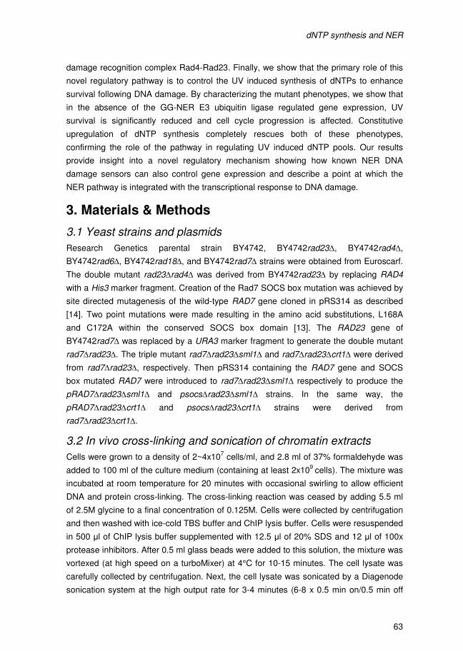
dNTP synthesis and NER
64
cycle). The sonicated cell lysate was spun down at 13,200 rpm for 15 minutes at 4°C.
The supernatant (chromatin extract) was finally transferred to a clean tube and stored at
-80°C until further use.
3.3 Chromatin immunoprecipitation (ChIP)
Protein A beads were washed twice with ChIP lysis buffer and then equilibrated with
ChIP lysis buffer supplemented with 0.1% BSA and 40 µg/ml single strand salmon sperm
DNA for 3 hours at 4°C. Next, 50 µl of chromatin extracts were added to 500 µl of ChIP
binding buffer (i.e. ChIP lysis buffer supplemented with 0.25% SDS and 1x protease
inhibitors), after which the solution was incubated with the equilibrated protein A beads.
After removal of the protein A beads by centrifugation, the chromatin
immunoprecipitation was carried out by adding 1-5 µl antibody to this cleared solution at
4°C for overnight. In the following step 20-30µl of protein A beads slurry (ChIP lysis
buffer washed twice) was added to the solution and incubated for 2-3 hours at 4°C. The
protein A beads were quick spun and washed successively with ChIP lysis buffer, ChIP
lysis buffer with 500mM NaCl, LiCl solution and TE buffer.
The protein A beads were incubated with 250 µl elution buffer at room temperature for
10 minutes. Then, the supernatant was collected by centrifugation. The pellet was eluted
again. The two parts of eluate were pooled together and incubated at 65°C overnight to
reverse the cross-linking.
Subsequently, the elution was treated with ribonuclease A and protease K and the DNA
was purified by a standard phenol/chloroform purification or a PCR purification kit
(QIAGEN). In order to precipitate the DNA, 100 µg glycogen, 1/10 volume of 3M sodium
acetate (pH5.2) and 2 volume of ethanol were added to the solution. The precipitated
DNA was resuspended in 50-100 µl TE buffer and stored at -20°C.
3.4 The quantitative PCR (qPCR)
Quantitative PCR was carried out with the following primer pairs:
Upper_DDR2-primer-1 TGCTCAAAGGTTTATGCCCGATGTT;
Lower_DDR2-primer-1: TGCATTATTGATGTCCCATAA-GGGG;
Upper_DDR2-primer-2: CCCAGACACGGTTGCCAAGGCCTCG;
Lower_DDR2-primer-2: CGGGCATAAACCTTTGAGCATCATC;
Upper_DDR2-primer-3: AGCCCTCCAAGCAAGCACGC;
Lower_DDR2-primer-3: CGTGCAAAGCAGGAGCAGCG;
Upper-GPG1-primer-1: GCGCCCT-GTATCAAAAAGAAGCTTT;
Lower-GPG1-primer -1: GGAACTTCCTCACACCGCGGTTTGT.
The internal control primers were as follows:
Forward GGTTCTAGTTAGTCACGTGCAG
Reverse CGTTATTTTACTTTTCGGAAGACA
dNTP synthesis and NER
65
A serial dilution of wild-type genomic DNA strain (Sc507) was used to make a standard
DNA concentration curve. Based on this standard curve all raw data acquired from real-
time PCR machine was quantified using the iQ5 software (Bio-Rad).
3.5 Northern blot assay
The hot phenol method was applied for RNA isolation and Northern blotting was
performed as described previously [13]. The following primers were used in this study:
Actin_NTS: biotin-GCCGGTTTTGCCGGTGACG;
Actin_TS: CCGGCAGATTCCAAACCCAAAA;
DDR2_NTS: biotin- ACAGATTGCTCAAAGGTTTATGCCCGATGTT;
DDR2_TS: GGTATCATCATCGTGGCAGTAA-GCG;
DUN1_NTS: Biotin-AAAAAACAGGATGAATCCAAAGCTCTA;
DUN1_TS: AAACGCTGCA-ATTCTAATGAG;
DDI1_NTS: Biotin-AAAAAAGATACAGGGGCTCAAACAACG;
DDI1_TS: ACAGCATCCCTAGGGAAACCT.
RNR1_NTS: bio-TTCAAGGCTTACCAAACGTTCCACA;
RNR1_TS: TCGAACTCGGTTTCCTCATCATCAA.
3.6 Flow cytometric analysis of yeast cell cycle
Cells were grown to log-phase in minimal medium, and then G1 cells were obtained from
the population using the Beckman JE-5.0 centrifugal elutriation system. Cells were
adjusted to a concentration of 1x10⁷ cells/ml. From this starter culture 25 ml was treated
with 150 J/m2 UV and 25 ml was left untreated. Samples of 1 ml were removed at
different time points and fixed in 70% ethanol. Cells were then resuspended in 50mM
Na-Citrate Buffer (pH 7), sonicated on low power for 40 seconds to remove clumps, and
treated with RNase. After washing and second sonication, cells were stained with 8
µg/ml Propidium Iodide. Flow cytometric analysis was performed to measure DNA
content by PI fluorescence. Histograms show gated cell counts based on PI fluorescence
height.
dNTP synthesis and NER
66
4. Results
4.1 The Rad4-Rad23 complex regulates gene expression of UV responsive genes
We previously showed that the ubiquitination of Rad4 in response to UV radiation
affected DNA repair and UV survival in a manner dependent on de novo protein
synthesis [13]. This and other observations made in this report suggested a possible role
for the Rad4-Rad23 complex in the regulation of gene transcription in response to DNA
damage. Recently, an increasing number of studies have confirmed a role for specific
NER factors including yeast Rad23 and the human XPC-hHR23B complex in gene
transcription [13, 15-18]. In an effort to determine which genes might be regulated by the
Rad4-Rad23 complex, and in particular which genes are regulated by this complex in
response to UV radiation, we studied the effect of both the Rad4-Rad23 complex and in
particular, the ubiquitination of Rad4 by the GG-NER E3 ligase in response to UV
radiation on gene transcription by using microarray gene expression analysis.
Initially, using wild-type yeast or strains deleted for RAD4 and RAD23, either individually
or in combination (rad4∆, rad23∆ and rad4∆rad23∆), we examined genome wide gene
expression using microarray analysis. From these studies we found differentially
expressed genes for all three mutant backgrounds tested (Figure 1A and S1A). We
expanded this initial list of 139 genes to 205 genes by including all genes exhibiting a
greater than 1.5 fold-change in gene expression levels. Cluster analysis of these 205
genes revealed the presence of four hierarchical clusters of genes that display a similar
pattern of expression across the three mutant strains analyzed (Figure 1B). Cluster 1
includes genes whose expression is not affected in either of the single mutants, but do
require Rad4-Rad23 for their normal level of expression and are repressed in their
absence. Cluster 2 represents genes that are repressed by the individual loss of either
Rad4 or Rad23, but are upregulated in the absence of both genes. Cluster 3 includes
genes that require both Rad4 and Rad23 for their normal expression level, while cluster
4 contains the genes that need both Rad4 and Rad23 for their repression (Figure 1B).
Rad23 has previously been noted to have an effect on gene regulation [16], but our
studies extend these observations to show that in combination with Rad23, Rad4 also
influences gene expression.
In order to determine whether any of these 205 genes were also regulated in response to
UV radiation, we UV irradiated wild type cells and examined their gene expression profile
at 15 and 60 minutes after exposure. The total number of genes in both the early and
late UV response categories amounts to 1360 genes. 309 of these genes are common to
both the early and late response categories (Figure S1B). This list of 309 UV responsive
genes was then compared with the rad4∆, rad23∆ and rad4∆rad23∆ untreated gene
expression datasets in an effort to detect the UV responsive genes that are
transcriptionally regulated by Rad4-Rad23. 101 of the 205 genes regulated by Rad4-
dNTP synthesis and NER
67
Rad23 are also UV responsive, with cluster 2 and 4 having a remarkable 93% and 66%
of their constituent genes being UV responsive (Figure 1B and S1C). Significantly, when
the hierarchical clusters of these 101 UV responsive and Rad4-Rad23 regulated genes
Figure 1 – The Rad4-Rad23 NER factor affects UV induced gene expression. (A). Significantly differentially expressed genes in rad4∆, rad23∆ and rad4∆rad23∆ cells compared to wild-typecells in the absence of UV irradiation is shown here. For each strain gene expression is related to the wild-type control as a fold-change. Genes that did not display changed expression in the mutant backgrounds are in grey on the diagonal (y=x). Significantly upregulated genes are depicted in red while down regulated genes are shown in green. (B) Heat-map of hierarchically clustered genes in the rad4/rad23 double and single mutants. Expression fold-change is relative to the wild-type control. The fraction of UV responsive genes is indicated in percentage per cluster. (C) Heat-map of the hierarchical clusters in B of the UV responsive genes only compared to the UV induced gene expression profile of wild-type cells. Fold-change in expression of the UV-irradiated samples is versus the unirradiated control. (D) Cluster 2 is blown-up showing the STress Responsive Elements containing genes and Crt1 controlled genes being affected in the mutant backgrounds.

dNTP synthesis and NER
66
4. Results
4.1 The Rad4-Rad23 complex regulates gene expression of UV responsive genes
We previously showed that the ubiquitination of Rad4 in response to UV radiation
affected DNA repair and UV survival in a manner dependent on de novo protein
synthesis [13]. This and other observations made in this report suggested a possible role
for the Rad4-Rad23 complex in the regulation of gene transcription in response to DNA
damage. Recently, an increasing number of studies have confirmed a role for specific
NER factors including yeast Rad23 and the human XPC-hHR23B complex in gene
transcription [13, 15-18]. In an effort to determine which genes might be regulated by the
Rad4-Rad23 complex, and in particular which genes are regulated by this complex in
response to UV radiation, we studied the effect of both the Rad4-Rad23 complex and in
particular, the ubiquitination of Rad4 by the GG-NER E3 ligase in response to UV
radiation on gene transcription by using microarray gene expression analysis.
Initially, using wild-type yeast or strains deleted for RAD4 and RAD23, either individually
or in combination (rad4∆, rad23∆ and rad4∆rad23∆), we examined genome wide gene
expression using microarray analysis. From these studies we found differentially
expressed genes for all three mutant backgrounds tested (Figure 1A and S1A). We
expanded this initial list of 139 genes to 205 genes by including all genes exhibiting a
greater than 1.5 fold-change in gene expression levels. Cluster analysis of these 205
genes revealed the presence of four hierarchical clusters of genes that display a similar
pattern of expression across the three mutant strains analyzed (Figure 1B). Cluster 1
includes genes whose expression is not affected in either of the single mutants, but do
require Rad4-Rad23 for their normal level of expression and are repressed in their
absence. Cluster 2 represents genes that are repressed by the individual loss of either
Rad4 or Rad23, but are upregulated in the absence of both genes. Cluster 3 includes
genes that require both Rad4 and Rad23 for their normal expression level, while cluster
4 contains the genes that need both Rad4 and Rad23 for their repression (Figure 1B).
Rad23 has previously been noted to have an effect on gene regulation [16], but our
studies extend these observations to show that in combination with Rad23, Rad4 also
influences gene expression.
In order to determine whether any of these 205 genes were also regulated in response to
UV radiation, we UV irradiated wild type cells and examined their gene expression profile
at 15 and 60 minutes after exposure. The total number of genes in both the early and
late UV response categories amounts to 1360 genes. 309 of these genes are common to
both the early and late response categories (Figure S1B). This list of 309 UV responsive
genes was then compared with the rad4∆, rad23∆ and rad4∆rad23∆ untreated gene
expression datasets in an effort to detect the UV responsive genes that are
transcriptionally regulated by Rad4-Rad23. 101 of the 205 genes regulated by Rad4-
dNTP synthesis and NER
67
Rad23 are also UV responsive, with cluster 2 and 4 having a remarkable 93% and 66%
of their constituent genes being UV responsive (Figure 1B and S1C). Significantly, when
the hierarchical clusters of these 101 UV responsive and Rad4-Rad23 regulated genes
Figure 1 – The Rad4-Rad23 NER factor affects UV induced gene expression. (A). Significantly differentially expressed genes in rad4∆, rad23∆ and rad4∆rad23∆ cells compared to wild-typecells in the absence of UV irradiation is shown here. For each strain gene expression is related to the wild-type control as a fold-change. Genes that did not display changed expression in the mutant backgrounds are in grey on the diagonal (y=x). Significantly upregulated genes are depicted in red while down regulated genes are shown in green. (B) Heat-map of hierarchically clustered genes in the rad4/rad23 double and single mutants. Expression fold-change is relative to the wild-type control. The fraction of UV responsive genes is indicated in percentage per cluster. (C) Heat-map of the hierarchical clusters in B of the UV responsive genes only compared to the UV induced gene expression profile of wild-type cells. Fold-change in expression of the UV-irradiated samples is versus the unirradiated control. (D) Cluster 2 is blown-up showing the STress Responsive Elements containing genes and Crt1 controlled genes being affected in the mutant backgrounds.
dNTP synthesis and NER
68
are compared with the expression data of the UV-irradiated wild-type cells, the
expression profile of the rad4∆rad23∆ cells is very similar to the post-UV expression
profile of wild-type cells (Figure 1C). This indicates that the UV induced gene expression
changes observed in wild type cells is mimicked in the rad4∆rad23∆ mutant, suggesting
a possible role for Rad4-Rad23 in the UV induced regulation of these genes.
4.2 Rad4-Rad23 increases gene expression of STRE containing and CRT1 regulated DDR genes following UV irradiation
We next focused our attention on the identity of genes found in cluster 2 (Figure 1B)
since the genes in this cluster require the Rad4-Rad23 heterodimer for their repression
in the absence of UV radiation, and 93% of them are also induced in wild type cells after
UV irradiation. Close examination of the upstream regulatory regions of the genes in this
cluster reveal that they are enriched for genes that contain a Stress Response Element
(STRE) within their promoter region (Figure 1D). The STRE is a hexanucleotide
regulatory element found in specific DNA damage response genes including DDR2, that
are targeted by the stress induced transcriptional activators Msn2 and Msn4 (see Figure
4A) [19]. The HUG1 and RNR3 genes also require Rad4-Rad23 for their repression in
the absence of UV irradiation and are also UV inducible. Instead of sharing common
transcriptional activators, these genes share a common transcriptional repressor called
Crt1, which binds to a regulatory element in their promoters (Figure 1D). Crt1 represses
a set of UV induced cell-cycle checkpoint and DNA repair genes, including the RNR
genes, that become activated following depression of Crt1 by Dun1 phosphorylation in
response to DNA damage [20]. The analysis of our data describes two classes of genes
whose regulation in response to UV depends on Rad4-Rad23. These observations
suggest that the Rad4-Rad23 heterodimer may act directly as a transcriptional regulator
of genes involved in the transcriptional response to DNA damage [20, 21], particularly in
regulating the RNR pathway which is explored in greater detail later.
4.3 The GG-NER E3 ligase regulates genes involved in the transcriptional response to DNA damage
Next, we specifically wanted to identify the genes controlled by the GG-NER E3 ligase,
and in particular those whose regulation is dependent on the ubiquitination of the Rad4
component of the Rad4-Rad23 complex. We have shown previously that a point mutant
in the SOCS box domain of the Rad7 component of the GG-NER E3 ligase specifically
fails to ubiquitinate Rad4 in response to UV. We demonstrated that this point mutant
strain is not UV sensitive, but that it significantly increases the UV sensitivity of a strain
that also has the RAD23 gene deleted [13]. These and other data confirmed that the GG-
NER E3 ligase functions in parallel with Rad23 in NER and UV survival, and achieves
this by controlling UV induced gene transcription via the ubiquitination of Rad4 [13]. To
determine how gene regulation by the GG-NER E3 ligase enhances NER and UV
survival, we sought to identify the genes specifically regulated by the E3 ligase complex
dNTP synthesis and NER
69
Figure 2 – The GG-NER E3 ligase regulates UV induced gene expression in concert with Rad4-Rad23. (A) Differential expression of the mutant background indicated versus the wild-typepRAD7 background. Highlighted in red are the genes with increased expression in the mutant backgrounds compared to the wild-type control, while in green genes with reduced expression versus the wild-type strain are indicated. (B) Genes from A that were differentially expressed by 1.5-fc in psocs/rad23 cells were hierarchically clustered resulting in the heat-map displayed here. (C) Heat-map of the hierarchical clusters in B of the UV responsive genes only compared to the UV induced gene expression profile of wild-type cells. Fold-change in expression of the UV-irradiated samples is versus the unirradiated control. (D) Significantly differentially expressed UV responsive genes from the psocs/rad23 dataset combined with the expression data from the rad4/rad23 dataset are shown in this heat-map. The expression of RNR and related genes from the rad4/rad23 and psocs/rad23 datasets are expressed as the fold-change versus wild-type or untreated controls.
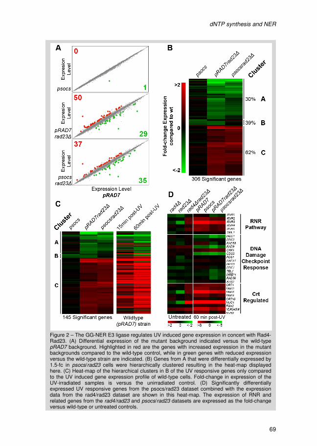
dNTP synthesis and NER
68
are compared with the expression data of the UV-irradiated wild-type cells, the
expression profile of the rad4∆rad23∆ cells is very similar to the post-UV expression
profile of wild-type cells (Figure 1C). This indicates that the UV induced gene expression
changes observed in wild type cells is mimicked in the rad4∆rad23∆ mutant, suggesting
a possible role for Rad4-Rad23 in the UV induced regulation of these genes.
4.2 Rad4-Rad23 increases gene expression of STRE containing and CRT1 regulated DDR genes following UV irradiation
We next focused our attention on the identity of genes found in cluster 2 (Figure 1B)
since the genes in this cluster require the Rad4-Rad23 heterodimer for their repression
in the absence of UV radiation, and 93% of them are also induced in wild type cells after
UV irradiation. Close examination of the upstream regulatory regions of the genes in this
cluster reveal that they are enriched for genes that contain a Stress Response Element
(STRE) within their promoter region (Figure 1D). The STRE is a hexanucleotide
regulatory element found in specific DNA damage response genes including DDR2, that
are targeted by the stress induced transcriptional activators Msn2 and Msn4 (see Figure
4A) [19]. The HUG1 and RNR3 genes also require Rad4-Rad23 for their repression in
the absence of UV irradiation and are also UV inducible. Instead of sharing common
transcriptional activators, these genes share a common transcriptional repressor called
Crt1, which binds to a regulatory element in their promoters (Figure 1D). Crt1 represses
a set of UV induced cell-cycle checkpoint and DNA repair genes, including the RNR
genes, that become activated following depression of Crt1 by Dun1 phosphorylation in
response to DNA damage [20]. The analysis of our data describes two classes of genes
whose regulation in response to UV depends on Rad4-Rad23. These observations
suggest that the Rad4-Rad23 heterodimer may act directly as a transcriptional regulator
of genes involved in the transcriptional response to DNA damage [20, 21], particularly in
regulating the RNR pathway which is explored in greater detail later.
4.3 The GG-NER E3 ligase regulates genes involved in the transcriptional response to DNA damage
Next, we specifically wanted to identify the genes controlled by the GG-NER E3 ligase,
and in particular those whose regulation is dependent on the ubiquitination of the Rad4
component of the Rad4-Rad23 complex. We have shown previously that a point mutant
in the SOCS box domain of the Rad7 component of the GG-NER E3 ligase specifically
fails to ubiquitinate Rad4 in response to UV. We demonstrated that this point mutant
strain is not UV sensitive, but that it significantly increases the UV sensitivity of a strain
that also has the RAD23 gene deleted [13]. These and other data confirmed that the GG-
NER E3 ligase functions in parallel with Rad23 in NER and UV survival, and achieves
this by controlling UV induced gene transcription via the ubiquitination of Rad4 [13]. To
determine how gene regulation by the GG-NER E3 ligase enhances NER and UV
survival, we sought to identify the genes specifically regulated by the E3 ligase complex
dNTP synthesis and NER
69
Figure 2 – The GG-NER E3 ligase regulates UV induced gene expression in concert with Rad4-Rad23. (A) Differential expression of the mutant background indicated versus the wild-typepRAD7 background. Highlighted in red are the genes with increased expression in the mutant backgrounds compared to the wild-type control, while in green genes with reduced expression versus the wild-type strain are indicated. (B) Genes from A that were differentially expressed by 1.5-fc in psocs/rad23 cells were hierarchically clustered resulting in the heat-map displayed here. (C) Heat-map of the hierarchical clusters in B of the UV responsive genes only compared to the UV induced gene expression profile of wild-type cells. Fold-change in expression of the UV-irradiated samples is versus the unirradiated control. (D) Significantly differentially expressed UV responsive genes from the psocs/rad23 dataset combined with the expression data from the rad4/rad23 dataset are shown in this heat-map. The expression of RNR and related genes from the rad4/rad23 and psocs/rad23 datasets are expressed as the fold-change versus wild-type or untreated controls.
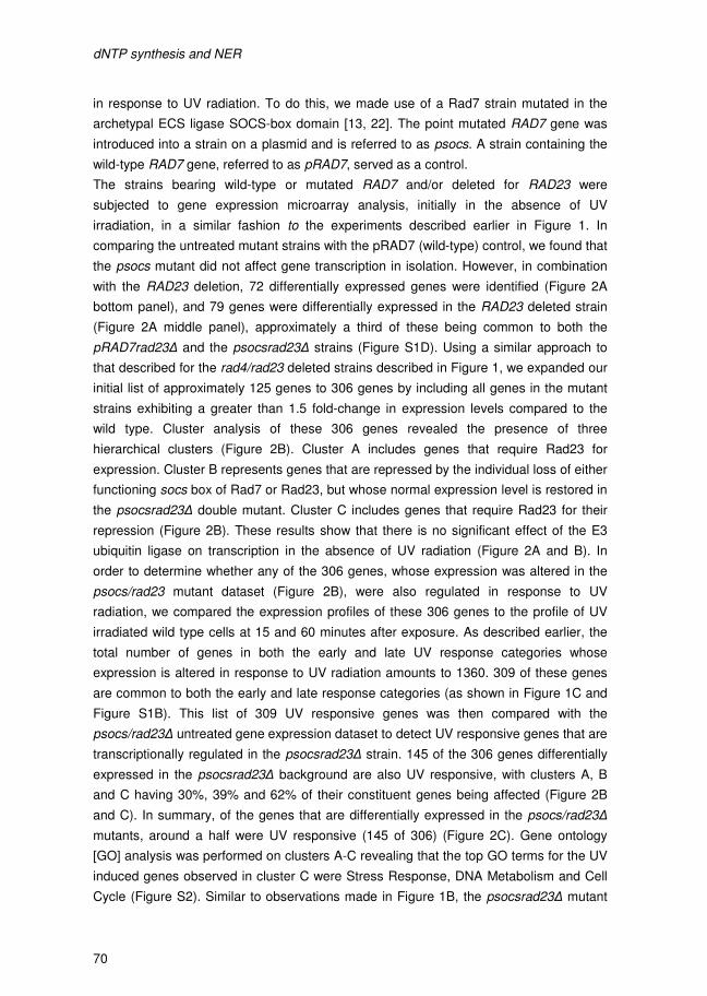
dNTP synthesis and NER
70
in response to UV radiation. To do this, we made use of a Rad7 strain mutated in the
archetypal ECS ligase SOCS-box domain [13, 22]. The point mutated RAD7 gene was
introduced into a strain on a plasmid and is referred to as psocs. A strain containing the
wild-type RAD7 gene, referred to as pRAD7, served as a control.
The strains bearing wild-type or mutated RAD7 and/or deleted for RAD23 were
subjected to gene expression microarray analysis, initially in the absence of UV
irradiation, in a similar fashion to the experiments described earlier in Figure 1. In
comparing the untreated mutant strains with the pRAD7 (wild-type) control, we found that
the psocs mutant did not affect gene transcription in isolation. However, in combination
with the RAD23 deletion, 72 differentially expressed genes were identified (Figure 2A
bottom panel), and 79 genes were differentially expressed in the RAD23 deleted strain
(Figure 2A middle panel), approximately a third of these being common to both the
pRAD7rad23∆ and the psocsrad23∆ strains (Figure S1D). Using a similar approach to
that described for the rad4/rad23 deleted strains described in Figure 1, we expanded our
initial list of approximately 125 genes to 306 genes by including all genes in the mutant
strains exhibiting a greater than 1.5 fold-change in expression levels compared to the
wild type. Cluster analysis of these 306 genes revealed the presence of three
hierarchical clusters (Figure 2B). Cluster A includes genes that require Rad23 for
expression. Cluster B represents genes that are repressed by the individual loss of either
functioning socs box of Rad7 or Rad23, but whose normal expression level is restored in
the psocsrad23∆ double mutant. Cluster C includes genes that require Rad23 for their
repression (Figure 2B). These results show that there is no significant effect of the E3
ubiquitin ligase on transcription in the absence of UV radiation (Figure 2A and B). In
order to determine whether any of the 306 genes, whose expression was altered in the
psocs/rad23 mutant dataset (Figure 2B), were also regulated in response to UV
radiation, we compared the expression profiles of these 306 genes to the profile of UV
irradiated wild type cells at 15 and 60 minutes after exposure. As described earlier, the
total number of genes in both the early and late UV response categories whose
expression is altered in response to UV radiation amounts to 1360. 309 of these genes
are common to both the early and late response categories (as shown in Figure 1C and
Figure S1B). This list of 309 UV responsive genes was then compared with the
psocs/rad23∆ untreated gene expression dataset to detect UV responsive genes that are
transcriptionally regulated in the psocsrad23∆ strain. 145 of the 306 genes differentially
expressed in the psocsrad23∆ background are also UV responsive, with clusters A, B
and C having 30%, 39% and 62% of their constituent genes being affected (Figure 2B
and C). In summary, of the genes that are differentially expressed in the psocs/rad23∆
mutants, around a half were UV responsive (145 of 306) (Figure 2C). Gene ontology
[GO] analysis was performed on clusters A-C revealing that the top GO terms for the UV
induced genes observed in cluster C were Stress Response, DNA Metabolism and Cell
Cycle (Figure S2). Similar to observations made in Figure 1B, the psocsrad23∆ mutant
dNTP synthesis and NER
71
closely mimics the UV induced gene expression response of wild-type cells (Figure 2C).
This indicates a possible role for both the E3 ubiquitin ligase and Rad4-Rad23 in the UV
induced regulation of the genes in these pathways.
4.4 The GG-NER E3 ligase controls UV induced expression of genes involved in the RNR pathway
The gene expression profiling studies conducted so far suggest that the GG-NER E3
ligase regulates genes involved in the DNA damage checkpoint pathway, particularly
genes involved in the RNR pathway that are regulated via STRE elements within their
promoters and/or regulated by the Crt1 repressor protein. Having identified the genes
misregulated in the psocsrad23∆ mutated strain in the absence of UV, we next examined
the effect of these mutations on UV induced gene transcription, specifically examining
genes involved in the DDR pathways identified above. To do this, we applied microarray
analysis for the UV treated pRAD7, pRAD7rad23∆ and psocsrad23∆ strains. The
resulting heat-map is displayed in Figure 2D, with the major DNA damage response
genes from the pathways indicated, grouped together.
The most striking feature to emerge from this analysis is that the RNR pathway genes
show strong evidence of being regulated by the Rad4-Rad23 complex and the GG-NER
E3 ligase activity in response to UV radiation. In contrast, the cluster of genes involved
as central players in the DNA damage response, including Mec1, Rad53, Tel1 and Chk1
in Figure 2D does not reveal a significant role for Rad4-Rad23 or the GG-NER E3 ligase
in expression of these genes within the central regulatory pathway. Similarly, the
expression of other genes such as RAD9 and RAD51 which are involved in primary
detection and signaling of different types of DNA damage are also not significantly
affected by Rad4-Rad23 or the GG-NER E3 ligase.
Quantitative analysis shown in Figure 3A reveals that the regulation of RNR pathway
genes, in particular of DUN1 and RNR1, does show an important role for Rad4-Rad23
and the GG-NER E3 ligase. The DUN1 gene is an important activator of RNR in
response to UV damage, but little is known about regulation of its expression in response
to UV. Figure 3A shows that DUN1 expression is dependent on the GG-NER E3 ligase in
response to UV damage. In addition, regulation of RNR gene expression is controlled by
the CRT1 repressor. Figure 2D and 3A show that most CRT1 controlled genes are also
regulated by the GG-NER E3 ligase in responsive to UV. To confirm the results obtained
from microarray analysis, we performed northern blotting to detect RNA levels in the
strains tested. Figure 3B confirms the requirement for the GG-NER E3 ligase in
upregulating both DUN1 and RNR1 in response to UV radiation. This further
underscores the requirement for Rad4-Rad23 and the GG-NER E3 ligase for the
regulation of UV induced RNR and DUN1 expression.
A previous report elegantly described the DDR induced gene expression of MAG1 and
DDI1 by the Rad6-Rad18 ubiquitination complex via the mono-ubiquitination of Rad17

dNTP synthesis and NER
72
[12]. This report
raised the
possibility that the
GG-NER E3 ligase
controlled
ubiquitination of
Rad4 may be a
component of this
pathway. How-
ever, we did not
observe any Rad4-
Rad23 regulated
gene expression
of the MAG1 and
DDI1 cluster of
genes (Figure S3).
MAG1 and DDI1
are fully up-
regulated after UV-
irradiation in-
dependently of
Rad23 or the GG-
NER E3 ligase.
We conclude that
the Rad4-Rad23
controlled and GG-
NER E3 ligase
coordinated gene
expression is not involved in the Rad6-Rad18 mediated DDR transcription response to
DNA damage [12]. This is also confirmed when we analyzed the UV induced expression
of DUN1 or DDR2 in a rad6∆ or rad18∆ deletion strains (Figure S3).
The combined data on the RNR pathway shows regulation by Rad4-Rad23 and the GG-
NER E3 ligase of these genes. RNR1-4 and DUN1 all require Rad4-Rad23 and the E3
ligase for repression in the absence of DNA damage. In response to DNA damage both
Rad23 and the GG-NER E3 ligase activity are required for full blown expression of these
genes (Figure 2D and 3A). A more subtle contribution can be seen for the RNR inhibitor
SML1. Sml1p is an inhibitor of the RNR enzyme complex, that is degraded in response
to DNA damage [23, 24]. We find that in the absence of the GG-NER E3 ligase UV
irradiation results in elevated levels of SML1 gene expression, which further inhibits the
RNR pathway function (Figure 3A). We were able to confirm these microarray
Figure 3 – (A) Heatmap of the genes from cluster 2 in figure 1 with the absolute log2 fold-change listed for each in gene in each background tested (B) Northern blot analysis of gene expression at the mRNA levels of candidate genes scored in the microarray analysis. RNA was extracted from the strains indicated at the blot was treated with probes for DUN1, RNR1, SML1 and ACT1 as a positive control.
dNTP synthesis and NER
73
expression data for RNR1, DUN1 and SML1 using northern blot analysis (Figure 3B). It
is noteworthy that the upregulation of SML1 is more evident when measuring mRNA via
northern blot (Figure 3B). Interestingly, Dun1 and its downstream targets described here
constitute an important end-point for the DDR Mec1-Rad53-Dun1 kinase signaling
cascade. To summarize, the RNR pathway is down regulated in the psocsrad23∆
background both by reduced DUN1 and RNR gene expression and also via upregulation
of its inhibitor SML1.
4.5 Rad4-Rad23 complex regulates gene transcription of UV responsive genes via changes in the occupancy of the complex at the promoters of specific DDR genes
The results described in Figure 1 show that the increase in gene expression observed in
cluster 2 in a rad4∆rad23∆ background, mimics the UV induced increase in gene
expression of this cluster of genes observed in wild type cells. Intriguingly, recent
evidence in mouse embryonic stem cells has revealed that XPC-hHR23B, the
homologues of Rad4-Rad23, can regulate gene expression of specific developmental
genes as a result of changes in the binding of the complex to elements in the promoter
regions of these genes, suggesting a direct role for XPC-hHR23B in regulating gene
transcription [18]. Therefore, we speculated that the Rad4-Rad23 heterodimer may also
regulate UV induced gene transcription via direct binding of the complex to elements
within the promoter regions of the UV responsive genes identified in cluster 2 (Figure
1D). To examine this, we measured the occupancy of the Rad4-Rad23 heterodimer in
the regulatory regions of STRE element containing genes in the cluster including DDR2
and DUN1, using chromatin immunoprecipitation (ChIP) and quantitative PCR. As shown
in Figure 4, we found that in unirradiated cells, the Rad4-Rad23 complex occupies the
chromatin in the promoter region of DDR2. Figure 4A (top panel) indicates the location
within the DDR2 gene amplified by three different sets of PCR primers. The lower panel
of Figure 4A shows that the occupancy of Rad23 following ChIP is enriched in the STRE
containing promoter element detected by the DDR2-primers-1 PCR primer set compared
to two other primer sets located either in the ORF [DDR2-Primers-3] or the upstream
region of the promoter [DDR2-primers-2]. We confirmed that Rad23 specifically binds at
the promoter region of the DDR2 gene by examining Rad23 binding in the promoter
region of the GPG1 gene. GPG1 expression is affected by Rad4-Rad23 (Figure 1D), but
does not contain a STRE element in its promoter. No enrichment for Rad23 in the
regulatory region of GPG1 was detected (data not shown). To examine the occupancy of
both Rad4 and Rad23 at the promoter of DDR2, we performed a double ChIP
experiment, first performing chromatin immunoprecipitation using Rad23 antibodies,
followed by Rad4 antibodies. This double ChIP procedure was carried out and qPCR
performed at the promoter regions of both DDR2 and DUN1. Figure 4B confirms the
occupancy of both Rad4 and Rad23 at the promoter regions of the DDR2 [right panel]

dNTP synthesis and NER
72
[12]. This report
raised the
possibility that the
GG-NER E3 ligase
controlled
ubiquitination of
Rad4 may be a
component of this
pathway. How-
ever, we did not
observe any Rad4-
Rad23 regulated
gene expression
of the MAG1 and
DDI1 cluster of
genes (Figure S3).
MAG1 and DDI1
are fully up-
regulated after UV-
irradiation in-
dependently of
Rad23 or the GG-
NER E3 ligase.
We conclude that
the Rad4-Rad23
controlled and GG-
NER E3 ligase
coordinated gene
expression is not involved in the Rad6-Rad18 mediated DDR transcription response to
DNA damage [12]. This is also confirmed when we analyzed the UV induced expression
of DUN1 or DDR2 in a rad6∆ or rad18∆ deletion strains (Figure S3).
The combined data on the RNR pathway shows regulation by Rad4-Rad23 and the GG-
NER E3 ligase of these genes. RNR1-4 and DUN1 all require Rad4-Rad23 and the E3
ligase for repression in the absence of DNA damage. In response to DNA damage both
Rad23 and the GG-NER E3 ligase activity are required for full blown expression of these
genes (Figure 2D and 3A). A more subtle contribution can be seen for the RNR inhibitor
SML1. Sml1p is an inhibitor of the RNR enzyme complex, that is degraded in response
to DNA damage [23, 24]. We find that in the absence of the GG-NER E3 ligase UV
irradiation results in elevated levels of SML1 gene expression, which further inhibits the
RNR pathway function (Figure 3A). We were able to confirm these microarray
Figure 3 – (A) Heatmap of the genes from cluster 2 in figure 1 with the absolute log2 fold-change listed for each in gene in each background tested (B) Northern blot analysis of gene expression at the mRNA levels of candidate genes scored in the microarray analysis. RNA was extracted from the strains indicated at the blot was treated with probes for DUN1, RNR1, SML1 and ACT1 as a positive control.
dNTP synthesis and NER
73
expression data for RNR1, DUN1 and SML1 using northern blot analysis (Figure 3B). It
is noteworthy that the upregulation of SML1 is more evident when measuring mRNA via
northern blot (Figure 3B). Interestingly, Dun1 and its downstream targets described here
constitute an important end-point for the DDR Mec1-Rad53-Dun1 kinase signaling
cascade. To summarize, the RNR pathway is down regulated in the psocsrad23∆
background both by reduced DUN1 and RNR gene expression and also via upregulation
of its inhibitor SML1.
4.5 Rad4-Rad23 complex regulates gene transcription of UV responsive genes via changes in the occupancy of the complex at the promoters of specific DDR genes
The results described in Figure 1 show that the increase in gene expression observed in
cluster 2 in a rad4∆rad23∆ background, mimics the UV induced increase in gene
expression of this cluster of genes observed in wild type cells. Intriguingly, recent
evidence in mouse embryonic stem cells has revealed that XPC-hHR23B, the
homologues of Rad4-Rad23, can regulate gene expression of specific developmental
genes as a result of changes in the binding of the complex to elements in the promoter
regions of these genes, suggesting a direct role for XPC-hHR23B in regulating gene
transcription [18]. Therefore, we speculated that the Rad4-Rad23 heterodimer may also
regulate UV induced gene transcription via direct binding of the complex to elements
within the promoter regions of the UV responsive genes identified in cluster 2 (Figure
1D). To examine this, we measured the occupancy of the Rad4-Rad23 heterodimer in
the regulatory regions of STRE element containing genes in the cluster including DDR2
and DUN1, using chromatin immunoprecipitation (ChIP) and quantitative PCR. As shown
in Figure 4, we found that in unirradiated cells, the Rad4-Rad23 complex occupies the
chromatin in the promoter region of DDR2. Figure 4A (top panel) indicates the location
within the DDR2 gene amplified by three different sets of PCR primers. The lower panel
of Figure 4A shows that the occupancy of Rad23 following ChIP is enriched in the STRE
containing promoter element detected by the DDR2-primers-1 PCR primer set compared
to two other primer sets located either in the ORF [DDR2-Primers-3] or the upstream
region of the promoter [DDR2-primers-2]. We confirmed that Rad23 specifically binds at
the promoter region of the DDR2 gene by examining Rad23 binding in the promoter
region of the GPG1 gene. GPG1 expression is affected by Rad4-Rad23 (Figure 1D), but
does not contain a STRE element in its promoter. No enrichment for Rad23 in the
regulatory region of GPG1 was detected (data not shown). To examine the occupancy of
both Rad4 and Rad23 at the promoter of DDR2, we performed a double ChIP
experiment, first performing chromatin immunoprecipitation using Rad23 antibodies,
followed by Rad4 antibodies. This double ChIP procedure was carried out and qPCR
performed at the promoter regions of both DDR2 and DUN1. Figure 4B confirms the
occupancy of both Rad4 and Rad23 at the promoter regions of the DDR2 [right panel]
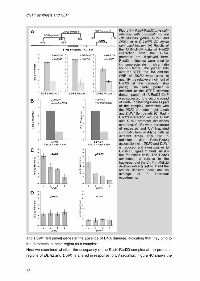
dNTP synthesis and NER
74
and DUN1 [left panel] genes in the absence of DNA damage, indicating that they bind to
the chromatin in these region as a complex.
Next we examined whether the occupancy of the Rad4-Rad23 complex at the promoter
regions of DDR2 and DUN1 is altered in response to UV radiation. Figure 4C shows the
Figure 4 – Rad4-Rad23 physically interacts with chro-matin of the UV induced genes DUN1 and DDR2 in a GG-NER E3 ligase controlled fashion. (A) Results of the ChIP-qPCR data of Rad23 interaction with the DDR2promoter are displayed here. Rad23 antibodies were used to immunoprecipitate chrom-atin bound Rad23. The primer sets over the STRE, the UAS and the ORF of DDR2 were used to quantify the relative enrichment of Rad23 at the promoter (top panel). The Rad23 protein is enriched at the STRE element (bottom panel). (B) A Rad23 ChIP was subjected to a second round of Rad4 IP detecting Rad4 as part of the complex interacting with the DDR2 promoter (right panel) and DUN1 (left panel). (C) Rad4-Rad23 interaction with the DDR2and DUN1 promoter diminishes over time. ChIPs were performed of untreated and UV irradiated chromatin from wild-type cells at different times after UV ir-radiation. (D) Rad4-Rad23 association with DDR2 and DUN1is reduced and ir-responsive to UV in E3 ligase mutants. As (C) but for psocs cells. The Rad23 enrichment is relative to the background of the ChIP in RAD23deletion extracts set to 1 and the results depicted here are an average of 3 individual experiments.
dNTP synthesis and NER
75
loss of occupancy of Rad23 from the promoter region of both DDR2 and DUN1 over a 2
hour period following UV irradiation. Taken together, these observations are consistent
with a model in which the UV induced loss of occupancy of Rad4-Rad23 from the
promoter regions of the UV responsive genes DDR2 and DUN1, results in the increased
expression of these genes after exposure of cells to UV radiation.
We then considered how the loss of occupancy of Rad4-Rad23 from the promoter
regions of these genes is regulated in response to UV. Previously, we demonstrated that
the ubiquitination of Rad4 by the GG-NER E3 ligase plays an important role in NER and
UV survival in a manner dependent on de novo protein synthesis [13]. In addition, the
effect of the GG-NER E3 ligase on gene expression described in figure 2D identifies a
role for the E3 ligase in UV induced transcription. We speculated that this E3 ubiquitin
ligase regulates the induction of UV responsive genes by controlling the occupancy of
the Rad4-Rad23 complex at their promoter regions in response to DNA damage. In order
to test this, we measured Rad23 occupancy at the promoters of both the DDR2 and
DUN1 genes in mutants of the GG-NER E3 ligase. We examined events in strains either
deleted for the ELC1 subunit of the E3 ligase, or mutated in the SOCS-box domain of the
Rad7 subunit. We have shown previously that both strains fail to ubiquitinate Rad4 in
response to UV radiation [13]. Figure 4D shows no loss of occupancy occurs of the
Rad4-Rad23 from the DDR2 and DUN1 promoters in response to UV damage in the
psocs and elc1 strain (data not shown). We noted that mutating the E3 ligase results in a
reduced level of Rad4-Rad23 binding at both the DDR2 and DUN1 promoters, even in
the absence of UV radiation. However, the reduced level of Rad4-Rad23 binding
observed does not induce transcription of either DDR2 or DUN1 in the absence of UV
damage (data not shown). In summary, our results show that in wild type cells the Rad4-
Rad23 complex represses gene expression of the genes in cluster 2 (Figure 1B-C) by
directly binding to the chromatin at the promoter region of these UV inducible genes.
Following UV irradiation, the Rad4-Rad23 complex dissociates from the promoter region
of these genes in a GG-NER E3 ligase dependent fashion facilitating increased gene
expression.
4.6 The GG-NER E3 ligase upregulates dNTP pools to enhance survival in response to UV damage
We speculated that the extreme UV sensitivity of psocsrad23∆ cells could be caused by
the failure to upregulate the expression of the RNR genes and compounded by the
increased expression of the RNR inhibitor SML1. This failure to respond to DNA damage
results in a lack of increased dNTP levels following UV irradiation. To test this hypothesis
we examined whether it is possible to suppress UV sensitivity of the psocsrad23∆ strain
by constitutively increasing the cellular dNTP pools. This was achieved by deletion of two
independent inhibitors of the RNR pathway. Sml1 inhibits the RNR enzyme complex,
while Crt1 is an inhibitor of transcription of the RNR genes. In this way, the Rad4-Rad23
dNTP synthesis and NER
74
and DUN1 [left panel] genes in the absence of DNA damage, indicating that they bind to
the chromatin in these region as a complex.
Next we examined whether the occupancy of the Rad4-Rad23 complex at the promoter
regions of DDR2 and DUN1 is altered in response to UV radiation. Figure 4C shows the
Figure 4 – Rad4-Rad23 physically interacts with chro-matin of the UV induced genes DUN1 and DDR2 in a GG-NER E3 ligase controlled fashion. (A) Results of the ChIP-qPCR data of Rad23 interaction with the DDR2promoter are displayed here. Rad23 antibodies were used to immunoprecipitate chrom-atin bound Rad23. The primer sets over the STRE, the UAS and the ORF of DDR2 were used to quantify the relative enrichment of Rad23 at the promoter (top panel). The Rad23 protein is enriched at the STRE element (bottom panel). (B) A Rad23 ChIP was subjected to a second round of Rad4 IP detecting Rad4 as part of the complex interacting with the DDR2 promoter (right panel) and DUN1 (left panel). (C) Rad4-Rad23 interaction with the DDR2and DUN1 promoter diminishes over time. ChIPs were performed of untreated and UV irradiated chromatin from wild-type cells at different times after UV ir-radiation. (D) Rad4-Rad23 association with DDR2 and DUN1is reduced and ir-responsive to UV in E3 ligase mutants. As (C) but for psocs cells. The Rad23 enrichment is relative to the background of the ChIP in RAD23deletion extracts set to 1 and the results depicted here are an average of 3 individual experiments.
dNTP synthesis and NER
75
loss of occupancy of Rad23 from the promoter region of both DDR2 and DUN1 over a 2
hour period following UV irradiation. Taken together, these observations are consistent
with a model in which the UV induced loss of occupancy of Rad4-Rad23 from the
promoter regions of the UV responsive genes DDR2 and DUN1, results in the increased
expression of these genes after exposure of cells to UV radiation.
We then considered how the loss of occupancy of Rad4-Rad23 from the promoter
regions of these genes is regulated in response to UV. Previously, we demonstrated that
the ubiquitination of Rad4 by the GG-NER E3 ligase plays an important role in NER and
UV survival in a manner dependent on de novo protein synthesis [13]. In addition, the
effect of the GG-NER E3 ligase on gene expression described in figure 2D identifies a
role for the E3 ligase in UV induced transcription. We speculated that this E3 ubiquitin
ligase regulates the induction of UV responsive genes by controlling the occupancy of
the Rad4-Rad23 complex at their promoter regions in response to DNA damage. In order
to test this, we measured Rad23 occupancy at the promoters of both the DDR2 and
DUN1 genes in mutants of the GG-NER E3 ligase. We examined events in strains either
deleted for the ELC1 subunit of the E3 ligase, or mutated in the SOCS-box domain of the
Rad7 subunit. We have shown previously that both strains fail to ubiquitinate Rad4 in
response to UV radiation [13]. Figure 4D shows no loss of occupancy occurs of the
Rad4-Rad23 from the DDR2 and DUN1 promoters in response to UV damage in the
psocs and elc1 strain (data not shown). We noted that mutating the E3 ligase results in a
reduced level of Rad4-Rad23 binding at both the DDR2 and DUN1 promoters, even in
the absence of UV radiation. However, the reduced level of Rad4-Rad23 binding
observed does not induce transcription of either DDR2 or DUN1 in the absence of UV
damage (data not shown). In summary, our results show that in wild type cells the Rad4-
Rad23 complex represses gene expression of the genes in cluster 2 (Figure 1B-C) by
directly binding to the chromatin at the promoter region of these UV inducible genes.
Following UV irradiation, the Rad4-Rad23 complex dissociates from the promoter region
of these genes in a GG-NER E3 ligase dependent fashion facilitating increased gene
expression.
4.6 The GG-NER E3 ligase upregulates dNTP pools to enhance survival in response to UV damage
We speculated that the extreme UV sensitivity of psocsrad23∆ cells could be caused by
the failure to upregulate the expression of the RNR genes and compounded by the
increased expression of the RNR inhibitor SML1. This failure to respond to DNA damage
results in a lack of increased dNTP levels following UV irradiation. To test this hypothesis
we examined whether it is possible to suppress UV sensitivity of the psocsrad23∆ strain
by constitutively increasing the cellular dNTP pools. This was achieved by deletion of two
independent inhibitors of the RNR pathway. Sml1 inhibits the RNR enzyme complex,
while Crt1 is an inhibitor of transcription of the RNR genes. In this way, the Rad4-Rad23
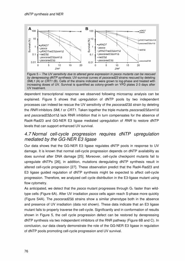
dNTP synthesis and NER
76
dependent transcriptional response we observed following microarray analysis can be
explained. Figure 5 shows that upregulation of dNTP pools by two independent
processes can indeed be rescue the UV sensitivity of the psocsrad23∆ strain by deleting
the RNR inhibitors SML1 or CRT1. Taken together the triple mutants psocsrad23∆sml1∆
and psocsrad23∆crt1∆ lack RNR inhibition that in turn compensates for the absence of
Rad4-Rad23 and GG-NER E3 ligase mediated upregulation of RNR to restore dNTP
levels that can support enhanced UV survival.
4.7 Normal cell-cycle progression requires dNTP upregulation mediated by the GG-NER E3 ligase
Our data shows that the GG-NER E3 ligase regulates dNTP pools in response to UV
damage. It is known that normal cell-cycle progression depends on dNTP availability as
does survival after DNA damage [25]. Moreover, cell-cycle checkpoint mutants fail to
upregulate dNTPs [26], in addition, mutations deregulating dNTP synthesis result in
altered cell-cycle progression [27]. These observation predict that the Rad4-Rad23 and
E3 ligase guided regulation of dNTP synthesis might be expected to affect cell-cycle
progression. Therefore, we analyzed cell cycle distribution in the E3-ligase mutant using
flow cytometry.
As anticipated, we detect that the psocs mutant progresses through G1 faster than wild-
type cells (Figure 6A). After UV irradiation psocs cells again reach S-phase more quickly
(Figure S4A). The psocsrad23∆ strains show a similar phenotype both in the absence
and presence of UV irradiation (data not shown). These data indicate that an E3 ligase
mutant fails to properly traverse the cell-cycle. Significantly and in conformation of results
shown in Figure 5, the cell cycle progression defect can be restored by derepressing
dNTP synthesis via two independent inhibitors of the RNR pathway (Figure 6B and C). In
conclusion, our data clearly demonstrate the role of the GG-NER E3 ligase in regulation
of dNTP pools promoting cell-cycle progression and UV survival.
Figure 5 – The UV sensitivity due to altered gene expression in psocs mutants can be rescued by derepressing dNTP synthesis. UV-survival curves of psocsrad23 strains rescued by deleting SML1 (A) or CRT1 (B). Cells of the strains indicated were grown to log-phase and treated with increasing doses of UV. Survival is quantified as colony-growth on YPD plates 2-3 days after UV treatment.
dNTP synthesis and NER
77
Figure 6 – psocs strains display an altered cell-cycle progression profile. Cells were G1 syn-chronized by elutriation and cell cycle progression was followed with time either after UV irradiation of mock-treatment. Shown here is cell cycle progression data of (A) untreated pRAD7 and psocs cells. Flow cyto-metry analysis of cell-cycle progression in the (B) pRAD7rad23∆sml1∆ and psocsrad23∆sml1∆ un-treated cells and (C) pRAD7rad23∆ crt1∆ and psocsrad23∆crt1∆ backgrounds displaying normal cell-cycle distribution.

dNTP synthesis and NER
78
5. Discussion Cells exposed to DNA damaging agents activate a DNA damage response that allows
cells to halt cell cycle progression, permitting time to repair the damage. Signaling
cascades involving post-translational modifications of key regulatory proteins and an
extensive DNA damage-induced gene expression program are processes that underpin
this response. It is the intricate interplay between these processes that provide the cell
with the opportunity to successfully complete DNA repair and enhance its survival
following DNA damage.
In an earlier report we identified a possible link between the transcriptional response to
DNA damage and NER, through the action of a novel E3 ubiquitin ligase complex
containing two well-known GG-NER factors, Rad7 and Rad16 [12]. We demonstrated
that this E3 ligase ubiquitinates Rad4 after UV irradiation, leading to Rad4 degradation.
Significantly, we discovered that the effect of this E3 ligase on DNA repair and UV
survival depends on the ubiquitination of Rad4, but not its subsequent degradation.
Furthermore, we noted that the function of the E3 ligase operates in parallel with a
previously described pathway involving Rad23 and the 19S proteasome [28, 29]. Indeed,
we showed that the effect of the GG-NER E3 ligase is masked in the presence of Rad23,
and its impact on DNA repair and UV survival can only be observed in a strain
additionally deleted in the RAD23 gene. This led us to propose a redundant two pathway
system; Pathway I involving an interaction between Rad23 and the 19S proteasome, and
Pathway II involving the GG-NER E3 ligase and its ubiquitination of Rad4 (See model
described in Figure 7). We demonstrated that the E3 ligase dependent contribution to
DNA repair and UV survival is completely inhibited in the presence of the translation
inhibitor cycloheximide, indicating a requirement for de novo protein synthesis, and
therefore its potential function in regulating gene transcription [13]. To investigate this
possibility, we used microarray analysis in an effort to determine the effect of Rad4-
Rad23 and in particular the GG-NER E3 ligase on gene expression in response to DNA
damage. We initially examined the effect of Rad4 and Rad23 on gene transcription and
then characterized the genes affected based on their expression profiles using
hierarchical clustering. We then compared the genes in these clusters to the UV
responsive genes of wild type cells. We found a remarkable 93% of the genes in cluster
2, which contains genes that require Rad4-Rad23 for their inhibition in the absence of
DNA damage, to be upregulated in response to UV damage in wild-type cells. This
suggests that in the absence of DNA damage, Rad4-Rad23 inhibits expression of these
genes which become activated in response to exposure of cells to UV radiation.
Examination of the genes in this cluster revealed many involved in the DNA damage
response, particularly those in the RNR pathway and many of them regulated by the
STRE or by the Crt1 repressor protein. Further gene expression profiling studies
involved refinement of this initial analysis in an attempt to identify the genes regulated
specifically by the GG-NER E3 ligase via its ubiquitination of Rad4. To do this we used
dNTP synthesis and NER
79
strains containing targeted point mutations in the SOCS box domain of the Rad7
component of the E3 ligase [22]. The psocs mutation has been shown previously to
specifically inhibit ubiquitination of Rad4 [13]. The data described here confirmed and
extended our initial observations, providing clear evidence for a role of the E3 ligase in
regulating transcription of the genes involved in the RNR pathway including DUN1 and
RNR1-4, all of which contribute to the control of cellular dNTP synthesis. We confirmed
these observations using northern blot analysis. The STRE containing DUN1 gene was
identified as one of the key factors regulated by the GG-NER E3 ligase suggesting the
possibility of a direct role for Rad4-Rad23 in upregulating this group of genes in
response to UV radiation. We considered whether the Radr-Rad23 complex controls
expression of these genes by binding to their promoter regions. To investigate this, we
examined the occupancy of the Rad4-Rad23 complex at the promoter of the STRE
containing DUN1 gene and other STRE containing genes including DDR2 using ChIP.
These experiments provide a mechanistic insight into the function of the GG-NER E3
ligase in Rad4-Rad23 controlled gene expression. We found that the Rad4-Rad23
complex is enriched at the STRE of both DUN1 and DDR2. This observation raises the
possibility that DDR2, a known component of the DDR but currently with unknown
function, might play a role in the regulation of dNTP pools. In response to UV irradiation
we find that the NER factors dissociate from the promoter element over the course of two
hours in a manner dependent on Rad4 ubiquitination by the GG-NER E3 ligase. Thus in
the absence of DNA damage Rad4-Rad23 remains bound at the STRE. However, in the
absence of the E3 ligase activity we fail to see loss of occupancy of Rad4-Rad23 at the
promoters of these genes in response to UV, despite the lower initial binding levels of the
complex before UV irradiation. Furthermore, this leads to a failure to induce gene
expression of these genes. These results indicate that the active GG-NER E3 ligase
mediated dissociation of the complex away from the promoter in response to DNA
damage facilitates expression of these genes.
Having established the mechanism by which the GG-NER E3 ligase controlled gene
expression of the RNR pathway genes via the UV induced dissociation of Rad4-Rad23
from the promoters of these genes, we set out to examine the significance of this
process and its impact on UV survival. To do this we took advantage of our previous
findings that demonstrated the severe UV sensitivity of the psocsrad23∆ strain [13]. We
reasoned that if the increased UV sensitivity observed in this strain compared to the
rad23∆ mutation was due to failure to upregulate the dNTP pools due to the defect in the
E3 ubiquitin ligase in response to DNA damage, that this phenotype might be rescued by
the constitutive upregulation of the cellular dNTP pools by deleting the SML1 and CRT1
genes, two independent inhibitors of the RNR pathway. Our results show that the effect
of the GG-NER E3 ligase mutant on UV survival can be completely rescued by
upregulating the cellular dNTP pools confirming the role of the E3 ligase in UV survival.

dNTP synthesis and NER
78
5. Discussion Cells exposed to DNA damaging agents activate a DNA damage response that allows
cells to halt cell cycle progression, permitting time to repair the damage. Signaling
cascades involving post-translational modifications of key regulatory proteins and an
extensive DNA damage-induced gene expression program are processes that underpin
this response. It is the intricate interplay between these processes that provide the cell
with the opportunity to successfully complete DNA repair and enhance its survival
following DNA damage.
In an earlier report we identified a possible link between the transcriptional response to
DNA damage and NER, through the action of a novel E3 ubiquitin ligase complex
containing two well-known GG-NER factors, Rad7 and Rad16 [12]. We demonstrated
that this E3 ligase ubiquitinates Rad4 after UV irradiation, leading to Rad4 degradation.
Significantly, we discovered that the effect of this E3 ligase on DNA repair and UV
survival depends on the ubiquitination of Rad4, but not its subsequent degradation.
Furthermore, we noted that the function of the E3 ligase operates in parallel with a
previously described pathway involving Rad23 and the 19S proteasome [28, 29]. Indeed,
we showed that the effect of the GG-NER E3 ligase is masked in the presence of Rad23,
and its impact on DNA repair and UV survival can only be observed in a strain
additionally deleted in the RAD23 gene. This led us to propose a redundant two pathway
system; Pathway I involving an interaction between Rad23 and the 19S proteasome, and
Pathway II involving the GG-NER E3 ligase and its ubiquitination of Rad4 (See model
described in Figure 7). We demonstrated that the E3 ligase dependent contribution to
DNA repair and UV survival is completely inhibited in the presence of the translation
inhibitor cycloheximide, indicating a requirement for de novo protein synthesis, and
therefore its potential function in regulating gene transcription [13]. To investigate this
possibility, we used microarray analysis in an effort to determine the effect of Rad4-
Rad23 and in particular the GG-NER E3 ligase on gene expression in response to DNA
damage. We initially examined the effect of Rad4 and Rad23 on gene transcription and
then characterized the genes affected based on their expression profiles using
hierarchical clustering. We then compared the genes in these clusters to the UV
responsive genes of wild type cells. We found a remarkable 93% of the genes in cluster
2, which contains genes that require Rad4-Rad23 for their inhibition in the absence of
DNA damage, to be upregulated in response to UV damage in wild-type cells. This
suggests that in the absence of DNA damage, Rad4-Rad23 inhibits expression of these
genes which become activated in response to exposure of cells to UV radiation.
Examination of the genes in this cluster revealed many involved in the DNA damage
response, particularly those in the RNR pathway and many of them regulated by the
STRE or by the Crt1 repressor protein. Further gene expression profiling studies
involved refinement of this initial analysis in an attempt to identify the genes regulated
specifically by the GG-NER E3 ligase via its ubiquitination of Rad4. To do this we used
dNTP synthesis and NER
79
strains containing targeted point mutations in the SOCS box domain of the Rad7
component of the E3 ligase [22]. The psocs mutation has been shown previously to
specifically inhibit ubiquitination of Rad4 [13]. The data described here confirmed and
extended our initial observations, providing clear evidence for a role of the E3 ligase in
regulating transcription of the genes involved in the RNR pathway including DUN1 and
RNR1-4, all of which contribute to the control of cellular dNTP synthesis. We confirmed
these observations using northern blot analysis. The STRE containing DUN1 gene was
identified as one of the key factors regulated by the GG-NER E3 ligase suggesting the
possibility of a direct role for Rad4-Rad23 in upregulating this group of genes in
response to UV radiation. We considered whether the Radr-Rad23 complex controls
expression of these genes by binding to their promoter regions. To investigate this, we
examined the occupancy of the Rad4-Rad23 complex at the promoter of the STRE
containing DUN1 gene and other STRE containing genes including DDR2 using ChIP.
These experiments provide a mechanistic insight into the function of the GG-NER E3
ligase in Rad4-Rad23 controlled gene expression. We found that the Rad4-Rad23
complex is enriched at the STRE of both DUN1 and DDR2. This observation raises the
possibility that DDR2, a known component of the DDR but currently with unknown
function, might play a role in the regulation of dNTP pools. In response to UV irradiation
we find that the NER factors dissociate from the promoter element over the course of two
hours in a manner dependent on Rad4 ubiquitination by the GG-NER E3 ligase. Thus in
the absence of DNA damage Rad4-Rad23 remains bound at the STRE. However, in the
absence of the E3 ligase activity we fail to see loss of occupancy of Rad4-Rad23 at the
promoters of these genes in response to UV, despite the lower initial binding levels of the
complex before UV irradiation. Furthermore, this leads to a failure to induce gene
expression of these genes. These results indicate that the active GG-NER E3 ligase
mediated dissociation of the complex away from the promoter in response to DNA
damage facilitates expression of these genes.
Having established the mechanism by which the GG-NER E3 ligase controlled gene
expression of the RNR pathway genes via the UV induced dissociation of Rad4-Rad23
from the promoters of these genes, we set out to examine the significance of this
process and its impact on UV survival. To do this we took advantage of our previous
findings that demonstrated the severe UV sensitivity of the psocsrad23∆ strain [13]. We
reasoned that if the increased UV sensitivity observed in this strain compared to the
rad23∆ mutation was due to failure to upregulate the dNTP pools due to the defect in the
E3 ubiquitin ligase in response to DNA damage, that this phenotype might be rescued by
the constitutive upregulation of the cellular dNTP pools by deleting the SML1 and CRT1
genes, two independent inhibitors of the RNR pathway. Our results show that the effect
of the GG-NER E3 ligase mutant on UV survival can be completely rescued by
upregulating the cellular dNTP pools confirming the role of the E3 ligase in UV survival.
dNTP synthesis and NER
80
Upregulation of dNTPs occurs in response to UV radiation and during S-phase and is
important for both processes [25]. Based on the mechanism by which the GG-NER E3
ligase controls expression the RNR pathway genes, we predicted that cell cycle
progression might also be affected if this mechanism is disrupted, since it is well known
that proper regulation of the dNTP pools is necessary for normal transit across the cell
cycle. We found that this is indeed the case as we see that the psocs mutant progresses
through the cell cycle more quickly even in the absence of UV. As we anticipated this
particular phenotype can also be rescued by the constitutive upregulation of dNTP
synthesis by deleting SML1 and CRT1 (Figure 6B and C). This is independent
confirmation of the significance of the GG-NER E3 ligase in regulation the dNTP pools in
response to DNA damage.
Overall our data shows that Rad4-Rad23 represses a family of UV responsive genes in
the absence of UV irradiation by direct interaction with their promoter regions. In
response to UV Rad4 is ubiquitinated by the GG-NER E3 ligase and the Rad4-Rad23
complex dissociates from the promoter region. By leaving the promoter ubiquitinated
Rad4-Rad23 facilitates the UV induced upregulation of these genes. Amongst these are
the DUN1 and RNR pathway genes that control dNTP synthesis during replication and in
response to DNA damage. The Rad4-Rad23 and GG-NER E3 ligase controlled gene
expression results in UV responsive upregulation of DUN1 and the RNR genes that can
drive the increase of dNTP synthesis, which facilitates UV survival and normal
progression through the cell cycle. Our findings are consistent with the model shown in
Figure 7 in which Pathway II regulates the gene expression of DUN1, a key gene in the
control of the RNR pathway downstream of the central spine of Mec1-Rad53 signaling.
The NER mediated gene transcription response provides downstream factors
instrumental for the DDR by facilitating DUN1 and RNR1-4 expression in conjunction
with the DDR signaling pathway (see Figure 7).
Our results also demonstrate that the pathway described here is distinct from the Rad6-
Rad18 mediated control of gene expression described in a previous study [12]. The
PCNA and 9-1-1 complexes stimulate the DDR as a results of primary damage
detection. Interestingly, these DNA repair factors trigger the DDR via the action of the
Rad6-Rad18 E2-E3 ubiquitin ligase complex. This upregulates DNA damage induced
gene expression of specific genes including MAG1 and DDI1 [12]. We found that
disrupting the Rad6-Rad18 pathway does not affect the UV induced gene expression of
DUN1 or DDR2 (Figure S3B). In a similar vein, DDI1 expression which is controlled by
the Rad6-Rad18 pathway, is not affected in the psocsrad23∆ mutant. This clearly
indicates that the GG-NER E3 ligase pathway is distinct from the Rad6-Rad18 pathway
as illustrated in Figure 7.
The gene expression response regulated by the GG-NER E3 ligase described here,
provides a novel mechanism by which the nucleotide excision repair pathway integrates
with the DDR. Typically, the hallmark signal for cell-cycle checkpoint activation is
dNTP synthesis and NER
81
stretches of ssDNA either bound by RPA at a DSB [30] or Rad14 as a NER intermediate
[31]. Similarly, the PCNA and 9-1-1 complexes trigger the DDR by primary DNA damage
detection. The common initiation event is DNA damage sensing that triggers signaling.
Whether the same DNA damage signals are sensed in response UV induced DNA
damage by the GG-NER E3 ligase remains to be determined.
Future studies will focus on how the GG-NER E3 ligase activity is initiated on in
response to UV irradiation. Research into the possible phosphorylation of E3 ligase
subunits via the DDR could provide new insight into this mechanism. Alternatively,
Figure 7 – A model of the Rad4-Rad23 and GG-NER E3 ligase action in the context of DDR signaling and dNTP synthesis. On the left side of the figure the redundant NER associated two pathway system is depicted [13]. Pathway I consists of the interplay between Rad23 and the 19S proteasome that controls NER without the need for de novo protein synthesis, while Pathway II involves Rad4 ubiquitination by the GG-NER E3 ligase that regulates the gene expression of DUN1 and a host of other genes described in this study. Ubiquitination and possible degradation of Rad4 drive its dissociation from the promoter region of STRE containing genes, DUN1 in this example. This loss of occupancy and the possible concerted action of other repressors and inhibitors changes the gene expression from repressive to active. This example is specific for the STRE containing DUN1 gene and other targets from cluster in figure 1B. Msn2-Msn4 is shown here as stress induced transcription factor that is known to facilitate transcription for STRE containing genes [19]. The central signaling cascade of Mec1-Rad53-Dun1 signaling is represented in the middle highlighted in orange, including the RNR pathway as an important end-point in this context. In response to DNA damage Crt1 is hyperphosphorylated mainly by Dun1. Derepression of the RNR genes by Rad4-Rad23 and Crt1 results in expression of these genes and subsequent upregulation of the cellular dNTP pools. In this way, the gene transcription response of DUN1 and RNR1-4 provides downstream factors vital for the DDR in conjunction with the DDR signaling pathway, similar to the Sgs1 and Rad6-Rad18 DNA damage induced transcription induction, highlighted in pink on the right side of the figure adopted from [12].
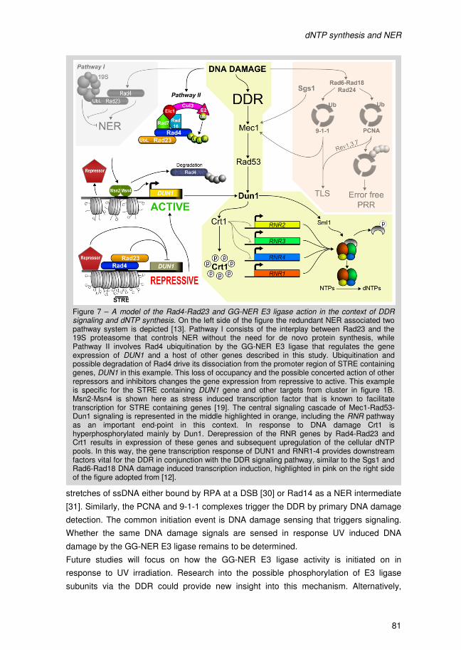
dNTP synthesis and NER
80
Upregulation of dNTPs occurs in response to UV radiation and during S-phase and is
important for both processes [25]. Based on the mechanism by which the GG-NER E3
ligase controls expression the RNR pathway genes, we predicted that cell cycle
progression might also be affected if this mechanism is disrupted, since it is well known
that proper regulation of the dNTP pools is necessary for normal transit across the cell
cycle. We found that this is indeed the case as we see that the psocs mutant progresses
through the cell cycle more quickly even in the absence of UV. As we anticipated this
particular phenotype can also be rescued by the constitutive upregulation of dNTP
synthesis by deleting SML1 and CRT1 (Figure 6B and C). This is independent
confirmation of the significance of the GG-NER E3 ligase in regulation the dNTP pools in
response to DNA damage.
Overall our data shows that Rad4-Rad23 represses a family of UV responsive genes in
the absence of UV irradiation by direct interaction with their promoter regions. In
response to UV Rad4 is ubiquitinated by the GG-NER E3 ligase and the Rad4-Rad23
complex dissociates from the promoter region. By leaving the promoter ubiquitinated
Rad4-Rad23 facilitates the UV induced upregulation of these genes. Amongst these are
the DUN1 and RNR pathway genes that control dNTP synthesis during replication and in
response to DNA damage. The Rad4-Rad23 and GG-NER E3 ligase controlled gene
expression results in UV responsive upregulation of DUN1 and the RNR genes that can
drive the increase of dNTP synthesis, which facilitates UV survival and normal
progression through the cell cycle. Our findings are consistent with the model shown in
Figure 7 in which Pathway II regulates the gene expression of DUN1, a key gene in the
control of the RNR pathway downstream of the central spine of Mec1-Rad53 signaling.
The NER mediated gene transcription response provides downstream factors
instrumental for the DDR by facilitating DUN1 and RNR1-4 expression in conjunction
with the DDR signaling pathway (see Figure 7).
Our results also demonstrate that the pathway described here is distinct from the Rad6-
Rad18 mediated control of gene expression described in a previous study [12]. The
PCNA and 9-1-1 complexes stimulate the DDR as a results of primary damage
detection. Interestingly, these DNA repair factors trigger the DDR via the action of the
Rad6-Rad18 E2-E3 ubiquitin ligase complex. This upregulates DNA damage induced
gene expression of specific genes including MAG1 and DDI1 [12]. We found that
disrupting the Rad6-Rad18 pathway does not affect the UV induced gene expression of
DUN1 or DDR2 (Figure S3B). In a similar vein, DDI1 expression which is controlled by
the Rad6-Rad18 pathway, is not affected in the psocsrad23∆ mutant. This clearly
indicates that the GG-NER E3 ligase pathway is distinct from the Rad6-Rad18 pathway
as illustrated in Figure 7.
The gene expression response regulated by the GG-NER E3 ligase described here,
provides a novel mechanism by which the nucleotide excision repair pathway integrates
with the DDR. Typically, the hallmark signal for cell-cycle checkpoint activation is
dNTP synthesis and NER
81
stretches of ssDNA either bound by RPA at a DSB [30] or Rad14 as a NER intermediate
[31]. Similarly, the PCNA and 9-1-1 complexes trigger the DDR by primary DNA damage
detection. The common initiation event is DNA damage sensing that triggers signaling.
Whether the same DNA damage signals are sensed in response UV induced DNA
damage by the GG-NER E3 ligase remains to be determined.
Future studies will focus on how the GG-NER E3 ligase activity is initiated on in
response to UV irradiation. Research into the possible phosphorylation of E3 ligase
subunits via the DDR could provide new insight into this mechanism. Alternatively,
Figure 7 – A model of the Rad4-Rad23 and GG-NER E3 ligase action in the context of DDR signaling and dNTP synthesis. On the left side of the figure the redundant NER associated two pathway system is depicted [13]. Pathway I consists of the interplay between Rad23 and the 19S proteasome that controls NER without the need for de novo protein synthesis, while Pathway II involves Rad4 ubiquitination by the GG-NER E3 ligase that regulates the gene expression of DUN1 and a host of other genes described in this study. Ubiquitination and possible degradation of Rad4 drive its dissociation from the promoter region of STRE containing genes, DUN1 in this example. This loss of occupancy and the possible concerted action of other repressors and inhibitors changes the gene expression from repressive to active. This example is specific for the STRE containing DUN1 gene and other targets from cluster in figure 1B. Msn2-Msn4 is shown here as stress induced transcription factor that is known to facilitate transcription for STRE containing genes [19]. The central signaling cascade of Mec1-Rad53-Dun1 signaling is represented in the middle highlighted in orange, including the RNR pathway as an important end-point in this context. In response to DNA damage Crt1 is hyperphosphorylated mainly by Dun1. Derepression of the RNR genes by Rad4-Rad23 and Crt1 results in expression of these genes and subsequent upregulation of the cellular dNTP pools. In this way, the gene transcription response of DUN1 and RNR1-4 provides downstream factors vital for the DDR in conjunction with the DDR signaling pathway, similar to the Sgs1 and Rad6-Rad18 DNA damage induced transcription induction, highlighted in pink on the right side of the figure adopted from [12].
dNTP synthesis and NER
82
association of the COP9/CSN signalosome to the GG-NER E3 ligase could be regulated
in an UV dependent fashion, with a possible role for DDR induced kinase activity
controlling the E3 ligase signalosome interaction. Interestingly, the E3 ligase activity of
the TC- and GG-NER specific ligases CSA-DDB1-CUL4A-ROC1 and DDB2-DDB1-
CUL4A-ROC1 can be repressed by interaction with the COP9/CSN signalosome [28,
29]. A similar scenario for the yeast GG-NER E3 ligase could be envisioned, but
mounting evidence is not present.
The novel Rad4-Rad23 based transcriptional response raises the question whether or
not the change in Rad4-Rad23 occupancy at the promoter region of the genes affected
is dependent of DNA damage detection. In other words is Rad4 dissociation driven by
ubiquitination alone or in combination with its affinity for UV induced DNA damage? How
this does novel pathway links in with the canonical function of Rad4-Rad23 in NER is
uncertain. It is still speculation at this stage to state that the Rad4-Rad23 controlled
genes expression can be truly independent of damage detection. Interestingly, dNTP
upregulation is required for survival in response to UV radiation [25], however whether
dNTP levels are required for NER or TLS remains unknown.
dNTP synthesis and NER
83
6. References 1. Kolodner, R.D., C.D. Putnam, and K. Myung, Maintenance of genome stability in
Saccharomyces cerevisiae. Science, 2002. 297(5581): p. 552-7.
2. Cleaver, J.E., E.T. Lam, and I. Revet, Disorders of nucleotide excision repair: the genetic
and molecular basis of heterogeneity. Nat Rev Genet, 2009. 10(11): p. 756-68.
3. Wijnhoven, S.W., et al., Tissue specific mutagenic and carcinogenic responses in NER
defective mouse models. Mutat Res, 2007. 614(1-2): p. 77-94.
4. Negrini, S., V.G. Gorgoulis, and T.D. Halazonetis, Genomic instability--an evolving
hallmark of cancer. Nat Rev Mol Cell Biol, 2010. 11(3): p. 220-8.
5. Branzei, D. and M. Foiani, Maintaining genome stability at the replication fork. Nat Rev
Mol Cell Biol, 2010. 11(3): p. 208-19.
6. Moynahan, M.E. and M. Jasin, Mitotic homologous recombination maintains genomic
stability and suppresses tumorigenesis. Nat Rev Mol Cell Biol, 2010. 11(3): p. 196-207.
7. Putnam, C.D., E.J. Jaehnig, and R.D. Kolodner, Perspectives on the DNA damage and
replication checkpoint responses in Saccharomyces cerevisiae. DNA Repair (Amst),
2009. 8(9): p. 974-82.
8. Navadgi-Patil, V.M. and P.M. Burgers, A tale of two tails: activation of DNA damage
checkpoint kinase Mec1/ATR by the 9-1-1 clamp and by Dpb11/TopBP1. DNA Repair
(Amst), 2009. 8(9): p. 996-1003.
9. Lazzaro, F., et al., Checkpoint mechanisms at the intersection between DNA damage
and repair. DNA Repair (Amst), 2009. 8(9): p. 1055-67.
10. Lee, K.Y. and K. Myung, PCNA modifications for regulation of post-replication repair
pathways. Mol Cells, 2008. 26(1): p. 5-11.
11. Moldovan, G.L., B. Pfander, and S. Jentsch, PCNA, the maestro of the replication fork.
Cell, 2007. 129(4): p. 665-79.
12. Fu, Y., et al., Rad6-Rad18 mediates a eukaryotic SOS response by ubiquitinating the 9-
1-1 checkpoint clamp. Cell, 2008. 133(4): p. 601-11.
13. Gillette, T.G., et al., Distinct functions of the ubiquitin-proteasome pathway influence
nucleotide excision repair. Embo Journal, 2006. 25(11): p. 2529-38.
14. Reed, S.H., Z. You, and E.C. Friedberg, The yeast RAD7 and RAD16 genes are
required for postincision events during nucleotide excision repair. In vitro and in vivo
studies with rad7 and rad16 mutants and purification of a Rad7/Rad16-containing protein
complex. J Biol Chem, 1998. 273(45): p. 29481-8.
15. Al-Moghrabi, N.M., I.S. Al-Sharif, and A. Aboussekhra, UV-induced de novo protein
synthesis enhances nucleotide excision repair efficiency in a transcription-dependent
manner in S. cerevisiae. DNA Repair, 2003. 2(11): p. 1185-1197.
16. Wade, S.L., et al., The Snf1 kinase and proteasome-associated Rad23 regulate UV-
responsive gene expression. Embo Journal, 2009. 28(19): p. 2919-31.
17. Le May, N., et al., NER factors are recruited to active promoters and facilitate chromatin
modification for transcription in the absence of exogenous genotoxic attack. Molecular
Cell, 2010. 38(1): p. 54-66.
18. Fong, Y.W., et al., A DNA repair complex functions as an Oct4/Sox2 coactivator in
embryonic stem cells. Cell, 2011. 147(1): p. 120-31.
dNTP synthesis and NER
82
association of the COP9/CSN signalosome to the GG-NER E3 ligase could be regulated
in an UV dependent fashion, with a possible role for DDR induced kinase activity
controlling the E3 ligase signalosome interaction. Interestingly, the E3 ligase activity of
the TC- and GG-NER specific ligases CSA-DDB1-CUL4A-ROC1 and DDB2-DDB1-
CUL4A-ROC1 can be repressed by interaction with the COP9/CSN signalosome [28,
29]. A similar scenario for the yeast GG-NER E3 ligase could be envisioned, but
mounting evidence is not present.
The novel Rad4-Rad23 based transcriptional response raises the question whether or
not the change in Rad4-Rad23 occupancy at the promoter region of the genes affected
is dependent of DNA damage detection. In other words is Rad4 dissociation driven by
ubiquitination alone or in combination with its affinity for UV induced DNA damage? How
this does novel pathway links in with the canonical function of Rad4-Rad23 in NER is
uncertain. It is still speculation at this stage to state that the Rad4-Rad23 controlled
genes expression can be truly independent of damage detection. Interestingly, dNTP
upregulation is required for survival in response to UV radiation [25], however whether
dNTP levels are required for NER or TLS remains unknown.
dNTP synthesis and NER
83
6. References 1. Kolodner, R.D., C.D. Putnam, and K. Myung, Maintenance of genome stability in
Saccharomyces cerevisiae. Science, 2002. 297(5581): p. 552-7.
2. Cleaver, J.E., E.T. Lam, and I. Revet, Disorders of nucleotide excision repair: the genetic
and molecular basis of heterogeneity. Nat Rev Genet, 2009. 10(11): p. 756-68.
3. Wijnhoven, S.W., et al., Tissue specific mutagenic and carcinogenic responses in NER
defective mouse models. Mutat Res, 2007. 614(1-2): p. 77-94.
4. Negrini, S., V.G. Gorgoulis, and T.D. Halazonetis, Genomic instability--an evolving
hallmark of cancer. Nat Rev Mol Cell Biol, 2010. 11(3): p. 220-8.
5. Branzei, D. and M. Foiani, Maintaining genome stability at the replication fork. Nat Rev
Mol Cell Biol, 2010. 11(3): p. 208-19.
6. Moynahan, M.E. and M. Jasin, Mitotic homologous recombination maintains genomic
stability and suppresses tumorigenesis. Nat Rev Mol Cell Biol, 2010. 11(3): p. 196-207.
7. Putnam, C.D., E.J. Jaehnig, and R.D. Kolodner, Perspectives on the DNA damage and
replication checkpoint responses in Saccharomyces cerevisiae. DNA Repair (Amst),
2009. 8(9): p. 974-82.
8. Navadgi-Patil, V.M. and P.M. Burgers, A tale of two tails: activation of DNA damage
checkpoint kinase Mec1/ATR by the 9-1-1 clamp and by Dpb11/TopBP1. DNA Repair
(Amst), 2009. 8(9): p. 996-1003.
9. Lazzaro, F., et al., Checkpoint mechanisms at the intersection between DNA damage
and repair. DNA Repair (Amst), 2009. 8(9): p. 1055-67.
10. Lee, K.Y. and K. Myung, PCNA modifications for regulation of post-replication repair
pathways. Mol Cells, 2008. 26(1): p. 5-11.
11. Moldovan, G.L., B. Pfander, and S. Jentsch, PCNA, the maestro of the replication fork.
Cell, 2007. 129(4): p. 665-79.
12. Fu, Y., et al., Rad6-Rad18 mediates a eukaryotic SOS response by ubiquitinating the 9-
1-1 checkpoint clamp. Cell, 2008. 133(4): p. 601-11.
13. Gillette, T.G., et al., Distinct functions of the ubiquitin-proteasome pathway influence
nucleotide excision repair. Embo Journal, 2006. 25(11): p. 2529-38.
14. Reed, S.H., Z. You, and E.C. Friedberg, The yeast RAD7 and RAD16 genes are
required for postincision events during nucleotide excision repair. In vitro and in vivo
studies with rad7 and rad16 mutants and purification of a Rad7/Rad16-containing protein
complex. J Biol Chem, 1998. 273(45): p. 29481-8.
15. Al-Moghrabi, N.M., I.S. Al-Sharif, and A. Aboussekhra, UV-induced de novo protein
synthesis enhances nucleotide excision repair efficiency in a transcription-dependent
manner in S. cerevisiae. DNA Repair, 2003. 2(11): p. 1185-1197.
16. Wade, S.L., et al., The Snf1 kinase and proteasome-associated Rad23 regulate UV-
responsive gene expression. Embo Journal, 2009. 28(19): p. 2919-31.
17. Le May, N., et al., NER factors are recruited to active promoters and facilitate chromatin
modification for transcription in the absence of exogenous genotoxic attack. Molecular
Cell, 2010. 38(1): p. 54-66.
18. Fong, Y.W., et al., A DNA repair complex functions as an Oct4/Sox2 coactivator in
embryonic stem cells. Cell, 2011. 147(1): p. 120-31.

dNTP synthesis and NER
84
19. Martinez-Pastor, M.T., et al., The Saccharomyces cerevisiae zinc finger proteins Msn2p
and Msn4p are required for transcriptional induction through the stress response element
(STRE). Embo Journal, 1996. 15(9): p. 2227-35.
20. Huang, M., Z. Zhou, and S.J. Elledge, The DNA replication and damage checkpoint
pathways induce transcription by inhibition of the Crt1 repressor. Cell, 1998. 94(5): p.
595-605.
21. Treger, J.M., T.R. Magee, and K. McEntee, Functional analysis of the stress response
element and its role in the multistress response of Saccharomyces cerevisiae. Biochem
Biophys Res Commun, 1998. 243(1): p. 13-9.
22. Ramsey, K.L., et al., The NEF4 complex regulates Rad4 levels and utilizes Snf2/Swi2-
related ATPase activity for nucleotide excision repair. Mol Cell Biol, 2004. 24(14): p.
6362-78.
23. Zhao, X., et al., The ribonucleotide reductase inhibitor Sml1 is a new target of the
Mec1/Rad53 kinase cascade during growth and in response to DNA damage. Embo
Journal, 2001. 20(13): p. 3544-53.
24. Andreson, B.L., et al., The ribonucleotide reductase inhibitor, Sml1, is sequentially
phosphorylated, ubiquitylated and degraded in response to DNA damage. Nucleic Acids
Res, 2010. 38(19): p. 6490-501.
25. Chabes, A., et al., Survival of DNA damage in yeast directly depends on increased dNTP
levels allowed by relaxed feedback inhibition of ribonucleotide reductase. Cell, 2003.
112(3): p. 391-401.
26. Koc, A. and G.F. Merrill, Checkpoint deficient rad53-11 yeast cannot accumulate dNTPs
in response to DNA damage. Biochem Biophys Res Commun, 2007. 353(2): p. 527-30.
27. Chabes, A. and B. Stillman, Constitutively high dNTP concentration inhibits cell cycle
progression and the DNA damage checkpoint in yeast Saccharomyces cerevisiae. Proc
Natl Acad Sci U S A, 2007. 104(4): p. 1183-8.
28. Russell, S.J., et al., The 19S regulatory complex of the proteasome functions
independently of proteolysis in nucleotide excision repair. Molecular Cell, 1999. 3(6): p.
687-95.
29. Gillette, T.G., et al., The 19S complex of the proteasome regulates nucleotide excision
repair in yeast. Genes Dev, 2001. 15(12): p. 1528-39.
30. Zou, L. and S.J. Elledge, Sensing DNA damage through ATRIP recognition of RPA-
ssDNA complexes. Science, 2003. 300(5625): p. 1542-8.
31. Giannattasio, M., et al., Physical and functional interactions between nucleotide excision
repair and DNA damage checkpoint. Embo Journal, 2004. 23(2): p. 429-38.
dNTP synthesis and NER
85
7. Supplementary Figures
Figure S 1 – (A) Venn diagram of the overlap in differential gene expression in the rad4/rad23 dataset indicating a 11 gene overlap between the rad23∆ and rad4∆rad23∆ mutants. (B) Venn diagram of the early and late UV response genes shown an overlap of 309 differentially expressed UV responsive genes. (C) Venn diagram of the overlap between the differentially expressed rad4/rad23 genes compared to the UV responsive dataset. (D) Overlap of the change in gene expression of the untreated psocs/rad23dataset
Figure S 2 – The GO term analysis of UV responsive genes differentially expressed in the psocs/rad23 dataset, highlighting an important role for the GG-NER E3 ligase in regulation of DNA metabolism, cell cycle and stress response pathways.
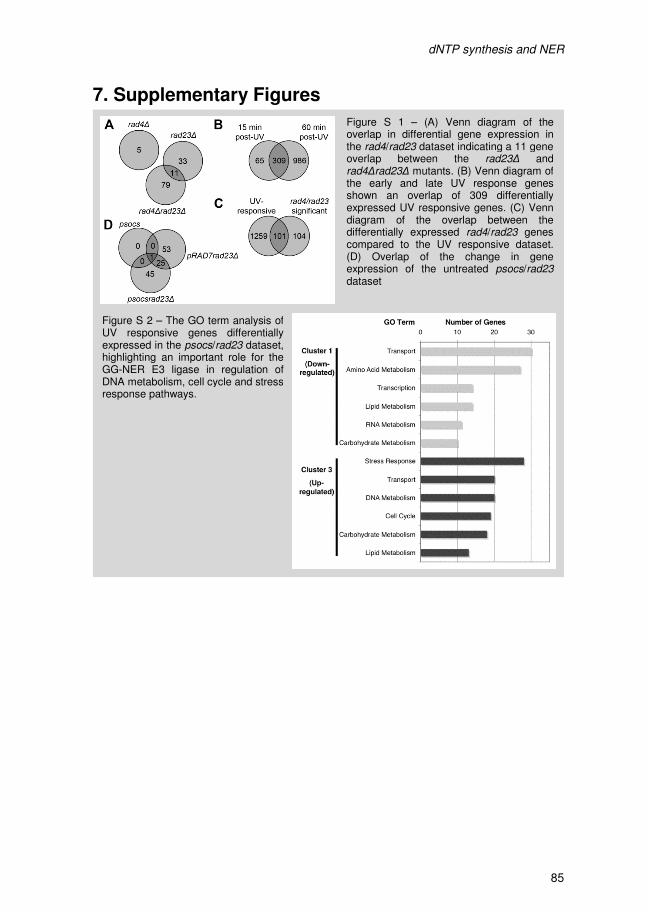
dNTP synthesis and NER
86
Figure S 3 – MAG1 and DDI1 gene expression is not controlled by the concerted action of Rad4-Rad23 and the GG-NER E3 ligase complex.(A) UV induced transcription of MAG1 and DDI1 is unaltered in the rad4/rad23 and psocs/rad23 datasets, shown in this heat-map. (B) Northern analysis confirming the microarray analysis shown in (A), These data display the intact expression of DDI1 in response to UV in psocs and rad23 mutants (first panel). Similarly, DDR2 and DUN1expression is unaffected by the deletion of RAD6 or RAD18 (second and third panel).
dNTP synthesis and NER
87
Figure S 4 – Cell-cycle progression is affected by the GG-NER E3 ligase mutation, independent of Rad23. (A) Flow cytometry analysis of the pRAD7 wild-type and psocs mutant in response to UV irradiation, showing altered cell cycle progression. (B) Cell-cycle distribution in the pRAD7rad23∆ and psocsrad23∆ backgrounds after UV irradiation was analyzed by flow cytometry.
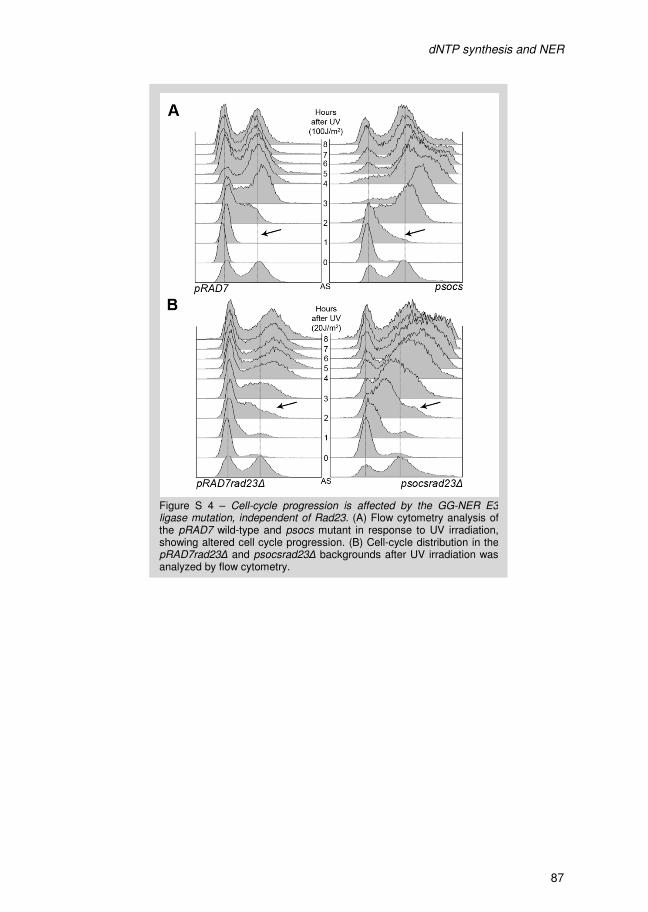
dNTP synthesis and NER
88
III The effect of Histone H1 on repair of rDNA in yeast
P. van Eijk, J. A. Brandsma, M. de Ruijter, J. Brouwer
dNTP synthesis and NER
88
III The effect of Histone H1 on repair of rDNA in yeast
P. van Eijk, J. A. Brandsma, M. de Ruijter, J. Brouwer

Histone H1 and rDNA repair
90
1. Abstract In Saccharomyces cerevisiae Rad34 is essential for Transcription Coupled Nucleotide
Excision Repair (TC-NER) of the transcribed strand of active rDNA [1, 2]. Rad34 is
homologous to the core repair factor Rad4, which is required for all other modes of NER.
Histone H1 is a chromatin factor in yeast, located in the rDNA region. Here we show that
deletion of the HHO1 gene rescues the requirement for Rad34 in TC-NER of rDNA, an
effect shown to be Rad4-dependent. The rescue of the rad34∆ repair defect by HHO1
deletion is not due to increased Global Genome NER (GG-NER). Moreover, the deletion
of HHO1 from wild-type cells does not affect NER. The presence of Histone H1 at the
rDNA locus directly or indirectly restricts NER to the use of Rad34 in repair while
excluding Rad4.
Histone H1 and rDNA repair
91
2. Introduction Nucleotide Excision Repair (NER) removes a wide variety of helix distorting lesions from
the genome [3]. The repair reaction is highly conserved mechanistically and the in vivo
importance of this mode of repair is evident from the UV sensitivity of NER deficient
cells. Moreover, humans and animal models deficient for NER factors display
predisposition to cancer [4]. NER deficiency not only results in UV-induced skin cancers
but also results in spontaneous tumorigenesis in other organ tissue [4].
Generally, in NER there are two different sub-pathways; Transcription Coupled NER
(TC-NER) and Global Genome NER (GG-NER). The Rad7-Rad16 GG-NER complex in
yeast is capable of damage recognition throughout the entire genome. TC-NER, on the
other hand, is initiated when, at an active gene, RNA polymerase II is blocked at a UV
lesion. TC-NER specifically requires Rad26 in concert with the core NER factors to repair
DNA damage.
Repair of the transcribed strand (TS) of an active gene is the sum of both GG-NER and
TC-NER while the non-transcribed strand (NTS) is repaired by GG-NER. This explains
why in general a strand-bias can be observed in active genes, whereby the TS is
repaired faster. Conversely, the repair rate of both DNA strands is similar for inactive
genes or in a genetic background without active TC-NER.
Rad4 is an essential NER factor in yeast, required for the early steps of the NER reaction
in both TC-NER and GG-NER of all RNA polymerase II transcribed genes. However, the
TS of active rDNA is still repaired in the absence of Rad4 [5] through the action of Rad34
in yeast [1]. Rad34 shares homology with all Rad4-like proteins [6] and has been shown
to interact with both of the known interaction partners of Rad4, Rad23 [7] and Rad33 [1].
When RAD34 is deleted from wild-type cells TC-NER at the rDNA locus is disrupted (i.e.
no strand bias present). Thus, if RAD34 is knocked out in a rad4∆ background, repair of
rDNA is completely abolished [1, 2]. These data establish that Rad34 is a genuine Rad4
homologue in yeast, uniquely required for TC-NER at the rDNA locus.
Yeast rDNA exists as a tandem, head-to-tail arrangement of roughly 150 copies of the
35S rRNA gene. This cassette of over 9kb encodes the 18S, 5.8S and 25S rRNAs that
are transcribed concurrently by RNA Polymerase I to form the pre-rRNA transcript.
Roughly 50% of all copies are actively transcribed while the other half is in an inactive
heterochromatic state [8]. Hho1p is a linker histone and thus copurifies with core
histones but is non-essential in yeast [9, 10]. The Histone H1 protein has been shown to
associate with and function at the rDNA locus [9, 11, 12]. Several functions for Hho1 at
the rDNA locus have been proposed: exclusion of RNA Pol II transcription [12],
repression of recombination [11, 12], DNA compaction and regulation of RNA Pol I
processivity [13]. However, hho1∆ mutants in yeast do not have a distinct phenotype [10]
and gene regulation at large, is not affected [14]. Here we show that histone H1 is
indirectly involved in NER by affecting the damage recognition factor used at the rDNA
locus.
Histone H1 and rDNA repair
90
1. Abstract In Saccharomyces cerevisiae Rad34 is essential for Transcription Coupled Nucleotide
Excision Repair (TC-NER) of the transcribed strand of active rDNA [1, 2]. Rad34 is
homologous to the core repair factor Rad4, which is required for all other modes of NER.
Histone H1 is a chromatin factor in yeast, located in the rDNA region. Here we show that
deletion of the HHO1 gene rescues the requirement for Rad34 in TC-NER of rDNA, an
effect shown to be Rad4-dependent. The rescue of the rad34∆ repair defect by HHO1
deletion is not due to increased Global Genome NER (GG-NER). Moreover, the deletion
of HHO1 from wild-type cells does not affect NER. The presence of Histone H1 at the
rDNA locus directly or indirectly restricts NER to the use of Rad34 in repair while
excluding Rad4.
Histone H1 and rDNA repair
91
2. Introduction Nucleotide Excision Repair (NER) removes a wide variety of helix distorting lesions from
the genome [3]. The repair reaction is highly conserved mechanistically and the in vivo
importance of this mode of repair is evident from the UV sensitivity of NER deficient
cells. Moreover, humans and animal models deficient for NER factors display
predisposition to cancer [4]. NER deficiency not only results in UV-induced skin cancers
but also results in spontaneous tumorigenesis in other organ tissue [4].
Generally, in NER there are two different sub-pathways; Transcription Coupled NER
(TC-NER) and Global Genome NER (GG-NER). The Rad7-Rad16 GG-NER complex in
yeast is capable of damage recognition throughout the entire genome. TC-NER, on the
other hand, is initiated when, at an active gene, RNA polymerase II is blocked at a UV
lesion. TC-NER specifically requires Rad26 in concert with the core NER factors to repair
DNA damage.
Repair of the transcribed strand (TS) of an active gene is the sum of both GG-NER and
TC-NER while the non-transcribed strand (NTS) is repaired by GG-NER. This explains
why in general a strand-bias can be observed in active genes, whereby the TS is
repaired faster. Conversely, the repair rate of both DNA strands is similar for inactive
genes or in a genetic background without active TC-NER.
Rad4 is an essential NER factor in yeast, required for the early steps of the NER reaction
in both TC-NER and GG-NER of all RNA polymerase II transcribed genes. However, the
TS of active rDNA is still repaired in the absence of Rad4 [5] through the action of Rad34
in yeast [1]. Rad34 shares homology with all Rad4-like proteins [6] and has been shown
to interact with both of the known interaction partners of Rad4, Rad23 [7] and Rad33 [1].
When RAD34 is deleted from wild-type cells TC-NER at the rDNA locus is disrupted (i.e.
no strand bias present). Thus, if RAD34 is knocked out in a rad4∆ background, repair of
rDNA is completely abolished [1, 2]. These data establish that Rad34 is a genuine Rad4
homologue in yeast, uniquely required for TC-NER at the rDNA locus.
Yeast rDNA exists as a tandem, head-to-tail arrangement of roughly 150 copies of the
35S rRNA gene. This cassette of over 9kb encodes the 18S, 5.8S and 25S rRNAs that
are transcribed concurrently by RNA Polymerase I to form the pre-rRNA transcript.
Roughly 50% of all copies are actively transcribed while the other half is in an inactive
heterochromatic state [8]. Hho1p is a linker histone and thus copurifies with core
histones but is non-essential in yeast [9, 10]. The Histone H1 protein has been shown to
associate with and function at the rDNA locus [9, 11, 12]. Several functions for Hho1 at
the rDNA locus have been proposed: exclusion of RNA Pol II transcription [12],
repression of recombination [11, 12], DNA compaction and regulation of RNA Pol I
processivity [13]. However, hho1∆ mutants in yeast do not have a distinct phenotype [10]
and gene regulation at large, is not affected [14]. Here we show that histone H1 is
indirectly involved in NER by affecting the damage recognition factor used at the rDNA
locus.
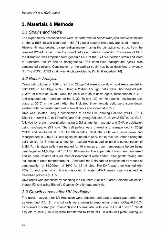
Histone H1 and rDNA repair
92
3. Materials & Methods
3.1 Strains and Media
The experiments described here were all performed in Saccharomyces cerevisiae based
on the W1588-4a wild-type strain [15]. All strains used in this study are listed in table 1.
Histone H1 was deleted by gene-displacement using the disruption construct from the
relevant BY4741 strain from the EuroScarf yeast deletion collection. By means of PCR
the disruption was amplified from genomic DNA of the BY4741 deletion strain and used
to transform the W1588-4a backgrounds. The short-lived background sgs1∆ was
constructed similarly. Construction of the rad34∆ strain has been described previously
[1]. The RDN1::ADE2 strain was kindly provided by Dr. M. Kaeberlein [16].
3.2 Repair Analysis
Yeast cell cultures of 200mL YPD of OD600=0.4 were spun down and resuspended in
cold PBS to an OD600 of 0.7. Using a 254nm UV light cells were UV-irradiated with
70J/m2 at a rate of 3W/m
2. Next, the cells were spun down again, resuspended in YPD
and aliquoted into 4 portions for the 0, 30, 60 and 120 min time points. Incubation took
place at 30°C in the dark. After the indicated time-intervals cells were spun down,
washed with cold water and split in two aliquots and stored at -80°C.
DNA was isolated using a combination of Yeast Cell Piercing Solution (YCPS, 0.1%
SB3-14, 100mM LiCl in TE buffer) and Cell Lysing Solution (CLS, 2mM EDTA, 2% SDS)
followed by protein precipitation using 2.5M ammonium acetate and DNA precipitation
using isopropanol (3:1 v/v). The cell pellets were thawed and resuspended in 200µl
YCPS and incubated at 65°C for 30 minutes. Next, the cells were spun down and
resuspended in 200µl CLS and again incubated at 65°C for 45 minutes. After placing the
cells on ice for 5 minutes ammonium acetate was added to an end-concentration of
2.5M. At this stage cells were rotated for 10 minutes at room temperature before being
centrifuged at 14,000rpm at 16°C for 10 minutes. The supernatant was then transferred
and an equal volume of 3 volumes of isopropanol were added. After gentle mixing and
incubation at room temperature for 10 minutes the DNA can be precipitated by means of
centrifugation at 14,000rpm at 16°C for 12 minutes. The DNA was then washed with
70% Ethanol after which it was dissolved in water. rDNA repair was measured as
described previously [1, 5].
DNA repair was quantified by scanning the Southern Blot in a Biorad Personal Molecular
Imager FX and using Biorad’s Quantity One for data analysis.
3.3 Growth curves after UV irradiation
The growth curves after UV irradiation were obtained and data analysis was performed
as described [17, 18]. In short cells were grown to exponential phase (OD600~0.5-0.7),
transferred to water (8x10⁶cells/ml) and UV irradiated with 254nm UV at 150Jm
-2. Small
aliquots of cells (~40.000) were transferred to fresh YPD in a 96-well plate. During 48
Histone H1 and rDNA repair
93
hours the AU595 was measured every 10 minutes with intermittent shaking at 30°C on a
Tecan GENios Microplate Reader.
3.4 Replicative aging
Replicative aging was measured as described [19]. In short cells for study were
restreaked on YPG plates containing glycerol as the only carbon-source to eliminate any
petits. From the YPG plate a single colony was restreaked on a thin YPD plate for use in
the micromanipulator. Using a Sanger Instruments Micromanipulator ~40 dividing cells
were picked from and sorted on a YPD plate. After the first division each mother was
discarded while the virgin daughter was kept for aging analysis. After incubation at 30°C
each 1.5hrs the daughter cell was removed until the mother cell stopped dividing. Plates
were incubated at 4°C overnight to slow down cell division. Cells will typically only
perform a single division overnight if kept at 4°C.
3.5 rDNA Recombination assay
The strain containing an ADE2 insert in its rDNA (kindly provided by Dr. M. Kaeberlein)
was used to assess the recombination rate in strains deleted for RAD34. Cells were
grown overnight and diluted in water to accommodate about 1x10⁴-2x10
4 cells/mL. This
suspension was used to plate 500-1000 cells on YPD plates. After growth at 30°C the
plates were stored at 4°C until red sector formation could be detected. If in the early
rounds of division recombination and thus loss of the ADE2 insert occurs sectoring of the
resulting colony is an indication of this event. By counting pink sector formation one can
get a measure for the frequency of recombination. For each strain 2,500 to 5,000
colonies were counted.
4. Results & Discussion
4.1 Analysis of rDNA repair in Histone H1 mutants
In yeast, Rad4-independent repair of the transcribed strand of rDNA depends on the
Rad4-like protein Rad34 [1, 2]. Because Histone H1 is involved in a variety of processes
at the rDNA locus [11-13], we deleted HHO1 to elucidate why rDNA requires Rad34
instead of Rad4. We analyzed this by measuring rDNA repair using a strand-specific
repair assay on genomic DNA in several NER deletion backgrounds. In the absence of
Rad34, NER at the rDNA locus of both the TS and NTS is performed by GG-NER
(Figure 1) [1, 5] and deleting HHO1 in a rad34∆ strain does not change the repair rate of
the NTS (Figure 1). However, repair of the TS in rad34∆hho1∆ cells is increased
compared to that of a rad34∆ single mutant (Figure 1). These repair data demonstrate
that hho1∆ is able to rescue the TS repair defect of a rad34∆ mutant.
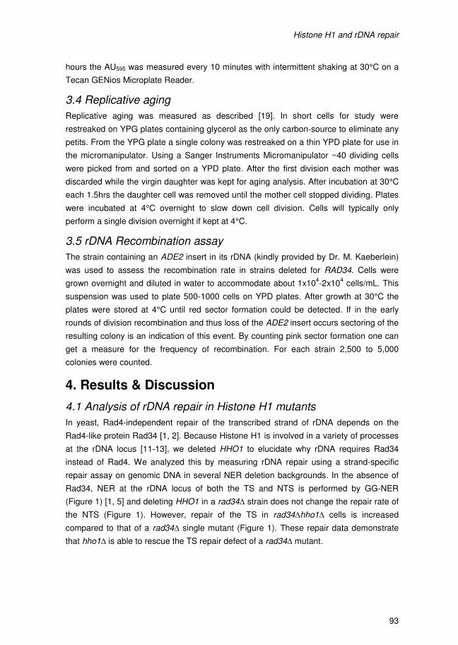
Histone H1 and rDNA repair
94
We next set out to determine what NER factors now contribute to repair in the
rad34∆hho1∆ double mutant. First, we determined whether the Rad4 protein, that is
homologous to Rad34, can replace Rad34 during TS repair in rad34∆hho1∆ cells. This
was tested by measuring repair in the triple mutant rad4∆rad34∆hho1∆ and comparing
that to the repair rate of rad34∆hho1∆ cells (Figure 2). In the absence of Rad4, Rad34
and Histone H1 no NER activity of either strand can be detected and thus the rescue
effect of the HHO1 deletion is lost. These data show that the TS repair in rad34∆hho1∆
cells is Rad4-dependent (Figure 2). Rad4 is now required for TS repair of rDNA but only
if, in the absence of Rad34, the HHO1 gene is also deleted.
The chromatin structure of the rDNA locus becomes decompacted in the absence of the
Histone H1 protein [13]. This could allow repair factors to better access the inactive
rDNA. In our experiments we make use of whole genomic DNA and this therefore
includes both the active and inactive rDNA copies. In case of rad34∆hho1∆ cells a more
open chromatin structure due to the absence of Histone H1 could lead to an increase in
repair of the TS of non-transcribed rDNA copies by GG-NER. In order to test this
hypothesis we eliminated GG-NER by deleting RAD16 in a rad34∆ and rad34∆hho1∆
background and measured their respective repair rates. If we then compare TS repair of
rad34∆rad16∆ cells to that of the rad34∆rad16∆hho1∆ triple mutant we again find the
increase in TS repair in the triple mutant rad34∆rad16∆hho1∆ (Figure 3A). This indicates
that increased repair in rad34∆hho1∆ cells is not due to GG-NER. Interestingly, a mode
of GG-NER that is Rad7- and Rad16-independent exists [1, 5] that could also benefit
from a more open chromatin structure in HHO1 deletion cells. However, the contribution
Figure 1 – Deleting Histone H1 rescues the TC-NER deficient rad34∆ phenotype. Strand-specific rDNA repair assay of rad34∆ and rad34∆hho1∆ cells. Damage removal is shown in representative Southernblots probed for (A - top panel) the transcribed strand (TS) and (B - top panel) the non-transcribed strand (NTS). Data was averaged from 3 individual experiments and quantified according the Poisson distribution of random DNA damage in a linear fragment shown in (A - lower panel) for the transcribed strand (TS) and in (B - lower panel) the non-transcribed strand (NTS). The time in minutes indicates the recovery time after UV-irradiation. Treatment with the damage-specific endonuclease T4endoV or mock treatment is indicated on the Southernblot images with + and - respectively.
Histone H1 and rDNA repair
95
of this unknown repair mechanism occurs at late 2 to 4 hour times points, whereas the
rescue effect of the HHO1 deletion in rad34∆ and rad16∆rad34∆ cells takes place during
the first 2 hours after UV irradiation, thus making it unlikely that the Rad7- and Rad16
independent repair pathway results in the TS repair we measure.
If Histone H1 merely decompacts rDNA resulting in more active rDNA copies or more
efficient overall repair, we expect that the deletion of HHO1 in the presence of Rad4 and
Rad34 would also affect NER. This was tested by measuring strand-specific repair in a
single hho1∆ deletion background (Figure 4). Deleting HHO1 by itself has no effect on
rDNA repair, showing that the absence of Histone H1 does not result in increased
accessibility of rDNA for NER factors.
In summary, the data presented here show that Rad4 can contribute to TS repair in the
absence of Rad34 only when Histone H1 is absent. The active role of Histone H1 that
Figure 3 – The rescue effect of deleting Histone H1 is independent of Rad16. Strand-specific rDNA repair assay of rad34∆rad16∆ and rad34∆rad16∆hho1∆ cells as described in Figure 1.
Figure 2 – The rescue effect of Histone H1 deletion depends on Rad4. Strand-specific rDNA repair assay of rad34∆hho1∆ and rad4∆rad34∆hho1∆ cells as described in Figure 1.
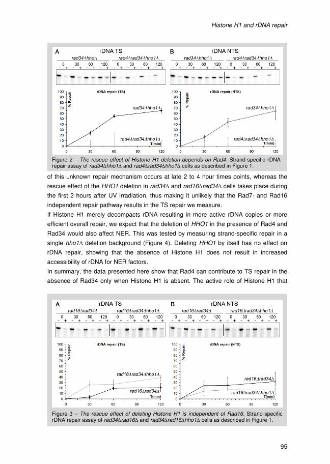
Histone H1 and rDNA repair
94
We next set out to determine what NER factors now contribute to repair in the
rad34∆hho1∆ double mutant. First, we determined whether the Rad4 protein, that is
homologous to Rad34, can replace Rad34 during TS repair in rad34∆hho1∆ cells. This
was tested by measuring repair in the triple mutant rad4∆rad34∆hho1∆ and comparing
that to the repair rate of rad34∆hho1∆ cells (Figure 2). In the absence of Rad4, Rad34
and Histone H1 no NER activity of either strand can be detected and thus the rescue
effect of the HHO1 deletion is lost. These data show that the TS repair in rad34∆hho1∆
cells is Rad4-dependent (Figure 2). Rad4 is now required for TS repair of rDNA but only
if, in the absence of Rad34, the HHO1 gene is also deleted.
The chromatin structure of the rDNA locus becomes decompacted in the absence of the
Histone H1 protein [13]. This could allow repair factors to better access the inactive
rDNA. In our experiments we make use of whole genomic DNA and this therefore
includes both the active and inactive rDNA copies. In case of rad34∆hho1∆ cells a more
open chromatin structure due to the absence of Histone H1 could lead to an increase in
repair of the TS of non-transcribed rDNA copies by GG-NER. In order to test this
hypothesis we eliminated GG-NER by deleting RAD16 in a rad34∆ and rad34∆hho1∆
background and measured their respective repair rates. If we then compare TS repair of
rad34∆rad16∆ cells to that of the rad34∆rad16∆hho1∆ triple mutant we again find the
increase in TS repair in the triple mutant rad34∆rad16∆hho1∆ (Figure 3A). This indicates
that increased repair in rad34∆hho1∆ cells is not due to GG-NER. Interestingly, a mode
of GG-NER that is Rad7- and Rad16-independent exists [1, 5] that could also benefit
from a more open chromatin structure in HHO1 deletion cells. However, the contribution
Figure 1 – Deleting Histone H1 rescues the TC-NER deficient rad34∆ phenotype. Strand-specific rDNA repair assay of rad34∆ and rad34∆hho1∆ cells. Damage removal is shown in representative Southernblots probed for (A - top panel) the transcribed strand (TS) and (B - top panel) the non-transcribed strand (NTS). Data was averaged from 3 individual experiments and quantified according the Poisson distribution of random DNA damage in a linear fragment shown in (A - lower panel) for the transcribed strand (TS) and in (B - lower panel) the non-transcribed strand (NTS). The time in minutes indicates the recovery time after UV-irradiation. Treatment with the damage-specific endonuclease T4endoV or mock treatment is indicated on the Southernblot images with + and - respectively.
Histone H1 and rDNA repair
95
of this unknown repair mechanism occurs at late 2 to 4 hour times points, whereas the
rescue effect of the HHO1 deletion in rad34∆ and rad16∆rad34∆ cells takes place during
the first 2 hours after UV irradiation, thus making it unlikely that the Rad7- and Rad16
independent repair pathway results in the TS repair we measure.
If Histone H1 merely decompacts rDNA resulting in more active rDNA copies or more
efficient overall repair, we expect that the deletion of HHO1 in the presence of Rad4 and
Rad34 would also affect NER. This was tested by measuring strand-specific repair in a
single hho1∆ deletion background (Figure 4). Deleting HHO1 by itself has no effect on
rDNA repair, showing that the absence of Histone H1 does not result in increased
accessibility of rDNA for NER factors.
In summary, the data presented here show that Rad4 can contribute to TS repair in the
absence of Rad34 only when Histone H1 is absent. The active role of Histone H1 that
Figure 3 – The rescue effect of deleting Histone H1 is independent of Rad16. Strand-specific rDNA repair assay of rad34∆rad16∆ and rad34∆rad16∆hho1∆ cells as described in Figure 1.
Figure 2 – The rescue effect of Histone H1 deletion depends on Rad4. Strand-specific rDNA repair assay of rad34∆hho1∆ and rad4∆rad34∆hho1∆ cells as described in Figure 1.
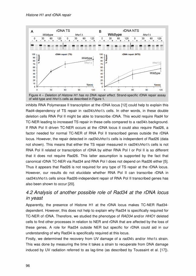
Histone H1 and rDNA repair
96
inhibits RNA Polymerase II transcription at the rDNA locus [12] could help to explain this
Rad4-dependency of TS repair in rad34∆hho1∆ cells. In other words, in these double
deletion cells RNA Pol II might be able to transcribe rDNA. This would require Rad4 for
TC-NER leading to increased TS repair in these cells compared to a rad34∆ background.
If RNA Pol II driven TC-NER occurs at the rDNA locus it could also require Rad26, a
factor needed for normal TC-NER of RNA Pol II transcribed genes outside the rDNA
locus. However, the repair detected in rad34∆hho1∆ cells is independent of Rad26 (data
not shown). This means that either the TS repair measured in rad34∆hho1∆ cells is not
RNA Pol II related or transcription of rDNA by either RNA Pol I or Pol II is so different
that it does not require Rad26. This latter assumption is supported by the fact that
canonical rDNA TC-NER via Rad34 and RNA Pol I does not depend on Rad26 either [5].
Thus it appears that Rad26 is not required for any type of TS repair at the rDNA locus.
However, our results do not elucidate whether RNA Pol II can transcribe rDNA in
rad34∆hho1∆ cells since Rad26-independent repair of RNA Pol II transcribed genes has
also been shown to occur [20].
4.2 Analysis of another possible role of Rad34 at the rDNA locus in yeast
Apparently, the presence of Histone H1 at the rDNA locus makes TC-NER Rad34-
dependent. However, this does not help to explain why Rad34 is specifically required for
TC-NER of rDNA. Therefore, we studied the phenotype of RAD34 and/or HHO1 deleted
cells to find other processes in relation to NER and rDNA that are affected by the loss of
these genes. A role for Rad34 outside NER but specific for rDNA could aid in our
understanding of why Rad34 is specifically required at this locus.
Firstly, we determined the recovery from UV damage of a rad34∆ and/or hho1∆ strain.
This was done by measuring the time it takes a strain to recuperate from DNA damage
induced by UV radiation referred to as lag-time (as described by Toussaint et al. [17]).
Figure 4 – Deletion of Histone H1 has no DNA repair effect. Strand-specific rDNA repair assay of wild-type and hho1∆ cells as described in Figure 1.
Histone H1 and rDNA repair
97
Strains deleted for
RAD34, HHO1 or
both show no sig-
nificant UV pheno-
type, indicating
that the repair
rates of rDNA in
these back-
grounds do not
hamper overall UV
survival.
The rDNA locus is
closely linked to
aging in yeast and
provided the first molecular mechanism to explain aging in a eukaryote [21]. This
prompted the idea that Rad34-dependent TC-NER or a separate function of the Rad34
protein could contribute to longevity. Therefore, we tested the replicative aging
phenotype of a rad34∆ cells compared to wild-type cells and the known short-lived sgs1∆
mutant by means of micromanipulation [19]. As shown in figure 6 the mortality rate of
rad34∆ cells overlaps with two independent wild-type curves while the short-lived sgs1∆
cells age much faster as reported previously [22]. Hence the presence of Rad34 or
Rad34-dependent TC-NER at the rDNA locus is not a determinant for replicative aging in
yeast, thus not explaining the use of Rad34 in the context of rDNA and aging.
The repetitive nature of the rDNA locus makes it a prime target for recombination and it
has been shown that the uncontrolled recombination leads to loss of rDNA copies that in-
duces aging [23]. Cells therefore carefully maintain the rDNA copy-number by preventing
recombination at
the rDNA locus.
Again the
presence of a
specific rDNA TC-
NER factor could
be warranted by
an important con-
tribution of Rad34
or Rad34-depend-
ent TC-NER to
prevent recom-
bination. To test
the effect of
Figure 5 – UV-phenotypes of RAD34 and HHO1 strains. Growth curves were obtained with or without UV treatment. Quantification of the growth curve data as described in [17] resulted in the doubling time, lag time and recovery time for all the strains tested displayed here as an average of at least three individual experiments.
Figure 6 – RAD34 deletion cells have wild-type mortality rates. Mortality curves of wild-type, rad34∆ and sgs1∆ cells. The data are an average of 40 cells monitored over time. The survival is a measure for the amount of cells that continued dividing after a certain number of divisions.
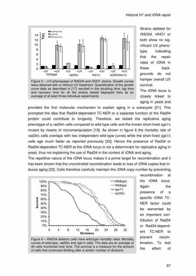
Histone H1 and rDNA repair
96
inhibits RNA Polymerase II transcription at the rDNA locus [12] could help to explain this
Rad4-dependency of TS repair in rad34∆hho1∆ cells. In other words, in these double
deletion cells RNA Pol II might be able to transcribe rDNA. This would require Rad4 for
TC-NER leading to increased TS repair in these cells compared to a rad34∆ background.
If RNA Pol II driven TC-NER occurs at the rDNA locus it could also require Rad26, a
factor needed for normal TC-NER of RNA Pol II transcribed genes outside the rDNA
locus. However, the repair detected in rad34∆hho1∆ cells is independent of Rad26 (data
not shown). This means that either the TS repair measured in rad34∆hho1∆ cells is not
RNA Pol II related or transcription of rDNA by either RNA Pol I or Pol II is so different
that it does not require Rad26. This latter assumption is supported by the fact that
canonical rDNA TC-NER via Rad34 and RNA Pol I does not depend on Rad26 either [5].
Thus it appears that Rad26 is not required for any type of TS repair at the rDNA locus.
However, our results do not elucidate whether RNA Pol II can transcribe rDNA in
rad34∆hho1∆ cells since Rad26-independent repair of RNA Pol II transcribed genes has
also been shown to occur [20].
4.2 Analysis of another possible role of Rad34 at the rDNA locus in yeast
Apparently, the presence of Histone H1 at the rDNA locus makes TC-NER Rad34-
dependent. However, this does not help to explain why Rad34 is specifically required for
TC-NER of rDNA. Therefore, we studied the phenotype of RAD34 and/or HHO1 deleted
cells to find other processes in relation to NER and rDNA that are affected by the loss of
these genes. A role for Rad34 outside NER but specific for rDNA could aid in our
understanding of why Rad34 is specifically required at this locus.
Firstly, we determined the recovery from UV damage of a rad34∆ and/or hho1∆ strain.
This was done by measuring the time it takes a strain to recuperate from DNA damage
induced by UV radiation referred to as lag-time (as described by Toussaint et al. [17]).
Figure 4 – Deletion of Histone H1 has no DNA repair effect. Strand-specific rDNA repair assay of wild-type and hho1∆ cells as described in Figure 1.
Histone H1 and rDNA repair
97
Strains deleted for
RAD34, HHO1 or
both show no sig-
nificant UV pheno-
type, indicating
that the repair
rates of rDNA in
these back-
grounds do not
hamper overall UV
survival.
The rDNA locus is
closely linked to
aging in yeast and
provided the first molecular mechanism to explain aging in a eukaryote [21]. This
prompted the idea that Rad34-dependent TC-NER or a separate function of the Rad34
protein could contribute to longevity. Therefore, we tested the replicative aging
phenotype of a rad34∆ cells compared to wild-type cells and the known short-lived sgs1∆
mutant by means of micromanipulation [19]. As shown in figure 6 the mortality rate of
rad34∆ cells overlaps with two independent wild-type curves while the short-lived sgs1∆
cells age much faster as reported previously [22]. Hence the presence of Rad34 or
Rad34-dependent TC-NER at the rDNA locus is not a determinant for replicative aging in
yeast, thus not explaining the use of Rad34 in the context of rDNA and aging.
The repetitive nature of the rDNA locus makes it a prime target for recombination and it
has been shown that the uncontrolled recombination leads to loss of rDNA copies that in-
duces aging [23]. Cells therefore carefully maintain the rDNA copy-number by preventing
recombination at
the rDNA locus.
Again the
presence of a
specific rDNA TC-
NER factor could
be warranted by
an important con-
tribution of Rad34
or Rad34-depend-
ent TC-NER to
prevent recom-
bination. To test
the effect of
Figure 5 – UV-phenotypes of RAD34 and HHO1 strains. Growth curves were obtained with or without UV treatment. Quantification of the growth curve data as described in [17] resulted in the doubling time, lag time and recovery time for all the strains tested displayed here as an average of at least three individual experiments.
Figure 6 – RAD34 deletion cells have wild-type mortality rates. Mortality curves of wild-type, rad34∆ and sgs1∆ cells. The data are an average of 40 cells monitored over time. The survival is a measure for the amount of cells that continued dividing after a certain number of divisions.
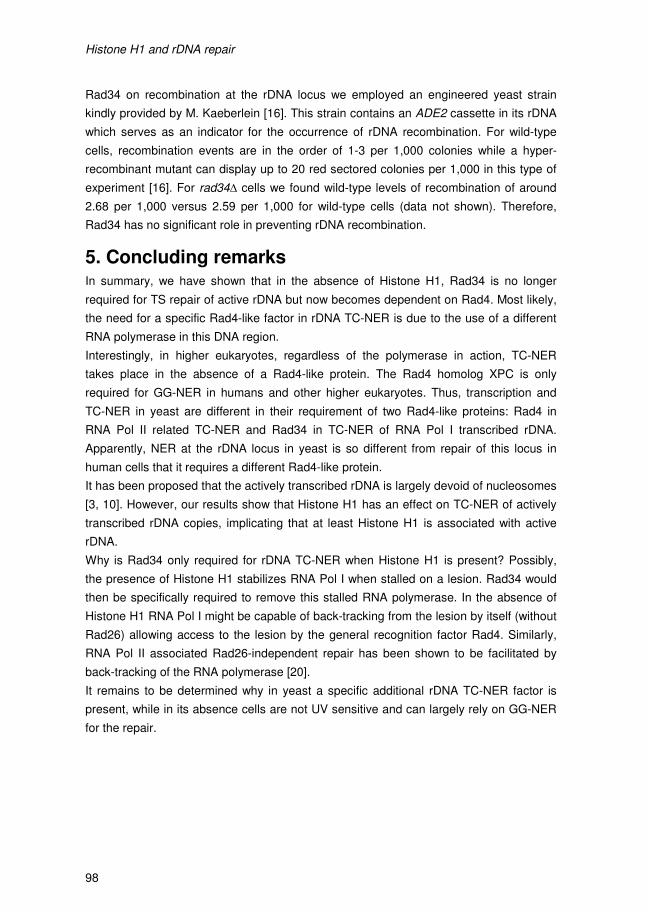
Histone H1 and rDNA repair
98
Rad34 on recombination at the rDNA locus we employed an engineered yeast strain
kindly provided by M. Kaeberlein [16]. This strain contains an ADE2 cassette in its rDNA
which serves as an indicator for the occurrence of rDNA recombination. For wild-type
cells, recombination events are in the order of 1-3 per 1,000 colonies while a hyper-
recombinant mutant can display up to 20 red sectored colonies per 1,000 in this type of
experiment [16]. For rad34∆ cells we found wild-type levels of recombination of around
2.68 per 1,000 versus 2.59 per 1,000 for wild-type cells (data not shown). Therefore,
Rad34 has no significant role in preventing rDNA recombination.
5. Concluding remarks In summary, we have shown that in the absence of Histone H1, Rad34 is no longer
required for TS repair of active rDNA but now becomes dependent on Rad4. Most likely,
the need for a specific Rad4-like factor in rDNA TC-NER is due to the use of a different
RNA polymerase in this DNA region.
Interestingly, in higher eukaryotes, regardless of the polymerase in action, TC-NER
takes place in the absence of a Rad4-like protein. The Rad4 homolog XPC is only
required for GG-NER in humans and other higher eukaryotes. Thus, transcription and
TC-NER in yeast are different in their requirement of two Rad4-like proteins: Rad4 in
RNA Pol II related TC-NER and Rad34 in TC-NER of RNA Pol I transcribed rDNA.
Apparently, NER at the rDNA locus in yeast is so different from repair of this locus in
human cells that it requires a different Rad4-like protein.
It has been proposed that the actively transcribed rDNA is largely devoid of nucleosomes
[3, 10]. However, our results show that Histone H1 has an effect on TC-NER of actively
transcribed rDNA copies, implicating that at least Histone H1 is associated with active
rDNA.
Why is Rad34 only required for rDNA TC-NER when Histone H1 is present? Possibly,
the presence of Histone H1 stabilizes RNA Pol I when stalled on a lesion. Rad34 would
then be specifically required to remove this stalled RNA polymerase. In the absence of
Histone H1 RNA Pol I might be capable of back-tracking from the lesion by itself (without
Rad26) allowing access to the lesion by the general recognition factor Rad4. Similarly,
RNA Pol II associated Rad26-independent repair has been shown to be facilitated by
back-tracking of the RNA polymerase [20].
It remains to be determined why in yeast a specific additional rDNA TC-NER factor is
present, while in its absence cells are not UV sensitive and can largely rely on GG-NER
for the repair.
Histone H1 and rDNA repair
99
Table 1 - Strains and their respective genotype used in this study
Strain Genotype Source
W1588-4a MATa leu2-3,112 ade2-1 can1-100 his3-11,15 ura3-1 trp1-1
R. Rothstein [15]
MGSC581 W1588-4a rad34∆::loxLEU2lox URA3 This lab [1]
MGSC982 W1588-4a hho1∆::loxHISlox This study
MGSC984 W1588-4a rad34∆::loxLEUlox hho1∆::loxHISlox This study
MGSC990 W1588-4a rad4∆::hisG hho1∆::loxHISlox This study
MGSC991 W1588-4a rad4∆::hisG rad34∆::loxLEUlox
hho1∆::loxHISlox This study
MGSC993 W1588-4a rad16∆::hisG rad34∆::loxLEUlox
hho1∆::loxHISlox This study
MGSC998 MATa ade2-1 leu2-3,112, can1-100, trp1-1 ura3-52, his3-11,15 RAD5 RDN1::ADE2
M.Kaeberlein [16]
MGSC1013 MATa ade2-1 leu2-3,112, can1-100, trp1-1 ura3-52, his3-
11,15 RAD5 RDN1::ADE2 sir2∆::KANMX This study
MGSC1071 MATa ade2-1 leu2-3,112, can1-100, trp1-1 ura3-52, his3-
11,15 RAD5 RDN1::ADE2 rad34∆::URA3 This study
MGSC1000 W1588-4a sgs1∆::KANMX This study
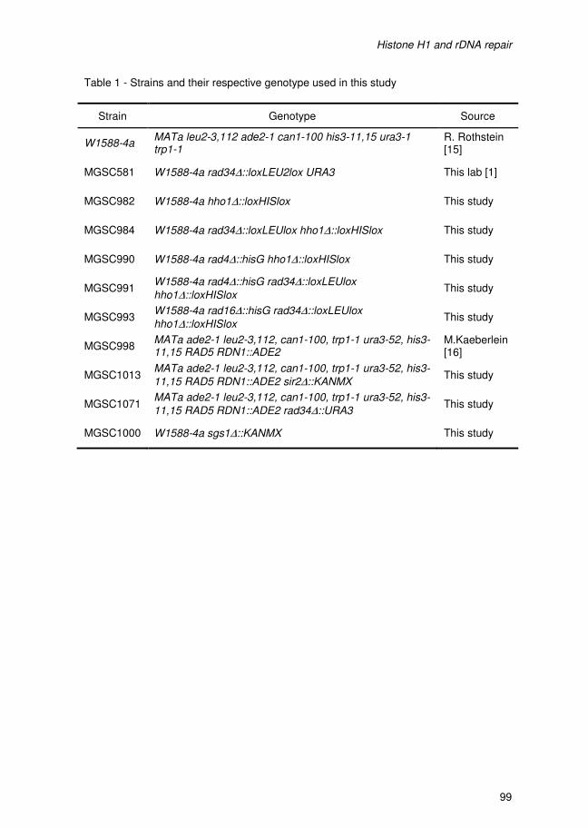
Histone H1 and rDNA repair
98
Rad34 on recombination at the rDNA locus we employed an engineered yeast strain
kindly provided by M. Kaeberlein [16]. This strain contains an ADE2 cassette in its rDNA
which serves as an indicator for the occurrence of rDNA recombination. For wild-type
cells, recombination events are in the order of 1-3 per 1,000 colonies while a hyper-
recombinant mutant can display up to 20 red sectored colonies per 1,000 in this type of
experiment [16]. For rad34∆ cells we found wild-type levels of recombination of around
2.68 per 1,000 versus 2.59 per 1,000 for wild-type cells (data not shown). Therefore,
Rad34 has no significant role in preventing rDNA recombination.
5. Concluding remarks In summary, we have shown that in the absence of Histone H1, Rad34 is no longer
required for TS repair of active rDNA but now becomes dependent on Rad4. Most likely,
the need for a specific Rad4-like factor in rDNA TC-NER is due to the use of a different
RNA polymerase in this DNA region.
Interestingly, in higher eukaryotes, regardless of the polymerase in action, TC-NER
takes place in the absence of a Rad4-like protein. The Rad4 homolog XPC is only
required for GG-NER in humans and other higher eukaryotes. Thus, transcription and
TC-NER in yeast are different in their requirement of two Rad4-like proteins: Rad4 in
RNA Pol II related TC-NER and Rad34 in TC-NER of RNA Pol I transcribed rDNA.
Apparently, NER at the rDNA locus in yeast is so different from repair of this locus in
human cells that it requires a different Rad4-like protein.
It has been proposed that the actively transcribed rDNA is largely devoid of nucleosomes
[3, 10]. However, our results show that Histone H1 has an effect on TC-NER of actively
transcribed rDNA copies, implicating that at least Histone H1 is associated with active
rDNA.
Why is Rad34 only required for rDNA TC-NER when Histone H1 is present? Possibly,
the presence of Histone H1 stabilizes RNA Pol I when stalled on a lesion. Rad34 would
then be specifically required to remove this stalled RNA polymerase. In the absence of
Histone H1 RNA Pol I might be capable of back-tracking from the lesion by itself (without
Rad26) allowing access to the lesion by the general recognition factor Rad4. Similarly,
RNA Pol II associated Rad26-independent repair has been shown to be facilitated by
back-tracking of the RNA polymerase [20].
It remains to be determined why in yeast a specific additional rDNA TC-NER factor is
present, while in its absence cells are not UV sensitive and can largely rely on GG-NER
for the repair.
Histone H1 and rDNA repair
99
Table 1 - Strains and their respective genotype used in this study
Strain Genotype Source
W1588-4a MATa leu2-3,112 ade2-1 can1-100 his3-11,15 ura3-1 trp1-1
R. Rothstein [15]
MGSC581 W1588-4a rad34∆::loxLEU2lox URA3 This lab [1]
MGSC982 W1588-4a hho1∆::loxHISlox This study
MGSC984 W1588-4a rad34∆::loxLEUlox hho1∆::loxHISlox This study
MGSC990 W1588-4a rad4∆::hisG hho1∆::loxHISlox This study
MGSC991 W1588-4a rad4∆::hisG rad34∆::loxLEUlox
hho1∆::loxHISlox This study
MGSC993 W1588-4a rad16∆::hisG rad34∆::loxLEUlox
hho1∆::loxHISlox This study
MGSC998 MATa ade2-1 leu2-3,112, can1-100, trp1-1 ura3-52, his3-11,15 RAD5 RDN1::ADE2
M.Kaeberlein [16]
MGSC1013 MATa ade2-1 leu2-3,112, can1-100, trp1-1 ura3-52, his3-
11,15 RAD5 RDN1::ADE2 sir2∆::KANMX This study
MGSC1071 MATa ade2-1 leu2-3,112, can1-100, trp1-1 ura3-52, his3-
11,15 RAD5 RDN1::ADE2 rad34∆::URA3 This study
MGSC1000 W1588-4a sgs1∆::KANMX This study
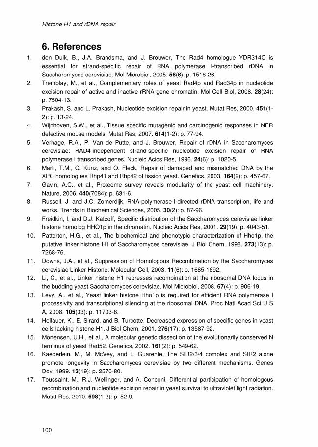
Histone H1 and rDNA repair
100
6. References 1. den Dulk, B., J.A. Brandsma, and J. Brouwer, The Rad4 homologue YDR314C is
essential for strand-specific repair of RNA polymerase I-transcribed rDNA in
Saccharomyces cerevisiae. Mol Microbiol, 2005. 56(6): p. 1518-26.
2. Tremblay, M., et al., Complementary roles of yeast Rad4p and Rad34p in nucleotide
excision repair of active and inactive rRNA gene chromatin. Mol Cell Biol, 2008. 28(24):
p. 7504-13.
3. Prakash, S. and L. Prakash, Nucleotide excision repair in yeast. Mutat Res, 2000. 451(1-
2): p. 13-24.
4. Wijnhoven, S.W., et al., Tissue specific mutagenic and carcinogenic responses in NER
defective mouse models. Mutat Res, 2007. 614(1-2): p. 77-94.
5. Verhage, R.A., P. Van de Putte, and J. Brouwer, Repair of rDNA in Saccharomyces
cerevisiae: RAD4-independent strand-specific nucleotide excision repair of RNA
polymerase I transcribed genes. Nucleic Acids Res, 1996. 24(6): p. 1020-5.
6. Marti, T.M., C. Kunz, and O. Fleck, Repair of damaged and mismatched DNA by the
XPC homologues Rhp41 and Rhp42 of fission yeast. Genetics, 2003. 164(2): p. 457-67.
7. Gavin, A.C., et al., Proteome survey reveals modularity of the yeast cell machinery.
Nature, 2006. 440(7084): p. 631-6.
8. Russell, J. and J.C. Zomerdijk, RNA-polymerase-I-directed rDNA transcription, life and
works. Trends in Biochemical Sciences, 2005. 30(2): p. 87-96.
9. Freidkin, I. and D.J. Katcoff, Specific distribution of the Saccharomyces cerevisiae linker
histone homolog HHO1p in the chromatin. Nucleic Acids Res, 2001. 29(19): p. 4043-51.
10. Patterton, H.G., et al., The biochemical and phenotypic characterization of Hho1p, the
putative linker histone H1 of Saccharomyces cerevisiae. J Biol Chem, 1998. 273(13): p.
7268-76.
11. Downs, J.A., et al., Suppression of Homologous Recombination by the Saccharomyces
cerevisiae Linker Histone. Molecular Cell, 2003. 11(6): p. 1685-1692.
12. Li, C., et al., Linker histone H1 represses recombination at the ribosomal DNA locus in
the budding yeast Saccharomyces cerevisiae. Mol Microbiol, 2008. 67(4): p. 906-19.
13. Levy, A., et al., Yeast linker histone Hho1p is required for efficient RNA polymerase I
processivity and transcriptional silencing at the ribosomal DNA. Proc Natl Acad Sci U S
A, 2008. 105(33): p. 11703-8.
14. Hellauer, K., E. Sirard, and B. Turcotte, Decreased expression of specific genes in yeast
cells lacking histone H1. J Biol Chem, 2001. 276(17): p. 13587-92.
15. Mortensen, U.H., et al., A molecular genetic dissection of the evolutionarily conserved N
terminus of yeast Rad52. Genetics, 2002. 161(2): p. 549-62.
16. Kaeberlein, M., M. McVey, and L. Guarente, The SIR2/3/4 complex and SIR2 alone
promote longevity in Saccharomyces cerevisiae by two different mechanisms. Genes
Dev, 1999. 13(19): p. 2570-80.
17. Toussaint, M., R.J. Wellinger, and A. Conconi, Differential participation of homologous
recombination and nucleotide excision repair in yeast survival to ultraviolet light radiation.
Mutat Res, 2010. 698(1-2): p. 52-9.
Histone H1 and rDNA repair
101
18. Toussaint, M. and A. Conconi, High-throughput and sensitive assay to measure yeast
cell growth: a bench protocol for testing genotoxic agents. Nature Protocols, 2006. 1(4):
p. 1922-8.
19. Park, P.U., M. McVey, and L. Guarente, Separation of mother and daughter cells.
Methods Enzymol, 2002. 351: p. 468-77.
20. Li, S. and M.J. Smerdon, Rpb4 and Rpb9 mediate subpathways of transcription-coupled
DNA repair in Saccharomyces cerevisiae. Embo Journal, 2002. 21(21): p. 5921-9.
21. Sinclair, D.A. and L. Guarente, Extrachromosomal rDNA circles--a cause of aging in
yeast. Cell, 1997. 91(7): p. 1033-42.
22. Sinclair, D.A., K. Mills, and L. Guarente, Accelerated aging and nucleolar fragmentation
in yeast sgs1 mutants. Science, 1997. 277(5330): p. 1313-6.
23. Sinclair, D.A., K. Mills, and L. Guarente, Molecular mechanisms of yeast aging. Trends in
Biochemical Sciences, 1998. 23(4): p. 131-4.

Histone H1 and rDNA repair
102
IV Phenotypic analysis of a rad4 mutant carrying a homologous
change that in XPC leads to the XP disorder
P. van Eijk, J.A. Brandsma, M. de Ruijter, J. Brouwer

Histone H1 and rDNA repair
102
IV Phenotypic analysis of a rad4 mutant carrying a homologous
change that in XPC leads to the XP disorder
P. van Eijk, J.A. Brandsma, M. de Ruijter, J. Brouwer
A yeast mutant corresponding to a XP patient mutation
104
1. Abstract XP-C patient phenotypes predominantly result from truncated proteins or lack of XPC [1].
Only two point-mutations that lead to amino acid substitutions leaving the full-length XPC
protein intact have been described (P331H and W690S). Surprisingly, we find that
Saccharomyces cerevisiae cells carrying the corresponding rad4W496S mutation are not
UV-sensitive. However, deletion of additional Nucleotide Excision Repair (NER) genes
results in additive or synergistic effects on UV resistance. More in-depth analysis of the
repair capacity of these cells shows that NER is affected in the rad4W496S mutant and it
is mainly disrupted in Global Genome Repair.
A yeast mutant corresponding to a XP patient mutation
105
2. Introduction Xeroderma pigmentosum (XP) is a rare autosomal, recessive human disorder
characterized by defects in Nucleotide Excision Repair (NER). Genetic defects in human
NER can also result in other disorders like Cockayne Syndrome (CS) or
Trichothiodystrophy (TTD) [2]. XP causes a 2,000-fold increased incidence of sunlight
induced skin cancer in patients and, depending on the complementation group, can also
result in neurological degeneration [2]. The XP-C complementation group is the most
common among XP patients and is defined by mutations in the XPC gene homologous
to the core NER RAD4 gene in yeast [3]. The XPC-hHR23A/B complex is, together with
the XPC binding protein Centrin 2 involved in the initial recognition of DNA damage.
The yeast Saccharomyces cerevisiae is shown to be an excellent model for the study of
the conserved mechanism of NER. In yeast a complex similar to that in human cells is
responsible for the recognition of DNA damage. The XPC homologue in yeast, Rad4
forms a complex with Rad23, and we showed recently that also a third protein can bind
to this recognition complex, Rad33 [4]. Rad33 shows no homology to Centrin2 but the
binding site in Rad4 is identical to that in XPC [4]. The homology between the human
and yeast recognition complexes in NER allows us to study the role of a conserved
tryptophan residue, which was found to be substituted as result of a point mutation in an
XPC patient. This XPC mutation is of special interest since it is one of two patient
mutations that have been described to lead to an amino acid substitution, leaving the full
length protein and its expression unchanged [5, 6]. One of these mutations, XPC-
W690S, results in defective repair and UV sensitivity in human cells [1]. It was shown
that decreased protein stability, impaired DNA binding and impaired repair all contribute
the XP phenotype [9]. Furthermore, mutant XPC protein displayed reduced ssDNA
binding in vitro while the interaction with Rad23 and Centrin 2 is still intact [10]. The
W690S resides in the conserved C-terminus that is shared by Rad4-like proteins. In this
region the Rad23 and TFIIH interactions are mapped [5, 7]. The affected tryptophan
residue at position 690 in XPC is conserved at position 496 in yeast Rad4 (Figure 1A top
panel) [1]. The information from the Rad4 protein structure allows us to pinpoint exactly
where the amino acid substitution affects the protein structure [8]. The substituted
residue is located in Beta Hairpin Domain 2 (BHD2) (Figure 1A lower panel) in close
proximity to the bases opposite the CPD lesion [8]. The neighboring residues R494,
Q495 and M498 in Rad4 are all in direct contact with the DNA backbone and bases
opposite the CPD lesion [8], suggesting that the mutation might affect DNA damage
binding.
To get more insight into the defect caused by this patient mutation we set out to
investigate the phenotype of the corresponding rad4W496S mutant in S.cerevisiae.
Surprisingly, we find that the equivalent W496S mutation in Rad4 does not result in UV
sensitivity. However, repair is moderately affected in rad4W496S cells, with GG-NER
being most severely hampered.
A yeast mutant corresponding to a XP patient mutation
106
3. Material & Methods
3.1 Strains and UV droptest
All strains used in this study are derived from the wild-type W1588-4a strain [11].
Relevant phenotypes are described in table 1. The rad4W496S substitution was
introduced into W1588 creating strain MGSC842. This background was made by the
Pop-in Pop-out approach using a Yeast Integrating (YIp) plasmid construct, YIp-
rad4CtW496S, of the Rad4 C-terminal region as indicated in figure 1A, containing the
point mutation corresponding to the W496S substitution. To construct a strain with the
RAD4 point mutation and a RAD26 deletion, disrupting TC-NER, we transformed
MGSC842 with linearized pRAD26::HIS3. Correct introduction of the disruption construct
was confirmed by PCR and Southernblot analysis. Similarly, to disrupt GG-NER we
deleted RAD16 by transforming MGSC842 with linearized pRAD16::HUH resulting in
strain MGSC934. Liquid cultures of the relevant strains were grown overnight after which
they were diluted in sterile water. For the UV droptest 1 or 2µl drops were dispensed on
a 7x8 grid on a YPD plate. After UV irradiation plates were incubated in the dark at 30oC
for 2-3 days.
3.2 Strand specific repair assay
Yeast cell cultures of 200mL YPD of OD600=0.4 were spun down and resuspended in
cold PBS to an OD600 of 0.7. Using 254nm UV light cells were irradiated with 70J/m2 at a
rate of 3W/m2. Next, the cells were spun down again, resuspended in YPD and aliquoted
into 4 portions for the 0, 30, 60 and 120min time points. Incubation takes place at 30oC in
the dark. After the indicated time-intervals cells were spun down, washed with cold water
and split in two aliquots and stored at -80oC.
DNA was isolated using a combination of Yeast Cell Piercing Solution (YCPS) and Cell
Lysing Solution (CLS) followed by protein precipitation using ammonium acetate and
DNA precipitation using isopropanol. The cell pellets were thawed and resuspended in
YCPS (0.1% SB3-14, 100mM LiCl in TE buffer) and incubated at 65oC for 30 minutes.
Next, the cells were spun down and resuspended in CLS (2mM EDTA, 2% SDS) and
again incubated at 65oC for 45 minutes. After placing the cells on ice for 5 minutes
ammonium acetate was added to an end-concentration of 2.5M. At this stage cells were
rotated for 10 minutes at room temperature before being centrifuged at 14,000rpm at
16oC for 10 minutes. The supernatant was then transferred and an equal volume of
isopropanol was added. After gentle mixing and incubation at room temperature for 10
minutes, the DNA was precipitated by means of centrifugation at 14,000rpm at 16oC for
12 minutes. The DNA was then be washed with 70% Ethanol after which it was dissolved
in water. RPB2 repair was measured as described previously [4].
DNA repair was quantified by scanning the Southern Blot in a Biorad Personal Molecular
Imager FX and using Biorad’s Quantity One for data analysis.
A yeast mutant corresponding to a XP patient mutation
107
4. Results & Discussion
4.1 The rad4W496S mutant is not UV sensitive
We constructed a yeast strain containing the point-mutation in RAD4 that leads to the
W496S substitution. This substitution was obtained by site-directed mutagenesis of a C-
terminal RAD4 DNA fragment. This construct was then introduced into yeast by means
of pop-in pop-out methodology to generate a genomic rad4W496S allele (see material
and methods). The UV phenotype of the resulting rad4W496S yeast strain was
investigated in a UV droptest. Surprisingly, the RAD4 mutant strain is as UV resistant as
wild-type cells shown in figure 1B.
However, when we introduced the rad4W496S substitution in a RAD23 deletion
background, we observed a synergistic UV sensitivity as shown by the UV drop test of
rad23∆rad4W496S cells in figure 1B. This strain is significantly more UV sensitive
compared to either of the single mutant strains, displaying a degree of sensitivity that is
similar to the NER deficient RAD4 deletion strain. Rad23 is a direct interaction partner of
Rad4 and is believed to stabilize the protein [12, 13]. Moreover, XPC-W690S was
reported to be unstable [9]. Taken together the possible Rad4W496S instability might be
more readily corrected by the yeast Rad23 protein, leading to UV resistance shown in
figure 1B. The absence of the stabilizing effect of Rad23 might be the cause of the
decrease in UV survival.
NER is characterized by two sub-pathways, GG-NER and TC-NER. In yeast, Rad16 is
specific for GG-NER, while Rad26 is a TC-NER specific protein. The specificity of these
proteins allows separate study of these sub-pathways by using the two different mutants.
We wanted to identify whether the NER sub-pathways are both equally or differently
affected by the W496S substitution. The rad16∆rad4W496S strain defective in GG-NER
and rad26∆rad4W496S cells deficient in TC-NER were subjected to UV irradiation using
a drop test depicted in figure 1B. TC-NER proficient rad16∆rad4W496S cells show a
moderate UV sensitivity, however, the rad26∆rad4W496S strain proficient in GG-NER
shows a strong synergistic effect (Figure 1B). The rad4W496S mutation thus genetically
interacts with both TC-NER and GG-NER factors, but seems to affect GG-NER more
severely.
4.2 The Rad4W496S mutation results in impaired NER in vivo
We found that UV survival is strongly affected by the rad4W496S mutation in the
absence of the Rad26 TC-NER factor, suggesting a defect in GG-NER, analogous to the
UV sensitivity due to the GG-NER defect resulting from the XPC-W690S protein in
human cells [1]. The repair phenotype of the rad4W496S mutant cells were studied in
more detail in different NER defective backgrounds by making use of a strand specific
repair assay as described previously [14]. Repair measurements in the rad4W496S
background show that after 2 hours of incubation, about 75% of the damages in the tran-
A yeast mutant corresponding to a XP patient mutation
106
3. Material & Methods
3.1 Strains and UV droptest
All strains used in this study are derived from the wild-type W1588-4a strain [11].
Relevant phenotypes are described in table 1. The rad4W496S substitution was
introduced into W1588 creating strain MGSC842. This background was made by the
Pop-in Pop-out approach using a Yeast Integrating (YIp) plasmid construct, YIp-
rad4CtW496S, of the Rad4 C-terminal region as indicated in figure 1A, containing the
point mutation corresponding to the W496S substitution. To construct a strain with the
RAD4 point mutation and a RAD26 deletion, disrupting TC-NER, we transformed
MGSC842 with linearized pRAD26::HIS3. Correct introduction of the disruption construct
was confirmed by PCR and Southernblot analysis. Similarly, to disrupt GG-NER we
deleted RAD16 by transforming MGSC842 with linearized pRAD16::HUH resulting in
strain MGSC934. Liquid cultures of the relevant strains were grown overnight after which
they were diluted in sterile water. For the UV droptest 1 or 2µl drops were dispensed on
a 7x8 grid on a YPD plate. After UV irradiation plates were incubated in the dark at 30oC
for 2-3 days.
3.2 Strand specific repair assay
Yeast cell cultures of 200mL YPD of OD600=0.4 were spun down and resuspended in
cold PBS to an OD600 of 0.7. Using 254nm UV light cells were irradiated with 70J/m2 at a
rate of 3W/m2. Next, the cells were spun down again, resuspended in YPD and aliquoted
into 4 portions for the 0, 30, 60 and 120min time points. Incubation takes place at 30oC in
the dark. After the indicated time-intervals cells were spun down, washed with cold water
and split in two aliquots and stored at -80oC.
DNA was isolated using a combination of Yeast Cell Piercing Solution (YCPS) and Cell
Lysing Solution (CLS) followed by protein precipitation using ammonium acetate and
DNA precipitation using isopropanol. The cell pellets were thawed and resuspended in
YCPS (0.1% SB3-14, 100mM LiCl in TE buffer) and incubated at 65oC for 30 minutes.
Next, the cells were spun down and resuspended in CLS (2mM EDTA, 2% SDS) and
again incubated at 65oC for 45 minutes. After placing the cells on ice for 5 minutes
ammonium acetate was added to an end-concentration of 2.5M. At this stage cells were
rotated for 10 minutes at room temperature before being centrifuged at 14,000rpm at
16oC for 10 minutes. The supernatant was then transferred and an equal volume of
isopropanol was added. After gentle mixing and incubation at room temperature for 10
minutes, the DNA was precipitated by means of centrifugation at 14,000rpm at 16oC for
12 minutes. The DNA was then be washed with 70% Ethanol after which it was dissolved
in water. RPB2 repair was measured as described previously [4].
DNA repair was quantified by scanning the Southern Blot in a Biorad Personal Molecular
Imager FX and using Biorad’s Quantity One for data analysis.
A yeast mutant corresponding to a XP patient mutation
107
4. Results & Discussion
4.1 The rad4W496S mutant is not UV sensitive
We constructed a yeast strain containing the point-mutation in RAD4 that leads to the
W496S substitution. This substitution was obtained by site-directed mutagenesis of a C-
terminal RAD4 DNA fragment. This construct was then introduced into yeast by means
of pop-in pop-out methodology to generate a genomic rad4W496S allele (see material
and methods). The UV phenotype of the resulting rad4W496S yeast strain was
investigated in a UV droptest. Surprisingly, the RAD4 mutant strain is as UV resistant as
wild-type cells shown in figure 1B.
However, when we introduced the rad4W496S substitution in a RAD23 deletion
background, we observed a synergistic UV sensitivity as shown by the UV drop test of
rad23∆rad4W496S cells in figure 1B. This strain is significantly more UV sensitive
compared to either of the single mutant strains, displaying a degree of sensitivity that is
similar to the NER deficient RAD4 deletion strain. Rad23 is a direct interaction partner of
Rad4 and is believed to stabilize the protein [12, 13]. Moreover, XPC-W690S was
reported to be unstable [9]. Taken together the possible Rad4W496S instability might be
more readily corrected by the yeast Rad23 protein, leading to UV resistance shown in
figure 1B. The absence of the stabilizing effect of Rad23 might be the cause of the
decrease in UV survival.
NER is characterized by two sub-pathways, GG-NER and TC-NER. In yeast, Rad16 is
specific for GG-NER, while Rad26 is a TC-NER specific protein. The specificity of these
proteins allows separate study of these sub-pathways by using the two different mutants.
We wanted to identify whether the NER sub-pathways are both equally or differently
affected by the W496S substitution. The rad16∆rad4W496S strain defective in GG-NER
and rad26∆rad4W496S cells deficient in TC-NER were subjected to UV irradiation using
a drop test depicted in figure 1B. TC-NER proficient rad16∆rad4W496S cells show a
moderate UV sensitivity, however, the rad26∆rad4W496S strain proficient in GG-NER
shows a strong synergistic effect (Figure 1B). The rad4W496S mutation thus genetically
interacts with both TC-NER and GG-NER factors, but seems to affect GG-NER more
severely.
4.2 The Rad4W496S mutation results in impaired NER in vivo
We found that UV survival is strongly affected by the rad4W496S mutation in the
absence of the Rad26 TC-NER factor, suggesting a defect in GG-NER, analogous to the
UV sensitivity due to the GG-NER defect resulting from the XPC-W690S protein in
human cells [1]. The repair phenotype of the rad4W496S mutant cells were studied in
more detail in different NER defective backgrounds by making use of a strand specific
repair assay as described previously [14]. Repair measurements in the rad4W496S
background show that after 2 hours of incubation, about 75% of the damages in the tran-

A yeast mutant corresponding to a XP patient mutation
108
scribed strand (TS) are repaired as compared to 90% in wild-type cells (Figure 2A & B).
Repair of the non-transcribed strand (NTS) on the other hand, is more severely reduced
to only 15% after 2 hours compared to 50-60% in wild-type cells (Figure 2). Thus the
W496S mutation reduces the efficiency of TC-NER only moderately, but severely impairs
GG-NER after UV irradiation.
Analogous to the survival experiments, strand specific repair was also investigated in
GG-NER and TC-NER deficient backgrounds containing the rad4W496S mutation. GG-
NER in a TC-NER deficient rad26∆ single mutant repairs 50-60% of the damages on
both strands during the time course of 2 hours (Figure 3B & D). However, GG-NER of
both strands in a rad26∆rad4W496S double mutant is severely reduced to only 15%
after 2 hours (Figure 3B & D). This again shows that GG-NER is severely impaired by
the rad4W496S mutation.
GG-NER is completely disrupted in a RAD16 deletion background resulting in the ab-
sence of NTS repair in rad16∆rad4W496S cells as expected (Figure 3A & C) [15]. In this
Figure 1 – The Rad4W496S point mutation is UV resistant and displays genetic interaction with other NER genes (A) Top panel: Sequence alignment of Rad4-like proteins depicting the conserved tryptophan at position 496 in Rad4. Bottom panel: schematic depiction of the structural domains of Rad4 [8]. (B) UV drop test indicating the genetic interaction of rad4W496S with several RAD deletions. Serial dilutions were prepared in sterile water from stationary culture starting with a one in hundred dilution. After applying the droplets, the plates were UV irradiated with the indicated doses and incubated in the dark at 30°C for 2-3 days.
A yeast mutant corresponding to a XP patient mutation
109
GG- NER defi-
cient rad16∆rad4
W496S back-
ground, TC-NER
removes ~80% of
the damages in
the TS two hours
after UV irradia-
tion.
Taken together,
our results clearly
show that TC-NER
is only moderately
affected by the
rad4W496S muta-
tion, whereas GG-
NER is strongly
impaired. This
impaired GG-NER,
however, does not
lead to UV sensi-
tivity per se. Only
when other NER
factors, like
Rad23, Rad16 or
Rad26, are absent
UV sensitivity is observed. UV resistance in the rad4W496S single mutant background
indicates that NER does occur, but apparently is delayed and can therefore not be
monitored in our strand-specific repair assay.
The XPC-W690S protein showed a reduced affinity for DNA, which could help to explain
why the corresponding rad4W496S mutant displays delayed repair in yeast. GG-NER
strongly depends on the DNA binding ability of the Rad4 protein and is therefore
severely reduced due to the W496S substitution. A Rad4 protein that is defective in DNA
binding can still function in TC-NER, because TC-NER specific factors, like Rad26 and
RNA Pol II, might facilitate binding of Rad4 to the damage. In human cells TC-NER
factors allow this repair pathway to function even without a Rad4-like protein. Thus, the
initial steps during TC-NER that can use a mutated Rad4 protein to interact with a
damage in yeast TC-NER, might circumvent the requirement for XPC in TC-NER in
human cells entirely.
Figure 2 – rad4W496S is disrupted both TC-NER and GG-NER. Strand specific repair assay of RPB2 in wild-type and rad4W496S mutant strains. Yeast cells were UV-irradiated with 70J/m
2 and allowed to
recover for the indicated repair times. (A) Repair analysis on Southernblot probed for the transcribed strand (TS) or non-transcribed strand (NTS) of both wild-type and rad4W496S cells. (B) Graphical representation of the relative amount of repair. Data was taken from at least 3 individual experiments.
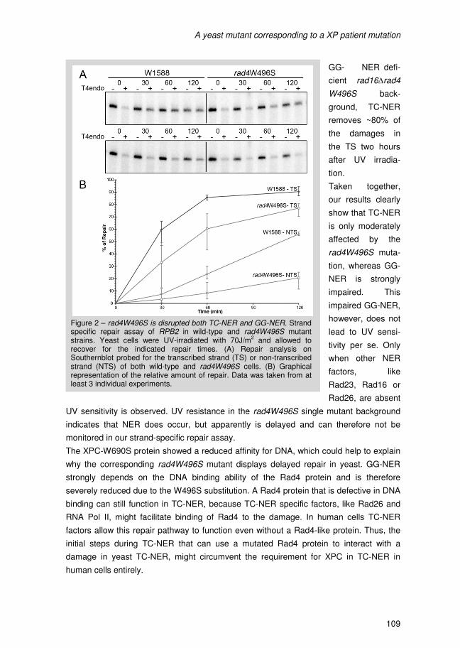
A yeast mutant corresponding to a XP patient mutation
108
scribed strand (TS) are repaired as compared to 90% in wild-type cells (Figure 2A & B).
Repair of the non-transcribed strand (NTS) on the other hand, is more severely reduced
to only 15% after 2 hours compared to 50-60% in wild-type cells (Figure 2). Thus the
W496S mutation reduces the efficiency of TC-NER only moderately, but severely impairs
GG-NER after UV irradiation.
Analogous to the survival experiments, strand specific repair was also investigated in
GG-NER and TC-NER deficient backgrounds containing the rad4W496S mutation. GG-
NER in a TC-NER deficient rad26∆ single mutant repairs 50-60% of the damages on
both strands during the time course of 2 hours (Figure 3B & D). However, GG-NER of
both strands in a rad26∆rad4W496S double mutant is severely reduced to only 15%
after 2 hours (Figure 3B & D). This again shows that GG-NER is severely impaired by
the rad4W496S mutation.
GG-NER is completely disrupted in a RAD16 deletion background resulting in the ab-
sence of NTS repair in rad16∆rad4W496S cells as expected (Figure 3A & C) [15]. In this
Figure 1 – The Rad4W496S point mutation is UV resistant and displays genetic interaction with other NER genes (A) Top panel: Sequence alignment of Rad4-like proteins depicting the conserved tryptophan at position 496 in Rad4. Bottom panel: schematic depiction of the structural domains of Rad4 [8]. (B) UV drop test indicating the genetic interaction of rad4W496S with several RAD deletions. Serial dilutions were prepared in sterile water from stationary culture starting with a one in hundred dilution. After applying the droplets, the plates were UV irradiated with the indicated doses and incubated in the dark at 30°C for 2-3 days.
A yeast mutant corresponding to a XP patient mutation
109
GG- NER defi-
cient rad16∆rad4
W496S back-
ground, TC-NER
removes ~80% of
the damages in
the TS two hours
after UV irradia-
tion.
Taken together,
our results clearly
show that TC-NER
is only moderately
affected by the
rad4W496S muta-
tion, whereas GG-
NER is strongly
impaired. This
impaired GG-NER,
however, does not
lead to UV sensi-
tivity per se. Only
when other NER
factors, like
Rad23, Rad16 or
Rad26, are absent
UV sensitivity is observed. UV resistance in the rad4W496S single mutant background
indicates that NER does occur, but apparently is delayed and can therefore not be
monitored in our strand-specific repair assay.
The XPC-W690S protein showed a reduced affinity for DNA, which could help to explain
why the corresponding rad4W496S mutant displays delayed repair in yeast. GG-NER
strongly depends on the DNA binding ability of the Rad4 protein and is therefore
severely reduced due to the W496S substitution. A Rad4 protein that is defective in DNA
binding can still function in TC-NER, because TC-NER specific factors, like Rad26 and
RNA Pol II, might facilitate binding of Rad4 to the damage. In human cells TC-NER
factors allow this repair pathway to function even without a Rad4-like protein. Thus, the
initial steps during TC-NER that can use a mutated Rad4 protein to interact with a
damage in yeast TC-NER, might circumvent the requirement for XPC in TC-NER in
human cells entirely.
Figure 2 – rad4W496S is disrupted both TC-NER and GG-NER. Strand specific repair assay of RPB2 in wild-type and rad4W496S mutant strains. Yeast cells were UV-irradiated with 70J/m
2 and allowed to
recover for the indicated repair times. (A) Repair analysis on Southernblot probed for the transcribed strand (TS) or non-transcribed strand (NTS) of both wild-type and rad4W496S cells. (B) Graphical representation of the relative amount of repair. Data was taken from at least 3 individual experiments.
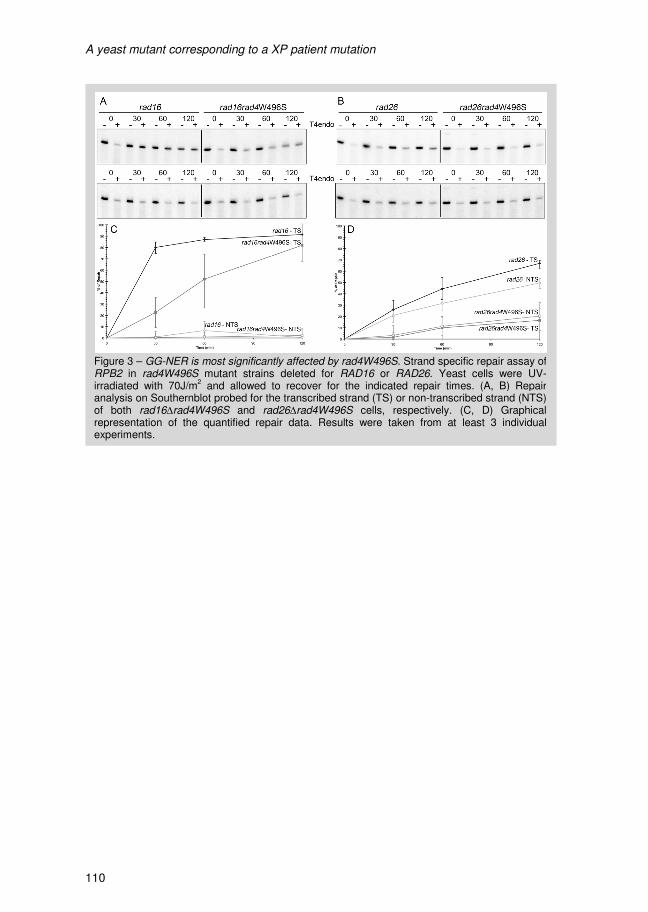
A yeast mutant corresponding to a XP patient mutation
110
Figure 3 – GG-NER is most significantly affected by rad4W496S. Strand specific repair assay of RPB2 in rad4W496S mutant strains deleted for RAD16 or RAD26. Yeast cells were UV-irradiated with 70J/m
2 and allowed to recover for the indicated repair times. (A, B) Repair
analysis on Southernblot probed for the transcribed strand (TS) or non-transcribed strand (NTS) of both rad16∆rad4W496S and rad26∆rad4W496S cells, respectively. (C, D) Graphical representation of the quantified repair data. Results were taken from at least 3 individual experiments.
A yeast mutant corresponding to a XP patient mutation
111
Table 1 – This table lists all the strains used in this study and their respective genotype
and source.
Strain Genotype Source
W1588-4a MATα leu2-3,112 ade2-1 can1-100 his3-11,15 ura3-1 trp1-1
R. Rothstein [11]
MGSC427 MATα ade2-1 can1-100 his3-11,15 leu2-3,112 trp1-1 rad23::hisG
This Lab
MGSC582 MATα ade2-1 can1-100 his3-11,15 leu2-3,112 trp1-1
ura3-1 rad26∆::HIS3 This Lab
MGSC609 MATα ade2-1 can1-100 his3-11,15 leu2-3,112 trp1-1
ura3-1 rad4∆::HisG This Lab
MGSC621 MATα ade2-1 can1-100 his3-11,15 leu2-3,112 trp1-1
ura3-1 rad16∆::HUH This Lab
MGSC622 MATα ade2-1 can1-100 his3-11,15 leu2-3,112 trp1-1
ura3-1 rad23∆::HUH This Lab
MGSC662 MATα ade2-1 can1-100 his3-11,15 leu2-3,112 trp1-1 ura3-1 rad33::KANMX
This Lab
MGSC842 MATα ade2-1 can1-100 his3-11,15 leu2-3,112 trp1-1 ura3-1 rad4W496S
This Study
MGSC849 MATα leu2-3ade2-1 can1-100 his3-11,15,112 trp1-1 ura3-1 rad4W496S rad26::HIS3
This Study
MGSC934 MATα ade2-1 can1-100 his3-11,15 leu2-3,112 trp1-1
ura3-1 rad4W496S rad16∆::HUH This Study
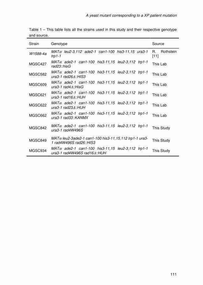
A yeast mutant corresponding to a XP patient mutation
112
5. References 1. Chavanne, F., et al., Mutations in the XPC gene in families with xeroderma pigmentosum
and consequences at the cell, protein, and transcript levels. Cancer Res, 2000. 60(7): p.
1974-82.
2. Cleaver, J.E., E.T. Lam, and I. Revet, Disorders of nucleotide excision repair: the genetic
and molecular basis of heterogeneity. Nat Rev Genet, 2009. 10(11): p. 756-68.
3. Verhage, R., et al., The RAD7 and RAD16 genes, which are essential for pyrimidine
dimer removal from the silent mating type loci, are also required for repair of the
nontranscribed strand of an active gene in Saccharomyces cerevisiae. Mol Cell Biol,
1994. 14(9): p. 6135-42.
4. den Dulk, B., et al., The NER protein Rad33 shows functional homology to human
Centrin2 and is involved in modification of Rad4. DNA Repair (Amst), 2008. 7(6): p. 858-
68.
5. Uchida, A., et al., The carboxy-terminal domain of the XPC protein plays a crucial role in
nucleotide excision repair through interactions with transcription factor IIH. DNA Repair
(Amst), 2002. 1(6): p. 449-61.
6. Bernardes de Jesus, B.M., et al., Dissection of the molecular defects caused by
pathogenic mutations in the DNA repair factor XPC. Mol Cell Biol, 2008. 28(23): p. 7225-
35.
7. Masutani, C., et al., Identification and characterization of XPC-binding domain of
hHR23B. Mol Cell Biol, 1997. 17(12): p. 6915-23.
8. Min, J.H. and N.P. Pavletich, Recognition of DNA damage by the Rad4 nucleotide
excision repair protein. Nature, 2007. 449(7162): p. 570-5.
9. Yasuda, G., et al., In vivo destabilization and functional defects of the xeroderma
pigmentosum C protein caused by a pathogenic missense mutation. Mol Cell Biol, 2007.
27(19): p. 6606-14.
10. Bunick, C.G., et al., Biochemical and structural domain analysis of xeroderma
pigmentosum complementation group C protein. Biochemistry, 2006. 45(50): p. 14965-
79.
11. Mortensen, U.H., et al., A molecular genetic dissection of the evolutionarily conserved N
terminus of yeast Rad52. Genetics, 2002. 161(2): p. 549-62.
12. Ortolan, T.G., et al., Rad23 stabilizes Rad4 from degradation by the Ub/proteasome
pathway. Nucleic Acids Res, 2004. 32(22): p. 6490-500.
13. Gillette, T.G., et al., Distinct functions of the ubiquitin-proteasome pathway influence
nucleotide excision repair. Embo Journal, 2006. 25(11): p. 2529-38.
14. den Dulk, B., et al., Rad33, a new factor involved in nucleotide excision repair in
Saccharomyces cerevisae. DNA repair, 2006. 5(6): p. 683-692.
15. Verhage, R., et al., The RAD7 and RAD16 genes, which are essential for pyrimidine
dimer removal from the silent mating type loci, are also required for repair of the
nontranscribed strand of an active gene in Saccharomyces cerevisiae. Molecular and
cellular biology, 1994. 14(9): p. 6135-42.
A yeast mutant corresponding to a XP patient mutation
113
16. Verhage, R.A., et al., Analysis of gene- and strand-specific repair in the moderately UV-
sensitive Saccharomyces cerevisiae rad23 mutant. Mutat Res, 1996. 362(2): p. 155-65.
17. Mueller, J.P. and M.J. Smerdon, Rad23 is required for transcription-coupled repair and
efficient overrall repair in Saccharomyces cerevisiae. Mol Cell Biol, 1996. 16(5): p. 2361-
8.
18. Miller, R.D., L. Prakash, and S. Prakash, Defective excision of pyrimidine dimers and
interstrand DNA crosslinks in rad7 and rad23 mutants of Saccharomyces cerevisiae. Mol
Gen Genet, 1982. 188(2): p. 235-9.
A yeast mutant corresponding to a XP patient mutation
114
V A novel, functionally distinct, bipartite Rad4-Rad23 interaction in
Nucleotide Excision Repair
P. van Eijk, J.A. Brandsma, M. de Ruijter, J. Brouwer
A yeast mutant corresponding to a XP patient mutation
114
V A novel, functionally distinct, bipartite Rad4-Rad23 interaction in
Nucleotide Excision Repair
P. van Eijk, J.A. Brandsma, M. de Ruijter, J. Brouwer
The bipartite Rad4-Rad23 interaction
116
1. Abstract In the yeast Saccharomyces cerevisiae the Rad4-Rad23 complex is implicated in the
initial damage recognition step of Nucleotide Excision Repair (NER). In the Rad4-Rad23
heterodimer, as well as in the analogous XPC-hHR23A/B complex in human cells, the C-
terminus of Rad4 or XPC is shown to interact with the R4B domain of the Rad23 protein.
Elucidation of the structure of Rad4, the homologue of XPC, revealed several interesting
domains that might have a role in protein-protein interactions. In this paper we study the
Rad4-Rad23 interaction in more detail. We constructed a set of deletions in the Rad4
and Rad23 proteins respectively, and used the yeast two-hybrid technique for extensive
analysis.
We show that Rad4 contains two distinct Rad23 binding domains, one located in the
TransGlutaminase Domain (TGD) and another in the C-terminal part of the protein. Both
interactions, however, require the same Rad4 binding domain (R4BD) of Rad23. We
present evidence that the UbL domain controls the interaction of Rad23 with either of the
two binding domains on Rad4. We present evidence that the two different Rad4-Rad23
complexes are differentially involved in Global Genome NER (GG-NER) and
Transcription-Coupled NER (TC-NER).
The bipartite Rad4-Rad23 interaction
117
2. Introduction The core damage recognition complex in the Nucleotide Excision Repair (NER) pathway
consists of Rad4, Rad23 and Rad33 in yeast [1, 2] and of XPC, hHR23A/B and Centrin 2
in higher eukaryotes [3]. Uniquely to yeast, a rDNA specific Rad4-homolog exists called
Rad34 [4, 5]. This protein also forms a complex with Rad33 and Rad23 and is
responsible for transcription-coupled NER (TC-NER) in active rDNA [4].
Moderate UV sensitivity is observed when RAD23 or NER accessory genes are deleted,
whereas the deletion of core NER factors, like Rad4, results in extreme UV sensitivity
due to complete absence of repair. RAD23 deletion cells show only moderate UV
sensitivity, although no appreciable repair activity can be detected [6]. Currently, Rad23
and hHR23B are believed to stabilize the Rad4 or XPC protein, respectively [7, 8]. This,
however, does not fully explain the phenotype of rad23∆ cells.
The Rad23 structure and function is interesting as it is one of a few proteins in yeast that
contains both Ubiquitin Like (UbL) and Ubiquitin Associating (UBA) domains (Figure 2A
bottom panel). Proteins that share these features with Rad23 are Dsk2 and Ddi1 and all
three proteins are thought to shuttle ubiquitinated proteins to the proteasome due their
affinity for ubiquitinated proteins and ability to interact with the proteasome [9-12]. The
UbL domain of Rad23 is ubiquitinated in vivo [8] but this does not interfere with protein
stability. The domain can also be replaced by the genuine ubiquitin sequence without
loss of protein function in NER [13]. The Rad23 UbL domain is partially dispensable for
NER both in vivo and in vitro [13, 15]. Interestingly, the UbL domain of Rad23 was shown
to interact with the 19S proteasome subunit, an interaction that seemed to enhance in
vitro NER [15]. However, the role for the UbL domain in relation to the Rad4-Rad23
interaction is poorly understood. The UBA domains in Rad23 intrinsically stabilize the
protein and allow it to be ubiquitinated and to interact with the proteasome without being
degraded [14].
The structure of Rad4 bound to a Rad23 fragment and DNA has been solved [16]. The
Rad4 protein used to solve the structure does not contain about a 100 amino acids from
the N- and C-terminal ends of the protein. These ends of Rad4 are susceptible to
proteolytic degradation in vitro, suggesting that the Rad4 termini are unstructured or
loosely folded [16]. However the truncated recombinant Rad4 protein binds normally to
DNA. Rad4 is made up of 4 protein domains (Figure 2A top panel) that interact with the
DNA backbone and with the DNA opposit the CPD lesion, but not the CPD itself. The
TransGlutaminase Domain (TGD) spans 300 amino acids of the N-terminal part of Rad4
and supports the interaction with the DNA phosphate backbone together with the Beta
Hairpin Domain 1 (BHD1). BHDs 2 and 3 interact with a 4 basepair section opposite the
CPD lesion. The CPD dimer is solvent exposed and therefore does not exist in a fixed
conformation in the structure. BHD3 inserts its Beta Hairpin into the DNA double helix
and the domain flips out the two undamaged bases opposite the lesion that interact with
both BHD2 and BHD3 [16]. In the crystal structure of the Rad4-Rad23 complex the

The bipartite Rad4-Rad23 interaction
116
1. Abstract In the yeast Saccharomyces cerevisiae the Rad4-Rad23 complex is implicated in the
initial damage recognition step of Nucleotide Excision Repair (NER). In the Rad4-Rad23
heterodimer, as well as in the analogous XPC-hHR23A/B complex in human cells, the C-
terminus of Rad4 or XPC is shown to interact with the R4B domain of the Rad23 protein.
Elucidation of the structure of Rad4, the homologue of XPC, revealed several interesting
domains that might have a role in protein-protein interactions. In this paper we study the
Rad4-Rad23 interaction in more detail. We constructed a set of deletions in the Rad4
and Rad23 proteins respectively, and used the yeast two-hybrid technique for extensive
analysis.
We show that Rad4 contains two distinct Rad23 binding domains, one located in the
TransGlutaminase Domain (TGD) and another in the C-terminal part of the protein. Both
interactions, however, require the same Rad4 binding domain (R4BD) of Rad23. We
present evidence that the UbL domain controls the interaction of Rad23 with either of the
two binding domains on Rad4. We present evidence that the two different Rad4-Rad23
complexes are differentially involved in Global Genome NER (GG-NER) and
Transcription-Coupled NER (TC-NER).
The bipartite Rad4-Rad23 interaction
117
2. Introduction The core damage recognition complex in the Nucleotide Excision Repair (NER) pathway
consists of Rad4, Rad23 and Rad33 in yeast [1, 2] and of XPC, hHR23A/B and Centrin 2
in higher eukaryotes [3]. Uniquely to yeast, a rDNA specific Rad4-homolog exists called
Rad34 [4, 5]. This protein also forms a complex with Rad33 and Rad23 and is
responsible for transcription-coupled NER (TC-NER) in active rDNA [4].
Moderate UV sensitivity is observed when RAD23 or NER accessory genes are deleted,
whereas the deletion of core NER factors, like Rad4, results in extreme UV sensitivity
due to complete absence of repair. RAD23 deletion cells show only moderate UV
sensitivity, although no appreciable repair activity can be detected [6]. Currently, Rad23
and hHR23B are believed to stabilize the Rad4 or XPC protein, respectively [7, 8]. This,
however, does not fully explain the phenotype of rad23∆ cells.
The Rad23 structure and function is interesting as it is one of a few proteins in yeast that
contains both Ubiquitin Like (UbL) and Ubiquitin Associating (UBA) domains (Figure 2A
bottom panel). Proteins that share these features with Rad23 are Dsk2 and Ddi1 and all
three proteins are thought to shuttle ubiquitinated proteins to the proteasome due their
affinity for ubiquitinated proteins and ability to interact with the proteasome [9-12]. The
UbL domain of Rad23 is ubiquitinated in vivo [8] but this does not interfere with protein
stability. The domain can also be replaced by the genuine ubiquitin sequence without
loss of protein function in NER [13]. The Rad23 UbL domain is partially dispensable for
NER both in vivo and in vitro [13, 15]. Interestingly, the UbL domain of Rad23 was shown
to interact with the 19S proteasome subunit, an interaction that seemed to enhance in
vitro NER [15]. However, the role for the UbL domain in relation to the Rad4-Rad23
interaction is poorly understood. The UBA domains in Rad23 intrinsically stabilize the
protein and allow it to be ubiquitinated and to interact with the proteasome without being
degraded [14].
The structure of Rad4 bound to a Rad23 fragment and DNA has been solved [16]. The
Rad4 protein used to solve the structure does not contain about a 100 amino acids from
the N- and C-terminal ends of the protein. These ends of Rad4 are susceptible to
proteolytic degradation in vitro, suggesting that the Rad4 termini are unstructured or
loosely folded [16]. However the truncated recombinant Rad4 protein binds normally to
DNA. Rad4 is made up of 4 protein domains (Figure 2A top panel) that interact with the
DNA backbone and with the DNA opposit the CPD lesion, but not the CPD itself. The
TransGlutaminase Domain (TGD) spans 300 amino acids of the N-terminal part of Rad4
and supports the interaction with the DNA phosphate backbone together with the Beta
Hairpin Domain 1 (BHD1). BHDs 2 and 3 interact with a 4 basepair section opposite the
CPD lesion. The CPD dimer is solvent exposed and therefore does not exist in a fixed
conformation in the structure. BHD3 inserts its Beta Hairpin into the DNA double helix
and the domain flips out the two undamaged bases opposite the lesion that interact with
both BHD2 and BHD3 [16]. In the crystal structure of the Rad4-Rad23 complex the
The bipartite Rad4-Rad23 interaction
118
partial Rad23 protein containing the Rad4 Binding Domain (R4BD) and UBA2 (Figure 1)
interacts with the Rad4 TGD domain.
The Rad4-Rad23 interaction was suggested to occur at the C-terminal domain of Rad4
domain, supported by our previous data [18]. Similarly, the interaction of XPC and
hHR23B was also defined to reside in the C-terminal part of XPC [17]. Together these
data suggest a possible interaction of Rad23 with a C-terminal part of Rad4, distinct from
the TGD interaction shown in the protein structure. We decided to further investigate the
Rad4-Rad23 interaction. We show that the Rad4-Rad23 interaction can occur on two
sites on Rad4, one with the TGD and one at C-terminal region of Rad4. Interestingly, the
Rad23 UbL-domain differently affects the two interactions.
3. Material & Methods
3.1 Strains and plasmids
All strains used in this study are derived from the wild-type W1588-4a [19]. Relevant
phenotypes are described in table 1. Full length RAD4 cannot be propagated in E.coli
due to lethality. By using the high efficiency of homologous recombination in yeast we
constructed the full length RAD4 plasmid in Saccharomyces cerevisiae (described
elsewhere).
3.2 Yeast Two-hybrid assay
For yeast twohybrid analysis the Clontech Matchmaker 3 system was used. The
respective DNA fragments indicated in figure 2A were put in-frame with the GAL4
activating domain (AD) in pGADT7 or the GAL4 binding domain (BD) in pGBKT7.
Subsequently, the resulting fusion constructs were introduced into the selection strains
Y187 and AH109, respectively. After mating, cells were spotted onto selection plates.
Selection for the presence of both plasmids was maintained by growing on plates lacking
tryptophan and leucine while selection for interaction is a result of the histidine and
adenine selection markers with a GAL1 UAS and TATA construct in their promoter
driving transcription. After growth at 30oC the plates were monitored every day for 3
days.
4. Results & Discussion
4.1 The Rad4-Rad23 interaction has a bipartite binding
We previously reported the two-hybrid interaction of the Rad4 C-terminal part (amino
acids 274-667) with Rad23 [18]. The deletions used in this study were based on the
homology found between all Rad4 like proteins [4] and included only part of the so-called
TGD domain of Rad4 (see figure 1). Structure analysis of Rad4 showed association of
the TGD domain of Rad4 with Rad23 [16] with a section of the TGD domain that was not
included in the C-terminal construct tested previously (Figure 1) [18]. Therefore we
The bipartite Rad4-Rad23 interaction
119
repeated the two-hybrid experiments using an N-terminal fragment that includes the
complete TGD domain (amino acids 123-433). All deletions used are shown in figure 2A.
These constructs are the N-terminal Rad4 TGD (amino acids 1-433), the C-terminal
Rad4Ct (amino acids 274 to 667) and the Rad4 C-terminal end construct (Rad4end). In
addition to truncated RAD4 constructs, full length RAD4 was also included in the plasmid
collection. The RAD4 gene cannot be cloned in E coli since, due to yet unknown
reasons, expression of the RAD4 gene is lethal in E.coli. We succeeded in constructing
full length RAD4 by making use of the very efficient recombination in yeast to create
RAD4 full length clones, and introduced in the same way full length RAD4 in the two-
hybrid vectors.
In the Rad23 protein different functional domains were identified of which UbL and R4BD
have been shown to have a function in NER. Therefore constructs with deletions of these
domains were included in the two-hybrid experiments described below. The Rad23 full
length (FL), ∆R4B, ∆UbL and ∆UbL∆R4B are schematically depicted in figure 2A.
Subsequently, different constructs were introduced in two-hybrid vectors and analyzed.
The results are shown in figure 2B.
Full length Rad23 interacts with the Rad4Ct as described previously (row 2, columns B,
D in figure 2B), showing the established Rad4-Rad23 interaction. In the absence of the
R4B domain, Rad23 no longer interacts with Rad4 (row 4, figure 2B) confirming that the
R4BD is absolutely required for the interaction with Rad4 [8, 20]. Interestingly, the Rad4
N-terminal TGD construct interacts with Rad23FL as well (C2, figure 2B). These data
suggest that two distinct interactions between Rad23 and Rad4 exist: one in the C-
terminal part of Rad4 and one with the Rad4 TGD. Strikingly, the R4BD of Rad23
appears to be essential for interaction with the Rad4 TGD as well (C4, figure 2B).
Figure 1 – The Rad4-Rad23 structure of the truncated proteins as described by Min & Pavletich [16] is shown here. The Rad4 Binding Domain (R4BD) of Rad23 is highlighted in green. The part of the Rad4 TransGlutamin-ase Domain (TGD) that was included in the constructs used in the earlier study on the Rad4-Rad23 interaction [18] is depicted in yellow and is shown to not be part of the Rad4 interaction site for Rad23.
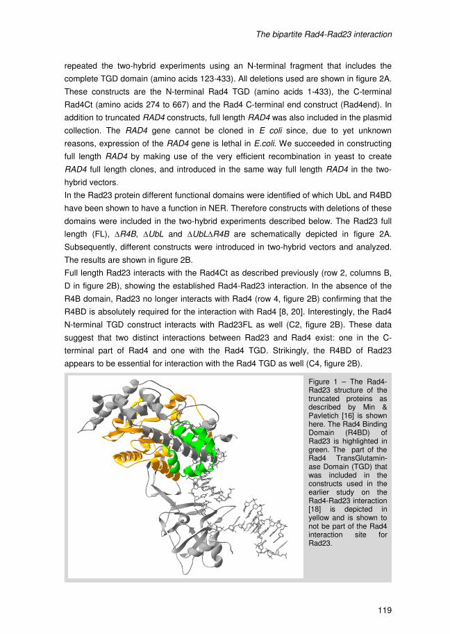
The bipartite Rad4-Rad23 interaction
118
partial Rad23 protein containing the Rad4 Binding Domain (R4BD) and UBA2 (Figure 1)
interacts with the Rad4 TGD domain.
The Rad4-Rad23 interaction was suggested to occur at the C-terminal domain of Rad4
domain, supported by our previous data [18]. Similarly, the interaction of XPC and
hHR23B was also defined to reside in the C-terminal part of XPC [17]. Together these
data suggest a possible interaction of Rad23 with a C-terminal part of Rad4, distinct from
the TGD interaction shown in the protein structure. We decided to further investigate the
Rad4-Rad23 interaction. We show that the Rad4-Rad23 interaction can occur on two
sites on Rad4, one with the TGD and one at C-terminal region of Rad4. Interestingly, the
Rad23 UbL-domain differently affects the two interactions.
3. Material & Methods
3.1 Strains and plasmids
All strains used in this study are derived from the wild-type W1588-4a [19]. Relevant
phenotypes are described in table 1. Full length RAD4 cannot be propagated in E.coli
due to lethality. By using the high efficiency of homologous recombination in yeast we
constructed the full length RAD4 plasmid in Saccharomyces cerevisiae (described
elsewhere).
3.2 Yeast Two-hybrid assay
For yeast twohybrid analysis the Clontech Matchmaker 3 system was used. The
respective DNA fragments indicated in figure 2A were put in-frame with the GAL4
activating domain (AD) in pGADT7 or the GAL4 binding domain (BD) in pGBKT7.
Subsequently, the resulting fusion constructs were introduced into the selection strains
Y187 and AH109, respectively. After mating, cells were spotted onto selection plates.
Selection for the presence of both plasmids was maintained by growing on plates lacking
tryptophan and leucine while selection for interaction is a result of the histidine and
adenine selection markers with a GAL1 UAS and TATA construct in their promoter
driving transcription. After growth at 30oC the plates were monitored every day for 3
days.
4. Results & Discussion
4.1 The Rad4-Rad23 interaction has a bipartite binding
We previously reported the two-hybrid interaction of the Rad4 C-terminal part (amino
acids 274-667) with Rad23 [18]. The deletions used in this study were based on the
homology found between all Rad4 like proteins [4] and included only part of the so-called
TGD domain of Rad4 (see figure 1). Structure analysis of Rad4 showed association of
the TGD domain of Rad4 with Rad23 [16] with a section of the TGD domain that was not
included in the C-terminal construct tested previously (Figure 1) [18]. Therefore we
The bipartite Rad4-Rad23 interaction
119
repeated the two-hybrid experiments using an N-terminal fragment that includes the
complete TGD domain (amino acids 123-433). All deletions used are shown in figure 2A.
These constructs are the N-terminal Rad4 TGD (amino acids 1-433), the C-terminal
Rad4Ct (amino acids 274 to 667) and the Rad4 C-terminal end construct (Rad4end). In
addition to truncated RAD4 constructs, full length RAD4 was also included in the plasmid
collection. The RAD4 gene cannot be cloned in E coli since, due to yet unknown
reasons, expression of the RAD4 gene is lethal in E.coli. We succeeded in constructing
full length RAD4 by making use of the very efficient recombination in yeast to create
RAD4 full length clones, and introduced in the same way full length RAD4 in the two-
hybrid vectors.
In the Rad23 protein different functional domains were identified of which UbL and R4BD
have been shown to have a function in NER. Therefore constructs with deletions of these
domains were included in the two-hybrid experiments described below. The Rad23 full
length (FL), ∆R4B, ∆UbL and ∆UbL∆R4B are schematically depicted in figure 2A.
Subsequently, different constructs were introduced in two-hybrid vectors and analyzed.
The results are shown in figure 2B.
Full length Rad23 interacts with the Rad4Ct as described previously (row 2, columns B,
D in figure 2B), showing the established Rad4-Rad23 interaction. In the absence of the
R4B domain, Rad23 no longer interacts with Rad4 (row 4, figure 2B) confirming that the
R4BD is absolutely required for the interaction with Rad4 [8, 20]. Interestingly, the Rad4
N-terminal TGD construct interacts with Rad23FL as well (C2, figure 2B). These data
suggest that two distinct interactions between Rad23 and Rad4 exist: one in the C-
terminal part of Rad4 and one with the Rad4 TGD. Strikingly, the R4BD of Rad23
appears to be essential for interaction with the Rad4 TGD as well (C4, figure 2B).
Figure 1 – The Rad4-Rad23 structure of the truncated proteins as described by Min & Pavletich [16] is shown here. The Rad4 Binding Domain (R4BD) of Rad23 is highlighted in green. The part of the Rad4 TransGlutamin-ase Domain (TGD) that was included in the constructs used in the earlier study on the Rad4-Rad23 interaction [18] is depicted in yellow and is shown to not be part of the Rad4 interaction site for Rad23.
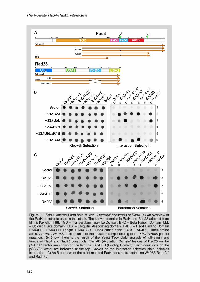
The bipartite Rad4-Rad23 interaction
120
Figure 2 – Rad23 interacts with both N- and C-terminal constructs of Rad4. (A) An overview of the Rad4 constructs used in this study. The known domains in Rad4 and Rad23 adopted from Min & Pavletich [16]. TGD – TransGlutaminase-like Domain. BHD – Beta Hairpin Domain. UbL – Ubiquitin Like domain. UBA – Ubiquitin Associating domain. R4BD – Rad4 Binding DomainRAD4FL – RAD4 Full Length. RAD4TGD – Rad4 amino acids 0-433. RAD4Ct – Rad4 amino acids. 274-667. W496S – the location of the mutation corrpesonding to the XPC-W690S patient mutation. (B) Shown here is the result of the Yeast Two-hybrid analysis of full-length andtruncated Rad4 and Rad23 constructs. The AD (Activation Domain’ fusions of Rad23 on the pGADT7 vector are shown on the left, the Rad4 BD (Binding Domain) fusion-constructs on the pGBKT7 vector are indicated at the top. Growth on the interaction selection plate indicates interaction. (C) As B but now for the point-mutated Rad4 constructs containing W496S Rad4Ct* and Rad4FL*.
The bipartite Rad4-Rad23 interaction
121
4.2 The Rad23 Ubiquitin-like Domain affects the Rad4-Rad23 bipartite interaction
The bipartite Rad4-Rad23 interaction was further confirmed when the Rad23∆UbL
construct was included in the analysis. Rad23 without the UbL domain interacts with
Rad4FL (B3) and Rad4TGD (C3, figure 2B). However, in the absence of the UbL domain
Rad23 can no longer sustain an interaction with the Rad4Ct (D3) and Rad4end (E3)
constructs that only contain the C-terminal Rad4 interaction site (Figure 2B). Thus the
Rad23 UbL domain is required for Rad23 to interact with the C-terminal end of Rad4 but
not for the interaction with the N-terminal region.
Interestingly, the Rad23∆UbL-Rad4TGD interaction (C3) is stronger than the Rad23FL-
Rad4TGD interaction (C2, figure 2B). Thus the N-terminal interaction improves when the
Rad23 UbL domain is deleted. This differential effect of the UbL domain on the Rad4-
Rad23 interactions (stimulation of binding to the C-terminal region and inhibition of
binding to the TGD) shows that Rad4 indeed contains two distinct binding sites for
Rad23. The Rad23FL interaction with the full-length Rad4 construct (B2) also weakens
when the UbL is absent (Figure 2B), suggesting that the C-terminal region is the
preferred interaction site in vivo.
The R4BD is required for both interactions with Rad4, but in view of the small size of this
domain and its position in the co-crystal structure, it is unlikely that Rad23 can
simultaneously bind to both domains in Rad4. It is possible that the UbL domain, through
the intramolecular interactions with a UBA domain, changes the conformation of Rad23
required to differentially interact with either the N- or C-terminal binding region of Rad4.
In the absence of the UbL domain Rad23 might exist as an 'open' conformation (no UbL-
UBA interaction) that can only interact with the TGD of Rad4. At the same time this
would imply that a 'closed' form of Rad23 due to intramolecular UbL-UBA interaction,
preferentially interacts with the C-terminal region of Rad4. It has been described for the
hHR23B protein that the interaction between UbL and UBA domains can be disrupted by
binding to a proteasomal subunit. Similarly, in Rad23 the UbL and UBA interaction might
be disrupted by yeast NER factors, thereby switching Rad23 from one Rad4 binding
mode to the other.
4.3 The Rad34-Rad23 interaction behaves similar to the N-terminal Rad4-Rad23 interaction
The Rad34 protein is a Rad4-like NER factor uniquely present in yeast and specifically
required for TC-NER of active rDNA. The homology between Rad4 and Rad34 is
supported by the sequence conservation and more importantly, by the analogous
interaction of Rad4 and Rad34 with both Rad23 and Rad33. To test if this homology
includes the novel bipartite interaction we studied the interaction of Rad34 with the same
Rad23 deletion constructs (Figure 2B, column G). We find that the Rad23∆UbL-Rad34
interaction (G3) is stronger than the Rad23FL-Rad34 interaction (G2, figure 2B). Thus
The bipartite Rad4-Rad23 interaction
122
the absence of the UbL domain strengthens the Rad23-Rad34 interaction. This pattern is
analogous to that of the N-terminal Rad23-Rad4TGD interaction. We therefore speculate
that Rad23 might only interact with the TGD-like domain of Rad34 and possibly lacks a
C-terminal interaction. The amino acid sequences of Rad4 and Rad34 reveal that Rad4
contains a longer C-terminal region compared to Rad34 [4], perhaps explaining why
Rad34 cannot support a C-terminal Rad23 interaction.
Rad34 is specifically involved in TC-NER at the rDNA locus [4, 5]. This could imply that if
Rad23 interacts via the Rad34 TGD-like domain that this interaction is sufficient to drive
TC-NER at the rDNA. Vice versa, the Rad23 interaction at the C-terminus of Rad4 might
be GG-NER specific and could not exist in the Rad34 molecule.
4.4 The C-terminal Rad4-Rad23 interaction is disrupted by a RAD4 mutation analogous to an XPC patient mutation
Interestingly, in the C-terminal region of XPC a patient mutation has been found that still
leads to a full-length XPC protein [21]. The tryptophan residue at position 690 in XPC is
affected by this mutation and this residue is conserved in Rad4. The XPC-W690S protein
has reduced affinity for damaged DNA and results in reduced repair capacity and UV
survival in human cells [21-23]. The analogous mutation rad4W496S in yeast results in a
NER defect, where GG-NER is more severely hampered than TC-NER (Chapter 4). The
effect of this mutation on the Rad4 and XPC protein function in NER and the location in
the C-terminus of this residue make it an interesting target for study in yeast. Therefore,
we measure the effect of the rad4W496S mutation in RAD4 on the Rad4-Rad23
interaction using yeast two-hybrid experiments (Figure 2C).
Interestingly, the W496S mutation completely disrupts the interaction of Rad23 with the
Rad4 C-terminal construct (compare E2 to F2, figure 2C) as no interaction between
Rad23FL and the mutated Rad4 C-terminal construct (Rad4Ct*) is observed. However,
full length Rad4 protein containing the W496S substitution (Rad4FL* in figure 2C) does
still interact with Rad23 (B2 and C2, figure 2C). This interaction is reduced compared to
the wild-type Rad4 construct but similar to the Rad4TGD construct. Morever, in the
absence of the UbL domain the interaction between mutant Rad4 and Rad23 is stronger
(C2 and C3). This indicates that in rad4W496S, Rad23 can only bind to the TGD.
Extending these observations with the data from chapter 4, it becomes apparent that the
GG-NER deficiency as a result of the rad4W496S substitution could be linked to the loss
of the C-terminal Rad4-Rad23 interaction. In other words, the C-terminal Rad4-Rad23
interaction is more important for GG-NER than TC-NER. We have also shown (Chapter
4) that the rad4W496S mutation also affects the activity of Rad4 in the absence of
Rad23. In a rad23∆ background the rad4W496S mutant is more UV sensitive than wild-
type RAD4. Most likely, the mutation influences the conformation of the Rad4 protein
such that on one hand it reduces the affinity of Rad4 for DNA and on the other hand it
disrupts the C-terminal interaction with Rad23.
The bipartite Rad4-Rad23 interaction
123
5. Concluding Remarks In summary, we report that the R4BD of Rad23 can interact with two distinct binding
domains on Rad4, one in the TGD and one in the C-terminal part of Rad4. We propose
that the switch between the two types of binding is regulated by the UbL domain of
Rad23. A mutation In Rad4 that disrupts interaction of Rad23 with the C-terminal domain
is mainly affected in GG-NER. On the other hand, the TC-NER specific Rad34 protein
seem to mainly interact with Rad23 via a TGD-like domain. Taken together these data
suggest that the UbL mediated switching between the different Rad23 binding modes
direct the Rad4-Rad23 complex to either GG-NER or TC-NER.
The bipartite Rad4-Rad23 interaction
122
the absence of the UbL domain strengthens the Rad23-Rad34 interaction. This pattern is
analogous to that of the N-terminal Rad23-Rad4TGD interaction. We therefore speculate
that Rad23 might only interact with the TGD-like domain of Rad34 and possibly lacks a
C-terminal interaction. The amino acid sequences of Rad4 and Rad34 reveal that Rad4
contains a longer C-terminal region compared to Rad34 [4], perhaps explaining why
Rad34 cannot support a C-terminal Rad23 interaction.
Rad34 is specifically involved in TC-NER at the rDNA locus [4, 5]. This could imply that if
Rad23 interacts via the Rad34 TGD-like domain that this interaction is sufficient to drive
TC-NER at the rDNA. Vice versa, the Rad23 interaction at the C-terminus of Rad4 might
be GG-NER specific and could not exist in the Rad34 molecule.
4.4 The C-terminal Rad4-Rad23 interaction is disrupted by a RAD4 mutation analogous to an XPC patient mutation
Interestingly, in the C-terminal region of XPC a patient mutation has been found that still
leads to a full-length XPC protein [21]. The tryptophan residue at position 690 in XPC is
affected by this mutation and this residue is conserved in Rad4. The XPC-W690S protein
has reduced affinity for damaged DNA and results in reduced repair capacity and UV
survival in human cells [21-23]. The analogous mutation rad4W496S in yeast results in a
NER defect, where GG-NER is more severely hampered than TC-NER (Chapter 4). The
effect of this mutation on the Rad4 and XPC protein function in NER and the location in
the C-terminus of this residue make it an interesting target for study in yeast. Therefore,
we measure the effect of the rad4W496S mutation in RAD4 on the Rad4-Rad23
interaction using yeast two-hybrid experiments (Figure 2C).
Interestingly, the W496S mutation completely disrupts the interaction of Rad23 with the
Rad4 C-terminal construct (compare E2 to F2, figure 2C) as no interaction between
Rad23FL and the mutated Rad4 C-terminal construct (Rad4Ct*) is observed. However,
full length Rad4 protein containing the W496S substitution (Rad4FL* in figure 2C) does
still interact with Rad23 (B2 and C2, figure 2C). This interaction is reduced compared to
the wild-type Rad4 construct but similar to the Rad4TGD construct. Morever, in the
absence of the UbL domain the interaction between mutant Rad4 and Rad23 is stronger
(C2 and C3). This indicates that in rad4W496S, Rad23 can only bind to the TGD.
Extending these observations with the data from chapter 4, it becomes apparent that the
GG-NER deficiency as a result of the rad4W496S substitution could be linked to the loss
of the C-terminal Rad4-Rad23 interaction. In other words, the C-terminal Rad4-Rad23
interaction is more important for GG-NER than TC-NER. We have also shown (Chapter
4) that the rad4W496S mutation also affects the activity of Rad4 in the absence of
Rad23. In a rad23∆ background the rad4W496S mutant is more UV sensitive than wild-
type RAD4. Most likely, the mutation influences the conformation of the Rad4 protein
such that on one hand it reduces the affinity of Rad4 for DNA and on the other hand it
disrupts the C-terminal interaction with Rad23.
The bipartite Rad4-Rad23 interaction
123
5. Concluding Remarks In summary, we report that the R4BD of Rad23 can interact with two distinct binding
domains on Rad4, one in the TGD and one in the C-terminal part of Rad4. We propose
that the switch between the two types of binding is regulated by the UbL domain of
Rad23. A mutation In Rad4 that disrupts interaction of Rad23 with the C-terminal domain
is mainly affected in GG-NER. On the other hand, the TC-NER specific Rad34 protein
seem to mainly interact with Rad23 via a TGD-like domain. Taken together these data
suggest that the UbL mediated switching between the different Rad23 binding modes
direct the Rad4-Rad23 complex to either GG-NER or TC-NER.

The bipartite Rad4-Rad23 interaction
124
Table 1 - Overview of the strains used in this studied with their relevant genotypes and
origin.
Strain Genotype Source
W1588-4a MATα leu2-3,112 ade2-1 can1-100 his3-11,15 ura3-1 trp1-1
R. Rothstein [19]
MGSC691 MATa, trp1-901, leu2-3, 112, ura3-52, his3-200, gal4∆,
gal80∆, LYS2 : : GAL1UAS-GAL1TATA-HIS3, GAL2UAS-GAL2TATA-ADE2, URA3 : : MEL1UAS-MEL1TATA-lacZ
AH109 [24]
MGSC692 MATa, ura3-52, his3-200, ade2-101, trp1-901 leu2-3, 112, gal4del, met-, gal80del, URA3 : : GAL1UAS-GAL1TATA-lacZ
Y187 [24]
The bipartite Rad4-Rad23 interaction
125
6. References 1. den Dulk, B., et al., Rad33, a new factor involved in nucleotide excision repair in
Saccharomyces cerevisiae. DNA Repair (Amst), 2006. 5(6): p. 683-92.
2. Jansen, L.E., R.A. Verhage, and J. Brouwer, Preferential binding of yeast Rad4.Rad23
complex to damaged DNA. J Biol Chem, 1998. 273(50): p. 33111-4.
3. Araki, M., et al., Centrosome protein centrin 2/caltractin 1 is part of the xeroderma
pigmentosum group C complex that initiates global genome nucleotide excision repair. J
Biol Chem, 2001. 276(22): p. 18665-72.
4. den Dulk, B., J.A. Brandsma, and J. Brouwer, The Rad4 homologue YDR314C is
essential for strand-specific repair of RNA polymerase I-transcribed rDNA in
Saccharomyces cerevisiae. Mol Microbiol, 2005. 56(6): p. 1518-26.
5. Tremblay, M., et al., Complementary roles of yeast Rad4p and Rad34p in nucleotide
excision repair of active and inactive rRNA gene chromatin. Mol Cell Biol, 2008. 28(24):
p. 7504-13.
6. Verhage, R., et al., The RAD7 and RAD16 genes, which are essential for pyrimidine
dimer removal from the silent mating type loci, are also required for repair of the
nontranscribed strand of an active gene in Saccharomyces cerevisiae. Mol Cell Biol,
1994. 14(9): p. 6135-42.
7. Gillette, T.G., et al., Distinct functions of the ubiquitin-proteasome pathway influence
nucleotide excision repair. Embo Journal, 2006. 25(11): p. 2529-38.
8. Ortolan, T.G., et al., Rad23 stabilizes Rad4 from degradation by the Ub/proteasome
pathway. Nucleic Acids Res, 2004. 32(22): p. 6490-500.
9. Chen, L. and K. Madura, Rad23 Promotes the Targeting of Proteolytic Substrates to the
Proteasome. Molecular and Cellular Biology, 2002. 22(13): p. 4902-4913.
10. Bertolaet, B.L., et al., UBA domains mediate protein-protein interactions between two
DNA damage-inducible proteins. Journal of Molecular Biology, 2001. 313(5): p. 955-63.
11. Bertolaet, B.L., et al., UBA domains of DNA damage-inducible proteins interact with
ubiquitin. Nat Struct Biol, 2001. 8(5): p. 417-22.
12. Biggins, S., I. Ivanovska, and M.D. Rose, Yeast ubiquitin-like genes are involved in
duplication of the microtubule organizing center. J Cell Biol, 1996. 133(6): p. 1331-46.
13. Watkins, J.F., et al., The Saccharomyces cerevisiae DNA repair gene RAD23 encodes a
nuclear protein containing a ubiquitin-like domain required for biological function. Mol
Cell Biol, 1993. 13(12): p. 7757-65.
14. Heessen, S., M.G. Masucci, and N.P. Dantuma, The UBA2 domain functions as an
intrinsic stabilization signal that protects Rad23 from proteasomal degradation. Molecular
Cell, 2005. 18(2): p. 225-35.
15. Russell, S.J., et al., The 19S regulatory complex of the proteasome functions
independently of proteolysis in nucleotide excision repair. Molecular Cell, 1999. 3(6): p.
687-95.
16. Min, J.H. and N.P. Pavletich, Recognition of DNA damage by the Rad4 nucleotide
excision repair protein. Nature, 2007. 449(7162): p. 570-5.

The bipartite Rad4-Rad23 interaction
124
Table 1 - Overview of the strains used in this studied with their relevant genotypes and
origin.
Strain Genotype Source
W1588-4a MATα leu2-3,112 ade2-1 can1-100 his3-11,15 ura3-1 trp1-1
R. Rothstein [19]
MGSC691 MATa, trp1-901, leu2-3, 112, ura3-52, his3-200, gal4∆,
gal80∆, LYS2 : : GAL1UAS-GAL1TATA-HIS3, GAL2UAS-GAL2TATA-ADE2, URA3 : : MEL1UAS-MEL1TATA-lacZ
AH109 [24]
MGSC692 MATa, ura3-52, his3-200, ade2-101, trp1-901 leu2-3, 112, gal4del, met-, gal80del, URA3 : : GAL1UAS-GAL1TATA-lacZ
Y187 [24]
The bipartite Rad4-Rad23 interaction
125
6. References 1. den Dulk, B., et al., Rad33, a new factor involved in nucleotide excision repair in
Saccharomyces cerevisiae. DNA Repair (Amst), 2006. 5(6): p. 683-92.
2. Jansen, L.E., R.A. Verhage, and J. Brouwer, Preferential binding of yeast Rad4.Rad23
complex to damaged DNA. J Biol Chem, 1998. 273(50): p. 33111-4.
3. Araki, M., et al., Centrosome protein centrin 2/caltractin 1 is part of the xeroderma
pigmentosum group C complex that initiates global genome nucleotide excision repair. J
Biol Chem, 2001. 276(22): p. 18665-72.
4. den Dulk, B., J.A. Brandsma, and J. Brouwer, The Rad4 homologue YDR314C is
essential for strand-specific repair of RNA polymerase I-transcribed rDNA in
Saccharomyces cerevisiae. Mol Microbiol, 2005. 56(6): p. 1518-26.
5. Tremblay, M., et al., Complementary roles of yeast Rad4p and Rad34p in nucleotide
excision repair of active and inactive rRNA gene chromatin. Mol Cell Biol, 2008. 28(24):
p. 7504-13.
6. Verhage, R., et al., The RAD7 and RAD16 genes, which are essential for pyrimidine
dimer removal from the silent mating type loci, are also required for repair of the
nontranscribed strand of an active gene in Saccharomyces cerevisiae. Mol Cell Biol,
1994. 14(9): p. 6135-42.
7. Gillette, T.G., et al., Distinct functions of the ubiquitin-proteasome pathway influence
nucleotide excision repair. Embo Journal, 2006. 25(11): p. 2529-38.
8. Ortolan, T.G., et al., Rad23 stabilizes Rad4 from degradation by the Ub/proteasome
pathway. Nucleic Acids Res, 2004. 32(22): p. 6490-500.
9. Chen, L. and K. Madura, Rad23 Promotes the Targeting of Proteolytic Substrates to the
Proteasome. Molecular and Cellular Biology, 2002. 22(13): p. 4902-4913.
10. Bertolaet, B.L., et al., UBA domains mediate protein-protein interactions between two
DNA damage-inducible proteins. Journal of Molecular Biology, 2001. 313(5): p. 955-63.
11. Bertolaet, B.L., et al., UBA domains of DNA damage-inducible proteins interact with
ubiquitin. Nat Struct Biol, 2001. 8(5): p. 417-22.
12. Biggins, S., I. Ivanovska, and M.D. Rose, Yeast ubiquitin-like genes are involved in
duplication of the microtubule organizing center. J Cell Biol, 1996. 133(6): p. 1331-46.
13. Watkins, J.F., et al., The Saccharomyces cerevisiae DNA repair gene RAD23 encodes a
nuclear protein containing a ubiquitin-like domain required for biological function. Mol
Cell Biol, 1993. 13(12): p. 7757-65.
14. Heessen, S., M.G. Masucci, and N.P. Dantuma, The UBA2 domain functions as an
intrinsic stabilization signal that protects Rad23 from proteasomal degradation. Molecular
Cell, 2005. 18(2): p. 225-35.
15. Russell, S.J., et al., The 19S regulatory complex of the proteasome functions
independently of proteolysis in nucleotide excision repair. Molecular Cell, 1999. 3(6): p.
687-95.
16. Min, J.H. and N.P. Pavletich, Recognition of DNA damage by the Rad4 nucleotide
excision repair protein. Nature, 2007. 449(7162): p. 570-5.
The bipartite Rad4-Rad23 interaction
126
17. Uchida, A., et al., The carboxy-terminal domain of the XPC protein plays a crucial role in
nucleotide excision repair through interactions with transcription factor IIH. DNA Repair
(Amst), 2002. 1(6): p. 449-61.
18. den Dulk, B., et al., The NER protein Rad33 shows functional homology to human
Centrin2 and is involved in modification of Rad4. DNA Repair (Amst), 2008. 7(6): p. 858-
68.
19. Mortensen, U.H., et al., A molecular genetic dissection of the evolutionarily conserved N
terminus of yeast Rad52. Genetics, 2002. 161(2): p. 549-62.
20. Masutani, C., et al., Identification and characterization of XPC-binding domain of
hHR23B. Mol Cell Biol, 1997. 17(12): p. 6915-23.
21. Chavanne, F., et al., Mutations in the XPC gene in families with xeroderma pigmentosum
and consequences at the cell, protein, and transcript levels. Cancer Res, 2000. 60(7): p.
1974-82.
22. Bunick, C.G., et al., Biochemical and structural domain analysis of xeroderma
pigmentosum complementation group C protein. Biochemistry, 2006. 45(50): p. 14965-
79.
23. Yasuda, G., et al., In vivo destabilization and functional defects of the xeroderma
pigmentosum C protein caused by a pathogenic missense mutation. Mol Cell Biol, 2007.
27(19): p. 6606-14.
24. James, P., J. Halladay, and E.A. Craig, Genomic libraries and a host strain designed for
highly efficient two-hybrid selection in yeast. Genetics, 1996. 144(4): p. 1425-36.
VI WCG4A, a commonly used Saccharomyces cerevisiae yeast strain, contains a RAD4 mutation
affecting UV resistance
P. van Eijk, Dr. J.A. Brandsma, M. de Ruijter, Prof. Dr. J. Brouwer

A RAD4 mutation in the WCG4A yeast strain
128
1. Abstract WCG4A is one of the Saccharomyces cerevisiae strains commonly used in DNA repair
and transcription studies. Especially when proteasome mutants are required this strain is
frequently applied since conditional proteasome mutants are available in this
background. Investigating the relation between Nucleotide Excision Repair (NER) and
the proteasome we found unexpected, extreme UV sensitivity in different NER deficient
derivatives of this strain when compared to the sensitivity in another yeast background,
W1588. We identified a mutation in RAD4 resulting in a C to Y substitution at position
571 of the Rad4 protein in WCG4A. Introducing the mutation responsible for this
phenotype into a W1588 wild-type background leads to a comparable phenotype and
similar genetic interactions as found for the WCG4A strain.
A RAD4 mutation in the WCG4A yeast strain
129
2. Introduction Nucleotide Excision Repair (NER) is a general DNA repair process which is conserved
in many organisms. The yeast Saccharomyces cerevisiae is widely used as a model
organism. A recent finding implicates the proteasome in yeast NER in which Rad23 has
a pivotal role [1, 2]. In this respect the yeast wild-type strain WCG4A is important
because conditional mutants of the 19S and 20S subunits of the proteasome are readily
available in this background [3, 4].
We recently identified a novel protein in yeast, Rad33, which is part of the DNA damage
recognition complex Rad4-Rad23 and we showed its interaction with Rad4 via a
conserved motif [5, 6]. To study the function of Rad33 in NER in relation to the
proteasome, we introduced a RAD33 deletion into the WCG4A strain and studied UV
survival. We observed an unusually high UV sensitivity in a WCG4A cells containing a
RAD33 deletion. Furthermore, mutations in other well described RAD mutants in
WCG4A also resulted in higher than expected UV sensitivity. It appeared that a point
mutation in the RAD4 gene of the WCG4A wild-type strain is the culprit.
3. Materials and Methods
3.1 Survival Curve & UV droptest
Yeast cells were grown to stationary cultures by 3 day incubations at 30°C and diluted in
water to an appropriate OD600. Cells were plated on YPD and UV irradiated with the
relevant UV dose using a 254nm UV-C lamp (Philips). After a 3 day incubation in the
dark at 30°C, colonies were counted and quantified. Data shown are the mean result of 3
individual experiments.
Cells for UV droptests were prepared in a similar fashion, however instead of plating the
cells the cell suspension in water (1.5 µL cell suspension in 0.5 mL MilliQ water) was
used to dispense 1.5 µL drops on a YPD plate. The YPD plates were irradiated with
increasing UV dose relevant to the strains being tested. The UV dose used is indicated
in the figures. After UV irradiation YPD plates were incubated in the dark at 30°C for 2-3
days and growth was monitored regularly. Representative scans of these plates are
included in the figures described here.
3.2 Mating
The mating of W1588 with WCG4A strains was performed as follows: the two strains are
plated together on a YPD plate and incubated at 30°C for 4 hours. Due to the absence of
selectable markers zygotes are then selected based on morphology and separated by
using a micromanipulator. After two days of growth the size of the colonies and cells can
be used to judge the presence of diploid cells. Of each mating 12 zygotes were picked of
which 6 were subjected to UV phenotyping.
A RAD4 mutation in the WCG4A yeast strain
128
1. Abstract WCG4A is one of the Saccharomyces cerevisiae strains commonly used in DNA repair
and transcription studies. Especially when proteasome mutants are required this strain is
frequently applied since conditional proteasome mutants are available in this
background. Investigating the relation between Nucleotide Excision Repair (NER) and
the proteasome we found unexpected, extreme UV sensitivity in different NER deficient
derivatives of this strain when compared to the sensitivity in another yeast background,
W1588. We identified a mutation in RAD4 resulting in a C to Y substitution at position
571 of the Rad4 protein in WCG4A. Introducing the mutation responsible for this
phenotype into a W1588 wild-type background leads to a comparable phenotype and
similar genetic interactions as found for the WCG4A strain.
A RAD4 mutation in the WCG4A yeast strain
129
2. Introduction Nucleotide Excision Repair (NER) is a general DNA repair process which is conserved
in many organisms. The yeast Saccharomyces cerevisiae is widely used as a model
organism. A recent finding implicates the proteasome in yeast NER in which Rad23 has
a pivotal role [1, 2]. In this respect the yeast wild-type strain WCG4A is important
because conditional mutants of the 19S and 20S subunits of the proteasome are readily
available in this background [3, 4].
We recently identified a novel protein in yeast, Rad33, which is part of the DNA damage
recognition complex Rad4-Rad23 and we showed its interaction with Rad4 via a
conserved motif [5, 6]. To study the function of Rad33 in NER in relation to the
proteasome, we introduced a RAD33 deletion into the WCG4A strain and studied UV
survival. We observed an unusually high UV sensitivity in a WCG4A cells containing a
RAD33 deletion. Furthermore, mutations in other well described RAD mutants in
WCG4A also resulted in higher than expected UV sensitivity. It appeared that a point
mutation in the RAD4 gene of the WCG4A wild-type strain is the culprit.
3. Materials and Methods
3.1 Survival Curve & UV droptest
Yeast cells were grown to stationary cultures by 3 day incubations at 30°C and diluted in
water to an appropriate OD600. Cells were plated on YPD and UV irradiated with the
relevant UV dose using a 254nm UV-C lamp (Philips). After a 3 day incubation in the
dark at 30°C, colonies were counted and quantified. Data shown are the mean result of 3
individual experiments.
Cells for UV droptests were prepared in a similar fashion, however instead of plating the
cells the cell suspension in water (1.5 µL cell suspension in 0.5 mL MilliQ water) was
used to dispense 1.5 µL drops on a YPD plate. The YPD plates were irradiated with
increasing UV dose relevant to the strains being tested. The UV dose used is indicated
in the figures. After UV irradiation YPD plates were incubated in the dark at 30°C for 2-3
days and growth was monitored regularly. Representative scans of these plates are
included in the figures described here.
3.2 Mating
The mating of W1588 with WCG4A strains was performed as follows: the two strains are
plated together on a YPD plate and incubated at 30°C for 4 hours. Due to the absence of
selectable markers zygotes are then selected based on morphology and separated by
using a micromanipulator. After two days of growth the size of the colonies and cells can
be used to judge the presence of diploid cells. Of each mating 12 zygotes were picked of
which 6 were subjected to UV phenotyping.

A RAD4 mutation in the WCG4A yeast strain
128
1. Abstract WCG4A is one of the Saccharomyces cerevisiae strains commonly used in DNA repair
and transcription studies. Especially when proteasome mutants are required this strain is
frequently applied since conditional proteasome mutants are available in this
background. Investigating the relation between Nucleotide Excision Repair (NER) and
the proteasome we found unexpected, extreme UV sensitivity in different NER deficient
derivatives of this strain when compared to the sensitivity in another yeast background,
W1588. We identified a mutation in RAD4 resulting in a C to Y substitution at position
571 of the Rad4 protein in WCG4A. Introducing the mutation responsible for this
phenotype into a W1588 wild-type background leads to a comparable phenotype and
similar genetic interactions as found for the WCG4A strain.
A RAD4 mutation in the WCG4A yeast strain
129
2. Introduction Nucleotide Excision Repair (NER) is a general DNA repair process which is conserved
in many organisms. The yeast Saccharomyces cerevisiae is widely used as a model
organism. A recent finding implicates the proteasome in yeast NER in which Rad23 has
a pivotal role [1, 2]. In this respect the yeast wild-type strain WCG4A is important
because conditional mutants of the 19S and 20S subunits of the proteasome are readily
available in this background [3, 4].
We recently identified a novel protein in yeast, Rad33, which is part of the DNA damage
recognition complex Rad4-Rad23 and we showed its interaction with Rad4 via a
conserved motif [5, 6]. To study the function of Rad33 in NER in relation to the
proteasome, we introduced a RAD33 deletion into the WCG4A strain and studied UV
survival. We observed an unusually high UV sensitivity in a WCG4A cells containing a
RAD33 deletion. Furthermore, mutations in other well described RAD mutants in
WCG4A also resulted in higher than expected UV sensitivity. It appeared that a point
mutation in the RAD4 gene of the WCG4A wild-type strain is the culprit.
3. Materials and Methods
3.1 Survival Curve & UV droptest
Yeast cells were grown to stationary cultures by 3 day incubations at 30°C and diluted in
water to an appropriate OD600. Cells were plated on YPD and UV irradiated with the
relevant UV dose using a 254nm UV-C lamp (Philips). After a 3 day incubation in the
dark at 30°C, colonies were counted and quantified. Data shown are the mean result of 3
individual experiments.
Cells for UV droptests were prepared in a similar fashion, however instead of plating the
cells the cell suspension in water (1.5 µL cell suspension in 0.5 mL MilliQ water) was
used to dispense 1.5 µL drops on a YPD plate. The YPD plates were irradiated with
increasing UV dose relevant to the strains being tested. The UV dose used is indicated
in the figures. After UV irradiation YPD plates were incubated in the dark at 30°C for 2-3
days and growth was monitored regularly. Representative scans of these plates are
included in the figures described here.
3.2 Mating
The mating of W1588 with WCG4A strains was performed as follows: the two strains are
plated together on a YPD plate and incubated at 30°C for 4 hours. Due to the absence of
selectable markers zygotes are then selected based on morphology and separated by
using a micromanipulator. After two days of growth the size of the colonies and cells can
be used to judge the presence of diploid cells. Of each mating 12 zygotes were picked of
which 6 were subjected to UV phenotyping.
A RAD4 mutation in the WCG4A yeast strain
128
1. Abstract WCG4A is one of the Saccharomyces cerevisiae strains commonly used in DNA repair
and transcription studies. Especially when proteasome mutants are required this strain is
frequently applied since conditional proteasome mutants are available in this
background. Investigating the relation between Nucleotide Excision Repair (NER) and
the proteasome we found unexpected, extreme UV sensitivity in different NER deficient
derivatives of this strain when compared to the sensitivity in another yeast background,
W1588. We identified a mutation in RAD4 resulting in a C to Y substitution at position
571 of the Rad4 protein in WCG4A. Introducing the mutation responsible for this
phenotype into a W1588 wild-type background leads to a comparable phenotype and
similar genetic interactions as found for the WCG4A strain.
A RAD4 mutation in the WCG4A yeast strain
129
2. Introduction Nucleotide Excision Repair (NER) is a general DNA repair process which is conserved
in many organisms. The yeast Saccharomyces cerevisiae is widely used as a model
organism. A recent finding implicates the proteasome in yeast NER in which Rad23 has
a pivotal role [1, 2]. In this respect the yeast wild-type strain WCG4A is important
because conditional mutants of the 19S and 20S subunits of the proteasome are readily
available in this background [3, 4].
We recently identified a novel protein in yeast, Rad33, which is part of the DNA damage
recognition complex Rad4-Rad23 and we showed its interaction with Rad4 via a
conserved motif [5, 6]. To study the function of Rad33 in NER in relation to the
proteasome, we introduced a RAD33 deletion into the WCG4A strain and studied UV
survival. We observed an unusually high UV sensitivity in a WCG4A cells containing a
RAD33 deletion. Furthermore, mutations in other well described RAD mutants in
WCG4A also resulted in higher than expected UV sensitivity. It appeared that a point
mutation in the RAD4 gene of the WCG4A wild-type strain is the culprit.
3. Materials and Methods
3.1 Survival Curve & UV droptest
Yeast cells were grown to stationary cultures by 3 day incubations at 30°C and diluted in
water to an appropriate OD600. Cells were plated on YPD and UV irradiated with the
relevant UV dose using a 254nm UV-C lamp (Philips). After a 3 day incubation in the
dark at 30°C, colonies were counted and quantified. Data shown are the mean result of 3
individual experiments.
Cells for UV droptests were prepared in a similar fashion, however instead of plating the
cells the cell suspension in water (1.5 µL cell suspension in 0.5 mL MilliQ water) was
used to dispense 1.5 µL drops on a YPD plate. The YPD plates were irradiated with
increasing UV dose relevant to the strains being tested. The UV dose used is indicated
in the figures. After UV irradiation YPD plates were incubated in the dark at 30°C for 2-3
days and growth was monitored regularly. Representative scans of these plates are
included in the figures described here.
3.2 Mating
The mating of W1588 with WCG4A strains was performed as follows: the two strains are
plated together on a YPD plate and incubated at 30°C for 4 hours. Due to the absence of
selectable markers zygotes are then selected based on morphology and separated by
using a micromanipulator. After two days of growth the size of the colonies and cells can
be used to judge the presence of diploid cells. Of each mating 12 zygotes were picked of
which 6 were subjected to UV phenotyping.
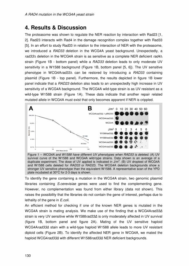
A RAD4 mutation in the WCG4A yeast strain
130
4. Results & Discussion The proteasome was shown to regulate the NER reaction by interaction with Rad23 [1,
2]. Rad23 interacts with Rad4 in the damage recognition complex together with Rad33
[5]. In an effort to study Rad33 in relation to the interaction of NER with the proteasome,
we introduced a RAD33 deletion in the WCG4A yeast background. Unexpectedly, a
rad33∆ deletion in the WCG4A strain is as sensitive as a complete NER deficient rad4∆
strain (Figure 1B - bottom panel) while a RAD33 deletion leads to only moderate UV
sensitivity in a W1588 background (Figure 1B, bottom panel [5, 6]). The UV sensitive
phenotype in WCG4Arad33∆ can be restored by introducing a RAD33 containing
plasmid (Figure 1B - top panel). Furthermore, the results depicted in figure 1B lower
panel indicate that a RAD23 deletion also leads to an unexpectedly high increase in UV
sensitivity of a WCG4A background. The WCG4A wild-type strain is as UV resistant as a
wild-type W1588 strain (Figure 1A). These data indicate that another repair related
mutated allele in WCG4A must exist that only becomes apparent if NER is crippled.
To identify the gene containing a mutation in the WCG4A strain, two genomic plasmid
libraries containing S.cerevisiae genes were used to find the complementing gene.
However, no complementation was found from either library (data not shown). This
raises the possibility that the libraries do not contain the gene of interest, perhaps due to
lethality of the gene in E.coli.
An efficient method for checking if one of the known NER genes is mutated in the
WCG4A strain is mating analysis. We make use of the finding that a WCG4Arad33∆
strain is very UV sensitive while W1588rad33∆ is only moderately affected in UV survival
(figure 1B, bottom panel and figure 2A). Mating of the UV sensitive haploid
WCG4Arad33∆ stain with a wild-type haploid W1588 allele leads to more UV resistant
diploid cells (Figure 2B). To identify the affected NER gene in WCG4A, we mated the
haploid WCG4rad33∆ with different W1588rad33∆ NER deficient backgrounds.
Figure 1 – WCG4A and W1588 have different UV phenotypes when RAD33 is deleted. (A) UV survival curve of the W1588 and WCG4A wild-type strains. Data shown is an average of a duplicate experiment. The dose of UV applied is indicated in J/m
2. (B) UV droptest of WCG4A
and W1588 cells deleted for RAD33 or RAD23. The WCG4A deletion backgrounds show a stronger UV sensitive phenotype than the equivalent W1588. A representative scan of the YPD plate incubated at 30°C for 2-3 days is shown.
A RAD4 mutation in the WCG4A yeast strain
131
In figure 2A the UV survival of all tested haploid rad mutants is depicted. These mating
studies indicate that both RAD14 and RAD23 alleles in WCG4A do not cause the
extreme UV sensitivity in concert with the RAD33 deletion (Figure 2B & C). However, the
extremely UV sensitive haploid W1588rad33∆rad4∆ strain fails to rescue the UV
phenotype of the WCG4Arad33∆ cells; the diploid strain is as sensitive as the haploid
cells (Figure 2D). We conclude that the RAD4 allele in WCG4A carries the mutation that
causes extreme UV sensitivity in WCG4A RAD33 and RAD23 deletion strains. In
hindsight this explains why both genome libraries did not contain the complementing
genes as RAD4 is lethal to E.coli.
Indeed, by sequencing we found the mutation to reside in the WCG4A RAD4 allele at
position 1712; G1712A, TGT→TAT, resulting in a C571Y substitution. This mutation is
novel and the affected amino acid is conserved. As a control the C571Y mutation was
transferred to the W1588 background. In addition a rad33∆rad4C571Y double mutant
was created. These strains show the same phenotype for UV survival as the comparable
Figure 2 – WCG4Arad33∆ was mated with several NER defective backgrounds that are ex-tremely UV sensitive. The UV droptests shown here are a single repre-sentative result of 3-12 zygotes selected after mating. (A) UV droptest of W1588 and WCG4A haploid strains deleted for the NER factors indicated. NER deficient strains deleted for RAD4, RAD14 or RAD23 and RAD33 are extremely UV sensitive. (B) UV droptest of the resulting diploid strains of the WCG4A and W1588 mating indicated. Both diploid strains indicated are moderately UV resistant. (C) The W1588rad33∆rad14∆ strain can rescue the WCG4A UV sensitive phenotype after mating of these backgrounds. The UV droptest shows restoration of wild-type UV resistance of the diploid strain. (D) Mating the WCG4rad33∆ strain with a W1588rad4∆rad33∆ strain, results in a UV sensitive diploid zygote.
A RAD4 mutation in the WCG4A yeast strain
130
4. Results & Discussion The proteasome was shown to regulate the NER reaction by interaction with Rad23 [1,
2]. Rad23 interacts with Rad4 in the damage recognition complex together with Rad33
[5]. In an effort to study Rad33 in relation to the interaction of NER with the proteasome,
we introduced a RAD33 deletion in the WCG4A yeast background. Unexpectedly, a
rad33∆ deletion in the WCG4A strain is as sensitive as a complete NER deficient rad4∆
strain (Figure 1B - bottom panel) while a RAD33 deletion leads to only moderate UV
sensitivity in a W1588 background (Figure 1B, bottom panel [5, 6]). The UV sensitive
phenotype in WCG4Arad33∆ can be restored by introducing a RAD33 containing
plasmid (Figure 1B - top panel). Furthermore, the results depicted in figure 1B lower
panel indicate that a RAD23 deletion also leads to an unexpectedly high increase in UV
sensitivity of a WCG4A background. The WCG4A wild-type strain is as UV resistant as a
wild-type W1588 strain (Figure 1A). These data indicate that another repair related
mutated allele in WCG4A must exist that only becomes apparent if NER is crippled.
To identify the gene containing a mutation in the WCG4A strain, two genomic plasmid
libraries containing S.cerevisiae genes were used to find the complementing gene.
However, no complementation was found from either library (data not shown). This
raises the possibility that the libraries do not contain the gene of interest, perhaps due to
lethality of the gene in E.coli.
An efficient method for checking if one of the known NER genes is mutated in the
WCG4A strain is mating analysis. We make use of the finding that a WCG4Arad33∆
strain is very UV sensitive while W1588rad33∆ is only moderately affected in UV survival
(figure 1B, bottom panel and figure 2A). Mating of the UV sensitive haploid
WCG4Arad33∆ stain with a wild-type haploid W1588 allele leads to more UV resistant
diploid cells (Figure 2B). To identify the affected NER gene in WCG4A, we mated the
haploid WCG4rad33∆ with different W1588rad33∆ NER deficient backgrounds.
Figure 1 – WCG4A and W1588 have different UV phenotypes when RAD33 is deleted. (A) UV survival curve of the W1588 and WCG4A wild-type strains. Data shown is an average of a duplicate experiment. The dose of UV applied is indicated in J/m
2. (B) UV droptest of WCG4A
and W1588 cells deleted for RAD33 or RAD23. The WCG4A deletion backgrounds show a stronger UV sensitive phenotype than the equivalent W1588. A representative scan of the YPD plate incubated at 30°C for 2-3 days is shown.
A RAD4 mutation in the WCG4A yeast strain
131
In figure 2A the UV survival of all tested haploid rad mutants is depicted. These mating
studies indicate that both RAD14 and RAD23 alleles in WCG4A do not cause the
extreme UV sensitivity in concert with the RAD33 deletion (Figure 2B & C). However, the
extremely UV sensitive haploid W1588rad33∆rad4∆ strain fails to rescue the UV
phenotype of the WCG4Arad33∆ cells; the diploid strain is as sensitive as the haploid
cells (Figure 2D). We conclude that the RAD4 allele in WCG4A carries the mutation that
causes extreme UV sensitivity in WCG4A RAD33 and RAD23 deletion strains. In
hindsight this explains why both genome libraries did not contain the complementing
genes as RAD4 is lethal to E.coli.
Indeed, by sequencing we found the mutation to reside in the WCG4A RAD4 allele at
position 1712; G1712A, TGT→TAT, resulting in a C571Y substitution. This mutation is
novel and the affected amino acid is conserved. As a control the C571Y mutation was
transferred to the W1588 background. In addition a rad33∆rad4C571Y double mutant
was created. These strains show the same phenotype for UV survival as the comparable
Figure 2 – WCG4Arad33∆ was mated with several NER defective backgrounds that are ex-tremely UV sensitive. The UV droptests shown here are a single repre-sentative result of 3-12 zygotes selected after mating. (A) UV droptest of W1588 and WCG4A haploid strains deleted for the NER factors indicated. NER deficient strains deleted for RAD4, RAD14 or RAD23 and RAD33 are extremely UV sensitive. (B) UV droptest of the resulting diploid strains of the WCG4A and W1588 mating indicated. Both diploid strains indicated are moderately UV resistant. (C) The W1588rad33∆rad14∆ strain can rescue the WCG4A UV sensitive phenotype after mating of these backgrounds. The UV droptest shows restoration of wild-type UV resistance of the diploid strain. (D) Mating the WCG4rad33∆ strain with a W1588rad4∆rad33∆ strain, results in a UV sensitive diploid zygote.
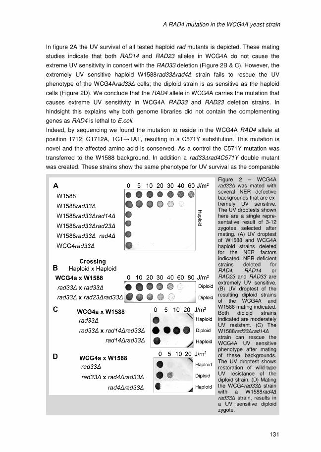
A RAD4 mutation in the WCG4A yeast strain
130
4. Results & Discussion The proteasome was shown to regulate the NER reaction by interaction with Rad23 [1,
2]. Rad23 interacts with Rad4 in the damage recognition complex together with Rad33
[5]. In an effort to study Rad33 in relation to the interaction of NER with the proteasome,
we introduced a RAD33 deletion in the WCG4A yeast background. Unexpectedly, a
rad33∆ deletion in the WCG4A strain is as sensitive as a complete NER deficient rad4∆
strain (Figure 1B - bottom panel) while a RAD33 deletion leads to only moderate UV
sensitivity in a W1588 background (Figure 1B, bottom panel [5, 6]). The UV sensitive
phenotype in WCG4Arad33∆ can be restored by introducing a RAD33 containing
plasmid (Figure 1B - top panel). Furthermore, the results depicted in figure 1B lower
panel indicate that a RAD23 deletion also leads to an unexpectedly high increase in UV
sensitivity of a WCG4A background. The WCG4A wild-type strain is as UV resistant as a
wild-type W1588 strain (Figure 1A). These data indicate that another repair related
mutated allele in WCG4A must exist that only becomes apparent if NER is crippled.
To identify the gene containing a mutation in the WCG4A strain, two genomic plasmid
libraries containing S.cerevisiae genes were used to find the complementing gene.
However, no complementation was found from either library (data not shown). This
raises the possibility that the libraries do not contain the gene of interest, perhaps due to
lethality of the gene in E.coli.
An efficient method for checking if one of the known NER genes is mutated in the
WCG4A strain is mating analysis. We make use of the finding that a WCG4Arad33∆
strain is very UV sensitive while W1588rad33∆ is only moderately affected in UV survival
(figure 1B, bottom panel and figure 2A). Mating of the UV sensitive haploid
WCG4Arad33∆ stain with a wild-type haploid W1588 allele leads to more UV resistant
diploid cells (Figure 2B). To identify the affected NER gene in WCG4A, we mated the
haploid WCG4rad33∆ with different W1588rad33∆ NER deficient backgrounds.
Figure 1 – WCG4A and W1588 have different UV phenotypes when RAD33 is deleted. (A) UV survival curve of the W1588 and WCG4A wild-type strains. Data shown is an average of a duplicate experiment. The dose of UV applied is indicated in J/m
2. (B) UV droptest of WCG4A
and W1588 cells deleted for RAD33 or RAD23. The WCG4A deletion backgrounds show a stronger UV sensitive phenotype than the equivalent W1588. A representative scan of the YPD plate incubated at 30°C for 2-3 days is shown.
A RAD4 mutation in the WCG4A yeast strain
131
In figure 2A the UV survival of all tested haploid rad mutants is depicted. These mating
studies indicate that both RAD14 and RAD23 alleles in WCG4A do not cause the
extreme UV sensitivity in concert with the RAD33 deletion (Figure 2B & C). However, the
extremely UV sensitive haploid W1588rad33∆rad4∆ strain fails to rescue the UV
phenotype of the WCG4Arad33∆ cells; the diploid strain is as sensitive as the haploid
cells (Figure 2D). We conclude that the RAD4 allele in WCG4A carries the mutation that
causes extreme UV sensitivity in WCG4A RAD33 and RAD23 deletion strains. In
hindsight this explains why both genome libraries did not contain the complementing
genes as RAD4 is lethal to E.coli.
Indeed, by sequencing we found the mutation to reside in the WCG4A RAD4 allele at
position 1712; G1712A, TGT→TAT, resulting in a C571Y substitution. This mutation is
novel and the affected amino acid is conserved. As a control the C571Y mutation was
transferred to the W1588 background. In addition a rad33∆rad4C571Y double mutant
was created. These strains show the same phenotype for UV survival as the comparable
Figure 2 – WCG4Arad33∆ was mated with several NER defective backgrounds that are ex-tremely UV sensitive. The UV droptests shown here are a single repre-sentative result of 3-12 zygotes selected after mating. (A) UV droptest of W1588 and WCG4A haploid strains deleted for the NER factors indicated. NER deficient strains deleted for RAD4, RAD14 or RAD23 and RAD33 are extremely UV sensitive. (B) UV droptest of the resulting diploid strains of the WCG4A and W1588 mating indicated. Both diploid strains indicated are moderately UV resistant. (C) The W1588rad33∆rad14∆ strain can rescue the WCG4A UV sensitive phenotype after mating of these backgrounds. The UV droptest shows restoration of wild-type UV resistance of the diploid strain. (D) Mating the WCG4rad33∆ strain with a W1588rad4∆rad33∆ strain, results in a UV sensitive diploid zygote.
A RAD4 mutation in the WCG4A yeast strain
130
4. Results & Discussion The proteasome was shown to regulate the NER reaction by interaction with Rad23 [1,
2]. Rad23 interacts with Rad4 in the damage recognition complex together with Rad33
[5]. In an effort to study Rad33 in relation to the interaction of NER with the proteasome,
we introduced a RAD33 deletion in the WCG4A yeast background. Unexpectedly, a
rad33∆ deletion in the WCG4A strain is as sensitive as a complete NER deficient rad4∆
strain (Figure 1B - bottom panel) while a RAD33 deletion leads to only moderate UV
sensitivity in a W1588 background (Figure 1B, bottom panel [5, 6]). The UV sensitive
phenotype in WCG4Arad33∆ can be restored by introducing a RAD33 containing
plasmid (Figure 1B - top panel). Furthermore, the results depicted in figure 1B lower
panel indicate that a RAD23 deletion also leads to an unexpectedly high increase in UV
sensitivity of a WCG4A background. The WCG4A wild-type strain is as UV resistant as a
wild-type W1588 strain (Figure 1A). These data indicate that another repair related
mutated allele in WCG4A must exist that only becomes apparent if NER is crippled.
To identify the gene containing a mutation in the WCG4A strain, two genomic plasmid
libraries containing S.cerevisiae genes were used to find the complementing gene.
However, no complementation was found from either library (data not shown). This
raises the possibility that the libraries do not contain the gene of interest, perhaps due to
lethality of the gene in E.coli.
An efficient method for checking if one of the known NER genes is mutated in the
WCG4A strain is mating analysis. We make use of the finding that a WCG4Arad33∆
strain is very UV sensitive while W1588rad33∆ is only moderately affected in UV survival
(figure 1B, bottom panel and figure 2A). Mating of the UV sensitive haploid
WCG4Arad33∆ stain with a wild-type haploid W1588 allele leads to more UV resistant
diploid cells (Figure 2B). To identify the affected NER gene in WCG4A, we mated the
haploid WCG4rad33∆ with different W1588rad33∆ NER deficient backgrounds.
Figure 1 – WCG4A and W1588 have different UV phenotypes when RAD33 is deleted. (A) UV survival curve of the W1588 and WCG4A wild-type strains. Data shown is an average of a duplicate experiment. The dose of UV applied is indicated in J/m
2. (B) UV droptest of WCG4A
and W1588 cells deleted for RAD33 or RAD23. The WCG4A deletion backgrounds show a stronger UV sensitive phenotype than the equivalent W1588. A representative scan of the YPD plate incubated at 30°C for 2-3 days is shown.
A RAD4 mutation in the WCG4A yeast strain
131
In figure 2A the UV survival of all tested haploid rad mutants is depicted. These mating
studies indicate that both RAD14 and RAD23 alleles in WCG4A do not cause the
extreme UV sensitivity in concert with the RAD33 deletion (Figure 2B & C). However, the
extremely UV sensitive haploid W1588rad33∆rad4∆ strain fails to rescue the UV
phenotype of the WCG4Arad33∆ cells; the diploid strain is as sensitive as the haploid
cells (Figure 2D). We conclude that the RAD4 allele in WCG4A carries the mutation that
causes extreme UV sensitivity in WCG4A RAD33 and RAD23 deletion strains. In
hindsight this explains why both genome libraries did not contain the complementing
genes as RAD4 is lethal to E.coli.
Indeed, by sequencing we found the mutation to reside in the WCG4A RAD4 allele at
position 1712; G1712A, TGT→TAT, resulting in a C571Y substitution. This mutation is
novel and the affected amino acid is conserved. As a control the C571Y mutation was
transferred to the W1588 background. In addition a rad33∆rad4C571Y double mutant
was created. These strains show the same phenotype for UV survival as the comparable
Figure 2 – WCG4Arad33∆ was mated with several NER defective backgrounds that are ex-tremely UV sensitive. The UV droptests shown here are a single repre-sentative result of 3-12 zygotes selected after mating. (A) UV droptest of W1588 and WCG4A haploid strains deleted for the NER factors indicated. NER deficient strains deleted for RAD4, RAD14 or RAD23 and RAD33 are extremely UV sensitive. (B) UV droptest of the resulting diploid strains of the WCG4A and W1588 mating indicated. Both diploid strains indicated are moderately UV resistant. (C) The W1588rad33∆rad14∆ strain can rescue the WCG4A UV sensitive phenotype after mating of these backgrounds. The UV droptest shows restoration of wild-type UV resistance of the diploid strain. (D) Mating the WCG4rad33∆ strain with a W1588rad4∆rad33∆ strain, results in a UV sensitive diploid zygote.
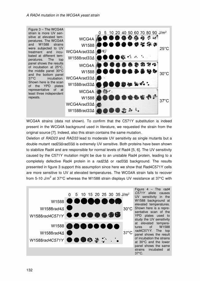
A RAD4 mutation in the WCG4A yeast strain
132
WCG4A strains (data not shown). To confirm that the C571Y substitution is indeed
present in the WCG4A background used in literature, we requested the strain from the
original source [7]. Indeed, also this strain contains the same mutation.
Deletion of RAD23 and RAD33 lead to moderate UV sensitivity as single mutants but a
double mutant rad23∆rad33∆ is extremely UV sensitive. Both proteins have been shown
to stabilize Rad4 and are responsible for normal levels of Rad4 [5, 6]. The UV sensitivity
caused by the C571Y mutation might be due to an unstable Rad4 protein, leading to a
completely defective Rad4 protein in a rad23∆ or rad33∆ background. The results
presented in figure 3 support this assumption since here we show that Rad4C571Y cells
are more sensitive to UV at elevated temperatures. The WCG4A strain fails to recover
from 5-10 J/m2 at 37°C whereas the W1588 strain displays UV resistance at 37°C with
Figure 3 – The WCG4A strain is more UV sen-sitive at elevated tem-peratures. The WCG4A and W1588 strains were subjected to UV treatment and incu-bated at different tem-peratures. The top panel shows the results of incubation at 25°C, the middle panel 30°C and the bottom panel 37°C incubation. Shown here is the scan of the YPD plates representative of at least three independent repeats.
Figure 4 – The rad4C571Y allele causes UV sensitivity in the W1588 background at elevated temperatures. Shown here is a repre-sentative scan of the YPD plates used to study the UV sensitivity at elevated tempera-tures of W1588rad4C571Y. The top panel shows the result of incubation the strains at 30°C and the lower panel shows the same strains incubated at 37°C.
A RAD4 mutation in the WCG4A yeast strain
133
growth up until 90 J/m2
(Figure 3). As anticipated W1588 cells become sensitive at
elevated temperature after the C571Y substitution is introduced into the Rad4 protein,
similar to the WCG4A background (Figure 4).
The additive decrease in UV resistance at elevated temperatures or after deleting the
direct interaction partners of Rad4C571Y, indicates that the protein is unstable in
WCG4A. Indeed, when we measure protein levels using Westernblot analysis we find
reduced levels of Rad4 protein in the W1588rad4C571Y background (data not shown).
Residue 571 is located in β-hairpin domain 3 (BHD3) at the base of one of the strands of
the three-stranded β-sheet [8]. The change from a cysteine to a tyrosine could lead to
destabilization of the BHD3 domain which in turn might cause the instability of the
protein. Apparently, the interactions with Rad23 and Rad33 stabilize this domain.
A RAD4 mutation in the WCG4A yeast strain
132
WCG4A strains (data not shown). To confirm that the C571Y substitution is indeed
present in the WCG4A background used in literature, we requested the strain from the
original source [7]. Indeed, also this strain contains the same mutation.
Deletion of RAD23 and RAD33 lead to moderate UV sensitivity as single mutants but a
double mutant rad23∆rad33∆ is extremely UV sensitive. Both proteins have been shown
to stabilize Rad4 and are responsible for normal levels of Rad4 [5, 6]. The UV sensitivity
caused by the C571Y mutation might be due to an unstable Rad4 protein, leading to a
completely defective Rad4 protein in a rad23∆ or rad33∆ background. The results
presented in figure 3 support this assumption since here we show that Rad4C571Y cells
are more sensitive to UV at elevated temperatures. The WCG4A strain fails to recover
from 5-10 J/m2 at 37°C whereas the W1588 strain displays UV resistance at 37°C with
Figure 3 – The WCG4A strain is more UV sen-sitive at elevated tem-peratures. The WCG4A and W1588 strains were subjected to UV treatment and incu-bated at different tem-peratures. The top panel shows the results of incubation at 25°C, the middle panel 30°C and the bottom panel 37°C incubation. Shown here is the scan of the YPD plates representative of at least three independent repeats.
Figure 4 – The rad4C571Y allele causes UV sensitivity in the W1588 background at elevated temperatures. Shown here is a repre-sentative scan of the YPD plates used to study the UV sensitivity at elevated tempera-tures of W1588rad4C571Y. The top panel shows the result of incubation the strains at 30°C and the lower panel shows the same strains incubated at 37°C.
A RAD4 mutation in the WCG4A yeast strain
133
growth up until 90 J/m2
(Figure 3). As anticipated W1588 cells become sensitive at
elevated temperature after the C571Y substitution is introduced into the Rad4 protein,
similar to the WCG4A background (Figure 4).
The additive decrease in UV resistance at elevated temperatures or after deleting the
direct interaction partners of Rad4C571Y, indicates that the protein is unstable in
WCG4A. Indeed, when we measure protein levels using Westernblot analysis we find
reduced levels of Rad4 protein in the W1588rad4C571Y background (data not shown).
Residue 571 is located in β-hairpin domain 3 (BHD3) at the base of one of the strands of
the three-stranded β-sheet [8]. The change from a cysteine to a tyrosine could lead to
destabilization of the BHD3 domain which in turn might cause the instability of the
protein. Apparently, the interactions with Rad23 and Rad33 stabilize this domain.

A RAD4 mutation in the WCG4A yeast strain
132
WCG4A strains (data not shown). To confirm that the C571Y substitution is indeed
present in the WCG4A background used in literature, we requested the strain from the
original source [7]. Indeed, also this strain contains the same mutation.
Deletion of RAD23 and RAD33 lead to moderate UV sensitivity as single mutants but a
double mutant rad23∆rad33∆ is extremely UV sensitive. Both proteins have been shown
to stabilize Rad4 and are responsible for normal levels of Rad4 [5, 6]. The UV sensitivity
caused by the C571Y mutation might be due to an unstable Rad4 protein, leading to a
completely defective Rad4 protein in a rad23∆ or rad33∆ background. The results
presented in figure 3 support this assumption since here we show that Rad4C571Y cells
are more sensitive to UV at elevated temperatures. The WCG4A strain fails to recover
from 5-10 J/m2 at 37°C whereas the W1588 strain displays UV resistance at 37°C with
Figure 3 – The WCG4A strain is more UV sen-sitive at elevated tem-peratures. The WCG4A and W1588 strains were subjected to UV treatment and incu-bated at different tem-peratures. The top panel shows the results of incubation at 25°C, the middle panel 30°C and the bottom panel 37°C incubation. Shown here is the scan of the YPD plates representative of at least three independent repeats.
Figure 4 – The rad4C571Y allele causes UV sensitivity in the W1588 background at elevated temperatures. Shown here is a repre-sentative scan of the YPD plates used to study the UV sensitivity at elevated tempera-tures of W1588rad4C571Y. The top panel shows the result of incubation the strains at 30°C and the lower panel shows the same strains incubated at 37°C.
A RAD4 mutation in the WCG4A yeast strain
133
growth up until 90 J/m2
(Figure 3). As anticipated W1588 cells become sensitive at
elevated temperature after the C571Y substitution is introduced into the Rad4 protein,
similar to the WCG4A background (Figure 4).
The additive decrease in UV resistance at elevated temperatures or after deleting the
direct interaction partners of Rad4C571Y, indicates that the protein is unstable in
WCG4A. Indeed, when we measure protein levels using Westernblot analysis we find
reduced levels of Rad4 protein in the W1588rad4C571Y background (data not shown).
Residue 571 is located in β-hairpin domain 3 (BHD3) at the base of one of the strands of
the three-stranded β-sheet [8]. The change from a cysteine to a tyrosine could lead to
destabilization of the BHD3 domain which in turn might cause the instability of the
protein. Apparently, the interactions with Rad23 and Rad33 stabilize this domain.
A RAD4 mutation in the WCG4A yeast strain
132
WCG4A strains (data not shown). To confirm that the C571Y substitution is indeed
present in the WCG4A background used in literature, we requested the strain from the
original source [7]. Indeed, also this strain contains the same mutation.
Deletion of RAD23 and RAD33 lead to moderate UV sensitivity as single mutants but a
double mutant rad23∆rad33∆ is extremely UV sensitive. Both proteins have been shown
to stabilize Rad4 and are responsible for normal levels of Rad4 [5, 6]. The UV sensitivity
caused by the C571Y mutation might be due to an unstable Rad4 protein, leading to a
completely defective Rad4 protein in a rad23∆ or rad33∆ background. The results
presented in figure 3 support this assumption since here we show that Rad4C571Y cells
are more sensitive to UV at elevated temperatures. The WCG4A strain fails to recover
from 5-10 J/m2 at 37°C whereas the W1588 strain displays UV resistance at 37°C with
Figure 3 – The WCG4A strain is more UV sen-sitive at elevated tem-peratures. The WCG4A and W1588 strains were subjected to UV treatment and incu-bated at different tem-peratures. The top panel shows the results of incubation at 25°C, the middle panel 30°C and the bottom panel 37°C incubation. Shown here is the scan of the YPD plates representative of at least three independent repeats.
Figure 4 – The rad4C571Y allele causes UV sensitivity in the W1588 background at elevated temperatures. Shown here is a repre-sentative scan of the YPD plates used to study the UV sensitivity at elevated tempera-tures of W1588rad4C571Y. The top panel shows the result of incubation the strains at 30°C and the lower panel shows the same strains incubated at 37°C.
A RAD4 mutation in the WCG4A yeast strain
133
growth up until 90 J/m2
(Figure 3). As anticipated W1588 cells become sensitive at
elevated temperature after the C571Y substitution is introduced into the Rad4 protein,
similar to the WCG4A background (Figure 4).
The additive decrease in UV resistance at elevated temperatures or after deleting the
direct interaction partners of Rad4C571Y, indicates that the protein is unstable in
WCG4A. Indeed, when we measure protein levels using Westernblot analysis we find
reduced levels of Rad4 protein in the W1588rad4C571Y background (data not shown).
Residue 571 is located in β-hairpin domain 3 (BHD3) at the base of one of the strands of
the three-stranded β-sheet [8]. The change from a cysteine to a tyrosine could lead to
destabilization of the BHD3 domain which in turn might cause the instability of the
protein. Apparently, the interactions with Rad23 and Rad33 stabilize this domain.
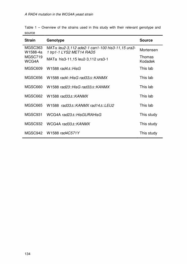
A RAD4 mutation in the WCG4A yeast strain
134
Table 1 – Overview of the strains used in this study with their relevant genotype and
source
Strain Genotype Source
MGSC363 W1588-4a
MATα leu2-3,112 ade2-1 can1-100 his3-11,15 ura3-1 trp1-1 LYS2 MET14 RAD5
Mortensen
MGSC719 WCG4A
MATa his3-11,15 leu2-3,112 ura3-1 Thomas Kodadek
MGSC609 W1588 rad4∆::HisG This lab
MGSC656 W1588 rad4::HisG rad33∆::KANMX This lab
MGSC660 W1588 rad23::HisG rad33∆::KANMX This lab
MGSC662 W1588 rad33∆::KANMX This lab
MGSC665 W1588 rad33∆::KANMX rad14∆::LEU2 This lab
MGSC931 WCG4A rad23∆::HisGURAHisG This study
MGSC932 WCG4A rad33∆::KANMX This study
MGSC942 W1588 rad4C571Y This study
A RAD4 mutation in the WCG4A yeast strain
135
5. References 1. Gillette, T.G., et al., The 19S complex of the proteasome regulates nucleotide excision
repair in yeast. Genes Dev, 2001. 15(12): p. 1528-39.
2. Gillette, T.G., et al., Distinct functions of the ubiquitin-proteasome pathway influence
nucleotide excision repair. Embo Journal, 2006. 25(11): p. 2529-38.
3. Heinemeyer, W., et al., PRE2, highly homologous to the human major histocompatibility
complex-linked RING10 gene, codes for a yeast proteasome subunit necessary for
chrymotryptic activity and degradation of ubiquitinated proteins. J Biol Chem, 1993.
268(7): p. 5115-20.
4. Hilt, W., et al., The PRE4 gene codes for a subunit of the yeast proteasome necessary
for peptidylglutamyl-peptide-hydrolyzing activity. Mutations link the proteasome to stress-
and ubiquitin-dependent proteolysis. J Biol Chem, 1993. 268(5): p. 3479-86.
5. den Dulk, B., et al., Rad33, a new factor involved in nucleotide excision repair in
Saccharomyces cerevisiae. DNA Repair (Amst), 2006. 5(6): p. 683-92.
6. den Dulk, B., et al., The NER protein Rad33 shows functional homology to human
Centrin2 and is involved in modification of Rad4. DNA Repair (Amst), 2008. 7(6): p. 858-
68.
7. Sulahian, R., et al., The proteasomal ATPase complex is required for stress-induced
transcription in yeast. Nucleic Acids Res, 2006. 34(5): p. 1351-7.
8. Min, J.H. and N.P. Pavletich, Recognition of DNA damage by the Rad4 nucleotide
excision repair protein. Nature, 2007. 449(7162): p. 570-5.
A RAD4 mutation in the WCG4A yeast strain
134
Table 1 – Overview of the strains used in this study with their relevant genotype and
source
Strain Genotype Source
MGSC363 W1588-4a
MATα leu2-3,112 ade2-1 can1-100 his3-11,15 ura3-1 trp1-1 LYS2 MET14 RAD5
Mortensen
MGSC719 WCG4A
MATa his3-11,15 leu2-3,112 ura3-1 Thomas Kodadek
MGSC609 W1588 rad4∆::HisG This lab
MGSC656 W1588 rad4::HisG rad33∆::KANMX This lab
MGSC660 W1588 rad23::HisG rad33∆::KANMX This lab
MGSC662 W1588 rad33∆::KANMX This lab
MGSC665 W1588 rad33∆::KANMX rad14∆::LEU2 This lab
MGSC931 WCG4A rad23∆::HisGURAHisG This study
MGSC932 WCG4A rad33∆::KANMX This study
MGSC942 W1588 rad4C571Y This study
A RAD4 mutation in the WCG4A yeast strain
135
5. References 1. Gillette, T.G., et al., The 19S complex of the proteasome regulates nucleotide excision
repair in yeast. Genes Dev, 2001. 15(12): p. 1528-39.
2. Gillette, T.G., et al., Distinct functions of the ubiquitin-proteasome pathway influence
nucleotide excision repair. Embo Journal, 2006. 25(11): p. 2529-38.
3. Heinemeyer, W., et al., PRE2, highly homologous to the human major histocompatibility
complex-linked RING10 gene, codes for a yeast proteasome subunit necessary for
chrymotryptic activity and degradation of ubiquitinated proteins. J Biol Chem, 1993.
268(7): p. 5115-20.
4. Hilt, W., et al., The PRE4 gene codes for a subunit of the yeast proteasome necessary
for peptidylglutamyl-peptide-hydrolyzing activity. Mutations link the proteasome to stress-
and ubiquitin-dependent proteolysis. J Biol Chem, 1993. 268(5): p. 3479-86.
5. den Dulk, B., et al., Rad33, a new factor involved in nucleotide excision repair in
Saccharomyces cerevisiae. DNA Repair (Amst), 2006. 5(6): p. 683-92.
6. den Dulk, B., et al., The NER protein Rad33 shows functional homology to human
Centrin2 and is involved in modification of Rad4. DNA Repair (Amst), 2008. 7(6): p. 858-
68.
7. Sulahian, R., et al., The proteasomal ATPase complex is required for stress-induced
transcription in yeast. Nucleic Acids Res, 2006. 34(5): p. 1351-7.
8. Min, J.H. and N.P. Pavletich, Recognition of DNA damage by the Rad4 nucleotide
excision repair protein. Nature, 2007. 449(7162): p. 570-5.

A RAD4 mutation in the WCG4A yeast strain
134
Table 1 – Overview of the strains used in this study with their relevant genotype and
source
Strain Genotype Source
MGSC363 W1588-4a
MATα leu2-3,112 ade2-1 can1-100 his3-11,15 ura3-1 trp1-1 LYS2 MET14 RAD5
Mortensen
MGSC719 WCG4A
MATa his3-11,15 leu2-3,112 ura3-1 Thomas Kodadek
MGSC609 W1588 rad4∆::HisG This lab
MGSC656 W1588 rad4::HisG rad33∆::KANMX This lab
MGSC660 W1588 rad23::HisG rad33∆::KANMX This lab
MGSC662 W1588 rad33∆::KANMX This lab
MGSC665 W1588 rad33∆::KANMX rad14∆::LEU2 This lab
MGSC931 WCG4A rad23∆::HisGURAHisG This study
MGSC932 WCG4A rad33∆::KANMX This study
MGSC942 W1588 rad4C571Y This study
A RAD4 mutation in the WCG4A yeast strain
135
5. References 1. Gillette, T.G., et al., The 19S complex of the proteasome regulates nucleotide excision
repair in yeast. Genes Dev, 2001. 15(12): p. 1528-39.
2. Gillette, T.G., et al., Distinct functions of the ubiquitin-proteasome pathway influence
nucleotide excision repair. Embo Journal, 2006. 25(11): p. 2529-38.
3. Heinemeyer, W., et al., PRE2, highly homologous to the human major histocompatibility
complex-linked RING10 gene, codes for a yeast proteasome subunit necessary for
chrymotryptic activity and degradation of ubiquitinated proteins. J Biol Chem, 1993.
268(7): p. 5115-20.
4. Hilt, W., et al., The PRE4 gene codes for a subunit of the yeast proteasome necessary
for peptidylglutamyl-peptide-hydrolyzing activity. Mutations link the proteasome to stress-
and ubiquitin-dependent proteolysis. J Biol Chem, 1993. 268(5): p. 3479-86.
5. den Dulk, B., et al., Rad33, a new factor involved in nucleotide excision repair in
Saccharomyces cerevisiae. DNA Repair (Amst), 2006. 5(6): p. 683-92.
6. den Dulk, B., et al., The NER protein Rad33 shows functional homology to human
Centrin2 and is involved in modification of Rad4. DNA Repair (Amst), 2008. 7(6): p. 858-
68.
7. Sulahian, R., et al., The proteasomal ATPase complex is required for stress-induced
transcription in yeast. Nucleic Acids Res, 2006. 34(5): p. 1351-7.
8. Min, J.H. and N.P. Pavletich, Recognition of DNA damage by the Rad4 nucleotide
excision repair protein. Nature, 2007. 449(7162): p. 570-5.
A RAD4 mutation in the WCG4A yeast strain
136
VII Summary & Conclusions
A RAD4 mutation in the WCG4A yeast strain
136

A RAD4 mutation in the WCG4A yeast strain
136
VII Summary & Conclusions
A RAD4 mutation in the WCG4A yeast strain
136

Summary & Conclusions
138
Nucleotide Excision Repair (NER) is a conserved DNA repair pathway capable of
removing a broad spectrum of DNA damage. In human cells a defect in NER leads to the
disorder Xeroderma pigmentosum, increasing the incidence of cancer. The yeast
Saccharomyces cerevisiae is an excellent model organism to study the mechanism of
NER. Two sub-pathways have been described, TC-NER for transcriptionally active
genes and GG-NER which removes lesion throughout the entire genome. The yeast
proteins Rad4 and Rad23 are important in NER and involved both sub-pathways.
Chapter 2 describes a novel mechanism of gene regulation by the GG-NER E3 ligase.
This protein complex can regulate dNTP synthesis via UV induced Rad4-ubiquitination.
The Rad4-Rad23 complex interacts with DNA at the promoter region of genes in the
absence of DNA damage. The GG-NER E3 ligase regulated dissociation of the Rad4-
Rad23 complex facilitates a change in gene expression in response to UV radiation.
Amongst genes that are targeted by this novel NER related gene expression mechanism
are the genes that regulate dNTP synthesis. The control of dNTP synthesis in response
to DNA damage or during S phase is described in more detail in section 2.1. The dNTP
synthesis factors are a downstream target of the DNA Damage Response (DDR). The
activity of the GG-NER E3 ligase and Rad4-Rad23 DNA binding that drive this pathway
are novel and runs independent to the established DDR signaling and DNA damage
induced gene expression controlled by Rad6-Rad18. This latter DDR pathway is
triggered upon detection and processing of the actually damage. The advantage of this
novel Rad4-Rad23 mechanism is that it can adapt gene expression much earlier in
response to damage induction. The GG-NER E3 ligase mediated dissociation of Rad4-
Rad23 from the promoter regions already alters gene expression before the NER
complex associates with UV lesions. In this way the cell can prepare for DDR signaling
and UV induced gene expression. In an attempt to expand the data described in Chapter
2, studying the genome-wide association of the Rad4-Rad23 complex using ChIP-on-
Chip analysis could be instrumental in deciphering the mechanism behind the
association-dissociation process of Rad4-Rad23 in response to DNA damage.
Yeast TC-NER is unique in requiring a specific Rad4-like protein for rDNA repair. Histone
H1 is a linker histone specifically associated with the rDNA. In Chapter 3 we describe the
effect of this histone on TC-NER at the rDNA. We find that Rad34 is only required when
Histone H1 is present. In the absence of both Histone H1 and Rad34 repair of the TS of
rDNA becomes dependent on Rad4. We propose a model in which Histone H1
association with RNA Pol I stalled at a lesion holds the complex in place and requires
Rad34 for displacement and subsequent TC-NER. This would be similar to the function
of Rad26 that is generally required for TC-NER of RNA Pol II transcribed genes. In the
absence of Hho1, RNA Pol I is more loosely attached to the DNA when it is stalled at a
damage and might thus back-track or dissociate from the lesion by itself. In this process
Rad4 can then replace Rad34 and participate in TC-NER.
Samenvatting & Conclusie
139
In Chapter 4 we analyze the phenotype of a yeast strain containing a RAD4 mutation.
The mutation affects a conserved residue that when altered in the human homolog XPC,
leads to the cancer prone disorder Xeroderma pigmentosum. The residue is important
for the association of XPC with DNA and mutation of this residue leads to UV sensitivity
in human cells. Here we determined that this residue is important for in vivo repair in
yeast as well but does not result in UV sensitivity by itself. The two sub-pathways in NER
are both affected, but GG-NER is most severely inhibited and displays a 4-fold reduction
in repair in the presence of the mutant rad4W496S allele in yeast. We hypothesize that
participation of the mutation protein in TC-NER is facilitated by the action of TC-NER
specific factors that remodel the RNA Pol II complex at the lesion. In such a way it allows
the association of a Rad4 protein that poorly binds to the DNA. Maybe for the same
reason the equivalent activity of TC-NER factors in higher eukaryotes completely
bypasses the need for XPC in TC-NER. However, the lack of such a remodeling activity
during GG-NER in yeast rad4W496S cells results in a strong defect in this pathway. The
defective GG-NER apparently leads to a very delayed repair that is not detectable in our
assays but does contribute to cellular survival as rad4W496S mutant cells are UV
resistant.
Chapter 5 is dedicated to the Rad4-Rad23 interaction that we describe in more detail.
We uncovered a novel N-terminal interaction of Rad23 with Rad4 that is important for
TC-NER. We could confirm the established Rad23 C-terminal interaction with Rad4 and
were able to show that it is specifically required for GG-NER. Interestingly, the
rad4W496S mutation, analogous to the mutation in an XP patient described in Chapter
4, is specifically affected in GG-NER and was shown to disrupt the C-terminal Rad4-
Rad23 interaction. These data highlight the intricate interaction between Rad4 and
Rad23 that can be differentially regulated in response to the specific sub-pathway in
which it operates. We hypothesize that the sub-pathways specifically mold Rad4-Rad23
into a different conformation to meet specific requirements needed to drive repair.
Moreover, we attribute an active in vivo role to the intramolecular UbL-UBA interaction in
the differential Rad4-Rad23 interactions.
Finally, in Chapter 6 we reveal an unexpected UV phenotype for the commonly used
wild-type yeast strain WCG4A. In this strain we found elevated levels of UV sensitivity in
combination with NER gene deletions which on their own lead to only moderate UV
sensitivity. Using mating experiments we identified the mutation to reside in the RAD4
gene. When we transferred the single nucleotide mutation (leading to the C571Y
substitution) to another yeast strain background we obtained exactly the same
phenotypes. The affected residue is located in the BHD3 domain of Rad4 and results in
destabilization of the protein as could be shown by the UV sensitivity at higher
temperature. The WCG4A yeast background is commonly used in proteasome related
studies as conditional 19S and 20S mutants are readily available. Based on these data
Summary & Conclusions
140
we would like to stress that this WCG4A strain should not be used in DNA repair related
studies.
Linking back to the data described in Chapter 4, the W496S mutation in Rad4 behaves
fairly similar to the C571Y substitution identified in WCG4A. In both cases the single
amino acid substitution in Rad4 does not affect UV survival on its own but does show an
effect if NER is crippled by deletion of NER accessory genes. The Rad4-interacting
proteins Rad23 and Rad33 both play an important role in maintaining normal Rad4
protein levels. In the absence of these proteins wild-type Rad4 levels are reduced, in part
leading to moderate UV sensitivity. This reduction in Rad4 protein levels, however, only
has a severe effect on UV survival if also RAD4 is mutated. The different RAD4 mutants
described in this thesis show that due to the stabilizing effect of Rad23 and Rad33, the
Rad4 protein can cope with different mutations without a significant effect on UV survival.
VIII Samenvatting & Conclusies
Summary & Conclusions
140
we would like to stress that this WCG4A strain should not be used in DNA repair related
studies.
Linking back to the data described in Chapter 4, the W496S mutation in Rad4 behaves
fairly similar to the C571Y substitution identified in WCG4A. In both cases the single
amino acid substitution in Rad4 does not affect UV survival on its own but does show an
effect if NER is crippled by deletion of NER accessory genes. The Rad4-interacting
proteins Rad23 and Rad33 both play an important role in maintaining normal Rad4
protein levels. In the absence of these proteins wild-type Rad4 levels are reduced, in part
leading to moderate UV sensitivity. This reduction in Rad4 protein levels, however, only
has a severe effect on UV survival if also RAD4 is mutated. The different RAD4 mutants
described in this thesis show that due to the stabilizing effect of Rad23 and Rad33, the
Rad4 protein can cope with different mutations without a significant effect on UV survival.
VIII Samenvatting & Conclusies
Summary & Conclusions
140
we would like to stress that this WCG4A strain should not be used in DNA repair related
studies.
Linking back to the data described in Chapter 4, the W496S mutation in Rad4 behaves
fairly similar to the C571Y substitution identified in WCG4A. In both cases the single
amino acid substitution in Rad4 does not affect UV survival on its own but does show an
effect if NER is crippled by deletion of NER accessory genes. The Rad4-interacting
proteins Rad23 and Rad33 both play an important role in maintaining normal Rad4
protein levels. In the absence of these proteins wild-type Rad4 levels are reduced, in part
leading to moderate UV sensitivity. This reduction in Rad4 protein levels, however, only
has a severe effect on UV survival if also RAD4 is mutated. The different RAD4 mutants
described in this thesis show that due to the stabilizing effect of Rad23 and Rad33, the
Rad4 protein can cope with different mutations without a significant effect on UV survival.
Samnevatting & Conclusie
142
Nucleotide excisie herstel (NER) is een geconserveerd DNA schadeherstel systeem dat
een breed scala aan DNA schades repareert. Een defect in NER in humane cellen leidt
tot de ziekte Xeroderma pigmentosum. Deze ziekte wordt gekarakteriseerd door een
verhoogd risico op kanker. De gist Saccharomyces cerevisiae is een buitengewoon
geschikt model organisme om het mechansime van NER te bestuderen. NER kan
onderverdeeld worden in twee routes: TC-NER (Transcription-Coupled NER) voor actief
getranscribeerde genen en GG-NER (Global Genome NER) dat verantwoordelijk is voor
schadeherstel in het hele genoom. De Rad4 en Rad23 eiwitten in gist spelen een
belangrijke rol in NER en zijn betrokken bij beide routes.
Hoofdstuk 2 beschrijft een nieuwe vorm van genregulatie door het GG-NER specifieke
E3 ligase. Dit eiwitcomplex kan de synthese van dNTP's aansturen via ubiquitinering van
Rad4. Het Rad4-Rad23 complex bindt aan het DNA in het promotergebied van sommige
genen in de afwezigheid van DNA schade. Het GG-NER E3 ligase zorgt voor dissociatie
van dit Rad4-Rad23 complex na UV straling met tot gevolg dat de expressie van
sommige genen verandert. Onder de genen die door dit nieuwe NER gerelateerde
mechanisme worden beinvloed bevinden zich de genen die zorgen voor de aanmaak
van dNTP's. Factoren betrokken bij de dNTP synthese zijn een doelwit van de
zogenaamde DNA Damage Respone (DDR). De door ons gevonden activiteit van het
GG-NER E3 ligase en Rad4-Rad23 in DDR opereert geheel onafhankelijk van de reeds
eerder gevonden DDR (en DNA schade geinduceerde genexpressie) die wordt
gecontroleerd door Rad6-Rad18. Voor het aansturen van de DDR door Rad6-Rad18
moet de schade al herkend zijn door schade herstellende eiwitten. Het voordeel van het
nieuwe Rad4-Rad23 mechanisme is dat genexpressie veel sneller kan worden
aangepast na DNA schade inductie. Het GG-NER E3 ligase zorgt namelijk voor
dissociatie van het Rad4-Rad23 complex van de promoter gebieden nog voordat het
NER complex de schade gevonden heeft. Op deze manier kan de cel zich tijdig
voorbereiden op DDR en UV geinduceerde genexpressie.
Gist TC-NER is uniek omdat het gebruik maakt van een specifiek Rad4 gerelateerd eiwit,
Rad34, voor herstel van genen coderend voor rRNA (rDNA). Histon H1 is een linker
histon dat specifiek bindt aan rDNA. In hoofdstuk 3 hebben we het effect van dit histon
op TC-NER in rDNA beschreven. We hebben laten zien dat Rad34 alleen nodig is
wanneer Histon H1 aanwezig is. In afwezigheid van Histon H1 kan Rad4 de rol van
Rad34 overnemen bij herstel van de getranscribeerde streng. We postuleren een model
waarin Histon H1 ervoor zorgt dat RNA Pol I op het DNA bij de schade blokkeert. Het
gevolg is dat Rad34 specifiek nodig is om het RNA polymerase van de schade af te
krijgen om zo TC-NER te laten plaats vinden. Deze functie van Rad34 lijkt heel veel op
de functie van Rad26 tijdens TC-NER van RNA Pol II getranscribeerde genen. Ook daar
is Rad26 nodig om RNA Pol II die vastgelopen is op een schade te verwijderen. In de
afwezigheid van Histon H1 is RNA Pol I waarschijnlijk minder stabiel gebonden aan een
Samenvatting & Conclusie
143
schade en kan dus makkelijker uit zichzelf loslaten. Rad4 kan vervolgens direct binden
en deelnemen aan TC-NER.
In hoofdstuk 4 analyseren we het fenotype van een gist stam die een mutatie (W496S)
heeft in het RAD4 gen. Dezelfde mutatie in het op Rad4 lijkende humane gen, XPC, leidt
tot de ziekte Xeroderma pigmentosum. Het gemuteerde residu is belangrijk voor de DNA
binding van XPC en de mutatie leidt tot UV gevoeligheid in humane cellen. We hebben
laten zien dat dit residu ook belangrijk is voor herstel in vivo in gist, maar de mutatie op
zich leidt niet tot UV gevoeligheid. Beide NER routes zijn aangetast, maar het effect op
GG-NER blijkt veel ernstiger dan op TC-NER. Mogelijk helpen eiwitten, die specifiek
betrokken zijn bij TC-NER, het gemuteerde Rad4 eiwit om toch aan de schade te kunnen
binden. Bij GG-NER ontbreken zulke factoren en als gevolg bindt Rad4 veel slechter aan
de schade. Dit leidt ertoe dat de schade sterk vertraagd hersteld wordt. Door deze
vertraagde werking van GG-NER is er in onze experimenten in de eerste uren na schade
inductie nog geen herstel waarneembaar. Uiteindelijk echter weet de gistcel toch nog te
overleven aangezien we geen verhoogde UV gevoeligheid kunnen meten.
Hoofdstuk 5 is gewijd aan de interactie tussen Rad4 en Rad23. We laten zien dat Rad23
op twee plekken in Rad4 kan binden: aan het N-terminale TGD (TransGlutaminase)
domein en aan het C-terminale deel van het eiwit. Omdat Rad23 voor beide interacties
hetzelfde (kleine) eiwitdomein gebruikt, is het niet mogelijk dat Rad23 beide contacten
tegelijk maakt. Mogelijk wordt de keuze tussen beide manieren van binden bepaald door
een intramoleculaire interactie tussen de UbL en UBA domeinen van Rad23. Wanneer
deze interactie plaatsvindt bindt Rad23 aan het C-terminale deel van Rad4. Vindt deze
interactie niet plaats dan wordt het TGD domein gebonden. Tenslotte hebben we laten
zien dat beide vormen van het Rad4-Rad23 complex een verschillende functie hebben.
Wanneer de eiwitten via de TGD met elkaar zijn gebonden zijn ze voornamelijk
betrokken bij TC-NER. In de andere vorm lijken ze betrokken bij GG-NER. Het is
interessant om te vermelden dat de hierboven beschreven rad4W496S mutatie gelegen
is in het C-terminale deel van Rad4 en geheel gestoord is in GG-NER. Inderdaad blijkt
deze mutant ook niet meer via zijn C-terminale domein aan Rad23 te kunnen binden.
In hoofdstuk 6 wordt het onverwachte UV fenotype besproken van de veelgebruikte
giststam, WCG4A. Deze stam laat een sterk verhoogde UV gevoeligheid zien wanneer
NER mutaties, die van zichzelf slechts een matige UV gevoeligheid veroorzaken, in deze
stam worden ingebracht. Door middel van het kruisen van giststammen hebben we het
gemuteerde gen kunnen identificeren als RAD4. Als we vervolgens de gevonden punt
mutatie (C571Y) overplaatsen in een andere giststam, krijgen we exact dezelfde
fenotypes. Het veranderde aminozuur bevindt zich in het BHD3 domein van Rad4 en
leidt tot destabilisatie van het eiwit, wat we konden afleiden uit de UV gevoeligheid bij
hogere temperatuur. De WCG4A gist stam wordt veel gebruikt in proteasome
gerelateerde studies omdat er conditionele 19S en 20S mutanten beschikbaar zijn in

Samnevatting & Conclusie
144
deze achtergrond. Op basis van deze bevindingen willen wij er met klem op wijzen dat
deze achtergrond niet geschikt is voor DNA schadeherstel gerelateerde studies.
Terugkomend op de data beschreven in hoofdstuk 4, is het opmerkelijk dat de W496S
mutatie zeer veel overeenkomsten laat zien met de C571Y substitutie. In beide gevallen
leidt de enkele aminozuur substitutie in Rad4 niet tot verandering in UV overleving, maar
vertoont alleen een effect als NER wordt verzwakt door deletie van andere NER genen.
De Rad4 bindende eiwitten Rad23 en Rad33 spelen beiden een belangrijke rol in het
stabiliseren van het Rad4 eiwit in de cel. In de afwezigheid van deze eiwitten is het Rad4
niveau verlaagd, wat deels bijdraagt aan de UV gevoeligheid. Deze afname in Rad4 eiwit
heeft echter alleen een drastisch effect als het Rad4 eiwit zelf ook is gemuteerd. Dankzij
de stabliserende werking van Rad23 en Rad33 kan Rad4 veel mutaties verdragen
zonder dat dat effect heeft op de UV overleving.
Samenvatting & Conclusie
145

146
IX Curriculum vitae
EDUCATION
9/2002 – 9/2007 Life Science & Technology, University Leiden & TU Delft
Master: Functional Genomics
Bachelor: Life Science & Technology
9/1996 – 6/2002 VWO (A-levels), Nova College Montfort, Rotterdam
Profile: Nature & Techniek including Biology
European Computer Drivers License (ECDL)
WORK & INTERNSHIPS
9-2007 – 2012 PhD Student at the department of Molecular Genetics at the Leiden
Institute of Chemistry, University of Leiden on Nucleotide Excision
Repair, in yeast. Supervisor: Dr. J.A. Brandsma.
1/2012 – 8/2012 Visiting Scholar at Cardiff University under the auspice of Dr. S.H.
Reed. A collaborative project on the research described in Chapter 2
of this thesis was performed.
11/2006 – 7/2007 Master research internship at the department of Molecular Genetics
at the Leiden Institute of Chemistry, University of Leiden on
Nucleotide Excision Repair, in yeast. Supervisor, Dr. J.A. Brandsma.
8/2006 – 11/2006 Business internship at the Dutch National Museum of Natural History
at the department of information services a web theme on ‘De Cel’
was made. Supervisor: H. Ahrens
4/2005 – 7/2005 Bachelor research internship at the department of Nephrology at the
Leiden University Medical Center (LUMC) on ‘Complement-
mediated nephritis in mice and in vivo complement inhibition’. Under
supervision of T.W.L. Groeneveld & Dr. A. Roos