Gauge Conditions for Long-Term Numerical Black Hole Evolutions Without Excision
Upload
khangminh22Category
view
0download
0
Arthroscopic Excision of Delayed Diagnosed Intra-articular Osteoid Osteoma of the Elbow: A Case Report
Sam Guk Park, Duk Seop Shin, Joon Hyuk Choi1, Ho Dong Na, Jae Woo Park
Departments of Orthopedic Surgery and 1Pathology, Yeungnam University Medical Center, Daegu, Korea
An intra-articular osteoid osteoma is a very rare cause of elbow pain, and its diagnosis and treatment remain challenging. Delayed di-agnosis may lead to arthritic change of the joint. In this study, the authors present the occurrence of intra-articular osteoid osteoma in the right elbow of a 15-year-old male patient who presented with prolonged pain and limited motion owing to delayed diagnosis. After confirming the nidus of osteoid osteoma from radiographic evaluation, the lesion was completely removed arthroscopically. The patient presented a complete relief of symptoms and full range of motion. This is the first domestic report of successful arthroscopic treatment of an intra-articular osteoid osteoma of the elbow.(Clin Shoulder Elbow 2018;21(3):162-168)
Key Words: Elbow; Intra-articular osteoid osteoma; Arthroscopy
CiSEClinics in Shoulder and Elbow
Copyright © 2018 Korean Shoulder and Elbow Society. All Rights Reserved.This is an Open Access article distributed under the terms of the Creative Commons Attribution Non-Commercial License (http://creativecommons.org/licenses/by-nc/4.0) which permits unrestricted non-commercial use, distribution, and reproduction in any medium, provided the original work is properly cited.
pISSN 2383-8337eISSN 2288-8721
CASE REPORT
Clinics in Shoulder and Elbow Vol. 21, No. 3, September, 2018https://doi.org/10.5397/cise.2018.21.3.162
Received March 1, 2018. Revised July 5, 2018. Accepted July 13, 2018.
Correspondence to: Jae Woo ParkDepartment of Orthopedic Surgery, Yeungnam University Medical Center, 170 Hyeonchung-ro, Nam-gu, Daegu 42415, KoreaTel: +82-53-620-3640, Fax: +82-53-628-4020, E-mail: [email protected], ORCID: https://orcid.org/0000-0002-3712-5136IRB approval: Yeungnam University Medical Center (No. YUMC 2018-03-028).
Financial support: None. Conflict of interests: None.
Osteoid osteoma is a well-recognized and relatively common benign bone tumor with characteristic clinical, radiologic, and histologic features. However, juxta-articular and intra-articular osteoid osteomas are rare and considered a separate clinical entity; these tumors rarely present clinical and radiological hall-marks of the classical osteoid osteoma.1) The occurrence of intra-articular osteoid osteoma in unusual locations results in difficulty in diagnosis. The elbow being a very rare location for intra-artic-ular osteoid osteoma, diagnosis is challenging.2-4) A confounding factor, such as a previous pediatric supracondylar fracture, may make the diagnosis even more difficult,5) resulting in misdiagno-sis of the disease. In this study, the authors present a patient who had suffered from pain and limited motion as a consequence of delayed diagnosis of intra-articular osteoid osteoma in the elbow, which was then successfully treated by arthroscopic excision.
Case Report
A 15-year-old male patient was referred to Yeungnam Uni-versity Medical Center for on-going pain and restricted range of
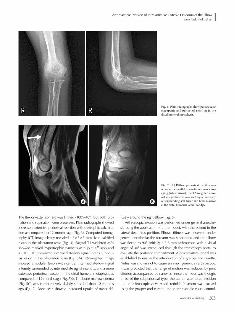
motion in the right dominant elbow for 12 months subsequent to a minor trauma, wherein a friend had twisted his arm. Three months after the injury, the patient had visited other institutions and had undergone laboratory and radiologic evaluations. Labo-ratory evaluation, including a complete blood count, erythrocyte sedimentation rate and blood chemistry profile, did not indicate any rheumatological diseases. Plain radiographs of the affected elbow revealed periarticular osteopenia and periosteal reaction in the distal humeral metaphysis (Fig. 1). Magnetic resonance imaging (MRI) showed joint effusion with synovial hypertrophy. Signal intensity of the surrounding soft tissue and bone mar-row was increased at the distal humerus lateral condyle on T2-weighted coronal images (Fig. 2). The patient was subsequently diagnosed as having bone contusion of the lateral humeral con-dyle and treated conservatively with intermittent nonsteroidal anti-inflammatory drugs (NSAID) and splinting. However, the pain and joint stiffness continued to progress. Twelve months after the initial injury, the patient visited our institution. History taking revealed the incidence of continuous pain which could partially be relieved by consuming NSAID and acetaminophen.
Arthroscopic Excision of Intra-articular Osteoid Osteoma of the ElbowSam Guk Park, et al.
www.cisejournal.org 163
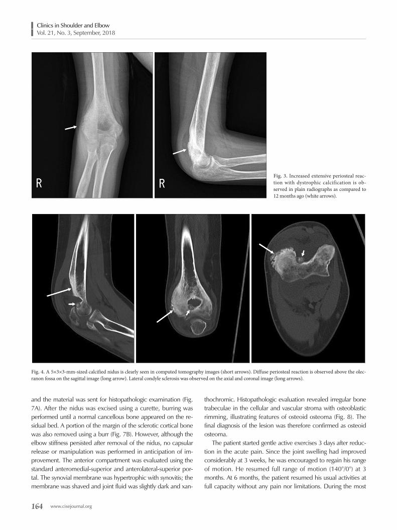
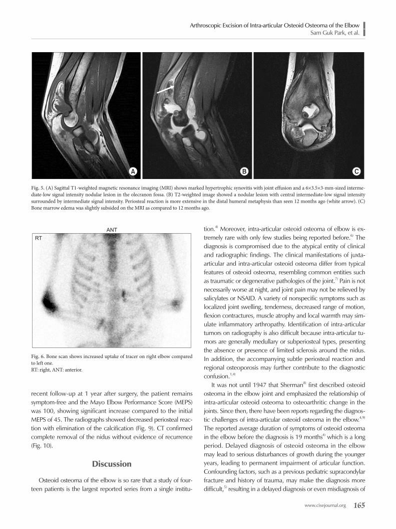
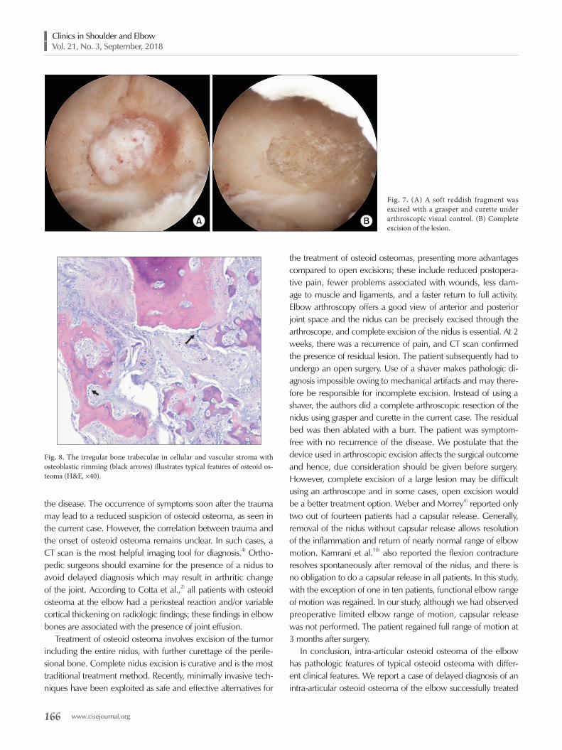
The flexion-extension arc was limited (100°/-40°), but both pro-nation and supination were preserved. Plain radiographs showed increased extensive periosteal reaction with dystrophic calcifica-tion as compared to 12 months ago (Fig. 3). Computed tomog-raphy (CT) image clearly revealed a 5×3×3-mm-sized calcified nidus in the olecranon fossa (Fig. 4). Sagittal T1-weighted MRI showed marked hypertrophic synovitis with joint effusion and a 6×3.5×3-mm-sized intermediate-low signal intensity nodu-lar lesion in the olecranon fossa (Fig. 5A). T2-weighted image showed a nodular lesion with central intermediate-low signal intensity surrounded by intermediate signal intensity, and a more extensive periosteal reaction in the distal humeral metaphysis as compared to 12 months ago (Fig. 5B). The bone marrow edema (Fig. 5C) was comparatively slightly subsided than 12 months ago (Fig. 2). Bone scan showed increased uptake of tracer dif-
fusely around the right elbow (Fig. 6). Arthroscopic excision was performed under general anesthe-
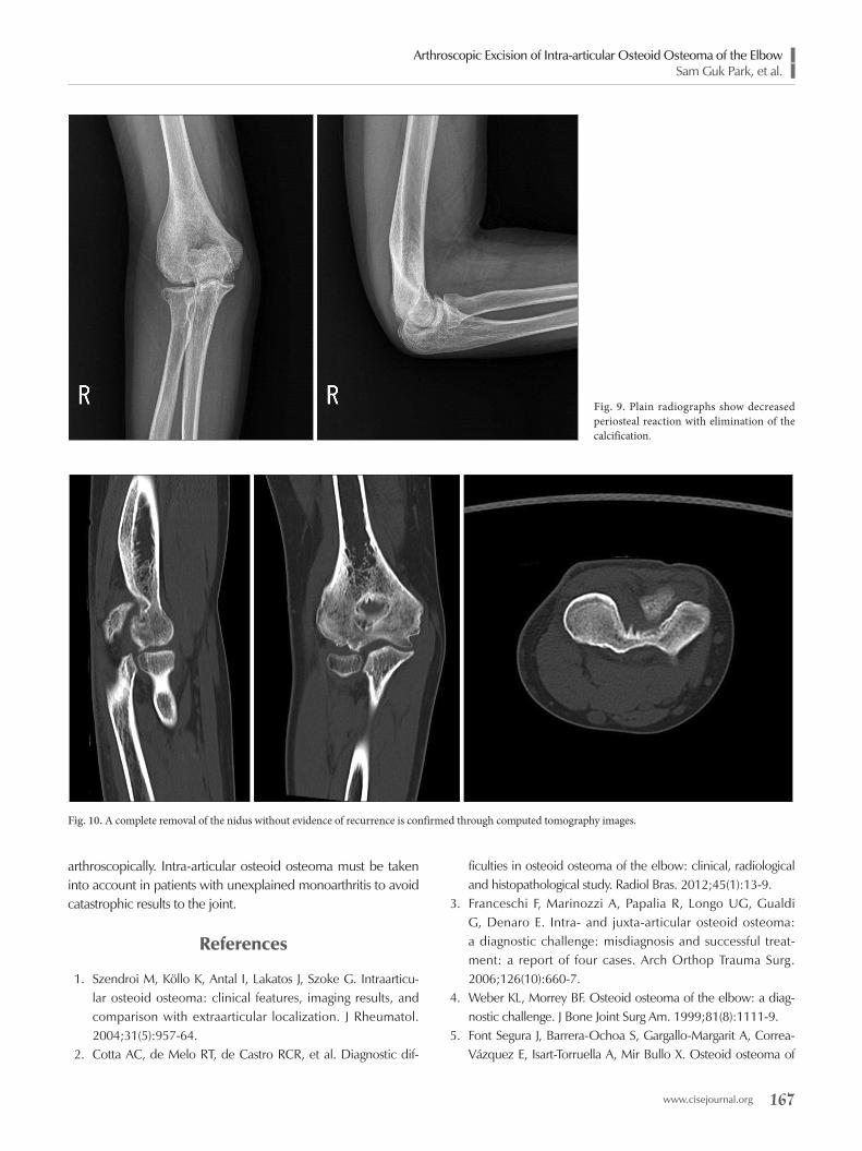
sia using the application of a tourniquet, with the patient in the lateral decubitus position. Elbow stiffness was observed under general anesthesia; the forearm was suspended and the elbow was flexed to 90°. Initially, a 3.8-mm arthroscope with a visual angle of 30° was introduced through the transtriceps portal to evaluate the posterior compartment. A posterolateral portal was established to enable the introduction of a grasper and curette. Nidus was shown not to cause an impingement in arthroscopy. It was predicted that the range of motion was reduced by joint effusion accompanied by synovitis. Since the nidus was thought to be of the subperiosteal type, the author attempted excision under arthroscopic view. A soft reddish fragment was excised using the grasper and curette under arthroscopic visual control,
Fig. 1. Plain radiographs show periarticular osteopenia and periosteal reaction in the distal humeral metaphysis.
A B
Fig. 2. (A) Diffuse periosteal reaction was seen on the sagittal magnetic resonance im-aging (white arrow). (B) T2-weighted coro-nal image showed increased signal intensity of surrounding soft tissue and bone marrow at the distal humerus lateral condyle.
164 www.cisejournal.org
Clinics in Shoulder and Elbow Vol. 21, No. 3, September, 2018
and the material was sent for histopathologic examination (Fig. 7A). After the nidus was excised using a curette, burring was performed until a normal cancellous bone appeared on the re-sidual bed. A portion of the margin of the sclerotic cortical bone was also removed using a burr (Fig. 7B). However, although the elbow stiffness persisted after removal of the nidus, no capsular release or manipulation was performed in anticipation of im-provement. The anterior compartment was evaluated using the standard anteromedial-superior and anterolateral-superior por-tal. The synovial membrane was hypertrophic with synovitis; the membrane was shaved and joint fluid was slightly dark and xan-
thochromic. Histopathologic evaluation revealed irregular bone trabeculae in the cellular and vascular stroma with osteoblastic rimming, illustrating features of osteoid osteoma (Fig. 8). The final diagnosis of the lesion was therefore confirmed as osteoid osteoma.
The patient started gentle active exercises 3 days after reduc-tion in the acute pain. Since the joint swelling had improved considerably at 3 weeks, he was encouraged to regain his range of motion. He resumed full range of motion (140°/0°) at 3 months. At 6 months, the patient resumed his usual activities at full capacity without any pain nor limitations. During the most
Fig. 3. Increased extensive periosteal reac-tion with dystrophic calcification is ob-served in plain radiographs as compared to 12 months ago (white arrows).
Fig. 4. A 5×3×3-mm-sized calcified nidus is clearly seen in computed tomography images (short arrows). Diffuse periosteal reaction is observed above the olec-ranon fossa on the sagittal image (long arrow). Lateral condyle sclerosis was observed on the axial and coronal image (long arrows).
Arthroscopic Excision of Intra-articular Osteoid Osteoma of the ElbowSam Guk Park, et al.
www.cisejournal.org 165
recent follow-up at 1 year after surgery, the patient remains symptom-free and the Mayo Elbow Performance Score (MEPS) was 100, showing significant increase compared to the initial MEPS of 45. The radiographs showed decreased periosteal reac-tion with elimination of the calcification (Fig. 9). CT confirmed complete removal of the nidus without evidence of recurrence (Fig. 10).
Discussion
Osteoid osteoma of the elbow is so rare that a study of four-teen patients is the largest reported series from a single institu-
tion.4) Moreover, intra-articular osteoid osteoma of elbow is ex-tremely rare with only few studies being reported before.6) The diagnosis is compromised due to the atypical entity of clinical and radiographic findings. The clinical manifestations of juxta-articular and intra-articular osteoid osteoma differ from typical features of osteoid osteoma, resembling common entities such as traumatic or degenerative pathologies of the joint.7) Pain is not necessarily worse at night, and joint pain may not be relieved by salicylates or NSAID. A variety of nonspecific symptoms such as localized joint swelling, tenderness, decreased range of motion, flexion contractures, muscle atrophy and local warmth may sim-ulate inflammatory arthropathy. Identification of intra-articular tumors on radiography is also difficult because intra-articular tu-mors are generally medullary or subperiosteal types, presenting the absence or presence of limited sclerosis around the nidus. In addition, the accompanying subtle periosteal reaction and regional osteoporosis may further contribute to the diagnostic confusion.1,4)
It was not until 1947 that Sherman8) first described osteoid osteoma in the elbow joint and emphasized the relationship of intra-articular osteoid osteoma to osteoarthritic change in the joints. Since then, there have been reports regarding the diagnos-tic challenges of intra-articular osteoid osteoma in the elbow.4,9) The reported average duration of symptoms of osteoid osteoma in the elbow before the diagnosis is 19 months4) which is a long period. Delayed diagnosis of osteoid osteoma in the elbow may lead to serious disturbances of growth during the younger years, leading to permanent impairment of articular function. Confounding factors, such as a previous pediatric supracondylar fracture and history of trauma, may make the diagnosis more difficult,5) resulting in a delayed diagnosis or even misdiagnosis of
A B C
Fig. 5. (A) Sagittal T1-weighted magnetic resonance imaging (MRI) shows marked hypertrophic synovitis with joint effusion and a 6×3.5×3-mm-sized interme-diate-low signal intensity nodular lesion in the olecranon fossa. (B) T2-weighted image showed a nodular lesion with central intermediate-low signal intensity surrounded by intermediate signal intensity. Periosteal reaction is more extensive in the distal humeral metaphysis than seen 12 months ago (white arrow). (C) Bone marrow edema was slightly subsided on the MRI as compared to 12 months ago.
RT
ANT
Fig. 6. Bone scan shows increased uptake of tracer on right elbow compared to left one. RT: right, ANT: anterior.
166 www.cisejournal.org
Clinics in Shoulder and Elbow Vol. 21, No. 3, September, 2018
the disease. The occurrence of symptoms soon after the trauma may lead to a reduced suspicion of osteoid osteoma, as seen in the current case. However, the correlation between trauma and the onset of osteoid osteoma remains unclear. In such cases, a CT scan is the most helpful imaging tool for diagnosis.4) Ortho-pedic surgeons should examine for the presence of a nidus to avoid delayed diagnosis which may result in arthritic change of the joint. According to Cotta et al.,2) all patients with osteoid osteoma at the elbow had a periosteal reaction and/or variable cortical thickening on radiologic findings; these findings in elbow bones are associated with the presence of joint effusion.
Treatment of osteoid osteoma involves excision of the tumor including the entire nidus, with further curettage of the perile-sional bone. Complete nidus excision is curative and is the most traditional treatment method. Recently, minimally invasive tech-niques have been exploited as safe and effective alternatives for
the treatment of osteoid osteomas, presenting more advantages compared to open excisions; these include reduced postopera-tive pain, fewer problems associated with wounds, less dam-age to muscle and ligaments, and a faster return to full activity. Elbow arthroscopy offers a good view of anterior and posterior joint space and the nidus can be precisely excised through the arthroscope, and complete excision of the nidus is essential. At 2 weeks, there was a recurrence of pain, and CT scan confirmed the presence of residual lesion. The patient subsequently had to undergo an open surgery. Use of a shaver makes pathologic di-agnosis impossible owing to mechanical artifacts and may there-fore be responsible for incomplete excision. Instead of using a shaver, the authors did a complete arthroscopic resection of the nidus using grasper and curette in the current case. The residual bed was then ablated with a burr. The patient was symptom-free with no recurrence of the disease. We postulate that the device used in arthroscopic excision affects the surgical outcome and hence, due consideration should be given before surgery. However, complete excision of a large lesion may be difficult using an arthroscope and in some cases, open excision would be a better treatment option. Weber and Morrey4) reported only two out of fourteen patients had a capsular release. Generally, removal of the nidus without capsular release allows resolution of the inflammation and return of nearly normal range of elbow motion. Kamrani et al.10) also reported the flexion contracture resolves spontaneously after removal of the nidus, and there is no obligation to do a capsular release in all patients. In this study, with the exception of one in ten patients, functional elbow range of motion was regained. In our study, although we had observed preoperative limited elbow range of motion, capsular release was not performed. The patient regained full range of motion at 3 months after surgery.
In conclusion, intra-articular osteoid osteoma of the elbow has pathologic features of typical osteoid osteoma with differ-ent clinical features. We report a case of delayed diagnosis of an intra-articular osteoid osteoma of the elbow successfully treated
A B
Fig. 7. (A) A soft reddish fragment was excised with a grasper and curette under arthroscopic visual control. (B) Complete excision of the lesion.
Fig. 8. The irregular bone trabeculae in cellular and vascular stroma with osteoblastic rimming (black arrows) illustrates typical features of osteoid os-teoma (H&E, ×40).
Arthroscopic Excision of Intra-articular Osteoid Osteoma of the ElbowSam Guk Park, et al.
www.cisejournal.org 167
arthroscopically. Intra-articular osteoid osteoma must be taken into account in patients with unexplained monoarthritis to avoid catastrophic results to the joint.
References
1. Szendroi M, Köllo K, Antal I, Lakatos J, Szoke G. Intraarticu-lar osteoid osteoma: clinical features, imaging results, and comparison with extraarticular localization. J Rheumatol. 2004;31(5):957-64.
2. Cotta AC, de Melo RT, de Castro RCR, et al. Diagnostic dif-
ficulties in osteoid osteoma of the elbow: clinical, radiological and histopathological study. Radiol Bras. 2012;45(1):13-9.
3. Franceschi F, Marinozzi A, Papalia R, Longo UG, Gualdi G, Denaro E. Intra- and juxta-articular osteoid osteoma: a diagnostic challenge: misdiagnosis and successful treat-ment: a report of four cases. Arch Orthop Trauma Surg. 2006;126(10):660-7.
4. Weber KL, Morrey BF. Osteoid osteoma of the elbow: a diag-nostic challenge. J Bone Joint Surg Am. 1999;81(8):1111-9.
5. Font Segura J, Barrera-Ochoa S, Gargallo-Margarit A, Correa-Vázquez E, Isart-Torruella A, Mir Bullo X. Osteoid osteoma of
Fig. 9. Plain radiographs show decreased periosteal reaction with elimination of the calcification.
Fig. 10. A complete removal of the nidus without evidence of recurrence is confirmed through computed tomography images.

168 www.cisejournal.org
Clinics in Shoulder and Elbow Vol. 21, No. 3, September, 2018
the distal humerus mimicking sequela of pediatric supracondy-lar fracture: arthroscopic resection-case report and a literature review. Case Rep Med. 2013. doi: 10.1155/2013/247328.
6. Contreras A, Isasi C, Silveira J, Barbadillo C, Mulero J, An-dreu JL. Intra-articular osteoid osteoma at the elbow: report of a case and review of the literature. J Clin Rheumatol. 2000;6(4):218-20.
7. Corbett JM, Wilde AH, McCormack LJ, Evarts CM. Intra-artic-ular osteoid osteoma; a diagnostic problem. Clin Orthop Relat Res. 1974;(98):225-30.
8. Sherman MS. Osteoid osteoma associated with changes in adjacent joint; report of two cases. J Bone Joint Surg Am. 1947;29(2):483-90.
9. Marcove RC, Freiberger RH. Osteoid osteoma of the elbow: a diagnostic problem. Report of four cases. J Bone Joint Surg Am. 1966;48(6):1185-90.
10. Kamrani RS, Moradi A, Sharafat Vaziri A, Nabian MH, Ghane B. Arthroscopic ablation of an osteoid osteoma of the elbow: a case series with a minimum of 18 months’ follow-up. J Shoul-der Elbow Surg. 2017;26(5):e122-7.
Copyright © 2022 FDOKUMEN