Functions of disordered regions in mammalian early base excision repair proteins
25
Functions of disordered regions in mammalian early base excision repair proteins Muralidhar L. Hegde, Department of Biochemistry and Molecular Biology, University of Texas Medical Branch, 301 University Blvd, Galveston, TX 77555-1079, USA Tapas K. Hazra, and Department of Biochemistry and Molecular Biology, University of Texas Medical Branch, 301 University Blvd, Galveston, TX 77555-1079, USA Department of Internal Medicine, University of Texas Medical Branch, Galveston, TX 77555-1079, USA Sankar Mitra Department of Biochemistry and Molecular Biology, University of Texas Medical Branch, 301 University Blvd, Galveston, TX 77555-1079, USA Sankar Mitra: [email protected] Abstract Reactive oxygen species, generated endogenously and induced as a toxic response, produce several dozen oxidized or modified bases and/or single-strand breaks in mammalian and other genomes. These lesions are predominantly repaired via the conserved base excision repair (BER) pathway. BER is initiated with excision of oxidized or modified bases by DNA glycosylases leading to formation of abasic (AP) site or strand break at the lesion site. Structural analysis by experimental and modeling approaches shows the presence of a disordered segment commonly localized at the N- or C-terminus as a characteristic signature of mammalian DNA glycosylases which is absent in their bacterial prototypes. Recent studies on unstructured regions in DNA metabolizing proteins have indicated their essential role in interaction with other proteins and target DNA recognition. In this review, we have discussed the unique presence of disordered segments in human DNA glycosylases, and AP endonuclease involved in the processing of glycosylase products, and their critical role in regulating repair functions. These disordered segments also include sites for posttranslational modifications and nuclear localization signal. The teleological basis for their structural flexibility is discussed. Keywords Base excision repair; DNA glycosylases; End processing proteins; Disordered terminal segments; Single strand breaks; Reactive oxygen species; Repair complex; Protein–protein and protein–DNA interactions © Springer Basel AG 2010 Correspondence to: Sankar Mitra, [email protected]. NIH Public Access Author Manuscript Cell Mol Life Sci. Author manuscript; available in PMC 2010 December 2. Published in final edited form as: Cell Mol Life Sci. 2010 November ; 67(21): 3573–3587. doi:10.1007/s00018-010-0485-5. NIH-PA Author Manuscript NIH-PA Author Manuscript NIH-PA Author Manuscript
-
Upload
independent -
Category
Documents
-
view
3 -
download
0
Transcript of Functions of disordered regions in mammalian early base excision repair proteins

Functions of disordered regions in mammalian early baseexcision repair proteins
Muralidhar L. Hegde,Department of Biochemistry and Molecular Biology, University of Texas Medical Branch, 301University Blvd, Galveston, TX 77555-1079, USA
Tapas K. Hazra, andDepartment of Biochemistry and Molecular Biology, University of Texas Medical Branch, 301University Blvd, Galveston, TX 77555-1079, USA
Department of Internal Medicine, University of Texas Medical Branch, Galveston, TX77555-1079, USA
Sankar MitraDepartment of Biochemistry and Molecular Biology, University of Texas Medical Branch, 301University Blvd, Galveston, TX 77555-1079, USASankar Mitra: [email protected]
AbstractReactive oxygen species, generated endogenously and induced as a toxic response, produceseveral dozen oxidized or modified bases and/or single-strand breaks in mammalian and othergenomes. These lesions are predominantly repaired via the conserved base excision repair (BER)pathway. BER is initiated with excision of oxidized or modified bases by DNA glycosylasesleading to formation of abasic (AP) site or strand break at the lesion site. Structural analysis byexperimental and modeling approaches shows the presence of a disordered segment commonlylocalized at the N- or C-terminus as a characteristic signature of mammalian DNA glycosylaseswhich is absent in their bacterial prototypes. Recent studies on unstructured regions in DNAmetabolizing proteins have indicated their essential role in interaction with other proteins andtarget DNA recognition. In this review, we have discussed the unique presence of disorderedsegments in human DNA glycosylases, and AP endonuclease involved in the processing ofglycosylase products, and their critical role in regulating repair functions. These disorderedsegments also include sites for posttranslational modifications and nuclear localization signal. Theteleological basis for their structural flexibility is discussed.
KeywordsBase excision repair; DNA glycosylases; End processing proteins; Disordered terminal segments;Single strand breaks; Reactive oxygen species; Repair complex; Protein–protein and protein–DNAinteractions
© Springer Basel AG 2010Correspondence to: Sankar Mitra, [email protected].
NIH Public AccessAuthor ManuscriptCell Mol Life Sci. Author manuscript; available in PMC 2010 December 2.
Published in final edited form as:Cell Mol Life Sci. 2010 November ; 67(21): 3573–3587. doi:10.1007/s00018-010-0485-5.
NIH
-PA Author Manuscript
NIH
-PA Author Manuscript
NIH
-PA Author Manuscript

IntroductionDamage to mammalian genomes, induced by a variety of reactive oxygen species (ROS) andreactive nitrogen species (RNS), that are generated either endogenously or by radiation andin response to other genotoxic and inflammatory agents, include a plethora of oxidativelydamaged bases, abasic (AP) sites and DNA single-strand breaks (SSBs) that are oftenmutagenic and have etiological linkage to sporadic cancers and a variety of otherpathophysiologies as well as aging [1,2]. It is generally estimated that more than 104 baselesions and SSBs are induced daily in a mammalian cell genome [3]. These base lesions andSSBs are typically repaired via the evolutionarily conserved DNA base excision repair(BER) pathway. BER is initiated with excision of an oxidized, alkylated or aberrant base bya lesion-specific DNA glycosylase, generating an AP site. In the case of oxidized bases, theAP sites are further cleaved by the intrinsic AP lyase activity of DNA glycosylases, while inother cases, the AP sites are cleaved by an AP endonuclease (APE). The resulting DNAstrand breaks have blocking groups either at the 3′ or 5′ end which need to be removed inorder to generate a single-stranded gap that could then be filled in by a DNA polymerasefollowed by the sealing of the nick by a DNA ligase [4]. The SSBs with blocked ends arealso generated directly by ROS that could include 3′-phosphate (3′-P), 3′-deoxyribosephosphate (3′-dRP) or 5′-deoxyriobosephosphate (5′-dRP) and oxidized sugarfragments such as 3′-phosphoglycolate [5,6]. While 5′-dRP is processed by the dRP lyaseactivity of DNA polymerase β (Polβ) in mammalian cells, 3′-P and 3′-dRP are removed bypolynucleotide kinase (PNK) and APE1, respectively [7]. Thus, the early step in repair ofmodified/oxidized bases and SSBs involves DNA glycosylases and nick-end processingenzymes.
Recent studies have suggested that BER is highly complex, involving a network ofintegrated pathways which are likely to be genome sequence-specific as well as cell cycle-specific [8,9]. Several studies by us and others have shown that the early BER proteins forma complex to initiate repair and binarily interact with most if not all of the downstreamproteins presumably for efficient co-ordination and sequential handover [7,10–13].
The major challenge for the early BER proteins in mammalian cells is lesion recognition viaefficient scanning of the gigabase size genome. Furthermore, a unique issue in BER, incontrast to nucleotide or mismatch excision repair pathways, is that the substrate lesion doesnot significantly distort the DNA helix and could retain near normal base pairing. Theselesions do not block transcription or replication and are invariably bypassed. Thus, lesionrecognition, particularly in highly condensed chromatin, poses a serious challenge which hasnot been extensively addressed. Interestingly, most early BER proteins have a stretch ofdisordered peptide segment invariably at one of the termini or which could serve sometimesas a linker bridging two domains, suggesting that such a common structural feature could beimportant for their functions. The focus of this review is to address the complexity of earlyBER activity in mammals and explore the common and unique structural features amongthese proteins that enable them to perform such an exigent function proficiently.
Basic BER mechanismBER, first elucidated in E. coli and subsequently found to be universally conserved, isinitiated with the recognition and excision of altered base lesions by about a dozen ofdistinct DNA glycosylases, each of which acts on a limited number of damaged bases[14,15]. Uracil DNA glycosylase (Udg) was the first DNA glycosylase to be discovered inE. coli, which removes the U from DNA. U is generated due to deamination of cytosine andis mutagenic [14]. Subsequently, similar enzyme activities were discovered in mammaliancells and nuclear (UNG2)-specific and mitochondria (UNG1)-specific UDG variants were
Hegde et al. Page 2
Cell Mol Life Sci. Author manuscript; available in PMC 2010 December 2.
NIH
-PA Author Manuscript
NIH
-PA Author Manuscript
NIH
-PA Author Manuscript

characterized [16]. Thymine-(T·G)–DNA glycosylase (TDG) is another mammalian DNAglycosylase that excises T and U paired with G [17]. DNA glycosylases specific for repair ofalkylated bases have been characterized. The methylated bases generated by chemicalreactions with endogenous 5-adenosylmethionine and exogenous alkylating agents,including many chemotherapeutic drugs and N-methylnitrosamine, are repaired either bydirect reversal without DNA repair synthesis or via the BER pathway [18]. Ada in E. coliwas the first repair protein discovered that carries out direct reversal of O6-alkylguanine[18,19]. Its mammalian ortholog O6-methylguanine methyltransferase (MGMT) wassubsequently cloned [20]. AlkB (E. coli) and its mammalian ortholog ABH are the otherdirect reversal enzymes for several alkylbase adducts [21]. N-methylpurine-DNAglycosylase (MPG, also named MAG) and its E. coli ortholog AlkA repair N-alkylpurinesvia the conventional BER pathway [22]. While DNA glycosylases in general recognize onlyabnormal bases in DNA, MutY, a mismatch-specific glycosylase discovered in E. coli andits mammalian homolog MYH excise normal base A from A·G and A·8-oxoG mispairs[23,24]. The glycosylases for the repair of U, alkyl base adducts or base mispairs aremonofunctional as they excise the base lesion, leaving an AP site without generating astrand break.
Oxidized base-specific DNA glycosylases in mammalian cells have been categorized in twofamilies based on their tertiary structure and AP lyase reaction and named after theirbacterial prototypes, Nth (endonuclease III) and Nei (endonuclease VIII) [25,26]. OGG1 andNTH1 belonging to Nth family were the first to be discovered. Subsequently, we and othersdiscovered NEIL1 and NEIL2 which belong to Nei family that also includes bacterial Fpg(formamidopyrimidine–DNA glycosylase) [1,26–30]. NEIL3 was recently added to the listof oxidized base-specific glycosylases [31]; however, its biological activity is not wellcharacterized. The Nth family glycosylases perform β-elimination at the AP site generating a3′-phospho-αβ-unsaturated aldehyde, a dehydration product of deoxyribosephospate (3′-PUA also named as 3′-dRP) at the strand break. The members of Nei family catalyze βδ-elimination at the AP site to produce 3′-P [7,32]. Thus, contrary to the APE-cleaved productof an AP site which contains 5′-dRP, and is removed by dRP lyase activity of Polβ, theoxidized base-specific DNA glycosylase-mediated strand breaks (BER intermediate)generate 3′ blocking groups, which need to be cleaned by specific end-processing enzymesin the subsequent BER step.
Processing of blocked termini: common step in the repair of altered basesand SSBs
Single-strand interruption processing, the second step in the repair of oxidized bases and APsites, is also required for repairing direct ROS-induced SSBs which invariably contain anunligatable blocking group, either 3′ or 5′ or both ends at the strand break [33]. Gap fillingby a DNA polymerase and nick sealing by a ligase require 3′-OH and 5′-P at the break site.End processing is thus an obligatory step involving multiple essential enzymes, specific forthe 5′ or 3′ blocking groups as well as their type.
5′ end processing at a strand breakAPE cleavage of the AP site generates 3′-OH and 5′-dRP termini. DNA polymerase β inmammalian cells removes the unmodified 5′-dRP moiety via its lyase activity [34,35].However, Polβ cannot process 5′ blocking groups generated by the cleavage of oxidized APsite by APE, and such dirty ends are processed via the long-patch BER pathway whichinvolves removal of displaced 2–8 nts along with the 5′ blocking group by FEN-1, asdiscussed later. A unique type of 5′ blocking groups are formed as intermediates duringabortive DNA ligation, such as adenylate groups covalently linked to the 5′ P terminus at
Hegde et al. Page 3
Cell Mol Life Sci. Author manuscript; available in PMC 2010 December 2.
NIH
-PA Author Manuscript
NIH
-PA Author Manuscript
NIH
-PA Author Manuscript

strand break, and such groups are processed by a protein named aprataxin [36,37]. Aprataxinreleases the adenylate moiety to form ligatable 5′-P.
3′ end processing at a strand breakThe Nth family glycosylases with intrinsic β lyase activity generate 3′-dRP (3′-PUA) and 5′-P at the strand break as already mentioned. The 3′dRP is removed by the intrinsicphosphodiesterase activity of mammalian APE1 to generate the 3′-OH terminus. On theother hand, the βδ lyase activity of Nei family DNA glycosylases would generate the 3′-P.We had shown that 3′-P is a poor substrate of APE1 but is efficiently removed by PNK inmammalian cells [7]. In addition, the 3′ blocks at the ROS-induced SSBs, mainly 3′-phosphoglycolate and 3′-phosphoglycoladehyde, are processed by APE1 [38–40].Tyrosylphosphodiesterase 1 (TDP1) is another 3′ end processing enzyme discovered in yeastand human cells, which removes the product of abortive topoiso-merase1(Top1) reaction,namely Top1(Tyr)-linked 3′ termini to form 3′-P, a substrate for PNK [41–44].
While there are multiple APEs in prokaryotes and lower eukaryotes, APE1 is thepredominant contributor of APE activity in mammalian cells. Its prototype in E. coli isexonuclease III (Xth). As AP sites and strand breaks are continuously generated in thegenome, it is not surprising that multiple enzymatic processes involving APEs are evolved.We have recently reviewed the functions of multiple APEs in BER in Hegde et al. [1].
Furthermore, TREX1 and TREX2 DNA 3′ exonucleases are ubiquitous in mammaliantissues, whose primary function may be editing during replication by Polβ or Polα lackingconstitutive 3′ exonuclease activity [45]. In addition, TREX1 has also been proposed to playa role in SSBR, and its deficiency has been linked to a severe brain disease [46].
XRCC1 and PARP are two other key proteins which play a role in early BER activity. WhileXRCC1 acts as a scaffold for recruiting BER proteins for excision or strand break repair,PARP acts a SSB sensor protein. We showed that XRCC1 physically interacts with NEILssuggesting its role in oxidized base repair [7,47]. XRCC1 also interacts with end processingenzymes PNK and APE1, and other BER proteins, Polβ and LigIIIα [48]. Moreover,processing of 5′-OH and 3′-P termini at SSBs is reduced in XRCC1-deficient cellssuggesting its role in end processing [49]. PARP present in mammalian cells and absent inE. coli is activated by SSBs and transfers ADP-ribose moiety from NAD to a variety ofproteins including itself. PARP-1 and PARP-2, the two proteins of the PARP superfamily,have been shown to be important players in the repair of SSBs both as sensors and forrecruiting other repair proteins to the strand break [50]. Thus, XRCC1 and PARP play anindirect but vital role in both base excision and end cleaning repair steps.
The early BER reactions for repair of modified bases and SSBs are schematicallyrepresented in Fig. 1.
Complexities in BER and SSBR: multiple repair subpathwaysUntil recently, BER was believed to be the simplest among the DNA repair pathwaysinvolving four or five reaction steps. However, recent studies reveal that BER is much morecomplex, involving a network of distinct cell cycle-dependent as well as genome region-specific repair subpathways and could also involve non-BER proteins.
Short-patch versus long-patch repair pathwaysIn the simple mammalian BER model, excision of the lesion leaves a 1-nt gap at the damagesite, which is subsequently filled by Polβ and the resulting nick sealed by DNA ligase IIIα
Hegde et al. Page 4
Cell Mol Life Sci. Author manuscript; available in PMC 2010 December 2.
NIH
-PA Author Manuscript
NIH
-PA Author Manuscript
NIH
-PA Author Manuscript

(LigIIIα) to complete single-nucleotide repair (SN-BER), also called short-patch repair (SP-BER) [51].
In contrast to this simple model, recent discoveries document several BER subpathwaysinvolving at least a dozen more proteins. The second mode of BER, characterized byMatsumuto, Dogliotti and coworkers [34,52,53], involves a repair patch size of 2–8nucleotides at the lesion site and was named long-patch repair (LP-BER). LP-BER was firstobserved during repair of an AP site analog lacking the aldehyde group. An upstreamsegment containing 2–8 nts including the 5′-blocking group is displaced as a single-strandflap during repair synthesis which is subsequently cleaved by flap endonuclease 1 (FEN-1).FEN-1’s normal function is to remove the 5′ RNA primers of Okazaki fragments duringdiscontinuous replication of the lagging strand [54–56]. LP-BER is believed to utilize DNAreplication proteins including DNA polymerase δ (Polδ), the sliding clamp PCNA, clamploader replication factor-C (RF-C) and DNA ligase I (LigI) in addition to FEN-1 [57,58].However, Polβ has also been shown to participate in LP-BER via strand displacement, incollaboration with FEN-1 [59]. The choice of SN-BER versus LP-BER is a complex issuethat is yet to be completely understood. Initial studies suggested that the nature of the 5′-phosphoribose terminus (normal vs oxidized) would be the deciding factor [60,61]. Howeverinvolvement of DNA replication proteins with LP-BER strongly suggests that LP-BERcould be preferred in BER during DNA replication, irrespective of the 5′ terminal group.
DNA replication/transcription-specific BER subpathwaysThe mammalian genome at the replication fork or transcription bubble is relatively unfoldedor nonchromatinized rendering it more prone to oxidative damage than nonreplicatingchromatin. Replication of unrepaired oxidized base lesions which do not block replicationcould be mutagenic. Thus, there is an urgency to repair these lesions prior to replication(pre-replicative repair) in order to maintain genome integrity. Furthermore, incorporation ofabnormal or oxidized bases (e.g., uracil or 8-oxoG) from the nucleotide pool into nascentDNA could be mutagenic as well, which also warrants urgent repair. Such repair was earlierdescribed as post-replicative repair [62].
Recent studies by us and others have suggested that there are distinct BER subpathways fortranscriptionally active versus inactive genomes as well as for quiescent versus replicatinggenome. Our initial studies with human NEIL1 and NEIL2 showed that both NEILs areactive on bubble and fork-structured DNA substrates that mimic DNA replication ortranscription intermediates [8]. At the same time, only NEIL1 is upregulated during the S-phase, based on which we had proposed that NEIL1 is preferentially involved in replication-associated repair (RA-BER) and NEIL2 in transcription-coupled BER (TC-BER)[8,27,28,63]. Subsequently, our recent studies have shown NEIL1’s preferential associationwith DNA replication proteins including PCNA [11], Replication protein A (RPA; [13]),FEN-1 [10], and Werner’s helicase (WRN; [12]) that strongly support this hypothesis.Similarly, several other DNA glycosylases such as MYH and UNG (which are describedlater in this article) interact with replication proteins PCNA and RPA [64,65]. Like NEIL1,UNG and MYH expression increases during the S-phase, and the UNG-PCNA-RPAcomplex co-localizes with replication foci suggesting preferential repair of nascent DNA[62,66]. In contrast, our studies with NEIL2 which has cell cycle-independent expressionsuggested its association with transcription-associated BER. Although more studies arerequired to further characterize the genome region and cell-state-specific BER pathways, itis clear that additional complexities are associated with these distinct BER subpathways. Forexample, we have shown stable interaction between NEIL1 and 9-1-1 complex, a stress-activated sliding clamp implying a linkage between NEIL1-initiated BER and damagesignaling pathways [67].
Hegde et al. Page 5
Cell Mol Life Sci. Author manuscript; available in PMC 2010 December 2.
NIH
-PA Author Manuscript
NIH
-PA Author Manuscript
NIH
-PA Author Manuscript

Involvement of non-BER proteins in the repair of base lesions/SSBsWhile several non-BER proteins have been shown to be involved in BER, adding anotherdimension to the BER complexity, their precise in vivo role in repair has yet to beunraveled. We showed that NEIL2 interacts with YB-1, a Y-box binding protein, and it wassuggested that YB-1 may be required for the fine-tuning of repair [68]. NTH1 was alsoshown to interact with and is stimulated by YB-1 [69]. The list of non-BER proteinsinteracting with BER proteins is still growing, which underscores the paradigm that the invivo repair process is far more complex than in vitro repair demonstrated with minimalcomponents.
Recent discoveries in BERDNA glycosylases as hub proteins: binary interaction of DNA glycosylases withdownstream proteins
Our initial characterization of NEIL1 and NEIL2 showed their similarity in in vitro repair of5-OHU via SN-BER in a reconstituted system containing PNK, Polβ, LigIIIα and XRCC1.XRCC1 acts as a scaffold for recruiting BER proteins for excision or strand break repair.Both the NEIL immunoprecipitates from human cells contain these BER proteins [7,47].Furthermore, NEILs binarily interact in the absence of DNA with Polβ, LigIIIα and XRCC1,although not PNK. Direct interaction of NEILs with LigIIIα, the last enzyme in SN-BER,indicated that the repair is controlled or regulated by the initiating DNA glycosylase whichacts as a hub protein. Similarly, as already discussed, NEIL1 binarily interacts with PCNA,RPA, FEN-1, and WRN in the absence of DNA, presumably for preferential BER duringDNA replication. The stoichiometry of proteins in the repair complex and whether NEILsare present in distinct complexes or in a single complex are currently being investigated inour laboratories.
Repair complex versus sequential repair hand-offThe initial BER mechanism was proposed, based on cocrystal structure analysis of substrate-bound BER proteins, to involve ‘hand-off’ or ‘passing the baton’ process, wherein the repairproduct of each enzyme in the BER pathway is handed over to the next enzyme, primarilybased on differential bending of DNA in each intermediate step [70,71]. However,characterization of the BER interactome involving multiprotein interactions including stablecomplex of DNA glycosylase with DNA ligase, and the presence of multiprotein complexeshas led to a new paradigm where complete repair occurs in the BER complex (BERosome).Although in vivo role of hand-off versus interactome modes of repair is not yet clear, wepropose that preformed BER complexes predominantly repair endogenous base lesions,while repair via hand-off mechanism by sequential recruitment could occur with inducedDNA damage. Further characterization of the dynamics of BERosomes is required tounravel the repair processes.
Common interaction interface of early BER proteinsFor several years, our laboratory has focused on characterizing collaborations and mappinginteractions among BER proteins. As already mentioned, NEIL1’s stable interaction withdownstream repair proteins utilizes a common interaction interface located near its C-terminus (residues 289–349) [7,10–13]. The segment is absent in NEIL1’s prototype Nei,and might have been acquired during evolution as a terminal addition [1]. We similarlyidentified the nonconserved N-terminal segment (65 residues) in APE1 which is involved inall its protein–protein interactions [72–74]. Although it is intriguing how NEIL1 or anyother protein could simultaneously bind to so many proteins with high specificity via a smallcommon interface, recent studies have indicated that it is not uncommon for the mammalian
Hegde et al. Page 6
Cell Mol Life Sci. Author manuscript; available in PMC 2010 December 2.
NIH
-PA Author Manuscript
NIH
-PA Author Manuscript
NIH
-PA Author Manuscript

hub proteins to have such an interaction surface, which invariably have a disorderedstructure. The flexibility of the disordered domain may facilitate interaction with diversepartners [75,76].
Disordered structure of the interaction interfaceThe C-terminal region of human NEIL1 (hNEIL1) spanning about 100 residues contains thecommon interaction interface whose disordered conformation was first suggested from thefact that NEIL1’s crystallization required deletion of 56 residues, while the proximal 44residues did not form a defined structure [77]. We verified this conclusion using variousprotein structure prediction softwares.
Prediction of disordered structure: various softwaresContrary to the concept about spontaneous formation of secondary and tertiary structure inproperly folded proteins that prevailed before the turn of this century, recent experimentalevidence as well as genome-wide prediction of intrinsic disorder in eukaryotic proteomeshave indicated that a large percentage of proteins have long disordered (unfolded) segmentsunder physiological conditions. Further, these disordered regions are essential for theirbiological functions. Recent advancement in the softwares for accurate prediction of proteinsecondary structure and their predisposition to remain intrinsically disordered has furtheredour understanding of the role of disordered structures in functional hierarchy. Commonlyused disorder prediction tools include PONDR [78–80], PrDOS [81,82], RONN [82],FoldIndex [83], GlobPlot [84], IUPred [85,86], FoldUnFold [87], etc. in the public domain,which evaluate intrinsic disorder on per residue basis. Among these, PONDR is most widelyused, an advanced version of which contains a reference collection set of VSL predictors(trained on variously characterized, short and long disordered regions). The PONDRdevelopers point out that short and long disordered regions might have differences in theiramino acid characteristics because predictors based on short regions of disorder fare poorlyfor long regions of disorder and vice versa [88–90]. The VSL predictors in PONDR takeadvantage of such differences to yield more accurate predictions. We used PONDR, PrDOSand RONN software, which generated similar disorder prediction in early BER proteins.
The commonly used approaches to characterize protein disorder are NMR and circulardichroism spectroscopy, and also small angle X-ray scattering, while the structuralinformation thus generated is often based on the crystal structure of truncated proteins [91].
Signature sequences of intrinsic disorderA major sequence characteristic of intrinsic disorder is the low content of bulky hydrophobicamino acid residues such as Val, Leu, Ile, Met, Phe, Trp and Tyr, which would normallyform the core of a folded globular protein. In contrast, a high proportion of polar andcharged residues such as Gln, Ser, Pro, Glu, Lys, and Gly, and sometimes also Ala, arecharacteristically present in disordered regions [78,92]. The presence of such chargedresidue-rich sequences was first discovered in transcriptional regulatory proteins about threedecades ago, which are often classified based on their amino acid composition, for exampleGlu-rich, Pro-rich and acidic activation domains [93]. Later, NMR spectroscopy and otherbiophysical studies confirmed intrinsically disordered nature of such sequences [91,94].
While disordered regions have been variously described as intrinsically disordered,intrinsically unstructured, natively unfolded, natively disordered and highly flexible [91], webelieve that the term ‘intrinsically disordered’ would be more appropriate than‘unstructured’ because many disordered regions have been shown to contain partial ortransient secondary and/or tertiary structural organization [95]. Dunker and Obradovic [96]proposed that intrinsically disordered regions may exist in two different structural forms:
Hegde et al. Page 7
Cell Mol Life Sci. Author manuscript; available in PMC 2010 December 2.
NIH
-PA Author Manuscript
NIH
-PA Author Manuscript
NIH
-PA Author Manuscript

molten globule-like (collapsed) and random coil-like (extended), whereas Uversky and co-workers suggested existence of another extended form, the pre-molten globule, which isdistinguishable from fully extended and molten globular conformations by the presence ofan unstable secondary structure [97]. The recently proposed protein quartet hypothesissuggests that the protein functions in eukaryotes could depend on any of the three disorderedforms along with the ordered form or on transitions between them [97].
Disorder predictions have been extensively utilized by protein crystallographers to designcrystallization targets after deletion of disordered segments. One first application of thedisorder predictor was in crystallization of Xeroderma pigmentosum group A (XPA) proteininvolved in DNA repair [98]. Extended disordered regions at the N- and C-termini of XPAwith ordered central core as predicted was confirmed with partial proteolysis and NMRspectroscopy [99].
The recently created databank of protein intrinsic disorder (http://www.disprot.org) suggeststhat proteins with long terminal extensions containing no or limited structure are common ineukaryotes and are involved in many key functions including cell cycle control, regulation,and signaling [99]. Such disordered tails were also shown to be more common in DNAbinding proteins than in other proteins, particularly in the ones that are involved in targetsequence binding that include early repair proteins and transcription factors [100,101]. It islikely that their structural flexibility and plasticity provides major functional advantage.
Predictions of disorder using accurate bioinformatic tools are in fact helping designexperiments to characterize their biological functions, and this is one of the fastest growingtopics of protein studies, humorously dubbed as ‘the protein unfoldomics decade’. Althoughthe number of proteins with experimentally determined disordered structure is still small, thebehavior of the experimentally verified ones are highly consistent with the predictions [102].
Comparison of E. coli versus mammalian early BER proteins to identifydisordered structures
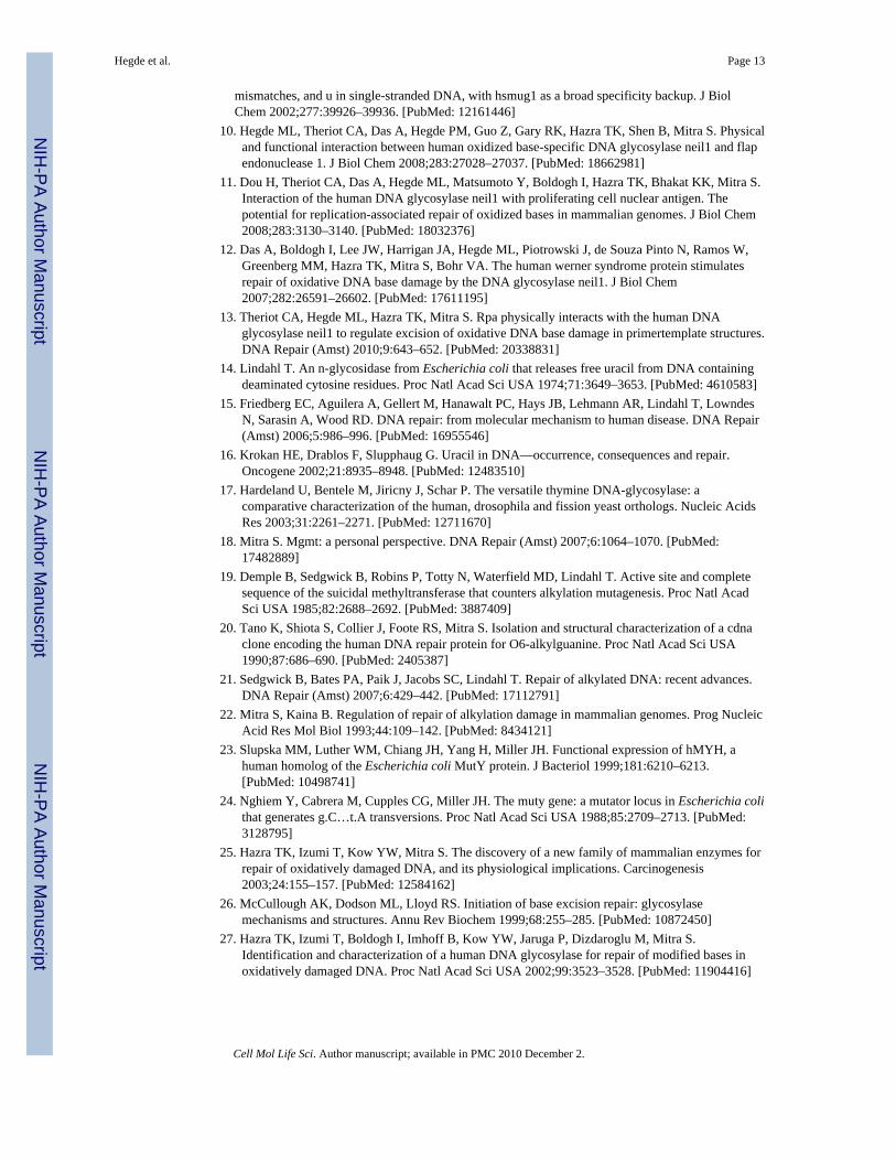
We used PONDR (prediction of natively disordered regions) and PrDOS (prediction ofdisordered structures) softwares to compare the secondary structure of human and bacterialearly BER proteins and correlated them with their available structural information. Asalready mentioned, hNEIL1 contains an extended disordered region spanning about 100residues in the C-terminus which is absent in E. coli Nei protein (Fig. 2). Comparison ofpredicted structures of human DNA glycosylases NTH1, MYH and their E. coli prototypesendonuclease III and MutY, respectively, indicates that both hNTH1 and hMYH haveextended disordered tails at the N-terminus that are absent in the E. coli enzymes. Similarly,the N-terminal disordered region present in hAPE1 is absent in Xth, its E. coli prototype(Fig. 3). PONDR modeling showed that such disordered terminal sequences may also bepresent in other human DNA glycosylases including UNG2 and TDG (Fig. 4). Although theunfolded sequence generally exists at the N- or C-terminus, this could exist internally insome proteins, acting as a linker joining two domains. HNEIL2 is such a protein with aninternal disordered segment near the N-terminus (residues 45–130) as indicated from thePONDR plot (Fig. 4). The average size of disordered extensions in early BER proteinsranges from 50 to 100 residues, with few exceptions, e.g., hOGG1 and human Polβ, whichhave short (~10 residues) disordered tails at both termini, as predicted by PrDOS (Fig. 5).The early BER proteins are generally small (30–50 kDa) and monomeric, whereas otherDNA transaction proteins such as PCNA are multimeric and typically possess disorderedlinkers bridging different subunits.
Hegde et al. Page 8
Cell Mol Life Sci. Author manuscript; available in PMC 2010 December 2.
NIH
-PA Author Manuscript
NIH
-PA Author Manuscript
NIH
-PA Author Manuscript

Among early BER proteins, the disordered N-terminal sequence of hNTH1 has beenextensively characterized. HNTH1 has a lower specific activity than E. coli Nth that lacksthe N-terminal extension [103,104], deletion of which increases hNTH1’s activity [105].This suggests that this segment inhibits enzyme turnover in the absence of other BERproteins.
Role of disordered domain in function of early BER proteinsThe presence of disordered extensions in proteins involved in transcriptional regulation,signal transduction, cell cycle control, DNA damage sensing and repair suggest theirinvolvement in diverse functions. Furthermore, such disordered regions may regulateformation of large multiprotein complexes [106,107]. An exhaustive discussion of this topicis beyond the scope of this review, focusing on the early BER proteins, in which thesedisordered segments include sites of posttranslational modifications, subcellular targeting,DNA scanning as well as common interface for protein–protein interactions, as alreadydiscussed (Fig. 6).
Posttranslational modifications in disordered segmentsPosttranslational modifications of proteins such as phosphorylation, acetylation,ubiquitylation, ADP-ribosylation, sumoylation and methylation play a critical role in diversecellular processes including DNA repair [108]. The modification sites are invariablylocalized in disordered regions, e.g., in the N-terminal segment in hAPE1 [74,109], N- andC-terminus of p53 [110], and C-terminal region in hNEIL1 (Bhakat et al., unpublished). Ourlaboratory identified and characterized acetylation of hAPE1 at Lys6 or Lys7, and thismodification plays an important role in APE1’s transcriptional regulatory functions [74].Recently, APE1 was also shown to be ubiquitynated at N-terminal Lys residues, whichregulates its degradation as well as cellular functions [109]. Such covalent modificationsmay have multiple physiological effects on these proteins, including stability, interactionwith DNA or other proteins, organelle targeting, and enzymatic activity [111]. We alsoshowed that hNEIL2 is acetylated at Lys49 and Lys153 both in vitro and in cells [112].Acetylation of Lys49 located in the disordered region (Fig. 4) inactivated NEIL2’s baseexcision and AP lyase activity while acetylation of Lys150 had no effect on the activity. Wehave proposed that acetylation of Lys49 could act as a regulatory switch for NEIL2’sactivity [112]. TDG is acetylated in the N-terminal segment, Lys70, 94, 95 and 98 which iswithin the disordered segment of 100 residues [113]. PONDR modeling of TDG sequenceindicates that the N-terminal 100 residues are in disordered conformation (Fig. 3). Strongacetylation sites in TDG were identified. Acetylation of TDG by CBP/p300 does not affectits binding to G·T or G·U base mispairs but indirectly deregulate TDG-coupled repair byreleasing CBP/p300 from DNA bound complex leading to reduced interaction with APE andin turn suppressing APE-dependent repair [113]. Thus, TDG acetylation could contribute togenomic instability and cancer susceptibility. We had earlier characterized acetylation ofhOGG1 at Lys338 and Lys341 within its short disordered C-terminus, which increases itsDNA glycosylase activity by reducing affinity for the product AP site [114].
The flexibility of disordered region appears to be a prerequisite for these modifications,presumably because the amino acid side chains in the flexible sequence are accessible formodifying enzymes, like kinases, phosphatases, acetyltransferases and deacetylases,methylases, and ubiquitin ligases, etc. [91].
Subcellular localizationOrganelle localization signals such as nuclear localization signal (NLS) or mitochondrialtransport signal (MTS) are contained in short segments (generally <20 residues) that mediate
Hegde et al. Page 9
Cell Mol Life Sci. Author manuscript; available in PMC 2010 December 2.
NIH
-PA Author Manuscript
NIH
-PA Author Manuscript
NIH
-PA Author Manuscript

transport to the target organelle. Multiple types of mammalian NLS sequences have beenidentified, the major ones belonging to the classical type consisting of seven basic residuesand the bipartite NLS with two strings of basic residues separated by a short interveningsequence [115]. Recent studies showed that almost all NLS sequences with overall basicnature are disordered [99,116].
We mapped the NLS of hAPE1 to the disordered N-terminal 20 residues, the deletion ofwhich markedly diminishes its translocation to the nucleus [117]. Our preliminary studies ofGFP-fusion polypeptide of truncated NEIL1 suggest the presence of putative NLS at thedisordered C-terminal region (unpublished observation). Similarly, the disordered N-terminal tails in hNTH1, UNG2 and TDG contain putative NLS and MTS [118–120]. Takentogether, these studies show that subcellular distribution of many human repair proteins ismediated through signals localized in their disordered regions.
DNA scanningBurg et al. have shown that target DNA search by proteins could be achieved via facilitateddiffusion comprising four mechanisms, namely, one-dimensional (1D) sliding, hopping, 3Dsearch and intersegmental transfer. An efficient search mechanism involves combination ofthese different modes [121].
Recent studies have shown that the most efficient and rapid scanning of the DNA for thetarget site involves 80% hopping and intersegmental transfer and 20% sliding by the DNAbinding proteins, which invariably contain a disordered terminal extension, or a disorderedlinker for multidomain or multisubunit proteins [101]. Bioinformatics analysis has suggestedthat nearly 70% of DNA binding proteins have such disordered tails compared to about 25%for non-DNA binding proteins [100,101]. In addition, the disordered segments are aboutseven residues longer on an average for DNA binding proteins compared to all proteins withdisordered tails [100]. Another unique characteristic of the disordered tails in DNA bindingproteins is clustering of positively charged residues in the distal region, which turned out tobe important for the scanning. Mutating such residues in HOXD9 and NK-2 markedlydecreased scanning efficiency [101]. Similar results were obtained when the N-terminalsegment in these proteins were deleted, suggesting that the initial scanning is mediated by anon-specific, mostly electrostatic, transient DNA binding via the basic disordered segmentwhich is followed by target DNA sequence binding by the active site.
In light of the above studies, we examined hNEIL1’s C-terminus, which possesses mostcharacteristics required for DNA scanning as described above, including the clustered basicresidues. Our recent biochemical studies using C-terminal deletion NEIL1 mutants showedthat the C-terminus is important for NEIL1’s substrate scanning and efficiency of damagerecognition, via its non-specific DNA binding (Hegde et al., unpublished). Although limitedstudies are available on the role of disordered regions of other early BER proteins in suchactivity, we expect that all of them have similar functions.
Intrinsic disorder and hub proteins: dynamic repair complexes mediated bydisorder/disorder interactions
As mentioned earlier, the ‘hub’ proteins like NEIL1 with several partners (usually >10) forma network of complexes [122]. Recent studies have indicated that ‘hub’ protein complexesare widely present in higher eukaryotes, whose formation mostly involves interaction withdisordered regions [123]. The crucial role of intrinsic disorder in hub proteins was reviewedearlier [91,124]. Bioinformatics analysis of known protein interactions suggested that suchinteractions among disordered structures are significantly preferred among human proteins[123]. Disorder-mediated interactions could either involve disorder–disorder or disorder–
Hegde et al. Page 10
Cell Mol Life Sci. Author manuscript; available in PMC 2010 December 2.
NIH
-PA Author Manuscript
NIH
-PA Author Manuscript
NIH
-PA Author Manuscript

order types. Both modes of interactions are prevalent in BER proteins, for example NEIL1–FEN-1 interaction involves disorder–disorder interfaces [10], while NEIL1–Polβ couldinvolve disorder–order type of contacts [7]. Specific recognition and binding of hub proteinsis achieved as a result of the flexibility of domain itself, because it facilitates conformationalrearrangements and induced-fit with specific partners [99,107]. Although it wasinconceivable a few years ago that such specificity could be obtained via disorder–disorderinteractions, these are quite common, particularly for hub proteins. Disorder-mediatedinteractions in fact confer advantages over order-mediated interactions because of the rapidand easy interconversion among diverse conformers, allowing formation of dynamiccomplexes [107]. The dynamics of such complexes could be further regulated byposttranslational modifications of one or more partners, as already discussed. Furthermore,disorder-mediated interactions have kinetic benefits, because the larger capture radius ofdisordered states facilitates faster on-rates for binding [125,126]. Finally, disorder-mediatedinteractions have steric advantages by providing a large surface area for binding interface forwrapping around partners resulting in stronger specificity [127,128].
Our recent studies using size fractionation chromatography of human cell nuclear extractsuggest that the BER proteins indeed exist in large, stable complexes, presumably in theabsence of DNA. Characterization of such complexes, the dynamics of their formation andregulation as well as their stoichiometry are warranted.
Evolutionary advantages of protein disorderDisordered regions in proteins generally show higher rates of mutation, presumably becausechanges in their protein sequence may not affect protein stability and function as severely asthat in ordered regions [107,129,130]. Although such an analysis of mutation distributionand mutation tolerance is yet to be carried out for BER proteins, a similar situation is likelyto exist. The unique presence of disordered segments in eukaryotic proteins but not in theprokaryotic counterparts, with the highest degree of disorder in mammals, suggests itsevolutionary development [107]. The disordered regions also enable alternative splicing ineukaryotic proteins without the risk of perturbing structured regions [131]. In addition, thedisorder provides advantage of limiting molecular size as complexity increases, byproviding common interface for multiprotein binding and sites of modifications. To achievea similar goal, folded proteins need to be considerably larger, and thus disorder may helphigher organisms to limit protein size and to reduce intracellular crowding [132].
Disordered extensions in early BER proteins as potential targets of cancertherapy
The BER proteins have been explored as targets for cancer therapy which generally involvedinactivation of key BER reactions such as ligation [133] or damage sensing proteins such asPARP [134]. DNA glycosylases turned out to be poor targets for such therapy, because ofthe nonessentiality of individual glycosylases due to back up functions of other glycosylases[1]. Although PARP inhibitors have been proven highly successful in recent therapeuticstudies, inhibition of a key BER reaction poses the challenge of accurate targeting anddosage regulation to prevent their undesired effect on normal cells [134]. Based on theemerging evidence for a disorder-mediated repair regulatory switch in early BER hubproteins that controls the repair pathway, we propose that the disordered regions of earlyBER proteins could be targeted for cancer therapy which would disrupt repair regulation.
Hegde et al. Page 11
Cell Mol Life Sci. Author manuscript; available in PMC 2010 December 2.
NIH
-PA Author Manuscript
NIH
-PA Author Manuscript
NIH
-PA Author Manuscript

Conclusions and perspectivesA combination of experimental studies and structural predictions has revealed a critical roleof disordered segments in many mammalian early BER enzyme functions including bothprotein–protein and protein–DNA interactions. Although few disordered regions have beenexperimentally characterized so far, we predict that such disordered segments are ubiquitousand essential for efficient repair. Future studies should address the role of disorderedsequences in other mammalian repair pathways and their evolutionary significance incomplex repair regulation.
Abbreviations
BER Base excision repair
SSBR Single-strand break repair
AP Abasic
APE AP endonuclease
ROS Reactive oxygen species
RNS Reactive nitrogen species
SSB Single-strand break
PONDR Prediction of naturally disordered regions in proteins
AcknowledgmentsThe research in the authors’ laboratory is supported by USPHS grants, R01 CA81063, R01 CA53791, P01CA92586 and P30 ES06676 (S.M.) and R01 CA 102271, R21 ES017353 (T.K.H). Because of the limited focus ofthe article on protein disorder in early BER proteins, many appropriate references could not be included, for whichthe authors apologize. We thank Mitra lab members for various stimulating discussions during preparation of thisreview.
References1. Hegde ML, Hazra TK, Mitra S. Early steps in the DNA base excision/single-strand interruption
repair pathway in mammalian cells. Cell Res 2008;18:27–47. [PubMed: 18166975]2. Ames BN, Shigenaga MK, Hagen TM. Oxidants, antioxidants, and the degenerative diseases of
aging. Proc Natl Acad Sci USA 1993;90:7915–7922. [PubMed: 8367443]3. Lindahl T. Instability and decay of the primary structure of DNA. Nature 1993;362:709–715.
[PubMed: 8469282]4. Mitra S, Hazra TK, Roy R, Ikeda S, Biswas T, Lock J, Boldogh I, Izumi T. Complexities of DNA
base excision repair in mammalian cells. Mol Cells 1997;7:305–312. [PubMed: 9264015]5. Breen AP, Murphy JA. Reactions of oxyl radicals with DNA. Free Radic Biol Med 1995;18:1033–
1077. [PubMed: 7628729]6. Demple B, DeMott MS. Dynamics and diversions in base excision DNA repair of oxidized abasic
lesions. Oncogene 2002;21:8926–8934. [PubMed: 12483509]7. Wiederhold L, Leppard JB, Kedar P, Karimi-Busheri F, Rasouli-Nia A, Weinfeld M, Tomkinson
AE, Izumi T, Prasad R, Wilson SH, Mitra S, Hazra TK. Ap endonuclease-independent DNA baseexcision repair in human cells. Mol Cell 2004;15:209–220. [PubMed: 15260972]
8. Dou H, Mitra S, Hazra TK. Repair of oxidized bases in DNA bubble structures by human DNAglycosylases neil1 and neil2. J Biol Chem 2003;278:49679–49684. [PubMed: 14522990]
9. Kavli B, Sundheim O, Akbari M, Otterlei M, Nilsen H, Skorpen F, Aas PA, Hagen L, Krokan HE,Slupphaug G. Hung2 is the major repair enzyme for removal of uracil from u:A matches, u:G
Hegde et al. Page 12
Cell Mol Life Sci. Author manuscript; available in PMC 2010 December 2.
NIH
-PA Author Manuscript
NIH
-PA Author Manuscript
NIH
-PA Author Manuscript
mismatches, and u in single-stranded DNA, with hsmug1 as a broad specificity backup. J BiolChem 2002;277:39926–39936. [PubMed: 12161446]
10. Hegde ML, Theriot CA, Das A, Hegde PM, Guo Z, Gary RK, Hazra TK, Shen B, Mitra S. Physicaland functional interaction between human oxidized base-specific DNA glycosylase neil1 and flapendonuclease 1. J Biol Chem 2008;283:27028–27037. [PubMed: 18662981]
11. Dou H, Theriot CA, Das A, Hegde ML, Matsumoto Y, Boldogh I, Hazra TK, Bhakat KK, Mitra S.Interaction of the human DNA glycosylase neil1 with proliferating cell nuclear antigen. Thepotential for replication-associated repair of oxidized bases in mammalian genomes. J Biol Chem2008;283:3130–3140. [PubMed: 18032376]
12. Das A, Boldogh I, Lee JW, Harrigan JA, Hegde ML, Piotrowski J, de Souza Pinto N, Ramos W,Greenberg MM, Hazra TK, Mitra S, Bohr VA. The human werner syndrome protein stimulatesrepair of oxidative DNA base damage by the DNA glycosylase neil1. J Biol Chem2007;282:26591–26602. [PubMed: 17611195]
13. Theriot CA, Hegde ML, Hazra TK, Mitra S. Rpa physically interacts with the human DNAglycosylase neil1 to regulate excision of oxidative DNA base damage in primertemplate structures.DNA Repair (Amst) 2010;9:643–652. [PubMed: 20338831]
14. Lindahl T. An n-glycosidase from Escherichia coli that releases free uracil from DNA containingdeaminated cytosine residues. Proc Natl Acad Sci USA 1974;71:3649–3653. [PubMed: 4610583]
15. Friedberg EC, Aguilera A, Gellert M, Hanawalt PC, Hays JB, Lehmann AR, Lindahl T, LowndesN, Sarasin A, Wood RD. DNA repair: from molecular mechanism to human disease. DNA Repair(Amst) 2006;5:986–996. [PubMed: 16955546]
16. Krokan HE, Drablos F, Slupphaug G. Uracil in DNA—occurrence, consequences and repair.Oncogene 2002;21:8935–8948. [PubMed: 12483510]
17. Hardeland U, Bentele M, Jiricny J, Schar P. The versatile thymine DNA-glycosylase: acomparative characterization of the human, drosophila and fission yeast orthologs. Nucleic AcidsRes 2003;31:2261–2271. [PubMed: 12711670]
18. Mitra S. Mgmt: a personal perspective. DNA Repair (Amst) 2007;6:1064–1070. [PubMed:17482889]
19. Demple B, Sedgwick B, Robins P, Totty N, Waterfield MD, Lindahl T. Active site and completesequence of the suicidal methyltransferase that counters alkylation mutagenesis. Proc Natl AcadSci USA 1985;82:2688–2692. [PubMed: 3887409]
20. Tano K, Shiota S, Collier J, Foote RS, Mitra S. Isolation and structural characterization of a cdnaclone encoding the human DNA repair protein for O6-alkylguanine. Proc Natl Acad Sci USA1990;87:686–690. [PubMed: 2405387]
21. Sedgwick B, Bates PA, Paik J, Jacobs SC, Lindahl T. Repair of alkylated DNA: recent advances.DNA Repair (Amst) 2007;6:429–442. [PubMed: 17112791]
22. Mitra S, Kaina B. Regulation of repair of alkylation damage in mammalian genomes. Prog NucleicAcid Res Mol Biol 1993;44:109–142. [PubMed: 8434121]
23. Slupska MM, Luther WM, Chiang JH, Yang H, Miller JH. Functional expression of hMYH, ahuman homolog of the Escherichia coli MutY protein. J Bacteriol 1999;181:6210–6213.[PubMed: 10498741]
24. Nghiem Y, Cabrera M, Cupples CG, Miller JH. The muty gene: a mutator locus in Escherichia colithat generates g.C…t.A transversions. Proc Natl Acad Sci USA 1988;85:2709–2713. [PubMed:3128795]
25. Hazra TK, Izumi T, Kow YW, Mitra S. The discovery of a new family of mammalian enzymes forrepair of oxidatively damaged DNA, and its physiological implications. Carcinogenesis2003;24:155–157. [PubMed: 12584162]
26. McCullough AK, Dodson ML, Lloyd RS. Initiation of base excision repair: glycosylasemechanisms and structures. Annu Rev Biochem 1999;68:255–285. [PubMed: 10872450]
27. Hazra TK, Izumi T, Boldogh I, Imhoff B, Kow YW, Jaruga P, Dizdaroglu M, Mitra S.Identification and characterization of a human DNA glycosylase for repair of modified bases inoxidatively damaged DNA. Proc Natl Acad Sci USA 2002;99:3523–3528. [PubMed: 11904416]
Hegde et al. Page 13
Cell Mol Life Sci. Author manuscript; available in PMC 2010 December 2.
NIH
-PA Author Manuscript
NIH
-PA Author Manuscript
NIH
-PA Author Manuscript

28. Hazra TK, Kow YW, Hatahet Z, Imhoff B, Boldogh I, Mokkapati SK, Mitra S, Izumi T.Identification and characterization of a novel human DNA glycosylase for repair of cytosine-derived lesions. J Biol Chem 2002;277:30417–30420. [PubMed: 12097317]
29. Bandaru V, Sunkara S, Wallace SS, Bond JP. A novel human DNA glycosylase that removesoxidative DNA damage and is homologous to Escherichia coli endonuclease viii. DNA Repair(Amst) 2002;1:517–529. [PubMed: 12509226]
30. Takao M, Kanno S, Shiromoto T, Hasegawa R, Ide H, Ikeda S, Sarker AH, Seki S, Xing JZ, LeXC, Weinfeld M, Kobayashi K, Miyazaki J, Muijtjens M, Hoeijmakers JH, van der Horst G, YasuiA. Novel nuclear and mitochondrial glycosylases revealed by disruption of the mouse Nth1 geneencoding an endonuclease iii homolog for repair of thymine glycols. EMBO J 2002;21:3486–3493. [PubMed: 12093749]
31. Liu M, Bandaru V, Bond JP, Jaruga P, Zhao X, Christov PP, Burrows CJ, Rizzo CJ, Dizdaroglu M,Wallace SS. The mouse ortholog of neil3 is a functional DNA glycosylase in vitro and in vivo.Proc Natl Acad Sci USA 2010;107:4925–4930. [PubMed: 20185759]
32. Zharkov DO, Shoham G, Grollman AP. Structural characterization of the fpg family of DNAglycosylases. DNA Repair (Amst) 2003;2:839–862. [PubMed: 12893082]
33. Caldecott KW. Single-strand break repair and genetic disease. Nat Rev Genet 2008;9:619–631.[PubMed: 18626472]
34. Frosina G, Fortini P, Rossi O, Carrozzino F, Raspaglio G, Cox LS, Lane DP, Abbondandolo A,Dogliotti E. Two pathways for base excision repair in mammalian cells. J Biol Chem1996;271:9573–9578. [PubMed: 8621631]
35. Sobol RW, Prasad R, Evenski A, Baker A, Yang XP, Horton JK, Wilson SH. The lyase activity ofthe DNA repair protein beta-polymerase protects from DNA-damage-induced cytotoxicity. Nature2000;405:807–810. [PubMed: 10866204]
36. Ahel I, Rass U, El-Khamisy SF, Katyal S, Clements PM, McKinnon PJ, Caldecott KW, West SC.The neurodegenerative disease protein aprataxin resolves abortive DNA ligation intermediates.Nature 2006;443:713–716. [PubMed: 16964241]
37. Rass U, Ahel I, West SC. Actions of aprataxin in multiple DNA repair pathways. J Biol Chem2007;282:9469–9474. [PubMed: 17276982]
38. Caldecott KW. Mammalian single-strand break repair: mechanisms and links with chromatin.DNA Repair (Amst) 2007;6:443–453. [PubMed: 17118715]
39. Whitehouse CJ, Taylor RM, Thistlethwaite A, Zhang H, Karimi-Busheri F, Lasko DD, WeinfeldM, Caldecott KW. Xrcc1 stimulates human polynucleotide kinase activity at damaged DNAtermini and accelerates DNA single-strand break repair. Cell 2001;104:107–117. [PubMed:11163244]
40. Parsons JL, Dianova II, Dianov GL. Ape1 is the major 3′-phosphoglycolate activity in human cellextracts. Nucleic Acids Res 2004;32:3531–3536. [PubMed: 15247342]
41. Pouliot JJ, Robertson CA, Nash HA. Pathways for repair of topoisomerase i covalent complexes inSaccharomyces cerevisiae. Genes Cells 2001;6:677–687. [PubMed: 11532027]
42. El-Khamisy SF, Saifi GM, Weinfeld M, Johansson F, Helleday T, Lupski JR, Caldecott KW.Defective DNA single-strand break repair in spinocerebellar ataxia with axonal neuropathy-1.Nature 2005;434:108–113. [PubMed: 15744309]
43. Yang SW, Burgin AB Jr, Huizenga BN, Robertson CA, Yao KC, Nash HA. A eukaryotic enzymethat can disjoin dead-end covalent complexes between DNA and type I topoisomerases. Proc NatlAcad Sci USA 1996;93:11534–11539. [PubMed: 8876170]
44. Pouliot JJ, Yao KC, Robertson CA, Nash HA. Yeast gene for a Tyr-DNA phosphodiesterase thatrepairs topoisomerase i complexes. Science 1999;286:552–555. [PubMed: 10521354]
45. Mazur DJ, Perrino FW. Structure and expression of the trex1 and trex2 3′ → 5′ exonuclease genes.J Biol Chem 2001;276:14718–14727. [PubMed: 11278605]
46. Crow YJ, Hayward BE, Parmar R, Robins P, Leitch A, Ali M, Black DN, van Bokhoven H,Brunner HG, Hamel BC, Corry PC, Cowan FM, Frints SG, Klepper J, Livingston JH, Lynch SA,Massey RF, Meritet JF, Michaud JL, Ponsot G, Voit T, Lebon P, Bonthron DT, Jackson AP,Barnes DE, Lindahl T. Mutations in the gene encoding the 3′–5′ DNA exonuclease trex1 causeaicardi-goutieres syndrome at the ags1 locus. Nat Genet 2006;38:917–920. [PubMed: 16845398]
Hegde et al. Page 14
Cell Mol Life Sci. Author manuscript; available in PMC 2010 December 2.
NIH
-PA Author Manuscript
NIH
-PA Author Manuscript
NIH
-PA Author Manuscript

47. Das A, Wiederhold L, Leppard JB, Kedar P, Prasad R, Wang H, Boldogh I, Karimi-Busheri F,Weinfeld M, Tomkinson AE, Wilson SH, Mitra S, Hazra TK. Neil2-initiated, ape-independentrepair of oxidized bases in DNA: evidence for a repair complex in human cells. DNA Repair(Amst) 2006;5:1439–1448. [PubMed: 16982218]
48. Vidal AE, Boiteux S, Hickson ID, Radicella JP. Xrcc1 coordinates the initial and late stages ofDNA abasic site repair through protein-protein interactions. EMBO J 2001;20:6530–6539.[PubMed: 11707423]
49. Lan L, Nakajima S, Oohata Y, Takao M, Okano S, Masutani M, Wilson SH, Yasui A. In situanalysis of repair processes for oxidative DNA damage in mammalian cells. Proc Natl Acad SciUSA 2004;101:13738–13743. [PubMed: 15365186]
50. Schreiber V, Dantzer F, Ame JC, de Murcia G. Poly(adpribose): novel functions for an oldmolecule. Nat Rev Mol Cell Biol 2006;7:517–528. [PubMed: 16829982]
51. Kubota Y, Nash RA, Klungland A, Schar P, Barnes DE, Lindahl T. Reconstitution of DNA baseexcision-repair with purified human proteins: interaction between DNA polymerase beta and thexrcc1 protein. EMBO J 1996;15:6662–6670. [PubMed: 8978692]
52. Matsumoto Y. Molecular mechanism of pcna-dependent base excision repair. Prog Nucleic AcidRes Mol Biol 2001;68:129–138. [PubMed: 11554292]
53. Klungland A, Lindahl T. Second pathway for completion of human DNA base excision-repair:reconstitution with purified proteins and requirement for dnase iv (fen1). EMBO J 1997;16:3341–3348. [PubMed: 9214649]
54. Maga G, Villani G, Tillement V, Stucki M, Locatelli GA, Frouin I, Spadari S, Hubscher U.Okazaki fragment processing: modulation of the strand displacement activity of DNA polymerasedelta by the concerted action of replication protein a, proliferating cell nuclear antigen, and flapendonuclease-1. Proc Natl Acad Sci USA 2001;98:14298–14303. [PubMed: 11724925]
55. Singh P, Zheng L, Chavez V, Qiu J, Shen B. Concerted action of exonuclease and gap-dependentendonuclease activities of fen-1 contributes to the resolution of triplet repeat sequences (ctg)n- and(gaa)n-derived secondary structures formed during maturation of okazaki fragments. J Biol Chem2007;282:3465–3477. [PubMed: 17138563]
56. Garg P, Stith CM, Sabouri N, Johansson E, Burgers PM. Idling by DNA polymerase deltamaintains a ligatable nick during lagging-strand DNA replication. Genes Dev 2004;18:2764–2773.[PubMed: 15520275]
57. Levin DS, McKenna AE, Motycka TA, Matsumoto Y, Tomkinson AE. Interaction between pcnaand DNA ligase i is critical for joining of okazaki fragments and long-patch base-excision repair.Curr Biol 2000;10:919–922. [PubMed: 10959839]
58. Prasad R, Dianov GL, Bohr VA, Wilson SH. Fen1 stimulation of DNA polymerase beta mediatesan excision step in mammalian long patch base excision repair. J Biol Chem 2000;275:4460–4466.[PubMed: 10660619]
59. Liu Y, Kao HI, Bambara RA. Flap endonuclease 1: a central component of DNA metabolism.Annu Rev Biochem 2004;73:589–615. [PubMed: 15189154]
60. Piersen CE, Prasad R, Wilson SH, Lloyd RS. Evidence for an imino intermediate in the DNApolymerase beta deoxyribose phosphate excision reaction. J Biol Chem 1996;271:17811–17815.[PubMed: 8663612]
61. Matsumoto Y, Kim K. Excision of deoxyribose phosphate residues by DNA polymerase betaduring DNA repair. Science 1995;269:699–702. [PubMed: 7624801]
62. Otterlei M, Warbrick E, Nagelhus TA, Haug T, Slupphaug G, Akbari M, Aas PA, Steinsbekk K,Bakke O, Krokan HE. Post-replicative base excision repair in replication foci. EMBO J1999;18:3834–3844. [PubMed: 10393198]
63. Hazra TK, Das A, Das S, Choudhury S, Kow YW, Roy R. Oxidative DNA damage repair inmammalian cells: a new perspective. DNA Repair (Amst) 2007;6:470–480. [PubMed: 17116430]
64. Nagelhus TA, Haug T, Singh KK, Keshav KF, Skorpen F, Otterlei M, Bharati S, Lindmo T,Benichou S, Benarous R, Krokan HE. A sequence in the N-terminal region of human uracil-DNAglycosylase with homology to xpa interacts with the C-terminal part of the 34-kDa subunit ofreplication protein a. J Biol Chem 1997;272:6561–6566. [PubMed: 9045683]
Hegde et al. Page 15
Cell Mol Life Sci. Author manuscript; available in PMC 2010 December 2.
NIH
-PA Author Manuscript
NIH
-PA Author Manuscript
NIH
-PA Author Manuscript

65. Parker A, Gu Y, Mahoney W, Lee SH, Singh KK, Lu AL. Human homolog of the muty repairprotein (hmyh) physically interacts with proteins involved in long patch DNA base excision repair.J Biol Chem 2001;276:5547–5555. [PubMed: 11092888]
66. Hagen L, Kavli B, Sousa MM, Torseth K, Liabakk NB, Sundheim O, Pena-Diaz J, Otterlei M,Horning O, Jensen ON, Krokan HE, Slupphaug G. Cell cycle-specific ung2 phosphorylationsregulate protein turnover, activity and association with rpa. EMBO J 2008;27:51–61. [PubMed:18079698]
67. Guan X, Bai H, Shi G, Theriot CA, Hazra TK, Mitra S, Lu AL. The human checkpoint sensorrad9-rad1-hus1 interacts with and stimulates neil1 glycosylase. Nucleic Acids Res 2007;35:2463–2472. [PubMed: 17395641]
68. Das S, Chattopadhyay R, Bhakat KK, Boldogh I, Kohno K, Prasad R, Wilson SH, Hazra TK.Stimulation of neil2-mediated oxidized base excision repair via yb-1 interaction during oxidativestress. J Biol Chem 2007;282:28474–28484. [PubMed: 17686777]
69. Marenstein DR, Ocampo MT, Chan MK, Altamirano A, Basu AK, Boorstein RJ, Cunningham RP,Teebor GW. Stimulation of human endonuclease iii by y box-binding protein 1 (DNA-bindingprotein b). Interaction between a base excision repair enzyme and a transcription factor. J BiolChem 2001;276:21242–21249. [PubMed: 11287425]
70. Wilson SH, Kunkel TA. Passing the baton in base excision repair. Nat Struct Biol 2000;7:176–178.[PubMed: 10700268]
71. Parikh SS, Mol CD, Hosfield DJ, Tainer JA. Envisioning the molecular choreography of DNAbase excision repair. Curr Opin Struct Biol 1999;9:37–47. [PubMed: 10047578]
72. Izumi T, Mitra S. Deletion analysis of human ap-endonuclease: minimum sequence required forthe endonuclease activity. Carcinogenesis 1998;19:525–527. [PubMed: 9525290]
73. Izumi T, Wiederhold LR, Roy G, Roy R, Jaiswal A, Bhakat KK, Mitra S, Hazra TK. MammalianDNA base excision repair proteins: their interactions and role in repair of oxidative DNA damage.Toxicology 2003;193:43–65. [PubMed: 14599767]
74. Bhakat KK, Izumi T, Yang SH, Hazra TK, Mitra S. Role of acetylated human ap-endonuclease(ape1/ref-1) in regulation of the parathyroid hormone gene. EMBO J 2003;22:6299–6309.[PubMed: 14633989]
75. Fuxreiter M, Tompa P, Simon I, Uversky VN, Hansen JC, Asturias FJ. Malleable machines takeshape in eukaryotic transcriptional regulation. Nat Chem Biol 2008;4:728–737. [PubMed:19008886]
76. Tompa P, Fuxreiter M. Fuzzy complexes: polymorphism and structural disorder in protein–proteininteractions. Trends Biochem Sci 2008;33:2–8. [PubMed: 18054235]
77. Doublie S, Bandaru V, Bond JP, Wallace SS. The crystal structure of human endonuclease viii-like1 (neil1) reveals a zincless finger motif required for glycosylase activity. Proc Natl Acad Sci USA2004;101:10284–10289. [PubMed: 15232006]
78. Romero P, Obradovic Z, Li X, Garner EC, Brown CJ, Dunker AK. Sequence complexity ofdisordered protein. Proteins 2001;42:38–48. [PubMed: 11093259]
79. Li X, Romero P, Rani M, Dunker AK, Obradovic Z. Predicting protein disorder for N-, C-, andinternal regions. Genome Inform Ser Workshop Genome Inform 1999;10:30–40.
80. Obradovic Z, Peng K, Vucetic S, Radivojac P, Dunker AK. Exploiting heterogeneous sequenceproperties improves prediction of protein disorder. Proteins 2005;61 Suppl 7:176–182. [PubMed:16187360]
81. Ishida T, Kinoshita K. Prdos: prediction of disordered protein regions from amino acid sequence.Nucleic Acids Res 2007;35:W460–W464. [PubMed: 17567614]
82. Yang ZR, Thomson R, McNeil P, Esnouf RM. Ronn: the bio-basis function neural networktechnique applied to the detection of natively disordered regions in proteins. Bioinformatics2005;21:3369–3376. [PubMed: 15947016]
83. Prilusky J, Felder CE, Zeev-Ben-Mordehai T, Rydberg EH, Man O, Beckmann JS, Silman I,Sussman JL. Foldindex: a simple tool to predict whether a given protein sequence is intrinsicallyunfolded. Bioinformatics 2005;21:3435–3438. [PubMed: 15955783]
84. Linding R, Russell RB, Neduva V, Gibson TJ. Globplot: exploring protein sequences forglobularity and disorder. Nucleic Acids Res 2003;31:3701–3708. [PubMed: 12824398]
Hegde et al. Page 16
Cell Mol Life Sci. Author manuscript; available in PMC 2010 December 2.
NIH
-PA Author Manuscript
NIH
-PA Author Manuscript
NIH
-PA Author Manuscript

85. Dosztanyi Z, Csizmok V, Tompa P, Simon I. The pairwise energy content estimated from aminoacid composition discriminates between folded and intrinsically unstructured proteins. J Mol Biol2005;347:827–839. [PubMed: 15769473]
86. Dosztanyi Z, Csizmok V, Tompa P, Simon I. Iupred: web server for the prediction of intrinsicallyunstructured regions of proteins based on estimated energy content. Bioinformatics 2005;21:3433–3434. [PubMed: 15955779]
87. Galzitskaya OV, Garbuzynskiy SO, Lobanov MY. Foldunfold: web server for the prediction ofdisordered regions in protein chain. Bioinformatics 2006;22:2948–2949. [PubMed: 17021161]
88. Peng K, Radivojac P, Vucetic S, Dunker AK, Obradovic Z. Length-dependent prediction of proteinintrinsic disorder. BMC Bioinformatics 2006;7:208. [PubMed: 16618368]
89. Peng K, Vucetic S, Radivojac P, Brown CJ, Dunker AK, Obradovic Z. Optimizing long intrinsicdisorder predictors with protein evolutionary information. J Bioinform Comput Biol 2005;3:35–60. [PubMed: 15751111]
90. Romero, Obradovic, Dunker K. Sequence data analysis for long disordered regions prediction inthe calcineurin family. Genome Inform Ser Workshop Genome Inform 1997;8:110–124.
91. Dyson HJ, Wright PE. Intrinsically unstructured proteins and their functions. Nat Rev Mol CellBiol 2005;6:197–208. [PubMed: 15738986]
92. Vucetic S, Brown CJ, Dunker AK, Obradovic Z. Flavors of protein disorder. Proteins2003;52:573–584. [PubMed: 12910457]
93. Mitchell PJ, Tjian R. Transcriptional regulation in mammalian cells by sequence-specific DNAbinding proteins. Science 1989;245:371–378. [PubMed: 2667136]
94. O’Hare P, Williams G. Structural studies of the acidic transactivation domain of the vmw65protein of herpes simplex virus using 1 h nmr. Biochemistry 1992;31:4150–4156. [PubMed:1314658]
95. Golovanov AP, Chuang TH, DerMardirossian C, Barsukov I, Hawkins D, Badii R, Bokoch GM,Lian LY, Roberts GC. Structure-activity relationships in flexible protein domains: regulation ofrho gtpases by rhogdi and d4 gdi. J Mol Biol 2001;305:121–135. [PubMed: 11114252]
96. Dunker AK, Lawson JD, Brown CJ, Williams RM, Romero P, Oh JS, Oldfield CJ, Campen AM,Ratliff CM, Hipps KW, Ausio J, Nissen MS, Reeves R, Kang C, Kissinger CR, Bailey RW,Griswold MD, Chiu W, Garner EC, Obradovic Z. Intrinsically disordered protein. J Mol GraphModel 2001;19:26–59. [PubMed: 11381529]
97. Uversky VN. Natively unfolded proteins: a point where biology waits for physics. Protein Sci2002;11:739–756. [PubMed: 11910019]
98. Iakoucheva LM, Kimzey AL, Masselon CD, Bruce JE, Garner EC, Brown CJ, Dunker AK, SmithRD, Ackerman EJ. Identification of intrinsic order and disorder in the DNA repair protein xpa.Protein Sci 2001;10:560–571. [PubMed: 11344324]
99. Radivojac P, Iakoucheva LM, Oldfield CJ, Obradovic Z, Uversky VN, Dunker AK. Intrinsicdisorder and functional proteomics. Biophys J 2007;92:1439–1456. [PubMed: 17158572]
100. Toth-Petroczy A, Simon I, Fuxreiter M, Levy Y. Disordered tails of homeodomains facilitateDNA recognition by providing a trade-off between folding and specific binding. J Am Chem Soc2009;131:15084–15085. [PubMed: 19919153]
101. Vuzman D, Azia A, Levy Y. Searching DNA via a “Monkey bar” mechanism: the significance ofdisordered tails. J Mol Biol 2010;396:674–684. [PubMed: 19958775]
102. He B, Wang K, Liu Y, Xue B, Uversky VN, Dunker AK. Predicting intrinsic disorder in proteins:an overview. Cell Res 2009;19:929–949. [PubMed: 19597536]
103. Thayer MM, Ahern H, Xing D, Cunningham RP, Tainer JA. Novel DNA binding motifs in theDNA repair enzyme endonuclease iii crystal structure. EMBO J 1995;14:4108–4120. [PubMed:7664751]
104. Ikeda S, Biswas T, Roy R, Izumi T, Boldogh I, Kurosky A, Sarker AH, Seki S, Mitra S.Purification and characterization of human nth1, a homolog of Escherichia coli endonuclease iii.Direct identification of lys-212 as the active nucleophilic residue. J Biol Chem 1998;273:21585–21593. [PubMed: 9705289]
105. Liu X, Roy R. Truncation of amino-terminal tail stimulates activity of human endonuclease III(hnth1). J Mol Biol 2002;321:265–276. [PubMed: 12144783]
Hegde et al. Page 17
Cell Mol Life Sci. Author manuscript; available in PMC 2010 December 2.
NIH
-PA Author Manuscript
NIH
-PA Author Manuscript
NIH
-PA Author Manuscript

106. Stein A, Pache RA, Bernado P, Pons M, Aloy P. Dynamic interactions of proteins in complexnetworks: a more structured view. FEBS J 2009;276:5390–5405. [PubMed: 19712106]
107. Mittag T, Kay LE, Forman-Kay JD. Protein dynamics and conformational disorder in molecularrecognition. J Mol Recognit 2010;23:105–116. [PubMed: 19585546]
108. Krueger KE, Srivastava S. Posttranslational protein modifications: current implications for cancerdetection, prevention, and therapeutics. Mol Cell Proteomics 2006;5:1799–1810. [PubMed:16844681]
109. Busso CS, Iwakuma T, Izumi T. Ubiquitination of mammalian ap endonuclease (ape1) regulatedby the p53-mdm2 signaling pathway. Oncogene 2009;28:1616–1625. [PubMed: 19219073]
110. Lee H, Mok KH, Muhandiram R, Park KH, Suk JE, Kim DH, Chang J, Sung YC, Choi KY, HanKH. Local structural elements in the mostly unstructured transcriptional activation domain ofhuman p53. J Biol Chem 2000;275:29426–29432. [PubMed: 10884388]
111. Seet BT, Dikic I, Zhou MM, Pawson T. Reading protein modifications with interaction domains.Nat Rev Mol Cell Biol 2006;7:473–483. [PubMed: 16829979]
112. Bhakat KK, Hazra TK, Mitra S. Acetylation of the human DNA glycosylase neil2 and inhibitionof its activity. Nucleic Acids Res 2004;32:3033–3039. [PubMed: 15175427]
113. Tini M, Benecke A, Um SJ, Torchia J, Evans RM, Chambon P. Association of cbp/p300 acetylaseand thymine DNA glycosylase links DNA repair and transcription. Mol Cell 2002;9:265–277.[PubMed: 11864601]
114. Bhakat KK, Mokkapati SK, Boldogh I, Hazra TK, Mitra S. Acetylation of human 8-oxoguanine-DNA glycosylase by p300 and its role in 8-oxoguanine repair in vivo. Mol Cell Biol2006;26:1654–1665. [PubMed: 16478987]
115. Dingwall C, Laskey RA. Nuclear targeting sequences—a consensus? Trends Biochem Sci1991;16:478–481. [PubMed: 1664152]
116. Lee BJ, Cansizoglu AE, Suel KE, Louis TH, Zhang Z, Chook YM. Rules for nuclear localizationsequence recognition by karyopherin beta 2. Cell 2006;126:543–558. [PubMed: 16901787]
117. Jackson EB, Theriot CA, Chattopadhyay R, Mitra S, Izumi T. Analysis of nuclear transportsignals in the human apurinic/apyrimidinic endonuclease (ape1/ref1). Nucleic Acids Res2005;33:3303–3312. [PubMed: 15942031]
118. Sarker AH, Ikeda S, Nakano H, Terato H, Ide H, Imai K, Akiyama K, Tsutsui K, Bo Z, Kubo K,Yamamoto K, Yasui A, Yoshida MC, Seki S. Cloning and characterization of a mousehomologue (mnthl1) of Escherichia coli endonuclease iii. J Mol Biol 1998;282:761–774.[PubMed: 9743625]
119. Ikeda S, Kohmoto T, Tabata R, Seki Y. Differential intracellular localization of the human andmouse endonuclease iii homologs and analysis of the sorting signals. DNA Repair (Amst)2002;1:847–854. [PubMed: 12531031]
120. Otterlei M, Haug T, Nagelhus TA, Slupphaug G, Lindmo T, Krokan HE. Nuclear andmitochondrial splice forms of human uracil–DNA glycosylase contain a complex nuclearlocalisation signal and a strong classical mitochondrial localisation signal, respectively. NucleicAcids Res 1998;26:4611–4617. [PubMed: 9753728]
121. Berg OG, Winter RB, von Hippel PH. Diffusion-driven mechanisms of protein translocation onnucleic acids. 1. Models and theory. Biochemistry 1981;20:6929–6948. [PubMed: 7317363]
122. Haynes C, Oldfield CJ, Ji F, Klitgord N, Cusick ME, Radivojac P, Uversky VN, Vidal M,Iakoucheva LM. Intrinsic disorder is a common feature of hub proteins from four eukaryoticinteractomes. PLoS Comput Biol 2006;2:e100. [PubMed: 16884331]
123. Shimizu K, Toh H. Interaction between intrinsically disordered proteins frequently occurs in ahuman protein–protein interaction network. J Mol Biol 2009;392:1253–1265. [PubMed:19660471]
124. Dunker AK, Cortese MS, Romero P, Iakoucheva LM, Uversky VN. Flexible nets. The roles ofintrinsic disorder in protein interaction networks. Febs J 2005;272:5129–5148. [PubMed:16218947]
125. Pontius BW. Close encounters: why unstructured, polymeric domains can increase rates ofspecific macromolecular association. Trends Biochem Sci 1993;18:181–186. [PubMed: 8328018]
Hegde et al. Page 18
Cell Mol Life Sci. Author manuscript; available in PMC 2010 December 2.
NIH
-PA Author Manuscript
NIH
-PA Author Manuscript
NIH
-PA Author Manuscript

126. Shoemaker BA, Portman JJ, Wolynes PG. Speeding molecular recognition by using the foldingfunnel: the fly-casting mechanism. Proc Natl Acad Sci USA 2000;97:8868–8873. [PubMed:10908673]
127. Russo AA, Jeffrey PD, Patten AK, Massague J, Pavletich NP. Crystal structure of the p27kip1cyclin-dependent-kinase inhibitor bound to the cyclin a-cdk2 complex. Nature 1996;382:325–331. [PubMed: 8684460]
128. Kiss R, Bozoky Z, Kovacs D, Rona G, Friedrich P, Dvortsak P, Weisemann R, Tompa P, PerczelA. Calcium-induced tripartite binding of intrinsically disordered calpastatin to its cognateenzyme, calpain. FEBS Lett 2008;582:2149–2154. [PubMed: 18519038]
129. Brown CJ, Takayama S, Campen AM, Vise P, Marshall TW, Oldfield CJ, Williams CJ, DunkerAK. Evolutionary rate heterogeneity in proteins with long disordered regions. J Mol Evol2002;55:104–110. [PubMed: 12165847]
130. Tokuriki N, Tawfik DS. Protein dynamism and evolvability. Science 2009;324:203–207.[PubMed: 19359577]
131. Romero PR, Zaidi S, Fang YY, Uversky VN, Radivojac P, Oldfield CJ, Cortese MS, SickmeierM, LeGall T, Obradovic Z, Dunker AK. Alternative splicing in concert with protein intrinsicdisorder enables increased functional diversity in multicellular organisms. Proc Natl Acad SciUSA 2006;103:8390–8395. [PubMed: 16717195]
132. Gunasekaran K, Tsai CJ, Kumar S, Zanuy D, Nussinov R. Extended disordered proteins: targetingfunction with less scaffold. Trends Biochem Sci 2003;28:81–85. [PubMed: 12575995]
133. Chen X, Zhong S, Zhu X, Dziegielewska B, Ellenberger T, Wilson GM, MacKerell AD Jr,Tomkinson AE. Rational design of human DNA ligase inhibitors that target cellular DNAreplication and repair. Cancer Res 2008;68:3169–3177. [PubMed: 18451142]
134. Drew Y, Plummer R. Parp inhibitors in cancer therapy: two modes of attack on the cancer cellwidening the clinical applications. Drug Resist Updat 2009;12:153–156. [PubMed: 19939726]
Hegde et al. Page 19
Cell Mol Life Sci. Author manuscript; available in PMC 2010 December 2.
NIH
-PA Author Manuscript
NIH
-PA Author Manuscript
NIH
-PA Author Manuscript

Fig. 1.Schematic representation of base excision (a) and single-strand break (b) repair steps inmammalian cells. Monofunctional DNA glycosylases (UDG, MPG) excise alkylated andmodified bases from DNA to generate AP sites that are then cleaved by APE1. The 5′blocking group at the break site is removed by Polβ to generate a single-nucleotide gap thatis filled in by Polβ (and sealed by DNA ligase IIIα). Oxidized bases are excised by OGG1/NTH1 and NEILs which also cleave the DNA strand to generate 3′ blocking groups. DNA isdirectly cleaved by ROS/radiation and topoisomerases to generate 3′ or 5′ blocking groups(3′D* and 5′D*) which are removed by several end cleaning enzymes. Other details aredescribed in the text
Hegde et al. Page 20
Cell Mol Life Sci. Author manuscript; available in PMC 2010 December 2.
NIH
-PA Author Manuscript
NIH
-PA Author Manuscript
NIH
-PA Author Manuscript
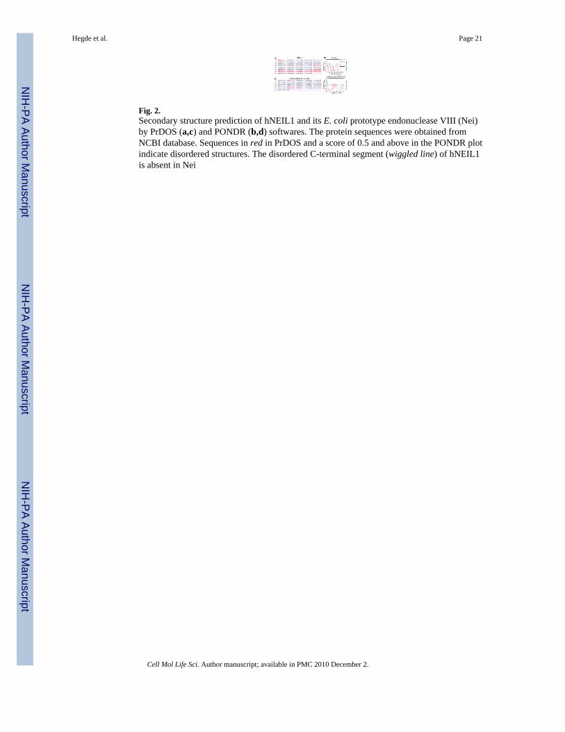
Fig. 2.Secondary structure prediction of hNEIL1 and its E. coli prototype endonuclease VIII (Nei)by PrDOS (a,c) and PONDR (b,d) softwares. The protein sequences were obtained fromNCBI database. Sequences in red in PrDOS and a score of 0.5 and above in the PONDR plotindicate disordered structures. The disordered C-terminal segment (wiggled line) of hNEIL1is absent in Nei
Hegde et al. Page 21
Cell Mol Life Sci. Author manuscript; available in PMC 2010 December 2.
NIH
-PA Author Manuscript
NIH
-PA Author Manuscript
NIH
-PA Author Manuscript

Fig. 3.PONDR plot of predicted secondary structures of hNTH1, hMYH, hAPE1 and their E. coliprototypes endonuclease III (Nth), MutY, Xth, respectively. Wiggled lines at the N-terminiof human enzymes represent disordered segments
Hegde et al. Page 22
Cell Mol Life Sci. Author manuscript; available in PMC 2010 December 2.
NIH
-PA Author Manuscript
NIH
-PA Author Manuscript
NIH
-PA Author Manuscript
Fig. 4.PONDR plot of disordered conformation at the N-terminus of hTDG and UNG2. HNEIL2has an internal disordered region
Hegde et al. Page 23
Cell Mol Life Sci. Author manuscript; available in PMC 2010 December 2.
NIH
-PA Author Manuscript
NIH
-PA Author Manuscript
NIH
-PA Author Manuscript

Fig. 5.PrDOS secondary structure prediction of hOGG1 and Polβ indicate short disorderedsegments (sequence in red) at both termini
Hegde et al. Page 24
Cell Mol Life Sci. Author manuscript; available in PMC 2010 December 2.
NIH
-PA Author Manuscript
NIH
-PA Author Manuscript
NIH
-PA Author Manuscript
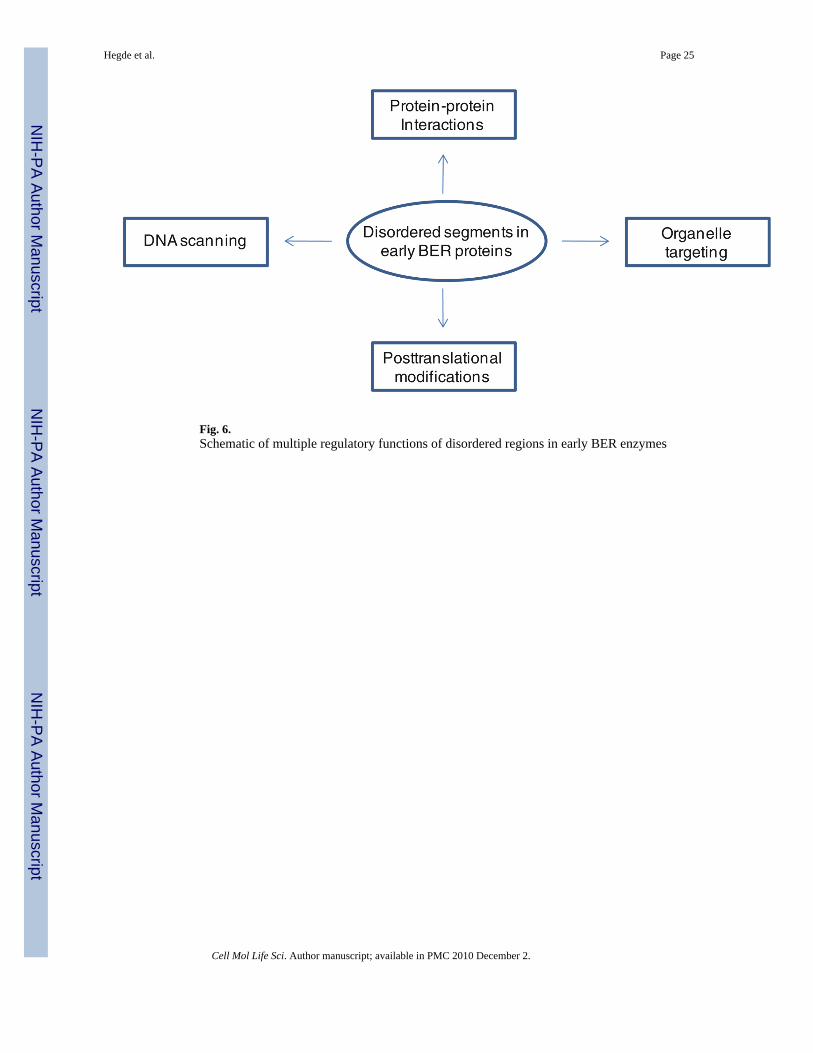
Fig. 6.Schematic of multiple regulatory functions of disordered regions in early BER enzymes
Hegde et al. Page 25
Cell Mol Life Sci. Author manuscript; available in PMC 2010 December 2.
NIH
-PA Author Manuscript
NIH
-PA Author Manuscript
NIH
-PA Author Manuscript