Transcription-coupled nucleotide excision repair and its ...
180
Transcription-coupled nucleotide excision repair and its regulation by the DNA damage checkpoint By Michael Josef Taschner A thesis submitted for the degree of Ph.D. at The University of London August 2009 Cancer Research UK London Research Institute Clare Hall Laboratories South Mimms Herts EN6 3LD and Department of Biochemistry University College London WC1E 6BT
-
Upload
khangminh22 -
Category
Documents
-
view
2 -
download
0
Transcript of Transcription-coupled nucleotide excision repair and its ...

Transcription-coupled nucleotide excision repair
and its regulation
by the DNA damage checkpoint
By
Michael Josef Taschner
A thesis submitted for the degree of Ph.D.
at
The University of London
August 2009
Cancer Research UK
London Research Institute
Clare Hall Laboratories
South Mimms
Herts EN6 3LD
and
Department of Biochemistry
University College London
WC1E 6BT

2
I, Michael J. Taschner, confirm that the work presented in this thesis is my own. Where
information has been derived from other sources, I confirm that this has been indicated
in the thesis.

3
Abstract Elaborate DNA repair mechanisms have evolved, allowing cells to repair damages in their
genomes. Nucleotide excision repair (NER) removes a variety of helix-distorting lesions,
including those caused by ultraviolet (UV) irradiation. NER operates via two subpathways.
Transcription-coupled repair (TC-NER) rapidly removes transcription-blocking lesions in the
transcribed strand (TS) of active genes, and in the yeast Saccharomyces cerevisiae depends on
the factors Rad26 and Rpb9. Lesions in untranscribed DNA, including the non-transcribed
strand (NTS) of active genes are removed slower by global genome repair (GG-NER).
Besides activating specific DNA repair systems, DNA damage also leads to a global cellular
response, known as the DNA damage checkpoint (DDC). Cell-cycle progression is temporarily
stopped after DNA damage to allow sufficient time for repair and prevent replication or
segregation of damaged chromosomes. The DDC is a complex signal transduction cascade
involving a number of protein kinases, the central players in budding yeast being Mec1 and
Tel1, the homologues of human ATR and ATM, respectively. Besides inhibiting cell-cycle
progression, accumulating evidence suggests that DNA repair systems are also influenced by
the checkpoint.
I have investigated the rates of repair of UV lesions in checkpoint deficient strains of
Saccharomyces cerevisiae and found that NER is significantly inhibited on both strands of an
active gene in the absence of Mec1. The effect on NTS repair seems to be caused by deficient
de novo synthesis of repair factors, whereas TC-NER is influenced mainly by post-translational
modification of one or more pre-existing proteins. I have characterised a checkpoint-dependent
phosphorylation of Rad26, and have shown using point mutants that this phosphorylation
increases the TC-NER capacity of cells, establishing a new link between NER and the
checkpoint.
In addition to these results about the interplay between the DDC and NER pathways,
preliminary data from two unrelated projects will be presented. One was an attempt to establish
a system for analysis of NER factor recruitment to an artificial, highly UV-damage-prone DNA
sequence. The other focussed on the regulation of UV-induced degradation of Rpb1, the largest
RNA Polymerase II (RNAPII) subunit, by the DDC.

4
List of tables
Table 2-1 Yeast strains used in this study .......................................................................................61 Table 2-2 Antibiotics and drugs used in this study.........................................................................63 Table 2-3 Antibodies used in this study...........................................................................................76 Table 5-1 Genes affected by RAD26 deletion after UV ...............................................................117

5
Table of figures Figure 1-1 Simplified overview of the two NER subpathways and Rpb1
ubiquitylation/degradation in human cells ..............................................................................19 Figure 1-2 Simplified overview of the two NER subpathways and Rpb1
ubiquitylation/degradation in budding yeast cells ..................................................................20 Figure 1-3 Simplified overview of the DNA damage checkpoint in budding yeast.....................43 Figure 2-1 Analysis of strand-specific CPD repair at nucleotide resolution.................................70 Figure 3-1 Schematic representation of the ‘hotspot’ gene............................................................81 Figure 3-2 Mapping of UV-induced DNA lesions on the hotspot gene ........................................83 Figure 3-3 Experimental outline of Chromatin immunoprecipitation (ChIP)...............................84 Figure 3-4 Analysis of RNAPII distribution at the hotspot gene...................................................86 Figure 3-5 Analysis of UV-dependent Rad14 recruitment to DNA ..............................................87 Figure 5-1 Normal NER requires the Mec1 kinase.........................................................................94 Figure 5-2 Quantification of the signals shown in Figure 5.1........................................................95 Figure 5-3 Efficient NER does not require the Chk1 and Rad53 kinases .....................................96 Figure 5-4 Efficient NER does not require the Dun1 kinase..........................................................96 Figure 5-5 Efficient repair of the NTS, but not the TS, requires de novo protein synthesis ........97 Figure 5-6 Checkpoint-dependent phosphorylation of Rad23 at S121 is not required for efficient
NER ...........................................................................................................................................98 Figure 5-7 Mutation of potential checkpoint-phosphorylation sites in Rad23 does not affect
survival after UV ......................................................................................................................99 Figure 5-8 Construction and analysis of a strain with N-terminally tagged RAD26 ..................101 Figure 5-10 UV-induced phosphorylation of Rad26 ....................................................................102 Figure 5-11 Involvement of the DNA damage checkpoint in Rad26 phosphorylation ..............104 Figure 5-12 Identification of the Rad26 phosphorylation site .....................................................106 Figure 5-13 Analysis of survival after UV irradiation in various strain backgrounds expressing
wild-type and mutant versions of Rad26...............................................................................108 Figure 5-14 Rad26 phosphorylation is required for efficient TC-NER.......................................110 Figure 5-15 Quantification of the signals shown in Figure 5.17..................................................111 Figure 5-16 Analysis of NER mec1Δ sml1Δ cells expressing Rad26 mutants ...........................112
Figure 5-17 Analysis of the effect of RAD26 deletion on the UV sensitivity of the checkpoint-
deficient mec1Δ sml1Δ strain .................................................................................................113
Figure 5-18 Rad26 phosphorylation does not seem to affect Rpb1 ubiquitylation/degradation114 Figure 5-19 Rad26 phosphorylation does not affect the DNA-dependent ATPase activity of the
protein......................................................................................................................................116 Figure 5-20 Rad26 phosphorylation does not affect gene expression changes after UV...........119 Figure 5-21 Analysis of the effect of checkpoint kinases on Rpb1 ubiquitylation/degradation121

6
Figure 5-22 DDC dependent phosphorylation of Def1 at S273 and S497 is not involved in
ubiquitylation/degradation of Rpb1.......................................................................................123
7
Table of contents
Abstract ................................................................................................................................................3
List of Tables .......................................................................................................................................4
Table of Figures ..................................................................................................................................5
Table of Contents ................................................................................................................................7
1 Introduction.................................................................................................................................12 1.1 DNA damage and repair .....................................................................................................12
1.1.1 Oxidative damage and Base Excision Repair (BER).................................................12 1.1.2 Mismatch repair (MMR) .............................................................................................13 1.1.3 Repair of DNA double strand breaks..........................................................................14
1.2 Nucleotide excision repair (NER) ......................................................................................15 1.2.1 NER in E.coli ...............................................................................................................16 1.2.2 NER in eukaryotes .......................................................................................................17
1.2.2.1 Damage-detection.................................................................................................17 1.2.2.2 Open-complex formation .....................................................................................23 1.2.2.3 Dual incisions and repair synthesis .....................................................................24
1.3 The interplay between DNA damage and transcription....................................................24 1.3.1 The regulation of transcription elongation .................................................................24 1.3.2 Transcription in the presence of DNA damage..........................................................25 1.3.3 Transcription-coupled Nucleotide Excision Repair (TC-NER) ................................26
1.3.3.1 TC-NER in E.coli and the role of the Mfd protein.............................................27 1.3.3.2 TC-NER in humans ..............................................................................................28 1.3.3.3 TC-NER in Saccharomyces cerevisiae ...............................................................35 1.3.3.4 Speculations about the mechanism of TC-NER in eukaroytic cells..................36
1.3.4 DNA damage-induced Rpb1 ubiquitylation and degradation...................................38 1.3.4.1 Identification of Rpb1 as a substrate for ubiquitylation.....................................38 1.3.4.2 Def1, a factor controlling Rpb1 ubiquitylation in budding yeast ......................39 1.3.4.3 Reversal of Rpb1 ubiquitylation by Ubp3 ..........................................................40
1.4 The DNA damage checkpoint ............................................................................................41 1.4.1 The main components of the DNA damage signalling pathway ..............................42
1.4.1.1 The activation of the PI3K-like kinases ..............................................................42 1.4.1.2 The Rad9 adapter protein .....................................................................................45 1.4.1.3 The effector kinases Rad53, Chk1 and Dun1 .....................................................45
1.4.2 Human pathologies associated with defects in DNA damage signalling .................46 1.4.2.1 Ataxia telangiectasia (AT), AT-like-disorder (A-T-LD) and Nijmegen
breakage syndrome (NBS) ....................................................................................46 1.4.2.2 Seckel syndrome...................................................................................................47

8
1.4.3 Relationship between DNA repair and the DNA damage checkpoint .....................48 1.4.3.1 Activation of the checkpoint by DNA repair intermediates ..............................48 1.4.3.2 Activation of damage-dependent transcription by the DDC..............................49 1.4.3.3 Direct phosphorylation of repair factors by checkpoint kinases .......................51
2 Materials and Methods ...............................................................................................................52 2.1 Buffers, Media and Solutions .............................................................................................52
2.1.1 Yeast media ..................................................................................................................52 2.1.1.1 YPD .......................................................................................................................52 2.1.1.2 Selective yeast drop-out media ............................................................................52
2.1.2 Bacterial media.............................................................................................................53 2.1.2.1 LB (rich medium) .................................................................................................53 2.1.2.2 SOC medium.........................................................................................................53 2.1.2.3 NZY medium ........................................................................................................53
2.1.3 General solutions..........................................................................................................53 2.1.3.1 PBS (Phosphate Buffered Saline)........................................................................53 2.1.3.2 TE (Tris-EDTA) ...................................................................................................54 2.1.3.3 TBE (Tris-Borate-EDTA) ....................................................................................54 2.1.3.4 TE/LiOAc..............................................................................................................54 2.1.3.5 PEG/TE/LiOAc.....................................................................................................54 2.1.3.6 10 x DNA loading buffer for agarose electrophoresis .......................................54 2.1.3.7 5 x SDS-PAGE loading buffer.............................................................................54 2.1.3.8 Formamide loading buffer for denaturing PAGE...............................................55 2.1.3.9 100 x Protease inhibitor cocktail .........................................................................55 2.1.3.10 Yeast lysis buffer ..................................................................................................55 2.1.3.11 TEV elution buffer................................................................................................55
2.1.4 Buffers for the Nucleotide Excision Repair assay .....................................................56 2.1.4.1 Sorbitol stock solution..........................................................................................56 2.1.4.2 2 x Lysis Buffer ....................................................................................................56 2.1.4.3 Binding and Wash Buffer (BW Buffer) ..............................................................56
2.1.5 Buffers for Chromatin Immunoprecipitation (ChIP) .................................................56 2.1.5.1 FA Lysis Buffer ....................................................................................................56 2.1.5.2 FA 500...................................................................................................................57 2.1.5.3 LiCl wash solution................................................................................................57 2.1.5.4 TES ........................................................................................................................57 2.1.5.5 ChIP elution buffer ...............................................................................................57
2.2 Bacterial techniques ............................................................................................................57 2.2.1 Transformation of competent E. coli cells .................................................................57 2.2.2 Plasmid mini-prep and maxi-prep...............................................................................58

9
2.2.3 Preparation of extracts from Micrococcus luteus (ML extract) ................................58 2.3 DNA techniques ..................................................................................................................59
2.3.1 Restriction digests and ligation reactions ...................................................................59 2.3.2 DNA sequencing ..........................................................................................................59 2.3.3 Polymerase Chain Reaction (PCR).............................................................................59 2.3.4 Purification of PCR products ......................................................................................60 2.3.5 Site-directed mutagenesis of plasmid DNA ...............................................................60 2.3.6 Agarose Gel Electrophoresis of DNA ........................................................................61 2.3.7 Purification of DNA from agarose gels ......................................................................61
2.4 Yeast techniques..................................................................................................................61 2.4.1 Yeast strains used in this study ...................................................................................61 2.4.2 Growth conditions, drug treatments and cell cycle synchronisations.......................62 2.4.3 Lithium-Acetate transformation of yeast cells ...........................................................64 2.4.4 Galactose-induced overexpression of proteins in yeast cells ....................................64 2.4.5 Preparation of yeast extracts using glass beads..........................................................65 2.4.6 Preparation of quick protein extracts from yeast .......................................................66 2.4.7 Isolation of high quality genomic DNA from yeast ..................................................66 2.4.8 Rapid isolation of genomic DNA from yeast.............................................................66 2.4.9 Analysis of the UV sensitivity of yeast strains ..........................................................66 2.4.10 Analysis of UV-induced degradation of Rpb1...........................................................67 2.4.11 Analysis of UV-induced ubiquitylation of Rpb1 .......................................................67 2.4.12 Strand-specific nucleotide excision repair (NER) assay ...........................................68
2.4.12.1 Irradiation and harvesting of samples .................................................................68 2.4.12.2 Isolation of genomic DNA...................................................................................69 2.4.12.3 Preparation of genomic DNA for analysis ..........................................................71 2.4.12.4 Isolation of RPB2 strands for labelling and analysis .........................................71 2.4.12.5 Radioactive labelling and analysis of the isolated DNA strands.......................72 2.4.12.6 Calculation of damage half-lifes (T50%)............................................................73
2.4.13 Chromatin Immunoprecipitation (ChIP) ....................................................................73 2.4.13.1 Cell growth and UV irradiation ...........................................................................73 2.4.13.2 Crosslinking, preparation of extracts and DNA shearing ..................................74 2.4.13.3 Immunoprecipitation ............................................................................................74 2.4.13.4 Analysis of immunoprecipitated DNA by Real time PCR ................................75
2.5 Protein analysis....................................................................................................................75 2.5.1 SDS Polyacrylamide Gel Electrophoresis (SDS-PAGE) ..........................................75 2.5.2 Transfer of proteins to membranes (Western Blot) ...................................................76 2.5.3 Detection of proteins on membranes ..........................................................................77 2.5.4 Staining of proteins in SDS-Gels using SYPRO Ruby stain ....................................77

10
2.5.5 Mass spectrometry analysis of Rad26 ........................................................................77 2.5.6 Immunoprecipitation of proteins from yeast extracts ................................................78 2.5.7 ATPase assay................................................................................................................78
3 Results I – NER/TCR analysis at site-specific UV lesions......................................................80 3.1 Project aim ...........................................................................................................................80 3.2 Design of the UV-hotspot construct...................................................................................81 3.3 Analysis of the hotspot gene...............................................................................................82
3.3.1 The ‘hotspot’ sequence is efficiently damaged after UV irradiation........................82 3.3.2 Analysis of RNAPII distribution across the hotspot gene.........................................83 3.3.3 Analysis of repair factor recruitment to the hotspot ..................................................85
4 Discussion I.................................................................................................................................89 4.1 Construction of a UV-damage-prone gene ........................................................................89 4.2 RNAPII does not accumulate at the hotspot after UV......................................................89 4.3 Specific recruitment of an NER-factor to the damage-prone region ...............................90 4.4 Potential solutions for the problem of transcriptional induction......................................91 4.5 Use of the hotspot sequence for analysis of GG-NER......................................................92
5 Results II – Regulation of NER by the DNA damage checkpoint ..........................................93 5.1 Analysis of NER efficiency in checkpoint-deficient strains ............................................93
5.1.1 Strains lacking the Mec1 kinase have NER defects ..................................................93 5.1.2 Strains lacking the Chk1, Rad53 and Dun1 kinases have normal NER ...................95 5.1.3 UV-induced de novo protein synthesis is required for efficient NTS repair............97
5.2 Targets of the Mec1 kinase.................................................................................................98 5.2.1 Rad23 phosphorylation mutants do not display defects in NER ..............................98 5.2.2 Analysis of checkpoint-dependent phosphorylation of Rad26 ...............................100
5.2.2.1 Construction of an N-terminally tagged Rad26 strain......................................100 5.2.2.2 Rad26 is phosphorylated in response to DNA damage....................................100 5.2.2.3 Rad26 phosphorylation is dependent on the Mec1 checkpoint kinase............103 5.2.2.4 Identification of the Rad26 phosphorylation site..............................................105 5.2.2.5 Analysis of an involvement of Rad26 phosphorylation in TC-NER...............107 5.2.2.6 Expression of a phosphomimic Rad26 mutant is not sufficient to
overcome the TCR defects in a strain lacking the Mec1 kinase......................111 5.2.2.7 Rad26 phosphorylation is not involved in ubiquitylation and degradation
of RNA Polymerase II ........................................................................................113 5.2.2.8 Rad26 phosphorylation does not affect DNA-dependent ATPase activity
of the protein .......................................................................................................115 5.2.2.9 Analysis of gene expression changes in rad26Δ cells after UV-irradiation....116
5.3 Analysis of an influence of the DNA damage checkpoint on ubiquitylation and
degradation of Rpb1 ...........................................................................................................120

11
5.3.1 Rpb1 ubiquitylation and degradation are influenced by the Mec1 and Rad53
kinases.........................................................................................................................120 5.3.2 DDC-dependent phosphorylation of Def1 is not involved in regulation of UV-
induced Rpb1 degradation.........................................................................................122 6 Discussion II .............................................................................................................................124
6.1 Regulation of NER by the DNA damage checkpoint .....................................................124 6.1.1 Regulation of NTS repair ..........................................................................................124
6.1.1.1 de novo protein synthesis is required for efficient NTS repair........................124 6.1.1.2 Phosphorylation of repair factors for efficient NTS repair? ............................125
6.1.2 Regulation of TC-NER..............................................................................................126 6.2 Rad26, a Mec1 target involved in the regulation of DNA repair by the DDC..............127
6.2.1 Rad26 is phosphorylated after DNA damage by the Mec1 kinase .........................127 6.2.2 Serine 27 is the main phosphorylation site on Rad26..............................................128 6.2.3 Rad26 phosphorylation is required for efficient TC-NER in the presence of a
functional DDC ..........................................................................................................128 6.2.4 Mutation of the Rad26 phosphorylation site does not affect survival or growth
recovery after UV irradiation ....................................................................................130 6.2.5 Speculation on the functional consequences of Rad26 phosphorylation ...............131
6.3 Phosphorylation of CSB by ATM/ATR?.........................................................................134 6.4 Regulation of Rpb1 ubiquitylation and degradation by the DDC..................................135
7 References .................................................................................................................................138
8 Appendix .................................................................................................................................. 166

1 Introduction
1.1 DNA damage and repair
Faithful transmission of the genetic material between generations is a prerequisite for genome
integrity and cell survival in all living cells. The genome therefore needs to be replicated and
segregated correctly. Failure to do so will result in the accumulation of alterations such as
mutations, which could alter the functions of the proteins encoded by their respective genes.
The DNA is, however, inherently unstable and is prone to chemical alterations. More than 104
DNA lesions are formed spontaneously per day in human cells, by spontaneous decay, cellular
metabolic byproducts, and replication errors (Lindahl, 1993). This damage load is further
increased by the action of various exogenous DNA damaging agents from the environment,
such as the Ultraviolet (UV) components of sunlight, cigarette smoke, ionizing radiation, and
many more. If left unrepaired, these lesions would interfere with essential metabolic processes
occurring on the DNA (such as transcription and replication), and would also lead to an
unacceptably high level of mutations, which is a strong driving force behind neoplastic
transformation, and diseases in general, in multicellular organisms. It is therefore not surprising
that all living cells have evolved a number of repair systems to deal with DNA lesions, many of
which are conserved between prokaryotes and higher eukaryotes. The importance of these repair
mechanisms is exemplified by the existence of inherited cancer-prone disorders based on the
inactivation of various DNA repair pathways (Hoeijmakers, 2001). In the following sections I
will give a very brief overview of the most relevant sources of DNA damage and repair by
describing their respective DNA repair pathways, but the main focus will be on UV-induced
lesions and Nucleotide Excision Repair (NER), as this is the pathway most relevant for the work
described here.
1.1.1 Oxidative damage and Base Excision Repair (BER)
BER is the main repair pathway for lesions such as base oxidation, deamination and alkylation
(Friedberg, 2006), and is the most important repair system for the removal of lesions generated
by normal cellular metabolism. It also repairs single-strand DNA breaks and removes uracil
from DNA, which is formed by spontaneous deamination of cytosine. The repair process is
initiated by non-enzymatic base loss, or the removal of the damaged base from the sugar-
phosphate backbone by a damage-specific DNA glycosylase (Krokan et al., 1997), resulting in
the formation of an abasic site (AP site) (Boiteux and Guillet, 2004). This site is then
13
recognized by an AP endonuclease, which cleaves it to form 3’ OH and 5’ deoxyribose
phosphate (dRP) termini (Doetsch and Cunningham, 1990). Some DNA glycosylases can carry
out this cleavage themselves (bifunctional glycosylases).
After the cleavage of the phosphate backbone, two distinct pathways of BER can be
distinguished, differing in the length of the newly synthesized DNA (Frosina et al., 1996). In
‘Short-patch’ BER (SP-BER), DNA polymerase beta (Polβ) inserts only a single base. With its
intrinsic dRP-lyase activity it can cleave the dRP residue to generate a 5’ phosphate, and the
remaining nick can be ligated by DNA ligase III. In ‘Long-patch’ BER (LP-BER), the length of
the newly synthesized DNA is typically 2-8 nucleotides, and involves DNA polymerases
δ/ε and PCNA. Displacement of the old strand creates a 5’-flap structure, which has to be
removed by a specialised flap-endonuclease, FEN-1. The remaining nick created during LP-
BER is sealed by DNA ligase I.
Because of the vital importance of the BER pathway in the removal of naturally occuring DNA
damage, inactivation of central BER proteins, such as DNA Polymerase β or the scaffold
protein XRCC1, is lethal in higher eukaryotes (Sugo et al., 2000; Tebbs et al., 1999), but certain
mutations in these proteins are also linked to cancers (Bhattacharyya and Banerjee, 2001;
Divine et al., 2001).
1.1.2 Mismatch repair (MMR)
The MMR pathway is mainly responsible for the removal of incorrectly inserted nucleotides
during DNA replication, but can also work on certain base modification, as well as small
insertion/deletion loops caused by replication slippage (reviewed in (Li, 2008)). The
prototypical MMR pathway in E. coli has been extensively studied. Here, the damage is
recognized by the MutS protein which recruits MutL to the lesion. MutL enhances mismatch
recognition by MutS and also recruits MutH to the complex. MutH binds to hemi-methylated
dGATC sequences in DNA (the newly synthesized strand being transiently unmethylated after
replication), and uses its endonucleoytic activity to incise the unmethylated daughter strand. The
UvrD helicase removes the damaged strand, creating a single-strand gap, which is eventually
filled by DNA repair synthesis and ligation.
In higher eukaryotes, MMR is more complex and less well understood. Several eukaroytic
homologs of MutS and MutL exist, suggesting that the basic mechanism is conserved, but none
has yet been identified for MutH (Kunkel and Erie, 2005; Modrich and Lahue, 1996). The
importance of this pathway in higher eukaryotes is exemplified by the existence of an inherited

14
cancer-prone disorder called hereditary non-polyposis colon carcinoma (HNPCC), a
heterogenous disease caused by heterozygous mutation of mainly MSH2 and MLH1 (Peltomaki
and de la Chapelle, 1997).
1.1.3 Repair of DNA double strand breaks
Double strand breaks (DSBs) are particularly dangerous for a cell, because segregation of
chromosomes in the presence of unrepaired DSBs can result in the loss of large amounts of
genetic information. DSBs can be induced by treatment of cells with ionizing radiation, but the
most physiological source is probably the replication of DNA containing single-strand breaks.
Such single-strand breaks can be caused by oxidative damage to the sugar-phosphate backbone,
or by the enzymatic activities of proteins such as AP-endonucleases during BER. Another
endogenous source of DSBs are the actions of topoisomerases (Wang, 2002). DSB formation
can also occur in a programmed way, for example in the process of mating type switching in
budding yeast (Haber, 1998), during the rearrangement of immunoglobulin genes in the immune
system of higher eukaryotes (Soulas-Sprauel et al., 2007), and during meiosis (Marston and
Amon, 2004).
Two main pathways exist for dealing with DSBs, non-homologous end-joining (NHEJ) and
homology directed recombination (HDR), and HDR can be further subdivided into gene
conversion and single strand annealing (SSA). NHEJ can be seen as the simple re-ligation of
free DNA ends after proper end-processing (reviewed in (Daley et al., 2005)). The Ku-complex
has affinity for free DNA ends and is thought to bring the DNA ends into close proximity by the
help of the MRN complex, and DNA ligase IV (together with other factors) then ligates the
ends. Because of the requirement of end-processing prior to ligation, the NHEJ pathway is often
accompanied by the loss of genetic information, making it error-prone.
For HDR, the ends have to be processed to form 3’ ssDNA (reviewed in (West, 2003)) and this
pathway requires long tracts of homology between the break point and a donor region
elsewhere, which is used for repair. The 5’-3’ resection is carried out by 5’ specific
exonucleases, and the resulting 3’ single-stranded tails are recognized by a number of proteins.
The Rad51 proteins forms filaments on these tails (West, 2003), a process which is facilitated
by RPA and Rad52 (Sung, 1997). This filament then scans the genome for a region of
homology and invades it, creating a D-loop. At this stage, other proteins such as Rad54, the
Rad55-Rad57 complex and RPA play important roles (Sugawara et al., 2003; Wang and Haber,
2004; Wolner et al., 2003). Repair synthesis then takes place, extending the invading strand.
Two distinct scenarios are possible after this extension step. In the classical model for

15
homologous recombination (HR) (Szostak et al., 1983), the D-loop is captured by the second
end of the DSB, and after further repair synthesis a double holliday junction is formed. These
junctions have to be resolved by specialised Holliday junction resolvases in order to disconnect
the two molecules (Ip et al., 2008). In the alternative pathway, known as ‘synthesis-dependent
strand annealing’ (SDSA) (reviewed in (Paques and Haber, 1999)), the extended strand leaves
the invaded duplex and can now bind to the 3’ overhang of the other end of the DSB. The
resulting gaps in the two strands can be filled, and the nicks are finally ligated.
The SSA pathway can be employed for DSB repair if no homologous sequences can be found in
order to carry out gene conversion (reviewed in (Paques and Haber, 1999)). Briefly, repair of
the break is possible if some homology can be found further inwards between the resected ends.
This leads to the formation of 3’ flap structures which have to be cleaved by the endonucleases
XPF/ERCC1 in humans and Rad1/Rad10 in yeast. Arising gaps can then be filled by DNA
synthesis and the nicks closed by a DNA ligase.
Several much-studied proteins, such as ATM, BRCA1 and 2 and others are involved in DSB
repair, and deficiencies in these proteins result in increased genomic instability and cancer
predisposition (Hoeijmakers, 2001).
1.2 Nucleotide excision repair (NER)
Among all repair pathways for DNA damage, the nucleotide excision repair (NER) system is
the most versatile, dealing with a wide variety of lesions, including cyclobutane pyrimidine
dimers (CPDs) and 6-4 pyrimidine-pyrimidone photoproducts (6-4PPs), the two main lesions
induced by UV irradiation, as well as a large number of bulky chemical adducts, such as those
produced by cigarette smoke, the UV-mimetic chemical 4-nitroquinoline 1-oxide (4NQO), and
cisplatin, a DNA damaging agent routinely used in cancer chemotherapy (de Laat et al., 1999;
Prakash and Prakash, 2000). The basis for this versatility was believed to be the fact that instead
of recognising a specific lesion, it is the damage-induced distortion of the DNA helix which is
detected by the NER lesion sensors. This view has been challenged now by the finding that non-
distorting lesions, such as the oxidative lesion thymine glycol (Tg), has also been shown to be a
substrate for NER.
The NER pathway is conserved between E.coli and humans, and can be divided into 4 main
steps: damage detection, formation of an open complex, dual incision leading to removal of the
damaged strand, and finally repair synthesis to close the gap (de Laat et al., 1999; Prakash and
Prakash, 2000). The whole reaction requires the activities of more than 30 factors in eukaryotic

16
cells and has been reconstituted with purified proteins from both budding yeast (Guzder et al.,
1995) and humans (Aboussekhra et al., 1995; Mu et al., 1995). A speciality of the damage-
detection step is that it occurs by different mechanisms depending on the transcriptional state of
the damaged sequence, and so NER can be divided into two distinct subpathways.
Untranscribed DNA, including the non-transcribed strand (NTS) of active genes, is repaired by
Global Genome NER (GG-NER), whereas the transcribed strand (TS) of an active gene is
repaired by Transcription-Coupled NER (TC-NER) (Svejstrup, 2002). In this section I will give
a brief overview of the general mechanism of NER in both E.coli and eukaryotes, the special
relationship between transcription and NER (TC-NER) will be dealt with in subsequent
sections.
1.2.1 NER in E.coli
Prokaryotic NER is understood in detail and has been important for the elucidation of the NER
mechanism in eukaryotes (reviewed in (Petit and Sancar, 1999)). In the 1960’s, it was first
discovered that UV-lesions were excised from bacterial DNA (Boyce and Howard-Flanders,
1964; Setlow and Carrier, 1964), followed by the formation of repair patches (Hanawalt and
Haynes, 1965). The genes responsible for these observations, uvrA, uvrB and uvrC, were
subsequently identified by complementation studies (Howard-Flanders et al., 1966).
UvrA is a protein containing two C4-type zinc fingers and two ATP Binding Cassette ATPase
(ABC ATPase) domains (Doolittle et al., 1986). UvrA forms a dimer and this dimer interacts
with UvrB, leading to the formation of a UvrA2B complex. This complex is recruited to the
damage, and damage recognition is believed to be mediated by UvrA, as only this protein has
been shown to bind damaged DNA (Mazur and Grossman, 1991).
UvrB is a member of the helicase superfamily as it contains 6 conserved helicase domains
(Theis et al., 2000). It is loaded onto damaged DNA as part of the UvrA2B complex and its
ATPase activity is stimulated by the presence of UvrA and damaged DNA (Caron and
Grossman, 1988; Oh et al., 1989). The function of UvrB is opening of the double helix and
lesion verification (Zou and Van Houten, 1999). UvrA2 then dissociates from the complex,
leaving behind the unwound DNA with the bound UvrB, which is subsequently recognised by
the UvrC endonuclease. This endonuclease has two distinct catalytic sites and is responsible for
both the 3’ and the 5’ incision on the damaged strand (Verhoeven et al., 2000). The UvrD
helicase is responsible for unwinding and displacing the damaged strand, and the single-strand
gap is finally filled by DNA polymerase I, and sealed by DNA ligase (Caron et al., 1985).

17
1.2.2 NER in eukaryotes
Even though the basic steps in the eukaryotic NER pathway are essentially the same as in its
prokaryotic counterpart, the reaction has become more complex during evolution (de Laat et al.,
1999; Prakash and Prakash, 2000). In humans, the importance of NER is exemplified by a
number of rare autosomal recessive disorders caused by defects in this pathway, including
Xeroderma pigmentosum (XP), Cockayne’s syndrome (CS), and Trichothiodystrophy (TTD)
(reviewed in (de Boer and Hoeijmakers, 2000)). XP patients display severe sensitivity to
sunlight, which results in a >1000 fold increased risk of developing skin cancer in sun-exposed
areas of the body, which clearly shows how important removal of UV-induced lesions is in
order to avoid an accumulation of UV-induced mutations. XP was the first cancer-prone
disorder to be directly linked to a defect in DNA repair (Cleaver, 1968). Seven distinct
complementation groups with defects in NER components have now been identified (XP-A to
G), and another group of XP-patients (XP variant; XPV) have defects in translesion DNA
synthesis across unrepaired UV lesions (reviewed in (Kannouche and Stary, 2003)). Cockayne’s
syndrome results from a defect in TC-NER, and will be discussed later.
The individual steps in eukaryotic GG-NER, together with the responsible factors, will be
presented individually below, and important mechanistic differences between the reaction in
budding yeast (the model system used in this work) and human cells will be pointed out.
Simplified schemes showing the NER subpathways and Rpb1 ubiquitylation pathways
(discussed later) in human cells and budding yeast can be found in Figures 1.1 and 1.2,
respectively.
1.2.2.1 Damage-detection
1.2.2.1.1 Yeast Rad4/Rad23 and human XPC/hRad23B
The first proteins to arrive at a DNA lesion are the Rad4/Rad23 complex in yeast (Guzder et al.,
1998a; Jansen et al., 1998), or the XPC/HR23B complex in humans (Reardon et al., 1996;
Sugasawa et al., 1998). Even though other factors, such as yeast Rad14 and its human homolog
XPA, also have affinity for damaged DNA (Guzder et al., 1993; Jones and Wood, 1993),
elegant studies in human cells have shown that only binding of XPC/HR23B is necessary for
efficient lesion detection in vitro (Sugasawa et al., 1998) and in vivo (Volker et al., 2001). The
XPC/HR23B complex is only required for GG-NER (Venema et al., 1990b), whereas in yeast,
the Rad4/Rad23 complex is required for both GG-NER and TC-NER (Gietz and Prakash, 1988;
18
Verhage et al., 1994), which represents an important difference between the yeast and the
human NER reaction. The Rad4 and XPC subunits are responsible for recognising damaged
DNA (Hoogstraten et al., 2008; Min and Pavletich, 2007). Importantly, binding of these
proteins to DNA induces significant DNA bending, which is much more energetically favorable
if helix distorting lesions are present, thus explaining the increased affinity for damaged DNA
and the high versatility of the NER pathway.
In human cells, the HR23A and B proteins have both been shown to be involved in NER, to
form a stable complex with XPC, and to increase the efficiency in the NER reaction in vitro
only in the presence of XPC. This stimulatory activity is dependent on XPC-binding (Masutani
et al., 1997; Sugasawa et al., 1996; Sugasawa et al., 1997). More recently it was shown that the
stimulation of the NER reaction by HR23B can be explained by the stabilization of the XPC
protein (Araki et al., 2001).
Yeast Rad23 has also been proposed to be required for stabilisation of Rad4 (Lommel et al.,
2002), but a later study suggested that the reduced Rad4 levels in rad23Δ cells result from a
decrease in RAD4 transcription (Gillette et al., 2006). Rad23 clearly has an important function
in NER, as RAD23 deletion renders cells sensitive to UV irradiation and leads to severely
impaired damage removal (Verhage et al., 1996b). It possesses an N-terminal ubiquitin-like
(Ubl) domain (Watkins et al., 1993), important for its function in NER by recruiting subunits of
the proteasome (Elsasser et al., 2002), which has non-proteolytic functions in the NER process
(Gillette et al., 2001; Russell et al., 1999) (reviewed in (Reed and Gillette, 2007)).
Even though XPC/HR23B were initially isolated as a heterodimeric complex, further
biochemical analysis revealed the presence of another member, Centrin2 (CEN2) (Araki et al.,
2001). This protein interacts directly with XPC, and like HR23B, is also involved in promoting
XPC stability (Araki et al., 2001). A similar factor was later also found in yeast and called
Rad33 (den Dulk et al., 2006). Further work has shown that Rad33 shows homology to Centrin
2, that it can bind directly to Rad4, and that this binding is necessary to protect Rad4 from
polyubiquitylation (den Dulk et al., 2008).

19
Figure 1-1 Simplified overview of the two NER subpathways and Rpb1 ubiquitylation/degradation in human cells see text for details

20
Figure 1-2 Simplified overview of the two NER subpathways and Rpb1 ubiquitylation/degradation in budding yeast cells see text for details

21
1.2.2.1.2 The Rad7/Rad16 complex in yeast
The Rad7/Rad16 complex is solely involved in GG-NER in yeast, i. e. in the repair of un-
transcribed DNA, including the NTS of active genes (Bang et al., 1992; Terleth et al., 1990;
Verhage et al., 1994). Consequently, deletion of the corresponding genes, either alone or in
combination, results in UV sensitivity, which is less pronounced than in completely NER-
deficient yeast mutants, such as rad14Δ (yeast XPA) (Verhage et al., 1996a). Rad7 and Rad16
form a complex with 1:1 stoichiometry, which can bind UV-damaged DNA in an ATP-
dependent manner (Guzder et al., 1997). Interestingly, no clear homologues in humans have yet
been identified for these factors. An additional member of the Rad7/Rad16 complex has later
been found to be Abf1 (Autonomously replicating sequence (ARS) binding factor 1), which is
encoded by an essential gene and has important roles during DNA replication (Diffley and
Stillman, 1989). This factor has been shown to be required for NER, as depletion of Abf1
renders cells incapable of performing NER and sensitive to killing by UV light (Reed et al.,
1999).
Rad16 is a member of the SWI2/SNF2 family of helicases (Bang et al., 1992), and because of
its ability to bind DNA has been proposed to be involved in recognition of DNA damage during
GG-NER, especially in the context of chromatin. Evidence for this comes from recent work
showing that the repair defect in rad16Δ and rad7Δ strains can be suppressed by deletion of a
histone deacetylase (Teng et al., 2008).
Using a reconstituted in vitro system for NER of damage in purified plasmid DNA, which
allows to detect the incisions carried out by the NER reaction, it was shown that the
Rad7/Rad16 complex is not required for NER per se (Guzder et al., 1995), but that it increases
the efficiency of the reaction (Guzder et al., 1997). Based on these findings it was proposed that
these factors assist in the recognition of the damage (Guzder et al., 1998b). However, a different
in vitro system using yeast extracts, which measures repair synthesis rates, showed that Rad16
are Rad7 are required for the reaction (Wang et al., 1996). An explanation for these apparently
contradicting results was obtained using an in vitro system that allows dissection of the
sequential steps of dual incision, excision of the damaged DNA and repair synthesis. This work
showed that Rad7 and Rad16 are not required for the incision step, but instead for excision of
the oligomer and repair synthesis, showing that the Rad7/Rad16 complex affects post-incision
events during NER (Reed et al., 1998). Later work showed that this can be explained by the fact
that the Rad16 subunit of the complex creates superhelicity in DNA, and that this activity is
necessary for excision of the damaged oligonucleotide (Yu et al., 2004).
22
The Rad7 and Rad16 proteins also have a different function in NER, acting in an E3 ubiquitin
ligase complex together with Elc1 and Cul3. This complex targets the Rad4 protein for
polyubiquitylation and subsequent degradation by the 26S proteasome after UV irradiation, but
importantly, only ubiquitylation, but not degradation of Rad4 is required for efficient NER
(Gillette et al., 2006). Taken together, the Rad16/Rad7 complex has important functions at
various steps during the NER reaction. A similar ubiquitin ligase complex was recently shown
to be involved in ubiquitylation of Rpb1, the largest RNA Polymerase II subunit (Ribar et al.,
2006, 2007) Harreman et al, in preparation) (see Section 1.3.4), but the Rad7 and Rad16
proteins are not required for this (Ribar et al., 2006, 2007).
1.2.2.1.3 The damaged DNA binding complex (DDB) in humans
The Damaged DNA Binding proteins 1 and 2 (Ddb1 and 2), like the XPC/hRad23B complex,
are involved exclusively in the GG-NER pathway in higher eukaryotes (Keeney et al., 1993).
These proteins are unique to higher eukaryotes, and it is tempting to speculate that they may
possibly have evolved as a replacement of the yeast-specific Rad16/Rad7 complex. The Ddb2
protein is responsible for the damaged DNA-binding activity of the complex (Li et al., 2006).
The Ddb1 subunit also forms complexes with various other proteins, and therefore has a much
broader spectrum of functions (Wittschieben and Wood, 2003). Mutations in the DDB2 gene
can be found in patients belonging to the XPE complementation group (Keeney et al., 1993;
Keeney et al., 1994). The exact function of DDB/XPE has for a long time remained unknown,
because reconstitution of NER in vitro does not require addition of this factor (Aboussekhra et
al., 1995; Mu et al., 1995). The DDB complex has been shown to bind to damaged DNA with a
preference for 6-4PPs over CPDs (Chu and Chang, 1988; Reardon et al., 1993).
More information about the involvement of DDB in NER was obtained when Groisman and
colleagues identified a ubiquitin ligase complex containing both Ddb1 and Ddb2 as well as
Cul4A, Roc1, and all the subunits of the COP9 signalosome (CSN), a negative regulator of E3
ligases. Microinjection of this complex into cells from XPE patients rescued the NER defect of
these cell lines (Groisman et al., 2003). Furthermore, the complex was recruited to chromatin
after UV irradiation of cells and CSN components disappeared from the complex, thereby
activating the ubiquitin ligase activity (Groisman et al., 2003). A relevant substrate for the
DDB-containing ubiquitin ligase was found later, when it was shown that it is involved in poly-
ubiquitylation of XPC (Sugasawa et al., 2005). Importantly, this modification seems to be
reversible and not to lead to proteasomal degradation, but instead alters the DNA binding
properties of the protein.

23
Other targets of the Ddb1/2 containing ubiquitin ligase complex were found to be histones H3
and H4 (Wang et al., 2006). Upon UV-irradiation of cells, these modifications seem to lead to
histone release from nucleosomes, and an increase in the efficiency of repair factor recruitment
to the site of the lesion (Wang et al., 2006).
1.2.2.2 Open-complex formation
Upon lesion recognition, opening of the DNA double helix is a crucial step in the NER reaction
in order to achieve efficient lesion excision, and it is achieved by the helicase acitivity of the
multi-subunit TFIIH complex. The ‘core-TFIIH’ complex contains the 5’ to 3’ DNA helicase
Rad3 and the 3’ to 5’ DNA helicase Rad25 in yeast, and their homologues XPD and XPB in
higher eukaryotes (Guzder et al., 1994a; Guzder et al., 1994b; Sung et al., 1993a; Sung et al.,
1987a; Sung et al., 1987b).
Surprisingly, the initial isolation of rad3Δ mutants revealed that it is an essential gene (Higgins
et al., 1983; Naumovski and Friedberg, 1983), even though other central NER factors are not.
The same was later found to be true for rad25Δ mutants (Park et al., 1992). The answer to this
puzzle came a decade later, when the TFIIH-complex was shown to have dual roles in the cell,
an essential function in gene transcription and a non-essential function in NER (Feaver et al.,
1993; Schaeffer et al., 1993).
The core-TFIIH complex interacts with another complex, referred to as TFIIK or Cdk-activating
kinase (CAK), to form the ‘holo-TFIIH’ complex. The CAK subcomplex is not required for
NER (Sung et al., 1996; Svejstrup et al., 1995). Instead it is released from the complex on
chromatin after UV irradiation, and this release is dependent on the XPA repair factor (Coin et
al., 2008). After UV irradiation, transcription is temporarily inhibited, and the dissociation of
CAK from TFIIH can potentially explain this. Importantly, after completion of DNA repair, the
CAK subcomplex reappears with the core TFIIH, and this is accompanied by the restoration of
the ability of TFIIH to function in transcription initiation (Coin et al., 2008).
After opening of the damaged DNA, the yeast Rad14 protein or its human homologue XPA
binds the DNA lesion, which is thought be an important step in verification of the damage
(Bankmann et al., 1992; Guzder et al., 1993; Jones and Wood, 1993; Tanaka et al., 1990). The
undamaged strand is protected in the open complex by binding of the essential heterotrimeric
RPA complex (Wold, 1997), and this binding is important for the NER reaction (Guzder et al.,
1995; He et al., 1995; Mu et al., 1995).

24
1.2.2.3 Dual incisions and repair synthesis
Formation of an open complex leads to the binding of two endonucleases, which are responsible
for the endonucleolytic cleavage reactions necessary for removal of the damaged strand. These
endonucleases are Rad2 (Habraken et al., 1993) and the Rad1/Rad10 complex (Sung et al.,
1993b; Tomkinson et al., 1993) in budding yeast, and their human homologues XPG
(O'Donovan et al., 1994) and XPF/ERCC1 (Park et al., 1995). Rad1/Rad10 (XPF/ERCC1)
(Matsunaga et al., 1995) is responsible for the 5’ incision, while Rad2 (XPG) performs the 3’
incision (O'Donovan et al., 1994). These incisions are placed asymmetrically around the lesion,
with the 5’ incision 15-24 nucleotides and the 3’ incision 2-8 nucleotides away from the DNA
lesion (Moggs et al., 1996). Rad2 and Rad1/Rad10 contribute more to the NER reaction than
merely their endonucleolytic activites. In the absence of either factor, no nicking of DNA can be
observed in vitro (Guzder et al., 1995), indicating that these proteins also serve a structural role
necessary for the proper assembly of the NER complex at the site of a DNA lesion. This may be
important to ensure that incision only occurs when both endonucleases are in place so that
unscheduled DNA nicking is avoided.
The result of the incisions is the removal of a stretch of DNA containing the lesion. The length
of this ssDNA fragment is typically 24-27 bases in yeast (Guzder et al., 1995), or 27-29 bases in
humans (Huang et al., 1992). The resulting gap is filled by DNA Polymerase delta or epsilon
(Budd and Campbell, 1995) bound to PCNA, and the remaining nick is sealed by DNA ligase I.
1.3 The interplay between DNA damage and transcription
1.3.1 The regulation of transcription elongation
Eukaryotic transcription of protein coding genes by RNA Polymerase II (RNAPII) is a complex
process and can be viewed as a series of sequential and highly regulated steps, namely
preinitiation complex assembly at the promoter, open complex formation and initiation,
promoter clearance, transcription elongation, transcription termination, and RNAPII recycling
(Svejstrup, 2004). During initiation, general transcription factors assemble at the promoter of a
gene in a stepwise manner, culminating in the recruitment of RNAPII (Lee and Young, 2000).
Additional factors, such as histone modifying enzymes and chromatin remodellers, are required
to achieve efficient assembly in the context of chromatin (Lee and Young, 2000).

25
Most work on the regulation of transcription has focussed on these early events occurring at the
promoter, and the subsequent step of transcriptional elongation has for a long time been thought
of as the simple addition of nucleotides. It has, however, become increasingly clear that
RNAPII frequently pauses or becomes blocked at this stage, requiring support from a large
number of elongation factors (reviewed in (Arndt and Kane, 2003)). Transcriptional pausing
occurs when RNAPII stops the addition of nucleotides to the growing RNA for some time,
before resuming transcription on its own. Several factors, such as ELL (Shilatifard et al., 1996),
Elongin (Bradsher et al., 1993a; Bradsher et al., 1993b), and CSB (Selby and Sancar, 1997a),
among others, have been shown to suppress pausing and to stimulate the catalytic activity of
RNAPII.
After undergoing transcriptional arrest, RNAPII cannot resume transcription efficiently without
help from other factors (Arndt and Kane, 2003). When RNAPII stops, it can slide backwards on
the DNA, which leads to misalignment of the 3’ end of the growing RNA chain with the active
site of the polymerase. The most important factor required to help RNAPII in such a case is
TFIIS, which binds to arrested RNAPII complexes and stimulates an endonucleolytic activity of
the polymerase itself, leading to truncation of the transcript and realignment of the end of the
RNA with the active site of the enzyme (reviewed in (Wind and Reines, 2000)).
Other elongation factors are required to help RNAPII transcribe DNA in the context of
chromatin, such as Elongator (Otero et al., 1999), FACT (Orphanides et al., 1998) and PAF
(Krogan et al., 2003a; Krogan et al., 2003b). An especially serious impediment to the
progression of RNAPII, which is most important for the work described here and will be
discussed in more details below, is the presence of DNA lesions in the template.
1.3.2 Transcription in the presence of DNA damage
DNA lesions not only lead to mutations but also have the ability to interfere with essential
processes on DNA, such as replication and transcription. In the case of damage-blocked DNA
replication, specialised error-prone DNA polymerases can be employed to bypass the damage
quickly, alleviating the cytotoxic effect of a blocked replication fork, and allowing the repair
machinery to deal with the lesion later. Defects in this pathway can be found in XP-V patients,
the only XP complementation group which has no defect in NER (Kannouche and Stary, 2003).
Instead, XPV encodes a DNA damage-bypass polymerase (Cordonnier et al., 1999; Masutani et
al., 1999a; Masutani et al., 1999b).

26
In the case of transcription, many DNA lesions are a complete block for transcribing RNA
Polymerase II (RNAPII) and cannot be bypassed (Tornaletti, 2009). Such lesions include
mainly bulky DNA adducts, such as UV-induced CPDs and 6-4PPs (Donahue et al., 1994;
Selby et al., 1997), lesions induced by cisplatin (Damsma et al., 2007; Tornaletti et al., 2003),
and aminofluorene/acetylaminofluorene (Donahue et al., 1996). Upon encountering such
barriers, RNAPII is stopped and cannot transcribe across the lesion. Because of the physical
impediment imposed by those lesions, general elongation factors such as TFIIS cannot help
RNAPII to bypass the block (Donahue et al., 1994).
Recent work by several groups tried to investigate the effects of non-bulky DNA adducts, such
as the oxidative lesions 8-oxoguanine (8-oxoG) and thymine glycol (Tg), on RNAPII
elongation. Even though such lesions are able to obstruct RNAPII progression, general
elongation factors, such as Elongin, CSB, and TFIIS can assist in order to bypass the obstacle at
the expense of base misincorporation opposite the lesion, leading to transcriptional mutagenesis
(Charlet-Berguerand et al., 2006; Kuraoka et al., 2007).
Whatever the lesion is which leads to RNAPII stopping, it has severe consequences for a cell
with damaged DNA, as the blocked RNAPII complex represents a barrier for all the other
polymerases behind it, leading to a potent block to gene transcription. The elongation complex
is extremely stable, so RNAPII does not fall off the DNA when it encounters an obstacle. In
theory, therefore, even a single DNA lesion in an essential gene can lead to cell death if it is not
repaired. In higher eukaryotes additional problems arise from a block to transcription, as
prolonged stalling of RNAPII has been shown to be a signal for the p53-mediated induction of
apoptosis (Ljungman and Zhang, 1996). It is therefore not surprising that mechanisms for the
efficient alleviation of damage-stalled transcription complexes have evolved (reviewed in
(Svejstrup, 2003)), namely fast removal of the lesion by transcription-coupled DNA repair, or
alternatively, removal of the stalled RNAPII complex by ubiquitylation and degradation of
Rpb1, the largest RNAPII subunit. These mechanisms will be discussed below.
1.3.3 Transcription-coupled Nucleotide Excision Repair (TC-NER)
In 1985, Hanawalt and colleagues first showed that in Chinese Hamster Ovary (CHO) cells
pyrimidine dimers are removed much faster from a transcribed gene than from an untranscribed
region downstream of it (Bohr et al., 1985). In the following year the same was shown to be
also true in human cells (Mellon et al., 1986). In theory this could be explained by increased
accessibility of transcribed DNA, but further examination of repair rates in the 2 individual
strands of a transcribed gene showed that fast removal of lesions was only observed in the

27
transcribed strand (TS), whereas the non-transcribed strand (NTS) was repaired with kinetics
similar to those observed for untranscribed DNA (Mellon et al., 1987). This phenomenon,
termed ‘transcription-coupled repair’ (TCR; TC-NER), was later also observed in yeast (Terleth
et al., 1989) and even in E.coli (Mellon and Hanawalt, 1989), highlighting the general
importance of the fast removal of transcription blocking DNA lesions. Further work on this
repair pathway showed that the arrest of an RNAPII complex at a lesion in the transcribed
strand serves as the signal for fast repair, meaning that the elongating RNAPII complex elicits
efficient repair when stopped by DNA lesions (reviewed in (Svejstrup, 2002)). Specialised
proteins, termed ‘Transcription-Repair Coupling Factors’ (TRCFs), are required to link the
stalled RNAPII to the NER machinery, and the coupling factors from E.coli, S.cerevisiae and
higher eukaryotes will be discussed below.
1.3.3.1 TC-NER in E.coli and the role of the Mfd protein
After the initial observation that preferential repair of the transcribed strand of the lac-operon in
E.coli is only detectable when transcription is induced (Mellon and Hanawalt, 1989), Sancar
and colleagues tried to reconstitute this phenomenon with highly purified factors in vitro (Selby
and Sancar, 1990). They found that, as expected, a CPD in the TS, but not the NTS, of a gene
represented a strong block to the progression of RNA Polymerase (RNAP), but that repair of
these lesions by the uvrABC system was actually inhibited by transcription, most likely caused
by steric hindrance of the stable elongation complex stalled at the dimer. They concluded that
their highly defined in vitro system lacked the transcription-repair coupling factor (TRCF),
whose functions should be on one hand to recruit the repair proteins to the lesion in the TS, and
on the other hand to overcome the inhibitory effect of the stalled RNAP complex on repair.
Preferential repair was shown to be possible in crude extracts, and a factor was partially purified
which conferred strand selectivity to the highly defined in vitro system (Selby and Sancar,
1991). This TRCF was shown to be the Mfd protein (Selby et al., 1991), a factor encoded by a
gene which, when mutated, abolished a phenomenon called ‘Mutation frequency decline’, i.e.
the loss of ultraviolet-light-induced mutations when an irradiated E. coli culture is incubated in
conditions that inhibit protein synthesis (Witkin, 1966).
The functional domains of the 130 kDa Mfd protein were determined by Selby and Sancar
(Selby and Sancar, 1993, 1995a, b). It contains a region with helicase-like motifs, which enable
Mfd to act as a DNA translocase, but not as a helicase, and a TRG motif (Translocation in
RecG), which is necessary for the translocation activity (Chambers et al., 2003; Mahdi et al.,
2003). Furthermore it has a UvrB-like domain, which allows it to recruit repair protein UvrA,
and a domain responsible for the interaction with RNAP (Roberts and Park, 2004).

28
The exact mechanistic details of Mfd function were nicely shown in vitro in a completely
defined system by Sancar and colleagues. Mfd-mediated repair of damage in the transcribed
strand involves release of RNAP from the damage, and recruitment of the Uvr (NER) proteins
so that the lesion can be removed (Selby and Sancar, 1993).
Mfd-dependent removal of elongation complexes is, however, not restricted to RNAP stalled at
a DNA lesion in the transcribed strand. Using immobilized elongation complexes, Roberts and
coworkers were able to show that if RNAP is stalled, either by a lesion in the template strand or
by depletion of NTPs, it backtracks on the DNA, i.e. it slides backwards which moves the end
of the RNA out of the active site of the enzyme (Park et al., 2002). The Mfd protein binds the
DNA upstream of the complex and uses its translocase acitivity to push the polymerase forward,
realigning the active site with the RNA. If continued transcription elongation is possible (no
physical obstruction and presence of NTPs), RNAP can now continue transcription, and if not
(DNA lesion or lack of NTPs), Mfd dissociates RNAP and its RNA, thereby allowing repair to
take place (Park et al., 2002).
1.3.3.2 TC-NER in humans
1.3.3.2.1 Cockayne’s syndrome – cellular phenoypes
Cockayne’s syndrome (Cockayne, 1936; Nance and Berry, 1992) is a rare autosomal recessive
disorder characterized by growth retardation, skeletal and retinal abnormalities, progressive
neural degeneration and severe photosensitivity, but no increased predisposition to cancer
(reviewed in (de Boer and Hoeijmakers, 2000)). Because of the severe developmental defects,
most patients die very early in life, with an average life expectancy of 10-12 years. Patients can
be divided into several complementation groups. About 90% of the patients belong to
Cockayne’s syndrome complementation groups A and B. Furthermore, certain mutations in
XPB and XPD (TFIIH), and XPG give rise to a combined XP/CS phenotype (de Boer and
Hoeijmakers, 2000).
Because of the photosensitivity of CS patients, it was initially proposed that CS cells have
defects in the NER pathway, and that this defect is the underlying cause for the phenotypes of
the affected individuals. Indeed, it was shown that despite normal overall NER proficiency
(Mayne et al., 1982), active genes are repaired less efficiently in CS fibroblasts (Mullenders et
al., 1988; Venema et al., 1990a). This decrease was later shown to be caused by an inability to

29
preferentially repair the transcribed strand of active genes (van Hoffen et al., 1993), indicating
that the mutated genes in CS patients encode the human transcription-repair coupling factors.
However, several lines of evidence have been obtained since then, which indicate that the lack
of transcription-coupled repair is not the reason for the observed sensitivity to DNA damaging
agents, and certainly not the underlying cause for the severe clinical phenotypes displayed by
Cockayne’s syndrome patients (see below).
Besides being sensitive to DNA damaging agents, another striking phenotype of CS cells is
their inability to recover transcription after DNA damage (Mayne and Lehmann, 1982). In cells
from healthy individuals, transcription is temporarily inhibited after induction of DNA lesions,
but recovers after a few hours. In CS cells, such a recovery does not occur. It could be
speculated that this is caused by the persistance of transcription-blocking lesions because of a
defect in TC-NER, or vice versa, that the lack of transcription after DNA damage is the reason
for the absence of transcription-dependent processes, such as TC-NER. Studies examining the
effects of the DNA damaging agent N-acetoxy-2-acetylaminofluorene (NA-AAF) were able to
shed some light into the relationship between transcriptional recovery and TC-NER. NA-AAF
induces DNA lesions which are repaired at the same rate, and - most importantly - without
strand bias, in normal and CS cells, yet CS cells can not recover transcription after treatment
with this drug, and display a much higher sensitivity towards it (van Oosterwijk et al., 1998; van
Oosterwijk et al., 1996). This indicates that the lack of fast damage-removal from the
transcribed strand is not the reason for the lack of transcriptional recovery, and that the lack of
TC-NER is not the reason for the damage-sensitivity of CS cells.
Further evidence for this comes from work analyzing the transcriptional acitivity in extracts.
Extract from irradiated CS cells cannot carry out in vitro transcription, even if the template is
undamaged (Rockx et al., 2000), indicating again that the main defect in CS cells might be a
defect in transcription rather than repair, especially after DNA damage, and that Cockayne’s
syndrome is a transcription rather than a repair syndrome. Indeed, it was found that CSB cells
display reduced rates of RNAPII transcription even in the absence of damage (Balajee et al.,
1997). Other mechanisms potentially explaining the transcription defects after DNA damage in
CS cells (reviewed in (Svejstrup, 2002)) include differences in RNAPII phosphorylation (Rockx
et al., 2000), sequestration of the basal transcription initiation factor TFIID (Vichi et al., 1997),
and defects in switching the TFIIH complex from a repair mode to a transcription mode (You et
al., 1998).

30
1.3.3.2.2 CSA and CSB – the main factors mutated in CS patients
The genes mutated in patients from the two Cockayne’s syndrome complementation groups
were identified based on their ability to rescue the defects in cell lines of the respective patients.
The CSB/ERCC6 gene encodes a member of a subfamily of putative translocases of the
Swi2/Snf2 family (Troelstra et al., 1992) with a molecular weight of 168 kDa, which can
interact with RNA Polymerase II (van Gool et al., 1997). Because of its similarity to Swi2/Snf2
family members it was speculated that it might act as a chromatin remodelling factor, and
indeed, an ATP-dependent chromatin remodelling activity was demonstrated for this protein
(Citterio et al., 2000). Based on the presence of translocase domains it was speculated that its
functions are similar to the functions of the E.coli Mfd protein (Selby and Sancar, 1993, 1995a,
b). As expected, CSB is a DNA-dependent ATPase, but lacks detectable helicase activity, but,
unlike Mfd, it is unable to disrupt a ternary complex of stalled RNA Polymerase II in vitro
(Selby and Sancar, 1997b). However, it enhances elongation by RNA Polymerase II and has
been shown to enable RNAPII to add an extra nucleotide when stalled at a transcription-
blocking DNA lesion, reminiscent of the ‘pushing’ activity of Mfd/TRCF described above, and
indicative of a ‘remodelling activity’ of the interface between RNAPII and DNA (Selby and
Sancar, 1997a).
The CSA/ERCC8 gene encodes a 44 kDa protein containing several WD40-repeats, known to be
capable of forming β-sheets and to mediate protein-protein interactions, and has been reported
to bind to both CSB and a subunit of TFIIH (Henning et al., 1995). After UV irradiation as well
as other damages that are subject to TC-NER, the CSA protein was shown to translocate to the
nuclear matrix, where it co-localizes with hyperphosphorylated RNA Polymerase II (Kamiuchi
et al., 2002). This translocation is dependent on TFIIH (Saijo et al., 2007) and on the presence
of CSB, but not the GG-NER factor XPC, nor the central NER factor XPA (Kamiuchi et al.,
2002), indicating that it represents an early event in TC-NER. The first insights into the function
of CSA were obtained when the protein was shown to be a component of a ubiquitin-ligase
complex together with DDB1, Cul4A, Rbx1 and all the subunits of the COP9 signalosome, and
this complex can bind to RNA Polymerase II (Groisman et al., 2003). The complex was shown
to have ubiquitin ligase activity, which was inhibited shortly after UV irradiation by the COP9
signalosome and came back at later timepoints after UV treatment (Groisman et al., 2003). A
few years later, CSB was shown to be ubiquitylated and degraded in a manner dependent on the
CSA protein and the proteasome, establishing the first clear functional link between the two
main players involved in Cockayne’s syndrome (Groisman et al., 2006). Degradation of CSB

31
was observed at a late stage of the repair process and shown to be necessary for efficient
recovery of transcription after completion of TC-NER. This finding was in apparent agreement
with previously published results showing that inhibition of the proteasome led to defects in
transcription recovery but not TC-NER (McKay et al., 2001).
CSA and CSB are not only involved in Cockayne’s syndrome, but also in a distinct disorder,
called ‘UV-sensitive syndrome’ (UVSS), first described by Yamaizumi and coworkers in 1994
(Itoh et al., 1994) (reviewed in (Spivak, 2005)). Cells from these patients are highly sensitive to
UV irradiation, but do not have detectable changes in unscheduled DNA synthesis (UDS),
which indicates normal GG-NER. However, further investigations showed that UVSS cells had
a clear defect in recovery of RNA synthesis after DNA damage (Itoh et al., 1994), as well as
TC-NER (Spivak et al., 2002). Even though initial cell fusion studies indicated that UVSS was
not caused by any known factors responsible for XP or CS (Itoh et al., 1995; Itoh et al., 1994),
later work led to the finding that at least one patient (UVs1KO) had a non-sense mutation in the
CSB gene, resulting in a STOP-codon at position 77 of the protein (Horibata et al., 2004).
Surprisingly, despite mutation of CSB, this patient only displayed sensitivity to sunlight, but
lacked all the other, more severe abnormalities seen in CS patients. Analysis of CSB protein
levels in UVSS and CSB patients showed that the severly truncated CSB protein involved in
UVSS is not expressed, whereas a longer, but still truncated, mutant protein could be found in a
patient with CS phenotypes (Horibata et al., 2004). The authors therefore speculated that
complete absence of the CSB proteins leads to the mild UVSS, whereas the presence of a
truncated protein might negatively affect one or several cellular processes, causing the more
severe CS phenotypes. It should be noted, however, that several severe cases of CSB have been
identified since then, which are caused by a complete lack of the protein (Hashimoto et al.,
2008; Laugel et al., 2008). Despite these contradictory results, it is obvious that depending on
the mutation in CSB, the clinical outcomes are variable.
A mutation in CSA has only recently been shown to lead to UVSS (Nardo et al., 2009). The
analyzed patient expressed a form of CSA with a single amino-acid substitution in the last WD-
repeat, and, like the other UVSS-patients, showed a lack of transcription recovery after UV
irradiation, but no developmental abnormalities. These phenotypes could be corrected by
expression of the wild-type CSA gene (Nardo et al., 2009).
1.3.3.2.3 Speculations about the molecular defect underlying the severe phenotypes of
CS patients

32
As mentioned earlier, the severe sensitivity to sunlight and other types of damaging agents
displayed by CS patients might be explained by the lack of transcriptional recovery after DNA
damage. However, the exact reasons why these patients show the severe developmental and
neurological symptoms is still a subject of speculation. They can certainly not be explained by
the lack of fast removal of transcription blocking DNA lesions, as these also persist in
completely NER-deficient patients, such as those belonging to the XPA complementation
group, yet XP patients do not display these severe symptoms. Furthermore, UVSS-patients are
also deficient in transcriptional recovery and TC-NER, yet they develop normally and are only
sensitive to sunlight (Itoh et al., 1994; Nardo et al., 2009; Spivak, 2005; Spivak et al., 2002).
Several alternative possibilities have been envisaged (reviewed in (Nouspikel, 2008)).
The fact that several studies have linked CS proteins, especially CSB, to transcription by RNA
Polymerase II (Balajee et al., 1997; Dianov et al., 1997; Selby and Sancar, 1997a; van Gool et
al., 1997) has led to the model that CS is a transcription syndrome rather than a repair
syndrome. This would also explain why certain mutations in XPB and XPD, subunits of the
general transcription factor TFIIH (which is involved in both transcription and NER), give rise
to a combined XP/CS phenotype. The involvement of XPG was harder to explain in this
respect, until it was shown recently that XPG is involved in stabilising TFIIH (Ito et al., 2007).
Importantly, wild type XPG was found to form a stable complex with TFIIH and CSB (Iyer et
al., 1996). In cells from patients with combined XPG/CS symptoms, this association was not
observed, and the CAK subcomplex as well as the XPD subunit were dissociated from TFIIH
(Ito et al., 2007). Further evidence for an involvement of XPG in transcription comes from its
yeast counterpart Rad2. This factor is required for efficient transcription by RNA Polymerase II
(Lee et al., 2002a), similar to the requirement for Rad26, the yeast CSB homolog (Lee et al.,
2001), and deletion of these genes individually led to a decline in transcription and defects in
growth. Interestingly, these defects were much more pronounced in the rad2 rad26 double
mutant (Lee et al., 2002a).
A model, which is linked to the idea of transcription defects, implicates the factors underlying
CS phenotypes in the switching between repair and transcription modes. TFIIH is involved in
both processes, and after completion of repair it should be necessary to make this complex
available for transcription again. As mentioned earlier, the CAK complex is lost from the TFIIH
core complex after DNA damage, but comes back after successful DNA repair, allowing
transcription to resume (Coin et al., 2008). It could be envisaged that CSB mediates this
switching from the repair to the transcription mode. The involvement of XPG in the association
of CAK with the core TFIIH nicely fits into this model (Ito et al., 2007). It could also explain
the involvement of CSA, which is needed to remove CSB, and possibly other components of the

33
DNA repair machinery, once the lesion is removed (Groisman et al., 2006), in order to allow
efficient transcriptional recovery.
Recent in vivo results by the Egly laboratory indicate that CSB is required for transcription of
certain genes after DNA damage, even if these genes themselves are not damaged (Proietti-De-
Santis et al., 2006). Chromatin-immunoprecipitation experiments showed that in cells from CSB
patients, RNAPII as well as general transcription factors are quickly lost after UV irradiation
from the promoters of the constitutively expressed DHFR and GAPDH genes, but not from the
damage-inducible and p53-responsive MDM2 and GADD45 genes. Moreover, phosphorylated
forms of Rpb1 (phosphorylated either on Serine 5 or Serine 2 of the C-terminal repeat domain)
rapidly disappear in CSB cells, but not in wild type cells (Proietti-De-Santis et al., 2006). These
results show that cells from CS patients may have a defect in transcription of certain genes,
especially after DNA damage induction. Genome-wide expression analysis, comparing wild
type cells and cells from a CSB patient, have been carried out, and the results indicate that the
expression of a large number of genes are affected by the CSB protein, both positively and
negatively (Newman et al., 2006). Interestingly, there is a considerable overlap between genes
deregulated by loss of CSB and those affected by disruption of chromatin structure, for example
by treatment of cells with histone deacetylase (HDAC) inhibitors. Many of the affected genes
are involved in inflammation, and the authors speculated that the severe phenotypes in CS
patients may be caused by misexpression of growth-suppressive, inflammatory, and pro-
apoptotic pathways (Newman et al., 2006). If this is correct, CSA patients as well as cells from
patients with combined XP/CS should display the same defects. Evidence for this has, however,
not yet been reported.
In addition to the involvement of CSB, TFIIH and XPG in transcription by RNAPII, work from
the Grummt laboratory has shown that these factors are also linked to synthesis of ribosomal
RNA (rRNA) by RNA Polymerase I (RNAPI) (Bradsher et al., 2002; Iben et al., 2002). All of
these proteins can be copurified in a complex with RNAPI, and CSB is found in the nucleolus
(Bradsher et al., 2002). Most importantly, CSB stimulates transcription from rDNA both in vitro
and in vivo (Bradsher et al., 2002). CSA was not involved in this process. One study has also
linked CSB in transcription of RNA Polymerase III transcribed genes, affecting the elongation
of transcription of highly structured RNAs (Yu et al., 2000).
Another hypothesis, which might be suitable to explain the neurological defects of CS-patients,
is the involvement of the factors underlying Cockayne’s syndrome in the removal of oxidative
DNA damage. Because of the high metabolic activity of the brain, neuronal cells consume more
oxygen than other cells, leading to byproducts such as reactive oxygen species (ROS) and
consequently an increased incidence of oxidative lesions (Tsutakawa and Cooper, 2000). Even

34
though the main publication implicating XPG, TFIIH and CSB in the transcription-coupled
repair of 8-oxoguanine lesions (Le Page et al., 2000) has been retracted, several other studies
have since then implicated CSB in this process, carried out in both rodents (Khobta et al., 2009;
Osterod et al., 2002; Sunesen et al., 2002; Trapp et al., 2007) and in human cell lines (Spivak
and Hanawalt, 2006; Tuo et al., 2001). Convincing evidence supporting this model in human
cells comes from studies comparing the effects of oxidative stress on cells from CS and UVSS
patients (Spivak and Hanawalt, 2006). Importantly, CSA and CSB cells display an increased
sensitivity towards hydrogen peroxide compared to wild type cells, whereas UVSS cells do not.
Furthermore, even though host cell reactivation (HCR) experiments using a UV-damaged
reporter gene showed that both CS and UVSS cells display a similar defect, only CS cells, but
not UVSS cells, were deficient in HCR of reporter genes containing the oxidative lesions
thymine glycol (Tg) or 8-oxoguanine (8-oxoG) (Spivak and Hanawalt, 2006). This study
provided the first evidence for a difference between CS and UVSS at the cellular level. More
recently, analysis of the first UVSS patient with a mutation in the CSA protein led to a similar
result. Cells from this individual were no more sensitive to oxidative stress than wild-type cells,
whereas cells from CSA patients were more sensitive (Nardo et al., 2009). Strikingly,
expression of the mutated CSA protein found in the UVSS patient in cells of the CSA patient
was able to rescue sensitivity to oxidative damage, but not the sensitivity to UV damage, clearly
showing that the functions of the CSA protein mediating survival after UV and oxidative
damage can be separated by a single point-mutation (Nardo et al., 2009).
More evidence for an involvement of CSB in BER comes from studies on poly(ADP-ribose)
polymerase I (PARP-1), a factor which plays important roles in BER. PARP-1 binds to several
BER components, including XRCC1, DNA polymerase β, and DNA ligase III (Caldecott et al.,
1996; Masson et al., 1998), and stimulates the general BER process (Dantzer et al., 2000;
Durkacz et al., 1980). Interestingly, the PARP-1 dependent stimulation of repair depends on the
presence of CSB (Flohr et al., 2003). Furthermore, CSB binds both in vitro and in vivo to
PARP-1 (Thorslund et al., 2005). Upon oxidative stress, the CSB/PARP-1 complex relocates to
the nucleus, and CSB gets modified by PARP-1, which negatively affects its ATPase activity
(Thorslund et al., 2005). Importantly, cells lacking functional CSB were more sensitive to
PARP-1 inhibition than wild type cells, indicating that CSB is required for an alternative
pathway to the one mediated by PARP-1, and the authors speculated that this might be the
transcription-coupled removal of oxidative lesions (Thorslund et al., 2005).

35
1.3.3.3 TC-NER in Saccharomyces cerevisiae
Based on sequence similarity, the budding yeast homologues of CSB and CSA were cloned and
named RAD26 and RAD28, respectively (Bhatia et al., 1996; van Gool et al., 1994). Whereas
the RAD26 gene was indeed shown to be involved in TC-NER in yeast cells (van Gool et al.,
1994), mutant strains lacking RAD28 were found to be TC-NER proficient, despite some
genetic evidence that RAD26 and RAD28 act in the same pathway (Bhatia et al., 1996). No
function has since then been reported for the Rad28 protein.
RAD26 encodes a protein of around 128 kDa which, like its bacterial and human counterparts,
acts as a DNA-dependent ATPase, but despite the presence of helicase motifs lacks detectable
helicase activity (Guzder et al., 1996). Evidence for additional functions of the Rad26 protein,
besides activation of TC-NER, has been obtained. Similar to CSB, Rad26 has been shown to be
required for efficient transcription elongation by RNA Polymerase II (Lee et al., 2001).
Furthermore, it has been speculated that Rad26 functions independently of DNA repair in
promoting transcription through damaged bases after treatment with Methyl-methane sulfonate
(MMS) (Lee et al., 2002b). Most of the work on this factor has, however, been focused on its
function in TC-NER.
Surprisingly, even though rad26Δ mutants were proposed to be completely deficient for TC-
NER, they were not more sensitive to UV irradiation than wild type cells (van Gool et al.,
1994). The reason for this is that yeast cells can remove DNA damage also in the transcribed
strand rather efficiently by GG-NER, via the RAD7 and RAD16 gene products. Indeed, cells
simultaneously lacking one of these GG-NER genes and RAD26 are much more UV-sensitive
that cells lacking only the GG-NER gene (Verhage et al., 1996a). In theory, if the Rad26 protein
is required for all TC-NER and the Rad7/Rad16 complex is indispensible for GG-NER, then the
rad7Δ rad26Δ and rad16Δ rad26Δ double mutants should display the same sensitivity as the
completely NER deficient rad14Δ mutant. Unexpectedly, this was not the case, as the rad14Δ
mutant was still more sensitive than the rad7Δ rad16Δ rad26Δ triple mutant (Verhage et al.,
1996a). This puzzle was solved in 2002, when it was shown that budding yeast possesses a
second TC-NER pathway, which is dependent on the Rpb9 protein (Li and Smerdon, 2002), a
non-essential RNA Polymerase II subunit (Woychik et al., 1991). Thus, rpb9Δ rad16Δ rad26Δ
cells are as sensitive to UV-irradiation as rad14Δ cells (Li and Smerdon, 2002).
36
1.3.3.4 Speculations about the mechanism of TC-NER in eukaroytic cells
Even though the fact that TC-NER is operative in rodents, humans and yeasts has been known
for decades (Bohr et al., 1985; Mellon et al., 1986; Mellon et al., 1987; Terleth et al., 1989), and
the factors responsible for the coupling between transcription and repair have been cloned and
purified (Groisman et al., 2003; Henning et al., 1995; Selby and Sancar, 1997b; Troelstra et al.,
1992), the exact mechanism of TC-NER in eukaryotic cells still remains a mystery. The fact
that both CSB and Rad26 display some sequence and functional similarity with the E.coli Mfd
protein makes it tempting to speculate that the mechanism of Mfd action has been conserved
during evolution. Unfortunately, however, no in vitro system reconstituted with highly purified
proteins, such as the one in E.coli (Selby and Sancar, 1993) has been established for eukaryotic
TC-NER, and some characteristics of the coupling factors, such as disruption of a damage-
stalled ternary complex, have already been shown to be different between the prokaryotic and
eukaryotic proteins (Selby and Sancar, 1997b).
One fact that is definitely conserved between bacterial and eukaryotic TC-NER is that the arrest
of a transcribing RNA Polymerase complex at the site of a DNA lesion serves as the initiating
signal for this pathway. It has been convincingly shown that if transcription is inhibited, either
by using the drug alpha-amanitin in mammalian cells (Christians and Hanawalt, 1992) or by
using the temperature-sensitive rpb1-1 mutant in yeast (Sweder and Hanawalt, 1992), TC-NER
can no longer be observed.
The subsequent steps in the pathway are poorly understood, but several lines of evidence
suggest that the eukaryotic coupling factors Rad26 and CSB, similar to Mfd, bind to damage-
stalled RNA Polymerase II complexes and link it to components of the NER machinery, most
importantly the TFIIH complex. In a reconstituted in vitro transcription system using tailed
templates, Tantin and colleagues could show that CSB, but not CSA, is able to bind to a ternary
elongation complex (Tantin et al., 1997), and that this complex is able to recruit another
complex containing TFIIH subunits (Tantin, 1998). Further evidence for a bridging function
between stalled RNAPII and TFIIH was obtained using high-resolution NER studies in budding
yeast. The TFIIH complex only dissociates from RNAPII 30-40 bases downstream of the
transcription initiation site. Interestingly, TC-NER in this region of the URA3 gene is
independent of the Rad26 protein (Tijsterman et al., 1997), suggesting that Rad26 is only
required for TC-NER during the elongation phase, after dissociation of TFIIH. Another
interesting finding of the same group was that repair of lesions in the NTS of URA3, but not in
the TS, is influenced by the presence of nucleosomes, with CPDs in internucleosomal regions
being repaired more efficiently than those located at the nucleosomal cores (Tijsterman et al.,
1999). Surprisingly, lesions in internucleosomal regions are repaired less efficiently in the TS

37
than in the NTS in rad26Δ mutants, indicating that the stalled RNAPII complex is a physical
impediment to DNA repair, and that Rad26 is required to somehow remove it, or at least move
it out of the way (Tijsterman and Brouwer, 1999), a situation somewhat reminiscent of that in
E.coli (Selby and Sancar, 1990).
Results from a reconstituted in vitro system using human cell extracts implicates not only CSB,
but also XPG, in recognition of stalled RNA Polymerase II (Sarker et al., 2005), and provides
an explanation for the involvement of a non-catalytic activity of XPG in TC-NER (Nouspikel et
al., 1997). Interestingly, even though CSB was able to bind to transcription-sized DNA bubbles
by itself, binding and ATPase activity was enhanced by XPG, and subsequent ATP-dependent
remodelling by TFIIH was necessary for XPG-incision of the DNA (Sarker et al., 2005). A
different in vitro system by Lainé and Egly used an RNAPII elongation complex stalled at a
cisplatin lesion as a bait to identify factors in cellular extracts, which can bind to it (Laine and
Egly, 2006). They identified the sequential recruitment of TFIIH, RPA, XPA, XPG and finally
XPF. Surprisingly, CSB was not required for the recruitment of repair factors, but it was needed
for incision (Laine and Egly, 2006). In contrast to this finding, work using in vivo crosslinking,
followed by chromatin immunoprecipitation (ChIP) and Western blot, showed that after UV
irradiation, hyperphosphorylated RNAPII seems to be bound to CSB at DNA damage, and this
binding is required for the recruitment of the CSA-containing ubiquitin ligase complex (Fousteri
et al., 2006). According to this study, CSB is also required to recruit all the NER factors.
Binding of other factors, like XAB2 and HMGN1 required both CSB and CSA (Fousteri et al.,
2006).
Reports by other groups, carried out mainly in budding yeast, are not as easy to reconcile with
the model that the coupling factors recruit repair factors to stalled RNAPII complexes. Sweder
and colleagues reported that the requirement for the Rad26 protein in TC-NER is dependent on
the carbon-source used to grow the cells, with a TC-NER defect being obvious in the rad26Δ
mutant only when it was grown in glucose, but not when it was grown on galactose (Bucheli et
al., 2001). Furthermore, the TCR defect caused by deletion of RAD26 was shown to be
suppressed by additional deletion of SPT4, a factor involved in the regulation of transcription
elongation (Jansen et al., 2000). The Spt4 protein is involved in regulating the transition
between initiation and elongation (Wada et al., 2000; Yamaguchi et al., 1999), and one possible
explanation for the observed lack of Rad26 requirement for TCR in the spt4Δ background is
that TFIIH no longer dissociates from RNAPII during elongation. This would make sense in
light of the finding that Rad26 is not required for TCR in the region where TFIIH is still bound
to the transcription machinery (Tijsterman et al., 1997). Surprisingly, however, TFIIH
localisation across the examined genes was not altered by loss of either SPT4 or RAD26.
(Jansen et al., 2002). Instead, DNA damage led to a loss of RNA Polymerase II phosphorylated

38
at serine 5 from promoters, and this was more pronounced in rad26Δ cells and less pronounced
in spt4Δ cells. Most importantly, this increased loss in rad26Δ cells was suppressed by
additional deletion of SPT4. Taken together, this work provided some evidence for the idea that
the requirement of Rad26 in TC-NER is indirect, via the regulation of transcription after DNA
damage (Jansen et al., 2002).
In conclusion, the exact mechanism of transcription-repair coupling in eukaryotic cells remains
elusive, and more work will be necessary to gain further insight into this process and the roles
of Rad26, CSB and CSA.
1.3.4 DNA damage-induced Rpb1 ubiquitylation and degradation
Regulated protein ubiquitylation, the covalent attachment of a small (<10 kDa) protein called
ubiquitin to a lysine residue in the target protein, plays an essential role in virtually all cellular
processes in eukaryotes, including DNA repair, transcription, replication, cell cycle progression,
and signal transduction. The process involves an enzymatic cascade consisting of ubiquitin
activating enzymes (E1), ubiquitin conjugating enzymes (E2), and ubiquitin ligases (E3)
(reviewed in (Pickart and Eddins, 2004)). Target proteins can be modified either by mono-
ubiquitylation or poly-ubiquitylation, and in the latter case different lysine residues in ubiquitin
can be used for the formation of poly-ubiquitin chains, most importantly lysine 48 (K48) and
lysine 63 (K63) (Pickart, 2001). In the case of K48-linked chains, modification is in most cases
followed by degradation of the target by the 26S proteasome, while chains linked through other
residues have mainly proteolysis-independent roles (Pickart, 2001).
1.3.4.1 Identification of Rpb1 as a substrate for ubiquitylation
An interesting target for ubiquitylation in both yeast and human cells is Rpb1, the largest
subunit of the RNAPII complex (Bregman et al., 1996; Huibregtse et al., 1997) (see Figures 1.1
and 1.2). In both systems, this poly-ubiquitylation was shown to occur after DNA damage and
this leads to degradation of Rpb1 by the 26S proteasome (Beaudenon et al., 1999; Bregman et
al., 1996; Ratner et al., 1998). Later work indicated that this modification is not only seen after
DNA damage, but also in other situations which result in problems with transcription
elongation, such as treatment with the drug 6-azauracil (6-AU; restricts nucleotide availability),
and the absence of the transcription elongation factor TFIIS (Somesh et al., 2005). The first
factor shown to be involved in this reaction was the HECT-domain E3 ubiquitin ligase Rsp5 in
budding yeast (Huibregtse et al., 1997), and later its human homologue, Nedd4 (Anindya et al.,

39
2007). The remaining enzymes required for the entire process in yeast were identified as Uba1
(E1) and Ubc4/5 (E2), when the entire reaction was reconstituted in vitro with highly purified
factors (Somesh et al., 2005). The initial studies of Rpb1 ubiquitylation suggested that Rpb1 in
the elongating form of RNAPII is targeted, because the modified form of the protein was
phosphorylated on its CTD (Ratner et al., 1998). Indeed, the use of free RNAPII or RNAPII in a
ternary complex showed that Rpb1 in a ternary complex was a much better substrate for the in
vitro ubiquitylation reaction (Somesh et al., 2005).
The reconstitution of the ubiquitylation reaction in vitro with yeast proteins led to the
identification of 2 distinct ubiquitylation sites on Rpb1, namely lysine 330 (K330) and lysine
695 (K695) (Somesh et al., 2007), and mutation of these residues leads to sensitivity to the drug
6-azauracil (6-AU; increases RNAPII stalling by restricting nucleotide availability), indicative
of defects in transcription elongation. Surprisingly, the chains formed by Rsp5 in vitro were
recently shown to be K63-linked, which is usually not linked to proteasomal degradation of
proteins. Furthermore, experiments on the genetic requirements of Rpb1 degradation showed
that another E3 ligase complex, containing Elc1, Cul3 and Ela1 is also required for this process
(Ribar et al., 2006, 2007). A solution for the apparent paradox of an involvement of 2 distinct
ubiquitin ligases in the ubiquitylation of a substrate comes from recent experiments in both
yeast and human cells, showing that polyubiquitylation of Rpb1 is carried out in 2 sequential
steps, mono-ubiquitylation by Rsp5/NEDD4, and the subsequent formation of a poly-ubiquitin
chain by the Elongin/Cullin-complex based on this (Harreman et al, in preparation).
1.3.4.2 Def1, a factor controlling Rpb1 ubiquitylation in budding yeast
The Def1 protein was initially identified as a binding partner of Rad26 on chromatin in budding
yeast, and was shown to be required specifically for Rpb1 degradation in response to UV
irradiation (Woudstra et al., 2002). Identification of a factor responsible for RNAPII
degradation in a complex with a factor involved in TC-NER was interesting in light of the
finding that, at least in prokaryotes, RNAP has to be displaced from the damage in order for
TC-NER to occur (Selby and Sancar, 1990). It was, however, shown that Def1 is only required
for Rpb1 degradation and not for TC-NER, confirming earlier results using strains lacking the
responsible E3 for Rpb1 ubiquityation, Rsp5 (Lommel et al., 2000). Vice versa, Rad26 was
exclusively required for TC-NER and not for Rpb1 degradation, and it even had an inhibitory
effect on this process. Interestingly, the Def1 protein was only required for Rpb1 degradation in
the presence of Rad26, indicating that it is required after DNA damage to overcome the
inhibitory effect of Rad26 (Woudstra et al., 2002). Surprisingly, genetic experiments showed
that DEF1 deletion does not lead to significant sensitivity to UV irradiation, but it dramatically

40
increases the sensitivity of completely NER deficient strains, such as rad14Δ (Woudstra et al.,
2002). This finding led to the conclusion that ubiquitylation and degradation of RNAPII is a
‘last resort’ mechanism to remove irreversibly arrested RNAPII complexes that cannot be
restarted by repair of the lesion responsible for the arrest.
Follow-up work on the exact function of the Def1 protein in the ubiquitylation reaction
employed an in vitro system using cell-free exacts. Such extracts from wild type cells supported
poly-ubiquitylation of Rpb1, whereas extract from def1Δ cells did not, but this defect could be
rescued by adding back the purified Def1 protein (Reid and Svejstrup, 2004), showing that the
effect of Def1 on the reaction is direct. Further strong evidence for a direct involvement was
obtained using a reconstituted system with highly purified factors. Here, Def1 specifically
enhanced modification of Rpb1 in a ternary elongation complex (Somesh et al., 2005). The
exact mechanism of how this regulation is achieved mechanistically is, however, still not
understood.
Besides regulating Rpb1 ubiquitylation and degradation, the Def1 protein is involved in other
cellular processes, such as maintenance of telomeres (Chen et al., 2005). A similar factor
controlling damage-induced Rpb1 polyubiquitylation in human cells has not yet been identified.
1.3.4.3 Reversal of Rpb1 ubiquitylation by Ubp3
In some cases, polyubiquitylation of Rpb1 will be sufficient to restart transcription, possibly by
the recruitment of the 19S subunit of the proteasome, which has been shown to have
proteolysis-independent roles in transcription elongation (Ferdous et al., 2001). In such a
situation, it would be counterproductive to degrade the ubiquitylated protein, and a proofreading
mechanism should exist to prevent such unnecessary degradation. Indeed, the ubiquitin-specific
protease Ubp3 in budding yeast has been shown to deubiquitylate Rpb1 both in vivo and in vitro
(Kvint et al., 2008). Consistent with a role in transcription elongation, deletion of UBP3 leads to
6-AU sensitivity and increases the sensitivity of other elongation mutants, such as a dst1Δ strain
lacking TFIIS. Furthermore, ubp3Δ cells display a higher level of Rpb1 ubiquitylation both in
the absence and presence of damage, and they degrade Rpb1 faster after UV-treatment.
Importantly, in strains with defects in NER, deletion of UBP3 leads to a decrease in UV-
sensitivity, providing further evidence that Rpb1 degradation is a ‘last resort’ mechanism and
that efficient removal of RNAPII is important in situations in which the block to transcription
can not be relieved by repair of the lesion (Kvint et al., 2008).

41
1.4 The DNA damage checkpoint
As already mentioned, faithful transmission of the genetic material between generations is
essential for cell survival, and DNA repair mechanisms exist to ensure that DNA lesions are
efficiently removed in order to preserve genomic integrity and prevent the formation of cancer
(Hoeijmakers, 2001). Besides DNA repair pathways, eukaryotic cells have evolved another
mechanism to safeguard their genome in the presence of DNA damage. A complex signal
transduction cascade, termed the DNA damage checkpoint (DDC), is activated after genotoxic
stress. Many of its components are protein kinases, and DDC activation leads to the
phosphorylation of a vast number of cellular proteins which function in many cellular pathways
(Albuquerque et al., 2008; Matsuoka et al., 2007; Smolka et al., 2007). Phosphorylation of these
components by the DDC leads to desired cellular outcomes, most importantly delay of cell
cycle progression, activation of DNA repair, activation of a damage-specific transcriptional
program, stabilization of stalled replication forks, and in multicellular organisms the induction
of apoptosis (reviewed in(Longhese et al., 1998; Rouse and Jackson, 2002a; Sancar et al.,
2004)).
The key players of the DDC can be classified according to their function in the signal
transduction pathway. Damage sensors are involved in the initial recognition of the DNA
lesions, and effector proteins are responsible for achieving the desired outcome of checkpoint
activation. In between these two classes of proteins are a number of signal transducers
responsible for the relay of the signal from the sensors to the effectors (Longhese et al., 1998;
Zhou and Elledge, 2000).
Of central importance for the DDC in budding yeast is the Mec1 kinase, whose activation is
necessary for all checkpoint responses (Longhese et al., 1998). An important substrate for this
kinase is the Rad9 adapter protein (Gilbert et al., 2001), phosphorylation of which is required to
mediate its interaction with the downstream effector kinases Chk1 and Rad53. Phosphorylation
of Chk1 and Rad53 by Mec1 leads to effector kinase activation (Sanchez et al., 1999; Sun et al.,
1996). After replication stress, the adapter protein Mrc1 is thought to fulfill the function of
Rad9 (Alcasabas et al., 2001). Once activation of Rad53 and Chk1 is achieved, these kinases
mediate the downstream events in damage signalling by phosphorylating effector proteins
(reviewed in (Lowndes and Murguia, 2000)). Another kinase, called Dun1, acts downstrem of
Rad53 in the cascade (Zhou and Elledge, 1993).
A large number of connections exist between the DDC and the cell cycle, replication,
transcription, and repair machineries of the cell. In this section I will give a brief introduction of
the main players of the checkpoint in the yeast S. cerevisiae, as well as disease-related

42
homologues in humans, with a particular emphasis on the kinase components. The regulation of
cell cycle progression will not be mentioned here, as this is not the focus of this work; instead,
the main focus will be on the interplay between the DDC and DNA repair. A simplified
overview of the DNA damage checkpoint in budding yeast can be found in Figure 1.3.
1.4.1 The main components of the DNA damage signalling pathway
1.4.1.1 The activation of the PI3K-like kinases
A family of serine/threonine protein kinases, which show strong similarity to the lipid kinase
phosphatidyl-inositol-3-kinase (PI3K), plays an important role in the DDC in all eukaryotes.
Members of this family include Mec1 and Tel1 in budding yeast and their homologues ATM,
ATR and DNA-PK in humans (reviewed in (Elledge, 1996)).
Mec1 is required for all kinds of DNA damage responses (reviewed in (Longhese et al., 1998)).
Deletion of TEL1 results in telomere shortening (Smogorzewska and de Lange, 2004), but does
not show obvious checkpoint signalling defects, or in increased sensitivity towards DNA
damaging agents (Morrow et al., 1995). Despite the lack of severe checkpoint defects in the
tel1Δ strain, deletion of TEL1 in the background of a mec1 mutant results in increased
sensitivity to DNA damage, and increased expression of TEL1 can alleviate the damage
sensitivity of a mec1 mutant (Morrow et al., 1995). These findings indicate that the Tel1 kinase
has functions in the DNA damage response, but that its contribution is usually masked by Mec1.
In contrast to the situation in budding yeast, both ATM (the Tel1 homologue) and ATR (the
Mec1 homologue) have important functions in the checkpoint response in mammalian cells, and
are thought to be activated by different kinds of DNA damage. Whereas ATM is specifically
involved in the response to unprocessed double-strand breaks (Jazayeri et al., 2006; Longhese et
al., 2006), ATR appears to be activated by processed DSB ends, replicative stress, and
intermediates of DNA repair pathways (Jazayeri et al., 2006; Longhese et al., 2003; Longhese et
al., 2006).

43
Figure 1-3 Simplified overview of the DNA damage checkpoint in budding yeast see text for details

44
Both Mec1 and Tel1 are recruited to the sites of DNA lesions, but they are not thought to be
able to directly recognize the damage. The Mec1 kinase exists in a heterodimeric complex with
its binding partner Lcd1/Ddc2/Pie1, the yeast homolog of the human ATRIP protein (Cortez et
al., 2001; Rouse and Jackson, 2000, 2002b; Wakayama et al., 2001). Lcd1 and ATRIP are
required for the recruitment of Mec1 and ATR, respectively, to RPA-coated ssDNA, a structure
commonly found after replicative stress or as a DNA repair intermediate (Rouse and Jackson,
2002b; Zou and Elledge, 2003). Neither Mec1 nor Lcd1 seem to have functions outside of the
complex in the sml1Δ background, as the phenotype of the double mutant is not more dramatic
than the phenotype of the single mutants (Rouse and Jackson, 2000).
Activation of PIKK family members also depends on other DNA damage sensors, such as the
PCNA-like Ddc1-Mec3-Rad17 complex (the budding yeast homologs of the Rad9-Hus1-Rad1
complex, better known as the 9-1-1 complex) and the Rad24-Rfc2-5 alternative replication
factor C (RFC) complex, which is responsible for 9-1-1 loading (Kondo et al., 2001; Melo et al.,
2001). The 9-1-1 complex stimulates the kinase activity of Mec1 (Majka et al., 2006).
Importantly, colocalization of the 9-1-1 checkpoint clamp and Mec1-Lcd1-RPA has been shown
to be sufficient for the activation of the DNA damage checkpoint, even in the absence of DNA
damage (Bonilla et al., 2008).
Both yeast Mec1/Lcd1 and human ATR/ATRIP have an important role during normal cell
growth even in the absence of exogenous DNA damage. This is exemplified by the fact that
both factors are encoded by essential genes (Brown and Baltimore, 2000; Desany et al., 1998;
Paciotti et al., 2000; Wakayama et al., 2001). The same is true for budding yeast Rad53, one of
the downstream effector kinases of Mec1 (see below). The important function of these factors
during the normal cell cycle is the regulation of DNA replication. Cells lacking MEC1 or
RAD53 display slower fork progression and difficulty in completely replicating their DNA
(Raveendranathan et al., 2006; Tercero and Diffley, 2001), and MEC1 prevents fork stalling and
fragmentation of chromosomes at so-called ‘replication slow zones’ (RSZs) (Cha and Kleckner,
2002). Importantly, the lethality of both MEC1 and RAD53 deletion can be suppressed either by
deletion of SML1, the gene encoding the inhibitor of the large subunit of the ribonucleotide
reductase (RNR) (Zhao et al., 1998), or by overexpression of RNR subunits (Desany et al.,
1998). This indicates that the essential funtion of these proteins during S-phase is to increase the
availability of dNTPs and prevent prolonged fork stalling at defined locations.
45
1.4.1.2 The Rad9 adapter protein
Historically, the RAD9 gene was the first checkpoint-component to be identified more than 20
years ago (Weinert and Hartwell, 1988). It is required for the damage checkpoint during all cell
cycle phases and its loss leads to an increase in genomic instability (Weinert and Hartwell,
1988, 1990). Rad9 is an important regulator in the activation of the effector kinases Chk1 and
Rad53 downstream of Mec1 (Sanchez et al., 1999). This function has been well-characterized in
the case of Rad53 activation, but the exact role of Rad9 in activation of Chk1 remains
enigmatic.
After DNA damage, Rad9 is phosphorylated in a Mec1-dependent manner (Vialard et al., 1998),
and this phosphorylation allows Rad9 dimerization via its C-terminal BRCT-repeats (Soulier
and Lowndes, 1999). Modified Rad9 can also be recognized by the Rad53 kinase through
interactions between the Rad53 FHA domains and phosphorylated residues in Rad9 (Schwartz
et al., 2002; Sun et al., 1998). The activation of the Rad53 kinase requires two different kinds of
phosphorylation events, namely direct phosphorylation by the Mec1 kinase and Rad53
autophosphorylation (Gilbert et al., 2001; Sweeney et al., 2005), and both events seem to be
mediated by Rad9. On one hand, Rad53 is recruited to the sites of DNA damage by Rad9,
thereby allowing phosphorylation of Rad53 by Mec1 (Lisby et al., 2004; Sweeney et al., 2005).
On the other hand, binding of Rad53 to Rad9 leads to a local increase in Rad53 concentration,
which is a pre-requisite for Rad53 autophosphorylation (Gilbert et al., 2001; Sweeney et al.,
2005).
After phosphorylation, Rad53 is released from Rad9, most likely because the modified protein
has a lower affinity for the adaptor, and can then mediate the phosphorylation of its substrates
(Gilbert et al., 2001). Besides its function in activation of downstream effector kinases, the
Rad9 protein is involved in the DNA damage-specific induction of some repair and
recombination genes (Aboussekhra et al., 1996) and may serve as an alternative DNA damage
sensor in a pathway distinct from that mediated by Rad24-RFC and 9-1-1 complexes (de la
Torre-Ruiz et al., 1998; Lydall and Weinert, 1995).
1.4.1.3 The effector kinases Rad53, Chk1 and Dun1
As outlined above, an important function of Mec1 after DNA damage is the activation of the
downstream kinases Rad53 and Chk1 (Sanchez et al., 1999; Sun et al., 1996). Rad53 activates
another kinase, Dun1, which is an important mediator of some of the downstream events of
checkpoint signalling (Bashkirov et al., 2003; Zhou and Elledge, 1993). Together, these effector
46
kinases mediate the various functions of the DDC, including the phosphorylation of cell cycle
regulators, the inhibition of late origin firing, the stabilisation of stalled replication forks,
upregulation of dNTP pools, and establishment of DNA damage-induced transcriptional
programs (reviewed in (Lowndes and Murguia, 2000; Rouse and Jackson, 2002a)).
Activation of Rad53 is an important, intermediary step in the DNA damage response in yeast
(reviewed in (Branzei and Foiani, 2006)). Interestingly, as is the case for MEC1, RAD53 is an
essential gene and the lethality of RAD53 deletion can also be suppressed by prior deletion of
SML1 (Desany et al., 1998; Zhao et al., 1998). Because of its robust hyperphosphorylation after
checkpoint activation, modified Rad53 is widely used as an experimental marker for checkpoint
activation. In addition to the decrease in its electrophoretic mobility after genomic insults,
activated Rad53 has an autokinase activity, which can be measured in order to examine
checkpoint activation (Pellicioli et al., 1999).
The Dun1 kinase, originally identified as a mutant deficient in the transcriptional induction of
genes after DNA damage (Zhou and Elledge, 1993), is recruited to activated Rad53 through
binding by the Dun1 FHA domain (Bashkirov et al., 2003), and then activated by Rad53
dependent phosphorylation of the Dun1 activation loop (Chen et al., 2007). Cells lacking this
kinase are sensitive to DNA damage, highlighting its importance for the appropriate response to
DNA damage (Zhou and Elledge, 1993). An extensively characterized Dun1 substrate is the
ribonucleotide reductase (RNR) inhibitor Sml1. After Dun1-dependent phosphorylation of
Sml1, it is targeted for degradation (Zhao and Rothstein, 2002). At the same time, Dun1 leads to
the activation of a transcriptional program, involving upregulation of the RNR subunits (see
below), and so this kinase controls both the abundance and activity of this enzyme. It is
therefore surprising, that even though MEC1 or RAD53 deletion is lethal unless SML1 is deleted
or RNR subunits are overepressed (Desany et al., 1998; Zhao et al., 1998), deletion of DUN1 is
not. This suggests that deletion of MEC1 or RAD53, but not DUN1, leads to defects requiring
increased dNTP synthesis.
1.4.2 Human pathologies associated with defects in DNA damage signalling
1.4.2.1 Ataxia telangiectasia (AT), AT-like-disorder (A-T-LD) and Nijmegen
breakage syndrome (NBS)
As already mentioned earlier, the ATM kinase (the human homologue of the yeast Tel1 kinase),
responds primarily to unprocessed DSBs (Jazayeri et al., 2006; Longhese et al., 2006).

47
Mutations in ATM result in defects in G1-S, intra-S and G2-M checkpoints in response to DSB
induction, and are associated with the human syndrome Ataxia telangiectasia (AT) (Savitsky et
al., 1995; Shiloh, 2003). A characteristic feature of cells from these patients is the failure to stop
replication after treatment with ionizing radiation (‘radioresistant DNA synthesis’; RDS), and
this is commonly used as a diagnostic marker for AT (reviewed in (Lavin, 2008)). The clinical
phenotypes of AT patients include progressive neurological symptoms such as ataxia and
occulomotor apraxia, as well as immunoglobin deficiencies and predisposition to lymphoma.
The MRN (Mre11/Rad50/Nbs1) complex plays important roles during DSB repair and initiation
of ATM signalling, and it is required to recruit the ATM kinase to the site of a DSB (Lee and
Paull, 2004). Mutations in the Nbs1 and Mre11 components of this complex lead to the
disorders ‘Nijmegen breakage syndrome’ (NBS) and ‘A-T-like disorder’ (A-T-LD),
respectively (Digweed and Sperling, 2004; Stewart et al., 1999). In agreement with a role of
these proteins in DSB repair together with ATM, NBS and AT-L-D patients have a similar DSB
repair defect to the one observed in AT patients. NBS patients display additional developmental
abnormalities, such as growth retardation and microcephaly (Digweed and Sperling, 2004),
which might be explained by partially deficient ATR signalling (see below).
1.4.2.2 Seckel syndrome
Seckel syndrome (SS) is a rare autosomal recessive disorder characterized by severe growth
retardation, marked microcephaly, mental retardation, skeletal abnormalities, and characteristic
facial features, including a receding forehead and chin, and a protruding nose. Because of the
‘bird-like’ faces of these patients, the disease was initally given the name ‘bird-headed
dwarfism’ (Harper et al., 1967). Seckel syndrome is a genetically heterogenous disease, and at
least 4 indiviual loci have been linked to it (Borglum et al., 2001; Faivre et al., 2002; Goodship
et al., 2000; Kilinc et al., 2003), but only 2 genetic defects, in ATR and Pericentrin, have been
identified.
The first causative mutation for Seckel syndrome was shown to be in the gene encoding the
central checkpoint kinase ATR (leading to ATR-SS) (O'Driscoll et al., 2003). It is important to
point out that knock-out of the gene encoding ATR, but not ATM, in mice results in
chromosome fragmentation and early embryonic lethality, highlighting the central role of this
kinase in the maintenance of genomic stability (Brown and Baltimore, 2000). This situation is
reminiscent of the one in budding yeast, where deletion of MEC1, but not TEL1, results in
lethality. In ATR-SS, a point mutation in the ATR gene leads to aberrant mRNA splicing and
dramatic reduction, but not a complete loss, of the ATR protein (O'Driscoll et al., 2003). Cells

48
from ATR-SS patients display increased chromosomal instability in response to replicative
stress (Casper et al., 2004), as well as clear defects in ATR-dependent checkpoint signalling,
including impaired G2/M checkpoint, increased formation of HU-induced micronuclei, and
impaired phosphorylation of ATR substrates (Alderton et al., 2004). Surprisingly, another
striking phenotype was the presence of more than 2 centrosomes after nocodazole treatment in a
subset of mitotic cells (Alderton et al., 2004). As proper centrosome function is important for
normal development of the brain (Lu et al., 2000), this finding was interesting in light of the
profound microcephaly found in SS patients.
The second factor found to be mutated in a different group of Seckel syndrome is Pericentrin
(PCNT), a protein which plays a structural role in centrosomes, again linking Seckel syndrome
to centrosomal function (Griffith et al., 2008). Interestingly, cells from PCNT-SS patients
displayed similar defects in the ATR-dependent signalling pathway as the one observed the
ATR-SS patients (Alderton et al., 2004), and several lines of evidence suggest that Pericentrin
has a signalling role downstream of ATR in the pathway (Griffith et al., 2008).
As mentioned above, a special feature in patients suffering from NBS, which is not shared by
AT and A-T-LD patients, is the presence of developmental abnormalities, most importantly
growth retardation and microcephaly (Digweed and Sperling, 2004). Nbs1 has been shown to be
required not only for signalling through ATM, but also through ATR (Stiff et al., 2005),
providing yet another link between ATR-dependent phosphorylation events and the
developmental defects in both SS and NBS.
1.4.3 Relationship between DNA repair and the DNA damage checkpoint
As already mentioned, activation of the DNA damage checkpoint cascade leads to a variety of
cellular outcomes, including cell cycle delay, stabilization of stalled replication forks, activation
of DNA repair pathways, and induction of apoptosis. As this work focusses on the interplay
between DNA repair and checkpoint signalling, the following section will focus on this
relationship.
1.4.3.1 Activation of the checkpoint by DNA repair intermediates
The signal required for the activation of the DNA damage checkpoint cascade is thought to be
Rpa-coated ssDNA, which is efficiently produced when replication forks encounter replication

49
blocking lesions, thereby leading to uncoupling between the replicative helicase and the DNA
polymerase (Rouse and Jackson, 2002a; Zou and Elledge, 2003). Several findings in both yeast
and human cells revealed that in non-cycling cells, activation of the DNA damage checkpoint in
G1 and G2 in response to UV irradiation requires NER-dependent processing of the lesions and
generation of an intermediate structure containing Rpa-coated ssDNA (Bomgarden et al., 2006;
Giannattasio et al., 2004; Marini et al., 2006). In yeast, both GG-NER and TC-NER are able to
activate the checkpoint (Giannattasio et al., 2004), while in human cells only GG-NER can do
so (Marini et al., 2006). It is currently not known whether the short stretches of ssDNA,
occuring after processing of a lesion by NER, are sufficient for activating the checkpoint, or if
additional processing is required to produce longer stretches, for example by the action of
exonucleases. In this respect it is important to note that in budding yeast, Exo1 is involved in
the activation of the G2 checkpoint after UV irradiation (Nakada et al., 2004).
Surprisingly, work in human cells has shown that ATR can bind directly to UV-damaged DNA
without the need for RPA-coated ssDNA (Unsal-Kacmaz et al., 2002), and that ATR, RPA and
the 9-1-1 complex can be found at sites of UV-lesions in the absence of replication and repair
(Jiang and Sancar, 2006), showing that at least in some situations, checkpoint factors can
directly detect lesions without the formation of repair intermediates.
1.4.3.2 Activation of damage-dependent transcription by the DDC
1.4.3.2.1 The SOS-response in E.coli
After exposure to DNA damage, stress responses result in specific alterations of gene
expression patterns in both prokaryotic and eukaryotic cells. The prototype of such a
transcriptional response is the SOS-system in E.coli, which leads to a coordinated induction of
about 48 genes (Courcelle et al., 2001). These genes include factors involved in recombination,
nucleotide excision repair, translesion synthesis, and inhibition of cell division. The prokaryotic
SOS-response relies mainly on the function of 2 proteins, the RecA ssDNA-binding protein and
the LexA transcriptional repressor (Little and Mount, 1982; Radman, 1975). Briefly, ssDNA is
generated after damage which is bound by RecA, leading to the formation of RecA
nucleoprotein filaments. LexA is a transcriptional repressor for SOS-responsive genes, and
binding by the RecA nucleoprotein filament induces an autocatalytic cleavage of LexA,
relieving the inhibition of target genes. One of these target genes is lexA itself, and the increased
production of LexA, together with the decrease in ssDNA following repair, leads to a shut-down
of the SOS-response.

50
1.4.3.2.2 Activation of damage-specific transcriptional programs in eukaryotes
The knowledge of damage-specific transcriptional regulation of genes in E.coli has led to a
large number of studies in S.cerevisiae, aimed at the identification of a similar response in
eukaryotes. High-throughput technologies, most importantly DNA microarrays, made it
possible to study changes in transcriptional profiles after many different types of DNA damage,
and have shown that the expression of more than 5 % of the entire yeast genome is affected by
DNA damage, both positively and negatively (Gasch et al., 2001; Gasch et al., 2000; Jelinsky et
al., 2000; Jelinsky and Samson, 1999). In contrast to the SOS-resonse in prokaryotes, only a
small subset of the induced genes are involved in DNA repair.
The involvement of the DNA damage checkpoint in this transcriptional response became
apparent when microarray experiments were performed, comparing damage-specific gene
expression changes in various checkpoint mutants of S. cerevisiae (Gasch et al., 2001). For a
large number of genes, such changes were dependent on the Mec1 kinase, and most of those
were also influenced by the absence of Dun1, showing that the Mec1-Rad53-Dun1 pathway is
mainly important for the transcriptional response to DNA damage. The mechanism of damage-
specific transcriptional upregulation of genes have been most extensively studied for the RNR
genes. Expression of all 4 RNR genes (RNR1-4) is induced by DNA damaging agents (Elledge
and Davis, 1987, 1989, 1990; Huang and Elledge, 1997). In the absence of damage,
transcription is repressed by binding of the Crt1 protein to the gene promoters, which in turn
recuits the Tup1 and Ssn6 repressors. Activation of the checkpoint leads to active Dun1 kinase,
which alleviates this repression by phosphorylation of Crt1 (Huang et al., 1998).
The Rad9 protein has also been implicated in the transcriptional regulation of genes after DNA
damage, and target genes involved in NER were shown to be RAD7, RAD16, RAD23, and
RAD2 (Aboussekhra et al., 1996). The damage-inducible expression of these genes was also
shown by other studies (Bang et al., 1995; Jones et al., 1990; Madura and Prakash, 1986; Siede
et al., 1989). This damage-induced upregulation of transcription has been shown to be important
for NER efficiency, mainly for efficient removal of lesions from the NTS of active genes (Al-
Moghrabi et al., 2003, 2009). Taken together, it is obvious that the damage-induced de novo
synthesis of repair factors is one way by which the DNA damage checkpoint can increase the
repair capacity of cells after DNA damage.
A role for damage-specific transcriptional upregulation of NER factors in mammalian cells has
been attributed to the p53 tumor suppressor (reviewed in (Adimoolam and Ford, 2003)).
51
Importantly, the presence of this factor is specifically required for GG-NER of UV induced
DNA lesions (Ford et al., 1998; Ford and Hanawalt, 1995, 1997). This selective requirement of
p53 for GG-NER might be explained by the p53-dependent transcriptional upregulation of the
GG-NER damage recognition factors XPC, p48 (DDB2) and GADD45 (Adimoolam and Ford,
2002; Smith and Seo, 2002; Tan and Chu, 2002). Indeed, overexpression of DDB2 enhances
GG-NER in a p53-deficient background (Fitch et al., 2003).
1.4.3.3 Direct phosphorylation of repair factors by checkpoint kinases
Given that many factors in the DNA damage checkpoint cascade are protein kinases, it is
conceivable that the activities of DNA repair factors are influenced by post-translational
modifications. Indeed, several large-scale studies aimed at the identification of novel substrates
for checkpoint factors, such as Mec1 and Rad53 in yeast and ATM/ATR in humans, have found
a large number of phosphorylation targets, and many of them are involved in repair pathways
(Albuquerque et al., 2008; Matsuoka et al., 2007; Smolka et al., 2007). For most of them, a
physiological significance of the modification remains to be determined. For a few,
phosphorylation has already been shown to have an influence on their activity. In budding yeast,
the efficiencies of homologous recombination and NHEJ are altered by checkpoint-dependent
phosphorylation of Rad55 (Herzberg et al., 2006) and Nej1 (Ahnesorg and Jackson, 2007),
respectively. In mammalian BER, Chk2-dependent phosphorylation of the scaffold protein
XRCC1 stimulates repair efficiency by increasing its interaction with DNA glycosylases (Chou
et al., 2008).
A couple of lines of evidence, mainly from human cells, also suggest an involvement of
checkpoint-dependent protein phosphorylation in NER efficiency. The central NER protein
XPA was identified as a target for the ATR kinase, and this phosphorylation is required for
efficient survival after UV irradiation (Wu et al., 2006b). Furthermore, ATR is required for
nuclear import of XPA, although this is phosphorylation-independent, but instead relies on the
physical association between ATR and XPA (Shell et al., 2009; Wu et al., 2006a). Using a
novel flow-cytometry based assay for the evaluation of NER efficiency, it was shown that
inhibition of ATR led to a total inhibition of UV lesion removal during S-phase in primary lung
fibroblasts, highlighting the important role of damage signalling in this repair pathway (Auclair
et al., 2008). A similar role for Mec1 in budding yeast has not yet been shown.

52
2 Materials and Methods
2.1 Buffers, Media and Solutions
Standard growth media were obtained from the media production services unit of Cancer
Research UK. Deionised water was used for all media, and solid media additionally contained
1.6 % agar. Prior to addition of sugar, amino acids and drugs, the media was autoclaved for 15
minutes.
2.1.1 Yeast media
2.1.1.1 YPD
1 % w/v yeast extract (DIFCO)
1 % w/v peptone (DIFCO)
2 % w/v glucose
Supplemented with adenine to a concentration of 40 µg/ml
2.1.1.2 Selective yeast drop-out media
6.7 mg/ml Yeast nitrogen base without amino acids (DIFCO)
2 % w/v of sugar (glucose, galactose or raffinose)
1.4 mg/ml Yeast Synthetic Drop-Out Medium Supplement (Sigma)
40 µg/ml adenine
40 µg/ml uracil
80 µg/ml leucine
40 µg/ml tryptophane
40 µg/ml histidine
All components were dissolved in water to obtain medium. The particular amino-acids being
selected for were not added to the growth medium. In order to pour selective plates, the water
was replaced with 1.6 % agar.

53
2.1.2 Bacterial media
2.1.2.1 LB (rich medium)
1 % w/v bacto tryptone (DIFCO)
0.5 % w/v yeast extract (DIFCO)
1 % w/v NaCl
pH adjusted to 7
2.1.2.2 SOC medium
2 % w/v bacto-tryptone (DIFCO)
0.5 % w/v yeast extract (DIFCO)
10 mM NaCl
2.5 mM KCl
10 mM MgCl2
10 mM MgSO4
20 mM glucose
pH adjusted to 7
2.1.2.3 NZY medium
10 mg/ml yeast extract
5 mg/ml NaCl
2 mg/ml Glucose
16 mg/ml NZ-Amine A (Sigma)
pH adjusted to 7
2.1.3 General solutions
2.1.3.1 PBS (Phosphate Buffered Saline)
0.13 M NaCl
7 mM Na2HPO4
2 mM NaH2PO4
pH adjusted to 7.5

54
2.1.3.2 TE (Tris-EDTA)
1 mM Tris-Cl pH 7.5
0.1 mM EDTA pH 8.0
A 10 x stock solution was routinely used to prepare 1 x TE.
2.1.3.3 TBE (Tris-Borate-EDTA)
89 mM Tris
89 mM boric acid
2 mM EDTA
A 10 x stock buffer was routinely used to prepare 1 x TBE.
2.1.3.4 TE/LiOAc
1 x TE pH 7.5
0.1 M lithium acetate
2.1.3.5 PEG/TE/LiOAc
same composition as TE/LiOAc, but instead of water 50 % PEG (3350) was used to prepare the
solution
2.1.3.6 10 x DNA loading buffer for agarose electrophoresis
20 mM EDTA pH 8.0
50 % glycerol
0.05 % bromophenol blue
2.1.3.7 5 x SDS-PAGE loading buffer
225 mM Tris-Cl pH 6.8
50 % glycerol
5 % SDS
0.05 % bromophenol blue
250 mM DTT

55
2.1.3.8 Formamide loading buffer for denaturing PAGE
95 % deionized formamide
20 mM EDTA pH 8.0
0.05 % Xylene cyanol FF
0.05 % bromophenol blue
2.1.3.9 100 x Protease inhibitor cocktail
28.4 µg/ml leupeptin
137 µg/ml pepstatin A
17 µg/ml PMSF
33 µg/ml benzamidine
all components were dissolved one by one in 100 % ethanol and the mix was stored at -20°C
2.1.3.10 Yeast lysis buffer
20 % glycerol
150 mM Tris-Acetate pH 7.5
3 mM EDTA
50 mM potassium acetate
0.1 % Triton X-100
2 mM N-Ethylmaleimide (NEM, Sigma)
20 µM lactacystin (Cayman Chemical Company)
1 x Protease inhibitor cocktail
2.1.3.11 TEV elution buffer
10 mM Tris-Cl pH 8.0
150 mM NaCl
2 mM beta-mercaptoethanol

56
2.1.4 Buffers for the Nucleotide Excision Repair assay
2.1.4.1 Sorbitol stock solution
0.9 M Sorbitol
100 mM Tris-Cl pH 8.0
100 mM EDTA pH 8.0
28 mM beta-mercaptoethanol and 1 mg/ml zymolyase 20 T were freshly added before use
2.1.4.2 2 x Lysis Buffer
4 M Urea
200 mM NaCl
100 mM Tris-Cl pH 8.0
10 mM CDTA (Sigma)
0.5 % w/v N-Lauroyl Sarcosine
This buffer was mixed 1:1 (v/v) with PBS to obtain 1 x Lysis buffer
2.1.4.3 Binding and Wash Buffer (BW Buffer)
1 M NaCl
5 mM Tris-Cl pH 8.0
0.5 mM EDTA pH 8.0
2.1.5 Buffers for Chromatin Immunoprecipitation (ChIP)
2.1.5.1 FA Lysis Buffer
50 mM Hepes-KOH pH 7.5
140 mM NaCl
1 mM EDTA
1 % Triton X-100
0.1 % sodium deoxycholate
1 x Protease Inhibitor Cocktail
57
2.1.5.2 FA 500
50 mM Hepes-KOH pH 7.5
500 mM NaCl
1 mM EDTA
1 % Triton X-100
0.1 % sodium deoxycholate
2.1.5.3 LiCl wash solution
10 mM Tris-HCl pH 8.0
250 mM LiCl
0.5 % NP-40
0.5 % sodium deoxycholate
1 mM EDTA
2.1.5.4 TES
10 mM Tris-HCl pH 8.0
1 mM EDTA
100 mM NaCl
2.1.5.5 ChIP elution buffer
100 mM Tris pH 7.8
10 mM EDTA
1 % SDS
400 mM NaCl
2.2 Bacterial techniques
2.2.1 Transformation of competent E. coli cells
TOP 10 competent E.coli cells were purchased from Invitrogen. 10 ng of pure plasmid DNA or
5 µl of a ligation mix were added to 50 µl aliquots of cells and the mixture was incubated on ice
for 30 minutes. After a 42°C heat shock for 30 seconds, the cells were briefly incubated on ice
for 2 minutes before 200 µl of SOC media was added. This cell suspension was then incubated

58
at 37°C for 1 hour, shaking at 200 rpm. After this incubation, 20 µl and 200 µl aliquots were
plated on LB plates containing 100 µg/ml ampicillin and the plates were incubated at 37°C over
night.
2.2.2 Plasmid mini-prep and maxi-prep
Mini- and Maxi-preps of plasmids were performed using the Plasmid Mini-Prep and Plasmid
Maxi-Prep kits from Qiagen according to the instructions of the manufacturer. Standard culture
volumes were 2 ml and 400 ml for mini-preps and maxi-preps, respectively. The purified
plasmid DNA was analysed by restriction digest and agarose gel electrophoresis to confirm the
identity of the obtained plasmid.
2.2.3 Preparation of extracts from Micrococcus luteus (ML extract)
An endonuclease activity specific for CPDs has been found in extracts of Micrococcus luteus.
Crude extracts from this bacterial strain were prepared as described here and later used for the
strand-specific nucleotide excision repair assay (Section 2.4.12).
3 grams of lyophilized M. luteus cells (Sigma) were dissolved in 100 ml of 10 mM Tris pH 8.0
and collected by centrifugation at 5000 rpm for 10 minutes. After removal of the supernatant,
the pellet was resuspended in 150 ml of 0.2 M sucrose, 0.01 M Tris pH 8.0. 20 mg of Lysozyme
(Sigma) were added and the cell suspension was mixed by shaking. The mixture was incubated
at 30°C for 30 minutes and then put on ice. 125 ml of ice-cold water were added and mixed well
by shaking.
The resulting lysate was transferred to a 500 ml beaker at 4°C. 30 ml of a 10 % streptomycin
sulfate solution (Sigma) were added slowly over a 30 minute period, while the solution was
constantly mixed with a magnetic stirrer. After addition of the enitre 30 ml, mixing was
continued for another 30 minutes at 4°C. This streptomycin sulfate step ensures efficient
precipitation and removal of high molecular weight DNA. The mixture was filtered using glass
wool in a 100 ml syringe and then centrifuged at 8000 rpm for 60 minutes. The supernatant was
carefully collected and the pellet was discarded. 195 g of ammonium sulfate (Sigma) were
slowly added to the 300 ml of supernatant to precipitate proteins. After centrifugation at 8000
rpm for 10 minutes, the supernatant was removed and the protein pellet was resuspended in 50
ml of 10 mM Tris pH 7.5, 1 mM beta-mercaptoethanol, 10 % ethyleneglycol. This extract was
then stored in 0.5 ml aliquots at -20°C for routine use, or at -80°C for long-term storage.
59
2.3 DNA techniques
2.3.1 Restriction digests and ligation reactions
All restriction enzymes were purchased from New England Biolabs and used according to the
manufacturer’s instructions. Ligation reactions were performed using T4 DNA ligase (Sigma),
used as recommended by the manufacturer. Whole ligation reactions were used for
transformation into E. coli.
2.3.2 DNA sequencing
Sequencing reactions were performed using the BigDye Terminator v3.1 Cycle Sequencing Kit
(Applied Biosystems) according to the manufacturer’s instructions. After generation of the
products in a PCR machine, the excess dye was removed using the Dye-Ex kit (QIAGEN)
according to the instructions of the manufacturer. The products were then analysed by the
Cancer Research UK Sequencing Service.
2.3.3 Polymerase Chain Reaction (PCR)
All PCRs were performed using the KOD Hot Start DNA Polymerase Kit (Novagen).
A standard reaction of 50 µl contained:
5 µl of 10 x KOD Buffer
5 µl of dNTPs (2 mM each)
4 µl of 25 mM MgSO4
3 µl of each primer (5 µM)
2 µl of DMSO
1 µl of KOD Hot Start DNA Polymerase
10 ng of template DNA
PCR grade water was used to reach the final reaction volume.

60
For tagging/deletion PCR amplification a standard temperature program was used:
1. 94°C 2 min
2. 94°C 30 sec
3. 57°C 30 sec
4. 72°C 2 min
(steps 2-4 were repeated 29 times)
5. 72°C 10 min
6. 4°C forever
For analysis of successful gene deletion in yeast, another program was routinely used. After
quick isolation of genomic yeast DNA from an overnight culture (Section 2.4.8), 1 µl of the
obtained solution was used as a template in the reaction mix described above. Three primers
were used in the mix, a forward primer 300 bp upstream of the start site, a reverse primer 300
bp downstream of the start site and a reverse primer which binds on the 5’ end of the inserted
deletion cassette. This primer combination allows to distinguish between the wildtype and the
mutant yeast strains.
The annealing temperature in the PCR program was lowered to 53°C and the time for
elongation reduced to 30 seconds, due to the shorter lengths of the products.
2.3.4 Purification of PCR products
For the use of PCR products in yeast transformations or DNA ligations, the products were
purified using the PCR purification kit from Qiagen according to the instructions of the
manufacturer. The products were routinely recovered from the column using 50 µl of TE.
2.3.5 Site-directed mutagenesis of plasmid DNA
All mutagenesis reactions were carried out using the QuikChange Multi Site-directed
mutagenesis kit (Stratagene) according to the instructions of the manufacturer. The products
were transformed into the supplied XL-10 Gold Ultracompetent Cells as recommended.
Plasmids were recovered from the bacteria by mini-prep and the presence of the desired
mutation was confirmed by DNA sequencing.
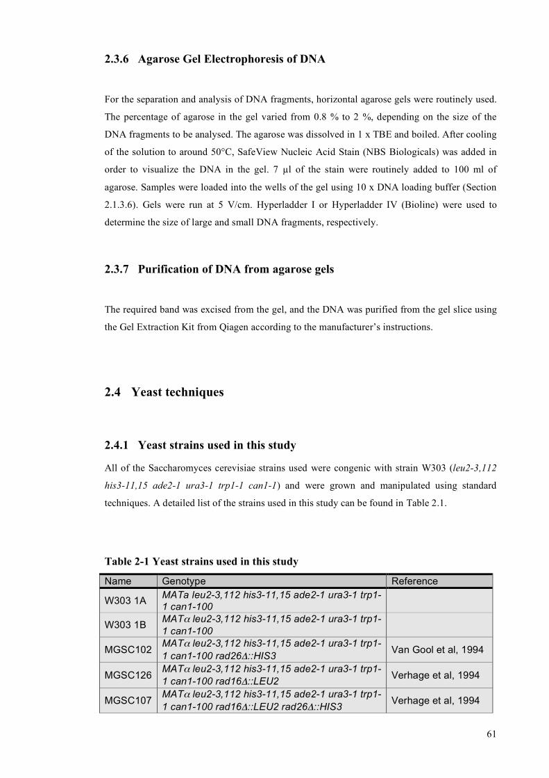
61
2.3.6 Agarose Gel Electrophoresis of DNA
For the separation and analysis of DNA fragments, horizontal agarose gels were routinely used.
The percentage of agarose in the gel varied from 0.8 % to 2 %, depending on the size of the
DNA fragments to be analysed. The agarose was dissolved in 1 x TBE and boiled. After cooling
of the solution to around 50°C, SafeView Nucleic Acid Stain (NBS Biologicals) was added in
order to visualize the DNA in the gel. 7 µl of the stain were routinely added to 100 ml of
agarose. Samples were loaded into the wells of the gel using 10 x DNA loading buffer (Section
2.1.3.6). Gels were run at 5 V/cm. Hyperladder I or Hyperladder IV (Bioline) were used to
determine the size of large and small DNA fragments, respectively.
2.3.7 Purification of DNA from agarose gels
The required band was excised from the gel, and the DNA was purified from the gel slice using
the Gel Extraction Kit from Qiagen according to the manufacturer’s instructions.
2.4 Yeast techniques
2.4.1 Yeast strains used in this study
All of the Saccharomyces cerevisiae strains used were congenic with strain W303 (leu2-3,112
his3-11,15 ade2-1 ura3-1 trp1-1 can1-1) and were grown and manipulated using standard
techniques. A detailed list of the strains used in this study can be found in Table 2.1.
Table 2-1 Yeast strains used in this study
Name Genotype Reference
W303 1A MATa leu2-3,112 his3-11,15 ade2-1 ura3-1 trp1-1 can1-100
W303 1B MATα leu2-3,112 his3-11,15 ade2-1 ura3-1 trp1-1 can1-100
MGSC102 MATα leu2-3,112 his3-11,15 ade2-1 ura3-1 trp1-1 can1-100 rad26Δ::HIS3 Van Gool et al, 1994
MGSC126 MATα leu2-3,112 his3-11,15 ade2-1 ura3-1 trp1-1 can1-100 rad16Δ::LEU2 Verhage et al, 1994
MGSC107 MATα leu2-3,112 his3-11,15 ade2-1 ura3-1 trp1-1 can1-100 rad16Δ::LEU2 rad26Δ::HIS3 Verhage et al, 1994

62
JSY1105 MATα leu2-3,112 his3-11,15 ade2-1 ura3-1 trp1-1 can1-100 URA3::MHRAD26 This study
JSY1106 MATα leu2-3,112 his3-11,15 ade2-1 ura3-1 trp1-1 can1-100 URA3::MHRAD26 rad16Δ::LEU2 This study
JSY1121 MATα leu2-3,112 his3-11,15 ade2-1 ura3-1 trp1-1 can1-100 rpb9Δ::TRP1 This study
JSY1107 MATα leu2-3,112 his3-11,15 ade2-1 ura3-1 trp1-1 can1-100 rad16Δ::LEU2 rpb9::TRP1 This study
JSY1108 MATα leu2-3,112 his3-11,15 ade2-1 ura3-1 trp1-1 can1-100 rad16Δ::LEU2 rpb9::TRP1 rad26::HIS3
This study
JSY1109 MATα leu2-3,112 his3-11,15 ade2-1 ura3-1 trp1-1 can1-100 URA3::MHRAD26 mec1∆::HIS3 sml1∆::TRP1
This study
JSY1110 MATα leu2-3,112 his3-11,15 ade2-1 ura3-1 trp1-1 can1-100 URA3::MHRAD26 chk1∆::HIS3 This study
JSY1111 MATα leu2-3,112 his3-11,15 ade2-1 ura3-1 trp1-1 can1-100 URA3::MHRAD26 rad53∆::HIS3 sml1∆::TRP1
This study
JSY1117 MATα leu2-3,112 his3-11,15 ade2-1 ura3-1 trp1-1 can1-100 URA3::MHRAD26 tel1∆::TRP This study
JSY1118 MATα leu2-3,112 his3-11,15 ade2-1 ura3-1 trp1-1 can1-100 URA3::MHRAD26 dun1∆::HIS This study
JSY1112 MATa leu2-3,112 his3-11,15 ade2-1 ura3-1 trp1-1 can1-100 mec1∆::HIS3 sml1∆::TRP1 This study
JSY1113 MATa leu2-3,112 his3-11,15 ade2-1 ura3-1 trp1-1 can1-100 chk1∆::HIS3 This study
JSY1114 MATa leu2-3,112 his3-11,15 ade2-1 ura3-1 trp1-1 can1-100 rad53∆::HIS3 sml1∆::TRP1 This study
JSY1119 MATα leu2-3,112 his3-11,15 ade2-1 ura3-1 trp1-1 can1-100 tel1Δ::TRP This study
JSY1120 MATα leu2-3,112 his3-11,15 ade2-1 ura3-1 trp1-1 can1-100 dun1Δ::HIS This study
JSY1115 MATa leu2-3,112 his3-11,15 ade2-1 ura3-1 trp1-1 can1-100 mec1∆::HIS3 sml1∆::TRP1 rad26∆::KanMx
This study
MGSC139 MATα leu2-3,112 his3-11,15 ade2-1 ura3-1 trp1-1 can1-100 rad14∆::LEU2 Verhage et al, 1996
JSY569 MATα leu2-3,112 his3-11,15 ade2-1 ura3-1 trp1-1 can1-100 def1∆::URA3 Woudstra et al, 2002
JSY626 MATα leu2-3,112 his3-11,15 ade2-1 ura3-1 trp1-1 can1-100 def1∆::URA3 rad14∆::LEU2 Woudstra et al, 2002
2.4.2 Growth conditions, drug treatments and cell cycle synchronisations
For all the experiments described in this work the yeast cells were grown at 30°C.
Logarithmically growing cells were used for analysis, unless otherwise indicated. Cell densities
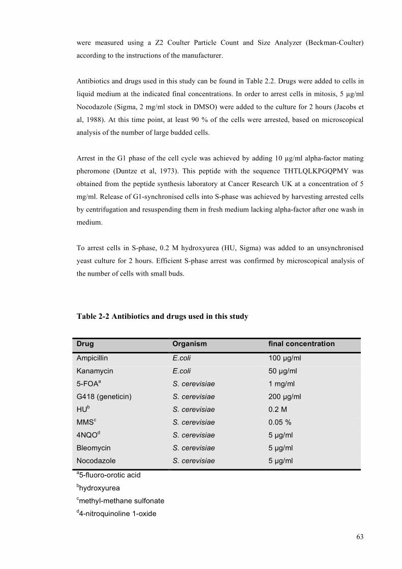
63
were measured using a Z2 Coulter Particle Count and Size Analyzer (Beckman-Coulter)
according to the instructions of the manufacturer.
Antibiotics and drugs used in this study can be found in Table 2.2. Drugs were added to cells in
liquid medium at the indicated final concentrations. In order to arrest cells in mitosis, 5 µg/ml
Nocodazole (Sigma, 2 mg/ml stock in DMSO) were added to the culture for 2 hours (Jacobs et
al, 1988). At this time point, at least 90 % of the cells were arrested, based on microscopical
analysis of the number of large budded cells.
Arrest in the G1 phase of the cell cycle was achieved by adding 10 µg/ml alpha-factor mating
pheromone (Duntze et al, 1973). This peptide with the sequence THTLQLKPGQPMY was
obtained from the peptide synthesis laboratory at Cancer Research UK at a concentration of 5
mg/ml. Release of G1-synchronised cells into S-phase was achieved by harvesting arrested cells
by centrifugation and resuspending them in fresh medium lacking alpha-factor after one wash in
medium.
To arrest cells in S-phase, 0.2 M hydroxyurea (HU, Sigma) was added to an unsynchronised
yeast culture for 2 hours. Efficient S-phase arrest was confirmed by microscopical analysis of
the number of cells with small buds.
Table 2-2 Antibiotics and drugs used in this study
Drug Organism final concentration
Ampicillin E.coli 100 µg/ml
Kanamycin E.coli 50 µg/ml
5-FOAa S. cerevisiae 1 mg/ml
G418 (geneticin) S. cerevisiae 200 µg/ml
HUb S. cerevisiae 0.2 M
MMSc S. cerevisiae 0.05 %
4NQOd S. cerevisiae 5 µg/ml
Bleomycin S. cerevisiae 5 µg/ml
Nocodazole S. cerevisiae 5 µg/ml a5-fluoro-orotic acid bhydroxyurea cmethyl-methane sulfonate d4-nitroquinoline 1-oxide

64
2.4.3 Lithium-Acetate transformation of yeast cells
2 ml yeast cultures were grown in the appropriate growth medium over night at 30°C. In the
morning, the cells were diluted in fresh medium to a cell density of 3 x 10⁶ cells/ml and further
grown at 30°C until the culture reached a cell density of 1 x 10⁷ cells/ml. The cells were
harvested by centrifugation at 3000 rpm for 5 minutes, the supernatant was discarded and the
pellet was resuspended in 1 ml of water and transferred to a 1.5 ml Eppendorf tube. After
centrifugation at 14000 rpm for 20 seconds the pellet was washed in 1 ml of TE/LiOAc solution
(Section 2.1.3.4) and centrifuged again. Finally, the pellet was resuspended in an appropriate
volume of Te/LiOAc solution to obtain a cell density of 109 cells/ml.
For the transformation of PCR products, in order to construct deletion strains as well as to tag
yeast proteins by homologous recombination, a 100 µl aliquot of this yeast suspension was
mixed with 20 µl of purified PCR product, 70 µl of 2 mg/ml salmon sperm DNA (boiled for 5
minutes and chilled on ice before use), and 600 µl of PEG/TE/LiOAc solution (Section 2.1.3.5).
For transformation of plasmids, 50 µl of the yeast suspension was mixed with 100 ng of plasmid
DNA, 35 µl of 2 mg/ml salmon sperm DNA and 300 µl of PEG/TE/LiOAc solution.
The mixture was briefly vortexted and then incubated at 30°C in an Eppendorf shaker (1000
rpm) for 1 hour. 70 µl or 35 µl of DMSO, for PCR products or plasmids, respectively, were
added and the suspension was heat shocked in a 42°C water bath for 15 minutes. Finally, the
cells were collected by centrifugation, washed briefly in 1 ml of water and then plated on the
appropriate selective plates.
In the case of selection for TRP+, URA+, HIS+ and LEU+ cells, the pellet was directly plated
onto selective plates. For KanMx+ selection, as well as selection against URA+ cells on 5-FOA,
the pellet was first plated on YPD and incubated over night, before being replica-plated on
appropriate plates containing the drug after about 16 hours.
2.4.4 Galactose-induced overexpression of proteins in yeast cells
For overexpression of proteins in yeast, the pYC2/CT vector (Invitrogen) was routinely used. It
is a centromeric plasmid carrying a URA3 marker, and your gene of interest can be cloned
between a GAL1 promoter and a CYC1 terminator. The final constructs were transformed into
65
the appropriate yeast strain, and the resulting plasmid-containing cells were grown for 24 hours
in 2 ml of SC-URA in the presence of raffinose as the only sugar. After this incubation the
culture usually reached a cell density of 108 cells/ml. 500 µl of this culture were routinely used
to inoculate a 150 ml culture, again in medium lacking uracil and containing raffinose. After
incubation at 30°C for 12-16 hours, the cell density of the culture reached 5 x 10⁶ cells/ml.
Galactose was always added to the cells at this density to a final concentration of 2 % (w/v).
Addition of glucose to a separate raffinose-culture served as a control for repression of the gene.
Incubation in the presence of galactose/glucose was carried out for 2 hours before the cells were
harvested. Protein extracts were then prepared for analysis by SDS-PAGE and/or
immunoprecipitation and purification of the overexpressed protein.
2.4.5 Preparation of yeast extracts using glass beads
Yeast cells from a liquid culture were harvested by centrifugation at 3000 rpm for 5 minutes.
The cell pellet was resuspended in 1 ml of PBS and transferred to a 1.5 ml tube (screw cap).
The cells were harvested again by centrifugation in a table-top centrifuge (full speed, 20
seconds) and washed in 1 ml ice-cold lysis buffer (Section 2.1.3.10). After another round of
centrifugation the pellet was resuspended in 700 µl of lysis buffer.
500 µl of 0.5 mm Zirconia/Silica Beads (BioSpec Products, Inc.) were added to the cell
suspension and the cells were disrupted using a FastPrep-24 Tissue and Cell Homogenizer (MP
Biomedicals). 6 rounds of shaking (30 seconds each) were carried out, separated by 2 minutes
during which the tubes were incubated on ice in order to prevent sample heating.
After disruption of the cells, a small hole was made at the bottom of the 1.5 ml tube using a
hypodermic needle and the tube was placed into a 2 ml Eppendorf tube. Centrifugation of these
tubes at 5000 rpm for 2 minutes at 4°C ensured that the protein extract, as well as the chromatin
and cell debris, were collected in the 2 ml tube while the beads remained in the 1.5 ml tube.
The cell debris and chromatin formed a pellet at the bottom of the tube after this centrifugation
step. In order to solubilize proteins from the chromatin pellet, 2 mM MgCl2 and 250 Units of
benzonase (Novagen) were added to the extract. The pellet was then resuspended by pipetting
and the tubes were rotated at 4°C for 1 hour. After this incubation the tubes were spun at 14000
rpm at 4°C for 15 minutes and the supernatant was transferred to a fresh tube.
66
2.4.6 Preparation of quick protein extracts from yeast
Rapid isolation of proteins from yeast cells was performed as described earlier (Kushnirov,
2000). About 107 yeast cells were harvested by centrifugation and the pellet was resuspended in
100 µl of water. 100 µl of 0.2 M NaOH were added and the tubes were briefly vortexed to
ensure proper resuspension of the cells. After an incubation time of 5 minutes at room
temperature the cells were pelleted, the supernatant was discarded and the pellet was
resuspended in 50 µl of 1 x SDS loading buffer. This suspension was then boiled for 5 minutes
and pelleted again. 10 µl of this supernatant were routinely analysed by SDS-PAGE.
2.4.7 Isolation of high quality genomic DNA from yeast
High quality genomic DNA from yeast was purified using the MasterPure Yeast DNA
Purification Kit (Epicentre Biotechnologies) according to the instructions of the manufacturer.
The supplied RNAse in the kit was used to further purify the DNA as recommended in the
manual, and the RNAse was afterwards removed by phenol/chloroform purification and
precipitation of the DNA.
2.4.8 Rapid isolation of genomic DNA from yeast
For analysis of successful gene deletion in yeast by PCR it is not necessary to obtain large
quantities of high quality DNA. For this purpose, 0.5 ml cultures were set up from individual
yeast colonies. After incubation at 30°C over night the cells reached densities of 1 – 2 x 10⁸
cells/ml. 200 µl of these cultures were centrifuged at 14000 rpm for 2 minutes and the
supernatant was discarded. The cell pellets were resuspended in 20 µl of 0.2 % SDS and boiled
for 10 minutes. After a short centrifugation at 14000 rpm, the supernatant was transferred to a
fresh tube. 1 µl of this solution was routinely used in PCR reactions to assess the success of a
gene deletion approach.
2.4.9 Analysis of the UV sensitivity of yeast strains
The yeast strains to be analysed were grown overnight in 1 ml of the appropriate growth
medium. In the morning, the cells were diluted to a cell density of 5 x 10⁶ cells/ml and the
incubation was continued in order to get the cells back into the logarithmic growth phase. As
soon as the culture reached a cell density of 2 x 10⁷ cells/ml, 4 10-fold serial dilutions were
67
prepared and 3 µl of these dilutions, as well as the undiluted culture, were spotted on
appropriate yeast plates. After spotting of all the strains, one plate was transferred to the
incubator without UV as a control for spotting equal numbers of cells. The other plates were
irradiated with various doses of 254 nm UVC light using a UV Stratalinker 2400 (Stratagene)
and then incubated at 30°C in the dark for growth.
2.4.10 Analysis of UV-induced degradation of Rpb1
The yeast strains to be analysed were grown overnight in the appropriate growth medium. On
the next day, the cells were diluted in 150 ml of medium to obtain a cell density of
3 x 10⁶ cells/ml. In order to get the cells back into log-phase, they were allowed to grow until
they reached a cell density of 1 x 10⁷ cells/ml.
50 µg/ml Cycloheximide (Sigma, 5 mg/ml stock in water) were added to the cultures for 1 hour
in order to inhibit any new protein synthesis. After this incubation period, the cells were
harvested by centrifugation (3000 rpm for 5 minutes), and resuspended in 150 ml of PBS. The
supernatant was saved for the post-UV incubation period. After removing a 30 ml aliquot which
served as a pre-UV control, the remaining cell suspension was UV-irradiated with a germicidal
lamp at a dose of 400 J/m2. After irradiation, the cells were harvested again by centrifugation
and resuspended in 120 ml of the original growth medium containing cycloheximide. At various
time points after the irradiation (every hour up to 4 hours post-UV), 30 ml aliquots were
removed from the culture, the cells were harvested by centrifugation, washed once in PBS and
then quickly frozen by dropping into liquid nitrogen.
After all the samples were collected, extracts were prepared as descibed (Section 2.4.5) and the
protein concentrations of all extracts were determined using the Bradford assay. Equal amounts
(10 µg) of extracts were loaded into the wells of a 4-12 % gradient gel (BioRad). SDS-PAGE
was performed as described (Section 2.5.1), and the proteins were transferred to a Hybond-C
Extra membrane (Section 2.5.2) for analysis by Western blot. Ponceau-S staining of the
membrane was used to control if the loading was equal in all the lanes. The Rpb1 protein was
detected with 8WG16 antibody.
2.4.11 Analysis of UV-induced ubiquitylation of Rpb1
Treatment of yeast cells with cycloheximide and UV was carried out as described for the
analysis of UV-induced degradation of Rpb1 (Section 2.4.10) and extracts were prepared as

68
described (Section 2.4.5). After quantification of protein concentrations, 2 mg of total yeast
extract were incubated with 50 µl of GST-Dsk2 beads (Anindya et al., 2007) to
immunoprecipiate mono- and polyubiquitylated proteins. This incubation was carried out for 4
hours at 4°C on a rotating wheel. After binding, the beads were harvested by centrifugation
(2000 rpm for 3 minutes), washed twice for 5 minutes each in ice-cold PBS and then once for 5
minutes in ice-cold PBS with 0.1 % Triton X-100. Finally, the beads were resuspended in 80 µl
of 1 x SDS-PAGE loading buffer (Section 2.1.3.7) and boiled for 10 minutes. After a quick
centrifugation step to collect the beads at the bottom of the tube, 15 µl of the supernatant were
loaded on 4-12 % gradient gels and SDS-PAGE and Western blotting was carried out as
described (Sections 2.5.1 and 2.5.2).
The membranes were stained with Ponceau S solution (Sigma) and the image was scanned. The
stained GST-Dsk2 protein served as a control to comfirm that equal amounts of beads used in
the immunoprecipitation step. Mono- and polyubiquitylated Rpb1 forms were detected using
4H8 antibody.
2.4.12 Strand-specific nucleotide excision repair (NER) assay
Analysis of strand specific nucleotide excision repair capacity in yeast cells was performed as
described (Teng et al., 2005) with slight modifications. A schematic outline of the assay can be
seen in Figure 2.1.
2.4.12.1 Irradiation and harvesting of samples
Yeast cells were grown in 500 ml of appropriate growth medium at 30°C until they reached a
cell density of 4 x 10⁷ cells/ml. The culture was harvested by centrifugation at 3000 rpm for 5
minutes and the supernatant was discarded. The pellet was resuspended in 1 l of ice-cold PBS,
resulting in a cell-density of 2 x 10⁷ cells/ml. An aliquot (one fifth or one sixth, depending on
the total number of samples) was removed and kept on ice in the dark as an unirradiated (U)
control. The remaining suspension was subjected to UVC-irradiation (254 nm) with 100 J/m2
using a germicidal lamp. Directly after UV, another aliquot was removed and kept on ice in the
dark. This sample was later used to determine the total amount of inflicted UV-damage (0). The
rest of the suspension was harvested by centrifugation, the supernatant was discarded and the
pellet was resuspended in fresh growth medium at a cell density of 4 x 10⁷ cells/ml. At the
desired time-points after UV irradiation, samples were removed, spun down at 3000 rpm for 5
min, and the pellet was resuspended in 50 ml of ice-cold PBS and kept in the dark.

69
After harvesting of all the time-points, the individual aliquots were again spun down, and the
pellets were once again washed in 50 ml of ice-cold PBS. Finally, after another centrifugation
step, the pellets were resuspended in 5 ml of Sorbitol solution (Section 2.1.4.1) containing
28 mM beta-mercaptoethanol and 5 mg of Zymolyase T-20 (MP Biomedicals) and the tubes
were incubated at 4°C o/n in the dark to obtain spheroblasts.
2.4.12.2 Isolation of genomic DNA
Spheroblasts were spun down at 3000 rpm for 5 min and the pellet was carefully washed with
10 ml of Sorbitol solution (Section 2.1.4.1), and then finally dissolved in 5 ml of 1 x NER Lysis
Buffer (Section 2.1.4.2) containing 200 µl of RNAse A stock solution (Sigma, 10 mg/ml in TE
buffer). The solution was vortexed rigorously to lyse the spheroblasts and then incubated for 1
hour at 37°C with occasional shaking. 250 µl of Pronase stock stolution (Roche, 20 mg/ml in
water) were added, and the tubes were incubated for another hour at 37°C after a brief vortexing
step before another 1 hour incubation at 65°C.
After cooling down the samples to room temperature, an equal volume (5 ml) of
phenol/chloroform/isoamylalcohol (PCI pH 8.0, Sigma) was added and the tubes were briefly
vortexed to mix the phases. The resulting emulsion was transferred to 15 ml Corex-tubes and
spun down for 10 min in an SS-34 rotor at 10000 rpm. The aqueous supernatant was transferred
to a 15 ml Falcon-tube and another 5 ml of PCI were added. After vortexing, the tubes were
spun at 4500 rpm for 10 minutes and the aqueous phase was again transferred to a fresh 15 ml
Falcon-tube. 5 ml of chloroform were added, the emulsion was once again vortexed and spun at
4500 rpm for 10 min. The supernatant was transferred to a 50 ml Falcon-tube, and 10 ml of ice-
cold 100 % ethanol were added to precipitate the genomic DNA. To obtain efficient
precipitation, the samples were incubated at -20°C over night.
Genomic DNA was then harvested by centrifuation at 4500 rpm for 10 minutes at 4°C, the
pellet was dissolved in 1 ml of TE buffer and transferred to a 2 ml Eppendorf tube. 1 ml of
isopropanol was added, and after mixing the tubes were incubated at room temperature for 30
min to once again precipitate the DNA. The tubes were spun at 14000 rpm for 5 minutes at 4°C
and the pellet was resuspended in 600 µl of TE-buffer, resulting in a DNA concentration of
around 0.5 µg/µl for the culture volume stated above. 1 µl aliquots of the DNA samples were
run in a 0.8 % agarose gel to make sure that the genomic DNA was intact and did not get
degraded during the purification steps.

70
Figure 2-1 Analysis of strand-specific CPD repair at nucleotide resolution
(A) Schematic outline of the individual steps in the protocol (see text for details). (B) Representative gel showing the result of such an experiment for the wild type strain
(W303).
71
2.4.12.3 Preparation of genomic DNA for analysis
100 µl of genomic DNA (~ 50 µg) were digested with 60 units of HaeIII restriction enzyme
(New England Biolabs) in 300 µl of 1 x restriction buffer (Buffer 2, New England Biolabs) for 2
hours at 37°C. A 5 µl aliquot was subjected to electrophoresis in a 1 % agarose gel to ensure
that all the samples were completely digested and to check that similar amounts of DNA were
present for all the time-points.
300 µl of PCI were added to the solution and mixed by vortexing. The two phases were
separated using MaXtract Low Density tubes (QIAGEN) according to the manufacturer’s
instructions and the aqueous phase was transferred to a fresh 1.5 ml Eppendorf tube. 30 µl (1/10
volume) of 3 M sodium acetate pH 5.3 and 330 µl of isopropanol were added and the solution
was incubated on dry ice for 15 minutes to precipitate DNA. The digested DNA was harvested
by centrifugation in a table-top centrifuge at 14000 rpm for 5 min at 4°C, the supernatant was
discarded and the pellet was dissolved in 100 µl of TE buffer. 10 µl of ML extract (Section
2.2.3) were added and the tubes were incubated at 37°C for 1 hour to induce incisions at CPDs.
After this incubation the DNA was purified twice using MaXtract Low Density tubes, first with
PCI and then with chloroform. The resulting aqueous phase (100 µl) was transferred to 0.5 ml
PCR tubes (ANACHEM).
2.4.12.4 Isolation of RPB2 strands for labelling and analysis
25 µl of 5 M NaCl were added to the samples to obtain a final NaCl concentration of 1 M. 1 µl
of biotinylated probe (2 mM stock), specific for either the TS or the NTS of the RPB2 fragment,
was added. The sequences of these probes were the following:
for isolation of the TS:
5’ biotin-GATAGCTTTTTTCCGTTTACCGATTATGTTAAGATCAAAGAA 3’
for isolation of the NTS:
5’ biotin-GATAGCTTTTTTCCAATAATGGACCTGCCAAATCTAATCT 3’
Regions of homology to the RPB2 sequence are shown in red, and the 6 thymidines for labelling
of the 3’ end of the isolated DNA strand are shown in blue. The six nucleotides between the
biotin and the thymidine stretch were included to ensure complete labelling by the DNA
polymerase while the strand was still bound to streptavidin beads.

72
The resulting solution was incubated at 95°C for 5 min and then at 55°C for 15 minutes in a
PCR machine. During this incubation, 10 µl of Dynabeads M-280 Streptavidine (Invitrogen, 6.7
x 108 beads/ml) per sample were washed once with water and twice with BW Buffer (Section
2.1.4.3). These washing steps were carried out using a Magnetic Particle Concentrator (MPC).
10 µl of the washed beads were added to each PCR tube, mixed by pipetting and incubated at
room temperature for 15 minutes with occasional mixing. The tubes were then placed in the
MPC and the supernatant was saved for isolation of the remaining DNA strand. The beads were
resuspended in 50 µl of BW Buffer and the tubes were incubated at 58°C for 5 min in a PCR
machine. After this high-stringency wash, the beads were washed two additional times with 50
µl of BW buffer at room temperature, before they were finally resuspended in 4.3 µl of water.
2.4.12.5 Radioactive labelling and analysis of the isolated DNA strands
Labelling of the DNA strands was performed using the Sequenase Version 2.0 DNA
Sequencing Kit (USB). For each sample, 5.7 µl of a mix containing 2 µl of Sequenase Buffer,
2.4 µl of Sequenase Dilution Buffer, 0.7 µl of 100 mM DTT, 0.25 µl of water, 0.25 µl of alpha-32P-dATP (Perkin Elmer, 6000 Ci/mmol) and 0.1 µl of Sequenase were added to the beads, and
the suspension was mixed by pipetting. The labelling was allowed to proceed at room
temperature for 15 minutes with occasional mixing. After this incubation, the tubes were placed
in the MPC and the supernatant was discarded. The beads were washed three times with 50 µl
of water before being resuspended in 5 µl of FA loading buffer (Section 2.1.3.8). Finally, the
tubes were placed in a 100°C heat block for 1 minute to ensure complete release of the labelled
DNA strands from the streptavidine beads.
6 % sequencing gels were used to separate the DNA fragments. These gels were prepared using
SequaGel concentrate and diluent (National Diagnostics) according to the manufacturer’s
instructions. The gels were prerun at 65 Watts for 1 hour in order to heat the gel to 50°C. The
tubes containing the samples were put in the MPC and the supernatants (5 µl) were loaded into
individual wells of the gel. Electrophoresis was carried out at 65 Watts for 2.5 hours. The gel
apparatus was disassembled, and the gel was dried at 80°C for 1 hour. For analysis of the
fragments, the dried gel was placed in a Storage Phosphor Screen Cassette (Molecular
Dynamics) for two days. An image of the gel was then obtained using a Typhoon Phosphor-
Imager.
73
2.4.12.6 Calculation of damage half-lifes (T50%)
For each lane of the gel, the intensities of the prominent top band (full length, undamaged DNA
strand) and the entire lower part of the lane (damage-specific bands, non-specific bands and
background) were determined using the Image-Quant 5.0 Software. The intensity of the lower
part of the U-lane was then subtracted from the intensities of the lower parts of all other lanes to
subtract the background and non-specific bands, which are not due to inflicted damages. The
resulting values (‘damages’) were then added to the intensities of the top band to obtain a value
for the total signal per lane.
The total signal for the 0 lane (no repair incubation) was set to 1 and a correction factor for all
the other lanes was determined, allowing to correct for loading differences between the lanes.
The intensities of the damages in all the lanes were corrected using this factor. The damages in
the 0 lane were then set to 1 and all the damages of the later time points were put in relation to
the 0 time point. The values were plotted in a graph using the Microsoft Excel software (x axis
= time, y axis = ratio of remaining damages), and the point on the x axis for which y=0.5 was
determined. This value was the halflife of the damages (T50%).
2.4.13 Chromatin Immunoprecipitation (ChIP)
2.4.13.1 Cell growth and UV irradiation
Yeast strains were grown in appropriate medium over night at 30°C and diluted in the morning
to achieve a cell density of 2.5 x 10⁶ cells / ml. The cells were then allowed to divide twice in
order to get back into the logarithmic growth phase, and were harvested when the cell density
reached 1 x 10⁷ cells / ml. After harvesting of the cells by centrifugation, the supernatant was
discarded and the pellet was resuspended in the original culture volume of PBS. UV irradiation
at the indicated dose was carried out using a germicidal lamp. A control of 10 ml was taken out
before UV treatment. After UV irradiation, the cells were again collected by centrifugation, the
supernatant was discarded, and the pellets were dissolved in the same volume of fresh medium.
Incubation of the cultures was then continued.

74
2.4.13.2 Crosslinking, preparation of extracts and DNA shearing
At various time points after the UV irradiation step, 10 ml samples (corresponding to 1 x 10⁸
cells) were removed and fixed with 1 % formaldehyde for 15 minutes. Crosslinking was
stopped by adding 1/10 volume of 2 M glycine for 5 minutes. The cells were then harvested by
centrifugation, washed twice with PBS and finally resuspended in 1 ml of FA Lysis Buffer.
Extracts were prepared as described in section 2.4.5, with the difference that after spinning the
extract and cellular debris from the bead-containing tube into the 2 ml tube, the chromatin-
containing pellet was resuspended in the supernatant and transferred to a 15 ml Falcon-tube.
500 µl of fresh FA Lysis Buffer were added and the samples were sonicated using a
BIORUPTOR (Diagenode). Ice was added to the water bath in order to keep the samples cold
during the process. Sonication was carried out in 30 second intervals with 30 second breaks for
a total time of 15 minutes. After this, the samples were transferred to fresh 1.5 ml tubes and
spun at full speed twice for 10 minutes. After each centrifugation the pellet was discarded and
the supernatant transferred to a fresh tube. The so obtained extracts containing solubilized
chromatin were used for immunoprecipitation of the target protein.
2.4.13.3 Immunoprecipitation
150 µl of the extracts were diluted to 300 µl with fresh FA Lysis Buffer. 1 µg of antibody was
added, and binding of the antibody to the target was performed over night at 4°C on a rotating
wheel. After the binding, the extracts were centifuged at 14000 rpm for 10 minutes to remove
precipitations. The supernatant was transferred to Spin-X Centrifuge Tube Filters (Costar) and
35 µl of a 1:1 slurry of Protein A beads (Pierce) in FA Lysis Buffer containing 1 mg/ml of BSA
were added. Using the Spin-X Centrifuge tube filters ensured proper removal of all the liquids
without losing any beads in the subsequent washing steps. Binding of the antibody/antigen
complex to the Protein A beads was carried out at 4°C for 2 hours with constant shaking. The
beads were then harvested by centrifugation and washed consecutively with FA Lysis Buffer,
FA-500, LiCl wash solution and TES (recipes can be found in section 2.1.5) for 10 minutes
each at 4°C with constant shaking. After removal of the TES, the filters containing the beads
were transferred to a fresh 1.5 ml tubes and the beads were resuspended in 150 µl of ChIP
elution buffer (Section 2.1.5.5). Elution was carried out at 37°C in an Eppendorf shaker for 30
minutes with constant shaking. The liquid containing the eluted protein/DNA complexes was
harvested by centrifugation. 150 µl of water and 20 µg of Proteinase K (Roche) were added and
reversal of crosslinks was achieved by incubation at 65°C over night. 3 µl of the extract used for
IP were mixed with 150 µl water, 150 µl of ChIP elution buffer and 20 µg of Proteinase K

75
(Sigma), and then incubated over night at 65°C. These samples served as input controls for the
IPs.
2.4.13.4 Analysis of immunoprecipitated DNA by Real time PCR
After reversal of the crosslinks, the DNA in the samples was purified using the Qiagen PCR
purification kit according to the instructions of the manufacturer. The DNA was eluted in 50 µl
of water, and then quantified by Real time PCR using a BioRad iCycler and the MyIQ software.
PCR reactions were set up using the iQ SYBR Green Supermix (BioRad) in 96 well plates.
Standard reactions of 20 µl contained:
10 µl of iQ SYBR Green Supermix
1 µl of eluted DNA
8 µl of water and
0.5 µl of both primers (10 µM).
Ct-values were obtained for both the DNA from the IPs and the inputs. The analysis was
performed in triplicates for each sample, and the average between these 3 values was calculated.
The resulting value (x) was used in the following formula: 2^(30-x). The so-obtained value for
the IP of each time point was divided by the value obtained for the input of the same time point,
in order to correct for input differences. Correction with a value for the telomere was then
carried out in order to correct for non-specific signals. An example of such an analysis can be
found in the Appendix.
2.5 Protein analysis
2.5.1 SDS Polyacrylamide Gel Electrophoresis (SDS-PAGE)
Precast 4-12% Bis-Tris gradient gels (Biorad) were routinely used to separate proteins. The
running buffer was XT MOPS buffer (Biorad) and electrophoresis were carried out in Criterion
chambers (Biorad). The Precision Plus protein marker from Biorad was used to determine the
size of the detected protein on Western Blots or gel stains. 4-12 % gradient gels were usually
run at 150 V for 90 minutes.

76
For analysis of high-molecular weight proteins, as well as post-translational modifications of
Rad26, 3-8 % gradient gels (Biorad) were used together with XT Tricine buffer (Biorad). These
gels were routinely run in the cold room at 90 V for 12-16 hours. After this time, the 100 kDa
marker reached the bottom of the gel.
2.5.2 Transfer of proteins to membranes (Western Blot)
After successful electrophoresis of the proteins by SDS-PAGE, the gels were briefly washed in
water and then transferred to transfer buffer (25 mM Tris, 60 mM glycine, 20 % methanol). A
piece of Hybond C-Extra membrane (Amersham Biosciences) was placed on top of the gel and
the obtained ‘sandwich’ was placed between 2 stacks of Whatman 3MM papers pre-wetted in
transfer buffer. This setup was placed in a Biorad Criterion Blotter and completely submerged
in transfer buffer. The transfer was carried out at 4°C for 70 minutes at 400 mA. After the
transfer, the membrane was briefly washed and then routinely stained using Ponceau-S solution
(Sigma) to check the efficiency of transfer and, in some cases, equal loading between the lanes.
The stain was removed by washing in PBS twice for 5 minutes each.
Table 2-3 Antibodies used in this study
1. Primary antibodies Epitope Source Supplier Dilution
Myc (9E10) Mouse CRUK antibody production 1:10000
HA Rabbit Abcam (ab9110) 1:5000
Rpb1 (4H8) Mouse CRUK antibody production 1:10000
Rpb1 (8WG16) Mouse CRUK antibody production 1:1000
Actin Mouse Abcam (ab8224) 1:1000
2. Secondary antibodies
Name Source Supplier Dilution
ECLTM anti-mouse-HRP Sheep GE Healthcare 1:10000
ECLTM anti-rabbit-HRP Donkey GE Healthcare 1:10000
all antibodies were diluted in 5 % milk/PBS

77
2.5.3 Detection of proteins on membranes
After transfer of proteins to the membranes, blocking was carried out with 5 % milk in PBS for
at least 30 minutes at room temperature in order to decrease non-specific binding of the
antibody to the membrane. Various antibodies were used in this study to detect the proteins of
interest. These antibodies can be found in Table 2.3 together with the dilution used. The
membrane was washed 3 times for 10 minutes in PBS after each antibody incubation. After the
last wash, the membranes were briefly dried and the chemiluminescent substrate was added.
These substrates were SuperSignal West Pico Chemiluminescent substrate (for routine use) and
SuperSignal West Dura Extended Duration Substrate (for weak signals). Both were purchased
from Thermo Scientific and used according to the instructions of the manufacturer. The
chemiluminescent signals were detected using SuperRX medical X-Ray Films (Fujifilm).
2.5.4 Staining of proteins in SDS-Gels using SYPRO Ruby stain
For staining of gels after SDS-PAGE the gels were washed twice for 5 minutes in water and
then fixed twice for 30 minutes in fixing solution (50 % methanol, 7 % acetic acid). This leads
to shrinking of the gel which might reduce the quality of the staining. To avoid this, the gel was
incubated in water twice for 10 minutes in order to rehydrate it. Afterwards the SYPRO Ruby
protein gel stain (Molecular probes) was added, and the gel was incubated in the stain over
night in the dark. After this incubation the gel was transferred to a fresh container and washed
twice for 30 minutes in destaining solution (10 % methanol, 7 % acetic acid). Then it was again
put in water for 10 minutes for rehydration, before it was analysed using a Typhoon Phosphor-
Imager.
2.5.5 Mass spectrometry analysis of Rad26
Polyacrylamide gel slices (1-2 mm) containing Rad26 were prepared for mass spectrometric
analysis using the Janus liquid handling system (Perkin Elmer). Briefly, the excised protein gel
piece was placed in a well of a 96-well microtitre plate and destained with 50% v/v acetonitrile
and 50 mM ammonium bicarbonate, reduced with 10 mM DTT, and alkylated with 55 mM
iodoacetamide. After alkylation, Rad26 was digested with 6 ng/µl trypsin (Promega) overnight
at 37 °C. The resulting peptides were extracted in 1% v/v formic acid, 2% v/v acetonitrile. The
digest was analysed by nano-scale capillary LC-MS/MS using a nanoAcquity UPLC (Waters) to
78
deliver a flow of approximately 300 nL/min. A C18 Symmetry 5 µm, 180 µm x 20 mm µ-
Precolumn (Waters), trapped the peptides prior to separation on a C18 BEH130 1.7 µm, 75 µm
x 100 mm analytical UPLC column (Waters). Peptides were eluted with a gradient of
acetonitrile. The analytical column outlet was directly interfaced via a modified nano-flow
electrospray ionisation source, with a 2-D linear ion trap mass spectrometer (LTQ XL/ETD,
ThermoScientific), equipped with a chemical ionization source to enable the generation and
injection of fluoranthene radical anions for the electron transfer dissociation (ETD) reaction
(Coon et al, 2004). The ETD process uses ion/ion chemistry to provide sequence information
not available through conventional methods such as CID. Peptide fragmentation using an ETD
approach augments the current methodologies available for the characterization of post-
translational modifications by more accurately identifying the specific amino acids that is
modified. Data dependent analysis was carried out in either CID or ETD mode, where
automatic MS/MS were acquired on multiply charged precursor ions in the m/z range 300–2000
m/z. As predominantly doubly protonated ions are generated by tryptic digestion, a
supplemental activation energy was used to improve fragmentation in the ETD experiments
(Swaney et al, 2007). All LC-MS/MS data were then searched against a protein database
(UniProt 13.6) using the Mascot search engine programme (Matrix Science, UK), with oxidised
methionine, carbamidomethyl cysteine and phospho serine, threonine and tyrosine included as
variable modifications (Perkins et al, 2007).
2.5.6 Immunoprecipitation of proteins from yeast extracts
After successful preparation of yeast extracts, 20-30 µl of Protein A beads coated with the
desired antibody were added. The beads were washed in lysis buffer (Section 2.1.3.10) prior to
addition to the extract. Binding to the beads was carried out over night at 4°C on a rotating
wheel. After binding, the beads were harvested by centrifugation at 3000 rpm for 2 minutes and
the supernatant was removed. The beads were washed once in lysis buffer containing 50 mM
KOAc, 3 times in lysis buffer containing 500 mM KOAc (15 minutes for each), and again
quickly in lysis buffer containing 50 mM KOAc. The immunoprecipitated protein, together with
its binding partners, was either recovered from the beads by boiling in 1 x SDS loading buffer
and then analysed by SDS-PAGE, or eluted from the beads using TEV protease if applicable.
2.5.7 ATPase assay
ATPase reactions with Rad26 were carried out in a total volume of 20 µl. The reactions
contained the following components:

79
10 µl 2 x ATPase buffer (40 mM Tris pH8, 8 mM MgCl2, 2 mM DTT, 0.05 mg/ml BSA)
1 µl of pRS316 plasmid DNA (100 ng/µl) or water as a control
1 µl of a mix of hot and cold gamma-32P-ATP (1:9 labelled ATP: 1µM cold ATP)
5 µl of purified protein (about 250 ng)
3 µl of water
The tubes were then incubated at 37°C in an Eppendorf shaker.
4 µl aliquots were removed after 0, 2, 5 and 15 minutes and transferred to tubes containing 20
µl of 0.1 M HCl. After mixing, these tubes were kept on dry ice to completely stop the reaction.
After all samples were obtained, 200 µl of charcoal solution (7 % activated charcoal, 50 mM
HCl, 5 mM H3PO4) were added, the samples were quickly vortexed and then incubated at room
temperature for 15 minutes. Finally, the tubes were centrifuged at 14000 rpm for 5 minutes and
100 µl of the clear supernatant containing inorganic phosphate were transferred to vials suitable
for the Scintillation counter. Cerenkov counting was carried out using a Tricarb 1500 Liquid
Scintillation Analyzer (Packard).
Quantification of ATPase activity was done by setting the value obtained for the 0 time point to
1 and putting all the later time points in relation to the 0 time point. These values were then used
to obtain a graph using the Microsoft Excel software. Error bars were obtained by determining
the standard deviation between independent experiments.

80
3 Results I – NER/TCR analysis at site-specific UV lesions
3.1 Project aim
DNA repair pathways are coordinated by the sequential assembly of many proteins at the site of
a DNA lesion. Knowledge about the exact timing of recruitment of particular proteins, as well
as the genetic requirements for the localization of particular factors to the DNA damage site, are
important for elucidating the repair mechanism. Work on double strand break (DSB) repair has
benefitted greatly from a system which allows the induction of a DSB in a temporally and
spatially controlled manner. This system makes use of the HO endonuclease, a sequence-
specific nuclease, which is normally involved in mating type switching (reviewed in (Haber,
1998)). The HO cleavage site (HOCS) can be introduced at any location in the genome, and
because its position and surrounding sequence is known, chromatin immunoprecipitation (ChIP)
experiments can be carried out. This allows a researcher to investigate if, and when, a particular
factor localizes to the DSB after induction of endonuclease expression, and which proteins are
required for this localization. This system is widely used in the field of double-strand break
repair.
The situation is more complicated for UV-induced DNA lesions, because every pair of
pyrimidines in the genome represents a potential site for the formation of a cyclobutane
pyrimidine dimer (CPD), or a 6-4 pyrimidine pyrimidone photoproduct (6-4PP) (Setlow and
Carrier, 1966). When examining the recruitment of an NER-factor to a particular region in the
genome by ChIP, only a small proportion of the examined molecules will actually have a lesion
there. One way to increase the chance of damage is to increase the UV intensity, but this will
inevitably lead to more damages in all other parts of the genome. This is problematic, because
factors with low abundance might be titrated away from the region of interest, and may
therefore never be detected. An alternative way to achieve efficient induction of UV damage at
a specific location is to use a sequence that is highly damage-prone, even at relatively low doses
of irradiation.
This project was aimed at setting up a system which should enable me to examine the
recruitment of NER factors to a UV damage-prone region by ChIP, by using a construct
containing a long stretch of pyrimidines, a ‘damage-hotspot’. This stretch should be damaged
much more efficiently by UV in vivo. By putting the ‘hotspot’ in the context of an inducible
gene (see below), it should be possible to distinguish TC-NER from GG-NER, and to examine
UV-dependent ubiquitylation of elongating RNAPII in vivo. Such a system would be highly
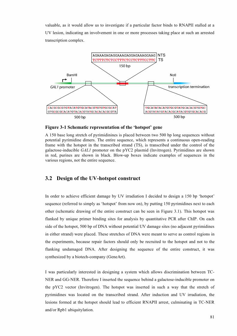
81
valuable, as it would allow us to investigate if a particular factor binds to RNAPII stalled at a
UV lesion, indicating an involvement in one or more processes taking place at such an arrested
transcription complex.
Figure 3-1 Schematic representation of the ‘hotspot’ gene A 150 base long stretch of pyrimidinines is placed between two 500 bp long sequences without potential pyrimidine dimers. The entire sequence, which represents a continuous open-reading frame with the hotspot in the transcribed strand (TS), is transcribed under the control of the galactose-inducible GAL1 promoter on the pYC2 plasmid (Invitrogen). Pyrimidines are shown in red, purines are shown in black. Blow-up boxes indicate examples of sequences in the various regions, not the entire sequence.
3.2 Design of the UV-hotspot construct
In order to achieve efficient damage by UV irradiation I decided to design a 150 bp ‘hotspot’
sequence (referred to simply as ‘hotspot’ from now on), by putting 150 pyrimidines next to each
other (schematic drawing of the entire construct can be seen in Figure 3.1). This hotspot was
flanked by unique primer binding sites for analysis by quantitative PCR after ChIP. On each
side of the hotspot, 500 bp of DNA without potential UV damage sites (no adjacent pyrimidines
in either strand) were placed. These stretches of DNA were meant to serve as control regions in
the experiments, because repair factors should only be recruited to the hotspot and not to the
flanking undamaged DNA. After designing the sequence of the entire construct, it was
synthesized by a biotech-company (GeneArt).
I was particularly interested in designing a system which allows discrimination between TC-
NER and GG-NER. Therefore I inserted the sequence behind a galactose-inducible promoter on
the pYC2 vector (Invitrogen). The hotspot was inserted in such a way that the stretch of
pyrimidines was located on the transcribed strand. After induction and UV irradiation, the
lesions formed at the hotspot should lead to efficient RNAPII arrest, culminating in TC-NER
and/or Rpb1 ubiquitylation.

82
It is important to point out that I avoided including any STOP-codons in this synthetic gene;
therefore the construct represents a continuous open-reading-frame. One problem that arose
from this is that the 500 bp damage-free flanking regions are highly repetitive, because only a
limited number of codons are available, and can only be used in a limited number of
combinations, in order to avoid having two adjacent pyrimidines in the TS or NTS of the gene.
We now believe this underlies some of the inconsistencies in the results I obtained with the
construct (see Discussion).
3.3 Analysis of the hotspot gene
3.3.1 The ‘hotspot’ sequence is efficiently damaged after UV irradiation
I first investigated if the stretch of 150 pyrimidines in the TS of the gene indeed represented a
sequence which can be efficiently damaged by UV irradiation. In order to do so, the DNA
construct containing it was irradiated in droplets of water on parafilm, using a UV Stratalinker
(Stratagene). UV-induced lesions represent a strong block to the progression of Taq-Polymerase
(Wellinger and Thoma, 1996), a fact which has been exploited for the mapping of lesions at
nucleotide resolution on DNA (Thoma et al, 1993; Chandrasekhar and van Houten, 1994). After
linearization of the plasmid with NotI (linearizing the plasmid downstream of the hotspot gene;
see Fig. 3.1), irradiated and un-irradiated hotspot construct was used in primer extension
reactions using a radioactively labelled primer. The same primer was also used on the un-
irradiated sample in the presence of ddNTPs in order to create a sequencing ladder, which
allows mapping of the lesions on the DNA at the nucleotide level. This primer extension
reaction should yield a 625 base long DNA fragment from an undamaged DNA template.
Lesions in the hotspot should lead to the appearance of shorter products (88-238 bases), which
map to the pyrimidine stretch (see schematic drawing in Figure 3.2A). Analysis of the products
on 6 % denaturing polyacrylamide gels indeed showed this pattern (Fig. 3.2B). The full-length
fragment, and some unspecific bands, was seen when the reaction was performed with un-
irradiated template. After irradiation of the DNA with increasing doses of UVC light, more and
more short fragments were seen which mapped exactly to the long pyrimidine stretch (see
sequencing ladder on the left in Figure 3.2B). Importantly, the fragments became increasingly
shorter at higher doses of UV, indicative of the generation of more lesions at the beginning of
the hotspot sequence. Importantly, no appearance of damage-specific bands was seen on the 500
bp sequence downstream of the hotspot (upper part of gel), which confirms that the pyrimidine
dimer-free stretch of DNA indeed represents a sequence which cannot be damaged, even at high
doses of UV.

83
Figure 3-2 Mapping of UV-induced DNA lesions on the hotspot gene (A) Outline of the experimental strategy to map lesions using primer extension (B) Representative gel showing primer extension results (see text for details) , the increasing
UV doses were 1, 2, 5 and 10 kJ/m2
3.3.2 Analysis of RNAPII distribution across the hotspot gene
Having shown that the pyrimidine stretch in the hotspot gene is efficiently damaged by UV
irradiation in vitro, I set out to determine its effect on RNAPII distribution before and after UV
treatment in vivo. In order to do so, the plasmid containing the gene was transformed into the
wild type W303 strain, and Rpb1 localization was examined by ChIP with 4H8 antibody,
followed by Real time PCR analysis of the immunoprecipitated DNA. Three regions were
chosen for this analysis, the GAL1 promoter upstream of the gene (‘start’), the pyrimidine
*nonspecific stop

84
stretch in the middle of the gene (‘hotspot’), and the termination sequence downstream of the
gene (‘end’). Unfortunately, because of the repetitive nature of the 500 bp damage-free regions
flanking the hotspot, no useful primer pairs for Real time PCR analysis could be found there.
Cells were grown in medium containing raffinose, and 2 % glucose or galactose was added at
the desired cell density (5 x 10⁶ cells/ml) to repress or activate transcription, respectively. A
primer pair in the middle of the endogenous GAL10 locus served as a control for efficient
galactose-mediated transcriptional induction. Another primer pair in the RPB2 open reading
frame served as a control for a constitutively expressed gene. After 2 hours of induction, the
cells were collected and UV-irradiated with 150 J/m2 of UVC light, and aliquots were harvested
either immediately after treatment, or after further incubation in medium for various lengths of
time. An outline of the experimental strategy is shown in Figure 3.3.
Figure 3-3 Experimental outline of Chromatin immunoprecipitation (ChIP) Cells containing the hotspot construct were grown in raffinose-containing medium, and the gene was either induced (2 % galactose) or repressed (2 % glucose) for 2 hours. Samples were taken before and at one or more time points after irradiation with a dose of 150 J/m2.
Results from these ChIP experiments are shown in Figure 3.4. One thing that immediately
caught my attention was the difference in induction levels between the hotspot gene and the
endogenous GAL10 gene (Figure 3.4A). For the GAL10 gene, these inductions (measured as the
ratio between the ChIP-signals for RNAPII in galactose versus glucose) were consistently
between 30 and 40-fold, as expected (Kristjuhan and Svejstrup, 2004). In sharp contrast to these
values, induction levels of the hotspot gene varied between 1.5 and 6-fold and displayed a
strong variation between independent experiments. In many cases, no detectable induction could
be achieved. Nevertheless, I continued to look at RNAPII distribution across the hotspot gene
before and after UV irradiation in the samples in which reasonably good induction could be
detected (3-fold and above). I expected to detect more RNAPII complexes at the hotspot
immediately after UV irradiation of cells grown in galactose, because the transient arrest of
RNAPII at UV-induced DNA lesions should increase the likelihood of finding RNAPII there.

85
Surprisingly, this was not the case. Instead, the ChIP signal was quickly lost from all examined
regions immediately after UV irradiation and reappeared thereafter. 90 minutes post-UV, the
intensity of the RNAPII signal was back at the pre-UV level (Figure 3.4B). This indicates that
transcription is transiently inhibited after UV irradiation, making it hard to show an increased
localization of RNAPII at the hotspot after UV treatment.
As can be seen in Figure 3.4B, the decrease of the ChIP signal for RNAPII after UV irradiation
was more pronounced at the 3’-end of the hotspot gene (termination sequence) compared to the
start (promoter), or at the hotspot sequence itself. I therefore calculated the ratio between the
ChIP signal at the beginning of the gene and that at the end of the gene. The ratio was
significantly higher than 1 shortly after UV irradiation, indicating that not all the RNAPII
complexes which start transcription reach the end of the gene, and that the presence of the
pyrimidine stretch might indeed be an obstacle for polymerases after damage induction (Figure
3.4C). During the time course, this ratio returned to its initial value of 1, indicative of removal
of the block. This result can be seen as an indication that upon UV irradiation the presence of
the pyrimidine stretch indeed represents a problem for transcribing RNAPII complexes.
3.3.3 Analysis of repair factor recruitment to the hotspot
Even though, as explained above, a dramatic effect of the hotspot on RNAPII distribution was
not obtained, I decided to carry on and try to detect the recruitment of NER factors to the
pyrimidine stretch after UV irradiation. I chose the Rad14 protein, because this factor is
indispensable for both NER subpathways (Prakash and Prakash, 2000). Rad14 was tagged on its
C-terminus with 6 HA-tags, and the functionality of the tagged protein was confirmed by
comparing the UV sensitivity of the tagged strain to the wild type as well as the rad14Δ deletion
strains. The sensitivity of the strain expressing epitope-tagged Rad14 was indistinguishable
from that of the wild type strain (W303), whereas rad14Δ was highly UV-sensitive (data not
shown).
In order to determine if the tagged protein could be found at the hotspot after UV-treatment, the
plasmid containing the hotspot gene was transformed into the tagged strain, and cells were
grown up in medium containing raffinose. Transcription of the hotspot gene was again induced
or repressed by adding 2% galactose or glucose, respectively. After 2 hours, cells were collected
and irradiated, and then allowed to recover for another 2 hours in medium. This time point was
chosen because the damage-halflife of lesions in the TS of active genes is reported to be
between 1 and 2 hours, whereas it is more than 4 hours for the NTS. At the 2-hour time point it
should be easy to distinguish between TC-NER and GG-NER. After chromatin
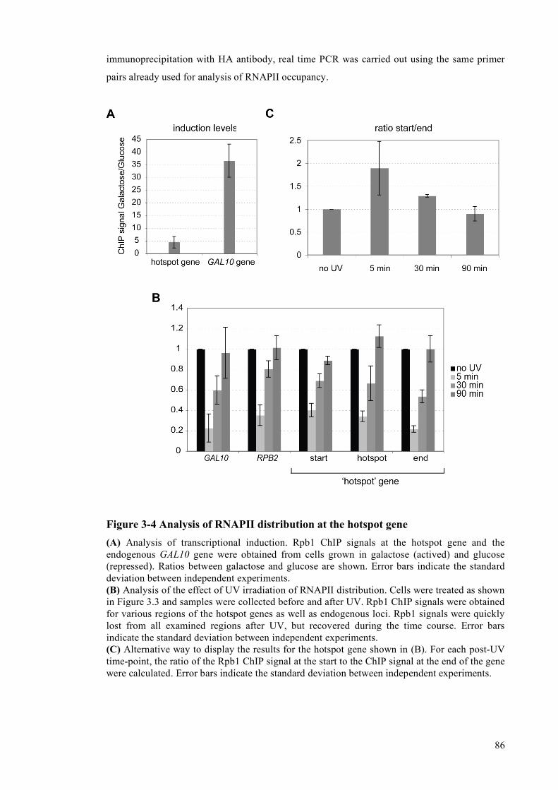
86
immunoprecipitation with HA antibody, real time PCR was carried out using the same primer
pairs already used for analysis of RNAPII occupancy.
Figure 3-4 Analysis of RNAPII distribution at the hotspot gene (A) Analysis of transcriptional induction. Rpb1 ChIP signals at the hotspot gene and the endogenous GAL10 gene were obtained from cells grown in galactose (actived) and glucose (repressed). Ratios between galactose and glucose are shown. Error bars indicate the standard deviation between independent experiments. (B) Analysis of the effect of UV irradiation of RNAPII distribution. Cells were treated as shown in Figure 3.3 and samples were collected before and after UV. Rpb1 ChIP signals were obtained for various regions of the hotspot genes as well as endogenous loci. Rpb1 signals were quickly lost from all examined regions after UV, but recovered during the time course. Error bars indicate the standard deviation between independent experiments. (C) Alternative way to display the results for the hotspot gene shown in (B). For each post-UV time-point, the ratio of the Rpb1 ChIP signal at the start to the ChIP signal at the end of the gene were calculated. Error bars indicate the standard deviation between independent experiments.

87
The results of such ChIP experiments are shown in Figure 3.5. Strikingly, significant
recruitment of Rad14 was restricted to the hotspot region, and was only seen when transcription
of the gene was induced with galactose. A similar pattern was neither seen for the promoter
(start) nor for the terminator sequences (end) of the same gene, and also not for the GAL10 or
RPB2 genes. It is important to point out, however, that the extent of Rad14 recruitment varied
significantly between independent experiments, and that there seemed to be a tight correlation
between the level of transcriptional induction of the gene in galactose (which varied
significantly), and the level of Rad14 recruitment to the hotspot. Figure 3.5 only shows the
results from experiments, in which the level of induction was higher than 4 fold. However, in
more than 50 % of the experiments, the induction was less, and no significant Rad14
recruitment was observed in these cases.
Figure 3-5 Analysis of UV-dependent Rad14 recruitment to DNA Chromatin-immunoprecipitations for HA-tagged Rad14 to regions of the hotspot gene or endogenous genes were carried out before, and 2 hours after, UV treatment in glucose (repressed) or galactose (activated). Signals obtained for unirradiated samples are always set to 1, signals for the irradiated samples are set in relation to the unirradiated sample. Significant binding of Rad14 was only observed at the hotspot. Differences in transcriptional induction of the gene did however lead to significant variations in the obtained recruitment levels.

88
Because of these experimental inconsistencies, I tried very hard to solve the problem of
inconsistency in transcriptional induction before looking more specifically at repair factor
recruitment to the hotspot. Unfortunately, despite changing a number of experimental
parameters (see Discussion), I was unable to get rid of these induction problems and therefore in
the end did not carry on with the repair factor ChIPs, and other experiments that we hoped
would have been possible with this experimental approach (see below).

89
4 Discussion I
4.1 Construction of a UV-damage-prone gene
For this project I have designed a plasmid-based construct containing a galactose-inducible
gene, which has a highly damage-prone sequence (a stretch of pyrimidines; ‘hotspot’) inserted
into surrounding ‘non-damageable’ DNA (free of adjacent pyrimidines). Indeed, mapping of in
vitro inflicted UV lesions using primer extension (shown in Figure 3.2), clearly showed that the
expectations were correct, and that the induced lesions were confined to the hotspot. Because
the hotspot sequence is located on the transcribed strand, RNAPII should be efficiently arrested
at this location after UV, making it possible to follow the recruitment of TC-NER factors (such
as Rad26), general NER factors (such as TFIIH and Rad14), and possibly also Rpb1
ubiquitylation factors (such as Def1, Rsp5, Elc1), to this particular region by ChIP, either under
inducing (galactose) or repressing conditions (glucose).
4.2 RNAPII does not accumulate at the hotspot after UV
One of my expectations was that RNAPII density would be dramatically increased at the
hotspot after UV, when transcription was activated in galactose-containing medium. This was
not the case; instead, RNAPII was rapidly lost after UV treatment from all the examined regions
in the hotspot gene, as well as from the endogenous GAL10 and RPB2 genes. This situation is
reminiscent of the one in human cells, where UV irradiation leads to a transient inhibition of
transcriptional initiation, which is restored after several hours in wild type cells, but not in cells
from CS patients (Mayne and Lehmann, 1982). In my experiments, transcription was restored
after about 90 minutes in the wild type strain. Initial experiments in the rad26Δ strain led to the
same result (data not shown), and so the transcriptional recovery (measured by Rpb1 ChIP) did
not seem to be significantly influenced by Rad26-mediated removal of lesions from the
transcribed strand, or other potential roles of the factor, such as in transcriptional initiation
during DNA damage, as has been shown for CSB (Proietti-De-Santis et al., 2006). Even though
the loss of RNAPII ChIP signals at the end of the hotspot gene after UV treatment was more
pronounced than at the start (thereby increasing the start/end-ratio shown in Figure 3.4C)
indicating that the hotspot sequence indeed represents a damage-induced barrier to RNAPII
progression, these results rely on the comparison of different primer pairs, and cannot really be
regarded as a definite proof of transcriptional impairment caused by the pyrimidine stretch. The

90
great variability in hotspot gene activation between experiments (even though the genomic
GAL10 induction was normal) was also a concern for the interpretation of these experiments. As
the aim of this project was to design a system for the analysis of repair factor binding to UV
damage sites in the presence or absence of active transcription, I instead carried on analysing
the UV-dependent recruitment of an NER-factor to the hotspot.
4.3 Specific recruitment of an NER-factor to the damage-prone
region
In order to investigate if NER proteins are preferentially recruited to the damage-prone
pyrimidine stretch after UV depending on the transcriptional state of the gene, I analysed the
binding of the general NER factor Rad14 to various regions of the hotspot construct, as well as
to the GAL10 and RPB2 loci in the yeast genome. Two hours after UV treatment, at a time when
TC-NER is taking place, but GG-NER is not yet activated efficiently (see Figures 5.1 and 5.2 in
this thesis), Rad14 was only detected at the hotspot, and only after induction of the genes with
galactose, indicating transcription-dependent recruitment of this repair factor. Unfortunately,
significant differences in the levels of transcriptional induction of the hotspot gene led to great
variations in the extent of Rad14 recruitment, and in many cases no recruitment could be
observed. Because the first step in TC-NER is the transcriptional arrest of RNAPII at the site of
the DNA lesion (Christians and Hanawalt, 1992; Sweder and Hanawalt, 1992), differences in
transcription levels will inevitably lead to TC-NER differences, so it is clearly necessary to first
find a solution for the observed variability of induction. Some changes to the construct I have
already made, as well as some ideas for future alterations will be discussed below.
From the Rad14 ChIPs I carried out, it was clear that higher inductions led to more significant
recruitment of the repair protein, making it tempting to speculate that achieving an induction
level similar to the one observed for the GAL10 gene will lead to even higher levels of Rad14
recruitment. Interestingly, no such Rad14 recruitment was ever observed at the constitutively
expressed RPB2 gene or at the galactose-inducible GAL10 gene (Fig. 3.5), even though the
levels of transcriptional induction of the latter locus were about 10-fold higher than that
achieved for the hotspot construct (Figure 3.4A). Irradiation of cells will inevitably lead to the
induction of photo-lesions in these regions, but my results clearly indicate that construction of a
highly damage-prone sequence is required for efficient detection of NER-factors, at least of
Rad14, on DNA after UV-irradiation. To my knowledge, this result is the first evidence of
successful UV-dependent chromatin-immunoprecipitation of a protein involved in NER,
showing that the ideas behind our experimental approach are valid. Finding a solution to the
problems of transcriptional induction will hopefully make this system a valuable tool for the

91
analysis of events that take place at an elongating RNAPII complex irreversibly arrested in vivo
at a UV-induced lesion, such as TC-NER and Rpb1 ubiquitylation.
4.4 Potential solutions for the problem of transcriptional induction
The hotspot gene was cloned between a GAL1-promoter and a CYC1 transcriptional termination
sequence. These elements were already present in the pYC2 vector (Invitrogen), a commercially
available and tested vector, making it highly unlikely that these sequence elements are
responsible for the observed variability in galactose-induced expression. I also used the same
vector for galactose-driven overexpression of other genes, such as RAD26 (see section 5.2.2.4),
and never observed any induction problems.
Several possibilities can be envisaged to explain the lack of proper transcriptional induction of
the synthetic gene containing the hotspot. First, the long stretch of pyrimidines in the
transcribed strand of the gene might represent a barrier to the efficient progression of elongating
RNAPII, even in the absence of UV-induced DNA damage. In order to investigate this, I
reduced the length of the hotspot to 30 nucleotides (one fifth of its original length), or totally
removed it from the construct. This, however, did not alleviate the lack of transcriptional
induction, indicating that the pyrimidine stretch is not the main reason for the induction
difficulties. A second possibility is that the sequences flanking the hotspot, i.e. the 500 bp
stretches of ‘damage-free’ DNA, are the main cause for the observed problems. As mentioned
earlier, these regions are highly repetitive, because of the limited number of codon combinations
available for construction of a continuous open-reading-frame, without the formation of
pyrimidine pairs in either strand. Because of the significant homology between these two
regions, I cannot rule out recombination (perhaps transcription-associated) between these
sequences, the formation of secondary DNA structures, or other events caused by repetitive
DNA. In order to look more closely at this, I redesigned the gene and included potential
pyrimidine dimers in the NTS. The TS of this new construct was, however, still kept dimer-free.
This resulted in significant reduction of the repetitive nature of the 500 bp sequences.
Unfortunately, it did not significantly improve transcriptional induction (data not shown). Taken
together, my attempts to alleviate the transcription problems with the synthetic gene were not
successful.
One further possibility to solve the problem in the future is to use naturally occuring sequences
flanking the hotspot, in order to prevent the use of any synthetic DNA. It would be easy, for
example, to insert the pyrimidine stretch into the endogenous GAL10 locus, which was
consistently induced at high levels in my experiments. My results showing that the presence and

92
the length of the hotspot sequence was apparently not the reason for the lack of induction makes
it likely that the GAL10 gene containing the hotspot will still be transcribed at a high rate. This
construct will, of course, not rule out that damage occurs elsewhere in the gene, i.e. on both
strands both in front of and behind the hotspot, but my preliminary Rad14 ChIP results (Fig.
3.5) indicate that detection of repair factor binding will be restricted to the pyrimidine stretch,
presumably because the likelihood of damage there is so much higher.
4.5 Use of the hotspot sequence for analysis of GG-NER
Even though a solution for the induction problems will have to be found in order to establish a
system which allows us to distinguish between transcripion-related and unrelated events at UV-
damaged DNA in vivo, the analysis of RNAPII-independent events (i.e. GG-NER) should
already be possible with the hotspot-construct in its present form. This will require the analysis
of repair factor recruitment to the pyrimidine stretch during a longer time-course.
However, this was not the main purpose for making and characterizing the construct, so I did
not pursue this line of research, but instead focused on alternative, more successful projects on
regulation of NER and Rpb1 ubiquitylation by the DNA damage checkpoint (see next chapter).

5 Results II – Regulation of NER by the DNA damage
checkpoint
5.1 Analysis of NER efficiency in checkpoint-deficient strains
The DNA damage checkpoint (DDC) orchestrates a plethora of cellular events in the presence
of DNA lesions, including cell cycle arrest, stabilisation of stalled replication forks, induction of
apoptosis and activation of DNA repair. Even though it is widely accepted that DNA repair is
influenced by the checkpoint, to which extent the efficiency of specific DNA repair pathways is
affected in response to mutations in checkpoint factors is largely unknown. In this study I
investigated the role of the DDC in Nucleotide Excision Repair (NER) in the budding yeast, S.
cerevisiae. To achieve this goal, I utilized a powerful method which allows analysis of strand-
specific repair of UV-induced CPDs at nucleotide resolution (Teng et al., 2005).
5.1.1 Strains lacking the Mec1 kinase have NER defects
Strains lacking the central checkpoint kinase Mec1 are deficient in the activation of the DDC
(Longhese et al., 1998). I constructed a strain lacking the MEC1 and SML1 genes (SML1
deletion being necessary to suppress the lethality of deletion of MEC1 (Zhao et al., 1998)), and
analysed the repair of the transcribed strand (TS; transcription-coupled repair) and the non-
transcribed strand (NTS: global genome repair) of the constitutively transcribed RPB2 gene.
Wild-type (W303) and TC-NER-deficient rad26Δ mutant strains (van Gool et al., 1994) served
as controls.
A representative gel is shown in Figure 5.1. The signals for the individual lanes were quantified
using a phosphor-imager, and the time required to remove 50 percent of the damaged
nucleotides (t50%) in the TS and the NTS were calculated for individual strains (Fig. 5.2). As
expected, the difference in repair kinetics between the preferentially repaired transcribed strand
(TS) and the non-transcribed strand (NTS) was evident in the WT strain, while no clear strand
specificity was observed in TC-NER-deficient rad26Δ cells. Interestingly, the cells lacking the
checkpoint kinase Mec1 exhibited a dramatic impairment of NER, characterised by markedly
slower repair of the TS, and virtually no detectable repair of the NTS at 4 hours post-UV
irradiation. This indicates that a functional DNA damage checkpoint is required for both NER
subpathways in yeast. Importantly, however, while repair of the NTS appears to be completely
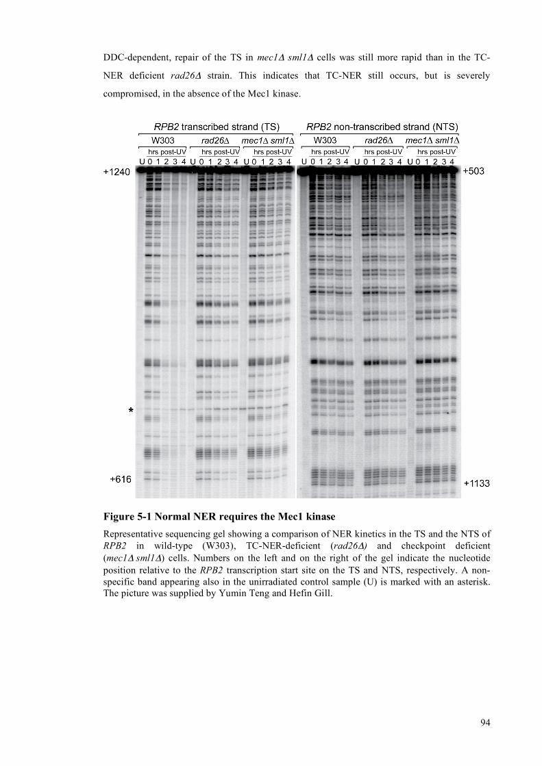
94
DDC-dependent, repair of the TS in mec1Δ sml1Δ cells was still more rapid than in the TC-
NER deficient rad26Δ strain. This indicates that TC-NER still occurs, but is severely
compromised, in the absence of the Mec1 kinase.
Figure 5-1 Normal NER requires the Mec1 kinase Representative sequencing gel showing a comparison of NER kinetics in the TS and the NTS of RPB2 in wild-type (W303), TC-NER-deficient (rad26Δ) and checkpoint deficient (mec1Δ sml1Δ) cells. Numbers on the left and on the right of the gel indicate the nucleotide position relative to the RPB2 transcription start site on the TS and NTS, respectively. A non-specific band appearing also in the unirradiated control sample (U) is marked with an asterisk. The picture was supplied by Yumin Teng and Hefin Gill.

95
Figure 5-2 Quantification of the signals shown in Figure 5.1 Damages remaining at the post-UV time points were calculated and the time necessary for removal of 50 % of the damages (t50%) was determined for both the TS and the NTS in the different strains. Error bars show the standard error between independent experiments.
5.1.2 Strains lacking the Chk1, Rad53 and Dun1 kinases have normal NER
Mec1, in certain contexts, is the most upstream kinase in the DNA damage checkpoint pathway
and activates several other kinases, the most important ones being Chk1, Rad53 and Dun1
(Sanchez et al., 1999; Zhou and Elledge, 1993). To determine whether Mec1 acts directly in
NER or via one of these downstream effectors, NER rates were analysed in strains deleted for
CHK1, RAD53 and DUN1. As is the case for mec1Δ cells, prior deletion of SML1 was required
to suppress the lethality of RAD53 deletion.
Interestingly, no significant alteration in the repair rates for the TS or the NTS were observed in
the chk1Δ or rad53Δ sml1Δ strains, both of which displayed damage half lives comparable to
the wild type strain (Figures 5.3). Similarly, deletion of DUN1 had no observable effect on
repair efficiency (Figures 5.4). These results suggest that the mechanism by which Mec1
regulates NER occurs independently of these factors, even though I can not rule out at this point
that double or triple mutants could have an effect on NER.
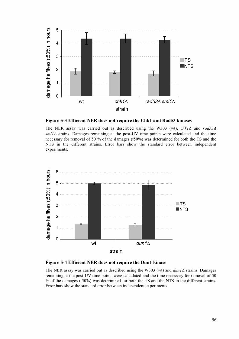
96
Figure 5-3 Efficient NER does not require the Chk1 and Rad53 kinases The NER assay was carried out as described using the W303 (wt), chk1Δ and rad53Δ sml1Δ strains. Damages remaining at the post-UV time points were calculated and the time necessary for removal of 50 % of the damages (t50%) was determined for both the TS and the NTS in the different strains. Error bars show the standard error between independent experiments.
Figure 5-4 Efficient NER does not require the Dun1 kinase The NER assay was carried out as described using the W303 (wt) and dun1Δ strains. Damages remaining at the post-UV time points were calculated and the time necessary for removal of 50 % of the damages (t50%) was determined for both the TS and the NTS in the different strains. Error bars show the standard error between independent experiments.
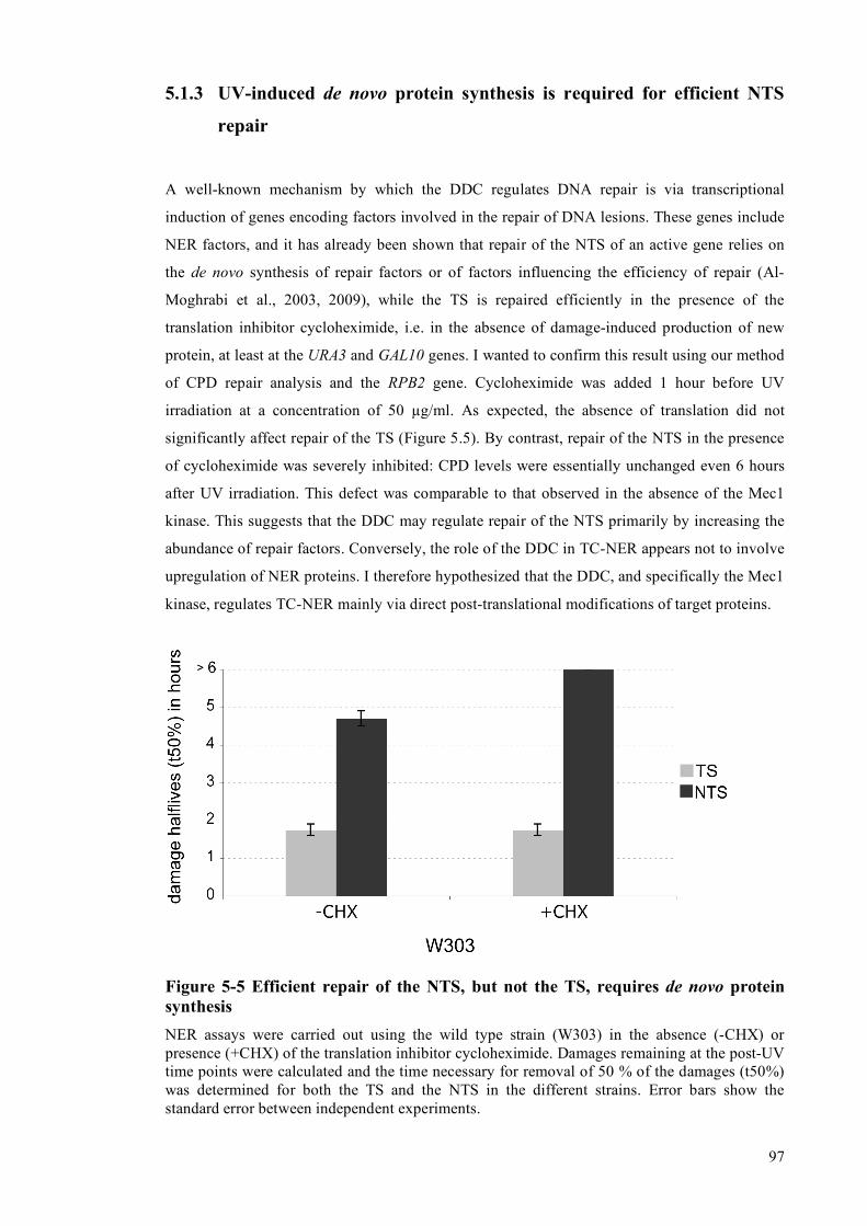
97
5.1.3 UV-induced de novo protein synthesis is required for efficient NTS
repair
A well-known mechanism by which the DDC regulates DNA repair is via transcriptional
induction of genes encoding factors involved in the repair of DNA lesions. These genes include
NER factors, and it has already been shown that repair of the NTS of an active gene relies on
the de novo synthesis of repair factors or of factors influencing the efficiency of repair (Al-
Moghrabi et al., 2003, 2009), while the TS is repaired efficiently in the presence of the
translation inhibitor cycloheximide, i.e. in the absence of damage-induced production of new
protein, at least at the URA3 and GAL10 genes. I wanted to confirm this result using our method
of CPD repair analysis and the RPB2 gene. Cycloheximide was added 1 hour before UV
irradiation at a concentration of 50 µg/ml. As expected, the absence of translation did not
significantly affect repair of the TS (Figure 5.5). By contrast, repair of the NTS in the presence
of cycloheximide was severely inhibited: CPD levels were essentially unchanged even 6 hours
after UV irradiation. This defect was comparable to that observed in the absence of the Mec1
kinase. This suggests that the DDC may regulate repair of the NTS primarily by increasing the
abundance of repair factors. Conversely, the role of the DDC in TC-NER appears not to involve
upregulation of NER proteins. I therefore hypothesized that the DDC, and specifically the Mec1
kinase, regulates TC-NER mainly via direct post-translational modifications of target proteins.
Figure 5-5 Efficient repair of the NTS, but not the TS, requires de novo protein synthesis NER assays were carried out using the wild type strain (W303) in the absence (-CHX) or presence (+CHX) of the translation inhibitor cycloheximide. Damages remaining at the post-UV time points were calculated and the time necessary for removal of 50 % of the damages (t50%) was determined for both the TS and the NTS in the different strains. Error bars show the standard error between independent experiments.

98
5.2 Targets of the Mec1 kinase
5.2.1 Rad23 phosphorylation mutants do not display defects in NER
Having shown that Mec1 is likely to regulate TC-NER by one or more post-translational
modification events, I set out to identify new Mec1 target proteins. Interestingly, the Rad23
protein has been identified as a target for Mec1 in proteome-wide screens in yeast (Albuquerque
et al., 2008) and human cells (Matsuoka et al., 2007). Yeast Rad23, and its binding partner Rad4
are involved in both NER subpathways (Verhage et al., 1994; Verhage et al., 1996b). I
speculated that phosphorylation of Rad23 by Mec1 might be required for its activation, a
hypothesis that could explain the observed NER defects in mec1Δ sml1Δ cells. To investigate
this possibility, I examined the NER efficiency of a rad23Δ deletion strain carrying vectors
expressing wild type RAD23 (RAD23wt), a mutant RAD23 in which Serine 121, the residue
targeted by Mec1 (Albuquerque et al., 2008), was mutated to alanine (RAD23SA), or carrying the
empty vector. As expected, repair of both the TS and the NTS was severely compromised in the
rad23Δ deletion strain, with no detectable repair during the time course of 4 hours. Expression
of wild type RAD23 completely reversed this defect (Figures 5.6). Interestingly, expression of
the RAD23SA mutant also rescued the NER deficiency. This indicates that the NER defects of
the mec1Δ sml1Δ mutant are not caused by a lack of Mec1-catalyzed Rad23 phosphorylation, at
least at this residue.
Figure 5-6 Checkpoint-dependent phosphorylation of Rad23 at S121 is not required for efficient NER The NER assay was carried out using rad23Δ strains expressing either wild type Rad23 (wt), no Rad23 (rad23Δ), or a S121A mutant version of Rad23 (RAD23SA). Damages remaining at the post-UV time points were calculated and the time necessary for removal of 50 % of the damages (t50%) was determined for both the TS and the NTS in the different strains. Error bars show the standard error between independent experiments.

99
To determine if checkpoint-dependent Rad23 Serine 121 phosphorylation plays a role in other
functions of the protein that are involved in survival after UV irradiation, I tested the UV
sensitivity of the rad23Δ strain, and strains expressing RAD23wt, the S121A mutant of Rad23
(RAD23SA), or a phosphorylation-mimic mutant, S121D (RAD23SD). Futhermore, because I
couldn’t exclude the possibility that one or more potential Mec1-dependent phosphorylation
sites were missed in the proteome-wide screen (Albuquerque et al., 2008), I also analysed
Rad23 versions in which all the SQ-sites in the protein (S40, S73 and S121) were mutated to
alanine (RAD233SA) or aspartic acid (RAD233SD). Figure 5.7 shows that, as expected, the rad23Δ
deletion strain displayed strong sensitivity to treatment with UV light, in agreement with the
observed defect in NER. Rescue of this strain with the wild type RAD23 gene reduced this
sensitivity to the level of the parental W303 strain. Surprisingly, all the mutant Rad23 versions
behaved like the wild type protein in this assay, indicating that Rad23 functions required for
survival after UV are not affected by inhibiting or mimicking phosphorylation at potential Mec1
(SQ ) sites (Fig. 5.7).
Figure 5-7 Mutation of potential checkpoint-phosphorylation sites in Rad23 does not affect survival after UV The sensitivity of W303 was compared to rad23Δ mutants carrying either empty vector (vector) or vector expressing wildtype (RAD23wt), serine 121 single point mutant versions of RAD23 (RAD23SA and RAD23SD, respectively) or serine 40/73/121 triple mutant versions of RAD23 (RAD233A and RAD233D, respectively). None of the point mutants displayed detectable differences in sensitivity to UV treatment compared to the wild type strain.

100
5.2.2 Analysis of checkpoint-dependent phosphorylation of Rad26
5.2.2.1 Construction of an N-terminally tagged Rad26 strain
One possible explanation for the TC-NER defects exhibited by mec1Δ cells is that the TC-NER-
specific Rad26 protein is activated directly by Mec1-mediated phosphorylation. To identify
potential post-translational modifications occurring on Rad26, I created a strain in which the
genomic RAD26 locus was tagged on its N-terminus with 9 Myc sequences, followed by 2 TEV
cleavage sites and 8 histidines (MHRAD26). A schematic outline of the N-terminal tagging
strategy is shown in Figure 5.8A. The tagged gene was still under control of its endogenous
regulatory elements, avoiding artefacts due to a change in expression level. As expected, Rad26
tagging led to the appearance of an anti-Myc-reactive species with a molecular weight of around
140 kDa, which was absent in the parental W303 strain (Fig. 5.8B).
Since addition of a large tag could potentially render Rad26 non-functional, I also determined
whether this protein can still initiate TC-NER. To investigate this, MHRAD26 cells were
crossed with a rad16Δ mutant and the resulting strains’ UV sensitivity analysed. In agreement
with previous studies (van Gool et al., 1994; Verhage et al., 1996a), the TC-NER-deficient
rad26Δ strain strain did not show increased UV-sensitivity when compared to the wild type,
whereas the GG-NER-deficient rad16Δ strain did (Fig. 5.8C). Additional deletion of RAD26 in
the rad16Δ background dramatically increased UV sensitivity. Importantly, the tagged
MHRAD26 gene did not cause increased UV sensitivity in the rad16∆ background, indicating
that the tagged protein was functional.
5.2.2.2 Rad26 is phosphorylated in response to DNA damage
To investigate if Rad26 is phosphorylated after UV irradiation, I treated the MHRAD26 strain
with UV and analysed the electrophoretic mobility of the protein by SDS-PAGE and Western
blotting. As shown in Figure 5.9A, a slower migrating form of the protein became visible after
UV treatment, indicative of a post-translational modification. The modification became
apparent just 10 minutes after UV treatment, peaked between 1 and 2 hours, and then started
disappearing.
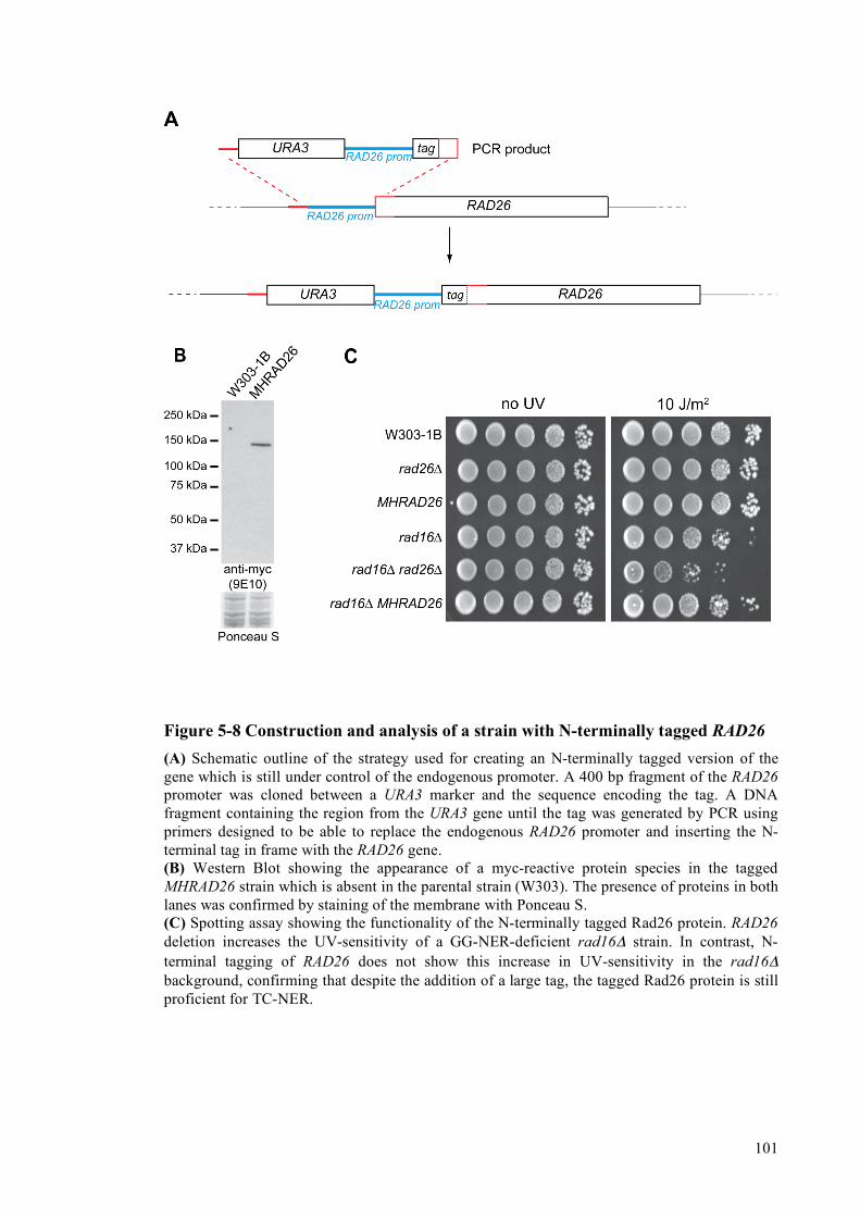
101
Figure 5-8 Construction and analysis of a strain with N-terminally tagged RAD26 (A) Schematic outline of the strategy used for creating an N-terminally tagged version of the gene which is still under control of the endogenous promoter. A 400 bp fragment of the RAD26 promoter was cloned between a URA3 marker and the sequence encoding the tag. A DNA fragment containing the region from the URA3 gene until the tag was generated by PCR using primers designed to be able to replace the endogenous RAD26 promoter and inserting the N-terminal tag in frame with the RAD26 gene. (B) Western Blot showing the appearance of a myc-reactive protein species in the tagged MHRAD26 strain which is absent in the parental strain (W303). The presence of proteins in both lanes was confirmed by staining of the membrane with Ponceau S. (C) Spotting assay showing the functionality of the N-terminally tagged Rad26 protein. RAD26 deletion increases the UV-sensitivity of a GG-NER-deficient rad16Δ strain. In contrast, N-terminal tagging of RAD26 does not show this increase in UV-sensitivity in the rad16Δ background, confirming that despite the addition of a large tag, the tagged Rad26 protein is still proficient for TC-NER.

102
Figure 5-9 UV-induced phosphorylation of Rad26 (A) Western blot analysis of Rad26 from wildtype cells harvested either before (0) or at various time points after UV irradiation. The decrease in electrophoretic mobility after UV treatment indicates a post-translational modification. MHRad26 was detected with 9E10 antibodies. (B) Western blot analysis of Rad26 from unsynchronized wild-type cells and cells which have been synchronised in G1 (alpha factor), S (hydroxyurea) or G2/M (nocodazole). MHRad26 was detected with 9E10 antibodies. (C) Experimental outline of the procedure leading to the result shown in (D) (D) Western blot analysis of Rad26 immunoprecipitated before and 2 hours after UV treatment and treated either with buffer alone or Shrimp alkaline phosphatase. MHRad26 was detected with 9E10 antibodies.
Longer exposures of the Western blots revealed that even in the absence of UV-damage a small
amount of Rad26 is modified. Interestingly, this damage-independent modification appeared to
be linked to the cell cycle, as it was absent in cells arrested in G1 and G2 (achieved by treatment
with alpha-factor and nocodazole, respectively) and enriched in cells arrested in S-phase (by
treatment with hydroxyurea) (Fig. 5.9B). Furthermore, I noted that no degradation of Rad26

103
was observed after UV irradiation, unlike observed previously for its human homologue CSB
(Groisman et al., 2006). Because of the pronouced decrease in electrophoretic mobility of the
protein, I first suspected Rad26 to be mono-ubiquitylated after DNA damage. However,
analysis of the immunoprecipitated and modified protein with ubiquitin-specific antibodies did
not give any signal, excluding an involvement of UV-specific Rad26 ubiquitylation (not
shown). In order to determine if phosphorylation of Rad26 is responsible for the observed shift
in electrophoretic mobility, I immunoprecipitated Rad26 before and after UV irradiation and
treated the bead-bound material with Shrimp Alkaline Phosphatase (SAP) (experimental
procedure outlined in Fig. 5.9C). The slower-migrating protein was again only clearly visible
after UV-irradiation, and it was lost after incubation with phosphatase, indicating that it
represents a phosphorylated form of Rad26 (Fig. 5.9D).
To determine whether Rad26 phosphorylation is a specific response to UV irradiation or if it
occurs in response to other types of DNA damage, I treated cells with various chemical agents
to induce different types of DNA lesions. All of these treatments caused Rad26 to be
phosphorylated (Fig. 5.10A). Interestingly, other forms of cellular stress, including heat-shock
and osmotic shock, did not result in modification of Rad26 (Fig. 5.10B).
Finally, because Rad26 is thought to be recruited to stalled RNA polymerase II complexes, and
because UV-induced DNA lesions cause RNAPII stalling, I tested whether 6-azauracil (6AU,
which causes frequent RNAPII stalling by restricting nucleotide availability) could also
promote Rad26 phosphorylation. Treatment with this drug did not cause a detectable increase in
the phosphorylated form of Rad26 (Fig. 5.10B). Thus, Rad26 phosphorylation appears to occur
specifically in response to DNA damage.
5.2.2.3 Rad26 phosphorylation is dependent on the Mec1 checkpoint kinase
To determine whether the DDC is required for Rad26 phosphorylation, I tested whether
this modification occurred in mec1Δ sml1Δ cells. Loss of the Mec1 checkpoint kinase
completely abolished phosphorylation of Rad26 (Fig. 5.10C). To investigate whether this effect
was specifically due to absence of the Mec1 kinase, or an indirect result of deletion of both
SML1 and MEC1, I reintroduced galactose-inducible forms of HA-tagged wildtype MEC1
(MEC1wt) or kinase-dead MEC1 (MEC1kd) into the MHRAD26 mec1Δ sml1Δ strain. Rad26
phosphorylation was recovered by growing cells expressing MEC1wt in galactose, but not
glucose, while the MEC1kd-containing strain had no Rad26 phosphorylation in either carbon
source, even though it expressed Mec1kd at a level similar to that of the wild-type protein in

104
inducing conditions (Fig. 5.10D). These data indicate that the kinase activity of Mec1 is
required for Rad26 phosphorylation.
Figure 5-10 Involvement of the DNA damage checkpoint in Rad26 phosphorylation (A) Western blot analysis of Rad26 after various kinds of DNA damage. MHRad26 was detected with 9E10 antibodies. (B) Western blot analysis of Rad26 after infliction of cellular stress different from DNA damage. MHRad26 was detected with 9E10 antibodies. (C) Western blot analysis of Rad26 phosphorylation in the absence of the Mec1 checkpoint kinase. MHRad26 was detected with 9E10 antibodies. (D) Western blot analysis of Rad26 in mec1sml1 cells expressing either galactose-inducible HA-tagged wildtype (wt) or kinase-dead (kd) Mec1. MHRad26 was detected with 9E10 antibodies. (E) Western blot analysis of Rad26 in strains lacking various kinases of the DNA damage checkpoint cascade. MHRad26 was detected with 9E10 antibodies.
The absence of Mec1 leads to inactivation of downstream kinases, the most important of which
are Chk1 and Rad53 (Longhese et al., 1998). To determine if phosphorylation of Rad26p was
mediated by Chk1 or Rad53 (either directly or indirectly), I deleted their encoding genes and
examined the phosphorylation state of Rad26 after UV-irradiation. Damage-induced
105
phosphorylation was evident in both of these strains, supporting the idea that Mec1 directly
phosphorylates Rad26 without the involvement of downstream kinases (Fig. 5.10E). I also
tested cells lacking Dun1, another kinase activated by Rad53 (Zhou and Elledge, 1993), and
Tel1, the yeast homologue of ATM (Greenwell et al., 1995; Morrow et al., 1995). Dun1 acts
downstream of Rad53, which itself had no role in Rad26 phosphorylation. Moreover, the
complete absence of Rad26 phosphorylation in the mec1Δ sml1Δ strain suggests that there is no
redundancy between Mec1 and Tel1 in this event as there are in certain others. Neither removal
of Dun1 or Tel1 would therefore be expected to affect phosphorylation of Rad26. Indeed, both
of these mutant strains exhibited normal Rad26 phosphorylation (Fig. 5.10E).
I tried to show biochemically using in vitro kinase assays that Rad26 is a direct target for the
Mec1 kinase, but several attempts to get the in vitro kinase working with a positive control
(PHAS-I), were not successful (data not shown), and direct biochemical evidence for such an
event is still missing.
5.2.2.4 Identification of the Rad26 phosphorylation site
Mec1 phosphorylates serines and threonines that are immediately adjacent to a glutamine
residue (SQ/TQ motifs). Rad26 possesses 1 TQ and 4 SQ consensus motifs (Fig. 5.11A). To
identify the site of Rad26 phosphorylation, the MHRAD26 locus was cloned into pRS316, and
each of the residues predicted to be a Mec1 target site were individually mutated to alanine. The
mutated plasmids were then re-introduced into rad26Δ cells, and the phosphorylation status of
Rad26 was analyzed after induction of damage with 4NQO (4-nitroquinoline-1-oxide; a UV
mimetic chemical compound which creates damages repaired by NER). As shown in Figure
5.11B, mutation of S27 in Rad26 led to the complete absence of a shifted Rad26 band, whereas
all the other single point mutants behaved like the wild type protein.
This result suggested that S27 is the sole site of Mec1-catalyzed Rad26 phosphorylation.
However, I could not rule out the possibility that another modification escaped detection by
SDS-PAGE and Western blotting, for example because it did not lead to a noticeable change in
Rad26 migration. Therefore, mass spectrometry (MS) was used as an alternative strategy to
analyze post-translational modificiations of Rad26. Due to the low abundance of Rad26, I was
unfortunately unable to purify enough modified protein from MHRAD26 cells for MS analysis.
To circumvent this problem, MHRAD26 was placed under the control of a galactose-inducible
promoter on a centromeric plasmid (pYC2 vector from Invitrogen; creating pYC2-MHRAD26)
and this construct was then introduced into a rad26Δ deletion mutant. I first investigated
whether the tagged Rad26 protein could be efficiently over-expressed from this vector. After

106
shifting cells from raffinose to galactose, the tagged form of Rad26 was rapidly and
dramatically induced (Figure 5.11C).
Figure 5-11 Identification of the Rad26 phosphorylation site (A) Schematic representation of the Rad26 protein. Grey boxes indicate important protein domains. The NLS is shown in light grey, the seven conserved helicase domains are shown in dark grey. The 5 potential Mec1 target sites (SQ/TQ) are indicated above. MHRad26 was detected with 9E10 antibodies. (B) Analysis of Rad26 SQ/TQ point mutants. The five SQ/TQ sites were mutated individually and the mutant genes were then expressed in a rad26 mutant strain. The phosphorylation status of the proteins was determined after 4-NQO treatment. (C) Galactose-induced overexpression of Rad26. The MHRAD26 open reading frame was cloned behind a galactose-inducible promoter on the pYC2 plasmid (Invitrogen). Expression of the protein can be efficiently induced with 2 % galactose and detected by Western blotting with both anti-myc and anti-his antibodies. Similar loading was confirmed by staining of the membrane with Ponceau S. (D) Purification of overexpressed Rad26 protein before and after damage with 4-NQO. Rad26 was immunoprecipitated from extracts and eluted from the beads with TEV protease. The eluate was analysed by SDS-PAGE on a 3-8 % gradient gel followed by SYPRO RUBY staining. More than one slower migrating form of Rad26 is visible.
107
(E) Purification of overexpressed wild-type and S27A Rad26 after damage with 4-NQO. No slower migrating form of Rad26 can be detected after DNA damage when the single point mutant version is expressed.
This over-expressed Rad26 was purified before and after 4-NQO treatment using Myc-affinity
resin and eluted with TEV protease. Despite expressing Rad26 at non-physiological levels, the
characteristic mobility shift was still visible after DNA damage. Interestingly, this analysis
revealed two slower-migrating forms of the Rad26 protein (Fig. 5.11D), suggesting that more
than one phosphate group was added to Rad26 after DNA damage. MS analysis verified this
idea: both S27 and S30 were found to be phosphorylated in the sample. These sites of
phosphorylation were identified following peptide fragmentation using both collision induced
dissociation (CID) and electron transfer dissociation (ETD).
Interestingly, while this work was in progress, a proteome-wide study of checkpoint-dependent
phosphorylation identified Mec1-dependent modification of S27 and S29 of Rad26
(Albuquerque et al., 2008). Surprisingly, neither S29 nor S30 exist in the context of a proper
Mec1 consensus motif. Thus, I speculated that S27 is the primary Rad26 phosphorylation site,
but that residues nearby might be targeted in a non-specific manner. In order to investigate this
idea, I mutated S27 in the pYC2-MHRAD26 plasmid (creating plasmid pYC2-MHRAD26S27A)
and purified both the wildtype and mutant protein after 4NQO treatment. Indeed, whereas the
wildtype protein showed 2 distinct slower-migrating bands, no shift was observed with the
S27A mutant (Figure 5.11E). The complete absence of Rad26 phosphorylation in this mutant
was also confirmed by mass-spectrometry. These results demonstrate that mainly S27 (and
sometimes also S29 and/or S30) of Rad26 are phosphorylated after DNA damage by the Mec1
kinase.
5.2.2.5 Analysis of an involvement of Rad26 phosphorylation in TC-NER
5.2.2.5.1 Rad26 phosphorylation does not lead to altered survival after UV irradiation
Given that I found that MEC1 deletion affects TC-NER (see Figures 5.1 and 5.2) and that
Rad26 is a direct target of Mec1 kinase, an obvious possibility was that this phosphorylation
event plays a role in TC-NER. I first used genetics to investigate this possibility, taking
advantage of the fact that the rad16Δ rad26Δ double mutant is much more UV-sensitive than
either single mutant (Verhage et al., 1996a).

108
Figure 5-12 Analysis of survival after UV irradiation in various strain backgrounds expressing wild-type and mutant versions of Rad26 (A) Spotting assay showing no significant alteration in UV sensitivity when wild-type and point-mutant versions of Rad26 are expressed in the rad16Δ background (top panel), rpb9Δ background (middle panel) or rad16Δ rpb9Δ background (lower panel). (B) Graphs showing survival rates after increasing doses of UV. No significant alteration in survival was detected in the rpb9Δ backgound (left), rad16Δ background (middle) or rad16Δ rpb9Δ background (right).
109
Point mutants in which S27 was mutated either to alanine (to prevent phosphorylation
(RAD26SA)) or to glutamic acid (to mimic phosphorylation (RAD26SE)) were introduced into
rad16Δ rad26Δ cells, and the UV sensitivity of these strains was tested by spotting assays. Like
wild type Rad26, both mutated forms of Rad26 rescued the rad16Δ rad26Δ double mutant such
that its UV-sensitivity was similar to that of the rad16Δ single mutant (Fig. 5.12A). In order to
exclude the possibility that the effect of the point mutations in Rad26 is masked by the second
TC-NER pathway in yeast, which is mediated by the Rpb9 protein (Li and Smerdon, 2002), I
also carried out the same assay using a rad26Δ rpb9Δ double mutant and a rad16Δ rad26Δ
rpb9Δ triple mutant. Again, the phosphorylation mutants acted like the wild type protein in
these alternative strain backgrounds (Fig. 5.12A). As it is easy to miss slight differences in UV-
sensitivity using this relatively crude assay, I also determined survival rates by plating defined
numbers of cells and determining exact survival rates following UV treatment. This more
sensitive assay confirmed that the phosphorylation mutants did not affect UV survival under
these conditions (Figure 5.12B).
5.2.2.5.2 Rad26 phosphorylation increases the efficiency of TC-NER
The damage survival assays show that phosphorylation of Rad26 is not absolutely required
for the function of the protein, but do not rule out the possibility that the modification affects the
rate of TC-NER. To investigate this possibility, I analyzed these mutants using the strand-
specific NER assay. Interestingly, the alanine mutant (which cannot be phosphorylated)
exhibited a significant delay in TC-NER, while TC-NER in the glutamate mutant (which
mimics persistent phosphorylation) appeared to be unaffected (see sequencing gel in Fig 5.13
and graphs in Fig. 5.14). Thus, whereas the half-life of damages in the transcribed strand was
1.7 hours in the wild type strain, it was 2.6 hours in the phosphorylation-site (SA) mutant (Fig.
5.14). As expected, repair of the NTS was not affected by either of the mutations (graph in Fig.
5.14, sequencing gel not shown). I conclude that damage-induced phosphorylation of Rad26 by
Mec1 kinase increases the rate of TC-NER.

110
Figure 5-13 Rad26 phosphorylation is required for efficient TC-NER Representative sequencing gel showing a comparison of NER kinetics in Rad26 mutants. Strand specific NER was analysed in the TS of RPB2 in rad26Δ cells expressing either no RAD26 (rad26Δ), wild-type RAD26 (RAD26WT), or mutant versions in which serine 27 is replaced by either alanine (RAD26SA) or glutamate (RAD26SE). Numbers on the left of the gel indicate the nucleotide position relative to the RPB2 transcription start site on the TS. A non-specific band appearing also in the unirradiated control sample (U) is marked with an asterisk.

111
Figure 5-14 Quantification of the signals shown in Figure 5.14 Damages remaining at the post-UV time points were calculated, and the time necessary for removal of 50 % of the damages (t50%) was determined for both the TS and the NTS in the different strains. Error bars show the standard error between independent experiments.
5.2.2.6 Expression of a phosphomimic Rad26 mutant is not sufficient to
overcome the TCR defects in a strain lacking the Mec1 kinase
Based on the findings that both the mec1Δ sml1Δ and the RAD26S27A mutant, but not the
RAD26SE mutant, have defects in TC-NER, I speculated that the requirement for the Mec1
kinase in this process might be bypassed by expressing the RAD26SE mutant in the mec1Δ sml1Δ
strain. To investigate this possibility, I deleted the RAD26 gene in the mec1Δ sml1Δ strain and
transformed the resulting strain with either empty vector or plasmids expressing RAD26wt,
RAD26SA or RAD26SE, and then analysed NER kinetics in these cells.
As shown in Figures 5.15, TC-NER was completely absent when RAD26 was deleted in the
mec1Δ sml1Δ background, showing that, as expected, the remaining TC-NER in this
background is entirely dependent on the presence of Rad26. Putting back the wild type version
of RAD26 completely restored TC-NER to the level previously observed for the mec1Δ sml1Δ
mutant. The RAD26SA mutant behaved exactly the same, confirming that mutating the phospho-
acceptor residue to an alanine does not change the activity of the protein in a strain already
deleted for kinase responsible for the phosphorylation event. Surprisingly, I did not observe
increased TC-NER rates in the strain expressing the phosphomimic RAD26SE mutant. This result
indicates that the Mec1 kinase has additional phosphorylation targets which are involved in TC-

112
NER, and shows that the TC-NER defect in mec1Δ sml1Δ cells is not due to a lack of Rad26
phosphorylation by Mec1.
Figure 5-15 Analysis of NER mec1Δ sml1Δ cells expressing Rad26 mutants
NER kinetics were determined for the TS and NTS of RPB2 in mec1Δ sml1Δ rad26Δ cells expressing either no RAD26 (+ vector), wild-type RAD26 (+ RAD26wt), or mutant versions in which serine 27 is replaced by either alanine (+ RAD26SA) or glutamate (+ RAD26SE). Damages remaining at the post-UV time points were calculated and the time necessary for removal of 50 % of the damages (t50%) was determined for both the TS and the NTS in the different strains. Error bars show the standard error between independent experiments.
As expected, repair of the NTS was not influenced by RAD26 deletion or by expressing the
mutant versions of RAD26 in the mec1Δ sml1Δ background, with little or no repair detected
even at the latest time point in any of the strains (Fig. 5.15).
The complete absence of detectable NER on the analyzed RPB2 fragment in the mec1Δ sml1Δ
rad26Δ triple mutant might suggest that this strain should be considerably more UV sensitive
than the mec1Δ sml1Δ double mutant, and I set out to investigate if this is really the case.
Surprisingly, the two strains did not display significant differences in UV sensitivity when
analysed by the spotting assay (Fig. 5.16), showing that the remaining repair of the TS in strains
lacking only the Mec1 kinase does not result in increased survival after UV irradiation. This
suggests that a considerable decrease in TC-NER does not necessarily correlate with a similar
decrease in UV-sensitivity under normal conditions – even when GG-NER is also compromised
- , and helps explain why the decrease in TC-NER observed in RAD26SA does not lead to
decreased viability after UV-irradiation.

113
Figure 5-16 Analysis of the effect of RAD26 deletion on the UV sensitivity of the checkpoint-deficient mec1Δ sml1Δ strain Spotting assay comparing the UV sensitivity of wild-type (W303), mec1Δ sml1Δ and mec1Δ sml1Δ rad26Δ strains. Deletion of RAD26 in the mec1Δ sml1Δ background does not lead to a significant increase in UV-sensitivity. If anything, there is a slight decrease, at least at the higher UV-dose.
5.2.2.7 Rad26 phosphorylation is not involved in ubiquitylation and
degradation of RNA Polymerase II
Since Rad26 interacts with Def1 (Woudstra et al., 2002), the factor controlling UV-induced
ubiquitylation and degradation of the RNAPII subunit Rpb1, I speculated that this process might
be influenced by phosphorylation of Rad26. In theory, a decrease in TC-NER efficiency might
be explained by faster degradation of Rpb1, because TC-NER and Rpb1-ubiquitylation are
thought to act on the same substrate and might compete for it.
To determine if phosphorylation of Rad26 influences ubiquitylation and degradation of Rpb1,
rad26Δ cells carrying either empty vector, RAD26wt, RAD26SA, or RAD26SE were treated with
cycloheximide to abolish synthesis of new proteins, and irradiated with UV. Cells were then
harvested at different time points (Figure 5.17A) and Rpb1 degradation was assessed by
Western blotting using an 8WG16 antibody. To measure Rpb1 ubiquitylation, I utilised a
method developed in our laboratory (Anindya et al., 2007), which relies on isolation of mono-
and poly-ubiquitylated proteins using GST-Dsk2 beads, followed by Western blotting and
detection of the protein of interest using specific antibodies.
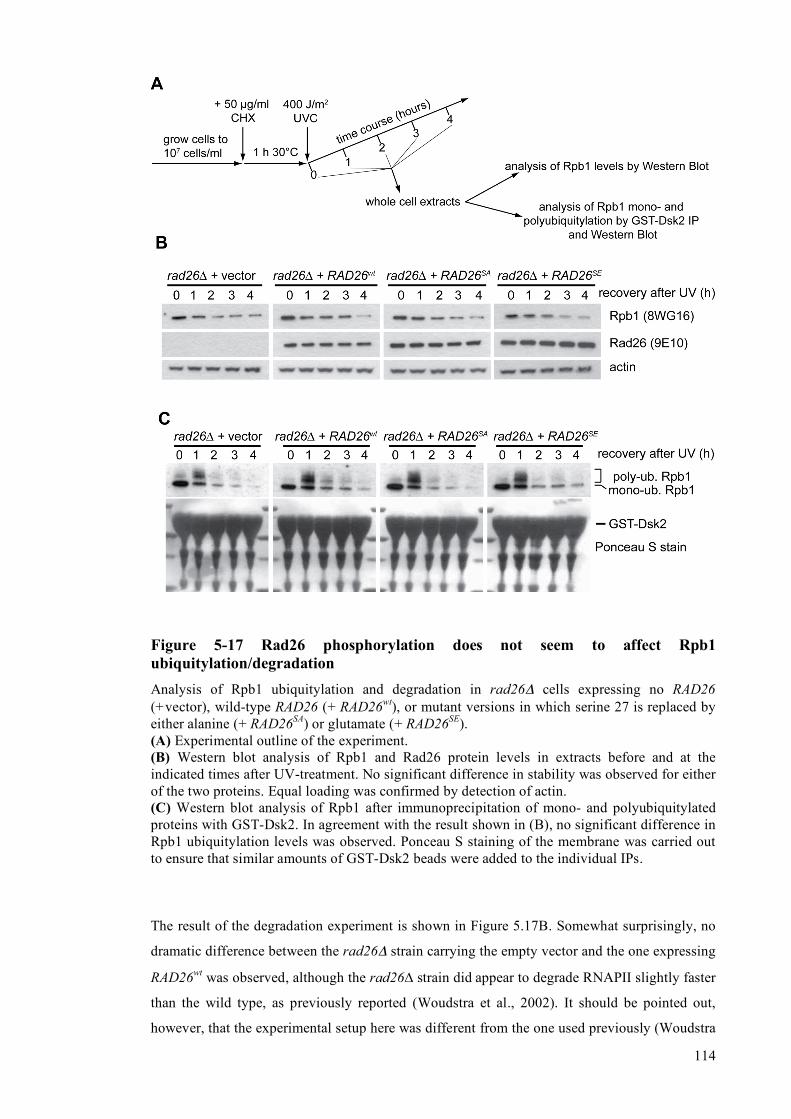
114
Figure 5-17 Rad26 phosphorylation does not seem to affect Rpb1 ubiquitylation/degradation Analysis of Rpb1 ubiquitylation and degradation in rad26Δ cells expressing no RAD26 (+vector), wild-type RAD26 (+ RAD26wt), or mutant versions in which serine 27 is replaced by either alanine (+ RAD26SA) or glutamate (+ RAD26SE). (A) Experimental outline of the experiment. (B) Western blot analysis of Rpb1 and Rad26 protein levels in extracts before and at the indicated times after UV-treatment. No significant difference in stability was observed for either of the two proteins. Equal loading was confirmed by detection of actin. (C) Western blot analysis of Rpb1 after immunoprecipitation of mono- and polyubiquitylated proteins with GST-Dsk2. In agreement with the result shown in (B), no significant difference in Rpb1 ubiquitylation levels was observed. Ponceau S staining of the membrane was carried out to ensure that similar amounts of GST-Dsk2 beads were added to the individual IPs.
The result of the degradation experiment is shown in Figure 5.17B. Somewhat surprisingly, no
dramatic difference between the rad26Δ strain carrying the empty vector and the one expressing
RAD26wt was observed, although the rad26∆ strain did appear to degrade RNAPII slightly faster
than the wild type, as previously reported (Woudstra et al., 2002). It should be pointed out,
however, that the experimental setup here was different from the one used previously (Woudstra

115
et al., 2002). In the original report, no cycloheximide was used in order to abolish new protein
synthesis, and Rpb1 degradation showed a markedly different pattern. Dramatic loss of the
Rpb1 protein was already observed one hour after UV treatment, and then returned to pre-UV
levels during the time course. In rad26Δ cells, loss of Rpb1 was more pronouced at early time
points in the previous study, but it seemed to be specifically the re-appearance which was
defective in this mutant. Interestingly, rad26Δ cells have been shown to have transcription
problems after UV irradiation, most likely due to the persistance of transcription-blocking
lesions in the TS of genes (Reagan and Friedberg, 1997), which will be especially pronounced
for long genes, such as Rpb1. Despite trying several times to obtain the pattern shown by
Woudstra et al., I did not manage to see any pronounced decrease of Rpb1 levels even in the
absence of cycloheximide. The reason for this is not clear at this point. More importantly, I can,
however, say that under the conditions used in my experiments, cells expressing RAD26SA or
RAD26SE degraded RNAPII like wild type, suggesting that the phosphorylation of Rad26 does
not play a significant role in this process. Again it should be noted that in contrast to the
situation in human cells, where CSB appears to be degraded in response to UV irradiation
(Groisman et al., 2006), no such degradation was observed for Rad26, neither for the wild type
nor for the mutant proteins, even though new protein synthesis was inhibited.
In agreement with the result of the degradation assay, no significant difference in mono- and
poly-ubiquitylation of Rpb1 was observed using our technique (Fig. 5.17C). Ponceau S staining
of the membrane after GST-Dsk2-IP, SDS-PAGE and Western blot confirmed that similar
quantities of GST-Dsk2 were used in the IP step.
5.2.2.8 Rad26 phosphorylation does not affect DNA-dependent ATPase activity
of the protein
The Rad26 protein is a DNA-dependent ATPase (Guzder et al., 1996) and this ATPase activity
is required for its function in TC-NER. The observed TC-NER defect of the RAD26SA mutant
might be explained if phosphorylation of Rad26 increases this activity to some extent. Having
purified unphosphorylated as well as partially phosphorylated Rad26 (see Figure 5.11D), I set
out to test the DNA-dependent ATPase activities of these proteins. As expected, and in
agreement with previous publications (Guzder et al., 1996), in the absence of DNA the ATPase
acitivity of the Rad26 protein could hardly be detected, but was greatly stimulated by its
presence (Fig. 5.18). Importantly, partially phosphorylated Rad26 and its unmodified
counterpart displayed the same level of ATPase activity in the presence of DNA, indicating that
ATP hydrolysis by Rad26 is not significantly influenced by its phosphorylation.
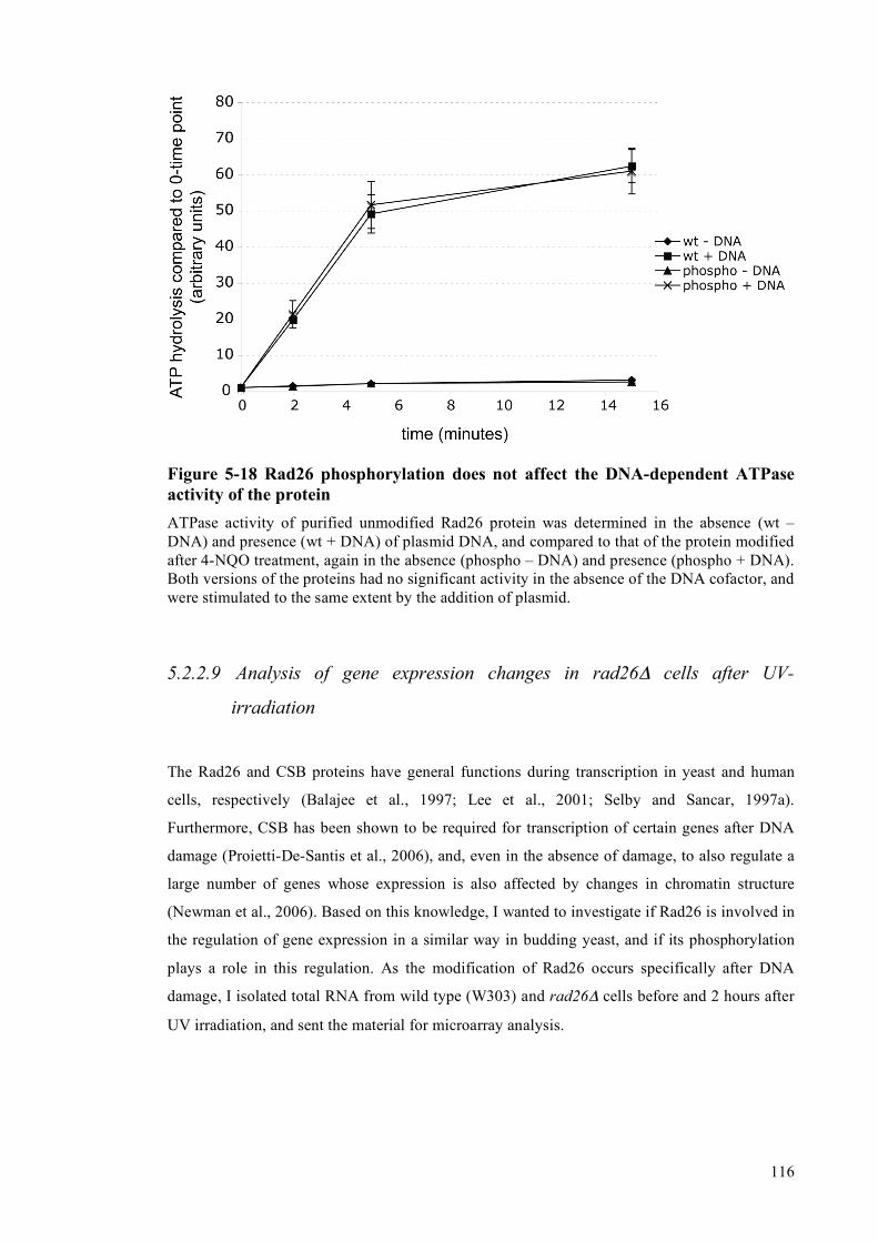
116
Figure 5-18 Rad26 phosphorylation does not affect the DNA-dependent ATPase activity of the protein ATPase activity of purified unmodified Rad26 protein was determined in the absence (wt – DNA) and presence (wt + DNA) of plasmid DNA, and compared to that of the protein modified after 4-NQO treatment, again in the absence (phospho – DNA) and presence (phospho + DNA). Both versions of the proteins had no significant activity in the absence of the DNA cofactor, and were stimulated to the same extent by the addition of plasmid.
5.2.2.9 Analysis of gene expression changes in rad26Δ cells after UV-
irradiation
The Rad26 and CSB proteins have general functions during transcription in yeast and human
cells, respectively (Balajee et al., 1997; Lee et al., 2001; Selby and Sancar, 1997a).
Furthermore, CSB has been shown to be required for transcription of certain genes after DNA
damage (Proietti-De-Santis et al., 2006), and, even in the absence of damage, to also regulate a
large number of genes whose expression is also affected by changes in chromatin structure
(Newman et al., 2006). Based on this knowledge, I wanted to investigate if Rad26 is involved in
the regulation of gene expression in a similar way in budding yeast, and if its phosphorylation
plays a role in this regulation. As the modification of Rad26 occurs specifically after DNA
damage, I isolated total RNA from wild type (W303) and rad26Δ cells before and 2 hours after
UV irradiation, and sent the material for microarray analysis.

117
Table 5-1 Genes affected by RAD26 deletion after UV Genes with lower expression in the mutant compared to the wildtype
ORF Name Function
YBR291C CTP1 Mitochondrial inner membrane citrate transporter
YNR050C LYS9 Saccharopine dehydrogenase
YDL182W LYS20 Homocitrate synthase isozyme,
YIL094C LYS12 Homo-isocitrate dehydrogenase
YPL276W NAD(+)-dependent formate dehydrogenase
YPL275W FDH2 NAD(+)-dependent formate dehydrogenase
YOR388C FDH1 NAD(+)-dependent formate dehydrogenase
YJL200C ACO2 Putative mitochondrial aconitase isozyme
YJL089W SIP4 C6 zinc cluster transcriptional activator
YBR147W Putative protein of unknown function
YJL088W ARG3 Ornithine carbamoyltransferase
YIR034C LYS1 Saccharopine dehydrogenase
YLR377C FBP1 Fructose-1,6-bisphosphatase
YJR154W Putative protein of unknown function
YKL217W JEN1 Lactate transporter,
YBR115C LYS2 Alpha aminoadipate reductase
YBR054W YRO2 Putative protein of unknown function
YGR125W Putative protein of unknown function
YGR260W TNA1 High affinity nicotinic acid plasma membrane permease
YGL184C STR3 Cystathionine beta-lyase
YDR234W LYS4 Homoaconitase
YMR107W SPG4 Protein required for survival at high temperature
YJR155W AAD10 Putative aryl-alcohol dehydrogenase
YNR066C Putative membrane-localized protein of unknown function
YER024W YAT2 Carnitine acetyltransferase
YER065C ICL1 Isocitrate lyase
YPR124W CTR1 High-affinity copper transporter of the plasma membrane
YCR005C CIT2 Citrate synthase
Genes with higher expression in the mutant compared to the wildtype
ORF Name Function
YGL089C MFa2 Mating pheromone alpha-factor
YKL071W Putative protein of unknown function
YFL056C AAD6 Putative aryl-alcohol dehydrogenase
YFL057C AAD16 Putative aryl-alcohol dehydrogenase
YLR460C Putative protein of unknown function
YCL027W FUS1 Membrane protein localized to the shmoo tip
YEL021W URA3 Orotidine-5'-phosphate (OMP) decarboxylase
YBR008C FLR1 Plasma membrane multidrug transporter
YPL171C OYE3 Widely conserved NADPH oxidoreductase
YJR004C SAG1 Alpha-agglutinin of alpha-cells
YCL026C-A FRM2 Protein of unknown function
YDL243C AAD4 Putative aryl-alcohol dehydrogenase
YJL045W Minor succinate dehydrogenase isozyme

118
This analysis led to the identification of several genes, whose expression after UV was either
higher or lower in the rad26Δ mutant after UV relative to the parental W303 strain. A list of
these ORFs can be found in table 5.1. Most of the identified genes were involved in biosynthetic
pathways, for example genes involved in lysine, arginine and cystein synthesis, or genes with
functions in the TCA (tricarboxylic acid) and glyoxylate cycles. Unfortunately, the changes in
the UV-dependent expression of these ORFs could not explain any of the defects in rad26Δ
cells, because none of the affected genes were linked to DNA repair in any known way.
However, it can not be excluded that one or more of the identified genes affects DNA repair in a
manner that still needs to be elucidated.
Nevertheless, I wanted to find out if Rad26 phosphorylation affected the observed changes in
gene expression. In order to do so, I chose two of the affected genes, and compared the UV-
dependent changes in mRNA level between rad26Δ cells carrying either empty vector, the
vector expressing wildtype Rad26 (RAD26WT), or the point mutants which had S27 mutated
either to alanine (RAD26SA) or glutamate (RAD26SE). The CTP1 gene showed a lower
expression after UV in rad26Δ than in the wild type, whereas expression of the AAD6 gene was
higher.
Quantitative RT-PCR analysis confirmed the result from the microarray, but no difference was
observed between the wild type and the mutated Rad26 proteins, indicating that Rad26-
dependent regulation of gene expression after UV is not altered by the phosphorylation of the
protein (Fig. 5.19). Although these experiments were only performed on a small subset of the
affected genes, the results suggest that Rad26 phosphorylation is not required for the role played
by Rad26 in damage-induced gene expression.
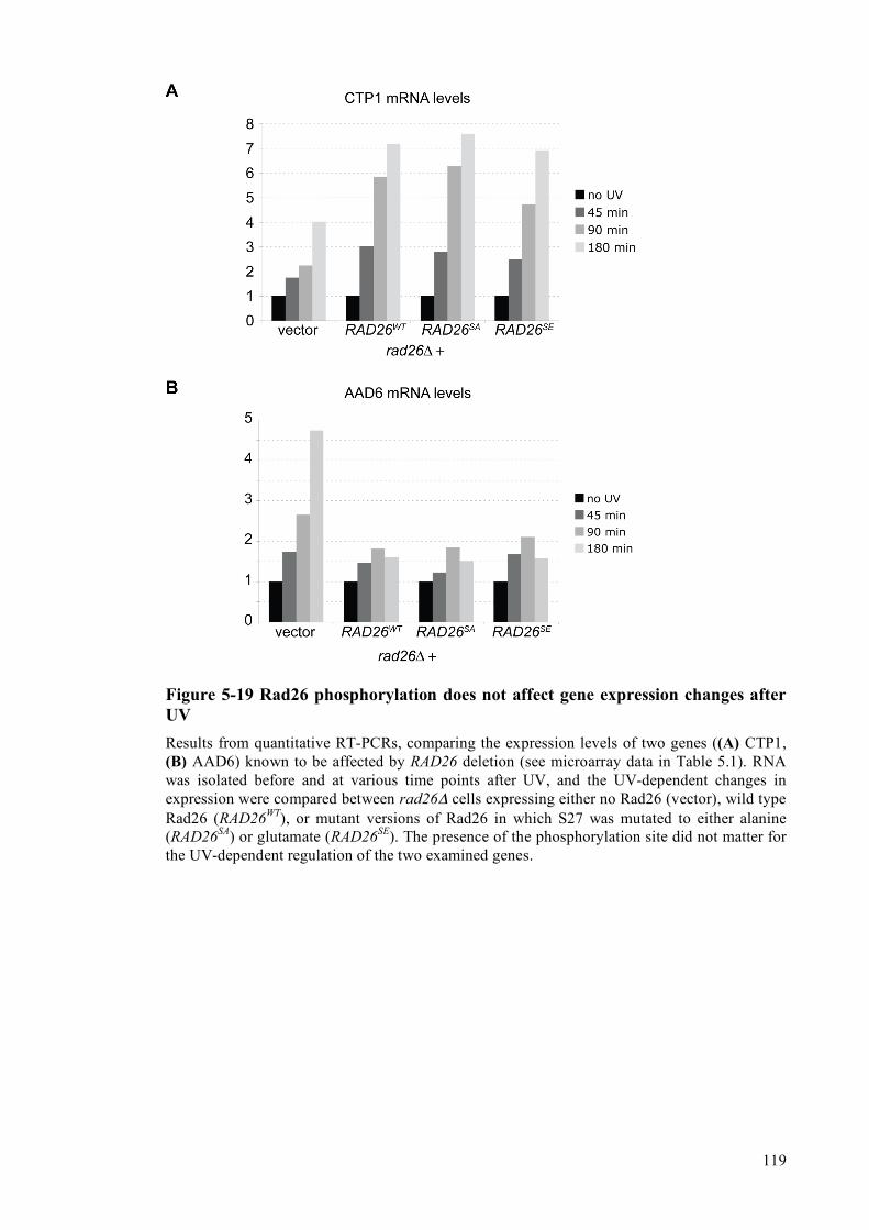
119
Figure 5-19 Rad26 phosphorylation does not affect gene expression changes after UV Results from quantitative RT-PCRs, comparing the expression levels of two genes ((A) CTP1, (B) AAD6) known to be affected by RAD26 deletion (see microarray data in Table 5.1). RNA was isolated before and at various time points after UV, and the UV-dependent changes in expression were compared between rad26Δ cells expressing either no Rad26 (vector), wild type Rad26 (RAD26WT), or mutant versions of Rad26 in which S27 was mutated to either alanine (RAD26SA) or glutamate (RAD26SE). The presence of the phosphorylation site did not matter for the UV-dependent regulation of the two examined genes.

120
5.3 Analysis of an influence of the DNA damage checkpoint on
ubiquitylation and degradation of Rpb1
Having created a number of yeast strains with deletions of various components of the DDC, I
decided to determine not only the NER capacity of these cells, but also their ability to perform
the alternative way of dealing with irreversibly stalled RNA polymerase II complexes, namely
ubiquitylation and degradation of the Rpb1 subunit. As the DDC coordinates a large number of
events after DNA damage, it was plausible that this is the case, and I could not find any
evidence in the literature that this possibility had been investigated before.
5.3.1 Rpb1 ubiquitylation and degradation are influenced by the Mec1 and
Rad53 kinases
In order to investigate if the DDC affects Rpb1 ubiquitylation and degradation, the checkpoint
deficient mec1Δ sml1Δ strain, as well as the chk1Δ and rad53Δ sml1Δ mutants (lacking the two
main downstream kinases) were chosen for analysis. Cell growth, cycloheximide treatment and
UV-irradiation was performed as oulined in Figure 5.17A. The result of both the degradation
experiment and the analysis of the ubiquitylation status of Rpb1 are shown in Figure 5.20.
In the wild-type W303 strain, degradation of Rpb1 becomes apparent around 2 hours after UV
treatment and after 4 hours the protein level is dramatically decreased (Fig. 5.20A). In contrast
to this, degradation is much less efficient in both the mec1Δ sml1Δ and the rad53Δ sml1Δ strain,
indicating that the DDC indeed has an influence on the process. Deletion of CHK1 did not seem
to significantly affect Rpb1 degradation, showing that the Mec1-Rad53 branch of the DDC is
mainly responsible for its regulation.
Rpb1 ubiquitylation levels were then examined by GST-Dsk2 pulldown, followed by Western
blot analysis of Rpb1 using 4H8 antibodies. Different exposures of this Western blot are shown
in the three top panels of Figure 5.20B. In agreement with the result of the degradation assay,
the signals for both mono- and polyubiquitylated Rpb1 looked similar when the wild type
(W303) was compared with the chk1Δ mutant strain. Mono-ubiquitylation, but little or no poly-
ubiquitlyation, was observed before UV treatment, whereas poly-ubiquitylation was strong at
the 1 hour time point after UV-irradiation, as expected. Signals started losing intensity at later
time-points, in agreement with efficient degradation of Rpb1 in these strains (Fig. 5.20A).

121
Figure 5-20 Analysis of the effect of checkpoint kinases on Rpb1 ubiquitylation/degradation Analysis of Rpb1 ubiquitylation and degradation in wild-type (W303) and strains with deletions of various checkpoint kinases (A) Western blot analysis of Rpb1 protein levels in extracts before and at the indicated times after UV-treatment. Degradation is severely impaired in cells lacking the Mec1 and Rad53 kinases, but not the Chk1 kinase. Equal loading was confirmed by detection of actin. (B) Western blot analysis of Rpb1 after immunoprecipitation of mono- and polyubiquitylated proteins with GST-Dsk2. In agreement with the result shown in (A), significant differences in mono- and polyubiquitylation are observed between the wildtype and both the mec1Δ sml1Δ and rad53Δ sml1Δ strains, but not the chk1Δ strain.
The mec1Δ sml1Δ and rad53Δ sml1Δ strains showed a clear change in Rpb1 ubiquitylation.
Interestingly, the patterns looked different in the two strains, suggesting that the Mec1 and
Rad53 kinases have different effects on one or more steps controlling Rpb1
ubiquitylation/degradation. The most prominent change was the presence of a much stronger
signal for mono-ubiquitylated Rpb1 in both strains, compared to wild type. This was more
pronounced in the mec1Δ sml1Δ strain, but the rad53Δ sml1Δ strain also clearly had more of
this modification than the wild type or chk1Δ strain. Despite the strong defect in Rpb1
degradation after UV damage in strains lacking either the Mec1 or Rad53 kinases, UV-induced

122
poly-ubiquitylation of Rpb1 could still be detected, even though the levels seemed to be
different from the wild type strain. In the rad53Δ sml1Δ strain I detected slightly lower signals
for poly-ubiquitylated Rpb1 (compare the signals at the 1 hour time point between W303 and
rad53Δ sml1Δ in the middle panel of Figure 5.20B). Surprisingly, the mec1Δ sml1Δ strain
seemed to have slightly increased Rpb1 poly-ubiquitylation levels after UV irradiation, despite
having a defect in Rpb1 degradation, possibly suggesting a defect in proteasome function, or at
least in post-ubiquitylation processes.
Taken together, these results show that the Mec1 and Rad53 kinases of the DDC somehow
affect mono- and/or polyubiquitylation of Rpb1 and that they have a defect in UV-induced
degradation of this protein.
5.3.2 DDC-dependent phosphorylation of Def1 is not involved in regulation
of UV-induced Rpb1 degradation
Having shown that the DDC also controls ubiquitylation and degradation of Rpb1, I set out to
determine the underlying mechanism. The possibility that the DDC induces synthesis of one or
more factors involved in this process could be excluded, because the analysis of Rpb1
ubiquitylation and degradation was carried out in the presence of cycloheximide. I therefore
focussed my attention on potential phosphorylation targets of the Mec1 and Rad53 kinases.
An obvious candidate was Def1, the factor required for normal Rpb1 ubiquitylation and
degradation (Reid and Svejstrup, 2004; Woudstra et al., 2002). Interestingly, this protein was
recently identified as a target of both the Mec1 and Rad53 kinases in proteome-wide screens
(Albuquerque et al., 2008; Smolka et al., 2007), in apparent agreement with the RNAPII
ubiquitylation defects observed in strains lacking these two kinases (shown in Fig. 5.19). These
proteome-wide studies showed that Rad53 phosphorylates Def1 at serine 273 (Smolka et al.,
2007), while Mec1 targets serine 497 (Albuquerque et al., 2008).
In order to investigate the possibility that these Def1 phosphorylation events are required for
Rpb1 ubiquitylation and degradation, both of the sites were mutated to alanines on a plasmid
carrying the DEF1 locus, and the resulting mutated plasmids were transformed into a def1Δ
rad14Δ strain. The functionality of the mutated Def1 protein was determined by spotting and
UV treatment in this genetic background because Def1 controls a pathway which is thought to
represent an alternative to DNA repair, namely the removal of irreversibly stalled RNA
Polymerase II complexes (Woudstra et al., 2002).

123
Accordingly, the rad14Δ def1Δ double mutant is significantly more UV sensitive than the
rad14Δ single mutant (Fig. 5.21). Expression of wild-type DEF1 (DEF1wt) reversed this
sensitivity to the level of the rad14Δ single mutant. DEF1 mutants that either had the
phosphorylation sites mutated individually (DEF1S273A and DEF1S497A) or in combination
(DEF1S273A/S497A), were equally efficient in rescuing the sensitivity of the def1Δ rad14Δ strain,
suggesting that DDC-dependent phosphorylation of Def1, at least at the tested sites, is not
required for the regulation of Rpb1 degradation.
This result shows that the DDC controls Rpb1 ubiquitylation and degradation at one or more
steps, which are not linked to phosphorylation of Def1 at serine 273 or 497. Some possibilities
will be presented in the next chapter.
Figure 5-21 DDC dependent phosphorylation of Def1 at S273 and S497 is not involved in ubiquitylation/degradation of Rpb1 A rad14Δ def1Δ mutant was transformed either with empty vector, or vectors carrying wild type DEF1 (DEF1WT), single point mutants of DEF1 (DEF1S273A and DEF1S497A), or a double mutant (DEF1S273A/S497A). The sensitivities of these strains were then compared to the wild type (W303) and rad14Δ strains to analyse an involvement of these phosphorylation sites in the ubiquitylation and degradation of Rpb1 after UV.
DEF1S497A

124
6 Discussion II
6.1 Regulation of NER by the DNA damage checkpoint
In this study I investigated possible links between the Nucleotide Excision Repair (NER)
pathway and the DNA damage checkpoint (DDC), by examining the efficiency of CPD repair at
both the TS and the NTS of the constitutively transcribed RPB2 gene in various mutant yeast
strains, lacking kinase components of the DDC. Interestingly, I found that repair on both strands
is influenced by the absence of a functional DDC. This shows that both sub-pathways of NER
are regulated by the checkpoint, and I have tried to find out how this regulation might be
achieved. At this point I would like to mention that all the findings presented in this work about
NER kinetics in the various mutant strains are restricted to the RPB2 gene, and it would be
interesting to find out if the observed requirement for individual checkpoint components also
applies to others.
6.1.1 Regulation of NTS repair
6.1.1.1 de novo protein synthesis is required for efficient NTS repair
The non-transcribed strand (NTS) of active genes is repaired by the slow Global Genome
Repair (GG-NER) pathway, because lesions in this strand do not block the progression of
RNAPII complexes. In S. cerevisiae this pathway is dependent on the Rad7/Rad16 complex, as
well as all the other central NER proteins (Verhage et al., 1994). The results presented in
Figures 5.1 and 5.2 clearly show that in the absence of a functional DDC, achieved by deletion
of the gene encoding the central Mec1 kinase, repair of the NTS is virtually undetectable during
a time course of up to 6 hours. It is important to note at this point, that deletion of MEC1 was
carried out in an sml1Δ background, and I can not rule out that deletion of SML1 itself has an
effect on NER. One attractive possibility to make sure that the observed NER delays are really
due to the absence of Mec1 would be to use temperature-sensitive or degron-versions of Mec1,
which would allow to examine the effect of the absence of the Mec1 kinase in the wild type
background.

125
In contrast to the result obtained for the mec1Δ sml1Δ mutant, strains with deletions of the
genes encoding the Chk1, Rad53 and Dun1 kinases do not show any significant alteration in the
CPD repair rate on the NTS. Previous reports showed a strong dependence of NTS repair, but
not TS repair, on UV-induced de novo synthesis of repair proteins (Al-Moghrabi et al., 2003,
2009). These studies focussed on the GAL10 and URA3 genes. In order to also show this
dependence of NTS repair on de novo protein synthesis for the RPB2 gene, I analysed the
efficiency of CPD removal from this locus in the absence or presence of the translational
inhibitor cycloheximide. I found that repair of the NTS is strongly inhibited in the presence of
this drug, while no significant defect could be detected on the TS.
All these results suggest that the DDC controls NTS repair by increasing the abundance of
repair proteins. Indeed, the synthesis of various NER factors, including Rad2, Rad7, Rad16 and
Rad23, has been shown to be induced by UV irradiation (Bang et al., 1995; Jones et al., 1990;
Madura and Prakash, 1990; Siede et al., 1989). Surprisingly, however, it has been shown that
most of the transcriptional changes after DNA damage are dependent on the Dun1 kinase
(Gasch et al., 2001), which becomes phosphorylated, and thereby activated, by the Rad53
kinase after DNA damage (Chen et al., 2007; Zhou and Elledge, 1993). This would suggest that
mutants with deletions of DUN1 and RAD53 should have a defect in GG-NER similar to the one
observed after treatment with cycloheximide. This is clearly not the case, suggesting that the up-
regulation of damage-inducible genes, necessary for efficient CPD repair on the NTS of RPB2,
does not rely on these two kinases.
Damage-induced transcriptional upregulation of RAD2, RAD7, RAD16 and RAD23 has been
shown to be controlled by the Rad9 protein (Aboussekhra et al., 1996). If the upregulation of
these factors is required for efficient repair of the NTS, then the rad9Δ mutant should have the
same NTS repair defects observed for the wildtype strain in the presence of cycloheximide.
Even though I have not focussed on the role of the Rad9 protein in NER, previous studies by
other groups have shown that repair on both the TS and the NTS is slower in the absence of this
factor (Al-Moghrabi et al., 2009; Yu et al., 2001), yet not as slow as in the absence of protein
synthesis (Al-Moghrabi et al., 2003). The most plausible explanation for this observed
discrepancy is that the transcriptional induction of additional factors, which is dependent only
on the Mec1 kinase but not the Rad9, Rad53, Chk1 and Dun1 proteins, is also important for
efficient NTS repair. As this work focussed on the repair of the TS, I did not pursue this issue
any further.
6.1.1.2 Phosphorylation of repair factors for efficient NTS repair?

126
Besides DDC-dependent activation of the transcriptional response to DNA damage, culminating
in the upregulation of repair factors, it is also possible that post-translational modifications of
repair factors by the Mec1 kinase contributes to efficient NTS repair. Interestingly, the Rad23
protein in S. cerevisiae as well as its human homolog hRad23A have recently been identified in
proteome-wide screens for potential targets of the Mec1 and ATM/ATR kinases, respectively
(Albuquerque et al., 2008; Matsuoka et al., 2007). In order to investigate if this modification
plays a role in the NER process, I complemented a rad23Δ mutant strain with mutant versions
of RAD23, in which the either only the identified Mec1 target residue, or all three potential
Mec1 target residues (SQ-sites), were mutated. Surprisingly, I did not detect any defect in NER
efficiency. Furthermore, the cells carrying the mutated versions of RAD23 did not display any
increase in UV-sensitivity than the parental wildtype strain, indicating that this modification
does not contribute to efficient repair, at least not under these experimental conditions.
The Rad16 protein is required for NTS-repair, and this factor has also been identified as a
phosphorylation target of Mec1 (Smolka et al., 2007). I mutated the Rad16 phosphorylation site,
but again could not detect any significant difference in survival after UV between strains
carrying the wildtype, or mutated, version of the RAD16 gene (data not shown).
Even though I cannot rule out that phosphorylation of other repair proteins contributes to
efficient repair of the NTS, I speculate that the main point at which the Mec1 dependent DDC
influences NTS repair is at the level of UV-induced de novo protein synthesis.
6.1.2 Regulation of TC-NER
In Figures 5.1 and 5.2 I clearly showed that repair in the TS of RPB2 is less efficient in a mec1Δ
sml1Δ strain lacking a functional DDC. When compared to a rad26Δ strain, it is obvious that
TC-NER is still possible, yet less efficient, in the absence of the Mec1 kinase. Again it should
be noted that analysis of the effect of MEC1 deletion was carried out in the sml1Δ background,
and it can not be excluded at this point that SML1 deletion on its own has some effect on NER.
Importantly, cells lacking the CHK1, RAD53 or DUN1 genes have no detectable defect in TC-
NER, indicating that the effect of the Mec1 kinase on the repair of the TS of RPB2 is direct.
Furthermore, in agreement with previous reports (Al-Moghrabi et al., 2003), UV induced de
novo protein synthesis has no major effect on the efficient repair of this strand. This implies that
the steady-state levels of NER factors are sufficient to ensure fast repair of transcription-
blocking DNA lesions, and shows that the regulation of TC-NER efficiency occurs via post-
translational modifications of one or more factors involved in this repair pathway.

127
6.2 Rad26, a Mec1 target involved in the regulation of DNA repair by
the DDC
As the most obvious candidate target to explain the TC-NER defect in the mec1Δ sml1Δ strain
is the TC-NER factor Rad26, I focussed my attention on this protein. In order to efficiently
detect the weakly abundant Rad26 protein in extracts of S. cerevisiae, I created a strain carrying
a tagged version of this gene. C-terminal tagging of Rad26 was not possible, as this leads to
functional inactivation (Elies Woudstra, unpublished observation). I therefore created a strain
with an N-terminal 9myc/2TEV/8His tag, using a strategy which ensures that the resulting gene
is still under control of its endogenous regulatory elements, in order to avoid artefacts due to
changes in expression levels. I was able to show genetically that the functionality of the tagged
protein was not compromised by the presence of the tag.
6.2.1 Rad26 is phosphorylated after DNA damage by the Mec1 kinase
UV irradiation of cells leads to a post-translational modification of Rad26, which is clearly
visible as the appearance of a form of the protein with reduced electrophoretic mobility (Fig.
5.9A), and I was able to show that Rad26 is phosphorylated. Treatment of the modified protein
with a phosphatase (Fig. 5.9D) resulted in the complete loss of the electrophoretic mobility
shift. Other sources of DNA damage (H2O2, MMS, bleomycine and 4-NQO), but not heat
shock, osmotic shock or 6-AU-mediated induction of RNAPII stalling led to this modification,
showing that Rad26 phosphorylation is a damage-specific response, and implicating a role of
the DDC. Indeed, I was able to show that this event is strictly dependent on the Mec1 kinase
(Fig. 5.10C). In contrast, the Tel1, Chk1, Rad53 and Dun1 components of the DDC are not
required. This is in perfect agreement with my results for the NER kinetics, which show that
only strains lacking the Mec1 kinase, but not Chk1, Rad53 or Dun1, have a clear defect in TC-
NER.
Besides being efficiently induced by DNA damage, a low level of Rad26 phosphorylation can
also be found in undamaged cells. Interestingly, this background level of modification seems to
be restricted to the S-phase of the cell cycle, as it is undetectable in cells arrested in G1 or G2,
but clearly visible in cells arrested in S-phase with the drug hydroxyurea (HU). This
modification can also be detected after the release of G1-arrested cells into the cell cycle by the
removal of the alpha-factor mating pheromone in the absence of HU (data not shown). The
Mec1 kinase has an essential role in the control of DNA replication during an unperturbed S-
phase. This cell-cycle dependent activation of Mec1 can be detected by the appearance of
phosphorylated forms of the Rad53 kinase. I speculate that the presence of phosphorylated

128
Rad26 during S-phase is simply due to this transient activation of the responsible kinase, and
that Rad26 phosphorylation has no role in S-phase. Interestingly, even though HU is a strong
activator of Mec1, the phosphorylation level of Rad26 was low after HU treatment compared to
the level after DNA damage. One possible explanation for this is that the kinase and the
substrate are in close proximity only after DNA damage, thereby facilitating the
phosphorylation of Rad26 by Mec1. Indeed, it has been shown by chromatin
immunoprecipitation experiments that components of the DDC are recruited to sites of DNA
lesions (Jiang and Sancar, 2006; Unsal-Kacmaz et al., 2002).
6.2.2 Serine 27 is the main phosphorylation site on Rad26
Serines or threonines followed by glutamines (SQ/TQ sites) are the main target sites for PIKKs
such as Mec1, Tel1, ATM and ATR. Analysis of the Rad26 amio acid sequence revealed that 5
of these consensus sites can be found in this protein. These sites were mutated individually to
alanines, and the phosphorylation status of these mutant versions after DNA damage was
analysed by SDS-PAGE and Western blotting. This experiment revealed that only mutation of
serine 27 abolished the shift in electrophoretic mobility. Interestingly, purification of
overexpressed wildtype Rad26 before and after DNA damage, followed by SYPRO Ruby
staining of the gel, showed the presence of more than one slower migrating form of the protein,
and mass spectrometric analysis of the protein revealed that serine 30 is also modified. In
contrast, Albuquerque et al showed that serines 27 and 29 are phosphorylated after damage
(Albuquerque et al., 2008). I speculated that serine 27 is the main phosphorylation site (as it can
be found in a consensus Mec1 target motif), and that nearby residues can be targeted non-
specifically in addition to it. Indeed, mutation of serine 27 and purification of the overexpressed
mutant protein after DNA damage revealed the absence of any slower migrating forms, and
mass spec analysis indicated that no residual phosphorylations are detectable on this protein.
One possibility, which cannot be excluded at this point, is that phosphorylation of serine 27
leads to modification of surrounding residues by a different kinase. In any case, the absence of
phosphorylation in the S27A single mutant made me decide to use this version to carry out
functional assays.
6.2.3 Rad26 phosphorylation is required for efficient TC-NER in the
presence of a functional DDC
Strand-specific NER assays confirmed that the phosphorylation of Rad26 by the Mec1 kinase
has a relevance in vivo. Cells carrying the S27A mutant of Rad26, but not the phosphorylation-

129
mimicking S27E mutant, had reproducible defects in TC-NER, detectable as an increase in the
half-life of damages on the TS of RPB2 by about 1 hour. Repair of the TS is still more efficient
than in the TC-NER deficient rad26Δ strain, showing that TC-NER is compromised in the
absence of the phosphorylation site, but that it is not completely dependent on this modification.
This delay in TS repair is somewhat similar to the defect observed in the mec1Δ sml1Δ mutant.
It should be noted that the most obvious difference between the wildtype and the S27A mutant
strain is at early time points (1 hour time point in Figure 5.13), in agreement with a very early
appearance of the modification after UV irradiation.
I also examined TC-NER efficiency in strains expressing various other Rad26 phosphorylation
site mutants. These include a mutant in which all five (S/T)Q sites are mutated to alanines, and
mutants in which serine 27 is mutated in combination with serine 29 and serine 30. I was unable
to detect any significant differences between these mutants and the S27A single mutant. This is
in agreement with the finding that no residual phosphorylations are detectable in this single
mutant.
The finding that both deletion of the gene encoding the Mec1 kinase and mutation of the Mec1-
dependent phosphorylation site on Rad26 lead to a similar effect on TC-NER, made me
speculate that Rad26 might be the only relevant target for the Mec1 kinase to ensure efficient
TC-NER. The full functionality of the S27E mutant (no delay in TC-NER compared to the wild
type) led me to investigate if expression of this mutant in the mec1Δ sml1Δ strain can bypass the
requirement for the DDC. This was not the case, as both wild type and S27E Rad26 showed the
same rate of CPD removal from the TS of RPB2 in the absence of a functional DDC. This
clearly shows that the Mec1 dependent DDC has additional targets after DNA damage which
lead to efficient TC-NER, and which are necessary for the phospho-mimic S27E mutant Rad26
to exert its effect.
This experiment also revealed that the remaining TS-repair in the mec1Δ sml1Δ mutant is
entirely dependent on the Rad26 protein. The presence of functional Rad26 is important in this
case because repair of the NTS is undetectable in the absence of the DDC. This situation is
similar to the one in the rad16Δ background, where additional deletion of RAD26 leads to a
dramatic increase in UV sensitivity. It is therefore very surprising that the mec1Δ sml1Δ rad26Δ
triple mutant is not more UV-sensitive than the mec1Δ sml1Δ double mutant (Fig. 5.16).
At this point I can only speculate why repair of the TS of active genes does not seem to be very
important in this case. One possibility is that overall GG-NER is not, or only partially, defective
in the absence of the Mec1 kinase. In this study I have only focussed on the NTS of active
genes, and in agreement with previous studies I have shown that the main function of the DDC

130
is UV-induced de novo protein synthesis of repair factors (Al-Moghrabi et al., 2003, 2009).
These studies have, however, also shown that protein synthesis is only required for repair of the
NTS of active genes, but not for transcriptionally inactive DNA. This means that there might be
two distinct GG-NER pathways, depending on the transcriptional state of the genomic locus. I
can currently not say whether or not MEC1 deletion affects the repair of untranscribed DNA, as
this was not the focus of this work. If it is not affected, and untranscribed regions of the genome
are still efficiently repaired in the absence of the Mec1 kinase, this might explain the fact that
the mec1Δ sml1Δ mutant is significantly less UV-sensitive than the completely NER-deficient
rad14Δ mutant.
Even if GG-NER of untranscribed DNA is still possible, it is still surprising that deletion of
RAD26 does not increase the UV-sensitivity of a mec1Δ sml1Δ mutant, given that apparently
the lesions in the TS persist for a long time. One possibility which can be envisaged is that
deletion of RAD26 leads to other effects in the DDC-deficient cells, which compensate for the
impairment of TC-NER. In light of my finding that the mec1Δ sml1Δ strain has a defect in
ubiquitylation and degradation of Rpb1, which serves as the alternative way to TC-NER in
order to deal with arrested RNAPII complexes, I can only speculate that after RAD26 deletion
this pathway is reactivated in these cells. In this respect it is important to note that indeed Rad26
has been suggested to have an inhibitory effect on RNAPII degradation (Woudstra et al., 2002).
6.2.4 Mutation of the Rad26 phosphorylation site does not affect survival
or growth recovery after UV irradiation
Having established that UV-induced phosphorylation of Rad26 is required for efficient TC-
NER, it was surprising that the UV sensitivity of strains carrying the Rad26 S27A mutant were
not detectably more UV sensitive than the wild type. This can potentially be explained by the
relatively small contribution of Rad26 phosphorylation to the efficiency of TC-NER. A delay in
repair of about 1 hour might not be severe enough to render the cells more sensitive to UV
irradiation.
I also speculated that I might be able detect a difference between the wildtype and the mutant
Rad26 strain by looking at the rates of UV-induced mutagenesis, as this might be a more
sensitive assay, but again I did not observe any significant change (data not shown).
In the original report which identified RAD26 as the gene encoding the yeast homolog of CSB,
the authors showed that despite not being UV-sensitive, the rad26Δ mutant has a slight delay in
growth recovery after UV irradiation (van Gool et al., 1994). I wanted to investigate if mutation

131
of the phosphorylation site might lead to such growth defects, but failed to detect any delay
even in the complete absence of the Rad26 protein (data not shown).
Because of my finding that Rad26 phosphorylation is induced by other types of DNA damage
apart from UV-light, such as oxidative stress (induced by treatment with hydrogen peroxide;
H2O2) and base alkylation (inflicted by treatment with methyl methane-sulfonate; MMS) (Fig.
5.10A), I could not rule out the possibility that mutation of the Rad26 phosphorylation site
renders the cells more sensitive to these agents. Interestingly, it was previously found that
RAD26 deletion increases the MMS-sensitivity of rad14Δ, mag1Δ, and also rad14Δ
mag1Δ cells, and it was speculated that Rad26 helps RNAPII transcribe through damaged bases
(Lee et al., 2002b). I tested the Rad26 phosphorylation mutants in these backgrounds, but could
not detect any significant differences in survival after MMS-treatment between the wild type
and the mutant versions of the protein (data not shown). With respect to oxidative stress, it is
important to point out that CS cells display increased sensitivity to H2O2 (Spivak and Hanawalt,
2006). I tested the sensitivity of rad26Δ cells towards this chemical, but this mutant was not
more sensitive than the wild type strain (data not shown)
An interesting possibility is that an effect of mutation of the Rad26 phosphorylation site on cell
viability after DNA damage can be seen in a genetic background different from the one I have
examined. Recent advances in the genome-wide analysis of genetic interactions, such as the
‘synthetic genetic array’ (SGA) (Tong et al., 2001) and ‘diploid-based synthetic lethality
analysis on microarrays’ (Pan et al., 2004), make it possible for researchers to quickly check
genetic interactions between a ‘gene of interest’ and other genes in the yeast genome. In order to
find out more about the function of the Rad26 phosphorylation in the future, an interesting
possibility would be to perform such a genome-wide analysis of genetic interactions (Synthetic
Genetic Array), specifically after DNA damage induction. This has, to our knowledge, not been
done yet, but should lead to an increased understanding of the roles of Rad26 in DNA repair.
This analysis can be done using the rad26Δ mutant to identify mutations in other genes which
work in parallel or together with this protein in the DNA damage response. Once potential
candidates have been identified, the plasmids expressing the wild type or phosphorylation site
mutant Rad26 proteins can be transformed into these double mutants, in order to check if the
phosphorylation mutant leads to increased/decreased rescue compared to wild type Rad26. We
tried to set up a collaboration with a group experienced in SGAs, but this line of
experimentation was still ongoing at the time of writing.
6.2.5 Speculation on the functional consequences of Rad26 phosphorylation

132
An important question, which arises from our finding that TC-NER efficiency is higher when
Rad26 is phosphorylated, is the one about the mechanism by which this is achieved.
Unfortunately, even the function of Rad26/CSB in eukaryotic TC-NER is not entirely
understood, and no in vitro assays using purified components have been established for TC-
NER which would enable me to directly compare the activities of unphosphorylated and
phosphorylated Rad26 proteins. I did, however, look more closely at several possibilities.
Firstly, given that RAD26 encodes a DNA-dependent ATPase (Guzder et al., 1996), I speculated
that this biochemical activity, which is required for TC-NER, might be influenced by the
modification of Rad26. If ATPase activity is increased by phosphorylation, this could explain
the TC-NER defects observed for the S27A point mutant. However, I clearly show in Figure
5.18 that the DNA-dependent ATPase acitivities of unmodified and modified Rad26 proteins in
the presence of DNA are indistinguishable. I would like to note that I have not been able to
purify completely phosphorylated Rad26 after DNA damage, but instead always obtained a
mixed population of Rad26 molecules (see Fig. 5.11D). Attempts to further enrich the modified
protein were not successful. However, as more than half of the purified protein was
phosphorylated in the preparation used for the ATPase assay, I assume that if the effect were
significant, this would have been enough to detect a stimulation of ATPase activity compared to
the unphosphorylated sample.
Secondly, because faster degradation of Rpb1 might lead to less efficient TC-NER due to
accelerated removal of the substrate, I looked at ubiquitylation and degradation of this protein in
the presence of Rad26 point mutants. Again I observed no significant difference between cells
expressing the wild-type or the point-mutated versions of Rad26. In this experiment I also
investigated whether or not Rad26 itself is destabilised after DNA damage and if
phosphorylation might be involved in this process. This is an important question in light of the
fact that CSB has been reported to be ubiquitylated and destabilised by a CSA-containing
ubiquitin-ligase complex, and that this destabilisation provided the first functional connection
between the main factors involved in Cockayne’s syndrome in human cells (Groisman et al.,
2006; Groisman et al., 2003). Interestingly, I did not detect any degradation of Rad26 after
DNA damage up to 4 hours post-UV, despite the use of the translational inhibitor
cycloheximide. This result shows a clear difference between the situation in yeast and humans,
and rules out the possibility that the Rad28 protein, the yeast homolog of CSA (Bhatia et al.,
1996), has a similar role as its human counterpart in destabilisation of Rad26, despite some
genetic evidence that it acts in the same pathway (Bhatia et al., 1996).
Phosphorylation of Rad26 might also influence its binding partners after UV irradiation. A well
known binding partner is the Def1 protein, the regulator of Rpb1 ubiquitylation and

133
degradation. However, I failed to detect any differences in binding of Def1 and wild type Rad26
in response to UV irradation, and consequently the UV-induced phosphorylation presumably
had no effect on this interaction (data not shown). This is in apparent agreement with my
finding that Rpb1 degradation is not affected by mutations of the Rad26 phosphorylation site. A
second possibility is that phosphorylation of Rad26 increases its affinity for damage-stalled
RNAPII, as this is thought to be the first step in the TC-NER mechanism. I was, however, never
able to efficiently co-immunoprecipitate RNAPII with Rad26, neither wildtype nor mutant. This
finding might be explained by the very transient nature of this interaction. Indeed, previous
reports identified a physical interaction between Rad26 and RNAPII after damage only when
the proteins were crosslinked with formaldehyde (Jansen et al., 2002). Importantly, this study
used overexpressed Rad26 protein, and in my case the low endogenous expression level might
make it even harder to detect potential binding partners. When I purified overexpressed Rad26
proteins from yeast cells (see Figure 5.11D), I also analysed weakly stained copurifying bands
by mass spectrometry, but was unable to find interesting candidates to explain the observed
influence of phosphorylation on TC-NER. I also failed to detect any RNAPII proteins in this
preparation. An important drawback to this attempt to explain the TC-NER defect by
differences in binding partners, is the fact that after mutation of the phosphorylation site, the
protein is still functional for TC-NER and only the kinetics of the reaction are reduced. This
means that every important binding partner will bind to both forms, maybe only with slightly
different kinetics. It might therefore be difficult to convincingly link the TC-NER defect to such
a slight difference in interactions.
The Rad26 protein is thought to bind to damage-stalled RNAPII complexes on chromatin after
UV irradiation in order to start the TC-NER process. I therefore thought it would be possible to
detect Rad26 on genes by chromatin immunoprecipitation (ChIP) after DNA damage, and
speculated that the kinetics of chromatin recruitment might be comparable to the observed
differences in TC-NER kinetics, i.e. that the phosphorylated form of Rad26 was recruited faster.
Unfortunately, I was unable to detect Rad26 above background using this assay, despite looking
at a number of genes with different expression levels. In a previous study, Jansen et al were able
to ChIP Rad26 from genes, however, the authors have again used overexpressed Rad26 in their
experiments (Jansen et al., 2002). In my case, the low endogenous levels of the Rad26 proteins
probably prohibited detection of Rad26 by ChIP. I also tried to overexpress Rad26 from a
galactose-inducible promoter, and to detect it after UV irradiation at the highly transcribed
GAL10 gene or the RPB2 gene, but again failed to obtain levels above background. The reason
for my lack of success with Rad26 ChIP is not known. In light of the finding that the observed
TC-NER defect is not very dramatic, it is, however, questionable if overexpression of Rad26 is
a good strategy for my purpose, as this increases the efficiency of TC-NER (Bucheli and
Sweder, 2004) and might mask the effect of the phosphorylation.

134
As mentioned in the introduction, an alternative function of Rad26, different to the classical
view of binding to damage-arrested RNAPII and recruiting NER factors, has been obtained by
the Brouwer lab. The TC-NER defect caused by RAD26 deletion can be suppressed by
additional deletion of SPT4 (Jansen et al., 2000), and this has been linked to regulation of
transcription after UV (Jansen et al., 2002). Importantly, deletion of RAD26 accelerated,
whereas deletion of SPT4 inhibited, loss of RNAPII phosphorylated at serine 5 on the CTD
from promoters after UV. This result represents an interesting possibility for the involvement of
the Rad26 phosphorylation. I carried out ChIP experiments using an antibody specific for this
phosphorylated form of RNAPII, but unfortunately this antibody did not work in ChIP in my
hands. Nevertheless, the possibility that the loss of this form of RNAPII from promoters after
UV is more dramatic in rad26Δ cells expressing the Rad26 phosphorylation mutant, compared
to those expressing the wild type protein, is an important possibility for future experiments.
Interestingly, a link between checkpoint components and Rad26 was previously found by Yu et
al, when they showed that both Rad9, Rad24 and Rad26 are required for ‘inducible NER’, i. e.
the faster removal of UV-induced lesions after pre-treatment of cells with a low dose of UV
irradiation (Yu et al., 2001). I investigated this possibility, but was unable to reproduce this
faster ‘inducible’ NER even in the wild type strain, which might be explained by technical
difficulties. Further attempts to show an involvement of Rad26 phosphorylation in inducible
NER might be another interesting possibility for future investigations.
6.3 Phosphorylation of CSB by ATM/ATR?
A crucial question about the relevance of my results from S. cerevisiae is whether this
phosphorylation is conserved in human cells. The absence of human CSB clearly leads to much
a more severe phenotype than the deletion of RAD26 in yeast, giving rise to the severe
neurodegenerative disease Cockayne’s syndrome (see Section 1.3.3.2), so obviously the
importance of this factor has increased during the evolution of higher eukaryotes. Interestingly,
while this work was in progress, a screen for novel ATM/ATR substrates revealed that CSB is
phosphorylated in an ATM/ATR-dependent manner (Matsuoka et al., 2007), and so this
phosphorylation event might have functional consequences for its activities.
It will be very interesting to see whether or not mutation of the relevant phosphorylation site(s)
on CSB leads to defects in its function. Because of the severe phenotypes resulting from loss of
CSB, including a defect in transcription recovery after DNA damage (Mayne and Lehmann,
1982), general transcription defects in the absence of exogenous DNA damage (Balajee et al.,

135
1997; van Gool et al., 1997), a lack of the transcription-coupled removal of lesions which are
substrates for NER (Troelstra et al., 1992), defects in the BER pathway (Spivak and Hanawalt,
2006; Tuo et al., 2001), and defects in transcription by RNA Polymerase I (Bradsher et al.,
2002), it might be much easier to detect a contribution of this post-translational modification in
one of these functions.
Interestingly, both Seckel syndrome patients (ATR defect) and Cockayne syndrome patients
(CSB defect) have overlapping phenotypes, including growth defects, progressive neurological
defects and skeletal abnormalities. The exact reason why CS patients display these
abnormalities is not understood, but it is certainly not due to the persistence of NER lesions in
the TS of active genes, as these lesions also persist in several classes of XP patients (XPA,
XPD, XPB, XPF and XPG), yet these patients do not display severe developmental defects (de
Boer and Hoejimakers, 2000). Furthermore, cells from UVSS patients lack these severe
abnormalities, despite having a defect in TC-NER (Spivak et al., 2002). One possiblity is that
CS cells display a defect in the transcription-coupled removal of oxidative lesions, which are
substrates for the BER pathway (Hanawalt, 2000; Tsutakawa and Cooper, 2000). This might be
of special importance in terminally differentiated cells of the nervous system for several
reasons. Firstly, the brain consumes far more oxygen than any other organ, thereby creating a
stressful environment by producing harmful metabolic by-products such as reactive oxygen
species (ROS). These ROS will create oxidative lesions, which may lead to stalling of RNAPII
complexes. In light of the finding that neurons have a high transcriptional activity this will
inevitably lead to problems. Secondly, levels of several enzymes of the BER pathway are
downregulated in the brain (Wilson and McNeill, 2007). This probably reduces the overall
efficiency of this repair pathway, making the transcription-coupled removal of such lesions
from the TS of active genes very important. Attenuated stalling of RNAPII is a strong signal for
apoptosis (Ljungman and Zhang, 1996), which is detrimental in the brain where cells cannot be
replaced.
Oxidative DNA damage will also lead to the activation of the DNA damage response, and it can
be envisaged that CSB is phosphorylated by ATM/ATR in order to increase its protective
activity in these cells.
6.4 Regulation of Rpb1 ubiquitylation and degradation by the DDC
The experiments on Rpb1 ubiquitylation and degradation in checkpoint mutants (Figure 5.20)
clearly show that the DDC not only controls TC-NER, but also the alternative way to remove
irreversibly arrested RNAPII complexes. This process is not only influenced by the central

136
Mec1 kinase, but also by the Rad53 kinase downstream of it. An involvement of transcriptional
upregulation of genes required for RNAPII ubiquitylation, which might be absent in
checkpoint-mutants, can be clearly excluded, as all RNAPII ubiquitylation and degradation
experiments were carried out in the presence of the translational inhibitor cycloheximide,
blocking UV-induced de novo protein synthesis. Therefore, the checkpoint is likely to influence
this event by post-translational modification of target proteins. Even though Def1, which would
be the most obvious target for regulating ubiquitylation of RNAPII (Reid and Svejstrup, 2004;
Woudstra et al., 2002), was identified as a target of both of these kinases (Albuquerque et al.,
2008; Smolka et al., 2007), mutation of the identified phosphorylation sites did not lead to any
detectable defects in this particular function of this factor, examined by analysis of the UV-
sensitivity in the rad14Δ background. I cannot, however, rule out the possibility that other Def1
functions, such as telomere maintenance (Chen et al., 2005), might be regulated by this
phosphorylation event.
In light of the knowledge that Rpb1 polyubiquitylation is a two-step process involving first
mono-ubiquitylation by Rsp5, and subsequently polyubiquitylation by an Elc1/Cul3 ubiquitin
ligase complex (Harreman et al, in preparation), I speculate that the DDC exerts its influence at
the second step. An indication for this is the finding that in cells deficient for the Mec1 and
Rad53 kinases, the amount of mono-ubiquitylated Rpb1 is dramatically increased, presumably
because the next step can not be carried out efficiently. Preliminary results from our lab also
indicate that purification of the Elc1/Cul3 containing ubiquitin ligase from 4-NQO damaged
cells increases its activity in Rpb1 polyubiquitylation in vitro when compared to a complex
purified from undamaged cells (Michelle Harreman, unpublished observation). This makes us
speculate that one or more components of this E3 ligase could be targeted for modification by
checkpoint kinases, or that additional factors could be included in the complex after DNA
damage, in a checkpoint-dependent manner. Preliminary results indicate that the latter
possibility is less likely, as purification of the complex before and after DNA damage did not
lead to significant differences in complex composition (Michelle Harreman, unpublished
observation). Interestingly, The Cul3 subunit of the E3 ligase responsible for Rpb1
polyubiquitylation is neddylated, a modification which has been shown to increase the activity
of ubiquitin ligases (reviewed in Merlet, 2009). Therefore, an attractive possibility is that after
DNA damage the DDC ultimately leads to Cul3 neddylation, thereby affecting specifically the
poly-ubiquitylation, but not mono-ubiquitylation, of Rpb1.
Again, I can only speculate that the findings presented here from S.cerevisiae might be
conserved in higher eukaryotes, and that Rpb1 ubiquitylation and degradation could also be
influenced by the checkpoint in human cells. This would be an interesting finding, as it could
point out another cellular defect in patients with mutations of checkpoint components.
137
Prelimiary results from our lab show that in ATR-deficient Seckel syndrome cells, Rpb1
degradation is not significantly affected (Beate Friedrich, unpublished observation). I would
like to point out, however, that Seckel syndrome cells still have low levels of ATR (O'Driscoll
et al., 2003), since the complete absence of ATR is lethal (Brown and Baltimore, 2000), and
that this residual activity might account for the observed proficient degradation of Rpb1. Future
experiments should focus on RNAi of ATR, or alternatively on the specific inhibition of ATR
using chemical compounds, and will hopefully shed light into the potential conservation of a
link between RNAPII degradation and the DDC in humans.

138
7 References
Aboussekhra, A., Biggerstaff, M., Shivji, M.K., Vilpo, J.A., Moncollin, V., Podust, V.N.,
Protic, M., Hubscher, U., Egly, J.M., and Wood, R.D. (1995). Mammalian DNA nucleotide
excision repair reconstituted with purified protein components. Cell 80, 859-868.
Aboussekhra, A., Vialard, J.E., Morrison, D.E., de la Torre-Ruiz, M.A., Cernakova, L., Fabre,
F., and Lowndes, N.F. (1996). A novel role for the budding yeast RAD9 checkpoint gene
in DNA damage-dependent transcription. EMBO J 15, 3912-3922.
Adimoolam, S., and Ford, J.M. (2002). p53 and DNA damage-inducible expression of the
xeroderma pigmentosum group C gene. Proc Natl Acad Sci U S A 99, 12985-12990.
Adimoolam, S., and Ford, J.M. (2003). p53 and regulation of DNA damage recognition during
nucleotide excision repair. DNA Repair (Amst) 2, 947-954.
Ahnesorg, P., and Jackson, S.P. (2007). The non-homologous end-joining protein Nej1p is a
target of the DNA damage checkpoint. DNA Repair (Amst) 6, 190-201.
Al-Moghrabi, N.M., Al-Sharif, I.S., and Aboussekhra, A. (2003). UV-induced de novo protein
synthesis enhances nucleotide excision repair efficiency in a transcription-dependent
manner in S. cerevisiae. DNA Repair (Amst) 2, 1185-1197.
Al-Moghrabi, N.M., Al-Sharif, I.S., and Aboussekhra, A. (2009). The RAD9-dependent gene
trans-activation is required for excision repair of active genes but not for repair of non-
transcribed DNA. Mutat Res 663, 60-68.
Albuquerque, C.P., Smolka, M.B., Payne, S.H., Bafna, V., Eng, J., and Zhou, H. (2008). A
multidimensional chromatography technology for in-depth phosphoproteome analysis. Mol
Cell Proteomics 7, 1389-1396.
Alcasabas, A.A., Osborn, A.J., Bachant, J., Hu, F., Werler, P.J., Bousset, K., Furuya, K.,
Diffley, J.F., Carr, A.M., and Elledge, S.J. (2001). Mrc1 transduces signals of DNA
replication stress to activate Rad53. Nat Cell Biol 3, 958-965.
Alderton, G.K., Joenje, H., Varon, R., Borglum, A.D., Jeggo, P.A., and O'Driscoll, M. (2004).
Seckel syndrome exhibits cellular features demonstrating defects in the ATR-signalling
pathway. Hum Mol Genet 13, 3127-3138.
Anindya, R., Aygun, O., and Svejstrup, J.Q. (2007). Damage-induced ubiquitylation of human
RNA polymerase II by the ubiquitin ligase Nedd4, but not Cockayne syndrome proteins or
BRCA1. Mol Cell 28, 386-397.
Araki, M., Masutani, C., Takemura, M., Uchida, A., Sugasawa, K., Kondoh, J., Ohkuma, Y.,
and Hanaoka, F. (2001). Centrosome protein centrin 2/caltractin 1 is part of the xeroderma
pigmentosum group C complex that initiates global genome nucleotide excision repair. J
Biol Chem 276, 18665-18672.

139
Arndt, K.M., and Kane, C.M. (2003). Running with RNA polymerase: eukaryotic transcript
elongation. Trends Genet 19, 543-550.
Auclair, Y., Rouget, R., Affar el, B., and Drobetsky, E.A. (2008). ATR kinase is required for
global genomic nucleotide excision repair exclusively during S phase in human cells. Proc
Natl Acad Sci U S A 105, 17896-17901.
Balajee, A.S., May, A., Dianov, G.L., Friedberg, E.C., and Bohr, V.A. (1997). Reduced RNA
polymerase II transcription in intact and permeabilized Cockayne syndrome group B cells.
Proc Natl Acad Sci U S A 94, 4306-4311.
Bang, D.D., Timmermans, V., Verhage, R., Zeeman, A.M., van de Putte, P., and Brouwer, J.
(1995). Regulation of the Saccharomyces cerevisiae DNA repair gene RAD16. Nucleic
Acids Res 23, 1679-1685.
Bang, D.D., Verhage, R., Goosen, N., Brouwer, J., and van de Putte, P. (1992). Molecular
cloning of RAD16, a gene involved in differential repair in Saccharomyces cerevisiae.
Nucleic Acids Res 20, 3925-3931.
Bankmann, M., Prakash, L., and Prakash, S. (1992). Yeast RAD14 and human xeroderma
pigmentosum group A DNA-repair genes encode homologous proteins. Nature 355, 555-
558.
Bashkirov, V.I., Bashkirova, E.V., Haghnazari, E., and Heyer, W.D. (2003). Direct kinase-to-
kinase signaling mediated by the FHA phosphoprotein recognition domain of the Dun1
DNA damage checkpoint kinase. Mol Cell Biol 23, 1441-1452.
Beaudenon, S.L., Huacani, M.R., Wang, G., McDonnell, D.P., and Huibregtse, J.M. (1999).
Rsp5 ubiquitin-protein ligase mediates DNA damage-induced degradation of the large
subunit of RNA polymerase II in Saccharomyces cerevisiae. Mol Cell Biol 19, 6972-6979.
Bhatia, P.K., Verhage, R.A., Brouwer, J., and Friedberg, E.C. (1996). Molecular cloning and
characterization of Saccharomyces cerevisiae RAD28, the yeast homolog of the human
Cockayne syndrome A (CSA) gene. J Bacteriol 178, 5977-5988.
Bhattacharyya, N., and Banerjee, S. (2001). A novel role of XRCC1 in the functions of a DNA
polymerase beta variant. Biochemistry 40, 9005-9013.
Bohr, V.A., Smith, C.A., Okumoto, D.S., and Hanawalt, P.C. (1985). DNA repair in an active
gene: removal of pyrimidine dimers from the DHFR gene of CHO cells is much more
efficient than in the genome overall. Cell 40, 359-369.
Boiteux, S., and Guillet, M. (2004). Abasic sites in DNA: repair and biological consequences in
Saccharomyces cerevisiae. DNA Repair (Amst) 3, 1-12.
Bomgarden, R.D., Lupardus, P.J., Soni, D.V., Yee, M.C., Ford, J.M., and Cimprich, K.A.
(2006). Opposing effects of the UV lesion repair protein XPA and UV bypass polymerase
eta on ATR checkpoint signaling. EMBO J 25, 2605-2614.
Bonilla, C.Y., Melo, J.A., and Toczyski, D.P. (2008). Colocalization of sensors is sufficient to
activate the DNA damage checkpoint in the absence of damage. Mol Cell 30, 267-276.

140
Borglum, A.D., Balslev, T., Haagerup, A., Birkebaek, N., Binderup, H., Kruse, T.A., and Hertz,
J.M. (2001). A new locus for Seckel syndrome on chromosome 18p11.31-q11.2. Eur J
Hum Genet 9, 753-757.
Boyce, R.P., and Howard-Flanders, P. (1964). Release of Ultraviolet Light-Induced Thymine
Dimers from DNA in E. Coli K-12. Proc Natl Acad Sci U S A 51, 293-300.
Bradsher, J., Auriol, J., Proietti de Santis, L., Iben, S., Vonesch, J.L., Grummt, I., and Egly, J.M.
(2002). CSB is a component of RNA pol I transcription. Mol Cell 10, 819-829.
Bradsher, J.N., Jackson, K.W., Conaway, R.C., and Conaway, J.W. (1993a). RNA polymerase
II transcription factor SIII. I. Identification, purification, and properties. J Biol Chem 268,
25587-25593.
Bradsher, J.N., Tan, S., McLaury, H.J., Conaway, J.W., and Conaway, R.C. (1993b). RNA
polymerase II transcription factor SIII. II. Functional properties and role in RNA chain
elongation. J Biol Chem 268, 25594-25603.
Branzei, D., and Foiani, M. (2006). The Rad53 signal transduction pathway: Replication fork
stabilization, DNA repair, and adaptation. Exp Cell Res 312, 2654-2659.
Bregman, D.B., Halaban, R., van Gool, A.J., Henning, K.A., Friedberg, E.C., and Warren, S.L.
(1996). UV-induced ubiquitination of RNA polymerase II: a novel modification deficient
in Cockayne syndrome cells. In Proc Natl Acad Sci U S A, pp. 11586-11590.
Brown, E.J., and Baltimore, D. (2000). ATR disruption leads to chromosomal fragmentation
and early embryonic lethality. Genes Dev 14, 397-402.
Bucheli, M., Lommel, L., and Sweder, K. (2001). The defect in transcription-coupled repair
displayed by a Saccharomyces cerevisiae rad26 mutant is dependent on carbon source and
is not associated with a lack of transcription. Genetics 158, 989-997.
Bucheli, M., and Sweder, K. (2004). In UV-irradiated Saccharomyces cerevisiae,
overexpression of Swi2/Snf2 family member Rad26 increases transcription-coupled repair
and repair of the non-transcribed strand. Mol Microbiol 52, 1653-1663.
Budd, M.E., and Campbell, J.L. (1995). DNA polymerases required for repair of UV-induced
damage in Saccharomyces cerevisiae. Mol Cell Biol 15, 2173-2179.
Caldecott, K.W., Aoufouchi, S., Johnson, P., and Shall, S. (1996). XRCC1 polypeptide interacts
with DNA polymerase beta and possibly poly (ADP-ribose) polymerase, and DNA ligase
III is a novel molecular 'nick-sensor' in vitro. Nucleic Acids Res 24, 4387-4394.
Caron, P.R., and Grossman, L. (1988). Involvement of a cryptic ATPase activity of UvrB and
its proteolysis product, UvrB* in DNA repair. Nucleic Acids Res 16, 10891-10902.
Caron, P.R., Kushner, S.R., and Grossman, L. (1985). Involvement of helicase II (uvrD gene
product) and DNA polymerase I in excision mediated by the uvrABC protein complex.
Proc Natl Acad Sci U S A 82, 4925-4929.
Casper, A.M., Durkin, S.G., Arlt, M.F., and Glover, T.W. (2004). Chromosomal instability at
common fragile sites in Seckel syndrome. Am J Hum Genet 75, 654-660.
141
Cha, R.S., and Kleckner, N. (2002). ATR homolog Mec1 promotes fork progression, thus
averting breaks in replication slow zones. Science 297, 602-606.
Chambers, A.L., Smith, A.J., and Savery, N.J. (2003). A DNA translocation motif in the
bacterial transcription--repair coupling factor, Mfd. Nucleic Acids Res 31, 6409-6418.
Charlet-Berguerand, N., Feuerhahn, S., Kong, S.E., Ziserman, H., Conaway, J.W., Conaway,
R., and Egly, J.M. (2006). RNA polymerase II bypass of oxidative DNA damage is
regulated by transcription elongation factors. EMBO J 25, 5481-5491.
Chen, S.H., Smolka, M.B., and Zhou, H. (2007). Mechanism of Dun1 activation by Rad53
phosphorylation in Saccharomyces cerevisiae. J Biol Chem 282, 986-995.
Chen, Y.B., Yang, C.P., Li, R.X., Zeng, R., and Zhou, J.Q. (2005). Def1p is involved in
telomere maintenance in budding yeast. J Biol Chem 280, 24784-24791.
Chou, W.C., Wang, H.C., Wong, F.H., Ding, S.L., Wu, P.E., Shieh, S.Y., and Shen, C.Y.
(2008). Chk2-dependent phosphorylation of XRCC1 in the DNA damage response
promotes base excision repair. EMBO J 27, 3140-3150.
Christians, F.C., and Hanawalt, P.C. (1992). Inhibition of transcription and strand-specific DNA
repair by alpha-amanitin in Chinese hamster ovary cells. Mutat Res 274, 93-101.
Chu, G., and Chang, E. (1988). Xeroderma pigmentosum group E cells lack a nuclear factor that
binds to damaged DNA. Science 242, 564-567.
Citterio, E., Van Den Boom, V., Schnitzler, G., Kanaar, R., Bonte, E., Kingston, R.E.,
Hoeijmakers, J.H., and Vermeulen, W. (2000). ATP-dependent chromatin remodeling by
the Cockayne syndrome B DNA repair-transcription-coupling factor. Mol Cell Biol 20,
7643-7653.
Cleaver, J.E. (1968). Defective repair replication of DNA in xeroderma pigmentosum. Nature
218, 652-656.
Cockayne, E.A. (1936). The Inheritance of Opaque Nerve Fibres in the Retina (Papilla
Leporina). Br J Ophthalmol 20, 569-575.
Coin, F., Oksenych, V., Mocquet, V., Groh, S., Blattner, C., and Egly, J.M. (2008). Nucleotide
excision repair driven by the dissociation of CAK from TFIIH. Mol Cell 31, 9-20.
Cordonnier, A.M., Lehmann, A.R., and Fuchs, R.P. (1999). Impaired translesion synthesis in
xeroderma pigmentosum variant extracts. Mol Cell Biol 19, 2206-2211.
Cortez, D., Guntuku, S., Qin, J., and Elledge, S.J. (2001). ATR and ATRIP: partners in
checkpoint signaling. Science 294, 1713-1716.
Courcelle, J., Khodursky, A., Peter, B., Brown, P.O., and Hanawalt, P.C. (2001). Comparative
gene expression profiles following UV exposure in wild-type and SOS-deficient
Escherichia coli. Genetics 158, 41-64.
Daley, J.M., Palmbos, P.L., Wu, D., and Wilson, T.E. (2005). Nonhomologous end joining in
yeast. Annu Rev Genet 39, 431-451.

142
Damsma, G.E., Alt, A., Brueckner, F., Carell, T., and Cramer, P. (2007). Mechanism of
transcriptional stalling at cisplatin-damaged DNA. Nat Struct Mol Biol 14, 1127-1133.
Dantzer, F., de La Rubia, G., Menissier-De Murcia, J., Hostomsky, Z., de Murcia, G., and
Schreiber, V. (2000). Base excision repair is impaired in mammalian cells lacking
Poly(ADP-ribose) polymerase-1. Biochemistry 39, 7559-7569.
de Boer, J., and Hoeijmakers, J.H. (2000). Nucleotide excision repair and human syndromes.
Carcinogenesis 21, 453-460.
de la Torre-Ruiz, M.A., Green, C.M., and Lowndes, N.F. (1998). RAD9 and RAD24 define two
additive, interacting branches of the DNA damage checkpoint pathway in budding yeast
normally required for Rad53 modification and activation. EMBO J 17, 2687-2698.
de Laat, W.L., Jaspers, N.G., and Hoeijmakers, J.H. (1999). Molecular mechanism of nucleotide
excision repair. Genes Dev 13, 768-785.
den Dulk, B., Sun, S.M., de Ruijter, M., Brandsma, J.A., and Brouwer, J. (2006). Rad33, a new
factor involved in nucleotide excision repair in Saccharomyces cerevisiae. DNA Repair
(Amst) 5, 683-692.
den Dulk, B., van Eijk, P., de Ruijter, M., Brandsma, J.A., and Brouwer, J. (2008). The NER
protein Rad33 shows functional homology to human Centrin2 and is involved in
modification of Rad4. DNA Repair (Amst) 7, 858-868.
Desany, B.A., Alcasabas, A.A., Bachant, J.B., and Elledge, S.J. (1998). Recovery from DNA
replicational stress is the essential function of the S-phase checkpoint pathway. Genes Dev
12, 2956-2970.
Dianov, G.L., Houle, J.F., Iyer, N., Bohr, V.A., and Friedberg, E.C. (1997). Reduced RNA
polymerase II transcription in extracts of cockayne syndrome and xeroderma
pigmentosum/Cockayne syndrome cells. Nucleic Acids Res 25, 3636-3642.
Diffley, J.F., and Stillman, B. (1989). Similarity between the transcriptional silencer binding
proteins ABF1 and RAP1. Science 246, 1034-1038.
Digweed, M., and Sperling, K. (2004). Nijmegen breakage syndrome: clinical manifestation of
defective response to DNA double-strand breaks. DNA Repair (Amst) 3, 1207-1217.
Divine, K.K., Gilliland, F.D., Crowell, R.E., Stidley, C.A., Bocklage, T.J., Cook, D.L., and
Belinsky, S.A. (2001). The XRCC1 399 glutamine allele is a risk factor for
adenocarcinoma of the lung. Mutat Res 461, 273-278.
Doetsch, P.W., and Cunningham, R.P. (1990). The enzymology of apurinic/apyrimidinic
endonucleases. Mutat Res 236, 173-201.
Donahue, B.A., Fuchs, R.P., Reines, D., and Hanawalt, P.C. (1996). Effects of aminofluorene
and acetylaminofluorene DNA adducts on transcriptional elongation by RNA polymerase
II. J Biol Chem 271, 10588-10594.

143
Donahue, B.A., Yin, S., Taylor, J.S., Reines, D., and Hanawalt, P.C. (1994). Transcript
cleavage by RNA polymerase II arrested by a cyclobutane pyrimidine dimer in the DNA
template. Proc Natl Acad Sci U S A 91, 8502-8506.
Doolittle, R.F., Johnson, M.S., Husain, I., Van Houten, B., Thomas, D.C., and Sancar, A.
(1986). Domainal evolution of a prokaryotic DNA repair protein and its relationship to
active-transport proteins. Nature 323, 451-453.
Durkacz, B.W., Omidiji, O., Gray, D.A., and Shall, S. (1980). (ADP-ribose)n participates in
DNA excision repair. Nature 283, 593-596.
Elledge, S.J. (1996). Cell cycle checkpoints: preventing an identity crisis. Science 274, 1664-
1672.
Elledge, S.J., and Davis, R.W. (1987). Identification and isolation of the gene encoding the
small subunit of ribonucleotide reductase from Saccharomyces cerevisiae: DNA damage-
inducible gene required for mitotic viability. Mol Cell Biol 7, 2783-2793.
Elledge, S.J., and Davis, R.W. (1989). Identification of the DNA damage-responsive element of
RNR2 and evidence that four distinct cellular factors bind it. Mol Cell Biol 9, 5373-5386.
Elledge, S.J., and Davis, R.W. (1990). Two genes differentially regulated in the cell cycle and
by DNA-damaging agents encode alternative regulatory subunits of ribonucleotide
reductase. Genes Dev 4, 740-751.
Elsasser, S., Gali, R.R., Schwickart, M., Larsen, C.N., Leggett, D.S., Muller, B., Feng, M.T.,
Tubing, F., Dittmar, G.A., and Finley, D. (2002). Proteasome subunit Rpn1 binds
ubiquitin-like protein domains. Nat Cell Biol 4, 725-730.
Faivre, L., Le Merrer, M., Lyonnet, S., Plauchu, H., Dagoneau, N., Campos-Xavier, A.B., Attia-
Sobol, J., Verloes, A., Munnich, A., and Cormier-Daire, V. (2002). Clinical and genetic
heterogeneity of Seckel syndrome. Am J Med Genet 112, 379-383.
Feaver, W.J., Svejstrup, J.Q., Bardwell, L., Bardwell, A.J., Buratowski, S., Gulyas, K.D.,
Donahue, T.F., Friedberg, E.C., and Kornberg, R.D. (1993). Dual roles of a multiprotein
complex from S. cerevisiae in transcription and DNA repair. Cell 75, 1379-1387.
Ferdous, A., Gonzalez, F., Sun, L., Kodadek, T., and Johnston, S.A. (2001). The 19S regulatory
particle of the proteasome is required for efficient transcription elongation by RNA
polymerase II. Mol Cell 7, 981-991.
Fitch, M.E., Cross, I.V., Turner, S.J., Adimoolam, S., Lin, C.X., Williams, K.G., and Ford, J.M.
(2003). The DDB2 nucleotide excision repair gene product p48 enhances global genomic
repair in p53 deficient human fibroblasts. DNA Repair (Amst) 2, 819-826.
Flohr, C., Burkle, A., Radicella, J.P., and Epe, B. (2003). Poly(ADP-ribosyl)ation accelerates
DNA repair in a pathway dependent on Cockayne syndrome B protein. Nucleic Acids Res
31, 5332-5337.

144
Ford, J.M., Baron, E.L., and Hanawalt, P.C. (1998). Human fibroblasts expressing the human
papillomavirus E6 gene are deficient in global genomic nucleotide excision repair and
sensitive to ultraviolet irradiation. Cancer Res 58, 599-603.
Ford, J.M., and Hanawalt, P.C. (1995). Li-Fraumeni syndrome fibroblasts homozygous for p53
mutations are deficient in global DNA repair but exhibit normal transcription-coupled
repair and enhanced UV resistance. Proc Natl Acad Sci U S A 92, 8876-8880.
Ford, J.M., and Hanawalt, P.C. (1997). Expression of wild-type p53 is required for efficient
global genomic nucleotide excision repair in UV-irradiated human fibroblasts. J Biol Chem
272, 28073-28080.
Fousteri, M., Vermeulen, W., van Zeeland, A.A., and Mullenders, L.H. (2006). Cockayne
syndrome A and B proteins differentially regulate recruitment of chromatin remodeling and
repair factors to stalled RNA polymerase II in vivo. Mol Cell 23, 471-482.
Frosina, G., Fortini, P., Rossi, O., Carrozzino, F., Raspaglio, G., Cox, L.S., Lane, D.P.,
Abbondandolo, A., and Dogliotti, E. (1996). Two pathways for base excision repair in
mammalian cells. J Biol Chem 271, 9573-9578.
Gasch, A.P., Huang, M., Metzner, S., Botstein, D., Elledge, S.J., and Brown, P.O. (2001).
Genomic expression responses to DNA-damaging agents and the regulatory role of the
yeast ATR homolog Mec1p. Mol Biol Cell 12, 2987-3003.
Gasch, A.P., Spellman, P.T., Kao, C.M., Carmel-Harel, O., Eisen, M.B., Storz, G., Botstein, D.,
and Brown, P.O. (2000). Genomic expression programs in the response of yeast cells to
environmental changes. Mol Biol Cell 11, 4241-4257.
Giannattasio, M., Lazzaro, F., Longhese, M.P., Plevani, P., and Muzi-Falconi, M. (2004).
Physical and functional interactions between nucleotide excision repair and DNA damage
checkpoint. Embo J 23, 429-438.
Gietz, R.D., and Prakash, S. (1988). Cloning and nucleotide sequence analysis of the
Saccharomyces cerevisiae RAD4 gene required for excision repair of UV-damaged DNA.
Gene 74, 535-541.
Gilbert, C.S., Green, C.M., and Lowndes, N.F. (2001). Budding yeast Rad9 is an ATP-
dependent Rad53 activating machine. Mol Cell 8, 129-136.
Gillette, T.G., Huang, W., Russell, S.J., Reed, S.H., Johnston, S.A., and Friedberg, E.C. (2001).
The 19S complex of the proteasome regulates nucleotide excision repair in yeast. Genes
Dev 15, 1528-1539.
Gillette, T.G., Yu, S., Zhou, Z., Waters, R., Johnston, S.A., and Reed, S.H. (2006). Distinct
functions of the ubiquitin-proteasome pathway influence nucleotide excision repair. EMBO
J 25, 2529-2538.
Goodship, J., Gill, H., Carter, J., Jackson, A., Splitt, M., and Wright, M. (2000). Autozygosity
mapping of a seckel syndrome locus to chromosome 3q22. 1-q24. Am J Hum Genet 67,
498-503.

145
Greenwell, P.W., Kronmal, S.L., Porter, S.E., Gassenhuber, J., Obermaier, B., and Petes, T.D.
(1995). TEL1, a gene involved in controlling telomere length in S. cerevisiae, is
homologous to the human ataxia telangiectasia gene. Cell 82, 823-829.
Griffith, E., Walker, S., Martin, C.A., Vagnarelli, P., Stiff, T., Vernay, B., Al Sanna, N., Saggar,
A., Hamel, B., Earnshaw, W.C., et al. (2008). Mutations in pericentrin cause Seckel
syndrome with defective ATR-dependent DNA damage signaling. Nat Genet 40, 232-236.
Groisman, R., Kuraoka, I., Chevallier, O., Gaye, N., Magnaldo, T., Tanaka, K., Kisselev, A.F.,
Harel-Bellan, A., and Nakatani, Y. (2006). CSA-dependent degradation of CSB by the
ubiquitin-proteasome pathway establishes a link between complementation factors of the
Cockayne syndrome. Genes Dev 20, 1429-1434.
Groisman, R., Polanowska, J., Kuraoka, I., Sawada, J., Saijo, M., Drapkin, R., Kisselev, A.F.,
Tanaka, K., and Nakatani, Y. (2003). The ubiquitin ligase activity in the DDB2 and CSA
complexes is differentially regulated by the COP9 signalosome in response to DNA
damage. Cell 113, 357-367.
Guzder, S.N., Habraken, Y., Sung, P., Prakash, L., and Prakash, S. (1995). Reconstitution of
yeast nucleotide excision repair with purified Rad proteins, replication protein A, and
transcription factor TFIIH. J Biol Chem 270, 12973-12976.
Guzder, S.N., Habraken, Y., Sung, P., Prakash, L., and Prakash, S. (1996). RAD26, the yeast
homolog of human Cockayne's syndrome group B gene, encodes a DNA-dependent
ATPase. J Biol Chem 271, 18314-18317.
Guzder, S.N., Qiu, H., Sommers, C.H., Sung, P., Prakash, L., and Prakash, S. (1994a). DNA
repair gene RAD3 of S. cerevisiae is essential for transcription by RNA polymerase II.
Nature 367, 91-94.
Guzder, S.N., Sung, P., Bailly, V., Prakash, L., and Prakash, S. (1994b). RAD25 is a DNA
helicase required for DNA repair and RNA polymerase II transcription. Nature 369, 578-
581.
Guzder, S.N., Sung, P., Prakash, L., and Prakash, S. (1993). Yeast DNA-repair gene RAD14
encodes a zinc metalloprotein with affinity for ultraviolet-damaged DNA. Proc Natl Acad
Sci U S A 90, 5433-5437.
Guzder, S.N., Sung, P., Prakash, L., and Prakash, S. (1997). Yeast Rad7-Rad16 complex,
specific for the nucleotide excision repair of the nontranscribed DNA strand, is an ATP-
dependent DNA damage sensor. J Biol Chem 272, 21665-21668.
Guzder, S.N., Sung, P., Prakash, L., and Prakash, S. (1998a). Affinity of yeast nucleotide
excision repair factor 2, consisting of the Rad4 and Rad23 proteins, for ultraviolet damaged
DNA. J Biol Chem 273, 31541-31546.
Guzder, S.N., Sung, P., Prakash, L., and Prakash, S. (1998b). The DNA-dependent ATPase
activity of yeast nucleotide excision repair factor 4 and its role in DNA damage
recognition. J Biol Chem 273, 6292-6296.

146
Haber, J.E. (1998). Mating-type gene switching in Saccharomyces cerevisiae. Annu Rev Genet
32, 561-599.
Habraken, Y., Sung, P., Prakash, L., and Prakash, S. (1993). Yeast excision repair gene RAD2
encodes a single-stranded DNA endonuclease. Nature 366, 365-368.
Hanawalt, P.C. (2000). DNA repair. The bases for Cockayne syndrome. Nature 405, 415-416.
Hanawalt, P.C., and Haynes, R.H. (1965). Repair Replication of DNA in Bacteria: Irrelevance
of Chemical Nature of Base Defect. Biochem Biophys Res Commun 19, 462-467.
Harper, R.G., Orti, E., and Baker, R.K. (1967). Bird-beaded dwarfs (Seckel's syndrome). A
familial pattern of developmental, dental, skeletal, genital, and central nervous system
anomalies. J Pediatr 70, 799-804.
Hashimoto, S., Suga, T., Kudo, E., Ihn, H., Uchino, M., and Tateishi, S. (2008). Adult-onset
neurological degeneration in a patient with Cockayne syndrome and a null mutation in the
CSB gene. J Invest Dermatol 128, 1597-1599.
He, Z., Henricksen, L.A., Wold, M.S., and Ingles, C.J. (1995). RPA involvement in the damage-
recognition and incision steps of nucleotide excision repair. Nature 374, 566-569.
Henning, K.A., Li, L., Iyer, N., McDaniel, L.D., Reagan, M.S., Legerski, R., Schultz, R.A.,
Stefanini, M., Lehmann, A.R., Mayne, L.V., and et al. (1995). The Cockayne syndrome
group A gene encodes a WD repeat protein that interacts with CSB protein and a subunit of
RNA polymerase II TFIIH. Cell 82, 555-564.
Herzberg, K., Bashkirov, V.I., Rolfsmeier, M., Haghnazari, E., McDonald, W.H., Anderson, S.,
Bashkirova, E.V., Yates, J.R., 3rd, and Heyer, W.D. (2006). Phosphorylation of Rad55 on
serines 2, 8, and 14 is required for efficient homologous recombination in the recovery of
stalled replication forks. Mol Cell Biol 26, 8396-8409.
Higgins, D.R., Prakash, S., Reynolds, P., Polakowska, R., Weber, S., and Prakash, L. (1983).
Isolation and characterization of the RAD3 gene of Saccharomyces cerevisiae and
inviability of rad3 deletion mutants. Proc Natl Acad Sci U S A 80, 5680-5684.
Hoeijmakers, J.H. (2001). Genome maintenance mechanisms for preventing cancer. Nature 411,
366-374.
Hoogstraten, D., Bergink, S., Ng, J.M., Verbiest, V.H., Luijsterburg, M.S., Geverts, B., Raams,
A., Dinant, C., Hoeijmakers, J.H., Vermeulen, W., and Houtsmuller, A.B. (2008). Versatile
DNA damage detection by the global genome nucleotide excision repair protein XPC. J
Cell Sci 121, 2850-2859.
Horibata, K., Iwamoto, Y., Kuraoka, I., Jaspers, N.G., Kurimasa, A., Oshimura, M., Ichihashi,
M., and Tanaka, K. (2004). Complete absence of Cockayne syndrome group B gene
product gives rise to UV-sensitive syndrome but not Cockayne syndrome. Proc Natl Acad
Sci U S A 101, 15410-15415.

147
Howard-Flanders, P., Boyce, R.P., and Theriot, L. (1966). Three loci in Escherichia coli K-12
that control the excision of pyrimidine dimers and certain other mutagen products from
DNA. Genetics 53, 1119-1136.
Huang, J.C., Svoboda, D.L., Reardon, J.T., and Sancar, A. (1992). Human nucleotide excision
nuclease removes thymine dimers from DNA by incising the 22nd phosphodiester bond 5'
and the 6th phosphodiester bond 3' to the photodimer. Proc Natl Acad Sci U S A 89, 3664-
3668.
Huang, M., and Elledge, S.J. (1997). Identification of RNR4, encoding a second essential small
subunit of ribonucleotide reductase in Saccharomyces cerevisiae. Mol Cell Biol 17, 6105-
6113.
Huang, M., Zhou, Z., and Elledge, S.J. (1998). The DNA replication and damage checkpoint
pathways induce transcription by inhibition of the Crt1 repressor. Cell 94, 595-605.
Huibregtse, J.M., Yang, J.C., and Beaudenon, S.L. (1997). The large subunit of RNA
polymerase II is a substrate of the Rsp5 ubiquitin-protein ligase. Proc Natl Acad Sci U S A
94, 3656-3661.
Iben, S., Tschochner, H., Bier, M., Hoogstraten, D., Hozak, P., Egly, J.M., and Grummt, I.
(2002). TFIIH plays an essential role in RNA polymerase I transcription. Cell 109, 297-
306.
Ip, S.C., Rass, U., Blanco, M.G., Flynn, H.R., Skehel, J.M., and West, S.C. (2008).
Identification of Holliday junction resolvases from humans and yeast. Nature 456, 357-
361.
Ito, S., Kuraoka, I., Chymkowitch, P., Compe, E., Takedachi, A., Ishigami, C., Coin, F., Egly,
J.M., and Tanaka, K. (2007). XPG stabilizes TFIIH, allowing transactivation of nuclear
receptors: implications for Cockayne syndrome in XP-G/CS patients. Mol Cell 26, 231-
243.
Itoh, T., Fujiwara, Y., Ono, T., and Yamaizumi, M. (1995). UVs syndrome, a new general
category of photosensitive disorder with defective DNA repair, is distinct from xeroderma
pigmentosum variant and rodent complementation group I. Am J Hum Genet 56, 1267-
1276.
Itoh, T., Ono, T., and Yamaizumi, M. (1994). A new UV-sensitive syndrome not belonging to
any complementation groups of xeroderma pigmentosum or Cockayne syndrome: siblings
showing biochemical characteristics of Cockayne syndrome without typical clinical
manifestations. Mutat Res 314, 233-248.
Iyer, N., Reagan, M.S., Wu, K.J., Canagarajah, B., and Friedberg, E.C. (1996). Interactions
involving the human RNA polymerase II transcription/nucleotide excision repair complex
TFIIH, the nucleotide excision repair protein XPG, and Cockayne syndrome group B
(CSB) protein. Biochemistry 35, 2157-2167.

148
Jansen, L.E., Belo, A.I., Hulsker, R., and Brouwer, J. (2002). Transcription elongation factor
Spt4 mediates loss of phosphorylated RNA polymerase II transcription in response to DNA
damage. Nucleic Acids Res 30, 3532-3539.
Jansen, L.E., den Dulk, H., Brouns, R.M., de Ruijter, M., Brandsma, J.A., and Brouwer, J.
(2000). Spt4 modulates Rad26 requirement in transcription-coupled nucleotide excision
repair. Embo J 19, 6498-6507.
Jansen, L.E., Verhage, R.A., and Brouwer, J. (1998). Preferential binding of yeast Rad4.Rad23
complex to damaged DNA. J Biol Chem 273, 33111-33114.
Jazayeri, A., Falck, J., Lukas, C., Bartek, J., Smith, G.C., Lukas, J., and Jackson, S.P. (2006).
ATM- and cell cycle-dependent regulation of ATR in response to DNA double-strand
breaks. Nat Cell Biol 8, 37-45.
Jelinsky, S.A., Estep, P., Church, G.M., and Samson, L.D. (2000). Regulatory networks
revealed by transcriptional profiling of damaged Saccharomyces cerevisiae cells: Rpn4
links base excision repair with proteasomes. Mol Cell Biol 20, 8157-8167.
Jelinsky, S.A., and Samson, L.D. (1999). Global response of Saccharomyces cerevisiae to an
alkylating agent. Proc Natl Acad Sci U S A 96, 1486-1491.
Jiang, G., and Sancar, A. (2006). Recruitment of DNA damage checkpoint proteins to damage
in transcribed and nontranscribed sequences. Mol Cell Biol 26, 39-49.
Jones, C.J., and Wood, R.D. (1993). Preferential binding of the xeroderma pigmentosum group
A complementing protein to damaged DNA. Biochemistry 32, 12096-12104.
Jones, J.S., Prakash, L., and Prakash, S. (1990). Regulated expression of the Saccharomyces
cerevisiae DNA repair gene RAD7 in response to DNA damage and during sporulation.
Nucleic Acids Res 18, 3281-3285.
Kamiuchi, S., Saijo, M., Citterio, E., de Jager, M., Hoeijmakers, J.H., and Tanaka, K. (2002).
Translocation of Cockayne syndrome group A protein to the nuclear matrix: possible
relevance to transcription-coupled DNA repair. Proc Natl Acad Sci U S A 99, 201-206.
Kannouche, P., and Stary, A. (2003). Xeroderma pigmentosum variant and error-prone DNA
polymerases. Biochimie 85, 1123-1132.
Keeney, S., Chang, G.J., and Linn, S. (1993). Characterization of a human DNA damage
binding protein implicated in xeroderma pigmentosum E. J Biol Chem 268, 21293-21300.
Keeney, S., Eker, A.P., Brody, T., Vermeulen, W., Bootsma, D., Hoeijmakers, J.H., and Linn,
S. (1994). Correction of the DNA repair defect in xeroderma pigmentosum group E by
injection of a DNA damage-binding protein. Proc Natl Acad Sci U S A 91, 4053-4056.
Khobta, A., Kitsera, N., Speckmann, B., and Epe, B. (2009). 8-Oxoguanine DNA glycosylase
(Ogg1) causes a transcriptional inactivation of damaged DNA in the absence of functional
Cockayne syndrome B (Csb) protein. DNA Repair (Amst) 8, 309-317.

149
Kilinc, M.O., Ninis, V.N., Ugur, S.A., Tuysuz, B., Seven, M., Balci, S., Goodship, J., and
Tolun, A. (2003). Is the novel SCKL3 at 14q23 the predominant Seckel locus? Eur J Hum
Genet 11, 851-857.
Kondo, T., Wakayama, T., Naiki, T., Matsumoto, K., and Sugimoto, K. (2001). Recruitment of
Mec1 and Ddc1 checkpoint proteins to double-strand breaks through distinct mechanisms.
Science 294, 867-870.
Kristjuhan, A., and Svejstrup, J.Q. (2004). Evidence for distinct mechanisms facilitating
transcript elongation through chromatin in vivo. Embo J 23, 4243-4252.
Krogan, N.J., Dover, J., Wood, A., Schneider, J., Heidt, J., Boateng, M.A., Dean, K., Ryan,
O.W., Golshani, A., Johnston, M., et al. (2003a). The Paf1 complex is required for histone
H3 methylation by COMPASS and Dot1p: linking transcriptional elongation to histone
methylation. Mol Cell 11, 721-729.
Krogan, N.J., Kim, M., Tong, A., Golshani, A., Cagney, G., Canadien, V., Richards, D.P.,
Beattie, B.K., Emili, A., Boone, C., et al. (2003b). Methylation of histone H3 by Set2 in
Saccharomyces cerevisiae is linked to transcriptional elongation by RNA polymerase II.
Mol Cell Biol 23, 4207-4218.
Krokan, H.E., Standal, R., and Slupphaug, G. (1997). DNA glycosylases in the base excision
repair of DNA. Biochem J 325 ( Pt 1), 1-16.
Kunkel, T.A., and Erie, D.A. (2005). DNA mismatch repair. Annu Rev Biochem 74, 681-710.
Kuraoka, I., Suzuki, K., Ito, S., Hayashida, M., Kwei, J.S., Ikegami, T., Handa, H., Nakabeppu,
Y., and Tanaka, K. (2007). RNA polymerase II bypasses 8-oxoguanine in the presence of
transcription elongation factor TFIIS. DNA Repair (Amst) 6, 841-851.
Kushnirov, V.V. (2000). Rapid and reliable protein extraction from yeast. Yeast 16, 857-860.
Kvint, K., Uhler, J.P., Taschner, M.J., Sigurdsson, S., Erdjument-Bromage, H., Tempst, P., and
Svejstrup, J.Q. (2008). Reversal of RNA polymerase II ubiquitylation by the ubiquitin
protease Ubp3. Mol Cell 30, 498-506.
Laine, J.P., and Egly, J.M. (2006). Initiation of DNA repair mediated by a stalled RNA
polymerase IIO. Embo J 25, 387-397.
Laugel, V., Dalloz, C., Stary, A., Cormier-Daire, V., Desguerre, I., Renouil, M., Fourmaintraux,
A., Velez-Cruz, R., Egly, J.M., Sarasin, A., and Dollfus, H. (2008). Deletion of 5'
sequences of the CSB gene provides insight into the pathophysiology of Cockayne
syndrome. Eur J Hum Genet 16, 320-327.
Lavin, M.F. (2008). Ataxia-telangiectasia: from a rare disorder to a paradigm for cell signalling
and cancer. Nat Rev Mol Cell Biol 9, 759-769.
Le Page, F., Kwoh, E.E., Avrutskaya, A., Gentil, A., Leadon, S.A., Sarasin, A., and Cooper,
P.K. (2000). Transcription-coupled repair of 8-oxoguanine: requirement for XPG, TFIIH,
and CSB and implications for Cockayne syndrome. Cell 101, 159-171.

150
Lee, J.H., and Paull, T.T. (2004). Direct activation of the ATM protein kinase by the
Mre11/Rad50/Nbs1 complex. Science 304, 93-96.
Lee, S.K., Yu, S.L., Prakash, L., and Prakash, S. (2001). Requirement for yeast RAD26, a
homolog of the human CSB gene, in elongation by RNA polymerase II. Mol Cell Biol 21,
8651-8656.
Lee, S.K., Yu, S.L., Prakash, L., and Prakash, S. (2002a). Requirement of yeast RAD2, a
homolog of human XPG gene, for efficient RNA polymerase II transcription. implications
for Cockayne syndrome. Cell 109, 823-834.
Lee, S.K., Yu, S.L., Prakash, L., and Prakash, S. (2002b). Yeast RAD26, a homolog of the
human CSB gene, functions independently of nucleotide excision repair and base excision
repair in promoting transcription through damaged bases. Mol Cell Biol 22, 4383-4389.
Lee, T.I., and Young, R.A. (2000). Transcription of eukaryotic protein-coding genes. In Annu
Rev Genet, pp. 77-137.
Li, G.M. (2008). Mechanisms and functions of DNA mismatch repair. Cell Res 18, 85-98.
Li, J., Wang, Q.E., Zhu, Q., El-Mahdy, M.A., Wani, G., Praetorius-Ibba, M., and Wani, A.A.
(2006). DNA damage binding protein component DDB1 participates in nucleotide excision
repair through DDB2 DNA-binding and cullin 4A ubiquitin ligase activity. Cancer Res 66,
8590-8597.
Li, S., and Smerdon, M.J. (2002). Rpb4 and Rpb9 mediate subpathways of transcription-
coupled DNA repair in Saccharomyces cerevisiae. Embo J 21, 5921-5929.
Lindahl, T. (1993). Instability and decay of the primary structure of DNA. Nature 362, 709-715.
Lisby, M., Barlow, J.H., Burgess, R.C., and Rothstein, R. (2004). Choreography of the DNA
damage response: spatiotemporal relationships among checkpoint and repair proteins. Cell
118, 699-713.
Little, J.W., and Mount, D.W. (1982). The SOS regulatory system of Escherichia coli. Cell 29,
11-22.
Ljungman, M., and Zhang, F. (1996). Blockage of RNA polymerase as a possible trigger for
u.v. light-induced apoptosis. Oncogene 13, 823-831.
Lommel, L., Bucheli, M.E., and Sweder, K.S. (2000). Transcription-coupled repair in yeast is
independent from ubiquitylation of RNA pol II: implications for Cockayne's syndrome.
Proc Natl Acad Sci U S A 97, 9088-9092.
Lommel, L., Ortolan, T., Chen, L., Madura, K., and Sweder, K.S. (2002). Proteolysis of a
nucleotide excision repair protein by the 26 S proteasome. Curr Genet 42, 9-20.
Longhese, M.P., Clerici, M., and Lucchini, G. (2003). The S-phase checkpoint and its
regulation in Saccharomyces cerevisiae. Mutat Res 532, 41-58.
Longhese, M.P., Foiani, M., Muzi-Falconi, M., Lucchini, G., and Plevani, P. (1998). DNA
damage checkpoint in budding yeast. EMBO J 17, 5525-5528.

151
Longhese, M.P., Mantiero, D., and Clerici, M. (2006). The cellular response to chromosome
breakage. Mol Microbiol 60, 1099-1108.
Lowndes, N.F., and Murguia, J.R. (2000). Sensing and responding to DNA damage. Curr Opin
Genet Dev 10, 17-25.
Lu, B., Jan, L., and Jan, Y.N. (2000). Control of cell divisions in the nervous system: symmetry
and asymmetry. Annu Rev Neurosci 23, 531-556.
Lydall, D., and Weinert, T. (1995). Yeast checkpoint genes in DNA damage processing:
implications for repair and arrest. Science 270, 1488-1491.
Madura, K., and Prakash, S. (1986). Nucleotide sequence, transcript mapping, and regulation of
the RAD2 gene of Saccharomyces cerevisiae. J Bacteriol 166, 914-923.
Madura, K., and Prakash, S. (1990). Transcript levels of the Saccharomyes cerevisiae DNA
repair gene RAD23 increase in response to UV light and in meiosis but remain constant in
the mitotic cell cycle. Nucleic Acids Res 18, 4737-4742.
Mahdi, A.A., Briggs, G.S., Sharples, G.J., Wen, Q., and Lloyd, R.G. (2003). A model for
dsDNA translocation revealed by a structural motif common to RecG and Mfd proteins.
EMBO J 22, 724-734.
Majka, J., Niedziela-Majka, A., and Burgers, P.M. (2006). The checkpoint clamp activates
Mec1 kinase during initiation of the DNA damage checkpoint. Mol Cell 24, 891-901.
Marini, F., Nardo, T., Giannattasio, M., Minuzzo, M., Stefanini, M., Plevani, P., and Muzi
Falconi, M. (2006). DNA nucleotide excision repair-dependent signaling to checkpoint
activation. Proc Natl Acad Sci U S A 103, 17325-17330.
Marston, A.L., and Amon, A. (2004). Meiosis: cell-cycle controls shuffle and deal. Nat Rev
Mol Cell Biol 5, 983-997.
Masson, M., Niedergang, C., Schreiber, V., Muller, S., Menissier-de Murcia, J., and de Murcia,
G. (1998). XRCC1 is specifically associated with poly(ADP-ribose) polymerase and
negatively regulates its activity following DNA damage. Mol Cell Biol 18, 3563-3571.
Masutani, C., Araki, M., Sugasawa, K., van der Spek, P.J., Yamada, A., Uchida, A., Maekawa,
T., Bootsma, D., Hoeijmakers, J.H., and Hanaoka, F. (1997). Identification and
characterization of XPC-binding domain of hHR23B. Mol Cell Biol 17, 6915-6923.
Masutani, C., Araki, M., Yamada, A., Kusumoto, R., Nogimori, T., Maekawa, T., Iwai, S., and
Hanaoka, F. (1999a). Xeroderma pigmentosum variant (XP-V) correcting protein from
HeLa cells has a thymine dimer bypass DNA polymerase activity. EMBO J 18, 3491-3501.
Masutani, C., Kusumoto, R., Yamada, A., Dohmae, N., Yokoi, M., Yuasa, M., Araki, M., Iwai,
S., Takio, K., and Hanaoka, F. (1999b). The XPV (xeroderma pigmentosum variant) gene
encodes human DNA polymerase eta. Nature 399, 700-704.
Matsunaga, T., Mu, D., Park, C.H., Reardon, J.T., and Sancar, A. (1995). Human DNA repair
excision nuclease. Analysis of the roles of the subunits involved in dual incisions by using
anti-XPG and anti-ERCC1 antibodies. J Biol Chem 270, 20862-20869.
152
Matsuoka, S., Ballif, B.A., Smogorzewska, A., McDonald, E.R., 3rd, Hurov, K.E., Luo, J.,
Bakalarski, C.E., Zhao, Z., Solimini, N., Lerenthal, Y., et al. (2007). ATM and ATR
substrate analysis reveals extensive protein networks responsive to DNA damage. Science
316, 1160-1166.
Mayne, L.V., and Lehmann, A.R. (1982). Failure of RNA synthesis to recover after UV
irradiation: an early defect in cells from individuals with Cockayne's syndrome and
xeroderma pigmentosum. Cancer Res 42, 1473-1478.
Mayne, L.V., Lehmann, A.R., and Waters, R. (1982). Excision repair in Cockayne syndrome.
Mutat Res 106, 179-189.
Mazur, S.J., and Grossman, L. (1991). Dimerization of Escherichia coli UvrA and its binding to
undamaged and ultraviolet light damaged DNA. Biochemistry 30, 4432-4443.
McKay, B.C., Chen, F., Clarke, S.T., Wiggin, H.E., Harley, L.M., and Ljungman, M. (2001).
UV light-induced degradation of RNA polymerase II is dependent on the Cockayne's
syndrome A and B proteins but not p53 or MLH1. Mutat Res 485, 93-105.
Mellon, I., Bohr, V.A., Smith, C.A., and Hanawalt, P.C. (1986). Preferential DNA repair of an
active gene in human cells. Proc Natl Acad Sci U S A 83, 8878-8882.
Mellon, I., and Hanawalt, P.C. (1989). Induction of the Escherichia coli lactose operon
selectively increases repair of its transcribed DNA strand. Nature 342, 95-98.
Mellon, I., Spivak, G., and Hanawalt, P.C. (1987). Selective removal of transcription-blocking
DNA damage from the transcribed strand of the mammalian DHFR gene. Cell 51, 241-249.
Melo, J.A., Cohen, J., and Toczyski, D.P. (2001). Two checkpoint complexes are independently
recruited to sites of DNA damage in vivo. Genes Dev 15, 2809-2821.
Min, J.H., and Pavletich, N.P. (2007). Recognition of DNA damage by the Rad4 nucleotide
excision repair protein. Nature 449, 570-575.
Modrich, P., and Lahue, R. (1996). Mismatch repair in replication fidelity, genetic
recombination, and cancer biology. Annu Rev Biochem 65, 101-133.
Moggs, J.G., Yarema, K.J., Essigmann, J.M., and Wood, R.D. (1996). Analysis of incision sites
produced by human cell extracts and purified proteins during nucleotide excision repair of
a 1,3-intrastrand d(GpTpG)-cisplatin adduct. J Biol Chem 271, 7177-7186.
Morrow, D.M., Tagle, D.A., Shiloh, Y., Collins, F.S., and Hieter, P. (1995). TEL1, an S.
cerevisiae homolog of the human gene mutated in ataxia telangiectasia, is functionally
related to the yeast checkpoint gene MEC1. Cell 82, 831-840.
Mu, D., Park, C.H., Matsunaga, T., Hsu, D.S., Reardon, J.T., and Sancar, A. (1995).
Reconstitution of human DNA repair excision nuclease in a highly defined system. J Biol
Chem 270, 2415-2418.
Mullenders, L.H., van Kesteren van Leeuwen, A.C., van Zeeland, A.A., and Natarajan, A.T.
(1988). Nuclear matrix associated DNA is preferentially repaired in normal human

153
fibroblasts, exposed to a low dose of ultraviolet light but not in Cockayne's syndrome
fibroblasts. Nucleic Acids Res 16, 10607-10622.
Nakada, D., Hirano, Y., and Sugimoto, K. (2004). Requirement of the Mre11 complex and
exonuclease 1 for activation of the Mec1 signaling pathway. Mol Cell Biol 24, 10016-
10025.
Nance, M.A., and Berry, S.A. (1992). Cockayne syndrome: review of 140 cases. Am J Med
Genet 42, 68-84.
Nardo, T., Oneda, R., Spivak, G., Vaz, B., Mortier, L., Thomas, P., Orioli, D., Laugel, V., Stary,
A., Hanawalt, P.C., et al. (2009). A UV-sensitive syndrome patient with a specific CSA
mutation reveals separable roles for CSA in response to UV and oxidative DNA damage.
Proc Natl Acad Sci U S A 106, 6209-6214.
Naumovski, L., and Friedberg, E.C. (1983). A DNA repair gene required for the incision of
damaged DNA is essential for viability in Saccharomyces cerevisiae. Proc Natl Acad Sci U
S A 80, 4818-4821.
Newman, J.C., Bailey, A.D., and Weiner, A.M. (2006). Cockayne syndrome group B protein
(CSB) plays a general role in chromatin maintenance and remodeling. Proc Natl Acad Sci
U S A 103, 9613-9618.
Nouspikel, T. (2008). Nucleotide excision repair and neurological diseases. DNA Repair (Amst)
7, 1155-1167.
Nouspikel, T., Lalle, P., Leadon, S.A., Cooper, P.K., and Clarkson, S.G. (1997). A common
mutational pattern in Cockayne syndrome patients from xeroderma pigmentosum group G:
implications for a second XPG function. Proc Natl Acad Sci U S A 94, 3116-3121.
O'Donovan, A., Scherly, D., Clarkson, S.G., and Wood, R.D. (1994). Isolation of active
recombinant XPG protein, a human DNA repair endonuclease. J Biol Chem 269, 15965-
15968.
O'Driscoll, M., Ruiz-Perez, V.L., Woods, C.G., Jeggo, P.A., and Goodship, J.A. (2003). A
splicing mutation affecting expression of ataxia-telangiectasia and Rad3-related protein
(ATR) results in Seckel syndrome. Nat Genet 33, 497-501.
Oh, E.Y., Claassen, L., Thiagalingam, S., Mazur, S., and Grossman, L. (1989). ATPase activity
of the UvrA and UvrAB protein complexes of the Escherichia coli UvrABC endonuclease.
Nucleic Acids Res 17, 4145-4159.
Orphanides, G., LeRoy, G., Chang, C.H., Luse, D.S., and Reinberg, D. (1998). FACT, a factor
that facilitates transcript elongation through nucleosomes. Cell 92, 105-116.
Osterod, M., Larsen, E., Le Page, F., Hengstler, J.G., Van Der Horst, G.T., Boiteux, S.,
Klungland, A., and Epe, B. (2002). A global DNA repair mechanism involving the
Cockayne syndrome B (CSB) gene product can prevent the in vivo accumulation of
endogenous oxidative DNA base damage. Oncogene 21, 8232-8239.
154
Otero, G., Fellows, J., Li, Y., de Bizemont, T., Dirac, A.M., Gustafsson, C.M., Erdjument-
Bromage, H., Tempst, P., and Svejstrup, J.Q. (1999). Elongator, a multisubunit component
of a novel RNA polymerase II holoenzyme for transcriptional elongation. Mol Cell 3, 109-
118.
Paciotti, V., Clerici, M., Lucchini, G., and Longhese, M.P. (2000). The checkpoint protein
Ddc2, functionally related to S. pombe Rad26, interacts with Mec1 and is regulated by
Mec1-dependent phosphorylation in budding yeast. Genes Dev 14, 2046-2059.
Pan, X., Yuan, D.S., Xiang, D., Wang, X., Sookhai-Mahadeo, S., Bader, J.S., Hieter, P.,
Spencer, F., and Boeke, J.D. (2004). A robust toolkit for functional profiling of the yeast
genome. Mol Cell 16, 487-496.
Paques, F., and Haber, J.E. (1999). Multiple pathways of recombination induced by double-
strand breaks in Saccharomyces cerevisiae. Microbiol Mol Biol Rev 63, 349-404.
Park, C.H., Bessho, T., Matsunaga, T., and Sancar, A. (1995). Purification and characterization
of the XPF-ERCC1 complex of human DNA repair excision nuclease. J Biol Chem 270,
22657-22660.
Park, E., Guzder, S.N., Koken, M.H., Jaspers-Dekker, I., Weeda, G., Hoeijmakers, J.H.,
Prakash, S., and Prakash, L. (1992). RAD25 (SSL2), the yeast homolog of the human
xeroderma pigmentosum group B DNA repair gene, is essential for viability. Proc Natl
Acad Sci U S A 89, 11416-11420.
Park, J.S., Marr, M.T., and Roberts, J.W. (2002). E. coli Transcription repair coupling factor
(Mfd protein) rescues arrested complexes by promoting forward translocation. Cell 109,
757-767.
Pellicioli, A., Lucca, C., Liberi, G., Marini, F., Lopes, M., Plevani, P., Romano, A., Di Fiore,
P.P., and Foiani, M. (1999). Activation of Rad53 kinase in response to DNA damage and
its effect in modulating phosphorylation of the lagging strand DNA polymerase. EMBO J
18, 6561-6572.
Peltomaki, P., and de la Chapelle, A. (1997). Mutations predisposing to hereditary nonpolyposis
colorectal cancer. Adv Cancer Res 71, 93-119.
Petit, C., and Sancar, A. (1999). Nucleotide excision repair: from E. coli to man. Biochimie 81,
15-25.
Pickart, C.M. (2001). Mechanisms underlying ubiquitination. Annu Rev Biochem 70, 503-533.
Pickart, C.M., and Eddins, M.J. (2004). Ubiquitin: structures, functions, mechanisms. Biochim
Biophys Acta 1695, 55-72.
Prakash, S., and Prakash, L. (2000). Nucleotide excision repair in yeast. Mutat Res 451, 13-24.
Proietti-De-Santis, L., Drane, P., and Egly, J.M. (2006). Cockayne syndrome B protein
regulates the transcriptional program after UV irradiation. Embo J 25, 1915-1923.
Radman, M. (1975). SOS repair hypothesis: phenomenology of an inducible DNA repair which
is accompanied by mutagenesis. Basic Life Sci 5A, 355-367.
155
Ratner, J.N., Balasubramanian, B., Corden, J., Warren, S.L., and Bregman, D.B. (1998).
Ultraviolet radiation-induced ubiquitination and proteasomal degradation of the large
subunit of RNA polymerase II. Implications for transcription-coupled DNA repair. J Biol
Chem 273, 5184-5189.
Raveendranathan, M., Chattopadhyay, S., Bolon, Y.T., Haworth, J., Clarke, D.J., and Bielinsky,
A.K. (2006). Genome-wide replication profiles of S-phase checkpoint mutants reveal
fragile sites in yeast. EMBO J 25, 3627-3639.
Reagan, M.S., and Friedberg, E.C. (1997). Recovery of RNA polymerase II synthesis following
DNA damage in mutants of Saccharomyces cerevisiae defective in nucleotide excision
repair. Nucleic Acids Res 25, 4257-4263.
Reardon, J.T., Mu, D., and Sancar, A. (1996). Overproduction, purification, and characterization
of the XPC subunit of the human DNA repair excision nuclease. J Biol Chem 271, 19451-
19456.
Reardon, J.T., Nichols, A.F., Keeney, S., Smith, C.A., Taylor, J.S., Linn, S., and Sancar, A.
(1993). Comparative analysis of binding of human damaged DNA-binding protein (XPE)
and Escherichia coli damage recognition protein (UvrA) to the major ultraviolet
photoproducts: T[c,s]T, T[t,s]T, T[6-4]T, and T[Dewar]T. J Biol Chem 268, 21301-21308.
Reed, S.H., Akiyama, M., Stillman, B., and Friedberg, E.C. (1999). Yeast autonomously
replicating sequence binding factor is involved in nucleotide excision repair. Genes Dev
13, 3052-3058.
Reed, S.H., and Gillette, T.G. (2007). Nucleotide excision repair and the ubiquitin proteasome
pathway--do all roads lead to Rome? DNA Repair (Amst) 6, 149-156.
Reed, S.H., You, Z., and Friedberg, E.C. (1998). The yeast RAD7 and RAD16 genes are
required for postincision events during nucleotide excision repair. In vitro and in vivo
studies with rad7 and rad16 mutants and purification of a Rad7/Rad16-containing protein
complex. J Biol Chem 273, 29481-29488.
Reid, J., and Svejstrup, J.Q. (2004). DNA damage-induced Def1-RNA polymerase II interaction
and Def1 requirement for polymerase ubiquitylation in vitro. J Biol Chem 279, 29875-
29878.
Ribar, B., Prakash, L., and Prakash, S. (2006). Requirement of ELC1 for RNA polymerase II
polyubiquitylation and degradation in response to DNA damage in Saccharomyces
cerevisiae. Mol Cell Biol 26, 3999-4005.
Ribar, B., Prakash, L., and Prakash, S. (2007). ELA1 and CUL3 are required along with ELC1
for RNA polymerase II polyubiquitylation and degradation in DNA-damaged yeast cells.
Mol Cell Biol 27, 3211-3216.
Roberts, J., and Park, J.S. (2004). Mfd, the bacterial transcription repair coupling factor:
translocation, repair and termination. Curr Opin Microbiol 7, 120-125.
156
Rockx, D.A., Mason, R., van Hoffen, A., Barton, M.C., Citterio, E., Bregman, D.B., van
Zeeland, A.A., Vrieling, H., and Mullenders, L.H. (2000). UV-induced inhibition of
transcription involves repression of transcription initiation and phosphorylation of RNA
polymerase II. Proc Natl Acad Sci U S A 97, 10503-10508.
Rouse, J., and Jackson, S.P. (2000). LCD1: an essential gene involved in checkpoint control and
regulation of the MEC1 signalling pathway in Saccharomyces cerevisiae. EMBO J 19,
5801-5812.
Rouse, J., and Jackson, S.P. (2002a). Interfaces between the detection, signaling, and repair of
DNA damage. Science 297, 547-551.
Rouse, J., and Jackson, S.P. (2002b). Lcd1p recruits Mec1p to DNA lesions in vitro and in vivo.
Mol Cell 9, 857-869.
Russell, S.J., Reed, S.H., Huang, W., Friedberg, E.C., and Johnston, S.A. (1999). The 19S
regulatory complex of the proteasome functions independently of proteolysis in nucleotide
excision repair. Mol Cell 3, 687-695.
Saijo, M., Hirai, T., Ogawa, A., Kobayashi, A., Kamiuchi, S., and Tanaka, K. (2007).
Functional TFIIH is required for UV-induced translocation of CSA to the nuclear matrix.
Mol Cell Biol 27, 2538-2547.
Sancar, A., Lindsey-Boltz, L.A., Unsal-Kacmaz, K., and Linn, S. (2004). Molecular
mechanisms of mammalian DNA repair and the DNA damage checkpoints. Annu Rev
Biochem 73, 39-85.
Sanchez, Y., Bachant, J., Wang, H., Hu, F., Liu, D., Tetzlaff, M., and Elledge, S.J. (1999).
Control of the DNA damage checkpoint by chk1 and rad53 protein kinases through distinct
mechanisms. Science 286, 1166-1171.
Sarker, A.H., Tsutakawa, S.E., Kostek, S., Ng, C., Shin, D.S., Peris, M., Campeau, E., Tainer,
J.A., Nogales, E., and Cooper, P.K. (2005). Recognition of RNA Polymerase II and
Transcription Bubbles by XPG, CSB, and TFIIH: Insights for Transcription-Coupled
Repair and Cockayne Syndrome. Mol Cell 20, 187-198.
Savitsky, K., Bar-Shira, A., Gilad, S., Rotman, G., Ziv, Y., Vanagaite, L., Tagle, D.A., Smith,
S., Uziel, T., Sfez, S., et al. (1995). A single ataxia telangiectasia gene with a product
similar to PI-3 kinase. Science 268, 1749-1753.
Schaeffer, L., Roy, R., Humbert, S., Moncollin, V., Vermeulen, W., Hoeijmakers, J.H.,
Chambon, P., and Egly, J.M. (1993). DNA repair helicase: a component of BTF2 (TFIIH)
basic transcription factor. Science 260, 58-63.
Schwartz, M.F., Duong, J.K., Sun, Z., Morrow, J.S., Pradhan, D., and Stern, D.F. (2002). Rad9
phosphorylation sites couple Rad53 to the Saccharomyces cerevisiae DNA damage
checkpoint. Mol Cell 9, 1055-1065.
Selby, C.P., Drapkin, R., Reinberg, D., and Sancar, A. (1997). RNA polymerase II stalled at a
thymine dimer: footprint and effect on excision repair. Nucleic Acids Res 25, 787-793.

157
Selby, C.P., and Sancar, A. (1990). Transcription preferentially inhibits nucleotide excision
repair of the template DNA strand in vitro. J Biol Chem 265, 21330-21336.
Selby, C.P., and Sancar, A. (1991). Gene- and strand-specific repair in vitro: partial purification
of a transcription-repair coupling factor. Proc Natl Acad Sci U S A 88, 8232-8236.
Selby, C.P., and Sancar, A. (1993). Molecular mechanism of transcription-repair coupling.
Science 260, 53-58.
Selby, C.P., and Sancar, A. (1995a). Structure and function of transcription-repair coupling
factor. I. Structural domains and binding properties. J Biol Chem 270, 4882-4889.
Selby, C.P., and Sancar, A. (1995b). Structure and function of transcription-repair coupling
factor. II. Catalytic properties. J Biol Chem 270, 4890-4895.
Selby, C.P., and Sancar, A. (1997a). Cockayne syndrome group B protein enhances elongation
by RNA polymerase II. Proc Natl Acad Sci U S A 94, 11205-11209.
Selby, C.P., and Sancar, A. (1997b). Human transcription-repair coupling factor CSB/ERCC6 is
a DNA-stimulated ATPase but is not a helicase and does not disrupt the ternary
transcription complex of stalled RNA polymerase II. J Biol Chem 272, 1885-1890.
Selby, C.P., Witkin, E.M., and Sancar, A. (1991). Escherichia coli mfd mutant deficient in
"mutation frequency decline" lacks strand-specific repair: in vitro complementation with
purified coupling factor. Proc Natl Acad Sci U S A 88, 11574-11578.
Setlow, R.B., and Carrier, W.L. (1964). The Disappearance of Thymine Dimers from DNA: An
Error-Correcting Mechanism. Proc Natl Acad Sci U S A 51, 226-231.
Setlow, R.B., and Carrier, W.L. (1966). Pyrimidine dimers in ultraviolet-irradiated DNA's. J
Mol Biol 17, 237-254.
Shell, S.M., Li, Z., Shkriabai, N., Kvaratskhelia, M., Brosey, C., Serrano, M.A., Chazin, W.J.,
Musich, P.R., and Zou, Y. (2009). Checkpoint kinase ATR promotes nucleotide excision
repair of UV-induced DNA damage via physical interaction with XPA. J Biol Chem.
Shilatifard, A., Lane, W.S., Jackson, K.W., Conaway, R.C., and Conaway, J.W. (1996). An
RNA polymerase II elongation factor encoded by the human ELL gene. Science 271, 1873-
1876.
Shiloh, Y. (2003). ATM and related protein kinases: safeguarding genome integrity. Nat Rev
Cancer 3, 155-168.
Siede, W., Robinson, G.W., Kalainov, D., Malley, T., and Friedberg, E.C. (1989). Regulation of
the RAD2 gene of Saccharomyces cerevisiae. Mol Microbiol 3, 1697-1707.
Smith, M.L., and Seo, Y.R. (2002). p53 regulation of DNA excision repair pathways.
Mutagenesis 17, 149-156.
Smogorzewska, A., and de Lange, T. (2004). Regulation of telomerase by telomeric proteins.
Annu Rev Biochem 73, 177-208.
158
Smolka, M.B., Albuquerque, C.P., Chen, S.H., and Zhou, H. (2007). Proteome-wide
identification of in vivo targets of DNA damage checkpoint kinases. Proc Natl Acad Sci U
S A 104, 10364-10369.
Somesh, B.P., Reid, J., Liu, W.F., Sogaard, T.M., Erdjument-Bromage, H., Tempst, P., and
Svejstrup, J.Q. (2005). Multiple mechanisms confining RNA polymerase II ubiquitylation
to polymerases undergoing transcriptional arrest. Cell 121, 913-923.
Somesh, B.P., Sigurdsson, S., Saeki, H., Erdjument-Bromage, H., Tempst, P., and Svejstrup,
J.Q. (2007). Communication between distant sites in RNA polymerase II through
ubiquitylation factors and the polymerase CTD. Cell 129, 57-68.
Soulas-Sprauel, P., Rivera-Munoz, P., Malivert, L., Le Guyader, G., Abramowski, V., Revy, P.,
and de Villartay, J.P. (2007). V(D)J and immunoglobulin class switch recombinations: a
paradigm to study the regulation of DNA end-joining. Oncogene 26, 7780-7791.
Soulier, J., and Lowndes, N.F. (1999). The BRCT domain of the S. cerevisiae checkpoint
protein Rad9 mediates a Rad9-Rad9 interaction after DNA damage. Curr Biol 9, 551-554.
Spivak, G. (2005). UV-sensitive syndrome. Mutat Res 577, 162-169.
Spivak, G., and Hanawalt, P.C. (2006). Host cell reactivation of plasmids containing oxidative
DNA lesions is defective in Cockayne syndrome but normal in UV-sensitive syndrome
fibroblasts. DNA Repair (Amst) 5, 13-22.
Spivak, G., Itoh, T., Matsunaga, T., Nikaido, O., Hanawalt, P., and Yamaizumi, M. (2002).
Ultraviolet-sensitive syndrome cells are defective in transcription-coupled repair of
cyclobutane pyrimidine dimers. DNA Repair (Amst) 1, 629-643.
Stewart, G.S., Maser, R.S., Stankovic, T., Bressan, D.A., Kaplan, M.I., Jaspers, N.G., Raams,
A., Byrd, P.J., Petrini, J.H., and Taylor, A.M. (1999). The DNA double-strand break repair
gene hMRE11 is mutated in individuals with an ataxia-telangiectasia-like disorder. Cell 99,
577-587.
Stiff, T., Reis, C., Alderton, G.K., Woodbine, L., O'Driscoll, M., and Jeggo, P.A. (2005). Nbs1
is required for ATR-dependent phosphorylation events. EMBO J 24, 199-208.
Sugasawa, K., Masutani, C., Uchida, A., Maekawa, T., van der Spek, P.J., Bootsma, D.,
Hoeijmakers, J.H., and Hanaoka, F. (1996). HHR23B, a human Rad23 homolog, stimulates
XPC protein in nucleotide excision repair in vitro. Mol Cell Biol 16, 4852-4861.
Sugasawa, K., Ng, J.M., Masutani, C., Iwai, S., van der Spek, P.J., Eker, A.P., Hanaoka, F.,
Bootsma, D., and Hoeijmakers, J.H. (1998). Xeroderma pigmentosum group C protein
complex is the initiator of global genome nucleotide excision repair. Mol Cell 2, 223-232.
Sugasawa, K., Ng, J.M., Masutani, C., Maekawa, T., Uchida, A., van der Spek, P.J., Eker, A.P.,
Rademakers, S., Visser, C., Aboussekhra, A., et al. (1997). Two human homologs of
Rad23 are functionally interchangeable in complex formation and stimulation of XPC
repair activity. Mol Cell Biol 17, 6924-6931.

159
Sugasawa, K., Okuda, Y., Saijo, M., Nishi, R., Matsuda, N., Chu, G., Mori, T., Iwai, S., Tanaka,
K., and Hanaoka, F. (2005). UV-induced ubiquitylation of XPC protein mediated by UV-
DDB-ubiquitin ligase complex. Cell 121, 387-400.
Sugawara, N., Wang, X., and Haber, J.E. (2003). In vivo roles of Rad52, Rad54, and Rad55
proteins in Rad51-mediated recombination. Mol Cell 12, 209-219.
Sugo, N., Aratani, Y., Nagashima, Y., Kubota, Y., and Koyama, H. (2000). Neonatal lethality
with abnormal neurogenesis in mice deficient in DNA polymerase beta. EMBO J 19, 1397-
1404.
Sun, Z., Fay, D.S., Marini, F., Foiani, M., and Stern, D.F. (1996). Spk1/Rad53 is regulated by
Mec1-dependent protein phosphorylation in DNA replication and damage checkpoint
pathways. Genes Dev 10, 395-406.
Sun, Z., Hsiao, J., Fay, D.S., and Stern, D.F. (1998). Rad53 FHA domain associated with
phosphorylated Rad9 in the DNA damage checkpoint. Science 281, 272-274.
Sunesen, M., Stevnsner, T., Brosh Jr, R.M., Dianov, G.L., and Bohr, V.A. (2002). Global
genome repair of 8-oxoG in hamster cells requires a functional CSB gene product.
Oncogene 21, 3571-3578.
Sung, P. (1997). Function of yeast Rad52 protein as a mediator between replication protein A
and the Rad51 recombinase. J Biol Chem 272, 28194-28197.
Sung, P., Bailly, V., Weber, C., Thompson, L.H., Prakash, L., and Prakash, S. (1993a). Human
xeroderma pigmentosum group D gene encodes a DNA helicase. Nature 365, 852-855.
Sung, P., Guzder, S.N., Prakash, L., and Prakash, S. (1996). Reconstitution of TFIIH and
requirement of its DNA helicase subunits, Rad3 and Rad25, in the incision step of
nucleotide excision repair. J Biol Chem 271, 10821-10826.
Sung, P., Prakash, L., Matson, S.W., and Prakash, S. (1987a). RAD3 protein of Saccharomyces
cerevisiae is a DNA helicase. Proc Natl Acad Sci U S A 84, 8951-8955.
Sung, P., Prakash, L., Weber, S., and Prakash, S. (1987b). The RAD3 gene of Saccharomyces
cerevisiae encodes a DNA-dependent ATPase. Proc Natl Acad Sci U S A 84, 6045-6049.
Sung, P., Reynolds, P., Prakash, L., and Prakash, S. (1993b). Purification and characterization
of the Saccharomyces cerevisiae RAD1/RAD10 endonuclease. J Biol Chem 268, 26391-
26399.
Svejstrup, J.Q. (2002). Mechanisms of transcription-coupled DNA repair. Nat Rev Mol Cell
Biol 3, 21-29.
Svejstrup, J.Q. (2003). Rescue of arrested RNA polymerase II complexes. In J Cell Sci, pp. 447-
451.
Svejstrup, J.Q. (2004). The RNA polymerase II transcription cycle: cycling through chromatin.
In Biochim Biophys Acta, pp. 64-73.

160
Svejstrup, J.Q., Wang, Z., Feaver, W.J., Wu, X., Bushnell, D.A., Donahue, T.F., Friedberg,
E.C., and Kornberg, R.D. (1995). Different forms of TFIIH for transcription and DNA
repair: holo-TFIIH and a nucleotide excision repairosome. In Cell, pp. 21-28.
Sweder, K.S., and Hanawalt, P.C. (1992). Preferential repair of cyclobutane pyrimidine dimers
in the transcribed strand of a gene in yeast chromosomes and plasmids is dependent on
transcription. Proc Natl Acad Sci U S A 89, 10696-10700.
Sweeney, F.D., Yang, F., Chi, A., Shabanowitz, J., Hunt, D.F., and Durocher, D. (2005).
Saccharomyces cerevisiae Rad9 acts as a Mec1 adaptor to allow Rad53 activation. Curr
Biol 15, 1364-1375.
Szostak, J.W., Orr-Weaver, T.L., Rothstein, R.J., and Stahl, F.W. (1983). The double-strand-
break repair model for recombination. Cell 33, 25-35.
Tan, T., and Chu, G. (2002). p53 Binds and activates the xeroderma pigmentosum DDB2 gene
in humans but not mice. Mol Cell Biol 22, 3247-3254.
Tanaka, K., Miura, N., Satokata, I., Miyamoto, I., Yoshida, M.C., Satoh, Y., Kondo, S., Yasui,
A., Okayama, H., and Okada, Y. (1990). Analysis of a human DNA excision repair gene
involved in group A xeroderma pigmentosum and containing a zinc-finger domain. Nature
348, 73-76.
Tantin, D. (1998). RNA polymerase II elongation complexes containing the Cockayne
syndrome group B protein interact with a molecular complex containing the transcription
factor IIH components xeroderma pigmentosum B and p62. J Biol Chem 273, 27794-
27799.
Tantin, D., Kansal, A., and Carey, M. (1997). Recruitment of the putative transcription-repair
coupling factor CSB/ERCC6 to RNA polymerase II elongation complexes. Mol Cell Biol
17, 6803-6814.
Tebbs, R.S., Flannery, M.L., Meneses, J.J., Hartmann, A., Tucker, J.D., Thompson, L.H.,
Cleaver, J.E., and Pedersen, R.A. (1999). Requirement for the Xrcc1 DNA base excision
repair gene during early mouse development. Dev Biol 208, 513-529.
Teng, Y., Liu, H., Gill, H.W., Yu, Y., Waters, R., and Reed, S.H. (2008). Saccharomyces
cerevisiae Rad16 mediates ultraviolet-dependent histone H3 acetylation required for
efficient global genome nucleotide-excision repair. EMBO Rep 9, 97-102.
Teng, Y., Yu, Y., Ferreiro, J.A., and Waters, R. (2005). Histone acetylation, chromatin
remodelling, transcription and nucleotide excision repair in S. cerevisiae: studies with two
model genes. DNA Repair (Amst) 4, 870-883.
Tercero, J.A., and Diffley, J.F. (2001). Regulation of DNA replication fork progression through
damaged DNA by the Mec1/Rad53 checkpoint. Nature 412, 553-557.
Terleth, C., Schenk, P., Poot, R., Brouwer, J., and van de Putte, P. (1990). Differential repair of
UV damage in rad mutants of Saccharomyces cerevisiae: a possible function of G2 arrest
upon UV irradiation. Mol Cell Biol 10, 4678-4684.

161
Terleth, C., van Sluis, C.A., and van de Putte, P. (1989). Differential repair of UV damage in
Saccharomyces cerevisiae. Nucleic Acids Res 17, 4433-4439.
Theis, K., Skorvaga, M., Machius, M., Nakagawa, N., Van Houten, B., and Kisker, C. (2000).
The nucleotide excision repair protein UvrB, a helicase-like enzyme with a catch. Mutat
Res 460, 277-300.
Thorslund, T., von Kobbe, C., Harrigan, J.A., Indig, F.E., Christiansen, M., Stevnsner, T., and
Bohr, V.A. (2005). Cooperation of the Cockayne syndrome group B protein and
poly(ADP-ribose) polymerase 1 in the response to oxidative stress. Mol Cell Biol 25, 7625-
7636.
Tijsterman, M., and Brouwer, J. (1999). Rad26, the yeast homolog of the cockayne syndrome B
gene product, counteracts inhibition of DNA repair due to RNA polymerase II
transcription. J Biol Chem 274, 1199-1202.
Tijsterman, M., de Pril, R., Tasseron-de Jong, J.G., and Brouwer, J. (1999). RNA polymerase II
transcription suppresses nucleosomal modulation of UV-induced (6-4) photoproduct and
cyclobutane pyrimidine dimer repair in yeast. Mol Cell Biol 19, 934-940.
Tijsterman, M., Verhage, R.A., van de Putte, P., Tasseron-de Jong, J.G., and Brouwer, J.
(1997). Transitions in the coupling of transcription and nucleotide excision repair within
RNA polymerase II-transcribed genes of Saccharomyces cerevisiae. Proc Natl Acad Sci U
S A 94, 8027-8032.
Tomkinson, A.E., Bardwell, A.J., Bardwell, L., Tappe, N.J., and Friedberg, E.C. (1993). Yeast
DNA repair and recombination proteins Rad1 and Rad10 constitute a single-stranded-DNA
endonuclease. Nature 362, 860-862.
Tong, A.H., Evangelista, M., Parsons, A.B., Xu, H., Bader, G.D., Page, N., Robinson, M.,
Raghibizadeh, S., Hogue, C.W., Bussey, H., et al. (2001). Systematic genetic analysis with
ordered arrays of yeast deletion mutants. Science 294, 2364-2368.
Tornaletti, S. (2009). DNA repair in mammalian cells: Transcription-coupled DNA repair:
directing your effort where it's most needed. Cell Mol Life Sci 66, 1010-1020.
Tornaletti, S., Patrick, S.M., Turchi, J.J., and Hanawalt, P.C. (2003). Behavior of T7 RNA
polymerase and mammalian RNA polymerase II at site-specific cisplatin adducts in the
template DNA. J Biol Chem 278, 35791-35797.
Trapp, C., Reite, K., Klungland, A., and Epe, B. (2007). Deficiency of the Cockayne syndrome
B (CSB) gene aggravates the genomic instability caused by endogenous oxidative DNA
base damage in mice. Oncogene 26, 4044-4048.
Troelstra, C., van Gool, A., de Wit, J., Vermeulen, W., Bootsma, D., and Hoeijmakers, J.H.
(1992). ERCC6, a member of a subfamily of putative helicases, is involved in Cockayne's
syndrome and preferential repair of active genes. Cell 71, 939-953.
162
Tsutakawa, S.E., and Cooper, P.K. (2000). Transcription-coupled repair of oxidative DNA
damage in human cells: mechanisms and consequences. Cold Spring Harb Symp Quant
Biol 65, 201-215.
Tuo, J., Muftuoglu, M., Chen, C., Jaruga, P., Selzer, R.R., Brosh, R.M., Jr., Rodriguez, H.,
Dizdaroglu, M., and Bohr, V.A. (2001). The Cockayne Syndrome group B gene product is
involved in general genome base excision repair of 8-hydroxyguanine in DNA. J Biol
Chem 276, 45772-45779.
Unsal-Kacmaz, K., Makhov, A.M., Griffith, J.D., and Sancar, A. (2002). Preferential binding of
ATR protein to UV-damaged DNA. Proc Natl Acad Sci U S A 99, 6673-6678.
van Gool, A.J., Citterio, E., Rademakers, S., van Os, R., Vermeulen, W., Constantinou, A.,
Egly, J.M., Bootsma, D., and Hoeijmakers, J.H. (1997). The Cockayne syndrome B
protein, involved in transcription-coupled DNA repair, resides in an RNA polymerase II-
containing complex. Embo J 16, 5955-5965.
van Gool, A.J., Verhage, R., Swagemakers, S.M., van de Putte, P., Brouwer, J., Troelstra, C.,
Bootsma, D., and Hoeijmakers, J.H. (1994). RAD26, the functional S. cerevisiae homolog
of the Cockayne syndrome B gene ERCC6. Embo J 13, 5361-5369.
van Hoffen, A., Natarajan, A.T., Mayne, L.V., van Zeeland, A.A., Mullenders, L.H., and
Venema, J. (1993). Deficient repair of the transcribed strand of active genes in Cockayne's
syndrome cells. Nucleic Acids Res 21, 5890-5895.
van Oosterwijk, M.F., Filon, R., de Groot, A.J., van Zeeland, A.A., and Mullenders, L.H.
(1998). Lack of transcription-coupled repair of acetylaminofluorene DNA adducts in
human fibroblasts contrasts their efficient inhibition of transcription. J Biol Chem 273,
13599-13604.
van Oosterwijk, M.F., Versteeg, A., Filon, R., van Zeeland, A.A., and Mullenders, L.H. (1996).
The sensitivity of Cockayne's syndrome cells to DNA-damaging agents is not due to
defective transcription-coupled repair of active genes. Mol Cell Biol 16, 4436-4444.
Venema, J., Mullenders, L.H., Natarajan, A.T., van Zeeland, A.A., and Mayne, L.V. (1990a).
The genetic defect in Cockayne syndrome is associated with a defect in repair of UV-
induced DNA damage in transcriptionally active DNA. Proc Natl Acad Sci U S A 87,
4707-4711.
Venema, J., van Hoffen, A., Natarajan, A.T., van Zeeland, A.A., and Mullenders, L.H. (1990b).
The residual repair capacity of xeroderma pigmentosum complementation group C
fibroblasts is highly specific for transcriptionally active DNA. Nucleic Acids Res 18, 443-
448.
Verhage, R., Zeeman, A.M., de Groot, N., Gleig, F., Bang, D.D., van de Putte, P., and Brouwer,
J. (1994). The RAD7 and RAD16 genes, which are essential for pyrimidine dimer removal
from the silent mating type loci, are also required for repair of the nontranscribed strand of
an active gene in Saccharomyces cerevisiae. Mol Cell Biol 14, 6135-6142.

163
Verhage, R.A., van Gool, A.J., de Groot, N., Hoeijmakers, J.H., van de Putte, P., and Brouwer,
J. (1996a). Double mutants of Saccharomyces cerevisiae with alterations in global genome
and transcription-coupled repair. Mol Cell Biol 16, 496-502.
Verhage, R.A., Zeeman, A.M., Lombaerts, M., van de Putte, P., and Brouwer, J. (1996b).
Analysis of gene- and strand-specific repair in the moderately UV-sensitive
Saccharomyces cerevisiae rad23 mutant. Mutat Res 362, 155-165.
Verhoeven, E.E., van Kesteren, M., Moolenaar, G.F., Visse, R., and Goosen, N. (2000).
Catalytic sites for 3' and 5' incision of Escherichia coli nucleotide excision repair are both
located in UvrC. J Biol Chem 275, 5120-5123.
Vialard, J.E., Gilbert, C.S., Green, C.M., and Lowndes, N.F. (1998). The budding yeast Rad9
checkpoint protein is subjected to Mec1/Tel1-dependent hyperphosphorylation and
interacts with Rad53 after DNA damage. EMBO J 17, 5679-5688.
Vichi, P., Coin, F., Renaud, J.P., Vermeulen, W., Hoeijmakers, J.H., Moras, D., and Egly, J.M.
(1997). Cisplatin- and UV-damaged DNA lure the basal transcription factor TFIID/TBP.
Embo J 16, 7444-7456.
Volker, M., Mone, M.J., Karmakar, P., van Hoffen, A., Schul, W., Vermeulen, W.,
Hoeijmakers, J.H., van Driel, R., van Zeeland, A.A., and Mullenders, L.H. (2001).
Sequential assembly of the nucleotide excision repair factors in vivo. Mol Cell 8, 213-224.
Wada, T., Orphanides, G., Hasegawa, J., Kim, D.K., Shima, D., Yamaguchi, Y., Fukuda, A.,
Hisatake, K., Oh, S., Reinberg, D., and Handa, H. (2000). FACT relieves DSIF/NELF-
mediated inhibition of transcriptional elongation and reveals functional differences
between P-TEFb and TFIIH. Mol Cell 5, 1067-1072.
Wakayama, T., Kondo, T., Ando, S., Matsumoto, K., and Sugimoto, K. (2001). Pie1, a protein
interacting with Mec1, controls cell growth and checkpoint responses in Saccharomyces
cerevisiae. Mol Cell Biol 21, 755-764.
Wang, H., Zhai, L., Xu, J., Joo, H.Y., Jackson, S., Erdjument-Bromage, H., Tempst, P., Xiong,
Y., and Zhang, Y. (2006). Histone H3 and H4 ubiquitylation by the CUL4-DDB-ROC1
ubiquitin ligase facilitates cellular response to DNA damage. Mol Cell 22, 383-394.
Wang, J.C. (2002). Cellular roles of DNA topoisomerases: a molecular perspective. Nat Rev
Mol Cell Biol 3, 430-440.
Wang, X., and Haber, J.E. (2004). Role of Saccharomyces single-stranded DNA-binding protein
RPA in the strand invasion step of double-strand break repair. PLoS Biol 2, E21.
Wang, Z., Wu, X., and Friedberg, E.C. (1996). A yeast whole cell extract supports nucleotide
excision repair and RNA polymerase II transcription in vitro. Mutat Res 364, 33-41.
Watkins, J.F., Sung, P., Prakash, L., and Prakash, S. (1993). The Saccharomyces cerevisiae
DNA repair gene RAD23 encodes a nuclear protein containing a ubiquitin-like domain
required for biological function. Mol Cell Biol 13, 7757-7765.

164
Weinert, T.A., and Hartwell, L.H. (1988). The RAD9 gene controls the cell cycle response to
DNA damage in Saccharomyces cerevisiae. Science 241, 317-322.
Weinert, T.A., and Hartwell, L.H. (1990). Characterization of RAD9 of Saccharomyces
cerevisiae and evidence that its function acts posttranslationally in cell cycle arrest after
DNA damage. Mol Cell Biol 10, 6554-6564.
West, S.C. (2003). Molecular views of recombination proteins and their control. Nat Rev Mol
Cell Biol 4, 435-445.
Wilson, D.M., 3rd, and McNeill, D.R. (2007). Base excision repair and the central nervous
system. Neuroscience 145, 1187-1200.
Wind, M., and Reines, D. (2000). Transcription elongation factor SII. Bioessays 22, 327-336.
Witkin, E.M. (1966). Radiation-induced mutations and their repair. Science 152, 1345-1353.
Wittschieben, B.O., and Wood, R.D. (2003). DDB complexities. DNA Repair (Amst) 2, 1065-
1069.
Wold, M.S. (1997). Replication protein A: a heterotrimeric, single-stranded DNA-binding
protein required for eukaryotic DNA metabolism. Annu Rev Biochem 66, 61-92.
Wolner, B., van Komen, S., Sung, P., and Peterson, C.L. (2003). Recruitment of the
recombinational repair machinery to a DNA double-strand break in yeast. Mol Cell 12,
221-232.
Woudstra, E.C., Gilbert, C., Fellows, J., Jansen, L., Brouwer, J., Erdjument-Bromage, H.,
Tempst, P., and Svejstrup, J.Q. (2002). A Rad26-Def1 complex coordinates repair and
RNA pol II proteolysis in response to DNA damage. Nature 415, 929-933.
Woychik, N.A., Lane, W.S., and Young, R.A. (1991). Yeast RNA polymerase II subunit RPB9
is essential for growth at temperature extremes. J Biol Chem 266, 19053-19055.
Wu, X., Shell, S.M., Liu, Y., and Zou, Y. (2006a). ATR-dependent checkpoint modulates XPA
nuclear import in response to UV irradiation. Oncogene.
Wu, X., Shell, S.M., Yang, Z., and Zou, Y. (2006b). Phosphorylation of nucleotide excision
repair factor xeroderma pigmentosum group A by ataxia telangiectasia mutated and Rad3-
related-dependent checkpoint pathway promotes cell survival in response to UV
irradiation. Cancer Res 66, 2997-3005.
Yamaguchi, Y., Takagi, T., Wada, T., Yano, K., Furuya, A., Sugimoto, S., Hasegawa, J., and
Handa, H. (1999). NELF, a multisubunit complex containing RD, cooperates with DSIF to
repress RNA polymerase II elongation. Cell 97, 41-51.
You, Z., Feaver, W.J., and Friedberg, E.C. (1998). Yeast RNA polymerase II transcription in
vitro is inhibited in the presence of nucleotide excision repair: complementation of
inhibition by Holo-TFIIH and requirement for RAD26. Mol Cell Biol 18, 2668-2676.
Yu, A., Fan, H.Y., Liao, D., Bailey, A.D., and Weiner, A.M. (2000). Activation of p53 or loss
of the Cockayne syndrome group B repair protein causes metaphase fragility of human U1,
U2, and 5S genes. Mol Cell 5, 801-810.

165
Yu, S., Owen-Hughes, T., Friedberg, E.C., Waters, R., and Reed, S.H. (2004). The yeast
Rad7/Rad16/Abf1 complex generates superhelical torsion in DNA that is required for
nucleotide excision repair. DNA Repair (Amst) 3, 277-287.
Yu, S., Teng, Y., Lowndes, N.F., and Waters, R. (2001). RAD9, RAD24, RAD16 and RAD26
are required for the inducible nucleotide excision repair of UV-induced cyclobutane
pyrimidine dimers from the transcribed and non-transcribed regions of the Saccharomyces
cerevisiae MFA2 gene. Mutat Res 485, 229-236.
Zhao, X., Muller, E.G., and Rothstein, R. (1998). A suppressor of two essential checkpoint
genes identifies a novel protein that negatively affects dNTP pools. Mol Cell 2, 329-340.
Zhao, X., and Rothstein, R. (2002). The Dun1 checkpoint kinase phosphorylates and regulates
the ribonucleotide reductase inhibitor Sml1. Proc Natl Acad Sci U S A 99, 3746-3751.
Zhou, B.B., and Elledge, S.J. (2000). The DNA damage response: putting checkpoints in
perspective. Nature 408, 433-439.
Zhou, Z., and Elledge, S.J. (1993). DUN1 encodes a protein kinase that controls the DNA
damage response in yeast. Cell 75, 1119-1127.
Zou, L., and Elledge, S.J. (2003). Sensing DNA damage through ATRIP recognition of RPA-
ssDNA complexes. Science 300, 1542-1548.
Zou, Y., and Van Houten, B. (1999). Strand opening by the UvrA(2)B complex allows dynamic
recognition of DNA damage. EMBO J 18, 4889-4901.

166
8. Appendix
8.1 Example of the analysis of Real-time PCR results from ChIPs Ct-values were obtained from the Real-time PCR machine
samples: Rad14 IP Glucose no UV 1 Rad14 IP Glucose 2 h post UV 2 Rad14 IP Galactose no UV 3 Rad14 IP Galactose 2 h post UV 4 input Glucose no UV 5 input Glucose 2 h post UV 6
input Galactose no UV 7
input Galactose 2 h post UV 8
sample primer pair Ct average sample primer pair Ct average
1 hotspot 23.62 23.690 5 hotspot 23.24 23.340 23.72 23.49 23.73 23.29
2 hotspot 23.07 23.027 6 hotspot 22.97 23.130 22.98 23.28 23.03 23.14
3 hotspot 24.9 25.247 7 hotspot 23.58 23.637 25.37 23.62 25.47 23.71
4 hotspot 23.77 24.083 8 hotspot 23.57 23.680 24.1 23.75 24.38 23.72
1 GAL10 30.26 30.047 5 GAL10 24.66 24.580 29.85 24.44 30.03 24.64
2 GAL10 29.88 29.833 6 GAL10 24.5 24.570 29.69 24.65 29.93 24.56
3 GAL10 29.1 28.993 7 GAL10 24.9 24.973 28.86 24.77 29.02 25.25
4 GAL10 29.66 29.643 8 GAL10 25.26 25.180 29.11 25.07 30.16 25.21
1 telomere 29.64 29.573 5 telomere 24.31 24.260 29.28 24.1 29.8 24.37
2 telomere 29.08 29.217 6 telomere 24.29 24.203 29.21 24.01 29.36 24.31
3 telomere 28.54 28.593 7 telomere 24.31 24.483 28.41 24.51 28.83 24.63
4 telomere 28.63 29.127 8 telomere 24.7 24.587 28.99 24.59 29.76 24.47

167
The average Ct-values were then summarised in a table:
Rad14 Glu - UV Glu + UV Gal - UV Gal + UV hotspot IP 23.690 23.027 25.247 24.083 input 23.340 23.130 23.637 23.680 GAL10 IP 30.047 29.833 28.993 29.643 input 24.580 24.570 24.973 25.180 telomere IP 29.573 29.217 28.593 29.127 input 24.260 24.203 24.483 24.587
Then a new value was calculated for each sample using the following formula:
value = 2^(30-x) x ….. value for each sample in the table shown above
Glu - UV Glu + UV Gal - UV Gal + UV hotspot IP 79.3413 125.6558 26.9709 60.4080 input 101.1253 116.9704 82.3293 79.8932 GAL10 IP 0.9682 1.1225 2.0093 1.2805 input 42.8137 43.1115 32.5970 28.2465 telomere IP 1.3441 1.7211 2.6512 1.8319 input 53.4456 55.5867 45.7807 42.6163
In order to correct for differences in the input, the value for each IP was divided by the value for
the respective input:
Glu - UV Glu + UV Gal - UV Gal + UV hotspot 0.784584098 1.074252648 0.327598351 0.75610928 GAL10 0.022613582 0.026036271 0.061639544 0.045331781 telomere 0.02514938 0.030962519 0.057911754 0.042985682
In order to correct for non-specific signals, the values were divided by the value obtained for the
telomere sequence from the same time point:
Glu - UV Glu + UV Gal - UV Gal + UV hotspot 31.19695538 34.69525985 5.656854249 17.58979381 GAL10 0.899170536 0.840896415 1.064370182 1.05457863
Finally, the values were set in relation to a reference. In this example, the value for the ‘+UV’
sample was set in relation to the ‘-UV’ sample, both for Glucose and for Galactose:
Glu - UV Glu + UV Gal - UV Gal + UV hotspot 1 1.112136086 1 3.109465621 GAL10 1 0.935191248 1 0.990800613
Such values were used for the display of the results in graphs.
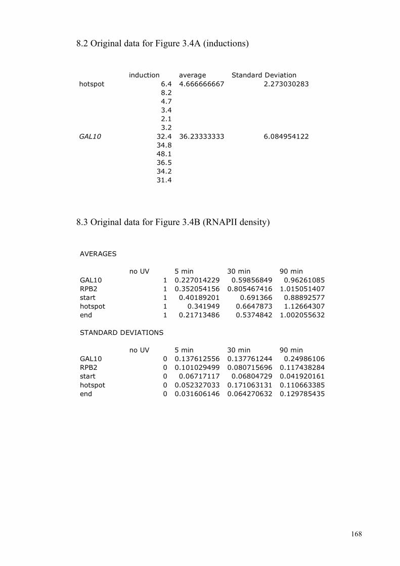
168
8.2 Original data for Figure 3.4A (inductions)
induction average Standard Deviation hotspot 6.4 4.666666667 2.273030283 8.2 4.7 3.4 2.1 3.2 GAL10 32.4 36.23333333 6.084954122 34.8 48.1 36.5 34.2 31.4
8.3 Original data for Figure 3.4B (RNAPII density)
AVERAGES no UV 5 min 30 min 90 min GAL10 1 0.227014229 0.59856849 0.96261085 RPB2 1 0.352054156 0.805467416 1.015051407 start 1 0.40189201 0.691366 0.88892577 hotspot 1 0.341949 0.6647873 1.12664307 end 1 0.21713486 0.5374842 1.002055632 STANDARD DEVIATIONS no UV 5 min 30 min 90 min GAL10 0 0.137612556 0.137761244 0.24986106 RPB2 0 0.101029499 0.080715696 0.117438284 start 0 0.06717117 0.06804729 0.041920161 hotspot 0 0.052327033 0.171063131 0.110663385 end 0 0.031606146 0.064270632 0.129785435
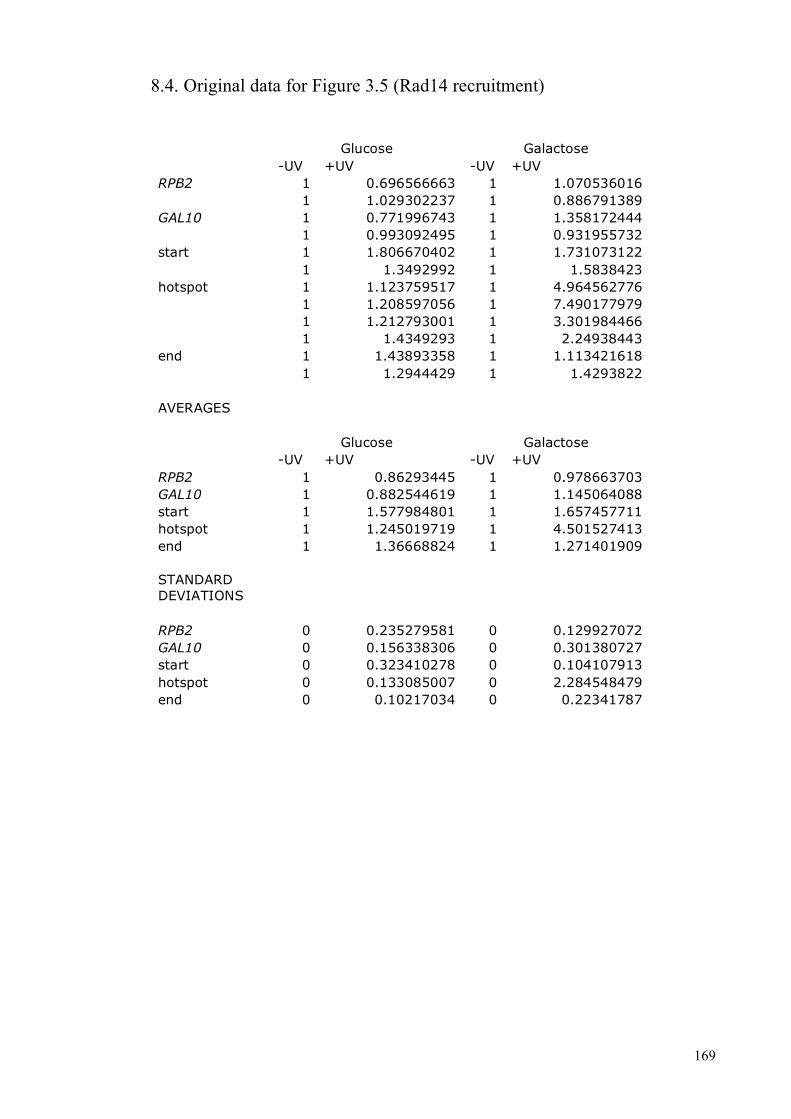
169
8.4. Original data for Figure 3.5 (Rad14 recruitment)
Glucose Galactose -UV +UV -UV +UV RPB2 1 0.696566663 1 1.070536016 1 1.029302237 1 0.886791389 GAL10 1 0.771996743 1 1.358172444 1 0.993092495 1 0.931955732 start 1 1.806670402 1 1.731073122 1 1.3492992 1 1.5838423 hotspot 1 1.123759517 1 4.964562776 1 1.208597056 1 7.490177979 1 1.212793001 1 3.301984466 1 1.4349293 1 2.24938443 end 1 1.43893358 1 1.113421618 1 1.2944429 1 1.4293822 AVERAGES Glucose Galactose -UV +UV -UV +UV RPB2 1 0.86293445 1 0.978663703 GAL10 1 0.882544619 1 1.145064088 start 1 1.577984801 1 1.657457711 hotspot 1 1.245019719 1 4.501527413 end 1 1.36668824 1 1.271401909 STANDARD DEVIATIONS RPB2 0 0.235279581 0 0.129927072 GAL10 0 0.156338306 0 0.301380727 start 0 0.323410278 0 0.104107913 hotspot 0 0.133085007 0 2.284548479 end 0 0.10217034 0 0.22341787

170
8.5. Data for the quantifications in Figure 5.2 first experiment – transcribed strand
WT 0 1 2 3 4 top band 1001388.053 1627432.574 997727.198 885618.046 813959.963 total damage 630066.767 772347.73 157053.521 53833.495 44633.991 sum 1631454.82 2399780.304 1154780.719 939451.541 858593.954 ratio 1 1.470944996 0.707822678 0.575836689 0.526275042 adjusted damage 630066.767 525069.076 221882.5788 93487.43502 84811.14899 0 1 2 3 4 damage (%) 100 83.33546594 35.21572481 14.83770291 13.46066059 repair (%) 0 16.66453406 64.78427519 85.16229709 86.53933941 t50% 1.7 rad26 0 1 2 3 4 top band 930251.69 654265.951 745717.923 698077.504 684905.913 total damage 605916.859 465973.235 300522.791 208688.633 188738.726 sum 1536168.549 1120239.186 1046240.714 906766.137 873644.639 ratio 1 0.729242365 0.681071563 0.590277765 0.568716655 adjusted damage 605916.859 638982.6719 441249.9472 353543.1039 331867.7663 0 1 2 3 4 damage (%) 100 105.4571535 72.82351376 58.34845138 54.77117221
repair (%) 0 -
5.457153477 27.17648624 41.65154862 45.22882779 t50% 4.0 mec1 0 1 2 3 4 top band 1124924.852 1531041.256 1165542.983 1429369.387 998624.132 total damage 817366.844 632444.449 399777.446 361813.451 215339.682 sum 1942291.696 2163485.705 1565320.429 1791182.838 1213963.814 ratio 1 1.113883002 0.805914185 0.922200739 0.62501622 adjusted damage 817366.844 567783.5535 496054.6092 392336.9778 344534.5498 0 1 2 3 4 damage remaining (%) 100 69.46496027 60.68934809 48.00010922 42.15176482 repair (%) 0 30.53503973 39.31065191 51.99989078 57.84823518 t50% 3.0

171
second experiment – transcribed strand
WT 0 1 2 3 4 top band 281064.637 268446.926 274526.338 393213.259 329901.274 total damage 210196.384 135853.492 43449.984 30325.552 18695.135 sum 491261.021 404300.418 317976.322 423538.811 348596.409 ratio 1 0.822984932 0.647265524 0.862146177 0.709595091 adjusted damage 210196.384 165074.094 67128.53136 35174.48992 26346.20114 damage (%) 100 78.53327008 31.93610189 16.734108 12.53408866 repair (%) 0 21.46672992 68.06389811 83.265892 87.46591134 t50%= 1.8 0 1 2 3 4 top band 293255.579 311091.82 274032.593 267507.498 295224.652 total damage 198124.07 170532.052 109801.684 80044.623 67977.287 sum 491379.649 481623.872 383834.277 347552.121 363201.939 ratio 1.000241477 0.980382834 0.781324511 0.707469362 0.739325783 adjusted damage 198076.2392 173944.3471 140532.7523 113142.1759 91944.97009 damage (%) 100 87.81686678 70.94881894 57.12051903 46.41898011 repair (%) 0 12.18313322 29.05118106 42.87948097 53.58101989 t50%=3.8 0 1 2 3 4 top band 264113.694 267996.195 278155.939 272040.821 240036.634 total damage 193166.05 141049.258 86529.976 68064.597 50850.006 sum 457279.744 409045.453 364685.915 340105.418 290886.64 ratio 0.930828469 0.83264382 0.742346531 0.692311019 0.59212237 adjusted damage 207520.5652 169399.2733 116562.7808 98315.05659 85877.52901 damage remaining (%) 100 81.63011369 56.16926719 47.37605474 41.38265955 repair (%) 0 18.36988631 43.83073281 52.62394526 58.61734045 t50% 2.9
summary TS
average t50% STDEV STD-Error wt 1.8 0.141421356 0.100298834 rad26 3.9 0.141421356 0.100298834 mec1 2.95 0.070710678 0.050149417

172
first experiment – non-transcribed strand
WT 0 1 2 3 4 top band 650116.866 1073939.161 730777.335 489818.719 771437.962 total damage 375548.648 574451.425 338672.902 189644.293 185262.511 sum 1025665.514 1648390.586 1069450.237 679463.012 956700.473 ratio 1 1.607142449 1.042689086 0.66246062 0.93276069 adjusted damage 375548.648 357436.533 324807.1804 286272.5532 198617.4084 damage remaining (%) 100 95.17715878 86.48870983 76.22782154 52.88726493 repair (%) 0 4.822841221 13.51129017 23.77217846 47.11273507 t50% = 4.8 rad26 0 1 2 3 4 top band 855661.338 826154.502 616409.418 850555.049 880239.629 total damage 440059.724 422576.028 231175.977 229747.088 147315.284 sum 1295721.062 1248730.53 847585.395 1080302.137 1027554.913 ratio 1.263297873 1.217483198 0.826376029 1.053269436 1.00184212 adjusted damage 348342.013 347089.8233 279746.7119 218127.5562 147044.4105 damage remaining (%) 100 99.64052865 80.30805973 62.61879075 42.21265454 repair (%) 0 0.359471346 19.69194027 37.38120925 57.78734546 t50% = 3.7 mec1 0 1 2 3 4 top band 962550.814 1014754.542 1051627.703 1271249.7 676688.404 total damage 551525.385 628017.793 551922.565 530319.477 332970.097 sum 1514076.199 1642772.335 1603550.268 1801569.177 1009658.501 ratio 1.476189048 1.601664785 1.563424183 1.756488009 0.984393535 adjusted damage 373614.3319 392103.141 353021.6374 301920.3514 338248.9677 damage remaining (%) 100 104.9486349 94.48824826 80.81069853 90.53425922
repair (%) 0 -
4.948634869 5.511751739 19.18930147 9.465740781 t50%=more than 6 hours

173
second experiment – non-transcribed strand
WT 0 1 2 3 4 top band 1420621.061 1113659.404 924010.245 1336602.163 1323936.455 total damage 804561.212 641212.88 457670.5 557023.413 340901.656 sum 2225182.273 1754872.284 1381680.745 1893625.576 1664838.111 ratio 1 0.788642039 0.620929243 0.85099796 0.748180556 adjusted damage 804561.212 813059.4726 737073.5151 654553.1704 455640.8919 damage remaining (%) 100 101.0562603 91.61186298 81.35529784 56.63222202
repair (%) 0 -
1.056260297 8.388137022 18.64470216 43.36777798 t50% = 5.3 rad26 0 1 2 3 4 top band 1571086.701 1715304.93 1528921.921 1790145.039 1801049.19 total damage 707187.503 661082.273 413508.166 331612.114 263825.784 sum 2278274.204 2376387.203 1942430.087 2121757.153 2064874.974 ratio 1.023859587 1.067951705 0.872930775 0.953520608 0.927957677 adjusted damage 690707.5068 619018.8842 473700.9825 347776.5571 284307.9921 damage remaining (%) 100 89.6209869 68.58199424 50.35077129 41.1618506 repair (%) 0 10.3790131 31.41800576 49.64922871 58.8381494 t50% = 3.9 mec1 0 1 2 3 4 top band 1115033.796 1587842.171 1452173.426 1086754.966 952422.93 total damage 691886.175 701979.177 620437.33 472786.193 435533.421 sum 1806919.971 2289821.348 2072610.756 1559541.159 1387956.351 ratio 0.812032341 1.02904889 0.931434149 0.70085996 0.623749509 adjusted damage 852042.634 682163.0963 666109.7093 674580.1158 698250.5228 damage remaining (%) 100 80.06208481 78.17797875 79.17210816 81.95018593 repair (%) 0 19.93791519 21.82202125 20.82789184 18.04981407 t50%=more than 6 hours

174
summary NTS
average t50% STDEV STD-Error wt 5.05 0.353553391 0.250747086 rad26 3.8 0.141421356 0.100298834 mec1 more than 6 0 0
8.6 Data for the quantifications in Figure 5.3
first experiment – transcribed strand
wt U 0 1 2 4 total 197780956.5 211578376.3 214171417.5 196690459.2 171518282.7 ratio 0.934788138 1 1.0122557 0.929634033 0.810660738 damage 71543123.2 129799795.1 121315608.3 96603857.44 79558735.3 ratio 76534051.15 129799795.1 119846801.8 103916007.9 98140605 corrected 53265743.98 43312750.67 27381956.71 21606553.84 1 0.813144574 0.514063161 0.405636948 t 50% = 2.1 h chk1 U 0 1 2 4 total 275533726.7 285215777.2 273807961.2 233300238.5 182550719.2 ratio 0.966053594 1 0.960002858 0.817978026 0.640044253 damage 148801873.6 193589824.4 175991185.6 131070280.9 94752039.15 ratio 154030661 193589824.4 183323606 160236921.7 148039824.9 corrected 44787950.79 34521732.39 11435048.09 -762048.757
1 0.77078169 0.255315278 -
0.017014593 t50% = 1.9 h rad53 U 0 1 2 4 total 257738974.5 317784509.6 266201534.3 221940517.1 220514815.3 ratio 0.811049522 1 0.83767939 0.69839942 0.693913041 damage 143214274 224553737.1 184106940.8 142705718.8 143224705.8 ratio 176578951.3 224553737.1 219782106.3 204332527.7 206401519.3 corrected 47974785.82 36803154.96 21353576.4 23422567.97 1 0.767135368 0.445099984 0.488226629 t50% = 1.9 h
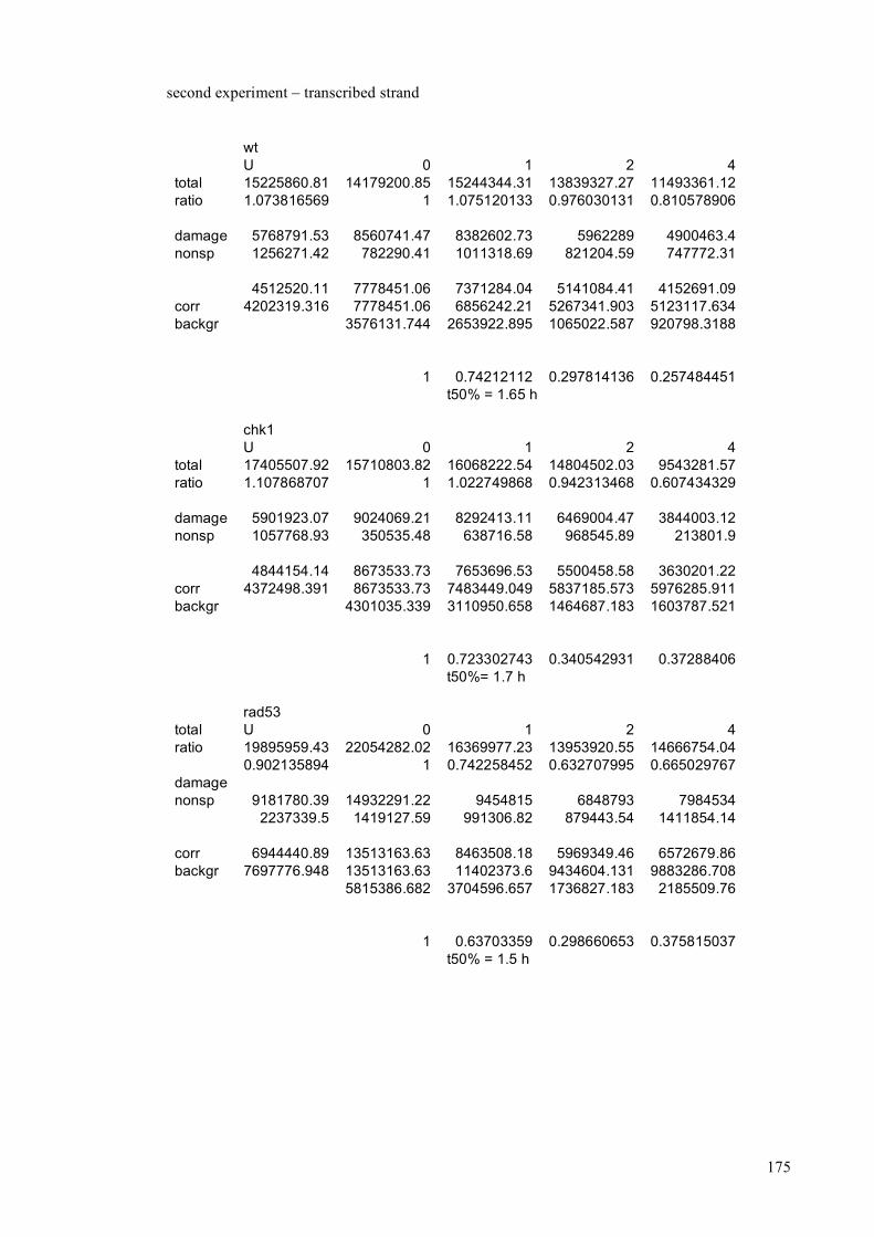
175
second experiment – transcribed strand
wt U 0 1 2 4 total 15225860.81 14179200.85 15244344.31 13839327.27 11493361.12 ratio 1.073816569 1 1.075120133 0.976030131 0.810578906 damage 5768791.53 8560741.47 8382602.73 5962289 4900463.4 nonsp 1256271.42 782290.41 1011318.69 821204.59 747772.31 4512520.11 7778451.06 7371284.04 5141084.41 4152691.09 corr 4202319.316 7778451.06 6856242.21 5267341.903 5123117.634 backgr 3576131.744 2653922.895 1065022.587 920798.3188 1 0.74212112 0.297814136 0.257484451 t50% = 1.65 h chk1 U 0 1 2 4 total 17405507.92 15710803.82 16068222.54 14804502.03 9543281.57 ratio 1.107868707 1 1.022749868 0.942313468 0.607434329 damage 5901923.07 9024069.21 8292413.11 6469004.47 3844003.12 nonsp 1057768.93 350535.48 638716.58 968545.89 213801.9 4844154.14 8673533.73 7653696.53 5500458.58 3630201.22 corr 4372498.391 8673533.73 7483449.049 5837185.573 5976285.911 backgr 4301035.339 3110950.658 1464687.183 1603787.521 1 0.723302743 0.340542931 0.37288406 t50%= 1.7 h rad53 total U 0 1 2 4 ratio 19895959.43 22054282.02 16369977.23 13953920.55 14666754.04 0.902135894 1 0.742258452 0.632707995 0.665029767 damage nonsp 9181780.39 14932291.22 9454815 6848793 7984534 2237339.5 1419127.59 991306.82 879443.54 1411854.14 corr 6944440.89 13513163.63 8463508.18 5969349.46 6572679.86 backgr 7697776.948 13513163.63 11402373.6 9434604.131 9883286.708 5815386.682 3704596.657 1736827.183 2185509.76 1 0.63703359 0.298660653 0.375815037 t50% = 1.5 h

176
first experiment – non-transcribed strand
wt u 0 1 2 4 top 173141412.4 188954519.8 167818361.8 141281562.5 117030865.3 0.916312627 1 0.888141559 0.747701419 0.619359968 damage 44135115.84 92926253.5 79910156.52 59457835 46784517 corrected 48166002 92926253.5 89974571.85 79520826.78 75536875.84 backgr 44760251.5 41808569.85 31354824.78 27370873.83 1 0.93405574 0.700506001 0.611499554 t50% = 4.8 h chk1 U 0 1 2 4 top 175279628.3 212979766.2 218480066.6 174536789.8 120671043.9 0.822987232 1 1.02582546 0.819499396 0.566584545 damage 60295225.24 115515962.2 120553533 89813924.86 54657087.2 corrected 73263864.71 115515962.2 117518562.1 109596084.2 96467663.44 backgr 42252097.45 44254697.34 36332219.52 23203798.73 1 1.047396461 0.859891502 0.549175074 t50%= 4.7 h rad53 U 0 1 2 4 top 175248198.9 256206911.3 148568184.6 107907857.1 106436986.7 0.684010427 1 0.579875788 0.421174653 0.415433706 damage 59643390.58 158634362.3 85457936 58645134 53457841 corrected 87196610.16 158634362.3 147372830.1 139241840.7 128679594.9 backgr 71437752.17 60176219.96 52045230.55 41482984.7 1 0.842358811 0.728539588 0.58068715 t50% = 4.5 h

177
second experiment – non-transcribed strand
wt u 0 1 2 4 total 21145731.25 19391508.65 20492418.92 17337044.69 11517124.38 ratio 1.090463441 1 1.056772801 0.894053423 0.593926166 damage 7776655.29 11951486.54 11673375.33 9465846.68 5636813.48 ratio 7131513.994 11951486.54 11046248.84 10587562.71 9490764.687 corrected 4819972.546 3914734.844 3456048.711 2359250.692 1 0.812190278 0.717026638 0.489473886 t50% = 3.9 h chk1 U 0 1 2 4 total 24447569.24 15421898.5 22675180.7 19150554.73 12632396.3 ratio 1.585250301 1 1.47032356 1.241776733 0.8191207 damage 8810999.37 8203435.9 11549764 9145675 5678942 ratio 5558112.409 8203435.9 7855253.304 7364991.435 6932973.367 corrected 2645323.491 2297140.895 1806879.026 1374860.958 1 0.868378065 0.683046528 0.519732639 t50% = 4 h rad53 U 0 1 2 4 total 24426052.89 27472742.71 15759173.95 12705587.13 11016577.73 ratio 0.889101359 1 0.573629438 0.462479748 0.401000288 damage 10524330.3 17012726.16 9145648 7145678 5814579 ratio 11837042.19 17012726.16 15943477.44 15450791.15 14500186.61 corrected 5175683.973 4106435.258 3613748.963 2663144.418 1 0.793409196 0.698216696 0.514549272 t50% = 4 h
summary TS and NTS
TS wt chk1 rad53 exp 1 2.1 1.9 1.9 exp 2 1.65 1.7 1.5 AVERAGE 1.875 1.8 1.7 STD-Dev 0.318198052 0.141421356 0.282842712 STD-Error 0.225672377 0.100298834 0.200597668 NTS wt chk1 rad53 exp 1 4.8 4.7 4.5 exp 2 3.9 4 4 AVERAGE 4.35 4.35 4.25 STD-Dev 0.636396103 0.494974747 0.353553391 STD-Error 0.451344754 0.35104592 0.250747086
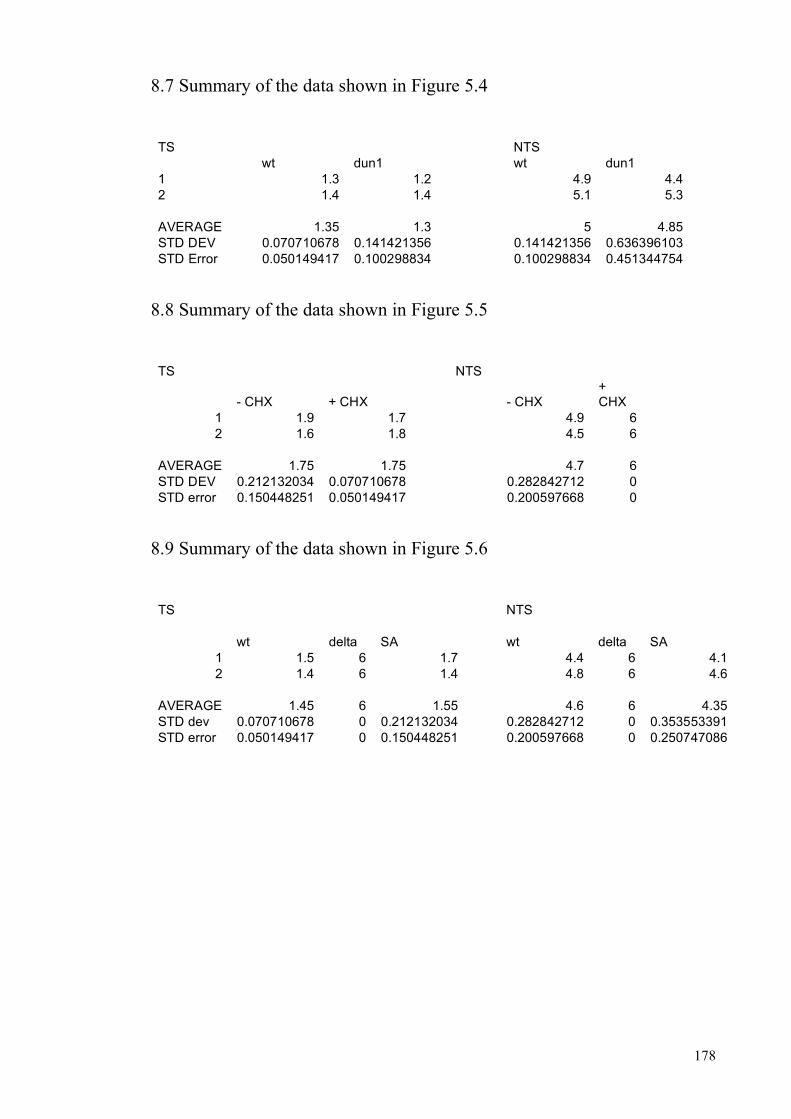
178
8.7 Summary of the data shown in Figure 5.4
TS NTS wt dun1 wt dun1 1 1.3 1.2 4.9 4.4 2 1.4 1.4 5.1 5.3 AVERAGE 1.35 1.3 5 4.85 STD DEV 0.070710678 0.141421356 0.141421356 0.636396103 STD Error 0.050149417 0.100298834 0.100298834 0.451344754
8.8 Summary of the data shown in Figure 5.5
TS NTS
- CHX + CHX - CHX + CHX
1 1.9 1.7 4.9 6 2 1.6 1.8 4.5 6
AVERAGE 1.75 1.75 4.7 6 STD DEV 0.212132034 0.070710678 0.282842712 0 STD error 0.150448251 0.050149417 0.200597668 0
8.9 Summary of the data shown in Figure 5.6
TS NTS wt delta SA wt delta SA
1 1.5 6 1.7 4.4 6 4.1 2 1.4 6 1.4 4.8 6 4.6
AVERAGE 1.45 6 1.55 4.6 6 4.35 STD dev 0.070710678 0 0.212132034 0.282842712 0 0.353553391 STD error 0.050149417 0 0.150448251 0.200597668 0 0.250747086

179
8.10 Summary of the data shown in Figure 5.14
RPB2 transcribed strand t50 % v wt SA SE 3.25 1.9 2.1 4 1 2.4 1.8 3.9 1.9 2.66 1.96 1.3 3.1 1.9 1.95 2.3 MEAN 3.716666667 1.75 2.72 1.953333333 STDEV 0.407226391 0.442530602 0.35383612 0.15011107 STERR 0.154252421 0.167625228 0.204529549 0.086769405 RPB2 non-transcribed strand v wt SA SE 4.6 5.6 5.2 5 5 5.2 5 3.6 5.2 4.9 3.5 4.2 4.2 MEAN 4.175 4.88 5.1 4.95 STDEV 0.741057803 0.641872261 0.141421356 0.070710678 STERR 0.370528901 0.287835095 0.101015254 0.040873224

180
8.11 Summary of the data shown in Figure 5.15
TS
mec1 mec1rad26 mec1rad26 + RAD26wt
mec1rad26 + RAD26SE
2.5 6 2.7 2.7 2.4 6 2.4 2.3 2.45 6 2.55 2.5 0.070710678 0 0.212132034 0.282842712 0.050149417 0 0.150448251 0.200597668
only the results from the TS are shown here, the t50% values for the NTS
were always more than 6 hours.