Genome-Wide Association Study of Nucleotide Variants ...
17
Citation: Waldner, M.; Kinnear, A.; Yacoub, E.; McAllister, T.; Register, K.; Li, C.; Jelinski, M. Genome-Wide Association Study of Nucleotide Variants Associated with Resistance to Nine Antimicrobials in Mycoplasma bovis. Microorganisms 2022, 10, 1366. https://doi.org/10.3390/ microorganisms10071366 Academic Editor: Teresa Semedo-Lemsaddek Received: 17 June 2022 Accepted: 1 July 2022 Published: 6 July 2022 Publisher’s Note: MDPI stays neutral with regard to jurisdictional claims in published maps and institutional affil- iations. Copyright: © 2022 by the authors. Licensee MDPI, Basel, Switzerland. This article is an open access article distributed under the terms and conditions of the Creative Commons Attribution (CC BY) license (https:// creativecommons.org/licenses/by/ 4.0/). microorganisms Article Genome-Wide Association Study of Nucleotide Variants Associated with Resistance to Nine Antimicrobials in Mycoplasma bovis Matthew Waldner 1 , Andrea Kinnear 1 , Elhem Yacoub 1 , Tim McAllister 2 , Karen Register 3 , Changxi Li 4,5 and Murray Jelinski 1, * 1 Western College of Veterinary Medicine, University of Saskatchewan, Saskatoon, SK S7N 5B4, Canada; [email protected] (M.W.); [email protected] (A.K.); [email protected] (E.Y.) 2 Lethbridge Research and Development Centre, Agriculture and Agri-Food Canada, Lethbridge, AB T1J 4B1, Canada; [email protected] 3 Ruminant Diseases and Immunology Research Unit, USDA/Agricultural Research Service/National Animal Disease Center, Ames, IA 50010, USA; [email protected] 4 Lacombe Research and Development Centre, Agriculture and Agri-Food Canada, Lacombe, AB T4L 1W1, Canada; [email protected] 5 Department of Agriculture, Food and Nutritional Science, University of Alberta, Edmonton, AB T6G 2P5, Canada * Correspondence: [email protected]; Tel.: +1-306-966-7166 Abstract: Antimicrobial resistance (AMR) studies of Mycoplasma bovis have generally focused on specific loci versus using a genome-wide association study (GWAS) approach. A GWAS approach, using two different models, was applied to 194 Mycoplasma bovis genomes. Both a fixed effects linear model (FEM) and a linear mixed model (LMM) identified associations between nucleotide variants (NVs) and antimicrobial susceptibility testing (AST) phenotypes. The AMR phenotypes represented fluoroquinolones, tetracyclines, phenicols, and macrolides. Both models identified known and novel NVs associated (Bonferroni adjusted p < 0.05) with AMR. Fluoroquinolone resistance was associated with multiple NVs, including previously identified mutations in gyrA and parC. NVs in the 30S ribosomal protein 16S were associated with tetracycline resistance, whereas NVs in 5S rRNA, 23S rRNA, and 50S ribosomal proteins were associated with phenicol and macrolide resistance. For all antimicrobial classes, resistance was associated with NVs in genes coding for ABC transporters and other membrane proteins, tRNA-ligases, peptidases, and transposases, suggesting a NV-based multifactorial model of AMR in M. bovis. This study was the largest collection of North American M. bovis isolates used with a GWAS for the sole purpose of identifying novel and non-antimicrobial-target NVs associated with AMR. Keywords: Mycoplasma bovis; genome-wide association study; antimicrobial resistance; fluoro- quinolone; tetracycline; phenicol; macrolide 1. Introduction Mycoplasma bovis is associated with a variety of cattle diseases having a range of clinical manifestations. In feedlot cattle, M. bovis is commonly associated with bovine respiratory disease (BRD) and chronic pneumonia and polyarthritis syndrome (CPPS) [1–4]. The lack of clinically efficacious vaccines against M. bovis has resulted in antimicrobials being administered for the prevention, metaphylaxis, and treatment of mycoplasmosis in feedlot cattle [5]. This extensive use of antimicrobials, however, has potentially contributed to increasing levels of antimicrobial resistance (AMR) in M. bovis isolates worldwide [6–15]. Furthermore, AMR in M. bovis can be attributed, in part, to the lack of a cell wall, making it innately resistant to beta-lactams, sulfonamides, trimethoprim, polymyxins, and nalidixic acid [16,17]. Microorganisms 2022, 10, 1366. https://doi.org/10.3390/microorganisms10071366 https://www.mdpi.com/journal/microorganisms
-
Upload
khangminh22 -
Category
Documents
-
view
4 -
download
0
Transcript of Genome-Wide Association Study of Nucleotide Variants ...

Citation: Waldner, M.; Kinnear, A.;
Yacoub, E.; McAllister, T.; Register, K.;
Li, C.; Jelinski, M. Genome-Wide
Association Study of Nucleotide
Variants Associated with Resistance
to Nine Antimicrobials in Mycoplasma
bovis. Microorganisms 2022, 10, 1366.
https://doi.org/10.3390/
microorganisms10071366
Academic Editor: Teresa
Semedo-Lemsaddek
Received: 17 June 2022
Accepted: 1 July 2022
Published: 6 July 2022
Publisher’s Note: MDPI stays neutral
with regard to jurisdictional claims in
published maps and institutional affil-
iations.
Copyright: © 2022 by the authors.
Licensee MDPI, Basel, Switzerland.
This article is an open access article
distributed under the terms and
conditions of the Creative Commons
Attribution (CC BY) license (https://
creativecommons.org/licenses/by/
4.0/).
microorganisms
Article
Genome-Wide Association Study of Nucleotide VariantsAssociated with Resistance to Nine Antimicrobials inMycoplasma bovisMatthew Waldner 1, Andrea Kinnear 1 , Elhem Yacoub 1, Tim McAllister 2 , Karen Register 3, Changxi Li 4,5
and Murray Jelinski 1,*
1 Western College of Veterinary Medicine, University of Saskatchewan, Saskatoon, SK S7N 5B4, Canada;[email protected] (M.W.); [email protected] (A.K.); [email protected] (E.Y.)
2 Lethbridge Research and Development Centre, Agriculture and Agri-Food Canada,Lethbridge, AB T1J 4B1, Canada; [email protected]
3 Ruminant Diseases and Immunology Research Unit, USDA/Agricultural Research Service/National AnimalDisease Center, Ames, IA 50010, USA; [email protected]
4 Lacombe Research and Development Centre, Agriculture and Agri-Food Canada,Lacombe, AB T4L 1W1, Canada; [email protected]
5 Department of Agriculture, Food and Nutritional Science, University of Alberta,Edmonton, AB T6G 2P5, Canada
* Correspondence: [email protected]; Tel.: +1-306-966-7166
Abstract: Antimicrobial resistance (AMR) studies of Mycoplasma bovis have generally focused onspecific loci versus using a genome-wide association study (GWAS) approach. A GWAS approach,using two different models, was applied to 194 Mycoplasma bovis genomes. Both a fixed effects linearmodel (FEM) and a linear mixed model (LMM) identified associations between nucleotide variants(NVs) and antimicrobial susceptibility testing (AST) phenotypes. The AMR phenotypes representedfluoroquinolones, tetracyclines, phenicols, and macrolides. Both models identified known and novelNVs associated (Bonferroni adjusted p < 0.05) with AMR. Fluoroquinolone resistance was associatedwith multiple NVs, including previously identified mutations in gyrA and parC. NVs in the 30Sribosomal protein 16S were associated with tetracycline resistance, whereas NVs in 5S rRNA, 23SrRNA, and 50S ribosomal proteins were associated with phenicol and macrolide resistance. Forall antimicrobial classes, resistance was associated with NVs in genes coding for ABC transportersand other membrane proteins, tRNA-ligases, peptidases, and transposases, suggesting a NV-basedmultifactorial model of AMR in M. bovis. This study was the largest collection of North American M.bovis isolates used with a GWAS for the sole purpose of identifying novel and non-antimicrobial-targetNVs associated with AMR.
Keywords: Mycoplasma bovis; genome-wide association study; antimicrobial resistance; fluoro-quinolone; tetracycline; phenicol; macrolide
1. Introduction
Mycoplasma bovis is associated with a variety of cattle diseases having a range ofclinical manifestations. In feedlot cattle, M. bovis is commonly associated with bovinerespiratory disease (BRD) and chronic pneumonia and polyarthritis syndrome (CPPS) [1–4].The lack of clinically efficacious vaccines against M. bovis has resulted in antimicrobialsbeing administered for the prevention, metaphylaxis, and treatment of mycoplasmosis infeedlot cattle [5]. This extensive use of antimicrobials, however, has potentially contributedto increasing levels of antimicrobial resistance (AMR) in M. bovis isolates worldwide [6–15].Furthermore, AMR in M. bovis can be attributed, in part, to the lack of a cell wall, making itinnately resistant to beta-lactams, sulfonamides, trimethoprim, polymyxins, and nalidixicacid [16,17].
Microorganisms 2022, 10, 1366. https://doi.org/10.3390/microorganisms10071366 https://www.mdpi.com/journal/microorganisms

Microorganisms 2022, 10, 1366 2 of 17
Research of AMR determinants of M. bovis has primarily focused on antimicrobialtarget-site modifications (‘hot spots’); specifically, single nucleotide polymorphisms (SNPs)in the genes coding for antimicrobial targets. Macrolide and phenicol resistances areprimarily associated with mutations within domains II and IV of the 23S component of the50S ribosomal subunit, respectively [7,18,19]. Tetracycline resistance is linked to mutationsin the 16S component of the 30S subunit [19,20], while mutations in the gyrA and parCgenes reduce the binding affinity of fluoroquinolones [21–24].
Although the aforementioned SNPs have a role in AMR, there is a lack of concordancebetween in vitro antimicrobial susceptibility testing (AST) results and known SNPs. Khalilet al. surmised that M. bovis may have resistance mechanisms other than SNPs, such asefflux pumps that contribute to fluoroquinolone resistance [22]. Similarly, Calcutt et al.noted the need for more research into mechanisms of resistance other than antimicrobialtarget-site modification [25]. One such method to identify possible mechanisms of resistanceis the use of genome-wide association studies (GWAS). GWAS apply a statistical model toassociate a set of phenotypic traits with a set of genetic variations, most commonly SNPs,across a set of genomes.
Only a few GWAS studies have been conducted to characterize AMR in M. bovis [26,27],which may be related to the technical expertise required in conducting such studies. Fur-thermore, a limitation of GWAS is the occurrence of false positive associations between thetraits and genetic variants, commonly referred to as “p-value inflation” [28]. In bacteria,p-value inflation is often caused by a lack of statistical power owing to linkage disequilib-rium in clonal populations as well as limited recombinations [28–30]. Other causes for falsepositives include polygenic inheritance for a trait and variant penetrance [31]. Althoughsample size requirements may differ depending on the variation of phenotypic traits, thenumber of DNA markers analyzed, and the purpose of the study, there is an underlyingconvention of having a minimum sample size of 100 bacterial isolates [32–34]. Bokma et al.performed a combination GWAS and manual study of 95 M. bovis genomes to identifygene targets associated with AMR [27]. This analysis identified known NVs as well asnovel variants associated with resistance to fluoroquinolones, macrolides, tetracyclines,and aminoglycosides. However, the GWAS may have been constrained by both the numberof isolates and skewed minimum inhibitory concentrations (MIC). Ledger et al. performeda GWAS on two M. bovis isolates to identify AMR traits associated with SNPs, multiplenucleotide polymorphisms (MNPs), as well as insertions and deletions (indels) [26]. Theyidentified 77 genes associated with AMR across six different functional groups: topoiso-merases, methyltransferases, 30S ribosomal proteins, 50S ribosomal proteins, tRNA ligases,and ABC transporters.
The objective of this study was to use GWAS to associate (p < 0.05) NVs with M.bovis AMR phenotypes. The study involved a dataset of 194 M. bovis genomes and AMRprofiles consisting of MIC values generated from the AST of nine different antimicrobials.Both a fixed effects linear model (FEM) and a linear mixed model (LMM) were used toanalyze each dataset, which utilized continuous numeric MICs versus a binary phenotype(susceptible or resistant). Population stratification was considered in the GWAS to accountfor the compounding effects of isolates obtained from a variety of sources.
2. Materials and Methods2.1. Sample Collection, Isolation, DNA Extraction, and Sequencing
Mycoplasma bovis isolates (n = 194) were acquired from North American feedlot cattle(n = 115), farmed bison (n = 77), white-tail deer (n = 1), and mule deer (n = 1). Isolatesoriginated from animals of varying health status (healthy n = 39, sick with pneumoniapresentation n = 17, dead n = 134, unknown n = four) and derived from different anatomicallocations (nasopharynges n = 57, lungs n = 88, stifle joints n = 38, unknown n = 11), and overa range of years (2006 to 2018). Deep nasopharyngeal (DNP) swabs were obtained fromcattle (n = 39) and bison (n = 18), while lung and joint samples were obtained at the timeof postmortem examination from animals having gross pathological findings consistent

Microorganisms 2022, 10, 1366 3 of 17
with mycoplasmosis (cattle n = 76, bison n = 48, white-tail deer n = one, mule deer n = one).The methods for culturing, isolation, DNA extraction, identification, and whole genomesequencing have been previously described [6,7].
2.2. Genome Assembly, Quality Control, and Data Preprocessing
Data-preprocessing required each isolate to be indexed to a BAM file, consensus sequenceFASTA file, and variant call file (VCF). Trimming of the MiSeq paired-reads was performed inTrimmomatic v0.39 [35] with the following settings: sliding window:5:15, leading:5, trailing:5,and minlen:75. SAM alignment files were created for each read set by aligning the trimmedpaired-reads to the M. bovis PG45 reference genome (CP002188.1/NC_014760) using BWAmem v0.7.17-r1188 [36]. These SAM files were then converted to BAM, sorted, and indexedwith Samtools [37]. Bcftools mpileup and vcf2fq generated the consensus FASTA sequence foreach sequenced isolate [37]. All whole genome sequences (WGS) had an average coverage of≥30X. VCF files were created by inputting the indexed BAM file groups from the assemblystage into freebayes [38], with settings ploidy 1 and strict-vcf. Bcftools was run three timeson the resultant VCFs with the following settings: annotate -x ‘FORMAT’, norm -m -, and+missing2ref to remove format tags, normalize multiallelic records into biallelic records, andfill the missing alleles with the reference allele, respectively. Raw reads for each of the genomeshave been made available from the Sequence Read Archive (SRA) under BioProject accessionno. PRJNA642970, PRJNA708306, and PRJNA785928.
2.3. Antimicrobial Resistance Phenotypes
AMR phenotypes for each GWAS were determined by AST using a customized Sensi-titre™ microplate (Trek Diagnostics, Oakwood, GA, USA) comprised of nine antimicrobialscommonly used in western Canada: enrofloxacin (ENRO), chlortetracycline (CTET), oxyte-tracycline (OXY), florfenicol (FFN), tilmicosin (TIL), tildipirosin (TIP), gamithromycin(GAM), tulathromycin (TUL), and tylosin tartrate (TYLT). The AST method has been pre-viously described [6], and was performed through preparation of the following serialtwo-fold dilutions: ENRO, 0.12–128 µg/mL; TIP, 0.12–128 µg/mL; GAM, 0.25–256 µg/mL;TUL, 0.25–256 µg/mL; TIL, 1–256 µg/mL; TYLT, 1–128 µg/mL; FFN, 0.25–256 µg/mL; OXY,0.5–256 µg/mL; and CTET, 1–256 µg/mL. A control in the form of penicillin (2–8 µg/mL)was also prepared. Growth was assessed by using the color redox indicator alamarBlue(Invitrogen™, Thermo Fisher Scientific, Waltham, MA, USA) based on a blue-to-pink colorchange. MICs were added to a phenotype file with the accompanying isolate identifiers.
2.4. Genome-Wide Association Study Pipeline
GWAS were implemented using Pyseer [39], a python-based adaptation of the SEERGWAS suite used for microbial GWAS [40]. A fixed effects linear model (FEM) and alinear mixed model (LMM), were run for each of the nine antimicrobials (18 GWAS). Eachmodel identified associations between NVs and AMR phenotypes, while accounting forconfounding population structures.
The GWAS first estimated the population structure for the FEM and LMM. A pairwisedistance matrix was created for the FEM using the FASTA of all isolate consensus sequencesas input for mash [41]. Nonparametric multidimensional scaling (MDS) was run onthe pairwise distance matrix using the scree_plot_pyseer function. Then, based upona subjective visual examination of the slope of the scree plot, five dimensions were retainedduring MDS in the FEM. The FEM also required a gene presence/absence table (Rtab file),with the distance file from mash to determine clusters of orthologous groups of proteins(COGs) for Pyseer execution. The COGs acted as the population substructure to accountfor the confounding population structure of the dataset during the FEM runs. To facilitatethe creation of the Rtab file, Prokka software was used for genome annotation through thegeneration of GFF files for each assembly [42]. Prokka received a FASTA assembly file ofeach isolate, an annotation FASTA file of the PG45 reference genome from GenomeNet(downloaded 31 August 2021), and the genetic code setting for M. bovis (-gcode 4). The

Microorganisms 2022, 10, 1366 4 of 17
Prokka files were then received by the Roary pipeline [42], which annotated the assembliesto calculate the pan genome. For each GWAS, the phenotype file, Rtab file, mash distancematrix, and dimension cut-off of five were passed to Pyseer to determine the COGs andcreate an MDS decomposition that functions as the accounting factor for the bacterialpopulation structure. The FEM assumed that the nucleotide variant effect and the MDSeffect were fixed effects. The LMM required only a similarity or kinship matrix as thepairwise distance matrix in order to correct for population structure [39], as the nucleotidevariant effect was a fixed effect and the population structure effect was random. Thismatrix was calculated using a VCF file and the list of the isolate identifiers using Pyseer’ssimilarity_pyseer script. A principle component analysis (PCA) of cattle source location,tissue sample location, disease status, isolation year, and host species were performedusing Plink 1.9 [43] against the VCF to determine if isolates clustered by these criteriato determine if known variables were having a fixed effect on the dataset that could beaccounted for. Further methods to account for fixed effects due to these variables wereunnecessary as it was determined that clustering according to these variables did not occur.
The phenotype file, VCF file, and population matrix file (MDS decomposition matrixfor FEM and kinship matrix for LMM) were passed to Pyseer for the GWAS runs. Each runwas set to exclude allelic frequencies of <0.02 and >0.98, as per the Pyseer tutorial. TheGWAS functioned by applying MDS to the population matrix, projecting the matrix into areduced number of dimensions. Linear regression using the relevant model and distancewas applied to the dataset to determine the variants’ associations to the phenotypes. TheGWAS results files were used to create quantile-quantile (Q-Q) plots to identify possiblep-value inflation for each GWAS. To ensure significance and quality, each set of results wasfiltered with a Bonferroni corrected p-value threshold of 0.05 and the removal of resultsthat failed a chi-squared test. Bonferroni thresholds were determined by the Pyseer scriptcount_patterns.py using the number of unique variant patterns as the number of multipletests output from each GWAS run. Residuals were plotted and determined to be normallydistributed, satisfying an essential assumption for using LMM.
2.5. Visualization and Verification
Plots were visualized using the ggplot2 package in R [44]. Manhattan plots, alsoreferred to as −log10(p-value) genome-wide association plots, relate the p-values of signifi-cant NVs (y-axis) of the GWAS to a genomic map in base pairs (x-axis). In this instance,the PG45 reference genome was used to map the location of the NVs on the x-axis. Onlythe variants meeting the Bonferroni-corrected p-value threshold were included in eachplot. A selection of NVs were manually labelled with the gene in which the NV wasidentified based on results with the most significant p-values per GWAS and/or the genecontaining the NV having been identified in previous AMR studies. Where applicable,NVs identified by the GWAS were verified by the scientific literature. Comparison wasachieved by aligning Escherichia coli genes against the annotated PG45 M. bovis genomein Geneious 2020.1.2 5 (https://www.geneious.com; accessed on 14 September 2021) todetermine positioning relative to the nucleotide numbering of the PG45 type strain as perthe common notation convention for M. bovis NVs and amino acids (AAs).
3. Results and Discussion3.1. Analysis of the Genome Assembly Quality and Minimum Inhibitory Concentrations
Assembly results for all 194 genomes are presented in Table S1. The number of readsfor each isolate ranged from 131,623 to 664,130, with an average of 292,566. Coverageranged from 30X to 137X, with an average of 64X. Freebayes identified a total of 83,208 NVs(14,492 in coding regions and 68,716 in non-coding regions). PCA analysis of the NVs withsourcing location, host species, isolation year, disease status, or tissue tropism showed nodefinitive clustering, suggesting no inherent population stratification due to these variables(Figure S1).

Microorganisms 2022, 10, 1366 5 of 17
Overall, there was wide variation in MIC levels across the M. bovis isolates andbetween antimicrobial classes (Figure 1). Most isolates had low resistance to ENRO with asmall number exhibiting resistance up to 16 µg/mL. The CTET and OXY resistant isolateshad a similar unimodal distribution at 4–8 and 2–4 µg/mL, respectively. FFN resistancealso had a unimodal distribution centered at 1–2 µg/mL. The macrolides showed notablevariation in overall MIC profiles even though all five macrolides share a common chemicalarchitecture (Figure S2). Most isolates had high MIC values for TIL and TIP, both of whichare derivatives of TYLT, and all three share a 16-membered core structure. Whereas GAMand TUL, which share a 15-membered core structure, had a bimodal MIC distribution, mostisolates had low MICs for GAM and TUL. The GWAS models received the MIC data asa continuous variable. Two separate models, FEM and LMM, were used to associate theNVs and MICs. This provided internal confirmation for newly identified NVs. The resultsof each model are discussed and presented together when they concur, and separatelywhen they do not. Additionally, internal testing of the GWAS pipeline during developmentshowed that the use of a continuous phenotype resulted in lower p-value inflation ascompared to binary phenotypic data.
Figure 1. Frequency distributions of Mycoplasma bovis isolates (y-axis) by minimum inhibitoryconcentrations (x-axis, µg/mL) for each of the nine antimicrobials. Each colour representsan antimicrobial class. Red = fluoroquinolones (ENRO = enrofloxacin); green = tetracyclines(CTET = chlortetracycline, OXY = oxytetracycline); blue = phenicols (FFN = florfenicol),and purple = macrolides (GAM = gamithromycin, TIL = tilmicosin; TIP = tildipirosin,TUL = tulathromycin, TYLT = tylosin tartrate).
3.2. Summary Visualization of AMR-Associated Nucleotide Variants within Coding Sequences
Table 1 is a summary of coding sequences that contained significant NVs within genecategories, where significance is defined as having a Bonferroni adjusted p-value < 0.05.Results from both the FEM and LMM models have been merged for brevity; however,

Microorganisms 2022, 10, 1366 6 of 17
this information has been summarized in Tables S2–S4, which contain a greater range ofdescriptive categories.
Table 1. Summary of coding sequences containing significant NVs within gene categories foreach antimicrobial.
Category ENRO CTET OXY FFN GAM TIL TIP TUL TYLT
30S rRNA and proteins 0 0 1 0 1 0 6 1 350S rRNA and proteins 0 0 1 1 5 0 11 3 6ABC transporter 2 1 8 5 24 4 32 25 27ATPase 1 0 2 1 4 1 9 3 4Elongation factor 1 0 0 0 1 0 2 1 2Hypothetical protein 12 1 5 14 68 2 107 45 74Membrane protein 1 0 0 3 6 0 9 3 8Methyltransferase 2 0 6 4 8 0 21 6 10Nuclease 4 0 2 3 12 0 22 9 18Peptidase 3 0 1 3 13 1 22 12 16Polymerase 1 2 4 2 4 0 11 2 6Topoisomerase 2 1 0 1 2 0 6 1 4Transmembrane protein 2 0 0 0 4 0 9 2 6Transmem. transportprotein 0 0 1 0 3 0 9 3 6
Transposase 5 1 3 3 20 0 31 14 23tRNA Ligase 2 0 3 2 2 0 13 2 4Uncharacterized protein 4 0 3 1 14 0 20 10 12Variable surfacelipoproteins 5 1 1 5 16 1 19 14 17
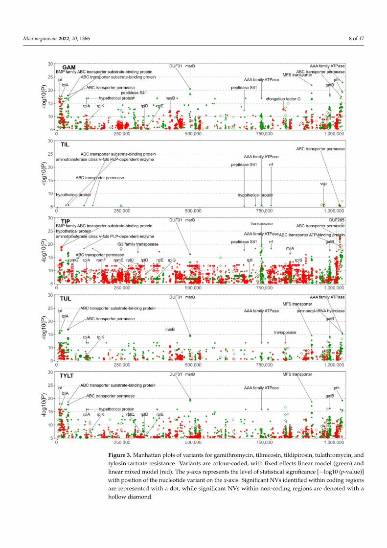
Figure 2 is the association of the variants to ENRO, CTET, OXY, and FFN, and Figure 3provides the association for the five macrolides: GAM. TIL, TIP, TUL and TYLT. The FEMand LMM results are displayed in green and red, respectively. The most significant resultsfor both models have been labelled with the gene of origin. Significant NVs identifiedwithin intergenic regions are denoted by a diamond. Only the NVs meeting the correctedp-value threshold are included. For each GWAS, the counts of NVs associated with AMRare found in Table S2 and the count of genes that contain significant NVs are found in TableS3. Statistical results for each NV by each GWAS are presented in Table S4. Additionalinformation such as effects on synonymity, acid substitution, and gene name was addedthrough automation.

Microorganisms 2022, 10, 1366 7 of 17
Figure 2. Manhattan plots of variants for enrofloxacin, chlortetracycline, oxytetracycline, and flor-fenicol resistance. Variants are colour-coded, with fixed effects linear model (green) and linearmixed model (red). The y-axis represents the level of statistical significance [−log10 (p-value)] withposition of the nucleotide variant on the x-axis. Significant NVs identified within coding regionsare represented with a dot, while significant NVs within non-coding regions are denoted with ahollow diamond.
Microorganisms 2022, 10, 1366 8 of 17
Figure 3. Manhattan plots of variants for gamithromycin, tilmicosin, tildipirosin, tulathromycin, andtylosin tartrate resistance. Variants are colour-coded, with fixed effects linear model (green) andlinear mixed model (red). The y-axis represents the level of statistical significance [−log10 (p-value)]with position of the nucleotide variant on the x-axis. Significant NVs identified within coding regionsare represented with a dot, while significant NVs within non-coding regions are denoted with ahollow diamond.

Microorganisms 2022, 10, 1366 9 of 17
3.3. Investigation into the p-Value Inflation Present within GWAS Results
The inherent caveat to detecting new NV associations with a GWAS is p-value inflation,leading to NVs that are falsely associated with the phenotype. Table 2 and Figure S3 showthat the ENRO, CTET, OXY, FFN FEM, and TIL FEM GWAS resulted in p-value inflationof λ < 1.1, which is acceptable [45]. Values above 1.3 are considered to be high p-valueinflation resulting in a greater false positive rate [45,46]. The FFN LMM had moderateinflation (λ = 1.17), with the remaining macrolide GWAS having high p-value inflation(λ > 1.3). Ideally, the Pyseer pipeline mitigates false positives (Type I error) by definingclusters of orthologous groups (COGs) and using a mixed model approach to control forconfounding population effects [29,39]. Potential reasons for the p-value inflation includeskewed MICs, population stratification, and polygenic inheritance [29–31,46,47]. The FFNLMM and macrolide GWAS with the most significant p-values, having 10s of variantswithin a small number of gene families, and having been previously linked to AMR, arediscussed below.
Table 2. Genomic inflation factors (λ) for each GWAS, where λ is the ratio of the median of theempirically observed distribution of the test statistic to the expected median.
GWAS Model ENRO CTET OXY FFN GAM TIL TIP TUL TYLT
Fixed Effects Model 1.06 1 1 1 3.1 1 2.85 2.27 2.45Linear Mixed Model 1 1 1.02 1.17 3.87 1.89 4.84 3.63 2.93
3.4. Significant NVs Specific to Enrofloxacin Resistance
The NVs with the highest significance to ENRO were identified in the parC and gyrAgenes. Overall, the GWAS identified 468 NVs within and between 68 coding sequencesassociated with ENRO resistance (Figure 2). NVs were identified within parC that includedmissense mutations resulting in Ser91Ile (E. coli Ser80Ile, AGT > ATT) and Asp95Asp(E. coli Asp84Asp, GAC > GAT) substitutions. Likewise, within gyrA a nonsynonymousmutation encoding for a Ser150Phe (E. coli Ser83Phe, TCT > TTT) was prevalent. Thesemutations have been previously identified as being highly predictive of fluoroquinoloneresistance [19,21–23]. An NV causing a synonymous mutation in the codon encoding Asp95of parC was also frequently identified. Synonymous mutations are known to affect geneexpression through the tendency for certain codons to be used more readily, creating acodon usage bias [48]. This is attributed to the selection of codons for translational efficiency,as has been previously shown for tuf genes in Salmonella Typhimurium [49].
3.5. Significant NVs Specific to Chlortetracycline and Oxytetracycline Resistance
The CTET GWAS identified 242 NVs among 48 coding sequences, while the OXYGWAS identified 50 NVs among nine coding sequences (Figure 2). Unexpectedly, GWASdid not identify known alleles within rrs1 or rrs2 associated with tetracycline resistance.Mutations in the 16S rRNA rrs1 and rrs2 genes are associated with OXY resistance [19,20].Nevertheless, multiple NVs associated with OXY AMR were identified in the proteins ofthe rRNA subunits. The OXY GWAS identified variants in the 30S ribosomal protein S16(rpsP gene) as well as variants in the 50S ribosomal protein L19 (rplS). However, the CTETGWAS did not share the same ribosomal protein mutations seen with the OXY resistance.This was unexpected since the mechanisms of action of these two antimicrobials are similar.
3.6. Significant NVs Specific to Florfenicol, Gamithromycin, Tilmicosin, Tildipirosin,Tulathromycin, and Tylosin Tartrate Resistance
The phenicol and macrolide GWAS displayed similarity, which was not unexpectedsince they have similar antimicrobial mechanisms of action. The FFN GWAS identified108 NVs among 61 coding sequences (Figure 2). The five macrolide GWAS identifiedNVs and coding sequences associated with AMR as follows: GAM, 3049 NVs, 305 codingsequences; TIL, 75 NVs, 11 coding sequences; TIP, 8847 NVs, 521 coding sequences; TUL,

Microorganisms 2022, 10, 1366 10 of 17
2003 NVs, 228 coding sequences; and TYLT, 3446 NVs, 368 coding sequences (Figure 3). Theincreased number of NVs detected in the GAM, TIP, TUL, and TYLT may be attributableto higher p-value inflation (λ > 1.3). This is likely due to the location of the NVs withinmultiple genes for ATP-binding cassette (ABC) transporter proteins, with 10s of NVs withina single gene. The high variability within these genes and the large number of genes codingfor transporter proteins plausibly leads to polygenic inheritance as the main contributorto the p-value inflation. Conversely, the low number of NVs in the TIL studies can beattributed to most isolates being resistant. M. bovis typically has an innate resistance toTIL, with relatively few isolates with a MIC < 256 µg/mL. However, the NVs associatedwith TIL were also identified in the other macrolide GWAS, and hence are discussed in themulti-drug resistance section below.
NVs associated with phenicol and macrolide resistance were identified in 23S riboso-mal RNA (MBOVPG45_RS01415) at the well-documented positions of A319980G (E. coliA2059G) and A319980C (E. coli A2060C) [7,18,19]. Furthermore, FFN and all the macrolideGWAS, except TIL, identified NVs in the 50S ribosomal proteins. Mutations related to FFNresistance were detected in the 50S ribosomal protein L4 (rplD gene), while the macrolideGWAS detected NVs in L1 (rplA), L3 (rplC), L4 (rplD), L5 (rplE), L9 (rplI), L11 (rplK), L17(rplQ), L19 (rplS), L31 (rpmE), L32 (rpmF), and L33 (rpmG). NVs for the macrolide studieswere also detected in genes encoding for proteins of the 30S ribosomal subunit: S2 (rpsB),S3 (rpsC), S4 (rpmE), S6 (rpsF), S16 (rpsP), S18 (rpsR), and S20 (rpsT). NVs in 50S ribosomalproteins L1 (rplA) and L4 (rplD) were commonly identified for the macrolides. However,none of the mutations in L22 (rplV) were significant, despite previous reports linkingmutations in L4 and L22 to macrolide resistance [7,18,19]. The 5S rRNA subunit of the 50Sribosomal complex was found to contain a non-synonymous mutation (NSM) for severalof the macrolides, although with low significance. The lack of identifiable 23S rRNA or 50SrRNA mutations associated with TIL resistance is consistent with previous reports [7,18,19].
With the exception of TIL, a number of macrolide NVs were associated with msrB,a gene associated with the repair of oxidative stress in bacteria [50]. Mutations in theMsrB protein have been linked to altered virulence in Enterococcus faecalis, with mutantsexhibiting greater sensitivity to H2O2 [50,51]. Although macrolides have been shown toinduce oxidative stress associated with toxicity in eukaryotic cells [52,53], this phenomenonhas not been well-characterized in bacteria, perhaps because it is not a primary mode ofaction for macrolides. The lack of research into the effects of msrB and oxidative stressin Mycoplasma spp. underscores the utility of using GWAS to identify associations be-tween functional genes and AMR. Once identified, these associations can be evaluated forbiological plausibility and cross-referenced to other bacteria.
3.7. Variants Associated with Multi-Drug Resistance
NVs within functional genes were associated with multidrug resistance (MDR). Theseincluded transporter proteins and membrane proteins; proteins that interact with ribosomesduring translation such as tRNA ligases and elongation factors; and proteins that affect muta-tion, sequence and structure, such as methyltransferases and transposases (Table 1) [26,54–59].
All the GWAS identified NVs within the ABC transporter proteins, and the proteinsdirectly interacting with ABC transporter pathways. Genes most prominently identified acrossall studies included: MBOVPG45_RS00180, MBOVPG45_RS00185, MBOVPG45_RS02005,MBOVPG45_RS00145, MBOVPG45_RS00165, MBOVPG45_RS00140, MBOVPG45_RS04335,MBOVPG45_RS03085, MBOVPG45_RS02710, MBOVPG45_RS02715, MBOVPG45_RS03550,and MBOVPG45_RS04315. These findings are salient because the ABC transporters are aubiquitous superfamily of membrane and transmembrane proteins that transport substratessuch as antimicrobials across the membrane [60]. Transporters acting as antimicrobial effluxpumps have been associated with single and multi-drug resistance in bacteria [61,62]. ABC-type macrolide-specific efflux pumps have been documented in Mycoplasma pneumoniae [63],and Ledger et al. suggested that mutations within efflux pumps may also influence AMRin M. bovis [26]. Our GWAS identified 19 of the 21 ABC transporters listed by Ledger et al.

Microorganisms 2022, 10, 1366 11 of 17
as containing NVs significant to AMR. The significant NVs linked to TIL resistance wereprimarily ABC transporters, giving credence to the idea of innate resistance through effluxpumps. Bokma et al. also reported multiple ABC-type macrolide efflux pump genes [27].
Multiple NVs were identified in genes coding for other groups of transmembrane andmembrane proteins. MBOVPG45_RS03800, a gene for the major facilitator superfamily(MFS) of efflux pumps, contained NVs to all of the macrolides investigated with the ex-ception of TIL. MFS transporters play a role in MDR in E. coli, Helicobacter pylori and otherbacterial species [54,64–66]. Significant NVs associated with ENRO resistance were withinvariable surface lipoproteins (Vsps) genes (MBOVPG45_RS04435, MBOVPG45_RS02100)with a single MNP in both genes containing NVs that cause mutations in 11 AAs. Cell mem-brane mutations are relevant as they prevent the entry of antimicrobials into the cell, whichis a primary mechanism of AMR in all bacteria [67]. Considering that quinolones, such asENRO, can only cross the cell membrane through porins, mutations in membrane proteinsand a reduction in the number of porins via Vsps [67] could contribute to AMR. Mutationsin Vsps were also common to FFN and the macrolides, with MBOVPG45_RS04435 havingsignificant NVs.
ENRO, OXY, FFN and macrolide GWAS, except TIL, all identified NVs within tRNA-ligases including, but not limited to, argS, asnS, gltX, ileS, and lysS. Also known asaminoacyl-tRNA synthetases, tRNA-ligases transfer single amino acids to tRNAs, enablingpeptide synthesis [55]. Mutations in argS and asnS have been linked to MDR in E. coli [68],and mutations in ileS confer resistance to mupirocin in Staphylococcus spp. [69]. While nota current antimicrobial target, there has been an effort to link mutations in tRNA-ligaseto AMR in M. bovis [26], which could possibly identify targets for using aminoacyl-tRNAsynthetase inhibitors as antimicrobials [55,70]. Ledger et al. listed 22 tRNA ligases, 13 ofwhich we identified as containing NVs to AMR [26].
In addition to the tRNA-ligases, NVs were identified in genes for protein synthesis. Theseincluded the aminotransferase class V-fold PLP-dependent enzyme gene MBOVPG45_RS00395,whose protein catalyzes the formation of AAs, along with the elongation factors 4 (lepA), G(fusA), Tu (tuf ), and Ts (tsf ) involved in protein synthesis. While there is lack of data regardingNVs in the V-fold PLP-dependent enzyme, mutations within the elongation factors have beenassociated with AMR in Mycoplasma spp., Pseudomonas aeruginosa, and E. coli [56,57,71–73].
The LMM GWAS associated GAM, TUL, and TYLT with NVs in the NusB geneMBOVPG45_RS01740, which has a role in ribosome biosynthesis by influencing rRNA fold-ing and annealing [74,75]. NVs in MBOVPG45_RS01740 may result in macrolide resistanceby disrupting ribosome biosynthesis [76]. While more research into the possible antimicro-bial effects of NusB-NusE dimers is needed, research into identifying antimicrobials thattarget this interaction are ongoing [77,78].
In all GWAS, except for those conducted on tetracyclines, NVs in genes codingfor S41 peptidases (MBOVPG45_RS00115, MBOVPG45_RS01155, MBOVPG45_RS01160,MBOVPG45_RS02105, MBOVPG45_RS02760, MBOVPG45_RS02800, MBOVPG45_RS02805)were associated with AMR. The S41 family of peptidases is common to a wide range oforganisms. While poorly characterized, these peptidases are believed to have a role inthe degradation of incorrectly synthesized proteins and cytoplasmic proteins. The S41peptidases have been proposed to influence the virulence of Mycoplasma mycoides capri byregulating H2O2 production and modulating cell surface proteins, including IgG-blockingvirulence proteins, peptidases, and hypothetical proteins [79–81].
The tetracycline and macrolide GWAS identified multiple NVs in DNA polymerases,DNA primases, DNA binding proteins, methyltransferases, topoisomerases, and kinases.Although none of these enzymes directly influence the action of tetracyclines, mutationsin these genes may contribute to AMR by altering gene expression [82–84]. Methylationof the 23S ribosomal RNA at the A2058 (E. coli numbering) residue has been shown todirectly contribute to macrolide resistance by impairing the binding of macrolides to theiractive site [58]. Tetracyclines and macrolides were associated with NVs coding for bothsynonymous mutations (SMs) and NSMs in a total of 24 methyltransferases. These include

Microorganisms 2022, 10, 1366 12 of 17
rlmB, rlmD, and MBOVPG45_RS02280, which Ledger et al. reported to contain NSMs linkedto a multi-drug resistant M. bovis [26]. RlmB has been speculated to play a role in AMR if itsbinding activity to 23S rRNA is impeded [26,85]. Excluding previously discussed mutationsin topoisomerase genes parC and gyrA leading to fluroquinolone resistance, NSMs andSMs in parE, topA, and a type IIA DNA topoisomerase subunit B (MBOVPG45_RS04255)were associated with macrolide resistance. NVs in parE and topA have been associated withthe MDR phenotype in M. bovis [26]. Mutations in topA have been shown to increase therate of sequence deletion and duplication, leading to the emergence of AMR genotypes inE. coli [86].
NVs in transposases or insertion sequences (ISs) were inferred from all analyses ex-cept those for TIL. Examples include ISMbov3 family transposases (MBOVPG45_RS00260,MBOVPG45_RS03285), ISMbov1 family transposases (MBOVPG45_RS00955, MBOVPG45_RS03260), IS30 family transposases (MBOVPG45_RS00195), IS3 family transposases (MBOVPG45_RS04445, MBOVPG45_RS01210), and other transposases (MBOVPG45_RS00705, MBOVPG45_RS04745, MBOVPG45_RS00895, MBOVPG45_RS03090, MBOVPG45_RS03450). The contribution ofthese mobile genetic elements (MGE) in genome diversity and evolution, as well as in modula-tion of activation and transcription of genes has been described [59]. Horizontal gene transfer(HGT) may drive the development of AMR through the transfer of resistance conferring mu-tations, as demonstrated by the transfer of ENRO resistance among isolates of Mycoplasmaagalactiae [87,88]. Little is known, however, about the role of MGE in the emergence of AMR inM. bovis.
It was problematic to interpret the NVs within domains of unknown function (DUFs),hypothetical proteins, and intergenic regions, since few have received functional an-notation or been investigated. Genes associated with the DUF31 family of proteins(MBOVPG45_RS01865, MBOVPG45_RS01925, MBOVPG45_RS02010, MBOVPG45_RS02120)contained NVs associated with ENRO, FFN and macrolide AMR. DUF31 family proteins areproposed to arise from putative peptidase genes and appear to have a role in the pathogenic-ity of Mycoplasma spp. [81,89,90]. Despite not being protein coding sequences, intergenicor non-coding NVs may have phenotypic consequences. Notably, the GWAS associatedintergenic NVs to FFN resistance. These intergenic NVs were positioned nearest to the geneMBOVPG45_RS03375 encoding an S8 family serine peptidase; MBOVPG45_RS03860 encod-ing a hypothetical protein; and whiA encoding a probable cell division protein. Likewise,NVs associated with TIL resistance were positioned nearest to ftsH, an ATP-dependent zincmetalloprotease, which has a role in membrane proteins, and MBOVPG45_RS04590, whichis a predicted Vsp. These proteins, however, have not been characterized with respect toAMR. Non-coding NVs such as those mentioned can alter the expression of nearby genesthrough the alteration of riboswitches, regulatory small RNAs, and transcription promoters,terminators, and regulator binding sites [91].
Due to an economized genome, resistance to each antimicrobial has been shown tobe associated with mutations in multiple genes rather than possessing specific resistance-conferring genes. Additionally, there exists overlaps in associations of NVs to separateantimicrobials and antimicrobial classes. Therefore, it is likely that a multifactorial modelof resistance exists, such that resistance is granted by modifications to antimicrobial targetsto prevent antimicrobial binding; modifications to proteins interacting with antimicrobialtargets to prevent antimicrobial binding and the repair of cellular damage caused byantimicrobials; and cellular defense mechanisms. Models of resistance such as these havebeen documented for E. coli [92], though in the case of M. bovis, resistance originatesprimarily from NVs in the core genome, as M. bovis has not been found to possess novelantimicrobial resistance genes. The results identified by the GWAS have a basis in AMRwithin M. bovis and other bacterial species. Therefore, our results serve as a basis for furtherresearch into antimicrobial mechanisms in M. bovis.
The study may have benefited from a greater number of isolates, particularly as theyrelate to the dispersion on AST phenotypes. Most of the isolates were sensitive to ENRO,whereas the AST phenotypes for CTET, OXY and FFN displayed a narrow unimodal

Microorganisms 2022, 10, 1366 13 of 17
distribution. Increasing the number of phenotypes in the extremes, very low and high MICvalues may have revealed more associations or increased the statistical significance of someof the associations. The next step would be to validate the associations to substantiate ifthey are spurious or real.
4. Conclusions
A relatively large body of knowledge exists regarding the specific SNPs involved inantimicrobial target-site modifications, which confer antimicrobial resistance. However,incongruence observed between AMR phenotypes and genotypes suggests that otherunidentified mechanisms of resistance exist. Calcutt et al. noted that this is an importantknowledge gap and posited, as have others, that transporters may be an important mecha-nism of resistance [25]. Our findings determined that GWAS is not only an effective methodfor confirming known target-specific NVs, but has the potential for discovering new NVsand genes associated with AMR. This was particularly true of variants linked to MDR. TheGWAS identified proteins that interact with ABC transporters pathways, which are knownto be associated with MDR in other bacteria. Mutations were also identified in tRNA-ligases, which have been linked to MDR in E. coli and Staphylococcus spp. There is alsothe potential for AMR to be mediated via NVs within S41 peptidases, DNA polymerasesand primases, methyltransferases, and kinases, to name but a few. Less well understood isthe significance of NVs within domains of unknown functions, hypothetical proteins, andintergenic regions. Although poorly understood, these NVs should not be overlooked, asthey may inform AMR as well as transmission and pathogenicity. A multifactorial modelof AMR in M. bovis likely exists, as modifications to antimicrobial targets, antimicrobialtarget-interacting proteins, and cellular defense mechanisms such as transporters and Vspswere found to be associated with AMR. Further GWAS utilizing larger and more diversedatasets may uncover additional genetic markers for AMR.
Supplementary Materials: The following supporting information can be downloaded at: https://www.mdpi.com/article/10.3390/microorganisms10071366/s1, Table S1: Assembly metadata ofthe 194 Mycoplasma bovis genomes enrolled in this study. Table S2: Counts of the significant nucleotidevariants for each GWAS. Table S3: Counts of the genes containing significant nucleotide variants foreach GWAS. Table S4: Results for all performed genome-wide association studies with metadata.Figure S1: PCA analysis of various metadata against the VCF dataset. Figure S2: Minimum inhibitoryconcentration values for each Mycoplasma bovis isolate. Figure S3: Quantile-quantile plots for eachgenome-wide association study.
Author Contributions: Conceptualization, M.W. and M.J.; Methodology, M.W.; Software, M.W.;Validation, M.W., M.J., C.L. and T.M.; Formal Analysis, M.W.; Investigation, M.W. and A.K.; Resources,K.R. and M.J.; Data Curation, M.W.; Writing—Original Draft Preparation, M.W. and M.J.; Writing—Review & Editing, M.W., A.K., E.Y., T.M., K.R., C.L. and M.J.; Visualization, M.W.; Supervision, M.J.;Project Administration, M.J.; Funding Acquisition, M.J. All authors have read and agreed to thepublished version of the manuscript.
Funding: This research was funded by the Canadian Cattlemen’s Association—Beef Cattle ResearchCouncil: Grant ANH.30.17; Saskatchewan Ministry of Agriculture—Agriculture Development Fund:#20060116, #20130170, #20160253, #20200187; Saskatchewan Cattlemen’s Association: 2020-109.Publication costs were covered by Saskatchewan Agricultural Development Fund #20200187.
Institutional Review Board Statement: The animal study protocol was approved by the Universityof Saskatchewan’s Animal Research Ethics Board (Protocols 20070023 and 20170021).
Informed Consent Statement: Not applicable.
Data Availability Statement: The raw paired-end read files analyzed in this study are openly avail-able the Sequence Read Archive (SRA) under BioProject accession no. PRJNA642970, PRJNA708306,and PRJNA785928.

Microorganisms 2022, 10, 1366 14 of 17
Acknowledgments: The authors express their appreciation to Karen Gesy and Manual Chirino-Trejo, Western College of Veterinary Medicine, for their expertise in the culturing, isolation, andidentification of the Mycoplasma bovis isolates.
Conflicts of Interest: The authors declare that they have no conflicts of interest.
References1. Nicholas, R.A.J.; Ayling, R.D. Mycoplasma bovis: Disease, Diagnosis, and Control. Res. Vet. Sci. 2003, 74, 105–112. [CrossRef]2. Maunsell, F.P.; Woolums, A.R.; Francoz, D.; Rosenbusch, R.F.; Step, D.L.; Wilson, D.J.; Janzen, E.D. Mycoplasma bovis Infections in
Cattle. J. Vet. Intern. Med. 2011, 25, 772–783. [CrossRef] [PubMed]3. Gagea, M.I.; Bateman, K.G.; Shanahan, R.A.; van Dreumel, T.; McEwen, B.J.; Carman, S.; Archambault, M.; Caswell, J.L. Naturally
Occurring Mycoplasma bovis—Associated Pneumonia and Polyarthritis in Feedlot Beef Calves. J. Vet. Diagn. Investig. 2006, 18,29–40. [CrossRef] [PubMed]
4. Caswell, J.L.; Bateman, K.G.; Cai, H.Y.; Castillo-Alcala, F. Mycoplasma bovis in Respiratory Disease of Feedlot Cattle. Vet. Clin. FoodAnim. Pract. 2010, 26, 365–379. [CrossRef]
5. Brault, S.A.; Hannon, S.J.; Gow, S.P.; Otto, S.J.G.; Booker, C.W.; Morley, P.S. Calculation of Antimicrobial Use Indicators in BeefFeedlots—Effects of Choice of Metric and Standardized Values. Front. Vet. Sci. 2019, 6, 330. [CrossRef]
6. Jelinski, M.; Kinnear, A.; Gesy, K.; Andrés-Lasheras, S.; Zaheer, R.; Weese, S.; McAllister, T.A. Antimicrobial Sensitivity Testing ofMycoplasma bovis Isolates Derived from Western Canadian Feedlot Cattle. Microorganisms 2020, 8, 124. [CrossRef]
7. Kinnear, A.; McAllister, T.A.; Zaheer, R.; Waldner, M.; Ruzzini, A.C.; Andrés-Lasheras, S.; Parker, S.; Hill, J.E.; Jelinski, M.D.Investigation of Macrolide Resistance Genotypes in Mycoplasma bovis Isolates from Canadian Feedlot Cattle. Pathogens 2020, 9, 622.[CrossRef]
8. Rosenbusch, R.F.; Kinyon, J.M.; Apley, M.; Funk, N.D.; Smith, S.; Hoffman, L.J. In Vitro Antimicrobial Inhibition Profiles ofMycoplasma bovis Isolates Recovered from Various Regions of the United States from 2002 to 2003. J. Vet. Diagn. Investig. 2005, 17,436–441. [CrossRef]
9. Hata, E.; Harada, T.; Itoh, M. Relationship between Antimicrobial Susceptibility and Multilocus Sequence Type of Mycoplasmabovis Isolates and Development of a Method for Rapid Detection of Point Mutations Involved in Decreased Susceptibility toMacrolides, Lincosamides, Tetracyclines, and Spectinomycin. Appl. Environ. Microbiol. 2019, 85, e00575-19. [CrossRef]
10. Ayling, R.D.; Baker, S.E.; Nicholas, R.A.; Peek, M.L.; Simon, A.J. Comparison of in Vitro Activity of Danofloxacin, Florfenicol,Oxytetracycline, Spectinomycin and Tilmicosin against Recent Field Isolates of Mycoplasma bovis. Vet. Rec. 2000, 146, 745–747.[CrossRef]
11. Gerchman, I.; Levisohn, S.; Mikula, I.; Lysnyansky, I. In Vitro Antimicrobial Susceptibility of Mycoplasma bovis Isolated in Israelfrom Local and Imported Cattle. Vet. Microbiol. 2009, 137, 268–275. [CrossRef] [PubMed]
12. Gautier-Bouchardon, A.V.; Ferré, S.; Grand, D.L.; Paoli, A.; Gay, E.; Poumarat, F. Overall Decrease in the Susceptibility ofMycoplasma bovis to Antimicrobials over the Past 30 Years in France. PLoS ONE 2014, 9, e87672. [CrossRef] [PubMed]
13. Sulyok, K.M.; Kreizinger, Z.; Fekete, L.; Hrivnák, V.; Magyar, T.; Jánosi, S.; Schweitzer, N.; Turcsányi, I.; Makrai, L.; Erdélyi, K.;et al. Antibiotic Susceptibility Profiles of Mycoplasma bovis Strains Isolated from Cattle in Hungary, Central Europe. BMC Vet. Res.2014, 10, 256. [CrossRef] [PubMed]
14. Heuvelink, A.; Reugebrink, C.; Mars, J. Antimicrobial Susceptibility of Mycoplasma bovis Isolates from Veal Calves and DairyCattle in the Netherlands. Vet. Microbiol. 2016, 189, 1–7. [CrossRef] [PubMed]
15. Klein, U.; de Jong, A.; Moyaert, H.; El Garch, F.; Leon, R.; Richard-Mazet, A.; Rose, M.; Maes, D.; Pridmore, A.; Thomson, J.R.; et al.Antimicrobial Susceptibility Monitoring of Mycoplasma hyopneumoniae and Mycoplasma bovis Isolated in Europe. Vet. Microbiol.2017, 204, 188–193. [CrossRef] [PubMed]
16. Goldstein, B.P. Resistance to Rifampicin: A Review. J. Antibiot. 2014, 67, 625–630. [CrossRef] [PubMed]17. Taylor-Robinson, D.; Bébéar, C. Antibiotic Susceptibilities of Mycoplasmas and Treatment of Mycoplasmal Infections. J. Antimicrob.
Chemother. 1997, 40, 622–630. [CrossRef]18. Lerner, U.; Amram, E.; Ayling, R.D.; Mikula, I.; Gerchman, I.; Harrus, S.; Teff, D.; Yogev, D.; Lysnyansky, I. Acquired Resistance to
the 16-Membered Macrolides Tylosin and Tilmicosin by Mycoplasma bovis. Vet. Microbiol. 2014, 168, 365–371. [CrossRef]19. Sulyok, K.M.; Kreizinger, Z.; Wehmann, E.; Lysnyansky, I.; Bányai, K.; Marton, S.; Jerzsele, Á.; Rónai, Z.; Turcsányi, I.; Makrai,
L.; et al. Mutations Associated with Decreased Susceptibility to Seven Antimicrobial Families in Field and Laboratory-DerivedMycoplasma bovis Strains. Antimicrob. Agents Chemother. 2017, 61, e01983-16. [CrossRef]
20. Amram, E.; Mikula, I.; Schnee, C.; Ayling, R.D.; Nicholas, R.A.J.; Rosales, R.S.; Harrus, S.; Lysnyansky, I. 16S RRNA GeneMutations Associated with Decreased Susceptibility to Tetracycline in Mycoplasma bovis. Antimicrob. Agents Chemother. 2015, 59,796–802. [CrossRef]
21. Sato, T.; Okubo, T.; Usui, M.; Higuchi, H.; Tamura, Y. Amino Acid Substitutions in GyrA and ParC Are Associated withFluoroquinolone Resistance in Mycoplasma bovis Isolates from Japanese Dairy Calves. J. Vet. Med. Sci. 2013, 75, 1063–1065.[CrossRef] [PubMed]

Microorganisms 2022, 10, 1366 15 of 17
22. Khalil, D.; Becker, C.A.M.; Tardy, F. Alterations in the Quinolone Resistance-Determining Regions and Fluoroquinolone Resistancein Clinical Isolates and Laboratory-Derived Mutants of Mycoplasma bovis: Not All Genotypes May Be Equal. Appl. Environ.Microbiol. 2016, 82, 1060–1068. [CrossRef] [PubMed]
23. Lysnyansky, I.; Mikula, I.; Gerchman, I.; Levisohn, S. Rapid Detection of a Point Mutation in the ParC Gene Associated withDecreased Susceptibility to Fluoroquinolones in Mycoplasma bovis. Antimicrob. Agents Chemother. 2009, 53, 4911–4914. [CrossRef][PubMed]
24. Fàbrega, A.; Madurga, S.; Giralt, E.; Vila, J. Mechanism of Action of and Resistance to Quinolones. Microb. Biotechnol. 2009, 2,40–61. [CrossRef]
25. Calcutt, M.J.; Lysnyansky, I.; Sachse, K.; Fox, L.K.; Nicholas, R.A.J.; Ayling, R.D. Gap Analysis of Mycoplasma bovis Disease,Diagnosis and Control: An Aid to Identify Future Development Requirements. Transbound. Emerg. Dis. 2018, 65, 91–109.[CrossRef]
26. Ledger, L.; Eidt, J.; Cai, H.Y. Identification of Antimicrobial Resistance-Associated Genes through Whole Genome Sequencing ofMycoplasma bovis Isolates with Different Antimicrobial Resistances. Pathogens 2020, 9, 588. [CrossRef]
27. Bokma, J.; Vereecke, N.; Nauwynck, H.; Haesebrouck, F.; Theuns, S.; Pardon, B.; Boyen, F. Genome-Wide Association StudyReveals Genetic Markers for Antimicrobial Resistance in Mycoplasma bovis. Microbiol. Spectr. 2021, 9, e00262-21. [CrossRef]
28. Tam, V.; Patel, N.; Turcotte, M.; Bossé, Y.; Paré, G.; Meyre, D. Benefits and Limitations of Genome-Wide Association Studies. Nat.Rev. Genet. 2019, 20, 467–484. [CrossRef]
29. Vilhjálmsson, B.J.; Nordborg, M. The Nature of Confounding in Genome-Wide Association Studies. Nat. Rev. Genet. 2013, 14, 1–2.[CrossRef]
30. Falush, D.; Bowden, R. Genome-Wide Association Mapping in Bacteria? Trends Microbiol. 2006, 14, 353–355. [CrossRef]31. Yang, J.; Weedon, M.N.; Purcell, S.; Lettre, G.; Estrada, K.; Willer, C.J.; Smith, A.V.; Ingelsson, E.; O’connell, J.R.; Mangino, M.; et al.
Genomic inflation factors under polygenic inheritance. Eur. J. Hum. Genet. 2011, 19, 807–812. [CrossRef] [PubMed]32. Hong, E.P.; Park, J.W. Sample Size and Statistical Power Calculation in Genetic Association Studies. Genomics Inf. 2012, 10, 117.
[CrossRef] [PubMed]33. Brynildsrud, O.; Bohlin, J.; Scheffer, L.; Eldholm, V. Rapid Scoring of Genes in Microbial Pan-Genome-Wide Association Studies
with Scoary. Genome Biol. 2016, 17, 238. [CrossRef] [PubMed]34. Springer. Theoretical and Applied Genetics: Submission Guidelines. Available online: https://www.springer.com/journal/122/
submission-guidelines (accessed on 6 June 2022).35. Bolger, A.M.; Lohse, M.; Usadel, B. Trimmomatic: A Flexible Trimmer for Illumina Sequence Data. Bioinformatics 2014, 30,
2114–2120. [CrossRef]36. Li, H. Aligning Sequence Reads, Clone Sequences and Assembly Contigs with BWA-MEM. arXiv 2013, arXiv:1303.3997.37. Li, H.; Handsaker, B.; Wysoker, A.; Fennell, T.; Ruan, J.; Homer, N.; Marth, G.; Abecasis, G.; Durbin, R.; 1000 Genome Project Data
Processing Subgroup. The Sequence Alignment/Map Format and SAMtools. Bioinformatics 2009, 25, 2078–2079. [CrossRef]38. Garrison, E.; Marth, G. Haplotype-Based Variant Detection from Short-Read Sequencing. arXiv 2012, arXiv:1207.3907.39. Lees, J.A.; Galardini, M.; Bentley, S.D.; Weiser, J.N.; Corander, J. Pyseer: A Comprehensive Tool for Microbial Pangenome-Wide
Association Studies. Bioinformatics 2018, 34, 4310–4312. [CrossRef]40. Lees, J.A.; Vehkala, M.; Välimäki, N.; Harris, S.R.; Chewapreecha, C.; Croucher, N.J.; Marttinen, P.; Davies, M.R.; Steer, A.C.; Tong,
S.Y.C.; et al. Sequence Element Enrichment Analysis to Determine the Genetic Basis of Bacterial Phenotypes. Nat. Commun. 2016,7, 12797. [CrossRef]
41. Ondov, B.D.; Treangen, T.J.; Melsted, P.; Mallonee, A.B.; Bergman, N.H.; Koren, S.; Phillippy, A.M. Mash: Fast Genome andMetagenome Distance Estimation Using MinHash. Genome Biol. 2016, 17, 132. [CrossRef]
42. Seemann, T. Prokka: Rapid Prokaryotic Genome Annotation. Bioinformatics 2014, 30, 2068–2069. [CrossRef] [PubMed]43. Chang, C.C.; Chow, C.C.; Tellier, L.C.; Vattikuti, S.; Purcell, S.M.; Lee, J.J. Second-Generation PLINK: Rising to the Challenge of
Larger and Richer Datasets. GigaScience 2015, 4, s13742-015. [CrossRef] [PubMed]44. Wickham, H. Ggplot2. WIREs Comput. Stat. 2011, 3, 180–185. [CrossRef]45. Devlin, B.; Roeder, K. Genomic Control for Association Studies. Biometrics 1999, 55, 997–1004. [CrossRef]46. Cardon, L.R.; Palmer, L.J. Population Stratification and Spurious Allelic Association. Lancet 2003, 361, 598–604. [CrossRef]47. Hinrichs, A.L.; Larkin, E.K.; Suarez, B.K. Population Stratification and Patterns of Linkage Disequilibrium. Genet. Epidemiol. 2009,
33, S88–S92. [CrossRef]48. Gustafsson, C.; Govindarajan, S.; Minshull, J. Codon Bias and Heterologous Protein Expression. Trends Biotechnol. 2004, 22,
346–353. [CrossRef]49. Brandis, G.; Hughes, D. The Selective Advantage of Synonymous Codon Usage Bias in Salmonella. PLoS Genet. 2016, 12, e1005926.
[CrossRef]50. Zhao, C.; Hartke, A.; La Sorda, M.; Posteraro, B.; Laplace, J.-M.; Auffray, Y.; Sanguinetti, M. Role of Methionine Sulfoxide
Reductases A and B of Enterococcus Faecalis in Oxidative Stress and Virulence. Infect Immun. 2010, 78, 3889–3897. [CrossRef]51. Romsang, A.; Atichartpongkul, S.; Trinachartvanit, W.; Vattanaviboon, P.; Mongkolsuk, S. Gene Expression and Physiological
Role of Pseudomonas Aeruginosa Methionine Sulfoxide Reductases during Oxidative Stress. J. Bacteriol. 2013, 195, 3299–3308.[CrossRef]

Microorganisms 2022, 10, 1366 16 of 17
52. Woodhead, J.L.; Yang, K.; Oldach, D.; MacLauchlin, C.; Fernandes, P.; Watkins, P.B.; Siler, S.Q.; Howell, B.A. Analyzing theMechanisms Behind Macrolide Antibiotic-Induced Liver Injury Using Quantitative Systems Toxicology Modeling. Pharm. Res.2019, 36, 48. [CrossRef] [PubMed]
53. Yan, Z.; Huang, X.; Xie, Y.; Song, M.; Zhu, K.; Ding, S. Macrolides Induce Severe Cardiotoxicity and Developmental Toxicity inZebrafish Embryos. Sci. Total Environ. 2019, 649, 1414–1421. [CrossRef] [PubMed]
54. Maiden, M.C.; Davis, E.O.; Baldwin, S.A.; Moore, D.C.; Henderson, P.J. Mammalian and Bacterial Sugar Transport Proteins AreHomologous. Nature 1987, 325, 641–643. [CrossRef] [PubMed]
55. Hurdle, J.G.; O’Neill, A.J.; Chopra, I. Prospects for Aminoacyl-TRNA Synthetase Inhibitors as New Antimicrobial Agents.Antimicrob. Agents Chemother. 2005, 49, 4821–4833. [CrossRef]
56. Chernov, V.M.; Chernova, O.A.; Mouzykantov, A.A.; Medvedeva, E.S.; Baranova, N.B.; Malygina, T.Y.; Aminov, R.I.; Trushin,M.V. Antimicrobial Resistance in Mollicutes: Known and Newly Emerging Mechanisms. FEMS Microbiol. Lett. 2018, 365, fny185.[CrossRef]
57. Bielecki, P.; Lukat, P.; Hüsecken, K.; Dötsch, A.; Steinmetz, H.; Hartmann, R.W.; Müller, R.; Häussler, S. Mutation in ElongationFactor G Confers Resistance to the Antibiotic Argyrin in the Opportunistic Pathogen Pseudomonas aeruginosa. Chembiochem 2012,13, 2339–2345. [CrossRef]
58. Weisblum, B. Erythromycin Resistance by Ribosome Modification. Antimicrob. Agents Chemother. 1995, 39, 577–585.59. Siguier, P.; Gourbeyre, E.; Chandler, M. Bacterial Insertion Sequences: Their Genomic Impact and Diversity. FEMS Microbiol. Rev.
2014, 38, 865–891. [CrossRef]60. Higgins, C.F. ABC Transporters: From Microorganisms to Man. Annu. Rev. Cell Biol. 1992, 8, 67–113. [CrossRef]61. Marquez, B. Bacterial Efflux Systems and Efflux Pumps Inhibitors. Biochimie 2005, 87, 1137–1147. [CrossRef]62. Fyfe, C.; Grossman, T.H.; Kerstein, K.; Sutcliffe, J. Resistance to Macrolide Antibiotics in Public Health Pathogens. Cold Spring
Harb. Perspect. Med. 2016, 6, a025395. [CrossRef] [PubMed]63. Li, L.S.; Mei, S.H.; Li, Z.B.; Fei, L.I.U.; Qing, Z.H. Whole Genome Analysis Reveals New Insights into Macrolide Resistance in
Mycoplasma pneumoniae. Biomed. Environ. Sci. 2017, 30, 343–350. [CrossRef] [PubMed]64. Paulsen, I.T.; Brown, M.H.; Skurray, R.A. Proton-Dependent Multidrug Efflux Systems. Microbiol. Rev. 1996, 60, 575–608.
[CrossRef] [PubMed]65. Saidijam, M.; Benedetti, G.; Ren, Q.; Xu, Z.; Hoyle, C.J.; Palmer, S.L.; Ward, A.; Bettaney, K.E.; Szakonyi, G.; Meuller, J.; et al.
Microbial Drug Efflux Proteins of the Major Facilitator Superfamily. Curr. Drug Targets 2006, 7, 793–811. [CrossRef]66. Kumar, S.; Mukherjee, M.M.; Varela, M.F. Modulation of Bacterial Multidrug Resistance Efflux Pumps of the Major Facilitator
Superfamily. Int. J. Bacteriol. 2013, 2013, 204141. [CrossRef]67. Kapoor, G.; Saigal, S.; Elongavan, A. Action and Resistance Mechanisms of Antibiotics: A Guide for Clinicians. J. Anaesthesiol.
Clin. Pharmacol. 2017, 33, 300–305. [CrossRef]68. Magalhães, S.; Aroso, M.; Roxo, I.; Ferreira, S.; Cerveira, F.; Ramalheira, E.; Ferreira, R.; Vitorino, R. Proteomic Profile of Susceptible
and Multidrug-Resistant Clinical Isolates of Escherichia coli and Klebsiella pneumoniae Using Label-Free and ImmunoproteomicStrategies. Res. Microbiol. 2017, 168, 222–233. [CrossRef]
69. Szczuka, E.; Kaznowski, A.; Bosacka, K.; Strzemieczna, E. Antimicrobial Resistance and Presence of IleS-2 Gene EncodingMupirocin Resistance in Clinical Isolates of Methicillin-Resistant Staphylococcus sp. Folia Microbiol. 2009, 54, 153–156. [CrossRef]
70. Pang, L.; Weeks, S.D.; Van Aerschot, A. Aminoacyl-tRNA Synthetases as Valuable Targets for Antimicrobial Drug Discovery. Int.J. Mol. Sci. 2021, 22, 1750. [CrossRef]
71. Harvey, K.L.; Jarocki, V.M.; Charles, I.G.; Djordjevic, S.P. The Diverse Functional Roles of Elongation Factor Tu (EF-Tu) in MicrobialPathogenesis. Front. Microbiol. 2019, 10, 2351. [CrossRef]
72. Kraal, B.; Zeef, L.A.; Mesters, J.R.; Boon, K.; Vorstenbosch, E.L.; Bosch, L.; Anborgh, P.H.; Parmeggiani, A.; Hilgenfeld, R.Antibiotic Resistance Mechanisms of Mutant EF-Tu Species in Escherichia coli. Biochem. Cell Biol. 1995, 73, 1167–1177. [CrossRef][PubMed]
73. Pech, M.; Karim, Z.; Yamamoto, H.; Kitakawa, M.; Qin, Y.; Nierhaus, K.H. Elongation Factor 4 (EF4/LepA) Accelerates ProteinSynthesis at Increased Mg2+ Concentrations. Proc. Natl. Acad. Sci. USA 2011, 108, 3199–3203. [CrossRef] [PubMed]
74. Huang, Y.-H.; Hilal, T.; Loll, B.; Bürger, J.; Mielke, T.; Böttcher, C.; Said, N.; Wahl, M.C. Structure-Based Mechanisms of a MolecularRNA Polymerase/Chaperone Machine Required for Ribosome Biosynthesis. Mol. Cell 2020, 79, 1024–1036.e5. [CrossRef]
75. Stagno, J.R.; Altieri, A.S.; Bubunenko, M.; Tarasov, S.G.; Li, J.; Court, D.L.; Byrd, R.A.; Ji, X. Structural Basis for RNA Recognitionby NusB and NusE in the Initiation of Transcription Antitermination. Nucleic Acids Res. 2011, 39, 7803–7815. [CrossRef]
76. Court, D.L.; Patterson, T.A.; Baker, T.; Costantino, N.; Mao, X.; Friedman, D.I. Structural and Functional Analyses of theTranscription-Translation Proteins NusB and NusE. J. Bacteriol. 1995, 177, 2589–2591. [CrossRef] [PubMed]
77. Qiu, Y.; Chan, S.T.; Lin, L.; Shek, T.L.; Tsang, T.F.; Zhang, Y.; Ip, M.; Chan, P.K.; Blanchard, N.; Hanquet, G.; et al. Nusbiarylins, aNew Class of Antimicrobial Agents: Rational Design of Bacterial Transcription Inhibitors Targeting the Interaction between theNusB and NusE Proteins. Bioorganic Chem. 2019, 92, 103203. [CrossRef]
78. Chu, A.J.; Qiu, Y.; Harper, R.; Lin, L.; Ma, C.; Yang, X. Nusbiarylins Inhibit Transcription and Target Virulence Factors in BacterialPathogen Staphylococcus aureus. Int. J. Mol. Sci. 2020, 21, 5772. [CrossRef]
79. Rawlings, N.D.; Barrett, A.J.; Bateman, A. MEROPS: The Peptidase Database. Nucleic Acids Res. 2010, 38, D227–D233. [CrossRef]

Microorganisms 2022, 10, 1366 17 of 17
80. Allam, A.B.; Brown, M.B.; Reyes, L. Disruption of the S41 Peptidase Gene in Mycoplasma mycoides capri Impacts Proteome Profile,H2O2 Production, and Sensitivity to Heat Shock. PLoS ONE 2012, 7, e51345. [CrossRef]
81. Ganter, S.; Miotello, G.; Manso-Silván, L.; Armengaud, J.; Tardy, F.; Gaurivaud, P.; Thiaucourt, F. Proteases as Secreted Exoproteinsin Mycoplasmas from Ruminant Lungs and Their Impact on Surface-Exposed Proteins. Appl. Environ. Microbiol. 2019, 85,e01439-19. [CrossRef]
82. Razin, A.; Razin, S. Methylated Bases in Mycoplasmal DNA. Nucleic Acids Res. 1980, 8, 1383–1390. [CrossRef] [PubMed]83. Wojciechowski, M.; Czapinska, H.; Bochtler, M. CpG Underrepresentation and the Bacterial CpG-Specific DNA Methyltransferase,
M.MpeI. Proc. Natl. Acad. Sci. USA 2013, 110, 105–110. [CrossRef] [PubMed]84. Barnes, M.H.; Tarantino, P.M.; Spacciapoli, P.; Brown, N.C.; Yu, H.; Dybvig, K. DNA Polymerase III of Mycoplasma pulmonis:
Isolation and Characterization of the Enzyme and Its Structural Gene, PolC. Mol. Microbiol. 1994, 13, 843–854. [CrossRef][PubMed]
85. Michel, G.; Sauvé, V.; Larocque, R.; Li, Y.; Matte, A.; Cygler, M. The Structure of the RlmB 23S RRNA Methyltransferase Reveals aNew Methyltransferase Fold with a Unique Knot. Structure 2002, 10, 1303–1315. [CrossRef]
86. Bachar, A.; Itzhaki, E.; Gleizer, S.; Shamshoom, M.; Milo, R.; Antonovsky, N. Point Mutations in Topoisomerase I Alter theMutation Spectrum in E. Coli and Impact the Emergence of Drug Resistance Genotypes. Nucleic Acids Res. 2020, 48, 761–769.[CrossRef]
87. Faucher, M.; Nouvel, L.-X.; Dordet-Frisoni, E.; Sagné, E.; Baranowski, E.; Hygonenq, M.-C.; Marenda, M.-S.; Tardy, F.; Citti, C.Mycoplasmas under Experimental Antimicrobial Selection: The Unpredicted Contribution of Horizontal Chromosomal Transfer.PLoS Genet. 2019, 15, e1007910. [CrossRef]
88. Citti, C.; Dordet-Frisoni, E.; Nouvel, L.X.; Kuo, C.H.; Baranowski, E. Horizontal Gene Transfers in Mycoplasmas (Mollicutes).Curr. Issues Mol. Biol. 2018, 29, 3–22. [CrossRef]
89. Blötz, C.; Singh, N.; Dumke, R.; Stülke, J. Characterization of an Immunoglobulin Binding Protein (IbpM) From Mycoplasmapneumoniae. Front. Microbiol. 2020, 11, 685. [CrossRef]
90. Qin, L.; Chen, Y.; You, X. Subversion of the Immune Response by Human Pathogenic Mycoplasmas. Front Microbiol 2019, 10, 1934.[CrossRef]
91. Waters, L.S.; Storz, G. Regulatory RNAs in Bacteria. Cell 2009, 136, 615–628. [CrossRef]92. Maeda, T.; Iwasawa, J.; Kotani, H.; Sakata, N.; Kawada, M.; Horinouchi, T.; Sakai, A.; Tanabe, K.; Furusawa, C. High-Throughput
Laboratory Evolution Reveals Evolutionary Constraints in Escherichia coli. Nat. Commun. 2020, 11, 5970. [CrossRef] [PubMed]