Crosstalk between mitochondrial (dys)function and mitochondrial abundance
Upload
khangminh22Category
view
0download
0
Research Article
Novel DNM1L variants impair mitochondrial dynamicsthrough divergent mechanismsKelsey A Nolden1,* , John M Egner1,*, Jack J Collier2,3, Oliver M Russell2, Charlotte L Alston2,4, Megan C Harwig1,Michael E Widlansky5, Souphatta Sasorith6 , Ines A Barbosa7 , Andrew GL Douglas8,9 , Julia Baptista10 ,Mark Walker11, Deirdre E Donnelly12, Andrew A Morris13 , Hui Jeen Tan14, Manju A Kurian15, Kathleen Gorman16,17,Santosh Mordekar18 , Charu Deshpande19, Rajib Samanta20, Robert McFarland2,4, R Blake Hill1 , Robert W Taylor2,4 ,Monika Olahova2
Imbalances in mitochondrial and peroxisomal dynamics areassociated with a spectrum of human neurological disorders.Mitochondrial and peroxisomal fission both involve dynamin-related protein 1 (DRP1) oligomerisation and membrane con-striction, although the precise biophysical mechanisms by whichdistinct DRP1 variants affect the assembly and activity of dif-ferent DRP1 domains remains largely unexplored. We analysedfour unreported de novo heterozygous variants in the dynamin-1-like gene DNM1L, affecting different highly conserved DRP1domains, leading to developmental delay, seizures, hypotonia,and/or rare cardiac complications in infancy. Single-nucleotideDRP1 stalk domain variants were found to correlate with moresevere clinical phenotypes, with in vitro recombinant humanDRP1 mutants demonstrating greater impairments in proteinoligomerisation, DRP1-peroxisomal recruitment, and both mi-tochondrial and peroxisomal hyperfusion compared to GTPaseor GTPase-effector domain variants. Importantly, we identifieda novel mechanism of pathogenesis, where a p.Arg710Gly variantuncouples DRP1 assembly from assembly-stimulated GTP hy-drolysis, providing mechanistic insight into how assembly-stateinformation is transmitted to the GTPase domain. Together,these data reveal that discrete, pathological DNM1L variantsimpair mitochondrial network maintenance by divergentmechanisms.
DOI 10.26508/lsa.202101284 | Received 1 November 2021 | Revised 7 July2022 | Accepted 7 July 2022 | Published online 1 August 2022
Introduction
In response to various environmental and cellular stimuli, themitochondrial network undergoes continuous architecturalremodelling. The morphology of the mitochondrial network iscontrolled by two dynamic events—mitochondrial fission and fu-sion (Kasahara & Scorrano, 2014; Mishra & Chan, 2014; Dorn et al,2015; Roy et al, 2015; Touvier et al, 2015; Wai & Langer, 2016; Harvey,2019). The balance of these events is essential for even distributionof mitochondrial content, mitochondrial protein quality control,and regulation of mitochondrial activity. Besides regulating mito-chondrial metabolism, mitochondrial fission and fusion events playan essential role in a number of cellular processes, including cellcycle regulation (Qian et al, 2012; Horbay & Bilyy, 2016; Pangou &Sumara, 2021), immune response (Cervantes-Silva et al, 2021), andcell death (Aouacheria et al, 2017).
Mitochondrial fusion is largely mediated by the outer mito-chondrial membrane proteins mitofusin 1 (MFN1) and mitofusin 2(MFN2) and the innermitochondrial membrane protein optic atrophy 1(OPA1). Perturbed mitochondrial fusion leads to morphological
1Department of Biochemistry, Medical College of Wisconsin, Milwaukee, WI, USA 2Wellcome Centre for Mitochondrial Research, Newcastle University, Translational andClinical Research Institute, Faculty of Medical Sciences, Newcastle upon Tyne, UK 3Department of Neurology and Neurosurgery, Montreal Neurological Institute, McGillUniversity, Montreal, Canada 4The National Health Service (NHS) Highly Specialised Service for Rare Mitochondrial Disorders, Newcastle upon Tyne Hospitals NHSFoundation Trust, Newcastle upon Tyne, UK 5Department of Medicine, Division of Cardiovascular Medicine and Department of Pharmacology, Medical College ofWisconsin, Milwaukee, WI, USA 6Laboratoire de Genetique Moleculaire, Centre Hospitalier Universitaire and PhyMedExp, INSERM U1046, CNRS UMR 9214, Montpellier,France 7Department of Medical and Molecular Genetics, School of Basic and Medical Biosciences, King’s College London, London, UK 8Wessex Clinical Genetics Service,University Hospital Southampton NHS Foundation Trust, Southampton, UK 9Human Development and Health, Faculty of Medicine, University of Southampton,Southampton, UK 10Peninsula Medical School, Faculty of Health, University of Plymouth, Plymouth, UK 11Department of Cellular Pathology, University HospitalSouthampton NHS Foundation Trust, Southampton, UK 12Northern Ireland Regional Genetics Centre, Belfast Health and Social Care Trust, Belfast City Hospital, Belfast,UK 13Willink Metabolic Unit, Manchester Centre for Genomic Medicine, Manchester University Hospitals NHS Foundation Trust, Manchester, UK 14Department ofPaediatric Neurology, Royal Manchester Children’s Hospital, Manchester University Hospitals NHS Foundation Trust, Manchester, UK 15Developmental NeurosciencesDepartment, Zayed Centre for Research into Rare Diseases in Children, University College London Great Ormond Street Institute of Child Health, Faculty of PopulationHealth Sciences, London, UK 16Department of Neurology and Clinical Neurophysiology, Children’s Health Ireland at Temple Street, Dublin, Ireland 17School of Medicineand Medical Science, University College Dublin, Dublin, Ireland 18Department of Paediatric Neurology, Sheffield Children’s Hospital, Sheffield, UK 19Clinical GeneticsUnit, Guys and St. Thomas’ NHS Foundation Trust, London, UK 20Department of Paediatric Neurology, University Hospitals Leicester NHS Trust, Leicester, UK
Correspondence: [email protected]*Kelsey A Nolden and John M Egner contributed equally to this work.
© 2022 Nolden et al. https://doi.org/10.26508/lsa.202101284 vol 5 | no 12 | e202101284 1 of 23
on 17 September, 2022life-science-alliance.org Downloaded from http://doi.org/10.26508/lsa.202101284Published Online: 1 August, 2022 | Supp Info:
changes characterised by the presence of fragmented mitochon-dria. Conversely, mitochondrial fission leads to the division ofmitochondria and impairment of this process causes the formationof hyperfused mitochondrial networks (Tilokani et al, 2018; Dorn,2019; Collier & Taylor, 2021).
The GTPase dynamin-1-like protein (also referred to as DynaminRelated Protein 1 or DRP1), encoded by the DNM1L gene, is thecentral effector of mitochondrial division. DRP1 is predominantlyfound in the cytosol, but upon activation is recruited to the outermitochondrial surface by membrane anchored receptor pro-teins—including mitochondrial fission factor (MFF), mitochondrialfission protein 1 (FIS1), and the mitochondrial dynamics proteins(MID49 and MID51) (Smirnova et al, 2001; James et al, 2003; Yoonet al, 2003; Stojanovski et al, 2004; Gandre-Babbe & Van Der Bliek,2008; Otera et al, 2010; Palmer et al, 2011; Liu et al, 2013; Losón et al,2013; Ihenacho et al, 2021)—to mediate mitochondrial fission. DRP1assembles at mitochondria–ER contact sites (Friedman et al, 2011),organising into higher order oligomeric complexes that encompassmitochondrial tubules in a circumferential manner in either ahelical (Mears et al, 2011; Frohlich et al, 2013; Kalia et al, 2018) orfilamentous organisation (Kalia et al, 2018). Subsequent GTP bindingand hydrolysis drives conformational changes in oligomeric DRP1structures, resulting in constriction of the membrane diameter,before a concert of interactions between mitochondria, otherorganelles, and vesicles trigger scission (Mears et al, 2011; Koirala et al,2013; Basu et al, 2017; Kraus & Ryan, 2017; Kalia et al, 2018; Nagashimaet al, 2020). Peroxisomal fission is independent of mitochondrialfission but requires many components of the mitochondrial fissionapparatus, including DRP1, MFF, and FIS1 (Li & Gould, 2003; Koch et al,2005; Kobayashi et al, 2007; Gandre-Babbe & Van Der Bliek, 2008; Oteraet al, 2010; Koch & Brocard, 2012; Yamano et al, 2014).
The importance of mitochondrial division and dynamics isevidenced by the fact that Dnm1l−/− knockout mice are embryoniclethal (Ishihara et al, 2009; Wakabayashi et al, 2009). Furthermore,cardiac-specific (Ashrafian et al, 2010; Ikeda et al, 2015; Ishihara etal, 2015; Song et al, 2015) and brain-specific (Ishihara et al, 2009;Wakabayashi et al, 2009) ablation of DRP1 leads to lethal dilatedcardiomyopathy and defective cerebellar development with earlypostnatal death, respectively. Defects in human mitochondrialdynamics caused by de novo monoallelic or biallelic pathogenicDNM1L variants are often associated with developmental delay,hypotonia and neurological disorders, including encephalopathy,refractory seizures, and/or autosomal dominant optic atrophy(Table S1). It has been suggested that de novo heterozygous DNM1Lvariants likely exert a dominant-negative effect over the wild-typeallele, impairing its ability to effectively achieve mitochondrialdivision (Whitley et al, 2018). However, the biophysical basis ofimpaired mitochondrial dynamics underpinned by human DNM1Lvariants remains unresolved. The first reported pathogenic DNM1L(NM_012062.5) variant, c.1184C>A, p.Ala395Asp (Waterham et al,2007), located in the stalk domain of DRP1, impairs DRP1 higherorder assembly and GTPase activity (Chang et al, 2010), but whetheralternative molecular mechanisms drive mitochondrial hyper-fusion and pathology caused by other pathological DNM1L variants,particularly affecting different domains, remains unknown.
Mitochondrial disease can arise from de novo heterozygous(Waterham et al, 2007; Chang et al, 2010; Chao et al, 2016; Fahrner
et al, 2016; Sheffer et al, 2016; Vanstone et al, 2016; Zaha et al, 2016;Gerber et al, 2017; Whitley et al, 2018; Batzir et al, 2019; Vandeleuret al, 2019; Verrigni et al, 2019; Longo et al, 2020; Liu et al, 2021; Wei &Qian, 2021), biallelic compound heterozygous (Nasca et al, 2016;Yoon et al, 2016; Hogarth et al, 2018; Verrigni et al, 2019), and ho-mozygous recessive (Hogarth et al, 2018) DNM1L variants (Table S1).The clinical course of individuals harbouring de novo DNM1Lvariants is both variable and unpredictable. Although there are noclear parallels between the clinical presentations and location ofreported DNM1L variants, some patterns in genotype–phenotypecorrelations are starting to emerge. Over time, we anticipate that anincreased mechanistic understanding of how DNM1L variants causemitochondrial hyperfusion will enable us to understand whetherspecific variants may be amenable to therapeutic intervention.
Using massively parallel sequencing techniques, we identifiedfive unrelated patients harbouring four previously unreported denovo heterozygous variants in DNM1L. Patients presented with aspectrum of neurological symptoms, as well as rarely reportedcardiomyopathy, a clinical feature recapitulated in cardiac-specificDnm1l−/− knockout mice (Ikeda et al, 2015). Extensive in vivo and invitro functional characterisation of patient DNM1L variantsdemonstrate that they impair mitochondrial network maintenanceand peroxisomal morphology via divergent mechanisms, withvariants in the DRP1 stalk domain correlating to greater diseaseseverity and earlier age of death. We found that distinct DNM1Lvariants either increased or diminished GTPase activity, alteredprotein stability and impaired oligomerisation in the aetiology ofDNM1L-related mitochondrial disease, subsequently leading to im-paired mitochondrial and peroxisomal recruitment with organellarhyperfusion and functional deficiencies. In addition, we show thatthe p.Arg710Gly DRP1 GTPase effector domain (GED) variant canimpair assembly driven GTP hydrolysis through disruption of thehighly conserved hinge 1 region in a human dynamin related protein.Uniquely, this variant uncouples DRP1 oligomerisation fromassembly-stimulated GTP hydrolysis, giving us a powerful tool toinvestigate how signals are transmitted from assembly state to theGTPase domain in dynamin-related proteins.
Results
Clinical data
We identified five individuals (patient 1 [P1], patient 2 [P2], patient 3[P3], patient 4 [P4] and patient 5 [P5]) from five unrelated non-consanguineous families (Fig 1A) with developmental delay (fourpatients), a broad range of neurological manifestations includingepilepsy (three patients), hypotonia (two patients), and/or cardiacproblems (two patients). The detailed clinical findings of all fivepatients are described in the Supplemental Data 1 and Table 1.
Molecular genetics investigations identify novel de novoheterozygous variants in DNM1L
To uncover candidate disease-causing variants in P1–P5, we usedmassively parallel sequencing techniques. Mitochondrial DNA
Pathomechanisms of DNM1L-related disease Nolden et al. https://doi.org/10.26508/lsa.202101284 vol 5 | no 12 | e202101284 2 of 23

(mtDNA) genome sequencing of blood-derived DNA from P1 did notidentify any likely pathogenic variants, whereas mtDNA copynumber analysis using muscle-derived DNA found no evidence ofmtDNA depletion. Trio array comparative genomic hybridization(aCGH) revealed a 15–20-kb chromosome 17p13.3 microdeletion ofuncertain significance within an intronic region of YWHAE, but thiswas shown to be inherited from the father. Diagnostic whole exomesequencing (WES) analysis of the patient/parent trio identified a denovo heterozygous c.1201G>A, p.Gly401Ser DNM1L variant (NM_012062.5).The de novo heterozygous DNM1L c.1201G>A, p.Gly401Ser missensevariant was classified as “likely pathogenic” using the Association ofClinical Genomic Science (ACGS) and The American College ofMedical Genetics and Genomics (ACMG) guidelines (Richards et al,2015) (https://www.acgs.uk.com/media/11631/uk-practice-guidelines-for-variant-classification-v4-01-2020.pdf) to apply the following crite-ria: PS2_moderate, PS3_moderate, PM2_moderate, PM4_supporting,and PP4_supporting.
Analysis of muscle DNA from P2 showed no evidence of mtDNAcopy number abnormalities or mtDNA rearrangements, whereassequencing of the entire mtDNA genome revealed no variants ofpathological significance. On account of the apparent respiratorychain defect involving complex I, a targeted Ampliseq capture was
used to facilitate analysis of the coding regions of the knownnuclear-encoded complex I subunits and assembly factors (50genes). Annotation and filtering of patient variants was performedas previously described (Alston et al, 2016) and identified a single,novel heterozygous variant c.152G>A, p.Arg51Gln in NDUFS5(NM_004552.3), which encodes a structural subunit of complex I. Thec.152G>A, p.Arg51Gln variant was initially categorised as a “variantof uncertain significance” according to the ACGS/ACMG criteriaPS2_moderate, PM2_moderate, PS3_supporting, PP3_supporting,and PP4_supporting. Patient cDNA studies showed no other vari-ants in the fibroblast-derived NDUFS5 cDNA transcript. Analysis ofparental samples by Sanger sequencing supported a de novooccurrence. Concurrent unbiased trio WES analysis of P2 and herparents was performed which revealed an additional de novoheterozygous variant, c.1088G>A, p.Gly363Asp in DNM1L. This variantwas classified as “likely pathogenic” using the ACGS/ACMG criteriaPS2_moderate, PS3_supporting, PM2_moderate, PP3_supporting,and PP4_supporting. In light of the c.1088G>A p.Gly363Asp DNM1Lvariant identified in P2, the c.152G>A p.Arg51Gln NDUFS5 variant wassubsequently reinvestigated—4 heterozygote individuals are nowrecorded on gnomAD (two of which are adults) which is contra-indicative of a dominantly acting pathogenic variant meaning that
Figure 1. Identification of five individualsharbouring de novo pathogenic variants inDNM1L.(A) Family pedigrees of DNM1L patients.Affected individuals are shown in black,squares represent males, circles representfemales, triangles represent pregnancy notcarried to term, and a diagonal line throughthe symbols indicates deceased subjects.(B) Schematic representation of known DRP1variants and DRP1 protein domainorganization: BSE (bundle signalling element),GTPase domain, stalk domain, variable domain(VD), and the GTPase effector domain (GED).Variants identified in this study are shown inblack and previously reported variants are ingrey. Partial amino acid sequencealignments of DRP1 showing evolutionaryconservation across different species.
Pathomechanisms of DNM1L-related disease Nolden et al. https://doi.org/10.26508/lsa.202101284 vol 5 | no 12 | e202101284 3 of 23

the PM2 criterion is no longer applicable. Moreover, in light ofan alternative diagnosis (DNM1L-related disease), the guidelinessupport application of the BP5 criterion which reclassifies thec.152G>A p.Arg51Gln NDUFS5 variant as “likely benign.”
Initial investigations for P3 including mtDNA genome analy-sis and mtDNA copy number analysis were normal. P3 was
subsequently enrolled onto the Genomics England 100,000 genomesequencing project, with targeted data analysis focusing on thegene panels for hereditary ataxia (v1.51) and paediatric motorneuronopathies (v1.0). Comparative genomic hybridization assayrevealed a chromosome 19p13.3 microduplication that was notpresent in either parent, but its significance was uncertain. This
Table 1. Clinical, genetic, and pathological findings in individuals with DNM1L variants.
ID
DNM1L variants Clinical features Muscle biopsy and laboratory findings
cDNA(NM_012062.5)Protein(NP_036192.2)
Age-at-onset
Clinicalcourse
Consanguinity;country oforigin
Clinical features and relevantbiochemical findings
Diagnostic musclebiopsy findings
Diagnosticbiochemicalfindings
Patient1a
female
c.1201G>A,p.(Gly401Ser) denovo heterozygous
8 mo Died,10 mo No; UK
Seizures, developmental delay,microcephaly, suddendeterioration in feeding andbreathing, brain MRI normal, ECGand echocardiogram abnormal,end-stage dilated cardiomyopathywith previous signs of hypertrophiccardiomyopathy, raised 3-MGA typeIV, and plasma lactate 7.0 mmol/l(normal range 0.7–2.1 mmol/l)
Hyperfused andenlargedmitochondria,abnormalmitochondrialmorphology withlow cristae densityon TEM
Low complex IV ratioof 0.010 (0.014–0.034)in muscle
Patient2a,bfemale
c.1088G>A,p.(Gly363Asp) denovo heterozygous
Birth Died,13 mo No; UK
Seizures, growth failure,developmental delay, failure tothrive, microcephaly, micrognathia,infantile spasms, hypotonia, brainMRI abnormal,electroencephalogramabnormal—hypsarrthythmia,echocardiogram showed mild leftventricular hypertrophy, CSFlactate 4.6–7.0 mmol/l (normalrange 0.7–2.1 mmol/l)
n.d.
ComplexI–immunodeficientmuscle fibres (IHC)and low complex Iand II respiratorychain complexactivities in muscle;low complex Iactivities infibroblasts
Patient3cfemale
c.687_689dupATT,p.(Leu230dup) denovo heterozygous
6 yr Died,20 yr
No; UK,Caucasian
Learning difficulties, epilepsy,ataxia, dystonia, myoclonus andperipheral neuropathy, blood andCSF lactate normal, glucoseconcentrations normal, urineorganic acid and plasma aminoacid analysis normal
Muscle electronmicroscopy andskin histology werenot conclusive, butmainly normal
Complexes I–IVnormal in the 1stmuscle biopsy. 2ndmuscle biopsy 3 yrlater showeddecreased complex Iand IV activity
Patient4a male
c.2128A>G,p.(Arg710Gly) denovo heterozygous
3 yr Died,17 yr No; UK
Chronic inflammatorydemyelinating polyneuropathy,extra-pyramidal movementdisorder, epilepsy, optic atrophy,fatigue, and episodic regression ofdevelopmental skills precipitatedby infection
n.d.
Mitochondrialrespiratory chainactivities (complexesI–IV) in musclenormal
Patient5d male
c.1201G>A,p.(Gly401Ser) denovo heterozygous
33 mo Alive,3 yr
No; UKCaucasian
Early onset epilepticencephalopathy, globaldevelopmental delay, hypotonia,nystagmus, dyskinesia, lactate andpyruvate concentrations in the CSFnormal, plasma amino acids,urinary amino acids, organic acidsand urine sialic acid normal
n.d. n.d.
aInvestigated by trio whole exome sequencing.bInvestigated by mitochondrial gene panel.cInvestigated by 100,000 genome project.dInvestigated by WES.
Pathomechanisms of DNM1L-related disease Nolden et al. https://doi.org/10.26508/lsa.202101284 vol 5 | no 12 | e202101284 4 of 23

analysis identified a single heterozygous c.687_689dup, p.Leu230dupDNM1L variant and analysis of parental samples supported a denovo occurrence. The 687_689dup, p.Leu230dup variant was clas-sified as “likely pathogenic” using the ACGS/ACMG criteriaPS2_moderate, PS3_moderate, PM2_moderate, PM4_supporting,and PP4_supporting.
Initial diagnostic investigations for P4 excluded the presence ofcommon pathogenic POLG variants or a pathogenic mtDNA variant.Subsequently, trio WES analysis of P4 and his parents identified asingle heterozygous c.2128A>G, p.Arg710Gly DNM1L variant that hadarisen de novo in the proband. The c.2128A>G, p.Arg710Gly variantwas classified as “likely pathogenic” using the ACGS/ACMG criteriaPS2_moderate, PS3_moderate, PM2_moderate, PP3_supporting,and PP4_supporting.
Finally, DNA from P5 was subject to singleton WES analysis whichrevealed the same single heterozygous c.1201G>A, p.Gly401Ser DNM1Lvariant that was present in P1.
All DNM1L variants have not been previously reported patho-genic and were absent from gnomAD database (https://gnomad.broadinstitute.org/). The DNM1L variants were confirmed by Sangersequencing, and analysis of parental samples was undertakeneither as part of the trio WES pipeline, or by targeted Sanger se-quencing which supported the de novo occurrence of a DNM1Lvariant in each clinically affected child.
In silico structural modelling of DRP1 variants
Three of the five patients (P1, P2, and P5) exhibited single-nucleotidevariations, c.1201G>A, p.Gly401Ser (G401S), or c.1088G>A, p.Gly363Asp(G363D), in the DRP1 stalk domain (Fig 1B) which has been shown toplay a key role in dimerization and self-assembly essential forfission (Frohlich et al, 2013; Francy et al, 2017; Kalia et al, 2018).Analysis of the cryoEM structure of DRP1 in co-complex with one ofits recruiting proteins, MID49 (PDB:5WP9), suggests that both res-idues are located at the dimer interface (Fig 2A). Indeed, a qua-druple mutant G401-404 AAAA has been shown to promotedisruption of tetramers (or any higher order oligomers) and theformation of stable dimers under certain conditions for DRP1 and otherdynamin related proteins (Gao et al, 2010, 2011; Faelber et al, 2011; Fordet al, 2011; Frohlich et al, 2013). The shared variant in P1 and P5 involvesresidue G401 which serves as a C-terminal capping residue for α-helix 1in the stalk domain. Glycine is the most common C-terminal cappingresidue as it can adopt a wide range ofφ ψ angles because of its small,single hydrogen-containing R-group, allowing for termination of ahelix (Richardson & Richardson, 1988; Aurora et al, 1994; Bang et al,2006; Beck et al, 2008). In the 5WP9 structure, G401 adopts a φ angleof 78.3° and ψ angle of −160° (Frohlich et al, 2013), a generallyunfavourable conformation for residues other than glycine, whichlikely allows it to form a sharp helix-turn-helix, a prevalent structuralmotif in DRP1. Conversely, serine has a limited number of preferredφ ψ angles (Beck et al, 2008) and a G401S substitution would likelyresult in an energetically unfavourable eclipsed conformation of theR group and adjacent amino or carbonyl groups. This would almostcertainly introduce significant steric clashes, slightly destabilize thehelix, and may impact self-assembly.
Regarding P2, G363 is an N-terminal α-helix capping residue andis in close proximity (4.2 A) to the G401 residue of a neighbouring
monomer (Fig 2B). Like the G401S substitution described above,G363 has relatively uncommon φ ψ angles of −107.6° and −82.6°,respectively. The substitution of G363 to a larger charged asparticacid, which does not typically populate thoseφψ angles (Beck et al,2008), would likely induce significant steric clashes with severalnearby residues, including G401 and P402 (inter-molecular clashes)and E349 (intra-molecular clash). This could in turn disturb localsecondary structure because of α-helix destabilisation, as well asDRP1 dimerisation. However, given the residue is adjacent to aflexible loop, one could predict that this region may be able toaccommodate minor structural changes with no effects on dimerstability.
In P3, there is an insertion of an extra leucine at position 231(L230dup) within the GTPase domain (Fig 1B), in a short α-helix thatis flanked by two disordered loops, the canonical G4 (N-terminal ofLeu230) and G5 (C-terminal of L230) motifs. The G4 and G5 motifs(Fig 2C) are critical for nucleotide binding (Wenger et al, 2013), and itis possible that the L230 duplication transmits a conformationalchange to these proximal loops and critical nucleotide bindingresidues such as K216, D218, and N246, impacting their GTP bindingability. In addition, dimerization via the GTPase domain is essentialfor GTP hydrolysis, and L230/L231 is spatially located near theα-helix containing the critical dimerization residue D190 (Kishida &Sugio, 2013; Wenger et al, 2013). Introduction of the extra leucineat position 231 has the potential to introduce conformationalchanges in nearby regions, such as the adjacent G4 and G5 motifsor to the D190 containing helix, which may ultimately impairGTPase domain dimerization. Furthermore, the areas surroundingthe L230/L231 residues of wild-type DRP1 engage in an extensiveinterface with MID49 (Fig 2D) (Kalia et al, 2018). This interface isalso mediated in part by the N-terminal loop of this region,specifically residue D221 of the G4 loop, which may be impaired bythe L230 duplication. Altogether, these predictions suggest mul-tiple mechanisms by which the L230dup event may lead to im-paired DRP1 activity.
In P4, the residue R710 is located within the bundle signallingelement domain (Fig 1B), a highly conserved position among thedynamin superfamily (Muhlberg et al, 1997; Sever et al, 1999; Zhuet al, 2004; Gao et al, 2010, 2011; Faelber et al, 2011; Ford et al, 2011;Frohlich et al, 2013). R710 forms a salt bridge with E702 in theC-terminal loop L2BS which is part of a highly conserved hinge motifbetween the GTPase and stalk domains (Frohlich et al, 2013).Substitution of this charged arginine to a small non-polar glycinewould induce a loss of this salt bridge, likely leading to decreasedprotein stability and altered conformation of the hinge (Fig 2E). Indynamin and the humanmyxovirus resistance protein 1 (MxA), bothof which belong to the dynamin superfamily of large GTPases, thehinge region is thought to facilitate conformational changes that leadto assembly stimulated GTP hydrolysis (Sever et al, 1999; Gao et al, 2011;Frohlich et al, 2013). Crystallographic structural data of DRP1 revealedmonomers with two different conformations, differing in their posi-tioning of the GTPase domain and bundle signalling element in re-lation to the stalk, suggesting that similar large-scale conformationalchanges around this hinge region are possible andmay relay assemblyinformation to the GTPase domain in a similar manner (Frohlich et al,2013). Therefore, the disruption in stability would likely have a negativeimpact on DRP1 assembly-stimulated hydrolysis.
Pathomechanisms of DNM1L-related disease Nolden et al. https://doi.org/10.26508/lsa.202101284 vol 5 | no 12 | e202101284 5 of 23

Mitochondrial and peroxisomal network analysis of DNM1Lpatient fibroblasts
Impaired mitochondrial fission due to defective DRP1 results inaltered mitochondrial networks that are characterised by elon-gated and highly interconnected filamentous mitochondria. Toassess the impact of the DNM1L variants identified in P1 (p.Gly401Ser),P2 (p.Gly363Asp), P3 (p.Leu230dup), and P4 (p.Arg710Gly) on mito-chondrial morphology, live mitochondrial networks in availablepatient-derived fibroblasts were visualised using high-resolution
confocal imaging after incubation with tetramethylrhodamine (TMRM),a cell-permeant dye that is actively sequestered into mitochondria onthe basis of the membrane potential.
Analysis of mitochondrial networks using the ImageJ tool Mi-tochondrial Network Analysis (MiNA) revealed marked hyperfusionof mitochondria in P1, P2, and P4 compared to age-matchedcontrols (Fig 3A and B). In addition, the mitochondrial networklength was analysed using immunofluorescence labelling of fixedpatient and age-matched control fibroblasts using TOM20 anti-bodies. The Columbus (PerkinElmer) software system was used to
Figure 2. In silico structural studies of DRP1 variants.(A) Locations of pathogenic variants marked on the crystal structure of nucleotide-free DRP1 (PDB: 4BEJ). (B, C, D) Residue–residue interactions and spatial relationshipsof residues to neighbouring motifs or DRP1 monomers of the wild-type version of residues from (A) (CryoEM structure of DRP1 assembled and in complex with MID49, PDB:5WP9). (B) Both G363 and G401 are α-helix capping residues found in close-proximity to each other between neighbouring DRP1monomers. Substitution of either glycine toa charged aspartate (G363D) or polar serine (G401S) would induce unfavourable steric clashes with neighbouring residues and disrupt helix stability. (C) L230 is locatedwithin a small α helix between the G4 and G5 loop motifs, critical for nucleotide binding. Addition of another leucine to this helix may disrupt these motifs, impairing GTPbinding. (D) The helix containing L230 is adjacent to the MID49 binding surface and the L230 duplication in this location may have negative effects on MID49 binding andrecruitment of DRP1 to the mitochondria. (E) The residue R710G, located within the bundle signalling element domain, forms a salt bridge with E702. The R710Gsubstitution would induce a loss of this salt bridge.
Pathomechanisms of DNM1L-related disease Nolden et al. https://doi.org/10.26508/lsa.202101284 vol 5 | no 12 | e202101284 6 of 23
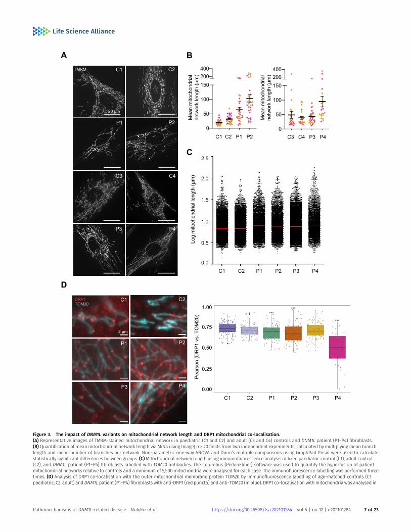
Figure 3. The impact of DNM1L variants on mitochondrial network length and DRP1 mitochondrial co-localisation.(A) Representative images of TMRM-stained mitochondrial network in paediatric (C1 and C2) and adult (C3 and C4) controls and DNM1L patient (P1–P4) fibroblasts.(B) Quantification of mean mitochondrial network length via MiNa using ImageJ n > 20 fields from two independent experiments, calculated by multiplying mean branchlength and mean number of branches per network. Non-parametric one-way ANOVA and Dunn’s multiple comparisons using GraphPad Prism were used to calculatestatistically significant differences between groups. (C)Mitochondrial network length using immunofluorescence analysis of fixed paediatric control (C1), adult control(C2), and DNM1L patient (P1–P4) fibroblasts labelled with TOM20 antibodies. The Columbus (PerkinElmer) software was used to quantify the hyperfusion of patientmitochondrial networks relative to controls and a minimum of 5,500 mitochondria were analysed for each case. The immunofluorescence labelling was performed threetimes. (D) Analysis of DRP1 co-localisation with the outer mitochondrial membrane protein TOM20 by immunofluorescence labelling of age-matched controls (C1:paediatric, C2: adult) and DNM1L patient (P1–P4) fibroblasts with anti-DRP1 (red puncta) and anti-TOM20 (in blue). DRP1 co-localisation with mitochondria was analysed in
Pathomechanisms of DNM1L-related disease Nolden et al. https://doi.org/10.26508/lsa.202101284 vol 5 | no 12 | e202101284 7 of 23

quantify the hyperfusion of patient mitochondrial networks relativeto controls. A minimum of 5,500 mitochondria were analysed foreach case. Largely consistent with live cell imaging, significanthyperfusion of mitochondrial networks were observed in all fourstudied patient fibroblasts using this approach (Fig 3C). Whereaslive cell imaging did not reveal extensive mitochondrial hyper-fusion in P3 fibroblasts, TOM20 immunostaining revealed elongatedmitochondria in P3 (p.Leu230dup) cells. Notably, these cells werethe least affected compared with those from other patients (Fig 3C).
To determine whether mitochondrial network alterations weredue to decreased DRP1 recruitment, we performed a co-localisationanalysis using the Pearson’s co-localisation coefficient betweenDRP1 and TOM20 which showed decreased DRP1 at the mito-chondria in P1 (p.Gly401Ser), P2 (p.Gly363Asp), and P4 (p.Arg710Gly)fibroblasts. Of these, P4 (p.Arg710Gly) had the most severe re-cruitment defect with the lowest Pearson’s R value and DRP1appearing primarily cytosolic without punctate structures, whichwere still seen in other variants albeit to a lesser extent than thecontrol fibroblasts (Fig 3D).
Although the degree of mitochondrial hyperfusion differedbetween patient fibroblasts, with P3 (p.Leu230dup) not displayingsignificant elongation by MiNA, this phenotype was consistent withpreviously reported de novo heterozygous DNM1L variants (c.95G>C,p.Gly32Ala; c.436G>A, p.Asp146Asn; c.1184C>A, p.Ala395Asp; c.1207C>T,p.Arg403Cys; c.1292G>A, p.Cys431Tyr) and a GTPase-deficient recombi-nant mutant (p.Lys38Ala) (Zhu et al, 2004; Waterham et al, 2007; Changet al, 2010; Whitley et al, 2018; Longo et al, 2020).
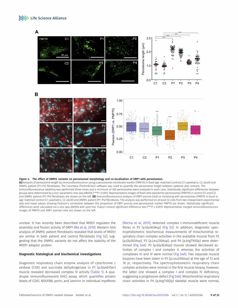
Given DRP1 has been implicated in both mitochondrial andperoxisomal fission (Waterham et al, 2007), we examined the effectof these variants on peroxisomal networks. Immunofluorescencelabelling of control and DNM1L patient fibroblasts with antibodiesagainst the peroxisomal membrane protein marker PMP70 wasused to determine the peroxisomal morphology. The analysis usingthe Columbus software revealed that peroxisomes in P1 (p.Gly401Ser),P2 (p.Gly363Asp), P3 (p.Leu230dup) and P4 (p.Arg710Gly)appeared more fused with fewer overall numbers of peroxi-somes and decreased size distribution, indicative of impairedfission (Fig 4A).
Co-localisation analysis between DRP1 and PMP70 showed de-creased DRP1 at the peroxisomes in P1 (p.Gly401Ser) and P2(p.Gly363Asp), but not P3 (p.Leu230dup) and P4 (p.Arg710Gly),suggesting that the elongated peroxisomes in P3 and P4 are notsimply due to decreased DRP1 recruitment (Fig 4B). Previous reportsargue that not all DNM1L variants impair peroxisomal morphology,with several other variants in the GTPase domain having noimpact on peroxisomal morphology despite affecting mitochon-drial network morphology. Specifically, the p.Glu2Ala, p.Ala192Glu(Gerber et al, 2017), and p.Gly32Ala (Whitley et al, 2018) variantshad normal peroxisomes in the setting of abnormal mitochondrialnetworks. Conversely, patient fibroblasts from a biallelic het-erozygous patient carrying p.Ser36Gly; p.Glu116Lysfs*6 variantshad both abnormal peroxisomal and mitochondrial fission (Nasca
et al, 2016). Similar impairments were also observed in thep.Asp146Asn (Longo et al, 2020) and p.Gly223Val variants (Verrigniet al, 2019) (Table S1).
Mitochondrial DNA nucleoid analysis of de novo DNM1L variants
Defective mitochondrial fission has also been associated with theformation of enlarged bulb-like structures (“mito-bulbs”) caused bynucleoid clustering (Ban-Ishihara et al, 2013). Previously, DNM1LsiRNA knockdown in HeLa cells as well as Dnm1l−/− knockout micestudies have demonstrated severe mtDNA nucleoid aggregationwithin the hyperfused mitochondrial networks, leading to respi-ratory deficiency and heart dysfunction in the fission-deficientmice(Ban-Ishihara et al, 2013; Ishihara et al, 2015). Imaging of fibroblastsincubated with TMRM revealed the presence of enlarged mito-chondria in all patients (Fig S1A), with P1 (p.Gly401Ser) and P2(p.Gly363Asp) most widely affected. Subsequent co-staining of P1and P2 fibroblasts with TMRM and PicoGreen (a fluorochrome whichreveals nucleoids by illuminating mtDNA) demonstrated the co-localisation of these enlarged “mito-bulbs” with large nucleoids(Fig S1B). Detailed analysis of mtDNA nucleoids stained withPicoGreen using Columbus software (PerkinElmer) revealedmarkeddifferences in the proportion of enlarged nucleoids (area > 1.5 μm2)in P1 (p.Gly401Ser) and P2 (p.Gly363Asp) compared with control (FigS1C). There was no difference in nucleoid size ratio between P3(p.Leu230dup) and control (Fig S1C). Although, upon visual exam-ination P4 (p.Arg710Gly) nucleoids appeared enlarged compare withcontrols, we were not able to accurately quantify the individualpuncta because of increased levels of lipofuscin present in thesecells (Fig S1D).
Altogether, assessment of patient fibroblasts demonstrated thatthe de novo variants identified in P1 (p.Gly401Ser) and P2(p.Gly363Asp) cause mitochondrial network hyperfusion, leading tomitochondrial enlargement and nucleoid clustering which is in-dicative of impaired nucleoid distribution and segregation.
The effect of DNM1L variants on DRP1 protein expression
To evaluate the molecular consequences of the c.1201G>A,p.Gly401Ser; c.1088G>A, p.Gly363Asp; c.687_689dupATT, p.Leu230dup,and c.2128A>G, p.Arg710Gly DRP1 variants, primary patient fibro-blasts (P1–P4) and age-matched controls (C1–C4) were analysed bySDS–PAGE and immunoblotting (Fig S2). Normal levels of DRP1protein in the monomeric form were found in P1 (p.Gly401Ser), P2(p.Gly363Asp), and P3 (p.Leu230dup), whereas P4 (p.Arg710Gly)showed decreased levels of DRP1 when compared with controls (FigS2). These data suggest that the mutated p.Gly401Ser, p.Gly363Asp,and p.Leu230dup DRP1 protein is expressed in P1, P2, and P3, re-spectively, and may act in a dominant-negative fashion, overridingthe effect of the wild-type allele. DRP1 recruitment to the mito-chondrial membrane is dependent on adaptor proteins such asMID49 and MID51. However, their role in disease remains largely
at least 32 cells per subject in two independent experimental sets. Pearson’s correlations between DRP1 puncta and TOM20 in each cell line are shown as box plots.One-way ANOVA with post hoc Tukey’s honest significant difference test was used to determine statistically significant differences (***P ≤ 0.001). Representative mergedimmunofluorescence images of fibroblasts stained with anti-TOM20 and anti-DRP1 antibodies are shown on the left.
Pathomechanisms of DNM1L-related disease Nolden et al. https://doi.org/10.26508/lsa.202101284 vol 5 | no 12 | e202101284 8 of 23
unclear. It has recently been described that MID51 regulates theassembly and fission activity of DRP1 (Ma et al, 2019). Western blotanalysis of DNM1L patient fibroblasts revealed that levels of MID51are similar in both patient and control fibroblasts (Fig S2), sug-gesting that the DNM1L variants do not affect the stability of theMID51 adaptor protein.
Diagnostic histological and biochemical investigations
Diagnostic respiratory chain enzyme analysis of cytochrome coxidase (COX) and succinate dehydrogenase in P1 (p.Gly401Ser)muscle revealed decreased complex IV activity (Table 1). A qua-druple immunofluorescent (IHC) assay, which quantifies proteinlevels of COX1, NDUFB8, porin, and laminin in individual myofibres
(Rocha et al, 2015), detected complex I-immunodeficient musclefibres in P2 (p.Gly363Asp) (Fig S3). In addition, diagnostic spec-trophotometric biochemical measurements of mitochondrial re-spiratory chain complex activities in the available muscle from P2(p.Gly363Asp), P3 (p.Leu230dup), and P4 (p.Arg710Gly) were deter-mined (Fig S4A). P2 (p.Gly363Asp) muscle showed decreased ac-tivities of complex I and complex II, whereas the activities ofcomplexes III and IV were normal (Fig S4A). Two separate musclebiopsies have been taken in P3 (p.Leu230dup) at the age of 13 and16 yr, respectively. The spectrophotometric respiratory chaincomplex activities were normal in the first muscle biopsy; however,the latter one showed a complex I and complex IV deficiency,suggesting a progressive defect (Fig S4A). Mitochondrial respiratorychain activities in P4 (p.Arg710Gly) skeletal muscle were normal,
Figure 4. The effect of DNM1L variants on peroxisomal morphology and co-localisation of DRP1 with peroxisomes.(A) Analysis of peroxisome length by immunofluorescence using a peroxisomal membrane marker (PMP70) in fixed age-matched controls (C1: paediatric, C2: adult) andDNM1L patient (P1–P4) fibroblasts. The Columbus (PerkinElmer) software was used to quantify the peroxisome length between patients and controls. Theimmunofluorescence labelling was performed three times and a minimum of 300 peroxisomes were analysed in each case. Statistically significant differences betweengroups were determined by a non-parametric one-way ANOVA (***P ≤ 0.001). Representative images of fixed cells stained for peroxisomes (PMP70) in control (C1 and C2)and DNM1L patient (P1–P4) fibroblasts are shown on the left. (B) Immunofluorescence analysis of DRP1 puncta (red) co-localising with peroxisomes (PMP70 in blue) inage-matched control (C1: paediatric, C2: adult) and DNM1L patient (P1–P4) fibroblasts. The analysis was performed on at least 32 cells from two independent experimentalsets and mean values showing Pearson’s correlation between the proportion of DRP1 puncta and peroxisomal marker PMP70 are shown. Statistically significantdifferences were calculated via a one-way ANOVA with post-hoc Tukey’s honest significant difference test (***P ≤ 0.001). Representative merged immunofluorescenceimages of PMP70 and DRP1 stained cells are shown on the left.
Pathomechanisms of DNM1L-related disease Nolden et al. https://doi.org/10.26508/lsa.202101284 vol 5 | no 12 | e202101284 9 of 23
except for increased complex III activity, which may be attributed toa compensatory response mechanism (Fig S4A).
Variants in DNM1L impair levels of OXPHOS proteins
Next, we determinedwhether themitochondrial network anomaliespresent in DNM1L patient fibroblasts were associated with OXPHOSdysfunction. Western blotting and quantification of bands obtainedby densitometry analysis of P1 (p.Gly401Ser) fibroblasts revealedthat the steady-state levels of OXPHOS proteins were relativelynormal, except for mild decreases in the complex I subunit, NDUFB8and the complex IV subunit COX2 (Fig S5), which was consistent withthe observed decreased complex IV activity in muscle tissue (Table1). P2 (p.Gly363Asp) mutant fibroblasts presented with a decrease inNDUFB8, UQCRC2, and COX2 protein levels (Fig S5). In addition, themarked decrease in NDUFB8 protein levels detected by Westernblotting correlate with the impaired complex I activity in patient-derived muscle and fibroblasts (Fig S4A and B). NGS analysis of P2also identified a de novo heterozygous c.152G>A, p.Arg51Gln variantin the NDUFS5 gene encoding a core accessory subunit of complex I.The c.152G>A, p.Arg51Gln NDUFS5 variant could partially contribute tothe decreased levels of NDUFB8 protein and impaired complex Iactivity; however, in silico pathogenicity assessment classified thevariant as likely benign and not pathogenic. Amultiple OXPHOS defectwas present in P3 (p.Leu230dup) fibroblasts, showing decreasedsteady-state levels of NDUFB8, UQCRC2, and COX2 (Fig S5), where onlyimpaired complex I and complex IV activity, correlated with therespiratory chain measurements in muscle (second biopsy) (FigS4A). Furthermore, a decrease in complex I (NDUFB8) and complexIV (COX2) subunits was detected in P4 (p.Arg710Gly) fibroblastswhen compared with controls (Fig S5). Similar to the increasedcomplex III activity in P4 muscle tissue (Fig S4A), densitometry analysisof the complex III subunit in P4 fibroblasts showedmild increase in thesteady-state levels of UQCRC2 (Fig S5).
Interestingly, there are some differences between the OXPHOSabnormalities amongst the patient muscle samples and fibroblasts.Most notably, P4 (p.Arg710Gly) whom had increased complex IIIactivity in skeletal muscle, but decreased complex I and IV proteinsin fibroblasts. We hypothesize that these differences likely stemfrom tissue-specific effects on respiration. Together these datasuggest that different DNM1L variants have distinct impact onOXPHOS function in fibroblasts, with minimal correlations to dis-ease onset or severity, suggesting that the OXPHOS defects presentin cells are a secondary consequence of the disrupted mito-chondrial network balance as opposed to a driver of disease.
Patient DRP1 variants have altered GTPase activity
DRP1 performs its mechanoenzyme function of mitochondrialmembrane constriction through the hydrolysis of GTP following itsassembly on the mitochondrial outer membrane. To determinewhether DNM1L variants altered GTPase activity in vitro, we firstexpressed human DRP1 in recombinant form recapitulating thedisease-causing variants identified in P1 and P5 c.1201G>A,p.Gly401Ser (G401S), P2 c.1088G>A, p.Gly363Asp (G363D), P3c.687_689dupATT, p.Leu230dup (L230dup), and P4 c.2128A>G,p.Arg710Gly (R710G). Bacterial expression of all variants were-
similar to wildtype (WT), except for L230dup which did not produceany full-length protein under multiple conditions and was unableto be purified for further studies. Wild-type human DRP1 and theremaining variants were purified to homogeneity and found to bewell folded by circular dichroism (Fig S6), but differences in themean residue ellipticity suggested differences in structure thatmight affect GTP hydrolysis. To test this, GTP hydrolysis wasmeasured in solution with increasing amounts of GTP substrate todetermine the apparent Michaelis constant (K0.5), the turnovernumber (kcat), and catalytic efficiency (kcat/K0.5) (Fig 5A–D). Theactivity of WT enzyme was similar to previous measurements(Chang et al, 2010; Frohlich et al, 2013; Koirala et al, 2013; Bustillo-Zabalbeitia et al, 2014; Cahill et al, 2015; Francy et al, 2017) with a K0.5GTP
of 201 ± 51 μM, a kcat of 0.24min−1, and kcat/K0.5 of 1.2 × 10⁻³ μM−1•min−1
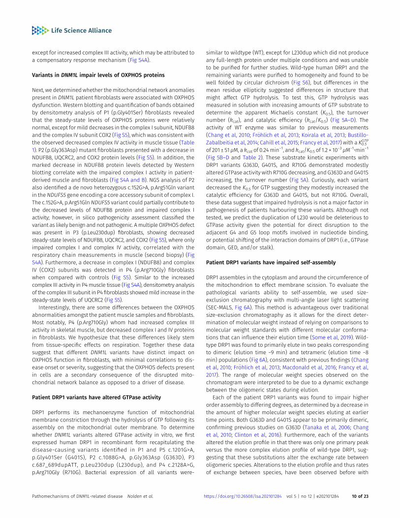
(Fig 5B–D and Table 2). These substrate kinetic experiments withDRP1 variants G363D, G401S, and R710G demonstrated modestlyaltered GTPase activity with R710G decreasing, and G363D and G401Sincreasing, the turnover number (Fig 5A). Curiously, each variantdecreased the K0.5 for GTP suggesting they modestly increased thecatalytic efficiency for G363D and G401S, but not R710G. Overall,these data suggest that impaired hydrolysis is not a major factor inpathogenesis of patients harbouring these variants. Although nottested, we predict the duplication of L230 would be deleterious toGTPase activity given the potential for direct disruption to theadjacent G4 and G5 loop motifs involved in nucleotide binding,or potential shifting of the interaction domains of DRP1 (i.e., GTPasedomain, GED, and/or stalk).
Patient DRP1 variants have impaired self-assembly
DRP1 assembles in the cytoplasm and around the circumference ofthe mitochondrion to effect membrane scission. To evaluate thepathological variants ability to self-assemble, we used size-exclusion chromatography with multi-angle laser light scattering(SEC-MALS, Fig 6A). This method is advantageous over traditionalsize-exclusion chromatography as it allows for the direct deter-mination of molecular weight instead of relying on comparisons tomolecular weight standards with different molecular conforma-tions that can influence their elution time (Some et al, 2019). Wild-type DRP1 was found to primarily elute in two peaks correspondingto dimeric (elution time ~9 min) and tetrameric (elution time ~8min) populations (Fig 6A), consistent with previous findings (Changet al, 2010; Frohlich et al, 2013; Macdonald et al, 2016; Francy et al,2017). The range of molecular weight species observed on thechromatogram were interpreted to be due to a dynamic exchangebetween the oligomeric states during elution.
Each of the patient DRP1 variants was found to impair higherorder assembly to differing degrees, as determined by a decrease inthe amount of higher molecular weight species eluting at earliertime points. Both G363D and G401S appear to be primarily dimeric,confirming previous studies on G363D (Tanaka et al, 2006; Changet al, 2010; Clinton et al, 2016). Furthermore, each of the variantsaltered the elution profile in that there was only one primary peakversus the more complex elution profile of wild-type DRP1, sug-gesting that these substitutions alter the exchange rate betweenoligomeric species. Alterations to the elution profile and thus ratesof exchange between species, have been observed before with
Pathomechanisms of DNM1L-related disease Nolden et al. https://doi.org/10.26508/lsa.202101284 vol 5 | no 12 | e202101284 10 of 23

Figure 5. Clinically identified DNM1L variants alter GTPase activity.(A) Substrate kinetics of recombinant wild-type DRP1 (WT) (1 μM) and genetic variants. DRP1 GTPase activity was measured using an enzyme coupled assaymonitoring NADH depletion, which is subsequently converted to activity (min−1). Data from three independent experiments were globally fit to aMichaelis–Menten model. Residuals of the fit are shown. (B, C, D) Distribution of K0.5, (C) kcat, and (D) kcat/K0.5 parameters from GTPase activity measurements.Reported values were obtained by globally fitting DRP1 GTPase activity measurements (n = 3) to a Michaelis–Menten model. The resulting values are reported inTable 2. K0.5 differences between WT and each variant significant to ***P < 0.05. kcat differences between WT and G363D, G363D and R710G, G363D and G401S, andR710G and G401S significant to ***P < 0.05. kcat/K0.5 differences between WT and both G363D and G401S, as well as between R710G and both G363D and G401Ssignificant to ***P < 0.05.
Table 2. Reported kinetic values among DRP1 variants. Kinetic parameters (K0.5, Vmax, kcat, and kcat/K0.5) were computed for DRP1 WT and each clinicalvariant.
K0.5 ± SD (μM) Vmax ± SD (μM/min) kcat (min21) kcat (min21)/K0.5 (μM)
WT 201 ± 51 0.24 ± 0.01 0.24 × 10⁻⁶ 1.2 × 10⁻⁹
G363D 79 ± 11 0.58 ± 0.02 0.58 × 10⁻⁶ 7.3 × 10⁻⁹
G401S 55 ± 9 0.36 ± 0.011 0.36 × 10⁻⁶ 6.5 × 10⁻⁹
R710G 96 ± 18 0.10 ± 0.004 0.10 × 10⁻⁶ 1.0 × 10⁻⁹
Pathomechanisms of DNM1L-related disease Nolden et al. https://doi.org/10.26508/lsa.202101284 vol 5 | no 12 | e202101284 11 of 23
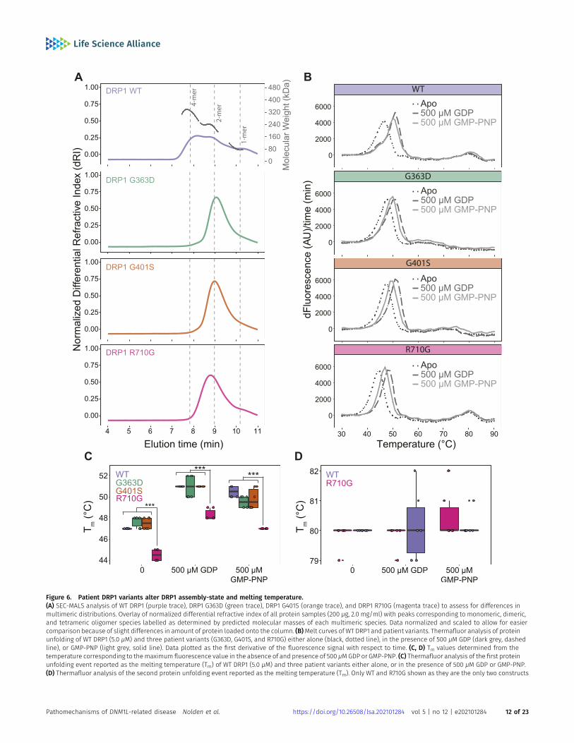
Figure 6. Patient DRP1 variants alter DRP1 assembly-state and melting temperature.(A) SEC-MALS analysis of WT DRP1 (purple trace), DRP1 G363D (green trace), DRP1 G401S (orange trace), and DRP1 R710G (magenta trace) to assess for differences inmultimeric distributions. Overlay of normalized differential refractive index of all protein samples (200 μg, 2.0 mg/ml) with peaks corresponding to monomeric, dimeric,and tetrameric oligomer species labelled as determined by predicted molecular masses of each multimeric species. Data normalized and scaled to allow for easiercomparison because of slight differences in amount of protein loaded onto the column. (B)Melt curves of WT DRP1 and patient variants. Thermafluor analysis of proteinunfolding of WT DRP1 (5.0 μM) and three patient variants (G363D, G401S, and R710G) either alone (black, dotted line), in the presence of 500 μM GDP (dark grey, dashedline), or GMP-PNP (light grey, solid line). Data plotted as the first derivative of the fluorescence signal with respect to time. (C, D) Tm values determined from thetemperature corresponding to themaximum fluorescence value in the absence of and presence of 500 μMGDP or GMP-PNP. (C) Thermafluor analysis of the first proteinunfolding event reported as the melting temperature (Tm) of WT DRP1 (5.0 μM) and three patient variants either alone, or in the presence of 500 μM GDP or GMP-PNP.(D) Thermafluor analysis of the second protein unfolding event reported as the melting temperature (Tm). Only WT and R710G shown as they are the only two constructs
Pathomechanisms of DNM1L-related disease Nolden et al. https://doi.org/10.26508/lsa.202101284 vol 5 | no 12 | e202101284 12 of 23

G363D, as well as other stalk domain variants including the lethalA395D substitution and G350D (Chang et al, 2010). Notably, R710Ghad an earlier peak elution time than G363D and G401S, 8.79 minversus 9.07 and 8.99 min, respectively. This suggests that R710Glikely retains some ability to assemble into higher order oligomericspecies, observed as a leftward shift in the peak elution time becauseof fast-exchange between dimeric and tetrameric species, contrary toG363D and G401S. In addition, treatment of total cell lysates derivedfrom control and DNM1L patient fibroblasts with a chemical cross-linker BMH (bismaleimidohexane) resulted in increased formation ofhigher order oligomeric DRP1 complexes in P4 (Fig S7). Therefore,these data further support our SEC-MALS results, suggesting that theR710G variant retains more ability to assemble with wild-type DRP1than other variants. Together, these results provide strong evidencethat these disease-causing variants alter DRP1 ability to assemble,which is critical for mediating mitochondrial fission.
Patient DRP1 variants are well-folded but have differing stability
Protein stability was evaluated using a fluorophore-based (SYPROOrange) thermal shift assay and revealed the presence of twounfolding events in wild-type DRP1 (Fig 6B). Repeating the assay in thepresence of either 500 μM GDP or 500 μM GMP-PNP showed increasedstability of the first unfolding event upon nucleotide binding, but notthe second. Therefore, we interpreted the first and second transitionsas corresponding to the unfolding of the GTPase and stalk domains,respectively. Given the variable domain of DRP1 is intrinsically dis-ordered (Strack & Cribbs, 2012; Frohlich et al, 2013; Wenger et al, 2013;Rosdah et al, 2020; Mahajan et al, 2021), it is not surprising that a thirdunfolding event corresponding to this domain was not observed givenno significant loss of secondary structure would be expected in thisregion upon unfolding. Both G363D and G401S were found to have onlyone distinct unfolding event corresponding to GTPase domainunfolding, consistent with the SEC-MALS data showing no higher orderorganisation. For wildtype, addition of GDP had little effect on the stalkdomain transitions as expected (Fig 6C and D). By contrast, addition ofGDP significantly increased the Tm of the GTPase domain even morethan GMP-PNP (Fig 6C and D), consistent with the known higher affinityof GDP over non-hydrolyzable GTP analogues for the GTPase domain(Frohlich et al, 2013). This overall pattern was the same for all con-structs indicating each variant is able to bind nucleotide, althoughDRP1 R710G showed a significantly lower GTPase domain meltingtemperature than WT, G363D and G401S, even in the presence ofnucleotide, indicating that this variant destabilized the protein but notits ability to respond to nucleotide.
Discussion
Here, we report the discovery of five patients with previously un-reported variants in DNM1L, including only the second GED domain
variant (p.Arg710Gly) to be identified to date. The p.Gly363Aspvariant has previously been studied given its high degree ofconservation across species, although this is the first report of apatient harbouring this pathogenic variant to our knowledge(Tanaka et al, 2006; Kobayashi et al, 2007; Chang et al, 2010; Oteraet al, 2010; Kwapiszewska et al, 2019). The variants described herewere predicted to be “likely pathogenic” according to ACGSguidelines, taking into account various criteria including variantallele frequency, functional studies, phenotypic fit and in silicopredictions. In silico structural analysis of each variant concurredand predicted likely impairment of DRP1 oligomerisation (L230dup,G363D and G401S), GTP hydrolysis (L230dup and R710G) and proteinstability (R710G) (Frohlich et al, 2013; Kalia et al, 2018). Analysis ofmitochondrial network morphology in fixed patient-derived celllines revealed impaired mitochondrial fission leading to hyper-fused mitochondrial networks (Fig 3C) and in some cases enlargedmtDNA nucleoids (Fig S1), confirming dysfunctional DRP1 as theprimary pathogenic factor in these patients. Furthermore, DNM1Lvariants present in P1 (p.Gly401Ser), P2 (p.Gly363Asp), P3 (p.Leu230dup)and P4 (p.Arg710Gly) also impaired normal peroxisomal fission (Fig4A), which is not surprising given DRP1’s prominent role in this process(Li & Gould, 2003; Koch et al, 2005; Tanaka et al, 2006; Kobayashi et al,2007; Gandre-Babbe & Van Der Bliek, 2008; Otera et al, 2010; Koch &Brocard, 2012; Yamano et al, 2014). P1 (p.Gly401Ser), P2 (p.Gly363Asp)and P4 (p.Arg710Gly) DNM1L variants caused decreased DRP1recruitment to the mitochondria (Fig 3D), but only P1 and P2 haddecreased DRP1-peroxisome co-localisation (Fig 4B), suggesting thatimpaired DRP1 p.Arg710Gly peroxisomal fission occurs through adifferent mechanism. These data indicate that p.Arg710Gly mediatedimpairments are not simply due to a lack of DRP1 at the peroxisomalmembrane, but may be due to impaired enzyme function withpreservation of DRP1–peroxisome recruiter interactions, which arelost with the p.Gly363Asp and p.Gly401Ser variants.
To evaluate the effects of these mutations on DRP1, we per-formed a series of experiments designed to elucidate the specificmechanisms underpinning impaired function. The GTP hydrolysisactivity is essential for DRP1 function. Interestingly, we found thatG363D and G401S had increased GTP hydrolytic activity compared toWT DRP1, whereas R710G had decreased activity (Fig 5A). Previousstudies examining the G363D variant have reported mixed hydro-lysis results including no effect on hydrolytic activity (Clinton et al,2016), or impaired hydrolysis (Tanaka et al, 2006; Chang et al, 2010).Given these discrepancies, it cannot be ruled out that differences inGTP hydrolysis may be due to variations in recombinant proteinconstructs or preparation methods (e.g., DRP1 isoforms, N- versusC-terminal tags, and calmodulin versus histidine purification tags)(Clinton et al, 2020). One might anticipate that increased GTP hy-drolysis would result in increased fission intracellularly. However, itis possible that these results are representative of futile GTP cyclingin which G363D and G401S retain hydrolytic capabilities but areunable to assemble into the higher order oligomeric species.
with a prominent second unfolding event. Data are representative of two independent experiments, each with three technical replicates. ***P < 0.00001. Differencesbetween Tm values of all constructs alone in comparison with constructs with 500 μM GDP or 500 μM GMP-PNP significant to P < 0.003. Tm values of all constructswith 500 μM GDP in comparison to 500 μM GMP-PNP are significant to P < 0.03 except for R710G with 500 μM GDP in comparison to R710G with 500 μM GMP-PNP whereP < 0.0003.
Pathomechanisms of DNM1L-related disease Nolden et al. https://doi.org/10.26508/lsa.202101284 vol 5 | no 12 | e202101284 13 of 23

Both G363D and G401S appear to be in mutational hotspots (Fig1B) with multiple variants in nearby regions reported includingG350R, G362S, G362D, A395D, A395G, R403C, L406S, and E410K (Changet al, 2010; Fahrner et al, 2016; Sheffer et al, 2016; Vanstone et al,2016; Zaha et al, 2016; Ryan et al, 2018; Whitley et al, 2018; Vandeleuret al, 2019; Verrigni et al, 2019). These variants reside spatially closeto each other within the stalk domain of the protein, a regionimportant for mediating protein oligomerisation (Frohlich et al,2013; Francy et al, 2017), which in turn is critical for stabilization ofDRP1–MFF complexes post recruitment to themitochondria (Clintonet al, 2016) as well as assembly with MID49 (Kalia et al, 2018). Thissuggests the variants may have impaired fission secondary to di-minished higher order assembly and/or poor recruitment to themitochondria secondary to impaired DRP1–recruiter interactions.Consistent with this, both p.Gly363Asp and p.Gly401Ser have de-creased DRP1 at the mitochondria as determined by DRP1-TOM20co-localisation analysis (Fig 3D). Therefore, the decrease in mito-chondrial fission despite increased GTP hydrolysis for both G363Dand G401S likely stems from a lack of DRP1 recruitment and pro-ductive fission activity at the mitochondria.
In addition, our SEC-MALS data suggest that both G363D andG401S are unable to attain higher order species as they eluted in aprimarily dimeric population (Fig 6A), consistent with previousreports on G363D (Chang et al, 2010; Clinton et al, 2016;Kwapiszewska et al, 2019). The glycine at position 401 is one of fourhighly conserved amino acids (GPRP, 401-404) located at the as-sembly interface where it is involved in mediating oligomerisationof proteins within the Dynamin superfamily including dynamin,DRP1, and MxA (Gao et al, 2010; Faelber et al, 2011; Ford et al, 2011).Like dynamin, these four residues required mutation to AAAA toprevent oligomerisation and inherent disorder of the loop region toachieve crystallisation of DRP1 (Frohlich et al, 2013). Therefore, it islikely that both substitutions directly impair higher order assemblyandmay also disrupt local secondary structure given these variantsdid not exhibit a clear unfolding of the stalk domain by thermal shiftanalysis.
From a clinical mitochondrial disease perspective, it is inter-esting that both P1 (c.1201G>A, p.Gly401Ser) and P2 (c.1088G>A,p.Gly363Asp) exhibited cardiac complications, including end-stagedilated cardiomyopathy with previous signs of hypertrophic car-diomyopathy in P1. Of the previously reported variants, c.1228G>A,p.Glu410Lys is the only pathogenic human DNM1L variant that hasbeen reported to result in severe cardiac involvement, which ul-timately resulted in death of the patient at 8 mo of age (Vandeleuret al, 2019). Cardiac involvement in patients with DNM1L-relatedmitochondrial disease has previously been postulated because aC452F substitution in mouse DRP1 (position p.Cys446Phe in humanDRP1 NP_036192.2) was shown to cause dilated cardiomyopathy(Cahill et al, 2015). Concordantly, a 3-mo-old patient who initiallypresented with infantile parkinsonism-like symptoms was identi-fied to possess the same C446F substitution and died at 2.5 yr of agebecause of sudden cardiac arrest (Dıez et al, 2017). However, nopost-mortem evaluation was performed to determine the cause ofcardiac arrest. It would therefore seem appropriate that patientswith confirmed pathogenic DNM1L variants follow a cardiac sur-veillance programme, as is in place for other forms ofmitochondrialdisease, with a view to appropriate pre-emptive treatment.
In general, pathogenic variants involving the stalk domain ofDRP1 also appear to bemore severe than those affecting the GTPasedomain which primarily present as optic abnormalities with orwithout concurrent neurological and developmental findings(Gerber et al, 2017; Hogarth et al, 2018; Whitley et al, 2018; Longo et al,2020; Wei & Qian, 2021). We note a similar trend in our cohort withP1, P5 (c.1201G>A, p.Gly401Ser), and P2 (c.1088G>A, p.Gly363Asp)experiencing an earlier onset of more severe symptoms, fasterdisease progression, and early death, whereas P3 (c.687_689dupATT,p.Leu230dup) and P4 (c.2128A>G, p.Arg710Gly) had a later onset andlived to an older age. Of note, P3 (p.Leu230dup) and P4 (p.Arg710Gly)also exhibited less severe peroxisomal defects compared with P1(p.Gly401Ser) and P2 (p.Gly363Asp). It may be that concurrent mito-chondrial and peroxisomal defects lead to more severe phenotypesand disease progression. Consistent with this, several other non-lethalDRP1 variants, located primarily in the GTPase domain, resulted in cellswith normal peroxisome morphology despite having impaired mito-chondrial networks (Chao et al, 2016; Gerber et al, 2017; Whitley et al,2018) (Table S1).
In true peroxisomal biogenesis disorders (PBDs), lipid meta-bolism, among other peroxisome-related metabolic pathways, isimpaired. Clinically, DNM1L and PBD patients have phenotypicoverlap including developmental delays, seizures, hypotonia, facialdysmorphism, and vision impairment. Unlike PBD patients though,DNM1L patients do not typically develop renal or hepatic dys-function, skeletal abnormalities, or cataracts (Waterham &Ebberink, 2012). Given these similarities, and the peroxisome fis-sion abnormalities in many DNM1L patients, one might hypothesizethat DNM1L patients would display similar biochemical profiles,with elevated very long-chain and branched-chain fatty acids (DeBiase et al, 2019). Unfortunately, there remains a dearth of DNM1Lpatient reports that analyse both peroxisomal morphology andperform the necessary analyses to fully evaluate peroxisomalfunction. Based on data currently available, there is not a clearcorrelation between laboratory findings, peroxisome morphology,and disease severity with some variants displaying normal peroxi-some morphology with normal laboratory tests (p.Gly362Ser) (Shefferet al, 2016), normal peroxisomes with elevated plasma VLCFA andnormal pristanic acid (p.Gly32Ala) (Whitley et al, 2018), abnormalperoxisomes with normal laboratory tests (p.Ser36Gly, p.Glu116-Lysfs*6; p.Gly362Ser; p.Ile512Thr, p.Gly362Asp; p.Gly350Arg, andp.Tyr691Cys) (Chao et al, 2016; Nasca et al, 2016; Verrigni et al, 2019),and abnormal peroxisomes with abnormal laboratory tests(p.Ala395Asp) (Waterham et al, 2007). Several studies noted ab-normal peroxisomal morphology but did not perform lipid profiling(Chao et al, 2016; Zaha et al, 2016; Longo et al, 2020), and it is unclearwhether these patients may have had abnormal results (Table S1).Although traditional peroxisome functional tests may not be fruitfuldiagnostically, future studies using lipidomic approaches may cap-ture more nuanced metabolic changes that occur, identifying po-tential biomarkers for DNM1L-associated disease with peroxisomeinvolvement. Ultimately, DNM1L disorders appear to derive primarilyfrom mitochondrial defects and the degree of peroxisome-drivenpathology remains unclear, but likely secondary.
Unfortunately, we were unable to obtain full-length recombinantDRP1 L230dup (P3) for in vitro studies. Given this residue’s relativeproximity to the nucleotide-binding site, a duplication event is
Pathomechanisms of DNM1L-related disease Nolden et al. https://doi.org/10.26508/lsa.202101284 vol 5 | no 12 | e202101284 14 of 23

likely to disrupt GTP binding. This would have direct impacts on GTPhydrolysis and resulting fission activity. DRP1 L230 is also near theDRP1–MID49 interface and the duplication may selectively inhibitrecruiter interactions. Currently, only the structure of DRP1 incomplex with MID49 has been solved (Kalia et al, 2018), so it ispossible that other recruiting proteins bind at alternate locationsenabling residual DRP1 activity to be performed. Alternatively, andcontrasting a dominant negative mechanism, this allele is cata-lytically dead and residual DRP1 activity is maintained by the wild-type allele. In support of this, patient fibroblasts demonstrated amilder hyperfusion of mitochondrial reticula compared with theother variants and they lived to 20 yr of age, suggesting slowerdisease progression.
Intramolecular interaction between a monomer’s GTPase Ef-fector Domain (GED), the N-terminal GTPase domain, and stalkdomain, as well as interactions between adjacent GEDs are es-sential for regulation of DRP1 GTP hydrolysis (Pitts et al, 2004; Zhuet al, 2004; Chang & Blackstone, 2007). This is a common feature inall dynamin proteins (Muhlberg et al, 1997; Schumacher & Staeheli,1998; Di Paolo et al, 1999; Sever et al, 1999; Shin et al, 1999; Smirnovaet al, 1999; Zhang & Hinshaw, 2001) where removal of the GED indynamin or DRP1 does not prevent nucleotide binding or higherorder assembly but decreases GTPase activity (Muhlberg et al, 1997;Zhu et al, 2004). Similarly, R710G can still bind GTP, evidenced by itsability to hydrolyse GTP and stabilisation of the GTPase domainupon nucleotide binding but has decreased GTPase activity. Mu-tation of R725 in dynamin (R710 in human DRP1 [NP_036192.2] andboth located in the hinge 1 region) prevents stimulation of GTPaseactivity by the GED domain, suggesting it is a key residue involved insensing and transmitting assembly information to the GTPasedomain (Sever et al, 1999). The hinge 1 region has also been shownto be important for MxA function which shares structural propertieswith the family of dynamin-like GTPases. However, disruption ofMxA R640 or E632 (equivalent to R710 and E676 in human DRP1[NP_036192.2]) impairs higher order oligomerisation and decreasedthe off-rate of GTP, thus causing increased GTP hydrolysis which isopposite of what is observed in dynamin (Sever et al, 1999; Gao et al,2011). Nearby dynamin residue K694 (equivalent human DRP1residue: K679) is also located in the GED, but mutation results inimpaired assembly, suggesting it lays at the interface betweenadjacent GEDs where it stabilizes their interaction during as-sembly (Sever et al, 1999). A previously reported de novop.Tyr691Cys DRP1 variant in the fifth α-helix of the stalk portion ofthe GED was proposed to disrupt GED–GTPase interactions(Batzir et al, 2019), but it seems more likely that this substitutionwould negatively impact GED–GED assembly given its location atthis interface. Interestingly, the c.2072A>G, p.Tyr691Cys DNM1Lpatient, and our c.2128A>G, p.Arg710Gly (P4) had similar, lesssevere phenotypes compared with stalk domain variants andpresented with epilepsy, optic atrophy, impaired mobility, andprominent cyclical vomiting.
Therefore, we predict that R710G is pathogenic because of adisruption in the sensing mechanism that facilitates assembly-driven increases in GTP hydrolysis. Furthermore, this variant hadthe greatest loss of recruitment to the mitochondria in patientfibroblasts, suggesting this process, or region of the protein, may beimportant for proper DRP1–mitochondrial recruiter recognition. It is
unclear if the substitution results in direct disruption of GED–GTPase domain interaction, or if it is a downstream mechanism.Supporting a direct disruption, R710G results in a lower Tm for theGTPase domain, albeit with retained nucleotide-binding capabil-ities, reflective of decreased protein stability, possibly due to loss ofthe intramolecular GED–GTPase domain interactions. It is thereforenot surprising that this patient had lower protein levels of DRP1,and this may be reflective of increased protein degradation sec-ondary to the decreased stability, whereas the other patients didnot, suggesting haploinsufficiency is not amajor driver of pathologyin those cases, which has been noted for other variants as well(Whitley et al, 2018). R710G is perhaps somewhat assembly deficientcompared with wildtype, but more assembled than G363D or G401Sand is found in a dynamic equilibrium between a dimeric andtetrameric state.
There are nine known DRP1 isoforms that arise from differentialsplicing in the GTPase or variable domains, with isoforms differingbased on their inclusion, or lack of, a 13–amino acid insert in theGTPase domain (A insert) and a partial or full 37–amino acid insertin the variable domain (B insert) (Rosdah et al, 2020). These iso-forms have varying GTPase rates in the presence of cardiolipin, aprimary component of the mitochondrial outer membrane, or inresponse to the DRP1 recruiter MFF (Macdonald et al, 2016). Cur-rently, none of the reported variants are found within the A or Binsert, suggesting all DRP1 isoforms in patients would be af-fected. This raises the question of why neuronal tissue is pre-dominantly affected in this patient population. It may be thatcertain isoforms are more tolerant of substitutions, experiencingfewer or less severe impacts on protein oligomerisation or GTPhydrolysis. Genetic mosaicism may also play a role in patientswith milder, or perhaps even subclinical phenotypes. It is alsounclear why fetal development is grossly normal, given thepreponderance of heterozygous dominance among DNM1Lvariants. A role for DRP1 in development is still emerging, butevidence supports the importance of DRP1 as global knockoutis embryonic lethal in mouse models (Ishihara et al, 2009;Wakabayashi et al, 2009).
Here, we have described with mechanistic precision howpathogenic variants disrupt DRP1 biophysical activity and lead tomitochondrial hyperfusion. We document that divergent mech-anisms including combinations of aberrant stability, organellarrecruitment, assembly, and GTPase activity contribute to patho-genesis caused by mutations in different domains of DRP1. Insummary, a thorough understanding of how DRP1 function isimpaired in human disease will provide insight into the diversephenotypes and variable disease severity associated with patho-genic DNM1L variants. A systematic characterisation of patientpresentation and progression will assist in the timely identificationof other patients with rare DNM1L variants, whereas understandingthe specific molecular mechanisms underlying DRP1 function willpromote the development of targeted therapeutics with a goal ofrestoring mitochondrial fission to non-pathological levels. Cru-cially, our work details the first example of a patient with a DNM1Lvariant in the hinge region which will be crucial to answering anoutstanding question: how assembly information is transmitted tothe GTPase domain to stimulate GTP hydrolysis in the dynaminsuperfamily.
Pathomechanisms of DNM1L-related disease Nolden et al. https://doi.org/10.26508/lsa.202101284 vol 5 | no 12 | e202101284 15 of 23

Materials and Methods
Ethical statement
Written informed consent for diagnostic molecular genetic analysisand research-based studies was obtained from all patients inaccordance with the Declaration of Helsinki protocols and ethicalapprovals of local institutional review boards.
Diagnostic studies of skeletal muscle biopsies
Available skeletal muscle biopsies were subjected to routine di-agnostic investigations, including diagnostic TEM studies of musclefrom P1. Diagnostic in vitro spectrophotometric measurements ofrespiratory chain complex activities were undertaken in P2, P3, andP4 muscle according to standard procedures (Kirby et al, 2007).Complex I and IV–immunodeficient muscle fibres in P2 were de-termined by a quadruple fluorescent IHC assay of OXPHOS function,which evaluates protein levels of mitochondrial subunits ofcomplex I (NDUFB8) and complex IV (COX1). In addition to theimmunofluorescence labelling of muscle sections using antibodiesagainst the above described OXPHOS complexes, themitochondrialmass was quantified using an antibody against the outer mitochon-drial membrane protein—porin (VDAC) and the myofibre boundarieswere labelled with anti-laminin, a membrane glycoprotein as previ-ously described (Rocha et al, 2015).
Molecular genetics studies
All patients underwent routine mtDNA diagnostic testing that ex-cluded variants in the mitochondrial genome. Next generationsequencing strategies followed by filtering and candidate variantanalysis were undertaken to elucidate the molecular bases ofstudies on mitochondrial disease patients. GnomAD (https://gnomad.broadinstitute.org/) database was used for minor allelefrequency analysis (≤0.01%). In silico pathogenicity tools were usedto assess the pathogenicity of candidate variants and classified as“likely pathogenic” using the Association of Clinical Genomic Sci-ence (ACGS) and The American College of Medical Geneticsand Genomics (ACMG) guidelines (Richards et al, 2015) (https://www.acgs.uk.com/media/11631/uk-practice-guidelines-for-variant-classification-v4-01-2020.pdf).
Family trio WES analysis was performed on P1 using the AgilentSure Select Human All Exon Kit v6 according to the manufacturer’sinstructions, followed by sequencing on an Illumina NextSeqplatform. For P2, targeted NGS sequencing using a custom 84.38-KbAmpliseq panel (Life Technologies) was initially performed tocapture relevant regions of 50 Complex I genes as previously de-scribed (Alston et al, 2016). Sequencing was performed using the IonPGM 200 Sequencing Kit on an Ion Torrent PGM Sequencer. Variantcalling was undertaken using the proprietary Ion Torrent VariantCaller plugin and sequence variants were annotated using wAN-NOVAR for prioritisation and classification. Further to targeted NGS,trio WES analysis was performed using Agilent SureSelectXT All Exonv.5 according to the manufacturer’s instructions, followed by se-quencing on an Illumina HiSeq2500 platform and in-house
pipelines were used for variant calls as previously described (Tayloret al, 2014; Rocha et al, 2015). For P3, whole genome sequencing wasperformed by Genomics England via the 100,000 genomes project.WES and variant filtering and prioritisation was performed in P4 aspreviously described (Taylor et al, 2014; Thompson et al, 2016). WESanalysis was also performed on P5 as described in P1 using theAgilent Sure Select Human All Exon Kit v6 and sequencing on anIllumina NextSeq platform.
In silico analysis and structural modelling
The structures of DRP1 (PDB: 4BEJ), DRP1 in complex with GDP.AlF4(3W6P), DRP1 in complex with GMP-PCP (3W6O), and co-assembledDRP1-MID49 (PDB: 5WP9) were used to assess the structural im-plications of the patient mutations using PyMOL by Schrodinger(https://pymol.org/2/). In silico mutagenesis was performed usingModeler software with standard parameters (https://salilab.org/modeller/).
Cell lines
Primary patient fibroblasts and age-matched controls were grownin high-glucose Dulbecco’s Modified Eagle Medium (Gibco) sup-plemented with 10% (vol/vol) FBS, 1% non-essential amino acids,1.0 mM sodium pyruvate, 50 μg/ml uridine, 50 U/ml penicillin, and50 μg/ml streptomycin at 37°C in an atmosphere of 5.0% CO2. Allprimary control and patient fibroblasts used in this study wereunder P0+12 passages.
Mitochondrial network analysis using the Mitochondrial Analysis(MiNa)
Asynchronized control and patient fibroblasts were culturedovernight on 35-mm Ibidi m-dishes (Ibidi, 88156) before incubationin 0.15% PicoGreen (P7581; Invitrogen) for 30 min, then washed andincubated in 5.0 nM TMRM (T668; Invitrogen) for 30 min. Z-stackimages were taken on a VisiTech iSIM with a 100× objective beforeprocessing using Fiji to generate maximum projection images.These images were analysed using the Mitochondrial Analysis(MiNa) tool on Fiji and for each image the mean mitochondrialnetwork length calculated by multiplying the mean branch lengthby the mean number of branches per network. Statistically sig-nificant differences were calculated via non-parametric one-wayANOVA and Dunn’s multiple comparisons using GraphPad Prism.
Analysis of mtDNA nucleoids, mitochondrial network, andperoxisomal morphology using the Columbus system
Cells were synchronised overnight by starvation using DMEM me-dium with 0.1% FBS (Mitra et al, 2009). G0-arrested cells were platedout on to 96-well plates and cultured in DMEM containing 10% FBSfor 24 h before incubation with 0.15% PicoGreen (P7581; Invitrogen)for 30 min at 37°C. After three washes in Flurobrite DMEM (A1896701;Thermo Fisher Scientific), Z-stack images were taken on a ZeissCellDiscoverer7 microscope with 50× water objective (NA 1.2).Maximum projection images were analysed using the Columbus(PerkinElmer) software system and for each image field, the
Pathomechanisms of DNM1L-related disease Nolden et al. https://doi.org/10.26508/lsa.202101284 vol 5 | no 12 | e202101284 16 of 23

proportion of enlarged nucleoids classed as over 1.5 μm2 werecalculated in the total nucleoid pool. Statistically significant dif-ferences were calculated via non-parametric one-way ANOVA usingGraphPad Prism.
For immunofluorescence analysis cells were synchronised asdescribed above and fixed with 4% paraformaldehyde in 1× PBS for15 min at 37°C. Following three washes in PBS, cells were incubatedfor 10 min with 50 mM NH4Cl to quench the paraformaldehyde,washed three times in PBS, and permeabilized with 0.1% TritonX-100 in PBS for 10min at RT. Subsequently, cells were washed threetimes with PBS, blocked with 5% FBS in PBS for 10 min at RT, andincubated with primary antibodies in diluted blocking bufferovernight at 4°C. Primary antibodies (anti-DRP1 BD Biosciences#611113 [1:500 dilution], anti-TOM20 Santa Cruz sc-17764 [1:500 di-lution], anti-TOM20 Abcam ab186735 [1:2,000 dilution], and anti-PMP70 Abcam ab3421 [1:3,000 dilution]) were washed off with PBS (3× 5 min) and appropriate secondary antibodies conjugated to AlexaFluor 488 or 647 (1:1,000 dilution Molecular Probes; Invitrogen) andHoechst 33342 (Thermo Fisher Scientific) stain (1:5,000 dilution)were applied for 1 h at RT. After 3 × 5min washes with PBS, cells wereanalysed using a Zeiss CellDiscoverer7 microscope. Z-stack imageswere taken with a 50× water objective (NA 1.2) before maximum-intensity projections were analysed using Columbus (PerkinElmer)for mitochondrial and peroxisomal network length and mtDNAnucleoids size. Statistically significant differences in mitochondrialnetwork length were calculated via non-parametric one-way ANOVAusing GraphPad Prism.
Immunofluorescence DRP1 co-localisation studies
For DRP1 co-localisation studies, synchronised cells were labelledwith antibodies against DRP1, TOM20, and PMP70 as describedabove. Images were prepared for co-localisation analysis in Fiji(ImageJ) using two separate ImageJ macros: one to split channelsinto separate folders and one to generate stacks of Z-projectionsfor each channel as well as a merged max-intensity projectionimage of all three channels (Schindelin et al, 2012) (https://github.com/Hill-Lab/DNM1L-Variants-Scripts). For the cells im-munostained with anti-PMP70, the same ImageJ macros were usedbut included a separate rotation (1–2°) and crop step to correct forslight skewing of the stitched images. Cells were outlined to createregions of interest (ROIs) using the software CellProfiler (McQuinet al, 2018). Single channel maximum-intensity projection imageswere corrected for illumination variations and primary objects wereclassified as nuclei using adaptive Otsu thresholding on the DAPIchannel. Secondary objects were classified as cells using the DRP1channel with nuclei as the input objects. For cells co-stained withanti-TOM20, cells were identified using the Watershed-Imagefeature of CellProfiler with the Global Minimum Cross-Entropythresholdingmethod. Cell outlines for cells co-stained with PMP70were created using the same method except no illumination var-iation correction was performed and cells were identified using theadaptive Otsu thresholding method.
Cell outlines were exported as a .png image file and used as theROIs for co-localisation analysis in ImageJ. Cell outlines were vi-sually inspected and cells that were not adequately outlined werecorrected manually in ImageJ. The ImageJ coloc2 plugin was then
used to calculate the Pearson’s Correlation between endogenousDRP1 and either endogenous TOM20 or endogenous PMP70 usingthe selected ROI regions from the maximum intensity projectionimages. RStudio (1.4.1106) (RStudio Team, 2021) was used to tidy andcompile this data using tidyverse 1.3.0 (Wickham, 2017), plot as boxplots using ggplot2 (3.3.3) (Wickham, 2016), and perform one-wayANOVA with post-hoc Tukey’s Honest Significant Difference test(https://github.com/Hill-Lab/DNM1L-Variants-Scripts).
Protein expression and purification
Recombinant DRP1 isoform 1 was expressed using a pET29b+ vectoras a DRP11–736-His6 fusion protein in BL21(DE3) Escherichia coli aspreviously described (Cahill et al, 2015; Bordt et al, 2017). Transformedcells were grown at 37°C in Luria broth containing kanamycin (30 g/ml)to anOD600 of ~1.0 with 0.5mM isopropyl 1-thio-b-D-galactopyranoside(IPTG). After 16–18 h, cells were harvested by centrifugation andresuspended in Buffer A (20 mM Hepes, pH 7.4, 500 mM NaCl, 40 mMimidazole, and 0.02% sodium azide) containing protease inhibitors(Roche Applied Science). Cells were lysed with an EmulsiFlex C3homogenizer (Avestin) at 15,000 p.s.i. and protein lysate was clar-ified through centrifugation at 15,000 rpm for 45 min at 4°C using aJA–20 fixed-angle rotor in a Beckman J2–21 centrifuge. Clarifiedlysate was applied to a nickel affinity column (Sepharose highperformance beads; GE Healthcare) equilibrated in Buffer A usingan FPLC. The column was washed with 10 column volumes each ofbuffers B (20 mM Hepes, pH 7.4, 500 mM NaCl, 40 mM imidazole, 10mM KCl, 1.0 mM ATP, and 0.02% sodium azide) and C (20 mM Hepes,pH 7.4, 500 mM NaCl, 40 mM imidazole, 0.5% [wt/vol] CHAPS, and0.02% sodium azide). Protein was eluted with Buffer D (20 mMHepes, pH 7.4, 500 mM NaCl, 500 mM imidazole, and 0.02% sodiumazide) and peak fractions were pooled, concentrated to ~1.0–2.0 mlusing Vivaspin 20 centrifugal concentrators (GE Healthcare) with amolecular weight cut-off of 50 kD, and dialyzed overnight at 4°C inGTPase reaction buffer (20 mM Hepes, pH 7.4, 150 mM KCl, 2.0 mMMgCl2, 1.0 mM DTT, 0.5 mM EDTA, and 0.02% sodium azide). Proteinconcentration was determined bymeasuring absorbance at 280 nmin the presence of 6.0 M guanidine HCl with a theoretical extinctioncoefficient of 35,870.96. Protein was then flash frozen in liquidnitrogen in single use 100–200 μl aliquots and stored at −80°C. Allstudied DRP1 variants were obtained through Quickchange mu-tagenesis (Stratagene) with a pET29b+–DRP11–736 (isoform 1) con-struct (primers available upon request). DRP1 variants wereinduced with 0.25 mM IPTG and otherwise expressed and purifiedakin to DRP1–WT.
GTPase activity measurements
DRP1 GTPase activity was measured using a continuous, regener-ative coupled GTPase assay which reports on GTP hydrolysis that isdirectly proportional to the depletion of NADH (Ingerman &Nunnari, 2005). Depletion of NADH was measured at Abs340 for 45min at 25°C using a Molecular Devices FlexStation 3 Multi–DetectionReader with Integrated Fluid Transfer. Reactions (150 μl) of 1.0 μMDRP1 (WT or variant) and 150 mM NaCl were performed in a flat–bottom 96-well plate (Corning Costar) in GTPase reaction buffer (25mMHepes, pH 7.4, 10 mMMgCl2, 1.0 mM phosphoenolpyruvate (PEP),
Pathomechanisms of DNM1L-related disease Nolden et al. https://doi.org/10.26508/lsa.202101284 vol 5 | no 12 | e202101284 17 of 23
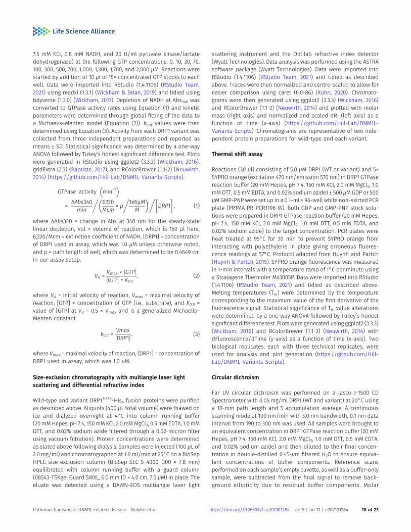
7.5 mM KCl, 0.8 mM NADH, and 20 U/ml pyruvate kinase/lactatedehydrogenase) at the following GTP concentrations: 0, 10, 30, 70,100, 300, 500, 700, 1,000, 1,300, 1,700, and 2,000 μM. Reactions werestarted by addition of 10 μl of 15× concentrated GTP stocks to eachwell. Data were imported into RStudio (1.4.1106) (RStudio Team,2021) using readxl (1.3.1) (Wickham & Brian, 2019) and tidied usingtidyverse (1.3.0) (Wickham, 2017). Depletion of NADH at Abs340 wasconverted to GTPase activity rates using Equation (1) and kineticparameters were determined through global fitting of the data toa Michaelis–Menten model (Equation (2)). kcat values were thendetermined using Equation (3). Activity from each DRP1 variant wascollected from three independent preparations and reported asmeans ± SD. Statistical significance was determined by a one-wayANOVA followed by Tukey’s honest significant difference test. Plotswere generated in RStudio using ggplot2 (3.3.3) (Wickham, 2016),gridExtra (2.3) (Baptiste, 2017), and RColorBrewer (1.1-2) (Neuwirth,2014) (https://github.com/Hill-Lab/DNM1L-Variants-Scripts).
GTPase activity min−1� �
= ΔAbs340min
�6220Mcm × p
�1e6µM
M
� ���DRP1
�; (1)
where ΔAbs340 = change in Abs at 340 nm for the steady-statelinear depletion, Vol = volume of reaction, which is 150 μl here,6,220/Mcm = extinction coefficient of NADH, [DRP1] = concentrationof DRP1 used in assay, which was 1.0 μM unless otherwise noted,and p = path length of well, which was determined to be 0.4649 cmin our assay setup.
V0 = Vmax × ½GTP�½GTP� + K0:5
; (2)
where V0 = initial velocity of reaction, Vmax = maximal velocity ofreaction, [GTP] = concentration of GTP (i.e., substrate), and K0.5 =value of [GTP] at V0 = 0.5 × Vmax and is a generalized Michaelis–Menten constant.
kcat =Vmax½DRP1�; (3)
where Vmax = maximal velocity of reaction, [DRP1] = concentration ofDRP1 used in assay, which was 1.0 μM.
Size-exclusion chromatography with multiangle laser lightscattering and differential refractive index
Wild-type and variant DRP11-736-His6 fusion proteins were purifiedas described above. Aliquots (400 μL total volume) were thawed onice and dialyzed overnight at 4°C into column running buffer(20mMHepes, pH 7.4, 150mM KCl, 2.0 mMMgCl2, 0.5 mM EDTA, 1.0 mMDTT, and 0.02% sodium azide filtered through a 0.02-micron filterusing vacuum filtration). Protein concentrations were determinedas stated above following dialysis. Samples were injected (100 μL of2.0 mg/ml) and chromatographed at 1.0 ml/min at 25°C on a BioSepHPLC size-exclusion column (BioSep-SEC-S 4000, 300 × 7.8 mm)equilibrated with column running buffer with a guard column(08543-TSKgel Guard SWXL, 6.0 mm ID × 4.0 cm, 7.0 μM) in place. Theeluate was detected using a DAWN-EOS multiangle laser light
scattering instrument and the Optilab refractive index detector(Wyatt Technologies). Data analysis was performed using the ASTRAsoftware package (Wyatt Technologies). Data were imported intoRStudio (1.4.1106) (RStudio Team, 2021) and tidied as describedabove. Traces were then normalized and centre-scaled to allow foreasier comparison using caret (6.0-86) (Kuhn, 2020). Chromato-grams were then generated using ggplot2 (3.3.3) (Wickham, 2016)and RColorBrewer (1.1-2) (Neuwirth, 2014) and plotted with molarmass (right axis) and normalized and scaled dRI (left axis) as afunction of time (x-axis) (https://github.com/Hill-Lab/DNM1L-Variants-Scripts). Chromatograms are representative of two inde-pendent protein preparations for wild-type and each variant.
Thermal shift assay
Reactions (30 μl) consisting of 5.0 μM DRP1 (WT or variant) and 5×SYPRO orange (excitation 470 nm/emission 570 nm) in DRP1 GTPasereaction buffer (20 mM Hepes, pH 7.4, 150 mM KCl, 2.0 mM MgCl2, 1.0mM DTT, 0.5 mM EDTA, and 0.02% sodium azide) ± 500 μMGDP or 500μMGMP-PNPwere set up in a 0.1-ml × 96-well white non-skirted PCRplate (PR1MA PR-PCR1196-W). Both GDP and GMP-PNP stock solu-tions were prepared in DRP1 GTPase reaction buffer (20 mM Hepes,pH 7.4, 150 mM KCl, 2.0 mM MgCl2, 1.0 mM DTT, 0.5 mM EDTA, and0.02% sodium azide) to the target concentration. PCR plates wereheat treated at 95°C for 30 min to prevent SYPRO orange frominteracting with polyethylene in plate giving erroneous fluores-cence readings at 57°C. Protocol adapted from Huynh and Partch(Huynh & Partch, 2015). SYPRO orange fluorescence was measuredin 1-min intervals with a temperature ramp of 1°C per minute usinga Stratagene Thermoler Mx3005P. Data were imported into RStudio(1.4.1106) (RStudio Team, 2021) and tidied as described above.Melting temperatures (Tm) were determined by the temperaturecorresponding to the maximum value of the first derivative of thefluorescence signal. Statistical significance of Tm value alterationswere determined by a one-way ANOVA followed by Tukey’s honestsignificant difference test. Plots were generated using ggplot2 (3.3.3)(Wickham, 2016) and RColorBrewer (1.1-2) (Neuwirth, 2014) withdFluorescence/dTime (y-axis) as a function of time (x-axis). Twobiological replicates, each with three technical replicates, wereused for analysis and plot generation (https://github.com/Hill-Lab/DNM1L-Variants-Scripts).
Circular dichroism
Far UV circular dichroism was performed on a Jasco J-1500 CDSpectrometer with 0.05 mg/ml DRP1 (WT and variant) at 20°C usinga 10-mm path length and 5 accumulation average. A continuousscanning mode at 100 nm/min with 3.0 nm bandwidth, 0.1 nm datainterval from 190 to 300 nm was used. All samples were brought toan equivalent concentration in DRP1 GTPase reaction buffer (20 mMHepes, pH 7.4, 150 mM KCl, 2.0 mM MgCl2, 1.0 mM DTT, 0.5 mM EDTA,and 0.02% sodium azide) and then diluted to their final concen-tration in double-distilled 0.45-μm filtered H2O to ensure equiva-lent concentrations of buffer components. Reference scansperformed on each sample’s empty cuvette, as well as a buffer-onlysample, were subtracted from the final signal to remove back-ground ellipticity due to residual buffer components. Molar
Pathomechanisms of DNM1L-related disease Nolden et al. https://doi.org/10.26508/lsa.202101284 vol 5 | no 12 | e202101284 18 of 23
ellipticity was converted to mean residue ellipticity (Equation (4))and data were scaled to a baseline of 0 at 260 nm using MicrosoftExcel. Data were imported into RStudio (1.4.1106) (RStudio Team,2021) and tidied as described above. Plots were generated usingggplot2 (3.3.3) (Wickham, 2016) and RColorBrewer (1.1-2) (Neuwirth,2014) with CD signal in terms of mean residue ellipticity on thevertical axis and wavelength on the horizontal axis (https://github.com/Hill-Lab/DNM1L-Variants-Scripts). Protein concentra-tions were determined again after data collection using the ab-sorbance and theoretical extinction coefficients at 205 and 214 nmto ensure that equivalent amounts of protein were used.
Ω = θ × MRW10 × c × d
(4)
where Ω = mean residue ellipticity, MRW = mean residue weightcalculated as the protein molecular weight/(N-1) where N = totalnumber of residues, c = concentration (mg/ml), and d = path length(cm).
Data Availability
All R scripts used for data analysis and visualization are availableupon request and/or for download at https://github.com/Hill-Lab/DNM1L-Variants-Scripts.
Supplementary Information
Supplementary Information is available at https://doi.org/10.26508/lsa.202101284.
Acknowledgements
R McFarland and RW Taylor are supported by the Wellcome Centre forMitochondrial Research (203105/Z/16/Z), Mitochondrial Disease PatientCohort (UK) (G0800674), the Medical Research Council (MRC) InternationalCentre for Genomic Medicine in Neuromuscular Disease (MR/S005021/1), theUK NIHR Biomedical Research Centre for Ageing and Age-related diseaseaward to the Newcastle upon Tyne Foundation Hospitals NHS Trust, and theUK NHS Highly Specialised Service for Rare Mitochondrial Disorders of Adultsand Children. M Olahova and RW Taylor receive funding from the PathologySociety. R McFarland, M Olahova, and RW Taylor receive funding from the LilyFoundation. OM Russell is supported by the Wellcome Centre for Mito-chondrial Research (203105/Z/16/Z). CL Alston is supported by a NationalInstitute for Health Research (NIHR) Post-Doctoral Fellowship (PDF-2018-11-ST2-021). This project was also supported by the following National Institutesof Health grants: TL1TR001437 and T32GM080202 (to KA Nolden),R01GM067180 (to RB Hill), and R01HL128240 (to ME Widlansky). The viewsexpressed in this publication are those of the author(s) and not necessarilythose of the National Health Service (NHS), the National Institute for HealthResearch (NIHR) or the Department of Health and Social Care. The content issolely the responsibility of the author(s) and does not necessarily representthe official views of the NIH. We would like to thank Dr. Julien Prudent fromthe MRC Mitochondrial Biology Unit at University of Cambridge for con-structive discussions about the project. We also thank Laura Bone fortechnical support and the Newcastle University Bioimaging Unit for assis-tance with the microscopy. We thank the Exeter Genomics Laboratory andthe Gastroenterology team at Sheffield Children’s Hospital for their support.
Author Contributions
KA Nolden: conceptualization, data curation, software, formalanalysis, investigation, methodology, and writing—original draft,review, and editing.JM Egner: conceptualization, data curation, formal analysis, in-vestigation, methodology, and writing—original draft.JJ Collier: data curation, software, formal analysis, investigation,methodology, and writing—original draft.OM Russell: software, formal analysis, investigation, and wri-ting—review and editing.CL Alston: formal analysis and investigation.MC Harwig: software, formal analysis, investigation, and wri-ting—review and editing.ME Widlansky: investigation.S Sasorith: software and formal analysis.IA Barbosa: formal analysis and investigation.AGL Douglas: investigation.J Baptista: formal analysis and investigation.M Walker: investigation.DE Donnelly: investigation.AA Morris: investigation.HJ Tan: formal analysis and investigation.MA Kurian: investigation.K Gorman: formal analysis and investigation.S Mordekar: investigation.C Deshpande: investigation.R Samanta: investigation.R McFarland: investigation and writing—review and editing.RB Hill: supervision, funding acquisition, investigation, and wri-ting—review and editing.RW Taylor: data curation, supervision, funding acquisition, inves-tigation, and writing—review and editing.M Olahova: conceptualization, data curation, formal analysis, su-pervision, funding acquisition, investigation, methodology, andwriting—original draft, review, and editing.
Conflict of Interest Statement
The authors have declared that no conflict of interest exists. RB Hill and KANolden have a financial interest in Cytegen, a company developing therapiesto improve mitochondrial function. However, neither the research describedherein was supported by Cytegen, nor was is in collaboration with thecompany.
References
Alston CL, Howard C, Olahova M, Hardy SA, He L, Murray PG, O’Sullivan S,Doherty G, Shield JPH, Hargreaves IP, et al (2016) Short report: Arecurrent mitochondrial p.Trp22Arg NDUFB3 variant causes adistinctive facial appearance, short stature and a mild biochemicaland clinical phenotype. J Med Genet 53: 634–641. doi:10.1136/jmedgenet-2015-103576
Aouacheria A, Baghdiguian S, Lamb HM, Huska JD, Pineda FJ, Hardwick JM(2017) Connecting mitochondrial dynamics and life-or-death eventsvia Bcl-2 family proteins. Neurochem Int 109: 141–161. doi:10.1016/j.neuint.2017.04.009
Pathomechanisms of DNM1L-related disease Nolden et al. https://doi.org/10.26508/lsa.202101284 vol 5 | no 12 | e202101284 19 of 23

Ashrafian H, Docherty L, Leo V, Towlson C, Neilan M, Steeples V, Lygate CA,Hough T, Townsend S, Williams D, et al (2010) A mutation in themitochondrial fission gene Dnm1l leads to cardiomyopathy. PLoSGenet 6: e1001000. doi:10.1371/journal.pgen.1001000
Aurora R, Srinivasan R, Rose GD (1994) Rules for α-helix termination byglycine. Science 264: 1126–1130. doi:10.1126/science.8178170
Ban-Ishihara R, Ishihara T, Sasaki N, Mihara K, Ishihara N (2013) Dynamics ofnucleoid structure regulated by mitochondrial fission contributes tocristae reformation and release of cytochrome c. Proc Natl Acad Sci US A 110: 11863–11868. doi:10.1073/pnas.1301951110
Bang D, Gribenko AV, Tereshko V, Kossiakoff AA, Kent SB, Makhatadze GI(2006) Dissecting the energetics of protein α-helix C-cap terminationthrough chemical protein synthesis. Nat Chem Biol 2: 139–143.doi:10.1038/nchembio766
Baptiste A (2017) gridExtra: Miscellaneous Functions for “Grid” Graphics. Rpackage version 2.3.
Basu K, Lajoie D, Aumentado-Armstrong T, Chen J, Koning RI, Bossy B, BostinaM, Sik A, Bossy-Wetzel E, Rouiller I (2017) Molecular mechanism ofDRP1 assembly studied in vitro by cryo-electronmicroscopy. PLoS One12: e0179397. doi:10.1371/journal.pone.0179397
Batzir NA, Bhagwat PK, Eble TN, Liu P, Eng CM, Elsea SH, Robak LA, Scaglia F,Goldman AM, Dhar SU, et al (2019) De novo missense variant in theGTPase effector domain (GED) of DNM1L leads to staticencephalopathy and seizures. Mol Case Stud 5: a003673. doi:10.1101/mcs.a003673
Beck DAC, Alonso DOV, Inoyama D, Daggett V (2008) The intrinsicconformational propensities of the 20 naturally occurring aminoacids and reflection of these propensities in proteins. Proc Natl AcadSci U S A 105: 12259–12264. doi:10.1073/pnas.0706527105
Bordt EA, Clerc P, Roelofs BA, Saladino AJ, Tretter L, Adam-Vizi V, Cherok E,Khalil A, Yadava N, Ge SX, et al (2017) The putative Drp1 inhibitor mdivi-1 is a reversible mitochondrial complex I inhibitor that modulatesreactive oxygen species. Dev Cell 40: 583–594. doi:10.1016/j.devcel.2017.02.020
Bustillo-Zabalbeitia I, Montessuit S, Raemy E, Basañez G, Terrones O,Martinou J-C (2014) Specific interaction with cardiolipin triggersfunctional activation of dynamin-related protein 1. PLoS One 9:102738. doi:10.1371/journal.pone.0102738
Cahill TJ, Leo V, Kelly M, Stockenhuber A, Kennedy NW, Bao L, Cereghetti G,Harper AR, Czibik G, Lao C, et al (2015) Resistance of dynamin-relatedprotein 1 oligomers to disassembly impairs mitophagy, resulting inmyocardial inflammation and heart failure. J Biol Chem 290:25907–25919. doi:10.1074/jbc.m115.665695
Cervantes-Silva MP, Cox SL, Curtis AM (2021) Alterations in mitochondrialmorphology as a key driver of immunity and host defence. EMBO Rep22: e53086. doi:10.15252/embr.202153086
Chang C-R, Manlandro CM, Arnoult D, Stadler J, Posey AE, Hill RB, Blackstone C(2010) A lethal de novo mutation in the middle domain of thedynamin-related GTPase Drp1 impairs higher order assembly andmitochondrial division. J Biol Chem 285: 32494–32503. doi:10.1074/jbc.m110.142430
Chang CR, Blackstone C (2007) Cyclic AMP-dependent protein kinasephosphorylation of Drp1 regulates its GTPase activity andmitochondrial morphology. J Biol Chem 282: 21583–21587. doi:10.1074/jbc.c700083200
Chao YH, Robak LA, Xia F, Koenig MK, Adesina A, Bacino CA, Scaglia F, Bellen HJ,Wangler MF (2016) Missense variants in the middle domain of DNM1Lin cases of infantile encephalopathy alter peroxisomes andmitochondria when assayed in Drosophila. Hum Mol Genet 25:1846–1856. doi:10.1093/hmg/ddw059
Clinton RW, Bauer BL, Mears JA (2020) Affinity purification and functionalcharacterization of dynamin-related protein 1. Methods Mol Biol 2159:41–53. doi:10.1007/978-1-0716-0676-6_4
Clinton RW, Francy CA, Ramachandran R, Qi X, Mears JA (2016)Dynamin-related protein 1 oligomerization in solution impairsfunctional interactions with membrane-anchoredmitochondrial fission factor. J Biol Chem 291: 478–492. doi:10.1074/jbc.m115.680025
Collier JJ, Taylor RW (2021) Machine learning algorithms reveal the secrets ofmitochondrial dynamics. EMBO Mol Med 13: e14316. doi:10.15252/emmm.202114316
De Biase I, Tortorelli S, Kratz L, Steinberg SJ, Cusmano-Ozog K, Braverman N(2020) Laboratory diagnosis of disorders of peroxisomal biogenesisand function: A technical standard of the American College of medicalgenetics and genomics (ACMG). Genet Med 22: 686–697. doi:10.1038/s41436-019-0713-9
Di Paolo C, Hefti HP, Meli M, Landis H, Pavlovic J (1999) Intramolecularbackfolding of the carboxyl-terminal end of MxA protein is aprerequisite for its oligomerization. J Biol Chem 274: 32071–32078.doi:10.1074/jbc.274.45.32071
Dıez H, Cortès-Saladelafont E, Ormazabal A, Marmiese AF, Armstrong J,Matalonga L, Bravo M, Briones P, Emperador S, Montoya J, et al (2017)Severe infantile parkinsonism because of a de novomutation on DLP1mitochondrial-peroxisomal protein. Mov Disord 32: 1108–1110.doi:10.1002/mds.27021
Dorn GW (2019) Evolving concepts of mitochondrial dynamics. Annu RevPhysiol 81: 1–17. doi:10.1146/annurev-physiol-020518-114358
Dorn GW, Vega RB, Kelly DP (2015) Mitochondrial biogenesis and dynamics inthe developing and diseased heart. Genes Dev 29: 1981–1991.doi:10.1101/gad.269894.115
Faelber K, Posor Y, Gao S, Held M, Roske Y, Schulze D, Haucke V, Noe F,Daumke O (2011) Crystal structure of nucleotide-free dynamin. Nature477: 556–560. doi:10.1038/nature10369
Fahrner JA, Liu R, Perry MS, Klein J, Chan DC (2016) A novel de novo dominantnegative mutation in DNM1L impairs mitochondrial fission andpresents as childhood epileptic encephalopathy. Am J Med Genet A170: 2002–2011. doi:10.1002/ajmg.a.37721
Ford MGJ, Jenni S, Nunnari J (2011) The crystal structure of dynamin. Nature477: 561–566. doi:10.1038/nature10441
Francy CA, Clinton RW, Frohlich C, Murphy C, Mears JA (20172017) Cryo-EMstudies of Drp1 reveal cardiolipin interactions that activate the helicaloligomer. Sci Rep 7: 10744. doi:10.1038/s41598-017-11008-3
Friedman JR, Lackner LL, West M, DiBenedetto JR, Nunnari J, Voeltz GK (2011)ER tubules Mark sites of mitochondrial division. Science 334: 358–362.doi:10.1126/science.1207385
Frohlich C, Grabiger S, Schwefel D, Faelber K, Rosenbaum E, Mears J, Rocks O,Daumke O (2013) Structural insights into oligomerization andmitochondrial remodelling of dynamin 1-like protein. EMBO J 32:1280–1292. doi:10.1038/emboj.2013.74
Gandre-Babbe S, Van Der Bliek AM (2008) The novel tail-anchoredmembrane protein Mff controls mitochondrial and peroxisomalfission in mammalian cells. Mol Biol Cell 19: 2402–2412. doi:10.1091/mbc.e07-12-1287
Gao S, von der Malsburg A, Dick A, Faelber K, Schroder GF, Haller O, Kochs G,Daumke O (2011) Structure of myxovirus resistance protein A revealsintra- and intermolecular domain interactions required for theantiviral function. Immunity 35: 514–525. doi:10.1016/j.immuni.2011.07.012
Gao S, von der Malsburg A, Paeschke S, Behlke J, Haller O, Kochs G, Daumke O(2010) Structural basis of oligomerization in the stalk region ofdynamin-like MxA. Nature 465: 502–506. doi:10.1038/nature08972
Gerber S, Charif M, Chevrollier A, Chaumette T, Angebault C, Kane MS, Paris A,Alban J, Quiles M, Delettre C, et al (2017) Mutations in DNM1L, as inOPA1, result indominant optic atrophy despite opposite effectsonmitochondrial fusion and fission. Brain 140: 2586–2596. doi:10.1093/brain/awx219
Pathomechanisms of DNM1L-related disease Nolden et al. https://doi.org/10.26508/lsa.202101284 vol 5 | no 12 | e202101284 20 of 23
Harvey AJ (2019) Mitochondria in early development: Linking themicroenvironment, metabolism and the epigenome. Reproduction157: R159–R179. doi:10.1530/rep-18-0431
Hogarth KA, Costford SR, Yoon G, Sondheimer N, Maynes JT (2018) DNM1Lvariant alters baseline mitochondrial function and response to stressin a patient with severe neurological dysfunction. Biochem Genet 56:56–77. doi:10.1007/s10528-017-9829-2
Horbay R, Bilyy R (2016) Mitochondrial dynamics during cell cycling.Apoptosis 21: 1327–1335. doi:10.1007/s10495-016-1295-5
Huynh K, Partch CL (2015) Analysis of protein stability and ligand interactionsby thermal shift assay. Curr Protoc Protein Sci 79: 28. 9.1–28.9.14.doi:10.1002/0471140864.ps2809s79
Ihenacho UK, Meacham KA, Harwig MC, Widlansky ME, Hill RB (2021)Mitochondrial fission protein 1: Emerging roles in organellar form andfunction in Health and disease. Front Endocrinol (Lausanne) 12:660095. doi:10.3389/fendo.2021.660095
Ikeda Y, Shirakabe A, Maejima Y, Zhai P, Sciarretta S, Toli J, Nomura M, MiharaK, Egashira K, Ohishi M, et al (2015) Endogenous Drp1 mediatesmitochondrial autophagy and protects the heart against energystress. Circ Res 116: 264–278. doi:10.1161/circresaha.116.303356
Ingerman E, Nunnari J (2005) A continuous, regenerative coupled gtpaseassay for dynamin-related proteins. Methods Enzymol 404: 611–619.doi:10.1016/S0076-6879(05)04053-X
Ishihara N, Nomura M, Jofuku A, Kato H, Suzuki SO, Masuda K, Otera H,Nakanishi Y, Nonaka I, Goto YI, et al (2009) Mitochondrial fission factorDrp1 is essential for embryonic development and synapse formationin mice. Nat Cell Biol 11: 958–966. doi:10.1038/ncb1907
Ishihara T, Ban-Ishihara R, Maeda M, Matsunaga Y, Ichimura A, Kyogoku S,Aoki H, Katada S, Nakada K, Nomura M, et al (2015) Dynamics ofmitochondrial DNA nucleoids regulated by mitochondrial fission isessential for maintenance of homogeneously active mitochondriaduring neonatal heart development. Mol Cell Biol 35: 211–223.doi:10.1128/mcb.01054-14
James DI, Parone PA, Mattenberger Y, Martinou JC (2003) hFis1, a novelcomponent of the mammalian mitochondrial fission machinery. J BiolChem 278: 36373–36379. doi:10.1074/jbc.m303758200
Kalia R, Wang RYR, Yusuf A, Thomas PV, Agard DA, Shaw JM, Frost A (2018)Structural basis of mitochondrial receptor binding and constrictionby DRP1. Nature 558: 401–405. doi:10.1038/s41586-018-0211-2
Kasahara A, Scorrano L (2014) Mitochondria: From cell death executioners toregulators of cell differentiation. Trends Cell Biol 24: 761–770.doi:10.1016/j.tcb.2014.08.005
Kirby DM, Thorburn DR, Turnbull DM, Taylor RW (2007) Biochemical assays ofrespiratory chain complex activity. Methods Cell Biol 80: 93–119.doi:10.1016/S0091-679X(06)80004-X
Kishida H, Sugio S (2013) Crystal structure of GTPase domain fused withminimal stalks from human dynamin-1-like protein (Dlp1) in complexwith several nucleotide analogues. Curr Top Pept Protein Res 14: 67–77.doi:10.2210/pdb3w6n/pdb
Kobayashi S, Tanaka A, Fujiki Y (2007) Fis1, DLP1, and Pex11p coordinatelyregulate peroxisome morphogenesis. Exp Cell Res 313: 1675–1686.doi:10.1016/j.yexcr.2007.02.028
Koch A, Yoon Y, Bonekamp NA, McNiven MA, Schrader M (2005) A role for Fis1in both mitochondrial and peroxisomal fission in mammalian cells.Mol Biol Cell 16: 5077–5086. doi:10.1091/mbc.e05-02-0159
Koch J, Brocard C (2012) PEX11 proteins attract Mff and human Fis1 tocoordinate peroxisomal fission. J Cell Sci 125: 3813–3826. doi:10.1242/jcs.102178
Koirala S, Guo Q, Kalia R, Bui HT, Eckert DM, Frost A, Shaw JM (2013)Interchangeable adaptors regulate mitochondrial dynamin assemblyfor membrane scission. Proc Natl Acad Sci U S A 110: E1342–E1351.doi:10.1073/pnas.1300855110
Kraus F, Ryan MT (2017) The constriction and scission machineries involvedin mitochondrial fission. J Cell Sci 130: 2953–2960. doi:10.1242/jcs.199562
Kuhn M (2020) Caret: Classification and Regression Training. R packageversion 6.0-86. https://CRAN.R-project.org.
Kwapiszewska K, Kalwarczyk T, Michalska B, Szczepanski K, Szymanski J,Patalas-Krawczyk P, Andryszewski T, Iwan M, Duszynski J, Hołyst R(2019) Determination of oligomerization state of Drp1 protein in livingcells at nanomolar concentrations. Sci Rep 9: 5906. doi:10.1038/s41598-019-42418-0
Li X, Gould SJ (2003) The dynamin-like GTPase DLP1 is essential forperoxisome division and is recruited to peroxisomes in part by PEX11. JBiol Chem 278: 17012–17020. doi:10.1074/jbc.m212031200
Liu T, Yu R, Jin SB, Han L, Lendahl U, Zhao J, Nister M (2013) The mitochondrialelongation factors MIEF1 andMIEF2 exert partially distinct functions inmitochondrial dynamics. Exp Cell Res 319: 2893–2904. doi:10.1016/j.yexcr.2013.07.010
Liu X, Zhang Z, Li D, Lei M, Li Q, Liu X, Zhang P (2021) DNM1L-Relatedmitochondrial fission defects presenting as encephalopathy: A casereport and literature review. Front Pediatr 9: 626657. doi:10.3389/fped.2021.626657
Longo F, Benedetti S, Zambon AA, Sora MGN, Di Resta C, De Ritis D, Quattrini A,Maltecca F, Ferrari M, Previtali SC (2020) Impaired turnover ofhyperfused mitochondria in severe axonal neuropathy due to a novelDRP1 mutation. Hum Mol Genet 29: 177–188. doi:10.1093/hmg/ddz211
Losón OC, Song Z, Chen H, Chan DC (2013) Fis1, Mff, MiD49, and MiD51 mediateDrp1 recruitment in mitochondrial fission. Mol Biol Cell 24: 659–667.doi:10.1091/mbc.e12-10-0721
Ma J, Zhai Y, Chen M, Zhang K, Chen Q, Pang X, Sun F (2019) New interfaces onMiD51 for Drp1 recruitment and regulation. PLoS One 14: e0211459.doi:10.1371/journal.pone.0211459
Macdonald PJ, Francy CA, Stepanyants N, Lehman L, Baglio A, Mears JA, Qi X,Ramachandran R (2016) Distinct splice variants of dynamin-relatedprotein 1 differentially utilize mitochondrial fission factor as aneffector of cooperative GTPase activity. J Biol Chem 291: 493–507.doi:10.1074/jbc.m115.680181
Mahajan M, Bharambe N, Shang Y, Lu B, Mandal A, Mohan PM, Wang R, BoatzJC, Galvez JMM, Shnyrova AV, et al (2021) NMR identification of aconserved Drp1 cardiolipin-binding motif essential for stress-induced mitochondrial fission. Proc Natl Acad Sci U S A 118:e2023079118. doi:10.1073/pnas.2023079118
McQuin C, Goodman A, Chernyshev V, Kamentsky L, Cimini BA, Karhohs KW,Doan M, Ding L, Rafelski SM, Thirstrup D, et al (2018) CellProfiler 3.0:Next-generation image processing for biology. PLoS Biol 16: e2005970.doi:10.1371/journal.pbio.2005970
Mears JA, Lackner LL, Fang S, Ingerman E, Nunnari J, Hinshaw JE (2011)Conformational changes in Dnm1 support a contractile mechanismfor mitochondrial fission. Nat Struct Mol Biol 18: 20–26. doi:10.1038/nsmb.1949
Mishra P, Chan DC (2014) Mitochondrial dynamics and inheritance during celldivision, development and disease. Nat Rev Mol Cell Biol 15: 634–646.doi:10.1038/nrm3877
Mitra K, Wunder C, Roysam B, Lin G, Lippincott-Schwartz J (2009) A hyperfusedmitochondrial state achieved at G1-S regulates cyclin E buildup andentry into S phase. Proc Natl Acad Sci U S A 106: 11960–11965.doi:10.1073/pnas.0904875106
Muhlberg AB, Warnock DE, Schmid SL (1997) Domain structure andintramolecular regulation of dynamin GTPase. EMBO J 16: 6676–6683.doi:10.1093/emboj/16.22.6676
Nagashima S, Tabara L-C, Tilokani L, Paupe V, Anand H, Pogson JH, Zunino R,McBride HM, Prudent J (2020) Golgi-derived PI (4) P-containingvesicles drive late steps of mitochondrial division. Science 367:1366–1371. doi:10.1126/science.aax6089
Pathomechanisms of DNM1L-related disease Nolden et al. https://doi.org/10.26508/lsa.202101284 vol 5 | no 12 | e202101284 21 of 23

Nasca A, Legati A, Baruffini E, Nolli C, Moroni I, Ardissone A, Goffrini P, Ghezzi D(2016) Biallelic mutations in DNM1L are associated with a slowlyprogressive infantile encephalopathy. Hum Mutat 37: 898–903.doi:10.1002/humu.23033
Neuwirth E (2014) RColorBrewer: ColorBrewer Palettes. R package version 1.1-2. https://CRAN.R-project.org.
Otera H, Wang C, Cleland MM, Setoguchi K, Yokota S, Youle RJ, Mihara K (2010)Mff is an essential factor for mitochondrial recruitment of Drp1 duringmitochondrial fission in mammalian cells. J Cell Biol 191: 1141–1158.doi:10.1083/jcb.201007152
Palmer CS, Osellame LD, Laine D, Koutsopoulos OS, Frazier AE, Ryan MT (2011)MiD49 and MiD51, new components of the mitochondrial fissionmachinery. EMBO Rep 12: 565–573. doi:10.1038/embor.2011.54
Pangou E, Sumara I (2021) The multifaceted regulation of mitochondrialdynamics during mitosis. Front Cell Dev Biol 9: 767221. doi:10.3389/fcell.2021.767221
Pitts KR, McNiven MA, Yoon Y (2004) Mitochondria-specific function of thedynamin family protein DLP1 is mediated by its C-terminal domains. JBiol Chem 279: 50286–50294. doi:10.1074/jbc.m405531200
Qian W, Choi S, Gibson GA, Watkins SC, Bakkenist CJ, Van Houten B (2012)Mitochondrial hyperfusion induced by loss of the fission protein Drp1causes ATM-dependent G2/M arrest and aneuploidy through DNAreplication stress. J Cell Sci 125: 5745–5757. doi:10.1242/jcs.109769
Richards S, Aziz N, Bale S, Bick D, Das S, Gastier-Foster J, Grody WW, Hegde M,Lyon E, Spector E, et al (2015) Standards and guidelines for theinterpretation of sequence variants: A joint consensusrecommendation of the American College of medical genetics andgenomics and the association for molecular pathology. Genet Med 17:405–424. doi:10.1038/gim.2015.30
Richardson JS, Richardson DC (1988) Amino acid preferences for specificlocations at the ends of α helices. Science 240: 1648–1652. doi:10.1126/science.3381086
Rocha MC, Grady JP, Grünewald A, Vincent A, Dobson PF, Taylor RW, TurnbullDM, Rygiel KA (2015) A novel immunofluorescent assay to investigateoxidative phosphorylation deficiency in mitochondrial myopathy:Understanding mechanisms and improving diagnosis. Sci Rep 5:15037. doi:10.1038/srep15037
Rosdah AA, Smiles WJ, Oakhill JS, Scott JW, Langendorf CG, Delbridge LMD,Holien JK, Lim SY (2020) New perspectives on the role of Drp1 isoformsin regulating mitochondrial pathophysiology. Pharmacol Ther 213:107594. doi:10.1016/j.pharmthera.2020.107594
Roy M, Reddy PH, Iijima M, Sesaki H (2015) Mitochondrial division and fusion inmetabolism. Curr Opin Cell Biol 33: 111–118. doi:10.1016/j.ceb.2015.02.001
RStudio Team (2021) RStudio: Integrated development environment for R.RStudio. Boston, MA: PBC. http://www.rstudio.com
Ryan CS, Fine AL, Cohen AL, Schiltz BM, Renaud DL, Wirrell EC, Patterson MC,Boczek NJ, Liu R, Babovic-Vuksanovic D, et al (2018) De novo DNM1Lvariant in a teenager with progressive paroxysmal dystonia and lethalsuper-refractory myoclonic status epilepticus. J Child Neurol 33:651–658. doi:10.1177/0883073818778203
Schindelin J, Arganda-Carreras I, Frise E, Kaynig V, Longair M, Pietzsch T,Preibisch S, Rueden C, Saalfeld S, Schmid B, et al (2012) Fiji: An opensource platform for biological image analysis. Nat Methods 9: 676–682.doi:10.1038/nmeth.2019
Schumacher B, Staeheli P (1998) Domains mediating intramolecular foldingand oligomerization of MxA GTPase. J Biol Chem 273: 28365–28370.doi:10.1074/jbc.273.43.28365
Sever S, Muhlberg AB, Schmid SL (1999) Impairment of dynamin’s GAPdomain stimulates receptor-mediated endocytosis. Nature 398:481–486. doi:10.1038/19024
Sheffer R, Douiev L, Edvardson S, Shaag A, Tamimi K, Soiferman D, Meiner V,Saada A (2016) Postnatal microcephaly and pain insensitivity due to a
de novo heterozygousDNM1Lmutation causing impairedmitochondrial fission and function. Am J Med Genet A 170: 1603–1607.doi:10.1002/ajmg.a.37624
Shin HW, Takatsu H, Mukai H, Munekata E, Murakami K, Nakayama K (1999)Intermolecular and interdomain interactions of a dynamin-relatedGTP-binding protein, Dnm1p/Vps1p-like protein. J Biol Chem 274:2780–2785. doi:10.1074/jbc.274.5.2780
Smirnova E, Griparic L, Shurland DL, Van der Bliek AM (2001) Dynamin-relatedprotein Drp1 is required for mitochondrial division in mammaliancells. Mol Biol Cell 12: 2245–2256. doi:10.1091/mbc.12.8.2245
Smirnova E, Shurland DL, Newman-Smith ED, Pishvaee B, Van Der Bliek AM(1999) A model for dynamin self-assembly based on binding betweenthree different protein domains. J Biol Chem 274: 14942–14947.doi:10.1074/jbc.274.21.14942
Some D, Amartely H, Tsadok A, Lebendiker M (2019) Characterization ofproteins by size-exclusion chromatography coupled tomulti-angle light scattering (SEC-MALS). J Vis Exp e59615. doi:10.3791/59615
Song M, Mihara K, Chen Y, Scorrano L, Dorn GW (2015) Mitochondrial fissionand fusion factors reciprocally orchestrate mitophagic culling inmouse hearts and cultured fibroblasts. Cell Metab 21: 273–286.doi:10.1016/j.cmet.2014.12.011
Stojanovski D, Koutsopoulos OS, Okamoto K, Ryan MT (2004) Levels of humanFis1 at the mitochondrial outer membrane regulate mitochondrialmorphology. J Cell Sci 117: 1201–1210. doi:10.1242/jcs.01058
Strack S, Cribbs JT (2012) Allosteric modulation of Drp1 mechanoenzymeassembly and mitochondrial fission by the variable domain. J BiolChem 287: 10990–11001. doi:10.1074/jbc.m112.342105
Tanaka A, Kobayashi S, Fujiki Y (2006) Peroxisome division is impaired in aCHO cell mutant with an inactivating point-mutation in dynamin-likeprotein 1 gene. Exp Cell Res 312: 1671–1684. doi:10.1016/j.yexcr.2006.01.028
Taylor RW, Pyle A, Griffin H, Blakely EL, Duff J, He L, Smertenko T, Alston CL,Neeve VC, Best A, et al (2014) Use of whole-exome sequencing todetermine the genetic basis of multiple mitochondrial respiratorychain complex deficiencies. JAMA 312: 68. doi:10.1001/jama.2014.7184
Thompson K, Majd H, Dallabona C, Reinson K, King MS, Alston CL, He L, Lodi T,Jones SA, Fattal-Valevski A, et al (2016) Recurrent de novo dominantmutations in SLC25A4 cause severe early-onset mitochondrialdisease and loss of mitochondrial DNA copy number. Am J Hum Genet99: 1405. doi:10.1016/j.ajhg.2016.11.001
Tilokani L, Nagashima S, Paupe V, Prudent J (2018) Mitochondrial dynamics:Overview of molecular mechanisms. Essays Biochem 62: 341–360.doi:10.1042/ebc20170104
Touvier T, De Palma C, Rigamonti E, Scagliola A, Incerti E, Mazelin L, ThomasJ-LL, D’Antonio M, Politi L, Schaeffer L, et al (2015) Muscle-specific Drp1overexpression impairs skeletal muscle growth via translationalattenuation. Cell Death Dis 6: e1663. doi:10.1038/cddis.2014.595
Vandeleur D, Chen CV, Huang EJ, Connolly AJ, Sanchez H, Moon-Grady AJ (2019)Novel and lethal case of cardiac involvement in DNM1Lmitochondrialencephalopathy. Am J Med Genet Part A 179: 2486–2489. doi:10.1002/ajmg.a.61371
Vanstone JR, Smith AM, McBride S, Naas T, Holcik M, Antoun G, Harper ME,Michaud J, Sell E, Chakraborty P, et al (2016) DNM1L-relatedmitochondrial fission defect presenting as refractory epilepsy. Eur JHum Genet 24: 1084–1088. doi:10.1038/ejhg.2015.243
Verrigni D, Di Nottia M, Ardissone A, Baruffini E, Nasca A, Legati A, Bellacchio E,Fagiolari G, Martinelli D, Fusco L, et al (2019) Clinical-genetic featuresand peculiar muscle histopathology in infantile DNM1L-relatedmitochondrial epileptic encephalopathy. Hum Mutat 40: 601–618.doi:10.1002/humu.23729
Wai T, Langer T (2016) Mitochondrial dynamics and metabolic regulation.Trends Endocrinol Metab 27: 105–117. doi:10.1016/j.tem.2015.12.001
Pathomechanisms of DNM1L-related disease Nolden et al. https://doi.org/10.26508/lsa.202101284 vol 5 | no 12 | e202101284 22 of 23
Wakabayashi J, Zhang Z, Wakabayashi N, Tamura Y, Fukaya M, Kensler TW,Iijima M, Sesaki H (2009) The dynamin-related GTPase Drp1 is requiredfor embryonic and brain development in mice. J Cell Biol 186: 805–816.doi:10.1083/jcb.200903065
Waterham HR, Ebberink MS (2012) Genetics and molecular basis of humanperoxisome biogenesis disorders. Biochim Biophys Acta 1822:1430–1441. doi:10.1016/j.bbadis.2012.04.006
Waterham HR, Koster J, van Roermund CWT, Mooyer PAW, Wanders RJA,Leonard JV (2007) A lethal defect of mitochondrial and peroxisomalfission. N Engl J Med 356: 1736–1741. doi:10.1056/nejmoa064436
Wei Y, Qian M (2021) Case Report: A Novel de novo Mutation in DNM1LPresenting With Developmental Delay, Ataxia, and PeripheralNeuropathy. Front Pediatr 9: 604105. doi:10.3389/fped.2021.604105
Wenger J, Klinglmayr E, Frohlich C, Eibl C, Gimeno A, Hessenberger M,Puehringer S, Daumke O, Goettig P (2013) Functional mapping ofhuman dynamin-1-like GTPase domain based on x-ray structureanalyses. PLoS One 8: e71835. doi:10.1371/journal.pone.0071835
Whitley BN, Lam C, Cui H, Haude K, Bai R, Escobar L, Hamilton A, Brady L,Tarnopolsky MA, Dengle L, et al (2018) Aberrant Drp1-mediatedmitochondrial division presents in humans with variable outcomes.Hum Mol Genet 27: 3710–3719. doi:10.1093/hmg/ddy287
Wickham H, Brian J (2019) readxl: Read excel files (version 1.3.1). R packageversion 1.3.1. https://cran.r-project.org; https://cran.r-project.org/package=readxl.
Wickham H (2016) ggplot2: Elegant Graphics for Data Analysis. New York:Springer-Verlag.
Wickham H (2017) tidyverse: Easily install and load “tidyverse” packages. Rpackage version 1.2.1.
Yamano K, Fogel AI, Wang C, van der Bliek AM, Youle RJ (2014) MitochondrialRab GAPs govern autophagosome biogenesis during mitophagy. Elife3: e01612. doi:10.7554/elife.01612
Yoon G, Malam Z, Paton T, Marshall CR, Hyatt E, Ivakine Z, Scherer SW, Lee KS,Hawkins C, Cohn RD, et al (2016) Lethal disorder of mitochondrialfission caused by mutations in DNM1L. J Pediatr 171: 313–316.e2.doi:10.1016/j.jpeds.2015.12.060
Yoon Y, Krueger EW, Oswald BJ, McNiven MA (2003) Themitochondrial proteinhFis1 regulates mitochondrial fission in mammalian cells through aninteraction with the dynamin-like protein DLP1. Mol Cell Biol 23:5409–5420. doi:10.1128/mcb.23.15.5409-5420.2003
Zaha K, Matsumoto H, Itoh M, Saitsu H, Kato K, Kato M, Ogata S, Murayama K,Kishita Y, Mizuno Y, et al (2016) DNM1L-related encephalopathy ininfancy with Leigh syndrome-like phenotype and suppression-burst.Clin Genet 90: 472–474. doi:10.1111/cge.12805
Zhang P, Hinshaw JE (2001) Three-dimensional reconstruction of dynaminin the constricted state. Nat Cell Biol 3: 922–926. doi:10.1038/ncb1001-922
Zhu PP, Patterson A, Stadler J, Seeburg DP, Sheng M, Blackstone C(2004) Intra- and intermolecular domain interactions of theC-terminal GTPase effector domain of the multimeric dynamin-likeGTPase Drp1. J Biol Chem 279: 35967–35974. doi:10.1074/jbc.m404105200
License: This article is available under a CreativeCommons License (Attribution 4.0 International, asdescribed at https://creativecommons.org/licenses/by/4.0/).
Pathomechanisms of DNM1L-related disease Nolden et al. https://doi.org/10.26508/lsa.202101284 vol 5 | no 12 | e202101284 23 of 23
Copyright © 2022 FDOKUMEN


![Variants of Penney's Game arXiv:2006.13002v1 [math.HO] 19 ...](https://static.fdokumen.com/doc/165x107/6319649f1e5d335f8d0b2299/variants-of-penneys-game-arxiv200613002v1-mathho-19-.jpg)