IL-23 and the Th17 pathway promote inflammation and impair antifungal immune resistance
Upload
khangminh22Category
view
0download
0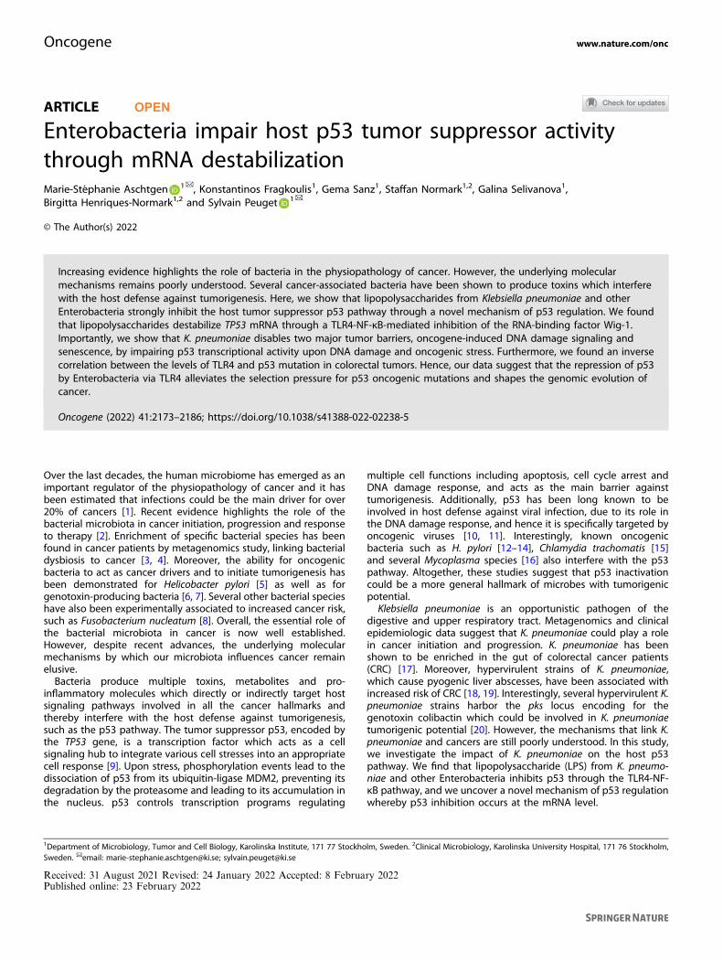
ARTICLE OPEN
Enterobacteria impair host p53 tumor suppressor activitythrough mRNA destabilizationMarie-Stéphanie Aschtgen 1✉, Konstantinos Fragkoulis1, Gema Sanz1, Staffan Normark1,2, Galina Selivanova1,Birgitta Henriques-Normark1,2 and Sylvain Peuget 1✉
© The Author(s) 2022
Increasing evidence highlights the role of bacteria in the physiopathology of cancer. However, the underlying molecularmechanisms remains poorly understood. Several cancer-associated bacteria have been shown to produce toxins which interferewith the host defense against tumorigenesis. Here, we show that lipopolysaccharides from Klebsiella pneumoniae and otherEnterobacteria strongly inhibit the host tumor suppressor p53 pathway through a novel mechanism of p53 regulation. We foundthat lipopolysaccharides destabilize TP53 mRNA through a TLR4-NF-κB-mediated inhibition of the RNA-binding factor Wig-1.Importantly, we show that K. pneumoniae disables two major tumor barriers, oncogene-induced DNA damage signaling andsenescence, by impairing p53 transcriptional activity upon DNA damage and oncogenic stress. Furthermore, we found an inversecorrelation between the levels of TLR4 and p53 mutation in colorectal tumors. Hence, our data suggest that the repression of p53by Enterobacteria via TLR4 alleviates the selection pressure for p53 oncogenic mutations and shapes the genomic evolution ofcancer.
Oncogene (2022) 41:2173–2186; https://doi.org/10.1038/s41388-022-02238-5
Over the last decades, the human microbiome has emerged as animportant regulator of the physiopathology of cancer and it hasbeen estimated that infections could be the main driver for over20% of cancers [1]. Recent evidence highlights the role of thebacterial microbiota in cancer initiation, progression and responseto therapy [2]. Enrichment of specific bacterial species has beenfound in cancer patients by metagenomics study, linking bacterialdysbiosis to cancer [3, 4]. Moreover, the ability for oncogenicbacteria to act as cancer drivers and to initiate tumorigenesis hasbeen demonstrated for Helicobacter pylori [5] as well as forgenotoxin-producing bacteria [6, 7]. Several other bacterial specieshave also been experimentally associated to increased cancer risk,such as Fusobacterium nucleatum [8]. Overall, the essential role ofthe bacterial microbiota in cancer is now well established.However, despite recent advances, the underlying molecularmechanisms by which our microbiota influences cancer remainelusive.Bacteria produce multiple toxins, metabolites and pro-
inflammatory molecules which directly or indirectly target hostsignaling pathways involved in all the cancer hallmarks andthereby interfere with the host defense against tumorigenesis,such as the p53 pathway. The tumor suppressor p53, encoded bythe TP53 gene, is a transcription factor which acts as a cellsignaling hub to integrate various cell stresses into an appropriatecell response [9]. Upon stress, phosphorylation events lead to thedissociation of p53 from its ubiquitin-ligase MDM2, preventing itsdegradation by the proteasome and leading to its accumulation inthe nucleus. p53 controls transcription programs regulating
multiple cell functions including apoptosis, cell cycle arrest andDNA damage response, and acts as the main barrier againsttumorigenesis. Additionally, p53 has been long known to beinvolved in host defense against viral infection, due to its role inthe DNA damage response, and hence it is specifically targeted byoncogenic viruses [10, 11]. Interestingly, known oncogenicbacteria such as H. pylori [12–14], Chlamydia trachomatis [15]and several Mycoplasma species [16] also interfere with the p53pathway. Altogether, these studies suggest that p53 inactivationcould be a more general hallmark of microbes with tumorigenicpotential.Klebsiella pneumoniae is an opportunistic pathogen of the
digestive and upper respiratory tract. Metagenomics and clinicalepidemiologic data suggest that K. pneumoniae could play a rolein cancer initiation and progression. K. pneumoniae has beenshown to be enriched in the gut of colorectal cancer patients(CRC) [17]. Moreover, hypervirulent strains of K. pneumoniae,which cause pyogenic liver abscesses, have been associated withincreased risk of CRC [18, 19]. Interestingly, several hypervirulent K.pneumoniae strains harbor the pks locus encoding for thegenotoxin colibactin which could be involved in K. pneumoniaetumorigenic potential [20]. However, the mechanisms that link K.pneumoniae and cancers are still poorly understood. In this study,we investigate the impact of K. pneumoniae on the host p53pathway. We find that lipopolysaccharide (LPS) from K. pneumo-niae and other Enterobacteria inhibits p53 through the TLR4-NF-κB pathway, and we uncover a novel mechanism of p53 regulationwhereby p53 inhibition occurs at the mRNA level.
Received: 31 August 2021 Revised: 24 January 2022 Accepted: 8 February 2022Published online: 23 February 2022
1Department of Microbiology, Tumor and Cell Biology, Karolinska Institute, 171 77 Stockholm, Sweden. 2Clinical Microbiology, Karolinska University Hospital, 171 76 Stockholm,Sweden. ✉email: [email protected]; [email protected]
www.nature.com/oncOncogene
1234567890();,:
RESULTSKlebsiella pneumoniae inhibits p53To investigate the effect of K. pneumoniae on the host p53pathway, we infected human immortalized fibroblasts (BJhTERT) with live bacteria. We observed a robust downregulationof p53 protein level, concomitant with the activation of NF-κB,as demonstrated by Ser536 phosphorylation of the NF-κBsubunit p65 (RelA) (Fig. 1a). Interestingly, p53 downregulationwas bacteria-cell contact independent and was recapitulatedwhen treating the cells with supernatant from K. pneumoniaeculture (KpSN), independently of the K. pneumoniae strains used
(Fig. 1b and Supplementary Fig. S1a-b). Pathway analysis ofgene expression changes in human fibroblasts exposed to KpSN,measured by RNA-seq, highlighted a robust activation of thecanonical immune and inflammatory pathways involved inthe host response to bacteria, as expected (Fig. 1c-d). Notably,the analysis of genes downregulated upon KpSN revealed asignificant inhibition of the p53 signaling pathway andDNA damage response (Fig. 1d). These data suggest thatK. pneumoniae-mediated p53 protein decline is sufficient toimpair the biological function of p53.
a
b
c
d
tRNA metabolic processregula�on of signal transduc�on by p53recombina�onal repairDSB repair via HRnuclear DNA replica�onDSB repaircilium assemblycell cycle DNA replica�onribosome biogenesisrRNA metabolic processsignal transduc�on by p53rRNA processingncRNA processingDNA replica�onDNA-dependent DNA replica�on
response to IFNγresponse to virus
cellular response to IFNγdefense response to virus
response to TNFcellular response to TNF
I-κB kinase/NF-κB signalingregula�on of response to cytokine s�mulus
regula�on of I-κB kinase/NF-κB signalingIFNγ-mediated signaling pathway
response to type I interferonregula�on of apopto�c signaling pathway
cellular response to type I interferontype I interferon signaling pathway
extrinsic apopto�c signalig pathway
-log10(Padj)0 5 10 15 2520
-log10(Padj)001 5
Interferon response
NF-κB response
TNFresponse
Apoptosis
DNA repair
Ciliumassembly
p53signaling
Ribosome biogenesis
e
pS536-p65
p53
β-ac�n
m.o.i.1h 2h
0 0.1 1 10 0.1 1 10Kp (MGH78578)
- 70kDa
- 55
- 40
-
pS536-p65
p53
β-ac�n
Kp -NS- 70kDa
- 55
- 40 log2FC (KpSN vs Untreated)
-log 1
0(FD
R)
0
100
200
300
0 0155-
ARRDC4
MBNL2ZADH2 DSEL
CERKFLRT2
SIM1RDH10TSHZ1
MAB21L1CHRM2
APPL2IVNS1ABPPBX1ZMAT3
SERPINB2IFIT1
IFIH1
IFIT3
USP18
DDX58CCL2
TRAF1CTSS
EPSTI1
BST2
LIF
SOD2ICAM1
APOL6
Fig. 1 Klebsiella pneumoniae inhibits p53. a NF-κB activation (followed by p65 phosphorylation) and p53 protein level in BJ hTert cells uponinfection by the K. pneumoniae (Kp) strain MGH78758 at different m.o.i. b Western blot of BJ hTert cells upon exposure to supernatant from K.pneumoniae culture (KpSN), from different K. pneumoniae strains. c RNA-seq data of BJ hTert cells exposed to KpSN, highlighting top15upregulated (orange) and downregulated genes (blue). d GO pathway enrichment analysis of the top up- and downregulated genes uponKpSN. e, Network visualization of the top up- and downregulated pathways.
M.-S. Aschtgen et al.
2174
Oncogene (2022) 41:2173 – 2186
Core p53 targets(Fisher) n=306Doxorubicin UP
n=3291
shp53 DOWNn=1716
2161 905632
1616364 18
Row Z-score
-1 0 1
p53
targ
etge
nes(
161)
KpSNDox 0.2 μM
- - +- + +
Dox 0.2 μM - +- +
p53
β-ac�n
KpSN 8h - -+ +- +- +- -+ +
pS536-p65
BJ hTert BJ hTert shp53
- 70kDa
- 55
- 40
cba
d
f
e
Rela
�ve
mRN
Ale
vel(
log 2
Fc)
-1
0
1
2
3
4
5
DoxKpSN
- - + +
- + - +
- - + +
- + - +
shp53
PMAIP1 (NOXA)
Rela
�ve
mRN
Ale
vel(
log 2
Fc)
-4
-2
0
2
4
DoxKpSN
- - + +
- + - +
- - + +
- + - +
shp53
CDKN1A (p21)*
Rela
�ve
mRN
Ale
vel(
log 2
Fc)
-1
0
1
2
DoxKpSN
- - + +
- + - +
- - + +
- + - +
shp53
TP53I3 (PIG3)***
Rela
�ve
mRN
Ale
vel(
log 2
Fc)
-4
-2
0
2
4
DoxKpSN
- - + +
- + - +
- - + +
- + - +
shp53
TP53INP1
*
Rela
�ve
mRN
Ale
vel(
log 2
Fc)
-4
-2
0
2
4
DoxKpSN
- - + +
- + - +
- - + +
- + - +
shp53
SESN1
*
*
Rela
�ve
mRN
Ale
vel(
log 2
Fc)
-6
-4
-2
0
2
DoxKpSN
- - + +
- + - +
- - + +
- + - +
shp53
ZMAT3 (Wig-1)
******
******
log2FC (KpSN vs Untreated)
-log 1
0(FD
R)
0
100
200
300
0 0155-
ZMAT3
PMAIP1
CDKN1A
TP53I3
TP53INP1
SESN1
-
KpSN 16h
p21
BJ hTert
Dox 0.2 μM - + + - - + +
- + - + - + - +
BJ hTert shp53
PIG3
β-ac�n
- 25kDa
- 35
- 40
Wig-1 - 35
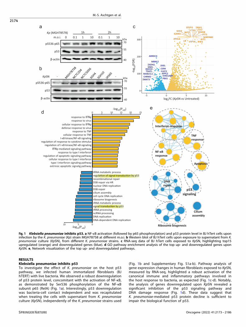
Fig. 2 Klebsiella pneumoniae impairs p53 response to DNA damage. a Western blot of BJ hTert cells treated with the DNA-damaging agentdoxorubicin (Dox) and with KpSN. b High confidence set of p53 target genes obtained by intersecting the core p53 target gene set define byFisher et al. with genes upregulated by doxorubicin and downregulated upon p53 knock-down in our RNA-seq data from BJ hTert cells. c RNA-seq data of high-confidence p53 target genes expression upon doxorubicin and KpSN. d Expression change of selected well-characterized p53target genes in RNA-seq data of BJ hTert cells exposed to KpSN. e RT-qPCR validation of selected p53 target genes in BJ hTert cells uponexposure to doxorubicin, KpSN and p53 knockdown. f Western blots of BJ hTert cells (wt and p53 knockdown) showing protein level of p53target genes upon 16 h exposure to doxorubicin and KpSN. *p < 0.05; ***p < 0.01.
M.-S. Aschtgen et al.
2175
Oncogene (2022) 41:2173 – 2186
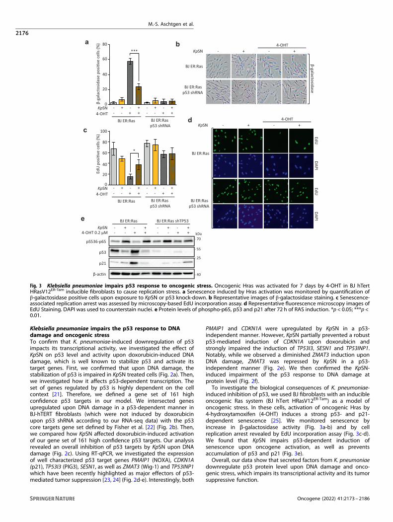
Klebsiella pneumoniae impairs the p53 response to DNAdamage and oncogenic stressTo confirm that K. pneumoniae-induced downregulation of p53impacts its transcriptional activity, we investigated the effect ofKpSN on p53 level and activity upon doxorubicin-induced DNAdamage, which is well known to stabilize p53 and activate itstarget genes. First, we confirmed that upon DNA damage, thestabilization of p53 is impaired in KpSN treated cells (Fig. 2a). Then,we investigated how it affects p53-dependent transcription. Theset of genes regulated by p53 is highly dependent on the cellcontext [21]. Therefore, we defined a gene set of 161 highconfidence p53 targets in our model. We intersected genesupregulated upon DNA damage in a p53-dependent manner inBJ-hTERT fibroblasts (which were not induced by doxorubicinupon p53 shRNA according to our RNA-seq data) with the p53core targets gene set defined by Fisher et al. [22] (Fig. 2b). Then,we compared how KpSN affected doxorubicin-induced activationof our gene set of 161 high confidence p53 targets. Our analysisrevealed an overall inhibition of p53 targets by KpSN upon DNAdamage (Fig. 2c). Using RT-qPCR, we investigated the expressionof well characterized p53 target genes PMAIP1 (NOXA), CDKN1A(p21), TP53I3 (PIG3), SESN1, as well as ZMAT3 (Wig-1) and TP53INP1which have been recently highlighted as major effectors of p53-mediated tumor suppression [23, 24] (Fig. 2d-e). Interestingly, both
PMAIP1 and CDKN1A were upregulated by KpSN in a p53-independent manner. However, KpSN partially prevented a robustp53-mediated induction of CDKN1A upon doxorubicin andstrongly impaired the induction of TP53I3, SESN1 and TP53INP1.Notably, while we observed a diminished ZMAT3 induction uponDNA damage, ZMAT3 was repressed by KpSN in a p53-independent manner (Fig. 2e). We then confirmed the KpSN-induced impairment of the p53 response to DNA damage atprotein level (Fig. 2f).To investigate the biological consequences of K. pneumoniae-
induced inhibition of p53, we used BJ fibroblasts with an inducibleoncogenic Ras system (BJ hTert HRasV12ER-Tam) as a model ofoncogenic stress. In these cells, activation of oncogenic Hras by4-hydroxytamoxifen (4-OHT) induces a strong p53- and p21-dependent senescence [25]. We monitored senescence byincrease in β-galactosidase activity (Fig. 3a-b) and by cellreplication arrest revealed by EdU incorporation assay (Fig. 3c-d).We found that KpSN impairs p53-dependent induction ofsenescence upon oncogene activation, as well as preventsaccumulation of p53 and p21 (Fig. 3e).Overall, our data show that secreted factors from K. pneumoniae
downregulate p53 protein level upon DNA damage and onco-genic stress, which impairs its transcriptional activity and its tumorsuppressive function.
β-ga
lact
osid
ase
posi�
ve c
ells
(%) 80
60
40
20
0
4-OHT - +- +KpSN - -+ +
- +- +- -+ +
BJ ER:Ras BJ ER:Rasp53 shRNA
***
EdU
posi�
ve c
ells
(%)
80
60
40
20
0
4-OHT - +- +KpSN - -+ +
- +- +- -+ +
BJ ER:Ras BJ ER:Rasp53 shRNA
*
100
a b
c
KpSN
β-galactosidase
-- ++
BJ ER:Ras
BJ ER:Rasp53 shRNA
4-OHT
EdUDAPI
EdUDAPI
BJ ER:Ras
BJ ER:Rasp53 shRNA
Kp --NS ++4-OHTd
e
4-OHT 0.2 μM - +- +KpSN - -+
p21
β-ac�n
p53
+- +- +- -+ +
BJ ER:Ras BJ ER:Ras shTP53
pS536-p65 - 70kDa
- 55
- 40
- 25
Fig. 3 Klebsiella pneumoniae impairs p53 response to oncogenic stress. Oncogenic Hras was activated for 7 days by 4-OHT in BJ hTertHRasV12ER-Tam inducible fibroblasts to cause replication stress. a Senescence induced by Hras activation was monitored by quantification ofβ-galactosidase positive cells upon exposure to KpSN or p53 knock-down. b Representative images of β-galactosidase staining. c Senescence-associated replication arrest was assessed by microscopy-based EdU incorporation assay. d Representative fluorescence microscopy images ofEdU Staining. DAPI was used to counterstain nuclei. e Protein levels of phospho-p65, p53 and p21 after 72 h of RAS induction. *p < 0.05; ***p <0.01.
M.-S. Aschtgen et al.
2176
Oncogene (2022) 41:2173 – 2186
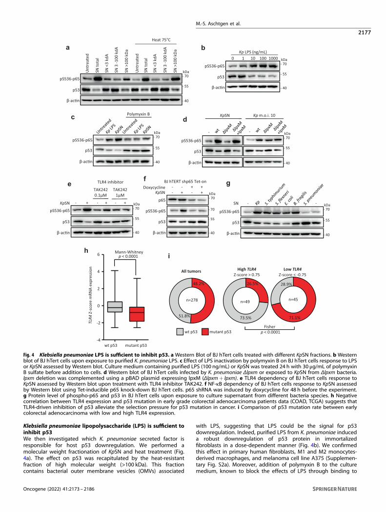
Klebsiella pneumoniae lipopolysaccharide (LPS) is sufficient toinhibit p53We then investigated which K. pneumoniae secreted factor isresponsible for host p53 downregulation. We performed amolecular weight fractionation of KpSN and heat treatment (Fig.4a). The effect on p53 was recapitulated by the heat-resistantfraction of high molecular weight (>100 kDa). This fractioncontains bacterial outer membrane vesicles (OMVs) associated
with LPS, suggesting that LPS could be the signal for p53downregulation. Indeed, purified LPS from K. pneumoniae induceda robust downregulation of p53 protein in immortalizedfibroblasts in a dose-dependent manner (Fig. 4b). We confirmedthis effect in primary human fibroblasts, M1 and M2 monocytes-derived macrophages, and melanoma cell line A375 (Supplemen-tary Fig. S2a). Moreover, addition of polymyxin B to the culturemedium, known to block the effects of LPS through binding to
a b
c d
gef
Unt
reat
edp53
SN to
tal
SN <
3 kd
A
SN 3
-100
kdA
SN >
100
kDa
Unt
reat
ed
SN to
tal
SN <
3 kd
A
SN 3
-100
kdA
SN >
100
kDa
Heat 75°C
pS536-p65
β-ac�n
- 70kDa
- 55
- 40
pS536-p65
Kp LPS (ng/mL)0 10 10001 100
p53
β-ac�n
- 70kDa
- 55
- 40
-
pS536-p65
p53
β-ac�n
SN -- 70kDa
- 55
- 40
Polymyxin B
pS536-p65
β-ac�n
p53
- 70kDa
- 55
- 40
pS536-p65
KpSN
TAK2420.1μM
TAK2421μM
- - -+ + +
p53
β-ac�n
TLR4 inhibitor
- 70kDa
- 55
- 40
-
pS536-p65
p53
β-ac�n
KpSN + - +
p65
BJ hTERT shp65 Tet-on-Doxycycline - + +
- 70kDa
- 55
- 40
- 70
-
pS536-p65
p53
β-ac�n
KpSN Kp m.o.i. 10
- 70kDa
- 55
- 40
-
p < 0.0001Mann-Whitney
wt p53 mutant p53
TLR4
Z-sc
ore
mRN
Aex
pres
sion
-4
-2
0
2
4
6h
p < 0.0001Fisher
n=49 n=45n=278
All tumors High TLR4Z-score > 0.75
Low TLR4Z-score < -0.75
48.2%
51.8%
26.5%
73.5%
28.9%
71.1%
wt p53 mutant p53
i
Fig. 4 Klebsiella pneumoniae LPS is sufficient to inhibit p53. a Western Blot of BJ hTert cells treated with different KpSN fractions. b Westernblot of BJ hTert cells upon exposure to purified K. pneumoniae LPS. c Effect of LPS inactivation by polymyxin B on BJ hTert cells response to LPSor KpSN assessed by Western blot. Culture medium containing purified LPS (100 ng/mL) or KpSN was treated 24 h with 30 μg/mL of polymyxinB sulfate before addition to cells. d Western blot of BJ hTert cells infected by K. pneumoniae Δlpxm or exposed to KpSN from Δlpxm bacteria.lpxm deletion was complemented using a pBAD plasmid expressing lpxM (Δlpxm+ lpxm). e TLR4 dependency of BJ hTert cells response toKpSN assessed by Western blot upon treatment with TLR4 inhibitor TAK242. f NF-κB dependency of BJ hTert cells response to KpSN assessedby Western blot using Tet-inducible p65 knock-down BJ hTert cells. p65 shRNA was induced by doxycycline for 48 h before the experiment.g Protein level of phospho-p65 and p53 in BJ hTert cells upon exposure to culture supernatant from different bacteria species. h Negativecorrelation between TLR4 expression and p53 mutation in early grade colorectal adenocarcinoma patients data (COAD, TCGA) suggests thatTLR4-driven inhibition of p53 alleviate the selection pressure for p53 mutation in cancer. i Comparison of p53 mutation rate between earlycolorectal adenocarcinoma with low and high TLR4 expression.
M.-S. Aschtgen et al.
2177
Oncogene (2022) 41:2173 – 2186
ed
Rela
�ve
TP53
mRN
Ale
vel(
FC) 1.4
1.0
0.6
0.0
0.2
0.4
0.8
1.2
****** ***
NS
Kp LPS 0 10 10001 100 ng/mLUntreated KpSN
Rela
�ve
TP53
mRN
Ale
vel(
FC)
1.0
0.6
0.0
0.2
0.4
0.8
1.2 ***
a
b
c
KpSN
Nutlin10μM
MG13210μM
- - -+ + +
p53
β-ac�n
kDa- 55
- 40
p53
β-ac�n
MG132
kDa- 55
- 40
1h 2h 3h 4h 0 1h 2h 3h 4hCHX + KpSN
0CHX Cont. KpSN
10h 10h
No CHX
1
0.8
0.6
0.4
0.2
00 1h 2h 3h 4h CHX
Rela
�ve
p53
prot
ein
leve
l(n
orm
alize
dto
β-a
c�n)
p53
β-ac�n
Densitometric quan�fica�onCHXCHX + KpSN
CHX CHX + Kp LPS0 2h1h 3h 0 2h1h 3h
1
0.8
0.6
XHCh10
0.4
0.2
02h 3h
Densitometric quan�fica�on
kDa- 55
- 40
CHXCHX + Kp LPS
f M1 macrophages
Rela
�ve
TP53
mRN
Ale
vel(
FC)
1.0
0.6
0.0
0.2
0.4
0.8
1.2
*** ***
1 2 3 4 5 6 7 8 9 10 119i4i
β γ
α
P1 P2
TP53 promoter-Luc
TP53(endogenous)
1.0
0.6
0.00.20.4
0.8
1.21.4
Luci
fera
sem
RNA
(fLuc
/rLu
c)
*** ***
Rela
�ve
TP53
mRN
Ale
vel(
FC)
1.0
0.6
0.00.20.4
0.8
1.2
TP53P1 isoforms
TP53P2 isoforms
Rela
�ve
mRN
Ale
vel(
FC)
1.0
0.6
0.0
0.2
0.4
0.8
1.2
Rela
�ve
mRN
Ale
vel(
FC)
***
***
****
1.0
0.6
0.0
0.2
0.4
0.8
1.2
g
h
i
**
LucTP53 UTRs
TP53(endogenous)
1.0
0.6
0.00.20.4
0.8
1.2
Luci
fera
sem
RNA
(fLuc
/rLu
c)
*** ***
Rela
�ve
TP53
mRN
Ale
vel(
FC)
1.0
0.6
0.00.20.4
0.8
1.2
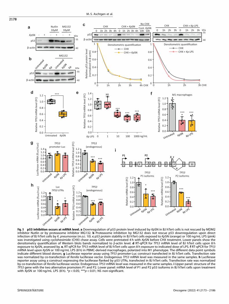
Fig. 5 p53 inhibition occurs at mRNA level. a Downregulation of p53 protein level induced by KpSN in BJ hTert cells is not rescued by MDM2inhibitor Nutlin or by proteasome inhibitor MG132. b Proteasome inhibition by MG132 does not rescue p53 downregulation upon directinfection of BJ hTert cells by K. pneumoniae (m.o.i. 10). c p53 protein stability in BJ hTert cells exposed to KpSN (orange) or 100 ng/mL LPS (pink)was investigated using cycloheximide (CHX) chase assay. Cells were pretreated 4 h with KpSN before CHX treatment. Lower panels show thedensitometry quantification of Western blots bands normalized to β-actin level. d RT-qPCR for TP53 mRNA level of BJ hTert cells upon 8 hexposure to KpSN, assessed by. e, RT-qPCR for TP53mRNA level of BJ hTert cells upon 8 h exposure to indicated dose of LPS. f RT-qPCR for TP53mRNA level upon KpSN or 100 ng/mL LPS (8 h) in PBMC-derived macrophages, polarized into M1 phenotype. The different data point symbolsindicate different blood donors. g Luciferase reporter assay using TP53 promoter-Luc construct transfected in BJ hTert cells. Transfection ratewas normalized by co-transfection of Renilla luciferase vector. Endogenous TP53 mRNA level was measured in the same samples. h Luciferasereporter assay using a construct expressing the luciferase flanked by p53 UTRs, transfected in BJ hTert cells. Transfection rate was normalizedby co-transfection of Renilla luciferase vector. Endogenous TP53mRNA level was measured in the same samples. i Upper panel: structure of theTP53 gene with the two alternative promoters P1 and P2. Lower panel: mRNA level of P1 and P2 p53 isoforms in BJ hTert cells upon treatmentwith KpSN or 100 ng/mL LPS (8 h). *p < 0.05; ***p < 0.01; NS non-significant.
M.-S. Aschtgen et al.
2178
Oncogene (2022) 41:2173 – 2186
log2FC (KpSN vs Untreated)
-log 1
0(FD
R)
0
100
200
300
0 0155-
ZMAT3
TIA1
ZNF385A
miR125b
ELAVL1RBM24
PARNCPEB1
RBM38
b
c d
g
a
h
e
log2FC (S. typhimurium vs non infected macrophages)
-log 1
0(FD
R)
0
100
200
300
0 0155-
ZMAT3
TP53
Rela
�ve
ZMAT
3 m
RNA
leve
l(FC
)
1.0
0.6
0.00.20.4
0.8
1.21.41.6
M1 macrophages
******
pS536-p65
p53
Wig-1
β-ac�n
- 70kDa
- 55
- 35
- 40
M1 macrophages
-KpSN 8h
BJ hTert
Dox - + +- + - +
pS536-p65
p53
Wig-1
β-ac�n
- 70kDa
- 55
- 40
- 35
PIG3 - 35
00.2
1
Rela
�ve
prot
ein
leve
l(n
orm
alize
dto
β-a
c�n)
0.40.6
1.2
0.8
*** ***
******
Wig-1p53
Control KpSN Kp LPS
***
TP53
mRN
A(%
inpu
t)
IP: Flag IgG0
0.05
0.1
0.15
0.2
RIP-qPCRBJ hTert Wig-1-Flag
f
Rela
�ve
TP53
mRN
Ale
vel
(FC
KpSN
vs u
ntre
ated
) ***1.0
0.6
0.0
0.2
0.4
0.8*
BJ hTERT
Wig-1
pS536-p65
p53
Wig-1
KpSN - + - +
BJ hTERT BJ hTert
β-ac�n
- 70kDa
- 55
- 40
- 35
Wig-1*H88A- +
BJ hTerte.v.
1.0
0.6
0.0
0.2
0.4
0.8
1.2
Rela
�ve
p53
prot
ein
leve
l(n
orm
alize
dto
β-a
c�n)
*** ***
NS
KpSN - + - + - +Wig-1 Wig-1*e.v.
H88A
5
3
0
1
2
4
7
Rela
�ve
Wig
-1 p
rote
inle
vel
(nor
mal
ized
to β
-ac�
n)
*
KpSN - + - + - +Wig-1 Wig-1*e.v.
H88A
6
0
0.5
1
1.5
2
***
*
Wig-1 PIG30
0.5
1
1.5
2
Rela
�ve
prot
ein
leve
l(n
orm
alize
dto
β-a
c�n)
Control KpSN Dox KpSN+Dox
NS NS
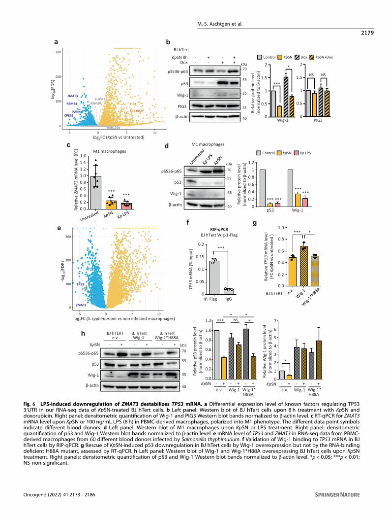
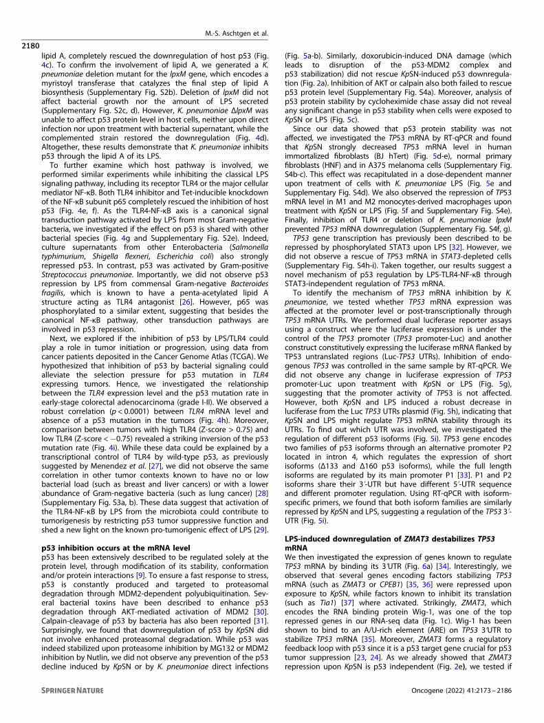
Fig. 6 LPS-induced downregulation of ZMAT3 destabilizes TP53 mRNA. a Differential expression level of known factors regulating TP533ʹUTR in our RNA-seq data of KpSN-treated BJ hTert cells. b Left panel: Western blot of BJ hTert cells upon 8 h treatment with KpSN anddoxorubicin. Right panel: densitometric quantification of Wig-1 and PIG3 Western blot bands normalized to β-actin level. c RT-qPCR for ZMAT3mRNA level upon KpSN or 100 ng/mL LPS (8 h) in PBMC-derived macrophages, polarized into M1 phenotype. The different data point symbolsindicate different blood donors. d Left panel: Western blot of M1 macrophages upon KpSN or LPS treatment. Right panel: densitometricquantification of p53 and Wig-1 Western blot bands normalized to β-actin level. emRNA level of TP53 and ZMAT3 in RNA-seq data from PBMC-derived macrophages from 60 different blood donors infected by Salmonella thyphimurium. f Validation of Wig-1 binding to TP53 mRNA in BJhTert cells by RIP-qPCR. g Rescue of KpSN-induced p53 downregulation in BJ hTert cells by Wig-1 overexpression but not by the RNA-bindingdeficient H88A mutant, assessed by RT-qPCR. h Left panel: Western blot of Wig-1 and Wig-1*H88A overexpressing BJ hTert cells upon KpSNtreatment. Right panels: densitometric quantification of p53 and Wig-1 Western blot bands normalized to β-actin level. *p < 0.05; ***p < 0.01;NS non-significant.
M.-S. Aschtgen et al.
2179
Oncogene (2022) 41:2173 – 2186
lipid A, completely rescued the downregulation of host p53 (Fig.4c). To confirm the involvement of lipid A, we generated a K.pneumoniae deletion mutant for the lpxM gene, which encodes amyristoyl transferase that catalyzes the final step of lipid Abiosynthesis (Supplementary Fig. S2b). Deletion of lpxM did notaffect bacterial growth nor the amount of LPS secreted(Supplementary Fig. S2c, d). However, K. pneumoniae ΔlpxM wasunable to affect p53 protein level in host cells, neither upon directinfection nor upon treatment with bacterial supernatant, while thecomplemented strain restored the downregulation (Fig. 4d).Altogether, these results demonstrate that K. pneumoniae inhibitsp53 through the lipid A of its LPS.To further examine which host pathway is involved, we
performed similar experiments while inhibiting the classical LPSsignaling pathway, including its receptor TLR4 or the major cellularmediator NF-κB. Both TLR4 inhibitor and Tet-inducible knockdownof the NF-κB subunit p65 completely rescued the inhibition of hostp53 (Fig. 4e, f). As the TLR4-NF-κB axis is a canonical signaltransduction pathway activated by LPS from most Gram-negativebacteria, we investigated if the effect on p53 is shared with otherbacterial species (Fig. 4g and Supplementary Fig. S2e). Indeed,culture supernatants from other Enterobacteria (Salmonellatyphimurium, Shigella flexneri, Escherichia coli) also stronglyrepressed p53. In contrast, p53 was activated by Gram-positiveStreptococcus pneumoniae. Importantly, we did not observe p53repression by LPS from commensal Gram-negative Bacteroidesfragilis, which is known to have a penta-acetylated lipid Astructure acting as TLR4 antagonist [26]. However, p65 wasphosphorylated to a similar extent, suggesting that besides thecanonical NF-κB pathway, other transduction pathways areinvolved in p53 repression.Next, we explored if the inhibition of p53 by LPS/TLR4 could
play a role in tumor initiation or progression, using data fromcancer patients deposited in the Cancer Genome Atlas (TCGA). Wehypothesized that inhibition of p53 by bacterial signaling couldalleviate the selection pressure for p53 mutation in TLR4expressing tumors. Hence, we investigated the relationshipbetween the TLR4 expression level and the p53 mutation rate inearly-stage colorectal adenocarcinoma (grade I-II). We observed arobust correlation (p < 0.0001) between TLR4 mRNA level andabsence of a p53 mutation in the tumors (Fig. 4h). Moreover,comparison between tumors with high TLR4 (Z-score > 0.75) andlow TLR4 (Z-score <−0.75) revealed a striking inversion of the p53mutation rate (Fig. 4i). While these data could be explained by atranscriptional control of TLR4 by wild-type p53, as previouslysuggested by Menendez et al. [27], we did not observe the samecorrelation in other tumor contexts known to have no or lowbacterial load (such as breast and liver cancers) or with a lowerabundance of Gram-negative bacteria (such as lung cancer) [28](Supplementary Fig. S3a, b). These data suggest that activation ofthe TLR4-NF-κB by LPS from the microbiota could contribute totumorigenesis by restricting p53 tumor suppressive function andshed a new light on the known pro-tumorigenic effect of LPS [29].
p53 inhibition occurs at the mRNA levelp53 has been extensively described to be regulated solely at theprotein level, through modification of its stability, conformationand/or protein interactions [9]. To ensure a fast response to stress,p53 is constantly produced and targeted to proteasomaldegradation through MDM2-dependent polyubiquitination. Sev-eral bacterial toxins have been described to enhance p53degradation through AKT-mediated activation of MDM2 [30].Calpain-cleavage of p53 by bacteria has also been reported [31].Surprisingly, we found that downregulation of p53 by KpSN didnot involve enhanced proteasomal degradation. While p53 wasindeed stabilized upon proteasome inhibition by MG132 or MDM2inhibition by Nutlin, we did not observe any prevention of the p53decline induced by KpSN or by K. pneumoniae direct infections
(Fig. 5a-b). Similarly, doxorubicin-induced DNA damage (whichleads to disruption of the p53-MDM2 complex andp53 stabilization) did not rescue KpSN-induced p53 downregula-tion (Fig. 2a). Inhibition of AKT or calpain also both failed to rescuep53 protein level (Supplementary Fig. S4a). Moreover, analysis ofp53 protein stability by cycloheximide chase assay did not revealany significant change in p53 stability when cells were exposed toKpSN or LPS (Fig. 5c).Since our data showed that p53 protein stability was not
affected, we investigated the TP53 mRNA by RT-qPCR and foundthat KpSN strongly decreased TP53 mRNA level in humanimmortalized fibroblasts (BJ hTert) (Fig. 5d-e), normal primaryfibroblasts (HNF) and in A375 melanoma cells (Supplementary Fig.S4b-c). This effect was recapitulated in a dose-dependent mannerupon treatment of cells with K. pneumoniae LPS (Fig. 5e andSupplementary Fig. S4d). We also observed the repression of TP53mRNA level in M1 and M2 monocytes-derived macrophages upontreatment with KpSN or LPS (Fig. 5f and Supplementary Fig. S4e).Finally, inhibition of TLR4 or deletion of K. pneumoniae lpxMprevented TP53 mRNA downregulation (Supplementary Fig. S4f, g).TP53 gene transcription has previously been described to be
repressed by phosphorylated STAT3 upon LPS [32]. However, wedid not observe a rescue of TP53 mRNA in STAT3-depleted cells(Supplementary Fig. S4h-i). Taken together, our results suggest anovel mechanism of p53 regulation by LPS-TLR4-NF-κB throughSTAT3-independent regulation of TP53 mRNA.To identify the mechanism of TP53 mRNA inhibition by K.
pneumoniae, we tested whether TP53 mRNA expression wasaffected at the promoter level or post-transcriptionally throughTP53 mRNA UTRs. We performed dual luciferase reporter assaysusing a construct where the luciferase expression is under thecontrol of the TP53 promoter (TP53 promoter-Luc) and anotherconstruct constitutively expressing the luciferase mRNA flanked byTP53 untranslated regions (Luc-TP53 UTRs). Inhibition of endo-genous TP53 was controlled in the same sample by RT-qPCR. Wedid not observe any change in luciferase expression of TP53promoter-Luc upon treatment with KpSN or LPS (Fig. 5g),suggesting that the promoter activity of TP53 is not affected.However, both KpSN and LPS induced a robust decrease inluciferase from the Luc TP53 UTRs plasmid (Fig. 5h), indicating thatKpSN and LPS might regulate TP53 mRNA stability through itsUTRs. To find out which UTR was involved, we investigated theregulation of different p53 isoforms (Fig. 5i). TP53 gene encodestwo families of p53 isoforms through an alternative promoter P2located in intron 4, which regulates the expression of shortisoforms (Δ133 and Δ160 p53 isoforms), while the full lengthisoforms are regulated by its main promoter P1 [33]. P1 and P2isoforms share their 3ʹ-UTR but have different 5ʹ-UTR sequenceand different promoter regulation. Using RT-qPCR with isoform-specific primers, we found that both isoform families are similarlyrepressed by KpSN and LPS, suggesting a regulation of the TP53 3ʹ-UTR (Fig. 5i).
LPS-induced downregulation of ZMAT3 destabilizes TP53mRNAWe then investigated the expression of genes known to regulateTP53 mRNA by binding its 3ʹUTR (Fig. 6a) [34]. Interestingly, weobserved that several genes encoding factors stabilizing TP53mRNA (such as ZMAT3 or CPEB1) [35, 36] were repressed uponexposure to KpSN, while factors known to inhibit its translation(such as Tia1) [37] where activated. Strikingly, ZMAT3, whichencodes the RNA binding protein Wig-1, was one of the toprepressed genes in our RNA-seq data (Fig. 1c). Wig-1 has beenshown to bind to an A/U-rich element (ARE) on TP53 3ʹUTR tostabilize TP53 mRNA [35]. Moreover, ZMAT3 forms a regulatoryfeedback loop with p53 since it is a p53 target gene crucial for p53tumor suppression [23, 24]. As we already showed that ZMAT3repression upon KpSN is p53 independent (Fig. 2e), we tested if
M.-S. Aschtgen et al.
2180
Oncogene (2022) 41:2173 – 2186
Wig-1 was inhibited at earlier time points (8 h) than other p53target genes. We observed the downregulation of both isoformsof Wig-1 at the same time frame as TP53 mRNA but before otherp53 targets such as PIG3 (Fig. 6b and Fig. 2f). Moreover, Wig-1 wasrepressed at mRNA and protein level in M1 and M2 monocytes-derived macrophages (Fig. 6c, d and Supplementary Fig. S5a-b).Analysis of RNA-seq data from PBMC-derived macrophages from60 different blood donors infected by Salmonella thyphimurium[38] confirmed the robust inhibition of both TP53 and ZMAT3 upondirect infection of macrophages by another Enterobacterialspecies (Fig. 6e).To confirm the role of Wig-1 in p53 downregulation, we validated
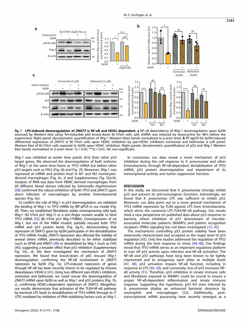
the binding of Wig-1 to TP53 mRNA by RIP-qPCR in our model (Fig.6f). Then, we established fibroblasts stably overexpressing wild typeWig-1 (BJ hTert pLV Wig-1) or a zinc-finger mutant unable to bindTP53 mRNA [35] (BJ hTert pLV Wig-1*H88A). Overexpression of wtWig-1, but not of the H88A mutant, partially rescued both TP53mRNA and p53 protein levels (Fig. 6g-h), demonstrating thatrepression of ZMAT3 gene by KpSN participates in the destabilizationof TP53 mRNA. Finally, ZMAT3 repression also affected the stability ofseveral others mRNA, previously described to be either stabilized(such as EIF4B and RRM1) [39] or destabilized by Wig-1 (such as FAS)[40], suggesting a broader effect than p53 inhibition (SupplementaryFig. S5c, d). We then investigated the mechanism of ZMAT3repression. We found that knock-down of p65 rescued Wig-1downregulation, confirming the NF-κB involvement in ZMAT3repression by KpSN (Fig. 7a). LPS-induced repression of genesthrough NF-κB has been recently shown to be regulated by histonedeacetylases (HDACs) [41]. Using two different pan-HDACs inhibitors,vorinostat and belinostat, we could rescue the downregulation ofZMAT3 mRNA upon KpSN as well as Wig-1 and p53 proteins (Fig. 7b,c), confirming HDACs-dependent repression of ZMAT3. Altogether,our results demonstrate that activation of the TLR4-NF-κB pathwayby bacterial LPS leads to destabilization of TP53 mRNA through its 3′UTR, mediated by inhibition of RNA-stabilizing factors such as Wig-1.
In conclusion, our data reveal a novel mechanism of p53inhibition during the cell response to K. pneumoniae and otherEnterobacteria, through NF-κB-dependent destabilization of TP53mRNA, p53 protein downregulation and impairment of itstranscriptional activity and tumor suppressive function.
DISCUSSIONIn this study, we discovered that K. pneumoniae strongly inhibitp53 and prevent its anti-tumorigenic functions. Interestingly, wefound that K. pneumoniae LPS was sufficient to inhibit p53.Moreover, our data point out to a more general mechanism ofTP53 mRNA repression by TLR4 agonist LPS from Enterobacteria,which elicits the canonical LPS-TLR4-NF-κB pathway. Our resultsshed a new perspective on published data about p53 response tobacteria, where inhibition of p53 downstream of microbe-associated molecular patterns (MAMPs) and pattern recognitionreceptors (PRRs) signaling has not been investigated [15, 42].The mechanisms controlling p53 protein stability have been
extensively characterized and accepted as the major level of p53regulation [43]. Only few studies addressed the regulation of TP53mRNA during the host response to stress [44–46]. Our findingsreveal that TP53 mRNA serves as an important regulatory platformto turn off p53 activity upon infection and NF-κB activation. TheNF-κB and p53 pathways have long been known to be tightlyintertwined and to antagonize each other at multiple levels[47, 48]. p53 activation impairs NF-κB function, including inresponse to LPS [49, 50], and conversely, loss of p53 increases NF-κB activity [51]. Therefore, p53 inhibition in innate immune cellsand fibroblasts exposed to MAMPs could be crucial to ensure aproper NF-κB-dependent inflammation and innate immuneresponse. Supporting this hypothesis, p53 KO mice infected byK. pneumoniae display an enhanced bacterial clearance byneutrophils and macrophages [52]. Additionally, post-transcriptional mRNA processing have recently emerged as a
b
c
Rela
�ve
ZMAT
3m
RNA
leve
l(FC
)
1.0
0.6
0.0
0.2
0.4
0.8
1.2
1.4
KpSN - + - + - +Vorinostat
(5μM)Belinostat
(1μM)
NSNS*
pS536-p65
p53
β-ac�n
KpSN - + - +
Wig-1
HDAC inhibitor - - + +- + - +- - + +
- 70kDa
- 55
- 40
- 35
Vorinostat (5 μM) Belinostat (1 μM)
1.0
0.6
0.0
0.2
0.4
0.8
1.4
Rela
�ve
p53
prot
ein
leve
l(n
orm
alize
dto
β-a
c�n)
KpSN - + - + - +Vor. Bel.
1.2 NS
NS
***1.0
0.6
0.0
0.2
0.4
0.8
1.4
Rela
�ve
Wig
-1 p
rote
inle
vel
(nor
mal
ized
to β
-ac�
n)
KpSN - + - + - +Vor. Bel.
1.2NS
NS***
a
pS536-p65
Wig-1
β-ac�n
- 70kDa
- 40
- 35
-KpSN + - +
BJ hTERT shp65 Tet-on-Doxycycline - + +
Rela
�ve
Wig
-1 p
rote
inle
vel
(nor
mal
ized
to β
-ac�
n)
1.0
0.6
0.00.20.4
0.8
1.2 *
***NS
-KpSN + - +shp65 ON
Fig. 7 LPS-induced downregulation of ZMAT3 is NF-κB and HDAC-dependent. a NF-κB dependency of Wig-1 downregulation upon KpSNassessed by Western blot using Tet-inducible p65 knock-down BJ hTert cells. p65 shRNA was induced by doxycycline for 48 h before theexperiment. Right panel: densitometric quantification of Wig-1 Western blots bands normalized to β-actin level. b RT-qpCR for KpSN-induceddifferential expression of ZMAT3 in BJ hTert cells upon HDAC inhibition by pan-HDAC inhibitors vorinostat and belinostat. c Left panel:Western blot of BJ hTert cells exposed to KpSN upon HDAC inhibition. Right panels: densitometric quantification of p53 and Wig-1 Westernblot bands normalized to β-actin level. *p < 0.05; ***p < 0.01; NS non-significant.
M.-S. Aschtgen et al.
2181
Oncogene (2022) 41:2173 – 2186
key level of regulation to coordinate the switch from immune cellproliferation to activation during innate immune response [38].Our finding that RNA binding factors such as Wig-1 are among thetop differentially expressed genes is consistent with this idea.Importantly, Wig-1 forms a positive feedback loop with p53 and itis an important p53 target gene [23, 24]. Thus, our data suggestthat TP53 mRNA destabilization and Wig-1 downregulationcooperate during acute inflammation to robustly activate NF-κBto fight bacterial infection, simultaneously weakening tumorbarrier (Fig. 8).Importantly, CRC patients have elevated circulating LPS and
systemic inflammation [53]. Moreover, CRC-associated dysbiosis isusually enriched in Gram-negative bacteria [54], which suggeststhat LPS secretion is an important factor linking bacterial dysbiosisand tumorigenesis. We found that commensal bacteria expressinga TLR4 antagonist LPS, such as B. fragilis, do not downregulatep53, which could contribute to their protective role in colitis-associated colorectal cancer [55]. In addition, colonization of thegut by Enterococcus faecalis, a Gram-positive bacterium, inducesinflammatory response and colitis similarly to Enterobacteriacolonization but does not induce tumorigenesis [56]. Altogether, itsuggests that activation of the TLR4-mediated signaling by LPSplays a critical role in promoting cancer not only by inducinginflammation but also by inhibiting p53.Chronic inflammation linked to bacterial dysbiosis could cause
persistent impairment of p53 tumor suppressor function in innateimmune cells, which might create a cellular context favorable foroncogenesis. Supporting this idea, mouse models with geneticinactivation of p53 specifically in myeloid cells are more prone totumorigenesis [57]. Additionally, inactivation of p53 in the tumormicroenvironment have profound consequences on the tumor cellsthemselves in a non-cell autonomous fashion [58, 59]. As tumorprogression is associated with disruption of the epithelial layer andinvasion of the tumor by bacteria, MAMPs signaling and down-stream inhibition of p53 in cancer-associated fibroblasts and tumor-associated macrophages could fuel tumor development. Accord-ingly, several studies have shown that LPS activation of TLR4accelerates tumor growth [29, 60]. Interestingly, our analysis ofcancer patient data demonstrated an inverse correlation betweenTLR4 expression and p53 mutation rate in CRC. Our results support
the idea that TLR4 activation disables p53 function in the absenceof mutation. Moreover, it suggests that the cancer-associatedbacterial microbiota exerts a selection pressure on cancer cells anddrives tumor evolution. In line with this hypothesis, recentmetagenomics analysis of lung cancer microbiota have shownsignificant differences in bacterial populations associated with p53wild-type versus p53-mutant tumors [61]. Further analyses ofmetagenomics data from cancer patients in connection with tumorgenome profiling are required to understand the connectionbetween the microbiota and host genomic alterations.
METHODSBacterial strainsAll bacterial strains used in this study are listed in Supplementary Table S1.K. pneumoniae SHG4 and SGH10 strains were a gift from Y.-H. Gan [62, 63],IA565 strain was a gift from G. Huffnagle [64]. Enterobacteria were growneither in DMEM 10% FBS, Luria Broth or low salt Luria Broth at 37 °C.S. pneumoniae was grown on agar blood plates at 37 °C with 5% CO2.
B. fragilis was grown on agar blood plates at 37 °C under anaerobicconditions. When stated, 100 μg/mL gentamycin, 50 μg/mL hygromycin,50 μg/mL apramycin was added to the culture.K. pneumoniae lpxM deletion mutant (ΔlpxM) was constructed using a
previously established allelic exchange method with a few modifications[65]. Briefly, the apramycin resistance cassette with extensions homologousto regions adjacent to lpxM was generated by PCR using the pIJ773 plasmid[65] as a template (primers: forward 5′-CTACACTATCCCATTATCTTGATTAAGCAGTCGATCTGCGGATTGGGCATGATTCCGGGATCGTCGACC-3′; reverse 5′-CGAGTAAGCACGGTAGAGATAAAAAAGCCTCCTGACGGAGGCTTTTTTTATGTAGGCTGGAGCTGCTTC-3′). The PCR product was then electroporated into K.pneumoniae cells carrying the pSIM18 plasmid [66]. Replacement of thegene by the apramycin resistance cassette flanked by two Flp recombinasetarget sequences was confirmed by PCR. The resulting strain was thentransformed with the pFLP-hyg plasmid (Addgene #87831; [65]) andincubated for 24 h at 30 °C, allowing excision of the cassette by the Flprecombinase. Plasmid pFLP-hyg was then eliminated at 42 °C, and thecassette excision was verified by sequencing. The pBAD33-gent plasmidproducing lpxM was constructed by standard restriction/ligation cloning byinserting an XbaI-lpxM-HindIII fragment (obtained by PCR using theprimers: forward 5′-GATCCTCTAGAGGATTGGGCATGGAAACGAAAAAAAAT-3′; reverse 5′-GATCCAAGCTTTTATTTCTTTTTCGTGAACAGCTCTTTGCG-3ʹ)into the XbaI-HindIII digested pBAD33-gent plasmid (Addgene #65098;[67]).
EnterobacteriaLPS
TLR4INFLAMMATION
& IMMUNE RESPONSE
HDACs
p53
TP53 mRNA
Wig-1
ZMAT3
TUMOR SUPPRESSION
NF-κB
Feedback loop
Oncogenic or genotoxic stress
p53
TP53 mRNA
Wig-1
ZMAT3
IMPAIREDTUMOR SUPPRESSION
NF-κB
Feedback loop
Oncogenic or genotoxic stress
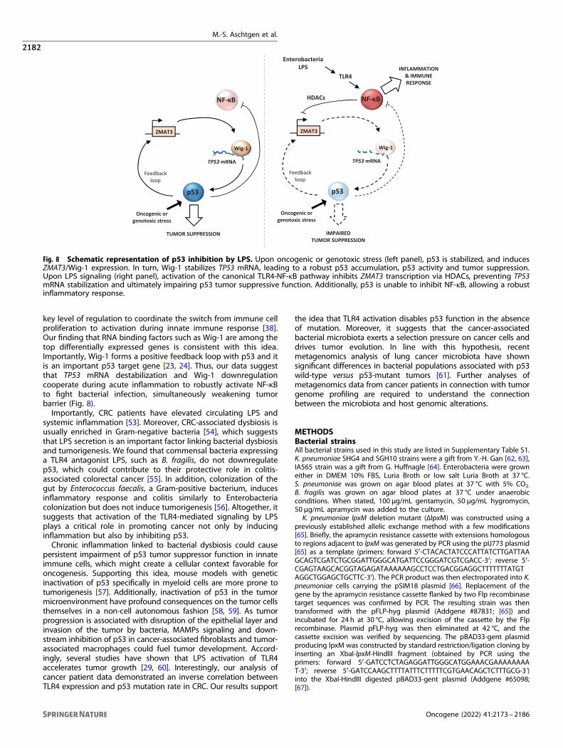
Fig. 8 Schematic representation of p53 inhibition by LPS. Upon oncogenic or genotoxic stress (left panel), p53 is stabilized, and inducesZMAT3/Wig-1 expression. In turn, Wig-1 stabilizes TP53 mRNA, leading to a robust p53 accumulation, p53 activity and tumor suppression.Upon LPS signaling (right panel), activation of the canonical TLR4-NF-κB pathway inhibits ZMAT3 transcription via HDACs, preventing TP53mRNA stabilization and ultimately impairing p53 tumor suppressive function. Additionally, p53 is unable to inhibit NF-κB, allowing a robustinflammatory response.
M.-S. Aschtgen et al.
2182
Oncogene (2022) 41:2173 – 2186
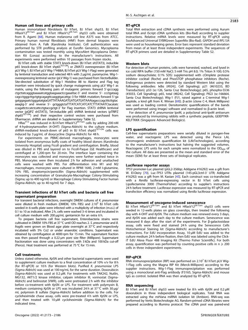
Human cell lines and primary cellsHuman immortalized fibroblasts BJ hTert, BJ hTert shp53, BJ hTertHRasV12ER-Tam and BJ hTert HRasV12ER-Tam shp53 cells were obtainedfrom R. Agami [68]. Human melanoma cell line A375 was from ATCC.Primary human normal fibroblasts (HNF) from dermal biopsies wereobtained from A. Falk (Karolinska Institute). Cell authentication wasperformed by STR profiling analysis at Eurofin Genomics. Mycoplasmacontamination was tested monthly using MycoAlert Mycoplasma Detec-tion Kit (Lonza) according to the manufacturer’s instructions. Allexperiments were performed within 10 passages from frozen stocks.BJ hTert cells with stable STAT3 knock-down (BJ hTert shSTAT3), inducible
p65 knock-down (BJ hTert shp65TET-ON) or ZMAT3 overexpression (BJ hTertpLV Wig-1; BJ hTert pLV Wig-1*H88A; BJ hTert pLV Wig-1-Flag) were generatedby lentiviral transduction and selected 48 h with 2 μg/mL puromycine. Wig-1-overexpressing lentiviral vector (pLV Wig-1) was purchased from VectorBuilder.Site-directed substitution of Wig-1 Histidine 88 to Alanine and Flag taginsertion were introduced by quick change mutagenesis using pLV Wig-1 asmatrix, using the following pairs of mutagenic primers: forward 5ʹ-gcccaggctgcttatcagggtaaaaatcatggtaagaaactccgaaattac-3ʹ and reverse 5ʹ- ccctgataagcagcctgggcttgctgtgcagagttcaaggtgacattgc-3 for H88A mutation; forward 5ʹ-gagatggagaatctgggatatgtaGATTATAAAGATGATGATGATAAAtagacccagctttcttgtacaaagtg-3ʹ and reverse 5ʹ- gctgggtctaTTTATCATCATCATCTTTATAATCtacatatcccagattctccatctcattcctgtaccgctgt-3ʹ for Flag insertion. STAT3 shRNA lentiviralvector (pGIPZ shSTAT3), Tet-inducible p65 shRNA lentiviral vectors (pTRIPZshp65TET-ON) and their respective control vectors were purchased fromDharmacon. shRNA are detailed in Supplementary Table S2.HRasV12 was induced in BJ-hTert HRasV12ER-Tam cells by adding 200 nM
of 4-hydroxytamoxifen (4-OHT) (Sigma-Aldrich) to the culture medium.shRNA-mediated knock-down of p65 in BJ hTert shp65TET-ON cells wasinduced by 3 μg/mL of doxycycline (Sigma-Aldrich) for 48 h.For experiments on PBMC-derived macrophages, monocytes were
isolated from anonymous buffy coats of healthy blood donors (KarolinskaUniversity Hospital) using Ficoll gradient and centrifugation. Briefly, bloodwas diluted in PBS and layered on to Ficoll-Paque (GE Healthcare) andcentrifuged at 1,200 rpm for 20min. The interface layer containing themonocytes was collected and monocytes were further washed twice inPBS. Monocytes were then incubated 2 h for adhesion and unattachedcells were washed with PBS. For differentiation into M1 and M2macrophages, monocytes were cultured in RPMI 1640, 2 mM I-glutamine,10% FBS, streptomycin/penicillin (Sigma-Aldrich) supplemented withincreasing concentration of Granulocyte-Macrophage Colony-Stimulating(Sigma; up to 400 ng/ml) or Macrophage Colony-Stimulating Factor human(Sigma-Aldrich; up to 40 ng/ml) for 7 days.
Transient infections of BJ hTert cells and bacteria cell freesupernatant preparationFor transient bacterial infections, overnight DMEM cultures of K. pneumoniaewere diluted in fresh medium (DMEM, 10% FBS) and 2.105 BJ hTert cellsseeded in 6-wells plate were infected with a multiplicity of infection (m.o.i.) of0.1 to 10 as indicated. After 2 h, cells were washed 3–6 times and incubated incell culture medium with 200 µg/mL gentamicin for an extra 6 h.To prepare bacteria cell free supernatant, Enterobacteria strains were
cultivated in DMEM 10% FBS at 37 °C until OD reaches 2. S. pneumoniae and B.fragilis were grown on Blood agar plate overnight at 37 °C and respectivelyincubated with 5% Co2 or under anaerobic conditions. Supernatant wasobtained by centrifugation at 4000 rpm for 15min. The supernatant fractionwas then passed through a 0.22 µm pore size filter (Millipore). Supernatantfractionation was done using concentrators with 3 kDa and 100 kDa cut-off(Pierce). Heat treatment was performed at 75 °C for 15min.
Cell treatmentsUnless stated otherwise, KpSN and other bacterial supernatants were usedto supplement culture medium to a final concentration of 10% v/v for 8 h(for RT-qPCR) or 10 h (for Western blots). Purified K. pneumoniae LPS(Sigma-Aldrich) was used at 100 ng/mL for the same duration. Doxorubicin(Sigma-Aldrich) was used at 0.2 μM. For treatments with TAK242, Nutlin,MG132, AKT1/2 kinase inhibitor, calpain inhibitor III, vorinostat (Sigma-Aldrich) and belinostat (VWR), cells were pretreated 2 h with the inhibitorbefore co-treatment with KpSN or LPS. For treatment with polymyxin B,medium containing KpSN or LPS was incubated 24 h at 37 °C with 30 μg/mL polymixin B sulfate (Sigma-Aldrich) before addition to the cells. Forcycloheximide chase assay, cells were pre-treated 4 h with KpSN or LPS,and then treated with 10 μM cycloheximide (Sigma-Aldrich) for theindicated time.
RT-qPCRTotal RNA extraction and cDNA synthesis were performed using Aurumtotal RNA and iScript cDNA synthesis kits (Bio-Rad) according to supplierinstructions. Relative mRNA levels were measured by RT-qPCR usingSsoAdvanced Universal SYBRGreen SuperMix (Bio-Rad). GAPDH and RPL13Awere used as housekeeping genes. Error bars represent standard deviationfrom mean of at least three independent experiments. The sequences ofRT-qPCR primers used are detailed in Supplementary Table S2.
Western blotsFor detection of human proteins, cells were harvested, washed, and lysed inice cold RIPA buffer (150mM NaCl; 5 mM Tris pH 8.0; 1% Triton X-100; 0.5%sodium deoxycholate; 0.1% SDS) supplemented with cOmplete proteaseinhibitor cocktail (Roche) and PhosSTOP phosphatase inhibitors (Roche).Endogenous proteins were detected by standard Western blot using thefollowing antibodies: IκBα (#9242, Cell Signaling); p21 (#610233, BDTransduction); p53 (sc-126, Santa Cruz Biotechnology); p65, phospho-S536(#3033, Cell Signaling); p65, total (#8242, Cell Signaling); PIG3 (sc-166664,Santa Cruz Biotechnology); Wig-1 (FJ1, raised against a C-terminal Wig-1peptide, a kind gift from K. Wiman [69]). β-actin (clone C-4, Merk Millipore)was used as loading control. Densitometric quantifications of the bandswere performed using ImageJ software and normalized to β-actin level.For detection of K. pneumoniae lpxM, a polyclonal anti-lpxM antiserum
was produced by immunizing rabbits with a synthetic peptide, GEIEPYKR-KELFTKKK (Singapore Advanced Biologics).
LPS quantificationCell-free supernatants preparations were serially diluted in pyrogen-freewater, and reactogenic LPS was detected using the Pierce LALchromogenic endotoxin quantification kit (LifeTechnologies), accordingto the manufacturer’s instructions but halving the suggested volumes.Reactogenic LPS units for each sample were normalized to the OD600 ofthe culture. All data are presented as the mean and standard error of themean (SEM) for at least three sets of biological replicates.
Luciferase reporter assaysTP53 promoter-Luc plasmid (pGL2-356bp; Addgene #16292) was a gift fromW. El-Deiry [70]. Luc-TP53 UTRs plasmid (145-pGL3ctrl-3ʹ UTR; Addgene#16292) was a gift from M. Kastan [45]. Each construct was co-transfectedwith a Renilla luciferase-expressing vector in BJ hTert cells usingLipofectamine 3000 (ThermoFisher) according to supplier’s instructions24 h before treatment. Luciferase expression was measured by RT-qPCR andtransfection efficiency was normalized using Renilla luciferase expression.
Measurement of oncogene-induced senescenceBJ hTert HRasV12ER-Tam and BJ hTert HRasV12ER-Tam shp53 cells wereseeded in 12-wells plates on glass coverslips, then treated the followingday with 4-OHT and KpSN. The culture medium was renewed every 3 days,and KpSN was added each day to the culture medium. Senescence wasmeasured 8 days after the start of the experiments. For β-galactosidaseassay, cells were fixed and stained 24 h using the Senescence CellsHistochemical Staining kit (Sigma-Aldrich) according to manufacturer’sinstructions. For EdU incorporation Assay, 10 μM EdU was added to theculture medium 24 h before fixation, then EdU was labeled using the Click-iT EdU Alexa Fluor 488 Imaging Kit (Thermo Fisher Scientific). For bothassay, quantification was performed by counting positive cells in n ⩾ 200cells in three independent experiments.
RIP-qPCRRNA immunoprecipitation (RIP) was performed on 2.107 BJ hTert pLV Wig-1-Flag cells using the Magna RIP Kit (Merck-Millipore) according to thesupplier instructions. Wig-1-Flag immunoprecipitation was performedusing a monoclonal anti-Flag antibody (F3165, Sigma-Aldrich) and mouseIgG as control. Isolated RNA was then analyzed by RT-qPCR.
RNA sequencingBJ hTert and BJ hTert shp53 were treated for 8 h with KpSN and 0.2 μMdoxorubicin in three independent biological replicates. Total RNA wasextracted using the mirVana miRNA isolation kit (Ambion). RNA-seq wasperformed by Vertis Biotechnologie AG. Random-primed cDNA libraries wereprepared according to Illumina protocol. The cDNA pool was paired-end
M.-S. Aschtgen et al.
2183
Oncogene (2022) 41:2173 – 2186
sequenced on an Illumina NextSeq 500 system using 2 × 150 bp read length.Quality control was performed using FastQC. nf-core/rnaseq pipeline (v1.4)was used to process the reads [71]. HISAT2 (v2.1.0) was used to align the rawRNA-seq fastq reads to the human reference genome (GRCh38.97). Readquantification was computed using featureCounts (v1.6.4). Differentialanalysis was performed with the R package DESeq2. False positive discovery(FDR) < 0.05 was set as cut-off for differentially expressed genes (DEGs). DEGswere plotted using R package ggplot2. Gene ontology (GO: biologicalprocess) and pathway enrichment analysis was performed using g:Profiler[72] and visualized using Cytoscape (v3.6.1) [73] as described by Reimandet al. [74]. The complete lists of differentially expressed genes and enrichedGO terms upon KpSN treatment are provided in Supplementary Table S3. Todefine a high confidence gene set of p53 targets in BJ hTert cells, weintersected genes upregulated upon doxorubicin treatment, repressed uponp53 shRNA and p53 core targets genes defined by Fisher et al. [22]. Completelist of the 161 high confidence p53 target genes is provided inSupplementary Table S4.
Public database patient data analysisPatient data from The Cancer Genome Atlas (TCGA; https://www.cancer.gov/tcga) were analyzed using cBioPortal for Cancer Genomics (http://cbioportal.org) [75, 76]. Normalized expression data and TP53 mutationsfrom early stage (grade I-II) Colorectal adenocarcinoma (COAD), Breastcarcinoma (BRCA), Liver hepatocellular carcinoma (LIHC) and Lungadenocarcinoma (LUAD) from TCGA PanCancer Atlas Studies wereanalyzed. Tumors were classified based on TLR4 expression Z-score acrossall samples as high TLR4 (Z-score > 0.75) or low TLR4 (Z-score <−0.75).
Statistical analysesUnless otherwise stated, statistical significance was calculated using two-tailed Student t-test from at least three independent experiments.
DATA AVAILABILITYThe datasets generated in the study are available in GEO database (https://www.ncbi.nlm.nih.gov/geo/) under accession number GSE174531.
REFERENCES1. de Martel C, Ferlay J, Franceschi S, Vignat J, Bray F, Forman D, et al. Global burden
of cancers attributable to infections in 2008: a review and synthetic analysis.Lancet Oncol. 2012;13:607–15.
2. Sepich-Poore GD, Zitvogel L, Straussman R, Hasty J, Wargo JA, Knight R. Themicrobiome and human cancer. Science. 2021;371.
3. Wirbel J, Pyl PT, Kartal E, Zych K, Kashani A, Milanese A, et al. Meta-analysis offecal metagenomes reveals global microbial signatures that are specific for col-orectal cancer. Nat Med. 2019;25:679–89.
4. Mei Q-X, Huang C-L, Luo S-Z, Zhang X-M, Zeng Y, Lu Y-Y. Characterization of theduodenal bacterial microbiota in patients with pancreatic head cancer vs. healthycontrols. Pancreatology. 2018;18:438–45.
5. Watanabe T, Tada M, Nagai H, Sasaki S, Nakao M. Helicobacter pylori infectioninduces gastric cancer in mongolian gerbils. Gastroenterology. 1998;115:642–8.
6. Ge Z, Rogers AB, Feng Y, Lee A, Xu S, Taylor NS, et al. Bacterial cytolethal dis-tending toxin promotes the development of dysplasia in a model of microbiallyinduced hepatocarcinogenesis. Cell Microbiol. 2007;9:2070–80.
7. Cuevas-Ramos G, Petit CR, Marcq I, Boury M, Oswald E, Nougayrède J-P.Escherichia coli induces DNA damage in vivo and triggers genomic instability inmammalian cells. Proc Natl Acad Sci USA. 2010;107:11537–42.
8. Brennan CA, Garrett WS. Fusobacterium nucleatum - symbiont, opportunist andoncobacterium. Nat Rev Microbiol. 2019;17:156–66.
9. Hafner A, Bulyk ML, Jambhekar A, Lahav G. The multiple mechanisms that reg-ulate p53 activity and cell fate. Nat Rev Mol Cell Biol. 2019;20:199–210.
10. Rivas C, Aaronson SA, Munoz-Fontela C. Dual Role of p53 in Innate AntiviralImmunity. Viruses. 2010;2:298–313.
11. Tornesello ML, Annunziata C, Tornesello AL, Buonaguro L, Buonaguro FM. Humanoncoviruses and p53 tumor suppressor pathway deregulation at the origin ofhuman cancers. Cancers. 2018;10.
12. Wei J, Nagy TA, Vilgelm A, Zaika E, Ogden SR, Romero-Gallo J, et al. Regulation ofp53 tumor suppressor by Helicobacter pylori in gastric epithelial cells. Gastro-enterology. 2010;139:1333–43.
13. Horvat A, Noto JM, Ramatchandirin B, Zaika E, Palrasu M, Wei J, et al. Helicobacterpylori pathogen regulates p14ARF tumor suppressor and autophagy in gastricepithelial cells. Oncogene. 2018;37:5054–65.
14. Buti L, Spooner E, Van der Veen AG, Rappuoli R, Covacci A, Ploegh HL. Helico-bacter pylori cytotoxin-associated gene A (CagA) subverts the apoptosis-stimulating protein of p53 (ASPP2) tumor suppressor pathway of the host. ProcNatl Acad Sci USA. 2011;108:9238–43.
15. González E, Rother M, Kerr MC, Al-Zeer MA, Abu-Lubad M, Kessler M, et al.Chlamydia infection depends on a functional MDM2-p53 axis. Nat Commun.2014;5:5201.
16. Zella D, Curreli S, Benedetti F, Krishnan S, Cocchi F, Latinovic OS, et al. Myco-plasma promotes malignant transformation in vivo, and its DnaK, a bacterialchaperone protein, has broad oncogenic properties. Proc Natl Acad Sci USA.2018;115:E12005–E12014.
17. Wang T, Cai G, Qiu Y, Fei N, Zhang M, Pang X, et al. Structural segregation of gutmicrobiota between colorectal cancer patients and healthy volunteers. ISME J.2012;6:320–9.
18. Huang W-K, Chang JW-C, See L-C, Tu H-T, Chen J-S, Liaw C-C, et al. Higher rate ofcolorectal cancer among patients with pyogenic liver abscess with Klebsiellapneumoniae than those without: an 11-year follow-up study. Colorectal Dis.2012;14:e794–801.
19. Jeong SW, Jang JY, Lee TH, Kim HG, Hong SW, Park SH, et al. Cryptogenic pyo-genic liver abscess as the herald of colon cancer. J Gastroenterol Hepatol.2012;27:248–55.
20. Lan Y, Zhou M, Jian Z, Yan Q, Wang S, Liu W. Prevalence of pks gene cluster andcharacteristics of Klebsiella pneumoniae-induced bloodstream infections. J ClinLab Anal. 2019;33:e22838.
21. Kastenhuber ER, Lowe SW. Putting p53 in context. Cell. 2017;170:1062–78.22. Fischer M, Grossmann P, Padi M, DeCaprio JA. Integration of TP53, DREAM, MMB-
FOXM1 and RB-E2F target gene analyses identifies cell cycle gene regulatorynetworks. Nucleic Acids Res. 2016;44:6070–86.
23. Janic A, Valente LJ, Wakefield MJ, Di Stefano L, Milla L, Wilcox S, et al. DNA repairprocesses are critical mediators of p53-dependent tumor suppression. Nat Med.2018;24:947–53.
24. Bieging-Rolett KT, Kaiser AM, Morgens DW, Boutelle AM, Seoane JA, Van NostrandEL, et al. Zmat3 is a key splicing regulator in the p53 tumor suppression program.Mol Cell. 2020;80:452–69.e9.
25. Ferbeyre G, de Stanchina E, Lin AW, Querido E, McCurrach ME, Hannon GJ. et al.Oncogenic ras and p53 cooperate to induce cellular senescence. Mol Cell Biol.2002;22:3497–508.
26. Berezow AB, Ernst RK, Coats SR, Braham PH, Karimi-Naser LM, Darveau RP. Thestructurally similar, penta-acylated lipopolysaccharides of Porphyromonas gin-givalis and Bacteroides elicit strikingly different innate immune responses. MicroPathog. 2009;47:68–77.
27. Menendez D, Shatz M, Azzam K, Garantziotis S, Fessler MB, Resnick MA. The toll-like receptor gene family is integrated into human DNA damage and p53 net-works. PLoS Genet. 2011;7:e1001360.
28. Hilty M, Burke C, Pedro H, Cardenas P, Bush A, Bossley C, et al. Disorderedmicrobial communities in asthmatic airways. PloS One. 2010;5:e8578.
29. Dapito DH, Mencin A, Gwak G-Y, Pradere J-P, Jang M-K, Mederacke I, et al. Pro-motion of hepatocellular carcinoma by the intestinal microbiota and TLR4.Cancer Cell. 2012;21:504–16.
30. Siegl C, Rudel T. Modulation of p53 during bacterial infections. Nat Rev Microbiol.2015;13:741–8.
31. Bergounioux J, Elisee R, Prunier A-L, Donnadieu F, Sperandio B, Sansonetti P, et al.Calpain activation by the Shigella flexneri effector VirA regulates key steps in theformation and life of the bacterium’s epithelial niche. Cell Host Microbe.2012;11:240–52.
32. Niu G, Wright KL, Ma Y, Wright GM, Huang M, Irby R, et al. Role ofStat3 in regulating p53 expression and function. Mol Cell Biol. 2005;25:7432–40.
33. Joruiz SM, Bourdon J-C p53 isoforms: key regulators of the cell fate decision. ColdSpring Harb Perspect Med. 2016;6.
34. Haronikova L, Olivares-Illana V, Wang L, Karakostis K, Chen S, Fåhraeus R. The p53mRNA: an integral part of the cellular stress response. Nucleic Acids Res.2019;47:3257–71.
35. Vilborg A, Glahder JA, Wilhelm MT, Bersani C, Corcoran M, Mahmoudi S, et al. Thep53 target Wig-1 regulates p53 mRNA stability through an AU-rich element. ProcNatl Acad Sci USA. 2009;106:15756–61.
36. Burns DM, Richter JD. CPEB regulation of human cellular senescence,energy metabolism, and p53 mRNA translation. Genes Dev. 2008;22:3449–60.
37. Díaz-Muñoz MD, Kiselev VY, Le Novère N, Curk T, Ule J, Turner M. Tia1 dependentregulation of mRNA subcellular location and translation controls p53 expressionin B cells. Nat Commun. 2017;8:530.
38. Pai AA, Baharian G, Pagé Sabourin A, Brinkworth JF, Nédélec Y, Foley JW, et al.Widespread Shortening of 3’ Untranslated Regions and Increased Exon InclusionAre Evolutionarily Conserved Features of Innate Immune Responses to Infection.PLoS Genet. 2016;12:e1006338.
M.-S. Aschtgen et al.
2184
Oncogene (2022) 41:2173 – 2186
39. Bersani C, Huss M, Giacomello S, Xu L-D, Bianchi J, Eriksson S, et al. Genome-wideidentification of Wig-1 mRNA targets by RIP-Seq analysis. Oncotarget.2016;7:1895–911.
40. Bersani C, Xu L-D, Vilborg A, Lui W-O, Wiman KG. Wig-1 regulates cell cycle arrestand cell death through the p53 targets FAS and 14-3-3σ. Oncogene.2014;33:4407–17.
41. Nguyen HCB, Adlanmerini M, Hauck AK, Lazar MA. Dichotomous engagement ofHDAC3 activity governs inflammatory responses. Nature. 2020;584:286–90.
42. Costa L, Corre S, Michel V, Le Luel K, Fernandes J, Ziveri J, et al. USF1 defect drivesp53 degradation during Helicobacter pylori infection and accelerates gastriccarcinogenesis. Gut. 2020;69:1582–91.
43. Levine AJ. p53: 800 million years of evolution and 40 years of discovery. Nat RevCancer. 2020;20:471–80.
44. Fu L, Minden MD, Benchimol S. Translational regulation of human p53 geneexpression. EMBO J. 1996;15:4392–401.
45. Chen J, Kastan MB. 5′-3′-UTR interactions regulate p53 mRNA translation andprovide a target for modulating p53 induction after DNA damage. Genes Dev.2010;24:2146–56.
46. Gajjar M, Candeias MM, Malbert-Colas L, Mazars A, Fujita J, Olivares-Illana V, et al.The p53 mRNA-Mdm2 interaction controls Mdm2 nuclear trafficking and isrequired for p53 activation following DNA damage. Cancer Cell. 2012;21:25–35.
47. Ak P, Levine AJ. p53 and NF-κB: different strategies for responding to stress leadto a functional antagonism. FASEB J. 2010;24:3643–52.
48. Gudkov AV, Komarova EA p53 and the carcinogenicity of chronic inflammation.Cold Spring Harb Perspect Med. 2016;6.
49. Meylan E, Dooley AL, Feldser DM, Shen L, Turk E, Ouyang C, et al. Requirement forNF-kappaB signalling in a mouse model of lung adenocarcinoma. Nature.2009;462:104–7.
50. Liu G, Park Y-J, Tsuruta Y, Lorne E, Abraham E. p53 Attenuateslipopolysaccharide-induced NF-kappaB activation and acute lung injury. JImmunol. 2009;182:5063–71.
51. Komarova EA, Krivokrysenko V, Wang K, Neznanov N, Chernov MV, KomarovPG, et al. p53 is a suppressor of inflammatory response in mice. FASEB J.2005;19:1030–2.
52. Madenspacher JH, Azzam KM, Gowdy KM, Malcolm KC, Nick JA, Dixon D, et al.p53 Integrates host defense and cell fate during bacterial pneumonia. J Exp Med.2013;210:891–904.
53. de Waal GM, de Villiers WJS, Forgan T, Roberts T, Pretorius E. Colorectal cancer isassociated with increased circulating lipopolysaccharide, inflammation andhypercoagulability. Sci Rep. 2020;10:8777.
54. Wu N, Yang X, Zhang R, Li J, Xiao X, Hu Y, et al. Dysbiosis signature of fecalmicrobiota in colorectal cancer patients. Micro Ecol. 2013;66:462–70.
55. Lee YK, Mehrabian P, Boyajian S, Wu W-L, Selicha J, Vonderfecht S, et al. Theprotective role of bacteroides fragilis in a murine model of colitis-associatedcolorectal cancer. mSphere. 2018;3.
56. Arthur JC, Perez-Chanona E, Mühlbauer M, Tomkovich S, Uronis JM, Fan T-J, et al.Intestinal inflammation targets cancer-inducing activity of the microbiota. Sci-ence. 2012;338:120–3.
57. He X-Y, Xiang C, Zhang C-X, Xie Y-Y, Chen L, Zhang G-X, et al. p53 in the myeloidlineage modulates an inflammatory microenvironment limiting initiation andinvasion of intestinal tumors. Cell Rep. 2015;13:888–97.
58. Procopio M-G, Laszlo C, Al Labban D, Kim DE, Bordignon P, Jo S-H, et al. Com-bined CSL and p53 downregulation promotes cancer-associated fibroblast acti-vation. Nat Cell Biol. 2015;17:1193–204.
59. Arandkar S, Furth N, Elisha Y, Nataraj NB, van der Kuip H, Yarden Y, et al. Alteredp53 functionality in cancer-associated fibroblasts contributes to their cancer-supporting features. Proc Natl Acad Sci USA. 2018;115:6410–5.
60. Fukata M, Chen A, Vamadevan AS, Cohen J, Breglio K, Krishnareddy S, et al. Toll-like receptor-4 promotes the development of colitis-associated colorectal tumors.Gastroenterology. 2007;133:1869–81.
61. Greathouse KL, White JR, Vargas AJ, Bliskovsky VV, Beck JA, von Muhlinen N, et al.Interaction between the microbiome and TP53 in human lung cancer. GenomeBiol. 2018;19:123.
62. Lam MMC, Wyres KL, Duchêne S, Wick RR, Judd LM, Gan Y-H, et al. Populationgenomics of hypervirulent Klebsiella pneumoniae clonal-group 23 revealsearly emergence and rapid global dissemination. Nat Commun. 2018;9:2703.
63. Lee IR, Molton JS, Wyres KL, Gorrie C, Wong J, Hoh CH, et al. Differential hostsusceptibility and bacterial virulence factors driving Klebsiella liver abscess in anethnically diverse population. Sci Rep. 2016;6:29316.
64. Hornick DB, Thommandru J, Smits W, Clegg S. Adherence properties of an mrkD-negative mutant of Klebsiella pneumoniae. Infect Immun. 1995;63:2026–32.
65. Huang T-W, Lam I, Chang H-Y, Tsai S-F, Palsson BO, Charusanti P. Capsule deletionvia a λ-Red knockout system perturbs biofilm formation and fimbriae expressionin Klebsiella pneumoniae MGH 78578. BMC Res Notes. 2014;7:13.
66. Chan W, Costantino N, Li R, Lee SC, Su Q, Melvin D, et al. A recombineering basedapproach for high-throughput conditional knockout targeting vector construc-tion. Nucleic Acids Res. 2007;35:e64.
67. Jimenez N, Lacasta A, Vilches S, Reyes M, Vazquez J, Aquillini E, et al. Genetics andproteomics of Aeromonas salmonicida lipopolysaccharide core biosynthesis. JBacteriol. 2009;191:2228–36.
68. Kolfschoten IGM, van Leeuwen B, Berns K, Mullenders J, Beijersbergen RL, Ber-nards R, et al. A genetic screen identifies PITX1 as a suppressor of RAS activity andtumorigenicity. Cell. 2005;121:849–58.
69. Zhang M, Heldin A, Palomar-Siles M, Öhlin S, Bykov VJN, Wiman KG. Synergisticrescue of nonsense mutant tumor suppressor p53 by combination treatmentwith aminoglycosides and Mdm2 inhibitors. Front Oncol. 2017;7:323.
70. Wang S, El-Deiry WS. p73 or p53 directly regulates human p53 transcription tomaintain cell cycle checkpoints. Cancer Res. 2006;66:6982–9.
71. Ewels PA, Peltzer A, Fillinger S, Patel H, Alneberg J, Wilm A, et al. The nf-core frame-work for community-curated bioinformatics pipelines. Nat Biotechnol. 2020;38:276–8.
72. Reimand J, Arak T, Adler P, Kolberg L, Reisberg S, Peterson H, et al. g:Profiler-aweb server for functional interpretation of gene lists (2016 update). Nucleic AcidsRes. 2016;44:W83–89.
73. Shannon P, Markiel A, Ozier O, Baliga NS, Wang JT, Ramage D, et al. Cytoscape: asoftware environment for integrated models of biomolecular interaction net-works. Genome Res. 2003;13:2498–504.
74. Reimand J, Isserlin R, Voisin V, Kucera M, Tannus-Lopes C, Rostamianfar A, et al.Pathway enrichment analysis and visualization of omics data using g:Profiler,GSEA, Cytoscape and EnrichmentMap. Nat Protoc. 2019;14:482–517.
75. Cerami E, Gao J, Dogrusoz U, Gross BE, Sumer SO, Aksoy BA, et al. The cBio cancergenomics portal: an open platform for exploring multidimensional cancergenomics data. Cancer Disco. 2012;2:401–4.
76. Gao J, Aksoy BA, Dogrusoz U, Dresdner G, Gross B, Sumer SO, et al. Integrativeanalysis of complex cancer genomics and clinical profiles using the cBioPortal. SciSignal. 2013;6:pl1.
ACKNOWLEDGEMENTSWe thank all our colleagues for sharing precious reagents and material with us, especiallyDr. Yunn-Hwen Gan (National University of Singapore), Dr. Gary Huffnagle (University ofMichigan) and Mr. Alexandros Petropoulos (Karolinska Institute) for bacteria strains, andProf. Klas Wiman (Karolinska Institute) for Wig-1 antibody. We thank Ms. AleksandraBoikova and Mr. Riccardo Bevilacqua for technical support. This study was supported bythe Swedish Research Council, the Swedish Cancer Society, Knut and Alice WallenbergFoundation, the Swedish foundation for Strategic Research and the Karolinska Institute.MSA was supported by a short-term fellowship from EMBO.
AUTHOR CONTRIBUTIONSMSA and SP designed the study; MSA, KF and SP performed the experiments; MSA,GSz and SP analyzed and interpreted the data; MSA and SP generated figures andtables and drafted the manuscript with the help of GSz; GS, SN and BHN reviewedand edited the manuscript and provided funding.
FUNDINGOpen access funding provided by Karolinska Institute.
COMPETING INTERESTSThe authors declare no competing interests.
ADDITIONAL INFORMATIONSupplementary information The online version contains supplementary materialavailable at https://doi.org/10.1038/s41388-022-02238-5.
Correspondence and requests for materials should be addressed to Marie-StéphanieAschtgen or Sylvain Peuget.
Reprints and permission information is available at http://www.nature.com/reprints
Publisher’s note Springer Nature remains neutral with regard to jurisdictional claimsin published maps and institutional affiliations.
M.-S. Aschtgen et al.
2185
Oncogene (2022) 41:2173 – 2186

Open Access This article is licensed under a Creative CommonsAttribution 4.0 International License, which permits use, sharing,
adaptation, distribution and reproduction in anymedium or format, as long as you giveappropriate credit to the original author(s) and the source, provide a link to the CreativeCommons license, and indicate if changes were made. The images or other third partymaterial in this article are included in the article’s Creative Commons license, unlessindicated otherwise in a credit line to the material. If material is not included in thearticle’s Creative Commons license and your intended use is not permitted by statutoryregulation or exceeds the permitted use, you will need to obtain permission directlyfrom the copyright holder. To view a copy of this license, visit http://creativecommons.org/licenses/by/4.0/.
© The Author(s) 2022
M.-S. Aschtgen et al.
2186
Oncogene (2022) 41:2173 – 2186
Copyright © 2022 FDOKUMEN