The DEAD box protein p68: a novel transcriptional coactivator of the p53 tumour suppressor
Upload
independentCategory
view
3download
0
Transcriptional Regulation by p53
Rachel Beckerman and Carol Prives
Department of Biological Sciences, Columbia University, New York, New York 10027
Correspondence: [email protected]
Inactivation of p53 is critical for the formation of most tumors. Illumination of the keyfunction(s) of p53 protein in protecting cells from becoming cancerous is therefore aworthy goal. Arguably p53’s most important function is to act as a transcription factor thatdirectly regulates perhaps several hundred of the cell’s RNA polymerase II (RNAP II)-trans-cribed genes, and indirectly regulates thousands of others. Indeed p53 is the most wellstudied mammalian transcription factor. The p53 tetramer binds to its response elementwhere it can recruit diverse transcriptional coregulators such as histone modifyingenzymes, chromatin remodeling factors, subunits of the mediator complex, and componentsof general transcription machinery and preinitiation complex (PIC) to modulate RNAPIIactivity at target loci (Laptenko and Prives 2006). The p53 transcriptional program is regu-lated in a stimulus-specific fashion (Murray-Zmijewski et al. 2008; Vousden and Prives2009), whereby distinct subsets of p53 target genes are induced in response to differentp53-activating agents, likely allowing cells to tailor their response to different types ofstress. How p53 is able to discriminate between these different loci is the subject ofintense research. Here, we describe key aspects of the fundamentals of p53-mediatedtranscriptional regulation and target gene promoter selectivity.
That p53 protein is a critical tumor suppres-sor in cancer biology is evidenced by its
high frequency of mutation in human cancers,presence as a germ-line mutation in Li-Frau-meni cancer prone families, and highly pene-trant tumorigenic phenotype in p53 null mice.Centred in the core of a complex wiring ofsignalling pathways, p53 has been proposed asthe master regulator of cell fate In unstressedcells, the activity of p53 is normally held incheck by its negative regulator, Mdm2, an E3ubiquitin ligase, which binds to p53 and targetsit for proteasomal degradation (Toledo andWahl 2006). In response to a plethora of stimuli,
however, this inhibition is relieved and p53 tar-get genes are transactivated to cause multipleoutcomes such as cell cycle arrest (e.g., p21,14-3-3), apoptosis (e.g., pig, bax, puma, noxa),senescence (e.g., pai-1), autophagy (e.g., dram),and others, or, they can regulate the p53 pathwayitself (e.g., mdm2) (Murray-Zmijewski et al.2008; Vousden and Prives 2009).
The architecture of the p53 protein itself(see Fig. 5) has features commonly associatedwith transcriptional regulators: a loosely struc-tured amino-terminal transactivation domain(NTD; comprising two transactivation subdo-main, TAD-I, residues 20–40; TAD-II, residues
Editors: Arnold J. Levine and David P. Lane
Additional Perspectives on The p53 Family available at www.cshperspectives.org
Copyright # 2010 Cold Spring Harbor Laboratory Press; all rights reserved; doi: 10.1101/cshperspect.a000935
Cite this article as Cold Spring Harb Perspect Biol 2010;2:a000935
1
on October 21, 2016 - Published by Cold Spring Harbor Laboratory Press http://cshperspectives.cshlp.org/Downloaded from
40–60) a proline-rich region (residues 63–97),an evolutionarily conserved core DNA-bindingdomain (DBD) (residues 100–300), a linkerregion (residues 301–323), a tetramerization do-main (residues 324–355), and finally, an un-structured basic domain located in the extremecarboxy-terminus, the CTD (residues 360–393).
Over 80% of cancer-derived p53 mutationsare found within the protein’s DNA binding do-main (Olivier et al. 2002), underscoring p53’smain role as a sequence-specific DNA bindingprotein, which was uncovered almost 20 yearsago (Vogelstein and Kinzler 1992). After thisdiscovery, the field of p53-mediated transcrip-tional regulation quickly exploded. To dateover 125 protein-coding genes and noncodingRNAs have been shown to be direct transcrip-tional targets of p53, i.e., ones defined as genesthat contain specific sequences to which p53binds leading to activation of their transcrip-tion on induction of p53 (Riley et al. 2008).Although originally characterized solely as atranscriptional coactivator, today p53’s func-tions have been expanded to include transcrip-tional repression (Ho and Benchimol 2003),regulation of translation (Ewen and Miller1996) and homologous recombination (Ber-trand et al. 2004), and even the induction of atranscription-independent apoptotic response(Vaseva and Moll 2009). Many mechanismsexist within the cell to fine-tune the p53 trans-criptional program. In addition to locus-specificcis-regulatory elements, these include a dizzyingnumber of posttranslational modifications ofp53, covalent and noncovalent p53 bindingpartners, and p53 response elements of variablebinding affinity. Each of these features dynami-cally contributes to the combinatorial regula-tion of the p53 response, and it is this sheerdiversity of variables that presents such a daunt-ing challenge to investigators wishing to studyp53-mediated transcription (see diagram; Fig.1). Here, we provide a basic overview of themechanistic role of p53 in transcription, andaddress some of the ways in which promoterselectivity is accomplished. Although the activ-ities of the p53 family members (p63, p73, andtheir various isoforms), as well as mutant p53,are also highly relevant to the regulation of
p53-mediated transcription, they are outsidethe scope of this article and are discussedelsewhere.
THE MECHANICS OF p53-MEDIATEDTRANSCRIPTION
DNA Binding
The first step in p53-mediated transcription isthe binding of the protein to its recognitionsite in DNA. After various types of genotoxicinsults, p53 is stabilized, translocates to thenucleus, and binds as a dimer of dimers toits response element (RE) (McLure and Lee1998; Kitayner et al. 2006). The p53 RE wasoriginally defined as RRRCWWGYYY (n ¼0–13) RRRCWWGYYY (where R is adenineor guanine, W is a purine base, and Y is a pyri-midine base) (el-Deiry et al. 1992; Funk et al.1992), although the range of functional p53binding sites includes many elements with oneor more base pairs that do not match theconsensus (Gohler et al. 2002). Noncanonicalsites, such as the pig3 (TGYCC)n microsatelliteresponse element (Contente et al. 2002), thetriplet pairs of the pentameric element atthe aqp3 locus (Zheng and Chen 2001), or the“head-to-tail” configuration of the mdr1 p53site (Johnson et al. 2001), have also beendescribed.
Although p53 REs tend to cluster withinnoncoding regions of the gene (Riley et al.2008), they can be located practically anywherewithin the target gene locus. P53 REs are mostcommonly found in the promoter at varyingdistances upstream (e.g., p21, noxa) from thetranscription start site (TSS), although some-times they are located very close (within �300bp) to the TSS (e.g., hdm2, pcna), or within earlyintronic sequences (e.g., puma, pig3 microsate-llite RE), but can even be found within exons(e.g., miR-34a). As a general—but not univer-sal—rule, REs decrease in transactivation poten-tial as they increase in distance from the TSS(Riley et al. 2008).
In addition to the primary sequence and thegenomic location of the p53 RE, several otherfeatures of both p53 and DNA topology play a
R. Beckerman and C. Prives
2 Cite this article as Cold Spring Harb Perspect Biol 2010;2:a000935
on October 21, 2016 - Published by Cold Spring Harbor Laboratory Press http://cshperspectives.cshlp.org/Downloaded from

role in the affinity of p53 for its target site. Ithas been shown that p53 binding to DNA isdependent on the DBD’s ability to coordi-nate a single Znþþ ion via C176, C238, andC242, and, expectedly, p53 that is mutant inthese residues is impaired in DNA binding(Hainaut and Milner 1993). Lending furthersupport to the central role of p53’s DBD cys-teine residues in transactivation, redox proteinssuch as Ref-1 can stimulate p53 binding in vitroby altering the redox state of p53 (Jayaramanet al. 1997; Seo et al. 2002). Notably also, severalgroups have shown that on binding to DNA, theDBD of p53 can induce a significant structuralbend in the DNA, thus allowing the p53 tet-ramer to bind in a more sterically favorablefashion. It has therefore been postulated that
REs with a flexible DNA conformation arefavorable for p53 binding (Balagurumoorthyet al. 1995; Nagaich et al. 1997a; Nagaich et al.1997b; Nagaich et al. 1999).
Although p53 binding to its target genes isclearly increased when the protein becomesstabilized after various forms of stress, surpris-ingly, a significant amount of p53 is also foundbound to select sites under basal conditionsdespite its low level in the cell (Espinosa et al.2003; Shaked et al. 2008). How does p53 locateits target sequence within this convoluted con-text of chromatin? One clue may be the factthat p53 contains two distinct DNA bindingdomains: the central core DBD with the abilityto specifically recognize the p53 RE, as wellas the highly basic CTD located in the last 30
Replication stress
DNA damage Oncogene activation Hypoxia
Translation stress
Input
1. Strength of p53 RE 2. Post-translational modifications 3. Binding partners 4. Epigenetic landscape
PAI-1
Senescence Cell cycle arrest Apoptosis Survival p53 regulation
p21 Puma Tigar Hdm2
Transcriptional output
Result
p53
Figure 1. p53 lies at the center of a complex signalling network. In response to various inputs (top of figure), thep53 protein becomes stabilized. On stabilization of p53, various transcriptional outputs can be realized whichmay be determined by the strength of the p53 RE, the posttranslational modification status of p53, specific p53binding partners, and the epigenetic landscape of the target gene promoter, among others. The transcriptionaloutput of p53 is responsible for determining which cellular process(es) occur in response distinct genotoxicinsults.
Transcriptional Regulation by p53
Cite this article as Cold Spring Harb Perspect Biol 2010;2:a000935 3
on October 21, 2016 - Published by Cold Spring Harbor Laboratory Press http://cshperspectives.cshlp.org/Downloaded from
residues of the protein that recognizes DNA(and RNA) nonspecifically. It has been shownthat this nonspecific CTD binding region playsa role in the ability of p53 to linearly diffuse onnaked DNA (Palecek et al. 1997; McKinney et al.2004; Liu and Kulesz-Martin 2006; Tafvizi et al.2008). Whether such sliding contributes top53’s binding site localization remains to bedetermined. Nevertheless, the roles of the p53CTD have been a subject of fascination formany years. Initial in vitro studies reportedthat this lysine rich region facilitates allostericchanges in p53 allowing increased binding bythe core domain (Halazonetis and Kandil1993; Hupp and Lane 1994). It was then shownthat modification of the CTD negatively regu-lates the core domain’s ability to bind to shortoligonucleotides in vitro and that acetylationof the CTD could enhance the transactivationof p53 targets (Avantaggiati et al. 1997; Guand Roeder 1997; Sakaguchi et al. 1998; Liuet al. 1999); as a result, it was postulated thatCTD acetylation enhances p53’s ability to bindto DNA. This hypothesis was challenged bythe observation that carboxy-terminal modifi-cation affects p53 binding to short oligonucleo-tides but not to long segments of DNA such thatare found in the cell (Espinosa and Emerson2001). Although in vivo, acetylated p53 hasbeen found enriched at promoters (Luo et al.2004), it is not yet clear whether p53 needs tobe acetylated to bind its RE, or whether DNA-bound p53 is more apt to be acetylated byHATs such as p300 (Dornan et al. 2003). Fur-ther, the CTD may also facilitate the enhancedrecruitment of the TRRAP histone acetyl-transferase containing complex as an alternatemechanism behind acetylated p53’s increasedactivity (Barlev et al. 2001). The CTD of p53also positively regulates p53 binding to uniqueDNA structures such that are likely to be foundin the cell, including single-stranded DNA over-hangs, hemicatenated DNA, minicircular DNA,and supercoiled DNA (Mazur et al. 1999;Zotchev et al. 2000; Gohler et al. 2002; McKin-ney and Prives 2002; Stros et al. 2004). Alongthese lines, a carboxy-terminally truncatedp53 (p53DC30) is markedly impaired in bind-ing to chromatinized DNA templates in vitro
(Espinosa and Emerson 2001) and to p53 targetpromoters in vivo when expressed at physiolog-ical levels (McKinney et al. 2004), indicatingthat the p53 CTD is in fact explicitly requiredfor promoter binding in the context ofchromatin.
Transcription Initiation
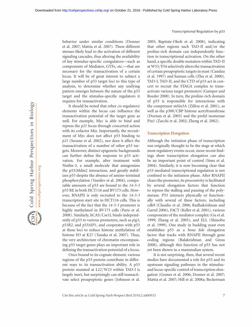
The ability of p53 to stimulate transcriptionon RNA polymerase II (RNAPII)-transcribedgenes is certainly the most well-studied func-tion of the tumor suppressor; a depictionof p53-dependent transactivation is shown inFigure 2. The modification of histones is neces-sary to open up chromatin so that the generaltranscription machinery can bind. In responseto DNA damage, p53 is involved in the recruit-ment of the histone variant H2A.Z, an eventwhich is required for full activation of p21(Gevry et al. 2007). An elegant study has furtherelucidated a role for PRMT1 and CARM1, his-tone methyltransferases (HMTs) that cooperatewith p300/CBP in a p53-dependent fashion,to facilitate transcription on the gadd45 locusafter UV irradiation (An et al. 2004). It is pos-sible that the action of these HMTs is requiredfor full transcriptional activation of other p53-responsive genes as well. However, the mostwell-documented p53-dependent histone modi-fication is acetylation: after p53 has bound toits recognition site, histone acetyltransferases(HATs) such as p300/CBP (Avantaggiati et al.1997; Gu and Roeder 1997; Lill et al. 1997;Scolnick et al. 1997), pCAF (Scolnick et al.1997; Barlev et al. 2001), GCN5 (Candau et al.1997), or TIP60 (Gevry et al. 2007), are re-cruited in a p53-dependent fashion to acetylatethe histones within the vicinity of p53 REs.
The relationship of p53 with its most well-studied HATs, p300 and CBP, is fairly complex(Grossman 2001). As mentioned above, notonly do p300/CBP acetylate histones, they alsoacetylate p53 itself, correlating with an increasein target transactivation (Avantaggiati et al.1997; Gu and Roeder 1997; Scolnick et al.1997). On DNA damage, p53 becomes phos-phorylated in its amino-terminal region, andit has been shown that these damage-inducible
R. Beckerman and C. Prives
4 Cite this article as Cold Spring Harb Perspect Biol 2010;2:a000935
on October 21, 2016 - Published by Cold Spring Harbor Laboratory Press http://cshperspectives.cshlp.org/Downloaded from

modifications enhance p300/CBP-mediatedacetylation at the CTD (Lambert et al. 1998;Sakaguchi et al. 1998). Furthermore, Mdm2,the main negative regulator of p53, can disruptthe interaction of p300 and p53 by competingwith p300 for binding to the NTD of p53(Grossman et al. 1998; Wadgaonkar and Collins1999). Finally, to convolute matters even fur-ther, when complexed with Mdm2, p300/CBPcan also serve as an E4 ubiquitin ligase for p53(Grossman et al. 2003), thus targeting it fordegradation. Taken together, these results dem-onstrate that p300 can paradoxically exert bothpositive and negative control over p53 function,perhaps implying that p300/CBP can switchthe balance from p53 degradation to stabili-zation after DNA damage.
Once chromatin has been modified and re-modeled (Li et al. 2007b), components of thepreinititation complex (PIC) can be recruitedor somehow altered to allow transcription
initiation. TFIID is recruited to the promoter’sTATA region to nucleate the formation ofthe PIC, followed by TFIIB, and finally by theassembly of the other transcription initiationfactors (TFIIF, TFIIE, TFIIH) complexed withunphosphorylated RNAPII (Orphanides et al.1996; Woychik and Hampsey 2002). Adaptorcomplexes such as SAGA and Mediator aidin the recruitment of these general initationfactors to the locus (Thomas and Chiang2006). p53 itself is able to assist in the recruit-ment of various PIC components to thepromoter, including TBP and its associatedfactors (Seto et al. 1992; Chen et al. 1993;Liu et al. 1993; Thut et al. 1995; Farmer et al.1996), as well as TFIIA and TFIIH (Ko andPrives 1996; Xing et al. 2001). At least oneTFIID subunit, TAF1, is recruited in an acety-lated-p53-dependent fashion to the p53 REon p21 before looping to the core promoter(Li et al. 2007a).
RNAPIIEFs
RNAPII
Numerous factors
Various stimuli
GTFs,TAFs
GTFs,TAFs
p53
p53
HATsMediator
RNA initiation
RNA elongation
Various stimuliNumerous factors
TBP
TBPHATs
Mediator
HMTs
HMTs
Figure 2. p53 can modulate transcription intiation and elongation at RNAPII-transcribed loci. p53 can directpreinitiation complex (PIC) assembly at certain target gene promoters under basal conditions, and at othersonly in response to stress. This process involves the ordered recruitment of histone methyltransferases(HMTs), histone acetyltransferase (HATs), and other coregulators in the vicinity of the p53 response element(RE) to open up chromatin so that RNAPII and its associated general transcription factors (GTFs) can bindto the transcription start site of the locus. In response to specific stimuli, p53 can also modulatetranscription elongation via functional and physical interactions with various elongation factors.
Transcriptional Regulation by p53
Cite this article as Cold Spring Harb Perspect Biol 2010;2:a000935 5
on October 21, 2016 - Published by Cold Spring Harbor Laboratory Press http://cshperspectives.cshlp.org/Downloaded from
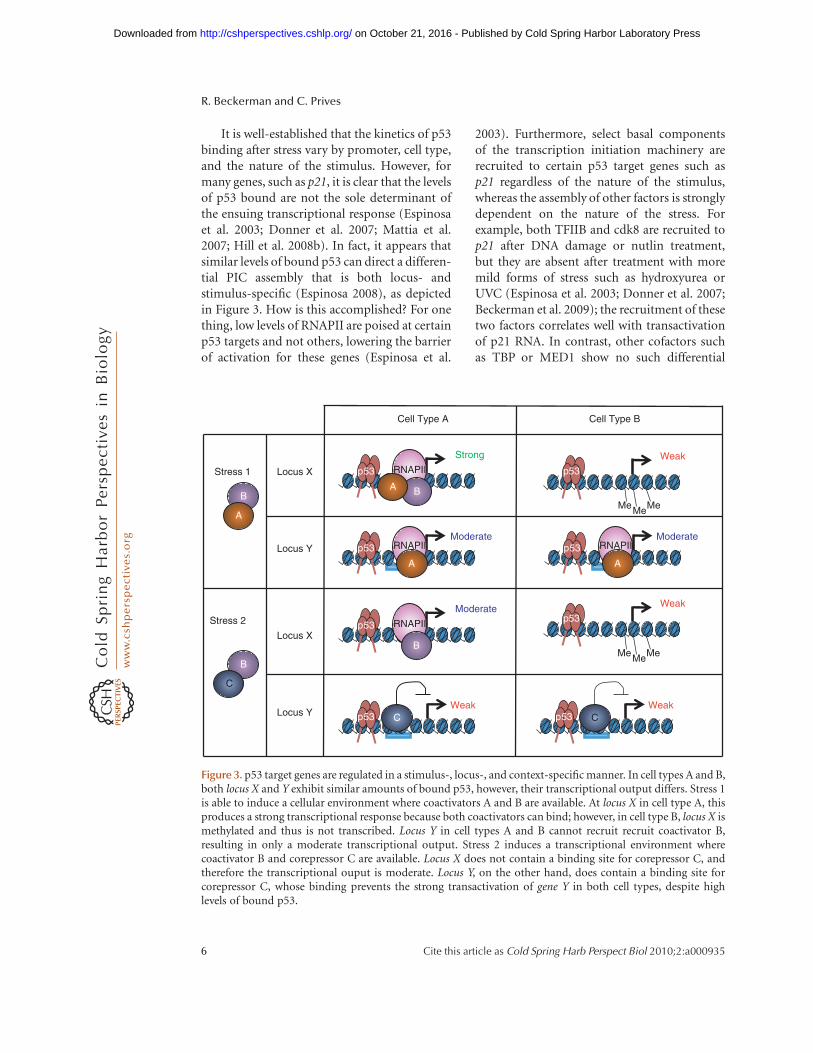
It is well-established that the kinetics of p53binding after stress vary by promoter, cell type,and the nature of the stimulus. However, formany genes, such as p21, it is clear that the levelsof p53 bound are not the sole determinant ofthe ensuing transcriptional response (Espinosaet al. 2003; Donner et al. 2007; Mattia et al.2007; Hill et al. 2008b). In fact, it appears thatsimilar levels of bound p53 can direct a differen-tial PIC assembly that is both locus- andstimulus-specific (Espinosa 2008), as depictedin Figure 3. How is this accomplished? For onething, low levels of RNAPII are poised at certainp53 targets and not others, lowering the barrierof activation for these genes (Espinosa et al.
2003). Furthermore, select basal componentsof the transcription initiation machinery arerecruited to certain p53 target genes such asp21 regardless of the nature of the stimulus,whereas the assembly of other factors is stronglydependent on the nature of the stress. Forexample, both TFIIB and cdk8 are recruited top21 after DNA damage or nutlin treatment,but they are absent after treatment with moremild forms of stress such as hydroxyurea orUVC (Espinosa et al. 2003; Donner et al. 2007;Beckerman et al. 2009); the recruitment of thesetwo factors correlates well with transactivationof p21 RNA. In contrast, other cofactors suchas TBP or MED1 show no such differential
Cell Type B
Stress 1
Stress 2
Locus X
Locus Y
Locus X
Locus Y
B
B
Moderate
B
p53 RNAPII
A
MeMe Me
MeMe Me
Weak
p53
Weak
p53
Cell Type A
B
Strong
p53 RNAPII
Moderate
B
p53 RNAPII
A
Weak
B
p53
B
Moderate
p53 RNAPII
CWeak
B
p53 C
A
C
A
Figure 3. p53 target genes are regulated in a stimulus-, locus-, and context-specific manner. In cell types A and B,both locus X and Y exhibit similar amounts of bound p53, however, their transcriptional output differs. Stress 1is able to induce a cellular environment where coactivators A and B are available. At locus X in cell type A, thisproduces a strong transcriptional response because both coactivators can bind; however, in cell type B, locus X ismethylated and thus is not transcribed. Locus Y in cell types A and B cannot recruit recruit coactivator B,resulting in only a moderate transcriptional output. Stress 2 induces a transcriptional environment wherecoactivator B and corepressor C are available. Locus X does not contain a binding site for corepressor C, andtherefore the transcriptional ouput is moderate. Locus Y, on the other hand, does contain a binding site forcorepressor C, whose binding prevents the strong transactivation of gene Y in both cell types, despite highlevels of bound p53.
R. Beckerman and C. Prives
6 Cite this article as Cold Spring Harb Perspect Biol 2010;2:a000935
on October 21, 2016 - Published by Cold Spring Harbor Laboratory Press http://cshperspectives.cshlp.org/Downloaded from
behavior under similar conditions (Donneret al. 2007; Mattia et al. 2007). These differentstresses likely lead to the activation of differentsignaling cascades, thus altering the availabilityof key stimulus-specific coregulators—such ascomponents of Mediator, GTFs, etc.—that arenecessary for the transactivation of a certainlocus. It will be of great interest to subject alarge number of p53 target loci to this type ofanalysis, to determine whether any unifyingpattern emerges between the nature of the p53target and the stimulus-specific regulators itrequires for transactivation.
It should be noted that other cis-regulatoryelements within the locus can influence thetransactivation potential of the target gene aswell. For example, Myc is able to bind andrepress the p21 locus through concerted actionwith its cofactor Miz. Importantly, the recruit-ment of Myc does not affect p53 binding top21 (Seoane et al. 2002), nor does it affect thetransactivation of a number of other p53 tar-gets. Moreover, distinct epigenetic backgroundscan further define the response to p53 acti-vation. For example, after treatment withNutlin-3, a small molecule that antagonizesthe p53:Mdm2 interaction, and greatly stabil-izes p53 despite the absence of amino-terminalphosphorylation (Vassilev et al. 2004), compa-rable amounts of p53 are bound to the 14-3-3p53 RE in both HCT116 and BV173 cells. How-ever, RNAPII is only recruited to the 14-3-3transcription start site in HCT116 cells. This isbecause of the fact that the 14-3-3 promoter ishighly methylated in BV173 cells (Paris et al.2008). Similarly, hCAS/Cse1L binds independ-ently of p53 to various promoters, such as pig3,p53R2, and p53AIP1, and cooperates with p53at these loci to reduce histone methylation ofhistone H3 at K27 (Tanaka et al. 2007). Thus,the very architecture of chromatin encompass-ing p53 target genes plays an important role indefining the transactivation potential of a locus.
Once bound to its cognate element, variousregions of the p53 protein contribute in differ-ent ways to its transactivation ability. A p53protein mutated at L22/W23 within TAD-I islargely inert, but surprisingly can still transacti-vate select proapoptotic genes (Johnson et al.
2005; Baptiste-Okoh et al. 2008), indicatingthat other regions such TAD-II and/or theproline-rich domain can independently func-tion in transcriptional activation. On the otherhand, a specific double mutation within TAD-IIat W53/F54 selectively alters the transactivationof certain proapoptotic targets in yeast (Candauet al. 1997) and human cells (Zhu et al. 2000).TAD-I, TAD-II, and the CTD of p53 act in con-cert to recruit the STAGA complex to trans-activate various target promoters (Gamper andRoeder 2008). In turn, the proline-rich domainof p53 is responsible for interactions withthe corepressor mSin3A (Zilfou et al. 2001), aswell as the p300/CBP histone acetyltransferase(Dornan et al. 2003) and the prolyl isomerasePin1 (Zacchi et al. 2002; Zheng et al. 2002).
Transcription Elongation
Although the initiation phase of transcriptionwas originally thought to be the stage at whichmost regulatory events occur, more recent find-ings show transcription elongation can alsobe an important point of control (Sims et al.2004). Similarly, it is now becoming clear thatp53-mediated transcriptional regulation is notconfined to the initiation phase. After RNAPIIclears the promoter, its processivity is facilitatedby several elongation factors that functionto repress the stalling and pausing of the poly-merase. P53 interacts physically or function-ally with several of these factors, includingcdk9 (Claudio et al. 2006; Radhakrishnan andGartel 2006), FACT (Keller et al. 2001), variouscomponents of the mediator complex (Gu et al.1999; Zhang et al. 2005), and ELL (Shinobuet al. 1999). One study in budding yeast evenestablishes p53 as a bona fide elongationfactor that tracks with RNAPII through genecoding regions (Balakrishnan and Gross2008), although this function of p53 has notyet been shown in a mammalian system.
It is not surprising, then, that several recentstudies have documented a role for p53 and itsupstream signaling pathways in the stimulus-and locus-specific control of transcription elon-gation (Gomes et al. 2006; Donner et al. 2007;Mattia et al. 2007; Hill et al. 2008a; Beckerman
Transcriptional Regulation by p53
Cite this article as Cold Spring Harb Perspect Biol 2010;2:a000935 7
on October 21, 2016 - Published by Cold Spring Harbor Laboratory Press http://cshperspectives.cshlp.org/Downloaded from

et al. 2009). In the case of chromium exposure,the elongation of p21 but not puma RNA isinhibited, in a process that implicates DNA-PK(Hill et al. 2008b), whereas after replicationstress, p21 elongation is inhibited in a Chk1-dependent fashion (Beckerman et al. 2009).Further work is required to extend these obser-vations to other p53 targets, as the regulationof transcription elongation is emerging as akey layer of control in the fine-tuning of thep53 response.
p53-Mediated Repression
In addition to its well-documented role in tran-scription activation, p53 has also been shownto associate with and repress a wide range oftargets. Although p53-mediated repression isstill poorly understood, several mechanismsfor the process have emerged.
(1) p53 can bind directly to its response ele-ment and recruit corepressors. One such exam-ple involves the recruitment of HDAC1 tospecific promoters (such as map4, p21, stath-min, HSP90-beta, myc, or nanog) via a p53-dependent interaction with mSIN3A (Murphyet al. 1999; Zhang et al. 2004; Ho et al. 2005;Lin et al. 2005) and, in the case of p21 repres-sion, also Zbtb4 (Weber et al. 2008). In this sce-nario, what prompts the bound p53 to promoterepression of the target, rather than its acti-vation? In some cases the answer may lie inthe orientation of the p53 binding site (in whicha “head to tail,” rather than the more common“head to head” orientation correlates with therepression of the target) (Johnson et al. 2001;Godar et al. 2008). The length of the spacerbetween the two half-sites may also contributeto the decision to repress a gene rather than acti-vate it (Hoffman et al. 2002).
(2) p53 can inhibit expression of some genesvia the activation of a repressor protein.The most well-studied example of this is thep53-mediated transactivation of p21 (Lohret al. 2003), which inhibits CDK-dependentphosphorylation of the retinoblastoma protein(Xiong et al. 1993; Niculescu et al. 1998) tokeep E2F-regulated genes in an inactive state
(Delavaine and La Thangue 1999; Harbourand Dean 2000). Another example of this typeof repression is the p53-dependent transactiva-tion of the Slug protein. After high doses ofg-irradiation Slug binds and represses thepuma promoter, preventing apoptosis (Wuet al. 2005).
(3) p53 can bind to its response element andsquelch the activities of another transcriptionalactivator; in other words, p53 binding canocclude the binding of this other, stronger,transcription factor. For example, in responseto hypoxic stress, p53 binds the alpha-fetoprotein promoter (AFP) and displaces thetranscriptional activator HNF3 to repress AFPexpression (Lee et al. 1999). This type of mech-anism has been documented on a number ofother genes, including cdc25C (St Clair et al.2004), HBV (Ori et al. 1998), and pold1 (Liand Lee 2001).
(4) p53 can repress genes that do not con-tain a p53 response element via protein-proteininteractions. The cyclin B2 promoter, for exam-ple, contains NF-Y recognition sites but no p53RE. Promoter-bound NF-Y interacts with p53,and in this case p53 recruits HDAC1 to repressthe cyclin B2 promoter (Imbriano et al. 2005).
Interestingly, in addition to acting on RNA-PII-directed genes, p53 can also direct therepression of RNAPI and RNAPIII promoters.In the case of RNAPI, p53 is able to bind toTAF(I)110, preventing its association with SL1and UBF, therefore repressing transcriptionon rRNA promoters by decreasing the rate ofPIC assembly (Zhai and Comai 2000). In thecase of RNAPIII-based transcription, the NTDof p53 is able to bind and sequester TFIIIB, pre-venting the transcription of 5S RNA and tRNA(Chesnokov et al. 1996; Budde and Grummt1999; Gridasova and Henry 2005; White 2005).
PROMOTER SELECTIVITY WITHIN THE p53TRANSCRIPTIONAL PROGRAM
One of the most intriguing questions in the fieldof p53-mediated transcription is how the pro-tein is able to discriminate between its myriad
R. Beckerman and C. Prives
8 Cite this article as Cold Spring Harb Perspect Biol 2010;2:a000935
on October 21, 2016 - Published by Cold Spring Harbor Laboratory Press http://cshperspectives.cshlp.org/Downloaded from
promoters in response to differing stimuli. Herewe focus on three established mechanisms thatcan contribute to the diversity of the transcrip-tional response: the p53 REs themselves, p53posttranslational modifications, and p53 bind-ing partners.
p53 Response Elements
With such a wide variety of REs in the genome,it is not surprising that p53 has different affinityfor different consensus sequences. In fact, recentwork has shown that tetrameric p53 is capableof binding to half- and even three quarter-consensus sites, such as those comprising thePIDD and Apaf-1 response elements (Jordanet al. 2008). Using a Saccharomyces cerevisiaemodel system, one study found as much as a1000-fold difference in transactivation by p53for its weaker versus its stronger sites, withthis difference being largely dependent on thecentral sequence element in the p53 RE (Ingaet al. 2002). On the whole, cell cycle target genesgenerally contain more robust binding elementsthan do proapoptotic targets (Weinberg et al.2005), perhaps because they are more evolutio-narily conserved (Horvath et al. 2007). Thisdifferential affinity of p53 for its REs allowsthe nuclear concentration of the protein to beone determining factor in how target genes areactivated, and hence also for which cellular out-come occurs after genotoxic stress (Szak et al.2001; Lokshin et al. 2005). The proclivity of ap53 RE to bend may also play a large role indetermining the affinity and stability of p53with its cognate element (Batta and Kundu2007; Pan and Nussinov 2008). Intriguingly, agenome-wide study has shown that not all sitesbound by p53 are in fact transcriptionally acti-vated (Wei et al. 2006a), raising the possibilitythat other coactivators and/or a specific modi-fication status of p53 are needed to inducerobust activation of select p53 target genes(discussed later).
Modifications
p53 is regulated spatially and temporally by manyposttranslational modifications, primarily at the
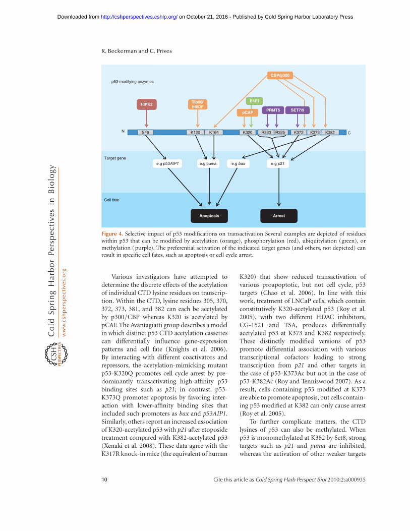
amino- and carboxy-termini, but also withinthe DBD. These modifications integrate the var-ious cell signaling pathways that converge on thep53 protein (Sakaguchi et al. 1998; Bode andDong 2004; Kruse and Gu 2009). Many modifi-cations have been shown to predispose p53 toselectively activate or repress certain targets;however, the precise combinatorial impact ofthe numerous p53 modifications on transacti-vation is only weakly characterized. Here, onlymodifications with a direct, established impacton transcription are discussed (summarizedin Fig. 4).
One modification that has generated muchattention is the phosphorylation of S46. Aftertreating cells with UV, p53 can be phosphory-lated at this residue to specifically induce itsability to transactivate a pro-apoptotic geneAIP-1 (Oda et al. 2000). HIPK2 is the main kin-ase implicated in the modification of this residue(D’Orazi et al. 2002), although AMPK (Okoshiet al. 2008), PKC-d (Yoshida et al. 2006), p38(Perfettini et al. 2005), and DYRK-2 (Taira et al.2007) can also phosphorylate this site. Phos-phorylation of S46 is the earliest and perhapsthe most clear-cut example of a modificationpromoting promoter selectivity by p53.
Furthermore, two groups have shown thatp53 can be acetylated in the DNA-bindingdomain in response to genotoxic stress at eitherK120 or K164 (Sykes et al. 2006; Tang et al. 2006;Tang et al. 2008). The K120 modification iscatalyzed by hMOF or TIP60, and this acety-lation is required for the p53-mediated transac-tivation of the proapoptotic targets puma andbax, at a post-binding step. The K164 residueis modified by p300/CBP and, in concert withthe acetylation of the extreme CTD, is indispen-sible for the activation of most p53 target genes(including p21, pig3, puma, and bax) with theremarkable exception of mdm2. It is possiblethat the unique, nucleosome-free architectureof the mdm2 p53 RE contributes to this distinc-tive phenomenon. To that end, it would beinformative to assess the transactivation capa-bility of the p53-8KR mutant (which abrogatesacetylation on K164, K120, and six lysines in theextreme CTD) on other p53 target genes with asimilar nucleosome-free status, such as pcna.
Transcriptional Regulation by p53
Cite this article as Cold Spring Harb Perspect Biol 2010;2:a000935 9
on October 21, 2016 - Published by Cold Spring Harbor Laboratory Press http://cshperspectives.cshlp.org/Downloaded from
Various investigators have attempted todetermine the discrete effects of the acetylationof individual CTD lysine residues on transcrip-tion. Within the CTD, lysine residues 305, 370,372, 373, 381, and 382 can each be acetylatedby p300/CBP whereas K320 is acetylated bypCAF. The Avantagiatti group describes a modelin which distinct p53 CTD acetylation cassettescan differentially influence gene-expressionpatterns and cell fate (Knights et al. 2006).By interacting with different coactivators andrepressors, the acetylation-mimicking mutantp53-K320Q promotes cell cycle arrest by pre-dominantly transactivating high-affinity p53binding sites such as p21; in contrast, p53-K373Q promotes apoptosis by favoring inter-action with lower-affinity binding sites thatincluded such promoters as bax and p53AIP1.Similarly, others report an increased associationof K320-acetylated p53 with p21 after etoposidetreatment compared with K382-acetylated p53(Xenaki et al. 2008). These data agree with theK317R knock-in mice (the equivalent of human
K320) that show reduced transactivation ofvarious proapoptotic, but not cell cycle, p53targets (Chao et al. 2006). In line with thiswork, treatment of LNCaP cells, which containconstitutively K320-acetylated p53 (Roy et al.2005), with two different HDAC inhibitors,CG-1521 and TSA, produces differentiallyacetylated p53 at K373 and K382 respectively.These distinctly modified versions of p53promote differential association with varioustranscriptional cofactors leading to strongtranscription from p21 and other targets inthe case of p53-K373Ac but not in the case ofp53-K382Ac (Roy and Tenniswood 2007). As aresult, cells containing p53 modified at K373are able to promote apoptosis, but cells contain-ing p53 modified at K382 can only cause arrest(Roy et al. 2005).
To further complicate matters, the CTDlysines of p53 can also be methylated. Whenp53 is monomethylated at K382 by Set8, strongtargets such as p21 and puma are inhibited,whereas the activation of other weaker targets
CBP/p300
Tip60/hMOF
E4F1HIPK2
Apoptosis Arrest
N C
Cell fate
Target gene
p53 modifying enzymes
S46 K120 K164 K320 K372 K373 K382R335R333
pCAF SET7/9PRMT5
e.g p53AIP1 e.g p21e.g puma e.g bax
Figure 4. Selective impact of p53 modifications on transactivation Several examples are depicted of residueswithin p53 that can be modified by acetylation (orange), phosphorylation (red), ubiquitylation (green), ormethylation (purple). The preferential activation of the indicated target genes (and others, not depicted) canresult in specific cell fates, such as apoptosis or cell cycle arrest.
R. Beckerman and C. Prives
10 Cite this article as Cold Spring Harb Perspect Biol 2010;2:a000935
on October 21, 2016 - Published by Cold Spring Harbor Laboratory Press http://cshperspectives.cshlp.org/Downloaded from
remains unaffected (Shi et al. 2007). On theother hand, Set7- and Set9-mediated methyla-tion of Lys372 increases transcription from thep21 locus (Chuikov et al. 2004), and is also animportant step in the acetylation of p53 CTDlysine residues and subsequent stabilization ofthe protein (Ivanov et al. 2007). Adding anotherlayer of complexity, p53 methylation at K372 isrefractory to Smyd2-mediated monomethyla-tion at K370, which itself causes inhibition ofp53 activity (Huang et al. 2006). Dimethylationat K370, however, increases p53 activity bypromoting interaction with its coactivator53BP1. LSD1, a lysine demethylase, can reverseK370 dimethylation, inhibiting this interaction(Huang et al. 2007). Furthermore, argininemethylation of the p53 tetramerization domainat R333, R335 and R337 has been implica-ted in transcriptional control: depletion of thePRMT5 methyltransferase results in reducedtranscription of p21, but not puma or noxa,resulting in increased apoptosis (Jansson et al.2008).
Even ubiquitylation can affect p53’s abilityto regulate transcription. Unlike Mdm2-me-diated monoubiquitylation of p53, which pro-motes export of p53 from the nucleus, the effect
of E4F1-mediated monoubiquitylation at K320of p53 is transcriptional. K320 ubiquitylationpredisposes the p53 protein to transactivate cellcycle targets such as p21 and cyclinG1 while hav-ing no impact on noxa transcription, promptingthe cell to undergo arrest rather than apoptosis(Le Cam et al. 2006).
Clearly, great complexity exists in the worldof p53 posttranslational modifications, convo-luted by the fact that many of the modificationsappear to be stress- and cell-type specific. How-ever, these studies (and others) put forth theexciting idea of a p53 modification code, analo-gous to the histone modification code, wherebydiverse modifications of p53 at specific residuescan direct it toward activating or repressingselect target genes.
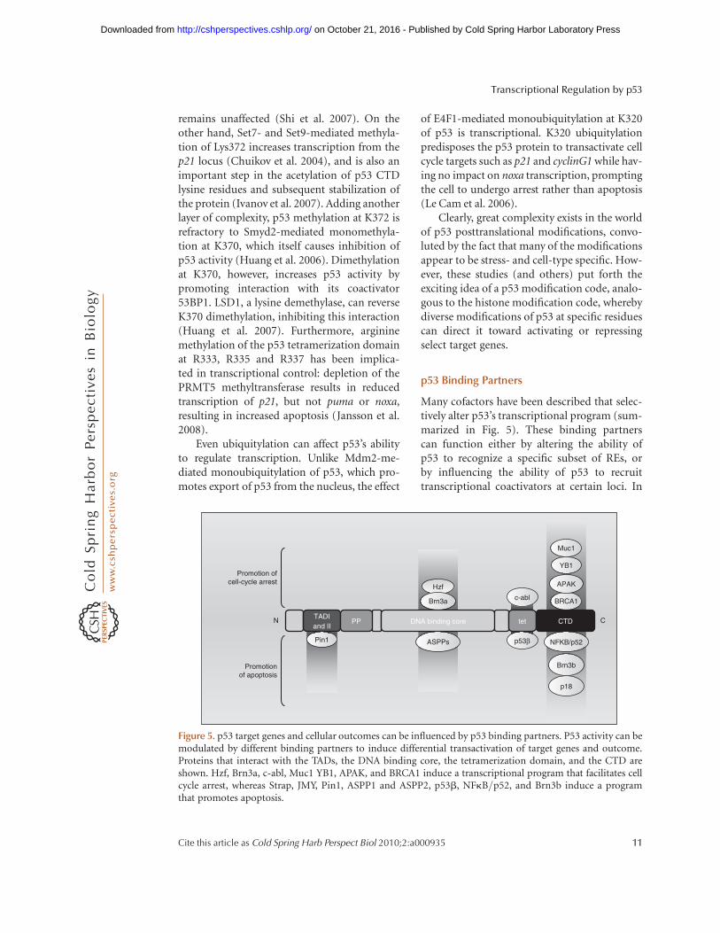
p53 Binding Partners
Many cofactors have been described that selec-tively alter p53’s transcriptional program (sum-marized in Fig. 5). These binding partnerscan function either by altering the ability ofp53 to recognize a specific subset of REs, orby influencing the ability of p53 to recruittranscriptional coactivators at certain loci. In
p18
Hzf
N C
Promotion of cell-cycle arrest
Brn3a
Promotion of apoptosis
c-abl
Muc1
YB1
BRCA1
APAK
TADI and II
PP DNA binding core tet CTD
ASPPs p53β
Brn3b
NFKB/p52Pin1
Figure 5. p53 target genes and cellular outcomes can be influenced by p53 binding partners. P53 activity can bemodulated by different binding partners to induce differential transactivation of target genes and outcome.Proteins that interact with the TADs, the DNA binding core, the tetramerization domain, and the CTD areshown. Hzf, Brn3a, c-abl, Muc1 YB1, APAK, and BRCA1 induce a transcriptional program that facilitates cellcycle arrest, whereas Strap, JMY, Pin1, ASPP1 and ASPP2, p53b, NFkB/p52, and Brn3b induce a programthat promotes apoptosis.
Transcriptional Regulation by p53
Cite this article as Cold Spring Harb Perspect Biol 2010;2:a000935 11
on October 21, 2016 - Published by Cold Spring Harbor Laboratory Press http://cshperspectives.cshlp.org/Downloaded from
particular cases, an interplay exists between themodification status of p53 modification and itsability to selectively interact with a bindingpartner.
One of the more clear-cut examples of co-factor-induced promoter selectivity involvesthe three members of the ASPP family of pro-teins, ASPP1, ASPP2 and iASPP. ASPP1 andASPP2 bind the p53 core domain and predis-pose the cell toward an apoptotic phenotype,by causing transactivation of bax but not p21(Samuels-Lev et al. 2001). On the other hand,iASPP binds to p53 and inhibits the transactiva-tion of proapoptotic genes (Bergamaschi et al.2006). Highlighting the interplay between p53posttranslational modifications and selectivecofactor binding, p53 phosphorylated at S46 isrecognized by the prolyl-isomerase Pin1, andbinding of Pin1 to thus modified p53 leads toits dissociation from iASPP and the consequentinduction of apoptosis (Mantovani et al. 2007).Moreover, a well-studied p53 polymorphism,P72, shows increased association with theiASPP protein when compared with the R72version, perhaps providing insight as to whyR72 is more pro-apoptotic than P72 (Bergama-schi et al. 2006).
Such symmetrical phenotypes are not uni-que to the ASPP family of proteins. An isoformof p53, p53-b, which can form heterotetra-mers with wild-type p53, promotes binding tothe bax but not the p21 RE (Bourdon et al.2005); conversely, phosphorylation of p53 byc-abl stabilizes the homotetramer, and promotesbinding to p21 rather than bax (Wei et al. 2005).Brn3a and Brn3b have similarly opposing phe-notypes: Brn3a influences p53 to transactivatebax and not p21, whereas Brn3b does the reverse(Budhram-Mahadeo et al. 2006).
Certain proteins are able to bind to and pre-dispose p53 exclusively toward an apoptoticprogram. For instance, YB1, a Y-box bindingprotein, interacts with p53 to block the trans-activation of bax, but not p21 (Homer et al.2005). Similarly, the p52 subunit of NF-kB hasbeen shown to inhibit p21 activation, but canalso act in concert with p53 to increase gadd45,puma, and DR5/Killer expression (Schummet al. 2006). Furthermore, the p38-regulated
p18/hamlet protein binds to p53 and showsincreased transactivation of target promoterssuch as noxa, but not bax, p21, or puma, result-ing in an increased apoptotic response (Cua-drado et al. 2007; Lafarga et al. 2007).
On the flip side, other p53 binding partnersselectively promote cell cycle arrest. BRCA1is one such case. By interacting with the p53CTD (Zhang et al. 1998), BRCA1 selectivelyredirects p53 to activate its cell cycle arrest andDNA repair genes (MacLachlan et al. 2002),perhaps by recruiting acetyltransferases suchas CBP/p300 to the locus (Pao et al. 2000).Notably, BRCA1 has also been implicated in sta-bilizing S15-phosphorylated p53 and the sub-sequent maintenance of G1 arrest (Fabbroet al. 2004). Similarly, the membrane glycopro-tein Muc1 is able to bind to and activate p21 in ap53-dependent fashion while at the same timerepressing Bax in a p53-independent manner(Wei et al. 2006b). An analogous function isascribed to Hzf, a zinc-finger protein itselfinduced by p53, which promotes p53 bindingand transactivation of p21 and 14-3-3, butnot bax (Das et al. 2007). APAK, a KRAB-typezinc finger protein, binds p53 under basal con-ditions and prevents the transactivation ofproapoptotic targets (Tian et al. 2009).
Several p53 cofactors, although indeeddisplaying the ability to discriminate betweenpromoters, nonetheless show no clear predis-position toward either pro- or antiapoptotictargets. For instance, it should be noted thatendogenously expressed Mdm2 can be foundat promoters such as p21 (Minsky and Oren2004; Arva et al. 2005; Ohkubo et al. 2006;White et al. 2006; Tang et al. 2008), negativelyinfluencing p53’s transcriptional output. Themechanism by which this occurs is unclear,but perhaps Mdm2 functions through recruit-ment of the histone deacetylases KAP1 (Wanget al. 2005) and HDAC1 (Ito et al. 2002), the dis-ruption of HAT interactions with p53 (Ito et al.2002; Jin et al. 2004), or the monoubiquityla-tion of histone H2B within the vicinity of thep53 RE (Minsky and Oren 2004). Notably, arole for a Mdm2/Rb/p53 trimeric complex hasbeen described in transrepressing pro-apoptotictargets (Hsieh et al. 1999). Interestingly, when
R. Beckerman and C. Prives
12 Cite this article as Cold Spring Harb Perspect Biol 2010;2:a000935
on October 21, 2016 - Published by Cold Spring Harbor Laboratory Press http://cshperspectives.cshlp.org/Downloaded from
both Mdm2 and MdmX are overexpressed, theycan bind many p53 target promoters with thenotable exception of Mdm2 gene itself.Although the precise influence of MdmX pro-moter binding on p53 transcriptional outputis unclear (Tang et al. 2008), it remains to beseen whether the disruption of the p53/Mdm2 interaction at the promoter is requiredfor the antirepression of certain target genes.
Although it is tempting to invoke cofactorsto explain p53’s decision to effectuate cell cyclearrest or apoptosis, we must keep in mind thatfor most binding partners, simply too few tar-gets have been looked at to draw any firmconclusions to this end. It is likely that as theinfluence of these p53-interacting proteins isassessed on more targets, their ability to strictlydiscriminate between pro- and antiapoptotictargets genes may have to be refined.
CONCLUDING REMARKS
Even more than 20 years on, our understandingof p53-mediated transcription continues togrow by leaps and bounds. It is now clear thep53 can both activate and repress its targetgenes; that p53’s regulatory influence extendsnot just to transcription initiation, but also toelongation; and that several features—includingbut not limited to the epigenetic landscape ofthe locus, p53 posttranslational modifications,the nature of the p53 RE, and p53-interactingpartners – function in concert to determine thespecificity of the p53 transcriptional response.Although it will take a massive effort to integratethe individual effects of each of these factors intoa coherent model for transcriptional regulationby p53, great insight into cancer therapy willundoubtedly be gained from doing so.
ACKNOWLEDGMENTS
This review was written under the auspices ofCA77742 and CA87497; RB is supported by aCanadian postgraduate scholarship from NSERC.
REFERENCES
An W, Kim J, Roeder RG. 2004. Ordered cooperative func-tions of PRMT1, p300, and CARM1 in transcriptionalactivation by p53. Cell 117: 735–748.
Arva NC, Gopen TR, Talbott KE, Campbell LE, Chicas A,White DE, Bond GL, Levine AJ, Bargonetti J. 2005. Achromatin-associated and transcriptionally inactivep53-Mdm2 complex occurs in mdm2 SNP309 homo-zygous cells. J Biol Chem 280: 26776–26787.
Avantaggiati ML, Ogryzko V, Gardner K, Giordano A, Lev-ine AS, Kelly K. 1997. Recruitment of p300/CBP inp53-dependent signal pathways. Cell 89: 1175–1184.
Balagurumoorthy P, Sakamoto H, Lewis MS, Zambrano N,Clore GM, Gronenborn AM, Appella E, Harrington RE.1995. Four p53 DNA-binding domain peptides bind nat-ural p53-response elements and bend the DNA. Proc NatlAcad Sci 92: 8591–8595.
Balakrishnan SK, Gross DS. 2008. The tumor suppressorp53 associates with gene coding regions and co-traverseswith elongating RNA polymerase II in an in vivo model.Oncogene 27: 2661–2672.
Baptiste-Okoh N, Barsotti AM, Prives C. 2008. A role forcaspase 2 and PIDD in the process of p53-mediatedapoptosis. Proc Natl Acad Sci 105: 1937–1942.
Barlev NA, Liu L, Chehab NH, Mansfield K, Harris KG,Halazonetis TD, Berger SL. 2001. Acetylation of p53 acti-vates transcription through recruitment of coactivators/histone acetyltransferases. Mol Cell 8: 1243–1254.
Batta K, Kundu TK. 2007. Activation of p53 function byhuman transcriptional coactivator PC4: Role of protein-protein interaction, DNA bending, and posttranslationalmodifications. Mol Cell Biol 27: 7603–7614.
Beckerman R, Donner AJ, Mattia M, Peart MJ, Manley JL,Espinosa JM, Prives C. 2009. A role for Chk1 in blockingtranscriptional elongation of p21 RNA during theS-phase checkpoint. Genes Develop 23: 1364–1377.
Bergamaschi D, Samuels Y, Sullivan A, Zvelebil M, BreyssensH, Bisso A, Del Sal G, Syed N, Smith P, Gasco M, et al.2006. iASPP preferentially binds p53 proline-richregion and modulates apoptotic function of codon72-polymorphic p53. Nat Gen 38: 1133–1141.
Bertrand P, Saintigny Y, Lopez BS. 2004. p53’s double life:Transactivation-independent repression of homologousrecombination. Trends Genet 20: 235–243.
Bode AM, Dong Z. 2004. Posttranslational modification ofp53 in tumorigenesis. Nature reviews 4: 793–805.
Bourdon JC, Fernandes K, Murray-Zmijewski F, Liu G, DiotA, Xirodimas DP, Saville MK, Lane DP. 2005. p53 iso-forms can regulate p53 transcriptional activity. GenesDevelop 19: 2122–2137.
Budde A, Grummt I. 1999. p53 represses ribosomal genetranscription. Oncogene 18: 1119–1124.
Budhram-Mahadeo VS, Bowen S, Lee S, Perez-Sanchez C,Ensor E, Morris PJ, Latchman DS. 2006. Brn-3b enhancesthe pro-apoptotic effects of p53 but not its induction ofcell cycle arrest by cooperating in trans-activation ofbax expression. Nucleic Acids Res 34: 6640–6652.
Candau R, Scolnick DM, Darpino P, Ying CY, HalazonetisTD, Berger SL. 1997. Two tandem and independent sub-activation domains in the amino terminus of p53 requirethe adaptor complex for activity. Oncogene 15: 807–816.
Chao C, Wu Z, Mazur SJ, Borges H, Rossi M, Lin T, Wang JY,Anderson CW, Appella E, Xu Y. 2006. Acetylationof mouse p53 at lysine 317 negatively regulates p53
Transcriptional Regulation by p53
Cite this article as Cold Spring Harb Perspect Biol 2010;2:a000935 13
on October 21, 2016 - Published by Cold Spring Harbor Laboratory Press http://cshperspectives.cshlp.org/Downloaded from
apoptotic activities after DNA damage. Mol Cell Biol 26:6859–6869.
Chen X, Farmer G, Zhu H, Prywes R, Prives C. 1993. Coop-erative DNA binding of p53 with TFIID (TBP): A possi-ble mechanism for transcriptional activation. GenesDevelop 7: 1837–1849.
Chesnokov I, Chu WM, Botchan MR, Schmid CW. 1996.p53 inhibits RNA polymerase III-directed transcriptionin a promoter-dependent manner. Mol Cell Biol 16:7084–7088.
Chuikov S, Kurash JK, Wilson JR, Xiao B, Justin N, IvanovGS, McKinney K, Tempst P, Prives C, Gamblin SJ, et al.2004. Regulation of p53 activity through lysine methyla-tion. Nature 432: 353–360.
Claudio PP, Cui J, Ghafouri M, Mariano C, White MK, SafakM, Sheffield JB, Giordano A, Khalili K, Amini S, et al.2006. Cdk9 phosphorylates p53 on serine 392 independ-ently of CKII. J Cell Physiol 208: 602–612.
Contente A, Dittmer A, Koch MC, Roth J, Dobbelstein M.2002. A polymorphic microsatellite that mediates induc-tion of PIG3 by p53. Nat Gen 30: 315–320.
Cuadrado A, Lafarga V, Cheung PC, Dolado I, Llanos S,Cohen P, Nebreda AR. 2007. A new p38 MAP kinase-regulated transcriptional coactivator that stimulatesp53-dependent apoptosis. EMBO J 26: 2115–2126.
D’Orazi G, Cecchinelli B, Bruno T, Manni I, Higashimoto Y,Saito S, Gostissa M, Coen S, Marchetti A, Del Sal G, et al.2002. Homeodomain-interacting protein kinase-2 phos-phorylates p53 at Ser 46 and mediates apoptosis. Nat CellBiol 4: 11–19.
Das S, Raj L, Zhao B, Kimura Y, Bernstein A, Aaronson SA,Lee SW. 2007. Hzf Determines cell survival upon geno-toxic stress by modulating p53 transactivation. Cell 130:624–637.
Delavaine L, La Thangue NB. 1999. Control of E2F activityby p21Waf1/Cip1. Oncogene 18: 5381–5392.
Donner AJ, Szostek S, Hoover JM, Espinosa JM. 2007.CDK8 is a stimulus-specific positive coregulator of p53target genes. Mol Cell 27: 121–133.
Dornan D, Shimizu H, Burch L, Smith AJ, Hupp TR. 2003.The proline repeat domain of p53 binds directly to thetranscriptional coactivator p300 and allosterically con-trols DNA-dependent acetylation of p53. Mol Cell Biol23: 8846–8861.
el-Deiry WS, Kern SE, Pietenpol JA, Kinzler KW, VogelsteinB. 1992. Definition of a consensus binding site for p53.Nat Gen 1: 45–49.
Espinosa JM. 2008. Mechanisms of regulatory diversitywithin the p53 transcriptional network. Oncogene 27:4013–4023.
Espinosa JM, Emerson BM. 2001. Transcriptional regula-tion by p53 through intrinsic DNA/chromatin bindingand site-directed cofactor recruitment. Mol Cell 8:57–69.
Espinosa JM, Verdun RE, Emerson BM. 2003. p53 functionsthrough stress- and promoter-specific recruitment oftranscription initiation components before and afterDNA damage. Mol Cell 12: 1015–1027.
Ewen ME, Miller SJ. 1996. p53 and translational control.Biochim Biophys Acta 1242: 181–184.
Fabbro M, Savage K, Hobson K, Deans AJ, Powell SN,McArthur GA, Khanna KK. 2004. BRCA1-BARD1 com-plexes are required for p53Ser-15 phosphorylation and aG1/S arrest following ionizing radiation-induced DNAdamage. J Biol Chem 279: 31251–31258.
Farmer G, Colgan J, Nakatani Y, Manley JL, Prives C. 1996.Functional interaction between p53, the TATA-bindingprotein (TBP), and TBP-associated factors in vivo. MolCell Biol 16: 4295–4304.
Funk WD, Pak DT, Karas RH, Wright WE, Shay JW. 1992. Atranscriptionally active DNA-binding site for human p53protein complexes. Mol Cell Biol 12: 2866–2871.
Gamper AM, Roeder RG. 2008. Multivalent binding of p53to the STAGA complex mediates coactivator recruitmentafter UV damage. Mol Cell Biol 28: 2517–2527.
Gevry N, Chan HM, Laflamme L, Livingston DM, Gau-dreau L. 2007. p21 transcription is regulated by differen-tial localization of histone H2A.Z. Genes Develop 21:1869–1881.
Godar S, Ince TA, Bell GW, Feldser D, Donaher JL, Bergh J,Liu A, Miu K, Watnick RS, Reinhardt F, et al. 2008.Growth-inhibitory and tumor- suppressive functions ofp53 depend on its repression of CD44 expression. Cell134: 62–73.
Gohler T, Reimann M, Cherny D, Walter K, Warnecke G,Kim E, Deppert W. 2002. Specific interaction of p53with target binding sites is determined by DNA confor-mation and is regulated by the C-terminal domain. JBiol Chem 277: 41192–41203.
Gomes NP, Bjerke G, Llorente B, Szostek SA, Emerson BM,Espinosa JM. 2006. Gene-specific requirement forP-TEFb activity and RNA polymerase II phosphorylationwithin the p53 transcriptional program. Genes Develop20: 601–612.
Gridasova AA, Henry RW. 2005. The p53 tumor suppressorprotein represses human snRNA gene transcription byRNA polymerases II and III independently of sequence-specific DNA binding. Mol Cell Biol 25: 3247–3260.
Grossman SR. 2001. p300/CBP/p53 interaction and regula-tion of the p53 response. Eur J Biochem FEBS 268:2773–2778.
Grossman SR, Deato ME, Brignone C, Chan HM, Kung AL,Tagami H, Nakatani Y, Livingston DM. 2003. Polyubiqui-tination of p53 by a ubiquitin ligase activity of p300.Science 300: 342–344.
Grossman SR, Perez M, Kung AL, Joseph M, Mansur C, XiaoZX, Kumar S, Howley PM, Livingston DM. 1998. p300/MDM2 complexes participate in MDM2-mediated p53degradation. Mol Cell 2: 405–415.
Gu W, Roeder RG. 1997. Activation of p53 sequence-specificDNA binding by acetylation of the p53 C-terminaldomain. Cell 90: 595–606.
Gu W, Malik S, Ito M, Yuan CX, Fondell JD, Zhang X, Mar-tinez E, Qin J, Roeder RG. 1999. A novel human SRB/MED-containing cofactor complex, SMCC, involved intranscription regulation. Mol Cell 3: 97–108.
Hainaut P, Milner J. 1993. A structural role for metal ions inthe “wild-type” conformation of the tumor suppressorprotein p53. Cancer Res 53: 1739–1742.
Halazonetis TD, Kandil AN. 1993. Conformational shiftspropagate from the oligomerization domain of p53 to
R. Beckerman and C. Prives
14 Cite this article as Cold Spring Harb Perspect Biol 2010;2:a000935
on October 21, 2016 - Published by Cold Spring Harbor Laboratory Press http://cshperspectives.cshlp.org/Downloaded from
its tetrameric DNA binding domain and restore DNAbinding to select p53 mutants. EMBO J 12: 5057–5064.
Harbour JW, Dean DC. 2000. The Rb/E2F pathway:Expanding roles and emerging paradigms. Genes Develop14: 2393–2409.
Hill R, Leidal AM, Madureira PA, Gillis LD, Cochrane HK,Waisman DM, Chiu A, Lee PW. 2008a. Hypersensitivityto chromium-induced DNA damage correlates withconstitutive deregulation of upstream p53 kinases inp21-/-HCT116 colon cancer cells. DNA Repair 7:239–252.
Hill R, Leidal AM, Madureira PA, Gillis LD, Waisman DM,Chiu A, Lee PW. 2008b. Chromium-mediated apoptosis:Involvement of DNA-dependent protein kinase (DNA-PK) and differential induction of p53 target genes.DNA Repair 7: 1484–1499.
Ho J, Benchimol S. 2003. Transcriptional repression medi-ated by the p53 tumour suppressor. Cell Death Differen-tiation 10: 404–408.
Ho JS, Ma W, Mao DY, Benchimol S. 2005. p53-Dependenttranscriptional repression of c-myc is required for G1 cellcycle arrest. Mol Cell Biol 25: 7423–7431.
Hoffman WH, Biade S, Zilfou JT, Chen J, Murphy M. 2002.Transcriptional repression of the anti-apoptotic survivingene by wild type p53. J Biol Chem 277: 3247–3257.
Homer C, Knight DA, Hananeia L, Sheard P, Risk J, LashamA, Royds JA, Braithwaite AW. 2005. Y-box factor YB1 con-trols p53 apoptotic function. Oncogene 24: 8314–8325.
Horvath MM, Wang X, Resnick MA, Bell DA. 2007. Diver-gent evolution of human p53 binding sites: Cell cycleversus apoptosis. PLoS Genet 3: e127.
Hsieh JK, Chan FS, O’Connor DJ, Mittnacht S, Zhong S, LuX. 1999. RB regulates the stability and the apoptotic func-tion of p53 via MDM2. Mol Cell 3: 181–193.
Huang J, Perez-Burgos L, Placek BJ, Sengupta R, Richter M,Dorsey JA, Kubicek S, Opravil S, Jenuwein T, Berger SL.2006. Repression of p53 activity by Smyd2-mediatedmethylation. Nature 444: 629–632.
Huang J, Sengupta R, Espejo AB, Lee MG, Dorsey JA,Richter M, Opravil S, Shiekhattar R, Bedford MT, Jenu-wein T, et al. 2007. p53 is regulated by the lysine demethy-lase LSD1. Nature 449: 105–108.
Hublitz P, Kunowska N, Mayer UP, Muller JM, Heyne K, YinN, Fritzsche C, Poli C, Miguet L, Schupp IW, et al. 2005.NIR is a novel INHAT repressor that modulates the tran-scriptional activity of p53. Genes Develop 19: 2912–2924.
Hupp TR, Lane DP. 1994. Allosteric activation of latent p53tetramers. Curr Biol 4: 865–875.
Imbriano C, Gurtner A, Cocchiarella F, Di Agostino S, BasileV, Gostissa M, Dobbelstein M, Del Sal G, Piaggio G, Man-tovani R. 2005. Direct p53 transcriptional repression: Invivo analysis of CCAAT-containing G2/M promoters.Mol Cell Biol 25: 3737–3751.
Inga A, Storici F, Darden TA, Resnick MA. 2002. Differentialtransactivation by the p53 transcription factor is highlydependent on p53 level and promoter target sequence.Mol Cell Biol 22: 8612–8625.
Ito A, Kawaguchi Y, Lai CH, Kovacs JJ, Higashimoto Y,Appella E, Yao TP. 2002. MDM2-HDAC1-mediateddeacetylation of p53 is required for its degradation.EMBO J 21: 6236–6245.
Ivanov GS, Ivanova T, Kurash J, Ivanov A, Chuikov S,Gizatullin F, Herrera-Medina EM, Rauscher F 3rd, Rein-berg D, Barlev NA. 2007. Methylation-acetylation inter-play activates p53 in response to DNA damage. Mol CellBiol 27: 6756–6769.
Jansson M, Durant ST, Cho EC, Sheahan S, Edelmann M,Kessler B, La Thangue NB. 2008. Arginine methylationregulates the p53 response. Nat Cell Biol 10: 1431–1439.
Jayaraman L, Murthy KG, Zhu C, Curran T, Xanthoudakis S,Prives C. 1997. Identification of redox/repair proteinRef-1 as a potent activator of p53. Genes Develop 11:558–570.
Jin Y, Zeng SX, Lee H, Lu H. 2004. MDM2 mediates p300/CREB-binding protein-associated factor ubiquitinationand degradation. J Biol Chem 279: 20035–20043.
Johnson RA, Ince TA, Scotto KW. 2001. Transcriptionalrepression by p53 through direct binding to a novelDNA element. J Biol Chem 276: 27716–27720.
Johnson TM, Hammond EM, Giaccia A, Attardi LD. 2005.The p53QS transactivation-deficient mutant showsstress-specific apoptotic activity and induces embryoniclethality. Nat Gen 37: 145–152.
Jordan JJ, Menendez D, Inga A, Noureddine M, Bell DA,Resnick MA. 2008. Noncanonical DNA motifs as transac-tivation targets by wild type and mutant p53. PLoS Genet4: e1000104.
Keller DM, Zeng X, Wang Y, Zhang QH, Kapoor M, Shu H,Goodman R, Lozano G, Zhao Y, Lu H. 2001. A DNAdamage-induced p53 serine 392 kinase complex containsCK2, hSpt16, and SSRP1. Mol Cell 7: 283–292.
Kitayner M, Rozenberg H, Kessler N, Rabinovich D, ShaulovL, Haran TE, Shakked Z. 2006. Structural basis of DNArecognition by p53 tetramers. Mol Cell 22: 741–753.
Knights CD, Catania J, Giovanni SD, Muratoglu S, Perez R,Swartzbeck A, Quong AA, Zhang X, Beerman T, PestellRG, et al. 2006. Distinct p53 acetylation cassettes differ-entially influence gene-expression patterns and cell fate.J Cell Biol 173: 533–544.
Ko LJ, Prives C. 1996. p53: Puzzle and paradigm. GenesDevelop 10: 1054–1072.
Kruse JP, Gu W. 2009. Modes of p53 regulation. Cell 137:609–622.
Lafarga V, Cuadrado A, Nebreda AR. 2007. p18(Hamlet)mediates different p53-dependent responses to DNA-damage inducing agents. Cell Cycle 6: 2319–2322.
Lambert PF, Kashanchi F, Radonovich MF, Shiekhattar R,Brady JN. 1998. Phosphorylation of p53 serine 15increases interaction with CBP. J Biol Chem 273:33048–33053.
Laptenko O, Prives C. 2006. Transcriptional regulation byp53: One protein, many possibilities. Cell Death Differ13: 951–961.
Le Cam L, Linares LK, Paul C, Julien E, Lacroix M, Hatchi E,Triboulet R, Bossis G, Shmueli A, Rodriguez MS, et al.2006. E4F1 is an atypical ubiquitin ligase that modulatesp53 effector functions independently of degradation. Cell127: 775–788.
Lee KC, Crowe AJ, Barton MC. 1999. p53-mediated repres-sion of alpha-fetoprotein gene expression by specificDNA binding. Mol Cell Biol 19: 1279–1288.
Transcriptional Regulation by p53
Cite this article as Cold Spring Harb Perspect Biol 2010;2:a000935 15
on October 21, 2016 - Published by Cold Spring Harbor Laboratory Press http://cshperspectives.cshlp.org/Downloaded from
Li B, Lee MY. 2001. Transcriptional regulation of the humanDNA polymerase delta catalytic subunit gene POLD1by p53 tumor suppressor and Sp1. J Biol Chem 276:29729–29739.
Li B, Carey M, Workman JL. 2007b. The role of chromatinduring transcription. Cell 128: 707–719.
Li AG, Piluso LG, Cai X, Gadd BJ, Ladurner AG, Liu X.2007a. An acetylation switch in p53 mediates holo-TFIIDrecruitment. Mol Cell 28: 408–421.
Lill NL, Grossman SR, Ginsberg D, DeCaprio J, LivingstonDM. 1997. Binding and modulation of p53 by p300/CBP coactivators. Nature 387: 823–827.
Liu Y, Kulesz-Martin MF. 2006. Sliding into home: Facili-tated p53 search for targets by the basic DNA bindingdomain. Cell Death and Differentiation 13: 881–884.
Lin T, Chao C, Saito S, Mazur SJ, Murphy ME, Appella E, XuY. 2005. p53 induces differentiation of mouse embryonicstem cells by suppressing Nanog expression. Nat Cell Biol7: 165–171.
Liu L, Scolnick DM, Trievel RC, Zhang HB, Marmorstein R,Halazonetis TD, Berger SL. 1999. p53 sites acetylated invitro by PCAF and p300 are acetylated in vivo in responseto DNA damage. Mol Cell Biol 19: 1202–1209.
Liu X, Miller CW, Koeffler PH, Berk AJ. 1993. The p53 acti-vation domain binds the TATA box-binding polypeptidein Holo-TFIID, and a neighboring p53 domain inhibitstranscription. Mol Cell Biol 13: 3291–3300.
Lohr K, Moritz C, Contente A, Dobbelstein M. 2003. p21/CDKN1A mediates negative regulation of transcriptionby p53. J Biol Chem 278: 32507–32516.
Lokshin M, Tanaka T, Prives C. 2005. Transcriptional regu-lation by p53 and p73. Cold Spring Harb Symp Quant Biol70: 121–128.
Luo J, Li M, Tang Y, Laszkowska M, Roeder RG, Gu W. 2004.Acetylation of p53 augments its site-specific DNA bind-ing both in vitro and in vivo. Proc Natl Acad Sci 101:2259–2264.
MacLachlan TK, Takimoto R, El-Deiry WS. 2002. BRCA1directs a selective p53-dependent transcriptionalresponse towards growth arrest and DNA repair targets.Mol Cell Biol 22: 4280–4292.
Mantovani F, Tocco F, Girardini J, Smith P, Gasco M, Lu X,Crook T, Del Sal G. 2007. The prolyl isomerase Pin1orchestrates p53 acetylation and dissociation from theapoptosis inhibitor iASPP. Nat Struct Mol Biol 14:912–920.
Mattia M, Gottifredi V, McKinney K, Prives C. 2007. p53-Dependent p21 mRNA elongation is impaired whenDNA replication is stalled. Mol Cell Biol 27: 1309–1320.
Mazur SJ, Sakaguchi K, Appella E, Wang XW, Harris CC,Bohr VA. 1999. Preferential binding of tumor suppressorp53 to positively or negatively supercoiled DNA involvesthe C-terminal domain. J Mol Biol 292: 241–249.
McKinney K, Prives C. 2002. Efficient specific DNA bindingby p53 requires both its central and C-terminal domainsas revealed by studies with high-mobility group 1 protein.Mol Cell Biol 22: 6797–6808.
McKinney K, Mattia M, Gottifredi V, Prives C. 2004. p53 lin-ear diffusion along DNA requires its C terminus. Mol Cell16: 413–424.
McLure KG, Lee PW. 1998. How p53 binds DNA as atetramer. EMBO J 17: 3342–3350.
Minsky N, Oren M. 2004. The RING domain of Mdm2mediates histone ubiquitylation and transcriptionalrepression. Mol Cell 16: 631–639.
Mirza A, McGuirk M, Hockenberry TN, Wu Q, Ashar H,Black S, Wen SF, Wang L, Kirschmeier P, Bishop WR,et al. 2002. Human survivin is negatively regulated bywild-type p53 and participates in p53-dependent apop-totic pathway. Oncogene 21: 2613–2622.
Murphy M, Ahn J, Walker KK, Hoffman WH, Evans RM,Levine AJ, George DL. 1999. Transcriptional repressionby wild-type p53 utilizes histone deacetylases, mediatedby interaction with mSin3a. Genes Develop 13: 2490–2501.
Murray-Zmijewski F, Slee EA, Lu X. 2008. A complex bar-code underlies the heterogeneous response of p53 tostress. Nat Rev 9: 702–712.
Nagaich AK, Appella E, Harrington RE. 1997a. DNA bend-ing is essential for the site-specific recognition of DNAresponse elements by the DNA binding domain of thetumor suppressor protein p53. J Biol Chem 272:14842–14849.
Nagaich AK, Zhurkin VB, Durell SR, Jernigan RL, Appella E,Harrington RE. 1999. p53-induced DNA bending andtwisting: p53 tetramer binds on the outer side of aDNA loop and increases DNA twisting. Proc Natl AcadSci 96: 1875–1880.
Nagaich AK, Zhurkin VB, Sakamoto H, Gorin AA, CloreGM, Gronenborn AM, Appella E, Harrington RE.1997b. Architectural accommodation in the complex offour p53 DNA binding domain peptides with the p21/waf1/cip1 DNA response element. J Biol Chem 272:14830–14841.
Niculescu AB 3rd, Chen X, Smeets M, Hengst L, Prives C,Reed SI. 1998. Effects of p21(Cip1/Waf1) at both theG1/S and the G2/M cell cycle transitions: pRb is a criticaldeterminant in blocking DNA replication and in prevent-ing endoreduplication. Mol Cell Biol 18: 629–643.
Oda K, Arakawa H, Tanaka T, Matsuda K, Tanikawa C, MoriT, Nishimori H, Tamai K, Tokino T, Nakamura Y, et al.2000. p53AIP1, a potential mediator of p53-dependentapoptosis, and its regulation by Ser-46-phosphorylatedp53. Cell 102: 849–862.
Ohkubo S, Tanaka T, Taya Y, Kitazato K, Prives C. 2006.Excess HDM2 impacts cell cycle and apoptosis and hasa selective effect on p53-dependent transcription. J BiolChem 281: 16943–16950.
Okoshi R, Ozaki T, Yamamoto H, Ando K, Koida N, Ono S,Koda T, Kamijo T, Nakagawara A, Kizaki H. 2008. Acti-vation of AMP-activated protein kinase inducesp53-dependent apoptotic cell death in response to ener-getic stress. J Biol Chem 283: 3979–3987.
Olivier M, Eeles R, Hollstein M, Khan MA, Harris CC, Hai-naut P. 2002. The IARC TP53 database: New online muta-tion analysis and recommendations to users. Hum Mutat19: 607–614.
Ori A, Zauberman A, Doitsh G, Paran N, Oren M, Shaul Y.1998. p53 binds and represses the HBV enhancer: Anadjacent enhancer element can reverse the transcriptioneffect of p53. EMBO J 17: 544–553.
R. Beckerman and C. Prives
16 Cite this article as Cold Spring Harb Perspect Biol 2010;2:a000935
on October 21, 2016 - Published by Cold Spring Harbor Laboratory Press http://cshperspectives.cshlp.org/Downloaded from
Orphanides G, Lagrange T, Reinberg D. 1996. The generaltranscription factors of RNA polymerase II. GenesDevelop 10: 2657–2683.
Palecek E, Vlk D, Stankova V, Brazda V, Vojtesek B, HuppTR, Schaper A, Jovin TM. 1997. Tumor suppressor pro-tein p53 binds preferentially to supercoiled DNA. Onco-gene 15: 2201–2209.
Pan Y, Nussinov R. 2008. p53-Induced DNA bending: Theinterplay between p53-DNA and p53-p53 interactions.J Phys Chem B 112: 6716–6724.
Pao GM, Janknecht R, Ruffner H, Hunter T, Verma IM.2000. CBP/p300 interact with and function as transcrip-tional coactivators of BRCA1. Proc Natl Acad Sci 97:1020–1025.
Paris R, Henry RE, Stephens SJ, McBryde M, Espinosa JM.2008. Multiple p53-independent gene silencing mecha-nisms define the cellular response to p53 activation.Cell Cycle 7: 2427–2433.
Perfettini JL, Castedo M, Nardacci R, Ciccosanti F, Boya P,Roumier T, Larochette N, Piacentini M, Kroemer G.2005. Essential role of p53 phosphorylation by p38MAPK in apoptosis induction by the HIV-1 envelope. JExp Med 201: 279–289.
Radhakrishnan SK, Gartel AL. 2006. CDK9 phosphorylatesp53 on serine residues 33, 315 and 392. Cell Cycle 5:519–521.
Riley T, Sontag E, Chen P, Levine A. 2008. Transcriptionalcontrol of human p53-regulated genes. Nat Rev MolCell Biol 9: 402–412.
Roy S, Tenniswood M. 2007. Site-specific acetylation of p53directs selective transcription complex assembly. J BiolChem 282: 4765–4771.
Roy S, Packman K, Jeffrey R, Tenniswood M. 2005. Histonedeacetylase inhibitors differentially stabilize acetylatedp53 and induce cell cycle arrest or apoptosis in prostatecancer cells. Cell Death Differ 12: 482–491.
Sakaguchi K, Herrera JE, Saito S, Miki T, Bustin M, VassilevA, Anderson CW, Appella E. 1998. DNA damage activatesp53 through a phosphorylation-acetylation cascade.Genes Develop 12: 2831–2841.
Samuels-Lev Y, O’Connor DJ, Bergamaschi D, Trigiante G,Hsieh JK, Zhong S, Campargue I, Naumovski L, CrookT, Lu X. 2001. ASPP proteins specifically stimulate theapoptotic function of p53. Mol Cell 8: 781–794.
Schumm K, Rocha S, Caamano J, Perkins ND. 2006. Regu-lation of p53 tumour suppressor target gene expressionby the p52 NF-kappaB subunit. EMBO J 25: 4820–4832.
Scolnick DM, Chehab NH, Stavridi ES, Lien MC, Caruso L,Moran E, Berger SL, Halazonetis TD. 1997. CREB-binding protein and p300/CBP-associated factor aretranscriptional coactivators of the p53 tumor suppressorprotein. Cancer Res 57: 3693–3696.
Seo YR, Kelley MR, Smith ML. 2002. Selenomethionine reg-ulation of p53 by a ref1-dependent redox mechanism.Proc Natl Acad Sci 99: 14548–14553.
Seoane J, Le HV, Massague J. 2002. Myc suppression of thep21(Cip1) Cdk inhibitor influences the outcome of thep53 response to DNA damage. Nature 419: 729–734.
Seto E, Usheva A, Zambetti GP, Momand J, Horikoshi N,Weinmann R, Levine AJ, Shenk T. 1992. Wild-type p53
binds to the TATA-binding protein and represses tran-scription. Proc Natl Acad Sci 89: 12028–12032.
Shaked H, Shiff I, Kott-Gutkowski M, Siegfried Z, Haupt Y,Simon I. 2008. Chromatin immunoprecipitation-on-chip reveals stress-dependent p53 occupancy in primarynormal cells but not in established cell lines. Cancer Res68: 9671–9677.
Shi X, Kachirskaia I, Yamaguchi H, West LE, Wen H, WangEW, Dutta S, Appella E, Gozani O. 2007. Modulation ofp53 function by SET8-mediated methylation at lysine382. Mol Cell 27: 636–646.
Shinobu N, Maeda T, Aso T, Ito T, Kondo T, Koike K, Hata-keyama M. 1999. Physical interaction and functionalantagonism between the RNA polymerase II elongationfactor ELL and p53. J Biol Chem 274: 17003–17010.
Sims RJ 3rd, Belotserkovskaya R, Reinberg D. 2004. Elonga-tion by RNA polymerase II: The short and long of it.Genes Develop 18: 2437–2468.
St Clair S, Giono L, Varmeh-Ziaie S, Resnick-Silverman L,Liu WJ, Padi A, Dastidar J, DaCosta A, Mattia M, Man-fredi JJ. 2004. DNA damage-induced downregulation ofCdc25C is mediated by p53 via two independent mecha-nisms: One involves direct binding to the cdc25Cpromoter. Mol Cell 16: 725–736.
Stros M, Muselikova-Polanska E, Pospisilova S, Strauss F.2004. High-affinity binding of tumor-suppressor proteinp53 and HMGB1 to hemicatenated DNA loops. Biochem-istry 43: 7215–7225.
Sykes SM, Mellert HS, Holbert MA, Li K, Marmorstein R,Lane WS, McMahon SB. 2006. Acetylation of the p53DNA-binding domain regulates apoptosis induction.Mol Cell 24: 841–851.
Szak ST, Mays D, Pietenpol JA. 2001. Kinetics of p53 bindingto promoter sites in vivo. Mol Cell Biol 21: 3375–3386.
Tafvizi A, Huang F, Leith JS, Fersht AR, Mirny LA, van OijenAM. 2008. Tumor suppressor p53 slides on DNAwith lowfriction and high stability. Biophys J 95: L01–03.
Taira N, Nihira K, Yamaguchi T, Miki Y, Yoshida K. 2007.DYRK2 is targeted to the nucleus and controls p53 viaSer46 phosphorylation in the apoptotic response toDNA damage. Mol Cell 25: 725–738.
Tanaka T, Ohkubo S, Tatsuno I, Prives C. 2007. hCAS/CSE1 L associates with chromatin and regulates expres-sion of select p53 target genes. Cell 130: 638–650.
Tang Y, Luo J, Zhang W, Gu W. 2006. Tip60-dependent ace-tylation of p53 modulates the decision between cell-cyclearrest and apoptosis. Mol Cell 24: 827–839.
Tang Y, Zhao W, Chen Y, Zhao Y, Gu W. 2008. Acetylation isindispensable for p53 activation. Cell 133: 612–626.
Thomas MC, Chiang CM. 2006. The general transcriptionmachinery and general cofactors. Crit Rev Biochem MolBiol 41: 105–178.
Thut CJ, Chen JL, Klemm R, Tjian R. 1995. p53 transcrip-tional activation mediated by coactivators TAFII40 andTAFII60. Science 267: 100–104.
Tian C, Xing G, Xie P, Lu K, Nie J, Wang J, Li L, Gao M,Zhang L, He F. 2009. KRAB-type zinc-finger proteinApak specifically regulates p53-dependent apoptosis.Nat Cell Biol 11: 580–591.
Toledo F, Wahl GM. 2006. Regulating the p53 pathway: Invitro hypotheses, in vivo veritas. Nat Rev 6: 909–923.
Transcriptional Regulation by p53
Cite this article as Cold Spring Harb Perspect Biol 2010;2:a000935 17
on October 21, 2016 - Published by Cold Spring Harbor Laboratory Press http://cshperspectives.cshlp.org/Downloaded from
Vaseva AV, Moll UM. 2009. The mitochondrial p53 pathway.Biochim Biophys Acta 1787: 414–420.
Vassilev LT, Vu BT, Graves B, Carvajal D, Podlaski F, FilipovicZ, Kong N, Kammlott U, Lukacs C, Klein C, et al. 2004. Invivo activation of the p53 pathway by small-moleculeantagonists of MDM2. Science 303: 844–848.
Vogelstein B, Kinzler KW. 1992. p53 function and dysfunc-tion. Cell 70: 523–526.
Vousden KH, Prives C. 2009. Blinded by the Light: TheGrowing Complexity of p53. Cell 137: 413–431.
Wadgaonkar R, Collins T. 1999. Murine double minute(MDM2) blocks p53-coactivator interaction, a newmechanism for inhibition of p53-dependent gene expres-sion. J Biol Chem 274: 13760–13767.
Wang C, Ivanov A, Chen L, Fredericks WJ, Seto E, RauscherFJ 3rd, Chen J. 2005. MDM2 interaction with nuclearcorepressor KAP1 contributes to p53 inactivation.EMBO J 24: 3279–3290.
Weber A, Marquardt J, Elzi D, Forster N, Starke S, Glaum A,Yamada D, Defossez PA, Delrow J, Eisenman RN, et al.2008. Zbtb4 represses transcription of P21CIP1 and con-trols the cellular response to p53 activation. EMBO J 27:1563–1574.
Wei G, Li AG, Liu X. 2005. Insights into selective activationof p53 DNA binding by c-Abl. J Biol Chem 280:12271–12278.
Wei X, Xu H, Kufe D. 2006b. MUC1 oncoprotein stabilizesand activates estrogen receptor alpha. Mol Cell 21:295–305.
Wei CL, Wu Q, Vega VB, Chiu KP, Ng P, Zhang T, Shahab A,Yong HC, Fu Y, Weng Z, et al. 2006a. A global map of p53transcription-factor binding sites in the human genome.Cell 124: 207–219.
Weinberg RL, Veprintsev DB, Bycroft M, Fersht AR. 2005.Comparative binding of p53 to its promoter and DNArecognition elements. J Mol Biol 348: 589–596.
White RJ. 2005. RNA polymerases I and III, growth controland cancer. Nat Rev Mol Cell Biol 6: 69–78.
White DE, Talbott KE, Arva NC, Bargonetti J. 2006. Mousedouble minute 2 associates with chromatin in thepresence of p53 and is released to facilitate activation oftranscription. Cancer Res 66: 3463–3470.
Woychik NA, Hampsey M. 2002. The RNA polymerase IImachinery: Structure illuminates function. Cell 108:453–463.
Wu WS, Heinrichs S, Xu D, Garrison SP, Zambetti GP,Adams JM, Look AT. 2005. Slug antagonizes p53-medi-ated apoptosis of hematopoietic progenitors by repres-sing puma. Cell 123: 641–653.
Xenaki G, Ontikatze T, Rajendran R, Stratford IJ, Dive C,Krstic-Demonacos M, Demonacos C. 2008. PCAF is an
HIF-1alpha cofactor that regulates p53 transcriptionalactivity in hypoxia. Oncogene 27: 5785–5796.
Xing J, Sheppard HM, Corneillie SI, Liu X. 2001. p53 Stim-ulates TFIID-TFIIA-promoter complex assembly, andp53-T antigen complex inhibits TATA binding protein-TATA interaction. Mol Cell Biol 21: 3652–3661.
Xiong Y, Hannon GJ, Zhang H, Casso D, Kobayashi R, BeachD. 1993. p21 is a universal inhibitor of cyclin kinases.Nature 366: 701–704.
Yoshida K, Liu H, Miki Y. 2006. Protein kinase C delta reg-ulates Ser46 phosphorylation of p53 tumor suppressor inthe apoptotic response to DNA damage. J Biol Chem 281:5734–5740.
Zacchi P, Gostissa M, Uchida T, Salvagno C, Avolio F, VoliniaS, Ronai Z, Blandino G, Schneider C, Del Sal G. 2002. Theprolyl isomerase Pin1 reveals a mechanism to control p53functions after genotoxic insults. Nature 419: 853–857.
Zhai W, Comai L. 2000. Repression of RNA polymerase Itranscription by the tumor suppressor p53. Mol CellBiol 20: 5930–5938.
Zhang X, Krutchinsky A, Fukuda A, Chen W, Yamamura S,Chait BT, Roeder RG. 2005. MED1/TRAP220 exists pre-dominantly in a TRAP/ Mediator subpopulationenriched in RNA polymerase II and is required forER-mediated transcription. Mol Cell 19: 89–100.
Zhang H, Somasundaram K, Peng Y, Tian H, Bi D, WeberBL, El-Deiry WS. 1998. BRCA1 physically associateswith p53 and stimulates its transcriptional activity. Onco-gene 16: 1713–1721.
Zhang Y, Wang JS, Chen LL, Cheng XK, Heng FY, Wu NH,Shen YF. 2004. Repression of hsp90beta gene by p53 inUV irradiation-induced apoptosis of Jurkat cells. J BiolChem 279: 42545–42551.
Zheng X, Chen X. 2001. Aquaporin 3, a glycerol and watertransporter, is regulated by p73 of the p53 family. FEBSletters 489: 4–7.
Zheng H, You H, Zhou XZ, Murray SA, Uchida T, Wulf G,Gu L, Tang X, Lu KP, Xiao ZX. 2002. The prolyl isomerasePin1 is a regulator of p53 in genotoxic response. Nature419: 849–853.
Zhu J, Zhang S, Jiang J, Chen X. 2000. Definition of the p53functional domains necessary for inducing apoptosis.J Biol Chem 275: 39927–39934.
Zilfou JT, Hoffman WH, Sank M, George DL, Murphy M.2001. The corepressor mSin3a interacts with the proline-rich domain of p53 and protects p53 from proteasome-mediated degradation. Mol Cell Biol 21: 3974–3985.
Zotchev SB, Protopopova M, Selivanova G. 2000. p53 C-terminal interaction with DNA ends and gaps has oppos-ing effect on specific DNA binding by the core. Nucleicacids Res 28: 4005–4012.
R. Beckerman and C. Prives
18 Cite this article as Cold Spring Harb Perspect Biol 2010;2:a000935
on October 21, 2016 - Published by Cold Spring Harbor Laboratory Press http://cshperspectives.cshlp.org/Downloaded from
28, 20102010; doi: 10.1101/cshperspect.a000935 originally published online AprilCold Spring Harb Perspect Biol
Rachel Beckerman and Carol Prives Transcriptional Regulation by P53
Subject Collection The p53 Family
GenesThe Origins and Evolution of the p53 Family of
Vladimir A. Belyi, Prashanth Ak, Elke Markert, et al.Drug DiscoveryThe Tumor Suppressor p53: From Structures to
Andreas C. Joerger and Alan R. FershtMouse Models of p53 Functions
Guillermina Lozanop53 Regulation of Metabolic Pathways
Eyal Gottlieb and Karen H. Vousden
Consequences, and Clinical UseTP53 Mutations in Human Cancers: Origins,
Magali Olivier, Monica Hollstein and Pierre HainautResponse by MDM2 and MDM4The Regulation of the p53-mediated Stress
Mary Ellen Perry
Thirty Yearsp53 Research: The Past Thirty Years and the Next
David Lane and Arnold Levine
Zebrafish Models of p53 FunctionsNarie Y. Storer and Leonard I. Zon
Transcriptional Regulation by P53Rachel Beckerman and Carol Prives
p63 and p73, the Ancestors of p53V. Dötsch, F. Bernassola, D. Coutandin, et al.
p53-based Cancer TherapyDavid P. Lane, Chit Fang Cheok and Sonia Lain
Pathologies Associated with the p53 ResponseAndrei V. Gudkov and Elena A. Komarova
SuperfamilyPhylogeny and Function of the Invertebrate p53
GartnerRachael Rutkowski, Kay Hofmann and Anton
Signaling PathwaySingle-nucleotide Polymorphisms in the p53
Mériaux, et al.Lukasz F. Grochola, Jorge Zeron-Medina, Sophie
Autoregulation of p53Tied Up in Loops: Positive and Negative
Xin LuMutations and CancerClinical Outcomes and Correlates of TP53
Ana I. Robles and Curtis C. Harris
http://cshperspectives.cshlp.org/cgi/collection/ For additional articles in this collection, see
Copyright © 2010 Cold Spring Harbor Laboratory Press; all rights reserved
on October 21, 2016 - Published by Cold Spring Harbor Laboratory Press http://cshperspectives.cshlp.org/Downloaded from
Copyright © 2022 FDOKUMEN