Epimutation and the legacy of negative definition in epigenetic ...
Upload
khangminh22Category
view
1download
0
TURUN YLIOPISTON JULKAISUJA – ANNALES UNIVERSITATIS TURKUENSISSarja - ser. D osa - tom. 1116 | Medica - Odontologica | Turku 2014
Subhash Kumar Tripathi
TRANSCRIPTIONAL AND EPIGENETIC REGULATION OF HUMAN CD4+ T HELPER LINEAGE SPECIFICATION

Supervised by
Professor Riitta Lahesmaa, M.D., Ph.D.Turku Centre for BiotechnologyUniversity of Turku and Åbo Akademi University Turku, Finland
University of Turku
Medical FacultyDepartment of Medical Microbiology and ImmunologyTurku Doctoral Programme of Molecular Medicine (TuDMM)Turku Centre for Biotechnology of University of Turku and Åbo Akademi UniversityNational Doctoral Programme in Informational and Structural Biology
Reviewed by
Professor Olli Vainio, M.D., Ph.D.Department of Medical Microbiology and Immunology, University of Oulu,Oulu, Finland
Docent Sami Heikkinen, Ph.D.Department of Biosciences, University of Eastern Finland, Kuopio, Finland
Opponent
Professor Dan Rudolf Littman, M.D., Ph.D.Investigator, Howard Hughes Medical Institute (HHMI),Helen L. and Martin S. Kimmel Professor of Molecular Immunology,Skirball Institute of Biomolecular Medicine,Department of Pathology and Microbiology New York University School of MedicineNew York, USA
The originality of this thesis has been checked in accordance with the University of Turku quality assurance system using the Turnitin OriginalityCheck service.
ISBN 978-951-29-5740-8 (PRINT)ISBN 978-951-29-5741-5 (PDF)ISSN 0355-9483Painosalama Oy - Turku, Finland 2014

Abstract 3
ABSTRACT
Subhash Kumar Tripathi Transcriptional and Epigenetic Regulation of Human CD4+ T Helper Lineage Specification From the Department of Medical Microbiology and Immunology, University of Turku, Turku Doctoral Programme of Molecular Medicine (TuDMM) Turku Centre for Biotechnology, University of Turku and Åbo Akademi University National Doctoral Program in Informational and Structural Biology _ _ _ _ _ _ _ _ _ _ _ _ _ _ _ _ _ _ _ _ _ _ _ _ _ _ _ _ _ _ _ _ _ _ _ _ _ _ _ _ _ _ _ _ _ _ _ Activated T helper (Th) cells have ability to differentiate into functionally distinct Th1, Th2 and Th17 subsets through a series of overlapping networks that include signaling and transcriptional control and the epigenetic mechanisms to direct immune responses. However, inappropriate execution in the differentiation process and abnormal function of these Th cells can lead to the development of several immune mediated diseases. Therefore, the thesis aimed at identifying genes and gene regulatory mechanisms responsible for Th17 differentiation and to study epigenetic changes associated with early stage of Th1/Th2 cell differentiation. Genome wide transcriptional profiling during early stages of human Th17 cell differentiation demonstrated differential regulation of several novel and currently known genes associated with Th17 differentiation. Selected candidate genes were further validated at protein level and their specificity for Th17 as compared to other T helper subsets was analyzed. Moreover, combination of RNA interference-mediated downregulation of gene expression, genome-wide transcriptome profiling and chromatin immunoprecipitation followed by massive parallel sequencing (ChIP-seq), combined with computational data integration lead to the identification of direct and indirect target genes of STAT3, which is a pivotal upstream transcription factor for Th17 cell polarization. Results indicated that STAT3 directly regulates the expression of several genes that are known to play a role in activation, differentiation, proliferation, and survival of Th17 cells. These results provide a basis for constructing a network regulating gene expression during early human Th17 differentiation. Th1 and Th2 lineage specific enhancers were identified from genome-wide maps of histone modifications generated from the cells differentiating towards Th1 and Th2 lineages at 72h. Further analysis of lineage-specific enhancers revealed known and novel transcription factors that potentially control lineage-specific gene expression. Finally, we found an overlap of a subset of enhancers with SNPs associated with autoimmune diseases through GWASs suggesting a potential role for enhancer elements in the disease development. In conclusion, the results obtained have extended our knowledge of Th differentiation and provided new mechanistic insights into dysregulation of Th cell differentiation in human immune mediated diseases.
Keywords: T helper cell differentiation, gene regulation, TF, epigenetic regulation, RNA interference, ChIPseq, STAT3, enhancer, GWAS, SNP, immune mediated disease

Tiivistelmä 4
TIIVISTELMÄ
Subhash Kumar Tripathi Ihmisen auttaja-T-solujen erilaistumisen transkriptionaalinen ja epigeneettinen säätely Lääketieteellinen mikrobiologia ja immunologia, Turun yliopisto Turku Doctoral Programme of Molecular Medicine (TuDMM) Turun Biotekniikan keskus, Turun yliopisto ja Åbo Akademi National Doctoral Programme in Informational and Structural Biology _ _ _ _ _ _ _ _ _ _ _ _ _ _ _ _ _ _ _ _ _ _ _ _ _ _ _ _ _ _ _ _ _ _ _ _ _ _ _ _ _ _ _ _ _ _ _ Auttaja-T-solut erilaistuvat toiminnallisesti erilaisiksi Th1-, Th2- ja Th17-soluiksi. Erilaistumista ohjataan useiden, osittain päällekkäisten, verkostojen kautta, joihin liittyy mm. soluviestintää, transkription säätelyä ja epigeneettisiä mekanismeja. Vääränlainen erilaistumisprosessi ja auttaja-T-solujen epänormaali toiminta voivat johtaa immuunivälitteisten tautien kehittymiseen. Tämän tutkimuksen tavoitteena oli tunnistaa Th17-solujen erilaistumiseen vaikuttavia geenejä ja niiden ilmenemistä sääteleviä mekanismeja. Lisäksi tutkittiin mitkä epigeneettiset muutokset liittyvät Th1- ja Th2-solujen alkuvaiheen erilaistumiseen. Th17-solujen erilaistumisen käynnistymisvaiheessa tehdyn transkriptomianalyysin avulla tunnistimme useita tunnettuja, mutta myös täysin uusia geenejä, joiden ilmeneminen vaikuttaa Th17 solujen erilaistumiseen. Osaa näistä tutkittiin edelleen proteiinitasolla sen selvittämiseksi, miten spesifisiä ne ovat Th17-soluille verrattuna muihin auttaja-T-soluihin. Osoitimme, että transkriptiofaktori STAT3 on keskeinen Th17-solujen erilaistumiselle. Ymmärtääksemme molekyylitason mekanismeja Th17 solujen erilaistumisen aikana, määritimme nyt ensimmäistä kertaa STAT3:n ensimmäiset kohdegeenit ihmisen T-soluissa, joissa on juuri käynnistetty Th17-solujen erilaistuminen. Tätä varten hyödynsimme siRNA-välitteistä geeninhiljennystä, transkriptomianalyysiä ja ChIP-sekvensointia. Yhdistämällä aineistot laskennallisesti onnistuimme tunnistamaan STAT3:n suorat ja epäsuorat kohdegeenit ja havaitsimme, että STAT3 säätelee suoraan useita geenejä, joiden tiedetään osallistuvan Th17-solujen aktivaatioon, erilaistumiseen, jakautumiseen ja ylläpitoon. Tuloksiamme voidaan jatkossa hyödyntää selvitettäessä niitä monimutkaisia verkostoja, jotka säätelevät geeniekspressiota Th17-solujen erilaistumisen aikana. Määritimme myös genominlaajuisesti histonimodifikaatioita Th1- ja Th2-solujen erilaistumisen aikana ja tunnistimme useita Th1- ja Th2-spesifisiä geeniekspressiota vahvistavia alueita, eli ns. enhancer-alueita. Näiden alueiden tarkempi analyysi osoitti, että niissä on mahdollisia sitoutumispaikkoja useille transkriptiofaktoreille joiden tiedetään osallistuvan Th1- ja Th2-solujen erilaistumiseen, mutta myös sellaisille transkriptiofaktoreille, joiden ei ole aiemmin tiedetty osallistuvan erilaistumisprosessiin. Lisäksi havaitsimme, että näillä enhancer-alueilla esiintyy sellaista nukleotidipolymorfiaa, joiden on GWAS-analyyseissä osoitettu assosioituvan autoimmuunitautien kanssa. Tämä puolestaan viittaa siihen, että genomin näillä alueilla olisi merkitystä autoimmuunitautien syntymiselle. Tämä väitöskirjatutkimus on lisännyt tietoa auttaja-T-solujen erilaistumisesta ja tuonut esiin sellaisia mekanismeja, jotka voivat johtaa auttaja-T-solujen vääränlaiseen erilaistumiseen ja sitä kautta immuunivälitteisiin sairauksiin.
Avainsanat: T-auttajasolujen erilaistuminen, geenien säätely, transkriptiotekijä, epige-neettinen säätely, RNA-interferenssi, ChIPseq, STAT3, enhancer, genominlaajuinen assosiaatiokartoitus, yhden nukleotidin monimuotoisuus, immuunivälitteinen sairaus.

Contents 5
CONTENTS
ABSTRACT .................................................................................................................. 3
TIIVISTELMÄ ............................................................................................................. 4
CONTENTS .................................................................................................................. 5
ABBREVIATIONS ....................................................................................................... 8
LIST OF ORIGINAL PUBLICATIONS ................................................................. 11
INTRODUCTION ...................................................................................................... 12
REVIEW OF THE LITERATURE .......................................................................... 14
2.1 General overview of the immune system ........................................................ 14 Innate (nonspecific) immune system ............................................................... 14 Adaptive (specific) immune system ................................................................ 15
2.2 CD4+ T- helper lineages in the immune system ............................................. 16 Th1 cells .......................................................................................................... 19 Th2 cells .......................................................................................................... 20 Th17 cells ........................................................................................................ 21 Th9 cells .......................................................................................................... 23 Th22 cells ........................................................................................................ 24 Tfh cells ........................................................................................................... 24 Treg cells ......................................................................................................... 25
2.3 Regulation of gene expression during T lymphocyte lineage specification ... 25 A short overview of genetic and epigenetic basis of gene regulation ............. 25 DNA methylation ............................................................................................ 28 Post-translational modification of the histone tails ......................................... 29 Nucleosome positioning or chromatin remodeling ......................................... 30 Cis-acting regulatory elements and chromatin interaction/chromosome confirmation .................................................................................................... 31 ncRNAs ........................................................................................................... 32 2.3.1 Transcriptional regulation of T-helper lineage specification ................ 32 2.3.2 Epigenetic mechanisms underlying T-helper lineage specification ...... 41
2.4 Inferring the significance of disease-associated SNPs in the gene regulation ......................................................................................................... 46
2.5 Future prospects in T helper cell differentiation and development ................. 47
AIMS OF THE STUDY ............................................................................................. 48
MATERIALS AND METHODS ............................................................................... 49

Contents 6
4.1 CD4+ T cell isolation from human cord blood ................................................ 49
4.2 Culture and polarization of CD4+ T cells to Th1, Th2, Th17 and iTreg cells ....................................................................................................... 49
4.3 Gene expression and analyses ......................................................................... 50 4.3.1 Microarray studies ................................................................................. 50 4.3.2 Helicos sequencing for digital gene expression analyses
(Publication III ...................................................................................... 51 4.3.3 Gene expression analysis by quantitative real time-PCR
(Publication I) ........................................................................................ 51
4.4 Protein expression analyses ............................................................................. 51 4.4.1 Western blotting (Publication I, II, and III) ........................................... 51 4.4.2 Flow cytometry (Publication I, II, and III) ............................................ 52 4.4.3 Cytokine secretion analyses (Publication I and II) ............................... 52
4.5 Human CD4+ T cell nucleofection with siRNAs (Publication II) .................. 53
4.6 DNA binding assays (Publication II and III) .................................................. 53 4.6.1 ChIP and ChIP-seq................................................................................. 53 4.6.2 DNA affinity precipitation assay (Publication II and III) ..................... 54
4.7 TF binding prediction analysis (Publication II and III) .................................. 54
4.8 Disease association analysis. ........................................................................... 54
RESULTS AND DISCUSSION ................................................................................. 61
5.1 Profiling of the gene expression changes during early stage of human Th17 cell differentiation (Publication I) ......................................................... 61 5.1.1 Global transcriptional profiles at the early stage of human Th17
differentiation ......................................................................................... 61 5.1.2 Dynamics of transcriptional changes during the early stage of
human Th17 cell polarization ................................................................ 62 5.1.3 Validation of selected Th17 regulated genes and their expression in
other T helper subtypes .......................................................................... 63
5.2 Regulation of early human Th17 cell differentiation by STAT3 (Publication II, unpublished) .......................................................................... 64 5.2.1 STAT3 affects the expression of Th17-associated genes ...................... 64 5.2.2 Identification of STAT3 regulated genes during early stage of Th17
differentiation ......................................................................................... 65 5.2.3 Identification of direct targets of STAT3 by genome wide ChIP
sequencing ............................................................................................. 67 5.2.4 STAT3 mediated transcriptional network in Th17 cell polarization ..... 68 5.2.5 Association of disease-associated SNPs with STAT3 binding sites ..... 70

Contents 7
5.3 Global mapping of lineage specific enhancer landscapes in early differentiating Th1 and Th2 cell lineages........................................................ 71 5.3.1 Global enhancer chromatin states identified lineage-specific
enhancer elements in Th1 and Th2 differentiating cells ........................ 71 5.3.2 Fate of lineage-specific enhancers during differentiation ..................... 72 5.3.3 Analysis of putative enhancer binders ................................................... 73 5.3.4 Enhancers overlap with disease associated SNPs .................................. 75 5.3.5 Inferring functional significance of rSNPs on enhancer elements ........ 76
SUMMARY ................................................................................................................. 78
ACKNOWLEDGEMENTS ....................................................................................... 80
REFERENCES ........................................................................................................... 83

Abbreviations 8
ABBREVIATIONS
AHR, aryl hydrocarbon receptor APC, antigen-presenting cell AMD, age-related macular degeneration AREDS, age-related eye disease study AS, ankylosis spondylitis BATF, basic leucine zipper transcription factor, ATF-like BCL6, B-cell CLL/lymphoma 6 β-ME, β-mercaptoethanol bp, base pair BRG1,brahma related gene 1 BSA, bovine serum albumin BTLA, B and T lymphocyte associated CCR, C-C chemokine receptor CD, crohn’s disease CD, cluster of differentiation cDNA, complementary DNA ChIP, chromatin immunoprecipitation ChIP-seq, chromatin immune-precipitation followed by high throughput DNA sequencing CIA, collagen-induced arthritis CLPs, common lymphoid precursors CNS, conserved non-coding sequences CRTH2, chemoattractant receptor homologous molecule expressed on Th2 cells CRMs,cis-regulatory modules Ct, cycle threshold DAPA, DNA affinity precipitation assay DGE, digital gene expression DHS, DNase1 hypersensitive sites DNA, deoxyribonucleic acid DTT, ditiotreitol EAE, experimental autoimmune encephalomyelitis EDTA, ethylenediaminetetraacetic acid ENCODE, encyclopedia of DNA elements eQTL, expression quantitative trait loci ES, embryonic stem FAM, carboxy-fluorescein Fc, constant region of an antibody FC, fold change FCS, fetal calf serum FDR, false discovery rate FOXP3, forkhead box P3 FLT1, Fms-related tyrosine kinase 1 FSC, forward scatter

Abbreviations 9
GAPDH, glyceraldehyde-3-phosphate dehydrogenase GATA3, GATA binding protein 3 GC, germinal centre GFI1, growth factor independent 1 transcription repressor GO, gene ontology GM-CSF, granulocyte-macrophage colony-stimulating factor GWAS, genome wide association studies H, histone HATs, histone acetyltransferases HDACs, histone deacetylases HEPES, 4-(2-hydroxyethyl)-1-piperazineethanesulfonic acid HIES, hyper-immunoglobulin E syndrome, Job’s syndrome HSc, hematopoietic stem IBD, inflammatory bowel disease ICOS, inducible T-cell co-stimulator ID3, inhibitor of DNA binding 3 IFN, interferon Ig, immunoglobulin IL, interleukin ILR, IL receptor iPS, induced pluripotent stem iTreg, inducible regulatory T cell LCRs, locus control regions LPS, lipopolysaccharide lincRNA, long intergenic non-coding RNAs MAF, v-maf musculoaponeurotic fibrosarcoma oncogene homolog MAMPs, microbe-associated molecular patterns me, methylation MHC, major histocompatibility complex MIP3α, Macrophage inflammatory protein 3α miRNA, microRNA mRNA, messenger RNA MS, multiple sclerosis ncRNAs, non-coding RNAs NFR, nucleosome free region NHGRI, National Human Genome Research Institute NLRs, Nod like receptors NK cell, natural killer cell nTreg, natural regulatory T cell PAGE, polyacrylamide gel electrophoresis PALLD, Palladin PBS, phosphate buffered saline PCR, polymerase chain reaction PcG, polycomb group PD-1, programmed cell death 1 PE, phycoerythrin

Abbreviations 10
PFKFB3, 6-phosphofructo-2-kinase/fructose-2,6-bisphosphatase PHLDA1, pleckstrin homology-like domain, family A, member 1 PRRs, pattern recognition receptors PRKCQ, Protein kinase C, theta PS, psoriasis PU.1, spleen focus forming virus (SFFV) proviral integration oncogene spi1 PVDF, polyvinylidene difluoride qPCR, quantitative PCR RA, rheumatoid arthritis RNA, ribonucleic acid RNAi, RNA interference RORC, RAR-related orphan receptor C RPMI, roswell park memorial institute medium RT, room temperature RT-PCR, reverse transcriptase polymerase chain reaction RUNX, runt-related transcription factor SAP, SH2 domain containing 1A SDS, sodium dodecyl sulphate Seq, massively parallel sequencing siRNA, small interfering RNA SLE, systemic lupus erythematosus SNP, single nucleotide polymorphism STAT, signal transducer and activator of transcription TAMRA, tetramethylrhodamine TBX21, T-box 21, synonym T-bet Tc, cytotoxic T cell TCR, T cell receptor T1D, type 1 diabetes TFs, transcription factors Tfh, T follicular helper TFBS, transcription factor binding site TGF, tumor growth factor Thp, T helper precursor Th , T helper cells TLRs, toll like receptors TNFα, tumor necrosis factor alpha TRANSFAC, transcription factor database TrxG, trithorax group TSS, transcription starting sites UC, ulcerative colitis VEGFR, vascular endothelial growth factor receptor

List of Original Publications 11
LIST OF ORIGINAL PUBLICATIONS
This thesis is based on the following original publications, which are referred to in the text by roman numerals (I-III).
I Soile Tuomela,*, Verna Salo*, Subhash K. Tripathi,*, Zhi Chen, Kirsti Laurila, Bhawna Gupta, Tarmo Äijö, Lotta Oikari, Brigitta Stockinger, Harri Lähdesmäki, and Riitta Lahesmaa. Identification of early gene expression changes during human Th17 cell differentiation. Blood 04/2012; 119 (23):e151-60. (* Equal contribution/ shared first authorship)
II Subhash K. Tripathi*, Zhi Chen*, Tarmo Äijo, Antti Larjo, Isis Ricaño-Ponce, Verna Salonen, Soile Tuomela, Cisca Wijmenga, Harri Lähdesmäki, Riitta Lahesmaa. STAT3 mediated transcription regulation during early human Th17 cell differentiation. (Original Manuscript). (* Equal contribution/ shared first authorship)
III R. David Hawkins*#, Antti Larjo*, Subhash K. Tripathi*, Ulrich Wagner, Ying Luu, Tapio Lönnberg, Sunil K. Raghav, Leonard K. Lee, Riikka Lund, Bing Ren, Harri Lähdesmäki #, and Riitta Lahesmaa#. Global Chromatin State Analysis Reveals Lineage-Specific Enhancers During the Initiation of Human Th1 and Th2 Polarization. Immunity. 2013 Jun 27; 38(6):1271-84. (* Equal contribution/ shared first authorship, # Corresponding authors)
The original communications have been reproduced with the permission of the copyright holders.

Introduction 12
INTRODUCTION
The immune system has evolved to defend our body from different intra and extracellular pathogenic organisms, such as bacteria, virus and fungi, as well as to eradicate faulty host cells. The hierarchy of distinct cell types performs the specific function either independently or synergistically to mount targeted response. The first line of defense is provided by innate immune cells, including monocytes, macrophages, dendritic cells, natural killer (NK) cells, neutrophils, basophils, eosinophils, and mast cells. These cells recognize specialized structures on foreign pathogens and transformed host cells and mount quick responses to eliminate them through phagocytosis or complement system. Though, innate immunity is not specific and long lasting, it activates the cells of adaptive immune system to mount specific and long lasting response. Thus, adaptive immunity provides specialized and long lasting immune responses that systematically eradicate the pathogenic or intracellular antigen. Lymphocytes are an integral component of the adaptive immune system and play a key role in the regulation of immune response. Lymphocytes are classified into two classes, antibody secreting B lymphocytes and T lymphocytes. T lymphocytes are categorized into two groups; CD4+ T helper (Th) cells and CD8+ cytotoxic killer T (Tc) cells. Depending on the nature of antigen signal, local cytokine milieu, TCR activation, and co-stimulatory signals, CD4+ Th precursor (Thp) cells differentiate into functionally different effector cells including Th1, Th2, and Th17 cells and regulatory T (Treg) cells. Each cell subset is characterized by the expression of key transcription factors (TFs) and secretion of signature cytokines. Controlled regulation of lineage specification and commitment during differentiation and development of these effector Th and Treg cell lineages is required for proper functioning and protection against pathogenic infections. However, inappropriate execution of lineage specification and commitment program during the activation and differentiation of Th cells can result in the pathogenesis of inflammatory autoimmune and allergic diseases.
Molecular basis for Th cell lineage specification and commitment relies on transcriptional and epigenetic mechanisms that modulate gene expression patterns to determine the fate of specific cell-type while opposing the fate of alternative subsets (Rothenberg, 2007). Thus, different Th cell subsets can be distinguished from each other based on their unique transcriptional and epigenetic profiles. During T cell development, TFs play a significant role in coordinating developmental events by priming the transcription of lineage specific regulatory genes which restrict the multi-lineage potential and drive the development potential towards specific T cell lineage fate (Evans and Jenner, 2013; Kanno et al., 2012; Zhou et al., 2009). However, it is becoming clear that integrated networks of regulatory TFs are needed to understand the complete differentiation program. In the past, efforts have been made to

Introduction 13
understand the complete picture of cellular specification during differentiation of the specific phenotype using system-wide approaches to construct and decode the gene regulatory networks of TFs co-expressed within the cell (Ciofani et al., 2012; Novershtern et al., 2011). On the other hand, epigenetic factors mediate cellular specificity and plasticity. For example, Th1 and Th2 specific cytokine loci are marked with specific epigenetic states: Ifng and Il18r1 loci marked with H3K4me3 in Th1 cells and H3K27me3 in Th2 cells (Hatton et al., 2006; Schoenborn et al., 2007; Wei et al., 2009). Likewise in Th2 cells, Il4 and Il13 loci are marked with distinct epigenetic modifications (Ansel et al., 2003, 2006). In past few years, the advent of new high throughput microarray and sequencing technologies and functional genomics approaches have opened the door to decode the TF mediated transcriptional regulatory networks and profile epigenetic modification throughout the genome during cellular differentiation and development program including T cells.
The objective of this thesis is to study the transcriptional and epigenetic regulation of human Th cell differentiation - with a special focus on transcriptional regulatory mechanisms responsible for Th17 cell differentiation and epigenetic regulation of human Th1/Th2 cell differentiation. The aim is to capture the early changes in gene expression profiles during human Th17 cell differentiation using Illumina Beadarrays (Publication I), to identify the immediate targets of the TF STAT3 by using RNA interference (RNAi) and gene expression profiling and chromatin immunoprecipitation followed by massive parallel sequencing (ChIP-seq) (Publication II). In addition, epigenetic changes in histone modification during the early stage of human Th1 and Th2 cell differentiation through ChIP-seq were investigated (Publication III).

Review of the Literature 14
REVIEW OF THE LITERATURE
2.1 General overview of the immune system
The immune system represents an integrated network of distinct cells, tissues, and organs that function to defend the host by mounting an immune response against invading pathogens, such as bacteria, virus, parasites, and fungi as well as its role in elimination of cancer cells. An immune system works on the basis of two principles, firstly recognition of a microbial pathogen or foreign substances, and secondly by mounting a response to kill and eliminate the invading pathogen. Recognition is a vital feature of a healthy immune system which must discriminate between ‘self’ and ‘nonself’. Normally the immune system lives in serenely with cells harboring distinctive “self” marker molecules, and when it encounters foreign cells or pathogens, which display ‘nonself’ marker molecules, it quickly launches an immune response to destroy and neutralize them. However, under abnormal circumstances, the immune system can commit an error in recognition, and mount an immune attack against the body’s own cells or tissues, resulting in autoimmune disease such as rheumatoid arthritis (RA), multiple sclerosis (MS), and type 1 diabetes (T1D). In the other situation, the immune system responds to innocuous substances, resulting in an inflammatory condition called allergy. This kind of innocuous substance is called an allergen (Alberts et al., 2002; Janeway, 1989; Kindt et al., 2006). The immune system is composed of two parts: the innate immune system, which accounts for nonspecific part, and the adaptive immune system which accounts for the specific component.
Innate (nonspecific) immune system
Innate immune system exerts the first line of immune protection against pathogenic infections. Upon encounter with a pathogen, the innate immune system launches a quick non-specific response to destroy the pathogen. The innate immune system operates its defense mechanisms through four types of defensive barriers. The first, anatomic barrier against invading pathogen uses the skin or surface of the mucus membrane to kill them. The second is a physiologic barrier, which includes temperature, pH (gastric acid), and various soluble factors (lysozyme, interferons, and complement proteins). Third, the phagocytic barrier ingests extracellular particulate material, or sometimes whole microorganisms, which is further destroyed by a mechanism called phagocytosis (a form of endocytosis). The fourth barrier is the inflammatory barrier, which involves induction of the complex sequence of events upon tissue damage due to a wound or by an infectious pathogen known as inflammatory response. The specialized cells involved in innate protection against invading pathogens are basophils, mast cells, eosinophils, dendritic cells, neutrophils, blood monocytes, macrophages and NK cells. These innate immune cells

Review of the Literature 15
nonspecifically recognize foreign molecules associated with groups of microbes know as microbe-associated molecular patterns (MAMPs), which include bacterial lipopolysaccharides (LPS), unmethylated CpG motifs, fungal chitins, and other ligands. These cells crosslink these MAMPs through MAMP receptors, such as Toll like receptors (TLRs) and other pattern recognition receptors (PRRs), for example, Nod like receptors (NLRs) and RIG-like helicases (RLHs) expressed by these cells and initiate an innate immune response against microbial pathogens. Basophils, eosinophils and mast cells are activated by crosslinking with antibodies or complement proteins to destroy microbial pathogens through release of antimicrobial compounds upon degranulation. If a pathogen evades innate immune responses, innate immune cells such as macrophage and dendritic cells function as antigen presenting cells (APCs) that present the antigen to the cells of the adaptive immune system to attain the next level of immune protection. Thus, innate and adaptive immunity work cooperatively in many ways to mount a more effective immune response.
Adaptive (specific) immune system
The adaptive immune system provides specific immunity with specialized cells that selectively mount an immune response for a specific foreign pathogen. Unlike innate immunity, adaptive immunity takes several days to mount a specific immune response for the pathogen. The adaptive immune system displays four characteristic features while mounting an immune response for a specific antigen: First, antigen specificity, provided by antibodies and T cells which discriminate subtle differences among various antigens. Secondly, the adaptive immune system exhibits a remarkable diversity in its recognition molecules, which helps in recognizing an enormous number of uniquely different structures on foreign microorganisms and molecules (foreign antigens). Thirdly, and an important feature of adaptive immune system is immunologic memory for a specific antigen, i.e. a second encounter with the same antigen induces a quick and a high level of the immune response. Finally, the adaptive immune system has the capability of self/nonself recognition during antigen presentation, i.e. it mounts an immune reaction only for the foreign antigen.
Lymphocytes are the components of an adaptive immune response. They mediate two broad types of response—humoral (antibody) and cell-mediated immune responses. These responses are carried out by two major types of lymphocytes, B cells and T cells, respectively. These cells possess antigen receptors with high specificity for processed antigens to mediate immune response. Upon antigenic challenge, lymphocytes are activated, differentiated and clonally amplified into functionally mature cells to produce pathogen specific antibodies or cytokines, in a sequence of events that take several days. In humoral responses, B cells mature in the bone marrow and express membrane bound antibodies (a class of proteins called immunoglobulins) on their surface. Upon their first encounter with a foreign antigen,

Review of the Literature 16
such as viruses and microbial toxins, naive B cells undergo clonal expansion and differentiate into antibody secreting effector B cells or plasma cells. Secreted antibodies neutralize antigen by blocking their ability to bind to receptors on host cells, or destroy by ingesting them via phagocytosis. Humoral immunity is especially good at processing and eliminating extracellular microbes.
Cell-mediated immune responses are mediated mainly by T lymphocytes and their effector lineages. T cell precursors are produced in the bone marrow and travel to the thymus for full maturation. During maturation, T cells start to express an antigen specific T cell receptor (TCR) on the cell membrane. These TCRs can only recognize an antigen that is presented by major histocompatibility complex (MHC) molecules, a type of polymorphic glycoproteins expressed on the cell membrane of APCs such as dendritic cells or B-cells. APCs express two major types of MHC molecules; MHC class I (MHC I) and MHC Class II (MHC II) molecules. T cells can be classified into two subpopulations depending on the class of MHC molecule presenting the antigen and type of the CD (cluster of differentiation) proteins on the surface; First: T cells displaying CD4 only recognize antigens bound to MHC II molecules on APCs, are known as CD4+ Th cells, and second: T cells that express CD8 only recognize antigens combined with MHC I molecules on APCs, are called cytotoxic CD8+ T cells (Tc). Upon antigen presentation by their respective MHC molecules, naive CD4+ Th cells and Tc cells have the ability to proliferate and differentiate into functionally distinct memory and effector T cells which secrete various growth factors known as cytokines or are involved in cell-mediated killing, respectively. Cytokines play a key role in the activation of other cells that are involved in immune response. However, inappropriate activation and regulation of Th and Tc cells may result in inflammatory and autoimmune diseases (Alberts et al., 2002; Janeway et al., 2001; Kindt et al., 2006)
2.2 CD4+ T- helper lineages in the immune system
CD4+ T cells are an integral component of adaptive immune responses. Upon antigen stimulation signals received from APCs, naïve CD4+ cells differentiate into functionally distinct effector Th and Treg cells (Abbas et al., 1996; Bettelli et al., 2007; Coffman, 2006; Coffman and Mosmann, 1991; Mosmann et al., 1986). Although these defend the body from various infections, they are also involved in the pathogenesis of various inflammatory and autoimmune diseases (Bettelli et al., 2007; Nicholson and Kuchroo, 1996; Umetsu and DeKruyff, 1997; Blom et al., 2011; Chang et al., 2010; Perumal and Kaplan, 2011; Staudt et al., 2010; Veldhoen et al., 2008) The initial dogma of the involvement of CD4+ Th cells in the protection against pathogens was limited to the Th1 and Th2 cell subsets (Amsen et al., 2007; Coffman, 2006; Liew, 2002; Romagnani et al., 1997). However, during the past decade, studies have identified and characterized new subsets of CD4+ cells, which include Th17, Treg, follicular helper T cell (Tfh), Th22 and Th9 (Bettelli et al., 2006, 2007; Crotty, 2011; Eyerich et al., 2009;

Review of the Literature 17
Fontenot et al., 2003; Fujita et al., 2009; Gavin and Rudensky, 2003; Kaplan, 2013; Korn et al., 2009; Perumal and Kaplan, 2011; Soroosh and Doherty, 2009; Stassen et al., 2012; Trifari and Spits, 2010; Vinuesa and Cyster, 2011) (Figure 1). These functionally distinct Th cell subsets are characterized by the expression of lineage specific transcriptional regulators, cell surface chemokine receptors and secretion of key cytokines (Table 1). These subset specific transcriptional regulators and cytokines induce several upstream signaling pathways and downstream transcriptional and epigenetic regulatory mechanisms to initiate and amplify Th cell differentiation and oppose the alternative fates (Ansel et al., 2003; Chen et al., 2003; Evans and Jenner, 2013; Gavin et al., 2007; Goswami et al., 2012; Hirahara et al., 2011; Jenner et al., 2009; Kanhere et al., 2012; Liu et al., 2013; Lund et al., 2003, 2004, 2007, 2005; Placek et al., 2009; Wang et al., 2008; Zhu, 2010). Th1 cells express STAT4 and TBX21 (T-bet) as their key TFs, that promote the secretion of proinflammatory cytokines, such as IFNγ, IL18, Tumor necrosis factor beta (TNFβ), and proliferation factor (growth factor) IL2 (Mosmann et al., 1986)(Gökmen et al., 2013; Good et al., 2009; Liberman et al., 2003; Placek et al., 2009). Th2 cells express the TFs STAT6 and GATA3, which induce the transcription of Il4, Il5, Il13 and Il25 (Bettelli et al., 2006; Elo et al., 2010; Jenner et al., 2009; Liberman et al., 2003; Shulman et al., 2013; Wei et al., 2009; Yagi et al., 2011; Zhu, 2010). Th17 cells express TFs, such as STAT3, BATF, RORA and RORC, and secrete IL17A, IL17F, IL21, IL22, and IL9 cytokines (Annunziato et al., 2013; Durant et al., 2010; Ivanov et al., 2006; Schraml et al., 2009). Tfh cells express BCL6 and STAT3, which increase the transcription of Il21 and Cxcr5 and secretion of TGF-β (Baumjohann et al., 2011; Johnston et al., 2009; Kroenke et al., 2012; Liu et al., 2013; Ma et al., 2012; Nurieva et al., 2008). Th9 and Th22 cells are named after cytokines they secrete, i.e. IL9 and IL22, respectively, although they as of yet are relatively poorly characterized (Blom et al., 2011; Chang et al., 2010; Perumal and Kaplan, 2011; Staudt et al., 2010; Veldhoen et al., 2008). Overall, these Th subsets serve specific immune functions in response to the wide range of pathogens.
Cytokines secreted by Th1 cells contribute to adaptive immunity against intracellular pathogens, for example, Mycobacterium tuberculosis, Listeria monocytogenes, and Leishmania major, through cell mediated killing or inducing macrophages to digest them (Adorini, 1999; Curtis et al., 2010; Harris et al., 2009; Hovav et al., 2003; Jankovic et al., 2007; Quiroga et al., 2004). Th2 cells mediate humoral responses and are involved in the destruction of extracellular pathogens such as helminths. Th2 secreted cytokines, such as IL4 and IL13, help in enhancing B cell proliferation and function. IL5 binds to its receptor on eosinophils and basophils and control their growth, differentiation and function, which in turn produce inflammatory mediators, such as histamine and leukotrienes and promote proliferation and differentiation of B cells (Bosnjak et al., 2011; Caraballo and Zakzuk, 2012; Fallon and Mangan, 2007; Georas et al., 2005; Kotsimbos and Hamid, 1997; Till et al., 1997; Viola et al., 1998). Th17 cells participate in the protection against extracellular bacterial and fungal

Review of the Literature 18
pathogens (Curtis and Way, 2009; Curtis et al., 2010; Ivanov et al., 2009; Leppkes et al., 2009; Lin et al., 2009; van de Veerdonk et al., 2009).
Figure 1: Schematic overview of the differentiation of naive CD4+ T helper cells into distinct effector and regulatory subsets and their immune function.
Controlled regulation of the molecular events occurring during differentiation of the Th cell subtypes is required for immune defense, and inappropriate execution of these mechanisms can result in various inflammatory and autoimmune diseases. For example, increased Th1 and Th17 cell responses are associated with various organ specific autoimmune diseases, such as T1D, RA, MS, crohn’s disease (CD) (Damsker et al., 2010; Duhen et al., 2013; El-behi et al., 2010; Hemdan et al., 2010; Jäger et al., 2009; Kleinewietfeld and Hafler, 2013; Kleinewietfeld et al., 2013; Marwaha et al., 2012; Zielinski et al., 2012). However, enhanced Th2 responses result in the pathogenesis of allergic reactions, such as asthma (Bosnjak et al., 2011; Caraballo and Zakzuk, 2012; Fallon and Mangan, 2007; Georas et al., 2005; Kotsimbos and Hamid, 1997).

Review of the Literature 19
Table 1: Functional and regulatory characteristics of regulatory and effector CD4+ T helper cell subsets.
Th1 cells
Th1 cells are important for both the eradication of intracellular pathogens and for the pathogenesis of many autoimmune diseases. Th1 differentiation is initiated by interleukin-12 (IL12), which activates STAT4 (Signal Transducer and Activator of Transcription 4). Activated STAT4 subsequently translocates to the nucleus to bind regulatory regions of genes and induce their transcription, including the key Th1 cytokine Ifnγ. IL12 executes its signals through a receptor complex composed of two subunits, IL12Rβ1 and IL12Rβ2, the expression of which are induced upon TCR activation (Berenson et al., 2006; Germann et al., 1993; Schulz et al., 2009; Ylikoski et al., 2005). Th1 cell differentiation is further promoted by IFNγ, which activates the phosphorylation of STAT1. Upon activation STAT1 further induces the transcription of Th1 specific genes, such as T-bet. T-bet is a key TF for the regulation of Th1 differentiation and induces the expression of Ifnγ in a positive feedback loop mechanism (Djuretic et al., 2007; Lund et al., 2005, 2003, 2004, 2007). Additionally, Th1 cells express CXCR3 chemokine receptor (Langenkamp et al., 2003; Sallusto et

Review of the Literature 20
al., 1998; Yamamoto et al., 2000; Zhang et al., 2000), which has an affinity for three distinct interferon inducible proteins, CXCL9, CXCL10 and CXCL11, and guides the migration of Th1 cells to sites of inflammation that may arise due to wound or infection (Groom and Luster, 2011a, 2011b; Groom et al., 2012).
Th2 cells
Th2 cells play an important role in immune protection against extracellular parasites and critical for the induction and the development of several allergic states including asthma. IL4 initiates Th2 differentiation from naive CD4+ T cells and activates the phosphorylation of STAT6. Upon phosphorylation, STAT6 translocates to the nucleus and induces the transcription of Th2 lineage specific TF Gata3 and multiple cytokines, including Il4, Il5, Il13 and Il25 (Chen et al., 2003; Jankovic et al., 2007; Kaplan et al., 1996, 1996; Lund et al., 2005, 2003, 2004, 2007; Shimoda et al., 1996; Takeda et al., 1996). These cytokines serve pleiotropic functions in specific immune responses. IL4 is the most important cytokine involved in allergic inflammation as it controls the immunoglobulin (Ig) class switching to IgE and secretion by B cells, as well as inducing the expression of FcεRI (low affinity IgE receptor) on the surface of B cells and mononuclear phagocytic cells. IL4 induces the expression of FcεRII (high-affinity IgE receptor) on mast cells and other cells and initiates their subsequent degranulation and secretion of various inflammatory mediators involved in the elimination of parasites (Bosnjak et al., 2011; Caraballo and Zakzuk, 2012; Fallon and Mangan, 2007; Georas et al., 2005; Kotsimbos and Hamid, 1997; Till et al., 1997; Viola et al., 1998). IL5 mainly acts on eosinophils through IL5R and induces their proliferation, survival, activation and differentiation. IL5 binds to IL5R on the surface of eosinophils which further leads to upregulation of Cd11b and blocking of apoptosis signals (Kotsimbos and Hamid, 1997). IL13 plays a significant role in the gastrointestinal infections due to parasites, such as helminths, and participates in the induction of asthma through increased mucosal secretion, as well as hypersensitivity. IL13 is also involved in the stimulation of tissue fibrosis at the site of inflammation (Till et al., 1997). IL25 promotes Th2 responses by upregulating the production of IL4, IL5 and IL13. It regulates key functions including IgE switching, enhanced Ig secretion, eosinophilia and production of mucus (Saenz et al., 2010; Wang et al., 2007). Th2 cells also express C-C type chemokine receptors on their surface, such as CCR4 and CRTH2 expressed on Th2 cells (Pappas et al., 2006; Sallusto et al., 1998). CCR4 binds its ligands, such as CCL17 and CCL22. These ligands are induced during an allergic response on the airway epithelial cells (Mariani et al., 2004; Perros et al., 2009; Sallusto et al., 1998). CRTH2 specifically crosslinks prostaglandin D2, which is produced by IgE activated mast cells, and stimulates cytokine production as well as proliferation and survival of Th2 cells during allergic inflammation (Cosmi et al., 2000; Gyles et al., 2006; Iwasaki et al., 2002; Pérez-Novo et al., 2010).

Review of the Literature 21
Th17 cells
Th17 cells play a key role in mounting host immune responses against extracellular bacteria and fungi, as well as in mediating tissue inflammation and autoimmune diseases (Damsker et al., 2010; Duhen et al., 2013; El-behi et al., 2010; Hemdan et al., 2010; Jäger et al., 2009; Kleinewietfeld et al., 2013; Marwaha et al., 2012; Zielinski et al., 2012). Defects in Th17 cell development are associated with hyper-immunoglobulin E syndrome (HIES, Job’s syndrome), a group of immune disorders that lead to increased susceptibility to staphylococcal and candida infections (Ma et al., 2008; Milner et al., 2008, 2010). Th17 cells are named after their associated cytokine family - IL17 (IL17A and IL17F). Other Th17 specific cytokines include IL21, IL22, IL26, GM-CSF, MIP3α, and TNFα. IL17A and IL17F share a common receptor, IL17RA, which is expressed in multiple tissues, including joints, skin, lung, and intestine, where they induce proinflammatory cytokines, such as IL6, IL1, and TNFα. IL21 is a cytokine of pleiotropic functions, including amplification of Th17 development, activation of T cells and NK cells, and stimulation of B cell differentiation into long lived plasma cells and memory B cells (Annunziato et al., 2007; Ge et al., 2013; Graeber and Olsen, 2012; Ma et al., 2008). IL22 and IL26 are members of the IL10 family, which play a significant role in mucosal immunity, tissue repair and are overexpressed in chronic inflammatory diseases, for example psoriasis (PS), RA and CD (Corvaisier et al., 2012; Dambacher et al., 2009; Prans et al., 2013; Xu et al., 2013a). Th17 cells express C-C type chemokine receptors on their surface, such as CCR4 and CCR6 (Ge et al., 2013; Oo et al., 2012; Singh et al., 2008; Wang et al., 2009a). CCL20 is produced at mucosal surfaces and by several other types of immune cells (Baba et al., 1997; Hirata et al., 2010; Hirota et al., 2007), and regulates the migration and recruitment of CCR6 expressing Th17 cell to the mucosal associated lymphoid tissues (MALT) in the intestine, such as Peyer’s patches (Annunziato et al., 2007; Hirota et al., 2007; Wang et al., 2009a). Additionally, they express IL23 receptor (IL23R) and CD161 (Cosmi et al., 2011; Kleinewietfeld et al., 2013; Miao et al., 2013a). However, the expression and role of CD161 in Th17 cells is disputed. Based on a recent study, CD161 is also expressed in a sub-population of human Treg cells that produces IL17 in STAT3-dependent manner when activated with IL1β only (Afzali et al., 2013; Pesenacker et al., 2013).
Th17 cell differentiation is initiated by a cocktail of cytokines, TGFβ1, IL6 and/or IL1β, which induce the expression of key TFs, such as Stat3, Irf4, Batf, and Rorγt, which are essential for the development of these cells (Annunziato et al., 2013; Ciofani et al., 2012; Durant et al., 2010; Ivanov et al., 2006; Schraml et al., 2009; Tuomela et al., 2012; Yang et al., 2008). Although this cytokine cocktail can produce Th17 cells, the addition of IL23 is needed for the stabilization of these cells, as well as their capacity to mediate autoimmune tissue inflammation in model of autoimmunity, experimental autoimmune encephalomyelitis (EAE) (Caruso et al., 2008; Maloy and

Review of the Literature 22
Kullberg, 2008). In developing Th17 cells, IL23 mediates these functions through binding to its receptor (IL23R), and blocks the expression of IL10, an anti-inflammatory cytokine, and make these cells pathogenic (Ghoreschi et al., 2010; Haines et al., 2013; Kobayashi et al., 2008; Lee et al., 2012; Morrison et al., 2011). Several SNPs have been associated with the IL23R, which have been linked to various autoimmune diseases, such as inflammatory bowel disease (IBD), PS, CD, MS, ulcerative colitis (UC), systemic lupus erythematosus (SLE) and ankylosis spondylitis (AS) (Davidson et al., 2013; Hazlett et al., 2012; Nair et al., 2009; Núñez et al., 2008; Pidasheva et al., 2011; Sánchez et al., 2007; Yu et al., 2012; Zhai et al., 2012). Although, IL23 signaling is associated with the pathogenic nature of Th17 cells in EAE model of autoimmunity as well as linked to various autoimmune diseases based on genome wide association studies (GWAS), It is still unclear whether IL23 signaling alone, or IL23 dependent cytokines and effector molecules make Th17 cells pathogenic in several autoimmune diseases. Studies have shown that GM-CSF is needed to make Th17 cells pathogenic, based on observations that GM-CSF-deficient mice were unable to induce EAE (Codarri et al., 2011; El-behi et al., 2010; McGeachy, 2011; Sonderegger et al., 2008). Additionally, recent studies showed that T-bet is crucial for the pathogenic property of Th17 cells. In developing Th17 cells, T-bet transactivates the expression of TGFβ3, which further induces the expression of both T-bet and Il23r (Gocke et al., 2007). T-bet−/− mice failed to induce EAE and other autoimmune diseases, suggesting a role of T-bet in the induction of pathogenic Th17 cells. However, a recent study presented evidence that appears to nullify the role of T-bet in pathogenic Th17 cells (Duhen et al., 2013). Interestingly, the role of salt concentration in modulating the generation and function of pathogenic Th17 cells and autoimmune response both in human and mouse has been discussed (Kleinewietfeld and Hafler, 2013; Wu et al., 2013). Apart from their role in autoimmune diseases, Th17 cells play a role in various cancers, including lymphoma, myeloma, colon cancer, gastric cancer, hepatocellular cancer, pancreatic cancer, prostate cancer, breast cancer, and ovarian cancer (Chugh et al., 2013; Galande et al., 2011; Greten et al., 2012; Kim et al., 2013; Prabhala et al., 2010; Qian et al., 2013; Su et al., 2010; Ye et al., 2013). It has also been suggested that Th17 are induced by stromal cells during H. pylori infections and in the gastric tumor microenvironment (Pinchuk et al., 2013; Su et al., 2010). Additionally, Th17 cells may promote tumor angiogenesis via activation of oncogenic STAT3 signaling and by inducing the expression of angiogenic factors, such as Vegf, Peg2, and various cytokines (Gu et al., 2011; Jiang et al., 2013). Th17 cells have stem cell-like properties and provide long term immunity (Muranski et al., 2011; Wei et al., 2012). However, despite the significant efforts made in describing involvement of Th17 cells in various cancers, the complete understanding of molecular mechanisms defining functional role of Th17 cells in tumor immunity are poorly understood.

Review of the Literature 23
Th9 cells
Nearly 20 years ago, a study suggested the emergence of an IL2 dependent IL9 producing CD4+ T cell population that was enhanced by cytokine cocktail of TGFβ and IL4 (Houssiau et al., 1992; Stassen et al., 2012). This finding was further confirmed by two major studies, after which these IL9 producing populations were named accordingly as Th9 cells (Dardalhon et al., 2008; Veldhoen et al., 2008). Th9 differentiation is induced by the cytokine cocktail of TGFβ and IL4 combined with activation of naïve CD4+ T cells either with a specific antigen or anti-CD3/CD28 antibodies, and induce the transcription of Pu.1 (purine-rich box 1) and Irf4 (Interferon Regulatory Factor 4), which regulate the transcription of Il9 gene (Kaplan, 2013; Perumal and Kaplan, 2011). Th9 cells express unique cytokine receptors, such as CXCR3, CCR3 and CCR6, which mediate the recruitment of Th9 cells to inflammatory sites (Kara et al., 2013). Functionally, Th9 cells are neither anergic nor suppressive, but can enhance T cell proliferation, tissue inflammation and, together with Th2 cells, take part in the eradication of extracellular parasites (Dardalhon et al., 2008; Lu et al., 2012; Veldhoen et al., 2008). Studies have linked IL9 production by Th9 cells with allergic diseases such as asthma and atopy (Soroosh and Doherty, 2009). Although IL9 is produced under specific conditions by other Th subtypes, including Th2, Th17 and induced Treg (iTreg), Th9 cells are a unique Th cell subset. For example, Th9 cells do not express TFs such as T-bet, Gata3, Rorγt, and Foxp3, which are the key transcription regulators of Th1, Th2, Th17, and iTreg cells. However, it has been shown that Th9 cells are progeny of Th2 cells, based on expression of Il9 and Il4 at the early stage of Th2 cell differentiation (Goswami et al., 2012; Veldhoen et al., 2008). Additionally, Th9 cells under TGFβ and IL4 culture conditions also co-express Il10 gene, another cytokine produced by Th2 cells (Veldhoen et al., 2008). Other reports have shown that Th9 cells originate from Th2 cells as a result of induction of other TFs, such as PU.1 and IRF4, which silence Il4 and activate Il9 gene transcription (Chang et al., 2010; Staudt et al., 2010). This suggests that Th2 cells act as an intermediate for Th9 cells. However, another study has reported a novel pathway of Th9 stimulation, which occurs directly from naive CD4+ T cells, in the presence of a high concentration of TGFβ and IL4, only upon engagement with OX40 and upon induction of the non-canonical NF-KB (RelB– p52) pathway (Tamiya et al., 2013; Xiao et al., 2012). Furthermore, in some models for Th9 generation, IL1, IL25, IL21 or IL33 favor the induction of Th9 cells (Angkasekwinai et al., 2010; Blom et al., 2011; Wong et al., 2010). As a final point to note, Th9 cells are less well studied than other Th subtypes. However, there is still scope for further investigation targeting several questions, such as molecular mechanisms involved during the course of Th9 cell generation, their connections with other Th cell subsets, and their significance in immune defense and in pathology of several immune mediated diseases.

Review of the Literature 24
Th22 cells
The Th22 cell subset is also a relatively new CD4+ Th cell subtype, characterized by the secretion of IL22 (Duhen et al., 2013; Trifari and Spits, 2010). IL22 belongs to the family of IL10 cytokines, including IL19, IL20, IL24, IL26, IL28 and IL29 (Wolk et al., 2010). Th22 cell differentiation can be induced by IL6 and TNF (Duhen et al., 2013, 2009). Th22 cells are different from Th1 and Th17 cells as they do not secrete IFNγ and IL17, respectively. Th22 cells also express C-C type chemokine receptors, such as CCR6, CCR4, CCR10, and aryl hydrocarbon receptor (AhR) is the key Th22 specific TF (Duhen et al., 2009; Eyerich et al., 2009; Zhang et al., 2011). Th22 cells have been described to mediate epithelial innate immune responses, as well as to participate in the pathogenesis of inflammatory skin diseases, such as atopic eczema, PS and allergic contact dermatitis. (Boniface et al., 2007; Eyerich et al., 2009; Fujita et al., 2009; Nograles et al., 2009; Zhang et al., 2011). Recently, Th22 cells have been shown to provide mucosal immunity against enteropathogenic bacteria, as well as having a role in HIV-associated mucosal immunopathogenesis (Basu et al., 2012; Kim et al., 2012). Th22 cells have also been suggested to participate in the pathogenesis of autoimmune diseases, including RA, AS, T1D, SLE and MS (Kagami et al., 2010; Qin et al., 2011; Xu et al., 2013a, 2013a; Zhang et al., 2013b; Zhao et al., 2013). Th22 cells along with Th17 cells are associated to be involved in cancers (Tian et al., 2013).
Tfh cells
Tfh cells are recently defined specialized subsets of CD4+ Th cell that selectively provide help to B cells during humoral immune responses. They express molecules which induce the activation and differentiation of B cells into immunoglobulin (Ig) secreting cells, as well as generating immunological memory (Crotty, 2011; Deenick et al., 2011; Perreau et al., 2013). Tfh cell differentiation is initiated by a cytokine cocktail of IL6 and IL21, and are characterized by the expression of BCL6 as their key TF, surface molecules, such as CD40L, CXCR5, SAP, BTLA, ICOS, AND PD-1, and secrete cytokines such as IL21, IL6, IL10. Upon interaction with a ligand, surface receptors including CXCR5, SAP, and BTLA, facilitate Tfh cell trafficking to the germinal center (GC) of B cell zone (follicles) of the secondary lymphoid organs, such as the lymph node and tonsils (Nurieva et al., 2008). Other important surface molecules, such as CD40L, ICOS, and PD-1 are required for direct cell-cell contact during T cell help to B cells (Tellier and Nutt, 2013). The precise control of Tfh cell generation and function is vital for providing immunity and health. Inappropriate development and function of Tfh cells can be associated with several immunopathologies, such as autoimmunity, immunodeficiency, and malignancy (Deenick et al., 2011; Gómez-Martín et al., 2011; Hu et al., 2012; Zhang et al., 2013b). Overall, a complete understanding of differentiation and the regulation of Tfh cells is of central importance for rational design of improved vaccine development strategies.

Review of the Literature 25
Treg cells
Treg cells are critical in the establishment and maintenance of peripheral tolerance through limiting the effector T cell responses (Gavin and Rudensky, 2003; Lehtimäki and Lahesmaa, 2013; Ohkura et al., 2013). Treg cells express key TF FOXP3 and secrete immune-regulatory cytokines, such as IL10 and TGFβ (Table 1) (Goodman et al., 2012). There are two major groups of Treg cells; i.e. thymus derived natural Treg (nTreg) and extrathymically derived adaptive or iTreg (Abbas et al., 2013). nTreg cells are developed in the thymus and express IL2rα chain (CD25) and FOXP3, which is critical for their development and immunosuppressive activity (Fontenot et al., 2003; Hori et al., 2003; Williams and Rudensky, 2007). iTreg cells are induced in the periphery from mature conventional CD4+ T cells upon antigenic stimulation under tolerogenic conditions and mount an antigen-specific immunosuppressive response (Haribhai et al., 2011). Thus, both of these cells have an important contribution to the maintenance of immunological homeostasis (Bilate and Lafaille, 2012). Several studies in the mouse have demonstrated that dysregulation of Treg development and function can lead to autoimmunity and cancer, and presumably similar effect can be possible in humans (Itoh et al., 1999; Miyara et al., 2011; Sakaguchi et al., 2001). High proportions of FOXP3+ Treg cells are associated with tumors in several animal tumor models and human cancer (Facciabene et al., 2012; Nishikawa and Sakaguchi, 2010; Quezada et al., 2011; Wilke et al., 2010). Treg cells for example, promote tumorigenesis by turning down the antitumor immune response, as indicated when the dysregulation of Treg cell function resulted in enhanced survival and diminished metastases (Onizuka et al., 1999; Shimizu et al., 1999). Furthermore, iTreg cells have been induced in various models of inflammatory diseases, including autoimmune and allergic diseases, such as arthritis, colitis, diabetes, EAE, and asthma (Piccirillo, 2008; Yadav et al., 2013). iTreg cells stimulate immune tolerance by controlling damage caused by inflammatory action of Th1, Th17, and Th2 effector cells.
2.3 Regulation of gene expression during T lymphocyte lineage specification
A short overview of genetic and epigenetic basis of gene regulation
In higher eukaryotic organisms, lineage specification and commitment during development and cellular differentiation is governed by a network of regulatory mechanisms that drive temporal and cell-type-specific gene expression changes and determine diverse cellular fates from a single common genome (Berger, 2007; Farkas et al., 2000). The eukaryotic genome, such as in human and mouse, is very large and hierarchically packaged into a chromatin structure to fit compactly within a cell nucleus. The nucleosome is the basic repeating structural unit of chromatin, composed

Review of the Literature 26
of 146 base pairs (bp) of DNA super helix wrapped around a core of the histone octamer with two units of each H2A, H2B, H3, and H4 proteins. H1 histone protein interconnects two nucleosomes by linker DNA. This wrapping structure can be described to resemble “beads on a string” and forms the template for the higher order of packing into dense chromatin fibers (Luger et al., 1997; Richmond and Davey, 2003; Robinson and Rhodes, 2006; Robinson et al., 2006). The higher order chromatin structure which limits the accessibility of trans-acting elements (such RNA polymerases and other regulatory proteins, e.g. TFs) to cis-acting DNA elements is transcriptionally silent (Farkas et al., 2000; Francis et al., 2004; Mohd-Sarip and Verrijzer, 2004; Narlikar et al., 2002). However, it can undergo several structural rearrangements, which allows the nucleosome to restructure and reposition (nucleosome positioning), and the removal of histone octamers (by ATP), exposes DNA sites to transcription regulatory factors, such as TFs, co-activators and basal transcription machinery, for the regulation of gene transcription (Dilworth et al., 2000; Narlikar et al., 2002, 2013; Ringrose and Paro, 2004). Thus, chromatin directs gene regulation programs through distinct transcriptional and epigenetic mechanisms (Cantone and Fisher, 2013).
Gene regulatory programs are governed by the network of TFs and epigenetic mechanisms that mediate specific changes in the gene expression. TFs represent a class of regulatory sequence specific DNA binding proteins that bind to cis-regulatory elements in the genome, such as promoters, enhancers, insulators, and silencers to regulate the expression of target gene(s) (Farkas et al., 2000; Ng et al., 2008; Schimmang, 2013). Apart from specifically recognizing and preferentially binding to the DNA strand through the DNA binding domain, TFs contain other functional domains, such as the trans-activation domain (protein binding domain), which enable them to recruit other regulatory factors, chromatin-remodeling complexes and histone-modifying enzymes to these cis-regulatory sequences to create the chromatin landscape. TFs preferentially bind to their response elements located within nucleosome depleted regions (Bai and Morozov, 2010; Guenther, 2011). Hence, TFs specifically bind to open chromatin structures based on the enrichment of the DNase1 hypersensitive sites (DHS) on these nucleosome depleted regulatory DNA elements.
The advancement of microarray and sequencing technologies have enabled us to define the global patterns of gene expression or ‘‘transcriptomes’’. Additionally, coupling microarray or sequencing technologies with RNA interference (RNAi) and/or gene knockout and chromatin immunoprecipitation (ChIP) techniques has allowed the identification of direct and indirect targets of TFs on a genome-wide scale and to define their contribution to specific cellular transcriptomes (Hawkins and Ren, 2006; Sajan and Hawkins, 2012; van Steensel, 2005). Global mapping of TFs revealed preferential binding at thousands of specific DNA sites in the genome in a sequence-specific manner and regulated the transcription of their target genes in different cell types in response to differentiation

Review of the Literature 27
cues (Johnson et al., 2007). Moreover, a single TF alone can not determine cell fate during lineage specification, the coordinated action of a series of transcriptional regulators that form transcriptional regulatory networks govern the functional program of the cells (Amit et al., 2009; Teng et al., 2013). Several lines of evidences suggest that the response elements contain binding sites for more than one TF, indicating that these TFs co-regulate the expression of target genes by binding to co-motifs on response elements. Importantly, studies on integrative analysis of single nucleotide polymorphisms (SNPs) from various GWAS databases suggest the association of SNPs over TF motifs (Bryzgalov et al., 2013; Ciofani et al., 2012). Thus, the disruption of TF binding sites by disease-associated SNPs causes changes in the gene expression profile in relevant cell types.
A second level of gene regulation is controlled by epigenetic mechanisms that regulate the accessibility of TFs to the cis-regulatory regions of their target genes within the highly ordered chromatin structure (Berger, 2007; Cantone and Fisher, 2013; Dilworth et al., 2000; Farkas et al., 2000; Francis et al., 2004; Luger et al., 1997; Mohd-Sarip and Verrijzer, 2004; Narlikar et al., 2002, 2013; Richmond and Davey, 2003; Ringrose and Paro, 2004; Robinson et al., 2006). The term ‘Epigenetics’ was coined by Conrad H. Waddington, who referred to it as a display of genetic activity that lead to heritable changes in the phenotype or gene expression program occurred without modifying DNA sequence (Holliday, 2006; Iovino and Cavalli, 2011; Nicol-Benoit et al., 2013; Slack, 2002; Waddington, 1969, 2012). He proposed a model for an epigenetic landscape to show cellular fate during development (Figure 2).
Figure 2: Simplified version of Waddington’s model showing the epigenetic landscape for CD4+ T helper cells.

Review of the Literature 28
The epigenetic mechanisms participate in the regulation of gene expression include DNA methylation, post-translational modification of histone tails and histone variants, remodeling of nucleosome structure, chromatin interaction/chromosome confirmation, and non-coding RNAs (ncRNAs) (Auyeung et al., 2013; Barski et al., 2007, 2009; Bartel, 2009; Berger, 2007; Huang and Berger, 2008; Lee, 2012; Mendenhall et al., 2013; Mercer and Mattick, 2013; Roh et al., 2005, 2007; Ziller et al., 2013). These epigenetic changes open up the chromatin structure and expose DNA sites accessible to the TFs and other regulatory DNA binding proteins for the regulation of gene transcription. Additionally, these epigenetic changes are established during the development and differentiation of various cells and tissues, and are altered in response to intrinsic and environmental stimuli. The general principles of epigenetic modifications are discussed in following subsections (Figure 3).
Figure 3: Epigenetic modifications associated with gene regulation.
DNA methylation
DNA methylation is central to epigenetic regulation of gene expression in eukaryotes. Generally, DNA methylation on CpG dinucleotide cytosine residues at gene promoters repress gene transcription by restricting the accessibility of TFs to the target DNA (Guibert et al., 2009; Koh and Rao, 2013; Li et al., 2013; Reddington et al., 2013; Smith and Meissner, 2013). In somatic cells, DNA methylation is maintained in successive generations by the action of DNMT1 and is considered as one of the most stable epigenetic mark with epigenetic memory. Global mapping of DNA methylation (DNA methylome) in plants and several organisms, have revealed DNA methylation both in the CpG and non CpG context (Akopian et al., 2012; Arand et al., 2012; Bock et al., 2012; Dowen et al., 2012; Gifford et al., 2013; Heyn and Esteller, 2012; Lister et al., 2013, 2008, 2008, 2009, 2011; Novakovic and Saffery, 2010; Sindhu et al., 2012; Smith and Meissner, 2013; Ziller et al., 2013). DNA methylation at CpGs is associated mainly with promoters and at non CpGs is associated with actively transcribed gene body regions in embryonic stem (ES) cells. Genome wide

Review of the Literature 29
Post-translational modification of the histone tails
comparative analysis of DNA methylation in ES cells and induced pluripotent stem (iPS) cells have identified differences in DNA methylation profiles between these cells types, which questions the efficacy of iPS cell reprogramming as an alternative to human ES cells (hESCs) (Bock et al., 2011; Meissner, 2010; Ohi et al., 2011). The function of DNMTs is well established, but the mechanisms of DNA methylation by these DNMTs are not fully characterized (Challen et al., 2012; Jost et al., 2013). However, there may be multiple mechanisms involved in accomplishing these tasks depending on the specific biological perspectives. For example, recent studies have shown that 5-hydroxymethylation (5hmC) of cytosine, a relatively new epigenetic modification, is found to be associated with pluripotent nature as well during differentiation of hematopoietic stem (HSc) and of ES cells (Ficz et al., 2011; Laird et al., 2013). A role of TET proteins have been linked to be involved in mediating DNA methylation reactions has been recently studied (Neri et al., 2013; Shen and Zhang, 2013; Shen et al., 2013). Interestingly, genome wide mapping of 5-hydroxymethylation revealed the deposition of this mark in actively transcribed gene bodies as well as over a subset of enhancers (Hackett et al., 2013; Pastor et al., 2011, 2012; Sérandour et al., 2012; Yamaguchi et al., 2013).
Post-translational modifications in the histone tails are associated with both active and repressive chromatin states, depending on the type of modification and location in the genome. Histone modification includes methylation, acetylation, phosphorylation, ubiquitylation and sumoylation. Differential combinatorial patterns of histone modification marks are used to profile the functionality of histone epigenome. Histone modification representing an active and silent chromatin state resulted by the action of trithorax group (TrxG) and polycomb group (PcG) protein complexes, respectively (Dilworth et al., 2000; Francis et al., 2004; Narlikar et al., 2002; Ringrose and Paro, 2004). Moreover, several genome-wide combinatorial analyses of these histone modification marks have revealed three distinct chromatin states: active, silent and poised (Barski et al., 2007, 2009; Berger, 2007; Bernstein et al., 2006; Ernst et al., 2011; Hawkins et al., 2010; Heintzman et al., 2009; Hon et al., 2009a, 2009b; Huang and Berger, 2008; Jiang et al., 2011a; Ram et al., 2011; Roh et al., 2007, 2005; Tan et al., 2011; Zhou et al., 2011; Zhu et al., 2013). For example, mono/di/trimethylation at the lysine 4 residue of H3 histone (H3K4me1/me2/me3) is associated with permissive transcription and H3K9me3 and H3K27me3 is associated with repressive transcription. However, opposing modifications, H3K4me3/ H3K27me3 co-localize to form a ‘bivalent domain’, representing poised chromatin states and have been reported to be present in several promoters in different cell types (Bernstein et al., 2006; Hawkins et al., 2010). H3K36 trimethylation is associated with actively transcribed gene body (Kolasinska-Zwierz et al., 2009). Acetylation of histone tails plays a critical role in chromatin function and gene expression. Histone acetylation is regulated by two antagonizing

Review of the Literature 30
Nucleosome positioning or chromatin remodeling
The nucleosome is the basic repeating structural unit of chromatin. It forms the higher order chromatin structure that regulate the accessibility of proteins to DNA and influences gene expression (Luger et al., 1997; Richmond and Davey, 2003; Robinson et al., 2006). However, nucleosomes can undergo restructuring and repositioning (nucleosome positioning), which includes the removal of histone octamers and facilitates the accessibility of transcription regulatory factors, such as TFs, coactivators and basal transcription machinery for the regulation of gene transcription, to DNA binding sites, (Berger, 2007; Cantone and Fisher, 2013; Dilworth et al., 2000; Farkas et al., 2000; Francis et al., 2004; Luger et al., 1997; Mohd-Sarip and Verrijzer, 2004; Narlikar et al., 2002, 2013; Richmond and Davey, 2003; Ringrose and Paro, 2004; Robinson et al., 2006).
Several studies have shown that nucleosome occupancy is reduced over the transcriptionally active regions, and these nucleosome free regions were flanked by two nucleosomes (Farkas et al., 2000; Francis et al., 2004; Mohd-Sarip and Verrijzer, 2004; Narlikar et al., 2002). Nucleosome positioning is an ATP-dependent process that regulates chromatin structure and nucleosome dynamics (Dilworth et al., 2000; Narlikar et al., 2013; Ringrose and Paro, 2004). Several enzymatic mechanisms can regulate histone-DNA interactions within the nucleosomes called chromatin or nucleosome remodelers. The chromatin remodelers are complexes of multiple proteins that drive ATP hydrolysis to slide or dissolve histone octamers. There are different types of nucleosome remodelers, such as SWI/SNF, SWR, INO80, ISWI, and Mi-2/CHD, each of these serves diverse functions to up or down regulate the gene transcription (Hauk and Bowman, 2011; Udugama et al., 2011; Yen et al., 2012; Zentner et al., 2013; Zofall et al., 2006).
enzymes, histone acetyltransferases (HATs) and deacetylases (HDACs), respectively that create and remove acetyl group in the histone tails. During the cellular development, gene knockout of HATs suggesting their importance in the proper execution of developmental programs (Wang et al., 2008, 2009b). H3K27ac occupancy in the genome is associated with active chromatin state and active gene transcription (Jenuwein, 2001; Rice and Allis, 2001). Genome wide studies have shown that colocalization of mono/di/tri methylation of lysine 4 of histone 3 (H3K4) with acetylated H3K27 is associated with chromatin decondensation and active gene transcription (Creyghton et al., 2010; Hawkins et al., 2010; Heintzman et al., 2009). Thus, these histone modifications are marked in distinct genomic locations, such as promoters, introns, exons, intergenic regions and associated with different functions in the context of gene regulation (Bernstein et al., 2006; Ernst et al., 2011; Hawkins et al., 2010; Heintzman et al., 2009; Jiang et al., 2011a; Ram et al., 2011; Zhou et al., 2011; Zhu et al., 2013).

Review of the Literature 31
Table 2: Chromatin modifications involved in gene regulation. (Adapted from Berger 2007, Wilson CB et al., 2009).
Cis-acting regulatory elements and chromatin interaction/chromosome confirmation
Cis-regulatory DNA elements regulate gene expression by controlling the on/off state of genes (Nelson and Wardle, 2013). Cis-regulatory elements are highly conserved among vertebrates and usually constitute binding sites for multiple TFs (Wittkopp and Kalay, 2012). These cis-regulatory modules are present at promoters, enhancers, insulators, silencer and locus control regions (LCRs) in the genome, and serve as site for epigenetic modifications (Riethoven, 2010). Different chromatin landscapes at these cis-regulatory regions regulate the accessibility of the transcription machinery, which further regulates the gene expression level in the development or lineage specific gene expression patterns (Ansel et al., 2003; Carey et al., 2012; Hardison and Taylor, 2012; Splinter and de Laat, 2011).

Review of the Literature 32
ncRNAs
ncRNAs represents another form of epigenetic regulation other than histone modifications and DNA methylation. Based on their transcript size, ncRNAs can be categorized into 2 major classes: long ncRNAs (>200 nucleotides) and small ncRNAs (<200 nucleotides) (Costa, 2005; Mattick, 2004). There are several classes of small ncRNAs, such as small interfering RNAs (siRNAs), micro RNAs (miRNAs) and PIWI-interacting RNAs (piRNAs), small nucleolar RNAs (snRNAs). These ncRNAs regulate gene expression via distinct mechanisms (Hauptman and Glavac, 2013; Karagiannis and El-Osta, 2004). For example, miRNAs are widely distributed throughout the genome and regulate gene expression by binding coding regions or untranslated regions (UTRs) of target mRNA transcripts, which results in either mRNA degradation or inhibition of translation. Long ncRNAs (lncRNAs) were reported to greatly regulate expression of genes (Shi et al., 2013; Wahlestedt, 2013). Studies have shown high tissue and species-specific expression patterns of lncRNA. Long intergenic ncRNA (lincRNA) is widely studied class of lncRNA (Guttman et al., 2011; Ulitsky and Bartel, 2013). Analyses of the miRNome and lincRNome in several models of cellular differentiation and development (including T cells) was recently reported and revealed cell-specific lincRNAs (Hu et al., 2013a, 2013b).
2.3.1 Transcriptional regulation of T-helper lineage specification
During the early stages of T lymphocyte development in the thymus, common lymphoid precursors (CLPs) go through sequential lineage specification to acquire essential characteristics for T cell fate commitment (CD4 or CD8 T cells), while suppressing the alternative lineage path. After their maturation in thymus, CD4+ or CD8+ T cells travel to peripheral lymphoid organs, such as spleen, lymph nodes, tonsils, and MALT tissues where they encounter antigenic signals from antigen presenting cells to perform distinct killer and helper function, respectively. The signaling pathways, transcriptional regulatory networks, and epigenetic regulatory mechanisms defining distinct the sequential stages from naive progenitors to mature CD8+ killer T or CD4+ Th cells have been studied in depth during the past two decades (David-Fung et al., 2009; Deftos et al., 2000; Naito and Taniuchi, 2010; Rothenberg, 2007; Siu, 2002; Tanaka and Taniuchi, 2014; Xu et al., 2013b). During early T cell development, TFs play a significant role in coordinating developmental events by priming the transcription of lineage specific regulatory genes that restrict the multi-lineage potential and drive the development potential towards their T cell lineage fate (Evans and Jenner, 2013; Kanno et al., 2012; Zhou et al., 2009). The TFs critical for lineage specification and commitment of T cells include Notch/CSL, GATA3, HEB, Bcl11b, HES-1, TCF1, PU.1, Th-POK, E proteins, amongst others, which play the key role in different stages of T cell development, as discussed earlier (Braunstein and

Review of the Literature 33
Anderson, 2012; Franco et al., 2006; Gimferrer et al., 2011; Kappes, 2010; Muroi et al., 2008; De Obaldia et al., 2013; Rui et al., 2012; Tanigaki and Honjo, 2007; You et al., 2013).
Lineage commitment during Th cell development and differentiation is determined by instruction from environmental cues (including cytokines), which are conveyed through cytokine receptors and converted into amplified intrinsic signals through activation of specific TFs, thus leading to changes in the gene expression profiles that drive specification towards specific Th lineage (Zhou et al., 2009). Thus, depending on the nature of signal and type of cytokines produced by APCs, multipotent naive CD4+ T cells differentiate into functionally distinct effector Th subsets, including Th1, Th2, Th17, Th9, Th22, Tfh, and Treg cells, thus making them a powerful model system for the study of lineage specification (Adamson et al., 2009; Evans and Jenner, 2013; Kanno et al., 2012; Zhou et al., 2009). The molecular basis for Th cell lineage specification and commitment relies on its unique gene expression patterns, which determine cell-type specific functions and properties while opposing the fate of alternative subsets. Thus, Th cell lineages can be distinguished from each other on the basis of their unique transcriptional profiles (Chen et al., 2003; Jankovic et al., 2007; Kaplan et al., 1996; Lund et al., 2005, 2003, 2004, 2007; Shimoda et al., 1996; Takeda et al., 1996; Tuomela et al., 2012). Moreover, T cell specific TFs drive gene expression profiles to determine specific T cell fates (Chen et al., 2003; Ciofani et al., 2012; Durant et al., 2010; Elo et al., 2010; Sadlon et al., 2010; Wei et al., 2011, 2010; Yu et al., 2009a; Zheng et al., 2007). There are major TFs that act as ‘master regulators’, which have been suggested to be essential and sufficient for driving unique cell fates. Although the concept of ‘master regulator’ TFs is useful in defining the key regulators, it undermines the complexity of Th cell function and is far from reality. However, it is becoming clear that integrated networks of regulatory TFs are needed to understand the complete differentiation program. In the past, efforts have been made to understand the complete picture of cellular specification during differentiation of specific phenotypes using system-wide approaches to construct and decode the gene regulatory networks of TFs co-expressed within the cell (Ciofani et al., 2012; Novershtern et al., 2011). With the advancement of new high throughput microarray and sequencing technologies, functional genomics approaches have played a key role in constructing TF mediated transcriptional regulatory networks of several cellular differentiation and development programs, including T cells. The current understanding of the TFs controlling lineage specification and commitment during Th cell differentiation is summarized below.
Transcriptional regulation of Th1 cell differentiation
Th1 cell differentiation is initiated by IL12, which signals through its receptors on activated CD4+ T cells. This induces phosphorylation of STAT4, which then
Review of the Literature 34
translocates to the nucleus and binds to regulatory elements in the DNA to regulate the transcription of its target genes, including Ifnγ. IFNγ further stimulates phosphorylation of STAT1, which induces the transcription of Th1 inducing genes, such as Il12rβ and T-bet, which further amplify the Th1 differentiation in a positive feedback loop (Schulz et al., 2009; Usui et al., 2003, 2006; Ylikoski et al., 2005; Zheng et al., 2011). Studies have demonstrated that STAT4 also promotes Th1 development by negatively regulating the Th2 cell differentiation (Lund et al., 2004; Murphy and Reiner, 2002; Ouyang et al., 1998, 2010). Additionally, role of IL2 dependent STAT5 has been implicated in Th1 differentiation (Liao et al., 2011). Thus, STAT4, and STAT1 bind at several genetic loci encoding lineage specifying cytokines and TFs and regulate their expression, and are therefore considered as pioneer factors based on their ability to initiate the process of lineage specification. Previous studies have revealed that STATs, including STAT1 and STAT4, help in establishing the landscape of global enhancers in differentiating Th1 cells (Afkarian et al., 2002; Murphy et al., 1999; Nishikomori et al., 2002). T-bet is the master regulator for Th1 differentiation, as it transactivates the expression of Ifnγ by forming a positive feedback loop and enhances the IL12 signaling by activating the expression of the Il12rβ2 gene (Usui et al., 2003, 2006). Nevertheless, other studies have suggested that STAT4 is needed for T-bet to accomplish IL12-dependent Th1 lineage specification (Thieu et al., 2008). Furthermore, T-bet interacts with several other Th cell defining TFs, such as Hlx, Ets, BCL6 and RUNX3 family members, to repress the alternative cell fates by repressing the transcription of their lineage specific genes (Djuretic et al., 2007; Koch et al., 2009, 2012; Mullen et al., 2002). It was shown that T-bet co-interacts with BCL6 to inhibit the expression of subsets of lineage defining genes of the alternative T helper cell fates than Th1 cells. Interaction of RUNX3 with T-bet is required for repression of Il4 transcription in mouse. Several studies have shown that T-bet interacts with GATA3 to repress Th2 cytokine gene expression and inhibits Th2 development (Hwang et al., 2005; Szabo et al., 2000). Moreover, further studies have shown that T-bet and GATA3 control the alternative lineage cell fate by targeting other pathways of T-cell differentiation (Jenner et al., 2009; Kanhere et al., 2012). Previously it has been shown that T-bet also inhibits Th17 development in a STAT1 independent manner, potentially by repression of RUNX1 mediated Rorc gene transcription (Lazarevic et al., 2011; Mathur et al., 2006; Rangachari et al., 2006; Villarino et al., 2010). Recent study reveals that T-bet suppresses IRF4 to negatively regulate Th17 lineage commitment (Gökmen et al., 2013).
Transcriptional regulation of Th2 cell differentiation
Th2 cell differentiation is programmed by IL4. Combined with TCR induced signals, IL4 phosphorylates and activates STAT6, which, in combination with the signals from NFAT, AP-1, NF-κB, activates the transcription of the signature cytokine Il4, and Gata3, a master TF required for Th2 cell lineage commitment. Upon

Review of the Literature 35
phosphorylation, STAT6 homodimerizes and translocates to the nucleus where it activates its target genes. It has been established that genetic inhibition of STAT6 expression severely impairs Th2 cell differentiation (Goenka and Kaplan, 2011). A study has shown that STAT6 enhances the expression of GATA3 by switching the PcG complex with the TrxG complex at the Gata3 locus during Th2 cell differentiation (Onodera et al., 2010). Moreover, other studies on genetic deletion or using RNAi combined with genome-wide mapping of STAT6 binding both in human and mouse system revealed a large number of STAT6 regulated genes during Th2 cell differentiation (Elo et al., 2010; Wei et al., 2010). Recently in mouse, it has been shown that STAT6 and STAT4 are involved in shaping the global enhancer landscapes in Th2 and Th1 cells, respectively (Vahedi et al., 2012). Consistent with these results, our study presented in this thesis has shown that STAT6 binds at the enhancer regions even before they become active in differentiating human Th2 cells (Hawkins et al., 2013). Hence, STATs, combined with TCR-induced TFs, such as NFAT and AP1, work as pioneer factors to shape the global enhancer landscapes, which are exploited by master TFs to control chromatin remodeling and gene expression of the number of genes important for respective Th cell subsets. However, other studies have shown STAT6-independent induction of Th2 differentiation, suggesting the existence of alternative pathways (Finkelman et al., 2000; Jankovic et al., 2000; Min et al., 2004). Studies have also demonstrated that IL2-induced STAT5 is critical for Th2 cell differentiation (Kagami et al., 2001; Lin and Leonard, 2000; Moriggl et al., 1999; Zhu, 2010). Moreover, both GATA3 and STAT5 bind on different sites of the Il4 locus and enhance the early production of IL4 (Zhu et al., 2003). In a recent study in Stat3- deficient Th2 cells, STAT6 failed to interact with its target loci, suggesting that STAT3 cooperates with STAT6 at several gene loci in developing Th2 cell differentiation, (Stritesky et al., 2011). Thus, these studies advocate the significance of multiple STATs in mediating Th2 differentiation.
GATA3 can auto regulate its own expression to further promote Th2 differentiation. Studies have shown that genetic deletion of Gata3 gene completely abrogate Th2 differentiation, both in vitro and in vivo; however forced expression of Gata3 is required for IL5 and IL13 production, but not IL4 (Pai et al., 2004; Zhu et al., 2004). Moreover, other studies have shown that GATA3 can operate its effect on Th2 differentiation via many different mechanisms, including transcriptional activation of Th2 specific cytokine genes (Il4, Il5, and Il13 genes), interaction with other TFs, and through epigenetic modification (Kishikawa et al., 2001; Tanaka et al., 2011; Zhang et al., 1998). Recent global mapping of GATA3 revealed that it can directly regulate the expression of a large number of genes involved in Th2 differentiation (Horiuchi et al., 2011; Wei et al., 2011). Additionally, it has been shown that there are lineage specific GATA3 binding sites for different lineage cells, indicating the need of other cofactors to determine global GATA3-binding in different lineages (Wei et al., 2011). Thus, GATA3 adopts different mechanisms to activate Th2 differentiation as well as to
Review of the Literature 36
maintain the cellular identity. However, studies have also shown that GATA3 is involved in repression of expression or function of several signaling pathways and transcriptions factors of the alternative Th cell lineages. For example, GATA3 suppresses Th1 differentiation by physically interacting with T-bet or directly inhibiting the expression of Stat4 and Il12rβ (Hwang et al., 2005; Jenner et al., 2009; Usui et al., 2003). Additionally, GATA3 also inhibits Th1 differentiation by interacting with RUNX3. RUNX3 cooperates with T-bet for binding the Ifng promoter and Il4 silencer regions to induce IFNγ production, and suppress IL4 production (Ansel et al., 2004). Furthermore, several lines of evidence suggested the role of GATA3 in chromatin remodeling of Th2 cytokine gene loci of Il4, Il13 and Il5. Studies showing that enforced expression of Gata3 induces DHS at these Th2 specific cytokine gene locus in Th1 cells (Ouyang et al., 2000). GATA3 activates or represses the cytokine gene locus, either through binding with the co-activator or the co-repressor, for example the interaction of GATA3 with Chd (a key component of NuRD chromatin remodeling complex) complex with HATs induces chromatin remodeling at Th2 cytokine locus and interaction with HDACs repress the T-bet transcription.
Several reports suggest the role of various other TFs in the regulation of Th2 differentiation. For example, JUNB and c-MAF cooperate to selectively stimulate Il4 expression (Kim et al., 1999; Li et al., 1999). IRF4 cooperates with NFATc2 to activate the Il4 promoter and Il4 gene expression in Th2 cells (Rengarajan et al., 2002). STAT6 induces TF Gfi-1, selectively enhances Gata3 expression and promotes Th2 cell expansion (Zhu et al., 2002). Notch signaling regulates Gata3 and Il4 expression by binding to the Gata3 promoter and the HSV enhancer of Il4 (Amsen et al., 2004, 2007; Fang et al., 2007). Dec2 induces the expression of JunB and Gata3 to induced Th2 cytokine gene expression (Yang et al., 2009). In Th2 cells, TCF-1 cooperates with β-catenin to induce the expression of Gata3 and repress the transcription of Ifnγ gene, thus promoting IL4-independent Th2 differentiation (Yu et al., 2009b). Furthermore, studies have shown that SATB1 expression is upregulated in Th2 cells (Chen et al., 2003; Lund et al., 2005). SATB1 recruits β-catenin to the Gata3 promoter and regulate the transcription of of Gata3 and Il5 genes (Ahlfors et al., 2010; Notani et al., 2010). Ikaros TF supports Th2 cell differentiation by repressing T-bet and Ifnγ transcription (Quirion et al., 2009; Thomas et al., 2010). Thus, these studies demonstrated the involvement of different TFs and signaling pathways to drive Th2 differentiation.
Transcriptional regulation of Th17 cell differentiation
Th 17 cells are a relatively new Th subset as compared to classical Th1 and Th2 cells. Unlike Th1 and Th2 cells, Th17 differentiation can be induced by a cocktail of more than one cytokine (Korn et al., 2009). In the mouse, Th17 differentiation from

Review of the Literature 37
naive CD4+ T cells is induced by a cytokine cocktail of TGFβ + IL6/IL21 or IL23 or IL23+IL1β. However, Th17 differentiation in humans can be induced by different cytokine cocktails, such as TGFβ + IL21, TGFβ + IL23 ( IL1β/IL6/TNF) or TGFβ + IL1β (IL6/IL21/IL23) in human cord blood and IL1β + IL6 (serum-containing medium) or IL1β + IL23 (serum-containing medium) in human PBMCs (Annunziato et al., 2013; Bettelli et al., 2006; Tuomela et al., 2012). These cytokines combined with TCR activation induce various TF mediated regulatory mechanisms that control the specification and commitment of developing Th17 cells during Th17 differentiation. The role of several TFs has been reported to perform either positive or negative regulatory functions during Th17 cell development.
During the early stages of Th17 differentiation, combined TCR and cytokine signaling activates STAT3, which translocates to the nucleus to initiate early gene expression by binding to its target genes including lineage specific TFs, Rorα and Rorγt (Laurence et al., 2007; Yang et al., 2007a, 2008). In mouse, using genome-wide transcriptional profiling and ChIP-Seq of STAT3, suggest that STAT3 directly regulates the transcription of several target genes implicated in Th17 cell differentiation, including key TFs, such as Batf, Rorα, Rorγt, Runx1, Ahr, Irf4 and c-Maf, and signature cytokine genes, such as Il17α, Il17f and Il21 (Ciofani et al., 2012; Durant et al., 2010). STAT3 is also important for the intrinsic expansion of Th17 cells, mainly in the context of inflammation (Durant et al., 2010).
RORγt is the key TF of Th17 cells. It has been observed that enforced expression of RORγt in Stat3 knockout mice is able to induce the expression of Il17a. While genetic deletion of Rorγt gene completely abolished the Th17 differentiation. Thus, these studies suggest that RORγt is essential and sufficient for generation of Th17 cells and can act as the master regulator (Huh and Littman, 2012; Huh et al., 2011; Ivanov et al., 2006; Ueda et al., 2012; Yang et al., 2008; Yu et al., 2013; Zhang et al., 2013a). Apart from STAT3 and RORγt, several other TFs have been reported to be expressed (though their expression is not restricted to Th17) and involved in promoting Th17 cell differentiation, including RORa, RUNX1, BATF, JUN, IRF4, AHR, NOTCH1, and c-MAF, AIOLOS, IKAROS, IkappaBzeta, HIF1α (Bauquet et al., 2009; Brüstle et al., 2007; Dang et al., 2011; Ivanov et al., 2006; Keerthivasan et al., 2011; Kim et al., 2013; Li et al., 2012; Nakahama et al., 2013; Okamoto et al., 2010; Quintana et al., 2013; Rutz et al., 2011; Schraml et al., 2009; Shi et al., 2011; Veldhoen et al., 2008; Wong et al., 2013; Yang et al., 2008; Zhang et al., 2013a). However, a number of TFs that negatively regulate Th17 development have been reported, such as T-bet, FOXP3, GFI1, ETS1, TCF1, EGR2, Th-POK, Jagged-1-Hes-1, TWIST1, PPARγ, KLF4, and ID3, in addition to other potential TFs (An et al., 2011; Chalmin et al., 2012; Engel et al., 2012; Gökmen et al., 2013; Joshi et al., 2011; Klotz et al., 2009; Lebson et al., 2010; Maruyama et al., 2011; Miao et al., 2013b; Moisan et al., 2007; Pham et al., 2013; You et al., 2013; Yu et al., 2011). Overall, these studies demonstrated that these
Review of the Literature 38
TFs utilize distinct mechanisms to modulate Th17 differentiation program, as discussed in the given references.
Transcriptional regulation of Tfh cell differentiation
Tfh cells are a new subtype of effector CD4+ T cells and are different from other established CD4+ T-cell lineages on the basis of their location at the follicular areas of germinal centers and specialized function to help B cell immunity (Crotty, 2011; Vinuesa and Cyster, 2011). Tfh cells also show distinct gene expression profiles in comparison to other effector Th cells including Th1, Th2, Th17, and Treg cells. Tfh cells are produced by a cytokine cocktail of IL6 and IL21, and are characterized by the expression of chemokine receptor CXCR5 (Heissmeyer and Vogel, 2013; Liu et al., 2013; Poholek et al., 2010; Tangye et al., 2013; Tellier and Nutt, 2013; Weinmann, 2013). BCL6 is a master regulator of the Tfh differentiation program. Studies have shown that genetic deletion of Bcl6 in CD4+ T cells results in the failure to generate Tfh cells and proper GC B cell responses in vivo, while enforced ectopic Bcl6 expression drives Tfh cell development (Baumjohann et al., 2011; Johnston et al., 2009; Nurieva et al., 2008, 2009; Yu et al., 2009a). Additionally, BCL6 is a transcriptional repressor and inhibits the expression of other Th cell lineage specifying factors, such T-bet, RORγt, and GATA3 (Lüthje et al., 2012). The above discussed studies suggest that BCL6 is essential and sufficient for the generation of Tfh cells. However, BCL6 serves different functions in other Th cell lineages (Huang et al., 2013). Additionally, BCL6 repressor, B-lymphocyte-induced maturation protein-1 (Blimp1) can suppress BCL6 expression. Overexpression of Blimp 1 blocks Tfh differentiation and genetic deletion of Blimp1 in CD4 T cells showed enhanced Tfh differentiation (Choi et al., 2013; Johnston et al., 2009). Tfh cells also express several other key TFs, including IRF4, c-Maf, BATF, and STAT3/5. STAT3 signaling is also critical in Tfh cell development. Recent studies show that STAT3 deficiency greatly reduced the number of CXCR5+ Tfh cells and led to defective GC responses and B-cell function to produce antibodies both in human and mouse (Ma et al., 2012; Nurieva et al., 2008). However, another STAT protein, STAT5, suppress Tfh cell generation and function (Johnston et al., 2012; Nurieva et al., 2012). Furthermore, it has been shown that c-MAF cooperates with BCL6 to promote Tfh cell differentiation (Kroenke et al., 2012). Recent studies have shown that BATF regulates Tfh differentiation by controlling the Bcl6 and c-Msf gene expression through direct binding of BATF-JUN complex (Ellyard and Vinuesa, 2011; Ise et al., 2011). Another TF which plays the role in Tfh differentiation is IRF4. Studies have shown that IFR4 promotes Tfh cell differentiation by cooperating either with STAT3 or BATF-JUN complex to regulate IL21-induced genes including Blimp-1. Irf4 deficient CD4+ T cells showed reduced STAT3 binding and failed to generate Tfh cells comprehensively due to an intrinsic defect in T cells (Bollig et al., 2012; Kwon et al., 2009; Li et al., 2012).

Review of the Literature 39
Transcriptional regulation of Th9 and Th22 cell differentiation
Differentiation of Th9 cells is induced by a cocktail of TGFβ and IL4 during activation of naive CD4+ T cells, either with a specific antigen or anti-CD3/CD28 antibodies and stimulates the expression of TFs Pu.1 and Irf4, which regulates the transcription of the Il9 gene (Veldhoen et al., 2008). Th9 cells are a unique subset of CD4+ Th cells that are different from Th2 and Treg cells. In Th9 cells, IL4 induces the activation of STAT6 and IRF4, whereas TGFβ induces the expression of Pu.1, which represses the transcription of both T-bet and Gata3. Collectively these events drive Il9 expression (Angkasekwinai et al., 2010; Murphy and Reiner, 2002; Perumal and Kaplan, 2011). Ectopic expression of Pu.1 in CD4+ T cells enhanced Th9 production by TGFβ and IL4, whereas Pu.1 deficiency lead to suppression of Th9 cells (Chang et al., 2010). Additionally, similar experimental approach on IRF4 resulted in the same effect on Th9 development as PU.1 (Staudt et al., 2010). Mechanistically, PU.1 and IRF4 bind to the promoter region of Il9 to induce Il9 gene expression (Chang et al., 2010; Staudt et al., 2010). Sequence analysis of Il9 regulatory region identified binding sites for other TFs, such as AP1, NF-Κb, NFAT, STATs, GATA1, GATA3, SMADs, and NOTCH (Perumal and Kaplan, 2011). Additionally, recent studies suggest a role for NF-KB, Notch receptors, BATF, SMAD2/3, TFs in the regulation of Th9 response (Elyaman et al., 2012; Jabeen et al., 2013; Xiao et al., 2012). However, the role of these TFs needs to be further considered.
The Th22 cell subset is similarly a relatively new CD4+ Th subtype, characterized by the secretion of IL22 (Duhen et al., 2009; Trifari and Spits, 2010; Trifari et al., 2009). Th22 cell differentiation can be induced by IL6 and TNFα (Duhen et al., 2009) and AHR has been suggested to serve as key TF (Duhen et al., 2009; Eyerich et al., 2009; Zhang et al., 2011). However, the molecular mechanisms and TFs controlling Th22 cells and differentiation are poorly characterized and need more attention.
Transcriptional regulation of Treg cell differentiation
It has been established that TF FOXP3 is a critical player in Treg cell development and homeostasis. Although, there is a general consensus that FOXP3 is essential for tolerance both in mice and humans, as documented by several studies where deficiency of FOXP3 and Treg has been associated with the severe autoimmune disease states (Fontenot et al., 2003; Hori et al., 2003; Khattri et al., 2003; Lin et al., 2007; Williams and Rudensky, 2007), studies on global analysis of FOXP3 occupancy in Treg cells suggest that FOXP3 is only partially responsible for Treg signatures (Birzele et al., 2011; Fu et al., 2012; Gavin et al., 2007; Hill et al., 2007; Marson et al., 2007; Pfoertner et al., 2006; Rudra et al., 2012; Zheng et al., 2007). This raises the possibility of the involvement of other TFs in Treg cell development (Delgoffe et al., 2012; Fu et al., 2012; Rudra et al., 2012). Studies have shown that

Review of the Literature 40
FOXP3 cooperates with other nuclear factors to establish the Treg signature and functions (Bettelli et al., 2005; Kim and Leonard, 2007). A recent study demonstrated that an array of TFs, including Eos, IRF4, SATB1, Lef1, and GATA1, can synergize with FOXP3 to form a transcriptional network controlling Treg cell differentiation (Fu et al., 2012). However, during the initial stage of Treg development, TFs activated by TCR signaling, such as NF-KB, NFAT, AP1 and FOXO1, have been shown to modulate Treg development and function (Kim and Leonard, 2007). Another study has suggested that FOXP3 cooperates with NF-KB to deter the gene expression program in effector Th cells (Bettelli et al., 2005). Additionally, studies have demonstrated that c-Rel, a NF-KB TF family member, regulates Foxp3 expression through directly binding to cis-regulatory elements, including known non-coding DNA regulatory region at the Foxp3 locus upon TCR/CD28 stimulation (Long et al., 2009; Visekruna et al., 2010). Apart from NF-KB, a Ca2+ activated transcription TF NFAT is suggested to regulate the Treg cells (Bopp et al., 2005). In human Treg cells, NFAT is directly bound to the FOXP3 promoter to regulate its expression but does not have influence on its suppressive activity of Treg cells (Mantel et al., 2006). Additionally, studies have suggested that FOXP3 physically interacts with NFAT to control the Treg function (Vaeth et al., 2012; Wu et al., 2006). TF, AP1 controls Foxp3 transcription through directly recruitment at the Foxp3 promoter (Mantel et al., 2006). CREB regulates Foxp3 expression by binding to the CNS2 element of Foxp3 non-coding DNA elements (Kim and Leonard, 2007). However, the molecular mechanisms through which NF-KB, NFAT, AP1 and CREB participate in the modulation of Treg differentiation and function has remained poorly understood and are open for future investigation. Another TF family, FOXO positively regulates Treg cell differentiation and function through distinct mechanisms (Kerdiles et al., 2010; Ouyang et al., 2010). Additionally, studies investigating the role of Eos TF in Treg cell differentiation suggest that Eos controls and takes part in FOXP3-dependent gene repression in regulatory CD4+ T cells (Pan et al., 2009a; Sharma et al., 2013). Helios regulates Treg cells by inducing epigenetic silencing of Il2 gene expression (Baine et al., 2013). Moreover, RUNX family members, such as RUNX1 and RUNX3 have been suggested to promote Treg cell differentiation and function either through regulating Foxp3 expression by direct binding at Foxp3 promoter region or by physical interaction with FOXP3 (Kitoh et al., 2009; Klunker et al., 2009; Ono et al., 2007). Furthermore, recent studies have suggested that IL2 mediated STAT5 signaling promotes Treg differentiation via distinct mechanisms both in vivo and in vitro (Burchill et al., 2007; Chen et al., 2011; Guo et al., 2013; Mahmud et al., 2013).

Review of the Literature 41
2.3.2 Epigenetic mechanisms underlying T-helper lineage specification
Emerging evidence has established that TF networks and signaling pathways coordinate with epigenetic mechanisms, including histone modifications, DNA methylation and chromatin remodeling, to modulate gene expression programs in various cellular differentiation systems (Auyeung et al., 2013; Barski et al., 2007, 2009; Bartel, 2009; Lee, 2012; Mendenhall et al., 2013; Roh et al., 2007, 2005; Shi et al., 2013; Ziller et al., 2013). It has been shown that most of the developmental genes in pluripotent/multipotent progenitor cells are either inactivated or expressed at very low levels and maintained in specific a bivalent chromatin structure, however, during cell differentiation, these bivalent chromatin structures change into monovalent structures resulting in either a gain or loss of gene activity, suggesting the role of chromatin regulation in determining the cell fate (Bernstein et al., 2006). These epigenetic changes are guided by various epigenetic modifying factors. For example, an undifferentiated ES cell state is marked by the expression of pluripotency genes, such as OCT4 or NANOG, and their promoter and or enhancer regions are enriched with H3K4me3 (promoters) and H3K4me1(enhancers) respectively (Gifford et al., 2013; Hawkins et al., 2010; Jiang et al., 2011a; Lister et al., 2011; Mendenhall et al., 2013). Furthermore, CD4+ T cells are multipotent cells and have the ability to differentiate into distinct cell fates that are determined by the expression of lineage specific TFs and signature cytokines and controlled by various layers of epigenetic mechanisms. For example, in naive CD4+ T cells, these TFs and cytokines are either inactive or minimally expressed and their gene loci are marked by bivalent or repressive monovalent chromatin structures. However, during differentiation, these epigenetic marks are erased or gained by active epigenetic marks enabling the transcription of these TFs and cytokine genes (Ansel et al., 2003; Zhou et al., 2009). In the recent past, studies on examining the novel epigenetic mechanisms as well as mapping the global profiles of the epigenetic modifications including histone modifications, DNA methylation, DHS and ncRNAs in Th cells have provided understanding of the epigenetic mechanisms regulating Th cell development and commitment (Chong et al., 2008; ENCODE Project Consortium et al., 2012; Liston et al., 2008; Monticelli, 2013; Pagani et al., 2013; Roh et al., 2007, 2005; Schones et al., 2008; Thurman et al., 2012; Vahedi et al., 2012; Wei et al., 2009, 2010; Zhou et al., 2008; Zou et al., 2001). Interestingly, very recently studies have started to focus on linking epigenetic regulation with several disease states originating from uncontrolled Th cell activity (Ciofani et al., 2012; Hawkins et al., 2013; Lovinsky-Desir and Miller, 2012; Ngalamika et al., 2012). Studies based on genetic deletion of the genes encoding for the microRNA-processing endonucleases Drosha and Dicer show that regulatory ncRNAs play an important role in T cell differentiation (Liston et al., 2008; Monticelli, 2013; Pagani et al., 2013; Zhou et al., 2008).

Review of the Literature 42
Epigenetic regulation of Th1 and Th2 differentiation
The first evidence of the role of epigenetic mechanisms in Th cells came from studies using drugs. Treatment with 5-azacytidine, a DNA methylation inhibitor, resulted in enhanced production of IL2 and IFNγ, and HDAC inhibitors caused increased production of IFNy and IL4, the signature cytokines for Th1 and Th2 cells, respectively (Valapour et al., 2002; Young et al., 1994). These findings were further supported by studies on genetic deletion of genes encoding DNA methyl transferase 1(DnmtI) and methyl-CpG-binding domain protein 2 (Mbd2) in Th1 and Th2 cells (proteins mediate gene silencing through recruitment of HDACs and chromatin remodeling complexes to the DNA methylation sites). Genetic deletion of Dnmt1 and Mbd2 genes in mice showed increased expression of Ifng and Il4 in Th1 and Th2 cells and ability of these cells to inhibit the expression of cytokine gene associated with opposing lineage suggesting the role of epigenetic mechanisms (Makar et al., 2003). Furthermore, Brahma related gene 1 (BRG1), a chromatin remodeling complex gene is part of chromatin remodeling complex with STAT4 (Th1 specific TF) involved in nucleosome displacement and chromatin remodeling at Ifng gene promoter and required for expression of Ifng gene in Th1 cells (Zhang and Boothby, 2006). Additionally, deletion of Mll gene (a histone methyltransferase) is required for maintenance not for induction of the expression of Gata3 and cytokines Il4 and Il13 in Th2 cells (Kozuka et al., 2011; Yamashita et al., 2006). However, Mel18 (H3K27me3 binding poly comb repressor complex 1 protein) knockout mice depleted Gata3 expression in Th2 cells (Kimura et al., 2001). Recently, SUV39H1 a methyl transferase that mediate methylation of H3K9me3 mark, associated with repressive HP1 protein to maintain the transcriptional silencing of Th1 gene loci, thus provide stability to Th2 cells (Allan et al., 2012).
Until recently, most studies defining epigenetic regulation in Th cells were focused on the changes in the chromatin structures and accessibility at cytokine gene loci of Th1 and Th2 cells. These cytokine gene loci are controlled through their promoters and many other cis-regulatory elements, such as enhancers, silencers, and insulators that have been experimentally determined by mapping of DHS, DNA methylation, various histone modifications and presence of several conserved non-coding sequences (CNS). The epigenetic mechanisms responsible for the regulation of gene expression through these cytokine loci are discussed in detail by several studies (Ansel et al., 2003; Rowell et al., 2008; Wilson et al., 2009). Studies have shown that undifferentiated CD4+ T cells express background levels of the T-bet and Gata3 TFs, and detectable amounts of cytokine gene expression for Ifng, Il4 and Il13 (Grogan et al., 2001), and gene loci are characterized by inactive or poised state of DHS, histone modifications and a high degree of CpG methylation (Baguet and Bix, 2004; Collins et al., 2010; Fields et al., 2004; Jones and Chen, 2006; Lee and Rao, 2004; Makar et al., 2003; Schoenborn et al., 2007; Tykocinski et al., 2005; Yamashita et al., 2006). However, in differentiated Th1 and Th2 cells, the chromatin state at

Review of the Literature 43
these gene loci are either associated with the acquisition of DHS, marked increase in permissive histone modifications, loss of repressive H3K27me3 and DNA demethylation which maintain active gene repertoire of lineage specific TFs and signature cytokines for specific lineage in cell specific manner and vice versa for opposing lineage (Baguet and Bix, 2004; Chang and Aune, 2007; Collins et al., 2010; Fields et al., 2004; Jones and Chen, 2006; Kim et al., 2007; Lee and Rao, 2004; Lee et al., 2000, 2001; Makar et al., 2003; Miller et al., 2008; Naoe et al., 2007; Ouyang et al., 2000; Schoenborn et al., 2007; Soutto et al., 2002; Yang et al., 2007b). Thus, from these studies, it is evident that the genetic loci for genes encoding for lineage specific TFs and cytokines are linked with permissive epigenetic state in selective lineages, while repressive or bivalent epigenetic state in the alternative lineages. In the recent years,several genome wide studies have profiled DHS state, nucleosome positioning, histone modifications, DNA methylation to understand the epigenetic changes associated with naive and differentiated Th cells involving both in human and mouse (ENCODE Project Consortium et al., 2012; Roh et al., 2007, 2005; Schones et al., 2008; Thurman et al., 2012; Vahedi et al., 2012; Wang et al., 2008; Wei et al., 2009, 2010). These studies suggest that changes in epigenetic profiles are correlated with gene transcription in T cells. For example, permissive histone modification, such as H3K4me1, H3K4me2, H3K4me3, H3K79me3, H3K27me1, H3K9me1, H4K20me1 and H3K27ac, are associated with gene activation, whereas H3K9me3, H3K27me2 and H3K27me are correlated with gene repression. Two opposing marks, H3K27me3 and H3K4me3, co-localized at various promoters to form a bivalent domain and associated with low gene expression state, thus associated genes are poised for either activation or repression during development and differentiation in ES cells as well as in T cells (Bernstein et al., 2006; Roh et al., 2006; Wei et al., 2009). Remarkably, T-bet and Gata3 gene promoters were marked with bivalent domains, suggesting that the T-bet and Gata3 genes are “poised” for expression upon specific signals in these cells (Wei et al., 2009). Recent studies including a study presented in this thesis (report III) investigated global mapping of enhancer elements during Th1 and Th2 differentiation that identified several lineage specific enhancers correlated with lineage specific gene expression profiles suggesting their role in determining cellular fate. Furthermore, lineage specific enhancer motif analysis and ChIP, followed by the next generation sequencing for pioneering TFs suggested the role of STATs in shaping the chromatin landscapes during Th cell differentiation (Wei L et al., 2010, Vadehi Get al., 2012, Hawkins RD et al., 2013).
Epigenetic regulation of Th17 and Treg Differentiation
Th17 and Treg cells are relatively new compared to classical Th1 and Th2 cells, and so far the epigenetic and regulatory mechanisms governing their differentiation and development have not yet been as extensively investigated. Th17 cells are marked by the secretion of signature cytokines, IL17A and IL17F, and it has been shown that

Review of the Literature 44
these cytokine loci are marked with H3 acetylation and H3K4me3, which are regulated in STAT3 dependent manner (Akimzhanov et al., 2007; Wei et al., 2007). Additionally, a regulatory CNS (CNS2) region localized upstream of Il17a cytokine loci is occupied by lineage specific TF RORγt. Moreover, global mapping of histone modification patterns showed that promoters of Th17 cytokines genes, Il21, Il17a & Il17f, Il1r1, Il17re, and key TF Rorγt were marked with active H3K4me3 mark and well correlated with the permissive expression pattern. On the other hand, Il17 and Il21 and Roryt promoter regions were marked with repressive H3K27me3 mark in other T helper lineages including, Th1, Th2, iTreg, and nTreg cells. Interestingly, Gata3 and T-bet gene loci are associated with bivalent epigenetic profiles, suggesting that these cells are poised to be redirected towards Th1 and Th2 cells (Wei et al., 2009).This suggest that these epigenetic marks mediate selective gene expression patterns during Th cell differentiation. Moreover, other studies confirm the previous finding that lineage specific TFs, such as STAT3, IRF4, RORC controls chromatin accessibility at gene loci for Th17 genes including Il17a, Il17f, Il23r, Ccl20, Il1r1, Ltb4r1 genes by regulating histone modifications in Th17 cells (Ciofani et al., 2012; Durant et al., 2010). However, there is still scope for integrative analysis of TFs and epigenetic modification mediated regulation of Th17 differentiation and development to understand the complete scenario.
nTreg and iTreg express FOXP3, and suppress various effector Th cell subsets (Th1, Th2, Th17), and Tfh cells (Bilate and Lafaille, 2012; Chaudhry et al., 2009; Chung et al., 2011; Curotto de Lafaille and Lafaille, 2009; Linterman et al., 2011; Rudra et al., 2012). It was shown that the difference in propensity of nTreg and iTreg cells for reprograming also depends on their epigenetic status, based on the histone modification and DNA methylation profile of the Foxp3 and Roryt locus, respectively. Induced Treg cells express RORγt in a TGF-β dependent manner, but Il17a expression is suppressed. This is consistent with the observation that the Il17a locus is marked by H3K27me3, but the Roryt locus is marked by H3K4me3, suggesting that Il17a expression is inhibited by FOXP3. However, in nTreg cells (Wei et al., 2009; Zhou et al., 2008) it has been established that FOXP3 is a key lineage specific regulator of Treg differentiation and development. Additionally, in the recent past, various studies have focused on understanding epigenetic modifications, including methylation and acetylation of histone, and methylation of cytosine residue at CpG dinucleotides, at the Foxp3 locus. It has been established that Foxp3 locus is methylated at CpG dinucleotides in naive CD4+ T cells, stimulated CD4+ T cells, and iTregs, while nTregs show demethylation of the Foxp3 locus and this process is governed by the DNA methyltransferases DNMT1 and DNMT3b (Baron et al., 2007; Janson et al., 2008; Lee et al., 2001; Liang et al., 2002; Nagar et al., 2008; Wieczorek et al., 2009). Likely, DNMT inhibitors induced Foxp3 expression and increased the number of Treg cells (Floess et al., 2007; Zorn et al., 2006). Additionally, studies on comparative global analysis of DNA methylation profiles in conventional Tcells (conv T) and Treg

Review of the Literature 45
cells displayed a Treg-specific DNA hypomethylation signature that was correlated with the expression of genes important for Treg function, such as Foxp3, Ctal4, Il2ra, Cd40lg, Ikzf2 (Helios), Ikzf4 (Eos), and Tnfrsf18 (GITR) (Ohkura et al., 2013; Schmidl et al., 2009; Wieczorek et al., 2009). In contrast, ChIP of H3K4me1 and H3K4me3 combined with the next generation sequencing identified lineage specific histone methylation patterns in human Treg cells (Tian et al., 2011). In this study, ChIPseq analysis demonstrated that enrichment of H3K4me3 at proximal promoter regions were similar both in CD4+CD25+FOXP3+ Treg and activated conventional CD4+CD25+FOXP3- T cells, including CTLA4, IL2Ra, and TNFRSF18 gene promoters except for few promoters including FOXP3 and CCR7 were also Treg lineage genes. In contrast, H3K4me1, a mark for enhancer elements, was enriched over non-promoter distal regions and exhibited a high degree of lineage specific binding pattern, including Treg specific genes, such as IL2RA, FOXP3, CTLA4 and TNFRSF18. These findings suggest that enhancer elements are important for driving lineage specific gene expression patterns in Treg cells, which is consistent with findings in other cellular systems, including various cancer cells, human ES and recently on Th1 and Th2 cells (Hawkins et al., 2010, 2013; Heintzman et al., 2009).
Role of ncRNAs in Th cell differentiation and development
Genetic deletion of the genes encoding for the miRNA-processing endonucleases, such as Drosha and Dicer showed disruption of machinery responsible for generation of miRNAs that regulate the stability and function of Th cells, suggesting that regulatory ncRNAs play an important role in Th cell differentiation and associated immune diseases (Chong et al., 2008; Liston et al., 2008; Monticelli, 2013; Pagani et al., 2013; Zhou et al., 2008). Several studies have attempted to build ‘miRNome’ or ‘lincRNome’ to identify the miRNAs and lincRNAs that drive Th cell differentiation and lineage commitment in mouse and human lymphocytes (Hu et al., 2013a, 2013b).
Two pioneer studies on genome wide profiling of miRNAs, using high-throughput RNA-seq, identified lineage specific miRNAs in nearly 50 immune cell types, suggesting that they have unique roles in regulating lineage specificity (Basso et al., 2009; Landgraf et al., 2007). Several studies have identified miRNAs that influence the development and function of the Th cells. For example, miR-125b is responsible for maintaining the naive state of precursor Th cells, miR-182 is involved in clonal expansion, miR-326 promotes Th17 differentiation, miR-146a has been shown to be involved in mediating Treg cell suppressor function (Lu et al., 2010; Pagani et al., 2013; Rossi et al., 2011; Stittrich et al., 2010), miR-155 regulates development of Treg cells and Th17 cells (Hu et al., 2013b; Kohlhaas et al., 2009; Yao et al., 2012), miR-10a, inhibits Bcl6 expression and regulates the plasticity of Th cells (Takahashi et al., 2012), and miR‑17~92 cluster regulates Th1 cell

Review of the Literature 46
differentiation (Jiang et al., 2011b). Recently, miRNAs miR‑301a miR-21 and miR-146b were shown to regulate Th-17 differentiation (Liu et al., 2013; Mycko et al., 2012).
2.4 Inferring the significance of disease-associated SNPs in the gene regulation
GWASs have identified several disease-associated SNPs, and have provided insight into understanding the etiological process of complex diseases. However, many of these studies have failed to determine the functional phenotypic relevance of the SNPs. This is probably because many of the SNPs are located far from the gene-coding regions. Thus, to understand how the SNPs affect complex traits relies on the functionality of the genomic elements harboring them. It has been observed that 45% and 43% of these disease-associated SNPs are located in the intronic and intergenic regions respectively (Hindorff et al., 2009). Based on analyses of the expression quantitative trait loci (eQTLs), regulatory SNPs (rSNPs) have been shown to alter the expression of associated genes (Cheung and Spielman, 2009). Large projects, such as the NIH Road Map project, ENCODE Project and other important studies on genome-
In contrast to miRNAs, lncRNAs utilize distinct molecular mechanisms to regulate gene expression and are regulated during normal physiological and disease states. Furthermore, lncRNAs have been shown to regulate T cell differentiation and function. For example, NRON lncRNA regulates trafficking and functional repression of NFAT (Sharma et al., 2011). Growth-arrest-specific transcript 5 (Gas5) arrests T cell growth (Mourtada-Maarabouni et al., 2008). LncRNA from the T early α promoter (Tea), Nettoie Theiler’s Pas Salmonella (NeST), also called Theiler’s Murine Encephalitis Virus Possible Gene1 (Tmevpg1), is selectively expressed in Th1 cells compared with Th2 and Th17cells and regulate the expression of Ifnγ (Collier et al., 2012; Gomez et al., 2013; Vigneau et al., 2003). Genome wide expression profiling of lncRNAs in mouse CD8+ T cells using custom array has indicated lncRNA mediated regulation of differentiation and activation of lymphocytes (Pang et al., 2009). Recently, genome-wide expression profiling of lincRNAs in various mouse T cell lineages has identified several lincRNAs showing highly dynamic and lineage-specific expression profiles (Hu et al., 2013a). Additionally, Pagani et. al. have claimed to have identified several lncRNAs obtained from a comprehensive transcriptome analysis of human lymphocytes subsets isolated from both peripheral blood and lymphoid tissues (Pagani et al., 2013). However, most of our current understanding relies on the studies regarding lncRNA and miRNA function in Th cells from mouse system, and relatively little is known in human Th cells and thus needs further attention. Additionally, the use of systems biology approaches in diseased patient samples will help to identify mutations associated with altered ncRNA expression and their correlation with disease state.
Review of the Literature 47
wide assessment of epigenetic features, have shown that the genomic distribution of the SNP-associated complex traits were enriched in the genomic regions similar to gene regulatory regions, such as promoters and enhancers, (Boyle et al., 2012; Ciofani et al., 2012; ENCODE Project Consortium et al., 2012; Kasowski et al., 2013; Maurano et al., 2012; Schaub et al., 2012). Thus, SNPs potentially affect expression of the genes associated with disease phenotypes through regulating the chromatin accessibility and epigenetic features of these regions. Although, many of the functional SNPs are not lead SNPs but constitute other variants in linkage disequilibrium (LD) with the lead SNP. All these SNPs in the LD are regarded as functional and trait associated. Furthermore, few recent studies including our own have integrated SNPs from publicly available GWASs catalogs with epigenetic landscapes in distinct Th cell lineages to determine whether the disease-associated SNPs are potential regulatory SNPs (Ciofani et al., 2012; Hawkins et al., 2013). It has been observed that several SNPs are located within the binding motifs of biologically relevant TFs on cis-regulatroy modules (CRMs), such as enhancers and promoters. Further, experiments to determine whether the regulatory SNPs disrupt the TF binding sites over CRMs suggest their regulatory significance in modulating gene expression of associated phenotypic trait (Hawkins et al., 2013).
2.5 Future prospects in T helper cell differentiation and development
Recent studies have expanded our understanding on known and novel Th cell lineages and molecular mechanisms involved in their differentiation and lineage commitment. The advent of new experimental techniques and genome-wide approaches using next generation sequencing technologies has allowed us to understand the complex cellular systems at the level of “-ome”s, such as the transcriptome, proteome, miRNAome, epigenome and interactome. However, the integration of information obtained from these different “omic” datasets and the extraction of something meaningful from these data remains a challenge. Although, researcher have begun linking this information with GWASs studies on complex human disease traits, including inflammatory and autoimmune diseases, providing clues regarding the molecular mechanisms involved in pathophysiology of immune mediated diseases associated with Th cells, future studies will continue to characterize the functional significance of cis-regulatory modules on non-coding regions of the genome and characterize the functional relevance of regulatory SNPs in the context of associated human diseases. Additionally, over the next few years, studies will focus on systematically understanding the dynamic interactions among the genome, proteome and epigenome, identifying factors and mechanisms involved in shaping or regulating the epigenome. These questions will hopefully provide novel insight into the system level understanding of Th cell differentiation and lineage commitment and their implications in human health and diseases.
Aims of the Study 48
AIMS OF THE STUDY
The overall goal of the thesis was to study the transcriptional and epigenetic regulation of human Th cell differentiation - with a special focus on transcriptional regulatory mechanisms responsible for Th17 cell differentiation and epigenetic regulation of human Th1/Th2 cell differentiation.
The specific aims of this PhD thesis were to:
I Profile the gene expression changes during early stage of human Th17 cell differentiation.
II Characterize the role of STAT3 mediated transcription regulation during early human Th17 cell differentiation.
III Perform genome wide analysis of histone modification marks revealing the lineage-specific enhancer landscapes during the early stage of human Th1 and Th2 cell differentiation.

Materials and Methods 49
MATERIALS AND METHODS
4.1 CD4+ T cell isolation from human cord blood
Naïve CD4+ T cells were isolated from umbilical cord blood of healthy neonates (Turku University Central Hospital, Turku, Finland) or peripheral blood (buffy coats) from healthy blood donors (Finnish Red Cross, Helsinki, Finland). Ficoll-Paque gradient centrifugation (Amersham Pharmacia Biotech, Uppsala, Sweden) was performed to isolate mononuclear cells followed by CD4+ cell purification using α-CD4-coated magnetic beads (Dynal CD4+ Cell Isolation Kit, Invitrogen, UK). Purified CD4+ cells from several individuals were pooled. The Molecular Immunology Laboratory at Turku Centre for Biotechnology has approval from the Finnish Ethics Committee for usage of blood of unknown donors. (Publication I, II, and III)
4.2 Culture and polarization of CD4+ T cells to Th1, Th2, Th17 and iTreg cells
For culturing Th1 and Th2 cells, naive CD4+ T cells were cultured in Yssel’s medium (Yessels et al., 1984) supplemented with 100 U/ml penicillin and streptomycin plus 1% human AB-serum (Finnish Red Cross, Helsinki, Finland). Cells were stimulated with plate-coated anti-CD3 (Immunotech, Marseille, France) and soluble anti-CD28 (Immunotech, Marseille, France). Th1 cell polarization was initiated with IL12 and anti-IL4 was added to oppose Th2 polarization, and Th2 cell polarization was initiated IL4 plus anti-IFNγ to block Th1 polarization. To promote cellular expansion, IL2 was added to the cultures at 48h (for details see materials and method section in Publication III).
For culturing Th17 cells, plate-bound anti-CD3 (Immunotech, France) and soluble anti-CD28 (Immunotech, France) were used to activate CD4+ cells in X-vivo serum free medium (Lonza, Walkersville, USA) supplemented with L-glutamine (Sigma, Dorset, UK), and 100 U/ml of penicillin and streptomycin (Sigma, Dorset, UK). A cytokine cocktail of IL6 (Roche, Basel, Switzerland), IL1β and TGFβ in the presence of neutralizing antibodies (anti-IL4 and anti-IFNγ) for Th1 and Th2 cell polarization was used to initiate Th17 polarization. iTreg differentiation was initiated with TGFβ with neutralizing antibodies (anti-IL4 and anti-IFNγ) to oppose Th1 and Th2 cell polarization (for details see materials and method section in Publication I and II).
Activated Th0 cells were cultured only in the presence of neutralizing antibody (anti-IL4 and anti-IFNγ). All antibodies were purchased from R&D Systems (Minneapolis, USA) unless otherwise stated along with cytokines. The details of the protocol and

Materials and Methods 50
quantity of antibodies used in the studies can be found from the materials and methods and supplementary methods section of the original Publications presented in this thesis (Publication I, II, and III).
4.3 Gene expression and analyses
4.3.1 Microarray studies
4.3.1.1 Illumina gene expression analyses (Publication I)
Total RNA from three cultures was isolated with RNeasy Kit according to the guidelines from the kit (Qiagen, Valencia, CA, USA). Total RNA from the samples collected from ten different time points of culture from Thp, Th0 and Th17 cells (100-250ng) was processed and hybridized on Illumina Sentrix Human HT-12 Expression BeadChip, version 3 (#BD-103-0603, Illumina, Inc. San Diego, USA).
R package Limma (Wettenhall and Smyth, 2004) was used for quantile normalization and to identify differentially expressed genes with moderate t-statistics with false discovery rate (FDR) <0.1 from all time points. The whole time-series of gene expression data was further filtered for the probes with p-values <0.05 at least in one time point and one cell type. Moreover, the probes with a standard deviation >0.15 across all the samples were considered in the analysis. Gene annotations and pathway analysis was performed with Ingenuity Pathways Analysis software package (www.ingenuity.com). K-means clustering mathematical method was performed to obtain clusters from all over the gene expression data.
4.3.1.2 Affymetrix gene expression analyses (Publication II)
Total RNA from samples collected at 0, 2, 12, 24, and 72 hours time-points of culture, with control siRNA and STAT3 siRNAs for Thp, Th0 and Th17 cells from three independent cultures was extracted using the RNeasy Kit (Qiagen, Valencia, CA) and hybridized on Affymetrix GeneChip Human Genome U219 arrays.
The Affymetrix microarray data processing was performed as described earlier (Irizarry et al., 2003). Affy package from Bioconductor was used for quantile-normalization and log2-transformation of raw data (Gautier et al., 2004). Th17- and Th0-measurements in the control-siRNA data were used to identify Th17 specific genes using paired and moderated t-statistic with a false discovery rate (FDR) < 0.1 (Benjamini and Hochberg, 1995; Wettenhall and Smyth, 2004). The effect of STAT3 knockdown on the gene expression was examined using the Th17 measurements from STAT3 specific siRNA and control (scramble) siRNA samples. Genes were

Materials and Methods 51
considered as differentially expressed when they showed consistent change in three biological experiments.
4.3.2 Helicos sequencing for digital gene expression analyses (Publication III)
Total RNA (100-250ng) from naive CD4+, activated Th0, and Th1 and Th2 cells was isolated (Trizol, Invitrogen, California, USA) and processed following the recommendations of Helicos sample preparation guidelines for digital gene expression sequencing on Heliscope sequencing platform (Helicos BioSciences, Cambridge, USA). The raw RNA-seq data was filtered and aligned to RefSeq hg18 reference genome followed by transcript count with Helicos Helisphere (http://open.helicosbio.com/mwiki/index.php/Main_Page). DESeq was used to combine duplicate values and identification of differentially expressed genes (Anders and Huber, 2010). Genes were considered as differentially expressed when they showed consistent change in three biological experiments.
4.3.3 Gene expression analysis by quantitative real time-PCR (Publication I)
Total RNA isolation and cDNA preparation is described in the materials & methods section of the original communications (Publication I and II). Universal Probe Library probes (Roche Applied Science, Basel, Switzerland) or custom made double labeled probes for FAM (reporter), TAMRA (quencher) were used. The qPCR reaction mixture (primers, probes, DNA template, and ROX Mix {Thermo Scientific, Foster City , California, USA }) was amplified and run on 7900HT Real-Time PCR System (Applied Biosystems, Foster City, California 94404, USA) (enzyme activation for 15 min and 40 cycles of 15s at 95ºC, 1min at 60ºC). The primers and probes were designed using Universal Probe Library for Human (Roche Applied Science) or primer 3 (NCBI) software (http://www.ncbi.nlm.nih.gov/tools/primer-blast/). The real time PCR method gives a quantitative value for each sample run as a cycle threshold (ct). The housekeeping gene EF1α was used as reference transcript to normalize the relative levels of gene expression of target mRNA (Hamalainen et al., 2001). The primers and probes (Oligomer Oy, Helsinki, Finland, or Roche Applied Science) used in thesis studies are listed in Table 3.
4.4 Protein expression analyses
4.4.1 Western blotting (Publication I, II, and III)
Cells were harvested, washed with PBS, boiled at 95 ºC and lysed in Triton-X lysis buffer (TXLB) supplemented with proteinase and phosphatase inhibitors respectively (Roche, Mannheim, Germany). The samples were then subjected to sonication with

Materials and Methods 52
mechanical shearing (Bioruptor UCD-200, Diagenode, Seraing Belgium) and centrifuged. The supernatant was collected and the protein concentration quantified (DC Protein Assay, Bio-Rad, Hercules, CA, USA). The required amount of protein was loaded on SDS-PAGE gels for protein separation and transferred to nitrocellulose or PVDF (polyvinylidene difluoride) membranes. After protein transfer, membranes were blocked with 5% nonfat dried milk or BSA (Bovine serum albumin, Sigma, UK) and 0.05-0.1% Tween 20/ TBS. The ImmobilonTM Western (Millipore Corporation, Billerica, USA) and Super Signal West Pico (Thermo Scientific, Rockford, USA) chemiluminescent HRP substrate was used for visualization of proteins.
4.4.2 Flow cytometry (Publication I, II, and III)
Surface Staining of Chemokine receptors
For surface staining, cells were washed and stained in FACS I buffer (0.5 % FBS/0.1 % atzide/PBS) at 4ºC for 15 to 30min followed by two times washing with FACS I buffer. After staining, cells were dissolved in 1% formalin-PBS. The staining was controlled with isotype specific antibodies. Cells were run on the LSR II flow cytometer (BD Biosciences, San Jose, CA, USA) and analyzed using the Flowing software (Cell Imaging Core, Turku Centre for Biotechnology, Finland) or CyflogicTM software (CyFlo Ltd, Turku, Finland).
Intracellular staining of cytokines and protein
Preparation, processing, staining of intracellular proteins and their subsequent analysis by flow cytometer is described in the materials and methods section of the original communications presented in this thesis (Publication I, II and III).
The detailed information about antibodies used for the protein expression analysis is listed in Table 4.
4.4.3 Cytokine secretion analyses (Publication I and II)
The secretion of IL17A in cell culture supernatant was detected using Millipore MILLIPLEX® MAP Kit (Human Cytokine / Chemokine, 96-Well Plate Assay, Darmstadt, Germany) on day 3. The assays were performed in triplicate according to the manufacturer’s recommendations. The number of living cells detected, based on forward and side scattering, in flow cytometric analysis (LSRII flow cytometer), was used to normalize the cytokine secretion for each sample.

Materials and Methods 53
4.5 Human CD4+ T cell nucleofection with siRNAs (Publication II)
Naïve CD4+ T cells were nucleofected (pulse electroporation) with control scramble siRNA and three individual siRNAs for STAT3 according to the previously described protocols (Tahvanainen et al., 2006). In short, CD4+ T cells ( 4-5 x 10⁶) were nucleofected with three individual STAT3 siRNAs for STAT3 or non-targeting control siRNA (5µg siRNA, all from Sigma, Dorset, UK) followed by a resting period of 48 hours before culturing. The nucleofection was performed in 100µl Opti-MEM (Invitrogen, Carlsbad, CA, USA) with a NucleofectorTM device using the U-014 program (Amaxa, Cologne, Germany). After nucleofection, cells were kept in complete RPMI 1640 medium (RPMI 1640, 10% FBS, L-glutamine, and penicillin/ streptomycin antibiotics) in a 37 ºC incubator for 48 hours resting period before activation.
4.6 DNA binding assays (Publication II and III)
4.6.1 ChIP and ChIP-seq
Chromatin immunoprecipitation is a powerful method used to identify and characterize DNA- protein interaction involved in gene regulation in living cells. ChIP was carried out as previously described (Hawkins et al., 2010). In short, cells were fixed with 1/10 volume of formaldehyde cross-linking solution for 10 min, and after that quenched with 1/20 volume of 2.5 mM glycine solution for 10 min followed by washing with ice-cold PBS. Cells were pelleted and chromatin was prepared as described in the original article (Publication II, III). Sonication was performed using a bioruptor for chromatin fragmentation. The DNA fragments were analyzed with agarose gel electrophoresis. Most of the DNA fragments were found to be in range of 500–100 bps. Samples were spun at maximum speed for 10 min to remove cell debris as a pellet, and the supernatant was used for ChIP. A small amount (20-25ul) of chromatin supernatant was saved for the DNA input control. Chromatin supernatants were then immunoprecipitated with specific antibodies and processed further as previously described (Hawkins et al., 2010).
For ChIP-seq, DNA libraries were generated according to the Illumina recommendations, and sequencing was performed using either an Illumina Genome Analyzer GAII or Illumina Hiseq 2000 (Illumina, Inc., San Diego, USA), generating 10-25 x 106 reads per sample. Sequenced reads were aligned to the human reference genome (version hg18 or 19) using Bowtie (Langmead, 2010; Langmead et al., 2009a, 2009b). Only uniquely mapped single read/genomic location were taken for further analysis. Background read density was corrected from Input measurements. TF binding regions were recovered with MACS software (Zhang et al., 2008b).

Materials and Methods 54
Based on the ChIP-seq peaks on UCSC genome browser (UCSC Genome Informatics Group, University of California, Santa Cruz, CA, USA) representing protein- DNA binding regions, DNA binding sites were selected for further validation by quantitative real time PCR analysis using Universal Probe Library probes (Roche Applied Science) with custom ordered oligonucleotides. The ChIP-qPCR results were represented as a percentage of the input values according to the following calculation: 100 * 2 (Input Ct - [ChIP]), where input values were set to 100% (Elo et al., 2010). The primers and probes used in the study are listed in Table 5 and 6.
4.6.2 DNA affinity precipitation assay (Publication II and III)
DNA affinity precipitation assay (DAPA) experiments were conducted as described earlier (Cesi et al., 2005; Lo et al., 2008; Pan et al., 2009b) with minor modifications. Biotinylated and antisense bait oligonucleotides were purchased from Oligomer (Helsinki, Finland), as listed in Table 7 and 8. TFs specific oligo baits and mutant oligos, where mutation is incorporated in TF-specific sites, were selected based on the DNA binding region from TF-specific peaks of ChIPseq data visualized on the UCSC browser. In the oligonucleotides, only sense strands were subjected to label with biotin and antisense strands were unlabeled. Detailed protocol of the method is described in the publications II and III.
4.7 TF binding prediction analysis (Publication II and III)
TF binding prediction analysis was performed using ProbTF (Lähdesmäki et al., 2008) and TRANSFAC (Matys et al., 2006) software pipelines.
4.8 Disease association analysis.
The dbGaP catalog was used to collect disease-associated SNPs and disease annotations of genes were obtained from the National Human Genome Research Institute Genome wide Association Catalog (NHGRI GWAS, NIH at Bethesda, Maryland, USA). The method is described in detail in the materials and methods sections of publication II and III.
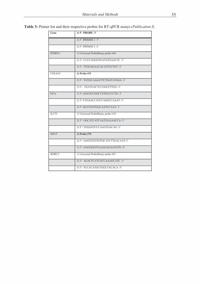
Materials and Methods 55
Table 3: Primer list and their respective probes for RT-qPCR assays (Publication I) Gene 1) 5' -PROBE- 3'
2) 5' -PRIMER 1- 3'
3) 5' -PRIMER 2 -3'
BTBD11 1) Universal Probelibrary probe #64
2) 5’- CCCCAGGGTGATATGAACTC -3’
3) 5’- TTGCGGAACACATTCCTGT -3’
COL6A3 1) Probe #31
2) 5’- TGTGCAAGATTCTGGTATGGA -3’
3) 5’- TGATGACTCCGGGTTTGG -3’
EF1a 1) 5' -AGCGCCGGCTATGCCCCTG- 3'
2) 5' -CTGAACCATCCAGGCCAAAT- 3'
3) 5' -GCCGTGTGGCAATCCAAT- 3'
IL17F 1) Universal Probelibrary probe #10
2) 5’- GGCATCATCAATGAAAACCA -3’
3) 5’- TGGGGTCCCAAGTGACAG -3’
MIAT 1) Probe #75
2) 5’- GAGTGTGTGTGCATCTTGACAAT-3’
3) 5’- GAGGGGTCGAAGAGAATGTG -3’
RORC2 1) Universal Probelibrary probe #87
2) 5’- AGACTCATCGCCAAAGCATC -3’
3) 5’- TCCACATGCTGGCTACACA -3’

Materials and Methods 56
Table 4: Primary antibodies used in the immunodetection and flow cytometry studies (Publication I, II, and III)
Product Catalog number Company
ATP1B1 GTX113390 GeneTex, CA, USA
BATF pab4003 Brookwood Biomedical, AL, USA
Cathepsin L (33/1) sc-65393 Santa Cruz, CA, USA
FOXP3 ab2481 Abcam, UK
FVT1 (KDSR) sc-100589 Santa Cruz
GATA3 558686 BD PharmingenTm USA
Histone H2B sc-10808 Santa Cruz
ITM2A 18306-1-AP ProteinTech Group, IL, USA
Nap-22 (H-100) BASP1 sc-66994 Santa Cruz
NAPSIN A ab9868 Abcam, UK
NOTCH1 sc-56014 Santa Cruz
RORy (H-190) sc-28559 Santa Cruz
Runx1 sc-28679 Santa Cruz
STAT3 9132L Cell Signaling Technology MA, USA
TBX21 561262 BD PharmingenTm USA
Vitamin D Receptor MA1-710 Thermo scientific, MA, USA
CCR6-PE 559562 BD, USA
CD122 (IL2RB) Mouse Anti-Human PE 554525 BD
CD52 anti-human APC 316008 Biolegend, CA, USA
CXCR5 Alexa Fluor® 488 Rat Anti-Human 558112 BD PharmingenTm USA
FAK (H-1) (PTK2) -PE sc-1688-PE Santa Cruz
IL-17A -FITC 11-7179 eBioscience, CA, USA
Lamin A/C (LMNA) PE sc-7292-PE Santa Cruz
phosphoSTAT3(pY705) 9131L Cell Signaling Technology MA, USA
STAT6 MA-120 Santa Cruz

Materials and Methods 57
Table 5: Primer and probe sequences used in ChIP-qPCR assays for validation of predicted STAT6 binding sites over selected Th2 specific enhancers (Publication III).
Probe sequence
Oligo name 5 `- oligo sequence -3 ` 5 `- 6(FAM) probe sequence
(TAMRA)-3 `
1 SEPTB1_1 CAAAAGGAGCAGCTAGAAGAG GGTGGGAGAAAGAGGAAGCT
SEPTB1_2 TATAAAGGGCTCCCAGGACA
2 GAB2_1 CTCCCTGACATCCGAACATT CCCCAATGCTCAGTTCTGAG
GAB2_2 TCACAGCAATTCGATGGTTT
3 RNF125_1 CTGATATCCCATCCCCAGAA CAACAATGAAGTGTTCTGTGTGC
RNF125_2 TTATAAAAATGCAGTATTTGGTTTTC
4 IL10RA_1 CTCAGCCTCCTTCCCTGAA CCTAGGGAAAGAGCTCAGGC
IL10RA_2 GCGAGTTCCCCTCAGAGTTA
5 FOXP1_1_1 TTCCTTTATGCGACACTGGA TGCCTGAAAAAGCAGAGACC
FOXP1_1_2 GGTGACGCCTGACACCAT
6 ABHD6_1 GAGCTGTCAGCAATCCTGTG ATGCACAGACTGCTTCCTCC
ABHD6_2 CAACCACTGTGTAAATGTCACTTG

Materials and Methods 58
Table 6: Primer list and their respective probes for STAT3 targets by ChIP-qPCR validation (Publication II)
Oligo name 5 `- oligo sequence -3 ` ProbeLibrary probe#
1 BATF_pro_1_11_ TGA AGT TTC CGC CCA TGT 11
BATF_pro_2_11 GCA CGC TCT CTC TCT CTC TTG
2 STAT3_pro_1_52 CTG GCT GTT CCG ACA GTT 52
STAT3_pro_2_52 CCC AAG TCC TCG GCT CTT
3 MYD88_pro_1_50 CCT CGA GAC CTC AAG GGT AGA 50
MYD88_pro_2_50 GCG CTT CCT CTT TCT CCT G
4 JAK3_pro_1_12 AGG CAG CGA GAG GAA AGT C 12
JAK3_pro_2_12 GCC CTG ACT TTC GGT AAA TG
5 ZNF460_pro_1_75 GAA ATG GGA GTA CTG GGC TTC 75
ZNF460_pro_2_75 GAC TTC CCG TAC CCT GAG GT
6 RELB_pro_1_14 AAA CGG CAG GTT CAA GTC C 14
RELB_pro_2_14 ATC ACG CCT TAC CCA TTG AG
7 IRF7_pro_1_49 GGA CGG GAA GTT TCG TCT C 49
IRF7_pro_2_49 TGG TCG CAT CCA ATA ATA AGA A
8 JUNB_pro_1_12 GAA ACC CCT CAC TCA TGT GC 12
JUNB_pro_2_12 AGG GGC TCA AAG GAC CTC
9 ICAM1_pro_1_88 CTT GGA AAT TCC GGA G 88
ICAM1_pro_2_88 TGC AGT TAT TTC CGG ACT GA
10 BATF_in_1_12 AGA GGG GGC GAA AAG 12
BATF_in_2_12 GTT GGT AAG ACG GGA ACT GG
11 PTPRCAP_in_1_52 TCT GGC CCT GTG AGA TCA G 52
PTPRCAP_in_2_52 TGT TGG GGG AGG TGA GTG
12 HNRNPH2_in_1_52 TCC GCC TCT TTC GTT CTC T 52
HNRNPH2_in_2_52 CTT CCC GGC ACT GAG ATG
13 NOTCH1_in_1_75 ACC GGG TGA CAG GAG CTA 75
NOTCH1_in_2_75 AAA GAA GAG GAA GGA GGC TCA
14 MED16_pro_1_3 TTG CAT ACG ACC ATT TCC AG 3
MED16_pro_2_3 GAA AGT GCT CGT TGT TCT ACC
15 RUNX3_inter_1_81 TGG CTT CCA CTT CTT AGA ATC C 81
RUNX3_inter_2_81 CCC CCT TCC CGT AAA TGA
16 SOCS1_inter_1_54 CTG ACG TTG GTC CCC ATC 54
SOCS1_inter_2_54 CGA TAA CGC TTG TTG AAA CCT
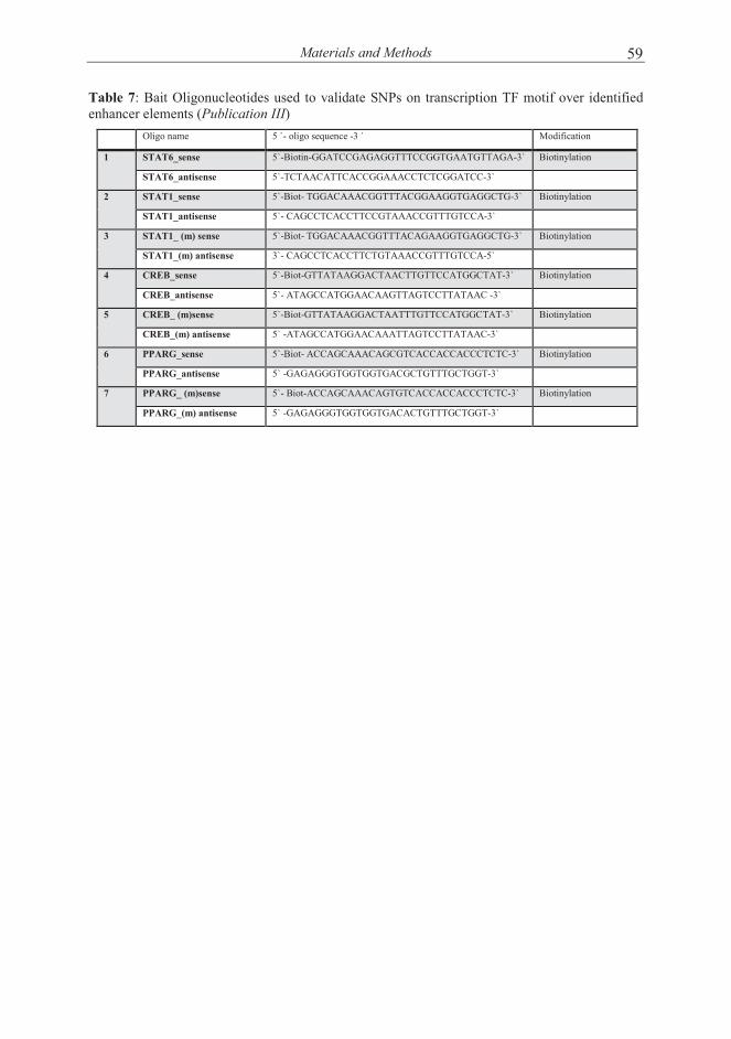
Materials and Methods 59
Table 7: Bait Oligonucleotides used to validate SNPs on transcription TF motif over identified enhancer elements (Publication III)
Oligo name 5 `- oligo sequence -3 ` Modification
1 STAT6_sense 5`-Biotin-GGATCCGAGAGGTTTCCGGTGAATGTTAGA-3` Biotinylation
STAT6_antisense 5`-TCTAACATTCACCGGAAACCTCTCGGATCC-3`
2 STAT1_sense 5`-Biot- TGGACAAACGGTTTACGGAAGGTGAGGCTG-3` Biotinylation
STAT1_antisense 5`- CAGCCTCACCTTCCGTAAACCGTTTGTCCA-3`
3 STAT1_ (m) sense 5`-Biot- TGGACAAACGGTTTACAGAAGGTGAGGCTG-3` Biotinylation
STAT1_(m) antisense 3`- CAGCCTCACCTTCTGTAAACCGTTTGTCCA-5`
4 CREB_sense 5`-Biot-GTTATAAGGACTAACTTGTTCCATGGCTAT-3` Biotinylation
CREB_antisense 5`- ATAGCCATGGAACAAGTTAGTCCTTATAAC -3`
5 CREB_ (m)sense 5`-Biot-GTTATAAGGACTAATTTGTTCCATGGCTAT-3` Biotinylation
CREB_(m) antisense 5` -ATAGCCATGGAACAAATTAGTCCTTATAAC-3`
6 PPARG_sense 5`-Biot- ACCAGCAAACAGCGTCACCACCACCCTCTC-3` Biotinylation
PPARG_antisense 5` -GAGAGGGTGGTGGTGACGCTGTTTGCTGGT-3`
7 PPARG_ (m)sense 5`- Biot-ACCAGCAAACAGTGTCACCACCACCCTCTC-3` Biotinylation
PPARG_(m) antisense 5` -GAGAGGGTGGTGGTGACACTGTTTGCTGGT-3`

Materials and Methods 60
Table 8: Bait oligonucleotides used to validate SNPs on STAT3 binding sites from ChIP-seq study (Publication II). Oligo name 5 `- oligo sequence -3 ` Modification
1 IL10_sense 5`-Biotin-GCA GAG CGT GAG GGG GAC TAG TGT TTA CT-3` Biotinylation
IL10_antisense 5`-AGT AAA CAC TAG TCC CCC TCA CGC TCT GC-3`
2 IL10_(m)sense 5`-Biotin-GCA GAG CGT GAG GGA GAC TAG TGT TTA CT-3` Biotinylation
IL10_(m)antisense 5`-AGT AAA CAC TAG TCT CCC TCA CGC TCT GC-3`
3 PFKFB3_sense 5`-Biot- ACC CTT GGT CTC TCG GAA TGC TAT TTT TT-3` Biotinylation
PFKFB3_antisense 5`- AAA AAA TAG CAT TCC GAG AGA CCA AGG GT-3`
4 PFKFB3_ (m) sense 5`-Biot- ACC CTT GGT CTC TCA GAA TGC TAT TTT TT-3` Biotinylation
PFKFB3_(m) antisense 5`- AAA AAA TAG CAT TCT GAG AGA CCA AGG GT-3`
5 IL7R_sense 5`-Biot-CAA ATA TTT CCT GAG TTT TTT TAT GAA-3` Biotinylation
IL7R_antisense 5`- TTC ATA AAA AAA CTC AGG AAA TAT TTG -3`
6 IL7R_ (m)sense 5`-Biot-CAA ATA TTT CCT GAA TTT TTT TAT GAA-3` Biotinylation
IL7R_(m) antisense 5`- TTC ATA AAA AAA TTC AGG AAA TAT TTG -3`
7 NDFIP1_sense 5`-Biot- AGG AAA TGT ATA GGA AAT GAT TGG ATC AT-3` Biotinylation
NDFIP1_antisense 5` -ATG ATC CAA TCA TTT CCT ATA CAT TTC CT-3`
8 NDFIP1_ (m)sense 5`-Biot- AGG AAA TGT ATA GGG AAT GAT TGG ATC AT-3` Biotinylation
NDFIP1_(m) antisense 5` -ATG ATC CAA TCA TTC CCT ATA CAT TTC CT-3`
9 SETD1A_sense 5`-Biot- AGC TCT GCC CTC CCC GCA AAC GCC AGC CT-3` Biotinylation
SETD1A_antisense 5`- AGG CTG GCG TTT GCG GGG AGG GCA GAG CT-3`
10 SETD1A_ (m1)sense 5`-Biot- AGC TCT GCC CTC CCG GCA AAC GCC AGC CT-3` Biotinylation
SETD1A_(m1) antisense 5`- AGG CTG GCG TTT GCC GGG AGG GCA GAG CT-3`
11 TERC_ sense 5`-Biot- TTC ACA AGC CCC CAT TGC CGG CGA GGG GT-3` Biotinylation
TERC_antisense 5`- ACC CCT CGC CGG CAA TGG GGG CTT GTG AA-3`
12 TERC_(m)sense 5`-Biot- TTC ACA AGC CCC CAC TGC CGG CGA GGG GT-3` Biotinylation
TERC_(m)antisense 5`- ACC CCT CGC CGG CAG TGG GGG CTT GTG AA-3`
13 STAT3_1_ sense 5'- TCC CCC CAC CAC TTC CCG GAA TAG CCC CAC -3' Biotinylation
STAT3_1_antisense 5'- GTG GGG CTA TTC CGG GAA GTG GTG GGG GGA -3'
14 STAT3_1_m1_ sense 5'- TCC CCC CAC CAC TAT CCG TCA TAG CCC CAC -3' Biotinylation
STAT3_1_m1_antisense 5'- GTG GGG CTA TGA CGG ATA GTG GTG GGG GGA -3'

Results and Discussion 61
RESULTS AND DISCUSSION
5.1 Profiling of the gene expression changes during early stage of human Th17 cell differentiation (Publication I)
5.1.1 Global transcriptional profiles at the early stage of human Th17 differentiation
During recent years, most of the studies of Th17 cells were involved in understanding and establishing their role in the pathogenesis of inflammation, autoimmunity, and cancer. However, for a precise understanding of these processes, it is essential to characterize the key signaling pathways and molecular mechanisms involved in the fate decision during the differentiation of Th17 cell lineage. To address this question, we performed global transcriptional profiling of in vitro cultured human CD4+ cells during the early stages of Th17 polarization. As a first step, we set up and established Th17 polarization conditions suitable for these measurements. For Th17 polarization, umbilical cord blood-derived naïve CD4+ T cells were activated with anti-CD3 and anti-CD28 in the presence of a cytokine cocktail of IL1β, IL6, and TGFβ, as well as neutralizing antibodies against IL4 and IFNγ. Th17 polarization was confirmed by analyzing the expression of marker genes such as IL17F, RORC, and CCR6, and by analyzing the secretion of IL17A (Publication I- Figure 1A-C). The selective upregulation of these marker genes under Th17 conditions showed successful polarization of the naive CD4+ T cells toward the Th17 cell lineage. Global transcriptional profiling during early stages of human Th17 polarization was then performed using Illumina Sentrix Human HT-12 Expression Version 3 Bead Chips. The samples for microarray analysis were collected at nine time points during the culture, starting from 0.5 hours until 72 hours (Publication I- Figure 1D). Microarray gene expression analysis revealed two waves of transcriptional changes in the cells polarized towards Th17 stimulation compared with TCR activation; the first wave falls within the first 4 hours of culture, and the second wave peaked after 6 hours (Publication I-Figure 2). The selected time points covered the early gene expression changes in the transcriptome, as very few genes were differentially expressed at the earliest 0.5 hours time point after which the number of differential expression increased (Publication - Table S3). The first wave of transcriptional changes peaked at 2 hours of polarization, where 256 probes representing 228 genes were differentially expressed. Additionally, most of the differentially expressed genes were up-regulated, suggesting stimulation of several signaling pathways in response to the Th17 inducing cytokine cocktail. In the second wave of transcriptional changes, which ranged from 6 hours to 72 hours, there was a large increase in the number of differentially expressed

Results and Discussion 62
genes, which peaked at 72 hours with 799 differentially expressed probes (Publication I-Figure 2). Out of 799 probes, 416 and 383 probes were up-regulated and down-regulated, respectively, indicating the regulation of Th17 polarization both by enhancing the signaling pathways that positively drive the differentiation and by shutting down the signaling pathways which negatively regulate the differentiation.
5.1.2 Dynamics of transcriptional changes during the early stage of human Th17 cell polarization
Gene annotation analysis revealed that the wide range of the differentially expressed genes were enzymes, kinases and transcription regulators. Additionally, the expression of cytokine and chemokine receptors (Publication I-Table 1) and their localization on the cell surface or in the extracellular space (Publication I-Table 2) suggest that the polarization process was initiated at the early stages with activation of the pathways required to mediate signaling with the neighboring cells. However, the appearance of new differentially expressed genes at each time point during the course of differentiation suggested that the process of lineage commitment to mature phenotype continues even after the first three days of polarization (Publication I-Figure 2). Gene expression pattern during the early polarization towards Th17 differentiation was highly dynamic in nature, as revealed by the vigorous changes in the gene expression throughout the selected time frame. For example, at 2 hour time point, most of the genes were differentially expressed and regulated only at this time point. Equally, several genes were differentially expressed and regulated at the 72 hour time point (Publication I- Table S3). As an illustration of these differences, the gene expression pattern showing log fold change ratio between Th17 and Th0 condition at 2 and 72 hours are shown as heat maps (Publication I- Figure 3A and 3B). Among these differentially expressed genes, upregulation of several known Th17 cell specific genes, such as RORC, RORA, AHR, BATF3, VDR, IL17F, IL9, CCR6, and IL23R, support the authenticity of the cultures and analyses. Importantly, our microarray results also revealed several novel genes potentially important for the Th17 differentiation process. We could not, however, detect the IL17A probe as differentially expressed because it was not functional on the microarray Chip used in the study. Furthermore, based on the overall gene expression pattern throughout the selected time points, we performed clustering of the differentially expressed genes (Th17/ Th0) to learn their dynamic expression patterns (Publication I-Figure 4, Table S4). The clustering analysis suggested three basic expression patterns throughout selected time points: upregulation, downregulation and rather steady expression. Additionally, there were only small differences in differential expression during the earliest time points, which were then followed by gene expression profiles biased towards the selected direction, suggesting that the counter regulation to the stimulus

Results and Discussion 63
due to TCR activation and the differential expression at later stages is due to both either active up or downregulation of the differentially expressed genes.
5.1.3 Validation of selected Th17 regulated genes and their expression in other T helper subtypes
The simplest way by which differential mRNA expression can affect the phenotype is when it regulates the corresponding protein level in the cell. We validated a number of genes with the biggest and long-lasting differential gene expression between Th17 and Th0 conditions at protein level (Publication I- Figure S1) by immunoblotting and flow cytometry. We used genes both with known and unknown functions as selection criterion for validation. Immunoblotting results validated the protein level expression of BASP1, CTSL1, RUNX1, BATF, FVT1, NOTCH1, VDR and ATP1B1 in cells polarized towards Th17 cells compared to their expression in activated Th0 cells at 24, 48 and 72 hours time points (Publication I-Figure 5A). And in keeping with the observations from the microarray data, flow cytometric analysis showed the upregulation of CXCR5, IL2RB and LMNA, and down regulation of CD2 and ITMA2 under Th17 polarizing conditions compared to Th0 cells (Publication I-Figure 5B). However, the microarray data also revealed the upregulation of COL6A3 and MIAT genes, and downregulation of BTBD11 gene in Th17 polarizing cells. These genes were validated with quantitative RT-PCR detection at 24, 48 and 72 hours (Publication I-Figure 5C).
In order to further characterize the selectivity of the above validated genes in Th17 polarizing conditions, we cultured cord blood-derived naive CD4+ T cells under Th0, Th1, Th2, iTreg or Th17 polarizing conditions for 72 hours and analyzed the expression of selected genes at the protein level with immunoblotting or flow cytometry. In confirmation of the specificity of differentiation for the Th cell subset cultures, the protein expression of TBX21, GATA3, and FOXP3, the key TFs for Th1, Th2 and iTreg cells, respectively, and the secretion of IL17A, which is the marker cytokine for Th17 cells, were found to be preferentially expressed in corresponding Th cell subsets (Publication I-Figure 6A-B). Consistent with the results obtained from microarray analysis and the protein validation, the expression of ATPB1, KDSR, IL2RB and CXCR5 were selectively increased under Th17 polarizing condition compared with Th1, Th2 or iTreg induction (Publication I-Figure 6C and 6D). However, even though the expression of CSTL and VDR were higher in cells under Th17 polarizing conditions than in Th0 cells, which confirmed the results from the microarray study, the highest expression of these genes were detected in the cells cultured under iTreg polarizing condition (Publication I-Figure 6C), suggesting that TGFβ induced expression of these genes. Furthermore, the expression of CD52 was observed to be downregulated in Th17 polarizing condition as compared to other T

Results and Discussion 64
helper subset polarizing conditions (Publication I-Figure 6D). To summarize, these results revealed a number of genes selectively regulated under Th17 polarizing condition, as well as other genes non-specific for Th17 conditions that are also differentially expressed in polarizing conditions for other Th cell subsets.
In summary, the data from this study describe in detail kinetic changes in the transcription profiles during early stages of Th17 cell differentiation and provide a valuable resource for elucidating molecular mechanisms and gene regulatory network governing Th17 differentiation in human. Additionally, further characterization of the candidate genes possibly playing key role in Th17 differentiation process and function, may prove to be potential pharmaceutical targets for the diagnostics or treatment of Th17 cell-mediated immune diseases.
5.2 Regulation of early human Th17 cell differentiation by STAT3 (Publication II, unpublished)
TFs play key role in driving Th cells lineage specification and commitment. STAT3, an early key TF for Th17 cells, critical for Th17 cell differentiation (Ciofani et al., 2012; Durant et al., 2010; Iida et al., 2011; Miyahara et al., 2008; Wang et al., 2009a; Yang et al., 2007a). In spite of its importance, our current understanding of STAT3 and its role in Th17 differentiation is based on studies carried out in mouse, whereas very little is known in human. Moreover, since STAT3 is a key upstream regulator of Th17 differentiation, characterization of downstream targets of STAT3 in human is of particular interest. In this study, we combined RNAi, ChIP, genome-wide methods and computational data integration to identify STAT3 targets during the initiation of human Th17 differentiation. In addition, we used data from GWAS catalog to detect the overlap of STAT3 binding sites with a large number of SNPs associated with various autoimmune and other immune disorders, including CD, RA, T1D, PS and allergic diseases such as asthma. The findings suggested that alterations in TF binding site motifs by SNPs can influence transcriptional regulation and cell fate, and thereby contribute to immunopathogenesis. Collectively, the results present new insight into human Th17 differentiation and how it may be modulated in diseased states.
5.2.1 STAT3 affects the expression of Th17-associated genes
To investigate the contribution of STAT3 in the Th17 cell differentiation process, we perturbed its expression in human CD4+ T cells using RNAi and investigated the effect on gene expression during early stages of Th17 differentiation. STAT3-RNAi-mediated downregulation in the expression of STAT3 and its known target BATF, compared with nontargeting scramble siRNA, demonstrated the validity of the three

Results and Discussion 65
distinct siRNAs used (Publication II-Figure 1A). Moreover, downregulation of STAT3 resulted in reduced expression of CCR6 and decreased secretion of IL17A (Publication II-Figure 1B, C). These results revealed that STAT3 is also critical for human Th17 cell differentiation, which is consistent with the reports in murine T cells (Ciofani et al., 2012; Durant et al., 2010).
5.2.2 Identification of STAT3 regulated genes during early stage of Th17 differentiation
To identify the global STAT3 targets at the early stages of Th17 differentiation, we analyzed and compared the gene transcription profiles from human CD4+ T cells treated with different siRNAs specific for STAT3 together with those with non-targeting scramble siRNA and polarized towards Th17 conditions at 2, 12, 24, and 72 hours time points. We observed that altogether 1654 genes were regulated in a STAT3 dependent manner. Out of these 1654 genes, STAT3 upregulated the expression of 727 genes and negatively regulated the expression of 927 genes. The down regulation of genes promoting Th17 polarization and upregulation of genes for other key lineages suggests the importance of STAT3 both in lineage specification and commitment
After validating the role of STAT3 in the initiation of human Th17 polarization, we next aimed to identify the genome wide targets of STAT3 during the early stages of this process. In order to achieve this, we analyzed and compared the effect of Th17 polarizing cytokine stimulation between cells nucleofected with STAT3-specific siRNA or non-targeting control-siRNA. Because we focused on the initiation of Th17 differentiation process, the genome wide gene expression changes were analyzed within three days in response to Th17 polarizing signals under STAT3-RNAi treated cultures and compared with cultures treated with non-targeting scramble siRNA. The results obtained were further compared with results from activated T cells nucleofected with STAT3-specific siRNA and control non-targeting scramble siRNA (Th0; Publication II- Figure 2A). Previously we have shown that the maximal changes in gene expression took place already at 2 hours in Th17 polarizing cells, compared to cells cultured in Th0 condition (Tuomela et al., 2012). Consistent with this previous finding, under control non-targeting scramble siRNA treatment, in total 2528 genes showed a change in gene expression under Th17 culturing conditions, compared with Th0 culturing conditions (FDR<0.1). Out of 2528 genes, 1091 genes were upregulated and 1437 genes were downregulated. Additionally, the cells cultured for 72 hours showed changes in the expression of 1713 genes, of which 879 genes were upregulated and 833 genes were down regulated (Publication II- Table S1). Among the upregulated genes were several well-known lineage specific genes, such as RORC, RORA, BATF, IRF4, FOSL2, AHR, IL23R, CCR6, IL21, IL17F and IL17A, suggesting that siRNA treated T cells were able to successfully polarize towards the Th17 phenotype.

Results and Discussion 66
during the differentiation of Th17 cells by promoting Th17 polarization as well as via opposing alternative lineages. Interestingly, already at 2 hours in cells differentiating towards Th17 condition, downregulation of STAT3 influenced the expression of 301 genes. Out of these 301 genes, the expression of 185 genes was reduced; including CXCR5 and SOCS3, indicating STAT3 positively regulates the expression of these genes. Conversely, the expression of 116 genes were upregulated upon STAT3 knockdown, including STAT1, STAT2 and SMAD7, suggesting STAT3 suppresses the expression of these genes (Publication II-Figure 2B, Table S2). However, the maximum number of genes regulated STAT3 were observed at 72 hours in differentiating Th17 cells, where it influenced the expression of 929 genes. (Publication II-Figure 2B).
To investigate the proportion of the Th17 polarizing cytokine-regulated genes that were under the control of STAT3, we compared the genes differentially expressed in Th17 conditions (Publication II-Figure 2A) with STAT3 regulated genes (Publication II-Figure 2B). We observed that already at the 2 hour time point around 7% of the STAT3 regulated genes were differentially expressed in Th17 conditions as compared to Th0 cells (Publication II -Figure 2C). Further, we observed that the proportion of STAT3 regulated genes was increased following differentiation time points. The highest numbers of genes were regulated by STAT3 at 72h with 36% of the total differentially expressed gene (Publication II - Figure 2C). Overall, these observations suggest that STAT3 is an early transcriptional regulator of the Th17 differentiation program. Interestingly, we observed that the FOSL2, NTRK3, IL16, DUSP10 and CASP1 genes were differentially expressed during the Th17 differentiation process as well as regulated by STAT3 at all the time points studied (Publication II -Figure 2D). Altogether, we detected 44 genes with at least 2.8 fold differences in gene expression that were regulated in Th17 differentiating conditions as well as influenced by STAT3 down-regulation (Publication II-Figure 2E). The gene expression patterns of these 44 genes were dynamic showing early and late regulation; for example GBP4, COL6A3, and CXCR5 were regulated at the earliest time points that we studied during Th17 polarizing stimulation as well as STAT3 knockdown, while CCR6, HOPX and CSF2 were regulated at the later stages. Additionally, several genes, including PALLD, COL6A3, CXCR5, FLT1, RORA, IL23R and PHLDA1, were expressed at multiple time points and regulated in STAT3 dependent manner (Publication II -Figure 2E). Interestingly, results suggest that STAT3 regulates the expression of genes involved in different cellular functions. For example, PALLD (Palladin) is a cytoskeletal protein involved in the control of cell shape, adhesion, contraction, migration and invasion (Brentnall, 2012; Mykkänen et al., 2001; Pogue-Geile et al., 2006). The role of PALLD in T cells is poorly characterized. A vascular endothelial growth factor receptor (VEGFR) family protein, FLT1 (Fms-Related Tyrosine Kinase 1, also known as VEGFR1) participates in the regulation of angiogenesis and vasculogenesis. It has been shown that CD45RO+ CD4+ memory T lymphocytes express FLT1, which

Results and Discussion 67
5.2.3 Identification of direct targets of STAT3 by genome wide ChIP sequencing
After the identification of genes regulated by STAT3 during the early stages of Th17 polarization, we next investigated the direct targets of STAT3 using ChIP-seq. To define an optimal early time point for ChIP-sequencing, we first studied the kinetics of STAT3 phosphorylation after induction of human Th17 differentiation (Publication II -Figure 3A). The phospho-STAT3 kinetics results revealed three waves of phosphorylation patterns, with two major peaks at 0.5 hour and 48 hour after Th17 polarization. We observed a maximum phosphorylation signal of STAT3 (67% of pSTAT3 positive cells) at 0.5 hour post Th17 polarization. STAT3 phosphorylation is further increased and peaks at 48 hour. However, although the first 0.5 hour peak was Th17 specific, the second highest peak at 48 hours was also observed in TCR/CD28 activated cells, suggesting a TCR/CD28 activation dependent increase in STAT3 phosphorylation at this time point. Since, we were interested in identifying immediate direct targets of STAT3 in Th17 polarized cells by ChIPseq; we chose the 0.5 hour time point because at this time point the level of phospho-STAT3 peaked maximum in Th17 polarized cells compared with Th0 cells. Altogether, we identified 2981 STAT3 binding sites (Publication II – Table S3). Further analysis revealed that around 21% STAT3 binding sites were located in the immediate promoter region or transcription starting sites (TSS), which is consistent with the importance of STAT3, mediated regulation of transcription. Consistent with other reports on genome wide binding analysis of STATs proteins, we also observed that around 70% of the STAT3 binding sites were found in either introns (35%) or intergenic regions (36%), suggesting that STAT3 may regulate gene expression through binding to distal regulatory elements (Publication II -Figure 3B), (Elo et al., 2010; Wei et al., 2010). Further analysis revealed that around 50% of the STAT3 binding sites were localized within 10kb up-
regulates IFNγ and IL17 production in memory T cells (Basu et al., 2010; Kim et al., 2010), suggesting that FLT1 plays a role in immune responses. PHLDA1 (pleckstrin homology-like domain, family A, member 1) is a proline-histidine rich nuclear protein shown to play a role in various tumors (Johnson et al., 2011; Neef et al., 2002). Interestingly, the expression of CXCR5, a chemokine receptor, was induced during Th17 differentiation and its expression was negatively regulated upon STAT3 downregulation by siRNAs specific for STAT3 (Publication II -Figure 2E). Previously, it has been observed that CXCR5 expression is induced by both IL6 and IL21 in a STAT3-dependent manner in Tfh cells (Nurieva et al., 2008). In line with microarray results, we validated the expression of CXCR5 in Th17 polarizing conditions and its regulation by STAT3 (Publication II -Figure 2F). Our findings confirm the STAT3 mediated regulation of CXCR5 and suggesting its role in Th17 cells.

Results and Discussion 68
and down-stream of TSS (Publication II -Figure 3C). Furthermore, in silico DNA motif analysis using TRANSFAC confirmed the enrichment of STAT3 motif as the strongest motif among the identified STAT3 binding sites (Publication II -Figure 6B).
We next extended our analysis by combining STAT3 ChIP-seq analysis with gene expression analysis of STAT3 RNAi to reveal direct targets of the Th17 polarization process. The results revealed that panels of genes regulated by STAT3 were in fact bound by it to their regulatory regions at 30 min after induction of Th17 polarization. Our results showed that STAT3 directly regulates 22% of the STAT3-regulated differentially expressed genes in polarizing Th17 cells at the 2 hour time point. Moreover, the proportions of direct STAT3 targets increased to 29% at 12h and 24h, and further decreased to 17% at 72h (Publication II -Figure 3D). Significantly, these analyses identified several previously reported direct targets of STAT3, including SOCS3, BATF and RORA (Chen et al., 2007). It has been established that STAT molecules are critical in driving Th subset differentiation, for example the roles of STAT1 and STAT4, STAT6, and STAT3 are well documented for Th1, Th2 and Th17 differentiation, respectively. Interestingly we observed that STAT3 regulated the expression of several STATs (STAT1, STAT2, and STAT4) including itself by direct binding to their promoters. Moreover, STAT3 binding sites were observed in the intergenic regions of the FOSL2 and DUSP10 genes, which were regulated upon STAT3 downregulation at all the measured time points. (Publication II –Table S3).
To further investigate the kinetics of STAT3 binding to its target genes, we selected 15 STAT3 ChIPseq binding sites annotated for various genes and validated these with ChIP-qPCR at the time points of 0h, 0.5h, 2h, 12h and 72h in cells differentiating towards Th17 and in Th0 cells that were activated only (Publication II-Figure S2) . ChIP-qPCR analysis revealed that STAT3 preferentially bound to the selected regions only the at 0.5h time point and under Th17 polarizing conditions (Publication II -Figure 4). Interestingly, STAT3 binding to these loci was lost at the later time points during Th17 polarization, suggesting temporal binding for STAT3 in regulating expression of associated genes. Thus, we were able to identify the early direct STAT3 targets involved in the initiation of Th17 polarization.
5.2.4 STAT3 mediated transcriptional network in Th17 cell polarization
Combined analysis of STAT3 ChIP-sequencing and gene expression allowed us to identify TFs regulated during early polarization of Th17 cells and regulated by STAT3. The maximum number of regulated TFs was found to be at 2h under Th17 polarizing conditions, where 279 and 185 TFs were upregulated and downregulated, respectively (Publication II -Figure 5A). Further overlap with gene expression upon STAT3 RNAi revealed that out of these, 20 TFs were regulated by STAT3, including FOSL2 and STAT3, which were upregulated in response to Th17 polarization and

Results and Discussion 69
positively regulated by STAT3 (Publication II -Figure 5B). In contrast, the expressions of TFs, such as NPAT, ZNF211 and NKAP were downregulated in Th17 polarizing cells and negatively regulated by STAT3, as STAT3 downregulation increased their expression. Additionally, although the expression of SMAD7, STAT1 and STAT2 were upregulated during Th17 polarization, STAT3 knockdown further enhanced their expression, indicating that STAT3 suppresses their expressions (Publication II –Figure 5C). The largest number of TFs regulated by STAT3 was observed at 72h (Publication II -Figure 5B), suggesting the rationale behind maximum regulation of overall gene expression by STAT3 (Publication II -Figure 2B). Among these STAT3 regulated TFs, including RORA, MAF, PPARG, RUNX1, HOPX, and RBPJ were upregulated during Th17 polarization in a STAT3-dependent manner. In contrast to this, several TFs, such as KLF3, GFI1, STAT4, GATA3 and IKZF2, were downregulated during early Th17 differentiation in a STAT3-dependent manner (Publication II-Figure 5D). Interestingly, a number of TFs, including BATF, RORA, FOSL2, IKZF2, HOPX and AFF3, were differentially expressed throughout multiple time points, implicating their role in determining the cellular fate of developing Th17 cells.
Combined analysis of the data from gene expression profiling with or without STAT3 downregulation by siRNA and STAT3 ChIP-Seq experiments, we divided the TFs into four categories. The first category of TFs encodes direct STAT3 targets, which were differentially expressed during Th17 polarization (Th17 vs Th0), regulated by STAT3 (STAT3 siRNA vs non-targeting scramble siRNA) as well as bound by STAT3 (STAT3 ChIP). The first category of TFs includes STAT1, STAT3, BATF, IKZF3, RUNX1, FOSL2, BCL6 and IRF9 (Publication II -Figure 5E). Notably, previous studies have shown the role of each of these TFs in the regulation of Th17 differentiation (Amadi-Obi et al., 2007; Ciofani et al., 2012; Hofer et al., 2012; Laurence et al., 2007; Quintana et al., 2013; Schraml et al., 2009; Zhang et al., 2008a). Each TF forms a highly coordinated network to further regulate Th17 cell differentiation (Publication II – Figure S3). The second category of TFs encodes indirect targets of STAT3, which are not bound by STAT3 but their expression was changed during Th17 differentiation and regulated by STAT3, suggesting that STAT3 indirectly regulates their expression through other factors. The third category encodes putative STAT3 targets that are bound by STAT3 and their expression regulated during Th17 differentiation but not regulated by STAT3. Interestingly, several TFs of this category, such as IRF4, NOTCH, RUNX3, IKZF4 and JUN, have previously been shown to be involved in the differentiation of Th17 cells or Treg cells (Brüstle et al., 2007; Fu et al., 2012; Keerthivasan et al., 2011; Klunker et al., 2009; Li et al., 2012; Sharma et al., 2013). The fourth category of TFs includes STAT3-independent TFs that were differentially expressed during Th17 differentiation but neither regulated nor bound by STAT3 (Publication II –Table S6). This category of TFs may cooperate with STAT3 or STAT3 regulated TFs to form the combinatorial
Results and Discussion 70
transcription regulation network, which regulates Th17 lineage specification via a highly coordinated transcriptional network (Publication II -Figure S3).
5.2.5 Association of disease-associated SNPs with STAT3 binding sites
Because Th17 cells were shown to participate in the development of various autoimmune diseases (Chen and O’Shea, 2008; Korn et al., 2009) and STAT3 is the major regulator of Th17 development, we used a SNP data base to examine whether disease-associated SNPs overlap with the STAT3 binding sites identified by STAT3 ChIP-seq analysis. We found that SNPs associated with PS, MS and CD significantly overlap with STAT3 binding sites (Publication II – Table S7 and Figure 6A). Equally, Immuno-Chip analysis showed that SNPs for 9 autoimmune diseases overlapped with ChIPseq STAT3 binding sites (Publication II -Table S8). We then searched for variant positions within the STAT3 motifs of STAT3 binding sites identified as overlapping with disease-associated SNPs (Publication II - Figure 6B). We observed multiple SNPs associated with autoimmune diseases within the STAT3 motifs (Publication II -Table S8). For example, a SNP (rs947474) associated with T1D and overlapping with a STAT3 motif was identified upstream of PFKFB3 (6-phosphofructo-2-kinase/fructose-2, 6-bisphosphatase 3) and PRKCQ (Protein kinase C, theta). The reference nucleotide for this SNP is G and the mutant is A. The G to A transition overlapped nucleotide 12 on the STAT3 motif, i.e., nucleotide A. Importantly, the PRKCQ gene regulate the expression of Ifng and Il17, and modulate immune response in an autoimmune disease model (Tan et al., 2006).
We further examined whether SNPs at the STAT3 binding motif of these predicted target genes could alter STAT3 binding. For these analysis, we performed DAPA assay using reference oligonucleotide sequences from the Hg19 harboring the predicted STAT3 binding site identified in our ChIP-seq data or point mutation (associated SNP) for selected from six disease-associated SNPs that overlap STAT3 binding sites (Publication II – Table S4 and S8). These were as follows: the CD, T1D and UC-associated SNP rs3024505, which is in a close vicinity of the IL10 gene; the T1D-associated SNP rs947474 nearby PFKFB3, LOC399715 and PRKCQ genes; the PCB-associated rs17313508 nearby IL7R, CAPSL and UGT3A1 genes; the MS associated SNP rs6580224 with nearest neighboring genes NDFIP1, GNPDA1 and SPRY4; the PS-associated SNP rs12443808 located near SETD1A, HSD3B7 and STX1B; and the Celiac disease (CeD) and MS associated SNP rs2293607 with location closest to TERC and ACTRT3 genes. Consistent with our previous finding that SNPs can change the TF binding affinity, the binding of STAT3 was reduced when variant oligonucleotides harboring disease associated SNPs rs3024505, rs947474, rs6580224 and rs2293607 were used (Publication II- Figure 6C). However, STAT3 binding was increased when variant oligonucleotide for PS-associated SNP

Results and Discussion 71
rs12443808 was used. These results suggest how a disease-associated SNP may regulate STAT3 mediated transcription by affecting the expression of its targets important for early human Th17 cell differentiation in a disease setting.
5.3 Global mapping of lineage specific enhancer landscapes in early differentiating Th1 and Th2 cell lineages
The precise control of lineage specification and commitment and proper balance between these effector and regulatory lineages are critical to avoid various autoimmune disorders. To understand the process of lineage specification and commitment, we analyzed global chromatin state maps of histone modifications in an attempt to identify lineage specific enhancers in cells polarizing towards Th1 and Th2 lineages after 72 hours. ChIP-seq with antibodies specific for histone modification marks that distinguish between distal enhancer and proximal promoter regions (H3K4me1, H3K4me3 and H3K27ac) were used. To our surprise, even at this initial stage of differentiation, our analysis revealed several lineages specific enhancer elements correlating with cell specific gene expression changes. Further examination of TF binding on lineage specific enhancers revealed enrichment of known and novel T cell-specific TFs important for driving lineage-specific gene expression. Finally, we integrated enhancer analysis with immunopathogenic associated SNPs from a GWASs database, indicating the significance of these distal cis-regulatory elements in the disease etiology.
5.3.1 Global enhancer chromatin states identified lineage-specific enhancer elements in Th1 and Th2 differentiating cells
To investigate the process of lineage specification and commitment, we analyzed and compared global chromatin state maps of histone modifications in order to identify lineage specific enhancers in cells differentiated towards Th1 and Th2 lineages after 72 hours. ChIP-seq with antibodies specific for histone modifications (H3K4me1, H3K27ac, and H3K4me3), which discriminate enhancer and proximal promoter elements, respectively, were used (Publication III - Figure S1A). At 72 hours, cells were already directed to polarize towards Th1 and Th2 cells, based on the protein expression of lineage specific TFs, T-bet and GATA3 (Publication III - Figure S1A). After seven days, the expression of key lineage markers confirmed that the cells had correctly differentiated to Th1 and Th2 cell lineages in the polarization settings used in the study (Publication III - Figure S1B and C).
Depending on the genomic location of H3K4me1 compared to H3K4me3 (Heintzman et al., 2009), our analysis identified 16,507, and 13,466 enhancers in the Th1 and Th2 cell lineages respectively, compared with 16,552 enhancers in activated Th0 cell

Results and Discussion 72
lineage (Publication III - Table S1A). Further analysis suggested that at this initial stage of polarization, the enhancer patterns were relatively similar among activated and differentiating Th1 and Th2 cells (Publication III - Figure 1B). However, lineage specific enhancers can be determined using a recent analytical method based on nucleosome displacement (or nucleosome free regions (NFRs)) when comparing histone modification profiles between two samples (He et al., 2010) (Publication III - Figure 1E-F, Figure 2, Figure S2, Table S1B). Based on this method, our analysis resulted in subsequent number of lineage-specific enhancers: 1636 (Th0), 2144 (Th1), and 2654 (Th2) (Table S2a).
To determine whether identified lineage-specific enhancers are correlated with lineage specific genes during Th1 and Th2 differentiation, we analyzed gene expression profiles from control activated Th0 and Th1, Th2 differentiating cells using the Heliscope platform. Gene expression analysis identified significant number of differentially expressed genes between Th1/ Th0 or Th2/ Th0 (Publication III - Table S4B). Further, to correlate expression of these genes with lineage specific-enhancers, we determined lineage-specific gene upregulated in one culture condition relative to the other culture conditions. We identified 121and 292 genes expressed in Th1 and Th2 culture condition respectively, as well as 116 were upregulated in Th0 culture condition (Publication III - Figure 1G; Table S4A; see Table S5 for all genes). In support of these observations, the genes identified in this study were consistent with our previous findings (Elo et al., 2010; Lund et al., 2005, 2003, 2007) suggesting that these genes are crucial for the cellular specification and commitment to attain the desired cell fate. The observation that only a limited number of lineage-specific enhancers were correlated with lineage-specific genes emphasized the role of enhancers in guiding the lineage specific gene expression program. In summary, we have used enhancer maps to establish the first understanding of lineage-specific gene regulation in a population of cells that were not yet fully committed and largely similar, thereby suggesting the role of enhancers in driving gene expression changes critical for each lineage commitment.
5.3.2 Fate of lineage-specific enhancers during differentiation
Previous studies have reported that acetylation of H3K27 is associated with enhancer activity (Heintzman et al., 2009; Roh et al., 2007, 2005). However, there are subsets of enhancers that lack this modification and are deemed as “poised enhancers” (Creyghton et al., 2010; Hawkins et al., 2010; Rada-Iglesias et al., 2011). For example, more than 2,000 enhancers lacked H3K27ac in human ES cells (Hawkins et al., 2010; Rada-Iglesias et al., 2011), while, less than 50% enhancers overlapped with H3K27ac in mouse ES cells (Creyghton et al., 2010). Likewise, we also observed that this modification was absent in a large fraction of enhancers in all of the Th cell

Results and Discussion 73
subsets that we analyzed. Consistent with this finding, lineage-specific enhancers lacking acetylation were identified (Publication III - Figure 2), suggesting that these enhancers are in a ‘poised state’ and waiting for signals that enable loading of TFs to drive transcription. The identification of poised enhancers at this initial stage of cellular differentiation reflects that the cells are poised for commitment into distinct lineages.
The presence of DHS is indicative of an open chromatin structure for TFs binding and is reflect active regulatory elements. Thus, in order to investigate the fate of the Th1 and Th2 cell specific enhancers during the later stage of differentiation, we made use of DHS data from ENCODE for peripheral blood-derived Th1 and Th2 cells at day 7 (ENCODE Project Consortium et al., 2012; Thurman et al., 2012) to compare with our data at 72 hours. The results for the Th1 lineage indicated that 489 out of 653 enhancers active at 72 hours (K27ac) remain active based on their DHS status at day seven, suggesting that these enhancers are required throughout cellular differentiation. The remaining 155 lose DHS, suggesting that these enhancers are active and required for lineage commitment rather than maintenance and are not used any more at the later stage of differentiation. Additionally, around 300 enhancers poised at 72 hours instead become active (gain DHS later) at day 7 indicating that these enhancers are required to maintain cellular state at the later stage. However, majority, of the enhancers (~1200) are marked by H3K4me1 only and they have neither the K27ac mark nor gain DHS, suggesting that these enhancers might be used either at some point during differentiation or remain poised to alter the gene expression in response to other external stimuli. The Th2 results demonstrated that 835 of altogether 847 enhancers were active at 72 hours (K27ac) and remained active based on their DHS status at day seven. The remaining enhancers (~9) were observed to lose DHS. Approximately 1400 enhancers of the 1800 enhancers poised at 72 hours become active (gain DHS later) later at day seven. In contrast to the Th1 linage, only ~400 of the enhancers are marked by H3K4me1 alone and remained inactive in Th2 cells (Publication III - Figure 2). The above observations suggest that the epigenetic state of the cells has already been instructed to modulate future cell fate commitment.
5.3.3 Analysis of putative enhancer binders
TFs are key players in driving the enhancer activity through binding to these distal regulatory sites and facilitating their looping to promoter regions of the target gene and therefore in determining upregulation of the lineage-specific gene. Enrichment of TF motifs for lineage specific TFs over regulatory site of enhancers has been used to predict cell type-specific active enhancers that reflect the biology of that cell type. Previous studies have shown strong correlations between enrichment of binding motifs for expressed lineage specific TFs within active enhancers, suggesting that

Results and Discussion 74
lineage-specific TFs are key drivers of enhancer activity (Ernst et al., 2011; Hawkins et al., 2010; Heinz et al., 2010; Hoffman et al., 2010; Robertson et al., 2008). Motivated by these findings, we examined the enrichment of known TF motifs over enhancer sequences with ProbTF (Lähdesmäki et al., 2008) and TRANSFAC (Matys et al., 2006) tools and combined these with statistical analysis. The identified TF motifs were then filtered for the TFs expressed in each cell lineage, thus revealing both novel and known TFs regulating the expression of lineage specific genes during Th cell differentiation (Publication III - Table S2).
Further analysis showed that lineage-specific enhancer sequences were in fact enriched for known TF motifs specific for T cell (Publication III - Table S3, Methods). For example, activated Th0 cell specific enhancer sequences contain TF motifs for AP-1, NFAT, and NF-KB TFs, which play a key role in T cell activation (Ansel et al., 2006). TF motifs enriched for Th1 enhancers include key Th1 regulators, such as STAT1, STAT4, ATF3 and JUN. The role of these TFs in regulating Th1 polarization have been well documented (Afkarian et al., 2002; Filén et al., 2010; Jenner et al., 2009; Szabo et al., 2000). Lastly, Th2 cell-specific enhancer motifs were overrepresented for TFs, including STAT6, GATA3, NFIL3, GFI1, PPARG and BACH. The expression of all of these TFs were shown to be enhanced in differentiating Th2 cells and suggested to control several Th2-specific genes (Elo et al., 2010; Horiuchi et al., 2011; Kaplan et al., 1996; Kashiwada et al., 2011; Zhu, 2010). Thus, the integration of lineage-specific enhancer maps and computational tools for analysis of TF motifs, which were further filtered for TF expression, has revealed how key lineage specific TFs are likely exploit enhancer binding sites to drive lineage specification and commitment during Th cell differentiation and development. Additionally, we also observed that several lineage specific enhancers were selectively enriched for TFs motifs for expressed TFs, such as OCT, HNF, FOX family members whose role in T cell differentiation is not clear (Publication III - Figure 3B-E; Table S3). Further investigations into these novel regulators could help fill in the gaps in constructing the early regulatory networks controlling Th cell differentiation.
Additionally, we have validated a subset of enhancers and their cognate predicted TF motifs, using Th2 specific enhancers with predicted motifs for STAT6 TF. Chromatin-immunoprecipitation for H3K4me1, H3K27ac and STAT6 at 0h (Thp), 4 hours and 72 hours was performed for six Th2 specific enhancers containing STAT6 motif and quantitated using RT- PCR (Publication III- Figure 4). Our analysis identified that these enhancers are located nearby genes which are specifically expressed in Th2 cells, including the genes with known and novel function such as RUNX1, IL10RA, FOXP1, SETBP1, GAB2 and ABHD6. We showed that these enhancers, enriched with H3K4me1 and H3K27ac, are specifically bound by STAT6 in cells differentiated to Th2 cells relative to Thp and stimulated Th0 cells, indicating that STAT6 regulate

Results and Discussion 75
these genes in Th2 cell lineage in an enhancer-specific manner. Altogether, these data provide insight into the mechanistic aspects of both TF binding and chromatin state modifications in driving lineage-specific gene expression. Additionally, our results suggest the early marking of some enhancers loaded with TFs, yet unable to gain functionality until a later state based on acquisition of H3K27ac (Publication III - Figure 4).
5.3.4 Enhancers overlap with disease associated SNPs
SNPs from GWASs have been associated with several complex diseases. However, many of the studies are unable to determine the functional phenotypic relevance of the SNPs, probably because many of the SNPs are located outside of the gene-coding regions. From an evaluation of GWAS catalogued, it has been observed that 45% and 43% of these disease- associated SNPs are located in the intronic and intergenic regions, respectively (Hindorff et al., 2009). Regulatory SNPs are shown to alter the expression of genes associated with the expression of quantitative traits (Cheung and Spielman, 2009). Large projects, such as the NIH Road Map project, ENCODE Project and other important studies on genome-wide assessment of epigenetic features have shown that the genomic distribution of SNP- associated complex traits were enriched in the genomic regions similar to gene regulatory regions, such as promoters and enhancers, (Boyle et al., 2012; Ciofani et al., 2012; ENCODE Project Consortium et al., 2012; Maurano et al., 2012; Ni et al., 2012; Schaub et al., 2012). Thus, the studies suggest that SNPs potentially affect the expression of genes associated with disease phenotypes through regulating the chromatin accessibility and epigenetic features of regions implicated in gene regulation. To investigate whether disease-associated SNPs overlap with the Th enhancer elements detected in our research are regulatory SNPs, we used SNPs from the National Human Genome Research Institute-Genome wide Association Catalog (www.genome.gov/gwastudies) and overlapped these with enhancers identified for Th1 or Th2 differentiated or activated cells at 72 hours. We performed integrative investigation of the mapped disease-associated SNPs, which revealed that 1,281 SNPs associated with multiple autoimmune disease were distributed among all disease categories (Publication III - Table S8; Table 1), including MS, UC, CD, PS, RA, T1D, and asthma (Anderson et al., 2011; Barrett et al., 2009; Cargill et al., 2007; Duerr et al., 2006; Hafler and De Jager, 2005; International Multiple Sclerosis Genetics Consortium et al., 2007; Moffatt et al., 2010; Nair et al., 2009; Rioux et al., 2007; Stahl et al., 2010). The highest overlap was observed for RA and UC with each more than four hundred SNPs. CD and MS were among the minimally enriched SNPs (~40 SNPs). GWAS analysis for age-related macular degeneration (AMD) and age-Related Eye Disease Study (AREDS) served as a control for our analysis because this diseases have little or no relationship with Th cells and the observed enhancers overlap was insignificant.

Results and Discussion 76
5.3.5 Inferring functional significance of rSNPs on enhancer elements
We next examined that SNPs overlapped with enhancers detected specifically for cells polarized for Th1 and Th2 lineages as well activated Th0 cells. Our analysis revealed 76 associated SNPs (Publication III - Table S10) directly overlapping with lineage-specific enhancer predictions based on lineage-specific NFR. Out of 76 SNPs, a maximum of 27 and 23 SNPs were associated with RA and UC , nine with T1D, one with CD, two with MS, three with PS, and 11 for asthma (Publication III - See Table S11 for lineage distributions). These SNPs were correlate or associate with all Th cell lineages studied. We further tested whether these SNPs could modulate the transcriptional networks through assessing if SNPs located over enhancers were actually falling within identified TF motifs. We did in fact identify several SNPs falling within TF motifs of biologically relevant TFs. For example, we found that eleven SNPs associated with asthma were overlapped with identified lineage-specific enhancers. For example, rs2604931 SNP, positioned within an AhR/ARNT motif in Th0 lineage specific enhancer resulted in G/A switch. Studies have suggested that AhR is critical in regulating Th17 cell fates (Quintana et al., 2008). However, the role of AHR in Th1 or Th2 lineage specification is largely unclear. The predicted gene for this enhancer is MAML3, a transcriptional co-activator for Notch receptors. The role of the Notch pathway in determining cell fate during development as well as for Th cell differentiation has been documented (Amsen et al., 2004). In another example, we found five associated SNPs for T1D within TF motifs of Th1-specific enhancer sites. Interestingly, the rs13101295 SNP lies within the GR motif of one such Th1-specific enhancer. The predicted enhancer target gene was RHOH, a Rho GTPase important for Th1 differentiation (Gu et al., 2006; Li et al., 2002). Additionally, there were several other predicted gene targets of enhancers overlapping with SNPs including well-known genes specific for T cells (Publication III - Table S2). For example, predicted target genes, such as AHRR, IER3 and DUSP16, were targets for enhancer SNPs in T1D. BATF, IL2RA, PFKFB3, PRKCQ and ITPR3 were predicted targets of enhancers harboring SNPs associated with RA. Interestingly, we found that a SNP, rs7904311, is falling within the STAT1 motif of an intronic enhancer of the IL2RA gene associated with RA. IL2RA and STAT1 are known to take part in T helper cell proliferation and polarization. Moreover, predicted targets for enhancers overlapping SNPs, including JAK3, PRM1, TNFRSF6B, IL6, NFKB, GATA3, and IL10 were associated with UC. It was notable that a SNP, rs406103, lies within the PPARG motif of a Th2- specific enhancer and GATA3 as its predicted target gene. GATA3 is known as master transcriptional regulator of Th2 cell differentiation. PPARG TF was highly expressed in Th2-specific manner in our gene expression data (Publication III - Figure 6).
SNPs in the TF motifs on Th-specific enhancers could alter the TF binding, and hence presumably cause changes in the target gene expression potentially regulating Th cell differentiation and eventually contribute to the diseases etiology, because enhancers
Results and Discussion 77
are central for guiding the expression levels of their target genes during early Th cell differentiation. Thus, in order to determine if an rSNP associated with disease affect TF binding within enhancer elements, and can cause functional changes in the enhancer mediated gene expression, we validated subsets of SNPs enriched within predicted TF motifs at selected enhancers using DAPA assays. As the bait for DAPA, we used in silico designed oligo sequences derived either from the hg18 genome reference sequence across the only predicted motif or predicted motif carrying SNPs as single base-pair variants for selected lineage-specific enhancers. We tested three SNPs in the TF motif region at predicted lineage-specific enhancers, including the rs7904311 SNP-associated with RA at the STAT1 binding site on predicted enhancer targeting IL-2RA in Th0 cells; the T1D-associated SNP rs604388 at the CREB binding site on predicted enhancer targeting SIRPG in Th0 cells, and UC-associated SNP rs406103 at the PPARG binding site on predicted enhancer targeting GATA3 in Th2 cells. As expected on the basis of the prediction analysis, CREB, PPARG, and STAT1 bound to reference bait sequence, while TF binding was reduced with oligonucleotides containing disease variant corresponding to the disease-associated SNPs (Publication III - Figure 6D). The results indicated that a SNP within TF motif at enhancers modulates TF binding, further indicating a likely mechanism through which SNPs could possibly modulate enhancer action and affect the expression of target genes.

Summary 78
SUMMARY
Selective specification of CD4+ T cells to differentiate into functionally distinct effector Th and Treg cell lineages is required to protect the body from pathogenic infections while defective differentiation program can result in various inflammatory and autoimmune diseases. Thus, elucidating the signaling pathways and regulatory mechanisms instructing the specification of Th cell lineages during differentiation and development is essential to understand the development of immune mediated diseases. Moreover, with technological advancement and increased knowledge of genomes, high-throughput analysis methods enable the identification of participating molecules and regulatory sites shaping the differentiation of Th cell lineages. In addition, most of our current knowledge on Th cell differentiation depends on studies performed in mouse, while the signaling pathways and molecular mechanisms guiding human Th17 cell differentiation are not well defined. The present study characterized molecules involved in human Th17 cell differentiation as well as the role of STAT3 in driving Th17 cell polarization by using genome-wide measurements. In addition, global chromatin state maps of histone modifications were analyzed to identify lineage-specific enhancer in cells polarizing towards Th1 and Th2 lineages.
Genome-wide gene expression profiling of in vitro–cultured human CD4+ T cells during the early stages of Th17 polarization identified known and novel genes involved in the process. In addition, our analysis revealed the dynamic regulation of these genes with respect to time throughout the early stages of Th17 differentiation. The differential expression of a few candidate genes was confirmed at the protein level by western blot and flow cytometry analyses. The selectivity of these candidate genes was further analyzed in other Th subsets, specifically in Th1, Th2 and iTreg cells. The dataset described provides a detailed resource for constructing the gene regulatory networks regulating the Th17 differentiation in human.
The role of STAT3 in regulating Th17 differentiation was investigated by using siRNA-mediated down-regulation of gene expression, genome-wide transcriptome profiling and ChIP- sequencing. Analysis of the data revealed several primary and secondary targets of STAT3 during the early stage of differentiation, including TFs, signaling molecules, enzymes, cytokines and chemokines. Further, a large number of SNPs associated with various immune disorders, including autoimmune diseases, such as CD, RA, T1D and asthma were found to be located at STAT3 binding sites. Finally, the integrated dataset on STAT3-mediated transcriptional network presented in this study will provide a basis for understanding and modulating Th17-mediated pathogenic immune responses in human.
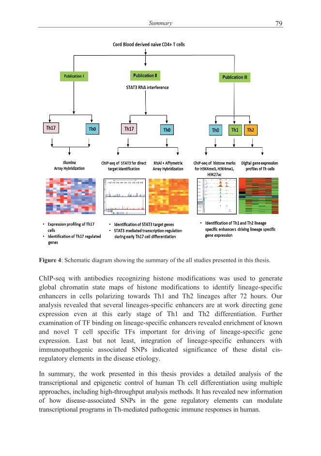
Summary 79
Figure 4: Schematic diagram showing the summary of the all studies presented in this thesis.
ChIP-seq with antibodies recognizing histone modifications was used to generate global chromatin state maps of histone modifications to identify lineage-specific enhancers in cells polarizing towards Th1 and Th2 lineages after 72 hours. Our analysis revealed that several lineages-specific enhancers are at work directing gene expression even at this early stage of Th1 and Th2 differentiation. Further examination of TF binding on lineage-specific enhancers revealed enrichment of known and novel T cell specific TFs important for driving of lineage-specific gene expression. Last but not least, integration of lineage-specific enhancers with immunopathogenic associated SNPs indicated significance of these distal cis-regulatory elements in the disease etiology.
In summary, the work presented in this thesis provides a detailed analysis of the transcriptional and epigenetic control of human Th cell differentiation using multiple approaches, including high-throughput analysis methods. It has revealed new information of how disease-associated SNPs in the gene regulatory elements can modulate transcriptional programs in Th-mediated pathogenic immune responses in human.

Acknowledgements 80
ACKNOWLEDGEMENTS
This work was carried out at the Turku Centre for Biotechnology (CBT), University of Turku and Åbo Akademi University and Department of Medical Microbiology and Immunology, Faculty of Medicine, University of Turku. I would like to acknowledge the hard work, sacrifice and support of several people, not just me during most trying times of my PhD thesis work at CBT.
First and foremost, I feel immense pleasure to express my uttermost gratitude and indebtedness to my remarkable supervisor, Professor Riitta Lahesmaa, Director of the Turku Centre for Biotechnology, for recruiting me to work and accomplishing my PhD thesis in her Molecular immunology research group. I am highly grateful for her incredible scientific insight, unconditional continuous support, providing well-equipped facilities, and the creative freedom to pursue research. You have given me opportunity to work and interact with world’s leading scientists and for giving me responsibility to work in international collaboration projects, as well as supervising students. I feel that I have learned a lot from you. Thank you for being my back-bone of moral and financial support and a great teacher!
I am highly obliged to the National Graduate School of Informational and Structural Biology (ISB) and its Director, Professor Mark Johnson for selecting me as graduate student at the school and awarding full financial support. I express my deep sense of gratitude to the members of my PhD thesis supervisory committee, Professor Anjana Rao and Dr. Brigitta Stockinger, and Professor Harri Lähdesmäki for their constructive criticism, guidance and advices during thesis committee meetings.
I am highly grateful to Professor Olli Vainio and Professor Sami Heikkinen for carefully reviewing my thesis and providing valuable comments to improve the quality of the thesis. I can say that I have learned a lot during the review process regarding scientific writing and reporting skills which will help me throughout my scientific career. I am also thankful to Dr. Robert Moulder for thoroughly reviewing the language of this thesis.
My heartfelt gratitude goes to Dr. David Hawkins for his unconditional support throughout this thesis. David, I would never forget our Skype discussions, Gmail chats, and one on one discussion about science, professional and personal problems. I am really grateful for sharing your exceptional scientific insight. Interactions with you have always given me strength to do better. Thank you from bottom of my heart.
I owe my deepest gratitude to my co-authors, Dr. David Hawkins, Dr. Zhi Chen, Dr. Soile Tuomela, Antti Larjo, Verna Salo, Dr. Riikka Lund, Dr. Kirsti Laurila, Dr. Tapio Lönnberg, Dr. Sunil K. Raghav, Dr. Bhawna Gupta, Ulrich Wagner, Ying Luu, Leonard K. Lee, Tarmo Äijo, Lotta Oikari, Isis Ricaño-Ponce, Prof. Bing Ren, Dr.

Acknowledgements 81
Brigitta Stockinger, Prof. Cisca Wijmenga, Prof. Harri Lähdesmäki and Prof. Riitta Lahesmaa for such a fruitful collaboration resulted high quality publications and made this thesis possible.
All the current and past members of the ATLAS group are sincerely acknowledged for making such a pleasant and encouraging working environment. Your support was vital especially at low times during the thesis work. I have been privileged to get to know all of you and share the lab with you! Specially, I wish to thank Dr. Sanna Filén, Verna Salo, Dr. Soile Tuomela, Dr. Helena Ahlfors, Dr. Elisa Närvä, Mirkka Heinonen, Dr. Minna Kyläniemi, Dr. Tapio Lönnberg, Dr. Johanna Tahvanainen, Dr. Rikka Lund, Dr. Sunil Raghav, Dr. Bhawna Gupta, Henna Kallionpää, Santosh Bhosale, Dr. Emani Maheswara Reddy, Dr. Ubaid Ullah, Moin Khan, Dr. Viveka Öling, Dr. Jussi Salmi, Dr. Nelly Rahkonen, Dr. Elizabeth Ngyen, Dr. Niina Lietzen, Dr. Sari Lehtimäki, Dr. Saara Hämälistö, Dr. Sanna Edelman, Dr. Robert Moulder, Kartiek Kanduri, Essi Laajala, Obaiah Dirasantha, Jussi Jalonen, Miro Viitala, Aki Stubb, Risto Eerola, and Adjunct Professor Omid Rasool. I would like to pay my special gratitude to Mahesh, Santosh and Verna for their friendship and support.
I am thankful to Dr. Kalyan Pasumarthy and Dr. Cristina Velensisi, members of Dr. David Hawkins laboratory for their friendship, joyful discussions and support. I also thank Dr. Zhi Chen and her laboratory menbers Imran and Brigitta for their friendship, help and support.
Marjo Hakkarainen, Sarita Heinonen, Päivi Junni and Elina Pietilä are sincerely acknowledged for excellent technical assistance. I highly appreciate their skilful work and never-ending willingness to help me with various tasks.
I express my gratitude to all supporting staff at CBT including administration specially, Sirkku Grönroos, Eva Hirvensalo, and Aila Jasmavaara, Tuula Suvanto, instrument maintenance, technical staff, IT service staff, the Finnish DNA Microarray Centre and Cell Imaging Core for their coordinated help towards the accomplishment of my goals. Anne Lahdenperä is acknowledged for her endless will to help with wet lab work and administrative issues that have been vital for the successful conduction of research.
My Indian friends, Raghu, Yashu, Hasan, Aman, Santosh Gupta, Gargi Gupta, Gurvinder, Rohini, Miraj, Bhanu, Gaurav Batra, Mahesh Tambe, Senthil, Swathi, Himanshu, Sangita, Neeraj, Amit, and many others deserve a special mention for their tremendous support. I wish to thank you, all my dear friends, for always being there for me, believing in me, being such great company, having the fascinating discussions about music, life and beyond as well as sharing so many beautiful, cheerful and memorable moments in life!

Acknowledgements 82
I owe my heartfelt and loving thanks to my wife Poonam for her support inside and outside home. Your love and care has been vital for me. So for we have passed 10 months together with love and affection and looking forward to enjoy this togetherness throughout life. Thank you! I also like to thank my father, mother, sister and brother in laws for their support.
I would like to thank my entire family for their support and care. I specially would like to thank my uncle, Sushil Tripathi and aunt, Rajni Tripathi for their love, care, support – You have played a key role in my career- Thank you! My sincerest thanks go to my Parents for their unconditional love, support and encouragement. I dedicate this work to you – you are the reason I am here today.
This work was financially supported by Academy of Finland, National Technology Agency (TEKES), Sigrid Juselius Foundation, ERASISBIO FP7 project, Turku University Hospital Fund, National Graduate School in Informational and Structural Biology, Turku University Foundation, University of Turku/Faculty of Medicine, The Sigrid Jusélius Foundation, The Finnish Cultural Foundation, Systems Biology Research Program, European Federation of Immunological Societies, Scandinavian Society for Immunology and Anti-tuberculosis foundation.
Turku, April, 2014
Subhash Kumar Tripathi

References 83
REFERENCES
Abbas, A.K., Murphy, K.M., and Sher, A. (1996). Functional diversity of helper T lymphocytes. Nature 383, 787–793.
Abbas, A.K., Benoist, C., Bluestone, J.A., Campbell, D.J., Ghosh, S., Hori, S., Jiang, S., Kuchroo, V.K., Mathis, D., Roncarolo, M.G., et al. (2013). Regulatory T cells: recommendations to simplify the nomenclature. Nat. Immunol. 14, 307–308.
Adamson, A.S., Collins, K., Laurence, A., and O’Shea, J.J. (2009). The Current STATus of lymphocyte signaling: new roles for old players. Curr. Opin. Immunol. 21, 161–166.
Adorini, L. (1999). Interleukin-12, a key cytokine in Th1-mediated autoimmune diseases. Cell. Mol. Life Sci. CMLS 55, 1610–1625.
Afkarian, M., Sedy, J.R., Yang, J., Jacobson, N.G., Cereb, N., Yang, S.Y., Murphy, T.L., and Murphy, K.M. (2002). T-bet is a STAT1-induced regulator of IL-12R expression in naïve CD4+ T cells. Nat. Immunol. 3, 549–557.
Afzali, B., Mitchell, P.J., Edozie, F.C., Povoleri, G.A.M., Dowson, S.E., Demandt, L., Walter, G., Canavan, J.B., Scotta, C., Menon, B., et al. (2013). CD161 expression characterizes a subpopulation of human regulatory T cells that produces IL-17 in a STAT3-dependent manner. Eur. J. Immunol. 43, 2043–2054.
Ahlfors, H., Limaye, A., Elo, L.L., Tuomela, S., Burute, M., Gottimukkala, K.V.P., Notani, D., Rasool, O., Galande, S., and Lahesmaa, R. (2010). SATB1 dictates expression of multiple genes including IL-5 involved in human T helper cell differentiation. Blood 116, 1443–1453.
Akimzhanov, A.M., Yang, X.O., and Dong, C. (2007). Chromatin remodeling of interleukin-17 (IL-17)-IL-17F cytokine gene locus during inflammatory helper T cell differentiation. J. Biol. Chem. 282, 5969–5972.
Akopian, V., Chan, M.M., Clement, K., Galonska, C., Gifford, C.A., Lehtola, E., Liao, J., Samavarchi-Tehrani, P., Sindhu, C., Smith, Z.D., et al. (2012). Epigenomics and chromatin dynamics. Genome Biol. 13, 313.
Alberts, B., Johnson, A., Lewis, J., Raff, M., Roberts, K., and Walter, P. (2002). Molecular biology of the cell (New York: Garland Science).
Allan, R.S., Zueva, E., Cammas, F., Schreiber, H.A., Masson, V., Belz, G.T., Roche, D., Maison, C., Quivy, J.-P., Almouzni, G., et al. (2012). An epigenetic silencing pathway controlling T helper 2 cell lineage commitment. Nature 487, 249–253.
Amadi-Obi, A., Yu, C.-R., Liu, X., Mahdi, R.M., Clarke, G.L., Nussenblatt, R.B., Gery, I., Lee, Y.S., and Egwuagu, C.E. (2007). TH17 cells contribute to uveitis and scleritis and are expanded by IL-2 and inhibited by IL-27/STAT1. Nat. Med. 13, 711–718.
Amit, I., Garber, M., Chevrier, N., Leite, A.P., Donner, Y., Eisenhaure, T., Guttman, M., Grenier, J.K., Li, W., Zuk, O., et al. (2009). Unbiased reconstruction of a mammalian transcriptional network mediating pathogen responses. Science 326, 257–263.
Amsen, D., Blander, J.M., Lee, G.R., Tanigaki, K., Honjo, T., and Flavell, R.A. (2004). Instruction of distinct CD4 T helper cell fates by different notch ligands on antigen-presenting cells. Cell 117, 515–526.
Amsen, D., Antov, A., Jankovic, D., Sher, A., Radtke, F., Souabni, A., Busslinger, M., McCright, B., Gridley, T., and Flavell, R.A. (2007). Direct regulation of Gata3 expression determines the T helper differentiation potential of Notch. Immunity 27, 89–99.
An, J., Golech, S., Klaewsongkram, J., Zhang, Y., Subedi, K., Huston, G.E., Wood, W.H., 3rd, Wersto, R.P., Becker, K.G., Swain, S.L., et al. (2011). Krüppel-like factor 4 (KLF4) directly regulates proliferation in thymocyte development and IL-17 expression during Th17 differentiation. FASEB J. Off. Publ. Fed. Am. Soc. Exp. Biol. 25, 3634–3645.
References 84
Anders, S., and Huber, W. (2010). Differential expression analysis for sequence count data. Genome Biol. 11.
Anderson, C.A., Boucher, G., Lees, C.W., Franke, A., D’Amato, M., Taylor, K.D., Lee, J.C., Goyette, P., Imielinski, M., Latiano, A., et al. (2011). Meta-analysis identifies 29 additional ulcerative colitis risk loci, increasing the number of confirmed associations to 47. Nat. Genet. 43, 246–252.
Angkasekwinai, P., Chang, S.H., Thapa, M., Watarai, H., and Dong, C. (2010). Regulation of IL-9 expression by IL-25 signaling. Nat. Immunol. 11, 250–256.
Annunziato, F., Cosmi, L., Santarlasci, V., Maggi, L., Liotta, F., Mazzinghi, B., Parente, E., Filì, L., Ferri, S., Frosali, F., et al. (2007). Phenotypic and functional features of human Th17 cells. J. Exp. Med. 204, 1849–1861.
Annunziato, F., Cosmi, L., Liotta, F., Maggi, E., and Romagnani, S. (2013). Main features of human T helper 17 cells. Ann. N. Y. Acad. Sci. 1284, 66–70.
Ansel, K.M., Lee, D.U., and Rao, A. (2003). An epigenetic view of helper T cell differentiation. Nat. Immunol. 4, 616–623.
Ansel, K.M., Greenwald, R.J., Agarwal, S., Bassing, C.H., Monticelli, S., Interlandi, J., Djuretic, I.M., Lee, D.U., Sharpe, A.H., Alt, F.W., et al. (2004). Deletion of a conserved Il4 silencer impairs T helper type 1-mediated immunity. Nat. Immunol. 5, 1251–1259.
Ansel, K.M., Djuretic, I., Tanasa, B., and Rao, A. (2006). Regulation of Th2 differentiation and Il4 locus accessibility. Annu. Rev. Immunol. 24, 607–656.
Arand, J., Spieler, D., Karius, T., Branco, M.R., Meilinger, D., Meissner, A., Jenuwein, T., Xu, G., Leonhardt, H., Wolf, V., et al. (2012). In vivo control of CpG and non-CpG DNA methylation by DNA methyltransferases. PLoS Genet. 8, e1002750.
Auyeung, V.C., Ulitsky, I., McGeary, S.E., and Bartel, D.P. (2013). Beyond secondary structure: primary-sequence determinants license pri-miRNA hairpins for processing. Cell 152, 844–858.
Baba, M., Imai, T., Nishimura, M., Kakizaki, M., Takagi, S., Hieshima, K., Nomiyama, H., and Yoshie, O. (1997). Identification of CCR6, the specific receptor for a novel lymphocyte-directed CC chemokine LARC. J. Biol. Chem. 272, 14893–14898.
Baguet, A., and Bix, M. (2004). Chromatin landscape dynamics of the Il4-Il13 locus during T helper 1 and 2 development. Proc. Natl. Acad. Sci. U. S. A. 101, 11410–11415.
Bai, L., and Morozov, A.V. (2010). Gene regulation by nucleosome positioning. Trends Genet. TIG 26, 476–483.
Baine, I., Basu, S., Ames, R., Sellers, R.S., and Macian, F. (2013). Helios induces epigenetic silencing of IL2 gene expression in regulatory T cells. J. Immunol. Baltim. Md 1950 190, 1008–1016.
Baron, U., Floess, S., Wieczorek, G., Baumann, K., Grützkau, A., Dong, J., Thiel, A., Boeld, T.J., Hoffmann, P., Edinger, M., et al. (2007). DNA demethylation in the human FOXP3 locus discriminates regulatory T cells from activated FOXP3(+) conventional T cells. Eur. J. Immunol. 37, 2378–2389.
Barrett, J.C., Clayton, D.G., Concannon, P., Akolkar, B., Cooper, J.D., Erlich, H.A., Julier, C., Morahan, G., Nerup, J., Nierras, C., et al. (2009). Genome-wide association study and meta-analysis find that over 40 loci affect risk of type 1 diabetes. Nat. Genet. 41, 703–707.
Barski, A., Cuddapah, S., Cui, K., Roh, T.-Y., Schones, D.E., Wang, Z., Wei, G., Chepelev, I., and Zhao, K. (2007). High-resolution profiling of histone methylations in the human genome. Cell 129, 823–837.
Barski, A., Jothi, R., Cuddapah, S., Cui, K., Roh, T.-Y., Schones, D.E., and Zhao, K. (2009). Chromatin poises miRNA- and protein-coding genes for expression. Genome Res. 19, 1742–1751.
Bartel, D.P. (2009). MicroRNAs: target recognition and regulatory functions. Cell 136, 215–233.
Basso, K., Sumazin, P., Morozov, P., Schneider, C., Maute, R.L., Kitagawa, Y., Mandelbaum, J., Haddad, J., Jr, Chen, C.-Z., Califano, A., et al. (2009). Identification of the human mature B cell miRNome. Immunity 30, 744–752.

References 85
Basu, A., Hoerning, A., Datta, D., Edelbauer, M., Stack, M.P., Calzadilla, K., Pal, S., and Briscoe, D.M. (2010). Cutting edge: Vascular endothelial growth factor-mediated signaling in human CD45RO+ CD4+ T cells promotes Akt and ERK activation and costimulates IFN-gamma production. J. Immunol. Baltim. Md 1950 184, 545–549.
Basu, R., O’Quinn, D.B., Silberger, D.J., Schoeb, T.R., Fouser, L., Ouyang, W., Hatton, R.D., and Weaver, C.T. (2012). Th22 cells are an important source of IL-22 for host protection against enteropathogenic bacteria. Immunity 37, 1061–1075.
Baumjohann, D., Okada, T., and Ansel, K.M. (2011). Cutting Edge: Distinct waves of BCL6 expression during T follicular helper cell development. J. Immunol. Baltim. Md 1950 187, 2089–2092.
Bauquet, A.T., Jin, H., Paterson, A.M., Mitsdoerffer, M., Ho, I.-C., Sharpe, A.H., and Kuchroo, V.K. (2009). The costimulatory molecule ICOS regulates the expression of c-Maf and IL-21 in the development of follicular T helper cells and TH-17 cells. Nat. Immunol. 10, 167–175.
Benjamini, Y., and Hochberg, Y. (1995). Controlling the False Discovery Rate: A Practical and Powerful Approach to Multiple Testing. J. R. Stat. Soc. Ser. B Methodol. 57, 289–300.
Benjamini, Y., and Yekutieli, D. (2001). The Control of the False Discovery Rate in Multiple Testing under Dependency. Ann. Stat. 29, 1165–1188.
Berenson, L.S., Gavrieli, M., Farrar, J.D., Murphy, T.L., and Murphy, K.M. (2006). Distinct characteristics of murine STAT4 activation in response to IL-12 and IFN-alpha. J. Immunol. Baltim. Md 1950 177, 5195–5203.
Berger, S.L. (2007). The complex language of chromatin regulation during transcription. Nature 447, 407–412.
Bernstein, B.E., Mikkelsen, T.S., Xie, X., Kamal, M., Huebert, D.J., Cuff, J., Fry, B., Meissner, A., Wernig, M., Plath, K., et al. (2006). A bivalent chromatin structure marks key developmental genes in embryonic stem cells. Cell 125, 315–326.
Bettelli, E., Dastrange, M., and Oukka, M. (2005). Foxp3 interacts with nuclear factor of activated T cells and NF-kappa B to repress cytokine gene expression and effector functions of T helper cells. Proc. Natl. Acad. Sci. U. S. A. 102, 5138–5143.
Bettelli, E., Carrier, Y., Gao, W., Korn, T., Strom, T.B., Oukka, M., Weiner, H.L., and Kuchroo, V.K. (2006). Reciprocal developmental pathways for the generation of pathogenic effector TH17 and regulatory T cells. Nature 441, 235–238.
Bettelli, E., Oukka, M., and Kuchroo, V.K. (2007). T(H)-17 cells in the circle of immunity and autoimmunity. Nat. Immunol. 8, 345–350.
Bilate, A.M., and Lafaille, J.J. (2012). Induced CD4+Foxp3+ regulatory T cells in immune tolerance. Annu. Rev. Immunol. 30, 733–758.
Birzele, F., Fauti, T., Stahl, H., Lenter, M.C., Simon, E., Knebel, D., Weith, A., Hildebrandt, T., and Mennerich, D. (2011). Next-generation insights into regulatory T cells: expression profiling and FoxP3 occupancy in Human. Nucleic Acids Res. 39, 7946–7960.
Blom, L., Poulsen, B.C., Jensen, B.M., Hansen, A., and Poulsen, L.K. (2011). IL-33 induces IL-9 production in human CD4+ T cells and basophils. PloS One 6, e21695.
Bock, C., Kiskinis, E., Verstappen, G., Gu, H., Boulting, G., Smith, Z.D., Ziller, M., Croft, G.F., Amoroso, M.W., Oakley, D.H., et al. (2011). Reference Maps of human ES and iPS cell variation enable high-throughput characterization of pluripotent cell lines. Cell 144, 439–452.
Bock, C., Beerman, I., Lien, W.-H., Smith, Z.D., Gu, H., Boyle, P., Gnirke, A., Fuchs, E., Rossi, D.J., and Meissner, A. (2012). DNA methylation dynamics during in vivo differentiation of blood and skin stem cells. Mol. Cell 47, 633–647.
Bollig, N., Brüstle, A., Kellner, K., Ackermann, W., Abass, E., Raifer, H., Camara, B., Brendel, C., Giel, G., Bothur, E., et al. (2012). Transcription factor IRF4 determines germinal center formation through follicular T-helper cell differentiation. Proc. Natl. Acad. Sci. U. S. A. 109, 8664–8669.

References 86
Boniface, K., Guignouard, E., Pedretti, N., Garcia, M., Delwail, A., Bernard, F.-X., Nau, F., Guillet, G., Dagregorio, G., Yssel, H., et al. (2007). A role for T cell-derived interleukin 22 in psoriatic skin inflammation. Clin. Exp. Immunol. 150, 407–415.
Bopp, T., Palmetshofer, A., Serfling, E., Heib, V., Schmitt, S., Richter, C., Klein, M., Schild, H., Schmitt, E., and Stassen, M. (2005). NFATc2 and NFATc3 transcription factors play a crucial role in suppression of CD4+ T lymphocytes by CD4+ CD25+ regulatory T cells. J. Exp. Med. 201, 181–187.
Bosnjak, B., Stelzmueller, B., Erb, K.J., and Epstein, M.M. (2011). Treatment of allergic asthma: modulation of Th2 cells and their responses. Respir. Res. 12, 114.
Boyle, A.P., Hong, E.L., Hariharan, M., Cheng, Y., Schaub, M.A., Kasowski, M., Karczewski, K.J., Park, J., Hitz, B.C., Weng, S., et al. (2012). Annotation of functional variation in personal genomes using RegulomeDB. Genome Res. 22, 1790–1797.
Braunstein, M., and Anderson, M.K. (2012). HEB in the spotlight: Transcriptional regulation of T-cell specification, commitment, and developmental plasticity. Clin. Dev. Immunol. 2012.
Brentnall, T.A. (2012). Arousal of cancer-associated stromal fibroblasts: palladin-activated fibroblasts promote tumor invasion. Cell Adhes. Migr. 6, 488–494.
Brüstle, A., Heink, S., Huber, M., Rosenplänter, C., Stadelmann, C., Yu, P., Arpaia, E., Mak, T.W., Kamradt, T., and Lohoff, M. (2007). The development of inflammatory T(H)-17 cells requires interferon-regulatory factor 4. Nat. Immunol. 8, 958–966.
Bryzgalov, L.O., Antontseva, E.V., Matveeva, M.Y., Shilov, A.G., Kashina, E.V., Mordvinov, V.A., and Merkulova, T.I. (2013). Detection of Regulatory SNPs in Human Genome Using ChIP-seq ENCODE Data. PloS One 8, e78833.
Burchill, M.A., Yang, J., Vogtenhuber, C., Blazar, B.R., and Farrar, M.A. (2007). IL-2 receptor beta-dependent STAT5 activation is required for the development of Foxp3+ regulatory T cells. J. Immunol. Baltim. Md 1950 178, 280–290.
Cantone, I., and Fisher, A.G. (2013). Epigenetic programming and reprogramming during development. Nat. Struct. Mol. Biol. 20, 282–289.
Caraballo, L., and Zakzuk, J. (2012). [The evolution of the Th2 immune responses and its relationships with parasitic diseases and allergy]. Bioméd. Rev. Inst. Nac. Salud 32, 145–157.
Carey, M.F., Peterson, C.L., and Smale, S.T. (2012). Identifying cis-acting DNA elements within a control region. Cold Spring Harb. Protoc. 2012, 279–296.
Cargill, M., Schrodi, S.J., Chang, M., Garcia, V.E., Brandon, R., Callis, K.P., Matsunami, N., Ardlie, K.G., Civello, D., Catanese, J.J., et al. (2007). A large-scale genetic association study confirms IL12B and leads to the identification of IL23R as psoriasis-risk genes. Am. J. Hum. Genet. 80, 273–290.
Caruso, R., Fina, D., Paoluzi, O.A., Del Vecchio Blanco, G., Stolfi, C., Rizzo, A., Caprioli, F., Sarra, M., Andrei, F., Fantini, M.C., et al. (2008). IL-23-mediated regulation of IL-17 production in Helicobacter pylori-infected gastric mucosa. Eur. J. Immunol. 38, 470–478.
Cesi, V., Giuffrida, M.L., Vitali, R., Tanno, B., Mancini, C., Calabretta, B., and Raschellà, G. (2005). C/EBP alpha and beta mimic retinoic acid activation of IGFBP-5 in neuroblastoma cells by a mechanism independent from binding to their site. Exp. Cell Res. 305, 179–189.
Challen, G.A., Sun, D., Jeong, M., Luo, M., Jelinek, J., Berg, J.S., Bock, C., Vasanthakumar, A., Gu, H., Xi, Y., et al. (2012). Dnmt3a is essential for hematopoietic stem cell differentiation. Nat. Genet. 44, 23–31.
Chalmin, F., Mignot, G., Bruchard, M., Chevriaux, A., Végran, F., Hichami, A., Ladoire, S., Derangère, V., Vincent, J., Masson, D., et al. (2012). Stat3 and Gfi-1 transcription factors control Th17 cell immunosuppressive activity via the regulation of ectonucleotidase expression. Immunity 36, 362–373.
Chang, S., and Aune, T.M. (2007). Dynamic changes in histone-methylation “marks” across the locus encoding interferon-gamma during the differentiation of T helper type 2 cells. Nat. Immunol. 8, 723–731.

References 87
Chang, H.-C., Sehra, S., Goswami, R., Yao, W., Yu, Q., Stritesky, G.L., Jabeen, R., McKinley, C., Ahyi, A.-N., Han, L., et al. (2010). The transcription factor PU.1 is required for the development of IL-9-producing T cells and allergic inflammation. Nat. Immunol. 11, 527–534.
Chaudhry, A., Rudra, D., Treuting, P., Samstein, R.M., Liang, Y., Kas, A., and Rudensky, A.Y. (2009). CD4+ regulatory T cells control TH17 responses in a Stat3-dependent manner. Science 326, 986–991.
Chen, Z., and O’Shea, J.J. (2008). Th17 cells: a new fate for differentiating helper T cells. Immunol. Res. 41, 87–102.
Chen, Q., Kim, Y.C., Laurence, A., Punkosdy, G.A., and Shevach, E.M. (2011). IL-2 controls the stability of Foxp3 expression in TGF-beta-induced Foxp3+ T cells in vivo. J. Immunol. Baltim. Md 1950 186, 6329–6337.
Chen, Z., Lund, R., Aittokallio, T., Kosonen, M., Nevalainen, O., and Lahesmaa, R. (2003). Identification of novel IL-4/Stat6-regulated genes in T lymphocytes. J. Immunol. Baltim. Md 1950 171, 3627–3635.
Chen, Z., Tato, C.M., Muul, L., Laurence, A., and O’Shea, J.J. (2007). Distinct regulation of interleukin-17 in human T helper lymphocytes. Arthritis Rheum. 56, 2936–2946.
Cheung, V.G., and Spielman, R.S. (2009). Genetics of human gene expression: mapping DNA variants that influence gene expression. Nat. Rev. Genet. 10, 595–604.
Choi, Y.S., Yang, J.A., Yusuf, I., Johnston, R.J., Greenbaum, J., Peters, B., and Crotty, S. (2013). Bcl6 expressing follicular helper CD4 T cells are fate committed early and have the capacity to form memory. J. Immunol. Baltim. Md 1950 190, 4014–4026.
Chong, M.M.W., Rasmussen, J.P., Rudensky, A.Y., Rundensky, A.Y., and Littman, D.R. (2008). The RNAseIII enzyme Drosha is critical in T cells for preventing lethal inflammatory disease. J. Exp. Med. 205, 2005–2017.
Chugh, S., Anand, V., Swaroop, L., Sharma, M., Seth, A., and Sharma, A. (2013). Involvement of Th17 cells in patients of urothelial carcinoma of bladder. Hum. Immunol. 74, 1258–1262.
Chung, Y., Tanaka, S., Chu, F., Nurieva, R.I., Martinez, G.J., Rawal, S., Wang, Y.-H., Lim, H., Reynolds, J.M., Zhou, X., et al. (2011). Follicular regulatory T cells expressing Foxp3 and Bcl-6 suppress germinal center reactions. Nat. Med. 17, 983–988.
Ciofani, M., Madar, A., Galan, C., Sellars, M., Mace, K., Pauli, F., Agarwal, A., Huang, W., Parkurst, C.N., Muratet, M., et al. (2012). A validated regulatory network for Th17 cell specification. Cell 151, 289–303.
Codarri, L., Gyülvészi, G., Tosevski, V., Hesske, L., Fontana, A., Magnenat, L., Suter, T., and Becher, B. (2011). RORγt drives production of the cytokine GM-CSF in helper T cells, which is essential for the effector phase of autoimmune neuroinflammation. Nat. Immunol. 12, 560–567.
Coffman, R.L. (2006). Origins of the T(H)1-T(H)2 model: a personal perspective. Nat. Immunol. 7, 539–541.
Coffman, R.L., and Mosmann, T.R. (1991). CD4+ T-cell subsets: regulation of differentiation and function. Res. Immunol. 142, 7–9.
Collier, S.P., Collins, P.L., Williams, C.L., Boothby, M.R., and Aune, T.M. (2012). Cutting edge: influence of Tmevpg1, a long intergenic noncoding RNA, on the expression of Ifng by Th1 cells. J. Immunol. Baltim. Md 1950 189, 2084–2088.
Collins, P.L., Chang, S., Henderson, M., Soutto, M., Davis, G.M., McLoed, A.G., Townsend, M.J., Glimcher, L.H., Mortlock, D.P., and Aune, T.M. (2010). Distal regions of the human IFNG locus direct cell type-specific expression. J. Immunol. Baltim. Md 1950 185, 1492–1501.
Corvaisier, M., Delneste, Y., Jeanvoine, H., Preisser, L., Blanchard, S., Garo, E., Hoppe, E., Barré, B., Audran, M., Bouvard, B., et al. (2012). IL-26 is overexpressed in rheumatoid arthritis and induces proinflammatory cytokine production and Th17 cell generation. PLoS Biol. 10, e1001395.

References 88
Cosmi, L., Annunziato, F., Galli MIG, Maggi RME, Nagata, K., and Romagnani, S. (2000). CRTH2 is the most reliable marker for the detection of circulating human type 2 Th and type 2 T cytotoxic cells in health and disease. Eur. J. Immunol. 30, 2972–2979.
Cosmi, L., Cimaz, R., Maggi, L., Santarlasci, V., Capone, M., Borriello, F., Frosali, F., Querci, V., Simonini, G., Barra, G., et al. (2011). Evidence of the transient nature of the Th17 phenotype of CD4+CD161+ T cells in the synovial fluid of patients with juvenile idiopathic arthritis. Arthritis Rheum. 63, 2504–2515.
Costa, F.F. (2005). Non-coding RNAs: new players in eukaryotic biology. Gene 357, 83–94.
Creyghton, M.P., Cheng, A.W., Welstead, G.G., Kooistra, T., Carey, B.W., Steine, E.J., Hanna, J., Lodato, M.A., Frampton, G.M., Sharp, P.A., et al. (2010). Histone H3K27ac separates active from poised enhancers and predicts developmental state. Proc. Natl. Acad. Sci. U. S. A. 107, 21931–21936.
Crotty, S. (2011). Follicular helper CD4 T cells (TFH). Annu. Rev. Immunol. 29, 621–663.
Curotto de Lafaille, M.A., and Lafaille, J.J. (2009). Natural and adaptive foxp3+ regulatory T cells: more of the same or a division of labor? Immunity 30, 626–635.
Curtis, M.M., and Way, S.S. (2009). Interleukin-17 in host defence against bacterial, mycobacterial and fungal pathogens. Immunology 126, 177–185.
Curtis, M.M., Rowell, E., Shafiani, S., Negash, A., Urdahl, K.B., Wilson, C.B., and Way, S.S. (2010). Fidelity of pathogen-specific CD4+ T cells to the Th1 lineage is controlled by exogenous cytokines, interferon-gamma expression, and pathogen lifestyle. Cell Host Microbe 8, 163–173.
Dambacher, J., Beigel, F., Zitzmann, K., De Toni, E.N., Göke, B., Diepolder, H.M., Auernhammer, C.J., and Brand, S. (2009). The role of the novel Th17 cytokine IL-26 in intestinal inflammation. Gut 58, 1207–1217.
Damsker, J.M., Hansen, A.M., and Caspi, R.R. (2010). Th1 and Th17 cells: adversaries and collaborators. Ann. N. Y. Acad. Sci. 1183, 211–221.
Dang, E.V., Barbi, J., Yang, H.-Y., Jinasena, D., Yu, H., Zheng, Y., Bordman, Z., Fu, J., Kim, Y., Yen, H.-R., et al. (2011). Control of T(H)17/T(reg) balance by hypoxia-inducible factor 1. Cell 146, 772–784.
Dardalhon, V., Awasthi, A., Kwon, H., Galileos, G., Gao, W., Sobel, R.A., Mitsdoerffer, M., Strom, T.B., Elyaman, W., Ho, I.-C., et al. (2008). IL-4 inhibits TGF-beta-induced Foxp3+ T cells and, together with TGF-beta, generates IL-9+ IL-10+ Foxp3(-) effector T cells. Nat. Immunol. 9, 1347–1355.
David-Fung, E.-S., Butler, R., Buzi, G., Yui, M.A., Diamond, R.A., Anderson, M.K., Rowen, L., and Rothenberg, E.V. (2009). Transcription factor expression dynamics of early T-lymphocyte specification and commitment. Dev. Biol. 325, 444–467.
Davidson, S.I., Jiang, L., Cortes, A., Wu, X., Glazov, E.A., Donskoi, M., Zheng, Y., Danoy, P.A., Liu, Y., Thomas, G.P., et al. (2013). Brief report: high-throughput sequencing of IL23R reveals a low-frequency, nonsynonymous single-nucleotide polymorphism that is associated with ankylosing spondylitis in a Han Chinese population. Arthritis Rheum. 65, 1747–1752.
Deenick, E.K., Ma, C.S., Brink, R., and Tangye, S.G. (2011). Regulation of T follicular helper cell formation and function by antigen presenting cells. Curr. Opin. Immunol. 23, 111–118.
Deftos, M.L., Huang, E., Ojala, E.W., Forbush, K.A., and Bevan, M.J. (2000). Notch1 signaling promotes the maturation of CD4 and CD8 SP thymocytes. Immunity 13, 73–84.
Delgoffe, G.M., Bettini, M.L., and Vignali, D.A.A. (2012). Identity crisis: it’s not just Foxp3 anymore. Immunity 37, 759–761.
Dilworth, F.J., Fromental-Ramain, C., Yamamoto, K., and Chambon, P. (2000). ATP-driven chromatin remodeling activity and histone acetyltransferases act sequentially during transactivation by RAR/RXR In vitro. Mol. Cell 6, 1049–1058.
Djuretic, I.M., Levanon, D., Negreanu, V., Groner, Y., Rao, A., and Ansel, K.M. (2007). Transcription factors T-bet and Runx3 cooperate to activate Ifng and silence Il4 in T helper type 1 cells. Nat. Immunol. 8, 145–153.

References 89
Dowen, R.H., Pelizzola, M., Schmitz, R.J., Lister, R., Dowen, J.M., Nery, J.R., Dixon, J.E., and Ecker, J.R. (2012). Widespread dynamic DNA methylation in response to biotic stress. Proc. Natl. Acad. Sci. U. S. A. 109, E2183–2191.
Duerr, R.H., Taylor, K.D., Brant, S.R., Rioux, J.D., Silverberg, M.S., Daly, M.J., Steinhart, A.H., Abraham, C., Regueiro, M., Griffiths, A., et al. (2006). A genome-wide association study identifies IL23R as an inflammatory bowel disease gene. Science 314, 1461–1463.
Duhen, R., Glatigny, S., Arbelaez, C.A., Blair, T.C., Oukka, M., and Bettelli, E. (2013). Cutting edge: the pathogenicity of IFN-γ-producing Th17 cells is independent of T-bet. J. Immunol. Baltim. Md 1950 190, 4478–4482.
Duhen, T., Geiger, R., Jarrossay, D., Lanzavecchia, A., and Sallusto, F. (2009). Production of interleukin 22 but not interleukin 17 by a subset of human skin-homing memory T cells. Nat. Immunol. 10, 857–863.
Durant, L., Watford, W.T., Ramos, H.L., Laurence, A., Vahedi, G., Wei, L., Takahashi, H., Sun, H.-W., Kanno, Y., Powrie, F., et al. (2010). Diverse targets of the transcription factor STAT3 contribute to T cell pathogenicity and homeostasis. Immunity 32, 605–615.
El-behi, M., Rostami, A., and Ciric, B. (2010). Current views on the roles of Th1 and Th17 cells in experimental autoimmune encephalomyelitis. J. Neuroimmune Pharmacol. Off. J. Soc. NeuroImmune Pharmacol. 5, 189–197.
Ellyard, J.I., and Vinuesa, C.G. (2011). A BATF-ling connection between B cells and follicular helper T cells. Nat. Immunol. 12, 519–520.
Elo, L.L., Järvenpää, H., Tuomela, S., Raghav, S., Ahlfors, H., Laurila, K., Gupta, B., Lund, R.J., Tahvanainen, J., Hawkins, R.D., et al. (2010). Genome-wide profiling of interleukin-4 and STAT6 transcription factor regulation of human Th2 cell programming. Immunity 32, 852–862.
Elyaman, W., Bassil, R., Bradshaw, E.M., Orent, W., Lahoud, Y., Zhu, B., Radtke, F., Yagita, H., and Khoury, S.J. (2012). Notch receptors and Smad3 signaling cooperate in the induction of interleukin-9-producing T cells. Immunity 36, 623–634.
ENCODE Project Consortium, Bernstein, B.E., Birney, E., Dunham, I., Green, E.D., Gunter, C., and Snyder, M. (2012). An integrated encyclopedia of DNA elements in the human genome. Nature 489, 57–74.
Engel, I., Zhao, M., Kappes, D., Taniuchi, I., and Kronenberg, M. (2012). The transcription factor Th-POK negatively regulates Th17 differentiation in Vα14i NKT cells. Blood 120, 4524–4532.
Ernst, J., Kheradpour, P., Mikkelsen, T.S., Shoresh, N., Ward, L.D., Epstein, C.B., Zhang, X., Wang, L., Issner, R., Coyne, M., et al. (2011). Mapping and analysis of chromatin state dynamics in nine human cell types. Nature 473, 43–49.
Evans, C.M., and Jenner, R.G. (2013). Transcription factor interplay in T helper cell differentiation. Brief. Funct. Genomics 12, 499–511.
Eyerich, S., Eyerich, K., Pennino, D., Carbone, T., Nasorri, F., Pallotta, S., Cianfarani, F., Odorisio, T., Traidl-Hoffmann, C., Behrendt, H., et al. (2009). Th22 cells represent a distinct human T cell subset involved in epidermal immunity and remodeling. J. Clin. Invest. 119, 3573–3585.
Facciabene, A., Motz, G.T., and Coukos, G. (2012). T-regulatory cells: key players in tumor immune escape and angiogenesis. Cancer Res. 72, 2162–2171.
Fallon, P.G., and Mangan, N.E. (2007). Suppression of TH2-type allergic reactions by helminth infection. Nat. Rev. Immunol. 7, 220–230.
Fang, T.C., Yashiro-Ohtani, Y., Del Bianco, C., Knoblock, D.M., Blacklow, S.C., and Pear, W.S. (2007). Notch directly regulates Gata3 expression during T helper 2 cell differentiation. Immunity 27, 100–110.
Farkas, G., Leibovitch, B.A., and Elgin, S.C. (2000). Chromatin organization and transcriptional control of gene expression in Drosophila. Gene 253, 117–136.

References 90
Ficz, G., Branco, M.R., Seisenberger, S., Santos, F., Krueger, F., Hore, T.A., Marques, C.J., Andrews, S., and Reik, W. (2011). Dynamic regulation of 5-hydroxymethylcytosine in mouse ES cells and during differentiation. Nature 473, 398–402.
Fields, P.E., Lee, G.R., Kim, S.T., Bartsevich, V.V., and Flavell, R.A. (2004). Th2-specific chromatin remodeling and enhancer activity in the Th2 cytokine locus control region. Immunity 21, 865–876.
Filén, S., Ylikoski, E., Tripathi, S., West, A., Björkman, M., Nyström, J., Ahlfors, H., Coffey, E., Rao, K.V.S., Rasool, O., et al. (2010). Activating transcription factor 3 is a positive regulator of human IFNG gene expression. J. Immunol. Baltim. Md 1950 184, 4990–4999.
Finkelman, F.D., Morris, S.C., Orekhova, T., Mori, M., Donaldson, D., Reiner, S.L., Reilly, N.L., Schopf, L., and Urban, J.F., Jr (2000). Stat6 regulation of in vivo IL-4 responses. J. Immunol. Baltim. Md 1950 164, 2303–2310.
Floess, S., Freyer, J., Siewert, C., Baron, U., Olek, S., Polansky, J., Schlawe, K., Chang, H.-D., Bopp, T., Schmitt, E., et al. (2007). Epigenetic control of the foxp3 locus in regulatory T cells. PLoS Biol. 5, e38.
Fontenot, J.D., Gavin, M.A., and Rudensky, A.Y. (2003). Foxp3 programs the development and function of CD4+CD25+ regulatory T cells. Nat. Immunol. 4, 330–336.
Francis, N.J., Kingston, R.E., and Woodcock, C.L. (2004). Chromatin compaction by a polycomb group protein complex. Science 306, 1574–1577.
Franco, C.B., Scripture-Adams, D.D., Proekt, I., Taghon, T., Weiss, A.H., Yui, M.A., Adams, S.L., Diamond, R.A., and Rothenberg, E.V. (2006). Notch/Delta signaling constrains reengineering of pro-T cells by PU.1. Proc. Natl. Acad. Sci. U. S. A. 103, 11993–11998.
Fu, W., Ergun, A., Lu, T., Hill, J.A., Haxhinasto, S., Fassett, M.S., Gazit, R., Adoro, S., Glimcher, L., Chan, S., et al. (2012). A multiply redundant genetic switch “locks in” the transcriptional signature of regulatory T cells. Nat. Immunol. 13, 972–980.
Fujita, H., Nograles, K.E., Kikuchi, T., Gonzalez, J., Carucci, J.A., and Krueger, J.G. (2009). Human Langerhans cells induce distinct IL-22-producing CD4+ T cells lacking IL-17 production. Proc. Natl. Acad. Sci. U. S. A. 106, 21795–21800.
Galand, C., Donnou, S., Crozet, L., Brunet, S., Touitou, V., Ouakrim, H., Fridman, W.H., Sautès-Fridman, C., and Fisson, S. (2011). Th17 cells are involved in the local control of tumor progression in primary intraocular lymphoma. PloS One 6, e24622.
Gautier, L., Cope, L., Bolstad, B.M., and Irizarry, R.A. (2004). affy--analysis of Affymetrix GeneChip data at the probe level. Bioinforma. Oxf. Engl. 20, 307–315.
Gavin, M., and Rudensky, A. (2003). Control of immune homeostasis by naturally arising regulatory CD4+ T cells. Curr. Opin. Immunol. 15, 690–696.
Gavin, M.A., Rasmussen, J.P., Fontenot, J.D., Vasta, V., Manganiello, V.C., Beavo, J.A., and Rudensky, A.Y. (2007). Foxp3-dependent programme of regulatory T-cell differentiation. Nature 445, 771–775.
Ge, M., Zheng, Y., Li, X., Lu, S., Li, H., Chen, F., Chen, D., Shao, Y., Shi, J., and Feng, S. (2013). Differential expression profile of Th1/Th17/Th2-related chemokines and their receptors in patients with acquired bone marrow failure syndromes. Hum. Immunol. 74, 176–180.
Georas, S.N., Guo, J., De Fanis, U., and Casolaro, V. (2005). T-helper cell type-2 regulation in allergic disease. Eur. Respir. J. 26, 1119–1137.
Germann, T., Gately, M.K., Schoenhaut, D.S., Lohoff, M., Mattner, F., Fischer, S., Jin, S.C., Schmitt, E., and Rüde, E. (1993). Interleukin-12/T cell stimulating factor, a cytokine with multiple effects on T helper type 1 (Th1) but not on Th2 cells. Eur. J. Immunol. 23, 1762–1770.
Ghoreschi, K., Laurence, A., Yang, X.-P., Tato, C.M., McGeachy, M.J., Konkel, J.E., Ramos, H.L., Wei, L., Davidson, T.S., Bouladoux, N., et al. (2010). Generation of pathogenic T(H)17 cells in the absence of TGF-β signalling. Nature 467, 967–971.
References 91
Gifford, C.A., Ziller, M.J., Gu, H., Trapnell, C., Donaghey, J., Tsankov, A., Shalek, A.K., Kelley, D.R., Shishkin, A.A., Issner, R., et al. (2013). Transcriptional and epigenetic dynamics during specification of human embryonic stem cells. Cell 153, 1149–1163.
Gimferrer, I., Hu, T., Simmons, A., Wang, C., Souabni, A., Busslinger, M., Bender, T.P., Hernandez-Hoyos, G., and Alberola-Ila, J. (2011). Regulation of GATA-3 expression during CD4 lineage differentiation. J. Immunol. Baltim. Md 1950 186, 3892–3898.
Gocke, A.R., Cravens, P.D., Ben, L.-H., Hussain, R.Z., Northrop, S.C., Racke, M.K., and Lovett-Racke, A.E. (2007). T-bet regulates the fate of Th1 and Th17 lymphocytes in autoimmunity. J. Immunol. Baltim. Md 1950 178, 1341–1348.
Goenka, S., and Kaplan, M.H. (2011). Transcriptional regulation by STAT6. Immunol. Res. 50, 87–96.
Gökmen, M.R., Dong, R., Kanhere, A., Powell, N., Perucha, E., Jackson, I., Howard, J.K., Hernandez-Fuentes, M., Jenner, R.G., and Lord, G.M. (2013). Genome-Wide Regulatory Analysis Reveals That T-bet Controls Th17 Lineage Differentiation through Direct Suppression of IRF4. J. Immunol. Baltim. Md 1950 191, 5925–5932.
Gomez, J.A., Wapinski, O.L., Yang, Y.W., Bureau, J.-F., Gopinath, S., Monack, D.M., Chang, H.Y., Brahic, M., and Kirkegaard, K. (2013). The NeST long ncRNA controls microbial susceptibility and epigenetic activation of the interferon-γ locus. Cell 152, 743–754.
Gómez-Martín, D., Díaz-Zamudio, M., Romo-Tena, J., Ibarra-Sánchez, M.J., and Alcocer-Varela, J. (2011). Follicular helper T cells poise immune responses to the development of autoimmune pathology. Autoimmun. Rev. 10, 325–330.
Good, S.R., Thieu, V.T., Mathur, A.N., Yu, Q., Stritesky, G.L., Yeh, N., O’Malley, J.T., Perumal, N.B., and Kaplan, M.H. (2009). Temporal induction pattern of STAT4 target genes defines potential for Th1 lineage-specific programming. J. Immunol. Baltim. Md 1950 183, 3839–3847.
Goodman, W.A., Cooper, K.D., and McCormick, T.S. (2012). Regulation generation: the suppressive functions of human regulatory T cells. Crit. Rev. Immunol. 32, 65–79.
Goswami, R., Jabeen, R., Yagi, R., Pham, D., Zhu, J., Goenka, S., and Kaplan, M.H. (2012). STAT6-dependent regulation of Th9 development. J. Immunol. Baltim. Md 1950 188, 968–975.
Graeber, K.E., and Olsen, N.J. (2012). Th17 cell cytokine secretion profile in host defense and autoimmunity. Inflamm. Res. Off. J. Eur. Histamine Res. Soc. Al 61, 87–96.
Greten, T.F., Zhao, F., Gamrekelashvili, J., and Korangy, F. (2012). Human Th17 cells in patients with cancer: Friends or foe? Oncoimmunology 1, 1438–1439.
Grogan, J.L., Mohrs, M., Harmon, B., Lacy, D.A., Sedat, J.W., and Locksley, R.M. (2001). Early transcription and silencing of cytokine genes underlie polarization of T helper cell subsets. Immunity 14, 205–215.
Groom, J.R., and Luster, A.D. (2011a). CXCR3 in T cell function. Exp. Cell Res. 317, 620–631.
Groom, J.R., and Luster, A.D. (2011b). CXCR3 ligands: redundant, collaborative and antagonistic functions. Immunol. Cell Biol. 89, 207–215.
Groom, J.R., Richmond, J., Murooka, T.T., Sorensen, E.W., Sung, J.H., Bankert, K., von Andrian, U.H., Moon, J.J., Mempel, T.R., and Luster, A.D. (2012). CXCR3 chemokine receptor-ligand interactions in the lymph node optimize CD4+ T helper 1 cell differentiation. Immunity 37, 1091–1103.
Gu, F.-M., Li, Q.-L., Gao, Q., Jiang, J.-H., Zhu, K., Huang, X.-Y., Pan, J.-F., Yan, J., Hu, J.-H., Wang, Z., et al. (2011). IL-17 induces AKT-dependent IL-6/JAK2/STAT3 activation and tumor progression in hepatocellular carcinoma. Mol. Cancer 10, 150.
Gu, Y., Chae, H.-D., Siefring, J.E., Jasti, A.C., Hildeman, D.A., and Williams, D.A. (2006). RhoH GTPase recruits and activates Zap70 required for T cell receptor signaling and thymocyte development. Nat. Immunol. 7, 1182–1190.
Guenther, M.G. (2011). Transcriptional control of embryonic and induced pluripotent stem cells. Epigenomics 3, 323–343.

References 92
Guibert, S., Forné, T., and Weber, M. (2009). Dynamic regulation of DNA methylation during mammalian development. Epigenomics 1, 81–98.
Guo, Z., Khattar, M., Schroder, P.M., Miyahara, Y., Wang, G., He, X., Chen, W., and Stepkowski, S.M. (2013). A dynamic dual role of IL-2 signaling in the two-step differentiation process of adaptive regulatory T cells. J. Immunol. Baltim. Md 1950 190, 3153–3162.
Guttman, M., Donaghey, J., Carey, B.W., Garber, M., Grenier, J.K., Munson, G., Young, G., Lucas, A.B., Ach, R., Bruhn, L., et al. (2011). lincRNAs act in the circuitry controlling pluripotency and differentiation. Nature 477, 295–300.
Gyles, S.L., Xue, L., Townsend, E.R., Wettey, F., and Pettipher, R. (2006). A dominant role for chemoattractant receptor-homologous molecule expressed on T helper type 2 (Th2) cells (CRTH2) in mediating chemotaxis of CRTH2+ CD4+ Th2 lymphocytes in response to mast cell supernatants. Immunology 119, 362–368.
Hackett, J.A., Sengupta, R., Zylicz, J.J., Murakami, K., Lee, C., Down, T.A., and Surani, M.A. (2013). Germline DNA demethylation dynamics and imprint erasure through 5-hydroxymethylcytosine. Science 339, 448–452.
Hafler, D.A., and De Jager, P.L. (2005). Applying a new generation of genetic maps to understand human inflammatory disease. Nat. Rev. Immunol. 5, 83–91.
Haines, C.J., Chen, Y., Blumenschein, W.M., Jain, R., Chang, C., Joyce-Shaikh, B., Porth, K., Boniface, K., Mattson, J., Basham, B., et al. (2013). Autoimmune memory T helper 17 cell function and expansion are dependent on interleukin-23. Cell Rep. 3, 1378–1388.
Hamalainen, H.K., Tubman, J.C., Vikman, S., Kyrölä, T., Ylikoski, E., Warrington, J.A., and Lahesmaa, R. (2001). Identification and validation of endogenous reference genes for expression profiling of T helper cell differentiation by quantitative real-time RT-PCR. Anal. Biochem. 299, 63–70.
Hardison, R.C., and Taylor, J. (2012). Genomic approaches towards finding cis-regulatory modules in animals. Nat. Rev. Genet. 13, 469–483.
Haribhai, D., Williams, J.B., Jia, S., Nickerson, D., Schmitt, E.G., Edwards, B., Ziegelbauer, J., Yassai, M., Li, S.-H., Relland, L.M., et al. (2011). A requisite role for induced regulatory T cells in tolerance based on expanding antigen receptor diversity. Immunity 35, 109–122.
Harris, J., Master, S.S., De Haro, S.A., Delgado, M., Roberts, E.A., Hope, J.C., Keane, J., and Deretic, V. (2009). Th1-Th2 polarisation and autophagy in the control of intracellular mycobacteria by macrophages. Vet. Immunol. Immunopathol. 128, 37–43.
Hatton, R.D., Harrington, L.E., Luther, R.J., Wakefield, T., Janowski, K.M., Oliver, J.R., Lallone, R.L., Murphy, K.M., and Weaver, C.T. (2006). A distal conserved sequence element controls Ifng gene expression by T cells and NK cells. Immunity 25, 717–729.
Hauk, G., and Bowman, G.D. (2011). Structural insights into regulation and action of SWI2/SNF2 ATPases. Curr. Opin. Struct. Biol. 21, 719–727.
Hauptman, N., and Glavac, D. (2013). MicroRNAs and long non-coding RNAs: prospects in diagnostics and therapy of cancer. Radiol. Oncol. 47, 311–318.
Hawkins, R.D., and Ren, B. (2006). Genome-wide location analysis: insights on transcriptional regulation. Hum. Mol. Genet. 15 Spec No 1, R1–7.
Hawkins, R.D., Hon, G.C., Lee, L.K., Ngo, Q., Lister, R., Pelizzola, M., Edsall, L.E., Kuan, S., Luu, Y., Klugman, S., et al. (2010). Distinct epigenomic landscapes of pluripotent and lineage-committed human cells. Cell Stem Cell 6, 479–491.
Hawkins, R.D., Larjo, A., Tripathi, S.K., Wagner, U., Luu, Y., Lönnberg, T., Raghav, S.K., Lee, L.K., Lund, R., Ren, B., et al. (2013). Global chromatin state analysis reveals lineage-specific enhancers during the initiation of human T helper 1 and T helper 2 cell polarization. Immunity 38, 1271–1284.
Hazlett, J., Stamp, L.K., Merriman, T., Highton, J., and Hessian, P.A. (2012). IL-23R rs11209026 polymorphism modulates IL-17A expression in patients with rheumatoid arthritis. Genes Immun. 13, 282–287.

References 93
He, H.H., Meyer, C.A., Shin, H., Bailey, S.T., Wei, G., Wang, Q., Zhang, Y., Xu, K., Ni, M., Lupien, M., et al. (2010). Nucleosome dynamics define transcriptional enhancers. Nat. Genet. 42, 343–347.
Heintzman, N.D., Hon, G.C., Hawkins, R.D., Kheradpour, P., Stark, A., Harp, L.F., Ye, Z., Lee, L.K., Stuart, R.K., Ching, C.W., et al. (2009). Histone modifications at human enhancers reflect global cell-type-specific gene expression. Nature 459, 108–112.
Heinz, S., Benner, C., Spann, N., Bertolino, E., Lin, Y.C., Laslo, P., Cheng, J.X., Murre, C., Singh, H., and Glass, C.K. (2010). Simple combinations of lineage-determining transcription factors prime cis-regulatory elements required for macrophage and B cell identities. Mol. Cell 38, 576–589.
Heissmeyer, V., and Vogel, K.U. (2013). Molecular control of Tfh-cell differentiation by Roquin family proteins. Immunol. Rev. 253, 273–289.
Hemdan, N.Y.A., Birkenmeier, G., Wichmann, G., Abu El-Saad, A.M., Krieger, T., Conrad, K., and Sack, U. (2010). Interleukin-17-producing T helper cells in autoimmunity. Autoimmun. Rev. 9, 785–792.
Heyn, H., and Esteller, M. (2012). DNA methylation profiling in the clinic: applications and challenges. Nat. Rev. Genet. 13, 679–692.
Hill, J.A., Feuerer, M., Tash, K., Haxhinasto, S., Perez, J., Melamed, R., Mathis, D., and Benoist, C. (2007). Foxp3 transcription-factor-dependent and -independent regulation of the regulatory T cell transcriptional signature. Immunity 27, 786–800.
Hindorff, L.A., Sethupathy, P., Junkins, H.A., Ramos, E.M., Mehta, J.P., Collins, F.S., and Manolio, T.A. (2009). Potential etiologic and functional implications of genome-wide association loci for human diseases and traits. Proc. Natl. Acad. Sci. U. S. A. 106, 9362–9367.
Hirahara, K., Vahedi, G., Ghoreschi, K., Yang, X.-P., Nakayamada, S., Kanno, Y., O’Shea, J.J., and Laurence, A. (2011). Helper T-cell differentiation and plasticity: insights from epigenetics. Immunology 134, 235–245.
Hirata, T., Osuga, Y., Takamura, M., Kodama, A., Hirota, Y., Koga, K., Yoshino, O., Harada, M., Takemura, Y., Yano, T., et al. (2010). Recruitment of CCR6-expressing Th17 cells by CCL 20 secreted from IL-1 beta-, TNF-alpha-, and IL-17A-stimulated endometriotic stromal cells. Endocrinology 151, 5468–5476.
Hirota, K., Yoshitomi, H., Hashimoto, M., Maeda, S., Teradaira, S., Sugimoto, N., Yamaguchi, T., Nomura, T., Ito, H., Nakamura, T., et al. (2007). Preferential recruitment of CCR6-expressing Th17 cells to inflamed joints via CCL20 in rheumatoid arthritis and its animal model. J. Exp. Med. 204, 2803–2812.
Hofer, M.J., Li, W., Manders, P., Terry, R., Lim, S.L., King, N.J.C., and Campbell, I.L. (2012). Mice deficient in STAT1 but not STAT2 or IRF9 develop a lethal CD4+ T-cell-mediated disease following infection with lymphocytic choriomeningitis virus. J. Virol. 86, 6932–6946.
Hoffman, B.G., Robertson, G., Zavaglia, B., Beach, M., Cullum, R., Lee, S., Soukhatcheva, G., Li, L., Wederell, E.D., Thiessen, N., et al. (2010). Locus co-occupancy, nucleosome positioning, and H3K4me1 regulate the functionality of FOXA2-, HNF4A-, and PDX1-bound loci in islets and liver. Genome Res. 20, 1037–1051.
Holliday, R. (2006). Epigenetics: a historical overview. Epigenetics Off. J. DNA Methylation Soc. 1, 76–80.
Hon, G., Wang, W., and Ren, B. (2009a). Discovery and annotation of functional chromatin signatures in the human genome. PLoS Comput. Biol. 5, e1000566.
Hon, G.C., Hawkins, R.D., and Ren, B. (2009b). Predictive chromatin signatures in the mammalian genome. Hum. Mol. Genet. 18, R195–201.
Hori, S., Nomura, T., and Sakaguchi, S. (2003). Control of regulatory T cell development by the transcription factor Foxp3. Science 299, 1057–1061.
Horiuchi, S., Onodera, A., Hosokawa, H., Watanabe, Y., Tanaka, T., Sugano, S., Suzuki, Y., and Nakayama, T. (2011). Genome-wide analysis reveals unique regulation of transcription of Th2-specific genes by GATA3. J. Immunol. Baltim. Md 1950 186, 6378–6389.

References 94
Houssiau, F.A., Renauld, J.C., Fibbe, W.E., and Van Snick, J. (1992). IL-2 dependence of IL-9 expression in human T lymphocytes. J. Immunol. Baltim. Md 1950 148, 3147–3151.
Hovav, A.-H., Mullerad, J., Davidovitch, L., Fishman, Y., Bigi, F., Cataldi, A., and Bercovier, H. (2003). The Mycobacterium tuberculosis recombinant 27-kilodalton lipoprotein induces a strong Th1-type immune response deleterious to protection. Infect. Immun. 71, 3146–3154.
Hu, G., Tang, Q., Sharma, S., Yu, F., Escobar, T.M., Muljo, S.A., Zhu, J., and Zhao, K. (2013a). Expression and regulation of intergenic long noncoding RNAs during T cell development and differentiation. Nat. Immunol. 14, 1190–1198.
Hu, R., Huffaker, T.B., Kagele, D.A., Runtsch, M.C., Bake, E., Chaudhuri, A.A., Round, J.L., and O’Connell, R.M. (2013b). MicroRNA-155 confers encephalogenic potential to Th17 cells by promoting effector gene expression. J. Immunol. Baltim. Md 1950 190, 5972–5980.
Hu, S., Young, K.H., Konoplev, S.N., and Medeiros, L.J. (2012). Follicular T-cell lymphoma: a member of an emerging family of follicular helper T-cell derived T-cell lymphomas. Hum. Pathol. 43, 1789–1798.
Huang, J., and Berger, S.L. (2008). The emerging field of dynamic lysine methylation of non-histone proteins. Curr. Opin. Genet. Dev. 18, 152–158.
Huang, C., Hatzi, K., and Melnick, A. (2013). Lineage-specific functions of Bcl-6 in immunity and inflammation are mediated by distinct biochemical mechanisms. Nat. Immunol. 14, 380–388.
Huh, J.R., and Littman, D.R. (2012). Small molecule inhibitors of RORγt: targeting Th17 cells and other applications. Eur. J. Immunol. 42, 2232–2237.
Huh, J.R., Leung, M.W.L., Huang, P., Ryan, D.A., Krout, M.R., Malapaka, R.R.V., Chow, J., Manel, N., Ciofani, M., Kim, S.V., et al. (2011). Digoxin and its derivatives suppress TH17 cell differentiation by antagonizing RORγt activity. Nature 472, 486–490.
Hwang, E.S., Szabo, S.J., Schwartzberg, P.L., and Glimcher, L.H. (2005). T helper cell fate specified by kinase-mediated interaction of T-bet with GATA-3. Science 307, 430–433.
Iida, T., Iwahashi, M., Katsuda, M., Ishida, K., Nakamori, M., Nakamura, M., Naka, T., Ojima, T., Ueda, K., Hayata, K., et al. (2011). Tumor-infiltrating CD4+ Th17 cells produce IL-17 in tumor microenvironment and promote tumor progression in human gastric cancer. Oncol. Rep. 25, 1271–1277.
International Multiple Sclerosis Genetics Consortium, Hafler, D.A., Compston, A., Sawcer, S., Lander, E.S., Daly, M.J., De Jager, P.L., de Bakker, P.I.W., Gabriel, S.B., Mirel, D.B., et al. (2007). Risk alleles for multiple sclerosis identified by a genomewide study. N. Engl. J. Med. 357, 851–862.
Iovino, N., and Cavalli, G. (2011). Rolling ES cells down the Waddington landscape with Oct4 and Sox2. Cell 145, 815–817.
Irizarry, R.A., Bolstad, B.M., Collin, F., Cope, L.M., Hobbs, B., and Speed, T.P. (2003). Summaries of Affymetrix GeneChip probe level data. Nucleic Acids Res. 31, e15.
Ise, W., Kohyama, M., Schraml, B.U., Zhang, T., Schwer, B., Basu, U., Alt, F.W., Tang, J., Oltz, E.M., Murphy, T.L., et al. (2011). The transcription factor BATF controls the global regulators of class-switch recombination in both B cells and T cells. Nat. Immunol. 12, 536–543.
Itoh, M., Takahashi, T., Sakaguchi, N., Kuniyasu, Y., Shimizu, J., Otsuka, F., and Sakaguchi, S. (1999). Thymus and autoimmunity: production of CD25+CD4+ naturally anergic and suppressive T cells as a key function of the thymus in maintaining immunologic self-tolerance. J. Immunol. Baltim. Md 1950 162, 5317–5326.
Ivanov, I.I., McKenzie, B.S., Zhou, L., Tadokoro, C.E., Lepelley, A., Lafaille, J.J., Cua, D.J., and Littman, D.R. (2006). The orphan nuclear receptor RORgammat directs the differentiation program of proinflammatory IL-17+ T helper cells. Cell 126, 1121–1133.
Ivanov, I.I., Atarashi, K., Manel, N., Brodie, E.L., Shima, T., Karaoz, U., Wei, D., Goldfarb, K.C., Santee, C.A., Lynch, S.V., et al. (2009). Induction of intestinal Th17 cells by segmented filamentous bacteria. Cell 139, 485–498.

References 95
Iwasaki, M., Nagata, K., Takano, S., Takahashi, K., Ishii, N., and Ikezawa, Z. (2002). Association of a new-type prostaglandin D2 receptor CRTH2 with circulating T helper 2 cells in patients with atopic dermatitis. J. Invest. Dermatol. 119, 609–616.
Jabeen, R., Goswami, R., Awe, O., Kulkarni, A., Nguyen, E.T., Attenasio, A., Walsh, D., Olson, M.R., Kim, M.H., Tepper, R.S., et al. (2013). Th9 cell development requires a BATF-regulated transcriptional network. J. Clin. Invest.
Jäger, A., Dardalhon, V., Sobel, R.A., Bettelli, E., and Kuchroo, V.K. (2009). Th1, Th17, and Th9 effector cells induce experimental autoimmune encephalomyelitis with different pathological phenotypes. J. Immunol. Baltim. Md 1950 183, 7169–7177.
Janeway, C.A., Jr (1989). The priming of helper T cells. Semin. Immunol. 1, 13–20.
Janeway, C., Travers, J., Paul, Walport, M., and Shlomchik, M.J. (2001). Immunobiology 5: the immune system in health and disease (New York: Garland Pub.).
Jankovic, D., Kullberg, M.C., Noben-Trauth, N., Caspar, P., Paul, W.E., and Sher, A. (2000). Single cell analysis reveals that IL-4 receptor/Stat6 signaling is not required for the in vivo or in vitro development of CD4+ lymphocytes with a Th2 cytokine profile. J. Immunol. Baltim. Md 1950 164, 3047–3055.
Jankovic, D., Kullberg, M.C., Feng, C.G., Goldszmid, R.S., Collazo, C.M., Wilson, M., Wynn, T.A., Kamanaka, M., Flavell, R.A., and Sher, A. (2007). Conventional T-bet(+)Foxp3(-) Th1 cells are the major source of host-protective regulatory IL-10 during intracellular protozoan infection. J. Exp. Med. 204, 273–283.
Janson, P.C.J., Winerdal, M.E., Marits, P., Thörn, M., Ohlsson, R., and Winqvist, O. (2008). FOXP3 promoter demethylation reveals the committed Treg population in humans. PloS One 3, e1612.
Jenner, R.G., Townsend, M.J., Jackson, I., Sun, K., Bouwman, R.D., Young, R.A., Glimcher, L.H., and Lord, G.M. (2009). The transcription factors T-bet and GATA-3 control alternative pathways of T-cell differentiation through a shared set of target genes. Proc. Natl. Acad. Sci. U. S. A. 106, 17876–17881.
Jenuwein, T. (2001). Re-SET-ting heterochromatin by histone methyltransferases. Trends Cell Biol. 11, 266–273.
Jiang, H., Shukla, A., Wang, X., Chen, W., Bernstein, B.E., and Roeder, R.G. (2011a). Role for Dpy-30 in ES cell-fate specification by regulation of H3K4 methylation within bivalent domains. Cell 144, 513–525.
Jiang, R., Wang, H., Deng, L., Hou, J., Shi, R., Yao, M., Gao, Y., Yao, A., Wang, X., Yu, L., et al. (2013). IL-22 is related to development of human colon cancer by activation of STAT3. BMC Cancer 13, 59.
Jiang, S., Li, C., Olive, V., Lykken, E., Feng, F., Sevilla, J., Wan, Y., He, L., and Li, Q.-J. (2011b). Molecular dissection of the miR-17-92 cluster’s critical dual roles in promoting Th1 responses and preventing inducible Treg differentiation. Blood 118, 5487–5497.
Johnson, D.S., Mortazavi, A., Myers, R.M., and Wold, B. (2007). Genome-wide mapping of in vivo protein-DNA interactions. Science 316, 1497–1502.
Johnson, E.O., Chang, K.-H., de Pablo, Y., Ghosh, S., Mehta, R., Badve, S., and Shah, K. (2011). PHLDA1 is a crucial negative regulator and effector of Aurora A kinase in breast cancer. J. Cell Sci. 124, 2711–2722.
Johnston, R.J., Poholek, A.C., DiToro, D., Yusuf, I., Eto, D., Barnett, B., Dent, A.L., Craft, J., and Crotty, S. (2009). Bcl6 and Blimp-1 are reciprocal and antagonistic regulators of T follicular helper cell differentiation. Science 325, 1006–1010.
Johnston, R.J., Choi, Y.S., Diamond, J.A., Yang, J.A., and Crotty, S. (2012). STAT5 is a potent negative regulator of TFH cell differentiation. J. Exp. Med. 209, 243–250.
Jones, B., and Chen, J. (2006). Inhibition of IFN-gamma transcription by site-specific methylation during T helper cell development. EMBO J. 25, 2443–2452.

References 96
Joshi, S., Pantalena, L.-C., Liu, X.K., Gaffen, S.L., Liu, H., Rohowsky-Kochan, C., Ichiyama, K., Yoshimura, A., Steinman, L., Christakos, S., et al. (2011). 1,25-dihydroxyvitamin D(3) ameliorates Th17 autoimmunity via transcriptional modulation of interleukin-17A. Mol. Cell. Biol. 31, 3653–3669.
Jost, E., Lin, Q., Weidner, C.I., Wilop, S., Hoffmann, M., Walenda, T., Schemionek, M., Herrmann, O., Zenke, M., Brümmendorf, T.H., et al. (2013). Epimutations mimic genomic mutations of DNMT3A in acute myeloid leukemia. Leukemia.
Kagami, S., Nakajima, H., Suto, A., Hirose, K., Suzuki, K., Morita, S., Kato, I., Saito, Y., Kitamura, T., and Iwamoto, I. (2001). Stat5a regulates T helper cell differentiation by several distinct mechanisms. Blood 97, 2358–2365.
Kagami, S., Rizzo, H.L., Lee, J.J., Koguchi, Y., and Blauvelt, A. (2010). Circulating Th17, Th22, and Th1 cells are increased in psoriasis. J. Invest. Dermatol. 130, 1373–1383.
Kanhere, A., Hertweck, A., Bhatia, U., Gökmen, M.R., Perucha, E., Jackson, I., Lord, G.M., and Jenner, R.G. (2012). T-bet and GATA3 orchestrate Th1 and Th2 differentiation through lineage-specific targeting of distal regulatory elements. Nat. Commun. 3, 1268.
Kanno, Y., Vahedi, G., Hirahara, K., Singleton, K., and O’Shea, J.J. (2012). Transcriptional and epigenetic control of T helper cell specification: molecular mechanisms underlying commitment and plasticity. Annu. Rev. Immunol. 30, 707–731.
Kaplan, M.H. (2013). Th9 cells: differentiation and disease. Immunol. Rev. 252, 104–115.
Kaplan, M.H., Schindler, U., Smiley, S.T., and Grusby, M.J. (1996). Stat6 is required for mediating responses to IL-4 and for development of Th2 cells. Immunity 4, 313–319.
Kappes, D.J. (2010). Expanding roles for ThPOK in thymic development. Immunol. Rev. 238, 182–194.
Kara, E.E., Comerford, I., Bastow, C.R., Fenix, K.A., Litchfield, W., Handel, T.M., and McColl, S.R. (2013). Distinct chemokine receptor axes regulate Th9 cell trafficking to allergic and autoimmune inflammatory sites. J. Immunol. Baltim. Md 1950 191, 1110–1117.
Karagiannis, T.C., and El-Osta, A. (2004). siRNAs: mechanism of RNA interference, in vivo and potential clinical applications. Cancer Biol. Ther. 3, 1069–1074.
Kashiwada, M., Cassel, S.L., Colgan, J.D., and Rothman, P.B. (2011). NFIL3/E4BP4 controls type 2 T helper cell cytokine expression. EMBO J. 30, 2071–2082.
Kasowski, M., Kyriazopoulou-Panagiotopoulou, S., Grubert, F., Zaugg, J.B., Kundaje, A., Liu, Y., Boyle, A.P., Zhang, Q.C., Zakharia, F., Spacek, D.V., et al. (2013). Extensive variation in chromatin states across humans. Science 342, 750–752.
Keerthivasan, S., Suleiman, R., Lawlor, R., Roderick, J., Bates, T., Minter, L., Anguita, J., Juncadella, I., Nickoloff, B.J., Le Poole, I.C., et al. (2011). Notch signaling regulates mouse and human Th17 differentiation. J. Immunol. Baltim. Md 1950 187, 692–701.
Kerdiles, Y.M., Stone, E.L., Beisner, D.R., Beisner, D.L., McGargill, M.A., Ch’en, I.L., Stockmann, C., Katayama, C.D., and Hedrick, S.M. (2010). Foxo transcription factors control regulatory T cell development and function. Immunity 33, 890–904.
Khattri, R., Cox, T., Yasayko, S.-A., and Ramsdell, F. (2003). An essential role for Scurfin in CD4+CD25+ T regulatory cells. Nat. Immunol. 4, 337–342.
Kim, H.-P., and Leonard, W.J. (2007). CREB/ATF-dependent T cell receptor-induced FoxP3 gene expression: a role for DNA methylation. J. Exp. Med. 204, 1543–1551.
Kim, C.J., Nazli, A., Rojas, O.L., Chege, D., Alidina, Z., Huibner, S., Mujib, S., Benko, E., Kovacs, C., Shin, L.Y.Y., et al. (2012). A role for mucosal IL-22 production and Th22 cells in HIV-associated mucosal immunopathogenesis. Mucosal Immunol. 5, 670–680.
Kim, J.I., Ho, I.C., Grusby, M.J., and Glimcher, L.H. (1999). The transcription factor c-Maf controls the production of interleukin-4 but not other Th2 cytokines. Immunity 10, 745–751.

References 97
Kim, J.S., Sklarz, T., Banks, L.B., Gohil, M., Waickman, A.T., Skuli, N., Krock, B.L., Luo, C.T., Hu, W., Pollizzi, K.N., et al. (2013). Natural and inducible TH17 cells are regulated differently by Akt and mTOR pathways. Nat. Immunol. 14, 611–618.
Kim, S.T., Fields, P.E., and Flavell, R.A. (2007). Demethylation of a specific hypersensitive site in the Th2 locus control region. Proc. Natl. Acad. Sci. U. S. A. 104, 17052–17057.
Kim, Y.-S., Choi, S.-J., Tae, Y.-M., Lee, B.-J., Jeon, S.G., Oh, S.-Y., Gho, Y.S., Zhu, Z., and Kim, Y.-K. (2010). Distinct roles of vascular endothelial growth factor receptor-1- and receptor-2-mediated signaling in T cell priming and Th17 polarization to lipopolysaccharide-containing allergens in the lung. J. Immunol. Baltim. Md 1950 185, 5648–5655.
Kimura, M., Koseki, Y., Yamashita, M., Watanabe, N., Shimizu, C., Katsumoto, T., Kitamura, T., Taniguchi, M., Koseki, H., and Nakayama, T. (2001). Regulation of Th2 cell differentiation by mel-18, a mammalian polycomb group gene. Immunity 15, 275–287.
Kindt, T.J., Goldsby, R.A., Osborne, B.A., and Kuby, J. (2006). Kuby immunology. (New York: W.H. Freeman).
Kishikawa, H., Sun, J., Choi, A., Miaw, S.C., and Ho, I.C. (2001). The cell type-specific expression of the murine IL-13 gene is regulated by GATA-3. J. Immunol. Baltim. Md 1950 167, 4414–4420.
Kitoh, A., Ono, M., Naoe, Y., Ohkura, N., Yamaguchi, T., Yaguchi, H., Kitabayashi, I., Tsukada, T., Nomura, T., Miyachi, Y., et al. (2009). Indispensable role of the Runx1-Cbfbeta transcription complex for in vivo-suppressive function of FoxP3+ regulatory T cells. Immunity 31, 609–620.
Kleinewietfeld, M., and Hafler, D.A. (2013). The plasticity of human Treg and Th17 cells and its role in autoimmunity. Semin. Immunol. 25, 305–312.
Kleinewietfeld, M., Manzel, A., Titze, J., Kvakan, H., Yosef, N., Linker, R.A., Muller, D.N., and Hafler, D.A. (2013). Sodium chloride drives autoimmune disease by the induction of pathogenic TH17 cells. Nature 496, 518–522.
Klotz, L., Burgdorf, S., Dani, I., Saijo, K., Flossdorf, J., Hucke, S., Alferink, J., Nowak, N., Novak, N., Beyer, M., et al. (2009). The nuclear receptor PPAR gamma selectively inhibits Th17 differentiation in a T cell-intrinsic fashion and suppresses CNS autoimmunity. J. Exp. Med. 206, 2079–2089.
Klunker, S., Chong, M.M.W., Mantel, P.-Y., Palomares, O., Bassin, C., Ziegler, M., Rückert, B., Meiler, F., Akdis, M., Littman, D.R., et al. (2009). Transcription factors RUNX1 and RUNX3 in the induction and suppressive function of Foxp3+ inducible regulatory T cells. J. Exp. Med. 206, 2701–2715.
Kobayashi, T., Okamoto, S., Hisamatsu, T., Kamada, N., Chinen, H., Saito, R., Kitazume, M.T., Nakazawa, A., Sugita, A., Koganei, K., et al. (2008). IL23 differentially regulates the Th1/Th17 balance in ulcerative colitis and Crohn’s disease. Gut 57, 1682–1689.
Koch, M.A., Tucker-Heard, G., Perdue, N.R., Killebrew, J.R., Urdahl, K.B., and Campbell, D.J. (2009). The transcription factor T-bet controls regulatory T cell homeostasis and function during type 1 inflammation. Nat. Immunol. 10, 595–602.
Koch, M.A., Thomas, K.R., Perdue, N.R., Smigiel, K.S., Srivastava, S., and Campbell, D.J. (2012). T-bet(+) Treg cells undergo abortive Th1 cell differentiation due to impaired expression of IL-12 receptor β2. Immunity 37, 501–510.
Koh, K.P., and Rao, A. (2013). DNA methylation and methylcytosine oxidation in cell fate decisions. Curr. Opin. Cell Biol. 25, 152–161.
Kohlhaas, S., Garden, O.A., Scudamore, C., Turner, M., Okkenhaug, K., and Vigorito, E. (2009). Cutting edge: the Foxp3 target miR-155 contributes to the development of regulatory T cells. J. Immunol. Baltim. Md 1950 182, 2578–2582.
Kolasinska-Zwierz, P., Down, T., Latorre, I., Liu, T., Liu, X.S., and Ahringer, J. (2009). Differential chromatin marking of introns and expressed exons by H3K36me3. Nat. Genet. 41, 376–381.
Korn, T., Bettelli, E., Oukka, M., and Kuchroo, V.K. (2009). IL-17 and Th17 Cells. Annu. Rev. Immunol. 27, 485–517.

References 98
Kotsimbos, A.T., and Hamid, Q. (1997). IL-5 and IL-5 receptor in asthma. Mem. Inst. Oswaldo Cruz 92 Suppl 2, 75–91.
Kozuka, T., Sugita, M., Shetzline, S., Gewirtz, A.M., and Nakata, Y. (2011). c-Myb and GATA-3 cooperatively regulate IL-13 expression via conserved GATA-3 response element and recruit mixed lineage leukemia (MLL) for histone modification of the IL-13 locus. J. Immunol. Baltim. Md 1950 187, 5974–5982.
Kroenke, M.A., Eto, D., Locci, M., Cho, M., Davidson, T., Haddad, E.K., and Crotty, S. (2012). Bcl6 and Maf cooperate to instruct human follicular helper CD4 T cell differentiation. J. Immunol. Baltim. Md 1950 188, 3734–3744.
Kwon, H., Thierry-Mieg, D., Thierry-Mieg, J., Kim, H.-P., Oh, J., Tunyaplin, C., Carotta, S., Donovan, C.E., Goldman, M.L., Tailor, P., et al. (2009). Analysis of interleukin-21-induced Prdm1 gene regulation reveals functional cooperation of STAT3 and IRF4 transcription factors. Immunity 31, 941–952.
Lähdesmäki, H., Rust, A.G., and Shmulevich, I. (2008). Probabilistic inference of transcription factor binding from multiple data sources. PloS One 3.
Laird, A., Thomson, J.P., Harrison, D.J., and Meehan, R.R. (2013). 5-hydroxymethylcytosine profiling as an indicator of cellular state. Epigenomics 5, 655–669.
Landgraf, P., Rusu, M., Sheridan, R., Sewer, A., Iovino, N., Aravin, A., Pfeffer, S., Rice, A., Kamphorst, A.O., Landthaler, M., et al. (2007). A mammalian microRNA expression atlas based on small RNA library sequencing. Cell 129, 1401–1414.
Langenkamp, A., Nagata, K., Murphy, K., Wu, L., Lanzavecchia, A., and Sallusto, F. (2003). Kinetics and expression patterns of chemokine receptors in human CD4+ T lymphocytes primed by myeloid or plasmacytoid dendritic cells. Eur. J. Immunol. 33, 474–482.
Langmead, B. (2010). Aligning short sequencing reads with Bowtie. Curr. Protoc. Bioinforma. Ed. Board Andreas Baxevanis Al Chapter 11, Unit 11.7.
Langmead, B., Trapnell, C., Pop, M., and Salzberg, S.L. (2009a). Ultrafast and memory-efficient alignment of short DNA sequences to the human genome. Genome Biol. 10, R25.
Langmead, B., Schatz, M.C., Lin, J., Pop, M., and Salzberg, S.L. (2009b). Searching for SNPs with cloud computing. Genome Biol. 10, R134.
Laurence, A., Tato, C.M., Davidson, T.S., Kanno, Y., Chen, Z., Yao, Z., Blank, R.B., Meylan, F., Siegel, R., Hennighausen, L., et al. (2007). Interleukin-2 signaling via STAT5 constrains T helper 17 cell generation. Immunity 26, 371–381.
Lazarevic, V., Chen, X., Shim, J.-H., Hwang, E.-S., Jang, E., Bolm, A.N., Oukka, M., Kuchroo, V.K., and Glimcher, L.H. (2011). T-bet represses T(H)17 differentiation by preventing Runx1-mediated activation of the gene encoding RORγt. Nat. Immunol. 12, 96–104.
Lebson, L., Gocke, A., Rosenzweig, J., Alder, J., Civin, C., Calabresi, P.A., and Whartenby, K.A. (2010). Cutting edge: The transcription factor Kruppel-like factor 4 regulates the differentiation of Th17 cells independently of RORγt. J. Immunol. Baltim. Md 1950 185, 7161–7164.
Lee, J.T. (2012). Epigenetic regulation by long noncoding RNAs. Science 338, 1435–1439.
Lee, D.U., and Rao, A. (2004). Molecular analysis of a locus control region in the T helper 2 cytokine gene cluster: a target for STAT6 but not GATA3. Proc. Natl. Acad. Sci. U. S. A. 101, 16010–16015.
Lee, H.J., Takemoto, N., Kurata, H., Kamogawa, Y., Miyatake, S., O’Garra, A., and Arai, N. (2000). GATA-3 induces T helper cell type 2 (Th2) cytokine expression and chromatin remodeling in committed Th1 cells. J. Exp. Med. 192, 105–115.
Lee, P.P., Fitzpatrick, D.R., Beard, C., Jessup, H.K., Lehar, S., Makar, K.W., Pérez-Melgosa, M., Sweetser, M.T., Schlissel, M.S., Nguyen, S., et al. (2001). A critical role for Dnmt1 and DNA methylation in T cell development, function, and survival. Immunity 15, 763–774.

References 99
Lee, Y., Awasthi, A., Yosef, N., Quintana, F.J., Xiao, S., Peters, A., Wu, C., Kleinewietfeld, M., Kunder, S., Hafler, D.A., et al. (2012). Induction and molecular signature of pathogenic TH17 cells. Nat. Immunol. 13, 991–999.
Lehtimäki, S., and Lahesmaa, R. (2013). Regulatory T Cells Control Immune Responses through Their Non-Redundant Tissue Specific Features. Front. Immunol. 4, 294.
Leppkes, M., Becker, C., Ivanov, I.I., Hirth, S., Wirtz, S., Neufert, C., Pouly, S., Murphy, A.J., Valenzuela, D.M., Yancopoulos, G.D., et al. (2009). RORgamma-expressing Th17 cells induce murine chronic intestinal inflammation via redundant effects of IL-17A and IL-17F. Gastroenterology 136, 257–267.
Li, B., Tournier, C., Davis, R.J., and Flavell, R.A. (1999). Regulation of IL-4 expression by the transcription factor JunB during T helper cell differentiation. EMBO J. 18, 420–432.
Li, K.K., Luo, L.-F., Shen, Y., Xu, J., Chen, Z., and Chen, S.-J. (2013). DNA methyltransferases in hematologic malignancies. Semin. Hematol. 50, 48–60.
Li, P., Spolski, R., Liao, W., Wang, L., Murphy, T.L., Murphy, K.M., and Leonard, W.J. (2012). BATF-JUN is critical for IRF4-mediated transcription in T cells. Nature 490, 543–546.
Li, X., Bu, X., Lu, B., Avraham, H., Flavell, R.A., and Lim, B. (2002). The hematopoiesis-specific GTP-binding protein RhoH is GTPase deficient and modulates activities of other Rho GTPases by an inhibitory function. Mol. Cell. Biol. 22, 1158–1171.
Liang, G., Chan, M.F., Tomigahara, Y., Tsai, Y.C., Gonzales, F.A., Li, E., Laird, P.W., and Jones, P.A. (2002). Cooperativity between DNA methyltransferases in the maintenance methylation of repetitive elements. Mol. Cell. Biol. 22, 480–491.
Liao, W., Lin, J.-X., Wang, L., Li, P., and Leonard, W.J. (2011). Modulation of cytokine receptors by IL-2 broadly regulates differentiation into helper T cell lineages. Nat. Immunol. 12, 551–559.
Liberman, A.C., Refojo, D., and Arzt, E. (2003). Cytokine signaling/transcription factor cross-talk in T cell activation and Th1-Th2 differentiation. Arch. Immunol. Ther. Exp. (Warsz.) 51, 351–365.
Liew, F.Y. (2002). T(H)1 and T(H)2 cells: a historical perspective. Nat. Rev. Immunol. 2, 55–60.
Lin, J.X., and Leonard, W.J. (2000). The role of Stat5a and Stat5b in signaling by IL-2 family cytokines. Oncogene 19, 2566–2576.
Lin, W., Haribhai, D., Relland, L.M., Truong, N., Carlson, M.R., Williams, C.B., and Chatila, T.A. (2007). Regulatory T cell development in the absence of functional Foxp3. Nat. Immunol. 8, 359–368.
Lin, Y., Ritchea, S., Logar, A., Slight, S., Messmer, M., Rangel-Moreno, J., Guglani, L., Alcorn, J.F., Strawbridge, H., Park, S.M., et al. (2009). Interleukin-17 is required for T helper 1 cell immunity and host resistance to the intracellular pathogen Francisella tularensis. Immunity 31, 799–810.
Linterman, M.A., Pierson, W., Lee, S.K., Kallies, A., Kawamoto, S., Rayner, T.F., Srivastava, M., Divekar, D.P., Beaton, L., Hogan, J.J., et al. (2011). Foxp3+ follicular regulatory T cells control the germinal center response. Nat. Med. 17, 975–982.
Lister, R., O’Malley, R.C., Tonti-Filippini, J., Gregory, B.D., Berry, C.C., Millar, A.H., and Ecker, J.R. (2008). Highly integrated single-base resolution maps of the epigenome in Arabidopsis. Cell 133, 523–536.
Lister, R., Pelizzola, M., Dowen, R.H., Hawkins, R.D., Hon, G., Tonti-Filippini, J., Nery, J.R., Lee, L., Ye, Z., Ngo, Q.-M., et al. (2009). Human DNA methylomes at base resolution show widespread epigenomic differences. Nature 462, 315–322.
Lister, R., Pelizzola, M., Kida, Y.S., Hawkins, R.D., Nery, J.R., Hon, G., Antosiewicz-Bourget, J., O’Malley, R., Castanon, R., Klugman, S., et al. (2011). Hotspots of aberrant epigenomic reprogramming in human induced pluripotent stem cells. Nature 471, 68–73.
Lister, R., Mukamel, E.A., Nery, J.R., Urich, M., Puddifoot, C.A., Johnson, N.D., Lucero, J., Huang, Y., Dwork, A.J., Schultz, M.D., et al. (2013). Global epigenomic reconfiguration during mammalian brain development. Science 341, 1237905.

References 100
Liston, A., Lu, L.-F., O’Carroll, D., Tarakhovsky, A., and Rudensky, A.Y. (2008). Dicer-dependent microRNA pathway safeguards regulatory T cell function. J. Exp. Med. 205, 1993–2004.
Liu, X., Nurieva, R.I., and Dong, C. (2013). Transcriptional regulation of follicular T-helper (Tfh) cells. Immunol. Rev. 252, 139–145.
Lo, Y.-H., Wu, C.-C., Shih, H.-M., and Lai, M.-Z. (2008). Selective activation of NFAT by promyelocytic leukemia protein. Oncogene 27, 3821–3830.
Long, M., Park, S.-G., Strickland, I., Hayden, M.S., and Ghosh, S. (2009). Nuclear factor-kappaB modulates regulatory T cell development by directly regulating expression of Foxp3 transcription factor. Immunity 31, 921–931.
Lovinsky-Desir, S., and Miller, R.L. (2012). Epigenetics, asthma, and allergic diseases: a review of the latest advancements. Curr. Allergy Asthma Rep. 12, 211–220.
Lu, L.-F., Boldin, M.P., Chaudhry, A., Lin, L.-L., Taganov, K.D., Hanada, T., Yoshimura, A., Baltimore, D., and Rudensky, A.Y. (2010). Function of miR-146a in controlling Treg cell-mediated regulation of Th1 responses. Cell 142, 914–929.
Lu, Y., Hong, S., Li, H., Park, J., Hong, B., Wang, L., Zheng, Y., Liu, Z., Xu, J., He, J., et al. (2012). Th9 cells promote antitumor immune responses in vivo. J. Clin. Invest. 122, 4160–4171.
Luger, K., Mäder, A.W., Richmond, R.K., Sargent, D.F., and Richmond, T.J. (1997). Crystal structure of the nucleosome core particle at 2.8 A resolution. Nature 389, 251–260.
Lund, R., Ahlfors, H., Kainonen, E., Lahesmaa, A.-M., Dixon, C., and Lahesmaa, R. (2005). Identification of genes involved in the initiation of human Th1 or Th2 cell commitment. Eur. J. Immunol. 35, 3307–3319.
Lund, R.J., Ylikoski, E.K., Aittokallio, T., Nevalainen, O., and Lahesmaa, R. (2003). Kinetics and STAT4- or STAT6-mediated regulation of genes involved in lymphocyte polarization to Th1 and Th2 cells. Eur. J. Immunol. 33, 1105–1116.
Lund, R.J., Chen, Z., Scheinin, J., and Lahesmaa, R. (2004). Early target genes of IL-12 and STAT4 signaling in th cells. J. Immunol. Baltim. Md 1950 172, 6775–6782.
Lund, R.J., Löytömäki, M., Naumanen, T., Dixon, C., Chen, Z., Ahlfors, H., Tuomela, S., Tahvanainen, J., Scheinin, J., Henttinen, T., et al. (2007). Genome-wide identification of novel genes involved in early Th1 and Th2 cell differentiation. J. Immunol. Baltim. Md 1950 178, 3648–3660.
Lüthje, K., Kallies, A., Shimohakamada, Y., Belz, G.T., Light, A., Tarlinton, D.M., and Nutt, S.L. (2012). The development and fate of follicular helper T cells defined by an IL-21 reporter mouse. Nat. Immunol. 13, 491–498.
Ma, C.S., Avery, D.T., Chan, A., Batten, M., Bustamante, J., Boisson-Dupuis, S., Arkwright, P.D., Kreins, A.Y., Averbuch, D., Engelhard, D., et al. (2012). Functional STAT3 deficiency compromises the generation of human T follicular helper cells. Blood 119, 3997–4008.
Ma, D., Zhu, X., Zhao, P., Zhao, C., Li, X., Zhu, Y., Li, L., Sun, J., Peng, J., Ji, C., et al. (2008). Profile of Th17 cytokines (IL-17, TGF-beta, IL-6) and Th1 cytokine (IFN-gamma) in patients with immune thrombocytopenic purpura. Ann. Hematol. 87, 899–904.
Mahmud, S.A., Manlove, L.S., and Farrar, M.A. (2013). Interleukin-2 and STAT5 in regulatory T cell development and function. JAK-STAT 2, e23154.
Makar, K.W., Pérez-Melgosa, M., Shnyreva, M., Weaver, W.M., Fitzpatrick, D.R., and Wilson, C.B. (2003). Active recruitment of DNA methyltransferases regulates interleukin 4 in thymocytes and T cells. Nat. Immunol. 4, 1183–1190.
Maloy, K.J., and Kullberg, M.C. (2008). IL-23 and Th17 cytokines in intestinal homeostasis. Mucosal Immunol. 1, 339–349.
Mantel, P.-Y., Ouaked, N., Rückert, B., Karagiannidis, C., Welz, R., Blaser, K., and Schmidt-Weber, C.B. (2006). Molecular mechanisms underlying FOXP3 induction in human T cells. J. Immunol. Baltim. Md 1950 176, 3593–3602.

References 101
Mariani, M., Lang, R., Binda, E., Panina-Bordignon, P., and D’Ambrosio, D. (2004). Dominance of CCL22 over CCL17 in induction of chemokine receptor CCR4 desensitization and internalization on human Th2 cells. Eur. J. Immunol. 34, 231–240.
Marson, A., Kretschmer, K., Frampton, G.M., Jacobsen, E.S., Polansky, J.K., MacIsaac, K.D., Levine, S.S., Fraenkel, E., von Boehmer, H., and Young, R.A. (2007). Foxp3 occupancy and regulation of key target genes during T-cell stimulation. Nature 445, 931–935.
Maruyama, T., Li, J., Vaque, J.P., Konkel, J.E., Wang, W., Zhang, B., Zhang, P., Zamarron, B.F., Yu, D., Wu, Y., et al. (2011). Control of the differentiation of regulatory T cells and T(H)17 cells by the DNA-binding inhibitor Id3. Nat. Immunol. 12, 86–95.
Marwaha, A.K., Leung, N.J., McMurchy, A.N., and Levings, M.K. (2012). TH17 Cells in Autoimmunity and Immunodeficiency: Protective or Pathogenic? Front. Immunol. 3, 129.
Mathur, A.N., Chang, H.-C., Zisoulis, D.G., Kapur, R., Belladonna, M.L., Kansas, G.S., and Kaplan, M.H. (2006). T-bet is a critical determinant in the instability of the IL-17-secreting T-helper phenotype. Blood 108, 1595–1601.
Mattick, J.S. (2004). RNA regulation: a new genetics? Nat. Rev. Genet. 5, 316–323.
Matys, V., Kel-Margoulis, O.V., Fricke, E., Liebich, I., Land, S., Barre-Dirrie, A., Reuter, I., Chekmenev, D., Krull, M., Hornischer, K., et al. (2006). TRANSFAC and its module TRANSCompel: transcriptional gene regulation in eukaryotes. Nucleic Acids Res. 34, D108–110.
Maurano, M.T., Humbert, R., Rynes, E., Thurman, R.E., Haugen, E., Wang, H., Reynolds, A.P., Sandstrom, R., Qu, H., Brody, J., et al. (2012). Systematic localization of common disease-associated variation in regulatory DNA. Science 337, 1190–1195.
McGeachy, M.J. (2011). GM-CSF: the secret weapon in the T(H)17 arsenal. Nat. Immunol. 12, 521–522.
Meissner, A. (2010). Epigenetic modifications in pluripotent and differentiated cells. Nat. Biotechnol. 28, 1079–1088.
Mendenhall, E.M., Williamson, K.E., Reyon, D., Zou, J.Y., Ram, O., Joung, J.K., and Bernstein, B.E. (2013). Locus-specific editing of histone modifications at endogenous enhancers. Nat. Biotechnol. 31, 1133–1136.
Mercer, T.R., and Mattick, J.S. (2013). Structure and function of long noncoding RNAs in epigenetic regulation. Nat. Struct. Mol. Biol. 20, 300–307.
Miao, J., Geng, J., Zhang, K., Li, X., Li, Q., Li, C., and Zhu, P. (2013a). Frequencies of circulating IL-17-producing CD4+CD161+ T cells and CD4+CD161+ T cells correlate with disease activity in rheumatoid arthritis. Mod. Rheumatol. Jpn. Rheum. Assoc.
Miao, T., Raymond, M., Bhullar, P., Ghaffari, E., Symonds, A.L.J., Meier, U.C., Giovannoni, G., Li, S., and Wang, P. (2013b). Early growth response gene-2 controls IL-17 expression and Th17 differentiation by negatively regulating Batf. J. Immunol. Baltim. Md 1950 190, 58–65.
Miller, S.A., Huang, A.C., Miazgowicz, M.M., Brassil, M.M., and Weinmann, A.S. (2008). Coordinated but physically separable interaction with H3K27-demethylase and H3K4-methyltransferase activities are required for T-box protein-mediated activation of developmental gene expression. Genes Dev. 22, 2980–2993.
Milner, J.D., Brenchley, J.M., Laurence, A., Freeman, A.F., Hill, B.J., Elias, K.M., Kanno, Y., Spalding, C., Elloumi, H.Z., Paulson, M.L., et al. (2008). Impaired T(H)17 cell differentiation in subjects with autosomal dominant hyper-IgE syndrome. Nature 452, 773–776.
Milner, J.D., Sandler, N.G., and Douek, D.C. (2010). Th17 cells, Job’s syndrome and HIV: opportunities for bacterial and fungal infections. Curr. Opin. HIV AIDS 5, 179–183.
Min, B., Prout, M., Hu-Li, J., Zhu, J., Jankovic, D., Morgan, E.S., Urban, J.F., Jr, Dvorak, A.M., Finkelman, F.D., LeGros, G., et al. (2004). Basophils produce IL-4 and accumulate in tissues after infection with a Th2-inducing parasite. J. Exp. Med. 200, 507–517.

References 102
Miyahara, Y., Odunsi, K., Chen, W., Peng, G., Matsuzaki, J., and Wang, R.-F. (2008). Generation and regulation of human CD4+ IL-17-producing T cells in ovarian cancer. Proc. Natl. Acad. Sci. U. S. A. 105, 15505–15510.
Miyara, M., Gorochov, G., Ehrenstein, M., Musset, L., Sakaguchi, S., and Amoura, Z. (2011). Human FoxP3+ regulatory T cells in systemic autoimmune diseases. Autoimmun. Rev. 10, 744–755.
Moffatt, M.F., Gut, I.G., Demenais, F., Strachan, D.P., Bouzigon, E., Heath, S., von Mutius, E., Farrall, M., Lathrop, M., Cookson, W.O.C.M., et al. (2010). A large-scale, consortium-based genomewide association study of asthma. N. Engl. J. Med. 363, 1211–1221.
Mohd-Sarip, A., and Verrijzer, C.P. (2004). Molecular biology. A higher order of silence. Science 306, 1484–1485.
Moisan, J., Grenningloh, R., Bettelli, E., Oukka, M., and Ho, I.-C. (2007). Ets-1 is a negative regulator of Th17 differentiation. J. Exp. Med. 204, 2825–2835.
Monticelli, S. (2013). MicroRNAs in T helper cell differentiation and plasticity. Semin. Immunol. 25, 291–298.
Moriggl, R., Topham, D.J., Teglund, S., Sexl, V., McKay, C., Wang, D., Hoffmeyer, A., van Deursen, J., Sangster, M.Y., Bunting, K.D., et al. (1999). Stat5 is required for IL-2-induced cell cycle progression of peripheral T cells. Immunity 10, 249–259.
Morrison, P.J., Ballantyne, S.J., and Kullberg, M.C. (2011). Interleukin-23 and T helper 17-type responses in intestinal inflammation: from cytokines to T-cell plasticity. Immunology 133, 397–408.
Mosmann, T.R., Cherwinski, H., Bond, M.W., Giedlin, M.A., and Coffman, R.L. (1986). Two types of murine helper T cell clone. I. Definition according to profiles of lymphokine activities and secreted proteins. J. Immunol. Baltim. Md 1950 136, 2348–2357.
Mourtada-Maarabouni, M., Hedge, V.L., Kirkham, L., Farzaneh, F., and Williams, G.T. (2008). Growth arrest in human T-cells is controlled by the non-coding RNA growth-arrest-specific transcript 5 (GAS5). J. Cell Sci. 121, 939–946.
Mullen, A.C., Hutchins, A.S., High, F.A., Lee, H.W., Sykes, K.J., Chodosh, L.A., and Reiner, S.L. (2002). Hlx is induced by and genetically interacts with T-bet to promote heritable T(H)1 gene induction. Nat. Immunol. 3, 652–658.
Muranski, P., Borman, Z.A., Kerkar, S.P., Klebanoff, C.A., Ji, Y., Sanchez-Perez, L., Sukumar, M., Reger, R.N., Yu, Z., Kern, S.J., et al. (2011). Th17 cells are long lived and retain a stem cell-like molecular signature. Immunity 35, 972–985.
Muroi, S., Naoe, Y., Miyamoto, C., Akiyama, K., Ikawa, T., Masuda, K., Kawamoto, H., and Taniuchi, I. (2008). Cascading suppression of transcriptional silencers by ThPOK seals helper T cell fate. Nat. Immunol. 9, 1113–1121.
Murphy, K.M., and Reiner, S.L. (2002). The lineage decisions of helper T cells. Nat. Rev. Immunol. 2, 933–944.
Murphy, K.M., Ouyang, W., Szabo, S.J., Jacobson, N.G., Guler, M.L., Gorham, J.D., Gubler, U., and Murphy, T.L. (1999). T helper differentiation proceeds through Stat1-dependent, Stat4-dependent and Stat4-independent phases. Curr. Top. Microbiol. Immunol. 238, 13–26.
Mycko, M.P., Cichalewska, M., Machlanska, A., Cwiklinska, H., Mariasiewicz, M., and Selmaj, K.W. (2012). MicroRNA-301a regulation of a T-helper 17 immune response controls autoimmune demyelination. Proc. Natl. Acad. Sci. U. S. A. 109, E1248–1257.
Mykkänen, O.M., Grönholm, M., Rönty, M., Lalowski, M., Salmikangas, P., Suila, H., and Carpén, O. (2001). Characterization of human palladin, a microfilament-associated protein. Mol. Biol. Cell 12, 3060–3073.
Nagar, M., Vernitsky, H., Cohen, Y., Dominissini, D., Berkun, Y., Rechavi, G., Amariglio, N., and Goldstein, I. (2008). Epigenetic inheritance of DNA methylation limits activation-induced expression of FOXP3 in conventional human CD25-CD4+ T cells. Int. Immunol. 20, 1041–1055.
References 103
Nair, R.P., Duffin, K.C., Helms, C., Ding, J., Stuart, P.E., Goldgar, D., Gudjonsson, J.E., Li, Y., Tejasvi, T., Feng, B.-J., et al. (2009). Genome-wide scan reveals association of psoriasis with IL-23 and NF-kappaB pathways. Nat. Genet. 41, 199–204.
Naito, T., and Taniuchi, I. (2010). The network of transcription factors that underlie the CD4 versus CD8 lineage decision. Int. Immunol. 22, 791–796.
Nakahama, T., Hanieh, H., Nguyen, N.T., Chinen, I., Ripley, B., Millrine, D., Lee, S., Nyati, K.K., Dubey, P.K., Chowdhury, K., et al. (2013). Aryl hydrocarbon receptor-mediated induction of the microRNA-132/212 cluster promotes interleukin-17-producing T-helper cell differentiation. Proc. Natl. Acad. Sci. U. S. A. 110, 11964–11969.
Naoe, Y., Setoguchi, R., Akiyama, K., Muroi, S., Kuroda, M., Hatam, F., Littman, D.R., and Taniuchi, I. (2007). Repression of interleukin-4 in T helper type 1 cells by Runx/Cbf beta binding to the Il4 silencer. J. Exp. Med. 204, 1749–1755.
Narlikar, G.J., Fan, H.-Y., and Kingston, R.E. (2002). Cooperation between complexes that regulate chromatin structure and transcription. Cell 108, 475–487.
Narlikar, G.J., Sundaramoorthy, R., and Owen-Hughes, T. (2013). Mechanisms and functions of ATP-dependent chromatin-remodeling enzymes. Cell 154, 490–503.
Neef, R., Kuske, M.A., Pröls, E., and Johnson, J.P. (2002). Identification of the human PHLDA1/TDAG51 gene: down-regulation in metastatic melanoma contributes to apoptosis resistance and growth deregulation. Cancer Res. 62, 5920–5929.
Nelson, A.C., and Wardle, F.C. (2013). Conserved non-coding elements and cis regulation: actions speak louder than words. Dev. Camb. Engl. 140, 1385–1395.
Neri, F., Incarnato, D., Krepelova, A., Rapelli, S., Pagnani, A., Zecchina, R., Parlato, C., and Oliviero, S. (2013). Genome-wide analysis identifies a functional association of Tet1 and Polycomb repressive complex 2 in mouse embryonic stem cells. Genome Biol. 14, R91.
Ng, J.-H., Heng, J.-C.D., Loh, Y.-H., and Ng, H.-H. (2008). Transcriptional and epigenetic regulations of embryonic stem cells. Mutat. Res. 647, 52–58.
Ngalamika, O., Zhang, Y., Yin, H., Zhao, M., Gershwin, M.E., and Lu, Q. (2012). Epigenetics, autoimmunity and hematologic malignancies: a comprehensive review. J. Autoimmun. 39, 451–465.
Ni, Y., Hall, A.W., Battenhouse, A., and Iyer, V.R. (2012). Simultaneous SNP identification and assessment of allele-specific bias from ChIP-seq data. BMC Genet. 13, 46.
Nicholson, L.B., and Kuchroo, V.K. (1996). Manipulation of the Th1/Th2 balance in autoimmune disease. Curr. Opin. Immunol. 8, 837–842.
Nicol-Benoit, F., le Goff, P., and Michel, D. (2013). Drawing a Waddington landscape to capture dynamic epigenetics. Biol. Cell Auspices Eur. Cell Biol. Organ. 105, 576–584.
Nishikawa, H., and Sakaguchi, S. (2010). Regulatory T cells in tumor immunity. Int. J. Cancer J. Int. Cancer 127, 759–767.
Nishikomori, R., Usui, T., Wu, C.-Y., Morinobu, A., O’Shea, J.J., and Strober, W. (2002). Activated STAT4 has an essential role in Th1 differentiation and proliferation that is independent of its role in the maintenance of IL-12R beta 2 chain expression and signaling. J. Immunol. Baltim. Md 1950 169, 4388–4398.
Nograles, K.E., Zaba, L.C., Shemer, A., Fuentes-Duculan, J., Cardinale, I., Kikuchi, T., Ramon, M., Bergman, R., Krueger, J.G., and Guttman-Yassky, E. (2009). IL-22-producing “T22” T cells account for upregulated IL-22 in atopic dermatitis despite reduced IL-17-producing TH17 T cells. J. Allergy Clin. Immunol. 123, 1244–1252.e2.
Notani, D., Gottimukkala, K.P., Jayani, R.S., Limaye, A.S., Damle, M.V., Mehta, S., Purbey, P.K., Joseph, J., and Galande, S. (2010). Global regulator SATB1 recruits beta-catenin and regulates T(H)2 differentiation in Wnt-dependent manner. PLoS Biol. 8, e1000296.

References 104
Novakovic, B., and Saffery, R. (2010). DNA methylation profiling highlights the unique nature of the human placental epigenome. Epigenomics 2, 627–638.
Novershtern, N., Subramanian, A., Lawton, L.N., Mak, R.H., Haining, W.N., McConkey, M.E., Habib, N., Yosef, N., Chang, C.Y., Shay, T., et al. (2011). Densely interconnected transcriptional circuits control cell states in human hematopoiesis. Cell 144, 296–309.
Núñez, C., Dema, B., Cénit, M.C., Polanco, I., Maluenda, C., Arroyo, R., de las Heras, V., Bartolomé, M., de la Concha, E.G., Urcelay, E., et al. (2008). IL23R: a susceptibility locus for celiac disease and multiple sclerosis? Genes Immun. 9, 289–293.
Nurieva, R.I., Chung, Y., Hwang, D., Yang, X.O., Kang, H.S., Ma, L., Wang, Y., Watowich, S.S., Jetten, A.M., Tian, Q., et al. (2008). Generation of T follicular helper cells is mediated by interleukin-21 but independent of T helper 1, 2, or 17 cell lineages. Immunity 29, 138–149.
Nurieva, R.I., Chung, Y., Martinez, G.J., Yang, X.O., Tanaka, S., Matskevitch, T.D., Wang, Y.-H., and Dong, C. (2009). Bcl6 mediates the development of T follicular helper cells. Science 325, 1001–1005.
Nurieva, R.I., Podd, A., Chen, Y., Alekseev, A.M., Yu, M., Qi, X., Huang, H., Wen, R., Wang, J., Li, H.S., et al. (2012). STAT5 protein negatively regulates T follicular helper (Tfh) cell generation and function. J. Biol. Chem. 287, 11234–11239.
De Obaldia, M.E., Bell, J.J., Wang, X., Harly, C., Yashiro-Ohtani, Y., Delong, J.H., Zlotoff, D.A., Sultana, D.A., Pear, W.S., and Bhandoola, A. (2013). T cell development requires constraint of the myeloid regulator C/EBP-α by the Notch target and transcriptional repressor Hes1. Nat. Immunol. 14, 1277–1284.
Ohi, Y., Qin, H., Hong, C., Blouin, L., Polo, J.M., Guo, T., Qi, Z., Downey, S.L., Manos, P.D., Rossi, D.J., et al. (2011). Incomplete DNA methylation underlies a transcriptional memory of somatic cells in human iPS cells. Nat. Cell Biol. 13, 541–549.
Ohkura, N., Kitagawa, Y., and Sakaguchi, S. (2013). Development and maintenance of regulatory T cells. Immunity 38, 414–423.
Okamoto, K., Iwai, Y., Oh-Hora, M., Yamamoto, M., Morio, T., Aoki, K., Ohya, K., Jetten, A.M., Akira, S., Muta, T., et al. (2010). IkappaBzeta regulates T(H)17 development by cooperating with ROR nuclear receptors. Nature 464, 1381–1385.
Onizuka, S., Tawara, I., Shimizu, J., Sakaguchi, S., Fujita, T., and Nakayama, E. (1999). Tumor rejection by in vivo administration of anti-CD25 (interleukin-2 receptor alpha) monoclonal antibody. Cancer Res. 59, 3128–3133.
Ono, M., Yaguchi, H., Ohkura, N., Kitabayashi, I., Nagamura, Y., Nomura, T., Miyachi, Y., Tsukada, T., and Sakaguchi, S. (2007). Foxp3 controls regulatory T-cell function by interacting with AML1/Runx1. Nature 446, 685–689.
Onodera, A., Yamashita, M., Endo, Y., Kuwahara, M., Tofukuji, S., Hosokawa, H., Kanai, A., Suzuki, Y., and Nakayama, T. (2010). STAT6-mediated displacement of polycomb by trithorax complex establishes long-term maintenance of GATA3 expression in T helper type 2 cells. J. Exp. Med. 207, 2493–2506.
Oo, Y.H., Banz, V., Kavanagh, D., Liaskou, E., Withers, D.R., Humphreys, E., Reynolds, G.M., Lee-Turner, L., Kalia, N., Hubscher, S.G., et al. (2012). CXCR3-dependent recruitment and CCR6-mediated positioning of Th-17 cells in the inflamed liver. J. Hepatol. 57, 1044–1051.
Ouyang, W., Ranganath, S.H., Weindel, K., Bhattacharya, D., Murphy, T.L., Sha, W.C., and Murphy, K.M. (1998). Inhibition of Th1 development mediated by GATA-3 through an IL-4-independent mechanism. Immunity 9, 745–755.
Ouyang, W., Löhning, M., Gao, Z., Assenmacher, M., Ranganath, S., Radbruch, A., and Murphy, K.M. (2000). Stat6-independent GATA-3 autoactivation directs IL-4-independent Th2 development and commitment. Immunity 12, 27–37.
Ouyang, W., Beckett, O., Ma, Q., Paik, J., DePinho, R.A., and Li, M.O. (2010). Foxo proteins cooperatively control the differentiation of Foxp3+ regulatory T cells. Nat. Immunol. 11, 618–627.

References 105
Pagani, M., Rossetti, G., Panzeri, I., de Candia, P., Bonnal, R.J.P., Rossi, R.L., Geginat, J., and Abrignani, S. (2013). Role of microRNAs and long-non-coding RNAs in CD4(+) T-cell differentiation. Immunol. Rev. 253, 82–96.
Pai, S.-Y., Truitt, M.L., and Ho, I.-C. (2004). GATA-3 deficiency abrogates the development and maintenance of T helper type 2 cells. Proc. Natl. Acad. Sci. U. S. A. 101, 1993–1998.
Pan, F., Yu, H., Dang, E.V., Barbi, J., Pan, X., Grosso, J.F., Jinasena, D., Sharma, S.M., McCadden, E.M., Getnet, D., et al. (2009a). Eos mediates Foxp3-dependent gene silencing in CD4+ regulatory T cells. Science 325, 1142–1146.
Pan, M.-R., Chang, T.-M., Chang, H.-C., Su, J.-L., Wang, H.-W., and Hung, W.-C. (2009b). Sumoylation of Prox1 controls its ability to induce VEGFR3 expression and lymphatic phenotypes in endothelial cells. J. Cell Sci. 122, 3358–3364.
Pang, K.C., Dinger, M.E., Mercer, T.R., Malquori, L., Grimmond, S.M., Chen, W., and Mattick, J.S. (2009). Genome-wide identification of long noncoding RNAs in CD8+ T cells. J. Immunol. Baltim. Md 1950 182, 7738–7748.
Pappas, J., Quan, N., and Ghildyal, N. (2006). A single-step enrichment of Th2 lymphocytes using CCR4 microbeads. Immunol. Lett. 102, 110–114.
Pastor, W.A., Pape, U.J., Huang, Y., Henderson, H.R., Lister, R., Ko, M., McLoughlin, E.M., Brudno, Y., Mahapatra, S., Kapranov, P., et al. (2011). Genome-wide mapping of 5-hydroxymethylcytosine in embryonic stem cells. Nature 473, 394–397.
Pastor, W.A., Huang, Y., Henderson, H.R., Agarwal, S., and Rao, A. (2012). The GLIB technique for genome-wide mapping of 5-hydroxymethylcytosine. Nat. Protoc. 7, 1909–1917.
Pérez-Novo, C.A., Holtappels, G., Vinall, S.L., Xue, L., Zhang, N., Bachert, C., and Pettipher, R. (2010). CRTH2 mediates the activation of human Th2 cells in response to PGD(2) released from IgE/anti-IgE treated nasal polyp tissue. Allergy 65, 304–310.
Perreau, M., Savoye, A.-L., De Crignis, E., Corpataux, J.-M., Cubas, R., Haddad, E.K., De Leval, L., Graziosi, C., and Pantaleo, G. (2013). Follicular helper T cells serve as the major CD4 T cell compartment for HIV-1 infection, replication, and production. J. Exp. Med. 210, 143–156.
Perros, F., Hoogsteden, H.C., Coyle, A.J., Lambrecht, B.N., and Hammad, H. (2009). Blockade of CCR4 in a humanized model of asthma reveals a critical role for DC-derived CCL17 and CCL22 in attracting Th2 cells and inducing airway inflammation. Allergy 64, 995–1002.
Perumal, N.B., and Kaplan, M.H. (2011). Regulating Il9 transcription in T helper cells. Trends Immunol. 32, 146–150.
Pesenacker, A.M., Bending, D., Ursu, S., Wu, Q., Nistala, K., and Wedderburn, L.R. (2013). CD161 defines the subset of FoxP3+ T cells capable of producing proinflammatory cytokines. Blood 121, 2647–2658.
Pfoertner, S., Jeron, A., Probst-Kepper, M., Guzman, C.A., Hansen, W., Westendorf, A.M., Toepfer, T., Schrader, A.J., Franzke, A., Buer, J., et al. (2006). Signatures of human regulatory T cells: an encounter with old friends and new players. Genome Biol. 7, R54.
Pham, D., Walline, C.C., Hollister, K., Dent, A.L., Blum, J.S., Firulli, A.B., and Kaplan, M.H. (2013). The transcription factor Twist1 limits T helper 17 and T follicular helper cell development by repressing the gene encoding the interleukin-6 receptor α chain. J. Biol. Chem. 288, 27423–27433.
Piccirillo, C.A. (2008). Regulatory T cells in health and disease. Cytokine 43, 395–401.
Pidasheva, S., Trifari, S., Phillips, A., Hackney, J.A., Ma, Y., Smith, A., Sohn, S.J., Spits, H., Little, R.D., Behrens, T.W., et al. (2011). Functional studies on the IBD susceptibility gene IL23R implicate reduced receptor function in the protective genetic variant R381Q. PloS One 6, e25038.
Pinchuk, I.V., Morris, K.T., Nofchissey, R.A., Earley, R.B., Wu, J.-Y., Ma, T.Y., and Beswick, E.J. (2013). Stromal cells induce Th17 during Helicobacter pylori infection and in the gastric tumor microenvironment. PloS One 8, e53798.
References 106
Placek, K., Coffre, M., Maiella, S., Bianchi, E., and Rogge, L. (2009). Genetic and epigenetic networks controlling T helper 1 cell differentiation. Immunology 127, 155–162.
Pogue-Geile, K.L., Chen, R., Bronner, M.P., Crnogorac-Jurcevic, T., Moyes, K.W., Dowen, S., Otey, C.A., Crispin, D.A., George, R.D., Whitcomb, D.C., et al. (2006). Palladin mutation causes familial pancreatic cancer and suggests a new cancer mechanism. PLoS Med. 3, e516.
Poholek, A.C., Hansen, K., Hernandez, S.G., Eto, D., Chandele, A., Weinstein, J.S., Dong, X., Odegard, J.M., Kaech, S.M., Dent, A.L., et al. (2010). In vivo regulation of Bcl6 and T follicular helper cell development. J. Immunol. Baltim. Md 1950 185, 313–326.
Prabhala, R.H., Pelluru, D., Fulciniti, M., Prabhala, H.K., Nanjappa, P., Song, W., Pai, C., Amin, S., Tai, Y.-T., Richardson, P.G., et al. (2010). Elevated IL-17 produced by TH17 cells promotes myeloma cell growth and inhibits immune function in multiple myeloma. Blood 115, 5385–5392.
Prans, E., Kingo, K., Traks, T., Silm, H., Vasar, E., and Kõks, S. (2013). Copy number variations in IL22 gene are associated with Psoriasis vulgaris. Hum. Immunol. 74, 792–795.
Qian, X., Gu, L., Ning, H., Zhang, Y., Hsueh, E.C., Fu, M., Hu, X., Wei, L., Hoft, D.F., and Liu, J. (2013). Increased Th17 cells in the tumor microenvironment is mediated by IL-23 via tumor-secreted prostaglandin E2. J. Immunol. Baltim. Md 1950 190, 5894–5902.
Qin, W.-Z., Chen, L.-L., Pan, H.-F., Leng, R.-X., Zhai, Z.-M., Wang, C., Li, R.-J., Wang, S., Wang, H.-P., and Ye, D.-Q. (2011). Expressions of IL-22 in circulating CD4+/CD8+ T cells and their correlation with disease activity in SLE patients. Clin. Exp. Med. 11, 245–250.
Quezada, S.A., Peggs, K.S., Simpson, T.R., and Allison, J.P. (2011). Shifting the equilibrium in cancer immunoediting: from tumor tolerance to eradication. Immunol. Rev. 241, 104–118.
Quintana, F.J., Basso, A.S., Iglesias, A.H., Korn, T., Farez, M.F., Bettelli, E., Caccamo, M., Oukka, M., and Weiner, H.L. (2008). Control of T(reg) and T(H)17 cell differentiation by the aryl hydrocarbon receptor. Nature 453, 65–71.
Quintana, F.J., Jin, H., Burns, E.J., Nadeau, M., Yeste, A., Kumar, D., Rangachari, M., Zhu, C., Xiao, S., Seavitt, J., et al. (2013). Corrigendum: Aiolos promotes TH17 differentiation by directly silencing Il2 expression. Nat. Immunol. 15, 109.
Quirion, M.R., Gregory, G.D., Umetsu, S.E., Winandy, S., and Brown, M.A. (2009). Cutting edge: Ikaros is a regulator of Th2 cell differentiation. J. Immunol. Baltim. Md 1950 182, 741–745.
Quiroga, M.F., Martínez, G.J., Pasquinelli, V., Costas, M.A., Bracco, M.M., Malbrán, A., Olivares, L.M., Sieling, P.A., and García, V.E. (2004). Activation of signaling lymphocytic activation molecule triggers a signaling cascade that enhances Th1 responses in human intracellular infection. J. Immunol. Baltim. Md 1950 173, 4120–4129.
Rada-Iglesias, A., Bajpai, R., Swigut, T., Brugmann, S.A., Flynn, R.A., and Wysocka, J. (2011). A unique chromatin signature uncovers early developmental enhancers in humans. Nature 470, 279–283.
Ram, O., Goren, A., Amit, I., Shoresh, N., Yosef, N., Ernst, J., Kellis, M., Gymrek, M., Issner, R., Coyne, M., et al. (2011). Combinatorial patterning of chromatin regulators uncovered by genome-wide location analysis in human cells. Cell 147, 1628–1639.
Rangachari, M., Mauermann, N., Marty, R.R., Dirnhofer, S., Kurrer, M.O., Komnenovic, V., Penninger, J.M., and Eriksson, U. (2006). T-bet negatively regulates autoimmune myocarditis by suppressing local production of interleukin 17. J. Exp. Med. 203, 2009–2019.
Reddington, J.P., Pennings, S., and Meehan, R.R. (2013). Non-canonical functions of the DNA methylome in gene regulation. Biochem. J. 451, 13–23.
Rengarajan, J., Tang, B., and Glimcher, L.H. (2002). NFATc2 and NFATc3 regulate T(H)2 differentiation and modulate TCR-responsiveness of naïve T(H)cells. Nat. Immunol. 3, 48–54.
Rice, J.C., and Allis, C.D. (2001). Histone methylation versus histone acetylation: new insights into epigenetic regulation. Curr. Opin. Cell Biol. 13, 263–273.

References 107
Richmond, T.J., and Davey, C.A. (2003). The structure of DNA in the nucleosome core. Nature 423, 145–150.
Riethoven, J.-J.M. (2010). Regulatory regions in DNA: promoters, enhancers, silencers, and insulators. Methods Mol. Biol. Clifton NJ 674, 33–42.
Ringrose, L., and Paro, R. (2004). Epigenetic regulation of cellular memory by the Polycomb and Trithorax group proteins. Annu. Rev. Genet. 38, 413–443.
Rioux, J.D., Xavier, R.J., Taylor, K.D., Silverberg, M.S., Goyette, P., Huett, A., Green, T., Kuballa, P., Barmada, M.M., Datta, L.W., et al. (2007). Genome-wide association study identifies new susceptibility loci for Crohn disease and implicates autophagy in disease pathogenesis. Nat. Genet. 39, 596–604.
Robertson, A.G., Bilenky, M., Tam, A., Zhao, Y., Zeng, T., Thiessen, N., Cezard, T., Fejes, A.P., Wederell, E.D., Cullum, R., et al. (2008). Genome-wide relationship between histone H3 lysine 4 mono- and tri-methylation and transcription factor binding. Genome Res. 18, 1906–1917.
Robinson, P.J.J., and Rhodes, D. (2006). Structure of the “30 nm” chromatin fibre: a key role for the linker histone. Curr. Opin. Struct. Biol. 16, 336–343.
Robinson, P.J.J., Fairall, L., Huynh, V.A.T., and Rhodes, D. (2006). EM measurements define the dimensions of the “30-nm” chromatin fiber: evidence for a compact, interdigitated structure. Proc. Natl. Acad. Sci. U. S. A. 103, 6506–6511.
Roh, T., Wei, G., Farrell, C.M., and Zhao, K. (2007). Genome-wide prediction of conserved and nonconserved enhancers by histone acetylation patterns. Genome Res. 17, 74–81.
Roh, T.-Y., Cuddapah, S., and Zhao, K. (2005). Active chromatin domains are defined by acetylation islands revealed by genome-wide mapping. Genes Dev. 19, 542–552.
Roh, T.-Y., Cuddapah, S., Cui, K., and Zhao, K. (2006). The genomic landscape of histone modifications in human T cells. Proc. Natl. Acad. Sci. U. S. A. 103, 15782–15787.
Romagnani, S., Parronchi, P., D’Elios, M.M., Romagnani, P., Annunziato, F., Piccinni, M.P., Manetti, R., Sampognaro, S., Mavilia, C., De Carli, M., et al. (1997). An update on human Th1 and Th2 cells. Int. Arch. Allergy Immunol. 113, 153–156.
Rossi, R.L., Rossetti, G., Wenandy, L., Curti, S., Ripamonti, A., Bonnal, R.J.P., Birolo, R.S., Moro, M., Crosti, M.C., Gruarin, P., et al. (2011). Distinct microRNA signatures in human lymphocyte subsets and enforcement of the naive state in CD4+ T cells by the microRNA miR-125b. Nat. Immunol. 12, 796–803.
Rothenberg, E.V. (2007). Negotiation of the T lineage fate decision by transcription-factor interplay and microenvironmental signals. Immunity 26, 690–702.
Rowell, E., Merkenschlager, M., and Wilson, C.B. (2008). Long-range regulation of cytokine gene expression. Curr. Opin. Immunol. 20, 272–280.
Rudra, D., deRoos, P., Chaudhry, A., Niec, R.E., Arvey, A., Samstein, R.M., Leslie, C., Shaffer, S.A., Goodlett, D.R., and Rudensky, A.Y. (2012). Transcription factor Foxp3 and its protein partners form a complex regulatory network. Nat. Immunol. 13, 1010–1019.
Rui, J., Liu, H., Zhu, X., Cui, Y., and Liu, X. (2012). Epigenetic silencing of CD8 genes by ThPOK-mediated deacetylation during CD4 T cell differentiation. J. Immunol. Baltim. Md 1950 189, 1380–1390.
Rutz, S., Noubade, R., Eidenschenk, C., Ota, N., Zeng, W., Zheng, Y., Hackney, J., Ding, J., Singh, H., and Ouyang, W. (2011). Transcription factor c-Maf mediates the TGF-β-dependent suppression of IL-22 production in T(H)17 cells. Nat. Immunol. 12, 1238–1245.
Sadlon, T.J., Wilkinson, B.G., Pederson, S., Brown, C.Y., Bresatz, S., Gargett, T., Melville, E.L., Peng, K., D’Andrea, R.J., Glonek, G.G., et al. (2010). Genome-wide identification of human FOXP3 target genes in natural regulatory T cells. J. Immunol. Baltim. Md 1950 185, 1071–1081.

References 108
Saenz, S.A., Siracusa, M.C., Perrigoue, J.G., Spencer, S.P., Urban, J.F., Jr, Tocker, J.E., Budelsky, A.L., Kleinschek, M.A., Kastelein, R.A., Kambayashi, T., et al. (2010). IL25 elicits a multipotent progenitor cell population that promotes T(H)2 cytokine responses. Nature 464, 1362–1366.
Sajan, S.A., and Hawkins, R.D. (2012). Methods for identifying higher-order chromatin structure. Annu. Rev. Genomics Hum. Genet. 13, 59–82.
Sakaguchi, S., Sakaguchi, N., Shimizu, J., Yamazaki, S., Sakihama, T., Itoh, M., Kuniyasu, Y., Nomura, T., Toda, M., and Takahashi, T. (2001). Immunologic tolerance maintained by CD25+ CD4+ regulatory T cells: their common role in controlling autoimmunity, tumor immunity, and transplantation tolerance. Immunol. Rev. 182, 18–32.
Sallusto, F., Lenig, D., Mackay, C.R., and Lanzavecchia, A. (1998). Flexible programs of chemokine receptor expression on human polarized T helper 1 and 2 lymphocytes. J. Exp. Med. 187, 875–883.
Sánchez, E., Rueda, B., Callejas, J.L., Sabio, J.M., Ortego-Centeno, N., Jimenez-Alonso, J., López-Nevot, M.A., and Martín, J. (2007). Analysis of interleukin-23 receptor (IL23R) gene polymorphisms in systemic lupus erythematosus. Tissue Antigens 70, 233–237.
Schaub, M.A., Boyle, A.P., Kundaje, A., Batzoglou, S., and Snyder, M. (2012). Linking disease associations with regulatory information in the human genome. Genome Res. 22, 1748–1759.
Schimmang, T. (2013). Transcription factors that control inner ear development and their potential for transdifferentiation and reprogramming. Hear. Res. 297, 84–90.
Schmidl, C., Klug, M., Boeld, T.J., Andreesen, R., Hoffmann, P., Edinger, M., and Rehli, M. (2009). Lineage-specific DNA methylation in T cells correlates with histone methylation and enhancer activity. Genome Res. 19, 1165–1174.
Schoenborn, J.R., Dorschner, M.O., Sekimata, M., Santer, D.M., Shnyreva, M., Fitzpatrick, D.R., Stamatoyannopoulos, J.A., Stamatoyonnapoulos, J.A., and Wilson, C.B. (2007). Comprehensive epigenetic profiling identifies multiple distal regulatory elements directing transcription of the gene encoding interferon-gamma. Nat. Immunol. 8, 732–742.
Schones, D.E., Cui, K., Cuddapah, S., Roh, T.-Y., Barski, A., Wang, Z., Wei, G., and Zhao, K. (2008). Dynamic regulation of nucleosome positioning in the human genome. Cell 132, 887–898.
Schraml, B.U., Hildner, K., Ise, W., Lee, W.-L., Smith, W.A.-E., Solomon, B., Sahota, G., Sim, J., Mukasa, R., Cemerski, S., et al. (2009). The AP-1 transcription factor Batf controls T(H)17 differentiation. Nature 460, 405–409.
Schulz, E.G., Mariani, L., Radbruch, A., and Höfer, T. (2009). Sequential polarization and imprinting of type 1 T helper lymphocytes by interferon-gamma and interleukin-12. Immunity 30, 673–683.
Sérandour, A.A., Avner, S., Oger, F., Bizot, M., Percevault, F., Lucchetti-Miganeh, C., Palierne, G., Gheeraert, C., Barloy-Hubler, F., Péron, C.L., et al. (2012). Dynamic hydroxymethylation of deoxyribonucleic acid marks differentiation-associated enhancers. Nucleic Acids Res. 40, 8255–8265.
Sharma, M.D., Huang, L., Choi, J.-H., Lee, E.-J., Wilson, J.M., Lemos, H., Pan, F., Blazar, B.R., Pardoll, D.M., Mellor, A.L., et al. (2013). An inherently bifunctional subset of Foxp3+ T helper cells is controlled by the transcription factor eos. Immunity 38, 998–1012.
Sharma, S., Findlay, G.M., Bandukwala, H.S., Oberdoerffer, S., Baust, B., Li, Z., Schmidt, V., Hogan, P.G., Sacks, D.B., and Rao, A. (2011). Dephosphorylation of the nuclear factor of activated T cells (NFAT) transcription factor is regulated by an RNA-protein scaffold complex. Proc. Natl. Acad. Sci. U. S. A. 108, 11381–11386.
Shen, L., and Zhang, Y. (2013). 5-Hydroxymethylcytosine: generation, fate, and genomic distribution. Curr. Opin. Cell Biol. 25, 289–296.
Shen, L., Wu, H., Diep, D., Yamaguchi, S., D’Alessio, A.C., Fung, H.-L., Zhang, K., and Zhang, Y. (2013). Genome-wide analysis reveals TET- and TDG-dependent 5-methylcytosine oxidation dynamics. Cell 153, 692–706.

References 109
Shi, L.Z., Wang, R., Huang, G., Vogel, P., Neale, G., Green, D.R., and Chi, H. (2011). HIF1alpha-dependent glycolytic pathway orchestrates a metabolic checkpoint for the differentiation of TH17 and Treg cells. J. Exp. Med. 208, 1367–1376.
Shi, X., Sun, M., Liu, H., Yao, Y., and Song, Y. (2013). Long non-coding RNAs: a new frontier in the study of human diseases. Cancer Lett. 339, 159–166.
Shimizu, J., Yamazaki, S., and Sakaguchi, S. (1999). Induction of tumor immunity by removing CD25+CD4+ T cells: a common basis between tumor immunity and autoimmunity. J. Immunol. Baltim. Md 1950 163, 5211–5218.
Shimoda, K., van Deursen, J., Sangster, M.Y., Sarawar, S.R., Carson, R.T., Tripp, R.A., Chu, C., Quelle, F.W., Nosaka, T., Vignali, D.A., et al. (1996). Lack of IL-4-induced Th2 response and IgE class switching in mice with disrupted Stat6 gene. Nature 380, 630–633.
Shulman, Z., Gitlin, A.D., Targ, S., Jankovic, M., Pasqual, G., Nussenzweig, M.C., and Victora, G.D. (2013). T follicular helper cell dynamics in germinal centers. Science 341, 673–677.
Sindhu, C., Samavarchi-Tehrani, P., and Meissner, A. (2012). Transcription factor-mediated epigenetic reprogramming. J. Biol. Chem. 287, 30922–30931.
Singh, S.P., Zhang, H.H., Foley, J.F., Hedrick, M.N., and Farber, J.M. (2008). Human T cells that are able to produce IL-17 express the chemokine receptor CCR6. J. Immunol. Baltim. Md 1950 180, 214–221.
Siu, G. (2002). Controlling CD4 gene expression during T cell lineage commitment. Semin. Immunol. 14, 441–451.
Slack, J.M.W. (2002). Conrad Hal Waddington: the last Renaissance biologist? Nat. Rev. Genet. 3, 889–895.
Smith, Z.D., and Meissner, A. (2013). DNA methylation: roles in mammalian development. Nat. Rev. Genet. 14, 204–220.
Sonderegger, I., Iezzi, G., Maier, R., Schmitz, N., Kurrer, M., and Kopf, M. (2008). GM-CSF mediates autoimmunity by enhancing IL-6-dependent Th17 cell development and survival. J. Exp. Med. 205, 2281–2294.
Soroosh, P., and Doherty, T.A. (2009). Th9 and allergic disease. Immunology 127, 450–458.
Soutto, M., Zhou, W., and Aune, T.M. (2002). Cutting edge: distal regulatory elements are required to achieve selective expression of IFN-gamma in Th1/Tc1 effector cells. J. Immunol. Baltim. Md 1950 169, 6664–6667.
Splinter, E., and de Laat, W. (2011). The complex transcription regulatory landscape of our genome: control in three dimensions. EMBO J. 30, 4345–4355.
Stahl, E.A., Raychaudhuri, S., Remmers, E.F., Xie, G., Eyre, S., Thomson, B.P., Li, Y., Kurreeman, F.A.S., Zhernakova, A., Hinks, A., et al. (2010). Genome-wide association study meta-analysis identifies seven new rheumatoid arthritis risk loci. Nat. Genet. 42, 508–514.
Stassen, M., Schmitt, E., and Bopp, T. (2012). From interleukin-9 to T helper 9 cells. Ann. N. Y. Acad. Sci. 1247, 56–68.
Staudt, V., Bothur, E., Klein, M., Lingnau, K., Reuter, S., Grebe, N., Gerlitzki, B., Hoffmann, M., Ulges, A., Taube, C., et al. (2010). Interferon-regulatory factor 4 is essential for the developmental program of T helper 9 cells. Immunity 33, 192–202.
Van Steensel, B. (2005). Mapping of genetic and epigenetic regulatory networks using microarrays. Nat. Genet. 37 Suppl, S18–24.
Stittrich, A.-B., Haftmann, C., Sgouroudis, E., Kühl, A.A., Hegazy, A.N., Panse, I., Riedel, R., Flossdorf, M., Dong, J., Fuhrmann, F., et al. (2010). The microRNA miR-182 is induced by IL-2 and promotes clonal expansion of activated helper T lymphocytes. Nat. Immunol. 11, 1057–1062.
Stritesky, G.L., Muthukrishnan, R., Sehra, S., Goswami, R., Pham, D., Travers, J., Nguyen, E.T., Levy, D.E., and Kaplan, M.H. (2011). The transcription factor STAT3 is required for T helper 2 cell development. Immunity 34, 39–49.

References 110
Su, X., Ye, J., Hsueh, E.C., Zhang, Y., Hoft, D.F., and Peng, G. (2010). Tumor microenvironments direct the recruitment and expansion of human Th17 cells. J. Immunol. Baltim. Md 1950 184, 1630–1641.
Szabo, S.J., Kim, S.T., Costa, G.L., Zhang, X., Fathman, C.G., and Glimcher, L.H. (2000). A novel transcription factor, T-bet, directs Th1 lineage commitment. Cell 100, 655–669.
Tahvanainen, J., Pykäläinen, M., Kallonen, T., Lähteenmäki, H., Rasool, O., and Lahesmaa, R. (2006). Enrichment of nucleofected primary human CD4+ T cells: a novel and efficient method for studying gene function and role in human primary T helper cell differentiation. J. Immunol. Methods 310, 30–39.
Takahashi, H., Kanno, T., Nakayamada, S., Hirahara, K., Sciumè, G., Muljo, S.A., Kuchen, S., Casellas, R., Wei, L., Kanno, Y., et al. (2012). TGF-β and retinoic acid induce the microRNA miR-10a, which targets Bcl-6 and constrains the plasticity of helper T cells. Nat. Immunol. 13, 587–595.
Takeda, K., Tanaka, T., Shi, W., Matsumoto, M., Minami, M., Kashiwamura, S., Nakanishi, K., Yoshida, N., Kishimoto, T., and Akira, S. (1996). Essential role of Stat6 in IL-4 signalling. Nature 380, 627–630.
Tamiya, T., Ichiyama, K., Kotani, H., Fukaya, T., Sekiya, T., Shichita, T., Honma, K., Yui, K., Matsuyama, T., Nakao, T., et al. (2013). Smad2/3 and IRF4 play a cooperative role in IL-9-producing T cell induction. J. Immunol. Baltim. Md 1950 191, 2360–2371.
Tan, M., Luo, H., Lee, S., Jin, F., Yang, J.S., Montellier, E., Buchou, T., Cheng, Z., Rousseaux, S., Rajagopal, N., et al. (2011). Identification of 67 histone marks and histone lysine crotonylation as a new type of histone modification. Cell 146, 1016–1028.
Tan, S.-L., Zhao, J., Bi, C., Chen, X.C., Hepburn, D.L., Wang, J., Sedgwick, J.D., Chintalacharuvu, S.R., and Na, S. (2006). Resistance to experimental autoimmune encephalomyelitis and impaired IL-17 production in protein kinase C theta-deficient mice. J. Immunol. Baltim. Md 1950 176, 2872–2879.
Tanaka, H., and Taniuchi, I. (2014). The CD4/CD8 Lineages: Central Decisions and Peripheral Modifications for T Lymphocytes. Curr. Top. Microbiol. Immunol. 373, 113–129.
Tanaka, S., Motomura, Y., Suzuki, Y., Yagi, R., Inoue, H., Miyatake, S., and Kubo, M. (2011). The enhancer HS2 critically regulates GATA-3-mediated Il4 transcription in T(H)2 cells. Nat. Immunol. 12, 77–85.
Tangye, S.G., Ma, C.S., Brink, R., and Deenick, E.K. (2013). The good, the bad and the ugly - TFH cells in human health and disease. Nat. Rev. Immunol. 13, 412–426.
Tanigaki, K., and Honjo, T. (2007). Regulation of lymphocyte development by Notch signaling. Nat. Immunol. 8, 451–456.
Tellier, J., and Nutt, S.L. (2013). The unique features of follicular T cell subsets. Cell. Mol. Life Sci. CMLS 70, 4771–4784.
Teng, L., He, B., Gao, P., Gao, L., and Tan, K. (2013). Discover context-specific combinatorial transcription factor interactions by integrating diverse ChIP-Seq data sets. Nucleic Acids Res.
Thieu, V.T., Yu, Q., Chang, H.-C., Yeh, N., Nguyen, E.T., Sehra, S., and Kaplan, M.H. (2008). Signal transducer and activator of transcription 4 is required for the transcription factor T-bet to promote T helper 1 cell-fate determination. Immunity 29, 679–690.
Thomas, R.M., Chen, C., Chunder, N., Ma, L., Taylor, J., Pearce, E.J., and Wells, A.D. (2010). Ikaros silences T-bet expression and interferon-gamma production during T helper 2 differentiation. J. Biol. Chem. 285, 2545–2553.
Thurman, R.E., Rynes, E., Humbert, R., Vierstra, J., Maurano, M.T., Haugen, E., Sheffield, N.C., Stergachis, A.B., Wang, H., Vernot, B., et al. (2012). The accessible chromatin landscape of the human genome. Nature 489, 75–82.
Tian, T., Sun, Y., Li, M., He, N., Yuan, C., Yu, S., Wang, M., Ji, C., and Ma, D. (2013). Increased Th22 cells as well as Th17 cells in patients with adult T-cell acute lymphoblastic leukemia. Clin. Chim. Acta Int. J. Clin. Chem. 426, 108–113.

References 111
Tian, Y., Jia, Z., Wang, J., Huang, Z., Tang, J., Zheng, Y., Tang, Y., Wang, Q., Tian, Z., Yang, D., et al. (2011). Global mapping of H3K4me1 and H3K4me3 reveals the chromatin state-based cell type-specific gene regulation in human Treg cells. PloS One 6, e27770.
Till, S., Durham, S., Dickason, R., Huston, D., Bungre, J., Walker, S., Robinson, D., Kay, A.B., and Corrigan, C. (1997). IL-13 production by allergen-stimulated T cells is increased in allergic disease and associated with IL-5 but not IFN-gamma expression. Immunology 91, 53–57.
Trifari, S., and Spits, H. (2010). IL-22-producing CD4+ T cells: middle-men between the immune system and its environment. Eur. J. Immunol. 40, 2369–2371.
Trifari, S., Kaplan, C.D., Tran, E.H., Crellin, N.K., and Spits, H. (2009). Identification of a human helper T cell population that has abundant production of interleukin 22 and is distinct from T(H)-17, T(H)1 and T(H)2 cells. Nat. Immunol. 10, 864–871.
Tuomela, S., Salo, V., Tripathi, S.K., Chen, Z., Laurila, K., Gupta, B., Äijö, T., Oikari, L., Stockinger, B., Lähdesmäki, H., et al. (2012). Identification of early gene expression changes during human Th17 cell differentiation. Blood 119, e151–160.
Tykocinski, L.-O., Hajkova, P., Chang, H.-D., Stamm, T., Sözeri, O., Löhning, M., Hu-Li, J., Niesner, U., Kreher, S., Friedrich, B., et al. (2005). A critical control element for interleukin-4 memory expression in T helper lymphocytes. J. Biol. Chem. 280, 28177–28185.
Udugama, M., Sabri, A., and Bartholomew, B. (2011). The INO80 ATP-dependent chromatin remodeling complex is a nucleosome spacing factor. Mol. Cell. Biol. 31, 662–673.
Ueda, A., Zhou, L., and Stein, P.L. (2012). Fyn promotes Th17 differentiation by regulating the kinetics of RORγt and Foxp3 expression. J. Immunol. Baltim. Md 1950 188, 5247–5256.
Ulitsky, I., and Bartel, D.P. (2013). lincRNAs: genomics, evolution, and mechanisms. Cell 154, 26–46.
Umetsu, D.T., and DeKruyff, R.H. (1997). Th1 and Th2 CD4+ cells in the pathogenesis of allergic diseases. Proc. Soc. Exp. Biol. Med. Soc. Exp. Biol. Med. N. Y. N 215, 11–20.
Usui, T., Nishikomori, R., Kitani, A., and Strober, W. (2003). GATA-3 suppresses Th1 development by downregulation of Stat4 and not through effects on IL-12Rbeta2 chain or T-bet. Immunity 18, 415–428.
Usui, T., Preiss, J.C., Kanno, Y., Yao, Z.J., Bream, J.H., O’Shea, J.J., and Strober, W. (2006). T-bet regulates Th1 responses through essential effects on GATA-3 function rather than on IFNG gene acetylation and transcription. J. Exp. Med. 203, 755–766.
Vaeth, M., Schliesser, U., Müller, G., Reissig, S., Satoh, K., Tuettenberg, A., Jonuleit, H., Waisman, A., Müller, M.R., Serfling, E., et al. (2012). Dependence on nuclear factor of activated T-cells (NFAT) levels discriminates conventional T cells from Foxp3+ regulatory T cells. Proc. Natl. Acad. Sci. U. S. A. 109, 16258–16263.
Vahedi, G., Takahashi, H., Nakayamada, S., Sun, H.-W., Sartorelli, V., Kanno, Y., and O’Shea, J.J. (2012). STATs shape the active enhancer landscape of T cell populations. Cell 151, 981–993.
Valapour, M., Guo, J., Schroeder, J.T., Keen, J., Cianferoni, A., Casolaro, V., and Georas, S.N. (2002). Histone deacetylation inhibits IL4 gene expression in T cells. J. Allergy Clin. Immunol. 109, 238–245.
Van de Veerdonk, F.L., Gresnigt, M.S., Kullberg, B.J., van der Meer, J.W.M., Joosten, L.A.B., and Netea, M.G. (2009). Th17 responses and host defense against microorganisms: an overview. BMB Rep. 42, 776–787.
Veldhoen, M., Uyttenhove, C., van Snick, J., Helmby, H., Westendorf, A., Buer, J., Martin, B., Wilhelm, C., and Stockinger, B. (2008). Transforming growth factor-beta “reprograms” the differentiation of T helper 2 cells and promotes an interleukin 9-producing subset. Nat. Immunol. 9, 1341–1346.
Vigneau, S., Rohrlich, P.-S., Brahic, M., and Bureau, J.-F. (2003). Tmevpg1, a candidate gene for the control of Theiler’s virus persistence, could be implicated in the regulation of gamma interferon. J. Virol. 77, 5632–5638.

References 112
Villarino, A.V., Gallo, E., and Abbas, A.K. (2010). STAT1-activating cytokines limit Th17 responses through both T-bet-dependent and -independent mechanisms. J. Immunol. Baltim. Md 1950 185, 6461–6471.
Vinuesa, C.G., and Cyster, J.G. (2011). How T cells earn the follicular rite of passage. Immunity 35, 671–680.
Viola, J.P., Kiani, A., Bozza, P.T., and Rao, A. (1998). Regulation of allergic inflammation and eosinophil recruitment in mice lacking the transcription factor NFAT1: role of interleukin-4 (IL-4) and IL-5. Blood 91, 2223–2230.
Visekruna, A., Huber, M., Hellhund, A., Bothur, E., Reinhard, K., Bollig, N., Schmidt, N., Joeris, T., Lohoff, M., and Steinhoff, U. (2010). c-Rel is crucial for the induction of Foxp3(+) regulatory CD4(+) T cells but not T(H)17 cells. Eur. J. Immunol. 40, 671–676.
Waddington, C.H. (1969). Gene regulation in higher cells. Science 166, 639–640.
Waddington, C.H. (2012). The epigenotype. 1942. Int. J. Epidemiol. 41, 10–13.
Wahlestedt, C. (2013). Targeting long non-coding RNA to therapeutically upregulate gene expression. Nat. Rev. Drug Discov. 12, 433–446.
Wang, L., Yi, T., Kortylewski, M., Pardoll, D.M., Zeng, D., and Yu, H. (2009a). IL-17 can promote tumor growth through an IL-6-Stat3 signaling pathway. J. Exp. Med. 206, 1457–1464.
Wang, Y.-H., Angkasekwinai, P., Lu, N., Voo, K.S., Arima, K., Hanabuchi, S., Hippe, A., Corrigan, C.J., Dong, C., Homey, B., et al. (2007). IL-25 augments type 2 immune responses by enhancing the expansion and functions of TSLP-DC-activated Th2 memory cells. J. Exp. Med. 204, 1837–1847.
Wang, Z., Zang, C., Rosenfeld, J.A., Schones, D.E., Barski, A., Cuddapah, S., Cui, K., Roh, T.-Y., Peng, W., Zhang, M.Q., et al. (2008). Combinatorial patterns of histone acetylations and methylations in the human genome. Nat. Genet. 40, 897–903.
Wang, Z., Zang, C., Cui, K., Schones, D.E., Barski, A., Peng, W., and Zhao, K. (2009b). Genome-wide mapping of HATs and HDACs reveals distinct functions in active and inactive genes. Cell 138, 1019–1031.
Wei, G., Wei, L., Zhu, J., Zang, C., Hu-Li, J., Yao, Z., Cui, K., Kanno, Y., Roh, T.-Y., Watford, W.T., et al. (2009). Global mapping of H3K4me3 and H3K27me3 reveals specificity and plasticity in lineage fate determination of differentiating CD4+ T cells. Immunity 30, 155–167.
Wei, G., Abraham, B.J., Yagi, R., Jothi, R., Cui, K., Sharma, S., Narlikar, L., Northrup, D.L., Tang, Q., Paul, W.E., et al. (2011). Genome-wide analyses of transcription factor GATA3-mediated gene regulation in distinct T cell types. Immunity 35, 299–311.
Wei, L., Laurence, A., Elias, K.M., and O’Shea, J.J. (2007). IL-21 is produced by Th17 cells and drives IL-17 production in a STAT3-dependent manner. J. Biol. Chem. 282, 34605–34610.
Wei, L., Vahedi, G., Sun, H.-W., Watford, W.T., Takatori, H., Ramos, H.L., Takahashi, H., Liang, J., Gutierrez-Cruz, G., Zang, C., et al. (2010). Discrete roles of STAT4 and STAT6 transcription factors in tuning epigenetic modifications and transcription during T helper cell differentiation. Immunity 32, 840–851.
Wei, S., Zhao, E., Kryczek, I., and Zou, W. (2012). Th17 cells have stem cell-like features and promote long-term immunity. Oncoimmunology 1, 516–519.
Weinmann, A.S. (2013). Regulatory mechanisms that control T-follicular helper and T-helper 1 cell flexibility. Immunol. Cell Biol.
Wettenhall, J.M., and Smyth, G.K. (2004). limmaGUI: a graphical user interface for linear modeling of microarray data. Bioinforma. Oxf. Engl. 20, 3705–3706.
Wieczorek, G., Asemissen, A., Model, F., Turbachova, I., Floess, S., Liebenberg, V., Baron, U., Stauch, D., Kotsch, K., Pratschke, J., et al. (2009). Quantitative DNA methylation analysis of FOXP3 as a new method for counting regulatory T cells in peripheral blood and solid tissue. Cancer Res. 69, 599–608.

References 113
Wilke, C.M., Wu, K., Zhao, E., Wang, G., and Zou, W. (2010). Prognostic significance of regulatory T cells in tumor. Int. J. Cancer J. Int. Cancer 127, 748–758.
Williams, L.M., and Rudensky, A.Y. (2007). Maintenance of the Foxp3-dependent developmental program in mature regulatory T cells requires continued expression of Foxp3. Nat. Immunol. 8, 277–284.
Wilson, C.B., Rowell, E., and Sekimata, M. (2009). Epigenetic control of T-helper-cell differentiation. Nat. Rev. Immunol. 9, 91–105.
Wittkopp, P.J., and Kalay, G. (2012). Cis-regulatory elements: molecular mechanisms and evolutionary processes underlying divergence. Nat. Rev. Genet. 13, 59–69.
Wolk, K., Witte, E., Witte, K., Warszawska, K., and Sabat, R. (2010). Biology of interleukin-22. Semin. Immunopathol. 32, 17–31.
Wong, L.Y., Hatfield, J.K., and Brown, M.A. (2013). Ikaros Sets the Potential for Th17 Lineage Gene Expression through Effects on Chromatin State in Early T Cell Development. J. Biol. Chem. 288, 35170–35179.
Wong, M.T., Ye, J.J., Alonso, M.N., Landrigan, A., Cheung, R.K., Engleman, E., and Utz, P.J. (2010). Regulation of human Th9 differentiation by type I interferons and IL-21. Immunol. Cell Biol. 88, 624–631.
Wu, C., Yosef, N., Thalhamer, T., Zhu, C., Xiao, S., Kishi, Y., Regev, A., and Kuchroo, V.K. (2013). Induction of pathogenic TH17 cells by inducible salt-sensing kinase SGK1. Nature 496, 513–517.
Wu, Y., Borde, M., Heissmeyer, V., Feuerer, M., Lapan, A.D., Stroud, J.C., Bates, D.L., Guo, L., Han, A., Ziegler, S.F., et al. (2006). FOXP3 controls regulatory T cell function through cooperation with NFAT. Cell 126, 375–387.
Xiao, X., Balasubramanian, S., Liu, W., Chu, X., Wang, H., Taparowsky, E.J., Fu, Y.-X., Choi, Y., Walsh, M.C., and Li, X.C. (2012). OX40 signaling favors the induction of T(H)9 cells and airway inflammation. Nat. Immunol. 13, 981–990.
Xu, W., Li, R., Dai, Y., Wu, A., Wang, H., Cheng, C., Qiu, W., Lu, Z., Zhong, X., Shu, Y., et al. (2013a). IL-22 secreting CD4+ T cells in the patients with neuromyelitis optica and multiple sclerosis. J. Neuroimmunol. 261, 87–91.
Xu, W., Carr, T., Ramirez, K., McGregor, S., Sigvardsson, M., and Kee, B.L. (2013b). E2A transcription factors limit expression of Gata3 to facilitate T lymphocyte lineage commitment. Blood 121, 1534–1542.
Yadav, M., Stephan, S., and Bluestone, J.A. (2013). Peripherally induced tregs - role in immune homeostasis and autoimmunity. Front. Immunol. 4, 232.
Yagi, R., Zhu, J., and Paul, W.E. (2011). An updated view on transcription factor GATA3-mediated regulation of Th1 and Th2 cell differentiation. Int. Immunol. 23, 415–420.
Yamaguchi, S., Hong, K., Liu, R., Inoue, A., Shen, L., Zhang, K., and Zhang, Y. (2013). Dynamics of 5-methylcytosine and 5-hydroxymethylcytosine during germ cell reprogramming. Cell Res. 23, 329–339.
Yamamoto, J., Adachi, Y., Onoue, Y., Adachi, Y.S., Okabe, Y., Itazawa, T., Toyoda, M., Seki, T., Morohashi, M., Matsushima, K., et al. (2000). Differential expression of the chemokine receptors by the Th1- and Th2-type effector populations within circulating CD4+ T cells. J. Leukoc. Biol. 68, 568–574.
Yamashita, M., Hirahara, K., Shinnakasu, R., Hosokawa, H., Norikane, S., Kimura, M.Y., Hasegawa, A., and Nakayama, T. (2006). Crucial role of MLL for the maintenance of memory T helper type 2 cell responses. Immunity 24, 611–622.
Yang, X.O., Panopoulos, A.D., Nurieva, R., Chang, S.H., Wang, D., Watowich, S.S., and Dong, C. (2007a). STAT3 regulates cytokine-mediated generation of inflammatory helper T cells. J. Biol. Chem. 282, 9358–9363.
Yang, X.O., Pappu, B.P., Nurieva, R., Akimzhanov, A., Kang, H.S., Chung, Y., Ma, L., Shah, B., Panopoulos, A.D., Schluns, K.S., et al. (2008). T helper 17 lineage differentiation is programmed by orphan nuclear receptors ROR alpha and ROR gamma. Immunity 28, 29–39.
References 114
Yang, X.O., Angkasekwinai, P., Zhu, J., Peng, J., Liu, Z., Nurieva, R., Liu, X., Chung, Y., Chang, S.H., Sun, B., et al. (2009). Requirement for the basic helix-loop-helix transcription factor Dec2 in initial TH2 lineage commitment. Nat. Immunol. 10, 1260–1266.
Yang, Y., Ochando, J.C., Bromberg, J.S., and Ding, Y. (2007b). Identification of a distant T-bet enhancer responsive to IL-12/Stat4 and IFNgamma/Stat1 signals. Blood 110, 2494–2500.
Yao, R., Ma, Y.-L., Liang, W., Li, H.-H., Ma, Z.-J., Yu, X., and Liao, Y.-H. (2012). MicroRNA-155 modulates Treg and Th17 cells differentiation and Th17 cell function by targeting SOCS1. PloS One 7, e46082.
Ye, J., Livergood, R.S., and Peng, G. (2013). The role and regulation of human Th17 cells in tumor immunity. Am. J. Pathol. 182, 10–20.
Yen, K., Vinayachandran, V., Batta, K., Koerber, R.T., and Pugh, B.F. (2012). Genome-wide nucleosome specificity and directionality of chromatin remodelers. Cell 149, 1461–1473.
Ylikoski, E., Lund, R., Kyläniemi, M., Filén, S., Kilpeläinen, M., Savolainen, J., and Lahesmaa, R. (2005). IL-12 up-regulates T-bet independently of IFN-gamma in human CD4+ T cells. Eur. J. Immunol. 35, 3297–3306.
You, P., Xing, F., Mao, C., Chen, Z., Zhang, H., Wang, Y., Xu, J., Di, J., Zeng, S., and Liu, J. (2013). Jagged-1-HES-1 signaling inhibits the differentiation of TH17 cells via ROR gammat. J. Biol. Regul. Homeost. Agents 27, 79–93.
Young, H.A., Ghosh, P., Ye, J., Lederer, J., Lichtman, A., Gerard, J.R., Penix, L., Wilson, C.B., Melvin, A.J., and McGurn, M.E. (1994). Differentiation of the T helper phenotypes by analysis of the methylation state of the IFN-gamma gene. J. Immunol. Baltim. Md 1950 153, 3603–3610.
Yu, D., Rao, S., Tsai, L.M., Lee, S.K., He, Y., Sutcliffe, E.L., Srivastava, M., Linterman, M., Zheng, L., Simpson, N., et al. (2009a). The transcriptional repressor Bcl-6 directs T follicular helper cell lineage commitment. Immunity 31, 457–468.
Yu, P., Shen, F., Zhang, X., Cao, R., Zhao, X., Liu, P., Tu, H., Yang, X., Shi, R., and Zhang, H. (2012). Association of single nucleotide polymorphisms of IL23R and IL17 with ulcerative colitis risk in a Chinese Han population. PloS One 7, e44380.
Yu, Q., Sharma, A., Oh, S.Y., Moon, H.-G., Hossain, M.Z., Salay, T.M., Leeds, K.E., Du, H., Wu, B., Waterman, M.L., et al. (2009b). T cell factor 1 initiates the T helper type 2 fate by inducing the transcription factor GATA-3 and repressing interferon-gamma. Nat. Immunol. 10, 992–999.
Yu, Q., Sharma, A., Ghosh, A., and Sen, J.M. (2011). T cell factor-1 negatively regulates expression of IL-17 family of cytokines and protects mice from experimental autoimmune encephalomyelitis. J. Immunol. Baltim. Md 1950 186, 3946–3952.
Yu, Y., Liu, Y., Shi, F.-D., Zou, H., Matarese, G., and La Cava, A. (2013). Cutting edge: Leptin-induced RORγt expression in CD4+ T cells promotes Th17 responses in systemic lupus erythematosus. J. Immunol. Baltim. Md 1950 190, 3054–3058.
Zentner, G.E., Tsukiyama, T., and Henikoff, S. (2013). ISWI and CHD chromatin remodelers bind promoters but act in gene bodies. PLoS Genet. 9.
Zhai, Y., Xu, K., Huang, F., Peng, H., Feng, C.-C., Zhu, K.-K., Leng, R.-X., Pan, H.-F., and Ye, D.-Q. (2012). Association of interleukin 23 receptor gene polymorphisms (rs10489629, rs7517847) with rheumatoid arthritis in European population: a meta-analysis. Mol. Biol. Rep. 39, 8987–8994.
Zhang, F., and Boothby, M. (2006). T helper type 1-specific Brg1 recruitment and remodeling of nucleosomes positioned at the IFN-gamma promoter are Stat4 dependent. J. Exp. Med. 203, 1493–1505.
Zhang, D.H., Yang, L., and Ray, A. (1998). Differential responsiveness of the IL-5 and IL-4 genes to transcription factor GATA-3. J. Immunol. Baltim. Md 1950 161, 3817–3821.
Zhang, F., Meng, G., and Strober, W. (2008a). Interactions among the transcription factors Runx1, RORgammat and Foxp3 regulate the differentiation of interleukin 17-producing T cells. Nat. Immunol. 9, 1297–1306.

References 115
Zhang, F., Fuss, I.J., Yang, Z., and Strober, W. (2013a). Transcription of RORγt in developing Th17 cells is regulated by E-proteins. Mucosal Immunol.
Zhang, N., Pan, H.-F., and Ye, D.-Q. (2011). Th22 in inflammatory and autoimmune disease: prospects for therapeutic intervention. Mol. Cell. Biochem. 353, 41–46.
Zhang, S., Lukacs, N.W., Lawless, V.A., Kunkel, S.L., and Kaplan, M.H. (2000). Cutting edge: differential expression of chemokines in Th1 and Th2 cells is dependent on Stat6 but not Stat4. J. Immunol. Baltim. Md 1950 165, 10–14.
Zhang, X., Ing, S., Fraser, A., Chen, M., Khan, O., Zakem, J., Davis, W., and Quinet, R. (2013b). Follicular helper T cells: new insights into mechanisms of autoimmune diseases. Ochsner J. 13, 131–139.
Zhang, Y., Liu, T., Meyer, C.A., Eeckhoute, J., Johnson, D.S., Bernstein, B.E., Nusbaum, C., Myers, R.M., Brown, M., Li, W., et al. (2008b). Model-based analysis of ChIP-Seq (MACS). Genome Biol. 9.
Zhao, L., Jiang, Z., Jiang, Y., Ma, N., Zhang, Y., Feng, L., and Wang, K. (2013). IL-22+ CD4+ T cells in patients with rheumatoid arthritis. Int. J. Rheum. Dis. 16, 518–526.
Zheng, J., Liu, Y., Qin, G., Lam, K.-T., Guan, J., Xiang, Z., Lewis, D.B., Lau, Y.-L., and Tu, W. (2011). Generation of human Th1-like regulatory CD4+ T cells by an intrinsic IFN-γ- and T-bet-dependent pathway. Eur. J. Immunol. 41, 128–139.
Zheng, Y., Josefowicz, S.Z., Kas, A., Chu, T.-T., Gavin, M.A., and Rudensky, A.Y. (2007). Genome-wide analysis of Foxp3 target genes in developing and mature regulatory T cells. Nature 445, 936–940.
Zhou, L., Chong, M.M.W., and Littman, D.R. (2009). Plasticity of CD4+ T cell lineage differentiation. Immunity 30, 646–655.
Zhou, V.W., Goren, A., and Bernstein, B.E. (2011). Charting histone modifications and the functional organization of mammalian genomes. Nat. Rev. Genet. 12, 7–18.
Zhou, X., Jeker, L.T., Fife, B.T., Zhu, S., Anderson, M.S., McManus, M.T., and Bluestone, J.A. (2008). Selective miRNA disruption in T reg cells leads to uncontrolled autoimmunity. J. Exp. Med. 205, 1983–1991.
Zhu, J. (2010). Transcriptional regulation of Th2 cell differentiation. Immunol. Cell Biol. 88, 244–249.
Zhu, J., Guo, L., Min, B., Watson, C.J., Hu-Li, J., Young, H.A., Tsichlis, P.N., and Paul, W.E. (2002). Growth factor independent-1 induced by IL-4 regulates Th2 cell proliferation. Immunity 16, 733–744.
Zhu, J., Cote-Sierra, J., Guo, L., and Paul, W.E. (2003). Stat5 activation plays a critical role in Th2 differentiation. Immunity 19, 739–748.
Zhu, J., Min, B., Hu-Li, J., Watson, C.J., Grinberg, A., Wang, Q., Killeen, N., Urban, J.F., Jr, Guo, L., and Paul, W.E. (2004). Conditional deletion of Gata3 shows its essential function in T(H)1-T(H)2 responses. Nat. Immunol. 5, 1157–1165.
Zhu, J., Adli, M., Zou, J.Y., Verstappen, G., Coyne, M., Zhang, X., Durham, T., Miri, M., Deshpande, V., De Jager, P.L., et al. (2013). Genome-wide chromatin state transitions associated with developmental and environmental cues. Cell 152, 642–654.
Zielinski, C.E., Mele, F., Aschenbrenner, D., Jarrossay, D., Ronchi, F., Gattorno, M., Monticelli, S., Lanzavecchia, A., and Sallusto, F. (2012). Pathogen-induced human TH17 cells produce IFN-γ or IL-10 and are regulated by IL-1β. Nature 484, 514–518.
Ziller, M.J., Gu, H., Müller, F., Donaghey, J., Tsai, L.T.-Y., Kohlbacher, O., De Jager, P.L., Rosen, E.D., Bennett, D.A., Bernstein, B.E., et al. (2013). Charting a dynamic DNA methylation landscape of the human genome. Nature 500, 477–481.
Zofall, M., Persinger, J., Kassabov, S.R., and Bartholomew, B. (2006). Chromatin remodeling by ISW2 and SWI/SNF requires DNA translocation inside the nucleosome. Nat. Struct. Mol. Biol. 13, 339–346.
Zorn, E., Nelson, E.A., Mohseni, M., Porcheray, F., Kim, H., Litsa, D., Bellucci, R., Raderschall, E., Canning, C., Soiffer, R.J., et al. (2006). IL-2 regulates FOXP3 expression in human CD4+CD25+

References 116
regulatory T cells through a STAT-dependent mechanism and induces the expansion of these cells in vivo. Blood 108, 1571–1579.
Zou, Y.R., Sunshine, M.J., Taniuchi, I., Hatam, F., Killeen, N., and Littman, D.R. (2001). Epigenetic silencing of CD4 in T cells committed to the cytotoxic lineage. Nat. Genet. 29, 332–336.
Copyright © 2022 FDOKUMEN