Epigenetic modulation of immunotherapy and implications in ...
Upload
khangminh22Category
view
1download
0
HAL Id: hal-01268002https://hal.archives-ouvertes.fr/hal-01268002
Submitted on 29 May 2020
HAL is a multi-disciplinary open accessarchive for the deposit and dissemination of sci-entific research documents, whether they are pub-lished or not. The documents may come fromteaching and research institutions in France orabroad, or from public or private research centers.
L’archive ouverte pluridisciplinaire HAL, estdestinée au dépôt et à la diffusion de documentsscientifiques de niveau recherche, publiés ou non,émanant des établissements d’enseignement et derecherche français ou étrangers, des laboratoirespublics ou privés.
High genetic and epigenetic stability in Coffea arabicaplants derived from embryogenic suspensions and
secondary embryogenesis as revealed by AFLP, MSAPand the phenotypic variation rate
Roberto Bobadilla Landey, Alberto Cenci, Frédéric Georget, Benoît Bertrand,Gloria Camayo, Eveline Dechamp, Juan Carlos Herrera, Sylvain Santoni,
Philippe Lashermes, June Simpson, et al.
To cite this version:Roberto Bobadilla Landey, Alberto Cenci, Frédéric Georget, Benoît Bertrand, Gloria Camayo, et al..High genetic and epigenetic stability in Coffea arabica plants derived from embryogenic suspensionsand secondary embryogenesis as revealed by AFLP, MSAP and the phenotypic variation rate. PLoSONE, Public Library of Science, 2013, 8 (2), �10.1371/journal.pone.0056372�. �hal-01268002�

High Genetic and Epigenetic Stability in Coffea arabicaPlants Derived from Embryogenic Suspensions andSecondary Embryogenesis as Revealed by AFLP, MSAPand the Phenotypic Variation RateRoberto Bobadilla Landey1, Alberto Cenci2, Frederic Georget1, Benoıt Bertrand1, Gloria Camayo3,
Eveline Dechamp1, Juan Carlos Herrera3, Sylvain Santoni4, Philippe Lashermes2, June Simpson5,
Herve Etienne1*
1 Unite Mixte de Recherche Resistance des Plantes aux Bioagresseurs, Centre de Cooperation Internationale en Recherche Agronomique pour le Developpement,
Montpellier, France, 2 Unite Mixte de Recherche Resistance des Plantes aux Bioagresseurs, Institut de Recherche pour le Developpement, Montpellier, France, 3 Centro
Nacional de Investigaciones de Cafe, Manizales, Colombia, 4 Unite Mixte de Recherche Amelioration Genetique et Adaptation des Plantes Tropicales et Mediterraneennes,
Institut National de la Recherche Agronomique, Montpellier, France, 5 Department of Plant Genetic Engineering, Centro de Investigacion y de Estudios Avanzados del
Instituto Politecnico Nacional, Irapuato, Guanajuato, Mexico
Abstract
Embryogenic suspensions that involve extensive cell division are risky in respect to genome and epigenome instability.Elevated frequencies of somaclonal variation in embryogenic suspension-derived plants were reported in many species,including coffee. This problem could be overcome by using culture conditions that allow moderate cell proliferation. In viewof true-to-type large-scale propagation of C. arabica hybrids, suspension protocols based on low 2,4-D concentrations andshort proliferation periods were developed. As mechanisms leading to somaclonal variation are often complex, thephenotypic, genetic and epigenetic changes were jointly assessed so as to accurately evaluate the conformity ofsuspension-derived plants. The effects of embryogenic suspensions and secondary embryogenesis, used as proliferationsystems, on the genetic conformity of somatic embryogenesis-derived plants (emblings) were assessed in two hybrids.When applied over a 6 month period, both systems ensured very low somaclonal variation rates, as observed throughmassive phenotypic observations in field plots (0.74% from 200 000 plant). Molecular AFLP and MSAP analyses performedon 145 three year-old emblings showed that polymorphism between mother plants and emblings was extremely low, i.e.ranges of 0–0.003% and 0.07–0.18% respectively, with no significant difference between the proliferation systems for thetwo hybrids. No embling was found to cumulate more than three methylation polymorphisms. No relation was establishedbetween the variant phenotype (27 variants studied) and a particular MSAP pattern. Chromosome counting showed that 7of the 11 variant emblings analyzed were characterized by the loss of 1–3 chromosomes. This work showed that bothembryogenic suspensions and secondary embryogenesis are reliable for true-to-type propagation of elite material.Molecular analyses revealed that genetic and epigenetic alterations are particularly limited during coffee somaticembryogenesis. The main change in most of the rare phenotypic variants was aneuploidy, indicating that mitoticaberrations play a major role in somaclonal variation in coffee.
Citation: Bobadilla Landey R, Cenci A, Georget F, Bertrand B, Camayo G, et al. (2013) High Genetic and Epigenetic Stability in Coffea arabica Plants Derived fromEmbryogenic Suspensions and Secondary Embryogenesis as Revealed by AFLP, MSAP and the Phenotypic Variation Rate. PLoS ONE 8(2): e56372. doi:10.1371/journal.pone.0056372
Editor: Tianzhen Zhang, Nanjing Agricultural University, China
Received October 29, 2012; Accepted January 8, 2013; Published February 1 , 2013
Copyright: � 2013 Bobadilla Landey et al. This is an open-access article distributed under the terms of the Creative Commons Attribution License, which permitsunrestricted use, distribution, and reproduction in any medium, provided the original author and source are credited.
Funding: Financial support for this study was provided by the Mexican Government through a grant to Roberto Bobadilla Landey by the Consejo Nacional deCiencia y Tecnologıa (CONACyT) (CVU:1623391) program (http://www.conacyt.mx/), by another grant from the PCP France-Mexico (http://www.pcp-mexique.com/) and by the CIRAD funds for doctoral support (http://www.cirad.fr/). The funders had no role in study design, data collection and analysis, decision topublish, or preparation of the manuscript.
Competing Interests: The authors have declared that no competing interests exist.
* E-mail: [email protected]
Introduction
Among micropropagation methods, somatic embryogenesis has
the best potential for rapid and large-scale multiplication of
selected varieties in a wide range of economically important
species. Schematically, the initial step of dedifferentiation leading
to the acquisition of embryogenic competence is common to most
plant species, whereas for the following step of proliferation of
embryogenic material, efficient procedures can be classified under
two main strategies. The first is the proliferation through
secondary embryogenesis (SCE) which involves first differentiating
the somatic embryos before enhancing their proliferation by
adventitious budding (Figure 1). The second consists of establish-
ing embryogenic suspensions (ESP) to favor large-scale embryo-
genic cell proliferation before the subsequent embryo differenti-
ation step. In order to come up with an industrial procedure, the
development of ESP represents the best option to ensure
synchronous and massive somatic embryo production [1]. In
PLOS ONE | www.plosone.org 1 February 2013 | Volume 8 | Issue 2 | e56372
3

addition, ESP allows the production of large numbers of
embryogenic-competent cells and this process can be easily scaled
up. Nevertheless, tissue culture systems such as somatic embryo-
genesis that involve the acquisition of competence for pluripoten-
tiality and extensive cell division are more risky with respect to
genome and epigenome instability [2]. The use of ESP has
frequently been associated with an increased risk of genetic
instability and somaclonal variation in the regenerated plants [3–
5]. Although ESP has been developed for some important crops, it
has therefore not been widely applied for commercial purposes.
Somaclonal variation in ESP-derived plants is probably related to
the presence of 2,4-dichlorophenoxyacetic acid (2,4-D), which is
often essential for maintaining proliferating cells in an embryo-
genic, undifferentiated state [6,7]. This auxin could enhance
somaclonal variation through the stimulation of rapid disorganized
growth that can influence the mitotic process, resulting in
chromosomal aberrations [8,9].
The term ‘somaclonal variation’ (SV) describes the tissue
culture-induced stable genetic, epigenetic or phenotypic variation
in clonally propagated plant populations [10]. Somaclonal
variation is considered to be one of the main bottlenecks in the
development of micropropagation procedures, especially in view
of large-scale commercial operations, for which the strict
maintenance of genetic and agronomic traits from selected
individuals is required. An analysis of the progeny of phenotypic
variants showed that some of the variations produced by somatic
embryogenesis can occur in the form of stable and heritable
mutations [8,11]. In maize, Kaeppler and Phillips [12] also
reported stable segregation of somaclonal variant phenotypic
qualities in several seed generations. It has also been well
documented that somaclonal variants commonly present cytolog-
ical aberrations such as chromosomal rearrangements (deletions,
duplications, inversions and translocations), and sometimes more
severe alterations like aneuploidy or polyploidy [12–16].
Although most mutants segregate in a Mendelian fashion upon
selfing and outcrossing [12], SV is sometimes present in the form
of transient mutations, suggesting the involvement of epigenetic
events [11]. Epigenetic traits are heritable changes associated with
chemical modification of DNA without alteration of the primary
DNA sequence [17]. Cytosine methylation has been proposed as a
possible cause of SV [11,12]. Epigenetic modifications (methyla-
tion) can mediate the transmission of an active or silent gene in the
short-term (mitotic cell division) or long-term (meiotic divisions
leading to transmission across generations) [18]. DNA methylation
in plants commonly occurs at cytosine (5-methylcytosine, m5C)
bases in all sequence contexts: the symmetric CG and CHG (in
which H could be A, T or C) and the asymmetric CHH contexts
[17,18]. Molecular marker approaches like methylation-sensitive
amplified polymorphism (MSAP) and Met-AFLP have proved
efficient in the analysis of methylation patterns [19,20]. The
existence of zones susceptible to methylation variations was
recently shown in somatic embryogenesis-derived plants (emblings)
in grapevine [21] and barley [20]. SV was also associated with the
activity of mobile DNA elements or retroelements [22,23]. Novel
mechanisms such as RNAi directed demethylation have recently
been proposed to explain retrotransposon activation [2,24].
Coffea arabica is an allotetraploid tree species (2n = 4X = 44)
characterized by low molecular polymorphism [25]. Somatic
embryogenesis is currently applied industrially for large-scale and
rapid dissemination of selected F1 hybrids that provide a highly
significant increase in the yield of high quality coffee [26,27].
Regarding industrial-scale micropropagation, upgrading produc-
tion to several million vitroplants per production unit would
undoubtedly boost economic profitability. This would require
switching from an SCE- to an ESP-based protocol. However,
former field observations revealed that SV occurs at relatively high
rates in ESP-derived C. arabica plants [28,29]. Apart from different
phenotypic variants easily identifiable through morphological
characteristics, we did not discover in trees showing a normal
phenotype any variations involving agronomically important
quantitative and physiological characteristics [30]. In view of
true-to-type propagation of selected C. arabica hybrid varieties, we
previously developed improved ESP protocols based on the use of
low exogenous 2,4-D concentrations and short proliferation
periods, allowing reliable somatic embryo mass regeneration
[27]. For potential commercial applications, here using two C.
arabica hybrids we verified the conformity of suspension-derived
plants with that of plants obtained by secondary embryogenesis,
i.e. the industrial process currently in use. The objectives were to
assess large-scale phenotype conformity in commercial field plots,
to quantify genetic and epigenetic modifications in the regenerated
plants through AFLP (Amplified fragment length polymorphism)
and MSAP molecular markers, and to cytologically characterize
the karyotype of different phenotypic variants detected in the
study.
Results
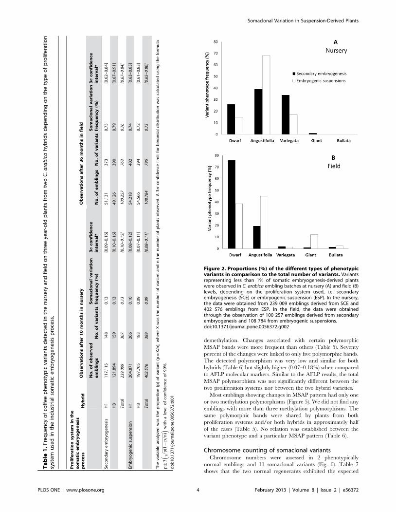
Frequency of phenotypic variantsEmbling batches of hybrids HI and H3 obtained from both
SCE and ESP were checked for phenotype variation at both
nursery and field levels. The frequency of phenotypic variants
assessed among more than 600 000 plants in the nursery was very
low (approx. 0.1%) and not significantly affected by the
proliferation system nor the hybrid variety (Table 1). Observation
of around 200 000 emblings in the field two years after planting
revealed roughly an additional 0.74% of abnormal phenotypes,
still without any significant difference between the two prolifer-
ation systems and hybrids. Apart from these phenotypic variants,
all the other studied trees flowered, grew and produced normally.
In conclusion, the overall phenotypic variation rate obtained by
pooling the data obtained both in the nursery and in the field was
less than 1% and no significant differences were noted between the
proliferation systems or between the hybrids used.
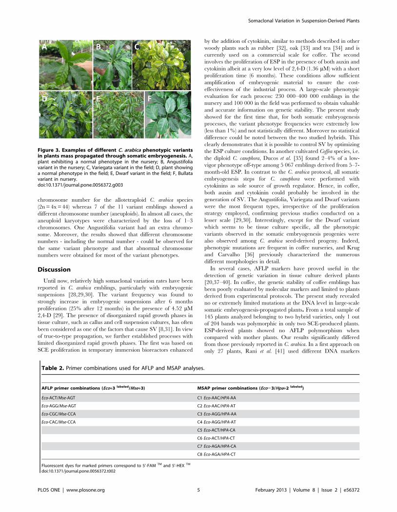
Both proliferation systems generated the same kind of pheno-
typic variants (Figures 2A, B), with the Dwarf and Angustifolia
types (Figures 3E, B) being the most frequent. Note that the
secondary embryogenesis proliferation system specifically en-
hanced the occurrence of Dwarf variants, whereas the embryo-
genic suspensions favored the occurrence of the Angustifolia type.
This latter phenotype can easily be detected and eliminated at the
nursery level along with the Variegata variant (Figure 3C). A
comparison of Figures 2A and 2B clearly shows that elimination in
the nursery is not efficient for the Dwarf type. This somaclonal
variation is more easily observable 2–3 years after planting in the
field thanks to the characteristic grouped canopy morphology and
low yield. Similarly, the Giant and Bullata (Figure 3F) phenotypic
variants could only be detected in the field on well-developed trees.
Locus specific polymorphisms revealed by AFLPIn order to verify the induction of molecular polymorphism by
the somatic embryogenesis process, AFLP analysis (four primer
combinations, Table 2) was carried out on mother plants and their
derived emblings. From a total of 204 bands obtained, only one
polymorphic fragment of 173 bp in size (Eco-ACT/Mse-AGT)
shared by two emblings of hybrid H1 and exhibiting a normal
phenotype, was found (Table 3). From a total of 198 bands
obtained, no polymorphism was found in emblings of hybrid H3.
Somaclonal Variation in Suspension-Derived Plants
PLOS ONE | www.plosone.org 2 February 2013 | Volume 8 | Issue 2 | e56372

All variants had the same AFLP pattern as the mother plants. For
both hybrids, no significant quantitative effect on AFLP was
detected when comparing SCE and ESP.
Methylation changes revealed by MSAPIn order to evaluate the occurrence of possible epigenetic
modifications in the micropropagated plants, a study on the
alteration of methylation patterns was performed by MSAP
analysis using eight primer combinations (Table 2). Only clear and
reproducible bands were selected for the analysis. More than 395
fragments were considered. First, MSAP patterns were obtained
from DNA digested by the two isoschizomers (HpaII and MspI), as
illustrated in Figure 4. They were further compared with those of
mother plants to classify the amplified fragments according to the
methylation pattern, as shown in Table 4. The percentages of
monomorphic fragments (pattern 1) were elevated and similar for
both hybrids at nearly 91%. The remaining 9% of fragments
(8.5% for H1 and 9.1% for H3) almost exclusively corresponded to
pattern 3.
A comparison of amplification patterns in mother plants and
their respective progeny are reported in Table 5. All differences
between mother and derived plants were switches between
patterns 1 and 3 and vice versa, i.e. likely modifications in the
restriction ability of HpaII. Among the polymorphic bands, eight
bands corresponded to a change from pattern 3 in mother plants
towards the unmethylated pattern 1 in emblings, suggesting
Figure 1. Schematic representation of two somatic embryogenesis processes applied at the industrial level. The first somaticembryogenesis process (upper section of the flow diagram) involved a proliferation step based on secondary embryogenesis in RITAH temporaryimmersion bioreactors (photos 1A, 1B). The second process (lower section of flow diagram) included a proliferation step based on embryogenicsuspensions (photos 1C, 1D). 1A, initial developmental stages of secondary embryos at the root pole of primary somatic embryos; 1B, clusters ofprimary and secondary embryos; 1C, clusters of embryogenic cells in suspension; 1D, embryogenic cells in suspension.doi:10.1371/journal.pone.0056372.g001
Somaclonal Variation in Suspension-Derived Plants
PLOS ONE | www.plosone.org 3 February 2013 | Volume 8 | Issue 2 | e56372
demethylation. Changes associated with certain polymorphic
MSAP bands were more frequent than others (Table 5). Seventy
percent of the changes were linked to only five polymorphic bands.
The detected polymorphism was very low and similar for both
hybrids (Table 6) but slightly higher (0.07–0.18%) when compared
to AFLP molecular markers. Similar to the AFLP results, the total
MSAP polymorphism was not significantly different between the
two proliferation systems nor between the two hybrid varieties.
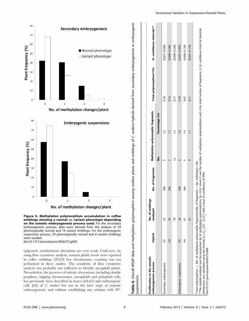
Most emblings showing changes in MSAP pattern had only one
or two methylation polymorphisms (Figure 5). We did not find any
emblings with more than three methylation polymorphisms. The
same polymorphic bands were shared by plants from both
proliferation systems and/or both hybrids in approximately half
of the cases (Table 5). No relation was established between the
variant phenotype and a particular MSAP pattern (Table 6).
Chromosome counting of somaclonal variantsChromosome numbers were assessed in 2 phenotypically
normal emblings and 11 somaclonal variants (Fig. 6). Table 7
shows that the two normal regenerants exhibited the expected
Ta
ble
1.
Fre
qu
en
cyo
fco
ffe
ep
he
no
typ
icva
rian
tsd
ete
cte
din
the
nu
rse
ryan
dfi
eld
on
thre
eye
ar-o
ldp
lan
tsfr
om
two
C.
ara
bic
ah
ybri
ds
de
pe
nd
ing
on
the
typ
eo
fp
rolif
era
tio
nsy
ste
mu
sed
inth
ein
du
stri
also
mat
ice
mb
ryo
ge
ne
sis
pro
cess
.
Pro
life
rati
on
syst
em
inth
eso
ma
tic
em
bry
og
en
esi
sp
roce
ssH
yb
rid
Ob
serv
ati
on
sa
fte
r1
0m
on
ths
inn
urs
ery
Ob
serv
ati
on
sa
fte
r3
6m
on
ths
infi
eld
No
.o
fo
bse
rve
de
mb
lin
gs
No
.o
fv
ari
an
tsS
om
acl
on
al
va
ria
tio
nfr
eq
ue
ncy
(%)
3s
con
fid
en
cein
terv
al*
No
.o
fe
mb
lin
gs
No
.o
fv
ari
an
tsS
om
acl
on
al
va
ria
tio
nfr
eq
ue
ncy
(%)
3s
con
fid
en
cein
terv
al*
Seco
nd
ary
em
bry
og
en
esi
sH
11
17
.11
51
48
0.1
3[0
.09
–0
.16
]5
1.1
31
37
30
.73
[0.6
2–
0.8
4]
H3
12
1.8
94
15
90
.13
[0.1
0–
0.1
6]
49
.12
63
90
0.7
9[0
.67
–0
.91
]
Tota
l23
9.00
930
70.
13[0
.10–
0.15
]10
0.25
776
30.
76[0
.67
–0.
84]
Emb
ryo
ge
nic
susp
en
sio
nH
12
04
.87
12
06
0.1
0[0
.08
–0
.12
]5
4.2
18
40
20
.74
[0.6
3–
0.8
5]
H3
19
7.7
05
18
30
.09
[0.0
7–
0.1
1]
54
.56
63
94
0.7
2[0
.61
–0
.83
]
Tota
l40
2.57
638
90.
09[0
.08–
0.11
]10
8.78
479
60.
73[0
.65
–0.
80]
Th
eva
riab
lean
alyz
ed
was
the
pro
po
rtio
n(p
)o
fva
rian
t(p
=X
/n),
wh
ere
Xw
asth
en
um
be
ro
fva
rian
tan
dn
the
nu
mb
er
of
pla
nts
ob
serv
ed
.A
3s
con
fid
en
celim
itfo
rb
ino
mia
ld
istr
ibu
tio
nw
asca
lcu
late
du
sin
gth
efo
rmu
la
p+
3ffiffiffiffiffiffiffiffiffiffiffiffiffiffiffiffiffiffiffiffiffi
p(1
{p=
n)
p��
wit
ha
leve
lo
fco
nfi
de
nce
of
99
%.
do
i:10
.13
71
/jo
urn
al.p
on
e.0
05
63
72
.t0
01
Figure 2. Proportions (%) of the different types of phenotypicvariants in comparison to the total number of variants. Variantsrepresenting less than 1% of somatic embryogenesis-derived plantswere observed in C. arabica embling batches at nursery (A) and field (B)levels, depending on the proliferation system used, i.e. secondaryembryogenesis (SCE) or embryogenic suspension (ESP). In the nursery,the data were obtained from 239 009 emblings derived from SCE and402 576 emblings from ESP. In the field, the data were obtainedthrough the observation of 100 257 emblings derived from secondaryembryogenesis and 108 784 from embryogenic suspensions.doi:10.1371/journal.pone.0056372.g002
Somaclonal Variation in Suspension-Derived Plants
PLOS ONE | www.plosone.org 4 February 2013 | Volume 8 | Issue 2 | e56372
chromosome number for the allotetraploid C. arabica species
(2n = 4x = 44) whereas 7 of the 11 variant emblings showed a
different chromosome number (aneuploids). In almost all cases, the
aneuploid karyotypes were characterized by the loss of 1–3
chromosomes. One Angustifolia variant had an extra chromo-
some. Moreover, the results showed that different chromosome
numbers - including the normal number - could be observed for
the same variant phenotype and that abnormal chromosome
numbers were obtained for most of the variant phenotypes.
Discussion
Until now, relatively high somaclonal variation rates have been
reported in C. arabica emblings, particularly with embryogenic
suspensions [28,29,30]. The variant frequency was found to
strongly increase in embryogenic suspensions after 6 months
proliferation (25% after 12 months) in the presence of 4.52 mM
2,4-D [29]. The presence of disorganized rapid growth phases in
tissue culture, such as callus and cell suspension cultures, has often
been considered as one of the factors that cause SV [8,31]. In view
of true-to-type propagation, we further established processes with
limited disorganized rapid growth phases. The first was based on
SCE proliferation in temporary immersion bioreactors enhanced
by the addition of cytokinin, similar to methods described in other
woody plants such as rubber [32], oak [33] and tea [34] and is
currently used on a commercial scale for coffee. The second
involves the proliferation of ESP in the presence of both auxin and
cytokinin albeit at a very low level of 2,4-D (1.36 mM) with a short
proliferation time (6 months). These conditions allow sufficient
amplification of embryogenic material to ensure the cost-
effectiveness of the industrial process. A large-scale phenotypic
evaluation for each process: 230 000–400 000 emblings in the
nursery and 100 000 in the field was performed to obtain valuable
and accurate information on genetic stability. The present study
showed for the first time that, for both somatic embryogenesis
processes, the variant phenotype frequencies were extremely low
(less than 1%) and not statistically different. Moreover no statistical
difference could be noted between the two studied hybrids. This
clearly demonstrates that it is possible to control SV by optimizing
the ESP culture conditions. In another cultivated Coffea species, i.e.
the diploid C. canephora, Ducos et al. [35] found 2–4% of a low-
vigor phenotype off-type among 5 067 emblings derived from 5–7-
month-old ESP. In contrast to the C. arabica protocol, all somatic
embryogenesis steps for C. canephora were performed with
cytokinins as sole source of growth regulator. Hence, in coffee,
both auxin and cytokinin could probably be involved in the
generation of SV. The Angustifolia, Variegata and Dwarf variants
were the most frequent types, irrespective of the proliferation
strategy employed, confirming previous studies conducted on a
lesser scale [29,30]. Interestingly, except for the Dwarf variant
which seems to be tissue culture specific, all the phenotypic
variants observed in the somatic embryogenesis progenies were
also observed among C. arabica seed-derived progeny. Indeed,
phenotypic mutations are frequent in coffee nurseries, and Krug
and Carvalho [36] previously characterized the numerous
different morphologies in detail.
In several cases, AFLP markers have proved useful in the
detection of genetic variation in tissue culture derived plants
[20,37–40]. In coffee, the genetic stability of coffee emblings has
been poorly evaluated by molecular markers and limited to plants
derived from experimental protocols. The present study revealed
no or extremely limited mutations at the DNA level in large-scale
somatic embryogenesis-propagated plants. From a total sample of
145 plants analyzed belonging to two hybrid varieties, only 1 out
of 204 bands was polymorphic in only two SCE-produced plants.
ESP-derived plants showed no AFLP polymorphism when
compared with mother plants. Our results significantly differed
from those previously reported in C. arabica. In a first approach on
only 27 plants, Rani et al. [41] used different DNA markers
Figure 3. Examples of different C. arabica phenotypic variantsin plants mass propagated through somatic embryogenesis. A,plant exhibiting a normal phenotype in the nursery; B, Angustifoliavariant in the nursery; C, Variegata variant in the field; D, plant showinga normal phenotype in the field; E, Dwarf variant in the field; F, Bullatavariant in nursery.doi:10.1371/journal.pone.0056372.g003
Table 2. Primer combinations used for AFLP and MSAP analyses.
AFLP primer combinations (Eco+3 labeled/Mse+3) MSAP primer combinations (Eco23/Hpa+2 labeled)
Eco-ACT/Mse-AGT C1 Eco-AAC/HPA-AA
Eco-AGG/Mse-AGT C2 Eco-AAC/HPA-AT
Eco-CGC/Mse-CCA C3 Eco-AGG/HPA-AA
Eco-CAC/Mse-CCA C4 Eco-AGG/HPA-AT
C5 Eco-ACT/HPA-CA
C6 Eco-ACT/HPA-CT
C7 Eco-AGA/HPA-CA
C8 Eco-AGA/HPA-CT
Fluorescent dyes for marked primers correspond to 5’-FAM TM and 5’-HEX TM
doi:10.1371/journal.pone.0056372.t002
Somaclonal Variation in Suspension-Derived Plants
PLOS ONE | www.plosone.org 5 February 2013 | Volume 8 | Issue 2 | e56372

(RAPD, random amplified polymorphic DNA and SSR, simple
sequence repeat) to assess the genetic integrity of C. arabica
emblings obtained from embryogenic calli, and they found a
higher polymorphism level (4%) in the nuclear genome. By
performing RAPD analyses on Norway spruce emblings, Heinze
and Schmidt [42] concluded that gross somaclonal variation was
absent in their plant regeneration system. In contrast, RAPD and
SSR markers allowed the detection of high genetic variation in
cotton emblings regenerated in the presence of 2,4-D [39]. AFLP
analysis of 24 rye emblings led to the scoring of 887 AFLP
markers, among which 8.8% identified the same polymorphism in
plants obtained independently, revealing putative mutational hot
spots [38].
DNA cytosine methylation plays an important role in plant
regulation and development [43,44]. Since the pioneer studies on
maize emblings [12], substantial evidence has been obtained
which indicates that demethylation can occur at a high frequency
during somatic embryogenesis and can be an important cause of
tissue culture induced variation [45]. DNA methylation has also
been implicated in gene silencing and transposable element
reactivation [24,46]. To our knowledge, epigenetic deregulation
during coffee micropropagation has not yet been studied. The very
low total methylation polymorphism values obtained for both
somatic embryogenesis processes and both hybrids (0.07–0.18%
range) indicated that the tissue culture procedures employed in
coffee weakly affected DNA methylation of the regenerated plants.
This finding is in accordance with the 0.87% total polymorphism
recently found in Freesia hybrid emblings by Gao et al. [40].
Moreover, the low number of methylation polymorphisms per
embling (range 1–3) confirmed that neither SCE nor ESP induced
additional stress at the methylation level during embryogenic
material proliferation. In contrast, a significantly higher accumu-
lation of methylation changes in some emblings has been regularly
observed in other species [20,21,47,48]. For example, in
grapevine, most emblings showed between zero and three changes,
similar to our findings, but a few accumulated up to 18 [21]. It has
also been well demonstrated that auxin levels strongly alter DNA
methylation of embryogenic cell cultures [49]. However, similar to
our results, examples of stable MSAP patterns have already been
reported using bamboo tissues at different developmental stages of
somatic embryogenesis [50]. The timing of plant regeneration
from proliferating callus cultures could be crucial for the
appearance of variation. In callus-derived hop plants, an increase
in the variation was detected by MSAP in prolonged callus
cultures [20]. Our results demonstrated that very few changes are
possible by limiting both the auxin level and culture duration.
MSAP markers have already been successfully used to
demonstrate epigenetic instabilities (methylation alteration) in-
duced by somatic embryogenesis in a great variety of plants, such
as the ornamental flower Freesia, banana, barley, grapevine and
maize [20,21,40,51–53], also indicating that demethylation events
were generally the most frequent. Although occurring at low
frequency, our results also indicated demethylation events and
mainly the loss of methylation in the internal cytosine of the 5’-
CCGG-39 sequence to produce a new HpaII band not detected in
mother plants but present in the amplification pattern of the
isoschizomer MspI. The detection of the same MSAP polymorphic
fragments in independent plant samples from different hybrids and
somatic embryogenesis processes suggests the existence of hotspots
of DNA methylation changes in the genome. The existence of
non-randomly behaving methylation polymorphic fragments in
micropropagated plants has already been described using Met-
AFLP [20,48] and MSAP [21,53–55].Ta
ble
3.
Sum
mar
yo
fA
FLP
dat
aan
do
bse
rve
dp
oly
mo
rph
ism
sam
on
gm
oth
er
pla
nts
and
em
blin
gs
de
rive
dfr
om
seco
nd
ary
em
bry
og
en
esi
so
re
mb
ryo
ge
nic
susp
en
sio
ns.
Pro
life
rati
on
syst
em
inth
eso
ma
tic
em
bry
og
en
esi
sp
roce
ssH
yb
rid
No
.o
fa
na
lyz
ed
em
bli
ng
sN
o.
of
fra
gm
en
tsP
oly
mo
rph
icfr
ag
me
nts
Em
bli
ng
ssh
ow
ing
po
lym
orp
his
ms
To
tal
po
lym
orp
his
m**
No
.(%
)N
o.
(%)
(%)
Seco
nd
ary
em
bry
og
en
esi
sH
13
32
04
1*
0.5
26
0.0
3
H3
45
19
80
00
00
Emb
ryo
ge
nic
susp
en
sio
ns
H1
26
20
40
00
00
H3
41
19
80
00
00
Dat
aw
ere
ob
tain
ed
for
two
C.
ara
bic
ah
ybri
ds
(H1
and
H3
)an
dco
mp
are
dw
ith
the
pat
tern
so
fth
em
oth
er
pla
nts
.*F
ou
nd
in2
em
blin
gs
wit
hn
orm
alp
he
no
typ
e(Nu2
10
and
23
2)
sho
win
ga
ne
wA
FLP
ban
dEc
o-A
CT
/Mse
-AG
T1
73
bp
**T
ota
lp
oly
mo
rph
ism
=[N
o.
of
po
lym
orp
hic
frag
me
nts
/(N
o.
of
frag
me
nts6
No
.e
mb
ling
s)]6
10
0d
oi:1
0.1
37
1/j
ou
rnal
.po
ne
.00
56
37
2.t
00
3
Somaclonal Variation in Suspension-Derived Plants
PLOS ONE | www.plosone.org 6 February 2013 | Volume 8 | Issue 2 | e56372
Gross changes, such as variation in ploidy level, number of
chromosomes and structural changes, are mitotic aberrations that
represent major genomic alterations of in vitro plants often
generated during in vitro proliferation and differentiation [12,56–
58]. Variations in chromosome number and structure have been
described among emblings for several species [59–62]. We
demonstrated that gross changes occur during coffee somatic
embryogenesis and are related to SV, whereas genetic and
Figure 4. Representation of MSAP electropherograms observed for coffee mother plants and embling progeny using theisoschizomers HpaII and MspI. Illustration of the pattern variation obtained for normal and variant phenotypes within the embling progeny.doi:10.1371/journal.pone.0056372.g004
Table 4. MSAP patterns corresponding to different methylation states of the symmetric sequence CCGG, as revealed by thespecificity of the restriction enzymes Hpa II and Msp I.
Restriction enzymes MSAP patterns after enzymatic digestion
Pattern 1 Pattern 2 Pattern 3 Pattern 4
Hpa II 1 1 0 0
Msp I 1 0 1 0
Methylation state Unmethylated Hemi-methylated Fully-methylated Fully-methylated
Methylation position None External cytosine Internal cytosine External cytosine
Schematic representation CCGGGGCC
CH3
CCGGGGCC
CH3
CCGGGGCCCH3
CH3
CCGGGGCCCH3
doi:10.1371/journal.pone.0056372.t004
Somaclonal Variation in Suspension-Derived Plants
PLOS ONE | www.plosone.org 7 February 2013 | Volume 8 | Issue 2 | e56372
Ta
ble
5.
MSA
Pm
eth
ylat
ion
pat
tern
sin
mo
the
rp
lan
tsan
dm
od
ifie
dp
atte
rns
ine
mb
ling
s.
Po
lym
orp
hic
MS
AP
fra
gm
en
ts(s
ize
inb
p)
MS
AP
me
thy
lati
on
pa
tte
rns
Pro
life
rati
on
syst
em
aff
ect
ed
by
the
me
thy
lati
on
cha
ng
eP
rese
nce
of
the
me
thy
lati
on
cha
ng
ed
ep
en
din
go
nth
ep
lan
tp
he
no
typ
eN
o.
of
me
thy
lati
on
cha
ng
es
for
ea
chfr
ag
me
nt
Mo
the
rp
lan
tsE
mb
lin
gs
Hy
bri
dH
1H
yb
rid
H3
No
rma
lV
ari
an
tN
o.
(%)
C3
-1
07
bp
Pat
tern
3P
atte
rn1
SCE,
ESP
SCE,
ESP
+**
28
9.8
C2
-12
7b
pP
atte
rn3
Pat
tern
10
ESP
+2
11
.2
C4
-13
4b
pP
atte
rn3
Pat
tern
10
SCE
+2
44
.8
C1
-2
51
bp
Pat
tern
1*
Pat
tern
3SC
E,ES
P0
+2
12
14
.6
C3
-2
53
bp
Pat
tern
3P
atte
rn1
0SC
E+
22
2.4
C2
-3
02
bp
Pat
tern
3P
atte
rn1
0SC
E,ES
P+
+1
61
9.5
C3
-3
16
bp
Pat
tern
3P
atte
rn1
0SC
E+
+4
4.8
C6
-37
0b
pP
atte
rn3
Pat
tern
1SC
E,ES
PES
P+
+1
01
2.2
C8
-3
70
bp
Pat
tern
3P
atte
rn1
SCE,
ESP
SCE,
ESP
++
12
14
.6
C5
-3
87
bp
Pat
tern
1P
atte
rn3
SCE
SCE
+2
13
15
.8
No
.ch
ang
es
82
Re
lati
on
wit
hth
ety
pe
of
C.
ara
bic
ah
ybri
d,
typ
eo
fp
rolif
era
tio
nsy
ste
m[s
eco
nd
ary
em
bry
og
en
esi
s(S
CE)
and
em
bry
og
en
icsu
spe
nsi
on
(ESP
)]an
dre
ge
ne
ran
tp
he
no
typ
e.
*Pat
tern
1:
Frag
me
nt
pre
sen
tin
bo
thH
paI
Ian
dM
spI
rest
rict
ion
dig
est
s(1
:1);
Pat
tern
3:
Frag
me
nt
abse
nt
inH
paI
Id
ige
sts
and
pre
sen
tin
Msp
Id
ige
sts
(0:1
).**
Re
lati
on
ship
wit
ha
par
ticu
lar
ph
en
oty
pe
isin
dic
ate
dw
ith
(+)
for
pre
sen
cean
d(2
)fo
rab
sen
ce.
do
i:10
.13
71
/jo
urn
al.p
on
e.0
05
63
72
.t0
05
Somaclonal Variation in Suspension-Derived Plants
PLOS ONE | www.plosone.org 8 February 2013 | Volume 8 | Issue 2 | e56372
epigenetic (methylation) alterations are very weak. Until now, by
using flow cytometry analysis, normal ploidy levels were reported
in coffee emblings [29,63] but chromosome counting was not
performed in these studies. The sensitivity of flow cytometry
analysis was probably not sufficient to identify aneuploid plants.
Nevertheless, the presence of mitotic aberrations, including double
prophase, lagging chromosomes, aneuploids and polyploid cells,
has previously been described in leaves [64,65] and embryogenic
calli [64] of C. arabica but not in the later steps of somatic
embryogenesis, and without establishing any relation with SV.
Figure 5. Methylation polymorphism accumulation in coffeeemblings showing a normal vs. variant phenotype dependingon the somatic embryogenesis process used. For the secondaryembryogenesis process, data were derived from the analysis of 59phenotypically normal and 19 variant emblings. For the embryogenicsuspension process, 59 phenotypically normal and 8 variant emblingswere studied.doi:10.1371/journal.pone.0056372.g005
Ta
ble
6.
Ove
rall
MSA
Pd
ata
and
me
thyl
atio
np
oly
mo
rph
ism
amo
ng
mo
the
rp
lan
tsan
de
mb
ling
so
fC
.a
rab
ica
hyb
rid
sd
eri
ved
fro
mse
con
dar
ye
mb
ryo
ge
ne
sis
or
em
bry
og
en
icsu
spe
nsi
on
s.
Pro
life
rati
on
inth
eso
ma
tic
em
bry
og
en
esi
sp
roce
ssH
yb
rid
No
.o
fe
mb
lin
gs
an
aly
ze
dN
o.
of
fra
gm
en
tsM
eth
yla
tio
np
oly
mo
rph
icfr
ag
me
nts
To
tal
po
lym
orp
his
m*(
%)
3s
con
fid
en
cein
terv
als
**
No
.P
erc
en
tag
e(%
)
Seco
nd
ary
em
bry
og
en
esi
sH
13
33
99
51
.20
.18
[0.0
71
–0
.29
4]
H3
45
39
67
1.7
0.1
6[0
.06
8–
0.2
46
]
78
12
1.5
0.1
7[0
.09
8–
0.2
38
]
Emb
ryo
ge
nic
susp
en
sio
ns
H1
26
39
94
1.0
0.1
8[0
.05
7–
0.3
09
]
H3
41
39
65
1.0
0.0
7[0
.00
6–
0.1
29
]
67
91
.10
.11
[0.0
51
–0
.17
4]
*To
tal
po
lym
orp
his
m=
[No
.o
fm
eth
ylat
ion
po
lym
orp
hic
frag
me
nts
/(N
o.
of
frag
me
nts6
No
.e
mb
ling
s)]6
10
0.
**T
he
vari
able
anal
yze
dw
asth
ep
rop
ort
ion
(p)
of
me
thyl
atio
np
oly
mo
rph
ism
s(p
=X
/n),
wh
ere
Xw
asth
en
um
be
ro
fm
eth
ylat
ion
po
lym
orp
his
ms
and
nth
eto
tal
nu
mb
er
of
frag
me
nts
.A
3s
con
fid
en
celim
itfo
rb
ino
mia
ld
istr
ibu
tio
nw
asca
lcu
late
du
sin
gth
efo
rmu
lap+
3ffiffiffiffiffiffiffiffiffiffiffiffiffiffiffiffiffiffiffiffiffi
p(1
{p=n
)p�
�w
ith
ale
vel
of
con
fid
en
ceo
f9
9%
.d
oi:1
0.1
37
1/j
ou
rnal
.po
ne
.00
56
37
2.t
00
6
Somaclonal Variation in Suspension-Derived Plants
PLOS ONE | www.plosone.org 9 February 2013 | Volume 8 | Issue 2 | e56372
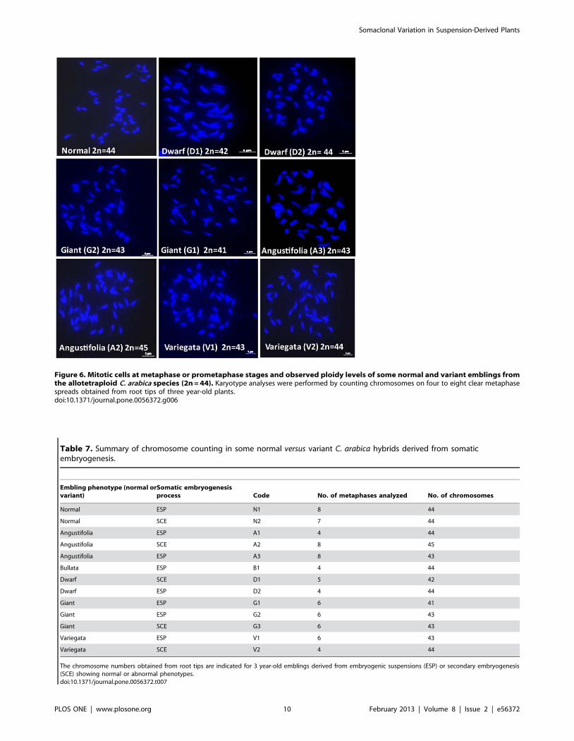
Figure 6. Mitotic cells at metaphase or prometaphase stages and observed ploidy levels of some normal and variant emblings fromthe allotetraploid C. arabica species (2n = 44). Karyotype analyses were performed by counting chromosomes on four to eight clear metaphasespreads obtained from root tips of three year-old plants.doi:10.1371/journal.pone.0056372.g006
Table 7. Summary of chromosome counting in some normal versus variant C. arabica hybrids derived from somaticembryogenesis.
Embling phenotype (normal orvariant)
Somatic embryogenesisprocess Code No. of metaphases analyzed No. of chromosomes
Normal ESP N1 8 44
Normal SCE N2 7 44
Angustifolia ESP A1 4 44
Angustifolia SCE A2 8 45
Angustifolia ESP A3 8 43
Bullata ESP B1 4 44
Dwarf SCE D1 5 42
Dwarf ESP D2 4 44
Giant ESP G1 6 41
Giant ESP G2 6 43
Giant SCE G3 6 43
Variegata ESP V1 6 43
Variegata SCE V2 4 44
The chromosome numbers obtained from root tips are indicated for 3 year-old emblings derived from embryogenic suspensions (ESP) or secondary embryogenesis(SCE) showing normal or abnormal phenotypes.doi:10.1371/journal.pone.0056372.t007
Somaclonal Variation in Suspension-Derived Plants
PLOS ONE | www.plosone.org 10 February 2013 | Volume 8 | Issue 2 | e56372
The presence of aneuploidy has also been well documented in
embryogenic calli of Hordeum vulgare [66] and sweet orange [60].
The mechanisms underlying SV remain largely theoretical and
unclear [16]. Thus it is often difficult to correlate a well-described
genetic or epigenetic mechanism to a variant phenotype. For
example, although DNA methylation has often been suggested as a
possible cause of SV, a number of studies have reported high levels
of methylation variation with no effect on the plant phenotype
[20,21,52]. Another example is given by oil palm emblings,
approximately 5% of which exhibit the ‘mantled’ phenotype
affecting the formation of floral organs in both male and female
flowers. Although a decrease in DNA methylation was observed, it
was not possible to determine the nature of the epigenetic
deregulation [67]. In the present study, it was possible to reveal a
large proportion of aneuploid karyotypes in different variant
phenotypes, hence showing that chromosomal rearrangements
could be directly involved in the occurrence of phenotypic
variation. The addition or subtraction of a single chromosome
has a greater impact on phenotype than whole genome
duplication, i.e. polyploidy [68]. It was clearly demonstrated in
Arabidopsis thaliana that certain phenotypic traits are strongly
associated with the dosage of specific chromosomes and that
chromosomal effects can be additive [69]. Similarly, in maize
seedlings, trisomic plants showed characteristic features such as
reduced stature, tassel morphology changes and the presence of
knots on the leaves, suggesting a phenotypic effect caused by the
altered copy of specific chromosome related genes [70]. A similar
mechanism could explain most of the variant phenotypes observed
in C. arabica. The observation of a variant phenotype in plants with
the expected chromosome number could be explained by the
coexistence of monosomic and trisomic chromosomes in the same
genome or by other chromosomal-like structural changes associ-
ated with undetected deletions, duplications, inversions or
translocations of specific chromosomal segments [10,71]. The
karyotype analyses performed in the present study were limited to
chromosome counting and did not enable observation of such
chromosomal alterations.
Conclusions
This report shows that somatic embryogenesis is reliable for
true-to-type and large-scale propagation of elite varieties in the C.
arabica species. Both ESP and SCE ensured high proliferation rates
along with very low SV rates, as observed through massive
phenotypic observations in a commercial nursery and field plots.
Molecular analysis (AFLP and MSAP) performed on 145 emblings
derived from two proliferation processes and two different F1
hybrids showed that polymorphism between mother plants and
emblings was extremely low. Consequently, it can be concluded
that genetic and epigenetic alterations were also particularly
limited during somatic embryogenesis in our controlled culture
conditions. C. arabica is a young allopolyploid still having the most
of its genes in duplicated copies [72]. It could be hypothesized that
the impact of genetic or epigenetic variations on phenotype was
restricted because of the buffer effect due to polyploidy. The main
change in most of the rare phenotypic variants was aneuploidy.
Although further studies are necessary for an accurate under-
standing of the chromosome anomalies involved in the acquisition
of a particular phenotype, it is now obvious that mitotic
aberrations play a major role in SV in coffee. The identification
and use of molecular markers at the heterozygous state in mother
plants (i.e. polymorphic between the two parental lines) would be
required to further investigate this type of chromosome abnor-
mality. Current studies based on the use of long-term embryogenic
cultures [73] are aimed at establishing the full range of cytological,
genetic and epigenetic (with a special focus on transposable
elements) mechanisms underlying SV.
Materials and Methods
Plant material and somatic embryogenesisSelected F1 hybrids of C. arabica [26] obtained by crossing
traditional dwarf American varieties (Caturra, Sarchimor T5296)
and wild accessions originating from Ethiopia and Sudan are
disseminated in Central America through somatic embryogenesis.
In the present study, emblings derived from the two hybrids
Sarchimor T5296 x Rume Sudan and Caturra x ET531, named
respectively H1 and H3, were analyzed to assess the SV level.
Large-scale phenotypic observations were performed in Nicaragua
both at the nursery (more than 600 000 young emblings) and field
level (more than 200 000 three year-old emblings) on 11 coffee
plots belonging to the ‘La Cumplida’ coffee research experimental
sites in the Matagalpa region (Nicaragua). Nursery and field
phenotypic observations were done on balanced amounts of plants
from hybrids H1 and H3 (Table 1). Field observations were
performed for all trees by visual evaluation of growth and
morphology, flowering and fruit yield during the first and second
production years. No specific permits were required for the
described field studies that were performed with the authorization
of the Coffee Research Department of ‘La Cumplida’, owner of
the experimental sites. These sites are not protected and the
studies did not involve endangered or protected species. Molecular
marker analysis was applied on F1 hybrid mother plants
propagated by rooted horticultural cuttings (four for each hybrid)
and used as source material for in vitro propagation, as well as on
adult emblings (3 years after planting) for which plants exhibiting
an abnormal phenotype (phenotypic variant) were distinguished
from plants with a normal phenotype and productivity (Table 8).
Molecular marker patterns obtained for emblings were systemat-
ically compared with those obtained with mother plants (four
plants per hybrid propagated by horticultural cutting) used as
reference.
The somatic embryogenesis process (Figure 1) involved the
following stages:
1) Production of embryogenic callus: pieces of young leaves were
surface-sterilized and used as explants. The explants were cultured
for 1 month on a 1/2 strength MS [74] ‘‘C’’ callogenesis medium
[75] containing 2.3 mM 2,4-D, 4.9 mM indole-3-butyric acid (IBA)
and 9.8 mM iso-pentenyladenine (iP), and then transferred for 6
months to MS/2 ‘‘ECP’’ embryogenic callus production medium
[75] containing 4.5 mM 2,4-D and 17.7 mM benzylaminopurine
(6-BA). All media were solidified using 2.4 g/l Phytagel (Sigma,
Steinheim, Germany). These steps were carried out at 26–27uC in
the dark.
2) 6-month multiplication step and embryo regeneration. Fully developed
somatic embryos were mass regenerated via two distinct multipli-
cation processes, i.e. either secondary (or repetitive) embryogenesis
or embryogenic suspensions (Figure 1).
Secondary embryogenesis (SCE). Two hundred mg of embryo-
genic aggregates were placed in a 1 liter-RITAH temporary
immersion bioreactor (CIRAD, Montpellier, France; [76]) along
with 200 ml of ‘‘R’’ MS/2 regeneration medium [75] containing
17.76 mM 6-BA, in darkness for 6 weeks. An immersion frequency
of 1 min every 12 h was applied. Proliferating embryo masses
were then placed in 1/2 strength MS [74] regeneration medium
containing 5.6 mM 6-BA and subcultured once every 6 weeks for
three proliferation cycles. Secondary embryos were produced with
an immersion frequency of 1 min every 12 h and a high culture
Somaclonal Variation in Suspension-Derived Plants
PLOS ONE | www.plosone.org 11 February 2013 | Volume 8 | Issue 2 | e56372

density (approx. 10 000 embryos). The cultures were kept at 27uC,
with a 12 h/12 h photoperiod and 50 mmol m22 s21 photosyn-
thetic photon flux density.
Embryogenic cell suspensions (ESP). Embryogenic calli were
transferred to 100 ml Erlenmeyer flasks at a density of 1 g/L in 1/
4 MS strength Yasuda liquid proliferation medium [77] with
1.36 mM 2,4-D and 4.4 mM 6-BA. Suspension cultures were
maintained by the monthly transfer of 1 g/L of embryogenic
aggregates into fresh medium. Six month-old suspensions were
used for somatic embryo regeneration. Embryo differentiation was
initiated by transferring embryogenic aggregates at a density of
1 g/L in 250 ml Erlenmeyer flasks in a full strength MS medium
containing 1.35 mM 6-BA. Fully developed torpedo-shaped
embryos were obtained after two 4 week subcultures in such
conditions. All suspension cultures were shaken at 110 rpm at 27
uC under 50 mmol m22 s21 photosynthetic photon flux density.
3) Pre-germination in a bioreactor. Germination was triggered by
applying a low culture density of around 800–900 embryos per 1 l-
RITAH bioreactor. An "EG" embryo germination medium [75]
containing 1.33 mM BA was used for 2 months and finally, for 2
weeks, the ‘‘EG’’ culture medium was supplemented with 234 mM
sucrose. By the end of the in vitro culture stage, each bioreactor
contained around 700 pre-germinated cotyledonary somatic
embryos with an elongated embryonic axis and a pair of open,
chlorophyllous cotyledons. Pre-germination was conducted in the
light (12/12 h, 50 mmol m22 s21).
4) Plantlet conversion was obtained after direct sowing of mature
somatic embryos in the nursery. Mature embryos were sown
vertically on top of the substrate (two parts soil, one part sand, one
part coffee pulp) sterilized by chemical treatment (Dazomet
(DMTT), Union Carbide). The somatic embryo culture density
in the plastic boxes (l.w.h = 30/21/10 cm) was approximately
3600 m22. The cultures were placed under a transparent roof that
provided 50% shade, and were watered for 2 min twice daily.
Conversion of somatic embryos into plants was generally observed
12 weeks after sowing, and characterized by the emergence of a
stem bearing at least two pairs of true leaves.
5) Growth and hardening in the nursery (21 weeks). Plantlets grown
from somatic embryos were transferred to 0.3 L plugs on a
substrate comprising peat-based growing medium (Pro-mix,
Premier Tech Ltd, Canada) and coffee pulp (3/1, v/v) under
conventional nursery conditions until they reached the required
size for planting in the field (approx. 30 cm). During this stage, the
shade (50% light interception) and relative humidity (80%) were
gradually reduced over 4 weeks to 0% light interception, with
natural RH ranging from 65 to 90%.
Molecular analysisDNA extraction. Young fully expanded leaves were selected
on three year-old plants for molecular analysis. Genomic DNA
was isolated from 100 mg of lyophilized leaves using Dellaporta
buffer [78] containing sodium dodecyl sulfate (SDS) detergent and
sodium bisulfite 1% w/v to avoid leaf oxidation. DNA was purified
in spin-column plates as described in the DNeasy plant kit protocol
from QIAGEN.
AFLP markers. AFLP analysis was carried out as described
by Vos et al. [79], with a total of four primer combinations
(Table 2), using 5-FAM or 5-HEX fluorescently labeled EcoR1 (+3)
and unlabeled MseI (+3) primers. A touchdown PCR program for
selective amplification was performed in an Eppendorf thermo-
cycler under the following conditions: 3 min at 94uC, 12 cycles of
45 s at 94uC, 12 cycles of 45 s at 65uC and 1 min at 72uC; the
annealing temperature was decreased by 0.7uC per cycle from a
starting point of 65uC during this stage, with a final round of 25
cycles of 94uC for 45 s, 56uC for 45 s, 72uC for 1 min and a final
elongation step of 72uC for 1 min. The same PCR conditions were
found to be appropriate for MSAP in preliminary tests.
MSAP markers. MSAP analysis was carried out as described
by Reyna-Lopez et al. [19] with minor adaptations for capillary
electrophoresis. The MSAP protocol is an adaptation of the AFLP
method for the evaluation of different states of methylation in the
symmetric sequence CCGG. In the MSAP protocol, the frequent
cutting endonuclease (MseI) was replaced by the two isoschizo-
meric restriction enzymes HpaII and MspI with different sensitivity
Table 8. Plant material used in molecular marker analyses.
Somatic embryogenesis proliferationstep Type of plant material No. of plants per C. arabica hybrid Total no. of plants
H1 H3
Secondary embryogenesis Emblings normal phenotype 28 31 59
Emblings variant phenotype 5 14 19
Angustifolia (A) 0 4
Bullata (B) 2 4
Dwarf (D) 0 2
Variegata (V) 3 4
Total 33 45 78
Embryogenic suspension Emblings normal phenotype 25 34 59
Emblings variant phenotype 1 7 8
Angustifolia (A) 1 2
Dwarf (D) 0 1
Variegata (V) 0 4
Total 26 41 67
Total number of plants analyzed from two F1 Coffea arabica hybrid lines (H1 and H3) corresponding to somatic-embryo derived 3 year-old plants (emblings) withnormal or variant phenotypes along with their respective mother plants as reference. The numbers of emblings for each variant phenotype are also given.doi:10.1371/journal.pone.0056372.t008
Somaclonal Variation in Suspension-Derived Plants
PLOS ONE | www.plosone.org 12 February 2013 | Volume 8 | Issue 2 | e56372

to the methylation state of the symmetric sequence CCGG
(Table 4). Specifically, HpaII is able to recognize and cut only
when the CCGG sequence is unmethylated or hemi-methylated
on the external cytosine. MspI is able to cut when CCGG sequence
is unmethylated or if the internal cytosine is fully or hemi-
methylated. Both HpaII and MspI are unable to cut if the external
cytosine presents full methylation. DNA methylation in plants
commonly occurs at cytosine bases in all sequence contexts: the
symmetric CG and CHG, in which (H = A, T or C) and the
asymmetric CHH contexts [17]. Selective amplification included a
total of eight primer combinations per isoschizomer (Table 2).
HpaII (+2) primers were fluorescently labeled with 5-FAM or 5-
HEX while EcoR1 (+3) remained unlabeled. In order to reduce the
possibility of technical artifacts, two repetitions using different
DNA extractions were performed for each primer combination.Capillary electrophoresis and data analysis. PCR prod-
ucts were separated by capillary electrophoresis with Pop 7TM
polymer in a 16 capillary 3130 XL Genetic Analyzer from Applied
Biosystems using an internally manufactured 524 ROX fluor-
ophore as sizing standard. The fragments used for fingerprinting
were visualized as electropherograms in applied Biosystems
software GeneMapperH version 3.7. Informative fragments were
mostly found in the 100–450 bp range. All amplified fragments
were classified based on the primer combination used and their
size. The sample fingerprint data was converted to binary code,
with ‘‘1’’ denoting the presence of the fragment and ‘‘0’’ the
absence. Different binary matrices were constructed for compar-
ative analysis depending on the kind of molecular marker.
As shown in Table 4, the MSAP patterns were classified as
follows: Pattern 1 when a comigrating amplified fragment was
obtained from the DNA template digested by both restriction
enzymes HpaII and MspI; Pattern 2 and 3 when an amplification
fragment was obtained only from the DNA template digested by
HpaII or MspI, respectively.
Before analysis of the embling versus mother plant population,
we successfully verified, on a set of plants from the Caturra variety,
that the same MSAP patterns were systematically generated
whatever the plant age and the leaf developmental stage (data not
shown). Hence, a possible developmental variability in the studied
plant material does not seem to introduce any additional source of
variation in the methylation state. Nevertheless, in all experiments,
only leaves from the same developmental stage were chosen.
Slide preparation and karyotyping. Root tips were har-
vested from individual adult emblings and placed in an aqueous
solution of 8-hydroxyquinoline (2.5 mM) used as pre-treatment,
for 4 h in darkness (2 h at 4uC plus 2 h at room temperature). A
solution of Carnoy (absolute ethanol and glacial acetic acid, 3:1 v/
v) was used to fix the tissues for at least 24 h at 220uC. Fixed
material was then stored in 70% ethanol at 4uC until use for slide
preparation. The stored root tips were used for slide preparations
by employing the technique for cell dissociation of enzymatically
macerated roots, as described previously by Herrera et al. [80].
Preparations were frozen in liquid nitrogen in order to remove the
coverslips, stained with 4’,6-diamidino-2-phenylindole, DAPI
(1mg/mL), and mounted in Vectashield (Vector Laboratories,
Peterborough, UK).
In order to determine the occurrence of chromosome modifi-
cations, individual plants of C. arabica regenerated by somatic
embryogenesis showing a normal (2 plants) or variant (11 plants)
phenotype were submitted to karyotype analysis. The Angustifolia,
Bullata, Dwarf, Giant and Variegata phenotypic variants were
analyzed. During slide examination, mitotic cells at metaphase or
prometaphase stages were used for chromosome counting.
Between 4 and 8 mitotic cells from each individual were analyzed
to determine the chromosome number. The best examples were
photomicrographed at metaphase to document the chromosome
number and morphology. A Nikon Eclipse 90i epi-fluorescence
microscope equipped with a digital, cooled B/W CCD camera
(VDS 1300B Vosskuhler H) was used with the appropriate filter
(UV-2E/C excitation wavelength 340–380).
Acknowledgments
The authors thank Clement Poncon, owner of the Coffee Research Center
‘La Cumplida’, and Jose Martin Hidalgo for their interest and invaluable
assistance during this work.
Author Contributions
Conceived and designed the experiments: HE. Performed the experiments:
RBL FG GC JCH ED SS PL HE. Analyzed the data: RBL AC BB PL JS
HE. Contributed reagents/materials/analysis tools: FG ED HE. Wrote the
paper: RBL AC BB HE JS.
References
1. Etienne H, Dechamp E, Barry Etienne D, Bertrand B (2006) Bioreactors in
coffee micropropagation (Review). Braz J Plant Physiol 18: 45–54.
2. Miguel C, Marum L (2011) An epigenetic view of plant cells cultured in vitro:
somaclonal variation and beyond. J Exp Bot 62: 3713–3725.
3. Jahne A, Lazzeri PA, Jager Gussen M, Lorz H (1991) Plant regeneration from
embryogenic cell suspensions derived from anther cultures of barley (Hordeum
vulgare L.). Theor Appl Genet 82: 74–80.
4. Rival A, Beule T, Barre P, Hamon S, Duval Y, et al. (1997) Comparative flow
cytometric estimation of nuclear DNA content in oil palm (Elaeis guineensis Jacq.)
tissue cultures and seed-derived plants. Plant Cell Rep 16: 884–887.
5. Lu S, Wang Z, Peng X, Guo Z, Zhang G, et al. (2006) An efficient callus
suspension culture system for triploid bermudagrass (Cynodon transvaalensis x C.
dactylon) and somaclonal variations. Plant Cell Tiss Org Cult 87: 77–84.
6. Lambe P, Mutambel HSN, Fouche JG, Deltour R, Foidart JM, et al. (1997)
DNA methylation as a key process in regulation of organogenic totipotency and
plant neoplastic progression. In Vitro Cell Dev Biol Plant 33: 155–162.
7. Von Aderkas P, Bonga J (2000) Influencing micropropagation and somatic
embryogenesis in mature trees by manipulation of phase change, stress and
culture environment. Tree Physiol 20: 921–928.
8. Karp A (1994) Origins, causes and uses of variation in plant tissue cultures. In:
Vasil IK, Thorpe TA, editors. Plant cell and tissue culture. Dordrecht: Kluwer
Academic Publishers. pp. 139–152.
9. Bukowska B (2006) Toxicity of 2,4-Dichlorophenoxyacetic acid – Molecular
mechanisms. Polish J of Environ Stud 15: 365–374.
10. Larkin PJ, Scowcroft WR (1981) Somaclonal variation: a novel source of
variability from cell cultures for plant improvement. Theor Appl Genet 60: 197–
214.
11. Kaeppler SM, Kaeppler HF, Rhee Y (2000) Epigenetic aspects of somaclonal
variation in plants. Plant Mol Biol 43: 179–188.
12. Kaeppler SM, Phillips RL (1993) DNA methylation and tissue culture-induced
DNA methylation variation in plants. In Vitro Cell Dev Bio Plant 29: 125–130.
13. Peschke VM, Phillips RL (1992) Genetic implications of somaclonal variation in
plants. Adv Genet 30: 41–75.
14. Duncan RR (1997) Tissue culture-induced variation and crop improvement.
Adv Agron 58: 201–240.
15. Sahijram L, Soneji J, Bollamma K (2003) Analyzing somaclonal variation in
micropropagated bananas (Musa spp.). In Vitro Cell Dev Bio Plant 39: 551–556.
16. Bairu MW, Aremu OA, Van Staden J (2011) Somaclonal variation in plants:
causes and detection methods. Plant Growth Regul 63:147–173.
17. Law J, Jacobsen SE (2010) Establishing, maintaining and modifying DNA
methylation patterns in plants and animals. Nat Rev Genet 11: 204–220.
18. Saze H (2008) Epigenetic memory transmission through mitosis and meiosis in
plants. Sem Cell Dev Biol 19: 527–536.
19. Reyna Lopez GE, Simpson J, Ruiz Herrera J (1997) Differences in DNA
methylation pattern are detectable during the dimorphic transition of fungi by
amplification of restriction polymorphisms. Mol Gen Genet 253: 703–710.
20. Bednarek PT, Orłowska R, Koebner RMD, Zimny J (2007) Quantification of
tissue-culture induced variation in barley (Hordeum vulgare L.). BMC Plant Biol 7:
10.
Somaclonal Variation in Suspension-Derived Plants
PLOS ONE | www.plosone.org 13 February 2013 | Volume 8 | Issue 2 | e56372
21. Schellenbaum P, Mohler V, Wenzel G, Walter B (2008) Variation in DNA
methylation patterns of grapevine somaclones (Vitis vinifera L.). BMC Plant Biol
8: 78.
22. Peschke VM, Phillips RL, Gegenbach BG (1987) Discovery of transposable
element activity among progeny of tissue culture-derived maize plants. Science
238: 804–807.
23. Mckenzie NL, Wen LY, Dale J (2002) Tissue-culture enhanced transposition of
the maize transposable element Dissociation in Brassica oleracea var. ‘Italica’.
Theor Appl Genet 105: 23–33.
24. Slotkin RK, Martienssen R (2007) Transposable elements and the epigenetic
regulation of the genome. Nat Rev Genet 8: 272–285.
25. Anthony F, Bertrand B, Quiros O, Lashermes P, Berthaud J, et al. (2001)
Genetic diversity of wild coffee (Coffea arabica L.) using molecular markers.
Euphytica 118: 53–65.
26. Bertrand B, Alpizar E, Llara L, Santacreo R, Hidalgo M, et al. (2011)
Performance of Arabica F1 hybrids in agroforestry and full-sun cropping systems
in comparison with pure lines varieties. Euphytica 181: 147–158.
27. Etienne H, Bertrand B, Montagnon C, Bobadilla Landey R, Dechamp E, et al.
(2012) Un exemple de transfert technologique reussi en micropropagation: la
multiplication de Coffea arabica par embryogenese somatique. Cah Agric 21: 115–
24.
28. Sondahl MR, Lauritis JA (1992) Coffee. In: Hammerschlag FA, Litz RE, editors.
Biotechnology of Perennial Fruit Crops. Wallingford: CAB International. pp.
401–420.
29. Etienne H, Bertrand B (2003) Somaclonal variation in Coffea arabica: effects of
genotype and embryogenic cell suspension age on frequency and phenotype of
variants. Tree Physiol 23: 419–426.
30. Etienne H, Bertrand B (2001) The effect of the embryogenic cell suspension
micropropagation technique on the trueness to type, field performance, bean
biochemical content and cup quality of Coffea arabica trees. Tree Physiol 21:
1031–1038.
31. Rani V, Raina S (2000) Genetic fidelity of organized meristem-derived
micropropagated plants: a critical reappraisal. In Vitro Cell Dev Bio Plant 36:
319–330.
32. Hua HW, Huang TD, Huang HS (2010) Micropropagation of self-rooting
juvenile clones by secondary somatic embryogenesis in Hevea brasiliensis. Plant
Breeding 129: 202–207.
33. Mallon R, Covelo P, Vieitez AM (2012) Improving secondary embryogenesis in
Quercus robur: Application of temporary immersion for mass propagation. Trees
Structure and Function 26: 731–741.
34. Akula A, Becker D, Bateson M (2000) High-yielding repetitive somatic
embryogenesis and plant recovery in a selected tea clone, ‘TRI-2025’, by
temporary immersion. Plant Cell Rep 19: 1140–1145.
35. Ducos JP, Alenton R, Reano JF, Kanchanomai C, Deshayes A, et al. (2003)
Agronomic performance of Coffea canephora P. trees derived from large-scale
somatic embryo production in liquid medium. Euphytica 131: 215–223.
36. Krug CA, Carvalho A (1951) The genetics of Coffea. Advances in Genetics 4:
127–158.
37. Hornero J, Martınez I, Celestino C, Gallego FJ, Torres V, et al. (2001) Early
checking of genetic stability of cork oak somatic embryos by AFLP analysis.
Int J Plant Sci 162: 827–833.
38. De la Puente R, Gonzalez AI, Ruiz ML, Polanco C (2008) Somaclonal variation
in rye (Secale cereale L.) analyzed using polymorphic and sequenced AFLP
markers. In Vitro Cell Dev Biol Plant 44: 419–426.
39. Jin S, Mushke R, Zhu H, Tu L, Lin Z, et al. (2008) Detection of somaclonal
variation of cotton (Gossypium hirsutum) using cytogenetics, flow cytometry and
molecular markers. Plant Cell Rep 27: 1303–1316.
40. Gao X, Yang D, Cao D, Ao M, Sui X, et al. (2010) In vitro micropropagation of
Freesia hybrida and the assessment of genetic and epigenetic stability in
regenerated plantlets. J Plant Growth Regul 29: 257–267.
41. Rani V, Singh KP, Shiran B, Nandy S, Goel S, et al. (2000) Evidence for new
nuclear and mitochondrial genome organizations among high-frequency
somatic embryogenesis-derived plants of allotetraploid Coffea arabica L.
(Rubiaceae). Plant Cell Rep 19: 1013–1020.
42. Heinze B, Schmidt J (1995) Monitoring genetic fidelity vs somaclonal variation in
Norway spruce (Picea abies) somatic embryogenesis by RAPD analysis. Euphytica
85: 341–345.
43. Zhang M, Kimatu JN, Xu K, Liu B (2010) DNA cytosine methylation in plant
development. Review. J Genet Genomics 37: 1–12.
44. Vanyushin BF, Ashapkin VV (2011) DNA methylation in higher plants: Past,
present and future. Biochim Biophys Acta 1809: 360–368.
45. Smulders MJM, de Klerk GJ (2011) Epigenetics in plant tissue culture. Plant
Growth Reg 63: 137–146.
46. Feschotte C, Jiang N, Wessler SR (2002) Plant transposable elements: when
genetics meets genomics. Nat Rev Genet 3: 329–341.
47. Li X, Yu X, Wang N, Feng Q, Dong Z, et al. (2007) Genetic and epigenetic
instabilities induced by tissue culture in wild barley (Hordeum brevisubulatum (Trin.)
Link). Plant Cell Tiss Org Cult 90: 153–168.
48. Fiuk A, Bednarek PT, Rybczynski JJ (2010) Flow cytometry, HPLC-RP and met
AFLP analyses to assess genetic variability in somatic embryo-derived plantlets of
Gentiana pannonica scop. Plant Mol Biol Rep 28: 413–420.
49. LoSchiavo F, Pitto L, Giuliano G, Torti G, Nuti Ronchi V, et al. (1989) DNA
methylation of embryogenic carrot cell cultures and its variations as caused by
mutation, differentiation, hormones, and hypomethylating drugs. Theor ApplGenet 77: 325–331.
50. Gillis K, Gielis J, Peters H, Dhooghe E, Orpins J (2007) Somatic embryogenesis
from mature Bambusa balcooa Roxburgh as basis for mass production of eliteforestry bamboos. Plant Cell Tiss Org Cult 91: 115–123.
51. Peraza Echeverria S, Herrera Valencia VA, Kay AJ (2001) Detection of DNA
methylation changes in micropropagated banana plants using methylation-sensitive amplification polymorphism (MSAP). Plant Sci 161: 359–367.
52. Yu X, Li X, Zhao X, Jiang L, Miao G, et al. (2011) Tissue culture-induced
genomic alteration in maize (Zea mays) inbred lines and F1 hybrids. Ann ApplBiol 158: 237–247.
53. Baranek M, Krizan B, Ondruzıkova E, Pidra M (2010) DNA-methylation
changes in grapevine somaclones following in vitro culture and thermotherapy.Plant Cell Tiss Org Cult 101: 11–22.
54. Guo WL, Wu R, Zhang YF, Liu XM, Wang HY, et al. (2007) Tissue culture-
induced locus-specific alteration in DNA methylation and its correlation withgenetic variation in Codonopsis lanceolata Benth et Hook. Plant Cell Rep 26: 1297–
1307.
55. Dıaz Martınez M, Nava Cedillo A, Guzman Lopez JA, Escobar Guzman R,Simpson J (2012) Polymorphism and methylation patterns in Agave tequilana
Weber var. ‘Azul’ plants propagated asexually by three different methods. PlantSci 185–186: 321–330.
56. Karp A (1991) On the current understanding of somaclonal variation. In: Miflin
BJ, editor. Oxford surveys of plant molecular and cell biology. Vol 17.Oxford :Oxford University Press. pp. 1–58.
57. Larkin PJ (1987) Somaclonal variation: history, method and meaning. Iowa
State 1 Res 61: 393–43.
58. Neelakandan AK, Wang K (2012) Recent progress in the understanding of tissue
culture-induced genome level changes in plants and potential applications. Plant
Cell Rep 31: 597–620.
59. Al Zahim MA, Ford Loyd BV, Newbury HJ (1999) Detection of somaclonal
variation in garlic (Allium sativum L.) using RAPD and cytological analysis. PlantCell Rep 18: 473–477.
60. Hao YI, Deng XX (2002) Occurrence of chromosomal variations and plant
regeneration from long-term-cultured citrus callus. In Vitro Cell Dev Biol Plant38: 472–476.
61. Mujib A, Banerjee S, Dev Ghosh P (2007) Callus induction, somatic
embryogenesis and chromosomal instability in tissue culture-raised hippeastrum(Hippeastrum hybridum cv. United Nations). Propag Ornam Plants 7: 169–174.
62. Leal F, Loureiro J, Rodriguez E, Pais MS, Santos C, et al. (2006) Nuclear DNA
content of Vitis vinifera cultivars and ploidy level analyses of somatic embryo-derived plants obtained from anther culture. Plant Cell Rep 25: 978–985.
63. Sanchez Teyer LF, Quiroz Figueroa F, Loyola Vargas V, Infante D (2003)
Culture-induced variation in plants of Coffea arabica cv. Caturra rojo, regeneratedby direct and indirect somatic embryogenesis. Mol Biotechnol 23: 107–115.
64. Menendez Yuffa A, Da Silva R, Rios L, Xena de Enrech N (2000) Mitotic
aberrations in coffee (Coffea arabica cv. ’Catimor’) leaf explants and their derivedembryogenic calli. Electron J Biotechnol 3: 0–5. ISSN 0717–3458.
65. Zoriniants SE, Nosov AV, Monforte Gonzalez M, Mendes Zeel M, Loyolas
Vargas VM (2003) Variation of nuclear DNA content during somaticembryogenesis and plant regeneration of Coffea arabica L. using cytophotometry.
Plant Sci 164: 141–146.
66. Gozukirmizi N, Ari S, Oraler G, Okatan Y, Unsal N (1990) Callus induction,
plant regeneration and chromosomal variations in barley. Acta Botanica
Neerlandica 39: 379–387.
67. Jaligot E, Beule T, Baurens FC, Billote N, Rival A (2004) Search for
methylation-sensitive amplification polymorphism associated with the ‘mantled’
variant phenotype in oil palm (Elaeis guineensis Jacq.). Genome 47: 224–228.
68. Birchler JA, Veitia RA (2007) The gene balance hypothesis: from classical
genetics to modern genomics. Plant Cell 19: 395–402.
69. Henry IM, Dilkes BP, Miller ES, Burkart Waco D, Comai L (2010) Phenotypicconsequences of aneuploidy in Arabidopsis thaliana. Genetics 186: 1231–45.
70. Makarevitch I, Harris C (2010) Aneuploidy causes tissue specific qualitative
changes in global gene expression patterns in maize. Plant Physiol 152: 927–938.
71. Fukuoka H, Kawata M, Tkaiwa M (1994) Molecular changes of organelle DNA
sequences in rice through dedifferentiation, long-term culture or the morpho-
genesis process. Plant Mol Biol 26: 899–907.
72. Cenci A, Combes MC, Lashermes P (2012) Genome evolution in diploid and
tetraploid Coffea species as revealed by comparative analysis of orthologous
genome segments. Plant Mol Biol 78: 135–145.
73. Ribas A, Dechamp E, Bertrand B, Champion A, Verdeil JL, et al. (2011)
Agrobacterium tumefaciens-mediated genetic transformation of Coffea arabica (L.) is
highly enhanced by using long-term maintained embryogenic callus. BMC PlantBiol 11: 92.
74. Murashige T, Skoog FA (1962) Revised medium for rapid growth and bioassayswith tobacco tissue cultures. Physiol Plant 15: 473–497.
75. Etienne H (2005) Protocol of somatic embryogenesis: Coffee (Coffea arabica L. and
C. canephora P.). In: Jain SM, Gupta PK, editors. Protocols for somaticembryogenesis in woody plants. Series: Forestry Sciences. Vol 77. The
Netherlands: Springer edn. pp. 167–179.
76. Teisson C, Alvard D (1995) A new concept of plant in vitro cultivation liquidmedium: temporary immersion. In: Terzi M et al., editors. Current Issues in
Plant Molecular and Cellular Biology. Dordrecht: Kluwer Academic Publishers.
pp. 105–110.
Somaclonal Variation in Suspension-Derived Plants
PLOS ONE | www.plosone.org 14 February 2013 | Volume 8 | Issue 2 | e56372

77. Yasuda T, Fujii Y, Yamaguchi T (1985) Embryogenic callus induction from
Coffea arabica leaf explants by benzyladenine. Plant Cell Physiol 26: 595–597.
78. Dellaporta SL, Wood J, Hicks JB (1983) A plant DNA miniprepration: version
II. Plant Mol Biol Rep 1: 19–21.
79. Vos P, Hogers R, Bleeker M, Rijans M, Van de Lee T, et al. (1995) AFLP: a new
technique for DNA fingerprinting. Nucleic Acids Research 23: 4407–4414.80. Herrera JC, D’hont A, Lashermes P (2007) Use of fluorescence in situ
hybridization as a tool for introgression analysis and chromosome identification
in coffee (Coffea arabica L.). Genome 50: 619–626.
Somaclonal Variation in Suspension-Derived Plants
PLOS ONE | www.plosone.org 15 February 2013 | Volume 8 | Issue 2 | e56372
Copyright © 2022 FDOKUMEN


![Sustained Photosynthetic Performance of Coffea spp. under Long-Term Enhanced [CO2]](https://static.fdokumen.com/doc/165x107/633a6600bff0159b5b0083e1/sustained-photosynthetic-performance-of-coffea-spp-under-long-term-enhanced-co2.jpg)