Epigenetic and Epitranscriptomic Control in Prostate Cancer
24
Citation: López, J.; Añazco- Guenkova, A.M.; Monteagudo- García, Ó.; Blanco, S. Epigenetic and Epitranscriptomic Control in Prostate Cancer. Genes 2022, 13, 378. https:// doi.org/10.3390/genes13020378 Academic Editor: Daniel J. Weisenberger Received: 8 January 2022 Accepted: 16 February 2022 Published: 18 February 2022 Publisher’s Note: MDPI stays neutral with regard to jurisdictional claims in published maps and institutional affil- iations. Copyright: © 2022 by the authors. Licensee MDPI, Basel, Switzerland. This article is an open access article distributed under the terms and conditions of the Creative Commons Attribution (CC BY) license (https:// creativecommons.org/licenses/by/ 4.0/). genes G C A T T A C G G C A T Review Epigenetic and Epitranscriptomic Control in Prostate Cancer Judith López 1,2,† , Ana M. Añazco-Guenkova 1,2,† , Óscar Monteagudo-García 1,2 and Sandra Blanco 1,2, * 1 Centro de Investigación del Cáncer and Instituto de Biología Molecular y Celular del Cáncer, Consejo Superior de Investigaciones Científicas (CSIC)—University of Salamanca, 37007 Salamanca, Spain; [email protected] (J.L.); [email protected] (A.M.A.-G.); [email protected] (Ó.M.-G.) 2 Instituto de Investigación Biomédica de Salamanca (IBSAL), Hospital Universitario de Salamanca, 37007 Salamanca, Spain * Correspondence: [email protected] † These authors contributed equally to this work. Abstract: The initiation of prostate cancer has been long associated with DNA copy-number alter- ations, the loss of specific chromosomal regions and gene fusions, and driver mutations, especially those of the Androgen Receptor. Non-mutational events, particularly DNA and RNA epigenetic dysregulation, are emerging as key players in tumorigenesis. In this review we summarize the molecular changes linked to epigenetic and epitranscriptomic dysregulation in prostate cancer and the role that alterations to DNA and RNA modifications play in the initiation and progression of prostate cancer. Keywords: epigenetics; DNA methylation; histone modifications; epitranscriptomics; RNA modifications; prostate cancer; novel therapeutics 1. Prostate Cancer Prostate cancer (PCa) is the second-most diagnosed cancer in men worldwide. In 2019 it accounted for nearly one in five new diagnoses. It is the first cancer in terms of prevalence and is also a leading cause of male cancer-associated deaths [1,2]. Early detection through testing for the prostate specific antigen (PSA) and the improvement of procedures for surgical intervention radiation therapy and androgen deprivation therapy (ADT) have significantly reduced the number of deaths [3]. However, in more advanced or aggressive forms of the pathology, PCa can evolve to stages characterised by invasion of the seminal vesicles followed by metastasis especially in the bone, usually resulting in the death of the patient. The progression to metastatic disease is commonly linked to the fact that the cancer becomes androgen-independent, a frequent feature in advanced prostate cancer [1]. In fact, while ADT is initially effective in the majority of men with PCa, in around 20% of cases, patients progress to castration-resistant prostate cancer (CRPC) for which treatment options are very limited, revealing that other genetic or non-mutational factors may account for the initiation and progression of the disease [4]. Until recently, the first-line treatment options for metastatic CRPC were taxane chemotherapeutic agents [5]; unfortunately, one-third of patients fail to respond to initial treatment and, within 24 months, those who initially respond will develop resistance [6], emphasizing the need to find new therapeutic targets. Over recent decades, a number of other genetic alterations have been identified associ- ated with PCa. The malignancy is generally characterised by frequent androgen-regulated promoters’ fusion with members of the E26 transformation-specific (ETS) family, such as ERG. This fusion was found in 53% of tumours when the complete sequences of seven primary human prostate tumours and their paired normal counterparts were analysed [7]. DNA copy number variations (CNVs) are also frequent [8], especially loss of heterozygosity (LOH), such as a loss of chromosome 8p21 and 10q23 regions, which occurs in up to 85% of high-grade prostatic intraepithelial neoplasias (PIN) and adenocarcinomas [1]. Loss of Genes 2022, 13, 378. https://doi.org/10.3390/genes13020378 https://www.mdpi.com/journal/genes
-
Upload
khangminh22 -
Category
Documents
-
view
2 -
download
0
Transcript of Epigenetic and Epitranscriptomic Control in Prostate Cancer

�����������������
Citation: López, J.; Añazco-
Guenkova, A.M.; Monteagudo-
García, Ó.; Blanco, S. Epigenetic and
Epitranscriptomic Control in Prostate
Cancer. Genes 2022, 13, 378. https://
doi.org/10.3390/genes13020378
Academic Editor: Daniel J.
Weisenberger
Received: 8 January 2022
Accepted: 16 February 2022
Published: 18 February 2022
Publisher’s Note: MDPI stays neutral
with regard to jurisdictional claims in
published maps and institutional affil-
iations.
Copyright: © 2022 by the authors.
Licensee MDPI, Basel, Switzerland.
This article is an open access article
distributed under the terms and
conditions of the Creative Commons
Attribution (CC BY) license (https://
creativecommons.org/licenses/by/
4.0/).
genesG C A T
T A C G
G C A T
Review
Epigenetic and Epitranscriptomic Control in Prostate CancerJudith López 1,2,† , Ana M. Añazco-Guenkova 1,2,†, Óscar Monteagudo-García 1,2 and Sandra Blanco 1,2,*
1 Centro de Investigación del Cáncer and Instituto de Biología Molecular y Celular del Cáncer, ConsejoSuperior de Investigaciones Científicas (CSIC)—University of Salamanca, 37007 Salamanca, Spain;[email protected] (J.L.); [email protected] (A.M.A.-G.); [email protected] (Ó.M.-G.)
2 Instituto de Investigación Biomédica de Salamanca (IBSAL), Hospital Universitario de Salamanca,37007 Salamanca, Spain
* Correspondence: [email protected]† These authors contributed equally to this work.
Abstract: The initiation of prostate cancer has been long associated with DNA copy-number alter-ations, the loss of specific chromosomal regions and gene fusions, and driver mutations, especiallythose of the Androgen Receptor. Non-mutational events, particularly DNA and RNA epigeneticdysregulation, are emerging as key players in tumorigenesis. In this review we summarize themolecular changes linked to epigenetic and epitranscriptomic dysregulation in prostate cancer andthe role that alterations to DNA and RNA modifications play in the initiation and progression ofprostate cancer.
Keywords: epigenetics; DNA methylation; histone modifications; epitranscriptomics; RNA modifications;prostate cancer; novel therapeutics
1. Prostate Cancer
Prostate cancer (PCa) is the second-most diagnosed cancer in men worldwide. In 2019it accounted for nearly one in five new diagnoses. It is the first cancer in terms of prevalenceand is also a leading cause of male cancer-associated deaths [1,2]. Early detection throughtesting for the prostate specific antigen (PSA) and the improvement of procedures forsurgical intervention radiation therapy and androgen deprivation therapy (ADT) havesignificantly reduced the number of deaths [3]. However, in more advanced or aggressiveforms of the pathology, PCa can evolve to stages characterised by invasion of the seminalvesicles followed by metastasis especially in the bone, usually resulting in the death of thepatient. The progression to metastatic disease is commonly linked to the fact that the cancerbecomes androgen-independent, a frequent feature in advanced prostate cancer [1]. In fact,while ADT is initially effective in the majority of men with PCa, in around 20% of cases,patients progress to castration-resistant prostate cancer (CRPC) for which treatment optionsare very limited, revealing that other genetic or non-mutational factors may account for theinitiation and progression of the disease [4]. Until recently, the first-line treatment optionsfor metastatic CRPC were taxane chemotherapeutic agents [5]; unfortunately, one-thirdof patients fail to respond to initial treatment and, within 24 months, those who initiallyrespond will develop resistance [6], emphasizing the need to find new therapeutic targets.
Over recent decades, a number of other genetic alterations have been identified associ-ated with PCa. The malignancy is generally characterised by frequent androgen-regulatedpromoters’ fusion with members of the E26 transformation-specific (ETS) family, such asERG. This fusion was found in 53% of tumours when the complete sequences of sevenprimary human prostate tumours and their paired normal counterparts were analysed [7].DNA copy number variations (CNVs) are also frequent [8], especially loss of heterozygosity(LOH), such as a loss of chromosome 8p21 and 10q23 regions, which occurs in up to 85%of high-grade prostatic intraepithelial neoplasias (PIN) and adenocarcinomas [1]. Loss of
Genes 2022, 13, 378. https://doi.org/10.3390/genes13020378 https://www.mdpi.com/journal/genes

Genes 2022, 13, 378 2 of 24
function alterations in cell cycle genes were found also with high frequency, especiallyin CDKN1B (p27) and CDKN2A (p16) genes [9]. Comparative analysis in more advancedand metastatic cancers showed that APC alterations were enriched in these types of cancer,while alterations in ATM and amplifications in AR were specifically enriched in CRPC.Furthermore, other genes such as PTEN were commonly found altered in all stages ofPCa [10]. Despite all these alterations, no single one was considered as the main driver ofthe disease.
With the advent of high-throughput next-generation sequencing, recurrent point muta-tions in several genes, including Androgen Receptor (AR), SPOP, TP53, FOXA1 and PTEN,are found associated with PCa incidence [11]. In recent years, through genomic profilingof prostate tumours with all clinical spectra, high frequencies of somatic and germlinealterations have been found in DNA damage repair genes (DDR), phosphatidylinositol3-kinase (PI3K), and mitogen-activated protein kinase (MAPK) signalling pathways; ingenes including BRCA1, BRCA2 and ATM (DDR pathway), PTEN, PIK3CA, AKT1 (PI3Kpathway), BRAF, HRAS and KRAS (MAPK pathway) [10,12], revealing possible drivers ofdisease initiation, metastasis and castration resistance.
This emerging era of high-throughput sequencing is revealing a complex scenario ofthe molecular drivers of prostate cancer, some of which are not associated with perma-nent DNA changes, but with epigenetic changes such as differential DNA methylationpatterns [13]. These findings could improve patient outcomes through the tailored ofpersonalised medicine, and the selection and identification of patient populations with ahigh risk of developing more aggressive forms of the disease. In the next section, we willcomprehensively summarize the role of the epigenetic machinery in regulating the devel-opment and progression of prostate cancer, as well as recent advances in the developmentof epigenetic inhibitors as a promising therapeutic strategy to treat PCa, along with mostrelevant clinical trials involving epigenetic drugs which are summarised in Table 1.
Table 1. Epigenetic inhibitors in clinical trials for PCa.
DRUG TRAIL ID PHASE PROTOCOL STATUSDNMT INHIBITORS
5-AZACYTIDINE NCT00384839 Phase II
Patients with CRPC received75 mg/m2 of 5-azacytidine for fiveconsecutive days of a 28-day cycle.Patients were treated until clinicalprogression up to a maximum of
12 cycles.
Completed. 5-Azacytidine modulatesPSA (doubling time > 3 months) in 56%of patients. Clinical progression-free
survival of 12.4 weeks [14]
5-AZACYTIDINE NCT00503984 Phase I/II mCRPC (+docetaxel, prednisone) Completed [15].5-AZACYTIDINE NCT00006019 Phase II mCRPC (+ Sodium phenylbutyrate) Completed.
DISULFIRAM NCT01118741 CRPC Completed.DECITABINE NCT03709550 Phase I/II mCRPC (+Enzalutamide) Not yet recruiting.
5-AZACYTIDINE NCT02959437 Phase I/II Advanced Solid tumours (+ PD-1 +IDO-1) Terminated by Sponsor
HMT INHIBITORSPRMT5 INHIBITOR
MAK683 NCT02900651 Phase I/II Diffuse large B cell lymphoma,advanced solid tumours Recruiting
EZH2 INHIBITORTAZEMETOSTAT NCT03213665 Phase II Advanced solid tumours Active. Not Recruiting
EZH2 INHIBITORCPI-1205 NCT03480646 Phase I/II mCRPC (+Abiraterone/prednisone or
enzalutamide) Active. Not Recruiting
EZH2 INHIBITORPF-06821497 NCT03460977 Phase I mCRPC Recruiting
EZH2 INHIBITOREPZ-6438 NCT04179864 Phase Ib mCRPC (+Abiraterone/prednisone or
enzalutamide) Recruiting
EZH2 INHIBITORSHR2554 NCT03741712 Phase I/II mCRPC (+SHR3680) Terminated
EZH1/2 INHIBITORDS3201 NCT04388852 Phase I/II mCRPC (+Ipilimumab) Recruiting

Genes 2022, 13, 378 3 of 24
Table 1. Cont.
DRUG TRAIL ID PHASE PROTOCOL STATUSHAT INHIBITORS
P300/CBP INHIBITORCCS1477 NCT03568656 Phase I/II
mCRPC(+Abiraterone/prednisone or
enzalutamide)Recruiting
P300/CBP INHIBITOR:FT-7051 NCT04575766 Phase I mCRPC Recruiting
BRD-CONTAINING PROTEIN INHIBITORSBMS-986158 NCT02419417 Phase I/II Advanced solid tumours Active. Not RecruitingINCB054329 NCT02431260 Phase I/II Advanced solid tumours Terminated
INCB057643 NCT02711137 Phase I/II Advanced solid tumours(+abiraterone) Terminated
GS-5829 NCT02607228 Phase I/II mCRPC (+enzalutamide) Terminated
ZEN003694 NCT02711956 Phase I/II mCRPC (+enzalutamide) Completed. Longer PFS in a subsetof patients [16].
ZEN003694 NCT02705469 Phase I mCRPC Completed.
ZEN003694 NCT04471974 Phase II mCRPC (+Enzalutamide +pembrolizumab) Recruiting
GSK525762 NCT03150056 Phase ImCRPC
(+Abiraterone/prednisone orenzalutamide)
Completed
ABBV-075 NCT02391480 Phase I mCRPC Completed. Not significantantitumour activity [17].
GSK2820151 NCT02630251 Phase I Advanced or recurrent solidtumours
Terminated (In 2017, GSK2820151was terminated due to development
of another BET Inhibitor(GSK525762) with a better
understanding of the risk benefitprofile.)
OTX015/MK-8628 NCT02259114 Phase Ib mCPRC Completed. Not significantantitumour activity [18].
PLX2853 NCT04556617 Phase I/IImCPRC
(+Abiraterone/prednisone orolaparib)
Recruiting
HDMT INHIBITORSLSD1 INHIBITOR:
INCB059872 NCT02712905 Phase I/II Solid tumours and hematologicmalignancy Active. Not Recruiting
LSD1 INHIBITOR:INCB059872 NCT02959437 Phase I/II Advanced Solid tumours
(+pembrolizumab + epacadostat) Terminated by Sponsor
LSD1 INHIBITOR:INCB057643 NCT02959437 Phase I/II Advanced Solid tumours
(+pembrolizumab + epacadostat) Terminated by Sponsor
HDAC INHIBITORS
VORINOSTAT/SAHA NCT00005634 Phase I mCRPC
Completed. Determine thetolerability, pharmacokinetic profile,
and biological effects of the drug.Not available [19].
VORINOSTAT/SAHA NCT00330161 Phase II
mCRPC with diseaseprogression on prior
chemotherapyreceived 400 mg vorinostat/SAHA
orally each day. Diseaseprogression
measured at 6 months. n = 27
Completed. Toxicity: significanttoxicities including
fatigue, nausea. IL-6 was higher inpatients with toxicity. Seven percentof patients achieved a stable disease
state. No PSA decline >50%observed. Median
time to progression and overallsurvival were 2.8 and 11.7 months,respectively. Significant toxicities
reported [19].
VORINOSTAT/SAHAAND DOCETAXEL NCT00565227 Phase I
Patients with advanced andrelapsed
tumours received oralvorinostat/SAHA
for the first 14 days of a 21-daycycle,
with docetaxel I.v. on day 4 of eachcycle. n = 12
Completed. Toxicity: neutropenia,peripheral neuropathy,
and gastrointestinal bleeding. Thecombination
of vorinostat/SAHA and docetaxelwas poorly
tolerated. No responses wereidentified [20].
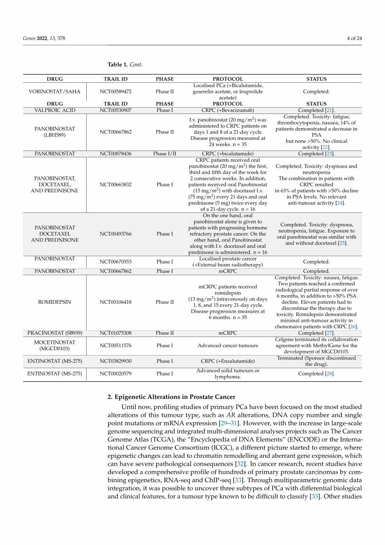
Genes 2022, 13, 378 4 of 24
Table 1. Cont.
DRUG TRAIL ID PHASE PROTOCOL STATUS
VORINOSTAT/SAHA NCT00589472 Phase IILocalised PCa (+Bicalutamide,goserelin acetate, or leuprolide
acetate)Completed.
DRUG TRAIL ID PHASE PROTOCOL STATUSVALPROIC ACID NCT00530907 Phase I CRPC (+Bevacizumab) Completed [21].
PANOBINOSTAT(LBH589) NCT00667862 Phase II
I.v. panobinostat (20 mg/m2) wasadministered to CRPC patients on
days 1 and 8 of a 21-day cycle.Disease progression measured at
24 weeks. n = 35
Completed. Toxicity: fatigue,thrombocytopenia, nausea; 14% of
patients demonstrated a decrease inPSA
but none >50%. No clinicalactivity [22].
PANOBINOSTAT NCT00878436 Phase I/II CRPC (+bicalutamide) Completed [23].
PANOBINOSTAT,DOCETAXEL,
AND PREDNISONENCT00663832 Phase I
CRPC patients received oralpanobinostat (20 mg/m2) the first,third and fifth day of the week for2 consecutive weeks. In addition,
patients received oral Panobinostat(15 mg/m2) with docetaxel I.v.
(75 mg/m2) every 21 days and oralprednisone (5 mg) twice every day
of a 21-day cycle. n = 16
Completed. Toxicity: dyspnoea andneutropenia
The combination in patients withCRPC resulted
in 63% of patients with >50% declinein PSA levels. No relevantanti-tumour activity [24].
PANOBINOSTATDOCETAXEL
AND PREDNISONENCT00493766 Phase I
On the one hand, oralpanobinostat alone is given to
patients with progressing hormonerefractory prostate cancer. On the
other hand, oral Panobinostatalong with I.v. docetaxel and oral
prednisone is administered. n = 16
Completed. Toxicity: dyspnoea,neutropenia, fatigue. Exposure to
oral panobinostat was similar withand without docetaxel [25].
PANOBINOSTAT NCT00670553 Phase I Localised prostate cancer(+External beam radiotherapy) Completed.
PANOBINOSTAT NCT00667862 Phase I mCRPC Completed.
ROMIDEPSIN NCT00106418 Phase II
mCRPC patients receivedromidepsin
(13 mg/m2) intravenously on days1, 8, and 15 every 21-day cycle.
Disease progression measures at6 months. n = 35
Completed. Toxicity: nausea, fatigue.Two patients reached a confirmed
radiological partial response of over6 months, in addition to >50% PSA
decline. Eleven patients had todiscontinue the therapy due to
toxicity. Romidepsin demonstratedminimal anti-tumour activity in
chemonaive patients with CRPC [26].PRACINOSTAT (SB939) NCT01075308 Phase II mCRPC Completed [27].
MOCETINOSTAT(MGCD0103) NCT00511576 Phase I Advanced cancer tumours
Celgene terminated its collaborationagreement with MethylGene for the
development of MGCD0103.
ENTINOSTAT (MS-275) NCT03829930 Phase I CRPC (+Enzalutamide) Terminated (Sponsor discontinuedthe drug).
ENTINOSTAT (MS-275) NCT00020579 Phase I Advanced solid tumours orlymphoma. Completed [28].
2. Epigenetic Alterations in Prostate Cancer
Until now, profiling studies of primary PCa have been focused on the most studiedalterations of this tumour type, such as AR alterations, DNA copy number and singlepoint mutations or mRNA expression [29–31]. However, with the increase in large-scalegenome sequencing and integrated multi-dimensional analyses projects such as The CancerGenome Atlas (TCGA), the “Encyclopedia of DNA Elements” (ENCODE) or the Interna-tional Cancer Genome Consortium (ICGC), a different picture started to emerge, whereepigenetic changes can lead to chromatin remodelling and aberrant gene expression, whichcan have severe pathological consequences [32]. In cancer research, recent studies havedeveloped a comprehensive profile of hundreds of primary prostate carcinomas by com-bining epigenetics, RNA-seq and ChIP-seq [33]. Through multiparametric genomic dataintegration, it was possible to uncover three subtypes of PCa with differential biologicaland clinical features, for a tumour type known to be difficult to classify [33]. Other studies

Genes 2022, 13, 378 5 of 24
have also established PCa subtypes based on distinct epigenetic changes. For instance,in the study by Armenia et al., the authors identified a new class of ETS-fusion-negativePCa defined by epigenetic alterations [34]. Using TCGA methylation and RNA-seq data,Xu et al. performed an epigenetic integrative analysis between normal and PCa tissue, inorder to detect the pathways in which DNA methylation-driven genes were significantlyenriched [35]. More recently, in the study by Lin et al., using single-cell RNA-seq profiles,the authors identified new signature genes and cell subtypes among CRPC cells [36]. Allthis evidence brings out a clear role for epigenetic regulation in PCa control.
Mechanistically many studies have shed light on the molecular effects underlyingepigenetic dysregulation in PCa. One of the most frequent DNA methylation changes occursat the GSTP1 promoter, a fact which was already described 20 years ago. GSTP1 modulatesseveral signalling pathways involved in proliferation, differentiation and apoptosis [37].After this finding, many other recurrent epigenetic alterations have been described, andmay be used in the future as a biomarker for the evaluation of PCa diagnosis and prognosis.Others include the promoter CpG island hypermethylation of PTEN, which causes itssilencing [38], or the hypermethylation of the tumour suppressor gene CDKN2A (whichencodes p16) that leads to increased proliferation, thus contributing to carcinogenesis [39].Even the loss of AR expression is regulated in 30% of CRPC by hypermethylation of itspromoter [40]. More interestingly, recent studies have described that, in metastatic CRPCand tumours that progress to AR-independency, epigenetic principal regulators are clearlyaltered, as well as key factor players in chromatin biology [40].
Besides DNA methylation, other epigenetic marks regulate chromatin structure andgene expression. Among the plethora of epigenetic master regulators, we can find writers,proteins that introduce chemical modifications to DNA and/or histones; readers that identifyand interpret those modifications with its domains; and erasers, whose function is to removethe modifications added by the writers [41]. Uncontrolled activity or expression can lead totumorigenesis through different molecular events, some of which we highlight here.
2.1. Writers
DNA methylation is mainly carried out by a family of DNA methyltransferases (DN-MTs). Five different enzymes exist in humans, DNMT1, DNMT3a, DNMT3b, DNMT3c,and DNMT3L. From those, DNMT3a, DNMT3b and DNMT3c directly carry out the denovo methylation, while DNMT3L performs it indirectly [42]. DNMT1 participates in themaintenance of the hemi-methylated DNA strand during replication [43]. DNA methyl-transferases are frequently affected epigenetic regulators in PCa, leading to hypermethyla-tion and the subsequent silencing of key tumour suppressor genes or oncogenes includingPTEN, CDKN2A, or AR [44–46]. Hypermethylation of oncogenes such as YAP1 has alsobeen associated with neuroendocrine prostate cancer (NEPC) [47]. Expression alterations ofDNMT family members have been associated with altered immune infiltration patterns andbiochemical recurrence [48]. In addition, the targeted activity of specific methyltransferasescan promote tumorigenesis. For example, DNMT1- and DNMT3b-mediated methylationregulates RAD9 transcription, which induces tumorigenicity [49]. DNMT3a promotesepithelial-to-mesenchymal transition (EMT) by regulating the transcription of key mR-NAs [50]. In sum, DNA methylation studies have revealed that DNA methylation levelsare a promising approach to classify prostate cancer patients and improve diagnostic toolsto predict clinical outcomes more accurately. In addition, drug development efforts totarget aberrant DNA hypermethylation led to the development of the DNMT inhibitor5’-azacitidine. The use of these DNMT inhibitors in pre-clinical studies has been shownto suppress tumour growth [51], and currently, DNMT inhibitors in combination withchemotherapy, AR inhibitors and immunotherapy are in clinical development for prostatecancer (Table 1).
Histone modifications are also common epigenetic marks. The most critical marksare catalysed by histone methyltransferases (HMT) and histone acetyltransferases (HATs).Lysine methyltransferases (KMT) are an important family involved in gene transcriptional

Genes 2022, 13, 378 6 of 24
regulation [52]. Specifically, KMT1A and KMT1E, also known as SUV39H1 and SETDB1respectively, are upregulated in PCa cells, increasing their migration and invasion; whileKMT1B (or SUV39H2) has been shown to enhance androgen-dependent activity by inter-acting with AR (Figure 1) [53,54]. Similarly, other epigenetic writers have been proposed asbiomarkers for PCa. For instance, the lysine methyltransferase SET and MYND domain-containing protein 3 (SMYD3) is found upregulated in PCa, and its increased expressionhas been linked to increased cell migration and proliferation (Figure 1) [55]. UpregulatedSETDB1 is associated with prognosis and is suggested to promote PCa bone metastasesthrough the WNT pathway [56]. Members of the protein arginine methyltransferase familyare also aberrantly expressed in prostate cancer cells, e.g., PRMT5, which catalyses histonearginine methylation at histone H4R3, causing epigenetic inactivation of several tumoursuppressors and thus promoting prostate cancer cell growth [57].
Genes 2022, 13, x FOR PEER REVIEW 7 of 26
Histone modifications are also common epigenetic marks. The most critical marks are catalysed by histone methyltransferases (HMT) and histone acetyltransferases (HATs). Lysine methyltransferases (KMT) are an important family involved in gene tran-scriptional regulation [52]. Specifically, KMT1A and KMT1E, also known as SUV39H1 and SETDB1 respectively, are upregulated in PCa cells, increasing their migration and inva-sion; while KMT1B (or SUV39H2) has been shown to enhance androgen-dependent activ-ity by interacting with AR (Figure 1) [53,54]. Similarly, other epigenetic writers have been proposed as biomarkers for PCa. For instance, the lysine methyltransferase SET and MYND domain-containing protein 3 (SMYD3) is found upregulated in PCa, and its in-creased expression has been linked to increased cell migration and proliferation (Figure 1) [55]. Upregulated SETDB1 is associated with prognosis and is suggested to promote PCa bone metastases through the WNT pathway [56]. Members of the protein arginine methyltransferase family are also aberrantly expressed in prostate cancer cells, e.g., PRMT5, which catalyses histone arginine methylation at histone H4R3, causing epigenetic inactivation of several tumour suppressors and thus promoting prostate cancer cell growth [57].
Figure 1. Writers, readers and erasers involved in PCa. Thick arrows indicate over- (red) or under-expression (blue) of the indicated proteins in PCa. Red arrows with flat heads indicate inhibitor compounds under clinical trial against the designated proteins. Writers: KMT1A, KMT1B and KMT1E tri-methylate H3K9; SMYD3 di- and methylates H3K4; PRMT5 methylates H4R3; NSD2 di-methylates H3K36. Erasers: LSD1 demethylates H3K9 and H3K4; KDM5B mono, di- and tri-demeth-ylates H3K4; KDM5B and KDM5C di- and tri-demethylate H3K4. Readers: BRD-containing pro-teins.
Alterations in some epigenetic writers such as upregulation of PRMT1 and Coactiva-tor-Associated Arginine Methyltransferase 1 (CARM1) are found in early tumours, sug-gesting that histone modifications and chromatin remodelling may act as epigenetic driv-ers at the initial stages of the disease [58]. In addition, within the nuclear receptor binding SET domain (NSD) family, probably the most relevant protein in cancer is NSD2, which
Figure 1. Writers, readers and erasers involved in PCa. Thick arrows indicate over- (red) or under-expression (blue) of the indicated proteins in PCa. Red arrows with flat heads indicate inhibitorcompounds under clinical trial against the designated proteins. Writers: KMT1A, KMT1B and KMT1Etri-methylate H3K9; SMYD3 di- and methylates H3K4; PRMT5 methylates H4R3; NSD2 di-methylatesH3K36. Erasers: LSD1 demethylates H3K9 and H3K4; KDM5B mono, di- and tri-demethylates H3K4;KDM5B and KDM5C di- and tri-demethylate H3K4. Readers: BRD-containing proteins.
Alterations in some epigenetic writers such as upregulation of PRMT1 and Coactivator-Associated Arginine Methyltransferase 1 (CARM1) are found in early tumours, suggestingthat histone modifications and chromatin remodelling may act as epigenetic drivers atthe initial stages of the disease [58]. In addition, within the nuclear receptor binding SETdomain (NSD) family, probably the most relevant protein in cancer is NSD2, which wasfirst associated with oncogenesis in tumours such as multiple myeloma [59]; in recentyears, it has been found overexpressed in several solid tumours including PCa, where it isespecially upregulated in metastatic stages and is correlated with recurrence (Figure 1) [60].NSD2 catalyses the di-methylation of histone H3 at lysine 36 (H3K36me2), and thus regu-lating chromatin accessibility and permissive gene transcription [61]. In addition, in vitro

Genes 2022, 13, 378 7 of 24
studies have indicated that NSD2 modulates TWIST1 to promote EMT and invasivenessin prostate cancer cell lines [62]. More interestingly, NF-kB pathway genes includingIL-6, IL-8 or VEGFA are transcriptionally regulated by NSD2, and thus upregulation ofNSD2 in CRPC results in enhanced cell proliferation, survival and increased expressionof inflammatory cytokines, which in turn induce NSD2 expression through a positivefeedback loop [63]. NSD2 also interacts with the AR DNA-binding domain enhancingAR transcriptional activity [64], suggesting that NSD2 may be implicated in promotingADT tolerance [40]. EZH2, a member of the Polycomb Repressive Complex 2 (PRC2) thatregulates transcriptional silencing via histone H3 methylation at lysine 27, is elevatedin PCa too [65]. Mechanistically, increased EZH2 expression favours lineage plasticityand neuroendocrine differentiation in androgen-independent tumours [66,67]. EZH2, likeother histone modifiers, can also modulate AR recruitment to target sites [68], and EHZ2inhibition, alone or in combination with AR inhibitors, has resulted in synergistic AR inhi-bition, resulting in the complete suppression of AR signalling [69,70]. More recently, EZH2inhibition, by activating the double-stranded RNA-STING stress response, has been shownto increase interferon pathway activity and PD-L1 expression by tumour cells, suggestingthat the combination of HMTs inhibitors with immune checkpoint inhibitors could improvethe therapeutic outcome [71]. Altogether, these findings have resulted in the initiation ofseveral clinical trials with EZH2 inhibitors, alone or in combination therapy with othertargeting agents or immunotherapy (Table 1) (Figure 1).
Histone acetyltransferases (HATs) increased activity can also promote PCa by eitheracetylating histones or transcription factors, thus inducing transcription [72]. For exam-ple, p300 and CREB-binding protein (CBP), by acetylating AR, increase its transcriptionalactivity [73], and its inhibition in pre-clinical and early clinical trials has shown to downreg-ulate the AR-dependent transcriptional program, modulate the expression levels of severalbiomarker in CRPC biopsies and tumour growth in both castration-sensitive and castration-resistant prostate tumours [74,75]. More recently, p300/CBP inhibition has been shown todecrease secretion of exosomal PD-L1 by tumour cells, suggesting that the combination ofHAT inhibitors with immune checkpoint inhibitors could play a synergic role and improvetherapeutic efficacy [76].
All this evidence indicates epigenetic writers as promising therapeutic targets, andseveral molecules targeting p300/CBP, PRMT5, EZH2 are being tested under clinical trials,alone or in combination with other therapies such as AR blockade or immunotherapy inadvanced prostate tumours (Figure 1), which we summarize in Table 1 [16,77–80].
2.2. Readers
The bromodomain (BRD)-containing proteins, whose function is to induce transcrip-tion initiation through chromatin remodelling, are some of the most studied in prostatecancer cells among other epigenetic readers. More than 70% of NEPC and more than 50%of primary and metastatic PCas show some dysregulation in any of the bromodomain con-taining proteins [81]. Within this large family, the bromodomain and extraterminal (BET)proteins have been associated with prostate cancer progression. For instance, BRD4, whichis the most critical, recognises acetylated lysines at enhancer regions, thus stimulating RNApolymerase II-dependent transcription by recruiting the elongation factor P-TEFb [82]. Inaddition, in PCa BRD4 also interacts with AR, recruiting AR to sequence-specific DNA-binding motifs to drive AR-mediated gene transcription, making AR-dependent cell linesselectively sensitive to BRD4 inhibitors [83,84]. BET bromodomain proteins have numer-ous AR-independent functions by activating c-Myc-dependent and c-Myc–independenttranscriptional regulators upregulated in CRPC [85].
Apart from the BET family, several BRD-containing proteins have been associated withmCRPC. TRIM24 is a transcription co-regulator overexpressed in CRPC, and is essentialfor cancer cell proliferation [86]. BAZ2A binds to H3K14ac, repressing transcription andpromoting a more aggressive form of PCa [87]. H3K4me2,3 epigenetic reader CHD1deficiency can lead to a dramatic decrease in cell proliferation, survival, and tumorigenic
Genes 2022, 13, 378 8 of 24
potential [88]. ATAD2, BRD8, CREBBP, or KTM2A, by recognizing acetylated histones, actas superenhancers and transcriptional regulators that initiate chromatin restructuring topromote tumorigenesis. This evidence show that dysregulated BRD-containing proteinsdrive aberrant transcriptional programs promoting oncogenesis. This evidence has broughtout the potential of targeting these chromatin remodellers, revealing that their inhibitionmay disrupt the dysregulated transcriptional networks with oncogenic functions. In fact,several small molecule inhibitors, blockers and proteolysis-targeting chimeras (PROTACs),which induce degradation of BET proteins through ubiquitination followed by proteolysishave been developed, and some are currently tested in clinical trials, which we summarizein Table 1 (Figure 1) [16,89–92].
2.3. Erasers
The critical erasers in PCa are DNA demethylases, histone demethylases (HDMT)and histone deacetylases (HDACs), which cause hypomethylation or deacetylation andconsequently the upregulation of gene expression, leading to disease progression andhigher cell invasion and metastasis [93].
To date, the only known DNA demethylases are TET enzymes, comprised of threemembers, namely TET1-3. DNA demethylation is a three-step process, where TETs par-ticipate in the first one, oxidizing m5C to 5-hydorxymethylation (hm5C) [94]. Decreasedexpression of TET enzymes has been associated with PCa. For instance, downregulation ofTET2 has been implicated in the regulation of AR signalling and prostate cancer develop-ment [95,96]. TET1 is downregulated in PCa, and its depletion promotes tumour growthand metastasis in prostate xenograft models and correlates with poor survival rates [97].
Within HDMTs, lysine demethylases (KDM) play important roles in cancer. For exam-ple, lysine-specific demethylase 1 (LSD1/KDM1A) overexpression has been highly studiedfor its oncogenic potential. Its overexpression is associated with increased tumour progres-sion and promotion of carcinogenesis in several cancer types, including PCa [98,99]. In PCa,higher expression of LSD1 is correlated with recurrence and poor survival in metastaticpatients, and its function has been shown to be distinctive in androgen-dependent andrefractory PCa (Figure 1) [98,100]. In fact, LSD1 is among the best-known modulatorsof AR transcriptional activity, which can either stimulate or suppress AR transcriptionalactivity, unveiling a dual role in PCa progression, which is common in other chromatinremodellers [98,101]. In addition, LSD1 can demethylate other key transcription factorsin PCa such as FOXA1, leading to its activation and recruitment to AR-dependent en-hancers [102]. Moreover, LSD1-mediated epigenetic changes can activate other key genessuch as Centrosome-associated protein E (CENPE) [103], as well as cooperate with ZNF217,promoting the expression of genes involved in tumorigenesis, thus leading to CRPC pro-motion [104].
Many other histone demethylases have been associated to PCa, and similarly to LSD1,their gain or loss of activity seem to have a dual role (Figure 1). H3K4 demethylase KDM5D(JARID1D) is downregulated in metastatic PCa [105], and its loss is associated with re-sistance to docetaxel in PCa [106]. By contrast, other HMTs are upregulated and theirincreased activity leads to cancer progression. For instance, KDM5C (JARID1DC) is upreg-ulated in PCa and has recently emerged as a predictive biomarker for therapy failure afterprostatectomy [107]. KDM6A deletion inhibits tumour progression [108]. KDM6B (JMJD3)regulates c-Myc expression, thus promoting cell proliferation [109]. KDM5B (JARID1DB)plays a role as an AR coactivator, and is upregulated in PCa [110]. Similarly, the KDM4(JMJD2) family of H3K9 demethylates, either alone or cooperating with LSD1, also play asignificant role in modulating AR transcriptional activity, [52,111–113], or promoting theexpression of AR spliced variants inducing resistance to ADT therapies [114]. KDM4Bpromotes AR-independent survival by cooperating with BMyb, and its inhibition reducesthe growth of AR-independent PCa cells [115]. KDM3B has been implicated in androgen-independent CRPC, and its genetic or pharmacological suppression has shown to reducethe survival of CRPC cells versus castration-sensitive cells [116]. The histone demethylase

Genes 2022, 13, 378 9 of 24
PHD-finger protein 8 (PHF8) is upregulated in PCa, acts as a transcriptional coactivator ofAR via H4K20 demethylation and promotes cancer cell proliferation, migration, invasion,and neuroendocrine differentiation [117,118]. KDM8 (JMJD5) promotes cellular prolifer-ation and controls the activity of AR, HIF-1a, and EZH2 [119]. All this evidence showsthat histone methylation dysregulation promotes oncogenesis by promoting chromatinstructural changes, and suggests that targeting histone lysine demethylation is a goodanti-cancer therapeutic option. In fact, recently developed LSD1 inhibitors have resultedin attenuated tumour growth, and are currently under clinical testing [104,120] (Table 1)(Figure 1).
Decreased histone deacetylase (HDACs) activity is found associated with PCa too.Contrary to HATs resulting in the increased acetylation of histones or AR, the activityof deacetylases such as SIRT1 or SIRT2 can regulate cellular growth and sensitivity toADT therapy through AR deacetylation [121], and the use of small molecule inhibitorshas been shown to effectively reduce tumour growth, EMT and metastasis, and sensitizemCRPC to ADT therapy (Figure 1) (Table 1) [23,27,122]. These studies have shown thatsingle-agent HDAC inhibitor clinical trials have not shown significant activity. However,their combination with AR inhibitors has resulted in improved therapeutic efficacy [23].While those results need to be further evaluated with newer AR inhibitors, they suggest thatcombining HADCs and AR inhibitors may be an effective strategy to re-sensitize tumoursto AR inhibition.
In conclusion, all this evidence highlights that epigenetic alterations are commonlyassociated with PCa and, most importantly, can be used to stratify patient risk, predictclinical outcomes, and to determine best therapeutic options or to reveal molecular pathwayvulnerability to distinct chemotherapeutic agents [123,124]. In addition, the oncogenicpotential of altered epigenomes in cancer cells suggests that rewiring the epigenome maybe an effective strategy to sensitize or kill cancer cells. In fact, several HMT/HDMTs,BET bromodomain, HDACs [40], and DNMT inhibitors are currently under clinical orpre-clinical stages in PCa [125,126], some of which have been approved by the FDA forother malignancies such as myelodysplastic syndrome (MDS), acute myeloid leukaemia(AML) or acute lymphocytic leukaemia (ALL) [127] (Table 1). Yet, further research willbe needed to completely understand the extensive transcriptional networks that theseepigenetic changes regulate, and how cancer cell fates may be affected. In addition, morestudies are necessary to assess their clinical efficacy, to reduce secondary and adverseeffects, and to optimise their use in combination with conventional chemotherapies andwith predictive biomarkers to select patients that would benefit from these therapies.
3. Epitranscriptomics Alterations in Prostate Cancer
Similarly to DNA, RNA can also be modified. Despite being known for over 50 years,the study of RNA modifications has suffered a delay regarding epigenetics, probably dueto the lack of suitable tools for their study [128]. Thus, the emergence of this field, knownas epitranscriptomics, is closely linked to the recent refinement of tools such as massspectrometry, next-generation sequencing [128] or cryo-electron microscopy [129], whichhave enabled the discovery of over 170 RNA modifications [130,131].
These modifications are found in all types of RNA, from messenger RNA (mRNA)to non-coding RNAs such as ribosomal RNA (rRNA), transfer RNA (tRNA), microRNAs(miRNAs) and long noncoding RNAs (lncRNAs) among many others [130]. tRNAs arethe more extensively modified, with an average of 15% modified nucleotides per moleculeand involving a large number of enzymes and a high diversity of modifications (reviewedin [132,133]). In rRNA, around 130 individual modifications can be found, with 2’-O-methylation of the ribose and pseudouridine (Ψ) being the most frequent modification(reviewed in [134]). In the case of mRNA, the most abundant internal modification is N6-Methyladenosine (m6A), with around 0.1-0.4% adenines of all mRNAs being modified [135].

Genes 2022, 13, 378 10 of 24
In contrast with DNA modifications, which are known to mainly regulate gene ex-pression [136], RNA modifications control many functions apart from transcription such asRNA stability, location, splicing, degradation or translation efficiency [134,137,138]. For in-stance, 5-methylcytosine (m5C) methylation of tRNAs stabilises their structure and protectsthem from nuclease-mediated cleavage [137]. However, the role or importance of most ofthese modifications are still unknown and, for others, it is only starting to emerge.
Despite the great diversity of modified nucleotides in RNA and the huge expansion ofthe field in the past years, little is known about the role of RNA modifications in PCa. Inthis review, we will summarize the most relevant RNA modifications found to date to havea regulatory role in prostate cancer.
3.1. m6A Deposition in RNA and Its Role in PCa
Methylation of position N6 of adenosine is the most studied modification. Thismodification mainly occurs in mRNA, but also in non-coding RNAs such as tRNAs, rRNA,miRNA, lncRNAs and circular RNAs (circRNAs) [139]. Despite the widespread occurrence,m6A deposition in mRNA is, by far, the best characterised. Recent improvements in high-throughput methods have demonstrated that m6A is not randomly distributed, but isspecifically enriched near stop codons in 3’-untranslated regions (UTRs), within long exons,in intergenic regions and introns and at 5’-UTRs [135,139].
m6A deposition is modulated by the dynamic crosstalk between methyltransferasesor “writers” and demethylases or “erasers”. In nascent pre-mRNA, m6A is deposited by amultimeric methyltransferase complex. The catalytic core of this complex is constituted bymethyltransferase-like 3 (METTL3) and methyltransferase-like 14 (METTL14), which catal-yses the formation of m6A and recognises RNA substrates, respectively [140]. This complexalso requires several cofactors such as RNA-binding motif protein 15 (RBM15), Wilms’ tumour-associated protein (WTAP), Cbl proto-oncogene-like 1 (CBLL1, also known as HAKAI), zincfinger CCCH-type containing 13 (ZC3H13) and Vir-like m6A methyltransferase-associated(VIRMA, also known as KIAA1429) [135,140]. Recent studies have identified another m6Amethyltransferase, METTL16, which acts as an independent RNA methyltransferase andmainly modifies mRNA and small nuclear RNAs (snRNAs) [141]. In addition, METTL5 hasalso been identified as a 18S rRNA m6A methyltransferase acting as a heterodimer togetherwith TRMT112 [142] (Figure 2A).
Ten years ago, the discovery of two demethylases, fat mass and obesity-associatedprotein (FTO) and AlkB homolog 5 (ALKBH5), brought to light the reversible nature of thismodification [143] and recently, another member of AlkB homolog family, ALKBH3, hasbeen reported as a novel m6A demethylase [140]. However, the role of the m6A demethy-lases is still controversial. In 2017, Mauer et al. found that FTO is able to demethylatem6Am at the 5’ cap, rather than m6A, thereby decreasing mRNA stability. The disparity ofthis discovery initiates a discussion about m6A detection techniques and concerning thenature of its reversibility [144]. Moreover, m6A can be specifically recognised by a groupof RNA binding enzymes or “readers” that recruit downstream complexes mediating thefunction of this modification [135]. Several readers have been reported, including: YTHdomain-containing proteins (YTHDF1/2/3 and YTHDC1/2), heterogeneous nuclear ri-bonucleoproteins (including hnRNPC, hnRNPG and hnRNPA2B1), insulin-like growthfactor 2 mRNA-binding proteins (IGF2BP1/2/3), proline-rich and coiled-coil-containingprotein 2A (PRRC2A) and the fragile X mental retardation 1 (FMR1) [135] (Figure 2A).
Genes 2022, 13, 378 11 of 24Genes 2022, 13, x FOR PEER REVIEW 12 of 26
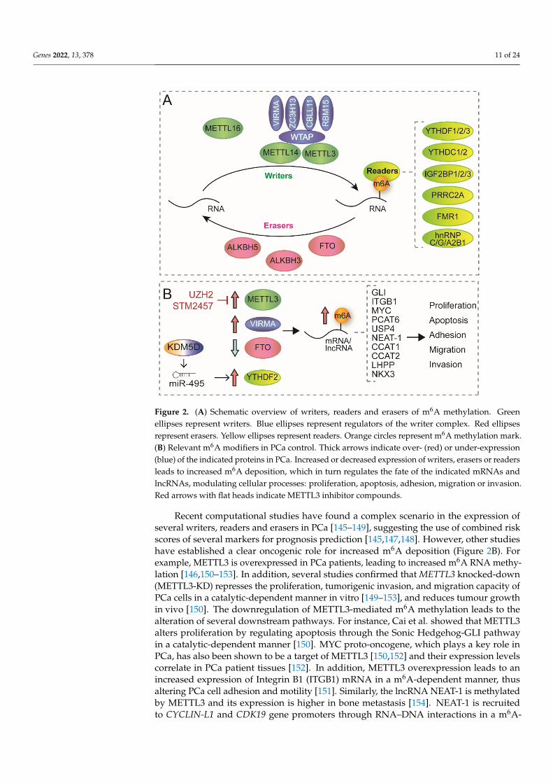
Figure 2. (A) Schematic overview of writers, readers and erasers of m6A methylation. Green ellipses represent writers. Blue ellipses represent regulators of the writer complex. Red ellipses represent erasers. Yellow ellipses represent readers. Orange circles represent m6A methylation mark. (B) Rel-evant m6A modifiers in PCa control. Thick arrows indicate over- (red) or under-expression (blue) of the indicated proteins in PCa. Increased or decreased expression of writers, erasers or readers leads to increased m6A deposition, which in turn regulates the fate of the indicated mRNAs and lncRNAs, modulating cellular processes: proliferation, apoptosis, adhesion, migration or invasion. Red arrows with flat heads indicate METTL3 inhibitor compounds.
Ten years ago, the discovery of two demethylases, fat mass and obesity-associated protein (FTO) and AlkB homolog 5 (ALKBH5), brought to light the reversible nature of this modification [143] and recently, another member of AlkB homolog family, ALKBH3, has been reported as a novel m6A demethylase [140]. However, the role of the m6A deme-thylases is still controversial. In 2017, Mauer et al. found that FTO is able to demethylate m6Am at the 5’ cap, rather than m6A, thereby decreasing mRNA stability. The disparity of this discovery initiates a discussion about m6A detection techniques and concerning the nature of its reversibility [144]. Moreover, m6A can be specifically recognised by a group of RNA binding enzymes or “readers” that recruit downstream complexes mediating the function of this modification [135]. Several readers have been reported, including: YTH domain-containing proteins (YTHDF1/2/3 and YTHDC1/2), heterogeneous nuclear ribo-nucleoproteins (including hnRNPC, hnRNPG and hnRNPA2B1), insulin-like growth fac-tor 2 mRNA-binding proteins (IGF2BP1/2/3), proline-rich and coiled-coil-containing pro-tein 2A (PRRC2A) and the fragile X mental retardation 1 (FMR1) [135] (Figure 2A).
Recent computational studies have found a complex scenario in the expression of several writers, readers and erasers in PCa [145–149], suggesting the use of combined risk scores of several markers for prognosis prediction [145,147,148]. However, other studies have established a clear oncogenic role for increased m6A deposition (Figure 2B). For ex-
Figure 2. (A) Schematic overview of writers, readers and erasers of m6A methylation. Greenellipses represent writers. Blue ellipses represent regulators of the writer complex. Red ellipsesrepresent erasers. Yellow ellipses represent readers. Orange circles represent m6A methylation mark.(B) Relevant m6A modifiers in PCa control. Thick arrows indicate over- (red) or under-expression(blue) of the indicated proteins in PCa. Increased or decreased expression of writers, erasers or readersleads to increased m6A deposition, which in turn regulates the fate of the indicated mRNAs andlncRNAs, modulating cellular processes: proliferation, apoptosis, adhesion, migration or invasion.Red arrows with flat heads indicate METTL3 inhibitor compounds.
Recent computational studies have found a complex scenario in the expression ofseveral writers, readers and erasers in PCa [145–149], suggesting the use of combined riskscores of several markers for prognosis prediction [145,147,148]. However, other studieshave established a clear oncogenic role for increased m6A deposition (Figure 2B). Forexample, METTL3 is overexpressed in PCa patients, leading to increased m6A RNA methy-lation [146,150–153]. In addition, several studies confirmed that METTL3 knocked-down(METTL3-KD) represses the proliferation, tumorigenic invasion, and migration capacity ofPCa cells in a catalytic-dependent manner in vitro [149–153], and reduces tumour growthin vivo [150]. The downregulation of METTL3-mediated m6A methylation leads to thealteration of several downstream pathways. For instance, Cai et al. showed that METTL3alters proliferation by regulating apoptosis through the Sonic Hedgehog-GLI pathwayin a catalytic-dependent manner [150]. MYC proto-oncogene, which plays a key role inPCa, has also been shown to be a target of METTL3 [150,152] and their expression levelscorrelate in PCa patient tissues [152]. In addition, METTL3 overexpression leads to anincreased expression of Integrin B1 (ITGB1) mRNA in a m6A-dependent manner, thusaltering PCa cell adhesion and motility [151]. Similarly, the lncRNA NEAT-1 is methylatedby METTL3 and its expression is higher in bone metastasis [154]. NEAT-1 is recruitedto CYCLIN-L1 and CDK19 gene promoters through RNA–DNA interactions in a m6A-

Genes 2022, 13, 378 12 of 24
dependent manner, leading to increased proliferation and migration both in vitro and inxenografts [154]. Moreover, METTL3 downregulation affects ubiquitin-specific proteaseUSP4 mRNA levels, leading to decreased migration and invasiveness in PCa [153]. Inaddition, one of the pathways by which an increase of METTL3 promotes bone metastasisis through the upregulation of the lncRNA PCAT6 via the m6A reader IGFBP2 [155]. Theseresults, together with the increased expression of METTL3 in PCa with bone metastasiscompared to primary tumours [151], suggest that METTL3-mediated methylation mightplay an important role in PCa progression and metastasis. More recently, studies haveshown an interplay between m6A regulator expression and immune infiltration. In Zhaoet al., low METTL3 and HNRNPA2B1 expression correlates with increased immune cellinfiltration [156]. In Liu et al. tumours with worse prognosis show a significant decreasedexpression of METTL14 and ZC3H13, markedly high expression of KIAA1429 and HN-RNPA2B1, and are characterised by high intratumor heterogeneity, Th2 cell infiltration, andlow Th17 cell infiltration [157], highlighting the functional role of the epitranscriptome inregulating a wide range of oncogenic processes.
Other members of the methyltransferase complex have been found dysregulated inPCa. VIRMA has been found upregulated in PCa [158], and its knockdown, by regulatingthe abundance of the oncogenic lncRNAs CCAT1 and CCAT2, can reduce the proliferation,migration and invasion of PCa cells through MYC regulation [158]. The m6A readerYTHDF2 has been found upregulated in PCa, its expression predicts worse overall survival,and its knockdown inhibits proliferation and migration of PCa in vivo and in vitro, bybinding and degrading m6A-methylated LHPP and NKX3-1 mRNAs, resulting in increasedAKT activity [159]. Interestingly, increased expression of YTHDF2 has been found to beepigenetically regulated by the H3K4me3 demethylase KDM5A. By binding to the miR-495promoter, KDM5A leads to miR-495 transcriptional inhibition and decreased expression,which results in decreased silencing of YTHDF2 mRNA [160]. By contrast, FTO has beenfound downregulated in PCa tissues and cell lines [161]. Similarly to the effects of theincreased expression of m6A writers, downregulation of FTO leads to increased cell invasionand migration capacity by increasing m6A levels [161].
In sum, all this evidence suggests that increased m6A deposition has an oncogeniceffect in prostate cancer cells and targeting METTL3 could have clinical benefits for PCapatients. With the recent emergence of METTL3 as a promising oncogenic target in severalhaematological malignancies and solid tumours, a great effort is being made to developsmall molecule inhibitors to target m6A deposition. Two recent inhibitors have beendeveloped that target and inhibit METTL3, STM2457 and UZH2. These two inhibitorshave shown to decrease m6A methylation activity in vitro and in vivo. In addition, theyhave shown potent inhibitory potential in several cancer cell lines including PCa, and apromising anti-cancer effect in cell lines and pre-clinical models of acute myeloid leukaemia(AML) [162,163]. These promising results open the door to explore new therapeutic possi-bilities in cancer research.
3.2. m5C and hm5C Deposition in RNA and Its Role in PCa
m5C deposition in RNA is conserved in all life domains [131] and has been found mostprevalently in tRNA and rRNA, but also, less frequently, in mRNA, lncRNA, vault RNA(vtRNA) and small nucleolar RNA (snoRNA) [164,165]. In mammals, there are two familiesof enzymes known to specifically methylate cytosine at position 5: the NOL1/NOP2/sun(NSUN) family and the DNA methyltransferase family member 2 (DNMT2) [131,165]. Todate, NOP2 (also known as nucleolar antigen p120) is the only m5C methyltransferase thathas been demonstrated to be associated with PCa. Increased expression of NOP2 has beenconsidered a marker of bad prognosis for years, correlating with Gleason score, PSA serumlevels and recurrence after radical prostatectomy [166,167]. Mechanistically, NOP2 catalysesthe methylation of cytoplasmic 28S rRNA [168]. This methyltransferase is expressed in thelate G1 and S phases of the cell cycle and is known to regulate nuclear activation associatedwith proliferation [166]. In addition, a recent genome-wide association study combining

Genes 2022, 13, 378 13 of 24
enhancer and eQTL mapping has reported that lower expression of NSUN4 is associatedwith an increased prostate cancer risk [169]. NSUN4 targets 12S mitochondrial rRNA,found on the small subunit [170], yet its potential role in PCa remains to be validated.In addition, whether other m5C RNA methyltransferases are associated with PCa is alsostill uncovered.
Similarly to DNA, hm5C and f5C have been found in mRNA and tRNAs in vivo [171].ALKBH1 has been found to catalyse the conversion of m5C into f5C in tRNAs in vivo [172].In vitro studies have shown that TET enzymes are able to oxidise m5C to hm5C in RNA [173].Despite this finding, the enzyme that catalyses hm5C conversion in mRNA is still unknown.TET2 is found downregulated in PCa [95,174], yet whether decreased hm5C RNA levelsare linked to PCa needs to be elucidated.
From a therapeutic point of view, m5C as well as other RNA modifications havebeen linked to the survival, proliferation and differentiation of tumour cells in severalcancers [175–177]. Interestingly, m5C as well as other RNA modifications have been asso-ciated with drug resistance regulation [175,177,178]. For instance, combined knockdownof NSUN2 and METTL1, which catalyses tRNA 7-methylguanosine, has shown to sensi-tize tumour cells to the chemotherapeutic agent 5’-fluorouracil (5-FU) [178], confirmingthe link between RNA methylation, cell survival and chemotherapy. In addition, in skincancer, a lack of NSUN2 specifically sensitizes tumour-initiating cells to 5-FU [177], furthershowing that the combination of classic chemotherapeutic agents and the inhibition ofRNA modifications could become a more effective therapeutic strategy. This role of RNAmodifications has not been evaluated in PCa, but aforementioned investigations linkingseveral RNA-modifying enzymes with key PCa tumorigenic pathways will pave the wayto finding novel therapeutic targets.
3.3. Pseudouridine in RNA and Its Role in PCa
Pseudouridine (Ψ), the C5-glycoside isomer of uridine, is among the most well-knownRNA modifications. It comprises about 5% of the total of all RNA nucleotides and isfound in almost all types of non-coding and coding RNAs [179]. Ψ biosynthesis can becarried out both by stand-alone enzymes and by RNA-guided ribonucleoprotein (RNP)complexes. To date, eleven pseudouridine synthases have been identified in humans;PUS1, TRUB2, PUS3, PUS4, RPUSD1, RPUSD2, RPUSD3, RPUSD4, PUS7, PUS7L, andPUS10 [180]. RNP complexes are constituted by the enzymatic core components dyskerin(DKC1), which carries the enzymatic activity, and three additional core proteins: nucleolarprotein 10 (NOP10), glycine-arginine-rich protein 1 (GAR1) and non-histone protein 2(NHP2); in addition, non-coding RNAs called H/ACA box snoRNAs that guide the proteincomplex to the specific uridines to be modified and NAF1 and SHQ1, which are H/ACA-specific chaperones [181]. This modification is irreversible and has shown to play animportant role in gene expression regulation and maintenance of structural stability [135].Similarly, the fate of Ψ-targeted RNAs is dependent on their readers. Recent studies haveidentified MetRS as a potential Ψ reader [182].
Aberrant deposition of Ψ, dysregulated expression of pseudouridylases and somaticmutations have been linked to PCa. For instance, in an integrative genomic profilingstudy of prostate cancer, somatic mutations in the chromosomal 3p were identified astumour suppressors; most importantly, SHQ1 was the only gene in the 3p region thatharboured tumour associated mutations [7,30], and loss-of-function studies confirmeda tumour growth-suppressive role of SHQ1 [183]. DKC1 is overexpressed in PCa andits knockdown decreases cell proliferation in prostate adenocarcinoma cell lines [184].While, mechanistically, it is still unknown whether increased DKC1 expression correlateswith Ψ increased levels, some studies have shown that targeting its binding site to theRNA component of Telomerase (hTR) can modulate telomerase activity [185]. However,Ψ levels have been found to be increased in the rRNA of PCa cell lines compared toprostate epithelial cells, as well as in PCa tissue compared to normal adjacent tissues [186],suggesting an oncogenic role for elevated Ψ levels. Computational analysis has also shown

Genes 2022, 13, 378 14 of 24
the upregulation of several H/ACA snoRNAs and amplification of DKC1 in metastatictumours compared to primary tumours [186–189]. Furthermore, the studies showed thatthe silencing of those snoRNA in PCa cell lines reduced their proliferation and metastaticpotential [189,190].
Other functional studies have highlighted an oncogenic role for increased Ψ deposi-tion. For example, the stand-alone pseudouridine synthase PUS10 has shown to play a keyrole in TRAIL-induced apoptosis, participating in the release of cytochrome C and SMACfrom the mitochondria in PCa cell lines [191]. Moreover, increased abundance of PUS1was recently found to be associated with an increased risk of relapse [192]. Furthermore,metabolomic analysis of urine samples has found increased levels of Ψ in PCa patients,suggesting Ψ levels in liquid non-invasive biopsies could be a novel predictive biomarkerto use in combination with others, such as PSA levels, in order to improve diagnosis [193].Taken together, the evidence so far shows that Ψ increased deposition has an oncogenicpotential, yet the molecular mechanisms underlying this effect still need to be fully investi-gated to establish whether targeting Ψ writers may have clinical benefits for PCa patients.Nonetheless, Ψ arises as a promising minimally invasive biomarker for PCa detection,but further studies and standardization are needed in order to obtain a fast, cost-effectivedetection method for the clinical practice.
3.4. RNA Editing in PCa
RNA editing is also a very prevalent post-transcriptional event in human transcrip-tomes. The most common type of RNA editing results from the conversion of adenosine(A) to inosine (I) in double-stranded RNA, a process catalysed by the adenosine deaminaseacting on the RNA (ADAR) family of enzymes. A-to-I editing is highly prevalent withinAlu elements, as well as introns, untranslated regions (UTRs) and coding transcripts [194].In a comprehensive study to identify edited RNAs in prostate cancer patients, 16 pairedDNA–RNA sequence libraries from prostate tumour specimens were analysed. Over ahundred thousand putative RNA editing events were found on introns and UTRs, andcoding regions predicted to result in deleterious amino acid alterations [195]. In a latertranscriptome-wide study, elevated RNA-editing and expression of ADAR enzymes werefound across multiple cancer tissues, including the prostate [196]. Moreover, rare germlineheterozygous variants in ADAR predispose to prostate cancer [197]. Despite these findings,little is known on the mechanistic link between elevated RNA editing levels and PCa, andfew have explored the molecular causes. For instance, RNA editing of AR mRNA hasbeen found in PCa cell lines, which leads to mutated AR with a gain-of-function activity,suggesting a contribution to hormone-refractory phenotypes [198]. Another study impli-cates an inflammation-driven malignant transformation due to increased type I interferonexpression [197].
Taken together, these studies disclose an important regulatory mechanism in cancerfor RNA editing; however, a systematic effort to define the putative roles of edited RNAsin PCa tumorigenesis and progression remains to be established.
3.5. 2′-O-methylation in PCa
One of the most abundant ribonucleotides in rRNA is 2′-O-methylation (Nm, ri-bomethylation), with up to 112 different positions in human ribosomes. Similarly to Ψ, itsdeposition is carried out by RNA-guided ribonucleoprotein (RNP) complexes formed bythe core proteins NOP56, NOP58, SNU13 and fibrillarin (FBL) and the guiding box C/DsnoRNAs [199]. Altered expression or mutations in the components of the 2′-O-methylationmachinery have been found to be associated with several cancer types, yet in PCa, only ex-pression changes in snoRNA U50 have been associated with a tumour suppressor functionin PCa [200]. More recently, a study unveiled a methyltransferase-independent function ofthe oncogene EZH2 that relies on a direct interaction with fibrillarin, leading to enhancedrRNA 2′-O methylation and protein translation [201]. This study highlights an important al-

Genes 2022, 13, 378 15 of 24
liance between epitranscriptomic and epigenetic pathways in tumorigenesis, and suggeststhat increased 2′-O methylation may have important consequences for cancer development.
4. Concluding Remarks
Despite the initial response to hormone-deprivation treatment, one of the main prob-lems in PCa management is the relapse and progression rate to metastatic tumour, whichhas limited therapeutic options, none of them completely curative [40,202]. This intensifiesthe urgent need for the investigation of new therapeutic approaches.
Recent evidence has highlighted that epigenetic alterations are emerging as potentialbiomarkers to stratify PCa patients and predict clinical outcomes [40]. Epigenetic alterationsare most common in advanced PCa, being especially dysregulated in metastatic CRPC [13].These findings suggest an important role of epigenetic regulation in advanced phases of thedisease and indicate that epigenetic mechanisms may regulate tumour selective pressures.The use of epigenetic modulators has been growing in recent years, and currently, sixepigenetic drugs are approved by FDA for cancer treatment, mainly for haematologicalmalignancies [127]. Regarding PCa, despite the huge number of studies pointing to epi-genetic modulators as prognostic markers, none of them are used nowadays in clinicalpractice. However, clinical trials have shown only mild results in PCa patients, probablybecause most of them have been undertaken in late-stage, heavily pre-treated patientsand without considering tumour subtypes [127,203,204]. A deep understanding of themolecular mechanism underlying the epigenetic mechanism and tumour biology will allowthe development of successful clinical trials and the eventual approval of epigenetics-basedtherapies for PCa.
As in DNA, RNA modifications are also known to regulate responses to environ-mental signals [137,177] suggesting that they too may regulate cancer cells’ survival ofchallenges occurred during tumour expansion or therapies, making them attractive thera-peutic targets. However, unlike epigenetics, epitranscriptomics has not reached the clinicyet. Changes in several RNA modifications have been linked to different tumours includingPCa [150–152,154,158,160,180,186,204], revealing their potential role as tumour biomark-ers. However, their use is still limited by the lack of easy, sensitive, cost-effective andreliable high-throughput detection methods. In addition, the aberrant expression of RNA-modifying enzymes has also been reported in PCa [150–152,158,166], but their specific rolesin regulating tumorigenesis remain to be further characterised.
Similarly to epigenetics, RNA modifications are emerging as promising therapeutictargets, and great efforts are now being made to develop small molecule inhibitors torewire the aberrant cancer epitranscriptomes. However, targeting RNA modificationscould be fairly complicated since they are linked to most aspects of RNA biology, and theiralteration could involve undesirable toxic effects. Moreover, the role of RNA modificationsis context-dependent and could differ between cancers or even between different cellpopulations [177,205]. Thus, there is still a long road ahead that will require great researchefforts in order to fully understand the biology of RNA modifications and the meansto effectively target them, so that ground-breaking epitranscriptomics can finally reachthe clinic.
Author Contributions: J.L., A.M.A.-G. and S.B. designed and wrote the manuscript. Ó.M.-G., wrotethe manuscript. J.L., A.M.A.-G., Ó.M.-G. and S.B. designed the figures. J.L. and A.M.A.-G. equallycontributed to this manuscript. All authors have read and agreed to the published version ofthe manuscript.
Funding: This work was funded by the Spanish Ministry of Science and Innovation MCIN/AEI/10.13039/501100011033 grant PID2019-111692RB-I00. In addition, we acknowledge funding fromThe Scientific Foundation AECC (LABAE19040BLAN), the University of Salamanca and FundaciónMemoria de Samuel Solorzano Barruso (FS/36-2020), and Consejería de Educación de la Junta deCastilla y León (CSI264P20) to S.B., A.M.A-G. was supported by USAL-Santander PhD fellowship.J.L. was supported by The Scientific Foundation AECC predoctoral fellowship (PRDSA19002LÓPE)and the Spanish Ministry of Universities predoctoral fellowship (FPU19/01190). The Instituto de

Genes 2022, 13, 378 16 of 24
Biología Molecular y Celular del Cancer is supported by the Programa de Apoyo a Planes Estratégicosde Investigación de Estructuras de Investigación de Excelencia, co-financed by the Castilla–Leónautonomous government and the European Regional Development Fund (CLC–2017–01).
Conflicts of Interest: The authors declare no conflict of interest.
References1. Shen, M.M.; Abate-Shen, C. Molecular genetics of prostate cancer: New prospects for old challenges. Genes Dev. 2010, 24,
1967–2000. [CrossRef] [PubMed]2. Siegel, R.L.; Miller, K.D.; Jemal, A. Cancer statistics, 2019. CA Cancer J. Clin. 2019, 69, 7–34. [CrossRef] [PubMed]3. Pernar, C.H.; Ebot, E.M.; Wilson, K.M.; Mucci, L.A. The Epidemiology of Prostate Cancer. Cold Spring Harb. Perspect. Med. 2018, 8,
a030361. [CrossRef] [PubMed]4. Schatten, H. Brief Overview of Prostate Cancer Statistics, Grading, Diagnosis and Treatment Strategies. Adv. Exp. Med. Biol. 2018,
1095, 1–14. [CrossRef] [PubMed]5. Nunzio, C.D.E.; Presicce, F.; Giacinti, S.; Bassanelli, M.; Tubaro, A. Castration-resistance prostate cancer: What is in the pipeline?
Minerva Urol. Nefrol. 2018, 70, 22–41. [CrossRef]6. Armstrong, C.M.; Gao, A.C. Adaptive pathways and emerging strategies overcoming treatment resistance in castration resistant
prostate cancer. Asian J. Urol. 2016, 3, 185–194. [CrossRef]7. Berger, M.F.; Lawrence, M.S.; Demichelis, F.; Drier, Y.; Cibulskis, K.; Sivachenko, A.Y.; Sboner, A.; Esgueva, R.; Pflueger, D.;
Sougnez, C.; et al. The genomic complexity of primary human prostate cancer. Nature 2011, 470, 214–220. [CrossRef]8. Abeshouse, A.; Ahn, J.; Akbani, R.; Ally, A.; Amin, S.; Andry, C.D.; Annala, M.; Aprikian, A.; Armenia, J.; Arora, A.; et al. The
Molecular Taxonomy of Primary Prostate Cancer. Cell 2015, 163, 1011–1025. [CrossRef]9. Abate-Shen, C.; Shen, M.M. Molecular genetics of prostate cancer. Genes Dev. 2000, 14, 2410–2434. [CrossRef]10. Abida, W.; Armenia, J.; Gopalan, A.; Brennan, R.; Walsh, M.; Barron, D.; Danila, D.; Rathkopf, D.; Morris, M.; Slovin, S.; et al.
Prospective Genomic Profiling of Prostate Cancer Across Disease States Reveals Germline and Somatic Alterations That MayAffect Clinical Decision Making. JCO Precis. Oncol. 2017, 2017, PO.17.00029. [CrossRef]
11. Barbieri, C.E.; Baca, S.C.; Lawrence, M.S.; Demichelis, F.; Blattner, M.; Theurillat, J.P.; White, T.A.; Stojanov, P.; Van Allen, E.;Stransky, N.; et al. Exome sequencing identifies recurrent SPOP, FOXA1 and MED12 mutations in prostate cancer. Nat. Genet.2012, 44, 685–689. [CrossRef] [PubMed]
12. Pritchard, C.C.; Mateo, J.; Walsh, M.F.; De Sarkar, N.; Abida, W.; Beltran, H.; Garofalo, A.; Gulati, R.; Carreira, S.; Eeles, R.; et al.Inherited DNA-Repair Gene Mutations in Men with Metastatic Prostate Cancer. N. Engl. J. Med. 2016, 375, 443–453. [CrossRef][PubMed]
13. Friedlander, T.W.; Roy, R.; Tomlins, S.A.; Ngo, V.T.; Kobayashi, Y.; Azameera, A.; Rubin, M.A.; Pienta, K.J.; Chinnaiyan, A.;Ittmann, M.M.; et al. Common structural and epigenetic changes in the genome of castration-resistant prostate cancer. Cancer Res.2012, 72, 616–625. [CrossRef] [PubMed]
14. Sonpavde, G.; Aparicio, A.M.; Zhan, F.; North, B.; Delaune, R.; Garbo, L.E.; Rousey, S.R.; Weinstein, R.E.; Xiao, L.; Boehm,K.A.; et al. Azacitidine favorably modulates PSA kinetics correlating with plasma DNA LINE-1 hypomethylation in men withchemonaïve castration-resistant prostate cancer. Urol. Oncol. 2011, 29, 682–689. [CrossRef] [PubMed]
15. Singal, R.; Ramachandran, K.; Gordian, E.; Quintero, C.; Zhao, W.; Reis, I.M. Phase I/II study of azacitidine, docetaxel, andprednisone in patients with metastatic castration-resistant prostate cancer previously treated with docetaxel-based therapy. Clin.Genitourin. Cancer 2015, 13, 22–31. [CrossRef] [PubMed]
16. Aggarwal, R.R.; Schweizer, M.T.; Nanus, D.M.; Pantuck, A.J.; Heath, E.I.; Campeau, E.; Attwell, S.; Norek, K.; Snyder, M.; Bauman,L.; et al. A Phase Ib/IIa Study of the Pan-BET Inhibitor ZEN-3694 in Combination with Enzalutamide in Patients with MetastaticCastration-resistant Prostate Cancer. Clin. Cancer Res. 2020, 26, 5338–5347. [CrossRef]
17. Piha-Paul, S.A.; Sachdev, J.C.; Barve, M.; LoRusso, P.; Szmulewitz, R.; Patel, S.P.; Lara, P.N., Jr.; Chen, X.; Hu, B.; Freise, K.J.; et al.First-in-Human Study of Mivebresib (ABBV-075), an Oral Pan-Inhibitor of Bromodomain and Extra Terminal Proteins, in Patientswith Relapsed/Refractory Solid Tumors. Clin. Cancer Res. 2019, 25, 6309–6319. [CrossRef]
18. Lewin, J.; Soria, J.C.; Stathis, A.; Delord, J.P.; Peters, S.; Awada, A.; Aftimos, P.G.; Bekradda, M.; Rezai, K.; Zeng, Z.; et al. PhaseIb Trial With Birabresib, a Small-Molecule Inhibitor of Bromodomain and Extraterminal Proteins, in Patients With SelectedAdvanced Solid Tumors. J. Clin. Oncol. 2018, 36, 3007–3014. [CrossRef]
19. Bradley, D.; Rathkopf, D.; Dunn, R.; Stadler, W.M.; Liu, G.; Smith, D.C.; Pili, R.; Zwiebel, J.; Scher, H.; Hussain, M. Vorinostatin advanced prostate cancer patients progressing on prior chemotherapy (National Cancer Institute Trial 6862): Trial resultsand interleukin-6 analysis: A study by the Department of Defense Prostate Cancer Clinical Trial Consortium and University ofChicago Phase 2 Consortium. Cancer 2009, 115, 5541–5549. [CrossRef]
20. Schneider, B.J.; Kalemkerian, G.P.; Bradley, D.; Smith, D.C.; Egorin, M.J.; Daignault, S.; Dunn, R.; Hussain, M. Phase I study ofvorinostat (suberoylanilide hydroxamic acid, NSC 701852) in combination with docetaxel in patients with advanced and relapsedsolid malignancies. Investig. New Drugs 2012, 30, 249–257. [CrossRef]
Genes 2022, 13, 378 17 of 24
21. Wheler, J.J.; Janku, F.; Falchook, G.S.; Jackson, T.L.; Fu, S.; Naing, A.; Tsimberidou, A.M.; Moulder, S.L.; Hong, D.S.; Yang, H.; et al.Phase I study of anti-VEGF monoclonal antibody bevacizumab and histone deacetylase inhibitor valproic acid in patients withadvanced cancers. Cancer Chemother. Pharmacol. 2014, 73, 495–501. [CrossRef] [PubMed]
22. Rathkopf, D.E.; Picus, J.; Hussain, A.; Ellard, S.; Chi, K.N.; Nydam, T.; Allen-Freda, E.; Mishra, K.K.; Porro, M.G.; Scher, H.I.; et al.A phase 2 study of intravenous panobinostat in patients with castration-resistant prostate cancer. Cancer Chemother. Pharmacol.2013, 72, 537–544. [CrossRef]
23. Ferrari, A.C.; Alumkal, J.J.; Stein, M.N.; Taplin, M.E.; Babb, J.; Barnett, E.S.; Gomez-Pinillos, A.; Liu, X.; Moore, D.; DiPaola, R.;et al. Epigenetic Therapy with Panobinostat Combined with Bicalutamide Rechallenge in Castration-Resistant Prostate Cancer.Clin. Cancer Res. 2019, 25, 52–63. [CrossRef] [PubMed]
24. Rathkopf, D.; Wong, B.Y.; Ross, R.W.; Anand, A.; Tanaka, E.; Woo, M.M.; Hu, J.; Dzik-Jurasz, A.; Yang, W.; Scher, H.I. A phase Istudy of oral panobinostat alone and in combination with docetaxel in patients with castration-resistant prostate cancer. CancerChemother. Pharmacol. 2010, 66, 181–189. [CrossRef] [PubMed]
25. Ferrari, A.C.; Stein, M.N.; Alumkal, J.J.; Gomez-Pinillos, A.; Catamero, D.D.; Mayer, T.M.; Collins, F.; Beer, T.M.; DiPaola, R.S. Aphase I/II randomized study of panobinostat and bicalutamide in castration-resistant prostate cancer (CRPC) patients progressingon second-line hormone therapy. J. Clin. Oncol. 2011, 29, 156. [CrossRef]
26. Molife, L.R.; Attard, G.; Fong, P.C.; Karavasilis, V.; Reid, A.H.; Patterson, S.; Riggs, C.E., Jr.; Higano, C.; Stadler, W.M.; McCulloch,W.; et al. Phase II, two-stage, single-arm trial of the histone deacetylase inhibitor (HDACi) romidepsin in metastatic castration-resistant prostate cancer (CRPC). Ann. Oncol. 2010, 21, 109–113. [CrossRef] [PubMed]
27. Eigl, B.J.; North, S.; Winquist, E.; Finch, D.; Wood, L.; Sridhar, S.S.; Powers, J.; Good, J.; Sharma, M.; Squire, J.A.; et al. A phase IIstudy of the HDAC inhibitor SB939 in patients with castration resistant prostate cancer: NCIC clinical trials group study IND195.Investig. New Drugs 2015, 33, 969–976. [CrossRef]
28. Ryan, Q.C.; Headlee, D.; Acharya, M.; Sparreboom, A.; Trepel, J.B.; Ye, J.; Figg, W.D.; Hwang, K.; Chung, E.J.; Murgo, A.; et al.Phase I and pharmacokinetic study of MS-275, a histone deacetylase inhibitor, in patients with advanced and refractory solidtumors or lymphoma. J. Clin. Oncol. 2005, 23, 3912–3922. [CrossRef]
29. Varambally, S.; Yu, J.; Laxman, B.; Rhodes, D.R.; Mehra, R.; Tomlins, S.A.; Shah, R.B.; Chandran, U.; Monzon, F.A.; Becich, M.J.;et al. Integrative genomic and proteomic analysis of prostate cancer reveals signatures of metastatic progression. Cancer Cell 2005,8, 393–406. [CrossRef]
30. Taylor, B.S.; Schultz, N.; Hieronymus, H.; Gopalan, A.; Xiao, Y.; Carver, B.S.; Arora, V.K.; Kaushik, P.; Cerami, E.; Reva, B.; et al.Integrative genomic profiling of human prostate cancer. Cancer Cell 2010, 18, 11–22. [CrossRef]
31. Hieronymus, H.; Schultz, N.; Gopalan, A.; Carver, B.S.; Chang, M.T.; Xiao, Y.; Heguy, A.; Huberman, K.; Bernstein, M.; Assel,M.; et al. Copy number alteration burden predicts prostate cancer relapse. Proc. Natl. Acad. Sci. USA 2014, 111, 11139–11144.[CrossRef] [PubMed]
32. Hanahan, D.; Weinberg, R.A. The hallmarks of cancer. Cell 2000, 100, 57–70. [CrossRef]33. Stelloo, S.; Nevedomskaya, E.; Kim, Y.; Schuurman, K.; Valle-Encinas, E.; Lobo, J.; Krijgsman, O.; Peeper, D.S.; Chang, S.L.; Feng,
F.Y.; et al. Integrative epigenetic taxonomy of primary prostate cancer. Nat. Commun. 2018, 9, 4900. [CrossRef] [PubMed]34. Armenia, J.; Wankowicz, S.A.M.; Liu, D.; Gao, J.; Kundra, R.; Reznik, E.; Chatila, W.K.; Chakravarty, D.; Han, G.C.; Coleman, I.;
et al. Publisher Correction: The long tail of oncogenic drivers in prostate cancer. Nat. Genet. 2019, 51, 1194. [CrossRef] [PubMed]35. Xu, N.; Wu, Y.P.; Ke, Z.B.; Liang, Y.C.; Cai, H.; Su, W.T.; Tao, X.; Chen, S.H.; Zheng, Q.S.; Wei, Y.; et al. Identification of key DNA
methylation-driven genes in prostate adenocarcinoma: An integrative analysis of TCGA methylation data. J. Transl. Med. 2019,17, 311. [CrossRef] [PubMed]
36. Lin, X.D.; Lin, N.; Lin, T.T.; Wu, Y.P.; Huang, P.; Ke, Z.B.; Lin, Y.Z.; Chen, S.H.; Zheng, Q.S.; Wei, Y.; et al. Identification of markergenes and cell subtypes in castration-resistant prostate cancer cells. J. Cancer 2021, 12, 1249–1257. [CrossRef] [PubMed]
37. Martignano, F.; Gurioli, G.; Salvi, S.; Calistri, D.; Costantini, M.; Gunelli, R.; De Giorgi, U.; Foca, F.; Casadio, V. GSTP1 Methylationand Protein Expression in Prostate Cancer: Diagnostic Implications. Dis. Markers 2016, 2016, 4358292. [CrossRef]
38. Suzuki, H.; Freije, D.; Nusskern, D.R.; Okami, K.; Cairns, P.; Sidransky, D.; Isaacs, W.B.; Bova, G.S. Interfocal heterogeneity ofPTEN/MMAC1 gene alterations in multiple metastatic prostate cancer tissues. Cancer Res. 1998, 58, 204–209.
39. Jarrard, D.F.; Bova, G.S.; Ewing, C.M.; Pin, S.S.; Nguyen, S.H.; Baylin, S.B.; Cairns, P.; Sidransky, D.; Herman, J.G.; Isaacs, W.B.Deletional, mutational, and methylation analyses of CDKN2 (p16/MTS1) in primary and metastatic prostate cancer. GenesChromosomes Cancer 1997, 19, 90–96. [CrossRef]
40. Ruggero, K.; Farran-Matas, S.; Martinez-Tebar, A.; Aytes, A. Epigenetic Regulation in Prostate Cancer Progression. Curr. Mol. Biol.Rep. 2018, 4, 101–115. [CrossRef]
41. Biswas, S.; Rao, C.M. Epigenetic tools (The Writers, The Readers and The Erasers) and their implications in cancer therapy. Eur. J.Pharmacol. 2018, 837, 8–24. [CrossRef] [PubMed]
42. Lyko, F. The DNA methyltransferase family: A versatile toolkit for epigenetic regulation. Nat Rev Genet 2018, 19, 81–92. [CrossRef]43. Uysal, F.; Cinar, O.; Can, A. Knockdown of Dnmt1 and Dnmt3a gene expression disrupts preimplantation embryo development
through global DNA methylation. J. Assist. Reprod. Genet. 2021, 38, 3135–3144. [CrossRef]44. Turnham, D.J.; Bullock, N.; Dass, M.S.; Staffurth, J.N.; Pearson, H.B. The PTEN Conundrum: How to Target PTEN-Deficient
Prostate Cancer. Cells 2020, 9, 2342. [CrossRef]

Genes 2022, 13, 378 18 of 24
45. Zhao, R.; Choi, B.Y.; Lee, M.H.; Bode, A.M.; Dong, Z. Implications of Genetic and Epigenetic Alterations of CDKN2A (p16(INK4a))in Cancer. EBioMedicine 2016, 8, 30–39. [CrossRef] [PubMed]
46. Nakayama, T.; Watanabe, M.; Suzuki, H.; Toyota, M.; Sekita, N.; Hirokawa, Y.; Mizokami, A.; Ito, H.; Yatani, R.; Shiraishi,T. Epigenetic regulation of androgen receptor gene expression in human prostate cancers. Lab. Investig. 2000, 80, 1789–1796.[CrossRef] [PubMed]
47. Asrani, K.; Torres, A.F.C.; Woo, J.; Vidotto, T.; Tsai, H.K.; Luo, J.; Corey, E.; Hanratty, B.; Coleman, I.; Yegnasubramanian, S.; et al.Reciprocal YAP1 loss and INSM1 expression in neuroendocrine prostate cancer. J. Pathol. 2021, 255, 425–437. [CrossRef]
48. Wang, L.; Ren, G.; Lin, B. Expression of 5-methylcytosine regulators is highly associated with the clinical phenotypes of prostatecancer and DNMTs expression predicts biochemical recurrence. Cancer Med. 2021, 10, 5681–5695. [CrossRef]
49. Zhu, A.; Hopkins, K.M.; Friedman, R.A.; Bernstock, J.D.; Broustas, C.G.; Lieberman, H.B. DNMT1 and DNMT3B regulatetumorigenicity of human prostate cancer cells by controlling RAD9 expression through targeted methylation. Carcinogenesis 2021,42, 220–231. [CrossRef]
50. Mancini, M.; Grasso, M.; Muccillo, L.; Babbio, F.; Precazzini, F.; Castiglioni, I.; Zanetti, V.; Rizzo, F.; Pistore, C.; De Marino, M.G.;et al. DNMT3A epigenetically regulates key microRNAs involved in epithelial-to-mesenchymal transition in prostate cancer.Carcinogenesis 2021, 42, 1449–1460. [CrossRef]
51. Lin, J.; Haffner, M.C.; Zhang, Y.; Lee, B.H.; Brennen, W.N.; Britton, J.; Kachhap, S.K.; Shim, J.S.; Liu, J.O.; Nelson, W.G.; et al.Disulfiram is a DNA demethylating agent and inhibits prostate cancer cell growth. Prostate 2011, 71, 333–343. [CrossRef][PubMed]
52. Coffey, K.; Rogerson, L.; Ryan-Munden, C.; Alkharaif, D.; Stockley, J.; Heer, R.; Sahadevan, K.; O’Neill, D.; Jones, D.; Darby, S.;et al. The lysine demethylase, KDM4B, is a key molecule in androgen receptor signalling and turnover. Nucleic Acids Res. 2013, 41,4433–4446. [CrossRef] [PubMed]
53. Yu, T.; Wang, C.; Yang, J.; Guo, Y.; Wu, Y.; Li, X. Metformin inhibits SUV39H1-mediated migration of prostate cancer cells.Oncogenesis 2017, 6, e324. [CrossRef] [PubMed]
54. Askew, E.B.; Bai, S.; Parris, A.B.; Minges, J.T.; Wilson, E.M. Androgen receptor regulation by histone methyltransferase Suppressorof variegation 3-9 homolog 2 and Melanoma antigen-A11. Mol. Cell Endocrinol. 2017, 443, 42–51. [CrossRef]
55. Huang, L.; Xu, A.M. SET and MYND domain containing protein 3 in cancer. Am. J. Transl. Res. 2017, 9, 1–14.56. Li, Q.; Li, Y.; Wang, Y.; Cui, Z.; Gong, L.; Qu, Z.; Zhong, Y.; Zhou, J.; Zhou, Y.; Gao, Y.; et al. Quantitative proteomic study of
human prostate cancer cells with different metastatic potentials. Int. J. Oncol. 2016, 48, 1437–1446. [CrossRef]57. Stopa, N.; Krebs, J.E.; Shechter, D. The PRMT5 arginine methyltransferase: Many roles in development, cancer and beyond. Cell
Mol. Life Sci. 2015, 72, 2041–2059. [CrossRef]58. Grypari, I.M.; Logotheti, S.; Zolota, V.; Troncoso, P.; Efstathiou, E.; Bravou, V.; Melachrinou, M.; Logothetis, C.; Tzelepi, V. The
protein arginine methyltransferases (PRMTs) PRMT1 and CARM1 as candidate epigenetic drivers in prostate cancer progression.Medicine 2021, 100, e27094. [CrossRef]
59. Keats, J.J.; Maxwell, C.A.; Taylor, B.J.; Hendzel, M.J.; Chesi, M.; Bergsagel, P.L.; Larratt, L.M.; Mant, M.J.; Reiman, T.; Belch,A.R.; et al. Overexpression of transcripts originating from the MMSET locus characterizes all t(4;14)(p16;q32)-positive multiplemyeloma patients. Blood 2005, 105, 4060–4069. [CrossRef]
60. Li, N.; Xue, W.; Yuan, H.; Dong, B.; Ding, Y.; Liu, Y.; Jiang, M.; Kan, S.; Sun, T.; Ren, J.; et al. AKT-mediated stabilization of histonemethyltransferase WHSC1 promotes prostate cancer metastasis. J. Clin. Investig. 2017, 127, 1284–1302. [CrossRef]
61. Li, Y.; Trojer, P.; Xu, C.F.; Cheung, P.; Kuo, A.; Drury, W.J., 3rd; Qiao, Q.; Neubert, T.A.; Xu, R.M.; Gozani, O.; et al. The target ofthe NSD family of histone lysine methyltransferases depends on the nature of the substrate. J. Biol. Chem. 2009, 284, 34283–34295.[CrossRef] [PubMed]
62. Ezponda, T.; Popovic, R.; Shah, M.Y.; Martinez-Garcia, E.; Zheng, Y.; Min, D.J.; Will, C.; Neri, A.; Kelleher, N.L.; Yu, J.; et al. Thehistone methyltransferase MMSET/WHSC1 activates TWIST1 to promote an epithelial-mesenchymal transition and invasiveproperties of prostate cancer. Oncogene 2013, 32, 2882–2890. [CrossRef] [PubMed]
63. Yang, P.; Guo, L.; Duan, Z.J.; Tepper, C.G.; Xue, L.; Chen, X.; Kung, H.J.; Gao, A.C.; Zou, J.X.; Chen, H.W. Histone methyltransferaseNSD2/MMSET mediates constitutive NF-κB signaling for cancer cell proliferation, survival, and tumor growth via a feed-forwardloop. Mol. Cell Biol. 2012, 32, 3121–3131. [CrossRef] [PubMed]
64. Kang, H.B.; Choi, Y.; Lee, J.M.; Choi, K.C.; Kim, H.C.; Yoo, J.Y.; Lee, Y.H.; Yoon, H.G. The histone methyltransferase, NSD2,enhances androgen receptor-mediated transcription. FEBS Lett. 2009, 583, 1880–1886. [CrossRef] [PubMed]
65. Melling, N.; Thomsen, E.; Tsourlakis, M.C.; Kluth, M.; Hube-Magg, C.; Minner, S.; Koop, C.; Graefen, M.; Heinzer, H.; Wittmer, C.;et al. Overexpression of enhancer of zeste homolog 2 (EZH2) characterizes an aggressive subset of prostate cancers and predictspatient prognosis independently from pre- and postoperatively assessed clinicopathological parameters. Carcinogenesis 2015, 36,1333–1340. [CrossRef]
66. Ku, S.Y.; Rosario, S.; Wang, Y.; Mu, P.; Seshadri, M.; Goodrich, Z.W.; Goodrich, M.M.; Labbe, D.P.; Gomez, E.C.; Wang, J.; et al.Rb1 and Trp53 cooperate to suppress prostate cancer lineage plasticity, metastasis, and antiandrogen resistance. Science 2017,355, 78–83. [CrossRef]
67. Mu, P.; Zhang, Z.; Benelli, M.; Karthaus, W.R.; Hoover, E.; Chen, C.C.; Wongvipat, J.; Ku, S.Y.; Gao, D.; Cao, Z.; et al. SOX2promotes lineage plasticity and antiandrogen resistance in TP53- and RB1-deficient prostate cancer. Science 2017, 355, 84–88.[CrossRef]

Genes 2022, 13, 378 19 of 24
68. Xu, K.; Wu, Z.J.; Groner, A.C.; He, H.H.; Cai, C.; Lis, R.T.; Wu, X.; Stack, E.C.; Loda, M.; Liu, T.; et al. EZH2 oncogenic activity incastration-resistant prostate cancer cells is Polycomb-independent. Science 2012, 338, 1465–1469. [CrossRef]
69. Kim, J.; Lee, Y.; Lu, X.; Song, B.; Fong, K.W.; Cao, Q.; Licht, J.D.; Zhao, J.C.; Yu, J. Polycomb- and Methylation-Independent Rolesof EZH2 as a Transcription Activator. Cell Rep. 2018, 25, 2808–2820.e4. [CrossRef]
70. Xiao, L.; Tien, J.C.; Vo, J.; Tan, M.; Parolia, A.; Zhang, Y.; Wang, L.; Qiao, Y.; Shukla, S.; Wang, X.; et al. Epigenetic Reprogrammingwith Antisense Oligonucleotides Enhances the Effectiveness of Androgen Receptor Inhibition in Castration-Resistant ProstateCancer. Cancer Res. 2018, 78, 5731–5740. [CrossRef]
71. Morel, K.L.; Sheahan, A.V.; Burkhart, D.L.; Baca, S.C.; Boufaied, N.; Liu, Y.; Qiu, X.; Canadas, I.; Roehle, K.; Heckler, M.; et al.EZH2 inhibition activates a dsRNA-STING-interferon stress axis that potentiates response to PD-1 checkpoint blockade in prostatecancer. Nat. Cancer 2021, 2, 444–456. [CrossRef] [PubMed]
72. Valdes-Mora, F.; Gould, C.M.; Colino-Sanguino, Y.; Qu, W.; Song, J.Z.; Taylor, K.M.; Buske, F.A.; Statham, A.L.; Nair, S.S.;Armstrong, N.J.; et al. Acetylated histone variant H2A.Z is involved in the activation of neo-enhancers in prostate cancer. Nat.Commun. 2017, 8, 1346. [CrossRef] [PubMed]
73. Fu, M.; Wang, C.; Reutens, A.T.; Wang, J.; Angeletti, R.H.; Siconolfi-Baez, L.; Ogryzko, V.; Avantaggiati, M.L.; Pestell, R.G.p300 and p300/cAMP-response element-binding protein-associated factor acetylate the androgen receptor at sites governinghormone-dependent transactivation. J. Biol. Chem. 2000, 275, 20853–20860. [CrossRef] [PubMed]
74. Jin, L.; Garcia, J.; Chan, E.; de la Cruz, C.; Segal, E.; Merchant, M.; Kharbanda, S.; Raisner, R.; Haverty, P.M.; Modrusan, Z.; et al.Therapeutic Targeting of the CBP/p300 Bromodomain Blocks the Growth of Castration-Resistant Prostate Cancer. Cancer Res.2017, 77, 5564–5575. [CrossRef] [PubMed]
75. Lasko, L.M.; Jakob, C.G.; Edalji, R.P.; Qiu, W.; Montgomery, D.; Digiammarino, E.L.; Hansen, T.M.; Risi, R.M.; Frey, R.; Manaves,V.; et al. Discovery of a selective catalytic p300/CBP inhibitor that targets lineage-specific tumours. Nature 2017, 550, 128–132.[CrossRef] [PubMed]
76. Liu, J.; He, D.; Cheng, L.; Huang, C.; Zhang, Y.; Rao, X.; Kong, Y.; Li, C.; Zhang, Z.; Liu, J.; et al. p300/CBP inhibition enhances theefficacy of programmed death-ligand 1 blockade treatment in prostate cancer. Oncogene 2020, 39, 3939–3951. [CrossRef]
77. Wu, C.; Jin, X.; Yang, J.; Yang, Y.; He, Y.; Ding, L.; Pan, Y.; Chen, S.; Jiang, J.; Huang, H. Inhibition of EZH2 by chemo- andradiotherapy agents and small molecule inhibitors induces cell death in castration-resistant prostate cancer. Oncotarget 2016, 7,3440–3452. [CrossRef]
78. Ge, R.; Wang, Z.; Montironi, R.; Jiang, Z.; Cheng, M.; Santoni, M.; Huang, K.; Massari, F.; Lu, X.; Cimadamore, A.; et al. Epigeneticmodulations and lineage plasticity in advanced prostate cancer. Ann. Oncol. 2020, 31, 470–479. [CrossRef]
79. Davies, A.; Zoubeidi, A.; Selth, L.A. The epigenetic and transcriptional landscape of neuroendocrine prostate cancer. Endocr.Relat. Cancer 2020, 27, R35–R50. [CrossRef]
80. Welti, J.; Sharp, A.; Brooks, N.; Yuan, W.; McNair, C.; Chand, S.N.; Pal, A.; Figueiredo, I.; Riisnaes, R.; Gurel, B.; et al. Targeting thep300/CBP Axis in Lethal Prostate Cancer. Cancer Discov. 2021, 11, 1118–1137. [CrossRef]
81. Urbanucci, A.; Mills, I.G. Bromodomain-containing proteins in prostate cancer. Mol. Cell Endocrinol. 2018, 462, 31–40. [CrossRef][PubMed]
82. Devaiah, B.N.; Case-Borden, C.; Gegonne, A.; Hsu, C.H.; Chen, Q.; Meerzaman, D.; Dey, A.; Ozato, K.; Singer, D.S. BRD4 is ahistone acetyltransferase that evicts nucleosomes from chromatin. Nat. Struct. Mol. Biol. 2016, 23, 540–548. [CrossRef] [PubMed]
83. Asangani, I.A.; Dommeti, V.L.; Wang, X.; Malik, R.; Cieslik, M.; Yang, R.; Escara-Wilke, J.; Wilder-Romans, K.; Dhanireddy, S.;Engelke, C.; et al. Therapeutic targeting of BET bromodomain proteins in castration-resistant prostate cancer. Nature 2014, 510,278–282. [CrossRef] [PubMed]
84. Faivre, E.J.; Wilcox, D.; Lin, X.; Hessler, P.; Torrent, M.; He, W.; Uziel, T.; Albert, D.H.; McDaniel, K.; Kati, W.; et al. Exploitation ofCastration-Resistant Prostate Cancer Transcription Factor Dependencies by the Novel BET Inhibitor ABBV-075. Mol. Cancer Res.2017, 15, 35–44. [CrossRef]
85. Coleman, D.J.; Gao, L.; King, C.J.; Schwartzman, J.; Urrutia, J.; Sehrawat, A.; Tayou, J.; Balter, A.; Burchard, J.; Chiotti, K.E.; et al.BET bromodomain inhibition blocks the function of a critical AR-independent master regulator network in lethal prostate cancer.Oncogene 2019, 38, 5658–5669. [CrossRef] [PubMed]
86. Groner, A.C.; Cato, L.; de Tribolet-Hardy, J.; Bernasocchi, T.; Janouskova, H.; Melchers, D.; Houtman, R.; Cato, A.C.B.; Tschopp, P.;Gu, L.; et al. TRIM24 Is an Oncogenic Transcriptional Activator in Prostate Cancer. Cancer Cell 2016, 29, 846–858. [CrossRef]
87. Peña-Hernández, R.; Aprigliano, R.; Carina Frommel, S.; Pietrzak, K.; Steiger, S.; Roganowicz, M.; Lerra, L.; Bizzarro, J.; Santoro,R. BAZ2A-mediated repression via H3K14ac-marked enhancers promotes prostate cancer stem cells. EMBO Rep. 2021, 22, e53014.[CrossRef]
88. Zhao, D.; Lu, X.; Wang, G.; Lan, Z.; Liao, W.; Li, J.; Liang, X.; Chen, J.R.; Shah, S.; Shang, X.; et al. Synthetic essentiality ofchromatin remodelling factor CHD1 in PTEN-deficient cancer. Nature 2017, 542, 484–488. [CrossRef]
89. Jang, M.K.; Mochizuki, K.; Zhou, M.; Jeong, H.S.; Brady, J.N.; Ozato, K. The bromodomain protein Brd4 is a positive regulatorycomponent of P-TEFb and stimulates RNA polymerase II-dependent transcription. Mol. Cell 2005, 19, 523–534. [CrossRef]
90. Lovén, J.; Hoke, H.A.; Lin, C.Y.; Lau, A.; Orlando, D.A.; Vakoc, C.R.; Bradner, J.E.; Lee, T.I.; Young, R.A. Selective inhibition oftumor oncogenes by disruption of super-enhancers. Cell 2013, 153, 320–334. [CrossRef]
91. Pérez-Salvia, M.; Esteller, M. Bromodomain inhibitors and cancer therapy: From structures to applications. Epigenetics 2017, 12,323–339. [CrossRef]

Genes 2022, 13, 378 20 of 24
92. Raina, K.; Lu, J.; Qian, Y.; Altieri, M.; Gordon, D.; Rossi, A.M.; Wang, J.; Chen, X.; Dong, H.; Siu, K.; et al. PROTAC-inducedBET protein degradation as a therapy for castration-resistant prostate cancer. Proc. Natl. Acad. Sci. USA 2016, 113, 7124–7129.[CrossRef]
93. Pakneshan, P.; Xing, R.H.; Rabbani, S.A. Methylation status of uPA promoter as a molecular mechanism regulating prostatecancer invasion and growth in vitro and in vivo. FASEB J. 2003, 17, 1081–1088. [CrossRef] [PubMed]
94. Zhang, M.; Wang, J.; Zhang, K.; Lu, G.; Liu, Y.; Ren, K.; Wang, W.; Xin, D.; Xu, L.; Mao, H.; et al. Ten-eleven translocation 1mediated-DNA hydroxymethylation is required for myelination and remyelination in the mouse brain. Nat. Commun. 2021,12, 5091. [CrossRef] [PubMed]
95. Nickerson, M.L.; Das, S.; Im, K.M.; Turan, S.; Berndt, S.I.; Li, H.; Lou, H.; Brodie, S.A.; Billaud, J.N.; Zhang, T.; et al. TET2 bindsthe androgen receptor and loss is associated with prostate cancer. Oncogene 2017, 36, 2172–2183. [CrossRef]
96. Takayama, K.; Misawa, A.; Suzuki, T.; Takagi, K.; Hayashizaki, Y.; Fujimura, T.; Homma, Y.; Takahashi, S.; Urano, T.; Inoue,S. TET2 repression by androgen hormone regulates global hydroxymethylation status and prostate cancer progression. Nat.Commun. 2015, 6, 8219. [CrossRef] [PubMed]
97. Hsu, C.H.; Peng, K.L.; Kang, M.L.; Chen, Y.R.; Yang, Y.C.; Tsai, C.H.; Chu, C.S.; Jeng, Y.M.; Chen, Y.T.; Lin, F.M.; et al. TET1suppresses cancer invasion by activating the tissue inhibitors of metalloproteinases. Cell Rep. 2012, 2, 568–579. [CrossRef]
98. Metzger, E.; Wissmann, M.; Yin, N.; Muller, J.M.; Schneider, R.; Peters, A.H.; Gunther, T.; Buettner, R.; Schule, R. LSD1 demethylatesrepressive histone marks to promote androgen-receptor-dependent transcription. Nature 2005, 437, 436–439. [CrossRef]
99. Hayami, S.; Kelly, J.D.; Cho, H.S.; Yoshimatsu, M.; Unoki, M.; Tsunoda, T.; Field, H.I.; Neal, D.E.; Yamaue, H.; Ponder, B.A.; et al.Overexpression of LSD1 contributes to human carcinogenesis through chromatin regulation in various cancers. Int. J. Cancer 2011,128, 574–586. [CrossRef]
100. Kahl, P.; Gullotti, L.; Heukamp, L.C.; Wolf, S.; Friedrichs, N.; Vorreuther, R.; Solleder, G.; Bastian, P.J.; Ellinger, J.; Metzger, E.; et al.Androgen receptor coactivators lysine-specific histone demethylase 1 and four and a half LIM domain protein 2 predict risk ofprostate cancer recurrence. Cancer Res. 2006, 66, 11341–11347. [CrossRef]
101. Cai, C.; He, H.H.; Chen, S.; Coleman, I.; Wang, H.; Fang, Z.; Chen, S.; Nelson, P.S.; Liu, X.S.; Brown, M.; et al. Androgenreceptor gene expression in prostate cancer is directly suppressed by the androgen receptor through recruitment of lysine-specificdemethylase 1. Cancer Cell 2011, 20, 457–471. [CrossRef] [PubMed]
102. Gao, S.; Chen, S.; Han, D.; Wang, Z.; Li, M.; Han, W.; Besschetnova, A.; Liu, M.; Zhou, F.; Barrett, D.; et al. Chromatin binding ofFOXA1 is promoted by LSD1-mediated demethylation in prostate cancer. Nat. Genet. 2020, 52, 1011–1017. [CrossRef] [PubMed]
103. Liang, Y.; Ahmed, M.; Guo, H.; Soares, F.; Hua, J.T.; Gao, S.; Lu, C.; Poon, C.; Han, W.; Langstein, J.; et al. LSD1-MediatedEpigenetic Reprogramming Drives CENPE Expression and Prostate Cancer Progression. Cancer Res. 2017, 77, 5479–5490.[CrossRef] [PubMed]
104. Sehrawat, A.; Gao, L.; Wang, Y.; Bankhead, A., 3rd; McWeeney, S.K.; King, C.J.; Schwartzman, J.; Urrutia, J.; Bisson, W.H.;Coleman, D.J.; et al. LSD1 activates a lethal prostate cancer gene network independently of its demethylase function. Proc. Natl.Acad. Sci. USA 2018, 115, E4179–E4188. [CrossRef] [PubMed]
105. Li, N.; Dhar, S.S.; Chen, T.Y.; Kan, P.Y.; Wei, Y.; Kim, J.H.; Chan, C.H.; Lin, H.K.; Hung, M.C.; Lee, M.G. JARID1D Is a Suppressorand Prognostic Marker of Prostate Cancer Invasion and Metastasis. Cancer Res. 2016, 76, 831–843. [CrossRef]
106. Komura, K.; Jeong, S.H.; Hinohara, K.; Qu, F.; Wang, X.; Hiraki, M.; Azuma, H.; Lee, G.S.; Kantoff, P.W.; Sweeney, C.J. Resistanceto docetaxel in prostate cancer is associated with androgen receptor activation and loss of KDM5D expression. Proc. Natl. Acad.Sci. USA 2016, 113, 6259–6264. [CrossRef]
107. Stein, J.; Majores, M.; Rohde, M.; Lim, S.; Schneider, S.; Krappe, E.; Ellinger, J.; Dietel, M.; Stephan, C.; Jung, K.; et al. KDM5C isoverexpressed in prostate cancer and is a prognostic marker for prostate-specific antigen-relapse following radical prostatectomy.Am. J. Pathol. 2014, 184, 2430–2437. [CrossRef]
108. Tang, D.; He, J.; Dai, Y.; Zhou, H.; Zhang, C.; Leng, Q.; Geng, X.; Fu, D.; Jiang, H.; Sun, R.; et al. Targeting KDM6A SuppressesSREBP1c-Dependent Lipid Metabolism and Prostate Tumorigenesis. Cancer Res 2021. [CrossRef]
109. Yildirim-Buharalioglu, G. KDM6B Regulates Prostate Cancer Cell Proliferation by Controlling c-MYC Expression. Mol. Pharmacol.2021, 101, 106–119. [CrossRef]
110. Xiang, Y.; Zhu, Z.; Han, G.; Ye, X.; Xu, B.; Peng, Z.; Ma, Y.; Yu, Y.; Lin, H.; Chen, A.P.; et al. JARID1B is a histone H3 lysine 4demethylase up-regulated in prostate cancer. Proc. Natl. Acad. Sci. USA 2007, 104, 19226–19231. [CrossRef]
111. Shin, S.; Janknecht, R. Activation of androgen receptor by histone demethylases JMJD2A and JMJD2D. Biochem. Biophys. Res.Commun. 2007, 359, 742–746. [CrossRef] [PubMed]
112. Wissmann, M.; Yin, N.; Muller, J.M.; Greschik, H.; Fodor, B.D.; Jenuwein, T.; Vogler, C.; Schneider, R.; Gunther, T.; Buettner, R.;et al. Cooperative demethylation by JMJD2C and LSD1 promotes androgen receptor-dependent gene expression. Nat. Cell Biol.2007, 9, 347–353. [CrossRef] [PubMed]
113. Yamane, K.; Toumazou, C.; Tsukada, Y.; Erdjument-Bromage, H.; Tempst, P.; Wong, J.; Zhang, Y. JHDM2A, a JmjC-containingH3K9 demethylase, facilitates transcription activation by androgen receptor. Cell 2006, 125, 483–495. [CrossRef] [PubMed]
114. Duan, L.; Chen, Z.; Lu, J.; Liang, Y.; Wang, M.; Roggero, C.M.; Zhang, Q.J.; Gao, J.; Fang, Y.; Cao, J.; et al. Histone lysinedemethylase KDM4B regulates the alternative splicing of the androgen receptor in response to androgen deprivation. NucleicAcids Res. 2019, 47, 11623–11636. [CrossRef] [PubMed]

Genes 2022, 13, 378 21 of 24
115. Duan, L.; Rai, G.; Roggero, C.; Zhang, Q.J.; Wei, Q.; Ma, S.H.; Zhou, Y.; Santoyo, J.; Martinez, E.D.; Xiao, G.; et al. KDM4/JMJD2Histone Demethylase Inhibitors Block Prostate Tumor Growth by Suppressing the Expression of AR and BMYB-Regulated Genes.Chem. Biol. 2015, 22, 1185–1196. [CrossRef] [PubMed]
116. Sarac, H.; Morova, T.; Pires, E.; McCullagh, J.; Kaplan, A.; Cingoz, A.; Bagci-Onder, T.; Onder, T.; Kawamura, A.; Lack, N.A.Systematic characterization of chromatin modifying enzymes identifies KDM3B as a critical regulator in castration resistantprostate cancer. Oncogene 2020, 39, 2187–2201. [CrossRef]
117. Maina, P.K.; Shao, P.; Liu, Q.; Fazli, L.; Tyler, S.; Nasir, M.; Dong, X.; Qi, H.H. c-MYC drives histone demethylase PHF8 duringneuroendocrine differentiation and in castration-resistant prostate cancer. Oncotarget 2016, 7, 75585–75602. [CrossRef]
118. Tong, D.; Liu, Q.; Liu, G.; Yuan, W.; Wang, L.; Guo, Y.; Lan, W.; Zhang, D.; Dong, S.; Wang, Y.; et al. The HIF/PHF8/AR axispromotes prostate cancer progression. Oncogenesis 2016, 5, e283. [CrossRef]
119. Wang, H.J.; Pochampalli, M.; Wang, L.Y.; Zou, J.X.; Li, P.S.; Hsu, S.C.; Wang, B.J.; Huang, S.H.; Yang, P.; Yang, J.C.; et al.KDM8/JMJD5 as a dual coactivator of AR and PKM2 integrates AR/EZH2 network and tumor metabolism in CRPC. Oncogene2019, 38, 17–32. [CrossRef]
120. Civenni, G.; Zoppi, G.; Vazquez, R.; Shinde, D.; Paganoni, A.; Kokanovic, A.; Lee, S.H.; Ruggeri, B.; Carbone, G.M.; Catapano, C.V.Abstract 1379: INCB059872, a novel FAD-directed LSD1 Inhibitor, is active in prostate cancer models and impacts prostate cancerstem-like cells. Cancer Res. 2018, 78, 1379. [CrossRef]
121. Lavery, D.N.; Bevan, C.L. Androgen receptor signalling in prostate cancer: The functional consequences of acetylation. J. Biomed.Biotechnol. 2011, 2011, 862125. [CrossRef]
122. Ruscetti, M.; Dadashian, E.L.; Guo, W.; Quach, B.; Mulholland, D.J.; Park, J.W.; Tran, L.M.; Kobayashi, N.; Bianchi-Frias, D.; Xing,Y.; et al. HDAC inhibition impedes epithelial-mesenchymal plasticity and suppresses metastatic, castration-resistant prostatecancer. Oncogene 2016, 35, 3781–3795. [CrossRef] [PubMed]
123. Lakshminarasimhan, R.; Liang, G. The Role of DNA Methylation in Cancer. Adv. Exp. Med. Biol. 2016, 945, 151–172. [CrossRef][PubMed]
124. Kelly, T.K.; De Carvalho, D.D.; Jones, P.A. Epigenetic modifications as therapeutic targets. Nat. Biotechnol. 2010, 28, 1069–1078.[CrossRef] [PubMed]
125. Juo, Y.Y.; Gong, X.J.; Mishra, A.; Cui, X.; Baylin, S.B.; Azad, N.S.; Ahuja, N. Epigenetic therapy for solid tumors: From benchscience to clinical trials. Epigenomics 2015, 7, 215–235. [CrossRef] [PubMed]
126. Yamazaki, J.; Issa, J.P. Epigenetic aspects of MDS and its molecular targeted therapy. Int. J. Hematol. 2013, 97, 175–182. [CrossRef]127. Graça, I.; Pereira-Silva, E.; Henrique, R.; Packham, G.; Crabb, S.J.; Jerónimo, C. Epigenetic modulators as therapeutic targets in
prostate cancer. Clin. Epigenet. 2016, 8, 98. [CrossRef]128. Helm, M.; Motorin, Y. Detecting RNA modifications in the epitranscriptome: Predict and validate. Nat. Rev. Genet. 2017, 18,
275–291. [CrossRef]129. Natchiar, S.K.; Myasnikov, A.G.; Kratzat, H.; Hazemann, I.; Klaholz, B.P. Visualization of chemical modifications in the human
80S ribosome structure. Nature 2017, 551, 472–477. [CrossRef]130. Boccaletto, P.; Machnicka, M.A.; Purta, E.; Pitkowski, P.; Bagiski, B.; Wirecki, T.K.; de Crcy-Lagard, V.; Ross, R.; Limbach, P.A.;
Kotter, A.; et al. MODOMICS: A database of RNA modification pathways. 2017 update. Nucleic Acids Res. 2017, 46, D303–D307.[CrossRef]
131. Garcia-Vilchez, R.; Sevilla, A.; Blanco, S. Post-transcriptional regulation by cytosine-5 methylation of RNA. Biochim. Biophys. ActaGene Regul. Mech. 2018, 1862, 240–252. [CrossRef] [PubMed]
132. Krutyhołowa, R.; Zakrzewski, K.; Glatt, S. Charging the code—tRNA modification complexes. Curr. Opin. Struct. Biol. 2019, 55,138–146. [CrossRef] [PubMed]
133. Pan, T. Modifications and functional genomics of human transfer RNA. Cell Res. 2018, 28, 395–404. [CrossRef] [PubMed]134. Sloan, K.E.; Warda, A.S.; Sharma, S.; Entian, K.-D.; Lafontaine, D.; Bohnsack, M.T. Tuning the ribosome: The influence of rRNA
modification on eukaryotic ribosome biogenesis and function. RNA Biol. 2017, 14, 1–16. [CrossRef] [PubMed]135. Nombela, P.; Miguel-López, B.; Blanco, S. The role of m(6)A, m(5)C and Ψ RNA modifications in cancer: Novel therapeutic
opportunities. Mol. Cancer 2021, 20, 18. [CrossRef] [PubMed]136. Bird, A.P. CpG-rich islands and the function of DNA methylation. Nature 1986, 321, 209–213. [CrossRef] [PubMed]137. Blanco, S.; Dietmann, S.; Flores, J.V.; Hussain, S.; Kutter, C.; Humphreys, P.; Lukk, M.; Lombard, P.; Treps, L.; Popis, M.; et al.
Aberrant methylation of tRNAs links cellular stress to neuro-developmental disorders. EMBO J. 2014, 33, 2020–2039. [CrossRef]138. Li, Y.; Ge, Y.-Z.; Xu, L.; Xu, Z.; Dou, Q.; Jia, R. The Potential Roles of RNA N6-Methyladenosine in Urological Tumors. Front. Cell
Dev. Biol. 2020, 8, 579919. [CrossRef]139. Dominissini, D.; Moshitch-Moshkovitz, S.; Schwartz, S.; Salmon-Divon, M.; Ungar, L.; Osenberg, S.; Cesarkas, K.; Jacob-Hirsch, J.;
Amariglio, N.; Kupiec, M.; et al. Topology of the human and mouse m6A RNA methylomes revealed by m6A-seq. Nature 2012,485, 201–206. [CrossRef]
140. Zhou, Z.; Lv, J.; Yu, H.; Han, J.; Yang, X.; Feng, D.; Wu, Q.; Yuan, B.; Lu, Q.; Yang, H. Mechanism of RNA modificationN6-methyladenosine in human cancer. Mol. Cancer 2020, 19, 104. [CrossRef]
141. Warda, A.S.; Kretschmer, J.; Hackert, P.; Lenz, C.; Urlaub, H.; Höbartner, C.; Sloan, K.E.; Bohnsack, M.T. Human METTL16 is aN(6)-methyladenosine (m(6)A) methyltransferase that targets pre-mRNAs and various non-coding RNAs. EMBO Rep. 2017, 18,2004–2014. [CrossRef] [PubMed]
Genes 2022, 13, 378 22 of 24
142. van Tran, N.; Ernst, F.G.M.; Hawley, B.R.; Zorbas, C.; Ulryck, N.; Hackert, P.; Bohnsack, K.E.; Bohnsack, M.T.; Jaffrey, S.R.; Graille,M.; et al. The human 18S rRNA m6A methyltransferase METTL5 is stabilized by TRMT112. Nucleic Acids Res. 2019, 47, 7719–7733.[CrossRef] [PubMed]
143. Li, Z.; Weng, H.; Su, R.; Weng, X.; Zuo, Z.; Li, C.; Huang, H.; Nachtergaele, S.; Dong, L.; Hu, C.; et al. FTO Plays an Oncogenic Rolein Acute Myeloid Leukemia as a N(6)-Methyladenosine RNA Demethylase. Cancer Cell. 2017, 31, 127–141. [CrossRef] [PubMed]
144. Mauer, J.; Luo, X.; Blanjoie, A.; Jiao, X.; Grozhik, A.V.; Patil, D.P.; Linder, B.; Pickering, B.F.; Vasseur, J.J.; Chen, Q.; et al. Reversiblemethylation of m(6)Am in the 5’ cap controls mRNA stability. Nature 2017, 541, 371–375. [CrossRef]
145. Xu, J.; Liu, Y.; Liu, J.; Xu, T.; Cheng, G.; Shou, Y.; Tong, J.; Liu, L.; Zhou, L.; Xiao, W.; et al. The Identification of Critical m6A RNAMethylation Regulators as Malignant Prognosis Factors in Prostate Adenocarcinoma. Front. Genet. 2020, 11, 602485. [CrossRef][PubMed]
146. Wu, Q.; Xie, X.; Huang, Y.; Meng, S.; Li, Y.; Wang, H.; Hu, Y. N6-methyladenosine RNA methylation regulators contribute to theprogression of prostate cancer. J. Cancer 2021, 12, 682–692. [CrossRef]
147. Ji, G.; Huang, C.; He, S.; Gong, Y.; Song, G.; Li, X.; Zhou, L. Comprehensive analysis of m6A regulators prognostic value inprostate cancer. Aging 2020, 12, 14863–14884. [CrossRef]
148. Zhang, Q.; Luan, J.; Song, L.; Wei, X.; Xia, J.; Song, N. Malignant Evaluation and Clinical Prognostic Values of M6A RNAMethylation Regulators in Prostate Cancer. J. Cancer 2021, 12, 3575–3586. [CrossRef]
149. Su, H.; Wang, Y.; Li, H. RNA m6A Methylation Regulators Multi-Omics Analysis in Prostate Cancer. Front. Genet. 2021, 12, 768041.[CrossRef]
150. Cai, J.; Yang, F.; Zhan, H.; Situ, J.; Li, W.; Mao, Y.; Luo, Y. RNA m(6)A Methyltransferase METTL3 Promotes the Growth of ProstateCancer by Regulating Hedgehog Pathway. Onco. Targets Ther. 2019, 12, 9143–9152. [CrossRef]
151. Li, E.; Wei, B.; Wang, X.; Kang, R. METTL3 enhances cell adhesion through stabilizing integrin β1 mRNA via an m6A-HuR-dependent mechanism in prostatic carcinoma. Am. J. Cancer. Res. 2020, 10, 1012–1025. [PubMed]
152. Yuan, Y.; Du, Y.; Wang, L.; Liu, X. The M6A methyltransferase METTL3 promotes the development and progression of prostatecarcinoma via mediating MYC methylation. J. Cancer. 2020, 11, 3588–3595. [CrossRef] [PubMed]
153. Chen, Y.; Pan, C.; Wang, X.; Xu, D.; Ma, Y.; Hu, J.; Chen, P.; Xiang, Z.; Rao, Q.; Han, X. Silencing of METTL3 effectively hindersinvasion and metastasis of prostate cancer cells. Theranostics 2021, 11, 7640–7657. [CrossRef] [PubMed]
154. Wen, S.; Wei, Y.; Zen, C.; Xiong, W.; Niu, Y.; Zhao, Y. Long non-coding RNA NEAT1 promotes bone metastasis of prostate cancerthrough N6-methyladenosine. Mol. Cancer 2020, 19, 171. [CrossRef] [PubMed]
155. Lang, C.; Yin, C.; Lin, K.; Li, Y.; Yang, Q.; Wu, Z.; Du, H.; Ren, D.; Dai, Y.; Peng, X. m 6 A modification of lncRNA PCAT6 promotesbone metastasis in prostate cancer through IGF2BP2-mediated IGF1R mRNA stabilization. Clin. Transl. Med. 2021, 11, e426.[CrossRef]
156. Zhao, Y.; Sun, H.; Zheng, J.; Shao, C. Analysis of RNA m 6 A methylation regulators and tumour immune cell infiltrationcharacterization in prostate cancer. Artif. Cells Nanomed. Biotechnol. 2021, 49, 407–435. [CrossRef]
157. Liu, Z.; Zhong, J.; Zeng, J.; Duan, X.; Lu, J.; Sun, X.; Liu, Q.; Liang, Y.; Lin, Z.; Zhong, W.; et al. Characterization of them6A-Associated Tumor Immune Microenvironment in Prostate Cancer to Aid Immunotherapy. Front. Immunol. 2021, 12, 735170.[CrossRef]
158. Barros-Silva, D.; Lobo, J.; Guimarães-Teixeira, C.; Carneiro, I.; Oliveira, J.; Martens-Uzunova, E.S.; Henrique, R.; Jerónimo, C.VIRMA-Dependent N6-Methyladenosine Modifications Regulate the Expression of Long Non-Coding RNAs CCAT1 and CCAT2in Prostate Cancer. Cancers 2020, 12, 771. [CrossRef]
159. Li, J.; Xie, H.; Ying, Y.; Chen, H.; Yan, H.; He, L.; Xu, M.; Xu, X.; Liang, Z.; Liu, B.; et al. YTHDF2 mediates the mRNA degradationof the tumor suppressors to induce AKT phosphorylation in N6-methyladenosine-dependent way in prostate cancer. Mol. Cancer2020, 19, 152. [CrossRef]
160. Du, C.; Lv, C.; Feng, Y.; Yu, S. Activation of the KDM5A/miRNA-495/YTHDF2/m6A-MOB3B axis facilitates prostate cancerprogression. J. Exp. Clin. Cancer Res. 2020, 39, 223. [CrossRef]
161. Zhu, K.; Li, Y.; Xu, Y. The FTO m6A demethylase inhibits the invasion and migration of prostate cancer cells by regulating totalm6A levels. Life Sci. 2021, 271, 119180. [CrossRef] [PubMed]
162. Dolbois, A.; Bedi, R.K.; Bochenkova, E.; Müller, A.; Moroz-Omori, E.V.; Huang, D.; Caflisch, A. 1,4,9-Triazaspiro[5.5]undecan-2-oneDerivatives as Potent and Selective METTL3 Inhibitors. J. Med. Chem. 2021, 64, 12738–12760. [CrossRef] [PubMed]
163. Yankova, E.; Blackaby, W.; Albertella, M.; Rak, J.; De Braekeleer, E.; Tsagkogeorga, G.; Pilka, E.S.; Aspris, D.; Leggate, D.; Hendrick,A.G.; et al. Small-molecule inhibition of METTL3 as a strategy against myeloid leukaemia. Nature 2021, 593, 597–601. [CrossRef][PubMed]
164. Squires, J.E.; Patel, H.R.; Nousch, M.; Sibbritt, T.; Humphreys, D.T.; Parker, B.J.; Suter, C.M.; Preiss, T. Widespread occurrence of5-methylcytosine in human coding and non-coding RNA. Nucleic Acids Res. 2012, 40, 5023–5033. [CrossRef] [PubMed]
165. Motorin, Y.; Lyko, F.; Helm, M. 5-methylcytosine in RNA: Detection, enzymatic formation and biological functions. Nucleic AcidsRes. 2010, 38, 1415–1430. [CrossRef] [PubMed]
166. Bantis, A.; Giannopoulos, A.; Gonidi, M.; Liossi, A.; Aggelonidou, E.; Petrakakou, E.; Athanassiades, P.; Athanassiadou, P.Expression of p120, Ki-67 and PCNA as proliferation biomarkers in imprint smears of prostate carcinoma and their prognosticvalue. Cytopathology 2004, 15, 25–31. [CrossRef]
Genes 2022, 13, 378 23 of 24
167. Kallakury, B.V.; Sheehan, C.E.; Rhee, S.J.; Fisher, H.A.; Kaufman, R.P., Jr.; Rifkin, M.D.; Ross, J.S. The prognostic significance ofproliferation-associated nucleolar protein p120 expression in prostate adenocarcinoma: A comparison with cyclins A and B1,Ki-67, proliferating cell nuclear antigen, and p34cdc2. Cancer 1999, 85, 1569–1576. [CrossRef]
168. Sharma, S.; Yang, J.; Watzinger, P.; Kotter, P.; Entian, K.D. Yeast Nop2 and Rcm1 methylate C2870 and C2278 of the 25S rRNA,respectively. Nucleic Acids Res. 2013, 41, 9062–9076. [CrossRef]
169. Kar, S.P.; Beesley, J.; Amin Al Olama, A.; Michailidou, K.; Tyrer, J.; Kote-Jarai, Z.; Lawrenson, K.; Lindstrom, S.; Ramus, S.J.;Thompson, D.J.; et al. Genome-Wide Meta-Analyses of Breast, Ovarian, and Prostate Cancer Association Studies Identify MultipleNew Susceptibility Loci Shared by at Least Two Cancer Types. Cancer Discov. 2016, 6, 1052–1067. [CrossRef]
170. Metodiev, M.D.; Spåhr, H.; Loguercio Polosa, P.; Meharg, C.; Becker, C.; Altmueller, J.; Habermann, B.; Larsson, N.-G.; Ruzzenente,B. NSUN4 is a dual function mitochondrial protein required for both methylation of 12S rRNA and coordination of mitoribosomalassembly. PLoS Genet. 2014, 10, e1004110. [CrossRef]
171. Delatte, B.; Wang, F.; Ngoc, L.V.; Collignon, E.; Bonvin, E.; Deplus, R.; Calonne, E.; Hassabi, B.; Putmans, P.; Awe, S.; et al.RNA biochemistry. Transcriptome-wide distribution and function of RNA hydroxymethylcytosine. Science 2016, 351, 282–285.[CrossRef] [PubMed]
172. Haag, S.; Sloan, K.E.; Ranjan, N.; Warda, A.S.; Kretschmer, J.; Blessing, C.; Hübner, B.; Seikowski, J.; Dennerlein, S.; Rehling, P.;et al. NSUN3 and ABH1 modify the wobble position of mt-tRNAMet to expand codon recognition in mitochondrial translation.EMBO J. 2016, 35, 2104–2119. [CrossRef] [PubMed]
173. Fu, L.; Guerrero, C.R.; Zhong, N.; Amato, N.J.; Liu, Y.; Liu, S.; Cai, Q.; Ji, D.; Jin, S.G.; Niedernhofer, L.J.; et al. Tet-mediatedformation of 5-hydroxymethylcytosine in RNA. J. Am. Chem. Soc. 2014, 136, 11582–11585. [CrossRef] [PubMed]
174. Kamdar, S.; Isserlin, R.; Van der Kwast, T.; Zlotta, A.R.; Bader, G.D.; Fleshner, N.E.; Bapat, B. Exploring targets of TET2-mediatedmethylation reprogramming as potential discriminators of prostate cancer progression. Clin. Epigenet. 2019, 11, 54. [CrossRef]
175. Delaunay, S.; Frye, M. RNA modifications regulating cell fate in cancer. Nat. Cell Biol. 2019, 21, 552–559. [CrossRef] [PubMed]176. Barbieri, I.; Tzelepis, K.; Pandolfini, L.; Shi, J.; Millán-Zambrano, G.; Robson, S.C.; Aspris, D.; Migliori, V.; Bannister, A.J.; Han, N.;
et al. Promoter-bound METTL3 maintains myeloid leukaemia by m6A-dependent translation control. Nature 2017, 552, 126–131.[CrossRef]
177. Blanco, S.; Bandiera, R.; Popis, M.; Hussain, S.; Lombard, P.; Aleksic, J.; Sajini, A.; Tanna, H.; Cortes-Garrido, R.; Gkatza, N.; et al.Stem cell function and stress response are controlled by protein synthesis. Nature 2016, 534, 335–340. [CrossRef]
178. Okamoto, M.; Fujiwara, M.; Hori, M.; Okada, K.; Yazama, F.; Konishi, H.; Xiao, Y.; Qi, G.; Shimamoto, F.; Ota, T.; et al. tRNAmodifying enzymes, NSUN2 and METTL1, determine sensitivity to 5-fluorouracil in HeLa cells. PLoS Genet. 2014, 10, e1004639.[CrossRef]
179. Rintala-Dempsey, A.C.; Kothe, U. Eukaryotic stand-alone pseudouridine synthases—RNA modifying enzymes and emergingregulators of gene expression? RNA Biol. 2017, 14, 1185–1196. [CrossRef]
180. Stockert, J.A.; Weil, R.; Yadav, K.K.; Kyprianou, N.; Tewari, A.K. Pseudouridine as a novel biomarker in prostate cancer. Urol.Oncol. 2021, 39, 63–71. [CrossRef]
181. Yu, Y.T.; Meier, U.T. RNA-guided isomerization of uridine to pseudouridine–Pseudouridylation. RNA Biol. 2014, 11, 1483–1494.[CrossRef]
182. Levi, O.; Arava, Y.S. Pseudouridine-mediated translation control of mRNA by methionine aminoacyl tRNA synthetase. NucleicAcids Res. 2021, 49, 432–443. [CrossRef] [PubMed]
183. Nallar, S.C.; Lin, L.; Srivastava, V.; Gade, P.; Hofmann, E.R.; Ahmed, H.; Reddy, S.P.; Kalvakolanu, D.V. GRIM-1, a novel growthsuppressor, inhibits rRNA maturation by suppressing small nucleolar RNAs. PLoS ONE 2011, 6, e24082. [CrossRef] [PubMed]
184. Sieron, P.; Hader, C.; Hatina, J.; Engers, R.; Wlazlinski, A.; Muller, M.; Schulz, W.A. DKC1 overexpression associated with prostatecancer progression. Br. J. Cancer 2009, 101, 1410–1416. [CrossRef] [PubMed]
185. Katunaric, M.; Zamolo, G. Modulating telomerase activity in tumor patients by targeting dyskerin binding site for hTR. Med.Hypotheses 2012, 79, 319–320. [CrossRef] [PubMed]
186. Stockert, J.A.; Gupta, A.; Herzog, B.; Yadav, S.S.; Tewari, A.K.; Yadav, K.K. Predictive value of pseudouridine in prostate cancer.Am. J. Clin. Exp. Urol. 2019, 7, 262–272.
187. McMahon, M.; Contreras, A.; Ruggero, D. Small RNAs with big implications: New insights into H/ACA snoRNA function andtheir role in human disease. Wiley Interdiscip. Rev. RNA 2015, 6, 173–189. [CrossRef]
188. Martens-Uzunova, E.S.; Jalava, S.E.; Dits, N.F.; van Leenders, G.J.; Moller, S.; Trapman, J.; Bangma, C.H.; Litman, T.; Visakorpi, T.;Jenster, G. Diagnostic and prognostic signatures from the small non-coding RNA transcriptome in prostate cancer. Oncogene 2012,31, 978–991. [CrossRef]
189. Crea, F.; Quagliata, L.; Michael, A.; Liu, H.H.; Frumento, P.; Azad, A.A.; Xue, H.; Pikor, L.; Watahiki, A.; Morant, R.; et al. Integratedanalysis of the prostate cancer small-nucleolar transcriptome reveals SNORA55 as a driver of prostate cancer progression. Mol.Oncol. 2016, 10, 693–703. [CrossRef]
190. Gong, J.; Li, Y.; Liu, C.J.; Xiang, Y.; Li, C.; Ye, Y.; Zhang, Z.; Hawke, D.H.; Park, P.K.; Diao, L.; et al. A Pan-cancer Analysis of theExpression and Clinical Relevance of Small Nucleolar RNAs in Human Cancer. Cell Rep. 2017, 21, 1968–1981. [CrossRef]
191. Jana, S.; Hsieh, A.C.; Gupta, R. Reciprocal amplification of caspase-3 activity by nuclear export of a putative human RNA-modifying protein, PUS10 during TRAIL-induced apoptosis. Cell Death Dis. 2017, 8, e3093. [CrossRef] [PubMed]

Genes 2022, 13, 378 24 of 24
192. Sinha, A.; Huang, V.; Livingstone, J.; Wang, J.; Fox, N.S.; Kurganovs, N.; Ignatchenko, V.; Fritsch, K.; Donmez, N.; Heisler, L.E.;et al. The Proteogenomic Landscape of Curable Prostate Cancer. Cancer Cell 2019, 35, 414–427.e6. [CrossRef] [PubMed]
193. Pérez-Rambla, C.; Puchades-Carrasco, L.; García-Flores, M.; Rubio-Briones, J.; López-Guerrero, J.A.; Pineda-Lucena, A. Non-invasive urinary metabolomic profiling discriminates prostate cancer from benign prostatic hyperplasia. Metabolomics 2017, 13, 52.[CrossRef] [PubMed]
194. Christofi, T.; Zaravinos, A. RNA editing in the forefront of epitranscriptomics and human health. J. Transl. Med. 2019, 17, 319.[CrossRef]
195. Mo, F.; Wyatt, A.W.; Sun, Y.; Brahmbhatt, S.; McConeghy, B.J.; Wu, C.; Wang, Y.; Gleave, M.E.; Volik, S.V.; Collins, C.C. Systematicidentification and characterization of RNA editing in prostate tumors. PLoS ONE 2014, 9, e101431. [CrossRef]
196. Paz-Yaacov, N.; Bazak, L.; Buchumenski, I.; Porath, H.T.; Danan-Gotthold, M.; Knisbacher, B.A.; Eisenberg, E.; Levanon, E.Y.Elevated RNA Editing Activity Is a Major Contributor to Transcriptomic Diversity in Tumors. Cell Rep. 2015, 13, 267–276.[CrossRef]
197. Beyer, U.; Brand, F.; Martens, H.; Weder, J.; Christians, A.; Elyan, N.; Hentschel, B.; Westphal, M.; Schackert, G.; Pietsch, T.; et al.Rare ADAR and RNASEH2B variants and a type I interferon signature in glioma and prostate carcinoma risk and tumorigenesis.Acta Neuropathol. 2017, 134, 905–922. [CrossRef]
198. Martinez, H.D.; Jasavala, R.J.; Hinkson, I.; Fitzgerald, L.D.; Trimmer, J.S.; Kung, H.J.; Wright, M.E. RNA editing of androgenreceptor gene transcripts in prostate cancer cells. J. Biol. Chem. 2008, 283, 29938–29949. [CrossRef]
199. Barros-Silva, D.; Klavert, J.; Jenster, G.; Jeronimo, C.; Lafontaine, D.L.J.; Martens-Uzunova, E.S. The role of OncoSnoRNAs andRibosomal RNA 2’-O-methylation in Cancer. RNA Biol. 2021, 18, 61–74. [CrossRef]
200. Pacilli, A.; Ceccarelli, C.; Trere, D.; Montanaro, L. SnoRNA U50 levels are regulated by cell proliferation and rRNA transcription.Int. J. Mol. Sci. 2013, 14, 14923–14935. [CrossRef]
201. Yi, Y.; Li, Y.; Meng, Q.; Li, Q.; Li, F.; Lu, B.; Shen, J.; Fazli, L.; Zhao, D.; Li, C.; et al. A PRC2-independent function for EZH2 inregulating rRNA 2’-O methylation and IRES-dependent translation. Nat. Cell Biol. 2021, 23, 341–354. [CrossRef] [PubMed]
202. Seruga, B.; Ocana, A.; Tannock, I.F. Drug resistance in metastatic castration-resistant prostate cancer. Nat. Rev. Clin. Oncol. 2011, 8,12–23. [CrossRef] [PubMed]
203. Munster, P.N.; Marchion, D.; Thomas, S.; Egorin, M.; Minton, S.; Springett, G.; Lee, J.H.; Simon, G.; Chiappori, A.; Sullivan, D.;et al. Phase I trial of vorinostat and doxorubicin in solid tumours: Histone deacetylase 2 expression as a predictive marker. Br. J.Cancer 2009, 101, 1044–1050. [CrossRef]
204. Li, J.; Meng, S.; Xu, M.; Wang, S.; He, L.; Xu, X.; Wang, X.; Xie, L. Downregulation of N(6)-methyladenosine binding YTHDF2protein mediated by miR-493-3p suppresses prostate cancer by elevating N(6)-methyladenosine levels. Oncotarget 2017, 9,3752–3764. [CrossRef] [PubMed]
205. Ladang, A.; Rapino, F.; Heukamp, L.C.; Tharun, L.; Shostak, K.; Hermand, D.; Delaunay, S.; Klevernic, I.; Jiang, Z.; Jacques, N.;et al. Elp3 drives Wnt-dependent tumor initiation and regeneration in the intestine. J. Exp. Med. 2015, 212, 2057–2075. [CrossRef]