64 Cu-Labeled Inhibitors of Prostate-Specific Membrane Antigen for PET Imaging of Prostate Cancer
13
64 Cu-Labeled Inhibitors of Prostate-Specific Membrane Antigen for PET Imaging of Prostate Cancer Sangeeta Ray Banerjee,* ,† Mrudula Pullambhatla, † Catherine A. Foss, † Sridhar Nimmagadda, † Riccardo Ferdani, ‡ Carolyn J. Anderson, ‡ Ronnie C. Mease, † and Martin G. Pomper* ,† † Russell H. Morgan Department of Radiology and Radiological Sciences, Johns Hopkins Medical Institutions, 1550 Orleans Street, Baltimore, Maryland 21287, United States ‡ Department of Radiology, University of Pittsburgh Medical Center, Pittsburgh, Pennsylvania 15219, United States * S Supporting Information ABSTRACT: Prostate-specific membrane antigen (PSMA) is a well-recognized target for identification and therapy of a variety of cancers. Here we report five 64 Cu-labeled inhibitors of PSMA, [ 64 Cu]3−7, which are based on the lysine−glutamate urea scaffold and utilize a variety of macrocyclic chelators, namely NOTA(3), PCTA(4), Oxo-DO3A(5), CB-TE2A(6), and DOTA(7), in an effort to determine which provides the most suitable pharmacokinetics for in vivo PET imaging. [ 64 Cu]3−7 were prepared in high radiochemical yield (60− 90%) and purity (>95%). Positron emission tomography (PET) imaging studies of [ 64 Cu]3−7 revealed specific accumulation in PSMA-expressing xenografts (PSMA+ PC3 PIP) relative to isogenic control tumor (PSMA− PC3 flu) and background tissue. The favorable kinetics and high image contrast provided by CB-TE2A chelated [ 64 Cu]6 suggest it as the most promising among the candidates tested. That could be due to the higher stability of [ 64 Cu]CB-TE2A as compared with [ 64 Cu]NOTA, [ 64 Cu]PCTA, [ 64 Cu]Oxo-DO3A, and [ 64 Cu]DOTA chelates in vivo. ■ INTRODUCTION The prostate-specific membrane antigen (PSMA) is emerging as an attractive target for addressing cancer, whether for diagnosis or therapy, due to its restricted expression within normal tissue, 1 its elevated expression in the epithelium of prostate tumors, and within the neovasculature of most solid tumors tested. 2 With respect to prostate cancer, elevated expression of PSMA is associated with metastasis, 3 castrate resistance, 4,5 and progression. 6 PSMA has also been used to guide antibody−drug conjugates and nanoparticles to PSMA- expressing tissues, including for human studies, some of which do not involve prostate cancer. 7−11 Radiohalogenated, urea- based, low-molecular-weight inhibitors of PSMA have recently been explored to image expression of PSMA in prostate tumor xenografts 12,13 as well as in clinical studies. 14−16 Radiometals, including 99m Tc, 17−23 111 In, 27−29 64 Cu, 30 86 Y, 31 and 89 Zr, 32,33 have also recently been implemented for imaging PSMA, in part to leverage the longer physical half-life of these nuclides, which will be necessary for tracking large peptides, aptamers, minibodies, antibodies, and nanoparticles. To enable targeting agents to bind with high affinity to PSMA, a spacer of approximately 20 Å is generally employed between the PSMA- targeting group and the metal chelator. 21 Moreover, we have shown that the chelating moiety has a significant effect on the pharmacokinetics of this class of low-molecular-weight PSMA- based imaging agents when radiolabeled with 99m Tc. 34 The search for small-molecule, functionalized affinity agents for PSMA that have longer retention and superior pharmacoki- netics properties for imaging and therapeutic applications is ongoing. 64 Cu-Labeled molecules are promising imaging agents for positron emission tomography (PET) due to the favorable nuclear characteristics of the isotope (t 1/2 = 12.7 h, β + 17.4%, E max = 0.656 MeV, β − 39%, E max = 0.573 MeV) and its availability in high specific activity. 35 The longer physical half- life of 64 Cu compared to 11 C(t 1/2 = 20 min) and 18 F(t 1/2 = 110 min) enables imaging at delayed time points, which allows sufficient time for clearance from background tissues, resulting in increased image contrast, particularly for targeting agents that demonstrate long circulation times such as antibodies and nanoparticles. Moreover, 64 Cu-based PET radiotracers have demonstrated efficacy for radioimmunotherapy 36 comparable to that for the strictly therapeutic radionuclide, 67 Cu (t 1/2 = 61.5 h, β − 100%, E max = 0.121 MeV). 37 Accordingly, 64 Cu could be used for imaging and therapy concurrently. 38 In vivo stability of 64 Cu-labeled targeted biomolecules depends on the stability of the corresponding Cu(II)-chelated coordination complex employed. 39−41 Numerous bifunctional chelating agents have been developed to form stable 64 Cu(II) complexes based on Received: December 15, 2013 Published: February 17, 2014 Article pubs.acs.org/jmc © 2014 American Chemical Society 2657 dx.doi.org/10.1021/jm401921j | J. Med. Chem. 2014, 57, 2657−2669 Open Access on 02/17/2015
Transcript of 64 Cu-Labeled Inhibitors of Prostate-Specific Membrane Antigen for PET Imaging of Prostate Cancer

64Cu-Labeled Inhibitors of Prostate-Specific Membrane Antigen forPET Imaging of Prostate CancerSangeeta Ray Banerjee,*,† Mrudula Pullambhatla,† Catherine A. Foss,† Sridhar Nimmagadda,†
Riccardo Ferdani,‡ Carolyn J. Anderson,‡ Ronnie C. Mease,† and Martin G. Pomper*,†
†Russell H. Morgan Department of Radiology and Radiological Sciences, Johns Hopkins Medical Institutions, 1550 Orleans Street,Baltimore, Maryland 21287, United States‡Department of Radiology, University of Pittsburgh Medical Center, Pittsburgh, Pennsylvania 15219, United States
*S Supporting Information
ABSTRACT: Prostate-specific membrane antigen (PSMA) isa well-recognized target for identification and therapy of avariety of cancers. Here we report five 64Cu-labeled inhibitorsof PSMA, [64Cu]3−7, which are based on the lysine−glutamateurea scaffold and utilize a variety of macrocyclic chelators,namely NOTA(3), PCTA(4), Oxo-DO3A(5), CB-TE2A(6),and DOTA(7), in an effort to determine which provides themost suitable pharmacokinetics for in vivo PET imaging.[64Cu]3−7 were prepared in high radiochemical yield (60−90%) and purity (>95%). Positron emission tomography(PET) imaging studies of [64Cu]3−7 revealed specificaccumulation in PSMA-expressing xenografts (PSMA+ PC3PIP) relative to isogenic control tumor (PSMA− PC3 flu) andbackground tissue. The favorable kinetics and high image contrast provided by CB-TE2A chelated [64Cu]6 suggest it as the mostpromising among the candidates tested. That could be due to the higher stability of [64Cu]CB-TE2A as compared with[64Cu]NOTA, [64Cu]PCTA, [64Cu]Oxo-DO3A, and [64Cu]DOTA chelates in vivo.
■ INTRODUCTION
The prostate-specific membrane antigen (PSMA) is emergingas an attractive target for addressing cancer, whether fordiagnosis or therapy, due to its restricted expression withinnormal tissue,1 its elevated expression in the epithelium ofprostate tumors, and within the neovasculature of most solidtumors tested.2 With respect to prostate cancer, elevatedexpression of PSMA is associated with metastasis,3 castrateresistance,4,5 and progression.6 PSMA has also been used toguide antibody−drug conjugates and nanoparticles to PSMA-expressing tissues, including for human studies, some of whichdo not involve prostate cancer.7−11 Radiohalogenated, urea-based, low-molecular-weight inhibitors of PSMA have recentlybeen explored to image expression of PSMA in prostate tumorxenografts12,13 as well as in clinical studies.14−16 Radiometals,including 99mTc,17−23 111In,27−29 64Cu,30 86Y,31 and 89Zr,32,33
have also recently been implemented for imaging PSMA, inpart to leverage the longer physical half-life of these nuclides,which will be necessary for tracking large peptides, aptamers,minibodies, antibodies, and nanoparticles. To enable targetingagents to bind with high affinity to PSMA, a spacer ofapproximately 20 Å is generally employed between the PSMA-targeting group and the metal chelator.21 Moreover, we haveshown that the chelating moiety has a significant effect on thepharmacokinetics of this class of low-molecular-weight PSMA-based imaging agents when radiolabeled with 99mTc.34 The
search for small-molecule, functionalized affinity agents forPSMA that have longer retention and superior pharmacoki-netics properties for imaging and therapeutic applications isongoing.
64Cu-Labeled molecules are promising imaging agents forpositron emission tomography (PET) due to the favorablenuclear characteristics of the isotope (t1/2 = 12.7 h, β+ 17.4%,Emax = 0.656 MeV, β− 39%, Emax = 0.573 MeV) and itsavailability in high specific activity.35 The longer physical half-life of 64Cu compared to 11C (t1/2 = 20 min) and 18F (t1/2 = 110min) enables imaging at delayed time points, which allowssufficient time for clearance from background tissues, resultingin increased image contrast, particularly for targeting agentsthat demonstrate long circulation times such as antibodies andnanoparticles. Moreover, 64Cu-based PET radiotracers havedemonstrated efficacy for radioimmunotherapy36 comparableto that for the strictly therapeutic radionuclide, 67Cu (t1/2 =61.5 h, β− 100%, Emax = 0.121 MeV).37 Accordingly, 64Cu couldbe used for imaging and therapy concurrently.38 In vivo stabilityof 64Cu-labeled targeted biomolecules depends on the stabilityof the corresponding Cu(II)-chelated coordination complexemployed.39−41 Numerous bifunctional chelating agents havebeen developed to form stable 64Cu(II) complexes based on
Received: December 15, 2013Published: February 17, 2014
Article
pubs.acs.org/jmc
© 2014 American Chemical Society 2657 dx.doi.org/10.1021/jm401921j | J. Med. Chem. 2014, 57, 2657−2669
Open Access on 02/17/2015

well-developed copper coordination chemistry42 and have beenused to functionalize small molecules,43,44 peptides,45−48
aptamers,49 and antibodies.50,51 Acyclic and macrocyclicpolyamines tend to have limited stability in vivo with respectto chelation of copper(II).52,53 Complexes of copper based onmacrocyclic polyamino carboxylates, such as copper chelates of1,4,8,11-tetraazacyclotetradecane-N,N′,N″,N‴-tetraacetic acid(TETA) and 1,4,7,10-tetraazacyclodoadecane-N,N′,N″,N‴-tet-raacetic acid (DOTA), have greater kinetic inertness thanacyclic analogues, although this has not eliminated trans-chelation.53,54 The cross-bridged tetraamine bicyclic polyami-nocarboxylates, specifically 1,4,8,11-tetraazabicyclo[6.6.2]-hexadecane-4,11-diacetic acid (CB-TE2A), provide an improve-ment over the monocyclic counterparts. The superior kineticstability of the copper(II) cross-bridged complexes relative toDOTA and TETA analogues55−57 is due to the rigidity of thecross-bridge system. Derivatives of the polyaminocarboxylateNOTA (1,4,7-triazacyclononane-1,4,7-triacetic acid) are en-couraging both because of the convenience of radiolabelingwith 64Cu(II) and the lack of subsequent transmetalation invivo.46,47,58
Here we study the effect of various chelators for labeling64Cu on PSMA-targeted ureas with respect to pharmacokinetics
for imaging prostate tumor xenografts in vivo. Although theselow-molecular-weight compounds may clear relatively rapidlyfrom nontarget sites, that is not always the case, andincreasingly, larger PSMA-specific probes with slower kineticsare sometimes used as diagnostic and/or therapeutic probes forPSMA, requiring the longer physical half-life of 64Cu.Optimization of the chelator for radiolabeling with 64Cu is animportant initial step in its implementation for imaging speciesthat target PSMA. We investigated the in vitro binding affinity,mouse biodistribution, and prostate tumor xenograft uptake offive 64Cu-labeled PSMA inhibitors. To do that, we preparedurea-based PSMA inhibitors that utilize the Lys-Glu urea motifand conjugated them with five different macrocyclic chelatingagents, NOTA, CB-TE2A, 3,6,9,15-tetraazabicyclo[9.3.1]-pen-tadeca-1(15),11,13-triene)-3,6,9-triacetic acid (PCTA), oxa-4,7,1-tetraazacyclododecane-4,7,10-triacetic acid (oxo-DO3A),59 and DOTA. Each conjugate was labeled with63/65Cu/64Cu. Imaging and biodistribution studies in NOD/SCID mice harboring prostate tumor xenografts demonstratedthat both 64Cu-labeled NOTA- and CB-TE2A-conjugatedradiotracers exhibited favorable pharmacokinetics over thePCTA, oxo-DO3A, and DOTA-conjugated compounds.Between the NOTA- and CB-TE2A-chelated compounds, the
Figure 1. Proposed structures of 64Cu-labeled inhibitors of PSMA.
Journal of Medicinal Chemistry Article
dx.doi.org/10.1021/jm401921j | J. Med. Chem. 2014, 57, 2657−26692658
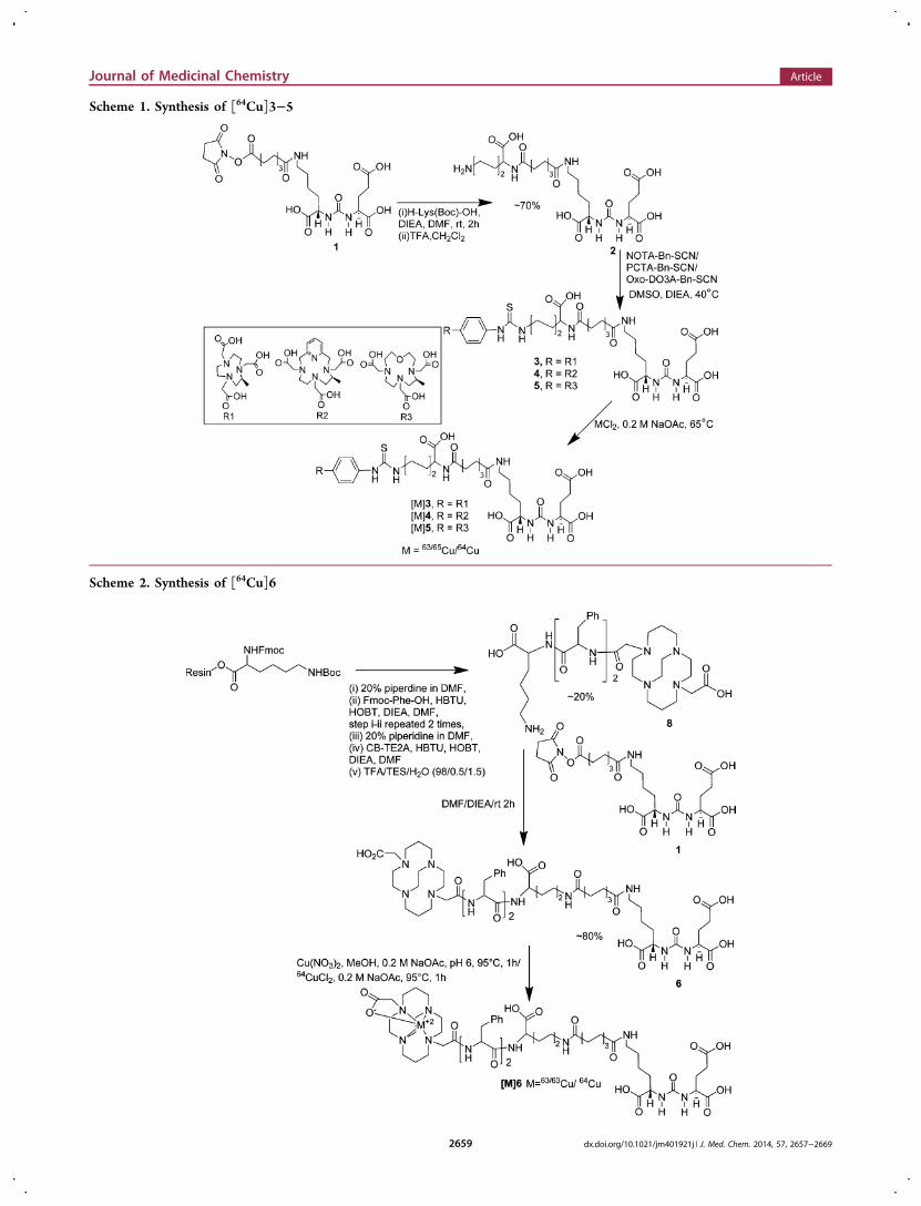
Scheme 1. Synthesis of [64Cu]3−5
Scheme 2. Synthesis of [64Cu]6
Journal of Medicinal Chemistry Article
dx.doi.org/10.1021/jm401921j | J. Med. Chem. 2014, 57, 2657−26692659

64Cu-labeled CB-TE2A conjugated [64Cu]6B exhibited superiortumor-to-background ratios and is the most promising agentfrom the series.
■ RESULTS
Chemical and Radiochemical Syntheses. MacrocyclicChelator-Conjugated PSMA Inhibitors. We have synthesizedtwo different types of ligands as shown in Figure 1 using Lys-Glu urea as the targeting moiety. For ligands 3−5 we used aLys-suberate linker21 and conjugated the linker withcommercially available chelating agents, NOTA-Bn-SCN,PCTA-Bn-SCN, and oxo-DOTA-Bn-SCN via thiourea bondformation. For those three bifunctional chelators, the macro-cyclic backbone was functionalized to carry a benzyl isocyanatefor conjugation with an amine function. Copper(II) com-pounds for 3−5 are predicted to demonstrate distortedoctahedron geometry.41 Because NOTA is a hexadentateN3O3 chelator, [64Cu(II)]3 is expected to carry a negativecharge. On the other hand, both PCTA (N4O3) and oxo-DOTA (N3O4) are heptadentate macrocycles and likely formneutral compounds with one pendant acetate arm aftercomplexation.35,41 The Lys-suberate linker was extendedusing two phenylalanine residues to prepare 6 and 7 containingCB-TE2A and DOTA, respectively, as chelating agents. Thepurpose of attaching those two phenylalanine residues to thelinker of 6 and 7 is mainly to compensate for the lipophilicity ofthe phenylthiourea moiety of compounds 3−5.22,24−26 Notethat one of the pendant acetate arms of the CB-TE2A andDOTA chelators has been modified to attach the PSMAtargeting moiety via amide bond formation to produce 6 and 7,respectively. [64Cu]CB-TE2A forms a positively chargedradiotracer when conjugated to a biomolecule, whereasDOTA-conjugated [64Cu]7 is expected to carry one pendantacetate, providing an overall neutral compound.Syntheses of PSMA targeting ligands 3, 4, and 5 are shown in
Scheme 1. Compounds 1 and 2 were prepared as previouslyreported.28 Compound 2 was conjugated with commerciallyavailable NOTA-Bn-SCN, PCTA-Bn-SCN, and oxo-DOTA-Bn-SCN in DMSO in the presence of diisopropylethylamine(DIEA) at 40 °C for 4 h, to give 3, 4, and 5 in ∼30−40% yield,respectively, after purification by high-performance liquidchromatography (HPLC). Synthesis of CB-TE2A-conjugatedligand 6 was performed using standard fluorenylmethoxycar-bonyl (Fmoc) solid phase peptide synthesis (SPPS), startingfrom Fmoc-Lys(Boc)-Wang resin according to Scheme 2. Aftertwo phenylalanine residues were coupled with the resin boundlysine, CB-TE2A was conjugated at the N-terminal of thesecond phenylalanine residue, after which the compound wascleaved from the resin by a 1:1 mixture of TFA/CH2Cl2 toproduce 8 in moderate yield (∼20%). The free ε-amine oflysine of 8 was then conjugated with 128 to produce 6. DOTA-conjugated 7 was prepared according to our previous report.26
NMR and mass spectrometry (MS) were used to confirm theidentity of newly synthesize compounds. Copper (63/65Cu)complexes were prepared by incubating conjugates 3−7 with anaqueous solution of Cu(NO3)2 at 95 °C as shown in Schemes1−2. The mass spectra of the copper-labeled compoundsshowed the expected isotope distribution pattern for naturalcopper, which is a mixture of 63Cu (69%) and 65Cu (31%). Thestable copper-labeled conjugates were used as authenticreference material for chromatography to identify thecorresponding 64Cu-labeled compounds.
Radiochemistry. The radiolabeling of new PSMA ligandswith 64Cu is shown in Schemes 1−2. Briefly, for ligands 3−5and 7, radiolabeling was performed at pH ∼5.5−6 in acetatebuffer at ∼65 °C for 30 min, whereas for ligand 6, radiolabelingwas achieved in a boiling water bath (95 °C) for 1 h at pH∼7.5−8 in acetate buffer. Radiolabeled products were obtainedin high yield (>60%, without decay correction) and radio-chemical purity (>95%) as determined by HPLC and radiothin-layer chromatography (radio-TLC). Two radiolabeledpeaks were observed for [64Cu]4, likely due to the separationof diastereomers as previously reported for this class ofbifunctional chelators.60 On the other hand, free ligand 6contained two nearly inseparable isomeric peaks by HPLC.Therefore, radiolabeling of 6 was performed using the isomericmixture to give two HPLC separable products designated[64Cu]6A and [64Cu]6B, isolated in 45% and 35% yield,respectively. We assumed that the presence of an asymmetriccenter at the Phe residue of the linker moiety adjacent to CB-TE2A was responsible for producing those two diastereomericcompounds. All radioligands were obtained in specific activitiesranging from 2.9 to 9.1 GBq/μmol (78−347 mCi/μmol). TheLog Poct/w values for the radioligands were determined byoctanol−water partition and are shown in Table 1. Surprisingly,
[64Cu]6A and [64Cu]6B were found to be more hydrophilicthan [64Cu]3 in spite of possessing two Phe residues on thelinker and only one carboxylate groups on the chelatorbackbone.
In Vitro Binding. Ligands and the corresponding stablemetal-labeled compounds demonstrated high binding affinity toPSMA, with Ki values ranging from 0.19 to 13.26 nM (Table1). The known, high-affinity PSMA inhibitor ZJ43 was used asa reference ligand (Supporting Information Figure S1) andexhibits a Ki of 4.3 nM.61 The low nanomolar affinitiesdisplayed by these five new copper compounds are similar tothe affinities of the majority PSMA-targeted imaging agents wehave synthesized to date.21,28
Small Animal PET-CT Imaging. Whole body PET-CTimages were obtained for [64Cu]3−7 in intact male severecombined immunodeficient (SCID) mice (Figures 2−4)bearing PSMA+ PC3 PIP and PSMA− PC3 flu xenografts inopposite, upper flanks. Pharmacokinetics derived from theimages were used to determine which compound(s) would befurther evaluated through ex vivo biodistribution. Irrespectiveof charge and lipophilicity, all radiotracers, [64Cu]3−7, enabledvisualization of PSMA+ PC3 PIP tumor and kidneys (Figure 2).Renal uptake of the radiotracers is partially due to the route of
Table 1. Radiolabeling Yield, Log Po/w, and PSMA InhibitoryActivity
yield (%) Log Poct/w Ki (nM)
3 2.84[64Cu]3 ∼65−70 −1.17 [63/65Cu]3 6.23
4 2.03[64Cu]4 ∼70−90 −1.42 [63/65Cu]4 10.76
5 3.10[64Cu]5 ∼75−85 −1.56 [63/65Cu]5 5.47
6 0.19[64Cu]6A ∼40−45 −2.68 [63/65Cu]6A 3.98[64Cu]6B ∼30−35 −2.31 [63/65Cu]6B 4.65
7 11.07[64Cu]7 ∼65−70 ND [63/65Cu ]7 13.26
Journal of Medicinal Chemistry Article
dx.doi.org/10.1021/jm401921j | J. Med. Chem. 2014, 57, 2657−26692660

excretion of these agents as well as to specific uptake from theexpression of PSMA in mouse proximal renal tubules.62
Although [64Cu]4, [64Cu]5, and [64Cu]7 demonstratedmoderate to high PSMA+ PC3 PIP tumor uptake, they alsoshowed significant accumulation within liver at 2.5 h post-injection (Figure 2), which remained high until 6 h (data notshown). Radioligands [64Cu]5 and [64Cu]7 exhibited signifi-cantly higher background uptake, probably related to lowerstability of these 64Cu complexes,58 resulting in trans-chelationof copper to other, endogenous proteins.56
PET-CT images acquired at 20 min, 6 and 28 h postinjectionof [64Cu]3 (Figure 3) (n = 2) showed clear uptake in PSMA+
PC3 PIP tumor. At 20 min and 6 h postinjection, the mostvisible tissues were PSMA+ PC3 PIP tumor and kidneys, withsome accumulation of radioactivity observed in liver andurinary bladder. Radioactivity in liver and kidneys clearedsignificantly by 28 h.Compounds [64Cu]6A and [64Cu]6B exhibited high radio-
tracer concentration both within PSMA+ PC3 PIP tumor and
kidneys, similar to the distribution profile observed with [64Cu]3. Significantly, both CB-TE2A conjugated diastereomers[64Cu]6A and [64Cu]6B exhibited similar PET imaging profilesas shown in Figure 4. Both compounds showed low liver uptakeas early as 20 min after the injection. Consequently, cleardelineation of tumor was achieved even at early time points. By2.5 h postinjection, radioactivity was largely cleared fromkidneys for both isomers, producing clear target-to-backgroundcontrast for these radiotracers. As a further test of bindingspecificity, we imaged animals administered [64Cu]6B afterpretreating them with 50 mg/kg of ZJ43 30 min prior toradiotracer.61 ZJ43 proved capable of blocking binding of[64Cu]6B (Supporting Information Figure S2), not only withinthe tumor but also within the renal cortex, confirming thatuptake observed in these tissues is PSMA-mediated.62
Biodistribution. On the basis of PET-CT imaging results,[64Cu]3, [64Cu]6A, and [64Cu]6B were further assessed in abiodistribution assays using the same isogenic human prostatecancer PSMA+ PC3 PIP and PSMA− PC3 flu tumor models (n= 4). Tables 2 and 3 show the pharmacokinetics in percentageof injected dose per gram of tissue (% ID/g) in selected organsfor [64Cu]3 and [64Cu]6A at 30 min, 1 h, 2 h, and at 4 or 5 hpostinjection, while Table 4 shows the % ID/g at the optimizedtime point of 2 h for [64Cu]3, [64Cu]4, [64Cu]6A, and[64Cu]6B. All compounds exhibited clear PSMA-dependent binding inPSMA+ PC3 PIP tumor xenografts, with [64Cu]3 demonstrat-ing high tumor uptake as early as 30 min postinjection (33.78 ±9.68% ID/g), peaking at 2 h (38.51 ± 5.68% ID/g) (Table 2),with extended retention of radioactivity up to 4 h postinjection(20.64 ± 5.06% ID/g). PSMA+ PC3 PIP to PSMA− PC3 flutumor uptake ratios were 14.03 ± 3.71 and 23.50 ± 4.65 at 30min at 4 h, respectively. The distribution within normal organsand tissues was also favorable, with low blood and normal tissueuptake and rapid clearance. The highest nonspecific uptakeobserved was in the liver (7.37 ± 0.40% ID/g) and spleen(28.02 ± 5.82% ID/g) at 30 min postinjection. However, thosevalues decreased to 4.18 ± 1.61% ID/g and 4.72 ± 1.49% ID/g,respectively, by 4 h. Kidney uptake was expectedly high andpeaked at 195.44 ± 29.82% ID/g at 30 min and decreased to108.05 ± 20.50% ID/g by 4 h.
Figure 2.Whole body PET-CT imaging of PSMA+ PC3 PIP and PSMA − PC3 flu tumor-bearing mice with compounds [64Cu]3, [64Cu]4, [64Cu]5,[64Cu]6B, and [64Cu]7 at 2.5 h postinjection, respectively. Mice were injected with ∼11.1 MBq (∼300 μCi) of radiotracer IV. PIP = PSMA+ PC3PIP (solid arrow); flu = PSMA− PC3 flu (unfilled arrow); K= kidney; B = bladder; L = left; R = right. All images are decay-corrected and adjusted tothe same maximum value.
Figure 3. Whole body PET-CT imaging of PC3 PIP and PC3 flutumor bearing mice with [64Cu]3 at 20 min (left), 6 h (middle), 28 h(right). Abdominal radioactivity is primarily due to uptake withinkidneys and bladder. PIP = PC3 PSMA+ PIP (solid arrow); flu = PC3PSMA− flu (unfilled arrow); K= kidney; L = left; R = right, B =bladder. All images are decay-corrected and adjusted to the samemaximum value.
Journal of Medicinal Chemistry Article
dx.doi.org/10.1021/jm401921j | J. Med. Chem. 2014, 57, 2657−26692661

Table 3 shows the organ-related % ID/g of uptake for [64Cu]6B. Similar to [64Cu]3, [64Cu]6B showed the highest PSMA-dependent tumor uptake with 29.50 ± 8.10% ID/g at 1 hpostinjection. Tumor uptake remained high, decreasing to18.69 ± 2.88% ID/g at 5 h. The PSMA+ PC3 PIP to PSMA−PC3 flu ratios were 19.36 ± 3.98 at 30 min, 79.40 ± 15.83 at 2h, and 146.79 ± 32.20 at 5 h. The PSMA+ PC3 PIP tumor-to-muscle ratio reached a maximum value of 436.14 ± 202.71 at 5h, nearly 3-fold higher than those observed with [64Cu]3. Renaluptake for [64Cu]6B was highest at 30 min at 256.11 ± 75.72%ID/g, much higher than that seen for [64Cu]3, however, itcleared much more rapidly, decreasing to 3.54 ± 0.04% ID/gby 5h. Nontarget organs, such as blood, heart, liver, spleen,
stomach, and pancreas, showed lower uptake (∼2% ID/g at 1h) and much faster clearance than for [64Cu]3 and decreased tobelow 1% ID/g by 5 h.Because [64Cu]3, [64Cu]4, and [64Cu]6 demonstrated
superior pharmacokinetics to [64Cu]5 and [64Cu]7, a one-time point (2 h) biodistribution study (Table 4) was performedwith [64Cu]3, [64Cu]4, and [64Cu]6B. Syntheses andbiodistribution studies were performed on the same day, asclose to one another as possible to minimize the changes inspecific activity of the radiotracer. The biodistribution andimaging data reveal several important points regarding the invivo properties of these compounds. The NOTA-chelatedradiotracers [64Cu]3, PCTA-chelated [64Cu]4, and CB-TE2A-
Figure 4. Whole body PET-CT imaging of PC3 PIP and PC3 flu tumor bearing mice with [64Cu]6A (top row) and [64Cu]6B (bottom row) at 20min, 2.5 h, 12 and 22 h postinjection. Abdominal radioactivity is primarily due to uptake within kidneys and bladder. PIP = PC3 PSMA+ PIP (solidarrow); flu = PC3 PSMA− flu (unfilled arrow); K= kidney; L = left; R = right, B = bladder. All images are decay-corrected and adjusted to the samemaximum value.
Table 2. Tissue Biodistribution for [64Cu]3 in Tumor-Bearing Micea
0.5 h 1 h 2 h 4 h
blood 2.37 ± 0.90 1.12 ± 0.21 0.82 ± 0.20 0.45 ± 0.20heart 1.78 ± 0.41 1.39 ± 0.24 1.11 ± 0.13 0.71 ± 0.26lung 6.40 ± 0.59 5.99 ± 0.70 4.51 ± 0.97 2.49 ± 0.99liver 7.37 ± 0.40 7.50 ± 1.43 6.44 ± 0.83 4.18 ± 1.61spleen 28.02 ± 5.82 27.58 ± 10.05 12.33 ± 6.32 4.72 ± 1.49fat 1.78 ± 0.31 1.43 ± 0.38 0.76 ± 0.34 0.65 ± 0.75kidney 195.44 ± 29.82 233.39 ± 22.76 199.69 ± 48.44 108.05 ± 20.50muscle 0.57 ± 0.32 0.60 ± 0.14 0.34 ± 0.12 0.21 ± 0.10small intestine 4.60 ± 1.42 5.58 ± 1.43 3.68 ± 0.41 2.44 ± 1.30large intestine 4.51 ± 1.64 6.01 ± 2.54 3.80 ± 1.67 2.42 ± 1.19bladder 5.65 ± 2.77 2.48 ± 0.42 13.33 ± 8.35 1.70 ± 0.97PC-3 PIP 33.79 ± 9.68 29.11 ± 3.02 38.51 ± 5.68 20.64 ± 5.06PC-3 flu 2.40 ± 0.17 2.02 ± 0.18 1.92 ± 0.23 1.04 ± 0.37PIP:flu 14.03 ± 3.71 14.37 ± 0.43 19.96 ± 0.81 23.50 ± 4.65PIP:blood 15.11 ± 5.38 26.64 ± 5.12 48.75 ± 10.27 50.01 ± 15.54
aValues expressed are in % ID/g ± standard deviation; n = 4 for all tissues.
Journal of Medicinal Chemistry Article
dx.doi.org/10.1021/jm401921j | J. Med. Chem. 2014, 57, 2657−26692662

chelated [64Cu]6B showed high uptake in PSMA+ PC3 PIPtumor, highest for [64Cu]4 (24.13 ± 10.06% ID/g). However,[64Cu]4 also exhibited slightly higher nonspecific uptake inPSMA− PC3 flu tumor and other organs, including blood,muscle, heart, liver, and stomach, resulting in lower tumor-to-organ ratios than for [64Cu]3 and [64Cu]6B. In the imagingstudies, [64Cu]4 exhibited considerable background radio-activity primarily due to high liver uptake of [64Cu]4.Diastereomers [64Cu]6A and [64Cu]6B, each containing theCB-TE2A chelating agent, possessed similar in vivo propertiesand exhibited significantly lower normal tissue uptake,including kidney, than either [64Cu]3 or [64Cu]4.
■ DISCUSSION
We have evaluated five 64Cu-labeled PSMA-targeted, ureaconjugates using different chelating agents with the aim ofoptimizing in vivo PET imaging properties of this class of
compounds for clinical application. It is generally accepted thatthe overall biologic profile of radiolabeled ligands is determinednot only by receptor-specific binding but also by nonspecificinteractions. Such off-target effects rely on the overallphysicochemical features of the radiolabeled compound, e.g.,molecular weight, charge, lipophilicity, and metabolic stability.The chelator used to attach the radionuclide to the targetingagent plays a key role in establishing those physicochemicalfeatures, particularly for agents smaller than antibodies andnanoparticles, namely those of relatively low molecular weight(<1500 Da). Two key findings emerged from this study. First,both NOTA and CB2-TE2A-conjugated radiotracers [64Cu]3and [64Cu]6 demonstrated significantly higher in vivo stability,as evidenced by their lower liver uptake than the other threeradiotracers, [64Cu]4, [64Cu]5, and [64Cu]7, which utilizedPCTA, oxo-DOTA, and DOTA, respectively. High liver uptakeand slow blood clearance for those latter three radiotracers are
Table 3. Tissue Biodistribution for [64Cu]6B in Tumor-Bearing Micea
0.5 h 1 h 2 h 5 h
blood 2.50 ± 1.08 0.64 ± 0.24 0.26 ± 0.13 0.06 ± 0.05heart 0.92 ± 0.27 0.32 ± 0.08 0.13 ± 0.05 0.06 ± 0.01lung 2.70 ± 0.73 0.83 ± 0.11 0.45 ± 0.21 0.15 ± 0.01liver 0.73 ± 0.17 0.42 ± 0.04 0.30 ± 0.06 0.21 ± 0.03stomach 1.02 ± 0.47 0.48 ± 0.28 0.25 ± 0.06 0.10 ± 0.03pancreas 0.90 ± 0.23 0.31 ± 0.19 0.13 ± 0.05 0.05 ± 0.04spleen 5.80 ± 2.61 2.14 ± 0.58 0.65 ± 0.15 0.25 ± 0.07fat 0.87 ± 0.15 0.87 ± 1.32 0.19 ± 0.10 0.05 ± 0.05kidney 256.11 ± 75.72 148.60 ± 48.41 65.39 ± 21.16 3.54 ± 0.04muscle 0.33 ± 0.01 1.71 ± 2.29 0.17 ± 0.05 0.05 ± 0.02small intestine 0.85 ± 0.41 1.38 ± 1.89 0.26 ± 0.07 0.11 ± 0.10large intestine 0.88 ± 0.42 0.77 ± 0.65 0.30 ± 0.08 0.14 ± 0.05bladder 3.99 ± 0.26 7.06 ± 3.75 2.21 ± 0.57 1.43 ± 1.41PC-3 PIP 23.14 ± 2.20 29.50 ± 8.10 20.46 ± 2.90 18.69 ± 2.88PC-3 flu 1.29 ± 0.12 0.66 ± 0.26 0.26 ± 0.04 0.13 ± 0.03PIP:flu 19.36 ± 3.98 48.41 ± 14.25 79.40 ± 15.83 146.79 ± 32.30PIP:blood 9.13 ± 5.23 47.47 ± 7.24 101.17 ± 6.70 1123.96 ± 25.83PIP:muscle 69.35 ± 4.29 91.30 ± 93.17 132.99 ± 43.97 436.14 ± 202.71
aValues expressed are in % ID/g ± standard deviation; n = 4 for all tissues.
Table 4. Tissue Biodistibistion for [64Cu]3, [64Cu]4, [64Cu]6A, and [64Cu]6B at 2 h Post-Injection in Tumor-Bearing Micea
[64Cu]3 [64Cu]4 [64Cu]6A [64Cu]6B
blood 1.06 ± 0.29 1.87 ± 0.63 0.20 ± 0.03 0.20 ± 0.07heart 1.48 ± 0.14 2.71 ± 0.13 0.26 ± 0.10 0.35 ± 0.21lung 5.31 ± 0.74 10.16 ± 2.29 1.17 ± 0.61 0.89 ± 0.15liver 8.63 ± 0.92 17.04 ± 1.44 1.68 ± 0.38 1.63 ± 0.72stomach 3.88 ± 0.82 6.37 ± 0.78 0.62 ± 0.42 0.62 ± 0.17pancreas 2.22 ± 0.34 3.51 ± 0.67 0.46 ± 0.44 0.80 ± 0.92spleen 13.42 ± 1.18 9.27 ± 2.28 0.39 ± 0.30 0.90 ± 0.23fat 0.76 ± 0.77 1.18 ± 0.59 0.27 ± 0.26 0.05 ± 0.02kidney 125.40 ± 42.47 166.57 ± 42.39 23.55 ± 8.96 24.87 ± 11.26muscle 0.33 ± 0.08 0.71 ± 0.32 0.11 ± 0.05 0.12 ± 0.05small intestine 6.24 ± 1.46 10.95 ± 3.70 0.74 ± 0.32 0.68 ± 0.13large intestine 10.13 ± 6.83 10.10 ± 4.76 1.49 ± 0.75 1.28 ± 0.70bladder 12.47 ± 10.30 10.56 ± 6.20 7.98 ± 8.32 5.92 ± 4.94PC-3 PIP 16.58 ± 3.15 24.13 ± 10.06 11.10 ± 5.42 16.89 ± 5.73PC-3 flu 2.12 ± 0.25 3.88 ± 0.62 0.36 ± 0.09 0.40 ± 0.12PIP:flu 11.00 ± 0.73 6.22 ± 1.86 30.90 ± 7.92 41.75 ± 5.54PIP:blood 16.52 ± 5.14 12.87 ± 1.87 52.41 ± 17.34 86.97 ± 15.22PIP:muscle 46.98 ± 19.05 38.34 ± 21.69 97.31 ± 42.46 138.88 ± 30.86
aValues expressed are in % ID/g ± standard deviation; n = 4 for all tissues.
Journal of Medicinal Chemistry Article
dx.doi.org/10.1021/jm401921j | J. Med. Chem. 2014, 57, 2657−26692663

indicative of free Cu(II), which is sequestered in liver.56,63
Second, liver uptake and blood concentration are much lowerat all time points for [64Cu]6A−B compared to [64Cu]3,suggesting higher in vivo stability of CB-TE2A-conjugated[64Cu]6A−B compared to NOTA-conjugated [64Cu]3. Thatmight also be related to the higher hydrophilicity of [64Cu]6A−B compared to [64Cu]3 (Log Po/w = −2.68 for [64Cu]6Bcompared to −1.17 for [64Cu]3). Low liver uptake of CB-TE2A-chelated [64Cu]6 ligands might also be related theirhigher stability in forming complexes with Cu(II) and Cu(I),assuming Cu(II) reduction is the primary reason for trans-chelation, as reported previously.56 It is worth mentioning that[64Cu]3, which possesses an overall negative charge, exhibitedhigher kidney and spleen uptake that was similar to thatobserved for 99mTc-oxo labeled PSMA-targeted compounds.34
Both kidney and tumor uptake of [64Cu]6B were specificallyblocked by ZJ43, demonstrating PSMA-mediated renal uptake,however, much faster renal clearance of [64Cu]6B was observedcompared to clearance from PSMA+ PIP tumor. We and othershave seen a similar pattern of faster clearance of radioactivityfrom PSMA-expressing kidney than PSMA+ PIP tumor,presumably associated with more rapid flow through normallyorganized renal parenchymal vasculature compared to therelatively disorganized vasculature of the xenograft.26,64,65 As anextension of that particular pharmacokinetic feature, [64Cu]6exhibited the highest tumor-to-normal tissue ratios in this seriesof agents and the ratios increased with time, thereby providinghigh image contrast. Moreover, no bone uptake was observedfor [64Cu]6, indicating that this agent may find application fordetection of prostate cancer metastases, which are preferentiallyfound in bone.The imaging and biodistribution results demonstrate higher
in vivo stability and renal clearance for CB-TE2A-chelated[64Cu]6 compared to NOTA-chelated [64Cu]3, which appearsto be in contradistinction to previous reports that involvesimilar comparison for agents that target somatostatin receptorand αvβ3 integrin. In those reports [64Cu]NOTA-conjugatedradiopeptides provided faster renal clearance than thoseconjugated with [64Cu]CB-TE2A.46,47 Although unclear atthis time, this difference between 64Cu-labeled species fortargeting of PSMA vs somatostatin receptor or integrin may beassociated with other physical properties unique to each systemsuch as binding affinity or capacity for internalization of thecognate ligands. It nevertheless underscores the uniqueness ofeach system and indicates that one chelator cannot be assumedto outperform another in a new, untested system.There are several classes of imaging agents available for PET-
based molecular imaging of PSMA, including antibodies,4,5,32,66
antibody fragments,67 aptamers,49 and low-molecular-weightPSMA-binding affinity agents.14,64 They have widely varyingpharmacokinetics, however, each class has shown PSMA-specific binding in preclinical studies. Rockey and colleagueshave recently optimized conditions for conjugating 64Cu to aPSMA-targeting aptamer with different chelators, which hasshown PSMA-mediated uptake in PSMA+ 22Rv1 prostatetumor cells relative to PSMA− PC3 cells.49 The radiolabeledmonoclonal antibodies [64Cu]DOTA-3/A1214 and [89Zr]DFO-J59155 both showed values of approximately 3:1 at 48 h post-injection for PSMA+ to PSMA- tumor. Among the long-livedPET radionuclides, 64Cu and 89Zr, the stability of the[89Zr]DFO complex is limited as shown in several in vitroand in vivo in studies, which is reflected in a bone accumulationranging from 3 to 15% and assumed to be associated with the
nonlinear nature of the desferrioxamine (DFO) chelatingagent.68 More recently, studies in biopsy tissue from patientswith prostate cancer and cell lines have demonstrated acorrelation between PSMA cell−surface expression andandrogen activity using 64Cu-labeled anti-PSMA antibody([64Cu]J591).20 ImmunoPET detection of PSMA couldprovide a path for quantitative monitoring of successfulandrogen blockade in patients. Low-molecular-weight urea-based agent [64Cu]6 could be an inexpensive alternative forthose applications. Furthermore, results obtained with [64Cu]6suggest that delayed imaging with 64Cu-labeled PSMA-targetedligands using CB-TE2A can improve the visualization of PSMA+ tumors in vivo anywhere outside of central nervous systemand may allow imaging of tumors with lower PSMA expressionlevels than possible with existing 18F- or 68Ga-labeled PSMA-targeted imaging agents.
■ CONCLUSIONWe describe five new low-molecular-weight, urea-based, 64Cu-labeled, PSMA-targeted radiotracers that incorporated well-established chelating agents. All compounds demonstrated hightumor uptake and retention with the choice of chelator having aprofound effect on pharmacokinetics. Our data revealed that[64Cu]6, which utilizes the CB-TE2A chelator, demonstratedimproved biodistribution with rapid clearance from normaltissues, including kidney, resulting in significantly improvedimage contrast. Accordingly, [64Cu]6 provides a potentiallyclinically viable imaging agent for PSMA+ tissues, particularly ifdelayed imaging, obtainable with a 64Cu-labeled agent, isrequired.
■ EXPERIMENTAL SECTIONSolvents and chemicals purchased from commercial sources were ofanalytical grade or better and used without further purification. All 9-fluorenylmethyloxycarbonyl (Fmoc) protected amino acids includingthe Fmoc-Lys(Boc)-Wang resin, 1-hydroxybenzotriazole monohy-drate, and 2-(1H-benzotriazole-1-yl)-1,1,3,3-tetramethyluronium hexa-fluorophosphate (HBTU) were purchased from Chem ImpexInternational Inc. (Wooddale, IL). [64Cu]CuCl2 was purchased fromthe University of Wisconsin. DOTA-tris(t-butyl ester)-monoacid, p-SCN-Bn-NOTA, p-SCN-Bn-PCTA, and p-SCN-Bn-oxo-DO3A werereceived from Macrocyclics Inc. (Dallas, TX). Copper(II) chloride,triethylsilane (Et3SiH), diisopropylethylamine (DIEA), and triethyl-amine (TEA) were purchased from Sigma-Aldrich (St. Louis, MO). Allother chemicals were purchased from Thermo Fisher Scientific(Pittsburgh, PA) unless otherwise specified. Analytical thin-layerchromatography (TLC) was performed using Aldrich aluminum-backed 0.2 mm silica gel Z19, 329-1 plates and visualized by ultravioletlight (254 nm), I2, and 1% ninhydrin in EtOH. Flash chromatographywas performed using silica gel (MP SiliTech 32-63 D 60 Å) purchasedfrom Bodman (Aston, PA). All in vitro PSMA binding studies anddetermination of partition coefficient experiments were performed intriplicate to ensure reproducibility. 1H NMR spectra were recorded ona Bruker UltrashieldTM 400 MHz spectrometer. Chemical shifts (δ)are reported in ppm downfield by reference to proton resonancesresulting from incomplete deuteration of the NMR solvent. Lowresolution ESI mass spectra were obtained on a Bruker DaltonicsEsquire 3000 Plus spectrometer. High resolution mass spectra wereobtained by the University of Notre Dame Mass Spectrometry andProteomics Facility, Notre Dame, IN, using ESI either by directinfusion on a Bruker microTOF-II or by LC elution via an ultrahighpressure Dionex RSLC with C18 column coupled with a BrukermicroTOF-Q II.
HPLC purification of compounds 3−7 was performed using aPhenomenex C18 Luna 10 × 250 mm2 column on a Waters 600E DeltaLC system with a Waters 486 variable wavelength UV/vis detector,
Journal of Medicinal Chemistry Article
dx.doi.org/10.1021/jm401921j | J. Med. Chem. 2014, 57, 2657−26692664

both controlled by Empower software. HPLC was performed using thefollowing methods. Method 1: solvent A (0.1% TFA in water) andsolvent B (0.1% TFA in in acetonitrile), flow rate 8 mL/min. Theelution gradient was 90% A and 10% B for 0−5 min and 90% A to 0%A and 10% B to 100% B over 6−30 min. Methods 2−5 were isocrati,cwith a flow rate 4 mL/min. Method 2: 85% A and 15% B for 0−50min. Method 3: 82% A and 18% B for 0−40 min. Method 4: 83% Aand 17% B for 0−40 min. Method 5: 78% A and 22% B for 0−30 min.Method 6: 80% A and 20% B for 0−30 min, flow rate 1 mL/min, and aWaters Novapak C18 150 × 3.9 mm2 column was used. HPLCpurification of [64Cu]3-7 were performed on a Varian Prostar System(Palo Alto, CA), equipped with a Varian ProStar 325 UV−vis variablewavelength detector and a Bioscan Flow-Count in-line radioactivitydetector, all controlled by Galaxie software. The specific radioactivitywas calculated as the radioactivity eluting at the retention time ofproduct during the preparative HPLC purification divided by the masscorresponding to the area under the curve of the UV absorption. Thepurity of tested compounds as determined by analytical HPLC withabsorbance at 220 nm were >95%.Synthesis and Characterization of Compounds 1−7.
Compounds 1,28 and 2,26 and 726 were prepared following ourprevious reports. Compounds 3−5 were prepared following a generalprocedure as described for compound 3.9 , 1 6 , 2 4 - T r i o x o - 1 - t h i o xo - 1 - ( ( 4 - ( ( ( S ) - 1 , 4 , 7 - t r i s -
(carboxymethyl)-1,4,7-triazonan-2-yl)methyl)phenyl)amino)-2,8,17,23,25-pentaazaoctacosane-7,22,26,28-tetracarboxylicAcid, Compound 3. To a solution of 2 (20 mg, 0.33 μmol in 500 μLof DMSO) was added NOTA-Bn-SCN (18.5 mg, 0.33 μmol in 500 μLof DMSO) and DIEA (100 μL), and the solution was kept at 40 °C for4 h. The reaction mixture was then evaporated to dryness, and thecrude residue was dissolved in 2 mL of water and loaded on aprepacked C18 column (5.5 g, Agilent SF10). The product was elutedwith 80/20−70/30 H2O (0.1%TFA)/CH3CN (0.1% TFA) solution.The fractions containing the products were combined together andevaporated to dryness to obtain a colorless solid. The solid thusobtained was further purified by HPLC (method 1, retention time, Rt15.80 min). Yield ∼8 mg (0.076 μmol, ∼23%) after HPLC purificationand lyphilization. 1H NMR (DMSO-d6) δ: 8.15 (m, 1H, HNCO(Lys-linker)), 7.75 (m, 1H, HNCO(Lys)), 7.14 (d, 2H, Bz), 6.78 (d, 2H,Bz), 6.34 (m, 2H, NH(CO)NH), 4.40−3.99 (m, 3H, HC-(NHCO2(Glu), HC(NHCO2(Lys), HC(NHCO2(Lys)), 3.40−3.30(m, 7H, NCH2, CHCH2, (NOTA)), 3.21−3.03(m, 16H,(CH2)2(NOTA), H2C-Bz, H2CNH(Lys-linker), H2CNH(Lys)),2 .40−2 .09 (m, 6H, H2CCO2(Glu) , H2CCO2( l inke r) ,H2CCO2(linker)),1.89−1.55 (m, 6H, H2CCH(Glu), H2CCH(Lys-linker), H2CCH(Lys)), 1.55−1.21 (m, 16H, (CH2)2(Lys),(CH2)2(Lys-Linker), (CH2)4(linker)). ESMS m/z: 1054 [M + H]+.HRESI+ MS: calcd for C46H72N9O17S, 1054.4767 [M + H]+; found,1054.4771.9 ,16 , 24 -T r i oxo -1 - th ioxo -1 - ( ( 4 - ( ( ( 4S ) - 3 , 6 , 9 - t r i s -
(carboxymethyl)-3,6,9,15-tetraazabicyclo[9.3.1]pentadeca-1-(14),12-dien-4-yl)methyl)phenyl)amino)-2,8,17,23,25-pentaa-zaoctacosane-7,22,26,28-tetracarboxylic Acid, Compound 4.The compound was purified by HPLC (method 1; Rt 15.60 min).Yield ∼12 mg (0.076 μmol, ∼23%) after HPLC purification andlyophilized. 1H NMR (DMSO-d6) δ: 8.14 (m, 1H, HNCO(Lys-linker)), 7.80−7.75 (m, 2H, HNCO(Lys), Py), 7.32 (d, 2H, Py), 7.15(d, 2H, Bz), 6.78 (d, 2H, Bz), 6.35 (m, 2H, NH(CO)NH), 4.42−3.99(m, 7H, HC(NHCO2(Glu), HC(NHCO2(Lys), HC(NHCO2(Lys),H2C-Py)), 3.42−3.31 (m, 7H, CHCH2, NCH2(PCTA)), 3.21−3.02(m, 12H, (CH2)2(PCTA), H2C-Bz, H2CNH(Lys-linker), H2CNH-(Lys)), 2.41−2.08 (m, 6H, H2CCO2(Glu), H2CCO2(linker),H2CCO2(linker)), 1.89−1.55 (m, 6H, H2CCH(Glu), H2CCH(Lys-linker), H2CCH(Lys)), 1.55−1.21 (m, 16H, (CH2)2 (Lys),(CH2)2(Lys-Linker), (CH2)4(linker)). ESMS m/z: 1132 [M + H]+.HRESI+ MS: calcd for C51H75N10O17S, 1131.5032 [M + H]+; found,1131.5011.9,16,24-Trioxo-1-thioxo-1-((4-((4,7,10-tris(carboxymethyl)-
1-oxa-4,7,10-triazacyclododecan-2-yl)methyl)phenyl)amino)-2,8,17,23,25-pentaazaoctacosane-7,22,26,28-tetracarboxylicAcid, Compound 5. The compound was purified by HPLC (method
1; Rt 13.40 min). Yield ∼10 mg (0.076 μmol, ∼23%) after HPLCpurification and lyophilization. 1H NMR (DMSO-d6) δ: 8.12 (m, 1H,HNCO(Lys-linker)), 7.80−7.75 (m, 1H, HNCO(Lys)), 7.14 (d, 2H,Bz), 6.79 (d, 2H, Bz), 6.32 (m, 2H, NH(CO)NH), 4.42−3.99 (m, 3H,HC(NHCO2(Glu), HC(NHCO2(Lys), HC(NHCO2(Lys)), 3.49−3.31 (m, 11H, NCH2(DO3A), CHCH2(DO3A), OCH2)), 3.21−3.02 (m, 16H, (CH2)2(DO3A), H2C-Bz, H2CNH(Lys-linker),H2CNH(Lys)), 2.41−2.08 (m, 6H, H2CCO2(Glu), H2CCO2(linker),H2CCO2(linker)), 1.91−1.50 (m, 6H, H2CCH(Glu), H2CCH(Lys-linker), H2CCH(Lys)), 1.50−1.22 (m, 16H, (CH2)2(Lys),(CH2)2(Lys-Linker), (CH2)4(linker). ESMS m/z: 1098 [M + 1]+,HRESI+ MS: calcd for C48H76N9O18S, 1098.5029 [M + H]+; found,1098.5048.
(14S,29S,33S)-4,7-Dibenzyl-1-(11-(carboxymethyl)-1,4,8,11-tetraazabicyclo[6.6.2]hexadecan-4-yl)-2,5,8,16,23,31-hexaoxo-3,6,9,15,24,30,32-heptaazapentatriacontane-14,29,33,35-tet-racarboxylic Acid, Compound 6. Fmoc-Lys(Boc)-Wang resin (100mg, 0.43 mM) was allowed to swell with CH2Cl2 (3 mL) followed byDMF (3 mL). A solution of 20% piperidine in DMF (3 × 3 mL) wasadded to the resin that was then shaken gently on a mechanical shakerfor 30 min at ambient temperature. The resin was washed with DMF(3 × 3 mL) and CH2Cl2 (3 × 3 mL). Formation of free amine wasassessed by the Kaiser test. After swelling the resin in DMF, a solutionof Fmoc-Phe-OH (3 equiv), HBTU (3 equiv), HOBt (3 equiv), andDIPEA (4.0 equiv) in DMF was added and gently shaken for 2 h. Theresin was then washed with DMF (3 × 3 mL) and CH2Cl2 (3 × 3mL). The coupling efficiency was assessed by the Kaiser test. Theaforementioned sequence was repeated for two more coupling stepswith Fmoc-Phe-OH and CB-TE2A, respectively. The final compoundwas cleaved from the resin using TFA:CH2Cl2 (1:1) and concentratedunder vacuum to produce 8. The concentrated product was purified byusing a C18 SepPak Vac 2g column. The product was eluted with asolution 70/30 water/acetonitrile (0.1% TFA in each). ESI-MS: 635[M]+. To a solution of 8 (20 mg, 31 μmol in 500 μL of DMSO) (27mg, 47 μmol) and DIEA (50 μL) was added 1, and the reactionmixture was left at ambient temperature for 2 h. The solution wasdiluted in 10 mL of water and was purified by HPLC (method 1, time(Rt), 20 min). Yield ∼10 mg (0.076 μmol, ∼22%) after HPLCpurification and lyophilization. 1H NMR (DMSO-d6): 9.02−8.07 (m,12H, HNCO(Lys-linker), HNCO(Lys), PhCH2), 6.34 (m, 2H,NH(CO)NH), 4.62 (m, 2H, NHCO2(Phe)), 4.37−4.28 (m, 3H,(m, 3H, HC(NHCO2(Glu), HC(NHCO2(Lys), HC(NHCO2(Lys)),3.65−3.01 (m, 14H, CH2 N(CB-TE2A), H2CPhe, H2CNH(Lys),H2CNH(Lys)), 2.74−2.41 (m, 20H, NCH2CH2(CB-TE2A), 2.41−2.08 (m, 6H, H2CCO2(Glu), H2CCO2(linker), H2CCO2(linker)),1.91−1.50 (m, 10H, CH2(CB-TE2A), H2CCH(Glu), H2CCH(Lys-linker), H2CCH(Lys)), 1.56−1.22 (m, 16H, (CH2)2(Lys),(CH2)2(Lys-Linker), (CH2)4(linker). ESMS m/z: 1222 [M + H]+.HRESI+ MS: calcd for C60H91N11O16, 1221.6645; found, 1221.4951.
Copper Complexes of Ligand 3, 4, 5, 6, and 7. Coppercompounds [63/65 Cu]3−5 and[63/65 Cu]7 were prepared by the samegeneral procedure as presented for compound 3 below. [63/65Cu]6A−B were prepared by same conditions as described for theirradiosynthesis.
[63/65Cu]3. To a solution of Cu(NO3)2 (1 mg, 20 μmol in 100 μL)in deionized water was added 3 (1 mg, 0.95 μmol) in 500 μL of 0.2 MNaOAc. The resulting solution was heated in boiling water for 0.5 h.The solution was purified by HPLC method 2. The retention time forthe product was 34 min. Yield: ∼50%. ESIMS m/z: 1115 [M + H]+.HRMS: calcd for C46H70CuN9O17S, 1115.3906 [M + H]+; found,1115.3828.
[63/65Cu]4. The compound was purified by HPLC method 3.Retention time for the product was at 22 min. Yield: ∼50%. ESIMS m/z: 1192 [M + H]+. HRMS: calcd for C51H73CuN10O17S, 1192.4172 [M+ H]+; found, 1192.4364.
[63/65Cu]5. The compound was purified by HPLC method 4.Retention time for the product was at 14 min. Yield: ∼60%. ESIMS m/z: 1160 [M + H]+. HRMS: calcd for C48H76CuN9O18S, 1160.4247 [M+ H]+; found, 1160.3828.
Journal of Medicinal Chemistry Article
dx.doi.org/10.1021/jm401921j | J. Med. Chem. 2014, 57, 2657−26692665

[63/65Cu]6A−B .The compound was purified by HPLC method 5.Retention time for the products were at 24 and 26 min. Yield: ∼50%.ESIMS m/z: 1285 [M + H]+. HRMS: calcd for C60H91CuN11O16,1284.5930 [M + H]+; found, 1284.5786.[63/65Cu]7. The compound was purified by HPLC method 6. Rt for
the product was at 13 min. Yield: ∼55%. ESIMS m/z: 1349 [M + H]+.HRMS: calcd for C60H90CuN11O20, 1348.5650 [M + H]+; found,1348.5671
64Cu Radiolabeling. Radioligands [64Cu]3−5 and 7 wereprepared by the same general method as described for [64Cu]3. Foreach radiolabeling reaction, approximately 30−40 μg of ligand and in200 mM NaOAc (purged under N2 for 2−3 min) was incubated with3−4 mCi 64CuCl2 at pH 5.5−6 for 0.3 h in a water bath at 65 °C.Radiolabeling was monitored by injecting aliquots of 20−40 μL of thesolution onto the HPLC. When the reaction reached completion, thereaction mixture was diluted with 1 mL of water then loaded onto apreparative HPLC for purification using method 2, Rt 41 min, for thedesired product and Rt 38 min for the free ligand. The radiolabeledproduct [64Cu]3 was obtained in ∼60−70% radiochemical yield, andthe radiochemical purity was >98% as measured by ITLC (GelmanITLC strips, 10 mM EDTA). HPLC method 2 was used to purify theradiolabeled product [64Cu]3. Rt 34 min for the desired product and Rt42 min for the free ligand. The specific activity of the probe was 2.9−9.1 GBq/μmol (n = 4). The acidic eluate was neutralized with 50 μL of1 M Na2(CO3), and the volume of the eluate was reduced undervacuum to dryness. The solid residue was diluted with saline to thedesired radioactivity concentration for biodistribution and imagingstudies. HPLC method 3 was used for [64Cu]4, Rt = 20−26 min for[64Cu]4 and Rt = 15 min for the free ligand 4. [64Cu]4 was isolated intwo peaks, Rt 20−22 min for the first fraction and 23−26 min for thesecond fraction. On the basis of the initial results (in PET imagingboth fractions displayed similar in vivo distributions), only the secondfraction was used for all studies. HPLC method 4 was used for [64Cu]5. Retention times for [64Cu]5 and free ligand were and 14.5 and 9.5min, respectively. The radiotracer [64Cu]7 was purified by HPLCmethod 6. Rt for [
64Cu]7 was at 13 min.For [64Cu]6A−B, radiolabeling was done in a boiling water bath for
1 h by incubation of 64CuCl2 with the corresponding ligand using 0.2NaOAc buffer at pH = 7.5−8. HPLC method 5, Rt = 24.9 min (A) and26.8 min (B) for the desired products and Rt = 19.8 min (A) and 21.5min (B) for the free ligands. The specific activity of the probe was 2.9GBq−12.84 GBq/μmol.NAALADase Assay. The PSMA inhibitory activities of 3−7 and
the corresponding copper-labeled analogues were determined using afluorescence-based assay according to a previously reported procedure.28 Briefly, lysates of LNCaP cell extracts (25 μL) were incubated withthe inhibitor (12.5 μL) in the presence of 4 μM N-acetylaspartylglu-tamate (NAAG) (12.5 μL) for 120 min. The amount of the glutamatereleased by NAAG hydrolysis was measured by incubating with aworking solution (50 μL) of the Amplex Red glutamic acid kit (LifeTechnologies, Grand Island, NY) for 60 min. Fluorescence wasmeasured with a VICTOR3 V multilabel plate reader (Perkin-ElmerInc., Waltham, MA) with excitation at 490 nm and emission at 642nm. Inhibition curves were determined using semilog plots, and IC50values were determined at the concentration at which enzyme activitywas inhibited by 50%. Enzyme inhibitory constants (Ki values) weregenerated using the Cheng−Prusoff conversion.69 Assays wereperformed in triplicate. Data analysis was performed using GraphPadPrism version 4.00 for Windows (GraphPad Software, San Diego,California).Cell Lines. Sublines of the androgen-independent PC3 human
prostate cancer cell line, originally derived from an advanced androgenindependent bone metastasis, were used. These sublines have beenmodified to express high (PC3 PIP) or possess low (PC3 flu) levels ofPSMA and were generously provided by Dr. Warren Heston(Cleveland Clinic). PSMA-expressing (PC3 PIP), nonexpressing(PC3 flu) PCa cell lines, were grown in RPMI 1640 medium(Corning Cellgro, Manassas, VA) containing 10% fetal bovine serum(FBS) (Sigma-Aldrich, St.Louis, MO) and 1% penicillin−streptomycin(Corning Cellgro, Manassas, VA) and PC-3 PIP cells were grown
under 20 μg/mL of puromycin to maintain PSMA expression. All cellcultures were maintained in an atmosphere containing 5% carbondioxide (CO2) at 37.0 °C in a humidified incubator.
Tumor Models. Animal studies were carried out in full compliancewith the regulations of the Johns Hopkins Animal Care and UseCommittee. Six-to-eight-week-old male, nonobese diabetic (NOD)/severe-combined immunodeficient (SCID) mice (Johns HopkinsImmune Compromised Core) were implanted subcutaneously (sc)with PC3 PIP (PSMA+) and PC3 flu (PSMA−) cells (1 × 10⁶ in 100μL of HBSS (Corning Cellgro, Manassas, VA) at the forward right andleft flanks, respectively. Mice were imaged or used in ex vivobiodistribution assays when the xenografts reached 5− 7 mm indiameter.
Small-Animal PET Imaging and Analysis. Dynamic and whole-body PET and CT images were acquired on an eXplore VISTA small-animal PET (GE Healthcare) and an X-SPECT small SPECT/CTsystem (Gamma Medica Ideas), respectively. For imaging studies, micewere anesthetized with 3% and maintained under 1.5% isoflurane (v/v). PET-CT Imaging studies were performed on NOD/SCID micebearing PSMA+ PC3 PIP and PSMA− PC3 flu tumors. Immediatelyafter intravenous injection of [64Cu]3−7, changes in radiotraceraccumulation were recorded over the whole body using an imagingsequence consisting of eight frames for a total of 30 min with variabledwell times as described previously.43,64 After dynamic imaging, whole-body PET images (two bed positions, 15 min emission per bedposition) were acquired at the indicated time points after injection ofradiotracer. For binding specificity studies, a mouse was subcuta-neously administered with a blocking dose of the known PSMAinhibitor ZJ43 (50 mg/kg) at 30 min before the injection of [64Cu]6B,and another mouse was injected with [64Cu]6B alone. After each PETscan, a CT scan was acquired as 512 projections using a 50 keV beamfor anatomic coregistration. PET emission data were corrected fordecay and dead time and were reconstructed using the three-dimensional ordered-subsets expectation maximization algorithm. Datawere displayed and analyzed using AMIDE software (http://sourceforge.net/amide), and volume-rendered images were generatedusing Amira 5.2.0 software (Visage Imaging Inc.; http://www.vsg3d.com/amira).
Ex Vivo Biodistribution. Mice bearing PSMA+ PC3 PIP andPSMA− PC3 flu xenografts were injected via the tail vein with 740kBq (20 μCi) of 64Cu in 200 μL of saline. At 30, 60, 240, and 240 or300 min postinjection, the mouse was sacrificed by cervical dislocationand the blood immediately collected by cardiac puncture. The heart,lungs, liver, stomach, pancreas, spleen, fat, kidney, muscle, small andlarge intestines, urinary bladder, PSMA+ PC3 PIP and PSMA− PC3flu tumors were collected. Each organ was weighed, and the tissueradioactivity was measured with an automated gamma counter (1282Compugamma CS, Pharmacia/LKBNuclear, Inc., Mt. Waverly,Victoria, Australia). The % ID/g was calculated by comparison withsamples of a standard dilution of the initial dose. All measurementswere corrected for decay.
Data Analysis. Data are expressed as mean ± standard deviation(SD). Prism software (GraphPAD, San Diego, California) was used todetermine statistical significance. Statistical significance was calculatedusing a paired t test. A P-value <0.0001 was considered significant.
■ ASSOCIATED CONTENT*S Supporting InformationDetailed spectral data and supporting PET-CT blocking studyimage. This material is available free of charge via the Internetat http://pubs.acs.org.
■ AUTHOR INFORMATIONCorresponding Authors*For S.R.B.: phone, 410-955-8697; fax, 410-614-3147; E-mail,[email protected]; address, Johns Hopkins Medical Institutions,1550 Orleans Street, 4M07 CRB II, Baltimore, Maryland21231, United States.
Journal of Medicinal Chemistry Article
dx.doi.org/10.1021/jm401921j | J. Med. Chem. 2014, 57, 2657−26692666
*For M.G.P.: phone, 410-955-2789; fax, 443-817-0990; E-mail,[email protected]; address, Johns Hopkins Medical Institu-tions, 1550 Orleans Street, 492 CRB II, Baltimore, Maryland21231, United States.NotesThe authors declare no competing financial interest.
■ ACKNOWLEDGMENTSWe thank the University of Wisconsin Cyclotron ResearchGroup for providing [64Cu]CuCl2. We also thank Nordion andMacrocyclics, Inc., for providing PCTA-Bn-SCN and Oxo-DO3A-Bn-SCN chelating agent, Drs. Cara Ferreira and RussellRedshaw for helpful discussion, and James Fox, Gilbert Green,and Dr. Yuchuan Wang for assistance with imaging and imageanalysis. We are grateful for the following sources of support:K25 CA148901, NIH NCI U54CA151838, NCI CA134675,and NCI R01 CA093375.
■ ABBREVIATIONS USEDPSMA, prostate-specific membrane antigen; GCPII, glutamatecarboxypeptidase II; NAALADase, N-acetylated-α-linked acidicdipeptidase; PET, positron emission tomography; SPECT,single photon emission computed tomography; NOTA, 1,4,7-triazacyclononane-1,4,7-triacetic acid; CB-TE2A, 4,11-bis-(carboxymethyl)-1,4,8,11-tetraazabicyclo[6.6.2]hexadecane;PCTA, 3,6,9,15-tetraazabicyclo[9.3.1]-pentadeca-1(15),11,13-triene)-3,6,9-triacetic acid ; oxo-DO3A, oxa-4,7,1-tetraazacyclo-dodecane-4,7,10-triacetic acid; DOTA, 1,4,7,10-tetraazacyclo-dodecane-N,N′,N″,N‴-tetraacetic acid
■ REFERENCES(1) Ghosh, A.; Heston, W. D. Tumor target prostate specificmembrane antigen (PSMA) and its regulation in prostate cancer. J.Cell Biochem. 2004, 91, 528−539.(2) Rajasekaran, A. K.; Anilkumar, G.; Christiansen, J. J. Is prostate-specific membrane antigen a multifunctional protein? Am. J. Physiol.,Cell Physiol. 2005, 288, C975−C981.(3) Chang, S. S.; Reuter, V. E.; Heston, W. D.; Gaudin, P. B.Comparison of anti-prostate-specific membrane antigen antibodies andother immunomarkers in metastatic prostate carcinoma. Urology 2001,57, 1179−1183.(4) Wright, G. L., Jr.; Grob, B. M.; Haley, C.; Grossman, K.; Newhall,K.; Petrylak, D.; Troyer, J.; Konchuba, A.; Schellhammer, P. F.;Moriarty, R. Upregulation of prostate-specific membrane antigen afterandrogen-deprivation therapy. Urology 1996, 48, 326−334.(5) Evans, M. J.; Smith-Jones, P. M.; Wongvipat, J.; Navarro, V.; Kim,S.; Bander, N. H.; Larson, S. M.; Sawyers, C. L. Noninvasivemeasurement of androgen receptor signaling with a positron-emittingradiopharmaceutical that targets prostate-specific membrane antigen.Proc. Natl. Acad. Sci. U. S. A. 2011, 108, 9578−9582.(6) Perner, S.; Hofer, M. D.; Kim, R.; Shah, R. B.; Li, H.; Moller, P.;Hautmann, R. E.; Gschwend, J. E.; Kuefer, R.; Rubin, M. A. Prostate-specific membrane antigen expression as a predictor of prostate cancerprogression. Human Pathol. 2007, 38, 696−701.(7) Sanna, V.; Pintus, G.; Roggio, A. M.; Punzoni, S.; Posadino, A.M.; Arca, A.; Marceddu, S.; Bandiera, P.; Uzzau, S.; Sechi, M. Targetedbiocompatible nanoparticles for the delivery of (−)-epigallocatechin 3-gallate to prostate cancer cells. J. Med. Chem. 2011, 54, 1321−1332.(8) DiPippo, V. A.; Magargal, W. W.; Moorji, S. M.; Murga, J. D.;Olson, W. C. Antiandrogen modulation of prostate-specific membraneantigen (PSMA): dynamics and synergy with PSMA-targeted therapy.ASCO Meet. Abstr. 2013, 31, e16007.(9) Petrylak, D. P.; Kantoff, P. W.; Mega, A. E.; Vogelzang, N. J.;Stephenson, J.; Fleming, M. T.; Stambler, N.; Petrini, M.; Blattman, S.;Israel, R. J. Prostate-specific membrane antigen antibody drug
conjugate (PSMA ADC): a phase I trial in metastatic castration-resistant prostate cancer (mCRPC) previously treated with a taxane.ASCO Meet. Abstr. 2013, 31, 5018.(10) Rotshteyn, Y.; Mercier, F.; Bruno, R.; Stambler, N.; Israel, R. J.;Wong, V. Correlation of PSMA ADC exposure with reduction intumor growth rate determined using serial PSA measurements from aphase I clinical trial. ASCO Meet. Abstr. 2013, 31, e16047.(11) Hrkach, J.; Von Hoff, D.; Mukkaram Ali, M.; Andrianova, E.;Auer, J.; Campbell, T.; De Witt, D.; Figa, M.; Figueiredo, M.; Horhota,A.; Low, S.; McDonnell, K.; Peeke, E.; Retnarajan, B.; Sabnis, A.;Schnipper, E.; Song, J. J.; Song, Y. H.; Summa, J.; Tompsett, D.;Troiano, G.; Van Geen Hoven, T.; Wright, J.; LoRusso, P.; Kantoff, P.W.; Bander, N. H.; Sweeney, C.; Farokhzad, O. C.; Langer, R.; Zale, S.Preclinical development and clinical translation of a PSMA-targeteddocetaxel nanoparticle with a differentiated pharmacological profile.Sci. Transl. Med. 2012, 4, 128ra39.(12) Hillier, S. M.; Maresca, K. P.; Femia, F. J.; Marquis, J. C.; Foss,C. A.; Nguyen, N.; Zimmerman, C. N.; Barrett, J. A.; Eckelman, W. C.;Pomper, M. G.; Joyal, J. L.; Babich, J. W. Preclinical evaluation of novelglutamate−urea−lysine analogues that target prostate-specific mem-brane antigen as molecular imaging pharmaceuticals for prostatecancer. Cancer Res. 2009, 69, 6932−6940.(13) Mease, R. C.; Foss, C. A.; Pomper, M. G. PET imaging inprostate cancer: focus on prostate-specific membrane antigen. Curr.Top. Med. Chem. 2013, 13, 951−962.(14) Cho, S. Y.; Gage, K. L.; Mease, R. C.; Senthamizhchelvan, S.;Holt, D. P.; Jeffrey-Kwanisai, A.; Endres, C. J.; Dannals, R. F.; Sgouros,G.; Lodge, M.; Eisenberger, M. A.; Rodriguez, R.; Carducci, M. A.;Rojas, C.; Slusher, B. S.; Kozikowski, A. P.; Pomper, M. G.Biodistribution, tumor detection, and radiation dosimetry of 18F−DCFBC, a low-molecular-weight inhibitor of prostate-specificmembrane antigen, in patients with metastatic prostate cancer. J.Nucl. Med. 2012, 53, 1883−1891.(15) Afshar-Oromieh, A.; Malcher, A.; Eder, M.; Eisenhut, M.;Linhart, H. G.; Hadaschik, B. A.; Holland-Letz, T.; Giesel, F. L.;Kratochwil, C.; Haufe, S.; Haberkorn, U.; Zechmann, C. M. PETimaging with a [(68)Ga]gallium-labelled PSMA ligand for thediagnosis of prostate cancer: biodistribution in humans and firstevaluation of tumour lesions. Eur. J. Nucl. Med. Mol. Imaging 2012, 40,486−495.(16) Barrett, J. A.; Coleman, R. E.; Goldsmith, S. J.; Vallabhajosula,S.; Petry, N. A.; Cho, S.; Armor, T.; Stubbs, J. B.; Maresca, K. P.;Stabin, M. G.; Joyal, J. L.; Eckelman, W. C.; Babich, J. W. First-in-manevaluation of 2 high-affinity PSMA-avid small molecules for imagingprostate cancer. J. Nucl. Med. 2013, 54, 380−387.(17) Maresca, K. P.; Hillier, S. M.; Lu, G.; Marquis, J. C.;Zimmerman, C. N.; Eckelman, W. C.; Joyal, J. L.; Babich, J. W.Small molecule inhibitors of PSMA incorporating technetium-99m forimaging prostate cancer: effects of chelate design on pharmacokinetics.Inorg. Chim. Acta 2012, 389, 168−172.(18) Nedrow-Byers, J. R.; Moore, A. L.; Ganguly, T.; Hopkins, M. R.;Fulton, M. D.; Benny, P. D.; Berkman, C. E. PSMA-targeted SPECTagents: Mode of binding effect on in vitro performance. Prostate 2013,73, 355−362.(19) Lu, G.; Maresca, K. P.; Hillier, S. M.; Zimmerman, C. N.;Eckelman, W. C.; Joyal, J. L.; Babich, J. W. Synthesis and SAR of99mTc/Re-Labeled Small Molecule Prostate Specific MembraneAntigen Inhibitors with Novel Polar Chelates. Biorg. Med. Chem.Lett. 2013, 23, 1557−1563.(20) Nedrow-Byers, J. R.; Jabbes, M.; Jewett, C.; Ganguly, T.; He, H.;Liu, T.; Benny, P.; Bryan, J. N.; Berkman, C. E. A phosphoramidate-based prostate-specific membrane antigen-targeted SPECT agent.Prostate 2012, 72, 904−912.(21) Banerjee, S. R.; Foss, C. A.; Castanares, M.; Mease, R. C.; Byun,Y.; Fox, J. J.; Hilton, J.; Lupold, S. E.; Kozikowski, A. P.; Pomper, M.G. Synthesis and evaluation of technetium-99m- and rhenium-labeledinhibitors of the prostate-specific membrane antigen (PSMA). J. Med.Chem. 2008, 51, 4504−4517.
Journal of Medicinal Chemistry Article
dx.doi.org/10.1021/jm401921j | J. Med. Chem. 2014, 57, 2657−26692667
(22) Kularatne, S. A.; Zhou, Z.; Yang, J.; Post, C. B.; Low, P. S.Design, synthesis, and preclinical evaluation of prostate-specificmembrane antigen targeted (99m)Tc-radioimaging agents. Mol.Pharmaceutics 2009, 6, 790−800.(23) Zhang, Y.; DiFilipp, F.; Doke, A.; Huang, J.; Heston, W.; Huang,S. Preliminary micro-SPECT and biodistribution study of a novelTc99m-labeled PSMA tracer derived from RBI1033. J. Nucl. Med. Meet.Abstr. 2012, 53 (Suppl1), 1661.(24) Schafer, M.; Bauder-Wust, U.; Leotta, K.; Zoller, F.; Mier, W.;Haberkorn, U.; Eisenhut, M.; Eder, M. A dimerized urea-basedinhibitor of the prostate-specific membrane antigen for 68Ga-PETimaging of prostate cancer. EJNMMI Res. 2012, 2, 23.(25) Eder, M.; Schafer, M.; Bauder-Wust, U.; Hull, W. E.; Wangler,C.; Mier, W.; Haberkorn, U.; Eisenhut, M. 68Ga-complex lipophilicityand the targeting property of a urea-based PSMA inhibitor for PETimaging. Bioconjugate Chem. 2012, 23, 688−697.(26) Banerjee, S. R.; Pullambhatla, M.; Byun, Y.; Nimmagadda, S.;Green, G.; Fox, J. J.; Horti, A.; Mease, R. C.; Pomper, M. G. 68Ga-labeled inhibitors of prostate-specific membrane antigen (PSMA) forimaging prostate cancer. J. Med. Chem. 2010, 53, 5333−5341.(27) Banerjee, S. R.; Pullambhatla, M.; Shallal, H.; Lisok, A.; Mease,R. C.; Pomper, M. G. A Modular Strategy to Prepare MultivalentInhibitors of Prostate-Specific Membrane Antigen (PSMA). Oncotarget2011, 2, 1244−1253.(28) Banerjee, S. R.; Pullambhatla, M.; Byun, Y.; Nimmagadda, S.;Foss, C. A.; Green, G.; Fox, J. J.; Lupold, S. E.; Mease, R. C.; Pomper,M. G. Sequential SPECT and optical imaging of experimental modelsof prostate cancer with a dual modality inhibitor of the prostate-specific membrane antigen. Angew. Chem. 2011, 50, 9167−9170.(29) Chen, Z.; Penet, M. F.; Nimmagadda, S.; Li, C.; Banerjee, S. R.;Winnard, P. T., Jr.; Artemov, D.; Glunde, K.; Pomper, M. G.;Bhujwalla, Z. M. PSMA-targeted theranostic nanoplex for prostatecancer therapy. ACS Nano 2012, 6, 7752−7762.(30) Hao, G.; Kumar, A.; Dobin, T.; Oz, O. K.; Hsieh, J. T.; Sun, X. Amultivalent approach of imaging probe design to overcome anendogenous anion binding competition for noninvasive assessment ofprostate specific membrane antigen. Mol. Pharmacol. 2013, 10, 2975−2985.(31) Banerjee, S. R.; Pullambhatla, M.; Byun, Y.; Nimmagadda, S.;Baidoo, K. E.; Brechbiel, M. W.; Mease, R. C.; Pomper, M. G.Preclinical Evaluation of 86Y-Labeled Inhibitors of Prostate SpecificMembrane Antigen. J. Labelled Compd. Radiopharm. 2011, 54, SupplS1, p S65.(32) Holland, J. P.; Divilov, V.; Bander, N. H.; Smith-Jones, P. M.;Larson, S. M.; Lewis, J. S. 89Zr−DFO−J591 for immunoPET ofprostate-specific membrane antigen expression in vivo. J. Nucl. Med.2010, 51, 1293−1300.(33) Viola-Villegas, N.; Evans, H.; Bartlett, D.; Wu, A.; Lewis, J.Preclinical development of Zr-89 labeled anti-PSMA minibody andcys-diabody. J Nucl. Med. Meet. Abstr. 2012, 53, 347.(34) Ray Banerjee, S.; Pullambhatla, M.; Foss, C. A.; Falk, A.; Byun,Y.; Nimmagadda, S.; Mease, R. C.; Pomper, M. G. Effect of chelatorson the pharmacokinetics of (99m)Tc-labeled imaging agents for theprostate-specific membrane antigen (PSMA). J. Med. Chem. 2013, 56,6108−6121.(35) Wadas, T. J.; Wong, E. H.; Weisman, G. R.; Anderson, C. J.Copper chelation chemistry and its role in copper radiopharmaceu-ticals. Curr. Pharm. Des. 2007, 13, 3−16.(36) Connett, J. M.; Anderson, C. J.; Guo, L. W.; Schwarz, S. W.;Zinn, K. R.; Rogers, B. E.; Siegel, B. A.; Philpott, G. W.; Welch, M. J.Radioimmunotherapy with a 64Cu-labeled monoclonal antibody: acomparison with 67Cu. Proc. Natl. Acad. Sci. U. S. A. 1996, 93, 6814−6818.(37) Blower, P. J.; Lewis, J. S.; Zweit, J. Copper radionuclides andradiopharmaceuticals in nuclear medicine. Nucl. Med. Biol. 1996, 23,957−980.(38) Guo, Y.; Parry, J. J.; Laforest, R.; Rogers, B. E.; Anderson, C. J.The role of p53 in combination radioimmunotherapy with 64Cu−
DOTA−cetuximab and cisplatin in a mouse model of colorectalcancer. J. Nucl. Med. 2013, 54, 1621−1629.(39) Donnelly, P. S. The role of coordination chemistry in thedevelopment of copper and rhenium radiopharmaceuticals. DaltonTrans. 2011, 40, 999−1010.(40) Smith, S. V. Molecular imaging with copper-64. J. Inorg. Biochem.2004, 98, 1874−1901.(41) Wadas, T. J.; Wong, E. H.; Weisman, G. R.; Anderson, C. J.Molecular imaging of cancer with copper-64 radiopharmaceuticals andpositron emission tomography (PET). Chem. Rev. 2010, 110, 2858−2902.(42) Shokeen, M.; Anderson, C. J. Coordinating radiometals ofcopper, gallium, indium, yttrium, and zirconium for PET and SPECTimaging of disease. Acc. Chem. Res. 2009, 110, 832−841.(43) De Silva, R. A.; Peyre, K.; Pullambhatla, M.; Fox, J. J.; Pomper,M. G.; Nimmagadda, S. Imaging CXCR4 expression in human cancerxenografts: evaluation of monocyclam 64Cu−AMD3465. J. Nucl. Med.2011, 52, 986−993.(44) Yuan, H.; Schroeder, T.; Bowsher, J. E.; Hedlund, L. W.; Wong,T.; Dewhirst, M. W. Intertumoral differences in hypoxia selectivity ofthe PET imaging agent 64Cu(II)-diacetyl-bis(N4-methylthiosemicar-bazone). J. Nucl. Med. 2006, 47, 989−998.(45) Liu, Z.; Li, Z. B.; Cao, Q.; Liu, S.; Wang, F.; Chen, X. Small-animal PET of tumors with (64)Cu-labeled RGD−bombesinheterodimer. J. Nucl. Med. 2009, 50, 1168−1177.(46) Dumont, R. A.; Deininger, F.; Haubner, R.; Maecke, H. R.;Weber, W. A.; Fani, M. Novel (64)Cu- and (68)Ga-labeled RGDconjugates show improved PET imaging of alpha(nu)beta(3) integrinexpression and facile radiosynthesis. J. Nucl. Med. 2011, 52, 1276−1284.(47) Fani, M.; Del Pozzo, L.; Abiraj, K.; Mansi, R.; Tamma, M. L.;Cescato, R.; Waser, B.; Weber, W. A.; Reubi, J. C.; Maecke, H. R. PETof somatostatin receptor-positive tumors using 64Cu− and 68Ga−somatostatin antagonists: the chelate makes the difference. J. Nucl.Med. 2011, 52, 1110−1118.(48) Liu, Z.; Li, Z. B.; Cao, Q.; Liu, S.; Wang, F.; Chen, X.; Small-animal, P. E. T. of tumors with (64)Cu-labeled RGD−bombesinheterodimer. J. Nucl. Med. 2009, 50, 1168−1177.(49) Rockey, W. M.; Huang, L.; Kloepping, K. C.; Baumhover, N. J.;Giangrande, P. H.; Schultz, M. K. Synthesis and radiolabeling ofchelator−RNA aptamer bioconjugates with copper-64 for targetedmolecular imaging. Bioorg. Med. Chem. 2011, 19, 4080−4090.(50) Hou, G. L.; Li, Y. H.; Zhang, Z. L.; Xiong, Y. H.; Chen, X. F.;Yao, K.; Liu, Z. W.; Han, H.; Qin, Z. K.; Zhou, F. J. A modifiedtechnique for neourethral anastomosis in orthotopic neobladderreconstruction. Urology 2009, 74, 1145−1149.(51) Cooper, M. S.; Ma, M. T.; Sunassee, K.; Shaw, K. P.; Williams, J.D.; Paul, R. L.; Donnelly, P. S.; Blower, P. J. Comparison of (64)Cu-complexing bifunctional chelators for radioimmunoconjugation: label-ing efficiency, specific activity, and in vitro/in vivo stability.Bioconjugate Chem. 2012, 23, 1029−1039.(52) Moi, M. K.; Meares, C. F.; McCall, M. J.; Cole, W. C.; DeNardo,S. J. Copper chelates as probes of biological systems: stable coppercomplexes with a macrocyclic bifunctional chelating agent. Anal.Biochem. 1985, 148, 249−253.(53) Jones-Wilson, T. M.; Deal, K. A.; Anderson, C. J.; McCarthy, D.W.; Kovacs, Z.; Motekaitis, R. J.; Sherry, A. D.; Martell, A. E.; Welch,M. J. The in vivo behavior of copper-64-labeled azamacrocycliccomplexes. Nucl. Med. Biol. 1998, 25, 523−530.(54) Garrison, J. C.; Rold, T. L.; Sieckman, G. L.; Figueroa, S. D.;Volkert, W. A.; Jurisson, S. S.; Hoffman, T. J. In vivo evaluation andsmall-animal PET/CT of a prostate cancer mouse model using 64Cubombesin analogs: side-by-side comparison of the CB-TE2A andDOTA chelation systems. J. Nucl. Med. 2007, 48, 1327−1337.(55) Sun, X.; Wuest, M.; Weisman, G. R.; Wong, E. H.; Reed, D. P.;Boswell, C. A.; Motekaitis, R.; Martell, A. E.; Welch, M. J.; Anderson,C. J. Radiolabeling and in vivo behavior of copper-64-labeled cross-bridged cyclam ligands. J. Med. Chem. 2002, 45, 469−477.
Journal of Medicinal Chemistry Article
dx.doi.org/10.1021/jm401921j | J. Med. Chem. 2014, 57, 2657−26692668

(56) Boswell, C. A.; Sun, X.; Niu, W.; Weisman, G. R.; Wong, E. H.;Rheingold, A. L.; Anderson, C. J. Comparative in vivo stability ofcopper-64-labeled cross-bridged and conventional tetraazamacrocycliccomplexes. J. Med. Chem. 2004, 47, 1465−1474.(57) Sprague, J. E.; Peng, Y.; Sun, X.; Weisman, G. R.; Wong, E. H.;Achilefu, S.; Anderson, C. J. Preparation and biological evaluation ofcopper-64-labeled tyr3-octreotate using a cross-bridged macrocyclicchelator. Clin. Cancer Res. 2004, 10, 8674−8682.(58) Ait-Mohand, S.; Fournier, P.; Dumulon-Perreault, V.; Kiefer, G.E.; Jurek, P.; Ferreira, C. L.; Benard, F.; Guerin, B. Evaluation of 64Cu-labeled bifunctional chelate−bombesin conjugates. Bioconjugate Chem.2011, 22, 1729−1735.(59) Ferreira, C. L.; Lamsa, E.; Woods, M.; Duan, Y.; Fernando, P.;Bensimon, C.; Kordos, M.; Guenther, K.; Jurek, P.; Kiefer, G. E.Evaluation of Bifunctional Chelates for the Development of Gallium-Based Radiopharmaceuticals. Bioconjugate Chem. 2010, 21, 531−536.(60) Ferreira, C. L.; Yapp, D. T.; Lamsa, E.; Gleave, M.; Bensimon,C.; Jurek, P.; Kiefer, G. E. Evaluation of novel bifunctional chelates forthe development of Cu-64-based radiopharmaceuticals. Nucl. Med.Biol. 2008, 35, 875−882.(61) Olszewski, R. T.; Bukhari, N.; Zhou, J.; Kozikowski, A. P.;Wroblewski, J. T.; Shamimi-Noori, S.; Wroblewska, B.; Bzdega, T.;Vicini, S.; Barton, F. B.; Neale, J. H. NAAG peptidase inhibitionreduces locomotor activity and some stereotypes in the PCP model ofschizophrenia via group II mGluR. J. Neurochem. 2004, 89, 876−885.(62) Silver, D. A.; Pellicer, I.; Fair, W. R.; Heston, W. D.; Cordon-Cardo, C. Prostate-specific membrane antigen expression in normaland malignant human tissues. Clin. Cancer Res. 1997, 3, 81−95.(63) Prasanphanich, A. F.; Nanda, P. K.; Rold, T. L.; Ma, L.; Lewis,M. R.; Garrison, J. C.; Hoffman, T. J.; Sieckman, G. L.; Figueroa, S. D.;Smith, C. J. [64Cu-NOTA-8-Aoc-BBN(7−14)NH2] targeting vectorfor positron-emission tomography imaging of gastrin-releasing peptidereceptor-expressing tissues. Proc. Natl. Acad. Sci. U. S. A. 2007, 104,12462−12467.(64) Chen, Y.; Pullambhatla, M.; Foss, C. A.; Byun, Y.; Nimmagadda,S.; Senthamizhchelvan, S.; Sgouros, G.; Mease, R. C.; Pomper, M. G.2-(3-{1-Carboxy-5-[(6-[18F]fluoro-pyridine-3-carbonyl)-amino]-pen-tyl}-ureido)-pen tanedioic acid, [18F]DCFPyL, a PSMA-based PETimaging agent for prostate cancer. Clin. Cancer Res. 2011, 17, 7645−7653.(65) Hillier, S. M.; Maresca, K. P.; Lu, G.; Merkin, R. D.; Marquis, J.C.; Zimmerman, C. N.; Eckelman, W. C.; Joyal, J. L.; Babich, J. W.99mTc-Labeled Small-Molecule Inhibitors of Prostate-Specific Mem-brane Antigen for Molecular Imaging of Prostate Cancer. J. Nucl. Med.2013, 54, 1369−1376.(66) Elsasser-Beile, U.; Reischl, G.; Wiehr, S.; Buhler, P.; Wolf, P.;Alt, K.; Shively, J.; Judenhofer, M. S.; Machulla, H. J.; Pichler, B. J. PETimaging of prostate cancer xenografts with a highly specific antibodyagainst the prostate-specific membrane antigen. J. Nucl. Med. 2009, 50,606−611.(67) Alt, K.; Wiehr, S.; Ehrlichmann, W.; Reischl, G.; Wolf, P.;Pichler, B. J.; Elsasser-Beile, U.; Buhler, P. High-resolution animal PETimaging of prostate cancer xenografts with three different 64Cu-labeled antibodies against native cell-adherent PSMA. Prostate 2010,70, 1413−1421.(68) Fischer, G.; Seibold, U.; Schirrmacher, R.; Wangler, B.; Wangler,C. (89)Zr, a radiometal nuclide with high potential for molecularimaging with PET: chemistry, applications and remaining challenges.Molecules 2013, 18, 6469−6490.(69) Cheng, Y.; Prusoff, W. H. Relationship between the inhibitionconstant (K1) and the concentration of inhibitor which causes 50%inhibition (I50) of an enzymatic reaction. Biochem. Pharmacol. 1973,22, 3099−3108.
Journal of Medicinal Chemistry Article
dx.doi.org/10.1021/jm401921j | J. Med. Chem. 2014, 57, 2657−26692669