Prostate Cancer - Uroweb
212
N. Mottet (Chair), P. Cornford (Vice-chair), R.C.N. van den Bergh, E. Briers, Expert Patient Advocate (European Prostate Cancer Coalition/Europa UOMO), M. De Santis, S. Gillessen, J. Grummet, A.M. Henry, T.H. van der Kwast, T.B. Lam, M.D. Mason, S. O’Hanlon, D.E. Oprea-Lager, G. Ploussard, H.G. van der Poel, O. Rouvière, I.G. Schoots. D. Tilki, T. Wiegel Guidelines Associates: T. Van den Broeck, M. Cumberbatch, A. Farolfi, N. Fossati, G. Gandaglia, N. Grivas, M. Lardas, M. Liew, L. Moris, P-P.M. Willemse Prostate Cancer EAU - EANM - ESTRO - ESUR - ISUP - SIOG Guidelines on © European Association of Urology 2021
-
Upload
khangminh22 -
Category
Documents
-
view
1 -
download
0
Transcript of Prostate Cancer - Uroweb

N. Mottet (Chair), P. Cornford (Vice-chair), R.C.N. van den Bergh, E. Briers, Expert Patient Advocate (European Prostate Cancer
Coalition/Europa UOMO), M. De Santis, S. Gillessen, J. Grummet, A.M. Henry, T.H. van der Kwast, T.B. Lam,
M.D. Mason, S. O’Hanlon, D.E. Oprea-Lager, G. Ploussard, H.G. van der Poel, O. Rouvière, I.G. Schoots. D. Tilki, T. Wiegel
Guidelines Associates: T. Van den Broeck, M. Cumberbatch, A. Farolfi, N. Fossati, G. Gandaglia, N. Grivas, M. Lardas,
M. Liew, L. Moris, P-P.M. Willemse
Prostate Cancer
EAU - EANM - ESTRO -ESUR - ISUP - SIOG
Guidelines on
© European Association of Urology 2021

PROSTATE CANCER - LIMITED UPDATE 20212
TABLE OF CONTENTS PAGE1. INTRODUCTION 10 1.1 Aims and scope 10 1.2 Panel composition 10 1.2.1 Acknowledgement 10 1.3 Available publications 10 1.4 Publication history and summary of changes 10 1.4.1 Publication history 10 1.4.2 Summary of changes 10
2. METHODS 15 2.1 Data identification 15 2.2 Review 16 2.3 Future goals 16
3. EPIDEMIOLOGY AND AETIOLOGY 16 3.1 Epidemiology 16 3.2 Aetiology 16 3.2.1 Family history / hereditary prostate cancer 16 3.2.1.1 Germline mutations and prostate cancer 17 3.2.2 Risk factors 17 3.2.2.1 Metabolic syndrome 17 3.2.2.1.1 Diabetes/metformin 17 3.2.2.1.2 Cholesterol/statins 18 3.2.2.1.3 Obesity 18 3.2.2.2 Dietary factors 18 3.2.2.3 Hormonally active medication 18 3.2.2.3.1 5-alpha-reductase inhibitors (5-ARIs) 18 3.2.2.3.2 Testosterone 18 3.2.2.4 Other potential risk factors 19 3.2.3 Summary of evidence for epidemiology and aetiology 19
4. CLASSIFICATION AND STAGING SYSTEMS 19 4.1 Classification 19 4.2 Gleason score and International Society of Urological Pathology 2014 grade 20 4.3 Prognostic relevance of stratification 21 4.4 Guidelines for classification and staging systems 21
5. DIAGNOSTIC EVALUATION 21 5.1 Screening and early detection 21 5.1.1 Screening 21 5.1.2 Early detection 23 5.1.3 Genetic testing for inherited prostate cancer 24 5.1.4 Guidelines for germline testing* 25 5.1.5 Guidelines for screening and early detection 25 5.2 Clinical diagnosis 25 5.2.1 Digital rectal examination 25 5.2.2 Prostate-specific antigen 25 5.2.2.1 PSA density 26 5.2.2.2 PSA velocity and doubling time 26 5.2.2.3 Free/total PSA ratio 26 5.2.3 Biomarkers in prostate cancer 26 5.2.3.1 Blood based biomarkers: PHI/4K score/IsoPSA 26 5.2.3.2 Urine biomarkers: PCA3/SelectMDX/Mi Prostate score (MiPS)/ExoDX 27 5.2.3.3 Tests to select men for a repeat biopsy 27 5.2.3.4 Guidelines for risk-assessment of asymptomatic men 28 5.2.4 The role of imaging in clinical diagnosis 28 5.2.4.1 Transrectal ultrasound and ultrasound-based techniques 28 5.2.4.2 Multiparametric magnetic resonance imaging 28

3PROSTATE CANCER - LIMITED UPDATE 2021
5.2.4.2.1 Multiparametric magnetic resonance imaging performance in detecting PCa 28 5.2.4.2.2 Targeted biopsy improves the detection of ISUP
grade > 2 cancer as compared to systematic biopsy. 28 5.2.4.2.3 MRI-TBx without systematic biopsy reduces the detection
of ISUP grade 1 PCa as compared to systematic biopsy. 29 5.2.4.2.4 The added value of systematic and targeted biopsy 29 5.2.4.2.5 Number of biopsy procedures potentially avoided in the ‘MR pathway’ 30 5.2.4.2.6 Other considerations 30 5.2.4.2.6.1 Multiparametric magnetic resonance imaging reproducibility 30 5.2.4.2.6.2 Targeted biopsy accuracy and reproducibility 31 5.2.4.2.6.3 Role of risk-stratification 31 5.2.4.2.6.4 Pre-biopsy MRI and MRI-TBx may induce an ISUP prostate cancer grade shift, as a result of improved diagnosis 33 5.2.4.2.7 Summary of evidence and practical considerations on pre-biopsy MRI and MRI-TBx 33 5.2.4.3 Guidelines for imaging in PCa detection 33 5.2.5 Baseline biopsy 34 5.2.6 Repeat biopsy 34 5.2.6.1 Repeat biopsy after previously negative biopsy 34 5.2.6.2 Saturation biopsy 34 5.2.7 Prostate biopsy procedure 34 5.2.7.1 Sampling sites and number of cores 34 5.2.7.2 Antibiotics prior to biopsy 35 5.2.7.2.1 Transperineal prostate biopsy 35 5.2.7.2.2 Transrectal prostate biopsy 35 5.2.7.3 Summary of evidence and recommendations for performing prostate biopsy (in line with the Urological Infections Guidelines Panel) 36 5.2.7.4 Local anaesthesia prior to biopsy 37 5.2.7.5 Complications 38 5.2.7.6 Seminal vesicle biopsy 38 5.2.7.7 Transition zone biopsy 38 5.2.8 Pathology of prostate needle biopsies 38 5.2.8.1 Processing 38 5.2.8.2 Microscopy and reporting 38 5.2.8.2.1 Recommended terminology for reporting prostate biopsies 39 5.2.8.3 Tissue-based prognostic biomarker testing 39 5.2.8.4 Histopathology of radical prostatectomy specimens 40 5.2.8.4.1 Processing of radical prostatectomy specimens 40 5.2.8.4.1.1 Guidelines for processing prostatectomy specimens 40 5.2.8.4.2 Radical prostatectomy specimen report 40 5.2.8.4.3 ISUP grade in prostatectomy specimens 41 5.2.8.4.4 Definition of extraprostatic extension 41 5.2.8.4.5 PCa volume 42 5.2.8.4.6 Surgical margin status 42 5.3 Diagnosis - Clinical Staging 42 5.3.1 T-staging 42 5.3.1.1 TRUS 42 5.3.1.2 MRI 42 5.3.2 N-staging 43 5.3.2.1 Computed tomography and magnetic resonance imaging 43 5.3.2.2 Risk calculators incorporating MRI findings 43 5.3.2.3 Choline PET/CT 43 5.3.2.4 Prostate-specific membrane antigen-based PET/CT 43 5.3.3 M-staging 44
PROSTATE CANCER - LIMITED UPDATE 20214
5.3.3.1 Bone scan 44 5.3.3.2 Fluoride PET and PET/CT, choline PET/CT and MRI 44 5.3.3.3 Prostate-specific membrane antigen-based PET/CT 45 5.3.4 Summary of evidence and practical considerations on initial N/M staging 45 5.3.5 Summary of evidence and guidelines for staging of prostate cancer 46 5.4 Estimating life expectancy and health status 46 5.4.1 Introduction 46 5.4.2 Life expectancy 46 5.4.3 Health status screening 47 5.4.3.1 Co-morbidity 47 5.4.3.2 Nutritional status 47 5.4.3.3 Cognitive function 48 5.4.3.4 Physical function 48 5.4.3.5 Shared decision-making 48 5.4.4 Conclusion 48 5.4.5 Guidelines for evaluating health status and life expectancy 50
6. TREATMENT 50 6.1 Treatment modalities 50 6.1.1 Deferred treatment (active surveillance/watchful waiting) 50 6.1.1.1 Definitions 51 6.1.1.2 Active surveillance 51 6.1.1.3 Watchful Waiting 52 6.1.1.3.1 Outcome of watchful waiting compared with active treatment 52 6.1.1.4 The ProtecT study 52 6.1.2 Radical prostatectomy 53 6.1.2.1 Introduction 53 6.1.2.2 Pre-operative preparation 53 6.1.2.2.1 Pre-operative patient education 53 6.1.2.3 Surgical techniques 54 6.1.2.3.1 Robotic anterior versus Retzius-sparing dissection 54 6.1.2.3.2 Pelvic lymph node dissection 55 6.1.2.3.3 Sentinel node biopsy analysis 55 6.1.2.3.4 Prostatic anterior fat pad dissection and histologic analysis 55 6.1.2.3.5 Management of the dorsal venous complex 56 6.1.2.3.6 Nerve-sparing surgery 56 6.1.2.3.7 Lymph-node-positive patients during radical prostatectomy 56 6.1.2.3.8 Removal of seminal vesicles 56 6.1.2.3.9 Techniques of vesico-urethral anastomosis 56 6.1.2.3.10 Bladder neck management 57 6.1.2.3.11 Urethral length preservation 57 6.1.2.3.12 Cystography prior to catheter removal 57 6.1.2.3.13 Urinary catheter 58 6.1.2.3.14 Use of a pelvic drain 58 6.1.2.4 Acute and chronic complications of surgery 58 6.1.2.4.1 Effect of anterior and posterior reconstruction on continence 58 6.1.2.4.2 Deep venous thrombosis prophylaxis 59 6.1.2.4.3 Early complications of extended lymph node dissection 59 6.1.3 Radiotherapy 59 6.1.3.1 External beam radiation therapy 59 6.1.3.1.1 Technical aspects: intensity-modulated external-beam
radiotherapy and volumetric arc external-beam radiotherapy 59
6.1.3.1.2 Dose escalation 60 6.1.3.1.3 Hypofractionation 61 6.1.3.1.4 Neoadjuvant or adjuvant hormone therapy plus
radiotherapy 63

5PROSTATE CANCER - LIMITED UPDATE 2021
6.1.3.1.5 Combined dose-escalated radiotherapy and androgen- deprivation therapy 64 6.1.3.2 Proton beam therapy 65 6.1.3.3 Spacer during external beam radiation therapy 65 6.1.3.4 Brachytherapy 65 6.1.3.4.1 Low-dose rate (LDR) brachytherapy 65 6.1.3.4.2 High-dose rate brachytherapy 66 6.1.3.5 Acute side effects of external beam radiotherapy and brachytherapy 66 6.1.4 Hormonal therapy 67 6.1.4.1 Introduction 67 6.1.4.1.1 Different types of hormonal therapy 67 6.1.4.1.1.1 Testosterone-lowering therapy (castration) 67 6.1.4.1.1.1.1 Castration level 67 6.1.4.1.1.1.2 Bilateral orchiectomy 67 6.1.4.1.1.1.3 Oestrogens 67 6.1.4.1.1.1.4 Luteinising-hormone-releasing hormone agonists 67 6.1.4.1.1.1.5 Luteinising-hormone-releasing hormone antagonists 67 6.1.4.1.1.1.6 Anti-androgens 68 6.1.4.1.1.1.6.1 Steroidal anti-androgens 68 6.1.4.1.1.1.6.2 Non-steroidal anti-androgens 68 6.1.4.1.1.2 New androgen receptor pathway targetting agents (ARTA) 68 6.1.4.1.1.2.1 Abiraterone acetate 68 6.1.4.1.1.2.2 Apalutamide, darolutamide, enzalutamide (alphabetical order) 68 6.1.4.1.1.3 New compounds 69 6.1.4.1.1.3.1 PARP inhibitors 69 6.1.4.1.1.3.2 Immune checkpoint inhibitors 69 6.1.4.1.1.3.3 Protein kinase B (AKT) inhibitors 69 6.1.5 Investigational therapies 69 6.1.5.1 Background 69 6.1.5.2 Cryotherapy 69 6.1.5.3 High-intensity focused ultrasound 70 6.1.5.4 Focal therapy 70 6.1.6 General guidelines for the treatment of prostate cancer 71 6.2 Treatment by disease stages 72 6.2.1 Treatment of low-risk disease 72 6.2.1.1 Active surveillance 72 6.2.1.1.1 Active surveillance - inclusion criteria 72 6.2.1.1.2 Tissue-based prognostic biomarker testing 72 6.2.1.1.3 Imaging for treatment selection 72 6.2.1.1.4 Monitoring during active surveillance 73 6.2.1.1.5 Active Surveillance - when to change strategy 74 6.2.1.2 Alternatives to active surveillance for the treatment of low-risk disease 74 6.2.1.3 Summary of evidence and guidelines for the treatment of low-risk disease 74 6.2.2 Treatment of intermediate-risk disease 75 6.2.2.1 Active Surveillance 75 6.2.2.2 Surgery 75 6.2.2.3 Radiation therapy 75 6.2.2.3.1 Recommended IMRT for intermediate-risk PCa 75 6.2.2.3.2 Brachytherapy monotherapy 75 6.2.2.4 Other options for the primary treatment of intermediate-risk PCa (experimental therapies) 75 6.2.2.5 Guidelines for the treatment of intermediate-risk disease 76 6.2.3 Treatment of high-risk localised disease 76 6.2.3.1 Radical prostatectomy 76 6.2.3.1.1 ISUP grade 4–5 76 6.2.3.1.2 Prostate-specific antigen > 20 ng/mL 77
PROSTATE CANCER - LIMITED UPDATE 20216
6.2.3.1.3 Radical prostatectomy in cN0 patients who are found to have pathologically confirmed lymph node invasion (pN1) 77
6.2.3.2 External beam radiation therapy 77 6.2.3.2.1 Recommended external beam radiation therapy treatment policy for high-risk localised PCa 77 6.2.3.2.2 Lymph node irradiation in cN0 77 6.2.3.2.3 Brachytherapy boost 77 6.2.3.3 Options other than surgery and radiotherapy for the primary treatment of localised PCa 77 6.2.3.4 Guidelines for radical treatment of high-risk localised disease 78 6.2.4 Treatment of locally advanced PCa 78 6.2.4.1 Surgery 78 6.2.4.2 Radiotherapy for locally advanced PCa 78 6.2.4.3 Treatment of cN1 M0 PCa 78 6.2.4.3.1 Guidelines for the management of cN1 M0 prostate cancer 80 6.2.4.4 Options other than surgery and radiotherapy for primary treatment 80 6.2.4.4.1 Investigational therapies 80 6.2.4.4.2 Androgen deprivation therapy monotherapy 80 6.2.4.5 Guidelines for radical treatment of locally-advanced disease 80 6.2.5 Adjuvant treatment after radical prostatectomy 80 6.2.5.1 Introduction 80 6.2.5.2 Risk factors for relapse 81 6.2.5.2.1 Biomarker-based risk stratification after radical prostatectomy 81 6.2.5.3 Immediate (adjuvant) post-operative external irradiation after RP (cN0 or pN0) 81 6.2.5.4 Comparison of adjuvant- and salvage radiotherapy 82 6.2.5.5 Adjuvant androgen ablation in men with N0 disease 83 6.2.5.6 Adjuvant treatment in pN1 disease 83 6.2.5.6.1 Adjuvant androgen ablation alone 83 6.2.5.6.2 Adjuvant radiotherapy combined with ADT in pN1 disease 83 6.2.5.6.3 Observation of pN1 patients after radical prostatectomy and extended lymph node dissection 84 6.2.5.7 Guidelines for adjuvant treatment in pN0 and pN1 disease after radical prostatectomy 84 6.2.5.8 Guidelines for non-curative or palliative treatments in prostate cancer 84 6.2.6 Persistent PSA after radical prostatectomy 85 6.2.6.1 Natural history of persistently elevated PSA after RP 85 6.2.6.2 Imaging in patients with persistently elevated PSA after RP 86 6.2.6.3 Impact of post-operative RT and/or ADT in patients with persistent PSA 87 6.2.6.4 Conclusion 87 6.2.6.5 Recommendations for the management of persistent PSA after radical prostatectomy 88 6.3 Management of PSA-only recurrence after treatment with curative intent 88 6.3.1 Background 88 6.3.2 Definitions of clinically relevant PSA relapse 88 6.3.3 Natural history of biochemical recurrence 88 6.3.4 The role of imaging in PSA-only recurrence 89 6.3.4.1 Assessment of metastases 89 6.3.4.1.1 Bone scan and abdominopelvic CT 89 6.3.4.1.2 Choline PET/CT 89 6.3.4.1.3 Fluoride PET and PET/CT 89 6.3.4.1.4 Fluciclovine PET/CT 89 6.3.4.1.5 Prostate-specific membrane antigen based PET/CT 90 6.3.4.1.6 Whole-body and axial MRI 90 6.3.4.2 Assessment of local recurrences 90 6.3.4.2.1 Local recurrence after radical prostatectomy 90 6.3.4.2.2 Local recurrence after radiation therapy 91 6.3.4.3 Summary of evidence on imaging in case of biochemical recurrence 91
7PROSTATE CANCER - LIMITED UPDATE 2021
6.3.4.4 Guidelines for imaging in patients with biochemical recurrence 91 6.3.5 Treatment of PSA-only recurrences 91 6.3.5.1 Treatment of PSA-only recurrences after radical prostatectomy 91 6.3.5.1.1 Salvage radiotherapy for PSA-only recurrence after radical prostatectomy (cTxcN0M0, without PET/ CT) 91 6.3.5.1.2 Salvage radiotherapy combined with androgen deprivation therapy (cTxcN0, without PET/CT) 93 6.3.5.1.2.1 Target volume, dose, toxicity 94 6.3.5.1.2.2 Salvage RT with or without ADT (cTx CN0/1) (with PET/CT) 95 6.3.5.1.2.3 Metastasis-directed therapy for rcN+ (with PET/CT) 95 6.3.5.1.3 Salvage lymph node dissection 95 6.3.5.1.4 Comparison of adjuvant- and salvage radiotherapy 96 6.3.5.2 Management of PSA failures after radiation therapy 96 6.3.5.2.1 Salvage radical prostatectomy 96 6.3.5.2.1.1 Oncological outcomes 96 6.3.5.2.1.2 Morbidity 97 6.3.5.2.1.3 Summary of salvage radical prostatectomy 97 6.3.5.2.2 Salvage cryoablation of the prostate 97 6.3.5.2.2.1 Oncological outcomes 97 6.3.5.2.2.2 Morbidity 98 6.3.5.2.2.3 Summary of salvage cryoablation of the prostate 98 6.3.5.2.3 Salvage re-irradiation 98 6.3.5.2.3.1 Salvage brachytherapy for radiotherapy failure 98 6.3.5.2.3.2 Salvage stereotactic ablative body radiotherapy for radiotherapy failure 99 6.3.5.2.3.2.1 Oncological outcomes and morbidity 99 6.3.5.2.3.2.2 Morbidity 99 6.3.5.2.3.2.3 Summary of salvage stereotactic
ablative body radiotherapy 99 6.3.5.2.4 Salvage high-intensity focused ultrasound 99 6.3.5.2.4.1 Oncological outcomes 99 6.3.5.2.4.2 Morbidity 100 6.3.5.2.4.3 Summary of salvage high-intensity focused
ultrasound 100 6.3.6 Hormonal therapy for relapsing patients 100 6.3.7 Observation 101 6.3.8 Guidelines for second-line therapy after treatment with curative intent 101 6.4 Treatment: Metastatic prostate cancer 101 6.4.1 Introduction 101 6.4.2 Prognostic factors 101 6.4.3 First-line hormonal treatment 102 6.4.3.1 Non-steroidal anti-androgen monotherapy 102 6.4.3.2 Intermittent versus continuous androgen deprivation therapy 102 6.4.3.3 Immediate versus deferred androgen deprivation therapy 103 6.4.4 Combination therapies 103 6.4.4.1 ‘Complete’ androgen blockade 103 6.4.4.2 Androgen deprivation combined with other agents 103 6.4.4.2.1 Androgen deprivation therapy combined with chemotherapy 103 6.4.4.2.2 Combination with the new hormonal treatments (abiraterone, apalutamide, enzalutamide) 104 6.4.5 Treatment selection and patient selection 106 6.4.6 Deferred treatment for metastatic PCa (stage M1) 106 6.4.7 Treatment of the primary tumour in newly diagnosed metastatic disease 106 6.4.8 Metastasis-directed therapy in M1-patients 107 6.4.9 Guidelines for the first-line treatment of metastatic disease 107
PROSTATE CANCER - LIMITED UPDATE 20218
6.5 Treatment: Castration-resistant PCa (CRPC) 108 6.5.1 Definition of CRPC 108 6.5.2 Management of mCRPC - general aspects 108 6.5.2.1 Molecular diagnostics 108 6.5.3 Treatment decisions and sequence of available options 108 6.5.4 Non-metastatic CRPC 108 6.5.5 Metastatic CRPC 109 6.5.5.1 Conventional androgen deprivation in CRPC 109 6.5.6 First-line treatment of metastatic CRPC 110 6.5.6.1 Abiraterone 110 6.5.6.2 Enzalutamide 110 6.5.6.3 Docetaxel 111 6.5.6.4 Sipuleucel-T 111 6.5.6.5 Ipatasertib 111 6.5.7 Second-line treatment for mCRPC and sequence 112 6.5.7.1 Cabazitaxel 112 6.5.7.2 Abiraterone acetate after prior docetaxel 113 6.5.7.3 Enzalutamide after docetaxel 113 6.5.7.4 Radium-223 113 6.5.8 Treatment after docetaxel and one line of hormonal treatment for mCRPC 113 6.5.8.1 PARP inhibitors for mCRPC 114 6.5.8.2 Sequencing treatment 114 6.5.8.2.1 ARTA -> ARTA (chemotherapy-naïve patients) 114 6.5.8.2.2 ARTA -> PARP inhibitor/olaparib 115 6.5.8.2.3 Docetaxel for mHSPC -> docetaxel rechallenge 115 6.5.8.2.4 ARTA -> docetaxel or docetaxel -> ARTA followed by PARP inhibitor 115 6.5.8.2.5 ARTA before or after docetaxel 115 6.5.8.2.6 ARTA –> docetaxel -> cabazitaxel or docetaxel –> ARTA -> cabazitaxel 115 6.5.9 Prostate-specific membrane antigen (PSMA) therapy 115 6.5.9.1 Background 115 6.5.9.2 PSMA-based therapy 115 6.5.10 Immunotherapy for mCRPC 115 6.5.11 Monitoring of treatment 115 6.5.12 When to change treatment 116 6.5.13 Symptomatic management in metastatic CRPC 116 6.5.13.1 Common complications due to bone metastases 116 6.5.13.2 Preventing skeletal-related events 116 6.5.13.2.1 Bisphosphonates 116 6.5.13.2.2 RANK ligand inhibitors 117 6.5.14 Summary of evidence and guidelines for life-prolonging treatments of castrate-resistant disease 117 6.5.15 Guidelines for systematic treatments of castrate-resistant disease 118 6.5.16 Guidelines for supportive care of castrate-resistant disease 118 6.5.17 Guideline for non-metastatic castrate-resistant disease 118 6.6 Summary of guidelines for the treatment of prostate cancer 118 6.6.1 General guidelines recommendations for treatment of prostate cancer 119 6.6.2 Guidelines recommendations for the various disease stages 119 6.6.3 Guidelines for metastatic disease, second-line and palliative treatments 122
7. FOLLOW-UP 124 7.1 Follow-up: After local treatment 124 7.1.1 Definition 124 7.1.2 Why follow-up? 124 7.1.3 How to follow-up? 124 7.1.3.1 Prostate-specific antigen monitoring 124 7.1.3.1.1 Active surveillance follow-up 124 7.1.3.1.2 Prostate-specific antigen monitoring after radical prostatectomy 124
9PROSTATE CANCER - LIMITED UPDATE 2021
7.1.3.1.3 Prostate-specific antigen monitoring after radiotherapy 124 7.1.3.1.4 Digital rectal examination 125 7.1.3.1.5 Transrectal ultrasound, bone scintigraphy, CT, MRI and PET/CT 125 7.1.4 How long to follow-up? 125 7.1.5 Summary of evidence and guidelines for follow-up after treatment with curative intent 125 7.2 Follow-up: During first line hormonal treatment (androgen sensitive period) 125 7.2.1 Introduction 125 7.2.2 Purpose of follow-up 125 7.2.3 General follow-up of men on ADT 126 7.2.3.1 Testosterone monitoring 126 7.2.3.2 Liver function monitoring 126 7.2.3.3 Serum creatinine and haemoglobine 126 7.2.3.4 Monitoring of metabolic complications 126 7.2.3.5 Monitoring bone problems 126 7.2.3.6 Monitoring lifestyle and cognition 127 7.2.4 Methods of follow-up in men on ADT without metastases 127 7.2.4.1 Prostate-specific antigen monitoring 127 7.2.4.2 Imaging 127 7.2.5 Methods of follow-up in men under ADT for metastatic hormone-sensitive PCa 127 7.2.5.1 PSA monitoring 127 7.2.5.2 Imaging as a marker of response in metastatic PCa 127 7.2.6 Guidelines for follow-up during hormonal treatment 128
8. QUALITY OF LIFE OUTCOMES IN PROSTATE CANCER 128 8.1 Introduction 128 8.2 Adverse effects of PCa therapies 128 8.2.1 Surgery 128 8.2.2 Radiotherapy 129 8.2.2.1 Side-effects of external beam radiotherapy 129 8.2.2.2 Side effects from brachytherapy 129 8.2.3 Local primary whole-gland treatments other than surgery or radiotherapy 129 8.2.3.1 Cryosurgery 129 8.2.3.2 High-intensity focused ultrasound 129 8.2.4 Hormonal therapy 129 8.2.4.1 Sexual function 130 8.2.4.2 Hot flushes 130 8.2.4.3 Non-metastatic bone fractures 130 8.2.4.4 Metabolic effects 130 8.2.4.5 Cardiovascular morbidity 131 8.2.4.6 Fatigue 131 8.2.4.7 Neurological side effects 131 8.3 Overall quality of life in men with PCa 131 8.3.1 Long-term (> 12 months) quality of life outcomes in men with localised disease 132 8.3.1.1 Men undergoing local treatments 132 8.3.1.2 Guidelines for quality of life in men undergoing local treatments 133 8.3.2 Improving quality of life in men who have been diagnosed with PCa 133 8.3.2.1 Men undergoing local treatments 133 8.3.2.2 Men undergoing systemic treatments 133 8.3.2.3 Decision regret 134 8.3.2.4 Decision aids in prostate cancer 134 8.3.2.5 Guidelines for quality of life in men undergoing systemic treatments 135
9. REFERENCES 135
10. CONFLICT OF INTEREST 211
11. CITATION INFORMATION 211
PROSTATE CANCER - LIMITED UPDATE 202110
1. INTRODUCTION1.1 Aims and scopeThe Prostate Cancer (PCa) Guidelines Panel have prepared this guidelines document to assist medical professionals in the evidence-based management of PCa.
It must be emphasised that clinical guidelines present the best evidence available to the experts but following guideline recommendations will not necessarily result in the best outcome. Guidelines can never replace clinical expertise when making treatment decisions for individual patients, but rather help to focus decisions - also taking personal values and preferences/individual circumstances of patients into account. Guidelines are not mandates and do not purport to be a legal standard of care.
1.2 Panel compositionThe PCa Guidelines Panel consists of an international multidisciplinary group of urologists, radiation oncologists, medical oncologists, radiologists, a pathologist, a geriatrician and a patient representative.
All imaging sections in the text have been developed jointly with the European Society of Urogenital Radiology (ESUR) and the European Association of Nuclear Medicine (EANM). Representatives of the ESUR and the EANM in the PCa Guidelines Panel are (in alphabetical order): Dr. D. Oprea-Lager, Prof.Dr. O Rouvière and Dr. I.G. Schoots.
All radiotherapy sections have been developed jointly with the European Society for Radiotherapy & Oncology (ESTRO). Representatives of ESTRO in the PCa Guidelines Panel are (in alphabetical order): Prof.Dr. A.M. Henry, Prof.Dr. M.D. Mason and Prof.Dr. T. Wiegel.
The International Society of Urological Pathology is represented by Prof.Dr. T. van der Kwast.Dr. S. O’Hanlon, consultant geriatrician, representing the International Society of Geriatric Oncology
(SOIG) contributed to the sections addressing life expectancy, health status and quality of life in particular. Dr. E. Briers, expert Patient Advocate Hasselt-Belgium representing the patient voice as delegated
by the European Prostate Cancer Coalition/Europa UOMO. All experts involved in the production of this document have submitted potential conflict of interest
statements which can be viewed on the EAU website Uroweb: https://uroweb.org/guideline/prostate-cancer/.
1.2.1 AcknowledgementThe PCa Guidelines Panel gratefully acknowledges the assistance and general guidance provided by Prof.Dr. M. Bolla, honorary member of the PCa Guidelines Panel.
1.3 Available publicationsA quick reference document (Pocket guidelines) is available, both in print and as an app for iOS and Android devices. These are abridged versions which may require consultation together with the full text version. Several scientific publications are available [1, 2] as are a number of translations of all versions of the PCa Guidelines. All documents can be accessed on the EAU website: http://uroweb.org/guideline/prostate-cancer/.
1.4 Publication history and summary of changes1.4.1 Publication historyThe EAU PCa Guidelines were first published in 2001. This 2021 document presents an update of the 2020 PCa Guidelines publication.
1.4.2 Summary of changesThe literature for the complete document has been assessed and updated based upon a review of all recommendations and creation of appropriate GRADE forms. Evidence summaries and recommendations have been amended throughout the current document and several new sections have been added.
All chapters of the 2021 PCa Guidelines have been updated. New data have been included in the following sections, resulting in new sections and added and revised recommendations in:
11PROSTATE CANCER - LIMITED UPDATE 2021
5.1.4 Guidelines for germline testing*
Recommendations Strength rating
Consider germline testing in men with metastatic PCa. weak
Consider germline testing in men with high-risk PCa who have a family member
diagnosed with PCa at age < 60 years.
weak
Consider germline testing in men with multiple family members diagnosed with PCa
at age < 60 years or a family member who died from PCa.
weak
Consider germline testing in men with a family history of high-risk germline
mutations or a family history of multiple cancers on the same side of the family.
weak
*Genetic counseling is required prior to germline testing.
5.2.7.3 Summary of evidence and recommendations for performing prostate biopsy
(in line with the Urological Infections Guidelines Panel)
Summary of evidence LE
A meta-analysis of seven studies including 1,330 patients showed significantly reduced
infectious complications in patients undergoing transperineal biopsy as compared to
transrectal biopsy.
1a
Meta-analysis of eight RCTs including 1,786 men showed that use of a rectal povidone-iodine
preparation before transrectal biopsy, in addition to antimicrobial prophylaxis, resulted in a
significantly lower rate of infectious complications.
1a
A meta-analysis on eleven studies with 1,753 patients showed significantly reduced infections
after transrectal biopsy when using antimicrobial prophylaxis as compared to placebo/control.
1a
Recommendations Strength rating*
Perform prostate biopsy using the transperineal approach due to the lower risk of
infectious complications.
Strong
Use routine surgical disinfection of the perineal skin for transperineal biopsy. Strong
Use rectal cleansing with povidone-iodine in men prior to transrectal prostate biopsy. Strong
Do not use fluoroquinolones for prostate biopsy in line with the European
Commission final decision on EMEA/H/A-31/1452.
Strong
Use either target prophylaxis based on rectal swab or stool culture; augmented
prophylaxis (two or more different classes of antibiotics); or alternative antibiotics
(e.g., fosfomycin trometamol, cephalosporin, aminoglycoside) for antibiotic
prophylaxis for transrectal biopsy.
Weak
Use a single oral dose of either cefuroxime or cephalexin or cephazolin as antibiotic
prophylaxis for transperineal biopsy. Patients with severe penicillin allergy may be
given sulphamethoxazole.
Weak
Ensure that prostate core biopsies from different sites are submitted separately for
processing and pathology reporting.
Strong
*Note on strength ratings:
The above strength ratings are explained here due to the major clinical implications of these new recommendations.
Although data showing the lower risk of infection via the transperineal approach is low in certainty, its statistical and
clinical significance warrants its Strong rating. Strong ratings are also given for routine surgical disinfection of skin
in transperineal biopsy and povidone-iodine rectal cleansing in transrectal biopsy as, although quality of data is low,
the clinical benefit is high and practical application simple. A Strong rating is given for avoiding fluoroquinolones in
prostate biopsy due to its legal implications in Europe.

PROSTATE CANCER - LIMITED UPDATE 202112
Figure 5.1: Prostate biopsy workflow to reduce infectious complications*
Indication for prostate biopsy?
Transperineal biopsy - 1st choice (⊕⊕⊝⊝) with:• perineal cleansing• antibiotic prophylaxis
Fluoroquinolones licensed?
Transperineal biopsy feasible?
Transrectal biopsy – 2nd choice (⊕⊕⊝⊝) with:• povidone-iodine rectal preparation• antibiotic prophylaxis
Yes No
Duration of antibiotic prophylaxis ≥ 24 hrs (⊕⊕⊝⊝)
1. Targeted prophylaxis (⊕⊕⊝⊝): based on rectal swab or stool cultures
2. Augmented prophylaxis (⊕⊝⊝⊝):• Fluoroquinolone plus aminoglycoside• Fluoroquinolone plus cephalosporin
3. Fluoroquinolone prophylaxis(range: ⊕⊝⊝⊝ -⊕⊕⊝⊝)
1. Targeted prophylaxis: based on rectal swab or stool cultures
2. Augmented prophylaxis: two or more different classes of antibiotics
3. Alternative antibiotics (⊕⊝⊝⊝):• fosfomycin trometamol (e.g. 3 g before
and 3 g 24-48 hrs after biopsy)• cephalosporin (e.g. ceftriaxone 1 g i.m.;
cefixime 400 mg p.o. for 3 days starting 24 hrs before biopsy)
• aminoglycoside (e.g. gentamicin 3 mg/kg i.v.; amikacin 15 mg/kg i.m.)
No Yes
6.1.6 General guidelines for the treatment of prostate cancer
Recommendations Strength ratingRadiotherapeutic treatment
Offer low-dose rate (LDR) brachytherapy monotherapy to patients with good urinary
function and low- or good prognosis intermediate-risk localised disease.
Strong
Offer LDR or high-dose rate brachytherapy boost combined with IMRT including
IGRT to patients with good urinary function and intermediate-risk disease with
adverse features or high-risk disease.
Strong
6.2.1.3 Summary of evidence and guidelines for the treatment of low-risk disease
Summary of evidence
Systematic biopsies have been scheduled in AS protocols, the number and frequency of biopsies
varied, there is no approved standard.
Although per-protocol MR scans are increasingly used in AS follow-up no conclusive evidence exist in
terms of their benefit/and whether biopsy may be ommitted based on the imaging findings.
Personalised risk-based approaches will ultimately replace protocol-based management of patients on
AS.
Recommendations Strength ratingActive surveillance (AS)
Selection of patients
Perform a mpMRI before a confirmatory biopsy if no MRI has been performed
before the initial biopsy.
Strong
13PROSTATE CANCER - LIMITED UPDATE 2021
Radiotherapeutic treatment
Offer low-dose rate brachytherapy to patients with low-risk PCa, without a recent
transurethral resection of the prostate and a good International Prostatic Symptom
Score.
Strong
Other therapeutic options
Do not offer ADT monotherapy to asymptomatic men not able to receive any local
treatment.
Strong
6.2.2.5 Guidelines for the treatment of intermediate-risk disease
Recommendations Strength ratingActive surveillance (AS)
Offer AS to highly selected patients with ISUP grade group 2 disease (i.e. < 10%
pattern 4, PSA < 10 ng/mL, < cT2a, low disease extent on imaging and biopsy)
accepting the potential increased risk of metastatic progression.
Weak
Radiotherapeutic treatment
Offer low-dose rate brachytherapy to intermediate-risk patients with ISUP grade 2
with < 33% of biopsy cores involved, without a recent transurethral resection of the
prostate and with a good International Prostatic Symptom Score.
Strong
For intensity-modulated radiotherapy (IMRT) plus image-guided radiotherapy (IGRT),
use a total dose of 76–78 Gy or moderate hypofractionation (60 Gy/20 fx in 4 weeks
or 70 Gy/28 fx in 6 weeks), in combination with short-term androgen deprivation
therapy (ADT) (4 to 6 months).
Strong
In patients not willing to undergo ADT, use a total dose of IMRT plus IGRT
(76–78 Gy) or moderate hypofractionation (60 Gy/20 fx in 4 weeks or 70 Gy/28 fx
in 6 weeks) or a combination with brachytherapy.
Weak
6.2.3.4 Guidelines for radical treatment of high-risk localised disease
Recommendation Strength ratingTherapeutic options outside surgery and radiotherapy
Only offer ADT monotherapy to those patients unwilling or unable to receive any
form of local treatment if they have a prostate-specific antigen (PSA)-doubling time
< 12 months, and either a PSA > 50 ng/mL or a poorly-differentiated tumour.
Strong
6.2.5.7 Guidelines for adjuvant treatment in pN0 and pN1 disease after radical prostatectomy
Recommendation Strength rating
Offer adjuvant intensity-modulated radiation therapy (IMRT) plus image-guided
radiation therapy (IGRT) to high-risk patients (pN0) with ISUP grade group 4–5 and
pT3 ± positive margins.
Strong
6.2.6.5 Recommendations for the management of persistent PSA after radical prostatectomy
Recommendation Strength rating
Offer a prostate-specific membrane antigen positron emission tomography (PSMA
PET) scan to men with a persistent PSA > 0.2 ng/mL if the results will influence
subsequent treatment decisions.
Weak
6.3.14 Guidelines for second-line therapy after treatment with curative intent
Local salvage treatment Strength ratingRecommendations for biochemical recurrence (BCR) after radical prostatectomy
Offer monitoring, including prostate-specific antigen (PSA), to EAU BCR low-risk
patients.
Weak
PROSTATE CANCER - LIMITED UPDATE 202114
Offer early salvage intensity-modulated radiotherapy plus image-guided
radiotherapy to men with two consecutive PSA rises.
Strong
A negative PET/CT scan should not delay salvage radiotherapy (SRT), if otherwise
indicated.
Strong
Do not wait for a PSA threshold before starting treatment. Once the decision for
SRT has been made, SRT (at least 66 Gy) should be given as soon as possible.
Strong
Recommendations for BCR after radiotherapy
Offer monitoring, including prostate-specific antigen (PSA), to EAU Low-Risk BCR
patients.
Weak
Only offer salvage radical prostatectomy (RP), brachytherapy, high-intensity focused
ultrasound, or cryosurgical ablation to highly selected patients with biopsy proven
local recurrence within a clinical trial setting or well-designed prospective cohort
study undertaken in experienced centres.
Strong
6.4.9 Guidelines for the first-line treatment of metastatic disease
Recommendations Strength rating
Discuss combination therapy including ADT plus systemic therapy with all M1 patients. Strong
Do not offer ADT monotherapy to patients whose first presentation is M1 disease
if they have no contraindications for combination therapy and have a sufficient
life expectancy to benefit from combination therapy and are willing to accept the
increased risk of side effects.
Strong
Do not offer ADT combined with surgery to M1 patients outside of clinical trials. Strong
Only offer metastasis-directed therapy to M1 patients within a clinical trial setting or
well-designed prospective cohort study.
Strong
6.5.14 Summary of evidence and guidelines for life-prolonging treatments of castrate-resistant disease
Recommendations Strength rating
Treat patients with mCRPC with life-prolonging agents. Strong
Offer mCRPC patients somatic and/or germline molecular testing as well as testing
for mismatch repair deficiencies or microsatellite instability.
Strong
6.5.15 Guidelines for systematic treatments of castrate-resistant disease
Recommendations Strength rating
Base the choice of treatment on the performance status, symptoms, co-morbidities,
location and extent of disease, genomic profile, patient preference, and on the
previous treatment for hormone-sensitive metastatic PCa (mHSPC) (alphabetical
order: abiraterone, cabazitaxel, docetaxel, enzalutamide, olaparib, radium-223,
sipuleucel-T).
Strong
Offer patients with mCRPC who are candidates for cytotoxic therapy and are
chemotherapy naïve docetaxel with 75 mg/m2 every 3 weeks.
Strong
Offer patients with mCRPC and progression following docetaxel chemotherapy
further life-prolonging treatment options, which include abiraterone, cabazitaxel,
enzalutamide, radium-223 and olaparib in case of DNA homologous recombination
repair (HRR) alterations.
Strong
Base further treatment decisions of mCRPC on performance status, previous
treatments, symptoms, co-morbidities, genomic profile, extent of disease and
patient preference.
Strong
Offer abiraterone or enzalutamide to patients previously treated with one or two lines
of chemotherapy.
Strong
Avoid sequencing of androgen receptor targeted agents, Weak
Offer chemotherapy to patients previously treated with abiraterone or enzalutamide. Strong
Offer cabazitaxel to patients previously treated with docetaxel. Strong
Recommendations for BCR after radiotherapy
Offer poly(ADP-ribose) polymerase (PARP) inhibitors to pretreated mCRPC patients
with relevant DNA repair gene mutations.
Strong

15PROSTATE CANCER - LIMITED UPDATE 2021
7.2.6 Guidelines for follow-up during hormonal treatment
Recommendations Strength rating
In M1 patients, schedule follow-up at least every 3 to 6 months. Strong
In patients on long-term androgen deprivation therapy (ADT), measure initial bone
mineral density to assess fracture risk.
Strong
During follow-up of patients receiving ADT, check PSA and testosterone levels and
monitor patients for symptoms associated with metabolic syndrome as a side effect
of ADT. As a minimum requirement, include a disease-specific history, haemoglobin,
serum creatinine, alkaline phosphatase, lipid profiles and HbA1c level measurements.
Strong
When disease progression is suspected, restaging is needed and the subsequent
follow up adapted/individualised.
Strong
In M1 patients perform regular imaging (CT and bone scan) even without PSA
progression.
Weak
In patients with suspected progression, assess the testosterone level. By definition,
castration- resistant PCa requires a testosterone level < 50 ng/dL (< 1.7 nM/L).
Strong
8.3.2.5 Guidelines for quality of life in men undergoing systemic treatments
Recommendations Strength rating
Advise men on ADT to maintain a healthy weight and diet, to stop smoking,
reduce alcohol to < 2 units daily and have yearly screening for diabetes and
hypercholesterolemia. Ensure that calcium and vitamin D meet recommended levels.
Strong
Offer anti-resorptive therapy to men on long term ADT with either a BMD T-score of
<-2.5 or with an additional clinical risk factor for fracture or annual bone loss on ADT
is confirmed to exceed 5%.
Strong
2. METHODS2.1 Data identificationFor the 2020 PCa Guidelines, new and relevant evidence has been identified, collated and appraised through a comprehensive review of the GRADE forms [see definition below) and associated recommendation. Changes in recommendations were only considered on the basis of high level evidence (i.e. systematic reviews with meta-analysis, randomised controlled trials [RCTs], and prospective comparative studies) published in the English language. A total of 279 new references were added to the 2021 PCa Guidelines. Additional information can be found in the general Methodology section of this print and online at the EAU website: https://uroweb.org/guidelines/policies-and-methodological-documents/.
For each recommendation within the guidelines there is an accompanying online strength rating form, the basis of which is a modified GRADE methodology [3, 4]. These forms address a number of key elements namely:
1. the overall quality of the evidence which exists for the recommendation, references used in this text are graded according to a classification system modified from the Oxford Centre for Evidence-Based Medicine Levels of Evidence [5];
2. the magnitude of the effect (individual or combined effects);3. the certainty of the results (precision, consistency, heterogeneity and other statistical or
study related factors);4. the balance between desirable and undesirable outcomes;5. the impact of patient values and preferences on the intervention;6. the certainty of those patient values and preferences.
These key elements are the basis which panels use to define the strength rating of each recommendation. The strength of each recommendation is represented by the words ‘strong’ or ‘weak’ [6]. The strength of each recommendation is determined by the balance between desirable and undesirable consequences of alternative management strategies, the quality of the evidence (including certainty of estimates), and nature and variability of patient values and preferences. The strength rating forms will be available online.

PROSTATE CANCER - LIMITED UPDATE 202116
A list of Associations endorsing the EAU Guidelines can also be viewed online at the above address. In addition, the International Society of Geriatric Oncology (SIOG), the European Society for Radiotherapy & Oncology (ESTRO), the European Society for Urogenital Radiology (ESUR), the European Association of Nuclear Medicine (EANM) and the International Society of Urological Pathology (ISUP) have endorsed the PCa Guidelines.
2.2 ReviewAll radiotherapy sections from the 2021 print were peer-reviewed prior to publication, as were Sections 5.4 (Evaluating life expectancy and health status) and Chapter 8 (Quality of life). Publications ensuing from systematic reviews have all been peer-reviewed.
2.3 Future goalsResults of ongoing and new systematic reviews will be included in the 2022 update of the PCa Guidelines:• A systematic review on the deferred treatment with curative intent for localised PCa, explore
heterogeneity of definitions, thresholds and criteria [7];• A systematic review on progression criteria and quality of life (QoL) of patients diagnosed with PCa;• A systematic review on the impact of surgeon and hospital caseload volume on oncological and non-
oncological outcomes after radical prostatectomy for non-metastatic prostate cancer [8]; • Evaluation of oncological outcomes and data quality in studies assessing nerve sparing versus non-nerve
sparing radiccal prostatectomy in non-metastatic prostate cancer: a systematic review [9];• Patient- and tumor-related prognostic factors for post-operative incontinence after radical prostatectomy
for non-metastatic prostate cancer: A systematic review and meta-analysis [10];• Care pathways for the various stages of PCa management are being developed. These pathways will, in
due time, inform treatment flowcharts and an interactive app.
3. EPIDEMIOLOGY AND AETIOLOGY3.1 EpidemiologyProstate cancer is the second most commonly diagnosed cancer in men, with an estimated 1.1 million diagnoses worldwide in 2012, accounting for 15% of all cancers diagnosed [11]. The frequency of autopsy-detected PCa is roughly the same worldwide [12]. A systematic review of autopsy studies reported a prevalence of PCa at age < 30 years of 5% (95% confidence interval [CI]: 3–8%), increasing by an odds ratio (OR) of 1.7 (1.6–1.8) per decade, to a prevalence of 59% (48–71%) by age > 79 years [13].
The incidence of PCa diagnosis varies widely between different geographical areas, being highest in Australia/New Zealand and Northern America (age-standardised rates [ASR] per 100,000 of 111.6 and 97.2, respectively), and in Western and Northern Europe (ASRs of 94.9 and 85, respectively), largely due to the use of prostate-specific antigen (PSA) testing and the aging population. The incidence is low in Eastern and South-Central Asia (ASRs of 10.5 and 4.5, respectively). Rates in Eastern and Southern Europe were low but have shown a steady increase [11, 12]. Incidence and disease stage distibution patters follow (inter)national organisations‘ recommendations (see Section 5.1) [14].
There is relatively less variation in mortality rates worldwide, although rates are generally high in populations of African descent (Caribbean: ASR of 29 and Sub-Saharan Africa: ASRs ranging between 19 and 14), intermediate in the USA and very low in Asia (South-Central Asia: ASR of 2.9) [11].
3.2 Aetiology3.2.1 Family history/hereditary prostate cancer Family history and ethnic background are associated with an increased PCa incidence suggesting a genetic predisposition [15, 16]. Only a small subpopulation of men with PCa have true hereditary disease. Hereditary PCa (HPCa) is associated with a six to seven year earlier disease onset but the disease aggressiveness and clinical course does not seem to differ in other ways [15, 17].
In a large USA population database, HPCa (in 2.18% of participants) showed a relative risk (RR) of 2.30 for diagnosis of any PCa, 3.93 for early-onset PCa, 2.21 for lethal PCa, and 2.32 for clinically significant PCa (csPCa) [18]. These increased risks of HPCa were higher than for familial PCa (> 2 first- or second-degree relatives with PCa on the same side of the pedigree), or familial syndromes such as hereditary breast and ovarian cancer and Lynch syndrome. The probability of high-risk PCa at age 65 was 11.4% (vs. a population risk of 1.4%) in a Swedish population-based study [19].
17PROSTATE CANCER - LIMITED UPDATE 2021
3.2.1.1 Germline mutations and prostate cancerGenome-wide association studies have identified more than 100 common susceptibility loci contributing to the risk for PCa [20-22]. Clinical cohort studies have reported rates of 15% to 17% of germline mutations independent of stage [23, 24]. Giri et al. studied clinical genetic data from men with PCa unselected for metastatic disease undergoing multigene testing across the US [23]. The authors found that 15.6% of men with PCa have pathogenic variants identified in genes tested (BRCA1, BRCA2, HOXB13, MLH1, MSH2, PMS2, MSH6, EPCAM, ATM, CHEK2, NBN, and TP53), and 10.9% of men have germline pathogenic variants in DNA repair genes (see Table 5.2). Pathogenic variants were most commonly identified in BRCA2 (4.5%), CHEK2 (2.2%), ATM (1.8%), and BRCA1 (1.1%) [23]. Presence of Gleason 8 or higher was significantly associated with DNA repair pathogenic variants (OR 1.85 [95% CI: 1.22–2.80], p = 0.004) [23].
Nicolosi and colleagues reported frequency and distribution of positive germline variants in 3,607 unselected PCa patients and found that 620 (17.2%) had a pathogenic germline variant [24]. The percentage of BRCA1/2 mutations in this study was 6%. Among unselected men with metastatic PCa, an incidence of 11.8% was found for germline mutations in genes mediating DNA-repair processes [25]. Most mutations were seen in BRCA2 (5.35%), ATM (1.6%), CHEK2 (1.9%), BRCA1 (0.9%), and PALB2 (0.4%).
Targeted genomic analysis of genes associated with an increased risk of PCa could offer options to identify families at high risk [26, 27].
Nyberg et al. presented results of a prospective cohort study of male BRCA1 and BRCA2 carriers and their PCa risk confirming BRCA2 association with aggressive PCa [28]. Mutations in the PCa cluster region of the BRCA2 gene may increase the PCa risk in particular. Castro and colleagues analysed the outcomes of 2,019 patients with PCa (18 BRCA1 carriers, 61 BRCA2 carriers, and 1,940 non-carriers). Prostate cancers with germline BRCA1/2 mutations were more frequently associated with ISUP > 4, T3/T4 stage, nodal involvement, and metastases at diagnosis than PCa in non-carriers [29]. BReast-CAncer susceptibility gene mutation carriers were reported to have worse outcome when compared to non-carriers after local therapy [30].
In a retrospective study of 313 patients who died of PCa and 486 patients with low-risk localised PCa, the combined BRCA1/2 and ATM mutation carrier rate was significantly higher in lethal PCa patients (6.07%) than in localised PCa patients (1.44%) [31].
The Identification of Men With a Genetic Predisposition to ProstAte Cancer (IMPACT) study, which evaluates targeted PCa screening (annually, biopsy recommended if PSA > 3.0 ng/mL) using PSA in men aged 40–69 years with germline BRCA1/2 mutations, has recently reported interim results [32]. The authors found that after 3 years of screening, BRCA2 mutation carriers were associated with a higher incidence of PCa, a younger age of diagnosis, and more clinically significant tumours compared with non-carriers. The influence of BRCA1 mutations on PCa remained unclear. No differences in age or tumour characteristics were detected between BRCA1 carriers and BRCA1 non-carriers. Limitations of the IMPACT study include the lack of multiparametric magnetic resonance imaging (mpMRI) data and targeted biopsies as it was initiated before that era.
Similarly, Mano et al. reported on an Israeli cohort in which men with BRCA1 and BRCA2 mutations had a significantly higher incidence of malignant disease. In contrast to findings of the IMPACT study, the rate of PCa among BRCA1 carriers was more than twice as high (8.6% vs. 3.8%) compared to the general population [33].
3.2.2 Risk factorsA wide variety of exogenous/environmental factors have been discussed as being associated with the risk of developing PCa or as being aetiologically important for the progression from latent to clinical PCa [34]. Japanese men have a lower PCa risk compared to men from the Western world. However, as Japanese men move from Japan to California, their risk of PCa increases, approaching that of American men, implying a role of environmental or dietary factors [35]. However, currently there are no known effective preventative dietary or pharmacological interventions.
3.2.2.1 Metabolic syndromeThe single components of metabolic syndrome (MetS) hypertension (p = 0.035) and waist circumference > 102 cm (p = 0.007) have been associated with a significantly greater risk of PCa, but in contrast, having > 3 components of MetS is associated with a reduced risk (OR: 0.70, 95% CI: 0.60–0.82) [36, 37].
3.2.2.1.1 Diabetes/metforminOn a population level, metformin users (but not other oral hypoglycaemic agents) were found to be at a decreased risk of PCa diagnosis compared with never-users (adjusted OR: 0.84, 95% CI: 0.74–0.96) [38]. In 540 diabetic participants of the Reduction by Dutasteride of Prostate Cancer Events (REDUCE) study,

PROSTATE CANCER - LIMITED UPDATE 202118
metformin use was not significantly associated with PCa and therefore not advised as a preventive measure (OR: 1.19, p = 0.50) [39]. The ongoing Systemic Therapy in Advancing or Metastatic Prostate Cancer: Evaluation of Drug Efficacy (STAMPEDE) trial assesses metformin use in advanced PCa (Arm K) [40].
3.2.2.1.2 Cholesterol/statinsA meta-analysis of 14 large prospective studies did not show an association between blood total cholesterol, high-density lipoprotein cholesterol, low-density lipoprotein cholesterol levels and the risk of either overall PCa or high-grade PCa [36]. Results from the REDUCE study also did not show a preventive effect of statins on PCa risk [39].
3.2.2.1.3 ObesityWithin the REDUCE study, obesity was associated with lower risk of low-grade PCa in multivariable analyses (OR: 0.79, p = 0.01), but increased risk of high-grade PCa (OR: 1.28, p = 0.042) [41]. This effect seems mainly explained by environmental determinants of height/body mass index (BMI) rather than genetically elevated height or BMI [42].
3.2.2.2 Dietary factorsThe association between a wide variety of dietary factors and PCa have been studied (Table 3.1).
Table 3.1: Dietary factors that have been associated with PCa
Alcohol High alcohol intake, but also total abstention from alcohol has been associated with a higher risk of PCa and PCa-specific mortality [43]. A meta-analysis shows a dose-response relationship with PCa [44].
Dairy A weak correlation between high intake of protein from dairy products and the risk of PCa was found [45].
Fat No association between intake of long-chain omega-3 poly-unsaturated fatty acids and PCa was found [46]. A relation between intake of fried foods and risk of PCa may exist [47].
Tomatoes (lycopenes/carotenes)
A trend towards a favourable effect of tomato intake (mainly cooked) and lycopenes on PCa incidence has been identified in meta-analyses [48, 49]. Randomised controlled trials comparing lycopene with placebo did not identify a significant decrease in the incidence of PCa [50].
Meat A meta-analysis did not show an association between red meat or processed meat consumption and PCa [51].
Phytoestrogens Phytoestrogen intake was significantly associated with a reduced risk of PCa in a meta-analysis [52].
Soy (phytoestrogens [isoflavones/coumestans])
Total soy food intake has been associated with reduced risk of PCa, but also with increased risk of advanced disease [53, 54].
Vitamin D A U-shaped association has been observed, with both low- and high vitamin-D concentrations being associated with an increased risk of PCa, and more strongly for high-grade disease [54, 55].
Vitamin E/Selenium An inverse association of blood, but mainly nail selenium levels (reflecting long-term exposure) with aggressive PCa have been found [56, 57]. Selenium and Vitamin E supplementation were, however, found not to affect PCa incidence [58].
3.2.2.3 Hormonally active medication3.2.2.3.1 5-alpha-reductase inhibitors (5-ARIs)Although it seems that 5-ARIs have the potential of preventing or delaying the development of PCa (~25%, for ISUP grade 1 cancer only), this must be weighed against treatment-related side effects as well as the potential small increased risk of high-grade PCa [59-61]. None of the available 5-ARIs have been approved by the European Medicines Agency (EMA) for chemoprevention.
3.2.2.3.2 TestosteroneHypogonadal men receiving testosterone supplements do not have an increased risk of PCa [62]. A pooled analysis showed that men with very low concentrations of free testosterone (lowest 10%) have a below average risk (OR: 0.77) of PCa [63].
19PROSTATE CANCER - LIMITED UPDATE 2021
3.2.2.4 Other potential risk factorsA significantly higher rate of ISUP > 2 PCas (hazard ratio [HR]: 4.04) was found in men with inflammatory bowel disease when compared to the general population [64]. Balding was associated with a higher risk of PCa death [65]. Gonorrhoea was significantly associated with an increased incidence of PCa (OR: 1.31, 95% CI: 1.14–1.52) [66]. Occupational exposure may also play a role, based on a meta-analysis which revealed that night-shift work is associated with an increased risk (2.8%, p = 0.030) of PCa [67]. Current cigarette smoking was associated with an increased risk of PCa death (RR: 1.24, 95% CI: 1.18–1.31) and with aggressive tumour features and worse prognosis, even after cessation [68, 69]. A meta-analysis on Cadmium (Cd) found a positive association (magnitude of risk unknown due to heterogeneity) between high Cd exposure and risk of PCa for occupational exposure, but not for non-occupational exposure, potentially due to higher Cd levels during occupational exposure [70]. Men positive for human papillomavirus-16 may be at increased risk [71]. Plasma concentration of the estrogenic insecticide chlordecone is associated with an increase in the risk of PCa (OR: 1.77 for highest tertile of values above the limit of detection) [72].
A number of other factors previously linked to an increased risk of PCa have been disproved including vasectomy [73] and self-reported acne [74]. There are conflicting data about the use of aspirin or nonsteroidal anti-inflammatory drugs and the risk of PCa and mortality [75, 76].
Ultraviolet radiation exposure decreased the risk of PCa (HR: 0.91, 95% CI: 0.88–0.95) [77]. A review found a small but protective association of circumcision status with PCa [78]. Higher ejaculation frequency (> 21 times a month vs. 4 to 7 times) has been associated with a 20% lower risk of PCa [79].
The associations with PCa identified to date lack evidence for causality. As a consequence there is no data to suggest effective preventative strategies.
3.2.3 Summary of evidence for epidemiology and aetiology
Summary of evidence LEProstate cancer is a major health concern in men, with incidence mainly dependent on age. 3Genetic factors are associated with risk of (aggressive) PCa. 3A variety of exogenous/environmental factors have been associated with PCa incidence and prognosis. 3Selenium or vitamin-E supplements have no beneficial effect in preventing PCa. 2aIn hypogonadal men, testosterone supplements do not increase the risk of PCa. 2No specific preventive or dietary measures are recommended to reduce the risk of developing PCa. 1a
4. CLASSIFICATION AND STAGING SYSTEMS4.1 ClassificationThe objective of a tumour classification system is to combine patients with a similar clinical outcome. This allows for the design of clinical trials on relatively homogeneous patient populations, the comparison of clinical and pathological data obtained from different hospitals across the world, and the development of recommendations for the treatment of these patient populations. Throughout these Guidelines the 2017 Tumour, Node, Metastasis (TNM) classification for staging of PCa (Table 4.1) [80] and the EAU risk group classification, which is essentially based on D’Amico’s classification system for PCa, are used (Table 4.2) [81]. The latter classification is based on the grouping of patients with a similar risk of biochemical recurrence (BCR) after radical prostatectomy (RP) or external beam radiotherapy (EBRT). Multiparametric resonance imaging and targeted biopsy may cause a stage shift in risk classification systems [82].
PROSTATE CANCER - LIMITED UPDATE 202120
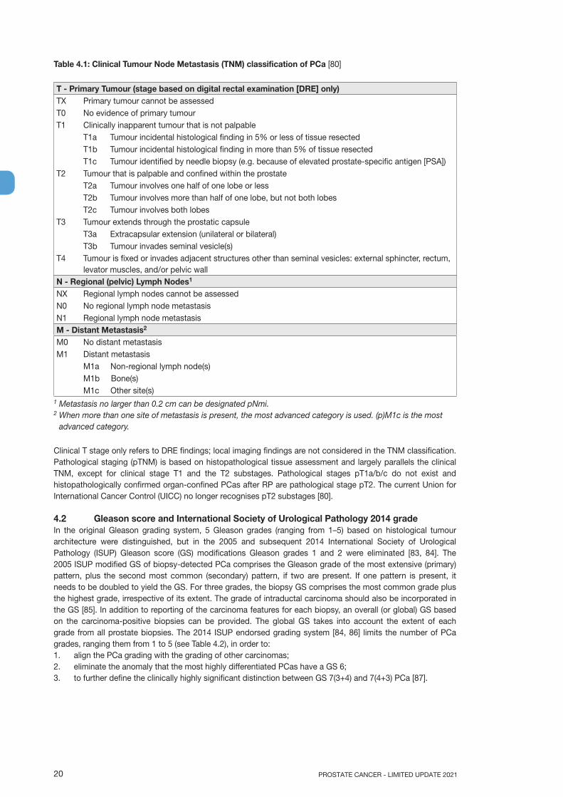
Table 4.1: Clinical Tumour Node Metastasis (TNM) classification of PCa [80]
T - Primary Tumour (stage based on digital rectal examination [DRE] only)TX Primary tumour cannot be assessedT0 No evidence of primary tumourT1 Clinically inapparent tumour that is not palpable
T1a Tumour incidental histological finding in 5% or less of tissue resectedT1b Tumour incidental histological finding in more than 5% of tissue resectedT1c Tumour identified by needle biopsy (e.g. because of elevated prostate-specific antigen [PSA])
T2 Tumour that is palpable and confined within the prostateT2a Tumour involves one half of one lobe or lessT2b Tumour involves more than half of one lobe, but not both lobesT2c Tumour involves both lobes
T3 Tumour extends through the prostatic capsuleT3a Extracapsular extension (unilateral or bilateral)T3b Tumour invades seminal vesicle(s)
T4 Tumour is fixed or invades adjacent structures other than seminal vesicles: external sphincter, rectum, levator muscles, and/or pelvic wall
N - Regional (pelvic) Lymph Nodes1
NX Regional lymph nodes cannot be assessedN0 No regional lymph node metastasisN1 Regional lymph node metastasisM - Distant Metastasis2
M0 No distant metastasisM1 Distant metastasis
M1a Non-regional lymph node(s)M1b Bone(s)M1c Other site(s)
1 Metastasis no larger than 0.2 cm can be designated pNmi.2 When more than one site of metastasis is present, the most advanced category is used. (p)M1c is the most
advanced category.
Clinical T stage only refers to DRE findings; local imaging findings are not considered in the TNM classification. Pathological staging (pTNM) is based on histopathological tissue assessment and largely parallels the clinical TNM, except for clinical stage T1 and the T2 substages. Pathological stages pT1a/b/c do not exist and histopathologically confirmed organ-confined PCas after RP are pathological stage pT2. The current Union for International Cancer Control (UICC) no longer recognises pT2 substages [80].
4.2 Gleason score and International Society of Urological Pathology 2014 gradeIn the original Gleason grading system, 5 Gleason grades (ranging from 1–5) based on histological tumour architecture were distinguished, but in the 2005 and subsequent 2014 International Society of Urological Pathology (ISUP) Gleason score (GS) modifications Gleason grades 1 and 2 were eliminated [83, 84]. The 2005 ISUP modified GS of biopsy-detected PCa comprises the Gleason grade of the most extensive (primary) pattern, plus the second most common (secondary) pattern, if two are present. If one pattern is present, it needs to be doubled to yield the GS. For three grades, the biopsy GS comprises the most common grade plus the highest grade, irrespective of its extent. The grade of intraductal carcinoma should also be incorporated in the GS [85]. In addition to reporting of the carcinoma features for each biopsy, an overall (or global) GS based on the carcinoma-positive biopsies can be provided. The global GS takes into account the extent of each grade from all prostate biopsies. The 2014 ISUP endorsed grading system [84, 86] limits the number of PCa grades, ranging them from 1 to 5 (see Table 4.2), in order to:1. align the PCa grading with the grading of other carcinomas;2. eliminate the anomaly that the most highly differentiated PCas have a GS 6;3. to further define the clinically highly significant distinction between GS 7(3+4) and 7(4+3) PCa [87].
21PROSTATE CANCER - LIMITED UPDATE 2021
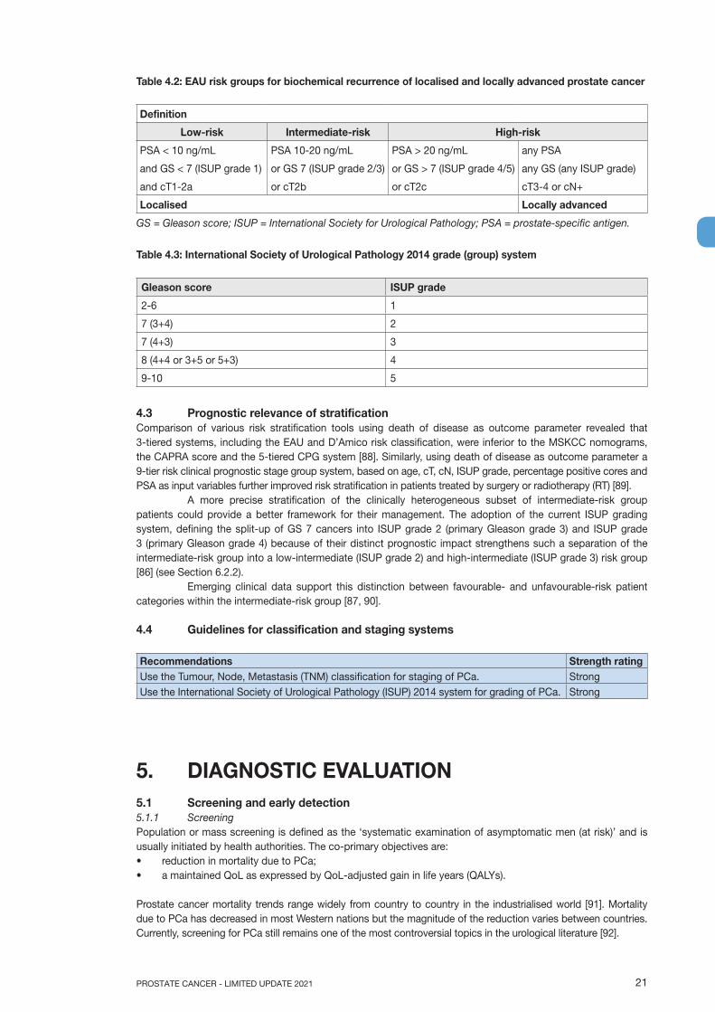
Table 4.2: EAU risk groups for biochemical recurrence of localised and locally advanced prostate cancer
Definition
Low-risk Intermediate-risk High-risk
PSA < 10 ng/mL PSA 10-20 ng/mL PSA > 20 ng/mL any PSA
and GS < 7 (ISUP grade 1) or GS 7 (ISUP grade 2/3) or GS > 7 (ISUP grade 4/5) any GS (any ISUP grade)
and cT1-2a or cT2b or cT2c cT3-4 or cN+
Localised Locally advanced
GS = Gleason score; ISUP = International Society for Urological Pathology; PSA = prostate-specific antigen.
Table 4.3: International Society of Urological Pathology 2014 grade (group) system
Gleason score ISUP grade
2-6 1
7 (3+4) 2
7 (4+3) 3
8 (4+4 or 3+5 or 5+3) 4
9-10 5
4.3 Prognostic relevance of stratificationComparison of various risk stratification tools using death of disease as outcome parameter revealed that 3-tiered systems, including the EAU and D’Amico risk classification, were inferior to the MSKCC nomograms,the CAPRA score and the 5-tiered CPG system [88]. Similarly, using death of disease as outcome parameter a9-tier risk clinical prognostic stage group system, based on age, cT, cN, ISUP grade, percentage positive cores andPSA as input variables further improved risk stratification in patients treated by surgery or radiotherapy (RT) [89].
A more precise stratification of the clinically heterogeneous subset of intermediate-risk group patients could provide a better framework for their management. The adoption of the current ISUP grading system, defining the split-up of GS 7 cancers into ISUP grade 2 (primary Gleason grade 3) and ISUP grade 3 (primary Gleason grade 4) because of their distinct prognostic impact strengthens such a separation of the intermediate-risk group into a low-intermediate (ISUP grade 2) and high-intermediate (ISUP grade 3) risk group [86] (see Section 6.2.2).
Emerging clinical data support this distinction between favourable- and unfavourable-risk patient categories within the intermediate-risk group [87, 90].
4.4 Guidelines for classification and staging systems
Recommendations Strength ratingUse the Tumour, Node, Metastasis (TNM) classification for staging of PCa. StrongUse the International Society of Urological Pathology (ISUP) 2014 system for grading of PCa. Strong
5. DIAGNOSTIC EVALUATION5.1 Screening and early detection5.1.1 ScreeningPopulation or mass screening is defined as the ‘systematic examination of asymptomatic men (at risk)’ and is usually initiated by health authorities. The co-primary objectives are:• reduction in mortality due to PCa;• a maintained QoL as expressed by QoL-adjusted gain in life years (QALYs).
Prostate cancer mortality trends range widely from country to country in the industrialised world [91]. Mortality due to PCa has decreased in most Western nations but the magnitude of the reduction varies between countries. Currently, screening for PCa still remains one of the most controversial topics in the urological literature [92].

PROSTATE CANCER - LIMITED UPDATE 202122
Initial widespread aggressive screening in USA was associated with a decrease in mortality [93]. In 2012 the US Preventive Services Task Force (USPSTF) released a recommendation against PSA-based screening [94], which was adopted in the 2013 AUA Guidelines [95] and resulted in a reduction in the use of PSA for early detection [96]. This reduction in the use of PSA testing was associated with higher rates of advanced disease at diagnosis (e.g., a 6% increase in the number of patients with metastatic PCa [14, 97-100]. While PCa mortality had decreased for two decades since the introduction of PSA testing [101], the incidence of advanced disease and, possibly, cancer-related mortality slowly increased from 2008 and accelerated in 2012 [102]. Moreover, additional evidence suggests a long-term benefit of PSA population screening in terms of reduction of cancer-specific mortality [103, 104]. However, the temporal relationship between PSA testing and decreased mortality, as well as a rising mortality following immediately after the USPSTF and AUA Guidelines recommendation against PSA testing questions the direct causative link between both points.
In 2017 the USPSTF issued an updated statement suggesting that men aged 55–69 should be informed about the benefits and harms of PSA-based screening as this might be associated with a small survival benefit. The USPSTF has now upgraded this recommendation to a grade C [105], from a previous grade ‘D’ [105-107]. They highlighted the fact that the decision to be screened should be an individual one. The grade D recommendation remains in place for men over 70 years old. This represents a major switch from discouraging PSA-based screening (grade D) to offering early diagnosis to selected men depending on individual circumstances.
A comparison of systematic and opportunistic screening suggested over-diagnosis and mortality reduction in the systematic screening group compared to a higher over-diagnosis with a marginal survival benefit, at best, in the opportunistic screening regimen [108].
A Cochrane review published in 2013 [109], which has since been updated [110], presents the main overview to date. The findings of the updated publication (based on a literature search until April 3, 2013) are almost identical to the 2009 review:• Screening is associated with an increased diagnosis of PCa (RR: 1.3, 95% CI: 1.02–1.65).• Screening is associated with detection of more localised disease (RR: 1.79, 95% CI: 1.19–2.70) and less
advanced PCa (T3–4, N1, M1) (RR: 0.80, 95% CI: 0.73–0.87).• From the results of 5 RCTs, randomising more than 341,000 men, no PCa-specific survival benefit was
observed (RR: 1.00, 95% CI: 0.86–1.17). This was the main endpoint in all trials.• From the results of four available RCTs, no overall survival (OS) benefit was observed (RR: 1.00, 95% CI:
0.96–1.03).The included studies applied a range of different screening measures and testing intervals in patients who had undergone prior PSA testing, to various degrees. None included the use of risk calculators, MRI prior to biopsy (vs. a single PSA threshold) or AS (as an alternative to RP) which no longer reflects current standard practice.
The diagnostic tool (i.e. biopsy procedure) was not associated with increased mortality within 120 days after biopsy in screened men as compared to controls in the two largest population-based screening populations (ERSPC and PLCO), in contrast to a 120-day mortality rate of 1.3% in screened vs. 0.3% in controls, respectively, in a Canadian population-based screening study [111]. Increased diagnosis has historically led to over-treatment with associated side effects. However, despite this, the impact on the patient’s overall QoL is still unclear. Population level screening has never been shown to be detrimental [112-114]. Nevertheless, all these findings have led to strong advice against systematic population-based screening in most countries, including those in Europe.
In case screening is considered, a single PSA test is not enough based on the Cluster Randomized Trial of PSA Testing for Prostate Cancer (CAP) trial. The CAP trial evaluated a single PSA screening vs. controls not undergoing PSA screening on PCa detection in men aged 50 to 69 years old. The single PSA screening intervention detected more low-risk PCa cases but had no significant effect on PCa mortality after a median follow-up of 10 years [115].
Since 2013, the European Randomized Study of Screening for Prostate Cancer (ERSPC) data have been updated with 16 years of follow-up (see Table 5.1) [116]. The key message is that with extended follow-up, the mortality reduction remains unchanged (21%, and 29% after non-compliance adjustment). However the number needed to screen (NNS) and to treat is decreasing, and is now below the NNS observed in breast cancer trials [116, 117]. Long-term follow-up of the PLCO (Prostate, Lung, Colon, Ovarian cancer screening trial) showed no survival benefit for screening at a median follow-up of 16.7 years but a significant 17% increase in Gleason score 2–6 cancers and 11% decrease in Gleason score 8–10 cancers [118].

23PROSTATE CANCER - LIMITED UPDATE 2021
Table 5.1: Follow-up data from the ERSPC study [116]
Years of follow-up Number needed to screen Number needed to treat
9 1,410 48
11 979 35
13 781 27
16 570 18
5.1.2 Early detectionAn individualised risk-adapted strategy for early detection may still be associated with a substantial risk of over-diagnosis. It is essential to remember that breaking the link between diagnosis and active treatment is the only way to decrease over-treatment, while still maintaining the potential benefit of individual early diagnosis for men requesting it [16, 119].
Men at elevated risk of having PCa are those > 50 years [120] or at age > 45 years with a family history of PCa (either paternal or maternal) [121] or of African descent [122, 123]. Men of African descent are more likely to be diagnosed with more advanced disease [124] and upgrade was more frequent after prostatectomy as compared to Caucasian men (49% vs. 26%) [125].
Germline mutations are associated with an increased risk of the development of aggressive PCa, i.e. BRCA2 [126, 127]. Prostate-specific antigen screening in male BRCA1 and 2 carriers detected more significant cancers at a younger age compared to non-mutation carriers [32, 33].
Men with a baseline PSA < 1 ng/mL at 40 years and < 2 ng/mL at 60 years are at decreased risk of PCa metastasis or death from PCa several decades later [128, 129].
The use of DRE alone in the primary care setting had a sensitivity and specificity below 60%, possibly due to inexperience, and can therefore not be recommended to exclude PCa [130]. Informed men requesting an early diagnosis should be given a PSA test and undergo a DRE [131]. Prostate-specific antigen measurement and DRE need to be repeated [115], but the optimal intervals for PSA testing and DRE follow-up are unknown as they varied between several prospective trials. A risk-adapted strategy might be a consideration, based on the initial PSA level. This could be every 2 years for those initially at risk, or postponed up to 8 to 10 years in those not at risk with an initial PSA < 1 ng/mL at 40 years and a PSA < 2 ng/mL at 60 years of age and a negative family history [132]. An analysis of ERSPC data supports a recommendation for an 8-year screening interval in men with an initial PSA concentration < 1 μg/L; fewer than 1% of men with an initial PSA concentration < 1 ng/mL were found to have a concentration above the biopsy threshold of 3 μg/L at 4-year follow-up; the cancer detection rate by 8 years was close to 1% [133]. The long-term survival and QoL benefits of extended PSA re-testing (every 8 years) remain to be proven at a population level. Data from the Goteborg arm of the ERSPC trial suggest that the age at which early diagnosis should be stopped remains controversial, but an individual’s life expectancy must definitely be taken into account. Men who have less than a 15-year life expectancy are unlikely to benefit, based on data from the Prostate Cancer Intervention Versus Observation Trial (PIVOT) and the ERSPC trials. Furthermore, although there is no simple tool to evaluate individual life expectancy; co-morbidity is at least as important as age. A detailed review can be found in Section 5.4 ‘Estimating life expectancy and health status’ and in the SIOG Guidelines [134].
Multiple tools are now available to determine the need for a biopsy to establish the diagnosis of a PCa, including imaging by MRI, if available (see Section 5.2.4).
Urine and serum biomarkers as well as tissue-based biomarkers have been proposed for improving detection and risk stratification of PCa patients, potentially avoiding unnecessary biopsies. However, further studies are necessary to validate their efficacy [135]. At present there is too limited data to implement these markers into routine screening programmes (see Section 5.2.3).
Risk calculators may be useful in helping to determine (on an individual basis) what the potential risk of cancer may be, thereby reducing the number of unnecessary biopsies. Several tools developed from cohort studies are available including:• the PCPT cohort: PCPTRC 2.0 http://myprostatecancerrisk.com/;• the ERSPC cohort: http://www.prostatecancer-riskcalculator.com/seven-prostate-cancer-risk-calculators;
An updated version was presented in 2017 including prediction of low and high risk now also based onthe ISUP grading system and presence of cribriform growth in histology [136].
• a local Canadian cohort: https://sunnybrook.ca/content/?page=asure-calc (among others).
PROSTATE CANCER - LIMITED UPDATE 202124
Since none of these risk calculators has clearly shown superiority, it remains a personal decision as to which one to use [137]. A comparative analysis showed risk calculators containing MRI to be most predictive, however, regional modifications may be required before implementation [138].
5.1.3 Genetic testing for inherited prostate cancerIncreasing evidence supports the implementation of genetic counselling and germline testing in early detection and PCa management. Several commercial screening panels are now available to assess main PCa risk genes [139]. However, it remains unclear when germline testing should be considered and how this may impact localised and metastatic disease management. Germline BRCA1 and BRCA2 mutations occur in approximately 0.2% to 0.3% of the general population [140]. It is important to understand the difference between somatic testing, which is performed on the tumour, and germline testing, which is performed on blood or saliva and identifies inherited mutations. Genetic counselling is required prior to and after undergoing germline testing.
Germline mutations can drive the development of aggressive PCa. Therefore, the following men with a personal or family history of PCa or other cancer types arising from DNA repair gene mutations should be considered for germline testing:• Men with metastatic PCa;• Men with high-risk PCa and a family member diagnosed with PCa at age < 60 years;• Men with multiple family members diagnosed with csPCa at age < 60 years or a family member who died
from PCa cancer;• Men with a family history of high-risk germline mutations or a family history of multiple cancers on the
same side of the family.
Further research in this field (including not so well-known germline mutations) is needed to develop screening, early detection and treatment paradigms for mutation carriers and family members.
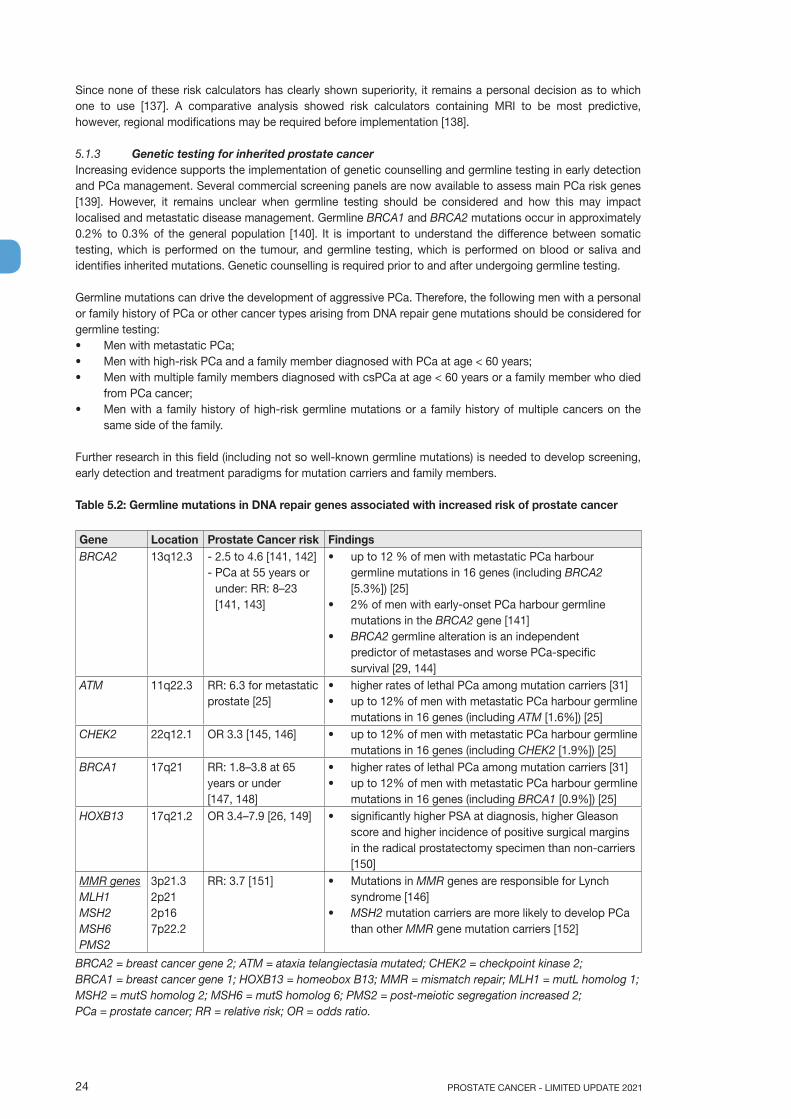
Table 5.2: Germline mutations in DNA repair genes associated with increased risk of prostate cancer
Gene Location Prostate Cancer risk Findings BRCA2 13q12.3 - 2.5 to 4.6 [141, 142]
- PCa at 55 years orunder: RR: 8–23[141, 143]
• up to 12 % of men with metastatic PCa harbourgermline mutations in 16 genes (including BRCA2[5.3%]) [25]
• 2% of men with early-onset PCa harbour germlinemutations in the BRCA2 gene [141]
• BRCA2 germline alteration is an independentpredictor of metastases and worse PCa-specificsurvival [29, 144]
ATM 11q22.3 RR: 6.3 for metastatic prostate [25]
• higher rates of lethal PCa among mutation carriers [31]• up to 12% of men with metastatic PCa harbour germline
mutations in 16 genes (including ATM [1.6%]) [25]CHEK2 22q12.1 OR 3.3 [145, 146] • up to 12% of men with metastatic PCa harbour germline
mutations in 16 genes (including CHEK2 [1.9%]) [25]BRCA1 17q21 RR: 1.8–3.8 at 65
years or under [147, 148]
• higher rates of lethal PCa among mutation carriers [31]• up to 12% of men with metastatic PCa harbour germline
mutations in 16 genes (including BRCA1 [0.9%]) [25]HOXB13 17q21.2 OR 3.4–7.9 [26, 149] • significantly higher PSA at diagnosis, higher Gleason
score and higher incidence of positive surgical marginsin the radical prostatectomy specimen than non-carriers[150]
MMR genes MLH1MSH2MSH6PMS2
3p21.32p212p167p22.2
RR: 3.7 [151] • Mutations in MMR genes are responsible for Lynchsyndrome [146]
• MSH2 mutation carriers are more likely to develop PCathan other MMR gene mutation carriers [152]
BRCA2 = breast cancer gene 2; ATM = ataxia telangiectasia mutated; CHEK2 = checkpoint kinase 2; BRCA1 = breast cancer gene 1; HOXB13 = homeobox B13; MMR = mismatch repair; MLH1 = mutL homolog 1; MSH2 = mutS homolog 2; MSH6 = mutS homolog 6; PMS2 = post-meiotic segregation increased 2; PCa = prostate cancer; RR = relative risk; OR = odds ratio.
25PROSTATE CANCER - LIMITED UPDATE 2021
5.1.4 Guidelines for germline testing*
Recommendations Strength ratingConsider germline testing in men with metastatic PCa. weakConsider germline testing in men with high-risk PCa who have a family member diagnosed with PCa at age < 60 years.
weak
Consider germline testing in men with multiple family members diagnosed with PCa at age < 60 years or a family member who died from PCa.
weak
Consider germline testing in men with a family history of high-risk germline mutations or a family history of multiple cancers on the same side of the family.
weak
*Genetic counseling is required prior to germline testing.
5.1.5 Guidelines for screening and early detection
Recommendations Strength ratingDo not subject men to prostate-specific antigen (PSA) testing without counselling them on the potential risks and benefits.
Strong
Offer an individualised risk-adapted strategy for early detection to a well-informed man and a life-expectancy of at least 10 to 15 years.
Weak
Offer early PSA testing to well-informed men at elevated risk of having PCa:• men from 50 years of age;• men from 45 years of age and a family history of PCa;• men of African descent from 45 years of age;• men carrying BRCA2 mutations from 40 years of age.
Strong
Offer a risk-adapted strategy (based on initial PSA level), with follow-up intervals of 2 years for those initially at risk:• men with a PSA level of > 1 ng/mL at 40 years of age;• men with a PSA level of > 2 ng/mL at 60 years of age;Postpone follow-up to 8 years in those not at risk.
Weak
Stop early diagnosis of PCa based on life expectancy and performance status; men who have a life-expectancy of < 15 years are unlikely to benefit.
Strong
5.2 Clinical diagnosisProstate cancer is usually suspected on the basis of DRE and/or PSA levels. Definitive diagnosis depends on histopathological verification of adenocarcinoma in prostate biopsy cores.
5.2.1 Digital rectal examinationMost PCas are located in the peripheral zone and may be detected by DRE when the volume is > 0.2 mL. In ~18% of cases, PCa is detected by suspect DRE alone, irrespective of PSA level [153]. A suspect DRE in patients with a PSA level < 2 ng/mL has a positive predictive value (PPV) of 5–30% [154]. An abnormal DRE is associated with an increased risk of a higher ISUP grade and is an indication for biopsy [155, 156].
5.2.2 Prostate-specific antigenThe use of PSA as a serum marker has revolutionised PCa diagnosis [157]. Prostate-specific antigen is organ but not cancer specific; therefore, it may be elevated in benign prostatic hypertrophy (BPH), prostatitis and other non-malignant conditions. As an independent variable, PSA is a better predictor of cancer than either DRE or transrectal ultrasound (TRUS) [158].
There are no agreed standards defined for measuring PSA [159]. It is a continuous parameter, with higher levels indicating greater likelihood of PCa. Many men may harbour PCa despite having low serum PSA [160]. Table 5.3 demonstrates the occurrence of ISUP > grade 2 PCa at low PSA levels, precluding an optimal PSA threshold for detecting non-palpable but csPCa. The use of nomograms may help in predicting indolent PCa [161].

PROSTATE CANCER - LIMITED UPDATE 202126
Table 5.3: Risk of PCa identified by systemic PCa biopsy in relation to low PSA values [160]
PSA level (ng/mL) Risk of PCa (%) Risk of ISUP grade > 2 PCa (%)0.0–0.5 6.6 0.80.6–1.0 10.1 1.01.1–2.0 17.0 2.02.1–3.0 23.9 4.63.1–4.0 26.9 6.7
5.2.2.1 PSA densityProstate-specific antigen density is the level of serum PSA divided by the prostate volume. The higher the PSA density, the more likely it is that the PCa is clinically significant (see Section 6.2.1 - Treatment of low-risk disease).
5.2.2.2 PSA velocity and doubling timeThere are two methods of measuring PSA kinetics:• PSA velocity (PSAV): absolute annual increase in serum PSA (ng/mL/year) [162];• PSA doubling time (PSA-DT): which measures the exponential increase in serum PSA over time [163].
Prostate-specific antigen velocity and PSA-DT may have a prognostic role in treating PCa but have limited diagnostic use because of background noise (total prostate volume, and BPH), different intervals between PSA determinations, and acceleration/deceleration of PSAV and PSA-DT over time [164]. These measurements do not provide additional information compared with PSA alone [165-168].
Prostate-specific antigen kinetics is mainly exponential, especially during relapse. Two methods have been described to calculate PSA-DT: the 2 -points and the log-slope method using more PSA measurements [169]. The latter is considered more accurate as it eliminates inter- and intra-essay variations as well as any physiological variations and potential imprecise measurements of low values [170, 171]. To obtain a reliable evaluation, at least 3 PSA measurements must be available, collected four weeks apart as a minimum from the same clinical laboratory [164]. A minimum increase of between 0.2 and 0.4 ng/mL is required [172].
As PSA-DT values may fluctuate over time in some men, only values obtained over the past 12 months should be considered to assess disease course [164]. The Memorial Sloan Kettering Cancer Center PSA Doubling Time calculator is one of the most widely used tools: https://www.mskcc.org/nomograms/prostate/psa_doubling_time.
5.2.2.3 Free/total PSA ratioFree/total (f/t) PSA must be used cautiously because it may be adversely affected by several pre-analytical and clinical factors (e.g., instability of free PSA at 4°C and room temperature, variable assay characteristics, and concomitant BPH in large prostates) [173]. Prostate cancer was detected in men with a PSA 4–10 ng/mL by biopsy in 56% of men with f/t PSA < 0.10, but in only 8% with f/t PSA > 0.25 ng/mL [174]. A systematic review including 14 studies found a pooled sensitivity of 70% in men with a PSA of 4–10 ng/mL [175]. Free/total PSA is of no clinical use if the total serum PSA is > 10 ng/mL or during follow-up of known PCa. The clinical value of f/t PSA is limited in light of the new diagnostic pathways incorporating MRI (see Section 5.2.4.2).
5.2.3 Biomarkers in prostate cancer5.2.3.1 Blood based biomarkers: PHI/4K score/IsoPSASeveral assays measuring a panel of kallikreins in serum or plasma are now commercially available, including the U.S. Food and Drug Administration (FDA) approved Prostate Health Index (PHI) test (combining free and total PSA and the [-2]pro-PSA isoform [p2PSA]), and the four kallikrein (4K) score test (measuring free, intact and total PSA and kallikrein-like peptidase 2 [hK2] in addition to age, DRE and prior biopsy status). Both tests are intended to reduce the number of unnecessary prostate biopsies in PSA-tested men. A few prospective multi-centre studies demonstrated that both the PHI and 4K score test out-performed f/t PSA PCa detection, with an improved prediction of csPCa in men with a PSA between 2–10 ng/mL [176-179]. In a head-to-head comparison both tests performed equally [180].
In contrast to the 4K score and PHI, which focus on the concentration of PSA isoforms, IsoPSA utilises a novel technology which focuses on the structure of PSA [181]. Using an aqueous two-phase solution, it partitions the isoforms of PSA and assesses for structural changes in PSA. In a recent multi-centre prospective validation in 271 men the assay AUC was 0.784 for high-grade vs. low-grade cancer/benign histology, which was superior to the AUCs of total PSA and percent free PSA [182].
27PROSTATE CANCER - LIMITED UPDATE 2021
5.2.3.2 Urine biomarkers: PCA3/SelectMDX/Mi Prostate score (MiPS)/ExoDXProstate cancer gene 3 (PCA3) is an overexpressed long non-coding RNA (lncRNA) biomarker that is detectable in urine sediments obtained after three strokes of prostatic massage during DRE. The commercially available Progensa urine test for PCA3 is superior to total and percent-free PSA for the detection of PCa in men with elevated PSA as it shows significant increases in the area under the receiver-operator characteristic curve (AUC) for positive biopsies [183-186]. PCA3 score increases with PCa volume, but there are conflicting data about whether it independently predicts the ISUP grade [187]. Currently, the main indication for the Progensa test is to determine whether repeat biopsy is needed after an initially negative biopsy, but its clinical effectiveness for this purpose is uncertain [188]. Wei et al. showed 42% sensitivity at a cut-off of 60 in the primary biopsy setting with a high specificity (91%) and a PPV of 80% suggesting that the assay may be used in the primary setting [189].
The SelectMDX test is similarly based on mRNA biomarker isolation from urine. The presence of HOXC6 and DLX1 mRNA levels is assessed to provide an estimate of the risk of both presence of PCa on biopsy as well as presence of high-risk cancer [190].
TMPRSS2-ERG fusion, a fusion of the trans-membrane protease serine 2 (TMPRSS2) and the ERG gene can be detected in 50% of PCas [191]. When detection of TMPRSS2-ERG in urine was added to PCA3 expression and serum PSA (Mi(chigan)Prostate Score [MiPS]), cancer prediction improved [192]. Exosomes secreted by cancer cells may contain mRNA diagnostic for high-grade PCa [193, 194]. Use of the ExoDx Prostate IntelliScore urine exosome assay resulted in avoiding 27% of unnecessary biopsies when compared to standard of care. However, currently, both the MiPS-score and ExoDx assay are considered investigational.
In 6 head-to-head comparison studies of PCA3 and PHI, only Seisen et al. found a significant difference; PCA3 detected more cancers, but for the detection of significant disease, defined as ISUP grade > 2, more than three positive cores, or > 50% cancer involvement in any core, PHI proved superior [195]. Russo et al. suggested in their systematic review that, based on moderate quality data, PHI and the 4K panel had a high diagnostic accuracy and showed similar performance in predicting the detection of significant disease with a AUC of 0.82 and 0.81, respectively [196]. However, in the screening population of the ERSPC study the use of both PCA3 and 4K panel when added to the risk calculator led to an improvement in AUC of less than 0.03 [197]. Based on the available evidence, some biomarkers could help in discriminating between aggressive and non-aggressive tumours with an additional value compared to the prognostic parameters currently used by clinicians [198]. However, upfront multiparametric magnetic resonance imaging (mpMRI) is also likely to affect the utility of above-mentioned biomarkers (see Section 5.2.4).
5.2.3.3 Tests to select men for a repeat biopsyIn men with an elevated risk of PCa with a prior negative biopsy, additional information may be gained by the Progensa-PCA3 and SelectMDX DRE urine tests, the serum 4Kscore and PHI tests or a tissue-based epigenetic test (ConfirmMDx). The role of PHI, Progensa PCA3, and SelectMDX in deciding whether to take a repeat biopsy in men who had a previous negative biopsy is uncertain and probably not cost-effective [188]. The ConfirmMDx test is based on the concept that benign prostatic tissue in the vicinity of a PCa focus shows distinct epigenetic alterations. In case PCa is missed at biopsy, demonstration of epigenetic changes in the benign tissue would indicate the presence of carcinoma. The ConfirmMDX test quantifies the methylation level of promoter regions of three genes (Methylated APC, RASSF1 and GSTP1) in benign prostatic tissue. A multi-centre study found a NPV of 88% when methylation was absent in all three markers, implying that a repeat biopsy could be avoided in these men [199]. Given the limited available data and the fact that the role of mpMRI in tumour detection was not accounted for, no recommendation can be made regarding the routine application of ConfirmMDX, in particular in the light of current use of mpMRI before biopsy. Table 5.4 presents an overview of the most commonly available commercial tests.
Table 5.4: Description of additional investigational tests after a negative prostate biopsy*
Name of test Test substrate Molecular FDA approvedProgensa DRE urine lncRNA PCA3 YesSelectMDX DRE urine mRNA HOXC6, DLX1 NoPHI Serum Total, free and p2PSA Yes4Kscore Test Serum/plasma Total, free, intact PSA, hK2 NoConfirmMDX Benign prostate biopsy Methylated APC, RASSF1 and GSTP1 No
*Isolated high-grade prostatic intraepithelial neoplasia (PIN) in one or two biopsy sites is no longer an indicationfor repeat biopsy [200].
PROSTATE CANCER - LIMITED UPDATE 202128
5.2.3.4 Guidelines for risk-assessment of asymptomatic men
Recommendations Strength ratingIn asymptomatic men with a prostate-specific antigen level between 2–10 ng/mL and a normal digital rectal examination, use one of the following tools for biopsy indication:• risk-calculator;• imaging;
Strong
• an additional serum, urine or tissue-based test. Weak
5.2.4 The role of imaging in clinical diagnosis 5.2.4.1 Transrectal ultrasound and ultrasound-based techniquesStandard TRUS is not reliable at detecting PCa [201] and the diagnostic yield of additional biopsies performed on hypoechoic lesions is negligible [202]. Prostate HistoScanningTM provided inconsistent results across studies [203]. New sonographic modalities such as micro-Doppler, sonoelastography contrast-enhanced US or high-resolution micro-ultrasound provided promising preliminary findings, either alone, or combined with so-called ‘multiparametric US’. However, these techniques still have limited clinical applicability due to lack of standardisation, lack of large-scale evaluation of inter-reader variability and unclear results in transition zones [204-206].
5.2.4.2 Multiparametric magnetic resonance imaging5.2.4.2.1 Multiparametric magnetic resonance imaging performance in detecting PCa Correlation with RP specimens shows that MRI has good sensitivity for the detection and localisation of ISUP grade > 2 cancers, especially when their diameter is larger than 10 mm [207-209]. This good sensitivity was further confirmed in patients who underwent template biopsies. In a recent Cochrane meta-analysis which compared MRI to template biopsies (> 20 cores) in biopsy-naïve and repeat-biopsy settings, MRI had a pooled sensitivity of 0.91 (95% CI: 0.83–0.95) and a pooled specificity of 0.37 (95% CI: 0.29–0.46) for ISUP grade > 2 cancers [210]. For ISUP grade > 3 cancers, MRI pooled sensitivity and specificity were 0.95 (95% CI: 0.87–0.99) and 0.35 (95% CI: 0.26–0.46), respectively.
MRI is less sensitive in identifying ISUP grade 1 PCa. It identifies less than 30% of ISUP grade 1 cancers smaller than 0.5 cc identified on RP specimens by histopathology analysis [207]. In series using template biopsy findings as the reference standard, MRI has a pooled sensitivity of 0.70 (95% CI: 0.59–0.80) and a pooled specificity of 0.27 (95% CI: 0.19–0.37) for identifying ISUP grade 1 cancers [210].
The probability of detecting malignancy by MRI-identified lesions was standardised first by the use of a 5-grade Likert score [211], and then by the Prostate Imaging – Reporting and Data System (PI-RADS) score which has been updated several times since its introduction [212, 213]. In a meta-analysis of 13 studies involving men with suspected or biopsy-proven PCa, the average PPVs for ISUP grade > 2 cancers of lesions with a PI-RADSv2 score of 3, 4 and 5 were 12%, 48% and 72% respectively, but with significant heterogeneity among studies (Table 5.5) [214]. Another retrospective study of 3,449 men with suspected or biopsy-proven untreated PCa imaged across 26 academic centres found similar results with lesions of PI-RADS v2 scores of 3, 4 and 5 having a PPV for ISUP grade > 2 cancers of 15% (95% CI: 11–19), 39% (95% CI: 34–45) and 72% (95% CI: 61–82), respectively [215].
Table 5.5: Proportion of malignant MRI lesions by ISUP grade groups and PI-RADS v2 scores [214]
PI-RADS v2 Total number of lesions
ISUP 1 ISUP 2 ISUP 3 ISUP > 4
3 707 14% (9.4–18.7) 9.3% (4.3–14.1) 1.5% (0.05–3) 0.7% (0–1.6)4 886 21% (13.0–28.9) 29.7% (13.9–45.5) 7.7% (3.4–12) 10.8% (5.7–15.9)5 495 12% (5.3–18.7) 33.5% (8.0–59.0) 15.7% (6.4–25.1) 23% (8.2–37.9)
Intervals in parenthesis are 95% CIs.
5.2.4.2.2 Targeted biopsy improves the detection of ISUP grade > 2 cancer as compared to systematic biopsy.
In pooled data of 25 reports on agreement analysis (head-to-head comparisons) between systematic biopsy (median number of cores, 8–15) and MRI-targeted biopsies (MRI-TBx; median number of cores, 2–7), the detection ratio (i.e. the ratio of the detection rates obtained by MRI-TBx alone and by systematic biopsy alone) was 1.12 (95% CI: 1.02–1.23) for ISUP grade > 2 cancers and 1.20 (95% CI: 1.06–1.36) for ISUP grade > 3 cancers, and therefore in favour of MRI-TBx.
29PROSTATE CANCER - LIMITED UPDATE 2021
However, the pooled detection ratios for ISUP grade > 2 cancers and ISUP grade > 3 cancers were 1.44 (95% CI: 1.19–1.75) and 1.64 (95% CI: 1.27–2.11), respectively, in patients with prior negative systematic biopsies, and only 1.05 (95% CI: 0.95–1.16) and 1.09 (95% CI: 0.94–1.26) in biopsy-naïve patients [210]. Another meta-analysis of RCTs limited to biopsy-naïve patients with a positive MRI found that MRI-TBx detected significantly more ISUP grade > 2 cancers than systematic biopsy (risk difference, -0.11 (95% CI: -0.2 to 0.0); p = 0.05), in prospective cohort studies (risk difference, -0.18 (95% CI: -0.24 to -0.11); p < 0.00001), and in retrospective cohort studies (risk difference, -0.07 (95% CI: -0.12 to -0.02); p = 0.004). There was no significant increase in the detection rate when systematic biopsies were combined to MRI-TBx [216].
Three prospective multi-centre trials evaluated MRI-TBx in biopsy-naïve patients. In the PRostate Evaluation for Clinically Important Disease: Sampling Using Image-guidance Or Not? (PRECISION) trial, 500 biopsy-naïve patients were randomised to either MRI-TBx only or TRUS-guided systematic biopsy only. The detection rate of ISUP grade > 2 cancers was significantly higher in men assigned to MRI-TBx (38%) than in those assigned to SBx (26%, p = 0.005, detection ratio 1.46) [217]. In the Assessment of Prostate MRI Before Prostate Biopsies (MRI-FIRST) trial, 251 biopsy-naïve patients underwent TRUS-guided systematic biopsy by an operator who was blinded to MRI findings, and MRI-TBx by another operator. Magnetic resonance Imaging-TBx detected ISUP grade > 2 cancers in a higher percentage of patients but the difference was not significant (32.3% vs. 29.9%, p = 0.38; detection ratio: 1.08) [202]. However, MRI-TBx detected significantly more ISUP grade > 3 cancers than systematic biopsy (19.9% vs. 15.1%, p = 0.0095; detection ratio: 1.32). A similar trend for improved detection of ISUP grade > 3 cancers by MRI-TBx was observed in the Cochrane analysis; however, it was not statistically significant (detection ratio 1.11 [0.88–1.40]) [210]. The Met Prostaat MRI Meer Mans (4M) study included 626 biopsy-naïve patients; all patients underwent systematic biopsy, and those with a positive MRI (PI-RADSv2 score of 3–5, 51%) underwent additional in-bore MRI-TBx. The results were close to those of the MRI-FIRST trial with a detection ratio for ISUP grade > 2 cancers of 1.09 (detection rate: 25% for MRI-TBx vs. 23% for systematic biopsy) [218]. However, in this study, MRI-TBx and systematic biopsy detected an equal number of ISUP grade > 3 cancers (11% vs. 12%; detection ratio: 0.92).
The Target Biopsy Techniques Based on Magnetic Resonance Imaging in the Diagnosis of Prostate Cancer in Patients with Prior Negative Biopsies (FUTURE) randomised trial compared three techniques of MRI-TBx in the repeat-biopsy setting [219]. In the subgroup of 152 patients who underwent both MRI-TBx and systematic biopsy, MRI-TBx detected significantly more ISUP grade > 2 cancers than systematic biopsy (34% vs. 16%; p < 0.001, detection ratio of 2.1), which is a finding consistent with the Cochrane agreement analysis (detection ratio: 1.44). An ISUP grade > 2 cancer would have been missed in only 1.3% (2/152) of patients, had systematic biopsy been omitted [220]. These findings support that MRI-TBx significantly out-performs systematic biopsy for the detection of ISUP grade > 2 in the repeat-biopsy setting. In biopsy-naïve patients, the difference appears to be less marked but remains in favour of MRI-TBx.
5.2.4.2.3 MRI-TBx without systematic biopsy reduces the detection of ISUP grade 1 PCa as compared to systematic biopsy.
In pooled data of 25 head-to-head comparisons between systematic biopsy and MRI-TBx, the detection ratio for ISUP grade 1 cancers was 0.62 (95% CI: 0.44–0.88) in patients with prior negative biopsy and 0.63 (95% CI: 0.54–0.74) in biopsy-naïve patients [210]. In the PRECISION and 4M trials, the detection rate of ISUP grade 1 patients was significantly lower in the MRI-TBx group as compared to systematic biopsy (9% vs. 22%, p < 0.001, detection ratio of 0.41 for PRECISION; 14% vs. 25%, p < 0.001, detection ratio of 0.56 for 4M) [217, 218]. In the MRI-FIRST trial, MRI-TBx detected significantly fewer patients with clinically insignificant PCa (defined as ISUP grade 1 and maximum cancer core length < 6 mm) than systematic biopsy (5.6% vs. 19.5%, p < 0.0001, detection ratio of 0.29) [202]. Consequently, MRI-TBx without systematic biopsy significantly reduces over-diagnosis of low-risk disease, as compared to systematic biopsy.
5.2.4.2.4 The added value of systematic and targeted biopsyMagnetic resonance imaging-targeted biopsies can be used in two different diagnostic pathways: 1) the ‘combined pathway’, in which patients with a positive MRI undergo combined systematic and targeted biopsy, and patients with a negative MRI undergo systematic biopsy; 2) the ‘MRI pathway’, in which patients with a positive MRI undergo only MRI-TBx, and patients with a negative MRI who are not biopsied at all.
Choosing between these pathways depends not only on the detection rates obtained by the two biopsy techniques, but also on whether or not they detect the same patients. Many studies evaluated combined systematic and targeted biopsy in the same patients and could therefore assess the absolute added value of each technique (i.e. the percentage of patients diagnosed by only one biopsy technique). Data from the Cochrane meta-analysis of these studies and from the MRI-FIRST and 4M trials suggest that the absolute added value of MRI-TBx for detecting ISUP grade > 2 cancers is higher than that of systematic biopsy (see Table 5.6).
PROSTATE CANCER - LIMITED UPDATE 202130
Table 5.6: Absolute added values of targeted and systematic biopsies for ISUP grade > 2 and > 3 cancer detection
ISUP > 2 ISUP > 3ISUP grade
Cochrane meta-analysis* [210]
MRI-FIRST trial* [202]
4M trial [218]
Cochrane meta-analysis* [210]
MRI-FIRST trial* [202]
4M trial [218]
Biopsy-naïve
Added value of MRI-TBx
6.3% (4.8-8.2)
7.6% (4.6-11.6)
7.0% (ND) 4.7%(3.5-6.3)
6.0% (3.4-9.7)
3.2% (ND)
Added value of systematic biopsy
4.3% (2.6-6.9)
5.2% (2.8-8.7)
5.0% (ND) 2.8% (1.7-4.8)
1.2% (0.2-3.5)
4.1% (ND)
Overall prevalence
27.7% (23.7-32.6)
37.5% (31.4-43.8)
30% (ND) 15.5% (12.6-19.5)
21.1% (16.2-26.7)
15% (ND)
Prior negative biopsy
Added value of MRI-TBx
9.6% (7.7-11.8)
- - 6.3% (5.2-7.7)
- -
Added value of systematic biopsy
2.3% (1.2-4.5)
- - 1.1% (0.5-2.6)
- -
Overall prevalence
22.8% (20.0-26.2)
- - 12.6% (10.5-15.6)
- -
*Intervals in parenthesis are 95% CI. The absolute added value of a given biopsy technique is defined by the percentage of patients of the entire cohort diagnosed only by this biopsy technique.ISUP = International Society for Urological Pathology (grade); MRI-TBx = magnetic resonance imaging-targeted biopsies; ND = not defined.
In Table 5.6, the absolute added values refer to the percentage of patients in the entire cohort; if the cancer prevalence is taken into account, the ‘relative’ percentage of additional detected PCa can be computed. Adding MRI-TBx to systematic biopsy in biopsy-naïve patients increases the number of ISUP grade > 2 and grade > 3 PCa by approximately 20% and 30%, respectively. In the repeat-biopsy setting, adding MRI-TBx increases detection of ISUP grade > 2 and grade > 3 PCa by approximately 40% and 50%, respectively. Omitting systematic biopsy in biopsy-naïve patients would miss approximately 16% of ISUP grade > 2 PCa and 18% of ISUP grade > 3 PCa. In the repeat-biopsy setting, it would miss approximately 10% of ISUP grade > 2 PCa and 9% of ISUP grade > 3 PCa.
5.2.4.2.5 Number of biopsy procedures potentially avoided in the ‘MR pathway’The diagnostic yield and number of biopsy procedures potentially avoided by the ‘MR pathway’ depends on the Likert/PI-RADS threshold used to define positive MRI. In pooled studies on biopsy-naïve patients and patients with prior negative biopsies, a Likert/PI-RADS threshold of > 3 would have avoided 30% (95% CI: 23–38) of all biopsy procedures while missing 11% (95% CI: 6–18) of all detected ISUP grade > 2 cancers (relative percentage) [210]. Increasing the threshold to > 4 would have avoided 59% (95% CI: 43–78) of all biopsy procedures while missing 28% (95% CI: 14–48) of all detected ISUP grade > 2 cancers [210]. Of note, the percentages of negative MRI (Likert/PI-RADS score < 2) in the MRI-FIRST, PRECISION and 4M trials were 21.1%, 28.9% and 49%, respectively [202, 217, 218].
5.2.4.2.6 Other considerations5.2.4.2.6.1 Multiparametric magnetic resonance imaging reproducibility Despite the use of the PI-RADSv2 scoring system [212], MRI inter-reader reproducibility remains moderate at best which currently limits its broad use by non-dedicated radiologists [221]. However, significant improvement in the accuracy of MRI and MRI-TBx can be observed over time, both in academic and community hospitals, especially after implementation of PI-RADSv2 scoring and multidisciplinary meetings using pathological correlation and feedback [222-225]. An updated version of the PI-RADS score (PI-RADSv2.1) has been recently published to improve reader reproducibility but it has not yet been fully evaluated [213]. It is still too early to predict whether quantitative approaches and computer-aided diagnostic systems will improve the characterisation of lesions seen at MRI [226-228].
31PROSTATE CANCER - LIMITED UPDATE 2021
5.2.4.2.6.2 Targeted biopsy accuracy and reproducibilityClinically significant PCa not detected by the ‘MRI pathway’ can be missed because of MRI failure (invisible cancer or reader’s misinterpretation) or because of targeting failure (target missed or undersampled by MRI-TBx). In two retrospective studies of 211 and 116 patients with a unilateral MRI lesion, targeted biopsy alone detected 73.5–85.5% of all csPCa (ISUP grade > 2); combining MRI-TBx with systematic biopsy of the lobe with the MRI lesion detected 96–96.4% of all csPCas and combined targeted and systematic biopsy of the contralateral lobe only identified 81.6–92.7% of csPCas [229, 230]. The difference may reflect targeting errors leading to undersampling of the tumour. The accuracy of MRI-TBx is indeed substantially impacted by the experience of the biopsy operator [221]. Increasing the number of cores taken per target may partially compensate for guiding imprecision. In a retrospective study of 479 patients who underwent MRI-TBx with 4 cores per target that were sequentially labelled, the first 3 cores detected 95.1% of the csPCas detected by the 4-core strategy [231]. In two other retrospective studies of 330 and 744 patients who underwent MRI-TBx with up to 5 cores per target, the one-core and 3-core sampling strategies detected respectively 63–75% and 90–93% of the ISUP grade > 2 PCa detected by the 5-core strategy [232, 233]. These percentages are likely to be influenced by the lesion size and location, the prostate volume or the operator’s experience, but no study has quantified the impact of these factors yet.
5.2.4.2.6.3 Role of risk-stratificationUsing risk-stratification to avoid biopsy proceduresProstate-specific antigen density (PSAD) may help refine the risk of csPCa in patients undergoing MRI as PSAD and the PI-RADS score are significant independent predictors of csPCa at biopsy [234, 235]. In a meta-analysis of 8 studies, pooled MRI NPV for ISUP grade > 2 cancer was 84.4% (95% CI: 81.3–87.2) in the whole cohort, 82.7% (95% CI: 80.5–84.7) in biopsy-naïve men and 88.2% (95% CI: 85–91.1) in men with prior negative biopsies. In the subgroup of patients with PSAD < 0.15 ng/ml, NPV increased to respectively 90.4% (95% CI: 86.8–93.4), 88.7% (95% CI: 83.1–93.3) and 94.1% (95% CI: 90.9–96.6) [236]. In contrast, the risk of csPCa is as high as 27–40% in patients with negative MRI and PSAD > 0.15–0.20 ng/mL/cc [218, 235, 237-240] (Table 5.7).
Combining MRI findings with the PCA3 score may also improve risk stratification [241]. Several groups have developed comprehensive risk calculators which combine MRI findings with simple clinical data as a tool to predict subsequent biopsy results [242]. At external validation, they tended to outperform risk calculators not incorporating MRI findings (ERSPC and PBCG) with good discriminative power (as measured by the AUC). However, they also tended to be miscalibrated with under- or over-prediction of the risk of ISUP grade > 2 cancer [243, 244]. In one study that externally assessed four risk calculators combining MRI findings and clinical data, only two demonstrated a distinct net benefit when a risk of false-negative prediction of 15% was accepted. The others were harmful for this risk level, as compared to the ‘biopsy all’ strategy [243]. This illustrates the prevalence-dependence of risk models. Recalibrations taking into account the local prevalence are possible, but this approach is difficult in routine clinical practice as the local prevalence is difficult to estimate and may change over time.
Using risk-stratification to avoid MRI and biopsy proceduresA retrospective analysis including 200 men from a prospective database of patients who underwent MRI and combined systematic and targeted biopsy showed that upfront use of the Rotterdam Prostate Cancer Risk Calculator (RPCRC) would have avoided MRI and biopsy in 73 men (37%). Of these 73 men, 10 had ISUP grade 1 cancer and 4 had ISUP grade > 2 cancer [245]. A prospective multi-centre study evaluated several diagnostic pathways in 545 biopsy-naïve men who underwent MRI and systematic and targeted biopsy. Using a PHI threshold of > 30 to perform MRI and biopsy would have avoided MRI and biopsy in 25% of men at the cost of missing 8% of the ISUP grade > 2 cancers [246]. Another prospective multi-centre trial including 532 men (with or without history of prostate biopsy) showed that using a threshold of > 10% for the Stockholm3 test to perform MRI and biopsy would have avoided MRI and biopsy in 38% of men at the cost of missing 8% of ISUP grade > 2 cancers [247].

PROSTATE CANCER - LIMITED UPDATE 202132
Table 5.7: Impact of the PSA density on csPCa detection rates in patients with negative MRI findings
Study Study design Population Biopsy protocol csPCa definition
csPCa detection rate
Washino, et al. 2017 [235]
Retrosp. Single-centre
n = 288 Biopsy naive
SBx (14 cores) + cognitive TBx
ISUP > 2 or MCCL > 4 mm
Whole cohort (prevalence): 49%
PI-RADS 1-2: 0% if PSAD < 0.15, 20% if PSAD = 0.15-0.29, 30% if PSAD > 0.3
Distler, et al. 2017 [234]
Retrosp. analysis of prospective database Single-centre
n = 1,040 Biopsy naive + prior negative biopsy
TTP (24 cores) + fusion TBx
ISUP > 2 Whole cohort (prevalence): 43%
PI-RADS 1-2 / Whole cohort: 11% if PSAD < 0.15, 33% if PSAD > 0.15
PI-RADS 1-2 / prior negative biopsy: 7% if PSAD < 0.15, 27% if PSAD > 0.15
Hansen, et al. 2017 [237]
Retrosp. Single-centre
n = 514 Prior negative biopsy or AS for ISUP 1 PCa
TTP (24 cores) + fusion TBx
ISUP > 2 Whole cohort (prevalence): 31%
Likert 1-2: 9% if PSAD < 0.10, 9% if PSAD < 0.2, 29% if PSAD > 0.2
Hansen, et al. 2018 [238]
Retrosp. Multi-centre
n = 807 Biopsy naive
TTP + cognitive or fusion TBx
ISUP > 2 Whole cohort (prevalence): 49%
PI-RADS 1-2: 10% if PSAD < 0.10, 21% if PSAD = 0.1-0.2, 33% if PSAD > 0.2
Oishi, et al. 2019 [239]
Retrosp. analysis of prospective database Single-centre
n = 135 Biopsy naive + prior negative biopsy + AS + Restaging Only pts with negative MRI (PI-RADS < 2)
SBx (12 cores) ISUP > 2 Whole cohort (prevalence): 18%
PSAD < 0.10: 6% (overall pop), 15% (biopsy naïve), 0% (prior negative biopsy)PSAD < 0.15: 10% (overall pop), 20% (biopsy naïve), 0% (prior negative biopsy)PSAD > 0.15: 40% (overall pop), 29% (biopsy naïve), 29% (prior negative biopsy)
Boesen, et al. 2019 [240]
Retrosp. analysis of prospective database Single-centre
n = 808Biopsy-naive
SBx (10 cores) + fusion TBx
ISUP > 2 Whole cohort (prevalence): 35%
PI-RADS 1-2: 3% if PSAD < 0.10, 5% < 0.15, 5% if PSAD < 0.2, 32% if PSAD > 0.2
33PROSTATE CANCER - LIMITED UPDATE 2021
Van der Leest, et al. 2019 [218]
Prospective Multi-centre
n = 626 Biopsy naive
TTP Bx (median 24 cores) + fusion TBx
ISUP > 2 Whole cohort (prevalence): 30%
PI-RADS 1-2: 1.3% if PSAD < 0.10, 2% if PSAD < 0.15
csPCa = clinically significant prostate cancer; Retrosp. = retrospective; n = number of patients; SBx = systematic transrectal biopsy; TBx = targeted biopsy; TTP = transperineal template biopsy; PSAD = PSA density; ISUP = international Society of Urological Pathology; MCCL = maximum cancer core length.
5.2.4.2.6.4 Pre-biopsy MRI and MRI-TBx may induce an ISUP prostate cancer grade shift, as a result of improved diagnosis
Magnetic resonance imaging findings are significant predictors of adverse pathology features on prostatectomy specimens, and of survival-free BCR after RP or RT [82, 248-250]. In addition, tumours visible on MRI are enriched in molecular hallmarks of aggressivity, as compared to invisible lesions [251]. Thus, MRI does identify aggressive tumours.
Nonetheless, improved targeting obtained by MRI-TBx can artificially inflate the ISUP grade of the tumours by focusing at the areas of high-grade cancer. As a result, ISUP grade > 2 cancers detected by MRI-TBx are, on average, of better prognosis than those detected by the classical diagnostic pathway. This is illustrated in a retrospective series of 1,345 patients treated by RP which showed that, in all risk groups, patients diagnosed by MRI-TBx had better BCR-free survival than those diagnosed by systematic biopsy only [82]. To mitigate this grade shift, in case of targeted biopsies, the 2019 ISUP consensus conference recommended using an aggregated ISUP grade summarizing the results of all biopsy cores from the same MR lesion, rather than using the result from the core with the highest ISUP grade [85]. When long-term follow-up of patients who underwent MRI-TBx is available, a revision of the risk-groups definition will become necessary. In the meantime, results of MRI-TBx must be interpreted in the context of this potential grade shift as an increasing proportion of patients diagnosed with ISUP grade 2 cancers by MRI-TBx might be eligible for AS [252].
5.2.4.2.7 Summary of evidence and practical considerations on pre-biopsy MRI and MRI-TBxPre-biopsy MRI and MRI-TBx substantially improve the detection of ISUP grade > 2 PCa. This improvement is most notable in the repeat-biopsy setting, with marginal added value for systematic biopsies. It is less marked in biopsy-naïve patients in whom systematic biopsy retains a higher added value, at least for the detection of ISUP grade 2 cancers. MRI-TBx also detects significantly less ISUP grade 1 cancers than systematic biopsies.
The ‘MRI pathway’ is appealing since it could decrease the number of biopsy procedures, reduce the detection of low-grade PCa while maintaining (or even improving) the detection of csPCa, as compared to systematic biopsy. However, some cavaets need pointing out. First, MRI findings must be interpreted in the light of the a priori risk of csPCa. Risk stratification combining clinical data, MRI findings and (maybe) other biomarkers will help, in the future, defining those patients that can safely avoid biopsy. Second, without standardisation of MRI interpretation and of MRI-TBx technique the ‘MR pathway’ may lead to suboptimal care outside large-volume (expert) centres. Indeed, the inter-reader reproducibility of MRI is moderate at best. Current biopsy-targeting methods remain imprecise and their accuracy is substantially impacted by the operator’s experience. As a result, 3 to 5 biopsy cores per target may be needed to reduce the risk of missing or undersampling the lesion, even with US/MR fusion systems. Third, the use of pre-biopsy MRI may induce grade shift, even with the use of an aggregated ISUP grade for each MR lesion targeted at biopsy. Clinicians must interpret MRI-TBx results in the context of this potential grade shift. A revision of the definitions of the risk groups will be needed in the future to take into account wider use of MRI and MRI-TBx.
Finally, it must be emphasised that the ‘MR pathway’ has only been evaluated in patients in whom the risk of csPCa was judged high enough to deserve biopsy. Pre-biopsy MRI must not be used in patients who do not have an indication for prostate biopsy based on their family history or clinical and biochemical data. Because of its low specificity, MRI in very low-risk patients would result in an inflation of false-positive findings and subsequent unnecessary biopsies.
5.2.4.3 Guidelines for imaging in PCa detection
Introductory statement LESystematic biopsy is an acceptable approach in case magnetic resonance imaging (MRI) is unavailable.
3
PROSTATE CANCER - LIMITED UPDATE 202134
Recommendations for all patients Strength ratingDo not use multiparametric magnetic resonance imaging (mpMRI) as an initial screening tool. StrongAdhere to PI-RADS guidelines for mpMRI acquisition and interpretation and evaluate mpMRI results in multidisciplinary meetings with pathological feedback.
Strong
Recommendations in biopsy naïve patients Strength ratingPerform mpMRI before prostate biopsy. StrongWhen mpMRI is positive (i.e. PI-RADS > 3), combine targeted and systematic biopsy. StrongWhen mpMRI is negative (i.e., PI-RADS < 2), and clinical suspicion of PCa is low, omit biopsy based on shared decision-making with the patient.
Weak
Recommendations in patients with prior negative biopsy Strength ratingPerform mpMRI before prostate biopsy. StrongWhen mpMRI is positive (i.e. PI-RADS > 3), perform targeted biopsy only. WeakWhen mpMRI is negative (i.e., PI-RADS < 2), and clinical suspicion of PCa is high, perform systematic biopsy based on shared share decision-making with the patient.
Strong
5.2.5 Baseline biopsyThe need for prostate biopsy is based on PSA level, other biomarkers and/or suspicious DRE and/or imaging (see Section 5.2.4). Age, potential co-morbidity and therapeutic consequences should also be considered and discussed beforehand [253]. Risk stratification is a potential tool for reducing unnecessary biopsies [253].
Limited PSA elevation alone should not prompt immediate biopsy. Prostate-specific antigen level should be verified after a few weeks, in the same laboratory using the same assay under standardised conditions (i.e. no ejaculation, manipulations, and urinary tract infections [UTIs]) [254, 255]. Empiric use of antibiotics in an asymptomatic patient in order to lower the PSA should not be undertaken [256].
Ultrasound (US)-guided biopsy is now the standard of care. Prostate biopsy is performed by either the transrectal or transperineal approach. Cancer detection rates, when performed without prior imaging with MRI, are comparable between the two approaches [257], however, some evidence suggests reduced infection risk with the transperineal route (see Section 5.2.6.4) [258, 259].
Transurethral resection of the prostate (TURP) should not be used as a tool for cancer detection [260].
5.2.6 Repeat biopsy5.2.6.1 Repeat biopsy after previously negative biopsyThe indications for repeat biopsy are:• rising and/or persistently elevated PSA (see Table 5.3 for risk estimates);• suspicious DRE, 5–30% PCa risk [153, 154];• intraductal carcinoma as a solitary finding, > 90% risk of associated high-grade PCa [261];• positive mpMRI findings (see Section 5.2.4.2).
The recommendation to perform a repeat biopsy after a diagnosis of atypical small acinar proliferation and extensive high-grade PIN is based on earlier studies on systematic biopsies using a 6–10 core biopsy protocol. In a contemporary series of biopsies the likelihood of finding a csPCa after follow-up biopsy after a diagnosis of atypical small acinar proliferation was only 6% [262].
The added value of other biomarkers remains unclear (see Sections 5.2.3.1 and 5.2.3.2).
5.2.6.2 Saturation biopsyThe incidence of PCa detected by saturation repeat biopsy (> 20 cores) is 30–43% and depends on the number of cores sampled during earlier biopsies [263]. Saturation biopsy may be performed with the transperineal technique, which detects an additional 38% of PCa. The rate of urinary retention varies substantially from 1.2% to 10% [264-267].
5.2.7 Prostate biopsy procedure 5.2.7.1 Sampling sites and number of coresOn baseline biopsies, where no prior imaging with mpMRI has been performed, or where mpMRI has not shown any suspicious lesion, the sample sites should be bilateral from apex to base, as far posterior and lateral as possible in the peripheral gland. Additional cores should be obtained from suspect areas identified by DRE; suspect areas on TRUS might be consideration for additional biopsies. Sextant biopsy is no longer considered adequate. At least 8 systematic biopsies are recommended in prostates with a size of about 30 cc [268]. Ten to

35PROSTATE CANCER - LIMITED UPDATE 2021
12 core biopsies are recommended in larger prostates, with > 12 cores not being significantly more conclusive [269, 270].
Eighteen to 24 template cores may be taken in transperineal biopsy with acceptable morbidity, but it is unknown whether this number improves detection of significant cancer [271-273]. As per transrectal biopsy, for maximal detection of significant cancer, cores should be directed towards the peripheral zone posteriorly and laterally, but in transperineal biopsy can also more easily be directed to the anterior horns of the peripheral zone as well. In the setting of a positive MRI with targeted biopsy cores being taken, the addition of template cores may increase the detection of significant cancer slightly, but also increases the detection of insignificant cancer [274, 275]. The optimal number of template cores in this setting is unknown.
Where mpMRI has shown a suspicious lesion MR-TBx can be obtained through cognitive guidance, US/MR fusion software or direct in-bore guidance. Current literature, including systematic reviews and meta-analyses, does not show a clear superiority of one image-guided technique over another [219, 276-279]. However, regarding approach, the only systematic review and meta-analysis comparing MRI-targeted transrectal biopsy to MRI-targeted transperineal biopsy, analysing 8 studies, showed a higher sensitivity for detection of csPCa when the transperineal approach was used (86% vs. 73%) [280]. This benefit was especially pronounced for anterior tumours. Multiple (3–5) cores should be taken from each lesion (see Section 5.2.4.2.7.2).
5.2.7.2 Antibiotics prior to biopsy5.2.7.2.1 Transperineal prostate biopsyA total of seven randomised studies including 1,330 patients compared the impact of biopsy route on infectious complications. Infectious complications were significantly higher following transrectal biopsy (37 events among 657 men) compared to transperineal biopsy (22 events among 673 men) (RR: 1.81 [range 1.09–3.00], 95% CI) [281-288]. In addition, a systematic review including 165 studies with 162,577 patients described sepsis rates of 0.1% and 0.9% for transperineal and transrectal biopsies, respectively [289]. Finally, a population-based study from the UK (n = 73,630) showed lower re-admission rates for sepsis in patients who had transperineal vs. transrectal biopsies (1.0% vs. 1.4%, respectively) [290]. The available evidence demonstrates that the transrectal approach should be abandoned in favour of the transperineal approach despite any possible logistical challenges.
To date, no RCT has been published investigating different antibiotic prophylaxis regimens for transperineal prostate biopsy. However, as it is a clean procedure that avoids rectal flora, quinolones or other antibiotics to cover rectal flora may not be necessary. A single dose of cephalosporin only to cover skin commensals has been shown to be sufficient in multiple single cohort series [267, 291]. Prior negative mid-stream urine (MSU) test and routine surgical disinfecting preparation of the perineal skin are mandatory. In one of the largest studies to date, 1,287 patients underwent transperineal biopsy under local anaesthesia only [292]. Antibiotic prophylaxis consisted of a single oral dose of either cefuroxime or cephalexin. Patients with cardiac valve replacements received amoxycillin and gentamicin, and those with severe penicillin allergy received sulphamethoxazole. No quinolones were used. Only one patient developed a UTI with positive urine culture and there was no urosepsis requiring hospitalisation.
In another study of 577 consecutive patients undergoing transperineal biopsy using single dose IV cephazolin prophylaxis, one patient (0.2%) suffered prostatitis not requiring hospitalisation [267]. There were no incidences of sepsis. In a further study of 485 patients using only cephazolin, 4 patients (0.8%) suffered infectious complications [293].
5.2.7.2.2 Transrectal prostate biopsyMeta-analysis of eight RCTs including 1,786 men showed that use of a rectal povidone-iodine preparation before biopsy, in addition to antimicrobial prophylaxis, resulted in a significantly lower rate of infectious complications (RR [95% CIs] 0.55 [0.41–0.72]) [288, 294-299]. Single RCTs showed no evidence of benefit for perineal skin disinfection [300], but reported an advantage for rectal povidone-iodine preparation before biopsy compared to after biopsy [301].
A meta-analysis of four RCTs including 671 men evaluated the use of rectal preparation by enema before transrectal biopsy. No significant advantage was found regarding infectious complications (RR [95% CIs] 0.96 [0.64–1.54]) [288, 302-304].
A meta-analysis of 26 RCTs with 3,857 patients found no evidence that use of peri-prostatic injection of local anaesthesia resulted in more infectious complications than no injection (RR [95% CIs] 1.07 [0.77-1.48]) [288]. A meta-analysis of 9 RCTs including 2,230 patients found that extended biopsy templates showed comparable infectious complications to standard templates (RR [95% CIs] 0.80 [0.53-1.22]) [288]. Additional meta-analyses found no difference in infections complications regarding needle guide type (disposable vs. reusable), needle type (coaxial vs. non-coaxial), needle size (large vs. small), and number of injections for peri-prostatic nerve block (standard vs. extended) [288].

PROSTATE CANCER - LIMITED UPDATE 202136
A meta-analysis of eleven studies with 1,753 patients showed significantly reduced infections after transrectal prostate biopsy when using antimicrobial prophylaxis as compared to placebo/control (RR [95% CI] 0.56 [0.40–0.77]) [305].
Fluoroquinolones have been traditionally used for antibiotic prophylaxis in this setting; however, overuse and misuse of fluoroquinolones has resulted in an increase in fluoroquinolone resistance. In addition, the European Commission has implemented stringent regulatory conditions regarding the use of fluoroquinolones resulting in the suspension of the indication for peri-operative antibiotic prophylaxis including prostate biopsy [306].
A systematic review and meta-analysis on antibiotic prophylaxis for the prevention of infectious complications following prostate biopsy concluded that in countries where fluoroquinolones are allowed as antibiotic prophylaxis, a minimum of a full one-day administration, as well as targeted therapy in case of fluoroquinolone resistance, or augmented prophylaxis (combination of two or more different classes of antibiotics) is recommended [305]. In countries where use of fluoroquinolones are suspended, cephalosporins or aminoglycosides can be used as individual agents with comparable infectious complications based on a meta-analysis of two RCTs [305]. A meta-analysis of three RCTs reported that fosfomycin trometamol was superior to fluoroquinolones (RR [95% CI] 0.49 [0.27–0.87]) [305], but routine general use should be critically assessed due to the relevant infectious complications reported in non-randomised studies [307]. Another possibility is the use of augmented prophylaxis without fluoroquinolones, although no standard combination has been established to date. Finally, targeted prophylaxis based on rectal swap/stool culture is plausible, but no RCTs are available on non-fluoroquinolones. See figure 5.1 for prostate biopsy workflow to reduce infections complications.
Based on a meta-analysis, suggested antimicrobial prophylaxis before transrectal biopsy may consist of:1. Targeted prophylaxis - based on rectal swab or stool culture.2. Augmented prophylaxis - two or more different classes of antibiotics (of note: this option is against
antibiotic stewardship programmes).3. Alternative antibiotics:
• fosfomycin trometamol (e.g., 3 g before and 3 g 24–48 hrs. after biopsy);• cephalosporin (e.g., ceftriaxone 1 g i.m; cefixime 400 mg p.o for 3 days starting 24 hrs. before biopsy)
aminoglycoside (e.g., gentamicin 3 mg/kg i.v.; amikacin 15 mg/kg i.m).
5.2.7.3 Summary of evidence and recommendations for performing prostate biopsy (in line with the Urological Infections Guidelines Panel)
Summary of evidence LEA meta-analysis of seven studies including 1,330 patients showed significantly reduced infectious complications in patients undergoing transperineal biopsy as compared to transrectal biopsy.
1a
Meta-analysis of eight RCTs including 1,786 men showed that use of a rectal povidone-iodine preparation before transrectal biopsy, in addition to antimicrobial prophylaxis, resulted in a significantly lower rate of infectious complications.
1a
A meta-analysis on eleven studies with 1,753 patients showed significantly reduced infections after transrectal biopsy when using antimicrobial prophylaxis as compared to placebo/control.
1a
Recommendations Strength rating*Perform prostate biopsy using the transperineal approach due to the lower risk of infectious complications.
Strong
Use routine surgical disinfection of the perineal skin for transperineal biopsy. StrongUse rectal cleansing with povidone-iodine in men prior to transrectal prostate biopsy. StrongDo not use fluoroquinolones for prostate biopsy in line with the European Commission final decision on EMEA/H/A-31/1452.
Strong
Use either target prophylaxis based on rectal swab or stool culture; augmented prophylaxis (two or more different classes of antibiotics); or alternative antibiotics (e.g., fosfomycin trometamol, cephalosporin, aminoglycoside) for antibiotic prophylaxis for transrectal biopsy.
Weak
Use a single oral dose of either cefuroxime or cephalexin or cephazolin as antibiotic prophylaxis for transperineal biopsy. Patients with severe penicillin allergy may be given sulphamethoxazole.
Weak
Ensure that prostate core biopsies from different sites are submitted separately for processing and pathology reporting.
Strong

37PROSTATE CANCER - LIMITED UPDATE 2021
*Note on strength ratings:The above strength ratings are explained here due to the major clinical implications of these new recommendations. Although data showing the lower risk of infection via the transperineal approach is low in certainty, its statistical and clinical significance warrants its Strong rating. Strong ratings are also given for routine surgical disinfection of skin in transperineal biopsy and povidone-iodine rectal cleansing in transrectal biopsy as, although quality of data is low, the clinical benefit is high and practical application simple. A ‘Strong’ rating is given for avoiding fluoroquinolones in prostate biopsy due to its legal implications in Europe.
Figure 5.1: Prostate biopsy workflow to reduce infectious complications*
Indication for prostate biopsy?
Transperineal biopsy - 1st choice (⊕⊕⊝⊝) with:• perineal cleansing• antibiotic prophylaxis
Fluoroquinolones licensed?
Transperineal biopsy feasible?
Transrectal biopsy – 2nd choice (⊕⊕⊝⊝) with:• povidone-iodine rectal preparation• antibiotic prophylaxis
Yes No
Duration of antibiotic prophylaxis ≥ 24 hrs (⊕⊕⊝⊝)
1. Targeted prophylaxis (⊕⊕⊝⊝): based on rectal swab or stool cultures
2. Augmented prophylaxis (⊕⊝⊝⊝):• Fluoroquinolone plus aminoglycoside• Fluoroquinolone plus cephalosporin
3. Fluoroquinolone prophylaxis(range: ⊕⊝⊝⊝ -⊕⊕⊝⊝)
1. Targeted prophylaxis: based on rectal swab or stool cultures
2. Augmented prophylaxis: two or more different classes of antibiotics
3. Alternative antibiotics (⊕⊝⊝⊝):• fosfomycin trometamol (e.g. 3 g before
and 3 g 24-48 hrs after biopsy)• cephalosporin (e.g. ceftriaxone 1 g i.m.;
cefixime 400 mg p.o. for 3 days starting 24 hrs before biopsy)
• aminoglycoside (e.g. gentamicin 3 mg/kg i.v.; amikacin 15 mg/kg i.m.)
No Yes
GRADE Working Group grades of evidence. • High certainty: (⊕⊕⊕⊕) very confident that the true effect lies close to that of the estimate of the effect. • Moderate certainty: (⊕⊕⊕) moderately confident in the effect estimate: the true effect is likely to be close to
the estimate of the effect, but there is a possibility that it is substantially different. • Low certainty: (⊕⊕) confidence in the effect estimate is limited: the true effect may be substantially
different from the estimate of the effect. • Very low certainty: (⊕) very little confidence in the effect estimate: the true effect is likely to be
substantially different from the estimate of effect. *Figure adapted from Pilatz et al., [308] with permission from Elsevier.
5.2.7.4 Local anaesthesia prior to biopsyUltrasound-guided peri-prostatic block is recommended [309]. It is not important whether the depot is apical or basal. Intra-rectal instillation of local anaesthesia is inferior to peri-prostatic infiltration [310]. Local anaesthesia can also be used effectively for mpMRI-targeted and systemic transperineal biopsy [311]. Patients are placed in the lithotomy position. Bupivacaine is injected into the perineal skin and subcutaneous tissues, followed two minutes later by a peri-prostatic block. A systematic review evaluating pain in 3 studies comparing transperineal vs. transrectal biopsies found that the transperineal approach significantly increased patient pain

PROSTATE CANCER - LIMITED UPDATE 202138
(RR: 1.83 [1.27–2.65]) [312]. In a randomised comparison a combination of peri-prostatic and pudendal block anaesthesia reduced pain during transperineal biopsies compared to peri-prostatic anaesthesia only [313]. Targeted biopsies can then be taken via a brachytherapy grid or a freehand needle-guiding device under local infiltration anaesthesia [311, 314, 315].
5.2.7.5 ComplicationsComplications of TRUS biopsy are listed in Table 5.8 [316]. Mortality after prostate biopsy is extremely rare and most are consequences of sepsis [111]. Low-dose aspirin is no longer an absolute contraindication [317]. A systematic review found favourable infection rates for transperineal compared to TRUS biopsies with similar rates of haematuria, haematospermia and urinary retention [318]. A meta-analysis of 4,280 men randomised between transperineal vs. TRUS biopsies in 13 studies found no significant differences in complication rates, however, data on sepsis compared only 497 men undergoing TRUS biopsy to 474 having transperineal biopsy. The transperineal approach required more (local) anaesthesia [257].
Table 5.8: Percentage of complications per TRUS biopsy session, irrespective of the number of cores
Complications Percentage of patients affectedHaematospermia 37.4Haematuria > 1 day 14.5Rectal bleeding < 2 days 2.2Prostatitis 1.0Fever > 38.5°C 0.8Epididymitis 0.7Rectal bleeding > 2 days +/− surgical intervention 0.7Urinary retention 0.2Other complications requiring hospitalisation 0.3
5.2.7.6 Seminal vesicle biopsyIndications for seminal vesicle (SV) (staging) biopsies are poorly defined. At a PSA of > 15 ng/mL, the odds of tumour involvement are 20–25% [319]. A SV staging biopsy is only useful if it has a decisive impact on treatment, such as ruling out radical tumour resection or for potential subsequent RT. Its added value compared with mpMRI is questionable.
5.2.7.7 Transition zone biopsyTransition zone sampling during baseline biopsies has a low detection rate and should be limited to MRI-detected lesions or repeat biopsies [320].
5.2.8 Pathology of prostate needle biopsies5.2.8.1 ProcessingProstate core biopsies from different sites are processed separately. Before processing, the number and length of the cores are recorded. The length of biopsy tissue significantly correlates with the PCa detection rate [321]. To achieve optimal flattening and alignment, a maximum of three cores should be embedded per tissue cassette, and sponges or paper used to keep the cores stretched and flat [322, 323]. To optimise detection of small lesions, paraffin blocks should be cut at three levels and intervening unstained sections may be kept for immunohistochemistry (IHC) [320].
5.2.8.2 Microscopy and reportingDiagnosis of PCa is based on histology. The diagnostic criteria include features pathognomonic of cancer, major and minor features favouring cancer and features against cancer. Ancillary staining and additional (deeper) sections should be considered if a suspect lesion is identified [324-326]. Diagnostic uncertainty is resolved by intradepartmental or external consultation [324]. Section 5.2.8.3 lists the recommended terminology for reporting prostate biopsies [322]. Type and subtype of PCa should be reported such as for instance acinar adenocarcinoma (> 95% of PCa), ductal adenocarcinoma (< 5%) and poorly differentiated small or large cell neuroendocrine carcinoma (< 1%), even if representing a small proportion of the PCa. The distinct aggressive nature of ductal adenocarcinoma and small/large cell neuroendocrine carcinoma should be commented upon in the pathology report [322].
39PROSTATE CANCER - LIMITED UPDATE 2021
5.2.8.2.1 Recommended terminology for reporting prostate biopsies [255]
Recommendations Strength ratingBenign/negative for malignancy; if appropriate, include a description. StrongActive inflammation.Granulomatous inflammation.High-grade prostatic intraepithelial neoplasia (PIN).High-grade PIN with atypical glands, suspicious for adenocarcinoma (PINATYP).Focus of atypical glands/lesion suspicious for adenocarcinoma/atypical small acinar proliferation, suspicious for cancer.Adenocarcinoma, provide type and subtype.Intraductal carcinoma.
Each biopsy site should be reported individually, including its location (in accordance with the sampling site) and histopathological findings, which include the histological type and the ISUP 2014 grade [84]. For mpMRI targeted biopsies consisting of multiple cores per target the aggregated ISUP grade and percentage of high-grade carcinoma should be reported per targeted lesion [85]. If the targeted biopsies are negative, presence of specific benign pathology should be mentioned, such as dense inflammation, fibromuscular hyperplasia or granulomatous inflammation [327]. A global ISUP grade comprising all systematic (non-targeted) biopsies is also reported (see Section 4.2). The global ISUP grade takes into account all systemic biopsies positive for carcinoma, by estimating the total extent of each Gleason grade present. For instance, if three biopsy sites are entirely composed of Gleason grade 3 and one biopsy site of Gleason grade 4 only, the global ISUP grade would be 2 (i.e. GS 7[3+4]) or 3 (i.e. GS 7[4+3]), dependent on whether the extent of Gleason grade 3 exceeds that of Gleason grade 4, whereas the worse grade would be ISUP grade 4 (i.e. GS 8[4+4]). Recent publications demonstrated that global ISUP grade is somewhat superior in predicting prostatectomy ISUP grade [328] and BCR [329].
Lymphovascular invasion (LVI) and extraprostatic extension (EPE) must each be reported, if identified. More recently, expansile cribriform pattern of PCa as well as intraductal carcinoma in biopsies were identified as independent prognosticators of metastatic disease [330] and PCa-specific survival [331] and presence of both adverse pathologies should be reported [85].
The proportion of systematic (non-targeted) carcinoma-positive cores as well as the extent of tumour involvement per biopsy core correlate with the ISUP grade, tumour volume, surgical margins and pathologic stage in RP specimens and predict BCR, post-prostatectomy progression and RT failure. These parameters are included in nomograms created to predict pathologic stage and SV invasion after RP and RT failure [332-334]. A pathology report should therefore provide both the proportion of carcinoma-positive cores and the extent of cancer involvement for each core. The length in mm and percentage of carcinoma in the biopsy have equal prognostic impact [335]. An extent of > 50% of adenocarcinoma in a single core is used in some AS protocols as a cut off [336] triggering immediate treatment vs. AS in patients with ISUP grade 1.
A prostate biopsy that does not contain glandular tissue should be reported as diagnostically inadequate. Mandatory elements to be reported for a carcinoma-positive prostate biopsy are:• type of carcinoma;• primary and secondary/worst Gleason grade (per biopsy site and global);• percentage high-grade carcinoma (global);• extent of carcinoma (in mm or percentage) (per biopsy site);• if present: EPE, SV invasion, LVI, intraductal carcinoma/cribriform pattern, peri-neural invasion;• ISUP grade (global);• In targeted biopsies aggregate ISUP grade and percentage high-grade carcinoma per targeted site;• In carcinoma-negative targeted biopsy report specific benign pathology, e.g., fibromuscular hyperplasia
or granulomatous inflammation, if present [85];
5.2.8.3 Tissue-based prognostic biomarker testingAfter a comprehensive literature review and several panel discussions an ASCO-EAU-AUA multidisciplinary expert panel made recommendations regarding the use of tissue-based PCa biomarkers. The recommendations were limited to 5 commercially available tests (Oncotype Dx®, Prolaris®, Decipher®, Decipher PORTOS and ProMark®) with extensive validation in large retrospective studies and evidence that their test results might actually impact clinical decision-taking [337].
PROSTATE CANCER - LIMITED UPDATE 202140
The selected commercially available tests significantly improved the prognostic accuracy of clinical multivariable models for identifying men who would benefit of AS and those with csPCa requiring curative treatment, as well as for guidance of patient management after RP. In addition, a few studies showed that tissue biomarker tests and MRI findings independently improved the detection of clinically significant cancer in an AS setting, but it remains unclear which men would benefit of both tests. Since the long-term impact of the use of these commercially available tests on oncological outcome remains unproven and prospective trials are largely lacking, the Panel concluded that these tests should not be offered routinely but only in subsets of patients where the test result provides clinically actionable information, such as for instance in men with favourable intermediate-risk PCa who might opt for AS or men with unfavourable intermediate-risk PCa scheduled for RT to decide on treatment intensification with hormonal therapy (HT).
5.2.8.4 Histopathology of radical prostatectomy specimens5.2.8.4.1 Processing of radical prostatectomy specimensHistopathological examination of RP specimens describes the pathological stage, histopathological type, grade and surgical margins of PCa. It is recommended that RP specimens are totally embedded to enable assessment of cancer location, multifocality and heterogeneity. For cost-effectiveness, partial embedding may also be considered, particularly for prostates > 60 g. The most widely accepted method includes complete embedding of the posterior prostate and a single mid-anterior left and right section. Compared with total embedding, partial embedding detected 98% of PCa with an ISUP grade > 2 with accurate staging in 96% of cases [338].
The entire RP specimen should be inked upon receipt in the laboratory to demonstrate the surgical margins. Specimens are fixed by immersion in buffered formalin for at least 24 hours, preferably before slicing. Fixation can be enhanced by injecting formalin which provides more homogeneous fixation and sectioning after 24 hours [339]. After fixation, the apex and the base (bladder neck) are removed and cut into (para)sagittal or radial sections; the shave method is not recommended [83]. The remainder of the specimen is cut in transverse, 3–4 mm sections, perpendicular to the long axis of the urethra. The resultant tissue slices can be embedded and processed as whole-mounts or after quadrant sectioning. Whole-mounts provide better topographic visualisation, faster histopathological examination and better correlation with pre-operative imaging, although they are more time-consuming and require specialist handling. For routine sectioning, the advantages of whole mounts do not outweigh their disadvantages.
5.2.8.4.1.1 Guidelines for processing prostatectomy specimens
Recommendations Strength ratingEnsure total embedding, by conventional (quadrant) or whole-mount sectioning. StrongInk the entire surface before cutting, to evaluate the surgical margin. StrongExamine the apex and base separately, using the cone method with sagittal or radial sectioning.
Strong
5.2.8.4.2 Radical prostatectomy specimen reportThe pathology report provides essential information on the prognostic characteristics relevant for clinical decision-making (Table 5.9). As a result of the complex information to be provided for each RP specimen, the use of synoptic(-like) or checklist reporting is recommended (Table 5.10). Synoptic reporting results in more transparent and complete pathology reporting [340].
Table 5.9: Mandatory elements provided by the pathology report
Histopathological type: > 95% of PCa represents conventional (acinar) adenocarcinoma.Grading according to ISUP grade (or not applicable if therapy-related changes).Presence of intraductal and/or cribriform carcinoma.Tumour (sub)staging and surgical margin status: location and extent of EPE, presence of bladder neck invasion, laterality of EPE or SV invasion, location and extent of positive surgical margins.Additional information may be provided on multifocality, and diameter/volume and zonal location of the dominant tumour.
41PROSTATE CANCER - LIMITED UPDATE 2021
Table 5.10: Example checklist: reporting of prostatectomy specimens
Histopathological typeType of carcinoma, e.g. conventional acinar, or ductalHistological gradePrimary (predominant) Gleason gradeSecondary Gleason gradeTertiary Gleason grade (if applicable)Global ISUP gradeApproximate percentage of Gleason grade 4 or 5Tumour quantitation (optional)Percentage of prostate involvedSize/volume of dominant tumour nodulePathological staging (pTNM)If extraprostatic extension is present:• indicate whether it is focal or extensive*;• specify sites;• indicate whether there is seminal vesicle invasion.If applicable, regional lymph nodes:• location;• number of nodes retrieved;• number of nodes involved.Surgical marginsIf carcinoma is present at the margin:• specify sites.OtherPresence of lymphovascular/angio-invasionLocation of dominant tumourPresence of intraductal carcinoma/cribriform architecture
*Focal is defined as carcinoma glands extending less than 1 high-power field (HPF) in the extraprostatic fat in 1 or 2 sections and extensive is extension beyond or in more sections.
5.2.8.4.3 ISUP grade in prostatectomy specimensGrading of conventional prostatic adenocarcinoma using the (ISUP 2014 modified) Gleason system is the strongest prognostic factor for clinical behaviour and treatment response [84]. The ISUP grade is incorporated in nomograms that predict disease-specific survival (DSS) after prostatectomy [341].
The ISUP grade is based on the sum of the most and second-most dominant (in terms of volume) Gleason grade. ISUP grade 1 is GS 6. ISUP grades 2 and 3 represent carcinomas constituted of Gleason grade 3 and 4 components, with ISUP grade 2 when 50% of the carcinoma, or more, is Gleason grade 3 and ISUP grade 3 when the grade 4 component represents more than 50% of the carcinoma. In a carcinoma almost entirely composed of Gleason grade 3 the presence of a minor (< 5%) Gleason pattern 4 component is not included in the Gleason score (ISUP grade 1), but its presence is commented upon.
ISUP grade 4 is largely composed of Gleason grade 4 and ISUP grade 5 of a combination of Gleason grade 4 and 5 or only Gleason grade 5. A global ISUP grade is given for multiple tumours, but a separate tumour focus with a higher ISUP grade should also be mentioned. Tertiary Gleason grade 5, if > 5% of the PCa volume, is an unfavourable prognostic indicator for BCR and should be incorporated in the ISUP grade. If less than 5% its presence should be mentioned in the report [85, 342].
5.2.8.4.4 Definition of extraprostatic extensionExtraprostatic extension is defined as carcinoma mixed with peri-prostatic adipose tissue, or tissue that extends beyond the prostate gland boundaries (e.g., neurovascular bundle, anterior prostate). Microscopic bladder neck invasion is considered EPE. It is useful to report the location and extent of EPE because the latter is related to recurrence risk [343].
There are no internationally accepted definitions of focal or microscopic, vs. non-focal or extensive EPE. Some describe focal as a few glands [344] or < 1 HPF in one or at most two sections [345] whereas others measure the depth of extent in millimetres [346].
PROSTATE CANCER - LIMITED UPDATE 202142
At the apex of the prostate, tumour mixed with skeletal muscle does not constitute EPE. In the bladder neck, microscopic invasion of smooth muscle fibres is not equated to bladder wall invasion, i.e. not as pT4, because it does not carry independent prognostic significance for PCa recurrence and should be recorded as EPE (pT3a) [347, 348]. Stage pT4 is only assigned when the tumour invades the bladder muscle wall as determined macroscopically [349].
5.2.8.4.5 PCa volumeThe independent prognostic value of PCa volume in RP specimens has not been established [345, 350-353]. Nevertheless, a cut-off of 0.5 mL is traditionally used to distinguish insignificant from clinically relevant cancer [350]. Improvement in prostatic radio-imaging allows more accurate pre-operative measurement of cancer volume. It is recommended that at least the diameter/volume of the dominant tumour nodule should be assessed, or a rough estimate of the percentage of cancer tissue provided [354].
5.2.8.4.6 Surgical margin statusSurgical margin is an independent risk factor for BCR. Margin status is positive if tumour cells are in contact with the ink on the specimen surface. Margin status is negative if tumour cells are close to the inked surface [351] or at the surface of the tissue lacking ink. In tissues that have severe crush artefacts, it may not be possible to determine margin status [355].
Surgical margin is separate from pathological stage, and a positive margin is not evidence of EPE [356]. There is insufficient evidence to prove a relationship between margin extent and recurrence risk [345]. However, some indication must be given of the multifocality and extent of margin positivity, such as the linear extent in mm of involvement: focal, < 1 mm vs. extensive, > 1 mm [357], or number of blocks with positive margin involvement. Gleason score at the positive margin was found to correlate with outcome, and should be reported [313].
5.3 Diagnosis - Clinical Staging 5.3.1 T-stagingThe cT category used in the risk table only refers to the DRE finding. The imaging parameters and biopsy results for local staging are, so far, not part of the risk category stratification [358].
5.3.1.1 TRUSTransrectal ultrasound is no more accurate at predicting organ-confined disease than DRE [359]. Some single-centre studies reported good results in local staging using 3D TRUS or colour Doppler but these good results were not confirmed by large-scale studies [360, 361].
5.3.1.2 MRIT2-weighted imaging remains the most useful method for local staging on MRI. At 1.5 Tesla, MRI has good specificity but low sensitivity for detecting T3 stages. Pooled data from a meta-analysis showed a sensitivity and specificity of 0.57 (95% CI: 0.49–0.64) and 0.91 (95% CI: 0.88–0.93), 0.58 (95% CI: 0.47–0.68) and 0.96 (95% CI: 0.95–0.97), and 0.61 (95% CI: 0.54–0.67) and 0.88 (95% CI: 0.85–0.91), for EPE, SVI, and overall stage T3 assessement, respectively [362]. Magnetic resonance imaging cannot detect microscopic EPE. Its sensitivity increases with the radius of extension within peri-prostatic fat. In one study, the EPE detection rate increased from 14 to 100% when the radius of extension increased from < 1 mm to > 3 mm [363]. In another study, MRI sensitivity, specificity and accuracy for detecting pT3 stage were 40%, 95% and 76%, respectively, for focal (i.e. microscopic) EPE, and 62%, 95% and 88% for extensive EPE [364].
The use of high field strength (3 Tesla) or functional imaging in addition to T2-weighted imaging improves sensitivity for EPE or SVI detection [362] but the experience of the reader remains of paramount importance [365] and the inter-reader agreement remains moderate with kappa values ranging from 0.41 to 0.68 [366].
Several series suggested that MRI findings could improve the prediction of the pathological stage when combined with clinical and biopsy data [248]. However, all these studies were based on cohorts of men diagnosed with systematic biopsy and their generalisability in the targeted biopsy setting is questionable. For risk calculators predicting EPE and SVI based on MRI findings, clinical data and combined systematic and targeted biopsy results have been recently reported [367], but they have not undergone external validation yet.
Given its low sensitivity for focal (microscopic) EPE, MRI is not recommended for local staging in low-risk patients [368-370]. However, MRI can still be useful for treatment planning.
43PROSTATE CANCER - LIMITED UPDATE 2021
5.3.2 N-staging5.3.2.1 Computed tomography and magnetic resonance imagingAbdominal CT and T1-T2-weighted MRI indirectly assess nodal invasion by using LN diameter and morphology. However, the size of non-metastatic LNs varies widely and may overlap the size of LN metastases. Usually, LNs with a short axis > 8 mm in the pelvis and > 10 mm outside the pelvis are considered malignant. Decreasing these thresholds improves sensitivity but decreases specificity. As a result, the ideal size threshold remains unclear [371, 372]. Computed tomography (CT) and MRI sensitivity is less than 40% [373, 374]. Among 4,264 patients, 654 (15.3%) of whom had positive LNs at LND, CT was positive in only 105 patients (2.5%) [371]. In a multi-centre database of 1,091 patients who underwent pelvic LN dissection, CT sensitivity and specificity were 8.8% and 98%, respectively [375]. Detection of microscopic LN invasion by CT is < 1% in patients with ISUP grade < 4 cancer, PSA < 20 ng/mL, or localised disease [376-378].
Diffusion-weighted MRI (DW-MRI) may detect metastases in normal-sized nodes, but a negative DW-MRI cannot rule out the presence of LN metastases [372, 379].
5.3.2.2 Risk calculators incorporating MRI findingsBecause CT and MRI lack sensitivity for direct detection of positive LNs, nomograms combining clinical and biopsy findings (such as the nomograms developed by the Memorial Sloan Cancer Center (MSKCC) [380], by Briganti et al. or by Gandaglia et al. [381, 382] have been used to identify patients at higher risk of LN invasion who should be considered for LN dissection. Although these nomograms are associated with good performance, they have been developed using systematic biopsy findings and may therefore not be sensitive to patients diagnosed with combined MRI-TBx and systematic biopsy.
Two models incorporating MRI-TBx biopsy findings and MRI-derived findings recently underwent external validation [367, 383]. One model tested on an external cohort of 187 patients treated by RP and extended LN dissection showed a prevalence of LN invasion of 13.9% (vs. 16.9% in the development cohort). The C-index was 0.73 (vs. 0.81 in the development cohort); at calibration analysis, the model tended to overpredict the actual risk [383]. Another model was validated in an external multi-centre cohort of 487 patients with a prevalence of 8% of LN invasion (vs. 12.5% in the development cohort).The AUC was 0.79 (vs. 0.81 in the development cohort). Using a risk cut-off of 7% would have avoided LN dissection in 56% of patients, while missing LN invasion in 13 (34%) of the 38 patients with LN invasion [367].
5.3.2.3 Choline PET/CTIn a meta-analysis of 609 patients, pooled sensitivity and specificity of choline PET/CT for pelvic LN metastases were 62% (95% CI: 51–66%) and 92% (95% CI: 89–94%), respectively [384]. In a prospective trial of 75 patients at intermediate risk of nodal involvement (10–35%), the sensitivity was only 8.2% at region-based analysis and 18.9% at patient-based analysis, which is too low to be of clinical value [385]. The sensitivity of choline PET/CT increases to 50% in patients at high risk and to 71% in patients at very high risk, in both cases out-performing contrast-enhanced CT [386]. However, comparisons between choline PET/CT and DW-MRI yielded contradictory results, with PET/CT sensitivity found to be superior [387], similar [388, 389] or inferior [385] than that of DW-MRI.
Due of its low sensitivity, choline PET/CT does not reach clinically acceptable diagnostic accuracy for detection of LN metastases, or to rule out a nodal dissection based on risk factors or nomograms (see Section 6.3.4.1.2).
5.3.2.4 Prostate-specific membrane antigen-based PET/CTProstate-specific membrane antigen (PSMA) positron-emission tomography (PET)/CT uses several different radiopharmaceuticals; the majority of published studies used 68Ga-labelling for PSMA PET imaging, but some used 18F-labelling. At present there are no conclusive data about comparison of such tracers, with additional new radiotracers being developed. 68Ga-labelled or 18F-labelled radiotracers for PSMA PET/CT imaging provide good contrast-to-noise ratio, thereby improving the detectability of lesions. Prostate-specific membrane antigen is also an attractive target because of its specificity for prostate tissue, even if PSMA expression in other non-prostatic malignancies or benign conditions may cause incidental false-positive findings [390-394].
A prospective, multi-centre study addressed the use of 68Ga-PSMA PET/CT in patients with newly diagnosed PCa and negative bone scan findings. In 103 eligible patients at increased risk for metastatic LNs prior to surgery, 97 extended pelvic lymph-node dissections (ePLND) were performed, resulting in the identification of 85 LN metastases in 41 patients (42.3%). Positron-emission tomography was positive in 17 patients, resulting in a per-patient-based sensitivity and specificity of 41.5% (95% CI: 26.7–57.8) and 90.9% (95% CI: 79.3–96.6), respectively. A treatment change occurred in 12.6% of patients [395]. Another prospective multi-centre trial investigated the diagnostic accuracy of 18F-DCFPyL ([2-(3-(1-carboxy-5-[(6-18F-fluoropyridine-3-carbonyl)-amino]-pentyl)-ureido)-pentanedioic acid] PET/CT for LN staging in 117 patients with primary PCa, prior to
PROSTATE CANCER - LIMITED UPDATE 202144
robot-assisted radical prostatectomy (RARP) with ePLND. 18F-DCFPyL PET/CT showed a high specificity (94.0%; CI: 86.9–97.5%), and a limited sensitivity (41.2%,CI: 19.4–66.5%) for the detection of pelvic LN metastases [396]. This suggests that current PSMA-based PET/CT imaging cannot yet replace diagnostic ePLND.
Prostate-specific antigen may be a predictor of a positive PSMA PET/CT. In the primary staging cohort from a meta-analysis [397], however, only 4 studies reported PSMA PET-positivity based on the PSA value, offering no robust estimates of positivity. The tracer uptake is also influenced by the ISUP grade and the PSA level. In a series of 90 patients with primary PCa, tumours with an ISUP grade between 1 and 3 showed significantly lower tracer uptake than tumours with an ISUP grade > 4. Similarly, patients with PSA levels > 10 ng/mL showed significantly higher uptake than those with PSA levels < 10 ng/mL [398].
Comparison between PSMA PET/CT and MRI was recently performed in a systematic review and meta-analysis including 13 studies (n = 1,597) [399]. 68Ga-PSMA was found to have a higher sensitivity and a comparable specificity for staging pre-operative LN metastases in intermediate- and high-risk PCa. The pooled sensitivity and specificity of 68Ga-PSMA PET were 0.65 (95% CI: 0.49–0.79) and 0.94 (95% CI: 0.88–0.97), respectively, while the corresponding values of MRI were 0.41 (95% CI: 0.26–0.57) and 0.92 (95% CI: 0.86–0.95). 68Ga-PSMA PET was potentially a more effective and appropriate imaging modality to predict LN metastasis prior to surgery as indicated by the area under the symmetric receiver-operating characteristic (SROC) curve. Another prospective trial reported superior sensitivity of PSMA PET/CT as compared to MRI for nodal staging of 36 high-risk PCa patients [400].
PSMA PET/CT has a good sensitivity and specificity for LN involvement, possibly impacting clinical decision-making. In a recent review and meta-analysis including 37 articles, a subgroup analysis was perfomed in patients undergoing PSMA PET/CT for primary staging. On a per-patient-based analysis, the sensitivity and specificity of 68Ga-PSMA PET were 77% and 97%, respectively, after eLND at the time of RP. On a per-lesion-based analysis, sensitivity and specificity were 75% and 99%, respectively [397].
In summary, PSMA PET/CT is more appropriate in N-staging as compared to MRI, abdominal contrast-enhanced CT or choline PET/CT; however, small LN metastases, under the spatial resolution of PET (~5 mm), may still be missed.
5.3.3 M-staging5.3.3.1 Bone scan99mTc-Bone scan has been the most widely used method for evaluating bone metastases of PCa. A meta-analysis showed combined sensitivity and specificity of 79% (95% CI: 73–83%) and 82% (95% CI: 78–85%) at patient level and 59% (95% CI: 55–63%) and 75% (95% CI: 71–79%) at lesion level [401]. Bone scan diagnostic yield is significantly influenced by the PSA level, the clinical stage and the tumour ISUP grade and these three factors were the only independent predictors of bone scan positivity in a study of 853 patients [402]. The mean bone scan positivity rate in 23 different series was 2.3% in patients with PSA levels < 10 ng/ mL, 5.3% in patients with PSA levels between 10.1 and 19.9 ng/mL and 16.2% in patients with PSA levels of 20.0–49.9 ng/mL. It was 6.4% in men with organ-confined cancer and 49.5% in men with locally advanced cancers. Detection rates were 5.6% and 29.9% for ISUP grade 2 and > 3, respectively [371]. In two studies, a major Gleason pattern of 4 was found to be a significant predictor of positive bone scan [403, 404]. Bone scanning should be performed in symptomatic patients, independent of PSA level, ISUP grade or clinical stage [371].
5.3.3.2 Fluoride PET and PET/CT, choline PET/CT and MRI18F-sodium fluoride (18F-NaF) PET or PET/CT was reported to have similar specificity and superior sensitivity to bone scintigraphy for detecting bone metastases in patients with newly diagnosed high-risk PCa [405, 406]. However, in a prospective study 18F-NaF PET showed no added value over bone scintigraphy in patients with newly diagnosed intermediate- or high-risk PCa and negative bone scintigraphy results [407]. Recently, the interobserver agreement for the detection of bone metastases and the accuracy of 18F-NaF PET/CT in the diagnosis of bone metastases were investigated. Bone metastases were identified in 211 out of 219 patients with an excellent interobserver agreement, demonstrating that 18F-NaF PET/CT is a robust tool for the detection of osteoblastic lesions in patients with PCa [408].
It remains unclear whether choline PET/CT is more sensitive than bone scan but it has higher specificity with fewer indeterminate bone lesions [384, 409, 410].
Diffusion-weighted whole-body and axial MRI are more sensitive than bone scan and targeted conventional radiography in detecting bone metastases in high-risk PCa [411, 412]. Whole-body MRI is also more sensitive and specific than combined bone scan, targeted radiography and abdominopelvic CT [413].

45PROSTATE CANCER - LIMITED UPDATE 2021
A meta-analysis found that MRI is more sensitive than choline PET/CT and bone scan for detecting bone metastases on a per-patient basis, although choline PET/CT had the highest specificity [401].
It is of note that choline PET/CT and DW-MRI can also detect visceral and nodal metastases. Bone scan and 18F-NaF PET/CT only assess the presence of bone metastases.
5.3.3.3 Prostate-specific membrane antigen-based PET/CTA systematic review including 12 studies (n = 322) reported high variation in 68Ga-PSMA PET/CT sensitivity for initial staging (range 33–99% median sensitivity on per-lesion analysis 33–92%, and on per-patient analysis 66–91%), with good specificity (per-lesion 82–100%, and per-patient 67–99%), with most studies demonstrating increased detection rates with respect to conventional imaging modalities (bone scan and CT) [414]. Table 5.11 reports the data of the 5 studies including histopathologic correlation.
Table 5.11: PSMA PET/CT results in primary staging alone [414]
Study Sensitivity(per lesion)
Specificity(per lesion)
PPV(per lesion)
NPV(per lesion)
Budaus 33% 100% 100% 69%Herlemann 84% 82% 84% 82%Van Leeuwen 58% 100% 94% 98%Maurer 74% 99% 95% 94%Rahbar 92% 92% 96% 85%
NPV = negative predictive value; PPV = positive predictive value.
One prospective multi-centre study evaluated changes in planned management before and after PSMA PET/CT in 108 intermediate- and high-risk patients referred for primary staging. As compared to conventional staging, additional LNs and bone/visceral metastases were detected in 25% and 6% of patients, respectively [415]; management changes occurred in 21% of patients. A recent retrospective review investigated the risk of metastases identified by 68Ga-PSMA at initial staging in 1,253 patients (high-risk disease in 49.7%) [416]. Metastatic disease was identified by PSMA PET/CT in 12.1% of men, including 8.2% with a PSA level of < 10 ng/mL and 43% with a PSA level of > 20 ng/mL. Lymph node metastases were suspected in 107 men, with 47.7% outside the boundaries of an ePLND. Bone metastases were identified in 4.7%. In men with intermediate-risk PCa metastases were identified in 5.2%, compared to 19.9% with high-risk disease.
In the PSMA PET/CT prospective multi-centre study in patients with high-risk PCa before curative-intent surgery or RT (proPSMA), 302 patients were randomly assigned to conventional imaging with CT and bone scintigraphy or 68Ga-PSMA-11 PET/CT. The primary outcome focused on the accuracy of first-line imaging for the identification of pelvic LN or distant metastases, using a predefined reference standard consisting of histopathology, imaging, and biochemistry at 6-month follow-up. Accuracy of 68Ga-PSMA PET/CT was 27% (95% CI: 23–31) higher than that of CT and bone scintigraphy (92% [88–95] vs. 65% [60–69]; p < 0.0001). Conventional imaging had a lower sensitivity (38% [24–52] vs. 85% [74–96]) and specificity (91% [85–97] vs. 98% [95–100]) than PSMA PET/CT. Furthermore, 68Ga-PSMA PET/CT scan prompted management change more frequently as compared to conventional imaging (41 [28%] men [21–36] vs. 23 [15%] men [10–22], p = 0.08), with less equivocal findings (7% [4–13] vs. 23% [17–31]) and lower radiation exposure (8.4 mSv vs. 19.2 mSv; p < 0.001) [417].
5.3.4 Summary of evidence and practical considerations on initial N/M stagingThe field of non-invasive N- and M-staging of PCa patients is evolving very rapidly. Evidence shows that choline PET/CT, PSMA PET/CT and MRI provide a more sensitive detection of LN- and bone metastases than the classical work-up with bone scan and abdominopelvic CT. In view of the evidence offered by the randomised, multi-centre proPSMA trial [417], replacing bone scan and abdominopelvic CT by more sensitive imaging modalities may be a consideration in patients with high-risk PCa undergoing initial staging. However, in absence of prospective studies demonstrating survival benefit, caution must be used when taking therapeutic decisions [418]. The prognosis and ideal management of patients diagnosed as metastatic by these more sensitive tests is unknown. In particular, it is unclear whether patients with metastases detectable only with PET/CT or MRI should be managed using systemic therapies, or whether they should be subjected to aggressive local and metastases-directed therapies [419].
Results from RCTs evaluating the management and outcome of patients with (and without) metastases detected by choline PET/CT, PSMA PET/CT and MRI are awaited before a decision can be made to treat patients based on the results of these tests [420].
PROSTATE CANCER - LIMITED UPDATE 202146
5.3.5 Summary of evidence and guidelines for staging of prostate cancer
Summary of evidence LEPSMA PET/CT is more accurate for staging than CT and bone scan but to date no outcome data exist to inform subsequent management.
1b
Any risk group staging Strength ratingUse pre-biopsy MRI for local staging information. WeakLow-risk localised diseaseDo not use additional imaging for staging purposes. StrongIntermediate-risk diseaseIn ISUP grade > 3, include at least cross-sectional abdominopelvic imaging and a bone-scan for metastatic screening.
Weak
High-risk localised disease/locally advanced diseasePerform metastatic screening including at least cross-sectional abdominopelvic imaging and a bone-scan.
Strong
5.4 Estimating life expectancy and health status5.4.1 IntroductionEvaluation of life expectancy and health status is important in clinical decision-making for screening, diagnosis, and treatment of PCa. Prostate cancer is common in older men (median age 68) and diagnoses in men > 65 will result in a 70% increase in annual diagnosis by 2030 in Europe and the USA [421, 422].
Active treatment mostly benefits patients with intermediate- or high-risk PCa and longest expected survival. In localised disease, over 10 years life expectancy is considered mandatory for any benefit from local treatment and an improvement in CSS may take longer to become apparent. Older age and worse baseline health status have been associated with a smaller benefit in PCa-specific mortality (PCSM) and life expectancy of surgery vs. active surveillance (AS) [423]. Although in a RCT the benefit of surgery with respect to death from PCa was largest in men < 65 years of age (RR: 0.45), RP was associated with a reduced risk of metastases and use of androgen deprivation therapy (ADT) among older men (RR: 0.68 and 0.60, respectively) [424]. External beam radiotherapy shows similar cancer control regardless of age, assuming a dose of > 72 Gy when using intensity-modulated or image-guided RT [425].
Older men have a higher incidence of PCa and may be under-treated despite the high overall mortality rates [426, 427]. Of all PCa-related deaths 71% occur in men aged > 75 years [428], probably due to the higher incidence of advanced disease and death from PCa despite higher death rates from competing causes [429-431]. In the USA, only 41% of patients aged > 75 years with intermediate- and high-risk disease received curative treatment compared to 88% aged 65–74 [432].
5.4.2 Life expectancyLife expectancy tables for European men are available online: https://ec.europa.eu/eurostat/web/products-datasets/-/tps00205. Survival may be variable and therefore estimates of survival must be individualised. Gait speed is a good single predictive method of life expectancy (from a standing start, at usual pace, generally over 6 meters). For men at age 75, 10-year survival ranged from 19% < 0.4 m/s to 87%, for > 1.4 m/s [433].
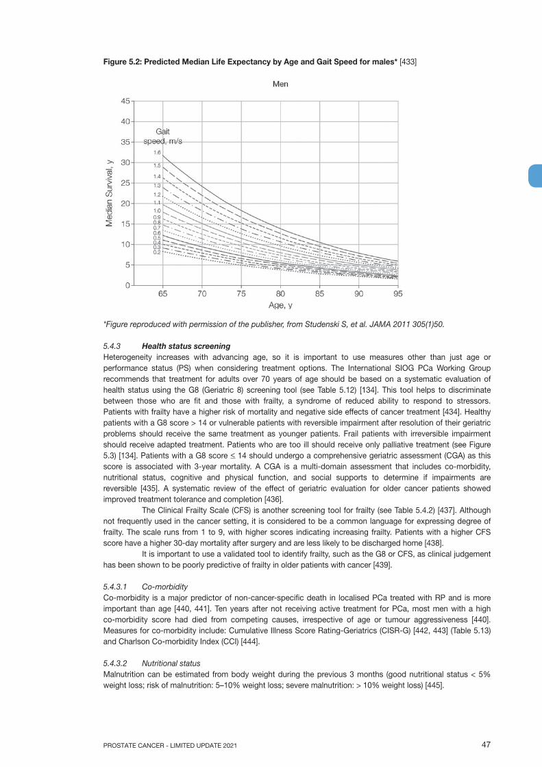
47PROSTATE CANCER - LIMITED UPDATE 2021
Figure 5.2: Predicted Median Life Expectancy by Age and Gait Speed for males* [433]
*Figure reproduced with permission of the publisher, from Studenski S, et al. JAMA 2011 305(1)50.
5.4.3 Health status screeningHeterogeneity increases with advancing age, so it is important to use measures other than just age or performance status (PS) when considering treatment options. The International SIOG PCa Working Group recommends that treatment for adults over 70 years of age should be based on a systematic evaluation of health status using the G8 (Geriatric 8) screening tool (see Table 5.12) [134]. This tool helps to discriminate between those who are fit and those with frailty, a syndrome of reduced ability to respond to stressors. Patients with frailty have a higher risk of mortality and negative side effects of cancer treatment [434]. Healthy patients with a G8 score > 14 or vulnerable patients with reversible impairment after resolution of their geriatric problems should receive the same treatment as younger patients. Frail patients with irreversible impairment should receive adapted treatment. Patients who are too ill should receive only palliative treatment (see Figure 5.3) [134]. Patients with a G8 score < 14 should undergo a comprehensive geriatric assessment (CGA) as this score is associated with 3-year mortality. A CGA is a multi-domain assessment that includes co-morbidity, nutritional status, cognitive and physical function, and social supports to determine if impairments are reversible [435]. A systematic review of the effect of geriatric evaluation for older cancer patients showed improved treatment tolerance and completion [436].
The Clinical Frailty Scale (CFS) is another screening tool for frailty (see Table 5.4.2) [437]. Although not frequently used in the cancer setting, it is considered to be a common language for expressing degree of frailty. The scale runs from 1 to 9, with higher scores indicating increasing frailty. Patients with a higher CFS score have a higher 30-day mortality after surgery and are less likely to be discharged home [438].
It is important to use a validated tool to identify frailty, such as the G8 or CFS, as clinical judgement has been shown to be poorly predictive of frailty in older patients with cancer [439].
5.4.3.1 Co-morbidityCo-morbidity is a major predictor of non-cancer-specific death in localised PCa treated with RP and is more important than age [440, 441]. Ten years after not receiving active treatment for PCa, most men with a high co-morbidity score had died from competing causes, irrespective of age or tumour aggressiveness [440]. Measures for co-morbidity include: Cumulative Illness Score Rating-Geriatrics (CISR-G) [442, 443] (Table 5.13) and Charlson Co-morbidity Index (CCI) [444].
5.4.3.2 Nutritional statusMalnutrition can be estimated from body weight during the previous 3 months (good nutritional status < 5% weight loss; risk of malnutrition: 5–10% weight loss; severe malnutrition: > 10% weight loss) [445].
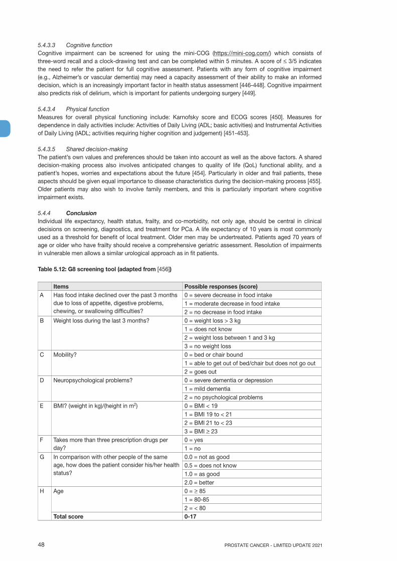
PROSTATE CANCER - LIMITED UPDATE 202148
5.4.3.3 Cognitive functionCognitive impairment can be screened for using the mini-COG (https://mini-cog.com/) which consists of three-word recall and a clock-drawing test and can be completed within 5 minutes. A score of < 3/5 indicates the need to refer the patient for full cognitive assessment. Patients with any form of cognitive impairment (e.g., Alzheimer’s or vascular dementia) may need a capacity assessment of their ability to make an informed decision, which is an increasingly important factor in health status assessment [446-448]. Cognitive impairment also predicts risk of delirium, which is important for patients undergoing surgery [449].
5.4.3.4 Physical functionMeasures for overall physical functioning include: Karnofsky score and ECOG scores [450]. Measures for dependence in daily activities include: Activities of Daily Living (ADL; basic activities) and Instrumental Activities of Daily Living (IADL; activities requiring higher cognition and judgement) [451-453].
5.4.3.5 Shared decision-makingThe patient’s own values and preferences should be taken into account as well as the above factors. A shared decision-making process also involves anticipated changes to quality of life (QoL) functional ability, and a patient’s hopes, worries and expectations about the future [454]. Particularly in older and frail patients, these aspects should be given equal importance to disease characteristics during the decision-making process [455]. Older patients may also wish to involve family members, and this is particularly important where cognitive impairment exists.
5.4.4 ConclusionIndividual life expectancy, health status, frailty, and co-morbidity, not only age, should be central in clinical decisions on screening, diagnostics, and treatment for PCa. A life expectancy of 10 years is most commonly used as a threshold for benefit of local treatment. Older men may be undertreated. Patients aged 70 years of age or older who have frailty should receive a comprehensive geriatric assessment. Resolution of impairments in vulnerable men allows a similar urological approach as in fit patients.
Table 5.12: G8 screening tool (adapted from [456])
Items Possible responses (score)A Has food intake declined over the past 3 months
due to loss of appetite, digestive problems, chewing, or swallowing difficulties?
0 = severe decrease in food intake1 = moderate decrease in food intake2 = no decrease in food intake
B Weight loss during the last 3 months? 0 = weight loss > 3 kg1 = does not know2 = weight loss between 1 and 3 kg3 = no weight loss
C Mobility? 0 = bed or chair bound1 = able to get out of bed/chair but does not go out2 = goes out
D Neuropsychological problems? 0 = severe dementia or depression1 = mild dementia2 = no psychological problems
E BMI? (weight in kg)/(height in m2) 0 = BMI < 191 = BMI 19 to < 212 = BMI 21 to < 233 = BMI > 23
F Takes more than three prescription drugs per day?
0 = yes1 = no
G In comparison with other people of the same age, how does the patient consider his/her health status?
0.0 = not as good0.5 = does not know1.0 = as good2.0 = better
H Age 0 = > 851 = 80-852 = < 80
Total score 0-17
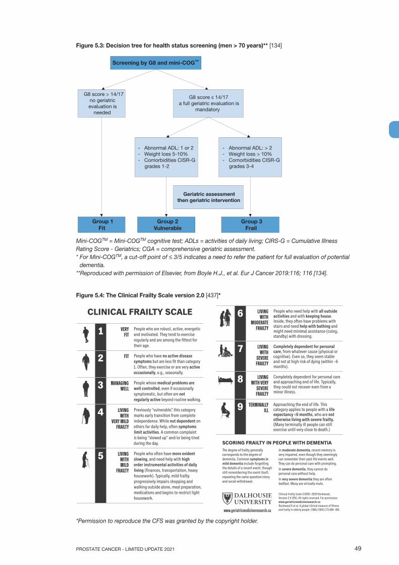
49PROSTATE CANCER - LIMITED UPDATE 2021
Figure 5.3: Decision tree for health status screening (men > 70 years)** [134]
Screening by G8 and mini-COGTM*
G8 score > 14/17 no geriatric evaluation is
needed
G8 score ≤ 14/17 a full geriatric evaluation is
mandatory
- Abnormal ADL: > 2- Weight loss > 10% - Comorbidities CISR-G grades 3-4
- Abnormal ADL: 1 or 2 - Weight loss 5-10% - Comorbidities CISR-G grades 1-2
Geriatric assessment then geriatric intervention
Group 3 Frail
Group 2 Vulnerable
Group 1 Fit
Mini-COGTM = Mini-COGTM cognitive test; ADLs = activities of daily living; CIRS-G = Cumulative Illness Rating Score - Geriatrics; CGA = comprehensive geriatric assessment. * For Mini-COGTM, a cut-off point of < 3/5 indicates a need to refer the patient for full evaluation of potential
dementia.**Reproduced with permission of Elsevier, from Boyle H.J., et al. Eur J Cancer 2019:116; 116 [134].
Figure 5.4: The Clinical Frailty Scale version 2.0 [437]*
1 VERY FIT
People who are robust, active, energetic and motivated. They tend to exercise regularly and are among the fittest for their age.
2 FIT People who have no active disease symptoms but are less fit than category 1. Often, they exercise or are very active occasionally, e.g., seasonally.
3 MANAGING WELL
People whose medical problems are well controlled, even if occasionally symptomatic, but often are not regularly active beyond routine walking.
4 LIVING WITH
VERY MILD FRAILTY
Previously “vulnerable,” this category marks early transition from complete independence. While not dependent on others for daily help, often symptoms limit activities. A common complaint is being “slowed up” and/or being tired during the day.
5 LIVING WITH MILD
FRAILTY
People who often have more evident slowing, and need help with high order instrumental activities of daily living (finances, transportation, heavy housework). Typically, mild frailty progressively impairs shopping and walking outside alone, meal preparation, medications and begins to restrict light housework.
CLINICAL FRAILTY SCALE
The degree of frailty generally corresponds to the degree of dementia. Common symptoms in mild dementia include forgetting the details of a recent event, though still remembering the event itself, repeating the same question/story and social withdrawal.
Clinical Frailty Scale ©2005–2020 Rockwood, Version 2.0 (EN). All rights reserved. For permission: www.geriatricmedicineresearch.ca Rockwood K et al. A global clinical measure of fitness and frailty in elderly people. CMAJ 2005;173:489–495.
SCORING FRAILTY IN PEOPLE WITH DEMENTIA
6 LIVING WITH
MODERATE FRAILTY
People who need help with all outside activities and with keeping house. Inside, they often have problems with stairs and need help with bathing and might need minimal assistance (cuing, standby) with dressing.
7 LIVING WITH
SEVERE FRAILTY
Completely dependent for personal care, from whatever cause (physical or cognitive). Even so, they seem stable and not at high risk of dying (within ~6 months).
8 LIVING WITH VERY
SEVERE FRAILTY
Completely dependent for personal care and approaching end of life. Typically, they could not recover even from a minor illness.
9 TERMINALLY ILL
Approaching the end of life. This category applies to people with a life expectancy <6 months, who are not otherwise living with severe frailty. (Many terminally ill people can still exercise until very close to death.)
In moderate dementia, recent memory is very impaired, even though they seemingly can remember their past life events well. They can do personal care with prompting.In severe dementia, they cannot do personal care without help.In very severe dementia they are often bedfast. Many are virtually mute.
www.geriatricmedicineresearch.ca
*Permission to reproduce the CFS was granted by the copyright holder.

PROSTATE CANCER - LIMITED UPDATE 202150
Table 5.13: Cumulative Illness Score Rating-Geriatrics (CISR-G)
1 Cardiac (heart only)2 Hypertension (rating is based on severity; affected systems are rated separately)3 Vascular (blood, blood vessels and cells, marrow, spleen, lymphatics)4 Respiratory (lungs, bronchi, trachea below the larynx)5 ENT (eye, ear, nose, throat, larynx)6 Upper GI (esophagus, stomach, duodenum. Biliar and parcreatic trees; do not include diabetes)7 Lower GI (intestines, hernias)8 Hepatic (liver only)9 Renal (kidneys only)10 Other GU (ureters, bladder, urethra, prostate, genitals)11 Musculo-Skeletal-Integumentary (muscles, bone, skin)12 Neurological (brain, spinal cord, nerves; do not include dementia)13 Endocrine-Metabolic (includes diabetes, diffuse infections, infections, toxicity)14 Psychiatric/Behavioural (includes dementia, depression, anxiety, agitation, psychosis)
All body systems are scores on a 0 - 4 scale.- 0: No problem affecting that system.- 1: Current mild problem or past significant problem.- 2: Moderate disability or morbidity and/or requires first line therapy.- 3: Severe problem and/or constant and significant disability and/or hard to control chronic
problems.- 4: Extremely severe problem and/or immediate treatment required and/or organ failure and/or
severe functional impairment.Total score 0-56
5.4.5 Guidelines for evaluating health status and life expectancy
Recommendations Strength ratingUse individual life expectancy, health status, and co-morbidity in PCa management. StrongUse the Geriatric-8, mini-COG and Clinical Frailty Scale tools for health status screening. StrongPerform a full specialist geriatric evaluation in patients with a G8 score < 14. StrongConsider standard treatment in vulnerable patients with reversible impairments (after resolution of geriatric problems) similar to fit patients, if life expectancy is > 10 years.
Weak
Offer adapted treatment to patients with irreversible impairment. WeakOffer symptom-directed therapy alone to frail patients. Strong
6. TREATMENTThis chapter reviews the available treatment modalities, followed by separate sections addressing treatment for the various disease stages.
6.1 Treatment modalities 6.1.1 Deferred treatment (active surveillance/watchful waiting)In localised disease a life expectancy of at least 10 years is considered mandatory for any benefit from local treatment. Data are available on patients who did not undergo local treatment with up to 25 years of follow-up, with endpoints of OS and CSS. Several series have shown a consistent CSS rate of 82–87% at 10 years [457-462], and 80–95% for T1/T2 and ISUP grade < 2 PCas [463]. In three studies with data beyond 15 years, the DSS was 80%, 79% and 58% [459, 461, 462], and two reported 20-year CSS rates of 57% and 32%, respectively [459, 461]. In addition, many patients classified as ISUP grade 1 would now be classified as ISUP grade 2–3 based on the 2005 Gleason classification, suggesting that the above-mentioned results should be considered as minimal. Patients with well-, moderately- and poorly-differentiated tumours had 10-year CSS rates of 91%, 90% and 74%, respectively, correlating with data from the pooled analysis [463]. Observation was most effective in men aged 65–75 years with low-risk PCa [464].
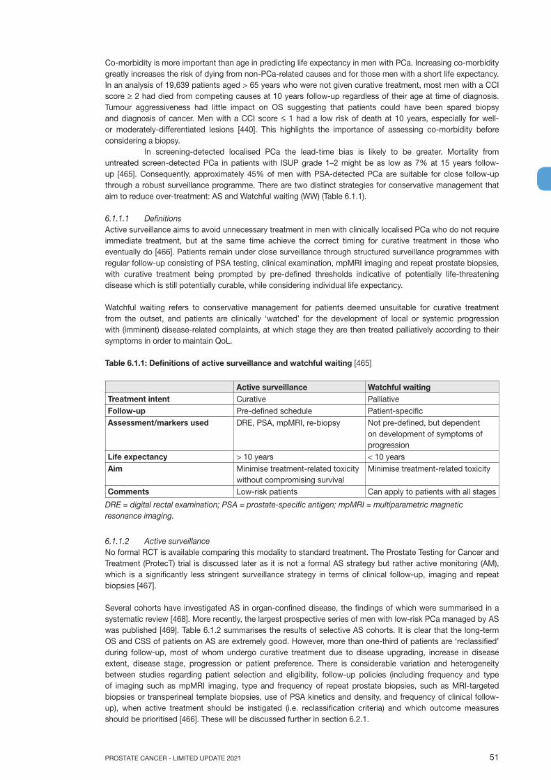
51PROSTATE CANCER - LIMITED UPDATE 2021
Co-morbidity is more important than age in predicting life expectancy in men with PCa. Increasing co-morbidity greatly increases the risk of dying from non-PCa-related causes and for those men with a short life expectancy. In an analysis of 19,639 patients aged > 65 years who were not given curative treatment, most men with a CCI score > 2 had died from competing causes at 10 years follow-up regardless of their age at time of diagnosis. Tumour aggressiveness had little impact on OS suggesting that patients could have been spared biopsy and diagnosis of cancer. Men with a CCI score < 1 had a low risk of death at 10 years, especially for well- or moderately-differentiated lesions [440]. This highlights the importance of assessing co-morbidity before considering a biopsy.
In screening-detected localised PCa the lead-time bias is likely to be greater. Mortality from untreated screen-detected PCa in patients with ISUP grade 1–2 might be as low as 7% at 15 years follow-up [465]. Consequently, approximately 45% of men with PSA-detected PCa are suitable for close follow-up through a robust surveillance programme. There are two distinct strategies for conservative management that aim to reduce over-treatment: AS and Watchful waiting (WW) (Table 6.1.1).
6.1.1.1 DefinitionsActive surveillance aims to avoid unnecessary treatment in men with clinically localised PCa who do not require immediate treatment, but at the same time achieve the correct timing for curative treatment in those who eventually do [466]. Patients remain under close surveillance through structured surveillance programmes with regular follow-up consisting of PSA testing, clinical examination, mpMRI imaging and repeat prostate biopsies, with curative treatment being prompted by pre-defined thresholds indicative of potentially life-threatening disease which is still potentially curable, while considering individual life expectancy.
Watchful waiting refers to conservative management for patients deemed unsuitable for curative treatment from the outset, and patients are clinically ‘watched’ for the development of local or systemic progression with (imminent) disease-related complaints, at which stage they are then treated palliatively according to their symptoms in order to maintain QoL.
Table 6.1.1: Definitions of active surveillance and watchful waiting [465]
Active surveillance Watchful waitingTreatment intent Curative PalliativeFollow-up Pre-defined schedule Patient-specificAssessment/markers used DRE, PSA, mpMRI, re-biopsy Not pre-defined, but dependent
on development of symptoms of progression
Life expectancy > 10 years < 10 yearsAim Minimise treatment-related toxicity
without compromising survivalMinimise treatment-related toxicity
Comments Low-risk patients Can apply to patients with all stages
DRE = digital rectal examination; PSA = prostate-specific antigen; mpMRI = multiparametric magnetic resonance imaging.
6.1.1.2 Active surveillanceNo formal RCT is available comparing this modality to standard treatment. The Prostate Testing for Cancer and Treatment (ProtecT) trial is discussed later as it is not a formal AS strategy but rather active monitoring (AM), which is a significantly less stringent surveillance strategy in terms of clinical follow-up, imaging and repeat biopsies [467].
Several cohorts have investigated AS in organ-confined disease, the findings of which were summarised in a systematic review [468]. More recently, the largest prospective series of men with low-risk PCa managed by AS was published [469]. Table 6.1.2 summarises the results of selective AS cohorts. It is clear that the long-term OS and CSS of patients on AS are extremely good. However, more than one-third of patients are ‘reclassified’ during follow-up, most of whom undergo curative treatment due to disease upgrading, increase in disease extent, disease stage, progression or patient preference. There is considerable variation and heterogeneity between studies regarding patient selection and eligibility, follow-up policies (including frequency and type of imaging such as mpMRI imaging, type and frequency of repeat prostate biopsies, such as MRI-targeted biopsies or transperineal template biopsies, use of PSA kinetics and density, and frequency of clinical follow-up), when active treatment should be instigated (i.e. reclassification criteria) and which outcome measures should be prioritised [466]. These will be discussed further in section 6.2.1.

PROSTATE CANCER - LIMITED UPDATE 202152
Table 6.1.2: Active surveillance in screening-detected prostate cancer
Studies N Median FU (mo) pT3 in RP patients* 10-year OS (%)
10-year CSS (%)
Van As, et al. 2008 [470] 326 22 8/18 (44%) 98 100Carter, et al. 2007 [471] 407 41 10/49 (20%) 98 100Adamy, et al. 2011 [472] 533-1,000 48 4/24 (17%) 90 99Soloway, et al. 2010 [473] 99 45 0/2 100 100Roemeling, et al. 2007 [474] 278 41 - 89 100Khatami, et al. 2007 [475] 270 63 - n.r. 100Klotz, et al. 2015 [476] 993 77 - 85 98.1Tosoian, et al. 2015 [469] 1,818 60 - 93 99.9Total 4,724-5,191 46.5 - 93 99
* Patients receiving active therapy following initial active surveillance.CSS = cancer-specific survival; FU = follow-up; mo = months; n = number of patients; n.r. = not reported; OS = overall survival; RP = radical prostatectomy.
6.1.1.3 Watchful Waiting6.1.1.3.1 Outcome of watchful waiting compared with active treatmentThe SPCG-4 study was a RCT from the pre-PSA era, randomising patients to either WW or RP (Table 6.1.3) [477]. The study found RP to provide superior CSS, OS and progression-free survival (PFS) compared to WW at a median follow-up of 23.6 years (range 3 weeks–28 years). The PIVOT trial was a RCT conducted in the early PSA era and made a similar comparison between RP vs. observation in 731 men (50% with non-palpable disease) [478] but in contrast to the SPCG-4, it found little to no benefit of RP (cumulative incidence of all-cause death, RP vs. observation: 68% vs. 73%; RR: 0.92, 95% CI: 0.84–1.01) within a median follow-up period of 18.6 years (interquartile range, 16.6 to 20 years). Exploratory subgroup analysis showed that the borderline benefit from RP was most marked for intermediate-risk disease (RR: 0.84, 95% CI: 0.73–0.98) but there was no benefit in patients with low- or high-risk disease. Overall, no adverse effects on HRQoL and psychological well-being was apparent in the first years [479]. However, one of the criticisms of the PIVOT trial is the relatively high overall mortality rate in the WW group (73% at a median follow-up period of 18.6 years (interquartile range, 16.6 to 20.0 years) compared with more contemporary series.
Table 6.1.3: Outcome of SPCG-4 at a median follow-up of 23.6 years [477]
RP (n = 348) (%) Watchful waiting (n = 348) (%)
Relative risk (95% CI)
p-value
Disease-specific mortality 19.6 31.3 0.55 (0.41–0.74) < 0.001Overall mortality 71.9 83.8 0.74 (0.62–0.87) < 0.001Metastatic progression 26.6 43.3 0.54 (0.42–0.70) < 0.001
CI = confidence interval; RP = radical prostatectomy.
6.1.1.4 The ProtecT studyThe ProtecT trial randomised 1,643 patients into three arms: active treatment with either RP or EBRT, and AM [467]. In this AM schedule patients with a PSA rise of more than 50% in 12 months underwent a repeat biopsy, but none had systematic repeat biopsies. Fifty-six percent of patients had low-risk disease, with 90% having a PSA < 10 ng/mL, 77% ISUP grade 1 (20% ISUP grade 2–3), and 76% T1c, while the other patients had mainly intermediate-risk disease. After 10 years of follow-up, CSS was the same between those actively treated and those on AM (99% and 98.8%, respectively), as was the OS. Only metastatic progression differed (6% in the AM group as compared to 2.6% in the treated group). The key finding was that AM was as effective as active treatment at 10 years, at a cost of increased progression and double the metastatic risk. Metastases remained rare (6%), but more frequent than seen with AS protocols; probably driven by differences in intensity of monitoring and patient selection. It is important to note that the AM arm in ProtecT represented an intermediate approach between contemporary AS protocols and WW in terms of a monitoring strategy based almost entirely on PSA measurements alone; there was no use of mpMRI scan, either at recruitment or during the monitoring period, nor was there any protocol-mandated repeat prostate biopsies at regular intervals. In addition, approximately 40% of randomised patients had intermediate-risk disease. Nevertheless, the ProtecT study has reinforced the role of deferred active treatment (i.e. either AS or some form of initial AM) as a feasible alternative to active curative interventions in patients with low-grade and

53PROSTATE CANCER - LIMITED UPDATE 2021
low-stage disease. Beyond 10 years, no data is available, as yet, although AS is likely to give more reassurance especially in younger men, based on more accurate risk stratification at recruitment and more stringent criteria regarding follow-up, imaging, repeat biopsy and reclassification. Individual life expectancy must be evaluated before considering any active treatment in low-risk patients and in those with up to 10 years’ individual life expectancy.
6.1.2 Radical prostatectomy6.1.2.1 IntroductionThe goal of RP by any approach is the eradication of cancer while, whenever possible, preserving pelvic organ function [480]. The procedure involves removing the entire prostate with its capsule intact and SVs, followed by vesico-urethral anastomosis. Surgical approaches have expanded from perineal and retropubic open approaches to laparoscopic and robotic-assisted techniques; anastomoses have evolved from Vest approximation sutures to continuous suture watertight anastomoses under direct vision and mapping of the anatomy of the dorsal venous complex (DVC) and cavernous nerves has led to excellent visualisation and potential for preservation of erectile function [481]. The main results from multi-centre RCTs involving RP are summarised in Table 6.1.4.
Table 6.1.4: Oncological results of radical prostatectomy in organ-confined disease in RCTs
Study Acronym Population Treatment period
Median FU (mo)
Risk category CSS (%)
Bill-Axelson, et al. 2018 [477]
SPCG-4 Pre-PSA era 1989-1999 283 Low risk and Intermediate risk
80.4 (at 23 yr.)
Wilt, et al. 2017 [482]
PIVOT Early years of PSA testing
1994-2002 152 Low risk Intermediate risk
95.9 91.5 (at 19.5 yr.)
Hamdy, et al. 2016 [467]
ProtecT Screened population
1999-2009 120 Mainly low- and intermediate risk
99 (at 10 yr.)
CSS = cancer-specific survival; FU = follow-up; mo = months; PSA = prostate-specific antigen; yr. = year.
6.1.2.2 Pre-operative preparation6.1.2.2.1 Pre-operative patient educationAs before any surgery appropriate education and patient consent is mandatory prior to RP. Peri-operative education has been shown to improve long-term patient satisfaction following RP [483]. Augmentation of standard verbal and written educational materials such as use of interactive multimedia tools [484, 485] and pre-operative patient-specific 3D printed prostate models has been shown to improve patient understanding and satisfaction and should be considered to optimise patient-centred care [486].
Pre-operative pelvic floor exercisesAlthough many patients who have undergone RP will experience a return to urinary continence [487], temporary urinary incontinence is common early after surgery, reducing QoL. Pre-operative pelvic floor exercises (PFE) with, or without, biofeedback have been used with the aim of reducing this early post-operative incontinence. A systematic review and meta-analysis of the effect of pre-RP PFE on post-operative urinary incontinence showed a significant improvement in incontinence rates at 3 months post-operatively with an OR of 0.64 (p = 0.005), but not at 1 month or 6 months [488]. Pre-operative PFE may therefore provide some benefit, however the analysis was hampered by the variety of PFE regimens and a lack of consensus on the definition of incontinence.
Prophylactic antibioticsProphylactic antibiotics should be used; however no high-level evidence is available to recommend specific prophylactic antibiotics prior to RP (See EAU Urological Infections Guidelines). In addition, as the susceptibility of bacterial pathogens and antibiotic availability varies worldwide, any use of prophylactic antibiotics should adhere to local guidelines.
Neoadjuvant androgen deprivation therapySeveral RCTs have analysed the impact of neoadjuvant ADT before RP, most of these using a 3-month period. The main findings were summarised in a Cochrane review [489]. Neoadjuvant ADT is associated with a decreased rate of pT3 (downstaging), decreased positive margins, and a lower incidence of positive LNs. These benefits are greater with increased treatment duration (up to 8 months). However, since neither the PSA

PROSTATE CANCER - LIMITED UPDATE 202154
relapse-free survival nor CSS were shown to improve, neoadjuvant ADT should not be considered as standard clinical practice. One recent RCT compared neoadjuvant luteinising hormone-releasing hormone (LHRH) alone vs. LHRH plus abiraterone plus prednisone prior to RP in 65 localised high-risk PCa patients [490]. Patients in the combination arm were found to have both significantly lower tumour volume and significantly lower BCR at > 4 years follow-up (p = 0.0014). Further supportive evidence is required before recommending combination neoadjuvant therapy including abiraterone prior to RP.
6.1.2.3 Surgical techniquesProstatectomy can be performed by open-, laparoscopic- or robot-assisted (RARP) approaches. The initial open technique of RP described by Young in 1904 was via the perineum [481] but suffered from a lack of access to pelvic LNs. If lymphadenectomy is required during perineal RP it must be done via a separate open retropubic (RRP) or laparoscopic approach. The open retropubic approach was popularised by Walsh in 1982 following his anatomical description of the DVC, enabling its early control and of the cavernous nerves, permitting a bilateral nerve-sparing procedure [491]. This led to the demise in popularity of perineal RP and eventually to the first laparoscopic RP reported in 1997 using retropubic principles, but performed transperitoneally [492]. The initial 9 cases averaged 9.4 hours, an indication of the significant technical and ergonomic difficulties of the technique. Most recently, RARP was introduced using the da Vinci Surgical System® by Binder in 2002 [493]. This technology combined the minimally-invasive advantages of laparoscopic RP with improved surgeon ergonomics and greater technical ease of suture reconstruction of the vesico-urethral anastomosis and has now become the preferred minimally-invasive approach, when available.
In a randomised phase III trial, RARP was shown to have reduced admission times and blood loss, but not earlier (12 weeks) functional or oncological outcomes compared to open RP [494]. An updated analysis with follow-up at 24 months did not reveal any significant differences in functional outcomes between the approaches [495]. Increased surgical experience has lowered the complication rates of RP and improved cancer cure [461-464]. Lower rates of positive surgical margins for high-volume surgeons suggest that experience and careful attention to surgical details, can improve cancer control with RP [496-498]. There is a lack of studies comparing the different surgical modalities for these longer-term outcomes [460, 479, 482, 499]. A systematic review and meta-analysis of non-RCTs demonstrated that RARP had lower peri-operative morbidity and a reduced risk of positive surgical margins compared with laparoscopic prostatectomy (LRP), although there was considerable methodological uncertainty [500]. There was no evidence of differences in urinary incontinence at 12 months and there was insufficient evidence to draw conclusions based on differences in cancer-related, patient-driven or erectile dysfunction (ED) outcomes. Another systematic review and meta-analysis included two small RCTs comparing RARP vs. LRP [501]. The results suggested higher rates of return of erectile function (RR: 1.51, 95% CI: 1.19–1.92) and return to continence function (RR: 1.14, 95% CI: 1.04–1.24) in the RARP group. However, a recent Cochrane review comparing either RARP or LRP vs. open RP included two RCTs and found no significant differences between the comparisons for oncological-, urinary- and sexual function outcomes, although RARP and LRP both resulted in statistically significant improvements in duration of hospital stay and blood transfusion rates over open RP [502]. Therefore, no surgical approach can be recommended over another.
Outcome after prostatectomy has been shown to be dependent on both surgeon [503] as well as hospital volume [504]. Although various volume criteria have been set worldwide, the level of evidence is insufficient to pinpoint a specific lower volume limit.
6.1.2.3.1 Robotic anterior versus Retzius-sparing dissectionRobot-assisted RP has typically been performed via the anterior approach, first dropping the bladder to expose the space of Retzius. However, the posterior approach (Retzius-sparing [RS-RARP]) has been used to minimise injury to support structures surrounding the prostate.
Galfano et al., first described RS-RARP in 2010 [505]. This approach commences dissection posteriorly at the pouch of Douglas, first dissecting the SVs and progressing caudally behind the prostate. All of the anterior support structures are avoided, giving rise to the hypothetical mechanism for improved early post-operative continence. Retzius-sparing-RARP thus offers the same potential advantage as the open perineal approach, but without disturbance of the perineal musculature.
Retzius-sparing-RARP has been recently investigated in RCTs leading to four systematic reviews and meta-analyses [506-508] including a 2020 Cochrane systematic review [509] and a large propensity score matched analysis [510]. The Cochrane review used the most rigorous methodology and analysed 5 RCTs with 502 patients. It found with moderate certainty that RS-RARP improved continence at 1 week post catheter removal compared to standard RARP (RR: 1.74). Continence may also be improved at 3 months
55PROSTATE CANCER - LIMITED UPDATE 2021
post-operatively (RR: 1.33), but this was based on low-certainty data. Continence outcomes appeared to equalise by 12 months (RR: 1.01). These findings matched those of the other systematic reviews. However, a significant concern was that RS-RARP appears to increase the risk of positive margins (RR: 1.95) but this was also low-certainty evidence. A single-surgeon propensity score matched analysis of 1,863 patients reached the same conclusion as the systematic reviews regarding earlier return to continence but did not show data on margin status [510].
Based on these data, recommendations cannot be made for one technique over another. However, the trade-offs between the risks of a positive margin vs. earlier continence recovery should be discussed with prospective patients. Furthermore, no high level evidence is available on high-risk disease with some concerns that RS-RARP may confer an increased positive margin rate based on pT3 results. In addition, RS-RARP may be more technically challenging in various scenarios such as anterior tumours, post-TURP, a grossly enlarged gland, or a bulky median lobe [511].
6.1.2.3.2 Pelvic lymph node dissectionA recent systematic review demonstrated that performing PLND during RP failed to improve oncological outcomes, including survival [512]. Moreover, a RCT failed to show a benefit of an extended approach vs. a limited PLND on early oncologic outcomes [513]. However, it is generally accepted that eLND provides important information for staging and prognosis which cannot be matched by any other currently available procedure [512].
Extended LND includes removal of the nodes overlying the external iliac artery and vein, the nodes within the obturator fossa located cranially and caudally to the obturator nerve, and the nodes medial and lateral to the internal iliac artery. With this template, 94% of patients are correctly staged [514].
The individual risk of patients harbouring positive LNs can be estimated based on validated nomograms. The Briganti nomogram [381, 382], the Roach formula [515] or the Partin and MSKCC nomograms [516] have shown similar diagnostic accuracy in predicting LN invasion. These nomograms have all been developed in the pre-MRI setting based on systematic random biopsy. A risk of nodal metastases over 5% can be used to identify candidates for nodal sampling by eLND during RP [517-519].
An updated nomogram has been externally validated in men diagnosed based on mpMRI followed by MRI-targeted biopsy [382]. Based on this nomogram patients can be spared an ePLND if their risk of nodal involvement is less than 7%; which would result in missing only 1.5% of patients with nodal invasion [382, 520]. This 7% cutoff is comparable to the 5% cutoff of the Briganti nomogram in patients diagnosed by systematic random biopsy alone. Therefore, this novel nomogram and a 7% threshold should be used after MRI-targeted biopsy to identify candidates for eLND [521].
6.1.2.3.3 Sentinel node biopsy analysisThe rationale for a sentinel node biopsy (SNB) is based on the concept that a sentinel node is the first to be involved by migrating tumour cells. Therefore, when this node is negative it is possible to avoid an ePLND. There is heterogeneity and variation in techniques in relation to SNB (e.g. the optimal tracer) but a multidisciplinary collaborative endeavour attempted to standardise definitions, thresholds and strategies in relation to techniques of SNB using consensus methods [522].
Intraprostatic injections of indocyanine green (ICG) have been used to visualise prostate-related LNs during lymphadenectomy. In a randomised comparison, Harke et al., found more cancer containing LNs in men that underwent a LN dissection guided by ICG but no difference in BCR at 22.9 month follow-up [523]. A systematic review showed a sensitivity of 95.2% and NPV of 98.0% for SNB in detecting men with metastases at eLND [524]. However, there is still insufficient high-quality evidence supporting oncological effectiveness of SNB for nodal staging. Sentinel node biopsy is therefore still considered as an experimental nodal staging procedure.
6.1.2.3.4 Prostatic anterior fat pad dissection and histologic analysisSeveral multi-centre and large single-centre series have shown the presence of lymphoid tissue within the fat pad anterior to the endopelvic fascia; the prostatic anterior fat pad (PAFP) [525-531]. This lymphoid tissue is present in 5.5–10.6% of cases and contains metastatic PCa in up to 1.3% of intermediate- and high-risk patients.
When positive, the PAFP is often the only site of LN metastasis. The PAFP is therefore a rare but recognised route of spread of disease. Unlike PLND, there is no morbidity associated with removal of the PAFP. The PAFP is always removed at RP for exposure of the endopelvic fascia and should be sent for histologic analysis as per all removed tissue.
PROSTATE CANCER - LIMITED UPDATE 202156
6.1.2.3.5 Management of the dorsal venous complexSince the description of the anatomical open RP by Walsh and Donker in the 1980s, various methods of controlling bleeding from the DVC have been proposed to optimise visualisation [491]. In the open setting, blood loss and transfusion rates have been found to be significantly reduced when ligating the DVC prior to transection [532]. However, concerns have been raised regarding the effect of prior DVC ligation on apical margin positivity and continence recovery due to the proximity of the DVC to both the prostatic apex and the urethral sphincter muscle fibres. In the robotic-assisted laparoscopic technique, due to the increased pressure of pneumoperitoneum, whether prior DVC ligation was used or not, blood loss was not found to be significantly different in one study [533]. In another study, mean blood loss was significantly less with prior DVC ligation (184 vs. 176 mL, p = 0.033), however it is debatable whether this was clinically significant [534]. The positive apical margin rate was not different, however, the latter study showed earlier return to full continence at 5 months post-operatively in the no prior DVC ligation group (61% vs. 40%, p < 0.01).
Ligation of the DVC can be performed with standard suture or using a vascular stapler. One study found significantly reduced blood loss (494 mL vs. 288 mL) and improved apical margin status (13% vs. 2%) when using the stapler [535].
Given the relatively small differences in outcomes, the surgeon’s choice to ligate prior to transection or not, or whether to use sutures or a stapler, will depend on their familiarity with the technique and the equipment available.
6.1.2.3.6 Nerve-sparing surgery During prostatectomy, preservation of the neurovascular bundles with parasympathetic nerve branches of the pelvic plexus may spare erectile function [536, 537].
Although age and pre-operative function may remain the most important predictors for post-operative erectile function, nerve-sparing has also been associated with improved continence outcomes and may therefore still be relevant for men with poor erectile function [538, 539]. The association with continence may be mainly due to the dissection technique used during nerve-sparing surgery, and not due to the preservation of the nerve bundles themselves [538].
Extra-, inter-, and intra-fascial dissection planes can be planned, with those closer to the prostate and performed bilaterally associated with superior (early) functional outcomes [540-543]. Furthermore, many different techniques are propagated such as retrograde approach after anterior release (vs. antegrade), and athermal and traction-free handling of bundles [544-546]. Nerve-sparing does not compromise cancer control if patients are carefully selected depending on tumour location, size and grade [547-549].
6.1.2.3.7 Lymph-node-positive patients during radical prostatectomyAlthough no RCTs are available, data from prospective cohort studies comparing survival of pN+ patients (as defined following pathological examination after RP) support that RP may have a survival benefit over abandonment of RP in node-positive cases [550]. As a consequence there is no role for performing frozen section of suspicious LNs.
6.1.2.3.8 Removal of seminal vesiclesThe more aggressive forms of PCa may spread directly into the SVs. For oncological clearance, the SVs have traditionally been removed intact with the prostate specimen [551]. However, in some patients the tips of the SVs can be challenging to dissect free. Furthermore, the cavernous nerves run past the SV tips such that indiscriminate dissection of the SV tips could potentially lead to ED [552]. However, a RCT comparing nerve-sparing RP with and without a SV-sparing approach found no difference in margin status, PSA recurrence, continence or erectile function outcomes. Another study of 71 consecutive RPs showed no cancer in any of the distal 1 cm of SVs, even in 12 patients with SV invasion [553]. Whilst complete SV removal should be the default, preservation of the SV tips may be considered in cases of low risk of involvement.
6.1.2.3.9 Techniques of vesico-urethral anastomosisFollowing prostate removal, the bladder neck is anastomosed to the membranous urethra. The objective is to create a precisely aligned, watertight, tension-free, and stricture-free anastomosis that preserves the integrity of the intrinsic sphincter mechanism. Several methods have been described, based on the direct or indirect approach, the type of suture (i.e. barbed vs. non-barbed/monofilament), and variation in suturing technique (e.g., continuous vs. interrupted, or single-needle vs. double-needle running suture). The direct vesico-urethral anastomosis, which involves the construction of a primary end-to-end inter-mucosal anastomosis of the bladder neck to the membranous urethra by using 6 interrupted sutures placed circumferentially, has become the standard method of reconstruction for open RP [554].

57PROSTATE CANCER - LIMITED UPDATE 2021
The development of laparoscopic- and robotic-assisted techniques to perform RP have facilitated the introduction of new suturing techniques for the anastomosis. A systematic review and meta-analysis [555] compared unidirectional barbed suture vs. conventional non-barbed suture for vesico-urethral anastomosis during robotic-assisted laparoscopic prostatectomy (RALP). The review included 3 RCTs and found significantly reduced anastomosis time, operative time and posterior reconstruction time in favour of the unidirectional barbed suture technique, but there were no differences in post-operative leak rate, length of catheterisation and continence rate. However, no definitive conclusions could be drawn due to the relatively low quality of the data. In regard to suturing technique, a systematic review and meta-analysis compared continuous vs. interrupted suturing for vesico-urethral anastomosis during RP [556]. The study included only one RCT with 60 patients [557]. Although the review found slight advantages for continuous suturing over interrupted suturing in terms of catheterisation time, anastomosis time and rate of extravasation, the overall quality of evidence was low and no clear recommendations were possible. A recent RCT [558] compared the technique of suturing using a single absorbable running suture vs. a double-needle single-knot running suture (i.e. Van Velthoven technique) in laparoscopic RP [559]. The study found slightly reduced anastomosis time with the single running suture technique, but anastomotic leak, stricture, and continence rates were similar.
Overall, although there are a variety of approaches, methods and techniques for performing the vesico-urethral anastomosis, no clear recommendations are possible due to the lack of high-certainty evidence. In practice, the chosen method should be based on surgeon experience and individual preference [554-565].
6.1.2.3.10 Bladder neck managementBladder neck mucosal eversionSome surgeons perform mucosal eversion of the bladder neck as its own step in open RP with the aim of securing a mucosa-to-mucosa vesico-urethral anastomosis and avoiding anastomotic stricture. Whilst bringing bladder and urethral mucosa together by the everted bladder mucosa covering the bladder muscle layer, this step may actually delay healing of the muscle layers. An alternative is to simply ensure bladder mucosa is included in the full thickness anastomotic sutures. A non-randomised study of 211 patients with and without bladder neck mucosal eversion showed no significant difference in anastomotic stricture rate [566]. The strongest predictor of anastomotic stricture in RP is current cigarette smoking [567].
Bladder neck preservationWhilst the majority of urinary continence is maintained by the external urethral sphincter at the membranous urethra (see below), a minor component is contributed by the internal lissosphincter at the bladder neck [568]. Preservation of the bladder neck has therefore been proposed to improve continence recovery post-RP. A RCT assessing continence recovery at 12 months and 4 years showed improved objective and subjective urinary continence in both the short- and long term without any adverse effect on oncological outcome [569]. These findings were confirmed by a systematic review [570]. However, concern remains regarding margin status for cancers located at the prostate base.
A systematic review addressing site-specific margin status found a mean base-specific positive margin rate of 4.9% with bladder neck preservation vs. only 1.9% without [568]. This study was inconclusive, but it would be sensible to exercise caution when considering bladder neck preservation if significant cancer is known to be at the prostate base. Bladder neck preservation should be performed routinely when the cancer is distant from the base. However, bladder neck preservation cannot be performed in the presence of a large median lobe or a previous TURP.
6.1.2.3.11 Urethral length preservationThe membranous urethra sits immediately distal to the prostatic apex and is chiefly responsible, along with its surrounding pelvic floor support structures, for urinary continence. It consists of the external rhabdosphincter which surrounds an inner layer of smooth muscle. Using pre-operative MRI, the length of membranous urethra has been shown to vary widely. A systematic review and meta-analysis has found that every extra millimetre of membranous urethral length seen on MRI pre-operatively improves early return to continence post-RP [571]. Therefore, it is likely that preservation of as much urethral length as possible during RP will maximise the chance of early return to continence. It may also be useful to measure urethral length pre-operatively to facilitate councelling of patients on their relative likelihood of early post-operative continence.
6.1.2.3.12 Cystography prior to catheter removalCystography may be used prior to catheter removal to check for a substantial anastomotic leak. If such a leak is found, catheter removal may then be deferred to allow further healing and sealing of the anastomosis. However, small comparative studies suggest that a cystogram to assess anastomotic leakage is not indicated as standard of care before catheter removal 8 to 10 days after surgery [572]. If a cystogram is used, men with
PROSTATE CANCER - LIMITED UPDATE 202158
LUTS, large prostates, previous TURP or bladder neck reconstruction, may benefit as these factors have been associated with leakage [573, 574]. Contrast-enhanced transrectal US is an alternative [575].
6.1.2.3.13 Urinary catheterA urinary catheter is routinely placed during RP to enable bladder rest and drainage of urine while the vesicourethral anastomosis heals. Compared to a traditional catheter duration of around 1 week, some centres remove the transurethral catheter early (post-operative day 2–3), usually after thorough anastomosis with posterior reconstruction or in patients selected peri-operatively on the basis of anastomosis quality [576-579]. No higher complication rates were found. Although shorter catheterisation has been associated with more favourable short-term functional outcomes, no differences in long-term function were found [580]. One RCT has shown no difference in rate of UTI following indwelling catheter (IDC) removal whether prophylactic ciprofloxacin was given prior to IDC removal or not, suggesting antibiotics should not be given at catheter removal [581].
As an alternative to transurethral catheterisation, suprapubic catheter insertion during RP has been suggested. Some reports suggest less bother regarding post-operative hygiene and pain [582-586], while others did not find any differences [587, 588]. No impact on long-term functional outcomes were seen.
6.1.2.3.14 Use of a pelvic drainA pelvic drain has traditionally been used in RP for potential drainage of urine leaking from the vesico-urethral anastomosis, blood, or lymphatic fluid when a PLND has been performed. Two RCTs in the robotic-assisted laparoscopic setting have been performed [589, 590]. Patients with urine leak at intra-operative anastomosis watertight testing were excluded. Both trials showed non-inferiority in complication rates when no drain was used. When the anastomosis is found to be watertight intra-operatively, it is reasonable to avoid inserting a pelvic drain. There is no evidence to guide usage of a pelvic drain in PLND.
6.1.2.4 Acute and chronic complications of surgery Post-operative incontinence and ED are common problems following surgery for PCa. A key consideration is whether these problems are reduced by using newer techniques such as RALP. Recent systematic reviews have documented complication rates after RALP [500, 591-594], and can be compared with contemporaneous reports after radical RRP [595]. Recently, a prospective controlled non-RCT of patients undergoing RP in 14 centres using RALP or RRP was published [596]. At 12 months after RALP, 21.3% of patients were incontinent, as were 20.2% after RRP. The adjusted OR was 1.08 (95% CI: 0.87–1.34). Erectile dysfunction was observed in 70.4% after RALP and 74.7% after RRP. The adjusted OR was 0.81 (95% CI: 0.66–0.98) [596].
A RCT comparing RALP and RRP reported outcomes at 12 weeks in 326 patients and functional outcomes at 2 years [494]. Urinary function scores did not differ significantly between RRP vs. RALP at 6 and 12 weeks post-surgery (74–50 vs. 71–10, p = 0.09; 83–80 vs. 82–50, p = 0.48), with comparable outcomes for sexual function scores (30–70 vs. 32–70, p = 0.45; 35–00 vs. 38–90, p = 0.18). In the RRP group 14 (9%) patients had post-operative complications vs. 6 (4%) in the RALP group. The intra-and peri-operative complications of retropubic RP and RALP are listed in Table 6.1.5. The early use of phosphodiesterase-5 (PDE5) inhibitors in penile rehabilitation remains controversial resulting in a lack of clear recommendations (see Section 8.3.2).
6.1.2.4.1 Effect of anterior and posterior reconstruction on continencePreservation of integrity of the external urethral sphincter is critical for continence post-RP. Less clear is the effect of reconstruction of surrounding support structures to return to continence. Several small RCTs have been conducted, however, pooling analyses is hampered by variation in the definitions of incontinence and surgical approach, such as open vs. robotic and intraperitoneal vs. extraperitoneal. In addition, techniques used to perform both anterior suspension or reconstruction and posterior reconstruction are varied. For example, anterior suspension is performed either through periosteum of the pubis or the combination of ligated DVC and puboprostatic ligaments (PPL). Posterior reconstruction from rhabdosphincter is described to either Denonvilliers fascia posterior to bladder or to posterior bladder wall itself.
Two trials assessing posterior reconstruction in RALRP found no significant improvement in return to continence [597, 598]. A third trial using posterior bladder wall for reconstruction showed only an earlier return to 1 pad per day (median 18 vs. 30 days, p = 0.024) [599]. When combining both anterior and posterior reconstruction, where for anterior reconstruction the PPL were sutured to the anterior bladder neck, another RCT found no improvement compared to a standard anastomosis with no reconstruction [600].
Four RCTs including anterior suspension have also shown conflicting results. Anterior suspension alone through the pubic periosteum, in the setting of extraperitoneal RALRP, showed no advantage [601]. However, when combined with posterior reconstruction in RRP, one RCT showed significant improvement in return to continence at one month (7.1% vs. 26.5%, p = 0.047) and 3 months (15.4% vs. 45.2%, p = 0.016), but not
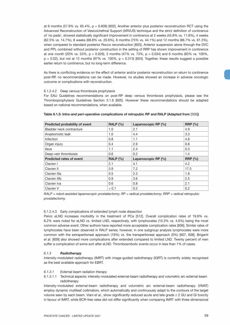
59PROSTATE CANCER - LIMITED UPDATE 2021
at 6 months (57.9% vs. 65.4%, p = 0.609) [602]. Another anterior plus posterior reconstruction RCT using the Advanced Reconstruction of VesicoUrethral Support (ARVUS) technique and the strict definition of continence of ‘no pads‘, showed statistically significant improvement in continence at 2 weeks (43.8% vs. 11.8%), 4 weeks (62.5% vs. 14.7%), 8 weeks (68.8% vs. 20.6%), 6 months (75% vs. 44.1%) and 12 months (86.7% vs. 61.3%), when compared to standard posterior Rocco reconstruction [603]. Anterior suspension alone through the DVC and PPL combined without posterior construction in the setting of RRP has shown improvement in continence at one month (20% vs. 53%, p = 0.029), 3 months (47% vs. 73%, p = 0.034) and 6 months (83% vs. 100%, p = 0.02), but not at 12 months (97% vs. 100%, p = 0.313) [604]. Together, these results suggest a possible earlier return to continence, but no long-term difference.
As there is conflicting evidence on the effect of anterior and/or posterior reconstruction on return to continence post-RP, no recommendations can be made. However, no studies showed an increase in adverse oncologic outcome or complications with reconstruction.
6.1.2.4.2 Deep venous thrombosis prophylaxisFor EAU Guidelines recommendations on post-RP deep venous thrombosis prophylaxis, please see the Thromboprophylaxis Guidelines Section 3.1.6 [605]. However these recommendations should be adapted based on national recommendations, when available.
Table 6.1.5: Intra-and peri-operative complications of retropubic RP and RALP (Adapted from [500])
Predicted probability of event RALP (%) Laparoscopic RP (%) RRP (%)Bladder neck contracture 1.0 2.1 4.9Anastomotic leak 1.0 4.4 3.3Infection 0.8 1.1 4.8Organ injury 0.4 2.9 0.8Ileus 1.1 2.4 0.3Deep-vein thrombosis 0.6 0.2 1.4Predicted rates of event RALP (%) Laparoscopic RP (%) RRP (%)Clavien I 2.1 4.1 4.2Clavien II 3.9 7.2 17.5Clavien IIIa 0.5 2.3 1.8Clavien IIIb 0.9 3.6 2.5Clavien Iva 0.6 0.8 2.1Clavien V < 0.1 0.2 0.2
RALP = robot-assisted laparoscopic prostatectomy; RP = radical prostatectomy; RRP = radical retropubic prostatectomy.
6.1.2.4.3 Early complications of extended lymph node dissectionPelvic eLND increases morbidity in the treatment of PCa [512]. Overall complication rates of 19.8% vs. 8.2% were noted for eLND vs. limited LND, respectively, with lymphoceles (10.3% vs. 4.6%) being the most common adverse event. Other authors have reported more acceptable complication rates [606]. Similar rates of lymphoceles have been observed in RALP series; however, in one subgroup analysis lymphoceles were more common with the extraperitoneal approach (19%) vs. the transperitoneal approach (0%) [607, 608]. Briganti et al. [609] also showed more complications after extended compared to limited LND. Twenty percent of men suffer a complication of some sort after eLND. Thromboembolic events occur in less than 1% of cases.
6.1.3 RadiotherapyIntensity-modulated radiotherapy (IMRT) with image-guided radiotherapy (IGRT) is currently widely recognised as the best available approach for EBRT.
6.1.3.1 External beam radiation therapy6.1.3.1.1 Technical aspects: intensity-modulated external-beam radiotherapy and volumetric arc external-beam
radiotherapy Intensity-modulated external-beam radiotherapy and volumetric arc external-beam radiotherapy (VMAT) employ dynamic multileaf collimators, which automatically and continuously adapt to the contours of the target volume seen by each beam. Viani et al., show significantly reduced acute and late grade > 2 GU and GI toxicity in favour of IMRT, while BCR-free rates did not differ significantly when comparing IMRT with three-dimensional

PROSTATE CANCER - LIMITED UPDATE 202160
conformal radiation therapy (3D-CRT) in a RCT including 215 patients [610]. A meta-analysis by Yu et al., (23 studies, 9,556 patients) concluded that IMRT significantly decreases the occurrence of grade 2–4 acute GI toxicity, late GI toxicity and late rectal bleeding, and achieves better PSA relapse-free survival in comparison with 3D-CRT. Intensity-modulated external-beam radiotherapy and 3D-CRT show comparable acute rectal toxicity, late GU toxicity and OS, while IMRT slightly increases the morbidity of acute GU toxicity [611]. Wortel et al., concluded that, as compared to 3D-CRT, image-guided IMRT was associated with significantly reduced late GI toxicity whereas GU toxicities remained comparable (242 IMRT patients vs. 189 3D-CRT patients) [612]. Finally, Zapatero et al., found, based on 733 consecutive patients (295 IMRT vs. 438 3D-CRT), that compared with 3D-CRT, high-dose IMRT/IGRT is associated with a lower rate of late urinary complications despite a higher radiation dose [613]. In conclusion, IMRT plus IGRT remain the standard of care for the treatment of PCa.
The advantage of VMAT over IMRT is shorter treatment times, generally two to three minutes. Both techniques allow for a more complex distribution of the dose to be delivered and provide concave isodose curves, which are particularly useful as a means of sparing the rectum. Radiotherapy treatment planning for IMRT and VMAT differs from that used in conventional EBRT, requiring a computer system capable of ‘inverse planning’ and the appropriate physics expertise. Treatment plans must conform to pre-specified dose constraints to critical organs at risk of normal tissue damage and a formal quality assurance process should be routine.
With dose escalation using IMRT/VMAT organ movement becomes a critical issue in terms of both tumour control and treatment toxicity. Techniques will therefore combine IMRT/VMAT with some form of IGRT in which organ movement can be visualised and corrected for in real time, although the optimum means of achieving this is still unclear [614]. Tomotherapy is another technique for the delivery of IMRT, using a linear accelerator mounted on a ring gantry that rotates as the patient is delivered through the centre of the ring, analogous to spiral CT scanning.
6.1.3.1.2 Dose escalationLocal control is a critical issue for the outcome of radiotherapy of PCa. It has been shown that local failure due to insufficient total dose is prognostic for death from PCa as a second wave of metastases is seen later on [615]. Several RCTs have shown that dose escalation (range 74–80 Gy) has a significant impact on 10-year biochemical relapse as well as metastases and disease-specific mortality [616-623]. These trials have generally included patients from several risk groups, and the use of neoadjuvant/adjuvant HT has varied (see Table 6.1.6). The best evidence of an OS benefit in patients with intermediate- or high-risk PCa, but not with low-risk PCa, derives from a non-randomised but well conducted propensity-matched retrospective analysis of the U.S. National Cancer Database including a total of 42,481 patients [624]. In everyday practice, a minimum dose of > 74 Gy is recommended for EBRT plus HT, with no different recommendations according to the patient’s risk group. If IMRT and IGRT are used for dose escalation, rates of severe late side effects (> grade 3) for the rectum are 2–3% and for the GU tract 2–5% [618, 621, 625-638].
Table 6.1.6: Randomised trials of dose escalation in localised PCa
Trial n PCa condition Radiotherapy Dose
Follow-up (median)
Outcome Results
MD Anderson study 2011 [623]
301 T1-T3, N0, M0, PSA 10 ng/mL vs. PSA > 10 ng/mL
70 vs.78 Gy 15 yr. DM, DSM, FFF
All patients:18.9% FFF at 70 Gy12% FFF at 78 Gy(p = 0.042)3.4% DM at 70 Gy1.1% DM at 78 Gy(p = 0.018)6.2% DSM at 70 Gy3.2% DSM at 78 Gy(p = 0.043)No difference in OS(p > 0.05)
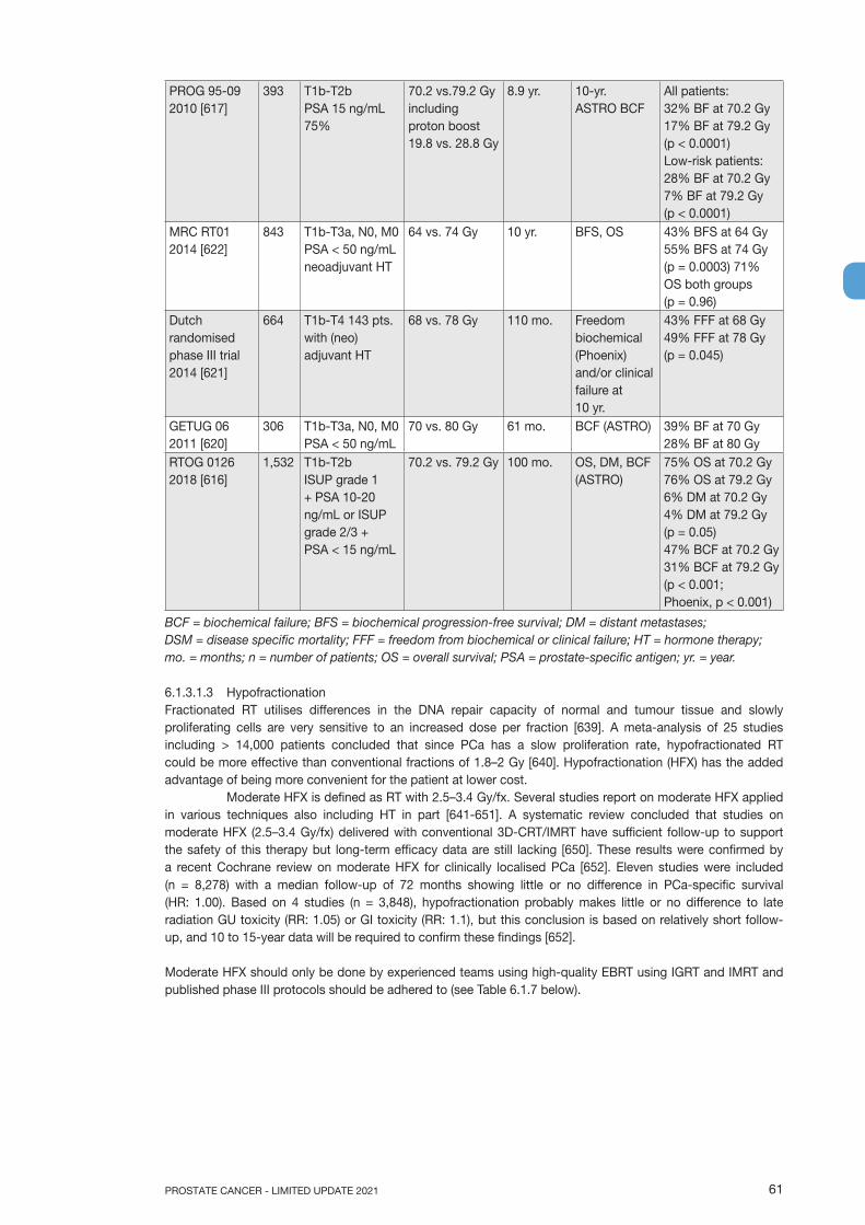
61PROSTATE CANCER - LIMITED UPDATE 2021
PROG 95-09 2010 [617]
393 T1b-T2b PSA 15 ng/mL 75%
70.2 vs.79.2 Gy including proton boost 19.8 vs. 28.8 Gy
8.9 yr. 10-yr. ASTRO BCF
All patients: 32% BF at 70.2 Gy 17% BF at 79.2 Gy (p < 0.0001) Low-risk patients: 28% BF at 70.2 Gy 7% BF at 79.2 Gy (p < 0.0001)
MRC RT01 2014 [622]
843 T1b-T3a, N0, M0 PSA < 50 ng/mL neoadjuvant HT
64 vs. 74 Gy 10 yr. BFS, OS 43% BFS at 64 Gy 55% BFS at 74 Gy (p = 0.0003) 71% OS both groups (p = 0.96)
Dutch randomised phase III trial 2014 [621]
664 T1b-T4 143 pts. with (neo) adjuvant HT
68 vs. 78 Gy 110 mo. Freedom biochemical (Phoenix) and/or clinical failure at 10 yr.
43% FFF at 68 Gy 49% FFF at 78 Gy (p = 0.045)
GETUG 06 2011 [620]
306 T1b-T3a, N0, M0 PSA < 50 ng/mL
70 vs. 80 Gy 61 mo. BCF (ASTRO) 39% BF at 70 Gy 28% BF at 80 Gy
RTOG 0126 2018 [616]
1,532 T1b-T2b ISUP grade 1 + PSA 10-20 ng/mL or ISUP grade 2/3 + PSA < 15 ng/mL
70.2 vs. 79.2 Gy 100 mo. OS, DM, BCF (ASTRO)
75% OS at 70.2 Gy 76% OS at 79.2 Gy 6% DM at 70.2 Gy 4% DM at 79.2 Gy (p = 0.05) 47% BCF at 70.2 Gy 31% BCF at 79.2 Gy (p < 0.001; Phoenix, p < 0.001)
BCF = biochemical failure; BFS = biochemical progression-free survival; DM = distant metastases; DSM = disease specific mortality; FFF = freedom from biochemical or clinical failure; HT = hormone therapy; mo. = months; n = number of patients; OS = overall survival; PSA = prostate-specific antigen; yr. = year.
6.1.3.1.3 HypofractionationFractionated RT utilises differences in the DNA repair capacity of normal and tumour tissue and slowly proliferating cells are very sensitive to an increased dose per fraction [639]. A meta-analysis of 25 studies including > 14,000 patients concluded that since PCa has a slow proliferation rate, hypofractionated RT could be more effective than conventional fractions of 1.8–2 Gy [640]. Hypofractionation (HFX) has the added advantage of being more convenient for the patient at lower cost.
Moderate HFX is defined as RT with 2.5–3.4 Gy/fx. Several studies report on moderate HFX applied in various techniques also including HT in part [641-651]. A systematic review concluded that studies on moderate HFX (2.5–3.4 Gy/fx) delivered with conventional 3D-CRT/IMRT have sufficient follow-up to support the safety of this therapy but long-term efficacy data are still lacking [650]. These results were confirmed by a recent Cochrane review on moderate HFX for clinically localised PCa [652]. Eleven studies were included (n = 8,278) with a median follow-up of 72 months showing little or no difference in PCa-specific survival (HR: 1.00). Based on 4 studies (n = 3,848), hypofractionation probably makes little or no difference to late radiation GU toxicity (RR: 1.05) or GI toxicity (RR: 1.1), but this conclusion is based on relatively short follow-up, and 10 to 15-year data will be required to confirm these findings [652].
Moderate HFX should only be done by experienced teams using high-quality EBRT using IGRT and IMRT and published phase III protocols should be adhered to (see Table 6.1.7 below).
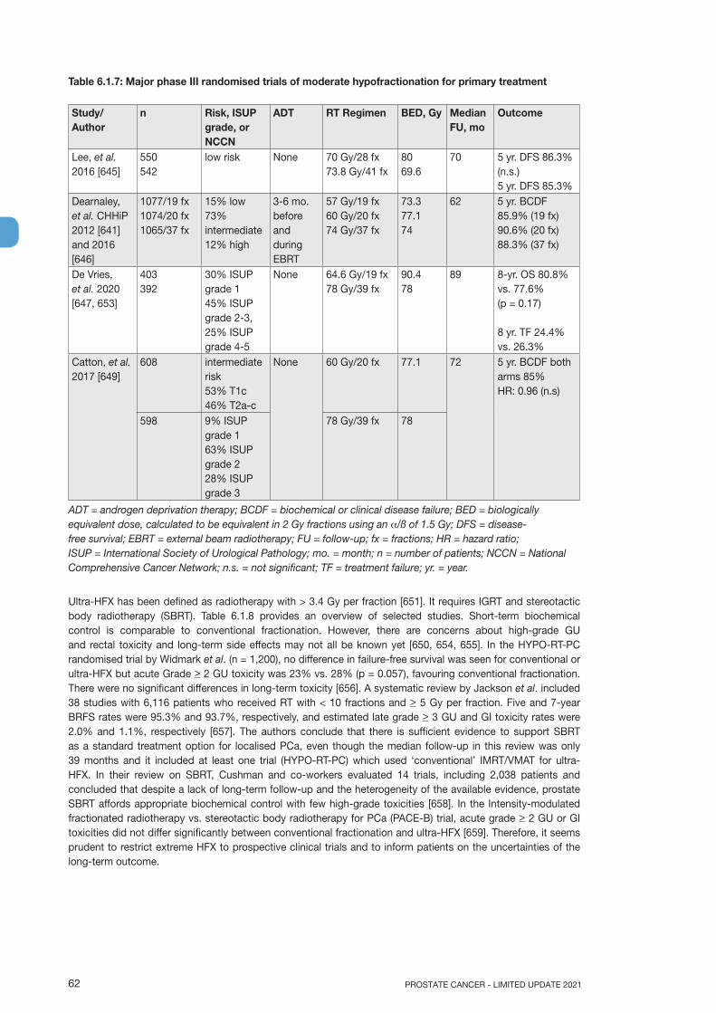
PROSTATE CANCER - LIMITED UPDATE 202162
Table 6.1.7: Major phase III randomised trials of moderate hypofractionation for primary treatment
Study/Author
n Risk, ISUP grade, or NCCN
ADT RT Regimen BED, Gy Median FU, mo
Outcome
Lee, et al. 2016 [645]
550 542
low risk None 70 Gy/28 fx 73.8 Gy/41 fx
80 69.6
70 5 yr. DFS 86.3% (n.s.) 5 yr. DFS 85.3%
Dearnaley, et al. CHHiP 2012 [641] and 2016 [646]
1077/19 fx 1074/20 fx 1065/37 fx
15% low 73% intermediate 12% high
3-6 mo. before and during EBRT
57 Gy/19 fx 60 Gy/20 fx 74 Gy/37 fx
73.3 77.1 74
62 5 yr. BCDF 85.9% (19 fx) 90.6% (20 fx) 88.3% (37 fx)
De Vries, et al. 2020 [647, 653]
403 392
30% ISUP grade 1 45% ISUP grade 2-3, 25% ISUP grade 4-5
None 64.6 Gy/19 fx 78 Gy/39 fx
90.4 78
89 8-yr. OS 80.8% vs. 77.6%(p = 0.17)
8 yr. TF 24.4% vs. 26.3%
Catton, et al. 2017 [649]
608 intermediate risk 53% T1c 46% T2a-c
None 60 Gy/20 fx 77.1 72 5 yr. BCDF both arms 85% HR: 0.96 (n.s)
598 9% ISUP grade 1 63% ISUP grade 2 28% ISUP grade 3
78 Gy/39 fx 78
ADT = androgen deprivation therapy; BCDF = biochemical or clinical disease failure; BED = biologically equivalent dose, calculated to be equivalent in 2 Gy fractions using an α/ß of 1.5 Gy; DFS = disease-free survival; EBRT = external beam radiotherapy; FU = follow-up; fx = fractions; HR = hazard ratio; ISUP = International Society of Urological Pathology; mo. = month; n = number of patients; NCCN = National Comprehensive Cancer Network; n.s. = not significant; TF = treatment failure; yr. = year.
Ultra-HFX has been defined as radiotherapy with > 3.4 Gy per fraction [651]. It requires IGRT and stereotactic body radiotherapy (SBRT). Table 6.1.8 provides an overview of selected studies. Short-term biochemical control is comparable to conventional fractionation. However, there are concerns about high-grade GU and rectal toxicity and long-term side effects may not all be known yet [650, 654, 655]. In the HYPO-RT-PC randomised trial by Widmark et al. (n = 1,200), no difference in failure-free survival was seen for conventional or ultra-HFX but acute Grade > 2 GU toxicity was 23% vs. 28% (p = 0.057), favouring conventional fractionation. There were no significant differences in long-term toxicity [656]. A systematic review by Jackson et al. included 38 studies with 6,116 patients who received RT with < 10 fractions and > 5 Gy per fraction. Five and 7-year BRFS rates were 95.3% and 93.7%, respectively, and estimated late grade > 3 GU and GI toxicity rates were 2.0% and 1.1%, respectively [657]. The authors conclude that there is sufficient evidence to support SBRT as a standard treatment option for localised PCa, even though the median follow-up in this review was only 39 months and it included at least one trial (HYPO-RT-PC) which used ‘conventional’ IMRT/VMAT for ultra-HFX. In their review on SBRT, Cushman and co-workers evaluated 14 trials, including 2,038 patients and concluded that despite a lack of long-term follow-up and the heterogeneity of the available evidence, prostate SBRT affords appropriate biochemical control with few high-grade toxicities [658]. In the Intensity-modulated fractionated radiotherapy vs. stereotactic body radiotherapy for PCa (PACE-B) trial, acute grade > 2 GU or GI toxicities did not differ significantly between conventional fractionation and ultra-HFX [659]. Therefore, it seems prudent to restrict extreme HFX to prospective clinical trials and to inform patients on the uncertainties of the long-term outcome.
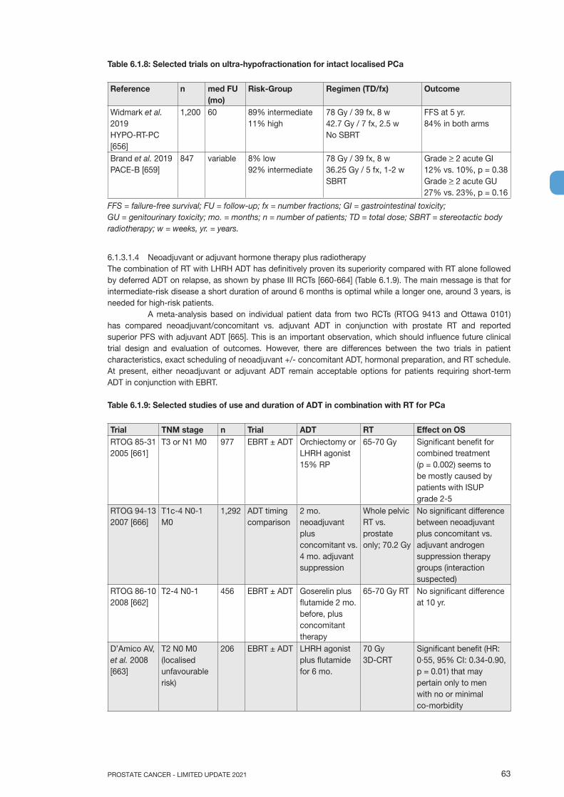
63PROSTATE CANCER - LIMITED UPDATE 2021
Table 6.1.8: Selected trials on ultra-hypofractionation for intact localised PCa
Reference n med FU (mo)
Risk-Group Regimen (TD/fx) Outcome
Widmark et al. 2019HYPO-RT-PC [656]
1,200 60 89% intermediate11% high
78 Gy / 39 fx, 8 w42.7 Gy / 7 fx, 2.5 wNo SBRT
FFS at 5 yr. 84% in both arms
Brand et al. 2019PACE-B [659]
847 variable 8% low92% intermediate
78 Gy / 39 fx, 8 w36.25 Gy / 5 fx, 1-2 wSBRT
Grade > 2 acute GI12% vs. 10%, p = 0.38Grade > 2 acute GU27% vs. 23%, p = 0.16
FFS = failure-free survival; FU = follow-up; fx = number fractions; GI = gastrointestinal toxicity; GU = genitourinary toxicity; mo. = months; n = number of patients; TD = total dose; SBRT = stereotactic body radiotherapy; w = weeks, yr. = years.
6.1.3.1.4 Neoadjuvant or adjuvant hormone therapy plus radiotherapyThe combination of RT with LHRH ADT has definitively proven its superiority compared with RT alone followed by deferred ADT on relapse, as shown by phase III RCTs [660-664] (Table 6.1.9). The main message is that for intermediate-risk disease a short duration of around 6 months is optimal while a longer one, around 3 years, is needed for high-risk patients.
A meta-analysis based on individual patient data from two RCTs (RTOG 9413 and Ottawa 0101) has compared neoadjuvant/concomitant vs. adjuvant ADT in conjunction with prostate RT and reported superior PFS with adjuvant ADT [665]. This is an important observation, which should influence future clinical trial design and evaluation of outcomes. However, there are differences between the two trials in patient characteristics, exact scheduling of neoadjuvant +/- concomitant ADT, hormonal preparation, and RT schedule. At present, either neoadjuvant or adjuvant ADT remain acceptable options for patients requiring short-term ADT in conjunction with EBRT.
Table 6.1.9: Selected studies of use and duration of ADT in combination with RT for PCa
Trial TNM stage n Trial ADT RT Effect on OSRTOG 85-31 2005 [661]
T3 or N1 M0 977 EBRT ± ADT Orchiectomy or LHRH agonist 15% RP
65-70 Gy Significant benefit for combined treatment (p = 0.002) seems to be mostly caused by patients with ISUP grade 2-5
RTOG 94-13 2007 [666]
T1c-4 N0-1 M0
1,292 ADT timing comparison
2 mo. neoadjuvant plus concomitant vs. 4 mo. adjuvant suppression
Whole pelvic RT vs. prostate only; 70.2 Gy
No significant difference between neoadjuvant plus concomitant vs. adjuvant androgen suppression therapy groups (interaction suspected)
RTOG 86-10 2008 [662]
T2-4 N0-1 456 EBRT ± ADT Goserelin plus flutamide 2 mo. before, plus concomitant therapy
65-70 Gy RT No significant difference at 10 yr.
D’Amico AV, et al. 2008 [663]
T2 N0 M0 (localised unfavourable risk)
206 EBRT ± ADT LHRH agonist plus flutamide for 6 mo.
70 Gy 3D-CRT
Significant benefit (HR: 0·55, 95% CI: 0.34-0.90, p = 0.01) that may pertain only to men with no or minimal co-morbidity
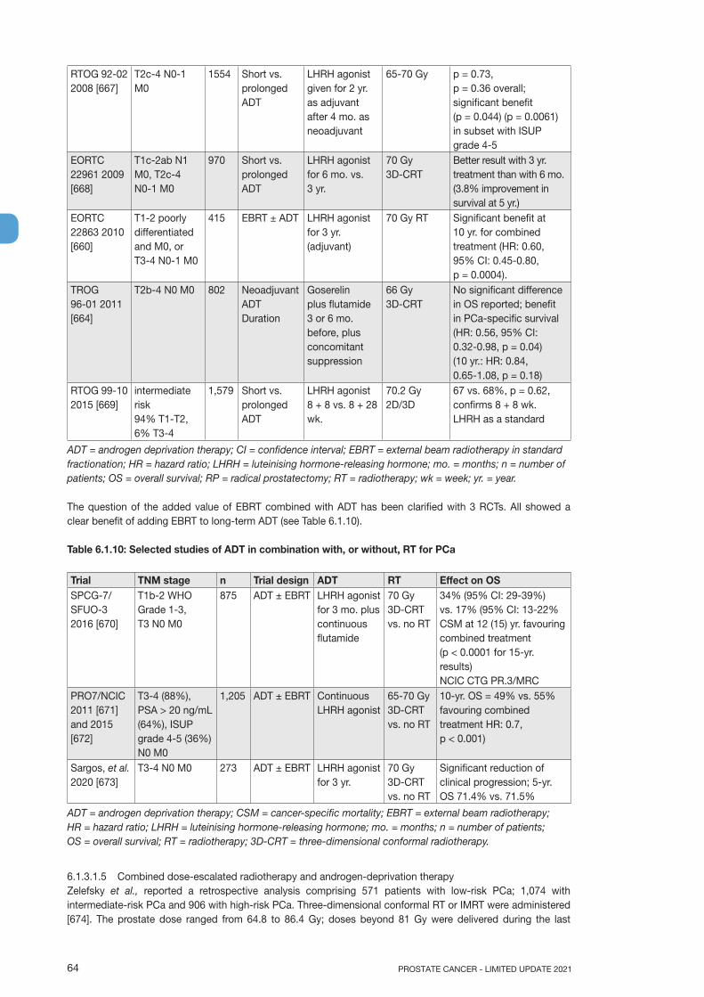
PROSTATE CANCER - LIMITED UPDATE 202164
RTOG 92-02 2008 [667]
T2c-4 N0-1 M0
1554 Short vs. prolonged ADT
LHRH agonist given for 2 yr. as adjuvant after 4 mo. as neoadjuvant
65-70 Gy p = 0.73, p = 0.36 overall; significant benefit (p = 0.044) (p = 0.0061) in subset with ISUP grade 4-5
EORTC 22961 2009 [668]
T1c-2ab N1 M0, T2c-4 N0-1 M0
970 Short vs. prolonged ADT
LHRH agonist for 6 mo. vs. 3 yr.
70 Gy 3D-CRT
Better result with 3 yr. treatment than with 6 mo. (3.8% improvement in survival at 5 yr.)
EORTC 22863 2010 [660]
T1-2 poorly differentiated and M0, or T3-4 N0-1 M0
415 EBRT ± ADT LHRH agonist for 3 yr. (adjuvant)
70 Gy RT Significant benefit at 10 yr. for combined treatment (HR: 0.60, 95% CI: 0.45-0.80, p = 0.0004).
TROG 96-01 2011 [664]
T2b-4 N0 M0 802 Neoadjuvant ADT Duration
Goserelin plus flutamide 3 or 6 mo. before, plus concomitant suppression
66 Gy 3D-CRT
No significant difference in OS reported; benefit in PCa-specific survival (HR: 0.56, 95% CI: 0.32-0.98, p = 0.04) (10 yr.: HR: 0.84, 0.65-1.08, p = 0.18)
RTOG 99-10 2015 [669]
intermediate risk 94% T1-T2, 6% T3-4
1,579 Short vs. prolonged ADT
LHRH agonist 8 + 8 vs. 8 + 28 wk.
70.2 Gy 2D/3D
67 vs. 68%, p = 0.62, confirms 8 + 8 wk. LHRH as a standard
ADT = androgen deprivation therapy; CI = confidence interval; EBRT = external beam radiotherapy in standard fractionation; HR = hazard ratio; LHRH = luteinising hormone-releasing hormone; mo. = months; n = number of patients; OS = overall survival; RP = radical prostatectomy; RT = radiotherapy; wk = week; yr. = year.
The question of the added value of EBRT combined with ADT has been clarified with 3 RCTs. All showed a clear benefit of adding EBRT to long-term ADT (see Table 6.1.10).
Table 6.1.10: Selected studies of ADT in combination with, or without, RT for PCa
Trial TNM stage n Trial design ADT RT Effect on OS SPCG-7/ SFUO-3 2016 [670]
T1b-2 WHO Grade 1-3, T3 N0 M0
875 ADT ± EBRT LHRH agonist for 3 mo. plus continuous flutamide
70 Gy 3D-CRT vs. no RT
34% (95% CI: 29-39%) vs. 17% (95% CI: 13-22% CSM at 12 (15) yr. favouring combined treatment (p < 0.0001 for 15-yr. results) NCIC CTG PR.3/MRC
PRO7/NCIC 2011 [671] and 2015 [672]
T3-4 (88%), PSA > 20 ng/mL (64%), ISUP grade 4-5 (36%) N0 M0
1,205 ADT ± EBRT Continuous LHRH agonist
65-70 Gy 3D-CRT vs. no RT
10-yr. OS = 49% vs. 55% favouring combined treatment HR: 0.7, p < 0.001)
Sargos, et al. 2020 [673]
T3-4 N0 M0 273 ADT ± EBRT LHRH agonist for 3 yr.
70 Gy 3D-CRT vs. no RT
Significant reduction of clinical progression; 5-yr. OS 71.4% vs. 71.5%
ADT = androgen deprivation therapy; CSM = cancer-specific mortality; EBRT = external beam radiotherapy; HR = hazard ratio; LHRH = luteinising hormone-releasing hormone; mo. = months; n = number of patients; OS = overall survival; RT = radiotherapy; 3D-CRT = three-dimensional conformal radiotherapy.
6.1.3.1.5 Combined dose-escalated radiotherapy and androgen-deprivation therapyZelefsky et al., reported a retrospective analysis comprising 571 patients with low-risk PCa; 1,074 with intermediate-risk PCa and 906 with high-risk PCa. Three-dimensional conformal RT or IMRT were administered [674]. The prostate dose ranged from 64.8 to 86.4 Gy; doses beyond 81 Gy were delivered during the last

65PROSTATE CANCER - LIMITED UPDATE 2021
10 years of the study using image-guided IMRT. Complete androgen blockade was administered at the discretion of the treating physician to 623 high-risk (69%), 456 intermediate-risk (42%) and 170 low-risk (30%) PCa patients. The duration of ADT was 3 months for low-risk patients and 6 months for intermediate-risk and high-risk patients, starting at 3 months before RT. The 10-year biochemical disease-free rate was significantly improved by dose escalation: above 75.6 Gy in low-risk, and above 81 Gy for the intermediate- and high-risk groups. It was also improved by adding 6 months of ADT in intermediate- and high-risk patients. In the multivariate analysis, neither the dose > 81 Gy, nor adding ADT, influenced OS. Three RCTs have shown that the benefits of ADT are independent of dose escalation, and that the use of ADT would not compensate for a lower radiotherapy dose:1. The GICOR study shows a better biochemical DFS in high-risk patients for 3D-CRT radiation dose > 72 Gy
when combined with long-term ADT [630].2. DART01/05 GICOR shows improved biochemical control and OS in high-risk patients if 2 years of
adjuvant ADT is combined with high-dose RT [675].3. EORTC trial 22991 shows that 6 months ADT improves biochemical and clinical DFS irrespective of the
dose (70, 74, 78 Gy) in intermediate-risk and low-volume high-risk localised PCa patients [676].
6.1.3.2 Proton beam therapyIn theory, proton beams are an attractive alternative to photon-beam RT for PCa, as they deposit almost all their radiation dose at the end of the particle’s path in tissue (the Bragg peak), in contrast to photons which deposit radiation along their path. There is also a very sharp fall-off for proton beams beyond their deposition depth, meaning that critical normal tissues beyond this depth could be effectively spared. In contrast, photon beams continue to deposit energy until they leave the body, including an exit dose.
One RCT on dose escalation (70.2 vs. 79.2 Gy) has incorporated protons for the boost doses of either 19.8 or 28.8 Gy. This trial shows improved outcome with the higher dose but it cannot be used as evidence for the superiority of proton therapy [617]. Thus, unequivocal information showing an advantage of protons over IMRT photon therapy is still not available. Studies from the SEER database and from Harvard describing toxicity and patient-reported outcomes do not point to an inherent superiority for protons [677, 678]. In terms of longer-term GI toxicity, proton therapy might even be inferior to IMRT [678].
A RCT comparing equivalent doses of proton-beam therapy with IMRT is underway. Meanwhile, proton therapy must be regarded as an experimental alternative to photon-beam therapy.
6.1.3.3 Spacer during external beam radiation therapyBiodegradable spacer insertion involves using a liquid gel or balloon to increase the distance between the prostate and rectum and consequently reduce the amount of radiation reaching the rectum. Various materials have been used with most evidence available for CE-marked hydrogel spacers [679]. A meta-analysis including one RCT and six cohort studies using the hydrogel spacer demonstrated a 5–8% reduction in the rectal volume receiving high-dose radiation, although heterogeneity between studies is found [680]. In the final analysis of the RCT with a median follow-up of 37 months and with approximately two-thirds of patients evaluable, those treated with spacer in situ had no deterioration from baseline bowel function whilst those treated without spacer had a lower mean bowel summary score of 5.8 points which met the threshold for minimally important difference of 4–6 points [681].
This meta-analysis highlights inconsistent reporting of procedural complications. In addition, with more widespread clinical use safety reports describe uncommon, but severe and life changing, complications including prostatic abscess, fistulae and sepsis [682]. Implantation is associated with a learning curve and should only be undertaken by teams with experience of TRUS and transperineal procedures with robust audit reporting in place [683]. Its role in the context of moderate or extreme hypofractionation is as yet unclear.
6.1.3.4 Brachytherapy6.1.3.4.1 Low-dose rate (LDR) brachytherapyLow-dose rate brachytherapy uses radioactive seeds permanently implanted into the prostate. There is a consensus on the group of patients with the best outcomes after LDR monotherapy [684]: Stage cT1b-T2a N0, M0; ISUP grade 1 with < 50% of biopsy cores involved with cancer or ISUP grade 2 with < 33% of biopsy cores involved with cancer; an initial PSA level of < 10 ng/mL; an International Prostatic Symptom Score (IPSS) < 12 and maximal flow rate > 15 mL/min on urinary flow tests [685]. In addition, with due attention to dose distribution, patients having had a previous TURP can undergo brachytherapy without an increase in risk of urinary toxicity. A minimal channel TURP is recommended and there should be at least a 3-month interval between TURP and brachytherapy to allow for adequate healing [686-689].
The only available RCT comparing RP and brachytherapy as monotherapy was closed due to poor accrual [690]. Outcome data are available from a number of large population cohorts with mature follow-up [691-698].
PROSTATE CANCER - LIMITED UPDATE 202166
The biochemical DFS for ISUP grade 1 patients after 5 and 10 years has been reported to range from 71% to 93% and 65% to 85%, respectively [691-698]. A significant correlation has been shown between the implanted dose and biochemical control [699]. A D90 (dose covering 90% of the prostate volume) of > 140 Gy leads to a significantly higher biochemical control rate (PSA < 1.0 ng/mL) after 4 years (92 vs. 68%). There is no benefit in adding neoadjuvant or adjuvant ADT to LDR monotherapy [691]. Low-dose rate brachytherapy can be combined with EBRT in good-, intermediate- and high-risk patients (see Section 6.2.3.2.3).
6.1.3.4.2 High-dose rate brachytherapyHigh-dose rate (HDR) brachytherapy uses a radioactive source temporarily introduced into the prostate to deliver radiation. The technical differences are outlined in Table 6.1.11. The use of the GEC (Groupe Européen de Curiethérapie)/ESTRO Guidelines is strongly recommended [700]. High-dose rate brachytherapy can be delivered in single or multiple fractions and is often combined with EBRT of at least 45 Gy [701]. A single RCT of EBRT (55 Gy in 20 fractions) vs. EBRT (35.75 Gy in 13 fractions), followed by HDR brachytherapy (17 Gy in two fractions over 24 hours) has been reported [702]. In 218 patients with organ-confined PCa the combination of EBRT and HDR brachytherapy showed a significant improvement in the biochemical disease-free rate (p = 0.04) at 5 and 10 year (75% and 46% compared to 61% and 39%). However, a very high, uncommon, rate of early recurrences was observed in the EBRT arm alone, even after 2 years, possibly due to a dose lower than the current standard used [702]. A systematic review of non-RCTs has suggested outcomes with EBRT plus HDR brachytherapy are superior to brachytherapy alone, but this needs confirmation in a prospective RCT [703].
Fractionated HDR brachytherapy as monotherapy can be offered to patients with low- and intermediate-risk PCa, who should be informed that results are only available from limited series in very experienced centres [704, 705]. Five-year PSA control rates over 90% are reported, with late grade 3+ GU toxicity rates < 5% and no, or very minimal, grade 3+ GI toxicity rates [704, 705].
Table 6.1.11: Difference between LDR and HDR brachytherapy
Differences in prostate brachytherapy techniquesLow dose rate (LDR) • Permanent seeds implanted
• Uses Iodine-125 (I-125) (most common), Palladium-103 (Pd-103) or Cesium-131 isotopes
• Radiation dose delivered over weeks and months• Acute side effects resolve over months• Radiation protection issues for patient and carers
High dose rate (HDR) • Temporary implantation• Iridium-192 (IR-192) isotope introduced through implanted needles or
catheters• Radiation dose delivered in minutes• Acute side effects resolve over weeks• No radiation protection issues for patient or carers
6.1.3.5 Acute side effects of external beam radiotherapy and brachytherapyGastrointestinal and urinary side effects are common during and after EBRT. In the EORTC 22991 trial, approximately 50% of patients reported acute GU toxicity of grade 1, 20% of grade 2, and 2% grade 3. In the same trial, approximately 30% of patients reported acute grade 1 GI toxicity, 10% grade 2, and less than 1% grade 3. Common toxicities included dysuria, urinary frequency, urinary retention, haematuria, diarrhoea, rectal bleeding and proctitis [629]. In addition, general side effects such as fatigue are common. It should be noted that the incidence of acute side effects is greater than that of late effects (see Section 8.2.2.1), implying that most acute effects resolve. In a RCT of conventional dose EBRT vs. EBRT and LDR brachytherapy the incidence of acute proctitis was reduced in the brachytherapy arm, but other acute toxicities were equivalent [706]. Acute toxicity of HDR brachytherapy has not been documented in a RCT, but retrospective reports confirm lower rates of GI toxicity compared with EBRT alone and grade 3 GU toxicity in 10%, or fewer, patients, but a higher incidence of urinary retention [707]. Similar findings are reported using HFX; in a pooled analysis of 864 patients treated using extreme HFX and stereotactic radiotherapy, declines in urinary and bowel domains were noted at 3 months which returned to baseline, or better, by 6 months [708].

67PROSTATE CANCER - LIMITED UPDATE 2021
6.1.4 Hormonal therapy6.1.4.1 Introduction6.1.4.1.1 Different types of hormonal therapyAndrogen deprivation can be achieved by either suppressing the secretion of testicular androgens or inhibiting the action of circulating androgens at the level of their receptor. These two methods can be combined to achieve what has been known as complete (or maximal or total) androgen blockade (CAB) using the old-fashioned anti-androgens [709].
6.1.4.1.1.1 Testosterone-lowering therapy (castration)6.1.4.1.1.1.1 Castration levelThe castration level is < 50 ng/dL (1.7 nmol/L), which was defined more than 40 years ago when testosterone testing was less sensitive. Current methods have shown that the mean value after surgical castration is 15 ng/dL [710]. Therefore, a more appropriate level should be defined as < 20 ng/dL (1 nmol/L). This definition is important as better results are repeatedly observed with lower testosterone levels compared to 50 ng/dL [711-713]. However, the castrate level considered by the regulatory authorities and in clinical trials addressing castration in PCa is still the historical < 50 ng/dL (1.7 mmol/L).
6.1.4.1.1.1.2 Bilateral orchiectomyBilateral orchiectomy or subcapsular pulpectomy is still considered the primary treatment modality for ADT. It is a simple, cheap and virtually complication-free surgical procedure. It is easily performed under local anaesthesia and it is the quickest way to achieve a castration level which is usually reached within less than twelve hours. It is irreversible and therefore does not allow for intermittent treatment [714].
6.1.4.1.1.1.3 OestrogensTreatment with oestrogens results in testosterone suppression and is not associated with bone loss [715]. Early studies tested oral diethylstilboestrol (DES) at several doses. Due to severe side effects, especially thromboembolic complications, even at lower doses these drugs are not considered as standard first-line treatment [716-718].
6.1.4.1.1.1.4 Luteinising-hormone-releasing hormone agonistsLong-acting LHRH agonists are currently the main forms of ADT. These synthetic analogues of LHRH are delivered as depot injections on a 1-, 2-, 3-, 6-monthly, or yearly, basis. The first injection induces a transient rise in luteinising hormone (LH) and follicle-stimulating hormone (FSH) leading to the ‘testosterone surge’ or ‘flare-up’ phenomenon which starts two to three days after administration and lasts for about one week. This may lead to detrimental clinical effects (the clinical flare) such as increased bone pain, acute bladder outlet obstruction, obstructive renal failure, spinal cord compression, and cardiovascular death due to hypercoagulation status [719]. Patients at risk are usually those with high-volume symptomatic bony disease. Concomitant therapy with an anti-androgen decreases the incidence of clinical flare but does not completely remove the risk. Anti-androgen therapy is usually continued for 4 weeks but neither the timing nor the duration of anti-androgen therapy are based on strong evidence. In addition, the long-term impact of preventing ‘flare-up’ is unknown [720, 721].
Chronic exposure to LHRH agonists results in the down-regulation of LHRH-receptors, suppressing LH and FSH secretion and therefore testosterone production. A castration level is usually obtained within 2 to 4 weeks [722]. Although there is no formal direct comparison between the various compounds, they are considered to be equally effective [723]. No survival difference with orchiectomy has been reported despite the lack of high-quality trials [724].
The different products have practical differences that need to be considered in everyday practice, including the storage temperature, whether a drug is ready for immediate use or requires reconstitution, and whether a drug is given by subcutaneous or intramuscular injection.
6.1.4.1.1.1.5 Luteinising-hormone-releasing hormone antagonistsLuteinising-hormone releasing hormone antagonists immediately bind to LHRH receptors, leading to a rapid decrease in LH, FSH and testosterone levels without any flare. The practical shortcoming of these compounds is the lack of a long-acting depot formulation with, so far, only monthly formulations being available. Degarelix is a LHRH antagonist. The standard dosage is 240 mg in the first month followed by monthly injections of 80 mg. Most patients achieve a castrate level at day three [722]. A phase III RCT compared degarelix to monthly leuprorelin following up patients for 12 months, suggesting a better PSA PFS for degarelix 240/80 mg compared to monthly leuprorelin [725]. A systematic review did not show a major difference between agonists and degarelix and highlighted the paucity of on-treatment data beyond 12 months as well as the lack of survival data [726]. Its definitive superiority over the LHRH analogues remains to be proven.
PROSTATE CANCER - LIMITED UPDATE 202168
Relugolix is an oral gonadotropin-releasing hormone antagonist. It was compared to the LHRH agonist leuprolid in a randomised phase III trial [727]. The primary endpoint was sustained testosterone suppression to castrate levels through 48 weeks. There was a significant difference of 7.9 percentage points (95% CI: 4.1–11.8) showing non-inferiority and superiority of relugolix. The incidence of major adverse cardiovascular events was significantly lower with relugolix (prespecified safety analysis). Relugolix has been approved by the FDA [728].
6.1.4.1.1.1.6 Anti-androgensThese oral compounds are classified according to their chemical structure as:• steroidal, e.g., cyproterone acetate (CPA), megestrol acetate and medroxyprogesterone acetate;• non-steroidal or pure, e.g., nilutamide, flutamide and bicalutamide.
Both classes compete with androgens at the receptor level. This leads to an unchanged or slightly elevated testosterone level. Conversely, steroidal anti-androgens have progestational properties leading to central inhibition by crossing the blood-brain barrier.
6.1.4.1.1.1.6.1 Steroidal anti-androgensThese compounds are synthetic derivatives of hydroxyprogesterone. Their main pharmacological side effects are secondary to castration (gynaecomastia is quite rare) whilst the non-pharmacological side effects are cardiovascular toxicity (4–40% for CPA) and hepatotoxicity.
Cyproterone acetate was the first licensed anti-androgen but the least studied. Its most effective dose as monotherapy is still unknown. Although CPA has a relatively long half-life (31–41 hours), it is usually administered in two or three fractionated doses of 100 mg each. In one RCT CPA showed a poorer OS when compared with LHRH analogues [729]. An underpowered RCT comparing CPA monotherapy with flutamide in M1b PCa did not show any difference in DSS and OS at a median follow-up of 8.6 years [730]. Other CPA monotherapy studies suffer from methodological limitations preventing firm conclusions.
6.1.4.1.1.1.6.2 Non-steroidal anti-androgensNon-steroidal anti-androgen monotherapy with e.g., nilutamide, flutamide or bicalutamide does not suppress testosterone secretion and it is claimed that libido, overall physical performance and bone mineral density (BMD) are frequently preserved [731]. Non-androgen-related pharmacological side effects differ between agents. Bicalutamide shows a more favourable safety and tolerability profile than flutamide and nilutamide [732]. The dosage licensed for use in CAB is 50 mg/day, and 150 mg for monotherapy. The androgen pharmacological side effects are mainly gynaecomastia (70%) and breast pain (68%). However, non-steroidal anti-androgen monotherapy offers clear bone protection compared with LHRH analogues and probably LHRH antagonists [731, 733]. All three agents share the potential for liver toxicity (occasionally fatal), requiring regular monitoring of patients’ liver enzymes.
6.1.4.1.1.2 New androgen receptor pathway targetting agents (ARTA)Once on ADT the development of castration-resistance (CRPC) is only a matter of time. It is considered to be mediated through two main overlapping mechanisms: androgen-receptor (AR)-independent and AR-dependent mechanisms (see Section 6.5 - Castrate-resistant PCa). In CRPC, the intracellular androgen level is increased compared to androgen sensitive cells and an over-expression of the AR has been observed, suggesting an adaptive mechanism [734]. This has led to the development of several new compounds targeting the androgen axis. In mCRPC, abiraterone acetate plus prednisolone and enzalutamide have been approved. In addition to ADT (sustained castration), abiraterone acetate plus prednisolone, apalutamide and enzalutamide have been approved for the treatment of metastatic hormone sensitive PCa (mHSPC) by the FDA, abiraterone/prednisolone and apalutamide also by the EMA (Note: EMA issued a positive opinion to extend the indication for enzalutamide at the time of this publication, final approval is pending). For the updated approval status see EMA and FDA websites [735-739]. Finally, apalutamide, darolutamide and enzalutamide have been approved for non-metastatic CRPC (nmCRPC) at high risk of further metastases [740-744].
6.1.4.1.1.2.1 Abiraterone acetateAbiraterone acetate is a CYP17 inhibitor (a combination of 17α-hydrolase and 17,20-lyase inhibition). By blocking CYP17, abiraterone acetate significantly decreases the intracellular testosterone level by suppressing its synthesis at the adrenal level and inside the cancer cells (intracrine mechanism). This compound must be used together with prednisone/prednisolone to prevent drug-induced hyperaldosteronism [735].
6.1.4.1.1.2.2 Apalutamide, darolutamide, enzalutamide (alphabetical order)These agents are novel non-steroidal anti-androgens with a higher affinity for the AR receptor than

69PROSTATE CANCER - LIMITED UPDATE 2021
bicalutamide. While previous non-steroidal anti-androgens still allow transfer of ARs to the nucleus and would act as partial agonists, all three agents also block AR transfer and therefore suppress any possible agonist-like activity [739-741]. Darolutamide has structurally unique properties [740]. In particular, in preclinical studies, it showed not to cross the blood-brain barrier [745, 746].
6.1.4.1.1.3 New compounds6.1.4.1.1.3.1 PARP inhibitorsPoly (ADP-ribose) polymerase inhibitors (PARPi) block the enzyme poly ADP ribose polymerase (PARP) and were developed aiming to selectively target cancer cells harbouring BRCA mutations and other mutations inducing homologous recombination deficiency and high level of replication pressure with a sensitivity to PARPi treatment. Due to the oncogenic loss of some DNA repair effectors and incomplete DNA repair repertoire, some cancer cells are addicted to certain DNA repair pathways such as Poly (ADP-ribose) polymerase (PARP)-related single-strand break repair pathway. The interaction between BRCA and PARP is a form of synthetic lethal effect which means the simultaneously functional loss of two genes lead to cell death, while a defect in any single gene only has a limited effect on cell viability [747]. The therapeutic indication for PCa is discussed in Section 6.5.8.1.
6.1.4.1.1.3.2 Immune checkpoint inhibitors Immune checkpoints are key regulators of the immune system. Checkpoint proteins, such as B7-1/B7-2 on antigen-presenting cells (APC) and CTLA-4 on T cells, help keep the immune responses in an equilibrium. The binding of B7-1/B7-2 to CTLA-4 keeps the T cells in the inactive state while blocking the binding of B7-1/B7-2 to CTLA-4 whilst an immune checkpoint inhibitor (anti-CTLA-4 antibody) allows the T cells to be active and to kill tumour cells.
Approved checkpoint inhibitors target the molecules CTLA4, programmed cell death protein 1 (PD-1), and programmed death-ligand 1 (PD-L1). Programmed death-ligand 1 is the transmembrane programmed cell death 1 protein which interacts with PD-L1 (PD-1 ligand 1). Cancer-mediated upregulation of PD-L1 on the cell surface may inhibit T cells. Antibodies that bind to either PD-1 or PD-L1 and therefore block the interaction may allow the T cells to induce cell killing. Examples of PD-1 inhibitors are pembrolizumab and nivolumab; of PD-L1 inhibitors, atezolizumab, avelumab and durvalumab and an example of CTLA4 inhibitors is ipilimumab [748, 749]. Therapeutic use is discussed in Section 6.5 - Castration-resistant PCa.
6.1.4.1.1.3.3 Protein kinase B (AKT) inhibitorsAberrant activation of the PI3K (phosphatidylinositol-4,5-bisphosphate 3-kinase)/AKT pathway, predominately due to PTEN loss (phosphatase and tensin homologue deleted from chromosome 10), is common in PCa (40–60% of mCRPC) and is associated with worse prognosis. The androgen receptor signaling and AKT pathway are reciprocally cross-regulated, so that inhibition of one leads to upregulation of the other. AKT inhibitors are small molecules which are designed to target and bind to all three isoforms of AKT, which is a key component of the PI3K/AKT pathway and a key driver of cancer cell growth. Ipatasertib is an oral, highly specific, AKT inhibitor which shows clinically significant activity when combined with abiraterone acetate in patients with loss of the tumour suppressor protein PTEN (on IHC) within the tumour [750, 751]. The therapeutic indication for PCa is discussed in Section 6.5.6.5.
6.1.5 Investigational therapies6.1.5.1 BackgroundBesides RP, EBRT and brachytherapy, other modalities have emerged as potential therapeutic options in patients with clinically localised PCa [752-755]. In this section, both whole gland and focal treatment will be considered, looking particularly at high-intensity focused US (HIFU), cryotherapeutic ablation of the prostate (cryotherapy) and focal photodynamic therapy, as sufficient data are available to form the basis of some initial judgements. Other options such as radiofrequency ablation and electroporation, among others, are considered to be in the early phases of evaluation [756]. In addition, a relatively newer development is focal ablative therapy [756, 757] whereby lesion-targeted ablation is undertaken in a precise organ-sparing manner. All these modalities have been developed as minimally invasive procedures with the aim of providing equivalent oncological safety, reduced toxicity, and improved functional outcomes.
6.1.5.2 CryotherapyCryotherapy uses freezing techniques to induce cell death by dehydration resulting in protein denaturation, direct rupture of cellular membranes by ice crystals and vascular stasis and microthrombi, resulting in stagnation of the microcirculation with consecutive ischaemic apoptosis [752-755]. Freezing of the prostate is ensured by the placement of 17 gauge cryo-needles under TRUS guidance, placement of thermosensors at the level of the external sphincter and rectal wall, and insertion of a urethral warmer. Two freeze-thaw cycles are used under TRUS guidance resulting in a temperature of -40°C in the mid-gland and at the neurovascular
PROSTATE CANCER - LIMITED UPDATE 202170
bundle. Currently, third and fourth generation cryotherapy devices are mainly used. Since its inception, cryotherapy has been used for whole-gland treatment in PCa either as a primary or salvage treatment option.
The main adverse effects of cryosurgery are ED (18%), urinary incontinence (2–20%), urethral sloughing (0–38%), rectal pain and bleeding (3%) and recto-urethral fistula formation (0–6%) [758]. There is a lack of prospective comparative data regarding oncological outcomes of whole-gland cryosurgery as a curative treatment option for men with localised PCa, with most studies being non-comparative single-arm case series with short follow-up [758].
6.1.5.3 High-intensity focused ultrasoundHigh-intensity focused ultrasound consists of focused US waves emitted from a transducer that cause tissue damage by mechanical and thermal effects as well as by cavitation [759]. The goal of HIFU is to heat malignant tissue above 65°C so that it is destroyed by coagulative necrosis. High-intensity focused US is performed under general or spinal anaesthesia, with the patient lying in the lateral or supine position. High-intensity focused US has previously been widely used for whole-gland therapy. The major adverse effects of HIFU include acute urinary retention (10%), ED (23%), urethral stricture (8%), rectal pain or bleeding (11%), recto-urethral fistula (0–5%) and urinary incontinence (10%) [758]. Disadvantages of HIFU include difficulty in achieving complete ablation of the prostate, especially in glands larger than 40 mL, and in targeting cancers in the anterior zone of the prostate. Similar to cryosurgery, the lack of any long-term prospective comparative data on oncological outcomes prevents whole-gland HIFU from being considered as a reasonable alternative to the established curative treatment options [758].
6.1.5.4 Focal therapyDuring the past two decades, there has been a trend towards earlier diagnosis of PCa as a result of greater public and professional awareness leading to the adoption of both formal and informal screening strategies. The effect of this has been that men are identified at an earlier stage with smaller tumours that occupy only 5–10% of the prostate volume, with a greater propensity for unifocal or unilateral disease [760-762]. Most focal therapies to date have been achieved with ablative technologies: cryotherapy, HIFU, photodynamic therapy, electroporation, and focal RT by brachytherapy or CyberKnife® Robotic Radiosurgery System technology (Accuray Inc., Sunnyvale, CA, USA). The main purpose of focal therapy is to ablate tumours selectively whilst limiting toxicity by sparing the neurovascular bundles, sphincter and urethra [763-765].
A systematic review and network meta-analysis [758] on ablative therapy in men with localised PCa performed a sub-group analysis of focal therapy vs. RP and EBRT. Nine case series reporting on focal therapy were identified (5 studies reporting on focal cryosurgical ablation of the prostate [CSAP], three studies on focal HIFU, and one study reported on both). For focal CSAP vs. RP or EBRT, no statistically significant differences were found for BCR at 3 years. For focal HIFU vs. RP or EBRT there were neither comparable data on oncological-, continence- nor potency outcomes at one year or more. More recently, Valerio et al., [757] performed a systematic review to summarise the evidence regarding the effectiveness of focal therapy in localised PCa. Data from 3,230 patients across 37 studies were included, covering different energy sources including HIFU, CSAP, photodynamic therapy, laser interstitial thermotherapy, focal brachytherapy, irreversible electroporation and radiofrequency ablation. The overall quality of the evidence was low, due to the majority of studies being single-centre, non-comparative and retrospective in design, heterogeneity of definitions and approaches, follow-up strategies, outcomes, and duration of follow-up. Although the review suggests that focal therapy has a favourable toxicity profile in the short-to-medium term, its oncological effectiveness remains unproven due to lack of reliable comparative data against standard interventions such as RP and EBRT.
In order to update the evidence base, a systematic review incorporating a narrative synthesis was performed by the Panel, including comparative studies assessing focal ablative therapy vs. radical treatment, AS or alternative focal ablative therapy, published between 1st January 2000 to 12th June 2020 [766]. Only English language papers were included in the review. Primary outcomes included oncological outcomes, adverse events and functional outcomes. Only comparative studies recruiting at least 50 patients in every arm were included. Relevant systematic reviews and ongoing prospective comparative studies with the same PICO elements were included, and systematic reviews were quality assessed using AMSTAR criteria. The results have been reported elsewhere [766]. In brief, out of 1,119 articles identified, 4 primary studies (1 RCT and 3 retrospective cohort studies) [767-771] recruiting 3,961 patients, and 10 systematic reviews were included [758]. Only qualitative synthesis was possible due to clinical heterogeneity. Overall risk of bias (RoB) and confounding were moderate to high. Comparative effectiveness data regarding focal therapy were inconclusive. Data quality and applicability were poor due to clinical heterogeneity, RoB and confounding, lack of long-term data, inappropriate outcome measures and poor external validity. The majority of systematic reviews had a low or critically low confidence rating.
The only identified RCT, Azzouzi et al., [767] deserves discussion. The authors compared focal

71PROSTATE CANCER - LIMITED UPDATE 2021
therapy using padeliporfin-based vascular-targeted photodynamic therapy (PDT) vs. AS in men with very low-risk PCa. The study found, at a median follow-up of 24 months, that less patients progressed in the PDT arm compared with the AS arm (adjusted HR: 0.34, 95% CI: 0.24–0.46), and needed less radical therapy (6% vs. 29%, p < 0.0001). In addition, more men in the PDT arm had a negative prostate biopsy at two years than men in the AS arm (adjusted RR: 3.67, 95% CI: 2.53–5.33). Updated results were published in 2018 showing that these benefits were maintained after four years [768]. Nevertheless, limitations of the study include inappropriately comparing an intervention designed to destroy cancer tissue in men with low-risk PCa against an intervention primarily aimed at avoiding unnecessary treatment in men with low-risk PCa, and an unusually high observed rate of disease progression in the AS arm (58% in two years). Furthermore, more patients in the AS arm chose to undergo radical therapy without a clinical indication which may have introduced confounding bias. Finally, the AS arm did not undergo any confirmatory biopsy or any mpMRI scanning, which is not representative of contemporary practice. Given the lack of robust comparative data on medium- to long-term oncological outcomes for focal therapy against curative interventions (i.e. RP or EBRT), significant uncertainties remain in regard to focal therapy as a proven alternative to either AS or radical therapy. Consequently, robust prospective trials reporting standardised outcomes [772] are needed before recommendations in support of focal therapy for routine clinical practice can be made [756, 772, 773]. At this time focal therapy should only be performed within the context of a clinical trial setting or well-designed prospective cohort study.
6.1.6 General guidelines for the treatment of prostate cancer
Recommendations Strength ratingInform patients that based on robust current data with up to 12 years of follow-up, no active treatment modality has shown superiority over any other active management options or deferred active treatment in terms of overall- and PCa-specific survival for clinically localised disease.
Strong
Offer a watchful waiting policy to asymptomatic patients with a life expectancy < 10 years (based on co-morbidities).
Strong
Inform patients that all active treatments have side effects. StrongSurgical treatmentInform patients that no surgical approach (open-, laparoscopic- or robotic radical prostatectomy) has clearly shown superiority in terms of functional or oncological results.
Weak
When a lymph node dissection (LND) is deemed necessary, perform an extended LND template for optimal staging.
Strong
Do not perform nerve-sparing surgery when there is a risk of ipsilateral extracapsular extension (based on cT stage, ISUP grade, nomogram, multiparametric magnetic resonance imaging).
Weak
Do not offer neoadjuvant androgen deprivation therapy before surgery. StrongRadiotherapeutic treatmentOffer intensity-modulated radiation therapy (IMRT) plus image-guided radiation therapy (IGRT) for definitive treatment of PCa by external-beam radiation therapy.
Strong
Offer moderate hypofractionation (HFX) with IMRT including IGRT to the prostate to patients with localised disease.
Strong
Ensure that moderate HFX adheres to radiotherapy protocols from trials with equivalent outcome and toxicity, i.e. 60 Gy/20 fractions in 4 weeks or 70 Gy/28 fractions in 6 weeks.
Strong
Offer low-dose rate (LDR) brachytherapy monotherapy to patients with good urinary function and low- or good prognosis intermediate-risk localised disease.
Strong
Offer LDR or high-dose rate brachytherapy boost combined with IMRT including IGRT to patients with good urinary function and intermediate-risk disease with adverse features or high-risk disease.
Strong
Active therapeutic options outside surgery and radiotherapyOnly offer cryotherapy and high-intensity focused ultrasound within a clinical trial setting or well-designed prospective cohort study.
Strong
Only offer focal therapy within a clinical trial setting or well-designed prospective cohort study.
Strong
PROSTATE CANCER - LIMITED UPDATE 202172
6.2 Treatment by disease stages6.2.1 Treatment of low-risk disease6.2.1.1 Active surveillance The main risk for men with low-risk disease is over treatment (see Sections 6.1.1.2 and 6.1.1.4); therefore, AS should be considered for all such patients.
6.2.1.1.1 Active surveillance - inclusion criteriaGuidance regarding selection criteria for AS is limited by the lack of data from prospective RCTs. As a consequence, the Panel undertook an international collaborative study involving healthcare practitioners and patients to develop consensus statements for deferred treatment with curative intent for localised PCa, covering all domains of AS (DETECTIVE Study) [252]. The criteria most often published include: ISUP grade 1, clinical stage cT1c or cT2a, PSA < 10 ng/mL and PSA density < 0.15 ng/mL/cc [468, 774]. The latter threshold remains controversial [774, 775]. These criteria were supported by the DETECTIVE consensus. There was no agreement around the maximum number of cores that can be involved with cancer or the maximum percentage core involvement although there was recognition that cT2c disease and extensive disease on MRI should exclude men from AS.
The DETECTIVE Study concluded that men with favourable ISUP 2 cancer (PSA < 10 ng/mL, clinical stage < cT2a and a low number of positive cores) should also be considered for deferred treatment [252]. In this setting, re-biopsy within 6 to 12 months to exclude sampling error is mandatory [774, 776] even if this could be modified in the future [777]. A systematic review and meta-analysis found three clinico-pathological variables which were significantly associated with reclassification, which included PSA density, > 2 positive cores, and African-American race [778].
The DETECTIVE consensus group were clear that those with ISUP 3 disease should not be considered. In addition, a previous pathology consensus group suggested excluding men from AS when any of the following features were present: predominant ductal carcinoma (including pure intraductal carcinoma), sarcomatoid carcinoma, small cell carcinoma, EPE or LVI in needle biopsy [345] and perineal invasion [779], and this view was supported.
Recently, a multidisciplinary consensus conference on germline testing attempted to develop a genetic implementation framework for the management of PCa [139]. Based on consensus, BRCA2-gene testing was recommended for AS discussions. However, the nature of such discussions and how a positive result influences management were beyond the scope of the project. For now, if included in AS programmes, patients with a BRCA2 mutation should be cautiously monitored; more robust data is needed to be able to make definitive conclusions.
6.2.1.1.2 Tissue-based prognostic biomarker testingBiomarkers, including Oncotype Dx®, Prolaris®, Decipher®, PORTOS and ProMark® are promising (see 5.2.8.3). however, further data will be needed before such markers can be used in standard clinical practice [198].
6.2.1.1.3 Imaging for treatment selectionIn men eligible for AS based upon systematic biopsy findings alone MRI can detect suspicious lesions triggering reclassification at confirmatory biopsy [780, 781]. However, systematic biopsy retains substantial added value at confirmatory biopsy.
A meta-analysis evaluated the proportion of men eligible for AS based on systematic TRUS-guided biopsy in whom the cancer was upgraded by MRI-TBx (17%) and systematic biopsy (20%) at confirmatory biopsy [782]. Ten percent of patients were upgraded by both biopsy methods, meaning MRI-TBx upgraded an additional 7% (95% CI: 5–10%) of men, whilst systematic biopsy upgraded an additional 10% of men (95% CI: 8–14%). Even if the analysed series used different definitions for csPCa (and thus for cancer upgrading), MRI-TBx and systematic biopsy appear to be complementary to each other, both missing a significant proportion of cancer upgrading or reclassification. Therefore, combining the two biopsy techniques appears to be the best way to select patients for AS at confirmatory biopsy.
Magnetic resonance imaging-positive men have approximately a three times higher chance (RR: 2.77, 95% CI: 1.76–4.38) to cancer upgrading than MRI-negative men, having their first MRI [782]. Similar results were obtained in an enrolled AS cohort (ISUP grade 1) which was compared to a surveillance cohort from the pre-MRI era; the MRI-negative men had an HR of 0.61 (95% CI: 0.39–0.95, p = 0.03) for upgrading, while the MRI-positive men had an HR of 1.96 (95% CI:1.36–2.82, p < 0.01) [783].
The Active Surveillance Magnetic Resonance Imaging Study (ASIST) randomised men on AS scheduled for confirmatory biopsy to either 12-core systematic biopsy or to MRI with targeted biopsy (when indicated) combined with systematic biopsy, up to 12 cores in total, avoiding oversampling in the MRI arm [784]. The initial report showed little benefit from targeted biopsy. However, after 2 years of follow-up, use of
73PROSTATE CANCER - LIMITED UPDATE 2021
MRI before confirmatory biopsy resulted in fewer failures of surveillance (19% vs. 35%, p = 0.017) and in fewer patients progressing to ISUP grade > 2 cancer (9.9% vs. 23%, p = 0.048) [785].
At the DECTECTIVE consensus meeting it was agreed that men eligible for AS after combined systematic- and MRI-targeted biopsy do not require a confirmatory biopsy [252].
6.2.1.1.4 Monitoring during active surveillanceBased on the DETECTIVE consensus study, the follow-up strategy should be based on serial DRE (at least once yearly), PSA (at least once, every 6 months) and repeated biopsy [252]. Several authors have reported data on sequential MRI evaluation, summarised in a review [786]. In MRI-positive men the overall upgrading from ISUP 1 to ISUP > 2 PCa was 30% (81/269), following combined targeted and standard biopsies. Upgrading occurred in 39% of these MRI-positive patients with MRI showing progression between entry and follow-up MRI, while only 21% upgrading occurred in patients with MRI showing stable findings. These data suggest that radiological progression is a predictor for upgrading. However, in three recent studies not included in this review, an association between MRI progression and pathological upgrade was not observed [787-789]. In a retrospective single-centre study of men on AS with ISUP 1 cancer and serial MRIs, the upgrading at 1 year was 24% (29/122) [787]. On multivariable logistic regression, radiological progression between serial mpMRI examinations was not predictive of upgrading. In a prospective single-centre study of men on AS with ISUP 1 cancer and serial MRIs, the upgrading at 1 year was 32% (35/111) in the overall population and 48% (30/63) in MRI-positive men. However, no difference in upgrading was observed between MRI-positive men with radiological progression (41% [7/17], 95% CI: 18–67%) and MRI-positive men without radiological progression (50% [23/46], 95% CI: 35–65%) [788]. These results were confirmed in a single-centre retrospective analysis of 207 men with ISUP grade 1 cancer with a 3-year interval between diagnosis and follow-up, with 42% (20/48) upgrading in men with radiological progression and 31% (41/132) in men with stable disease. The authors concluded that increases in MRI score were not associated with reclassification after adjusting for the 3-year MRI score (p = 0.9) [789]. Data are more limited on serial unchanged negative MRI findings. In a small side study including 75 men within the Prostate Cancer Research International Active Surveillance (PRIAS) study with an MRI at baseline, 46 (61%) had a negative MRI. Fifteen percent (7/46) were reclassified to ISUP grade > 2 PCa at 12 months by systematic biopsy [790]. Other studies on serial negative MRI showed upgrading to ISUP grade > 2 PCa in 2% (1/56) [781], 5% (2/41) [787], 10% (5/48) [788], and 16% (8/51), respectively [789]. Data on the combination of serial MRI and PSA as a trigger for re-biopsy are even more limited. Using MRI and PSA changes as the sole triggers for re-biopsy would have detected only 70% (14/20) of progressions. Changes on MRI, prompting a non-protocol biopsy, accounted for 50% (10/20) of detected upgrading and resulted in 10 (50%) additional biopsy procedures which failed to show pathological progression [791]. Protocol-based re-biopsy, without MRI or PSA changes, however, detected pathological progression in only 7% (6/87) of men. In another serial MRI study on AS, PSA velocity was significantly associated with subsequent requirement for radical therapy in patients with no visible lesions (negative MRI); 0.98 (0.56–1.11) ng/mL/year in progressed disease vs. 0.12 (0.16 to 0.51) ng/mL/year in non-progressed disease (p < 0.01) [781]. Prostate-specific antigen doubling time was significant in patients with visible lesions (positive MRI); 3.2 (1.9–5.2) years in histopathological progressed disease vs. 5.7 (2.5–11.1) years in histopathological non-progressed disease (p < 0.01). In patients with no visible lesions on their first MRI, a cut-off of 0.5 ng/mL/year in PSA-velocity had a sensitivity of 89% (8/9 progressions identified) and a specificity of 75% for progression to radical therapy [781]. Another study showed similar results on PSA-velocity and PSA doubling times in MRI-negative men on AS [792]. For the PSA-velocity threshold value of > 0.5 ng/mL/year, sensitivity and specificity were 92% and 86%, respectively. Prostate-specific antigen doubling time < 3 years in the first 2 years were associated with tumour reclassification in 82% of cases (9/11) with a sensitivity and specificity for detection of tumour progression of 69% and 97%, respectively.
The DETECTIVE consensus study concluded that repeat biopsy should be performed if there is a change in mpMRI (i.e. increase in PI-RADS score, lesion volume or radiological T stage), or by DRE or PSA progression, and if repeat biopsies are required they should be performed by MRI-guided targeted biopsies (including in-bore, cognitive guidance or MRI fusion) combined with systematic biopsies [252]. The situation regarding protocol-mandated, untriggered, biopsies or untriggered mpMRI biopsies remains less clear. The DETECTIVE study failed to achieve consensus on these issues. Most contemporary long-term single-arm case series on AS include protocol-mandated untriggered prostate biopsies at varying intervals, although comparative effectiveness data remain lacking. Presently, it remains unclear if regular repeat mpMRI should be performed in the absence of any triggers (i.e. protocol-mandated). Similarly, it remains unclear if protocol-mandated, untriggered repeat prostate biopsies should be performed at regular intervals. As such, no recommendations can be made at this time regarding these issues.
PROSTATE CANCER - LIMITED UPDATE 202174
6.2.1.1.5 Active Surveillance - when to change strategyMen may remain on AS whilst they continue to consent, have a life expectancy of > 10 years and the disease remains indolent. Patient anxiety about continued surveillance occurs in around 10% of patients on AS [793] and was recognised as a valid reason for active treatment [252]. More common is the development of other co-morbidities which may result in a decision to transfer to a WW strategy.
A PSA change alone (especially a PSA-DT < 3 years) is a less powerful indicator to change management based on its weak link with grade progression [794, 795]. As a consequence, this should instead trigger further investigation. There was clear agreement in the DETECTIVE consensus meeting that a change in PSA should lead to repeat-MRI and repeat biopsy. It was also agreed that changes on follow-up MRI needed a confirmatory biopsy before considering active treatment. However, there was no agreement on the histopathology criteria (neither the extent of core involvement nor the number of cores involved) required to trigger a change in management [252].
6.2.1.2 Alternatives to active surveillance for the treatment of low-risk disease In terms of alternatives to AS in the management of patients with low-risk disease there is some data from randomised studies. In the PIVOT trial (Section 6.1.1.3.1) which compared surgery vs. observation, only 42% of patients had low-risk disease [478]. Sub-group analysis revealed that for low-risk disease there was no statistically significant difference in all-cause mortality between surgery vs. observation (RR: 0.93, 95% CI: 0.78–1.11). In the ProtecT study (Section 6.1.1.4) which compared AM vs. surgery vs. EBRT, 56% of patients had low-risk disease [467]. However, no sub-group analysis was performed on this group. The study found no difference between the three arms in terms of OS and CSS, but AM had higher metastatic progression compared with surgery or EBRT (6.0% vs. 2.6). There is no robust data comparing contemporary AS protocols with either surgery or EBRT in patients with low-risk disease. On balance, although AS should be the default management strategy in patients with low-risk disease and a life expectancy > 10 years, it would be reasonable to consider surgery and EBRT as alternatives to AS in patients suitable for such treatments and who accept a trade-off between toxicity and prevention of disease progression.
6.2.1.3 Summary of evidence and guidelines for the treatment of low-risk disease
Summary of evidenceSystematic biopsies have been scheduled in AS protocols, the number and frequency of biopsies varied, there is no approved standard. Although per-protocol MR scans are increasingly used in AS follow-up no conclusive evidence exist in terms of their benefit/and whether biopsy may be ommitted based on the imaging findings. Personalised risk-based approaches will ultimately replace protocol-based management of patients on AS.
Recommendations Strength ratingActive surveillance (AS)Selection of patientsOffer AS to patients with a life expectancy > 10 years and low-risk disease. StrongIf a patient has had upfront multiparametric magnetic resonance imaging (mpMRI) followed by systematic and targeted biopsies there is no need for confirmatory biopsies.
Weak
Patients with intraductal and cribiform histology on biopsy should be excluded from AS. StrongPerform a mpMRI before a confirmatory biopsy if no MRI has been performed before the initial biopsy.
Strong
Take both targeted biopsy (of any PI-RADS > 3 lesion) and systematic biopsy if a confirmatory biopsy is performed.
Strong
Follow-up strategyPerform serum prostate-specific antigen (PSA) assessment every 6 months. StrongPerform digital rectal examination (DRE) every 12 months. StrongCounsel patients about the possibility of needing further treatment in the future. StrongActive treatmentOffer surgery and radiotherapy as alternatives to AS to patients suitable for such treatments and who accept a trade-off between toxicity and prevention of disease progression.
Weak
Pelvic lymph node dissection (PLND)Do not perform a PLND. Strong

75PROSTATE CANCER - LIMITED UPDATE 2021
Radiotherapeutic treatmentOffer low-dose rate brachytherapy to patients with low-risk PCa, without a recent transurethral resection of the prostate and a good International Prostatic Symptom Score.
Strong
Use intensity-modulated radiation therapy plus image-guided radiation therapy with a total dose of 74-80 Gy or moderate hypofractionation (60 Gy/20 fx in 4 weeks or 70 Gy/28 fx in 6 weeks), without androgen deprivation therapy (ADT).
Strong
Other therapeutic optionsDo not offer ADT monotherapy to asymptomatic men not able to receive any local treatment.
Strong
Only offer whole gland treatment (such as cryotherapy, high-intensity focused ultrasound, etc.) or focal treatment within a clinical trial setting or well-designed prospective cohort study.
Strong
6.2.2 Treatment of intermediate-risk diseaseWhen managed with non-curative intent, intermediate-risk PCa is associated with 10-year and 15-year PCSM rates of 13.0% and 19.6%, respectively [796].
6.2.2.1 Active SurveillanceIn the ProtecT trial, up to 22% of the randomised patients in the AM arm had ISUP grade > 1 and 10% a PSA > 10 ng/mL [467]. A Canadian consensus group proposes that low volume ISUP grade 2 (< 10% Gleason pattern 4) may also be considered for AS. These recommendations have been endorsed by the American Society of Clinical Oncology (ASCO) [797] and the recent DETECTIVE consensus meeting [252] for those patients with a PSA < 10 ng/mL and low core positivity.
However, data is less consistent in other patient groups. It is clear that the presence of any grade 4 pattern is associated with a 3-fold increased risk of metastases compared to ISUP grade 1, while a PSA up to 20 ng/mL might be an acceptable threshold [776, 798, 799]. In addition, it is likely that mpMRI and targeted biopsies will detect small focuses of Gleason 4 cancer that might have been missed with systematic biopsy. Therefore, care must be taken when explaining this treatment strategy especially to patients with the longest life expectancy.
6.2.2.2 SurgeryPatients with intermediate-risk PCa should be informed about the results of two RCTs (SPCG-4 and PIVOT) comparing RRP vs. WW in localised PCa. In the SPCG-4 study, death from any cause (RR: 0.71, 95% CI: 0.53–0.95), death from PCa (RR: 0.38, 95% CI: 0.23–0.62) and distant metastases (RR: 0.49, 95% CI: 0.32–0.74) were significantly reduced in intermediate-risk PCa at 18 years. In the PIVOT trial, according to a pre-planned subgroup analysis among men with intermediate-risk tumours, RP significantly reduced all-cause mortality (HR: 0.69, 95% CI: 0.49–0.98), but not death from PCa (0.50, 95% CI: 0.21–1.21) at 10 years. The risk of having positive LNs in intermediate-risk PCa is between 3.7–20.1% [800]. An eLND should be performed in intermediate-risk PCa if the estimated risk for pN+ exceeds 5% [381] or 7% if using the nomogram by Gandaglia et al., which incorporates MRI-guided biopsies [521]. In all other cases eLND can be omitted, which means accepting a low risk of missing positive nodes.
6.2.2.3 Radiation therapy6.2.2.3.1 Recommended IMRT for intermediate-risk PCaPatients suitable for ADT can be given combined IMRT with short-term ADT (4–6 months) [801-803]. For patients unsuitable for ADT (e.g., due to co-morbidities) or unwilling to accept ADT (e.g. to preserve their sexual health) the recommended treatment is IMRT (76–78 Gy) or a combination of IMRT and brachytherapy (see Section 6.2.3.2.3).
6.2.2.3.2 Brachytherapy monotherapyLow-dose rate brachytherapy can be offered to patients with ISUP grade 2 with < 33% of biopsy cores involved with cancer and without a recent TURP and good IPSS Score. Fractionated HDR brachytherapy as monotherapy can be offered to selected patients with intermediate-risk PCa although they should be informed that results are only available from small series in very experienced centres. Five-year PSA control rates over 90% are reported, with late grade 3+ GU toxicity rates < 5% and no, or very minimal, grade 3+ GI toxicity rates [704, 804]. There are no direct data to inform on the use of ADT in this setting. For the combination of EBRT plus brachytherapy boost please see Section 6.2.3.2.3.
6.2.2.4 Other options for the primary treatment of intermediate-risk PCa (experimental therapies)A prospective study on focal therapy using HIFU in patients with localised intermediate-risk disease was published but the data was derived from an uncontrolled, single-arm case series [773]. There is a paucity of
PROSTATE CANCER - LIMITED UPDATE 202176
high-certainty data for either whole-gland or focal ablative therapy in the setting of intermediate-risk disease. Consequently, neither whole-gland treatment nor focal treatment can be considered as standard therapy for intermediate-risk patients and, if offered, it should only be in the setting of clinical trials [756].
Data regarding the use of ADT monotherapy for intermediate-risk disease have been inferred indirectly from the EORTC 30891 trial, which was a RCT comparing deferred ADT vs. immediate ADT in 985 patients with T0–4 N0–2 M0 disease [800]. The trial showed a small, but statistically significant, difference in OS in favour of immediate ADT monotherapy but there was no significant difference in CSS, predominantly because the risk of cancer-specific mortality was low in patients with PSA < 8 ng/mL. Consequently, the use of ADT monotherapy for this group of patients is not considered as standard, even if they are not eligible for radical treatment.
6.2.2.5 Guidelines for the treatment of intermediate-risk disease
Recommendations Strength ratingActive surveillance (AS)Offer AS to highly selected patients with ISUP grade group 2 disease (i.e. < 10% pattern 4, PSA < 10 ng/mL, < cT2a, low disease extent on imaging and biopsy) accepting the potential increased risk of metastatic progression.
Weak
Radical prostatectomy (RP)Offer RP to patients with intermediate-risk disease and a life expectancy of > 10 years. StrongOffer nerve-sparing surgery to patients with a low risk of extracapsular disease. StrongPelvic lymph node dissection (ePLND)Perform an ePLND in intermediate-risk disease (see Section 6.1.2.3.2). StrongRadiotherapeutic treatmentOffer low-dose rate brachytherapy to intermediate-risk patients with ISUP grade 2 with < 33% of biopsy cores involved, without a recent transurethral resection of the prostate and with a good International Prostatic Symptom Score.
Strong
For intensity-modulated radiotherapy (IMRT) plus image-guided radiotherapy (IGRT), use a total dose of 76–78 Gy or moderate hypofractionation (60 Gy/20 fx in 4 weeks or 70 Gy/28 fx in 6 weeks), in combination with short-term androgen deprivation therapy (ADT) (4 to 6 months).
Strong
In patients not willing to undergo ADT, use a total dose of IMRT plus IGRT (76–78 Gy) or moderate hypofractionation (60 Gy/20 fx in 4 weeks or 70 Gy/28 fx in 6 weeks) or a combination with brachytherapy.
Weak
Other therapeutic optionsOnly offer whole-gland ablative therapy (such as cryotherapy, high-intensity focused ultrasound, etc.) or focal ablative therapy for intermediate-risk disease within a clinical trial setting or well-designed prospective cohort study.
Strong
Do not offer ADT monotherapy to intermediate-risk asymptomatic men not able to receive any local treatment.
Weak
6.2.3 Treatment of high-risk localised diseasePatients with high-risk PCa are at an increased risk of PSA failure, need for secondary therapy, metastatic progression and death from PCa. Nevertheless, not all high-risk PCa patients have a uniformly poor prognosis after RP [805]. When managed with non-curative intent, high-risk PCa is associated with 10-year and 15-year PCSM rates of 28.8 and 35.5%, respectively [806]. There is no consensus regarding the optimal treatment of men with high-risk PCa.
6.2.3.1 Radical prostatectomy Provided that the tumour is not fixed to the pelvic wall or there is no invasion of the urethral sphincter, RP is a reasonable option in selected patients with a low tumour volume. Extended PLND should be performed in all high-risk PCa cases [381, 382]. Patients should be aware pre-operatively that surgery may be part of multi-modal treatment.
6.2.3.1.1 ISUP grade 4–5The incidence of organ-confined disease is 26–31% in men with an ISUP grade > 4 on systematic biopsy. A high rate of downgrading exists between the biopsy ISUP grade and the ISUP grade of the resected specimen [806]. Several retrospective case series have demonstrated CSS rates over 60% at 15 years after RP in the
77PROSTATE CANCER - LIMITED UPDATE 2021
context of a multi-modal approach (adjuvant or salvage ADT and/or RT) in patients with a biopsy ISUP grade 5 [424, 494, 807, 808].
6.2.3.1.2 Prostate-specific antigen > 20 ng/mLReports in patients with a PSA > 20 ng/mL who underwent surgery as initial therapy within a multi-modal approach demonstrated a CSS at 15 years of over 70% [424, 494, 501, 809-811].
6.2.3.1.3 Radical prostatectomy in cN0 patients who are found to have pathologically confirmed lymph node invasion (pN1)
At 15 years follow-up cN0 patients who undergo RP but who were found to have pN1 were reported to have an overall CSS and OS of 45% and 42%, respectively [812-818]. However, this is a very heterogeneous patient group and further treatment must be individualised based on risk factors (see Sections 6.2.5.2 and 6.2.5.6).
6.2.3.2 External beam radiation therapy 6.2.3.2.1 Recommended external beam radiation therapy treatment policy for high-risk localised PCa For high-risk localised PCa, a combined modality approach should be used consisting of IMRT plus long-term ADT. The duration of ADT has to take into account PS, co-morbidities and the number of poor prognostic factors. It is important to recognise that in several studies EBRT plus short-term ADT did not improve OS in high-risk localised PCa and long-term ADT (at least 2 to 3 years) is currently recommended for these patients [662, 663, 666].
6.2.3.2.2 Lymph node irradiation in cN0There is no high level evidence for prophylactic whole-pelvic irradiation since RCTs have failed to show that patients benefit from prophylactic irradiation (46–50 Gy) of the pelvic LNs in high-risk disease [819-821]. In the RTOG 94-13 study there were no PFS differences between patients treated with whole-pelvic or prostate-only RT but interactions between whole-pelvic RT and the duration of ADT were reported following the subgroup analysis [666]. Furthermore, in most trials dealing with high-risk PCa irradiation of a whole pelvis field was considered standard of care. The benefits of pelvic nodal irradiation using IMRT merit further investigation in RCTs as conducted by the RTOG or the UK NCRI group. Performing an ePLND in order to decide whether or not pelvic RT is required (in addition to combined prostate EBRT plus long-term ADT) remains purely experimental in the absence of high level evidence.
6.2.3.2.3 Brachytherapy boostIn men with intermediate- or high-risk PCa, brachytherapy boost with supplemental EBRT and hormonal treatment [822] may be considered. External beam RT (total dose of 78 Gy) has been compared with EBRT (total dose 46 Gy) followed by LDR brachytherapy boost (prescribed dose 115 Gy) in intermediate-risk and high-risk patients in a randomised trial with 12 months of ADT in both arms [823]. The LDR boost resulted in 5- and 7-year PSA PFS increase (89% and 86%, respectively, compared to 84% and 75%). This improvement was achieved at a cost of increased late grade 3+ GU toxicity (18% compared to 8%) [824]. Toxicity resulted mainly in the development of urethral strictures and incontinence and great care should be taken during treatment planning.
See Section 6.1.3.4.2 for details on a RCT directly comparing EBRT alone and EBRT with HDR brachytherapy boost. The TROG 03.04 RADAR trial randomised men with locally-advanced PCa to 6 or 18 months ADT with an additional upfront radiation dose escalation stratification with dosing options of 66, 70, or 74 Gy EBRT, or 46 Gy EBRT plus HDR brachytherapy boost. After a minimum follow-up of 10 years HDR boost signficantly reduced distant progression, the study primary endpoint (subhazard ratio 0.68 [95% CI: 0.57–0.80]; p < 0.0001), when compared to EBRT alone and independent of duration of ADT [822]. Although radiation dose escalation using brachytherapy boost provides much higher biological doses, the TROG 03.04 RADAR RCT and systematic reviews show ADT use independently predicts better outcomes regardless of radiation dose intensification [822, 825, 826]. Omitting ADT may result in inferior OS and based on current evidence ADT use and duration should be in line with that used when delivering EBRT alone.
6.2.3.3 Options other than surgery and radiotherapy for the primary treatment of localised PCa Currently there is a lack of evidence supporting any other treatment option apart from RP and radical RT in localised high-risk PCa.
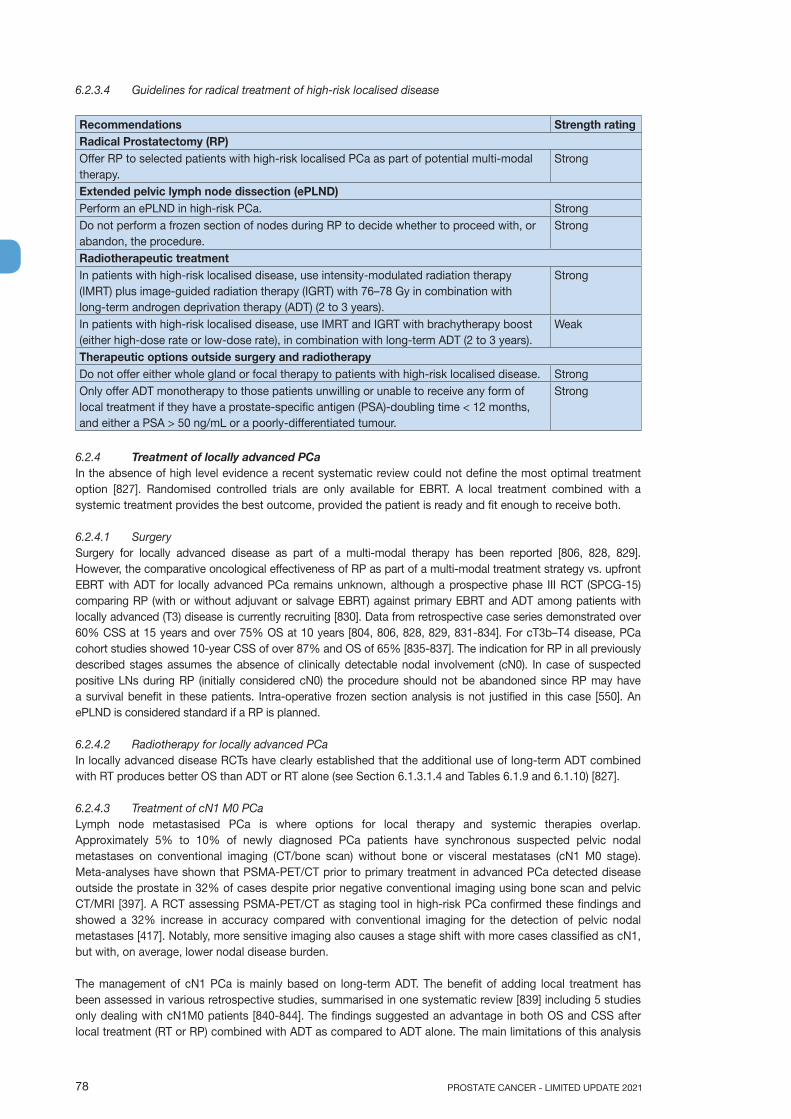
PROSTATE CANCER - LIMITED UPDATE 202178
6.2.3.4 Guidelines for radical treatment of high-risk localised disease
Recommendations Strength ratingRadical Prostatectomy (RP)Offer RP to selected patients with high-risk localised PCa as part of potential multi-modal therapy.
Strong
Extended pelvic lymph node dissection (ePLND)Perform an ePLND in high-risk PCa. StrongDo not perform a frozen section of nodes during RP to decide whether to proceed with, or abandon, the procedure.
Strong
Radiotherapeutic treatmentIn patients with high-risk localised disease, use intensity-modulated radiation therapy (IMRT) plus image-guided radiation therapy (IGRT) with 76–78 Gy in combination with long-term androgen deprivation therapy (ADT) (2 to 3 years).
Strong
In patients with high-risk localised disease, use IMRT and IGRT with brachytherapy boost (either high-dose rate or low-dose rate), in combination with long-term ADT (2 to 3 years).
Weak
Therapeutic options outside surgery and radiotherapyDo not offer either whole gland or focal therapy to patients with high-risk localised disease. StrongOnly offer ADT monotherapy to those patients unwilling or unable to receive any form of local treatment if they have a prostate-specific antigen (PSA)-doubling time < 12 months, and either a PSA > 50 ng/mL or a poorly-differentiated tumour.
Strong
6.2.4 Treatment of locally advanced PCaIn the absence of high level evidence a recent systematic review could not define the most optimal treatment option [827]. Randomised controlled trials are only available for EBRT. A local treatment combined with a systemic treatment provides the best outcome, provided the patient is ready and fit enough to receive both.
6.2.4.1 SurgerySurgery for locally advanced disease as part of a multi-modal therapy has been reported [806, 828, 829]. However, the comparative oncological effectiveness of RP as part of a multi-modal treatment strategy vs. upfront EBRT with ADT for locally advanced PCa remains unknown, although a prospective phase III RCT (SPCG-15) comparing RP (with or without adjuvant or salvage EBRT) against primary EBRT and ADT among patients with locally advanced (T3) disease is currently recruiting [830]. Data from retrospective case series demonstrated over 60% CSS at 15 years and over 75% OS at 10 years [804, 806, 828, 829, 831-834]. For cT3b–T4 disease, PCa cohort studies showed 10-year CSS of over 87% and OS of 65% [835-837]. The indication for RP in all previously described stages assumes the absence of clinically detectable nodal involvement (cN0). In case of suspected positive LNs during RP (initially considered cN0) the procedure should not be abandoned since RP may have a survival benefit in these patients. Intra-operative frozen section analysis is not justified in this case [550]. An ePLND is considered standard if a RP is planned.
6.2.4.2 Radiotherapy for locally advanced PCa In locally advanced disease RCTs have clearly established that the additional use of long-term ADT combined with RT produces better OS than ADT or RT alone (see Section 6.1.3.1.4 and Tables 6.1.9 and 6.1.10) [827].
6.2.4.3 Treatment of cN1 M0 PCa Lymph node metastasised PCa is where options for local therapy and systemic therapies overlap. Approximately 5% to 10% of newly diagnosed PCa patients have synchronous suspected pelvic nodal metastases on conventional imaging (CT/bone scan) without bone or visceral mestatases (cN1 M0 stage). Meta-analyses have shown that PSMA-PET/CT prior to primary treatment in advanced PCa detected disease outside the prostate in 32% of cases despite prior negative conventional imaging using bone scan and pelvic CT/MRI [397]. A RCT assessing PSMA-PET/CT as staging tool in high-risk PCa confirmed these findings and showed a 32% increase in accuracy compared with conventional imaging for the detection of pelvic nodal metastases [417]. Notably, more sensitive imaging also causes a stage shift with more cases classified as cN1, but with, on average, lower nodal disease burden.
The management of cN1 PCa is mainly based on long-term ADT. The benefit of adding local treatment has been assessed in various retrospective studies, summarised in one systematic review [839] including 5 studies only dealing with cN1M0 patients [840-844]. The findings suggested an advantage in both OS and CSS after local treatment (RT or RP) combined with ADT as compared to ADT alone. The main limitations of this analysis
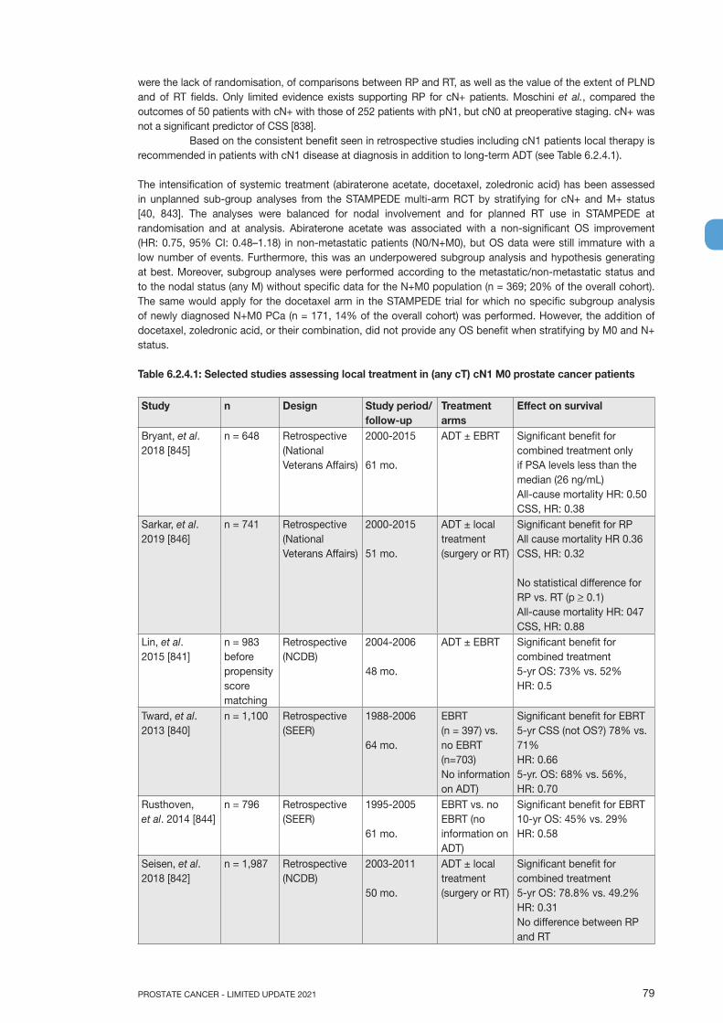
79PROSTATE CANCER - LIMITED UPDATE 2021
were the lack of randomisation, of comparisons between RP and RT, as well as the value of the extent of PLND and of RT fields. Only limited evidence exists supporting RP for cN+ patients. Moschini et al., compared the outcomes of 50 patients with cN+ with those of 252 patients with pN1, but cN0 at preoperative staging. cN+ was not a significant predictor of CSS [838].
Based on the consistent benefit seen in retrospective studies including cN1 patients local therapy is recommended in patients with cN1 disease at diagnosis in addition to long-term ADT (see Table 6.2.4.1).
The intensification of systemic treatment (abiraterone acetate, docetaxel, zoledronic acid) has been assessed in unplanned sub-group analyses from the STAMPEDE multi-arm RCT by stratifying for cN+ and M+ status [40, 843]. The analyses were balanced for nodal involvement and for planned RT use in STAMPEDE at randomisation and at analysis. Abiraterone acetate was associated with a non-significant OS improvement (HR: 0.75, 95% CI: 0.48–1.18) in non-metastatic patients (N0/N+M0), but OS data were still immature with a low number of events. Furthermore, this was an underpowered subgroup analysis and hypothesis generating at best. Moreover, subgroup analyses were performed according to the metastatic/non-metastatic status and to the nodal status (any M) without specific data for the N+M0 population (n = 369; 20% of the overall cohort). The same would apply for the docetaxel arm in the STAMPEDE trial for which no specific subgroup analysis of newly diagnosed N+M0 PCa (n = 171, 14% of the overall cohort) was performed. However, the addition of docetaxel, zoledronic acid, or their combination, did not provide any OS benefit when stratifying by M0 and N+ status.
Table 6.2.4.1: Selected studies assessing local treatment in (any cT) cN1 M0 prostate cancer patients
Study n Design Study period/follow-up
Treatment arms
Effect on survival
Bryant, et al. 2018 [845]
n = 648 Retrospective(National Veterans Affairs)
2000-2015
61 mo.
ADT ± EBRT Significant benefit for combined treatment only if PSA levels less than the median (26 ng/mL)All-cause mortality HR: 0.50 CSS, HR: 0.38
Sarkar, et al. 2019 [846]
n = 741 Retrospective(National Veterans Affairs)
2000-2015
51 mo.
ADT ± local treatment (surgery or RT)
Significant benefit for RPAll cause mortality HR 0.36CSS, HR: 0.32
No statistical difference for RP vs. RT (p > 0.1)All-cause mortality HR: 047CSS, HR: 0.88
Lin, et al. 2015 [841]
n = 983 before propensity score matching
Retrospective (NCDB)
2004-2006
48 mo.
ADT ± EBRT Significant benefit for combined treatment 5-yr OS: 73% vs. 52%HR: 0.5
Tward, et al. 2013 [840]
n = 1,100 Retrospective (SEER)
1988-2006
64 mo.
EBRT (n = 397) vs. no EBRT (n=703)No information on ADT)
Significant benefit for EBRT 5-yr CSS (not OS?) 78% vs. 71%HR: 0.665-yr. OS: 68% vs. 56%, HR: 0.70
Rusthoven, et al. 2014 [844]
n = 796 Retrospective (SEER)
1995-2005
61 mo.
EBRT vs. no EBRT (no information on ADT)
Significant benefit for EBRT 10-yr OS: 45% vs. 29%HR: 0.58
Seisen, et al. 2018 [842]
n = 1,987 Retrospective (NCDB)
2003-2011
50 mo.
ADT ± local treatment (surgery or RT)
Significant benefit for combined treatment 5-yr OS: 78.8% vs. 49.2%HR: 0.31No difference between RP and RT

PROSTATE CANCER - LIMITED UPDATE 202180
James, et al. 2016 [843]
n = 177 Unplanned sub-group analysis RCT
2005-2014
17 mo.
ADT ± EBRT Significant benefit for combined treatment 5-yr OS: 93% vs. 71%2-yr FFS: 81% vs 53%FFS, HR: 0.48
ADT = androgen deprivation therapy; EBRT = external beam radiotherapy in standard fractionation; FFS = failure-free survival; HR = hazard ratio; mo = months; n = number of patients; OS = overall survival; RT = radiotherapy; wk = week; yr = year.
6.2.4.3.1 Guidelines for the management of cN1 M0 prostate cancer
Recommendations Strength ratingOffer patients with cN1 disease a local treatment (either radical prostatectomy or intensity modulated radiotherapy plus image-guided radiotherapy) plus long-term ADT.
Weak
6.2.4.4 Options other than surgery and radiotherapy for primary treatment6.2.4.4.1 Investigational therapiesCurrently cryotherapy, HIFU or focal therapies have no place in the management of locally-advanced PCa.
6.2.4.4.2 Androgen deprivation therapy monotherapyThe deferred use of ADT as single treatment modality has been answered by the EORTC 30891 trial [800]. Nine hundred and eighty-five patients with T0-4 N0-2 M0 PCa received ADT alone, either immediately or after symptomatic progression or occurrence of serious complications. After a median follow-up of 12.8 years, the OS favoured immediate treatment (HR: 1.21, 95% CI: 1.05–1.39). Surprisingly, no different disease-free or symptom-free survival was observed, raising the question of survival benefit. In locally-advanced T3-T4 M0 disease unsuitable for surgery or RT, immediate ADT may only benefit patients with a PSA > 50 ng/mL and a PSA-DT < 12 months or those that are symptomatic [800, 847]. The median time to start deferred treatment was 7 years. In the deferred treatment arm 25.6% died without needing treatment.
6.2.4.5 Guidelines for radical treatment of locally-advanced disease
Recommendations Strength ratingRadical Prostatectomy (RP)Offer RP to selected patients with locally-advanced PCa as part of multi-modal therapy. StrongExtended pelvic lymph node dissection (ePLND)Perform an ePLND prior to RP in locally-advanced PCa. StrongRadiotherapeutic treatmentsIn patients with locally-advanced disease, offer intensity-modulated radiation therapy (IMRT) plus image-guide radiation therapy in combination with long-term androgen deprivation therapy (ADT).
Strong
Offer long-term ADT for at least 2 years. WeakTherapeutic options outside surgery and radiotherapyDo not offer whole gland treatment or focal treatment to patients with locally-advanced PCa.
Strong
Only offer ADT monotherapy to those patients unwilling or unable to receive any form of local treatment if they have a prostate-specific antigen (PSA)-doubling time < 12 months, and either a PSA > 50 ng/mL, a poorly-differentiated tumour or troublesome local disease-related symptoms.
Strong
Offer patients with cN1 disease a local treatment (either RP or IMRT plus IGRT) plus long-term ADT.
Strong
6.2.5 Adjuvant treatment after radical prostatectomy6.2.5.1 IntroductionAdjuvant treatment is by definition additional to the primary or initial therapy with the aim of decreasing the risk of relapse. A post-operative detectable PSA is an indication of persistent prostate cells (see Section 6.2.6). All information listed below refers to patients with a post-operative undetectable PSA.
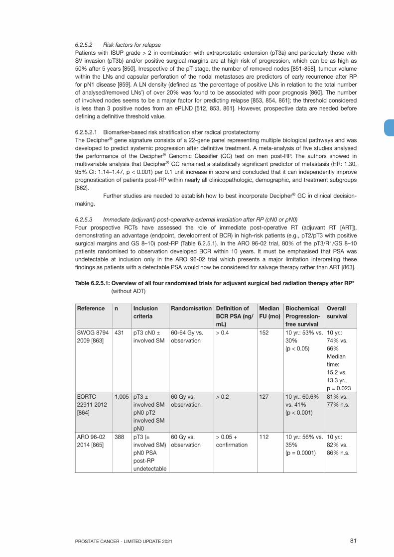
81PROSTATE CANCER - LIMITED UPDATE 2021
6.2.5.2 Risk factors for relapsePatients with ISUP grade > 2 in combination with extraprostatic extension (pT3a) and particularly those with SV invasion (pT3b) and/or positive surgical margins are at high risk of progression, which can be as high as 50% after 5 years [850]. Irrespective of the pT stage, the number of removed nodes [851-858], tumour volume within the LNs and capsular perforation of the nodal metastases are predictors of early recurrence after RP for pN1 disease [859]. A LN density (defined as ‘the percentage of positive LNs in relation to the total number of analysed/removed LNs’) of over 20% was found to be associated with poor prognosis [860]. The number of involved nodes seems to be a major factor for predicting relapse [853, 854, 861]; the threshold considered is less than 3 positive nodes from an ePLND [512, 853, 861]. However, prospective data are needed before defining a definitive threshold value.
6.2.5.2.1 Biomarker-based risk stratification after radical prostatectomyThe Decipher® gene signature consists of a 22-gene panel representing multiple biological pathways and was developed to predict systemic progression after definitive treatment. A meta-analysis of five studies analysed the performance of the Decipher® Genomic Classifier (GC) test on men post-RP. The authors showed in multivariable analysis that Decipher® GC remained a statistically significant predictor of metastasis (HR: 1.30, 95% CI: 1.14–1.47, p < 0.001) per 0.1 unit increase in score and concluded that it can independently improve prognostication of patients post-RP within nearly all clinicopathologic, demographic, and treatment subgroups [862].
Further studies are needed to establish how to best incorporate Decipher® GC in clinical decision-making.
6.2.5.3 Immediate (adjuvant) post-operative external irradiation after RP (cN0 or pN0)Four prospective RCTs have assessed the role of immediate post-operative RT (adjuvant RT [ART]), demonstrating an advantage (endpoint, development of BCR) in high-risk patients (e.g., pT2/pT3 with positive surgical margins and GS 8–10) post-RP (Table 6.2.5.1). In the ARO 96-02 trial, 80% of the pT3/R1/GS 8–10 patients randomised to observation developed BCR within 10 years. It must be emphasised that PSA was undetectable at inclusion only in the ARO 96-02 trial which presents a major limitation interpreting these findings as patients with a detectable PSA would now be considered for salvage therapy rather than ART [863].
Table 6.2.5.1: Overview of all four randomised trials for adjuvant surgical bed radiation therapy after RP* (without ADT)
Reference n Inclusion criteria
Randomisation Definition of BCR PSA (ng/mL)
Median FU (mo)
Biochemical Progression-free survival
Overall survival
SWOG 8794 2009 [863]
431 pT3 cN0 ± involved SM
60-64 Gy vs. observation
> 0.4 152 10 yr.: 53% vs. 30% (p < 0.05)
10 yr.: 74% vs. 66% Median time: 15.2 vs. 13.3 yr., p = 0.023
EORTC 22911 2012 [864]
1,005 pT3 ± involved SM pN0 pT2 involved SM pN0
60 Gy vs. observation
> 0.2 127 10 yr.: 60.6% vs. 41%(p < 0.001)
81% vs. 77% n.s.
ARO 96-02 2014 [865]
388 pT3 (± involved SM) pN0 PSA post-RP undetectable
60 Gy vs. observation
> 0.05 + confirmation
112 10 yr.: 56% vs. 35% (p = 0.0001)
10 yr.: 82% vs. 86% n.s.
PROSTATE CANCER - LIMITED UPDATE 202182
FinnProstate Group 2019 [866]
250 pT2,R1/ pT3a
66.6 Gy vs. observation (+SRT)
> 0.4 (in 2 successive measurements)
112 vs. 103 (patients alive)
10 yr.: 82% vs. 61% p < 0.001
10 yr.: 92% vs. 87% n.s.
*See Section 6.3.5.1 for delayed (salvage) post-radical prostatectomy external irradiation.BCR = biochemical recurrence; FU = follow-up; mo = months; n = number of patients; n.s. = not significant; OS = overall survival; PSA = prostate-specific antigen; RP = radical prostatectomy; SM = surgical margin.
6.2.5.4 Comparison of adjuvant- and salvage radiotherapyTwo retrospective matched studies (510 and 149 patients receiving ART) failed to show an advantage for metastasis-free survival [867, 868]. However, both studies were underpowered for high-risk patients (pT3b/R1/ISUP grade 4–5 PCa).
In contrast to these studies, a propensity score-matched retrospective analysis of two cohorts of 366 pT3 and/or R1 patients found that compared to SRT at a PSA between 0.1 and 0.5 ng/mL, ART given at an undetectable PSA (< 0.1 ng/mL) improved all three endpoints; BCR, metastasis-free survival, and OS [869].
Both approaches (ART and early SRT) together with the efficacy of adjuvant ADT are compared in three prospective RCTs: the Medical Research Council (MRC) Radiotherapy and Androgen Deprivation In Combination After Local Surgery (RADICALS) [870], the Trans-Tasman Oncology Group (TROG) Radiotherapy Adjuvant Versus Early Salvage (RAVES) [871], and Groupe d’Etude des Tumeurs Uro-Génitales (GETUG-AFU 17) [872]. In addition, a preplanned meta-analysis of all three trials has been published (Table 6.2.5.2) [873]. Two trials closed early after randomising 333/470 patients (RAVES) and 424/718 (GETUG-AFU-17) patients. RADICALS-RT included 1,396 patients with the option of subsequent inclusion in RADICALS-HT; 154/649 (24%) of patients starting in the adjuvant RT group also received neo-adjuvant or adjuvant hormone therapy; 90 patients for 6 months/45 for 2 years/19 patients outside RADICALS-HT. From the SRT group, 61/228 (27%) received neo-adjuvant or adjuvant hormone therapy for 6 months (n = 33) and 2 years (n = 13). Fifteen of these patients were treated outside the trial [870]. All men in the GETUG-AFU-17 trial (n = 424) received 6 months of hormone treatment. All together, 684 out of 2,153 patients received additional ADT for at least 6 months across both trials [873]. Radiotherapy to the pelvic lymphatics was allowed in the GETUG-AFU and in the RADICALS-RT Trials. To be included in these trails, patients must have had a post-RP PSA < 0.2 ng/mL (RADICALS-RT) or < 0.1 ng/mL (RAVES, GETUG-AFU17) (see Table 6.2.5.2). Biochemical progression was defined as a PSA > 0.4 ng/mL after RT.
The primary endpoint for RAVES and GETUG-AFU 17 was biochemical PFS and for RADICALS-RT metastasis-free survival. So far only PFS data has been reported, and not metastasis-free survival or OS data. With a median follow-up between 4.9 years and 6.25 years there was no statistical significant difference for biochemical PFS (BPFS) for both treatments in all three trials (see Table 6.2.5.2) indicating that in the majority of patients adjuvant irradiation should be avoided. Additionally, there was a significant lower rate of grade > 2 GU late side effects and grade 3–4 urethral strictures in favour of early SRT; which may also be caused by the low number of patients with PSA-progression and subsequent need for early SRT at the time of analysis (40% of patients).
It is important to note that the indication for ART changed over the last ten years with the introduction of ultra-sensitive PSA-tests, favouring early SRT. Therefore many patients, randomised in these three trials (accruing 2006–2008) are not likely to benefit from ART as there is a low risk of biochemical progression (~20–30%) in, for example, pT3R0- or pT2R1-tumours. The median pre-SRT-PSA in all 3 trials was 0.24 ng/mL which is much lower than the conventional cut-off level of PSA < 0.5 ng/mL used to base ´early´ SRT on. Therefore, patients with ‘low-risk factors’ of biochemical progression after RP should be closely followed up with ultra-senstive assays and SRT should start as soon as PSA starts to rise, which has to be confirmed by a second PSA measurement. The proportion of ´very high risk´ patients (with at least two out of three features: pT3/R1/GS 8-10) in all three trials was low (between 10–20%) and therefore even the meta-analysis may be underpowered to show an outcome in favour of SRT [873]. In addition, the side-effect profile may have been impacted with a larger proportion of ART patients receiving treatment with older 3D-treatment planning techniques as compared to SRT patients (GETUG-AFU 17: ART, 69% 3D vs. 46% SRT) and patients treated more recently were more likely to undergo IMRT techniques with a proven lower rate of late side effects [610].
For these reasons 10-year results and results of metastasis-free survival endpoints should be awaited before drawing final conclusions. Due to the small number of patients with a combination of high-risk features included in these three trials (between 10–20%) ART remains a recommended treatment option in highly selected patients with at least two out of three high-risk features (‘very high-risk patients’) such as pT3/R1/ISUP > 3 [854, 874].
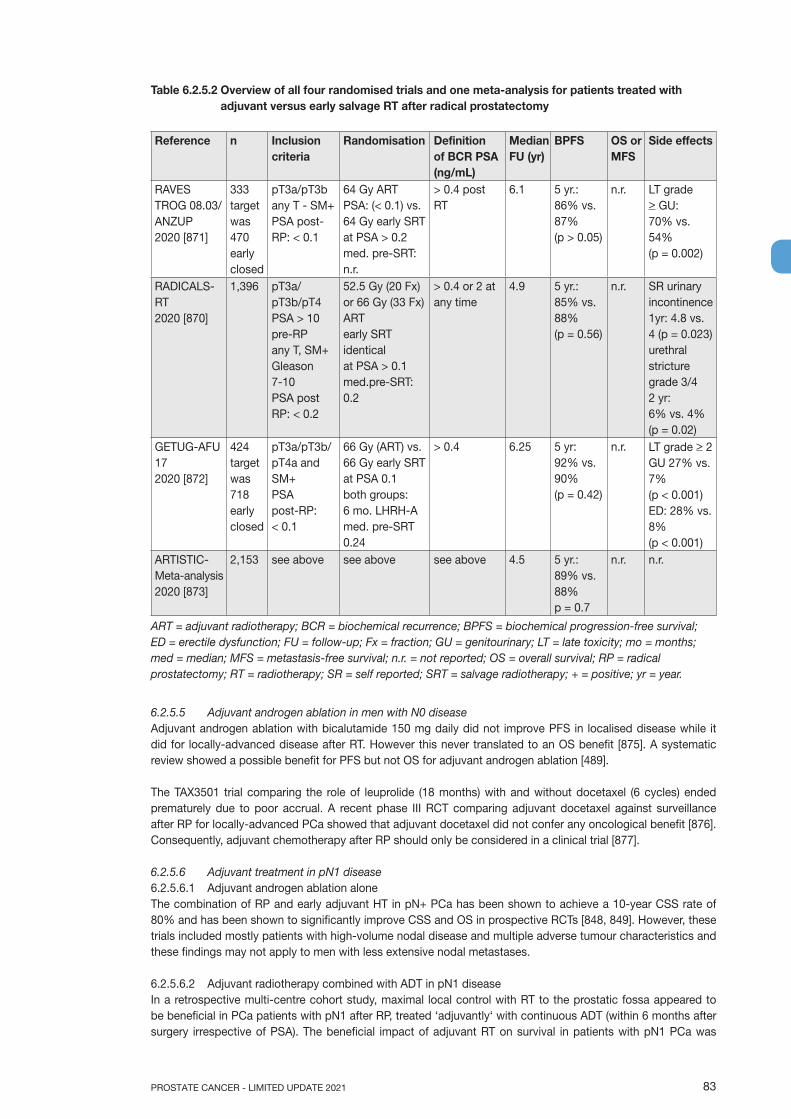
83PROSTATE CANCER - LIMITED UPDATE 2021
Table 6.2.5.2 Overview of all four randomised trials and one meta-analysis for patients treated with adjuvant versus early salvage RT after radical prostatectomy
Reference n Inclusion criteria
Randomisation Definition of BCR PSA (ng/mL)
Median FU (yr)
BPFS OS or MFS
Side effects
RAVES TROG 08.03/ANZUP2020 [871]
333targetwas 470earlyclosed
pT3a/pT3bany T - SM+PSA post-RP: < 0.1
64 Gy ARTPSA: (< 0.1) vs.64 Gy early SRTat PSA > 0.2med. pre-SRT: n.r.
> 0.4 post RT
6.1 5 yr.:86% vs. 87% (p > 0.05)
n.r. LT grade> GU:70% vs.54%(p = 0.002)
RADICALS-RT2020 [870]
1,396 pT3a/pT3b/pT4PSA > 10 pre-RPany T, SM+Gleason 7-10PSA post RP: < 0.2
52.5 Gy (20 Fx) or 66 Gy (33 Fx) ARTearly SRTidenticalat PSA > 0.1med.pre-SRT: 0.2
> 0.4 or 2 at any time
4.9 5 yr.: 85% vs. 88%(p = 0.56)
n.r. SR urinary incontinence 1yr: 4.8 vs. 4 (p = 0.023)urethralstricturegrade 3/4 2 yr:6% vs. 4%(p = 0.02)
GETUG-AFU 17 2020 [872]
424target was718early closed
pT3a/pT3b/pT4a and SM+PSA post-RP: < 0.1
66 Gy (ART) vs. 66 Gy early SRTat PSA 0.1 both groups:6 mo. LHRH-Amed. pre-SRT 0.24
> 0.4 6.25 5 yr:92% vs. 90%(p = 0.42)
n.r. LT grade > 2 GU 27% vs. 7%(p < 0.001)ED: 28% vs. 8%(p < 0.001)
ARTISTIC-Meta-analysis2020 [873]
2,153 see above see above see above 4.5 5 yr.: 89% vs. 88%p = 0.7
n.r. n.r.
ART = adjuvant radiotherapy; BCR = biochemical recurrence; BPFS = biochemical progression-free survival; ED = erectile dysfunction; FU = follow-up; Fx = fraction; GU = genitourinary; LT = late toxicity; mo = months; med = median; MFS = metastasis-free survival; n.r. = not reported; OS = overall survival; RP = radical prostatectomy; RT = radiotherapy; SR = self reported; SRT = salvage radiotherapy; + = positive; yr = year.
6.2.5.5 Adjuvant androgen ablation in men with N0 diseaseAdjuvant androgen ablation with bicalutamide 150 mg daily did not improve PFS in localised disease while it did for locally-advanced disease after RT. However this never translated to an OS benefit [875]. A systematic review showed a possible benefit for PFS but not OS for adjuvant androgen ablation [489].
The TAX3501 trial comparing the role of leuprolide (18 months) with and without docetaxel (6 cycles) ended prematurely due to poor accrual. A recent phase III RCT comparing adjuvant docetaxel against surveillance after RP for locally-advanced PCa showed that adjuvant docetaxel did not confer any oncological benefit [876]. Consequently, adjuvant chemotherapy after RP should only be considered in a clinical trial [877].
6.2.5.6 Adjuvant treatment in pN1 disease6.2.5.6.1 Adjuvant androgen ablation aloneThe combination of RP and early adjuvant HT in pN+ PCa has been shown to achieve a 10-year CSS rate of 80% and has been shown to significantly improve CSS and OS in prospective RCTs [848, 849]. However, these trials included mostly patients with high-volume nodal disease and multiple adverse tumour characteristics and these findings may not apply to men with less extensive nodal metastases.
6.2.5.6.2 Adjuvant radiotherapy combined with ADT in pN1 diseaseIn a retrospective multi-centre cohort study, maximal local control with RT to the prostatic fossa appeared to be beneficial in PCa patients with pN1 after RP, treated ‘adjuvantly‘ with continuous ADT (within 6 months after surgery irrespective of PSA). The beneficial impact of adjuvant RT on survival in patients with pN1 PCa was
PROSTATE CANCER - LIMITED UPDATE 202184
highly influenced by tumour characteristics. Men with low-volume nodal disease (< 3 LNs), ISUP grade 2–5 and pT3–4 or R1, as well as men with 3 to 4 positive nodes were more likely to benefit from RT after surgery, while the other subgroups did not [878].
These results were confirmed by a US National Cancer Database analysis based on 5,498 patients [879]. Another US National Cancer Database study including 8,074 pN1 patients reports improved OS after ADT + EBRT (including pelvic LNs) vs. observation and vs. ADT alone in all men with single or multiple adverse pathological features. Men without any adverse pathological features did not benefit from immediate adjuvant therapy [880].
In a series of 2,596 pN1 patients receiving ADT (n = 1,663) or ADT plus RT (n = 906), combined treatment was associated with improved OS, with a HR of 1.5 for ADT alone [881]. In a SEER retrospective population-based analysis, adding RT to RP showed a non-significant trend for improved OS but not PCa-specific survival, but data on the extent of additional RT is lacking in this study [844]. Radiotherapy should be given to the pelvic lymphatics and the prostatic fossa [878, 882-884]. For pN1 patients no data are available regarding adjuvant EBRT without ADT.
6.2.5.6.3 Observation of pN1 patients after radical prostatectomy and extended lymph node dissectionSeveral retrospective studies and a systematic review addressed the management of patients with pN1 PCa at RP [861, 878, 884-886]. A subset of patients with limited nodal disease (1–2 positive LNs) showed favourable oncological outcomes and did not require additional treatment.
An analysis of 209 pN1 patients with one or two positive LNs at RP showed that 37% remained metastasis-free without need of salvage treatment at a median follow-up of 60.2 months [885]. Touijer et al. reported their results of 369 LN-positive patients (40 with and 329 without adjuvant treatment) and showed that higher pathologic grade group and three or more positive LNs were significantly associated with an increased risk of BCR on multivariable analysis [861]. Biochemical-free survival rates in pN1 patients without adjuvant treatment ranged from 43% at 4 years to 28% at 10 years [886]. Reported CSS rates were 78% at 5 years and 72% at 10 years. The majority of these patients were managed with initial observation after surgery, had favourable disease characteristics, and 63% had only one positive node [886].
6.2.5.7 Guidelines for adjuvant treatment in pN0 and pN1 disease after radical prostatectomy
Recommendations Strength ratingDo not prescribe adjuvant androgen deprivation therapy (ADT) in pN0 patients. StrongOnly offer adjuvant intensity-modulated radiation therapy (IMRT) plus image-guided radiation therapy (IGRT) to high-risk patients (pN0) with at least two out of three high-risk features (ISUP grade group 4–5, pT3 ± positive margins).
Strong
Discuss three management options with patients with pN1 disease after an extended lymph node dissection, based on nodal involvement characteristics:1. Offer adjuvant ADT;2. Offer adjuvant ADT with additional IMRT plus IGRT;3. Offer observation (expectant management) to a patient after eLND and < 2 nodes and a
PSA < 0.1 ng/mL.
Weak
6.2.5.8 Guidelines for non-curative or palliative treatments in prostate cancer
Recommendations Strength ratingWatchful waiting (WW) for localised prostate cancerOffer WW to asymptomatic patients not eligible for local curative treatment and those with a short life expectancy.
Strong
Watchful waiting for locally-advanced prostate cancerOffer a deferred treatment policy using androgen deprivation monotherapy to M0 asymptomatic patients with a prostate-specific antigen (PSA) doubling time > 12 months, a PSA < 50 ng/mL and well-differentiated tumour, who are unwilling or unable to receive any form of local treatment.
Weak

85PROSTATE CANCER - LIMITED UPDATE 2021
6.2.6 Persistent PSA after radical prostatectomyBetween 5 and 20% of men continue to have detectable or persistent PSA after RP (when defined in the majority of studies as detectable post-RP PSA of > 0.1 ng/mL within 4 to 8 weeks of surgery) [887, 888]. It may result from persistent local disease, pre-existing metastases or residual benign prostate tissue.
6.2.6.1 Natural history of persistently elevated PSA after RPSeveral studies have shown that persistent PSA after RP is associated with more advanced disease (such as positive surgical margins (PSM), pathologic stage > T3a, positive nodal status and/or pathologic ISUP grade > 3) and poor prognosis (See table 6.2.6.1). Initially defined as > 0.1 ng/mL, improvements in the sensitivity of PSA assays now allow for the detection of PSA at much lower levels.
Moreira et al., demonstrated that failure to achieve a PSA of less than 0.03 ng/mL within 6 months of surgery was associated with an increased risk of BCR and overall mortality [889, 890]. However, since the majority of the published literature is based on the > 0.1 ng/mL PSA cut-off there is significantly more long-term data for this definition.
Predictors of PSA persistence were higher BMI, higher pre-operative PSA and ISUP grade > 3 [890]. In patients with PSA persistence, one and 5-year BCR-free survival were 68% and 36%, compared to 95% and 72%, respectively, in men without PSA persistence [889]. Ten-year OS in patients with and without PSA persistence was 63% and 80%, respectively. In line with these data, Ploussard et al. reported that approximately 74% of patients with persistent PSA develop BCR [887]. Spratt et al., confirmed that a persistently detectable PSA after RP represents one of the worst prognostic factors associated with oncological outcome [891]. Of 150 patients with a persistent PSA, 95% received RT before detectable metastasis. In a multivariable analysis the presence of a persistently detectable PSA post-RP was associated with a 4-fold increase in the risk of developing metastasis. This was confirmed by recent data from Preisser et al. who showed that persistent PSA is prognostic of an increased risk of metastasis and death [892]. At 15 years after RP, metastasis-free survival rates, OS and CSS rates were 53.0 vs. 93.2% (p < 0.001), 64.7 vs. 81.2% (p < 0.001) and 75.5 vs. 96.2% (p < 0.001) for persistent vs. undetectable PSA, respectively. The median follow-up was 61.8 months for patients with undetectable PSA vs. 46.4 months for patients with persistent PSA. In multivariable Cox regression models, persistent PSA represented an independent predictor for metastasis (HR: 3.59, p < 0.001), death (HR: 1.86, p < 0.001) and cancer-specific death (HR: 3.15, p < 0.001).
However, not all patients with persistent PSA after RP experience disease recurrence. Xiang et al., showed a 50% 5-year BCR-free survival in men who had a persistent PSA level > 0.1 but < 0.2 ng/mL at 6-8 weeks after RP [893].
Rogers et al., assessed the clinical outcome of 160 men with a persistently detectable PSA level after RP [894]. No patient received adjuvant therapy before documented metastasis. In their study, 38% of patients had no evidence of metastases for > 7 years while 32% of the patients were reported to develop metastases within 3 years. Noteworthy is that a significant proportion of patients had low-risk disease. In multivariable analysis the PSA slope after RP (as calculated using PSA levels 3 to 12 months after surgery) and pathological ISUP grade were significantly associated with the development of distant metastases.
Table 6.2.6.1: Studies on the natural history of patients with persistent PSA after RP
Authors Study population
n Definition PSA persistence
Treatment Outcome Other details/comments
Ploussard et al., J Urol 2013 [887]
496 men pN0 with persistent PSA 14 centres 1998 - 2011
PSA > 0.1 ng/mL at 6 wk.
74.4% with BCR5% with metastasis
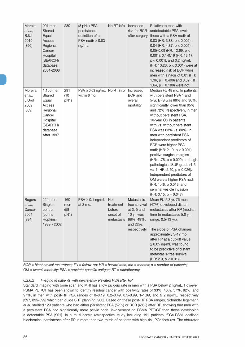
PROSTATE CANCER - LIMITED UPDATE 202186
Moreira et al., BJUI 2010 [890]
901 men Shared Equal Access Regional Cancer Hospital (SEARCH) database. 2001-2008
230 (8 pN1) PSA persistence definition of a PSA nadir > 0.03 ng/mL
No RT info Increased risk for BCR after surgery
Relative to men with undetectable PSA levels, those with a PSA nadir of 0.03 (HR: 3.88, p < 0.001), 0.04 (HR: 4.87, p < 0.001), 0.05-0.09 (HR: 12.69, p < 0.001), 0.1-0.19 (HR: 13.17, p < 0.001), and 0.2 ng/mL (HR: 13.23, p < 0.001) were at increased risk of BCR while men with a nadir of 0.01 (HR: 1.36, p = 0.400) and 0.02 (HR: 1.64, p = 0.180) were not.
Moreira et al., J Urol 2009 [889]
1,156 men Shared Equal Access Regional Cancer Hospital (SEARCH) database. After 1997
291 (10 pN1)
PSA > 0.03 ng/mL within 6 mo.
No RT info Increased BCR and overall mortality
Median FU 48 mo. In patients with persistent PSA 1 and 5-yr. BFS was 68% and 36%, significantly lower than 95% and 72%, respectively, in men without persistent PSA. 10-year OS in patients with vs. without persistent PSA was 63% vs. 80%. In men with persistent PSA independent predictors of BCR were higher PSA nadir (HR: 2.19, p < 0.001), positive surgical margins (HR: 1.75, p = 0.022) and high pathological ISUP grade (4-5 vs. 1, HR: 2.40, p = 0.026). Independent predictors of OM were a higher PSA nadir (HR: 1.46, p 0.013) and seminal vesicle invasion (HR: 3.15, p = 0.047)
Rogers et al., Cancer 2004 [894]
224 menSingle-centre (Johns Hopkins)1989 - 2002
160 men (58 pN1)
PSA > 0.1 ng/mL at 3 mo.
No treatment before onset of metastasis
Metastasis-free survival at 3, 5 and 10 yr. was 68%, 49%, and 22%, respectively.
Mean FU 5.3 yr. 75 men (47%) developed distant metastases after RP (median time to metastases 5.0 yr.; range, 0.5-13 yr.).
The slope of PSA changes approximately 3-12 mo. after RP at a cut-off value > 0.05 ng/mL was found to be predictive of distant metastasis-free survival (HR: 2.9, p < 0.01).
BCR = biochemical recurrence; FU = follow-up; HR = hazard ratio; mo = months; n = number of patients; OM = overall mortality; PSA = prostate-specific antigen; RT = radiotherapy.
6.2.6.2 Imaging in patients with persistently elevated PSA after RPStandard imaging with bone scan and MRI has a low pick-up rate in men with a PSA below 2 ng/mL. However, PSMA PET/CT has been shown to identify residual cancer with positivity rates of 33%, 46%, 57%, 82%, and 97%, in men with post-RP PSA ranges of 0–0.19, 0.2–0.49, 0.5–0.99, 1–1.99, and > 2 ng/mL, respectively [397, 895-899] which can guide SRT planning [900]. Based on these post-RP PSA ranges, Schmidt-Hegemann et al. studied 129 patients who had either persistent PSA (52%) or BCR (48%) after RP, showing that men with a persistent PSA had significantly more pelvic nodal involvement on PSMA PET/CT than those developing a detectable PSA [901]. In a multi-centre retrospective study including 191 patients, 68Ga-PSM localised biochemical persistence after RP in more than two-thirds of patients with high-risk PCa features. The obturator
87PROSTATE CANCER - LIMITED UPDATE 2021
and presacral/mesorectal nodes were identified as high risk for residual disease [902]. Another retrospective study included 150 patients with persistent PSA after RARP who were re-staged with both 68Ga-PSMA and 18F-DCFPyL PSMA. The authors found that in the presence of persistent PSA the majority of patients already had metastatic pelvic LNs or distant metastases which would support a role of PSMA PET/CT imaging in guiding (salvage) treatment strategies [903]. At present there is uncertainty regarding the best treatment if PSMA PET/CT shows metastatic disease.
6.2.6.3 Impact of post-operative RT and/or ADT in patients with persistent PSAThe benefit of SRT in patients with persistent PSA remains unclear due to a lack of RCTs, however, it would appear that men with a persistent PSA do less well than men with BCR undergoing RT.
Wiegel et al., [888] showed that following salvage RT to the prostate bed, patients with a detectable PSA after RP had significantly worse oncological outcomes when compared with those who achieved an undetectable PSA. Ten-year metastasis-free survival was 67% vs. 83% and OS was 68% vs. 84%, respectively. Recent data from Preisser et al., [892] also compared oncological outcomes of patients with persistent PSA who received SRT vs. those who did not. In the subgroup of patients with persistent PSA, after 1:1 propensity score matching between patients with SRT vs. no RT, OS rates at 10 years after RP were 86.6 vs. 72.6% in the entire cohort (p < 0.01), 86.3 vs. 60.0% in patients with positive surgical margin (p = 0.02), 77.8 vs. 49.0% in pT3b disease (p < 0.001), 79.3 vs. 55.8% in ISUP grade 1 disease (p < 0.01) and 87.4 vs. 50.5% in pN1 disease (p < 0.01), respectively. Moreover, CSS rates at 10 years after RP were 93.7 vs. 81.6% in the entire cohort (p < 0.01), 90.8 vs. 69.7% in patients with positive surgical margin (p = 0.04), 82.7 vs. 55.3% in pT3b disease (p < 0.01), 85.4 vs. 69.7% in ISUP grade 1 disease (p < 0.01) and 96.2 vs. 55.8% in pN1 disease (p < 0.01), for SRT vs. no RT, respectively. In multivariable models, after 1:1 propensity score matching, SRT was associated with lower risk for death (HR: 0.42, p = 0.02) and lower cancer-specific death (HR: 0.29, p = 0.03). These survival outcomes in patients with persistent PSA who underwent SRT suggest they benefit but outcomes are worse than for men experiencing BCR.
It is clear from a number of studies that poor outcomes are driven by the level of pre-RT PSA, the presence of ISUP grade > 4 in the RP histology and pT3b disease [888, 904-908]. Fossati et al., suggested that only men with a persistent PSA after RP and ISUP grade < 3 benefit significantly [909], although this is not supported by Preisser et al. [892]. The current data does not allow making any clear treatment decisions.
Addition of ADT may improve PFS [904]. Choo et al. studied the addition of 2-year ADT to immediate RT to the prostate bed in patients with pathologic T3 disease (pT3) and/or positive surgical margins after RP [904]. Twenty-nine of the 78 included patients had persistently detectable post-operative PSA. The relapse-free rate was 85% at 5 years and 68% at 7 years, which was superior to the 5-year progression-free estimates of 74% and 61% in the post-operative RT arms of the EORTC and the SWOG studies, respectively, which included patients with undetectable PSA after RP [863, 864]. Patients with persistently detectable post-operative PSA comprised approximately 50% and 12%, respectively, of the study cohorts in the EORTC and the SWOG studies.
In the ARO 96-02, a prospective RCT, 74 patients with PSA persistence (20%) received immediate SRT only (66 Gy per protocol [arm C]). The 10-year clinical relapse-free survival was 63% [888]. The GETUG-22 trial comparing RT with RT plus short-term ADT for post-RP PSA persistence (0.2–2.0 ng/mL) reported good tolerability of the combined treatment. The oncological endpoints are yet to be published [910].
Ploussard and colleagues recently performed a systematic review of oncologic outcomes and effectiveness of salvage therapies in men with persistent PSA after RP. Their findings confirmed a strong correlation of PSA persistence with poor oncologic outcomes [911]. The authors also report that SRT was associated with improved survival outcomes, although the available evidence is of low quality [911].
6.2.6.4 ConclusionThe available data suggest that patients with PSA persistence after RP may benefit from early aggressive multi-modality treatment, however, the lack of prospective RCTs makes firm recommendations difficult.
PROSTATE CANCER - LIMITED UPDATE 202188
6.2.6.5 Recommendations for the management of persistent PSA after radical prostatectomy
Recommendations Strength ratingOffer a prostate-specific membrane antigen positron emission tomography (PSMA PET) scan to men with a persistent prostate-specific antigen > 0.2 ng/mL if the results will influence subsequent treatment decisions.
Weak
Treat men with no evidence of metastatic disease with salvage radiotherapy and additional hormonal therapy.
Weak
6.3 Management of PSA-only recurrence after treatment with curative intentFollow-up will be addressed in Chapter 7 and is not discussed here.
6.3.1 BackgroundBetween 27% and 53% of all patients undergoing RP or RT develop a rising PSA (PSA recurrence). Whilst a rising PSA level universally precedes metastatic progression, physicians must inform the patient that the natural history of PSA-only recurrence may be prolonged and that a measurable PSA may not necessarily lead to clinically apparent metastatic disease. Physicians treating patients with PSA-only recurrence face a difficult set of decisions in attempting to delay the onset of metastatic disease and death while avoiding over-treating patients whose disease may never affect their OS or QoL. It should be emphasised that the treatment recommendations for these patients should be given after discussion in a multidisciplinary team.
6.3.2 Definitions of clinically relevant PSA relapseThe PSA level that defines treatment failure depends on the primary treatment. Patients with rising PSA after RP or primary RT have different risks of subsequent symptomatic metastatic disease based on various parameters, including the PSA level. Therefore, physicians should carefully interpret BCR endpoints when comparing treatments.
After RP, the threshold that best predicts further metastases is a PSA > 0.4 ng/mL and rising [912-914]. However, with access to ultra-sensitive PSA testing, a rising PSA much below this level will be a cause for concern for patients. After primary RT, with or without short-term hormonal manipulation, the RTOG-ASTRO Phoenix Consensus Conference definition of PSA failure (with an accuracy of > 80% for clinical failure) is ‘any PSA increase > 2 ng/mL higher than the PSA nadir value, regardless of the serum concentration of the nadir’ [915].
After HIFU or cryotherapy no endpoints have been validated against clinical progression or survival; therefore, it is not possible to give a firm recommendation of an acceptable PSA threshold after these alternative local treatments [916].
6.3.3 Natural history of biochemical recurrenceOnce a PSA relapse has been diagnosed, it is important to determine whether the recurrence has developed at local or distant sites. A recent systematic review and meta-analysis investigated the impact of BCR on hard endpoints and concluded that patients experiencing BCR are at an increased risk of developing distant metastases, PCa-specific and overall mortality [916]. However, the effect size of BCR as a risk factor for mortality is highly variable. After primary RP its impact ranges from HR 1.03 (95% CI: 1.004–1.06) to HR 2.32 (95% CI: 1.45–3.71) [917, 918]. After primary RT, OS rates are approximately 20% lower at 8 to 10 years follow-up even in men with minimal co-morbidity [919, 920]. Still, the variability in reported effect sizes of BCR remains high and suggests that only certain patient subgroups with BCR might be at an increased risk of mortality.
The risk of subsequent metastases, PCa-specific- and overall mortality may be predicted by the initial clinical and pathologic factors (e.g., T-category, PSA, ISUP grade) and PSA kinetics (PSA-DT and interval to PSA failure), which was further investigated by this systematic review [916].
For patients with BCR after RP, the following outcomes were found to be associated with significant prognostic factors:• distant metastatic recurrence: positive surgical margins, high RP specimen pathological ISUP grade, high
pT category, short PSA-DT, high pre-SRT PSA;• prostate-cancer-specific mortality: high RP specimen pathological ISUP grade, short interval to
biochemical failure as defined by investigators, short PSA-DT;• overall mortality: high RP specimen pathological ISUP grade, short interval to biochemical failure, high
PSA-DT.
89PROSTATE CANCER - LIMITED UPDATE 2021
For patients with BCR after RT, the corresponding outcomes are:• distant metastatic recurrence: high biopsy ISUP grade, high cT category, short interval to biochemical
failure;• prostate-cancer-specific mortality: short interval to biochemical failure;• overall mortality: high age, high biopsy ISUP grade, short interval to biochemical failure, high initial
(pre-treatment) PSA.
Based on this meta-analysis, proposal is to stratify patients into ‘EAU Low-Risk BCR’ (PSA-DT > 1 year AND pathological ISUP grade < 4 for RP; interval to biochemical failure > 18 months AND biopsy ISUP grade < 4 for RT) or ‘EAU High-Risk BCR’ (PSA-DT < 1 year OR pathological ISUP grade 4–5 for RP, interval to biochemical failure < 18 months OR biopsy ISUP grade 4–5 for RT), since not all patients with BCR will have similar outcomes. The stratification into ‘EAU Low-Risk’ or ‘EAU High-Risk’ BCR has recently been validated in a European cohort [921].
6.3.4 The role of imaging in PSA-only recurrenceImaging is only of value if it leads to a treatment change which results in an improved outcome. In practice, however, there are very limited data available regarding the outcomes consequent on imaging at relapse.
6.3.4.1 Assessment of metastases6.3.4.1.1 Bone scan and abdominopelvic CTBecause BCR after RP or RT precedes clinical metastases by 7 to 8 years on average [855, 922], the diagnostic yield of common imaging techniques (bone scan and abdominopelvic CT) is low in asymptomatic patients [923]. In men with PSA-only relapse after RP the probability of a positive bone scan is < 5%, when the PSA level is < 7 ng/mL [924, 925]. Only 11–14% of patients with BCR after RP have a positive CT [924]. In a series of 132 men with BCR after RP the mean PSA level and PSA velocity associated with a positive CT were 27.4 ng/mL and 1.8 ng/mL/month, respectively [926].
6.3.4.1.2 Choline PET/CTIn two different meta-analyses the combined sensitivities and specificities of choline PET/CT for all sites of recurrence in patients with BCR were 86–89% and 89–93%, respectively [927, 928].
Choline PET/CT may detect multiple bone metastases in patients showing a single metastasis on bone scan [929] and may be positive for bone metastases in up to 15% of patients with BCR after RP and negative bone scan [930]. The specificity of choline PET/CT is also higher than bone scan with fewer false-positive and indeterminate findings [401]. Detection of LN metastases using choline PET/CT remains limited by the relatively poor sensitivity of the technique (see Section 5.3.2.3). Choline PET/CT sensitivity is strongly dependent on the PSA level and kinetics [409, 931, 932]. In patients with BCR after RP, PET/CT detection rates are only 5–24% when the PSA level is < 1 ng/mL but rises to 67–100% when the PSA level is > 5 ng/mL. Despite its limitations, choline PET/CT may change medical management in 18–48% of patients with BCR after primary treatment [933-935].
Choline PET/CT should only be recommended in patients fit enough for curative loco-regional salvage treatment. The sensitivity of choline PET is known to be strongly influenced by PSA level and kinetics and drops to sub-optimal values in patients with a low PSA [932]; after RP a possible PSA cut-off level for choline PET/CT analysis seems to be between 1 and 2 ng/mL [932].
6.3.4.1.3 Fluoride PET and PET/CT18F-NaF PET/CT has a higher sensitivity than bone scan in detecting bone metastases [936]. However, 18F-NaF PET is limited by a relative lack of specificity and by the fact that it does not assess soft-tissue metastases [937].
6.3.4.1.4 Fluciclovine PET/CT18F-Fluciclovine PET/CT has a slightly higher sensitivity than choline PET/CT in detecting the site of relapse in BCR [938]. In a recent multi-centre trial evaluating 596 patients with BCR in a mixed population (33.3% after RP, 59.5% after RT ± RP, 7.1% other) fluciclovine PET/CT showed an overall detection rate of 67.7%, with a sensitivity of 62.7% (95% CI: 56–69%); lesions could be visualised either at local level (38.7%) or in LNs and bones (9%) [939]. As for choline PET/CT, fluciclovine PET/CT sensitivity is dependent on the PSA level, with a sensitivity likely inferior to 50% at PSA < 1 ng/mL.
A retrospective study investigated 18F-fluciclovine PET/CT and pelvic MRI for PCa at PSA relapse after initial treatment. In the examined cohort of 129 patients 18F-Fluciclovine PET/CT and pelvic MRI demonstrated a high degree of concordance (~94%). The sensitivity, specificity, PPV and NPV for 18F-fluciclovine PET/CT and MRI were 96.6%, 94.3%, 93.4%, and 97%, and 91.5%, 95.7%, 94.7%, and 93%,
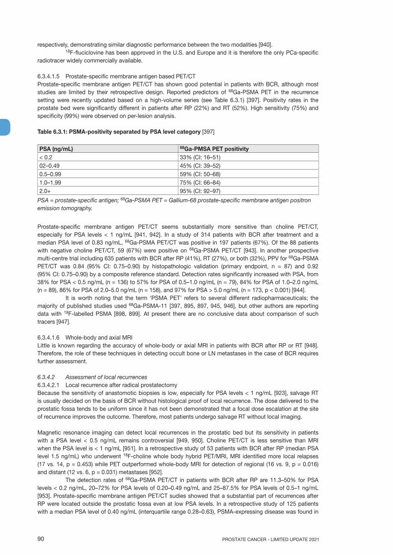
PROSTATE CANCER - LIMITED UPDATE 202190
respectively, demonstrating similar diagnostic performance between the two modalities [940].18F-fluciclovine has been approved in the U.S. and Europe and it is therefore the only PCa-specific
radiotracer widely commercially available.
6.3.4.1.5 Prostate-specific membrane antigen based PET/CTProstate-specific membrane antigen PET/CT has shown good potential in patients with BCR, although most studies are limited by their retrospective design. Reported predictors of 68Ga-PSMA PET in the recurrence setting were recently updated based on a high-volume series (see Table 6.3.1) [397]. Positivity rates in the prostate bed were significantly different in patients after RP (22%) and RT (52%). High sensitivity (75%) and specificity (99%) were observed on per-lesion analysis.
Table 6.3.1: PSMA-positivity separated by PSA level category [397]
PSA (ng/mL) 68Ga-PMSA PET positivity < 0.2 33% (CI: 16–51)02–0.49 45% (CI: 39–52)0.5–0.99 59% (CI: 50–68)1.0–1.99 75% (CI: 66–84)2.0+ 95% (CI: 92–97)
PSA = prostate-specific antigen; 68Ga-PSMA PET = Gallium-68 prostate-specific membrane antigen positron emission tomography.
Prostate-specific membrane antigen PET/CT seems substantially more sensitive than choline PET/CT, especially for PSA levels < 1 ng/mL [941, 942]. In a study of 314 patients with BCR after treatment and a median PSA level of 0.83 ng/mL, 68Ga-PSMA PET/CT was positive in 197 patients (67%). Of the 88 patients with negative choline PET/CT, 59 (67%) were positive on 68Ga-PSMA PET/CT [943]. In another prospective multi-centre trial including 635 patients with BCR after RP (41%), RT (27%), or both (32%), PPV for 68Ga-PSMA PET/CT was 0.84 (95% CI: 0.75–0.90) by histopathologic validation (primary endpoint, n = 87) and 0.92 (95% CI: 0.75–0.90) by a composite reference standard. Detection rates significantly increased with PSA, from 38% for PSA < 0.5 ng/mL (n = 136) to 57% for PSA of 0.5–1.0 ng/mL (n = 79), 84% for PSA of 1.0–2.0 ng/mL (n = 89), 86% for PSA of 2.0–5.0 ng/mL (n = 158), and 97% for PSA > 5.0 ng/mL (n = 173, p < 0.001) [944].
It is worth noting that the term ‘PSMA PET’ refers to several different radiopharmaceuticals; the majority of published studies used 68Ga-PSMA-11 [397, 895, 897, 945, 946], but other authors are reporting data with 18F-labelled PSMA [898, 899]. At present there are no conclusive data about comparison of such tracers [947].
6.3.4.1.6 Whole-body and axial MRILittle is known regarding the accuracy of whole-body or axial MRI in patients with BCR after RP or RT [948]. Therefore, the role of these techniques in detecting occult bone or LN metastases in the case of BCR requires further assessment.
6.3.4.2 Assessment of local recurrences6.3.4.2.1 Local recurrence after radical prostatectomyBecause the sensitivity of anastomotic biopsies is low, especially for PSA levels < 1 ng/mL [923], salvage RT is usually decided on the basis of BCR without histological proof of local recurrence. The dose delivered to the prostatic fossa tends to be uniform since it has not been demonstrated that a focal dose escalation at the site of recurrence improves the outcome. Therefore, most patients undergo salvage RT without local imaging.
Magnetic resonance imaging can detect local recurrences in the prostatic bed but its sensitivity in patients with a PSA level < 0.5 ng/mL remains controversial [949, 950]. Choline PET/CT is less sensitive than MRI when the PSA level is < 1 ng/mL [951]. In a retrospective study of 53 patients with BCR after RP (median PSA level 1.5 ng/mL) who underwent 18F-choline whole body hybrid PET/MRI, MRI identified more local relapses (17 vs. 14, p = 0.453) while PET outperformed whole-body MRI for detection of regional (16 vs. 9, p = 0.016) and distant (12 vs. 6, p = 0.031) metastases [952].
The detection rates of 68Ga-PSMA PET/CT in patients with BCR after RP are 11.3–50% for PSA levels < 0.2 ng/mL, 20–72% for PSA levels of 0.20–0.49 ng/mL and 25–87.5% for PSA levels of 0.5–1 ng/mL [953]. Prostate-specific membrane antigen PET/CT sudies showed that a substantial part of recurrences after RP were located outside the prostatic fossa even at low PSA levels. In a retrospective study of 125 patients with a median PSA level of 0.40 ng/mL (interquartile range 0.28–0.63), PSMA-expressing disease was found in

91PROSTATE CANCER - LIMITED UPDATE 2021
66 patients (53%), of whom 25 patients (38%) had PSMA-avid lesions outside the pelvis, 33 (50%) had lesions confined to the pelvic LNs and prostate bed, and 8 (12%) had recurrence only in the prostate fossa [954]. In another retrospective study of 119 men with a mean PSA level of 0.34 ng/mL, 68Ga-PSMA PET/CT was positive in 41 patients (34.4%); only 3 were positive in the prostatic fossa, while 21 had uptake in the skeleton, 4 in retroperitoneal LNs and 21 in pelvic LNs [895]. Combining 68Ga-PSMA PET and MRI may improve the detection of local recurrences, as compared to 68Ga-PSMA PET/CT [955].
6.3.4.2.2 Local recurrence after radiation therapyIn patients with BCR after RT biopsy status is a major predictor of outcome, provided the biopsies are obtained 18–24 months after initial treatment. Given the morbidity of local salvage options it is necessary to obtain histological proof of the local recurrence before treating the patient [923].
Transrectal US is not reliable in identifying local recurrence after RT. In contrast, MRI has yielded excellent results and can be used for biopsy targeting and guiding local salvage treatment [923, 956-959], even if it slightly underestimates the volume of the local recurrence [960]. Detection of recurrent cancer is also feasible with choline PET/CT [961], but choline PET/CT has not yet been compared to mpMRI. Prostate-specific membrane antigen PET/CT can also play a role in the detection of local recurrences after RT [397].
6.3.4.3 Summary of evidence on imaging in case of biochemical recurrenceIn patients with BCR imaging can detect both local recurences and distant metastases, however, the sensitivity of detection depends on the PSA level. After RP, PSMA PET/CT seems to be the imaging modality with the highest sensitivity at low PSA levels (< 0.5 ng/mL) and may help distinguishing patients with recurrences confined to the prostatic fossa from those with distant metastases which may impact the design and use of post-RP salvage RT. After RT, MRI has shown excellent results at detecting local recurrences and guiding prostate biopsy. Given the substantial morbidity of post-RT salvage local treatments, distant metastases must be ruled out in patients with local recurrences and who are fit for these salvage therapies. Choline-, fluciclovine- or PSMA-PET/CT can be used to detect metastases in these patients but for this indication PSMA PET/CT seems the most sensitive technique.
6.3.4.4 Guidelines for imaging in patients with biochemical recurrence
Recommendations Strength ratingProstate-specific antigen (PSA) recurrence after radical prostatectomyPerform prostate-specific membrane antigen (PSMA) positron emission tomography (PET) computed tomography (CT) if the PSA level is > 0.2 ng/mL and if the results will influence subsequent treatment decisions.
Weak
In case PSMA PET/CT is not available, and the PSA level is > 1 ng/mL, perform fluciclovine PET/CT or choline PET/CT imaging if the results will influence subsequent treatment decisions.
Weak
PSA recurrence after radiotherapyPerform prostate magnetic resonance imaging to localise abnormal areas and guide biopsies in patients fit for local salvage therapy.
Weak
Perform PSMA PET/CT (if available) or fluciclovine PET/CT or choline PET/CT in patients fit for curative salvage treatment.
Strong
6.3.5 Treatment of PSA-only recurrencesThe timing and treatment modality for PSA-only recurrences after RP or RT remain a matter of controversy based on the limited evidence.
6.3.5.1 Treatment of PSA-only recurrences after radical prostatectomy6.3.5.1.1 Salvage radiotherapy for PSA-only recurrence after radical prostatectomy (cTxcN0M0, without PET/ CT)Early SRT provides the possibility of cure for patients with an increasing PSA after RP. Boorjian et al., reported a 75% reduced risk of systemic progression with SRT when comparing 856 SRT patients with 1,801 non-SRT patients [962]. The RAVES and RADICAL trials assessing SRT in post-RP patients with PSA levels exceeding 0.1–0.2 ng/mL showed 5-year freedom from BCR and BCR-free survival rates of 88% [870, 963].
The PSA level at BCR was shown to be prognostic [962]. More than 60% of patients who are treated before the PSA level rises to > 0.5 ng/mL will achieve an undetectable PSA level [964-967], corresponding to a ~80% chance of being progression-free 5 years later [968]. A retrospective analysis of 635 patients who were followed after RP and experienced BCR and/or local recurrence and either received no salvage treatment (n = 397) or salvage RT alone (n = 160) within 2 years of BCR

PROSTATE CANCER - LIMITED UPDATE 202192
showed that salvage RT was associated with a 3-fold increase in PCa-specific survival relative to those who received no salvage treatment (p < 0.001). Salvage RT has been shown to be effective mainly in patients with a short PSA-DT [969]. According to a Panel systematic review which also proposed a scoring system [916], men with a PSA-DT > 1 year and a pathological GS < 8 have a significantly lower risk of clinical progression and can be classified as ‘EAU Low Risk’; men with a PSA-DT < 1 year or a pathological pGS 8–10 are at increased risk of clinical progression and should be classified as ‘EAU High Risk’. These definitions have been externally validated and may be helpful for individualised treatment decisions [921]. Despite the indication for salvage RT, a ‘wait and see‘ strategy remains an option in patients with a PSA-DT of more than 12 months and other favourable factors such a time to BCR > 3 years, < pT3a, ISUP grade < 2/3 [916, 970]. For an overview see Table 6.3.2.
Although biochemical progression is now widely accepted as a surrogate marker of PCa recurrence; metastatic disease, disease-specific and OS are more meaningful endpoints to support clinical decision-making. A systematic review and meta-analysis on the impact of BCR after RP reports SRT to be favourable for OS and PCa-specific mortality. In particular SRT should be initiated in patients with rapid PSA kinetics after RP and with a PSA cut-off of 0.4 ng/mL [916]. A recent international multi-institutional analysis of pooled data from RCTs has suggested that metastasis-free survival is the most valid surrogate endpoint with respect to impact on OS [971, 972]. Table 6.3.3 summarises results of recent studies on clinical endpoints after SRT.
Table 6.3.2: Selected studies of post-prostatectomy salvage radiotherapy, stratified by pre-salvage radiotherapy PSA level* (cTxcN0M0, without PET/CT)
Reference n Median FU (mo)
pre-SRT PSA (ng/mL) median
RT dose ADT bNED/PFS (year)
5-yr. results
Bartkowiak, et al. 2018 [973]
464 71 0.31 66.6 Gy 54% (5.9) 73% vs. 56%; PSA < 0.2 vs. > 0.2 ng/mL p < 0.0001
Soto, et al. 2012 [974]
441 36 < 1 (58%) 68 Gy 24% ADT
63/55% (3) ADT/no ADT
44/40% ADT/no ADT p < 0.16
Stish, et al. 2016 [964]
1,106 107 0.6 68 Gy 16% ADT
50% (5) 36% (10)
44% vs. 58%; PSA < 0.5 vs. > 0.5 ng/mL p < 0.001
Tendulkar, et al. 2016 [975]
2,460 60 0.5 66 Gy 16% ADT
56% (5) SRT; PSA < 0.2 ng/mL 71% 0.21-0.5 ng/mL 63% 0.51-1.0 ng/mL 54% 1.01-2.0 ng/mL 43% > 2 ng/mL 37% p < 0.001
*Androgen deprivation therapy can influence the outcome ‘biochemically no evidence of disease (bNED)’ or ‘progression-free survival’. To facilitate comparisons, 5-year bNED/PFS read-outs from Kaplan-Meier plots are included.ADT = androgen deprivation therapy; bNED = biochemically no evidence of disease; FU = follow up; mo = months; n = number of patients; PFS = progression-free survival; PSA = prostate-specific antigen; SRT = salvage radiotherapy; yr = year.
Table 6.3.3: Recent studies reporting clinical endpoints after SRT (cTxcN0M0, without PET/CT) (the majority of included patients did not receive ADT)
Reference n Median FU (mo)
Regimen Outcome
Bartkowiak, et al. 2018 [973]
464 71 66.6 (59.4-72) Gy no ADT
5.9 yr. OS post-SRT PSA < 0.1 ng/mL 98% post-SRT PSA > 0.1 ng/mL 92% p = 0.005
93PROSTATE CANCER - LIMITED UPDATE 2021
Jackson, et al. 2014 [976]
448 64 68.4 Gy no ADT 5 yr. DM post-SRT PSA < 0.1 ng/mL 5% post-SRT PSA > 0.1 ng/mL 29% p < 0.0001 5 yr. DSM post-SRT PSA < 0.1 ng/mL 2% post-SRT PSA > 0.1 ng/mL 7% p < 0.0001 OS post-SRT PSA < 0.1 ng/mL 97% post-SRT PSA > 0.1 ng/mL 90% p < 0.0001
Stish, et al. 2016 [964]
1,106 107 68 (64.8-70.2) Gy 39% 2D treatment planning incl. 16% ADT
5 and 8.9 yr. DM SRT: PSA < 0.5 ng/mL 7% and 12% SRT: PSA > 0.5 ng/mL 14% and 23% p < 0.001 5 and 8.9 yr. DSM SRT: PSA < 0.5 ng/mL < 1% and 6% SRT: PSA > 0.5 ng/mL 5% and 10% p = 0.02 5 and 8.9 yr. OS SRT: PSA < 0.5 ng/mL 94% and 86% SRT: PSA > 0.5 ng/mL 91% and 78% p = 0.14
Tendulkar, et al. 2016 [975]
2,460 60 66 (64.8-68.4) Gy incl. 16% ADT
10 yr. DM SRT: PSA 0.01-0.2 ng/mL 9% SRT: PSA 0.21-0.50 ng/mL 15% SRT: PSA 0.51-1.0 ng/mL 19% SRT: PSA 1.01-2.0 ng/mL 20% SRT: PSA > 2 ng/mL 37%, p < 0.001
ADT = androgen deprivation therapy; DM = distant metastasis; DSM = disease specific mortality; FU = follow up; mo. = month; n = number of patients; OS = overall survival; PSA = prostate specific antigen; SRT = salvage radiotherapy.
6.3.5.1.2 Salvage radiotherapy combined with androgen deprivation therapy (cTxcN0, without PET/CT)Data from RTOG 9601 [977] suggest both CSS and OS benefit when adding 2 years of bicalutamide (150 mg o.d.) to SRT. According to GETUG-AFU 16 also 6-months treatment with a LHRH-analogue can significantly improve 10-year BCR, biochemical PFS and, modestly, metastasis-free survival. However, SRT combined with either goserelin or placebo showed similar DSS and OS rates [978]. Table 6.3.4 provides an overview of these two RCTs.
These RCTs support adding ADT to SRT. However when interpreting these data it has to be kept in mind that RTOG 9601 used outdated radiation dosages (< 66 Gy) and technique. The question with respect to the patient risk profile, whether to offer combination treatment or not, and the optimal combination (LHRH or bicalutamide) remains, as yet, unsolved. The EAU risk classification may offer guidance in this respect [916, 921].
One of these RCTs reports improved OS (RTOG 96-01) and the other improved metastasis-free survival but due to methodological discrepancies also related to follow-up and risk patterns, it is, as yet, not evident which patients should receive ADT, which type of ADT and for how long. Men at high risk of further progression (e.g., with a PSA > 0.7 ng/mL and GS > 8) may benefit from SRT combined with two years of ADT; for those at lower risk (e.g., PSA < 0.7 ng/mL and GS 8) SRT combined with 6 months of ADT may be sufficient. Men with a low-risk profile (PSA < 0.5 ng/mL and GS < 8) may receive SRT alone. In a sub-analysis of men with a PSA of 0.61 to 1.5 (n = 253) there was an OS benefit associated with anti-androgen assignment (HR: 0.61, 95% CI: 0.39–0.94). In those receiving early SRT (PSA 0.6 ng/mL, n = 389), there was no improvement in OS (HR: 1.16, 95% CI: 0.79–1.70), an increased other-cause mortablity hazard (subdistribution HR: 1.94, 95% CI: 1.17–3.20, p = 0.01) and increased odds of late grades 3–5 cardiac and neurologic toxic effects (OR: 3.57, 95% CI: 1.09–15.97, p = 0.05). These results suggest that pre-SRT PSA level may be a prognostic biomarker for outcomes of anti-androgen treatment with SRT. In patients receiving late SRT (PSA > 0.6 ng/mL, hormone therapy was associated with improved outcomes. In men receiving early SRT (PSA < 0.6 ng/mL), long-term anti-androgen treatment was not associated with improved OS [979].
A recent review addressing the benefit from combining HT with SRT suggested risk stratification of patients based on the pre-SRT PSA (< 0.5, 0.6–1, > 1 ng/mL), margin status and ISUP grade as a framework

PROSTATE CANCER - LIMITED UPDATE 202194
to individualise treatment [980]. In a retrospective multi-centre-study including 525 patients, only in patients with more aggressive disease characteristics (pT3b/4 and ISUP grade > 4 or pT3b/4 and PSA at early SRT > 0.4 ng/mL) the administration of concomitant ADT was associated with a reduction in distant metastasis [981]. Similarly, in a retrospective analysis of 1,125 patients, stage > pT3b, GS > 8 and a PSA level at SRT > 5 ng/mL were identified as risk factors for clinical recurrence. A significant effect of long-term ADT was observed in patients with > 2 adverse features. For patients with a single risk factor, short-term HT was sufficient whilst patients without risk factors showed no significant benefit from concomitant ADT [982].
Table 6.3.4: Randomised controlled trials comparing salvage radiotherapy combined with androgen deprivation therapy vs. salvage radiotherapy alone
Reference n Risk groups Median FU (mo)
Regimen Outcome
GETUG-AFU 16 2019 [978]
369 RT + ADT 374 RT
ISUP grade < 2/3 89%,
ISUP grade > 4 11% cN0
112 66 Gy + 6 mo. GnRH analogue 6 mo. 66 Gy
10 yr. PFS 64%p < 0.000110 yr. PFS 49%10 yr. MFS 75%p = 0.03410 yr. MFS 69%
RTOG 9601 2017 [977]
384 RT + ADT 376 RT
pT2 R1, pT3 cN0
156 64.8 Gy + bicalutamide 24 mo. 64.8 Gy + placebo
12 yr. DM 14%p = 0.00512 yr. DM 23%12 yr. OS 76%p = 0.0412 yr. OS 71%12 yr. DSM 5.8%p < 0.00112 yr. DSM 13.4%
ADT = androgen deprivation therapy; DM = distant metastasis; DSM = disease specific mortality; PFS = progression free survival; FU = follow-up; GnRH = gonadotropin-releasing hormone; MFS = metastasis-free survival; OS = overall survival; PFS = progression-free survival; mo = months; n = number of patients; RT = radiotherapy; yr = year.
6.3.5.1.2.1 Target volume, dose, toxicityThere have been various attempts to define common outlines for ‘clinical target volumes‘ of PCa [983-986] and for organs at risk of normal tissue complications [987]. However, given the variations of techniques and dose-constraints, a satisfactory consensus has not yet been achieved. A benefit in biochemical PFS but not metastasis-free survival has been reported in patients receiving whole pelvis SRT (± ADT) but the advantages must be weighed against possible side effects [988].
The optimal SRT dose has not been well defined. It should be at least 66 Gy to the prostatic fossa (± the base of the SVs, depending on the pathological stage after RP) [965, 989]. In a systematic review, the pre-SRT PSA level and SRT dose both correlated with BCR, showing that relapse-free survival decreased by 2.4% per 0.1 ng/mL PSA and improved by 2.6% per Gy, suggesting that a treatment dose above 70 Gy should be administered at the lowest possible PSA level [990]. The combination of pT stage, margin status and ISUP grade and the PSA at SRT seems to define the risk of biochemical progression, metastasis and overall mortality [867, 991, 992]. In a study on 894 node-negative PCa patients, doses ranging from 64 to > 74 Gy were assigned to twelve risk groups defined by their pre-SRT PSA classes < 0.1, 0.1–0.2, 0.2–0.4, and > 0.4 ng/mL and ISUP grade, < 1 vs. 2/3 vs. > 4 [993]. The updated Stephenson nomograms incorporate the SRT and ADT doses as predictive factors for biochemical failure and distant metastasis [975].
Salvage RT is also associated with toxicity. In one report on 464 SRT patients receiving median 66.6 (max. 72) Gy, acute grade 2 toxicity was recorded in 4.7% for both the GI and GU tract. Two men had late grade 3 reactions of the GI tract. Severe GU tract toxicity was not observed. Late grade 2 complications occurred in 4.7% (GI tract) and 4.1% (GU tract), respectively, and 4.5% of the patients developed moderate urethral stricture [973].
In a RCT on dose escalation for SRT involving 350 patients, acute grade 2 and 3 GU toxicity was observed in 13.0% and 0.6%, respectively, with 64 Gy and in 16.6% and 1.7%, respectively, with 70 Gy.

95PROSTATE CANCER - LIMITED UPDATE 2021
Gastrointestinal tract toxicity of grades 2 and 3 occurred in 16.0% and 0.6%, respectively, with 64 Gy, and in 15.4% and 2.3%, respectively, with 70 Gy. Late effects have yet to be reported [994, 995].
With dose escalation over 72 Gy and/or up to a median of 76 Gy, the rate of severe side effects, especially genitourinary symptoms, clearly increases, even with newer planning and treatment techniques [996, 997]. In particular, when compared with 3D-CRT, IMRT was associated with a reduction in grade 2 GI toxicity from 10.2 to 1.9% (p = 0.02) but had no differential effect on the relatively high level of GU toxicity (5-year, 3D-CRT 15.8% vs. IMRT 16.8%) [996]. After a median salvage IMRT dose of 76 Gy, the 5-year risk of grade 2–3 toxicity rose to 22% for GU and 8% for GI symptoms, respectively [997]. Doses of at least 66 Gy and up to 72 Gy can be recommended [973, 994].
6.3.5.1.2.2 Salvage RT with or without ADT (cTx CN0/1) (with PET/CT)In a prospective multi-centre study of 323 patients with BCR, PSMA PET/CT changed the management intent in 62% of patients as compared to conventional staging. This was due to a significant reduction in the number of men in whom the site of disease recurrence was unknown (77% vs. 19%, p < 0.001) and a significant increase in the number of men with metastatic disease (11% vs. 57%) [415]. A recent prospective study in a subgroup of 119 BCR patients with low PSA (< 0.5 ng/mL) reported a change in the intended treatment in 30.2% of patients [895]; however, no data exist on the impact on final outcome. Another prospective study in 272 patients with early biochemical recurrent PCa after RP showed that 68Ga-PSMA-ligand PET/CT may tailor further therapy decisions (e.g., local vs. systemic treatment) at low PSA values (0.2–1 ng/mL) [897].
A single-centre study retrospectively assessed 164 men from a prospective database who underwent imaging with PSMA PET/CT for a rising PSA after RP with PSA levels < 0.5 ng/mL. In men with a negative PSMA PET/CT who received salvage RT, 85% (23 out of 27) demonstrated a treatment response compared to a further PSA increase in 65% of those not treated (22 out of 34). In the 36/99 men with disease confined to the prostate fossa on PSMA, 83% (29 out of 36) responded to salvage RT [945]. Thus, PSMA PET/CT might stratify men into a group with high response (negative findings or recurrence confined to the prostate) and poor response (positive nodes or distant disease) to salvage RT. As there are no prospective phase III data (in particular not for PCa-specific survival or OS) these results have to be confirmed before a recommendation can be provided.
6.3.5.1.2.3 Metastasis-directed therapy for rcN+ (with PET/CT)Radiolabelled PSMA PET/CT is increasingly used as a diagnostic tool to assess metastatic disease burden in patients with BCR following prior definitive therapy. A review including 30 studies and 4,476 patients showed overall estimates of positivity in a restaging setting of 38% in pelvic LNs and 13% in extra-pelvic LN metastases [397]. The percentage positivity of PSMA PET/CT was proven to increase with higher PSA values, from 33% (95% CI: 16–51) for a PSA of < 0.2 ng/mL, to 45% (39–52), 59% (50–68), 75% (66–84), and 95% (92–97) for PSA subgroup values of 0.2–0.49, 0.5–0.99, 1.00–1.99, and > 2.00 ng/mL, respectively [397]. Results of this review demonstrated high sensitivity and specificity of 68Ga-PSMA PET in advanced PCa with a per-lesion-analised sensitivity and specificity of 75% and 99%, respectively.
In patients relapsing after a local treatment (including cN+ and higly selected M1 patients), a metastases-targeting therapy has been proposed, with the aim to delay systemic treatment. Metastasis-directed (MDT) therapy in PET/CT detected nodal oligo-recurrent PCa after RP was assessed in a large retrospective multi-institutional study (263 patients received MDT and 1,816 patients standard of care as control group [matched 3:1]). Metastasis-targeting therapy consisted of salvage LN resection (n = 166) and stereotactic ablation RT (SABR) (n = 97). After a median follow-up of 70 months, the MDT-group showed significantly better CSS (5-year survival 98.6% vs. 95.7%, p < 0.01, respectively) [998].
Another retrospective study compared SABR with elective nodal irradiation (ENRT) in PET/CT-detected nodal oligo-recurrent PCa (n = 506 patients, 365 of which with N1 pelvic recurrence). With a median follow-up of 36 months, ENRT (n = 197) was associated with a significant reduction of nodal recurrences compared with SABR (n = 309) of 2% vs. 18%, respectively, but at the cost of higher side effects of ENRT [999]. These results have to be confirmed in prospective trials before any recommendations can be made. In these situations SABR should be used in highly selected patients only. For MDT in M1-patients see Section 6.4.8.
6.3.5.1.3 Salvage lymph node dissection The surgical management of (recurrent) nodal metastases in the pelvis has been the topic of several retrospective analyses [1000-1002] and a systematic review [1003]. The reported 5-year BCR-free survival rates ranged from 6% to 31%. Five-year OS was approximately 84% [1003]. Biochemical recurrence rates were found to be dependent on PSA at surgery and location and number of positive nodes [1004]. Addition of RT to the lymphatic template after salvage LN dissection may improve the BCR rate [1005]. In a recent multi-centre
PROSTATE CANCER - LIMITED UPDATE 202196
retrospective study long-term outcomes of salvage LN dissection were reported to be worse than previously described in studies with shorter follow-up [1006]. Biochemical recurrence-free survival at 10 years was 11%. Patients with a PSA response after salvage LN dissection and patients receiving ADT within 6 months from salvage LN dissection had a lower risk of death from PCa [1006]. High level evidence for the oncological value of salvage LN dissection is still lacking [1003].
6.3.5.1.4 Comparison of adjuvant- and salvage radiotherapy Section 6.2.5.4 - ART, is referred to for more details. Main findings are that after RP the vast majority of patients do not need ART which is supported by the results of 3 phase III RCTs comparing adjuvant RT and early salvage RT with a median follow-up of 5 years [870-874].
However, longer term (10-year) results and results of metastasis-free survival endpoints are needed before final conclusions can be drawn. Due to the small number of patients with a combination of high-risk features included in these 3 trails (only approximately 20%) ART remains a recommended treatment option in highly selected patients with at least two out of three high-risk features, such as pT3/R1/ISUP > 3 [854, 874].
6.3.5.2 Management of PSA failures after radiation therapy Therapeutic options in these patients are ADT or salvage local procedures. A recent systematic review and meta-analysis included all studies comparing the efficacy and toxicity of salvage RP, salvage HIFU, salvage cryotherapy, SBRT, salvage LDR brachytherapy, and salvage HDR brachytherapy in the management of locally recurrent PCa after primary radical EBRT [1007]. The outcomes were BCR-free survival at 2 and 5 years.
No significant differences with regards to recurrence-free survival between these modalities was found [1007]. Five-year recurrence-free survival ranged from 50% after cryotherapy to 60% after HDR brachytherapy and SBRT [1007]. The authors reported that severe GU toxicity exceeded 21% for HIFU and RP, whereas it ranged from 4.2% to 8.1% with re-irradiation. Differences in severe GI toxicity also appeared to favour re-irradiation, particularly HDR brachytherapy [1007]. Due to the methodological limitations of this review (the majority of the included studies were uncontrolled single-arm case series and there was considerable heterogeneity in the definitions of core outcomes) the available evidence for these treatment options is of low quality and strong recommendations regarding the choice of any of these techniques cannot be made. The following is an overview of the most important findings for each of these techniques.
6.3.5.2.1 Salvage radical prostatectomySalvage RP after RT is associated with a higher likelihood of adverse events compared to primary surgery because of the risk of fibrosis and poor wound healing due to radiation [1008].
6.3.5.2.1.1 Oncological outcomesIn a systematic review of the literature, Chade, et al., showed that SRP provided 5- and 10-year BCR-free survival estimates ranging from 47–82% and from 28-53%, respectively. The 10-year CSS and OS rates ranged from 70–83% and from 54–89%, respectively. The pre-SRP PSA value and prostate biopsy ISUP grade were the strongest predictors of the presence of organ-confined disease, progression, and CSS [1009]. In a recent multi-centre analysis including 414 patients, 5-year BCR-free survival, CSS and OS were 56.7%, 97.7% and 92.1%, respectively [1010]. Pathological T stage > T3b (OR: 2.348) and GS (up to OR 7.183 for GS > 8) were independent predictors for BCR.
Table 6.3.5: Oncological results of selected salvage radical prostatectomy case series
Reference n Median FU (mo)
Pathologic Organ-confined (%)
PSM (%)
Lymph-node involvement (%)
BCR-free probability (%)
CSS (%)
Time probability
Chade, et al. 2011 [1011]
404 55 55 25 16 37 83 10 yr.
Mandel, et al. 2016 [1012]
55 36 50 27 22 49 89 5 yr.
Ogaya-Pinies, et al. 2018 [1013]
96 14 50 17 8 85* - 14 mo.
Marra, et al. 2021 [1010]
414 36 46 30 16 57 98 5 yr.
*Percentage of patients without BCR.BCR = biochemical recurrence; CSS = cancer-specific survival; FU = follow-up; mo = months; n = number of patients; PSM = positive surgical margin.

97PROSTATE CANCER - LIMITED UPDATE 2021
6.3.5.2.1.2 MorbidityCompared to primary open RP, SRP is associated with a higher risk of later anastomotic stricture (47 vs. 5.8%), urinary retention (25.3% vs. 3.5%), urinary fistula (4.1% vs. 0.06%), abscess (3.2% vs. 0.7%) and rectal injury (9.2 vs. 0.6%) [1014]. In more recent series, these complications appear to be less common [1008, 1009, 1012].
Functional outcomes are also worse compared to primary surgery, with urinary incontinence ranging from 21% to 90% and ED in nearly all patients [1009, 1012].
Table 6.3.6: Peri-operative morbidity in selected salvage radical prostatectomy case series
Reference n Rectal injury (%) Anastomotic stricture (%)
Clavien 3-5 (%) Blood loss, mL, mean, range
Ward, et al. 2005 [1015] 138 5 22 - -Sanderson, et al. 2006 [1016] 51 2 41 6 -Gotto, et al. 2010 [1014] 98 9 41 25 -Gontero, et al. 2019 [1008] 395 1.6 25 3.6 10.1
n = number of patients.
6.3.5.2.1.3 Summary of salvage radical prostatectomyIn general, SRP should be considered only in patients with low co-morbidity, a life expectancy of at least 10 years, a pre-SRP PSA < 10 ng/mL and initial biopsy ISUP grade < 2/3, no LN involvement or evidence of distant metastatic disease pre-SRP, and those whose initial clinical staging was T1 or T2 [1009].
A meta-analysis and systematic review of local salvage therapies after RT for PCa has suggested that re-irradiation with SBRT, HDR brachytherapy or LDR brachytherapy appears to result in less severe GU toxicity than RP, and re-irradiation with HDR brachytherapy in less severe GI toxicity than RP [1007].
Salvage RP might be associated with more severe GU and GI toxicity compared to re-irradiation [1007].
6.3.5.2.2 Salvage cryoablation of the prostate6.3.5.2.2.1 Oncological outcomesSalvage cryoablation of the prostate (SCAP) has been proposed as an alternative to salvage RP, as it has a potentially lower risk of morbidity and equal efficacy.
In a recent systematic review a total of 32 studies assessed SCAP, recruiting a total of 5,513 patients. The overwhelming majority of patients (93%) received whole-gland SCAP. The adjusted pooled analysis for 2-year BCR-free survival for SCAP was 67.49% (95% CI: 61.68–72.81%), and for 5-year BCR-free survival was 50.25% (95% CI: 44.10–56.40%). However, the certainty of the evidence was low. Table 6.3.7 summarises the results of a selection of the largest series on SCAP to date in relation to oncological outcomes (BCR only) [1007].
Table 6.3.7: Oncological results of selected salvage cryoablation of the prostate case series, including at least 250 patients
Reference n Median FU (mo)
Time point of outcome measurement (yr)
BCR-free probability Definition of failure
Ginsburg, et al. 2017 [1017] 898 19.0 5 71.3% Phoenix criteriaSpiess, et al. 2010 [1018] 450 40.8 3.4 39.6% PSA > 0.5 ng/mLLi, et al. 2015 [1019] 486 18.2 5 63.8% Phoenix criteriaKovac, et al. 2016 [1020] 486 18.2 5 75.5%
(nadir PSA < 0.4 ng/mL);22.1% (nadir PSA > 0.4 ng/mL)
Phoenix criteria
Ahmad, et al. 2013 [1021] 283 23.9 3 67.0% (nadir PSA < 1 ng/mL);14.0% (nadir PSA > 1 ng/mL)
Phoenix criteria
Pisters, et al. 2008 [1022] 279 21.6 5 58.9% (ASTRO);54.5% (Phoenix)
ASTRO and Phoenix criteria
ASTRO = American Society for Therapeutic Radiology and Oncology; BCR = biochemical recurrence;FU = follow-up; mo. = months; n = number of patients; PSA = prostate-specific antigen; yr. = year.
PROSTATE CANCER - LIMITED UPDATE 202198
6.3.5.2.2.2 MorbidityThe main adverse effects and complications relating to SCAP include urinary incontinence, urinary retention due to bladder outflow obstruction, recto-urethral fistula, and erectile dysfunction. The recent SR and meta-analysis [1007] showed an adjusted pooled analysis for severe GU toxicity for SCAP of 15.44% (95% CI: 10.15–21.54%). As before, the certainty of the evidence was low. Table 6.3.8 summarises the results of a selection of the largest series on SCAP to date in relation to GU outcomes.
Table 6.3.8: Peri-operative morbidity, erectile function and urinary incontinence in selected salvage cryoablation case series, including at least 100 patients
Study n Time pointof outcome measurement (mo)
Incontinence (%)
Obstruction/Retention (%)
Rectourethral fistula (%)
ED (%)
Li, et al. 2015 [1019] 486 12 33.3 21.7 4.7 71.3Ahmad, et al. 2013 [1021] 283 12 12.0 8.1 1.8 83.0Pisters, et al. 2008 [1022] 279 12 4.4 3.2 1.2 NACespedes, et al. 1997 [1023] 143 Median 27.0 28.0 14.0 NA NAChin, et al. 2001 [1024] 118 Median 18.6 6.7 NA 3.3 NA
ED = erectile dysfunction; mo = months; n = number of patients.
6.3.5.2.2.3 Summary of salvage cryoablation of the prostateIn general, the evidence base relating to the use of SCAP is poor, with significant uncertainties relating to long-term oncological outcomes, and SCAP appears to be associated with significant morbidity. Consequently, SCAP should only be performed in selected patients in experienced centres as part of a clinical trial or well-designed prospective cohort study.
6.3.5.2.3 Salvage re-irradiation6.3.5.2.3.1 Salvage brachytherapy for radiotherapy failureCarefully selected patients with a good PS, primary localised PCa, good urinary function and histologically proven local recurrence are candidates for salvage brachytherapy using either HDR- or LDR.
In a recent systematic review a total of 16 studies (4 prospective) and 32 studies (2 prospective) assessed salvage HDR and LDR brachytherapy, respectively [1007] with the majority (> 85%) receiving whole-gland brachytherapy rather than focal treatment. The adjusted pooled analysis for 2-year BCR-free survival for HDR was 77% (95% CI: 70–83%) and for LDR was 81% (95% CI:74–86%). The 5-year BCR-free survival for HDR was 60% (95% CI: 52–67%) and for LDR was 56% (95% CI: 48–63%). As noted above, brachytherapy techniques are associated with lower rates of severe GU toxicity when compared to RP or HIFU, at 8% for HDR (95% CI: 5.1–11%) and 8.1% for LDR (95% CI: 4.3–13%). Rates of severe GI toxicity are reported to be very low at 0% for HDR (95% CI: 0–0.2%) and 1.5% for LDR (95% CI: 0.2–3.4%). High-dose-rate or LDR brachytherapy are effective treatment options with an acceptable toxicity profile. However, the published series are small and likely under-report toxicity. Consequently this treatment should be offered in experienced centres ideally within randomised clinical trials or prospective registry studies.
Table 6.3.9: Treatment-related toxicity and BCR-free probablity in selected salvage brachytherapy studies including at least 100 patients.
Author Study design n and BT type Median FU (mo)
Treatment toxicity BCR-free probability
Lopez, et al. 2019 [1025]
multi-centre retrospective
75 HDR 44 LDR
52 23.5% late G3+ GU 5 yr 71% (95% CI: 65.9-75.9%)
Crook, et al. 2019 [1026]
multi-centre prospective
100 LDR 54 14% late G3 combined GI/GU
n.r.
Smith, et al. 2020 [1027]
single-centre retrospective
108 LDR 76 15.7%/2.8% late G3 GU/GI
5 yr. 63.1%10 yr. 52%
Lyczek, et al. 2009 [1028]
single-centre retrospective
115 HDR n.r. 12.2%/0.9%late G3+ GU/GI
60% at 40 mo.
BT = brachytherapy; GI = gastrointestinal; GU = genito-urinary; HDR = high-dose rate; LDR = low-dose rate; mo = months; n = number of patients; n.r. = not reported; yr = year.

99PROSTATE CANCER - LIMITED UPDATE 2021
6.3.5.2.3.2 Salvage stereotactic ablative body radiotherapy for radiotherapy failure6.3.5.2.3.2.1 Oncological outcomes and morbidityStereotactic ablative body radiotherapy (CyberKnife or Linac-based treatment) is a potentially viable new option to treat local recurrence after RT. Carefully selected patients with good IPSS-score, without obstruction, good PS and histologically proven localised local recurrence are potential candidates for SABR. In a recent meta-analysis and systematic review five mostly retrospective studies including 206 patients were treated with CyberKnife or linac-based-treatment showing 2-year RFS estimates (61.6%, 95% CI: 52.6–69.9%) [1007]. In a retrospective multi-centre study (n = 100) the median pre-salvage PSA was 4.3 ng/mL with 34% of patients having received ADT for twelve months (median). All recurrences were biopsy proven. Patients were treated with the CyberKnife with a single dose of 6 Gy in six daily fractions (total dose 36 Gy). With a median follow-up of 30 months the estimated 3-year second BCR-free survival was 55% [1029].
In a smaller retrospective series including 50 men with histologically proven local recurrence with a median pre-salvage PSA of 3.9 ng/mL only 15% had received additional ADT. The estimated 5-year second BCR-free survival was 60% (median follow-up of 44 months) which is an outcome comparable to series treating patients with RP, HIFU or brachytherapy [1030]. Table 6.3.10 summarises the results of the two larger SABR series addressing oncological outcomes and morbidity.
Table 6.3.10: Treatment-related toxicity and BCR-free survival in selected SABR studies including at least 50 patients
Author Study design n and RT-type
Median FU (mo)
Fractionation(SD/TD)
ADT Treatment toxicity
BCR-free survival
Fuller, et al. 2020 [1030]
single-centre retrospective
50Cyber Knife
44 SD 6.8 GyTD 34 Gy
7/50 5 yr: 8% late G3+ GU
5 yr. 60%
Pasquier, et al. 2020 [1029]
multi-centre retrospective
100 Cyber Knife
30 SD 6 GyTD 36 Gy
34/100median 12 mo.
3 yr. grade 2+ GU 20.8%GI 1%
3 yr. 55%
BCR = biochemical recurrence-free; FU = follow-up; mo = months; n = number of patients; RT-type = type of radiotherapy; SD = single dose; TD = total dose; yr = year.
6.3.5.2.3.2.2 MorbidityIn a retrospective single-centre study with 50 consecutive patients chronic significant toxicity was only seen for the GU domain with 5-year grade 2+ and grade 3+ GU rates of 17% and 8%, respectively. No GI toxicity > grade 1 was seen. Of note, of the fifteen patients who were sexually potent pre-salvage SBRT, twelve subsequently lost potency [1030]. In a retrospective French (GETUG) multi-centre series (n = 100) the 3-year late grade 2+ GU and GI toxicity was 20.8% (95% CI: 13–29%) and 1% (95% CI: 0.1–5.1%), respectively [1029].
6.3.5.2.3.2.3 Summary of salvage stereotactic ablative body radiotherapyDespite the encouraging results so far the number of patients treated with SABR is relatively limited. In view of the rates of higher grade 2+ GU side effects, SABR should only be offered to selected patients, in experienced centres as part of a clinical trial or well-designed prospective study.
6.3.5.2.4 Salvage high-intensity focused ultrasound6.3.5.2.4.1 Oncological outcomesSalvage HIFU has emerged as an alternative thermal ablation option for radiation-recurrent PCa. Being relatively newer than SCAP the data for salvage HIFU are even more limited. A recent systematic review and meta-analysis also included salvage HIFU [1007]. A total of 20 studies assessed salvage HIFU, recruiting 1,783 patients. The overwhelming majority of patients (86%) received whole-gland salvage HIFU. The adjusted pooled analysis for 2-year BCR-free survival for salvage HIFU was 54.14% (95% CI: 47.77–60.38%) and for 5-year BCR-free survival 52.72% (95% CI: 42.66–62.56%). However, the certainty of the evidence was low. Table 6.3.11 summarises the results of a selection of the largest series on salvage HIFU to date in relation to oncological outcomes (BCR only).
PROSTATE CANCER - LIMITED UPDATE 2021100
Table 6.3.11: Oncological results of selected salvage cryoablation of the prostate case series, including at least 250 patients
Study n Median FU (mo)
Time point of outcome measurement (yr)
BCR-free probability
Definition of failure
Crouzet, et al. 2017 [1031]
418 39.6 5 49.0% Phoenix criteria
Murat, et al. 2009 [1032]
167 Mean 18.1
3 25.0% (high-risk); 53.0% (low-risk)*
Phoenix criteria or positive biopsy or initiation of post-HIFU salvage therapy
Kanthabalan, et al. 2017 [1033]
150 35.0 3 48.0% Phoenix criteria
Jones, et al. 2018 [1034]
100 12.0 1 50.0% Nadir PSA > 0.5 ng/mL or positive biopsy
*Results stratified by pre-EBRT D’Amico risk groupsBCR = biochemical recurrence; CSS = cancer-specific survival; FU = follow-up; mo = months; n = number ofpatients; yr = year.
6.3.5.2.4.2 MorbidityThe main adverse effects and complications relating to salvage HIFU include urinary incontinence, urinary retention due to bladder outflow obstruction, rectourethral fistula and erectile dysfunction. The recent systematic review and meta-analysis showed an adjusted pooled analysis for severe GU toxicity for salvage HIFU of 22.66% (95% CI: 16.98–28.85%) [1007]. The certainty of the evidence was low. Table 6.3.12 summarises the results of a selection of the largest series on salvage HIFU to date in relation to GU outcomes.
Table 6.3.12: Peri-operative morbidity, erectile function and urinary incontinence in selected salvage HIFU case series, including at least 100 patients
Study n Time point of outcome measurement (yr)
Incontinence* (%)
Obstruction/retention (%)
Rectourethralfistula (%)
ED (%)
Crouzet, et al. 2017 [1031]
418 Median 39.6 42.3 18.0 2.3 n.r.
Murat, et al. 2009 [1032]
167 Median 18.1 49.5 7.8 3.0 n.r.
Kanthabalan, et al. 2017 [1033]
150 24 12.5 8.0 2.0 41.7
Jones, et al. 2018 [1034]
100 12 42.0 49.0 5.0 74.0
*Incontinence was heterogeneously defined; figures represent at least 1 pad usage.ED = erectile dysfunction; mo = months; n.r. = not reported; n = number of patients.
6.3.5.2.4.3 Summary of salvage high-intensity focused ultrasoundThere is a lack of high-certainty data which prohibits any recommendations regarding the indications for salvage HIFU in routine clinical practice. There is also a risk of significant morbidity associated with its use in the salvage setting. Consequently, salvage HIFU should only be performed in selected patients in experienced centres as part of a clinical trial or well-designed prospective cohort study.
6.3.6 Hormonal therapy for relapsing patientsThe Panel conducted a systematic review including studies published from 2000 onwards [1035]. Conflicting results were found on the clinical effectiveness of HT after previous curative therapy of the primary tumour. Some studies reported a favourable effect of HT, including the only RCT addressing the research question of this review (86% vs. 79% advantage in OS in the early HT group) [1036]. Other studies did not find any differences between early vs. delayed, or no, HT. One study found an unfavourable effect of HT [1037]. This may be the result of selecting clinically unfavourable cases for (early) HT and more intensive diagnostic work-up and follow-up in these patients.
101PROSTATE CANCER - LIMITED UPDATE 2021
The studied population is highly heterogeneous regarding their tumour biology and therefore clinical course. Predictive factors for poor outcomes were; CRPC, distant metastases, CSS, OS, short PSA-DT, high ISUP grade, high PSA, increased age and co-morbidities. In some studies, such as the Boorjian, et al. study [970], high-risk patients, mainly defined by a high ISUP grade and a short PSA-DT (most often less than 6 months) seem to benefit most from (early) HT, especially in men with a long life expectancy.
No data were found on the effectiveness of different types of HT, although it is unlikely that this will have a significant impact on survival outcomes in this setting. Non-steroidal anti-androgens have been claimed to be inferior compared to castration, but this difference was not seen in M0 patients [969]. One of the included RCTs suggested that intermittent HT is not inferior to continuous HT in terms of OS and CSS [1038]. A small advantage was found in some QoL domains but not overall QoL outcomes. An important limitation of this RCT is the lack of any stratifying criteria such as PSA-DT or initial risk factors. Based on the lack of definitive efficacy and the undoubtedly associated significant side effects, patients with recurrence after primary curative therapy should not receive standard HT since only a minority of them will progress to metastases or PCa-related death. The objective of HT should be to improve OS, postpone distant metastases, and improve QoL. Biochemical response to only HT holds no clinical benefit for a patient. For older patients and those with co-morbidities the side effects of HT may even decrease life expectancy; in particular cardiovascular risk factors need to be considered [1039, 1040]. Early HT should be reserved for those at the highest risk of disease progression defined mainly by a short PSA-DT at relapse (< 6–12 months) or a high initial ISUP grade (> 2/3) and a long life expectancy.
6.3.7 ObservationIn unselected relapsing patients the median actuarial time to the development of metastasis will be 8 years and the median time from metastasis to death will be a further 5 years [855]. For patients with EAU Low-Risk BCR features (see Section 6.3.3), unfit patients with a life expectancy of less than 10 years or patients unwilling to undergo salvage treatment, active follow-up may represent a viable option.
6.3.8 Guidelines for second-line therapy after treatment with curative intent
Local salvage treatment Strength ratingRecommendations for biochemical recurrence (BCR) after radical prostatectomyOffer monitoring, including prostate-specific antigen (PSA), to EAU Low-Risk BCR patients. WeakOffer early salvage intensity-modulated radiotherapy plus image-guided radiotherapy to men with two consecutive PSA rises.
Strong
A negative PET/CT scan should not delay salvage radiotherapy (SRT), if otherwise indicated. StrongDo not wait for a PSA threshold before starting treatment. Once the decision for SRT has been made, SRT (at least 66 Gy) should be given as soon as possible.
Strong
Offer hormonal therapy in addition to SRT to men with BCR. WeakRecommendations for BCR after radiotherapyOffer monitoring, including PSA to EAU Low-Risk BCR patients. WeakOnly offer salvage radical prostatectomy (RP), brachytherapy, high-intensity focused ultrasound, or cryosurgical ablation to highly selected patients with biopsy proven local recurrence within a clinical trial setting or well-designed prospective cohort study undertaken in experienced centres.
Strong
Salvage RP should only be performed in experienced centres. WeakRecommendations for systemic salvage treatmentDo not offer androgen deprivation therapy to M0 patients with a PSA-doubling time > 12 months.
Strong
6.4 Treatment: Metastatic prostate cancer6.4.1 IntroductionAll prospective data available rely on the definition of M1 disease based on CT scan and bone scan. The influence on treatment and outcome of newer, more sensitive, imaging has not been assessed yet.
6.4.2 Prognostic factorsMedian survival of patients with newly diagnosed metastases is approximately 42 months [1041] with ADT alone, however, it is highly variable since the M1 population is heterogeneous. Several prognostic factors for survival have been suggested including the number and location of bone metastases, presence of visceral metastases, ISUP grade, PS status and initial PSA alkaline phosphatase, but only few have been validated [1042-1045].
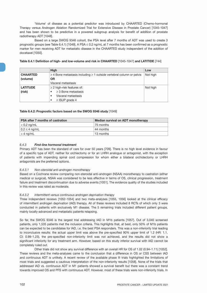
PROSTATE CANCER - LIMITED UPDATE 2021102
‘Volume‘ of disease as a potential predictor was introduced by CHAARTED (Chemo-hormonal Therapy versus Androgen Ablation Randomized Trial for Extensive Disease in Prostate Cancer) [1045-1047] and has been shown to be predictive in a powered subgroup analysis for benefit of addition of prostate radiotherapy ADT [1048].
Based on a large SWOG 9346 cohort, the PSA level after 7 months of ADT was used to create 3 prognostic groups (see Table 6.4.1) [1049]. A PSA < 0.2 ng/mL at 7 months has been confirmed as a prognostic marker for men receiving ADT for metastatic disease in the CHAARTED study independent of the addition of docetaxel [1050].
Table 6.4.1 Definition of high- and low-volume and risk in CHAARTED [1045-1047] and LATITIDE [744]
High LowCHAARTED (volume)
> 4 Bone metastasis including > 1 outside vertebral column or pelvis OR Visceral metastasis
Not high
LATITUDE (risk)
> 2 high-risk features of:• > 3 Bone metastasis • Visceral metastasis • > ISUP grade 4
Not high
Table 6.4.2: Prognostic factors based on the SWOG 9346 study [1049]
PSA after 7 months of castration Median survival on ADT monotherapy< 0.2 ng/mL 75 months0.2 < 4 ng/mL 44 months> 4 ng/mL 13 months
6.4.3 First-line hormonal treatmentPrimary ADT has been the standard of care for over 50 years [709]. There is no high level evidence in favour of a specific type of ADT, neither for orchiectomy or for an LHRH analogue or antagonist, with the exception of patients with impending spinal cord compression for whom either a bilateral orchidectomy or LHRH antagonists are the preferred options.
6.4.3.1 Non-steroidal anti-androgen monotherapyBased on a Cochrane review comparing non-steroidal anti-androgen (NSAA) monotherapy to castration (either medical or surgical), NSAA was considered to be less effective in terms of OS, clinical progression, treatment failure and treatment discontinuation due to adverse events [1051]. The evidence quality of the studies included in this review was rated as moderate.
6.4.3.2 Intermittent versus continuous androgen deprivation therapyThree independent reviews [1052-1054] and two meta-analyses [1055, 1056] looked at the clinical efficacy of intermittent androgen deprivation (IAD) therapy. All of these reviews included 8 RCTs of which only 3 were conducted in patients with exclusively M1 disease. The 5 remaining trials included different patient groups, mainly locally-advanced and metastatic patients relapsing.
So far, the SWOG 9346 is the largest trial addressing IAD in M1b patients [1057]. Out of 3,040 screened patients, only 1,535 patients met the inclusion criteria. This highlights that, at best, only 50% of M1b patients can be expected to be candidates for IAD, i.e. the best PSA responders. This was a non-inferiority trial leading to inconclusive results: the actual upper limit was above the pre-specified 90% upper limit of 1.2 (HR: 1.1, CI: 0.99–1.23), the pre-specified non-inferiority limit was not achieved, and the results did not show a significant inferiority for any treatment arm. However, based on this study inferior survival with IAD cannot be completely ruled out.
Other trials did not show any survival difference with an overall HR for OS of 1.02 (0.94–1.11) [1052]. These reviews and the meta-analyses came to the conclusion that a difference in OS or CSS between IAD and continuous ADT is unlikely. A recent review of the available phase III trials highlighted the limitations of most trials and suggested a cautious interpretation of the non-inferiority results [1058]. None of the trials that addressed IAD vs. continuous ADT in M1 patients showed a survival benefit but there was a constant trend towards improved OS and PFS with continuous ADT. However, most of these trials were non-inferiority trials. In

103PROSTATE CANCER - LIMITED UPDATE 2021
some cohorts the negative impact on sexual function was less pronounced with IAD. There is a trend favouring IAD in terms of QoL, especially regarding treatment-related side effects such as hot flushes [1059, 1060].
6.4.3.3 Immediate versus deferred androgen deprivation therapyIn symptomatic patients immediate treatment is mandatory, however, controversy still exists for asymptomatic metastatic patients due to the lack of high quality studies. A first Cochrane review extracted four RCTs: the VACURG I and II trials, the MRC trial, and the ECOG 7887 study [1050, 1051]. These studies were conducted in the pre-PSA era and included patients with advanced metastatic or non-metastatic PCa who received immediate vs. deferred ADT [1061]. No improvement in PCa CSS was observed, although immediate ADT significantly reduced disease progression. The Cochrane analysis was updated in 2019 and concluded that early ADT probably extends time to death of any cause and time to death from PCa [1062]. Since the analysis included only a very limited number of M1 patients who were not evaluated separately, the benefit of immediate ADT in this setting remains unclear.
6.4.4 Combination therapiesAll of the following combination therapies have been studied with continuous ADT, not intermittent ADT.
6.4.4.1 ‘Complete’ androgen blockadeThe largest RCT in 1,286 M1b patients found no difference between surgical castration with or without flutamide [1063]. However, results with other anti-androgens or castration modalities have differed and systematic reviews have shown that CAB using a NSAA appears to provide a small survival advantage (< 5%) vs. monotherapy (surgical castration or LHRH agonists) [1064, 1065] beyond 5 years of survival [1066] but this minimal advantage in a small subset of patients must be balanced against the increased side effects associated with long-term use of NSAAs.
6.4.4.2 Androgen deprivation combined with other agents6.4.4.2.1 Androgen deprivation therapy combined with chemotherapyThree large RCTs were conducted [801, 1045, 1067]. All trials compared ADT alone as the standard of care with ADT combined with immediate docetaxel (75 mg/sqm, every 3 weeks within 3 months of ADT initiation). The primary objective in all three studies was to assess OS. The key findings are summarised in Table 6.4.3.
Table 6.4.3: Key findings - Hormonal treatment combined with chemotherapy
STAMPEDE [801, 1068] GETUG [1067] CHAARTED [1045,1046]ADT ADT + Docetaxel + P ADT ADT + Docetaxel ADT ADT + Docetaxel
n 1,184 592 193 192 393 397Newly diagnosed M+
58% 59% 75% 67% 73% 73%
Key inclusion criteria
Patients scheduled for long-term ADT- newly diagnosed M1 or N+
situations- locally advanced (at least two
of cT3 cT4, ISUP grade > 4, PSA > 40 ng/mL)
- relapsing locally treated disease with a PSA > 4 ng/mL and a PSA-DT < 6 mo.
or PSA > 20 ng/mL,or nodalor metastatic relapse
Metastatic disease Karnofsky score > 70%
Metastatic disease ECOG PS 0, 1 or 2
Primary objective
OS OS OS
Median follow up (mo)
43; 78.2 (update M1) 50 54 (update)
HR (95% CI) 0.78 (0.66-0.93) 1.01 (0.75-1.36) 0.72 (0.59-0.89)

PROSTATE CANCER - LIMITED UPDATE 2021104
M1 onlyn 1,086 - -HR (95% CI) 0.81 (0.69-0.95) - -
ADT = androgen deprivation therapy; ECOG = Eastern Cooperative Oncology Group; FU = follow-up; HR = hazard ratio; ISUP = International Society for Urological Pathology; mo = month; n = number of patients; OS = overall survival; P = prednisone; PSA-DT = prostate-specific antigen-doubling time.
In the GETUG 15 trial, all patients had newly diagnosed M1 PCa, either de novo or after a primary treatment [1067]. They were stratified based on previous treatment and Glass risk factors [1042]. In the CHAARTED trial the same inclusion criteria applied and patients were stratified according to disease volume; high volume being defined as either presence of visceral metastases or four, or more, bone metastases, with at least one outside the spine and pelvis [1045].
STAMPEDE is a multi-arm multi-stage trial in which the reference arm (ADT monotherapy) included 1,184 patients. One of the experimental arms was docetaxel combined with ADT (n = 593), another was docetaxel combined with zoledronic acid (n = 593). Patients were included with either M1 or N1, or having two of the following 3 criteria: T3/4, PSA > 40 ng/mL or ISUP grade 4–5. Also relapsed patients after local treatment were included if they met one of the following criteria: PSA > 4 ng/mL with a PSA-DT < 6 months or a PSA > 20 ng/mL, N1 or M1. No stratification was used regarding metastatic disease volume (high/low volume) [801]. In all 3 trials toxicity was mainly haematological with around 12–15% grade 3–4 neutropenia, and 6–12% grade 3–4 febrile neutropenia. The use of granulocyte colony-stimulating factor receptor (GCSF) was shown to be beneficial in reducing febrile neutropenia. Primary or secondary prophylaxis with GCSF should be based on available guidelines [1069, 1070].
Based on these data, upfront docetaxel combined with ADT should be considered as a standard in men presenting with metastases at first presentation provided they are fit enough to receive the drug [1070]. Docetaxel is used at the standard dose of 75 mg/sqm combined with steroids as pre-medication. Continuous oral corticosteroid therapy is not mandatory.
In subgroup analyses from GETUG-AFU 15 and CHAARTED the beneficial effect of the addition of docetaxel to ADT is most evident in men with de novo metastatic high-volume disease [1046, 1047], while it was in the same range whatever the volume in the post-hoc analysis from STAMPEDE [1068]. The effects were less apparent in men who had prior local treatment although the numbers were small and the event rates lower.
A systematic review and meta-analysis which included these 3 trials showed that the addition of docetaxel to standard of care improved survival [1070]. The HR of 0.77 (95% CI: 0.68–0.87, p < 0.0001) translates into an absolute improvement in 4-year survival of 9% (95% CI: 5–14). Docetaxel in addition to standard of care also improves failure-free survival, with a HR of 0.64 (0.58–0.70, p < 0.0001) translating into a reduction in absolute 4-year failure rates of 16% (95% CI: 12–19).
6.4.4.2.2 Combination with the new hormonal treatments (abiraterone, apalutamide, enzalutamide)In two large RCTs (STAMPEDE, LATITUDE) the addition of abiraterone acetate (1000 mg daily) plus prednisone (5 mg daily) to ADT in men with mHSPC was studied [40, 744, 1071]. The primary objective of both trials was an improvement in OS. Both trials showed a significant OS benefit but in LATITUDE in high-risk metastatic patients only with a HR of 0.62 (0.51–0.76) [744]. The HR in STAMPEDE was very similar with 0.63 (0.52–0.76) in the total patient population (metastatic and non-metastatic) and a HR of 0.61 in the subgroup of metastatic patients [40]. The inclusion criteria in the two trials differed but both trials were positive for OS. While only high-risk patients were included in the LATITUDE trial a post-hoc analysis from STAMPEDE showed the same benefit whatever the risk or the volume stratification [1072].
All secondary objectives such as PFS, time to radiographic progression, time to pain, or time to chemotherapy were positive and in favour of the combination. The key findings are summarised in Table 6.4.4. No difference in treatment-related deaths was observed with the combination of ADT plus abiraterone acetate and prednisone compared to ADT monotherapy (HR: 1.37 [0.82–2.29]). However, twice as many patients discontinued treatment due to toxicity in the combination arms in STAMPEDE (20%) compared to LATITUDE (12%). Based on these data upfront abiraterone acetate plus prednisone combined with ADT should be considered as a standard in men presenting with metastases at first presentation, provided they are fit enough to receive the drug (see Table 6.4.4) [1071].
In three large RCTs (ENZAMET, ARCHES and TITAN) the addition of AR antagonists to ADT in men with mHSPC was tested [742, 743, 1073]. In ARCHES the primary endpoint was radiographic progression-free survival (rPFS). Radiographic PFS was significantly improved for the combination of enzalutamide and ADT

105PROSTATE CANCER - LIMITED UPDATE 2021
with a HR of 0.39 (0.3–0.5). Approximately 36% of the patients had low-volume disease; around 25% had prior local therapy and 18% of the patients had received prior docetaxel. In ENZAMET the primary endpoint was OS. The addition of enzalutamide to ADT improved OS with a HR of 0.67 (0.52–0.86). Approximately half of the patients had concomitant docetaxel; about 40% had prior local therapy and about half of the patients had low-volume disease [743]. In the TITAN trial, ADT plus apalutamide was used and rPFS and OS were co-primary endpoints. Radiographic PFS was significantly improved by the addition of apalutamide with a HR of 0.48 (0.39–0.6); OS at 24 months was improved for the combination with a HR of 0.67 (0.51–0.89). In this trial 16% of patients had prior local therapy, 37% had low-volume disease and 11% received prior docetaxel [742].
In summary, the addition of AR antagonists significantly improves clinical outcomes with no convincing evidence of differences between subgroups. The majority of patients treated had de novo metastatic disease and the evidence is most compelling in this situation. In the trials with the AR antagonists, a proportion of patients had metachronous disease (see Table 6.4.5); therefore a combination should also be considered for men progressing after radical local therapy. Lastly, whether the addition of an AR antagonist plus docetaxel adds further OS benefit is currently not observed. Longer follow-up data are needed before a definitive conclusion is possible. At the moment, since toxicity clearly increases, AR antagonists plus docetaxel should not be given outside of clinical trials.
Table 6.4.4: Results from the STAMPEDE arm G and LATITUDE studies
STAMPEDE [40] LATITUDE [744]ADT ADT + AA + P ADT + placebo ADT + AA + P
n 957 960 597 602Newly diagnosed N+ 20% 19% 0 0Newly diagnosed M+ 50% 48% 100% 100%Key inclusion criteria Patients scheduled for long-term ADT
- newly diagnosed M1 or N+ situations- locally advanced (at least two of cT3 cT4,
ISUP grade > 4, PSA > 40 ng/mL)- relapsing locally treated disease with a
PSA > 4 ng/mL and a PSA-DT < 6 mo.
or PSA > 20 ng/mLor nodal or metastatic relapse
Newly diagnosed M1 disease and 2 out of the 3 risk factors: ISUP grade > 4, > 3 bone lesions, measurable visceral metastasis
Primary objective OS
OSRadiographic PFS
Median follow up (mo) 40 30.43-yr. OS 83% (ADT + AA + P)
76% (ADT)66% (ADT + AA + P)49% (ADT + placebo)
HR (95% CI) 0.63 (0.52 - 0.76) 0.62 (0.51-0.76)M1 onlyn 1,002 1,1993-yr. OS NA 66% (ADT + AA + P)
49% (ADT + placebo)HR (95% CI) 0.61 (0.49-0.75) 0.62 (0.51-0.76)HR FFS (biological, radiological, clinical or
death): 0.29 (0.25-0.34)Radiographic PFS:0.49 (0.39-0.53)
AA = abiraterone acetate; ADT = androgen deprivation therapy; CI = confidence interval; ECOG = Eastern Cooperative Oncology Group; FFS = failure-free survival; HR = hazard ratio; mo = month; n = number of patients; NA = not available; OS = overall survival; P = prednisone; PFS = progression-free survival; PSA = prostate-specific antigen; yr. = year.
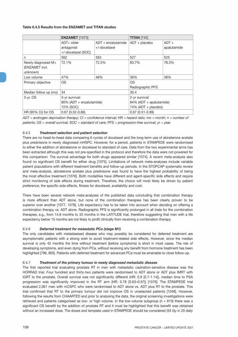
PROSTATE CANCER - LIMITED UPDATE 2021106
Table 6.4.5 Results from the ENZAMET and TITAN studies
ENZAMET [1073] TITAN [742]ADT+ older antagonist +/-docetaxel (SOC)
ADT + enzalutamide +/-docetaxel
ADT + placebo ADT + apalutamide
n 562 563 527 525Newly diagnosed M+(ENZAMET incl. unknown)
72.1% 72.5% 83.7% 78.3%
Low volume 47% 48% 36% 38%Primary objective OS
OSRadiographic PFS
Median follow up (mo) 34 30.43-yr. OS 3-yr survival:
80% (ADT + enzalutamide)72% (SOC)
2-yr survival:84% (ADT + apalutamide)74% (ADT + placebo)
HR (95% CI) for OS 0.67 (0.52-0.86) 0.67 (0.51-0.89)
ADT = androgen deprivation therapy; CI = confidence interval; HR = hazard ratio; mo = month; n = number of patients; OS = overall survival; SOC = standard of care; PFS = progression-free survival; yr = year.
6.4.5 Treatment selection and patient selectionThere are no head-to-head data comparing 6 cycles of docetaxel and the long-term use of abiraterone acetate plus prednisone in newly diagnosed mHSPC. However, for a period, patients in STAMPEDE were randomised to either the addition of abiraterone or docetaxel to standard of care. Data from the two experimental arms has been extracted although this was not pre-specified in the protocol and therefore the data were not powered for this comparison. The survival advantage for both drugs appeared similar [1074]. A recent meta-analysis also found no significant OS benefit for either drug [1075]. Limitations of network meta-analyses include variable patient populations with different treatment benefits and follow-up periods. In the STOPCAP systematic review and meta-analysis, abiraterone acetate plus prednisone was found to have the highest probability of being the most effective treatment [1076]. Both modalities have different and agent-specific side effects and require strict monitoring of side effects during treatment. Therefore, the choice will most likely be driven by patient preference, the specific side effects, fitness for docetaxel, availability and cost.
There have been several network meta-analyses of the published data concluding that combination therapy is more efficient than ADT alone, but none of the combination therapies has been clearly proven to be superior over another [1077, 1078]. Life expectancy has to be taken into account when deciding on offering a combination therapy vs. ADT alone. Radiographic PFS is significantly prolonged in all trials for the combination therapies, e.g., from 14.8 months to 33 months in the LATITUDE trial, therefore suggesting that men with a life expectancy below 15 months are not likely to profit clinically from receiving a combination therapy.
6.4.6 Deferred treatment for metastatic PCa (stage M1)The only candidates with metastasised disease who may possibly be considered for deferred treatment are asymptomatic patients with a strong wish to avoid treatment-related side effects. However, since the median survival is only 42 months the time without treatment (before symptoms) is short in most cases. The risk of developing symptoms, and even dying from PCa, without receiving any benefit from hormone treatment has been highlighted [796, 805]. Patients with deferred treatment for advanced PCa must be amenable to close follow-up.
6.4.7 Treatment of the primary tumour in newly diagnosed metastatic diseaseThe first reported trial evaluating prostate RT in men with metastatic castration-sensitive disease was the HORRAD trial. Four hundred and thirty-two patients were randomised to ADT alone or ADT plus IMRT with IGRT to the prostate. Overall survival was not significantly different (HR: 0.9 [0.7-1.14]), median time to PSA progression was significantly improved in the RT arm (HR: 0.78 [0.63–0.97]) [1079]. The STAMPEDE trial evaluated 2,061 men with mCSPC who were randomised to ADT alone vs. ADT plus RT to the prostate. This trial confirmed that RT to the primary tumour did not improve OS in unselected patients [1048]. However, following the results from CHAARTED and prior to analysing the data, the original screening investigations were retrieved and patients categorised as low- or high volume. In the low-volume subgroup (n = 819) there was a significant OS benefit by the addition of prostate RT and it must be highlighted that this benefit was obtained without an increased dose. The doses and template used in STAMPEDE should be considered (55 Gy in 20 daily
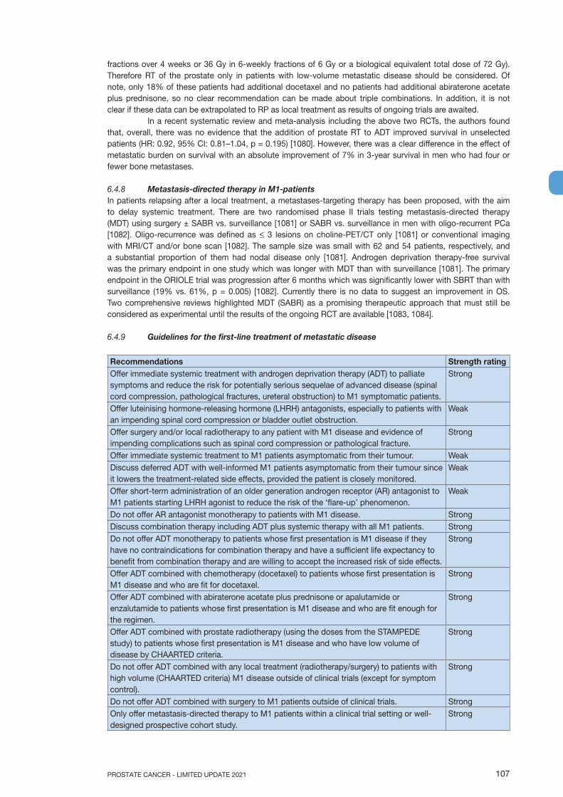
107PROSTATE CANCER - LIMITED UPDATE 2021
fractions over 4 weeks or 36 Gy in 6-weekly fractions of 6 Gy or a biological equivalent total dose of 72 Gy). Therefore RT of the prostate only in patients with low-volume metastatic disease should be considered. Of note, only 18% of these patients had additional docetaxel and no patients had additional abiraterone acetate plus prednisone, so no clear recommendation can be made about triple combinations. In addition, it is not clear if these data can be extrapolated to RP as local treatment as results of ongoing trials are awaited.
In a recent systematic review and meta-analysis including the above two RCTs, the authors found that, overall, there was no evidence that the addition of prostate RT to ADT improved survival in unselected patients (HR: 0.92, 95% CI: 0.81–1.04, p = 0.195) [1080]. However, there was a clear difference in the effect of metastatic burden on survival with an absolute improvement of 7% in 3-year survival in men who had four or fewer bone metastases.
6.4.8 Metastasis-directed therapy in M1-patientsIn patients relapsing after a local treatment, a metastases-targeting therapy has been proposed, with the aim to delay systemic treatment. There are two randomised phase II trials testing metastasis-directed therapy (MDT) using surgery ± SABR vs. surveillance [1081] or SABR vs. surveillance in men with oligo-recurrent PCa [1082]. Oligo-recurrence was defined as < 3 lesions on choline-PET/CT only [1081] or conventional imaging with MRI/CT and/or bone scan [1082]. The sample size was small with 62 and 54 patients, respectively, and a substantial proportion of them had nodal disease only [1081]. Androgen deprivation therapy-free survival was the primary endpoint in one study which was longer with MDT than with surveillance [1081]. The primary endpoint in the ORIOLE trial was progression after 6 months which was significantly lower with SBRT than with surveillance (19% vs. 61%, p = 0.005) [1082]. Currently there is no data to suggest an improvement in OS. Two comprehensive reviews highlighted MDT (SABR) as a promising therapeutic approach that must still be considered as experimental until the results of the ongoing RCT are available [1083, 1084].
6.4.9 Guidelines for the first-line treatment of metastatic disease
Recommendations Strength ratingOffer immediate systemic treatment with androgen deprivation therapy (ADT) to palliate symptoms and reduce the risk for potentially serious sequelae of advanced disease (spinal cord compression, pathological fractures, ureteral obstruction) to M1 symptomatic patients.
Strong
Offer luteinising hormone-releasing hormone (LHRH) antagonists, especially to patients with an impending spinal cord compression or bladder outlet obstruction.
Weak
Offer surgery and/or local radiotherapy to any patient with M1 disease and evidence of impending complications such as spinal cord compression or pathological fracture.
Strong
Offer immediate systemic treatment to M1 patients asymptomatic from their tumour. WeakDiscuss deferred ADT with well-informed M1 patients asymptomatic from their tumour since it lowers the treatment-related side effects, provided the patient is closely monitored.
Weak
Offer short-term administration of an older generation androgen receptor (AR) antagonist to M1 patients starting LHRH agonist to reduce the risk of the ‘flare-up’ phenomenon.
Weak
Do not offer AR antagonist monotherapy to patients with M1 disease. StrongDiscuss combination therapy including ADT plus systemic therapy with all M1 patients. StrongDo not offer ADT monotherapy to patients whose first presentation is M1 disease if they have no contraindications for combination therapy and have a sufficient life expectancy to benefit from combination therapy and are willing to accept the increased risk of side effects.
Strong
Offer ADT combined with chemotherapy (docetaxel) to patients whose first presentation is M1 disease and who are fit for docetaxel.
Strong
Offer ADT combined with abiraterone acetate plus prednisone or apalutamide or enzalutamide to patients whose first presentation is M1 disease and who are fit enough for the regimen.
Strong
Offer ADT combined with prostate radiotherapy (using the doses from the STAMPEDE study) to patients whose first presentation is M1 disease and who have low volume of disease by CHAARTED criteria.
Strong
Do not offer ADT combined with any local treatment (radiotherapy/surgery) to patients with high volume (CHAARTED criteria) M1 disease outside of clinical trials (except for symptom control).
Strong
Do not offer ADT combined with surgery to M1 patients outside of clinical trials. StrongOnly offer metastasis-directed therapy to M1 patients within a clinical trial setting or well-designed prospective cohort study.
Strong
PROSTATE CANCER - LIMITED UPDATE 2021108
6.5 Treatment: Castration-resistant PCa (CRPC)6.5.1 Definition of CRPCCastrate serum testosterone < 50 ng/dL or 1.7 nmol/L plus either:a. Biochemical progression: Three consecutive rises in PSA at least one week apart resulting in two 50%
increases over the nadir, and a PSA > 2 ng/mLor
b. Radiological progression: The appearance of new lesions: either two or more new bone lesions on bone scan or a soft tissue lesion using RECIST (Response Evaluation Criteria in Solid Tumours) [1085]. Symptomatic progression alone must be questioned and subject to further investigation. It is not sufficient to diagnose CRPC.
6.5.2 Management of mCRPC - general aspectsSelection of treatment for mCRPC is multifactorial and in general dependent on:• previous treatment for mHSPC and for non-mHSPC;• previous treatment for mCRPC;• quality of response and pace of progression on previous treatment;• known cross resistance between androgen receptor targeted agents (ARTA);• co-medication and known drug interactions (see approved summary of product characteristics);• known genetic alterations;• known histological variants and DNA repair deficiency (consider platinum or targeted therapy like poly-
ADP ribose); • local approval status of drugs and reimbursement situation;• available clinical trials;• The patient and his co-morbidities.
6.5.2.1 Molecular diagnosticsAll metastatic patients should be offered somatic genomic testing for homologous repair and MMR defects, preferably on metastatic carcinoma tissue but testing on primary tumour may also be performed. Alternatively, but still less common, genetic testing on circulating tumour DNA (ctDNA) is an option and has been used in some trials. One test, the FoundationOne® Liquid CDx has been FDA approved [1086]. Defective MMR assessment can be performed by immunohistochemistry for MMR proteins (MSH2, MSH6, MLH1 and PMS2) and/or by next-generation sequencing assays [1087]. Germline testing for BRCA1/2, ATM and MMR is recommended for high risk and particularly for metastatic PCa if clinically indicated.
Molecular diagnostics should be performed by a certified (accredited) institution using a standard NGS (Next Generation Sequencing) multiplication procedure (minimum depth of coverage of 200 X). The genes and respective exons should be listed; not only DNA for mutations but RNA needs to be examined for fusions and protein expression to obtain all clinically relevant information. A critical asset is the decision support helping to rate the mutations according to their clinical relevance [1088, 1089].
Level 1 evidence for the use of PARP-inhibitors has been reported [1090-1092]. Microsatellite instability (MSI)-high (or mismatch repair deficiency [MMR]) is rare in PCa, but for those patients, pembrolizumab has been approved by the FDA and could be a valuable additional treatment option [1093, 1094].
Germline molecular testing is discussed in Section 5.1.3 - Genetic testing for inherited PCa. Recommendations for germline testing are provided in Section 5.1.4.
6.5.3 Treatment decisions and sequence of available optionsApproved agents for the treatment of mCRPC in Europe are docetaxel, abiraterone/prednisolone, enzalutamide, cabazitaxel, olaparib and radium-223. In general, sequencing of ARTA like abiraterone and enzalutamide is not recommended particularly if the time of response to ADT and to the first ARTA was short (< 12 months) and high-risk features of rapid progression are present [1095, 1096].
The use of chemotherapy with docetaxel and subsequent cabazitaxel in the treatment sequence is recommended and should be applied early enough when the patient is still fit for chemotherapy. This is supported by high level evidence [1095].
6.5.4 Non-metastatic CRPCFrequent PSA testing in men treated with ADT has resulted in earlier detection of biochemical progression. Of these men approximately one-third will develop bone metastases detectable on bone scan within two years [1097].
In men with CRPC and no detectable clinical metastases using bone scan and CT-scan, baseline PSA level, PSA velocity and PSA-DT have been associated with time to first bone metastasis, bone metastasis-free survival and OS [1097, 1098]. These factors may be used when deciding which patients should be evaluated for metastatic disease. A consensus statement by the PCa Radiographic Assessments for Detection

109PROSTATE CANCER - LIMITED UPDATE 2021
of Advanced Recurrence (RADAR) group suggested a bone scan and a CT scan when the PSA reached 2 ng/mL and if this was negative, it should be repeated when the PSA reached 5 ng/mL, and again after every doubling of the PSA based on PSA testing every three months in asymptomatic men [1099]. Symptomatic patients should undergo relevant investigation regardless of PSA level. With more sensitive imaging techniques like PSMA PET/CT or whole-body MRI, more patients are diagnosed with early mCRPC [1100]. It remains unclear if the use of PSMA PET/CT in this setting improves outcome.
Three large phase III RCTs, PROSPER [1101], SPARTAN [1102] and ARAMIS [1103], evaluated metastasis-free survival as the primary endpoint in patients with nmCRPC (M0 CRPC) treated with enzalutamide (PROSPER) vs. placebo or apalutamide (SPARTAN) vs. placebo or darolutamide vs. placebo (ARAMIS), respectively. The M0 status was established by CT and bone scans. Only patients at high risk for the development of metastasis with a short PSA-DT of < 10 months were included. Patient characteristics in both trials revealed that about two-thirds of participants had a PSA-DT of < 6 months. All trials showed a significant metastasis-free survival benefit (PROSPER: median metastasis-free survival was 36.6 months in the enzalutamide group vs. 14.7 months in the placebo group [HR for metastasis or death: 0.29, 95% CI: 0.24–0.35, p < 0.001]; SPARTAN: median metastasis-free survival was 40.5 months in the apalutamide group vs. 16.2 months in the placebo group [HR for metastasis or death, 0.28, 95% CI: 0.23–0.35, p < 0.001]; ARAMIS: median metastasis-free survival was 40.4 months in the darolutamide group vs. 18.4 months in the placebo group (HR: 0.41, 95% CI: 0.34–0.50; 2-sided p < 0.0001]). All 3 trials showed a survival benefit after a follow-up of more than 30 months. In view of the long-term treatment with these AR targeting agents in asymptomatic patients, potential adverse events need to be taken into consideration and the patient informed accordingly.
6.5.5 Metastatic CRPCThe remainder of this section focuses on the management of men with proven metastatic CRPC (mCRPC) on conventional imaging.
6.5.5.1 Conventional androgen deprivation in CRPCEventually men with PCa will show evidence of disease progression despite castration. Two trials have shown only a marginal survival benefit for patients remaining on LHRH analogues during second- and third-line therapies [1104, 1105]. However, in the absence of prospective data, the modest potential benefits of continuing castration outweigh the minimal risk of treatment. In addition, all subsequent treatments have been studied in men with ongoing androgen suppression, therefore, it should be continued in these patients.
Table 6.5.1: Randomised phase III controlled trials - first-line treatment of mCRPC
Author Intervention Comparison Selection criteria Main outcomesDOCETAXELSWOG 99-16 2004 [1106]
docetaxel/EMP, every 3 weeks, 60 mg/m2, EMP 3 x 280 mg/day
mitoxantrone, every 3 weeks, 12 mg/m2 prednisone 5 mg BID
OS: 17.52 vs. 15.6 mo. (p = 0.02, HR: 0.80; 95% CI: 0.67-0.97) PFS: 6.3 vs. 3.2 mo. (p < 0.001)
TAX 327 2008 [1107, 1108]
docetaxel, every 3 weeks, 75 mg/m2 prednisone 5 mg BID or docetaxel, weekly, 30 mg/m2 prednisone 5 mg BID
mitoxantrone, every 3 weeks, 12 mg/m2, Prednisone 5 mg BID
OS: 19.2 for 3 weekly vs. 17.8 mo. 4-weekly and 16.3 in the control group. (p = 0.004, HR: 0.79, 95% CI: 0.67-0.93)
ABIRATERONECOU-AA-302 2013 [1109-1111]
abiraterone + prednisone
placebo + prednisone
- No previous docetaxel. - ECOG 0-1. - PSA or radiographic
progression. - No or mild symptoms. - No visceral metastases.
OS: 34.7 vs. 30.3 mo. (HR: 0.81, p = 0.0033). FU: 49.2 mo. rPFS: 16.5 vs. 8.3 mo. (p < 0.0001)
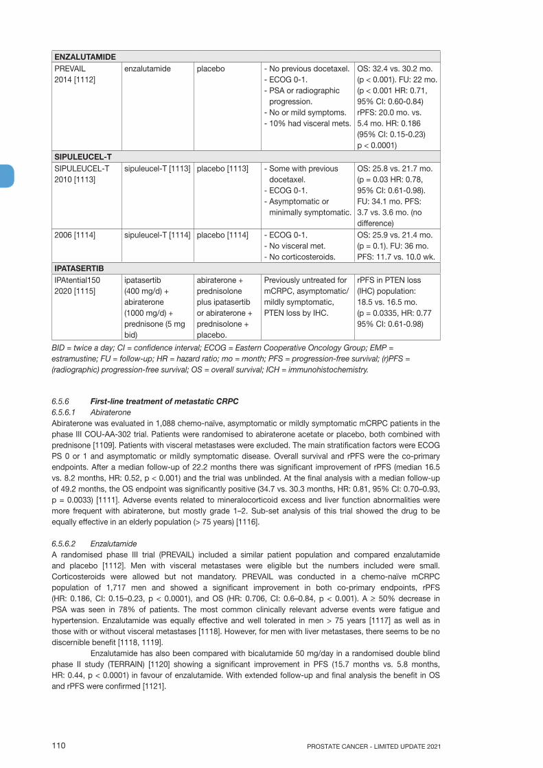
PROSTATE CANCER - LIMITED UPDATE 2021110
ENZALUTAMIDEPREVAIL 2014 [1112]
enzalutamide placebo - No previous docetaxel. - ECOG 0-1. - PSA or radiographic
progression. - No or mild symptoms. - 10% had visceral mets.
OS: 32.4 vs. 30.2 mo. (p < 0.001). FU: 22 mo. (p < 0.001 HR: 0.71, 95% CI: 0.60-0.84) rPFS: 20.0 mo. vs. 5.4 mo. HR: 0.186 (95% CI: 0.15-0.23) p < 0.0001)
SIPULEUCEL-TSIPULEUCEL-T 2010 [1113]
sipuleucel-T [1113] placebo [1113] - Some with previous docetaxel.
- ECOG 0-1. - Asymptomatic or
minimally symptomatic.
OS: 25.8 vs. 21.7 mo. (p = 0.03 HR: 0.78, 95% CI: 0.61-0.98). FU: 34.1 mo. PFS: 3.7 vs. 3.6 mo. (no difference)
2006 [1114] sipuleucel-T [1114] placebo [1114] - ECOG 0-1. - No visceral met. - No corticosteroids.
OS: 25.9 vs. 21.4 mo. (p = 0.1). FU: 36 mo. PFS: 11.7 vs. 10.0 wk.
IPATASERTIBIPAtential1502020 [1115]
ipatasertib (400 mg/d) + abiraterone (1000 mg/d) + prednisone (5 mg bid)
abiraterone + prednisolone plus ipatasertib or abiraterone + prednisolone + placebo.
Previously untreated for mCRPC, asymptomatic/mildly symptomatic, PTEN loss by IHC.
rPFS in PTEN loss (IHC) population: 18.5 vs. 16.5 mo. (p = 0.0335, HR: 0.77 95% CI: 0.61-0.98)
BID = twice a day; CI = confidence interval; ECOG = Eastern Cooperative Oncology Group; EMP = estramustine; FU = follow-up; HR = hazard ratio; mo = month; PFS = progression-free survival; (r)PFS = (radiographic) progression-free survival; OS = overall survival; ICH = immunohistochemistry.
6.5.6 First-line treatment of metastatic CRPC6.5.6.1 AbirateroneAbiraterone was evaluated in 1,088 chemo-naïve, asymptomatic or mildly symptomatic mCRPC patients in the phase III COU-AA-302 trial. Patients were randomised to abiraterone acetate or placebo, both combined with prednisone [1109]. Patients with visceral metastases were excluded. The main stratification factors were ECOG PS 0 or 1 and asymptomatic or mildly symptomatic disease. Overall survival and rPFS were the co-primary endpoints. After a median follow-up of 22.2 months there was significant improvement of rPFS (median 16.5 vs. 8.2 months, HR: 0.52, p < 0.001) and the trial was unblinded. At the final analysis with a median follow-up of 49.2 months, the OS endpoint was significantly positive (34.7 vs. 30.3 months, HR: 0.81, 95% CI: 0.70–0.93, p = 0.0033) [1111]. Adverse events related to mineralocorticoid excess and liver function abnormalities were more frequent with abiraterone, but mostly grade 1–2. Sub-set analysis of this trial showed the drug to be equally effective in an elderly population (> 75 years) [1116].
6.5.6.2 EnzalutamideA randomised phase III trial (PREVAIL) included a similar patient population and compared enzalutamide and placebo [1112]. Men with visceral metastases were eligible but the numbers included were small. Corticosteroids were allowed but not mandatory. PREVAIL was conducted in a chemo-naïve mCRPC population of 1,717 men and showed a significant improvement in both co-primary endpoints, rPFS (HR: 0.186, CI: 0.15–0.23, p < 0.0001), and OS (HR: 0.706, CI: 0.6–0.84, p < 0.001). A > 50% decrease in PSA was seen in 78% of patients. The most common clinically relevant adverse events were fatigue and hypertension. Enzalutamide was equally effective and well tolerated in men > 75 years [1117] as well as in those with or without visceral metastases [1118]. However, for men with liver metastases, there seems to be no discernible benefit [1118, 1119].
Enzalutamide has also been compared with bicalutamide 50 mg/day in a randomised double blind phase II study (TERRAIN) [1120] showing a significant improvement in PFS (15.7 months vs. 5.8 months, HR: 0.44, p < 0.0001) in favour of enzalutamide. With extended follow-up and final analysis the benefit in OS and rPFS were confirmed [1121].

111PROSTATE CANCER - LIMITED UPDATE 2021
6.5.6.3 DocetaxelA statistically significant improvement in median survival of 2.0–2.9 months has been shown with docetaxel-based chemotherapy compared to mitoxantrone plus prednisone [1108, 1122]. The standard first-line chemotherapy is docetaxel 75 mg/m2, 3-weekly doses combined with prednisone 5 mg twice a day (BID), up to 10 cycles. Prednisone can be omitted if there are contraindications or no major symptoms. The following independent prognostic factors; visceral metastases, pain, anaemia (Hb < 13 g/dL), bone scan progression, and prior estramustine may help stratify the response to docetaxel. Patients can be categorised into three risk groups: low risk (0 or 1 factor), intermediate (2 factors) and high risk (3 or 4 factors), and show three significantly different median OS estimates of 25.7, 18.7 and 12.8 months, respectively [1123].
Age by itself is not a contraindication to docetaxel [1124] but attention must be paid to careful monitoring and co-morbidities as discussed in Section 5.4 - Estimating life expectancy and health status [1125]. In men with mCRPC who are thought to be unable to tolerate the standard dose and schedule, docetaxel 50 mg/m2 every two weeks seems to be well tolerated with less grade 3–4 adverse events and a prolonged time to treatment failure [1126].
6.5.6.4 Sipuleucel-TIn 2010 a phase III trial of sipuleucel-T showed a survival benefit in 512 asymptomatic or minimally symptomatic mCRPC patients [1113]. After a median follow-up of 34 months, the median survival was 25.8 months in the sipuleucel-T group compared to 21.7 months in the placebo group, with a HR of 0.78 (p = 0.03). No PSA decline was observed and PFS was similar in both arms. The overall tolerance was very good, with more cytokine-related adverse events grade 1–2 in the sipuleucel-T group, but the same grade 3–4 adverse events in both arms. Sipuleucel-T is not available in Europe (and has had its licence withdrawn).
6.5.6.5 IpatasertibThe AKT inhibitor ipatasertib in combination with abiraterone plus prednisone was studied in asymptomatic or mildly symptomatic patients with PTEN loss by IHC and previously untreated for mCRPC. The randomised phase III trial (IPAtential) showed a significant benefit for the first endpoint rPFS in the PTEN loss (IHC) population (18.5 vs. 16.5 mo; p = 0,0335, HR: 0.77, 95% CI: 0.61–0,98). The OS results are still pending. Side effects of the AKT inhibitor ipatasertib include rash and diarrhoea [750]. This combination is still investigational [1115].
Table 6.5.2: Randomised controlled phase III - second-line/third-line trials in mCRPC
Author Intervention Comparison Selection criteria Main outcomesABIRATERONECOU-AA-301 2012 [1127]
abiraterone + prednisone HR
placebo + prednisone
Previous docetaxel. ECOG 0-2. PSA or radiographic progression.
OS: 15.8 vs. 11.2 mo. (p < 0.0001, HR: 0.74, 95% CI: 0.64-0.86; p < 0.0001). FU: 20.2 mo.rPFS: no change
COU-AA-301 2011 [1128]
OS: 14.8 vs. 10.9 mo.(p < 0.001 HR: 0.65; 95% CI: 0.54-0.77). FU: 12.8 mo.rPFS: 5.6 vs. 3.6 mo.
Radium-223ALSYMPCA 2013 [1129]
radium-223 placebo Previous or no previous docetaxel. ECOG 0-2. Two or more symptomatic bone metastases. No visceral metastases.
OS: 14.9 vs. 11.3 mo. (p = 0.002, HR: 0.61; 95% CI: 0.46-0.81). All secondary endpoints show a benefit over best standard of care.
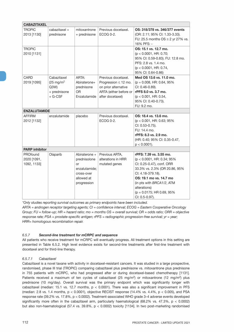
PROSTATE CANCER - LIMITED UPDATE 2021112
CABAZITAXELTROPIC 2013 [1130]
cabazitaxel + prednisone
mitoxantrone + prednisone
Previous docetaxel. ECOG 0-2.
OS: 318/378 vs. 346/377 events(OR: 2.11; 95% CI: 1.33-3.33). FU: 25.5 months OS > 2 yr 27% vs. 16% PFS: -
TROPIC 2010 [1131]
OS: 15.1 vs. 12.7 mo.(p < 0.0001, HR: 0.70; 95% CI: 0.59-0.83). FU: 12.8 mo.PFS: 2.8 vs. 1.4 mo.(p < 0.0001, HR: 0.74, 95% CI: 0.64-0.86)
CARD 2019 [1095]
Cabazitaxel (25 mg/m2 Q3W) + prednisone + G-CSF
ARTA: Abiraterone+ prednisone OREnzalutamide
Previous docetaxel.Progression < 12 mo. on prior alternative ARTA (either before or after docetaxel)
Med OS 13.6 vs. 11.0 mo. (p = 0.008, HR: 0.64, 95% CI: 0.46-0.89). rPFS 8.0 vs. 3.7 mo. (p < 0.001, HR: 0.54, 95% CI: 0.40-0.73).FU: 9.2 mo.
ENZALUTAMIDEAFFIRM 2012 [1132]
enzalutamide placebo Previous docetaxel. ECOG 0-2.
OS: 18.4 vs. 13.6 mo.(p < 0.001, HR: 0.63; 95% CI: 0.53-0.75). FU: 14.4 mo.rPFS: 8.3 vs. 2.9 mo.(HR: 0.40; 95% CI: 0.35-0.47, p < 0.0001).
PARP inhibitorPROfound2020 [1091, 1092, 1133]
Olaparib Abiraterone + prednisolone or enzalutamide;cross-over allowed at progression
Previous ARTA, alterations in HRR mutated genes
rPFS: 7.39 vs. 3.55 mo. (p < 0.0001, HR: 0.34; 95% CI: 0.25-0.47), conf. ORR 33.3% vs. 2.3% (OR 20.86, 95% CI: 4.18-379.18).OS: 19.1 mo vs. 14.7 mo (in pts with BRCA1/2, ATM alterations)(p = 0.0175; HR 0.69, 95% CI: 0.5-0.97).
*Only studies reporting survival outcomes as primary endpoints have been included.ARTA = androgen receptor targeting agents; CI = confidence interval; ECOG = Eastern Cooperative Oncology Group; FU = follow-up; HR = hazard ratio; mo = months OS = overall survival; OR = odds ratio; ORR = objective response rate; PSA = prostate-specific antigen; rPFS = radiographic progression-free survival; yr = year; HRR= homologous recombination repair.
6.5.7 Second-line treatment for mCRPC and sequenceAll patients who receive treatment for mCRPC will eventually progress. All treatment options in this setting are presented in Table 6.5.2. High level evidence exists for second-line treatments after first-line treatment with docetaxel and for third-line therapy.
6.5.7.1 CabazitaxelCabazitaxel is a novel taxane with activity in docetaxel-resistant cancers. It was studied in a large prospective, randomised, phase III trial (TROPIC) comparing cabazitaxel plus prednisone vs. mitoxantrone plus prednisone in 755 patients with mCRPC, who had progressed after or during docetaxel-based chemotherapy [1131]. Patients received a maximum of ten cycles of cabazitaxel (25 mg/m2) or mitoxantrone (12 mg/m2) plus prednisone (10 mg/day). Overall survival was the primary endpoint which was significantly longer with cabazitaxel (median: 15.1 vs. 12.7 months, p < 0.0001). There was also a significant improvement in PFS (median: 2.8 vs. 1.4 months, p < 0.0001), objective RECIST response (14.4% vs. 4.4%, p < 0.005), and PSA response rate (39.2% vs. 17.8%, p < 0.0002). Treatment-associated WHO grade 3–4 adverse events developed significantly more often in the cabazitaxel arm, particularly haematological (68.2% vs. 47.3%, p < 0.0002) but also non-haematological (57.4 vs. 39.8%, p < 0.0002) toxicity [1134]. In two post-marketing randomised

113PROSTATE CANCER - LIMITED UPDATE 2021
phase III trials, cabazitaxel was shown not to be superior to docetaxel in the first-line setting; in the second-line setting in terms of OS, 20 mg/m2 cabazitaxel was not inferior to 25 mg/m2, but less toxic. Therefore, the lower dose should be preferred [1135, 1136]. Cabazitaxel should preferably be given with prophylactic granulocyte colony-stimulating factor (G-CSF) and should be administered by physicians with expertise in handling neutropenia and sepsis [1137].
6.5.7.2 Abiraterone acetate after prior docetaxelPositive results of the large phase III trial (COU-AA-301) were reported after a median follow-up of 12.8 months [1128] and confirmed by the final analysis [1127]. A total of 1,195 patients with mCRPC were randomised 2:1 to abiraterone acetate plus prednisone or placebo plus prednisone. All patients had progressive disease based on the Prostate Cancer Clinical Trials Working Group 2 (PCWG2) criteria after docetaxel therapy (with a maximum of two previous chemotherapeutic regimens). The primary endpoint was OS, with a planned HR of 0.8 in favour of abiraterone. After a median follow-up of 20.2 months, the median survival in the abiraterone group was 15.8 months compared to 11.2 months in the placebo arm (HR: 0.74, p < 0.0001). The benefit was observed in all subgroups and all the secondary objectives were in favour of abiraterone (PSA, radiologic tissue response, time to PSA or objective progression). The incidence of the most common grade 3–4 adverse events did not differ significantly between arms, but mineralocorticoid-related side effects were more frequent in the abiraterone group, mainly grade 1–2 (fluid retention, oedema and hypokalaemia).
6.5.7.3 Enzalutamide after docetaxelThe planned interim analysis of the AFFIRM study was published in 2012 [1132]. This trial randomised 1,199 patients with mCRPC in a 2:1 fashion to enzalutamide or placebo. The patients had progressed after docetaxel treatment, according to the PCWG2 criteria. Corticosteroids were not mandatory, but could be prescribed, and were received by about 30% of the patients. The primary endpoint was OS, with an expected HR benefit of 0.76 in favour of enzalutamide. After a median follow-up of 14.4 months, the median survival in the enzalutamide group was 18.4 months compared to 13.6 months in the placebo arm (HR: 0.63, p < 0.001). This led to the recommendation to halt and unblind the study. The benefit was observed irrespective of age, baseline pain intensity, and type of progression. In the final analysis with longer follow-up the OS results were confirmed despite crossover and extensive post-progression therapies [1121]. Enzalutamide was active also in patients with visceral metastases.
All the secondary objectives were in favour of enzalutamide (PSA, soft tissue response, QoL, time to PSA or objective progression). No difference in terms of side effects was observed in the two groups, with a lower incidence of grade 3–4 adverse events in the enzalutamide arm. There was a 0.6% incidence of seizures in the enzalutamide group compared to none in the placebo arm.
6.5.7.4 Radium-223The only bone-specific drug that is associated with a survival benefit is the α-emitter radium-223. In a large phase III trial (ALSYMPCA) 921 patients with symptomatic mCRPC, who failed or were unfit for docetaxel, were randomised to six injections of 50 kBq/kg radium-223 or placebo, plus standard of care. The primary endpoint was OS. Radium-223 significantly improved median OS by 3.6 months (HR: 0.70, p < 0.001) and was also associated with prolonged time to first skeletal event, improvement in pain scores and improvement in QoL [1129]. The associated toxicity was mild and, apart from slightly more haematologic toxicity and diarrhoea with radium-223, it did not differ significantly from that in the placebo arm [1129]. Radium-223 was effective and safe whether or not patients were docetaxel pre-treated [1138]. Due to safety concerns, use of radium-223 was recently restricted to after docetaxel and at least one AR targeted agent [1139]. In particular, the use of radium-223 in combination with abiraterone acetate plus prednisolone showed significant safety risks related to fractures and more deaths. This was most striking in patients without the concurrent use of anti-resorptive agents [1140].
6.5.8 Treatment after docetaxel and one line of hormonal treatment for mCRPCFor men progressing quickly on AR targeted therapy (< 12 months) it is now clear that cabazitaxel is the treatment supported by the best data. The CARD trial, an open label randomised phase III trial, evaluated cabazitaxel after docetaxel and one line of ARTA (either abiraterone plus prednisolone or enzalutamide) [1095]. It included patients progressing in less than 12 months on previous abiraterone or enzalutamide for mCRPC. Cabazitaxel more than doubled rPFS vs. another ARTA and reduced the risk of death by 36% vs. ARTA. The rPFS with cabazitaxel remained superior regardless of the ARTA sequence and if docetaxel was given before, or after, the first ARTA.
The choice of further treatment after docetaxel and one line of hormonal treatment for mCRPC is open for patients who have a > 12 months response to first-line abiraterone or enzalutamide for mCRPC [1141]. Either radium-223 or second-line chemotherapy (cabazitaxel) are reasonable options. In general,
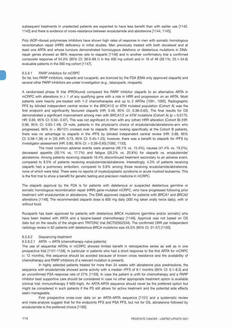
PROSTATE CANCER - LIMITED UPDATE 2021114
subsequent treatments in unselected patients are expected to have less benefit than with earlier use [1142, 1143] and there is evidence of cross-resistance between enzalutamide and abiraterone [1144, 1145].
Poly (ADP-ribose) polymerase inhibitors have shown high rates of response in men with somatic homologous recombination repair (HRR) deficiency in initial studies. Men previously treated with both docetaxel and at least one ARTA and whose tumours demonstrated homozygous deletions or deleterious mutations in DNA-repair genes showed an 88% response rate to olaparib [1146] and in another confirmatory trial a confirmed composite response of 54.3% (95% CI: 39·0–69.1) in the 400 mg cohort and in 18 of 46 (39.1%; 25.1–54.6) evaluable patients in the 300 mg cohort [1147].
6.5.8.1 PARP inhibitors for mCRPCSo far, two PARP inhibitors, olaparib and rucaparib, are licenced by the FDA (EMA only approved olaparib) and several other PARP inhibitors are under investigation (e.g., talazoparib, niraparib).
A randomised phase III trial (PROfound) compared the PARP inhibitor olaparib to an alternative ARTA in mCRPC with alterations in > 1 of any qualifying gene with a role in HRR and progression on an ARTA. Most patients were heavily pre-treated with 1–2 chemotherapies and up to 2 ARTAs [1091, 1092]. Radiographic PFS by blinded independent central review in the BRCA1/2 or ATM mutated population (Cohort A) was the first endpoint and significantly favoured olaparib (HR: 0.49, 95% CI: 0.38–0.63). The final results for OS demonstrated a significant improvement among men with BRCA1/2 or ATM mutations (Cohort A) (p = 0.0175; HR: 0.69, 95% CI: 0.50– 0.97). This was not significant in men with any (other) HRR alteration (Cohort B) (HR: 0.96, 95% CI: 0.63–1.49). Of note, patients in the physician’s choice of enzalutamide/abiraterone-arm who progressed, 66% (n = 86/131) crossed over to olaparib. When looking specifically at the Cohort B patients, there was no advantage to olaparib in the rPFS by blinded independent central review (HR: 0.88, 95% CI: 0.58–1.36) or in OS (HR: 0.73, 95% CI: 0.45–1.23), however, there was a benefit to olaparib for rPFS by investigator assessment (HR: 0.60, 95% CI: > 0.39–0.93) [1092, 1133].
The most common adverse events were anaemia (46.1% vs. 15.4%), nausea (41.4% vs. 19.2%), decreased appetite (30.1% vs. 17.7%) and fatigue (26.2% vs. 20.8%) for olaparib vs. enzalutamide/abiraterone. Among patients receiving olaparib 16.4% discontinued treatment secondary to an adverse event, compared to 8.5% of patients receiving enzalutamide/abiraterone. Interestingly, 4.3% of patients receiving olaparib had a pulmonary embolism, compared to 0.8% among those receiving enzalutamide/abiraterone, none of which were fatal. There were no reports of myelodysplastic syndrome or acute myeloid leukaemia. This is the first trial to show a benefit for genetic testing and precision medicine in mCRPC.
The olaparib approval by the FDA is for patients with deleterious or suspected deleterious germline or somatic homologous recombination repair (HRR) gene-mutated mCRPC, who have progressed following prior treatment with enzalutamide or abiraterone. The EMA approved olaparib for patients with BRCA1 and BRCA2 alterations [1148]. The recommended olaparib dose is 600 mg daily (300 mg taken orally twice daily), with or without food.
Rucaparib has been approved for patients with deleterious BRCA mutations (germline and/or somatic) who have been treated with ARTA and a taxane-based chemotherapy [1149]. Approval was not based on OS data but on the results of the single-arm TRITON2 trial (NCT02952534). The confirmed ORR per independent radiology review in 62 patients with deleterious BRCA mutations was 43.5% (95% CI: 31–57) [1150].
6.5.8.2 Sequencing treatment6.5.8.2.1 ARTA -> ARTA (chemotherapy-naïve patients)The use of sequential ARTAs in mCRPC showed limited benefit in retrospective series as well as in one prospective trial [1151-1158]. In particular in patients who had a short response to the first ARTA for mCRPC (< 12 months), this sequence should be avoided because of known cross resistance and the availability of chemotherapy and PARP inhibitors (if a relevant mutation is present).
In highly selected patients treated for more than 24 weeks with abiraterone plus prednisolone, the sequence with enzalutamide showed some activity with a median rPFS of 8.1 months (95% CI: 6.1–8.3) and an unconfirmed PSA response rate of 27% [1159]. In case the patient is unfit for chemotherapy and a PARP inhibitor best supportive care should be considered in case no other appropriate treatment option is available (clinical trial: immunotherapy if MSI-high). An ARTA-ARTA sequence should never be the preferred option but might be considered in such patients if the PS still allows for active treatment and the potential side effects seem manageable.
First prospective cross-over data on an ARTA-ARTA sequence [1151] and a systematic review and meta-analysis suggest that for the endpoints PFS and PSA PFS, but not for OS, abiraterone followed by enzalutamide is the preferred choice [1160].
115PROSTATE CANCER - LIMITED UPDATE 2021
6.5.8.2.2 ARTA -> PARP inhibitor/olaparib This sequence in patients with deleterious or suspected deleterious germline or somatic homologous recombination repair (HRR) gene-mutated mCRPC is supported by data from the randomised phase III PROfound trial [1092]. A subgoup of patients in this trial was pre-treated with one or two ARTAs and no chemotherapy. The ARTA – docetaxel - PARP inhibitor vs. ARTA – PARP inhibitor - docetaxel sequences are still under investigation.
6.5.8.2.3 Docetaxel for mHSPC -> docetaxel rechallenge There is limited evidence for second- or third-line use of docetaxel after treatment with docetaxel for mHSPC. Docetaxel seems to be less active than ARTA at progression to mCRPC following docetaxel for mHSPC [1161].
6.5.8.2.4 ARTA -> docetaxel or docetaxel -> ARTA followed by PARP inhibitorBoth olaparib and rucaparib are active in biomarker-selected mCRPC patients after ARTA and docetaxel in either sequence [1092, 1149].
6.5.8.2.5 ARTA before or after docetaxel There is level 1 evidence for both sequences (see Table 6.5.2).
6.5.8.2.6 ARTA –> docetaxel -> cabazitaxel or docetaxel –> ARTA -> cabazitaxel Both third-line treatment sequences are supported by level 1 evidence. Of note, there is high level evidence favouring cabazitaxel vs. a second ARTA after docetaxel and one ARTA. CARD is the first prospective randomised phase III trial adressing this question (see Table 6.5.2) [1095].
6.5.9 Prostate-specific membrane antigen (PSMA) therapy6.5.9.1 BackgroundDuring the 90s several radiopharmaceuticals including phosphorous-32, strontium-89, yttrium-90, samarium-153, and rhenium-186 [1162] were developed for the treatment of bone pain secondary to metastasis from PCa. They were effective at palliation; relieving pain and improving QoL, especially in the setting of diffuse bone metastasis. However, they never gained widespread adoption. The first radioisotope to demonstrate a survival benefit was radium-223 (see Section 6.5.7.4).
6.5.9.2 PSMA-based therapyThe increasing use of PSMA PET as a diagnostic tracer and the realisation that this allowed identification of a greater number of metastatic deposits led to attempts to treat cancer by replacing the imaging isotope with a therapeutic isotope which accumulates where the tumour is demonstrated (theranostics) [1164]. Therefore, after identification of the target usually with diagnostic 68Gallium-labelled PSMA, therapeutic radiopharmaceuticals labelled with beta (lutetium-177 or ytrium-90) or a (actinium-225) emitting isotopes could be used to treat metastatic PCa. At present, all of these agents should be regarded as investigational.
The PSMA therapeutic radiopharmaceutical supported with the most robust data is 177Lu-PSMA-617. The first patient was treated in 2014 and early clinical studies evaluating the safety and efficacy of Lu-PSMA therapy have demonstrated promising results, despite the fact that a significant proportion of men had already progressed on multiple therapies [1165]. Nonetheless, most of the literature is based on single-centre experience and RCTs are lacking [1166]. Recently, data from uncontrolled prospective phase II trials have been published reporting high response rates with low toxic effects [1167, 1168]. Positive signals are coming from a randomised phase II trial comparing Lu-PSMA with cabazitaxel in ARTA and docetaxel pre-treated patients. The primary endpoint of PSA reduction > 50% was achieved in highly selected patients (PSMA- and FDG PET/CT criteria) was superior with Lu-PSMA [1169]. More robust data are expected from ongoing trials.
6.5.10 Immunotherapy for mCRPC The immune checkpoint inhibitor pembrolizumab was approved by the FDA for all MMR–deficient cancers or in those with instable microsatellite status (MSI-high) [1093]. This also applies to PCa but is a very rare finding in this tumour entity [1094]. In all other PCa patients pembrolizumab monotherapy is still experimental. It shows limited anti-tumour activity with an acceptable safety profile, again in a small subset of patients. A phase II trial enrolled 258 patients treated with pembrolizumab [1163]. The objective response rate was around 4%, but those responses were durable. Combination immunotherapy is under investigation.
6.5.11 Monitoring of treatmentBaseline examinations should include a medical history, clinical examination as well as baseline blood tests (PSA, total testosterone level, full blood count, renal function, baseline liver function tests, alkaline phosphatase), bone scan and CT of chest, abdomen and pelvis [1170, 1171]. The use of choline or PSMA
PROSTATE CANCER - LIMITED UPDATE 2021116
PET/CT scans for progressing CRPC is unclear and most likely not as beneficial as for patients with BCR or hormone-naïve disease. Flares, PSMA upregulation and discordant results compared with PSA response or progression on ARTA have been described [1172]. Prostate-specific antigen alone is not reliable enough [1173] for monitoring disease activity in advanced CRPC since visceral metastases may develop in men without rising PSA [1174]. Instead, the PCWG2 recommends a combination of bone scintigraphy and CT scans, PSA measurements and clinical benefit in assessing men with CRPC [1122]. A majority of experts at the 2015 Advanced Prostate Cancer Consensus Conference (APCCC) suggested regular review and repeating blood profile every two to three months with bone scintigraphy and CT scans at least every six months, even in the absence of a clinical indication [1170]. This reflects that the agents with a proven OS benefit all have potential toxicity and considerable cost and patients with no objective benefit should have their treatment modified. The APCCC participants stressed that such treatments should not be stopped for PSA progression alone. Instead, at least two of the three criteria (PSA progression, radiographic progression and clinical deterioration) should be fulfilled to stop treatment. For trial purposes, the updated PCWG3 put more weight on the importance of documenting progression in existing lesions and introduced the concept of no longer ‘clinically benefiting‘ to underscore the distinction between first evidence of progression and the clinical need to terminate or change treatment [1175]. These recommendations also seem valid for clinical practice outside trials.
6.5.12 When to change treatmentThe timing of mCRPC treatment change remains a matter of debate in mCRPC although it is clearly advisable to start or change treatment immediately in men with symptomatic progressing metastatic disease. Preferably, any treatment change should precede development of de novo symptoms or worsening of existing symptoms. Although, the number of effective treatments is increasing, head-to-head comparisons are still rare, as are prospective data assessing the sequencing of available agents. Therefore it is not clear how to select the most appropriate ‘second-line‘ treatment, in particular in patients without HRR alterations or other biomarkers. A positive example, however, is the CARD trial which clearly established cabazitaxel as the better third-line treatment in docetaxel pre-treated patients after one ARTA compared to the use of a second ARTA [1095].
The ECOG PS has been used to stratify patients. Generally men with a PS of 0–1 are likely to tolerate treatments and those with a PS of > 2 are less likely to benefit. However, it is important that treatment decisions are individualised, in particular when symptoms related to disease progression are impacting on PS. In such cases, a trial of active life-prolonging agents to establish if a given treatment will improve the PS may be appropriate. Sequencing of treatment is discussed in a summery paper published following the St. Gallen Advanced Prostate Cancer Consensus Conference 2019 [1176].
6.5.13 Symptomatic management in metastatic CRPCCastration-resistant PCa is usually a debilitating disease often affecting the elderly male. A multidisciplinary approach is required with input from urologists, medical oncologists, radiation oncologists, nurses, psychologists and social workers [1176, 1177]. Critical issues of palliation must be addressed when considering additional systemic treatment, including management of pain, constipation, anorexia, nausea, fatigue and depression.
6.5.13.1 Common complications due to bone metastasesMost patients with CRPC have painful bone metastases. External beam radiotherapy is highly effective, even as a single fraction [1178, 1179]. A single infusion of a third generation bisphosphonate could be considered when RT is not available [1180]. Common complications due to bone metastases include vertebral collapse or deformity, pathological fractures and spinal cord compression. Cementation can be an effective treatment for painful spinal fracture whatever its origin, clearly improving both pain and QoL [1181]. It is important to offer standard palliative surgery, which can be effective for managing osteoblastic metastases [1182, 1183]. Impending spinal cord compression is an emergency. It must be recognised early and patients should be educated to recognise the warning signs. Once suspected, high-dose corticosteroids must be given and MRI performed as soon as possible. A systematic neurosurgery or orthopaedic surgeon consultation should be planned to discuss a possible decompression, followed by EBRT [1184]. Otherwise, EBRT with, or without, systemic therapy, is the treatment of choice.
6.5.13.2 Preventing skeletal-related events6.5.13.2.1 BisphosphonatesZoledronic acid has been evaluated in mCRPC to reduce skeletal-related events (SRE). This study was conducted when no active anti-cancer treatments, but for docetaxel, were available. Six hundred and forty three patients who had CRPC with bone metastases were randomised to receive zoledronic acid, 4 or 8 mg every three weeks for 15 consecutive months, or placebo [1185]. The 8 mg dose was poorly tolerated and reduced to 4 mg but did not show a significant benefit. However, at 15 and 24 months of follow-up, patients

117PROSTATE CANCER - LIMITED UPDATE 2021
treated with 4 mg zoledronic acid had fewer SREs compared to the placebo group (44 vs. 33%, p = 0.021) and in particular fewer pathological fractures (13.1 vs. 22.1%, p = 0.015). Furthermore, the time to first SRE was longer in the zoledronic acid group. No survival benefit has been seen in any prospective trial with bisphosphonates.
6.5.13.2.2 RANK ligand inhibitorsDenosumab is a fully human monoclonal antibody directed against RANKL (receptor activator of nuclear factor kappa-B ligand), a key mediator of osteoclast formation, function, and survival. In M0 CRPC, denosumab has been associated with increased bone-metastasis-free survival compared to placebo (median benefit: 4.2 months, HR: 0.85, p = 0.028) [1178]. This benefit did not translate into a survival difference (43.9 compared to 44.8 months, respectively) and neither the FDA or the EMA have approved denosumab for this indication [1186].
The efficacy and safety of denosumab (n = 950) compared with zoledronic acid (n = 951) in patients with mCRPC was assessed in a phase III trial. Denosumab was superior to zoledronic acid in delaying or preventing SREs as shown by time to first on-study SRE (pathological fracture, radiation or surgery to bone, or spinal cord compression) of 20.7 vs. 17.1 months, respectively (HR: 0.82, p = 0.008). Both urinary N-telopeptide and bone-specific alkaline phosphatase were significantly suppressed in the denosumab arm compared with the zoledronic acid arm (p < 0.0001 for both). However, these findings were not associated with any survival benefit and in a recent post-hoc re-evaluation of endpoints, denosumab showed identical results when comparing SREs and symptomatic skeletal events [1187].
The potential toxicity (e.g., osteonecrosis of the jaw, hypocalcaemia) of these drugs must always be kept in mind (5–8.2% in M0 CRPC and mCRPC, respectively) [1187-1189]. Patients should have a dental examination before starting therapy as the risk of jaw necrosis is increased by several risk factors including a history of trauma, dental surgery or dental infection [1190]. Also, the risk for osteonecrosis of the jaw increased numerically with the duration of use in a pivotal trial [1191] (one year vs. two years with denosumab), but this was not statistically significant when compared to zoledronic acid [1186]. According to the EMA, hypocalcaemia is a concern in patients treated with denosumab and zoledronic acid. Hypocalcaemia must be corrected by adequate intake of calcium and vitamin D before initiating therapy [1192]. Hypocalcaemia should be identified and prevented during treatment with bone protective agents (risk of severe hypocalcaemia is 8% and 5% for denosumab and zoledronic acid, respectively) [1189]. Serum calcium should be measured in patients starting therapy and monitored during treatment, especially during the first weeks and in patients with risk factors for hypocalcaemia or on other medication affecting serum calcium. Daily calcium (> 500 mg) and vitamin D (> 400 IU equivalent) are recommended in all patients, unless in case of hypercalcaemia [1189, 1193, 1194].
6.5.14 Summary of evidence and guidelines for life-prolonging treatments of castrate-resistant disease
Summary of evidence LEFirst-line treatment for mCRPC will be influenced by which treatments were used when metastatic cancer was first discovered.
4
No clear-cut recommendation can be made for the most effective drug for first-line CRPC treatment (i.e., hormone therapy, chemotherapy or radium-223) as no validated predictive factors exist.
3
Recommendations Strength ratingEnsure that testosterone levels are confirmed to be < 50 ng/dL before diagnosing castrate-resistant PCa (CRPC).
Strong
Counsel, manage and treat patients with metastatic CRPC (mCRPC) in a multidisciplinary team.
Strong
Treat patients with mCRPC with life-prolonging agents. StrongOffer mCRPC patients somatic and/or germline molecular testing as well as testing for mismatch repair deficiencies or microsatellite instability.
Strong
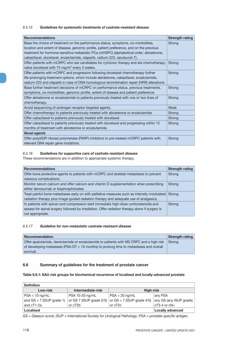
PROSTATE CANCER - LIMITED UPDATE 2021118
6.5.15 Guidelines for systematic treatments of castrate-resistant disease
Recommendations Strength ratingBase the choice of treatment on the performance status, symptoms, co-morbidities, location and extent of disease, genomic profile, patient preference, and on the previous treatment for hormone-sensitive metastatic PCa (mHSPC) (alphabetical order: abiraterone, cabazitaxel, docetaxel, enzalutamide, olaparib, radium-223, sipuleucel-T).
Strong
Offer patients with mCRPC who are candidates for cytotoxic therapy and are chemotherapy naïve docetaxel with 75 mg/m2 every 3 weeks.
Strong
Offer patients with mCRPC and progression following docetaxel chemotherapy further life-prolonging treatment options, which include abiraterone, cabazitaxel, enzalutamide, radium-223 and olaparib in case of DNA homologous recombination repair (HRR) alterations.
Strong
Base further treatment decisions of mCRPC on performance status, previous treatments, symptoms, co-morbidities, genomic profile, extent of disease and patient preference.
Strong
Offer abiraterone or enzalutamide to patients previously treated with one or two lines of chemotherapy.
Strong
Avoid sequencing of androgen receptor targeted agents. WeakOffer chemotherapy to patients previously treated with abiraterone or enzalutamide. StrongOffer cabazitaxel to patients previously treated with docetaxel. StrongOffer cabazitaxel to patients previously treated with docetaxel and progressing within 12 months of treatment with abiraterone or enzalutamide.
Strong
Novel agentsOffer poly(ADP-ribose) polymerase (PARP) inhibitors to pre-treated mCRPC patients with relevant DNA repair gene mutations.
Strong
6.5.16 Guidelines for supportive care of castrate-resistant diseaseThese recommendations are in addition to appropriate systemic therapy.
Recommendations Strength ratingOffer bone protective agents to patients with mCRPC and skeletal metastases to prevent osseous complications.
Strong
Monitor serum calcium and offer calcium and vitamin D supplementation when prescribing either denosumab or bisphosphonates.
Strong
Treat painful bone metastases early on with palliative measures such as intensity-modulated radiation therapy plus image-guided radiation therapy and adequate use of analgesics.
Strong
In patients with spinal cord compression start immediate high-dose corticosteroids and assess for spinal surgery followed by irradiation. Offer radiation therapy alone if surgery is not appropriate.
Strong
6.5.17 Guideline for non-metastatic castrate-resistant disease
Recommendation Strength ratingOffer apalutamide, darolutamide or enzalutamide to patients with M0 CRPC and a high risk of developing metastasis (PSA-DT < 10 months) to prolong time to metastases and overall survival.
Strong
6.6 Summary of guidelines for the treatment of prostate cancer
Table 6.6.1: EAU risk groups for biochemical recurrence of localised and locally-advanced prostate
DefinitionLow-risk Intermediate-risk High-risk
PSA < 10 ng/mLand GS < 7 (ISUP grade 1)and cT1-2a
PSA 10-20 ng/mLor GS 7 (ISUP grade 2/3)or cT2b
PSA > 20 ng/mLor GS > 7 (ISUP grade 4/5)or cT2c
any PSAany GS (any ISUP grade)cT3-4 or cN+
Localised Locally advanced
GS = Gleason score; ISUP = International Society for Urological Pathology; PSA = prostate-specific antigen.
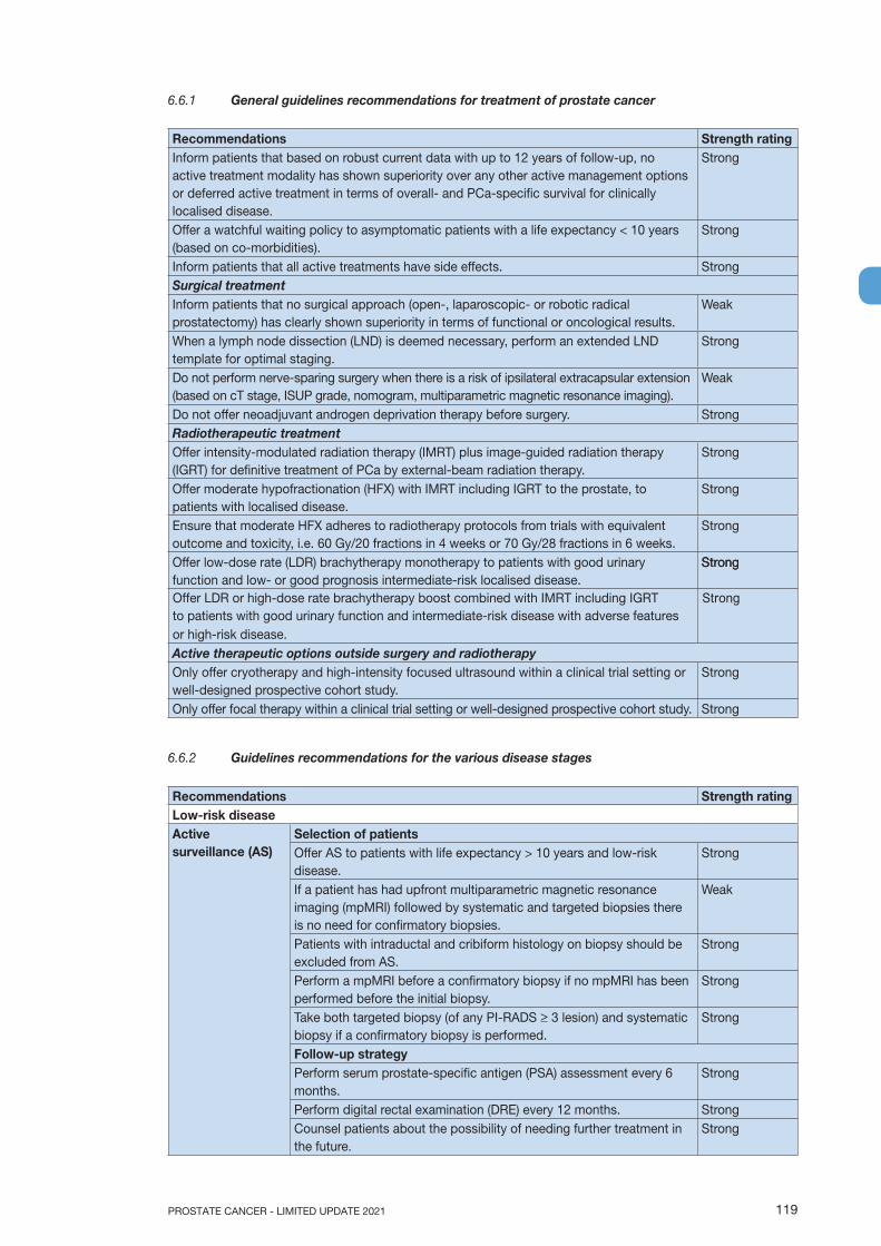
119PROSTATE CANCER - LIMITED UPDATE 2021
6.6.1 General guidelines recommendations for treatment of prostate cancer
Recommendations Strength ratingInform patients that based on robust current data with up to 12 years of follow-up, no active treatment modality has shown superiority over any other active management options or deferred active treatment in terms of overall- and PCa-specific survival for clinically localised disease.
Strong
Offer a watchful waiting policy to asymptomatic patients with a life expectancy < 10 years (based on co-morbidities).
Strong
Inform patients that all active treatments have side effects. StrongSurgical treatmentInform patients that no surgical approach (open-, laparoscopic- or robotic radical prostatectomy) has clearly shown superiority in terms of functional or oncological results.
Weak
When a lymph node dissection (LND) is deemed necessary, perform an extended LND template for optimal staging.
Strong
Do not perform nerve-sparing surgery when there is a risk of ipsilateral extracapsular extension (based on cT stage, ISUP grade, nomogram, multiparametric magnetic resonance imaging).
Weak
Do not offer neoadjuvant androgen deprivation therapy before surgery. StrongRadiotherapeutic treatmentOffer intensity-modulated radiation therapy (IMRT) plus image-guided radiation therapy (IGRT) for definitive treatment of PCa by external-beam radiation therapy.
Strong
Offer moderate hypofractionation (HFX) with IMRT including IGRT to the prostate, to patients with localised disease.
Strong
Ensure that moderate HFX adheres to radiotherapy protocols from trials with equivalent outcome and toxicity, i.e. 60 Gy/20 fractions in 4 weeks or 70 Gy/28 fractions in 6 weeks.
Strong
Offer low-dose rate (LDR) brachytherapy monotherapy to patients with good urinary function and low- or good prognosis intermediate-risk localised disease.
Strong
Offer LDR or high-dose rate brachytherapy boost combined with IMRT including IGRT Strongto patients with good urinary function and intermediate-risk disease with adverse features or high-risk disease.Active therapeutic options outside surgery and radiotherapyOnly offer cryotherapy and high-intensity focused ultrasound within a clinical trial setting or well-designed prospective cohort study.
Strong
Only offer focal therapy within a clinical trial setting or well-designed prospective cohort study. Strong
6.6.2 Guidelines recommendations for the various disease stages
Recommendations Strength ratingLow-risk diseaseActive surveillance (AS)
Selection of patientsOffer AS to patients with life expectancy > 10 years and low-risk disease.
Strong
If a patient has had upfront multiparametric magnetic resonance imaging (mpMRI) followed by systematic and targeted biopsies there is no need for confirmatory biopsies.
Weak
Patients with intraductal and cribiform histology on biopsy should be excluded from AS.
Strong
Perform a mpMRI before a confirmatory biopsy if no mpMRI has been performed before the initial biopsy.
Strong
Take both targeted biopsy (of any PI-RADS > 3 lesion) and systematic biopsy if a confirmatory biopsy is performed.
Strong
Follow-up strategyPerform serum prostate-specific antigen (PSA) assessment every 6 months.
Strong
Perform digital rectal examination (DRE) every 12 months. StrongCounsel patients about the possibility of needing further treatment in the future.
Strong
Strong
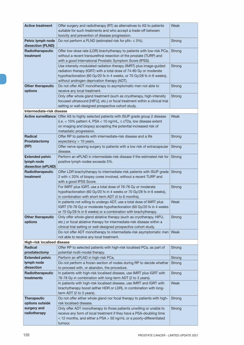
PROSTATE CANCER - LIMITED UPDATE 2021120
Active treatment Offer surgery and radiotherapy (RT) as alternatives to AS to patients suitable for such treatments and who accept a trade-off between toxicity and prevention of disease progression.
Weak
Pelvic lymph node dissection (PLND)
Do not perform a PLND (estimated risk for pN+ < 5%). Strong
Radiotherapeutic treatment
Offer low-dose rate (LDR) brachytherapy to patients with low-risk PCa, without a recent transurethral resection of the prostate (TURP) and with a good International Prostatic Symptom Score (IPSS).
Strong
Use intensity-modulated radiation therapy (IMRT) plus image-guided radiation therapy (IGRT) with a total dose of 74-80 Gy or moderate hypofractionation (60 Gy/20 fx in 4 weeks, or 70 Gy/28 fx in 6 weeks), without androgen deprivation therapy (ADT).
Strong
Other therapeutic options
Do not offer ADT monotherapy to asymptomatic men not able to receive any local treatment.
Strong
Only offer whole gland treatment (such as cryotherapy, high-intensity focused ultrasound [HIFU], etc.) or focal treatment within a clinical trial setting or well-designed prospective cohort study.
Strong
Intermediate-risk diseaseActive surveillance Offer AS to highly selected patients with ISUP grade group 2 disease
(i.e. < 10% pattern 4, PSA < 10 ng/mL, < cT2a, low disease extent on imaging and biopsy) accepting the potential increased risk of metastatic progression.
Weak
Radical Prostatectomy (RP)
Offer RP to patients with intermediate-risk disease and a life expectancy > 10 years.
Strong
Offer nerve-sparing surgery to patients with a low risk of extracapsular disease.
Strong
Extended pelvic lymph node dissection (ePLND)
Perform an ePLND in intermediate-risk disease if the estimated risk for positive lymph nodes exceeds 5%.
Strong
Radiotherapeutic treatment
Offer LDR brachytherapy to intermediate-risk patients with ISUP grade 2 with < 33% of biopsy cores involved, without a recent TURP and with a good IPSS Score.
Strong
For IMRT plus IGRT, use a total dose of 76-78 Gy or moderate hypofractionation (60 Gy/20 fx in 4 weeks or 70 Gy/28 fx in 6 weeks), in combination with short-term ADT (4 to 6 months).
Strong
In patients not willing to undergo ADT, use a total dose of IMRT plus IGRT (76-78 Gy) or moderate hypofractionation (60 Gy/20 fx in 4 weeks or 70 Gy/28 fx in 6 weeks) or a combination with brachytherapy.
Weak
Other therapeutic options
Only offer whole-gland ablative therapy (such as cryotherapy, HIFU, etc.) or focal ablative therapy for intermediate-risk disease within a clinical trial setting or well-designed prospective cohort study.
Strong
Do not offer ADT monotherapy to intermediate-risk asymptomatic men not able to receive any local treatment.
Weak
High-risk localised diseaseRadical prostatectomy
Offer RP to selected patients with high-risk localised PCa, as part of potential multi-modal therapy.
Strong
Extended pelvic lymph node dissection
Perform an ePLND in high-risk PCa. StrongDo not perform a frozen section of nodes during RP to decide whether to proceed with, or abandon, the procedure.
Strong
Radiotherapeutic treatments
In patients with high-risk localised disease, use IMRT plus IGRT with 76-78 Gy in combination with long-term ADT (2 to 3 years).
Strong
In patients with high-risk localised disease, use IMRT and IGRT with brachytherapy boost (either HDR or LDR), in combination with long-term ADT (2 to 3 years).
Weak
Therapeutic options outside surgery and radiotherapy
Do not offer either whole gland nor focal therapy to patients with high-risk localised disease.
Strong
Only offer ADT monotherapy to those patients unwilling or unable to receive any form of local treatment if they have a PSA-doubling time < 12 months, and either a PSA > 50 ng/mL or a poorly-differentiated tumour.
Strong

121PROSTATE CANCER - LIMITED UPDATE 2021
Locally-advanced diseaseRadical prostatectomy
Offer RP to selected patients with locally-advanced PCa as part of multi-modal therapy.
Strong
Extended pelvic lymph node dissection
Perform an ePLND prior to RP in locally-advanced PCa. Strong
Radiotherapeutic treatments
In patients with locally-advanced disease, offer IMRT plus IGRT in combination with long-term ADT.
Strong
Offer long-term ADT for at least two years. WeakTherapeutic options outside surgery and radiotherapy
Do not offer whole gland treatment or focal treatment to patients with locally-advanced PCa.
Strong
Only offer ADT monotherapy to those patients unwilling or unable to receive any form of local treatment if they have a PSA-doubling time < 12 months, and either a PSA > 50 ng/mL, a poorly-differentiated tumour or troublesome local disease-related symptoms.
Strong
Offer patients with cN1 disease a local treatment (either RP or IMRT plus IGRT) plus long-term ADT.
Weak
Adjuvant treatment after radical prostatectomypN0 & pN1 disease
Do not prescribe adjuvant ADT in pN0 patients. StrongOnly offer adjuvant intensity-modulated radiation therapy (IMRT) plus image-guided radiation therapy (IGRT) to high-risk patients (pN0) with at least two out of three high-risk features (ISUP grade group 4–5, pT3 ± positive margins).
Strong
Discuss three management options with patients with pN1 disease after an ePLND, based on nodal involvement characteristics: 1. Offer adjuvant ADT; 2. Offer adjuvant ADT with additional IMRT plus IGRT; 3. Offer observation (expectant management) to a patient after
eLND and < 2 nodes and a PSA < 0.1 ng/mL.
Weak
Non-curative or palliative treatments in a first-line settingLocalised diseaseWatchful waiting Offer WW to asymptomatic patients not eligible for local curative
treatment and those with a short life expectancy.Strong
Locally-advanced diseaseWatchful waiting Offer a deferred treatment policy using ADT monotherapy to M0
asymptomatic patients with a PSA-DT > 12 months, a PSA < 50 ng/mL and well-differentiated tumour who are unwilling or unable to receive any form of local treatment.
Weak
Persistent PSA after radical prostatectomyOffer a prostate-specific membrane antigen positron-emission tomography (PSMA PET) scan to men with a persistent PSA > 0.2 ng/mL if the results will influence subsequent treatment decisions
Weak
Treat men with no evidence of metastatic disease with salvage RT and additional hormonal therapy.
Weak
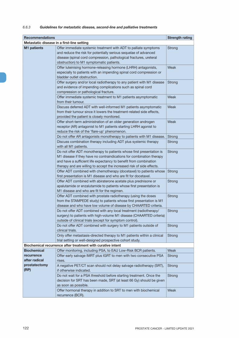
PROSTATE CANCER - LIMITED UPDATE 2021122
6.6.3 Guidelines for metastatic disease, second-line and palliative treatments
Recommendations Strength ratingMetastatic disease in a first-line settingM1 patients Offer immediate systemic treatment with ADT to palliate symptoms
and reduce the risk for potentially serious sequelae of advanced disease (spinal cord compression, pathological fractures, ureteral obstruction) to M1 symptomatic patients.
Strong
Offer luteinising hormone-releasing hormone (LHRH) antagonists, especially to patients with an impending spinal cord compression or bladder outlet obstruction.
Weak
Offer surgery and/or local radiotherapy to any patient with M1 disease and evidence of impending complications such as spinal cord compression or pathological fracture.
Strong
Offer immediate systemic treatment to M1 patients asymptomatic from their tumour.
Weak
Discuss deferred ADT with well-informed M1 patients asymptomatic from their tumour since it lowers the treatment-related side effects, provided the patient is closely monitored.
Weak
Offer short-term administration of an older generation androgen receptor (AR) antagonist to M1 patients starting LHRH agonist to reduce the risk of the ‘flare-up’ phenomenon.
Weak
Do not offer AR antagonists monotherapy to patients with M1 disease. StrongDiscuss combination therapy including ADT plus systemic therapy with all M1 patients.
Strong
Do not offer ADT monotherapy to patients whose first presentation is M1 disease if they have no contraindications for combination therapy and have a sufficient life expectancy to benefit from combination therapy and are willing to accept the increased risk of side effects.
Strong
Offer ADT combined with chemotherapy (docetaxel) to patients whose first presentation is M1 disease and who are fit for docetaxel.
Strong
Offer ADT combined with abiraterone acetate plus prednisone or apalutamide or enzalutamide to patients whose first presentation is M1 disease and who are fit for the regimen.
Strong
Offer ADT combined with prostate radiotherapy (using the doses from the STAMPEDE study) to patients whose first presentation is M1 disease and who have low volume of disease by CHAARTED criteria.
Strong
Do not offer ADT combined with any local treatment (radiotherapy/surgery) to patients with high-volume M1 disease (CHAARTED criteria) outside of clinical trials (except for symptom control).
Strong
Do not offer ADT combined with surgery to M1 patients outside of clinical trials.
Strong
Only offer metastasis-directed therapy to M1 patients within a clinical trial setting or well-designed prospective cohort study.
Strong
Biochemical recurrence after treatment with curative intent Biochemical recurrence after radical prostatectomy (RP)
Offer monitoring, including PSA, to EAU Low-Risk BCR patients. WeakOffer early salvage IMRT plus IGRT to men with two consecutive PSA rises.
Strong
A negative PET/CT scan should not delay salvage radiotherapy (SRT), if otherwise indicated.
Strong
Do not wait for a PSA threshold before starting treatment. Once the decision for SRT has been made, SRT (at least 66 Gy) should be given as soon as possible.
Strong
Offer hormonal therapy in addition to SRT to men with biochemical recurrence (BCR).
Weak
123PROSTATE CANCER - LIMITED UPDATE 2021
Biochemical recurrence after RT
Offer monitoring, including PSA, to EAU Low-Risk BCR patients. WeakOnly offer salvage RP, brachytherapy, HIFU, or cryosurgical ablation to highly selected patients with biopsy proven local recurrence within a clinical trial setting or well-designed prospective cohort study undertaken in experienced centres.
Strong
Systemic salvage treatment
Do not offer ADT to M0 patients with a PSA-DT > 12 months. Strong
Life-prolonging treatments of castration-resistant diseaseEnsure that testosterone levels are confirmed to be < 50 ng/dL, before diagnosing castration-resistant PCa (CRPC).
Strong
Counsel, manage and treat patients with metastatic CRPC (mCRPC) in a multidisciplinary team.
Strong
Treat patients with mCRPC with life-prolonging agents. StrongOffer mCRPC patients somatic and/or germline molecular testing as well as testing for mismatch repair deficiencies or microsatellite instability.
Strong
Systemic treatments of castrate-resistant diseaseBase the choice of treatment on the performance status (PS), symptoms, co-morbidities, location and extent of disease, genomic profile, patient preference, and on the previous treatment for hormone-sensitive metastatic PCa (mHSPC) (alphabetical order: abiraterone, cabazitaxel, docetaxel, enzalutamide, olaparib, radium-223, sipuleucel-T).Offer patients with mCRPC who are candidates for cytotoxic therapy and are chemotherapy naïve docetaxel with 75 mg/m2 every 3 weeks.
Strong
Offer patients with mCRPC and progression following docetaxel chemotherapy further life-prolonging treatment options, which include abiraterone, cabazitaxel, enzalutamide, radium-223 and olaparib in case of DNA homologous recombination repair (HRR).
Strong
Base further treatment decisions of mCRPC on pre-treatment PS status, previous treatments, symptoms, co-morbidities, genomic profile, extent of disease and patient preference.
Strong
Offer abiraterone or enzalutamide to patients previously treated with one or two lines of chemotherapy.
Strong
Avoid sequencing of androgen receptor targeted agents. WeakOffer chemotherapy to patients previously treated with abiraterone or enzalutamide.
Strong
Offer cabazitaxel to patients previously treated with docetaxel. StrongOffer cabazitaxel to patients previously treated with docetaxel and progressing within 12 months of treatment with abiraterone or enzalutamide.
Strong
Novel agentsOffer poly(ADP-ribose) polymerase (PARP) inhibitors to pre-treated mCRPC patients with relevant DNA repair gene mutations.
Strong
Supportive care of castration-resistant diseaseOffer bone protective agents to patients with mCRPC and skeletal metastases to prevent osseous complications.
Strong
Monitor serum calcium and offer calcium and vitamin D supplementation when prescribing either denosumab or bisphosphonates.
Strong
Treat painful bone metastases early on with palliative measures such as IMRT plus IGRT and adequate use of analgesics.
Strong
In patients with spinal cord compression start immediate high-dose corticosteroids and assess for spinal surgery followed by irradiation. Offer radiation therapy alone if surgery is not appropriate.
Strong
Non-metastatic castrate-resistant diseaseOffer apalutamide, darolutamide or enzalutamide to patients with M0 CRPC and a high risk of developing metastasis (PSA-DT < 10 months) to prolong time to metastases and overall survival.
Strong

PROSTATE CANCER - LIMITED UPDATE 2021124
7. FOLLOW-UPThe rationale for following up patients is to assess immediate- and long-term oncological results, ensure treatment compliance and allow initiation of further therapy, when appropriate. In addition, follow-up allows monitoring of side effects or complications of therapy, functional outcomes and an opportunity to provide psychological support to PCa survivors, all of which is covered in Chapter 8.
7.1 Follow-up: After local treatment7.1.1 DefinitionLocal treatment is defined as RP or RT, either by IMRT plus IGRT or LDR- or HDR-brachytherapy, or any combination of these, including neoadjuvant and adjuvant therapy. Unestablished alternative treatments such as HIFU, cryosurgery and focal therapy options do not have a well-defined, validated, PSA cut-off to define BCR but follow the general principles as presented in this section. In general, a confirmed rising PSA is considered a sign of disease recurrence.
7.1.2 Why follow-up?The first post-treatment clinic visit focuses on detecting treatment-related complications and assist patients in coping with their new situation apart from providing information on the pathological analysis. Men with PCa are at increased risk of depression and attention for mental health status is required [1195, 1196]. Tumour or patient characteristics may prompt changing the follow-up schedule.
7.1.3 How to follow-up?The procedures indicated at follow-up visits vary according to the clinical situation. A disease-specific history is mandatory at every follow-up visit and includes psychological aspects, signs of disease progression, and treatment-related complications. Evaluation of treatment-related complications in the post-treatment period is highlighted in Sections 6.1.2.4, 6.1.2.4.3, 6.3.9.2, 6.3.10.2.2, 6.3.11.2 and 8.2. The examinations used for cancer-related follow-up after curative surgery or RT are discussed below.
7.1.3.1 Prostate-specific antigen monitoringMeasurement of PSA is the cornerstone of follow-up after local treatment. While PSA thresholds depend on the local treatment used, PSA recurrence almost always precedes clinical recurrence [914, 1197]. The key question is to establish when a PSA rise is clinically significant since not all PSA increases have the same clinical value (see Section 6.3) [916].
7.1.3.1.1 Active surveillance follow-upPatients included in an AS programme should be monitored according to the recommendations presented in Section 6.2.2.
7.1.3.1.2 Prostate-specific antigen monitoring after radical prostatectomyFollowing RP, the PSA level is expected to be undetectable (< 0.1 ng/mL). Prostate-specific antigen is generally determined every 3 months in the first year, every 6 months until 3 years and yearly thereafter but evidence for a specific interval is low [467] and mainly based on the observation that early recurrences are more likely to be associated with more rapid progression [916, 1198, 1199]. Prostate-specific antigen level is expected to be undetectable 2 months after a successful RP [1200]. As mentioned in Section 6.3.2 the PSA-threshold at relapse best predicting metastases after RP is > 0.4 ng/mL. Persistently measurable PSA in patients treated with RP is discussed in Section 6.2.6 - Persistent PSA.
Ultrasensitive PSA assays remain controversial for routine follow-up after RP. Men with an ultrasensitive PSA nadir < 0.01 ng/mL have a high (96%) likelihood of remaining relapse free within 2 years [1201]. In addition, post-RP ultrasensitive PSA levels > 0.01 ng/mL in combination with clinical characteristics such as ISUP grade and surgical margin status may predict PSA progression and can be useful to establish follow-up intervals [1199]. Lastly, PSA and associated PSA-DT [1202] calculated prior to 0.2 ng/mL may help identify suitable candidates for early intervention [1203]. Prostate-specific antigen monitoring after salvage RT to the prostatic fossa is done at similar intervals and an early PSA rise predicts more rapid progression [1198].
7.1.3.1.3 Prostate-specific antigen monitoring after radiotherapyFollowing RT, PSA drops more slowly as compared to post RP. A nadir < 0.5 ng/mL is associated with a favourable outcome after RT although the optimal cut-off value remains controversial [1204]. The interval before reaching the nadir can be up to 3 years, or more. At the 2006 RTOG-ASTRO Consensus Conference the Phoenix definition of radiation failure was proposed to establish a better correlation between definition
125PROSTATE CANCER - LIMITED UPDATE 2021
and clinical outcome (mainly metastases), namely, an increase of 2 ng/mL above the post-treatment PSA nadir [915]. This definition also applies to patients who received HT [915].
7.1.3.1.4 Digital rectal examinationLocal recurrence after curative treatment is possible without a concomitant rise in PSA level [1205]. However, this has only been proven in patients with unfavourable undifferentiated tumours. Prostate-specific antigen measurement and DRE comprise the most useful combination for first-line examination in follow-up after RT but the role of DRE was questioned since it failed to detect any local recurrence in the absence of a rising PSA in a series of 899 patients [1206]. In a series of 1,118 prostatectomy patients, no local histologically proven recurrence was found by DRE alone and PSA measurement may be the only test needed after RP [1207, 1208].
7.1.3.1.5 Transrectal ultrasound, bone scintigraphy, CT, MRI and PET/CTImaging techniques have no place in routine follow-up of localised PCa as long as the PSA is not rising. Imaging is only justified in patients for whom the findings will affect treatment decisions, either in case of BCR or in patients with symptoms (see Section 6.3.4 for a more detailed discussion).
7.1.4 How long to follow-up?Most patients who fail treatment for PCa do so within 7 years after local therapy [482]. Patients should be followed up more closely during the initial post-treatment period when risk of failure is highest. Prostate-specific antigen measurement, disease-specific history and DRE (if considered) are recommended every 6 months until 3 years and then annually. Whether follow-up should be stopped if PSA remains undetectable (after RP) or stable (after RT) remains an unanswered question.
7.1.5 Summary of evidence and guidelines for follow-up after treatment with curative intent
Summary of evidence LEA rising PSA must be differentiated from a clinically meaningful relapse.The PSA threshold that best predicts further metastases after RP is > 0.4 ng/mL and > NADIR + 2 after IMRT plus IGRT (± ADT].
3
Palpable nodules combined with increasing serum PSA suggest at least local recurrence. 2a
Recommendations Strength ratingRoutinely follow up asymptomatic patients by obtaining at least a disease-specific history and serum prostate-specific antigen (PSA) measurement. These should be performed at 3, 6 and 12 months after treatment, then every 6 months until 3 years, and then annually.
Strong
At recurrence, only perform imaging if the result will affect treatment planning. Strong
7.2 Follow-up: During first line hormonal treatment (androgen sensitive period)7.2.1 IntroductionAndrogen deprivation therapy is used in various situations: combined with radiotherapy for localised or locally-advanced disease, as monotherapy for a relapse after a local treatment, or in the presence of metastatic disease often in combination with other treatments. All these situations are based on the benefits of testosterone suppression either by drugs (LHRH agonists or antagonists) or orchidectomy. Inevitably, the disease will become castrate-resistant, although ADT will be maintained.
This paragraph addresses the general principles of follow-up of patients on ADT alone. As treatment of CRPC and follow-up are closely linked, Section 6.5.7 includes further information on other drug treatments. Furthermore the specific follow-up needed for every single drug is outside the scope of this text.
Regular clinical follow-up is mandatory and cannot be replaced by imaging or laboratory tests alone. Complementary investigations must be restricted to those that are clinically helpful to avoid unnecessary examinations and costs.
7.2.2 Purpose of follow-upThe main objectives of follow-up in patients receiving ADT are to ensure treatment compliance, to monitor treatment response, to detect side effects early, and to guide treatment at the time of CRPC. After the initiation of ADT, it is recommended that patients are evaluated every 3 to 6 months. This must be individualised and each patient should be advised to contact his physician in the event of troublesome symptoms.

PROSTATE CANCER - LIMITED UPDATE 2021126
7.2.3 General follow-up of men on ADTPatients under ADT require regular follow-up, including monitoring of serum testosterone, creatinine, liver function and metabolic parameters at 3 to 6 month intervals. Men on ADT can experience toxicity independent of their disease stage.
7.2.3.1 Testosterone monitoringTestosterone monitoring should be considered standard clinical practice in men on ADT. Many men receiving medical castration will achieve a castrate testosterone level (< 20 ng/dL), and most a testosterone level < 50 ng/dL. However, approximately 13–38% of patients fail to achieve these levels and up to 24% of men may experience temporary testosterone surges (testosterone > 50 ng/dL) during long-term treatment [1200] referred to as ‘acute on-chronic effect’ or ‘breakthrough response’ [1209]. Breakthrough rates for the < 20 ng/dL threshold were found to be more frequent (41.3%) and an association with worse clinical outcomes was suggested [1209].
The timing of measurements is not clearly defined. A 3 to 6-month testosterone level assessment has been suggested to ensure castration is achieved (especially during medical castration) and maintained. In case the castrate testosterone level is not reached, switching to another agonist or antagonist or to an orchiectomy should be considered. In patients with a confirmed rising PSA and/or clinical progression, serum testosterone must be evaluated in all cases to confirm a castration-resistant state. Ideally, suboptimal testosterone castrate levels should be confirmed with mass spectrometry or an immunoassay [1210, 1211].
7.2.3.2 Liver function monitoringLiver function tests will detect treatment toxicity (especially applicable for NSAA), but rarely indicate disease progression. Men on combined ADT should have their transaminase levels checked at least twice a year in view of potential liver toxicity but a more frequent check is needed with some drugs (like abiraterone acetate). Alkaline phosphatase may increase secondary to bone metastases and androgen-induced osteoporosis, therefore it may be helpful to determine bone-specific isoenzymes as none are directly influenced by ADT [1212].
7.2.3.3 Serum creatinine and haemoglobineEstimated glomerular filtration rate monitoring is good clinical practice as an increase may be linked to ureteral obstruction or bladder retention. A decline in Hb is a known side effect of ADT. A significant decline after 3 months of ADT is independently associated with shorter progression-free and OS rates and might explain significant fatigue although other causes should be considered [1213]. Anaemia is often multi-factorial and other possible aetiologies should be excluded.
7.2.3.4 Monitoring of metabolic complicationsThe most severe complications of androgen suppression are metabolic syndrome, cardiovascular morbidity, mental health problems, and bone resorption (see Section 8.2.4.5).
All patients should be screened for diabetes by checking fasting glucose and HbA1c (at baseline and routinely) in addition to checking blood lipid levels. Men with impaired glucose tolerance and/or diabetes should be referred for an endocrine consultation. Prior to starting ADT a cardiology consultation should be considered in men with a history of cardiovascular disease and in men older than 65 years. Men on ADT are at increased risk of cardiovascular problems and hypertension and regular checks are required [1214].
7.2.3.5 Monitoring bone problemsAndrogen deprivation therapy increases the risk of osteoporosis. Adminstration of ADT for more than a year, as compared to less than one year, showed a higher risk of osteoperosis (HR: 1.77 and 1.38, respectively) [1215]. Several scores (e.g., Fracture Risk Assessment Tool [FRAX score], Osteoporosis Self-Assessment Tool [OST], Osteoporosis Risk Assessment Instrument [ORAI], Osteoporosis Index of Risk [OSIRIS], Osteoporosis Risk Estimation [SCORE]) can help identify men at risk of osteoporotic complications but validation of these scores in the ADT setting is required (see Section 8.3.2.2) [1216-1218].
Routine bone monitoring for osteoporosis should be performed using DEXA scan [1219-1221]. Presence of osteoporosis should prompt the use of bone protective agents. The criteria for initiation of bone protective agents are mentioned in Section 8.3.2.2. If no bone protective agents are given, a DEXA scan should be done regularly, at least every 2 years [1222]
A review summarising the incidence of bone fractures showed an almost doubling of the risk of fractures when using ADT depending on patients’ age and duration and type of ADT with the highest incidence in older men and men on additional novel ARTA medication across the entire spectrum of disease [1223]. In case of an osteoporotic fracture a bone protective agent is mandatory. Vitamin D and calcium levels should be regularly monitored when patients receive ADT and patients should be supplemented if needed. (see Section 6.5.15).
127PROSTATE CANCER - LIMITED UPDATE 2021
7.2.3.6 Monitoring lifestyle and cognitionLifestyle (e.g., diet, exercise, smoking status, etc.) affects QoL and potentially outcome [1212, 1213]. During follow-up men should be counselled on the beneficial effects of exercise to avoid ADT-related toxicity [1224]. Androgen deprivation therapy may affect mental and cognitive health and men on ADT are three times more likely to report depression [1225]. Attention to mental health should therefore be an integral part of the follow-up scheme.
7.2.4 Methods of follow-up in men on ADT without metastases7.2.4.1 Prostate-specific antigen monitoringProstate-specific antigen is a key marker for following the course of androgen-sensitive non-metastasised PCa. Imaging should be considered when PSA is rising > 2 ng/mL or in case of symptoms suggestive of metastasis.
7.2.4.2 ImagingIn general, asymptomatic patients with a stable PSA level do not require further imaging, although care needs to be taken in patients with aggressive variants when PSA levels may not reflect tumour progression [1226]. New bone pain requires at least targeted imaging and potentially a bone scan. When PSA progression suggests CRPC status and treatment modification is considered, imaging, by means of a bone and CT scan, is recommended for restaging. Detection of metastases greatly depends on imaging (see Section 6.3.4).
7.2.5 Methods of follow-up in men under ADT for metastatic hormone-sensitive PCa In metastatic patients it is of the utmost importance to counsel about early signs of spinal cord compression, urinary tract complications (ureteral obstruction, bladder outlet obstruction) or bone lesions that are at an increased fracture risk. The intervals for follow-up in M1 patients should be guided by patients’ complaints and can vary. Since most men will receive another anti-cancer therapy combined with ADT such as ARTA, chemotherapy or local radiotherapy, follow-up frequency should also be dependent on the treatment modality.
7.2.5.1 PSA monitoringIn men on ADT alone, a PSA decline to < 4 ng/mL suggests a likely prolonged response and follow-up visits may be scheduled every 3 to 6 months provided the patient is asymptomatic or clinically improving. Depending on symptoms and risk assessment, more frequent visits may be indicated. Treatment response may be evaluated based on a change in serum PSA level [1049, 1050] and bone- and CT scan although there is no consensus about how frequently these should be performed [1176]. A rise in PSA level usually precedes the onset of clinical symptoms by several months. A rising PSA should prompt assessment of testosterone level, which is mandatory to define CRPC status, as well as restaging using imaging. However it is now recognised that a stable PSA during ADT is not enough to characterise a non-progressive situation [1227].
7.2.5.2 Imaging as a marker of response in metastatic PCaTreatment response in soft-tissue metastases can be assessed by morphological imaging methods using the Response Evaluation Criteria in Solid Tumours (RECIST) criteria. However, these criteria cannot be used in bone where response assessment is difficult [1228, 1229].
When bone scan is used to follow bone metastases, a quantitative estimation of tracer uptake at bone scan can be obtained through automated methods such as the Bone Scan Index [1230]. Nonetheless, bone scan is limited by the so-called ‘flare’ phenomenon which is defined by the development of new images induced by treatment on a first follow-up scan which, after longer observation, actually represent a favourable response. Flare is observed within 8 to 12 weeks of treatment initiation and can lead to a false-positive diagnosis of disease progression. Computed tomography cannot be used to monitor sclerotic bone lesions because bone sclerosis can occur under effective treatment and reflects bone healing. Magnetic resonance imaging can directly assess the bone marrow and demonstrate progression based on morphologic criteria or changes in apparent diffusion coefficient. A standardisation for reporting is available [1231]. The ability of PET/CT to assess response has been evaluated in a few studies. Until further data are available, MRI and PET/CT should not be used outside trials for treatment monitoring in metastatic patients [1232].
Men with metastasised PCa on ADT should also in the absence of PSA rise be followed up with regular imaging since twenty-five percent of men on ADT in the ECOG3805 CHAARTED trial developed progression on imaging without a PSA rise [1227].
PROSTATE CANCER - LIMITED UPDATE 2021128
7.2.6 Guidelines for follow-up during hormonal treatment
Recommendations Strength ratingThe follow-up strategy must be individualised based on stage of disease, prior symptoms, prognostic factors and the treatment given.
Strong
In patients with stage M0 disease, schedule follow-up at least every 6 months. As a minimum requirement, include a disease-specific history, serum prostate-specific antigen (PSA) determination, as well as liver and renal function in the diagnostic work-up.
Strong
In M1 patients, schedule follow-up at least every 3 to 6 months. StrongIn patients on long-term androgen deprivation therapy (ADT), measure initial bone mineral density to assess fracture risk.
Strong
During follow-up of patients receiving ADT, check PSA and testosterone levels and monitor patients for symptoms associated with metabolic syndrome as a side effect of ADT.
Strong
As a minimum requirement, include a disease-specific history, haemoglobin, serum creatinine, alkaline phosphatase, lipid profiles and HbA1c level measurements.
Strong
Counsel patients (especially with M1b status) about the clinical signs suggestive of spinal cord compression.
Strong
When disease progression is suspected, restaging is needed and the subsequent follow-up adapted/individualised.
Strong
In M1 patients perform regular imaging (CT and bone scan) even without PSA progression. WeakIn patients with suspected progression, assess the testosterone level. By definition, castration-resistant PCa requires a testosterone level < 50 ng/dL (< 1.7 nM/L).
Strong
8. QUALITY OF LIFE OUTCOMES IN PROSTATE CANCER
This chapter is presented in two parts. The first (8.2) will summarise long-term consequences (> 12 months) of therapies for PCa. Based on two systematic reviews, the second (8.3) provides evidence-based recommendations for supporting patients when selecting primary treatment options for localised disease and also supportive interventions aimed at improving disease-specific QoL across all stages of disease.
8.1 IntroductionQuality of life and personalised care go hand in hand. Treating PCa can affect an individual both physically and mentally, as well as close relations and work or vocation. These multifaceted issues all have a bearing on an individual‘s perception of QoL [1233]. Approaching care from a holistic point of view requires the intervention of a multi-disciplinary team including urologists, medical oncologists, radiation oncologists, oncology nurses, behavioural practitioners and many others including fellow patients. Attention to the psychosocial concerns of people with PCa is integral to quality clinical care, and this can include the needs of carers and partners [1234]. Prostate cancer care should not be reduced to focusing on the organ in isolation: side effects or late adverse effects of treatment can manifest systemically and have a major influence on the patient’s QoL. Taking QoL into consideration relies on understanding the patient’s values and preferences so that optimal treatment proposals can be formulated and discussed.
8.2 Adverse effects of PCa therapies8.2.1 SurgeryThe absence of standardisation in reporting surgical complications for RP and the introduction of different techniques has resulted in a wide variation in the types of complications reported, as well as variation in the overall incidence of complications [1235-1238]. The most common post-operative issue is ED but other related issues to consider include dry ejaculation, which occurs with removal of the prostate, change in the quality of orgasm and occasional pain on orgasm. Men also complain of loss of penile length (3.73%, 19/510 men) [1239]. The second most commonly occurring complication is long-term incontinence [1235-1238] but voiding difficulties may also occur associated with bladder neck contracture (e.g., 1.1% after RALP) [1240].
For those undergoing minimally invasive procedures port site hernia has been reported in 0.66% after inserting 12 mm bladeless trocar [1241] and can occur more rarely with 8 mm and 5 mm trocars [1241]. A key consideration is whether long-term consequences of surgery are reduced by using newer techniques
129PROSTATE CANCER - LIMITED UPDATE 2021
such as RALP. Systematic reviews have documented complication rates after RALP [500, 591-594], and can be compared with contemporaneous reports after RRP [595]. From these reports, the mean continence rates at 12 months were 89–100% for patients treated with RALP and 80–97% for patients treated with RRP. A prospective controlled non-randomised trial of patients undergoing RP in 14 centres using RALP or RRP demonstrated that at 12 months after RALP, 21.3% were incontinent, as were 20.2% after RRP. The unadjusted OR was 1.08 (95% CI: 0.87–1.34). Erectile dysfunction was observed in 70.4% after RALP and 74.7% after RRP. The unadjusted OR was 0.81 (95% CI: 0.66–0.98) [596, 1242]. Further follow-up demonstrates similar functional outcomes with both techniques at 24 months [1242, 1243]. A single-centre randomised phase III study comparing RALP and RRP (n = 326) also demonstrates similar functional outcomes with both techniques at 24 months [495].
8.2.2 Radiotherapy8.2.2.1 Side-effects of external beam radiotherapyAnalysis of the toxicity outcomes of the Prostate Testing for Cancer and Treatment (ProtecT) trial [1244] shows that patients treated with EBRT and 6 months of ADT report bowel toxicity including persistent diarrhoea, bowel urgency and/or incontinence and rectal bleeding (described in detail in Section 8.3.1.1 below). Participants in the ProtecT study were treated with 3D-CRT and more recent studies using IMRT demonstrate less bowel toxicity than noted previously with 3D-CRT [1245].
A systematic review and meta-analysis of observational studies comparing patients exposed or unexposed to radiotherapy in the course of treatment for PCa demonstrates an increased risk of developing second cancers for bladder (OR: 1.39), colorectal (OR: 1.68) and rectum (OR: 1.62) with similar risks over lag times of 5 and 10 years. Absolute excess risks over 10 years are small (1–4%) but should be discussed with younger patients in particular [1246].
8.2.2.2 Side effects from brachytherapySome patients experience significant urinary complications following implantation such as urinary retention (1.5-22%), with post-implantation TURP reported as being required in up to 8.7% of cases, and incontinence (0–19%) [1247]. Chronic urinary morbidity is more common with combined EBRT and BT and can occur in up to 20% of patients, depending on the severity of the symptoms before brachytherapy. Urethral strictures account for at least 50% of urinary complications and can be resolved with dilation in the majority [822, 824]. Prevention of morbidity depends on careful patient selection, and expert assessment of IPSS score, backed up by urodynamic studies.
8.2.3 Local primary whole-gland treatments other than surgery or radiotherapy8.2.3.1 CryosurgeryIn Ramsay et al.’s systematic review and meta-analysis there was evidence that the rate of urinary incontinence at one year was lower for cryotherapy than for RP, but the size of the difference decreased with longer follow-up [758]. There was no significant difference between cryotherapy vs. EBRT in terms of urinary incontinence at one year (< 1%); cryotherapy had a similar ED rate (range 0–40%) to RP at one year. There were insufficient data to compare cryotherapy vs. EBRT in terms of ED.
8.2.3.2 High-intensity focused ultrasoundIn terms of toxicity there are insufficient data on urinary incontinence, ED or bowel dysfunction to draw any conclusions, although at one year HIFU had lower incontinence rates than RP (OR: 0.06, 95% CI: 0.01–0.48) [758].
8.2.4 Hormonal therapyA summary of impacts on psychological factors due to the use of ADT such as sexual function, mood, depression, cognitive function and impact on partners can be found in two clinical reviews [1248, 1249].
A small RCT evaluated the QoL at one-year follow-up in patients with non-localised PCa, between various ADT regimens, or no treatment. Patients treated by ADT reported a significant decline in spatial reasoning, spatial abilities and working memory as well as increased depression, tension, anxiety, fatigue and irritability during treatment [1250]. Conversely, a prospective observational study with follow-up to 3 years failed to demonstrate an association with cognitive decline in men on ADT when compared to men with PCa not treated with ADT and healthy controls [1251]. A prospective observational study of non-metastatic PCa found that immediate ADT was associated with a lower overall QoL compared to deferred treatment [1252]. Another retrospective non-randomised study suggested that men receiving LHRH agonists reported more worry and physical discomfort and poorer overall health, and were less likely to believe themselves free of cancer than patients undergoing orchiectomy. The stage at diagnosis had no effect on health outcomes [1253].
Using a specific non-validated questionnaire, bicalutamide monotherapy showed a significant advantage over castration in the domains of physical capacity and sexual interest (not sexual function) at
PROSTATE CANCER - LIMITED UPDATE 2021130
12 months [1254]. A post-hoc analysis, including only patients with sexual interest suggested that bicalutamide was associated with better sexual preservation, including maintained sexual interest, feeling sexually attractive [1255], preserved libido and erectile function [1256]. Intermittent androgen deprivation has been discussed elsewhere (see Section 6.4.3.2).
8.2.4.1 Sexual functionCessation of sexual activity is very common in people undergoing ADT, affecting up to 93% [1257]. Androgen deprivation therapy reduces both libido and the ability to gain and maintain erections. The management of acquired ED is mostly non-specific [1258].
8.2.4.2 Hot flushesHot flushes are a common side-effect of ADT (prevalence estimated between 44–80% of men on ADT) [1257]. They appear 3 months after starting ADT, usually persist long-term and have a significant impact on QoL. Oestrogen-receptor modulators or low-dose oestrogen therapies, e.g. DES, 0.5–1 mg/day, reduce the frequency and severity of hot flushes. Both treatments carry a risk of cardiovascular complications [1259].
Serotonin re-uptake inhibitors (e.g., venlafaxine or sertraline) also appear to be effective in men but less than hormone therapies based on a prospective RCT comparing venlafaxine, 75 mg daily, with medroxyprogesterone, 20 mg daily, or cyproterone acetate, 100 mg daily [1260]. After 6 months of LHRH (n = 919), 311 men had significant hot flushes and were randomised to one of the treatments. Based on median daily hot-flush score, venlafaxine was inferior -47.2% (interquartile range -74.3 to -2.5) compared to -94.5% (-100.0 to -74.5) in the cyproterone group, and -83.7% (-98.9 to -64.3) in the medroxyprogesterone group. With a placebo effect influencing up to 30% of patients [1261], the efficacy of clonidine, veralipride, gabapentine [1262] and acupuncture [1263] need to be compared in prospective RCTs.
8.2.4.3 Non-metastatic bone fracturesDue to increased bone turnover and decreased bone mineral density (BMD) in a time-dependent manner, ADT use is linked to an increased risk of fracture (up to 45% RR with long-term ADT) [1264]. Severe fractures in men are associated with a significant risk of death [1265]. A precise evaluation of BMD should be performed by dual emission X-ray absorptiometry (DEXA), ideally before or shortly after starting long-term ADT. An initial low BMD (T-score < -2.5 or < -1, with other risk factors) indicates a high risk of subsequent non-metastatic fracture and causes should be investigated. Other risk factors include increasing age, body mass index of 19 or less, history of previous fracture or parent with fractured hip, current smoking, use of glucocorticoids, rheumatoid arthritis, alcohol consumption > 2 units per day, history of falls and a number of other chronic medical conditions [1266]. Fracture risk alorithms which combine BMD and clinical risk factors such as FRAX score can be used to guide treatment decisions but uncertainty exists regarding the optimal intervention threshold, therefore no specific risk algorithm can be recommended for men on ADT for PCa. Obesity (increase in body fat mass by up to 10%) and sarcopenia (decrease in lean tissue mass by up to 3%) as well as weight loss are common and occur during the first year of ADT [1267]. These changes increase the fracture risk [1268].
Bicalutamide monotherapy may have less impact on BMD but is limited by its suboptimal efficacy [1269, 1270] (see Section 6.1.4.1.1.5.2.3). The intermittent LHRH-agonist modality might be associated with less bone impact [1271].
8.2.4.4 Metabolic effectsLipid alterations are common and may occur as early as the first 3 months of treatment [1267]. Androgen deprivation therapy also decreases insulin sensitivity and increases fasting plasma insulin levels, which is a marker of insulin resistance. In diabetic patients, metformin appears to be an attractive option for protection against metabolic effects based on retrospective analysis [1272], but there is insufficient data to recommend its use in non-diabetic patients.
Metabolic syndrome is an association of independent cardiovascular disease risk factors, often associated withinsulin resistance. The definition requires at least three of the following criteria [1273]:• waist circumference > 102 cm;• serum triglyceride > 1.7 mmol/L;• blood pressure > 130/80 mmHg or use of medication for hypertension;• high-density lipoprotein (HDL) cholesterol < 1 mmol/L;• glycaemia > 5.6 mmol/L or the use of medication for hyperglycaemia.
The prevalence of a metabolic-like syndrome is higher during ADT compared with men not receiving ADT [1274].
131PROSTATE CANCER - LIMITED UPDATE 2021
Skeletal muscle mass heavily influences basal metabolic rate and is in turn heavily influenced by endocrine pathways [1275]. Androgen deprivation therapy-induced hypogonadism results in negative effects on skeletal muscle health. A prospective longitudinal study involving 252 men on ADT for a median of 20.4 months reported lean body mass decreases progressively over 3 years; 1.0% at one year, 2.1% at 2 years, and 2.4% at 3 years which appears more pronounced in men at > 70 years of age [1276].
8.2.4.5 Cardiovascular morbidityCardiovascular mortality is a common cause of death in PCa patients [1040, 1277, 1278]. Several studies showed that ADT after only 6 months was associated with an increased risk of diabetes mellitus, cardiovascular disease, and myocardial infarction [1279]. The RTOG 92-02 [1280] and 94-08 [1281] trials confirmed an increased cardiovascular risk, unrelated to the duration of ADT and not accompanied by an overall increased cardiovascular mortality. No increase in cardiovascular mortality has been reported in a systematic meta-analysis of trials RTOG 8531, 8610, 9202, EORTC 30891 or EORTC 22863 [1282]. However, serious concerns about the conclusions of this meta-analysis have been raised due to poor consideration of bias in the included studies [1283, 1284]. Meta-analysis of observational data reports consistent links between ADT and the risk of cardiovascular disease patients treated for PCa e.g. the associations between GnRH agonists and nonfatal or fatal myocardial infarction or stroke RR: 1.57 (95% CI: 1.26–1.94) and RR: 1.51 (95% CI: 1.24–1.84), respectively [1285]. An increase in cardiovascular mortality has been reported in patients suffering from previous congestive heart failure or myocardial infarction in a retrospective database analysis [1286] or presenting with a metabolic syndrome [1287]. It has been suggested that LHRH antagonists might be associated with less cardiovascular morbidity compared to agonists [1288]. In a phase III RCT the use of relugolix, an oral LHRH antagonist, was associated with a reduced risk of major adverse cardiovascular events when compared to leuprolide, an injectable LHRH agonists, at 2.9% vs. 6.2%, respectively, over a follow-up time of of 48 weeks (HR 0.46, 95% CI: 0.24–0.88 [727].
These concerns resulted in an FDA warning and consensus paper from the American Heart, Cancer Society and Urological Associations [1039]. Preventive advice includes non-specific measures such as loss of weight, increased exercise, minimising alcohol intake, improved nutrition and smoking cessation [69, 1289].
8.2.4.6 FatigueFatigue often develops as a side-effect of ADT. Regular exercise appears to be the best protective measure. Anaemia may be a cause of fatigue [1257, 1290]. Anaemia requires an aetiological diagnosis (medullar invasion, renal insufficiency, iron deficiency, chronic bleeding) and individualised treatment. Iron supplementation (using injectable formulations only) must be systematic if deficiency is observed. Regular blood transfusions may be required in patients with severe anaemia. Erythropoiesis-stimulating agents might be considered in dedicated cases, taking into account the possible increased risk of thrombovascular events [1291].
8.2.4.7 Neurological side effectsCastration seems also to be associated with an increased risk of stroke [1292], and is suspect to be associated with an increased risk for depression and cognitive decline such as Alzheimer disease [1293].
8.3 Overall quality of life in men with PCaLiving longer with PCa does not necessarily equate to living well [1233, 1234]. There is clear evidence of unmet needs and ongoing support requirements for some individuals after diagnosis and treatment for PCa [1294]. Cancer impacts on the wider family and cognitive behavioural therapy can help reduce depression, anxiety and stress in caregivers [1295]. Radical treatment for PCa can negatively impact long-term QoL (e.g., sexual, urinary and bowel dysfunction) as can ADT used in short- or long-term treatment, e.g., sexual problems, fatigue, psychological morbidity, adverse metabolic sequelae and increased cardiovascular and bone fracture risk [1248, 1296]. Direct symptoms from advanced or metastatic cancer, e.g., pain, hypercalcaemia, spinal cord compression and pathological fractures, also adversely affect health [1297, 1298]. Patient’s QoL including domains such as sexual function, urinary function and bowel function is worse after treatment for PCa compared to non-cancer controls [1299, 1300].
The concept of ‘quality of life’ is subjective and can mean different things to different people, but there are some generally common features across virtually all patients. Drawing from these common features, specific tools or ‘patient-reported outcome measures’ (PROMs) have been developed and validated for men with PCa. These questionnaires assess common issues after PCa diagnosis and treatment and generate scores which reflect the impact on perceptions of HRQoL. During the process of undertaking two dedicated systematic reviews around cancer-specific QoL outcomes in patients with PCa as the foundation for our guideline recommendations, the following validated PROMs were found in our searches (see Table 8.3.1).
PROSTATE CANCER - LIMITED UPDATE 2021132
Table 8.3.1: PROMs assessing cancer specific quality of life
Questionnaire Domains/itemsFunctional Assessment of Cancer Therapy-General (FACT-G) [1301]
Physical well-being, social/family well-being, emotional well-being, and functional well-being.
Functional Assessment of Cancer Therapy-Prostate (FACT-P) [1302]
12 cancer site specific items to assess for prostate-related symptoms. Can be combined with FACT-G or reported separately.
European Organisation for Research and Treatment of Cancer QLQ-C30 (EORTC QLQ-C30) [1303]
Five functional scales (physical, role, cognitive, emotional, and social); three symptom scales (fatigue, pain, and nausea and vomiting); global health status/QoL scale; and a number of single items assessing additional symptoms commonly reported by cancer patients (dyspnoea, loss of appetite, insomnia, constipation and diarrhoea) and perceived financial impact of the disease.
European Organisation for Research and Treatment of Cancer QLQ-PR 25 (EORTC QLQ-PR 25) [1304]
Urinary, bowel and treatment-related symptoms, as well as sexual activity and sexual function.
Expanded prostate cancer index composite (EPIC) [1305] Urinary, bowel, sexual, and hormonal symptoms. Expanded prostate cancer index composite short form 26 (EPIC 26) [1306]
Urinary, sexual, bowel, and hormonal domains.
UCLA Prostate Cancer Index (UCLA PCI) [1307] Urinary, bowel, and sexual domains.Prostate Cancer Quality of Life Instrument (PCQoL) [1308]
Urinary, sexual, and bowel domains, supplemented by a scale assessing anxiety.
Prostate Cancer Outcome Study Instrument [1309] Urinary, bowel, and sexual domains.
8.3.1 Long-term (> 12 months) quality of life outcomes in men with localised disease8.3.1.1 Men undergoing local treatmentsThe results of the Prostate Testing for Cancer and Treatment (ProtecT) trial (n = 1,643 men) reported no difference in EORTC QLQ-C30 assessed global QoL, up to 5 years of follow-up in men aged 50–69 years with T1-T2 disease randomised for treatment with AM, RP or RT with 6 months of ADT [1244]. However, EPIC urinary summary scores (at 6 years) were worse in men treated with RP compared to AM or RT (88.7 vs. 89.0 vs. 91.4, respectively) as were urinary incontinence (80.9 vs. 85.8 vs. 89.4, respectively) and sexual summary, function and bother scores (32.3 vs. 40.6 vs. 41.3 for sexual summary, 23.7 vs. 32.5 vs. 32.7 for sexual function and 51.4 vs. 57.9 vs. 60.1 for sexual bother, respectively) at 6 years of follow-up. Minimal clinically important differences for the 50 item EPIC questionnaire are not available. For men receiving RT with 6 months of ADT, EPIC bowel scores were poorer compared to AM and RP in all domains: function (90.8 vs. 92.3 vs. 92.3, respectively), bother (91.7 vs. 94.2 vs. 93.7, respectively) and summary (91.2 vs. 93.2 vs. 93.0, respectively) at 6 years of follow-up in the ProtecT trial.
The findings regarding RP and RT are supported by other observational studies [1238, 1310]. The Prostate Cancer Outcomes Study (PCOS) [1238] studied a cohort of 1,655 men, of whom 1,164 had undergone RP and 491 RT. The study reported that at 5 years of follow-up, men who underwent RP had a higher prevalence of urinary incontinence and ED, while men treated with RT had a higher prevalence of bowel dysfunction. However, despite these differences detected at 5 years, there were no significant differences in the adjusted odds of urinary incontinence, bowel dysfunction or ED between RP and RT at 15 years. More recently, investigators reported that although EBRT was associated with a negative effect in bowel function, the difference in bowel domain score was below the threshold for clinical significance 12 months after treatment [1245]. As 81% of patients in the EBRT arm of the study received IMRT, these data suggest that the risk of side effects is reduced with IMRT compared to older 3D-CRT techniques. This is supported by a contemporary 5-year prospective, population-based cohort study where PROs were compared in men with favourable- and unfavourable-risk localised disease [1310]. In the 1,386 men with favourable risk, comparison between AS and nerve-sparing prostatectomy, EBRT or LDR brachytherapy demonstrates that surgery is associated with worse urinary incontinence at 5 years and sexual dysfunction at 3 years when compared to AS. External beam RT is associated with changes not clinically different from AS, and LDR brachytherapy is associated with worse irritative urinary-, bowel- and sexual symptoms at one year. In 619 men with unfavourable risk disease, comparison between non-nerve sparing RP and EBRT with ADT demonstrates that surgery is associated with worse urinary incontinence and sexual function through 5 years.
133PROSTATE CANCER - LIMITED UPDATE 2021
With respect to brachytherapy cancer-specific QoL outcomes, one small RCT (n = 200) evaluated bilateral nerve-sparing RP and brachytherapy in men with localised disease (up to T2a), which reported worsening of physical functioning as well as irritative urinary symptomatology in 20% of brachytherapy patients at one year of follow-up. However, there were no significant differences in EORTC QLQ-C30/PR-25 scores at 5 years of follow-up when compared to pre-treatment values [1311]. It should be noted of this trial, within group tests only were reported. In a subsequent study by the same group comparing bilateral nerve-sparing RARP and brachytherapy (n = 165), improved continence was noted with brachytherapy in the first 6 months but lower potency rates up to 2 years [1312]. These data and a synthesis of 18 randomised and non-randomised studies in a systematic review involving 13,604 patients are the foundation of the following recommendations [1313].
8.3.1.2 Guidelines for quality of life in men undergoing local treatments
Recommendations Strength ratingAdvise eligible patients for active surveillance that global quality of life is equivalent for up to 5 years compared to radical prostatectomy or external beam radiotherapy.
Strong
Discuss the negative impact of surgery on urinary and sexual function, as well as the negative impact of radiotherapy on bowel function with patients.
Strong
Advise patients treated with brachytherapy of the negative impact on irritative urinary symptomatology at one year but not after 5 years.
Weak
8.3.2 Improving quality of life in men who have been diagnosed with PCa8.3.2.1 Men undergoing local treatmentsIn men with localised disease, nurse-led multi-disciplinary rehabilitation (addressing sexual functioning, cancer worry, relationship issues depression, managing bowel and urinary function problems) provided positive short-term effects (4 months) on sexual function (effect size 0.45) and long-term (12 months) positive effects on sexual limitation (effect size 0.5) and cancer worry (effect size 0.51) [1314].
In men with post-surgical urinary incontinence, conservative management options include pelvic floor muscle training with or without biofeedback, electrical stimulation, extra-corporeal magnetic innervation (ExMI), compression devices (penile clamps), lifestyle changes, or a combination of methods. Uncertainty around the effectiveness and value of these conservative interventions remains [1315]. Surgical interventions including sling and artificial urinary sphincter significantly decrease the number of pads used per day and increase the QoL compared with before intervention. The overall cure rate is around 60% and results in improvement in incontinence by about 25% [1316].
The use of PDE5 inhibitors in penile rehabilitation has been subject to some debate. A single centre, double blind RCT of 100 men undergoing nerve-sparing surgery reported no benefit of nightly sildenafil (50 mg) compared to on-demand use [1317]. However, a multi-centre double blind RCT (n = 423) in men aged < 68 years, with normal pre-treatment erectile function undergoing either open, conventional or robot-assisted laparoscopic nerve-sparing RP, tadalafil (5 mg) once per day improved participants EPIC sexual domain-scores (least squares mean difference +9.6, 95% CI: 3.1–16.0) when compared to 20 mg ‘on demand’ or placebo at 9 months of follow-up [622]. Therefore, based on discordant results, no clear recommendation is possible, even if a trend exists for early use of PDE5 inhibitors after RP for penile rehabilitation [1318]. A detailed discussion can be found in the EAU Sexual and Reproductive Health Guidelines [1319].
8.3.2.2 Men undergoing systemic treatmentsSimilar to men treated with a radical approach (see above), in men with T1-T3 disease undergoing RT and ADT, a combined nurse-led psychological support and physiotherapist-led multi-disciplinary rehabilitation has reported improvements in QoL. Specifically this intervention involved action planning around patients’ needs related to lifestyle changes, weight control, toilet habits, sexuality, and psychological problems. This was complemented with pelvic floor muscle therapy. Improvements in urinary (adjusted mean 4.5, 95% CI: 0.6–8.4), irritative (adjusted mean 5.8, 95% CI: 1.4–10.3) and hormonal (adjusted mean 4.8, 95% CI: 0.8–8.8) EPIC domains were found up to 22 weeks of follow-up [1320].
Providing supervised aerobic and resistance exercise training of a moderate intensity improves EORTC QLQ-C30 role (adjusted mean 15.8, 95% CI: 6.6–24.9) and cognitive domain outcomes (adjusted mean 11.4, 95% CI: 3.3–19.6) as well as symptom scales for fatigue (adjusted mean 11.0, 95% CI: 20.2–1.7), nausea (adjusted mean 4.0, 95% CI: 7.4–0.25), and dyspnoea (adjusted mean 12.4, 95% CI: 22.5–2.3) up to 3 months in men treated with ADT [1321]. Such interventions have also reported clinically relevant improvements in FACT-P (mean difference 8.9, 95% CI: 3.7–14.2) in men on long-term ADT [1322, 1323]. These findings are supported by a systematic review which reported improvements up to 12 weeks in cancer-specific QoL in a meta-analysis of high quality trials (SMD 0.33, 95%, CI: 0.08–0.58) [1290].

PROSTATE CANCER - LIMITED UPDATE 2021134
In case dietary intake is not adequate, vitamin D and calcium supplementation should be offered, as there is evidence that vitamin D and calcium have modest effects on bone in men on ADT [1324]. Online tools are available to calculate daily calcium intake for individual patients. For vitamin D deficiency a dose of at least 800 IU/day colecalciferol can be recommended. Use of a 25(OH) assay may be helpful to measure vitamin D levels [1325, 1326].
Anti-resorptive therapy is recommended for men on ADT for > 6 months with either a BMD T score of < -2.5 or with an additional risk factor for osteoporosis or annual bone loss confirmed to exceed 5%, or in cases of severe fracture. Referral to a bone specialist should be considered in complex cases with severe fracture and/or multiple risk factors. Alendronate, risedronate, zoledronate and denosumab have all been shown to prevent bone loss in men with locally-advanced PCa on ADT [1222, 1327-1329]. Patients should be warned about the < 5% risk of osteonecrosis of the jaw and/or atypical femoral fractures associated with these drugs. Bisphosphonates increase BMD in the hip and spine by up to 7% in one year [1328, 1330]. The optimal regimen for zoledronic acid remains unclear: quarterly [1331] or yearly [1332] injections. The question is relevant as the risk of jaw necrosis is both dose- and time-related [1333]. A quarterly regimen could be considered for a BMD < 2.5 as a yearly injection is unlikely to provide sufficient protection [1334]. Care should be taken when discontinuing treatment as rebound increased bone resorption can occur.
In M0 patients, denosumab has been shown to increase the lumbar BMD by 5.6% compared to a 1% decrease in the placebo arm after 2 years, using a 60 mg subcutaneous regimen every 6 months [1335]. This was associated with a significant decrease in vertebral fracture risk (1.5% vs. 3.9%, p = 0.006). The benefits were similar whatever the age (< or > 70 years), the duration or type of ADT, the initial BMD, the patient’s weight or the initial BMI. This benefit was not associated with any significant toxicity, e.g. jaw osteonecrosis or delayed healing in vertebral fractures. In M0 patients, with the use of a higher dosage (120 mg every 4 weeks), a delay in bone metastases of 4.2 months has been shown [1187] without any impact on OS, but with an increase in side effects. Therefore, this later regimen cannot be recommended.
8.3.2.3 Decision regretSeveral treatments with curative intent for localised PCa are available all with comparable 10-year OS [467]. They vary in terms of the incidence of major side effects, including urinary symptoms, bowel symptoms and compromised sexual functioning [1244, 1245, 1336]. For this reason, patients’ treatment preferences, in which they weigh expected benefits against likely side effects, are a central consideration in shared decision-making and in making informed treatment decisions [1337-1339].
It remains challenging, however, to evaluate whether the decision-making process can be viewed as successful; that is, whether the choice of treatment best reflects the patient’s preferences and expectations [1340, 1341]. According to Decision Justification Theory (DJT), it is the more specific information on which treatment experiences lead to regret that decision regret needs to be better understood and to minimise it in future patients [1342]. Maguire et al. found that about 25% of men with PCa undergoing either single or combined modality treatments report experiencing worse side effects than expected [1343]. Schroeck et al. found urinary incontinence most strongly correlated with regret after prostatectomy [1344].
Unmet expectations are comparable among the treatment groups, except for fatigue. Fatigue is less frequently reported as worse than expected by patients who received BT when compared to patients who received RP or EBRT. This could be explained by the less invasive treatment course of BT in comparison to EBRT with or without ADT and RP [1345]. Unmet expectations were more frequently reported by patients with positive surgical margins following surgery; having had a passive role in the decision-making process; and who had higher scores on the decisional conflict scale (i.e. more uncertainty about the treatment decision). Interestingly, positive surgical margins are not directly associated with an increased risk of PC-related mortality [969]. Active participation and support in the process of forming a preference increases the chance of choosing a treatment that is in line with patients’ expectations [1339, 1346, 1347].
While it may seem desirable to tailor the patients’ role in decision-making to their initial preference, and particularly to a preference for deferring to the advice of the clinician, this does not result in less decisional conflict or regret. Increasing patients knowledge regardless of initial preference may actually be preferable [1344].
8.3.2.4 Decision aids in prostate cancerShared decision-making can increase patients’ comfort when confronted with management decisions but has been shown to improve health outcome [1348] and more training seems needed for health care professionals guiding patients [1349]. Patient education decreased PSA testing [1350] and increased adherence to active surveillance protocols [1351, 1352]. Autonomous active decision-making by patients was associated with less regret after prostatectomy regardless of the method chosen and decision aids reduce decisional conflict [1353]. Still guidance is needed to optimise patients’ understanding of the options [1354]. Patients prioritised
135PROSTATE CANCER - LIMITED UPDATE 2021
effectiveness and pain control over mode of administration and risk of fatigue when confronted with treatment choice in metastasised PCa [1355]. When implementing decision aids clinical validity and utility should be carefully evaluated and distinguished [1356]. A decision aid should educate as well as promote shared decision-making to optimise efficacy [1357] and pay attention to communicative aspects [1358].
8.3.2.5 Guidelines for quality of life in men undergoing systemic treatments
Recommendations Strength ratingOffer men on androgen deprivation therapy (ADT), 12 weeks of supervised (by trained exercise specialists) combined aerobic and resistance exercise.
Strong
Advise men on ADT to maintain a healthy weight and diet, to stop smoking, reduce alcohol to < 2 units daily and have yearly screening for diabetes and hypercholesterolemia. Ensure that calcium and vitamin D meet recommended levels.
Strong
Offer men with T1-T3 disease specialist nurse-led, multi-disciplinary rehabilitation based on the patients’ personal goals addressing incontinence, sexuality, depression and fear of recurrence, social support and positive lifestyle changes after any radical treatment.
Strong
Offer men starting on long-term ADT dual emission X-ray absorptiometry (DEXA) scanning to assess bone mineral density.
Strong
Offer anti-resorptive therapy to men on long term ADT with either a BMD T-score of < -2.5 or with an additional clinical risk factor for fracture or annual bone loss on ADT is confirmed to exceed 5%.
Strong
9. REFERENCES1. Mottet, N., et al. EAU-EANM-ESTRO-ESUR-SIOG Guidelines on Prostate Cancer – 2020 update.
Part 1: Screening, Diagnosis, and Local Treatment with Curative Intent. Eur Urol, 2021. 79: 342.https://pubmed.ncbi.nlm.nih.gov/33172724/
2. Cornford, P., et al. EAU-EANM-ESTRO-ESUR-SIOG Guidelines on Prostate Cancer. Part II 2020 update: Treatment of Relapsing, Metastatic, and Castration-Resistant Prostate Cancer. Eur Urol, 2021. 79: 263.https://pubmed.ncbi.nlm.nih.gov/33039206/
3. Guyatt, G.H., et al. What is “quality of evidence” and why is it important to clinicians? BMJ, 2008. 336: 995.https://pubmed.ncbi.nlm.nih.gov/18456631
4. Guyatt, G.H., et al. GRADE: an emerging consensus on rating quality of evidence and strength of recommendations. BMJ, 2008. 336: 924.https://pubmed.ncbi.nlm.nih.gov/18436948
5. Phillips B, et al. Oxford Centre for Evidence-based Medicine Levels of Evidence. 1998. Updated by Jeremy Howick March 2009 [access data March 2021]. https://www.cebm.net/2009/06/oxford-centre-evidence-based-medicine-levels-evidence-march-2009/
6. Guyatt, G.H., et al. Going from evidence to recommendations. BMJ, 2008. 336: 1049.https://pubmed.ncbi.nlm.nih.gov/18467413
7. Willemse, P.-P.M., et al. Systematic review of deferred treatment with curative intent for localised prostate cancer to explore heterogeneity of definitions, thresholds and criteria and clinical effectiveness. PROSPERO International prospective register of systematic reviews, 2018. CRD42018071780.https://www.crd.york.ac.uk/prospero/display_record.php?RecordID=71780
8. Van den Broeck, T., et al. The impact of surgeon and hospital volume on oncological and non-oncological outcomes in patients undergoing radical prostatectomy for non-metastatic prostate cancer. PROSPERO International prospective register of systematic reviews, 2020. CRD42020186466 https://www.crd.york.ac.uk/prospero/display_record.php?ID=CRD42020186466
9. Moris, L., et al. Oncological outcomes of patients who underwent nerve sparing surgery compared to non-nerve-sparing radical prostatectomy for non-metastatic prostate cancer. PROSPERO International prospective register of systematic reviews, 2020. CRD42020186493 https://www.crd.york.ac.uk/prospero/display_record.php?ID=CRD42020186493

PROSTATE CANCER - LIMITED UPDATE 2021136
10. Lardas, M., et al. Patient- and tumor-related prognostic factors for post-operative incontinence after radical prostatectomy for non-metastatic prostate cancer: A systematic review and meta-analysis. PROSPERO International prospective register of systematic reviews, 2020 CRD42020186524https://www.crd.york.ac.uk/prospero/display_record.php?RecordID=186524
11. Ferlay, J., et al. Cancer incidence and mortality worldwide: sources, methods and major patterns in GLOBOCAN 2012. Int J Cancer, 2015. 136: E359.https://pubmed.ncbi.nlm.nih.gov/25220842
12. Haas, G.P., et al. The worldwide epidemiology of prostate cancer: perspectives from autopsy studies. Can J Urol, 2008. 15: 3866.https://pubmed.ncbi.nlm.nih.gov/18304396
13. Bell, K.J., et al. Prevalence of incidental prostate cancer: A systematic review of autopsy studies. Int J Cancer, 2015. 137: 1749.https://pubmed.ncbi.nlm.nih.gov/25821151
14. Fleshner, K., et al. The effect of the USPSTF PSA screening recommendation on prostate cancer incidence patterns in the USA. Nat Rev Urol, 2017. 14: 26.https://pubmed.ncbi.nlm.nih.gov/27995937
15. Hemminki, K. Familial risk and familial survival in prostate cancer. World J Urol, 2012. 30: 143.https://pubmed.ncbi.nlm.nih.gov/22116601
16. Jansson, K.F., et al. Concordance of tumor differentiation among brothers with prostate cancer. Eur Urol, 2012. 62: 656.https://pubmed.ncbi.nlm.nih.gov/22386193
17. Randazzo, M., et al. A positive family history as a risk factor for prostate cancer in a population-based study with organised prostate-specific antigen screening: results of the Swiss European Randomised Study of Screening for Prostate Cancer (ERSPC, Aarau). BJU Int, 2016. 117: 576.https://pubmed.ncbi.nlm.nih.gov/26332304
18. Beebe-Dimmer, J.L., et al. Risk of Prostate Cancer Associated With Familial and Hereditary Cancer Syndromes. J Clin Oncol, 2020. 38: 1807.https://pubmed.ncbi.nlm.nih.gov/32208047
19. Bratt, O., et al. Family History and Probability of Prostate Cancer, Differentiated by Risk Category: A Nationwide Population-Based Study. J Natl Cancer Inst, 2016. 108.https://pubmed.ncbi.nlm.nih.gov/27400876
20. Amin Al Olama, A., et al. Multiple novel prostate cancer susceptibility signals identified by fine-mapping of known risk loci among Europeans. Hum Mol Genet, 2015. 24: 5589.https://pubmed.ncbi.nlm.nih.gov/26025378
21. Eeles, R.A., et al. Identification of 23 new prostate cancer susceptibility loci using the iCOGS custom genotyping array. Nat Genet, 2013. 45: 385.https://pubmed.ncbi.nlm.nih.gov/23535732
22. Schumacher, F.R., et al. Association analyses of more than 140,000 men identify 63 new prostate cancer susceptibility loci. Nat Genet, 2018. 50: 928.https://pubmed.ncbi.nlm.nih.gov/29892016
23. Giri, V.N., et al. Germline genetic testing for inherited prostate cancer in practice: Implications for genetic testing, precision therapy, and cascade testing. Prostate, 2019. 79: 333.https://pubmed.ncbi.nlm.nih.gov/30450585
24. Nicolosi, P., et al. Prevalence of Germline Variants in Prostate Cancer and Implications for Current Genetic Testing Guidelines. JAMA Oncol, 2019. 5: 523.https://pubmed.ncbi.nlm.nih.gov/30730552
25. Pritchard, C.C., et al. Inherited DNA-Repair Gene Mutations in Men with Metastatic Prostate Cancer. N Engl J Med, 2016. 375: 443.https://pubmed.ncbi.nlm.nih.gov/27433846
26. Ewing, C.M., et al. Germline mutations in HOXB13 and prostate-cancer risk. N Engl J Med, 2012. 366: 141.https://pubmed.ncbi.nlm.nih.gov/22236224
27. Lynch, H.T., et al. Screening for familial and hereditary prostate cancer. Int J Cancer, 2016. 138: 2579.https://pubmed.ncbi.nlm.nih.gov/26638190
28. Nyberg, T., et al. Prostate Cancer Risks for Male BRCA1 and BRCA2 Mutation Carriers: A Prospective Cohort Study. Eur Urol, 2020. 77: 24.https://pubmed.ncbi.nlm.nih.gov/31495749
29. Castro, E., et al. Germline BRCA mutations are associated with higher risk of nodal involvement, distant metastasis, and poor survival outcomes in prostate cancer. J Clin Oncol, 2013. 31: 1748.https://pubmed.ncbi.nlm.nih.gov/23569316
137PROSTATE CANCER - LIMITED UPDATE 2021
30. Castro, E., et al. Effect of BRCA Mutations on Metastatic Relapse and Cause-specific Survival After Radical Treatment for Localised Prostate Cancer. Eur Urol, 2015. 68: 186.https://pubmed.ncbi.nlm.nih.gov/25454609
31. Na, R., et al. Germline Mutations in ATM and BRCA1/2 Distinguish Risk for Lethal and Indolent Prostate Cancer and are Associated with Early Age at Death. Eur Urol, 2017. 71: 740.https://pubmed.ncbi.nlm.nih.gov/27989354
32. Page, E.C., et al. Interim Results from the IMPACT Study: Evidence for Prostate-specific Antigen Screening in BRCA2 Mutation Carriers. Eur Urol, 2019. 76: 831.https://pubmed.ncbi.nlm.nih.gov/31537406
33. Mano, R., et al. Malignant Abnormalities in Male BRCA Mutation Carriers: Results From a Prospectively Screened Cohort. JAMA Oncol, 2018. 4: 872.https://pubmed.ncbi.nlm.nih.gov/29710070
34. Leitzmann, M.F., et al. Risk factors for the onset of prostatic cancer: age, location, and behavioral correlates. Clin Epidemiol, 2012. 4: 1.https://pubmed.ncbi.nlm.nih.gov/22291478
35. Breslow, N., et al. Latent carcinoma of prostate at autopsy in seven areas. The International Agency for Research on Cancer, Lyons, France. Int J Cancer, 1977. 20: 680.https://pubmed.ncbi.nlm.nih.gov/924691
36. Esposito, K., et al. Effect of metabolic syndrome and its components on prostate cancer risk: meta-analysis. J Endocrinol Invest, 2013. 36: 132.https://pubmed.ncbi.nlm.nih.gov/23481613
37. Blanc-Lapierre, A., et al. Metabolic syndrome and prostate cancer risk in a population-based case-control study in Montreal, Canada. BMC Public Health, 2015. 15: 913.https://pubmed.ncbi.nlm.nih.gov/26385727
38. Preston, M.A., et al. Metformin use and prostate cancer risk. Eur Urol, 2014. 66: 1012.https://pubmed.ncbi.nlm.nih.gov/24857538
39. Freedland, S.J., et al. Statin use and risk of prostate cancer and high-grade prostate cancer: results from the REDUCE study. Prostate Cancer Prostatic Dis, 2013. 16: 254.https://pubmed.ncbi.nlm.nih.gov/23567655
40. James, N.D., et al. Abiraterone for Prostate Cancer Not Previously Treated with Hormone Therapy. N Engl J Med, 2017. 377: 338.https://pubmed.ncbi.nlm.nih.gov/28578639
41. Vidal, A.C., et al. Obesity increases the risk for high-grade prostate cancer: results from the REDUCE study. Cancer Epidemiol Biomarkers Prev, 2014. 23: 2936.https://pubmed.ncbi.nlm.nih.gov/25261967
42. Davies, N.M., et al. The effects of height and BMI on prostate cancer incidence and mortality: a Mendelian randomization study in 20,848 cases and 20,214 controls from the PRACTICAL consortium. Cancer Causes Control, 2015. 26: 1603.https://pubmed.ncbi.nlm.nih.gov/26387087
43. Dickerman, B.A., et al. Alcohol intake, drinking patterns, and prostate cancer risk and mortality: a 30-year prospective cohort study of Finnish twins. Cancer Causes Control, 2016. 27: 1049.https://pubmed.ncbi.nlm.nih.gov/27351919
44. Zhao, J., et al. Is alcohol consumption a risk factor for prostate cancer? A systematic review and meta-analysis. BMC Cancer, 2016. 16: 845.https://pubmed.ncbi.nlm.nih.gov/27842506
45. Key, T.J. Nutrition, hormones and prostate cancer risk: results from the European prospective investigation into cancer and nutrition. Recent Results Cancer Res, 2014. 202: 39.https://pubmed.ncbi.nlm.nih.gov/24531775
46. Alexander, D.D., et al. Meta-Analysis of Long-Chain Omega-3 Polyunsaturated Fatty Acids (LComega-3PUFA) and Prostate Cancer. Nutr Cancer, 2015. 67: 543.https://pubmed.ncbi.nlm.nih.gov/25826711
47. Lippi, G., et al. Fried food and prostate cancer risk: systematic review and meta-analysis. Int J Food Sci Nutr, 2015. 66: 587.https://pubmed.ncbi.nlm.nih.gov/26114920
48. Chen, P., et al. Lycopene and Risk of Prostate Cancer: A Systematic Review and Meta-Analysis. Medicine (Baltimore), 2015. 94: e1260.https://pubmed.ncbi.nlm.nih.gov/26287411
49. Rowles, J.L., 3rd, et al. Processed and raw tomato consumption and risk of prostate cancer: a systematic review and dose-response meta-analysis. Prostate Cancer Prostatic Dis, 2018. 21: 319.https://pubmed.ncbi.nlm.nih.gov/29317772
PROSTATE CANCER - LIMITED UPDATE 2021138
50. Ilic, D., et al. Lycopene for the prevention and treatment of benign prostatic hyperplasia and prostate cancer: a systematic review. Maturitas, 2012. 72: 269.https://pubmed.ncbi.nlm.nih.gov/22633187
51. Bylsma, L.C., et al. A review and meta-analysis of prospective studies of red and processed meat, meat cooking methods, heme iron, heterocyclic amines and prostate cancer. Nutr J, 2015. 14: 125.https://pubmed.ncbi.nlm.nih.gov/26689289
52. Zhang, M., et al. Is phytoestrogen intake associated with decreased risk of prostate cancer? A systematic review of epidemiological studies based on 17,546 cases. Andrology, 2016. 4: 745.https://pubmed.ncbi.nlm.nih.gov/27260185
53. Applegate, C.C., et al. Soy Consumption and the Risk of Prostate Cancer: An Updated Systematic Review and Meta-Analysis. Nutrients, 2018. 10.https://pubmed.ncbi.nlm.nih.gov/29300347
54. Kristal, A.R., et al. Plasma vitamin D and prostate cancer risk: results from the Selenium and Vitamin E Cancer Prevention Trial. Cancer Epidemiol Biomarkers Prev, 2014. 23: 1494.https://pubmed.ncbi.nlm.nih.gov/24732629
55. Nyame, Y.A., et al. Associations Between Serum Vitamin D and Adverse Pathology in Men Undergoing Radical Prostatectomy. J Clin Oncol, 2016. 34: 1345.https://pubmed.ncbi.nlm.nih.gov/26903577
56. Cui, Z., et al. Serum selenium levels and prostate cancer risk: A MOOSE-compliant meta-analysis. Medicine (Baltimore), 2017. 96: e5944.https://pubmed.ncbi.nlm.nih.gov/28151881
57. Allen, N.E., et al. Selenium and Prostate Cancer: Analysis of Individual Participant Data From Fifteen Prospective Studies. J Natl Cancer Inst, 2016. 108.https://pubmed.ncbi.nlm.nih.gov/27385803
58. Lippman, S.M., et al. Effect of selenium and vitamin E on risk of prostate cancer and other cancers: the Selenium and Vitamin E Cancer Prevention Trial (SELECT). JAMA, 2009. 301: 39.https://pubmed.ncbi.nlm.nih.gov/19066370
59. Kramer, B.S., et al. Use of 5-alpha-reductase inhibitors for prostate cancer chemoprevention: American Society of Clinical Oncology/American Urological Association 2008 Clinical Practice Guideline. J Clin Oncol, 2009. 27: 1502.https://pubmed.ncbi.nlm.nih.gov/19252137
60. Andriole, G.L., et al. Effect of dutasteride on the risk of prostate cancer. N Engl J Med, 2010. 362: 1192.https://pubmed.ncbi.nlm.nih.gov/20357281
61. Thompson, I.M., et al. The influence of finasteride on the development of prostate cancer. N Engl J Med, 2003. 349: 215.https://pubmed.ncbi.nlm.nih.gov/12824459
62. Haider, A., et al. Incidence of prostate cancer in hypogonadal men receiving testosterone therapy: observations from 5-year median followup of 3 registries. J Urol, 2015. 193: 80.https://pubmed.ncbi.nlm.nih.gov/24980615
63. Watts, E.L., et al. Low Free Testosterone and Prostate Cancer Risk: A Collaborative Analysis of 20 Prospective Studies. Eur Urol, 2018. 74: 585.https://pubmed.ncbi.nlm.nih.gov/30077399
64. Burns, J.A., et al. Inflammatory Bowel Disease and the Risk of Prostate Cancer. Eur Urol, 2019. 75: 846.https://pubmed.ncbi.nlm.nih.gov/30528221
65. Zhou, C.K., et al. Male Pattern Baldness in Relation to Prostate Cancer-Specific Mortality: A Prospective Analysis in the NHANES I Epidemiologic Follow-up Study. Am J Epidemiol, 2016. 183: 210.https://pubmed.ncbi.nlm.nih.gov/26764224
66. Lian, W.Q., et al. Gonorrhea and Prostate Cancer Incidence: An Updated Meta-Analysis of 21 Epidemiologic Studies. Med Sci Monit, 2015. 21: 1902.https://pubmed.ncbi.nlm.nih.gov/26126881
67. Rao, D., et al. Does night-shift work increase the risk of prostate cancer? a systematic review and meta-analysis. Onco Targets Ther, 2015. 8: 2817.https://pubmed.ncbi.nlm.nih.gov/26491356
68. Islami, F., et al. A systematic review and meta-analysis of tobacco use and prostate cancer mortality and incidence in prospective cohort studies. Eur Urol, 2014. 66: 1054.https://pubmed.ncbi.nlm.nih.gov/25946735

139PROSTATE CANCER - LIMITED UPDATE 2021
69. Brookman-May, S.D., et al. Latest Evidence on the Impact of Smoking, Sports, and Sexual Activity as Modifiable Lifestyle Risk Factors for Prostate Cancer Incidence, Recurrence, and Progression: A Systematic Review of the Literature by the European Association of Urology Section of Oncological Urology (ESOU). Eur Urol Focus, 2019. 5: 756.https://pubmed.ncbi.nlm.nih.gov/29576530
70. Ju-Kun, S., et al. Association Between Cd Exposure and Risk of Prostate Cancer: A PRISMA-Compliant Systematic Review and Meta-Analysis. Medicine (Baltimore), 2016. 95: e2708.https://pubmed.ncbi.nlm.nih.gov/26871808
71. Russo, G.I., et al. Human papillomavirus and risk of prostate cancer: a systematic review and meta-analysis. Aging Male, 2018: 1.https://pubmed.ncbi.nlm.nih.gov/29571270
72. Multigner, L., et al. Chlordecone exposure and risk of prostate cancer. J Clin Oncol, 2010. 28: 3457.https://pubmed.ncbi.nlm.nih.gov/20566993
73. Bhindi, B., et al. The Association Between Vasectomy and Prostate Cancer: A Systematic Review and Meta-analysis. JAMA Intern Med, 2017. 177: 1273.https://pubmed.ncbi.nlm.nih.gov/28715534
74. Cremers, R.G., et al. Self-reported acne is not associated with prostate cancer. Urol Oncol, 2014. 32: 941.https://pubmed.ncbi.nlm.nih.gov/25011577
75. Huang, T.B., et al. Aspirin use and the risk of prostate cancer: a meta-analysis of 24 epidemiologic studies. Int Urol Nephrol, 2014. 46: 1715.https://pubmed.ncbi.nlm.nih.gov/24687637
76. Bhindi, B., et al. The impact of the use of aspirin and other nonsteroidal anti-inflammatory drugs on the risk of prostate cancer detection on biopsy. Urology, 2014. 84: 1073.https://pubmed.ncbi.nlm.nih.gov/25443907
77. Lin, S.W., et al. Prospective study of ultraviolet radiation exposure and risk of cancer in the United States. Int J Cancer, 2012. 131: E1015.https://pubmed.ncbi.nlm.nih.gov/22539073
78. Pabalan, N., et al. Association of male circumcision with risk of prostate cancer: a meta-analysis. Prostate Cancer Prostatic Dis, 2015. 18: 352.https://pubmed.ncbi.nlm.nih.gov/26215783
79. Rider, J.R., et al. Ejaculation Frequency and Risk of Prostate Cancer: Updated Results with an Additional Decade of Follow-up. Eur Urol, 2016. 70: 974.https://pubmed.ncbi.nlm.nih.gov/27033442
80. Brierley, J.D., et al., TNM classification of malignant tumors. UICC International Union Against Cancer. 8th edn. 2017.https://www.uicc.org/resources/tnm/publications-resources
81. Cooperberg, M.R., et al. The University of California, San Francisco Cancer of the Prostate Risk Assessment score: a straightforward and reliable preoperative predictor of disease recurrence after radical prostatectomy. J Urol, 2005. 173: 1938.https://pubmed.ncbi.nlm.nih.gov/15879786
82. Ploussard, G., et al. Decreased accuracy of the prostate cancer EAU risk group classification in the era of imaging-guided diagnostic pathway: proposal for a new classification based on MRI-targeted biopsies and early oncologic outcomes after surgery. World J Urol, 2020. 38: 2493.https://pubmed.ncbi.nlm.nih.gov/31838560
83. Epstein, J.I., et al. The 2005 International Society of Urological Pathology (ISUP) Consensus Conference on Gleason Grading of Prostatic Carcinoma. Am J Surg Pathol, 2005. 29: 1228.https://pubmed.ncbi.nlm.nih.gov/16096414
84. Epstein, J.I., et al. The 2014 International Society of Urological Pathology (ISUP) Consensus Conference on Gleason Grading of Prostatic Carcinoma: Definition of Grading Patterns and Proposal for a New Grading System. Am J Surg Pathol, 2016. 40: 244.https://pubmed.ncbi.nlm.nih.gov/26492179
85. van Leenders, G., et al. The 2019 International Society of Urological Pathology (ISUP) Consensus Conference on Grading of Prostatic Carcinoma. Am J Surg Pathol, 2020. 44: e87.https://pubmed.ncbi.nlm.nih.gov/32459716
86. Epstein, J.I., et al. A Contemporary Prostate Cancer Grading System: A Validated Alternative to the Gleason Score. Eur Urol, 2016. 69: 428.https://pubmed.ncbi.nlm.nih.gov/26166626
PROSTATE CANCER - LIMITED UPDATE 2021140
87. Kane, C.J., et al. Variability in Outcomes for Patients with Intermediate-risk Prostate Cancer (Gleason Score 7, International Society of Urological Pathology Gleason Group 2-3) and Implications for Risk Stratification: A Systematic Review. Eur Urol Focus, 2017. 3: 487.https://pubmed.ncbi.nlm.nih.gov/28753804
88. Zelic, R., et al. Predicting Prostate Cancer Death with Different Pretreatment Risk Stratification Tools: A Head-to-head Comparison in a Nationwide Cohort Study. Eur Urol, 2020. 77: 180.https://pubmed.ncbi.nlm.nih.gov/31606332
89. Dess, R.T., et al. Development and Validation of a Clinical Prognostic Stage Group System for Nonmetastatic Prostate Cancer Using Disease-Specific Mortality Results From the International Staging Collaboration for Cancer of the Prostate. JAMA Oncol, 2020. 6: 1912.https://pubmed.ncbi.nlm.nih.gov/33090219
90. Zumsteg, Z.S., et al. Unification of favourable intermediate-, unfavourable intermediate-, and very high-risk stratification criteria for prostate cancer. BJU Int, 2017. 120: E87.https://pubmed.ncbi.nlm.nih.gov/28464446
91. IARC France All Cancers (excluding non-melanoma skin cancer) Estimated Incidence, Mortality and Prevalence Worldwide in 2012.https://gco.iarc.fr/today/data/pdf/fact-sheets/cancers/cancer-fact-sheets-29.pdf
92. Etzioni, R., et al. Limitations of basing screening policies on screening trials: The US Preventive Services Task Force and Prostate Cancer Screening. Med Care, 2013. 51: 295.https://pubmed.ncbi.nlm.nih.gov/23269114
93. Loeb, S. Guideline of guidelines: prostate cancer screening. BJU Int, 2014. 114: 323.https://pubmed.ncbi.nlm.nih.gov/24981126
94. Moyer, V.A. Screening for prostate cancer: U.S. Preventive Services Task Force recommendation statement. Ann Intern Med, 2012. 157: 120.https://pubmed.ncbi.nlm.nih.gov/22801674
95. Carter, H.B., et al. Early detection of prostate cancer: AUA Guideline. J Urol, 2013. 190: 419.https://pubmed.ncbi.nlm.nih.gov/23659877
96. Drazer, M.W., et al. National Prostate Cancer Screening Rates After the 2012 US Preventive Services Task Force Recommendation Discouraging Prostate-Specific Antigen-Based Screening. J Clin Oncol, 2015. 33: 2416.https://pubmed.ncbi.nlm.nih.gov/26056181
97. Hu, J.C., et al. Increase in Prostate Cancer Distant Metastases at Diagnosis in the United States. JAMA Oncol, 2017. 3: 705.https://pubmed.ncbi.nlm.nih.gov/28033446
98. Jemal, A., et al. Prostate Cancer Incidence and PSA Testing Patterns in Relation to USPSTF Screening Recommendations. Jama, 2015. 314: 2054.https://pubmed.ncbi.nlm.nih.gov/26575061
99. Gaylis, F.D., et al. Change in prostate cancer presentation coinciding with USPSTF screening recommendations at a community-based urology practice. Urol Oncol, 2017. 35: 663.e1.https://pubmed.ncbi.nlm.nih.gov/28736250
100. Shah, N., et al. Prostate Biopsy Characteristics: A Comparison Between the Pre- and Post-2012 United States Preventive Services Task Force (USPSTF) Prostate Cancer Screening Guidelines. Rev Urol, 2018. 20: 77.https://pubmed.ncbi.nlm.nih.gov/30288144
101. Siegel, R.L., et al. Cancer statistics, 2019. CA Cancer J Clin, 2019. 69: 7.https://pubmed.ncbi.nlm.nih.gov/30620402
102. Kelly, S.P., et al. Past, Current, and Future Incidence Rates and Burden of Metastatic Prostate Cancer in the United States. Eur Urol Focus, 2018. 4: 121.https://pubmed.ncbi.nlm.nih.gov/29162421
103. Fenton, J.J., et al. Prostate-Specific Antigen-Based Screening for Prostate Cancer: Evidence Report and Systematic Review for the US Preventive Services Task Force. Jama, 2018. 319: 1914.https://pubmed.ncbi.nlm.nih.gov/29801018
104. Ilic, D., et al. Prostate cancer screening with prostate-specific antigen (PSA) test: a systematic review and meta-analysis. BMJ, 2018. 362: k3519.https://pubmed.ncbi.nlm.nih.gov/30185521
105. Grossman, D.C., et al. Screening for Prostate Cancer: US Preventive Services Task Force Recommendation Statement. JAMA, 2018. 319: 1901.https://pubmed.ncbi.nlm.nih.gov/2680553

141PROSTATE CANCER - LIMITED UPDATE 2021
106. Bibbins-Domingo, K., et al. The US Preventive Services Task Force 2017 Draft Recommendation Statement on Screening for Prostate Cancer: An Invitation to Review and Comment. JAMA, 2017. 317: 1949.https://pubmed.ncbi.nlm.nih.gov/28397958
107. U.S. Preventive Services Task Force. Final Recommendation Statement. Prostate Cancer: Screening 2018 [accessed March 2021].https://www.uspreventiveservicestaskforce.org/uspstf/recommendation/prostate-cancer-screening
108. Arnsrud Godtman, R., et al. Opportunistic testing versus organized prostate-specific antigen screening: outcome after 18 years in the Goteborg randomized population-based prostate cancer screening trial. Eur Urol, 2015. 68: 354.https://pubmed.ncbi.nlm.nih.gov/25556937
109. Ilic, D., et al. Screening for prostate cancer. Cochrane Database Syst Rev, 2013: Cd004720.https://pubmed.ncbi.nlm.nih.gov/23440794
110. Hayes, J.H., et al. Screening for prostate cancer with the prostate-specific antigen test: a review of current evidence. JAMA, 2014. 311: 1143.https://pubmed.ncbi.nlm.nih.gov/24643604
111. Borghesi, M., et al. Complications After Systematic, Random, and Image-guided Prostate Biopsy. Eur Urol, 2017. 71: 353.https://pubmed.ncbi.nlm.nih.gov/27543165
112. Booth, N., et al. Health-related quality of life in the Finnish trial of screening for prostate cancer. Eur Urol, 2014. 65: 39.https://pubmed.ncbi.nlm.nih.gov/23265387
113. Vasarainen, H., et al. Effects of prostate cancer screening on health-related quality of life: results of the Finnish arm of the European randomized screening trial (ERSPC). Acta Oncol, 2013. 52: 1615.https://pubmed.ncbi.nlm.nih.gov/23786174
114. Heijnsdijk, E.A., et al. Quality-of-life effects of prostate-specific antigen screening. N Engl J Med, 2012. 367: 595.https://pubmed.ncbi.nlm.nih.gov/22894572
115. Martin, R.M., et al. Effect of a Low-Intensity PSA-Based Screening Intervention on Prostate Cancer Mortality: The CAP Randomized Clinical Trial. JAMA, 2018. 319: 883.https://pubmed.ncbi.nlm.nih.gov/29509864
116. Hugosson, J., et al. A 16-yr Follow-up of the European Randomized study of Screening for Prostate Cancer. Eur Urol, 2019. 76: 43.https://pubmed.ncbi.nlm.nih.gov/30824296
117. The benefits and harms of breast cancer screening: an independent review. Lancet, 2012. 380: 1778.https://pubmed.ncbi.nlm.nih.gov/23117178
118. Pinsky, P.F., et al. Extended follow-up for prostate cancer incidence and mortality among participants in the Prostate, Lung, Colorectal and Ovarian randomized cancer screening trial. BJU Int, 2019. 123: 854.https://pubmed.ncbi.nlm.nih.gov/30288918
119. Brandt, A., et al. Age-specific risk of incident prostate cancer and risk of death from prostate cancer defined by the number of affected family members. Eur Urol, 2010. 58: 275.https://pubmed.ncbi.nlm.nih.gov/20171779
120. Carlsson, S., et al. Screening for Prostate Cancer Starting at Age 50-54 Years. A Population-based Cohort Study. Eur Urol, 2017. 71: 46.https://pubmed.ncbi.nlm.nih.gov/27084245
121. Albright, F., et al. Prostate cancer risk prediction based on complete prostate cancer family history. Prostate, 2015. 75: 390.https://pubmed.ncbi.nlm.nih.gov/25408531
122. Kamangar, F., et al. Patterns of cancer incidence, mortality, and prevalence across five continents: defining priorities to reduce cancer disparities in different geographic regions of the world. J Clin Oncol, 2006. 24: 2137.https://pubmed.ncbi.nlm.nih.gov/16682732
123. Chornokur, G., et al. Disparities at presentation, diagnosis, treatment, and survival in African American men, affected by prostate cancer. Prostate, 2011. 71: 985.https://pubmed.ncbi.nlm.nih.gov/21541975
124. Karami, S., et al. Earlier age at diagnosis: another dimension in cancer disparity? Cancer Detect Prev, 2007. 31: 29.https://pubmed.ncbi.nlm.nih.gov/17303347
PROSTATE CANCER - LIMITED UPDATE 2021142
125. Sanchez-Ortiz, R.F., et al. African-American men with nonpalpable prostate cancer exhibit greater tumor volume than matched white men. Cancer, 2006. 107: 75.https://pubmed.ncbi.nlm.nih.gov/16736511
126. Bancroft, E.K., et al. Targeted Prostate Cancer Screening in BRCA1 and BRCA2 Mutation Carriers: Results from the Initial Screening Round of the IMPACT Study. Eur Urol, 2014. 66: 489.https://pubmed.ncbi.nlm.nih.gov/24484606
127. Gulati, R., et al. Screening Men at Increased Risk for Prostate Cancer Diagnosis: Model Estimates of Benefits and Harms. Cancer Epidemiol Biomarkers Prev, 2017. 26: 222.https://pubmed.ncbi.nlm.nih.gov/27742670
128. Vickers, A.J., et al. Strategy for detection of prostate cancer based on relation between prostate specific antigen at age 40-55 and long term risk of metastasis: case-control study. BMJ, 2013. 346: f2023.https://pubmed.ncbi.nlm.nih.gov/23596126
129. Carlsson, S., et al. Influence of blood prostate specific antigen levels at age 60 on benefits and harms of prostate cancer screening: population based cohort study. Bmj, 2014. 348: g2296.https://pubmed.ncbi.nlm.nih.gov/24682399
130. Naji, L., et al. Digital Rectal Examination for Prostate Cancer Screening in Primary Care: A Systematic Review and Meta-Analysis. Ann Fam Med, 2018. 16: 149.https://pubmed.ncbi.nlm.nih.gov/29531107
131. Loeb, S., et al. Pathological characteristics of prostate cancer detected through prostate specific antigen based screening. J Urol, 2006. 175: 902.https://pubmed.ncbi.nlm.nih.gov/16469576
132. Gelfond, J., et al. Intermediate-Term Risk of Prostate Cancer is Directly Related to Baseline Prostate Specific Antigen: Implications for Reducing the Burden of Prostate Specific Antigen Screening. J Urol, 2015. 194: 46.https://pubmed.ncbi.nlm.nih.gov/25686543
133. Roobol, M.J., et al. Is additional testing necessary in men with prostate-specific antigen levels of 1.0 ng/mL or less in a population-based screening setting? (ERSPC, section Rotterdam). Urology, 2005. 65: 343.https://pubmed.ncbi.nlm.nih.gov/15708050
134. Boyle, H.J., et al. Updated recommendations of the International Society of Geriatric Oncology on prostate cancer management in older patients. Eur J Cancer, 2019. 116: 116.https://pubmed.ncbi.nlm.nih.gov/31195356
135. Kretschmer, A., et al. Biomarkers in prostate cancer - Current clinical utility and future perspectives. Crit Rev Oncol Hematol, 2017. 120: 180.https://pubmed.ncbi.nlm.nih.gov/29198331
136. Roobol, M.J., et al. Improving the Rotterdam European Randomized Study of Screening for Prostate Cancer Risk Calculator for Initial Prostate Biopsy by Incorporating the 2014 International Society of Urological Pathology Gleason Grading and Cribriform growth. Eur Urol, 2017. 72: 45.https://pubmed.ncbi.nlm.nih.gov/28162815
137. Louie, K.S., et al. Do prostate cancer risk models improve the predictive accuracy of PSA screening? A meta-analysis. Ann Oncol, 2015. 26: 848.https://pubmed.ncbi.nlm.nih.gov/25403590
138. Mortezavi, A., et al. Head-to-head Comparison of Conventional, and Image- and Biomarker-based Prostate Cancer Risk Calculators. Eur Urol Focus, 2020.https://pubmed.ncbi.nlm.nih.gov/32451315
139. Giri, V.N., et al. Implementation of Germline Testing for Prostate Cancer: Philadelphia Prostate Cancer Consensus Conference 2019. J Clin Oncol, 2020. 38: 2798.https://pubmed.ncbi.nlm.nih.gov/32516092
140. John, E.M., et al. Prevalence of pathogenic BRCA1 mutation carriers in 5 US racial/ethnic groups. Jama, 2007. 298: 2869.https://pubmed.ncbi.nlm.nih.gov/18159056
141. Edwards, S.M., et al. Two percent of men with early-onset prostate cancer harbor germline mutations in the BRCA2 gene. Am J Hum Genet, 2003. 72: 1.https://pubmed.ncbi.nlm.nih.gov/12474142
142. van Asperen, C.J., et al. Cancer risks in BRCA2 families: estimates for sites other than breast and ovary. J Med Genet, 2005. 42: 711.https://pubmed.ncbi.nlm.nih.gov/16141007
143PROSTATE CANCER - LIMITED UPDATE 2021
143. Agalliu, I., et al. Rare germline mutations in the BRCA2 gene are associated with early-onset prostate cancer. Br J Cancer, 2007. 97: 826.https://pubmed.ncbi.nlm.nih.gov/17700570
144. Leongamornlert, D., et al. Frequent germline deleterious mutations in DNA repair genes in familial prostate cancer cases are associated with advanced disease. Br J Cancer, 2014. 110: 1663.https://pubmed.ncbi.nlm.nih.gov/24556621
145. Wang, Y., et al. CHEK2 mutation and risk of prostate cancer: a systematic review and meta-analysis. Int J Clin Exp Med, 2015. 8: 15708.https://pubmed.ncbi.nlm.nih.gov/26629066
146. Zhen, J.T., et al. Genetic testing for hereditary prostate cancer: Current status and limitations. Cancer, 2018. 124: 3105.https://pubmed.ncbi.nlm.nih.gov/29669169
147. Leongamornlert, D., et al. Germline BRCA1 mutations increase prostate cancer risk. Br J Cancer, 2012. 106: 1697.https://pubmed.ncbi.nlm.nih.gov/22516946
148. Thompson, D., et al. Cancer Incidence in BRCA1 mutation carriers. J Natl Cancer Inst, 2002. 94: 1358.https://pubmed.ncbi.nlm.nih.gov/12237281
149. Karlsson, R., et al. A population-based assessment of germline HOXB13 G84E mutation and prostate cancer risk. Eur Urol, 2014. 65: 169.https://pubmed.ncbi.nlm.nih.gov/22841674
150. Storebjerg, T.M., et al. Prevalence of the HOXB13 G84E mutation in Danish men undergoing radical prostatectomy and its correlations with prostate cancer risk and aggressiveness. BJU Int, 2016. 118: 646.https://pubmed.ncbi.nlm.nih.gov/26779768
151. Ryan, S., et al. Risk of prostate cancer in Lynch syndrome: a systematic review and meta-analysis. Cancer Epidemiol Biomarkers Prev, 2014. 23: 437.https://pubmed.ncbi.nlm.nih.gov/24425144
152. Rosty, C., et al. High prevalence of mismatch repair deficiency in prostate cancers diagnosed in mismatch repair gene mutation carriers from the colon cancer family registry. Fam Cancer, 2014. 13: 573.https://pubmed.ncbi.nlm.nih.gov/25117503
153. Richie, J.P., et al. Effect of patient age on early detection of prostate cancer with serum prostate-specific antigen and digital rectal examination. Urology, 1993. 42: 365.https://pubmed.ncbi.nlm.nih.gov/7692657
154. Carvalhal, G.F., et al. Digital rectal examination for detecting prostate cancer at prostate specific antigen levels of 4 ng./ml. or less. J Urol, 1999. 161: 835.https://pubmed.ncbi.nlm.nih.gov/10022696
155. Okotie, O.T., et al. Characteristics of prostate cancer detected by digital rectal examination only. Urology, 2007. 70: 1117.https://pubmed.ncbi.nlm.nih.gov/18158030
156. Gosselaar, C., et al. The role of the digital rectal examination in subsequent screening visits in the European randomized study of screening for prostate cancer (ERSPC), Rotterdam. Eur Urol, 2008. 54: 581.https://pubmed.ncbi.nlm.nih.gov/18423977
157. Stamey, T.A., et al. Prostate-specific antigen as a serum marker for adenocarcinoma of the prostate. N Engl J Med, 1987. 317: 909.https://pubmed.ncbi.nlm.nih.gov/2442609
158. Catalona, W.J., et al. Comparison of digital rectal examination and serum prostate specific antigen in the early detection of prostate cancer: results of a multicenter clinical trial of 6,630 men. J Urol, 1994. 151: 1283.https://pubmed.ncbi.nlm.nih.gov/7512659
159. Semjonow, A., et al. Discordance of assay methods creates pitfalls for the interpretation of prostate-specific antigen values. Prostate Suppl, 1996. 7: 3.https://pubmed.ncbi.nlm.nih.gov/8950358
160. Thompson, I.M., et al. Prevalence of prostate cancer among men with a prostate-specific antigen level < or =4.0 ng per milliliter. N Engl J Med, 2004. 350: 2239.https://pubmed.ncbi.nlm.nih.gov/15163773
161. Dong, F., et al. Validation of pretreatment nomograms for predicting indolent prostate cancer: efficacy in contemporary urological practice. J Urol, 2008. 180: 150.https://pubmed.ncbi.nlm.nih.gov/18485398
PROSTATE CANCER - LIMITED UPDATE 2021144
162. Carter, H.B., et al. Longitudinal evaluation of prostate-specific antigen levels in men with and without prostate disease. JAMA, 1992. 267: 2215.https://pubmed.ncbi.nlm.nih.gov/1372942
163. Schmid, H.P., et al. Observations on the doubling time of prostate cancer. The use of serial prostate-specific antigen in patients with untreated disease as a measure of increasing cancer volume. Cancer, 1993. 71: 2031.https://pubmed.ncbi.nlm.nih.gov/7680277
164. Arlen, P.M., et al. Prostate Specific Antigen Working Group guidelines on prostate specific antigen doubling time. J Urol, 2008. 179: 2181.https://pubmed.ncbi.nlm.nih.gov/18423743
165. Heidenreich, A. Identification of high-risk prostate cancer: role of prostate-specific antigen, PSA doubling time, and PSA velocity. Eur Urol, 2008. 54: 976.https://pubmed.ncbi.nlm.nih.gov/18640768
166. Ramirez, M.L., et al. Current applications for prostate-specific antigen doubling time. Eur Urol, 2008. 54: 291.https://pubmed.ncbi.nlm.nih.gov/18439749
167. O’Brien, M.F., et al. Pretreatment prostate-specific antigen (PSA) velocity and doubling time are associated with outcome but neither improves prediction of outcome beyond pretreatment PSA alone in patients treated with radical prostatectomy. J Clin Oncol, 2009. 27: 3591.https://pubmed.ncbi.nlm.nih.gov/19506163
168. Vickers, A.J., et al. Systematic review of pretreatment prostate-specific antigen velocity and doubling time as predictors for prostate cancer. J Clin Oncol, 2009. 27: 398.https://pubmed.ncbi.nlm.nih.gov/19064972
169. Choo, R., et al. Wide variation of prostate-specific antigen doubling time of untreated, clinically localized, low-to-intermediate grade, prostate carcinoma. BJU Int, 2004. 94: 295.https://pubmed.ncbi.nlm.nih.gov/15291854
170. Cannon, G.M., Jr., et al. Prostate-specific antigen doubling time in the identification of patients at risk for progression after treatment and biochemical recurrence for prostate cancer. Urology, 2003. 62 Suppl 1: 2.https://pubmed.ncbi.nlm.nih.gov/14747037
171. Daskivich, T.J., et al. Prostate specific antigen doubling time calculation: not as easy as 1, 2, 4. J Urol, 2006. 176: 1927.https://pubmed.ncbi.nlm.nih.gov/17070213
172. Loberg, R.D., et al. Prostate-specific antigen doubling time and survival in patients with advanced metastatic prostate cancer. Urology, 2003. 62 Suppl 1: 128.https://pubmed.ncbi.nlm.nih.gov/14747050
173. Stephan, C., et al. The influence of prostate volume on the ratio of free to total prostate specific antigen in serum of patients with prostate carcinoma and benign prostate hyperplasia. Cancer, 1997. 79: 104.https://pubmed.ncbi.nlm.nih.gov/8988733
174. Catalona, W.J., et al. Use of the percentage of free prostate-specific antigen to enhance differentiation of prostate cancer from benign prostatic disease: a prospective multicenter clinical trial. JAMA, 1998. 279: 1542.https://pubmed.ncbi.nlm.nih.gov/9605898
175. Huang, Y., et al. Value of free/total prostate-specific antigen (f/t PSA) ratios for prostate cancer detection in patients with total serum prostate-specific antigen between 4 and 10 ng/mL: A meta-analysis. Medicine (Baltimore), 2018. 97: e0249.https://pubmed.ncbi.nlm.nih.gov/29595681
176. Bryant, R.J., et al. Predicting high-grade cancer at ten-core prostate biopsy using four kallikrein markers measured in blood in the ProtecT study. J Natl Cancer Inst, 2015. 107.https://pubmed.ncbi.nlm.nih.gov/25863334
177. Loeb, S., et al. The Prostate Health Index: a new test for the detection of prostate cancer. Ther Adv Urol, 2014. 6: 74.https://pubmed.ncbi.nlm.nih.gov/24688603
178. de la Calle, C., et al. Multicenter Evaluation of the Prostate Health Index to Detect Aggressive Prostate Cancer in Biopsy Naive Men. J Urol, 2015. 194: 65.https://pubmed.ncbi.nlm.nih.gov/25636659
179. Catalona, W.J., et al. A multicenter study of [-2]pro-prostate specific antigen combined with prostate specific antigen and free prostate specific antigen for prostate cancer detection in the 2.0 to 10.0 ng/ml prostate specific antigen range. J Urol, 2011. 185: 1650.https://pubmed.ncbi.nlm.nih.gov/21419439

145PROSTATE CANCER - LIMITED UPDATE 2021
180. Nordstrom, T., et al. Comparison Between the Four-kallikrein Panel and Prostate Health Index for Predicting Prostate Cancer. Eur Urol, 2015. 68: 139.https://pubmed.ncbi.nlm.nih.gov/25151013
181. Klein, E.A., et al. The Single-parameter, Structure-based IsoPSA Assay Demonstrates Improved Diagnostic Accuracy for Detection of Any Prostate Cancer and High-grade Prostate Cancer Compared to a Concentration-based Assay of Total Prostate-specific Antigen: A Preliminary Report. Eur Urol, 2017. 72: 942.https://pubmed.ncbi.nlm.nih.gov/28396176
182. Stovsky, M., et al. Clinical Validation of IsoPSA™, a Single Parameter, Structure Based Assay for Improved Detection of High Grade Prostate Cancer. J Urol, 2019. 201: 1115.https://pubmed.ncbi.nlm.nih.gov/30810464
183. Deras, I.L., et al. PCA3: a molecular urine assay for predicting prostate biopsy outcome. J Urol, 2008. 179: 1587.https://pubmed.ncbi.nlm.nih.gov/18295257
184. Hessels, D., et al. DD3(PCA3)-based molecular urine analysis for the diagnosis of prostate cancer. Eur Urol, 2003. 44: 8.https://pubmed.ncbi.nlm.nih.gov/12814669
185. Nakanishi, H., et al. PCA3 molecular urine assay correlates with prostate cancer tumor volume: implication in selecting candidates for active surveillance. J Urol, 2008. 179: 1804.https://pubmed.ncbi.nlm.nih.gov/18353398
186. Hessels, D., et al. Predictive value of PCA3 in urinary sediments in determining clinico-pathological characteristics of prostate cancer. Prostate, 2010. 70: 10.https://pubmed.ncbi.nlm.nih.gov/19708043
187. Auprich, M., et al. Contemporary role of prostate cancer antigen 3 in the management of prostate cancer. Eur Urol, 2011. 60: 1045.https://pubmed.ncbi.nlm.nih.gov/21871709
188. Nicholson, A., et al. The clinical effectiveness and cost-effectiveness of the PROGENSA(R) prostate cancer antigen 3 assay and the Prostate Health Index in the diagnosis of prostate cancer: a systematic review and economic evaluation. Health Technol Assess, 2015. 19: 1.https://pubmed.ncbi.nlm.nih.gov/26507078
189. Wei, J.T., et al. Can urinary PCA3 supplement PSA in the early detection of prostate cancer? J Clin Oncol, 2014. 32: 4066.https://pubmed.ncbi.nlm.nih.gov/25385735
190. Van Neste, L., et al. Detection of High-grade Prostate Cancer Using a Urinary Molecular Biomarker-Based Risk Score. Eur Urol, 2016. 70: 740.https://pubmed.ncbi.nlm.nih.gov/27108162
191. Tomlins, S.A., et al. Recurrent fusion of TMPRSS2 and ETS transcription factor genes in prostate cancer. Science, 2005. 310: 644.https://pubmed.ncbi.nlm.nih.gov/16254181
192. Tomlins, S.A., et al. Urine TMPRSS2:ERG Plus PCA3 for Individualized Prostate Cancer Risk Assessment. Eur Urol, 2016. 70: 45.https://pubmed.ncbi.nlm.nih.gov/25985884
193. Donovan, M.J., et al. A molecular signature of PCA3 and ERG exosomal RNA from non-DRE urine is predictive of initial prostate biopsy result. Prostate Cancer Prostatic Dis, 2015. 18: 370.https://pubmed.ncbi.nlm.nih.gov/26345389
194. McKiernan, J., et al. A Novel Urine Exosome Gene Expression Assay to Predict High-grade Prostate Cancer at Initial Biopsy. JAMA Oncol, 2016. 2: 882.https://pubmed.ncbi.nlm.nih.gov/27032035
195. Seisen, T., et al. Accuracy of the prostate health index versus the urinary prostate cancer antigen 3 score to predict overall and significant prostate cancer at initial biopsy. Prostate, 2015. 75: 103.https://pubmed.ncbi.nlm.nih.gov/25327361
196. Russo, G.I., et al. A Systematic Review and Meta-analysis of the Diagnostic Accuracy of Prostate Health Index and 4-Kallikrein Panel Score in Predicting Overall and High-grade Prostate Cancer. Clin Genitourin Cancer, 2017. 15: 429.https://pubmed.ncbi.nlm.nih.gov/28111174
197. Vedder, M.M., et al. The added value of percentage of free to total prostate-specific antigen, PCA3, and a kallikrein panel to the ERSPC risk calculator for prostate cancer in prescreened men. Eur Urol, 2014. 66: 1109. https://pubmed.ncbi.nlm.nih.gov/25168616
PROSTATE CANCER - LIMITED UPDATE 2021146
198. Lamy, P.J., et al. Prognostic Biomarkers Used for Localised Prostate Cancer Management: A Systematic Review. Eur Urol Focus, 2018. 4: 790.https://pubmed.ncbi.nlm.nih.gov/28753865
199. Partin, A.W., et al. Clinical validation of an epigenetic assay to predict negative histopathological results in repeat prostate biopsies. J Urol, 2014. 192: 1081.https://pubmed.ncbi.nlm.nih.gov/24747657
200. Moore, C.K., et al. Prognostic significance of high grade prostatic intraepithelial neoplasia and atypical small acinar proliferation in the contemporary era. J Urol, 2005. 173: 70.https://pubmed.ncbi.nlm.nih.gov/15592031
201. Smeenge, M., et al. Role of transrectal ultrasonography (TRUS) in focal therapy of prostate cancer: report from a Consensus Panel. BJU Int, 2012. 110: 942.https://pubmed.ncbi.nlm.nih.gov/22462566
202. Rouviere, O., et al. Use of prostate systematic and targeted biopsy on the basis of multiparametric MRI in biopsy-naive patients (MRI-FIRST): a prospective, multicentre, paired diagnostic study. Lancet Oncol, 2019. 20: 100.https://pubmed.ncbi.nlm.nih.gov/30470502
203. Wysock, J.S., et al. HistoScanning(TM) to Detect and Characterize Prostate Cancer-a Review of Existing Literature. Curr Urol Rep, 2017. 18: 97.https://pubmed.ncbi.nlm.nih.gov/29064054
204. Correas, J.M., et al. Advanced ultrasound in the diagnosis of prostate cancer. World J Urol, 2021. 39: 661.https://pubmed.ncbi.nlm.nih.gov/32306060
205. Lughezzani, G., et al. Comparison of the Diagnostic Accuracy of Micro-ultrasound and Magnetic Resonance Imaging/Ultrasound Fusion Targeted Biopsies for the Diagnosis of Clinically Significant Prostate Cancer. Eur Urol Oncol, 2019. 2: 329.https://pubmed.ncbi.nlm.nih.gov/31200848
206. Cornud, F., et al. MRI-directed high-frequency (29MhZ) TRUS-guided biopsies: initial results of a single-center study. Eur Radiol, 2020. 30: 4838.https://pubmed.ncbi.nlm.nih.gov/32350662
207. Bratan, F., et al. Influence of imaging and histological factors on prostate cancer detection and localisation on multiparametric MRI: a prospective study. Eur Radiol, 2013. 23: 2019.https://pubmed.ncbi.nlm.nih.gov/23494494
208. Borofsky, S., et al. What Are We Missing? False-Negative Cancers at Multiparametric MR Imaging of the Prostate. Radiology, 2018. 286: 186.https://pubmed.ncbi.nlm.nih.gov/29053402
209. Johnson, D.C., et al. Detection of Individual Prostate Cancer Foci via Multiparametric Magnetic Resonance Imaging. Eur Urol, 2019. 75: 712.https://pubmed.ncbi.nlm.nih.gov/30509763
210. Drost, F.H., et al. Prostate MRI, with or without MRI-targeted biopsy, and systematic biopsy for detecting prostate cancer. Cochrane Database Syst Rev, 2019. 4: CD012663.https://pubmed.ncbi.nlm.nih.gov/31022301
211. Dickinson, L., et al. Magnetic resonance imaging for the detection, localisation, and characterisation of prostate cancer: recommendations from a European consensus meeting. Eur Urol, 2011. 59: 477.https://pubmed.ncbi.nlm.nih.gov/21195536
212. Weinreb, J.C., et al. PI-RADS Prostate Imaging – Reportin49g and Data System: 2015, Version 2. Eur Urol, 2016. 69: 16.https://pubmed.ncbi.nlm.nih.gov/26427566
213. Turkbey, B., et al. Prostate Imaging Reporting and Data System Version 2.1: 2019 Update of Prostate Imaging Reporting and Data System Version 2. Eur Urol, 2019. 76: 340.https://pubmed.ncbi.nlm.nih.gov/30898406
214. Barkovich, E.J., et al. A Systematic Review of the Existing Prostate Imaging Reporting and Data System Version 2 (PI-RADSv2) Literature and Subset Meta-Analysis of PI-RADSv2 Categories Stratified by Gleason Scores. AJR Am J Roentgenol, 2019. 212: 847.https://pubmed.ncbi.nlm.nih.gov/30807218
215. Westphalen, A.C., et al. Variability of the Positive Predictive Value of PI-RADS for Prostate MRI across 26 Centers: Experience of the Society of Abdominal Radiology Prostate Cancer Disease-focused Panel. Radiology, 2020. 296: 76.https://pubmed.ncbi.nlm.nih.gov/32315265
147PROSTATE CANCER - LIMITED UPDATE 2021
216. Goldberg, H., et al. Comparison of Magnetic Resonance Imaging and Transrectal Ultrasound Informed Prostate Biopsy for Prostate Cancer Diagnosis in Biopsy Naïve Men: A Systematic Review and Meta-Analysis. J Urol, 2020. 203: 1085.https://pubmed.ncbi.nlm.nih.gov/31609177
217. Kasivisvanathan, V., et al. MRI-Targeted or Standard Biopsy for Prostate-Cancer Diagnosis. N Engl J Med, 2018. 378: 1767.https://pubmed.ncbi.nlm.nih.gov/29552975
218. van der Leest, M., et al. Head-to-head Comparison of Transrectal Ultrasound-guided Prostate Biopsy Versus Multiparametric Prostate Resonance Imaging with Subsequent Magnetic Resonance-guided Biopsy in Biopsy-naive Men with Elevated Prostate-specific Antigen: A Large Prospective Multicenter Clinical Study. Eur Urol, 2019. 75: 570.https://pubmed.ncbi.nlm.nih.gov/30477981
219. Wegelin, O., et al. The FUTURE Trial: A Multicenter Randomised Controlled Trial on Target Biopsy Techniques Based on Magnetic Resonance Imaging in the Diagnosis of Prostate Cancer in Patients with Prior Negative Biopsies. Eur Urol, 2019. 75: 582.https://pubmed.ncbi.nlm.nih.gov/30522912
220. Exterkate, L., et al. Is There Still a Need for Repeated Systematic Biopsies in Patients with Previous Negative Biopsies in the Era of Magnetic Resonance Imaging-targeted Biopsies of the Prostate? Eur Urol Oncol, 2020. 3: 216.https://pubmed.ncbi.nlm.nih.gov/31239236
221. Stabile, A., et al. Factors Influencing Variability in the Performance of Multiparametric Magnetic Resonance Imaging in Detecting Clinically Significant Prostate Cancer: A Systematic Literature Review. Eur Urol Oncol, 2020. 3: 145.https://pubmed.ncbi.nlm.nih.gov/32192942
222. Farrell, C., et al. Prostate Multiparametric Magnetic Resonance Imaging Program Implementation and Impact: Initial Clinical Experience in a Community Based Health System. Urology Practice, 2018. 5: 165.https://www.sciencedirect.com/science/article/abs/pii/S2352077917300729
223. Meng, X., et al. The Institutional Learning Curve of Magnetic Resonance Imaging-Ultrasound Fusion Targeted Prostate Biopsy: Temporal Improvements in Cancer Detection in 4 Years. J Urol, 2018. 200: 1022.https://pubmed.ncbi.nlm.nih.gov/29886090
224. Raeside, M., et al. Prostate MRI evolution in clinical practice: Audit of tumour detection and staging versus prostatectomy with staged introduction of multiparametric MRI and Prostate Imaging Reporting and Data System v2 reporting. J Med Imaging Radiat Oncol, 2019. 63: 487.https://pubmed.ncbi.nlm.nih.gov/30951248
225. Shaish, H., et al. Impact of a Structured Reporting Template on Adherence to Prostate Imaging Reporting and Data System Version 2 and on the Diagnostic Performance of Prostate MRI for Clinically Significant Prostate Cancer. J Am Coll Radiol, 2018. 15: 749.https://pubmed.ncbi.nlm.nih.gov/29506919
226. Niaf, E., et al. Prostate focal peripheral zone lesions: characterization at multiparametric MR imaging--influence of a computer-aided diagnosis system. Radiology, 2014. 271: 761.https://pubmed.ncbi.nlm.nih.gov/24592959
227. Litjens, G.J., et al. Clinical evaluation of a computer-aided diagnosis system for determining cancer aggressiveness in prostate MRI. Eur Radiol, 2015. 25: 3187.https://pubmed.ncbi.nlm.nih.gov/26060063
228. Hoang Dinh, A., et al. Quantitative Analysis of Prostate Multiparametric MR Images for Detection of Aggressive Prostate Cancer in the Peripheral Zone: A Multiple Imager Study. Radiology, 2016. 280: 117.https://pubmed.ncbi.nlm.nih.gov/26859255
229. Bryk, D.J., et al. The Role of Ipsilateral and Contralateral Transrectal Ultrasound-guided Systematic Prostate Biopsy in Men With Unilateral Magnetic Resonance Imaging Lesion Undergoing Magnetic Resonance Imaging-ultrasound Fusion-targeted Prostate Biopsy. Urology, 2017. 102: 178.https://pubmed.ncbi.nlm.nih.gov/27871829
230. Freifeld, Y., et al. Optimal sampling scheme in men with abnormal multiparametric MRI undergoing MRI-TRUS fusion prostate biopsy. Urol Oncol, 2019. 37: 57.https://pubmed.ncbi.nlm.nih.gov/30446460
231. Kenigsberg, A.P., et al. Optimizing the Number of Cores Targeted During Prostate Magnetic Resonance Imaging Fusion Target Biopsy. Eur Urol Oncol, 2018. 1: 418.https://pubmed.ncbi.nlm.nih.gov/31158081

PROSTATE CANCER - LIMITED UPDATE 2021148
232. Zhang, M., et al. Value of Increasing Biopsy Cores per Target with Cognitive MRI-targeted Transrectal US Prostate Biopsy. Radiology, 2019. 291: 83.https://pubmed.ncbi.nlm.nih.gov/30694165
233. Lu, A.J., et al. Role of Core Number and Location in Targeted Magnetic Resonance Imaging-Ultrasound Fusion Prostate Biopsy. Eur Urol, 2019. 76: 14.https://pubmed.ncbi.nlm.nih.gov/31047733
234. Distler, F.A., et al. The Value of PSA Density in Combination with PI-RADS for the Accuracy of Prostate Cancer Prediction. J Urol, 2017. 198: 575.https://pubmed.ncbi.nlm.nih.gov/28373135
235. Washino, S., et al. Combination of prostate imaging reporting and data system (PI-RADS) score and prostate-specific antigen (PSA) density predicts biopsy outcome in prostate biopsy naive patients. BJU Int, 2017. 119: 225.https://pubmed.ncbi.nlm.nih.gov/26935594
236. Pagniez, M.A., et al. Predictive Factors of Missed Clinically Significant Prostate Cancers in Men with Negative Magnetic Resonance Imaging: A Systematic Review and Meta-Analysis. J Urol, 2020. 204: 24.https://pubmed.ncbi.nlm.nih.gov/31967522
237. Hansen, N.L., et al. The influence of prostate-specific antigen density on positive and negative predictive values of multiparametric magnetic resonance imaging to detect Gleason score 7-10 prostate cancer in a repeat biopsy setting. BJU Int, 2017. 119: 724.https://pubmed.ncbi.nlm.nih.gov/27488931
238. Hansen, N.L., et al. Multicentre evaluation of magnetic resonance imaging supported transperineal prostate biopsy in biopsy-naive men with suspicion of prostate cancer. BJU Int, 2018. 122: 40.https://pubmed.ncbi.nlm.nih.gov/29024425
239. Oishi, M., et al. Which Patients with Negative Magnetic Resonance Imaging Can Safely Avoid Biopsy for Prostate Cancer? J Urol, 2019. 201: 268.https://pubmed.ncbi.nlm.nih.gov/30189186
240. Boesen, L., et al. Prebiopsy Biparametric Magnetic Resonance Imaging Combined with Prostate-specific Antigen Density in Detecting and Ruling out Gleason 7-10 Prostate Cancer in Biopsy-naive Men. Eur Urol Oncol, 2019. 2: 311.https://pubmed.ncbi.nlm.nih.gov/31200846
241. Ploussard, G., et al. The role of prostate cancer antigen 3 (PCA3) in prostate cancer detection. Expert Rev Anticancer Ther, 2018. 18: 1013.https://pubmed.ncbi.nlm.nih.gov/30016891
242. Schoots, I.G., et al. Multivariate risk prediction tools including MRI for individualized biopsy decision in prostate cancer diagnosis: current status and future directions. World J Urol, 2020. 38: 517.https://pubmed.ncbi.nlm.nih.gov/30868240
243. Saba, K., et al. External Validation and Comparison of Prostate Cancer Risk Calculators Incorporating Multiparametric Magnetic Resonance Imaging for Prediction of Clinically Significant Prostate Cancer. J Urol, 2020. 203: 719.https://pubmed.ncbi.nlm.nih.gov/31651228
244. Radtke, J.P., et al. Prediction of significant prostate cancer in biopsy-naïve men: Validation of a novel risk model combining MRI and clinical parameters and comparison to an ERSPC risk calculator and PI-RADS. PLoS One, 2019. 14: e0221350.https://pubmed.ncbi.nlm.nih.gov/31450235
245. Mannaerts, C.K., et al. Prostate Cancer Risk Assessment in Biopsy-naïve Patients: The Rotterdam Prostate Cancer Risk Calculator in Multiparametric Magnetic Resonance Imaging-Transrectal Ultrasound (TRUS) Fusion Biopsy and Systematic TRUS Biopsy. Eur Urol Oncol, 2018. 1: 109.https://pubmed.ncbi.nlm.nih.gov/31100233
246. Kim, L., et al. Clinical utility and cost modelling of the phi test to triage referrals into image-based diagnostic services for suspected prostate cancer: the PRIM (Phi to RefIne Mri) study. BMC Med, 2020. 18: 95.https://pubmed.ncbi.nlm.nih.gov/32299423
247. Gronberg, H., et al. Prostate Cancer Diagnostics Using a Combination of the Stockholm3 Blood Test and Multiparametric Magnetic Resonance Imaging. Eur Urol, 2018. 74: 722.https://pubmed.ncbi.nlm.nih.gov/30001824
248. Dell’Oglio, P., et al. Impact of multiparametric MRI and MRI-targeted biopsy on pre-therapeutic risk assessment in prostate cancer patients candidate for radical prostatectomy. World J Urol, 2019. 37: 221.https://pubmed.ncbi.nlm.nih.gov/29948044
149PROSTATE CANCER - LIMITED UPDATE 2021
249. Woo, S., et al. Prognostic Value of Pretreatment MRI in Patients With Prostate Cancer Treated With Radiation Therapy: A Systematic Review and Meta-Analysis. AJR Am J Roentgenol, 2020. 214: 597.https://pubmed.ncbi.nlm.nih.gov/31799874
250. Faiena, I., et al. PI-RADS Version 2 Category on 3 Tesla Multiparametric Prostate Magnetic Resonance Imaging Predicts Oncologic Outcomes in Gleason 3 + 4 Prostate Cancer on Biopsy. J Urol, 2019. 201: 91.https://pubmed.ncbi.nlm.nih.gov/30142318
251. Houlahan, K.E., et al. Molecular Hallmarks of Multiparametric Magnetic Resonance Imaging Visibility in Prostate Cancer. Eur Urol, 2019. 76: 18.https://pubmed.ncbi.nlm.nih.gov/30685078
252. Lam, T.B.L., et al. EAU-EANM-ESTRO-ESUR-SIOG Prostate Cancer Guideline Panel Consensus Statements for Deferred Treatment with Curative Intent for Localised Prostate Cancer from an International Collaborative Study (DETECTIVE Study). Eur Urol, 2019. 76: 790.https://pubmed.ncbi.nlm.nih.gov/31587989
253. Roobol, M.J., et al. A risk-based strategy improves prostate-specific antigen-driven detection of prostate cancer. Eur Urol, 2010. 57: 79.https://pubmed.ncbi.nlm.nih.gov/19733959
254. Eastham, J.A., et al. Variation of serum prostate-specific antigen levels: an evaluation of year-to-year fluctuations. JAMA, 2003. 289: 2695.https://pubmed.ncbi.nlm.nih.gov/12771116
255. Stephan, C., et al. Interchangeability of measurements of total and free prostate-specific antigen in serum with 5 frequently used assay combinations: an update. Clin Chem, 2006. 52: 59.https://pubmed.ncbi.nlm.nih.gov/16391327
256. Eggener, S.E., et al. Empiric antibiotics for an elevated prostate-specific antigen (PSA) level: a randomised, prospective, controlled multi-institutional trial. BJU Int, 2013. 112: 925.https://pubmed.ncbi.nlm.nih.gov/23890317
257. Xue, J., et al. Comparison between transrectal and transperineal prostate biopsy for detection of prostate cancer: a meta-analysis and trial sequential analysis. Oncotarget, 2017. 8: 23322.https://pubmed.ncbi.nlm.nih.gov/28177897
258. Roberts, M.J., et al. Prostate Biopsy-related Infection: A Systematic Review of Risk Factors, Prevention Strategies, and Management Approaches. Urology, 2017. 104: 11.https://pubmed.ncbi.nlm.nih.gov/28007492
259. Pilatz, A., et al. Update on Strategies to Reduce Infectious Complications After Prostate Biopsy. Eur Urol Focus, 2019. 5: 20.https://pubmed.ncbi.nlm.nih.gov/30503175
260. Zigeuner, R., et al. Detection of prostate cancer by TURP or open surgery in patients with previously negative transrectal prostate biopsies. Urology, 2003. 62: 883.https://pubmed.ncbi.nlm.nih.gov/14624913
261. Guo, C.C., et al. Intraductal carcinoma of the prostate on needle biopsy: Histologic features and clinical significance. Mod Pathol, 2006. 19: 1528.https://pubmed.ncbi.nlm.nih.gov/16980940
262. Ericson, K.J., et al. Prostate cancer detection following diagnosis of atypical small acinar proliferation. Can J Urol, 2017. 24: 8714.https://pubmed.ncbi.nlm.nih.gov/28436357
263. Walz, J., et al. High incidence of prostate cancer detected by saturation biopsy after previous negative biopsy series. Eur Urol, 2006. 50: 498.https://pubmed.ncbi.nlm.nih.gov/16631303
264. Moran, B.J., et al. Re-biopsy of the prostate using a stereotactic transperineal technique. J Urol, 2006. 176: 1376.https://pubmed.ncbi.nlm.nih.gov/16952636
265. Nakai, Y., et al. Transperineal template-guided saturation biopsy aimed at sampling one core for each milliliter of prostate volume: 103 cases requiring repeat prostate biopsy. BMC Urol, 2017. 17: 28.https://pubmed.ncbi.nlm.nih.gov/28381267
266. Ekwueme, K., et al. Transperineal template-guided saturation biopsy using a modified technique: outcome of 270 cases requiring repeat prostate biopsy. BJU Int, 2013. 111: E365.https://pubmed.ncbi.nlm.nih.gov/23714648
267. Pepdjonovic, L., et al. Zero hospital admissions for infection after 577 transperineal prostate biopsies using single-dose cephazolin prophylaxis. World J Urol, 2017. 35: 1199.https://pubmed.ncbi.nlm.nih.gov/27987032
PROSTATE CANCER - LIMITED UPDATE 2021150
268. Donovan, J., et al. Prostate Testing for Cancer and Treatment (ProtecT) feasibility study. Health Technol Assess, 2003. 7: 1.https://pubmed.ncbi.nlm.nih.gov/12709289
269. Eichler, K., et al. Diagnostic value of systematic biopsy methods in the investigation of prostate cancer: a systematic review. J Urol, 2006. 175: 1605.https://pubmed.ncbi.nlm.nih.gov/16600713
270. Shariat, S.F., et al. Using biopsy to detect prostate cancer. Rev Urol, 2008. 10: 262.https://pubmed.ncbi.nlm.nih.gov/19145270
271. Kuru, T.H., et al. Definitions of terms, processes and a minimum dataset for transperineal prostate biopsies: a standardization approach of the Ginsburg Study Group for Enhanced Prostate Diagnostics. BJU Int, 2013. 112: 568.https://pubmed.ncbi.nlm.nih.gov/23773772
272. Vyas, L., et al. Indications, results and safety profile of transperineal sector biopsies (TPSB) of the prostate: a single centre experience of 634 cases. BJU Int, 2014. 114: 32.https://pubmed.ncbi.nlm.nih.gov/24053629
273. Abdollah, F., et al. Trans-rectal versus trans-perineal saturation rebiopsy of the prostate: is there a difference in cancer detection rate? Urology, 2011. 77: 921.https://pubmed.ncbi.nlm.nih.gov/21131034
274. Ahmed, H.U., et al. Diagnostic accuracy of multi-parametric MRI and TRUS biopsy in prostate cancer (PROMIS): a paired validating confirmatory study. Lancet, 2017. 389: 815.https://pubmed.ncbi.nlm.nih.gov/28110982
275. Tschirdewahn, S., et al. Detection of Significant Prostate Cancer Using Target Saturation in Transperineal Magnetic Resonance Imaging/Transrectal Ultrasonography-fusion Biopsy. Eur Urol Focus, 2020.https://pubmed.ncbi.nlm.nih.gov/32660838
276. Wegelin, O., et al. Comparing Three Different Techniques for Magnetic Resonance Imaging-targeted Prostate Biopsies: A Systematic Review of In-bore versus Magnetic Resonance Imaging-transrectal Ultrasound fusion versus Cognitive Registration. Is There a Preferred Technique? Eur Urol, 2017. 71: 517.https://pubmed.ncbi.nlm.nih.gov/27568655
277. Hamid, S., et al. The SmartTarget Biopsy Trial: A Prospective, Within-person Randomised, Blinded Trial Comparing the Accuracy of Visual-registration and Magnetic Resonance Imaging/Ultrasound Image-fusion Targeted Biopsies for Prostate Cancer Risk Stratification. Eur Urol, 2019. 75: 733.https://pubmed.ncbi.nlm.nih.gov/30527787
278. Simmons, L.A.M., et al. Accuracy of Transperineal Targeted Prostate Biopsies, Visual Estimation and Image Fusion in Men Needing Repeat Biopsy in the PICTURE Trial. J Urol, 2018. 200: 1227.https://pubmed.ncbi.nlm.nih.gov/30017964
279. Watts, K.L., et al. Systematic review and meta-analysis comparing cognitive vs. image-guided fusion prostate biopsy for the detection of prostate cancer. Urol Oncol, 2020. 38: 734.e19.https://pubmed.ncbi.nlm.nih.gov/32321689
280. Tu, X., et al. Transperineal Magnetic Resonance Imaging-Targeted Biopsy May Perform Better Than Transrectal Route in the Detection of Clinically Significant Prostate Cancer: Systematic Review and Meta-analysis. Clin Genitourin Cancer, 2019. 17: e860.https://pubmed.ncbi.nlm.nih.gov/31281065
281. Cerruto, M.A., et al. Transrectal versus transperineal 14-core prostate biopsy in detection of prostate cancer: a comparative evaluation at the same institution. Arch Ital Urol Androl, 2014. 86: 284.https://pubmed.ncbi.nlm.nih.gov/25641452
282. Chae, Y., et al. The comparison between transperineal and transrectal ultrasound-guided prostate needle biopsy. Korean J Urol, 2009. 50: 119.https://www.cochranelibrary.com/central/doi/10.1002/central/CN-00753703/full
283. Guo, L.H., et al. Comparison between Ultrasound Guided Transperineal and Transrectal Prostate Biopsy: A Prospective, Randomized, and Controlled Trial. Sci Rep, 2015. 5: 16089.https://pubmed.ncbi.nlm.nih.gov/26526558
284. Hara, R., et al. Prostatic biopsy at Kawasaki Medical School: A prospective study of the results of transperineal biopsy over the past 13 years and the results of systematic 12-site biopsy using the transperineal and transrectal methods. Nishinihon J Urol, 2006. 68: 403.https://www.researchgate.net/publication/289682762
285. Singh, S., A., et al. Comparison of infective complications in Transperineal versus Transrectal Ultrasound Guided Prostatic Biopsy in patients suspected to have prostate cancer. Indian J Urol, 2017. 33: S43. [No abstract available].
151PROSTATE CANCER - LIMITED UPDATE 2021
286. Udeh, E.I., et al. Transperineal versus transrectal prostate biopsy: our findings in a tertiary health institution. Niger J Clin Pract, 2015. 18: 110.https://pubmed.ncbi.nlm.nih.gov/25511354
287. Wegelin, O., et al. Complications and Adverse Events of Three Magnetic Resonance Imaging-based Target Biopsy Techniques in the Diagnosis of Prostate Cancer Among Men with Prior Negative Biopsies: Results from the FUTURE Trial, a Multicentre Randomised Controlled Trial. Eur Urol Oncol, 2019. 2: 617.https://pubmed.ncbi.nlm.nih.gov/31519516
288. Pradere, B., et al. Non-antibiotic Strategies for the Prevention of Infectious Complications following Prostate Biopsy: A Systematic Review and Meta-Analysis. J Urol, 2020: 101097ju0000000000001399.https://pubmed.ncbi.nlm.nih.gov/33026903
289. Bennett, H.Y., et al. The global burden of major infectious complications following prostate biopsy. Epidemiol Infect, 2016. 144: 1784.https://pubmed.ncbi.nlm.nih.gov/26645476
290. Berry, B., et al. Comparison of complications after transrectal and transperineal prostate biopsy: a national population-based study. BJU Int, 2020. 126: 97.https://pubmed.ncbi.nlm.nih.gov/32124525
291. Pepe, P., et al. Morbidity after transperineal prostate biopsy in 3000 patients undergoing 12 vs 18 vs more than 24 needle cores. Urology, 2013. 81: 1142.https://pubmed.ncbi.nlm.nih.gov/23726443
292. Stefanova, V., et al. Transperineal Prostate Biopsies Using Local Anesthesia: Experience with 1,287 Patients. Prostate Cancer Detection Rate, Complications and Patient Tolerability. J Urol, 2019. 201: 1121.https://pubmed.ncbi.nlm.nih.gov/30835607
293. Baba, K., et al. Assessment of antimicrobiral prophylaxis in transperineal prostate biopsy: A single-center retrospective study of 485 cases. J Infect Chemother, 2018. 24: 637.https://pubmed.ncbi.nlm.nih.gov/29685852
294. Abughosh, Z., et al. A prospective randomized trial of povidone-iodine prophylactic cleansing of the rectum before transrectal ultrasound guided prostate biopsy. J Urol, 2013. 189: 1326.https://pubmed.ncbi.nlm.nih.gov/23041343
295. Ghafoori, M., et al. Decrease in infection rate following use of povidone-iodine during transrectal ultrasound guided biopsy of the prostate: a double blind randomized clinical trial. Iran J Radiol, 2012. 9: 67.https://pubmed.ncbi.nlm.nih.gov/23329966
296. Kanjanawongdeengam, P., et al. Reduction in bacteremia rates after rectum sterilization before transrectal, ultrasound-guided prostate biopsy: a randomized controlled trial. J Med Assoc Thai, 2009. 92: 1621.https://pubmed.ncbi.nlm.nih.gov/20043564
297. Melekos, M.D. Efficacy of prophylactic antimicrobial regimens in preventing infectious complications after transrectal biopsy of the prostate. Int Urol Nephrol, 1990. 22: 257.https://pubmed.ncbi.nlm.nih.gov/2210982
298. Sharpe, J.R., et al. Urinary tract infection after transrectal needle biopsy of the prostate. J Urol, 1982. 127: 255.https://pubmed.ncbi.nlm.nih.gov/7062377
299. Brown, R.W., et al. Bacteremia and bacteriuria after transrectal prostatic biopsy. Urology, 1981. 18: 145.https://pubmed.ncbi.nlm.nih.gov/7269016
300. Taher, Y., et al. A. Prospective randomized controlled study to assess the effect of perineal region cleansing with povidone iodine before transrectal needle biopsy of the prostate on infectious complications. Urology 2014. 84: S171.https://www.cochranelibrary.com/central/doi/10.1002/central/CN-01023606/full
301. Yu, L., et al. [Impact of insertion timing of iodophor cotton ball on the control of infection complications after transrectal ultrasound guided prostate biopsy]. Zhonghua Yi Xue Za Zhi, 2014. 94: 609.https://pubmed.ncbi.nlm.nih.gov/24762693
302. Tekdoǧan, Ü., et al. The efficiency of prophylactic antibiotic treatment in patients without risk factor who underwent transrectal. Turk Uroloji Dergisi, 2006. 32: 261.https://www.researchgate.net/publication/289651865
PROSTATE CANCER - LIMITED UPDATE 2021152
303. Wang, H., et al. [Investigation of infection risk and the value of antibiotic prophylaxis during transrectal biopsy of the prostate by endotoxin determination]. Zhonghua Nan Ke Xue, 2004. 10: 496.https://pubmed.ncbi.nlm.nih.gov/15354517
304. Lindert, K.A., et al. Bacteremia and bacteriuria after transrectal ultrasound guided prostate biopsy. J Urol, 2000. 164: 76.https://pubmed.ncbi.nlm.nih.gov/10840428
305. Pilatz, A., et al. Antibiotic Prophylaxis for the Prevention of Infectious Complications following Prostate Biopsy: A Systematic Review and Meta-Analysis. J Urol, 2020. 204: 224.https://pubmed.ncbi.nlm.nih.gov/32105195
306. European Medicine Agency. Disabling and potentially permanent side effects lead to suspension or restrictions of quinolone and fluoroquinolone antibiotics. 2019 [access date March 2021].https://www.ema.europa.eu/en/documents/referral/quinolone-fluoroquinolone-article-31-referral-disabling-potentially-permanent-side-effects-lead_en.pdf
307. Carignan, A., et al. Effectiveness of fosfomycin tromethamine prophylaxis in preventing infection following transrectal ultrasound-guided prostate needle biopsy: Results from a large Canadian cohort. J Glob Antimicrob Resist, 2019. 17: 112.https://pubmed.ncbi.nlm.nih.gov/30553114
308. Pilatz, A., et al. European Association of Urology Position Paper on the Prevention of Infectious Complications Following Prostate Biopsy. Eur Urol, 2021. 79: 11.
309. von Knobloch, R., et al. Bilateral fine-needle administered local anaesthetic nerve block for pain control during TRUS-guided multi-core prostate biopsy: a prospective randomised trial. Eur Urol, 2002. 41: 508.https://pubmed.ncbi.nlm.nih.gov/12074792
310. Adamakis, I., et al. Pain during transrectal ultrasonography guided prostate biopsy: a randomized prospective trial comparing periprostatic infiltration with lidocaine with the intrarectal instillation of lidocaine-prilocain cream. World J Urol, 2004. 22: 281.https://pubmed.ncbi.nlm.nih.gov/14689224
311. Bass, E.J., et al. Magnetic resonance imaging targeted transperineal prostate biopsy: a local anaesthetic approach. Prostate Cancer Prostatic Dis, 2017. 20: 311.https://pubmed.ncbi.nlm.nih.gov/28485391
312. Xiang, J., et al. Transperineal versus transrectal prostate biopsy in the diagnosis of prostate cancer: a systematic review and meta-analysis. World J Surg Oncol, 2019. 17: 31.https://wjso.biomedcentral.com/articles/10.1186/s12957-019-1573-0
313. Iremashvili, V.V., et al. Periprostatic local anesthesia with pudendal block for transperineal ultrasound-guided prostate biopsy: a randomized trial. Urology, 2010. 75: 1023.https://pubmed.ncbi.nlm.nih.gov/20080288
314. Meyer, A.R., et al. Initial Experience Performing In-office Ultrasound-guided Transperineal Prostate Biopsy Under Local Anesthesia Using the PrecisionPoint Transperineal Access System. Urology, 2018. 115: 8.https://pubmed.ncbi.nlm.nih.gov/29409845
315. Kum, F., et al. Initial outcomes of local anaesthetic freehand transperineal prostate biopsies in the outpatient setting. BJU Int, 2020. 125: 244.https://pubmed.ncbi.nlm.nih.gov/30431694
316. NCCN Clinical practice Guidelines in Oncology®: Prostate Cancer Early Detection, Version 1. 2021https://www.nccn.org/professionals/physician_gls/pdf/prostate_detection.pdf
317. Giannarini, G., et al. Continuing or discontinuing low-dose aspirin before transrectal prostate biopsy: results of a prospective randomized trial. Urology, 2007. 70: 501.https://pubmed.ncbi.nlm.nih.gov/17688919
318. Garcia C., et al. Does transperineal prostate biopsy reduce complications compared with transrectal biopsy? a systematic review and meta-analysis of randomised controlled trials. 2016. 195:4 SUPPL. 1 p. e328.https://www.jurology.com/article/S0022-5347(16)03167-0/pdf
319. Linzer, D.G., et al. Seminal vesicle biopsy: accuracy and implications for staging of prostate cancer. Urology, 1996. 48: 757.https://www.auajournals.org/doi/full/10.1016/j.juro.2016.02.2879
320. Pelzer, A.E., et al. Are transition zone biopsies still necessary to improve prostate cancer detection? Results from the tyrol screening project. Eur Urol, 2005. 48: 916.https://pubmed.ncbi.nlm.nih.gov/16126324

153PROSTATE CANCER - LIMITED UPDATE 2021
321. Iczkowski, K.A., et al. Needle core length in sextant biopsy influences prostate cancer detection rate. Urology, 2002. 59: 698.https://pubmed.ncbi.nlm.nih.gov/11992843
322. Van der Kwast, T., et al. Guidelines on processing and reporting of prostate biopsies: the 2013 update of the pathology committee of the European Randomized Study of Screening for Prostate Cancer (ERSPC). Virchows Arch, 2013. 463: 367.https://pubmed.ncbi.nlm.nih.gov/23918245
323. Rogatsch, H., et al. Diagnostic effect of an improved preembedding method of prostate needle biopsy specimens. Hum Pathol, 2000. 31: 1102.https://pubmed.ncbi.nlm.nih.gov/11014578
324. Novis, D.A., et al. Diagnostic uncertainty expressed in prostate needle biopsies. A College of American Pathologists Q-probes Study of 15,753 prostate needle biopsies in 332 institutions. Arch Pathol Lab Med, 1999. 123: 687.https://pubmed.ncbi.nlm.nih.gov/10420224
325. Iczkowski, K.A. Current prostate biopsy interpretation: criteria for cancer, atypical small acinar proliferation, high-grade prostatic intraepithelial neoplasia, and use of immunostains. Arch Pathol Lab Med, 2006. 130: 835.https://pubmed.ncbi.nlm.nih.gov/16740037
326. Reyes, A.O., et al. Diagnostic effect of complete histologic sampling of prostate needle biopsy specimens. Am J Clin Pathol, 1998. 109: 416.https://pubmed.ncbi.nlm.nih.gov/9535395
327. Gordetsky, J.B., et al. Histologic findings associated with false-positive multiparametric magnetic resonance imaging performed for prostate cancer detection. Hum Pathol, 2019. 83: 159.https://pubmed.ncbi.nlm.nih.gov/30179687
328. Sauter, G., et al. Clinical Utility of Quantitative Gleason Grading in Prostate Biopsies and Prostatectomy Specimens. Eur Urol, 2016. 69: 592.https://pubmed.ncbi.nlm.nih.gov/26542947
329. Cole, A.I., et al. Prognostic Value of Percent Gleason Grade 4 at Prostate Biopsy in Predicting Prostatectomy Pathology and Recurrence. J Urol, 2016. 196: 405.https://pubmed.ncbi.nlm.nih.gov/26920466
330. Kweldam, C.F., et al. Disease-specific survival of patients with invasive cribriform and intraductal prostate cancer at diagnostic biopsy. Mod Pathol, 2016. 29: 630.https://pubmed.ncbi.nlm.nih.gov/26939875
331. Saeter, T., et al. Intraductal Carcinoma of the Prostate on Diagnostic Needle Biopsy Predicts Prostate Cancer Mortality: A Population-Based Study. Prostate, 2017. 77: 859.https://pubmed.ncbi.nlm.nih.gov/28240424
332. Sebo, T.J., et al. Predicting prostate carcinoma volume and stage at radical prostatectomy by assessing needle biopsy specimens for percent surface area and cores positive for carcinoma, perineural invasion, Gleason score, DNA ploidy and proliferation, and preoperative serum prostate specific antigen: a report of 454 cases. Cancer, 2001. 91: 2196.https://pubmed.ncbi.nlm.nih.gov/11391602
333. Grossklaus, D.J., et al. Percent of cancer in the biopsy set predicts pathological findings after prostatectomy. J Urol, 2002. 167: 2032.https://pubmed.ncbi.nlm.nih.gov/11956432
334. Freedland, S.J., et al. Preoperative model for predicting prostate specific antigen recurrence after radical prostatectomy using percent of biopsy tissue with cancer, biopsy Gleason grade and serum prostate specific antigen. J Urol, 2004. 171: 2215.https://pubmed.ncbi.nlm.nih.gov/15126788
335. Brimo, F., et al. Prognostic value of various morphometric measurements of tumour extent in prostate needle core tissue. Histopathology, 2008. 53: 177.https://pubmed.ncbi.nlm.nih.gov/18752501
336. Bangma, C.H., et al. Active surveillance for low-risk prostate cancer. Crit Rev Oncol Hematol, 2013. 85: 295.https://pubmed.ncbi.nlm.nih.gov/22878262
337. Eggener, S.E., et al. Molecular Biomarkers in Localized Prostate Cancer: ASCO Guideline. J Clin Oncol, 2019: JCO1902768.https://pubmed.ncbi.nlm.nih.gov/31829902
338. Sehdev, A.E., et al. Comparative analysis of sampling methods for grossing radical prostatectomy specimens performed for nonpalpable (stage T1c) prostatic adenocarcinoma. Hum Pathol, 2001. 32: 494.https://pubmed.ncbi.nlm.nih.gov/11381367
PROSTATE CANCER - LIMITED UPDATE 2021154
339. Ruijter, E.T., et al. Rapid microwave-stimulated fixation of entire prostatectomy specimens. Biomed-II MPC Study Group. J Pathol, 1997. 183: 369.https://pubmed.ncbi.nlm.nih.gov/9422995
340. Chan, N.G., et al. Pathological reporting of colorectal cancer specimens: a retrospective survey in an academic Canadian pathology department. Can J Surg, 2008. 51: 284.https://pubmed.ncbi.nlm.nih.gov/18815652
341. Partin, A.W., et al. Contemporary update of prostate cancer staging nomograms (Partin Tables) for the new millennium. Urology, 2001. 58: 843.https://pubmed.ncbi.nlm.nih.gov/11744442
342. Harnden, P., et al. Should the Gleason grading system for prostate cancer be modified to account for high-grade tertiary components? A systematic review and meta-analysis. Lancet Oncol, 2007. 8: 411.https://pubmed.ncbi.nlm.nih.gov/17466898
343. Magi-Galluzzi, C., et al. International Society of Urological Pathology (ISUP) Consensus Conference on Handling and Staging of Radical Prostatectomy Specimens. Working group 3: extraprostatic extension, lymphovascular invasion and locally advanced disease. Mod Pathol, 2011. 24: 26.https://pubmed.ncbi.nlm.nih.gov/20802467
344. Epstein, J.I., et al. Influence of capsular penetration on progression following radical prostatectomy: a study of 196 cases with long-term followup. J Urol, 1993. 150: 135.https://pubmed.ncbi.nlm.nih.gov/7685422
345. Marks, R.A., et al. The relationship between the extent of surgical margin positivity and prostate specific antigen recurrence in radical prostatectomy specimens. Hum Pathol, 2007. 38: 1207.https://pubmed.ncbi.nlm.nih.gov/17490720
346. Sung, M.T., et al. Radial distance of extraprostatic extension measured by ocular micrometer is an independent predictor of prostate-specific antigen recurrence: A new proposal for the substaging of pT3a prostate cancer. Am J Surg Pathol, 2007. 31: 311.https://pubmed.ncbi.nlm.nih.gov/17255778
347. Aydin, H., et al. Positive proximal (bladder neck) margin at radical prostatectomy confers greater risk of biochemical progression. Urology, 2004. 64: 551.https://pubmed.ncbi.nlm.nih.gov/15351591
348. Ploussard, G., et al. The prognostic significance of bladder neck invasion in prostate cancer: is microscopic involvement truly a T4 disease? BJU Int, 2010. 105: 776.https://pubmed.ncbi.nlm.nih.gov/19863529
349. Hoedemaeker, R.F., et al. Staging prostate cancer. Microsc Res Tech, 2000. 51: 423.https://pubmed.ncbi.nlm.nih.gov/11074612
350. Stamey, T.A., et al. Prostate cancer is highly predictable: a prognostic equation based on all morphological variables in radical prostatectomy specimens. J Urol, 2000. 163: 1155.https://pubmed.ncbi.nlm.nih.gov/10737486
351. Epstein, J.I., et al. Prognostic factors and reporting of prostate carcinoma in radical prostatectomy and pelvic lymphadenectomy specimens. Scand J Urol Nephrol Suppl, 2005: 34.https://pubmed.ncbi.nlm.nih.gov/16019758
352. Kikuchi, E., et al. Is tumor volume an independent prognostic factor in clinically localized prostate cancer? J Urol, 2004. 172: 508.https://pubmed.ncbi.nlm.nih.gov/15247716
353. van Oort, I.M., et al. Maximum tumor diameter is not an independent prognostic factor in high-risk localized prostate cancer. World J Urol, 2008. 26: 237.https://pubmed.ncbi.nlm.nih.gov/18265988
354. van der Kwast, T.H., et al. International Society of Urological Pathology (ISUP) Consensus Conference on Handling and Staging of Radical Prostatectomy Specimens. Working group 2: T2 substaging and prostate cancer volume. Mod Pathol, 2011. 24: 16.https://pubmed.ncbi.nlm.nih.gov/20818340
355. Evans, A.J., et al. Interobserver variability between expert urologic pathologists for extraprostatic extension and surgical margin status in radical prostatectomy specimens. Am J Surg Pathol, 2008. 32: 1503.https://pubmed.ncbi.nlm.nih.gov/18708939
356. Chuang, A.Y., et al. Positive surgical margins in areas of capsular incision in otherwise organ-confined disease at radical prostatectomy: histologic features and pitfalls. Am J Surg Pathol, 2008. 32: 1201.https://pubmed.ncbi.nlm.nih.gov/18580493
155PROSTATE CANCER - LIMITED UPDATE 2021
357. Sammon, J.D., et al. Risk factors for biochemical recurrence following radical perineal prostatectomy in a large contemporary series: a detailed assessment of margin extent and location. Urol Oncol, 2013. 31: 1470.https://pubmed.ncbi.nlm.nih.gov/22534086
358. Paner, G.P., et al. Updates in the Eighth Edition of the Tumor-Node-Metastasis Staging Classification for Urologic Cancers. Eur Urol, 2018. 73: 560.https://pubmed.ncbi.nlm.nih.gov/29325693
359. Smith, J.A., Jr., et al. Transrectal ultrasound versus digital rectal examination for the staging of carcinoma of the prostate: results of a prospective, multi-institutional trial. J Urol, 1997. 157: 902.https://pubmed.ncbi.nlm.nih.gov/9072596
360. Mitterberger, M., et al. The value of three-dimensional transrectal ultrasonography in staging prostate cancer. BJU Int, 2007. 100: 47.https://pubmed.ncbi.nlm.nih.gov/17433033
361. Sauvain, J.L., et al. Value of power doppler and 3D vascular sonography as a method for diagnosis and staging of prostate cancer. Eur Urol, 2003. 44: 21.https://pubmed.ncbi.nlm.nih.gov/12814671
362. de Rooij, M., et al. Accuracy of Magnetic Resonance Imaging for Local Staging of Prostate Cancer: A Diagnostic Meta-analysis. Eur Urol, 2016. 70: 233.https://pubmed.ncbi.nlm.nih.gov/26215604
363. Jager, G.J., et al. Local staging of prostate cancer with endorectal MR imaging: correlation with histopathology. AJR Am J Roentgenol, 1996. 166: 845.https://pubmed.ncbi.nlm.nih.gov/8610561
364. Cornud, F., et al. Extraprostatic spread of clinically localized prostate cancer: factors predictive of pT3 tumor and of positive endorectal MR imaging examination results. Radiology, 2002. 224: 203.https://pubmed.ncbi.nlm.nih.gov/12091684
365. Heijmink, S.W., et al. Prostate cancer: body-array versus endorectal coil MR imaging at 3 T--comparison of image quality, localization, and staging performance. Radiology, 2007. 244: 184.https://pubmed.ncbi.nlm.nih.gov/17495178
366. Futterer, J.J., et al. Staging prostate cancer with dynamic contrast-enhanced endorectal MR imaging prior to radical prostatectomy: experienced versus less experienced readers. Radiology, 2005. 237: 541.https://pubmed.ncbi.nlm.nih.gov/16244263
367. Gandaglia, G., et al. The Key Combined Value of Multiparametric Magnetic Resonance Imaging, and Magnetic Resonance Imaging-targeted and Concomitant Systematic Biopsies for the Prediction of Adverse Pathological Features in Prostate Cancer Patients Undergoing Radical Prostatectomy. Eur Urol, 2020. 77: 733.https://pubmed.ncbi.nlm.nih.gov/31547938
368. Wang, L., et al. Prostate cancer: incremental value of endorectal MR imaging findings for prediction of extracapsular extension. Radiology, 2004. 232: 133.https://pubmed.ncbi.nlm.nih.gov/15166321
369. D’Amico, A.V., et al. Endorectal magnetic resonance imaging as a predictor of biochemical outcome after radical prostatectomy in men with clinically localized prostate cancer. J Urol, 2000. 164: 759.https://pubmed.ncbi.nlm.nih.gov/10953141
370. Engelbrecht, M.R., et al. Patient selection for magnetic resonance imaging of prostate cancer. Eur Urol, 2001. 40: 300.https://pubmed.ncbi.nlm.nih.gov/11684846
371. Abuzallouf, S., et al. Baseline staging of newly diagnosed prostate cancer: a summary of the literature. J Urol, 2004. 171: 2122.https://pubmed.ncbi.nlm.nih.gov/15126770
372. Kiss, B., et al. Current Status of Lymph Node Imaging in Bladder and Prostate Cancer. Urology, 2016. 96: 1.https://pubmed.ncbi.nlm.nih.gov/26966038
373. Harisinghani, M.G., et al. Noninvasive detection of clinically occult lymph-node metastases in prostate cancer. N Engl J Med, 2003. 348: 2491.https://pubmed.ncbi.nlm.nih.gov/12815134
374. Hovels, A.M., et al. The diagnostic accuracy of CT and MRI in the staging of pelvic lymph nodes in patients with prostate cancer: a meta-analysis. Clin Radiol, 2008. 63: 387.https://pubmed.ncbi.nlm.nih.gov/18325358
375. Gabriele, D., et al. Is there still a role for computed tomography and bone scintigraphy in prostate cancer staging? An analysis from the EUREKA-1 database. World J Urol, 2016. 34: 517.https://pubmed.ncbi.nlm.nih.gov/26276152

PROSTATE CANCER - LIMITED UPDATE 2021156
376. Flanigan, R.C., et al. Limited efficacy of preoperative computed tomographic scanning for the evaluation of lymph node metastasis in patients before radical prostatectomy. Urology, 1996. 48: 428.https://pubmed.ncbi.nlm.nih.gov/8804497
377. Tiguert, R., et al. Lymph node size does not correlate with the presence of prostate cancer metastasis. Urology, 1999. 53: 367.https://pubmed.ncbi.nlm.nih.gov/9933056
378. Spevack, L., et al. Predicting the patient at low risk for lymph node metastasis with localized prostate cancer: an analysis of four statistical models. Int J Radiat Oncol Biol Phys, 1996. 34: 543.https://pubmed.ncbi.nlm.nih.gov/8621276
379. Thoeny, H.C., et al. Metastases in normal-sized pelvic lymph nodes: detection with diffusion-weighted MR imaging. Radiology, 2014. 273: 125.https://pubmed.ncbi.nlm.nih.gov/24893049
380. Memorial Sloan Kettering Cancer Center. Dynamic Prostate Cancer Nomogram: Coefficients. 2020.https://www.mskcc.org/nomograms/prostate/pre_op/coefficients
381. Briganti, A., et al. Updated nomogram predicting lymph node invasion in patients with prostate cancer undergoing extended pelvic lymph node dissection: the essential importance of percentage of positive cores. Eur Urol, 2012. 61: 480.https://pubmed.ncbi.nlm.nih.gov/22078338
382. Gandaglia, G., et al. Development and Internal Validation of a Novel Model to Identify the Candidates for Extended Pelvic Lymph Node Dissection in Prostate Cancer. Eur Urol, 2017. 72: 632.https://pubmed.ncbi.nlm.nih.gov/28412062
383. Draulans, C., et al. Development and External Validation of a Multiparametric Magnetic Resonance Imaging and International Society of Urological Pathology Based Add-On Prediction Tool to Identify Prostate Cancer Candidates for Pelvic Lymph Node Dissection. J Urol, 2020. 203: 713.https://pubmed.ncbi.nlm.nih.gov/31718396
384. von Eyben, F.E., et al. Meta-analysis of (11)C-choline and (18)F-choline PET/CT for management of patients with prostate cancer. Nucl Med Commun, 2014. 35: 221.https://pubmed.ncbi.nlm.nih.gov/24240194
385. Van den Bergh, L., et al. Final analysis of a prospective trial on functional imaging for nodal staging in patients with prostate cancer at high risk for lymph node involvement. Urol Oncol, 2015. 33: 109 e23.https://pubmed.ncbi.nlm.nih.gov/25655681
386. Schiavina, R., et al. Preoperative Staging With (11)C-Choline PET/CT Is Adequately Accurate in Patients With Very High-Risk Prostate Cancer. Clin Genitourin Cancer, 2018. 16: 305.https://pubmed.ncbi.nlm.nih.gov/29859737
387. Pinaquy, J.B., et al. Comparative effectiveness of [(18) F]-fluorocholine PET-CT and pelvic MRI with diffusion-weighted imaging for staging in patients with high-risk prostate cancer. Prostate, 2015. 75: 323.https://pubmed.ncbi.nlm.nih.gov/25393215
388. Heck, M.M., et al. Prospective comparison of computed tomography, diffusion-weighted magnetic resonance imaging and [11C]choline positron emission tomography/computed tomography for preoperative lymph node staging in prostate cancer patients. Eur J Nucl Med Mol Imaging, 2014. 41: 694.https://pubmed.ncbi.nlm.nih.gov/24297503
389. Budiharto, T., et al. Prospective evaluation of 11C-choline positron emission tomography/computed tomography and diffusion-weighted magnetic resonance imaging for the nodal staging of prostate cancer with a high risk of lymph node metastases. Eur Urol, 2011. 60: 125.https://pubmed.ncbi.nlm.nih.gov/21292388
390. Maurer, T., et al. Current use of PSMA-PET in prostate cancer management. Nat Rev Urol, 2016. 13: 226.https://pubmed.ncbi.nlm.nih.gov/26902337
391. Dias, A.H., et al. Prostate-Specific Membrane Antigen PET/CT: Uptake in Lymph Nodes With Active Sarcoidosis. Clin Nucl Med, 2017. 42: e175.https://pubmed.ncbi.nlm.nih.gov/28045734
392. Froehner, M., et al. PSMA-PET/CT-Positive Paget Disease in a Patient with Newly Diagnosed Prostate Cancer: Imaging and Bone Biopsy Findings. Case Rep Urol, 2017. 2017: 1654231.https://pubmed.ncbi.nlm.nih.gov/28396816
393. Jochumsen, M.R., et al. Benign Traumatic Rib Fracture: A Potential Pitfall on 68Ga-Prostate-Specific Membrane Antigen PET/CT for Prostate Cancer. Clin Nucl Med, 2018. 43: 38.https://pubmed.ncbi.nlm.nih.gov/29076907
157PROSTATE CANCER - LIMITED UPDATE 2021
394. Werner, R.A., et al. (18)F-Labeled, PSMA-Targeted Radiotracers: Leveraging the Advantages of Radiofluorination for Prostate Cancer Molecular Imaging. Theranostics, 2020. 10: 1.https://pubmed.ncbi.nlm.nih.gov/31903102
395. van Kalmthout, L.W.M., et al. Prospective Validation of Gallium-68 Prostate Specific Membrane Antigen-Positron Emission Tomography/Computerized Tomography for Primary Staging of Prostate Cancer. J Urol, 2020. 203: 537.https://pubmed.ncbi.nlm.nih.gov/31487220
396. Jansen, B.H.E., et al. Pelvic lymph-node staging with (18)F-DCFPyL PET/CT prior to extended pelvic lymph-node dissection in primary prostate cancer - the SALT trial. Eur J Nucl Med Mol Imaging, 2021. 48: 509.https://pubmed.ncbi.nlm.nih.gov/32789599
397. Perera, M., et al. Gallium-68 Prostate-specific Membrane Antigen Positron Emission Tomography in Advanced Prostate Cancer-Updated Diagnostic Utility, Sensitivity, Specificity, and Distribution of Prostate-specific Membrane Antigen-avid Lesions: A Systematic Review and Meta-analysis. Eur Urol, 2020. 77: 403.https://pubmed.ncbi.nlm.nih.gov/27363387
398. Uprimny, C., et al. 68Ga-PSMA-11 PET/CT in primary staging of prostate cancer: PSA and Gleason score predict the intensity of tracer accumulation in the primary tumour. Eur J Nucl Med Mol Imaging, 2017. 44: 941.https://pubmed.ncbi.nlm.nih.gov/28138747
399. Wu, H., et al. Diagnostic Performance of (68)Gallium Labelled Prostate-Specific Membrane Antigen Positron Emission Tomography/Computed Tomography and Magnetic Resonance Imaging for Staging the Prostate Cancer with Intermediate or High Risk Prior to Radical Prostatectomy: A Systematic Review and Meta-analysis. World J Mens Health, 2020. 38: 208.https://pubmed.ncbi.nlm.nih.gov/31081294
400. Tulsyan, S., et al. Comparison of 68Ga-PSMA PET/CT and multiparametric MRI for staging of high-risk prostate cancer68Ga-PSMA PET and MRI in prostate cancer. Nucl Med Commun, 2017. 38: 1094.https://pubmed.ncbi.nlm.nih.gov/28957842
401. Shen, G., et al. Comparison of choline-PET/CT, MRI, SPECT, and bone scintigraphy in the diagnosis of bone metastases in patients with prostate cancer: a meta-analysis. Skeletal Radiol, 2014. 43: 1503.https://pubmed.ncbi.nlm.nih.gov/24841276
402. Briganti, A., et al. When to perform bone scan in patients with newly diagnosed prostate cancer: external validation of the currently available guidelines and proposal of a novel risk stratification tool. Eur Urol, 2010. 57: 551.https://pubmed.ncbi.nlm.nih.gov/20034730
403. O’Sullivan, J.M., et al. Broadening the criteria for avoiding staging bone scans in prostate cancer: a retrospective study of patients at the Royal Marsden Hospital. BJU Int, 2003. 92: 685.https://pubmed.ncbi.nlm.nih.gov/14616446
404. Ayyathurai, R., et al. A study on staging bone scans in newly diagnosed prostate cancer. Urol Int, 2006. 76: 209.https://pubmed.ncbi.nlm.nih.gov/16601380
405. Tateishi, U., et al. A meta-analysis of (18)F-Fluoride positron emission tomography for assessment of metastatic bone tumor. Ann Nucl Med, 2010. 24: 523.https://pubmed.ncbi.nlm.nih.gov/20559896
406. Evangelista, L., et al. Diagnostic imaging to detect and evaluate response to therapy in bone metastases from prostate cancer: current modalities and new horizons. Eur J Nucl Med Mol Imaging, 2016. 43: 1546.https://pubmed.ncbi.nlm.nih.gov/26956538
407. Zacho, H.D., et al. No Added Value of (18)F-Sodium Fluoride PET/CT for the Detection of Bone Metastases in Patients with Newly Diagnosed Prostate Cancer with Normal Bone Scintigraphy. J Nucl Med, 2019. 60: 1713.https://pubmed.ncbi.nlm.nih.gov/31147402
408. Zacho, H.D., et al. Observer Agreement and Accuracy of (18)F-Sodium Fluoride PET/CT in the Diagnosis of Bone Metastases in Prostate Cancer. J Nucl Med, 2020. 61: 344.https://pubmed.ncbi.nlm.nih.gov/31481577
409. Brogsitter, C., et al. 18F-Choline, 11C-choline and 11C-acetate PET/CT: comparative analysis for imaging prostate cancer patients. Eur J Nucl Med Mol Imaging, 2013. 40 Suppl 1: S18.https://pubmed.ncbi.nlm.nih.gov/23579863
PROSTATE CANCER - LIMITED UPDATE 2021158
410. Picchio, M., et al. [11C]Choline PET/CT detection of bone metastases in patients with PSA progression after primary treatment for prostate cancer: comparison with bone scintigraphy. Eur J Nucl Med Mol Imaging, 2012. 39: 13.https://pubmed.ncbi.nlm.nih.gov/21932120
411. Gutzeit, A., et al. Comparison of diffusion-weighted whole body MRI and skeletal scintigraphy for the detection of bone metastases in patients with prostate or breast carcinoma. Skeletal Radiol, 2010. 39: 333.https://pubmed.ncbi.nlm.nih.gov/20205350
412. Lecouvet, F.E., et al. Can whole-body magnetic resonance imaging with diffusion-weighted imaging replace Tc 99m bone scanning and computed tomography for single-step detection of metastases in patients with high-risk prostate cancer? Eur Urol, 2012. 62: 68.https://pubmed.ncbi.nlm.nih.gov/22366187
413. Pasoglou, V., et al. One-step TNM staging of high-risk prostate cancer using magnetic resonance imaging (MRI): toward an upfront simplified “all-in-one” imaging approach? Prostate, 2014. 74: 469.https://pubmed.ncbi.nlm.nih.gov/24375774
414. Corfield, J., et al. (68)Ga-prostate specific membrane antigen (PSMA) positron emission tomography (PET) for primary staging of high-risk prostate cancer: a systematic review. World J Urol, 2018. 36: 519.https://pubmed.ncbi.nlm.nih.gov/29344682
415. Roach, P.J., et al. The Impact of (68)Ga-PSMA PET/CT on Management Intent in Prostate Cancer: Results of an Australian Prospective Multicenter Study. J Nucl Med, 2018. 59: 82.https://pubmed.ncbi.nlm.nih.gov/28646014
416. Yaxley, J.W., et al. Risk of metastatic disease on (68) gallium-prostate-specific membrane antigen positron emission tomography/computed tomography scan for primary staging of 1253 men at the diagnosis of prostate cancer. BJU Int, 2019. 124: 401.https://pubmed.ncbi.nlm.nih.gov/31141284
417. Hofman, M.S., et al. Prostate-specific membrane antigen PET-CT in patients with high-risk prostate cancer before curative-intent surgery or radiotherapy (proPSMA): a prospective, randomised, multicentre study. Lancet, 2020. 395: 1208.https://pubmed.ncbi.nlm.nih.gov/32209449
418. Cornford, P., et al. Prostate-specific Membrane Antigen Positron Emission Tomography Scans Before Curative Treatment: Ready for Prime Time? Eur Urol, 2020. 78: e125.https://pubmed.ncbi.nlm.nih.gov/32624287
419. Hicks, R.J., et al. Seduction by Sensitivity: Reality, Illusion, or Delusion? The Challenge of Assessing Outcomes after PSMA Imaging Selection of Patients for Treatment. J Nucl Med, 2017. 58: 1969.https://pubmed.ncbi.nlm.nih.gov/28935839
420. Hofman, M.S., et al. A prospective randomized multicentre study of the impact of gallium-68 prostate-specific membrane antigen (PSMA) PET/CT imaging for staging high-risk prostate cancer prior to curative-intent surgery or radiotherapy (proPSMA study): clinical trial protocol. BJU Int, 2018. 122: 783.https://pubmed.ncbi.nlm.nih.gov/29726071
421. Smith, B.D., et al. Future of cancer incidence in the United States: burdens upon an aging, changing nation. J Clin Oncol, 2009. 27: 2758.https://pubmed.ncbi.nlm.nih.gov/19403886
422. Arnold, M., et al. Recent trends in incidence of five common cancers in 26 European countries since 1988: Analysis of the European Cancer Observatory. Eur J Cancer, 2015. 51: 1164.https://pubmed.ncbi.nlm.nih.gov/24120180
423. Liu, D., et al. Active surveillance versus surgery for low risk prostate cancer: a clinical decision analysis. J Urol, 2012. 187: 1241.https://pubmed.ncbi.nlm.nih.gov/22335873
424. Bill-Axelson, A., et al. Radical prostatectomy or watchful waiting in early prostate cancer. N Engl J Med, 2014. 370: 932.https://pubmed.ncbi.nlm.nih.gov/24597866
425. Kupelian, P.A., et al. Comparison of the efficacy of local therapies for localized prostate cancer in the prostate-specific antigen era: a large single-institution experience with radical prostatectomy and external-beam radiotherapy. J Clin Oncol, 2002. 20: 3376.https://pubmed.ncbi.nlm.nih.gov/12177097
426. Bubolz, T., et al. Treatments for prostate cancer in older men: 1984-1997. Urology, 2001. 58: 977.https://pubmed.ncbi.nlm.nih.gov/11744472

159PROSTATE CANCER - LIMITED UPDATE 2021
427. Houterman, S., et al. Impact of comorbidity on treatment and prognosis of prostate cancer patients: a population-based study. Crit Rev Oncol Hematol, 2006. 58: 60.https://pubmed.ncbi.nlm.nih.gov/16213153
428. Ries L.A.G., et al. eds. SEER cancer Statistics Review, 1975-2005. 2008.https://seer.cancer.gov/archive/csr/1975_2005/
429. Scosyrev, E., et al. Prostate cancer in the elderly: frequency of advanced disease at presentation and disease-specific mortality. Cancer, 2012. 118: 3062.https://pubmed.ncbi.nlm.nih.gov/22006014
430. Richstone, L., et al. Radical prostatectomy in men aged >or=70 years: effect of age on upgrading, upstaging, and the accuracy of a preoperative nomogram. BJU Int, 2008. 101: 541.https://pubmed.ncbi.nlm.nih.gov/18257855
431. Sun, L., et al. Men older than 70 years have higher risk prostate cancer and poorer survival in the early and late prostate specific antigen eras. J Urol, 2009. 182: 2242.https://pubmed.ncbi.nlm.nih.gov/19758616
432. Hamilton, A.S., et al. Trends in the treatment of localized prostate cancer using supplemented cancer registry data. BJU Int, 2011. 107: 576.https://pubmed.ncbi.nlm.nih.gov/20735387
433. Studenski, S., et al. Gait speed and survival in older adults. JAMA, 2011. 305: 50.https://pubmed.ncbi.nlm.nih.gov/21205966
434. Ethun, C.G., et al. Frailty and cancer: Implications for oncology surgery, medical oncology, and radiation oncology. CA Cancer J Clin, 2017. 67: 362.https://pubmed.ncbi.nlm.nih.gov/28731537
435. Bellera, C.A., et al. Screening older cancer patients: first evaluation of the G-8 geriatric screening tool. Ann Oncol, 2012. 23: 2166.https://pubmed.ncbi.nlm.nih.gov/22250183
436. Hamaker, M.E., et al. The effect of a geriatric evaluation on treatment decisions and outcome for older cancer patients - A systematic review. J Geriatr Oncol, 2018. 9: 430.https://pubmed.ncbi.nlm.nih.gov/29631898
437. Rockwood, K., et al. Using the Clinical Frailty Scale in Allocating Scarce Health Care Resources. Can Geriatr J, 2020. 23: 210.https://pubmed.ncbi.nlm.nih.gov/32904824
438. McIsaac, D.I., et al. Frailty as a Predictor of Death or New Disability After Surgery: A Prospective Cohort Study. Ann Surg, 2020. 271: 283.https://pubmed.ncbi.nlm.nih.gov/30048320
439. van Walree, I.C., et al. Clinical judgment versus geriatric assessment for frailty in older patients with cancer. J Geriatr Oncol, 2020. 11: 1138.https://pubmed.ncbi.nlm.nih.gov/32576520
440. Albertsen, P.C., et al. Impact of comorbidity on survival among men with localized prostate cancer. J Clin Oncol, 2011. 29: 1335.https://pubmed.ncbi.nlm.nih.gov/21357791
441. Tewari, A., et al. Long-term survival probability in men with clinically localized prostate cancer: a case-control, propensity modeling study stratified by race, age, treatment and comorbidities. J Urol, 2004. 171: 1513.https://pubmed.ncbi.nlm.nih.gov/15017210
442. Parmelee, P.A., et al. Validation of the Cumulative Illness Rating Scale in a geriatric residential population. J Am Geriatr Soc, 1995. 43: 130.https://pubmed.ncbi.nlm.nih.gov/7836636
443. Groome, P.A., et al. Assessing the impact of comorbid illnesses on death within 10 years in prostate cancer treatment candidates. Cancer, 2011. 117: 3943.https://pubmed.ncbi.nlm.nih.gov/21858801
444. Charlson, M.E., et al. A new method of classifying prognostic comorbidity in longitudinal studies: development and validation. J Chronic Dis, 1987. 40: 373.https://pubmed.ncbi.nlm.nih.gov/3558716
445. Blanc-Bisson, C., et al. Undernutrition in elderly patients with cancer: target for diagnosis and intervention. Crit Rev Oncol Hematol, 2008. 67: 243.https://pubmed.ncbi.nlm.nih.gov/18554922
446. Sachs, G.A., et al. Cognitive impairment: an independent predictor of excess mortality: a cohort study. Ann Intern Med, 2011. 155: 300.https://pubmed.ncbi.nlm.nih.gov/21893623
PROSTATE CANCER - LIMITED UPDATE 2021160
447. Robinson, T.N., et al. Preoperative cognitive dysfunction is related to adverse postoperative outcomes in the elderly. J Am Coll Surg, 2012. 215: 12.https://pubmed.ncbi.nlm.nih.gov/22626912
448. Borson, S., et al. The Mini-Cog as a screen for dementia: validation in a population-based sample. J Am Geriatr Soc, 2003. 51: 1451.https://pubmed.ncbi.nlm.nih.gov/14511167
449. Korc-Grodzicki, B., et al. Prevention of post-operative delirium in older patients with cancer undergoing surgery. J Geriatr Oncol, 2015. 6: 60.https://pubmed.ncbi.nlm.nih.gov/25454768
450. Oken, M.M., et al. Toxicity and response criteria of the Eastern Cooperative Oncology Group. Am J Clin Oncol, 1982. 5: 649.https://pubmed.ncbi.nlm.nih.gov/7165009
451. Katz, S., et al. Studies of illness in the aged. The index of ADL: a standardized measure of biological and psychological function. JAMA, 1963. 185: 914.https://pubmed.ncbi.nlm.nih.gov/14044222
452. Lawton, M.P., et al. Assessment of older people: self-maintaining and instrumental activities of daily living. Gerontologist, 1969. 9: 179.https://pubmed.ncbi.nlm.nih.gov/5349366
453. Stineman, M.G., et al. All-cause 1-, 5-, and 10-year mortality in elderly people according to activities of daily living stage. J Am Geriatr Soc, 2012. 60: 485.https://pubmed.ncbi.nlm.nih.gov/22352414
454. Paladino, J., et al. Communication Strategies for Sharing Prognostic Information With Patients: Beyond Survival Statistics. JAMA, 2019.https://pubmed.ncbi.nlm.nih.gov/31415085
455. Rostoft, S., et al. Shared decision-making in older patients with cancer - What does the patient want? J Geriatr Oncol, 2020.https://pubmed.ncbi.nlm.nih.gov/32839118
456. Soubeyran, P., et al. Screening for vulnerability in older cancer patients: the ONCODAGE Prospective Multicenter Cohort Study. PLoS One, 2014. 9: e115060.https://pubmed.ncbi.nlm.nih.gov/25503576
457. Chodak, G.W., et al. Results of conservative management of clinically localized prostate cancer. N Engl J Med, 1994. 330: 242.https://pubmed.ncbi.nlm.nih.gov/8272085
458. Sandblom, G., et al. Long-term survival in a Swedish population-based cohort of men with prostate cancer. Urology, 2000. 56: 442.https://pubmed.ncbi.nlm.nih.gov/10962312
459. Johansson, J.E., et al. Natural history of localised prostatic cancer. A population-based study in 223 untreated patients. Lancet, 1989. 1: 799.https://pubmed.ncbi.nlm.nih.gov/2564901
460. Bill-Axelson, A., et al. Radical prostatectomy versus watchful waiting in early prostate cancer. N Engl J Med, 2005. 352: 1977.https://pubmed.ncbi.nlm.nih.gov/15888698
461. Adolfsson, J., et al. The 20-Yr outcome in patients with well- or moderately differentiated clinically localized prostate cancer diagnosed in the pre-PSA era: the prognostic value of tumour ploidy and comorbidity. Eur Urol, 2007. 52: 1028.https://pubmed.ncbi.nlm.nih.gov/17467883
462. Jonsson, E., et al. Adenocarcinoma of the prostate in Iceland: a population-based study of stage, Gleason grade, treatment and long-term survival in males diagnosed between 1983 and 1987. Scand J Urol Nephrol, 2006. 40: 265.https://pubmed.ncbi.nlm.nih.gov/16916765
463. Lu-Yao, G.L., et al. Outcomes of localized prostate cancer following conservative management. Jama, 2009. 302: 1202.https://pubmed.ncbi.nlm.nih.gov/19755699
464. Hayes, J.H., et al. Observation versus initial treatment for men with localized, low-risk prostate cancer: a cost-effectiveness analysis. Ann Intern Med, 2013. 158: 853.https://pubmed.ncbi.nlm.nih.gov/23778902
465. Albertsen, P.C. Observational studies and the natural history of screen-detected prostate cancer. Curr Opin Urol, 2015. 25: 232.https://pubmed.ncbi.nlm.nih.gov/25692723
161PROSTATE CANCER - LIMITED UPDATE 2021
466. Bruinsma, S.M., et al. Expert consensus document: Semantics in active surveillance for men with localized prostate cancer - results of a modified Delphi consensus procedure. Nat Rev Urol, 2017. 14: 312.https://pubmed.ncbi.nlm.nih.gov/28290462
467. Hamdy, F.C., et al. 10-Year Outcomes after Monitoring, Surgery, or Radiotherapy for Localized Prostate Cancer. N Engl J Med, 2016. 375: 1415.https://pubmed.ncbi.nlm.nih.gov/27626136
468. Thomsen, F.B., et al. Active surveillance for clinically localized prostate cancer--a systematic review. J Surg Oncol, 2014. 109: 830.https://pubmed.ncbi.nlm.nih.gov/24610744
469. Tosoian, J.J., et al. Active Surveillance of Grade Group 1 Prostate Cancer: Long-term Outcomes from a Large Prospective Cohort. Eur Urol, 2020. 77: 675.https://pubmed.ncbi.nlm.nih.gov/26324359
470. van As, N.J., et al. Predicting the probability of deferred radical treatment for localised prostate cancer managed by active surveillance. Eur Urol, 2008. 54: 1297.https://pubmed.ncbi.nlm.nih.gov/18342430
471. Carter, H.B., et al. Expectant management of prostate cancer with curative intent: an update of the Johns Hopkins experience. J Urol, 2007. 178: 2359.https://pubmed.ncbi.nlm.nih.gov/17936806
472. Adamy, A., et al. Role of prostate specific antigen and immediate confirmatory biopsy in predicting progression during active surveillance for low risk prostate cancer. J Urol, 2011. 185: 477.https://pubmed.ncbi.nlm.nih.gov/21167529
473. Soloway, M.S., et al. Careful selection and close monitoring of low-risk prostate cancer patients on active surveillance minimizes the need for treatment. Eur Urol, 2010. 58: 831.https://pubmed.ncbi.nlm.nih.gov/20800964
474. Roemeling, S., et al. Active surveillance for prostate cancers detected in three subsequent rounds of a screening trial: characteristics, PSA doubling times, and outcome. Eur Urol, 2007. 51: 1244.https://pubmed.ncbi.nlm.nih.gov/17161520
475. Khatami, A., et al. PSA doubling time predicts the outcome after active surveillance in screening-detected prostate cancer: results from the European randomized study of screening for prostate cancer, Sweden section. Int J Cancer, 2007. 120: 170.https://pubmed.ncbi.nlm.nih.gov/17013897
476. Klotz, L., et al. Long-term follow-up of a large active surveillance cohort of patients with prostate cancer. J Clin Oncol, 2015. 33: 272.https://pubmed.ncbi.nlm.nih.gov/25512465
477. Bill-Axelson, A., et al. Radical Prostatectomy or Watchful Waiting in Prostate Cancer - 29-Year Follow-up. N Engl J Med, 2018. 379: 2319.https://pubmed.ncbi.nlm.nih.gov/30575473
478. Wilt, T.J., et al. Radical Prostatectomy or Observation for Clinically Localized Prostate Cancer: Extended Follow-up of the Prostate Cancer Intervention Versus Observation Trial (PIVOT). Eur Urol, 2020. 77: 713.https://pubmed.ncbi.nlm.nih.gov/32089359
479. Steineck, G., et al. Quality of life after radical prostatectomy or watchful waiting. N Engl J Med, 2002. 347: 790.https://pubmed.ncbi.nlm.nih.gov/12226149
480. Adolfsson, J. Watchful waiting and active surveillance: the current position. BJU Int, 2008. 102: 10.https://pubmed.ncbi.nlm.nih.gov/18422774
481. Hatzinger, M., et al. [The history of prostate cancer from the beginning to DaVinci]. Aktuelle Urol, 2012. 43: 228.https://pubmed.ncbi.nlm.nih.gov/23035261
482. Wilt, T.J., et al. Follow-up of Prostatectomy versus Observation for Early Prostate Cancer. N Engl J Med, 2017. 377: 132.https://pubmed.ncbi.nlm.nih.gov/28700844
483. Kretschmer, A., et al. Perioperative patient education improves long-term satisfaction rates of low-risk prostate cancer patients after radical prostatectomy. World J Urol, 2017. 35: 1205.https://pubmed.ncbi.nlm.nih.gov/28093628
484. Gyomber, D., et al. Improving informed consent for patients undergoing radical prostatectomy using multimedia techniques: a prospective randomized crossover study. BJU Int, 2010. 106: 1152.https://pubmed.ncbi.nlm.nih.gov/20346048
PROSTATE CANCER - LIMITED UPDATE 2021162
485. Huber, J., et al. Multimedia support for improving preoperative patient education: a randomized controlled trial using the example of radical prostatectomy. Ann Surg Oncol, 2013. 20: 15.https://pubmed.ncbi.nlm.nih.gov/22851045
486. Wake, N., et al. Patient-specific 3D printed and augmented reality kidney and prostate cancer models: impact on patient education. 3D Print Med, 2019. 5: 4.https://pubmed.ncbi.nlm.nih.gov/30783869
487. De Nunzio, C., et al. The EORTC quality of life questionnaire predicts early and long-term incontinence in patients treated with robotic assisted radical prostatectomy: Analysis of a large single center cohort. Urol Oncol, 2019. 37: 1006.https://pubmed.ncbi.nlm.nih.gov/31326315
488. Chang, J.I., et al. Preoperative Pelvic Floor Muscle Exercise and Postprostatectomy Incontinence: A Systematic Review and Meta-analysis. Eur Urol, 2016. 69: 460.https://pubmed.ncbi.nlm.nih.gov/26610857
489. Kumar, S., et al. Neo-adjuvant and adjuvant hormone therapy for localised and locally advanced prostate cancer. Cochrane Database Syst Rev, 2006: CD006019.https://pubmed.ncbi.nlm.nih.gov/17054269
490. Efstathiou, E., et al. Clinical and Biological Characterisation of Localised High-risk Prostate Cancer: Results of a Randomised Preoperative Study of a Luteinising Hormone-releasing Hormone Agonist with or Without Abiraterone Acetate plus Prednisone. Eur Urol, 2019. 76: 418.https://pubmed.ncbi.nlm.nih.gov/31176622
491. Walsh, P.C., et al. Impotence following radical prostatectomy: insight into etiology and prevention. J Urol, 1982. 128: 492.https://pubmed.ncbi.nlm.nih.gov/7120554
492. Schuessler, W.W., et al. Laparoscopic radical prostatectomy: initial short-term experience. Urology, 1997. 50: 854.https://pubmed.ncbi.nlm.nih.gov/9426713
493. Binder, J., et al. [Robot-assisted laparoscopy in urology. Radical prostatectomy and reconstructive retroperitoneal interventions]. Urologe A, 2002. 41: 144.https://pubmed.ncbi.nlm.nih.gov/11993092
494. Yaxley, J.W., et al. Robot-assisted laparoscopic prostatectomy versus open radical retropubic prostatectomy: early outcomes from a randomised controlled phase 3 study. Lancet, 2016. 388: 1057.https://pubmed.ncbi.nlm.nih.gov/27474375
495. Coughlin, G.D., et al. Robot-assisted laparoscopic prostatectomy versus open radical retropubic prostatectomy: 24-month outcomes from a randomised controlled study. Lancet Oncol, 2018. 19: 1051.https://pubmed.ncbi.nlm.nih.gov/30017351
496. Albertsen, P.C., et al. Competing risk analysis of men aged 55 to 74 years at diagnosis managed conservatively for clinically localized prostate cancer. Jama, 1998. 280: 975.https://pubmed.ncbi.nlm.nih.gov/9749479
497. Albertsen, P.C., et al. Statistical considerations when assessing outcomes following treatment for prostate cancer. J Urol, 1999. 162: 439.https://pubmed.ncbi.nlm.nih.gov/10411053
498. Iversen, P., et al. Bicalutamide (150 mg) versus placebo as immediate therapy alone or as adjuvant to therapy with curative intent for early nonmetastatic prostate cancer: 5.3-year median followup from the Scandinavian Prostate Cancer Group Study Number 6. J Urol, 2004. 172: 1871.https://pubmed.ncbi.nlm.nih.gov/15540741
499. Jacobs, B.L., et al. Use of advanced treatment technologies among men at low risk of dying from prostate cancer. Jama, 2013. 309: 2587.https://pubmed.ncbi.nlm.nih.gov/23800935
500. Ramsay, C., et al. Systematic review and economic modelling of the relative clinical benefit and cost-effectiveness of laparoscopic surgery and robotic surgery for removal of the prostate in men with localised prostate cancer. Health Technol Assess, 2012. 16: 1.https://pubmed.ncbi.nlm.nih.gov/23127367
501. Allan, C., et al. Laparoscopic versus Robotic-Assisted Radical Prostatectomy for the Treatment of Localised Prostate Cancer: A Systematic Review. Urol Int, 2016. 96: 373.https://pubmed.ncbi.nlm.nih.gov/26201500
502. Ilic, D., et al. Laparoscopic and robotic-assisted versus open radical prostatectomy for the treatment of localised prostate cancer. Cochrane Database Syst Rev, 2017. 9: CD009625.https://pubmed.ncbi.nlm.nih.gov/28895658
163PROSTATE CANCER - LIMITED UPDATE 2021
503. Begg, C.B., et al. Variations in morbidity after radical prostatectomy. N Engl J Med, 2002. 346: 1138.https://pubmed.ncbi.nlm.nih.gov/11948274
504. Gershman, B., et al. Redefining and Contextualizing the Hospital Volume-Outcome Relationship for Robot-Assisted Radical Prostatectomy: Implications for Centralization of Care. J Urol, 2017. 198: 92.https://pubmed.ncbi.nlm.nih.gov/28153509
505. Galfano, A., et al. A new anatomic approach for robot-assisted laparoscopic prostatectomy: a feasibility study for completely intrafascial surgery. Eur Urol, 2010. 58: 457.https://pubmed.ncbi.nlm.nih.gov/20566236
506. Checcucci, E., et al. Retzius-sparing robot-assisted radical prostatectomy vs the standard approach: a systematic review and analysis of comparative outcomes. BJU Int, 2020. 125: 8.https://pubmed.ncbi.nlm.nih.gov/31373142
507. Phukan, C., et al. Retzius sparing robotic assisted radical prostatectomy vs. conventional robotic assisted radical prostatectomy: a systematic review and meta-analysis. World J Urol, 2020. 38: 1123.https://pubmed.ncbi.nlm.nih.gov/31089802
508. Tai, T.E., et al. Effects of Retzius sparing on robot-assisted laparoscopic prostatectomy: a systematic review with meta-analysis. Surg Endosc, 2020. 34: 4020.https://pubmed.ncbi.nlm.nih.gov/31617093
509. Rosenberg, J.E., et al. Retzius-sparing versus standard robotic-assisted laparoscopic prostatectomy for the treatment of clinically localized prostate cancer. Cochrane Database Syst Rev, 2020. 8: Cd013641.https://pubmed.ncbi.nlm.nih.gov/32813279
510. Lee, J., et al. Retzius Sparing Robot-Assisted Radical Prostatectomy Conveys Early Regain of Continence over Conventional Robot-Assisted Radical Prostatectomy: A Propensity Score Matched Analysis of 1,863 Patients. J Urol, 2020. 203: 137.https://pubmed.ncbi.nlm.nih.gov/31347951
511. Stonier, T., et al. Retzius-sparing robot-assisted radical prostatectomy (RS-RARP) vs standard RARP: it’s time for critical appraisal. BJU Int, 2019. 123: 5.https://pubmed.ncbi.nlm.nih.gov/29959814
512. Fossati, N., et al. The Benefits and Harms of Different Extents of Lymph Node Dissection During Radical Prostatectomy for Prostate Cancer: A Systematic Review. Eur Urol, 2017. 72: 84.https://pubmed.ncbi.nlm.nih.gov/28126351
513. Lestingi, J.F.P., et al. Extended Versus Limited Pelvic Lymph Node Dissection During Radical Prostatectomy for Intermediate- and High-risk Prostate Cancer: Early Oncological Outcomes from a Randomized Phase 3 Trial. Eur Urol, 2020.https://pubmed.ncbi.nlm.nih.gov/33293077
514. Mattei, A., et al. The template of the primary lymphatic landing sites of the prostate should be revisited: results of a multimodality mapping study. Eur Urol, 2008. 53: 118.https://pubmed.ncbi.nlm.nih.gov/17709171
515. Roach, M., 3rd, et al. Predicting the risk of lymph node involvement using the pre-treatment prostate specific antigen and Gleason score in men with clinically localized prostate cancer. Int J Radiat Oncol Biol Phys, 1994. 28: 33.https://pubmed.ncbi.nlm.nih.gov/7505775
516. Cimino, S., et al. Comparison between Briganti, Partin and MSKCC tools in predicting positive lymph nodes in prostate cancer: a systematic review and meta-analysis. Scand J Urol, 2017. 51: 345.https://pubmed.ncbi.nlm.nih.gov/28644701
517. Abdollah, F., et al. Indications for pelvic nodal treatment in prostate cancer should change. Validation of the Roach formula in a large extended nodal dissection series. Int J Radiat Oncol Biol Phys, 2012. 83: 624.https://pubmed.ncbi.nlm.nih.gov/22099031
518. Dell’Oglio, P., et al. External validation of the European association of urology recommendations for pelvic lymph node dissection in patients treated with robot-assisted radical prostatectomy. J Endourol, 2014. 28: 416.https://pubmed.ncbi.nlm.nih.gov/24188052
519. Hinev, A.I., et al. Validation of nomograms predicting lymph node involvement in patients with prostate cancer undergoing extended pelvic lymph node dissection. Urol Int, 2014. 92: 300.https://pubmed.ncbi.nlm.nih.gov/24480972
PROSTATE CANCER - LIMITED UPDATE 2021164
520. Gandaglia, G., et al. External Validation of the 2019 Briganti Nomogram for the Identification of Prostate Cancer Patients Who Should Be Considered for an Extended Pelvic Lymph Node Dissection. Eur Urol, 2020. 78: 138.https://pubmed.ncbi.nlm.nih.gov/32268944
521. Gandaglia, G., et al. A Novel Nomogram to Identify Candidates for Extended Pelvic Lymph Node Dissection Among Patients with Clinically Localized Prostate Cancer Diagnosed with Magnetic Resonance Imaging-targeted and Systematic Biopsies. Eur Urol, 2019. 75: 506.https://pubmed.ncbi.nlm.nih.gov/30342844
522. van der Poel, H.G., et al. Sentinel node biopsy for prostate cancer: report from a consensus panel meeting. BJU Int, 2017. 120: 204.https://pubmed.ncbi.nlm.nih.gov/28188689
523. Harke, N.N., et al. Fluorescence-supported lymphography and extended pelvic lymph node dissection in robot-assisted radical prostatectomy: a prospective, randomized trial. World J Urol, 2018. 36: 1817.https://pubmed.ncbi.nlm.nih.gov/29767326
524. Wit, E.M.K., et al. Sentinel Node Procedure in Prostate Cancer: A Systematic Review to Assess Diagnostic Accuracy. Eur Urol, 2017. 71: 596.https://pubmed.ncbi.nlm.nih.gov/27639533
525. Weng, W.C., et al. Impact of prostatic anterior fat pads with lymph node staging in prostate cancer. J Cancer, 2018. 9: 3361.https://pubmed.ncbi.nlm.nih.gov/30271497
526. Hosny, M., et al. Can Anterior Prostatic Fat Harbor Prostate Cancer Metastasis? A Prospective Cohort Study. Curr Urol, 2017. 10: 182.https://pubmed.ncbi.nlm.nih.gov/29234260
527. Ball, M.W., et al. Pathological analysis of the prostatic anterior fat pad at radical prostatectomy: insights from a prospective series. BJU Int, 2017. 119: 444.https://pubmed.ncbi.nlm.nih.gov/27611825
528. Kwon, Y.S., et al. Oncologic outcomes in men with metastasis to the prostatic anterior fat pad lymph nodes: a multi-institution international study. BMC Urol, 2015. 15: 79.https://pubmed.ncbi.nlm.nih.gov/26231860
529. Ozkan, B., et al. Role of anterior prostatic fat pad dissection for extended lymphadenectomy in prostate cancer: a non-randomized study of 100 patients. Int Urol Nephrol, 2015. 47: 959.https://pubmed.ncbi.nlm.nih.gov/25899767
530. Kim, I.Y., et al. Detailed analysis of patients with metastasis to the prostatic anterior fat pad lymph nodes: a multi-institutional study. J Urol, 2013. 190: 527.https://pubmed.ncbi.nlm.nih.gov/23485503
531. Hansen, J., et al. Assessment of rates of lymph nodes and lymph node metastases in periprostatic fat pads in a consecutive cohort treated with retropubic radical prostatectomy. Urology, 2012. 80: 877.https://pubmed.ncbi.nlm.nih.gov/22950996
532. Rainwater, L.M., et al. Technical consideration in radical retropubic prostatectomy: blood loss after ligation of dorsal venous complex. J Urol, 1990. 143: 1163.https://pubmed.ncbi.nlm.nih.gov/2342176
533. Woldu, S.L., et al. Outcomes with delayed dorsal vein complex ligation during robotic assisted laparoscopic prostatectomy. Can J Urol, 2013. 20: 7079.https://pubmed.ncbi.nlm.nih.gov/24331354
534. Lei, Y., et al. Athermal division and selective suture ligation of the dorsal vein complex during robot-assisted laparoscopic radical prostatectomy: description of technique and outcomes. Eur Urol, 2011. 59: 235.https://pubmed.ncbi.nlm.nih.gov/20863611
535. Wu, S.D., et al. Suture versus staple ligation of the dorsal venous complex during robot-assisted laparoscopic radical prostatectomy. BJU Int, 2010. 106: 385.https://pubmed.ncbi.nlm.nih.gov/20067457
536. Walsh, P.C., et al. Radical prostatectomy and cystoprostatectomy with preservation of potency. Results using a new nerve-sparing technique. Br J Urol, 1984. 56: 694.https://pubmed.ncbi.nlm.nih.gov/6534493
537. Walz, J., et al. A Critical Analysis of the Current Knowledge of Surgical Anatomy of the Prostate Related to Optimisation of Cancer Control and Preservation of Continence and Erection in Candidates for Radical Prostatectomy: An Update. Eur Urol, 2016. 70: 301.https://pubmed.ncbi.nlm.nih.gov/26850969
165PROSTATE CANCER - LIMITED UPDATE 2021
538. Michl, U., et al. Nerve-sparing Surgery Technique, Not the Preservation of the Neurovascular Bundles, Leads to Improved Long-term Continence Rates After Radical Prostatectomy. Eur Urol, 2016. 69: 584.https://pubmed.ncbi.nlm.nih.gov/26277303
539. Avulova, S., et al. The Effect of Nerve Sparing Status on Sexual and Urinary Function: 3-Year Results from the CEASAR Study. J Urol, 2018. 199: 1202.https://pubmed.ncbi.nlm.nih.gov/29253578
540. Stolzenburg, J.U., et al. A comparison of outcomes for interfascial and intrafascial nerve-sparing radical prostatectomy. Urology, 2010. 76: 743.https://pubmed.ncbi.nlm.nih.gov/20573384
541. Steineck, G., et al. Degree of preservation of the neurovascular bundles during radical prostatectomy and urinary continence 1 year after surgery. Eur Urol, 2015. 67: 559.https://pubmed.ncbi.nlm.nih.gov/25457018
542. Shikanov, S., et al. Extrafascial versus interfascial nerve-sparing technique for robotic-assisted laparoscopic prostatectomy: comparison of functional outcomes and positive surgical margins characteristics. Urology, 2009. 74: 611.https://pubmed.ncbi.nlm.nih.gov/19616830
543. Tewari, A.K., et al. Anatomical grades of nerve sparing: a risk-stratified approach to neural-hammock sparing during robot-assisted radical prostatectomy (RARP). BJU Int, 2011. 108: 984.https://pubmed.ncbi.nlm.nih.gov/21917101
544. Nielsen, M.E., et al. High anterior release of the levator fascia improves sexual function following open radical retropubic prostatectomy. J Urol, 2008. 180: 2557.https://pubmed.ncbi.nlm.nih.gov/18930504
545. Ko, Y.H., et al. Retrograde versus antegrade nerve sparing during robot-assisted radical prostatectomy: which is better for achieving early functional recovery? Eur Urol, 2013. 63: 169.https://pubmed.ncbi.nlm.nih.gov/23092543
546. Tewari, A.K., et al. Functional outcomes following robotic prostatectomy using athermal, traction free risk-stratified grades of nerve sparing. World J Urol, 2013. 31: 471.https://pubmed.ncbi.nlm.nih.gov/23354288
547. Catalona, W.J., et al. Nerve-sparing radical prostatectomy: evaluation of results after 250 patients. J Urol, 1990. 143: 538.https://pubmed.ncbi.nlm.nih.gov/2304166
548. Neill, M.G., et al. Does intrafascial dissection during nerve-sparing laparoscopic radical prostatectomy compromise cancer control? BJU Int, 2009. 104: 1730.https://pubmed.ncbi.nlm.nih.gov/20063449
549. Ward, J.F., et al. The impact of surgical approach (nerve bundle preservation versus wide local excision) on surgical margins and biochemical recurrence following radical prostatectomy. J Urol, 2004. 172: 1328.https://pubmed.ncbi.nlm.nih.gov/15371834
550. Engel, J., et al. Survival benefit of radical prostatectomy in lymph node-positive patients with prostate cancer. Eur Urol, 2010. 57: 754.https://pubmed.ncbi.nlm.nih.gov/20106588
551. Beulens, A.J.W., et al. Linking surgical skills to postoperative outcomes: a Delphi study on the robot-assisted radical prostatectomy. J Robot Surg, 2019. 13: 675.https://pubmed.ncbi.nlm.nih.gov/30610535
552. Gilbert, S.M., et al. Functional Outcomes Following Nerve Sparing Prostatectomy Augmented with Seminal Vesicle Sparing Compared to Standard Nerve Sparing Prostatectomy: Results from a Randomized Controlled Trial. J Urol, 2017. 198: 600.https://pubmed.ncbi.nlm.nih.gov/28392393
553. Korman, H.J., et al. Radical prostatectomy: is complete resection of the seminal vesicles really necessary? J Urol, 1996. 156: 1081.https://pubmed.ncbi.nlm.nih.gov/8709312
554. Steiner, M.S., et al. Impact of anatomical radical prostatectomy on urinary continence. J Urol, 1991. 145: 512.https://pubmed.ncbi.nlm.nih.gov/1997701
555. Li, H., et al. The Use of Unidirectional Barbed Suture for Urethrovesical Anastomosis during Robot-Assisted Radical Prostatectomy: A Systematic Review and Meta-Analysis of Efficacy and Safety. PLoS One, 2015. 10: e0131167.https://pubmed.ncbi.nlm.nih.gov/26135310
PROSTATE CANCER - LIMITED UPDATE 2021166
556. Kowalewski, K.F., et al. Interrupted versus Continuous Suturing for Vesicourethral Anastomosis During Radical Prostatectomy: A Systematic Review and Meta-analysis. Eur Urol Focus, 2019. 5: 980.https://pubmed.ncbi.nlm.nih.gov/29907547
557. Matsuyama, H., et al. Running suture versus interrupted suture for vesicourethral anastomosis in retropubic radical prostatectomy: a randomized study. Int J Urol, 2015. 22: 271.https://pubmed.ncbi.nlm.nih.gov/25400263
558. Wiatr, T., et al. Single Running Suture versus Single-Knot Running Suture for Vesicourethral Anastomosis in Laparoscopic Radical Prostatectomy: A Prospective Randomised Comparative Study. Urol Int, 2015. 95: 445.https://pubmed.ncbi.nlm.nih.gov/26655169
559. Van Velthoven, R.F., et al. Technique for laparoscopic running urethrovesical anastomosis:the single knot method. Urology, 2003. 61: 699.https://pubmed.ncbi.nlm.nih.gov/12670546
560. Vest, S.A. Radical penineal prostatectomy. Surg Gynecol Obstet, 1940. 70: 935. [No abstract available].
561. Igel, T.C., et al. Comparison of techniques for vesicourethral anastomosis: simple direct versus modified Vest traction sutures. Urology, 1988. 31: 474.https://pubmed.ncbi.nlm.nih.gov/3287741
562. Berlin, J.W., et al. Voiding cystourethrography after radical prostatectomy: normal findings and correlation between contrast extravasation and anastomotic strictures. AJR Am J Roentgenol, 1994. 162: 87.https://pubmed.ncbi.nlm.nih.gov/8273697
563. Levy, J.B., et al. Vesicourethral healing following radical prostatectomy: is it related to surgical approach? Urology, 1994. 44: 888.https://pubmed.ncbi.nlm.nih.gov/7985317
564. Novicki, D.E., et al. Comparison of the modified vest and the direct anastomosis for radical retropubic prostatectomy. Urology, 1997. 49: 732.https://pubmed.ncbi.nlm.nih.gov/9145979
565. Atherton, L., et al. Radical retropubic prostatectomy for carcinoma. J Urol, 1956. 75: 111.https://pubmed.ncbi.nlm.nih.gov/13286806
566. Schoeppler, G.M., et al. The impact of bladder neck mucosal eversion during open radical prostatectomy on bladder neck stricture and urinary extravasation. Int Urol Nephrol, 2012. 44: 1403.https://pubmed.ncbi.nlm.nih.gov/22585294
567. Borboroglu, P.G., et al. Risk factors for vesicourethral anastomotic stricture after radical prostatectomy. Urology, 2000. 56: 96.https://pubmed.ncbi.nlm.nih.gov/10869633
568. Bellangino, M., et al. Systematic Review of Studies Reporting Positive Surgical Margins After Bladder Neck Sparing Radical Prostatectomy. Curr Urol Rep, 2017. 18: 99.https://pubmed.ncbi.nlm.nih.gov/29116405
569. Nyarangi-Dix, J.N., et al. Complete bladder neck preservation promotes long-term post-prostatectomy continence without compromising midterm oncological outcome: analysis of a randomised controlled cohort. World J Urol, 2018. 36: 349.https://pubmed.ncbi.nlm.nih.gov/29214353
570. Ma, X., et al. Bladder neck preservation improves time to continence after radical prostatectomy: a systematic review and meta-analysis. Oncotarget, 2016. 7: 67463.https://pubmed.ncbi.nlm.nih.gov/27634899
571. Mungovan, S.F., et al. Preoperative Membranous Urethral Length Measurement and Continence Recovery Following Radical Prostatectomy: A Systematic Review and Meta-analysis. Eur Urol, 2017. 71: 368.https://pubmed.ncbi.nlm.nih.gov/27394644
572. Guru, K.A., et al. Is a cystogram necessary after robot-assisted radical prostatectomy? Urol Oncol, 2007. 25: 465.https://pubmed.ncbi.nlm.nih.gov/18047953
573. Tillier, C., et al. Vesico-urethral anastomosis (VUA) evaluation of short- and long-term outcome after robot-assisted laparoscopic radical prostatectomy (RARP): selective cystogram to improve outcome. J Robot Surg, 2017. 11: 441.https://pubmed.ncbi.nlm.nih.gov/28078524
574. Yadav, R., et al. Selective indication for check cystogram before catheter removal following robot assisted radical prostatectomy. Indian J Urol, 2016. 32: 120.https://pubmed.ncbi.nlm.nih.gov/27127354
167PROSTATE CANCER - LIMITED UPDATE 2021
575. Schoeppler, G.M., et al. Detection of urinary leakage after radical retropubic prostatectomy by contrast enhanced ultrasound - do we still need conventional retrograde cystography? BJU Int, 2010. 106: 1632.https://pubmed.ncbi.nlm.nih.gov/20590540
576. Gratzke, C., et al. Early Catheter Removal after Robot-assisted Radical Prostatectomy: Surgical Technique and Outcomes for the Aalst Technique (ECaRemA Study). Eur Urol, 2016. 69: 917.https://pubmed.ncbi.nlm.nih.gov/26578444
577. James, P., et al. Safe removal of the urethral catheter 2 days following laparoscopic radical prostatectomy. ISRN Oncol, 2012. 2012: 912642.https://pubmed.ncbi.nlm.nih.gov/22957273
578. Lista, G., et al. Early Catheter Removal After Robot-assisted Radical Prostatectomy: Results from a Prospective Single-institutional Randomized Trial (Ripreca Study). Eur Urol Focus, 2020. 6: 259.https://pubmed.ncbi.nlm.nih.gov/30413390
579. Brassetti, A., et al. Removing the urinary catheter on post-operative day 2 after robot-assisted laparoscopic radical prostatectomy: a feasibility study from a single high-volume referral centre. J Robot Surg, 2018. 12: 467.https://pubmed.ncbi.nlm.nih.gov/29177945
580. Tilki, D., et al. The impact of time to catheter removal on short-, intermediate- and long-term urinary continence after radical prostatectomy. World J Urol, 2018. 36: 1247.https://pubmed.ncbi.nlm.nih.gov/29582100
581. Berrondo, C., et al. Antibiotic prophylaxis at the time of catheter removal after radical prostatectomy: A prospective randomized clinical trial. Urol Oncol, 2019. 37: 181 e7.https://pubmed.ncbi.nlm.nih.gov/30558984
582. Martinschek, A., et al. Transurethral versus suprapubic catheter at robot-assisted radical prostatectomy: a prospective randomized trial with 1-year follow-up. World J Urol, 2016. 34: 407.https://pubmed.ncbi.nlm.nih.gov/26337521
583. Harke, N., et al. Postoperative patient comfort in suprapubic drainage versus transurethral catheterization following robot-assisted radical prostatectomy: a prospective randomized clinical trial. World J Urol, 2017. 35: 389.https://pubmed.ncbi.nlm.nih.gov/27334135
584. Krane, L.S., et al. Impact of percutaneous suprapubic tube drainage on patient discomfort after radical prostatectomy. Eur Urol, 2009. 56: 325.https://pubmed.ncbi.nlm.nih.gov/19394131
585. Morgan, M.S., et al. An Assessment of Patient Comfort and Morbidity After Robot-Assisted Radical Prostatectomy with Suprapubic Tube Versus Urethral Catheter Drainage. J Endourol, 2016. 30: 300.https://pubmed.ncbi.nlm.nih.gov/26472083
586. Galfano, A., et al. Pain and discomfort after Retzius-sparing robot-assisted radical prostatectomy: a comparative study between suprapubic cystostomy and urethral catheter as urinary drainage. Minerva Urol Nefrol, 2019. 71: 381.https://pubmed.ncbi.nlm.nih.gov/31144484
587. Prasad, S.M., et al. Early removal of urethral catheter with suprapubic tube drainage versus urethral catheter drainage alone after robot-assisted laparoscopic radical prostatectomy. J Urol, 2014. 192: 89.https://pubmed.ncbi.nlm.nih.gov/24440236
588. Afzal, M.Z., et al. Modification of Technique for Suprapubic Catheter Placement After Robot-assisted Radical Prostatectomy Reduces Catheter-associated Complications. Urology, 2015. 86: 401.https://pubmed.ncbi.nlm.nih.gov/26189333
589. Porcaro, A.B., et al. Is a Drain Needed After Robotic Radical Prostatectomy With or Without Pelvic Lymph Node Dissection? Results of a Single-Center Randomized Clinical Trial. J Endourol, 2019.https://pubmed.ncbi.nlm.nih.gov/30398382
590. Chenam, A., et al. Prospective randomised non-inferiority trial of pelvic drain placement vs no pelvic drain placement after robot-assisted radical prostatectomy. BJU Int, 2018. 121: 357.https://pubmed.ncbi.nlm.nih.gov/28872774
591. Novara, G., et al. Systematic review and meta-analysis of studies reporting oncologic outcome after robot-assisted radical prostatectomy. Eur Urol, 2012. 62: 382.https://pubmed.ncbi.nlm.nih.gov/22749851
592. Novara, G., et al. Systematic review and meta-analysis of perioperative outcomes and complications after robot-assisted radical prostatectomy. Eur Urol, 2012. 62: 431.https://pubmed.ncbi.nlm.nih.gov/22749853
PROSTATE CANCER - LIMITED UPDATE 2021168
593. Ficarra, V., et al. Systematic review and meta-analysis of studies reporting potency rates after robot-assisted radical prostatectomy. Eur Urol, 2012. 62: 418.https://pubmed.ncbi.nlm.nih.gov/22749850
594. Ficarra, V., et al. Systematic review and meta-analysis of studies reporting urinary continence recovery after robot-assisted radical prostatectomy. Eur Urol, 2012. 62: 405.https://pubmed.ncbi.nlm.nih.gov/22749852
595. Maffezzini, M., et al. Evaluation of complications and results in a contemporary series of 300 consecutive radical retropubic prostatectomies with the anatomic approach at a single institution. Urology, 2003. 61: 982.https://pubmed.ncbi.nlm.nih.gov/12736020
596. Haglind, E., et al. Urinary Incontinence and Erectile Dysfunction After Robotic Versus Open Radical Prostatectomy: A Prospective, Controlled, Nonrandomised Trial. Eur Urol, 2015. 68: 216.https://pubmed.ncbi.nlm.nih.gov/25770484
597. Joshi, N., et al. Impact of posterior musculofascial reconstruction on early continence after robot-assisted laparoscopic radical prostatectomy: results of a prospective parallel group trial. Eur Urol, 2010. 58: 84.https://pubmed.ncbi.nlm.nih.gov/20362386
598. Sutherland, D.E., et al. Posterior rhabdosphincter reconstruction during robotic assisted radical prostatectomy: results from a phase II randomized clinical trial. J Urol, 2011. 185: 1262.https://pubmed.ncbi.nlm.nih.gov/21334025
599. Jeong, C.W., et al. Effects of new 1-step posterior reconstruction method on recovery of continence after robot-assisted laparoscopic prostatectomy: results of a prospective, single-blind, parallel group, randomized, controlled trial. J Urol, 2015. 193: 935.https://pubmed.ncbi.nlm.nih.gov/25315960
600. Menon, M., et al. Assessment of early continence after reconstruction of the periprostatic tissues in patients undergoing computer assisted (robotic) prostatectomy: results of a 2 group parallel randomized controlled trial. J Urol, 2008. 180: 1018.https://pubmed.ncbi.nlm.nih.gov/18639300
601. Stolzenburg, J.U., et al. Influence of bladder neck suspension stitches on early continence after radical prostatectomy: a prospective randomized study of 180 patients. Asian J Androl, 2011. 13: 806.https://pubmed.ncbi.nlm.nih.gov/21909121
602. Hurtes, X., et al. Anterior suspension combined with posterior reconstruction during robot-assisted laparoscopic prostatectomy improves early return of urinary continence: a prospective randomized multicentre trial. BJU Int, 2012. 110: 875.https://pubmed.ncbi.nlm.nih.gov/22260307
603. Student, V., Jr., et al. Advanced Reconstruction of Vesicourethral Support (ARVUS) during Robot-assisted Radical Prostatectomy: One-year Functional Outcomes in a Two-group Randomised Controlled Trial. Eur Urol, 2017. 71: 822.https://pubmed.ncbi.nlm.nih.gov/27283216
604. Noguchi, M., et al. A randomized clinical trial of suspension technique for improving early recovery of urinary continence after radical retropubic prostatectomy. BJU Int, 2008. 102: 958.https://pubmed.ncbi.nlm.nih.gov/18485031
605. Tikkinen, K.A.O., et al. EAU Guidelines on Thromboprophylaxis in Urological Surgery, Edn. presented at the 32nd Annual Congress, London 2017. EAU Guidelines Office, Arnhem, The Netherlands.https://uroweb.org/guideline/thromboprophylaxis/
606. Burkhard, F.C., et al. The role of lymphadenectomy in prostate cancer. Nat Clin Pract Urol, 2005. 2: 336.https://pubmed.ncbi.nlm.nih.gov/16474786
607. Ploussard, G., et al. Pelvic lymph node dissection during robot-assisted radical prostatectomy: efficacy, limitations, and complications-a systematic review of the literature. Eur Urol, 2014. 65: 7.https://pubmed.ncbi.nlm.nih.gov/23582879
608. Davis, J.W., et al. Robot-assisted extended pelvic lymph node dissection (PLND) at the time of radical prostatectomy (RP): a video-based illustration of technique, results, and unmet patient selection needs. BJU Int, 2011. 108: 993.https://pubmed.ncbi.nlm.nih.gov/21917102
609. Briganti, A., et al. Complications and other surgical outcomes associated with extended pelvic lymphadenectomy in men with localized prostate cancer. Eur Urol, 2006. 50: 1006.https://pubmed.ncbi.nlm.nih.gov/16959399

169PROSTATE CANCER - LIMITED UPDATE 2021
610. Viani, G.A., et al. Intensity-modulated radiotherapy reduces toxicity with similar biochemical control compared with 3-dimensional conformal radiotherapy for prostate cancer: A randomized clinical trial. Cancer, 2016. 122: 2004.https://pubmed.ncbi.nlm.nih.gov/27028170
611. Yu, T., et al. The Effectiveness of Intensity Modulated Radiation Therapy versus Three-Dimensional Radiation Therapy in Prostate Cancer: A Meta-Analysis of the Literatures. PLoS One, 2016. 11: e0154499.https://pubmed.ncbi.nlm.nih.gov/27171271
612. Wortel, R.C., et al. Late Side Effects After Image Guided Intensity Modulated Radiation Therapy Compared to 3D-Conformal Radiation Therapy for Prostate Cancer: Results From 2 Prospective Cohorts. Int J Radiat Oncol Biol Phys, 2016. 95: 680.https://pubmed.ncbi.nlm.nih.gov/27055398
613. Zapatero, A., et al. Reduced late urinary toxicity with high-dose intensity-modulated radiotherapy using intra-prostate fiducial markers for localized prostate cancer. Clin Transl Oncol, 2017. 19: 1161.https://pubmed.ncbi.nlm.nih.gov/28374321
614. de Crevoisier, R., et al. Daily Versus Weekly Prostate Cancer Image Guided Radiation Therapy: Phase 3 Multicenter Randomized Trial. Int J Radiat Oncol Biol Phys, 2018. 102: 1420.https://pubmed.ncbi.nlm.nih.gov/30071296
615. Kishan, A.U., et al. Local Failure and Survival After Definitive Radiotherapy for Aggressive Prostate Cancer: An Individual Patient-level Meta-analysis of Six Randomized Trials. Eur Urol, 2020. 77: 201.https://pubmed.ncbi.nlm.nih.gov/31718822
616. Michalski, J.M., et al. Effect of Standard vs Dose-Escalated Radiation Therapy for Patients With Intermediate-Risk Prostate Cancer: The NRG Oncology RTOG 0126 Randomized Clinical Trial. JAMA Oncol, 2018. 4: e180039.https://pubmed.ncbi.nlm.nih.gov/29543933
617. Zietman, A.L., et al. Randomized trial comparing conventional-dose with high-dose conformal radiation therapy in early-stage adenocarcinoma of the prostate: long-term results from proton radiation oncology group/american college of radiology 95-09. J Clin Oncol, 2010. 28: 1106.https://pubmed.ncbi.nlm.nih.gov/20124169
618. Viani, G.A., et al. Higher-than-conventional radiation doses in localized prostate cancer treatment: a meta-analysis of randomized, controlled trials. Int J Radiat Oncol Biol Phys, 2009. 74: 1405.https://pubmed.ncbi.nlm.nih.gov/19616743
619. Peeters, S.T., et al. Dose-response in radiotherapy for localized prostate cancer: results of the Dutch multicenter randomized phase III trial comparing 68 Gy of radiotherapy with 78 Gy. J Clin Oncol, 2006. 24: 1990.https://pubmed.ncbi.nlm.nih.gov/16648499
620 Beckendorf, V., et al. 70 Gy versus 80 Gy in localized prostate cancer: 5-year results of GETUG 06 randomized trial. Int J Radiat Oncol Biol Phys, 2011. 80: 1056.https://pubmed.ncbi.nlm.nih.gov/21147514
621. Heemsbergen, W.D., et al. Long-term results of the Dutch randomized prostate cancer trial: impact of dose-escalation on local, biochemical, clinical failure, and survival. Radiother Oncol, 2014. 110: 104.https://pubmed.ncbi.nlm.nih.gov/24246414
622. Dearnaley, D.P., et al. Escalated-dose versus control-dose conformal radiotherapy for prostate cancer: long-term results from the MRC RT01 randomised controlled trial. Lancet Oncol, 2014. 15: 464.https://pubmed.ncbi.nlm.nih.gov/24581940
623. Pasalic, D., et al. Dose Escalation for Prostate Adenocarcinoma: A Long-Term Update on the Outcomes of a Phase 3, Single Institution Randomized Clinical Trial. Int J Radiat Oncol Biol Phys, 2019. 104: 790.https://pubmed.ncbi.nlm.nih.gov/30836166
624. Kalbasi, A., et al. Dose-Escalated Irradiation and Overall Survival in Men With Nonmetastatic Prostate Cancer. JAMA Oncol, 2015. 1: 897.https://pubmed.ncbi.nlm.nih.gov/26181727
625. Zietman, A.L., et al. Comparison of conventional-dose vs high-dose conformal radiation therapy in clinically localized adenocarcinoma of the prostate: a randomized controlled trial. JAMA, 2005. 294: 1233.https://pubmed.ncbi.nlm.nih.gov/16160131
626. Peeters, S.T., et al. Acute and late complications after radiotherapy for prostate cancer: results of a multicenter randomized trial comparing 68 Gy to 78 Gy. Int J Radiat Oncol Biol Phys, 2005. 61: 1019.https://pubmed.ncbi.nlm.nih.gov/15752881

PROSTATE CANCER - LIMITED UPDATE 2021170
627. Dearnaley, D.P., et al. The early toxicity of escalated versus standard dose conformal radiotherapy with neo-adjuvant androgen suppression for patients with localised prostate cancer: results from the MRC RT01 trial (ISRCTN47772397). Radiother Oncol, 2007. 83: 31.https://pubmed.ncbi.nlm.nih.gov/17391791
628. Kuban, D.A., et al. Long-term results of the M. D. Anderson randomized dose-escalation trial for prostate cancer. Int J Radiat Oncol Biol Phys, 2008. 70: 67.https://pubmed.ncbi.nlm.nih.gov/17765406
629. Matzinger, O., et al. Acute toxicity of curative radiotherapy for intermediate- and high-risk localised prostate cancer in the EORTC trial 22991. Eur J Cancer, 2009. 45: 2825.https://pubmed.ncbi.nlm.nih.gov/19682889
630. Zapatero, A., et al. Risk-Adapted Androgen Deprivation and Escalated Three-Dimensional Conformal Radiotherapy for Prostate Cancer: Does Radiation Dose Influence Outcome of Patients Treated With Adjuvant Androgen Deprivation? A GICOR Study. J Clin Oncol, 2005. 23: 6561.https://pubmed.ncbi.nlm.nih.gov/16170164
631. Zelefsky, M.J., et al. Long-term outcome of high dose intensity modulated radiation therapy for patients with clinically localized prostate cancer. J Urol, 2006. 176: 1415.https://pubmed.ncbi.nlm.nih.gov/16952647
632. Vora, S.A., et al. Analysis of biochemical control and prognostic factors in patients treated with either low-dose three-dimensional conformal radiation therapy or high-dose intensity-modulated radiotherapy for localized prostate cancer. Int J Radiat Oncol Biol Phys, 2007. 68: 1053.https://pubmed.ncbi.nlm.nih.gov/17398023
633. Kupelian, P.A., et al. Hypofractionated intensity-modulated radiotherapy (70 Gy at 2.5 Gy per fraction) for localized prostate cancer: Cleveland Clinic experience. Int J Radiat Oncol Biol Phys, 2007. 68: 1424.https://pubmed.ncbi.nlm.nih.gov/17544601
634. Gomez-Iturriaga Pina, A., et al. Median 5 year follow-up of 125iodine brachytherapy as monotherapy in men aged<or=55 years with favorable prostate cancer. Urology, 2010. 75: 1412.https://pubmed.ncbi.nlm.nih.gov/20035986
635. Ishiyama, H., et al. Genitourinary toxicity after high-dose-rate (HDR) brachytherapy combined with Hypofractionated External beam radiotherapy for localized prostate cancer: an analysis to determine the correlation between dose-volume histogram parameters in HDR brachytherapy and severity of toxicity. Int J Radiat Oncol Biol Phys, 2009. 75: 23.https://pubmed.ncbi.nlm.nih.gov/19243900
636. Gelblum, D.Y., et al. Rectal complications associated with transperineal interstitial brachytherapy for prostate cancer. Int J Radiat Oncol Biol Phys, 2000. 48: 119.https://pubmed.ncbi.nlm.nih.gov/10924980
637. Lee, W.R., et al. Late toxicity and biochemical recurrence after external-beam radiotherapy combined with permanent-source prostate brachytherapy. Cancer, 2007. 109: 1506.https://pubmed.ncbi.nlm.nih.gov/17340591
638. Zelefsky, M.J., et al. Five-year biochemical outcome and toxicity with transperineal CT-planned permanent I-125 prostate implantation for patients with localized prostate cancer. Int J Radiat Oncol Biol Phys, 2000. 47: 1261.https://pubmed.ncbi.nlm.nih.gov/10889379
639. Fowler, J.F. The radiobiology of prostate cancer including new aspects of fractionated radiotherapy. Acta Oncol, 2005. 44: 265.https://pubmed.ncbi.nlm.nih.gov/16076699
640. Dasu, A., et al. Prostate alpha/beta revisited -- an analysis of clinical results from 14 168 patients. Acta Oncol, 2012. 51: 963.https://pubmed.ncbi.nlm.nih.gov/22966812
641. Dearnaley, D., et al. Conventional versus hypofractionated high-dose intensity-modulated radiotherapy for prostate cancer: preliminary safety results from the CHHiP randomised controlled trial. Lancet Oncol, 2012. 13: 43.https://pubmed.ncbi.nlm.nih.gov/22169269
642. Kuban, D.A., et al. Preliminary Report of a Randomized Dose Escalation Trial for Prostate Cancer using Hypofractionation. Int J Radiat Oncol Biol Phys, 2010. 78: S58.https://www.redjournal.org/article/S0360-3016(10)01144-2/abstract
643. Pollack, A., et al. Randomized trial of hypofractionated external-beam radiotherapy for prostate cancer. J Clin Oncol, 2013. 31: 3860.https://pubmed.ncbi.nlm.nih.gov/24101042
171PROSTATE CANCER - LIMITED UPDATE 2021
644. Aluwini, S., et al. Hypofractionated versus conventionally fractionated radiotherapy for patients with prostate cancer (HYPRO): acute toxicity results from a randomised non-inferiority phase 3 trial. Lancet Oncol, 2015. 16: 274.https://pubmed.ncbi.nlm.nih.gov/25656287
645. Lee, W.R., et al. Randomized Phase III Noninferiority Study Comparing Two Radiotherapy Fractionation Schedules in Patients With Low-Risk Prostate Cancer. J Clin Oncol, 2016. 34: 2325.https://pubmed.ncbi.nlm.nih.gov/27044935
646. Dearnaley, D., et al. Conventional versus hypofractionated high-dose intensity-modulated radiotherapy for prostate cancer: 5-year outcomes of the randomised, non-inferiority, phase 3 CHHiP trial. Lancet Oncol, 2016. 17: 1047.https://pubmed.ncbi.nlm.nih.gov/27339115
647. Aluwini, S., et al. Hypofractionated versus conventionally fractionated radiotherapy for patients with prostate cancer (HYPRO): late toxicity results from a randomised, non-inferiority, phase 3 trial. Lancet Oncol, 2016. 17: 464.https://pubmed.ncbi.nlm.nih.gov/26968359
648. Incrocci, L., et al. Hypofractionated versus conventionally fractionated radiotherapy for patients with localised prostate cancer (HYPRO): final efficacy results from a randomised, multicentre, open-label, phase 3 trial. Lancet Oncol, 2016. 17: 1061.https://pubmed.ncbi.nlm.nih.gov/27339116
649. Catton, C.N., et al. Randomized Trial of a Hypofractionated Radiation Regimen for the Treatment of Localized Prostate Cancer. J Clin Oncol, 2017. 35: 1884.https://pubmed.ncbi.nlm.nih.gov/28296582
650. Koontz, B.F., et al. A systematic review of hypofractionation for primary management of prostate cancer. Eur Urol, 2015. 68: 683.https://pubmed.ncbi.nlm.nih.gov/25171903
651. Hocht, S., et al. Hypofractionated radiotherapy for localized prostate cancer. Strahlenther Onkol, 2017. 193: 1.https://pubmed.ncbi.nlm.nih.gov/27628966
652. Hickey, B.E., et al. Hypofractionation for clinically localized prostate cancer. Cochrane Database Syst Rev, 2019. 9: CD011462.https://pubmed.ncbi.nlm.nih.gov/31476800
653. de Vries, K.C., et al. Hyprofractionated Versus Conventionally Fractionated Radiation Therapy for Patients with Intermediate- or High-Risk, Localized, Prostate Cancer: 7-Year Outcomes From the Randomized, Multicenter, Open-Label, Phase 3 HYPRO Trial. Int J Radiat Oncol Biol Phys, 2020. 106: 108.https://pubmed.ncbi.nlm.nih.gov/31593756
654. Aluwini, S., et al. Stereotactic body radiotherapy with a focal boost to the MRI-visible tumor as monotherapy for low- and intermediate-risk prostate cancer: early results. Radiat Oncol, 2013. 8: 84.https://pubmed.ncbi.nlm.nih.gov/23570391
655. Katz, A., et al. Stereotactic Body Radiation Therapy for Low-, Intermediate-, and High-Risk Prostate Cancer: Disease Control and Quality of Life at 6 Years. Int J Radiat Oncol Biol Phys, 2013. 87: S24.https://www.redjournal.org/article/S0360-3016(13)00738-4/fulltext
656. Widmark, A., et al. Ultra-hypofractionated versus conventionally fractionated radiotherapy for prostate cancer: 5-year outcomes of the HYPO-RT-PC randomised, non-inferiority, phase 3 trial. Lancet, 2019. 394: 385.https://pubmed.ncbi.nlm.nih.gov/31227373
657. Jackson, W.C., et al. Stereotactic Body Radiation Therapy for Localized Prostate Cancer: A Systematic Review and Meta-Analysis of Over 6,000 Patients Treated On Prospective Studies. Int J Radiat Oncol Biol Phys, 2019. 104: 778.https://pubmed.ncbi.nlm.nih.gov/30959121
658. Cushman, T.R., et al. Stereotactic body radiation therapy for prostate cancer: systematic review and meta-analysis of prospective trials. Oncotarget, 2019. 10: 5660.https://pubmed.ncbi.nlm.nih.gov/31608141
659. Brand, D.H., et al. Intensity-modulated fractionated radiotherapy versus stereotactic body radiotherapy for prostate cancer (PACE-B): acute toxicity findings from an international, randomised, open-label, phase 3, non-inferiority trial. Lancet Oncol, 2019. 20: 1531.https://pubmed.ncbi.nlm.nih.gov/31540791
660. Bolla, M., et al. External irradiation with or without long-term androgen suppression for prostate cancer with high metastatic risk: 10-year results of an EORTC randomised study. Lancet Oncol, 2010. 11: 1066.https://pubmed.ncbi.nlm.nih.gov/20933466
PROSTATE CANCER - LIMITED UPDATE 2021172
661. Pilepich, M.V., et al. Androgen suppression adjuvant to definitive radiotherapy in prostate carcinoma--long-term results of phase III RTOG 85-31. Int J Radiat Oncol Biol Phys, 2005. 61: 1285.https://pubmed.ncbi.nlm.nih.gov/15817329
662. Roach, M., 3rd, et al. Short-term neoadjuvant androgen deprivation therapy and external-beam radiotherapy for locally advanced prostate cancer: long-term results of RTOG 8610. J Clin Oncol, 2008. 26: 585.https://pubmed.ncbi.nlm.nih.gov/18172188
663. D’Amico, A.V., et al. Androgen suppression and radiation vs radiation alone for prostate cancer: a randomized trial. JAMA, 2008. 299: 289.https://pubmed.ncbi.nlm.nih.gov/18212313
664. Denham, J.W., et al. Short-term neoadjuvant androgen deprivation and radiotherapy for locally advanced prostate cancer: 10-year data from the TROG 96.01 randomised trial. Lancet Oncol, 2011. 12: 451.https://pubmed.ncbi.nlm.nih.gov/21440505
665. Spratt, D.E., et al. Prostate Radiotherapy With Adjuvant Androgen Deprivation Therapy (ADT) Improves Metastasis-Free Survival Compared to Neoadjuvant ADT: An Individual Patient Meta-Analysis. J Clin Oncol, 2021. 39: 136.https://pubmed.ncbi.nlm.nih.gov/33275486
666. Lawton, C.A., et al. An update of the phase III trial comparing whole pelvic to prostate only radiotherapy and neoadjuvant to adjuvant total androgen suppression: updated analysis of RTOG 94-13, with emphasis on unexpected hormone/radiation interactions. Int J Radiat Oncol Biol Phys, 2007. 69: 646.https://pubmed.ncbi.nlm.nih.gov/17531401
667. Horwitz, E.M., et al. Ten-year follow-up of radiation therapy oncology group protocol 92-02: a phase III trial of the duration of elective androgen deprivation in locally advanced prostate cancer. J Clin Oncol, 2008. 26: 2497.https://pubmed.ncbi.nlm.nih.gov/18413638
668. Bolla, M., et al. Duration of androgen suppression in the treatment of prostate cancer. N Engl J Med, 2009. 360: 2516.https://pubmed.ncbi.nlm.nih.gov/19516032
669. Pisansky, T.M., et al. Duration of androgen suppression before radiotherapy for localized prostate cancer: radiation therapy oncology group randomized clinical trial 9910. J Clin Oncol, 2015. 33: 332.https://pubmed.ncbi.nlm.nih.gov/25534388
670. Fossa, S.D., et al. Ten- and 15-yr Prostate Cancer-specific Mortality in Patients with Nonmetastatic Locally Advanced or Aggressive Intermediate Prostate Cancer, Randomized to Lifelong Endocrine Treatment Alone or Combined with Radiotherapy: Final Results of The Scandinavian Prostate Cancer Group-7. Eur Urol, 2016. 70: 684.https://pubmed.ncbi.nlm.nih.gov/27025586
671. Warde, P., et al. Combined androgen deprivation therapy and radiation therapy for locally advanced prostate cancer: a randomised, phase 3 trial. Lancet, 2011. 378: 2104.https://pubmed.ncbi.nlm.nih.gov/22056152
672. Mason, M.D., et al. Final Report of the Intergroup Randomized Study of Combined Androgen-Deprivation Therapy Plus Radiotherapy Versus Androgen-Deprivation Therapy Alone in Locally Advanced Prostate Cancer. J Clin Oncol, 2015. 33: 2143.https://pubmed.ncbi.nlm.nih.gov/25691677
673. Sargos, P., et al. Long-term androgen deprivation, with or without radiotherapy, in locally advanced prostate cancer: updated results from a phase III randomised trial. BJU Int, 2020. 125: 810.https://pubmed.ncbi.nlm.nih.gov/30946523
674. Zelefsky, M.J., et al. Dose escalation for prostate cancer radiotherapy: predictors of long-term biochemical tumor control and distant metastases-free survival outcomes. Eur Urol, 2011. 60: 1133.https://pubmed.ncbi.nlm.nih.gov/21889832
675. Zapatero, A., et al. High-dose radiotherapy with short-term or long-term androgen deprivation in localised prostate cancer (DART01/05 GICOR): a randomised, controlled, phase 3 trial. Lancet Oncol, 2015. 16: 320.https://pubmed.ncbi.nlm.nih.gov/25702876
676. Bolla, M., et al. Short Androgen Suppression and Radiation Dose Escalation for Intermediate- and High-Risk Localized Prostate Cancer: Results of EORTC Trial 22991. J Clin Oncol, 2016. 34: 1748.https://pubmed.ncbi.nlm.nih.gov/26976418
677. Gray, P.J., et al. Patient-reported outcomes after 3-dimensional conformal, intensity-modulated, or proton beam radiotherapy for localized prostate cancer. Cancer, 2013. 119: 1729.https://pubmed.ncbi.nlm.nih.gov/23436283
173PROSTATE CANCER - LIMITED UPDATE 2021
678. Sheets, N.C., et al. Intensity-modulated radiation therapy, proton therapy, or conformal radiation therapy and morbidity and disease control in localized prostate cancer. JAMA, 2012. 307: 1611.https://pubmed.ncbi.nlm.nih.gov/22511689
679. National Institute for Health and Care Excellence (NICE). Biodegradable spacer insertion to reduce rectal toxicity during radiotherapy for prostate cancer. Interventional procedures guidance [IPG590]. 2017. [access data March 2021]. https://www.nice.org.uk/guidance/ipg590
680. Miller, L.E., et al. Association of the Placement of a Perirectal Hydrogel Spacer With the Clinical Outcomes of Men Receiving Radiotherapy for Prostate Cancer: A Systematic Review and Meta-analysis. JAMA Netw Open, 2020. 3: e208221.https://pubmed.ncbi.nlm.nih.gov/32585020
681. Hamstra, D.A., et al. Continued Benefit to Rectal Separation for Prostate Radiation Therapy: Final Results of a Phase III Trial. Int J Radiat Oncol Biol Phys, 2017. 97: 976.https://pubmed.ncbi.nlm.nih.gov/28209443
682. Aminsharifi, A., et al. Major Complications and Adverse Events Related to the Injection of the SpaceOAR Hydrogel System Before Radiotherapy for Prostate Cancer: Review of the Manufacturer and User Facility Device Experience Database. J Endourol, 2019. 33: 868.https://pubmed.ncbi.nlm.nih.gov/31452385
683. Pinkawa, M., et al. Learning curve in the application of a hydrogel spacer to protect the rectal wall during radiotherapy of localized prostate cancer. Urology, 2013. 82: 963.https://pubmed.ncbi.nlm.nih.gov/24074991
684. Ash, D., et al. ESTRO/EAU/EORTC recommendations on permanent seed implantation for localized prostate cancer. Radiother Oncol, 2000. 57: 315.https://pubmed.ncbi.nlm.nih.gov/11104892
685. Martens, C., et al. Relationship of the International Prostate Symptom score with urinary flow studies, and catheterization rates following 125I prostate brachytherapy. Brachytherapy, 2006. 5: 9.https://pubmed.ncbi.nlm.nih.gov/16563992
686. Le, H., et al. The influence of prostate volume on outcome after high-dose-rate brachytherapy alone for localized prostate cancer. Int J Radiat Oncol Biol Phys, 2013. 87: 270.https://pubmed.ncbi.nlm.nih.gov/23849693
687. Salembier, C., et al. A history of transurethral resection of the prostate should not be a contra-indication for low-dose-rate (125)I prostate brachytherapy: results of a prospective Uro-GEC phase-II trial. J Contemp Brachytherapy, 2020. 12: 1.https://pubmed.ncbi.nlm.nih.gov/32190063
688. Salembier, C., et al. Prospective multi-center dosimetry study of low-dose Iodine-125 prostate brachytherapy performed after transurethral resection. J Contemp Brachytherapy, 2013. 5: 63.https://pubmed.ncbi.nlm.nih.gov/23878549
689. Stone, N.N., et al. Prostate brachytherapy in men with gland volume of 100cc or greater: Technique, cancer control, and morbidity. Brachytherapy, 2013. 12: 217.https://pubmed.ncbi.nlm.nih.gov/23384439
690. Crook, J.M., et al. Comparison of health-related quality of life 5 years after SPIRIT: Surgical Prostatectomy Versus Interstitial Radiation Intervention Trial. J Clin Oncol, 2011. 29: 362.https://pubmed.ncbi.nlm.nih.gov/21149658
691. Machtens, S., et al. Long-term results of interstitial brachytherapy (LDR-Brachytherapy) in the treatment of patients with prostate cancer. World J Urol, 2006. 24: 289.https://pubmed.ncbi.nlm.nih.gov/16645877
692. Grimm, P., et al. Comparative analysis of prostate-specific antigen free survival outcomes for patients with low, intermediate and high risk prostate cancer treatment by radical therapy. Results from the Prostate Cancer Results Study Group. BJU Int, 2012. 109 Suppl 1: 22.https://pubmed.ncbi.nlm.nih.gov/22239226
693. Potters, L., et al. Monotherapy for stage T1-T2 prostate cancer: radical prostatectomy, external beam radiotherapy, or permanent seed implantation. Radiother Oncol, 2004. 71: 29.https://pubmed.ncbi.nlm.nih.gov/15066293
694. Sylvester, J.E., et al. Fifteen-year biochemical relapse-free survival, cause-specific survival, and overall survival following I(125) prostate brachytherapy in clinically localized prostate cancer: Seattle experience. Int J Radiat Oncol Biol Phys, 2011. 81: 376.https://pubmed.ncbi.nlm.nih.gov/20864269
695. Potters, L., et al. 12-year outcomes following permanent prostate brachytherapy in patients with clinically localized prostate cancer. J Urol, 2005. 173: 1562.https://pubmed.ncbi.nlm.nih.gov/15821486
PROSTATE CANCER - LIMITED UPDATE 2021174
696. Stone, N.N., et al. Intermediate term biochemical-free progression and local control following 125iodine brachytherapy for prostate cancer. J Urol, 2005. 173: 803.https://pubmed.ncbi.nlm.nih.gov/15711273
697. Zelefsky, M.J., et al. Multi-institutional analysis of long-term outcome for stages T1-T2 prostate cancer treated with permanent seed implantation. Int J Radiat Oncol Biol Phys, 2007. 67: 327.https://pubmed.ncbi.nlm.nih.gov/17084558
698. Lawton, C.A., et al. Results of a phase II trial of transrectal ultrasound-guided permanent radioactive implantation of the prostate for definitive management of localized adenocarcinoma of the prostate (radiation therapy oncology group 98-05). Int J Radiat Oncol Biol Phys, 2007. 67: 39.https://pubmed.ncbi.nlm.nih.gov/17084551
699. Stock, R.G., et al. Importance of post-implant dosimetry in permanent prostate brachytherapy. Eur Urol, 2002. 41: 434.https://pubmed.ncbi.nlm.nih.gov/12074816
700. Hoskin, P.J., et al. GEC/ESTRO recommendations on high dose rate afterloading brachytherapy for localised prostate cancer: an update. Radiother Oncol, 2013. 107: 325.https://pubmed.ncbi.nlm.nih.gov/23773409
701. Galalae, R.M., et al. Long-term outcome after elective irradiation of the pelvic lymphatics and local dose escalation using high-dose-rate brachytherapy for locally advanced prostate cancer. Int J Radiat Oncol Biol Phys, 2002. 52: 81.https://pubmed.ncbi.nlm.nih.gov/11777625
702. Hoskin, P.J., et al. Randomised trial of external beam radiotherapy alone or combined with high-dose-rate brachytherapy boost for localised prostate cancer. Radiother Oncol, 2012. 103: 217.https://pubmed.ncbi.nlm.nih.gov/22341794
703. Pieters, B.R., et al. Comparison of three radiotherapy modalities on biochemical control and overall survival for the treatment of prostate cancer: a systematic review. Radiother Oncol, 2009. 93: 168.https://pubmed.ncbi.nlm.nih.gov/19748692
704. Hauswald, H., et al. High-Dose-Rate Monotherapy for Localized Prostate Cancer: 10-Year Results. Int J Radiat Oncol Biol Phys, 2016. 94: 667.https://pubmed.ncbi.nlm.nih.gov/26443877
705. Zamboglou, N., et al. High-dose-rate interstitial brachytherapy as monotherapy for clinically localized prostate cancer: treatment evolution and mature results. Int J Radiat Oncol Biol Phys, 2013. 85: 672.https://pubmed.ncbi.nlm.nih.gov/22929859
706. Hoskin, P.J., et al. High dose rate brachytherapy in combination with external beam radiotherapy in the radical treatment of prostate cancer: initial results of a randomised phase three trial. Radiother Oncol, 2007. 84: 114.https://pubmed.ncbi.nlm.nih.gov/17531335
707. Hoskin, P., et al. High-dose-rate brachytherapy alone given as two or one fraction to patients for locally advanced prostate cancer: acute toxicity. Radiother Oncol, 2014. 110: 268.https://pubmed.ncbi.nlm.nih.gov/24231242
708. King, C.R., et al. Health-related quality of life after stereotactic body radiation therapy for localized prostate cancer: results from a multi-institutional consortium of prospective trials. Int J Radiat Oncol Biol Phys, 2013. 87: 939.https://pubmed.ncbi.nlm.nih.gov/24119836
709. Pagliarulo, V., et al. Contemporary role of androgen deprivation therapy for prostate cancer. Eur Urol, 2012. 61: 11.https://pubmed.ncbi.nlm.nih.gov/21871711
710. Oefelein, M.G., et al. Reassessment of the definition of castrate levels of testosterone: implications for clinical decision making. Urology, 2000. 56: 1021.https://pubmed.ncbi.nlm.nih.gov/11113751
711. Morote, J., et al. Individual variations of serum testosterone in patients with prostate cancer receiving androgen deprivation therapy. BJU Int, 2009. 103: 332.https://pubmed.ncbi.nlm.nih.gov/19007366
712. Pickles, T., et al. Incomplete testosterone suppression with luteinizing hormone-releasing hormone agonists: does it happen and does it matter? BJU Int, 2012. 110: E500.https://pubmed.ncbi.nlm.nih.gov/22564197
713. Klotz, L., et al. MP74-01 Nadir testosterone within first year of androgen-deprivation therapy (ADT) predicts for time to castration-resistant progression: a secondary analysis of the PR-7 trial of intermittent versus continuous ADT. J Urol. 191: e855.https://pubmed.ncbi.nlm.nih.gov/25732157
175PROSTATE CANCER - LIMITED UPDATE 2021
714. Desmond, A.D., et al. Subcapsular orchiectomy under local anaesthesia. Technique, results and implications. Br J Urol, 1988. 61: 143.https://pubmed.ncbi.nlm.nih.gov/3349279
715. Scherr, D.S., et al. The nonsteroidal effects of diethylstilbestrol: the rationale for androgen deprivation therapy without estrogen deprivation in the treatment of prostate cancer. J Urol, 2003. 170: 1703.https://pubmed.ncbi.nlm.nih.gov/14532759
716. Klotz, L., et al. A phase 1-2 trial of diethylstilbestrol plus low dose warfarin in advanced prostate carcinoma. J Urol, 1999. 161: 169.https://pubmed.ncbi.nlm.nih.gov/10037391
717. Farrugia, D., et al. Stilboestrol plus adrenal suppression as salvage treatment for patients failing treatment with luteinizing hormone-releasing hormone analogues and orchidectomy. BJU Int, 2000. 85: 1069.https://pubmed.ncbi.nlm.nih.gov/10848697
718. Hedlund, P.O., et al. Parenteral estrogen versus combined androgen deprivation in the treatment of metastatic prostatic cancer: part 2. Final evaluation of the Scandinavian Prostatic Cancer Group (SPCG) Study No. 5. Scand J Urol Nephrol, 2008. 42: 220.https://pubmed.ncbi.nlm.nih.gov/18432528
719. Bubley, G.J. Is the flare phenomenon clinically significant? Urology, 2001. 58: 5.https://pubmed.ncbi.nlm.nih.gov/11502435
720. Collette, L., et al. Why phase III trials of maximal androgen blockade versus castration in M1 prostate cancer rarely show statistically significant differences. Prostate, 2001. 48: 29.https://pubmed.ncbi.nlm.nih.gov/11391684
721. Krakowsky, Y., et al. Risk of Testosterone Flare in the Era of the Saturation Model: One More Historical Myth. Eur Urol Focus, 2019. 5: 81.https://pubmed.ncbi.nlm.nih.gov/28753828
722. Klotz, L., et al. The efficacy and safety of degarelix: a 12-month, comparative, randomized, open-label, parallel-group phase III study in patients with prostate cancer. BJU Int, 2008. 102: 1531.https://pubmed.ncbi.nlm.nih.gov/19035858
723. Seidenfeld, J., et al. Single-therapy androgen suppression in men with advanced prostate cancer: a systematic review and meta-analysis. Ann Intern Med, 2000. 132: 566.https://pubmed.ncbi.nlm.nih.gov/10744594
724. Ostergren, P.B., et al. Luteinizing Hormone-Releasing Hormone Agonists are Superior to Subcapsular Orchiectomy in Lowering Testosterone Levels of Men with Prostate Cancer: Results from a Randomized Clinical Trial. J Urol, 2017. 197: 1441.https://pubmed.ncbi.nlm.nih.gov/27939836
725. Shore, N.D. Experience with degarelix in the treatment of prostate cancer. Ther Adv Urol, 2013. 5: 11.https://pubmed.ncbi.nlm.nih.gov/23372607
726. Sciarra, A., et al. A meta-analysis and systematic review of randomized controlled trials with degarelix versus gonadotropin-releasing hormone agonists for advanced prostate cancer. Medicine (Baltimore), 2016. 95: e3845.https://pubmed.ncbi.nlm.nih.gov/27399062
727. Shore, N.D., et al. Oral Relugolix for Androgen-Deprivation Therapy in Advanced Prostate Cancer. N Engl J Med, 2020. 382: 2187.https://pubmed.ncbi.nlm.nih.gov/32469183
728. U.S. Food and Drug Adminstration approves relugolix for advanced prostate cancer 2020 [access date March 2021]. https://www.fda.gov/drugs/drug-approvals-and-databases/fda-approves-relugolix-advanced-prostate-cancer
729. Moffat, L.E. Comparison of Zoladex, diethylstilbestrol and cyproterone acetate treatment in advanced prostate cancer. Eur Urol, 1990. 18 Suppl 3: 26.https://pubmed.ncbi.nlm.nih.gov/2151272
730. Schroder, F.H., et al. Metastatic prostate cancer treated by flutamide versus cyproterone acetate. Final analysis of the “European Organization for Research and Treatment of Cancer” (EORTC) Protocol 30892. Eur Urol, 2004. 45: 457.https://pubmed.ncbi.nlm.nih.gov/15041109
731. Smith, M.R., et al. Bicalutamide monotherapy versus leuprolide monotherapy for prostate cancer: effects on bone mineral density and body composition. J Clin Oncol, 2004. 22: 2546.https://pubmed.ncbi.nlm.nih.gov/15226323
732. Iversen, P. Antiandrogen monotherapy: indications and results. Urology, 2002. 60: 64.https://pubmed.ncbi.nlm.nih.gov/12231053

PROSTATE CANCER - LIMITED UPDATE 2021176
733. Wadhwa, V.K., et al. Long-term changes in bone mineral density and predicted fracture risk in patients receiving androgen-deprivation therapy for prostate cancer, with stratification of treatment based on presenting values. BJU Int, 2009. 104: 800.https://pubmed.ncbi.nlm.nih.gov/19338564
734. Montgomery, R.B., et al. Maintenance of intratumoral androgens in metastatic prostate cancer: a mechanism for castration-resistant tumor growth. Cancer Res, 2008. 68: 4447.https://pubmed.ncbi.nlm.nih.gov/18519708
735. U.S. Food and Drug Adminstration approves abiraterone acetate in combination with prednisone for high-risk metastatic castration-sensitive prostate cancer. 2018 [access date March 2021].https://www.fda.gov/drugs/resources-information-approved-drugs/fda-approves-abiraterone-acetate-combination-prednisone-high-risk-metastatic-castration-sensitive
736. U.S. Food and Drug Adminstration approves enzalutamide for metastatic castration-sensitive prostate cancer 2019 [access date March 2021].https://www.fda.gov/drugs/resources-information-approved-drugs/fda-approves-enzalutamide-metastatic-castration-sensitive-prostate-cancer
737. U.S. Food and Drug Adminstration approves apalutamide for metastatic castration-sensitive prostate cancer. 2019 [access date March 2021].https://www.fda.gov/drugs/resources-information-approved-drugs/fda-approves-apalutamide-metastatic-castration-sensitive-prostate-cancer
738. European Medicines Agency. Zytiga. 2011 [access date March 2021].https://www.ema.europa.eu/en/medicines/human/EPAR/zytiga
739. European Medicines Agency. Erleada (apalutamide). 2019 [access date March 2021].https://www.ema.europa.eu/en/medicines/human/EPAR/erleada
740. European Medicines Agency. Nubeqa (darolutamide). 2020 [access date March 2021].https://www.ema.europa.eu/en/medicines/human/EPAR/nubeqa
741. European Medicines Agency. Xtandi (enzalutamide). 2013 [access date March 2021].https://www.ema.europa.eu/en/medicines/human/EPAR/xtandi
742. Chi, K.N., et al. Apalutamide for Metastatic, Castration-Sensitive Prostate Cancer. N Engl J Med, 2019. 381: 13.https://pubmed.ncbi.nlm.nih.gov/31150574
743. Armstrong, A.J., et al. ARCHES: A Randomized, Phase III Study of Androgen Deprivation Therapy With Enzalutamide or Placebo in Men With Metastatic Hormone-Sensitive Prostate Cancer. J Clin Oncol, 2019. 37: 2974.https://pubmed.ncbi.nlm.nih.gov/31329516
744. Fizazi, K., et al. Abiraterone plus Prednisone in Metastatic, Castration-Sensitive Prostate Cancer. N Engl J Med, 2017. 377: 352.https://pubmed.ncbi.nlm.nih.gov/28578607
745. Moilanen, A.M., et al. Discovery of ODM-201, a new-generation androgen receptor inhibitor targeting resistance mechanisms to androgen signaling-directed prostate cancer therapies. Sci Rep, 2015. 5: 12007.https://pubmed.ncbi.nlm.nih.gov/26137992
746. Zurth, C., et al. Blood-brain barrier penetration of [14C]darolutamide compared with [14C]enzalutamide in rats using whole body autoradiography. J Clin Oncol, 2018. 36: 345.https://ascopubs.org/doi/abs/10.1200/JCO.2018.36.6_suppl.345
747. Lord, C.J., et al. PARP inhibitors: Synthetic lethality in the clinic. Science, 2017. 355: 1152.https://pubmed.ncbi.nlm.nih.gov/28302823
748. Darvin, P., et al. Immune checkpoint inhibitors: recent progress and potential biomarkers. Exp Molecul Med, 2018. 50: 1.https://www.researchgate.net/publication/329617084
749. Hargadon, K.M., et al. Immune checkpoint blockade therapy for cancer: An overview of FDA-approved immune checkpoint inhibitors. Int Immunopharmacol, 2018. 62: 29.https://pubmed.ncbi.nlm.nih.gov/29990692
750. de Bono, J.S., et al. Randomized Phase II Study Evaluating Akt Blockade with Ipatasertib, in Combination with Abiraterone, in Patients with Metastatic Prostate Cancer with and without PTEN Loss. Clin Cancer Res, 2019. 25: 928.https://pubmed.ncbi.nlm.nih.gov/30037818
751. Sarker, D., et al. Targeting the PI3K/AKT pathway for the treatment of prostate cancer. Clin Cancer Res, 2009. 15: 4799.https://pubmed.ncbi.nlm.nih.gov/19638457
752. Fahmy, W.E., et al. Cryosurgery for prostate cancer. Arch Androl, 2003. 49: 397.https://pubmed.ncbi.nlm.nih.gov/12893518

177PROSTATE CANCER - LIMITED UPDATE 2021
753. Rees, J., et al. Cryosurgery for prostate cancer. BJU Int, 2004. 93: 710.https://pubmed.ncbi.nlm.nih.gov/15049977
754. Han, K.R., et al. Third-generation cryosurgery for primary and recurrent prostate cancer. BJU Int, 2004. 93: 14.https://pubmed.ncbi.nlm.nih.gov/14678360
755. Beerlage, H.P., et al. Current status of minimally invasive treatment options for localized prostate carcinoma. Eur Urol, 2000. 37: 2.https://pubmed.ncbi.nlm.nih.gov/10671777
756. van der Poel, H.G., et al. Focal Therapy in Primary Localised Prostate Cancer: The European Association of Urology Position in 2018. Eur Urol, 2018. 74: 84.https://pubmed.ncbi.nlm.nih.gov/29373215
757. Valerio, M., et al. New and Established Technology in Focal Ablation of the Prostate: A Systematic Review. Eur Urol, 2017. 71: 17.https://pubmed.ncbi.nlm.nih.gov/27595377
758. Ramsay, C.R., et al. Ablative therapy for people with localised prostate cancer: a systematic review and economic evaluation. Health Technol Assess, 2015. 19: 1.https://pubmed.ncbi.nlm.nih.gov/26140518
759. Madersbacher, S., et al. High-energy shockwaves and extracorporeal high-intensity focused ultrasound. J Endourol, 2003. 17: 667.https://pubmed.ncbi.nlm.nih.gov/14622487
760. Mouraviev, V., et al. Pathologic basis of focal therapy for early-stage prostate cancer. Nat Rev Urol, 2009. 6: 205.https://pubmed.ncbi.nlm.nih.gov/19352395
761. Cooperberg, M.R., et al. Contemporary trends in low risk prostate cancer: risk assessment and treatment. J Urol, 2007. 178: S14.https://pubmed.ncbi.nlm.nih.gov/17644125
762. Polascik, T.J., et al. Pathologic stage T2a and T2b prostate cancer in the recent prostate-specific antigen era: implications for unilateral ablative therapy. Prostate, 2008. 68: 1380.https://pubmed.ncbi.nlm.nih.gov/18543281
763. Ahmed, H.U., et al. Will focal therapy become a standard of care for men with localized prostate cancer? Nat Clin Pract Oncol, 2007. 4: 632.https://pubmed.ncbi.nlm.nih.gov/17965641
764. Eggener, S.E., et al. Focal therapy for localized prostate cancer: a critical appraisal of rationale and modalities. J Urol, 2007. 178: 2260.https://pubmed.ncbi.nlm.nih.gov/17936815
765. Crawford, E.D., et al. Targeted focal therapy: a minimally invasive ablation technique for early prostate cancer. Oncology (Williston Park), 2007. 21: 27.https://pubmed.ncbi.nlm.nih.gov/17313155
766. Bates, A.S., Ayers, J., et al. A Systematic Review of Focal Ablative Therapy for Clinically Localised Prostate Cancer in Comparison with Standard Management Options: Limitations of the Available Evidence and Recommendations for Clinical Practice and Further Research. Eur Urol Focus, 2021.https://pubmed.ncbi.nlm.nih.gov/33423943
767. Azzouzi, A.R., et al. Padeliporfin vascular-targeted photodynamic therapy versus active surveillance in men with low-risk prostate cancer (CLIN1001 PCM301): an open-label, phase 3, randomised controlled trial. Lancet Oncol, 2017. 18: 181.https://pubmed.ncbi.nlm.nih.gov/28007457
768. Gill, I.S., et al. Randomized Trial of Partial Gland Ablation with Vascular Targeted Phototherapy versus Active Surveillance for Low Risk Prostate Cancer: Extended Followup and Analyses of Effectiveness. J Urol, 2018. 200: 786.https://pubmed.ncbi.nlm.nih.gov/29864437
769. Albisinni, S., et al. Comparing High-Intensity Focal Ultrasound Hemiablation to Robotic Radical Prostatectomy in the Management of Unilateral Prostate Cancer: A Matched-Pair Analysis. J Endourol, 2017. 31: 14.https://pubmed.ncbi.nlm.nih.gov/27799004
770. Zheng, X., et al. Focal Laser Ablation Versus Radical Prostatectomy for Localized Prostate Cancer: Survival Outcomes From a Matched Cohort. Clin Genitourin Cancer, 2019. 17: 464.https://pubmed.ncbi.nlm.nih.gov/31594734
771. Zhou, X., et al. Comparative Effectiveness of Radiotherapy versus Focal Laser Ablation in Patients with Low and Intermediate Risk Localized Prostate Cancer. Sci Rep, 2020. 10: 9112.https://pubmed.ncbi.nlm.nih.gov/32499484

PROSTATE CANCER - LIMITED UPDATE 2021178
772. MacLennan, S., et al. A core outcome set for localised prostate cancer effectiveness trials. BJU Int, 2017. 120: E64.https://pubmed.ncbi.nlm.nih.gov/28346770
773. Guillaumier, S., et al. A Multicentre Study of 5-year Outcomes Following Focal Therapy in Treating Clinically Significant Nonmetastatic Prostate Cancer. Eur Urol, 2018. 74: 422.https://pubmed.ncbi.nlm.nih.gov/29960750
774. Loeb, S., et al. Active surveillance for prostate cancer: a systematic review of clinicopathologic variables and biomarkers for risk stratification. Eur Urol, 2015. 67: 619.https://pubmed.ncbi.nlm.nih.gov/25457014
775. Ha, Y.S., et al. Prostate-specific antigen density toward a better cutoff to identify better candidates for active surveillance. Urology, 2014. 84: 365.https://pubmed.ncbi.nlm.nih.gov/24925834
776. Morash, C., et al. Active surveillance for the management of localized prostate cancer: Guideline recommendations. Can Urol Assoc J, 2015. 9: 171.https://pubmed.ncbi.nlm.nih.gov/26225165
777. Satasivam, P., et al. Can Confirmatory Biopsy be Omitted in Patients with Prostate Cancer Favorable Diagnostic Features on Active Surveillance? J Urol, 2016. 195: 74.https://pubmed.ncbi.nlm.nih.gov/26192258
778. Petrelli, F., et al. Predictive Factors for Reclassification and Relapse in Prostate Cancer Eligible for Active Surveillance: A Systematic Review and Meta-analysis. Urology, 2016. 91: 136.https://pubmed.ncbi.nlm.nih.gov/26896733
779. Moreira, D.M., et al. Baseline Perineural Invasion is Associated with Shorter Time to Progression in Men with Prostate Cancer Undergoing Active Surveillance: Results from the REDEEM Study. J Urol, 2015. 194: 1258.https://pubmed.ncbi.nlm.nih.gov/25988518
780. Dieffenbacher, S., et al. Standardized Magnetic Resonance Imaging Reporting Using the Prostate Cancer Radiological Estimation of Change in Sequential Evaluation Criteria and Magnetic Resonance Imaging/Transrectal Ultrasound Fusion with Transperineal Saturation Biopsy to Select Men on Active Surveillance. Eur Urol Focus, 2020. 7: 102.https://pubmed.ncbi.nlm.nih.gov/30878348
781. Gallagher, K.M., et al. Four-year outcomes from a multiparametric magnetic resonance imaging (MRI)-based active surveillance programme: PSA dynamics and serial MRI scans allow omission of protocol biopsies. BJU Int, 2019. 123: 429.https://pubmed.ncbi.nlm.nih.gov/30113755
782. Schoots, I.G., et al. Is magnetic resonance imaging-targeted biopsy a useful addition to systematic confirmatory biopsy in men on active surveillance for low-risk prostate cancer? A systematic review and meta-analysis. BJU Int, 2018. 122: 946.https://pubmed.ncbi.nlm.nih.gov/29679430
783. Mamawala, M.K., et al. Utility of multiparametric magnetic resonance imaging in the risk stratification of men with Grade Group 1 prostate cancer on active surveillance. BJU Int, 2020. 125: 861.https://pubmed.ncbi.nlm.nih.gov/32039537
784. Klotz, L., et al. Active Surveillance Magnetic Resonance Imaging Study (ASIST): Results of a Randomized Multicenter Prospective Trial. Eur Urol, 2019. 75: 300.https://pubmed.ncbi.nlm.nih.gov/30017404
785. Klotz, L., et al. Randomized Study of Systematic Biopsy Versus Magnetic Resonance Imaging and Targeted and Systematic Biopsy in Men on Active Surveillance (ASIST): 2-year Postbiopsy Follow-up. Eur Urol, 2020. 77: 311.https://pubmed.ncbi.nlm.nih.gov/31708295
786. Schoots, I.G., et al. Role of MRI in low-risk prostate cancer: finding the wolf in sheep’s clothing or the sheep in wolf’s clothing? Curr Opin Urol, 2017. 27: 238.https://pubmed.ncbi.nlm.nih.gov/28306604
787. Hsiang, W., et al. Outcomes of Serial Multiparametric Magnetic Resonance Imaging and Subsequent Biopsy in Men with Low-risk Prostate Cancer Managed with Active Surveillance. Eur Urol Focus, 2019.https://pubmed.ncbi.nlm.nih.gov/31147263
788. Osses, D.F., et al. Prostate cancer upgrading with serial prostate magnetic resonance imaging and repeat biopsy in men on active surveillance: are confirmatory biopsies still necessary? BJU Int, 2020. 126: 124.https://pubmed.ncbi.nlm.nih.gov/32232921

179PROSTATE CANCER - LIMITED UPDATE 2021
789. Chesnut, G.T., et al. Role of Changes in Magnetic Resonance Imaging or Clinical Stage in Evaluation of Disease Progression for Men with Prostate Cancer on Active Surveillance. Eur Urol, 2020. 77: 501.https://pubmed.ncbi.nlm.nih.gov/31874726
790. Hamoen, E.H.J., et al. Value of Serial Multiparametric Magnetic Resonance Imaging and Magnetic Resonance Imaging-guided Biopsies in Men with Low-risk Prostate Cancer on Active Surveillance After 1 Yr Follow-up. Eur Urol Focus, 2019. 5: 407.https://pubmed.ncbi.nlm.nih.gov/29331622
791. Thurtle, D., et al. Progression and treatment rates using an active surveillance protocol incorporating image-guided baseline biopsies and multiparametric magnetic resonance imaging monitoring for men with favourable-risk prostate cancer. BJU Int, 2018. 122: 59.https://pubmed.ncbi.nlm.nih.gov/29438586
792. Olivier, J., et al. Low-risk prostate cancer selected for active surveillance with negative MRI at entry: can repeat biopsies at 1 year be avoided? A pilot study. World J Urol, 2019. 37: 253.https://pubmed.ncbi.nlm.nih.gov/30039385
793. Klotz, L., et al. Clinical results of long-term follow-up of a large, active surveillance cohort with localized prostate cancer. J Clin Oncol, 2010. 28: 126.https://pubmed.ncbi.nlm.nih.gov/19917860
794. Ross, A.E., et al. Prostate-specific antigen kinetics during follow-up are an unreliable trigger for intervention in a prostate cancer surveillance program. J Clin Oncol, 2010. 28: 2810.https://pubmed.ncbi.nlm.nih.gov/20439642
795. Thomsen, F.B., et al. Association between PSA kinetics and cancer-specific mortality in patients with localised prostate cancer: analysis of the placebo arm of the SPCG-6 study. Ann Oncol, 2016. 27: 460.https://pubmed.ncbi.nlm.nih.gov/26681677
796. Immediate versus deferred treatment for advanced prostatic cancer: initial results of the Medical Research Council Trial. The Medical Research Council Prostate Cancer Working Party Investigators Group. Br J Urol, 1997. 79: 235.https://pubmed.ncbi.nlm.nih.gov/9052476
797. Chen, R.C., et al. Active Surveillance for the Management of Localized Prostate Cancer (Cancer Care Ontario Guideline): American Society of Clinical Oncology Clinical Practice Guideline Endorsement. J Clin Oncol, 2016. 34: 2182.https://pubmed.ncbi.nlm.nih.gov/26884580
798. Musunuru, H.B., et al. Active Surveillance for Intermediate Risk Prostate Cancer: Survival Outcomes in the Sunnybrook Experience. J Urol, 2016. 196: 1651.https://pubmed.ncbi.nlm.nih.gov/27569437
799. Raldow, A.C., et al. Risk Group and Death From Prostate Cancer: Implications for Active Surveillance in Men With Favorable Intermediate-Risk Prostate Cancer. JAMA Oncol, 2015. 1: 334.https://pubmed.ncbi.nlm.nih.gov/26181182
800. Studer, U.E., et al. Using PSA to guide timing of androgen deprivation in patients with T0-4 N0-2 M0 prostate cancer not suitable for local curative treatment (EORTC 30891). Eur Urol, 2008. 53: 941.https://pubmed.ncbi.nlm.nih.gov/18191322
801. James, N.D., et al. Addition of docetaxel, zoledronic acid, or both to first-line long-term hormone therapy in prostate cancer (STAMPEDE): survival results from an adaptive, multiarm, multistage, platform randomised controlled trial. Lancet, 2016. 387: 1163.https://pubmed.ncbi.nlm.nih.gov/26719232
802. Krauss, D., et al. Lack of benefit for the addition of androgen deprivation therapy to dose-escalated radiotherapy in the treatment of intermediate- and high-risk prostate cancer. Int J Radiat Oncol Biol Phys, 2011. 80: 1064.https://pubmed.ncbi.nlm.nih.gov/20584576
803. Kupelian, P.A., et al. Effect of increasing radiation doses on local and distant failures in patients with localized prostate cancer. Int J Radiat Oncol Biol Phys, 2008. 71: 16.https://pubmed.ncbi.nlm.nih.gov/17996382
804. Chang, K., et al. Comparison of two adjuvant hormone therapy regimens in patients with high-risk localized prostate cancer after radical prostatectomy: primary results of study CU1005. Asian J Androl, 2016. 18: 452.https://pubmed.ncbi.nlm.nih.gov/26323560
805. Walsh, P.C. Immediate versus deferred treatment for advanced prostatic cancer: initial results of the Medical Research Council trial. The Medical Research Council Prostate Cancer Working Party Investigators Group. J Urol, 1997. 158: 1623.https://pubmed.ncbi.nlm.nih.gov/9302187

PROSTATE CANCER - LIMITED UPDATE 2021180
806. Donohue, J.F., et al. Poorly differentiated prostate cancer treated with radical prostatectomy: long-term outcome and incidence of pathological downgrading. J Urol, 2006. 176: 991.https://pubmed.ncbi.nlm.nih.gov/16890678
807. Bianco, F.J., Jr., et al. Radical prostatectomy: long-term cancer control and recovery of sexual and urinary function (“trifecta”). Urology, 2005. 66: 83.https://pubmed.ncbi.nlm.nih.gov/16194712
808. Walz, J., et al. A nomogram predicting 10-year life expectancy in candidates for radical prostatectomy or radiotherapy for prostate cancer. J Clin Oncol, 2007. 25: 3576.https://pubmed.ncbi.nlm.nih.gov/17704404
809. Eastham, J.A., et al. Variations among individual surgeons in the rate of positive surgical margins in radical prostatectomy specimens. J Urol, 2003. 170: 2292.https://pubmed.ncbi.nlm.nih.gov/14634399
810. Vickers, A.J., et al. The surgical learning curve for laparoscopic radical prostatectomy: a retrospective cohort study. Lancet Oncol, 2009. 10: 475.https://pubmed.ncbi.nlm.nih.gov/19342300
811. Trinh, Q.D., et al. A systematic review of the volume-outcome relationship for radical prostatectomy. Eur Urol, 2013. 64: 786.https://pubmed.ncbi.nlm.nih.gov/23664423
812. Pisansky, T.M., et al. Correlation of pretherapy prostate cancer characteristics with histologic findings from pelvic lymphadenectomy specimens. Int J Radiat Oncol Biol Phys, 1996. 34: 33.https://pubmed.ncbi.nlm.nih.gov/12118563
813. Joniau, S., et al. Pretreatment tables predicting pathologic stage of locally advanced prostate cancer. Eur Urol, 2015. 67: 319.https://pubmed.ncbi.nlm.nih.gov/24684960
814. Loeb, S., et al. Intermediate-term potency, continence, and survival outcomes of radical prostatectomy for clinically high-risk or locally advanced prostate cancer. Urology, 2007. 69: 1170.https://pubmed.ncbi.nlm.nih.gov/17572209
815. Carver, B.S., et al. Long-term outcome following radical prostatectomy in men with clinical stage T3 prostate cancer. J Urol, 2006. 176: 564.https://pubmed.ncbi.nlm.nih.gov/16813890
816. Freedland, S.J., et al. Radical prostatectomy for clinical stage T3a disease. Cancer, 2007. 109: 1273.https://pubmed.ncbi.nlm.nih.gov/17315165
817. Joniau, S., et al. Radical prostatectomy in very high-risk localized prostate cancer: long-term outcomes and outcome predictors. Scand J Urol Nephrol, 2012. 46: 164.https://pubmed.ncbi.nlm.nih.gov/22364377
818. Johnstone, P.A., et al. Radical prostatectomy for clinical T4 prostate cancer. Cancer, 2006. 106: 2603.https://pubmed.ncbi.nlm.nih.gov/16700037
819. Leibel, S.A., et al. The effects of local and regional treatment on the metastatic outcome in prostatic carcinoma with pelvic lymph node involvement. Int J Radiat Oncol Biol Phys, 1994. 28: 7.https://pubmed.ncbi.nlm.nih.gov/8270461
820. Asbell, S.O., et al. Elective pelvic irradiation in stage A2, B carcinoma of the prostate: analysis of RTOG 77-06. Int J Radiat Oncol Biol Phys, 1988. 15: 1307.https://pubmed.ncbi.nlm.nih.gov/3058656
821. Pommier, P., et al. Is there a role for pelvic irradiation in localized prostate adenocarcinoma? Preliminary results of GETUG-01. J Clin Oncol, 2007. 25: 5366.https://pubmed.ncbi.nlm.nih.gov/18048817
822. Joseph, D., et al. Radiation Dose Escalation or Longer Androgen Suppression to Prevent Distant Progression in Men With Locally Advanced Prostate Cancer: 10-Year Data From the TROG 03.04 RADAR Trial. Int J Radiat Oncol Biol Phys, 2020. 106: 693.https://pubmed.ncbi.nlm.nih.gov/32092343
823. Morris, W.J., et al. Androgen Suppression Combined with Elective Nodal and Dose Escalated Radiation Therapy (the ASCENDE-RT Trial): An Analysis of Survival Endpoints for a Randomized Trial Comparing a Low-Dose-Rate Brachytherapy Boost to a Dose-Escalated External Beam Boost for High- and Intermediate-risk Prostate Cancer. Int J Radiat Oncol Biol Phys, 2017. 98: 275.https://pubmed.ncbi.nlm.nih.gov/28262473
824. Rodda, S., et al. ASCENDE-RT: An Analysis of Treatment-Related Morbidity for a Randomized Trial Comparing a Low-Dose-Rate Brachytherapy Boost with a Dose-Escalated External Beam Boost for High- and Intermediate-Risk Prostate Cancer. Int J Radiat Oncol Biol Phys, 2017. 98: 286.https://pubmed.ncbi.nlm.nih.gov/28433432
181PROSTATE CANCER - LIMITED UPDATE 2021
825. Jackson, W.C., et al. Addition of Androgen-Deprivation Therapy or Brachytherapy Boost to External Beam Radiotherapy for Localized Prostate Cancer: A Network Meta-Analysis of Randomized Trials. J Clin Oncol, 2020. 38: 3024.https://pubmed.ncbi.nlm.nih.gov/32396488
826. Keyes, M., et al. American Brachytherapy Society Task Group Report: Use of androgen deprivation therapy with prostate brachytherapy-A systematic literature review. Brachytherapy, 2017. 16: 245.https://pubmed.ncbi.nlm.nih.gov/28110898
827. Moris, L., et al. Benefits and Risks of Primary Treatments for High-risk Localized and Locally Advanced Prostate Cancer: An International Multidisciplinary Systematic Review. Eur Urol, 2020. 77: 614.https://pubmed.ncbi.nlm.nih.gov/32146018
828. Yossepowitch, O., et al. Radical prostatectomy for clinically localized, high risk prostate cancer: critical analysis of risk assessment methods. J Urol, 2007. 178: 493.https://pubmed.ncbi.nlm.nih.gov/17561152
829. Bastian, P.J., et al. Clinical and pathologic outcome after radical prostatectomy for prostate cancer patients with a preoperative Gleason sum of 8 to 10. Cancer, 2006. 107: 1265.https://pubmed.ncbi.nlm.nih.gov/16900523
830. Surgery Versus Radiotherapy for Locally Advanced Prostate Cancer (SPCG-15). 2014. 2019.https://clinicaltrials.gov/ct2/show/NCT02102477
831. Walz, J., et al. Pathological results and rates of treatment failure in high-risk prostate cancer patients after radical prostatectomy. BJU Int, 2011. 107: 765.https://pubmed.ncbi.nlm.nih.gov/20875089
832. D’Amico, A.V., et al. Pretreatment nomogram for prostate-specific antigen recurrence after radical prostatectomy or external-beam radiation therapy for clinically localized prostate cancer. J Clin Oncol, 1999. 17: 168.https://pubmed.ncbi.nlm.nih.gov/10458230
833. Spahn, M., et al. Outcome predictors of radical prostatectomy in patients with prostate-specific antigen greater than 20 ng/ml: a European multi-institutional study of 712 patients. Eur Urol, 2010. 58: 1.https://pubmed.ncbi.nlm.nih.gov/20299147
834. Zwergel, U., et al. Outcome of prostate cancer patients with initial PSA> or =20 ng/ml undergoing radical prostatectomy. Eur Urol, 2007. 52: 1058.https://pubmed.ncbi.nlm.nih.gov/17418938
835. Magheli, A., et al. Importance of tumor location in patients with high preoperative prostate specific antigen levels (greater than 20 ng/ml) treated with radical prostatectomy. J Urol, 2007. 178: 1311.https://pubmed.ncbi.nlm.nih.gov/17698095
836. Gerber, G.S., et al. Results of radical prostatectomy in men with locally advanced prostate cancer: multi-institutional pooled analysis. Eur Urol, 1997. 32: 385.https://pubmed.ncbi.nlm.nih.gov/9412793
837. Ward, J.F., et al. Radical prostatectomy for clinically advanced (cT3) prostate cancer since the advent of prostate-specific antigen testing: 15-year outcome. BJU Int, 2005. 95: 751.https://pubmed.ncbi.nlm.nih.gov/15794776
838. Moschini, M., et al. Outcomes for Patients with Clinical Lymphadenopathy Treated with Radical Prostatectomy. Eur Urol, 2016. 69: 193.https://pubmed.ncbi.nlm.nih.gov/26264160
839. Ventimiglia, E., et al. A Systematic Review of the Role of Definitive Local Treatment in Patients with Clinically Lymph Node-positive Prostate Cancer. Eur Urol Oncol, 2019. 2: 294.https://pubmed.ncbi.nlm.nih.gov/31200844
840. Tward, J.D., et al. Radiation therapy for clinically node-positive prostate adenocarcinoma is correlated with improved overall and prostate cancer-specific survival. Pract Radiat Oncol, 2013. 3: 234.https://pubmed.ncbi.nlm.nih.gov/24674370
841. Lin, C.C., et al. Androgen deprivation with or without radiation therapy for clinically node-positive prostate cancer. J Natl Cancer Inst, 2015. 107.https://pubmed.ncbi.nlm.nih.gov/25957435
842. Seisen, T., et al. Efficacy of Local Treatment in Prostate Cancer Patients with Clinically Pelvic Lymph Node-positive Disease at Initial Diagnosis. Eur Urol, 2017. 73: 452.https://pubmed.ncbi.nlm.nih.gov/28890245
843. James, N.D., et al. Failure-Free Survival and Radiotherapy in Patients With Newly Diagnosed Nonmetastatic Prostate Cancer: Data From Patients in the Control Arm of the STAMPEDE Trial. JAMA Oncol, 2016. 2: 348.https://pubmed.ncbi.nlm.nih.gov/26606329
PROSTATE CANCER - LIMITED UPDATE 2021182
844. Rusthoven, C.G., et al. The impact of definitive local therapy for lymph node-positive prostate cancer: a population-based study. Int J Radiat Oncol Biol Phys, 2014. 88: 1064.https://pubmed.ncbi.nlm.nih.gov/24661660
845. Bryant, A.K., et al. Definitive Radiation Therapy and Survival in Clinically Node-Positive Prostate Cancer. Int J Radiat Oncol Biol Phys, 2018. 101: 1188.https://pubmed.ncbi.nlm.nih.gov/29891203
846. Sarkar, R.R., et al. Association between Radical Prostatectomy and Survival in Men with Clinically Node-positive Prostate Cancer. Eur Urol Oncol, 2019. 2: 584.https://pubmed.ncbi.nlm.nih.gov/31411995
847. Studer, U.E., et al. Immediate or deferred androgen deprivation for patients with prostate cancer not suitable for local treatment with curative intent: European Organisation for Research and Treatment of Cancer (EORTC) Trial 30891. J Clin Oncol, 2006. 24: 1868.https://pubmed.ncbi.nlm.nih.gov/16622261
848. Ghavamian, R., et al. Radical retropubic prostatectomy plus orchiectomy versus orchiectomy alone for pTxN+ prostate cancer: a matched comparison. J Urol, 1999. 161: 1223.https://pubmed.ncbi.nlm.nih.gov/10081874
849. Messing, E.M., et al. Immediate versus deferred androgen deprivation treatment in patients with node-positive prostate cancer after radical prostatectomy and pelvic lymphadenectomy. Lancet Oncol, 2006. 7: 472.https://pubmed.ncbi.nlm.nih.gov/16750497
850. Hanks, G.E. External-beam radiation therapy for clinically localized prostate cancer: patterns of care studies in the United States. NCI Monogr, 1988: 75.https://pubmed.ncbi.nlm.nih.gov/3050542
851. Bader, P., et al. Is a limited lymph node dissection an adequate staging procedure for prostate cancer? J Urol, 2002. 168: 514.https://pubmed.ncbi.nlm.nih.gov/12131300
852. Briganti, A., et al. Two positive nodes represent a significant cut-off value for cancer specific survival in patients with node positive prostate cancer. A new proposal based on a two-institution experience on 703 consecutive N+ patients treated with radical prostatectomy, extended pelvic lymph node dissection and adjuvant therapy. Eur Urol, 2009. 55: 261.https://pubmed.ncbi.nlm.nih.gov/18838212
853. Schumacher, M.C., et al. Good outcome for patients with few lymph node metastases after radical retropubic prostatectomy. Eur Urol, 2008. 54: 344.https://pubmed.ncbi.nlm.nih.gov/18511183
854. Abdollah, F., et al. More extensive pelvic lymph node dissection improves survival in patients with node-positive prostate cancer. Eur Urol, 2015. 67: 212.https://pubmed.ncbi.nlm.nih.gov/24882672
855. Pound, C.R., et al. Natural history of progression after PSA elevation following radical prostatectomy. JAMA, 1999. 281: 1591.https://pubmed.ncbi.nlm.nih.gov/10235151
856. Aus, G., et al. Prognostic factors and survival in node-positive (N1) prostate cancer-a prospective study based on data from a Swedish population-based cohort. Eur Urol, 2003. 43: 627.https://pubmed.ncbi.nlm.nih.gov/12767363
857. Cheng, L., et al. Risk of prostate carcinoma death in patients with lymph node metastasis. Cancer, 2001. 91: 66.https://pubmed.ncbi.nlm.nih.gov/11148561
858. Seiler, R., et al. Removal of limited nodal disease in patients undergoing radical prostatectomy: long-term results confirm a chance for cure. J Urol, 2014. 191: 1280.https://pubmed.ncbi.nlm.nih.gov/24262495
859. Passoni, N.M., et al. Prognosis of patients with pelvic lymph node (LN) metastasis after radical prostatectomy: value of extranodal extension and size of the largest LN metastasis. BJU Int, 2014. 114: 503.https://pubmed.ncbi.nlm.nih.gov/24053552
860. Daneshmand, S., et al. Prognosis of patients with lymph node positive prostate cancer following radical prostatectomy: long-term results. J Urol. 172: 2252.https://pubmed.ncbi.nlm.nih.gov/15538242
861. Touijer, K.A., et al. Long-term outcomes of patients with lymph node metastasis treated with radical prostatectomy without adjuvant androgen-deprivation therapy. Eur Urol, 2014. 65: 20.https://pubmed.ncbi.nlm.nih.gov/23619390

183PROSTATE CANCER - LIMITED UPDATE 2021
862. Spratt, D.E., et al. Individual Patient-Level Meta-Analysis of the Performance of the Decipher Genomic Classifier in High-Risk Men After Prostatectomy to Predict Development of Metastatic Disease. J Clin Oncol, 2017. 35: 1991.https://pubmed.ncbi.nlm.nih.gov/28358655
863. Thompson, I.M., et al. Adjuvant radiotherapy for pathological T3N0M0 prostate cancer significantly reduces risk of metastases and improves survival: long-term followup of a randomized clinical trial. J Urol, 2009. 181: 956.https://pubmed.ncbi.nlm.nih.gov/19167731
864. Bolla, M., et al. Postoperative radiotherapy after radical prostatectomy for high-risk prostate cancer: long-term results of a randomised controlled trial (EORTC trial 22911). Lancet, 2012. 380: 2018.https://pubmed.ncbi.nlm.nih.gov/23084481
865. Wiegel, T., et al. Adjuvant Radiotherapy Versus Wait-and-See After Radical Prostatectomy: 10-year Follow-up of the ARO 96-02/AUO AP 09/95 Trial. Eur Urol, 2014. 66: 243.https://pubmed.ncbi.nlm.nih.gov/24680359
866. Hackman, G., et al. Randomised Trial of Adjuvant Radiotherapy Following Radical Prostatectomy Versus Radical Prostatectomy Alone in Prostate Cancer Patients with Positive Margins or Extracapsular Extension. Eur Urol, 2019. 76: 586.https://pubmed.ncbi.nlm.nih.gov/31375279
867. Fossati, N., et al. Long-term Impact of Adjuvant Versus Early Salvage Radiation Therapy in pT3N0 Prostate Cancer Patients Treated with Radical Prostatectomy: Results from a Multi-institutional Series. Eur Urol, 2017. 71: 886.https://pubmed.ncbi.nlm.nih.gov/27484843
868. Buscariollo, D.L., et al. Long-term results of adjuvant versus early salvage postprostatectomy radiation: A large single-institutional experience. Pract Radiat Oncol, 2017. 7: e125.https://pubmed.ncbi.nlm.nih.gov/28274403
869. Hwang, W.L., et al. Comparison Between Adjuvant and Early-Salvage Postprostatectomy Radiotherapy for Prostate Cancer With Adverse Pathological Features. JAMA Oncol, 2018. 4: e175230.https://pubmed.ncbi.nlm.nih.gov/29372236
870. Parker, C.C., et al. Timing of radiotherapy after radical prostatectomy (RADICALS-RT): a randomised, controlled phase 3 trial. Lancet, 2020. 396: 1413.https://pubmed.ncbi.nlm.nih.gov/33002429
871. Kneebone, A., et al. Adjuvant radiotherapy versus early salvage radiotherapy following radical prostatectomy (TROG 08.03/ANZUP RAVES): a randomised, controlled, phase 3, non-inferiority trial. Lancet Oncol, 2020. 21: 1331.https://pubmed.ncbi.nlm.nih.gov/33002437
872. Sargos, P., et al. Adjuvant radiotherapy versus early salvage radiotherapy plus short-term androgen deprivation therapy in men with localised prostate cancer after radical prostatectomy (GETUG-AFU 17): a randomised, phase 3 trial. Lancet Oncol, 2020. 21: 1341.https://pubmed.ncbi.nlm.nih.gov/33002438
873. Vale, C.L., et al. Adjuvant or early salvage radiotherapy for the treatment of localised and locally advanced prostate cancer: a prospectively planned systematic review and meta-analysis of aggregate data. Lancet, 2020. 396: 1422.https://pubmed.ncbi.nlm.nih.gov/33002431
874. Tilki, D., et al. Timing of radiotherapy after radical prostatectomy. Lancet, 2020. 396: 1374.https://pubmed.ncbi.nlm.nih.gov/33002430
875. Iversen, P., et al. Antiandrogen monotherapy in patients with localized or locally advanced prostate cancer: final results from the bicalutamide Early Prostate Cancer programme at a median follow-up of 9.7 years. BJU Int, 2010. 105: 1074.https://pubmed.ncbi.nlm.nih.gov/22129214
876. Ahlgren, G.M., et al. Docetaxel Versus Surveillance After Radical Prostatectomy for High-risk Prostate Cancer: Results from the Prospective Randomised, Open-label Phase 3 Scandinavian Prostate Cancer Group 12 Trial. Eur Urol, 2018. 73: 870.https://pubmed.ncbi.nlm.nih.gov/29395502
877. Schweizer, M.T., et al. Adjuvant leuprolide with or without docetaxel in patients with high-risk prostate cancer after radical prostatectomy (TAX-3501): important lessons for future trials. Cancer, 2013. 119: 3610.https://pubmed.ncbi.nlm.nih.gov/23943299
878. Abdollah, F., et al. Impact of adjuvant radiotherapy on survival of patients with node-positive prostate cancer. J Clin Oncol, 2014. 32: 3939.https://pubmed.ncbi.nlm.nih.gov/25245445
PROSTATE CANCER - LIMITED UPDATE 2021184
879. Abdollah, F., et al. Impact of Adjuvant Radiotherapy in Node-positive Prostate Cancer Patients: The Importance of Patient Selection. Eur Urol, 2018. 74: 253.https://pubmed.ncbi.nlm.nih.gov/29720348
880. Gupta, M., et al. Adjuvant radiation with androgen-deprivation therapy for men with lymph node metastases after radical prostatectomy: identifying men who benefit. BJU Int, 2019. 123: 252.https://pubmed.ncbi.nlm.nih.gov/29626845
881. Jegadeesh, N., et al. The role of adjuvant radiotherapy in pathologically lymph node-positive prostate cancer. Cancer, 2017. 123: 512.https://pubmed.ncbi.nlm.nih.gov/27859018
882. Briganti, A., et al. Combination of adjuvant hormonal and radiation therapy significantly prolongs survival of patients with pT2-4 pN+ prostate cancer: results of a matched analysis. Eur Urol, 2011. 59: 832.https://pubmed.ncbi.nlm.nih.gov/21354694
883. Moghanaki, D., et al. Elective irradiation of pelvic lymph nodes during postprostatectomy salvage radiotherapy. Cancer, 2013. 119: 52.https://pubmed.ncbi.nlm.nih.gov/22736478
884. Tilki, D., et al. Adjuvant radiation therapy is associated with better oncological outcome compared with salvage radiation therapy in patients with pN1 prostate cancer treated with radical prostatectomy. BJU Int, 2017. 119: 717.https://pubmed.ncbi.nlm.nih.gov/27743493
885. Mandel, P., et al. Long-term oncological outcomes in patients with limited nodal disease undergoing radical prostatectomy and pelvic lymph node dissection without adjuvant treatment. World J Urol, 2017. 35: 1833.https://pubmed.ncbi.nlm.nih.gov/28828530
886. Marra, G., et al. Management of Patients with Node-positive Prostate Cancer at Radical Prostatectomy and Pelvic Lymph Node Dissection: A Systematic Review. Eur Urol Oncol, 2020. 3: 565.https://pubmed.ncbi.nlm.nih.gov/32933887
887. Ploussard, G., et al. Predictive factors of oncologic outcomes in patients who do not achieve undetectable prostate specific antigen after radical prostatectomy. J Urol, 2013. 190: 1750.https://pubmed.ncbi.nlm.nih.gov/23643600
888. Wiegel, T., et al. Prostate-specific antigen persistence after radical prostatectomy as a predictive factor of clinical relapse-free survival and overall survival: 10-year data of the ARO 96-02 trial. Int J Radiat Oncol Biol Phys, 2015. 91: 288.https://pubmed.ncbi.nlm.nih.gov/25445556
889. Moreira, D.M., et al. Natural history of persistently elevated prostate specific antigen after radical prostatectomy: results from the SEARCH database. J Urol, 2009. 182: 2250.https://pubmed.ncbi.nlm.nih.gov/19758614
890. Moreira, D.M., et al. Definition and preoperative predictors of persistently elevated prostate-specific antigen after radical prostatectomy: results from the Shared Equal Access Regional Cancer Hospital (SEARCH) database. BJU Int, 2010. 105: 1541.https://pubmed.ncbi.nlm.nih.gov/19912191
891. Spratt, D.E., et al. Performance of a Prostate Cancer Genomic Classifier in Predicting Metastasis in Men with Prostate-specific Antigen Persistence Postprostatectomy. Eur Urol, 2018. 74: 107.https://pubmed.ncbi.nlm.nih.gov/29233664
892. Preisser, F., et al. Persistent Prostate-Specific Antigen After Radical Prostatectomy and Its Impact on Oncologic Outcomes. Eur Urol, 2019. 76: 106.https://pubmed.ncbi.nlm.nih.gov/30772034
893. Xiang, C., et al. Prediction of Biochemical Recurrence Following Radiotherapy among Patients with Persistent PSA after Radical Prostatectomy: A Single-Center Experience. Urol Int, 2018. 101: 47.https://pubmed.ncbi.nlm.nih.gov/29627830
894. Rogers, C.G., et al. Natural history of disease progression in patients who fail to achieve an undetectable prostate-specific antigen level after undergoing radical prostatectomy. Cancer, 2004. 101: 2549.https://pubmed.ncbi.nlm.nih.gov/15470681
895. Farolfi, A., et al. (68)Ga-PSMA-11 PET/CT in prostate cancer patients with biochemical recurrence after radical prostatectomy and PSA <0.5 ng/ml. Efficacy and impact on treatment strategy. Eur J Nucl Med Mol Imaging, 2019. 46: 11.https://pubmed.ncbi.nlm.nih.gov/29905907
185PROSTATE CANCER - LIMITED UPDATE 2021
896. Ceci, F., et al. (68)Ga-PSMA-11 PET/CT in recurrent prostate cancer: efficacy in different clinical stages of PSA failure after radical therapy. Eur J Nucl Med Mol Imaging, 2019. 46: 31.https://pubmed.ncbi.nlm.nih.gov/25975367
897. Rauscher, I., et al. Efficacy, Predictive Factors, and Prediction Nomograms for (68)Ga-labeled Prostate-specific Membrane Antigen-ligand Positron-emission Tomography/Computed Tomography in Early Biochemical Recurrent Prostate Cancer After Radical Prostatectomy. Eur Urol, 2018. 73: 656.https://pubmed.ncbi.nlm.nih.gov/29358059
898. Wondergem, M., et al. Early lesion detection with (18)F-DCFPyL PET/CT in 248 patients with biochemically recurrent prostate cancer. Eur J Nucl Med Mol Imaging, 2019. 46: 1911.https://pubmed.ncbi.nlm.nih.gov/31230088
899. Mena, E., et al. Clinical impact of PSMA-based (18)F-DCFBC PET/CT imaging in patients with biochemically recurrent prostate cancer after primary local therapy. Eur J Nucl Med Mol Imaging, 2018. 45: 4.https://pubmed.ncbi.nlm.nih.gov/28894899
900. Habl, G., et al. (68) Ga-PSMA-PET for radiation treatment planning in prostate cancer recurrences after surgery: Individualized medicine or new standard in salvage treatment. Prostate, 2017. 77: 920.https://pubmed.ncbi.nlm.nih.gov/28317152
901. Schmidt-Hegemann, N.S., et al. Outcome after PSMA PET/CT based radiotherapy in patients with biochemical persistence or recurrence after radical prostatectomy. Radiat Oncol, 2018. 13: 37.https://pubmed.ncbi.nlm.nih.gov/29499730
902. Farolfi, A., et al. (68)Ga-PSMA-11 Positron Emission Tomography Detects Residual Prostate Cancer after Prostatectomy in a Multicenter Retrospective Study. J Urol, 2019. 202: 1174.https://pubmed.ncbi.nlm.nih.gov/31233369
903. Meijer, D., et al. Biochemical Persistence of Prostate-specific Antigen after Robot-assisted Laparoscopic Radical Prostatectomy: Tumor localizations using PSMA PET/CT imaging. J Nucl Med, 2020.https://pubmed.ncbi.nlm.nih.gov/33158904
904. Choo, R., et al. Prospective study evaluating postoperative radiotherapy plus 2-year androgen suppression for post-radical prostatectomy patients with pathologic T3 disease and/or positive surgical margins. Int J Radiat Oncol Biol Phys, 2009. 75: 407.https://pubmed.ncbi.nlm.nih.gov/19211197
905. Gandaglia, G., et al. Impact of Postoperative Radiotherapy in Men with Persistently Elevated Prostate-specific Antigen After Radical Prostatectomy for Prostate Cancer: A Long-term Survival Analysis. Eur Urol, 2017. 72: 910.https://pubmed.ncbi.nlm.nih.gov/28622831
906. Garcia-Barreras, S., et al. Predictive factors and the important role of detectable prostate-specific antigen for detection of clinical recurrence and cancer-specific mortality following robot-assisted radical prostatectomy. Clin Transl Oncol, 2018. 20: 1004.https://pubmed.ncbi.nlm.nih.gov/29243074
907. Lohm, G., et al. Salvage radiotherapy in patients with persistently detectable PSA or PSA rising from an undetectable range after radical prostatectomy gives comparable results. World J Urol, 2013. 31: 423.https://pubmed.ncbi.nlm.nih.gov/22460203
908. Ploussard, G., et al. Clinical outcomes after salvage radiotherapy without androgen deprivation therapy in patients with persistently detectable PSA after radical prostatectomy: results from a national multicentre study. World J Urol, 2014. 32: 1331.https://pubmed.ncbi.nlm.nih.gov/24270970
909. Fossati, N., et al. Impact of Early Salvage Radiation Therapy in Patients with Persistently Elevated or Rising Prostate-specific Antigen After Radical Prostatectomy. Eur Urol, 2018. 73: 436.https://pubmed.ncbi.nlm.nih.gov/28779974
910. Guerif, S.G., et al. The acute toxicity results of the GETUG-AFU 22 study: A multicenter randomized phase II trial comparing the efficacy of a short hormone therapy in combination with radiotherapy to radiotherapy alone as a salvage treatment for patients with detectable PSA after radical prostatectomy. J Clin Oncol, 2017. 35: 16.https://ascopubs.org/doi/abs/10.1200/JCO.2017.35.6_suppl.16
911. Ploussard, G., et al. Management of Persistently Elevated PSA after Radical Prostatectomy: a Systematic Review of the Literature. Eur Oncol Oncol, 2021. S2588: 00006.https://pubmed.ncbi.nlm.nih.gov/33574012
912. Amling, C.L., et al. Defining prostate specific antigen progression after radical prostatectomy: what is the most appropriate cut point? J Urol, 2001. 165: 1146.https://pubmed.ncbi.nlm.nih.gov/11257657
PROSTATE CANCER - LIMITED UPDATE 2021186
913. Toussi, A., et al. Standardizing the Definition of Biochemical Recurrence after Radical Prostatectomy-What Prostate Specific Antigen Cut Point Best Predicts a Durable Increase and Subsequent Systemic Progression? J Urol, 2016. 195: 1754.https://pubmed.ncbi.nlm.nih.gov/26721226
914. Stephenson, A.J., et al. Defining biochemical recurrence of prostate cancer after radical prostatectomy: a proposal for a standardized definition. J Clin Oncol, 2006. 24: 3973.https://pubmed.ncbi.nlm.nih.gov/16921049
915. Roach, M., 3rd, et al. Defining biochemical failure following radiotherapy with or without hormonal therapy in men with clinically localized prostate cancer: recommendations of the RTOG-ASTRO Phoenix Consensus Conference. Int J Radiat Oncol Biol Phys, 2006. 65: 965.https://pubmed.ncbi.nlm.nih.gov/16798415
916. Van den Broeck, T., et al. Prognostic Value of Biochemical Recurrence Following Treatment with Curative Intent for Prostate Cancer: A Systematic Review. Eur Urol, 2019. 75: 967.https://pubmed.ncbi.nlm.nih.gov/30342843
917. Jackson, W.C., et al. Intermediate Endpoints After Postprostatectomy Radiotherapy: 5-Year Distant Metastasis to Predict Overall Survival. Eur Urol, 2018. 74: 413.https://pubmed.ncbi.nlm.nih.gov/29306514
918. Choueiri, T.K., et al. Impact of postoperative prostate-specific antigen disease recurrence and the use of salvage therapy on the risk of death. Cancer, 2010. 116: 1887.https://pubmed.ncbi.nlm.nih.gov/20162710
919. Freiberger, C., et al. Long-term prognostic significance of rising PSA levels following radiotherapy for localized prostate cancer - focus on overall survival. Radiat Oncol, 2017. 12: 98.https://pubmed.ncbi.nlm.nih.gov/28615058
920. Royce, T.J., et al. Surrogate End Points for All-Cause Mortality in Men With Localized Unfavorable-Risk Prostate Cancer Treated With Radiation Therapy vs Radiation Therapy Plus Androgen Deprivation Therapy: A Secondary Analysis of a Randomized Clinical Trial. JAMA Oncol, 2017. 3: 652.https://pubmed.ncbi.nlm.nih.gov/28097317
921. Tilki, D., et al. External Validation of the European Association of Urology Biochemical Recurrence Risk Groups to Predict Metastasis and Mortality After Radical Prostatectomy in a European Cohort. Eur Urol, 2019. 75: 896.https://pubmed.ncbi.nlm.nih.gov/30955970
922. Zagars, G.K., et al. Kinetics of serum prostate-specific antigen after external beam radiation for clinically localized prostate cancer. Radiother Oncol, 1997. 44: 213.https://pubmed.ncbi.nlm.nih.gov/9380819
923. Rouviere, O., et al. Imaging of prostate cancer local recurrences: why and how? Eur Radiol, 2010. 20: 1254.https://pubmed.ncbi.nlm.nih.gov/19921202
924. Beresford, M.J., et al. A systematic review of the role of imaging before salvage radiotherapy for post-prostatectomy biochemical recurrence. Clin Oncol (R Coll Radiol), 2010. 22: 46.https://pubmed.ncbi.nlm.nih.gov/19948393
925. Gomez, P., et al. Radionuclide bone scintigraphy in patients with biochemical recurrence after radical prostatectomy: when is it indicated? BJU Int, 2004. 94: 299.https://pubmed.ncbi.nlm.nih.gov/15291855
926. Kane, C.J., et al. Limited value of bone scintigraphy and computed tomography in assessing biochemical failure after radical prostatectomy. Urology, 2003. 61: 607.https://pubmed.ncbi.nlm.nih.gov/12639656
927. Evangelista, L., et al. Choline PET or PET/CT and biochemical relapse of prostate cancer: a systematic review and meta-analysis. Clin Nucl Med, 2013. 38: 305.https://pubmed.ncbi.nlm.nih.gov/23486334
928. Fanti, S., et al. PET/CT with (11)C-choline for evaluation of prostate cancer patients with biochemical recurrence: meta-analysis and critical review of available data. Eur J Nucl Med Mol Imaging, 2016. 43: 55.https://pubmed.ncbi.nlm.nih.gov/26450693
929. Fuccio, C., et al. Role of 11C-choline PET/CT in the restaging of prostate cancer patients showing a single lesion on bone scintigraphy. Ann Nucl Med, 2010. 24: 485.https://pubmed.ncbi.nlm.nih.gov/20544323
930. Fuccio, C., et al. Role of 11C-choline PET/CT in the re-staging of prostate cancer patients with biochemical relapse and negative results at bone scintigraphy. Eur J Radiol, 2012. 81: e893.https://pubmed.ncbi.nlm.nih.gov/22621862
187PROSTATE CANCER - LIMITED UPDATE 2021
931. Treglia, G., et al. Relationship between prostate-specific antigen kinetics and detection rate of radiolabelled choline PET/CT in restaging prostate cancer patients: a meta-analysis. Clin Chem Lab Med, 2014. 52: 725.https://pubmed.ncbi.nlm.nih.gov/24310773
932. Castellucci, P., et al. Early Biochemical Relapse After Radical Prostatectomy: Which Prostate Cancer Patients May Benefit from a Restaging 11C-Choline PET/CT Scan Before Salvage Radiation Therapy? J Nucl Med, 2014. 55: 1424.https://pubmed.ncbi.nlm.nih.gov/24935990
933. Mitchell, C.R., et al. Operational characteristics of (11)c-choline positron emission tomography/computerized tomography for prostate cancer with biochemical recurrence after initial treatment. J Urol, 2013. 189: 1308.https://pubmed.ncbi.nlm.nih.gov/23123372
934. Soyka, J.D., et al. Clinical impact of 18F-choline PET/CT in patients with recurrent prostate cancer. Eur J Nucl Med Mol Imaging, 2012. 39: 936.https://pubmed.ncbi.nlm.nih.gov/22415598
935. Ceci, F., et al. Impact of 11C-choline PET/CT on clinical decision making in recurrent prostate cancer: results from a retrospective two-centre trial. Eur J Nucl Med Mol Imaging, 2014. 41: 2222.https://pubmed.ncbi.nlm.nih.gov/25182750
936. Beer, A.J., et al. Radionuclide and hybrid imaging of recurrent prostate cancer. Lancet Oncol, 2011. 12: 181.https://pubmed.ncbi.nlm.nih.gov/20599424
937. Beheshti, M., et al. Detection of bone metastases in patients with prostate cancer by 18F fluorocholine and 18F fluoride PET-CT: a comparative study. Eur J Nucl Med Mol Imaging, 2008. 35: 1766.https://pubmed.ncbi.nlm.nih.gov/18465129
938. Nanni, C., et al. (18)F-FACBC (anti1-amino-3-(18)F-fluorocyclobutane-1-carboxylic acid) versus (11)C-choline PET/CT in prostate cancer relapse: results of a prospective trial. Eur J Nucl Med Mol Imaging, 2016. 43: 1601.https://pubmed.ncbi.nlm.nih.gov/26960562
939. Bach-Gansmo, T., et al. Multisite Experience of the Safety, Detection Rate and Diagnostic Performance of Fluciclovine ((18)F) Positron Emission Tomography/Computerized Tomography Imaging in the Staging of Biochemically Recurrent Prostate Cancer. J Urol, 2017. 197: 676.https://pubmed.ncbi.nlm.nih.gov/27746282
940. Chen, B., et al. Comparison of Diagnostic Utility of Fluciclovine PET/CT Versus Pelvic Multiparametric MRI for Prostate Cancer in the Pelvis in the Setting of Rising PSA After Initial Treatment. Clin Nucl Med, 2020. 45: 349.https://pubmed.ncbi.nlm.nih.gov/31977495
941. Morigi, J.J., et al. Prospective Comparison of 18F-Fluoromethylcholine Versus 68Ga-PSMA PET/CT in Prostate Cancer Patients Who Have Rising PSA After Curative Treatment and Are Being Considered for Targeted Therapy. J Nucl Med, 2015. 56: 1185.https://pubmed.ncbi.nlm.nih.gov/26112024
942. Afshar-Oromieh, A., et al. Comparison of PET imaging with a (68)Ga-labelled PSMA ligand and (18)F-choline-based PET/CT for the diagnosis of recurrent prostate cancer. Eur J Nucl Med Mol Imaging, 2014. 41: 11.https://pubmed.ncbi.nlm.nih.gov/24072344
943. Caroli, P., et al. (68)Ga-PSMA PET/CT in patients with recurrent prostate cancer after radical treatment: prospective results in 314 patients. Eur J Nucl Med Mol Imaging, 2018. 45: 2035.https://pubmed.ncbi.nlm.nih.gov/29922948
944. Fendler, W.P., et al. Assessment of 68Ga-PSMA-11 PET Accuracy in Localizing Recurrent Prostate Cancer: A Prospective Single-Arm Clinical Trial. JAMA Oncol, 2019. 5: 856.https://pubmed.ncbi.nlm.nih.gov/30920593
945. Emmett, L., et al. Treatment Outcomes from (68)Ga-PSMA PET/CT-Informed Salvage Radiation Treatment in Men with Rising PSA After Radical Prostatectomy: Prognostic Value of a Negative PSMA PET. J Nucl Med, 2017. 58: 1972.https://pubmed.ncbi.nlm.nih.gov/28747524
946. Bluemel, C., et al. 68Ga-PSMA-PET/CT in Patients With Biochemical Prostate Cancer Recurrence and Negative 18F-Choline-PET/CT. Clin Nucl Med, 2016. 41: 515.https://pubmed.ncbi.nlm.nih.gov/26975008
PROSTATE CANCER - LIMITED UPDATE 2021188
947. Giesel, F.L., et al. Intraindividual Comparison of (18)F-PSMA-1007 and (18)F-DCFPyL PET/CT in the Prospective Evaluation of Patients with Newly Diagnosed Prostate Carcinoma: A Pilot Study. J Nucl Med, 2018. 59: 1076.https://pubmed.ncbi.nlm.nih.gov/29269569
948. Eiber, M., et al. Whole-body MRI including diffusion-weighted imaging (DWI) for patients with recurring prostate cancer: technical feasibility and assessment of lesion conspicuity in DWI. J Magn Reson Imaging, 2011. 33: 1160.https://pubmed.ncbi.nlm.nih.gov/21509875
949. Liauw, S.L., et al. Evaluation of the prostate bed for local recurrence after radical prostatectomy using endorectal magnetic resonance imaging. Int J Radiat Oncol Biol Phys, 2013. 85: 378.https://pubmed.ncbi.nlm.nih.gov/22717242
950. Linder, B.J., et al. Early localization of recurrent prostate cancer after prostatectomy by endorectal coil magnetic resonance imaging. Can J Urol, 2014. 21: 7283.https://pubmed.ncbi.nlm.nih.gov/24978358
951. Kitajima, K., et al. Detection of recurrent prostate cancer after radical prostatectomy: comparison of 11C-choline PET/CT with pelvic multiparametric MR imaging with endorectal coil. J Nucl Med, 2014. 55: 223.https://pubmed.ncbi.nlm.nih.gov/24434294
952. Achard, V., et al. Recurrent prostate cancer after radical prostatectomy: restaging performance of 18F-choline hybrid PET/MRI. Med Oncol, 2019. 36: 67.https://pubmed.ncbi.nlm.nih.gov/31190232
953. Luiting, H.B., et al. Use of gallium-68 prostate-specific membrane antigen positron-emission tomography for detecting lymph node metastases in primary and recurrent prostate cancer and location of recurrence after radical prostatectomy: an overview of the current literature. BJU Int, 2020. 125: 206.https://pubmed.ncbi.nlm.nih.gov/31680398
954. Boreta, L., et al. Location of Recurrence by Gallium-68 PSMA-11 PET Scan in Prostate Cancer Patients Eligible for Salvage Radiotherapy. Urology, 2019. 129: 165.https://pubmed.ncbi.nlm.nih.gov/30928607
955. Guberina, N., et al. Whole-Body Integrated [(68)Ga]PSMA-11-PET/MR Imaging in Patients with Recurrent Prostate Cancer: Comparison with Whole-Body PET/CT as the Standard of Reference. Mol Imaging Biol, 2020. 22: 788.https://pubmed.ncbi.nlm.nih.gov/31482413
956. Donati, O.F., et al. Multiparametric prostate MR imaging with T2-weighted, diffusion-weighted, and dynamic contrast-enhanced sequences: are all pulse sequences necessary to detect locally recurrent prostate cancer after radiation therapy? Radiology, 2013. 268: 440.https://pubmed.ncbi.nlm.nih.gov/23481164
957. Abd-Alazeez, M., et al. Multiparametric MRI for detection of radiorecurrent prostate cancer: added value of apparent diffusion coefficient maps and dynamic contrast-enhanced images. Prostate Cancer Prostatic Dis, 2015. 18: 128.https://pubmed.ncbi.nlm.nih.gov/25644248
958. Alonzo, F., et al. Detection of locally radio-recurrent prostate cancer at multiparametric MRI: Can dynamic contrast-enhanced imaging be omitted? Diagn Interv Imaging, 2016. 97: 433.https://pubmed.ncbi.nlm.nih.gov/26928245
959. Dinis Fernandes, C., et al. Quantitative 3T multiparametric MRI of benign and malignant prostatic tissue in patients with and without local recurrent prostate cancer after external-beam radiation therapy. J Magn Reson Imaging, 2019. 50: 269.https://pubmed.ncbi.nlm.nih.gov/30585368
960. Dinis Fernandes, C., et al. Quantitative 3-T multi-parametric MRI and step-section pathology of recurrent prostate cancer patients after radiation therapy. Eur Radiol, 2019. 29: 4160.https://pubmed.ncbi.nlm.nih.gov/30421016
961. Ceci, F., et al. 11C-choline PET/CT detects the site of relapse in the majority of prostate cancer patients showing biochemical recurrence after EBRT. Eur J Nucl Med Mol Imaging, 2014. 41: 878.https://pubmed.ncbi.nlm.nih.gov/24346416
962. Boorjian, S.A., et al. Radiation therapy after radical prostatectomy: impact on metastasis and survival. J Urol, 2009. 182: 2708.https://pubmed.ncbi.nlm.nih.gov/19836762
963. Kneebone, A., et al. A Phase III Multi-Centre Randomised Trial comparing adjuvant versus early salvage Radiotherapy following a Radical Prostatectomy: Results of the TROG 08.03 and ANZUP “RAVES” Trial. Int J Radiat Oncol Biol Phys, 2019. 105: S37.https://www.redjournal.org/article/S0360-3016(19)31291-X/fulltext
189PROSTATE CANCER - LIMITED UPDATE 2021
964. Stish, B.J., et al. Improved Metastasis-Free and Survival Outcomes With Early Salvage Radiotherapy in Men With Detectable Prostate-Specific Antigen After Prostatectomy for Prostate Cancer. J Clin Oncol, 2016. 34: 3864.https://pubmed.ncbi.nlm.nih.gov/27480153
965. Pfister, D., et al. Early salvage radiotherapy following radical prostatectomy. Eur Urol, 2014. 65: 1034.https://pubmed.ncbi.nlm.nih.gov/23972524
966. Siegmann, A., et al. Salvage radiotherapy after prostatectomy - what is the best time to treat? Radiother Oncol, 2012. 103: 239.https://pubmed.ncbi.nlm.nih.gov/22119375
967. Ohri, N., et al. Can early implementation of salvage radiotherapy for prostate cancer improve the therapeutic ratio? A systematic review and regression meta-analysis with radiobiological modelling. Eur J Cancer, 2012. 48: 837.https://pubmed.ncbi.nlm.nih.gov/21945099
968. Wiegel, T., et al. Achieving an undetectable PSA after radiotherapy for biochemical progression after radical prostatectomy is an independent predictor of biochemical outcome--results of a retrospective study. Int J Radiat Oncol Biol Phys, 2009. 73: 1009.https://pubmed.ncbi.nlm.nih.gov/18963539
969. Trock, B.J., et al. Prostate cancer-specific survival following salvage radiotherapy vs observation in men with biochemical recurrence after radical prostatectomy. Jama, 2008. 299: 2760.https://pubmed.ncbi.nlm.nih.gov/18560003
970. Boorjian, S.A., et al. Long-term risk of clinical progression after biochemical recurrence following radical prostatectomy: the impact of time from surgery to recurrence. Eur Urol, 2011. 59: 893.https://pubmed.ncbi.nlm.nih.gov/21388736
971. Sweeney, C., et al. The Development of Intermediate Clinical Endpoints in Cancer of the Prostate (ICECaP). J Natl Cancer Inst, 2015. 107: djv261.https://pubmed.ncbi.nlm.nih.gov/26409187
972. Xie, W., et al. Metastasis-Free Survival Is a Strong Surrogate of Overall Survival in Localized Prostate Cancer. J Clin Oncol, 2017. 35: 3097.https://pubmed.ncbi.nlm.nih.gov/28796587
973. Bartkowiak, D., et al. Prostate-specific antigen after salvage radiotherapy for postprostatectomy biochemical recurrence predicts long-term outcome including overall survival. Acta Oncol, 2018. 57: 362.https://pubmed.ncbi.nlm.nih.gov/28816074
974. Soto, D.E., et al. Concurrent androgen deprivation therapy during salvage prostate radiotherapy improves treatment outcomes in high-risk patients. Int J Radiat Oncol Biol Phys, 2012. 82: 1227.https://pubmed.ncbi.nlm.nih.gov/21549519
975. Tendulkar, R.D., et al. Contemporary Update of a Multi-Institutional Predictive Nomogram for Salvage Radiotherapy After Radical Prostatectomy. J Clin Oncol, 2016. 34: 3648.https://pubmed.ncbi.nlm.nih.gov/27528718
976. Jackson, W.C., et al. Combining prostate-specific antigen nadir and time to nadir allows for early identification of patients at highest risk for development of metastasis and death following salvage radiation therapy. Pract Radiat Oncol, 2014. 4: 99.https://pubmed.ncbi.nlm.nih.gov/24890350
977. Shipley, W., et al. Radiation with or without Antiandrogen Therapy in Recurrent Prostate Cancer. N Eng J Med, 2017. 376: 417.https://pubmed.ncbi.nlm.nih.gov/28146658
978. Carrie, C., et al. Short-term androgen deprivation therapy combined with radiotherapy as salvage treatment after radical prostatectomy for prostate cancer (GETUG-AFU 16): a 112-month follow-up of a phase 3, randomised trial. Lancet Oncol, 2019. 20: 1740.https://pubmed.ncbi.nlm.nih.gov/27160475
979. Dess, R.T., et al. Association of Presalvage Radiotherapy PSA Levels After Prostatectomy With Outcomes of Long-term Antiandrogen Therapy in Men With Prostate Cancer. JAMA Oncol, 2020. 6: 735.https://pubmed.ncbi.nlm.nih.gov/32215583
980. Spratt, D.E., et al. A Systematic Review and Framework for the Use of Hormone Therapy with Salvage Radiation Therapy for Recurrent Prostate Cancer. Eur Urol, 2018. 73: 156.https://pubmed.ncbi.nlm.nih.gov/28716370
981. Gandaglia, G., et al. Use of Concomitant Androgen Deprivation Therapy in Patients Treated with Early Salvage Radiotherapy for Biochemical Recurrence After Radical Prostatectomy: Long-term Results from a Large, Multi-institutional Series. Eur Urol, 2018. 73: 512.https://pubmed.ncbi.nlm.nih.gov/29229176
PROSTATE CANCER - LIMITED UPDATE 2021190
982. Fossati, N., et al. Assessing the Role and Optimal Duration of Hormonal Treatment in Association with Salvage Radiation Therapy After Radical Prostatectomy: Results from a Multi-Institutional Study. Eur Urol, 2019. 76: 443.https://pubmed.ncbi.nlm.nih.gov/30799187
983. Michalski, J.M., et al. Development of RTOG consensus guidelines for the definition of the clinical target volume for postoperative conformal radiation therapy for prostate cancer. Int J Radiat Oncol Biol Phys, 2010. 76: 361.https://pubmed.ncbi.nlm.nih.gov/19394158
984. Poortmans, P., et al. Guidelines for target volume definition in post-operative radiotherapy for prostate cancer, on behalf of the EORTC Radiation Oncology Group. Radiother Oncol, 2007. 84: 121.https://pubmed.ncbi.nlm.nih.gov/17706307
985. Wiltshire, K.L., et al. Anatomic boundaries of the clinical target volume (prostate bed) after radical prostatectomy. Int J Radiat Oncol Biol Phys, 2007. 69: 1090.https://pubmed.ncbi.nlm.nih.gov/17967303
986. Sassowsky, M., et al. Use of EORTC target definition guidelines for dose-intensified salvage radiation therapy for recurrent prostate cancer: results of the quality assurance program of the randomized trial SAKK 09/10. Int J Radiat Oncol Biol Phys, 2013. 87: 534.https://pubmed.ncbi.nlm.nih.gov/23972722
987. Gay, H.A., et al. Pelvic normal tissue contouring guidelines for radiation therapy: a Radiation Therapy Oncology Group consensus panel atlas. Int J Radiat Oncol Biol Phys, 2012. 83: e353.https://pubmed.ncbi.nlm.nih.gov/22483697
988. Ramey, S.J., et al. Multi-institutional Evaluation of Elective Nodal Irradiation and/or Androgen Deprivation Therapy with Postprostatectomy Salvage Radiotherapy for Prostate Cancer. Eur Urol, 2018. 74: 99.https://pubmed.ncbi.nlm.nih.gov/29128208
989. Pisansky, T.M., et al. Salvage Radiation Therapy Dose Response for Biochemical Failure of Prostate Cancer After Prostatectomy-A Multi-Institutional Observational Study. Int J Radiat Oncol Biol Phys, 2016. 96: 1046.https://pubmed.ncbi.nlm.nih.gov/27745980
990. King, C.R. The dose-response of salvage radiotherapy following radical prostatectomy: A systematic review and meta-analysis. Radiother Oncol, 2016. 121: 199.https://pubmed.ncbi.nlm.nih.gov/27863963
991. Fossati, N., et al. Assessing the Optimal Timing for Early Salvage Radiation Therapy in Patients with Prostate-specific Antigen Rise After Radical Prostatectomy. Eur Urol, 2016. 69: 728.https://pubmed.ncbi.nlm.nih.gov/26497924
992. Abugharib, A., et al. Very Early Salvage Radiotherapy Improves Distant Metastasis-Free Survival. J Urol, 2017. 197: 662.https://pubmed.ncbi.nlm.nih.gov/27614333
993. Fiorino, C., et al. Predicting the 5-Year Risk of Biochemical Relapse After Postprostatectomy Radiation Therapy in >/=PT2, pN0 Patients With a Comprehensive Tumor Control Probability Model. Int J Radiat Oncol Biol Phys, 2016. 96: 333.https://pubmed.ncbi.nlm.nih.gov/27497691
994. Ghadjar, P., et al. Acute Toxicity and Quality of Life After Dose-Intensified Salvage Radiation Therapy for Biochemically Recurrent Prostate Cancer After Prostatectomy: First Results of the Randomized Trial SAKK 09/10. J Clin Oncol, 2015. 33: 4158.https://pubmed.ncbi.nlm.nih.gov/26527774
995. Ghadjar, P., et al. Impact of dose intensified salvage radiation therapy on urinary continence recovery after radical prostatectomy: Results of the randomized trial SAKK 09/10. Radiother Oncol, 2018. 126: 257.https://pubmed.ncbi.nlm.nih.gov/29103826
996. Goenka, A., et al. Improved toxicity profile following high-dose postprostatectomy salvage radiation therapy with intensity-modulated radiation therapy. Eur Urol, 2011. 60: 1142.https://pubmed.ncbi.nlm.nih.gov/21855208
997. Ost, P., et al. High-dose salvage intensity-modulated radiotherapy with or without androgen deprivation after radical prostatectomy for rising or persisting prostate-specific antigen: 5-year results. Eur Urol, 2011. 60: 842.https://pubmed.ncbi.nlm.nih.gov/21514039
998. Steuber, T., et al. Standard of Care Versus Metastases-directed Therapy for PET-detected Nodal Oligorecurrent Prostate Cancer Following Multimodality Treatment: A Multi-institutional Case-control Study. Eur Urol Focus, 2019. 5: 1007.https://pubmed.ncbi.nlm.nih.gov/29530632
191PROSTATE CANCER - LIMITED UPDATE 2021
999. De Bleser, E., et al. Metastasis-directed Therapy in Treating Nodal Oligorecurrent Prostate Cancer: A Multi-institutional Analysis Comparing the Outcome and Toxicity of Stereotactic Body Radiotherapy and Elective Nodal Radiotherapy. Eur Urol, 2019. 76: 732.https://pubmed.ncbi.nlm.nih.gov/31331782
1000. Suardi, N., et al. Long-term outcomes of salvage lymph node dissection for clinically recurrent prostate cancer: results of a single-institution series with a minimum follow-up of 5 years. Eur Urol, 2015. 67: 299.https://pubmed.ncbi.nlm.nih.gov/24571959
1001. Tilki, D., et al. Salvage lymph node dissection for nodal recurrence of prostate cancer after radical prostatectomy. J Urol, 2015. 193: 484.https://pubmed.ncbi.nlm.nih.gov/25180792
1002. Fossati, N., et al. Identifying the Optimal Candidate for Salvage Lymph Node Dissection for Nodal Recurrence of Prostate Cancer: Results from a Large, Multi-institutional Analysis. Eur Urol, 2019. 75: 176.https://pubmed.ncbi.nlm.nih.gov/30301694
1003. Ploussard, G., et al. Salvage Lymph Node Dissection for Nodal Recurrent Prostate Cancer: A Systematic Review. Eur Urol, 2019. 76: 493.https://pubmed.ncbi.nlm.nih.gov/30391078
1004. Ost, P., et al. Metastasis-directed therapy of regional and distant recurrences after curative treatment of prostate cancer: a systematic review of the literature. Eur Urol, 2015. 67: 852.https://pubmed.ncbi.nlm.nih.gov/25240974
1005. Rischke, H.C., et al. Adjuvant radiotherapy after salvage lymph node dissection because of nodal relapse of prostate cancer versus salvage lymph node dissection only. Strahlenther Onkol, 2015. 191: 310.https://pubmed.ncbi.nlm.nih.gov/25326142
1006. Bravi, C.A., et al. Long-term Outcomes of Salvage Lymph Node Dissection for Nodal Recurrence of Prostate Cancer After Radical Prostatectomy: Not as Good as Previously Thought. Eur Urol, 2020. 78: 661.https://pubmed.ncbi.nlm.nih.gov/32624288
1007. Valle, L.F., et al. A Systematic Review and Meta-analysis of Local Salvage Therapies After Radiotherapy for Prostate Cancer (MASTER). Eur Urol, 2020.https://pubmed.ncbi.nlm.nih.gov/33309278
1008. Gontero, P., et al. Salvage Radical Prostatectomy for Recurrent Prostate Cancer: Morbidity and Functional Outcomes from a Large Multicenter Series of Open versus Robotic Approaches. J Urol, 2019. 202: 725.https://pubmed.ncbi.nlm.nih.gov/31075058
1009. Chade, D.C., et al. Cancer control and functional outcomes of salvage radical prostatectomy for radiation-recurrent prostate cancer: a systematic review of the literature. Eur Urol, 2012. 61: 961.https://pubmed.ncbi.nlm.nih.gov/22280856
1010. Marra, G., et al. Oncological outcomes of salvage radical prostatectomy for recurrent prostate cancer in the contemporary era: A multicenter retrospective study. Urol Oncol, 2021.https://pubmed.ncbi.nlm.nih.gov/33436329
1011. Chade, D.C., et al. Salvage radical prostatectomy for radiation-recurrent prostate cancer: a multi-institutional collaboration. Eur Urol, 2011. 60: 205.https://pubmed.ncbi.nlm.nih.gov/21420229
1012. Mandel, P., et al. Salvage radical prostatectomy for recurrent prostate cancer: verification of European Association of Urology guideline criteria. BJU Int, 2016. 117: 55.https://pubmed.ncbi.nlm.nih.gov/25711672
1013. Ogaya-Pinies, G., et al. Salvage robotic-assisted radical prostatectomy: oncologic and functional outcomes from two high-volume institutions. World J Urol, 2019. 37: 1499.https://pubmed.ncbi.nlm.nih.gov/30006908
1014. Gotto, G.T., et al. Impact of prior prostate radiation on complications after radical prostatectomy. J Urol, 2010. 184: 136.https://pubmed.ncbi.nlm.nih.gov/20478594
1015. Ward, J.F., et al. Salvage surgery for radiorecurrent prostate cancer: contemporary outcomes. J Urol, 2005. 173: 1156.https://pubmed.ncbi.nlm.nih.gov/15758726
1016. Sanderson, K.M., et al. Salvage radical prostatectomy: quality of life outcomes and long-term oncological control of radiorecurrent prostate cancer. J Urol, 2006. 176: 2025.https://pubmed.ncbi.nlm.nih.gov/17070244
PROSTATE CANCER - LIMITED UPDATE 2021192
1017. Ginsburg, K.B., et al. Avoidance of androgen deprivation therapy in radiorecurrent prostate cancer as a clinically meaningful endpoint for salvage cryoablation. Prostate, 2017. 77: 1446.https://pubmed.ncbi.nlm.nih.gov/28856702
1018. Spiess, P.E., et al. A pretreatment nomogram predicting biochemical failure after salvage cryotherapy for locally recurrent prostate cancer. BJU Int, 2010. 106: 194.https://pubmed.ncbi.nlm.nih.gov/19922545
1019. Li, R., et al. The Effect of Androgen Deprivation Therapy Before Salvage Whole-gland Cryoablation After Primary Radiation Failure in Prostate Cancer Treatment. Urology, 2015. 85: 1137.https://pubmed.ncbi.nlm.nih.gov/25799176
1020. Kovac, E., et al. Five-Year Biochemical Progression-Free Survival Following Salvage Whole-Gland Prostate Cryoablation: Defining Success with Nadir Prostate-Specific Antigen. J Endourol, 2016. 30: 624.https://pubmed.ncbi.nlm.nih.gov/26915721
1021. Ahmad, I., et al. Prostate gland lengths and iceball dimensions predict micturition functional outcome following salvage prostate cryotherapy in men with radiation recurrent prostate cancer. PLoS One, 2013. 8: e69243.https://pubmed.ncbi.nlm.nih.gov/23950886
1022. Pisters, L.L., et al. Salvage prostate cryoablation: initial results from the cryo on-line data registry. J Urol, 2008. 180: 559.https://pubmed.ncbi.nlm.nih.gov/19524984
1023. Cespedes, R.D., et al. Long-term followup of incontinence and obstruction after salvage cryosurgical ablation of the prostate: results in 143 patients. J Urol, 1997. 157: 237.https://pubmed.ncbi.nlm.nih.gov/8976261
1024. Chin, J.L., et al. Results of salvage cryoablation of the prostate after radiation: identifying predictors of treatment failure and complications. J Urol, 2001. 165: 1937.https://pubmed.ncbi.nlm.nih.gov/11371885
1025. Henríquez López, I., et al. Salvage brachytherapy for locally-recurrent prostate cancer after radiation therapy: A comparison of efficacy and toxicity outcomes with high-dose rate and low-dose rate brachytherapy. Radiother Oncol, 2019. 141: 156.https://pubmed.ncbi.nlm.nih.gov/31570236
1026. Crook, J.M., et al. A Prospective Phase 2 Trial of Transperineal Ultrasound-Guided Brachytherapy for Locally Recurrent Prostate Cancer After External Beam Radiation Therapy (NRG Oncology/RTOG-0526). Int J Radiat Oncol Biol Phys, 2019. 103: 335.https://pubmed.ncbi.nlm.nih.gov/30312717
1027. Smith, W.H., et al. Salvage low dose rate brachytherapy for prostate cancer recurrence following definitive external beam radiation therapy. Radiother Oncol, 2020. 155: 42.https://pubmed.ncbi.nlm.nih.gov/33075391
1028. Łyczek, J., et al. HDR brachytherapy as a solution in recurrences of locally advanced prostate cancer. J Contemp Brachytherapy, 2009. 1: 105.https://pubmed.ncbi.nlm.nih.gov/27795720
1029. Pasquier, D., et al. Salvage Stereotactic Body Radiation Therapy for Local Prostate Cancer Recurrence After Radiation Therapy: A Retrospective Multicenter Study of the GETUG. Int J Radiat Oncol Biol Phys, 2019. 105: 727.https://pubmed.ncbi.nlm.nih.gov/31344433
1030. Fuller, D., et al. Retreatment for Local Recurrence of Prostatic Carcinoma After Prior Therapeutic Irradiation: Efficacy and Toxicity of HDR-Like SBRT. Int J Radiat Oncol Biol Phys, 2020. 106: 291.https://pubmed.ncbi.nlm.nih.gov/31629838
1031. Crouzet, S., et al. Salvage high-intensity focused ultrasound (HIFU) for locally recurrent prostate cancer after failed radiation therapy: Multi-institutional analysis of 418 patients. BJU Int, 2017. 119: 896.https://pubmed.ncbi.nlm.nih.gov/28063191
1032. Murat, F.J., et al. Mid-term results demonstrate salvage high-intensity focused ultrasound (HIFU) as an effective and acceptably morbid salvage treatment option for locally radiorecurrent prostate cancer. Eur Urol, 2009. 55: 640.https://pubmed.ncbi.nlm.nih.gov/18508188
1033. Kanthabalan, A., et al. Focal salvage high-intensity focused ultrasound in radiorecurrent prostate cancer. BJU Int, 2017. 120: 246.https://pubmed.ncbi.nlm.nih.gov/28258616
1034. Jones, T.A., et al. High Intensity Focused Ultrasound for Radiorecurrent Prostate Cancer: A North American Clinical Trial. J Urol, 2018. 199: 133.https://pubmed.ncbi.nlm.nih.gov/28652121

193PROSTATE CANCER - LIMITED UPDATE 2021
1035. van den Bergh, R.C., et al. Role of Hormonal Treatment in Prostate Cancer Patients with Nonmetastatic Disease Recurrence After Local Curative Treatment: A Systematic Review. Eur Urol, 2016. 69: 802.https://pubmed.ncbi.nlm.nih.gov/26691493
1036. Duchesne, G.M., et al. Timing of androgen-deprivation therapy in patients with prostate cancer with a rising PSA (TROG 03.06 and VCOG PR 01-03 [TOAD]): a randomised, multicentre, non-blinded, phase 3 trial. Lancet Oncol, 2016. 17: 727.https://pubmed.ncbi.nlm.nih.gov/27155740
1037. Siddiqui, S.A., et al. Timing of androgen deprivation therapy and its impact on survival after radical prostatectomy: a matched cohort study. J Urol, 2008. 179: 1830.https://pubmed.ncbi.nlm.nih.gov/18353378
1038. Crook, J.M., et al. Intermittent androgen suppression for rising PSA level after radiotherapy. N Engl J Med, 2012. 367: 895.https://pubmed.ncbi.nlm.nih.gov/22931259
1039. Levine, G.N., et al. Androgen-deprivation therapy in prostate cancer and cardiovascular risk: a science advisory from the American Heart Association, American Cancer Society, and American Urological Association: endorsed by the American Society for Radiation Oncology. Circulation, 2010. 121: 833.https://pubmed.ncbi.nlm.nih.gov/20124128
1040. O’Farrell, S., et al. Risk and Timing of Cardiovascular Disease After Androgen-Deprivation Therapy in Men With Prostate Cancer. J Clin Oncol, 2015. 33: 1243.https://pubmed.ncbi.nlm.nih.gov/25732167
1041. James, N.D., et al. Survival with Newly Diagnosed Metastatic Prostate Cancer in the “Docetaxel Era”: Data from 917 Patients in the Control Arm of the STAMPEDE Trial (MRC PR08, CRUK/06/019). Eur Urol, 2015. 67: 1028.https://pubmed.ncbi.nlm.nih.gov/25301760
1042. Glass, T.R., et al. Metastatic carcinoma of the prostate: identifying prognostic groups using recursive partitioning. J Urol, 2003. 169: 164.https://pubmed.ncbi.nlm.nih.gov/12478127
1043. Gravis, G., et al. Prognostic Factors for Survival in Noncastrate Metastatic Prostate Cancer: Validation of the Glass Model and Development of a Novel Simplified Prognostic Model. Eur Urol, 2015. 68: 196.https://pubmed.ncbi.nlm.nih.gov/25277272
1044. Gravis, G., et al. Androgen Deprivation Therapy (ADT) Plus Docetaxel Versus ADT Alone in Metastatic Non castrate Prostate Cancer: Impact of Metastatic Burden and Long-term Survival Analysis of the Randomized Phase 3 GETUG-AFU15 Trial. Eur Urol, 2016. 70: 256.https://pubmed.ncbi.nlm.nih.gov/26610858
1045. Sweeney, C.J., et al. Chemohormonal Therapy in Metastatic Hormone-Sensitive Prostate Cancer. N Engl J Med, 2015. 373: 737.https://pubmed.ncbi.nlm.nih.gov/26244877
1046. Kyriakopoulos, C.E., et al. Chemohormonal Therapy in Metastatic Hormone-Sensitive Prostate Cancer: Long-Term Survival Analysis of the Randomized Phase III E3805 CHAARTED Trial. J Clin Oncol, 2018. 36: 1080.https://pubmed.ncbi.nlm.nih.gov/29384722
1047. Gravis, G., et al. Burden of Metastatic Castrate Naive Prostate Cancer Patients, to Identify Men More Likely to Benefit from Early Docetaxel: Further Analyses of CHAARTED and GETUG-AFU15 Studies. Eur Urol, 2018. 73: 847.https://pubmed.ncbi.nlm.nih.gov/29475737
1048. Parker, C.C., et al. Radiotherapy to the primary tumour for newly diagnosed, metastatic prostate cancer (STAMPEDE): a randomised controlled phase 3 trial. Lancet, 2018. 392: 2353.https://pubmed.ncbi.nlm.nih.gov/30355464
1049. Hussain, M., et al. Absolute prostate-specific antigen value after androgen deprivation is a strong independent predictor of survival in new metastatic prostate cancer: data from Southwest Oncology Group Trial 9346 (INT-0162). J Clin Oncol, 2006. 24: 3984.https://pubmed.ncbi.nlm.nih.gov/16921051
1050. Harshman, L.C., et al. Seven-Month Prostate-Specific Antigen Is Prognostic in Metastatic Hormone-Sensitive Prostate Cancer Treated With Androgen Deprivation With or Without Docetaxel. J Clin Oncol, 2018. 36: 376.https://pubmed.ncbi.nlm.nih.gov/29261442
PROSTATE CANCER - LIMITED UPDATE 2021194
1051. Kunath, F., et al. Non-steroidal antiandrogen monotherapy compared with luteinising hormone-releasing hormone agonists or surgical castration monotherapy for advanced prostate cancer. Cochrane Database Syst Rev, 2014. 6: CD009266.https://pubmed.ncbi.nlm.nih.gov/24979481
1052. Niraula, S., et al. Treatment of prostate cancer with intermittent versus continuous androgen deprivation: a systematic review of randomized trials. J Clin Oncol, 2013. 31: 2029.https://pubmed.ncbi.nlm.nih.gov/23630216
1053. Botrel, T.E., et al. Intermittent versus continuous androgen deprivation for locally advanced, recurrent or metastatic prostate cancer: a systematic review and meta-analysis. BMC Urol, 2014. 14: 9.https://pubmed.ncbi.nlm.nih.gov/24460605
1054. Tsai, H.T., et al. Efficacy of intermittent androgen deprivation therapy vs conventional continuous androgen deprivation therapy for advanced prostate cancer: a meta-analysis. Urology, 2013. 82: 327.https://pubmed.ncbi.nlm.nih.gov/23896094
1055. Brungs, D., et al. Intermittent androgen deprivation is a rational standard-of-care treatment for all stages of progressive prostate cancer: results from a systematic review and meta-analysis. Prostate Cancer Prostatic Dis, 2014. 17: 105.https://pubmed.ncbi.nlm.nih.gov/24686773
1056. Magnan, S., et al. Intermittent vs Continuous Androgen Deprivation Therapy for Prostate Cancer: A Systematic Review and Meta-analysis. JAMA Oncol, 2015. 1: 1261.https://pubmed.ncbi.nlm.nih.gov/26378418
1057. Hussain, M., et al. Intermittent versus continuous androgen deprivation in prostate cancer. N Engl J Med, 2013. 368: 1314.https://pubmed.ncbi.nlm.nih.gov/23550669
1058. Hussain, M., et al. Evaluating Intermittent Androgen-Deprivation Therapy Phase III Clinical Trials: The Devil Is in the Details. J Clin Oncol, 2016. 34: 280.https://pubmed.ncbi.nlm.nih.gov/26552421
1059. Verhagen, P.C., et al. Intermittent versus continuous cyproterone acetate in bone metastatic prostate cancer: results of a randomized trial. World J Urol, 2014. 32: 1287.https://pubmed.ncbi.nlm.nih.gov/24258313
1060. Calais da Silva, F., et al. Locally advanced and metastatic prostate cancer treated with intermittent androgen monotherapy or maximal androgen blockade: results from a randomised phase 3 study by the South European Uroncological Group. Eur Urol, 2014. 66: 232.https://pubmed.ncbi.nlm.nih.gov/23582949
1061. Nair, B., et al. Early versus deferred androgen suppression in the treatment of advanced prostatic cancer. Cochrane Database Syst Rev, 2002: Cd003506.https://pubmed.ncbi.nlm.nih.gov/11869665
1062. Kunath, F., et al. Early versus deferred standard androgen suppression therapy for advanced hormone-sensitive prostate cancer. Cochrane Database Syst Rev, 2019. 6: CD003506.https://pubmed.ncbi.nlm.nih.gov/31194882
1063. Eisenberger, M.A., et al. Bilateral orchiectomy with or without flutamide for metastatic prostate cancer. N Engl J Med, 1998. 339: 1036.https://pubmed.ncbi.nlm.nih.gov/9761805
1064. Maximum androgen blockade in advanced prostate cancer: an overview of the randomised trials. Prostate Cancer Trialists’ Collaborative Group. Lancet, 2000. 355: 1491.https://pubmed.ncbi.nlm.nih.gov/10801170
1065. Schmitt, B., et al. Maximal androgen blockade for advanced prostate cancer. Cochrane Database Syst Rev, 2000: Cd001526.https://pubmed.ncbi.nlm.nih.gov/10796804
1066. Akaza, H., et al. Combined androgen blockade with bicalutamide for advanced prostate cancer: long-term follow-up of a phase 3, double-blind, randomized study for survival. Cancer, 2009. 115: 3437.https://pubmed.ncbi.nlm.nih.gov/19536889
1067. Gravis, G., et al. Androgen-deprivation therapy alone or with docetaxel in non-castrate metastatic prostate cancer (GETUG-AFU 15): a randomised, open-label, phase 3 trial. Lancet Oncol, 2013. 14: 149.https://pubmed.ncbi.nlm.nih.gov/23306100
1068. Clarke, N.W., et al. Addition of docetaxel to hormonal therapy in low- and high-burden metastatic hormone sensitive prostate cancer: long-term survival results from the STAMPEDE trial. Ann Oncol, 2019. 30: 1992.https://pubmed.ncbi.nlm.nih.gov/31560068

195PROSTATE CANCER - LIMITED UPDATE 2021
1069. Smith, T.J., et al. Recommendations for the Use of WBC Growth Factors: American Society of Clinical Oncology Clinical Practice Guideline Update. J Clin Oncol, 2015. 33: 3199.https://pubmed.ncbi.nlm.nih.gov/26169616
1070. Sathianathen, N.J., et al. Taxane-based chemohormonal therapy for metastatic hormone-sensitive prostate cancer. Cochrane Database Syst Rev, 2018. 10: CD012816.https://pubmed.ncbi.nlm.nih.gov/30320443
1071. Rydzewska, L.H.M., et al. Adding abiraterone to androgen deprivation therapy in men with metastatic hormone-sensitive prostate cancer: A systematic review and meta-analysis. Eur J Cancer, 2017. 84: 88.https://pubmed.ncbi.nlm.nih.gov/28800492
1072. Hoyle, A.P., et al. Abiraterone in “High-” and “Low-risk” Metastatic Hormone-sensitive Prostate Cancer. Eur Urol, 2019. 76: 719.https://pubmed.ncbi.nlm.nih.gov/31447077
1073. Davis, I.D., et al. Enzalutamide with Standard First-Line Therapy in Metastatic Prostate Cancer. N Engl J Med, 2019. 381: 121.https://pubmed.ncbi.nlm.nih.gov/31157964
1074. Sydes, M.R., et al. Adding abiraterone or docetaxel to long-term hormone therapy for prostate cancer: directly randomised data from the STAMPEDE multi-arm, multi-stage platform protocol. Ann Oncol, 2018. 29: 1235.https://pubmed.ncbi.nlm.nih.gov/29529169
1075. Wallis, C.J.D., et al. Comparison of Abiraterone Acetate and Docetaxel with Androgen Deprivation Therapy in High-risk and Metastatic Hormone-naive Prostate Cancer: A Systematic Review and Network Meta-analysis. Eur Urol, 2018. 73: 834.https://pubmed.ncbi.nlm.nih.gov/29037513
1076. Vale, C.L., et al. What is the optimal systemic treatment of men with metastatic, hormone-naive prostate cancer? A STOPCAP systematic review and network meta-analysis. Ann Oncol, 2018. 29: 1249.https://pubmed.ncbi.nlm.nih.gov/29788164
1077. Marchioni, M., et al. New Antiandrogen Compounds Compared to Docetaxel for Metastatic Hormone Sensitive Prostate Cancer: Results from a Network Meta-Analysis. J Urol, 2020. 203: 751.https://pubmed.ncbi.nlm.nih.gov/31689158
1078. Sathianathen, N.J., et al. Indirect Comparisons of Efficacy between Combination Approaches in Metastatic Hormone-sensitive Prostate Cancer: A Systematic Review and Network Meta-analysis. Eur Urol, 2020. 77: 365.https://pubmed.ncbi.nlm.nih.gov/31679970
1079. Boeve, L.M.S., et al. Effect on Survival of Androgen Deprivation Therapy Alone Compared to Androgen Deprivation Therapy Combined with Concurrent Radiation Therapy to the Prostate in Patients with Primary Bone Metastatic Prostate Cancer in a Prospective Randomised Clinical Trial: Data from the HORRAD Trial. Eur Urol, 2019. 75: 410.https://pubmed.ncbi.nlm.nih.gov/30266309
1080. Burdett, S., et al. Prostate Radiotherapy for Metastatic Hormone-sensitive Prostate Cancer: A STOPCAP Systematic Review and Meta-analysis. Eur Urol, 2019. 76: 115.https://pubmed.ncbi.nlm.nih.gov/30826218
1081. Ost, P., et al. Surveillance or Metastasis-Directed Therapy for Oligometastatic Prostate Cancer Recurrence: A Prospective, Randomized, Multicenter Phase II Trial. J Clin Oncol, 2018. 36: 446.https://pubmed.ncbi.nlm.nih.gov/29240541
1082. Phillips, R., et al. Outcomes of Observation vs Stereotactic Ablative Radiation for Oligometastatic Prostate Cancer: The ORIOLE Phase 2 Randomized Clinical Trial. JAMA Oncol, 2020. 6: 650.https://pubmed.ncbi.nlm.nih.gov/32215577
1083. Battaglia, A., et al. Novel Insights into the Management of Oligometastatic Prostate Cancer: A Comprehensive Review. Eur Urol Oncol, 2019. 2: 174.https://pubmed.ncbi.nlm.nih.gov/31017094
1084. Connor, M.J., et al. Targeting Oligometastasis with Stereotactic Ablative Radiation Therapy or Surgery in Metastatic Hormone-sensitive Prostate Cancer: A Systematic Review of Prospective Clinical Trials. Eur Urol Oncol, 2020. 3: 582.https://pubmed.ncbi.nlm.nih.gov/32891600
1085. Eisenhauer, E.A., et al. New response evaluation criteria in solid tumours: revised RECIST guideline (version 1.1). Eur J Cancer, 2009. 45: 228.https://pubmed.ncbi.nlm.nih.gov/19097774
PROSTATE CANCER - LIMITED UPDATE 2021196
1086. U.S. Food and Drug Adminstration approves liquid biopsy NGS companion diagnostic test for multiple cancers and biomarkers 2020 [access date March 2021].https://www.fda.gov/drugs/fda-approves-liquid-biopsy-ngs-companion-diagnostic-test-multiple-cancers-and-biomarkers
1087. Lotan, T.L., et al. Report From the International Society of Urological Pathology (ISUP) Consultation Conference on Molecular Pathology of Urogenital Cancers. I. Molecular Biomarkers in Prostate Cancer. Am J Surg Pathol, 2020. 44: e15.https://pubmed.ncbi.nlm.nih.gov/32044806
1088. Dienstmann, R., et al. Standardized decision support in next generation sequencing reports of somatic cancer variants. Mol Oncol, 2014. 8: 859.https://pubmed.ncbi.nlm.nih.gov/24768039
1089. Li, M.M., et al. Standards and Guidelines for the Interpretation and Reporting of Sequence Variants in Cancer: A Joint Consensus Recommendation of the Association for Molecular Pathology, American Society of Clinical Oncology, and College of American Pathologists. J Mol Diagn, 2017. 19: 4.https://pubmed.ncbi.nlm.nih.gov/27993330
1090. Hussain, M., et al. LBA12_PRPROfound: Phase III study of olaparib versus enzalutamide or abiraterone for metastatic castration-resistant prostate cancer (mCRPC) with homologous recombination repair (HRR) gene alterations. Ann Oncol, 2019. 30.https://www.researchgate.net/publication/336195431
1091. de Bono, J., et al. Olaparib for Metastatic Castration-Resistant Prostate Cancer. N Engl J Med, 2020. 382: 2091.https://pubmed.ncbi.nlm.nih.gov/32343890
1092. Hussain, M., et al. Survival with Olaparib in Metastatic Castration-Resistant Prostate Cancer. N Engl J Med, 2020. 383: 2345.https://pubmed.ncbi.nlm.nih.gov/32955174
1093. U.S. Food and Drug Adminstration. Pembrolizumab (KEYTRUDA) 2016 [access date March 2021]. 2020.https://www.ema.europa.eu/en/medicines/human/summaries-opinion/keytruda-2
1094. Le, D.T., et al. PD-1 Blockade in Tumors with Mismatch-Repair Deficiency. N Engl J Med, 2015. 372: 2509.https://pubmed.ncbi.nlm.nih.gov/26028255
1095. de Wit, R., et al. Cabazitaxel versus Abiraterone or Enzalutamide in Metastatic Prostate Cancer. N Engl J Med, 2019. 381: 2506.https://pubmed.ncbi.nlm.nih.gov/31566937
1096. Loriot, Y., et al. Prior long response to androgen deprivation predicts response to next-generation androgen receptor axis targeted drugs in castration resistant prostate cancer. Eur J Cancer, 2015. 51: 1946.https://pubmed.ncbi.nlm.nih.gov/26208462
1097. Smith, M.R., et al. Natural history of rising serum prostate-specific antigen in men with castrate nonmetastatic prostate cancer. J Clin Oncol, 2005. 23: 2918.https://pubmed.ncbi.nlm.nih.gov/15860850
1098. Smith, M.R., et al. Disease and host characteristics as predictors of time to first bone metastasis and death in men with progressive castration-resistant nonmetastatic prostate cancer. Cancer, 2011. 117: 2077.https://pubmed.ncbi.nlm.nih.gov/21523719
1099. Crawford, E.D., et al. Challenges and recommendations for early identification of metastatic disease in prostate cancer. Urology, 2014. 83: 664.https://pubmed.ncbi.nlm.nih.gov/24411213
1100. Fendler, W.P., et al. Prostate-Specific Membrane Antigen Ligand Positron Emission Tomography in Men with Nonmetastatic Castration-Resistant Prostate Cancer. Clin Cancer Res, 2019. 25: 7448.https://pubmed.ncbi.nlm.nih.gov/31511295
1101. Hussain, M., et al. Enzalutamide in Men with Nonmetastatic, Castration-Resistant Prostate Cancer. N Engl J Med, 2018. 378: 2465.https://pubmed.ncbi.nlm.nih.gov/29949494
1102. Smith, M.R., et al. Apalutamide Treatment and Metastasis-free Survival in Prostate Cancer. N Engl J Med, 2018. 378: 1408.https://pubmed.ncbi.nlm.nih.gov/29420164
1103. Fizazi, K., et al. Darolutamide in Nonmetastatic, Castration-Resistant Prostate Cancer. N Engl J Med, 2019. 380: 1235.https://pubmed.ncbi.nlm.nih.gov/30763142
197PROSTATE CANCER - LIMITED UPDATE 2021
1104. Hussain, M., et al. Effects of continued androgen-deprivation therapy and other prognostic factors on response and survival in phase II chemotherapy trials for hormone-refractory prostate cancer: a Southwest Oncology Group report. J Clin Oncol, 1994. 12: 1868.https://pubmed.ncbi.nlm.nih.gov/8083710
1105. Taylor, C.D., et al. Importance of continued testicular suppression in hormone-refractory prostate cancer. J Clin Oncol, 1993. 11: 2167.https://pubmed.ncbi.nlm.nih.gov/8229130
1106. Petrylak, D.P., et al. Docetaxel and estramustine compared with mitoxantrone and prednisone for advanced refractory prostate cancer. N Engl J Med, 2004. 351: 1513.https://pubmed.ncbi.nlm.nih.gov/15470214
1107. Berthold, D.R., et al. Docetaxel plus prednisone or mitoxantrone plus prednisone for advanced prostate cancer: updated survival in the TAX 327 study. J Clin Oncol, 2008. 26: 242.https://pubmed.ncbi.nlm.nih.gov/18182665
1108. Tannock, I.F., et al. Docetaxel plus prednisone or mitoxantrone plus prednisone for advanced prostate cancer. N Engl J Med, 2004. 351: 1502.https://pubmed.ncbi.nlm.nih.gov/15470213
1109. Ryan, C.J., et al. Abiraterone in metastatic prostate cancer without previous chemotherapy. N Engl J Med, 2013. 368: 138.https://pubmed.ncbi.nlm.nih.gov/23228172
1110. Rathkopf, D.E., et al. Updated interim efficacy analysis and long-term safety of abiraterone acetate in metastatic castration-resistant prostate cancer patients without prior chemotherapy (COU-AA-302). Eur Urol, 2014. 66: 815.https://pubmed.ncbi.nlm.nih.gov/24647231
1111. Ryan, C.J., et al. Abiraterone acetate plus prednisone versus placebo plus prednisone in chemotherapy-naive men with metastatic castration-resistant prostate cancer (COU-AA-302): final overall survival analysis of a randomised, double-blind, placebo-controlled phase 3 study. Lancet Oncol, 2015. 16: 152.https://pubmed.ncbi.nlm.nih.gov/25601341
1112. Beer, T.M., et al. Enzalutamide in metastatic prostate cancer before chemotherapy. N Engl J Med, 2014. 371: 424.https://pubmed.ncbi.nlm.nih.gov/24881730
1113. Kantoff, P.W., et al. Sipuleucel-T immunotherapy for castration-resistant prostate cancer. N Engl J Med, 2010. 363: 411.https://pubmed.ncbi.nlm.nih.gov/20818862
1114. Small, E.J., et al. Placebo-controlled phase III trial of immunologic therapy with sipuleucel-T (APC8015) in patients with metastatic, asymptomatic hormone refractory prostate cancer. J Clin Oncol, 2006. 24: 3089.https://pubmed.ncbi.nlm.nih.gov/16809734
1115. De Bono, J.S., et al. IPATential150: Phase III study of ipatasertib (ipat) plus abiraterone (abi) vs placebo (pbo) plus abi in metastatic castration-resistant prostate cancer (mCRPC). Ann Oncol 2020. 31: S1142.https://www.annalsofoncology.org/article/S0923-7534(20)42332-4/abstract
1116. Roviello, G., et al. Targeting the androgenic pathway in elderly patients with castration-resistant prostate cancer: A meta-analysis of randomized trials. Medicine (Baltimore), 2016. 95: e4636.https://pubmed.ncbi.nlm.nih.gov/27787354
1117. Graff, J.N., et al. Efficacy and safety of enzalutamide in patients 75 years or older with chemotherapy-naive metastatic castration-resistant prostate cancer: results from PREVAIL. Ann Oncol, 2016. 27: 286.https://pubmed.ncbi.nlm.nih.gov/26578735
1118. Evans, C.P., et al. The PREVAIL Study: Primary Outcomes by Site and Extent of Baseline Disease for Enzalutamide-treated Men with Chemotherapy-naive Metastatic Castration-resistant Prostate Cancer. Eur Urol, 2016. 70: 675.https://pubmed.ncbi.nlm.nih.gov/27006332
1119. Alumkal, J.J., et al. Effect of Visceral Disease Site on Outcomes in Patients With Metastatic Castration-resistant Prostate Cancer Treated With Enzalutamide in the PREVAIL Trial. Clin Genitourin Cancer, 2017. 15: 610.https://pubmed.ncbi.nlm.nih.gov/28344102
1120. Shore, N.D., et al. Efficacy and safety of enzalutamide versus bicalutamide for patients with metastatic prostate cancer (TERRAIN): a randomised, double-blind, phase 2 study. Lancet Oncol, 2016. 17: 153.https://pubmed.ncbi.nlm.nih.gov/26774508
PROSTATE CANCER - LIMITED UPDATE 2021198
1121. Beer, T.M., et al. Enzalutamide in Men with Chemotherapy-naive Metastatic Castration-resistant Prostate Cancer: Extended Analysis of the Phase 3 PREVAIL Study. Eur Urol, 2017. 71: 151.https://pubmed.ncbi.nlm.nih.gov/27477525
1122. Scher, H.I., et al. Trial Design and Objectives for Castration-Resistant Prostate Cancer: Updated Recommendations From the Prostate Cancer Clinical Trials Working Group 3. J Clin Oncol, 2016. 34: 1402.https://pubmed.ncbi.nlm.nih.gov/26903579
1123. Armstrong, A.J., et al. Prediction of survival following first-line chemotherapy in men with castration-resistant metastatic prostate cancer. Clin Cancer Res, 2010. 16: 203.https://pubmed.ncbi.nlm.nih.gov/20008841
1124. Italiano, A., et al. Docetaxel-based chemotherapy in elderly patients (age 75 and older) with castration-resistant prostate cancer. Eur Urol, 2009. 55: 1368.https://pubmed.ncbi.nlm.nih.gov/18706755
1125. Horgan, A.M., et al. Tolerability and efficacy of docetaxel in older men with metastatic castrate-resistant prostate cancer (mCRPC) in the TAX 327 trial. J Geriatr Oncol, 2014. 5: 119.https://pubmed.ncbi.nlm.nih.gov/24495703
1126. Kellokumpu-Lehtinen, P.L., et al. 2-Weekly versus 3-weekly docetaxel to treat castration-resistant advanced prostate cancer: a randomised, phase 3 trial. Lancet Oncol, 2013. 14: 117.https://pubmed.ncbi.nlm.nih.gov/23294853
1127. Fizazi, K., et al. Abiraterone acetate for treatment of metastatic castration-resistant prostate cancer: final overall survival analysis of the COU-AA-301 randomised, double-blind, placebo-controlled phase 3 study. Lancet Oncol, 2012. 13: 983.https://pubmed.ncbi.nlm.nih.gov/22995653
1128. de Bono, J.S., et al. Abiraterone and increased survival in metastatic prostate cancer. N Engl J Med, 2011. 364: 1995.https://pubmed.ncbi.nlm.nih.gov/21612468
1129. Parker, C., et al. Alpha emitter radium-223 and survival in metastatic prostate cancer. N Engl J Med, 2013. 369: 213.https://pubmed.ncbi.nlm.nih.gov/23863050
1130. Bahl, A., et al. Impact of cabazitaxel on 2-year survival and palliation of tumour-related pain in men with metastatic castration-resistant prostate cancer treated in the TROPIC trial. Ann Oncol, 2013. 24: 2402.https://pubmed.ncbi.nlm.nih.gov/23723295
1131. de Bono, J.S., et al. Prednisone plus cabazitaxel or mitoxantrone for metastatic castration-resistant prostate cancer progressing after docetaxel treatment: a randomised open-label trial. Lancet, 2010. 376: 1147.https://pubmed.ncbi.nlm.nih.gov/20888992
1132. Scher, H.I., et al. Increased survival with enzalutamide in prostate cancer after chemotherapy. N Engl J Med, 2012. 367: 1187.https://pubmed.ncbi.nlm.nih.gov/22894553
1133. de Bono, J.S., et al. Final overall survival (OS) analysis of PROfound: Olaparib vs physician’s choice of enzalutamide or abiraterone in patients (pts) with metastatic castration-resistant prostate cancer (mCRPC) and homologous recombination repair (HRR) gene alterations. Ann Oncol 2020. 31: S507.https://oncologypro.esmo.org/meeting-resources/esmo-virtual-congress-2020/final-overall-survival-os-analysis-of-profound-olaparib-vs-physician-s-choice-of-enzalutamide-or-abiraterone-in-patients-pts-with-metastatic-c
1134. Scher, H.I., et al. Clinical trials in relapsed prostate cancer: defining the target. J Natl Cancer Inst, 1996. 88: 1623.https://pubmed.ncbi.nlm.nih.gov/8931606
1135. Sartor, A., et al. Cabazitaxel vs docetaxel in chemotherapy-naive (CN) patients with metastatic castration-resistant prostate cancer (mCRPC): A three-arm phase III study (FIRSTANA). J Clin Oncol 2016. 34: Abstract 5006.https://ascopubs.org/doi/10.1200/JCO.2016.34.15_suppl.5006
1136. Eisenberger, M., et al. Phase III Study Comparing a Reduced Dose of Cabazitaxel (20 mg/m(2)) and the Currently Approved Dose (25 mg/m(2)) in Postdocetaxel Patients With Metastatic Castration-Resistant Prostate Cancer-PROSELICA. J Clin Oncol, 2017. 35: 3198.https://pubmed.ncbi.nlm.nih.gov/28809610
1137. Di Lorenzo, G., et al. Peg-filgrastim and cabazitaxel in prostate cancer patients. Anticancer Drugs, 2013. 24: 84.https://pubmed.ncbi.nlm.nih.gov/23044721
199PROSTATE CANCER - LIMITED UPDATE 2021
1138. Hoskin, P., et al. Efficacy and safety of radium-223 dichloride in patients with castration-resistant prostate cancer and symptomatic bone metastases, with or without previous docetaxel use: a prespecified subgroup analysis from the randomised, double-blind, phase 3 ALSYMPCA trial. Lancet Oncol, 2014. 15: 1397.https://pubmed.ncbi.nlm.nih.gov/25439694
1139. European Medicines Agency (EMA). EMA restricts use of prostate cancer medicine Xofigo. 2018.https://www.ema.europa.eu/en/news/ema-restricts-use-prostate-cancer-medicine-xofigo
1140. Smith, M., et al. Addition of radium-223 to abiraterone acetate and prednisone or prednisolone in patients with castration-resistant prostate cancer and bone metastases (ERA 223): a randomised, double-blind, placebo-controlled, phase 3 trial. Lancet Oncol, 2019. 20: 408.https://pubmed.ncbi.nlm.nih.gov/30738780
1141. de Bono, J.S., et al. Subsequent Chemotherapy and Treatment Patterns After Abiraterone Acetate in Patients with Metastatic Castration-resistant Prostate Cancer: Post Hoc Analysis of COU-AA-302. Eur Urol, 2017. 71: 656.https://pubmed.ncbi.nlm.nih.gov/27402060
1142. Badrising, S., et al. Clinical activity and tolerability of enzalutamide (MDV3100) in patients with metastatic, castration-resistant prostate cancer who progress after docetaxel and abiraterone treatment. Cancer, 2014. 120: 968.https://pubmed.ncbi.nlm.nih.gov/24382803
1143. Zhang, T., et al. Enzalutamide versus abiraterone acetate for the treatment of men with metastatic castration-resistant prostate cancer. Expert Opin Pharmacother, 2015. 16: 473.https://pubmed.ncbi.nlm.nih.gov/25534660
1144. Antonarakis, E.S., et al. AR-V7 and resistance to enzalutamide and abiraterone in prostate cancer. N Engl J Med, 2014. 371: 1028.https://pubmed.ncbi.nlm.nih.gov/25184630
1145. Attard, G., et al. Abiraterone Alone or in Combination With Enzalutamide in Metastatic Castration-Resistant Prostate Cancer With Rising Prostate-Specific Antigen During Enzalutamide Treatment. J Clin Oncol, 2018. 36: 2639.https://pubmed.ncbi.nlm.nih.gov/30028657
1146. Mateo, J., et al. DNA-Repair Defects and Olaparib in Metastatic Prostate Cancer. N Engl J Med, 2015. 373: 1697.https://pubmed.ncbi.nlm.nih.gov/26510020
1147. Mateo, J., et al. Olaparib in patients with metastatic castration-resistant prostate cancer with DNA repair gene aberrations (TOPARP-B): a multicentre, open-label, randomised, phase 2 trial. Lancet Oncol, 2020. 21: 162.https://pubmed.ncbi.nlm.nih.gov/31806540
1148. European Medicines Agency. Lynparza (olaparib). 2014 [access date March 2021].https://www.ema.europa.eu/en/medicines/human/EPAR/lynparza
1149. Abida, W., et al. Rucaparib in Men With Metastatic Castration-Resistant Prostate Cancer Harboring a BRCA1 or BRCA2 Gene Alteration. J Clin Oncol, 2020. 38: 3763.https://pubmed.ncbi.nlm.nih.gov/32795228
1150. U.S. Food and Drug Adminstration grants accelerated approval to rucaparib for BRCA-mutated metastatic castration-resistant prostate cancer. 2020 [access date March 2021].https://www.fda.gov/drugs/fda-grants-accelerated-approval-rucaparib-brca-mutated-metastatic-castration-resistant-prostate
1151. Khalaf, D.J., et al. Optimal sequencing of enzalutamide and abiraterone acetate plus prednisone in metastatic castration-resistant prostate cancer: a multicentre, randomised, open-label, phase 2, crossover trial. Lancet Oncol, 2019. 20: 1730.https://www.researchgate.net/publication/337174368
1152. Miyake, H., et al. Comparative Assessment of Efficacies Between 2 Alternative Therapeutic Sequences With Novel Androgen Receptor-Axis-Targeted Agents in Patients With Chemotherapy-Naïve Metastatic Castration-Resistant Prostate Cancer. Clin Genitourin Cancer, 2017. 15: e591.https://pubmed.ncbi.nlm.nih.gov/28063845
1153. Terada, N., et al. Exploring the optimal sequence of abiraterone and enzalutamide in patients with chemotherapy-naïve castration-resistant prostate cancer: The Kyoto-Baltimore collaboration. Int J Urol, 2017. 24: 441.https://pubmed.ncbi.nlm.nih.gov/28455853
1154. Azad, A.A., et al. Efficacy of enzalutamide following abiraterone acetate in chemotherapy-naive metastatic castration-resistant prostate cancer patients. Eur Urol, 2015. 67: 23.https://pubmed.ncbi.nlm.nih.gov/25018038

PROSTATE CANCER - LIMITED UPDATE 2021200
1155. Kobayashi, T., et al. Sequential Use of Androgen Receptor Axis-targeted Agents in Chemotherapy-naive Castration-resistant Prostate Cancer: A Multicenter Retrospective Analysis With 3-Year Follow-up. Clin Genitourin Cancer, 2020. 18: e46.https://pubmed.ncbi.nlm.nih.gov/31759831
1156. Komura, K., et al. Comparison of Radiographic Progression-Free Survival and PSA Response on Sequential Treatment Using Abiraterone and Enzalutamide for Newly Diagnosed Castration-Resistant Prostate Cancer: A Propensity Score Matched Analysis from Multicenter Cohort. J Clin Med, 2019. 8.https://pubmed.ncbi.nlm.nih.gov/31430900
1157. Matsubara, N., et al. Abiraterone Followed by Enzalutamide Versus Enzalutamide Followed by Abiraterone in Chemotherapy-naive Patients With Metastatic Castration-resistant Prostate Cancer. Clin Genitourin Cancer, 2018. 16: 142.https://pubmed.ncbi.nlm.nih.gov/29042308
1158. Maughan, B.L., et al. Comparing Sequencing of Abiraterone and Enzalutamide in Men With Metastatic Castration-Resistant Prostate Cancer: A Retrospective Study. Prostate, 2017. 77: 33.https://pubmed.ncbi.nlm.nih.gov/27527643
1159. de Bono, J.S., et al. Antitumour Activity and Safety of Enzalutamide in Patients with Metastatic Castration-resistant Prostate Cancer Previously Treated with Abiraterone Acetate Plus Prednisone for ≥24 weeks in Europe. Eur Urol, 2018. 74: 37.https://pubmed.ncbi.nlm.nih.gov/28844372
1160. Mori, K., et al. Sequential therapy of abiraterone and enzalutamide in castration-resistant prostate cancer: a systematic review and meta-analysis. Prostate Cancer Prostatic Dis, 2020. 23: 539.https://www.nature.com/articles/s41391-020-0222-6
1161. Lavaud, P., et al. Anticancer Activity and Tolerance of Treatments Received Beyond Progression in Men Treated Upfront with Androgen Deprivation Therapy With or Without Docetaxel for Metastatic Castration-naïve Prostate Cancer in the GETUG-AFU 15 Phase 3 Trial. Eur Urol, 2018. 73: 696.https://pubmed.ncbi.nlm.nih.gov/29074061
1162. Serafini, A.N. Current status of systemic intravenous radiopharmaceuticals for the treatment of painful metastatic bone disease. Int J Radiat Oncol Biol Phys, 1994. 30: 1187.https://pubmed.ncbi.nlm.nih.gov/7525518
1163. Antonarakis, E.S., et al. Pembrolizumab for Treatment-Refractory Metastatic Castration-Resistant Prostate Cancer: Multicohort, Open-Label Phase II KEYNOTE-199 Study. J Clin Oncol, 2020. 38: 395.https://pubmed.ncbi.nlm.nih.gov/31774688
1164. Ballinger, J.R. Theranostic radiopharmaceuticals: established agents in current use. Br J Radiol, 2018. 91: 20170969.https://pubmed.ncbi.nlm.nih.gov/29474096
1165. Emmett, L., et al. Lutetium (177) PSMA radionuclide therapy for men with prostate cancer: a review of the current literature and discussion of practical aspects of therapy. J Med Radiat Sci, 2017. 64: 52.https://pubmed.ncbi.nlm.nih.gov/28303694
1166. Calopedos, R.J.S., et al. Lutetium-177-labelled anti-prostate-specific membrane antigen antibody and ligands for the treatment of metastatic castrate-resistant prostate cancer: a systematic review and meta-analysis. Prostate Cancer Prostatic Dis, 2017. 20: 352.https://pubmed.ncbi.nlm.nih.gov/28440324
1167. Hofman, M.S., et al. [(177)Lu]-PSMA-617 radionuclide treatment in patients with metastatic castration-resistant prostate cancer (LuPSMA trial): a single-centre, single-arm, phase 2 study. Lancet Oncol, 2018. 19: 825.https://pubmed.ncbi.nlm.nih.gov/29752180
1168. Emmett, L., et al. Results of a Prospective Phase 2 Pilot Trial of (177)Lu-PSMA-617 Therapy for Metastatic Castration-Resistant Prostate Cancer Including Imaging Predictors of Treatment Response and Patterns of Progression. Clin Genitourin Cancer, 2019. 17: 15.https://pubmed.ncbi.nlm.nih.gov/30425003
1169. Hofman, M.S., et al. TheraP: a randomized phase 2 trial of (177) Lu-PSMA-617 theranostic treatment vs cabazitaxel in progressive metastatic castration-resistant prostate cancer (Clinical Trial Protocol ANZUP 1603). BJU Int, 2019. 124 Suppl 1: 5.https://pubmed.ncbi.nlm.nih.gov/31638341
1170. Gillessen, S., et al. Management of patients with advanced prostate cancer: recommendations of the St Gallen Advanced Prostate Cancer Consensus Conference (APCCC) 2015. Ann Oncol, 2015. 26: 1589.https://pubmed.ncbi.nlm.nih.gov/27141017
201PROSTATE CANCER - LIMITED UPDATE 2021
1171. Saad, F., et al. Prostate-specific Antigen Progression in Enzalutamide-treated Men with Nonmetastatic Castration-resistant Prostate Cancer: Any Rise in Prostate-specific Antigen May Require Closer Monitoring. Eur Urol, 2020. 78: 847.https://pubmed.ncbi.nlm.nih.gov/33010985
1172. Aggarwal, R., et al. Heterogeneous Flare in Prostate-specific Membrane Antigen Positron Emission Tomography Tracer Uptake with Initiation of Androgen Pathway Blockade in Metastatic Prostate Cancer. Eur Urol Oncol, 2018. 1: 78.https://pubmed.ncbi.nlm.nih.gov/31100231
1173. Payne, H., et al. Prostate-specific antigen: an evolving role in diagnosis, monitoring, and treatment evaluation in prostate cancer. Urol Oncol, 2011. 29: 593.https://pubmed.ncbi.nlm.nih.gov/20060331
1174. Pezaro, C.J., et al. Visceral disease in castration-resistant prostate cancer. Eur Urol, 2014. 65: 270.https://pubmed.ncbi.nlm.nih.gov/24295792
1175. Ohlmann C, et al. Second-line chemotherapy with docetaxel for prostate-specific antigen relapse in men with hormone refractory prostate cancer previously treated with docetaxel based chemotherapy. Eur Urol Suppl 2006. 5: abstract #289.https://ascopubs.org/doi/abs/10.1200/jco.2005.23.16_suppl.4682
1176. Gillessen, S., et al. Management of Patients with Advanced Prostate Cancer: Report of the Advanced Prostate Cancer Consensus Conference 2019. Eur Urol, 2020. 77: 508.https://pubmed.ncbi.nlm.nih.gov/32001144
1177. Rao, K., et al. Uro-oncology multidisciplinary meetings at an Australian tertiary referral centre--impact on clinical decision-making and implications for patient inclusion. BJU Int, 2014. 114 Suppl 1: 50.https://pubmed.ncbi.nlm.nih.gov/25070295
1178. Cereceda, L.E., et al. Management of vertebral metastases in prostate cancer: a retrospective analysis in 119 patients. Clin Prostate Cancer, 2003. 2: 34.https://pubmed.ncbi.nlm.nih.gov/15046682
1179. Chaichana, K.L., et al. Outcome following decompressive surgery for different histological types of metastatic tumors causing epidural spinal cord compression. Clinical article. J Neurosurg Spine, 2009. 11: 56.https://pubmed.ncbi.nlm.nih.gov/19569942
1180. Hoskin, P., et al. A Multicenter Randomized Trial of Ibandronate Compared With Single-Dose Radiotherapy for Localized Metastatic Bone Pain in Prostate Cancer. J Natl Cancer Inst, 2015. 107.https://pubmed.ncbi.nlm.nih.gov/26242893
1181. Frankel, B.M., et al. Percutaneous vertebral augmentation: an elevation in adjacent-level fracture risk in kyphoplasty as compared with vertebroplasty. Spine J, 2007. 7: 575.https://pubmed.ncbi.nlm.nih.gov/17905320
1182. Dutka, J., et al. Time of survival and quality of life of the patients operatively treated due to pathological fractures due to bone metastases. Ortop Traumatol Rehabil, 2003. 5: 276.https://pubmed.ncbi.nlm.nih.gov/18034018
1183. Frankel, B.M., et al. Segmental polymethylmethacrylate-augmented pedicle screw fixation in patients with bone softening caused by osteoporosis and metastatic tumor involvement: a clinical evaluation. Neurosurgery, 2007. 61: 531.https://pubmed.ncbi.nlm.nih.gov/17881965
1184. Lawton, A.J., et al. Assessment and Management of Patients With Metastatic Spinal Cord Compression: A Multidisciplinary Review. J Clin Oncol, 2019. 37: 61.https://pubmed.ncbi.nlm.nih.gov/30395488
1185. Saad, F., et al. A randomized, placebo-controlled trial of zoledronic acid in patients with hormone-refractory metastatic prostate carcinoma. J Natl Cancer Inst, 2002. 94: 1458.https://pubmed.ncbi.nlm.nih.gov/12359855
1186. Fizazi, K., et al. Denosumab versus zoledronic acid for treatment of bone metastases in men with castration-resistant prostate cancer: a randomised, double-blind study. Lancet, 2011. 377: 813.https://pubmed.ncbi.nlm.nih.gov/21353695
1187. Smith, M.R., et al. Denosumab and bone-metastasis-free survival in men with castration-resistant prostate cancer: results of a phase 3, randomised, placebo-controlled trial. Lancet, 2012. 379: 39.https://pubmed.ncbi.nlm.nih.gov/22093187
1188. Marco, R.A., et al. Functional and oncological outcome of acetabular reconstruction for the treatment of metastatic disease. J Bone Joint Surg Am, 2000. 82: 642.https://pubmed.ncbi.nlm.nih.gov/10819275
PROSTATE CANCER - LIMITED UPDATE 2021202
1189. Stopeck, A.T., et al. Safety of long-term denosumab therapy: results from the open label extension phase of two phase 3 studies in patients with metastatic breast and prostate cancer. Support Care Cancer, 2016. 24: 447.https://pubmed.ncbi.nlm.nih.gov/26335402
1190. Aapro, M., et al. Guidance on the use of bisphosphonates in solid tumours: recommendations of an international expert panel. Ann Oncol, 2008. 19: 420.https://pubmed.ncbi.nlm.nih.gov/17906299
1191. Medication-Related Osteonecrosis of the Jaws, eds. S. Otto. 2015, Berlin Heidelberg.1192. European Medicines Agency. Xgeva. 2019 [access date March 2021].
https://www.ema.europa.eu/en/medicines/human/EPAR/xgeva1193. Stopeck, A.T., et al. Denosumab compared with zoledronic acid for the treatment of bone
metastases in patients with advanced breast cancer: a randomized, double-blind study. J Clin Oncol, 2010. 28: 5132.https://pubmed.ncbi.nlm.nih.gov/21060033
1194. Body, J.J., et al. Hypocalcaemia in patients with metastatic bone disease treated with denosumab. Eur J Cancer, 2015. 51: 1812.https://pubmed.ncbi.nlm.nih.gov/26093811
1195. Rice, S.M., et al. Depression and Prostate Cancer: Examining Comorbidity and Male-Specific Symptoms. Am J Mens Health, 2018. 12: 1864.https://pubmed.ncbi.nlm.nih.gov/29957106
1196. van Stam, M.A., et al. Prevalence and correlates of mental health problems in prostate cancer survivors: A case-control study comparing survivors with general population peers. Urol Oncol, 2017. 35: 531 e1.https://pubmed.ncbi.nlm.nih.gov/28457651
1197. Horwitz, E.M., et al. Definitions of biochemical failure that best predict clinical failure in patients with prostate cancer treated with external beam radiation alone: a multi-institutional pooled analysis. J Urol, 2005. 173: 797.https://pubmed.ncbi.nlm.nih.gov/15711272
1198. Jackson, W.C., et al. Impact of Biochemical Failure After Salvage Radiation Therapy on Prostate Cancer-specific Mortality: Competition Between Age and Time to Biochemical Failure. Eur Urol Oncol, 2018. 1: 276.https://pubmed.ncbi.nlm.nih.gov/31100248
1199. Grivas, N., et al. Ultrasensitive prostate-specific antigen level as a predictor of biochemical progression after robot-assisted radical prostatectomy: Towards risk adapted follow-up. J Clin Lab Anal, 2019. 33: e22693.https://pubmed.ncbi.nlm.nih.gov/30365194
1200. Stamey, T.A., et al. Prostate specific antigen in the diagnosis and treatment of adenocarcinoma of the prostate. II. Radical prostatectomy treated patients. J Urol, 1989. 141: 1076.https://pubmed.ncbi.nlm.nih.gov/2468795
1201. Shen, S., et al. Ultrasensitive serum prostate specific antigen nadir accurately predicts the risk of early relapse after radical prostatectomy. J Urol, 2005. 173: 777.https://pubmed.ncbi.nlm.nih.gov/15711268
1202. Teeter, A.E., et al. Does Early Prostate Specific Antigen Doubling Time after Radical Prostatectomy, Calculated Prior to Prostate Specific Antigen Recurrence, Correlate with Prostate Cancer Outcomes? A Report from the SEARCH Database Group. J Urol, 2018. 199: 713.https://pubmed.ncbi.nlm.nih.gov/28870860
1203. Seikkula, H., et al. Role of ultrasensitive prostate-specific antigen in the follow-up of prostate cancer after radical prostatectomy. Urol Oncol, 2015. 33: 16.e1.https://pubmed.ncbi.nlm.nih.gov/25456996
1204. Ray, M.E., et al. PSA nadir predicts biochemical and distant failures after external beam radiotherapy for prostate cancer: a multi-institutional analysis. Int J Radiat Oncol Biol Phys, 2006. 64: 1140.https://pubmed.ncbi.nlm.nih.gov/16198506
1205. Oefelein, M.G., et al. The incidence of prostate cancer progression with undetectable serum prostate specific antigen in a series of 394 radical prostatectomies. J Urol, 1995. 154: 2128.https://pubmed.ncbi.nlm.nih.gov/7500474
1206. Doneux, A., et al. The utility of digital rectal examination after radical radiotherapy for prostate cancer. Clin Oncol (R Coll Radiol), 2005. 17: 172.https://pubmed.ncbi.nlm.nih.gov/15901001
203PROSTATE CANCER - LIMITED UPDATE 2021
1207. Chaplin, B.J., et al. Digital rectal examination is no longer necessary in the routine follow-up of men with undetectable prostate specific antigen after radical prostatectomy: the implications for follow-up. Eur Urol, 2005. 48: 906.https://pubmed.ncbi.nlm.nih.gov/16126322
1208. Warren, K.S., et al. Is routine digital rectal examination required for the followup of prostate cancer? J Urol, 2007. 178: 115.https://pubmed.ncbi.nlm.nih.gov/17499293
1209. Saad, F., et al. Testosterone Breakthrough Rates during Androgen Deprivation Therapy for Castration Sensitive Prostate Cancer. J Urol, 2020. 204: 416.https://pubmed.ncbi.nlm.nih.gov/32096678
1210. Rouleau, M., et al. Discordance between testosterone measurement methods in castrated prostate cancer patients. Endocr Connect, 2019. 8: 132.https://pubmed.ncbi.nlm.nih.gov/30673630
1211. Morote, J., et al. Serum Testosterone Levels in Prostate Cancer Patients Undergoing Luteinizing Hormone-Releasing Hormone Agonist Therapy. Clin Genitourin Cancer, 2018. 16: e491.https://pubmed.ncbi.nlm.nih.gov/29198640
1212. Daniell, H.W. Osteoporosis due to androgen deprivation therapy in men with prostate cancer. Urology, 2001. 58: 101.https://pubmed.ncbi.nlm.nih.gov/11502461
1213. Beer, T.M., et al. The prognostic value of hemoglobin change after initiating androgen-deprivation therapy for newly diagnosed metastatic prostate cancer: A multivariate analysis of Southwest Oncology Group Study 8894. Cancer, 2006. 107: 489.https://pubmed.ncbi.nlm.nih.gov/16804926
1214. Iacovelli, R., et al. The Cardiovascular Toxicity of Abiraterone and Enzalutamide in Prostate Cancer. Clin Genitourin Cancer, 2018. 16: e645.https://pubmed.ncbi.nlm.nih.gov/29339044
1215. Ng, H.S., et al. Development of comorbidities in men with prostate cancer treated with androgen deprivation therapy: an Australian population-based cohort study. Prostate Cancer Prostatic Dis, 2018. 21: 403.https://pubmed.ncbi.nlm.nih.gov/29720722
1216. Kanis, J.A., et al. Case finding for the management of osteoporosis with FRAX--assessment and intervention thresholds for the UK. Osteoporos Int, 2008. 19: 1395.https://pubmed.ncbi.nlm.nih.gov/18751937
1217. Cianferotti, L., et al. The prevention of fragility fractures in patients with non-metastatic prostate cancer: a position statement by the international osteoporosis foundation. Oncotarget, 2017. 8: 75646.https://pubmed.ncbi.nlm.nih.gov/29088899
1218. Rubin, K.H., et al. Comparison of different screening tools (FRAX®, OST, ORAI, OSIRIS, SCORE and age alone) to identify women with increased risk of fracture. A population-based prospective study. Bone, 2013. 56: 16.https://pubmed.ncbi.nlm.nih.gov/23669650
1219. Conde, F.A., et al. Risk factors for male osteoporosis. Urol Oncol, 2003. 21: 380.https://pubmed.ncbi.nlm.nih.gov/14670549
1220. Hamdy, R.C., et al. Algorithm for the management of osteoporosis. South Med J, 2010. 103: 1009.https://pubmed.ncbi.nlm.nih.gov/20818296
1221. Higano, C.S. Bone loss and the evolving role of bisphosphonate therapy in prostate cancer. Urol Oncol, 2003. 21: 392.https://pubmed.ncbi.nlm.nih.gov/14670551
1222. Coleman, R., et al. Bone health in cancer: ESMO Clinical Practice Guidelines. Ann Oncol, 2020. 31: 1650.https://pubmed.ncbi.nlm.nih.gov/32801018
1223. Edmunds, K., et al. Incidence of the adverse effects of androgen deprivation therapy for prostate cancer: a systematic literature review. Support Care Cancer, 2020. 28: 2079.https://pubmed.ncbi.nlm.nih.gov/31912360
1224. Edmunds, K., et al. The role of exercise in the management of adverse effects of androgen deprivation therapy for prostate cancer: a rapid review. Support Care Cancer, 2020. 28: 5661.https://pubmed.ncbi.nlm.nih.gov/32699997
1225. Thomas, H.R., et al. Association Between Androgen Deprivation Therapy and Patient-reported Depression in Men With Recurrent Prostate Cancer. Clin Genitourin Cancer, 2018. 16: 313.https://pubmed.ncbi.nlm.nih.gov/29866496

PROSTATE CANCER - LIMITED UPDATE 2021204
1226. Miller, P.D., et al. Prostate specific antigen and bone scan correlation in the staging and monitoring of patients with prostatic cancer. Br J Urol, 1992. 70: 295.https://pubmed.ncbi.nlm.nih.gov/1384920
1227. Bryce, A.H., et al. Patterns of Cancer Progression of Metastatic Hormone-sensitive Prostate Cancer in the ECOG3805 CHAARTED Trial. Eur Urol Oncol, 2020. 3: 717.https://pubmed.ncbi.nlm.nih.gov/32807727
1228. Padhani, A.R., et al. Rationale for Modernising Imaging in Advanced Prostate Cancer. Eur Urol Focus, 2017. 3: 223.https://pubmed.ncbi.nlm.nih.gov/28753774
1229. Lecouvet, F.E., et al. Monitoring the response of bone metastases to treatment with Magnetic Resonance Imaging and nuclear medicine techniques: a review and position statement by the European Organisation for Research and Treatment of Cancer imaging group. Eur J Cancer, 2014. 50: 2519.https://pubmed.ncbi.nlm.nih.gov/25139492
1230. Ulmert, D., et al. A novel automated platform for quantifying the extent of skeletal tumour involvement in prostate cancer patients using the Bone Scan Index. Eur Urol, 2012. 62: 78.https://pubmed.ncbi.nlm.nih.gov/22306323
1231. Padhani, A.R., et al. METastasis Reporting and Data System for Prostate Cancer: Practical Guidelines for Acquisition, Interpretation, and Reporting of Whole-body Magnetic Resonance Imaging-based Evaluations of Multiorgan Involvement in Advanced Prostate Cancer. Eur Urol, 2017. 71: 81.https://pubmed.ncbi.nlm.nih.gov/27317091
1232. Trabulsi, E.J., et al. Optimum Imaging Strategies for Advanced Prostate Cancer: ASCO Guideline. J Clin Oncol, 2020. 38: 1963.https://pubmed.ncbi.nlm.nih.gov/31940221
1233. Bourke, L., et al. Survivorship and Improving Quality of Life in Men with Prostate Cancer. Eur Urol, 2015. 68: 374.https://pubmed.ncbi.nlm.nih.gov/25941049
1234. Resnick, M.J., et al. Prostate cancer survivorship care guideline: American Society of Clinical Oncology Clinical Practice Guideline endorsement. J Clin Oncol, 2015. 33: 1078.https://pubmed.ncbi.nlm.nih.gov/25667275
1235. Carlsson, S., et al. Surgery-related complications in 1253 robot-assisted and 485 open retropubic radical prostatectomies at the Karolinska University Hospital, Sweden. Urology, 2010. 75: 1092.https://pubmed.ncbi.nlm.nih.gov/20022085
1236. Ficarra, V., et al. Retropubic, laparoscopic, and robot-assisted radical prostatectomy: a systematic review and cumulative analysis of comparative studies. Eur Urol, 2009. 55: 1037.https://pubmed.ncbi.nlm.nih.gov/19185977
1237. Rabbani, F., et al. Comprehensive standardized report of complications of retropubic and laparoscopic radical prostatectomy. Eur Urol, 2010. 57: 371.https://pubmed.ncbi.nlm.nih.gov/19945779
1238. Resnick, M.J., et al. Long-term functional outcomes after treatment for localized prostate cancer. N Engl J Med, 2013. 368: 436.https://pubmed.ncbi.nlm.nih.gov/23363497
1239. Parekh, A., et al. Reduced penile size and treatment regret in men with recurrent prostate cancer after surgery, radiotherapy plus androgen deprivation, or radiotherapy alone. Urology, 2013. 81: 130.https://pubmed.ncbi.nlm.nih.gov/23273077
1240. Msezane, L.P., et al. Bladder neck contracture after robot-assisted laparoscopic radical prostatectomy: evaluation of incidence and risk factors and impact on urinary function. J Endourol, 2008. 22: 377.https://pubmed.ncbi.nlm.nih.gov/18095861
1241. Chiong, E., et al. Port-site hernias occurring after the use of bladeless radially expanding trocars. Urology, 2010. 75: 574.https://pubmed.ncbi.nlm.nih.gov/19854489
1242. Haglind, E., et al. Corrigendum re: “Urinary Incontinence and Erectile Dysfunction After Robotic Versus Open Radical Prostatectomy: A Prospective, Controlled, Nonrandomised Trial” [Eur Urol 2015;68:216-25]. Eur Urol, 2017. 72: e81.https://pubmed.ncbi.nlm.nih.gov/28552613
1243. Park, B., et al. Comparison of oncological and functional outcomes of pure versus robotic-assisted laparoscopic radical prostatectomy performed by a single surgeon. Scand J Urol, 2013. 47: 10.https://pubmed.ncbi.nlm.nih.gov/22835035

205PROSTATE CANCER - LIMITED UPDATE 2021
1244. Donovan, J.L., et al. Patient-Reported Outcomes after Monitoring, Surgery, or Radiotherapy for Prostate Cancer. N Engl J Med, 2016. 375: 1425.https://pubmed.ncbi.nlm.nih.gov/27626365
1245. Barocas, D.A., et al. Association Between Radiation Therapy, Surgery, or Observation for Localized Prostate Cancer and Patient-Reported Outcomes After 3 Years. JAMA, 2017. 317: 1126.https://pubmed.ncbi.nlm.nih.gov/28324093
1246. Wallis, C.J., et al. Second malignancies after radiotherapy for prostate cancer: systematic review and meta-analysis. BMJ, 2016. 352: i851.https://pubmed.ncbi.nlm.nih.gov/26936410
1247. Budaus, L., et al. Functional outcomes and complications following radiation therapy for prostate cancer: a critical analysis of the literature. Eur Urol, 2012. 61: 112.https://pubmed.ncbi.nlm.nih.gov/22001105
1248. Nguyen, P.L., et al. Adverse Effects of Androgen Deprivation Therapy and Strategies to Mitigate Them. Eur Urol, 2014: 67(5):825.https://pubmed.ncbi.nlm.nih.gov/25097095
1249. Donovan, K.A., et al. Psychological effects of androgen-deprivation therapy on men with prostate cancer and their partners. Cancer, 2015. 121: 4286.https://pubmed.ncbi.nlm.nih.gov/26372364
1250. Cherrier, M.M., et al. Cognitive and mood changes in men undergoing intermittent combined androgen blockade for non-metastatic prostate cancer. Psychooncology, 2009. 18: 237.https://pubmed.ncbi.nlm.nih.gov/18636420
1251. Alibhai, S.M., et al. Effects of long-term androgen deprivation therapy on cognitive function over 36 months in men with prostate cancer. Cancer, 2017. 123: 237.https://pubmed.ncbi.nlm.nih.gov/27583806
1252. Herr, H.W., et al. Quality of life of asymptomatic men with nonmetastatic prostate cancer on androgen deprivation therapy. J Urol, 2000. 163: 1743.https://pubmed.ncbi.nlm.nih.gov/10799173
1253. Potosky, A.L., et al. Quality-of-life outcomes after primary androgen deprivation therapy: results from the Prostate Cancer Outcomes Study. J Clin Oncol, 2001. 19: 3750.https://pubmed.ncbi.nlm.nih.gov/11533098
1254. Iversen, P., et al. Bicalutamide monotherapy compared with castration in patients with nonmetastatic locally advanced prostate cancer: 6.3 years of followup. J Urol, 2000. 164: 1579.https://pubmed.ncbi.nlm.nih.gov/11025708
1255. Iversen, P., et al. Nonsteroidal antiandrogens: a therapeutic option for patients with advanced prostate cancer who wish to retain sexual interest and function. BJU Int, 2001. 87: 47.https://pubmed.ncbi.nlm.nih.gov/11121992
1256. Boccardo, F., et al. Bicalutamide monotherapy versus flutamide plus goserelin in prostate cancer patients: results of an Italian Prostate Cancer Project study. J Clin Oncol, 1999. 17: 2027.https://pubmed.ncbi.nlm.nih.gov/10561254
1257. Walker, L.M., et al. Luteinizing hormone--releasing hormone agonists: a quick reference for prevalence rates of potential adverse effects. Clin Genitourin Cancer, 2013. 11: 375.https://pubmed.ncbi.nlm.nih.gov/23891497
1258. Elliott, S., et al. Androgen deprivation therapy for prostate cancer: recommendations to improve patient and partner quality of life. J Sex Med, 2010. 7: 2996.https://pubmed.ncbi.nlm.nih.gov/20626600
1259. de Voogt, H.J., et al. Cardiovascular side effects of diethylstilbestrol, cyproterone acetate, medroxyprogesterone acetate and estramustine phosphate used for the treatment of advanced prostatic cancer: results from European Organization for Research on Treatment of Cancer trials 30761 and 30762. J Urol, 1986. 135: 303.https://pubmed.ncbi.nlm.nih.gov/2935644
1260. Irani, J., et al. Efficacy of venlafaxine, medroxyprogesterone acetate, and cyproterone acetate for the treatment of vasomotor hot flushes in men taking gonadotropin-releasing hormone analogues for prostate cancer: a double-blind, randomised trial. Lancet Oncol, 2010. 11: 147.https://pubmed.ncbi.nlm.nih.gov/19963436
1261. Sloan, J.A., et al. Methodologic lessons learned from hot flash studies. J Clin Oncol, 2001. 19: 4280.https://pubmed.ncbi.nlm.nih.gov/11731510
1262. Moraska, A.R., et al. Gabapentin for the management of hot flashes in prostate cancer survivors: a longitudinal continuation Study-NCCTG Trial N00CB. J Support Oncol, 2010. 8: 128.https://pubmed.ncbi.nlm.nih.gov/20552926
PROSTATE CANCER - LIMITED UPDATE 2021206
1263. Frisk, J., et al. Two modes of acupuncture as a treatment for hot flushes in men with prostate cancer--a prospective multicenter study with long-term follow-up. Eur Urol, 2009. 55: 156.https://pubmed.ncbi.nlm.nih.gov/18294761
1264. Smith, M.R., et al. Risk of clinical fractures after gonadotropin-releasing hormone agonist therapy for prostate cancer. J Urol, 2006. 175: 136.https://pubmed.ncbi.nlm.nih.gov/16406890
1265. Cree, M., et al. Mortality and institutionalization following hip fracture. J Am Geriatr Soc, 2000. 48: 283.https://pubmed.ncbi.nlm.nih.gov/10733054
1266. Compston, J.E., et al. Osteoporosis. Lancet, 2019. 393: 364.https://pubmed.ncbi.nlm.nih.gov/30696576
1267. Saylor, P.J., et al. Metabolic complications of androgen deprivation therapy for prostate cancer. J Urol, 2009. 181: 1998.https://pubmed.ncbi.nlm.nih.gov/19286225
1268. Gonnelli, S., et al. Obesity and fracture risk. Clin Cases Miner Bone Metab, 2014. 11: 9.https://pubmed.ncbi.nlm.nih.gov/25002873
1269. Sieber, P.R., et al. Bicalutamide 150 mg maintains bone mineral density during monotherapy for localized or locally advanced prostate cancer. J Urol, 2004. 171: 2272.https://pubmed.ncbi.nlm.nih.gov/15126801
1270. Wadhwa, V.K., et al. Bicalutamide monotherapy preserves bone mineral density, muscle strength and has significant health-related quality of life benefits for osteoporotic men with prostate cancer. BJU Int, 2011. 107: 1923.https://pubmed.ncbi.nlm.nih.gov/20950306
1271. Higano, C., et al. Bone mineral density in patients with prostate cancer without bone metastases treated with intermittent androgen suppression. Urology, 2004. 64: 1182.https://pubmed.ncbi.nlm.nih.gov/15596194
1272. Nobes, J.P., et al. A prospective, randomized pilot study evaluating the effects of metformin and lifestyle intervention on patients with prostate cancer receiving androgen deprivation therapy. BJU Int, 2012. 109: 1495.https://pubmed.ncbi.nlm.nih.gov/21933330
1273. Grundy, S.M., et al. Diagnosis and management of the metabolic syndrome: an American Heart Association/National Heart, Lung, and Blood Institute Scientific Statement. Circulation, 2005. 112: 2735.https://pubmed.ncbi.nlm.nih.gov/16157765
1274. Braga-Basaria, M., et al. Metabolic syndrome in men with prostate cancer undergoing long-term androgen-deprivation therapy. J Clin Oncol, 2006. 24: 3979.https://pubmed.ncbi.nlm.nih.gov/16921050
1275. Cheung, A.S., et al. Muscle and bone effects of androgen deprivation therapy: current and emerging therapies. Endocr Relat Cancer, 2014. 21: R371.https://pubmed.ncbi.nlm.nih.gov/25056176
1276. Smith, M.R., et al. Sarcopenia during androgen-deprivation therapy for prostate cancer. J Clin Oncol, 2012. 30: 3271.https://pubmed.ncbi.nlm.nih.gov/22649143
1277. Saigal, C.S., et al. Androgen deprivation therapy increases cardiovascular morbidity in men with prostate cancer. Cancer, 2007. 110: 1493.https://pubmed.ncbi.nlm.nih.gov/17657815
1278. Lu-Yao, G., et al. Changing patterns in competing causes of death in men with prostate cancer: a population based study. J Urol, 2004. 171: 2285.https://pubmed.ncbi.nlm.nih.gov/15126804
1279. Keating, N.L., et al. Diabetes and cardiovascular disease during androgen deprivation therapy: observational study of veterans with prostate cancer. J Natl Cancer Inst, 2010. 102: 39.https://pubmed.ncbi.nlm.nih.gov/19996060
1280. Efstathiou, J.A., et al. Cardiovascular mortality and duration of androgen deprivation for locally advanced prostate cancer: analysis of RTOG 92-02. Eur Urol, 2008. 54: 816.https://pubmed.ncbi.nlm.nih.gov/18243498
1281. Jones, C.U., et al. Radiotherapy and short-term androgen deprivation for localized prostate cancer. N Engl J Med, 2011. 365: 107.https://pubmed.ncbi.nlm.nih.gov/21751904
1282. Nguyen, P.L., et al. Association of androgen deprivation therapy with cardiovascular death in patients with prostate cancer: a meta-analysis of randomized trials. Jama, 2011. 306: 2359.https://pubmed.ncbi.nlm.nih.gov/22147380
207PROSTATE CANCER - LIMITED UPDATE 2021
1283. Bourke, L., et al. Endocrine therapy in prostate cancer: time for reappraisal of risks, benefits and cost-effectiveness? Br J Cancer, 2013. 108: 9.https://pubmed.ncbi.nlm.nih.gov/23321508
1284. Blankfield, R.P. Androgen deprivation therapy for prostate cancer and cardiovascular death. JAMA, 2012. 307: 1252; author reply 1252.https://pubmed.ncbi.nlm.nih.gov/22453560
1285. Bosco, C., et al. Quantifying observational evidence for risk of fatal and nonfatal cardiovascular disease following androgen deprivation therapy for prostate cancer: a meta-analysis. Eur Urol, 2015. 68: 386.https://pubmed.ncbi.nlm.nih.gov/25484142
1286. Nguyen, P.L., et al. Influence of androgen deprivation therapy on all-cause mortality in men with high-risk prostate cancer and a history of congestive heart failure or myocardial infarction. Int J Radiat Oncol Biol Phys, 2012. 82: 1411.https://pubmed.ncbi.nlm.nih.gov/21708431
1287. Tsai, H.K., et al. Androgen deprivation therapy for localized prostate cancer and the risk of cardiovascular mortality. J Natl Cancer Inst, 2007. 99: 1516.https://pubmed.ncbi.nlm.nih.gov/17925537
1288. Albertsen, P.C., et al. Cardiovascular morbidity associated with gonadotropin releasing hormone agonists and an antagonist. Eur Urol, 2014. 65: 565.https://pubmed.ncbi.nlm.nih.gov/24210090
1289. Gilbert, S.E., et al. Effects of a lifestyle intervention on endothelial function in men on long-term androgen deprivation therapy for prostate cancer. Br J Cancer, 2016. 114: 401.https://pubmed.ncbi.nlm.nih.gov/26766737
1290. Bourke, L., et al. Exercise for Men with Prostate Cancer: A Systematic Review and Meta-analysis. Eur Urol, 2015: 69(4):693.https://pubmed.ncbi.nlm.nih.gov/26632144
1291. Ahmadi, H., et al. Androgen deprivation therapy: evidence-based management of side effects. BJU Int, 2013. 111: 543.https://pubmed.ncbi.nlm.nih.gov/23351025
1292. Meng, F., et al. Stroke related to androgen deprivation therapy for prostate cancer: a meta-analysis and systematic review. BMC Cancer, 2016. 16: 180.https://pubmed.ncbi.nlm.nih.gov/26940836
1293. Nead, K.T., et al. Androgen Deprivation Therapy and Future Alzheimer’s Disease Risk. J Clin Oncol, 2016. 34: 566.https://pubmed.ncbi.nlm.nih.gov/26644522
1294. Bennett, D., et al. Factors influencing job loss and early retirement in working men with prostate cancer-findings from the population-based Life After Prostate Cancer Diagnosis (LAPCD) study. J Cancer Surviv, 2018. 12: 669.https://pubmed.ncbi.nlm.nih.gov/30058009
1295. Borji, M., et al. Positive Effects of Cognitive Behavioral Therapy on Depression, Anxiety and Stress of Family Caregivers of Patients with Prostate Cancer: A Randomized Clinical Trial. Asian Pac J Cancer Prev, 2017. 18: 3207.https://pubmed.ncbi.nlm.nih.gov/29281868
1296. Bourke, L., et al. A qualitative study evaluating experiences of a lifestyle intervention in men with prostate cancer undergoing androgen suppression therapy. Trials, 2012. 13: 208.https://pubmed.ncbi.nlm.nih.gov/23151126
1297. Berruti, A., et al. Incidence of skeletal complications in patients with bone metastatic prostate cancer and hormone refractory disease: predictive role of bone resorption and formation markers evaluated at baseline. J Urol, 2000. 164: 1248.https://pubmed.ncbi.nlm.nih.gov/10992374
1298. Carlin, B.I., et al. The natural history, skeletal complications, and management of bone metastases in patients with prostate carcinoma. Cancer, 2000. 88: 2989.https://pubmed.ncbi.nlm.nih.gov/10898342
1299. Smith, D.P., et al. Quality of life three years after diagnosis of localised prostate cancer: population based cohort study. BMJ, 2009. 339: b4817.https://pubmed.ncbi.nlm.nih.gov/19945997
1300. Taylor, K.L., et al. Long-term disease-specific functioning among prostate cancer survivors and noncancer controls in the prostate, lung, colorectal, and ovarian cancer screening trial. J Clin Oncol, 2012. 30: 2768.https://pubmed.ncbi.nlm.nih.gov/22734029

PROSTATE CANCER - LIMITED UPDATE 2021208
1301. Cella, D.F., et al. The Functional Assessment of Cancer Therapy scale: development and validation of the general measure. J Clin Oncol, 1993. 11: 570.https://pubmed.ncbi.nlm.nih.gov/8445433
1302. Esper, P., et al. Measuring quality of life in men with prostate cancer using the functional assessment of cancer therapy-prostate instrument. Urology, 1997. 50: 920.https://pubmed.ncbi.nlm.nih.gov/9426724
1303. Groenvold, M., et al. Validation of the EORTC QLQ-C30 quality of life questionnaire through combined qualitative and quantitative assessment of patient-observer agreement. J Clin Epidemiol, 1997. 50: 441.https://pubmed.ncbi.nlm.nih.gov/9179103
1304. van Andel, G., et al. An international field study of the EORTC QLQ-PR25: a questionnaire for assessing the health-related quality of life of patients with prostate cancer. Eur J Cancer, 2008. 44: 2418.https://pubmed.ncbi.nlm.nih.gov/18774706
1305. Wei, J.T., et al. Development and validation of the expanded prostate cancer index composite (EPIC) for comprehensive assessment of health-related quality of life in men with prostate cancer. Urology, 2000. 56: 899.https://pubmed.ncbi.nlm.nih.gov/11113727
1306. Szymanski, K.M., et al. Development and validation of an abbreviated version of the expanded prostate cancer index composite instrument for measuring health-related quality of life among prostate cancer survivors. Urology, 2010. 76: 1245.https://pubmed.ncbi.nlm.nih.gov/20350762
1307. Litwin, M.S., et al. The UCLA Prostate Cancer Index: development, reliability, and validity of a health-related quality of life measure. Med Care, 1998. 36: 1002.https://pubmed.ncbi.nlm.nih.gov/9674618
1308. Giesler, R.B., et al. Assessing quality of life in men with clinically localized prostate cancer: development of a new instrument for use in multiple settings. Qual Life Res, 2000. 9: 645.https://pubmed.ncbi.nlm.nih.gov/11236855
1309. Potosky, A.L., et al. Prostate cancer practice patterns and quality of life: the Prostate Cancer Outcomes Study. J Natl Cancer Inst, 1999. 91: 1719.https://pubmed.ncbi.nlm.nih.gov/10528021
1310. Hoffman, K.E., et al. Patient-Reported Outcomes Through 5 Years for Active Surveillance, Surgery, Brachytherapy, or External Beam Radiation With or Without Androgen Deprivation Therapy for Localized Prostate Cancer. Jama, 2020. 323: 149.https://pubmed.ncbi.nlm.nih.gov/31935027
1311. Giberti, C., et al. Radical retropubic prostatectomy versus brachytherapy for low-risk prostatic cancer: a prospective study. World J Urol, 2009. 27: 607.https://pubmed.ncbi.nlm.nih.gov/19455340
1312. Giberti, C., et al. Robotic prostatectomy versus brachytherapy for the treatment of low risk prostate cancer. Can J Urol, 2017. 24: 8728.https://pubmed.ncbi.nlm.nih.gov/28436359
1313. Lardas, M., et al. Quality of Life Outcomes after Primary Treatment for Clinically Localised Prostate Cancer: A Systematic Review. Eur Urol, 2017. 72: 869.https://pubmed.ncbi.nlm.nih.gov/28757301
1314. Giesler, R.B., et al. Improving the quality of life of patients with prostate carcinoma: a randomized trial testing the efficacy of a nurse-driven intervention. Cancer, 2005. 104: 752.https://pubmed.ncbi.nlm.nih.gov/15986401
1315. Anderson, C.A., et al. Conservative management for postprostatectomy urinary incontinence. Cochrane Database Syst Rev, 2015. 1: CD001843.https://pubmed.ncbi.nlm.nih.gov/25602133
1316. Chen, Y.C., et al. Surgical treatment for urinary incontinence after prostatectomy: A meta-analysis and systematic review. PLoS One, 2017. 12: e0130867.https://pubmed.ncbi.nlm.nih.gov/28467435
1317. Pavlovich, C.P., et al. Nightly vs on-demand sildenafil for penile rehabilitation after minimally invasive nerve-sparing radical prostatectomy: results of a randomized double-blind trial with placebo. BJU Int, 2013. 112: 844.https://pubmed.ncbi.nlm.nih.gov/23937708
1318. Philippou, Y.A., et al. Penile rehabilitation for postprostatectomy erectile dysfunction. Cochrane Database Syst Rev, 2018. 10: CD012414.https://pubmed.ncbi.nlm.nih.gov/30352488

209PROSTATE CANCER - LIMITED UPDATE 2021
1319. Salonia, A., et al., EAU Guidelines on Sexual and Reproductive Health, Edn. presented at the 36th Annual Congress, Milan. EAU Guidelines Office, Arnhem, The Netherlands.https://uroweb.org/guideline/sexual-and-reproductive-health/
1320. Dieperink, K.B., et al. The effects of multidisciplinary rehabilitation: RePCa-a randomised study among primary prostate cancer patients. Br J Cancer, 2013. 109: 3005.https://pubmed.ncbi.nlm.nih.gov/3859951
1321. Galvao, D.A., et al. Combined resistance and aerobic exercise program reverses muscle loss in men undergoing androgen suppression therapy for prostate cancer without bone metastases: a randomized controlled trial. J Clin Oncol, 2010. 28: 340.https://pubmed.ncbi.nlm.nih.gov/19949016
1322. Bourke, L., et al. Lifestyle changes for improving disease-specific quality of life in sedentary men on long-term androgen-deprivation therapy for advanced prostate cancer: a randomised controlled trial. European urology, 2014. 65: 865.https://pubmed.ncbi.nlm.nih.gov/24119318
1323. Cella, D., et al. Estimating clinically meaningful changes for the Functional Assessment of Cancer Therapy--Prostate: results from a clinical trial of patients with metastatic hormone-refractory prostate cancer. Value Health, 2009. 12: 124.https://pubmed.ncbi.nlm.nih.gov/18647260
1324. Skolarus, T.A., et al. Androgen-deprivation-associated bone disease. Curr Opin Urol, 2014. 24: 601.https://pubmed.ncbi.nlm.nih.gov/25144145
1325. Nair-Shalliker, V., et al. Post-treatment levels of plasma 25- and 1,25-dihydroxy vitamin D and mortality in men with aggressive prostate cancer. Sci Rep, 2020. 10: 7736.https://pubmed.ncbi.nlm.nih.gov/32385370
1326. Grant, W.B. Review of Recent Advances in Understanding the Role of Vitamin D in Reducing Cancer Risk: Breast, Colorectal, Prostate, and Overall Cancer. Anticancer Res, 2020. 40: 491.https://pubmed.ncbi.nlm.nih.gov/31892604
1327. Shapiro, C.L., et al. Management of Osteoporosis in Survivors of Adult Cancers With Nonmetastatic Disease: ASCO Clinical Practice Guideline. J Clin Oncol, 2019. 37: 2916.https://pubmed.ncbi.nlm.nih.gov/31532726
1328. Briot, K., et al. French recommendations for osteoporosis prevention and treatment in patients with prostate cancer treated by androgen deprivation. Joint Bone Spine, 2019. 86: 21.https://pubmed.ncbi.nlm.nih.gov/30287350
1329. Saylor, P.J., et al. Bone Health and Bone-Targeted Therapies for Prostate Cancer: ASCO Endorsement of a Cancer Care Ontario Guideline. J Clin Oncol, 2020. 38: 1736.https://pubmed.ncbi.nlm.nih.gov/31990618
1330. Brown, J.E., et al. Guidance for the assessment and management of prostate cancer treatment-induced bone loss. A consensus position statement from an expert group. J Bone Oncol, 2020. 25: 100311.https://pubmed.ncbi.nlm.nih.gov/32995252
1331 Smith, M.R., et al. Randomized controlled trial of zoledronic acid to prevent bone loss in men receiving androgen deprivation therapy for nonmetastatic prostate cancer. J Urol, 2003. 169: 2008.https://pubmed.ncbi.nlm.nih.gov/12771706
1332. Michaelson, M.D., et al. Randomized controlled trial of annual zoledronic acid to prevent gonadotropin-releasing hormone agonist-induced bone loss in men with prostate cancer. J Clin Oncol, 2007. 25: 1038.https://pubmed.ncbi.nlm.nih.gov/17369566
1333. Migliorati, C.A., et al. Bisphosphonate-associated osteonecrosis: a long-term complication of bisphosphonate treatment. Lancet Oncol, 2006. 7: 508.https://pubmed.ncbi.nlm.nih.gov/16750501
1334. Wadhwa, V.K., et al. Frequency of zoledronic acid to prevent further bone loss in osteoporotic patients undergoing androgen deprivation therapy for prostate cancer. BJU Int, 2010. 105: 1082.https://pubmed.ncbi.nlm.nih.gov/19912210
1335. Smith, M.R., et al. Denosumab in men receiving androgen-deprivation therapy for prostate cancer. N Engl J Med, 2009. 361: 745.https://pubmed.ncbi.nlm.nih.gov/19671656
1336. Chen, R.C., et al. Association Between Choice of Radical Prostatectomy, External Beam Radiotherapy, Brachytherapy, or Active Surveillance and Patient-Reported Quality of Life Among Men With Localized Prostate Cancer. Jama, 2017. 317: 1141.https://pubmed.ncbi.nlm.nih.gov/28324092
PROSTATE CANCER - LIMITED UPDATE 2021210
1337. Sanda, M.G., et al. Clinically Localized Prostate Cancer: AUA/ASTRO/SUO Guideline. Part I: Risk Stratification, Shared Decision Making, and Care Options. J Urol, 2018. 99: 683.https://pubmed.ncbi.nlm.nih.gov/29203269
1338. Makarov, D.V., et al. AUA white paper on implementation of shared decision making into urological practice. Urol Pract, 2016. 3: 355.https://www.sciencedirect.com/science/article/abs/pii/S2352077915002733
1339. Stiggelbout, A.M., et al. Shared decision making: Concepts, evidence, and practice. Patient Educ Couns, 2015. 98: 1172.https://pubmed.ncbi.nlm.nih.gov/26215573
1340. Violette, P.D., et al. Decision aids for localized prostate cancer treatment choice: Systematic review and meta-analysis. CA Cancer J Clin, 2015. 65: 239.https://pubmed.ncbi.nlm.nih.gov/25772796
1341. Ramsey, S.D., et al. Unanticipated and underappreciated outcomes during management of local stage prostate cancer: a prospective survey. J Urol, 2010. 184: 120.https://pubmed.ncbi.nlm.nih.gov/20478590
1342. Connolly, T., et al. Regret in decision making. Curr Direct Psycho Sci, 2002. 11: 212.https://pubmed.ncbi.nlm.nih.gov/16045415
1343. Maguire, R., et al. Expecting the worst? The relationship between retrospective and prospective appraisals of illness on quality of life in prostate cancer survivors. Psychooncology, 2018. 27: 1237.https://pubmed.ncbi.nlm.nih.gov/29430755
1344. Schroeck, F.R., et al. Satisfaction and regret after open retropubic or robot-assisted laparoscopic radical prostatectomy. Eur Urol, 2008. 54: 785.https://pubmed.ncbi.nlm.nih.gov/18585849
1345. Steentjes, L., et al. Factors associated with current and severe physical side-effects after prostate cancer treatment: What men report. Eur J Cancer Care (Engl), 2018. 27.https://pubmed.ncbi.nlm.nih.gov/27726215
1346. Orom, H., et al. What Is a “Good” Treatment Decision? Decisional Control, Knowledge, Treatment Decision Making, and Quality of Life in Men with Clinically Localized Prostate Cancer. Med Decis Making, 2016. 36: 714.https://pubmed.ncbi.nlm.nih.gov/26957566
1347. Davison, B.J., et al. Quality of life, sexual function and decisional regret at 1 year after surgical treatment for localized prostate cancer. BJU Int, 2007. 100: 780.https://pubmed.ncbi.nlm.nih.gov/17578466
1348. Martínez-González, N.A., et al. Shared decision making for men facing prostate cancer treatment: a systematic review of randomized controlled trials. Patient Prefer Adherence, 2019. 13: 1153.https://pubmed.ncbi.nlm.nih.gov/31413545
1349. Menichetti, J., et al. Quality of life in active surveillance and the associations with decision-making-a literature review. Transl Androl Urol, 2018. 7: 160.https://pubmed.ncbi.nlm.nih.gov/29594030
1350. Ivlev, I., et al. Prostate Cancer Screening Patient Decision Aids: A Systematic Review and Meta-analysis. Am J Prev Med, 2018. 55: 896.https://pubmed.ncbi.nlm.nih.gov/30337235
1351. Kinsella, N., et al. A Single Educational Seminar Increases Confidence and Decreases Dropout from Active Surveillance by 5 Years After Diagnosis of Prostate Cancer. Eur Urol Oncol, 2019. 2: 464.https://pubmed.ncbi.nlm.nih.gov/31277784
1352. Hoffman, R.M., et al. Selecting Active Surveillance: Decision Making Factors for Men with a Low-Risk Prostate Cancer. Med Decis Making, 2019. 39: 962.https://pubmed.ncbi.nlm.nih.gov/31631745
1353. Berry, D.L., et al. Decision Support with the Personal Patient Profile-Prostate: A Multicenter Randomized Trial. J Urol, 2018. 199: 89.https://pubmed.ncbi.nlm.nih.gov/28754540
1354. Campagna, J.P., et al. Prostate Cancer Survival Estimates by the General Public Using Unrestricted Internet Searches and Online Nomograms. Eur Urol Focus, 2020. 6: 959.https://pubmed.ncbi.nlm.nih.gov/30723050
1355. de Freitas, H.M., et al. Patient Preferences for Metastatic Hormone-Sensitive Prostate Cancer Treatments: A Discrete Choice Experiment Among Men in Three European Countries. Adv Ther, 2019. 36: 318.https://pubmed.ncbi.nlm.nih.gov/30617763

211PROSTATE CANCER - LIMITED UPDATE 2021
1356. Lorent, M., et al. Meta-analysis of predictive models to assess the clinical validity and utility for patient-centered medical decision making: application to the CAncer of the Prostate Risk Assessment (CAPRA). BMC Med Inform Decis Mak, 2019. 19: 2.https://pubmed.ncbi.nlm.nih.gov/30616621
1357. Riikonen, J.M., et al. Decision Aids for Prostate Cancer Screening Choice: A Systematic Review and Meta-analysis. JAMA Intern Med, 2019. 179: 1072.https://pubmed.ncbi.nlm.nih.gov/31233091
1358. Vromans, R.D., et al. Communicative aspects of decision aids for localized prostate cancer treatment - A systematic review. Urol Oncol, 2019. 37: 409.https://pubmed.ncbi.nlm.nih.gov/31053529
10. CONFLICT OF INTERESTAll members of the EAU-EANM-ESTRO-ESUR-SIOG Prostate Cancer Guidelines Panel have provided disclosure statements of all relationships that they have that might be perceived as a potential source of a conflict of interest. This information is publicly accessible through the European Association of Urology website: https://uroweb.org/guideline/prostate-cancer/.
This guidelines document was developed with the financial support of the European Association of Urology. No external sources of funding and support have been involved. The EAU is a non-profit organization and funding is limited to administrative assistance and travel and meeting expenses. No honoraria or other reimbursements have been provided.
11. CITATION INFORMATIONThe format in which to cite the EAU Guidelines will vary depending on the style guide of the journal in which the citation appears. Accordingly, the number of authors or whether, for instance, to include the publisher, location, or an ISBN number may vary.
The compilation of the complete Guidelines should be referenced as:EAU Guidelines. Edn. presented at the EAU Annual Congress Milan 2021. ISBN 978-94-92671-13-4.
If a publisher and/or location is required, include:EAU Guidelines Office, Arnhem, The Netherlands. http://uroweb.org/guidelines/compilations-of-all-guidelines/
References to individual guidelines should be structured in the following way:Contributors’ names. Title of resource. Publication type. ISBN. Publisher and publisher location, year.

PROSTATE CANCER - LIMITED UPDATE 2021212