Genomic Profiling Reveals Alternative Genetic Pathways of Prostate Tumorigenesis
Upload
khangminh22Category
view
0download
0
REVIEW ARTICLE OPEN
Targeting signaling pathways in prostate cancer: mechanismsand clinical trialsYundong He1✉, Weidong Xu2, Yu-Tian Xiao2,3, Haojie Huang4, Di Gu5✉ and Shancheng Ren 2✉
Prostate cancer (PCa) affects millions of men globally. Due to advances in understanding genomic landscapes and biologicalfunctions, the treatment of PCa continues to improve. Recently, various new classes of agents, which include next-generationandrogen receptor (AR) signaling inhibitors (abiraterone, enzalutamide, apalutamide, and darolutamide), bone-targeting agents(radium-223 chloride, zoledronic acid), and poly(ADP-ribose) polymerase (PARP) inhibitors (olaparib, rucaparib, and talazoparib)have been developed to treat PCa. Agents targeting other signaling pathways, including cyclin-dependent kinase (CDK)4/6, Akstrain transforming (AKT), wingless-type protein (WNT), and epigenetic marks, have successively entered clinical trials. Furthermore,prostate-specific membrane antigen (PSMA) targeting agents such as 177Lu-PSMA-617 are promising theranostics that couldimprove both diagnostic accuracy and therapeutic efficacy. Advanced clinical studies with immune checkpoint inhibitors (ICIs) haveshown limited benefits in PCa, whereas subgroups of PCa with mismatch repair (MMR) or CDK12 inactivation may benefit from ICIstreatment. In this review, we summarized the targeted agents of PCa in clinical trials and their underlying mechanisms, and furtherdiscussed their limitations and future directions.
Signal Transduction and Targeted Therapy (2022) 7:198 ; https://doi.org/10.1038/s41392-022-01042-7
INTRODUCTIONProstate cancer (PCa) is the second most common cancer, and itis the fifth leading cause of cancer-related death among men.1
The incidence rates of PCa are 37.5 per 100,000 in developedcountries and 11.3 per 100,000 in developing countries, whilemortality rates are 8.1 per 100,000 in developed countries and 5.9per 100,000 in developing countries.1 Approximately 10 millionmen are presently diagnosed with PCa. PCa causes more than400,000 deaths annually worldwide, and by 2040, the mortalityrate is expected to reach more than 800,000 deaths annually.1–4
Prostate-specific antigen (PSA) testing and digital rectal examina-tions (DRE) facilitate the diagnosis of PCa in most men at earlystages of the disease.5,6 Androgen receptor (AR) signaling playsan essential role in PCa initiation and disease progression,7 andandrogen deprivation therapy (ADT) has been a backbone oftreatment for patients with advanced disease.8,9 Generally,localized PCa is managed by deferred treatment or active localtherapy (such as radical prostatectomy or radiation therapy) withor without ADT. For metastatic PCa, ADT with gonadotropin-releasing hormone (GnRH) antagonists/agonists followed bytreatment with docetaxel plus prednisolone and continued ADTafter disease progression has become the standard treatment.10
However, patient responses to ADT vary, and most patientseventually develop castration-resistant prostate cancer (CRPC).11
In the past decade, significant progress has been made in thetreatment of CRPC; this progress has been aided by the approvalof effective AR-targeting agents, including abiraterone, enzaluta-mide, apalutamide, and darolutamide.12–16 The agents newly
approved in the 21st century for PCa treatment and diagnosis aresummarized in Table 1.Bone metastasis is a major concern in patients with CRPC.
Successful therapeutic strategies for the treatment of bonemetastases include radium 223, bisphosphonates, and receptoractivator of nuclear factor kappa-B ligand (RANKL) inhibitordenosumab.17–22 Treatments targeting genomic alterations inDNA repair pathways have been increasingly validated in clinicalsettings. Poly(ADP-ribose) polymerase (PARP) inhibitors, includingolaparib, rucaparib, and talazoparib, are being evaluated in phase2/3 trials for metastatic castration-resistant prostate cancer(mCRPC).23–27 Moreover, early clinical studies on agents thattarget immune checkpoints, such as cytotoxic T-lymphocyteassociated protein 4 (CTLA4), programmed cell death protein 1(PD1) or programmed death-ligand 1 (PD-L1) have been evaluatedin clinics.28–32 Prostate-specific membrane antigen (PSMA) ishighly expressed in PCa cell membranes.33,34 Thus, PSMA-targeting small molecules or antibodies labeled with radionuclidesor cytostatic agents have been evaluated in several clinicalstudies.35–45 Moreover, multifarious cell growth and survivalpathways, including phosphatidylinositol-3-kinase (PI3K)/Ak straintransforming (AKT)/mechanistic target of rapamycin (mTOR),interact with AR signaling, and are involved in PCa progression.Single-agent treatment with PI3K/AKT/mTOR specific inhibitors orcombination approaches with AR signaling inhibitors have beeninvestigated in clinical studies.46–52 Alterations of epigeneticmodifications, such as histone methylation and acetylation, aswell as DNA methylation, are ubiquitous in PCa.53,54 Therefore,
Received: 12 March 2022 Revised: 25 May 2022 Accepted: 30 May 2022
1Shanghai Key Laboratory of Regulatory Biology, School of Life Sciences, East China Normal University, Shanghai, China; 2Department of Urology, Shanghai Changzheng Hospital,Shanghai, China; 3Department of Urology, Shanghai Changhai Hospital, Shanghai, China; 4Department of Urology, Mayo Clinic College of Medicine and Science, Rochester, MN,USA and 5Department of Urology, The First Affiliated Hospital of Guangzhou Medical University, Guangzhou, Guangdong, ChinaCorrespondence: Yundong He ([email protected]) or Di Gu ([email protected]) or Shancheng Ren ([email protected])These authors contributed equally: Yundong He, Weidong Xu, Yu-Tian Xiao
www.nature.com/sigtransSignal Transduction and Targeted Therapy
© The Author(s) 2022
1234567890();,:

compounds concerning epigenetic targets, such as lysinemethyltransferase (KMT), histone lysine demethylase (KDM),histone acetyltransferase (HAT), bromodomain and extra-terminal (BET), histone deacetylase (HDAC) or DNA methyltrans-ferase (DNMT), have entered clinical trials.54–62 Agents targetingother PCa-related signaling pathways, including cyclin-dependentkinase (CDK)4/6, p53, wingless-type protein (WNT) signaling,vascular endothelial growth factor (VEGF), endothelin A receptor(ETAR), fibroblast growth factor receptor (FGFR), epidermal growthfactor receptor (EGFR), receptor tyrosine kinases (RTKs), transform-ing growth factor beta (TGFβ), proto-oncogene tyrosine-proteinkinase Src (SRC), and mitogen-activated protein kinase kinase(MEK), have also entered clinical trials.63–74 Alternative splicingaffects genes (such as FGFR, ERG, VEGFA, and AR) that are clearlylinked to the etiology of PCa; therefore, developing novel targetedtherapies that modulate alternative splicing for the treatment ofPCa are warranted.75 In this review, we have discussed strategiesfor targeting signaling pathways in PCa, as well as theirmechanisms and related clinical trials.
GENOMIC LANDSCAPE AND THERAPEUTIC TARGETSInflammation and chronic prostatic diseases, which are possiblyassociated with diet, chemical injury, and microbial infection, arebelieved to drive prostate carcinogenesis through DNA damageand mutagenesis.76,77 The initiation and progression of PCa arelinked to complex interactions between acquired somatic genealterations and microenvironmental factors.4,76,77 Both hereditaryand environmental factors can increase the risk of PCa. Estima-tions of the hereditary risk of PCa have been partly explained inthe Nordic Twin Study of Cancer (which included 80,309monozygotic and 123,382 same-sex dizygotic twins), which foundthat ~60% cases of PCa are influenced by genetic factors.78 Ofenvironmental factors, smoking, alcohol consumption, and infec-tions (such as gonorrhea and HPV) increase the risk of developingPCa.79–82 Furthermore, obesity and diet (such as the consumptionof saturated animal fat and meat) are also associated withincreased risk of PCa.83–85 Interestingly, incidence rates of PCahave large international geographical variations (for example,
Australia/New Zealand has the highest incidence of PCa, almost 25times higher than that in areas such as South-Central Asia), whileimmigrants moving from countries with lower PCa incidence tocountries with higher PCa rates soon acquire higher risks,86,87
suggesting complex mechanisms for the etiologies of PCa.Nevertheless, with the development of next-generation
sequencing techniques, substantial advances have been madein understanding genomic alterations in PCa (Fig. 1a, b).88–94 Inthe early stage of PCa, frequent genomic alterations includeTMPRSS2-ERG fusions in 40–60% of patients and SPOP mutationsin 5–15% of patients.88,90 Interestingly, Asian patients with PCahave fewer TMPRSS2-ERG fusions, whereas genomic alterationsin FOXA1, ZNF292, and CHD1 are observed in more than 40% ofthese patients.95 Aberrations in AR are infrequent in the earlystage of PCa, but AR pathway alterations and increased ARsignaling commonly occur in advanced PCa via amplification,gain-of-function mutations, or overexpression or increasedtranscription of AR (Fig. 1a, b).88,90,92 Genomic alteration ofPTEN and TP53 often occurs across different stages of PCa(Fig. 1a, b).92 The proportion of PTEN and TP53 deletions ormutations is 10–20% in localized PCa, but increases to nearly40% in mCRPC (Fig. 1a, b).88,90,92 Oncogene MYC amplification orWNT signaling activation via APC loss and CTNNB1 amplificationare also frequent, occurring in approximately 10–30% of allmCRPC cases (Fig. 1a, b).88,90,92 RB1 loss is seen in approximately10% of cases in mCRPC, and has been associated with poorprognoses (Fig. 1a, b).88,90,92 The concurrence of PTEN deletion,TP53 mutations, and RB1 loss are correlated with lineageplasticity and neuroendocrine prostate cancer (NEPC), which ishighly treatment-refractory.88,89,96,97 Aberrations in DNAdamage response genes, such as BRCA1, BRCA2, ATM, CHEK2,and CDK12, occur in approximately 20% of metastatic PCa (Fig.1a, b).88,90,92 The alterations in DNA damage response genes aswell as mismatch repair (MMR) genes have led to efficientlytargeted approaches, which will be discussed later. Given thatgenomic alterations and signaling activation are diverse indifferent stages of the disease as well as in individual patients,multiple approaches have been developed to target variouspathways and practiced in clinical trials (Fig. 1c).
Table 1. Theranostic agents approved for PCa in the 21st century
International non-proprietaryname (INN)
Brand name Pharmacotherapeutic group EMA approval date FDA approval date China NMPAapproval date
Zoledronic acid Zometa Bone-targeting therapy Mar 2001 Feb 2002 Dec 2018
Degarelix Firmagon Endocrine therapy Feb 2009 Dec 2008 July 2019
Sipuleucel-T Provenge Immunotherapy Sept 2013 Apr 2010 /
Cabazitaxel Jevtana Antineoplastic agents Mar 2011 Jun 2010 /
Denosumab Prolia/Xgeva Bone-targeting therapy July 2011 Nov 2010 May 2019
Abiraterone Zytiga Endocrine therapy Sept 2011 Apr 2011 Dec 2019
Enzalutamide Xtandi Endocrine therapy Jun 2013 Aug 2012 Nov 2019
Radium-223 dichloride Xofigo Therapeutic radiopharmaceuticals Nov 2013 May 2013 Aug 2020
Fluciclovine (18F) Axumin Diagnostic radiopharmaceuticals May 2017 May 2016 /
Pembrolizumab Keytruda Immunotherapy / May 2017 /
Padeliporfin Tookad Antineoplastic agents Sept 2017 / /
Darolutamide Nubeqa Endocrine therapy Mar 2020 Jul 2019 Feb 2021
Rucaparib Rubraca Antineoplastic agents / May 2020 /
Olaparib Lynparza Antineoplastic agents Nov 2020 May 2020 Jun 2021
Relugolix Orgovyx Endocrine therapy Mar 2022 Dec 2020 /
Piflufolastat F 18 Pylarify Diagnostic radiopharmaceuticals / May 2021 /
Dostarlimab-gxly Jemperli Immunotherapy / Aug 2021 /177Lu-PSMA-617 Pluvicto Therapeutic radiopharmaceuticals / Mar 2022 /68Ga-PSMA-11 Locametz Diagnostic radiopharmaceuticals / Mar 2022 /
Targeting signaling pathways in prostate cancer: mechanisms and clinical. . .He et al.
2
Signal Transduction and Targeted Therapy (2022) 7:198

Fig. 1 Overview of genetic alterations and therapeutic strategies in PCa. a Genetic alterations in localized PCa, metastatic castration-sensitivePCa, and metastatic castration-resistant PCa.90–93 b Common somatic mutations at different disease stages of PCa.4 c Overview of therapeutictargeting strategies for the treatment of PCa
Targeting signaling pathways in prostate cancer: mechanisms and clinical. . .He et al.
3
Signal Transduction and Targeted Therapy (2022) 7:198
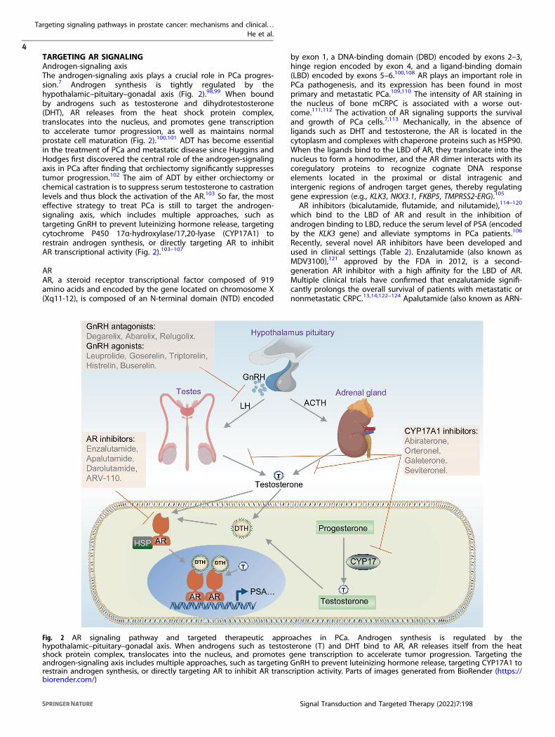
TARGETING AR SIGNALINGAndrogen-signaling axisThe androgen-signaling axis plays a crucial role in PCa progres-sion.7 Androgen synthesis is tightly regulated by thehypothalamic–pituitary–gonadal axis (Fig. 2).98,99 When boundby androgens such as testosterone and dihydrotestosterone(DHT), AR releases from the heat shock protein complex,translocates into the nucleus, and promotes gene transcriptionto accelerate tumor progression, as well as maintains normalprostate cell maturation (Fig. 2).100,101 ADT has become essentialin the treatment of PCa and metastatic disease since Huggins andHodges first discovered the central role of the androgen-signalingaxis in PCa after finding that orchiectomy significantly suppressestumor progression.102 The aim of ADT by either orchiectomy orchemical castration is to suppress serum testosterone to castrationlevels and thus block the activation of the AR.103 So far, the mosteffective strategy to treat PCa is still to target the androgen-signaling axis, which includes multiple approaches, such astargeting GnRH to prevent luteinizing hormone release, targetingcytochrome P450 17α-hydroxylase/17,20-lyase (CYP17A1) torestrain androgen synthesis, or directly targeting AR to inhibitAR transcriptional activity (Fig. 2).103–107
ARAR, a steroid receptor transcriptional factor composed of 919amino acids and encoded by the gene located on chromosome X(Xq11-12), is composed of an N-terminal domain (NTD) encoded
by exon 1, a DNA-binding domain (DBD) encoded by exons 2–3,hinge region encoded by exon 4, and a ligand-binding domain(LBD) encoded by exons 5–6.100,108 AR plays an important role inPCa pathogenesis, and its expression has been found in mostprimary and metastatic PCa.109,110 The intensity of AR staining inthe nucleus of bone mCRPC is associated with a worse out-come.111,112 The activation of AR signaling supports the survivaland growth of PCa cells.7,113 Mechanically, in the absence ofligands such as DHT and testosterone, the AR is located in thecytoplasm and complexes with chaperone proteins such as HSP90.When the ligands bind to the LBD of AR, they translocate into thenucleus to form a homodimer, and the AR dimer interacts with itscoregulatory proteins to recognize cognate DNA responseelements located in the proximal or distal intragenic andintergenic regions of androgen target genes, thereby regulatinggene expression (e.g., KLK3, NKX3.1, FKBP5, TMPRSS2-ERG).105
AR inhibitors (bicalutamide, flutamide, and nilutamide),114–120
which bind to the LBD of AR and result in the inhibition ofandrogen binding to LBD, reduce the serum level of PSA (encodedby the KLK3 gene) and alleviate symptoms in PCa patients.106
Recently, several novel AR inhibitors have been developed andused in clinical settings (Table 2). Enzalutamide (also known asMDV3100),121 approved by the FDA in 2012, is a second-generation AR inhibitor with a high affinity for the LBD of AR.Multiple clinical trials have confirmed that enzalutamide signifi-cantly prolongs the overall survival of patients with metastatic ornonmetastatic CRPC.13,14,122–124 Apalutamide (also known as ARN-
Fig. 2 AR signaling pathway and targeted therapeutic approaches in PCa. Androgen synthesis is regulated by thehypothalamic–pituitary–gonadal axis. When androgens such as testosterone (T) and DHT bind to AR, AR releases itself from the heatshock protein complex, translocates into the nucleus, and promotes gene transcription to accelerate tumor progression. Targeting theandrogen-signaling axis includes multiple approaches, such as targeting GnRH to prevent luteinizing hormone release, targeting CYP17A1 torestrain androgen synthesis, or directly targeting AR to inhibit AR transcription activity. Parts of images generated from BioRender (https://biorender.com/)
Targeting signaling pathways in prostate cancer: mechanisms and clinical. . .He et al.
4
Signal Transduction and Targeted Therapy (2022) 7:198

509)125 has a greater efficacy than enzalutamide and wasapproved for treatment of nonmetastatic CRPC by the FDA in2018. Apalutamide inhibits the nuclear localization and DNAbinding of AR in PCa cells.125 A clinical study showed thatapalutamide administration significantly lengthened metastasis-free survival in patients with nonmetastatic CRPC.15
Notably, AR gene mutations and amplifications occur in ~60%of mCRPC (Fig. 1a). AR mutations are predominant in LBD,limiting binding affinity of AR inhibitors.126–129 Darolutamide(also named ODM-201),130,131 approved for treatment ofnonmetastatic CRPC by the FDA in 2019, is a novel AR inhibitorthat antagonizes mutated AR, such as F877L and T878A, whichconfers resistance to enzalutamide and apalutamide.132–134
Phase 3 trial studies have shown that darolutamide significantlyprolongs metastasis-free survival for high-risk nonmetastaticCRPC.16,135 Furthermore, the latest AR protein degrader ARV-110,an oral proteolysis targeting chimera (PROTAC),136 specificallydegrades more than 95% of AR and overcomes enzalutamideresistance in xenograft models.137,138 ARV-110 is presently underevaluation in clinical trials (Table 2). Current AR-targetedtherapies primarily target LBD. However, AR variants such asAR-V7 and ARv567es lack the entire LBD or a functional LBD, butretain their ability to bind DNA in the absence of androgens anddisplay constitutive activity, thus conferring drug resistance tonext-generation AR inhibitors.139–143
GnRHGnRH, also known as luteinizing hormone-releasing hormone(LHRH),144,145 is a hypothalamic peptide (pGlu-His-Trp-Ser-Tyr-Gly-Leu-Arg-Pro-Gly-NH2) that plays a central role in controlling thehypothalamic–pituitary-axis in mammals.146–148 GnRH binds to theGnRH receptor (GnRHR), which belongs to the rhodopsin-like Gprotein coupled receptor (GPCR) family, and induces the release ofluteinizing hormone (LH), which then arrives to the Leydig cells of thetestes to stimulate testosterone synthesis.148–151 Schally and Guillemin(1982) first developed a synthetic GnRH agonist (also known as GnRHanalog) to manipulate the hypothalamic–pituitary–gonadal axis.152
Mechanically, the GnRH agonists induce sustained stimulation of thepituitary gland to induce the downregulation and desensitization ofGnRHR, resulting in the reduction of LH release and suppression oftestosterone production to castration levels.103,148,153 An early studyby Tolis et al. found that patients with advanced PCa treated dailywith GnRH agonists experienced a 75% suppression in serumtestosterone levels, resulting in a decreased prostate size andreduction in tumor-associated bone pain.152 Several synthetic GnRHagonists have been developed for clinical use since the 1980s,including leuprolide, triptorelin, and buserelin.154–166 Many clinicalstudies of GnRH agonists are completed or ongoing (Table 2).Although long-acting GnRH agonists suppress the release of LH andtestosterone, GnRH agonists initially produce a rapid and transientincrease in LH and testosterone levels, which is called the “flare-up”
Table 2. Selective clinical trials of AR signaling inhibitors
Drug Target Condition Status Phase NCT identifier
Degarelix GnRHR PCa Completed 3 NCT00451958
Degarelix GnRHR PCa Completed 3 NCT01071915
Degarelix GnRHR PCa Completed 3 NCT02015871
Relugolix GnRHR PCa Recruiting 3 NCT05050084
Relugolix GnRHR PCa Completed 3 NCT03085095
Abarelix GnRHR PCa Completed 3 NCT00841113
Leuprolide GnRHR PCa Completed 4 NCT00220194
Leuprolide GnRHR PCa Recruiting 3 NCT04914195
Goserelin GnRHR PCa Completed 3 NCT00439751
Goserelin GnRHR PCa Not yet recruiting 4 NCT03971110
Triptorelin GnRHR PCa Completed 3 NCT01715129
Triptorelin GnRHR PCa Completed 3 NCT00104741
Histrelin GnRHR Metastatic PCa Recruiting 2/3 NCT04787744
Histrelin GnRHR PCa Completed 3 NCT01697384
Buserelin GnRHR PCa Completed 3 NCT00003653
Buserelin GnRHR PCa Recruiting 3 NCT05050084
Enzalutamide AR CRPC Completed 3 NCT00974311
Enzalutamide AR mCRPC Completed 4 NCT02116582
Enzalutamide AR mCRPC Completed 4 NCT02485691
Apalutamide AR PCa Completed 2 NCT01790126
Apalutamide AR Nonmetastatic CRPC Recruiting 3 NCT04108208
Darolutamide AR PCa Not yet recruiting 3 NCT02799602
Darolutamide AR Nonmetastatic CRPC Completed 3 NCT02200614
ARV-110 AR mCRPC Recruiting 1/2 NCT03888612
Abiraterone CYP17A1 mCRPC Completed 3 NCT04056754
Abiraterone CYP17A1 mCRPC Completed 3 NCT00887198
Abiraterone CYP17A1 mCRPC Completed 3 NCT02111577
Seviteronel CYP17A1 CRPC Completed 2 NCT02445976
Orteronel CYP17A1 mCRPC Completed 3 NCT01193257
Galeterone CYP17A1 CRPC Completed 2 NCT01709734
Targeting signaling pathways in prostate cancer: mechanisms and clinical. . .He et al.
5
Signal Transduction and Targeted Therapy (2022) 7:198
phenomenon, and may lead to side effects, such as bone pain andcardiovascular complications.103,154
In contrast to GnRH agonists, GnRH antagonists bind competi-tively to the GnRHR in the pituitary to rapidly prevent LHproduction, thereby suppressing testosterone to castration levels,which reduces the risk of the “flare-up” phenomenon.103,167
Abarelix was the first GnRH antagonist approved by the FDA, but itwas discontinued from the market because of severe hypersensi-tivity reactions.168–171 Degarelix, which was approved by the FDAin 2008, is the most widespread GnRH antagonist used in clinicalpractice.172–177 Relugolix, which is a novel oral GnRH antagonist,was approved by the FDA in 2020.178,179 Numerous promisingclinical trials on GnRH antagonists in combination with radio-therapy or chemotherapy have been completed or are stillongoing (Table 2). However, the use of these agents remainscontroversial. For example, a clinical study revealed thatneoadjuvant degarelix is related to the upregulation of DHT intumors.180 Other studies have found that the administration ofGnRH agonists or antagonists can decrease lean body mass andincrease fat mass.181–183 Further studies will provide criticalevidence to address whether GnRH agonists or antagonists aresafe for patients with cardiovascular disease.
CYP17A1CYP17A1, a membrane-bound monooxygenase, is a pivotalenzyme for androgen synthesis.184 CYP17A1 is composed of 508amino acids with four structural domains, including a substrate-binding domain, a catalytic activity area, a heme-binding region,and a redox-partner binding site.185,186 CYP17A1 has both 17α-hydroxylase and 17,20-lyase catalytic activities, and is essential inthe production of both androgens and glucocorticoids.185,186
CYP17A1 predominantly localizes at the endoplasmic reticulum inthe adrenal glands, testicular Leydig cells, and ovarian thecalcells.187 The 17α-hydroxylase activity of CYP17A1 is required forthe hydroxylation of pregnenolone and progesterone at the C17position, which generates 17α-hydroxypregnenolone and 17α-hydroxyprogesterone.188 The 17,20-lyase activity of CYP17A1 isessential for the cleavage of 17α-hydroxypregnenolone or 17α-hydroxyprogesterone, which form dehydroepiandrosterone(DHEA) and androstenedione, respectively, and is a critical stepfor testosterone synthesis.105,188 Importantly, CYP17A1 also con-fers to intratumoral androgen biosynthesis in CRPC.189–192 Lowlevels of androgen are still found in the serum during ADT; thus,many CYP17A1 inhibitors have been tested in clinics (Table 2).Abiraterone, a selective inhibitor of 17α-hydroxylase and 17,20-
lyase,193 was first approved by the FDA for treatment of mCPRC in2011.194,195 Administration of abiraterone induces a significantdecline in PSA levels, improves overall survival, and alleviates painin both chemotherapy-naive and docetaxel-resistantpatients.12,196–198 Although the suppression of 17α-hydroxylaseactivity leads to overproduction of mineralocorticoids, which canresult in adverse events (such as hypokalaemia, fluid retention,hypertension, and cardiac disorders), these side effects are largelyprevented by the co-administration of glucocorticoidprednisone.199
Other CYP17A1 inhibitors, such as orteronel (TAK-700) andgaleterone (TOK-001), have also been developed.200–202 Orteronelis a nonsteroidal selective inhibitor of 17,20-lyase, while galeter-one has multiple mechanisms of action, including CYP17A1inhibition, AR antagonism, and a reduction in both full-lengthAR and AR-V7 levels.192,200–203 Orteronel preferentially inhibits17,20-lyase over 17α-hydroxylase, leading to a reduction in the riskof overproduction of mineralocorticoids.184,192,201 Results fromphase 1/2 studies have indicated that patients had an approxi-mately 60% PSA response rate at 12 weeks after the administra-tion of orteronel twice daily.204 A phase 1 study in patients withCRPC observed that ~50% of men had a PSA decline after12 weeks of treatment with galeterone, and no adrenal
mineralocorticoid excess was noted.205 Therefore, orteronel andgaleterone are potentially attractive drugs for longer durationtherapy and overcoming drug resistance, although a clinical studyshowed that orteronel did not meet the primary endpoint ofoverall survival.206 Clinical studies of galeterone compared toenzalutamide in mCRPC expressing AR-V7 have been conducted,but the result do not meet the primary endpoint.207 Additionally,seviteronel (VT-464), a newly developed drug used as a CYP17A1inhibitor and AR antagonist, selectively inhibits 17,20-lyase andgreatly decreases AR transactivation and offers an advantage overabiraterone because it does not require combination withprednisone.208 Notably, abiraterone treatment markedly increasesintratumoral expression of CYP17A1 in tumor biopsies from CRPCpatients, and many patients ultimately become resistant to CYP17inhibitors.143,190,209
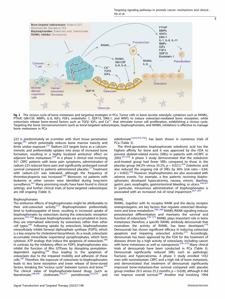
TARGETING BONE MICROENVIRONMENTBone microenvironment of PCaBone metastasis in PCa is a highly frequent event that occurs in upto 90% of patients with advanced disease.210–212 The bonemicroenvironment is a dynamic compartment that provides amilieu in which metastatic cancer cells can colonize andgrow.213,214 The “vicious cycle” hypothesis is an appropriatemodel with which to explain the process of cancer cellsmetastasizing to the bone (Fig. 3).213,215 Tumor cells in boneinduce osteoclast-mediated bone resorption, while osteoclastsrelease bone-stored factors that stimulate tumor cell proliferation,establishing a vicious cycle. Bone metastasis is driven by thecooperation among metastatic tumor cells, bone-forming osteo-blasts, bone-dissolving osteoclasts, and other cell populations.212
Physiologically, mature osteoblasts, osteocytes, and osteoclastsregulate the dynamic remodeling of bone tissue.216 The increasedlevels of parathyroid hormone-related peptide (PTHrP) fromosteoclasts can induce bone resorption by upregulating thereceptor activator of RANKL, which promotes the release ofvarious growth factors (such as ionized calcium and TGFβ) into thebone microenvironment to support cancer cell implantation andtransformation.217–221 Invading tumor cells secrete osteolyticcytokines, such as granulocyte-macrophage colony-stimulatingfactor, matrix metalloproteinases, interleukin (IL)-6, insulin-likegrowth factors (IGFs), fibroblast growth factors (FGFs), endothelin1, growth differentiation factor 15 (GDF15), dickkopf-1 (DKK-1),and WNTs.222–226 These osteolytic cytokines stimulate preosteo-blast differentiation and promote osteoclast maturation toaccelerate bone resorption.217,227,228 Meanwhile, osteoblastsrelease IL-6 and RANKL to accelerate the maturation of osteoclasts,further secreting growth factors to facilitate tumor cell growth(Fig. 3).212,217,229 Thus, various approaches that target the bonemicroenvironment, such as bone-targeting agents, are effectivefor managing bone metastases in PCa (Fig. 3).
Bone-targeted radioisotopesBone-seeking therapeutic radioisotopes are distinct among antic-ancer therapies, because they target calcium hydroxyapatite in thebone instead of tumor cells. Bone metastases often containosteosclerotic lesions, with increased osteoblastic bone formation.Thus, ionizing radiation is selectively delivered to bone withincreased osteoblastic activity to simultaneously target multiplemetastases,230 enabling the delivery of high-energy radiation tobone metastases while limiting toxicity to other normal cells.Calcium-mimetic radiopharmaceuticals, such as the first genera-tion of primarily β-emitting radioisotopes strontium-89 andsamarium-153, have been approved early by the FDA based onsuccessful endpoints of bone pain palliation.231–235 More recently,the bone-targeted radioisotope radium-223 was also approved bythe FDA for patients with mCRPC and painful bone metas-tases.17,18,236–238 Unlike strontium-89 and samarium-153, radium-
Targeting signaling pathways in prostate cancer: mechanisms and clinical. . .He et al.
6
Signal Transduction and Targeted Therapy (2022) 7:198
223 is predominately an α-emitter with short tissue penetrationrange,230 which potentially reduces bone marrow toxicity andlimits undue exposure.237 Radium-223 targets bone as a calcium-mimetic and preferentially uptakes into areas of increased boneformation, resulting in a highly localized antitumor effect onadjacent bone metastases.239 In a phase 3 clinical trial involving921 CRPC patients with bone pain symptoms, administration ofradium-223 reduced bone pain and significantly prolonged overallsurvival compared to patients administered placebo.17 Treatmentwith radium-223 was tolerated, although the frequency ofthrombocytopenia was increased.240 Moreover, no patients withleukemia or other cancers were identified during long-termsurveillance.241 Many promising results have been found in clinicalsettings, and further clinical trials of bone-targeted radioisotopesare still ongoing (Table 3).
BisphosphonatesThe antitumor effects of bisphosphonates might be attributable totheir anti-osteoclast activity.212 Bisphosphonates preferentiallybind to hydroxyapatite of bone, resulting in increased uptake ofbisphosphonates by osteoclasts during the osteoclastic resorptionprocess.242–244 Because bisphosphonates are accumulated in bone,they are internalized selectively by osteoclasts rather than othercell types.245 Following uptake by osteoclasts, bisphosphonatesintracellularly inhibit farnesyl diphosphate synthase (FDPS), whichis a key enzyme for cholesterol biosynthesis. As a result, osteoclastsaccumulate intracellular isopentenyl pyrophosphates, which formcytotoxic ATP analogs that induce the apoptosis of osteoclasts.246
In contrast, by the inhibitory effect on FDPS, bisphosphonates alsoinhibit the function of Rho GTPases by disrupting prenylation-dependent signaling,244 thus leading to the apoptosis ofosteoclasts due to the impaired mobility and adhesion of thesecells.247 Therefore, the exposure of osteoclasts to bisphosphonatesleads to less bone resorption and lower release of bone-storedfactors, breaking the “vicious cycle” between tumors and bone.247
The clinical value of bisphosphonate-based drugs (such asibandronate,248,249 clodronate,250,251 pamidronate,252,253 and
zoledronate19,22,254–256) has been shown in numerous trials ofPCa (Table 3).The third-generation bisphosphonate zoledronic acid has the
highest affinity for bone and it was approved by the FDA toprevent skeletal-related events (SREs) in patients with mCRPC in2002.257,258 A phase 3 study demonstrated that the zoledronicacid-treated group had fewer SREs compared to those in theplacebo group (44.2% versus 33.2%, p= 0.021).258 Zoledronic acidalso reduced the ongoing risk of SREs by 36% (risk ratio= 0.64,p= 0.002).259 However, bisphosphonates are also associated withadverse events. For example, a few patients receiving bispho-sphonates developed hypocalcemia, nausea, emesis, diarrhea,gastric pain, esophagitis, gastrointestinal bleeding, or ulcers.260,261
In particular, intravenous administration of bisphosphonates isassociated with an increased risk of renal impairment.261–265
RANKLRANKL, together with its receptor RANK and the decoy receptorosteoprotegerin, are key factors that regulate osteoclast develop-ment and bone metabolism.266–269 RANKL/RANK signaling inducespreosteoclast differentiation and maintains the survival andfunction of osteoclasts.270–273 RANKL plays important role in bonemetastases; therefore, a specific RANKL antibody denosumab thatneutralizes the activity of RANKL has been developed.274
Denosumab has shown significant efficacy in inducing osteoclastapoptosis and impairing osteoclast activity.275 Accordingly,denosumab has been approved by the FDA for the treatment ofdiseases driven by a high activity of osteoclasts, including cancerwith bone metastases as well as osteoporosis.276–279 Many clinicaltrials of denosumab have been conducted in PCa (Table 3).Denosumab significantly reduced SREs such as pathologicfractures and hypercalcemia. A phase 3 study enrolled 1432men with nonmetastatic CRPC and a high risk of bone metastasis,and demonstrated that treatment of denosumab significantlyimproved bone-metastasis-free survival compared with placebogroup (median 29.5 versus 25.2 months; p= 0.028), although it didnot improve overall survival.280 Another trial involving 1904
Fig. 3 The vicious cycle of bone metastasis and targeting strategies in PCa. Tumor cells in bone secrete osteolytic cytokines such as RANKL,PTHrP, GM-CSF, MMPs, IL-6, IGFs, FGFs, endothelin 1, GDF15, DKK-1, and WNTs to induce osteoclast-mediated bone resorption, whileosteoclasts release bone-stored factors such as TGFβ, IGFs, and Ca2+ that stimulate tumor cell proliferation, establishing a vicious cycle.Targeting the bone microenvironment (such as bone-targeted radioisotopes, bisphosphonates, and RANKL inhibitors) is effective to managebone metastases in PCa
Targeting signaling pathways in prostate cancer: mechanisms and clinical. . .He et al.
7
Signal Transduction and Targeted Therapy (2022) 7:198
patients found that denosumab treatment increased the mediantime to first on-study SREs compared with the results of zoledronicacid (20.7 versus 17.1 months; p= 0.008).22 Because denosumabtreatment is associated with life-threatening hypocalcemia,proactive treatment of calcium and calcitriol should be consideredwhen using denosumab.281
Calcium channelsCytosolic calcium (Ca2+) signaling plays an important role in thebone metastasis of PCa.219 Elevated Ca2+ stimulates PTHrPsecretion and activates RANKL/RANK signaling in osteoclasts,which promotes bone resorption and calcium release, in turnpromoting tumor cell proliferation and maintaining PCa cellhoming to bone.219 Targeting calcium signaling could be apromising strategy for managing PCa bone metastasis. Newagents for targeting calcium signaling include calcium-ATPaseinhibitors, voltage-gated calcium channel inhibitors, transientreceptor potential (TRP) channel inhibitors, and Orai inhibitors,although most of these agents are still in the early stages ofstudies.282 For instance, mipsagargin (G-202), a SERCA inhibitor,was tested in mCRPC in a phase 2 clinical trial; however, the studywas withdrawn without results posted (Table 3). The TRPV6inhibitor, SOR-C13, is currently under evaluation in patients withadvanced tumors, including PCa, in a phase 1 clinical trial (Table3). Because calcium channels are also critical for numerous cellularhomeostasis and physiological functions under normal condi-tions,283 future calcium-based therapies should specifically targetPCa cells to decrease normal tissue toxicity.
TARGETING PSMAPSMA and PSMA-targeted ligandsPSMA is a type II transmembrane glycoprotein that includesactivities of folate hydrolase and N-acetyl-α-linked acidic dipepti-dase and consists of 750 amino acids located in three domains,including the intracellular domain, which contains 19 amino acids,the transmembrane domain, which consists of 24 amino acids, andthe extracellular domain, which contains 707 amino acids.284,285
PSMA is expressed at a very low level in normal prostatic tissuesand nonprostatic tissues, but its expression in PCa tissues increasesby 100–1000 times compared to that in normal tissues.286 Thus,PSMA is a theranostic target for imaging diagnostics and targetedradionuclide therapy for PCa and its metastases.287–289
Three main types of ligands are used to target PSMA:monoclonal antibodies, aptamers, and small-molecule inhibitors.PSMA monoclonal antibodies can be classified into two types:intracellular domain antibodies (7E11, PM2J004.5) and extracel-lular domain antibodies (J591, J533, J415).284,290,291 Importantly,the humanized monoclonal antibody J591, which targets theextracellular domain of PSMA, has an impressive applicationprospect in the diagnosis and treatment of PCa. Aptamers ofPSMA (such as xPSM-A9, xPSM-A10, A10-3.2, and A9g) arenucleotides or deoxynucleotides that can selectively recognizePSMA.292–294 Small-molecule inhibitors that can interact withPSMA, including 123I-MIP-1072, 123I-MIP-1095, PSMA-I&T, PSMA-I&S, and PSMA-617, have become the preferred choice formolecular imaging probes and targeted therapy for PCa.295,296
PSMA-based diagnostic imagingThe early PSMA-targeted imaging agent was ProstaScint (alsoknown as 111In-capromab pendetide), a mouse monoclonalantibody (7E11) linked to 111In for SPECT (single photon emissioncomputed tomography) imaging. However, ProstaScint was onlyable to bind to the intracellular epitope of PSMA, and cannot beaccessed in viable tumor cells, thus limiting its clinical perfor-mance.297,298 68Ga-PSMA-11 (also called PSMA-HBED-CC) for PET isprobably the tracer most often used for PCa.299–301 Several 68Ga-labeled PSMA ligands have been developed as theranostic agents,including 68Ga-PSMA-617 and 68Ga-PSMA-I&T.302–304 The18F-labeled agents include 18F-DCFBC, 18F-DCFPyL, and 18F-PSMA-1007, which exhibit many advantages such as a lowerpositron range and longer half-life compared with those of 68Ga-labeled agents.305–308 PSMA-based imaging has shown improvedsensitivity and diagnostic accuracy in PCa. For example, a study in96 patients with PCa demonstrated that 18F-PSMA-1007 PET had asensitivity of 85.9% and a specificity of 99.5% in a patient-based
Table 3. Selective clinical trials of drugs targeting bone microenvironment
Drug Target Condition Status Phase NCT identifier
Radium-223 Calcium-mimetic α-emitting Bone-metastatic CRPC Completed 3 NCT00699751
Radium-223 Calcium mimetic α-emitting Bone-metastatic CRPC Completed 3 NCT01618370
Radium-223 Calcium mimetic α-emitting Bone-metastatic CRPC Completed 3 NCT01810770
Radium-223 Calcium mimetic α-emitting Bone-metastatic CRPC Recruiting 3 NCT03432949
Strontium-89 Calcium mimetic β-emitting Bone-metastatic CRPC Completed 3 NCT00002503
Samarium-153 Calcium mimetic β-emitting Bone-metastatic CRPC Completed 3 NCT00365105
Zoledronic acid FDPS Bone-metastatic CRPC Completed 3 NCT00242567
Zoledronic acid FDPS Bone-metastatic CRPC Completed 3 NCT00079001
Zoledronic acid FDPS Bone-metastatic CRPC Completed 3 NCT00321620
Zoledronic acid FDPS Bone-metastatic CRPC Completed 4 NCT00242554
Clodronic acid FDPS PCa Completed Unknown NCT01198457
Risedronic acid FDPS PCa Completed 3 NCT00426777
Alendronic acid FDPS Metastatic PCa Terminated 2 NCT00019695
Ibandronic acid FDPS Metastatic PCa Completed 3 NCT00082927
Denosumab RANKL CRPC Completed 3 NCT01824342
Denosumab RANKL CRPC Completed 3 NCT00286091
Denosumab RANKL CRPC Completed 3 NCT00838201
Denosumab RANKL PCa Completed 3 NCT00925600
Mipsagargin SERCA Metastatic PCa Withdrawn 2 NCT01734681
SOR-C13 TRPV6 Advanced cancers Recruiting 1 NCT03784677
Targeting signaling pathways in prostate cancer: mechanisms and clinical. . .He et al.
8
Signal Transduction and Targeted Therapy (2022) 7:198

analysis for detecting positive lymph nodes larger than 3mm.309
Furthermore, the 99mTc-labeled PSMA ligand 99mTc-MIP-1404 is ina phase 3 clinical trial designed to evaluate its sensitivity andspecificity to detect PCa (Table 4). Standardized criteria of thePSMA ligand PET are evolving and will facilitate its use in clinicalpractice; several prospective trials (Table 4) are ongoing to supportfinal market approval.310
PSMA-targeted radionuclide therapy (PSMA-TRT)In contrast to conventional external radiotherapy, targeted radio-nuclide therapy (TRT) is a treatment conducted by injecting aradionuclide-labeled ligand into the body to specifically targetcancer cells. The radionuclide then releases α-particles, β-particles,or auger electrons to produce free radicals that induce DNAdamage, thus specifically promoting apoptosis or necrosis oftargeted cells.284,311 Conjugations of PSMA-targeted ligands(antibodies or small molecules) with radionuclides such asβ-emitters (most commonly 177Lu) or α-emitters (commonly225Ac) produce PSMA-TRT agents, including 177Lu-PSMA-617,225Ac-PSMA-617, and 177Lu-J591 (Table 4).312–314 More impor-tantly, in a recent phase 2 study comparing 177Lu-PSMA-617 andcabazitaxel in mCRPC, 177Lu-PSMA-617 led to a higher PSAresponse (66% versus 44% by intention-to-treat analysis; p=0.0016) and fewer adverse events, indicating that 177Lu-PSMA-617is a potential alternative therapy to cabazitaxel in patients withmCRPC.315 Lu-PSMA-617 was approved by the FDA for thetreatment of mCRPC in 2021. In addition to 177Lu-PSMA-617,225Ac-PSMA-617 and 177Lu-J591 have been studied in clinical trials(Table 4). Ongoing clinical trials of PSMA-TRT will further explorethe optimal sequencing of this therapy in earlier disease settings,as well as novel combinations. Overall, PSMA expression atdifferent metastatic sites among different patients and theselection of optimal patients remain to be defined.
PSMA-antibody-drug conjugates (PSMA-ADC)PSMA-specific antibodies have been used to bind cytotoxic drugsvia different chemical bonds to obtain PSMA-ADC. PSMA-ADCavoids systemic medication and reduces the toxicity to non-targetorgans compared to traditional cytotoxic drugs. Many applicationsof monoclonal antibody-based PSMA-ADC have entered clinicaltrials (Table 4), including that of MLN2704, which links to theantimicrotubule agent maytansinoid-1;316 PSMA-MMAE (mono-methyl auristatin E), which connects to the microtubule disrupting
agent MMAE;317 MEDI3726, which combines with thepyrrolobenzodiazepine-based linker-drug tesirine;318 and BIND-014, which conjugates with docetaxel.40 MLN2704 was discon-tinued after a phase 1/2 study because of the instability of thebond between the antibody and the drug.319,320 The first clinicaltrial of MEDI3726 observed a high incidence of treatment-relatedadverse events.321 A phase 2 clinical trial demonstrated thatPSMA-MMAE showed some activity with regards to PSA declineand circulating tumor cell reduction in patients with mCRPC, but italso included significant treatment-related toxicities, such asneutropenia and neuropathy.317 Interestingly, the phase 2 clinicaltrial of BIND-014 (a novel PSMA-ADC) in patients withchemotherapy-naive mCRPC suggest that BIND-014 is welltolerated and patients are likely to benefit from the treatment.40
Optimization of dose administration, conjugation of more appro-priate drugs, and patient selection should be considered toimprove the efficacy of PSMA-ADC in the future.
PSMA-based chimeric antigen receptor (CAR)-T cells therapyCAR-T cells are genetically engineered T cells that express anartificial T-cell receptor, endowing T-cell populations with theability to target tumors independently of majorhistocompatibility-complex (MHC) engagement.322 CAR-T-celltherapy has gained momentum in PCa treatment in clinicaltrials. CAR-T cells are activated when the antigen is recognizedby CAR, thus stimulating the release of cytotoxins, such asperforin and granzyme, into tumor cells to induce apoptosis.PSMA is considered to be a reliable target for CAR-T-cell therapy.First-generation CAR-T cells targeting PSMA were constructedwith a chimeric anti-PSMA immunoglobulin-T-cell receptor genebased on the monoclonal antibody 3D8.323 Second-generationCAR-T cells were constructed by inserting the CD28 signaldomain into first-generation CAR-T cells.324 Recently, many newPSMA-based CAR-T cells, such as CART-PSMA-TGFβRDN, havebeen evaluated in phase 1 clinical trials in CRPC patients.42
Meanwhile, other clinical trials are underway and will test thesafety and efficacy of PSMA-targeted CAR-T cells for thetreatment of PCa (Table 4). Additionally, side effects such ascytokine release syndrome, immune effector cell-associatedneurotoxicity syndrome, and cytopenia are commonly observedin patients receiving CAR-T-cell therapy; therefore, additionaltreatment with corticosteroids should be considered when usingCAR-T-cell treatment.325
Table 4. Selective clinical trials of PSMA-targeted agents
Drug Target Condition Status Phase NCT identifier
177Lu-PSMA-617 PSMA mCRPC Unknown 2 NCT03392428177Lu-PSMA-617 PSMA mCRPC Recruiting 3 NCT04689828225Ac-PSMA-617 PSMA PCa, CRPC Recruiting 1 NCT04597411177Lu-J591 PSMA Metastatic PCa Completed 2 NCT00195039
MLN2704 PSMA mCRPC Completed 1/2 NCT00070837
PSMA-MMAE PSMA mCRPC Completed 2 NCT01695044
MEDI3726 PSMA mCRPC Completed 1 NCT02991911
BIND-014 PSMA mCRPC Completed 2 NCT01812746.68Ga-PSMA-11 PSMA PCa Completed 3 NCT0380347568Ga-PSMA-617 PSMA PCa Completed 2 NCT0360475718F-PSMA-1007 PSMA PCa Completed 3 NCT0410255399mTc-MIP-1404 PSMA PCa Completed 3 NCT02615067
CART-PSMA-TGFβRDN PSMA mCRPC Not yet recruiting 1 NCT04227275
PD1-PSMA-CART PSMA CRPC Recruiting 1 NCT04768608
LIGHT-PSMA-CART PSMA CRPC Suspended 1 NCT04053062
P-PSMA-101 CAR-T PSMA PCa Recruiting 1 NCT04249947
Targeting signaling pathways in prostate cancer: mechanisms and clinical. . .He et al.
9
Signal Transduction and Targeted Therapy (2022) 7:198

TARGETING DNA REPAIR PATHWAYSPARP function in DNA repair and synthetic lethalityPARP is a family of enzymes involved in DNA repair andtranscriptional regulation.326 Activation of PARP1/2 is importantfor recruiting the key effectors of DNA repair (Fig. 4).327 DNAdamage in cells, including single-strand breaks (SSB) and double-strand breaks (DSB), can be induced by exposure to chemicals(such as chemotherapy), physical agents (such as radiotherapy), orendogenous reactive metabolites (such as reactive oxygen andnitrogen species) (Fig. 4). Effective DNA repair is essential forcellular survival. Mechanisms of SSB repair include base-excisionrepair, nucleotide excision repair, and mismatch excision repair,whereas DSB repair includes homologous recombination (HR) andnon-homologous end-joining (NHEJ).327 The primary mechanismfor inhibiting PARP in cancer therapy is synthetic lethality, whichindicates two genomic alteration events that are each relativelyinnocuous individually but become lethal when they occurtogether.328 When PARP1/2 is pharmacologically inhibited, theaccumulation of SSB by PARP inhibition can progress to DSB,which is usually repaired through HR. The DSBs can be fixed if theDNA repair system is intact in cells; however, PARP inhibitionwould lead to lethality if a cell lacks HR repair capacity (mutationsof BRCA1, BRCA2, or ATM).329 PARP inhibition would not induce celldeath in normal cells due to efficient DSB repair mechanisms;however, PARP inhibition would be lethal for tumor cells withdeficient HR, such as BRCA1/2mutations.330–332 Furthermore, PARPinhibition would result in fork collapse and would transform intoDSB, since PARP1 is involved in the restart of stalled forks.333,334 If
the function of the BRCA (breast cancer susceptibility protein) isdeficient, these DSB would not be repaired, thus causing syntheticlethality. Up to 30% of mCRPC tumors harbor DNA damage repairgene aberrations,24 which can be therapeutically used with PARPinhibitors to induce synthetic lethality. However, the interpretationof PARP inhibition-related mechanisms of synthetic lethality maybe incomplete. PARP inhibitors may also induce cytotoxic effectsby inhibition of SSB repair, as well as other mechanisms.335
Moreover, genomic alterations, such as TMPRSS2-ERG fusion, SPOPmutation, PTEN loss, and CHD1 deletion, are linked to an impairedDNA damage response phenotype, which might increase thetherapeutic effectiveness of PARP inhibition.336 DNA damageresponse genes are regulated by AR; consequently, the ADTresponse is also influenced by DNA repair deficiency.337 Functionalinactivation of DNA repair pathways also enhances sensitivity tochemotherapy and radiotherapy, and this effect is furtherenhanced by inhibitors of the targeting DNA repair pathwaysthat induce synthetic sensitivity or lethality in DNA repair-deficientcancers (Fig. 4).336
PARP inhibitorsSeveral PARP inhibitors have been evaluated in clinical trials (Table5). In 2020, olaparib was approved by the FDA for the treatment ofmCRPC with deficient HR genes. The first clinical data from aphase 2 study demonstrated that 88% of patients with ahomozygous deletion or mutation in gene-associated DNA repairresponded to olaparib.23 Responses were observed in patientswith deletions or mutations of BRCA1, BRCA2, FANCA, CHEK2, and
Fig. 4 Inhibition of PARP mediates synthetic lethality in PCa. When PARP1/2 are pharmacologically inhibited, the accumulation of SSBs byPARP inhibition can progress to DSBs, which are usually repaired through HR. The DSBs can be fixed if the DNA repair system is intact in cells;however, PARP inhibition can lead to lethality if a cell is lacking HR repair capacity (mutations of BRCA1, BRCA2, or ATM). BCL2 overexpression,TMPRSS2-ERG fusion, SPOP mutation, PTEN loss, and CHD1 deletion are also linked with an impaired DNA damage response phenotype, whichmight increase the therapeutic effectiveness of PARP inhibition
Targeting signaling pathways in prostate cancer: mechanisms and clinical. . .He et al.
10
Signal Transduction and Targeted Therapy (2022) 7:198

PALB2. The overall survival in patients with BRCA1/2 alterations aremore favorable than in those who were negative (13.8 versus7.5 months; p= 0.05).23 Rucaparib was approved by the FDA in2020 based on a recent clinical study, which involved 78 mCRPCpatients with DNA repair gene alterations, including ATM (n= 49),CDK12 (n= 15), CHEK2 (n= 12), and other genes (n= 14).338 A PSAresponse was seen in 54.8% of all patients, and those that hadPALB2, FANCA, BRIP1, and RAD51B mutations showed a betterresponse compared with those with ATM alterations, while theobjective response rate and PSA responses in patients with ATM,CHEK2, and CDK12 mutations were low compared to those withBRCA mutated tumors.338 The efficacy of rucaparib is currentlybeing evaluated in a phase 3 trial (Table 5). A phase 2 studydemonstrated that the objective response rate of talazoparib wasseen in 29.8% (31 of 104) of patients, whereas serious treatment-related adverse events were reported in 43 (34%) patients.27 Inaddition, PARP1/2 selective inhibitors, including niraparib andpamiparib, are currently being tested in phase 2/3 trials of PCa(Table 5). Finally, trials combining PARP inhibitors with otherdrugs, such as AR-targeting agents and radium-223, have gainedmomentum based on the concept of cross-sensitivity. Forexample, a randomized trial combining veliparib and abirateronedetermined that the subgroup patients (27%) with aberrations inDNA repair genes showed better response rates to the combina-tion group compared to those with abiraterone alone.339
However, the identification of prognostic and predictive biomar-kers of responses in combination trials remains difficult.
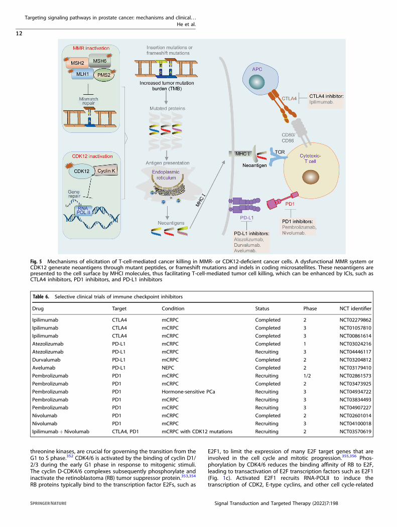
TARGETING IMMUNE CHECKPOINTSMMR defect and immunotherapy responseTumors with defects in MMR genes or microsatellite instability(MSI) often have an enhanced antitumor immune response,displaying a higher density of tumor-infiltrating lymphocytes(TILs).340,341 This phenomenon is attributed to high rates ofmutations and increased levels of neoantigens in MMR-deficienttumors, which occur through different mechanisms, includingmutant peptides, frameshift mutations, and indels in codingmicrosatellites.342,343 These neoantigens are presented on the cellsurface by MHCI molecules, facilitating T-cell-mediated tumor cellkilling (Fig. 5). In mammalian cells, MutL homolog 1 (MLH1), MutShomologue 2 (MSH2), MutS homologue 6 (MSH6) and PMS1homologue 2 (PMS2) are the main proteins of the DNA MMRsystem, which are critical for recognizing and repairing erroneousinsertion, deletion, and misincorporation of bases during DNAreplication or DNA recombination.344 Approximately 3–5% of PCacases are associated with deficiency of MMR genes, such as MSH2,
MSH6, PMS2, and MLH1, resulting in hypermutation and MSI.345
Mutation of MMR genes in PCa is highly associated with increasedexpression of neoantigens and accumulation of TILs.346
Immune checkpoint inhibitors (ICIs)Several clinical studies have evaluated the efficacy of ICIs,including PD-L1 (nivolumab, atezolizumab, durvalumab, andavelumab), PD1 (pembrolizumab and nivolumab), and CTLA4(ipilimumab) antibodies (Fig. 5 and Table 6). Early studies showedthat ICIs exhibit limited anticancer activity.347,348 It is currentlyaccepted that the selection of patients with deficiency in MMRgenes is important, because this subset of patients is potentiallyresponsive to ICIs.345 The anti-PD1 antibody pembrolizumab isapproved by the FDA to treat cancers, including PCa with MMRmutations or MSI.349 The responses of ICIs in MMR mutations orMSI PCa are not universal. For example, ~54% (6 of 11) CRPCpatients with MMR mutations or MSI-high tumors achieved a 50%reduction in PSA level after treatment with pembrolizumab.345 Itremains unclear why some patients with MMR loss/MSI-high donot respond to ICI therapy in PCa. Despite these disappointingresults, the interest in combining ICIs with other therapies remainshigh. Promising results were observed from a phase 1/2 clinicaltrial of pembrolizumab plus docetaxel, AR inhibitors, or PARPinhibitors in patients with mCRPC.350 Moreover, several phase 3clinical trials to evaluate the efficacy of pembrolizumab combinedwith docetaxel, enzalutamide, and olaparib are ongoing (Table 6).Notably, up to 10% of mCRPC patients present with CDK12
aberration (Fig. 1a), which is associated with the response toICIs.351 CDK12, which forms a complex with cyclin K, is critical forDNA repair during gene translation (Fig. 5). Inactivation of CDK12leads to focal tandem duplications that increase gene fusions ormutations, thus enhancing neoantigen production and the tumorimmune response (Fig. 5).351 Two of four mCRPC patients withCDK12 mutations had obvious PSA responses after administrationof a PD1 inhibitor.351 A phase 2 clinical trial to evaluate the efficacyof ipilimumab and nivolumab for CDK12-mutated mCRPC isongoing (Table 6).
TARGETING THE CELL CYCLECDK4/6 and cell cycleHyperproliferation is a hallmark of cancer development. The cellcycle can be divided into four ordered phases: G1 (gap 1), S (DNAsynthesis), G2 (gap 2), and M (mitosis), which are preciselycontrolled by molecules such as CDKs. The key regulatorycheckpoints in the G1 and G2 phases determine whether cellsenter the S phase and mitosis. CDK4 and CDK6, two serine/
Table 5. Selective clinical trials of PARP inhibitors
Drug Target Condition Status Phase NCT identifier
Olaparib (AZD2281) PARPs PCa Completed 1 NCT02324998
Olaparib (AZD2281) PARPs mCRPC Not yet recruiting 3 NCT03732820
Olaparib (AZD2281) PARPs mCRPC Not yet recruiting 3 NCT02987543
Olaparib (AZD2281) PARPs mCRPC Not yet recruiting 3 NCT03834519
Rucaparib (AG014699) PARPs HR deficient mCRPC Completed 2 NCT02952534
Rucaparib (AG014699) PARPs mCRPC Not yet recruiting 3 NCT02975934
Veliparib (ABT-888) PARPs mCRPC Completed 2 NCT01576172
Talazoparib (BMN 673) PARPs HR deficient mCRPC Not yet recruiting 2 NCT03148795
Talazoparib (BMN 673) PARPs mCRPC Recruiting 3 NCT03395197
Niraparib (MK-4827) PARP1/2 mCRPC Completed 1 NCT02924766
Niraparib (MK-4827) PARP1/2 mCRPC Recruiting 3 NCT04497844
Pamiparib (BGB-290) PARP1/2 mCRPC Terminated 2 NCT03712930
Pamiparib (BGB-290) PARP1/2 mCRPC Not yet recruiting 1 NCT03150810
Targeting signaling pathways in prostate cancer: mechanisms and clinical. . .He et al.
11
Signal Transduction and Targeted Therapy (2022) 7:198
threonine kinases, are crucial for governing the transition from theG1 to S phase.352 CDK4/6 is activated by the binding of cyclin D1/2/3 during the early G1 phase in response to mitogenic stimuli.The cyclin D-CDK4/6 complexes subsequently phosphorylate andinactivate the retinoblastoma (RB) tumor suppressor protein.353,354
RB proteins typically bind to the transcription factor E2Fs, such as
E2F1, to limit the expression of many E2F target genes that areinvolved in the cell cycle and mitotic progression.355,356 Phos-phorylation by CDK4/6 reduces the binding affinity of RB to E2F,leading to transactivation of E2F transcription factors such as E2F1(Fig. 1c). Activated E2F1 recruits RNA-POLII to induce thetranscription of CDK2, E-type cyclins, and other cell cycle-related
Fig. 5 Mechanisms of elicitation of T-cell-mediated cancer killing in MMR- or CDK12-deficient cancer cells. A dysfunctional MMR system orCDK12 generate neoantigens through mutant peptides, or frameshift mutations and indels in coding microsatellites. These neoantigens arepresented to the cell surface by MHCI molecules, thus facilitating T-cell-mediated tumor cell killing, which can be enhanced by ICIs, such asCTLA4 inhibitors, PD1 inhibitors, and PD-L1 inhibitors
Table 6. Selective clinical trials of immune checkpoint inhibitors
Drug Target Condition Status Phase NCT identifier
Ipilimumab CTLA4 mCRPC Completed 2 NCT02279862
Ipilimumab CTLA4 mCRPC Completed 3 NCT01057810
Ipilimumab CTLA4 mCRPC Completed 3 NCT00861614
Atezolizumab PD-L1 mCRPC Completed 1 NCT03024216
Atezolizumab PD-L1 mCRPC Recruiting 3 NCT04446117
Durvalumab PD-L1 mCRPC Completed 2 NCT03204812
Avelumab PD-L1 NEPC Completed 2 NCT03179410
Pembrolizumab PD1 mCRPC Recruiting 1/2 NCT02861573
Pembrolizumab PD1 mCRPC Completed 2 NCT03473925
Pembrolizumab PD1 Hormone-sensitive PCa Recruiting 3 NCT04934722
Pembrolizumab PD1 mCRPC Recruiting 3 NCT03834493
Pembrolizumab PD1 mCRPC Recruiting 3 NCT04907227
Nivolumab PD1 mCRPC Completed 2 NCT02601014
Nivolumab PD1 mCRPC Recruiting 3 NCT04100018
Ipilimumab+Nivolumab CTLA4, PD1 mCRPC with CDK12 mutations Recruiting 2 NCT03570619
Targeting signaling pathways in prostate cancer: mechanisms and clinical. . .He et al.
12
Signal Transduction and Targeted Therapy (2022) 7:198

proteins that further phosphorylate RB and promote the G1-to-S-phase cell cycle transition.357,358 CDK4/6 also phosphorylates othersubstrates and plays an important role in differentiation andmetabolism.359,360
CDK4/6 inhibitorsCurrently, three new CDK4/6 inhibitors, palbociclib (PD0332991),ribociclib (LEE011), and abemaciclib (LY2835219), have enteredearly phase trials for PCa (Table 7). A recent phase 2 clinic studyevaluated the efficacy of ADT (including bicalutamide, goserelin,and leuprolide) plus palbociclib in patients with RB-positivemetastatic hormone-sensitive PCa. The primary PSA endpoint wasmet in 80% of patients in both the ADT alone and ADT pluspalbociclib groups (16/20 versus 32/40; p= 0.87), while 1-yearbiochemical progression-free survival (PFS) was 69% in the ADTalone group and 74% in the ADT plus palbociclib group.361
Although these important clinical data are still not sufficient, thisstudy suggests that co-targeting of AR signaling and the cell cycle ispossible. Furthermore, abiraterone plus abemaciclib was evaluatedin a phase 2/3 trial, and ribociclib plus enzalutamide or docetaxelare under investigation in different trials for the treatment ofmCRPC (Table 7). PCa has shown limited response to immunother-apy because of its cold tumor environment.348 Notably, CDK4/6inhibitors have been shown to increase the tumor immuneresponse and TILs,362,363 which supports the potential synergisticeffects of CDK4/6 inhibitors and ICIs. Additional studies are trying toidentify the population of appropriate patients and synergisticcombinations that would make these agents more efficacious.
p53 and the targeting approachesThe tumor suppressor p53 is widely known as the “genomeguardian.” Activated p53 binds to a specific DNA sequence as atetramer to promote gene expression (such as CDKN1A, BAX,PUMA, and NOXA), thus inducing cell apoptosis and cell-cyclearrest.364 The TP53 gene, which encodes the p53 protein, isfrequently mutated in PCa, especially in neuroendocrine-likemCRPC. The combined alteration of RB and TP53 occurs in 74%of neuroendocrine-like mCRPC.365 p53 mutations are predomi-nantly distributed throughout the DBD, and these p53 mutationseither lose their DNA-binding ability or form a heterodimercomplex with wild-type p53 to attenuate wild-type p53 functions,thus disrupting the tumor-suppressive functions of p53.366 More-over, many mutant p53 proteins acquire gain-of-function activ-ities, which enable them to deactivate the other p53 familymembers, specifically p73 and p63.367 Under normal conditions,p53 has a half-life of less than 20min owing to the feedbackregulation of proteasome-mediated p53 degradation by E3ubiquitin-protein ligase MDM2.368
p53 alteration fosters more lethal PCa; thus, targeting p53 is anattractive therapeutic strategy for aggressive PCa. Approaches fortargeting p53 can be summarized as follows: first, compounds
such as idasanutlin (RG7388) and RG7112 were developed toprevent degradation of wild-type p53 by blocking p53-MDM2interactions,369,370 thus maintaining their ability to suppress tumorformation. Although no clinical studies exist for PCa, multiple p53-MDM2 antagonists are undergoing clinical trials; of these,idasanutlin is the most advanced and is under testing in a phase3 clinical trial in patients with refractory acute myeloid leuke-mia.371 Second, pharmacological reactivation of mutant p53 usessmall molecules like APR-246, COTI-2, and arsenic trioxide (Table 7),which bind to mutant p53 and convert the protein to a p53 wild-type-like conformation, thus restoring wild-type DNA-bindingproperties.372–374 Based on the results of a phase 1b/2 trial formyelodysplastic syndrome,375 the FDA granted Fast Trackdesignation to APR-246 for the treatment of myelodysplasticsyndrome in patients with a p53 mutation.376 APR-246 was testedin PCa, and showed a favorable pharmacokinetic profile in a phase1 trial (Table 7).377 Third, mutant p53 neoantigens can elicitintratumoral T-cell responses;378 therefore, p53 neoantigens areconsidered to be promising targets. For example, a recent studygenerated a T-cell-based therapy that links T cells to cancer cellsusing a novel antibody that specifically binds to the p53R175H
peptide-MHC complex, lysing cancer cells depending on thepresence of the neoantigen.379 Clinical studies targeting mutantp53 neoantigens in PCa are rare. More clinical studies on p53-targeted agents in PCa are warranted, and it is projected that atleast several of these molecules will prove effective in the future.
TARGETING THE PI3K/AKT/MTOR SIGNALING AXISPTEN/PI3K/AKT/mTOR signalingInactivation of PTEN (phosphatase and tensin homologue) bydeletion or mutation has been identified in ~20% of primary PCaand approximately 35% of CPRC cases (Fig. 1a).380 PTEN, a dual-specificity phosphatase, converts phosphatidylinositol-3,4,5-tri-sphosphate (PIP3) into phosphatidylinositol 4,5-bisphosphate(PIP2),
381 causing PTEN to function as a direct antagonist of theactivity of class I PI3K, which converts PIP2 to PIP3. As a result,PTEN loss leads to aberrant accumulation of PIP3 on cellmembranes, which leads to the recruitment of PDK1 tophosphorylate its substrate AKT. Phosphorylated AKT subse-quently regulates several downstream signaling cascades, includ-ing mTOR, which is crucial for protein synthesis, autophagy,cellular proliferation, and metabolism.382 PTEN acts as a gate-keeper of the PI3K/AKT/mTOR pathway. Therefore, PTEN deletionsor mutations are strongly associated with activation of PI3K/AKT/mTOR signaling and poor prognosis in advanced PCa.380
PI3K, ATK, mTOR, and PIM (proviral-integration site for Moloney-murine leukemia virus) inhibitorsPI3K, a plasma membrane-associated protein kinase, is formed bytwo functional subunits: a catalytic subunit (p110α, p110β, or
Table 7. Selective clinical trials of CDK4/6 inhibitors or p53-targeted agents
Drug Target Condition Status Phase NCT identifier
Palbociclib (PD0332991) CDK4/6 RB-positive metastatic PCa Completed 2 NCT02059213
Palbociclib (PD0332991) CDK4/6 mCRPC Not yet recruiting 2 NCT02905318
Ribociclib (LEE011) CDK4/6 mCRPC Completed 2 NCT02494921
Ribociclib (LEE011) CDK4/6 RB-positive metastatic PCa Recruiting 2 NCT02555189
Abemaciclib (LY2835219) CDK4/6 mCRPC Recruiting 2/3 NCT03706365
Abemaciclib (LY2835219) CDK4/6 Locally advanced PCa Recruiting 2 NCT04298983
Abemaciclib (LY2835219) CDK4/6 mCRPC Not yet recruiting 2 NCT04408924
APR-246 Mutant p53 Refractory PCa Completed 1 NCT00900614
Arsenic trioxide Mutant p53 Stage IV PCa Completed 2 NCT00004149
Targeting signaling pathways in prostate cancer: mechanisms and clinical. . .He et al.
13
Signal Transduction and Targeted Therapy (2022) 7:198

p110δ) and a regulatory subunit (p85α, p55α, p50α, p85β, or p55γisoform).383 The catalytic subunit p110β is believed to be themost relevant isoform for PCa progression.384,385 PI3K inhibitors,such as BKM120 and PX866, target the catalytic subunits of allthree isoforms (p110α, p110β, and p110δ). BKM120 showed apartial response in a phase 1 study.386 PX866, a derivative ofwortmannin, was well tolerated in patients with recurrentmCRPC.387,388 However, monotherapy with PI3K inhibitors islimited in the clinic.AKT, a serine/threonine protein kinase, is the main downstream
effector of PI3K, and is fully activated when both Thr308 andSer473 sites are phosphorylated.389 Activated AKT phosphorylatesseveral targets, such as mTOR, GSK3, FOXO, and AMPK, which areinvolved in multiple cellular processes.390,391 Thus far, theAKT inhibitors that have reached the clinical phase includeallosteric inhibitors (such as perifosine and MK-2206) and ATP-competitive inhibitors (such as capivasertib, ipatasertib, anduprosertib). Of note, the combination of capivasertib withdocetaxel resulted in a greater than 50% reduction in PSA levelsin 70% of men with mCRPC in a phase 1 study.392 Ipatasertibprolonged PSA progression-free interval and overall survivalcompared to the placebo group in a phase 2 trial.49 A phase 3trial to evaluate the efficacy of abiraterone plus ipatasertib for thetreatment of mCRPC is ongoing (Table 8).The serine/threonine protein kinase mTOR, the major down-
stream effector of AKT signaling, interacts with different proteinsand forms two distinct complexes, mTORC1 and mTORC2.393
Several types of mTOR inhibitors exist, including mTORC1inhibitors (such as rapamycin, everolimus, and temsirolimus),mTORC1/2 dual inhibitors (such as sapanisertib and vistusertib),and dual PI3K-mTORC1/2 inhibitors (such as apitolisib andBEZ235). Clinical trials using single mTORC1 inhibitors showedpredictable toxicity with no favorable clinical responses inmCRPC.394,395 Sapanisertib was previously tested in a phase2 study in advanced CRPC but showed limited clinical efficacy.47
Vistusertib was tested in men with high-risk PCa and wasadministered prior to radical prostatectomy in a phase 1 trial(Table 8). Currently, apitolisib plus abiraterone is being tested forCRPC in phase 1/2 clinical trials (Table 8). A novel mTOR and DNA-
PK (DNA-dependent protein kinase) dual inhibitor, CC-155, wasevaluated in a phase 1 study (Table 8).PIM kinases have been found to sustain the PI3K/AKT/mTOR
pathway.396,397 Increased expression of PIM family members hasbeen detected in PCa, and PIM confers resistance not only to PI3K/AKT inhibitors, but also to chemotherapy and radiotherapy.398
Therefore, co-targeting PIM and PI3K/AKT/mTOR could offersuperior clinical outcomes relative to targeting either of thesealone. The combination of the PIM inhibitor AZD1208 and thePI3K/mTOR inhibitor BEZ235 (dactolisib) has been investigated inclinical trials.399 A novel and highly efficient triple PIM/PI3K/mTORinhibitor AUM302 elicited a superior functional outcome com-pared to the effects of the combination of AZD1208 and BEZ235;these results may help reduce treatment toxicity in future trials.399
Overall, the clinical application of PI3K/AKT/mTOR inhibitors is stilllimited in PCa, and further studies that can identify newbiomarkers for patient selection or improve co-targeting strategiesare still required to enhance their therapeutic effects.
TARGETING EPIGENETIC MARKSEpigenetic modificationsEpigenetic traits are heritable phenotypes attributable to changesin chromosomes or DNA modifications without alterations in theDNA sequence.400 In addition to genomic changes, epigeneticalterations (such as histone modifications and DNA methylation)have been reported to be associated with PCa progression.401–403
Epigenetic modifications, including acetylation, methylation,ubiquitination, and phosphorylation, play critical roles in transcrip-tion, DNA repair, and replication.404 Epigenetic regulation is adynamic and reversible process that adds epigenetic marks ontoeither histones or DNA by epigenetic writers, recognizes or recruitsepigenetic marks by epigenetic readers, and removes epigeneticmarks by epigenetic erasers (Fig. 6). Aberrant histone modifica-tions may upregulate oncogenes or reduce the expression oftumor suppressor genes. Importantly, histone methylation/acet-ylation and DNA methylation play a central role in controllinggene expression, thus promoting the progression and metastasisof PCa.405,406
Table 8. Selective clinical trials of PI3K, AKT and mTOR inhibitors
Drug Target Condition Status Phase NCT identifier
Buparlisib (BKM120) PI3K CRPC Completed 1 NCT01634061
Buparlisib (BKM120) PI3K CRPC Terminated 2 NCT01385293
Dactolisib (BEZ235) PI3K mCRPC Terminated 1/2 NCT01717898
Samotolisib (LY3023414) PI3K CRPC Completed 2 NCT02407054
AZD8186 PI3K CRPC Completed 1 NCT01884285
GSK2636771 PI3K CRPC Completed 1 NCT02215096
Perifosine (KRX-0401) AKT mCRPC Completed 2 NCT00060437
MK2206 AKT Advanced cancers Completed 1 NCT01295632
Ipatasertib (GDC-0068) AKT Locally advanced PCa Recruiting 1/2 NCT04737109
Ipatasertib (GDC-0068) AKT mCRPC Not yet recruiting 3 NCT03072238
Capivasertib (AZD5363) AKT CRPC Completed 1 NCT04087174
Sirolimus mTOR Locally advanced PCa Completed 1/2 NCT00311623
Ridaforolimus (MK8669) mTOR mCRPC Completed 2 NCT00777959
Temsirolimus (CCI-779) mTOR CRPC Completed 2 NCT00919035
Everolimus (RAD001) mTOR CRPC Completed 2 NCT00814788
Sapanisertib (MLN0128) mTOR mCRPC Completed 2 NCT02091531
Vistusertib (AZD2014) mTOR PCa Completed 1 NCT02064608
Apitolisib (GDC-0980) PI3K, mTOR CRPC Not yet recruiting 1/2 NCT01485861
CC-115 mTOR, DNA-PK CRPC Not yet recruiting 1 NCT02833883
Targeting signaling pathways in prostate cancer: mechanisms and clinical. . .He et al.
14
Signal Transduction and Targeted Therapy (2022) 7:198
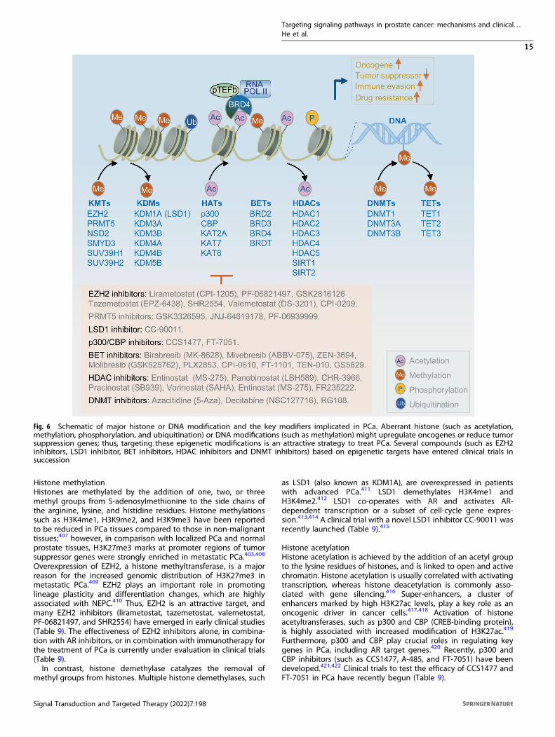
Histone methylationHistones are methylated by the addition of one, two, or threemethyl groups from S-adenosylmethionine to the side chains ofthe arginine, lysine, and histidine residues. Histone methylationssuch as H3K4me1, H3K9me2, and H3K9me3 have been reportedto be reduced in PCa tissues compared to those in non-malignanttissues;407 however, in comparison with localized PCa and normalprostate tissues, H3K27me3 marks at promoter regions of tumorsuppressor genes were strongly enriched in metastatic PCa.403,408
Overexpression of EZH2, a histone methyltransferase, is a majorreason for the increased genomic distribution of H3K27me3 inmetastatic PCa.409 EZH2 plays an important role in promotinglineage plasticity and differentiation changes, which are highlyassociated with NEPC.410 Thus, EZH2 is an attractive target, andmany EZH2 inhibitors (lirametostat, tazemetostat, valemetostat,PF-06821497, and SHR2554) have emerged in early clinical studies(Table 9). The effectiveness of EZH2 inhibitors alone, in combina-tion with AR inhibitors, or in combination with immunotherapy forthe treatment of PCa is currently under evaluation in clinical trials(Table 9).In contrast, histone demethylase catalyzes the removal of
methyl groups from histones. Multiple histone demethylases, such
as LSD1 (also known as KDM1A), are overexpressed in patientswith advanced PCa.411 LSD1 demethylates H3K4me1 andH3K4me2.412 LSD1 co-operates with AR and activates AR-dependent transcription or a subset of cell-cycle gene expres-sion.413,414 A clinical trial with a novel LSD1 inhibitor CC-90011 wasrecently launched (Table 9).415
Histone acetylationHistone acetylation is achieved by the addition of an acetyl groupto the lysine residues of histones, and is linked to open and activechromatin. Histone acetylation is usually correlated with activatingtranscription, whereas histone deacetylation is commonly asso-ciated with gene silencing.416 Super-enhancers, a cluster ofenhancers marked by high H3K27ac levels, play a key role as anoncogenic driver in cancer cells.417,418 Activation of histoneacetyltransferases, such as p300 and CBP (CREB-binding protein),is highly associated with increased modification of H3K27ac.419
Furthermore, p300 and CBP play crucial roles in regulating keygenes in PCa, including AR target genes.420 Recently, p300 andCBP inhibitors (such as CCS1477, A-485, and FT-7051) have beendeveloped.421,422 Clinical trials to test the efficacy of CCS1477 andFT-7051 in PCa have recently begun (Table 9).
Fig. 6 Schematic of major histone or DNA modification and the key modifiers implicated in PCa. Aberrant histone (such as acetylation,methylation, phosphorylation, and ubiquitination) or DNA modifications (such as methylation) might upregulate oncogenes or reduce tumorsuppression genes; thus, targeting these epigenetic modifications is an attractive strategy to treat PCa. Several compounds (such as EZH2inhibitors, LSD1 inhibitor, BET inhibitors, HDAC inhibitors and DNMT inhibitors) based on epigenetic targets have entered clinical trials insuccession
Targeting signaling pathways in prostate cancer: mechanisms and clinical. . .He et al.
15
Signal Transduction and Targeted Therapy (2022) 7:198

In contrast, HDACs can remove acetylation of histones. Eighteendifferent types of HDACs have been identified in humans,423 andoverexpression of HDACs occurs in different malignancies,including PCa.424 HDAC1 and HDAC2 expression is positivelycorrelated with higher Gleason scores of PCa, while theexpressions of HDAC1, HDAC2, and HDAC3 are positivelyassociated with the proliferative marker Ki67.425 HDAC inhibitorsare potential therapeutic options because the expression ofHDACs is associated with poor clinical outcomes.425 There are fiveclasses of HDAC inhibitors, including hydroxamic acids, cyclictetrapeptides, short chain carboxylic acids, benzamides, and keto-derivatives.426 Several HDAC inhibitors, including vorinostat,pracinostat, panobinostat, and romidepsin, have been tested inphase 2 clinical trials for PCa (Table 9); however, most patientsexhibited either toxicity from these agents or disease progres-sion.427 Clinical trials involving HDAC inhibitors have not achievedsignificant success because of poor oral bioavailability, non-selectivity of the drugs, or other mechanisms that remain to beexplored.427
BET proteinAcetylated lysines are recognized by a class of proteins containingbromodomains, such as the BET proteins BRD2, BRD3, BRD4, andBRDT.428 Acetylated lysine residues in histones can be bound byBET proteins via BD1 and BD2 bromodomains, which is the keystep in regulating transcription.428 Importantly, the expression ofBRD4 is significantly associated with poor clinical outcomes.429,430
BET proteins, such as BRD4, are tightly linked to AR signalingactivation and drug resistance in SPOP mutant PCa.431–433
Numerous BET inhibitors, including pan-BET bromodomaininhibitors (such as JQ1, I-BET151, birabresib, mivebresib, andZEN-3694) and selective inhibitors (such as ABBV-744, molibresib,and PLX2853), have been demonstrated to have antitumor effectsin preclinical models. Birabresib (MK-8628) and mivebresib (ABBV-075) were tested in patients with solid tumors, including CRPC, butneither birabresib nor mivebresib demonstrated significantantitumor activity in CRPC patients (Table 9).59,61 However, a
clinical trial at phase 1/2 demonstrated that enzalutamide plusZEN-3694 prolonged the PFS in a subset of patients with mCRPCresistant to enzalutamide and/or abiraterone.62 A new study foundmolibresib (GSK525762) was well tolerated in patients.434 Recently,phase 1 or phase 2 clinical trials of ZEN-3694 and PLX2853, incombination with AR inhibitors (enzalutamide or abiraterone)have been launched in CRPC patients (Table 9). Ongoing clinicalstudies of BET inhibitors are needed to demonstrate their safetyprofiles and the role of pharmacodynamic regulation of ARsignaling in patients.
DNA methylationDNA methylation is achieved when a methyl group is added to theC5 position of cytosine residues in the CpG dinucleotides, and islinked to gene silencing.435 DNMT enzymes catalyze 5-methylcytosine (5mC) in DNA, whereas these marks can be removed bythe DNA demethylases TET (ten-eleven translocation) family.436
Approximately 60% of all promoters colocalize with CpGislands437; therefore, aberrant DNA hypermethylation at CpGislands can lead to gene silencing, such as inactivation of tumorsuppressor genes.88,438 A previous studied showed that 22% oftumors were associated with hypermethylation in PCa.439 DNMTinhibitors azacytidine (5-Aza) and decitabine have been devel-oped to target aberrant DNA hypermethylation. Azacitidine anddecitabine (NSC127716) were clinically evaluated for PCa (Table 9).A phase 1/2 study of azacitidine in combination with docetaxel inmCRPC demonstrated a PSA response in 52% (10/19) of patients,without exhibiting dose-limiting toxicity.57
TARGETING WNT SIGNALINGWNT/β-catenin signalingThe canonical WNT/β-catenin pathway is activated in late-stagePCa, and promotes tumor cell growth and drug resistance inPCa.440 The binding of WNT ligands to their receptors in cellsurface activates signaling pathways that regulate cell differentia-tion and proliferation.441 In the absence of WNT ligands,
Table 9. Selective clinical trials of epigenetic inhibitors
Drug Target Condition Status Phase NCT identifier
Lirametostat (CPI-1205) EZH2 mCRPC Not yet recruiting 1/2 NCT03480646
PF-06821497 EZH2 CRPC Recruiting 1 NCT03460977
Tazemetostat (EPZ-6438) EZH2 mCRPC Recruiting 1 NCT04846478
Tazemetostat (EPZ-6438) EZH2 mCRPC Recruiting 1/2 NCT04179864
SHR2554 EZH2 CRPC Terminated 1/2 NCT03741712
Valemetostat (DS-3201) EZH2 mCRPC Recruiting 1 NCT04388852
CC-90011 LSD1 mCRPC Recruiting 1 NCT04628988
CCS1477 p300/CBP mCRPC Recruiting 1/2 NCT03568656
FT-7051 p300/CBP mCRPC Recruiting 1 NCT04575766
Birabresib (MK-8628) BET bromodomain CRPC Completed 1 NCT02259114
Mivebresib (ABBV-075) BET bromodomain PCa Completed 1 NCT02391480
Molibresib (GSK525762) BET bromodomain CRPC Completed 1 NCT03150056
ZEN-3694 BET bromodomain mCRPC Completed 1/2 NCT02711956
ZEN-3694 BET bromodomain mCRPC Recruiting 2 NCT04471974
PLX2853 BET bromodomain mCRPC Not yet recruiting 1/2 NCT04556617
Entinostat (MS-275) HDACs CRPC Terminated 1 NCT03829930
Panobinostat (LBH589) HDACs CRPC Completed 1 NCT00878436
Pracinostat (SB939) HDACs CRPC Completed 2 NCT01075308
Vorinostat (SAHA) HDACs Metastatic PCa Completed 2 NCT00330161
Azacitidine (5-Aza) DNMT PCa Completed 2 NCT00384839
Decitabine (NSC127716) DNMT PCa Withdrawn 1/2 NCT03709550
Targeting signaling pathways in prostate cancer: mechanisms and clinical. . .He et al.
16
Signal Transduction and Targeted Therapy (2022) 7:198

cytoplasmic β-catenin is rapidly degraded by a destructioncomplex, whose components contain adenomatous polyposis coliprotein (APC), AXIN, casein kinase 1 (CK1), β-transducin-repeat-containing protein (β-TrCP), and glycogen synthase kinase 3 (GSK3)(Fig. 7).65,441,442 When WNT ligands bind to frizzled (FZD) receptorsand co-receptors LRP5/6, LRP5/6 are phosphorylated by CK1 andGSK3, and then the signal is transduced to activate the cytoplasmicphosphoprotein dishevelled (DVL). Phosphorylated DVL recruitsthe destruction complex to the plasma membrane. This inhibitsGSK3 and prevents phosphorylation of β-catenin, thereby resultingin the stabilization and accumulation of β-catenin proteins.Sequentially, β-catenin proteins translocate into nucleus, form acomplex with T-cell factors (TCFs)/lymphoid enhancer-bindingfactor 1 (LEF1), recruit transcription factors and co-activators, suchas the CBP/p300, and activate the transcription of downstreamtarget genes, including ABCB1, MYC, MYCN, NEUROG1, NEUROD1,SOX2, SUZ12, TWIST, and YAP (Fig. 7).65,441–443 Importantly,activating mutations of the WNT/β-catenin pathway genes wereenriched in metastatic PCa (19%) compared to those in primaryPCa (6%).444 An increasing number of studies have indicated thatthe activation of WNT/β-catenin signaling is highly linked to cellproliferation, invasion, bone metastasis, drug resistance, andneuroendocrine differentiation in the late stage of PCa.445–455
WNT signaling inhibitorsNumerous agents that target different components of the WNTpathway have been developed and tested in clinical trials (Fig. 7),although only a few have been associated with PCa. Severalstrategies can prevent the activation of WNT signaling. First,targeting WNT ligands and their receptors with monoclonalantibodies and small molecules is an attractive therapeuticstrategy. The WNT ligand WNT-5A was targeted using a WNT-5A-mimicking peptide named Foxy-5.456 Foxy-5 was evaluated in aphase 1 clinical trial in patients with solid malignant tumorsincluding PCa (Table 10). An R-spondin-3 antibody (OMP-131R10)was tested in a phase 1 clinical trial for advanced solid tumors.65
The antibody OMP-18R5 (vantictumab) targets FZD1, FZD2, FZD5,FZD7, and FZD8.65 OTSA101 is a radiolabeled antibody againstFZD10.457 Pafricept (OMP-54F28) is a decoy WNT receptor that hasbeen tested in a phase 1a clinical trial and has demonstratedevidence of WNT pathway inhibition,458 but its development hasbeen terminated due to bone-related toxicity.459 Second, inhibi-tion of WNT secretion by targeting porcupine, a membrane-boundO-acetyltransferase, is another optional strategy. Porcupine isimportant for WNT palmitoylation, which is essential for thesecretion of WNT proteins.460 Several porcupine inhibitors, such asIWP-2, WNT-C59, LGK974 (WNT974), and ETC-159, also have been
Fig. 7 The WNT signaling pathway and targeting therapeutic strategies. WNT ligands bind to FZD and LRP5/6 receptors to phosphorylateDVL, and then phosphorylated DVL recruits the destruction complex to the plasma membrane. This inhibits GSK3 and preventsphosphorylation of β-catenin, resulting in the stabilization and accumulation of β-catenin proteins to form a complex with TCF/LEF in thenucleus, thereby activating the transcription of downstream target genes. There are several targeting strategies that prevent the activation ofWNT signaling, such as targeting WNT ligands and their receptors, inhibiting the WNT secretion by targeting porcupine, and disrupting theinteraction between CBP and β-catenin
Targeting signaling pathways in prostate cancer: mechanisms and clinical. . .He et al.
17
Signal Transduction and Targeted Therapy (2022) 7:198
developed65,461, and of these, LGK974 (discontinued) and ETC-159(phase 1) have entered trials in patients with advanced solidtumors.462 Third, the interaction between cofactor CBP andβ-catenin is critical for transcriptional activation of β-catenin.65
Thus, disruption of the interaction between CBP and β-cateninprovides a new strategy to prevent WNT signaling. ICG-001 and itsderivative PRI-724 were found to inhibit the binding of β-cateninto CBP.463 PRI-724, which has an acceptable toxicity profile, wastested in phase 2 clinical trials for metastatic colorectal cancer (butwas withdrawn owing to drug supply issues) and advancedmyeloid malignancies (completed).463
Although targeting WNT signaling is very attractive for treatinglate-stage PCa, WNT signaling inhibitors are in early stages ofdevelopment and application, and several limitations remain. First,WNT signaling is complicated because there are 19 WNT-secretedglycoproteins and more than 15 types of WNT receptors inhumans,464,465 which activate different downstream pathways.Second, variations in and balances between canonical andnoncanonical WNT signaling is elusive,466 making it even moredifficult to target the WNT pathway. Third, WNT signaling plays afundamental role in the homeostasis of the intestine, hair follicles,and hematopoietic system;464 thus, blocking the WNT pathwaycould cause systemic toxicity. Prospective studies include: (I) thecombination of WNT inhibitors with other cancer drugs orimmunotherapies to limit toxicities or improve therapeuticefficacy, (II) the classification of disease subtypes to distinguishmethods of WNT activation among different stages of disease, and(III) the expansion of models to more clearly understandmechanism of action between the canonical and noncanonicalWNT pathways and thus, discover novel therapeutic approaches.
TARGETING OTHER PATHWAYSVEGFVEGF is a prominent factor involved in angiogenesis and is highlyassociated with tumor growth, including in PCa.66,467 Angiogen-esis is essential for tumor growth, because the newly formedblood vessels are important to sustain adequate energy andoxygen.468,469 The binding of VEGFs (VEGFA, VEGFB, VEGFC, andVEGFD) to cell surface receptors (VEGFR1, VEGFR2, VEGFR3)activates signaling pathways that play important roles in cellgrowth and motility in PCa.468 Of note, VEGFA is the foundingmember of the vascular permeability factor, and is frequentlyoverexpressed in PCa.470,471 Overexpression of VEGFA in PCa is
associated with angiogenesis, recurrence, and advanced diseasestages among patients.472,473 Importantly, bevacizumab, a huma-nized monoclonal antibody against VEGFA, is used to treat severaldifferent cancers.474 A phase 3 clinical study in mCRPC patientsdemonstrated that the combination of bevacizumab and doc-etaxel did not result in a significant increase in median overallsurvival versus docetaxel alone (22.6 versus 21.5 months; p=0.181); however, improvements were found in median PFS (9.9versus 7.5 months; p < 0.001) and major PSA response (69.5%versus 57.9%; P < 0.001).475
ETARActivation of ETAR by endothelin-1 is involved in tumorprogression through angiogenesis, invasion, apoptosis, and theeffect of the bone microenvironment.476,477 Activated ETARsignaling promotes osteoblast proliferation and new boneformation, which is highly associated with bone metastasis inPCa.478,479 Thus, the ETAR inhibitors zibotentan (ZD4054) andatrasentan have been tested in men with mCRPC. In a phase 2trial, zibotentan prolonged overall survival from 17.3 to24.5 months in patients with mCRPC,480 but a phase 3 trial didnot result in a statistically significant improvement in overallsurvival in the patients.481 Similarly, atrasentan delayed PFS andPSA progression in a phase 2 trial,482 but a phase 3 trialdemonstrated that atrasentan did not significantly reduce thedisease progression time in CRPC patients.483 However, a phase 3trial in mCRPC demonstrated that patients with the highest levelof bone metabolism markers (such as pyridinoline and alkalinephosphatase) obtained a survival benefit from atrasentancompared to that from placebo (13 versus 5 months; p= 0.005).484
TGFβTGF-β is a cytokine that regulates many cellular functions, such ascell differentiation and migration. Binding of TGFβ to TGFβR2(TGFβ receptor 2) phosphorylates and activates TGFβR1 (TGFβreceptor 1). Subsequently, activated TGFβR1 phosphorylatesSMAD2 and SMAD3 proteins, leading to SMAD2/SMAD3 com-plexes with SMAD4. Next, these complexes are translocated intothe nucleus and stimulate target gene expression.485,486 IncreasedTGFβ or its target genes in PCa are associated with a moreaggressive disease, metastasis, and poor prognosis.487–489. TGFβalso plays an important role in the context of the bonemicroenvironment and supports the progression of bone metas-tasis in PCa.490–492 Furthermore, TGFβ facilitates tumor growth
Table 10. Selective clinical trials of other signaling pathway inhibitors
Drug Target Condition Status Phase NCT identifier
FOXY-5 WNT-5A receptors PCa Completed 1 NCT02020291
Bevacizumab VEGF-A PCa Completed 3 NCT00110214
Zibotentan (ZD4054) ETAR mCRPC Completed 3 NCT00554229
Erdafitinib (JNJ-42756493) FGFR DN-PCa Suspended 2 NCT03999515
Cetuximab EGFR mCRPC Completed 2 NCT00728663
Gefitinib (ZD1839) EGFR PCa Completed 2 NCT00265070
Dovitinib (TKI258) FGFR, VEGFR, PDGFR CRPC Completed 2 NCT01741116
Sunitinib (SU11248) VEGFR, PDGFR mCRPC Terminated 3 NCT00676650
Cabozantinib (XL184) VEGFR, c-MET, c-KIT mCRPC Completed 3 NCT01605227
Masitinib (AB1010) KIT, PDGFR, FGFR mCRPC Completed 3 NCT03761225
Galunisertib (LY2157299) TGFβR1 mCRPC Recruiting 2 NCT02452008
M7824 TGFβ, PD-L1 Metastatic PCa Recruiting 1/2 NCT04633252
Dasatinib (BMS-354825) SRC, c-KIT CRPC Completed 3 NCT00744497
Saracatinib (AZD0530) SRC CRPC Completed 2 NCT00513071
Trametinib (GSK1120212) MEK mCRPC Not yet recruiting 2 NCT02881242
Targeting signaling pathways in prostate cancer: mechanisms and clinical. . .He et al.
18
Signal Transduction and Targeted Therapy (2022) 7:198

through its immunosuppressive function.493,494 To target TGFβsignaling, galunisertib (LY2157299), an oral small-molecule inhi-bitor of TGFβR1, has been developed.495 A clinical study ofgalunisertib plus enzalutamide in mCRPC has been initiated (Table10). Furthermore, a clinical trial of M7824,496 targeting both TGFβand PD-L1, also has been launched for the treatment of metastaticPCa (Table 10).
RTKs and nonreceptor tyrosine kinases (NRTKs)Both RTKs (such as FGFR, EGFR, PDGFR, and VEGFR) and NRTKs(such as SRC) are very important in the carcinogenesis andprogression of PCa, and are potential targets for the treatment ofPCa.497,498 Signal transductions through ligands binding with RTKsor stimulation of unique NRTKs lead to cross-phosphorylation ofspecific tyrosine residues, which activate downstream signalingsuch as PI3K/AKT, phospholipase C, and Janus tyrosine kinase.499,500
The signaling subsequently regulates the transcription of genesinvolved in proliferation, survival and differentiation.499
Fibroblast growth factor (FGF) receptors (FGFR1/4) and theirligands (FGF1/2/4/8/17) are overexpressed in PCa.501–504
Enhanced FGF signaling leads to tumor progression, angiogenesis,epithelial-to-mesenchymal transition (EMT), and upregulation ofAR.505–507 Moreover, FGF/FGFR activation is highly correlated withAR-null PCa and drug resistance.508 Therefore, inhibition of theFGF axis may be a viable strategy for the treatment of PCa.Erdafitinib (JNJ-42756493),509 a small oral molecule that worksagainst all four FGFR family members, is being tested in a phase 2clinical study (Table 10) of AR-null and neuroendocrine-null PCa,termed double-negative prostate cancer (DNPC).EGFR mutation or overexpression leads to malignant progres-
sion in many cancer types.510 In PCa, increased expression of EGFRcorrelates with a high Gleason score and advanced stagedisease,511 and activation of EGFR promotes metastatic progres-sion and recurrence.512 EGFR inhibitors such as gefitinib (a small-molecule compound) and cetuximab (a monoclonal antibody)have been widely used in metastatic colorectal cancer and non-small cell lung cancer. In a phase 2 clinical trial, gefitinib did notexhibit any response to PSA or objective measurable disease inpatients with CRPC.510 However, in another trial, cetuximabresulted in an obvious PSA decline in many patients, andimproved PFS was found in patients with EGFR overexpression.513
The SRC signaling pathway has many effects on tumorigenesisand tumor progression.73,498 In PCa, SRC activity is highly increasedin bone metastasis.73,514–516 Encouraging results in preclinicalstudies of PCa led to several clinical studies of SRC inhibitors, suchas dasatinib (BMS-354825)517,518 and saracatinib (AZD0530).519,520
However, a phase 3 study of docetaxel plus dasatinib did notimprove the overall survival in patients with mCRPC, despite thefact that the PSA progression time was delayed.518 Similar resultswere found in the trials of abiraterone plus dasatinib.517,521 A phase2 study of saracatinib also failed to achieve satisfactory oncologicaloutcomes, while adverse events were observed, such as dehydra-tion, thrombocytopenia, and weakness.519
Moreover, multitargeted tyrosine kinase inhibitors, such asdovitinib (TKI258),522 sunitinib (SU11248),523,524 cabozantinib(XL184),525 and masitinib (AB1010)526, have entered clinical trialsfor PCa (Table 10). For example, dovitinib, an oral multitargeted RTKinhibitor, potently inhibits FGFR1/2/3, VEGFR1/2/3, PDGFR, andKIT.522 A phase 2 clinical study found that dovitinib showed a betterresponse in chemotherapy-naive patients and patients with highserum VEGFR2.522 Finally, cabozantinib,525 a small-molecule inhibitorof c-MET and VEGFR2, has been evaluated in two phase 3 clinicaltrials, which reported no survival benefit of cabozantinib, suggestingthat further rational development may be justified.527–529
MEKAberrant activation of mitogen-activated protein kinase (MAPK)signaling resulting from upstream activating mutations, such as
the rat sarcoma viral oncogene homolog (RAS), v-raf-1 murineleukemia viral oncogene homolog (RAF), and growth factors,converges with MEK. Increased activation of the RAS/RAF/MEK/extracellular signal-regulated kinase (ERK) pathway has beenassociated with poor prognosis and androgen independence inadvanced PCa.508,530,531 Increased expression of MAPK pathwaymembers and high levels of phosphorylated ERK1/2 wereobserved in mCRPCs.532–535 Pharmacological targeting of theMEK/ERK pathway may be a viable strategy for patients withmCRPC, and the MEK1/2 inhibitor trametinib (GSK1120212) iscurrently being tested in a phase 2 trial of patients with mCRPC(Table 10).
Modulation of alternative splicingThe vast majority of human genes (>95%) are alternatively splicedby the spliceosome, which enables genes to express spliceisoforms that often exhibit distinctive functions.75,536 Alternativesplicing affects genes (such as FGFR, ERG, VEGFA, and AR) that areclearly linked to the etiology of PCa, allowing us to develop noveltargeted therapies that modulate alternative splicing for thetreatment of PCa. For example, alternative splicing of FGFR2 leadsto the expression of FGFR2(IIIb) and FGFR2(IIIc) isoforms, whereasthe IIIb isoform is specific to epithelial cells, while the IIIc isoform isexclusively expressed in most mesenchymal cells. Therefore, thissplicing switch is a sensor of the EMT that results in tumor growthand/or metastasis.537 Using the FGFR2-based splicing reporter as areadout, a recent preclinical study identified compounds such asnemadipine-A (a T-type calcium channel inhibitor), NNC-55-0396dihydrochloride (a L-type Ca channel inhibitor), and naltrexonehydrochloride (an opioid antagonist) that change FGFR2 splicingand induce an epithelial phenotype in PCa cells.538 Moreover, theERG oncogene is fused with the TMPRSS2 promoter in 50% of PCacases, and the fusion most often occurs between the ERG exon 4and exons 1 or 2 of TMPRSS2.539 Interestingly, a noveloligonucleotide-based agent that targets ERG by inducingskipping of its constitutive exon 4 resulted in a reduction in ERGprotein levels and tumor growth in mice.540 Although theseagents have not entered clinical practice yet, splicing events are aplausible mechanism for treating PCa, and further research iswarranted.
FUTURE DIRECTIONS AND CONCLUSIONSThe arsenal of drugs available to treat advanced PCa hasexpanded significantly in recent years. Despite these advances,current treatment options have many limitations (Table 11), andmore personalized treatment strategies and targeted agents arerequired for novel therapeutic options. In most cases, ARcontinues to be the primary molecular driver in CRPC patients,while effective therapies targeting AR are not always curative.541
AR overexpression, AR amplification, AR mutations, and ARvariants (AR-Vs) are important mechanisms that contribute todrug resistance.143,542,543 Of note, drug resistance mediated by AR-Vs continues to be an issue, as it is very difficult to directly targetAR-Vs.541,544 Despite the loss of AR-LBD, AR-Vs contain AR-DBDand are capable of transcriptional regulation. To date, AR-V7 is thetype of AR-V most frequently detected in CRPC.545,546 AR-V7 wasfound to contribute to resistance to enzalutamide and abirateronein PCa patients.547 In addition, other studies have demonstratedthat other AR-Vs such as AR-V1, AR-V3, AR-V9, and ARv567esconfer anti-AR drug resistance.548,549 Thus, further development ofAR-V-targeted drugs such as AR-DBD or AR-NTD is required,550,551
since these domains are shared between full-length AR, LBD-mutant AR, and AR-Vs.Remarkably, the number of patients with distant metastasis is
rapidly increasing and shows no signs of cessation. Poor clinicaloutcomes are often observed following prolonged use of potentAR-targeted therapy, which is highly associated with treatment-
Targeting signaling pathways in prostate cancer: mechanisms and clinical. . .He et al.
19
Signal Transduction and Targeted Therapy (2022) 7:198

induced neuroendocrine PCa (NEPC).552 Most NEPCs exhibit AR-indifference, drug resistance, and poor clinical outcomes553, aswell as have high levels of genomic alterations, such as loss offunction mutations of PTEN, RB1, and TP53.554,555 Moreover, otherphenotypes, such as the double-negative (AR-null and NE-null)PCa or AR noncanonical function-dependent PCa also occur inpatients,508,556 prompting investigators to examine oncogenicsignaling pathways that drive these progressive and drug-resistantPCa. Several new pathways and key molecules have beenidentified, including GR, GATA2, IGF2, ONECUT2, POM121, AURKA,N-Myc, HP1α, PEG10, SRRM4, BRN2, SOX2, and PRMT5, whichcould be effective therapeutic targets for PCa.365,557–566
Notably, the pioneer factor FOXA1 mutation and overexpressioncould be an important factor in PCa.406,567–573 Gain-of-functionmutations of FOXA1 frequently occur in up to 9% and 13% ofprimary PCa and mCRPC cases, respectively.569,573 A previousstudy identified FOXA1 single nucleotide variants in approximately25% of the NEPC cases.574 Interestingly, a whole genomesequence study of PCa found that the rate of FOXA1 mutationin Asian populations (~40%) is significantly higher than in westerncohorts (~8%).95 Importantly, FOXA1 overexpression also sup-presses the immune response, resulting in therapeutic resistanceof ICIs in PCa.348 While it is well known that FOXA1 is very difficultto target, the proteolysis targeting chimeras (PROTACs), such asoligonucleotide-based PROTACs (O’PROTACs),575 present a newopportunity to develop novel anticancer agents targeting FOXA1and its mutations.More prospective clinical trials are urgently required for the
optimization of treatment sequences and drug combinations thatcan delay resistance or decrease drug toxicities. Interestingly, aphase 2 study demonstrated that the use of abiraterone followedby enzalutamide led to a longer PSA progression-free intervalcompared with the reverse sequence.576 Results from a phase 1btrial that evaluated the combination of cabozantinib (a multi-tyrosine kinase inhibitor) and atezolizumab (a PD-L1 inhibitor) inhigh-risk PCa are encouraging.577 Therefore, more prospectiveclinical studies regarding treatment sequences and drug
combinations are required, including a combination of the newapproved agent 177Lu-PSMA-617 either with PARP inhibitors orwith AR signaling inhibitors, as well as with chemotherapy in thePSMA-positive population. Moreover, future studies should includea combination of EZH2 inhibitors with PARP inhibitors, tyrosinekinase inhibitors, CDK4/6 inhibitors, CDK7/9 inhibitors, AKTinhibitors, MEK inhibitors, CBP/p300 inhibitors, AURKA inhibitors,or chemotherapy for aggressive AR-negative PCa or NEPC.PCa is an immune “cold” tumor that remains unresponsive to
ICIs. Future directions also include the development of newimmunotherapy agents or combinations that improve the efficacyof immunotherapy in PCa, including the combination of ICIs withinhibitors that target myeloid-derived suppressor cells(MDSC),578,579 cancer-associated fibroblasts (CAF),580 hypoxia,581
or other cancer immune response suppression factors such asKDM5B, PTPN2, SOCS1, ADAR1, MYC, and integrin αvβ6.361,582–587
PCa remains a complex and severe health issue globally, buttechnological advances, such as genomic sequencing andpredictive algorithms, are constantly improving our understandingof the biology of this disease, thus facilitating the discovery ofdrugs with the best function and minimal toxicity. These advancesare expected to promote precision medicine and improve clinicaloutcomes of PCa in the future.
ACKNOWLEDGEMENTSThis work was supported by grants from the Science and Technology Commission ofShanghai Municipality (18410750200 to R.S.), National Natural Science Foundation ofChina (82125025 to R.S., 81872105 to R.S.), and National Major R&D Program(2017YFC0908002).
AUTHOR CONTRIBUTIONSH.Y. and R.S. were responsible for the search strategy and overall structure of thepaper. H.Y. and X.Y.-T. performed the literature search and wrote the paper. H.Y.plotted the figures. R.S., X.W., G.D., and H.H. contributed to the content of the paperand revised the paper. All authors have read and approved the final paper.
Table 11. Promises and limitations of therapeutic targets for PCa
Target Promises Limitations
AR signaling • Backbone of systemic therapy• Multiple lines of treatment options available• Survival benefits
• Inevitability of castration resistance• Sexual dysfunction• Other complications such as increase fat mass
Bone microenvironment • Prevention of skeletal-related events • No survival benefits• Dental complications• Risk of hypocalcemia
PSMA • High specificity• Diagnostic and therapeutic options available
• Issues of accessibility• PSMA PET might lead to inappropriate changes in PCa managementor trial participation
DNA repair • Promising response for selected patients • Low incidence of DNA repair gene alterations in PCa• Unsubstantiated benefit for DNA repair gene alterations other thanBRCA1/2 and ATM
• Lack of evidence for OS benefit
Immune checkpoints • Possibility for long-term cancer remission • Generally poor response due to immunologically ‘cold’ tumormicroenvironment
• Eligible for very few PCa patients
Cell cycle • Opportunity for synergistic success • Limited clinical trials• Questionable safety profile
PI3K/AKT/mTOR signaling • Opportunity for synergistic success • Limited clinical trials• Biomarkers needed for patient selection
Epigenetic marks • Mechanistically novel and promising • Questionable safety profile• Limited efficacy
WNT signaling • Mechanistically promising for late stage orrefractory PCa
• Bone-related toxicity• Limited clinical trials
Targeting signaling pathways in prostate cancer: mechanisms and clinical. . .He et al.
20
Signal Transduction and Targeted Therapy (2022) 7:198

ADDITIONAL INFORMATIONCompeting interests: The authors declare no competing interests.
REFERENCES1. Sung, H. et al. Global Cancer Statistics 2020: globocan estimates of incidence
and mortality worldwide for 36 cancers in 185 countries. CA Cancer J. Clin. 71,209–249 (2021).
2. Disease, G. B. D. & Injury, I., Prevalence, C. Global, regional, and national inci-dence, prevalence, and years lived with disability for 354 diseases and injuriesfor 195 countries and territories, 1990-2017: a systematic analysis for the GlobalBurden of Disease Study 2017. Lancet 392, 1789–1858 (2018).
3. Foreman, K. J. et al. Forecasting life expectancy, years of life lost, and all-cause andcause-specific mortality for 250 causes of death: reference and alternative sce-narios for 2016-40 for 195 countries and territories. Lancet 392, 2052–2090 (2018).
4. Sandhu, S. et al. Prostate cancer. Lancet 398, 1075–1090 (2021).5. Mistry, K. & Cable, G. Meta-analysis of prostate-specific antigen and digital rectal
examination as screening tests for prostate carcinoma. J. Am. Board Fam. Pract.16, 95–101 (2003).
6. Halpern, J. A. et al. Use of digital rectal examination as an adjunct to prostatespecific antigen in the detection of clinically significant prostate cancer. J. Urol.199, 947–953 (2018).
7. Lonergan, P. E. & Tindall, D. J. Androgen receptor signaling in prostate cancerdevelopment and progression. J. Carcinog. 10, 20 (2011).
8. Huggins, C. & Hodges, C. V. Studies on prostatic cancer. I. The effect of castra-tion, of estrogen and androgen injection on serum phosphatases in metastaticcarcinoma of the prostate. CA Cancer J. Clin. 22, 232–240 (1972).
9. Ku, S. Y., Gleave, M. E. & Beltran, H. Towards precision oncology in advancedprostate cancer. Nat. Rev. Urol. 16, 645–654 (2019).
10. Tannock, I. F. et al. Docetaxel plus prednisone or mitoxantrone plus prednisonefor advanced prostate cancer. N. Engl. J. Med. 351, 1502–1512 (2004).
11. Klotz, L. et al. Nadir testosterone within first year of androgen-deprivationtherapy (ADT) predicts for time to castration-resistant progression: a secondaryanalysis of the PR-7 trial of intermittent versus continuous ADT. J. Clin. Oncol. 33,1151–1156 (2015).
12. Ryan, C. J. et al. Abiraterone in metastatic prostate cancer without previouschemotherapy. N. Engl. J. Med. 368, 138–148 (2013).
13. Scher, H. I. et al. Increased survival with enzalutamide in prostate cancer afterchemotherapy. N. Engl. J. Med. 367, 1187–1197 (2012).
14. Beer, T. M. et al. Enzalutamide in metastatic prostate cancer before che-motherapy. N. Engl. J. Med. 371, 424–433 (2014).
15. Smith, M. R. et al. Apalutamide treatment and Metastasis-free Survival in Pros-tate Cancer. N. Engl. J. Med. 378, 1408–1418 (2018).
16. Fizazi, K. et al. Darolutamide in nonmetastatic, castration-resistant prostatecancer. N. Engl. J. Med. 380, 1235–1246 (2019).
17. Parker, C. et al. Alpha emitter radium-223 and survival in metastatic prostatecancer. N. Engl. J. Med. 369, 213–223 (2013).
18. Saad, F. et al. Radium-223 and concomitant therapies in patients with metastaticcastration-resistant prostate cancer: an international, early access, open-label,single-arm phase 3b trial. Lancet Oncol. 17, 1306–1316 (2016).
19. James, N. D. et al. Addition of docetaxel, zoledronic acid, or both to first-linelong-term hormone therapy in prostate cancer (STAMPEDE): survival resultsfrom an adaptive, multiarm, multistage, platform randomised controlled trial.Lancet 387, 1163–1177 (2016).
20. Vale, C. L. et al. Addition of docetaxel or bisphosphonates to standard of care inmen with localised or metastatic, hormone-sensitive prostate cancer: a systematicreview and meta-analyses of aggregate data. Lancet Oncol. 17, 243–256 (2016).
21. Smith, M. R. et al. Denosumab in men receiving androgen-deprivation therapyfor prostate cancer. N. Engl. J. Med. 361, 745–755 (2009).
22. Fizazi, K. et al. Denosumab versus zoledronic acid for treatment of bonemetastases in men with castration-resistant prostate cancer: a randomised,double-blind study. Lancet 377, 813–822 (2011).
23. Mateo, J. et al. DNA-repair defects and olaparib in metastatic prostate cancer. N.Engl. J. Med. 373, 1697–1708 (2015).
24. de Bono, J. et al. Olaparib for metastatic castration-resistant prostate cancer. N.Engl. J. Med. 382, 2091–2102 (2020).
25. Hussain, M. et al. Survival with olaparib in metastatic castration-resistant pros-tate cancer. N. Engl. J. Med. 383, 2345–2357 (2020).
26. Abida, W. et al. Rucaparib in men with metastatic castration-resistant prostatecancer harboring a BRCA1 or BRCA2 gene alteration. J. Clin. Oncol. 38,3763–3772 (2020).
27. de Bono, J. S. et al. Talazoparib monotherapy in metastatic castration-resistantprostate cancer with DNA repair alterations (TALAPRO-1): an open-label, phase 2trial. Lancet Oncol. 22, 1250–1264 (2021).
28. Beer, T. M. et al. Randomized, double-blind, phase III trial of Ipilimumab versusplacebo in asymptomatic or minimally symptomatic patients with metastaticchemotherapy-naive castration-resistant prostate cancer. J. Clin. Oncol. 35,40–47 (2017).
29. Sharma, P. et al. Nivolumab plus Ipilimumab for metastatic castration-resistantprostate cancer: preliminary analysis of patients in the CheckMate 650 trial.Cancer Cell. 38, 489–499.e483 (2020).
30. Shenderov, E. et al. Nivolumab plus ipilimumab, with or without enzalutamide,in AR-V7-expressing metastatic castration-resistant prostate cancer: a phase-2nonrandomized clinical trial. Prostate 81, 326–338 (2021).
31. Karzai, F. et al. Activity of durvalumab plus olaparib in metastatic castration-resistant prostate cancer in men with and without DNA damage repair muta-tions. J. Immunother. Cancer 6, 141 (2018).
32. Kwan, E. M. et al. Avelumab combined with stereotactic ablative body radio-therapy in metastatic castration-resistant prostate cancer: the phase 2 ICE-PACclinical trial. Eur. Urol. 81, 253–262 (2022).
33. Silver, D. A. et al. Prostate-specific membrane antigen expression in normal andmalignant human tissues. Clin. Cancer Res. 3, 81–85 (1997).
34. Perner, S. et al. Prostate-specific membrane antigen expression as a predictor ofprostate cancer progression. Hum. Pathol. 38, 696–701 (2007).
35. Schmidkonz, C. et al. (99m) Tc-MIP-1404-SPECT/CT for the detection of PSMA-positive lesions in 225 patients with biochemical recurrence of prostate cancer.Prostate 78, 54–63 (2018).
36. Hofman, M. S. et al. [(177)Lu]-PSMA-617 radionuclide treatment in patients withmetastatic castration-resistant prostate cancer (LuPSMA trial): a single-centre,single-arm, phase 2 study. Lancet Oncol. 19, 825–833 (2018).
37. Rahbar, K. et al. German multicenter study investigating 177Lu-PSMA-617radioligand therapy in advanced prostate cancer patients. J. Nucl. Med. 58,85–90 (2017).
38. Baum, R. P. et al. 177Lu-labeled prostate-specific membrane antigen radioligandtherapy of metastatic castration-resistant prostate cancer: safety and efficacy. J.Nucl. Med. 57, 1006–1013 (2016).
39. Hofman, M. S. et al. Prostate-specific membrane antigen PET-CT in patients withhigh-risk prostate cancer before curative-intent surgery or radiotherapy(proPSMA): a prospective, randomised, multicentre study. Lancet 395,1208–1216 (2020).
40. Autio, K. A. et al. Safety and rfficacy of BIND-014, a docetaxel nanoparticletargeting prostate-specific membrane antigen for patients with metastaticcastration-resistant prostate cancer: a phase 2 clinical trial. JAMA Oncol. 4,1344–1351 (2018).
41. Thang, S. P. et al. Clinical outcomes of 177lutetium-prostate-specific membraneantigen therapy in advanced prostate cancer-a prospective pilot study in anAsian population. Nucl. Med Commun. 41, 618–628 (2020).
42. Kloss, C. C. et al. Dominant-negative TGF-beta receptor enhances PSMA-targeted human CAR T cell proliferation and augments prostate cancer eradi-cation. Mol. Ther. 26, 1855–1866 (2018).
43. Calais, J. et al. (18)F-fluciclovine PET-CT and (68)Ga-PSMA-11 PET-CT in patientswith early biochemical recurrence after prostatectomy: a prospective, single-centre, single-arm, comparative imaging trial. Lancet Oncol. 20, 1286–1294 (2019).
44. Violet, J. et al. Long-term follow-up and outcomes of retreatment in anexpanded 50-patient single-center phase II prospective trial of (177)Lu-PSMA-617 theranostics in metastatic castration-resistant prostate cancer. J. Nucl. Med.61, 857–865 (2020).
45. Tagawa, S. T. et al. Phase 1/2 study of fractionated dose lutetium-177-labeled anti-prostate-specific membrane antigen monoclonal antibody J591 ((177) Lu-J591)for metastatic castration-resistant prostate cancer. Cancer 125, 2561–2569 (2019).
46. Armstrong, A. J. et al. A phase II trial of temsirolimus in men with castration-resistant metastatic prostate cancer. Clin. Genitourin. Cancer 11, 397–406 (2013).
47. Graham, L. et al. A phase II study of the dual mTOR inhibitor MLN0128 inpatients with metastatic castration resistant prostate cancer. Investig. N. Drugs36, 458–467 (2018).
48. Chow, H. et al. A phase 2 clinical trial of everolimus plus bicalutamide forcastration-resistant prostate cancer. Cancer 122, 1897–1904 (2016).
49. de Bono, J. S. et al. Randomized phase II study evaluating akt blockade withipatasertib, in combination with abiraterone, in patients with metastatic pros-tate cancer with and without PTEN loss. Clin. Cancer Res. 25, 928–936 (2019).
50. Massard, C. et al. Phase Ib dose-finding study of abiraterone acetate plusbuparlisib (BKM120) or dactolisib (BEZ235) in patients with castration-resistantprostate cancer. Eur. J. Cancer 76, 36–44 (2017).
51. Wei, X. X. et al. A phase I study of abiraterone acetate combined with BEZ235, adual PI3K/mTOR inhibitor, in metastatic castration resistant prostate cancer.Oncologist 22, 503–e543 (2017).
52. Narayan, V. et al. Phase 1 trial of everolimus and radiation therapy for salvagetreatment of biochemical recurrence in prostate cancer patients followingprostatectomy. Int J. Radiat. Oncol. Biol. Phys. 97, 355–361 (2017).
Targeting signaling pathways in prostate cancer: mechanisms and clinical. . .He et al.
21
Signal Transduction and Targeted Therapy (2022) 7:198

53. Nowacka-Zawisza, M. & Wisnik, E. DNA methylation and histone modificationsas epigenetic regulation in prostate cancer (Review). Oncol. Rep. 38, 2587–2596(2017).
54. Kumaraswamy, A. et al. Recent advances in epigenetic biomarkers and epige-netic targeting in prostate cancer. Eur. Urol. 80, 71–81 (2021).
55. Rathkopf, D. E. et al. A phase 2 study of intravenous panobinostat in patientswith castration-resistant prostate cancer. Cancer Chemother. Pharmacol. 72,537–544 (2013).
56. Wheler, J. J. et al. Phase I study of anti-VEGF monoclonal antibody bevacizumaband histone deacetylase inhibitor valproic acid in patients with advanced can-cers. Cancer Chemother. Pharmacol. 73, 495–501 (2014).
57. Singal, R. et al. Phase I/II study of azacitidine, docetaxel, and prednisone inpatients with metastatic castration-resistant prostate cancer previously treatedwith docetaxel-based therapy. Clin. Genitourin. Cancer 13, 22–31 (2015).
58. Eigl, B. J. et al. A phase II study of the HDAC inhibitor SB939 in patients withcastration resistant prostate cancer: NCIC clinical trials group study IND195.Investig. N. Drugs 33, 969–976 (2015).
59. Lewin, J. et al. Phase Ib trial with birabresib, a small-molecule inhibitor of bro-modomain and extraterminal proteins, in patients with selected advanced solidtumors. J. Clin. Oncol. 36, 3007–3014 (2018).
60. Ferrari, A. C. et al. Epigenetic therapy with panobinostat combined with bica-lutamide rechallenge in castration-resistant prostate cancer. Clin. Cancer Res. 25,52–63 (2019).
61. Piha-Paul, S. A. et al. First-in-human study of mivebresib (ABBV-075), an oral pan-inhibitor of bromodomain and extra terminal proteins, in patients withrelapsed/refractory solid tumors. Clin. Cancer Res. 25, 6309–6319 (2019).
62. Aggarwal, R. R. et al. A phase Ib/IIa study of the Pan-BET inhibitor ZEN-3694 incombination with enzalutamide in patients with metastatic castration-resistantprostate cancer. Clin. Cancer Res. 26, 5338–5347 (2020).
63. Kase, A. M., Copland Iii, J. A. & Tan, W. Novel therapeutic strategies for CDK4/6inhibitors in metastatic castrate-resistant prostate cancer. Onco Targets Ther. 13,10499–10513 (2020).
64. Kumari, S. et al. Therapeutic potential of p53 reactivation in prostate cancer:Strategies and opportunities. Eur. J. Pharmacol. 919, 174807 (2022).
65. Murillo-Garzon, V. & Kypta, R. WNT signalling in prostate cancer. Nat. Rev. Urol.14, 683–696 (2017).
66. Melegh, Z. & Oltean, S. Targeting angiogenesis in prostate cancer. Int. J. Mol. Sci.20, 2676 (2019).
67. Fizazi, K. et al. Phase III, randomized, placebo-controlled study of docetaxel incombination with zibotentan in patients with metastatic castration-resistantprostate cancer. J. Clin. Oncol. 31, 1740–1747 (2013).
68. Corn, P. G., Wang, F., McKeehan, W. L. & Navone, N. Targeting fibroblast growthfactor pathways in prostate cancer. Clin. Cancer Res. 19, 5856–5866 (2013).
69. Lin, S. R., Yeh, H. L. & Liu, Y. N. Interplay of epidermal growth factor receptor andsignal transducer and activator of transcription 3 in prostate cancer: beyondandrogen receptor transactivation. Cancers 13, 3452 (2021).
70. Chau, V., Madan, R. A. & Aragon-Ching, J. B. Protein kinase inhibitors for thetreatment of prostate cancer. Expert Opin. Pharmacother. 22, 1889–1899 (2021).
71. Odero-Marah, V., Hawsawi, O., Henderson, V. & Sweeney, J. Epithelial-mesenchymal transition (EMT) and prostate cancer. Adv. Exp. Med Biol. 1095,101–110 (2018).
72. Ahel, J., Hudorovic, N., Vicic-Hudorovic, V. & Nikles, H. Tgf-beta in the naturalhistory of prostate cancer. Acta Clin. Croat. 58, 128–138 (2019).
73. Varkaris, A. et al. Src signaling pathways in prostate cancer. Cancer MetastasisRev. 33, 595–606 (2014).
74. Nickols, N. G. et al. MEK-ERK signaling is a therapeutic target in metastaticcastration resistant prostate cancer. Prostate Cancer Prostatic Dis. 22, 531–538(2019).
75. Paschalis, A. et al. Alternative splicing in prostate cancer. Nat. Rev. Clin. Oncol.15, 663–675 (2018).
76. Sfanos, K. S., Yegnasubramanian, S., Nelson, W. G. & De Marzo, A. M. Theinflammatory microenvironment and microbiome in prostate cancer develop-ment. Nat. Rev. Urol. 15, 11–24 (2018).
77. de Bono, J. S. et al. Prostate carcinogenesis: inflammatory storms. Nat. Rev.Cancer 20, 455–469 (2020).
78. Mucci, L. A. et al. Familial risk and heritability of cancer among twins in Nordiccountries. JAMA 315, 68–76 (2016).
79. Huncharek, M., Haddock, K. S., Reid, R. & Kupelnick, B. Smoking as a risk factor forprostate cancer: a meta-analysis of 24 prospective cohort studies. Am. J. PublicHealth 100, 693–701 (2010).
80. Zhao, J., Stockwell, T., Roemer, A. & Chikritzhs, T. Is alcohol consumption a riskfactor for prostate cancer? A systematic review and meta-analysis. BMC Cancer16, 845 (2016).
81. Al Moustafa, A. E. Involvement of human papillomavirus infections in prostatecancer progression. Med Hypotheses 71, 209–211 (2008).
82. Taylor, M. L., Mainous, A. G. 3rd & Wells, B. J. Prostate cancer and sexuallytransmitted diseases: a meta-analysis. Fam. Med. 37, 506–512 (2005).
83. Parekh, N. et al. Obesity and prostate cancer detection: insights from threenational surveys. Am. J. Med. 123, 829–835 (2010).
84. Aronson, W. J. et al. Growth inhibitory effect of low fat diet on prostate cancercells: results of a prospective, randomized dietary intervention trial in men withprostate cancer. J. Urol. 183, 345–350 (2010).
85. Schultz, C., Meier, M. & Schmid, H. P. Nutrition, dietary supplements and ade-nocarcinoma of the prostate. Maturitas 70, 339–342 (2011).
86. Torre, L. A. et al. Global cancer statistics, 2012. CA Cancer J. Clin. 65, 87–108 (2015).87. Shimizu, H. et al. Cancers of the prostate and breast among Japanese and white
immigrants in Los Angeles County. Br. J. Cancer 63, 963–966 (1991).88. Fraser, M. et al. Genomic hallmarks of localized, non-indolent prostate cancer.
Nature 541, 359–364 (2017).89. Blee, A. M. et al. TMPRSS2-ERG controls luminal epithelial lineage and anti-
androgen sensitivity in PTEN and TP53-mutated prostate cancer. Clin. CancerRes. 24, 4551–4565 (2018).
90. Cancer Genome Atlas Research, N. The Molecular taxonomy of primary prostatecancer. Cell 163, 1011–1025 (2015).
91. Stopsack, K. H. et al. Oncogenic genomic alterations, clinical phenotypes, andoutcomes in metastatic castration-sensitive prostate cancer. Clin. Cancer Res. 26,3230–3238 (2020).
92. Abida, W. et al. Genomic correlates of clinical outcome in advanced prostatecancer. Proc. Natl Acad. Sci. USA. 116, 11428–11436 (2019).
93. Gao, J. et al. Integrative analysis of complex cancer genomics and clinical pro-files using the cBioPortal. Sci. Signal. 6, pl1 (2013).
94. Cerami, E. et al. The cBio cancer genomics portal: an open platform for exploringmultidimensional cancer genomics data. Cancer Discov. 2, 401–404 (2012).
95. Li, J. et al. A genomic and epigenomic atlas of prostate cancer in Asian popu-lations. Nature 580, 93–99 (2020).
96. Robinson, D. et al. Integrative clinical genomics of advanced prostate cancer.Cell 162, 454 (2015).
97. Beltran, H. et al. The role of lineage plasticity in prostate cancer therapy resis-tance. Clin. Cancer Res. 25, 6916–6924 (2019).
98. Herbison, A. E. Control of puberty onset and fertility by gonadotropin-releasinghormone neurons. Nat. Rev. Endocrinol. 12, 452–466 (2016).
99. Corradi, P. F., Corradi, R. B. & Greene, L. W. Physiology of the hypothalamicpituitary gonadal axis in the male. Urol. Clin. North Am. 43, 151–162 (2016).
100. Heinlein, C. A. & Chang, C. Androgen receptor in prostate cancer. Endocr. Rev.25, 276–308 (2004).
101. Ryan, C. J. & Tindall, D. J. Androgen receptor rediscovered: the new biology andtargeting the androgen receptor therapeutically. J. Clin. Oncol. 29, 3651–3658(2011).
102. Huggins, C. & Hodges, C. V. Studies on prostatic cancer: I. The effect of castra-tion, of estrogen and of androgen injection on serum phosphatases in meta-static carcinoma of the prostate. 1941. J. Urol. 168, 9–12 (2002).
103. Moussa, M. et al. Current and emerging gonadotropin-releasing hormone(GnRH) antagonists for the treatment of prostate cancer. Expert Opin. Phar-macother. 22, 2373–2381 (2021).
104. Davies, A. H. & Zoubeidi, A. Targeting androgen receptor signaling: a historicalperspective. Endocr. Relat. Cancer 28, T11–T18 (2021).
105. Dai, C., Heemers, H. & Sharifi, N. Androgen signaling in prostate cancer. ColdSpring Harb. Perspect. Med 7, a030452 (2017).
106. Massard, C. & Fizazi, K. Targeting continued androgen receptor signaling inprostate cancer. Clin. Cancer Res. 17, 3876–3883 (2011).
107. Velho, P. I., Bastos, D. A. & Antonarakis, E. S. New approaches to targeting theandrogen receptor pathway in prostate cancer. Clin. Adv. Hematol. Oncol. 19,228–240 (2021).
108. Fujita, K. & Nonomura, N. Role of androgen receptor in prostate cancer: a review.World J. Mens. Health 37, 288–295 (2019).
109. Karantanos, T., Corn, P. G. & Thompson, T. C. Prostate cancer progression afterandrogen deprivation therapy: mechanisms of castrate resistance and noveltherapeutic approaches. Oncogene 32, 5501–5511 (2013).
110. Saylor, P. J. Prostate cancer: the androgen receptor remains front and centre.Nat. Rev. Clin. Oncol. 10, 126–128 (2013).
111. Crnalic, S. et al. Nuclear androgen receptor staining in bone metastases isrelated to a poor outcome in prostate cancer patients. Endocr. Relat. Cancer 17,885–895 (2010).
112. Ruizeveld de Winter, J. A. et al. Androgen receptor status in localized and locallyprogressive hormone refractory human prostate cancer. Am. J. Pathol. 144,735–746 (1994).
113. Taplin, M. E. Drug insight: role of the androgen receptor in the development andprogression of prostate cancer. Nat. Clin. Pr. Oncol. 4, 236–244 (2007).
114. McLeod, D. G. et al. Bicalutamide 150 mg plus standard care vs standard carealone for early prostate cancer. BJU Int. 97, 247–254 (2006).
Targeting signaling pathways in prostate cancer: mechanisms and clinical. . .He et al.
22
Signal Transduction and Targeted Therapy (2022) 7:198

115. Iversen, P. et al. Bicalutamide monotherapy compared with castration inpatients with nonmetastatic locally advanced prostate cancer: 6.3 years of fol-lowup. J. Urol. 164, 1579–1582 (2000).
116. Akaza, H. et al. Combined androgen blockade with bicalutamide for advancedprostate cancer: long-term follow-up of a phase 3, double-blind, randomizedstudy for survival. Cancer 115, 3437–3445 (2009).
117. Eisenberger, M. A. et al. Bilateral orchiectomy with or without flutamide formetastatic prostate cancer. N. Engl. J. Med. 339, 1036–1042 (1998).
118. Fossa, S. D. et al. Flutamide versus prednisone in patients with prostate cancersymptomatically progressing after androgen-ablative therapy: a phase III studyof the European organization for research and treatment of cancer genitour-inary group. J. Clin. Oncol. 19, 62–71 (2001).
119. Dijkman, G. A., Janknegt, R. A., De Reijke, T. M. & Debruyne, F. M. Long-termefficacy and safety of nilutamide plus castration in advanced prostate cancer,and the significance of early prostate specific antigen normalization. Interna-tional Anandron Study Group. J. Urol. 158, 160–163 (1997).
120. Kassouf, W., Tanguay, S. & Aprikian, A. G. Nilutamide as second line hormonetherapy for prostate cancer after androgen ablation fails. J. Urol. 169, 1742–1744(2003).
121. Tran, C. et al. Development of a second-generation antiandrogen for treatmentof advanced prostate cancer. Science 324, 787–790 (2009).
122. Hussain, M. et al. Enzalutamide in Men with Nonmetastatic, Castration-ResistantProstate Cancer. N. Engl. J. Med. 378, 2465–2474 (2018).
123. Davis, I. D. et al. Enzalutamide with standard first-line therapy in metastaticprostate cancer. N. Engl. J. Med. 381, 121–131 (2019).
124. Sternberg, C. N. et al. Enzalutamide and survival in nonmetastatic, castration-resistant prostate cancer. N. Engl. J. Med. 382, 2197–2206 (2020).
125. Clegg, N. J. et al. ARN-509: a novel antiandrogen for prostate cancer treatment.Cancer Res. 72, 1494–1503 (2012).
126. van de Wijngaart, D. J. et al. Systematic structure-function analysis of androgenreceptor Leu701 mutants explains the properties of the prostate cancer mutantL701H. J. Biol. Chem. 285, 5097–5105 (2010).
127. Goldstein, A. et al. Detection fidelity of AR mutations in plasma derived cell-freeDNA. Oncotarget 8, 15651–15662 (2017).
128. Romanel, A. et al. Plasma AR and abiraterone-resistant prostate cancer. Sci.Transl. Med. 7, 312re310 (2015).
129. LaTulippe, E. et al. Comprehensive gene expression analysis of prostate cancerreveals distinct transcriptional programs associated with metastatic disease.Cancer Res. 62, 4499–4506 (2002).
130. Shore, N. D. Darolutamide (ODM-201) for the treatment of prostate cancer.Expert Opin. Pharmacother. 18, 945–952 (2017).
131. Moilanen, A. M. et al. Discovery of ODM-201, a new-generation androgenreceptor inhibitor targeting resistance mechanisms to androgen signaling-directed prostate cancer therapies. Sci. Rep. 5, 12007 (2015).
132. Gottlieb, B. et al. The androgen receptor gene mutations database: 2012 update.Hum. Mutat. 33, 887–894 (2012).
133. Rathkopf, D. E. et al. Androgen receptor mutations in patients with castration-resistant prostate cancer treated with apalutamide. Ann. Oncol. 28, 2264–2271(2017).
134. Borgmann, H. et al. Moving towards precision urologic oncology: targetingenzalutamide-resistant prostate cancer and mutated forms of the androgenreceptor using the novel inhibitor darolutamide (ODM-201). Eur. Urol. 73, 4–8 (2018).
135. Fizazi, K. et al. Nonmetastatic, castration-resistant prostate cancer and survivalwith darolutamide. N. Engl. J. Med. 383, 1040–1049 (2020).
136. Gadd, M. S. et al. Structural basis of PROTAC cooperative recognition forselective protein degradation. Nat. Chem. Biol. 13, 514–521 (2017).
137. Sun, X. & Rao, Y. PROTACs as potential therapeutic agents for cancer drugresistance. Biochemistry 59, 240–249 (2020).
138. Mullard, A. Targeted degraders clear first safety hurdles. Nat. Rev. Drug Discov.19, 435 (2020).
139. Hu, R. et al. Ligand-independent androgen receptor variants derived fromsplicing of cryptic exons signify hormone-refractory prostate cancer. Cancer Res.69, 16–22 (2009).
140. Zhang, X. et al. Androgen receptor variants occur frequently in castrationresistant prostate cancer metastases. PLoS ONE 6, e27970 (2011).
141. Sun, S. et al. Castration resistance in human prostate cancer is conferred by afrequently occurring androgen receptor splice variant. J. Clin. Investig. 120,2715–2730 (2010).
142. Li, Y. et al. Androgen receptor splice variants mediate enzalutamide resistance incastration-resistant prostate cancer cell lines. Cancer Res. 73, 483–489 (2013).
143. Antonarakis, E. S. et al. AR-V7 and resistance to enzalutamide and abiraterone inprostate cancer. N. Engl. J. Med. 371, 1028–1038 (2014).
144. Schally, A. V. et al. Gonadotropin-releasing hormone: one polypeptide regulatessecretion of luteinizing and follicle-stimulating hormones. Science 173, 1036–1038(1971).
145. Guillemin, R. & Burgus, R. The hormones of the hypothalamus. Sci. Am. 227,24–33 (1972).
146. Conn, P. M. & Crowley, W. F. Jr. Gonadotropin-releasing hormone and its ana-logs. Annu Rev. Med. 45, 391–405 (1994).
147. Tzoupis, H., Nteli, A., Androutsou, M. E. & Tselios, T. Gonadotropin-releasinghormone and GnRH receptor: structure, function and drug development. Curr.Med Chem. 27, 6136–6158 (2020).
148. Fontana, F. et al. Gonadotropin-releasing hormone receptors in prostate cancer:molecular aspects and biological functions. Int. J. Mol. Sci. 21, 9511 (2020).
149. Stopa, E. G. et al. Computer-assisted mapping of immunoreactive mammaliangonadotropin-releasing hormone in adult human basal forebrain and amygdala.Endocrinology 128, 3199–3207 (1991).
150. Seeburg, P. H., Mason, A. J., Stewart, T. A. & Nikolics, K. The mammalian GnRHgene and its pivotal role in reproduction. Recent Prog. Horm. Res. 43, 69–98(1987).
151. Maggi, R. et al. GnRH and GnRH receptors in the pathophysiology of the humanfemale reproductive system. Hum. Reprod. Update 22, 358–381 (2016).
152. Tolis, G. et al. Tumor growth inhibition in patients with prostatic carcinomatreated with luteinizing hormone-releasing hormone agonists. Proc. Natl Acad.Sci. U.S.A. 79, 1658–1662 (1982).
153. Engel, J. B. & Schally, A. V. Drug Insight: clinical use of agonists and antagonistsof luteinizing-hormone-releasing hormone. Nat. Clin. Pract. Endocrinol. Metab. 3,157–167 (2007).
154. Bolla, M. et al. Improved survival in patients with locally advanced prostatecancer treated with radiotherapy and goserelin. N. Engl. J. Med. 337, 295–300(1997).
155. Stolley, P. D. et al. Three-area epidemiological study of geographic differences instroke mortality. II. Results Stroke 8, 551–557 (1977).
156. Ahmann, F. R. et al. Zoladex: a sustained-release, monthly luteinizing hormone-releasing hormone analogue for the treatment of advanced prostate cancer. J.Clin. Oncol. 5, 912–917 (1987).
157. in LiverTox: Clinical and Research Information on Drug-Induced Liver Injury (2012).158. Schlegel, P. N. et al. Effective long-term androgen suppression in men with
prostate cancer using a hydrogel implant with the GnRH agonist histrelin.Urology 58, 578–582 (2001).
159. Schlegel, P. N., Histrelin Study, G. Efficacy and safety of histrelin subdermal implantin patients with advanced prostate cancer. J. Urol. 175, 1353–1358 (2006).
160. Chertin, B. et al. An implant releasing the gonadotropin hormone-releasinghormone agonist histrelin maintains medical castration for up to 30 months inmetastatic prostate cancer. J. Urol. 163, 838–844 (2000).
161. Shore, N., Cookson, M. S. & Gittelman, M. C. Long-term efficacy and tolerabilityof once-yearly histrelin acetate subcutaneous implant in patients with advancedprostate cancer. BJU Int. 109, 226–232 (2012).
162. Roila, F. Buserelin in the treatment of prostatic cancer. Biomed. Pharmacother.43, 279–285 (1989).
163. Kuhn, J. M. et al. Prevention of the transient adverse effects of a gonadotropin-releasing hormone analogue (buserelin) in metastatic prostatic carcinoma byadministration of an antiandrogen (nilutamide). N. Engl. J. Med. 321, 413–418(1989).
164. Klign, J. G., de Voogt, H. J., Schroder, F. H. & de Jong, F. H. Combined treatmentwith buserelin and cyproterone acetate in metastatic prostatic carcinoma.Lancet 2, 493 (1985).
165. Volmer, M. C., Lascano, A., Poggi, U. & Arguelles, A. E. Lung metastases ofprostate carcinoma cleared by intranasal GRH analogue. Lancet 1, 1507 (1985).
166. van Poppel, H. & Nilsson, S. Testosterone surge: rationale for gonadotropin-releasing hormone blockers? Urology 71, 1001–1006 (2008).
167. Frampton, J. E. & Lyseng-Williamson, K. A. Degarelix. Drugs 69, 1967–1976(2009).
168. McLeod, D. et al. A phase 3, multicenter, open-label, randomized study ofabarelix versus leuprolide acetate in men with prostate cancer. Urology 58,756–761 (2001).
169. Garnick, M. B. & Mottet, N. New treatment paradigm for prostate cancer: abarelixinitiation therapy for immediate testosterone suppression followed by a lutei-nizing hormone-releasing hormone agonist. BJU Int. 110, 499–504 (2012).
170. Trachtenberg, J. et al. A phase 3, multicenter, open label, randomized study ofabarelix versus leuprolide plus daily antiandrogen in men with prostate cancer.J. Urol. 167, 1670–1674 (2002).
171. Moul, J. W. Utility of LHRH antagonists for advanced prostate cancer. Can. J. Urol.21, 22–27 (2014).
172. Klotz, L. et al. The efficacy and safety of degarelix: a 12-month, comparative,randomized, open-label, parallel-group phase III study in patients with prostatecancer. BJU Int. 102, 1531–1538 (2008).
173. Crawford, E. D. et al. A phase III extension trial with a 1-arm crossover fromleuprolide to degarelix: comparison of gonadotropin-releasing hormone agonistand antagonist effect on prostate cancer. J. Urol. 186, 889–897 (2011).
Targeting signaling pathways in prostate cancer: mechanisms and clinical. . .He et al.
23
Signal Transduction and Targeted Therapy (2022) 7:198

174. Sawazaki, H., Araki, D., Kitamura, Y. & Yagi, K. Metabolic changes with degarelixvs leuprolide plus bicalutamide in patients with prostate cancer: a randomizedclinical study. World J. Urol. 38, 1465–1471 (2020).
175. Damber, J. E. et al. The effect of baseline testosterone on the efficacy ofdegarelix and leuprolide: further insights from a 12-month, comparative, phaseIII study in prostate cancer patients. Urology 80, 174–180 (2012).
176. Boccon-Gibod, L. et al. Degarelix as an intermittent androgen deprivationtherapy for one or more treatment cycles in patients with prostate cancer. Eur.Urol. 66, 655–663 (2014).
177. Tombal, B. et al. Additional analysis of the secondary end point of biochemicalrecurrence rate in a phase 3 trial (CS21) comparing degarelix 80 mg versusleuprolide in prostate cancer patients segmented by baseline characteristics.Eur. Urol. 57, 836–842 (2010).
178. Shore, N. D. et al. Oral relugolix for androgen-deprivation therapy in advancedprostate cancer. N. Engl. J. Med. 382, 2187–2196 (2020).
179. Dearnaley, D. P. et al. The oral gonadotropin-releasing hormone receptorantagonist relugolix as neoadjuvant/adjuvant androgen deprivation therapy toexternal beam radiotherapy in patients with localised intermediate-risk prostatecancer: a randomised, open-label, parallel-group phase 2 trial. Eur. Urol. 78,184–192 (2020).
180. Sayyid, R. K. et al. A phase II, randomized, open-label study of neoadjuvantdegarelix versus LHRH agonist in prostate cancer patients prior to radicalprostatectomy. Clin. Cancer Res. 23, 1974–1980 (2017).
181. Levine, G. N. et al. Androgen-deprivation therapy in prostate cancer and car-diovascular risk: a science advisory from the American Heart Association,American Cancer Society, and American Urological Association: endorsed by theAmerican Society for Radiation Oncology. Circulation 121, 833–840 (2010).
182. Smith, M. R. et al. Sarcopenia during androgen-deprivation therapy for prostatecancer. J. Clin. Oncol. 30, 3271–3276 (2012).
183. Cereda, V. et al. Hormonal prostate cancer therapies and cardiovascular disease:a systematic review. Heart Fail. Rev. 27, 19–134 (2022).
184. Kmetova Sivonova, M. et al. The role of CYP17A1 in prostate cancer develop-ment: structure, function, mechanism of action, genetic variations and itsinhibition. Gen. Physiol. Biophys. 36, 487–499 (2017).
185. Auchus, R. J. & Miller, W. L. Molecular modeling of human P450c17 (17alpha-hydroxylase/17,20-lyase): insights into reaction mechanisms and effects ofmutations. Mol. Endocrinol. 13, 1169–1182 (1999).
186. Auchus, R. J. The genetics, pathophysiology, and management of human defi-ciencies of P450c17. Endocrinol. Metab. Clin. North Am. 30, 101–119 (2001). vii.
187. Missaghian, E. et al. Role of DNA methylation in the tissue-specific expression ofthe CYP17A1 gene for steroidogenesis in rodents. J. Endocrinol. 202, 99–109(2009).
188. Yoshimoto, F. K. & Auchus, R. J. The diverse chemistry of cytochrome P450 17A1(P450c17, CYP17A1). J. Steroid Biochem Mol. Biol. 151, 52–65 (2015).
189. Locke, J. A. et al. Androgen levels increase by intratumoral de novo ster-oidogenesis during progression of castration-resistant prostate cancer. CancerRes. 68, 6407–6415 (2008).
190. Cai, C. et al. Intratumoral de novo steroid synthesis activates androgen receptorin castration-resistant prostate cancer and is upregulated by treatment withCYP17A1 inhibitors. Cancer Res. 71, 6503–6513 (2011).
191. Reid, A. H., Attard, G., Barrie, E. & de Bono, J. S. CYP17 inhibition as a hormonalstrategy for prostate cancer. Nat. Clin. Pract. Urol. 5, 610–620 (2008).
192. Alex, A. B., Pal, S. K. & Agarwal, N. CYP17 inhibitors in prostate cancer: latestevidence and clinical potential. Ther. Adv. Med Oncol. 8, 267–275 (2016).
193. Vasaitis, T. S., Bruno, R. D. & Njar, V. C. CYP17 inhibitors for prostate cancertherapy. J. Steroid Biochem Mol. Biol. 125, 23–31 (2011).
194. Parker, C. & Sartor, O. Abiraterone and increased survival in metastatic prostatecancer. N. Engl. J. Med. 365, 767 (2011).
195. Antonarakis, E. S. & Eisenberger, M. A. Expanding treatment options for meta-static prostate cancer. N. Engl. J. Med. 364, 2055–2058 (2011).
196. Fizazi, K. et al. Abiraterone plus prednisone in metastatic, castration-sensitiveprostate cancer. N. Engl. J. Med. 377, 352–360 (2017).
197. James, N. D. et al. Abiraterone for prostate cancer not previously treated withhormone therapy. N. Engl. J. Med. 377, 338–351 (2017).
198. Fizazi, K. et al. Abiraterone acetate plus prednisone in patients with newlydiagnosed high-risk metastatic castration-sensitive prostate cancer (LATITUDE):final overall survival analysis of a randomised, double-blind, phase 3 trial. LancetOncol. 20, 686–700 (2019).
199. Attard, G., Reid, A. H., Olmos, D. & de Bono, J. S. Antitumor activity with CYP17blockade indicates that castration-resistant prostate cancer frequently remainshormone driven. Cancer Res. 69, 4937–4940 (2009).
200. Kaku, T. et al. Discovery of orteronel (TAK-700), a naphthylmethylimidazolederivative, as a highly selective 17,20-lyase inhibitor with potential utility in thetreatment of prostate cancer. Bioorg. Med Chem. 19, 6383–6399 (2011).
201. Yamaoka, M. et al. Orteronel (TAK-700), a novel non-steroidal 17,20-lyase inhi-bitor: effects on steroid synthesis in human and monkey adrenal cells andserum steroid levels in cynomolgus monkeys. J. Steroid Biochem Mol. Biol. 129,115–128 (2012).
202. Njar, V. C. & Brodie, A. M. Discovery and development of Galeterone (TOK-001 orVN/124-1) for the treatment of all stages of prostate cancer. J. Med Chem. 58,2077–2087 (2015).
203. Kwegyir-Afful, A. K. et al. Galeterone and VNPT55 induce proteasomal degra-dation of AR/AR-V7, induce significant apoptosis via cytochrome c release andsuppress growth of castration resistant prostate cancer xenografts in vivo.Oncotarget 6, 27440–27460 (2015).
204. Zhu, H. & Garcia, J. A. Targeting the adrenal gland in castration-resistant prostatecancer: a case for orteronel, a selective CYP-17 17,20-lyase inhibitor. Curr. Oncol.Rep. 15, 105–112 (2013).
205. Montgomery, B. et al. Androgen receptor modulation optimized for response(ARMOR) phase I and II Studies: galeterone for the treatment of castration-resistant prostate cancer. Clin. Cancer Res. 22, 1356–1363 (2016).
206. Fizazi, K. et al. Phase III, randomized, double-blind, multicenter trial comparingorteronel (TAK-700) plus prednisone with placebo plus prednisone in patientswith metastatic castration-resistant prostate cancer that has progressed duringor after docetaxel-based therapy: ELM-PC 5. J. Clin. Oncol. 33, 723–731 (2015).
207. Taplin, M. E. et al. Androgen receptor modulation optimized for response-splicevariant: a phase 3, randomized trial of galeterone versus enzalutamide inandrogen receptor splice variant-7-expressing metastatic castration-resistantprostate cancer. Eur. Urol. 76, 843–851 (2019).
208. Toren, P. J. et al. Anticancer activity of a novel selective CYP17A1 inhibitor inpreclinical models of castrate-resistant prostate cancer. Mol. Cancer Ther. 14,59–69 (2015).
209. Mostaghel, E. A. et al. Resistance to CYP17A1 inhibition with abiraterone incastration-resistant prostate cancer: induction of steroidogenesis and androgenreceptor splice variants. Clin. Cancer Res. 17, 5913–5925 (2011).
210. Saxby, H., Mikropoulos, C. & Boussios, S. An update on the prognostic andpredictive serum biomarkers in metastatic prostate cancer. Diagnostics 10, 549(2020).
211. Mollica, V. et al. Bone targeting agents in patients with metastatic prostatecancer: state of the art. Cancers 13, 546 (2021).
212. Hofbauer, L. C. et al. Novel approaches to target the microenvironment of bonemetastasis. Nat. Rev. Clin. Oncol. 18, 488–505 (2021).
213. Croucher, P. I., McDonald, M. M. & Martin, T. J. Bone metastasis: the importanceof the neighbourhood. Nat. Rev. Cancer 16, 373–386 (2016).
214. Wong, S. K. et al. Prostate cancer and bone metastases: the underlyingmechanisms. Int. J. Mol. Sci. 20, 2587 (2019).
215. Berish, R. B. et al. Translational models of prostate cancer bone metastasis. Nat.Rev. Urol. 15, 403–421 (2018).
216. Datta, H. K. et al. The cell biology of bone metabolism. J. Clin. Pathol. 61,577–587 (2008).
217. Roodman, G. D. Biology of osteoclast activation in cancer. J. Clin. Oncol. 19,3562–3571 (2001).
218. Msaouel, P., Nandikolla, G., Pneumaticos, S. G. & Koutsilieris, M. Bonemicroenvironment-targeted manipulations for the treatment of osteoblasticmetastasis in castration-resistant prostate cancer. Expert Opin. Investig. Drugs 22,1385–1400 (2013).
219. Liao, J., Schneider, A., Datta, N. S. & McCauley, L. K. Extracellular calcium as acandidate mediator of prostate cancer skeletal metastasis. Cancer Res. 66,9065–9073 (2006).
220. Saidak, Z., Mentaverri, R. & Brown, E. M. The role of the calcium-sensing receptorin the development and progression of cancer. Endocr. Rev. 30, 178–195 (2009).
221. Msaouel, P. et al. Targeting the bone microenvironment in metastatic castration-resistant prostate cancer. Curr. Drug Targets 17, 276–289 (2016).
222. Koutsilieris, M., Rabbani, S. A., Bennett, H. P. & Goltzman, D. Characteristics ofprostate-derived growth factors for cells of the osteoblast phenotype. J. Clin.Investig. 80, 941–946 (1987).
223. Fizazi, K. et al. Prostate cancer cells-osteoblast interaction shifts expression ofgrowth/survival-related genes in prostate cancer and reduces expression ofosteoprotegerin in osteoblasts. Clin. Cancer Res. 9, 2587–2597 (2003).
224. Zhang, J. et al. Osteoprotegerin inhibits prostate cancer-induced osteoclasto-genesis and prevents prostate tumor growth in the bone. J. Clin. Investig. 107,1235–1244 (2001).
225. Hall, C. L. et al. Prostate cancer cells promote osteoblastic bone metastasesthrough Wnts. Cancer Res. 65, 7554–7560 (2005).
226. Coleman, R. E. et al. Bone metastases. Nat. Rev. Dis. Prim. 6, 83 (2020).227. Ell, B. & Kang, Y. SnapShot: bone metastasis. Cell 151, 690–690 (2012). e691.228. Ren, G., Esposito, M. & Kang, Y. Bone metastasis and the metastatic niche. J. Mol.
Med. 93, 1203–1212 (2015).
Targeting signaling pathways in prostate cancer: mechanisms and clinical. . .He et al.
24
Signal Transduction and Targeted Therapy (2022) 7:198

229. Hofbauer, L. C., Rachner, T. D., Coleman, R. E. & Jakob, F. Endocrine aspects ofbone metastases. Lancet Diabetes Endocrinol. 2, 500–512 (2014).
230. Harrison, M. R., Wong, T. Z., Armstrong, A. J. & George, D. J. Radium-223 chloride:a potential new treatment for castration-resistant prostate cancer patients withmetastatic bone disease. Cancer Manag. Res. 5, 1–14 (2013).
231. Lewington, V. J. et al. A prospective, randomised double-blind crossover studyto examine the efficacy of strontium-89 in pain palliation in patients withadvanced prostate cancer metastatic to bone. Eur. J. Cancer 27, 954–958 (1991).
232. Porter, A. T. et al. Results of a randomized phase-III trial to evaluate the efficacyof strontium-89 adjuvant to local field external beam irradiation in the man-agement of endocrine resistant metastatic prostate cancer. Int. J. Radiat. Oncol.Biol. Phys. 25, 805–813 (1993).
233. Quilty, P. M. et al. A comparison of the palliative effects of strontium-89 andexternal beam radiotherapy in metastatic prostate cancer. Radiother. Oncol. 31,33–40 (1994).
234. Sartor, O. et al. Samarium-153-Lexidronam complex for treatment of painfulbone metastases in hormone-refractory prostate cancer. Urology 63, 940–945(2004).
235. Manna, F. et al. Metastases in prostate cancer. Cold Spring Harb. Perspect. Med. 9,a033688 (2019).
236. Sartor, O. et al. Effect of radium-223 dichloride on symptomatic skeletal eventsin patients with castration-resistant prostate cancer and bone metastases:results from a phase 3, double-blind, randomised trial. Lancet Oncol. 15,738–746 (2014).
237. Gallicchio, R. et al. Radium-223 for the treatment of bone metastases incastration-resistant prostate cancer: when and why. Tumori 105, 367–377(2019).
238. Smith, A. W., Greenberger, B. A., Den, R. B. & Stock, R. G. Radiopharmaceuticalsfor Bone Metastases. Semin Radiat. Oncol. 31, 45–59 (2021).
239. Targeted Alpha Therapy Working, G. et al. Targeted alpha therapy, an emergingclass of cancer agents: a review. JAMA Oncol. 4, 1765–1772 (2018).
240. Hoskin, P. et al. Efficacy and safety of radium-223 dichloride in patients withcastration-resistant prostate cancer and symptomatic bone metastases, with orwithout previous docetaxel use: a prespecified subgroup analysis from therandomised, double-blind, phase 3 ALSYMPCA trial. Lancet Oncol. 15,1397–1406 (2014).
241. Parker, C. C. et al. Three-year safety of Radium-223 Dichloride in patients withcastration-resistant prostate cancer and symptomatic bone metastases fromphase 3 randomized Alpharadin in symptomatic prostate cancer trial. Eur. Urol.73, 427–435 (2018).
242. Rodan, G. A. & Fleisch, H. A. Bisphosphonates: mechanisms of action. J. Clin.Investig. 97, 2692–2696 (1996).
243. Russell, R. G. & Rogers, M. J. Bisphosphonates: from the laboratory to the clinicand back again. Bone 25, 97–106 (1999).
244. Caraglia, M. et al. Emerging anti-cancer molecular mechanisms of aminobi-sphosphonates. Endocr. Relat. Cancer 13, 7–26 (2006).
245. Russell, R. G. Bisphosphonates: the first 40 years. Bone 49, 2–19 (2011).246. Monkkonen, H. et al. A new endogenous ATP analog (ApppI) inhibits the
mitochondrial adenine nucleotide translocase (ANT) and is responsible for theapoptosis induced by nitrogen-containing bisphosphonates. Br. J. Pharmacol.147, 437–445 (2006).
247. Clezardin, P. Bisphosphonates’ antitumor activity: an unravelled side of a mul-tifaceted drug class. Bone 48, 71–79 (2011).
248. Hoskin, P. et al. A Multicenter randomized trial of ibandronate compared withsingle-dose radiotherapy for localized metastatic bone pain in prostate cancer.J. Natl Cancer Inst. 107, djv197 (2015).
249. Coleman, R. E. et al. Double-blind, randomised, placebo-controlled, dose-findingstudy of oral ibandronate in patients with metastatic bone disease. Ann. Oncol.10, 311–316 (1999).
250. Mason, M. D. et al. Oral sodium clodronate for nonmetastatic prostate cancer−results of a randomized double-blind placebo-controlled trial: MedicalResearch Council PR04 (ISRCTN61384873). J. Natl Cancer Inst. 99, 765–776(2007).
251. Dearnaley, D. P. et al. A double-blind, placebo-controlled, randomized trial oforal sodium clodronate for metastatic prostate cancer (MRC PR05 Trial). J. NatlCancer Inst. 95, 1300–1311 (2003).
252. Small, E. J. et al. Combined analysis of two multicenter, randomized, placebo-controlled studies of pamidronate disodium for the palliation of bone pain inmen with metastatic prostate cancer. J. Clin. Oncol. 21, 4277–4284 (2003).
253. Smith, M. R. et al. Pamidronate to prevent bone loss during androgen-deprivation therapy for prostate cancer. N. Engl. J. Med. 345, 948–955 (2001).
254. Henry, D. H. et al. Randomized, double-blind study of denosumab versuszoledronic acid in the treatment of bone metastases in patients with advancedcancer (excluding breast and prostate cancer) or multiple myeloma. J. Clin.Oncol. 29, 1125–1132 (2011).
255. Himelstein, A. L. et al. Effect of longer-interval vs standard dosing of zoledronicacid on skeletal events in patients with bone metastases: a randomized clinicaltrial. JAMA 317, 48–58 (2017).
256. Mitsiades, C. S. et al. Randomized controlled clinical trial of a combination ofsomatostatin analog and dexamethasone plus zoledronate vs. zoledronate inpatients with androgen ablation-refractory prostate cancer. Anticancer Res. 26,3693–3700 (2006).
257. Dunford, J. E. et al. Structure-activity relationships for inhibition of farnesyldiphosphate synthase in vitro and inhibition of bone resorption in vivo bynitrogen-containing bisphosphonates. J. Pharm. Exp. Ther. 296, 235–242 (2001).
258. Saad, F. et al. A randomized, placebo-controlled trial of zoledronic acid inpatients with hormone-refractory metastatic prostate carcinoma. J. Natl CancerInst. 94, 1458–1468 (2002).
259. Saad, F. et al. Long-term efficacy of zoledronic acid for the prevention of skeletalcomplications in patients with metastatic hormone-refractory prostate cancer. J.Natl Cancer Inst. 96, 879–882 (2004).
260. Bartl, R., Bartl, C. & Gradinger, R. [Use of bisphosphonates in orthopedic surgery].Orthopade 37, 595–613 (2008).
261. Gartrell, B. A. et al. Toxicities following treatment with bisphosphonates andreceptor activator of nuclear factor-kappaB ligand inhibitors in patients withadvanced prostate cancer. Eur. Urol. 65, 278–286 (2014).
262. Macherey, S. et al. Bisphosphonates for advanced prostate cancer. CochraneDatabase Syst. Rev. 12, CD006250 (2017).
263. Papapetrou, P. D. Bisphosphonate-associated adverse events. Hormones 8,96–110 (2009).
264. Miller, P. D. et al. Renal safety in patients treated with bisphosphonates forosteoporosis: a review. J. Bone Min. Res. 28, 2049–2059 (2013).
265. Perazella, M. A. & Markowitz, G. S. Bisphosphonate nephrotoxicity. Kidney Int. 74,1385–1393 (2008).
266. Anderson, D. M. et al. A homologue of the TNF receptor and its ligand enhanceT-cell growth and dendritic-cell function. Nature 390, 175–179 (1997).
267. Dell’Aquila, E. et al. Denosumab for cancer-related bone loss. Expert Opin. Biol.Ther. 20, 1261–1274 (2020).
268. Lacey, D. L. et al. Osteoprotegerin ligand is a cytokine that regulates osteoclastdifferentiation and activation. Cell 93, 165–176 (1998).
269. Yasuda, H. et al. Osteoclast differentiation factor is a ligand for osteoprotegerin/osteoclastogenesis-inhibitory factor and is identical to TRANCE/RANKL. Proc.Natl Acad. Sci. U.S.A. 95, 3597–3602 (1998).
270. Udagawa, N. et al. Osteoclast differentiation by RANKL and OPG signalingpathways. J. Bone Min. Metab. 39, 19–26 (2021).
271. Lorenzo, J. The many ways of osteoclast activation. J. Clin. Investig. 127,2530–2532 (2017).
272. Li, C., Chung, C. J., Hwang, C. J. & Lee, K. J. Local injection of RANKL facilitatestooth movement and alveolar bone remodelling. Oral. Dis. 25, 550–560 (2019).
273. Ming, J., Cronin, S. J. F. & Penninger, J. M. Targeting the RANKL/RANK/OPG axisfor cancer therapy. Front Oncol. 10, 1283 (2020).
274. Pageau, S. C. Denosumab. MAbs 1, 210–215 (2009).275. Saad, F. et al. The role of bisphosphonates or denosumab in light of the
availability of new therapies for prostate cancer. Cancer Treat. Rev. 68, 25–37(2018).
276. Raje, N. et al. Denosumab versus zoledronic acid in bone disease treatment ofnewly diagnosed multiple myeloma: an international, double-blind, double-dummy, randomised, controlled, phase 3 study. Lancet Oncol. 19, 370–381(2018).
277. Bone, H. G. et al. 10 years of denosumab treatment in postmenopausal womenwith osteoporosis: results from the phase 3 randomised FREEDOM trial andopen-label extension. Lancet Diabetes Endocrinol. 5, 513–523 (2017).
278. Dempster, D. W. et al. Effects of long-term denosumab on bone histomorpho-metry and mineralization in women with postmenopausal osteoporosis. J. Clin.Endocrinol. Metab. 103, 2498–2509 (2018).
279. Leder, B. Z. et al. Denosumab and teriparatide transitions in postmenopausalosteoporosis (the DATA-Switch study): extension of a randomised controlledtrial. Lancet 386, 1147–1155 (2015).
280. Smith, M. R. et al. Denosumab and bone-metastasis-free survival in men withcastration-resistant prostate cancer: results of a phase 3, randomised, placebo-controlled trial. Lancet 379, 39–46 (2012).
281. Lau, L. H. et al. Hypocalcaemia following denosumab in prostate cancer: aclinical review. Clin. Endocrinol. 92, 495–502 (2020).
282. Ardura, J. A., Alvarez-Carrion, L., Gutierrez-Rojas, I. & Alonso, V. Role of calciumsignaling in prostate cancer progression: effects on cancer hallmarks and bonemetastatic mechanisms. Cancers 12, 1071 (2020).
283. Terrar, D. A. Calcium signaling in the heart. Adv. Exp. Med. Biol. 1131, 395–443(2020).
284. Wang, F. et al. Advances in PSMA-targeted therapy for prostate cancer. ProstateCancer Prostatic Dis. 25, 11–26 (2022).
Targeting signaling pathways in prostate cancer: mechanisms and clinical. . .He et al.
25
Signal Transduction and Targeted Therapy (2022) 7:198

285. Jones, W., Griffiths, K., Barata, P. C. & Paller, C. J. PSMA Theranostics: review of thecurrent status of PSMA-targeted imaging and radioligand therapy. Cancers 12,1367 (2020).
286. Kinoshita, Y. et al. Expression of prostate-specific membrane antigen in normaland malignant human tissues. World J. Surg. 30, 628–636 (2006).
287. Cimadamore, A. et al. New prostate cancer targets for diagnosis, imaging, andtherapy: focus on prostate-specific membrane antigen. Front Oncol. 8, 653(2018).
288. Wester, H. J. & Schottelius, M. PSMA-targeted radiopharmaceuticals for imagingand therapy. Semin Nucl. Med. 49, 302–312 (2019).
289. Eiber, M. et al. Prostate-specific membrane antigen ligands for imaging andtherapy. J. Nucl. Med. 58, 67S–76S (2017).
290. Pastorino, S. et al. Toward the discovery and development of PSMA targetedinhibitors for nuclear medicine applications. Curr. Radiopharm. 13, 63–79 (2020).
291. Bander, N. H. et al. Targeting metastatic prostate cancer with radiolabeledmonoclonal antibody J591 to the extracellular domain of prostate specificmembrane antigen. J. Urol. 170, 1717–1721 (2003).
292. Lupold, S. E., Hicke, B. J., Lin, Y. & Coffey, D. S. Identification and characterizationof nuclease-stabilized RNA molecules that bind human prostate cancer cells viathe prostate-specific membrane antigen. Cancer Res. 62, 4029–4033 (2002).
293. Dassie, J. P. et al. Systemic administration of optimized aptamer-siRNA chimeraspromotes regression of PSMA-expressing tumors. Nat. Biotechnol. 27, 839–849(2009).
294. Rockey, W. M. et al. Rational truncation of an RNA aptamer to prostate-specificmembrane antigen using computational structural modeling. Nucleic Acid Ther.21, 299–314 (2011).
295. Hillier, S. M. et al. Preclinical evaluation of novel glutamate-urea-lysine analo-gues that target prostate-specific membrane antigen as molecular imagingpharmaceuticals for prostate cancer. Cancer Res. 69, 6932–6940 (2009).
296. Bouchelouche, K., Turkbey, B. & Choyke, P. L. PSMA PET and radionuclidetherapy in prostate cancer. Semin Nucl. Med. 46, 522–535 (2016).
297. Wilkinson, S. & Chodak, G. The role of 111indium-capromab pendetide imagingfor assessing biochemical failure after radical prostatectomy. J. Urol. 172,133–136 (2004).
298. Bander, N. H. Technology insight: monoclonal antibody imaging of prostatecancer. Nat. Clin. Pract. Urol. 3, 216–225 (2006).
299. Afshar-Oromieh, A. et al. [68Ga]Gallium-labelled PSMA ligand as superior PETtracer for the diagnosis of prostate cancer: comparison with 18F-FECH. Eur. J.Nucl. Med Mol. Imaging 39, 1085–1086 (2012).
300. Afshar-Oromieh, A. et al. PET imaging with a [68Ga]gallium-labelled PSMAligand for the diagnosis of prostate cancer: biodistribution in humans and firstevaluation of tumour lesions. Eur. J. Nucl. Med Mol. Imaging 40, 486–495 (2013).
301. Afshar-Oromieh, A. et al. Comparison of PET imaging with a (68)Ga-labelledPSMA ligand and (18)F-choline-based PET/CT for the diagnosis of recurrentprostate cancer. Eur. J. Nucl. Med Mol. Imaging 41, 11–20 (2014).
302. Schollhammer, R. et al. 68Ga-PSMA-617 compared With 68Ga-RM2 and 18F-FCholine PET/CT for the initial staging of high-risk prostate cancer. Clin. Nucl.Med. 44, e535–e536 (2019).
303. Liu, D. et al. PET/CT using (68) Ga-PSMA-617 versus (18) F-fluorodeoxyglucose todifferentiate low- and high-grade gliomas. J. Neuroimaging. 31, 733–742 (2021).
304. Schmuck, S. et al. Multiple time-point 68Ga-PSMA I&T PET/CT for characteriza-tion of primary prostate cancer: value of early dynamic and delayed imaging.Clin. Nucl. Med. 42, e286–e293 (2017).
305. Cho, S. Y. et al. Biodistribution, tumor detection, and radiation dosimetry of 18F-DCFBC, a low-molecular-weight inhibitor of prostate-specific membrane anti-gen, in patients with metastatic prostate cancer. J. Nucl. Med. 53, 1883–1891(2012).
306. Giesel, F. L. et al. F-18 labelled PSMA-1007: biodistribution, radiation dosimetryand histopathological validation of tumor lesions in prostate cancer patients.Eur. J. Nucl. Med Mol. Imaging 44, 678–688 (2017).
307. Rowe, S. P. et al. PSMA-based [(18)F]DCFPyL PET/CT is superior to conventionalimaging for lesion detection in patients with metastatic prostate cancer. Mol.Imaging Biol. 18, 411–419 (2016).
308. Schwarzenboeck, S. M. et al. PSMA ligands for PET imaging of prostate cancer. J.Nucl. Med. 58, 1545–1552 (2017).
309. Sprute, K. et al. Diagnostic accuracy of (18)F-PSMA-1007 PET/CT imaging forlymph node staging of prostate carcinoma in primary and biochemical recur-rence. J. Nucl. Med. 62, 208–213 (2021).
310. Rahbar, K., Afshar-Oromieh, A., Jadvar, H. & Ahmadzadehfar, H. PSMA ther-anostics: current status and future directions. Mol. Imaging 17,1536012118776068 (2018).
311. Dash, A., Knapp, F. F. & Pillai, M. R. Targeted radionuclide therapy—an overview.Curr. Radiopharm. 6, 152–180 (2013).
312. Sun, M. et al. Review of 177Lu-PSMA-617 in patients with metastatic castration-resistant prostate cancer. Cureus 12, e8921 (2020).
313. Kratochwil, C. et al. 225Ac-PSMA-617 for PSMA-targeted alpha-radiation therapy ofmetastatic castration-resistant prostate cancer. J. Nucl. Med. 57, 1941–1944 (2016).
314. Vallabhajosula, S. et al. Radioimmunotherapy of prostate cancer using 90Y- and177Lu-labeled J591 monoclonal antibodies: effect of multiple treatments onmyelotoxicity. Clin. Cancer Res. 11, 7195s–7200s (2005).
315. Hofman, M. S. et al. [(177)Lu]Lu-PSMA-617 versus cabazitaxel in patients withmetastatic castration-resistant prostate cancer (TheraP): a randomised, open-label, phase 2 trial. Lancet 397, 797–804 (2021).
316. Milowsky, M. I. et al. Phase 1/2 multiple ascending dose trial of the prostate-specific membrane antigen-targeted antibody drug conjugate MLN2704 inmetastatic castration-resistant prostate cancer. Urol. Oncol. 34, 530 e515–530.e521 (2016).
317. Petrylak, D. P. et al. PSMA ADC monotherapy in patients with progressivemetastatic castration-resistant prostate cancer following abiraterone and/orenzalutamide: efficacy and safety in open-label single-arm phase 2 study.Prostate 80, 99–108 (2020).
318. Huang, Y. et al. Multifaceted bioanalytical methods for the comprehensivepharmacokinetic and catabolic assessment of MEDI3726, an anti-prostate-specific membrane antigen pyrrolobenzodiazepine antibody-drug conjugate.Anal. Chem. 92, 11135–11144 (2020).
319. Galsky, M. D. et al. Phase I trial of the prostate-specific membrane antigen-directed immunoconjugate MLN2704 in patients with progressive metastaticcastration-resistant prostate cancer. J. Clin. Oncol. 26, 2147–2154 (2008).
320. Baskin, A. S. et al. Low self-efficacy is associated with decreased emergencydepartment use in underserved men with prostate cancer. Urol. Oncol. 34, 3e15–21 (2016).
321. de Bono, J. S. et al. Phase I Study of MEDI3726: a prostate-specific membraneantigen-targeted antibody-drug conjugate, in patients with mCRPC after failureof abiraterone or enzalutamide. Clin. Cancer Res. 27, 3602–3609 (2021).
322. Sadelain, M., Brentjens, R. & Riviere, I. The basic principles of chimeric antigenreceptor design. Cancer Discov. 3, 388–398 (2013).
323. Ma, Q. et al. Anti-prostate specific membrane antigen designer T cells forprostate cancer therapy. Prostate 61, 12–25 (2004).
324. Ma, Q., Gomes, E. M., Lo, A. S. & Junghans, R. P. Advanced generation anti-prostate specific membrane antigen designer T cells for prostate cancerimmunotherapy. Prostate 74, 286–296 (2014).
325. Schubert, M. L. et al. Side-effect management of chimeric antigen receptor(CAR) T-cell therapy. Ann. Oncol. 32, 34–48 (2021).
326. Ray Chaudhuri, A. & Nussenzweig, A. The multifaceted roles of PARP1 in DNArepair and chromatin remodelling. Nat. Rev. Mol. Cell Biol. 18, 610–621 (2017).
327. Mateo, J. et al. DNA repair in prostate cancer: biology and clinical implications.Eur. Urol. 71, 417–425 (2017).
328. Lord, C. J. & Ashworth, A. PARP inhibitors: synthetic lethality in the clinic. Science355, 1152–1158 (2017).
329. Slade, D. PARP and PARG inhibitors in cancer treatment. Genes Dev. 34, 360–394(2020).
330. Farmer, H. et al. Targeting the DNA repair defect in BRCA mutant cells as atherapeutic strategy. Nature 434, 917–921 (2005).
331. McCabe, N. et al. Deficiency in the repair of DNA damage by homologousrecombination and sensitivity to poly(ADP-ribose) polymerase inhibition. CancerRes. 66, 8109–8115 (2006).
332. Murai, J. et al. Stereospecific PARP trapping by BMN 673 and comparison witholaparib and rucaparib. Mol. Cancer Ther. 13, 433–443 (2014).
333. Ying, S., Hamdy, F. C. & Helleday, T. Mre11-dependent degradation of stalledDNA replication forks is prevented by BRCA2 and PARP1. Cancer Res. 72,2814–2821 (2012).
334. Petermann, E. et al. Hydroxyurea-stalled replication forks become progressivelyinactivated and require two different RAD51-mediated pathways for restart andrepair. Mol. Cell. 37, 492–502 (2010).
335. Patel, A. G., Sarkaria, J. N. & Kaufmann, S. H. Nonhomologous end joining drivespoly(ADP-ribose) polymerase (PARP) inhibitor lethality in homologousrecombination-deficient cells. Proc. Natl Acad. Sci. U.S.A. 108, 3406–3411 (2011).
336. Burdak-Rothkamm, S., Mansour, W. Y. & Rothkamm, K. DNA damage repairdeficiency in prostate cancer. Trends Cancer 6, 974–984 (2020).
337. Asim, M. et al. Synthetic lethality between androgen receptor signalling and thePARP pathway in prostate cancer. Nat. Commun. 8, 374 (2017).
338. Abida, W. et al. Non-BRCA DNA damage repair gene alterations and response tothe PARP inhibitor rucaparib in metastatic castration-resistant prostate cancer:analysis from the phase II TRITON2 study. Clin. Cancer Res. 26, 2487–2496 (2020).
339. Hussain, M. et al. Targeting androgen receptor and DNA repair in metastaticcastration-resistant prostate cancer: results from NCI 9012. J. Clin. Oncol. 36,991–999 (2018).
340. Turajlic, S. et al. Insertion-and-deletion-derived tumour-specific neoantigens andthe immunogenic phenotype: a pan-cancer analysis. Lancet Oncol. 18,1009–1021 (2017).
Targeting signaling pathways in prostate cancer: mechanisms and clinical. . .He et al.
26
Signal Transduction and Targeted Therapy (2022) 7:198
341. Willis, J. A. et al. Immune activation in mismatch repair-deficient carcinogenesis:more than just mutational rate. Clin. Cancer Res. 26, 11–17 (2020).
342. Schumacher, T. N., Scheper, W. & Kvistborg, P. Cancer neoantigens. Annu. Rev.Immunol. 37, 173–200 (2019).
343. Maby, P. et al. Correlation between density of CD8+ T-cell infiltrate in micro-satellite unstable colorectal cancers and frameshift mutations: a rationale forpersonalized immunotherapy. Cancer Res. 75, 3446–3455 (2015).
344. Jiricny, J. The multifaceted mismatch-repair system. Nat. Rev. Mol. Cell Biol. 7,335–346 (2006).
345. Abida, W. et al. Analysis of the Prevalence of Microsatellite Instability in ProstateCancer and Response to Immune Checkpoint Blockade. JAMA Oncol. 5, 471–478(2019).
346. Nava Rodrigues, D. et al. Immunogenomic analyses associate immunologicalalterations with mismatch repair defects in prostate cancer. J. Clin. Investig. 128,4441–4453 (2018).
347. Rescigno, P. & de Bono, J. S. Immunotherapy for lethal prostate cancer. Nat. Rev.Urol. 16, 69–70 (2019).
348. He, Y. et al. FOXA1 overexpression suppresses interferon signaling and immuneresponse in cancer. J. Clin. Investig. 131, e147025 (2021).
349. Boyiadzis, M. M. et al. Significance and implications of FDA approval of pem-brolizumab for biomarker-defined disease. J. Immunother. Cancer 6, 35 (2018).
350. Yu, E. Y. et al. Pembrolizumab Plus Docetaxel and Prednisone in Patients withMetastatic Castration-resistant Prostate Cancer: Long-term Results from thePhase 1b/2 KEYNOTE-365 Cohort B Study. Eur. Urol. 82, 22–30 (2022).
351. Wu, Y. M. et al. Inactivation of CDK12 delineates a distinct immunogenic class ofadvanced prostate cancer. Cell 173, 1770–1782.e1714 (2018).
352. Massague, J. G1 cell-cycle control and cancer. Nature 432, 298–306 (2004).353. Malumbres, M. & Barbacid, M. Cell cycle, CDKs and cancer: a changing paradigm.
Nat. Rev. Cancer 9, 153–166 (2009).354. Malumbres, M. Cyclin-dependent kinases. Genome Biol. 15, 122 (2014).355. Ren, B. et al. E2F integrates cell cycle progression with DNA repair, replication,
and G(2)/M checkpoints. Genes Dev. 16, 245–256 (2002).356. Burkhart, D. L. & Sage, J. Cellular mechanisms of tumour suppression by the
retinoblastoma gene. Nat. Rev. Cancer 8, 671–682 (2008).357. Weinberg, R. A. The retinoblastoma protein and cell cycle control. Cell 81,
323–330 (1995).358. Sherr, C. J. & McCormick, F. The RB and p53 pathways in cancer. Cancer Cell. 2,
103–112 (2002).359. Hydbring, P., Malumbres, M. & Sicinski, P. Non-canonical functions of cell cycle
cyclins and cyclin-dependent kinases. Nat. Rev. Mol. Cell Biol. 17, 280–292 (2016).360. Salazar-Roa, M. & Malumbres, M. Fueling the cell division cycle. Trends Cell Biol.
27, 69–81 (2017).361. Palmbos, P. L. et al. A randomized phase II study of androgen deprivation
therapy with or without palbociclib in RB-positive metastatic hormone-sensitiveprostate cancer. Clin. Cancer Res. 27, 3017–3027 (2021).
362. Deng, J. et al. CDK4/6 inhibition augments antitumor immunity by enhancingT-cell activation. Cancer Discov. 8, 216–233 (2018).
363. Zhang, J. et al. Cyclin D-CDK4 kinase destabilizes PD-L1 via cullin 3-SPOP tocontrol cancer immune surveillance. Nature 553, 91–95 (2018).
364. Bykov, V. J. N., Eriksson, S. E., Bianchi, J. & Wiman, K. G. Targeting mutant p53 forefficient cancer therapy. Nat. Rev. Cancer 18, 89–102 (2018).
365. Mu, P. et al. SOX2 promotes lineage plasticity and antiandrogen resistance inTP53- and RB1-deficient prostate cancer. Science 355, 84–88 (2017).
366. Leroy, B., Anderson, M. & Soussi, T. TP53 mutations in human cancer: databasereassessment and prospects for the next decade. Hum. Mutat. 35, 672–688(2014).
367. Muller, P. A. & Vousden, K. H. Mutant p53 in cancer: new functions and ther-apeutic opportunities. Cancer Cell. 25, 304–317 (2014).
368. Moll, U. M. & Petrenko, O. The MDM2-p53 interaction. Mol. Cancer Res. 1,1001–1008 (2003).
369. Ding, Q. et al. Discovery of RG7388, a potent and selective p53-MDM2 inhibitorin clinical development. J. Med Chem. 56, 5979–5983 (2013).
370. Vu, B. et al. Discovery of RG7112: A Small-Molecule MDM2 Inhibitor in ClinicalDevelopment. ACS Med Chem. Lett. 4, 466–469 (2013).
371. Montesinos, P. et al. MIRROS: a randomized, placebo-controlled, Phase III trial ofcytarabine +/- idasanutlin in relapsed or refractory acute myeloid leukemia.Future Oncol. 16, 807–815 (2020).
372. Perdrix, A. et al. PRIMA-1 and PRIMA-1(Met) (APR-246): from mutant/wild typep53 reactivation to unexpected mechanisms underlying their potent anti-tumoreffect in combinatorial therapies. Cancers 9, 172 (2017).
373. Salim, K. Y., Maleki Vareki, S., Danter, W. R. & Koropatnick, J. COTI-2, a novel smallmolecule that is active against multiple human cancer cell lines in vitro andin vivo. Oncotarget 7, 41363–41379 (2016).
374. Chen, S. et al. Arsenic trioxide rescues structural p53 mutations through acryptic allosteric site. Cancer Cell. 39, 225–239 (2021). e228.
375. Sallman, D. A. et al. Eprenetapopt (APR-246) and azacitidine in TP53-mutantmyelodysplastic syndromes. J. Clin. Oncol. 39, 1584–1594 (2021).
376. Duffy, M. J., Synnott, N. C., O’Grady, S. & Crown, J. Targeting p53 for the treat-ment of cancer. Semin Cancer Biol. 79, 58–67 (2022).
377. Lehmann, S. et al. Targeting p53 in vivo: a first-in-human study with p53-targeting compound APR-246 in refractory hematologic malignancies andprostate cancer. J. Clin. Oncol. 30, 3633–3639 (2012).
378. Malekzadeh, P. et al. Neoantigen screening identifies broad TP53 mutantimmunogenicity in patients with epithelial cancers. J. Clin. Invest. 129,1109–1114 (2019).
379. Hsiue, E. H. et al. Targeting a neoantigen derived from a common TP53 muta-tion. Science 371, eabc8697 (2021).
380. Jamaspishvili, T. et al. Clinical implications of PTEN loss in prostate cancer. Nat.Rev. Urol. 15, 222–234 (2018).
381. Maehama, T. & Dixon, J. E. The tumor suppressor, PTEN/MMAC1, depho-sphorylates the lipid second messenger, phosphatidylinositol 3,4,5-trispho-sphate. J. Biol. Chem. 273, 13375–13378 (1998).
382. Song, M. S., Salmena, L. & Pandolfi, P. P. The functions and regulation of thePTEN tumour suppressor. Nat. Rev. Mol. Cell Biol. 13, 283–296 (2012).
383. Fruman, D. A. et al. The PI3K pathway in human disease. Cell 170, 605–635 (2017).384. Jia, S. et al. Essential roles of PI(3)K-p110beta in cell growth, metabolism and
tumorigenesis. Nature 454, 776–779 (2008).385. Jiang, X., Chen, S., Asara, J. M. & Balk, S. P. Phosphoinositide 3-kinase pathway
activation in phosphate and tensin homolog (PTEN)-deficient prostate cancercells is independent of receptor tyrosine kinases and mediated by the p110betaand p110delta catalytic subunits. J. Biol. Chem. 285, 14980–14989 (2010).
386. Bendell, J. C. et al. Phase I, dose-escalation study of BKM120, an oral pan-Class IPI3K inhibitor, in patients with advanced solid tumors. J. Clin. Oncol. 30, 282–290(2012).
387. Wipf, P. et al. Synthesis and biological evaluation of synthetic viridins derivedfrom C(20)-heteroalkylation of the steroidal PI-3-kinase inhibitor wortmannin.Org. Biomol. Chem. 2, 1911–1920 (2004).
388. Hotte, S. J. et al. A phase II study of PX-866 in patients with recurrent ormetastatic castration-resistant prostate cancer: canadian cancer trials groupstudy IND205. Clin. Genitourin. Cancer 17, 201–208 (2019). e201.
389. Risso, G. et al. Akt/PKB: one kinase, many modifications. Biochem J. 468, 203–214(2015).
390. Song, M., Bode, A. M., Dong, Z. & Lee, M. H. AKT as a therapeutic target forcancer. Cancer Res. 79, 1019–1031 (2019).
391. Manning, B. D. & Toker, A. AKT/PKB signaling: navigating the network. Cell 169,381–405 (2017).
392. Crabb, S. J. et al. ProCAID: a phase I clinical trial to combine the AKT inhibitorAZD5363 with docetaxel and prednisolone chemotherapy for metastatic cas-tration resistant prostate cancer. Investig. N. Drugs 35, 599–607 (2017).
393. Jhanwar-Uniyal, M. et al. Diverse signaling mechanisms of mTOR complexes:mTORC1 and mTORC2 in forming a formidable relationship. Adv. Biol. Regul. 72,51–62 (2019).
394. Amato, R. J., Jac, J., Mohammad, T. & Saxena, S. Pilot study of rapamycin inpatients with hormone-refractory prostate cancer. Clin. Genitourin. Cancer 6,97–102 (2008).
395. George, D. J. et al. Phase 2 clinical trial of TORC1 inhibition with everolimus inmen with metastatic castration-resistant prostate cancer. Urol. Oncol. 38, 79.e15–79.e22 (2020).
396. Rebello, R. J., Huglo, A. V. & Furic, L. PIM activity in tumours: a key node oftherapy resistance. Adv. Biol. Regul. 67, 163–169 (2018).
397. Luszczak, S. et al. PIM kinase inhibition: co-targeted therapeutic approaches inprostate cancer. Signal Transduct. Target Ther. 5, 7 (2020).
398. Eerola, S. K. et al. Expression and ERG regulation of PIM kinases in prostatecancer. Cancer Med. 10, 3427–3436 (2021).
399. Luszczak, S. et al. Co-targeting PIM and PI3K/mTOR using multikinase inhibitorAUM302 and a combination of AZD-1208 and BEZ235 in prostate cancer. Sci.Rep. 10, 14380 (2020).
400. Berger, S. L., Kouzarides, T., Shiekhattar, R. & Shilatifard, A. An operationaldefinition of epigenetics. Genes Dev. 23, 781–783 (2009).
401. Kgatle, M. M. et al. Prostate cancer: epigenetic alterations, risk factors, andtherapy. Prostate Cancer 2016, 5653862 (2016).
402. Ruggero, K., Farran-Matas, S., Martinez-Tebar, A. & Aytes, A. Epigenetic regula-tion in prostate cancer progression. Curr. Mol. Biol. Rep. 4, 101–115 (2018).
403. Sugiura, M. et al. Epigenetic modifications in prostate cancer. Int J. Urol. 28,140–149 (2021).
404. Dawson, M. A. & Kouzarides, T. Cancer epigenetics: from mechanism to therapy.Cell 150, 12–27 (2012).
405. Ruscetti, M. et al. HDAC inhibition impedes epithelial-mesenchymal plasticityand suppresses metastatic, castration-resistant prostate cancer. Oncogene 35,3781–3795 (2016).
Targeting signaling pathways in prostate cancer: mechanisms and clinical. . .He et al.
27
Signal Transduction and Targeted Therapy (2022) 7:198

406. Pomerantz, M. M. et al. Prostate cancer reactivates developmental epigenomicprograms during metastatic progression. Nat. Genet. 52, 790–799 (2020).
407. Ellinger, J. et al. Global levels of histone modifications predict prostate cancerrecurrence. Prostate 70, 61–69 (2010).
408. Chen, Z., Wang, L., Wang, Q. & Li, W. Histone modifications and chromatinorganization in prostate cancer. Epigenomics 2, 551–560 (2010).
409. Varambally, S. et al. The polycomb group protein EZH2 is involved in progres-sion of prostate cancer. Nature 419, 624–629 (2002).
410. Dardenne, E. et al. N-Myc induces an EZH2-mediated transcriptional programdriving neuroendocrine prostate cancer. Cancer Cell. 30, 563–577 (2016).
411. Crea, F. et al. The emerging role of histone lysine demethylases in prostatecancer. Mol. Cancer 11, 52 (2012).
412. He, Y. et al. LSD1 promotes S-phase entry and tumorigenesis via chromatin co-occupation with E2F1 and selective H3K9 demethylation. Oncogene 37, 534–543(2018).
413. Metzger, E. et al. LSD1 demethylates repressive histone marks to promoteandrogen-receptor-dependent transcription. Nature 437, 436–439 (2005).
414. Liang, Y. et al. LSD1-mediated epigenetic reprogramming drives CENPEexpression and prostate cancer progression. Cancer Res. 77, 5479–5490 (2017).
415. Kanouni, T. et al. Discovery of CC-90011: a potent and selective reversibleinhibitor of lysine specific demethylase 1 (LSD1). J. Med Chem. 63, 14522–14529(2020).
416. Struhl, K. Histone acetylation and transcriptional regulatory mechanisms. GenesDev. 12, 599–606 (1998).
417. Loven, J. et al. Selective inhibition of tumor oncogenes by disruption of super-enhancers. Cell 153, 320–334 (2013).
418. Wen, S. et al. Aberrant activation of super enhancer and choline metabolismdrive antiandrogen therapy resistance in prostate cancer. Oncogene 39,6556–6571 (2020).
419. Hnisz, D. et al. Super-enhancers in the control of cell identity and disease. Cell155, 934–947 (2013).
420. Welti, J. et al. Targeting the p300/CBP axis in lethal prostate cancer. CancerDiscov. 11, 1118–1137 (2021).
421. Lasko, L. M. et al. Discovery of a selective catalytic p300/CBP inhibitor thattargets lineage-specific tumours. Nature 550, 128–132 (2017).
422. He, Z. X. et al. Current development of CBP/p300 inhibitors in the last decade.Eur. J. Med. Chem. 209, 112861 (2021).
423. Seto, E. & Yoshida, M. Erasers of histone acetylation: the histone deacetylaseenzymes. Cold Spring Harb. Perspect. Biol. 6, a018713 (2014).
424. Ropero, S. & Esteller, M. The role of histone deacetylases (HDACs) in humancancer. Mol. Oncol. 1, 19–25 (2007).
425. Weichert, W. et al. Histone deacetylases 1, 2 and 3 are highly expressed inprostate cancer and HDAC2 expression is associated with shorter PSA relapsetime after radical prostatectomy. Br. J. Cancer 98, 604–610 (2008).
426. Roche, J. & Bertrand, P. Inside HDACs with more selective HDAC inhibitors. Eur. J.Med Chem. 121, 451–483 (2016).
427. Rana, Z., Diermeier, S., Hanif, M. & Rosengren, R. J. Understanding failure andimproving treatment using HDAC inhibitors for prostate cancer. Biomedicines 8,22 (2020).
428. Stathis, A. & Bertoni, F. BET proteins as targets for anticancer treatment. CancerDiscov. 8, 24–36 (2018).
429. Urbanucci, A. et al. Androgen receptor deregulation drives bromodomain-mediated chromatin alterations in prostate cancer. Cell Rep. 19, 2045–2059(2017).
430. Welti, J. et al. Targeting Bromodomain and Extra-Terminal (BET) Family Proteinsin Castration-Resistant Prostate Cancer (CRPC). Clin. Cancer Res. 24, 3149–3162(2018).
431. Asangani, I. A. et al. Therapeutic targeting of BET bromodomain proteins incastration-resistant prostate cancer. Nature 510, 278–282 (2014).
432. Dai, X. et al. Prostate cancer-associated SPOP mutations confer resistance to BETinhibitors through stabilization of BRD4. Nat. Med. 23, 1063–1071 (2017).
433. Zhang, P. et al. Intrinsic BET inhibitor resistance in SPOP-mutated prostatecancer is mediated by BET protein stabilization and AKT-mTORC1 activation.Nat. Med. 23, 1055–1062 (2017).
434. Piha-Paul, S. A. et al. Phase 1 study of molibresib (GSK525762), a bromodomainand extra-terminal domain protein inhibitor, in NUT carcinoma and other solidtumors. JNCI Cancer Spectr. 4, pkz093 (2020).
435. Kulis, M. & Esteller, M. DNA methylation and cancer. Adv. Genet. 70, 27–56(2010).
436. Rasmussen, K. D. & Helin, K. Role of TET enzymes in DNA methylation, devel-opment, and cancer. Genes Dev. 30, 733–750 (2016).
437. Antequera, F. Structure, function and evolution of CpG island promoters. CellMol. Life Sci. 60, 1647–1658 (2003).
438. Gordevicius, J. et al. Cell-free DNA modification dynamics in abiraterone acetate-treated prostate cancer patients. Clin. Cancer Res. 24, 3317–3324 (2018).
439. Zhao, S. G. et al. The DNA methylation landscape of advanced prostate cancer.Nat. Genet. 52, 778–789 (2020).
440. Davies, A. H., Beltran, H. & Zoubeidi, A. Cellular plasticity and the neuroendo-crine phenotype in prostate cancer. Nat. Rev. Urol. 15, 271–286 (2018).
441. Nusse, R. & Clevers, H. Wnt/beta-catenin signaling, disease, and emergingtherapeutic modalities. Cell 169, 985–999 (2017).
442. Polakis, P. Wnt signaling in cancer. Cold Spring Harb. Perspect. Biol. 4, a008052(2012).
443. Herbst, A. et al. Comprehensive analysis of beta-catenin target genes in color-ectal carcinoma cell lines with deregulated Wnt/beta-catenin signaling. BMCGenomics. 15, 74 (2014).
444. Yeh, Y. et al. Wnt/Beta-catenin signaling and prostate cancer therapy resistance.Adv. Exp. Med. Biol. 1210, 351–378 (2019).
445. Yokoyama, N. N. et al. Wnt signaling in castration-resistant prostate cancer:implications for therapy. Am. J. Clin. Exp. Urol. 2, 27–44 (2014).
446. Kypta, R. M. & Waxman, J. Wnt/beta-catenin signalling in prostate cancer. Nat.Rev. Urol. 9, 418–428 (2012).
447. Thiele, S. et al. Expression profile of WNT molecules in prostate cancer and itsregulation by aminobisphosphonates. J. Cell Biochem. 112, 1593–1600 (2011).
448. Zhu, H. et al. Analysis of Wnt gene expression in prostate cancer: mutual inhi-bition by WNT11 and the androgen receptor. Cancer Res. 64, 7918–7926 (2004).
449. Chen, G. et al. Up-regulation of Wnt-1 and beta-catenin production in patientswith advanced metastatic prostate carcinoma: potential pathogenetic andprognostic implications. Cancer 101, 1345–1356 (2004).
450. Wan, X. et al. Activation of beta-catenin signaling in androgen receptor-negativeprostate cancer cells. Clin. Cancer Res. 18, 726–736 (2012).
451. de la Taille, A. et al. Beta-catenin-related anomalies in apoptosis-resistant andhormone-refractory prostate cancer cells. Clin. Cancer Res. 9, 1801–1807 (2003).
452. Zhang, Z. et al. Inhibition of the Wnt/beta-catenin pathway overcomes resis-tance to enzalutamide in castration-resistant prostate cancer. Cancer Res. 78,3147–3162 (2018).
453. Horvath, L. G. et al. Secreted frizzled-related protein 4 inhibits proliferation andmetastatic potential in prostate cancer. Prostate 67, 1081–1090 (2007).
454. Yee, D. S. et al. The Wnt inhibitory factor 1 restoration in prostate cancer cellswas associated with reduced tumor growth, decreased capacity of cell migra-tion and invasion and a reversal of epithelial to mesenchymal transition. Mol.Cancer 9, 162 (2010).
455. Zi, X. et al. Expression of Frzb/secreted Frizzled-related protein 3, a secreted Wntantagonist, in human androgen-independent prostate cancer PC-3 cells suppressestumor growth and cellular invasiveness. Cancer Res. 65, 9762–9770 (2005).
456. Kelsey, R. Prostate cancer: Foxy-5 in prostate cancer model. Nat. Rev. Urol. 14,638 (2017).
457. Li, H. K. et al. alpha-particle therapy for synovial sarcoma in the mouse using anastatine-211-labeled antibody against frizzled homolog 10. Cancer Sci. 109,2302–2309 (2018).
458. Jimeno, A. et al. A first-in-human phase I study of the anticancer stem cell agentipafricept (OMP-54F28), a decoy receptor for Wnt ligands, in patients withadvanced solid tumors. Clin. Cancer Res. 23, 7490–7497 (2017).
459. Moore, K. N. et al. A phase 1b dose escalation study of ipafricept (OMP54F28) incombination with paclitaxel and carboplatin in patients with recurrentplatinum-sensitive ovarian cancer. Gynecol. Oncol. 154, 294–301 (2019).
460. Mikels, A. J. & Nusse, R. Wnts as ligands: processing, secretion and reception.Oncogene 25, 7461–7468 (2006).
461. Katoh, M. & Katoh, M. Molecular genetics and targeted therapy of WNT-relatedhuman diseases (Review). Int J. Mol. Med. 40, 587–606 (2017).
462. Kahn, M. Can we safely target the WNT pathway? Nat. Rev. Drug Discov. 13,513–532 (2014).
463. Emami, K. H. et al. A small molecule inhibitor of beta-catenin/CREB-bindingprotein transcription [corrected]. Proc. Natl Acad. Sci. U.S.A. 101, 12682–12687(2004).
464. Clevers, H. & Nusse, R. Wnt/beta-catenin signaling and disease. Cell 149,1192–1205 (2012).
465. Niehrs, C. The complex world of WNT receptor signalling. Nat. Rev. Mol. Cell Biol.13, 767–779 (2012).
466. Katoh, M. Canonical and non-canonical WNT signaling in cancer stem cells andtheir niches: cellular heterogeneity, omics reprogramming, targeted therapyand tumor plasticity (Review). Int J. Oncol. 51, 1357–1369 (2017).
467. Sarkar, C., Goswami, S., Basu, S. & Chakroborty, D. Angiogenesis inhibition inprostate cancer: an update. Cancers 12, 2382 (2020).
468. Carmeliet, P. & Jain, R. K. Angiogenesis in cancer and other diseases. Nature 407,249–257 (2000).
469. Carmeliet, P. Angiogenesis in health and disease. Nat. Med. 9, 653–660 (2003).470. Dvorak, H. F. Vascular permeability factor/vascular endothelial growth factor: a
critical cytokine in tumor angiogenesis and a potential target for diagnosis andtherapy. J. Clin. Oncol. 20, 4368–4380 (2002).
Targeting signaling pathways in prostate cancer: mechanisms and clinical. . .He et al.
28
Signal Transduction and Targeted Therapy (2022) 7:198

471. Roberts, E., Cossigny, D. A. & Quan, G. M. The role of vascular endothelial growthfactor in metastatic prostate cancer to the skeleton. Prostate Cancer 2013,418340 (2013).
472. Borre, M., Nerstrom, B. & Overgaard, J. Association between immunohisto-chemical expression of vascular endothelial growth factor (VEGF), VEGF-expressing neuroendocrine-differentiated tumor cells, and outcome in pros-tate cancer patients subjected to watchful waiting. Clin. Cancer Res. 6,1882–1890 (2000).
473. Strohmeyer, D. et al. Vascular endothelial growth factor and its correlation withangiogenesis and p53 expression in prostate cancer. Prostate 45, 216–224(2000).
474. Garcia, J. et al. Bevacizumab (Avastin®) in cancer treatment: A review of 15 yearsof clinical experience and future outlook. Cancer Treat Rev. 86, 102017 (2020).
475. Kelly, W. K. et al. Randomized, double-blind, placebo-controlled phase III trialcomparing docetaxel and prednisone with or without bevacizumab in men withmetastatic castration-resistant prostate cancer: CALGB 90401. J. Clin. Oncol. 30,1534–1540 (2012).
476. Bagnato, A. & Natali, P. G. Endothelin receptors as novel targets in tumortherapy. J. Transl. Med. 2, 16 (2004).
477. Nelson, J., Bagnato, A., Battistini, B. & Nisen, P. The endothelin axis: emergingrole in cancer. Nat. Rev. Cancer 3, 110–116 (2003).
478. Yin, J. J. et al. A causal role for endothelin-1 in the pathogenesis of osteoblasticbone metastases. Proc. Natl Acad. Sci. U.S.A. 100, 10954–10959 (2003).
479. Drake, J. M., Danke, J. R. & Henry, M. D. Bone-specific growth inhibition ofprostate cancer metastasis by atrasentan. Cancer Biol. Ther. 9, 607–614 (2010).
480. James, N. D. et al. Safety and efficacy of the specific endothelin-A receptorantagonist ZD4054 in patients with hormone-resistant prostate cancer andbone metastases who were pain free or mildly symptomatic: a double-blind,placebo-controlled, randomised, phase 2 trial. Eur. Urol. 55, 1112–1123 (2009).
481. Nelson, J. B. et al. Phase 3, randomized, placebo-controlled study of zibotentan(ZD4054) in patients with castration-resistant prostate cancer metastatic tobone. Cancer 118, 5709–5718 (2012).
482. Carducci, M. A. et al. Effect of endothelin-A receptor blockade with atrasentanon tumor progression in men with hormone-refractory prostate cancer: a ran-domized, phase II, placebo-controlled trial. J. Clin. Oncol. 21, 679–689 (2003).
483. Carducci, M. A. et al. A phase 3 randomized controlled trial of the efficacy andsafety of atrasentan in men with metastatic hormone-refractory prostate cancer.Cancer 110, 1959–1966 (2007).
484. Lara, P. N. Jr. et al. Serum biomarkers of bone metabolism in castration-resistantprostate cancer patients with skeletal metastases: results from SWOG 0421. J.Natl Cancer Inst. 106, dju013 (2014).
485. Hata, A. & Chen, Y. G. TGF-beta Signaling from receptors to Smads. Cold SpringHarb. Perspect. Biol. 8, a022061 (2016).
486. Thompson-Elliott, B., Johnson, R. & Khan, S. A. Alterations in TGFbeta signalingduring prostate cancer progression. Am. J. Clin. Exp. Urol. 9, 318–328 (2021).
487. Shariat, S. F. et al. Association of pre- and postoperative plasma levels oftransforming growth factor beta(1) and interleukin 6 and its soluble receptorwith prostate cancer progression. Clin. Cancer Res. 10, 1992–1999 (2004).
488. Stravodimos, K. et al. Immunohistochemical expression of transforming growthfactor beta 1 and nm-23 H1 antioncogene in prostate cancer: divergent correlationwith clinicopathological parameters. Anticancer Res. 20, 3823–3828 (2000).
489. Wikstrom, P., Damber, J. & Bergh, A. Role of transforming growth factor-beta1 inprostate cancer. Microsc Res. Tech. 52, 411–419 (2001).
490. Fournier, P. G. et al. The TGF-beta signaling regulator PMEPA1 suppressesprostate cancer metastases to bone. Cancer Cell. 27, 809–821 (2015).
491. van der Pluijm, G. et al. Interference with the microenvironmental supportimpairs the de novo formation of bone metastases in vivo. Cancer Res. 65,7682–7690 (2005).
492. Yuen, H. F. et al. Id proteins expression in prostate cancer: high-level expressionof Id-4 in primary prostate cancer is associated with development of metastases.Mod. Pathol. 19, 931–941 (2006).
493. Lee, H. M., Timme, T. L. & Thompson, T. C. Resistance to lysis by cytotoxic T cells:a dominant effect in metastatic mouse prostate cancer cells. Cancer Res. 60,1927–1933 (2000).
494. Matthews, E. et al. Down-regulation of TGF-beta1 production restores immu-nogenicity in prostate cancer cells. Br. J. Cancer 83, 519–525 (2000).
495. Herbertz, S. et al. Clinical development of galunisertib (LY2157299 mono-hydrate), a small molecule inhibitor of transforming growth factor-beta signal-ing pathway. Drug Des. Devel Ther. 9, 4479–4499 (2015).
496. Knudson, K. M. et al. M7824, a novel bifunctional anti-PD-L1/TGFbeta Trapfusion protein, promotes anti-tumor efficacy as monotherapy and in combina-tion with vaccine. Oncoimmunology 7, e1426519 (2018).
497. George, D. J. Receptor tyrosine kinases as rational targets for prostate cancertreatment: platelet-derived growth factor receptor and imatinib mesylate.Urology 60, 115–121 (2002).
498. Chang, Y. M., Kung, H. J. & Evans, C. P. Nonreceptor tyrosine kinases in prostatecancer. Neoplasia 9, 90–100 (2007).
499. Lemmon, M. A. & Schlessinger, J. Cell signaling by receptor tyrosine kinases. Cell141, 1117–1134 (2010).
500. Paul, M. D. & Hristova, K. The transition model of RTK activation: a quantitativeframework for understanding RTK signaling and RTK modulator activity. Cyto-kine Growth Factor Rev. 49, 23–31 (2019).
501. Sahadevan, K. et al. Selective over-expression of fibroblast growth factorreceptors 1 and 4 in clinical prostate cancer. J. Pathol. 213, 82–90 (2007).
502. Giri, D., Ropiquet, F. & Ittmann, M. Alterations in expression of basic fibroblastgrowth factor (FGF) 2 and its receptor FGFR-1 in human prostate cancer. Clin.Cancer Res. 5, 1063–1071 (1999).
503. Heer, R. et al. Fibroblast growth factor 17 is over-expressed in human prostatecancer. J. Pathol. 204, 578–586 (2004).
504. Gnanapragasam, V. J. et al. FGF8 isoform b expression in human prostate cancer.Br. J. Cancer 88, 1432–1438 (2003).
505. Memarzadeh, S. et al. Enhanced paracrine FGF10 expression promotes forma-tion of multifocal prostate adenocarcinoma and an increase in epithelialandrogen receptor. Cancer Cell. 12, 572–585 (2007).
506. Acevedo, V. D. et al. Inducible FGFR-1 activation leads to irreversible prostateadenocarcinoma and an epithelial-to-mesenchymal transition. Cancer Cell. 12,559–571 (2007).
507. Johansson, A. et al. Mast cells are novel independent prognostic markers inprostate cancer and represent a target for therapy. Am. J. Pathol. 177,1031–1041 (2010).
508. Bluemn, E. G. et al. Androgen receptor pathway-independent prostate cancer issustained through FGF signaling. Cancer Cell. 32, 474–489 (2017). e476.
509. Perera, T. P. S. et al. Discovery and pharmacological characterization of JNJ-42756493 (Erdafitinib), a functionally selective small-molecule FGFR familyinhibitor. Mol. Cancer Ther. 16, 1010–1020 (2017).
510. Canil, C. M. et al. Randomized phase II study of two doses of gefitinib inhormone-refractory prostate cancer: a trial of the National Cancer Institute ofCanada-Clinical Trials Group. J. Clin. Oncol. 23, 455–460 (2005).
511. Di Lorenzo, G. et al. Expression of epidermal growth factor receptor correlateswith disease relapse and progression to androgen-independence in humanprostate cancer. Clin. Cancer Res. 8, 3438–3444 (2002).
512. Festuccia, C. et al. Epidermal growth factor modulates prostate cancer cellinvasiveness regulating urokinase-type plasminogen activator activity. EGF-receptor inhibition may prevent tumor cell dissemination. Thromb. Haemost. 93,964–975 (2005).
513. Cathomas, R. et al. Efficacy of cetuximab in metastatic castration-resistantprostate cancer might depend on EGFR and PTEN expression: results from aphase II trial (SAKK 08/07). Clin. Cancer Res. 18, 6049–6057 (2012).
514. Fizazi, K. The role of Src in prostate cancer. Ann. Oncol. 18, 1765–1773 (2007).515. Vlaeminck-Guillem, V., Gillet, G. & Rimokh, R. SRC: marker or actor in prostate
cancer aggressiveness. Front Oncol. 4, 222 (2014).516. Saad, F. Src as a therapeutic target in men with prostate cancer and bone
metastases. BJU Int. 103, 434–440 (2009).517. Spetsieris, N. et al. A Phase 2 trial of abiraterone followed by randomization to
addition of dasatinib or sunitinib in men with metastatic castration-resistantprostate cancer. Clin. Genitourin. Cancer 19, 22–31.e25 (2021).
518. Araujo, J. C. et al. Docetaxel and dasatinib or placebo in men with metastaticcastration-resistant prostate cancer (READY): a randomised, double-blind phase3 trial. Lancet Oncol. 14, 1307–1316 (2013).
519. Posadas, E. M. et al. Saracatinib as a metastasis inhibitor in metastatic castration-resistant prostate cancer: a University of Chicago Phase 2 Consortium and DOD/PCFProstate Cancer Clinical Trials Consortium Study. Prostate 76, 286–293 (2016).
520. Lara, P. N. Jr. et al. A phase II trial of the Src-kinase inhibitor AZD0530 in patientswith advanced castration-resistant prostate cancer: a California Cancer Con-sortium study. Anticancer Drugs 20, 179–184 (2009).
521. Dorff, T. B. et al. Randomized phase II trial of abiraterone alone or with dasatinibin men with metastatic castration-resistant prostate cancer (mCRPC). Clin.Genitourin. Cancer 17, 241–247 (2019). e241.
522. Choi, Y. J. et al. Phase II study of dovitinib in patients with castration-resistantprostate cancer (KCSG-GU11-05). Cancer Res Treat. 50, 1252–1259 (2018).
523. Chow, L. Q. & Eckhardt, S. G. Sunitinib: from rational design to clinical efficacy. J.Clin. Oncol. 25, 884–896 (2007).
524. Michaelson, M. D. et al. Randomized, placebo-controlled, phase III trial of suni-tinib plus prednisone versus prednisone alone in progressive, metastatic,castration-resistant prostate cancer. J. Clin. Oncol. 32, 76–82 (2014).
525. Yakes, F. M. et al. Cabozantinib (XL184), a novel MET and VEGFR2 inhibitor,simultaneously suppresses metastasis, angiogenesis, and tumor growth. Mol.Cancer Ther. 10, 2298–2308 (2011).
526. Dubreuil, P. et al. Masitinib (AB1010), a potent and selective tyrosine kinaseinhibitor targeting KIT. PLoS One 4, e7258 (2009).
Targeting signaling pathways in prostate cancer: mechanisms and clinical. . .He et al.
29
Signal Transduction and Targeted Therapy (2022) 7:198

527. Basch, E. M. et al. Cabozantinib versus mitoxantrone-prednisone in symptomaticmetastatic castration-resistant prostate cancer: a randomized phase 3 trial witha primary pain endpoint. Eur. Urol. 75, 929–937 (2019).
528. Dueck, A. C. et al. Assessment of adverse events from the patient perspective ina phase 3 metastatic castration-resistant prostate cancer clinical trial. JAMAOncol. 6, e193332 (2020).
529. Sonpavde, G. P. et al. Cabozantinib for progressive metastatic castration-resistant prostate cancer following docetaxel: combined analysis of two phase 3trials. Eur. Urol. Oncol. 3, 540–543 (2020).
530. Mukherjee, R. et al. Raf-1 expression may influence progression to androgeninsensitive prostate cancer. Prostate 64, 101–107 (2005).
531. McCubrey, J. A. et al. Roles of the Raf/MEK/ERK pathway in cell growth, malig-nant transformation and drug resistance. Biochim. Biophys. Acta 1773,1263–1284 (2007).
532. Mulholland, D. J. et al. Pten loss and RAS/MAPK activation cooperate to promoteEMT and metastasis initiated from prostate cancer stem/progenitor cells. CancerRes. 72, 1878–1889 (2012).
533. Taylor, B. S. et al. Integrative genomic profiling of human prostate cancer.Cancer Cell. 18, 11–22 (2010).
534. Gioeli, D. et al. Activation of mitogen-activated protein kinase associated withprostate cancer progression. Cancer Res. 59, 279–284 (1999).
535. Mukherjee, R. et al. Upregulation of MAPK pathway is associated with survival incastrate-resistant prostate cancer. Br. J. Cancer 104, 1920–1928 (2011).
536. Antonopoulou, E. & Ladomery, M. Targeting splicing in prostate cancer. Int. J.Mol. Sci. 19, 1287 (2018).
537. Oltean, S., Febbo, P. G. & Garcia-Blanco, M. A. Dunning rat prostate adeno-carcinomas and alternative splicing reporters: powerful tools to study epithelialplasticity in prostate tumors in vivo. Clin. Exp. Metastasis. 25, 611–619 (2008).
538. Li, L., Zheng, J., Stevens, M. & Oltean, S. A repositioning screen using anFGFR2 splicing reporter reveals compounds that regulate epithelial-mesenchymal transitions and inhibit growth of prostate cancer xenografts.Mol. Ther. Methods Clin. Dev. 25, 147–157 (2022).
539. Adamo, P. & Ladomery, M. R. The oncogene ERG: a key factor in prostate cancer.Oncogene 35, 403–414 (2016).
540. Li, L. et al. Targeting the ERG oncogene with splice-switching oligonucleotides as anovel therapeutic strategy in prostate cancer. Br. J. Cancer 123, 1024–1032 (2020).
541. Luo, J. et al. Role of androgen receptor variants in prostate cancer: report fromthe 2017 mission androgen receptor variants meeting. Eur. Urol. 73, 715–723(2018).
542. Karantanos, T. et al. Understanding the mechanisms of androgen deprivationresistance in prostate cancer at the molecular level. Eur. Urol. 67, 470–479(2015).
543. He, Y. et al. Androgen receptor splice variants bind to constitutively openchromatin and promote abiraterone-resistant growth of prostate cancer. NucleicAcids Res. 46, 1895–1911 (2018).
544. He, Y. et al. Ailanthone targets p23 to overcome MDV3100 resistance incastration-resistant prostate cancer. Nat. Commun. 7, 13122 (2016).
545. Luo, J. Development of AR-V7 as a putative treatment selection marker formetastatic castration-resistant prostate cancer. Asian J. Androl. 18, 580–585(2016).
546. Lu, C. & Luo, J. Decoding the androgen receptor splice variants. Transl. Androl.Urol. 2, 178–186 (2013).
547. Robinson, D. et al. Integrative clinical genomics of advanced prostate cancer.Cell 161, 1215–1228 (2015).
548. Hu, R., Isaacs, W. B. & Luo, J. A snapshot of the expression signature of androgenreceptor splicing variants and their distinctive transcriptional activities. Prostate71, 1656–1667 (2011).
549. Kohli, M. et al. Androgen receptor variant AR-V9 is coexpressed with AR-V7 inprostate cancer metastases and predicts abiraterone resistance. Clin. Cancer Res.23, 4704–4715 (2017).
550. Li, H. et al. Discovery of small-molecule inhibitors selectively targeting the DNA-binding domain of the human androgen receptor. J. Med Chem. 57, 6458–6467(2014).
551. Peng, S. et al. Regression of castration-resistant prostate cancer by a novelcompound QW07 targeting androgen receptor N-terminal domain. Cell Biol.Toxicol. 36, 399–416 (2020).
552. Roubaud, G., Liaw, B. C., Oh, W. K. & Mulholland, D. J. Strategies to avoidtreatment-induced lineage crisis in advanced prostate cancer. Nat. Rev. Clin.Oncol. 14, 269–283 (2017).
553. Huang, J. et al. Immunohistochemical characterization of neuroendocrine cellsin prostate cancer. Prostate 66, 1399–1406 (2006).
554. Tan, H. L. et al. Rb loss is characteristic of prostatic small cell neuroendocrinecarcinoma. Clin. Cancer Res. 20, 890–903 (2014).
555. Grasso, C. S. et al. Integrative molecular profiling of routine clinical prostatecancer specimens. Ann. Oncol. 26, 1110–1118 (2015).
556. He, Y. et al. A noncanonical AR addiction drives enzalutamide resistance inprostate cancer. Nat. Commun. 12, 1521 (2021).
557. Arora, V. K. et al. Glucocorticoid receptor confers resistance to antiandrogens bybypassing androgen receptor blockade. Cell 155, 1309–1322 (2013).
558. Vidal, S. J. et al. A targetable GATA2-IGF2 axis confers aggressiveness in lethalprostate cancer. Cancer Cell. 27, 223–239 (2015).
559. Guo, H. et al. ONECUT2 is a driver of neuroendocrine prostate cancer. Nat.Commun. 10, 278 (2019).
560. Rodriguez-Bravo, V. et al. Nuclear pores promote lethal prostate cancer byincreasing POM121-driven E2F1, MYC, and AR nuclear import. Cell 174,1200–1215.e1220 (2018).
561. Lee, J. K. et al. N-Myc drives neuroendocrine prostate cancer initiated fromhuman prostate epithelial cells. Cancer Cell. 29, 536–547 (2016).
562. Ci, X. et al. Heterochromatin protein 1alpha mediates development and aggres-siveness of neuroendocrine prostate cancer. Cancer Res. 78, 2691–2704 (2018).
563. Akamatsu, S. et al. The placental gene PEG10 promotes progression of neu-roendocrine prostate cancer. Cell Rep. 12, 922–936 (2015).
564. Li, Y. et al. SRRM4 drives neuroendocrine transdifferentiation of prostate adeno-carcinoma under androgen receptor pathway inhibition. Eur. Urol. 71, 68–78 (2017).
565. Bishop, J. L. et al. The master neural transcription factor BRN2 is an androgenreceptor-suppressed driver of neuroendocrine differentiation in prostate cancer.Cancer Discov. 7, 54–71 (2017).
566. Beketova, E., Owens, J. L., Asberry, A. M. & Hu, C. D. PRMT5: a putative oncogeneand therapeutic target in prostate cancer. Cancer Gene Ther. 29, 264–276 (2022).
567. Teng, M. et al. Pioneer of prostate cancer: past, present and the future of FOXA1.Protein Cell. 12, 29–38 (2021).
568. Lupien, M. et al. FoxA1 translates epigenetic signatures into enhancer-drivenlineage-specific transcription. Cell 132, 958–970 (2008).
569. Grasso, C. S. et al. The mutational landscape of lethal castration-resistantprostate cancer. Nature 487, 239–243 (2012).
570. Barbieri, C. E. et al. Exome sequencing identifies recurrent SPOP, FOXA1 andMED12 mutations in prostate cancer. Nat. Genet. 44, 685–689 (2012).
571. Gerhardt, J. et al. FOXA1 promotes tumor progression in prostate cancer andrepresents a novel hallmark of castration-resistant prostate cancer. Am. J. Pathol.180, 848–861 (2012).
572. Parolia, A. et al. Distinct structural classes of activating FOXA1 alterations inadvanced prostate cancer. Nature 571, 413–418 (2019).
573. Adams, E. J. et al. FOXA1 mutations alter pioneering activity, differentiation andprostate cancer phenotypes. Nature 571, 408–412 (2019).
574. Beltran, H. et al. Circulating tumor DNA profile recognizes transformation tocastration-resistant neuroendocrine prostate cancer. J. Clin. Investig. 130,1653–1668 (2020).
575. Shao, J. et al. Destruction of DNA-binding proteins by programmable oligonu-cleotide PROTAC (O’PROTAC): effective targeting of LEF1 and ERG. Adv. Sci. 8,e2102555 (2021).
576. Khalaf, D. J. et al. Optimal sequencing of enzalutamide and abiraterone acetateplus prednisone in metastatic castration-resistant prostate cancer: a multicentre,randomised, open-label, phase 2, crossover trial. Lancet Oncol. 20, 1730–1739(2019).
577. Agarwal, N. et al. Cabozantinib (C) in combination with atezolizumab (A) inpatients (pts) with metastatic castration-resistant prostate cancer (mCRPC):results of Cohort 6 of the COSMIC-021 Study. J. Clin. Oncol. 38, 6_suppl.139(2020).
578. Peng, S. et al. Single-cell analysis reveals EP4 as a target for restoring t-cellinfiltration and sensitizing prostate cancer to immunotherapy. Clin. Cancer Res.28, 552–567 (2022).
579. Molgora, M. et al. TREM2 modulation remodels the tumor myeloid landscapeenhancing anti-PD-1 immunotherapy. Cell 182, 886–900 (2020). e817.
580. Koikawa, K. et al. Targeting Pin1 renders pancreatic cancer eradicable bysynergizing with immunochemotherapy. Cell 184, 4753–4771.e4727 (2021).
581. Jayaprakash, P. et al. Targeted hypoxia reduction restores T cell infiltration andsensitizes prostate cancer to immunotherapy. J. Clin. Investig. 128, 5137–5149(2018).
582. Zhang, S. M. et al. KDM5B promotes immune evasion by recruiting SETDB1 tosilence retroelements. Nature 598, 682–687 (2021).
583. Bagati, A. et al. Integrin alphavbeta6-TGFbeta-SOX4 pathway drives immuneevasion in triple-negative breast cancer. Cancer Cell. 39, 54–67.e59 (2021).
584. Manguso, R. T. et al. In vivo CRISPR screening identifies Ptpn2 as a cancerimmunotherapy target. Nature 547, 413–418 (2017).
585. Lawson, K. A. et al. Functional genomic landscape of cancer-intrinsic evasion ofkilling by T cells. Nature 586, 120–126 (2020).
586. Ishizuka, J. J. et al. Loss of ADAR1 in tumours overcomes resistance to immunecheckpoint blockade. Nature 565, 43–48 (2019).
587. Han, H. et al. Small-molecule MYC inhibitors suppress tumor growth andenhance immunotherapy. Cancer Cell. 36, 483–497e415. (2019).
Targeting signaling pathways in prostate cancer: mechanisms and clinical. . .He et al.
30
Signal Transduction and Targeted Therapy (2022) 7:198

Open Access This article is licensed under a Creative CommonsAttribution 4.0 International License, which permits use, sharing,
adaptation, distribution and reproduction in anymedium or format, as long as you giveappropriate credit to the original author(s) and the source, provide a link to the CreativeCommons license, and indicate if changes were made. The images or other third partymaterial in this article are included in the article’s Creative Commons license, unlessindicated otherwise in a credit line to the material. If material is not included in thearticle’s Creative Commons license and your intended use is not permitted by statutoryregulation or exceeds the permitted use, you will need to obtain permission directlyfrom the copyright holder. To view a copy of this license, visit http://creativecommons.org/licenses/by/4.0/.
© The Author(s) 2022
Targeting signaling pathways in prostate cancer: mechanisms and clinical. . .He et al.
31
Signal Transduction and Targeted Therapy (2022) 7:198
Copyright © 2022 FDOKUMEN