Pathways for cytokine secretion
12
Pathways for Cytokine Secretion Cytokine secretion is a widely studied process, although little is known regarding the specific mechanisms that regulate cytokine release. Recent findings have shed light on some of the precise molecular pathways that regulate the packaging of newly synthesized cytokines from immune cells. These findings begin to elucidate pathways and mechanisms that underpin cytokine release in all cells. In this article, we review the highlights of some of these novel discoveries. Amanda C. Stanley 1 and Paige Lacy 2 1 Institute for Molecular Bioscience, University of Queensland, St. Lucia, Queensland, Australia; and 2 Pulmonary Research Group, Department of Medicine, University of Alberta, Edmonton, Alberta, Canada [email protected] The secretion of cytokines and chemokines from cells is a fundamental response to injury and in- fection in the body. Cytokines profoundly alter the body’s response to cellular damage or invasive pathogens and are secreted by a wide range of cell types. Among the first cells to secrete cytokines in response to pathogenic or harmful signals are ep- ithelial and endothelial cells, which initiate potent immune and physiological responses (101, 121). These cells signal to a variety of innate immune cells and attract these to sites of injury or infection (5, 19). Epithelial cells work in concert with the innate immune system to mount appropriate in- flammatory or adaptive immune responses (101). Innate immune cells generate a substantial range of cytokines and regulate the immune response to injury or infection, and these include macro- phages/monocytes, dendritic cells (DCs), natural killer (NK) cells, mast cells, eosinophils, and neu- trophils. These cells collectively fulfill an essential role in immunity by controlling the opportunistic invasion of a substantial range of viral, bacterial, and parasitic pathogens and can directly recognize pathogens, or their products, through a diverse array of receptors, including pattern-recognition receptors (PRRs), such as the Toll-like receptors (TLRs) (52). Innate immune cells can combat pathogens directly via the production of cytotoxic mediators, and in many cases this is combined with phagocytosis and intracellular killing of pathogens. Epithelial and innate immune cells possess the ability to alert or disarm the rest of the immune system through the release of a large number of pro-inflammatory and immunoregulatory cyto- kines and chemokines. Some of the most potent pro-inflammatory cytokines and chemokines re- leased by innate immune cells include tumor ne- crosis factor (TNF), interleukin (IL)-6, IL-1, and CCL5, and collectively they have been reviewed previously (37, 47, 112). Cytokine secretion from these cells serves as a bridge for cross-communi- cation with other innate immune cells and, with the adaptive immune system, to regulate the am- plification of inflammation and the expansion of T cells and B cells with the associated production of antibodies and cytotoxic responses. The present understanding of cytokine secretion from lympho- cytes has been reviewed elsewhere (51, 54). Recently, more information has come to light from innate immune cells regarding the distal steps of cytokine secretion from the Golgi complex through membrane-bound organelles for classical secretion involving membrane fusion and exocyto- sis. Cytokines may also be released through alter- native pathways, such as molecular transporters, in nonclassical secretion. Understanding these in- tracellular pathways is important for advancing our knowledge of cellular function in innate im- munity and in disease. In this review, we focus on the latest advances describing mechanisms of cy- tokine and chemokine release. The Release of Cytokines Through Diverse Trafficking Pathways Multiple secretory pathways for cytokines have been characterized in individual innate immune cells. A key function for these distinct pathways is to confer selective control over the release of cyto- kines into the tissue microenvironment, both spa- tially and temporally, and therefore to enable the development of a controlled and appropriate im- mune or physiological response. Until recently, very little was known about how cytokines are secreted. New findings have shown that most cytokines are released through classical secretion. In this form of secretion, cytokines may be packaged in the Golgi for storage in secretory vesicles or granules and then secreted only during receptor- mediated release in a form of “regulated exocytosis” (54, 79, 111), or they may be released rapidly upon their synthesis through recycling endosomes (REs) and small secretory vesicles through “constitutive exocytosis” (112, 113) (FIGURE 1). Constitutive exo- cytosis involves trafficking of newly synthesized pro- tein cargo that may or may not be initiated by receptor stimulation of nuclear DNA transcription and RNA translation. A secretory granule is defined as a membrane-bound intracellular organelle, found REVIEWS PHYSIOLOGY 25: 218 –229, 2010; doi:10.1152/physiol.00017.2010 1548-9213/10 ©2010 Int. Union Physiol. Sci./Am. Physiol. Soc. 218 on August 18, 2010 physiologyonline.physiology.org Downloaded from
Transcript of Pathways for cytokine secretion
Pathways for Cytokine Secretion
Cytokine secretion is a widely studied process, although little is known regarding
the specific mechanisms that regulate cytokine release. Recent findings have shed
light on some of the precise molecular pathways that regulate the packaging of
newly synthesized cytokines from immune cells. These findings begin to elucidate
pathways and mechanisms that underpin cytokine release in all cells. In this article,
we review the highlights of some of these novel discoveries.
Amanda C. Stanley1 and Paige Lacy2
1Institute for Molecular Bioscience, University of Queensland,St. Lucia, Queensland, Australia; and 2Pulmonary Research
Group, Department of Medicine, University of Alberta,Edmonton, Alberta, Canada
The secretion of cytokines and chemokines fromcells is a fundamental response to injury and in-fection in the body. Cytokines profoundly alter thebody’s response to cellular damage or invasivepathogens and are secreted by a wide range of celltypes. Among the first cells to secrete cytokines inresponse to pathogenic or harmful signals are ep-ithelial and endothelial cells, which initiate potentimmune and physiological responses (101, 121).These cells signal to a variety of innate immunecells and attract these to sites of injury or infection(5, 19). Epithelial cells work in concert with theinnate immune system to mount appropriate in-flammatory or adaptive immune responses (101).Innate immune cells generate a substantial rangeof cytokines and regulate the immune response toinjury or infection, and these include macro-phages/monocytes, dendritic cells (DCs), naturalkiller (NK) cells, mast cells, eosinophils, and neu-trophils. These cells collectively fulfill an essentialrole in immunity by controlling the opportunisticinvasion of a substantial range of viral, bacterial,and parasitic pathogens and can directly recognizepathogens, or their products, through a diversearray of receptors, including pattern-recognitionreceptors (PRRs), such as the Toll-like receptors(TLRs) (52). Innate immune cells can combatpathogens directly via the production of cytotoxicmediators, and in many cases this is combinedwith phagocytosis and intracellular killing ofpathogens.
Epithelial and innate immune cells possess theability to alert or disarm the rest of the immunesystem through the release of a large number ofpro-inflammatory and immunoregulatory cyto-kines and chemokines. Some of the most potentpro-inflammatory cytokines and chemokines re-leased by innate immune cells include tumor ne-crosis factor (TNF), interleukin (IL)-6, IL-1�, andCCL5, and collectively they have been reviewedpreviously (37, 47, 112). Cytokine secretion fromthese cells serves as a bridge for cross-communi-cation with other innate immune cells and, withthe adaptive immune system, to regulate the am-plification of inflammation and the expansion of T
cells and B cells with the associated production ofantibodies and cytotoxic responses. The presentunderstanding of cytokine secretion from lympho-cytes has been reviewed elsewhere (51, 54).
Recently, more information has come to lightfrom innate immune cells regarding the distalsteps of cytokine secretion from the Golgi complexthrough membrane-bound organelles for classicalsecretion involving membrane fusion and exocyto-sis. Cytokines may also be released through alter-native pathways, such as molecular transporters,in nonclassical secretion. Understanding these in-tracellular pathways is important for advancingour knowledge of cellular function in innate im-munity and in disease. In this review, we focus onthe latest advances describing mechanisms of cy-tokine and chemokine release.
The Release of Cytokines ThroughDiverse Trafficking Pathways
Multiple secretory pathways for cytokines havebeen characterized in individual innate immunecells. A key function for these distinct pathways isto confer selective control over the release of cyto-kines into the tissue microenvironment, both spa-tially and temporally, and therefore to enable thedevelopment of a controlled and appropriate im-mune or physiological response.
Until recently, very little was known about howcytokines are secreted. New findings have shownthat most cytokines are released through classicalsecretion. In this form of secretion, cytokines may bepackaged in the Golgi for storage in secretory vesiclesor granules and then secreted only during receptor-mediated release in a form of “regulated exocytosis”(54, 79, 111), or they may be released rapidly upontheir synthesis through recycling endosomes (REs)and small secretory vesicles through “constitutiveexocytosis” (112, 113) (FIGURE 1). Constitutive exo-cytosis involves trafficking of newly synthesized pro-tein cargo that may or may not be initiated byreceptor stimulation of nuclear DNA transcriptionand RNA translation. A secretory granule is definedas a membrane-bound intracellular organelle, found
REVIEWSPHYSIOLOGY 25: 218–229, 2010; doi:10.1152/physiol.00017.2010
1548-9213/10 ©2010 Int. Union Physiol. Sci./Am. Physiol. Soc.218
on August 18, 2010
physiologyonline.physiology.orgD
ownloaded from
in varying abundance depending on the cell type,that retains secretory proteins for release upon stim-ulation by receptors in the cell membrane. A specialsubset of secretory granules is the lysosome-relatedorganelle (LRO), also known as the secretory lyso-some, which is found in many secretory cells of theimmune system (3, 8). LROs are similar in manyregards to lysosomes in that they overlap in theirphysiological and structural characteristics, althoughthey additionally possess the ability to fuse with theplasma membrane to release their contents into theextracellular environment (3, 111). Cells such as lym-phocytes, DCs, and NK cells contain secretory lyso-somes that have the ability to fuse with phagosomesor the cell membrane and to release lysosomal en-zymes (111). The crystalloid granules in eosinophilsmay also be similar to secretory lysosomes, sincethey are considered to be specialized primary lyso-somes due to their expression of lysosomal enzymesand markers (90). In some cases, the release of cyto-kines is polarized toward immunological synapses orphagocytic cups by constitutive exocytosis (50, 82).Cytokine release occurs either constitutively follow-ing continuous transcript expression or in re-sponse to receptor signaling from TLRs, Fc�
receptors, cytokine receptors, and complementreceptors, among others.
Most cytokines possess a signal sequence allow-ing them to be synthesized and packaged in theendoplasmic reticulum (ER), subsequently direct-ing their trafficking through the Golgi complex viasmall, membrane-bound vesicles for transport tospecific sites in cells for their storage or immediaterelease. For example, IL-2, IL-3, and IL-7 carry aclassical signal peptide and contain glycosylationsequences, implying trafficking through the ER andGolgi complex (18, 35, 126). Several other cytokinessuch as IL-1�, IL-15, IL-18, and macrophage mi-gration inhibitory factor (MIF) do not have a signalsequence, as discussed later. These leaderless se-cretory proteins do not follow the classic exocytoticroute to exit the cell and are secreted by differentmechanisms (84).
Granulocytes, such as mast cells, eosinophils,and neutrophils, have the ability to release pre-formed cytokines from secretory vesicles or gran-ules. These cells continuously synthesize cytokinesfrom early stages of cell development, and thenpackage these together with other inflammatorymediators into secretory granules or small vesiclesfor later release upon appropriate stimulation (14,47, 69).
In contrast, macrophages do not contain gran-ules. Studies based on morphological and bio-chemical analysis of their secretory organelles haveshown that macrophages do have LROs that do notstore granule proteins but instead continually ex-port their contents to the cell surface (82, 113).
These organelles have not been implicated in cy-tokine secretion in macrophages, and their func-tion has never been fully addressed. The traffickingand secretion of cytokines by constitutive path-ways in macrophages has now been comparativelywell elucidated (67, 82, 113). Macrophages tempo-rally regulate the secretion of an array of cytokinesto initiate immune responses, as discussed below.
Trafficking Machinery
Protein trafficking through vesicles in almost allsecretory cells ranging from yeast to mammaliancells is dependent on a range of molecular families,including members of the ras-related superfamilyof guanosine triphosphatases (GTPases), the Raband Rho proteins, actin microtubule motors, lyso-somal trafficking molecules such as Lyst, and afamily of intracellular membrane receptors knownas SNAP (soluble NSF attachment protein) recep-tors (SNAREs) (8, 15, 34, 53, 57, 110). Rab GTPasesare membrane-bound, whereas Rho GTPases are pre-dominantly cytoplasmic in their inactive GDP-boundform, and these cycle between active (GTP-bound) and
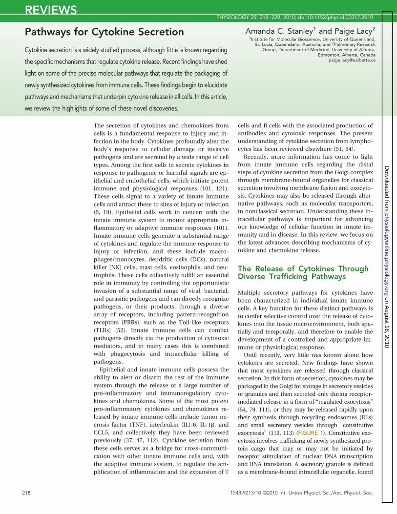
FIGURE 1. Regulated and constitutive exocytosis of cytokinesRegulated exocytosis involves ligand-receptor signaling to secretory granules or vesi-cles, which contain preformed cytokines, that are mobilized to the cell surface for re-lease. Constitutive exocytosis is instead dependent on receptor-mediated transcriptionevents in the nucleus, leading to trafficking via Golgi through recycling endosomes tothe cell surface. The pathway for regulated exocytosis is general for granulocytes,whereas the constitutive pathway shown here is an example of this type of traffickingfor TNF release from macrophages. The R-SNAREs involved in regulated and constitu-tive exocytosis are distinct, with VAMP-2, -7, and -8 implicated in regulated secretion,whereas Vti1b and VAMP-3 are essential for constitutive release of recycling endo-somes. A number of GTPases are also associated with cytokine release.
REVIEWS
PHYSIOLOGY • Volume 25 • August 2010 • www.physiologyonline.org 219
on August 18, 2010
physiologyonline.physiology.orgD
ownloaded from

inactive (GDP-bound) forms to mediate vesicular traf-ficking (12, 110).
A number of these molecules have been de-scribed in the regulation of granule exocytosis frominnate and adaptive immune cells (57, 111).Rab27a is activated in eosinophils (20) and is in-volved in exocytosis of azurophilic granules fromneutrophils (42, 81), whereas a double knockoutmouse model of Rab27a and Rab27b showed defi-cient mast cell degranulation (76). A deficiency inRab27a is also associated with the human diseasephenotype seen in the autosomal recessive immu-nodeficiency Griscelli syndrome 2, and NK cellsfrom patients with this disorder fail to secrete theirlytic granules (125). Rac1, Rac2, and Cdc42 havebeen implicated in granule exocytosis in mast cellsand neutrophils through modulation of the actincytoskeleton (1, 48) and have been associated withcytokine secretion in other cells, such as IL-8 frompolarized epithelial cells (Table 1) (45). However,no specific role for Rab or Rho GTPases in cytokinesecretion from innate immune cells has yet beenestablished. Although some studies indicate a rolefor the actin cytoskeleton and microtubules in cy-tokine secretion, there is little known regarding thespecific intracellular regulatory molecules requiredfor cytoskeletal remodeling in cytokine traffickingand release in innate immune cells.
SNAREs are intracellular proteins that interactwith each other to allow fusion of adjacent mem-branes (106, 115) and have proven to be the mostinformative group of proteins in defining pathwaysfor the release of cytokine and granules from innate
immune cells (Table 1). The SNARE hypothesismaintains that a single SNARE protein on a donormembrane binds to two cognate SNAREs on targetmembranes to form a four-helix complex calledthe trans-SNARE complex (FIGURE 2) (115). A cen-tral coiled-coil domain termed the “SNARE motif”serves as the binding region for SNARE-SNAREprotein interaction between members of theSNARE family. The SNARE family is divided intoR-SNAREs (usually on vesicles) and Q-SNAREs(usually at the target membrane) on the basis ofthe central functional residue in their SNARE motifbeing either arginine (R) or glutamine (Q) (31).When membrane fusion occurs, the R-SNAREs andQ-SNAREs bind together to allow the vesicle andtarget membranes to be brought together in closeproximity (29, 115). After fusion, the trans-SNAREcomplex is rapidly disassembled to the cis-SNAREcomplex to allow continued cycling of the compo-nents for further fusion events. Members ofR-SNAREs include vesicle-associated membraneproteins (VAMPs), whereas Q-SNAREs are associatedwith syntaxins and the SNAP-25 family of proteins.Several other families of proteins, including the Sec/Munc and Rab proteins, regulate SNARE binding anddisassembly (62, 114).
In exocytosis, R-SNAREs are usually associatedwith the vesicle or granule membrane, whereastheir cognate Q-SNAREs are commonly situated onthe inner leaflet of the plasma membrane. It is thefusion of the vesicular membrane with the targetplasma membrane in exocytosis that serves a fun-damental role in the release of many of the cell’s
Table 1. Families of molecules involved in SNARE complex formation in cytokine secretion from several cell types
MoleculeType
Secretory Pathway Cytokine Cell Type Reference
GTPasesRho GTPases
Rac1 Secretory vesicles - plasma membrane IL-8 Epithelial cells 45Cdc42 Secretory vesicles - plasma membrane IL-8 Epithelial cells 45
Rab GTPasesRab11a Recycling endosomes - plasma membrane TNF Macrophages 82
Recycling endosomes - plasma membrane IFN�, TNF NK cells 98SNAREs
R-SNAREsVti1b Trans-Golgi network - recycling endosomes TNF Macrophages 82,83VAMP-2 Secretory vesicles - plasma membrane CCL5 Eosinophils 58VAMP-3 Recycling endosome - plasma membrane IL-6, TNF Macrophages 67,82
Recycling endosomes - plasma membrane IFN�, TNF NK cells 98VAMP-8 Secretory vesicle - plasma membrane TNF Macrophages 94
Q-SNAREsSNAP-23 Secretory vesicles - plasma membrane CCL5 Eosinophils 65
Secretory granules - plasma membrane TNF Mast cells 116Syntaxin-4 Recycling endosomes - plasma membrane TNF Macrophages 82,83,87
Secretory vesicles - plasma membrane CCL5 Eosinophils 65Syntaxin-6 Trans-Golgi network - recycling endosomes TNF Macrophages 82,83Syntaxin-7 Trans-Golgi network - recycling endosomes TNF Macrophages 82
REVIEWS
PHYSIOLOGY • Volume 25 • August 2010 • www.physiologyonline.org220
on August 18, 2010
physiologyonline.physiology.orgD
ownloaded from

vesicular and granular contents to the extracellularmilieu (15).
Cytokine Secretion and Traffickingin Epithelial Cells
Epithelial cells have a profound effect on shapingthe immune responses in favor of Th1, Th2, or Tregulatory cells following activation of TLRs by vi-ral, bacterial, and fungal ligands (123). These cellssecrete a considerable range of cytokines and che-mokines including TNF and IL-1� (109), and TNFhas been detected in lipid bodies of colonic epi-thelial cells (7). More recently, thymic stromal lym-phopoietin, IL-25, and IL-33 were found to be keycytokines generated from airway epithelial cells,which promote allergic responses (101, 123). Themechanisms of trafficking of cytokines from epi-thelial cells is not yet understood, although this islikely to employ constitutive exocytotic pathways,and polarized protein trafficking in epithelial cellshas been shown to require different sets ofSNAREs. A specific pathway for apical delivery ofsecretory vesicles involves VAMP-7 (TI-VAMP) (61),whereas VAMP-3 (cellubrevin) localizes with thebasolateral membrane (32). VAMP-3 was shown tocooperate with adaptor protein-1B to mediate ba-solateral membrane trafficking in MDCK cells (32).The apical transcytotic pathway may requireVAMP-8 (endobrevin) (91). Although syntaxin-3 isrequired for the apical pathway, syntaxin-4 is pri-marily found in the basolateral membrane (17). Arecent study showed that VAMP-7 is important inGolgi to plasma membrane trafficking and medi-ates the transport of CD82 as well as affecting cellsurface diffusion of the EGF receptor in HeLa cells(23). All these results indicate that apical, basolat-eral, and transcytotic pathways in epithelial cells
use distinct sets of SNAREs (17). Whether all ofthese pathways are utilized in cytokine secretion isyet to be determined.
Exocytosis of Packaged Cytokines
Several types of innate immune cells generate cy-tokines that are released upon stimulation of spe-cific receptors (14, 69, 73, 79). These includeeosinophils, mast cells, and neutrophils, whichpossess a diversity of granule types that releasetheir contents by regulated exocytosis upon stim-ulation by PRRs and chemotactic ligands. Thuscytokines may be released immediately within min-utes of receptor stimulation, whereas lymphocytes cantake several hours or days to make and release cyto-kines (6, 59, 97, 105). Thus innate immune cells areuniquely poised to initiate a potent and immediatepro-inflammatory response upon contact with patho-gens and/or inflammatory stimuli.
Eosinophils
Eosinophils are normally rare white blood cells butdramatically increase in both the blood and tissuesduring allergy and helminth infection (47, 99).They are capable of synthesizing, storing, and se-creting up to 35 different cytokines (47). Many ofthese cytokines are potent inducers of immuneand inflammatory responses in asthma, eczema,rhinitis, and other inflammatory diseases. Theseunique cells contain large crystalloid granules thatare enriched in crystallized major basic protein, aswell as an extensive tubulovesicular network that isinvolved in rapid trafficking of small secretory ves-icles (57, 73). Eosinophils release mediators andcytokines in a differential manner via piecemealdegranulation, a mode of exocytosis characterizedby small vesicles budding from crystalloid granules
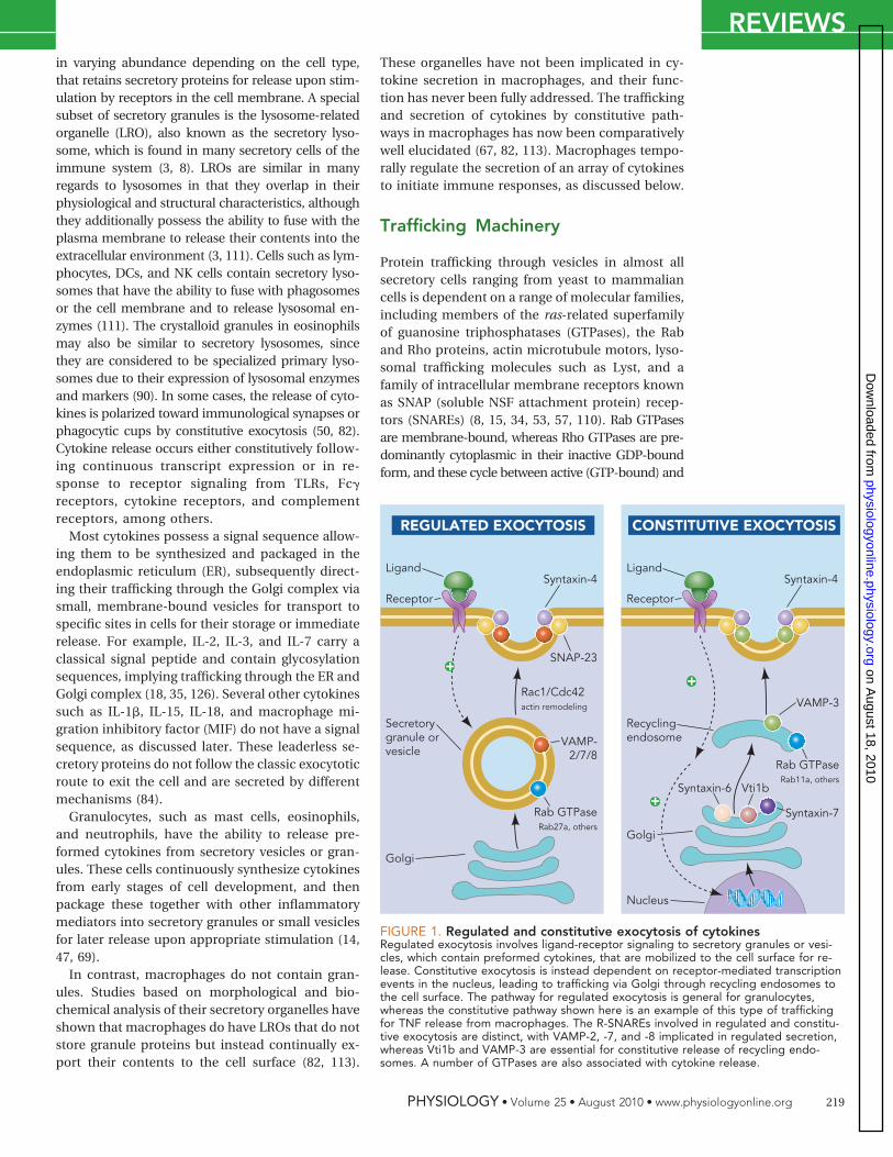
FIGURE 2. SNARE-mediated vesicle or granule fusion in exocytosisIn resting cells, Q-SNAREs are bound together in a cis-SNARE complex, which, on binding to secretory vesicle orgranule R-SNAREs (VAMP), is transformed into a four-helix bundle called the trans-SNARE complex. Formation of thetrans-SNARE complex leads to fusion of the granule and plasma membrane, followed by extrusion of granulecontents.
REVIEWS
PHYSIOLOGY • Volume 25 • August 2010 • www.physiologyonline.org 221
on August 18, 2010
physiologyonline.physiology.orgD
ownloaded from

that is frequently observed in tissue eosinophilsfrom allergic subjects (26, 28). Piecemeal degranu-lation is thought to be a general secretory methodused by immune cells and other cell types in thebody, including enteroendocrine cells of the gas-trointestinal tract, chromaffin cells of the adrenalmedulla, and chief cells of the parathyroid gland(22). Eosinophil-derived cytokines are stored incrystalloid granules and small secretory vesicles,which are rapidly released in response to interferon-�(IFN�) and other agonists (47, 60). For example, therelease of the chemokine CCL5, packaged primar-ily in the crystalloid granule, occurs within severalminutes of stimulation by IFN� (59, 72). Recently,several Th1 cytokines (IFN�, IL-12) and Th2 cyto-kines (IL-4, IL-13), as well as TNF, IL-6, and IL-10,were shown to be transported through the tubu-lovesicular system and small secretory vesiclesfrom their sites of storage in the crystalloid gran-ules (73, 74, 108). A mechanism for CCL11-mediatedIL-4 secretion was suggested by its colocalization withthe IL-4 receptor � chain, thereby anchoring IL-4 to thesecretory vesicle membrane for selective packag-ing, trafficking, and release (107). This receptor-mediated trafficking mechanism has also beendescribed for IL-15 secretion from DCs (80) andmay be important for the release of other cyto-kines from a variety of cells. In stimulated DCs,IL-15 and IL-15R� are preassembled in com-plexes within the ER and Golgi. In this way, IL-15R� chaperones IL-15 to the cell surface for
stable IL-15/IL-15R� complex expression on thecell membrane (80).
Cytokine release from eosinophils may be dependenton SNARE molecules for transport and exocytosis. Eo-sinophils express VAMP-2 (synaptobrevin-2), VAMP-7,VAMP-8, syntaxin-4, and SNAP-23 (46, 58, 65, 66).VAMP-2 is present in small secretory vesicles, contain-ing the chemokine CCL5, that translocate to the cellmembrane upon IFN� stimulation (FIGURE 3A) (58). Incrystalloid granules, VAMP-7 and VAMP-8 are stronglyexpressed; however, only VAMP-7 was required for thesecretion of granule proteins eosinophil peroxidase andeosinophil-derived neurotoxin (66). Syntaxin-4 andSNAP-23 were localized to the cell membrane in eosin-ophils, where they function as cognate intracellular re-ceptors for VAMP-2 (65). The precise functions ofR- and Q-SNAREs in eosinophil cytokine release, how-ever, have not yet been determined.
Taken together, eosinophils store the majority oftheir cytokines and chemokines in crystalloid gran-ules and may rapidly release these in a SNARE-de-pendent manner by regulated exocytosis followingreceptor stimulation.
Mast Cells
Mast cells are tissue-resident granulocytic cellsthat possess large secretory granules and smallersecretory vesicles. Mast cells can synthesize andrelease up to 33 different cytokines, chemokines,and growth factors (69) and store preformed cy-tokines in lipid bodies and granules (7). Mastcell-derived TNF and IL-4 are stored in secretorygranules and are rapidly released upon stimulationby cross-linking cell surface complexes of IgE andantigen (36, 85, 86, 124). These studies suggest thatTNF and IL-4 are released through FcεRI-mediateddegranulation, although no evidence has been ob-tained for the mode of cytokine release. Traffickingoutside of mast cell secretory granules occurs forIL-6, since IL-6 was shown to be released from adistinct population of secretory vesicles in the ab-sence of degranulation (55). Stimulation of cyto-kine release from mast cells occurs in a differentialmanner in response to TLR activation (71). Thisdifferential release is suggested to take placethrough piecemeal degranulation (25), similarly toeosinophils, allowing selective secretion (117).
Mast cells express many SNARE molecules,which are required for degranulation in responseto cross linking of the allergy-related IgE receptor,FcεRI (10, 88, 102). These include the R-SNAREsVAMP-2, VAMP-3, VAMP-7, and VAMP-8 (43, 88,93, 119, 120). VAMP-8 was shown to be associatedwith secretory granules and translocated withVAMP-7 to the cell membrane during degranulation.These observations were recently confirmed in hu-man mast cells isolated from intestinal samples,which showed expression of VAMP-3, VAMP-7, and
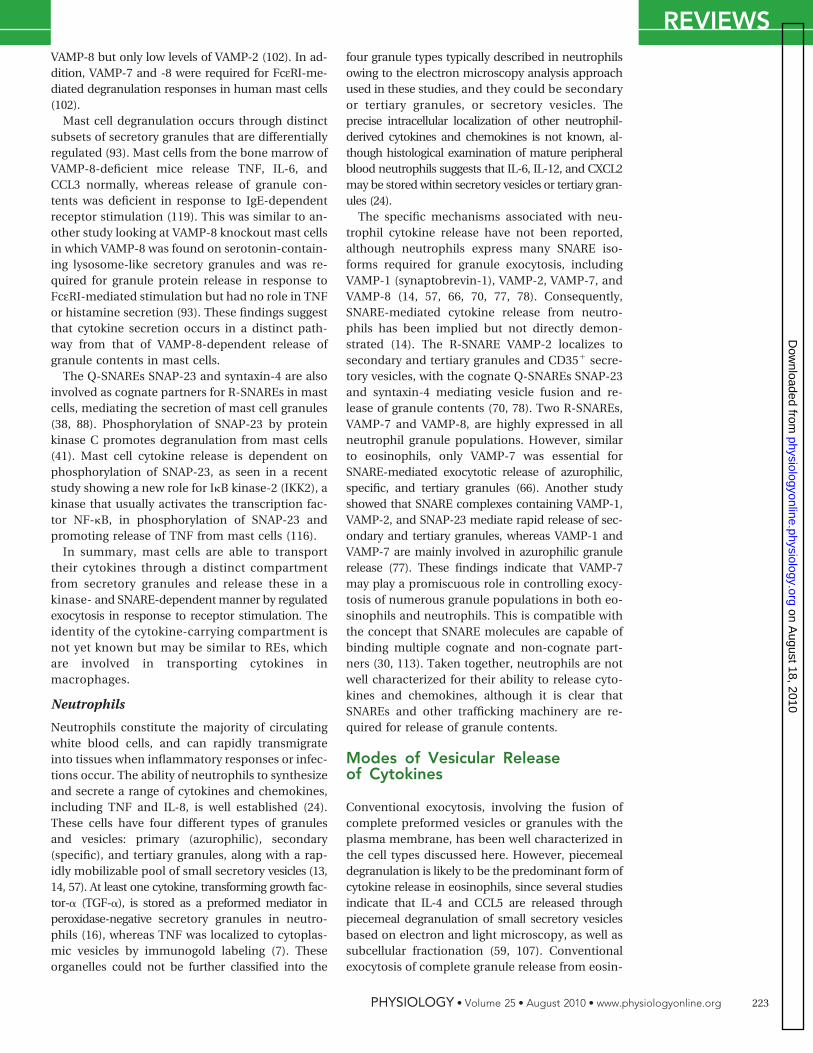
FIGURE 3. Secretion of cyto-kines through regulatory andconstitutive exocytosisA: human eosinophils stimulated withIFN� for 10 min led to transport ofCCL5 (red) in a rapidly mobilizablepool of small secretory vesicles,which colocalized with VAMP-2(green) at cell surfaces (arrows).These secretory vesicles, which aretoo small to characterize by opticalmicroscopy, were identified by theirintrinsic buoyant density using equi-librium gradient ultracentrifugation(59). B: murine macrophages (RAW264.7 cells) release cell surface TNF(red) in response to LPS after 90 minof stimulation, which can be de-tected at cell surfaces by coincubat-ing cells with the TACE inhibitor,TAPI, followed by immunofluores-cence labeling. Original magnificationfor both images is �630.
REVIEWS
PHYSIOLOGY • Volume 25 • August 2010 • www.physiologyonline.org222
on August 18, 2010
physiologyonline.physiology.orgD
ownloaded from
VAMP-8 but only low levels of VAMP-2 (102). In ad-dition, VAMP-7 and -8 were required for FcεRI-me-diated degranulation responses in human mast cells(102).
Mast cell degranulation occurs through distinctsubsets of secretory granules that are differentiallyregulated (93). Mast cells from the bone marrow ofVAMP-8-deficient mice release TNF, IL-6, andCCL3 normally, whereas release of granule con-tents was deficient in response to IgE-dependentreceptor stimulation (119). This was similar to an-other study looking at VAMP-8 knockout mast cellsin which VAMP-8 was found on serotonin-contain-ing lysosome-like secretory granules and was re-quired for granule protein release in response toFcεRI-mediated stimulation but had no role in TNFor histamine secretion (93). These findings suggestthat cytokine secretion occurs in a distinct path-way from that of VAMP-8-dependent release ofgranule contents in mast cells.
The Q-SNAREs SNAP-23 and syntaxin-4 are alsoinvolved as cognate partners for R-SNAREs in mastcells, mediating the secretion of mast cell granules(38, 88). Phosphorylation of SNAP-23 by proteinkinase C promotes degranulation from mast cells(41). Mast cell cytokine release is dependent onphosphorylation of SNAP-23, as seen in a recentstudy showing a new role for I�B kinase-2 (IKK2), akinase that usually activates the transcription fac-tor NF-�B, in phosphorylation of SNAP-23 andpromoting release of TNF from mast cells (116).
In summary, mast cells are able to transporttheir cytokines through a distinct compartmentfrom secretory granules and release these in akinase- and SNARE-dependent manner by regulatedexocytosis in response to receptor stimulation. Theidentity of the cytokine-carrying compartment isnot yet known but may be similar to REs, whichare involved in transporting cytokines inmacrophages.
Neutrophils
Neutrophils constitute the majority of circulatingwhite blood cells, and can rapidly transmigrateinto tissues when inflammatory responses or infec-tions occur. The ability of neutrophils to synthesizeand secrete a range of cytokines and chemokines,including TNF and IL-8, is well established (24).These cells have four different types of granulesand vesicles: primary (azurophilic), secondary(specific), and tertiary granules, along with a rap-idly mobilizable pool of small secretory vesicles (13,14, 57). At least one cytokine, transforming growth fac-tor-� (TGF-�), is stored as a preformed mediator inperoxidase-negative secretory granules in neutro-phils (16), whereas TNF was localized to cytoplas-mic vesicles by immunogold labeling (7). Theseorganelles could not be further classified into the
four granule types typically described in neutrophilsowing to the electron microscopy analysis approachused in these studies, and they could be secondaryor tertiary granules, or secretory vesicles. Theprecise intracellular localization of other neutrophil-derived cytokines and chemokines is not known, al-though histological examination of mature peripheralblood neutrophils suggests that IL-6, IL-12, and CXCL2may be stored within secretory vesicles or tertiary gran-ules (24).
The specific mechanisms associated with neu-trophil cytokine release have not been reported,although neutrophils express many SNARE iso-forms required for granule exocytosis, includingVAMP-1 (synaptobrevin-1), VAMP-2, VAMP-7, andVAMP-8 (14, 57, 66, 70, 77, 78). Consequently,SNARE-mediated cytokine release from neutro-phils has been implied but not directly demon-strated (14). The R-SNARE VAMP-2 localizes tosecondary and tertiary granules and CD35� secre-tory vesicles, with the cognate Q-SNAREs SNAP-23and syntaxin-4 mediating vesicle fusion and re-lease of granule contents (70, 78). Two R-SNAREs,VAMP-7 and VAMP-8, are highly expressed in allneutrophil granule populations. However, similarto eosinophils, only VAMP-7 was essential forSNARE-mediated exocytotic release of azurophilic,specific, and tertiary granules (66). Another studyshowed that SNARE complexes containing VAMP-1,VAMP-2, and SNAP-23 mediate rapid release of sec-ondary and tertiary granules, whereas VAMP-1 andVAMP-7 are mainly involved in azurophilic granulerelease (77). These findings indicate that VAMP-7may play a promiscuous role in controlling exocy-tosis of numerous granule populations in both eo-sinophils and neutrophils. This is compatible withthe concept that SNARE molecules are capable ofbinding multiple cognate and non-cognate part-ners (30, 113). Taken together, neutrophils are notwell characterized for their ability to release cyto-kines and chemokines, although it is clear thatSNAREs and other trafficking machinery are re-quired for release of granule contents.
Modes of Vesicular Releaseof Cytokines
Conventional exocytosis, involving the fusion ofcomplete preformed vesicles or granules with theplasma membrane, has been well characterized inthe cell types discussed here. However, piecemealdegranulation is likely to be the predominant form ofcytokine release in eosinophils, since several studiesindicate that IL-4 and CCL5 are released throughpiecemeal degranulation of small secretory vesiclesbased on electron and light microscopy, as well assubcellular fractionation (59, 107). Conventionalexocytosis of complete granule release from eosin-
REVIEWS
PHYSIOLOGY • Volume 25 • August 2010 • www.physiologyonline.org 223
on August 18, 2010
physiologyonline.physiology.orgD
ownloaded from
ophils has only been determined using in vitrotechniques and has never been detected in vivo, atleast in tissues obtained from nasal polyps of aller-gic subjects (28). Instead, tissue eosinophils inallergic subjects largely undergo piecemeal de-granulation (67%) and cytolysis (necrosis in theremainder of tissue cells) (28). Therefore, it is un-likely that cytokine release occurs through conven-tional granule exocytosis from eosinophils inallergic diseases. In the case of mast cells, it isabundantly clear that serotonin and histamine re-lease are associated with conventional granuleexocytosis. However, cytokine release from mastcells may occur through a distinct pathway fromconventional granule exocytosis, as discussedabove. This suggests that cytokines are releasedthrough a vesicular pathway that is distinct fromconventional granule exocytosis in mast cells.
Constitutive Release of Cytokines
Constitutive cytokine release describes the packag-ing of cytokines through the ER and Golgi into acontinuously trafficking pathway of carrier vesiclesthat transport cargo to the plasma membrane forrelease within minutes or hours of protein synthe-sis. In some innate immune cells, post-Golgi cargoto the plasma membrane has been shown to go viaREs. REs are tubulovesicular structures that areresponsible for transporting recycling membranesand proteins to and from the plasma membrane(122) and are present in nearly all cells, includingepithelial cells, skeletal muscle cells, neuronalcells, NK cells, mast cells, and macrophages, al-though they have not yet been characterized inneutrophils or eosinophils. The RE has a slightlyhigher luminal pH (�6.4) than peripheral earlyendosomes and is often located deeper in the cellnear the microtubule-organizing center (89, 122).In most cells, REs have diverse roles in the deliveryof membranes, receptors, and luminal contents tovarious parts of cells for secretion, cytokinesis, cellfate, transporter upregulation, and polarization ofcells. In some innate immune cells, REs performmany of these functions along with cytokine secre-tion (67, 82). Factors that regulate RE traffickinginclude VAMP-3 (82) and Rab11a (21). REs can bedetected by measurement of transferrin uptake (4),and transferrin receptor immunoreactivity (67, 82).Pathways engaged in constitutive cytokine releasethrough REs have been best defined in macro-phages for their ability to package and secrete TNFand IL-6 (67, 82). NK cells also possess REs thatare involved in transporting cytokines constitu-tively to the cell surface, independently of theirgranules (98).
Macrophages
Macrophages are tissue-resident cells that derivefrom maturation of extravasated peripheral bloodmonocytes that are abundant on mucosal surfacessuch as the airways. These cells offer surveillanceof tissues and initiate multiple actions, includingrobust pro-inflammatory cytokine release, whenthey encounter pathogens and dying cells (49).Macrophages contain constitutively trafficked car-rier vesicles that transport cytokines, includingTNF and IL-6, between intracellular compart-ments, as well as to the cell surface for release(105). These carrier vesicles include REs, which aredistinct from LROs or secretory lysosomes. Whenbacterially derived lipopolysaccharide (LPS) stimu-lates macrophages via TLR4, the type II transmem-brane precursor pro-TNF is rapidly synthesized andaccumulates in the Golgi within 20 min, and fromthere it is delivered to the cell surface (FIGURE 3B)(63, 82, 95, 105). Once TNF reaches the cell surface,it is cleaved by TNF converting enzyme (TACE,ADAM17) to release its ectodomain as a solublecytokine (9). Secreted soluble TNF is detected �40min after stimulation (105). Both microtubules andactin filaments are involved in post-Golgi-vesiculartrafficking of TNF (105).
The anterograde transport of TNF from thetrans-Golgi network (TGN) in macrophages wasrecently shown to require the TGN golgin, p230/golgin-245 (p230) (63). Golgins are long coiled-coilproteins, characterized by a COOH-terminal GRIPdomain, that are specifically recruited to subdo-mains of the TGN from which dynamic tubularprecursors arise (104). These coiled-coil mem-brane-bound proteins participate in membrane teth-ering that can lead to membrane fusion or can allowstable linking of two membranes (96). Golgins arethought to have a function in selective budding ofGolgi-derived vesicles, and distinct spatial segrega-tion allows association of different golgins, such asp230 and golgin-97, with TGN tubules bearing dif-ferent cargo molecules (64). Depletion of p230 inmacrophages using a micro-RNA approach blockspost-Golgi transport of TNF (63). Importantly, re-duced TNF secretion was also demonstrated invivo in mice where p230 was silenced by retroviraltransduction (63). Thus p230 has an essential rolein cytokine export from the Golgi and secretionfrom activated macrophages.
Similarly, LPS stimulation leads to the accumu-lation of newly synthesized IL-6 in the Golgi com-plex in macrophages within an hour of LPSstimulation, which is then loaded into tubovesicu-lar carriers that bud off the TGN either as the solelabeled cargo or together with TNF (67). Transfec-tion of exogenous IL-6 fused to a GFP tag confirmedthat soluble cargo accumulated in the dilated ends of
REVIEWS
PHYSIOLOGY • Volume 25 • August 2010 • www.physiologyonline.org224
on August 18, 2010
physiologyonline.physiology.orgD
ownloaded from

Golgi cisternae (67). Since IL-6 is a soluble cytokinerather than a transmembrane cytokine like TNF, itutilizes multiple carriers (including p230 and golgin-97) for its export from the Golgi.
Using markers of RE in confocal and epifluores-cence imaging, it was shown that, following exportfrom the Golgi, both TNF and IL-6 colocalize withREs in macrophages during their transport to thecell surface following LPS stimulation (82). ThusREs act as an intermediate step for cytokine releasebefore secretion at the cell surface (67, 82). Fur-thermore, it was shown that IL-6 and TNF localizeto different subcompartments of REs (67), suggest-ing that REs may additionally function to sort thesetwo cytokines for release at different points on thecell surface.
A number of other molecules and pathways havebeen implicated in Golgi-derived cytokine releasein macrophages. Choline cytidyltransferase (CCT)is the rate-limiting enzyme in the phosphatidyl-choline (PtdCho) biosynthetic pathway requiredfor membrane biogenesis. CCT-� is the major CCTisoform required to mediate PtdCho synthesis formaintaining normal Golgi structure and function.CCT-� expression in the trans-Golgi region is increasedin LPS-stimulated macrophages, and CCT-� has beenshown to be required for both TNF and IL-6 exportfrom the Golgi complex and secretion (118). Anotherfamily of regulatory molecules known as secretorycarrier membrane proteins (SCAMPs) are widely dis-tributed integral membrane proteins that have beenimplicated in regulating vesicular transport in nu-merous neuronal and nonneuronal tissues. SCAMP5has been shown to be involved in the secretion ofsignal peptide-containing cytokines including TNFand CCL5 from the Golgi complex in monocytes andmacrophages (40). SCAMP5 is mainly localized inGolgi-associated compartments, and ionomycin trig-gers rapid translocation of SCAMP5 to the plasmamembrane along the classical exocytosis pathway.SCAMP5 is thought to mediate exocytosis of cy-tokines in cooperation with SNARE machinery(SNAP-23 and syntaxin-4) by binding to calcium-sensitive synaptotagmins (40).
In activated macrophages, increased vesicular traf-ficking of cytokines occurs concomitantly with theupregulation in expression of relevant SNAREs andother components of trafficking machinery (2, 82, 83,87), implicating SNARE molecules in RE function.Membrane fusion of TNF-containing vesicles fromthe TGN to REs, and subsequently from REs to theplasma membrane, is mediated by SNAREs. A novelQ-SNARE complex of syntaxin-6-syntaxin-7-Vti1b isupregulated in LPS-stimulated macrophages and hasbeen shown to regulate transport of TNF out of theTGN (82). This Q-SNARE complex binds theR-SNARE VAMP-3 on REs, necessitating TNF deliveryto the cell surface via REs (82).
Another molecule involved in the trafficking ofTNF-containing vesicles to the plasma membranein LPS-stimulated macrophages is the lysosomalcysteine protease, cathepsin B (39). In addition,evidence for a role for VAMP-8 in complement-induced TNF secretion from macrophages hasbeen shown using VAMP-8 knockout mice (94).However, it is unclear how VAMP-3 and VAMP-8coordinate to mediate the secretion of TNF, andwhy cathepsin B should be important in transportof TNF to the plasma membrane, since VAMP-8and cathepsin B are both lysosome-associated pro-teins. It is possible that lysosomes and REs ex-change their contents during secretion and thatsome form of cross-regulation between these com-partments occurs.
In summary, the macrophage is currently themost well characterized innate immune cell typefor its ability to traffic and secrete cytokines. Fur-ther studies should shed light on the interrelation-ships of these regulatory pathways in the transportof pro-inflammatory and immunoregulatory medi-ators. SNARE-dependent cytokine secretion fromother innate immune cells, including NK cells andDCs, is not well characterized. NK target cell killingmediated by granzyme B secretion is dependent onVAMP-7 (68), and VAMP-8 is involved in phagoso-mal trafficking in DCs (44). NK cells also rely onREs to traffic cytokines to the cell surface (98).Understanding the regulatory mechanisms inthese and other innate immune cells will be im-portant, since these may be distinct from those ofgranulocytes and macrophages.
Polarity of Cytokine Release
Cytoskeleton-dependent polarized release of cyto-kines is an essential mechanism for receptor cross-linking in cell-cell communication and killing, andwas first identified in T cells (92). Trafficking andsecretion of cytokines can be customized for theircell-specific roles in immunity. Cytokines can bedirected toward the immunological synapse aris-ing from cell-cell contact between T cells or NKcells and their target cells, or with antigen-present-ing cells (51, 111). Classically, an immunologicalsynapse is a stable region of contact that formsbetween a central cluster of T-cell receptors (TCRs)on a T cell [supramolecular activation cluster(cSMAC)] and a surrounding ring of adhesion mol-ecules [peripheral supramolecular activation clus-ter (pSMAC)] on an antigen-presenting cell (111).The release of cytokines can also occur in a polar-ized manner toward phagocytic cups and filipodiain macrophages (56, 67, 82, 113). In phagocytosingmacrophages, TNF secretion was shown to be con-current with the supply of VAMP-3-containing REmembranes polarized toward the expanding
REVIEWS
PHYSIOLOGY • Volume 25 • August 2010 • www.physiologyonline.org 225
on August 18, 2010
physiologyonline.physiology.orgD
ownloaded from
actin-rich phagocytic cup for SNARE-mediatedfusion during the initial stages of phagocytosis (67,82). Syntaxin-4 and TACE are also concentrated inphagocytic cups to allow fusion of REs and cleav-age of soluble TNF. However, whereas TNF andIL-6 are both transported through REs, IL-6 isreleased circumferentially from the peripheralmembrane (67, 82). One possible mechanism forthe different pathways of TNF and IL-6 secretion isthat they may be separated at the Golgi in subcom-partments of REs, since TNF is synthesized as amembrane-bound precursor, whereas IL-6 is gen-erated as a soluble cytokine (67).
Interestingly, it has now been demonstrated thatcytokines can be released by vesicles in both polar-ized and non-polarized pathways in T-helper cells(50, 51, 54). Some cytokines such as IFN� and IL-2are delivered in a polarized fashion to the immuno-logical synapse with antigen-presenting cells to im-part specific communication, whereas others such asTNF and CCL3 are secreted separately and releasedmulti-directionally to promote inflammation and toestablish chemokine gradients. Therefore, secretionof different cytokines is not only polarized but isdependent on different trafficking proteins andmolecularly distinct processes, some of whichare associated with Rab GTPases and SNARE pro-teins syntaxin-6 and Vti1b (50).
Like T-helper cells, NK cells can also form animmunological synapse with a target cell to delivercytotoxic signals or with a DC to impart commu-nication. However, recent evidence suggests thatalthough polarity is established in NK cells to en-sure directional delivery of perforin in lytic gran-ules toward the immunological synapse, cytokinesare released multi-directionally (98). It is not clearhow TNF and IFN� are trafficked and releasedsimultaneously to other sites on the cell surface forlargely non-polarized secretion in a pathway de-pendent on REs, despite the alignment of REs withsynapses. Inactivation of REs in NK cells with endo-cytosed activated horseradish peroxidase, or muta-tion of RE proteins Rab11 or VAMP-3, abrogates TNFand IFN� secretion, yet the secretion of cytotoxicgranules remains unaffected (98). Therefore, in NKcells, secretory pathways for cytokines and cytolyticagents diverge at the level of REs. This divergence inthe polarity of cytotoxic molecules and cytokines al-lows NK cells to simultaneously kill target cells at thesynapse and to recruit other immune cells with dif-ferent sets of secreted mediators.
In turn, DCs can polarize the release of cytokinestoward the immunological synapse during interac-tion with NK cells (103), a crucial step in the initi-ation/amplification of early phases of an immuneresponse that favors the onset of an adaptive im-mune response. NK cells trigger immature DCs topolarize and secrete IL-18 toward the synapse.
IL-18 is an inflammatory cytokine that lacks a se-cretory leader sequence and is translocated fromthe cytosol into secretory lysosomes for release(11). DC IL-18 is trafficked in secretory lysosomesthat are recruited toward the adhering NK cell andare restricted to the synaptic cleft in a calcium-dependent and tubulin-mediated manner (103).This polarization of IL-18 activates interacting NKcells to upregulate cytotoxicity without spreadingof cytokine. There is no evidence at this time tosuggest that polarized cytokine secretion may oc-cur in other innate immune cells, such as mastcells, eosinophils, and neutrophils.
Non-Classical Secretionof Cytokines
Several cytokines are released using non-classicalpathways, independent of ER/Golgi trafficking. Themechanisms involved are still poorly understood.IL-1� has emerged as one of the most importantmediators of inflammation and host responses toinfection (27). IL-1� is a leaderless protein first gen-erated as biologically inactive pro-IL-1�, which issynthesized directly into the cytoplasm of cells be-cause it lacks a conventional hydrophobic signalsequence (100). Pro-IL-1� is then processed intomature, biologically active IL-1� by caspase-1cleavage at intracellular sites and is subsequentlyreleased into the extracellular milieu. Differentmodels of non-classical release have been pro-posed for IL-1� (27). These include classical exo-cytosis of IL-1�-containing secretory lysosomes, aswell as non-classical secretion pathways of IL-1�
release from shed plasma membrane mi-crovesicles, fusion of multivesicular bodies withthe plasma membrane and subsequent release ofIL-1�-containing exosomes, and export of IL-1�
through the plasma membrane using specificmembrane transporters (27). There is also someevidence that a significant proportion of IL-1� re-lease occurs upon cell lysis. MIF is another leader-less cytokine that does not enter the ER/Golgisecretory pathway and instead appears to be se-creted from the cytoplasm, possibly through anABC transporter (33). Secretion of MIF requiresp115, a vesicle-docking protein that is localized pre-dominantly to the cytosolic side of vesiculotubularintermediate clusters and to the cis-Golgi, to act as anintracellular binding partner (75). These different po-tential mechanisms of non-classical release remaincontroversial, and further work is required to eluci-date these pathways.
Summary
In this review, we have seen that there are multipleand complex pathways for the release of cytokines
REVIEWS
PHYSIOLOGY • Volume 25 • August 2010 • www.physiologyonline.org226
on August 18, 2010
physiologyonline.physiology.orgD
ownloaded from
by cells of the innate immune system. Increasingly,and unsurprisingly, it emerges that cells use differ-ent pathways, organelles, carriers, and moleculesto exert an exquisite control of release of theseimportant immunoregulatory messengers. This isessential for the development of appropriate in-nate and adaptive immune responses towardinflammatory or infectious agents. Regulation ofexocytosis of cytokine-containing vesicles or gran-ules appears to be specific to the cell type, suchthat it is not possible to fully extrapolate findingsfrom a single cell type. In addition, each cytokinemust be individually studied for its unique traffick-ing pathway, since even within a single cell typethe pathway of cytokine secretion is dictated by thepresence or absence of a signal peptide sequenceas well as numerous other packaging processes.Since many of the components of trafficking ma-chinery were first identified in neuronal cells, it isanticipated that further insights gained from neu-ronal and neuroendocrine cells will increasinglyinstruct us about molecules and mechanisms ac-tive in other cell types. Therefore, much remains tobe determined regarding the precise mechanismsthat control cytokine packaging and trafficking forrelease in innate immune cells and indeed in othercells of the body. �
Data reported here was produced by funding from theNational Health and Medical Research Council of Australia(A. C. Stanley), the Alberta Heritage Foundation for Med-ical Research (Visiting Scientist from Alberta Award; P.Lacy), and the Canadian Institutes of Health Research (P.Lacy).
No conflicts of interest, financial or otherwise, are de-clared by the author(s).
REFERENCES1. Abdel-Latif D, Steward M, Macdonald DL, Francis GA, Di-
nauer MC, Lacy P. Rac2 is critical for neutrophil primarygranule exocytosis. Blood 104: 832–839, 2004.
2. Achuthan A, Masendycz P, Lopez JA, Nguyen T, James DE,Sweet MJ, Hamilton JA, Scholz GM. Regulation of the endo-somal SNARE protein syntaxin 7 by colony-stimulating factor1 in macrophages. Mol Cell Biol 28: 6149–6159, 2008.
3. Andrews NW. Regulated secretion of conventional lyso-somes. Trends Cell Biol 10: 316–321, 2000.
4. Bajno L, Peng XR, Schreiber AD, Moore HP, Trimble WS,Grinstein S. Focal exocytosis of VAMP3-containing vesicles atsites of phagosome formation. J Cell Biol 149: 697–706,2000.
5. Barrett NA, Austen KF. Innate cells and T helper 2 cell im-munity in airway inflammation. Immunity 31: 425–437, 2009.
6. Baumgartner RA, Deramo VA, Beaven MA. Constitutive andinducible mechanisms for synthesis and release of cytokinesin immune cell lines. J Immunol 157: 4087–4093, 1996.
7. Beil WJ, Weller PF, Peppercorn MA, Galli SJ, Dvorak AM.Ultrastructural immunogold localization of subcellular sites ofTNF-alpha in colonic Crohn’s disease. J Leukoc Biol 58: 284–298, 1995.
8. Benado A, Nasagi-Atiya Y, Sagi-Eisenberg R. Protein traffick-ing in immune cells. Immunobiology 214: 403–421, 2009.
9. Black RA, Rauch CT, Kozlosky CJ, Peschon JJ, Slack JL,Wolfson MF, Castner BJ, Stocking KL, Reddy P, Srinivasan S,Nelson N, Boiani N, Schooley KA, Gerhart M, Davis R, FitznerJN, Johnson RS, Paxton RJ, March CJ, Cerretti DP. A metal-loproteinase disintegrin that releases tumour-necrosis factor-alpha from cells. Nature 385: 729–733, 1997.
10. Blank U, Cyprien B, Martin-Verdeaux S, Paumet F, Pombo I,Rivera J, Roa M, Varin-Blank N. SNAREs and associated reg-ulators in the control of exocytosis in the RBL-2H3 mast cellline. Mol Immunol 38: 1341–1345, 2002.
11. Blott EJ, Griffiths GM. Secretory lysosomes. Nat Rev Mol CellBiol 3: 122–131, 2002.
12. Bokoch GM. Regulation of innate immunity by Rho GTPases.Trends Cell Biol 15: 163–171, 2005.
13. Borregaard N, Cowland JB. Granules of the human neutro-philic polymorphonuclear leukocyte. Blood 89: 3503–3521,1997.
14. Borregaard N, Sorensen OE, Theilgaard-Monch K. Neutro-phil granules: a library of innate immunity proteins. TrendsImmunol 28: 340–345, 2007.
15. Burgoyne RD, Morgan A. Secretory granule exocytosis.Physiol Rev 83: 581–632, 2003.
16. Calafat J, Janssen H, Stahle-Backdahl M, Zuurbier AE, KnolEF, Egesten A. Human monocytes and neutrophils storetransforming growth factor-alpha in a subpopulation of cyto-plasmic granules. Blood 90: 1255–1266, 1997.
17. Carmosino M, Valenti G, Caplan M, Svelto M. Polarized traffictowards the cell surface: how to find the route. Biol Cell 102:75–91, 2010.
18. Conradt HS, Ausmeier M, Dittmar KE, Hauser H, LindenmaierW. Secretion of glycosylated human interleukin-2 by recom-binant mammalian cell lines. Carbohydr Res 149: 443–450,1986.
19. Coombes JL, Powrie F. Dendritic cells in intestinal immuneregulation. Nat Rev Immunol 8: 435–446, 2008.
20. Coughlin JJ, Odemuyiwa SO, Davidson CE, Moqbel R. Dif-ferential expression and activation of Rab27A in human eo-sinophils: relationship to blood eosinophilia. BiochemBiophys Res Commun 373: 382–386, 2008.
21. Cox D, Lee DJ, Dale BM, Calafat J, Greenberg S. A Rab11-containing rapidly recycling compartment in macrophagesthat promotes phagocytosis. Proc Natl Acad Sci USA 97:680–685, 2000.
22. Crivellato E, Nico B, Mallardi F, Beltrami CA, Ribatti D. Piece-meal degranulation as a general secretory mechanism? AnatRec A Discov Mol Cell Evol Biol 274: 778–784, 2003.
23. Danglot L, Chaineau M, Dahan M, Gendron MC, Boggetto N,Perez F, Galli T. Role of TI-VAMP and CD82 in EGFR cell-surface dynamics and signaling. J Cell Sci 123: 723–735,2010.
24. Denkers E, Del Rio L, Bennouna S. Neutrophil production ofIL-12 and other cytokines during microbial infection. In: TheNeutrophil: An Emerging Regulator of Inflammatory andImmune Response, edited by Ma C. Basel: Karger, 2003,p. 95–114.
25. Dvorak AM. Basophils and mast cells: piecemeal degranula-tion in situ and ex vivo: a possible mechanism for cytokine-induced function in disease. Immunol Ser 57: 169–271, 1992.
26. Dvorak AM, Ackerman SJ, Furitsu T, Estrella P, Letourneau L,Ishizaka T. Mature eosinophils stimulated to develop in hu-man-cord blood mononuclear cell cultures supplementedwith recombinant human interleukin-5. II. Vesicular transportof specific granule matrix peroxidase, a mechanism for ef-fecting piecemeal degranulation. Am J Pathol 140: 795–807,1992.
27. Eder C. Mechanisms of interleukin-1beta release. Immuno-biology 214: 543–553, 2009.
28. Erjefalt JS, Greiff L, Andersson M, Matsson E, Petersen H,Linden M, Ansari T, Jeffery PK, Persson CG. Allergen-inducedeosinophil cytolysis is a primary mechanism for granule pro-tein release in human upper airways. Am J Respir Crit CareMed 160: 304–312, 1999.
29. Fasshauer D. Structural insights into the SNARE mechanism.Biochim Biophys Acta 1641: 87–97, 2003.
REVIEWS
PHYSIOLOGY • Volume 25 • August 2010 • www.physiologyonline.org 227
on August 18, 2010
physiologyonline.physiology.orgD
ownloaded from

30. Fasshauer D, Antonin W, Margittai M, Pabst S,Jahn R. Mixed and non-cognate SNARE com-plexes. Characterization of assembly and bio-physical properties. J Biol Chem 274: 15440–15446, 1999.
31. Fasshauer D, Sutton RB, Brunger AT, Jahn R.Conserved structural features of the synaptic fu-sion complex: SNARE proteins reclassified as Q-and R-SNAREs. Proc Natl Acad Sci USA 95:15781–15786, 1998.
32. Fields IC, Shteyn E, Pypaert M, Proux-GillardeauxV, Kang RS, Galli T, Folsch H. v-SNARE cellubre-vin is required for basolateral sorting of AP-1B-dependent cargo in polarized epithelial cells. JCell Biol 177: 477–488, 2007.
33. Flieger O, Engling A, Bucala R, Lue H, Nickel W,Bernhagen J. Regulated secretion of macro-phage migration inhibitory factor is mediated bya non-classical pathway involving an ABC trans-porter. FEBS Lett 551: 78–86, 2003.
34. Gasman S, Chasserot-Golaz S, Bader MF, VitaleN. Regulation of exocytosis in adrenal chromaffincells: focus on ARF and Rho GTPases. Cell Signal15: 893–899, 2003.
35. Goodwin RG, Lupton S, Schmierer A, Hjerrild KJ,Jerzy R, Clevenger W, Gillis S, Cosman D, NamenAE. Human interleukin 7: molecular cloning andgrowth factor activity on human and murine B-lineage cells. Proc Natl Acad Sci USA 86: 302–306, 1989.
36. Gordon JR, Galli SJ. Mast cells as a source of bothpreformed and immunologically inducible TNF-alpha/cachectin. Nature 346: 274–276, 1990.
37. Gordon S. Alternative activation of macro-phages. Nat Rev Immunol 3: 23–35, 2003.
38. Guo Z, Turner C, Castle D. Relocation of thet-SNARE SNAP-23 from lamellipodia-like cell sur-face projections regulates compound exocytosisin mast cells. Cell 94: 537–548, 1998.
39. Ha SD, Martins A, Khazaie K, Han J, Chan BM,Kim SO. Cathepsin B is involved in the traffickingof TNF-alpha-containing vesicles to the plasmamembrane in macrophages. J Immunol 181: 690–697, 2008.
40. Han C, Chen T, Yang M, Li N, Liu H, Cao X. HumanSCAMP5, a novel secretory carrier membrane pro-tein, facilitates calcium-triggered cytokine secre-tion by interaction with SNARE machinery. JImmunol 182: 2986–2996, 2009.
41. Hepp R, Puri N, Hohenstein AC, Crawford GL,Whiteheart SW, Roche PA. Phosphorylation ofSNAP-23 regulates exocytosis from mast cells. JBiol Chem 280: 6610–6620, 2005.
42. Herrero-Turrion MJ, Calafat J, Janssen H, FukudaM, Mollinedo F. Rab27a regulates exocytosis oftertiary and specific granules in human neutro-phils. J Immunol 181: 3793–3803, 2008.
43. Hibi T, Hirashima N, Nakanishi M. Rat basophilicleukemia cells express syntaxin-3 and VAMP-7 ingranule membranes. Biochem Biophys Res Com-mun 271: 36–41, 2000.
44. Ho YH, Cai DT, Wang CC, Huang D, Wong SH.Vesicle-associated membrane protein-8/endo-brevin negatively regulates phagocytosis of bac-teria in dendritic cells. J Immunol 180: 3148–3157, 2008.
45. Hobert ME, Sands KA, Mrsny RJ, Madara JL.Cdc42 and Rac1 regulate late events in Salmo-nella typhimurium-induced interleukin-8 secre-tion from polarized epithelial cells. J Biol Chem277: 51025–51032, 2002.
46. Hoffmann HJ, Bjerke T, Karawajczyk M, Dahl R,Knepper MA, Nielsen S. SNARE proteins are crit-ical for regulated exocytosis of ECP from humaneosinophils. Biochem Biophys Res Commun 282:194–199, 2001.
47. Hogan SP, Rosenberg HF, Moqbel R, Phipps S,Foster PS, Lacy P, Kay AB, Rothenberg ME. Eo-sinophils: biological properties and role in healthand disease. Clin Exp Allergy 38: 709–750, 2008.
48. Hong-Geller E, Cerione RA. Cdc42 and Rac stim-ulate exocytosis of secretory granules by activat-ing the IP(3)/calcium pathway in RBL-2H3 mastcells. J Cell Biol 148: 481–494, 2000.
49. Hume DA. The mononuclear phagocyte system.Curr Opin Immunol 18: 49–53, 2006.
50. Huse M, Lillemeier BF, Kuhns MS, Chen DS, DavisMM. T cells use two directionally distinct path-ways for cytokine secretion. Nat Immunol 7: 247–255, 2006.
51. Huse M, Quann EJ, Davis MM. Shouts, whispersand the kiss of death: directional secretion in Tcells. Nat Immunol 9: 1105–1111, 2008.
52. Iwasaki A, Medzhitov R. Regulation of adaptiveimmunity by the innate immune system. Science327: 291–295, 2010.
53. Jahn R, Scheller RH. SNAREs: engines for mem-brane fusion. Nat Rev Mol Cell Biol 7: 631–643,2006.
54. Jolly C, Sattentau QJ. Regulated secretion fromCD4� T cells. Trends Immunol 28: 474–481,2007.
55. Kandere-Grzybowska K, Letourneau R, KempurajD, Donelan J, Poplawski S, Boucher W, Athanas-siou A, Theoharides TC. IL-1 induces vesicularsecretion of IL-6 without degranulation from hu-man mast cells. J Immunol 171: 4830–4836,2003.
56. Kay JG, Murray RZ, Pagan JK, Stow JL. Cytokinesecretion via cholesterol-rich lipid raft-associatedSNAREs at the phagocytic cup. J Biol Chem 281:11949–11954, 2006.
57. Lacy P. The role of Rho GTPases and SNAREs inmediator release from granulocytes. PharmacolTher 107: 358–376, 2005.
58. Lacy P, Logan MR, Bablitz B, Moqbel R. Fusionprotein vesicle-associated membrane protein 2 isimplicated in IFN-gamma-induced piecemeal de-granulation in human eosinophils from atopic in-dividuals. J Allergy Clin Immunol 107: 671–678,2001.
59. Lacy P, Mahmudi-Azer S, Bablitz B, Hagen SC,Velazquez JR, Man SF, Moqbel R. Rapid mobiliza-tion of intracellularly stored RANTES in response tointerferon-gamma in human eosinophils. Blood 94:23–32, 1999.
60. Lacy P, Moqbel R. Eosinophil cytokines. ChemImmunol 76: 134–155, 2000.
61. Lafont F, Verkade P, Galli T, Wimmer C, LouvardD, Simons K. Raft association of SNAP receptorsacting in apical trafficking in Madin-Darby caninekidney cells. Proc Natl Acad Sci USA 96: 3734–3738, 1999.
62. Lang T, Jahn R. Core proteins of the secretorymachinery. Handb Exp Pharmacol 184: 107–127,2008.
63. Lieu ZZ, Lock JG, Hammond LA, La Gruta NL,Stow JL, Gleeson PA. A trans-Golgi network gol-gin is required for the regulated secretion of TNFin activated macrophages in vivo. Proc Natl AcadSci USA 105: 3351–3356, 2008.
64. Lock JG, Hammond LA, Houghton F, GleesonPA, Stow JL. E-cadherin transport from the trans-Golgi network in tubulovesicular carriers is selec-tively regulated by golgin-97. Traffic 6: 1142–1156, 2005.
65. Logan MR, Lacy P, Bablitz B, Moqbel R. Expres-sion of eosinophil target SNAREs as potentialcognate receptors for vesicle-associated mem-brane protein-2 in exocytosis. J Allergy Clin Im-munol 109: 299–306, 2002.
66. Logan MR, Lacy P, Odemuyiwa SO, Steward M,Davoine F, Kita H, Moqbel R. A critical role forvesicle-associated membrane protein-7 in exocy-tosis from human eosinophils and neutrophils.Allergy 61: 777–784, 2006.
67. Manderson AP, Kay JG, Hammond LA, BrownDL, Stow JL. Subcompartments of the macro-phage recycling endosome direct the differentialsecretion of IL-6 and TNFalpha. J Cell Biol 178:57–69, 2007.
68. Marcet-Palacios M, Odemuyiwa SO, Coughlin JJ,Garofoli D, Ewen C, Davidson CE, Ghaffari M,Kane KP, Lacy P, Logan MR, Befus AD, BleackleyRC, Moqbel R. Vesicle-associated membraneprotein 7 (VAMP-7) is essential for target cellkilling in a natural killer cell line. Biochem BiophysRes Commun 366: 617–623, 2008.
69. Marshall JS. Mast-cell responses to pathogens.Nat Rev Immunol 4: 787–799, 2004.
70. Martin-Martin B, Nabokina SM, Blasi J, Lazo PA,Mollinedo F. Involvement of SNAP-23 and syn-taxin 6 in human neutrophil exocytosis. Blood 96:2574–2583, 2000.
71. McCurdy JD, Olynych TJ, Maher LH, Marshall JS.Cutting edge: distinct Toll-like receptor 2 activa-tors selectively induce different classes of mediatorproduction from human mast cells. J Immunol 170:1625–1629, 2003.
72. Melo RC, Perez SA, Spencer LA, Dvorak AM,Weller PF. Intragranular vesiculotubular compart-ments are involved in piecemeal degranulationby activated human eosinophils. Traffic 6: 866–879, 2005.
73. Melo RC, Spencer LA, Dvorak AM, Weller PF.Mechanisms of eosinophil secretion: large vesi-culotubular carriers mediate transport and re-lease of granule-derived cytokines and otherproteins. J Leukoc Biol 83: 229–236, 2008.
74. Melo RC, Spencer LA, Perez SA, Ghiran I, DvorakAM, Weller PF. Human eosinophils secrete pre-formed, granule-stored interleukin-4 through distinctvesicular compartments. Traffic 6: 1047–1057, 2005.
75. Merk M, Baugh J, Zierow S, Leng L, Pal U, Lee SJ,Ebert AD, Mizue Y, Trent JO, Mitchell R, NickelW, Kavathas PB, Bernhagen J, Bucala R. TheGolgi-associated protein p115 mediates the se-cretion of macrophage migration inhibitory fac-tor. J Immunol 182: 6896–6906, 2009.
76. Mizuno K, Tolmachova T, Ushakov DS, Romao M,Abrink M, Ferenczi MA, Raposo G, Seabra MC.Rab27b regulates mast cell granule dynamics andsecretion. Traffic 8: 883–892, 2007.
77. Mollinedo F, Calafat J, Janssen H, Martin-Martin B,Canchado J, Nabokina SM, Gajate C. Combinato-rial SNARE complexes modulate the secretion ofcytoplasmic granules in human neutrophils. J Im-munol 177: 2831–2841, 2006.
78. Mollinedo F, Martin-Martin B, Calafat J, NabokinaSM, Lazo PA. Role of vesicle-associated membraneprotein-2, through Q-soluble N-ethylmaleimide-sensitive factor attachment protein receptor/R-sol-uble N-ethylmaleimide-sensitive factor attachmentprotein receptor interaction, in the exocytosis ofspecific and tertiary granules of human neutrophils.J Immunol 170: 1034–1042, 2003.
79. Moqbel R, Coughlin JJ. Differential secretion ofcytokines. Sci STKE 338: pe26, 2006.
80. Mortier E, Woo T, Advincula R, Gozalo S, Ma A.IL-15Ralpha chaperones IL-15 to stable dendriticcell membrane complexes that activate NK cellsvia trans presentation. J Exp Med 205: 1213–1225, 2008.
81. Munafo DB, Johnson JL, Ellis BA, Rutschmann S,Beutler B, Catz SD. Rab27a is a key component ofthe secretory machinery of azurophilic granules ingranulocytes. Biochem J 402: 229–239, 2007.
82. Murray RZ, Kay JG, Sangermani DG, Stow JL. Arole for the phagosome in cytokine secretion.Science 310: 1492–1495, 2005.
REVIEWS
PHYSIOLOGY • Volume 25 • August 2010 • www.physiologyonline.org228
on August 18, 2010
physiologyonline.physiology.orgD
ownloaded from
83. Murray RZ, Wylie FG, Khromykh T, Hume DA,Stow JL. Syntaxin 6 and Vti1b form a novelSNARE complex, which is up-regulated in acti-vated macrophages to facilitate exocytosis of tu-mor necrosis factor-alpha. J Biol Chem 280:10478–10483, 2005.
84. Nickel W. The mystery of nonclassical proteinsecretion. A current view on cargo proteins andpotential export routes. Eur J Biochem 270:2109–2119, 2003.
85. Olszewski MB, Groot AJ, Dastych J, Knol EF.TNF trafficking to human mast cell granules: ma-ture chain-dependent endocytosis. J Immunol178: 5701–5709, 2007.
86. Olszewski MB, Trzaska D, Knol EF, AdamczewskaV, Dastych J. Efficient sorting of TNF-alpha to ro-dent mast cell granules is dependent on N-linkedglycosylation. Eur J Immunol 36: 997–1008, 2006.
87. Pagan JK, Wylie FG, Joseph S, Widberg C, BryantNJ, James DE, Stow JL. The t-SNARE syntaxin 4 isregulated during macrophage activation to func-tion in membrane traffic and cytokine secretion.Curr Biol 13: 156–160, 2003.
88. Paumet F, Le Mao J, Martin S, Galli T, David B,Blank U, Roa M. Soluble NSF attachment proteinreceptors (SNAREs) in RBL-2H3 mast cells: func-tional role of syntaxin 4 in exocytosis and identifi-cation of a vesicle-associated membrane protein8-containing secretory compartment. J Immunol164: 5850–5857, 2000.
89. Perret E, Lakkaraju A, Deborde S, Schreiner R,Rodriguez-Boulan E. Evolving endosomes: howmany varieties and why? Curr Opin Cell Biol 17:423–434, 2005.
90. Persson T, Calafat J, Janssen H, Karawajczyk M,Carlsson SR, Egesten A. Specific granules of hu-man eosinophils have lysosomal characteristics:presence of lysosome-associated membrane pro-teins and acidification upon cellular activation.Biochem Biophys Res Commun 291: 844–854,2002.
91. Pocard T, Le Bivic A, Galli T, Zurzolo C. Distinctv-SNAREs regulate direct and indirect apical de-livery in polarized epithelial cells. J Cell Sci 120:3309–3320, 2007.
92. Poo WJ, Conrad L, Janeway CA Jr. Receptor-di-rected focusing of lymphokine release by helper Tcells. Nature 332: 378–380, 1988.
93. Puri N, Roche PA. Mast cells possess distinctsecretory granule subsets whose exocytosis isregulated by different SNARE isoforms. ProcNatl Acad Sci USA 105: 2580–2585, 2008.
94. Pushparaj PN, Tay HK, Wang CC, Hong W, Me-lendez AJ. VAMP8 is essential in anaphylatoxin-induced degranulation, TNF-alpha secretion,peritonitis, and systemic inflammation. J Immu-nol 183: 1413–1418, 2009.
95. Raabe T, Bukrinsky M, Currie RA. Relative contribu-tion of transcription and translation to the induc-tion of tumor necrosis factor-alpha by lipopoly-saccharide. J Biol Chem 273: 974–980, 1998.
96. Ramirez IB, Lowe M. Golgins and GRASPs: hold-ing the Golgi together. Semin Cell Dev Biol 20:770–779, 2009.
97. Reddy M, Eirikis E, Davis C, Davis HM, PrabhakarU. Comparative analysis of lymphocyte activationmarker expression and cytokine secretion profilein stimulated human peripheral blood mononu-clear cell cultures: an in vitro model to monitorcellular immune function. J Immunol Methods293: 127–142, 2004.
98. Reefman E, Kay JG, Wood SM, Offenhauser C,Brown DL, Roy S, Stanley AC, Low PC, Mander-son AP, Stow JL. Cytokine secretion is distinctfrom secretion of cytotoxic granules in NK cells.J Immunol 184: 4852–4862, 2010.
99. Rothenberg ME, Hogan SP. The eosinophil. AnnuRev Immunol 24: 147–174, 2006.
100. Rubartelli A, Cozzolino F, Talio M, Sitia R. A novelsecretory pathway for interleukin-1 beta, a pro-tein lacking a signal sequence. EMBO J 9: 1503–1510, 1990.
101. Saenz SA, Taylor BC, Artis D. Welcome to theneighborhood: epithelial cell-derived cytokineslicense innate and adaptive immune responses atmucosal sites. Immunol Rev 226: 172–190, 2008.
102. Sander LE, Frank SP, Bolat S, Blank U, Galli T,Bigalke H, Bischoff SC, Lorentz A. Vesicle asso-ciated membrane protein (VAMP)-7 and VAMP-8,but not VAMP-2 or VAMP-3, are required foractivation-induced degranulation of mature hu-man mast cells. Eur J Immunol 38: 855–863,2008.
103. Semino C, Angelini G, Poggi A, Rubartelli A. NK/iDC interaction results in IL-18 secretion by DCsat the synaptic cleft followed by NK cell activa-tion and release of the DC maturation factorHMGB1. Blood 106: 609–616, 2005.
104. Short B, Haas A, Barr FA. Golgins and GTPases,giving identity and structure to the Golgi appa-ratus. Biochim Biophys Acta 1744: 383–395,2005.
105. Shurety W, Merino-Trigo A, Brown D, Hume DA,Stow JL. Localization and post-Golgi traffickingof tumor necrosis factor-alpha in macrophages. JInterferon Cytokine Res 20: 427–438, 2000.
106. Sollner T, Whiteheart SW, Brunner M, Erdjument-Bromage H, Geromanos S, Tempst P, RothmanJE. SNAP receptors implicated in vesicle target-ing and fusion. Nature 362: 318–324, 1993.
107. Spencer LA, Melo RC, Perez SA, Bafford SP,Dvorak AM, Weller PF. Cytokine receptor-medi-ated trafficking of preformed IL-4 in eosinophilsidentifies an innate immune mechanism of cyto-kine secretion. Proc Natl Acad Sci USA 103:3333–3338, 2006.
108. Spencer LA, Szela CT, Perez SA, Kirchhoffer CL,Neves JS, Radke AL, Weller PF. Human eosino-phils constitutively express multiple Th1, Th2,and immunoregulatory cytokines that are se-creted rapidly and differentially. J Leukoc Biol 85:117–123, 2009.
109. Stadnyk AW. Cytokine production by epithelialcells. FASEB J 8: 1041–1047, 1994.
110. Stenmark H. Rab GTPases as coordinators of ves-icle traffic. Nat Rev Mol Cell Biol 10: 513–525,2009.
111. Stinchcombe JC, Griffiths GM. Secretory mecha-nisms in cell-mediated cytotoxicity. Annu RevCell Dev Biol 23: 495–517, 2007.
112. Stow JL, Ching Low P, Offenhauser C, Sanger-mani D. Cytokine secretion in macrophages andother cells: pathways and mediators. Immunobi-ology 214: 601–612, 2009.
113. Stow JL, Manderson AP, Murray RZ. SNAREingimmunity: the role of SNAREs in the immunesystem. Nat Rev Immunol 6: 919–929, 2006.
114. Sudhof TC, Rothman JE. Membrane fusion: grap-pling with SNARE and SM proteins. Science 323:474–477, 2009.
115. Sutton RB, Fasshauer D, Jahn R, Brunger AT.Crystal structure of a SNARE complex involved insynaptic exocytosis at 2.4 A resolution. Nature395: 347–353, 1998.
116. Suzuki K, Verma IM. Phosphorylation of SNAP-23by IkappaB kinase 2 regulates mast cell degran-ulation. Cell 134: 485–495, 2008.
117. Theoharides TC, Kempuraj D, Tagen M, Conti P,Kalogeromitros D. Differential release of mastcell mediators and the pathogenesis of inflamma-tion. Immunol Rev 217: 65–78, 2007.
118. Tian Y, Pate C, Andreolotti A, Wang L, TuomanenE, Boyd K, Claro E, Jackowski S. Cytokine secre-tion requires phosphatidylcholine synthesis. JCell Biol 181: 945–957, 2008.
119. Tiwari N, Wang CC, Brochetta C, Ke G, Vita F, QiZ, Rivera J, Soranzo MR, Zabucchi G, Hong W,Blank U. VAMP-8 segregates mast cell-pre-formed mediator exocytosis from cytokine traf-ficking pathways. Blood 111: 3665–3674, 2008.
120. Tiwari N, Wang CC, Brochetta C, Scandiuzzi L,Hong W, Blank U. Increased formation of VAMP-3-containing SNARE complexes in mast cellsfrom VAMP-8 deficient cells. appetite. InflammRes 58, Suppl 1: 13–14, 2009.
121. Turner JR. Intestinal mucosal barrier function inhealth and disease. Nat Rev Immunol 9: 799–809, 2009.
122. van Ijzendoorn SC. Recycling endosomes. J CellSci 119: 1679–1681, 2006.
123. Wang Y, Bai C, Li K, Adler KB, Wang X. Role ofairway epithelial cells in development of asthmaand allergic rhinitis. Respir Med 102: 949–955,2008.
124. Wilson SJ, Shute JK, Holgate ST, Howarth PH,Bradding P. Localization of interleukin (IL) -4 butnot IL-5 to human mast cell secretory granules byimmunoelectron microscopy. Clin Exp Allergy 30:493–500, 2000.
125. Wood SM, Meeths M, Chiang SC, BechensteenAG, Boelens JJ, Heilmann C, Horiuchi H, RosthojS, Rutynowska O, Winiarski J, Stow JL, Norden-skjold M, Henter JI, Ljunggren HG, Bryceson YT.Different NK cell-activating receptors preferen-tially recruit Rab27a or Munc13–4 to perforin-containing granules for cytotoxicity. Blood 114:4117–4127, 2009.
126. Yang YC, Ciarletta AB, Temple PA, Chung MP,Kovacic S, Witek-Giannotti JS, Leary AC, Kriz R,Donahue RE, Wong GG, et al. Human IL-3 (multi-CSF): identification by expression cloning of anovel hematopoietic growth factor related tomurine IL-3. Cell 47: 3–10, 1986.
REVIEWS
PHYSIOLOGY • Volume 25 • August 2010 • www.physiologyonline.org 229
on August 18, 2010
physiologyonline.physiology.orgD
ownloaded from