Cytokine-driven cell cycling is mediated through Cdc25A
9
THE JOURNAL OF CELL BIOLOGY JCB: ARTICLE The Journal of Cell Biology, Vol. 169, No. 5, June 6, 2005 755–763 http://www.jcb.org/cgi/doi/10.1083/jcb.200409099 JCB 755 Cytokine-driven cell cycling is mediated through Cdc25A Annette R. Khaled, 1,3 Dmitry V. Bulavin, 2 Christina Kittipatarin, 1 Wen Qing Li, 3 Michelle Alvarez, 1 Kyungjae Kim, 3,5 Howard A. Young, 4 Albert J. Fornace, 2 and Scott K. Durum 3 1 University of Central Florida, BioMolecular Science Center, Orlando, FL 32628 2 Division of Basic Sciences, National Cancer Institute, Bethesda, MD 20892 3 Laboratory of Molecular Immunoregulation and 4 Laboratory of Experimental Immunology, National Cancer Institute at Frederick, Frederick, MD 21702 5 Department of Pharmacy, Sahm-Yook University, Seoul, Korea, 139-742 ymphocytes are the central mediators of the im- mune response, requiring cytokines for survival and proliferation. Survival signaling targets the Bcl-2 family of apoptotic mediators, however, the pathway for the cytokine-driven proliferation of lymphocytes is poorly understood. Here we show that cytokine-induced cell cycle progression is not solely dependent on the synthe- sis of cyclin-dependent kinases (Cdks) or cyclins. Rather, we observe that in lymphocyte cell lines dependent on interleukin-3 or interleukin-7, or primary lymphocytes dependent on interleukin 7, the phosphatase Cdc25A is L the critical mediator of proliferation. Withdrawal of IL-7 or IL-3 from dependent lymphocytes activates the stress kinase, p38 MAPK, which phosphorylates Cdc25A, in- ducing its degradation. As a result, Cdk/cyclin com- plexes remain phosphorylated and inactive and cells arrest before the induction of apoptosis. Inhibiting p38 MAPK or expressing a mutant Cdc25A, in which the two p38 MAPK target sites, S75 and S123, are altered, ren- ders cells resistant to cytokine withdrawal, restoring the activity of Cdk/cyclin complexes and driving the cell cycle independent of a growth stimulus. Introduction The cytokines interleukin-7 (IL-7) and interleukin-3 (IL-3) are important mediators of T and B lymphocyte survival and growth. IL-7, in particular, is required for the homeostasis of peripheral CD4 and CD8 T cells through mechanisms not fully understood (Schluns et al., 2000; Tan et al., 2001; Khaled and Durum, 2002; Kondrack et al., 2003; Li et al., 2003; Schluns and Lefrancois, 2003; Seddon et al., 2003). Cytokines are known to prevent cell death through the induction of the anti- apoptotic proteins BCL-2 or BCL-X L and the inhibition of pro- apoptotic proteins like BAX, BAD, or BIM (Maraskovsky et al., 1997; Khaled et al., 1999b, 2001b; Vander et al., 1999; Kim et al., 2003). Overexpression of bcl-2 protects cells from apop- tosis after IL-7 or IL-3 withdrawal, however cells also undergo growth arrest, indicating that these cytokines, in addition to promoting survival, induce replication (Maraskovsky et al., 1997; Khaled et al., 2001b; Li et al., 2004; unpublished data). IL-7 has been shown to be required for homeostatic T cell proliferation in mice (Schluns et al., 2000; Geiselhart et al., 2001). It therefore appears that the replication of lymphocytes in the presence of IL-7 (and similarly IL-3) may not be merely a default pathway reflecting the survival effect of the cytokine, but rather may be a distinct replication signal from the cytokine receptor. Cell cycle progression is normally mediated by enzy- matic complexes containing Cdks, which phosphorylate sub- strates such as the Retinoblastoma (Rb) protein, releasing E2F and inducing transcription of genes needed for cell division (Nevins, 2001). Cdks are partially activated by binding to spe- cific cyclins and then are fully activated by phosphorylation of a threonine (T160) located in a conserved domain, the T-loop. Progression through G 1 and S phase of the cell cycle requires G 1 -Cdk/cyclins, like Cdk4, Cdk6, and cyclin D, as well as the G 1 -S Cdk/cyclins, like Cdk2 and cyclin E, and later cyclin A. Though transcriptionally regulated, Cdk activity is primarily controlled through phosphorylation of two conserved residues found in the ATP binding loop, T14 and Y15, mediated by Wee1 kinase and Myt1 (Pines, 1999). In this manner, a pool of phosphorylated Cdks can accumulate during the G 1 and G 2 phases. Activation of Cdks, through dephosphorylation of T14 and Y15, is mediated by members of the phosphatase family, Correspondence to Annette R. Khaled: [email protected]; or Scott K. Durum: [email protected] Abbreviations used in this paper: DP, dominant-positive; PI, propidium iodide; Rb, Retinoblastoma; RPA, RNase protection assay. The online version of this article contains supplemental material.
Transcript of Cytokine-driven cell cycling is mediated through Cdc25A
TH
EJ
OU
RN
AL
OF
CE
LL
BIO
LO
GY
JCB: ARTICLE
The Journal of Cell Biology, Vol. 169, No. 5, June 6, 2005 755–763http://www.jcb.org/cgi/doi/10.1083/jcb.200409099
JCB 755
Cytokine-driven cell cycling is mediated through Cdc25A
Annette R. Khaled,
1,3
Dmitry V. Bulavin,
2
Christina Kittipatarin,
1
Wen Qing Li,
3
Michelle Alvarez,
1
Kyungjae Kim,
3,5
Howard A. Young,
4
Albert J. Fornace,
2
and Scott K. Durum
3
1
University of Central Florida, BioMolecular Science Center, Orlando, FL 32628
2
Division of Basic Sciences, National Cancer Institute, Bethesda, MD 20892
3
Laboratory of Molecular Immunoregulation and
4
Laboratory of Experimental Immunology, National Cancer Institute at Frederick, Frederick, MD 21702
5
Department of Pharmacy, Sahm-Yook University, Seoul, Korea, 139-742
ymphocytes are the central mediators of the im-mune response, requiring cytokines for survival andproliferation. Survival signaling targets the Bcl-2
family of apoptotic mediators, however, the pathway forthe cytokine-driven proliferation of lymphocytes is poorlyunderstood. Here we show that cytokine-induced cellcycle progression is not solely dependent on the synthe-sis of cyclin-dependent kinases (Cdks) or cyclins. Rather,we observe that in lymphocyte cell lines dependent oninterleukin-3 or interleukin-7, or primary lymphocytesdependent on interleukin 7, the phosphatase Cdc25A is
L
the critical mediator of proliferation. Withdrawal of IL-7or IL-3 from dependent lymphocytes activates the stresskinase, p38 MAPK, which phosphorylates Cdc25A, in-ducing its degradation. As a result, Cdk/cyclin com-plexes remain phosphorylated and inactive and cellsarrest before the induction of apoptosis. Inhibiting p38MAPK or expressing a mutant Cdc25A, in which the twop38 MAPK target sites, S75 and S123, are altered, ren-ders cells resistant to cytokine withdrawal, restoring theactivity of Cdk/cyclin complexes and driving the cell cycleindependent of a growth stimulus.
Introduction
The cytokines interleukin-7 (IL-7) and interleukin-3 (IL-3) areimportant mediators of T and B lymphocyte survival andgrowth. IL-7, in particular, is required for the homeostasis ofperipheral CD4 and CD8 T cells through mechanisms not fullyunderstood (Schluns et al., 2000; Tan et al., 2001; Khaled andDurum, 2002; Kondrack et al., 2003; Li et al., 2003; Schlunsand Lefrancois, 2003; Seddon et al., 2003). Cytokines areknown to prevent cell death through the induction of the anti-apoptotic proteins BCL-2 or BCL-X
L
and the inhibition of pro-apoptotic proteins like BAX, BAD, or BIM (Maraskovsky etal., 1997; Khaled et al., 1999b, 2001b; Vander et al., 1999; Kimet al., 2003). Overexpression of
bcl-2
protects cells from apop-tosis after IL-7 or IL-3 withdrawal, however cells also undergogrowth arrest, indicating that these cytokines, in addition topromoting survival, induce replication (Maraskovsky et al.,1997; Khaled et al., 2001b; Li et al., 2004; unpublished data).IL-7 has been shown to be required for
homeostatic
T cell
proliferation in mice (Schluns et al., 2000; Geiselhart et al.,2001). It therefore appears that the replication of lymphocytesin the presence of IL-7 (and similarly IL-3) may not bemerely a default pathway reflecting the survival effect of thecytokine, but rather may be a distinct replication signal fromthe cytokine receptor.
Cell cycle progression is normally mediated by enzy-matic complexes containing Cdks, which phosphorylate sub-strates such as the Retinoblastoma (Rb) protein, releasing E2Fand inducing transcription of genes needed for cell division(Nevins, 2001). Cdks are partially activated by binding to spe-cific cyclins and then are fully activated by phosphorylation ofa threonine (T160) located in a conserved domain, the T-loop.Progression through G
1
and S phase of the cell cycle requiresG
1
-Cdk/cyclins, like Cdk4, Cdk6, and cyclin D, as well as theG
1
-S Cdk/cyclins, like Cdk2 and cyclin E, and later cyclin A.Though transcriptionally regulated, Cdk activity is primarilycontrolled through phosphorylation of two conserved residuesfound in the ATP binding loop, T14 and Y15, mediated byWee1 kinase and Myt1 (Pines, 1999). In this manner, a pool ofphosphorylated Cdks can accumulate during the G
1
and G
2
phases. Activation of Cdks, through dephosphorylation of T14and Y15, is mediated by members of the phosphatase family,
Correspondence to Annette R. Khaled: [email protected]; or Scott K. Durum:[email protected] used in this paper: DP, dominant-positive; PI, propidium iodide;Rb, Retinoblastoma; RPA, RNase protection assay.The online version of this article contains supplemental material.
JCB • VOLUME 169 • NUMBER 5 • 2005756
Cdc25A, B, and C (Nilsson and Hoffmann, 2000). Cdc25A isnecessary for promoting the G
1
-S phase transition by removingan inhibitory phosphate on Cdk2, promoting Cdk2-cyclin E/Aactivity (Mailand et al., 2000; Coulonval et al., 2003), but mayalso have a role during mitotic entry, activating Cdk1 (Mailandet al., 2002). Throughout the cell cycle, Cdc25A is tightly regu-lated at the protein level through phosphorylation which in-duces ubiquitination leading to degradation (Donzelli et al.,2004), a process that ultimately contributes to cell cycle arrestthrough the absence of active Cdks. In addition to inhibitoryphosphorylations, Cdks are also inhibited by negative regula-tion mediated through the p16 (INK4) family (p16, p15, p18,and p19) and the p21 (Cip/Kip) family (p21, p27, and p57),which bind to and inhibit the enzymatic activity of the Cdks(Vidal and Koff, 2000).
Though inhibition of lymphocyte cell death has been ac-corded the major function of IL-7, an activity in cell cycleprogression also is observed. Naive T cells proliferated invitro when stimulated with IL-4, IL-7 or IL-15, but only IL-7was essential for the homeostatic proliferation of naive Tcells in vivo (Schluns et al., 2000; Tan et al., 2001). In ourprevious studies, we found that withdrawal of IL-7 inducedgrowth arrest in dependent cells (Kim et al., 2003). Thepresent study examines the mechanisms by which IL-7 andIL-3 induce replication in lymphocyte lines and concludesthat a key pathway involves the stress kinase p38MAPK inacti-vating the phosphatase Cdc25A.
Results
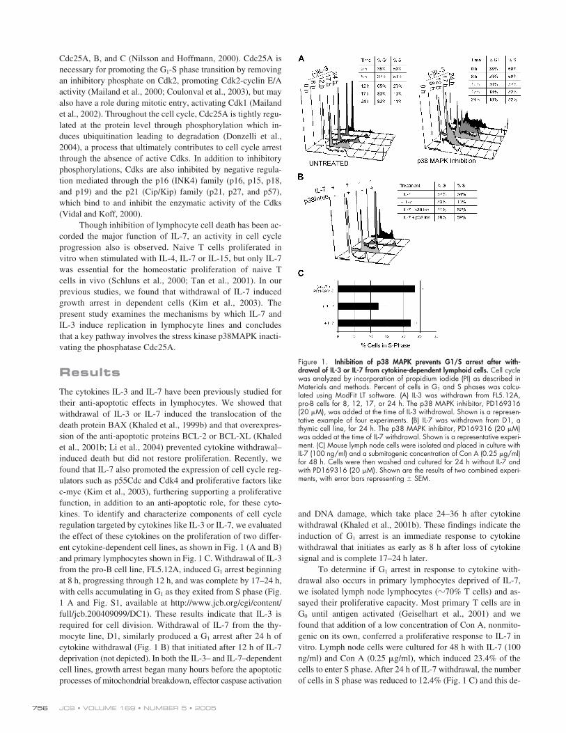
The cytokines IL-3 and IL-7 have been previously studied fortheir anti-apoptotic effects in lymphocytes. We showed thatwithdrawal of IL-3 or IL-7 induced the translocation of thedeath protein BAX (Khaled et al., 1999b) and that overexpres-sion of the anti-apoptotic proteins BCL-2 or BCL-XL (Khaledet al., 2001b; Li et al., 2004) prevented cytokine withdrawal–induced death but did not restore proliferation. Recently, wefound that IL-7 also promoted the expression of cell cycle reg-ulators such as p55Cdc and Cdk4 and proliferative factors likec-myc (Kim et al., 2003), furthering supporting a proliferativefunction, in addition to an anti-apoptotic role, for these cyto-kines. To identify and characterize components of cell cycleregulation targeted by cytokines like IL-3 or IL-7, we evaluatedthe effect of these cytokines on the proliferation of two differ-ent cytokine-dependent cell lines, as shown in Fig. 1 (A and B)and primary lymphocytes shown in Fig. 1 C. Withdrawal of IL-3from the pro-B cell line, FL5.12A, induced G
1
arrest beginningat 8 h, progressing through 12 h, and was complete by 17–24 h,with cells accumulating in G
1
as they exited from S phase (Fig.1 A and Fig. S1, available at http://www.jcb.org/cgi/content/full/jcb.200409099/DC1). These results indicate that IL-3 isrequired for cell division. Withdrawal of IL-7 from the thy-mocyte line, D1, similarly produced a G
1
arrest after 24 h ofcytokine withdrawal (Fig. 1 B) that initiated after 12 h of IL-7deprivation (not depicted). In both the IL-3– and IL-7–dependentcell lines, growth arrest began many hours before the apoptoticprocesses of mitochondrial breakdown, effector caspase activation
and DNA damage, which take place 24–36 h after cytokinewithdrawal (Khaled et al., 2001b). These findings indicate theinduction of G
1
arrest is an immediate response to cytokinewithdrawal that initiates as early as 8 h after loss of cytokinesignal and is complete 17–24 h later.
To determine if G
1
arrest in response to cytokine with-drawal also occurs in primary lymphocytes deprived of IL-7,we isolated lymph node lymphocytes (
�
70% T cells) and as-sayed their proliferative capacity. Most primary T cells are inG
0
until antigen activated (Geiselhart et al., 2001) and wefound that addition of a low concentration of Con A, nonmito-genic on its own, conferred a proliferative response to IL-7 invitro. Lymph node cells were cultured for 48 h with IL-7 (100ng/ml) and Con A (0.25
�
g/ml), which induced 23.4% of thecells to enter S phase. After 24 h of IL-7 withdrawal, the numberof cells in S phase was reduced to 12.4% (Fig. 1 C) and this de-
Figure 1. Inhibition of p38 MAPK prevents G1/S arrest after with-drawal of IL-3 or IL-7 from cytokine-dependent lymphoid cells. Cell cyclewas analyzed by incorporation of propidium iodide (PI) as described inMaterials and methods. Percent of cells in G1 and S phases was calcu-lated using ModFit LT software. (A) IL-3 was withdrawn from FL5.12A,pro-B cells for 8, 12, 17, or 24 h. The p38 MAPK inhibitor, PD169316(20 �M), was added at the time of IL-3 withdrawal. Shown is a represen-tative example of four experiments. (B) IL-7 was withdrawn from D1, athymic cell line, for 24 h. The p38 MAPK inhibitor, PD169316 (20 �M)was added at the time of IL-7 withdrawal. Shown is a representative experi-ment. (C) Mouse lymph node cells were isolated and placed in culture withIL-7 (100 ng/ml) and a submitogenic concentration of Con A (0.25 �g/ml)for 48 h. Cells were then washed and cultured for 24 h without IL-7 andwith PD169316 (20 �M). Shown are the results of two combined experi-ments, with error bars representing � SEM.

CYCLING IS MEDIATED THROUGH CDC25A • KHALED ET AL.
757
clined further to 4% by 48 h (not depicted). Hence, withdrawalof IL-7 from either cytokine-dependent cell lines or primarylymphocytes induces growth arrest before apoptosis.
Previously, we reported that shortly after withdrawal ofIL-3 or IL-7, the MAPK stress pathway was activated (Fig. S2,available at http://www.jcb.org/cgi/content/full/jcb.200409099/DC1) and played an important role in inducing the later eventsof apoptosis (Khaled et al., 2001a; Rajnavolgyi et al., 2002).Therefore, we examined whether stress kinases could also beinvolved in cell cycle regulation, specifically the induction ofgrowth arrest observed in the absence of a cytokine signal.PD169316, a potent inhibitor of the stress kinase p38 MAPK,dramatically increased the number of FL5.12A cells in S phaseafter withdrawal of IL-3, and the weaker p38 MAPK inhibitorSB202190 also showed significant effects (Fig. 1 A and Fig. 2A). In contrast, inhibition of other MAPKs, ERK or JNK, failedto restore G
1
-S phase progression (Fig. 2 A) after IL-3 with-drawal. An effect of pharmacological inhibition of p38 MAPKwas accelerated cell death in the absence of cytokines, as can benoted by the increased number of sub-G
1
cells (Fig. 1, A and B).This was not observed upon inhibition of the ERK or JNK path-way, Thus, potent pharmacological inhibitors of p38 MAPK, inaddition to relieving the G1 arrest, also induce some cell death.Perhaps this effect results from mitotic catastrophe or fromother drug effects.
To specifically inhibit p38 MAPK without using pharma-cological inhibitors, we expressed a dominant negative p38MAPK, which specifically inhibits p38 MAPK activity, inFL5.12A cells and again observed restoration of cell cycling(
�
22% in S phase; Fig. 2 A) and DNA synthesis, shown byBrdU incorporation (
�
18% cells synthesizing DNA; Fig. 2 B),in the absence of IL-3. In the IL-7–dependent thymocyte line,D1, G
1
arrest after IL-7 withdrawal was also dramatically re-leased by inhibiting p38 MAPK with PD169316, resulting inincreased progression into S phase (Fig.1 B). Primary lympho-cytes showed a similar pattern upon inhibition of p38 MAPK,relieving the arrest that followed IL-7 withdrawal (Fig. 1 C).These results indicate that activation of p38 MAPK after cyto-kine withdrawal induces rapid G
1
-S phase arrest (well beforethe onset of apoptosis) and that inhibition of p38 MAPK sub-stantially restores S phase progression, confirmed by measure-ments of DNA synthesis.
We also assayed for the activity of other checkpoint ki-nases, specifically CHK1, to determine if these were inducedby cytokine withdrawal. Unlike the increased activation of p38MAPK detected upon cytokine deprivation, we found that pro-tein levels of CHK1 were negligible by 17 h of IL-3 (Fig. S2)or IL-7 (not depicted) withdrawal, though detectable in thepresence of cytokines. Therefore, inhibition of CHK1 by RNAinterference (Fig. 2 A) did not restore S phase progression inthe absence of IL-3, suggesting that CHK1 initiated checkpointregulation was not the mechanism by which cytokine with-drawal induced growth arrest.
Figure 2. Inhibition of p38 MAPK, but not ERK or JNK, restores S-pro-gression and Rb phosphorylation in the absence of IL-3. Cell cycle wasanalyzed by incorporation of PI as described in Materials and methods.Percent of cells in S phase was calculated using ModFit LT software. (A)FL5.12A cells were cultured with or without IL-3 for 17 h. The p38 MAPKinhibitors, PD169316 (20 �M) or SB202190 (20 �M), the MEK1 inhibitor(activator of ERK), PD98059 (20 �M), and the Jun kinase inhibitor, JNKinhibitor II (20 �M), were added at the time IL-3 was withdrawn. DMSO(0.1%) was included as a negative control. Two different FL5.12A clonesB3 and A2, stably transfected with a dominant negative (DN) p38 MAPK,were placed in culture without IL-3 for 17 h. Results shown are representa-tive of two experiments. (B) FL5.12A-A2 cells, expressing DN-p38 MAPK,incorporated Brdu during 17 h IL-3 withdrawal, while untransfected cellsarrested. (C) Levels of phosphorylated Rb protein were assayed inFL5.12A cells after 48 h of IL-3 withdrawal. Nuclear lysates were resolvedby SDS-PAGE and immunoblotting as described in Materials and methodsusing an antibody specific for Rb protein phosphorylated at serine 780.
Figure 3. Inhibition of p38 MAPK after IL-3 withdrawal induces minor in-creases in G1-S Cdks and cyclins in B lymphocytes. (A) FL5.12A cells wereincubated for 12 h in the absence of IL-3 and then further incubated for 5 hwith either PD169316 (20 �M) or IL-3 (not depicted) for a total of 17 h.Cells were then harvested and total RNA isolated. RPAs were performedas described in Materials and methods. Transcription of L32 was mea-sured as a control for total mRNA expressed. Results were quantified usinga Bio-Rad Gel Documentation system, and mRNA transcripts for Cdks andcyclins normalized to the levels of L32 expression. (B) Whole cell lysateswere prepared from FL5.12A cells deprived of IL-3 for 17 h and immuno-blotted as described in Materials and methods for detection of Cdk4 andcyclin D3 with specific antibodies.

JCB • VOLUME 169 • NUMBER 5 • 2005758
Phosphorylation of Rb, which releases E2F and promotesentry into S-phase, was interrupted by IL-3 withdrawal and re-stored by inhibition of p38 MAPK (Fig. 2 C), suggesting that inthese cells p38 MAPK acted upstream of the events leading toRb phosphorylation. One possibility would be that p38 MAPKnegatively regulates the synthesis of the Cdk 4/6-cyclin D com-plexes (Lavoie et al., 1996), thereby retaining hypophosphory-lated Rb in the absence of these complexes. If so, then wewould expect levels of mRNA transcripts for G
1
-S phase Cdksand cyclins to increase upon p38 MAPK inhibition. However,RNase protection assays (RPAs), to measure mRNA transcriptsfor Cdks and cyclins, for example Cdk 4, during initiation ofG
1
arrest showed decline of transcription after 12 h of IL-3(Fig. 3 A) and IL-7 (not depicted) withdrawal, and minor, ifany, increase in transcription as a result of inhibiting p38MAPK with PD169316. Re-addition of IL-3 or IL-7 after cy-tokine withdrawal substantially increased the transcription ofCdks and cyclins as would be expected upon restoration of cy-tokine signaling pathways (unpublished data). In the absence ofIL-3, inhibition of p38 MAPK did result in a slight increase inexpression of cyclins D2, D3 and E (Fig. 3 A), which will befurther discussed. RPAs were also performed to measure tran-scription of Cdks 1, 5, 7, and 8 and cyclins A2, B1, C, B2, F,G1, G2, and H, and no effects of p38 MAPK inhibition wereobserved (not depicted).
To determine if decreased transcription was reflected byloss of protein, we measured the protein levels for one of theCdks, Cdk4, and one of the cyclins, cyclin D3. We found thatthese cell cycle mediators could still be detected by immuno-blotting cell lysates even 17 h after IL-3 withdrawal when cellsare fully arrested (Fig. 3, B and C), demonstrating that, thoughmRNA levels decreased, the proteins themselves were not lim-iting in the early stages of cytokine withdrawal. Thus, themechanism through which p38 MAPK regulates cell cycling incytokine-dependent lymphocytes is not solely based on con-trolling the synthesis of Cdks and cyclins.
Whereas other growth factors induce the synthesis of cy-clin D, we considered the possibility that posttranslationalregulation of cyclin D could be regulated by IL-3 and IL-7,because it has been reported that p38 MAPK could phos-phorylate cyclin D1, leading to its ubiquitination and degrada-tion (Casanovas et al., 2000). We therefore overexpressedcyclin D1 in FL5.12A cells to determine whether it wouldmimic the effect of p38 MAPK inhibition in releasing cellsfrom G
1
arrest after IL-3 loss. This was not the case becauseoverexpression of cyclin D1 in FL5.12A cells failed to relievecell cycle arrest in the absence of IL-3 whereas the p38 MAPKinhibitor continued to have this effect as it did in untransfected
cells (Fig. S3, available at http://www.jcb.org/cgi/content/full/jcb.200409099/DC1). Together, withdrawal of IL-3 leads toG
1
arrest by a mechanism unrelated to the levels of cyclin DmRNA (Fig. 3 A) or protein (Fig. 3, B and C).
Inhibition of Cdk2, a kinase involved in the transition intoS phase, with a potent and selective pharmacological inhibitor(Cdk2 Inhibitor II – Compound 3), countered the effect of in-hibiting p38 MAPK after 17 h of IL-3 withdrawal (Fig. 4). Thiseffect was evident in the absence of IL-3. Because there wereminimal effects of p38 MAPK inhibition on the transcription ofCdk2 (Fig. 3 A), we examined a role for regulation of Cdk2 ac-tivity, specifically through the Cdc25 family of phosphatasesknown to dephosphorylate and activate Cdks (Sexl et al., 1999).Treatment of FL5.12A cells with a pharmacological inhibitor(NSC 95397) of Cdc25 phosphatases reversed the S phase pro-gression induced by p38 MAPK inhibition (Fig. 4). These find-ings suggest that Cdc25 phosphatases, and their activation ofCdks, could be essential components of the pathway throughp38 MAPK by which cytokines regulate cell proliferation.
Previous studies had shown that p38 MAPK could di-rectly phosphorylate members of the Cdc25 family, Cdc25Band Cdc25C, inducing their degradation (Bulavin et al., 2001,2002). However, it is Cdc25A that is involved in the G
1
-Sphase transition, dephosphorylating Cdk2 on an inhibitory ty-rosine, Y15, thereby activating it. We therefore assayed forCdc25A and observed that the levels of endogenous (Fig. 5 A)and overexpressed HA-tagged Cdc25A rapidly declined 4–8 hafter IL-3 withdrawal (Fig. 5 B), suggesting degradation of theprotein. Others have shown that the degradation of Cdc25A isproteasome dependent (Mailand et al., 2000) and mediatedthrough ubiquitination (Busino et al., 2004). We found that in-hibiting p38 MAPK subsequently restored Cdc25A protein lev-els, both endogenous levels and overexpressed HA-taggedCdc25A, promoting stability of the phosphatase (Fig. 5 B).Hence, p38 MAPK appears to act upstream of the decline ofCdc25A after IL-3 withdrawal, and regulating Cdc25A proteinstability may be one of the functions of this kinase.
Two critical serines on Cdc25A (S75 and S123) areknown to be kinase targets. DNA damage triggers the Chk ki-nases which phosphorylate Cdc25A on S123 leading to itsdegradation (Falck et al., 2001; Sorensen et al., 2003), and in
Xenopus
embryos, phosphorylation of S73 (S75 in the humansequence) is mediated by another unknown kinase (Shimuta etal., 2002), perhaps also Chk1 as has been recently shown(Hassepass et al., 2003). However, Chk kinases are known tobe activated during cell cycle checkpoints triggered by DNAdamage (Bartek and Lukas, 2003), which occurs later in theapoptotic process, and we did not detect Chk1 protein during
Figure 4. Inhibition of Cdk2 or Cdc25 reverses the effectsof p38 MAPK inhibition on the promotion of cell cyclingduring IL-3 withdrawal. Cell cycling was analyzed byincorporation of PI and percent of cells in S phase wascalculated using ModFit LT software as described in Mate-rials and methods. FL5.12A cells were cultured without IL-3and PD169316 (20 �M) in the presence of 20 �M of apharmacological inhibitor of Cdk2 (Cdk2 inhibitor II) or20 �M of a pharmacological inhibitor of Cdc25 (NSC95397) for 17 h and effects of cell cycling assessed.

CYCLING IS MEDIATED THROUGH CDC25A • KHALED ET AL.
759
cytokine withdrawal (Fig. S2) nor observed any effects on cellcycle progression upon Chk1 inhibition by RNA interference(Fig. 2 A). This demonstrates that cell cycle arrest due to cy-tokine withdrawal does not involve Chk1 and suggests thatphosphorylation of Cdc25A is mediated by a different kinaseactivity in cytokine-deprived lymphocytes.
The regulatory sites on Cdc25A, S75 and S123, can bedirect targets of p38 MAPK as shown by p38 MAPK in vitrokinase assays using HeLa cells (Goloudina et al., 2003). Theseresults, together with the increased stability of Cdc25A pro-moted by p38 MAPK inhibition (Fig. 5 B), suggest that p38MAPK is the kinase principally responsible for phosphoryla-tion of S75 and S123 on Cdc25A, inducing the decline inCdc25A after cytokine withdrawal.
Having shown that the decrease in Cdc25A proteins cor-related with cell cycle arrest after cytokine withdrawal, wetested whether it played an important functional role. BecauseS75 and 123 were the targets of p38 MAPK, leading to its deg-radation, we mutated these sites and measured the effect on cellcycling after cytokine withdrawal. Mutation of S123, and S75to alanines produced a stable form of Cdc25A that significantlyrestored S phase entry after 24 h of IL-3 withdrawal as shownin Fig. 6 A. The Cdc25A double mutant (S75,123A) functionedlike a dominant-positive (DP), promoting cell cycling in the ab-sence of IL-3 compared with expression of the WT protein.Significantly, that activity of Cdc25A-DP in the absence of IL-3resulted in DNA synthesis, as shown by incorporation of BrdU,and not just accumulation in S phase, with cells progressinginto G2/M (Fig. 6 A). This was not observed in cells expressingWT Cdc25A that accumulated in G
1
phase.The effect of expressing the Cdc25A-DP in IL-7–dependent
D1 cells was even more striking, with increased cells cycling inthe absence of IL-7 (24 h) compared with WT protein (Fig. 6 B).Using BrdU incorporation to measure DNA synthesis, we alsoobserved restoration of S phase progression by this Cdc25Amutein after IL-7 withdrawal (Fig. 6 B). These effects requiredmutation of both sites (S75 and S123) on Cdc25A, because
either one alone had minimal effects on cell cycling (not de-picted). Expression of Cdc25-DP (cotransfected with the selec-tion marker GFP) in primary lymphocytes was able to restore
Figure 5. Cdc25A protein is degraded upon cytokine withdrawal andstabilized, in the absence of cytokines, by inhibition of p38 MAPK. (A) Levelsof endogenous mouse Cdc25A protein were measured in FL5.12A cells.Whole cell lysates were made from cells deprived of IL-3 for 2, 4, 8, and16 h, then resolved by SDS-PAGE and immunoblotted as described inMaterials and methods using an antibody specific for Cdc25A. Total p38MAPK was measured as a loading control. (B) Endogenous mouseCdc25A was measured in FL5.12A cells stably overexpressing wild-type(WT) human Cdc25A. Whole cell lysates were made from cells culturedwith or without IL-3 and 20 �M of PD169316 for 16 h, and then resolvedby SDS-PAGE and immunoblotted as described in Materials and methodsusing antibodies specific for mouse Cdc25A and for human Cdc25A.Levels of p38 were detected as a loading control. Shown are representativeexperiments of three such performed.
Figure 6. Expression of Cdc25A mutated at S75 and S123 prevents G1
arrest and sustains S phase progression after withdrawal of IL-3 or IL-7.DNA synthesis was assayed by incorporation of BrdU and cell cycle wasanalyzed by uptake of 7AAD and assayed by flow cytometry as describedin Materials and methods. Percent of cells in G1 and S phase was calcu-lated using ModFit LT software, excluding apoptotic cells and aggregates.(A) FL5.12A cells stably expressing the dominant-positive (DP) Cdc25A(S75,123A) mutein or Cdc25A-WT were cultured with or without IL-3 for24 h. (B) D1 cells stably expressing the Cdc25A-DP (S75, 123A) mutein orCdc25-WT were cultured with or without IL-7 for 24 h. (C) Viability and cellsize of D1 cells stably expressing Cdc25A-DP or CDc25A-WT are shown indot blots displaying forward (FSC) and side scatter (SSC). Results shownare representative of three or more experiments performed. Apoptotic cellswere not excluded (D). Lymph node cells were isolated and grown with IL-7(100 ng/ml) and Con A (0.25 �g/ml) as previously described. Primarycells were transfected with Cdc25A-DP or Cdc25A-WT and GFP, as a se-lection marker. Transfected cells, were selected by gating on GFP expres-sion for analysis of DNA synthesis and cycling by BrdU/7AAD stainingafter 24 h of IL-7 withdrawal as described in Materials and methods.Shown is a representative experiment of three performed.

JCB • VOLUME 169 • NUMBER 5 • 2005760
DNA synthesis (measured by BrdU incorporation) in primarycells deprived of IL-7 for up to 48 h (Fig. 6 D; Fig. S4, availableat http://www.jcb.org/cgi/content/full/jcb.200409099/DC1).Cdc25A-WT also had a positive effect on primary lymphocyteproliferation, though not as striking as that of the DP protein,perhaps due to reduced levels of p38 MAPK in primary cells ascompared with cell lines. These findings indicate that Cdc25Ais pivotal regulator of IL-7-driven proliferation of lymphocytes,cell lines and primary cells, and a novel target of IL-7 signaltransduction through p38 MAPK.
In addition to promoting cell cycling, expression of theCdc25A-DP was able to extend the life of cells grown in theabsence of IL-7. Shown in Fig. 6 C, are forward (FSC)/sidescatter (SSC) plots of D1 cells grown in the absence of IL-7 for48 h. Cells expressing the Cdc25A WT protein, lacking IL-7,had begun to undergo apoptosis and shrink in size, as indicatedby decreased FSC, whereas cells expressing Cdc25A-DP re-mained viable and did not undergo atrophy. Therefore, expres-sion of Cdc25A-DP replaced the IL-7 signal for cycling andcell maintenance for 2–5 d of cytokine withdrawal, after whichcells expired likely due to loss of nutrient uptake (Khaled andDurum, 2003). Untransfected cells or cells expressing the WT
protein shrink and die after 36–48 h of cytokine deprivation. Ascomparison, expression of the anti-apoptotic proteins, BCL-2(Li et al., 2004) or BCL-XL (Fig. S5, available at http://www.jcb.org/cgi/content/full/jcb.200409099/DC1), thoughprotecting cells from death, did not promote cell cycling, butrather inhibited growth and instead maintained cells in ashrunken, vegetative state, likely undergoing autophagy in theabsence of cytokines.
Mutation of S75 and S123 on Cdc25A also mimicked theeffect of inhibiting p38MAPK in stabilizing the Cdc25A proteinafter IL-3 withdrawal (Fig. 7, A and B) and IL-7 withdrawal(not depicted). Mutation of S75 alone had a partial effect of sta-bilizing Cdc25A (Fig. 7 B), although cell cycle progressionwas not restored, whereas mutation of S123 alone did not re-store protein stability (not depicted). Hence, mutation of bothS75 and S123 are required to deregulate Cdc25A, promotingprotein stability and function in the context of cytokine depri-vation in lymphocytes.
The Cdc25A-DP mutant protein was also fully competentfor phosphatase activity. The DP mutant sustained phosphory-lation of Rb and Cdks (Cdk2 and Cdk1) after withdrawal of IL-3or IL-7 (Fig. 7 C). Specifically, we detected the accumulationof hyperphosphorylated Cdks at T160 and Y15, correlatingwith active cell cycling despite cytokine deprivation. It is pos-sible that the increased expression of cyclin E (and perhapscyclins D2 and D3) transcripts observed during p38 MAPKinhibition (Fig. 3 A) resulted from phosphorylated Rb which inturn would result in the release of E2F and the subsequent in-duction of various genes, such as cyclins, involved in cell cycleprogression. Hence, expression of Cdc25A-DP was sufficientto induce cell cycling and maintain cell integrity in the absenceof a cytokine growth stimulus, and this in part resulted from theactivation of Cdks/cyclins and the phosphorylation of Rb.
Discussion
Lymphocytes depend on external signals from cytokines forsurvival and proliferation. Here we have examined the mecha-nisms by which IL-3 and IL-7 induce proliferation of lymphoidcell lines and primary lymphocytes and find that this pathwaydiffers from that of better-studied factors that induce growth ofmesenchymal cells. Rather than inducing synthesis of cyclins,these cytokines appear to protect lymphocytes from a stress re-sponse. Withdrawal of IL-3 or IL-7 induced cell cycle arrestthrough activation of a stress kinase, p38 MAPK, which oc-curred in the first few hours after cytokine withdrawal. p38MAPK then directly phosphorylated the phosphatase Cdc25Aat S75 and S123, targeting the phosphatase for degradation.Because Cdc25A is required to remove an inhibitory phosphate(Y15) from Cdk2, the latter kinase was inactive, failed to phos-phorylate Rb and the cells arrested at the G
1
-S boundary. Weshow that inhibiting either component of this pathway, block-ing p38 MAPK activity or expressing a p38 MAPK-resistantform of Cdc25A, prevented growth arrest in the absence of acytokine receptor signal, restoring cell cycle progression aswell maintaining cell viability and size. We have producedtime courses (some are shown) for most of these phenomena.
Figure 7. Cdc25A-DP (S75,123A) remains stable and sustains phos-phorylation of Rb and Cdks after IL-3 withdrawal. (A) Protein levels ofCdc25A-DP (S75,123A) were measured in FL5.12A cells. Cells were de-prived of IL-3 for 17 h, and cytosolic extracts were resolved by SDS-PAGEand immunoblotted as described in Materials and methods using an anti-body specific for mouse (to detect endogenous protein) or human Cdc25A(to detect the overexpressed HA-tagged protein). Total p38 MAPK wasmeasured as a loading control. Shown is a representative experiment oftwo. (B) Stability of Cdc25A was determined in FL5.12A cells overexpress-ing the Cdc25A-DP mutant (DP) and the Cdc25A single mutant (S75A) (SP)after IL-3 withdrawal. The immunoblot was quantified using a Bio-Rad GelDocumentation system and results shown in the adjacent panel. The graphindicates fold differences in protein levels measured with or without IL-3 for16 h. (C) Levels of phosphorylated Rb protein and Cdk2 were measured inFL5.12A cells and FL5.12A cells stably overexpressing the Cdc25A-DPmutein. Nuclear extracts were made from cells deprived of IL-3 for 17 h,then resolved by SDS-PAGE and immunoblotted, as described in Materialsand methods, using antibodies specific for phospho-Rb protein (phosphory-lated at serine 780), phospho-Cdk (tyrosine 15) (detects phospho-Cdk2and phospho-Cdk1) and phospho-Cdk (threonine 160; detects phospho-Cdk2 and phospho-Cdk1). Total Cdk2 levels were also measured with aspecific antibody. Note, the same cell lysates (equally loaded) were usedin the phospho-Rb, phospho-Cdks, and Cdk-2 blots.
CYCLING IS MEDIATED THROUGH CDC25A • KHALED ET AL.
761
The sequence of events after cytokine withdrawal is: (1–12 h)p38 activation, Cdc25a phosphorylation and degradation; (12–48 h) G1 arrest; (24–48 h) apoptosis. The time points selectedfor the figures were intended to illustrate each of these phases.
Another mechanism reported for cell cycle arrest aftercytokine withdrawal is through an increase in p27
kip1
, a nega-tive regulator of proliferation through its interactions withCdks, especially Cdk2 (for review see Olashaw and Pledger,2002). Protein levels of p27
kip1
accumulate in serum-starved andgrowth factor–deprived cells, and we have confirmed this in-crease occurs in cells deprived of IL-7 or IL-3. However, wenoted that p27
kip1
levels also increased after cytokine withdrawalin cells overexpressing Cdc25A-DP (unpublished data) that werenot arrested in G
1
(Fig. 6); thus these cells replicated normally inthe absence of cytokines despite their elevation in p27
kip1
. Thissuggests that the positive effect of Cdc25A-DP on cell cycle pro-gression can dominate over the negative effect of p27
kip1
.Microinjection of antibodies to Cdc25A have been shown
to arrest cells in G
1
(Jinno et al., 1994), indicating the impor-tance of this phosphatase in the cell cycle. Overexpression ofwild-type Cdc25A in rat-1 cells accelerated entry into S phaseand the activation of Cdk2 (Blomberg and Hoffmann, 1999).However, in the lymphocytes in our studies, overexpressedwild-type Cdc25A was degraded in the absence of IL-3 or IL-7and did not restore cell cycle progression in cell lines (Figs. 5and 6), whereas overexpression of the stable Cdc25A-DP sus-tained cell growth (Figs. 6 and 7). Thus, in our studies, the fail-ure to rescue cell division with overexpressed wild-typeCdc25A in cell lines is presumably because it is rapidly de-graded after phosphorylation by p38 MAPK, which is activatedby cytokine withdrawal. However, in primary cells, expressionof Cdc25A-WT did have growth promoting effects likely be-cause the levels of active p38 MAPK may be reduced in com-parison to the cell lines. Even in this case, however, expressionof Cdc25A-DP resulted in IL-7–independent DNA synthesisbeyond that observed with expression of the WT protein.
In cell lines, Cdc25A-DP supported multiple rounds ofcell division in the absence of cytokine. Over the first 2 d afivefold increase in cell numbers was observed, thereafter, cellscontinued to divide until the rate of cell death increased to thepoint that all cells were dead by day 5. The eventual death ofcytokine-deprived Cdc25A-DP expressing cells could resultboth from apoptosis (because they cease expressing Bcl-2) andfrom metabolic depression because cytokine withdrawal alsoreduces glucose uptake as we showed for IL-7 (Khaled and Du-rum, 2003) and others showed for IL-3 (Plas et al., 2001).
If cytokines induce survival via Bcl-2 or Bcl-XL and pro-liferation via Cdc25A one might predict that transfecting bothBcl-2 and Cdc25A should render cells capable of both survivaland proliferation in the absence of cytokine. However we wereunable to obtain stable lines of Bcl-2/Cdc25a doubly trans-fected cells. This could be because, as reported by others (Ja-numyan et al., 2003; Cheng et al., 2004), overexpression ofBCL-2 or BCL-XL, although maintaining the life of the cell,can also inhibit its replication. Or it could be due to lack of glu-cose uptake, as noted above; thus a cell protected by Bcl-2 orBCL-XL may be able to survive in a quiescent state when de-
prived of cytokine-induced glucose uptake, but when driven todivide by Cdc25A-DP the cell may die from metabolic stress.
Recent studies in HeLa cells showed that osmotic stressor UV irradiation induced cell cycle arrest. This arrest was ac-companied by degradation of wild-type Cdc25A, but notCdc25A-DP, however, in contrast to our studies, cell cyclingwas not restored by Cdc25A-DP (Goloudina et al., 2003). Thissuggests that multiple checkpoints, in addition to Cdc25A reg-ulation, must be involved in the growth arrest from osmoticstress or UV irradiation and ensuing DNA damage. One suchpathway would be stabilization of p53 leading to p21 induc-tion; this would occur after UV irradiation but does not occurafter cytokine withdrawal, for which we show that Cdc25A de-stabilization is the major mechanism of growth arrest.
CHK1 and CHK2 are also reported to phosphorylate anddestab
i
lize CDC25A. We measured levels of CHK1 in cyto-kine-dependent cells and found that this protein disappeared af-ter cytokine withdrawal (Fig. S2 B). CHK2 is not as critical aregulator of Cdc25A as is CHK1. We also treated cells withCHK1 small interfering RNA and saw no effect on the G1 ar-rest induced by cytokine withdrawal (Fig. 2 A). In contrast tothe disappearance of CHK1, p38 activity dramatically in-creased after cytokine withdrawal (Fig. S2 A). These observa-tions favor a role for p38, rather than CHK1 in the down-regu-lation of Cdc25A after cytokine withdrawal.
We found that p38 MAPK was required to phosphorylateS75 and S123 on Cdc25A and induce its degradation. Othershave shown that, in response to DNA damage, phosphorylationof S75, followed by phosphorylation of S82 and S88, leads to
�
TrCP-dependent ubquitination and degradation of Cdc25A(Donzelli et al., 2004). The kinase that phosphorylates Cdc25Aat S82 and S88 is unknown, and it may be active in cytokine-deprived lymphocytes in addition to p38 MAPK as we haveshown. In other cell types, others have shown that mutation ofS75 alone was sufficient to confer stability to Cdc25A (Golou-dina et al., 2003; Busino et al., 2004), however we found only apartial effect, whereas mutation of both S75 and S123 conferreda full stabilizing effect (Fig. 7). Mutation of both sites onCdc25A also restored proliferation to cells deprived of cytokines(Fig. 6), whereas single mutations had little effect (not depicted),providing the strongest support for the importance of the stabil-ity of this phosphatase in regulating lymphocyte proliferation.
We have shown that cytokine withdrawal from lympho-cytes results in two distinguishable responses, one after theother. First, cells undergo cell cycle arrest; second, they un-dergo apoptotic cell death. These two processes, cell cycle ar-rest and apoptosis are not only distinguishable kinetically andmechanistically, they also appear to require different levels ofcytokine receptor occupancy, i.e., a low concentration of thecytokine is sufficient to protect from cell death but is insuffi-cient to induce cell division (unpublished data). These concen-tration effects of cytokines, low dose inducing survival, highdose inducing division, presumably account for these two ho-meostatic activities of IL-7 in the peripheral immune system(Khaled and Durum, 2002). Thus, when lymphocyte density isat its maximum, cells would consume the available IL-7 as rap-idly as it is synthesized and each cell would encounter just

JCB • VOLUME 169 • NUMBER 5 • 2005762
enough IL-7 to protect from cell death but not enough to prolif-erate. When the lymphoid compartment is relatively empty, IL-7would be sufficiently abundant to drive proliferation as well assurvival. Our studies with lymphoid cell lines and primary cellssuggest that, in vivo, this homeostatic proliferation of lympho-cytes may be regulated by p38MAPK and Cdc25A and furtherstudies to test this hypothesis are underway.
Materials and methods
Cell lines, cells, and treatments
The IL-7–dependent cell line, D1, was established from CD4
�
CD8
�
mousethymocytes isolated from a p53
�
/
�
mouse as previously described (Kim etal., 2003). The IL-3–dependent cells are murine pro-B cell line, FL5.12A(Khaled et al., 2001b). Pharmacological inhibitors of p38 MAPK,PD169316, and SB202190 (Calbiochem), MEK1 inhibitor, PD98059(Calbiochem), JNK Inhibitor II (Calbiochem), Cdk2 Inhibitor II (Compound3; Calbiochem), and Cdc25 inhibitor (NSC 95397; Sigma-Aldrich) weremade as 20 mM stocks in DMSO. Inhibition of Chk1 was achieved by in-troducing chemically synthesized small interfering RNA (Santa Cruz Bio-technology, Inc.) using a lipid reagent,
Trans-IT
TKO (Mirus) following themanufacturer’s protocol. Cells were treated as described in figure leg-ends. Lymph node cells from 12-wk-old C57Bl/6 mice were isolated bymechanical teasing and placed in culture, 5
�
10
6
cells/ml, in the pres-ence of 100 ng/ml IL-7 (Peprotech) and 0.25 ng/ml Con A (Sigma-Aldrich)from 48 h before IL-7 withdrawal.
Plasmids, site-directed mutagenesis, and transfections
For dominant negative inhibition of p38 MAPK, pCMV-Flag-p38 (agf; agift from R. Davis, University of Massachusetts Medical Center, Worcester,MA) was used to transfect cells. Overexpression of cyclin D1 wasachieved with pCMV-HA-cyclin D1 (a gift from P. Kaldis, National CancerInstitute at Frederick). The pCMV-HA-Cdc25 WT plasmid (a gift from J.Bartek, Danish Cancer Society, Copenhagen, Denmark) was used to gen-erate mutants pCMV-HA-Cdc25 S75A and/or S123A, as previously de-scribed (Goloudina et al., 2003). For expression in the D1 cells, the1.6-kBEcoR1 fragment from either pCMV-HA-Cdc25 WT or pCMV-HA-Cdc25S75,123A mutant was subcloned into pcDNA 6/V5-HisB (Invitrogen) forselection with Blastocidin. Cells were transfected by electroporation (BTXmodel 830) using standard methodologies. Before electroporation, cellswere incubated in a hypoosmolar electroporation buffer (Eppendorf) toslightly swell the cells and improve transfection efficiency. Primary lympho-cytes were isolated from lymph nodes as described, treated with IL-7 andCon A for 48 h, and transfected with
TransIT
-LTI Transfection reagent (Mi-rus) following the manufacturer’s guidelines. Transfected cells (4–6%)were selected by GFP coexpression and assayed for cell cycling and DNAsynthesis after 48 h with or without IL-7.
Cell cycle analysis and BrdU incorporation
For cell cycle analysis, DNA content was measured by propidium iodide(PI) or 7AAD staining. Cells, withdrawn from cytokine and/or treated withpharmacological inhibitors (as described in figure legends), were placedin detergent buffer (Kim et al., 2003) with 50
�
g/ml Rnase (Invitrogen) ata concentration of 1–2
�
10
6
cells/ml with an equal volume of PI (50
�
g/ml) or 7AAD. Samples were mixed and incubated at RT for 1 h beforeanalysis, then assayed by flow cytometry using a FACSCalibur flow cy-tometer (Becton Dickinson) and CellQuest software. Listmode data was ac-quired and analyzed using ModFit LT software (Verity), excluding apop-totic cells and cell aggregates. DNA synthesis was assessed by BrdUincorporation, using FITC-anti-BrdU or PE-anti-BrdU antibody for detection,and 7AAD staining for DNA content with a commercially available kit fol-lowing the manufacturer’s instructions (BD Biosciences). BrdU incorpora-tion in primary T cells was measured using an anti-BrdU-PE–labeled anti-body, gating on the GFP-positive cells.
Immunoblotting and RPA
For detection of Cdc25A, cell lysates and the making of specific antibod-ies have been previously described (Goloudina et al., 2003). For analysisof phospho-Rb and phospho-Cdk2, nuclear cell lysates were made using amodified Dignam protocol containing phosphatase inhibitors and pro-tease inhibitors (Dignam et al., 1983; Trede et al., 1993; Khaled et al.,1999a). Cells lysates were resolved by SDS-PAGE and immunoblotted.For detection of Cdc25A, mAbs were used (Ab-3, NeoMarkers). For de-
tection of the phosphorylated form of Rb, cyclin D3, and Cdk4, specificantibodies were used: a rabbit polyclonal for detecting phospho-Rb(Serine 780; Cell Signaling Technology) and mouse monoclonals for de-tecting cyclin D3 and Cdk4 (Cell Signaling Technology). Cdk2 was de-tected using a rabbit pAb (M2; Santa Cruz Biotechnology, Inc.). To detectthe phosphorylated forms of Cdk2 and Cdk1, blots were probed for phos-pho-Cdks (Tyr15) with a rabbit pAb to phospho-Cdk1 (Tyr15; Cell Signal-ing Technology), which can also detect Cdk2 when catalytically inacti-vated by phosphorylation at Tyr15, and for phospho-Cdk (Thr160) with arabbit pAb specific for Cdk2 and Cdk1 when activated and phosphory-lated at this site (Cell Signaling Technology). Appropriate secondary rab-bit or mouse antibodies cross-linked to HRP (Cell Signaling) were used fordetection. ECL (Pierce Chemical Co.) was used for visualization followingthe manufacturer’s protocol. For detection of mRNA levels for Cdks andcyclins, RPA was performed with BD Riboquant multi-probe kits, mCC-1,mCYC-1, and mCYC-2 (BD Biosciences) following the manufacturer’sguidelines. For measurement of p38 activity a commercially available p38nonradioactive kinase assay kit (Cell Signaling) was used following themanufacturer’s protocol. Chk1 protein levels were determined by immuno-blotting nuclear and cytosolic lysates with a specific antibody (sc8408;Santa Cruz Biotechnology, Inc.) as described above for Rb, and detectionof
�
-tubulin with a specific antibody (Sigma-Aldrich) was used as a load-ing control for nuclear lysates.
Online supplemental material
Fig. S1 is an example of IL-3 driven progression through S phase as demon-strated by BrdU incorporation. Fig. S2 demonstrates that p38 MAPK activ-ity increases rapidly after cytokine withdrawal, whereas the protein levelsof CHK1 decline. Fig. S3 shows that overexpression of cyclin D1 fails to re-store S phase progression in cells deprived of IL-3. Fig. S4 displays the gat-ing used to select and examine GFP/Cdc25-DP double transfected primarylymphocytes. Fig. S5 demonstrates that overexpression of BCL-XL, thoughable to protect cells from apoptosis due to IL-3 withdrawal, maintains cellsin a shrunken, nonproliferative state. Online supplemental material is avail-able at http://www.jcb.org/cgi/content/full/jcb.200409099/DC1.
We wish to acknowledge Deepika Minhas, Ph.D. for scientific contributionsand discussions and Amy Grenier, B.S. for assistance performing flow cytom-etry. We are also grateful to P.E. Kolattukudy, Ph.D. and J. Oppenheim, M.D.for their comments on the manuscript.
This publication has been funded in whole or in part with Federalfunds from the National Cancer Institute, National Institutes of Health, undercontract no. NO1-CO-12400 and the career development award K22CA097984-01.
The content of this publication does not necessarily reflect the views orpolicies of the Department of Health and Human Services, nor does mentionof trade names, commercial products, or organizations imply endorsement bythe U.S. government.
Submitted: 16 September 2004Accepted: 26 April 2005
References
Bartek, J., and J. Lukas. 2003. Chk1 and Chk2 kinases in checkpoint control andcancer.
Cancer Cell.
3:421–429.
Blomberg, I., and I. Hoffmann. 1999. Ectopic expression of Cdc25A acceleratesthe G(1)/S transition and leads to premature activation of cyclin E- andcyclin A-dependent kinases.
Mol. Cell. Biol.
19:6183–6194.
Bulavin, D.V., Y. Higashimoto, I.J. Popoff, W.A. Gaarde, V. Basrur, O. Po-tapova, E. Appella, and A.J. Fornace Jr. 2001. Initiation of a G2/M check-point after ultraviolet radiation requires p38 kinase.
Nature.
411:102–107.
Bulavin, D.V., S.A. Amundson, and A.J. Fornace. 2002. p38 and Chk1 kinases:different conductors for the G(2)/M checkpoint symphony.
Curr. Opin.Genet. Dev.
12:92–97.
Busino, L., M. Chiesa, G.F. Draetta, and M. Donzelli. 2004. Cdc25A phos-phatase: combinatorial phosphorylation, ubiquitylation and proteolysis.
Oncogene.
23:2050–2056.
Casanovas, O., F. Miro, J.M. Estanyol, E. Itarte, N. Agell, and O. Bachs. 2000.Osmotic stress regulates the stability of cyclin D1 in a p38SAPK2-dependent manner.
J. Biol. Chem.
275:35091–35097.
Cheng, N., Y.M. Janumyan, L. Didion, C. Van Hofwegen, E. Yang, and C.M.Knudson. 2004. Bcl-2 inhibition of T-cell proliferation is related to pro-longed T-cell survival.
Oncogene.
23:3770–3780.
Coulonval, K., L. Bockstaele, S. Paternot, and P.P. Roger. 2003. Phosphoryla-

CYCLING IS MEDIATED THROUGH CDC25A • KHALED ET AL.
763
tions of cyclin-dependent kinase 2 revisited using two-dimensional gelelectrophoresis.
J. Biol. Chem.
278:52052–52060.
Dignam, J.D., R.M. Lebovitz, and R.G. Roeder. 1983. Accurate transcriptioninitiation by RNA polymerase II in a soluble extract from isolated mam-malian nuclei.
Nucleic Acids Res.
11:1475–1489.
Donzelli, M., L. Busino, M. Chiesa, D. Ganoth, A. Hershko, and G.F. Draetta.2004. Hierarchical order of phosphorylation events commits Cdc25A tobetaTrCP-dependent degradation.
Cell Cycle.
3:469–471.
Falck, J., N. Mailand, R.G. Syljuasen, J. Bartek, and J. Lukas. 2001. The ATM-Chk2-Cdc25A checkpoint pathway guards against radioresistant DNAsynthesis.
Nature.
410:842–847.
Geiselhart, L.A., C.A. Humphries, T.A. Gregorio, S. Mou, J. Subleski, and K.L.Komschlies. 2001. IL-7 administration alters the CD4:CD8 ratio, in-creases T cell numbers, and increases T cell function in the absence ofactivation.
J. Immunol. 166:3019–3027.
Goloudina, A., H. Yamaguchi, D.B. Chervyakova, E. Appella, A.J. Fornace Jr.,and D.V. Bulavin. 2003. Regulation of human Cdc25A stability by serine75 phosphorylation is not sufficient to activate a S phase checkpoint.Cell Cycle. 2:473–478.
Hassepass, I., R. Voit, and I. Hoffmann. 2003. Phosphorylation at serine 75 isrequired for UV-mediated degradation of human Cdc25A phosphatase atthe S-phase checkpoint. J. Biol. Chem. 278:29824–29829.
Janumyan, Y.M., C.G. Sansam, A. Chattopadhyay, N. Cheng, E.L. Soucie, L.Z.Penn, D. Andrews, C.M. Knudson, and E. Yang. 2003. Bcl-xL/Bcl-2 co-ordinately regulates apoptosis, cell cycle arrest and cell cycle entry.EMBO J. 22:5459–5470.
Jinno, S., K. Suto, A. Nagata, M. Igarashi, Y. Kanaoka, H. Nojima, and H.Okayama. 1994. Cdc25A is a novel phosphatase functioning early in thecell cycle. EMBO J. 13:1549–1556.
Khaled, A.R., and S.K. Durum. 2002. Lymphocide: cytokines and the control oflymphoid homeostasis. Nat. Rev. Immunol. 2:817–830.
Khaled, A.R., and S.K. Durum. 2003. Death and Baxes: mechanisms of lym-photrophic cytokines. Immunol. Rev. 193:48–57.
Khaled, A.R., E.J. Butfiloski, B. Villas, E.S. Sobel, and J. Schiffenbauer. 1999a.Aberrant expression of the NF-kappaB and IkappaB proteins in B cellsfrom viable motheaten mice. Autoimmunity. 30:115–128.
Khaled, A.R., K. Kim, R. Hofmeister, K. Muegge, and S.K. Durum. 1999b.Withdrawal of IL-7 induces Bax translocation from cytosol to mito-chondria through a rise in intracellular pH. Proc. Natl. Acad. Sci. USA.96:14476–14481.
Khaled, A.R., A.N. Moor, A. Li, K. Kim, D.K. Ferris, K. Muegge, R.J. Fisher,L. Fliegel, and S.K. Durum. 2001a. Trophic factor withdrawal: p38 mito-gen-activated protein kinase activates NHE1, Which induces intracellularalkalinization. Mol. Cell. Biol. 21:7545–7557.
Khaled, A.R., D.A. Reynolds, H.A. Young, C.B. Thompson, K. Muegge, andS.K. Durum. 2001b. Interleukin-3 withdrawal induces an early increasein mitochondrial membrane potential unrelated to the Bcl-2 family.Roles of intracellular pH, ADP transport, and F(0)F(1)-ATPase. J. Biol.Chem. 276:6453–6462.
Kim, K., A.R. Khaled, D. Reynolds, H.A. Young, C.K. Lee, and S.K. Durum.2003. Characterization of an interleukin-7-dependent thymic cell linederived from a p53(�/�) mouse. J. Immunol. Methods. 274:177–184.
Kondrack, R.M., J. Harbertson, J.T. Tan, M.E. McBreen, C.D. Surh, and L.M.Bradley. 2003. Interleukin 7 regulates the survival and generation ofmemory CD4 cells. J. Exp. Med. 198:1797–1806.
Lavoie, J.N., G. L’Allemain, A. Brunet, R. Muller, and J. Pouyssegur. 1996. Cy-clin D1 expression is regulated positively by the p42/p44MAPK and nega-tively by the p38/HOGMAPK pathway. J. Biol. Chem. 271:20608–20616.
Li, J., G. Huston, and S.L. Swain. 2003. IL-7 promotes the transition of CD4 ef-fectors to persistent memory cells. J. Exp. Med. 198:1807–1815.
Li, W.Q., Q. Jiang, A.R. Khaled, J.R. Keller, and S.K. Durum. 2004. Interleu-kin-7 inactivates the pro-apoptotic protein bad promoting T cell survival.J. Biol. Chem. 279:29160–29166.
Mailand, N., J. Falck, C. Lukas, R.G. Syljuasen, M. Welcker, J. Bartek, and J.Lukas. 2000. Rapid destruction of human Cdc25A in response to DNAdamage. Science. 288:1425–1429.
Mailand, N., A.V. Podtelejnikov, A. Groth, M. Mann, J. Bartek, and J. Lukas.2002. Regulation of G(2)/M events by Cdc25A through phosphorylation-dependent modulation of its stability. EMBO J. 21:5911–5920.
Maraskovsky, E., L.A. O’Reilly, M. Teepe, L.M. Corcoran, J.J. Peschon, andA. Strasser. 1997. Bcl-2 can rescue T lymphocyte development in inter-leukin-7 receptor-deficient mice but not in mutant rag-1�/� mice. Cell.89:1011–1019.
Nevins, J.R. 2001. The Rb/E2F pathway and cancer. Hum. Mol. Genet. 10:699–703.
Nilsson, I., and I. Hoffmann. 2000. Cell cycle regulation by the Cdc25 phos-phatase family. Prog. Cell Cycle Res. 4:107–114.
Olashaw, N., and W.J. Pledger. 2002. Paradigms of growth control: relation toCdk activation. Sci. STKE. 2002:RE7.
Pines, J. 1999. Four-dimensional control of the cell cycle. Nat. Cell Biol. 1:E73–E79.
Plas, D.R., S. Talapatra, A.L. Edinger, J.C. Rathmell, and C.B. Thompson.2001. Akt and Bcl-xL promote growth factor-independent survivalthrough distinct effects on mitochondrial physiology. J. Biol. Chem.276:12041–12048.
Rajnavolgyi, E., N. Benbernou, B. Rethi, D. Reynolds, H.A. Young, M. Ma-gocsi, K. Muegge, and S.K. Durum. 2002. IL-7 withdrawal induces astress pathway activating p38 and Jun N-terminal kinases. Cell. Signal.14:761–769.
Schluns, K.S., and L. Lefrancois. 2003. Cytokine control of memory T-cell de-velopment and survival. Nat. Rev. Immunol. 3:269–279.
Schluns, K.S., W.C. Kieper, S.C. Jameson, and L. Lefrancois. 2000. Interleu-kin-7 mediates the homeostasis of naive and memory CD8 T cells invivo. Nat. Immunol. 1:426–432.
Seddon, B., P. Tomlinson, and R. Zamoyska. 2003. Interleukin 7 and T cell re-ceptor signals regulate homeostasis of CD4 memory cells. Nat. Immunol.4:680–686.
Sexl, V., J.A. Diehl, C.J. Sherr, R. Ashmun, D. Beach, and M.F. Roussel. 1999.A rate limiting function of cdc25A for S phase entry inversely correlateswith tyrosine dephosphorylation of Cdk2. Oncogene. 18:573–582.
Shimuta, K., N. Nakajo, K. Uto, Y. Hayano, K. Okazaki, and N. Sagata. 2002.Chk1 is activated transiently and targets Cdc25A for degradation at theXenopus midblastula transition. EMBO J. 21:3694–3703.
Sorensen, C.S., R.G. Syljuasen, J. Falck, T. Schroeder, L. Ronnstrand, K.K.Khanna, B.B. Zhou, J. Bartek, and J. Lukas. 2003. Chk1 regulates the Sphase checkpoint by coupling the physiological turnover and ionizing radi-ation-induced accelerated proteolysis of Cdc25A. Cancer Cell. 3:247–258.
Tan, J.T., E. Dudl, E. LeRoy, R. Murray, J. Sprent, K.I. Weinberg, and C.D.Surh. 2001. IL-7 is critical for homeostatic proliferation and survival ofnaive T cells. Proc. Natl. Acad. Sci. USA. 98:8732–8737.
Trede, N.S., E. Castigli, R.S. Geha, and T. Chatila. 1993. Microbial superantigensinduce NF-kappa B in the human monocytic cell line THP-1. J. Immunol.150:5604–5613.
Vander, H.M., N.S. Chandel, P.T. Schumacker, and C.B. Thompson. 1999. Bcl-xL prevents cell death following growth factor withdrawal by facilitatingmitochondrial ATP/ADP exchange. Mol. Cell. 3:159–167.
Vidal, A., and A. Koff. 2000. Cell-cycle inhibitors: three families united by acommon cause. Gene. 247:1–15.