Cytokine Responses to Adenovirus and Adenovirus Vectors
21
Viruses 2022, 14, 888. https://doi.org/10.3390/v14050888 www.mdpi.com/journal/viruses Review Cytokine Responses to Adenovirus and Adenovirus Vectors Svetlana Atasheva 1, * and Dmitry M. Shayakhmetov 1,2,3, * 1 Lowance Center for Human Immunology, Departments of Pediatrics and Medicine, Emory University School of Medicine, Atlanta, GA 30322, USA 2 Emory Vaccine Center, Emory University School of Medicine, Atlanta, GA 30322, USA 3 Discovery and Developmental Therapeutics Program, Winship Cancer Institute of Emory University, Atlanta, GA 30322, USA * Correspondence: [email protected] (S.A.); [email protected] (D.M.S.) Abstract: The expression of cytokines and chemokines in response to adenovirus infection is tightly regulated by the innate immune system. Cytokine-mediated toxicity and cytokine storm are known clinical phenomena observed following naturally disseminated adenovirus infection in immuno- compromised hosts as well as when extremely high doses of adenovirus vectors are injected intra- venously. This dose-dependent, cytokine-mediated toxicity compromises the safety of adenovirus- based vectors and represents a critical problem, limiting their utility for gene therapy applications and the therapy of disseminated cancer, where intravenous injection of adenovirus vectors may provide therapeutic benefits. The mechanisms triggering severe cytokine response are not suffi- ciently understood, prompting efforts to further investigate this phenomenon, especially in clini- cally relevant settings. In this review, we summarize the current knowledge on cytokine and chem- okine activation in response to adenovirus- and adenovirus-based vectors and discuss the underly- ing mechanisms that may trigger acute cytokine storm syndrome. First, we review profiles of cyto- kines and chemokines that are activated in response to adenovirus infection initiated via different routes. Second, we discuss the molecular mechanisms that lead to cytokine and chemokine tran- scriptional activation. We further highlight how immune cell types in different organs contribute to synthesis and systemic release of cytokines and chemokines in response to adenovirus sensing. Fi- nally, we review host factors that can limit cytokine and chemokine expression and discuss cur- rently available and potential future interventional approaches that allow for the mitigation of the severity of the cytokine storm syndrome. Effective cytokine-targeted interventional approaches may improve the safety of systemic adenovirus delivery and thus broaden the potential clinical utility of adenovirus-based therapeutic vectors. Keywords: adenovirus; inflammation; cytokines; innate immunity; cytokine storm syndrome 1. Introduction The innate immune system is the first line of defense against invading pathogens through recognition of conserved pathogen-associated molecular patterns (PAMPs). PAMPs frequently represent indispensable and integral parts of invading microorgan- isms [1] and include cell-wall components of bacteria or fungi, dsRNA molecules of viral genomes, and highly repetitive symmetry structures that are absent in the mammalian host [2,3]. The presence of PAMPs warrants swift activation of the immune system to ini- tiate protective host immune response. The innate immune system is comprised of a net- work of tissue-resident and circulatory cells equipped with molecular machinery capable of recognizing PAMPs of invading microorganisms. Following the recognition of a path- ogen, the innate immune system initiates the activation and release of cytokines and chemokines to alert surrounding tissues and the whole organism of the presence of a pathogen [4]. Cytokines and chemokines function as signal molecules that activate multi- ple cell-intrinsic and cell-extrinsic inflammatory and immune defense programs that Citation: Atasheva, S.; Shayakhmetov, D.M. Cytokine Responses to Adenovirus and Adenovirus Vectors. Viruses 2022, 14, 888. https://doi.org/10.3390/ v14050888 Academic Editor: Glen R. Nemerow Received: 30 March 2022 Accepted: 20 April 2022 Published: 24 April 2022 Publisher’s Note: MDPI stays neu- tral with regard to jurisdictional claims in published maps and institu- tional affiliations. Copyright: © 2022 by the authors. Li- censee MDPI, Basel, Switzerland. This article is an open access article distributed under the terms and con- ditions of the Creative Commons At- tribution (CC BY) license (https://cre- ativecommons.org/licenses/by/4.0/).
-
Upload
khangminh22 -
Category
Documents
-
view
3 -
download
0
Transcript of Cytokine Responses to Adenovirus and Adenovirus Vectors
Viruses 2022, 14, 888. https://doi.org/10.3390/v14050888 www.mdpi.com/journal/viruses
Review
Cytokine Responses to Adenovirus and Adenovirus Vectors Svetlana Atasheva 1,* and Dmitry M. Shayakhmetov 1,2,3,*
1 Lowance Center for Human Immunology, Departments of Pediatrics and Medicine, Emory University School of Medicine, Atlanta, GA 30322, USA
2 Emory Vaccine Center, Emory University School of Medicine, Atlanta, GA 30322, USA 3 Discovery and Developmental Therapeutics Program, Winship Cancer Institute of Emory University,
Atlanta, GA 30322, USA * Correspondence: [email protected] (S.A.); [email protected] (D.M.S.)
Abstract: The expression of cytokines and chemokines in response to adenovirus infection is tightly regulated by the innate immune system. Cytokine-mediated toxicity and cytokine storm are known clinical phenomena observed following naturally disseminated adenovirus infection in immuno-compromised hosts as well as when extremely high doses of adenovirus vectors are injected intra-venously. This dose-dependent, cytokine-mediated toxicity compromises the safety of adenovirus-based vectors and represents a critical problem, limiting their utility for gene therapy applications and the therapy of disseminated cancer, where intravenous injection of adenovirus vectors may provide therapeutic benefits. The mechanisms triggering severe cytokine response are not suffi-ciently understood, prompting efforts to further investigate this phenomenon, especially in clini-cally relevant settings. In this review, we summarize the current knowledge on cytokine and chem-okine activation in response to adenovirus- and adenovirus-based vectors and discuss the underly-ing mechanisms that may trigger acute cytokine storm syndrome. First, we review profiles of cyto-kines and chemokines that are activated in response to adenovirus infection initiated via different routes. Second, we discuss the molecular mechanisms that lead to cytokine and chemokine tran-scriptional activation. We further highlight how immune cell types in different organs contribute to synthesis and systemic release of cytokines and chemokines in response to adenovirus sensing. Fi-nally, we review host factors that can limit cytokine and chemokine expression and discuss cur-rently available and potential future interventional approaches that allow for the mitigation of the severity of the cytokine storm syndrome. Effective cytokine-targeted interventional approaches may improve the safety of systemic adenovirus delivery and thus broaden the potential clinical utility of adenovirus-based therapeutic vectors.
Keywords: adenovirus; inflammation; cytokines; innate immunity; cytokine storm syndrome
1. Introduction The innate immune system is the first line of defense against invading pathogens
through recognition of conserved pathogen-associated molecular patterns (PAMPs). PAMPs frequently represent indispensable and integral parts of invading microorgan-isms [1] and include cell-wall components of bacteria or fungi, dsRNA molecules of viral genomes, and highly repetitive symmetry structures that are absent in the mammalian host [2,3]. The presence of PAMPs warrants swift activation of the immune system to ini-tiate protective host immune response. The innate immune system is comprised of a net-work of tissue-resident and circulatory cells equipped with molecular machinery capable of recognizing PAMPs of invading microorganisms. Following the recognition of a path-ogen, the innate immune system initiates the activation and release of cytokines and chemokines to alert surrounding tissues and the whole organism of the presence of a pathogen [4]. Cytokines and chemokines function as signal molecules that activate multi-ple cell-intrinsic and cell-extrinsic inflammatory and immune defense programs that
Citation: Atasheva, S.;
Shayakhmetov, D.M. Cytokine
Responses to Adenovirus and
Adenovirus Vectors. Viruses 2022,
14, 888. https://doi.org/10.3390/
v14050888
Academic Editor: Glen R. Nemerow
Received: 30 March 2022
Accepted: 20 April 2022
Published: 24 April 2022
Publisher’s Note: MDPI stays neu-
tral with regard to jurisdictional
claims in published maps and institu-
tional affiliations.
Copyright: © 2022 by the authors. Li-
censee MDPI, Basel, Switzerland.
This article is an open access article
distributed under the terms and con-
ditions of the Creative Commons At-
tribution (CC BY) license (https://cre-
ativecommons.org/licenses/by/4.0/).
Viruses 2022, 14, 888 2 of 21
ultimately synergize to clear the infection and protect the host [5]. Cytokines are potent mediators of cell–cell communication that can support cell survival or initiate cell death [5,6]. Chemokines attract various inflammatory immune cells [7] that can directly kill vi-rus-infected cells and promote further release of inflammatory cytokines through feed-forward signaling amplification loops. While some cytokines and chemokines have a nar-row spectrum of action, others have pleiotropic effects and are required for homeostatic functions, proper development, and functional maturation of various cell types [8]. Given their critical role and potency in triggering biological responses, the transcription, trans-lation, and release of cytokines and chemokines are tightly regulated to avoid exuberant acute or latent collateral tissue damage and cytokine storm syndrome, which can lead to death [9]. Not surprisingly, de-regulated and exuberant cytokine and chemokine produc-tion and systemic release lead to persistent and frequently life-threatening pathologies.
2. Cytokine Responses to Adenovirus in Different Biological Contexts Adenoviruses and adenovirus-based vectors are widely used in basic research, trans-
lational studies, and in clinic alike. In this review, we discuss cytokine responses to ade-noviruses in the context of several therapeutic applications, including those where adeno-viruses are used as vectors for vaccination (adenovirus vectors expressing an antigen for eliciting an antigen-specific immune response), for the therapy of cancer (oncolytic vi-ruses), or for treating genetic diseases (gene therapy). However, these definitions and ter-minologies are rather fluid and interchangeable, as any expression of a gene of interest from the engineered virus may also be considered a gene therapy.
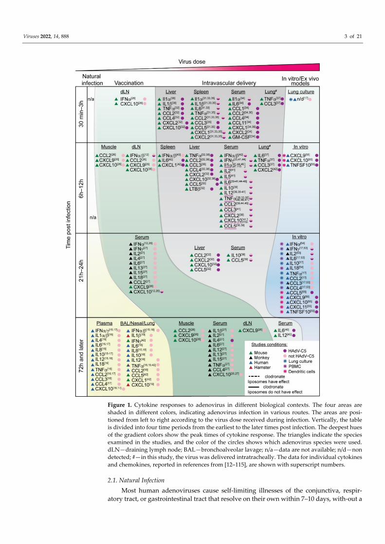
Although the exposure of immune cells to adenovirus triggers a rather stereotypic pro-inflammatory response with a defined set of cytokines and chemokines becoming ac-tivated shortly after detection of adenovirus particles, the magnitude and the spectrum of cytokine and chemokine activation varies greatly and depends on the initial virus dose that enters the host (Figure 1). Natural infection with adenovirus is believed to be initiated by a very small number of infectious viral particles, and thus, the initial recognition by the immune system leads to only subclinical and/or locoregional immune responses. In con-trast, upon vaccination or systemic treatments with adenovirus-based vectors, the amounts of virus particles administered into the body are orders of magnitude higher than naturally occurring infection. Specifically, clinical trials have demonstrated that ad-enovirus-based vaccines are highly effective and generate protective immunity when they are administered intramuscularly at doses that range from 109 to 1011 adenovirus particles [10,11]. At the vaccine injection site, the virus infects muscle cells in addition to being se-questered by the antigen-presenting cells, which transfer the virus to the draining lymph nodes, leading to the generation of both antigen- and virus-specific humoral and cellular immune responses through mechanisms of adaptive immunity [12]. Furthermore, sys-temic intravenous injection of adenovirus vectors for the therapy of metastatic cancer or genetic diseases requires even higher amounts of therapeutic virus, and the administered virus amounts can reach up to 3.8 × 10¹³ viral particles per single injection dose [13].
Viruses 2022, 14, 888 3 of 21
Figure 1. Cytokine responses to adenovirus in different biological contexts. The four areas are shaded in different colors, indicating adenovirus infection in various routes. The areas are posi-tioned from left to right according to the virus dose received during infection. Vertically, the table is divided into four time periods from the earliest to the later times post infection. The deepest hues of the gradient colors show the peak times of cytokine response. The triangles indicate the species examined in the studies, and the color of the circles shows which adenovirus species were used. dLN—draining lymph node; BAL—bronchoalveolar lavage; n/a—data are not available; n/d—non detected; #—in this study, the virus was delivered intratracheally. The data for individual cytokines and chemokines, reported in references from [12–115], are shown with superscript numbers.
2.1. Natural Infection Most human adenoviruses cause self-limiting illnesses of the conjunctiva, respir-
atory tract, or gastrointestinal tract that resolve on their own within 7–10 days, with-out a

Viruses 2022, 14, 888 4 of 21
need for hospitalization or medical intervention [14]. Therefore, data on the cy-tokine re-sponse in humans to the most common adenoviruses that cause respiratory illness, par-ticularly species C adenoviruses (HAdV-C2 and HAdV-C5), are not availa-ble. However, infections with some human species B adenoviruses (e.g., HAdV-B3 and HAdV-B7) can lead to severe respiratory illness and manifest as pneumonia and acute respiratory dis-tress syndrome (ARDS) [14]. Recently, the levels of cytokines and chemokines in plasma and bronchoalveolar lavage fluid (BALF) in immunocompetent adult and pediatric pa-tient cohorts with pneumonia and ARDS caused by HAdV-B3 and HAdV-B7 infections were reported [15–18]. The amounts and the profiles of cyto-kines were determined in these patient cohorts were cytokines with potent pro-inflammatory properties, including IL-1α, IL-1β, IL-6, IL-8, IL-12, IFN-γ, IFN-α2, TNF-α, and chemokines CCL2, CCL3, and CXCL10. Interestingly, the same spectrum of cytokines was observed in BALF, nasal sam-ples, and serum, suggesting that the cytokine response to adenovirus infection is stereo-typic and is both local and systemic [15]. Moreover, clinical observations suggest that more severe disease is associated with prolonged expression and higher release of pro-inflammatory cytokines [16]. In patients with severe pneumonia and ARDS, these cyto-kines lingered for more than two weeks, while in milder cases, they subsided by 14 days after symptom onset [16].
Human adenoviruses exhibit species-restricted phenotypes, making studying dis-ease progression in animal models particularly problematic. However, studies of HAdV-B14p1 in the Syrian hamster model have shed some light on virus-mediated immune- and histopathologies [19]. HAdV-B14p1 is a species B adenovirus (similar to HAdV-B3 and HAdV-B7) implicated in severe pneumonia and ARDS in humans [14]. The infection of Syrian hamsters with HAdV-B14p1 led to a more severe ARDS-like lung pathology com-pared to infection with the parental HAdV-B14 virus [19]. In addition to more extensive lung tissue damage, lung immune-pathology in this model included the high expression of pro-inflammatory cytokines IL-1β and TNF-α and chemokines CCL3 and CXCL10 [19]. HAdV-B14p1-infected cells had lower expression of the viral protein E1B 20K, an inhibitor of the host immune response [20], suggesting that the failure to inhibit pro-inflammatory cytokine production may be responsible for, and lead to, exuberant host pro-inflamma-tory immune activation and tissue damage [19].
Experimental infection with non-human adenoviruses, for instance, mouse adenovi-rus 1 (MAV1), triggers the activation of a similar set of cytokines during respiratory dis-ease in mice comparable to human adenovirus infection in humans. The amounts of cyto-kines IFN-γ and TNF-α, as well as chemokines CXCL1, CCL2, and CCL5, were signifi-cantly elevated in the BALF of MAV1-infected mice on day seven post-infection [21]. A fowl adenovirus, known to cause severe disease in poultry populations, induced elevated mRNA transcription for the same spectrum of pro-inflammatory cytokines detected fol-lowing mouse and human adenovirus infection (IL-1β, IL-6, IL-8, IFN-α, IFN-β, IFN-γ) [22]. Interestingly, an intramuscular route of infection was associated with higher cytokine expression in the spleen and led to more severe symptoms relative to an oral route of infection [22].
In summary, the cytokines and chemokines released in response to adenovirus infec-tion are pro-inflammatory in nature and include cytokines IL-1α, IL-1β, IL-6, IL-8, IFN-γ, IFN-α, and TNF-α and chemokines CCL2, CCL3, and CXCL10. This set of cytokines and chemokines is predominant across species ranging from humans to birds. These pro-in-flammatory cytokines are thought to be essential for mounting a protective anti-viral im-mune response. However, their continued activation may trigger a feed-forward “ampli-fication loop” of pro-inflammatory signaling, when the initial expression of pro-inflam-matory cytokines attracts the cells of hematopoietic origin to the site of infection. This in turn produces more and a wider spectrum of pro-inflammatory cytokines, attracting even more innate immune cells of hematopoietic origin that locally release proteins with potent anti-microbial and anti-viral properties, leading to collateral tissue damage and associated pathologies [23]. Taken together, the severity of the natural adenoviral disease appears to

Viruses 2022, 14, 888 5 of 21
directly correlate with the amounts and the duration of the production of cytokines and chemokines at the site of the infection.
2.2. Vaccination Adenovirus-based vectors are highly effective vaccine platforms. Under the severe
pressure of the rapid and global spread of SARS-CoV-2 coronavirus, the causative agent of COVID-19 respiratory disease, adenovirus-based vaccines against SARS-CoV-2 were developed and deployed in the span of just a few months by several counties. To date, adenovirus-vectored vaccines against SARS-CoV-2 based on HAdV-D26 (Janssen Vac-cines, Johnson & Johnson, New Brunswick, New Jersey, United States) [11], ChAdOx1 (Astra-Zeneca, Oxford, United Kingdom) [24], HAdV-D26/HAdV-C5 (Sputnik V vaccine Gamaleya Research Institute of Epidemiology and Microbiology, Moscow, Russia) [10], and HAdV-C5 (Ad5-nCoV, Sinopharm and CanSinoBIO, Tianjin, China) [25] have been administered to billions of people around the world and have undoubtedly saved millions of lives. The effectiveness of adenoviruses as vaccine vectors is based on their ability to elicit potent CD8+ T-cell responses and robust humoral immunity [12]. However, despite billions of people receiving the adenovirus-based vaccines, data on the profile of cytokines released in response to vaccinations are limited. Therefore, we will review the available data for vaccination studies performed in pre-clinical mouse and non-human primate models (Figure 1, Vaccination).
In mice, after intramuscular vaccine administration, IFN-α and CXCL10 are detected three hours post vector injection in the local draining lymph nodes but not in the muscle tissue [26]. Six hours post-vaccination, these proteins can be detected not only in the drain-ing lymph nodes but also in muscle tissue surrounding the injection site [12,26]. In the blood, elevated amounts of IFN-γ, IL-2, IL-6, IL-12, IL-15, and IL-8 cytokines, in addition to chemokines CXCL9 and CXCL10, are observed only at 24 h post-vaccine administration [12,26,27]. These same cytokines and chemokines are elevated in the plasma of monkeys at seven days post-vaccination [26,27].
It is noteworthy that vaccinations in animal models with non-HAdV-C5-based vec-tors led to the production of higher amounts and prolonged expression of pro-inflamma-tory cytokines and chemokines in comparison to inoculation with HAdV-C5 vectors [12,27]. This observation is reminiscent of a more potent pro-inflammatory responses to non-HAdv-C5 adenovirus serotypes upon natural infection. While HAdV-C5 causes self-limiting common cold illness in immunocompetent hosts, the non-HAdV-C5 adenovirus species cause more severe respiratory disease and activate robust pro-inflammatory cyto-kine production that may last longer than two weeks [16].
2.3. Intravascular Administration In addition to being a highly effective vaccine platform, adenovirus vectors are a very
promising gene delivery platform for the therapy of genetic diseases and oncolytic plat-form for therapy of localized and disseminated cancers. For many of these applications to be effective, the virus must be injected intravascularly (i.v.). Similar to other gene delivery platforms, after i.v. administration, the majority of the injected dose of adenovirus vector is sequestered within the reticulo-endothelial system of the liver and spleen due to the specialized tissue architecture and the abundance of innate phagocytic cells in these or-gans that sequester pathogens from the blood [28,29]. In the settings requiring i.v. admin-istration of the therapeutic virus, the virus dose that enters the bloodstream over a very short period of time is exceptionally high, reaching up to 3.8 × 10¹³ viral particles [13]. It is critical to note that despite adenovirus vectors for cancer and gene therapy applications being engineered to be profoundly attenuated and to have very limited or no capacity for replication in normal tissues, the immune system still recognizes therapeutic vectors as genuine viral pathogens due to the PAMP moieties present in the adenovirus capsid or through sensing virus entry into cells (see below). Therefore, upon injection of extremely high amounts of virus particles in the bloodstream, the immune system promptly

Viruses 2022, 14, 888 6 of 21
recognizes the presence of the virus and activates innate immune defensive mechanisms that trigger the production of inflammatory cytokines and chemokines. The timing of the cytokine response after i.v. injection strongly supports this model (Figure 1). Whereas af-ter natural infection the significant amounts of cytokines were only detected at 7 days post-infection, after vaccination, cytokines in the serum appear within 24 h. In the case of i.v. injection of the adenovirus to mice, elevated transcription of the pro-inflammatory cytokine IL-1α can be detected as early as 10 min post-virus injection [30]. At one-hour post-virus injection, elevated levels of the “early response” pro-inflammatory cytokines IL-1, IL-6, IL-8, TNF-α and chemokines CCL2, CCL3, CCL4, CXCL1, CXCL2, CXCL9, and CXCL10 can be detected in the liver, spleen, blood, and lungs [30–38] (Figure 1, Intravas-cular delivery). Peak concentrations of the major early response pro-inflammatory cyto-kine IL-6 in the blood occurs at six hours post-i.v. virus injection in mice [32,33,37–43]. Almost none of the early response cytokines continue to be present in the blood at 24 h post-i.v. virus injection.
In humans, the elevation of pro-inflammatory cytokines in the blood after i.v. injec-tion of adenovirus vectors is dose-dependent and occurs with kinetics similar to those observed in mice. IL-1 is one of the first pro-inflammatory cytokines that appears in the blood at three hours post-virus administration; however, by 18 h post-virus injection, the amount of IL-1 in the blood returns to baseline levels [44]. Blood levels of other early re-sponse cytokines, namely, IL-6, TNF-α, and IFN-γ and the CCL2 chemokine, are also in-creased and peak at 6 h after i.v. injection of adenovirus vectors [44–48]. By 24 h post-injection, the amounts of these cytokines in the blood subside to the baseline levels [44–46]. It is noteworthy that compared to HAdV-C5-based vectors, i.v. administration of the non-HAdV-C5-based vectors induced more potent elevation of IL-6 in the blood, albeit early cytokine responses in humans were measured for only one non-HAdV-C5-based adenovirus vector, namely, a group B adenovirus HAdv-B11 [46,48].
In contrast to i.v. administration of adenovirus vectors in humans, clinical trials em-ploying intra-tumoral virus injection showed that IL-6 release in response to the first virus injection is not substantial and mostly confined to its normal ranges [49–52]. However, repeated intra-tumoral injection may lead to elevated IL-6 concentrations in the blood, though at much lower levels than after i.v. virus injection [49,50].
Taken together, regardless of the delivery route, following detection of the adenovi-rus or adenovirus vectors by the innate immune system, the transcription of the same set of the early response pro-inflammatory cytokines, namely, IL-1, IL-6, IL-8, TNF-α, and the CCL2, CCL3, CCL4, CXCL1, CXCL2, CXCL9, and CXCL10 chemokines becomes acti-vated, leading to their production and release. Moreover, the cytokine profiles produced in response to adenovirus in the in vitro infection models of human or monkey PBMC, lung cultures, or blood-derived dendritic cells are also similar to those detected after vac-cination, natural infection, or i.v. administration of adenovirus vector (Figure 1, In vitro models) [17,53–55]. The stereotypic nature of the innate immune response to adenovirus in diverse biological contexts strongly suggest that the activation of pro-inflammatory cy-tokines and chemokines in response to adenovirus is driven in different systems by the same molecular mechanisms.
3. Molecular Mechanisms Implicated in Activation of Cytokines and Chemokines in Response to Adenovirus
The innate immune system senses the presence and the entry of pathogens into cells through an array of cell-surface-localized and cytosolic receptors that recognize pathogen-specific PAMPs as well as pathogen-induced perturbations of cellular homeostatic func-tions. Sensing of the pathogen through this network of receptors triggers downstream signal transduction pathways that activate the expression of large sets of genes, many of which are directly involved in enabling host defense functions, including transcriptional activation of genes encoding pro-inflammatory cytokines and chemokines. In the case of adenoviruses, the transcription of cytokine and chemokine genes following detection of

Viruses 2022, 14, 888 7 of 21
adenovirus entry into cells is driven by the IRF3, IRF7, and the NF-κB families of tran-scription factors [56]. The specific signal transduction pathways that were implicated in sensing adenovirus and that lead to activation of IRF3, IRF7, and NF-kB transcription fac-tors are shown in Figure 2. Whereas the genes playing the key role in activating host an-tiviral responses, activated by IRF3 and IRF7, are genes encoding type I interferons, tran-scription factor NF-κB activates the expression of many genes encoding cytokines and chemokines, including IL-1β, IL-6, IL-8, IL-12, IL-18, CCL2, CCL3, CCL4, CXCL1, CXCL2, and CXCL10 [57]. In addition, some chemokines, such as CXCL10, are synergistically ac-tivated by both NF-κB- and IFN-I/STAT1-mediated signaling [58].
Figure 2. Molecular mechanisms triggering cytokine activation in response to adenovirus infection. Several main events trigger cytokine and chemokine activation during adenovirus infection. The hubs that play the major roles are shown in red. The orange molecules are associated with IRF3-dependent transcription. The teal color indicates the factors that signal through the NF-κB transcrip-tion hub. Integrin β3 activation of IL-1α and IL-1α-dependent augmentation of cytokine production are other main events leading to cytokine expression after adenovirus infection. The data for indi-vidual proteins, reported in references from [30–116], are shown with superscript numbers.

Viruses 2022, 14, 888 8 of 21
The transcription of the type I interferon genes can be activated by three different pathways depending on the cell type detecting the adenovirus. Virus disassembly in the endosomes and the exposure of viral DNA in plasmacytoid dendritic cells trigger the ac-tivation of the TLR9/MyD88 axis [59,60], leading to the IRF7-dependent transcription of IFN-I genes [61]. Alternatively, the detection of the viral DNA in the cytoplasm leads to the TLR-independent activation of type I IFN through cGAS/STING/TBK1/IRF3 signaling pathway [59,62–64]. Additionally, in mouse splenic myeloid dendritic cells, IFN transcrip-tion is independent of TLRs or IRF3 signaling but dependent on SAPK/JNK and IRF7 ac-tivation [43].
Several additional players that potentiate IFN production in response to adenovirus infection were recently identified. In the cytosol, cGAS that recognizes adenoviral dsDNA genomes cooperates with ZCCHC3 to allow for the highly efficient production of cGAMP, with subsequent activation of STING/IRF3-dependent transcription of IFN-I [65]. In the nucleus, protein hnRNAPA2B1 binds to viral DNA, leading to increased phosphorylation of TBK1 [66], thus potentiating signaling that activates IRF3. There are also players that the adenovirus targets to suppress IFN production and signaling. For example, adenovi-rus infection leads to increased transcription of MYSM1, a repressor of STING signaling, thus enabling the virus to reduce IFN-I expression activated through the STING/TBK1/IRF3 pathway [67].
The NF-kB-dependent transcription of pro-inflammatory cytokines can be activated downstream of multiple sensors of the Toll-like receptor (TLR) family, including TLR2, TLR4, and TLR9 [34,36], which transduce sensory signals through the MyD88/TRAF6/NF-kB axis [34,36,68–70]. In addition, depending on the cell type and the identity of the up-stream receptor that detects adenovirus entry into the cell, different cellular serine/threo-nine phosphokinases and members of the MAPK pathway (including p38MAPK, ERK1/2, PI3K, AKT) also play a role in the transcriptional activation of cytokines and chemokines [32,71–73]. Cells that express components of the NOD-like receptor family (NLR), includ-ing NOD2 and NLRP3, or a non-NLR cytosolic DNA sensor absent in melanoma 2 (AIM2); adaptor protein ASC; and caspase-1 can respond to adenovirus by activating the inflam-masome pathway [70,74]. NLRP3/ASC/Caspase-1 or AIM2/ASC/Caspase-1 inflam-masome activation results in caspase-1-mediated cleavage of the pro-form of IL-1β to its mature functionally active cytokine form and its systemic release [70], which can subse-quently activate an array of cytokines and chemokines via IL-1RI and the feed-forward amplification loop of pro-inflammatory signaling.
Similar to other viruses, adenoviruses evolved mechanisms to suppress pathways activating IFN-I, aiding in the evasion of IFN-I-dependent effector mechanisms. Adeno-virus-encoded, virus-associated RNAs, transcribed by RNA polymerase III (VA RNAs) shortly after adenovirus genome entry into the nucleus, are well-established and highly effective suppressors of PKR [75]. In addition to its canonical function of arresting cellular protein synthesis, PKR is also required for the phosphorylation of the inflammasome adaptor ASC and its oligomerization [76]. Specifically, adenovirus VA RNAs arrest ASC oligomerization, thus preventing NLRP3 inflammasome formation and inhibiting func-tional maturation of IL-1β and other cytokines whose functional maturation depends on caspase-1 processing [76]. Another strategy evolved by adenovirus to avoid IFN-I activa-tion, and its antiviral effectors depends on the function of proteins encoded in the E4 re-gion of the adenovirus genome. Specifically, the splicing of viral mRNAs produced fol-lowing the bi-directional transcription of adenovirus proteins during virus replication is tightly regulated by adenoviral E4 proteins so that the formation of the IFN-I and PKR-activating dsRNA complexes of complementary mRNAs is avoided [77]. Yet another ad-enoviral protein, RID1α, encoded in the E3 region of the adenoviral genome, functions to avoid the NF-κB-dependent activation of cytokine and chemokine expression down-stream of the EGFR signaling [78].
Strikingly, the analysis of pro-inflammatory responses to adenovirus has demon-strated that the transcriptional and/or functional activation of cytokines and chemokines

Viruses 2022, 14, 888 9 of 21
strongly depends on the viruses’ ability to rupture cellular endosomes [34,69,71]. Adeno-virus mutant ts1, which cannot escape cellular endosomal compartments through endo-some rupture [79], has reduced the activation of cytokines and chemokines, compared to virus variants that can efficiently escape endosomal compartments after virus internaliza-tion into the cell [30,43,71]. IL-1α is one of the earliest pro-inflammatory cytokines tran-scriptionally activated in splenic MARCO+ and CD169+ marginal zone macrophages after i.v. administration of adenovirus vectors in mice [30]. Although IL-1α was transcription-ally activated within 10 min after administration for both the mutant ts1 and unmodified HAdv-C5-based vector, the functional maturation of IL-1α and the activation of the IL-1α-IL-1RI-dependent pro-inflammatory signaling failed to occur after administration of the ts1 mutant virus. In contrast, an HAdv-C5-based vector, capable of escaping endoso-mal compartments through endosome rupture, triggered not only early Il1a gene tran-scription but also functional maturation of IL-1α, manifested by its translocation into the nucleus and activation of an IL-1RI-dependent array of pro-inflammatory cytokines and chemokines, including IL-1β and IL-6, and the CCL2, CXCL1, and CXCL2 chemokines [30]. The extremely rapid activation of Il1a gene transcription after i.v. virus administra-tion suggests that adenovirus sensing in macrophage cells occurs either at the plasma membrane or in early endosomal compartments. Indeed, the interaction of adenovirus penton RGD motif with β3 integrins displayed at the macrophage surface was necessary to trigger Il1a transcription and i.v. administration of an adenovirus mutant lacking the RGD amino acids in the penton protein or administration of the virus with unmodified capsid to mice deficient for β3 integrin expression failed to trigger Il1a transcriptional ac-tivation and initiation of the IL-1α-IL-1RI-dependent pro-inflammatory signaling cascade [30].
Taken together, these data strongly suggest that adenovirus is sensed by an array of innate immune receptors at every step of virus entry into the cell, including at the plasma membrane, endosomal compartments, cytosol, and even within the nucleus. Based on the dose-dependent nature of cytokine responses to adenovirus, it is plausible that during natural infection, which is initiated by a low dose of the virus, only some of the innate immune sensors become activated, leading to the production of low amounts and a lim-ited number of inflammatory cytokines and chemokines. In contrast, upon i.v. administra-tion of large amounts of adenovirus vectors, most, if not all, of the innate immune recep-tors become simultaneously engaged, triggering production of high amounts and a broad spectrum of inflammatory cytokines and chemokine. This highly potentiated cytokine and chemokine release by itself, as well as through the induction of a feed-forward am-plification loop of the pro-inflammatory signaling, may culminate in the cytokine storm syndrome, severe systemic toxicity, and even death.
4. Cell Types That Produce Cytokines and Chemokines in Response to Adenovirus As described above, transcriptional activation and production of cytokines and
chemokines occurs following triggering of sensors of innate immunity that detect adeno-virus particles at the cell surface or inside the cell. Not all cell types can recognize the invading microorganisms and mount activation of cytokine and chemokine genes. Tissue-resident macrophages in liver and spleen are highly efficient at removing blood-borne pathogens and are the first cell types that encounter the incoming virus after i.v. admin-istration. The liver-resident macrophages, Kupffer cells, represent the largest pool of in-nate phagocytic cells in the body. They efficiently sequester blood-borne adenovirus par-ticles via a variety of plasma membrane receptors, including complement receptor CRIg [80] and scavenger receptors SR1 [81,82] and CD36 [33]. Within 30 min of sequestering adenovirus particles from the blood, Kupffer cells activate the expression of IL-1α, IL-1β, TNF-α, CCL2, CCL3, CXCL2, and CXCL10 [38]. It is noteworthy that the expression of IL-6 was not detected in the liver, suggesting that the systemic IL-6 observed 6 h post-i.v. virus administration is driven not by Kupffer cells but by cell types residing in other or-gans, most notably the spleen. Moreover, within 1 h post-i.v. adenovirus administration,
Viruses 2022, 14, 888 10 of 21
the majority of Kupffer cells that sequestered adenovirus particles undergo “defensive suicide”, a necrotic-type cell death associated with the loss of plasma membrane integrity, thus effectively reducing the amounts of circulating adenovirus in the blood and limiting virus spread to vital organs, most notably the liver [35,83]. Upon induction of a “defensive suicide” necrosis, Kupffer cells release their cytosolic contents into the surrounding milieu and simultaneously release the pro-inflammatory cytokines and other factors that activate both local and systemic inflammation [83]. It is plausible that Kupffer cells are primarily responsible for the early production and release of IL-1α and IL-1β into the circulation, as the plasma concentrations of these cytokines increase early and subside in the liver at later times when Kupffer cells disintegrate or are no longer physiologically active [38].
The splenic marginal zone CD169/MOMA+ and MARCO+ macrophages also effi-ciently sequester adenovirus from the bloodstream. Although the identity of the plasma membrane receptor(s) that mediate virus sequestration in marginal zone macrophages re-mains unknown, as discussed, the adenovirus penton RGD amino acids interact with β3 integrins on the surface of these phagocytic cells and trigger signaling, leading to the tran-scriptional and functional activation of IL-1α [30]. In turn, IL-1α triggers the activation of a variety of cytokines and chemokines, most notably IL-6, via its cognate receptor, IL-1RI [30]. Moreover, IL-1α is the principal mediator activating the CXCL1/2-CXCR2 chemokine signaling axis, leading to the influx and retention of neutrophils into the splenic marginal zone [31]. Local degranulation or release of cytotoxic factors by neutrophils leads to the elimination of adenovirus-containing macrophages from the spleen in 24 h, thus limiting systemic spread of adenovirus through the blood [31].
The key role of macrophages and other phagocytic cells in triggering cytokine pro-duction in response to adenovirus was confirmed in experiments using clodronate lipo-somes, which, upon i.v. injection, eliminate all phagocytic cells, including macrophages in the liver and spleen [84]. The elimination of all phagocytic cells prior to adenovirus injection in mice greatly reduces the amounts of IL-1, IL-2, IL-6, IL-12, and TNF-α cyto-kines and CCL2 and CCL3 chemokines in the blood after i.v. adenovirus administration (Figure 1, underlined), suggesting that phagocytic cells are the primary source of cyto-kines and chemokines released into the circulation in response to i.v. adenovirus injection [39–41].
The dendritic cells in the spleen are the principal source of type I IFNs, a set of the key anti-viral cytokines that trigger the upregulation of thousands of genes with various host defense and homeostatic functions [43,54,60]. In response to virus infection, many types of cells can produce IFN-α/β. However, dendritic cells generate orders of magnitude higher amounts of type I IFN than other cell types [59]. Dendritic cells can sequester the virus through cognate fiber receptors or DC-SIGN [85], and in addition to type I IFN pro-duction, they synthesize other pro-inflammatory cytokines. In vitro studies confirm that monocyte-derived dendritic cells can sequester adenovirus–antibody complexes, leading to the expression of type I IFNs and other inflammatory cytokines [86,87].
In an in vitro model of natural infection using human polarized mucosal epithelial cells and peripheral blood monocyte-derived macrophages, adenovirus infection and vi-rus sequestration in macrophages activates IL-8 production [88]. The local production of IL-8 from macrophages initiates the re-localization of the adenovirus receptors from the basolateral to the apical surface of epithelial cells, making them susceptible to direct in-fection with the virus via the fiber protein, the principal cell attachment protein of the adenovirus [88].
Taken together, evidence suggests that tissue-resident innate phagocytic cells, pri-marily tissue-resident macrophages and dendritic cells, represent the principal compart-ment that sequesters adenovirus particles and triggers local and systemic cytokine and chemokine production. Consistent with their sentinel and scavenger functions, tissue-res-ident macrophages sequester large amounts of adenovirus particles from the blood and become promptly eliminated either through a “defensive suicide” necrosis, like the Kup-ffer cells in the liver, or through neutrophil-mediated cytotoxicity against marginal zone

Viruses 2022, 14, 888 11 of 21
macrophages in the spleen. The production of pro-inflammatory cytokines and chemo-kines and elimination of virus-containing macrophages limit spread of the virus to vital organs and prompt initiation of adaptive immune responses to enable clearance of ade-novirus in non-phagocytic cells throughout the body.
5. Factors Modulating Cytokine and Chemokine Production in Response to Adenovirus
Upon entry into the cell, adenoviruses utilize a variety of primary attachment recep-tors, including CAR, CD46, DSG2, and sialic acid, as well as cellular integrins, which serve as co-receptors and mediate virus internalization into the cell [89–92]. Virus interaction with primary attachment receptors is mediated by the fiber protein, whereas virus inter-action with cellular integrins is mediated by the penton. However, when virus particles enter the bloodstream, numerous factors in the blood recognize and bind to the virus. These blood factors effectively "tag" viral particles, and the virus–blood factor complexes interact with a different set of receptors and enter cells through mechanisms unavailable to the “naked” virus. Moreover, these cell surface receptors not only dictate which cell types the virus–blood factor complexes will enter but also how these cells may respond to the virus infection (Figure 3).
Figure 3. Factors enhancing cytokine and chemokine production in response to adenovirus infec-tion. Different factors that bind to adenovirus particles can change the receptor selectivity and target the virus to macrophages and dendritic cells in the absence of the cognate virus receptors. The data for individual signaling molecules as reported in references from [33–118], are shown in parentheses adjacent to indicated proteins.
Upon searching for the natural mechanisms that may neutralize adenoviruses, it was found that human alpha defensin 5 (HD5), a small antimicrobial peptide, has potent in-hibitory activity against species C adenovirus HAdV-C5 [93]. However, HD5 does not

Viruses 2022, 14, 888 12 of 21
inhibit adenoviruses of species D and F, but rather augments the virus infection in vitro and increases adaptive responses to the virus-encoded transgene antigens in vivo [94]. Furthermore, pre-treatment of mice with HD5 prior to HAdV-D26 injection resulted in a higher cytokine response than to the virus alone. Mechanistically, more robust cytokine production in response to the virus–HD5 complexes is thought to be due to more efficient virus entry into dendritic cells in the presence of HD5. It is plausible that HD5 functions as a polycation, and upon binding to adenovirus, it shields the negatively charged amino acids present at the surface of the virion, thus reducing repulsion between the negatively charged virus surface and plasma membrane, leading to a more efficient infection of den-dritic cells [94].
Lactoferrin (Lf) is another protein that enhances adenovirus entry into dendritic cells [95]. Specifically, it was shown that the bovine Lf enhances entry of human adenoviruses into dendritic cells more efficiently than human Lf, which can potentially be explained by the differential glycosylation patterns of these proteins [85]. Interestingly, the HAdV-C5-bLf complexes utilize the DC-SIGN receptor on dendritic cells, which is not a natural HAdV-C5 receptor, while HAdV-B35–bLf complexes require a cognate CD46 receptor for cell entry. Similar to the HD5-mediated enhancement of infection and cytokine produc-tion by the dendritic cells, the adenovirus–bLf complexes also trigger a more potent re-lease of cytokines than the virus alone [85]. The higher efficacy of entry of adenovirus–bLf complexes into dendritic cells can potentially be explained by the engagement of alternate cell surface receptors, which are not utilized for cell entry by the virus without Lf.
Coagulation factor X (FX) binds to HAdV-C5 and other adenoviruses with low-na-nomolar or even picomolar affinity [34,96]. FX binding to adenoviruses shields virus par-ticles in the blood from other factors and enables virus escape from neutralization via nat-ural IgM-dependent activation of the complement system [97]. Furthermore, in the mouse system, when HAdV-C5–FX complexes are sequestered by the splenic marginal zone mac-rophages, they activate a broader spectrum of inflammatory cytokines and chemokines, compared to a mutant virus that cannot bind to FX [34]. Importantly, the FX-mediated potentiation of cytokine activation was not observed in vitro when adenovirus–FX com-plexes were added to human dendritic cells differentiated from peripheral blood mono-cytes [98]. These data suggest that the sensing of adenovirus–FX complexes by the innate immune system may be species-specific or that potential differences in cell-intrinsic sen-sory mechanisms exist between tissue resident macrophages in vivo and in vitro differen-tiated dendritic cells.
Adenovirus-specific IgG antibodies can also potentiate virus entry into dendritic cells with subsequent activation of anti-viral and inflammatory cytokine production. In vitro, the pre-incubation of pooled human immunoglobulin (IVIg) with adenovirus leads to the formation of immunocomplexes consisting of adenovirus virions cross-linked by the an-tibodies [86]. Compared to the virus alone, these adenovirus immunocomplexes infect monocyte-derived human dendritic cells with higher efficacy [86]. Dendritic cells se-quester these immunocomplexes via an array of Fc receptors, thus completely bypassing cognate adenovirus fiber receptors [99]. The pre-treatment of adenovirus with IVIg in-creased virus-encoded transgene expression in dendritic cells and potentiates cytokine production from these cells [86,87]. At least two mechanisms may contribute to the eleva-tion of cytokine production by the dendritic cells in response to adenovirus immunocom-plexes. One mechanism involves TRIM21, an intracellular Fc receptor that recognizes the adenovirus–antibody complexes in the cytosol, leading to elevated cytokine production from the cells [100]. The second mechanism that may contribute to potentiation of cyto-kine production in response to adenovirus immunocomplexes is aberrant intracellular trafficking to the late LAMP1-positive lysosomes. The release of adenovirus particles from the late lysosomes is associated with enhanced cytokine response compared to the virus particles that were released from the early endosomal compartments [53,99].
The elevated cytokine production in the presence of adenovirus-specific antibodies was confirmed in vivo in rodents and primates pre-immunized with HAdV-C5. In pre-
Viruses 2022, 14, 888 13 of 21
immunized animals, the i.v. injection of a high dose of HAdV-C5 showed an augmented cytokine response, particularly at 6 h post-systemic virus injection [101,102]. However, pre-immunization almost completely prevented liver transduction and subsequent hepa-totoxicity in animals [101,103]. Moreover, experiments in mice demonstrated that the en-hanced cytokine production in response to adenovirus in pre-immunized animals de-pends in part on the functional complement system, particularly on the presence of com-plement component 3 (C3). C3 was implicated in potentiating neutrophil influx to the spleen after i.v. adenovirus administration [31], enhancing adenovirus-immunocomplex signaling [86], as well as in CRIg-mediated uptake of adenovirus particle by the Kupffer cells in the liver [79]. Furthermore, after i.v. administration, adenovirus triggers comple-ment activation via both classical (antibody-dependent) and alternative pathways [104]. It remains unclear whether C3 triggers potentiated cytokine production via an ana-phylatoxin C3a domain, which is released after proteolytic processing of C3, and can di-rectly activate C3a receptors on macrophages and dendritic cells, or whether C3 is re-quired to be covalently bound to the virus particles in the form of C3b and activate other complement receptors, CR1-CR4. It is certainly likely that upon complement activation, both products of proteolytic cleavage of C3 contribute to the inflammatory cytokine acti-vation and release observed shortly after i.v. virus administration. Another complement component 4 (C4) blocks virus disassembly and thus effectively neutralizes the virus [105,106]. The specific role of C4-mediated adenovirus neutralization in cytokine produc-tion is currently unknown and requires further investigation. It is clear, however, that as a principal component of humoral innate immunity, the complement system becomes ac-tivated in response to systemic adenovirus administration. Complement activation con-tributes to direct virus neutralization and the activation of cytokine and chemokine pro-duction by the innate phagocytic cells, which enables virus elimination, promotes activa-tion of adaptive immunity that leads to clearance from the body of virus-infected cells, and generates long lasting virus-specific immunity.
6. Management of Cytokine Responses to Therapeutic Adenovirus Vectors The key driver for the extensive efforts to better understand acute inflammatory cy-
tokine production and cytokine storm syndrome is a quest to find approaches to improve the safety of adenovirus-based vectors as therapeutic platforms for treating human dis-eases. The advantages of adenovirus vectors are many and include established, highly efficient, standardizable, and cost-effective manufacturing of therapeutic vector stocks, large payload capacity (allowing for delivery of therapeutic transgenes that cannot be ac-commodated by alternate vector systems), ability to target adenovirus vectors to specific cell types of interest through modification of virus capsid proteins, vector stability, the episomal nature of a double-stranded DNA viral genome that does not integrate into cel-lular chromosomes (thus minimizing risk of insertional mutagenesis) and well under-stood biology of virus reproduction cycle, allowing for the attenuation of virus virulence and improving the safety of this vector platform. However, as we discussed above, despite attenuation and even complete lack of capacity for replication due to numerous deletions in key regulatory genes in the adenovirus genome, therapeutic adenovirus vectors are still recognized by the innate immune system as genuine pathogens, and innate phagocytic cells mount a potent systemic inflammatory cytokine response that can lead to cytokine storm syndrome. This is particularly problematic when extremely high amounts of ade-novirus particles are administered over a short period of time, especially via an i.v. route. The recognition of therapeutic vectors as genuine pathogens and subsequent activation of potent systemic inflammatory cytokine responses is not unique to adenovirus vectors and is also observed in clinical trials evaluating administration of high doses of therapeutic adeno-associated virus (AAV)-based vectors to patients with rare genetic diseases [107,108]. Furthermore, the early iterations of cell therapy based on CAR-T cells that rev-olutionized treatment of certain types of cancer triggered cytokine storm syndrome that required the development of mitigation strategies to improve the safety of this approach

Viruses 2022, 14, 888 14 of 21
[109]. Specifically, the administration of cancer-specific CAR-T cells in combination with the anti-IL-6 receptor antibody, tocilizumab [110], or recombinant IL-1RI antagonist, An-akinra [111], allowed for a significant reduction in inflammatory cytokine production in response to CAR-T cells therapy. Because IL-1 and IL-6 were found to be some of the very first cytokines activated in response to i.v. administration of adenovirus vectors [44,47,48], it is plausible that interfering with IL-1 and/or IL-6 signaling with currently available FDA-approved drugs, may prove to be an effective strategy to suppress acute systemic cytokine activation after administration of therapeutic adenovirus vectors. Indeed, recent studies in non-human primates demonstrated that safety of systemic delivery of a high dose adenovirus vector could be improved when virus was administrated following a “cytokine prophylaxis”, pre-treatment of animals with tocilizumab, Anakinra, and a broadly suppressive dexamethasone [112]. While the suppression of cytokine production prior to administering adenovirus-based therapy may prove beneficial in the context of adenovirus-based gene therapy applications, this “cytokine prophylaxis” approach may not be useful when adenoviruses are administered to patients for therapy of cancer. It is broadly accepted that virus-based activation of pro-inflammatory type-I cytokines within tumor microenvironment and lymphoid tissues is essential for breaking the state of local and systemic immune tolerance to tumors and, thus, is necessary for efficient anti-tumor response after virotherapy. Future clinical trials are needed to show whether a tailored “cytokine prophylaxis” approach can be utilized for improving the safety of adenovirus vectors without deleterious effects on their efficacy as cancer therapeutics.
Another approach to improving the safety of adenovirus-based vectors is the modu-lation of virus–host interactions that lead to virus sequestration in immune phagocytic cells and activation of inflammatory cytokine production through the introduction of mu-tations into the virus capsid. Our group has recently reported that in HAdv-C5 capsid, natural IgM antibodies bind to the HVR1 region of the hexon protein. Accordingly, ade-novirus vector with a mutated HVR1 hexon region failed to bind IgM in mouse and hu-man sera and escaped sequestration in Kupffer cells after i.v. administration [33]. Further-more, a substitution of RGD amino acids in the adenovirus penton protein for a laminin-derived peptide that cannot interact with macrophage β3 integrins resulted in generation of a mutant virus that triggered muted cytokine activation in the spleen after i.v. admin-istration [33]. Collectively, these data provide evidence that despite the multifaceted na-ture of innate immune activation and inflammatory response to adenovirus vectors, many critical steps of virus recognition by the innate immunity can be obviated through the introduction of structural modifications into adenovirus capsid proteins, thus allowing for the generation of adenovirus vectors with an improved safety profile.
The historical perception that adenovirus vectors are uniquely unsafe for therapeutic use in people undoubtedly stems from the tragic death of a patient, initially identified as subject 019 and later disclosed as Jesse Gelsinger, who was treated with a high dose of an adenovirus vector (3.8 × 10¹³ viral particles) that was administered via hepatic artery dur-ing a gene therapy trial in 1999 [13]. It is noteworthy that this same high dose of adenovi-rus vector was administered to a second participant in the trial, and that second patient experienced a significant but only transient elevation of IL-6 in the blood, while IL-6 con-tinued to be highly elevated after virus administration to Jesse Gelsinger [13]. The mech-anistic reason for this profoundly consequential divergence in cytokine response between two participants in this high dose vector cohort remains unknown. Nevertheless, a recent study analyzed the production of IL-6 and the expression of activation markers by in vitro differentiated dendritic cells after their exposure to adenovirus vector mixed with sera from healthy donors as well as with a frozen archived blood sample obtained from Jesse Gelsinger prior to adenovirus administration [87]. The authors found that exposure of dendritic cells to adenovirus vector mixed with blood of subject 019 triggered the produc-tion of very high amounts IL-6, which were significantly higher than the amounts of IL-6 released after cell exposure to adenovirus vector mixed with sera from 6 other donors tested in this study [87]. Although the authors suggested that pre-existing anti-HAdv-C5-

Viruses 2022, 14, 888 15 of 21
specific antibodies may have played a role in potentiating virus entry into dendritic cells leading to potentiated IL-6 production, the amount of antibodies recognizing adenovirus proteins varied greatly between sera donors and were not in high amounts in the blood of subject 019, suggesting that a yet unidentified factor(s) present in the blood other than, or in addition to, virus-specific antibodies may be responsible for potentiating IL-6 pro-duction by the dendritic cells after their exposure to the adenovirus–serum mixture. Alt-hough limited by the number of serum samples analyzed, this study provides rationale for prospective analysis of serum samples from patients subject to therapy with adenovi-rus vectors to identify rare patients with virus hyper-sensitivity phenotype, like subject 019, that must be excluded from treatment cohorts or therapy with adenovirus vectors. Such a relatively simple, prospective patient stratification tool will prevent the admin-istration of adenovirus to patients with inflammatory hypersensitivity response and thus reduce the risk of exuberant and potentially lethal inflammatory response to administra-tion of the therapeutic vectors.
7. Summary In summary, cytokines and chemokines are critical cell–cell communication proteins
that become synthesized, activated, and released in a dose-dependent manner in response to natural adenovirus infection and in response to administration of therapeutic vectors based on adenovirus and other viral and non-viral delivery vectors [30,108,113–115]. Based on the abundance of accumulated data and improved understanding of specific signaling pathways that adenovirus triggers upon entry into immune phagocytic cells [30,56–79,116–118], one can conclude that the safety of adenovirus vectors can be managed through at least two independent and complementary approaches. The first approach of a “cytokine prophylaxis” involves administering currently available FDA-approved drugs that target key pro-inflammatory signaling pathways prior to, or together with, therapeutic vector administration. The second approach relies on the introduction of tar-geted mutations into adenovirus capsid to generate adenovirus vectors that avoid inter-action with innate immune cells or cellular receptors that trigger exuberant cytokine re-sponses. Furthermore, the insight obtained through analyzing dendritic cell responses to adenovirus mixture with serum samples in vitro provides the opportunity for the devel-opment of in vitro patient stratification tools for prospective identification of the rare pa-tients with extreme inflammatory hypersensitivity phenotype. Such patients can therefore be excluded from recruitment to clinical trials or therapy cohorts, ensuring the safety of therapy for the patients in need. Future directions may focus on the identification of spe-cific factors in the blood that trigger an inflammatory hypersensitivity response to adeno-virus and optimization of clinical trial designs to allow for incorporation of all currently available approaches to improving safety of adenovirus-based therapeutics that aim to address the needs of patients that currently have limited or no therapeutic options.
Author Contributions: S.A.; conceptualization of the manuscript, S.A.–design and production of illustrations, S.A. and D.M.S.; writing—original draft preparation, review, and editing, D.M.S.; funding acquisition. All authors have read and agreed to the published version of the manuscript.
Funding: This work was supported by the US NIH grant AI107960, David C. Lowance Endowment Fund, and Children’s Healthcare of Atlanta Research Trust to D.M.S.
Institutional Review Board Statement: Not applicable.
Informed Consent Statement: Not applicable.
Data Availability Statement: Not applicable.
Conflicts of Interest: D.M.S. is a paid consultant of Merck and Co. D.M.S. is a shareholder and an officer of AdCure Bio, which develops adenovirus technologies for therapeutic use. S.A. declares no competing interest. The funders had no role in the writing of the manuscript, or in the decision to publish this review.

Viruses 2022, 14, 888 16 of 21
References 1. Mogensen, T.H. Pathogen recognition and inflammatory signaling in innate immune defenses. Clin. Microbiol. Rev. 2009, 22,
240–273, Table of Contents. https://doi.org/10.1128/CMR.00046-08. 2. Wolf, A.J.; Underhill, D.M. Peptidoglycan recognition by the innate immune system. Nat. Rev. Immunol. 2018, 18, 243–254.
https://doi.org/10.1038/nri.2017.136. 3. Medzhitov, R. Recognition of microorganisms and activation of the immune response. Nature 2007, 449, 819–826.
https://doi.org/10.1038/nature06246. 4. Mosser, D.M.; Edwards, J.P. Exploring the full spectrum of macrophage activation. Nat. Rev. Immunol. 2008, 8, 958–969.
https://doi.org/10.1038/nri2448. 5. Altan-Bonnet, G.; Mukherjee, R. Cytokine-mediated communication: A quantitative appraisal of immune complexity. Nat. Rev.
Immunol. 2019, 19, 205–217. https://doi.org/10.1038/s41577-019-0131-x. 6. Karki, R.; Sharma, B.R.; Tuladhar, S.; Williams, E.P.; Zalduondo, L.; Samir, P.; Zheng, M.; Sundaram, B.; Banoth, B.; Malireddi,
R.K.S.; et al. Synergism of TNF-alpha and IFN-gamma Triggers Inflammatory Cell Death, Tissue Damage, and Mortality in SARS-CoV-2 Infection and Cytokine Shock Syndromes. Cell 2021, 184, 149–168.e117. https://doi.org/10.1016/j.cell.2020.11.025.
7. Schulz, O.; Hammerschmidt, S.I.; Moschovakis, G.L.; Forster, R. Chemokines and Chemokine Receptors in Lymphoid Tissue Dynamics. Annu. Rev. Immunol. 2016, 34, 203–242. https://doi.org/10.1146/annurev-immunol-041015-055649.
8. Arango Duque, G.; Descoteaux, A. Macrophage cytokines: Involvement in immunity and infectious diseases. Front. Immunol. 2014, 5, 491. https://doi.org/10.3389/fimmu.2014.00491.
9. Fajgenbaum, D.C.; June, C.H. Cytokine Storm. N. Engl. J. Med. 2020, 383, 2255–2273. https://doi.org/10.1056/NEJMra2026131. 10. Logunov, D.Y.; Dolzhikova, I.V.; Shcheblyakov, D.V.; Tukhvatulin, A.I.; Zubkova, O.V.; Dzharullaeva, A.S.; Kovyrshina, A.V.;
Lubenets, N.L.; Grousova, D.M.; Erokhova, A.S.; et al. Safety and efficacy of an rAd26 and rAd5 vector-based heterologous prime-boost COVID-19 vaccine: An interim analysis of a randomised controlled phase 3 trial in Russia. Lancet 2021, 397, 671–681. https://doi.org/10.1016/S0140-6736(21)00234-8.
11. Sadoff, J.; Gray, G.; Vandebosch, A.; Cardenas, V.; Shukarev, G.; Grinsztejn, B.; Goepfert, P.A.; Truyers, C.; Fennema, H.; Spies-sens, B.; et al. Safety and Efficacy of Single-Dose Ad26.COV2.S Vaccine against COVID-19. N. Engl. J. Med. 2021, 384, 2187–2201. https://doi.org/10.1056/NEJMoa2101544.
12. Quinn, K.M.; Zak, D.E.; Costa, A.; Yamamoto, A.; Kastenmuller, K.; Hill, B.J.; Lynn, G.M.; Darrah, P.A.; Lindsay, R.W.; Wang, L.; et al. Antigen expression determines adenoviral vaccine potency independent of IFN and STING signaling. J. Clin. Investig. 2015, 125, 1129–1146. https://doi.org/10.1172/JCI78280.
13. Raper, S.E.; Chirmule, N.; Lee, F.S.; Wivel, N.A.; Bagg, A.; Gao, G.P.; Wilson, J.M.; Batshaw, M.L. Fatal systemic inflammatory response syndrome in a ornithine transcarbamylase deficient patient following adenoviral gene transfer. Mol. Genet. Metab. 2003, 80, 148–158. https://doi.org/10.1016/j.ymgme.2003.08.016.
14. Lynch, J.P., 3rd; Kajon, A.E. Adenovirus: Epidemiology, Global Spread of Novel Serotypes, and Advances in Treatment and Prevention. Semin. Respir. Crit. Care Med. 2016, 37, 586–602. https://doi.org/10.1055/s-0036-1584923.
15. Fan, H.; Lu, B.; Cao, C.; Li, H.; Yang, D.; Huang, L.; Ding, T.; Wu, M.; Lu, G. Plasma TNFSF13B and TNFSF14 Function as Inflammatory Indicators of Severe Adenovirus Pneumonia in Pediatric Patients. Front. Immunol. 2020, 11, 614781. https://doi.org/10.3389/fimmu.2020.614781.
16. Li, J.; Wei, J.; Xu, Z.; Jiang, C.; Li, M.; Chen, J.; Li, Y.; Yang, M.; Gu, Y.; Wang, F.; et al. Cytokine/Chemokine Expression Is Closely Associated Disease Severity of Human Adenovirus Infections in Immunocompetent Adults and Predicts Disease Progression. Front. Immunol 2021, 12, 691879. https://doi.org/10.3389/fimmu.2021.691879.
17. Chen, Q.; Liu, J.; Liang, W.; Chen, Y.; Dou, M.; Liu, Z.; Chen, Y.; Zheng, Z.; Zhu, B.; Lin, Y. Clinical Features, Replication Com-petence, and Innate Immune Responses of Human Adenovirus Type 7 Infection. J. Infect. Dis. 2021, 223, 1390–1399. https://doi.org/10.1093/infdis/jiaa524.
18. Qi, L.; Wang, Y.; Wang, H.; Deng, J. Adenovirus 7 Induces Interlukin-6 Expression in Human Airway Epithelial Cells via p38/NF-kappaB Signaling Pathway. Front. Immunol. 2020, 11, 551413. https://doi.org/10.3389/fimmu.2020.551413.
19. Radke, J.R.; Covert, H.J.; Bauer, F.; Ananthanarayanan, V.; Cook, J.L. Adenovirus 14p1 Immunopathogenesis during Lung In-fection in the Syrian Hamster. Viruses 2020, 12, 595. https://doi.org/10.3390/v12060595.
20. Radke, J.R.; Yong, S.L.; Cook, J.L. Low-Level Expression of the E1B 20-Kilodalton Protein by Adenovirus 14p1 Enhances Viral Immunopathogenesis. J. Virol. 2016, 90, 497–505. https://doi.org/10.1128/JVI.01790-15.
21. Molloy, C.T.; Andonian, J.S.; Seltzer, H.M.; Procario, M.C.; Watson, M.E., Jr.; Weinberg, J.B. Contributions of CD8 T cells to the pathogenesis of mouse adenovirus type 1 respiratory infection. Virology 2017, 507, 64–74. https://doi.org/10.1016/j.vi-rol.2017.04.005.
22. Li, R.; Li, G.; Lin, J.; Han, S.; Hou, X.; Weng, H.; Guo, M.; Lu, Z.; Li, N.; Shang, Y.; et al. Fowl Adenovirus Serotype 4 SD0828 Infections Causes High Mortality Rate and Cytokine Levels in Specific Pathogen-Free Chickens Compared to Ducks. Front. Immunol. 2018, 9, 49. https://doi.org/10.3389/fimmu.2018.00049.
23. Di Paolo, N.C.; Shayakhmetov, D.M. Interleukin 1alpha and the inflammatory process. Nat. Immunol. 2016, 17, 906–913. https://doi.org/10.1038/ni.3503.
24. Falsey, A.R.; Sobieszczyk, M.E.; Hirsch, I.; Sproule, S.; Robb, M.L.; Corey, L.; Neuzil, K.M.; Hahn, W.; Hunt, J.; Mulligan, M.J.; et al. Phase 3 Safety and Efficacy of AZD1222 (ChAdOx1 nCoV-19) COVID-19 Vaccine. N. Engl. J. Med. 2021, 385, 2348–2360. https://doi.org/10.1056/NEJMoa2105290.

Viruses 2022, 14, 888 17 of 21
25. Al Kaabi, N.; Zhang, Y.; Xia, S.; Yang, Y.; Al Qahtani, M.M.; Abdulrazzaq, N.; Al Nusair, M.; Hassany, M.; Jawad, J.S.; Abdalla, J.; et al. Effect of 2 Inactivated SARS-CoV-2 Vaccines on Symptomatic COVID-19 Infection in Adults: A Randomized Clinical Trial. JAMA 2021, 326, 35–45. https://doi.org/10.1001/jama.2021.8565.
26. Collignon, C.; Bol, V.; Chalon, A.; Surendran, N.; Morel, S.; van den Berg, R.A.; Capone, S.; Bechtold, V.; Temmerman, S.T. Innate Immune Responses to Chimpanzee Adenovirus Vector 155 Vaccination in Mice and Monkeys. Front. Immunol. 2020, 11, 579872. https://doi.org/10.3389/fimmu.2020.579872.
27. Teigler, J.E.; Iampietro, M.J.; Barouch, D.H. Vaccination with adenovirus serotypes 35, 26, and 48 elicits higher levels of innate cytokine responses than adenovirus serotype 5 in rhesus monkeys. J. Virol. 2012, 86, 9590–9598. https://doi.org/10.1128/JVI.00740-12.
28. Khare, R.; Hillestad, M.L.; Xu, Z.; Byrnes, A.P.; Barry, M.A. Circulating antibodies and macrophages as modulators of adenovi-rus pharmacology. J. Virol. 2013, 87, 3678–3686. https://doi.org/10.1128/JVI.01392-12.
29. Xu, Z.; Tian, J.; Smith, J.S.; Byrnes, A.P. Clearance of adenovirus by Kupffer cells is mediated by scavenger receptors, natural antibodies, and complement. J. Virol. 2008, 82, 11705–11713. https://doi.org/10.1128/JVI.01320-08.
30. Di Paolo, N.C.; Miao, E.A.; Iwakura, Y.; Murali-Krishna, K.; Aderem, A.; Flavell, R.A.; Papayannopoulou, T.; Shayakhmetov, D.M. Virus binding to a plasma membrane receptor triggers interleukin-1 alpha-mediated proinflammatory macrophage re-sponse in vivo. Immunity 2009, 31, 110–121. https://doi.org/10.1016/j.immuni.2009.04.015.
31. Di Paolo, N.C.; Baldwin, L.K.; Irons, E.E.; Papayannopoulou, T.; Tomlinson, S.; Shayakhmetov, D.M. IL-1alpha and complement cooperate in triggering local neutrophilic inflammation in response to adenovirus and eliminating virus-containing cells. PLoS Pathog. 2014, 10, e1004035. https://doi.org/10.1371/journal.ppat.1004035.
32. Tibbles, L.A.; Spurrell, J.C.; Bowen, G.P.; Liu, Q.; Lam, M.; Zaiss, A.K.; Robbins, S.M.; Hollenberg, M.D.; Wickham, T.J.; Muruve, D.A. Activation of p38 and ERK signaling during adenovirus vector cell entry lead to expression of the C-X-C chemokine IP-10. J. Virol. 2002, 76, 1559–1568. https://doi.org/10.1128/jvi.76.4.1559-1568.2002.
33. Atasheva, S.; Emerson, C.C.; Yao, J.; Young, C.; Stewart, P.L.; Shayakhmetov, D.M. Systemic cancer therapy with engineered adenovirus that evades innate immunity. Sci. Transl. Med. 2020, 12, eabc6659. https://doi.org/10.1126/scitranslmed.abc6659.
34. Doronin, K.; Flatt, J.W.; Di Paolo, N.C.; Khare, R.; Kalyuzhniy, O.; Acchione, M.; Sumida, J.P.; Ohto, U.; Shimizu, T.; Akashi-Takamura, S.; et al. Coagulation factor X activates innate immunity to human species C adenovirus. Science 2012, 338, 795–798. https://doi.org/10.1126/science.1226625.
35. Di Paolo, N.C.; Doronin, K.; Baldwin, L.K.; Papayannopoulou, T.; Shayakhmetov, D.M. The transcription factor IRF3 triggers "defensive suicide" necrosis in response to viral and bacterial pathogens. Cell Rep. 2013, 3, 1840–1846. https://doi.org/10.1016/j.celrep.2013.05.025.
36. Appledorn, D.M.; Patial, S.; McBride, A.; Godbehere, S.; Van Rooijen, N.; Parameswaran, N.; Amalfitano, A. Adenovirus vector-induced innate inflammatory mediators, MAPK signaling, as well as adaptive immune responses are dependent upon both TLR2 and TLR9 in vivo. J. Immunol. 2008, 181, 2134–2144. https://doi.org/10.4049/jimmunol.181.3.2134.
37. Zsengeller, Z.; Otake, K.; Hossain, S.A.; Berclaz, P.Y.; Trapnell, B.C. Internalization of adenovirus by alveolar macrophages initiates early proinflammatory signaling during acute respiratory tract infection. J. Virol. 2000, 74, 9655–9667. https://doi.org/10.1128/jvi.74.20.9655-9667.2000.
38. Shayakhmetov, D.M.; Li, Z.Y.; Ni, S.; Lieber, A. Interference with the IL-1-signaling pathway improves the toxicity profile of systemically applied adenovirus vectors. J. Immunol. 2005, 174, 7310–7319. https://doi.org/10.4049/jimmunol.174.11.7310.
39. Appledorn, D.M.; McBride, A.; Seregin, S.; Scott, J.M.; Schuldt, N.; Kiang, A.; Godbehere, S.; Amalfitano, A. Complex interac-tions with several arms of the complement system dictate innate and humoral immunity to adenoviral vectors. Gene Ther. 2008, 15, 1606–1617. https://doi.org/10.1038/gt.2008.114.
40. Zhang, Y.; Chirmule, N.; Gao, G.P.; Qian, R.; Croyle, M.; Joshi, B.; Tazelaar, J.; Wilson, J.M. Acute cytokine response to systemic adenoviral vectors in mice is mediated by dendritic cells and macrophages. Mol. Ther. J. Am. Soc. Gene Ther. 2001, 3, 697–707. https://doi.org/10.1006/mthe.2001.0329.
41. Coughlan, L.; Bradshaw, A.C.; Parker, A.L.; Robinson, H.; White, K.; Custers, J.; Goudsmit, J.; Van Roijen, N.; Barouch, D.H.; Nicklin, S.A.; et al. Ad5:Ad48 hexon hypervariable region substitutions lead to toxicity and increased inflammatory responses following intravenous delivery. Mol. Ther. J. Am. Soc. Gene Ther. 2012, 20, 2268–2281. https://doi.org/10.1038/mt.2012.162.
42. Browne, A.; Tookman, L.A.; Ingemarsdotter, C.K.; Bouwman, R.D.; Pirlo, K.; Wang, Y.; McNeish, I.A.; Lockley, M. Pharmaco-logical Inhibition of beta3 Integrin Reduces the Inflammatory Toxicities Caused by Oncolytic Adenovirus without Compromis-ing Anticancer Activity. Cancer Res. 2015, 75, 2811–2821. https://doi.org/10.1158/0008-5472.CAN-14-3761.
43. Fejer, G.; Drechsel, L.; Liese, J.; Schleicher, U.; Ruzsics, Z.; Imelli, N.; Greber, U.F.; Keck, S.; Hildenbrand, B.; Krug, A.; et al. Key role of splenic myeloid DCs in the IFN-alphabeta response to adenoviruses in vivo. PLoS Pathog. 2008, 4, e1000208. https://doi.org/10.1371/journal.ppat.1000208.
44. Reid, T.; Galanis, E.; Abbruzzese, J.; Sze, D.; Wein, L.M.; Andrews, J.; Randlev, B.; Heise, C.; Uprichard, M.; Hatfield, M.; et al. Hepatic arterial infusion of a replication-selective oncolytic adenovirus (dl1520): Phase II viral, immunologic, and clinical end-points. Cancer Res. 2002, 62, 6070–6079.
45. Atencio, I.A.; Grace, M.; Bordens, R.; Fritz, M.; Horowitz, J.A.; Hutchins, B.; Indelicato, S.; Jacobs, S.; Kolz, K.; Maneval, D.; et al. Biological activities of a recombinant adenovirus p53 (SCH 58500) administered by hepatic arterial infusion in a Phase 1 colorectal cancer trial. Cancer Gene Ther. 2006, 13, 169–181. https://doi.org/10.1038/sj.cgt.7700870.

Viruses 2022, 14, 888 18 of 21
46. Machiels, J.P.; Salazar, R.; Rottey, S.; Duran, I.; Dirix, L.; Geboes, K.; Wilkinson-Blanc, C.; Pover, G.; Alvis, S.; Champion, B.; et al. A phase 1 dose escalation study of the oncolytic adenovirus enadenotucirev, administered intravenously to patients with epithelial solid tumors (EVOLVE). J. Immuno Ther. Cancer 2019, 7, 20. https://doi.org/10.1186/s40425-019-0510-7.
47. Small, E.J.; Carducci, M.A.; Burke, J.M.; Rodriguez, R.; Fong, L.; van Ummersen, L.; Yu, D.C.; Aimi, J.; Ando, D.; Working, P.; et al. A phase I trial of intravenous CG7870, a replication-selective, prostate-specific antigen-targeted oncolytic adenovirus, for the treatment of hormone-refractory, metastatic prostate cancer. Mol. Ther. J. Am. Soc. Gene Ther. 2006, 14, 107–117. https://doi.org/10.1016/j.ymthe.2006.02.011.
48. Garcia, M.; Moreno, R.; Gil-Martin, M.; Cascallo, M.; de Olza, M.O.; Cuadra, C.; Piulats, J.M.; Navarro, V.; Domenech, M.; Ale-many, R.; et al. A Phase 1 Trial of Oncolytic Adenovirus ICOVIR-5 Administered Intravenously to Cutaneous and Uveal Mela-noma Patients. Hum. Gene Ther. 2019, 30, 352–364. https://doi.org/10.1089/hum.2018.107.
49. Ranki, T.; Pesonen, S.; Hemminki, A.; Partanen, K.; Kairemo, K.; Alanko, T.; Lundin, J.; Linder, N.; Turkki, R.; Ristimaki, A.; et al. Phase I study with ONCOS-102 for the treatment of solid tumors—An evaluation of clinical response and exploratory anal-yses of immune markers. J. Immuno Ther. Cancer 2016, 4, 17. https://doi.org/10.1186/s40425-016-0121-5.
50. Khorana, A.A.; Rosenblatt, J.D.; Sahasrabudhe, D.M.; Evans, T.; Ladrigan, M.; Marquis, D.; Rosell, K.; Whiteside, T.; Phillippe, S.; Acres, B.; et al. A phase I trial of immunotherapy with intratumoral adenovirus-interferon-gamma (TG1041) in patients with malignant melanoma. Cancer Gene Ther. 2003, 10, 251–259. https://doi.org/10.1038/sj.cgt.7700568.
51. Shirakawa, Y.; Tazawa, H.; Tanabe, S.; Kanaya, N.; Noma, K.; Koujima, T.; Kashima, H.; Kato, T.; Kuroda, S.; Kikuchi, S.; et al. Phase I dose-escalation study of endoscopic intratumoral injection of OBP-301 (Telomelysin) with radiotherapy in oesophageal cancer patients unfit for standard treatments. Eur. J. Cancer 2021, 153, 98–108. https://doi.org/10.1016/j.ejca.2021.04.043.
52. Chiocca, E.A.; Smith, K.M.; McKinney, B.; Palmer, C.A.; Rosenfeld, S.; Lillehei, K.; Hamilton, A.; DeMasters, B.K.; Judy, K.; Kirn, D. A phase I trial of Ad.hIFN-beta gene therapy for glioma. Mol. Ther. J. Am. Soc. Gene Ther. 2008, 16, 618–626. https://doi.org/10.1038/sj.mt.6300396.
53. Teigler, J.E.; Kagan, J.C.; Barouch, D.H. Late endosomal trafficking of alternative serotype adenovirus vaccine vectors augments antiviral innate immunity. J. Virol. 2014, 88, 10354–10363. https://doi.org/10.1128/JVI.00936-14.
54. Provine, N.M.; Amini, A.; Garner, L.C.; Spencer, A.J.; Dold, C.; Hutchings, C.; Silva Reyes, L.; FitzPatrick, M.E.B.; Chinnakannan, S.; Oguti, B.; et al. MAIT cell activation augments adenovirus vector vaccine immunogenicity. Science 2021, 371, 521–526. https://doi.org/10.1126/science.aax8819.
55. Tran, T.T.P.; Eichholz, K.; Amelio, P.; Moyer, C.; Nemerow, G.R.; Perreau, M.; Mennechet, F.J.D.; Kremer, E.J. Humoral immune response to adenovirus induce tolerogenic bystander dendritic cells that promote generation of regulatory T cells. PLoS Pathog. 2018, 14, e1007127. https://doi.org/10.1371/journal.ppat.1007127.
56. Atasheva, S.; Yao, J.; Shayakhmetov, D.M. Innate immunity to adenovirus: Lessons from mice. FEBS Lett. 2019, 593, 3461–3483. https://doi.org/10.1002/1873-3468.13696.
57. Liu, T.; Zhang, L.; Joo, D.; Sun, S.C. NF-kappaB signaling in inflammation. Signal Transduct. Target. Ther. 2017, 2, 17023. https://doi.org/10.1038/sigtrans.2017.23.
58. Burke, S.J.; Goff, M.R.; Lu, D.; Proud, D.; Karlstad, M.D.; Collier, J.J. Synergistic expression of the CXCL10 gene in response to IL-1beta and IFN-gamma involves NF-kappaB, phosphorylation of STAT1 at Tyr701, and acetylation of histones H3 and H4. J. Immunol. 2013, 191, 323–336. https://doi.org/10.4049/jimmunol.1300344.
59. Zhu, J.; Huang, X.; Yang, Y. Innate immune response to adenoviral vectors is mediated by both Toll-like receptor-dependent and -independent pathways. J. Virol. 2007, 81, 3170–3180. https://doi.org/10.1128/JVI.02192-06.
60. Basner-Tschakarjan, E.; Gaffal, E.; O'Keeffe, M.; Tormo, D.; Limmer, A.; Wagner, H.; Hochrein, H.; Tuting, T. Adenovirus effi-ciently transduces plasmacytoid dendritic cells resulting in TLR9-dependent maturation and IFN-alpha production. J. Gene Med. 2006, 8, 1300–1306. https://doi.org/10.1002/jgm.964.
61. Gilliet, M.; Cao, W.; Liu, Y.J. Plasmacytoid dendritic cells: Sensing nucleic acids in viral infection and autoimmune diseases. Nat. Rev. Immunol. 2008, 8, 594–606. https://doi.org/10.1038/nri2358.
62. Wang, F.; Alain, T.; Szretter, K.J.; Stephenson, K.; Pol, J.G.; Atherton, M.J.; Hoang, H.D.; Fonseca, B.D.; Zakaria, C.; Chen, L.; et al. S6K-STING interaction regulates cytosolic DNA-mediated activation of the transcription factor IRF3. Nat. Immunol. 2016, 17, 514–522. https://doi.org/10.1038/ni.3433.
63. Anghelina, D.; Lam, E.; Falck-Pedersen, E. Diminished Innate Antiviral Response to Adenovirus Vectors in cGAS/STING-Defi-cient Mice Minimally Impacts Adaptive Immunity. J. Virol. 2016, 90, 5915–5927. https://doi.org/10.1128/JVI.00500-16.
64. Tsuzuki, S.; Tachibana, M.; Hemmi, M.; Yamaguchi, T.; Shoji, M.; Sakurai, F.; Kobiyama, K.; Kawabata, K.; Ishii, K.J.; Akira, S.; et al. TANK-binding kinase 1-dependent or -independent signaling elicits the cell-type-specific innate immune responses in-duced by the adenovirus vector. Int. Immunol. 2016, 28, 105–115. https://doi.org/10.1093/intimm/dxv058.
65. Lian, H.; Wei, J.; Zang, R.; Ye, W.; Yang, Q.; Zhang, X.N.; Chen, Y.D.; Fu, Y.Z.; Hu, M.M.; Lei, C.Q.; et al. ZCCHC3 is a co-sensor of cGAS for dsDNA recognition in innate immune response. Nat. Commun. 2018, 9, 3349. https://doi.org/10.1038/s41467-018-05559-w.
66. Wang, L.; Wen, M.; Cao, X. Nuclear hnRNPA2B1 initiates and amplifies the innate immune response to DNA viruses. Science 2019, 365, eaav0758. https://doi.org/10.1126/science.aav0758.
67. Tian, M.; Liu, W.; Zhang, Q.; Huang, Y.; Li, W.; Wang, W.; Zhao, P.; Huang, S.; Song, Y.; Shereen, M.A.; et al. MYSM1 Represses Innate Immunity and Autoimmunity through Suppressing the cGAS-STING Pathway. Cell Rep. 2020, 33, 108297. https://doi.org/10.1016/j.celrep.2020.108297.

Viruses 2022, 14, 888 19 of 21
68. Suzuki, M.; Cerullo, V.; Bertin, T.K.; Cela, R.; Clarke, C.; Guenther, M.; Brunetti-Pierri, N.; Lee, B. MyD88-dependent silencing of transgene expression during the innate and adaptive immune response to helper-dependent adenovirus. Hum. Gene Ther. 2010, 21, 325–336. https://doi.org/10.1089/hum.2009.155.
69. Barlan, A.U.; Griffin, T.M.; McGuire, K.A.; Wiethoff, C.M. Adenovirus membrane penetration activates the NLRP3 inflam-masome. J. Virol. 2011, 85, 146–155. https://doi.org/10.1128/JVI.01265-10.
70. Muruve, D.A.; Petrilli, V.; Zaiss, A.K.; White, L.R.; Clark, S.A.; Ross, P.J.; Parks, R.J.; Tschopp, J. The inflammasome recognizes cytosolic microbial and host DNA and triggers an innate immune response. Nature 2008, 452, 103–107. https://doi.org/10.1038/nature06664.
71. Smith, J.S.; Xu, Z.; Tian, J.; Palmer, D.J.; Ng, P.; Byrnes, A.P. The role of endosomal escape and mitogen-activated protein kinases in adenoviral activation of the innate immune response. PLoS ONE 2011, 6, e26755. https://doi.org/10.1371/journal.pone.0026755.
72. Rajaiya, J.; Xiao, J.; Rajala, R.V.; Chodosh, J. Human adenovirus type 19 infection of corneal cells induces p38 MAPK-dependent interleukin-8 expression. Virol. J. 2008, 5, 17. https://doi.org/10.1186/1743-422X-5-17.
73. Liu, Q.; White, L.R.; Clark, S.A.; Heffner, D.J.; Winston, B.W.; Tibbles, L.A.; Muruve, D.A. Akt/protein kinase B activation by adenovirus vectors contributes to NFkappaB-dependent CXCL10 expression. J. Virol. 2005, 79, 14507–14515. https://doi.org/10.1128/JVI.79.23.14507-14515.2005.
74. Suzuki, M.; Cela, R.; Bertin, T.K.; Sule, G.; Cerullo, V.; Rodgers, J.R.; Lee, B. NOD2 signaling contributes to the innate immune response against helper-dependent adenovirus vectors independently of MyD88 in vivo. Hum. Gene Ther. 2011, 22, 1071–1082. https://doi.org/10.1089/hum.2011.002.
75. Punga, T.; Darweesh, M.; Akusjarvi, G. Synthesis, Structure, and Function of Human Adenovirus Small Non-Coding RNAs. Viruses 2020, 12, 1182. https://doi.org/10.3390/v12101182.
76. Darweesh, M.; Kamel, W.; Gavrilin, M.A.; Akusjarvi, G.; Svensson, C. Adenovirus VA RNAI Blocks ASC Oligomerization and Inhibits NLRP3 Inflammasome Activation. Front. Immunol. 2019, 10, 2791. https://doi.org/10.3389/fimmu.2019.02791.
77. Price, A.M.; Steinbock, R.T.; Di, C.; Hayer, K.E.; Li, Y.; Herrmann, C.; Parenti, N.A.; Whelan, J.N.; Weiss, S.R.; Weitzman, M.D. Adenovirus prevents dsRNA formation by promoting efficient splicing of viral RNA. Nucleic Acids Res. 2022, 50, 1201–1220. https://doi.org/10.1093/nar/gkab896.
78. Zeng, X.; Carlin, C.R. Adenovirus early region 3 RIDalpha protein limits NFkappaB signaling through stress-activated EGF receptors. PLoS Pathog. 2019, 15, e1008017. https://doi.org/10.1371/journal.ppat.1008017.
79. Wiethoff, C.M.; Wodrich, H.; Gerace, L.; Nemerow, G.R. Adenovirus protein VI mediates membrane disruption following cap-sid disassembly. J. Virol. 2005, 79, 1992–2000. https://doi.org/10.1128/JVI.79.4.1992-2000.2005.
80. He, J.Q.; Katschke, K.J., Jr.; Gribling, P.; Suto, E.; Lee, W.P.; Diehl, L.; Eastham-Anderson, J.; Ponakala, A.; Komuves, L.; Egen, J.G.; et al. CRIg mediates early Kupffer cell responses to adenovirus. J. Leukoc. Biol. 2013, 93, 301–306. https://doi.org/10.1189/jlb.0612311.
81. Haisma, H.J.; Boesjes, M.; Beerens, A.M.; van der Strate, B.W.; Curiel, D.T.; Pluddemann, A.; Gordon, S.; Bellu, A.R. Scavenger receptor A: A new route for adenovirus 5. Mol. Pharm. 2009, 6, 366–374. https://doi.org/10.1021/mp8000974.
82. Khare, R.; Reddy, V.S.; Nemerow, G.R.; Barry, M.A. Identification of adenovirus serotype 5 hexon regions that interact with scavenger receptors. J. Virol. 2012, 86, 2293–2301. https://doi.org/10.1128/JVI.05760-11.
83. Manickan, E.; Smith, J.S.; Tian, J.; Eggerman, T.L.; Lozier, J.N.; Muller, J.; Byrnes, A.P. Rapid Kupffer cell death after intravenous injection of adenovirus vectors. Mol. Ther. J. Am. Soc. Gene Ther. 2006, 13, 108–117. https://doi.org/10.1016/j.ymthe.2005.08.007.
84. Di Paolo, N.C.; van Rooijen, N.; Shayakhmetov, D.M. Redundant and synergistic mechanisms control the sequestration of blood-born adenovirus in the liver. Mol. Ther. J. Am. Soc. Gene Ther. 2009, 17, 675–684. https://doi.org/10.1038/mt.2008.307.
85. Adams, W.C.; Bond, E.; Havenga, M.J.E.; Holterman, L.; Goudsmit, J.; Karlsson Hedestam, G.B.; Koup, R.A.; Lore, K. Adenovi-rus serotype 5 infects human dendritic cells via a coxsackievirus-adenovirus receptor-independent receptor pathway mediated by lactoferrin and DC-SIGN. J. Gen. Virol. 2009, 90, 1600–1610. https://doi.org/10.1099/vir.0.008342-0.
86. Eichholz, K.; Bru, T.; Tran, T.T.; Fernandes, P.; Welles, H.; Mennechet, F.J.; Manel, N.; Alves, P.; Perreau, M.; Kremer, E.J. Im-mune-Complexed Adenovirus Induce AIM2-Mediated Pyroptosis in Human Dendritic Cells. PLoS Pathog. 2016, 12, e1005871. https://doi.org/10.1371/journal.ppat.1005871.
87. Somanathan, S.; Calcedo, R.; Wilson, J.M. Adenovirus-Antibody Complexes Contributed to Lethal Systemic Inflammation in a Gene Therapy Trial. Mol. Ther. J. Am. Soc. Gene Ther. 2020, 28, 784–793. https://doi.org/10.1016/j.ymthe.2020.01.006.
88. Lutschg, V.; Boucke, K.; Hemmi, S.; Greber, U.F. Chemotactic antiviral cytokines promote infectious apical entry of human adenovirus into polarized epithelial cells. Nat. Commun. 2011, 2, 391. https://doi.org/10.1038/ncomms1391.
89. Wickham, T.J.; Mathias, P.; Cheresh, D.A.; Nemerow, G.R. Integrins alpha v beta 3 and alpha v beta 5 promote adenovirus internalization but not virus attachment. Cell 1993, 73, 309–319. https://doi.org/10.1016/0092-8674(93)90231-e.
90. Chen, R.F.; Lee, C.Y. Adenoviruses types, cell receptors and local innate cytokines in adenovirus infection. Int. Rev. Immunol. 2014, 33, 45–53. https://doi.org/10.3109/08830185.2013.823420.
91. Wang, H.; Li, Z.Y.; Liu, Y.; Persson, J.; Beyer, I.; Moller, T.; Koyuncu, D.; Drescher, M.R.; Strauss, R.; Zhang, X.B.; et al. Desmoglein 2 is a receptor for adenovirus serotypes 3, 7, 11 and 14. Nat. Med. 2011, 17, 96–104. https://doi.org/10.1038/nm.2270.
92. Gaggar, A.; Shayakhmetov, D.M.; Lieber, A. CD46 is a cellular receptor for group B adenoviruses. Nat. Med. 2003, 9, 1408–1412. https://doi.org/10.1038/nm952.

Viruses 2022, 14, 888 20 of 21
93. Smith, J.G.; Silvestry, M.; Lindert, S.; Lu, W.; Nemerow, G.R.; Stewart, P.L. Insight into the mechanisms of adenovirus capsid disassembly from studies of defensin neutralization. PLoS Pathog. 2010, 6, e1000959. https://doi.org/10.1371/jour-nal.ppat.1000959.
94. Tartaglia, L.J.; Badamchi-Zadeh, A.; Abbink, P.; Blass, E.; Aid, M.; Gebre, M.S.; Li, Z.; Pastores, K.C.; Trott, S.; Gupte, S.; et al. Alpha-defensin 5 differentially modulates adenovirus vaccine vectors from different serotypes in vivo. PLoS Pathog. 2019, 15, e1008180. https://doi.org/10.1371/journal.ppat.1008180.
95. Gunther, P.S.; Mikeler, E.; Hamprecht, K.; Schneider-Schaulies, J.; Jahn, G.; Dennehy, K.M. CD209/DC-SIGN mediates efficient infection of monocyte-derived dendritic cells by clinical adenovirus 2C isolates in the presence of bovine lactoferrin. J. Gen. Virol. 2011, 92, 1754–1759. https://doi.org/10.1099/vir.0.030965-0.
96. Waddington, S.N.; McVey, J.H.; Bhella, D.; Parker, A.L.; Barker, K.; Atoda, H.; Pink, R.; Buckley, S.M.; Greig, J.A.; Denby, L.; et al. Adenovirus serotype 5 hexon mediates liver gene transfer. Cell 2008, 132, 397–409. https://doi.org/10.1016/j.cell.2008.01.016.
97. Xu, Z.; Qiu, Q.; Tian, J.; Smith, J.S.; Conenello, G.M.; Morita, T.; Byrnes, A.P. Coagulation factor X shields adenovirus type 5 from attack by natural antibodies and complement. Nat. Med. 2013, 19, 452–457. https://doi.org/10.1038/nm.3107.
98. Eichholz, K.; Mennechet, F.J.; Kremer, E.J. Human coagulation factor X-adenovirus type 5 complexes poorly stimulate an innate immune response in human mononuclear phagocytes. J. Virol. 2015, 89, 2884–2891. https://doi.org/10.1128/JVI.03576-14.
99. Zaiss, A.K.; Vilaysane, A.; Cotter, M.J.; Clark, S.A.; Meijndert, H.C.; Colarusso, P.; Yates, R.M.; Petrilli, V.; Tschopp, J.; Muruve, D.A. Antiviral antibodies target adenovirus to phagolysosomes and amplify the innate immune response. J. Immunol. 2009, 182, 7058–7068. https://doi.org/10.4049/jimmunol.0804269.
100. Watkinson, R.E.; McEwan, W.A.; Tam, J.C.; Vaysburd, M.; James, L.C. TRIM21 Promotes cGAS and RIG-I Sensing of Viral Genomes during Infection by Antibody-Opsonized Virus. PLoS Pathog. 2015, 11, e1005253. https://doi.org/10.1371/jour-nal.ppat.1005253.
101. Varnavski, A.N.; Calcedo, R.; Bove, M.; Gao, G.; Wilson, J.M. Evaluation of toxicity from high-dose systemic administration of recombinant adenovirus vector in vector-naive and pre-immunized mice. Gene Ther. 2005, 12, 427–436. https://doi.org/10.1038/sj.gt.3302347.
102. Varnavski, A.N.; Zhang, Y.; Schnell, M.; Tazelaar, J.; Louboutin, J.P.; Yu, Q.C.; Bagg, A.; Gao, G.P.; Wilson, J.M. Preexisting immunity to adenovirus in rhesus monkeys fails to prevent vector-induced toxicity. J. Virol. 2002, 76, 5711–5719. https://doi.org/10.1128/jvi.76.11.5711-5719.2002.
103. Dhar, D.; Spencer, J.F.; Toth, K.; Wold, W.S. Pre-existing immunity and passive immunity to adenovirus 5 prevents toxicity caused by an oncolytic adenovirus vector in the Syrian hamster model. Mol. Ther. J. Am. Soc. Gene Ther. 2009, 17, 1724–1732. https://doi.org/10.1038/mt.2009.156.
104. Tian, J.; Xu, Z.; Smith, J.S.; Hofherr, S.E.; Barry, M.A.; Byrnes, A.P. Adenovirus activates complement by distinctly different mechanisms in vitro and in vivo: Indirect complement activation by virions in vivo. J. Virol. 2009, 83, 5648–5658. https://doi.org/10.1128/JVI.00082-09.
105. Bottermann, M.; Foss, S.; Caddy, S.L.; Clift, D.; van Tienen, L.M.; Vaysburd, M.; Cruickshank, J.; O'Connell, K.; Clark, J.; Mayes, K.; et al. Complement C4 Prevents Viral Infection through Capsid Inactivation. Cell Host Microbe 2019, 25, 617–629 e617. https://doi.org/10.1016/j.chom.2019.02.016.
106. Emerson, C.C.; Stewart, P.L. Structure-Based Modeling of Complement C4 Mediated Neutralization of Adenovirus. Viruses 2021, 13, 111. https://doi.org/10.3390/v13010111.
107. Hamilton, B.A.; Wright, J.F. Challenges Posed by Immune Responses to AAV Vectors: Addressing Root Causes. Front. Immunol. 2021, 12, 675897. https://doi.org/10.3389/fimmu.2021.675897.
108. Ronzitti, G.; Gross, D.A.; Mingozzi, F. Human Immune Responses to Adeno-Associated Virus (AAV) Vectors. Front. Immunol. 2020, 11, 670. https://doi.org/10.3389/fimmu.2020.00670.
109. Neelapu, S.S.; Tummala, S.; Kebriaei, P.; Wierda, W.; Gutierrez, C.; Locke, F.L.; Komanduri, K.V.; Lin, Y.; Jain, N.; Daver, N.; et al. Chimeric antigen receptor T-cell therapy—Assessment and management of toxicities. Nat. Rev. Clin. Oncol. 2018, 15, 47–62. https://doi.org/10.1038/nrclinonc.2017.148.
110. Le, R.Q.; Li, L.; Yuan, W.; Shord, S.S.; Nie, L.; Habtemariam, B.A.; Przepiorka, D.; Farrell, A.T.; Pazdur, R. FDA Approval Sum-mary: Tocilizumab for Treatment of Chimeric Antigen Receptor T Cell-Induced Severe or Life-Threatening Cytokine Release Syndrome. Oncologist 2018, 23, 943–947. https://doi.org/10.1634/theoncologist.2018-0028.
111. Strati, P.; Ahmed, S.; Kebriaei, P.; Nastoupil, L.J.; Claussen, C.M.; Watson, G.; Horowitz, S.B.; Brown, A.R.T.; Do, B.; Rodriguez, M.A.; et al. Clinical efficacy of anakinra to mitigate CAR T-cell therapy-associated toxicity in large B-cell lymphoma. Blood Adv. 2020, 4, 3123–3127. https://doi.org/10.1182/bloodadvances.2020002328.
112. Li, C.; Wang, H.; Gil, S.; Germond, A.; Fountain, C.; Baldessari, A.; Kim, J.; Liu, Z.; Georgakopoulou, A.; Radtke, S.; et al. Safe and efficient in vivo hematopoietic stem cell transduction in nonhuman primates using HDAd5/35++ vectors. Mol. Ther. Methods Clin. Dev. 2022, 24, 127–141. https://doi.org/10.1016/j.omtm.2021.12.003.
113. Pradhan, P.; Toy, R.; Jhita, N.; Atalis, A.; Pandey, B.; Beach, A.; Blanchard, E.L.; Moore, S.G.; Gaul, D.A.; Santangelo, P.J.; et al. TRAF6-IRF5 kinetics, TRIF, and biophysical factors drive synergistic innate responses to particle-mediated MPLA-CpG co-presentation. Sci. Adv. 2021, 7, eabd4235. https://doi.org/10.1126/sciadv.abd4235.
114. Betker, J.L.; Anchordoquy, T.J. The effect of repeat administration of lipoplexes on gene delivery, biodistribution, and cytokine response in immunocompetent tumor-bearing mice. J. Pharm. Sci. 2021. https://doi.org/10.1016/j.xphs.2021.12.017.

Viruses 2022, 14, 888 21 of 21
115. Barton, K.N.; Siddiqui, F.; Pompa, R.; Freytag, S.O.; Khan, G.; Dobrosotskaya, I.; Ajlouni, M.; Zhang, Y.; Cheng, J.; Movsas, B.; et al. Phase I trial of oncolytic adenovirus-mediated cytotoxic and interleukin-12 gene therapy for the treatment of metastatic pancreatic cancer. Mol. Ther. Oncolytics 2021, 20, 94–104. https://doi.org/10.1016/j.omto.2020.11.006.
116. Zhou, X.; Ramke, M.; Chintakuntlawar, A.V.; Lee, J.Y.; Rajaiya, J.; Chodosh, J. Role of MyD88 in adenovirus keratitis. Immunol. Cell Biol. 2017, 95, 108–116. https://doi.org/10.1038/icb.2016.73.
117. Hopfner, K.P.; Hornung, V. Molecular mechanisms and cellular functions of cGAS-STING signalling. Nat. Rev. Mol. Cell Biol. 2020, 21, 501–521. https://doi.org/10.1038/s41580-020-0244-x.
118. Stichling, N.; Suomalainen, M.; Flatt, J.W.; Schmid, M.; Pacesa, M.; Hemmi, S.; Jungraithmayr, W.; Maler, M.D.; Freudenberg, M.A.; Pluckthun, A.; et al. Lung macrophage scavenger receptor SR-A6 (MARCO) is an adenovirus type-specific virus entry receptor. PLoS Pathog. 2018, 14, e1006914. https://doi.org/10.1371/journal.ppat.1006914.