3 Main Classification of Vectors (With Diagram)
64
4/20/2020 3 Main Classification of Vectors (With Diagram) www.biotechnologynotes.com/recombinant-dna-technology/3-main-classification-of-vectors-with-diagram/395 1/64 Privacy Policy | Join our Community :- Upload Now 3 Main Classification of Vectors (With Diagram) Article shared by : The classifications are: 1. On the Basis of Our Aim with Gene of Interest 2. On the Basis of Host Cell Used 3. On the Basis of Cellular Nature of Host Cell. Vector Classification # 1. On the Basis of Our Aim with Gene of Interest: The point is what we are targeting from our gene of interest — its multiple copies or its protein product. Depending on these criteria vectors are of following two types: 1. Cloning Vectors: We use a cloning vector when our aim is to just obtain numerous copies (clones) of our gene of interest (hence the name cloning vectors). These are mostly used in construction of gene libraries. A number of organisms can be used as sources for cloning vectors. Some are created synthetically, as in the case of yeast artificial chromosomes and bacterial artificial chromosomes, while others are taken from bacteria and bacteriophages. In all cases, the vector needs to be genetically modified in order to accommodate the foreign DNA by creating an insertion site where the new DNA will fitted. Example: PUC cloning vectors, pBR322 cloning vectors, etc. 2. Expression Vectors or Expression construct:
-
Upload
khangminh22 -
Category
Documents
-
view
0 -
download
0
Transcript of 3 Main Classification of Vectors (With Diagram)

4/20/2020 3 Main Classification of Vectors (With Diagram)
www.biotechnologynotes.com/recombinant-dna-technology/3-main-classification-of-vectors-with-diagram/395 1/64
Privacy Policy | Join our Community :- Upload Now
3 Main Classification of Vectors(With Diagram)Article shared by :
The classifications are: 1. On the Basis of Our Aim with Gene ofInterest 2. On the Basis of Host Cell Used 3. On the Basis of CellularNature of Host Cell.
Vector Classification # 1.
On the Basis of Our Aim with Gene of Interest:
The point is what we are targeting from our gene of interest — its multiplecopies or its protein product.
Depending on these criteria vec tors are of following two types:
1. Cloning Vectors:
We use a cloning vec tor when our aim is to just obtain numer ous copies(clones) of our gene of interest (hence the name cloning vectors). These aremostly used in construction of gene libraries. A number of organisms can beused as sources for cloning vectors.
Some are created synthetically, as in the case of yeast artificial chromosomesand bacte rial artificial chromosomes, while others are taken from bacteria andbacteriopha ges. In all cases, the vector needs to be genetically modified inorder to accommo date the foreign DNA by creating an in sertion site where thenew DNA will fit ted. Example: PUC cloning vectors, pBR322 cloning vectors,etc.
2. Expression Vectors or Expression construct:
4/20/2020 3 Main Classification of Vectors (With Diagram)
www.biotechnologynotes.com/recombinant-dna-technology/3-main-classification-of-vectors-with-diagram/395 2/64
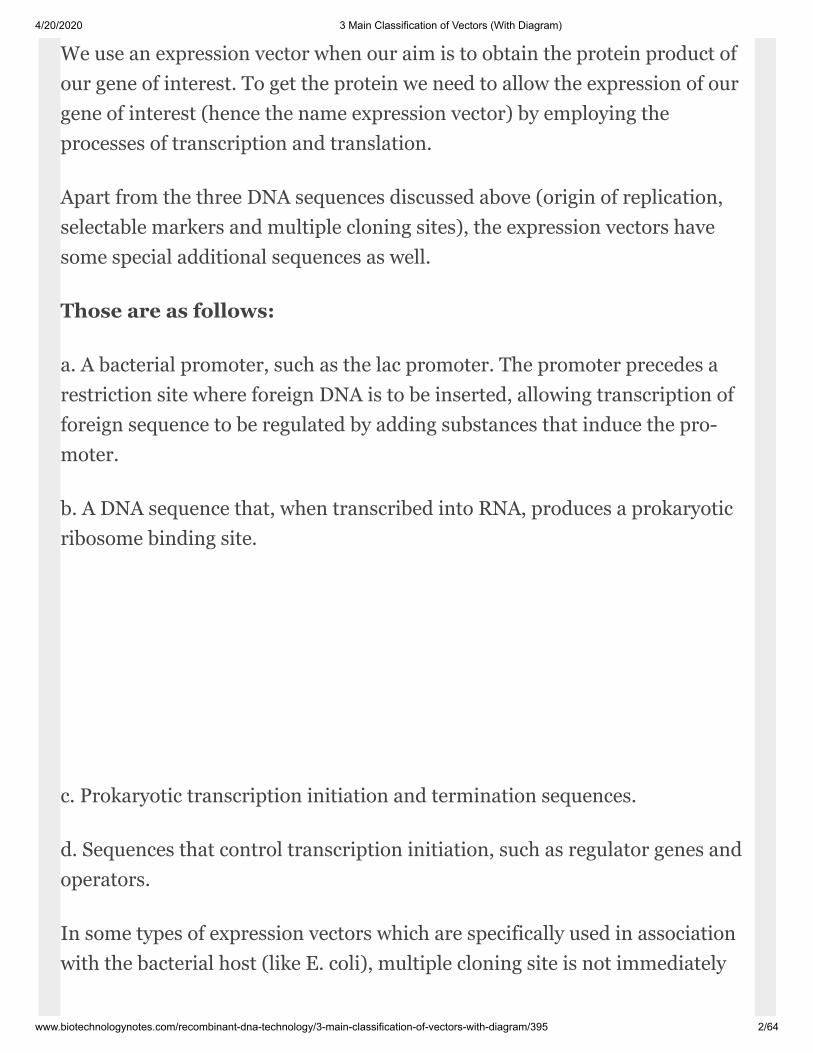
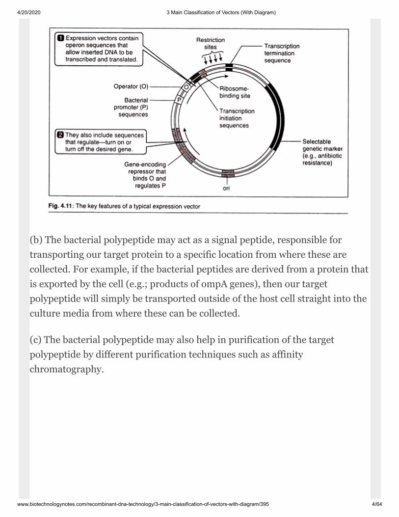
We use an expression vector when our aim is to obtain the protein prod uct ofour gene of interest. To get the pro tein we need to allow the expression of ourgene of interest (hence the name expres sion vector) by employing theprocesses of transcription and translation.
Apart from the three DNA sequences discussed above (origin of replication,selectable markers and multiple cloning sites), the expression vectors havesome special additional se quences as well.
Those are as follows:
a. A bacterial promoter, such as the lac promoter. The promoter precedes arestriction site where foreign DNA is to be inserted, allowing transcription offoreign sequence to be regulated by adding substances that induce the pro -moter.
b. A DNA sequence that, when tran scribed into RNA, produces a prokaryoticribosome binding site.
c. Prokaryotic transcription initiation and termination sequences.
d. Sequences that control transcription initiation, such as regulator genes andoperators.
In some types of expression vectors which are specifically used in associationwith the bacterial host (like E. coli), multiple cloning site is not immediately

4/20/2020 3 Main Classification of Vectors (With Diagram)
www.biotechnologynotes.com/recombinant-dna-technology/3-main-classification-of-vectors-with-diagram/395 3/64
adjacent to the ribo some binding sequence, but instead is preceded by aspecial sequence coding for a bacterial polypeptide.
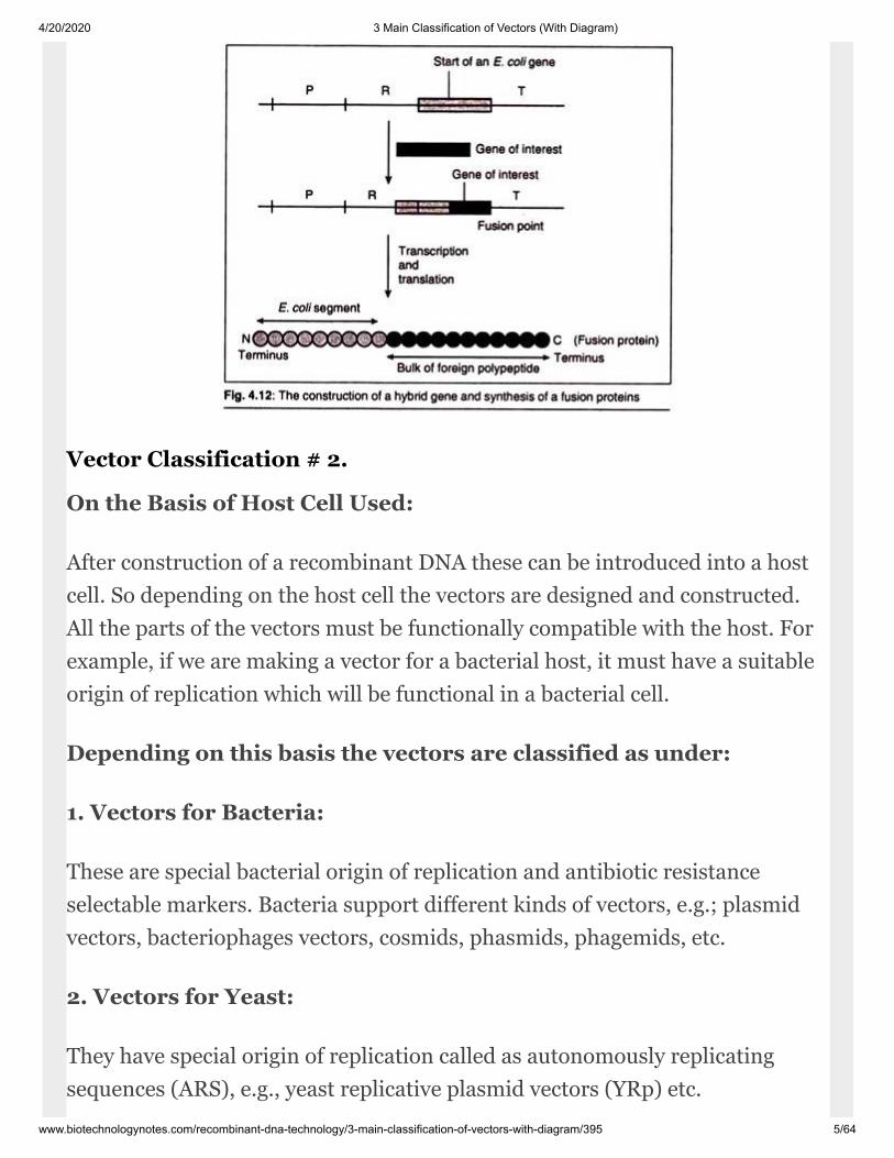
While using such type of expres sion vectors the gene of interest is inserted justafter the gene for bacterial polypeptide. In this way we fuse two readingframes, producing a hybrid gene that starts with the bacterial gene andprogresses without a break into the codons of our gene of interest.
The product of gene expression is therefore a hybrid protein, con sisting ofshort bacterial polypeptide fused into amino terminus of our targetpolypeptide se quence. This hybrid polypeptide chain consist ing of twodifferent types of polypeptides is called a fusion protein.
The followings are the reasons for incorporation of a fusion proteinbefore our gene of interest:
(a) The presence of bacterial peptide at the start of fusion protein may stabilizethe molecule and prevent it from being de graded by the host cell. In contrastthe for eign polypeptides that lack a bacterial seg ment are often destroyed.
4/20/2020 3 Main Classification of Vectors (With Diagram)
www.biotechnologynotes.com/recombinant-dna-technology/3-main-classification-of-vectors-with-diagram/395 4/64
(b) The bacterial polypeptide may act as a sig nal peptide, responsible fortransporting our target protein to a specific location from where these arecollected. For ex ample, if the bacterial peptides are derived from a protein thatis exported by the cell (e.g.; products of ompA genes), then our targetpolypeptide will simply be transported outside of the host cell straight into theculture media from where these can be collected.
(c) The bacterial polypeptide may also help in purification of the targetpolypeptide by different purification techniques such as affinitychromatography.
4/20/2020 3 Main Classification of Vectors (With Diagram)
www.biotechnologynotes.com/recombinant-dna-technology/3-main-classification-of-vectors-with-diagram/395 5/64
Vector Classification # 2.
On the Basis of Host Cell Used:
After construction of a recombinant DNA these can be introduced into a hostcell. So depend ing on the host cell the vectors are designed and constructed.All the parts of the vectors must be functionally compatible with the host. Forexample, if we are making a vector for a bacterial host, it must have a suitableorigin of replication which will be functional in a bac terial cell.
Depending on this basis the vectors are classified as under:
1. Vectors for Bacteria:
These are spe cial bacterial origin of replication and an tibiotic resistanceselectable markers. Bac teria support different kinds of vectors, e.g.; plasmidvectors, bacteriophages vectors, cosmids, phasmids, phagemids, etc.
2. Vectors for Yeast:
They have special origin of replication called as autono mously replicatingsequences (ARS), e.g., yeast replicative plasmid vectors (YRp) etc.
4/20/2020 3 Main Classification of Vectors (With Diagram)
www.biotechnologynotes.com/recombinant-dna-technology/3-main-classification-of-vectors-with-diagram/395 6/64
3. Vectors for Animals:
These vectors are needed in biotechnology for the synthesis of recombinantprotein from genes that are not expressed correctly when cloned in E. coli oryeast, and methods for cloning in humans are being sought by clinical mo -lecular biologists attempting to devise techniques for gene therapy, in which adisease is treated by introduction of a cloned gene into the patient, e.g., P-element, SV40 etc.
4. Vectors for Plants:
The production of genetically modified plants has become possible due tosuccessful use of plant vec tors. e.g., Ti-plasmid, Ri-plasmid etc.
Vector Classification # 3.
On the Basis of Cellular Nature of Host Cell:
On this basis, the vectors are of two types:
1. Prokaryotic Vectors:
This comprises of all vectors for bacterial cells.
2. Eukaryotic Vectors:
This comprises of all the vectors for yeast, animal and plant cells.
Prokaryotic Vectors (Bacterial Vectors):
The E. coli cell which is frequently used as a prokaryotic host needs specifictypes of vec tors which are designed accordingly to func tion in its cytoplasm.Plasmid based and bacteriophage based vectors are most common prokaryoticvectors. The prokaryotic vectors include plasmid derived vectors,
4/20/2020 3 Main Classification of Vectors (With Diagram)
www.biotechnologynotes.com/recombinant-dna-technology/3-main-classification-of-vectors-with-diagram/395 7/64
bacteriophage derived vectors, phagemid vectors, plasmid vectors and fosmidvectors.
These are dis cussed as follows:
Plasmid Vectors:
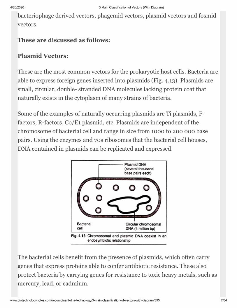
These are the most common vectors for the prokaryotic host cells. Bacteria areable to ex press foreign genes inserted into plasmids (Fig. 4.13). Plasmids aresmall, circular, double- stranded DNA molecules lacking protein coat thatnaturally exists in the cytoplasm of many strains of bacteria.
Some of the examples of naturally occurring plasmids are Ti plasmids, F-factors, R-factors, Co/E1 plasmid, etc. Plas mids are independent of thechromosome of bacterial cell and range in size from 1000 to 200 000 basepairs. Using the enzymes and 70s ribosomes that the bacterial cell houses,DNA contained in plasmids can be replicated and expressed.
The bacterial cells benefit from the pre sence of plasmids, which often carrygenes that express proteins able to confer antibiotic re sistance. These alsoprotect bacteria by carry ing genes for resistance to toxic heavy metals, such asmercury, lead, or cadmium.

4/20/2020 3 Main Classification of Vectors (With Diagram)
www.biotechnologynotes.com/recombinant-dna-technology/3-main-classification-of-vectors-with-diagram/395 8/64
In addi tion, some bacteria carry plasmids possessing genes that enablebacteria to break down her bicides, certain industrial chemicals, or thecomponents of petroleum. The relationship be tween bacteria and plasmids isendosymbiotic; both the bacteria and plasmids benefit from mutualarrangement. Plasmids also possess characteristic copy number.
The higher the copy number, higher is the number of indi vidual plasmids in ahost bacterial cell. If more copies of plasmid exist, more protein will besynthesized because of the larger number of gene copies carried by theplasmid. The num ber of copies plays a role in phenotypic mani festation of agene. For example, the more cop ies of an antibiotic-resistance gene there are,the higher the resistance to the antibiotic.
It is very important to note that naturally occur ring plasmids do not have allnecessary se quences which are required by a DNA molecule to act as aprofitable vector. Due to this, natu ral plasmids are extracted and modified byinserting suitable DNA segments and a com plete vector DNA molecule ismade.
Plasmid-cloning vectors are derived from bacterial plas mids and are the mostwidely used, versatile, and easily manipulated ones.
The following are different types of plasmid vectors:
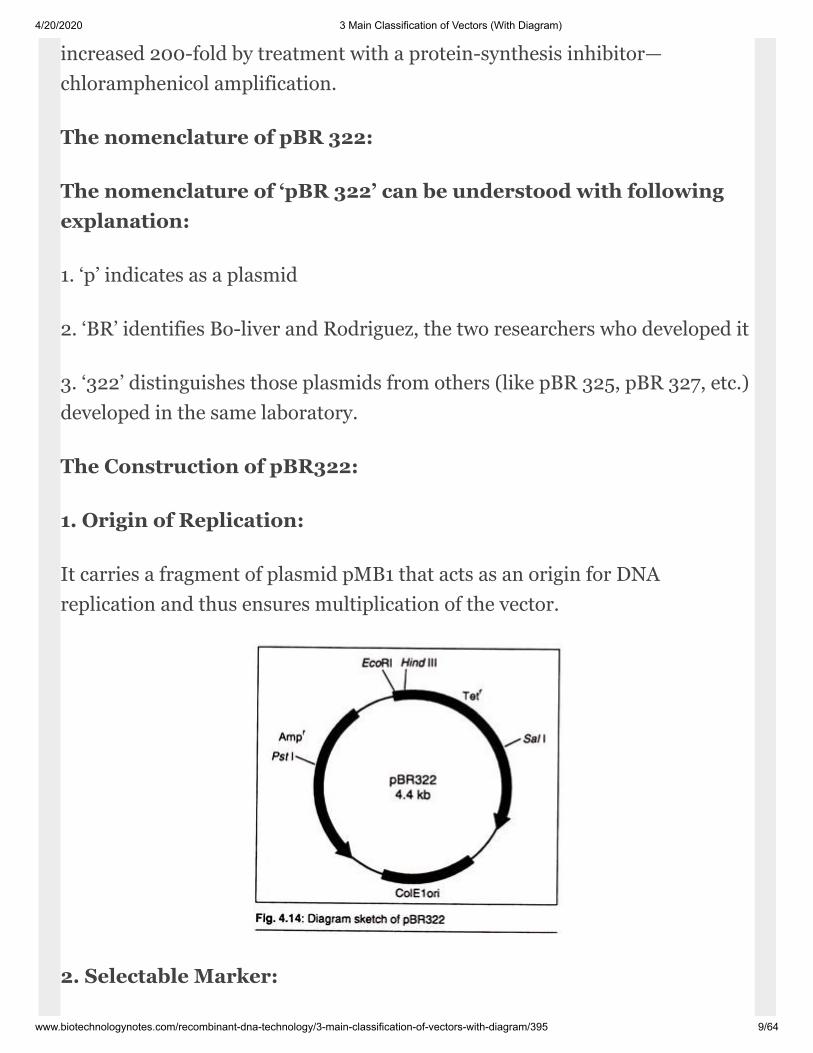
I. pBR322:
This was the first widely used, purpose built plasmid vector. pBR322 has arelatively small size of 4,363 bp. This is important because transformationefficiency is inversely proportional to size and above 10 kbp is very low.
Thus, there is ‘room’ in pBR322 for an insert of at least six kbp. Also thisvector has a rea sonably high copy number (~15 copies per cell), which can be
4/20/2020 3 Main Classification of Vectors (With Diagram)
www.biotechnologynotes.com/recombinant-dna-technology/3-main-classification-of-vectors-with-diagram/395 9/64
increased 200-fold by treatment with a protein-synthesis inhibitor—chloramphenicol amplification.
The nomenclature of pBR 322:
The nomenclature of ‘pBR 322’ can be under stood with followingexplanation:
1. ‘p’ indicates as a plasmid
2. ‘BR’ identifies Bo-liver and Rodriguez, the two researchers who developed it
3. ‘322’ distinguishes those plasmids from others (like pBR 325, pBR 327, etc.)developed in the same laboratory.
The Construction of pBR322:
1. Origin of Replication:
It carries a frag ment of plasmid pMB1 that acts as an ori gin for DNAreplication and thus ensures multiplication of the vector.
2. Selectable Marker:

4/20/2020 3 Main Classification of Vectors (With Diagram)
www.biotechnologynotes.com/recombinant-dna-technology/3-main-classification-of-vectors-with-diagram/395 10/64
It carries two anti biotic resistance genes—ampicillin and tetracycline.
3. Cloning Sites:
It carries a number of unique restriction sites. Some of these are located in oneof the antibiotic resistance genes (e.g., sites for Pst I, Pvu I, and Sac I are foundin Ampr and BamHI and Hind III in Tetr). Cloning into one of these sitesinactivates the gene allowing recombi nants to be differentiated from non-recombinants known as insertional inactivation.
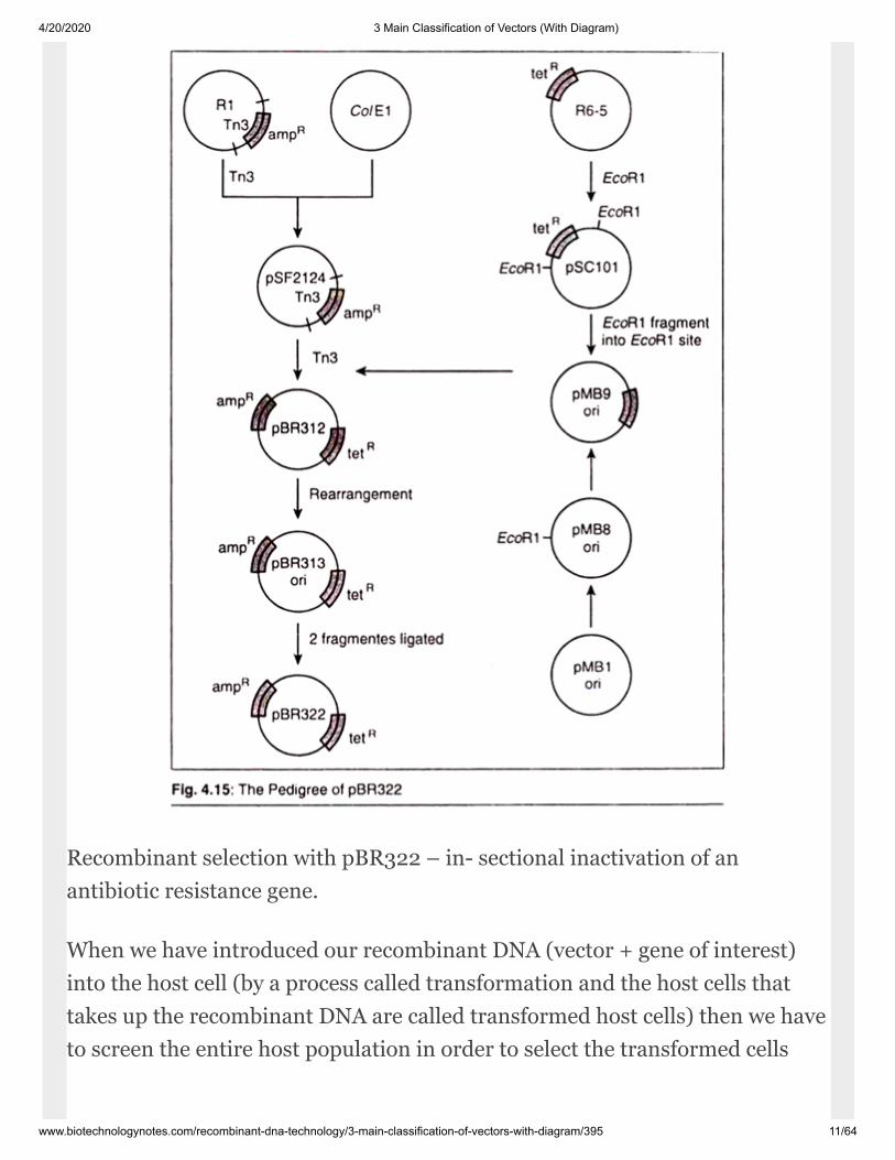
Pedigree of pBR322:
By pedigree we understand the origin of pBR322. pBR322 is not a naturallyoccurring plasmid. It is manufactured by following cer tain steps which areoutlined in (Fig. 4.15). It is important to note that pBR322 comprises DNAderived from three different naturally occur ring plasmids.
The amp gene originally re sided on the plasmid R1 (a naturally occur ringantibiotic resistant plasmid in E. coli), the tet is derived from R6-5 (a secondantibiotic resistant plasmid) and the origin of replica tion is derived frompMB1, which is closely related to the Colicin producing plasmid ColE1.
R
R
4/20/2020 3 Main Classification of Vectors (With Diagram)
www.biotechnologynotes.com/recombinant-dna-technology/3-main-classification-of-vectors-with-diagram/395 11/64
Recombinant selection with pBR322 – in- sectional inactivation of anantibiotic resis tance gene.
When we have introduced our recombinant DNA (vector + gene of interest)into the host cell (by a process called transformation and the host cells thattakes up the recombinant DNA are called transformed host cells) then we haveto screen the entire host population in order to select the transformed cells
4/20/2020 3 Main Classification of Vectors (With Diagram)
www.biotechnologynotes.com/recombinant-dna-technology/3-main-classification-of-vectors-with-diagram/395 12/64
(with recombi nant DNA) from the non-transformed one (without recombinantDNA).
Every vector has some mechanism associated with it for this screening.
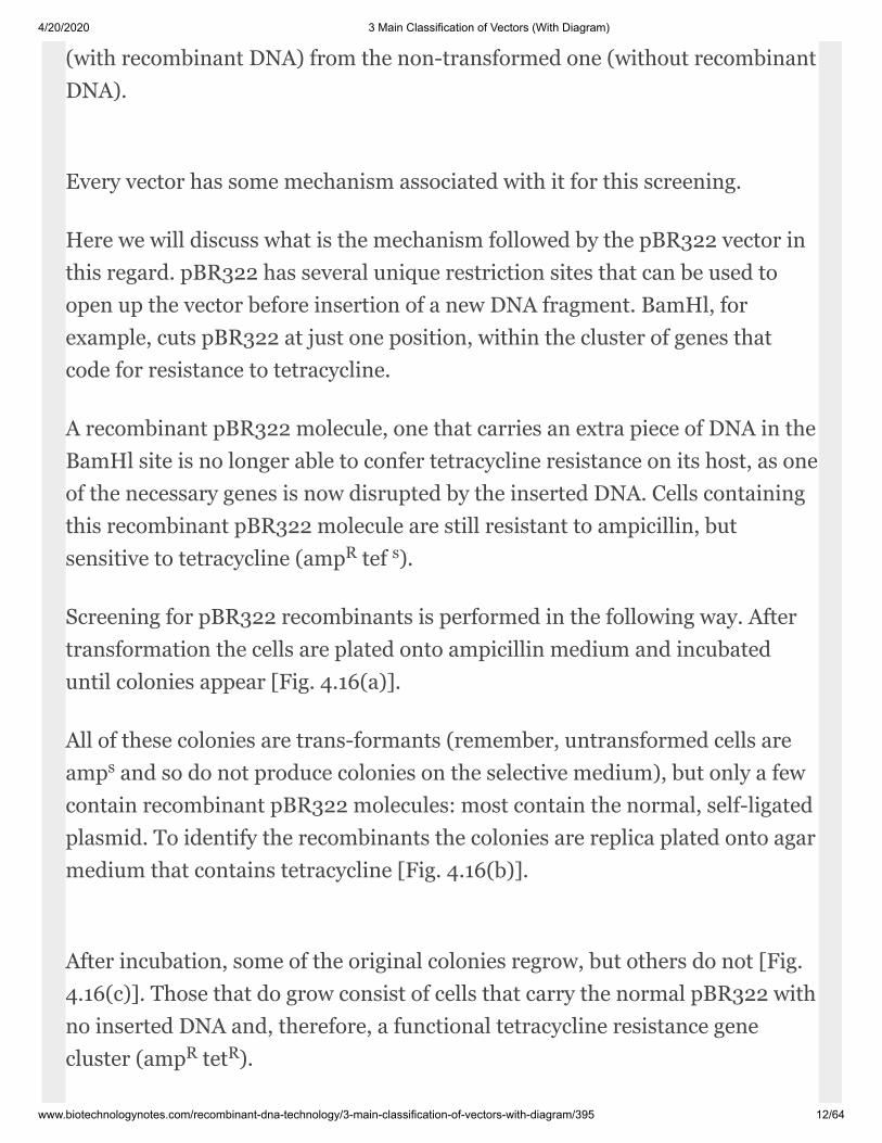
Here we will discuss what is the mechanism followed by the pBR322 vector inthis regard. pBR322 has several unique restriction sites that can be used toopen up the vec tor before insertion of a new DNA fragment. BamHl, forexample, cuts pBR322 at just one position, within the cluster of genes thatcode for resistance to tetracycline.
A recombinant pBR322 molecule, one that carries an extra piece of DNA in theBamHl site is no longer able to confer tetracycline resistance on its host, as oneof the necessary genes is now dis rupted by the inserted DNA. Cells containingthis recombinant pBR322 molecule are still resistant to ampicillin, butsensitive to tetra cycline (amp tef ).
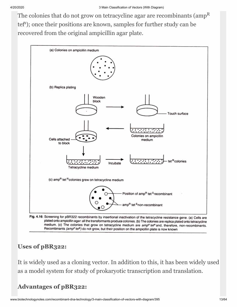
Screening for pBR322 re combinants is performed in the following way. Aftertransformation the cells are plated onto ampicillin medium and incubateduntil colo nies appear [Fig. 4.16(a)].
All of these colonies are trans-formants (remember, untransformed cells areamp and so do not produce colonies on the selective medium), but only a fewcon tain recombinant pBR322 molecules: most con tain the normal, self-ligatedplasmid. To iden tify the recombinants the colonies are replica plated onto agarmedium that contains tetra cycline [Fig. 4.16(b)].
After incubation, some of the original colonies regrow, but others do not [Fig.4.16(c)]. Those that do grow consist of cells that carry the normal pBR322 withno inserted DNA and, therefore, a functional tet racycline resistance genecluster (amp tet ).
R s
s
R R
4/20/2020 3 Main Classification of Vectors (With Diagram)
www.biotechnologynotes.com/recombinant-dna-technology/3-main-classification-of-vectors-with-diagram/395 13/64
The colonies that do not grow on tetracycline agar are recombinants (amptef ); once their positions are known, samples for further study can berecovered from the original ampicillin agar plate.
Uses of pBR322:
It is widely used as a cloning vector. In addi tion to this, it has been widely usedas a model system for study of prokaryotic transcription and translation.
Advantages of pBR322:
R
s
4/20/2020 3 Main Classification of Vectors (With Diagram)
www.biotechnologynotes.com/recombinant-dna-technology/3-main-classification-of-vectors-with-diagram/395 14/64
1. Small size (~ 4.4 kb) enables easy purifi cation and manipulation.
2. Two selectable markers (amp and tet) al low easy selection of recombinantDNA.
3. It can be amplified up to 1000-3000 copies per cell when protein synthesisis blocked by the application of chloramphenicol.
Disadvantages of pBR322:
1. It has very high mobility i. e; it can move to another cell in the presence of aconjugative plasmid like F-factor. The nic-bom (bom=basis of mobility) regionof pBR322 is responsible for this feature. Due to this, the vector may get lost ina population of mixed host cells.
2. There is a limitation in the size of the gene of interest that it canaccommodate.
3. Not a very high copy number is present as is expected from a good vector.
4. Although insertional inactivation of an antibiotic resistance gene providesan ef fective means of recombinant identifica tion, the method is madeinconvenient by the need to carry out two screenings, one with the antibioticthat selects for trans-formants, followed by the second screen, after replicaplating, with the an tibiotic which distinguishes recombinants.
This makes the screening process time- consuming and laborious.
Another vector pBR327 was derived from pBR322, by deletion of nucleotidesbetween 1,427 to 2,516. These nucleotides are deleted to reduce the size of thevector and to elimi nate sequences that were known to interfere with theexpression of cloned DNA in eukaryotic cells. pBR327 still contains genes forre sistance against two antibiotics (tetracycline and ampicillin).

4/20/2020 3 Main Classification of Vectors (With Diagram)
www.biotechnologynotes.com/recombinant-dna-technology/3-main-classification-of-vectors-with-diagram/395 15/64
pBR327 has following two ad vantages over pBR322:
1. pBR327 has high copy number (30-45 cop ies per cell).
2. It lacks mobility.
II. pUC Vectors:
pUC are obtained by modifying the pBR322 vector. pUC vectors are smallerthan pBR322 of being only ~2.7 kb. But comparatively they have a high copynumber. A mutation within the origin of replication produces 500 to 600copies of the plasmid per cell without amplification.
The Nomenclature of pUC Vectors:
The nomenclature of ‘pUC’ can be understood with the followingexplanation:
1. ‘p’ indicates the plasmid.
2. ‘UC’ stands for university of California where it was first developed by J.Mess ing et al.
We also see many numbers after this like pUC8, pUC18, pUC19 and so on.They are just the series of pUC and have been named just to separate fromeach other.
The construction of pUC vectors:
1. Origin of Replication: It is derived from the origin of replication ofpBR322.The ColE1origin of replication of pBR322 has been modified bycarrying out a chance mutation so that each transformed E. coli cell has 500-600 copies of the plasmid.
4/20/2020 3 Main Classification of Vectors (With Diagram)
www.biotechnologynotes.com/recombinant-dna-technology/3-main-classification-of-vectors-with-diagram/395 16/64
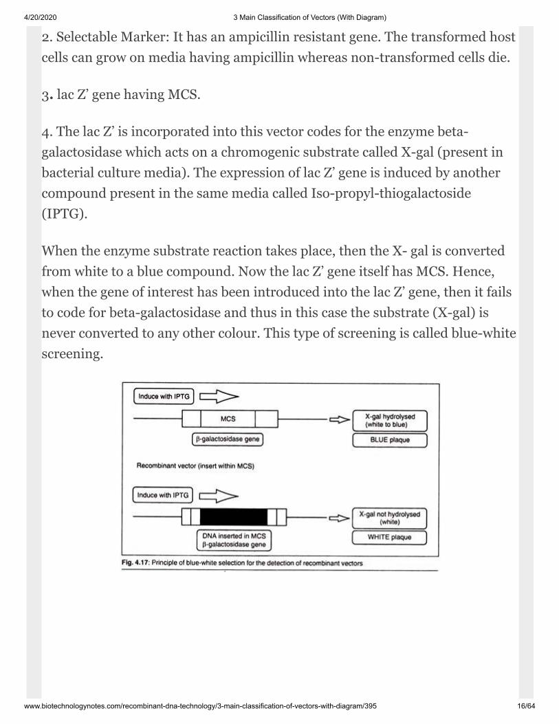
2. Selectable Marker: It has an ampicil lin resistant gene. The transformed hostcells can grow on media having ampicillin whereas non-transformed cells die.
3. lac Z’ gene having MCS.
4. The lac Z’ is incorporated into this vector codes for the enzyme beta-galactosidase which acts on a chromogenic substrate called X-gal (present inbacterial culture media). The expression of lac Z’ gene is in duced by anothercompound present in the same media called Iso-propyl-thiogalactoside(IPTG).
When the enzyme substrate reaction takes place, then the X- gal is convertedfrom white to a blue com pound. Now the lac Z’ gene itself has MCS. Hence,when the gene of interest has been introduced into the lac Z’ gene, then it failsto code for beta-galactosidase and thus in this case the substrate (X-gal) isnever converted to any other colour. This type of screening is called blue-whitescreen ing.

4/20/2020 3 Main Classification of Vectors (With Diagram)
www.biotechnologynotes.com/recombinant-dna-technology/3-main-classification-of-vectors-with-diagram/395 17/64
Pedigree of pUC Vectors:
During the construction of pUC the only se lectable marker that is kept out ofpBR322 is ampicillin resistant gene. But all the MCS are removed from theamp by carrying out chance mutations. The ColE1 origin of replication is alsomodified by the same process so that it can smoothly carry out the process ofreplica tion again and again ultimately increasing the copy number of thevector.
Along with this a lac Z’ sequence coding for beta galactosidase is also inserted.Similarly after this by the pro cess of chance mutation we create MCS withinthe lac Z’ sequence (Fig. 4.19).
R
4/20/2020 3 Main Classification of Vectors (With Diagram)
www.biotechnologynotes.com/recombinant-dna-technology/3-main-classification-of-vectors-with-diagram/395 18/64
Screening of Transformed Host Cells us ing pUC Vectors:
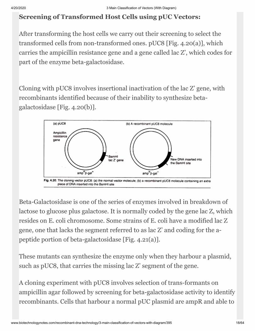
After transforming the host cells we carry out their screening to select thetransformed cells from non-transformed ones. pUC8 [Fig. 4.20(a)], whichcarries the ampicillin resistance gene and a gene called lac Z’, which codes forpart of the enzyme beta-galactosidase.
Cloning with pUC8 involves insertional inactivation of the lac Z’ gene, withrecombinants identified because of their inability to synthesize beta-galactosidase [Fig. 4.20(b)].
Beta-Galactosidase is one of the series of enzymes involved in breakdown oflactose to glucose plus galactose. It is normally coded by the gene lac Z, whichresides on E. coli chromosome. Some strains of E. coli have a modi fied lac Zgene, one that lacks the segment re ferred to as lac Z’ and coding for the a-peptide portion of beta-galactosidase [Fig. 4.21(a)].
These mutants can synthesize the enzyme only when they harbour a plasmid,such as pUC8, that carries the missing lac Z’ segment of the gene.
A cloning experiment with pUC8 involves selection of trans-formants onampicillin agar followed by screening for beta-galactosidase ac tivity to identifyrecombinants. Cells that harbour a normal pUC plasmid are ampR and able to
4/20/2020 3 Main Classification of Vectors (With Diagram)
www.biotechnologynotes.com/recombinant-dna-technology/3-main-classification-of-vectors-with-diagram/395 19/64
synthesize beta-galactosidase [Fig. 4.21(a)]; recombinants are also ampR butun able to make beta-galactosidase [Fig. 4.21(b)].
Screening for beta-galactosidase presence or absence is, in fact, quite easy.Rather than assay for lactose being split to glucose and galactose, we test for aslightly different reac tion that is also catalysed by beta-galactosi dase.
This involves a lactose analogue called X-gal (5-bromo-4-chloro-3-indolyl-beta-D- galactopyranoside) which is broken down by beta-galactosidase to aproduct that is coloured deep blue.
If X-gal (plus an inducer of the en zyme such as Iso-pro-pylthiogalactoside,IPTG) is added to the agar, along with ampicillin, then non-recombinantcolonies, the cells of which synthesize beta-galactosidase, will be colouredblue, whereas recombinants with a disrupted lac Z’ gene and unable to make p-galactosidase, will be white.
This system, which is called Lac selection, is summarized in [Fig. 4.21(b)].Note that both ampicillin resistance and the presence or absence of p-galactosidase is tested on a single agar plate. The two screen ings are, therefore,carried out together and there is no need for the time-consuming replica-plating step that is necessary with plas mids such as pBR322.
4/20/2020 3 Main Classification of Vectors (With Diagram)
www.biotechnologynotes.com/recombinant-dna-technology/3-main-classification-of-vectors-with-diagram/395 20/64
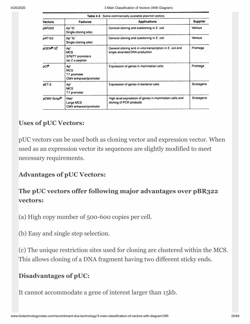
Uses of pUC Vectors:
pUC vectors can be used both as cloning vec tor and expression vector. Whenused as an expression vector its sequences are slightly modified to meetnecessary requirements.
Advantages of pUC Vectors:
The pUC vectors offer following major advan tages over pBR322vectors:
(a) High copy number of 500-600 copies per cell.
(b) Easy and single step selection.
(c) The unique restriction sites used for clon ing are clustered within the MCS.This allows cloning of a DNA fragment having two different sticky ends.
Disadvantages of pUC:
It cannot accommodate a gene of interest larger than 15kb.
4/20/2020 3 Main Classification of Vectors (With Diagram)
www.biotechnologynotes.com/recombinant-dna-technology/3-main-classification-of-vectors-with-diagram/395 21/64
Fosmid Vectors:
These are similar to cosmids but are based on the bacterial F-plasmid. Thecloning vec tor is limited, as a host (usually E. coli) can only contain one fosmidmolecule. Fosmids are 40 kb of random genomic DNA. Fosmid library isprepared from a genome of target organism and cloned into a fosmid vector.
Low copy num ber offers higher stability than comparable high copy numbercosmids. Fosmid system may be useful for constructing stable libraries fromcomplex genomes.
Bacteriophage Derived Vectors:
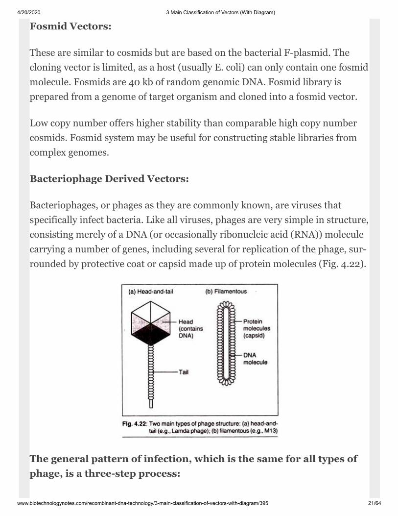
Bacteriophages, or phages as they are com monly known, are viruses thatspecifically in fect bacteria. Like all viruses, phages are very simple in structure,consisting merely of a DNA (or occasionally ribonucleic acid (RNA)) moleculecarrying a number of genes, includ ing several for replication of the phage, sur -rounded by protective coat or capsid made up of protein molecules (Fig. 4.22).
The general pattern of infection, which is the same for all types ofphage, is a three-step process:
4/20/2020 3 Main Classification of Vectors (With Diagram)
www.biotechnologynotes.com/recombinant-dna-technology/3-main-classification-of-vectors-with-diagram/395 22/64
1. The phage particle attaches to the outside of bacterium and injects its DNAchromo some into the cell.
2. The phage DNA molecule is replicated, usually by specific phage enzymescoded by genes on the phage chromosome.
3. Other phage genes direct synthesis of pro tein components of capsid, andnew phage particles are assembled and released from the bacterium.
With some phage types the entire infection cycle is completed very quickly,possibly in less than 20min. This type of rapid infection is called a lytic cycle,as release of the new ph age particles is associated with lysis of the bacterialcell.
The characteristic feature of a lytic infection cycle is that phage DNA repli -cation is immediately followed by synthesis of capsid proteins, and the phageDNA molecule is never maintained in a stable condition in the host cell. Incontrast to a lytic cycle, lysogenic infection is characterized by reten tion of thephage DNA molecule in the host bacterium, possibly for many thousands ofcell divisions.
Fred Blatter and his colleagues were the first to develop a bacteriophage asvector.
The following are different types of plasmid vectors:
I. Bacteriophage M13 vectors:
General Biology:
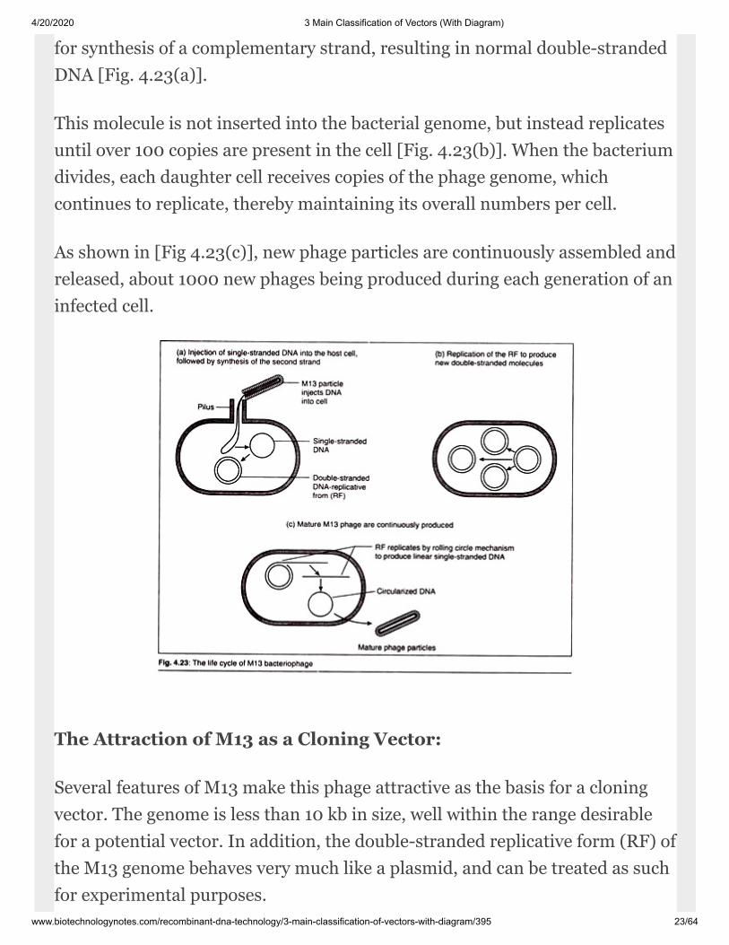
The M13 family of vectors is derived from bac teriophage M13. This is a malespecific (infects E. coli having f. pili), lysogenic filamentous phage with acircular single-stranded DNA genome about 6,407 bp (6.4 kb) in length. Onceinside the host-cell the single-stranded DNA of M13 phage acts as the template
4/20/2020 3 Main Classification of Vectors (With Diagram)
www.biotechnologynotes.com/recombinant-dna-technology/3-main-classification-of-vectors-with-diagram/395 23/64
for synthe sis of a complementary strand, resulting in normal double-strandedDNA [Fig. 4.23(a)].
This molecule is not inserted into the bacte rial genome, but instead replicatesuntil over 100 copies are present in the cell [Fig. 4.23(b)]. When the bacteriumdivides, each daughter cell receives copies of the phage genome, whichcontinues to replicate, thereby maintaining its overall numbers per cell.
As shown in [Fig 4.23(c)], new phage particles are continuously assembled andreleased, about 1000 new ph ages being produced during each generation of aninfected cell.
The Attraction of M13 as a Cloning Vector:
Several features of M13 make this phage at tractive as the basis for a cloningvector. The genome is less than 10 kb in size, well within the range desirablefor a potential vector. In addition, the double-stranded replicative form (RF) ofthe M13 genome behaves very much like a plasmid, and can be treated as suchfor experimental purposes.
4/20/2020 3 Main Classification of Vectors (With Diagram)
www.biotechnologynotes.com/recombinant-dna-technology/3-main-classification-of-vectors-with-diagram/395 24/64
It is easily prepared from a culture of infected E. coli cells and can bereintroduced by transfection. Most importantly, genes cloned with an M13-based vec tor can be obtained in the form of single- stranded DNA. Single-stranded version of cloned genes are useful for several techniques, notablyDNA sequencing and in vitro mutagen esis.
Using an M13 vector is an easy and reli able way of obtaining single-strandedDNA for this type of work.
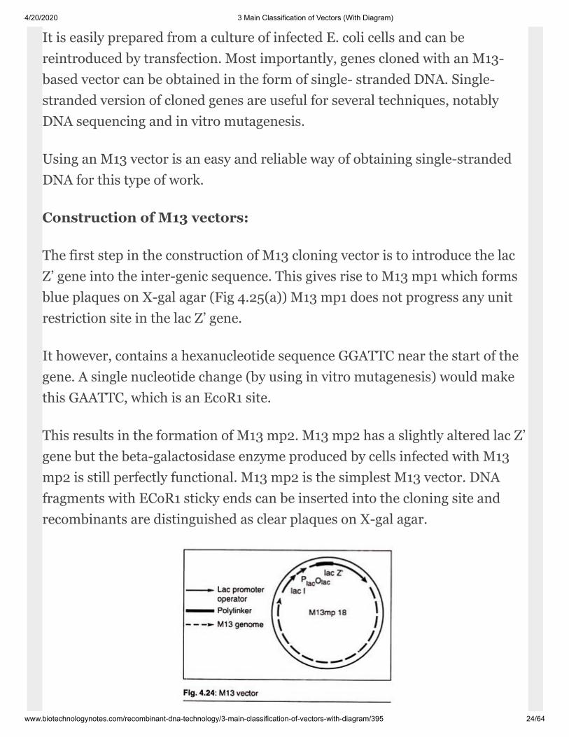
Construction of M13 vectors:
The first step in the construction of M13 clon ing vector is to introduce the lacZ’ gene into the inter-genic sequence. This gives rise to M13 mp1 which formsblue plaques on X-gal agar (Fig 4.25(a)) M13 mp1 does not progress any unitrestriction site in the lac Z’ gene.
It however, contains a hexanucleotide sequence GGATTC near the start of thegene. A single nucleotide change (by using in vitro mutagen esis) would makethis GAATTC, which is an EcoR1 site.
This results in the formation of M13 mp2. M13 mp2 has a slightly altered lac Z’gene but the beta-galactosidase enzyme pro duced by cells infected with M13mp2 is still perfectly functional. M13 mp2 is the simplest M13 vector. DNAfragments with ECoR1 sticky ends can be inserted into the cloning site andrecombinants are distinguished as clear plaques on X-gal agar.

4/20/2020 3 Main Classification of Vectors (With Diagram)
www.biotechnologynotes.com/recombinant-dna-technology/3-main-classification-of-vectors-with-diagram/395 25/64
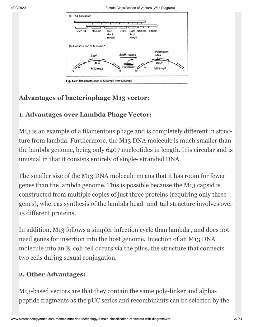
We go for further modifications of M13 mp2 resulting in the production ofanother M13 vector called M13 mp7. In the generation of M13 mp7 first of allwe synthesize a short oli gonucleotide called poly-linker that consists of a seriesof restriction sites and has EcoR1 sticky ends. This poly-linker is inserted intothe EcoRI site of M13 mp2 to generate M13mp7.
This poly-linker also provides as many as four possible cloning sites (ECoRI,BamHl, SaiI and PstI) to the new vector. It is very important to note that thepoly-linker is designed so that it does not totally disrupt the lac Z’ gene: a read -ing frame is maintained throughout the poly-linker, and a functional, thoughaltered, beta-galactosidase enzyme is still produced.
Screening of transformed host cells us ing bacteriophage M13vectors:
Insertion of new DNA almost invariably pre vents beta-galactosidaseproduction. So recombinant plaques are clear on X-gal agar. Alter natively, ifthe poly-linker is reinserted, and the original M13mp7 reformed, then blueplaques result.
Uses of bacteriophage M13 Vectors:
1. DNA Sequencing:
4/20/2020 3 Main Classification of Vectors (With Diagram)
www.biotechnologynotes.com/recombinant-dna-technology/3-main-classification-of-vectors-with-diagram/395 26/64
For a long time the most important application of M13 clon ing was in DNAsequence determination by the Sanger method, also called the dideoxy orchain-termination method. This relies on synthesis of DNA in the presence ofchain terminating inhibitors, the 2′, 3′- di-deoxynucleoside triphosphates(ddNTPs). The method is now a very standard tool of molecular biology.
2. Phage Display Vectors:
An important use of filamentous phage is in phage dis play systems. Here,coding sequences are inserted into one of the coat protein genes. The result isthat the phage are generated with a hybrid form of this protein, which is afusion of the normal protein sequence and the protein product of the insertedse quence (assuming the inserted sequence has the same reading frame as thecoat protein gene).
The phages are secreted from the cell, with this extra material ‘displayed’ onthe outside. These display vectors have many uses, e.g., in screening librariesby panning and for vaccine production.
3. Other Applications:
Some protocols for site-directed mutagenesis also use single- stranded DNA,which can be obtained with vectors based on filamentous phages. Single-stranded DNA is also of particular use in generating probes for RNA analy sis.
Probes can be prepared that are spe cific for RNA transcripts from eitherstrand of DNA. The latter applications are out side the scope of this book, butmore infor mation can be obtained from specialized laboratory manuals.
4/20/2020 3 Main Classification of Vectors (With Diagram)
www.biotechnologynotes.com/recombinant-dna-technology/3-main-classification-of-vectors-with-diagram/395 27/64
Advantages of bacteriophage M13 vector:
1. Advantages over Lambda Phage Vec tor:
M13 is an example of a filamentous phage and is completely different in struc -ture from lambda. Furthermore, the M13 DNA molecule is much smaller thanthe lambda genome, being only 6407 nucle otides in length. It is circular and isun usual in that it consists entirely of single- stranded DNA.
The smaller size of the M13 DNA molecule means that it has room for fewergenes than the lambda genome. This is possible because the M13 capsid iscons tructed from multiple copies of just three proteins (requiring only threegenes), whereas synthesis of the lambda head- and-tail structure involves over15 differ ent proteins.
In addition, M13 follows a simpler infection cycle than lambda , and does notneed genes for insertion into the host genome. Injection of an M13 DNAmolecule into an E. coli cell occurs via the pilus, the structure that connectstwo cells during sexual conjugation.
2. Other Advantages:
M13-based vectors are that they contain the same poly-linker and alpha-peptide fragments as the pUC series and recombinants can be selected by the

4/20/2020 3 Main Classification of Vectors (With Diagram)
www.biotechnologynotes.com/recombinant-dna-technology/3-main-classification-of-vectors-with-diagram/395 28/64
blue → white colour test. Also the size of the genome is below 10 kb and so iseasy to handle.
Disadvantages of bacteriophage M13 vec tors:
The following are the disadvantages of bacte riophage M13 vectors:
1. Gene of interest more than 2kb cannot be cloned.
2. It has low yield of DNA.
3. The phage produce many toxins in high concentration.
II. Lamda Phage Vectors:
This is a widely used vector for the cloning of very large pieces of genes.
General Biology:

4/20/2020 3 Main Classification of Vectors (With Diagram)
www.biotechnologynotes.com/recombinant-dna-technology/3-main-classification-of-vectors-with-diagram/395 29/64
Lambda is a typical example of a head-and-tail phage. The genetic material isDNA which is present in the polyhedral head structure and the tail serves toattach the phage to the bacterial surface and to inject the DNA into the cell.The lambda DNA molecule is 49 kb in size.
It is a temperate phase and this can carry out lytic and lysogenic cycles. Thepositions and identities of most of the genes on the lambda DNA molecule areknown (Fig. 4.28).
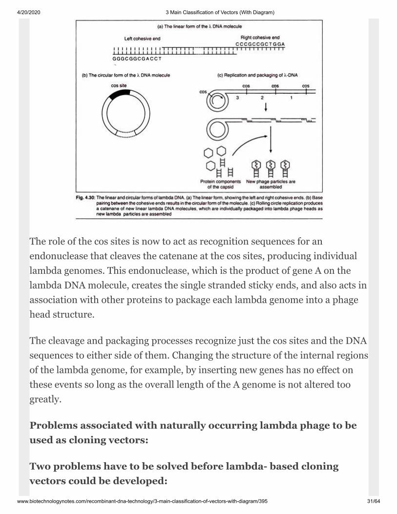
Lambda phage can have both linear and circular forms of DNA. The moleculeshown in (Fig. 4.28) is linear, with two free ends, and represents the DNApresent in the phage head. This linear molecule consists of two comple mentarystrands of DNA, base paired accord ing to the Watson-Crick rules.’ However, ateither end of the molecule is a short 12-nucleotide stretch in which the DNA issingle-stranded [Fig. 4.30(a)].
The two single strands are complementary, and so can base pair with oneanother to form a circular, completely double-stranded molecule [Fig.4.30(b)]. Comple mentary single strands are often referred to as ‘sticky’ ends orcohesive ends, because base pairing between them can ‘stick’ together the twoends of a DNA molecule (or the ends of two different DNA molecules).
The lambda cohesive ends are called the cos sites and they play two distinctroles during the lambda in fection cycle. First, they allow the linear DNA
4/20/2020 3 Main Classification of Vectors (With Diagram)
www.biotechnologynotes.com/recombinant-dna-technology/3-main-classification-of-vectors-with-diagram/395 30/64
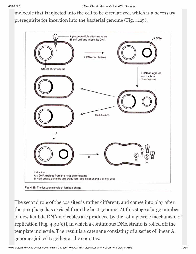
molecule that is injected into the cell to be cir cularized, which is a necessaryprerequisite for insertion into the bacterial genome (Fig. 4.29).
The second role of the cos sites is rather different, and comes into play afterthe pro-ph age has excised from the host genome. At this stage a large numberof new lambda DNA molecules are produced by the rolling circle mechanism ofreplication [Fig. 4.30(c)], in which a continuous DNA strand is rolled off thetemplate molecule. The result is a catenane consisting of a series of linear Agenomes joined together at the cos sites.
4/20/2020 3 Main Classification of Vectors (With Diagram)
www.biotechnologynotes.com/recombinant-dna-technology/3-main-classification-of-vectors-with-diagram/395 31/64
The role of the cos sites is now to act as recognition sequences for anendonuclease that cleaves the catenane at the cos sites, producing individuallambda genomes. This endonuclease, which is the product of gene A on thelambda DNA mol ecule, creates the single stranded sticky ends, and also acts inassociation with other proteins to package each lambda genome into a phagehead structure.
The cleavage and packaging processes recognize just the cos sites and the DNAsequences to either side of them. Chang ing the structure of the internal regionsof the lambda genome, for example, by inserting new genes has no effect onthese events so long as the overall length of the A genome is not al tered toogreatly.
Problems associated with naturally oc curring lambda phage to beused as clon ing vectors:
Two problems have to be solved before lambda- based cloningvectors could be developed:
4/20/2020 3 Main Classification of Vectors (With Diagram)
www.biotechnologynotes.com/recombinant-dna-technology/3-main-classification-of-vectors-with-diagram/395 32/64
1. The lambda molecule can be increased in size by only about 5%,representing the ad dition of only 3kb of new DNA. If the total size of themolecule is more than 52kb, then it cannot be packaged into the lambda headstructure and infective phage par ticles are not formed. This severely limits thesize of a DNA fragment that can be inserted into an unmodified lambda vec tor.
2. The lambda genome is so large that it has more than one recognitionsequence for virtually every restriction endonucleases. Restriction cannot beused to cleave the normal lambda molecule in a way that will allow insertion ofnew DNA, because the molecules would be cut into several small fragmentsthat would be very unlikely to reform a viable lambda genome on relega tion.
Due to these reasons the DNA of naturally occurring lambda phage cannot beused as a cloning vector. To solve this issue we modify the lambda’s genomeand make it suitable to be a successful vector.
Solving the Problems:
Solving Problem (1):
From research it has been found out that large segment in the central region ofthe lambda DNA molecule can be removed without affecting the ability of thephage to infect E. coli cells. Removal of this non essential region betweenpositions 20 and 35 on the map decreases the size of the lambda genome by upto 15kb.
This makes a room for as much as 18kb of new DNA which can be added to itto form a recombinant molecule.
This non-essential genes thus removed are involved in integration and excisionof the lambda pro-phage from the E. coli chromosome. A deleted lambdagenome is, therefore, non-lysogenic and can follow only the lytic infec tioncycle. This in itself is desirable for a clon ing vector as it means that we can get

4/20/2020 3 Main Classification of Vectors (With Diagram)
www.biotechnologynotes.com/recombinant-dna-technology/3-main-classification-of-vectors-with-diagram/395 33/64
plaques (a visible structure formed within a cell cul ture, such as bacterialcultures within some nutrient medium).
Solving problem (2):
We can remove un necessary restriction sites by carrying out in vitromutagenesis. For example, an ECoRI site, GAATTC, could be changed toGGATTC, which is not recognized by the enzyme.
Types of Lambda Vectors:

4/20/2020 3 Main Classification of Vectors (With Diagram)
www.biotechnologynotes.com/recombinant-dna-technology/3-main-classification-of-vectors-with-diagram/395 34/64
There are two types of lambda cloning vectors.
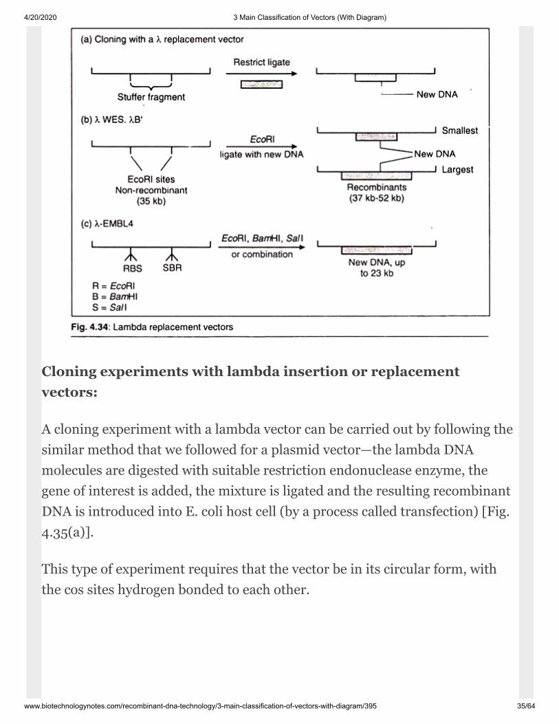
(a) Lambda Insertion Vectors:
In this case a large segment of the non-essential re gion has been deleted, andthe two arms ligated together. An insertion vector pos sesses at least oneunique restriction site into which new DNA can be inserted. The size of theDNA fragment that an indi vidual vector can carry depends on the extent towhich the non-essential region has been deleted, e.g.; lambda-gtl0, lambda-ZAP11.
(b) Lambda Replacement Vectors:
These vectors have two recognition sites for the restriction endonucleases.These sites flank a segment of DNA that is replaced by the DNA to be cloned[Fig.4.34(a)]. Of ten the replaceable fragment (or stuffer fragment) carriesadditional restriction sites that can be used to cut it up into small pieces sothat its own reinsertion during a cloning experiment is very unlikely.
Re placement vectors are generally designed to carry large pieces of DNA thaninser tion vectors can handle e.g., lambda- EMBL, lambda-GEMll, etc.
4/20/2020 3 Main Classification of Vectors (With Diagram)
www.biotechnologynotes.com/recombinant-dna-technology/3-main-classification-of-vectors-with-diagram/395 35/64
Cloning experiments with lambda inser tion or replacementvectors:
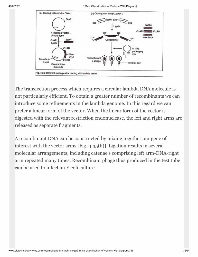
A cloning experiment with a lambda vector can be carried out by following thesimilar method that we followed for a plasmid vector—the lambda DNAmolecules are digested with suit able restriction endonuclease enzyme, thegene of interest is added, the mixture is ligated and the resulting recombinantDNA is introduced into E. coli host cell (by a process called trans fection) [Fig.4.35(a)].
This type of experiment requires that the vector be in its circular form, withthe cos sites hydrogen bonded to each other.
4/20/2020 3 Main Classification of Vectors (With Diagram)
www.biotechnologynotes.com/recombinant-dna-technology/3-main-classification-of-vectors-with-diagram/395 36/64
The transfection process which requires a circular lambda DNA molecule isnot particu larly efficient. To obtain a greater number of recombinants we canintroduce some refine ments in the lambda genome. In this regard we canprefer a linear form of the vector. When the linear form of the vector isdigested with the relevant restriction endonuclease, the left and right arms arereleased as separate frag ments.
A recombinant DNA can be constructed by mixing together our gene ofinterest with the vector arms [Fig. 4.35(b)]. Ligation results in severalmolecular arrangements, including catenae’s comprising left arm-DNA-rightarm repeated many times. Recombinant ph age thus produced in the test tubecan be used to infect an E.coli culture.
4/20/2020 3 Main Classification of Vectors (With Diagram)
www.biotechnologynotes.com/recombinant-dna-technology/3-main-classification-of-vectors-with-diagram/395 37/64
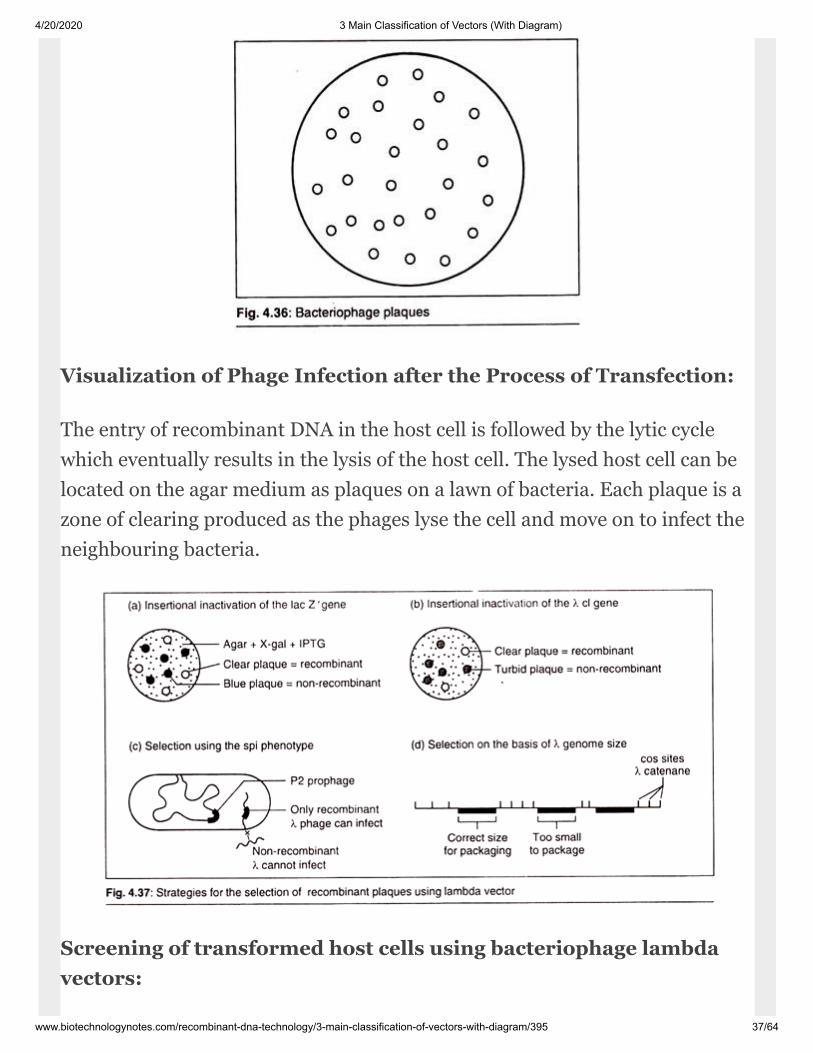
Visualization of Phage Infection after the Process of Transfection:
The entry of recombinant DNA in the host cell is followed by the lytic cyclewhich eventually results in the lysis of the host cell. The lysed host cell can belocated on the agar medium as plaques on a lawn of bacteria. Each plaque is azone of clearing produced as the phages lyse the cell and move on to infect theneighbouring bacteria.
Screening of transformed host cells us ing bacteriophage lambdavectors:
4/20/2020 3 Main Classification of Vectors (With Diagram)
www.biotechnologynotes.com/recombinant-dna-technology/3-main-classification-of-vectors-with-diagram/395 38/64
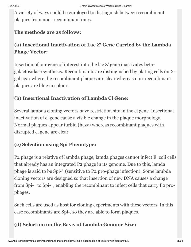
A variety of ways could be employed to distin guish between recombinantplaques from non- recombinant ones.
The methods are as follows:
(a) Insertional Inactivation of Lac Z’ Gene Carried by the LambdaPhage Vector:
Insertion of our gene of interest into the lac Z’ gene inactivates beta-galactosidase synthesis. Recombinants are distinguished by plating cells on X-gal agar where the recombinant plaques are clear whereas non-recombinantplaques are blue in colour.
(b) Insertional Inactivation of Lambda Cl Gene:
Several lambda cloning vectors have restriction site in the cl gene. Insertionalinactivation of cl gene cause a visible change in the plaque morphology.Normal plaques appear turbid (hazy) whereas re combinant plaques withdisrupted cl gene are clear.
(c) Selection using Spi Phenotype:
P2 ph age is a relative of lambda phage, lamda phages cannot infect E. coli cellsthat al ready has an integrated P2 phage in its genome. Due to this, lamdaphage is said to be Spi- (sensitive to P2 pro-phage in fection). Some lambdacloning vectors are designed so that insertion of new DNA causes a changefrom Spi- to Spi- , en abling the recombinant to infect cells that carry P2 pro-phages.
Such cells are used as host for cloning experiments with these vectors. In thiscase recombinants are Spi-, so they are able to form plaques.
(d) Selection on the Basis of Lambda Ge nome Size:
+
+ –
4/20/2020 3 Main Classification of Vectors (With Diagram)
www.biotechnologynotes.com/recombinant-dna-technology/3-main-classification-of-vectors-with-diagram/395 39/64
We know this from the begin ning that any gene of interest which is less than37kb or more than 52kb cannot be packed in the head of lambda phage. Manylambda vectors have been constructed by deleting large segments of thelambda DNA molecule and so are less than 37kb in length.
These can only be packaged into mature phage particles after our gene ofinterest has been inserted. This brings the total genome size up to 37kb ormore. Hence, with these vectors only recombi nant phages are able to replicate.
Uses of Bacteriophage Lambda Vectors:
The main use of all lambda based vectors is to clone DNA fragments that aretoo long to be handled by plasmid or M13 vectors. A replace ment vector suchas lambda-EMBL4 can carry up to 20kb of our gene of interest. This com pareswith a maximum insert size of about 8kb for almost all plasmids and less than3kb for M13 vectors.
Advantages of Bacteriophage Lambda Vec tors
Following are the advantages of lambda vec tors:
1. Storage of phage particles is comparatively much easier than that of plasmidbased vectors.
2. The shelf-life of phage particles is infinite.
3. Transfection of E. coli is much easier with phage particles.
Disadvantages of Bacteriophage Lambda Vectors:
If you have isolated a clone, it is frequently quite difficult to isolate largequantities of DNA. In practice, many problems are encoun tered that do notoccur with plasmids. There is still no truly rapid, reliable protocol for theproduction of very clean lambda-DNA.
4/20/2020 3 Main Classification of Vectors (With Diagram)
www.biotechnologynotes.com/recombinant-dna-technology/3-main-classification-of-vectors-with-diagram/395 40/64
The most successful method is to use anion exchange columns. The mostfrequent problem is that the preparation contains dirt that makes furtherprocessing, such as a restric tion digestion, difficult or impossible.
Even in the replacement vectors, almost two thirds of the DNA is made up ofvector sequences. If possible, you should clone the sections that are of interestusing plasmids. LambdaZAP banks can save work, because the plasmidportions are cut out in vivo, along with inserted DNA. That process is highlyefficient, requires only a relatively few work steps, and lasts only 1 to 2 days.
III. Cosmid Vectors:
It is the most sophisticated type of lambda based vector. Cosmids are thehybrids between the phage DNA molecule and bacterial plas mid. Their designcentres on the fact that the enzymes that package the lambda DNA mol eculeinto the phage protein coat need only the cos sites in order to function.
Construction of Cosmid Vectors:
A cosmid is basically a plasmid that carries a cos site. It also needs a selectablemarker, such as ampicillin resistant gene, and a plasmid origin of replication.

4/20/2020 3 Main Classification of Vectors (With Diagram)
www.biotechnologynotes.com/recombinant-dna-technology/3-main-classification-of-vectors-with-diagram/395 41/64
This is important to note that as cosmid lacks all the lambda genes, so at doesnot produce plaques. Instead colonies are formed on the selective media justas with plasmid vectors.
Cloning Experiment with Cosmid Vectors:
This is carried out as follows. The cosmid is opened and its unique restrictionsite and our gene of interest is inserted. These fragments are usually producedby partial digestion with a restriction endonuclease, as total digestion almostinvariably results in fragments that are too small to be cloned with a cosmid.
Ligation is carried out so that catenanes are formed. These lambda phages arethen used to infect an E. coli culture. All colonies are recombinant colonies asnon-recombinant lambda phages cannot be packaged into the head of thelambda bacteriophage.
Uses of Cosmid Vectors:
4/20/2020 3 Main Classification of Vectors (With Diagram)
www.biotechnologynotes.com/recombinant-dna-technology/3-main-classification-of-vectors-with-diagram/395 42/64
Cosmids are used for construction of genomic libraries of eukaryotes sincethese can be used for cloning large fragments of DNA.
Advantages of Cosmids:
Followings are advantages of cosmid vectors:
1. These can be used to clone gene of inter est up to 40 kb.
2. As the lambda phage will insert the recom binant DNA into the host cell, anextra step of inserting the recombinant DNA into the host cell is notperformed.
3. Easy screening method is found.
The Phagemid Vectors:
Although M13 vectors are very useful for the production of single-strandedversions of cloned genes they do have one disadvantage. There is a limit to thesize of DNA fragment that can be cloned with an M13 vector, with 1500bpgenerally being looked on as the maxi mum capacity.
To get around this problem a number of novel vectors have been constructedwhich are the hybrids of plasmids and M13 vectors. We call them phagemids(‘phage’ from M13 bacteriophage and ‘mid’ from plasmid).
Construction of Phagemid Vector:
A typical phagemid has following parts:
1. Phage M13 origin of replication.
2. A portion of lac Z’ gene driven by lac pro moter.
3. A multiple cloning site (MCS) with lac Z’ gene.
4/20/2020 3 Main Classification of Vectors (With Diagram)
www.biotechnologynotes.com/recombinant-dna-technology/3-main-classification-of-vectors-with-diagram/395 43/64
4. Phage T7 and T3 promoter sequences flanking the MCS sequences.
5. ColE1 origin of replication.
6. amp resistant gene.
Plasmids that carry the M13 replication origin in addition to a conventionalorigin of dsDNA synthesis can be replicated either as dsDNA from the latter oras single-stranded DNA from the M13 origin. Replication from the M13 originrequires the appropriate pro teins (such as gene II protein) to be provided froma helper phage also replicating within the cell.
Replication generates single-stranded DNA which can then be packaged intophage coats. Examples of phagemids are the vectors pUC118, 119 and 120.
They are replicated as plasmids until the cell containing them is co-infectedwith a helper phage, such as M13K07, which provides the proteins for single-stranded DNA synthesis and packaging. M13K07 is an M13 phage that hasbeen modified, most im portantly by the incorporation of a plasmid replicationorigin.
Replication from this origin allows the helper phage to be present in a highcopy number per cell and, therefore, to pro vide the larger quantities of theproteins that are required to replicate and package the phagemid molecule.
M13K07 also contains a kanamycin resistance gene to allow for selec tion forthe presence of the helper phage. (Of course, it is possible that the M13K07helper phage may be packaged too, but in practice the packaged phagemidmolecules are found to be in a 100-fold excess over the helper phage.)
Another example of these vectors is the pBluescript series, such aspBluescriptIIKSÞ, shown in Fig. 4.39. This series of plasmids con tains, inaddition to features already described, promoters from the E. colibacteriophages T3 or T7, which are useful for expressing cloned sequences.
R
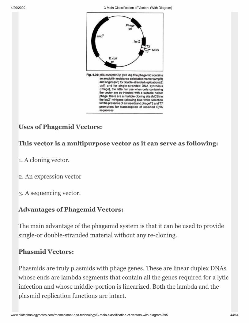
4/20/2020 3 Main Classification of Vectors (With Diagram)
www.biotechnologynotes.com/recombinant-dna-technology/3-main-classification-of-vectors-with-diagram/395 44/64
Uses of Phagemid Vectors:
This vector is a multipurpose vector as it can serve as following:
1. A cloning vector.
2. An expression vector
3. A sequencing vector.
Advantages of Phagemid Vectors:
The main advantage of the phagemid system is that it can be used to providesingle-or double-stranded material without any re-cloning.
Phasmid Vectors:
Phasmids are truly plasmids with phage genes. These are linear duplex DNAswhose ends are lambda segments that contain all the genes required for a lyticinfection and whose middle-portion is linearized. Both the lambda and theplasmid replication functions are intact.
4/20/2020 3 Main Classification of Vectors (With Diagram)
www.biotechnologynotes.com/recombinant-dna-technology/3-main-classification-of-vectors-with-diagram/395 45/64
Nor mally, plasmid vectors carry a lambda attach ment site. Once inside E. colicell, the phas mid can replicate like a phage and form plaques in the normalway. However, if the vector contains the gene that encodes lambda repressor,then the plasmid replicates as a plasmid rather than as a phage.
Depending upon the functioning or non-functioning of cl- Protein (coded byrepressor), the phasmid can replicate as plasmid (cl-Protein inactive) or phagewhen cl-protein is active. The activity of cl-protein can be inactive by growingthe E.coli culture at 40°C.
Plasmids may be used in variety of ways. For example, DNA may be cloned inthe plasmid vector in a conventional way and then the recombinant plasmidcan be lifted onto the phage. Plasmids are easy to store, they have aneffectively infinite shelf life and screening phages by molecular hybridiza tiongives cleaner results than screening bac terial colonies.
Eukaryotic Vectors:
Most cloning experiments are carried out with E. coli as the host, and thewidest variety of cloning vectors are available for this organism. E. coli isparticularly popular when the aim of the cloning experiment is to study thebasic features of molecular biology such as gene structure and function.However, under some circumstances it may be desirable to use a dif ferent hostfor a gene cloning experiment.
But when the aim of the RDT experiment is not just to study a gene but to usecloning to con trol or improve synthesis of an important meta bolic product(e.g., a hormone such as insu lin), or to change the properties of the organ ism(e.g., to introduce herbicide resistance into a crop plant), then we take a hostcell which is more advanced and capable of meeting an advanced level ofmetabolism.
4/20/2020 3 Main Classification of Vectors (With Diagram)
www.biotechnologynotes.com/recombinant-dna-technology/3-main-classification-of-vectors-with-diagram/395 46/64
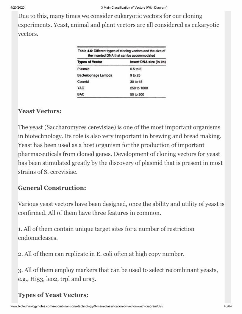
Due to this, many times we consider eukaryotic vectors for our cloningexperiments. Yeast, animal and plant vectors are all considered as eukaryoticvectors.
Yeast Vectors:
The yeast (Saccharomyces cerevisiae) is one of the most important organismsin biotechnol ogy. Its role is also very important in brewing and bread making.Yeast has been used as a host organism for the production of importantpharmaceuticals from cloned genes. Develop ment of cloning vectors for yeasthas been stimulated greatly by the discovery of plasmid that is present in moststrains of S. cerevisiae.
General Construction:
Various yeast vectors have been designed, once the ability and utility of yeast isconfirmed. All of them have three features in common.
1. All of them contain unique target sites for a number of restrictionendonucleases.
2. All of them can replicate in E. coli often at high copy number.
3. All of them employ markers that can be used to select recombinant yeasts,e.g., Hi53, leo2, trpl and ura3.
Types of Yeast Vectors:

4/20/2020 3 Main Classification of Vectors (With Diagram)
www.biotechnologynotes.com/recombinant-dna-technology/3-main-classification-of-vectors-with-diagram/395 47/64
All the yeast vectors can be divided into three types:
1. Yeast cloning vectors (or Yeast plasmid vectors)
2. Yeast expression vectors
3. Yeast artificial chromosomes (YAC)
Yeast Cloning Vectors (or Yeast Plasmid Vectors):
These vectors are used to clone (make several duplicate copies) our gene ofinterest in the yeast host cell. All the cloning vectors have been engineeredfrom 2fx plasmid which is the naturally occurring plasmid in the yeast cell.
They are of following types:
1. Yeast Episomal Plasmids (YEps):
It is 6,318 bp long and has a copy number of 70-200. Most of the YEps areshuttle vec tors (can be used as vectors both in prokaryotic hosts andeukaryotic hosts) and thus have been engineered accord ingly. An example ofYEps is Yep13.
4/20/2020 3 Main Classification of Vectors (With Diagram)
www.biotechnologynotes.com/recombinant-dna-technology/3-main-classification-of-vectors-with-diagram/395 48/64
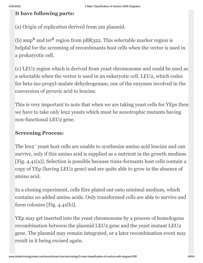
It have following parts:
(a) Origin of replication derived from 2m plas mid.
(b) amp and tet region from pBR322. This selectable marker region ishelpful for the screening of recombinants host cells when the vector is used ina prokaryotic cell.
(c) LEU2 region which is derived from yeast chromosome and could be used asa selectable when the vector is used in an eukary otic cell. LEU2, which codesfor beta-iso-propyl-malate dehydrogenase, one of the enzymes involved in theconversion of pyruvic acid to leucine.
This is very important to note that when we are taking yeast cells for YEps thenwe have to take only leu2 yeasts which must be auxo trophic mutants havingnon-functional LEU2 gene.
Screening Process:
The leu2 yeast host cells are unable to syn thesize amino acid leucine and cansurvive, only if this amino acid is supplied as a nutri ent in the growth medium[Fig. 4.41(a)]. Se lection is possible because trans-formants host cells contain acopy of YEp (having LEU2 gene) and are quite able to grow in the absence ofamino acid.
In a cloning experiment, cells Eire plated out onto minimal medium, whichcon tains no added amino acids. Only transformed cells are able to survive andform colonies [Fig. 4.41(b)].
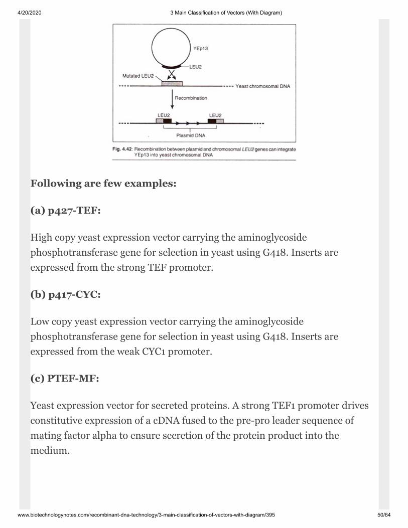
YEp may get inserted into the yeast chro mosome by a process of homologousrecombi nation between the plasmid LEU2 gene and the yeast mutant LEU2gene. The plasmid may remain integrated, or a later recombination event mayresult in it being excised again.
R R
−

4/20/2020 3 Main Classification of Vectors (With Diagram)
www.biotechnologynotes.com/recombinant-dna-technology/3-main-classification-of-vectors-with-diagram/395 49/64
2. Yeast Integrative Plasmids (YIps):
These are basically bacterial plasmids car rying a yeast gene. An example isYIp5, which is pBR322 with an inserted URA3 gene.
3. Yeast Replicative Plasmids (YRps):
These are able to multiply as independent plasmids because they carry achromo somal DNA sequence that includes an ori gin of replication. An exampleis YRp7.
Yeast Expression Vectors:
These vectors are used when our aim is to ex press our gene of interest in theyeast cell. Yeast expression vectors will employ promoter and terminatorsequences in addition to the gene of interest. Apart from these we have genetictags like the gene for green fluores cent protein (GFP) for tracking the locationof the protein after its biosynthesis.
4/20/2020 3 Main Classification of Vectors (With Diagram)
www.biotechnologynotes.com/recombinant-dna-technology/3-main-classification-of-vectors-with-diagram/395 50/64
Following are few examples:
(a) p427-TEF:
High copy yeast expression vector carrying the aminoglycosidephosphotransferase gene for selection in yeast using G418. Inserts areexpressed from the strong TEF promoter.
(b) p417-CYC:
Low copy yeast expression vector carrying the aminoglycosidephosphotransferase gene for selection in yeast using G418. Inserts areexpressed from the weak CYC1 promoter.
(c) PTEF-MF:
Yeast expression vector for secreted proteins. A strong TEF1 promoter drivesconstitutive expression of a cDNA fused to the pre-pro leader sequence ofmating factor alpha to ensure secretion of the protein product into themedium.
4/20/2020 3 Main Classification of Vectors (With Diagram)
www.biotechnologynotes.com/recombinant-dna-technology/3-main-classification-of-vectors-with-diagram/395 51/64
Yeast Artificial Chromosomes:
Yeast artificial chromosomes (YACs) are synthetic double-stranded linearconstructs containing the elements necessary for replica tion as independentchromosomes in yeast. See artificial chromosomes for more details.
Animal Vectors:
The followings are some examples of animal vectors:
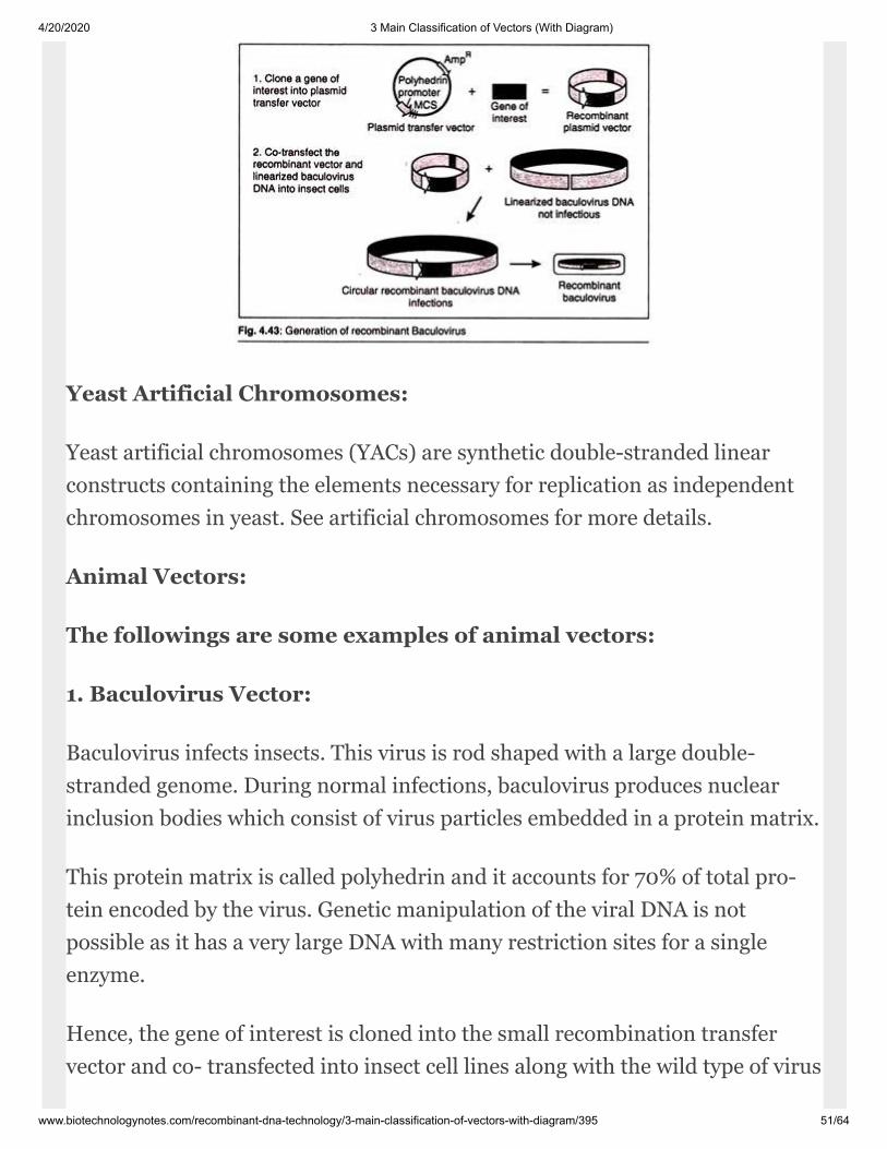
1. Baculovirus Vector:
Baculovirus in fects insects. This virus is rod shaped with a large double-stranded genome. During normal infections, baculovirus produces nuclearinclusion bodies which consist of virus particles embedded in a protein ma trix.
This protein matrix is called polyhedrin and it accounts for 70% of total pro -tein encoded by the virus. Genetic manipu lation of the viral DNA is notpossible as it has a very large DNA with many restric tion sites for a singleenzyme.
Hence, the gene of interest is cloned into the small recombination transfervector and co- transfected into insect cell lines along with the wild type of virus
4/20/2020 3 Main Classification of Vectors (With Diagram)
www.biotechnologynotes.com/recombinant-dna-technology/3-main-classification-of-vectors-with-diagram/395 52/64
in the cell. Homolo gous recombination takes place between the polyhedringene and our gene of inter est.
Thus, our gene of interest will be trans ferred from the vector plasmid into thewild type of virus and polyhedrin gene will be transferred from the virus on theplas mid.
This is something like displacement reaction. This displacement of gene willnot affect the replication of virus, as polyhedrin gene is not required for repli -cation. The recombination virus replicates in the cells and generatescharacteristic plaques (without inclusion bodies).
Nor mally the virus is cultured in the insect cell line of Spodopterafrugiperda.The gene of interest is expressed during the infection and very high yields ofprotein can be achieved by the time the cell lyses.
2. Bovine Papilloma Virus Vector:
Bo vine papilloma virus (BPV) causes warts (uncontrolled epithelialproliferation) in cattle.
BPV normally infects terminally differen tiated squamous epithelial cells.
BPV has a capsid protein surrounding a circular double-stranded DNA of size79 kb. 69% of this genome is important for viral function, whereas 31 % of thegenome can be replaced by any foreign DNA sequence like our gene of interest.
The recombinant BPV is constructed by ligating our gene of interest and BPVvector (69%) onto the pBR 322 plasmid, thus generating the shuttle vectorcontaining plasmid ori site and virus replication sequences. These shuttlevectors are multiplied in E. coli cells first and then they are transformed intomouse cell line.

4/20/2020 3 Main Classification of Vectors (With Diagram)
www.biotechnologynotes.com/recombinant-dna-technology/3-main-classification-of-vectors-with-diagram/395 53/64
The major advantage of BPV is the generation of permanent cell line. As theinfected cells are not killed, a stable plasmid number is found even when theinsert is of large size. The selection of transform-ants is very easy as they forma pile of cells on the transferred monolayer of cells called “Focus”.
The transformed cells are then selected by the presence of marker gene whichis mostly the neomy cin phospho transferase gene coding for resistance againstG418. Example of this type of vector is p3.7LDL.
3. SV40 Virus based Vectors:
SV 40 is a spherical virus with double-stranded cir cular DNA of size 5.2 kb.The viral protein contains three viral coded proteins. VP1 is the major proteinpresent in the capsid with a size of 47000 kDa. Two more pro teins VP2 andVP3 are also present.
The DNA of virus is associated with the four histones (H4, H2A, H2B and H3)proteins. The viral DNA can be segmented into five precise segments codingfor five different proteins small T, large T, VP1, VP2 and VP3. VP1 codingregion overlaps VP2 and VP3 in a different translation reading frame. SV 40virus infects monkey kidney cell lines.
The virus travels to the nucleus and gets uncoated. Then both the T-geneslocated near the origin are translated in the clockwise direction. The large Tpro tein is important for virus DNA replica tion and starts after the translationof large T -protein.
Replication starts at the origin and is bi-directional. It terminates when tworeplication forks meet. About 105 molecules of duplex DNA are synthe sizedper cell. Along with DNA replication, VP1, VP2 and VP3 proteins are synthe -sized.
Then packing of DNA occurs to form new virions, which are released by the ly -sis of cell. The entire process can also be initiated by transfection with naked
4/20/2020 3 Main Classification of Vectors (With Diagram)
www.biotechnologynotes.com/recombinant-dna-technology/3-main-classification-of-vectors-with-diagram/395 54/64
SV 40 DNA. SV 40 vectors are constructed simi lar to phage vectors. Portionsof the viral genome are removed and replaced by other DNA segments. Thereare three types of SV 40 vehicles each of which have a distinct advantage ordisadvantage among themselves.
They are as follows:
(a) SV40 Passive Transforming Vectors:
These vectors neither replicate nor pro duce virions, but simply integrate theDNA segments into the cellular DNA. These transformed cells replicate thenew DNA as an integral part of their own genomes. These plasmids are alsoshuttle vectors and include selective markers like herpes virus, thymidinekinase or neo genes.
Apart from the selective markers, they include transcriptional regulator signalsand polyadenylation sites.
(b) SV40 Trans-ducting Vectors:
These vec tors are capable of replicating and pack ing into virion particles.Transducing vec tors contain a segment of 300 bp which functions as the originof replication and provides the transcriptional regulatory sig nals for thesynthesis of mRNAs.
This type of vector takes an insert of size 3.9 to 4.5 kb. These plasmids do nothave the genes that code for VP1, VP2 and VP3. As no DNA can be added to SV40 DNA without removing any DNA from the genome, to add the insert, thegenome DNA that is not required is removed.
The functions of the DNA that are lost by these deletions are supplied by usinga helper virus or by inserting the SV 40 deleted genes into the host DNA.Normally the recombinant SV 40 vectors (usually consist of DNA of in terest
4/20/2020 3 Main Classification of Vectors (With Diagram)
www.biotechnologynotes.com/recombinant-dna-technology/3-main-classification-of-vectors-with-diagram/395 55/64
and replication sequence and gene for coding VP1, VP2 and VP3) are trans -formed into the cos cell line.
Cos cell line is a kidney cell line of the African green monkey kidney. It has theT-protein gene incorporated in the genome. So when the vector is transfectedinto these cells, virion particles are yielded with the help of helper virus.
(c) SV40 Plasmid Vectors:
These vectors multiply in the monkey cell line but are not packed as thevirions. These vectors usually contain origin of replication se quences andlarger T-protein gene but do not contain VP1, VP2 and VP3 genes. They areshuttle vectors, and have the ability to multiply both in E. coli and monkey cellline.
Plant Vectors:
Cloning vectors for higher plants were devel oped in 1980s and their use hasled to the ge netically modified (GM) crops that are in the headline today.
We will examine the details of plant vec tors and the genetic modification ofcrops. Here we look at the cloning vectors and how they are used.
Three types of cloning sys tem have been used with varying degreesof success with higher plants:
1. Vectors based on naturally occurring plas mids of Agrobacterium (e.g., Tiplasmids from A. tumifaciens and Ri plasmid from A. rhizogens).
2. Direct gene transfer using various types of plasmid DNA. (e.g., using ofsupercoiled plasmids).
3. Vectors based on plant viruses (e.g., Caulimo virus vectors and Gemini virusvectors).
4/20/2020 3 Main Classification of Vectors (With Diagram)
www.biotechnologynotes.com/recombinant-dna-technology/3-main-classification-of-vectors-with-diagram/395 56/64
Shuttle vectors:
Shuttle vectors are those which can multiply into two different unrelatedspecies. Shuttle reactors are designed to replicate in the cells of two species, asthey contain two origins of replication, one appropriate for each species as wellas genes that are required for replication and not supplied by the host cell, i.e.,it is self-sufficient with the process of its replication.
The shuttle vectors are of following types:
1. Eukaryotic – Prokaryotic Shuttle Vec tors:
Vectors that can propagate in eukaryotes and prokaryotes. e.g., YEp vec torscan be propagated in yeast (fungi) as well as in E. coli (bacteria).
2. Prokaryotic – Prokaryotic Shuttle Vec tors:
Vectors that can be propagated in two unrelated prokaryotic host cells, e.g.,RSF1010 vectors can be propagated both in bacteria as well in spirochetes.
The common features of such shuttle vec tors or eukaryotic vectorsare the following:
(a) They are capable of replicating into two or more types of hosts includingprokaryotic and eukaryotic cells.
(b) They replicate autonomously, or integrate into host genome and replicatewhen the host cell multiplies.
(c) These vectors are commonly used for transporting genes from oneorganism to another.
4/20/2020 3 Main Classification of Vectors (With Diagram)
www.biotechnologynotes.com/recombinant-dna-technology/3-main-classification-of-vectors-with-diagram/395 57/64
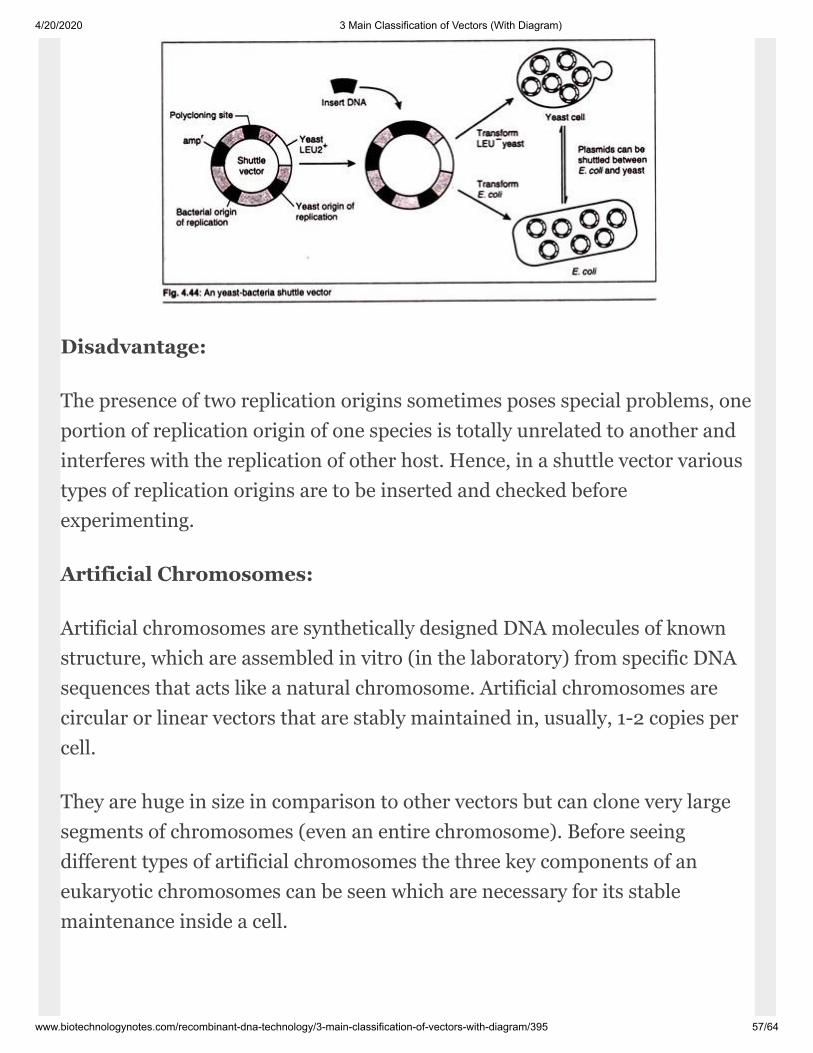
Disadvantage:
The presence of two replication origins some times poses special problems, oneportion of replication origin of one species is totally un related to another andinterferes with the rep lication of other host. Hence, in a shuttle vec tor varioustypes of replication origins are to be inserted and checked beforeexperimenting.
Artificial Chromosomes:
Artificial chromosomes are synthetically de signed DNA molecules of knownstructure, which are assembled in vitro (in the labora tory) from specific DNAsequences that acts like a natural chromosome. Artificial chromo somes arecircular or linear vectors that are stably maintained in, usually, 1-2 copies percell.
They are huge in size in comparison to other vectors but can clone very largesegments of chromosomes (even an entire chromosome). Before seeingdifferent types of artificial chro mosomes the three key components of aneukaryotic chromosomes can be seen which are necessary for its stablemaintenance inside a cell.

4/20/2020 3 Main Classification of Vectors (With Diagram)
www.biotechnologynotes.com/recombinant-dna-technology/3-main-classification-of-vectors-with-diagram/395 58/64
1. The centromere, which is required for the chromosome to be distributedcorrectly to daughter cells during cell division.
2. Two telomeres, the structures at the ends of a chromosome, which areneeded for the ends to be replicated correctly in order and which also preventthe chromo some from being nibbled away by exonu cleases.
3. The origin of replication, which are the positions along the chromosome atwhich DNA replication initiates, similar to the origin of replication of aplasmid.
Once we have defined the chromosomal structure of an eukaryotic organism(like hu mans and yeast), then we can isolate the key components of theirchromosomes and join them together to form an artificial chromo some. Theninto this artificial chromosome we can insert our gene of interest which can besubsequently cloned in its respective cell.
Fol lowing are the types of artificial chromosomes:
A. Yeast Artificial Chromosomes (YAC):
A YAC can be considered as a functional arti ficial chromosome, since itincludes three spe cific DNA sequences that enable it to propa gate from oneyeast cell to its offspring:
1. The Telomere (TEL):
The telomere which is located at each chromosome end, pro tects the linearDNA from degradation by Nucleases.
2. The Centromere(CEN):
The centromere which is the attachment site for mitotic spindle fibres, “pulls”one copy of each du plicated chromosome into each new daugh ter cell.
4/20/2020 3 Main Classification of Vectors (With Diagram)
www.biotechnologynotes.com/recombinant-dna-technology/3-main-classification-of-vectors-with-diagram/395 59/64
3. Origin of Replication(OR):
Replication origin sequences which are specific DNA sequences that allow theDNA replication machinery to assemble on the DNA and move at thereplication forks.
4. A and B:
Selectable markers that allow easy isolation of yeast cells that have taken upartificial chromosome.
5. Recognition Site:
Recognition Site for two restriction en zymes EcoRI and BamHl.
Cloning experiment using a YAC vector:
1. Large DNA fragments are obtained by car rying out restriction digestionusing EcoRI.
2. The YAC is digested by two restriction enzymes EcoRI and Bam HI.
3. Those two elements recombine at the EcoRI sites and are covalently linkedby DNA ligase.
4. A recombinant YAC vector, a yeast artifi cial chromosome with genomicDNA inserted, is produced. This vector can be used to infect yeast cells andgenerate an un limited number of copies.
Uses of YAC Vectors:
1. YAC can be used to study various aspects of chromosome structure andbehaviour; for instance, to examine the segregation of chromosomes duringmeiosis.

4/20/2020 3 Main Classification of Vectors (With Diagram)
www.biotechnologynotes.com/recombinant-dna-technology/3-main-classification-of-vectors-with-diagram/395 60/64
2. YAC cloning system can take DNA insert greater than l00kb. Due to thisthey can be used to study the functions and modes of expression of genes thathad previously been intractable to analysis by recombi nant DNA techniques.
3. YACs can be propagated in mammalian cells, enabling the functionalanalysis to be carried out in the organism in which the gene normally resides.Thus by using them we can learn about the true form of gene expression invivo conditions.
4. Yeast artificial chromosomes are very helpful in the production of genelibraries. E. coli vectors can take DNA insert maxi mum up to 300kb. Due tothis some 30000 clones are needed for a human gene library if we use them ascloning vector.
However, YAC vectors are routinely used to clone 600 kb fragments, andspecial types are able to handle DNA up to 1400 kb in length, the latterbringing the size of a human gene library down to just 6500 clones.
Sometimes YAC is seen with problem of lacking insert stability, the clonedDNA some times becoming rearranged by intra-molecular recombination.Nevertheless, YACs have been of immense value in providing long pieces ofcloned DNA for use in large scale DNA sequencing projects. Example of YAC ispYAC3.

4/20/2020 3 Main Classification of Vectors (With Diagram)
www.biotechnologynotes.com/recombinant-dna-technology/3-main-classification-of-vectors-with-diagram/395 61/64
2. Bacterial Artificial Chromosomes (BAC):
Bacterial Artificial Chromosomes (BACs) are cloning vectors based on theextra-chromosomal plasmids of E.coli, called F factor or fertility factor. These
4/20/2020 3 Main Classification of Vectors (With Diagram)
www.biotechnologynotes.com/recombinant-dna-technology/3-main-classification-of-vectors-with-diagram/395 62/64
vectors enable the construction of artificial chromosomes, which can be clonedin E.coli.
This vector is useful for cloning DNA fragments up to 350 kb, but can behandled like regular bacterial plasmid vectors, and is very useful forsequencing large stretches of chromosomal DNA.
Like any other vector, BACs contain ori sequences derived from E. coli plasmidF factor, multiple cloning sites (MCS) having unique restriction sites, andsuitable selectable markers. The genomes of several large DNA viruses andRNA vi ruses have been cloned as BACs. These con structs are referred to as“infectious clones”, as transfection of the BAC construct into host cells issufficient to initiate viral infection.
The infectious property of these BACs has made the study of many virusessuch as herpes viruses, poxviruses and coronaviruses more ac cessible. BACsare now being utilized to a greater extent in modelling genetic diseases, oftenalongside transgenic mice.
BACs have been used in this field as complex genes may have severalregulatory sequences upstream of the encoding sequence, including variouspromoter sequences that will govern a gene’s expression level. BACs have beenused to some degree of success with mice when studying neurological diseasessuch as Alzheimer’s dis ease or as in the case of aneuploidy associated withDown’s syndrome.
Examples of BAC are pBACe3.6, pBeloBAC11 etc.
3. Human Artificial Chromosomes (HAC):
A human artificial chromosome (HAC) is a mini-chromosome that isconstructed artifi cially in human cells. That is, instead of 46 chromosomes, thecell could have 47 with the 47th being very small, roughly 6-10 mega-bases insize, and able to carry new genes introduced by human researchers.
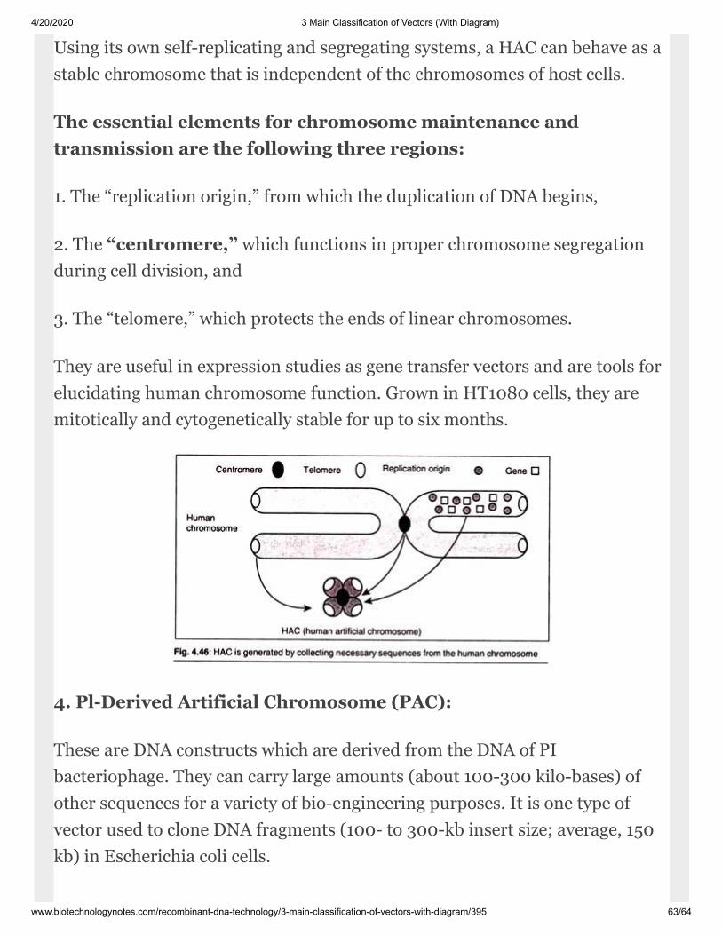
4/20/2020 3 Main Classification of Vectors (With Diagram)
www.biotechnologynotes.com/recombinant-dna-technology/3-main-classification-of-vectors-with-diagram/395 63/64
Us ing its own self-replicating and segregating systems, a HAC can behave as astable chro mosome that is independent of the chromo somes of host cells.
The essential elements for chromosome maintenance andtransmission are the follow ing three regions:
1. The “replication origin,” from which the du plication of DNA begins,
2. The “centromere,” which functions in proper chromosome segregationduring cell division, and
3. The “telomere,” which protects the ends of linear chromosomes.
They are useful in expression studies as gene transfer vectors and are tools foreluci dating human chromosome function. Grown in HT1080 cells, they aremitotically and cytogenetically stable for up to six months.
4. Pl-Derived Artificial Chromosome (PAC):
These are DNA constructs which are derived from the DNA of PIbacteriophage. They can carry large amounts (about 100-300 kilo-bases) ofother sequences for a variety of bio-engineering purposes. It is one type ofvector used to clone DNA fragments (100- to 300-kb insert size; average, 150kb) in Escherichia coli cells.
4/20/2020 3 Main Classification of Vectors (With Diagram)
www.biotechnologynotes.com/recombinant-dna-technology/3-main-classification-of-vectors-with-diagram/395 64/64
Terms of Service Privacy Policy Contact Us
Microorganisms Associated with Food (Types) | Food BiotechnologyAUGUST 1, 2018
Different Systems or Modes of Microbial Cultures | Microorganism | BiotechnologyAUGUST 1, 2018
Rancidity of Food: Introduction, Types, Factors and Prevention of Rancidity | Food Chemistry |BiotechnologyAUGUST 1, 2018
Classification of Food Starches | Food Chemistry | BiotechnologyAUGUST 1, 2018
Colloidal Systems in Food: Functions, Types and Stability | Food ChemistryAUGUST 1, 2018
Home ››
Releted Articles:
Recombinant DNA Technology (RDT): 5 Main ApplicationsRecombinant DNA Technology (Notes)Nucleic Acid Hybridization (With Diagram)Top 7 Blotting Techniques Used in Detection of DNA
LATEST