Tropism and transduction of oncolytic adenovirus vectors in ...
Upload
independentCategory
view
2download
0
Improved NYVAC-Based Vaccine VectorsKaren V. Kibler1., Carmen E. Gomez2., Beatriz Perdiguero2, Shukmei Wong1, Trung Huynh1, Susan
Holechek1, William Arndt1, Victoria Jimenez2, Ruben Gonzalez-Sanz2, Karen Denzler1, Elias K. Haddad3,
Ralf Wagner5, Rafick P. Sekaly3,4, James Tartaglia6, Giuseppe Pantaleo7, Bertram L. Jacobs1*, Mariano
Esteban2*
1 The Biodesign Institute at Arizona State University, Tempe, Arizona, United States of America, 2 Centro Nacional de Biotecnologia-Consejo Superior de Investigaciones
Cientificas, Madrid, Spain, 3 Vaccine and Gene Therapy Institute of Florida, Port St. Lucie, Florida, United States of America, 4 University of Montreal, Montreal, Canada,
5 University of Regensburg, Regensburg, Germany, 6 Sanofi-Pasteur, Swiftwater, Pennsylvania, United States of America, 7 Division of Immunology and Allergy, Centre
Hospitalier Universitaire Vaudois, Lausanne, Switzerland
Abstract
While as yet there is no vaccine against HIV/AIDS, the results of the phase III Thai trial (RV144) have been encouraging andsuggest that further improvements of the prime/boost vaccine combination of a poxvirus and protein are needed. With thisaim, in this investigation we have generated derivatives of the candidate vaccinia virus vaccine vector NYVAC withpotentially improved functions. This has been achieved by the re-incorporation into the virus genome of two host rangegenes, K1L and C7L, in conjunction with the removal of the immunomodulatory viral molecule B19, an antagonist of type Iinterferon action. These novel virus vectors, referred to as NYVAC-C-KC and NYVAC-C-KC-DB19R, have acquired relevantbiological characteristics, giving higher levels of antigen expression in infected cells, replication-competency in humankeratinocytes and dermal fibroblasts, activation of selective host cell signal transduction pathways, and limited virus spreadin tissues. Importantly, these replication-competent viruses have been demonstrated to maintain a highly attenuatedphenotype.
Citation: Kibler KV, Gomez CE, Perdiguero B, Wong S, Huynh T, et al. (2011) Improved NYVAC-Based Vaccine Vectors. PLoS ONE 6(11): e25674. doi:10.1371/journal.pone.0025674
Editor: Maciej S. Lesniak, The University of Chicago, United States of America
Received April 29, 2011; Accepted September 7, 2011; Published November 9, 2011
Copyright: � 2011 Kibler et al. This is an open-access article distributed under the terms of the Creative Commons Attribution License, which permitsunrestricted use, distribution, and reproduction in any medium, provided the original author and source are credited.
Funding: This study was conducted as part of the Poxvirus T cell Vaccine Discovery Consortium (PTVDC) as part of the Collaboration for AIDS Vaccine Discovery(CAVD) with support from the Bill and Melinda Gates Foundation. Mariano Esteban was supported in part by a Spanish grant (SAF2008-02036). These funders hadno role in study design, data collection and analysis, decision to publish, or preparation of the manuscript. JT is an employee of Sanofi Pasteur and as such, SanofiPasteur is considered by PLoS ONE to have contributed funding to this research.
Competing Interests: The authors have read the journal’s policy and have the following conflicts: JT is an employee of Sanofi Pasteur. This does not alter theauthors’ adherence to all the PLoS ONE policies on sharing data and materials.
* E-mail: [email protected] (BLJ); [email protected] (ME)
. These authors contributed equally to this work.
Introduction
Following the eradication of smallpox through the use of an
efficient live vaccinia virus (VACV) for vaccination, and thereafter,
through the discovery of poxviruses as recombinant vectors
expressing a large range of foreign genes [1,2], pox viruses
became natural candidates for vectors to vaccinate against
multiple disease-causing organisms. However, the rate of compli-
cations from wtVACV led researchers and regulators to seek pox
virus vectors that would be highly attenuated to provide greater
safety in humans. Three of the most well-characterized highly
attenuated pox vectors have been ALVAC [3,4], MVA [5], and
NYVAC [3]. ALVAC, which is a vaccine strain of canary pox
virus, is naturally attenuated in a human host, and does not
replicate productively in human cells. MVA, which is also a virus
that does not replicate in human cells, was passaged in chick
embryo fibroblasts (CEFs) to achieve attenuation. MVA suffered
several large deletions and numerous point mutations during
passage in CEFs that are responsible for its attenuation. NYVAC
was deleted of 18 specific open reading frames that are present in
the parental Copenhagen strain, including the host range genes
K1L and C7L. NYVAC is replication-incompetent in most human
cells. While one of the advantages of replication-deficient viruses is
their safety profiles, it has been postulated that efficacy of these
viruses is limited by the failure to replicate and the attendant
limitation in antigen accumulation during virus infection.
Among the many heterologous antigens that have been
expressed in these replication-defective poxvirus vaccine vectors
are those derived from HIV [4,6]. Most recently, ALVAC was
included in the RV144 Phase III HIV vaccine clinical trial
conducted in Thailand in which a 31% efficacy rate against HIV
infection was achieved after three years [7]. With the trial’s
encouraging demonstration that a vaccine regimen can protect to
at least a limited extent, one conclusion of these findings is that the
components of vaccination need to be optimized to afford
protection to a large percentage of vaccinees. With this aim, in
this report we describe the generation of improved NYVAC
recombinants expressing the HIV antigens Env/Gag-Pol-Nef (E/
GPN) from clade C. These improvements have been obtained by
removal of a viral gene that inhibits type I interferon (IFN)
pathways and by restoration of replication competence in human
cells by inserting the K1L and C7L host range genes back into the
viral genome. Our findings demonstrate that these new constructs
(NYVAC-C-KC and NYVAC-C-KC-DB19R) have acquired new
PLoS ONE | www.plosone.org 1 November 2011 | Volume 6 | Issue 11 | e25674

biological properties distinct from the parental NYVAC-C, which
make them potentially improved vaccine vector candidates for
human application.
Materials and Methods
Ethics StatementAll animal work at Arizona State University has been conducted
in compliance with national and international guidelines. This
study was approved by the Institutional Animal Care and Use
Committee at Arizona State University, Tempe, Arizona, under
Protocol #10-0970R.
All animal work at CNB-CSIS has been conducted in
compliance with national and international guidelines and with
the Royal Decree (RD 1201/2005). These studies were approved
by the Ethical Committee of Animal Experimentation of Centro
Nacional de Biotecnologia, Madrid, Spain, under Permit numbers
152/07 and 080030.
PBMCs for signal transduction assays were obtained from
healthy blood donors recruited by the Blood Transfusion Center
of Madrid; donations were approved by the ethics commission of
the Center and written informed consent was obtained from the
donors; all information was kept confidential by the Blood Center.
Construction of recombinant virusesGeneration of NYVAC-C-DB19R. The plasmid transfer
vector pGem-RG-B19R wm, used for the construction of the
recombinant virus NYVAC-C-DB19R, with the B19R ORF
deleted, was obtained by sequential cloning of five DNA
fragments containing dsRed2 and rsGFP genes and B19R
recombination flanking sequences into the plasmid pGem-7Zf(-)
(Promega). The dsRed2 gene under the control of the synthetic
early/late promoter was amplified by PCR from plasmid pG-
dsRed2 with oligonucleotides Red2-B (59-GAACTAGGATCC-
TAACTCGAGAAA-39) (Bam HI site underlined) and Red2-N
(59-ATTAGTATGCATTTATTTATTTAGG-39) (Nsi I site
underlined) (785 bp), digested with Bam HI and Nsi I and
inserted into the Bam HI/Nsi I-digested pGem-7Zf(-) to generate
pGem-Red wm (3740 bp). The rsGFP gene under the control of the
synthetic early/late promoter was amplified by PCR from plas-
mid pG-dsRed2 with oligonucleotides GFP-X (59-CGTTGG-
TCTAGAGAGAAAAATTG-39) (Xba I site underlined) and
GFP-E (59-CTATAGAATTCTCAAGCTATGC-39) (Eco RI site
underlined) (832 bp), digested with Xba I and Eco RI and inserted
into plasmid pGem-Red wm previously digested with Xba I and
Eco RI to obtain pGem-Red-GFP wm (4540 bp). NYVAC genome
was used as the template to amplify the left flank of the B19R gene
(364 bp) with oligonucleotides LFB19R-AatII-F (59-TTTTTT-
GACGTCGAGAAAGTTAAGAAGATAC-39) (Aat II site
underlined) and LFB19R-XbaI-R (59-TTTTTTTCTAGATC-
TTTATTATACGGCACTAA-39) (Xba I site underlined). This
left flank was digested with Aat II and Xba I and cloned into
plasmid pGem-Red-GFP wm previously digested with the same
restriction enzymes to generate pGem-RG-LFsB19R wm
(4871 bp). The repeated left flank of the B19R gene (364 bp) was
amplified by PCR from NYVAC genome with oligonucleotides
LFB19R9-EcoRI-F (59-TTTTTTGAATTCGAGAAAGTTAAG-
AAGATAC-39) (Eco RI site underlined) and LFB19R9-ClaI-R (59-
TTTTTTATCGATTCTTTATTATACGGCACTAA-39) (Cla I
site underlined), digested with Eco RI and Cla I and inserted into
the Eco RI/Cla I-digested pGem-RG-LFsB19R wm to generate
pGem-RG-LFdB19R wm (5194 bp). The right flank of the B19R
gene (381 bp) was amplified by PCR from the NYVAC genome
with oligonucleotides RFB19R-ClaI-F (59-TTTTTTATCGAT-
ATATACAATGCATTTTTATATAC-39) (Cla I site underlined)
and RFB19R-BamHI-R (59-TTTTTTGGATCCAGTTCTATC-
ATAATCATC-39) (Bam HI site underlined), digested with Cla I
and Bam HI and inserted into the Cla I/Bam HI-digested pGem-
RG-LFdB19R wm. The resulting plasmid pGem-RG-B19R wm
(5545 bp) was confirmed by DNA sequence analysis and directs the
deletion of the B19R gene from the NYVAC-C genome.
The deletion mutant NYVAC-C-DB19R was constructed by
fluorescence selection using dsRed2 and rsGFP genes as the
selectable markers. 36106 BSC-40 (ATCC) cells were infected
with an MOI of 0.01 of NYVAC-C and transfected 1 hour later
with 6 mg DNA of plasmid pGem-RG-B19R wm using Lipofecta-
mine (Invitrogen, San Diego, CA). 48 hours post-infection, the
cells were harvested, lysed by freeze-thaw cycling, sonicated and
used for recombinant virus screening. The deletion mutant was
selected from progeny virus by consecutive rounds of plaque
purification in BSC-40 cells during which plaques were screened
for Red2/GFP fluorescence. In the first two passages viruses from
selected plaques expressed both fluorescent proteins, in the next
two passages viral progeny from selected plaques expressed only
one fluorescent marker and in the last two passages viruses from
selected plaques do not express any marker due to the loss of the
fluorescent marker. The deletion mutant was detected by PCR
amplifying of the B19R locus. The resulting NYVAC-C-DB19R
positive virus plaques were grown in BSC-40 cells, and further
passaged twice in primary CEF cells. A P2 stock was prepared in
CEFs (Charles River) and used for the propagation of the virus
large cultures in CEF cells, followed by virus purification by
sequential centrifugation in two 36% (w/v) sucrose cushions in
10 mM Tris-HCl pH 9, and titrated by plaque assay in BSC-40
cells. The purified grown stock of virus was referred to as P3. The
B8R locus of NYVAC-C-DB19R was deleted in a similar manner.
Generation of NYVAC-C-KC and NYVAC-C-KC-
DB19R. The C7L and K1L genes were amplified from the
Copenhagen genome using the following primers: NY1, 59 GT-
TTGCATCGTGCTTTAACATCAATGG 39; NY2, 59 GTCT-
TACTCATTGCATCGTACGGTTGGCTTATTCCATAGTA-
GCTTGTG 39; NY3, 59 CTACTATGGAATAAGCCAACC-
GTACGATGCAATGAGTAAGACAATAGG 39; and NY4, 59
GTACCTGGCAATAGGTGATAATATGAC 39. The ‘‘KC’’
fragment was then inserted into the NYVAC-C genome by in
vivo recombination (IVR): 150 ng DNA was used to transfect
MRC-5 cells (kind gift of Sanofi Pasteur) according to the
manufacturer’s protocol (Lipofectamine, Invitrogen), in 35 mm
dishes; cells were infected with the parental virus, either NYVAC
or NYVAC-C, at a multiplicity of infection (MOI) of 0.05 by
adding the virus to the transfection mix. After 30 minutes of
incubation, 1 ml of OptiMEM with 1% serum was added to each
dish. Cells were scraped into the medium at 36 hours post-
infection. Following three rounds of freeze/thaw, the IVR scrape
was used to infect Vero cells (kind gift of Sanofi Pasteur) to select
for viruses competent for large plaque formation in Vero cells. The
same method was used to insert KC into NYVAC-C-DB19R.
PCR analysisTo test for deletion of B19R, viral DNA was extracted by the
method of SDS-Proteinase K-Phenol from BSC-40 cells mock-
infected or infected at an MOI of 5 with wtNYVAC, NYVAC-C
or NYVAC-C-DB19R. Primers LFB19R-AatII-F and LFB19R-
BamHI-R spanning the B19R flanking regions were used for PCR
analysis of B19R locus. The amplification reactions were carried
out with Platinum Taq DNA polymerase (Invitrogen, San Diego,
CA). For verification of the KC fragment, primers NY1 and NY4
Poxvirus Vaccine Vectors
PLoS ONE | www.plosone.org 2 November 2011 | Volume 6 | Issue 11 | e25674

were used on viral DNA obtained by standard phenol extraction
from BHK-infected cells (ATCC).
Expression of HIV-1 proteins gp120 and GPNTo verify the correct expression of the HIV-1 proteins gp120
and GPN from the viruses, monolayers of either BSC-40 or BHK
cells were mock-infected or infected at an MOI of 5 with
wtNYVAC, NYVAC-C, NYVAC-C-KC, NYVAC-C-DB19R, or
NYVAC-C-KC-DB19R. At 48 hours post-infection, cells were
lysed in Laemmli buffer, cell extracts fractionated by 10% SDS-
PAGE and analyzed by Western blot using rabbit polyclonal anti-
gp120 antibody (Centro Nacional de Biotecnologıa; diluted
1:3000) or polyclonal anti-gag p24 serum (ARP 432, NIBSC,
Centralised Facility for AIDS reagent, UK; diluted 1:1000)
followed by anti-rabbit-HRP (Sigma; diluted 1:5000) to evaluate
the expression of gp120 and GPN proteins respectively.
Analysis of virus growthTo determine virus-growth profiles, monolayers of CEF cells
grown in 12-well tissue culture plates were infected in duplicate at
an MOI of 0.01 with wtNYVAC, NYVAC-C or NYVAC-C-
DB19R. Following virus adsorption for 60 minutes at 37 C, the
inoculum was removed. The infected cells were washed once with
DMEM without serum and incubated with fresh DMEM
containing 2% FCS at 37 C in a 5% CO2 atmosphere. At
different times post-infection (0, 24, 48 and 72 hours), cells were
removed by scraping (lysates at 56105 cells/ml), freeze-thawed
three times and briefly sonicated. Virus titers in cell lysates were
determined by crystal violet staining in BSC-40 cells.
To compare replication competence in human cells, NYVAC-
C, NYVAC-C-KC, and NYVAC-C-KC-DB19R were used to
infect HeLa cells (ATCC) at an MOI of 5. At 0, 6, 12, and
24 hours post-infection cells were scraped into the medium,
pelleted, and the supernatant discarded. The cell pellets were
resuspended in 200 ml medium and titers were obtained in BHK
cells. Multi-step growth curves were done at an MOI of 0.01 in the
indicated cells lines, keratinocytes (Invitrogen) and dermal
fibroblasts (Lonza). IFN treatment was 1000 units/ml added for
24 hours prior to infection.
Signal Transduction AssaysHeLa cells and human keratinocytes were infected at an MOI of
5 and allowed to incubate for 6 hours. Cell lysates were prepared
by standard methods and analyzed by Western blots probed with
antibodies specific to the phosphorylated forms of PKR (Cell
Signaling), eIF2a (Cell Signaling), and IRF3 (Epitomic), or to the
internal control, GAPDH (Abcam).
For human peripheral blood mononuclear cells (PBMCs),
phospho-protein levels present in extracts of cells infected with
different recombinant viruses were measured simultaneously by
Cytometric Bead Array (CBA) using an LSR II flow cytometer
(Becton Dickinson). In CBA technology, different bead popula-
tions with distinct fluorescence intensities had been coated with
capturing antibodies specific for different analytes. These bead
populations could be resolved in the fluorescence channels of the
flow cytometer. After the beads had been incubated with 50 ml of
sample, different analytes in the sample were captured by their
corresponding beads. The protein captured beads were then
mixed with phycoerythrin-conjugated detection antibodies to form
sandwich complexes. Following incubation, washing and acquisi-
tion of fluorescence data, the results were generated in graphical
format using the BD CBA software. The concentrations of total
and phospho STAT1 were measured simultaneously using the Cell
Signaling Master Buffer Kit (BD Biosciences Pharmingen, CA,
USA; Cat. No. 558223). For this CBA analysis, 46106 PBMCs
were mock-infected or infected at an MOI of 5 with wtNYVAC,
NYVAC-C, NYVAC-C-KC, NYVAC-C-DB19R or NYVAC-C-
KC-DB19R. At 4 hours post-infection, cells were removed by
pipetting, centrifuged at room temperature for 5 minutes at
3000 rpm, supernatants discarded and cellular pellet resuspended
in lysis buffer (Cell Signaling 106) containing phosphatase
inhibitors. After an incubation of 15 minutes at 4 C, cells were
centrifuged at room temperature for 5 minutes at 13,000 rpm and
supernatants transferred to new tubes. Denaturation Buffer was
added and solution was mixed and denatured for 5 minutes at
100 C. Protein content in samples was measured with a
bicinchoninic protein assay reagent kit (Pierce Co., Rockford,
IL) and CBA analysis was performed according to manufacturer’s
recommendations.
Transcriptional analysisEx vivo derived plasmacytoid dendritic cells (pDc, [8]) and
myeloid dendritic cells (mDC, [8]) were infected with 0.1 MOI of
NYVAC-C, NYVAC-C-KC, NYVAC-C-DB8RDB19R and NY-
VAC-C-KC-DB8RDB19R for 6 hours. Cells were then harvested
for RNA extraction and gene array analysis. Gene array analysis
was performed on BeadChips using the Illumina platform.
RNA extraction, amplification, hybridization, and scanning
were performed as described previously [9]. Briefly, total RNA
was purified from sorted dendritic cells using RNA extraction kits
(Qiagen). RNA was quantified and assessed for quality using a
spectrophotometer (NanoDrop Technologies). Total RNA was
then amplified by incorporating biotin using the Illumina Total
Prep RNA Amplification kit, which is based on the Eberwine
amplification protocol. The biotinylated cRNA was hybridized
onto Illumina Human RefSeq-8 V3 BeadChips (PBMC samples,
[8]) at 58 C for 20 hours and quantified using an Illumina
BeadStation 500GX scanner and Illumina BeadScan software.
Gene array analysis: scanned raw data was screened and
inspected for quality. Genes with intensities below background in
all samples were removed and then minimum-replaced (a
surrogate-replacement policy) using the mean background value
of the built-in Illumina probe controls. Normalization of chips was
done by quantile-normalization.
Gene expression data was analyzed using the R software
package. Genes are filtered by detection call and by variance filters
to allow a reduction in the number of tests and a corresponding
increase in power of the differential gene expression analysis [10].
The resulting matrix was log2 transformed and used as input for
linear modeling using Bioconductor’s limma package, which
estimates the fold-change between predefined groups by fitting a
linear model and using an empirical Bayes method to moderate
standard errors of the estimated log-fold changes for expression
values from each gene. P value from the resulting comparison was
set to 0.05 in this analysis. Determination of commonly and
uniquely regulated genes is based solely on the nominal p values
and fold change.
All data is MIAME compliant and the raw data has been
deposited in a MIAME compliant database.
Measurement of apoptotic cell death by cell cycleanalysis
The different stages of cell cycle and the percentage of cells with
subG0 DNA content were analyzed by propidium iodide (PI)
staining. Briefly, HeLa cells were mock-infected or infected at an
MOI of 5 with NYVAC-C, NYVAC-C-KC, NYVAC-C-DB19R
or NYVAC-C-KC-DB19R. At 24 hours post-infection, cells were
centrifuged at room temperature for 10 minutes at 2000 rpm and
Poxvirus Vaccine Vectors
PLoS ONE | www.plosone.org 3 November 2011 | Volume 6 | Issue 11 | e25674

Poxvirus Vaccine Vectors
PLoS ONE | www.plosone.org 4 November 2011 | Volume 6 | Issue 11 | e25674

supernatants discarded. Pellets were resuspended in detergent lysis
buffer and cells stained with PI. After an incubation of 30 minutes
at 37 C in the dark, the percentage of cells with hypodiploid DNA
content was determined using an LSR II flow cytometer (Becton
Dickinson). The results are expressed as fold increase in apoptotic
cells with respect to uninfected cells.
Pathogenicity in newborn miceFor studies in newborn mice, pregnant CD1 mice were
purchased from Charles River at 10 days gestation. The animals
were housed one animal per cage. Intracranial infections with the
indicated viruses, using a total volume of 10 mL, were conducted at
48 to 72 hours post-birth of the pups (at least 10 pups per virus),
using a 27-gauge needle, as previously described [11]. Animals
were monitored twice daily for 14 days for morbidity and
mortality.
Mice immunization for biodistributionBALB/c mice were purchased from Harlan. To analyze the
biodistribution of virus recombinants in animals, BALB/c were
immunized by intraperitoneal route (IP) with a dose of
26107 PFU/mouse of WR(TK2), NYVAC-C, NYVAC-C-KC,
NYVAC-C-DB19R or NYVAC-C-KC-DB19R. At different times
post-infection (24, 48 and 72 hours) mice were sacrificed and
mouse tissues were processed for plaque assay titration. Peritoneal
cells were harvested by mouse peritoneal cavity lavage with 10 ml
of sterile PBS, centrifuged at room temperature for 5 minutes at
1200 rpm and stored at 270 C. Spleens, draining lymph nodes,
Figure 1. Generation and characterization of NYVAC-C-DB19R. A) Scheme of construction. The plasmid transfer vector pGem-RG-B19Rwm was obtained by sequential cloning of selectable markers dsRed2 and rsGFP and B19R recombination flanking sequences into the plasmid pGem-7Zf. For the generation of NYVAC-C-DB19R, BSC-40 cells were infected at an MOI of 0.01 with NYVAC-C and transfected 1 hour later with 6 mg DNA ofplasmid pGem-RG-B19R wm. 48 hours post-infection, the cells were harvested, lysed by freeze-thaw cycling, sonicated and used for recombinantvirus screening. The deletion mutant was selected from progeny virus by consecutive rounds of plaque purification in BSC-40 cells during whichplaques were screened for Red2/GFP fluorescence. B) PCR analysis. Viral DNA was extracted from BSC-40 cells mock-infected or infected at an MOIof 5 with NYVAC wt, NYVAC-C or NYVAC-C-DB19R. Primers LFB19R-AatII-F and LFB19R-BamHI-R spanning B19R flanking regions were used for PCRanalysis of B19R locus. C) Expression of HIV-1 antigens gp120 and GPN. BSC-40 cells were mock-infected or infected at an MOI of 5 with NYVACwt, NYVAC-C or NYVAC-C-DB19R. At 48 hours post-infection, cells were lysed in Laemmli buffer, cells extracts were fractionated by 10% SDS-PAGEand analyzed by Western blot using rabbit polyclonal anti-gp120 antibody or polyclonal anti-gag p24 serum. D) Replication in CEF cells. CEF cellswere infected at an MOI of 0.01 with NYVAC wt, NYVAC-C or NYVAC-C-DB19R. At different times post-infection (hpi, 0, 24, 48 and 72 hours), cells wereharvested, freeze-thawed three times and sonicated. Virus titers in cell lysates were determined by crystal violet staining in BSC-40 cells.doi:10.1371/journal.pone.0025674.g001
Figure 2. Scheme of construction of NYVAC-C-KC-DB19R. To insert C7L and K1L back into the NYVAC genome, each gene plus a correspondingportion of the flanking regions was amplified by PCR. The two fragments were combined into one fragment using PCR. The entire cassette, containingC7L and K1L plus flanking regions homologous to the adjacent genes of the NYVAC genome (C8L and K2L), was inserted into NYVAC-C by in vivorecombination. To create NYVAC-C-KC-DB19R, the KC fragment was inserted into NYVAC-C-DB19R by the same method. IGR: Intergenic region.doi:10.1371/journal.pone.0025674.g002
Poxvirus Vaccine Vectors
PLoS ONE | www.plosone.org 5 November 2011 | Volume 6 | Issue 11 | e25674
Poxvirus Vaccine Vectors
PLoS ONE | www.plosone.org 6 November 2011 | Volume 6 | Issue 11 | e25674

ovaries, and livers were dissected under sterile conditions and
stored at 270 C. Peritoneal cells were resuspended in 200 ml
complete DMEM media and tissues from individual mice were
homogenized in 200 ml complete DMEM media using an
Eppendorf-fitted Dounce homogenizer. The production of
infectious virus in different mouse tissues was tested by plaque
assay in BSC-40 cells. The virus titer was expressed as Plaque
Forming Units (PFU) per gram of protein. Protein content in tissue
extracts was measured with a bicinchoninic protein assay reagent
kit (Pierce Co., Rockford, IL).
Another test was also used for biodistribution by measuring
levels of antigen expression in mice infected with recombinants
NYVAC and NYVAC-KC expressing the luciferase marker. The
recombinant viruses WR-luc and NYVAC-luc have been
previously described [12,13]. The recombinant NYVAC-KC-luc
was generated by the re-insertion of KC into the genome of
NYVAC-luc as described above, but using RK-13 cells (ATCC) as
selection for virus-plaque isolation. Insertion of KC was confirmed
by PCR and by growth characteristics in RK-13 cells.
Results
Generation of NYVAC-C-DB19R with a deletion of thevirus inhibitor of type I IFN
To originally construct the parental NYVAC virus, VACV-Cop
had been deleted of eighteen open reading frames (ORFs),
including the TK gene [14]. For the potential use of NYVAC as
an HIV vaccine vector, the genes encoding the HIV clade C
antigens envelope (env), gag, polymerase (pol), and nef, were
inserted into the empty TK locus to create NYVAC-C [15]. The
immune response elicited by NYVAC-C was tested in mice
[15,16], monkeys [17] and humans [18,19]. The results indicated
that the vector was immunogenic, but that some improvements
were needed, as the response triggered in the three different
models was not strong and was largely directed against env.
Moreover, priming with a DNA vector expressing the homologous
HIV antigens was needed, as fewer than 40% of the human
volunteers responded to two doses of the vaccine alone [18].
To improve the vector, we compiled a list of vaccinia virus genes
that are present in the NYVAC genome that are known to
interrupt or abrogate host immune system pathways, particularly
those of the interferon system [20]. We selected for deletion in the
NYVAC genome the gene B19R (B18R in WR strain), which
encodes a soluble type I IFN binding protein [21], and B8R, which
encodes a soluble type II IFN binding protein [22]. NYVAC-C-
DB19R was constructed by replacing the B19R gene with a GFP/
dsRed selection cassette through the use of in vivo recombination
(IVR). The viral genome then resolves to remove the duplicate
coding regions, leaving an empty locus in the place of the targeted
gene. The process to insert the cassette and remove it is illustrated
in Figure 1A. The B8R, B19R double deletion was prepared in a
similar manner, starting with NYVAC-C-DB19R. Characteriza-
tion of NYVAC-C-DB19R is shown in Figures 1 B–D. Similar
data has been obtained for the double mutant (data not shown).
Deletion of B19R was verified by PCR (Fig. 1B) and expression of
the HIV antigens was verified by Western blot (Fig. 1C). The
deletion mutant NYVAC-C-DB19R replicated in CEFs, as shown
in Figure 1D, to titers similar to those of the parental NYVAC-C.
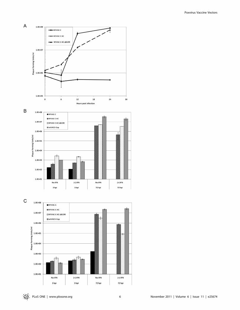
Generation of replication-competent NYVAC VectorsBecause NYVAC has limited replication capacity in cultured
human cells [14], our next goal was to produce a modified
NYVAC vector able to complete the replication cycle in cultured
human cells, but still maintaining an attenuated phenotype. K1L
and C7L are two of the genes that were deleted during the
construction of NYVAC, and these are both host range genes [23].
To restore replication in human cells, we inserted the K1L and C7L
genes back into the NYVAC genome. Figure 2 shows a schematic
diagram of the genome sequences assembled by PCR and inserted
into the genome through IVR to create NYVAC-C-KC, NYVAC-
C-KC-DB19R and NYVAC-C-KC-DB8RDB19R.
Replication in human cellsTo verify that the presence of the two genes, K1L and C7L, in
the modified virus does result in improved ability of the virus to
replicate in human cells leading to higher levels of antigen
expression, we examined first the replication capacity of NYVAC-
C-KC compared to that of NYVAC-C, in a single step growth
curve in HeLa cells (Fig. 3A). NYVAC-C-KC and NYVAC-C-
KC-DB19R replicated under these experimental conditions in
HeLa cells, but NYVAC-C did not. To be certain that the
addition of the K1L and C7L genes would restore replication in
human cell types that are physiologically relevant to poxvirus
infection, we analyzed multi-step growth curves in human
keratinocytes and human dermal fibroblasts and compared the
NYVAC-C-KC replication in these primary cells (Fig. 3B and 3C)
to that observed in HeLa cells (Fig. 3A). Though NYVAC-C did
not replicate in any of the tested cells, NYVAC-C-KC and
NYVAC-C-KC-DB19R replicated to titers comparable to that of
the wtVACV-Cop in human keratinocytes and dermal fibroblasts
(Fig. 3B and 3C). While wtVACV-Cop was fully resistant to
treatment with IFN, NYVAC-C-KC and NYVAC-C-KC-DB19R
showed increased sensitivity to IFN, especially in dermal
fibroblasts. Thus, the addition of the K1L and C7L genes to
NYVAC did restore replication competence in human cells.
Transgene expressionInsertion of K1L and C7L into NYVAC-C and deletion of B19R
also had a dramatic effect on transgene expression. The K1L and
C7L genes have been shown to decrease activation of PKR and
inhibit the subsequent phosphorylation and inactivation of eIF2a[24,25,26]. This can be seen in Figure 4B. NYVAC-C infection
induced an intermediate amount of PKR phosphorylation
compared to infection with wtVACV-Cop (VC-2) on the one
hand and virus deleted for the PKR inhibitor, E3L (vP1080), on
the other hand. Insertion of K1L and C7L into NYVAC-C
inhibited activation of PKR to background levels seen in VC2-
infected cells. Inhibition of PKR activation in NYVAC-C-KC-
infected cells was accompanied by an increase of expression of
gag-pol-nef protein and gp120 in infected human cells, compared
to cells infected with NYVAC-C (Fig. 4A). Deletion of B19R from
NYVAC-C increased phosphorylation of PKR and eIF2a, nearly
Figure 3. Replication capacity of NYVAC-C-KC and NYVAC-C-KC-DB19R in different human cells. A) Single-step growth curve inHeLa cells. Cells were infected with the indicated viruses at an MOI of 5 and harvested at 0, 6, 12, and 24 hours post-infection. Error bars are basedon two separate infections. B) Multi-step growth curves in human keratinocytes. Cells were infected with the indicated viruses at an MOI of0.01 and harvested at 3 or 72 hours post-infection, plus or minus 1000 Units/ml IFN. Error bars are based on two separate infections. C) Multi-stepgrowth curves in human dermal fibroblasts. Cells were infected with the indicated viruses at an MOI of 0.01 and harvested at 3 or 72 hourspost-infection, plus or minus 1000 Units/ml IFN. Error bars are based on two separate infections.doi:10.1371/journal.pone.0025674.g003
Poxvirus Vaccine Vectors
PLoS ONE | www.plosone.org 7 November 2011 | Volume 6 | Issue 11 | e25674

Figure 4. Signal transduction. A) Cells were infected at an MOI of 5; lysates were prepared at 6 hours post-infection and proteins were identifiedby a standard Western blot probed with anti-gp120 or anti-gag. B) HeLa cells were infected with the indicated viruses and harvested at 6 hours post-infection. Lysates were prepared and analyzed by Western blot to detect phosphorylation of shown signal transduction pathway components. C)Human keratinocytes were infected with the indicated viruses and harvested 6 hours post-infection. Lysates were prepared and analyzed by Westernblot to detect phosphorylation of shown signal transduction pathway components. D) Comparative analysis of P-STAT1 protein levels of
Poxvirus Vaccine Vectors
PLoS ONE | www.plosone.org 8 November 2011 | Volume 6 | Issue 11 | e25674

to the levels of virus deleted for E3L. This increase in both
activation of PKR and phosphorylation of eIF2a was accompa-
nied by a dramatic decrease in transgene expression. Insertion of
K1L and C7L into NYVAC-C-DB19R reversed this trend, leading
to a restoration of transgene protein synthesis to the level seen in
cells infected with NYVAC-C-KC.
Induction of Host ResponsesSince B19 blocks the effects of type I IFN, we wanted to
determine if deletion of B19R would have an effect on signaling
downstream of IFN binding to its receptor on cells. The STAT-1
pathway, which is downstream of IFN induction, was not activated
by infection with any virus containing B19R (wtNYVAC,
NYVAC-C or NYVAC-C-KC, Fig. 4D). In fact, infection with
any of these viruses led to decreased STAT-1 phosphorylation
compared to mock-infected cells. However, deletion of B19R led to
phosphorylation of STAT-1, even in the virus with K1L and C7L
restored, compared to infection with the viruses containing B19R.
Several components of the virus sensing system are inducible by
IFN (e.g., RIG-I and MDA-5). Since deletion of B19R can lead to
IFN-induced signaling, we wanted to determine if these viruses
affected phosphorylation of IRF-3. IRF-3 is a key transcription
factor, which when phosphorylated and activated can lead to
induction of type I IFN, and IFN is an important mediator
between the innate and adaptive immune systems. Figure 4B
shows the results of Western blot analysis of lysates from HeLa
cells infected with either VC2 (wtVACV-Cop), vP1080 (Cop-
DE3L), NYVAC-C, NYVAC-C-KC, NYVAC-C-DB19R, or
NYVAC-C-KC-DB19R to detect phosphorylation of IRF-3.
wtVACV-Cop did not lead to phosphorylation of IRF-3,
consistent with previously published results [27]. vP1080 (Copen-
hagen deleted of the E3L gene) was a strong activator of IRF-3
phosphorylation because the E3L protein is not present to mask
the dsRNA [27]. Neither NYVAC-C nor NYVAC-C-KC induced
detectable phosphorylation of IRF-3. However, deletion of B19R
from either of these viruses resulted in phosphorylation of IRF-3.
Results in human keratinocytes (Fig. 4C) were similar to those in
HeLa cells, although we did observe donor-to-donor variation in
the activation patterns (data not shown).
The signal transduction results were extended by microarray
analysis of infected cells, as shown in Figure 4E. The detailed
microarray data from human dendritic cells (DCs) infected with
the NYVAC vectors has been recently described [8]. Shown here
is representative data from DCs infected with either NYVAC-C or
NYVAC-C-KC, or either of those viruses deleted also of both B8R
and B19R. The results from infection with this double-deletion
mutant were indistinguishable from those of the single deletion of
B19R only (data not shown). Infection with either NYVAC-C or
NYVAC-C-KC induced very little change to any of the shown
IFN-inducible genes, consistent with the IRF-3 phosphorylation
and STAT-phosphorylation results shown in Figure 4B and 4D.
The deletion of B8R and B19R from NYVAC-C results in a strong
inflammatory response, as evidenced by the transcriptional up-
regulation of all the genes shown on the graph, again consistent
with the results seen in Figure 4B and 4D. With the insertion of
K1L and C7L, this response was tempered; infection with NYVAC-
C-KC-DB8RDB19R induced increased transcriptional levels of
seven of the genes shown, relative to NYVAC-C-KC (IFI35,
IFI44, IFI44L, IFIT1, IFIT3, IFITM1, and IFITM2). The lower
level of induction of these genes by NYVAC-C-KC-DB8RDB19R
compared to that of NYVAC-C-DB8RDB19R is consistent with
signal transduction data, since NYVAC-C-KC-DB8RDB19R
induced less IRF-3 and STAT-1 phosphorylation than did
NYVAC-C-DB8RDB19R (data not shown).
Since activation of PKR has previously been shown to lead to
induction of apoptosis, we assayed for the ability of these modified
viruses to induce apoptosis. Consistent with the activation of PKR
in HeLa cells (Fig. 4B), apoptosis in HeLa cells infected with
non-replication vs. replication competent B19R deletion mutants. Phospho-STAT1 levels present in extracts of cells infected with differentrecombinant viruses were measured by Cytometric Bead Array (CBA) using an LSR II flow cytometer. The concentrations of total and phospho STAT1were measured simultaneously using the Cell Signaling Master Buffer Kit (BD Biosciences Pharmingen). For this CBA analysis, PBMCs were mock-infected or infected at an MOI of 5 with NYVAC wt, NYVAC-C, NYVAC-C-KC, NYVAC-C-DB19R or NYVAC-C-KC-DB19R. At 4 hours post-infection, cellswere harvested and processed for CBA analysis as described in Materials and Methods and according to manufacturer’s recommendations. Resultsare given as the ratio between phospho and total STAT1. E) Microarray. RNA was extracted from human pDCs infected with the indicated viruses.Data from a subset of the IFN-induced genes is shown. Fold increase (decrease) is normalized to results for mock-infected cells.doi:10.1371/journal.pone.0025674.g004
Figure 5. Comparative analysis of the apoptosis induction of non-replication versus replication-competent B19R deletion mutants.The different stages of cell cycle and the percentage of cells with subG0 DNA content were analyzed by propidium iodide (PI) staining and FACSanalysis. HeLa cells were mock-infected or infected at an MOI of 5 with NYVAC-C, NYVAC-C-KC, NYVAC-C-DB19R or NYVAC-C-KC-DB19R. At 24 hourspost-infection cells were harvested and processed as described in Materials and Methods. The percentage of cells with hypodiploid DNA content wasdetermined using an LSR II flow cytometer (Becton Dickinson). The results are expressed as fold increase in apoptotic cells with respect to uninfectedcells.doi:10.1371/journal.pone.0025674.g005
Poxvirus Vaccine Vectors
PLoS ONE | www.plosone.org 9 November 2011 | Volume 6 | Issue 11 | e25674

NYVAC-C or NYVAC-C-DB19R was increased when normal-
ized to uninfected cells, while the level of apoptosis was not
increased in cells infected with the viruses containing the K1L and
C7L genes (Fig. 5).
Safety profileThough our goal was to improve the parental NYVAC vaccine
vector, it was equally important to maintain sufficient safety levels
with the pox vector. To compare the pathogenicity of NYVAC-C-
KC constructs to existing vaccines, we utilized a newborn mouse
model, the most sensitive mouse model available for detecting pox
virus pathogenesis [11]. Newborn mice were inoculated intracra-
nially (IC) with the indicated viruses. As can be seen in Figure 6,
wtVACV-Cop was highly pathogenic in this model, with an LD50
of only 10 pfu. VACV-CopC, with the loss of the TK gene, is
further attenuated by about one log, and has an LD50 comparable
to that of the New York City Board of Health (NYCBH) [28]
strain of VACV, the strain of VACV currently used for
vaccination against smallpox. NYVAC-C-KC was attenuated by
approximately four logs compared to NYCBH. Deletion of B19R
from NYVAC-C-KC further attenuated the virus by approxi-
mately one log, approaching the attenuation of MVA and
NYVAC-C. Thus, the safety profile of NYVAC-C-KC-DB19R,
despite this construct being nearly fully replication-competent in
primary human skin cells in culture, is in the same general range as
the highly attenuated, replication-restricted vaccinia virus strains,
MVA and NYVAC-C.
Since most of the complications due to use of vaccinia virus
require spread from the site of inoculation, we followed two
approaches to characterize spread of the virus in infected animals:
one determining virus titers in different organs and the second,
evaluating viral expression by the levels of the marker luciferase in
NYVAC recombinants.
To measure titers in different organs, mice were inoculated
intraperitoneally (IP) with 26107 pfu/mouse of virus and at
different days post infection the animals were sacrificed and the
presence of virus in the peritoneal wash, ovaries, liver, spleen, and
lymph nodes was measured. As shown in Figure 7, the WR strain
of VACV was detectable at high titers in all tissue samples
(peritoneum, ovaries, liver spleen and lymph nodes), even up to
72 hours post-infection. Infectious NYVAC-C or NYVAC-C-
DB19R was not detectable in any tissue at any time. Both
NYVAC-C-KC and NYVAC-C-KC-DB19R were transiently
detectable in the peritoneum and ovaries, but resolved by 48 hours
post-infection. Levels of NYVAC-C-KC were similar to wtVACV-
WR in the peritoneal cavity, but 4- to 5-fold lower than wtVACV-
WR in ovaries. NYVAC-C-KC-DB19R was detectable in 5-fold
and 100-fold lower amounts in the peritoneal cavity and ovaries,
respectively, compared to wtVACV-WR.
These results were confirmed with recombinant viruses
expressing luciferase. Groups of mice were infected IP with
26107 pfu/mouse of WR-luc, NYVAC-luc or NYVAC-KC-luc
and at different time points, levels of luciferase were evaluated in
mouse tissues. As shown in Figure 8, luciferase expression
remained elevated in animals infected with the fully replication-
competent WR-luc, while the levels were reduced in NYVAC-
infected animals. Luciferase expression was intermediate for
animals infected with NYVAC-KC-luc.
Discussion
An important consideration in the HIV/AIDS vaccine field is
the nature of the vectors needed for protective efficacy. While
Figure 6. Pathogenesis study. Newborn CD1 mice (minimum of 10 per group) were inoculated IC with the indicated viruses at the indicated dosesof plaque forming units (pfu). Mice were monitored twice daily for 14 days for signs of morbidity/mortality.doi:10.1371/journal.pone.0025674.g006
Poxvirus Vaccine Vectors
PLoS ONE | www.plosone.org 10 November 2011 | Volume 6 | Issue 11 | e25674

numerous different combinations of vectors are being considered
as vaccine regimens, one combination has been shown to be
partially effective. This has been shown in the phase III Thai
clinical study (RV144) with the vaccine combination of a
canarypox virus (ALVAC) and the monomeric protein gp120,
giving 31% protection against HIV infection [7]. The modest
efficacy of the RV144 trial suggests that improvement of the
vaccination protocol is possible. Since ALVAC is a replication-
restricted virus in human cells and the other candidate VACV
vectors, MVA and NYVAC, are also replication-restricted, it is
possible that improvements of these vectors might be achieved
through enhancement of their replication capacity. In this
investigation we have achieved this goal by the re-insertion of
the host restriction genes K1L and C7L into the NYVAC genome.
Re-insertion of these two genes restored replication capacity in
human cultured cells, including physiologically relevant primary
keratinocytes and primary dermal fibroblasts. A concern of
replication-competence in human cultured cells has been tem-
pered by the demonstration that the replication-competent viruses
described in this manuscript are highly attenuated compared to
other replication-competent strains. This has been demonstrated
through intracranial inoculation of the vectors in newborn mice.
Clearly the vector NYVAC-C-KC is highly attenuated compared
to the Copenhagen and NYCBH strains (Fig. 6). The attenuation
was further improved by deleting the type I IFN inhibitor B19.
The likely reason that increased attenuation is associated with
deletion of B19R is that type I IFN induced during virus infection
is no longer blocked by the virus inhibitor B19 and, hence, pro-
inflammatory pathways are more readily activated (Fig. 4).
The consequences of the incorporation of the host genes K1L-
C7L into NYVAC-C include the enhanced expression of foreign
antigens, such as the HIV-1 protein gp120 and the fusion protein
gag-pol-nef in cultured cells (Fig. 4A). Viral protein expression in
vaccinia virus-infected cells is often regulated by activation of the
cellular protein, PKR, which when activated, effectively inhibits
translation of viral proteins. Poxviruses use several mechanisms to
Figure 7. Comparative analysis of the biodistribution of non-replication versus replication-competent B19R deletion mutants. Fivegroups of mice (n = 6) received an IP inoculation of 26107 PFU/mouse of WR (TK2), NYVAC-C, NYVAC-C-KC, NYVAC-C-DB19R or NYVAC-C-KC-DB19R.At different times post-inoculation (24, 48 and 72 hours) animals were sacrificed and different mouse tissues (peritoneal cells, ovaries, livers, spleensand draining lymph nodes) were harvested and processed for titration as described in Materials and Methods. The production of infectious virus wasdetermined by plaque assay in BSC-40 cells. The virus titer was expressed as Plaque Forming Units (pfu) per gram of protein.doi:10.1371/journal.pone.0025674.g007
Poxvirus Vaccine Vectors
PLoS ONE | www.plosone.org 11 November 2011 | Volume 6 | Issue 11 | e25674

block activation of PKR, including the dsRNA binding protein
E3L [27,29]. However, work done by others has shown that K1L is
also necessary to prevent activation of PKR in vaccinia virus-
infected cells [24,30], and that even expression of early viral
proteins is down-regulated in the absence of K1L. Therefore,
antigen expression by the viral vector is enhanced by the presence
of K1L. We also detected increased luciferase expression in animals
infected with NYVAC-KC-luc. In this case, the increased level of
antigen expression may result from a combination of restored
replication-competence which likely leads to an increase in the
number of cells infected in the animal, and to a de novo increase in
translation in infected cells in the animal. In the absence of a
mechanism to separate replication-competence from increased
protein expression in the individual cell, it is difficult to ascertain
the contribution of each.
It is curious that deletion of B19R has such a dramatic effect on
activation of PKR and phosphorylation of IRF-3. Our hypothesis
is that B19 normally blocks the priming effect resulting from small
amounts of IFN secreted from the infected host cell. In the absence
of B19, this priming effect is allowed to take place, inducing
pathogen-associated molecular pattern (PAMP) sensors, such as
RIG-I and PKR, and initiating signal transduction pathways that
may enhance the adaptive immune response against the antigen.
The highly-attenuated phenotype of the NYVAC-C-KC-DB19R
virus, along with higher antigen expression levels than observed
with its parental virus NYVAC-C, and its ability to induce pro-
inflammatory signaling and pro-inflammatory gene expression,
suggest that this virus provides a potential improved vaccine vector
for human application.
Previous studies on the immunological characteristics of
NYVAC-C have been documented in different models: mouse
[15,16], macaque [17] and human clinical trials [18,19]. The
results obtained thus far indicate that NYVAC-C is immunogenic
but needs further improvements. In fact, prime/boost combina-
tion with DNA vectors is needed to expand the breadth and
strength of the immune responses to HIV antigens. In the phase I
clinical studies, two doses of NYVAC-C given intramuscularly
resulted in positive responses to HIV antigens in fewer than 40%
Figure 8. Comparative levels of luciferase in mice infected with parental NYVAC versus NYVAC-C-KC. Mice were infected IP with 26107
plaque forming units/mouse of viruses (WR-luc, NYVAC-luc and NYVAC-KC-luc) expressing the luciferase marker and at different times several tissueswere harvested, lysed and luciferase activity was determined as previously described [12,13]. Values are represented as reference luciferase units pergram of tissue.doi:10.1371/journal.pone.0025674.g008
Poxvirus Vaccine Vectors
PLoS ONE | www.plosone.org 12 November 2011 | Volume 6 | Issue 11 | e25674

vaccinees [18,19]. It was only after priming with DNA vectors that
responses rose to about 90% of vaccinees [18]. It is likely that the
limited expression of HIV antigens and the replication-restriction
of NYVAC-C in human cells played an important role in the
observed limitations in the immune responses. The use of
NYVAC-K-KC or NYVAC-C-KC-DB19R might represent an
important advance in improving NYVAC-based vaccine vectors.
This has been further supported by additional studies with these
vectors showing activation of pathways involved in antigen
processing and presentation, enhanced cross-presentation to
HIV-specific CD8 T cells, and proliferation of specific memory
CD8 T cells in vitro [8].
Acknowledgments
The authors wish to thank Constance Chamberlain and Nobuko
Fukushima for technical assistance.
Author Contributions
Conceived and designed the experiments: RW RS JT GP BJ ME EH KK.
Performed the experiments: KK CG BP SW TH SH WA VJ RG KD EH.
Analyzed the data: KK EH RS BJ ME CG. Contributed reagents/
materials/analysis tools: RW JT RS BJ ME. Wrote the paper: KK CG BJ
ME.
References
1. Moss B (1996) Genetically engineered poxviruses for recombinant gene
expression, vaccination, and safety. Proc Natl Acad Sci U S A 93: 11341–11348.2. Paoletti E (1996) Applications of pox virus vectors to vaccination: an update.
Proc Natl Acad Sci U S A 93: 11349–11353.
3. Paoletti E, Tartaglia J, Taylor J (1994) Safe and effective poxvirus vectors–NYVAC and ALVAC. Dev Biol Stand 82: 65–69.
4. Franchini G, Gurunathan S, Baglyos L, Plotkin S, Tartaglia J (2004) Poxvirus-based vaccine candidates for HIV: two decades of experience with special
emphasis on canarypox vectors. Expert Rev Vaccines 3: S75–88.
5. Drexler I, Staib C, Sutter G (2004) Modified vaccinia virus Ankara as antigendelivery system: how can we best use its potential? Curr Opin Biotechnol 15:
506–512.6. Gomez CE, Najera JL, Krupa M, Esteban M (2008) The poxvirus vectors MVA
and NYVAC as gene delivery systems for vaccination against infectious diseasesand cancer. Curr Gene Ther 8: 97–120.
7. Rerks-Ngarm S, Pitisuttithum P, Nitayaphan S, Kaewkungwal J, Chiu J, et al.
(2009) Vaccination with ALVAC and AIDSVAX to prevent HIV-1 infection inThailand. N Engl J Med 361: 2209–2220.
8. Quakkelaar ED, Redeker A, Haddad EK, Harari A, McCaughey SM, et al.(2011) Improved innate and adaptive immunostimulation by genetically
modified HIV-1 protein expressing NYVAC vectors. PLoS One 6: e16819.
9. Gaucher D, Therrien R, Kettaf N, Angermann BR, Boucher G, et al. (2008)Yellow fever vaccine induces integrated multilineage and polyfunctional immune
responses. J Exp Med 205: 3119–3131.10. Hackstadt AJ, Hess AM (2009) Filtering for increased power for microarray data
analysis. BMC Bioinformatics 10: 11.11. Li Z, Rubin SA, Taffs RE, Merchlinsky M, Ye Z, et al. (2004) Mouse
neurotoxicity test for vaccinia-based smallpox vaccines. Vaccine 22: 1486–1493.
12. Gomez CE, Najera JL, Domingo-Gil E, Ochoa-Callejero L, Gonzalez-Aseguinolaza G, et al. (2007) Virus distribution of the attenuated MVA and
NYVAC poxvirus strains in mice. J Gen Virol 88: 2473–2478.13. Rodriguez JF, Rodriguez D, Rodriguez JR, McGowan EB, Esteban M (1988)
Expression of the firefly luciferase gene in vaccinia virus: a highly sensitive gene
marker to follow virus dissemination in tissues of infected animals. Proc NatlAcad Sci U S A 85: 1667–1671.
14. Tartaglia J, Perkus ME, Taylor J, Norton EK, Audonnet JC, et al. (1992)NYVAC: a highly attenuated strain of vaccinia virus. Virology 188: 217–232.
15. Gomez CE, Najera JL, Jimenez V, Bieler K, Wild J, et al. (2007) Generation andimmunogenicity of novel HIV/AIDS vaccine candidates targeting HIV-1 Env/
Gag-Pol-Nef antigens of clade C. Vaccine 25: 1969–1992.
16. Wild J, Bieler K, Kostler J, Frachette MJ, Jeffs S, et al. (2009) Preclinicalevaluation of the immunogenicity of C-type HIV-1-based DNA and NYVAC
vaccines in the Balb/C mouse model. Viral Immunol 22: 309–319.17. Mooij P, Balla-Jhagjhoorsingh SS, Beenhakker N, van Haaften P, Baak I, et al.
(2009) Comparison of human and rhesus macaque T-cell responses elicited by
boosting with NYVAC encoding human immunodeficiency virus type 1 clade C
immunogens. J Virol 83: 5881–5889.
18. Harari A, Bart PA, Stohr W, Tapia G, Garcia M, et al. (2008) An HIV-1 clade C
DNA prime, NYVAC boost vaccine regimen induces reliable, polyfunctional,
and long-lasting T cell responses. J Exp Med 205: 63–77.
19. Bart PA, Goodall R, Barber T, Harari A, Guimaraes-Walker A, et al. (2008)
EV01: a phase I trial in healthy HIV negative volunteers to evaluate a clade C
HIV vaccine, NYVAC-C undertaken by the EuroVacc Consortium. Vaccine 26:
3153–3161.
20. Perdiguero B, Esteban M (2009) The interferon system and vaccinia virus
evasion mechanisms. J Interferon Cytokine Res 29: 581–598.
21. Colamonici OR, Domanski P, Sweitzer SM, Larner A, Buller RM (1995)
Vaccinia virus B18R gene encodes a type I interferon-binding protein that
blocks interferon alpha transmembrane signaling. J Biol Chem 270:
15974–15978.
22. Alcami A, Smith GL (1995) Vaccinia, cowpox, and camelpox viruses encode
soluble gamma interferon receptors with novel broad species specificity. J Virol
69: 4633–4639.
23. Perkus ME, Goebel SJ, Davis SW, Johnson GP, Limbach K, et al. (1990)
Vaccinia virus host range genes. Virology 179: 276–286.
24. Meng X, Jiang C, Arsenio J, Dick K, Cao J, et al. (2009) Vaccinia virus K1L and
C7L inhibit antiviral activities induced by type I interferons. J Virol 83:
10627–10636.
25. Najera JL, Gomez CE, Domingo-Gil E, Gherardi MM, Esteban M (2006)
Cellular and biochemical differences between two attenuated poxvirus vaccine
candidates (MVA and NYVAC) and role of the C7L gene. J Virol 80:
6033–6047.
26. Willis KL, Patel S, Xiang Y, Shisler JL (2009) The effect of the vaccinia K1
protein on the PKR-eIF2alpha pathway in RK13 and HeLa cells. Virology 394:
73–81.
27. Langland JO, Kash JC, Carter V, Thomas MJ, Katze MG, et al. (2006)
Suppression of proinflammatory signal transduction and gene expression by the
dual nucleic acid binding domains of the vaccinia virus E3L proteins. J Virol 80:
10083–10095.
28. Weltzin R, Liu J, Pugachev KV, Myers GA, Coughlin B, et al. (2003) Clonal
vaccinia virus grown in cell culture as a new smallpox vaccine. Nat Med 9:
1125–1130.
29. Jacobs BL, Langland JO (1996) When two strands are better than one: the
mediators and modulators of the cellular responses to double-stranded RNA.
Virology 219: 339–349.
30. Shisler JL, Jin XL (2004) The vaccinia virus K1L gene product inhibits host NF-
kappaB activation by preventing IkappaBalpha degradation. J Virol 78:
3553–3560.
Poxvirus Vaccine Vectors
PLoS ONE | www.plosone.org 13 November 2011 | Volume 6 | Issue 11 | e25674
Copyright © 2022 FDOKUMEN