Vaccine: A General Overview
47
Transcript of Vaccine: A General Overview


Micrographia Today
www.micrographiatoday.org
The Ebola outbreak in West Africa is the world's deadliest to date and the World Health Organization has declared an international health emergency as more than 2,100 people have died of the virus in Guinea, Liberia, Sierra Leone andof this issue is to create public awareness by describing the basic features of the virus, its epidemiology and global response to Ebola outbreaks. Moreover, it will give readers a brief insight on the current strategies tTherefore, this issue will be focused on the epidemiology and its global response to Ebola outbreaks, and current strategies to the diagnosis, treatment, prevention and control.
In this issue we would also like to introduce Dr. Vincent Racaniello Ph.D., Professor of Microbiology & Immunology in the College of Physicians and Surgeons of Columbia University. He has done an immense research on viruses since 1975. He is the one who, cloned poliovirus DNA into a bacterial plasmid and determined the nucleotide sequence of the poliovirus genome.
This issue of Micrographia Today talk’s vaccines, viruses, bacteria and talks to one of the best known brains in the field of virology to unravel the microscopic detaienable our readers to know more and benefit from their experiences.
With best wishes,
Micrographia Today
2
FROM EDITOR’S DESKDear Readers,
As the Editor-in-Chief, I would like to welcome you all into the New Year, 2015. I would also like to take opportunity to thank the people who have put their trust in me and have considered me worthy of handling this position. Over the years, our magazine has risen to great heights and has come to be widely known across the globe and it is a great honour to be at this position.
The Ebola outbreak in West Africa is the world's deadliest to date and the World Health Organization has declared an international health emergency as more than 2,100 people have died of the virus in Guinea, Liberia, Sierra Leone and Nigeria this year. Thus, the aim of this issue is to create public awareness by describing the basic features of the virus, its epidemiology and global response to Ebola outbreaks. Moreover, it will give readers a brief insight on the current strategies to diagnosis, treatment, and prevention of the disease. Therefore, this issue will be focused on the epidemiology and its global response to Ebola outbreaks, and current strategies to the diagnosis, treatment, prevention and control.
also like to introduce Dr. Vincent Racaniello Ph.D., Professor of Microbiology & Immunology in the College of Physicians and Surgeons of Columbia University. He has done an immense research on viruses since 1975. He is the one who,
to a bacterial plasmid and determined the nucleotide sequence of
This issue of Micrographia Today talk’s vaccines, viruses, bacteria and talks to one of the best known brains in the field of virology to unravel the microscopic detaienable our readers to know more and benefit from their experiences.
@wethemicrobiologist.in
Vol.2. Issue1
FROM EDITOR’S DESK
Chief, I would like to welcome you all into the New Year, 2015. I would also like to take this opportunity to thank the people who have put their trust in me and have considered me worthy of handling this position. Over the years, our magazine has risen to great heights and has come to be widely known across the globe
o be at this position.
The Ebola outbreak in West Africa is the world's deadliest to date and the World Health Organization has declared an international health emergency as more than 2,100 people
Nigeria this year. Thus, the aim of this issue is to create public awareness by describing the basic features of the virus, its epidemiology and global response to Ebola outbreaks. Moreover, it will give readers a brief
o diagnosis, treatment, and prevention of the disease. Therefore, this issue will be focused on the epidemiology and its global response to Ebola outbreaks, and current strategies to the diagnosis, treatment, prevention and control.
also like to introduce Dr. Vincent Racaniello Ph.D., Professor of Microbiology & Immunology in the College of Physicians and Surgeons of Columbia University. He has done an immense research on viruses since 1975. He is the one who,
to a bacterial plasmid and determined the nucleotide sequence of
This issue of Micrographia Today talk’s vaccines, viruses, bacteria and talks to one of the best known brains in the field of virology to unravel the microscopic details of viruses to

Micrographia Today
3
Vol.2. Issue1
www.micrographiatoday.org
MAGAZINE BOARDEditor - in - ChiefMr. Swapnil Vichare
Co-Editor in ChiefMr. Saumyadip Sarkar
EditorMr. Golam Moktadir Khan
News CorrespondentMr. Bapi Jha
ReviewersMr. Bamgbose Princeteejay TimothyMr. Shrikant SonawaneMs. Neha AilaniMr. Kunlere 'Hi-Dee' IdowuMd. Mehedi Hasan MagnetDr. Azhar Bhatt
www.micrographiatoday.org
Disclaimer: Views and opinion expressed in this magazine are not those of We The Microbiologist, it’s of the authors and writers. We, at We The Microbiologist Micrographia Today do our best to verify the information published but do not take any responsibility of absolute accuracy of the information. We The Microbiologist does not accept any responsibility for any decision taken by readers based on the information provided in the magazine. No part of this magazine can be reproduced without prior written permission.
[Note: Images for the cover page is derived from google images]
LEADERSHIPSPresident
Ms. Harshada [email protected]
Principal SecretaryMr. Bapi Jha
Managing DirectorMr. Saumyadip Sarkar
Organizing SecretaryMr. Trinankur Bhattacharya
Editor - in – ChiefMr. Swapnil Vichare
EditorMr. Golam Moktadir Khan
International Outreach CoordinatorsMr. Golam Moktadir Khan
Bangladesh (bd.wethemicrobiologist.in)
Mr. Bamgbose Princeteejay TimothyNigeria
Mr. Sajjad AhmadPakistan (pk.wethemicrobiologist.in)
www.wethemicrobiologist.in

Micrographia Today
4
Vol.2. Issue1
www.micrographiatoday.org
Table of Content
Sr.No TITLE AUTHOR PAGE NO.
1. News Highlisted in 2014 Bapi Jha 05
2.Biggest Contributions from Scientific
Communicators of 2014Correspondents 10
3. Invention Column Correspondents 12
4.16SrRNA and the Strategy Used in Bacterial
IdentificationSaumyadip Sarkar 13
5.COVER Story: Vaccines - A General
OverviewBamgbose Timothy
16
6. Soil Microbes Will Bring the Climate Change Correspondents 20
7.The Recent Ebola Virus Disease (EVD)
Outbreak in NigeriaKUNLERE Idowu
Olagoke22
8.A brief Introduction to Ti-plasmids and its mechanism to create genetically modified
plantsSaumyadip Sarkar 27
9. Updates on Ebola Outbreak Correspondents 33
10. Person on Spotlight: Dr. Vincent Racaniello Saumyadip Sarkar 37
11. Life Science Quiz to Solve Correspondents 45
12. Events Update WTM 47

Micrographia Today
www.micrographiatoday.org
NEWS HIGHLISTED
Nobel Prize for Discovering Brain’s Inner GPS
Nobel Prize for Physiology in Medicine has been awarded to three Scientists for discovering brain’s remarkable “inner GPS”. Question came to most of the prominent scientists that what is this inner GPS system in braallows to work out where it is, how to get from one place to another, and thus storing information for future reference.Naming the first, Dr. John O’Keefe who holds both British and American citizenships, also the current director of University College London’s Neural Circuits and Behavior Center,
The hypothesis and the research ahead:In 1971, it gave the start when Dr. O’Keefe discovered the first component of the positioning system in the brain. In that research he found that in a part of the brain called hippocampus, a nerve cell (named place cells) that always remain activated in rat in certain place of a room. When it was shifted to other part of the room, other nerve cells become active. He concluded that brain can place cells to constitute the “map” of the room.
Micrographia Today
5
NEWS HIGHLISTED in 2014
Prize for Discovering Brain’s Inner GPS
Nobel Prize for Physiology in Medicine has been awarded to three Scientists for discovering brain’s remarkable “inner GPS”. Question came to most of the prominent scientists that what is this inner GPS system in brain. In short it is the system of cells that allows to work out where it is, how to get from one place to another, and thus storing information for future reference.Naming the first, Dr. John O’Keefe who holds both British and American citizenships, also the current director of University College London’s Neural Circuits and Behavior Center,
UK receives one-half of the prize. The other halves were shared by husband-wife scientists, Dr. Edvard I. Moser and May-Britt Moser citizens and are in neuroscience Institutes in Trondeim.They have cleared all the confusions that has been craving in minds of philosophers and scientists for centuries, i.e. how brain can map our surroundings and thus helping to navigate the complex environment.
hypothesis and the research ahead:In 1971, it gave the start when Dr. O’Keefe discovered the first component of the positioning system in the brain. In that research he found that in a part of the brain called hippocampus, a nerve cell
) that always remain activated in rat in certain place of a room. When it was shifted to other part of the room, other nerve cells become active. He concluded that brain can place cells to constitute the “map” of the room.
Vol.2. Issue1
Nobel Prize for Physiology in Medicine has been awarded to three Scientists for discovering brain’s remarkable “inner GPS”. Question came to most of the prominent
in. In short it is the system of cells that allows to work out where it is, how to get from one place to another, and thus storing
Naming the first, Dr. John O’Keefe who holds both British and American citizenships, also the current director of University College London’s Neural Circuits and Behavior Center,
half of the prize. The other halves wife scientists, Dr. Edvard
Britt Moser – both Norwegian n neuroscience Institutes in
They have cleared all the confusions that has been craving in minds of philosophers and scientists for centuries, i.e. how brain can map our surroundings and thus helping to navigate the complex

Micrographia Today
www.micrographiatoday.org
The next vital thinking ahead camhappened over three decades later in 2015. Edvard and Maytype of nerve cell, which they named as “grid cells”. It functions to coordinate system enabling precise positioning and pathfinding. Grid cells are located in the entorhinal cortex.Following these discoveries, later it was found that between place cells and grid cells, there is a linked circuit in hippocampus and entorhinal cortex. These help the braindetermine position in a space and navigate properly.It is important to know that all these research was carried out in rats in laboratory. In the advancement of recent techniques with brain imaging, with patients having brain surgery have shown that human brain has sim
The Discovery with the benefit:The contribution from the three Noble Laureates has opened the gates to understand how groups of specialized cells work together to execute multiple functions. The work has not only helped to understand the cognitive processes but also functions like thinking, planning and memory.The hippocampus and entorhinal cortex are often reported to be affected in Alzheimer’s disease. The disease makes the patients to lose their way and even not able to ascethe environment. Now the researchers of the Alzheimer’s are stepping forward to investigate the mechanism of the devastating loss of memory in patients.
The Response after Winning:NOBEL PRIZE IN PHYSIOLOGY OR M
Micrographia Today
6
The next vital thinking ahead came with the discovery of brain’s inner GPS, which happened over three decades later in 2015. Edvard and May-Britt Moser identified another type of nerve cell, which they named as “grid cells”. It functions to coordinate system
nd pathfinding. Grid cells are located in the entorhinal cortex.Following these discoveries, later it was found that between place cells and grid cells, there is a linked circuit in hippocampus and entorhinal cortex. These help the brain
tion in a space and navigate properly.It is important to know that all these research was carried out in rats in laboratory. In the advancement of recent techniques with brain imaging, with patients having brain surgery have shown that human brain has similar place and grid cells.
The Discovery with the benefit:The contribution from the three Noble Laureates has opened the gates to understand how groups of specialized cells work together to execute multiple functions. The work has not
rstand the cognitive processes but also functions like thinking, planning
The hippocampus and entorhinal cortex are often reported to be affected in Alzheimer’s disease. The disease makes the patients to lose their way and even not able to ascethe environment. Now the researchers of the Alzheimer’s are stepping forward to investigate the mechanism of the devastating loss of memory in patients.
The Response after Winning:MEDICINE 2014 (SOURCE NOBELPRIZE.ORG)
Dr. O’Keefe was working in London when the phone call came with the news. As said in one of the meetings, “Oh this couldn’t possibly be, this couldn’t possibly be what I think it is,” but soon after realizing, “but of course it was.”On the other handEdvard Moser was on plane
Vol.2. Issue1
e with the discovery of brain’s inner GPS, which Britt Moser identified another
type of nerve cell, which they named as “grid cells”. It functions to coordinate system nd pathfinding. Grid cells are located in the entorhinal cortex.
Following these discoveries, later it was found that between place cells and grid cells, there is a linked circuit in hippocampus and entorhinal cortex. These help the brain to
It is important to know that all these research was carried out in rats in laboratory. In the advancement of recent techniques with brain imaging, with patients having brain surgery
The contribution from the three Noble Laureates has opened the gates to understand how groups of specialized cells work together to execute multiple functions. The work has not
rstand the cognitive processes but also functions like thinking, planning
The hippocampus and entorhinal cortex are often reported to be affected in Alzheimer’s disease. The disease makes the patients to lose their way and even not able to ascertain with the environment. Now the researchers of the Alzheimer’s are stepping forward to investigate the mechanism of the devastating loss of memory in patients.
)Dr. O’Keefe was working in London when the phone call came with the news. As said in one of the meetings, “Oh this couldn’t possibly be, this couldn’t possibly be what I think it is,” but soon after realizing, “but of course it was.”On the other hand, Dr. Edvard Moser was on plane

Micrographia Today
7
Vol.2. Issue1
www.micrographiatoday.org
to Munich when the award was announced. He didn’t came to know until he came off the plane. He noticed that a representative of the airport came with flowers to greet and escort him in the car.Dr. May-Britt cried after she came to know the news to be the Nobel Winner. She was that time in between meetings. She said to the NobelPrize.org in a telephone interview, “I was in shock and I’m still in shock. This is so great!”
John O’Keefe has been a mentor to the Mosers in one time point. Edvard Moser said on the telephone interview to NobelPrize.org, in 1996 they worked in his lab, where John O’Keefe trained them in the techniques they has been using since.
Source: Medical News Today (Story by Cathrine Paddock, PhD).Read More: http://www.nature.com/news/neuroscience-brains-of-norway-1.16079
Response to the Fellowship Hike
Union Minister of State for Science and Technology, Mr. Jitendra Singh announced the
most awaited response towards the fellowship hike made by over 20,000 research scholars
in India.
According to the revised figures on the fellowship amount, here is the status of the
increased fellowship accepted by the government.
PositionInitial Amount
(INR/pm)
Upgraded Amount
(INR/pm)
Junior Research Fellow 16,000 25,000
Senior Research Fellow 18,000 28,000
Research Associate I 22,000 36,000
Research Associate II 23,000 38,000
Research Associate III 24,000 40,000

Micrographia Today
www.micrographiatoday.org
Read the full story: Not everyone is happy with hike in research fellowships
2014. The Hindu. (http://www.thehindu.com/news/national/karnataka/not
happy-with-hike-in-research-fellowships/article6524285.ece
Antibiotic resistance bacteria was found before Penicillin was discovered
A study published in the Journal
War soldier died of dysentery proves the reason why it is difficult to tackle. The sample of
the bacteria Shigella flexneri has been kept in government achieves for nearly 100years
highlighted that the bacteria was resistant to penicillin even before Alexander Flemming
actually discovers the drug.
British stretcher bearers in Passchendaele in 1917
This proves that the bacteria quickly evolve the defence mechanism against antibioti
holding efficient reason for the limiting use. Researchers from Welcome Trust Sanger
Micrographia Today
8
Not everyone is happy with hike in research fellowships
w.thehindu.com/news/national/karnataka/not
fellowships/article6524285.ece)
Antibiotic resistance bacteria was found before Penicillin was
A study published in the Journal The Lancet defines a bacteria isolated from a First World
War soldier died of dysentery proves the reason why it is difficult to tackle. The sample of
has been kept in government achieves for nearly 100years
ia was resistant to penicillin even before Alexander Flemming
British stretcher bearers in Passchendaele in 1917 (Source: The Telegraph, UK)
This proves that the bacteria quickly evolve the defence mechanism against antibioti
holding efficient reason for the limiting use. Researchers from Welcome Trust Sanger
Vol.2. Issue1
Not everyone is happy with hike in research fellowships. October 21st,
w.thehindu.com/news/national/karnataka/not-everyone-is-
Antibiotic resistance bacteria was found before Penicillin was
defines a bacteria isolated from a First World
War soldier died of dysentery proves the reason why it is difficult to tackle. The sample of
has been kept in government achieves for nearly 100years
ia was resistant to penicillin even before Alexander Flemming
(Source: The Telegraph, UK)
This proves that the bacteria quickly evolve the defence mechanism against antibiotics,
holding efficient reason for the limiting use. Researchers from Welcome Trust Sanger
Micrographia Today
www.micrographiatoday.org
Institute and Public Health England sequenced the entire genome of
track down the disease that had evolved in the previous century.
News Source: Antibiotic resistance began before discovery of penicillin, DNA from First
World War shows – The Telegraph
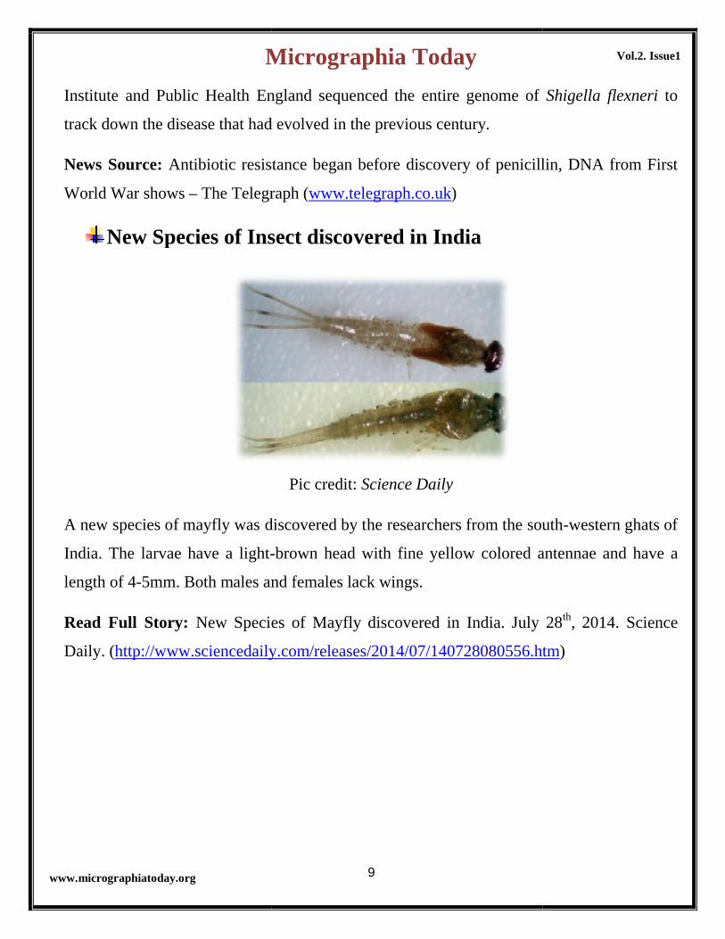
New Species of Insect discovered in India
A new species of mayfly was discovered by the researchers from the south
India. The larvae have a light-
length of 4-5mm. Both males and females lack wings.
Read Full Story: New Species of Mayfly
Daily. (http://www.sciencedaily.com/releases/2014/07/140728080556.htm
Micrographia Today
9
Institute and Public Health England sequenced the entire genome of
track down the disease that had evolved in the previous century.
Antibiotic resistance began before discovery of penicillin, DNA from First
Telegraph (www.telegraph.co.uk)
New Species of Insect discovered in India
Pic credit: Science Daily
f mayfly was discovered by the researchers from the south
-brown head with fine yellow colored antennae and have a
5mm. Both males and females lack wings.
New Species of Mayfly discovered in India. July 28
http://www.sciencedaily.com/releases/2014/07/140728080556.htm
Vol.2. Issue1
Institute and Public Health England sequenced the entire genome of Shigella flexneri to
Antibiotic resistance began before discovery of penicillin, DNA from First
f mayfly was discovered by the researchers from the south-western ghats of
antennae and have a
discovered in India. July 28th, 2014. Science
http://www.sciencedaily.com/releases/2014/07/140728080556.htm)

Micrographia Today
10
Vol.2. Issue1
www.micrographiatoday.org
Biggest Contributions from Scientific Communicators of 2014Science Journalism has now emerged with new essence of presenting science by opening the windows of the closed Laboratories to the readers. Enhancing with understanding about new scientific research advances here are some highlights of the biggest contributions from Science Communicators around the globe including India.
Scientific research is advancing day by day, and more than that scientific communication has made readers from different background to gain interest as well as sufficient knowledge based on varied subjects of Science. In 2014, among the vast contributions here are some which you would like to look back and read again.
Top Indian Young Scientific Contributors
1. Mr. Suman Bhattacharya
Microbiology Student from St. Xavier’s College Kolkata, India has expanded the platform of learning Biology into ‘e-Learning’. Students like to follow learning biology in more animated form rather than covering books.
Mr. Suman has created that platform which helped many students to learn and understand biology which are quite unexplained in books. “Shomu’s Biology”, as it is named has 1800 recorded videos on different topics on Biology and
most of them can be found in youtube.
www.shomusbiology.com
2. Mr. Varun C. N.
‘Facts that are interesting in recent time’ is what Mr. Varun has highlighted through his blog Microboids. Research Scholar from Department of Neuromicrobiology, NIMHANS, Bangalore, India is a wonderful scientific contributor explaining the current status of Medical Microbiology by curving out the recent news of research.
E-learning is all about podcasting the science, which he believes to be helpful for the readers to know and understand more than what is always explained in regular university syllabus. The Blog “Microboids” is now has reached a well ranked page with wide viewers maintaining the webtitle ‘Sharing Knowledge improves knowledge’.
www.microboids.net

Micrographia Today
www.micrographiatoday.org
Top foreign Scientific Contributors
Tim Sandle
A professor, a guide and also a well known science journalist contributing scientific readings on Pharmaceutical Microbiology through his own blog named Pharmaceutical Microbiology. Micrographia Today itself had the opportunity to interview him whi
Beyond his blog posts he is a committee member of the Pharmaceutical Microbiology Interest groups called Pharmig, even serves for National Blood Service Advisory Cleaning and Disinfection committee and is member of editorboards of several journals.
http://www.timsandle.com/
Carl Zimmer
When you are reading writings by Carl Zimmer, you must grab a cup of coffee and sit before your computer for New York Times columnist every Thursfamous scientific writing he is a popular speaker at different Universities and Schools.
Mr. Zimmer is an author of 12 books, among which “The Tangled Bank: An Introduction to Evolution” was the first book published in 2009. Apart from New York Times columnist he is also a science journalist at National Geographic, Time, Scientific American, Science and Popular Science. Being one of the excellent contribution towards science, he a lecturer on Writing about Science at Yale University.
http://carlzimmer.com/
Dr. Vincent Racaniello
When you talk about Dr. Vincent the word that first comes from your mouth is “The Godfather of Virology”. Students, researchers and authors talk about Viruses but Dr. Vincent stands Micrographia Today must be mentioned to be enough lucky to have his words mentioned in this magazine (check the Interview section of the Magazine).
A Higgins professor at Columbia University, bloggmultiple weekly podcasts on Microbiology, Virology and Parasitism commonly
known as TWiM, TWiV and TWiP.
www.virology.ws
Micrographia Today
11
Top foreign Scientific Contributors
A professor, a guide and also a well known science journalist contributing scientific readings on Pharmaceutical Microbiology through his own blog named Pharmaceutical Microbiology. Micrographia Today itself had the opportunity to interview him which was published in Vol.1 Issue 2.
Beyond his blog posts he is a committee member of the Pharmaceutical Microbiology Interest groups called Pharmig, even serves for National Blood Service Advisory Cleaning and Disinfection committee and is member of editorboards of several journals.
When you are reading writings by Carl Zimmer, you must grab a cup of coffee and sit before your computer for New York Times columnist every Thursfamous scientific writing he is a popular speaker at different Universities and
Mr. Zimmer is an author of 12 books, among which “The Tangled Bank: An Introduction to Evolution” was the first book published in 2009. Apart from New
Times columnist he is also a science journalist at National Geographic, Time, Scientific American, Science and Popular Science. Being one of the excellent contribution towards science, he a lecturer on Writing about Science at Yale
When you talk about Dr. Vincent the word that first comes from your mouth is “The Godfather of Virology”. Students, researchers and authors talk about Viruses but Dr. Vincent stands tall for giving new dimension to understand about viruses. Micrographia Today must be mentioned to be enough lucky to have his words mentioned in this magazine (check the Interview section of the Magazine).
A Higgins professor at Columbia University, blogger at virology.ws and hosts multiple weekly podcasts on Microbiology, Virology and Parasitism commonly
Vol.2. Issue1
A professor, a guide and also a well known science journalist contributing scientific readings on Pharmaceutical Microbiology through his own blog named Pharmaceutical Microbiology. Micrographia Today itself had the opportunity to
Beyond his blog posts he is a committee member of the Pharmaceutical Microbiology Interest groups called Pharmig, even serves for National Blood Service Advisory Cleaning and Disinfection committee and is member of editorial
When you are reading writings by Carl Zimmer, you must grab a cup of coffee and sit before your computer for New York Times columnist every Thursday. Beyond famous scientific writing he is a popular speaker at different Universities and
Mr. Zimmer is an author of 12 books, among which “The Tangled Bank: An Introduction to Evolution” was the first book published in 2009. Apart from New
Times columnist he is also a science journalist at National Geographic, Time, Scientific American, Science and Popular Science. Being one of the excellent contribution towards science, he a lecturer on Writing about Science at Yale
When you talk about Dr. Vincent the word that first comes from your mouth is “The Godfather of Virology”. Students, researchers and authors talk about Viruses
tall for giving new dimension to understand about viruses. Micrographia Today must be mentioned to be enough lucky to have his words mentioned in this magazine (check the Interview section of the Magazine).
er at virology.ws and hosts multiple weekly podcasts on Microbiology, Virology and Parasitism commonly

Micrographia Today
12
Vol.2. Issue1
www.micrographiatoday.org
INVENTION Column Killer Bacteria: V. cholerae
Researchers from Ecole Polytechnique Fédérale de Lausanne identified how Vibrio cholerae bacterium can kill other bacteria to pirate the DNA, which makes them potentially virulent. It has a spring loaded spear called type VI secretion system T6SS, a predatory killing device
used to compete against other bacteria. With the availability of chitin, bacteria moves toward to survival mode called natural competence, where it attacks other bacteria with the spear system. This mode of acquisition allows them to absorb around 40 gene fragments which are commonly known as horizontal gene transfer. The research was published in the journal Science, Jan 2015.
[http://dx.doi.org/10.1126/science.1260064]
Vaccine against ‘Mad-cow’ like disease in deer discovered
A research published in the journal Vaccine, Dec 2014 where the researchers from New York University School of Medicine developed a vaccine against brain-based wasting syndrome in deer. Researchers say that this discovery can allow them to investigate against similar disease called kuru.
The team made the vaccine using Salmonella bacteria which easily enters the gut. The attenuated bacteria with inserted prion like protein allow them to enter the gut. This finally allows to develop anti-prion antibodies.
[http://dx.doi.org/10.1016/j.vaccine.2014.11.035]
DO You Know?
Horizontal gene transfer is the main primary reason for the bacterial antibiotic resistance. It plays an important role in bacterial evolution by maintaining the transmission of virulence factors.
The two major components of Horizontal gene transfers are bacteriophages and plasmids. Most of the research had mentioned about vertical gene transfer, but horizontal gene transfer has increased an important growing research to understand the dominant form of gene transfer. Nowadays artificial gene transfer has been an integral part of research where looking into specific gene expression in bacteria.
Mad Cow disease is also known as Bovine Spongiform Encephalopathy (BSE) which causes slow progressive fatal disease affecting central nervous system of adult cattle.
The disease is caused due to the abnormal version of protein called prions. A human version of the disease called Creutzfeldt-Jakob disease (vCJD) was known to cause by eating beef products (contaminated).

Micrographia Today
13
Vol.2. Issue1
www.micrographiatoday.org
AUTHORS SPEAK
16S rRNA and the Strategy Used in Bacterial Identification
Saumyadip Sarkar
Department of Human Genetics, Institute of Life Sciences, Bhubaneswar, India
Unlike a few decades back, microbiologists are now well aware of 16S rRNA sequencing, a
useful tool now widely employed in the identification of microorganisms. The 16S rRNA
which is a component of the smaller subunit of prokaryotic ribosome commonly used in
phylogenies was discovered by Dr. Carl Woese and George E. Fox.
Ribosome, a major protein complex helps in protein synthesis
(translation) and consists of two units; the larger (50S) and
the smaller subunit (30S), together making the complete 70S
[where S stands for the Svedberg Unit, a non-S.I unit for
sedimentation rate]. The 16S rRNA which has the signature
sequence for the bacterial identification is found in the
smaller 30S subunit.
Usually, multiple sequences are present in 16S rRNA of a
single bacterium. However, signature sequences which are
specific conserved sequences are always found in all groups
of organisms. These sequences which are described as unique
are present in the 16S rRNA region, are about 5-10bases long

Micrographia Today
14
Vol.2. Issue1
www.micrographiatoday.org
and help in the identification of prokaryotic organisms, archea and eukarya. The average
length of the 16S rRNA gene is 1522bp.
In the past, conventional microbiological methods such as cultural and morphological
characterizations and biochemical tests were the major tools used for laboratory
identification of bacterial isolates- these are, however, being replaced or complemented
with more modern diagnostic techniques. For example, nucleic acid detection method has
overcome the limitations in conventional microbiological methods. [1] It has also been
suggested that NGS-based 16S rRNA sequencing is a more cost effective technique for the
bacterial identification than conventional microbiological methods.
Basic facts about 16S rRNA as a tool for bacterial identification:
1. Ribosomes containing 16S rRNA are present in all cells.
2. RNA genes are highly conserved.
3. Nucleic Acid detection method is an efficient technique for bacterial identification.
Universal Primers used for 16S rRNA sequencing [2]:
Primer Name 5’ to 3’ Sequence
8F AGA GTT TGA TCC TGG CTC AG
U1492R GGT TAC CTT GTT ACG ACT T
928F TAA AAC TYA AAK GAA TTG ACG GG
336R ACT GCT GCS YCC CGT AGG AGT CT
1100F YAA CGA GCG CAA CCC
1100R GGG TTG CGC TCG TTG
337F GAC TCC TAC GGG AGG CWG CAG
907R CCG TCA ATT CCT TTR AGT TT
785F GGA TTA GAT ACC CTG GTA

Micrographia Today
15
Vol.2. Issue1
www.micrographiatoday.org
805R GAC TAC CAG GGT ATC TAA TC
533F GTG CCA GCM GCC GCG GTA A
518R GTA TTA CCG CGG CTG CTG G
27F AGA GTT TGA TCM TGG CTC AG
1492R CGG TTA CCT TGT TAC GAC TT
[2] Based on the availability of large number of 16S rRNA sequences, along with NCBI’s
databases, there are multiple other databases which are widely used. They include:
1. EzTaxon-e contains complete hierarchical taxonomic structures (from phylum rank to
species rank) for both the domains of bacteria and archaea. http://eztaxon-
e.ezbiocloud.net/
2. Ribosomal Database Project. http://rdp.cme.msu.edu/
3. SILVA provides comprehensive, quality checked and regularly updated datasets of
aligned small (16S/18S, SSU) and large subunit (23S/28S, LSU) ribosomal RNA
(rRNA) sequences for all three domains of life (Bacteria, Archaea and Eukarya).
4. Greengenes. The greengenes web application provides access to the 2011 version of
the greengenes 16S rRNA gene sequence alignment for browsing, blasting, probing,
and downloading.
References
1. 16S Ribosomal RNA Sequencing. Microbiology Virtual Lab, Amrita University Data.
http://amrita.vlab.co.in/?sub=3&brch=76&sim=1421&cnt=1
2. 16S Ribosomal RNA – Wikipedia.http://en.wikipedia.org/wiki/16S_ribosomal_RNA
Micrographia Today
16
Vol.2. Issue1
www.micrographiatoday.org
COVER STORYVACCINES
A General OverviewBamgbose Timothy
Mysore University, Mysore, India
A vaccine is a non-pathogenic antigen that mimics a particular pathogen in order to elicit an immune response as if that actual pathogen were in the body.Vaccination (Latin: vacca—cow) is so named because the first vaccine was derived from a virus affecting cows—the relatively benign cowpox virus—which provides a degree of immunity to smallpox, a contagious and deadly disease.The word vaccination was first used by Edward Jenner in 1796 and Louis Pasteur furthered the concept through his pioneering work in microbiology. Let us see briefly different types of vaccines:
LIVE VACCINESLive vaccines are prepared from live organisms. These organisms are passed through chick embryos or other such media repeatedly and they retain the capacity to trigger off the defence mechanism. Live vaccines are usually more potent than inactivated vaccines. They multiply within the host and produce more antigens. Ex: Polio, Flu shot
INACTIVATED VACCINESThis type of vaccines cannot replicate and generally not as effective as live vaccines. It has less interference from circulating antibody than live vaccines. Generally requires 3-5 doses and Immune response mostly humoral antibody titer may diminish with time hence booster dose always required.
WHOLE KILLED VACCINESThe preparation is simply inactivation; the outer coat is left intact while the replicative function is destroyed. Preparation of killed vaccines may take the route of

Micrographia Today
17
Vol.2. Issue1
www.micrographiatoday.org
heat or chemicals. Excessive treatment can destroy immunogenicity whereas insufficient treatment can leave infectious microbe capable of causing disease.Ex: Typhoid, Cholera
TOXOIDSome species of bacterial produce what is known as exotoxins. Toxoids are vaccines which consist of exotoxins that have been inactivated, either by heat or chemicals. These vaccines are intended to build immunity against the toxins, but not necessarily the bacteria that produce the toxins.
Ex: Botulinum antitoxin, Diphtheria antitoxin.
SUB-UNITVaccines that use a component of the pathogen rather than the whole organismEx: Influenza A and B, Herpes simplex virus, Foot and mouth disease
POLYSACCHARIDE BASEThe virulence of some pathogenic bacteria depends primarily on the anti-phagocytic properties of their hydrophilic polysaccharide capsules. TWO TYPES: PURE POLYSACCHARIDE VACCINES
Coating of the capsule with antibodies or complement greatly increases the ability of the macrophages and the neutrophils to phagocytise such pathogens. Ex: Pneumococcal, Meningococcal, Salmonella typhi (Vi)
CONJUGATE POLYSACCHARIDE Some bacteria have an outer coating of sugar molecules (called polysaccharides) Conjugation is the process of attaching the polysaccharide antigen to a protein carrier that the infant’s immune system already recognises in order to provoke an immune responseEx: Haemophilus influenzae type b
RECOMBINANTS VACCINESRecombinant technology is having the potential to give safer and perhaps the only means of developing safe and effective vaccines. Existing vaccines have all the

Micrographia Today
18
Vol.2. Issue1
www.micrographiatoday.org
antigens from pathogens but in r-DNA technology, a proper antigen and its concerned gene are introduced into an organismEx: Hepatitis –B vaccine
ANTI IDIOTYPESIn this unique type of vaccine, antibodies from sick individuals are isolated. These antibodies are then injected into a lab animal, which may then produce an antibody whose antigen binding site mimics the epitope that the original antibody binds to.These antibodies are then isolated and injected into a healthy individual, who may produce antibodies with an antigen binding site that binds to the antigen binding site of the animal antibodies.Because the animal binding site resembles the epitope of an antigen on a particular pathogen, the individual will have immunity against that pathogen.
DNA AS VACCINESDNA vaccines consist of plasmids that contains genes for certain types of antigens. Once administered, the plasmid is taken up by the target cell and the genes are expressed. The cell then either excretes the antigen or displays it on an MHC-I molecule.
CHIMERIC VACCINESChimeric vaccines usually consist of attenuated viruses that have been engineered to carry antigens from multiple types of pathogens.For example, the yellow fever vaccine YF17D has been engineered to carry antigens from HIV, different types of bacteria, malaria, even cancer.The main aim of a chimeric vaccine is the establishment of immunity against several different diseases with one administration
SOME OF THE CURRENT VACCINES IN USECholera: Crude fraction of Vibrio choleraTuberculosis: Attenuated M .tuberculosisHaemophilus meningitis: Purified polysaccharideMeningococcal meningitis: Purified polysaccharidePertussis: Killed Bordetella Capsular Pneumonia ( by Streptococcus pneumonia): Mixture of 23 different capsular polysaccharides

Micrographia Today
19
Vol.2. Issue1
www.micrographiatoday.org
Hepatitis B: Subunit VaccinesMeasles: Inactivated Virus
VACCINES SUCCESS STORY COMPLETE ERADICATION OF SMALLPOX
W.H.O. PREDICTION: ERADICATION OF PARALYTIC POLIO THROUGHOUT THE WORLD.
SIGNIFICANT REDUCTION OF INCIDENCE OF DISEASES: DIPTHERIA, MEASLES, MUMPS, PERTUSSIS, RUBELLA,POLIOMYELITIS, TETANUS
FUTURE FORECAST SEARCH FOR EFFECTIVE VACCINES FOR DISEASES LIKE: EBOLA.
MALARIA, TUBERCULOSIS AND AIDS
IMPROVEMENT IN SAFETY AND EFFICACY OF PRESENT VACCINES
EFFICIENT DELIVERY TO NEEDY
REDUCTION OF ADVERSE SIDE EFFECTS
FOR FURTHER READINGBooks:
Janet S. Butel, Stephen A. Morse,Geo. F. Brooks Jawetz, Melnick and Adelberg’s Medical Microbiology (1998) 21st Ed. Prentice Hall Inter. Inc PG. No. 145-176.
Lansing M. Prescott, Hohn P. Harely, Donald A. Klein Microbiology (2005), 6th Ed. Mc Graw-Hill Companies. Inc (USA). Pg. No. 779-789.
George F. Brooks, Karen C. Carroll, Janet S. Butel, Stephen A. Morse. (2007). Jawetz ,Melnick Adelbergs Medical microbiology, 24th ed. McGraw-Hill Professional.
Websites:
wiki.answers.com
www.policyalman ac.org
www.science daily.com
www.springerlink.com
www.astm.org
Library.thinkquest.org

Micrographia Today
www.micrographiatoday.org
MICROBES FOR ENVIRONMENTSoil Microbes Will Bring the Climate
Researchers from the University of Arizona discovered soil microbe which can drive the global climate via gas exchange between the soil and the atmosphere. Now these tiny invisible creatures are mere slaves of the environment or influential mediators are the open questions. This single species microbe was discovered recently, was an unexpectplayer in climate change.
The wet areas show where the mire has thawed out completely.( at Sweden)
The research was published in the journal climate by how these microbes can control the overflowing emission of greenhouse gases. Earlier in this year, the same team of researchers a single species of the microbe which has remained undiscovered can able to twere isolated from the permafrost soils of Sweden. Researchers has suspected that the same microbe plays specific role in global warming by emitting vast amount of carbon in the form of methane (a common powerful greenhouse gas known to trap large amounts of heat in earth’s atmosphere). The bacteria were named as which translates as “methane-bloomer from the Stordalen Mire”.
Micrographia Today
20
MICROBES FOR ENVIRONMENTSoil Microbes Will Bring the Climate
ChangeResearchers from the University of Arizona discovered soil microbe which can drive the global climate via gas exchange between the soil and the atmosphere. Now these tiny invisible creatures are mere slaves of the environment or influential mediators are the open questions. This single species microbe was discovered recently, was an unexpect
The wet areas show where the mire has thawed out completely.( at Sweden)
The research was published in the journal Nature, help scientists to improve the future climate by how these microbes can control the overflowing emission of greenhouse gases. Earlier in this year, the same team of researchers a single species of the microbe which has remained undiscovered can able to thaw under globally rising temperatures. The microbes were isolated from the permafrost soils of Sweden. Researchers has suspected that the same microbe plays specific role in global warming by emitting vast amount of carbon in the
powerful greenhouse gas known to trap large amounts of heat in earth’s atmosphere). The bacteria were named as Methanoflorens stordalenmirensis,
bloomer from the Stordalen Mire”.
Vol.2. Issue1
MICROBES FOR ENVIRONMENTSoil Microbes Will Bring the Climate
Researchers from the University of Arizona discovered soil microbe which can drive the global climate via gas exchange between the soil and the atmosphere. Now these tiny invisible creatures are mere slaves of the environment or influential mediators are the open questions. This single species microbe was discovered recently, was an unexpected key
The wet areas show where the mire has thawed out completely.( at Sweden)
, help scientists to improve the future climate by how these microbes can control the overflowing emission of greenhouse gases. Earlier in this year, the same team of researchers a single species of the microbe which has
haw under globally rising temperatures. The microbes were isolated from the permafrost soils of Sweden. Researchers has suspected that the same microbe plays specific role in global warming by emitting vast amount of carbon in the
powerful greenhouse gas known to trap large amounts of heat Methanoflorens stordalenmirensis,

Micrographia Today
21
Vol.2. Issue1
www.micrographiatoday.org
The exact role of these bacteria had remained unknown, but the new research has bloomed its function. The abundance of these Methanoflorens than other microbial community while thawing permafrost should provide the valid collective impact on the global climate change. All wetlands are not the same when it comes to methane release provides a question whether Methanoflorens resides in some whereas absent in others.
The different isotopes of Methane and their ratios are the major short comings of current climate change models. The assumption of the wrong isotope ratio would bring a wrong outcome of the over release of the carbon by biological organisms than the majorly caused by human activities, such as fossil-fuel burning. Soil microbes able to make methane by two different ways – the organic molecule form of acetate or in the form of carbon dioxide and water. In both the process, microbes able to produce energy by breathing out methane as we humans breathe out carbon dioxide. In this research, most of the methane has been estimated to come out not from the acetate rather the other way, while thawing the permafrost. Now the ratio shifts towards the frozen soils turned out to be wetlands.
Now the question comes how much carbon is released by these microbes into the atmosphere and do they provide the true climate change. As due to global warming the global freezer, the permafrost is failing and hence better understanding is still required on how these microbes release carbon on larger scale to change the worldwide ecosystem and bring forward the climate change.
Research:
Carmody K. McCalley, Ben J. Woodcroft, Suzanne B. Hodgkins, Richard A. Wehr, Eun-Hae Kim, Rhiannon Mondav, Patrick M. Crill, Jeffrey P. Chanton, Virginia I. Rich, Gene W. Tyson, Scott R. Saleska. Methane dynamics regulated by microbial community response to permafrost thaw. Nature, 2014; 514 (7523):478 DOI: 10.1038/nature13798.

Micrographia Today
22
Vol.2. Issue1
www.micrographiatoday.org
SCIENTIFIC REPORT
The Recent Ebola Virus Disease (EVD)
Outbreak in Nigeria KUNLERE Idowu Olagoke
Department of Microbiology (Environmental Microbiology Unit), University of Ibadan, Oyo State,
Nigeria.
Ebola: From the BluesWhen news first broke back in December 2013 about the outbreak of EVD in one of the West African countries, not many took it seriously, perhaps because they did not know how deadly it was. But by the first and second quarters of 2014, a mere few months down the line, with a steady jump in fatality rate, and the risk of its spread increasing with each passing day like wild fire, news about it was all over the place, everyone seemed to have come to realize that we had a developing humanitarian crisis on our hands that could spiral further, causing even more catastrophic consequences if left unchecked- this sudden awakening birthed an international response to the outbreak- but by the third quarter of 2014, the virus had spread to more countries including Nigeria, Spain and the United States leaving a wreck along its path.Ebola: Basic FactsEbola virus disease, EVD which was formerly known as Ebola haemorrhagic fever, is a severe, often fatal illness in humans. The virus is transmitted to people from wild animals (such fruit bats which are thought to be the natural host of the Ebola virus) and spreads in the human population through human-to-human transmission “by contact (through broken skin or mucous membranes) with the blood, or other body fluids or tissues (vomit, stools) of infected people or animals, whether alive or dead. It could also be contracted by handling, without protection, materials contaminated by secretions from an infected person, such as clothing, needles, phones or bed linen.According to WHO, this outbreak which is caused by a strain of Ebola virus has been noted to have a very close homology (98%) to the Zaire Ebola virus and has also been detected in breast milk, urine and semen; however, saliva and tears may also carry some risk. Since
Micrographia Today
www.micrographiatoday.org
viruses depend heavily on the cells of their host including the Ebola virus are not as resistant as bacteria are outside the body. Thus, Ebola does not survive in water; it is also not known to spread through the air. However, Ebola is a highly infectious disease and requires proper infection control practices to prevent health professionals and those who touch dead bodies during funerals from becoming inthemselves; when dealing with confirmed or suspected cases, the use of gloves, impermeable gowns, protective goggles or face shield and a face mask is advised, direct contact including physical touch and also contact with infectious droplets must be
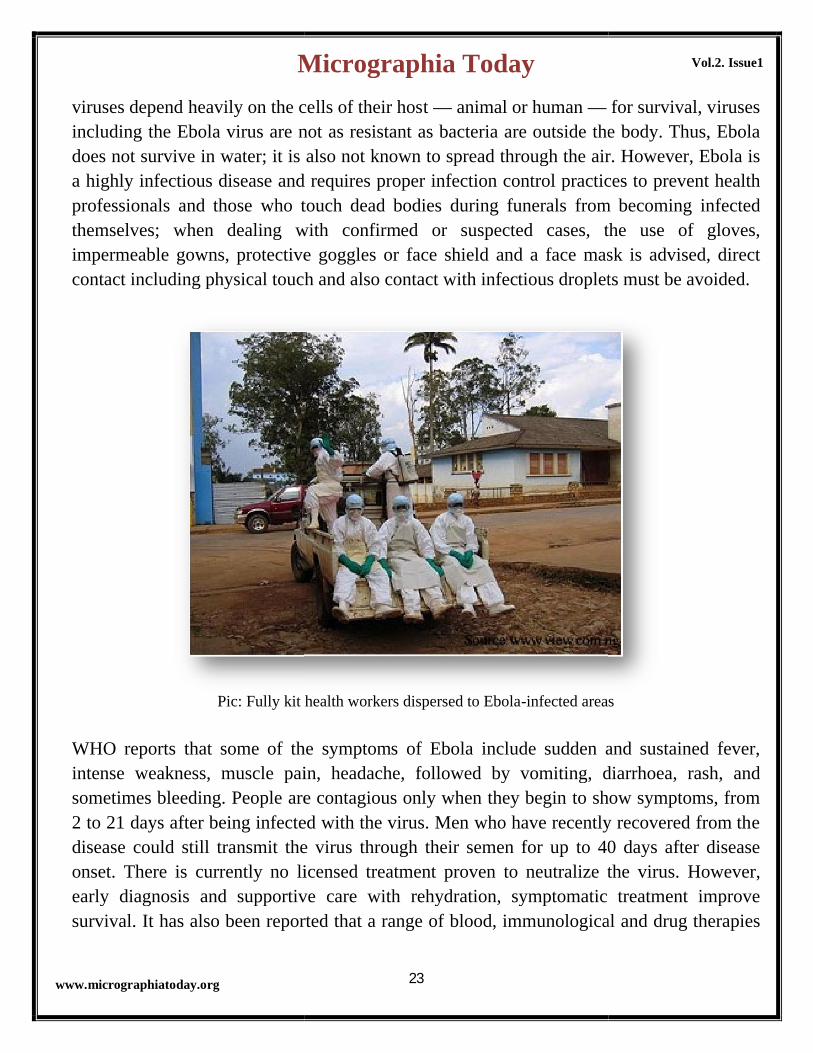
Pic: Fully kit health workers dispersed to Ebola
WHO reports that some of the symptoms of Ebola include sudden and sustained fever, intense weakness, muscle pain, headache, followed by vomiting, diarrhoea, rash, and sometimes bleeding. People are contagious only when they begin to show symptoms, from 2 to 21 days after being infected with the virus. Men who have recently recovered from the disease could still transmit the virus through their semen for up to 40 days after disease onset. There is currently no licensed treatment proven to neutralize the viruearly diagnosis and supportive care with rehydration, symptomatic treatment improve survival. It has also been reported that a range of blood, immunological and drug therapies
Micrographia Today
23
viruses depend heavily on the cells of their host — animal or human —including the Ebola virus are not as resistant as bacteria are outside the body. Thus, Ebola
survive in water; it is also not known to spread through the air. However, Ebola is a highly infectious disease and requires proper infection control practices to prevent health professionals and those who touch dead bodies during funerals from becoming inthemselves; when dealing with confirmed or suspected cases, the use of gloves, impermeable gowns, protective goggles or face shield and a face mask is advised, direct contact including physical touch and also contact with infectious droplets must be
Fully kit health workers dispersed to Ebola-infected areas
WHO reports that some of the symptoms of Ebola include sudden and sustained fever, intense weakness, muscle pain, headache, followed by vomiting, diarrhoea, rash, and sometimes bleeding. People are contagious only when they begin to show symptoms, from
o 21 days after being infected with the virus. Men who have recently recovered from the disease could still transmit the virus through their semen for up to 40 days after disease onset. There is currently no licensed treatment proven to neutralize the viruearly diagnosis and supportive care with rehydration, symptomatic treatment improve survival. It has also been reported that a range of blood, immunological and drug therapies
Vol.2. Issue1
for survival, viruses including the Ebola virus are not as resistant as bacteria are outside the body. Thus, Ebola
survive in water; it is also not known to spread through the air. However, Ebola is a highly infectious disease and requires proper infection control practices to prevent health professionals and those who touch dead bodies during funerals from becoming infected themselves; when dealing with confirmed or suspected cases, the use of gloves, impermeable gowns, protective goggles or face shield and a face mask is advised, direct contact including physical touch and also contact with infectious droplets must be avoided.
infected areas
WHO reports that some of the symptoms of Ebola include sudden and sustained fever, intense weakness, muscle pain, headache, followed by vomiting, diarrhoea, rash, and sometimes bleeding. People are contagious only when they begin to show symptoms, from
o 21 days after being infected with the virus. Men who have recently recovered from the disease could still transmit the virus through their semen for up to 40 days after disease onset. There is currently no licensed treatment proven to neutralize the virus. However, early diagnosis and supportive care with rehydration, symptomatic treatment improve survival. It has also been reported that a range of blood, immunological and drug therapies

Micrographia Today
www.micrographiatoday.org
are currently under development but more evidence from human studiesascertain safety and efficacy.Ebola in Nigeria: All hands on deckAs news of the virus spread, (and considering Nigeria’s proximity to other West African States and our indifference to adequate preparedness in the past), one could almost was only a matter of time before it finds its way to the Nigerian soil but no one could have rightly guessed that it would take a high“ambassador” named Patrick Sawyer from Liberia to willfully import the vishores. Since the first recorded Ebola case in the Nigeria on 24Ministry of Health, about 177 people were paced under health surveillance.
Pic: Health workers attending to an Ebola
Although Nigeria was able to mount a robust response (at the federal, state and local levels) led by the federal government to contain the Ebola outbreak, other West African countries with far lesser resources and populations were not as proactive. Mention must be made those whose prompt contributions helped Nigeria timely nipped the Ebola scourge in the bud including President Goodluck Jonathan and his team, Governors Babatunde Raji Fashola and Rotimi Amaechi of Lagos and Rivers States respectively and their teams ofexperts, security personnel, NGOs and particularly, health officials including those whose paid the supreme price.While the Nigeria has since been declared Ebolanormal but public awareness is still very high and public space. For once, it was inspiring to see the government at all levels in Nigeria take the initiative in frontally addressing a social threat facing the nation, this was unlike what
Micrographia Today
24
are currently under development but more evidence from human studies
Ebola in Nigeria: All hands on deckAs news of the virus spread, (and considering Nigeria’s proximity to other West African States and our indifference to adequate preparedness in the past), one could almost was only a matter of time before it finds its way to the Nigerian soil but no one could have rightly guessed that it would take a high-ranking, wicked, indiscreet and lawless “ambassador” named Patrick Sawyer from Liberia to willfully import the vishores. Since the first recorded Ebola case in the Nigeria on 24th July, 2014 by the Federal Ministry of Health, about 177 people were paced under health surveillance.
Health workers attending to an Ebola-infected patient
ia was able to mount a robust response (at the federal, state and local levels) led by the federal government to contain the Ebola outbreak, other West African countries with far lesser resources and populations were not as proactive. Mention must be made those whose prompt contributions helped Nigeria timely nipped the Ebola scourge in the bud including President Goodluck Jonathan and his team, Governors Babatunde Raji Fashola and Rotimi Amaechi of Lagos and Rivers States respectively and their teams ofexperts, security personnel, NGOs and particularly, health officials including those whose
While the Nigeria has since been declared Ebola-free by the WHO and life has returned to normal but public awareness is still very high and safety measures are still been observed in public space. For once, it was inspiring to see the government at all levels in Nigeria take the initiative in frontally addressing a social threat facing the nation, this was unlike what
Vol.2. Issue1
are currently under development but more evidence from human studies is still needed
As news of the virus spread, (and considering Nigeria’s proximity to other West African States and our indifference to adequate preparedness in the past), one could almost tell it was only a matter of time before it finds its way to the Nigerian soil but no one could have
ranking, wicked, indiscreet and lawless “ambassador” named Patrick Sawyer from Liberia to willfully import the virus to our
July, 2014 by the Federal Ministry of Health, about 177 people were paced under health surveillance.
ia was able to mount a robust response (at the federal, state and local levels) led by the federal government to contain the Ebola outbreak, other West African countries with far lesser resources and populations were not as proactive. Mention must be made of those whose prompt contributions helped Nigeria timely nipped the Ebola scourge in the bud including President Goodluck Jonathan and his team, Governors Babatunde Raji Fashola and Rotimi Amaechi of Lagos and Rivers States respectively and their teams ofexperts, security personnel, NGOs and particularly, health officials including those whose
free by the WHO and life has returned to safety measures are still been observed in
public space. For once, it was inspiring to see the government at all levels in Nigeria take the initiative in frontally addressing a social threat facing the nation, this was unlike what

Micrographia Today
25
Vol.2. Issue1
www.micrographiatoday.org
Nigerians had come to know and expect in the past when it was almost a misgiving for such prompt response and leadership to be expected from public officials. The Nigerian response to the EVD has been hailed worldwide as a standard.In Summary: The recent Ebola Virus Disease (EVD) Outbreak in Nigeria
1. On 24th July 2014 by the Federal Ministry of Health declared an outbreak of the Ebola Virus Disease (EVD) in Nigeria.
2. In combating the Ebola outbreak, the Federal Government inaugurated a National Ebola Emergency Operations Centre (NEEOC) led by the Incident Manager, employed an Incident Management System (IMS) to coordinate the response and consolidate decision making. The EOC/IMS structure was the overall implementing arm of the response for the Ministry of Health and the Nigerian Centre for Disease Control (NCDC). The NEEOC supported and provided orientation for the establishment of a Lagos and Rivers based State Ebola Emergency Operations Centre (SEEOC).
3. For operational effectiveness, UNICEF served as the team lead for the Social Mobilization group and the Management and Coordination group, WHO as the team lead for the Epidemiology/Surveillance group for the SEEOCs, Lagos State Ministry of Health as the team lead for Case Management and Information Control, Lagos University Teaching Hospital as the team lead for Social Mobilization and Laboratory Services, while the Federal Port Health Services oversaw Ports and Point of Entry
4. While the outbreak lasted, 19 cases were confirmed of which 7 deaths were recorded while 12 recovered and were discharged; that’s a case fatality rate of 40%. According to WHO estimates, the average case fatality rate in this recent outbreak worldwide is 70%.
5. Of the confirmed 17 cases, 11 were among health care workers which also accounted for 5 of the 7 deaths- this shocking statistics heightened the occupational hazards health workers face in the discharge of their duty.
6. The confirmed cases and contacts on Nigerian soil were in two of Nigeria’s most populated and economically viable states; Lagos and Rivers respectively.
7. Patients, family members of patients, contact persons and community members affected by EVD were provided psychosocial support.

Micrographia Today
26
Vol.2. Issue1
www.micrographiatoday.org
8. In order to halt the spread of new infections and stigmatization of contacts, education, training, massive public sensitization, household and interpersonal communication were employed amongst other tools.
9. Since no new case of EVD was recorded in Nigeria since 24th September, 2014, Nigeria was declared Ebola-free following the end of a 42-day contact tracing period.
ReferencesAsuzu, 2014. War against the Ebola Virus Disease in Nigeria: Counting Our Successes and Scaling Up On It.
Barbara Knust , 2014. Ebola Virus Disease Outbreak — West Africa, September 2014. MMWR / October 3, 2014 / Vol. 63 / No. 39, pp 865-866
Bruce Aylward, 24 October 2014. Ebola Outbreak in West Africa.
Claire Schaffnit-Chatterjee, 9 October 2014. Ebola: A human and economic catastrophe in West Africa
Ebola Response Roadmap: http://www.who.int/csr/resources/publications/ebola/response-roadmap/en/
Manji Cheto, 2014. West Africa: Economic Implications of Ebola Outbreak for Affected Countries.
WHO, 15 October 2014. Ebola Response Roadmap Situation Report
WHO, 2014. Ebola outbreak: http://www.who.int/csr/disease/ebola/en/
WHO, 2014. Ebola Virus Disease fact sheet: http://www.who.int/mediacentre/factsheets/fs103/en/
UNICEF, 24 September 2014. Nigeria Ebola Humanitarian Situation Report
UNMEER portal: http://www.un.org/ebolaresponse/mission.shtml
Shuaib et al., 2014. Ebola Virus Disease Outbreak — Nigeria, July–September 2014. MMWR / October 3, 2014 / Vol. 63 / No. 39, pp 867-872
Mirkovic et al., 2014. Importation and Containment of Ebola Virus Disease — Senegal, August–September 2014. MMWR / October 3, 2014 / Vol. 63 / No. 39, pp 873-875

Micrographia Today
www.micrographiatoday.org
A brief Introduction to Ti
mechanism to create genetically modified
Department of Human Genetics, Institute of Life Sciences, Bhubaneswar, India
Agrobacterium tumefaciens is known to contain a plasmid which can induce tumour to the
plants. The plasmid is known as Ti
small region of the plasmid is taken up into the chromosome of the plant. The genes present
in the Ti plasmid are known as phyto
growth [1].
The genes in the virulence region contain set of operons
code for enzymes responsible for transducing the gene into the chromosome of th
cell. [2]
virA: codes for a receptor that is able to react with the presence of phenolic
compounds.
Micrographia Today
27
A brief Introduction to Ti-plasmid and its
echanism to create genetically modified
plants
Saumyadip Sarkar
Department of Human Genetics, Institute of Life Sciences, Bhubaneswar, India
is known to contain a plasmid which can induce tumour to the
plants. The plasmid is known as Ti-plasmid (~250Kbp). After the bacteria infect plants, a
small region of the plasmid is taken up into the chromosome of the plant. The genes present
asmid are known as phyto-oncogenes that can induce tumour inducing neoplastic
The genes in the virulence region contain set of operons virABCDEFG
code for enzymes responsible for transducing the gene into the chromosome of th
codes for a receptor that is able to react with the presence of phenolic
Vol.2. Issue1
plasmid and its
echanism to create genetically modified
Department of Human Genetics, Institute of Life Sciences, Bhubaneswar, India
is known to contain a plasmid which can induce tumour to the
. After the bacteria infect plants, a
small region of the plasmid is taken up into the chromosome of the plant. The genes present
oncogenes that can induce tumour inducing neoplastic
virABCDEFG which is able to
code for enzymes responsible for transducing the gene into the chromosome of the plant
codes for a receptor that is able to react with the presence of phenolic

Micrographia Today
28
Vol.2. Issue1
www.micrographiatoday.org
virB: codes for proteins that can form pilus like structure.
virC: able to bind overdrive sequence.
virD1 and virD2: produce endonucleases that target the T-DNA segment
virD4: coupling protein
virE: binds to T strand protecting it from nuclease attack.
virG: activates vir gene expression while binding to the consensus sequence, after
being phosphorylated by virA.
Crown Gall Disease
The tumours caused by the bacteria Agrobacterium tumefaciens is the causative agent of
Crown Gall Disease. It is a collection of uncontrolled cells causing a tumour in plants. The
tumour is usually seen in the wounded sites. Exposure of the plants towards these bacteria
containing the plasmid makes it heritable (through mitosis) changing plants cells to tumour.
The most important difference found between the normal plant cells and the crown gall
cells are that the crown gall cells do not phytochromes than in the case of normal plant
cells. These tumour cells can able to produce auxins and cytokinins for their growth as well
as large amount of opines (amino acids and sugar derivatives). [3]

Micrographia Today
29
Vol.2. Issue1
www.micrographiatoday.org
Regulation of Vir gene expression
Virulence genes (vir genes) are inducible and can be induced by the exposure of the
Agrobacterium tumefaciens to the wound of the plants. Many chemicals are known to
induce vir genes, specifically acetosyringone. Mutation in the virA and virG are pleiotropic,
i.e. they can affect the expression of all other genes. The virA gene product is able to
phosphorylate itself on a histadine residue and is able to transfer this phosphate to an
aspertate side chain of the product formed by virG. [3]
Ti plasmid and genetic engineering:
To introduce the Ti plasmid as a vector into the plants, the virulence region needs to be
disarm to prevent tumours, instead will inject the gene of interest. Researchers usually
accomplish this by deleting the region which encodes for auxin and cytokinin synthesis,
thereby introducing the gene of interest in that specific region creating a transformed cell.
Antibiotic resistant genes are also inserted to confirm its insertion.
Finally the cloned gene is inserted in the plasmid and this plasmid is then used to infect the
cultured plant cells using the bacteria Agrobacterium tumefaciens. The infected cells are
then grown in a medium containing the antibiotic with auxins and cytokinin which will help
to induce growth. Successfully transformed cells will be able to grow in the medium due to
the presence of the antibiotic resistant gene.
Various methods have been applied for converting these transformed cells into a whole new
plant containing the gene of interest. Multiple plants have been grown by researchers and
some has been opposed. The regeneration requires adjustment of the ratio of the auxin to
cytokinin to make proper growth of shoot and root.

Micrographia Today
30
Vol.2. Issue1
www.micrographiatoday.org
Fig: Ti plasmid and its use in the production of genetically modified tobacco plant
The proper transformation of the infected cells into new plants is a big challenge for the
researchers and has got some remarkable success. Numerous foreign genes have been
introduced to develop modified plants like tobacco with soyabean protein storage genes.
Some of the desirable characteristics like disease resistant, salt resistant, herbicide resistant
genes have been introduced into plants and are recently commercial crops are grown which
are genetically engineered. [1]
Summary
Ti plasmid is tumour inducing plasmid carried by the bacterium Agrobacterium
tumefaciens. The ~250Kbp plasmid contain virulence genes that control the plasmid. The
bacterium comes in contact with the plant through wounds and the plasmid is transferred
into the chromosome which becomes heritable to induce Crown gall disease in successive
generations of the plant.

Micrographia Today
31
Vol.2. Issue1
www.micrographiatoday.org
Genetic engineering tools has made the researchers to omit the auxins and the cytokinins
and replacing with a gene of interest, thereby creating a genetically modified plant instead
of causing tumors in plants.
References:
1. Lincoln, T., Eduardo, Z. Plant Physiology Fifth Editon Online. Topic 21.7: The Ti
Plasmid and Plant Genetic Engineering. (http://5e.plantphys.net/article.php?id=216)
2. Stachel, S.E., Nester, E.W., (1986). The genetic and transcriptional organization of
the vir region of the A6 Ti plasmid of Agrobacterium tumefaciens. The EMBO
journal 5(7): 1445-1454.
3. Lecture Notes. University of Kentucky.
(http://www.uky.edu/~aghunt00/PLS620/notes/)
Further Study:
Check video on Ti-Plasmid:
http://highered.mheducation.com/olcweb/cgi/pluginpop.cgi?it=swf::535::535::/sites/dl/free/
0072437316/120078/bio40.swf::The+Ti+Plasmid

Micrographia Today
32
Vol.2. Issue1
www.micrographiatoday.org

Micrographia Today
33
Vol.2. Issue1
www.micrographiatoday.org
Updates on EBOLA Outbreak
No signal of EBOV adapting to Humans
EBOV (Ebola Virus) after the discovery in 1970 at regular intervals known to cause
zoonotic outbreaks in human populations. The current outbreak has shown the sustained
transmission with exponential threat for humans. Gire et al., has published 78genomes of
EBOV samples in May and June from Sierra Leone. The data suggested a rapid
accumulation of genetic variation, where the substitution rate is twice as high within 2014.
It is clear that mutations are being frequent in EBOV but there till now no concrete
evidence based on its rapid evolution. Based on the current status from the current outbreak,
81 genomes has been published with 29 unique sequences and no genome contain
2nonsynomous mutation. An article published in the Journal Biorxiv, where researchers
aligns all the genetic evidences on EBOV including the outbreak since 1976 showed limited
evolutionary divergence. The EBOV now was concluded by the researchers to be single
polymorphic population. The sequence similarities were 99.84%. The differences found in
the sequences are fixed substitutions rather segregating polymorphisms.
Research Source: Stephanie J. Spielman, Austin G. Meyer and Claus O. Wilke 2014.
Current data show no signal of Ebola virus adapting to humans. Biorxiv
http://dx.doi.org/10.1101/011429.

Micrographia Today
34
Vol.2. Issue1
www.micrographiatoday.org
Ebola-infected doctor to arrive in Nebraska from Sierra Leone
Dr. Martian Salia, a permanent US
citizen from Sierra Leone contacted
Ebola while treating patients, arrives in
Nebraska for treatment. He is the sixth
doctor who fell sick with the disease in
Sierra Leone and unfortunately the
other five died. Experts suggest that
western countries have better treatment
facilities with advanced healthcare systems make better success in the treatment of Ebola
virus disease. Dr. Salia will be the third patient to be treated in Nebraska medical center
with high equipped bio-containment units and with hired staffs who can deal with deadly
infectious diseases.
Full Story: Ebola-infected doctor to arrive in Nebraska from Sierra Leone. Lauren
Gambino report, 15 November 2014. The Guardian
(http://www.theguardian.com/world/2014/nov/15/ebola-infected-doctor-to-arrive-in-
nebraska-from-sierra-leone)
Democratic Republic of Congo now free from Ebola
Fig: A health worker sprays a colleague with disinfectant during a training session for Congolese health workers to deal with Ebola virus in Kinshasa, Democratic Republic of Congo

Micrographia Today
35
Vol.2. Issue1
www.micrographiatoday.org
Three months continuous outbreak comes to an end after 42days without any new reported
case of the outbreak in the Democratic Republic of Congo. Among 66patients reported with
the disease 49patients have died and after last registered record in October 4th, 2014 no such
case reports has been identified. Comparing with the West-African outbreak in Guinea,
Liberia and Sierra Leone; Democratic Republic of Congo’s outbreak has got little access
towards the disease.
Full Story: Congo declares its Ebola outbreak over. Reuters 15th November 2014. Thomson
Reuters Foundation. (http://www.trust.org/item/20141115132253-
sg167/?source=jtOtherNews1)
Ebola Virus Factsheet and reports published by US-AID
Suspected and Confirmed cases by 12th November 2014: 14,068 (reported by World
Health Organization).
Nations Cases
Liberia 6,822
Sierra Leone 5,368
Guinea 1,878
On November 7th, WHO has released a new burial practice to maintain religious rites
and safe enough with the deceased bodies.
GoS announced to plan for re-opening commercial flights to and from Liberia and
Sierra Leone to release.

Micrographia Today
36
Vol.2. Issue1
www.micrographiatoday.org
Read full: West Africa – Ebola Outbreak –Factsheet 7 (FY 15). November 12, 2014.
(http://www.usaid.gov/ebola/fy15/fs07)
The Plans to help children and families suffering from Ebola.
Plan is on the ground fighting the outbreak in communities across Guinea, Liberia and
Sierra Leone. The responsibilities undertook by PLAN International Organization:
Running public health information campaigns - including radio broadcasts
Providing medical kits
Distributing food aid
Training health workers in effective infection control procedures
Setting-up hand washing stations at schools, health posts and other public
facilities to help keep families safe.
Read Full: Ebola Outbreak Appeal. PLAN International. (http://plan-
international.org/what-you-can-do/emergency-appeals/ebola-outbreak-appeal) .

Micrographia Today
37
Vol.2. Issue1
www.micrographiatoday.org
Journey of Virology with Dr. Vincent Racaniello
Dr. Vincent Racaniello is the Higgins Professor of Microbiology and Immunology at the college of Physicians and Surgions of Columbia University. Along with his academic research, he is known for expanding knowledge with great contributions through his virology blog virology.ws, and his wide podcast on excellent communication on Biological Sciences like This Week in Virology (TWiV), This Week in Microbiology (TWiM) and This Week in Parasitism (TWiP).
On behalf of Microbiology World, Mr. Saumyadip Sarkar has communicated Dr. Vincent and shared his wonderful scientific journey.
Mr. Saumyadip: Dr. Vincent Racaniello, the famous scientific contributor leading about viruses through various aspect like your own The Virology blog www.virology.ws, This Week in Virology (www.twiv.tv), your youtube channel , even in The American Society of Microbiology’s The Microbe World (www.microbeworld.org) and also through some social networking sites. We would like to walk along to your scientific
journey but would like to start from your early childhood. How you used to take science as?
Dr. Vincent: With a Father who was a surgeon and a Mother who was a high school English teacher, I grew up surrounded with the understanding that knowledge was the key to a career. I loved all my science subjects, but a high school advanced biology class solidified my interest. It was taught by someone who had done some research and could convey
to us how it was done. However upon entering college the pressure was applied to follow in my father’s footsteps. I majored in biology but after 4 years had no laboratory experience and graduated without a plan. I returned home and secured a job as a laboratory technician. During that time I read ‘Fever’ by John Fuller, the account of the discovery of Lassa virus. This booksparked my interest in virology. Shortly thereafter, at a dinner at the home of one of my college friends, I

Micrographia Today
www.micrographiatoday.org
met Dr. Edwin Kilbourne, chair of Microbiology at Mt. Sinai School of Medicine in New York. He suggested that I apply to their department. I did, I was accepted, and there I entered the laboratory of Dr. Peter Palese.
Mr. Saumyadip: You have started your work on viruses since 1975. Your research was focused on Influenza viruses at Mt. Sinai School of Medicine of the City University of New York. Can you please reveal your work and understanding about this virus?
Dr. Vincent: When I entered Peter Palese’s laboratory in 1975 he had
Micrographia Today
38
just published a technique to map the RNA segments of influenza A viruses. This technique involved labeling viral RNAs in infected cells with 32P and fractionation of the 8 segments by gel electrophoresis. This technique for the time was
powerful, enabling analysis of viral reassortants and assignments of proteins to RNA segments. I wanted to apply the same approach to mapping the genomes of influenza B and C viruses. This formed the bulk of my Ph.D. thesis. While I was in Peter’s laboratory, recombinant DNA technology and nucleotide sequencing came of age. I learned how to sequence viral RNA and begin molecular cloning. During
my years there the 1976 swine flu outbreak at Fort Dix occurred. Peter’s lab used RNAse T1 fingerprinting to show that the virus was a swine virus, not a human strain, and not likely to spread in humans. The 1977 H1N1 pandemic also took place during my stay in that laboratory; Peter’s lab showed, using RNAse T1 fingerprinting, that this virus was very similar to a virus that had been circulating in the 1950s. How it had remained ‘hidden’ for so many years was unclear.
When I finished in Peter’s lab it wafuture was very bright for influenza virus, in terms of molecular approaches to study the virus’ evolution, antigenicity, and pathogenesis. While I planned to do my postdoctoral work on poliovirus, I had plans to return to influenza virus when starting my own laboratory. That never happened, because of my work in the Baltimore laboratory. Now I look back and wonder where I would be had I returned to influenza virus. Poliovirus
Vol.2. Issue1
my years there the 1976 swine flu outbreak at Fort Dix occurred. Peter’s lab used RNAse T1 fingerprinting to show that the virus was a swine virus, not a human strain, and not likely to spread in humans. The 1977 H1N1 pandemic also took place during my stay in that laboratory; Peter’s lab showed, using RNAse T1 fingerprinting, that this virus was very similar to a virus that had been circulating in the 1950s. How it had remained ‘hidden’ for so many years was unclear.
When I finished in Peter’s lab it was clear that the future was very bright for influenza virus, in terms of molecular approaches to study the virus’ evolution, antigenicity, and pathogenesis. While I planned to do my postdoctoral work on poliovirus, I had plans to return to influenza virus when starting my own laboratory. That never happened, because of my work in the Baltimore laboratory. Now I look back and wonder where I would be had I returned to influenza virus. Poliovirus

Micrographia Today
39
Vol.2. Issue1
www.micrographiatoday.org
has been very good to me –not only did I develop the first infectious DNA clone of an animal virus using poliovirus, but in my laboratory, we identified the cell receptor, and produced a transgenic mouse model for infection. It would be hard to equal these accomplishments in another viral system. Nevertheless there have been many amazing accomplishments in the field of influenza virus, including reconstruction of the 1918 H1N1 virus, tracing the origin of human strains in animals, and studies on receptor utilization and pathogenesis in animal models. I have to believe that I would have been part of some of these discoveries.
Mr. Saumyadip: After you have completed your PhD, you have received your postdoctoral fellowship from laboratory of Dr. David Baltimore at Massachusetts Institute of Technology, where you worked on polio viruses. How had been your PhD research help you for the
better understanding of the work on polio viruses?
Dr. Vincent: In Peter Palese’s lab I was taught how to be a virologist. I learn how to grow viruses in cells and in eggs, how to titrate infectivity by plaque assay, measure hemagglutination, purify viruses and viral RNA, and much more. This training was a perfect prelude to my stay in the Baltimore laboratory, where the members had far less training with virological techniques. Beyond mere techniques, I had learned how to plan experiments to understand problems in viral replication; I had a good understanding of the viral life cycle, of how viruses are constructed, and how the viral genome is replicated and decoded. To be sure, my training was not complete, and I learned a great deal more in the Baltimore laboratory. However my training with Dr. Palese was an essential foundation for what I did later.
Mr. Saumyadip: Moving a little ahead of your early
research. How you thought of approaching people with understanding of Viruses? People from different countries read your book “Principles of Virology”. How you gave a start to write about viruses?
Dr. Vincent: I had been working at Columbia University for over 15 years, doing research on poliovirus, when I was asked by a group of authors to join them in writing a virology textbook. These authors were all based in the area around Princeton NJ so that they could meet regularly and work on the book. I thought that this was a great opportunity to make an important contribution, so I joined the group. Working on ‘Principles of Virology’ was a tremendous learning experience. The first edition was published in 2000 and we are now working on the fourth edition. I think it is the correct way to present information about viruses, according to function, not by individual viruses.

Micrographia Today
www.micrographiatoday.org
Mr. Saumyadip: Columbia University is now well known for your own established laboratory in 1982. The start was with picornaviruses and now with varied research outlook over viral strategies. How does the thought came up? How Columbia University helped you behind establishing your goals?
Micrographia Today
40
Dr. Vincent: It is common for postdoctoral scientists to take the virus they work on to their own research lab, and that is what I did with poliovirus. Columbia University, in offering me a faculty position, provided a laboratory for me to work in, and money to start my laboratory. I was surrounded with excellent scientists, all Columbia faculty, who helped me get
started. And the students and postdocs whmy laboratory came in part because of Columbia. Without this stimulating and supportive environment, I would not have been able to do what I did.
Mr. Saumyadip You proved to be the star among scientists contributing
Vol.2. Issue1
started. And the students and postdocs who joined my laboratory came in part because of Columbia. Without this stimulating and supportive environment, I would not have been able to do what I
Mr. Saumyadip You proved to be the star among scientists contributing

Micrographia Today
41
Vol.2. Issue1
www.micrographiatoday.org
Microbiology in wide range in different audiences. Moreover you have been providing online courses on Virology under Coursera and providing latest research based on Viruses in your Blog. How you feel about the public approaches behind your way of sharing knowledge? How you started to communicate with public?
Dr. Vincent: The knowledge I gained in writing ‘Principles of Virology’ inspired me to begin blogging about viruses at virology.ws. I then used the textbook to develop a new undergraduate course in virology at Columbia University, in 2010. From this came my virology lectures, which can be found at my blog, virology.ws, at iTunes University, on YouTube, and on Coursera.org. Finally this lead to starting my first podcast, This Week in Virology. In reaching out to the public with information about viruses, I learned about social media, such as
Twitter, Facebook, Google Plus, and more. So in a way all my science outreach began with this virology textbook.
Mr. Saumyadip: Influenza virus had been your favorite topic for research. There are various pandemic viruses like Influenza and maybe there is multiple flu vaccine. The role model of each viruses works on various aspects, so what you think how a multiple flu vaccine can deal with the multiple viruses?
Dr. Vincent: I do think a universal influenza vaccine will be possible within the next 10 years. A number of different laboratories have found common, conserved sequences in a number of viral proteins, and these can be used to generate broadly protective immunity. I think in particular the stalk of the HA protein is a great immunogen for making broadly protective antibodies. One big problem that remains is how to induce the formation of broadly protective anti-influenza antibodies in people: they
are rarely made in natural infections. But the Palese lab and others have made good progress with different approaches, and I think this problem will eventually be solved. Even if a flu vaccine were needed every ten years, it would be a major improvement. However we also have to figure out how to make.
Mr. Saumyadip: The modern technologies nowadays really help researchers for the better understanding the role of viruses. Even 3D modeling and animations allow researchers to present and readers to understand the basic fact of virus strategies. How you feel that these technologies for you are efficient enough and what technology is still not yet fulfill your next step of the research?
Dr. Vincent: Technology has always allowed researchers to answer the important questions about their organisms. I always find it amazing to watch how technology grows over the years. For example, when I was a postdoctoral

Micrographia Today
42
Vol.2. Issue1
www.micrographiatoday.org
fellow with David Baltimore, DNA sequencing had just been developed. For the first time we were able to determine the sequences of genes and viruses. We thought that this was amazing. It took me one year, using Maxam-Gilbert sequencing, to determine the 7,442 nucleotide sequence of the poliovirus genome. One year! Today it would take less than an hour, because of next generation technologies that have been developed. Moreover we can now sequence entire animal genomes as well as complex microbial communities. This is only one example of how technology advances. Polymerase chain reaction, microarrays, RNA-seq, high resolution microscopy,CRIPSPR gene knockouts, and more, all make our experiments not only easier, but makes them possible. I don’t know what the next technology will be, but you can be assured that there will be new technologies developed routinely and it will help our research.
Mr. Saumyadip: Stepping little forward for the next generation virologists. You have been very friendly with the PhD students in your lab. Ashlee Bennett has been one who is well seen in the online course in Coursera, so would you like to introduce some of your PhD students working under you? Also would like to know how your students are enthusiastic over the research on viruses?
Dr. Vincent: I have two Ph.D. students in my laboratory. One is Ashlee Bennett who you know from the Coursera videos. Ashlee is working on a project to understand how zinc compounds inhibit rhinovirus infectivity. She is very enthusiastic about her work but also worried about having a future in science. She should be finished with her work in 2015, but is not sure of her next step. My other Ph.D. student is Michael Schreiber, who has been working to understand the blocks to rhinovirus replication in macrophages. Neither Michael nor Ashlee
will be pursuing careers in academic science, so it is safe to say that they will be pursuing alternative options. The third member of my laboratory is Amy Rosenfeld, who obtained her Ph.D. with me many years ago. She has returned to my lab to acquire additional data, which will enable her to obtain a faculty position. She has begun a project on enterovirus D68 to identify its cellular receptor and determine its neurotropic potential. More than anything else she wants to do scientific research.
Mr. Saumyadip: Now something little ahead of science background. People really want to know how you developed your interests over science and moreover would also like to know your life apart from science.
Dr. Vincent: I’ve described above in another section how I came to have a career in science. Here I’ll describe my life outside of science. This ‘outside’ life consists of my family: my wife Doris who is also a scientist, and our three

Micrographia Today
43
Vol.2. Issue1
www.micrographiatoday.org
children. Any spare time that I have – which is not much – I spend with them. Our oldest son, Aidan, is a sophomore at Stevens Institute of Technology where he is majoring in cybersecurity. Our other two children, Devin and Nadia, are juniors in high school. The do not yet know what they want to do with their lives. We live in a suburb of New Jersey on 2.5 acres of land with two dogs and a rabbit. My wife worked for Merck for 27years; this past year she joined a small company in North Carolina working on drugs for heart attacks. She spends three days a week working in North Carolina, and the rest of the time working from home. If you ask me if I have any hobbies, I would say no,because science and science communication is what I love to do. Most of my work on my blog and podcasts take place at home, at nights and on the weekends. When I was young, very young, I loved to build train layouts and race model cars, but I haven’t done those for years. There is no time, but I don’t regret that.
Mr. Saumyadip: Being the most well known virologists whom people from different research backgrounds want to follow, will like to know your words for the current and next generation researchers.
Dr. Vincent: If you want to be a virologist, or any kind of scientist, then I have some advice. First, you must be passionate about science. You must love it absolutely and be willing to spend most of your time doing it, and still loving it. You must not want to do anything else with your life – if you think you would like to be a writer, or a doctor, then do that. You also have to want to work hard, read extensively, think all the time, and communicate with others. Work hard in school and learn as much as you can. Major in science, then if possible get a Ph.D. in a scientific field. Do a postdoctoral fellowship in the best laboratory you can find. It’s a privilege to be a scientist, and only the most dedicated can have such a career. Science drives our world. We will always need
scientists to make the discoveries that make our lives better. So don’t be scared when people tell you there is no money to do science. It’s not true. It’s not easy, but there is money to do science out there. If you truly want to be a scientist, you can be one.
Mr. Saumyadip: Knowing enormous opportunities that abound in Microbiology but which remain relatively untapped, so how do we improve the interests of young intellectuals in Microbiology as a field of study in developing countries like India and Nigeria. [Selected question from the public] Would also like to know few words for We The Microbiologist.
Dr. Vincent: There is no better way to help and improve your country than to build the scientific infrastructure and cultivate good scientists. Let me use China as an example. A long time ago China realized that science was an important part of the country’s future. They sent Chinese students to the US,

Micrographia Today
44
Vol.2. Issue1
www.micrographiatoday.org
Europe, and other areas will well developed science programs, to obtain their Ph.Ds and do postdoctoral fellowships. Over the years China learned how to do excellent science and now they train many of their own scientists. We used to have hundreds of Chinese applicants for our Ph.D. program, and now we have none – they train in China. Of course, this building up of science in China took a lot of money. But India, Nigeria, and other countries can afford to build up their science programs. This process has to work from bottom to top – students have to tell their countries that they want to be scientists. This is easier than ever with communication afforded by the internet. Students in these countries should
communicate with scientists all over the world, to make themselves known (as you do) and bring notice to the world that your country wants to move forward in science. I can’t emphasize how important it is for you to work within your country to show how important science is for your future. The life of your citizens will be improved markedly, from living longer and more healthy, from having research that provides medicines, better food growing technology, energy, transportation, and much more. There is nothing better you can do than to encourage the development of science in your country.
We The Microbiologist is exactly what I was speaking of in the previous
paragraph, when I said that young scientists in all countries have to make themselves more visible to promote the development of science. You have made a great start, but don’t stop. Use the internet and social media to make yourself visible. Introduce yourselves to scientists in all fields everywhere, and let them know what you are doing on behalf of your country. Go to meetings if you can and introduce your organization. Encourage them to promote science in their own countries, by teaching in schools, giving talks to adults, or using the internet. Your organization is a key part of the process of getting great science into your respective countries, and you must be creative and tireless in promoting it.

Micrographia Today
45
Vol.2. Issue1
www.micrographiatoday.org
Life Science QUIZ
Solve the questions and send us your answers to us to get response of your practice. To get solutions mention the question number followed by your answer. For example for question number 1, answer is option A then you can write as 1. A. Answers can be sent at [email protected].
Keep solving and keep generating you knowledge on Microbiology and Allied Sciences.
1. Anthrax vaccine hasA. Inactivated VirusB. Inactivated BacteriaC. Extract attenuated bacteriaD. Extract of attenuated virus
2. The predominant antibody in saliva isA. IgGB. IgAC. IgMD. IgD
3. Deamination of 5-methylcytosine producesA. UracilB. CytosineC. ThymineD. Adenine
4. If the length of one helix in DNA is 34Å. The type of DNA isA. AB. BC. CD. D
5. RNA dependent DNA Polymerase is present inA. Human papiloma virusB. Hepatitis BC. Hepatitis CD. Epstein-Barr virus
6. Which of the following triplet codon does not code for any amino acidA. UUAB. UCAC. UAAD. UGU
7. The TATA box plays a key role I assembling active transcription complexes byA. Binding with TATA box-binding
proteinB. Binding with DNA proteinC. Binding with RNA proteinD. It does not bind any protein
8. Philadelphia chromosome is due toA. Chromosomal deletionB. Chromosomal translocationC. Inversion of chromosomeD. Duplication of chromosome
9. Most antibodies are synthesized by theA. Central lymphoid organsB. Peripheral lymphoid organsC. Primary lymphoid organsD. Macrophages
10. The basis of tuberculin test used to detect Mycobacterium tuberculosisinfection is A. Anaphylactic hypersensitivity
(Type1)

Micrographia Today
46
Vol.2. Issue1
www.micrographiatoday.org
B. Antibody dependent cytotoxic hypersensitivity (Type2)
C. The immune complex mediated hypersensitivity (Type3)
D. Cell mediated delayed hypersensitivity (Type4)
11. Which of the following enzyme is responsible for the class switch recombination during immunoglobulin generation?A. Activation-induced deaminaseB. RAGC. TelomeraseD. MRN complex
12. In cell cycle centrioles replicate in A. G1 phaseB. S phaseC. G2 phaseD. M phase
13. The enzyme cytochrome oxidase found inA. Mitochondrial matrixB. Mitochondrial inner membraneC. Mitochondrial outer membraneD. Intermembranous space of
Mitochondria14. The fluid mosaic model proposed by
Sander and Nicolson is applicable toA. Only prokaryotic membraneB. Only eukaryotic membraneC. Both prokaryotic and eukaryotic
membranesD. Only to Organelle membranes
15. Luciferase enzyme is found inA. MitochondriaB. Vacuole
C. SphaerosomeD. Peroxisome
16. Among the following which gene is not concerned with induction of CancerA. SrcB. RasC. P53
D. Actin17. Which of the following types of cells
would you expect to contain a high density of cytoplasmic intermediate filaments?A. AmoebaB. SpermC. Epithelial cellsD. Plant cells
18. N-linked glycosilation can occur in a protein containing the amino acid sequenceA. Asn-Thr-Thr-GlyB. Asn-Asn-Asn-GlyC. Glu-Asn-Thr-GlyD. Ser-Ser-Ser-Gly
19. Colchicine inhibitsA. DNA replicationB. Formation of spindle fibersC. Condensation of chromosomesD. Cytokinesis
20. Lactose uptake in E. coli is an example ofA. Passive transportB. Primary active transportC. Secondary active transportD. Simple diffusion

Micrographia Today
47
Vol.2. Issue1
www.micrographiatoday.org
EVENTS UpdateThe Internationally Organized Event called International Young Microbiologist Competition 2014 by We The Microbiologist, Microbiology World and National Academy of Young Scientists has got immense support from the supporters specially ELSEIER India for providing gifts for the winners which would be sent after the declaration of the result.
With this hope of great support We The Microbiologist will host the event for 2015 IYMC and will be announced soon in the WTM website.
http://www.wethemicrobiologist.in/events.html