cockroaches as vectors of bacteria in hospital environments.
61
1 | Page COCKROACHES AS VECTORS OF BACTERIA IN HOSPITAL ENVIRONMENTS. By RUMBIDZAYI MAKUMANA (146419V) A dissertation submitted in partial fulfillment of the requirements for the degree: Bachelor of Science Honours in Applied Biological Sciences and Biotechnology Department of Biological Sciences Faculty of Science and Technology Midlands State University June 2018
-
Upload
khangminh22 -
Category
Documents
-
view
3 -
download
0
Transcript of cockroaches as vectors of bacteria in hospital environments.

1 | P a g e
COCKROACHES AS VECTORS OF BACTERIA IN HOSPITAL ENVIRONMENTS.
By
RUMBIDZAYI MAKUMANA (146419V)
A dissertation submitted in partial fulfillment of the requirements for the
degree: Bachelor of Science Honours in Applied Biological Sciences and
Biotechnology
Department of Biological Sciences
Faculty of Science and Technology
Midlands State University
June 2018
i | P a g e
APPROVAL FORM
This is to certify that that the dissertation entitled “An assessment of the bacterial load and
antibiotic susceptibility patterns of bacteria found in cockroaches obtained at a hospital in
Gweru , submitted in partial fulfilment of the requirements for Bachelor of Science Honours
Degree in Applied Biological Sciences and Biotechnology at Midlands State University, is a
record of the original research carried out by Rumbidzayi Makumana (R146419V)under my
supervision and no part of the dissertation has been submitted for any other degree or
diploma.
The assistance and the help received during the course of this research have been duly
acknowledged. Therefore, I recommend that I will be accepted as fulfilling the dissertation
requirements.
Name of supervisor …………………………………….
Signature …………………………………….
Chairperson signature ………………………………………
ii | P a g e
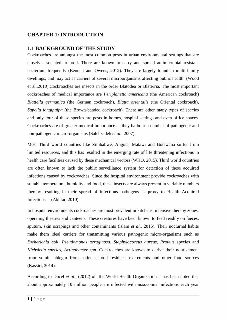
ABSTRACT
Cockroaches have become one of the most common pests due to their wide distribution in
human dwellings, kitchens, food outlets and the hospital. In hospitals their wide abundancy
may be related to poor sanitary conditions, plenty of food sources, environments which are
unhygienic and moist shady places. This study was carried out to evaluate the microbial
fauna of cockroaches in a hospital environment as proxy to Health acquired infections. Two
different species of cockroaches Periplaneta americana and Blatella germanica were
collected in the canteen and the main kitchen of a hospital in Gweru. The former and the
latter were cultured using standard procedures and antibiotic susceptibility tests were done.
Microbial isolation was done for the two species of cockroaches by growing saline
suspensions of the cockroach samples onto selective and differential media. Bacterial isolates
were identified by biochemical tests. Medically important microorganisms were isolated
from their external surface and their internal surface. These bacteria were Proteus spp,
Klebsiella spp, Escherichia coli, Pseudomonas aeruginosa, Staphylococcus aureus. The
susceptibilities of Staphylococcus aureus, Pseudomonas aeruginosa, Klebsiella spp,
Escherichia coli, and Proteus species to 6 antibiotics were tested. Most of the gram negative
bacteria were resistant to amoxicillin, Augmentin and streptomycin and sensitive to
ciprofloxacin and chloramphenicol. Enumeration was also conducted using saline
suspensions of the internal and external washings and the range from P. americana to B.
germanica from external washings were 3.60 ×10⁴ to 3.20×10
4 cfu/ml respectively and for
the internal washings 3.40 ×10⁴ to 3.23 ×10
4 cfu/ml respectively .The data collected was
subjected to Two-way analysis of variance (ANOVA) using SPSS version 21. Two-way
Anova results showed that there is no statistically significant interaction between type of
washings and type of species in influencing the mean total bacterial count (p>0.05). Two-way
Anova data also revealed that the mean total bacterial count of periplaneta americana was
significantly higher than that of blatella germanica (p<0.05). In conclusion, all cockroaches
harboured pathogenic bacteria with multidrug resistance, this means that cockroaches play a
potential role in the epidemiology of nosocomial infections in this hospital.

iii | P a g e
ACKNOWLEDGEMENTS
First and Foremost l would like to thank the Lord Almighty for guiding me through my
studies. It is my greatest pleasure to acknowledge the depth l owe to many people who have
assisted me in coming up with this project. I would like to express my special gratitude to my
academic supervisor Ms Mguni, for her constant support and mentorship. Ms Mguni kindly
undertook the formidable task of reading this whole dissertation and I am grateful to her for
her frank and helpful comments. Special thanks are extended to Mr Banda (Premier services
Laboratory scientist) for assisting me in coming up with a project on microbiology and also
Dr Mapaya for her assistance in making this project a success. Her input in terms of
continued assistance throughout my project was greatly appreciated and helpful. Special
thanks are also extended to Dr T. Muteveri for his guidance in data analysis. I would also like
to thank all research scientists and research associates Mr Dowo and Mr C. Mabhugu for their
support and assistance throughout my project. To all my friends Ratidzo and Lisa and other
scientists that are not mentioned I owe them my thanks for giving me the benefit of their
expertise and making this project a success.

iv | P a g e
DEDICATIONS
I sincerely dedicate this body of work to my loving sister (Milly Makumana) and husband
(Lookout Sigodo) who see the best in me and the heights of greatness I shall reach, even
when I do not see it myself.
To succeed, you need to find something to hold on to, something to motivate you, something
to inspire you –Tony Dorsett
v | P a g e
TABLE OF CONTENTS APPROVAL FORM ..................................................................................................................................... i
ABSTRACT ................................................................................................................................................ ii
ACKNOWLEDGEMENTS .......................................................................................................................... iii
DEDICATIONS ......................................................................................................................................... iv
CHAPTER 1: INTRODUCTION ................................................................................................................... 1
1.1 Background of the study ................................................................................................................... 1
1.2 Problem statement ........................................................................................................................... 2
1.3 Justification ....................................................................................................................................... 4
1.4 Project Objectives ............................................................................................................................. 5
1.4.2 Specific Project Objectives ............................................................................................................. 5
CHAPTER 2 .............................................................................................................................................. 6
LITERATURE REVIEW ............................................................................................................................... 6
2.1 General description of cockroaches.................................................................................................. 6
2.1.1 The Biology of Cockroaches ........................................................................................................... 6
2.1.2 Ecology and Behaviour of Cockroaches ......................................................................................... 9
2.1.3 The Pathology of Cockroaches ..................................................................................................... 10
2.2 Environmental Management .......................................................................................................... 11
2.3 Hospital Acquired Infections ........................................................................................................... 12
2.3.1 Urinary infections ......................................................................................................................... 13
2.3.2Wound infections .......................................................................................................................... 13
3.0 MATERIALS AND METHODS ............................................................................................................ 14
3.1 Study site ......................................................................................................................................... 14
3.3 The identification and isolation of microorganisms from the cockroaches on their external
surfaces. ................................................................................................................................................ 14
3.4 Microscopic Identification .............................................................................................................. 15
3.5.1 Catalase test ................................................................................................................................. 15
3.5.2 Oxidase test.................................................................................................................................. 16
3.5.3 Citrate test ................................................................................................................................... 16
3.5.4 Indole test .................................................................................................................................... 16
3.5.5 Coagulase ..................................................................................................................................... 16
3.6 Bacteriological analysis using quantitative methods - bacterial enumeration .............................. 17
3.6.1 Total Bacterial Counts .................................................................................................................. 17
3.7 Antibiotic susceptibility tests .......................................................................................................... 18
3.8 Statistical Analysis ........................................................................................................................... 18
CHAPTER 4 RESULTS .............................................................................................................................. 19
vi | P a g e
4.1 BACTERIAL IDENTIFICATION ............................................................................................................ 19
4.2 Prevalence of bacterial isolates from different anatomical sampling sites .................................... 22
4.3 QUANTITATIVE ANALYSIS –COLONY COUNT .................................................................................. 23
4.3.1 Total Bacterial and Total Coliform Counts ................................................................................... 23
4.3.2 Statistical analysis; ....................................................................................................................... 24
4.4Antibiotic resistance ........................................................................................................................ 29
CHAPTER 5 DISCUSSION ..................................................................................................................... 30
5.1 Cockroaches and the carriage of microorganisms .......................................................................... 30
5.1.1 Isolated bacteria .......................................................................................................................... 31
5.1.2 Escherichia coli ............................................................................................................................. 32
5.1.3 Staphylococcus aureus ................................................................................................................. 33
5.1.4 Klebsiella Pneumonia ................................................................................................................... 33
5.1.5 Pseudomonas aeruginosa ............................................................................................................ 34
5.1.6 Proteus sp .................................................................................................................................... 34
5.2 Quantitative analysis ....................................................................................................................... 35
5.3 Antibiotic resistance patterns ......................................................................................................... 35
5.4 CONCLUSION ................................................................................................................................... 36
5.5 RECOMMENDATIONS...................................................................................................................... 36
REFERENCES .......................................................................................................................................... 38
APPENDICES .......................................................................................................................................... 46
Appendix 2 ............................................................................................................................................ 47
Appendix 3- Macroscopic identification(Wani, 2013) .......................................................................... 48
Appendix 4 ............................................................................................................................................ 49
Appendix 4.1 Antibiotic susceptibility tests .......................................................................................... 49
Appendix 5 ............................................................................................................................................ 51
1 | P a g e
CHAPTER 1: INTRODUCTION
1.1 BACKGROUND OF THE STUDY
Cockroaches are amongst the most common pests in urban environmental settings that are
closely associated to food. There are known to carry and spread antimicrobial resistant
bacterium frequently (Bennett and Owens, 2012). They are largely found in multi-family
dwellings, and may act as carriers of several microorganisms affecting public health (Wood
et al.,2010).Cockroaches are insects in the order Blatodea or Blaterria. The most important
cockroaches of medical importance are Periplanetta americana (the American cockroach)
Blattella germanica (the German cockroach), Blatta orientalis (the Oriental cockroach),
Supella longipalpa (the Brown-banded cockroach). There are other many types of species
and only four of these species are pests in homes, hospital settings and even office spaces.
Cockroaches are of greater medical importance as they harbour a number of pathogenic and
non-pathogenic micro-organisms (Salehzadeh et al., 2007).
Most Third world countries like Zimbabwe, Angola, Malawi and Botswana suffer from
limited resources, and this has resulted in the emerging rate of life threatening infections in
health care facilities caused by these mechanical vectors (WHO, 2015). Third world countries
are often known to lack the public surveillance system for detection of these acquired
infections caused by cockroaches. Since the hospital environment provide cockroaches with
suitable temperature, humidity and food, these insects are always present in variable numbers
thereby resulting in their spread of infectious pathogens as proxy to Health Acquired
Infections (Akhtar, 2010).
In hospital environments cockroaches are most prevalent in kitchens, intensive therapy zones,
operating theatres and canteens. These creatures have been known to feed readily on faeces,
sputum, skin scrapings and other contaminants (lslam et al., 2016). Their nocturnal habits
make them ideal carriers for transmitting various pathogenic micro-organisms such as
Escherichia coli, Pseudomonas aeruginosa, Staphylococcus aureus, Proteus species and
Klebsiella species, Actinobacter spp. Cockroaches are known to derive their nourishment
from vomit, phlegm from patients, food residues, excrements and other food sources
(Kassiri, 2014).
According to Ducel et al., (2012) of the World Health Organization it has been noted that
about approximately 10 million people are infected with nosocomial infections each year
2 | P a g e
especially in public hospitals due to the presence of mechanical vectors such as houseflies
,cockroaches and poor sanitation (Ducel et al., 2012) defined a health acquired infection or
nosocomial infections as infections that develop in a hospitalised patient or any other health
care facility in which the infection was absent or incubating at the time of admission. Health
care associated infections such as severe acute respiratory syndrome, urinary tract infections
have brought about the immediate need for efficient infection control strategies to be put in
place. These infections are preventable through implementation of best infection prevention
and control practices especially at health care facilities (Ahaduzzaman et al., 2014).
Healthcare facilities are known to be the ideal settings for the transmission of infections to
patients (who are more susceptible), healthcare workers, their families and communities who
visit the hospital facility. Most healthcare associated infections lead to prolonged hospital
stays, increased costs of care and death (Hsuch et al., 2012). Some of the most important
bacteria to be analysed due to its resistance to antibiotics is a gram positive bacteria found in
the family Staphylococcae (Medveďová and Valík, 2012).
1.2 PROBLEM STATEMENT
Most developing countries in Sub Saharan Africa suffer from limited resources, therefore the
emerging rate of life threatening infections at health care facilities. Limited data, often of low
quality, are available from low and middle income countries (Abdelkhalek et al.,2016).
However, recent analysis by World Health Organization found that health care-associated
infections are more frequent in resource limited settings than in developed countries (WHO,
2015). The ratio of health acquired infections is now higher in public hospitals than in private
hospitals, this may be due to poor hygienic standards leading to an emergency of pests such
as cockroaches (WHO, 2015). Some studies have also reported that cockroaches found in
hospitals are playing important role in deteriorating hospital environment and patient‟s health
by imposing stress, infections, anxiety, asthma, allergy in persons who have deficient
immunity. Cockroaches have been found in hospital vicinity inside intensive therapy zone,
neo natal units, ICU, medical wards, surgical wards and canteen, kitchen and medical stores
(Sabra and Abdel-Fattah, 2012).
Sub Saharan African countries such as Zimbabwe, Botswana and Mozambique have given
little attention to the spread of health acquired infections and the existence of such pestilence
pests (Ducel et al.,2012). In 2012 statistics stated that up to 54 % of isolates from hospital
environments were found to be human pathogens worldwide (Medveová and Valík, 2012). At
3 | P a g e
any given time, the prevalence of health care associated infection varies between 5.7% and
19.1% in low and middle income countries (Ducel et al.,2012). The proportion of patients
with ICU-acquired infection ranged from 4.4% to 88.9% with a frequency of overall
infections as high as 42.7 episodes per 1000 patient days in low and middle income countries
such as Zimbabwe. This is almost three times higher than in high income countries such as
Britain, America and Russia to name a few (WHO, 2015). Furthermore, in some developing
countries, the frequency of infections associated with the use of central lines and ventilators
and other invasive devices can be up to 19 times higher than those reported from Germany
and the USA (Sausa et al., 2014).
New- born babies are also at higher risk, with infection rates in developing countries 3-20
times higher than in high income countries. Among hospital born babies in developing
countries, health care-associated infections are responsible for 4% to 56% of all causes of
death in the neonatal period, and 75% in South-East Asia and Sub-Saharan Africa (WHO,
2015).
Surgical site infection and wound infections is the leading infection in the general patient
population in countries with limited resources, affecting up to two third of operated patients
and with a frequency up to nine times higher than in developed countries (Oliva et al., 2010).
Several factors that lead to the prevalence of contaminants such as cockroaches harbouring
bacteria are more specific to settings with limited resources and these are inadequate
environmental hygienic conditions and waste disposal; poor infrastructure; insufficient
equipment; understaffing; overcrowding; poor knowledge and application of basic infection
control measures; lack of knowledge of injection and blood transfusion safety and the
absence of local and national guidelines and policies. All these factors therefore lead to the
emerging rates of cockroaches infestation (Medveďová and Valík, 2012).
Studies made in Botswana and worldwide have also shown that cockroaches are associated
with a number of pathogenic and non-pathogenic organisms therefore may lead in
transmission then it results in higher costs of medication for patients, health workers and the
public, cockroaches have also been found to cause asthma allergies, prolonged hospital stay
for operated patients due to post operational infections and the increase of antibiotic
resistance of bacteria such as staphylococcus species. In Zimbabwe many people have limited
access to medication due to impoverishment therefore they may eventually die of such
infections (Mpuchane, 2006).

4 | P a g e
1.3 JUSTIFICATION
Cockroaches are known to carry 151 different species of bacteria and these in turn cause
infections, it is therefore important to lower their rate of proliferation and enhance efficiency
of infection control practices in hospitals and residential homes (lslam et al.,2016). As is the
case for many other patient safety issues, health care-associated infections create additional
suffering and come at a high cost for patients and their families. Infections prolong hospital
stays, create long-term disability, increase resistance to antimicrobials, represent a massive
additional financial burden for health systems, generate high costs for patients and their
family, and cause unnecessary deaths. Such infections annually account for 37 000
attributable deaths in Europe and potentially many more that could be related, and they
account for 99 000 deaths in the USA (Kassiri et al.,2014).
Annual financial losses due to health care-associated infections are also significant: they are
estimated at approximately €7 billion in Europe, including direct costs only and reflecting 16
million extra days of hospital stay, and at about US$ 6.5 billion in the USA (Ducel et al
.,2012). Financial costs attributable to health care-associated infections are poorly and
variably reported in low and middle income countries (Pai, 2012).
For instance, the economic burden of health care associated infections in Belo Horizonte,
Brazil, was estimated to be equal to US$ 18 million in 1992. In Mexican ICUs, the overall
cost of one single health care-associated infection episode was US$ 12 155. In several ICUs
in Argentina, the overall extra-cost estimates for catheter-related bloodstream infection and
health care-associated pneumonia averaged US$ 4 888 and US$ 2 255 per case, respectively (
Doosti et al., 2015). The emergence and the rapid spread of antimicrobial resistance bacteria
such as S. aureus has become a global concern. Multidrug resistant (MDR) Staphylococcus
poses a growing problem for human health and has been considered as horrifying public
health threat (Ahaduzzaman et al., 2014).
Nosocomial infections in Zimbabwe can be reduced by : identifying local determinants of
the Health Control System of Acquired Infections burden; improving reporting and
surveillance systems at the national level; ensuring minimum requirements in terms of
facilities and dedicated resources available for Health Control System of Acquired Infection
surveillance at the institutional level, including microbiology laboratories' capacity; ensuring
that core components for infection control are in place at the national and health care setting
5 | P a g e
levels; implementing standard precautions, particularly best hand hygiene practices at the
bedside; improving staff education and accountability; conducting research to adapt and
validate surveillance protocols based on the reality of developing countries and conducting
research on the potential involvement of patients and the food they are given in that particular
health environment, conducting national spraying day to reduce the number of mechanical
vectors in all hospitals around the country(Oliva et al.,2010).
This project since it has never been undertaken here in Zimbabwe may be able to act as an
awareness prospect to raise safety concerns in terms of cockroaches as pests. Safety
awareness programmes may reduce the number of people being admitted daily in hospitals
thereby lowering medication costs and morbidity and mortality caused by health acquired
infections and a deeper understanding of cockroaches to the nation may also help in the
reduction in antibiotic resistance of some bacteria such as Staphylococcus. Study of
cockroaches and their association with the hospital environment may help clinicians in
eradication of post surgery infections, neonatal infections, Urinary tract infections and life
threatening septicaemia (Ahaduzzaman et al.,2014).
1.4 PROJECT OBJECTIVES
1.4.1 Main objectives : To investigate cockroaches as vectors of bacteria and to determine
antibiotic susceptibility of acquired bacteria in a health environment as proxy to Hospital
acquired infections.
1.4.2 SPECIFIC PROJECT OBJECTIVES
To isolate and identify the types of bacteria harboured by Periplaneta americana and
Blatella germanica
To determine the possible role of cockroaches in the epidemiology of health acquired
Infections.
To enumerate the bacteria that are found in the external and internal washings of the
two species of cockroaches P.americana and B.germanica.
To investigate antibiotic susceptibility of bacteria isolated from cockroaches.
6 | P a g e
CHAPTER 2
LITERATURE REVIEW
2.1 GENERAL DESCRIPTION OF COCKROACHES
Cockroaches are members of the order Blattodea and they belong to the kingdom Animalia,
the phylum is Arthropoda, the class is insect and the superorder is dityoptera. Cockroaches
are brown insects with an antenna and are about one and a half inches (4 centimeters) long
when fully grown. Cockroaches have a relatively small head and a broad, flattened body most
are reddish brown to dark brown which includes the termites (Gordh and Headrick, 2009).
The body of cockroaches is divided into a thorax of three segments and a ten-segmented
abdomen. The external surface has a tough exoskeleton which contains calcium carbonate
and protects the inner organs and provides attachment to muscles (Hoell et al.,1998). It is
coated with wax to repel water. The wings are attached to the second and third thoracic
segments. The tegmina, or first pair of wings, are tough and protective, lying as a shield on
top of the membranous hind wings, which are used in flight. All four wings have branching
longitudinal veins, and multiple cross-veins (Legentre et al., 2015).
Cockroaches also have a long antennae and legs, feeding by scavenging. The three pairs of
legs are sturdy with large coxae and five claws each. They are attached to each of the three
thoracic segments( Mullen and Burden, 2002).The front legs are the shortest and the hind legs
the longest, providing the main propulsive power when the insect runs .Cockroaches are
insects, flattened from top to bottom, usually with two pairs of wings folded flat over the
back. Most species rarely fly but they move very fast using legs. The colour usually varies
from light brown to black. The species vary from 2– 3mm to over 80mm in length, of over
3500 identified species only a few are of importance to people because they have adapted to
living in buildings(Sweld, 2015).
2.1.1 THE BIOLOGY OF COCKROACHES
The most common species are: Periplaneta americana, the American cockroach, which
occurs around the world. It is 35 to 40mm in length and has a shiny reddish to chocolate
brown colour. The egg case measures 8 to 10mm and contains 16 eggs (Bell et al.,2011)
.Periplaneta australasiae, the Australian cockroach, which occurs mainly in tropical and
subtropical areas. It is similar to the American cockroach, but smaller (31 to 37mm long) and
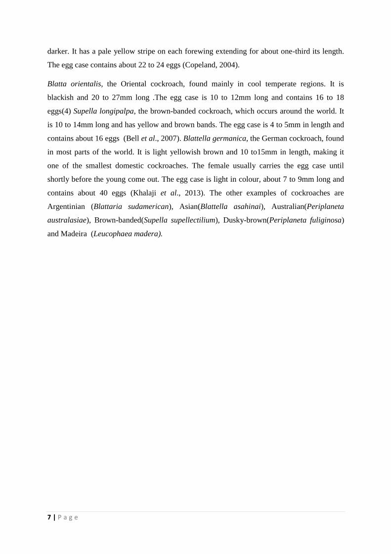
7 | P a g e
darker. It has a pale yellow stripe on each forewing extending for about one-third its length.
The egg case contains about 22 to 24 eggs (Copeland, 2004).
Blatta orientalis, the Oriental cockroach, found mainly in cool temperate regions. It is
blackish and 20 to 27mm long .The egg case is 10 to 12mm long and contains 16 to 18
eggs(4) Supella longipalpa, the brown-banded cockroach, which occurs around the world. It
is 10 to 14mm long and has yellow and brown bands. The egg case is 4 to 5mm in length and
contains about 16 eggs (Bell et al., 2007). Blattella germanica, the German cockroach, found
in most parts of the world. It is light yellowish brown and 10 to15mm in length, making it
one of the smallest domestic cockroaches. The female usually carries the egg case until
shortly before the young come out. The egg case is light in colour, about 7 to 9mm long and
contains about 40 eggs (Khalaji et al., 2013). The other examples of cockroaches are
Argentinian (Blattaria sudamerican), Asian(Blattella asahinai), Australian(Periplaneta
australasiae), Brown-banded(Supella supellectilium), Dusky-brown(Periplaneta fuliginosa)
and Madeira (Leucophaea madera).
8 | P a g e
Figure 2.1 Common household cockroaches (a) Periplaneta americana,(b) Blatta
orientalis (c) Blatella germanica (d) Supella longipalpa.
Cockroaches are relatively primitive, having only three stages in their life cycle: egg, nymph
and adult. The female deposits its eggs in groups surrounded by a leathery, bean-shaped egg
case or capsule called an ootheca. Some species, such as the German cockroach, carry the
ootheca for several weeks attached to the back end of the body. Most others deposit the
9 | P a g e
ootheca after one or two days. Oothecae are very distinctive and can frequently be used to
determine the species present (Beccaloni and Eggleton, 2013).
Depending on the species, temperature and humidity, the eggs hatch after 1–3 months. The
young cockroaches, or nymphs, are wingless, and usually only a few millimetres long; they
are white on hatching but darken within a few hours. They grow in stages by repeatedly
shedding the cuticle or skin (Siachua et al., 2008). They are fully grown after several months
to more than a year, depending on the species. The adults may or may not possess wings,
consisting of one outer leathery pair beneath which is folded a membranous pair (Huang et
al., 2012)
2.1.2 ECOLOGY AND BEHAVIOUR OF COCKROACHES
Cockroaches are known to play a supplementary role in the spread of some diseases.
Cockroaches are the most significant and objectionable pests found in apartments, homes,
food-handling establishments, hospitals, and health care facilities worldwide. They are
among the most common pests in many homes and other buildings (Cornwell, 2005)
Cockroaches consume garbage, rotting food, and even faecal waste of other cockroaches.
They then transmit disease to your food, eating utensils, kitchen surfaces, and other areas
around your home. They can easily contaminate food by leaving droppings which may
contain bacteria that can cause food poisoning, fungi, and other pathogenic organisms
(Malcom, 2013). Their nocturnal and filthy habits make them also ideal carriers of various
pathogenic microorganisms. At night they search for food in kitchens, food storage places,
rubbish bins, drains and sewers. Indoor species, especially the German cockroach, exploit
conditions associated with high-density human populations and impoverished living
conditions. Cockroaches are largely found in multi-family dwellings, and may act as a carrier
of several microorganism affecting public health (Jeanson et al., 2005).
Cockroaches are proven or suspected carriers of the organisms causing: diarrhoea, dysentery,
cholera, leprosy, plague, typhoid fever and other viral diseases such as poliomyelitis. In
addition they carry the eggs of parasitic worms and may cause allergic reactions, including
dermatitis, itching, swelling of the eyelids and more serious respiratory conditions
Botella,2005: Oliva et al.,2010.
Their nocturnal lifestyle and dirty habits make them ideal carriers for transmitting numerous
pathogenic bacteria, such as Listeria monocytogenes, Klebsiella pneumoniae, Staphylococcus
aureus, Pseudomonas aeruginosa, Shigella species, proteus sp, Streptococcus species.,
10 | P a g e
Salmonella species, Campylobacter species, Escherichia coli, and other potential pathogens
(Salehzadeh et al., 2007; Kassiri et al., 2014; Doosti et al., 2015. ln a study done in Botswana
More than 33.3% of cockroach isolates have shown resistance to antimicrobials (Mpuchane
et al., 2006). Staphylococcae is responsible for the skin infections which then can manifest in
various ways like boils, cellulitis and more severe, invasive soft tissue infection.
Staphylococcus is resistant to many commonly used antibiotics methicillin resistant
Staphylococcus aureus, Vancomycin intermediate Staphylococcus aureus and Vancomycin
resistant Staphylococcus aureus. Secondly Gram positive bacteria e.g. Staphylococcus aureus
is a cutaneous bacteria that colonize the skin and nose of both hospital staff and patients
cause a wide variety of lung, bone, heart and blood stream infections and are frequently
resistant to antibiotics; beta-haemolytic streptococci are also important (Cheesbrough, 2006).
2.1.3 THE PATHOLOGY OF COCKROACHES
Cockroaches are known to transmit up to nine different diseases resulting from their
contamination on food and water. In addition to bacteria, many species of parasites were
isolated from external surface and guts of cockroaches included cysts of Entameoba coli,
Entameoba histolytica and Giardia lamblia and adult and ova of Enterobius vermicularis and
ova of Hymenolepis nana and Ascaris lumbricoides (Wannigama, 2014) It was also proved
experimentally that cockroaches may play an important role in the transmission of
helminthes. Some of the diseases include Salmonellosis: cockroaches similar to rodents are
known to transmit the Salmonella bacteria which can cause salmonellosis, which is a disease
in humans with symptoms similar to food poisoning. Cockroaches are known to accumulate
the bacteria by feeding on contaminated food. Salmonella remains in their digestive system
for a month or more and is deposited through their vomit and faeces (Rentz,2014).
Typhoid fever is another type of a bacterial infection caused by the Salmonella typhi
bacterium and is a highly infectious disease. It is believed that cockroaches accumulate this
disease by consuming faeces contaminated with the bacterium. Children and the elderly are
thought to be most at risk due to their immunocompromised state (Wu Hao, 2013).
Cholera is an acute diarrhoeal infection caused by the Vibrio cholerae bacterium. It is most
common in developing countries and areas that have inadequate environmental management.
Infection occurs through ingestion of food and drink contaminated with the bacterium
(Gilliot, 2003). If exposed to the bacterium, cockroaches can spread the organism through
their faeces and vomit, contaminating surfaces and food. Researchers from the World Health
11 | P a g e
Organization have estimated that worldwide there are roughly 1.4 million to 4.3 million cases
of cholera per year resulting in 28,000 to 142,000 deaths (WHO,2015).
Dysentery is a type of gastroenteritis that results in diarrhoea with blood. Generally, most
people suffer from mild symptoms and recover within a week or so without medical
attention. There are two types of dysentery. They are: Bacillary dysentery: Sometimes
referred to as shigellosis. It is caused by the Shigella bacteria. Amoebic dysentery: Is caused
by a single-celled parasite called Entamoeba. It is usually found in tropical areas (Gulani et
al., 2016).
Campylobacteriosis is an infection caused by the Campylobacter bacterium. It is one the most
common bacterial infections in humans, and is a common foodborne illness. Transmission
occurs through ingesting contaminated food and drink such as unpasteurised milk and
undercooked and poorly handled poultry. Researchers have isolated a Campylobacter jejuni
subspecies in the gut contents and on the external surface of both American cockroaches and
Oriental cockroaches (Umunnabuike and Irokanulo, 2000) .Escherichia coli (E. coli) is a
bacterium commonly found in the gut of humans. Although most strains of the bacteria are
harmless, some can cause serious food poisoning. The common symptoms of an E. coli
infection are watery diarrhoea and abdominal cramping. Less common symptoms are: Fever,
Chills, Nausea and Muscle aches (Guarino, 2016).
Cockroaches can trigger asthma because they certain proteins in their bodies which can be an
allergen for certain people. When tiny particles from cockroach bodies are spread through the
air in buildings, these proteins are inhaled and an asthma attack can be triggered in sensitive
people. The American College of Allergy, Asthma & Immunology reports that the saliva,
faeces and shed skin of cockroaches can trigger both asthma and other allergic responses
(Botella et al., 2005).
2.2 ENVIRONMENTAL MANAGEMENT
Cleanliness and hygiene should be maintained in the hospital area and leftover food should be
stored in tightly covered containers in clean cabinets or refrigerators. All areas have to be
kept clean so that no fragments of food or organic matter remain. Rubbish bins should be
securely covered and emptied frequently, preferably daily. Effective control is easier in
temperate climates (where cockroach populations cannot survive outdoors in winter) than in
humid and warm areas. The key to control is cleanliness, which may be difficult in houses
where there are children and domestic animals (Gilliot, 2003). In isolated homes, control is
12 | P a g e
easier to achieve than in apartments where cockroaches may have easy access from adjacent
quarters. Reinfestation occurs from outdoors in warm areas, or along heating ducts and water
pipes in apartments, or from groceries or luggage brought from cockroach infested areas
(Gliniewicz, 2003). Cockroaches may even sometimes be found in very clean houses, but are
unlikely to establish colonies. The presence of several sizes of nymphs and oothecae is an
indication of a well established colony. Infestations can be detected by searching behind
skirting boards, boxes, furniture and other common hiding places. At night, cockroaches are
easily detected using light (Rentz , 2014).
2.3 HOSPITAL ACQUIRED INFECTIONS
According to (Ducel et al., 2012) A hospital acquired infection also known as a nosocomial
infections is defined as „An infection acquired in hospital by a patient who was admitted for
a reason other than that infection or An infection acquired by a patient in a hospital or any
other hospice in whom the infection was absent or incubating at the time the patient was
admitted. These infections can even start to appear even after the patient has been discharged,
and hospital acquired infections can also manifest among the staff of the facility/health
workers (Manott and Gravani, 2006). In Zimbabwe the care of many patients is provided in
facilities which range from highly expensive and equipped clinics and technologically
advanced hospitals such as Baines hospital to front-line units with only basic facilities, but
despite that the rate of Health Acquired Infection keeps on increasing (WHO, 2015). Despite
progress in public health and hospital care, infections continue to develop in hospitalized
patients, and may also affect hospital staff. There are also several factors that promote
infection among hospitalized patients such as decrease in immunity among patients due to
several diseases; the increasing variety of medical procedures and invasive techniques
creating potential routes of infection; and the transmission of drug-resistant bacteria among
crowded hospital populations, where poor infection control practices may facilitate
transmission (Huang et al., 2012).
Bacteria are the most common nosocomial pathogens Gram-negative bacteria:
Enterobacteriacae (e.g. Escherichia coli, Proteus, Klebsiella, Enterobacter, Serratia
marcescens), may colonize sites when the host defences are compromised (catheter insertion,
bladder catheter, cannula insertion) and cause serious infections (surgical site, lung,
bacteraemia, peritoneum infection) .They may also be highly resistant (Pavlik and falkinham,
2009).

13 | P a g e
Gram-negative organisms such as Pseudomonas spp. are often isolated in water and damp
areas. They may colonize the digestive tract of hospitalized patients. Selected other bacteria
are a unique risk in hospitals. For instance, Legionella species may cause pneumonia
(sporadic or endemic) through inhalation of aerosols containing contaminated water (air
conditioning, showers (Feizhaddad et al., 2012).
2.3.1 URINARY INFECTIONS
Cockroaches due to their nocturnal movements acts as mechanical vectors in contaminating
catherised devices in search for food. Catherised patients especially those with the device in
situ for long periods experience repeated episodes of infections that can damage the urinary
tract. (Bennet, 2010). Urinary infections forms the most common nosocomial infection;
according to recent records there are about 80% of infections which are associated with the
use of an indwelling bladder catheter (Fox, 2008). Although there are highly common most
of the urinary infections are associated with less morbidity than other nosocomial infections,
but although at a lesser extent can occasionally lead to bacteraemia and death. The bacteria
responsible arise from the gut flora, either normal (Escherichia coli) or acquired in hospital
(multiresistant Klebsiella). Catherised patients especially those with the device in situ for
long periods experience repeated episodes of infections that can damage the urinary tract
(Mullen and Durden, 2002).
2.3.2WOUND INFECTIONS
The skin is highly known for its use as a barrier for many microorganisms as they prevent
entry of bacteria entering the body (Bouamama et al., 2010). Most cockroaches move on
devices and cloth used in cleaning of wounds thereby leaving bacteria which can then cause
infections on wounds. Wounds often are a result of a trauma that occur to people through
accidents ,surgery, burns, certain infectious opportunistic diseases and other various
ulcerations. Culture can be used in identification of organisms causing infection by use of
wound swab (Khalaji et al., 2013).Burns are most problematic especially in those patients
having burns affecting more than 60%of the surface of the body are more susceptible with
70% of these burns becoming infected and most patients developing blood stream infections.
Gram negative bacteria such as Proteus species are common (Cheesbrough, 2006).
14 | P a g e
3.0 MATERIALS AND METHODS
3.1 STUDY SITE
The sampling area of this study was a Health care facility in Gweru. The experiments were
carried out at Gweru General Hospital Laboratory. The health care facility has a population
of approximately 20 patient care units. The Sample size of this study were 2 hospital units‟
the hospital kitchen and the canteen. In this study our sampling strategy was a convenient
sampling. Sampling step – the first cockroaches for inclusion in this study were collected at
the following patient care units –the hospital main kitchen and the canteen kitchen and were
then transferred to the hospital laboratory for further research.
3.2 Sample Collection
The simple jar traps method was used in the collection of the required cockroach specimens.
Bread crumps were placed at the bottom of the container to act as attractants and a thick layer
of petroleum jelly was added on the inner rim of the container to prevent the insects from
escaping. Thirty- four cockroaches were collected, over a period of 1 month,16 from the
health main kitchen and 18 from the canteen. The cockroaches were collected in late day
time. After a two day observation the trapped cockroaches were collected in sterile test tubes,
transported to the laboratory and anaesthetized by freezing at 00C for 5 minutes and then
stored at 50C. The cockroach differences were compared using their morphological features
(Harwood and James, 1979).
3.3 THE IDENTIFICATION AND ISOLATION OF
MICROORGANISMS FROM THE COCKROACHES ON THEIR
EXTERNAL SURFACES.
Following the morphological identification of each of the cockroaches, two ml of sterile
normal saline was added in each tube and the cockroaches were thoroughly shaken for two
minutes. The cockroaches were then taken out of the tubes and the remaining liquid
containing bacteria was centrifuged at 2000 rpm for 10 minutes. The supernatant was then
removed and the remaining sediment was used for culture. From each tube a fixed volume of
1 ml each of the washings was cultured on selective media MacConkey agar, mannitol salt,
nutrient agar and blood agar plates separately. Culture plates with different culture media
were labelled ES(external surface) , P (periplaneta americana) and the sample number, the
labels were put in this order on the plates P, E.S and 1. The same procedure was done for

15 | P a g e
external washings of Blatella germanica each plate were also labelled ES,S (Blatella
germanica) and 1 .The cultures were incubated overnight at 37 oC. The ultimate colonies
were then identified by standard bacteriological procedures (Cheesbrough, 2006).
3.3.1 ThE identification and isolation of microorganisms from the cockroaches on their
internal surfaces.
Following the external washings culture, the external washings surface of cockroaches were
then washed with 70 percent ethyl alcohol for 5 minutes and were then allowed to dry at
room temperature under sterile conditions. The decontaminated cockroaches were then
washed with sterile normal saline for 2-3 minutes to remove traces of alcohol. The gut of the
cockroaches were dissected out and macerated aseptically with a sterile pestle and mortar in 2
ml of sterile normal saline. The resulting macerate were cultured on selective media at 37oC
overnight and in a similar way as described above and the results were recorded.
3.4 MICROSCOPIC IDENTIFICATION
Gram staining was done. All the positive samples of gram staining were subjected to
coagulase and catalase tests for biochemical confirmation as described by Cheesbrough
(2006).
3.5 Biochemical Tests
Biochemical tests were performed on colonies from primary cultures for identification of the
isolates. Gram-negative rods were identified by performing a series of biochemical tests such
as Kligler iron test, citrate utilisation test (SC), Lysine decarboxylase test, indole motility test,
urease test, coagulase test, catalase test and oxidase test. Gram-positive cocci were identified
based on their gram reaction, catalase, coagulase and oxidase test results.
3.5.1 CATALASE TEST
Two drops of 3% hydrogen peroxide (H 2O 2) were added into a test tube using a dropper.
An isolated bacterial colony from the MacConkey agar was placed in the drop of hydrogen
peroxide by using a sterilised inoculation loop sterilised by dipping it in methylated spirit,
flaming it until red hot and then allowing it to cool in the air. The drop was viewed for any
effervescence.
16 | P a g e
3.5.2 OXIDASE TEST
A manufactured oxidase test strip was inoculated with an isolated bacterial colony using a
sterilised inoculation loop. The test part of the strip was viewed for any colour change.
3.5.3 CITRATE TEST
Simmons citrate agar was prepared by weighing 11g of the agar powder and dissolving in
250ml of distilled water. The solution was then mixed and heated until the powder was
completely dissolved. The solution was then autoclaved at 121°C for 15minutes, and after
that, it was poured into labelled bijou bottles to cool and set. Using a sterile straight wire, the
slope was first streaked with a saline suspension of the test organism/colony type and then the
butt was stabbed. The Simmons citrate agar was incubated at 35 0C for 48 hours. The medium
was then observed for any colour change.
3.5.4 INDOLE TEST
Bijou bottles with peptone water were labelled with the test organism name and sample code.
Gram negative lactose fermenting colonies were picked from the MacConkey agar plates
using a sterilised inoculating loop and a suspension was made in the peptone water. These
were then incubated at 370C for 24 hours and two drops of Kovac‟s reagent were added into
each bottle. Samples were observed for the presence of a red ring or a brown ring and
checked against positive and negative controls available in the laboratory then recorded
3.5.5 COAguLASE
Glass slides were labelled with the test organism and sample code. Sterile saline was
inoculated onto the glass slides using a sterilised inoculating loop. A colony of the test
organism was then emulsified onto the slides and a loopful (not more) of plasma was added
to one of the suspensions, and mixed gently. Clumping of the organisms was observed within
10 seconds No plasma was added to the second suspension. This was used to differentiate any
granular appearance of the organism from true coagulase clumping.
17 | P a g e
3.6 BACTERIOLOGICAL ANALYSIS USING QUANTITATIVE
METHODS - BACTERIAL ENUMERATION
3.6.1 TOTAL BACTERIAL COUNTS
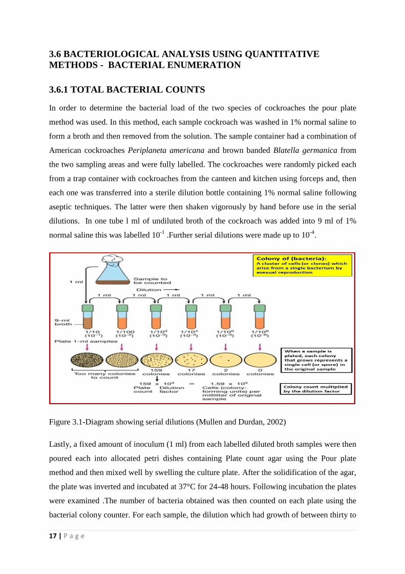
In order to determine the bacterial load of the two species of cockroaches the pour plate
method was used. In this method, each sample cockroach was washed in 1% normal saline to
form a broth and then removed from the solution. The sample container had a combination of
American cockroaches Periplaneta americana and brown banded Blatella germanica from
the two sampling areas and were fully labelled. The cockroaches were randomly picked each
from a trap container with cockroaches from the canteen and kitchen using forceps and, then
each one was transferred into a sterile dilution bottle containing 1% normal saline following
aseptic techniques. The latter were then shaken vigorously by hand before use in the serial
dilutions. In one tube l ml of undiluted broth of the cockroach was added into 9 ml of 1%
normal saline this was labelled 10-1
.Further serial dilutions were made up to 10-4
.
Figure 3.1-Diagram showing serial dilutions (Mullen and Durdan, 2002)
Lastly, a fixed amount of inoculum (1 ml) from each labelled diluted broth samples were then
poured each into allocated petri dishes containing Plate count agar using the Pour plate
method and then mixed well by swelling the culture plate. After the solidification of the agar,
the plate was inverted and incubated at 37°C for 24-48 hours. Following incubation the plates
were examined .The number of bacteria obtained was then counted on each plate using the
bacterial colony counter. For each sample, the dilution which had growth of between thirty to

18 | P a g e
three hundred colonies were selected and the colonies were counted. For samples which had
less than thirty colonies they were regarded as too little to count and those higher than 300
too high to count. The results were recorded and calculations of colony forming units per
millilitre (cfu/ml) were done as follows;
Total bacterial Count (Cfu/ml) = number of colonies X the dilution factor
3.6.2 Total ColifoRm count
In Total coliform counts the same procedure used in Total bacterial counts was used except
the type of media used to grow the bacteria was MacConkey agar.
3.7 ANTIBIOTIC SUSCEPTIBILITY TESTS
Isolated bacteria were tested for antimicrobial susceptibility using Mueller Hinton agar with
Kirby Bauer disk diffusion method using :for gram positives, gentamicin (10 µg),amoxicillin
30 µg, ciprofloxacin 5µg, chloramphenicol 30µg,streptomycin 30 µg, Augmentin 25 µg and
erythromycin15µg and :for gram negative gentamicin 10 µg, amoxicillin 10 µg,
chloramphenicol 30 µg ,streptomycin 30 µg and erythromycin 15 µg. Inhibition diameters
were measured and interpreted according to the standard interpretive zone size chart of the
manufacturer.
3.8 STATISTICAL ANALYSIS
Rates of isolation bacteria in antibiotic susceptibility tests were compared. A P value of less
than 0.05 was considered to be statistically significant. The Total bacterial counts conformed
to normality (Shapiro Wilk) and were analysed using Two Way Analysis of Variances (two-
way ANOVA), SPSS Package version 21.
19 | P a g e
CHAPTER 4 RESULTS
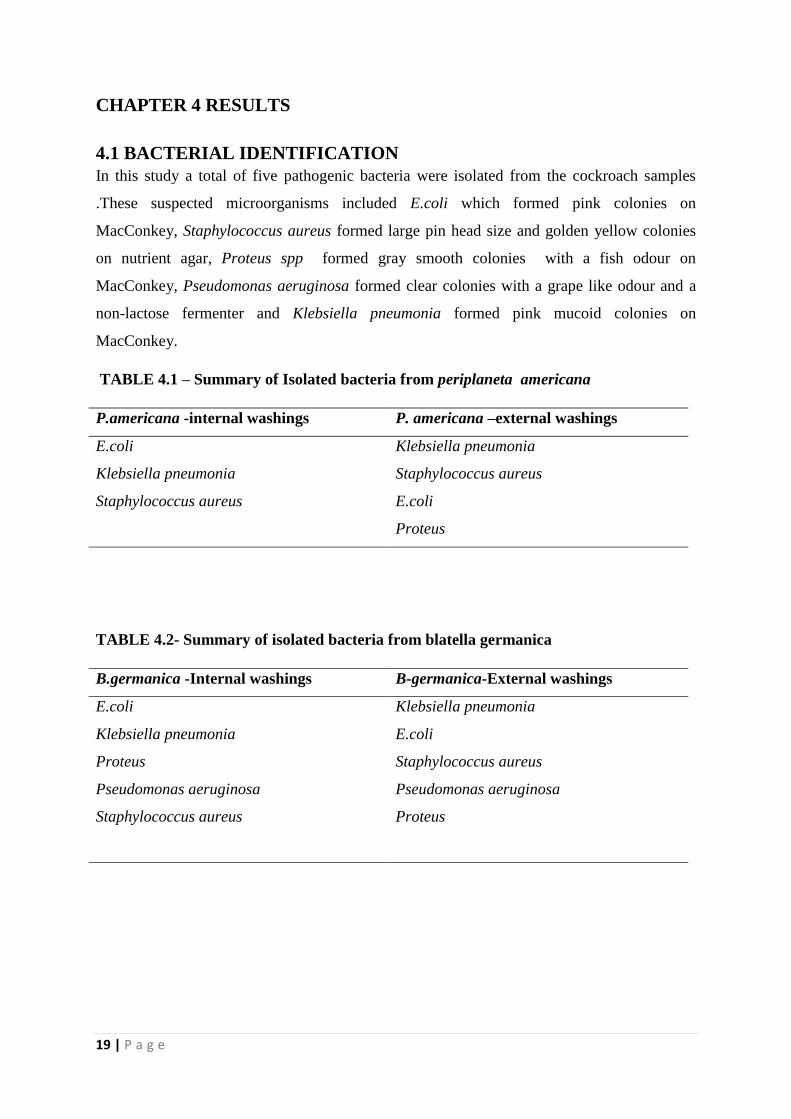
4.1 BACTERIAL IDENTIFICATION
In this study a total of five pathogenic bacteria were isolated from the cockroach samples
.These suspected microorganisms included E.coli which formed pink colonies on
MacConkey, Staphylococcus aureus formed large pin head size and golden yellow colonies
on nutrient agar, Proteus spp formed gray smooth colonies with a fish odour on
MacConkey, Pseudomonas aeruginosa formed clear colonies with a grape like odour and a
non-lactose fermenter and Klebsiella pneumonia formed pink mucoid colonies on
MacConkey.
TABLE 4.1 – Summary of Isolated bacteria from periplaneta americana
P.americana -internal washings P. americana –external washings
E.coli Klebsiella pneumonia
Klebsiella pneumonia Staphylococcus aureus
Staphylococcus aureus E.coli
Proteus
TABLE 4.2- Summary of isolated bacteria from blatella germanica
B.germanica -Internal washings B-germanica-External washings
E.coli Klebsiella pneumonia
Klebsiella pneumonia E.coli
Proteus Staphylococcus aureus
Pseudomonas aeruginosa Pseudomonas aeruginosa
Staphylococcus aureus Proteus
20 | P a g e
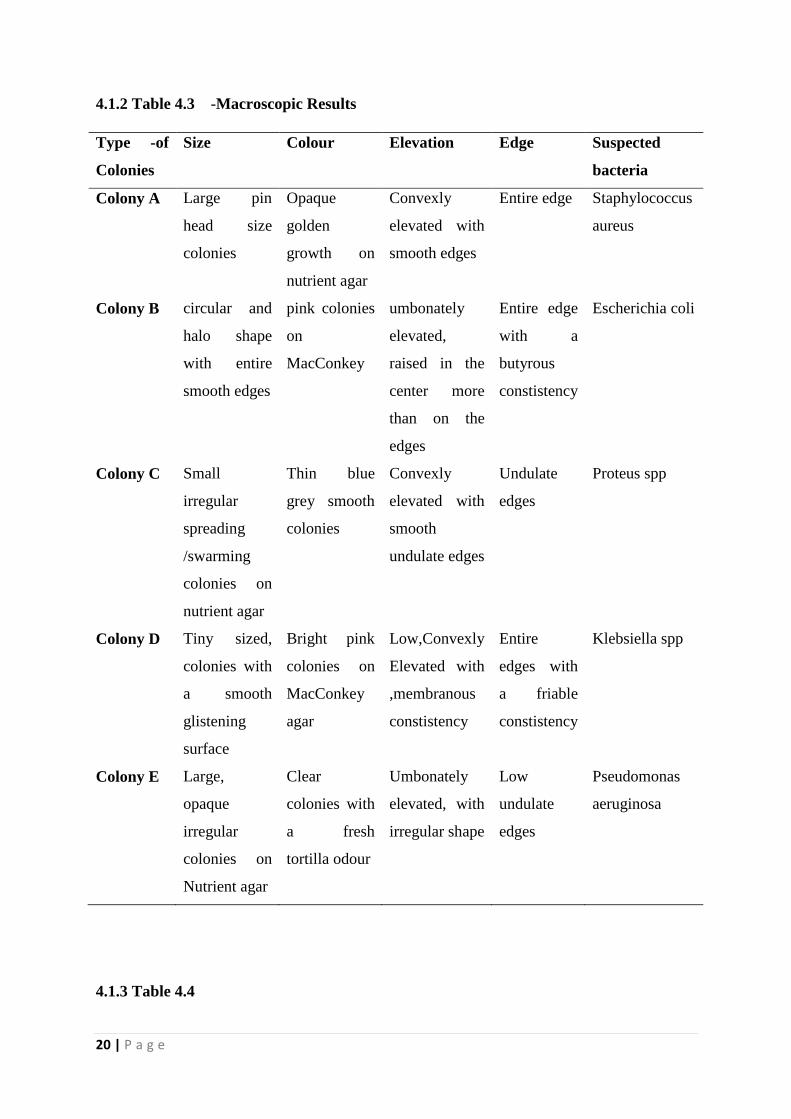
4.1.2 Table 4.3 -Macroscopic Results
Type -of
Colonies
Size Colour Elevation Edge Suspected
bacteria
Colony A Large pin
head size
colonies
Opaque
golden
growth on
nutrient agar
Convexly
elevated with
smooth edges
Entire edge Staphylococcus
aureus
Colony B circular and
halo shape
with entire
smooth edges
pink colonies
on
MacConkey
umbonately
elevated,
raised in the
center more
than on the
edges
Entire edge
with a
butyrous
constistency
Escherichia coli
Colony C Small
irregular
spreading
/swarming
colonies on
nutrient agar
Thin blue
grey smooth
colonies
Convexly
elevated with
smooth
undulate edges
Undulate
edges
Proteus spp
Colony D Tiny sized,
colonies with
a smooth
glistening
surface
Bright pink
colonies on
MacConkey
agar
Low,Convexly
Elevated with
,membranous
constistency
Entire
edges with
a friable
constistency
Klebsiella spp
Colony E Large,
opaque
irregular
colonies on
Nutrient agar
Clear
colonies with
a fresh
tortilla odour
Umbonately
elevated, with
irregular shape
Low
undulate
edges
Pseudomonas
aeruginosa
4.1.3 Table 4.4
21 | P a g e
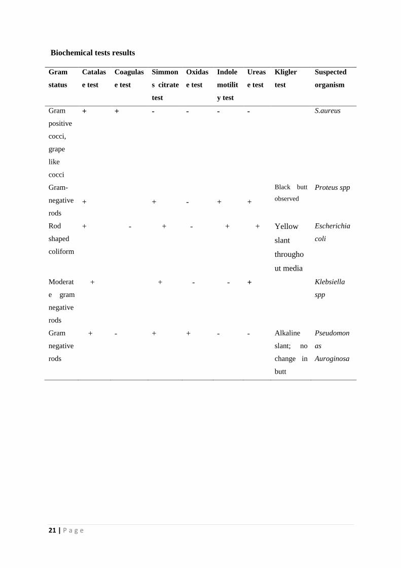
Biochemical tests results
Gram
status
Catalas
e test
Coagulas
e test
Simmon
s citrate
test
Oxidas
e test
Indole
motilit
y test
Ureas
e test
Kligler
test
Suspected
organism
Gram
positive
cocci,
grape
like
cocci
+ + - - - - S.aureus
Gram-
negative
rods
+
+
-
+
+
Black butt
observed
Proteus spp
Rod
shaped
coliform
+ - + - + + Yellow
slant
througho
ut media
Escherichia
coli
Moderat
e gram
negative
rods
+ + - - + Klebsiella
spp
Gram
negative
rods
+ - + + - - Alkaline
slant; no
change in
butt
Pseudomon
as
Auroginosa
22 | P a g e
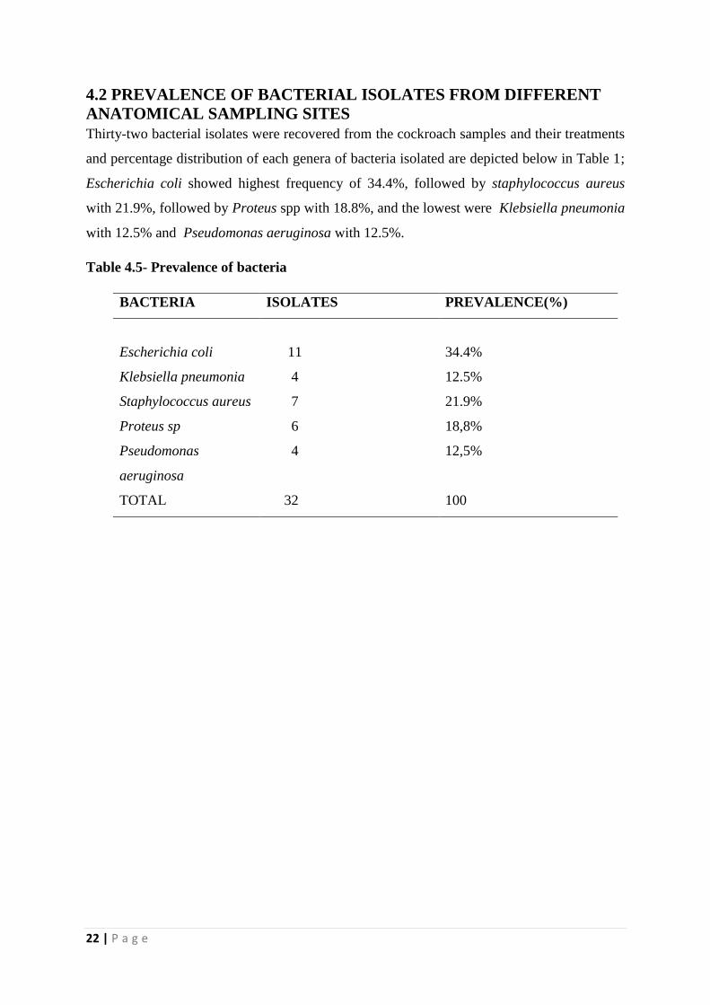
4.2 PREVALENCE OF BACTERIAL ISOLATES FROM DIFFERENT
ANATOMICAL SAMPLING SITES
Thirty-two bacterial isolates were recovered from the cockroach samples and their treatments
and percentage distribution of each genera of bacteria isolated are depicted below in Table 1;
Escherichia coli showed highest frequency of 34.4%, followed by staphylococcus aureus
with 21.9%, followed by Proteus spp with 18.8%, and the lowest were Klebsiella pneumonia
with 12.5% and Pseudomonas aeruginosa with 12.5%.
Table 4.5- Prevalence of bacteria
BACTERIA ISOLATES PREVALENCE(%)
Escherichia coli
Klebsiella pneumonia
Staphylococcus aureus
Proteus sp
Pseudomonas
aeruginosa
TOTAL
11
4
7
6
4
32
34.4%
12.5%
21.9%
18,8%
12,5%
100
23 | P a g e
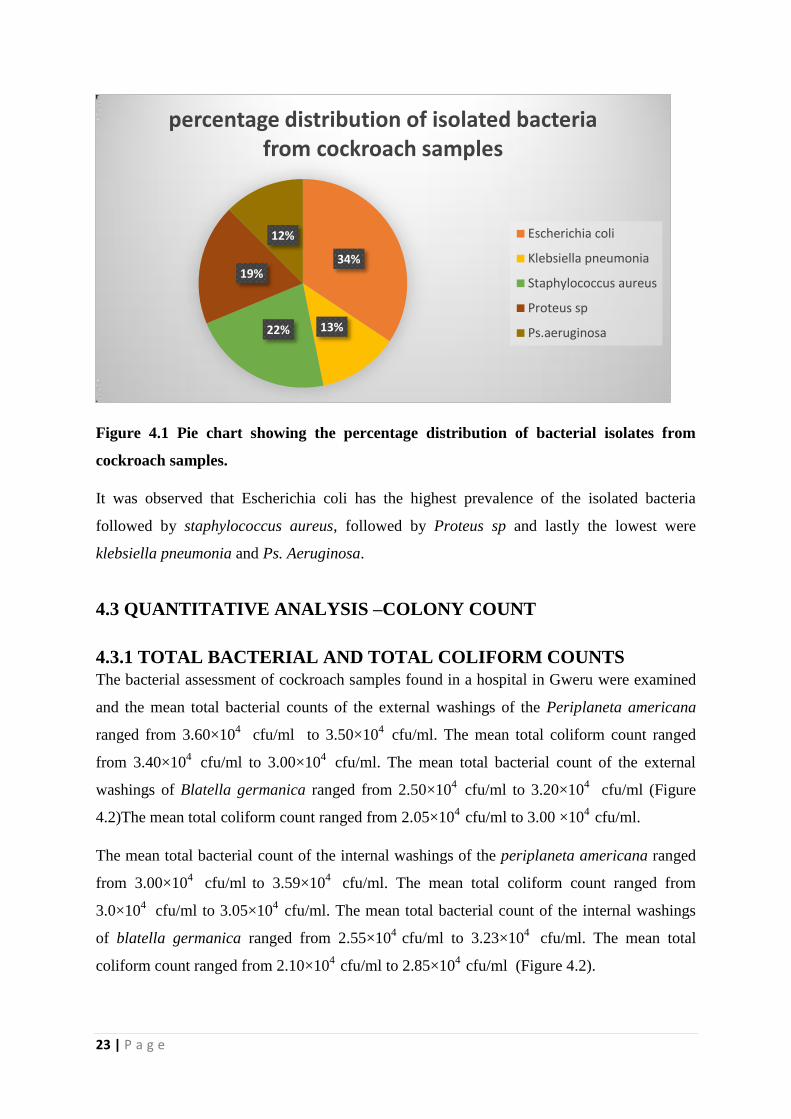
Figure 4.1 Pie chart showing the percentage distribution of bacterial isolates from
cockroach samples.
It was observed that Escherichia coli has the highest prevalence of the isolated bacteria
followed by staphylococcus aureus, followed by Proteus sp and lastly the lowest were
klebsiella pneumonia and Ps. Aeruginosa.
4.3 QUANTITATIVE ANALYSIS –COLONY COUNT
4.3.1 TOTAL BACTERIAL AND TOTAL COLIFORM COUNTS
The bacterial assessment of cockroach samples found in a hospital in Gweru were examined
and the mean total bacterial counts of the external washings of the Periplaneta americana
ranged from 3.60×10⁴
cfu/ml to 3.50×10⁴
cfu/ml. The mean total coliform count ranged
from 3.40×10⁴
cfu/ml to 3.00×10⁴
cfu/ml. The mean total bacterial count of the external
washings of Blatella germanica ranged from 2.50×10⁴
cfu/ml to 3.20×10⁴
cfu/ml (Figure
4.2)The mean total coliform count ranged from 2.05×10⁴
cfu/ml to 3.00 ×10⁴
cfu/ml.
The mean total bacterial count of the internal washings of the periplaneta americana ranged
from 3.00×10⁴
cfu/ml to 3.59×10
4 cfu/ml. The mean total coliform count ranged from
3.0×10⁴
cfu/ml to 3.05×10⁴
cfu/ml. The mean total bacterial count of the internal washings
of blatella germanica ranged from 2.55×10⁴
cfu/ml to 3.23×10⁴
cfu/ml. The mean total
coliform count ranged from 2.10×10⁴
cfu/ml to 2.85×10⁴
cfu/ml (Figure 4.2).
34%
13% 22%
19%
12%
percentage distribution of isolated bacteria from cockroach samples
Escherichia coli
Klebsiella pneumonia
Staphylococcus aureus
Proteus sp
Ps.aeruginosa
24 | P a g e
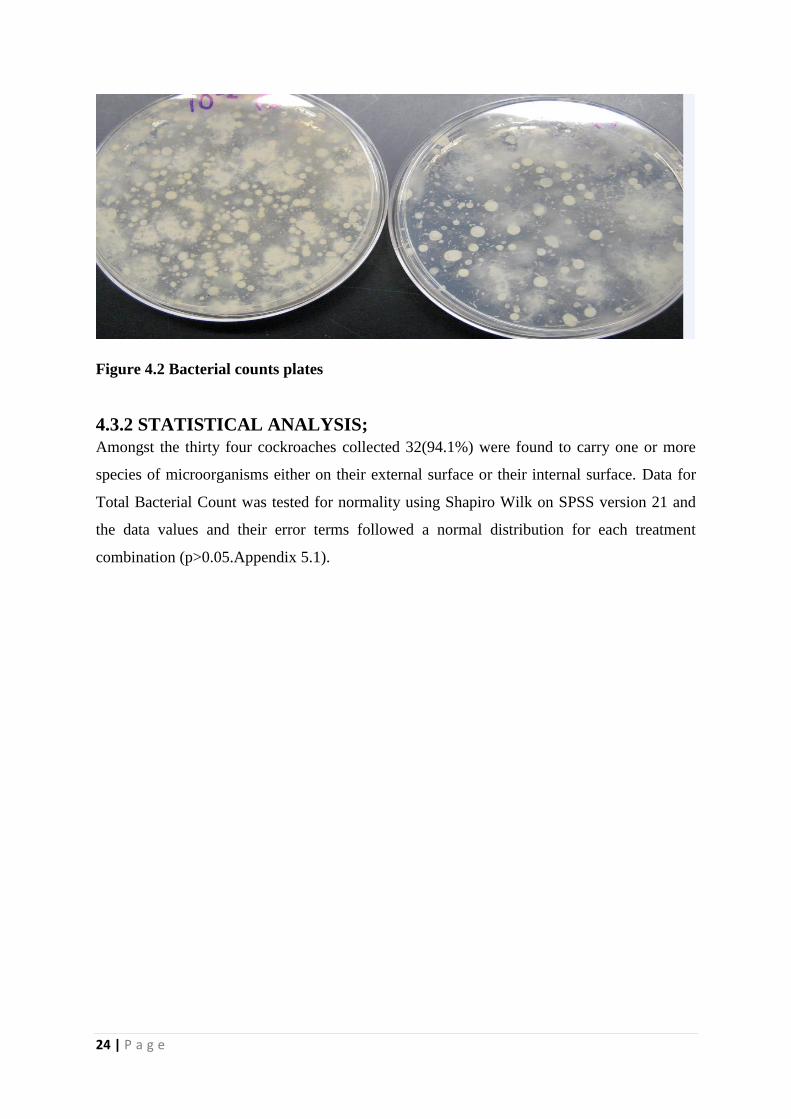
Figure 4.2 Bacterial counts plates
4.3.2 STATISTICAL ANALYSIS;
Amongst the thirty four cockroaches collected 32(94.1%) were found to carry one or more
species of microorganisms either on their external surface or their internal surface. Data for
Total Bacterial Count was tested for normality using Shapiro Wilk on SPSS version 21 and
the data values and their error terms followed a normal distribution for each treatment
combination (p>0.05.Appendix 5.1).
25 | P a g e
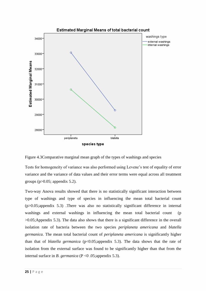
Figure 4.3Comparative marginal mean graph of the types of washings and species
Tests for homogeneity of variance was also performed using Levene‟s test of equality of error
variance and the variance of data values and their error terms were equal across all treatment
groups (p>0.05; appendix 5.2).
Two-way Anova results showed that there is no statistically significant interaction between
type of washings and type of species in influencing the mean total bacterial count
(p>0.05;appendix 5.3) .There was also no statistically significant difference in internal
washings and external washings in influencing the mean total bacterial count (p
>0.05;Appendix 5.3). The data also shows that there is a significant difference in the overall
isolation rate of bacteria between the two species periplaneta americana and blatella
germanica. The mean total bacterial count of periplaneta americana is significantly higher
than that of blatella germanica (p<0.05;appendix 5.3). The data shows that the rate of
isolation from the external surface was found to be significantly higher than that from the
internal surface in B. germanica (P <0 .05;appendix 5.3).
26 | P a g e
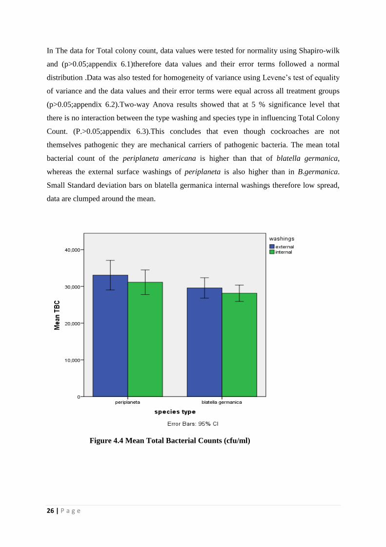
In The data for Total colony count, data values were tested for normality using Shapiro-wilk
and (p>0.05;appendix 6.1)therefore data values and their error terms followed a normal
distribution .Data was also tested for homogeneity of variance using Levene‟s test of equality
of variance and the data values and their error terms were equal across all treatment groups
(p>0.05;appendix 6.2).Two-way Anova results showed that at 5 % significance level that
there is no interaction between the type washing and species type in influencing Total Colony
Count. (P.>0.05;appendix 6.3).This concludes that even though cockroaches are not
themselves pathogenic they are mechanical carriers of pathogenic bacteria. The mean total
bacterial count of the periplaneta americana is higher than that of blatella germanica,
whereas the external surface washings of periplaneta is also higher than in B.germanica.
Small Standard deviation bars on blatella germanica internal washings therefore low spread,
data are clumped around the mean.
Figure 4.4 Mean Total Bacterial Counts (cfu/ml)
27 | P a g e
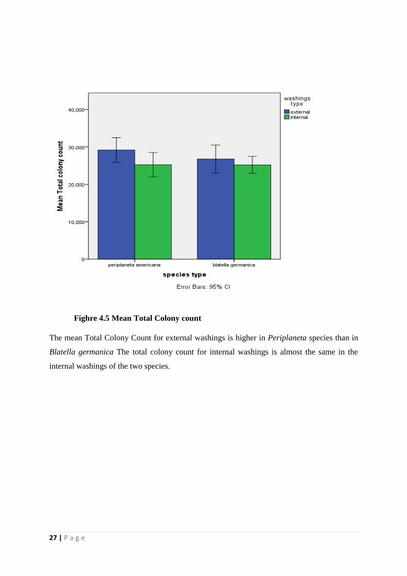
Fighre 4.5 Mean Total Colony count
The mean Total Colony Count for external washings is higher in Periplaneta species than in
Blatella germanica The total colony count for internal washings is almost the same in the
internal washings of the two species.
28 | P a g e
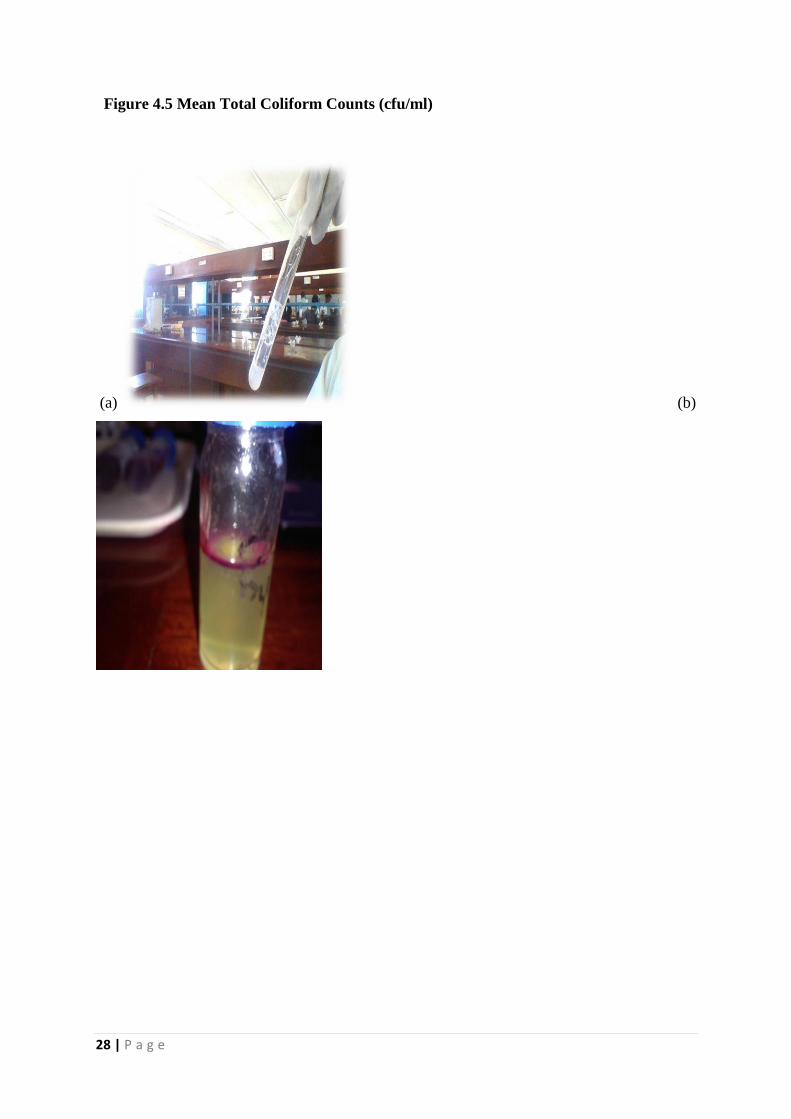
Figure 4.5 Mean Total Coliform Counts (cfu/ml)
(a) (b)
29 | P a g e
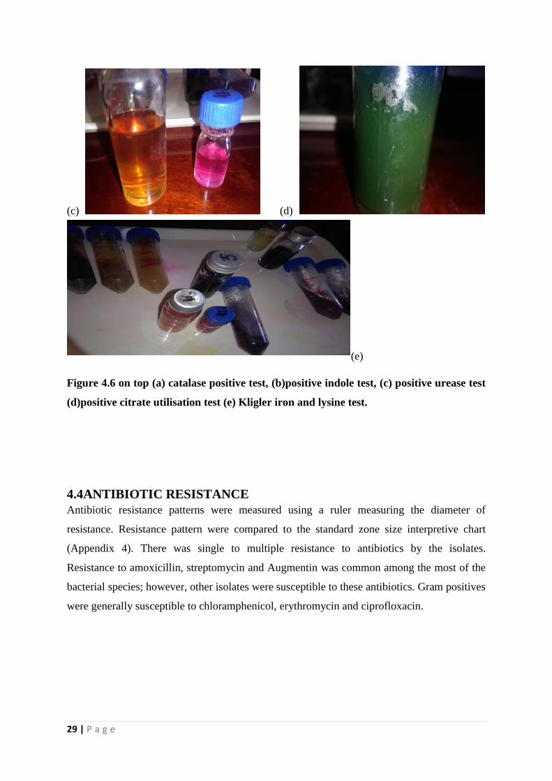
(c) (d)
(e)
Figure 4.6 on top (a) catalase positive test, (b)positive indole test, (c) positive urease test
(d)positive citrate utilisation test (e) Kligler iron and lysine test.
4.4ANTIBIOTIC RESISTANCE
Antibiotic resistance patterns were measured using a ruler measuring the diameter of
resistance. Resistance pattern were compared to the standard zone size interpretive chart
(Appendix 4). There was single to multiple resistance to antibiotics by the isolates.
Resistance to amoxicillin, streptomycin and Augmentin was common among the most of the
bacterial species; however, other isolates were susceptible to these antibiotics. Gram positives
were generally susceptible to chloramphenicol, erythromycin and ciprofloxacin.

30 | P a g e
CHAPTER 5 DISCUSSION
5.1 COCKROACHES AND THE CARRIAGE OF MICROORGANISMS
The purpose of this study was to prove the importance of sanitation in the hospital
environment. A greater number of problems can result from insanitary environmental
conditions and one of the key problems is the proliferation of such persistent pests such as
cockroaches. In Zimbabwe the most commonly found species of cockroaches are the
American cockroach(periplaneta americana) and the German cockroach (blatella
germanica).Periplanta americana is approximately 35 to 40 mm in length and blatella
germanica is 10 to 15 mm. Therefore Periplanta americana had a significantly higher
bacterial count than blatella germanica, this may be due to a larger surface area both on the
31 | P a g e
external and internal surface of the cockroach (Ahaduzzaman et al., 2014). Because
Periplaneta americana is 3- to 4-fold longer than B. germanica, it likely has significantly
larger external and internal surface areas. These findings suggest that insect size could
correlate with microorganism carriage. In addition to bacteria, in other studies done in
Taiwan Salehzadeh et al (2007)many species of parasites were isolated from external surface
and guts of cockroaches included cysts of Entameoba coli, Entameoba histolytica and
Giardia lamblia and adult and ova of Enterobius vermicularis and ova of Hymenolepis nana
and Ascaris lumbricoides (Hsueh et al., 2002).
Although Periplaneta harboured more total bacterial count than Blatella germanica it was
observed that the type of species of bacteria found in Blatella germanica (5 types of species)
were more than that of Periplaneta americana (3 types of species). This result is due to the
roaming territory differences between the two species and they are also known to have
different personalities regarding how they seek shelter (Akinjoginla et al., 2012). This mean
that Blatella germanica may be a more important potential vector of health acquired
infections. In many other studies, the German cockroach has been reported to harbour a
considerable number of pathogenic bacteria and fungi and other symbiotic protozoans which
are able to digest cellulose (Heidari et al., 2015).
In this study, the microorganisms which were isolated from external surface of cockroaches
were higher from that isolated from the internal surfaces. This demonstrated that bacteria and
parasites may be disseminated by contact more than their food habits. Ingestion, intestinal
transit of these organisms and their subsequent diffusion by faeces are not an absolute
necessity before cockroaches can disseminate organism and become involved in spreading
diseases (Hamid and Shahnaz, 2012). Many studies have also revealed the predominance (up
to 88 %) of isolates of Gram negative bacteria on the cuticle of cockroaches. Most of them
belong to the group Enterobacteriaceae. In fact, Blattaria are considered to be an ecological
niche of some Enterobacteriaceae (Russell and Jarvis, 2001).
5.1.1 ISOLATED BACTERIA
In this study five potentially pathogenic bacterial species were carried by cockroaches in
great numbers. Although in this study a small number of the pathogenic species have been
observed compared to other studies done by Feizhaddad et al.,(2012), this was due to
inadequate equipment and resources for a large scale study. Areas where people have low
level immunity (patients) should be aware of bacteria contamination from cockroaches
32 | P a g e
(Carter and Cole, 2012). In a study carried out by Vazirianzadeh et al. (2008) the isolated
bacteria were, Bacillus cereus, Bacillus subtilis, and S. aureus , E. coli, K. pneumoniae,
Neisseria species the study was mainly based on the sampling of external body and faeces
pellets of Egyptian cockroaches in different regions of Ahvaz city (Southwest Iran). In
another study carried out at the medical centres of Khorramshahr County in Iran, pathogenic
microorganisms were isolated from the external surfaces of all American cockroaches:
Klebsiella (47.9%), Pseudomonas (37%), E. coli (30.1%), Staphylococcus (24.6%),
Enterobacter (19.2%), Streptococcus (15.1%), Serratia (8.2%), Bacillus (4.1%), and Proteus
(2.7%) (Keseler et al., 2009). The study revealed that the place of collecting the cockroaches
can affect on results according to transmitted microorganisms: Klebsiella and Enterobacter
were more prevalent among the home cockroaches than in those from hospital environment.
Amongst the most common bacterial pathogens encountered in the hospital setting in this
study were:
5.1.2 ESCHERICHIA COLI
Escherichia coli is a Gram-negative, rod-shaped, coliform bacterium of the genus
Escherichia. The presence of E.coli was also confirmed by the biochemical tests that were
done. Most E. coli strains are harmless, but some serotypes can cause serious food poisoning
in their hosts. Escherichia coli is a facultative anaerobe which constitute 0.9% of gut flora
and their faecal-oral transmission is the major route through which pathogenic strains of the
bacterium cause disease (Tenaillan et al., 2010). Cells are able to survive outside the body for
a limited amount of time, which makes Escherichia coli the most important indication in
hospital environmental surveillance as a measure of faecal contamination. In this study
Escherichia coli was isolated in all the tested samples of cockroaches. The presence of E.coli
could indicate the degree of faecal contamination that is taking place in the two sampling
sites and most probably the fact that cockroaches are being exposed to the patient‟s faecal
matter as this is confirmed by the presence of E. coli. Cockroaches are able to spread this
bacteria through mechanical means during their nocturnal movements. The presence of E.coli
are also associated with many outbreaks of gastro-enteritis, food poisoning and dysentery
(Devrajani et al., 2009).
A study which was conducted in Rawalpindi hospital revealed that all collected cockroaches
carry S. aureus and E. coli on their external body surface (Mlso et al., 2005). This study also
indicated almost the same result. Indoor sanitary condition has been significantly correlated
33 | P a g e
with infestation of cockroaches. Therefore prevention of such bacteria can only be done
through thorough pest control practices in hospitals and in homes (Rajagopala et al., 2014).
5.1.3 STAPHYLOCOCCUS AUREUS
Staphylococcus aureus is a Gram positive bacteria and is a facultative anaerobe. In this
study,7 samples were positive for Staphylococcus aureus and this was confirmed by the
Gram staining, catalase positive and coagulase positive. Pathogenic strains produce
enterotoxins which, when ingested, can cause gastroenteritis (Lowy, 2012).The presence of
staphylococcus in both the internal surface of the cockroaches and the internal surface proved
that cockroaches are responsible for most Staphylococcal infections found in hospital settings
(Heymann, 2002).There are many strains of Staphylococcus aureus and as such it is easy to
spread around a hospital facility causing nosocomial infections (Hennekinne, 2012).
Strains of bacteria that are resistant to beta-lactam antibiotics are called methicillin-resistant
Staphylococcus aureus (MRSA) (Tang et al., 2015). Methicillin resistant S. aureus (MRSA)
is also one of the supreme causes of nosocomial infections in hospitals. 40% to 70% hospital
associated infections are chiefly caused by S. aureus in intensive care units (ICUs) in which
MRSA is a major cause of HAIs (Lin et al., 2012). Health workers and medical staff are
more likely to contract MRSA infections. These infections will ultimately lead to severe
illness and deaths of medical staff and workers working on pathogenic microorganisms
(Mahmood et al., 2010) and these in turn would have been spread by the movements of
cockroaches.
5.1.4 KLEBSIELLA PNEUMONIA
In this study Klebsiella pneumonia was isolated in four samples of both species of
cockroaches. The existence of the bacteria was observed by growing the samples on
MacConkey in which it produced mucoid pink colonies indicating its lactose fermenting
characteristics(Tortora, 2010). Klebsiella Pneumonia is a type of Gram-negative bacteria that
causes different types of healthcare-associated infections, including pneumonia, bloodstream
infections, wound or surgical site infections, and meningitis. The existence of such a bacteria
klebsiella pneumonia in cockroaches is a hazard to health. It has since been observed by
scientists that Klebsiella has increasingly developed antimicrobial resistance, most recently to
the class of antibiotics known as carbapenems, carbapenem antibiotics often are the last line
34 | P a g e
of defence against Gram-negative infections that are resistant to other antibiotics(Siegel et al.,
2007).
5.1.5 PSEUDOMONAS AERUGINOSA
Pseudomonas aeruginosa is a common Gram-negative, rod-shaped bacterium. In this study, 4
samples were positive for this bacteria P. aeruginosa (Knockgether et al., 2011). Even
though cockroaches are not themselves pathogenic they carry such pathogenic bacteria
(Horner et al., 2012). Pseudomonas infections are generally treated with antibiotics.
Unfortunately, in hospitalized patients, Pseudomonas infections, like those caused by many
other hospital bacteria, are becoming more difficult to treat because of increasing antibiotic
resistance (Funke, 2016). Due to multi drug resistance pattern in most bacteria isolates,
selecting the right antibiotic usually requires that a specimen from a patient be sent to a
laboratory to test to see which antibiotics might still be effective for treating the infection
(Karlowsky et al., 2009).
Multidrug-resistant Pseudomonas can be deadly for patients in critical care. An estimated
51,000 healthcare-associated P. aeruginosa infections occur in the United States each year.
More than 6,000 (13%) of these are multidrug-resistant, with roughly 400 deaths per year
attributed to these infections (Leach et al., 2016). Multidrug-resistant Pseudomonas was
given a threat level of serious threat in the CDC AR Threat report (Ryan and Ray, 2004).
5.1.6 PROTEUS SP
Proteus vulgaris was isolated from cockroaches in this study and the striking microbiological
characteristic of Proteus species is their swarming activity and fishy odour on MacConkey
agar. Swarming appears macroscopically as concentric rings of growth emanating from a
single colony or an inoculum (Adler et al., 2013; (Armbbruster and Mobley, 2012). P.
mirabilis. ,P. vulgaris and P. penneri are easily isolated from individuals in long-term care
facilities and hospitals and from patients with underlying diseases or compromised immune
systems and causes 90% of Proteus infections (Bauchillan et al., 2013)( These bacteria
disseminated by cockroaches and other mechanical vectors causes nosocomial infections
which results in higher motility and costs Doern et al., 2005). There has also been one case
study of P. vulgaris causing bacteremia and brain abscesses, with the suspected point of entry
being the digestive tract (Chong et al ., 2013).
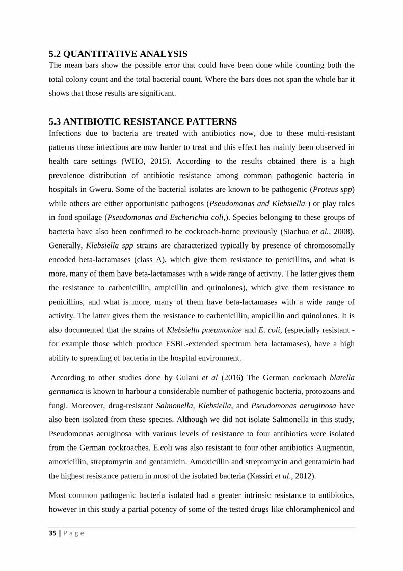
35 | P a g e
5.2 QUANTITATIVE ANALYSIS
The mean bars show the possible error that could have been done while counting both the
total colony count and the total bacterial count. Where the bars does not span the whole bar it
shows that those results are significant.
5.3 ANTIBIOTIC RESISTANCE PATTERNS
Infections due to bacteria are treated with antibiotics now, due to these multi-resistant
patterns these infections are now harder to treat and this effect has mainly been observed in
health care settings (WHO, 2015). According to the results obtained there is a high
prevalence distribution of antibiotic resistance among common pathogenic bacteria in
hospitals in Gweru. Some of the bacterial isolates are known to be pathogenic (Proteus spp)
while others are either opportunistic pathogens (Pseudomonas and Klebsiella ) or play roles
in food spoilage (Pseudomonas and Escherichia coli,). Species belonging to these groups of
bacteria have also been confirmed to be cockroach-borne previously (Siachua et al., 2008).
Generally, Klebsiella spp strains are characterized typically by presence of chromosomally
encoded beta-lactamases (class A), which give them resistance to penicillins, and what is
more, many of them have beta-lactamases with a wide range of activity. The latter gives them
the resistance to carbenicillin, ampicillin and quinolones), which give them resistance to
penicillins, and what is more, many of them have beta-lactamases with a wide range of
activity. The latter gives them the resistance to carbenicillin, ampicillin and quinolones. It is
also documented that the strains of Klebsiella pneumoniae and E. coli, (especially resistant -
for example those which produce ESBL-extended spectrum beta lactamases), have a high
ability to spreading of bacteria in the hospital environment.
According to other studies done by Gulani et al (2016) The German cockroach blatella
germanica is known to harbour a considerable number of pathogenic bacteria, protozoans and
fungi. Moreover, drug-resistant Salmonella, Klebsiella, and Pseudomonas aeruginosa have
also been isolated from these species. Although we did not isolate Salmonella in this study,
Pseudomonas aeruginosa with various levels of resistance to four antibiotics were isolated
from the German cockroaches. E.coli was also resistant to four other antibiotics Augmentin,
amoxicillin, streptomycin and gentamicin. Amoxicillin and streptomycin and gentamicin had
the highest resistance pattern in most of the isolated bacteria (Kassiri et al., 2012).
Most common pathogenic bacteria isolated had a greater intrinsic resistance to antibiotics,
however in this study a partial potency of some of the tested drugs like chloramphenicol and
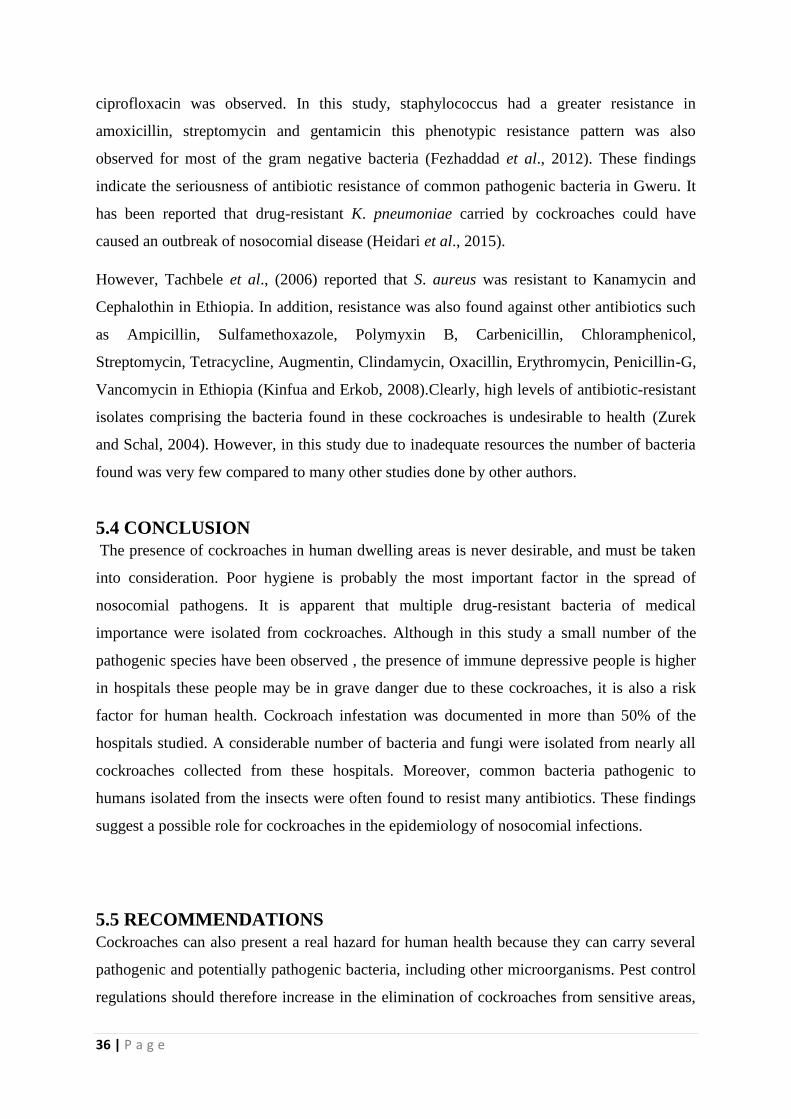
36 | P a g e
ciprofloxacin was observed. In this study, staphylococcus had a greater resistance in
amoxicillin, streptomycin and gentamicin this phenotypic resistance pattern was also
observed for most of the gram negative bacteria (Fezhaddad et al., 2012). These findings
indicate the seriousness of antibiotic resistance of common pathogenic bacteria in Gweru. It
has been reported that drug-resistant K. pneumoniae carried by cockroaches could have
caused an outbreak of nosocomial disease (Heidari et al., 2015).
However, Tachbele et al., (2006) reported that S. aureus was resistant to Kanamycin and
Cephalothin in Ethiopia. In addition, resistance was also found against other antibiotics such
as Ampicillin, Sulfamethoxazole, Polymyxin B, Carbenicillin, Chloramphenicol,
Streptomycin, Tetracycline, Augmentin, Clindamycin, Oxacillin, Erythromycin, Penicillin-G,
Vancomycin in Ethiopia (Kinfua and Erkob, 2008).Clearly, high levels of antibiotic-resistant
isolates comprising the bacteria found in these cockroaches is undesirable to health (Zurek
and Schal, 2004). However, in this study due to inadequate resources the number of bacteria
found was very few compared to many other studies done by other authors.
5.4 CONCLUSION
The presence of cockroaches in human dwelling areas is never desirable, and must be taken
into consideration. Poor hygiene is probably the most important factor in the spread of
nosocomial pathogens. It is apparent that multiple drug-resistant bacteria of medical
importance were isolated from cockroaches. Although in this study a small number of the
pathogenic species have been observed , the presence of immune depressive people is higher
in hospitals these people may be in grave danger due to these cockroaches, it is also a risk
factor for human health. Cockroach infestation was documented in more than 50% of the
hospitals studied. A considerable number of bacteria and fungi were isolated from nearly all
cockroaches collected from these hospitals. Moreover, common bacteria pathogenic to
humans isolated from the insects were often found to resist many antibiotics. These findings
suggest a possible role for cockroaches in the epidemiology of nosocomial infections.
5.5 RECOMMENDATIONS
Cockroaches can also present a real hazard for human health because they can carry several
pathogenic and potentially pathogenic bacteria, including other microorganisms. Pest control
regulations should therefore increase in the elimination of cockroaches from sensitive areas,

37 | P a g e
such as hospitals, this practice is very essential. In food-handling establishments and human
dwellings, cockroaches must also be controlled, to maintain acceptable hygiene standards.
Therefore, further studies are clearly necessary to be carried out in order to investigate
relevant control methods against cockroaches using effective health programs focusing on
hygiene measures in medical institutions.
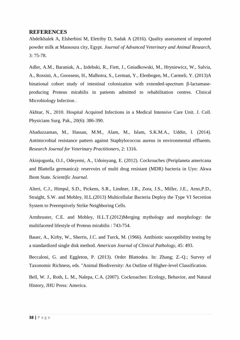
38 | P a g e
REFERENCES
Abdelkhalek A, Elsherbini M, Eletriby D, Sadak A (2016). Quality assessment of imported
powder milk at Mansoura city, Egypt. Journal of Advanced Veterinary and Animal Research,
3: 75-78.
Adler, A.M., Baraniak, A., Izdebski, R., Fiett, J., Gniadkowski, M., Hryniewicz, W., Salvia,
A., Rossini, A., Goossens, H., Malhotra, S., Lerman, Y., Elenbogen, M., Carmeli, Y. (2013)A
binational cohort study of intestinal colonization with extended-spectrum β-lactamase-
producing Proteus mirabilis in patients admitted to rehabilitation centres. Clinical
Microbiology Infection .
Akhtar, N., 2010. Hospital Acquired Infections in a Medical Intensive Care Unit. J. Coll.
Physicians Surg. Pak., 20(6): 386-390.
Ahaduzzaman, M., Hassan, M.M., Alam, M., Islam, S.K.M.A., Uddin, I. (2014).
Antimicrobial resistance pattern against Staphylococcus aureus in environmental effluents.
Research Journal for Veterinary Practitioners, 2: 1316.
Akinjogunla, O.J., Odeyemi, A., Udoinyang, E. (2012). Cockroaches (Periplaneta americana
and Blattella germanica): reservoirs of multi drug resistant (MDR) bacteria in Uyo: Akwa
Ibom State. Scientific Journal.
Alteri, C.J., Himpsl, S.D., Pickens, S.R., Lindner, J.R., Zora, J.S., Miller, J.E., Arno,P.D.,
Straight, S.W. and Mobley, H.L.(2013) Multicellular Bacteria Deploy the Type VI Secretion
System to Preemptively Strike Neighboring Cells.
Armbruster, C.E. and Mobley, H.L.T.(2012)Merging mythology and morphology: the
multifaceted lifestyle of Proteus mirabilis : 743-754.
Bauer, A., Kirby, W., Sherris, J.C. and Turck, M. (1966). Antibiotic susceptibility testing by
a standardized single disk method. American Journal of Clinical Pathology, 45: 493.
Beccaloni, G. and Eggleton, P. (2013). Order Blattodea. In: Zhang; Z.-Q.; Survey of
Taxonomic Richness, eds. "Animal Biodiversity: An Outline of Higher-level Classification.
Bell, W. J., Roth, L. M., Nalepa, C.A. (2007). Cockroaches: Ecology, Behavior, and Natural
History, JHU Press: America.

39 | P a g e
Botella, C.M., Juan, M. C., Baños, R. M., Alcañiz, M., Guillén, V.and Rey, B. (2005).
"Mixing Realities? An Application of Augmented Reality for the Treatment of Cockroach
Phobia". Cyber Psychology & Behaviour.
Bouamama, L., Sorlozano, A., Laglaoui, A., Lebbadi, M., Aarab, A. and Gutierrez, J.
(2010)Antibiotic resistance patterns of bacterial strains isolated from Periplaneta americana
and Musca domestica in Tangier, Morocco. J Infect Dev Ctries:194–201.
Ben, G. (2016). The case for cockroach milk: The next superfood?
Bennett, G.W. and Owens, J.M. (2012). Advances in urban pest management. Van Nostrand
Reinhold; pp 220-225.
Bouchillon, S.K., Badal, R.E., Hoban, D.J., Hawser, S.P.(2011)Antimicrobial Susceptibility
of Inpatient Urinary Tract Isolates of Gram-Negative Bacilli in the United States: Results
from the Study for Monitoring Antimicrobial Resistance Trends (SMART) Program.
Carter, G.R. and Cole, Jr J.R. (2012). Diagnostic Procedure in Veterinary Bacteriology and
Mycology. 6th Edition, Academic Press: San Diego: California; pp 620.
Cheesbrough, M. (2006). District laboratory practice in tropical countries .vol.11
microbiology 2nd
edition, Cambridge University press; Britain. pp 158-195.
Copeland, M.(2004). Cockroach, Reaktion Books. (pp 86 - 88).
Chong, Y., Shimoda, S., Yakushiji, H., Ito, Y., Miyamoto, T., Kamimura, T., Shimono, N.
and Akashi, K.(2013) Community spread of extended-spectrum beta-lactamase-producing
Escherichia coli, Klebsiella pneumoniae and Proteus mirabilis: a long-term study in Japan. J
Medical Microbiology.
Doern, G.V., Brueggemann, A.B., Perla, R., Daly, J., Halkias, D., Jones, R.N. and Saubolle,
M.A.(1997) Multicenter laboratory evaluation of the bioMerieux Vitek antimicrobial
susceptibility testing system with 11 antimicrobial agents versus members of the family
Enterobacteriaceae and Pseudomonas aeruginosa.
Ducel, G. ,Fabry, J. and Nicolle L.(2012) Prevention of Hospital Acquired Infections,2nd
edition, WHO/BAC/79.1. 2.
Elgderi, R. M., K. S. Ghenghesh and Berbash, N. (2006) Carriage by the German cockroach
(Blattella germanica) of multipleantibiotic-resistant bacteria that are potentially pathogenic to
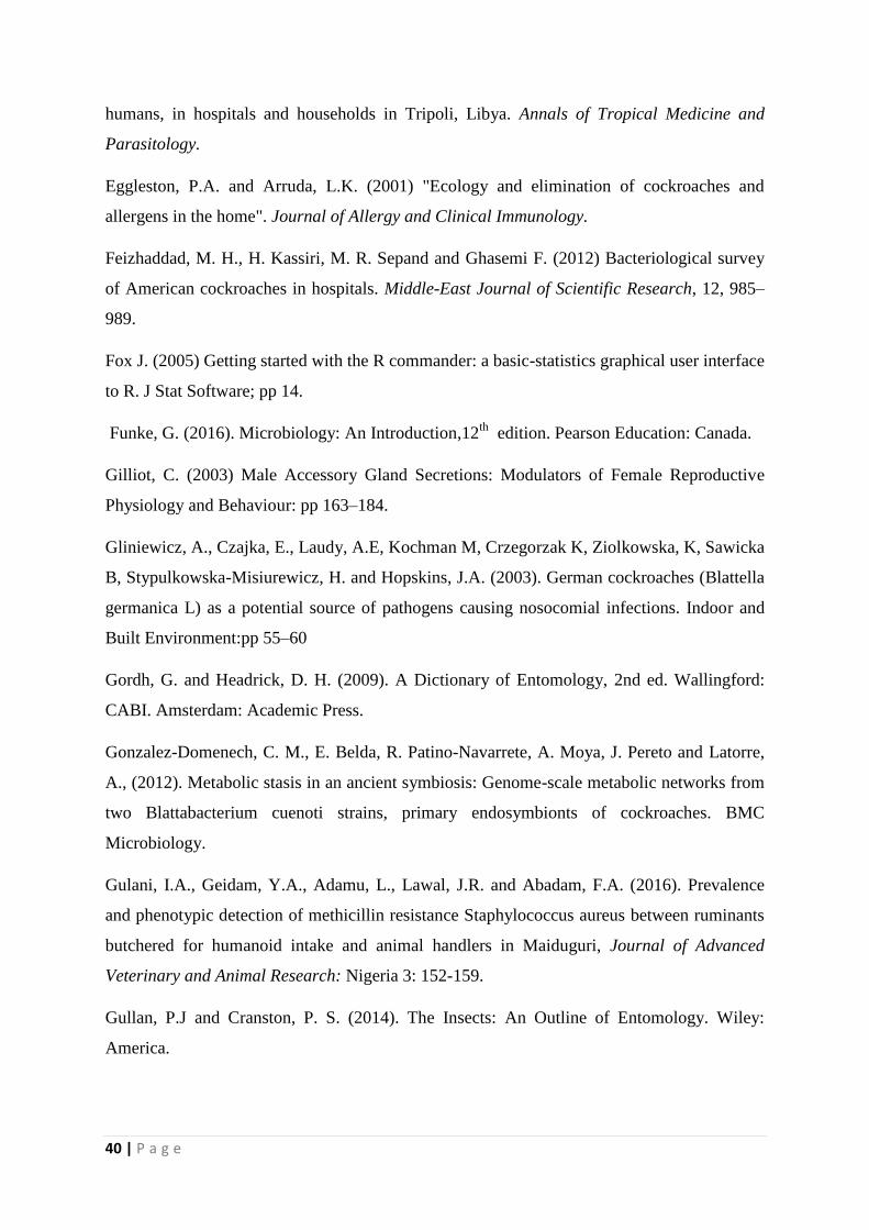
40 | P a g e
humans, in hospitals and households in Tripoli, Libya. Annals of Tropical Medicine and
Parasitology.
Eggleston, P.A. and Arruda, L.K. (2001) "Ecology and elimination of cockroaches and
allergens in the home". Journal of Allergy and Clinical Immunology.
Feizhaddad, M. H., H. Kassiri, M. R. Sepand and Ghasemi F. (2012) Bacteriological survey
of American cockroaches in hospitals. Middle-East Journal of Scientific Research, 12, 985–
989.
Fox J. (2005) Getting started with the R commander: a basic-statistics graphical user interface
to R. J Stat Software; pp 14.
Funke, G. (2016). Microbiology: An Introduction,12th
edition. Pearson Education: Canada.
Gilliot, C. (2003) Male Accessory Gland Secretions: Modulators of Female Reproductive
Physiology and Behaviour: pp 163–184.
Gliniewicz, A., Czajka, E., Laudy, A.E, Kochman M, Crzegorzak K, Ziolkowska, K, Sawicka
B, Stypulkowska-Misiurewicz, H. and Hopskins, J.A. (2003). German cockroaches (Blattella
germanica L) as a potential source of pathogens causing nosocomial infections. Indoor and
Built Environment:pp 55–60
Gordh, G. and Headrick, D. H. (2009). A Dictionary of Entomology, 2nd ed. Wallingford:
CABI. Amsterdam: Academic Press.
Gonzalez-Domenech, C. M., E. Belda, R. Patino-Navarrete, A. Moya, J. Pereto and Latorre,
A., (2012). Metabolic stasis in an ancient symbiosis: Genome-scale metabolic networks from
two Blattabacterium cuenoti strains, primary endosymbionts of cockroaches. BMC
Microbiology.
Gulani, I.A., Geidam, Y.A., Adamu, L., Lawal, J.R. and Abadam, F.A. (2016). Prevalence
and phenotypic detection of methicillin resistance Staphylococcus aureus between ruminants
butchered for humanoid intake and animal handlers in Maiduguri, Journal of Advanced
Veterinary and Animal Research: Nigeria 3: 152-159.
Gullan, P.J and Cranston, P. S. (2014). The Insects: An Outline of Entomology. Wiley:
America.
41 | P a g e
Harwood, R,F. and James, M.T.(1979) In Entomology in human and animal health, 7th ed.
New York; Macmillan Publishing
Hamid, K. and Shahnaz, K. (2012). Cockroaches Periplaneta americana (L.), Dictyoptera;
Blattidae as carriers of bacterial pathogens, Khorramshahr County, Iran. Jundishapur Journal
of Microbiology, 5: 320-322.
Heidari, S.E., Doosti, .A and Khalaji, P.Y. (2015). Prevalence and antibiotic susceptibility
patterns of Staphylococcus aureus isolated from cockroaches from Chaharmahal va Bakhtiari
hospitals. Medical Science Journal of Islamic Azad University-Tehran Medical Branch: 206-
212.
Hennekinne, J.A., De Buyser, M.L. and Dragacci, M. (2012) Staphylococcus aureus and its
food poisoning toxins: characterization and outbreak investigation. FEMS Microbiology
Reviews 36(4):815
Heymann, D.L. (2008) Control of communicable diseases manual: American Public Health
Association.
Hsueh, P.R., Liu, C.Y. and Luh, K.T. (2002). Current status of antimicrobial resistance in
Taiwan. Emergence Infect Dis:132-137.
Huang, C.Y., Sabree, Z.L. and Moran, N.A. (2012). "Genome Sequence of Blattabacterium
sp. Strain BGIGA, Endosymbiont of the Blaberus giganteus Cockroach". Journal of
Bacteriology.
Hoell, H. V., Doyen, J. T.and Purcell, A. H. (1998). Introduction to Insect Biology and
Diversity, 2nd ed., Oxford University Press: Britain.
Islam, M.J., Uddin, M.S., Nasrin, M.S., Nazir, K.H., Rahman, M.T.and Alam, M.M. (2007).
Prevalence of enterotoxigenic and toxic shock syndrome toxin-1 producing coagulase
positive Staphylococcus aureus cockroaches (Periplaneta americana and Blattella germanica).
Acta Tropica, 93: 259-265.
Kassiri, H., Kasiri, A. and Quaderi, A. (2014). Detection and Prevalence Rate of American
Cockroaches (Periplaneta americana) Bacterial Infections in Human Dwellings. Archive of
Clinical Infectious Diseases.
42 | P a g e
Keseler, I. M., C. Bonavides-Martnez, J. Collado-Vides, S. Gama-Castro, R. P. Gunsalus, D.
A. Johnson, M. Krummenacker, L. M. Nolan, S. Paley & I. T. Paulsen. (2009). EcoCyc: A
comprehensive view of Escherichia coli biology. Nucleic Acids Research, 37, 464–470
Khalaji, Y., A. Doosti & S. Ghorbani-Dalini, (2013). Molecular evaluation of antibiotic
resistance prevalence in Pseudomonas aeruginosa isolated from cockroaches in South
Kinfua, A. and Erkob, B.(2008). Cockroaches as carrier of human intestinal parasites in tow
localities in Ethiopia. Trans. Roy. Soc. Trop. Med. Hygiene
Klockgether, J., Cramer, N., Wiehlmann, L., Davenport, C.F. and Tümmler, B. (2011).
"Pseudomonas aeruginosa Genomic Structure and Diversity". Frontiers in Microbiology.
Leach, R., Moore, K. and Bell, D. (2016). Oxford Desk Reference: Acute Medicine. Oxford
University Press:Britain.
Legendre, F., Nel, A., Svenson, G. J., Robillard, T., Pellens, R. and Grandcolas, P. (2015).
"Phylogeny of Dictyoptera: Dating the Origin of Cockroaches, Praying Mantises and
Termites with Molecular Data and Controlled Fossil Evidence."
Lihoreau, M., Costa, J.T.and Rivault, C. (2012). "The social biology of domiciliary
cockroaches: colony structure, kin recognition and collective decisions". Insectes Sociaux.
Li, J. and Ho, S.H. Pandan leaves (Pandanus amaryllifolius Roxb.) As A Natural Cockroach
Repellent. Proceedings of the 9th National Undergraduate Research Opportunities
Programme (13 September 2003).
Lowy, F.D. (2012). Staphylococcal Infections. Harrison‟s Principles of Internal Medicine.
18th edition. New York: McGraw-Hill.
Marriott, N.G.and Gravani, R.B.(2006) Pest control. Principles of food sanitation (Food
science texts series) New York: Springer.
Mlso, W.R., Qureshi, A.H., Khan, I.A. and Hussain, S., (2005) Frequency of different species
of cockroaches in tertiary care hospital and their role in transmission of bacterial pathogens.
Pak. J. Med. Res.
Mpuchane, S. Matsheka, I.M., Gashe, B.A., Allotey, J., Murindamombe, G. and Mrema, N.,
(2006). Microbiological studies of cockroaches from three localities in Gaborone, Botswana.
African Journal of Food, Agriculture, Nutrition and Development, 6, 17
43 | P a g e
Malcolm, M. (2013). How to eat a cockroach: a Telegraph guide. Washington Post: New
York.
Mullen, G. and Durden, L. (2002). Medical and Veterinary Entomology, 2nd
ed ; New York.
Pai H-H. (2012)Multidrug resistant bacteria isolated from cockroaches in long-term care
facilities and nursing homes.
Pavlik I, Falkinham JO. The occurrence of pathogenic and potentially pathogenic
mycobacteria in animals and the role of the environment in the spread of infection. In: Kazda
J, Pavlik I, Falkinham JO III, Hruska K, editors. The ecology of mycobacteria: impact on
animal‟s and human‟s health. Dordrecht: Springer; 2009. p. 199–281.
Rajagopala S.V., Sikorski, P., Kumar, A., Mosca, R., Vlasblom, J., Arnold, R., Franca-Koh,
J., Pakala, S.B., Phanse, S., Ceol, A., Häuser, R., Siszler, G., Wuchty, S., Emili, A., Babu,
M., Aloy, P., Pieper, R., Uetz, P. ( 2014). "The binary protein-protein interaction landscape of
Escherichia coli".
Rentz, D. (2014). A Guide to the Cockroaches of Australia. CSIRO Publishing nature
Biotechnology.
Richard, S. (2015). The Cockroach Papers: A Compendium of History and Lore ,University
of Chicago Press.
Rivault, C., Cloarec, A., Le Guyader A. (1993). Bacterial contamination of food by
cockroaches. Journal of Environmental Health, 55: 21-23.
Roth, L.M., Willis, E.R.. (2010)The medical and veterinary importance of cockroaches.
Smithsonian miscellaneous collection, 1957, 134: 1–147
Russell, J.B., Jarvis, G.N. (2001). "Practical mechanisms for interrupting the oral-fecal
lifecycle of Escherichia coli". Journal of Molecular Microbiology and Biotechnology.
Sabra, S.M., and Abdel-Fattah, M.M. 2012. Epidemiological and microbiological profile of
nosocomial infection in Taif hospitals, KSA (2010-2011). World Journal of Medical
Sciences, 7: 1-9. 2.
Salehzadeh, A., Tavacol, P., Mahjub, H. (2007). Bacterial, fungal and parasitic contamination
of cockroaches in public hospitals of Hamadan, Iran. Journal of Vector Borne Diseases, 44:
105-110
44 | P a g e
Sayyad, S., Vahabi, A., Vahabi, B., Sayyadi, M., Sahne, S.H. (2016). Investigation of
bacteriological infections of the American Cockroaches in Paveh City, Kermanshah Province.
Materia Socio-Medica, 28: 1720. Schroeder
Siachua, P.; Pinmai, Kh.; Somrithipol, S. and Tor-Udom, S.(2008). Isolation of medically
important fungi from cockroaches at Thammasat Chalermprakiat hospital. Thamasat, Med.
J.8(3):345-349.
Tachebe, J., Erku, W., Gerbe-Michae, L.T., Ashenafi M. (2006)Cockroach associated food-
borne bacteria from hospital and restaurant in Addis Ababa, Ethiopia: distribution and
antibiograms. J Rural Trop Public Health.
Tenaillon, O. Skurnik, D. Picard, B. Denamur, E. (2010). "The population genetics of
commensal Escherichia coli". Nature Reviews Microbiology.
Tortora, G.(2010). Microbiology: An Introduction. San Francisco, CA: Benjamin Cummings.
Umunnabuike, A.C., Irokanulo, E.A.. Isolation of Campylobacter subsp. jejuni from Oriental
and American cockroaches caught in kitchens and poultry houses in Vom, Nigeria.
Vazirianzadeh, B., S. S. Solary, M. Rahdar, R. Hajhossien & M. Mehdinejad, (2008).
Identification of bacteria which possible transmitted by Musca domestica.
Wannigama, D. L., R. Dwivedi, A. ,Zahraei, R. (2014). Prevalence and antibiotic resistance
of Gram-negative pathogenic bacteria species isolated from Periplaneta americana and
Blattella germanica in Varanasi, India. Journal of Arthropod-Borne Diseases, 8, 10–20. Yagi
Wood, F., Robinson, W.H., Kraft ,S.K., Zungoli, P.A. (2010). Survey of attitudes and
knowledge of public housing residents toward cockroaches. Bulletin of the Entomological
Society of America, 27: 9-13.
World Health Organization WHO, (2015). “Estimation of the global burden of foodborne
diseases,”FoodSafety,Availablefromhttp://www.who.int/foodsafety/areas_work./Foodbornedi
seases/ferg/en/ (accessed 10/05/17).
Wu, H.A., Arthur G., Hu, Xing Ping (2013). "Instar Determination of Blaptica dubia
(Blattodea: Blaberidae) Using Gaussian Mixture Models". Annals of the Entomological
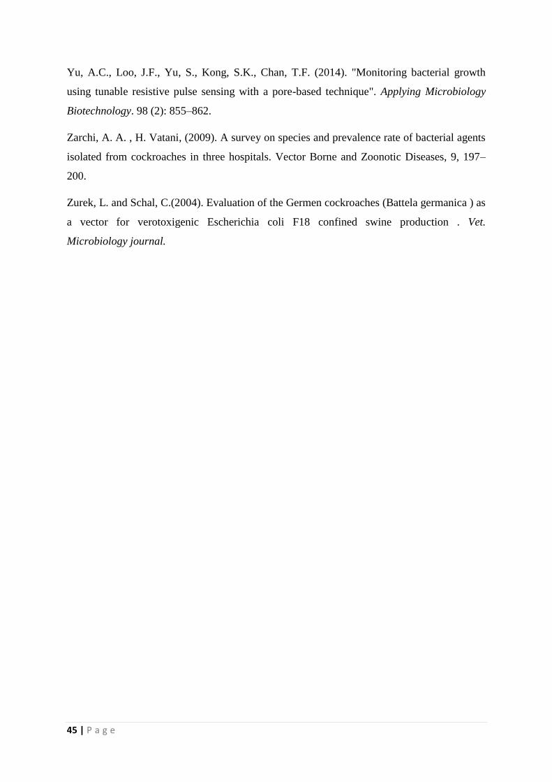
45 | P a g e
Yu, A.C., Loo, J.F., Yu, S., Kong, S.K., Chan, T.F. (2014). "Monitoring bacterial growth
using tunable resistive pulse sensing with a pore-based technique". Applying Microbiology
Biotechnology. 98 (2): 855–862.
Zarchi, A. A. , H. Vatani, (2009). A survey on species and prevalence rate of bacterial agents
isolated from cockroaches in three hospitals. Vector Borne and Zoonotic Diseases, 9, 197–
200.
Zurek, L. and Schal, C.(2004). Evaluation of the Germen cockroaches (Battela germanica ) as
a vector for verotoxigenic Escherichia coli F18 confined swine production . Vet.
Microbiology journal.
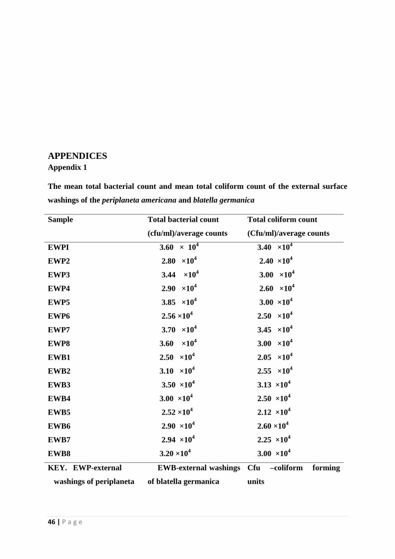
46 | P a g e
APPENDICES
Appendix 1
The mean total bacterial count and mean total coliform count of the external surface
washings of the periplaneta americana and blatella germanica
Sample Total bacterial count
(cfu/ml)/average counts
Total coliform count
(Cfu/ml)/average counts
EWPI 3.60 × 10⁴
3.40 ×10⁴
EWP2 2.80 ×10⁴ 2.40 ×10
4
EWP3 3.44 ×10⁴ 3.00 ×10
4
EWP4 2.90 ×10⁴ 2.60 ×10
4
EWP5 3.85 ×10⁴ 3.00 ×10
4
EWP6 2.56 ×10⁴ 2.50 ×10
4
EWP7 3.70 ×10⁴ 3.45 ×10
4
EWP8 3.60 ×10⁴ 3.00 ×10
4
EWB1 2.50 ×10⁴ 2.05 ×10
4
EWB2 3.10 ×10⁴ 2.55 ×10
4
EWB3 3.50 ×10⁴ 3.13 ×10
4
EWB4 3.00 ×10⁴ 2.50 ×10
4
EWB5 2.52 ×10⁴ 2.12 ×10
4
EWB6 2.90 ×10⁴ 2.60 ×10
4
EWB7 2.94 ×10⁴ 2.25 ×10
4
EWB8 3.20 ×10⁴ 3.00 ×10
4
KEY. EWP-external
washings of periplaneta
EWB-external washings
of blatella germanica
Cfu –coliform forming
units
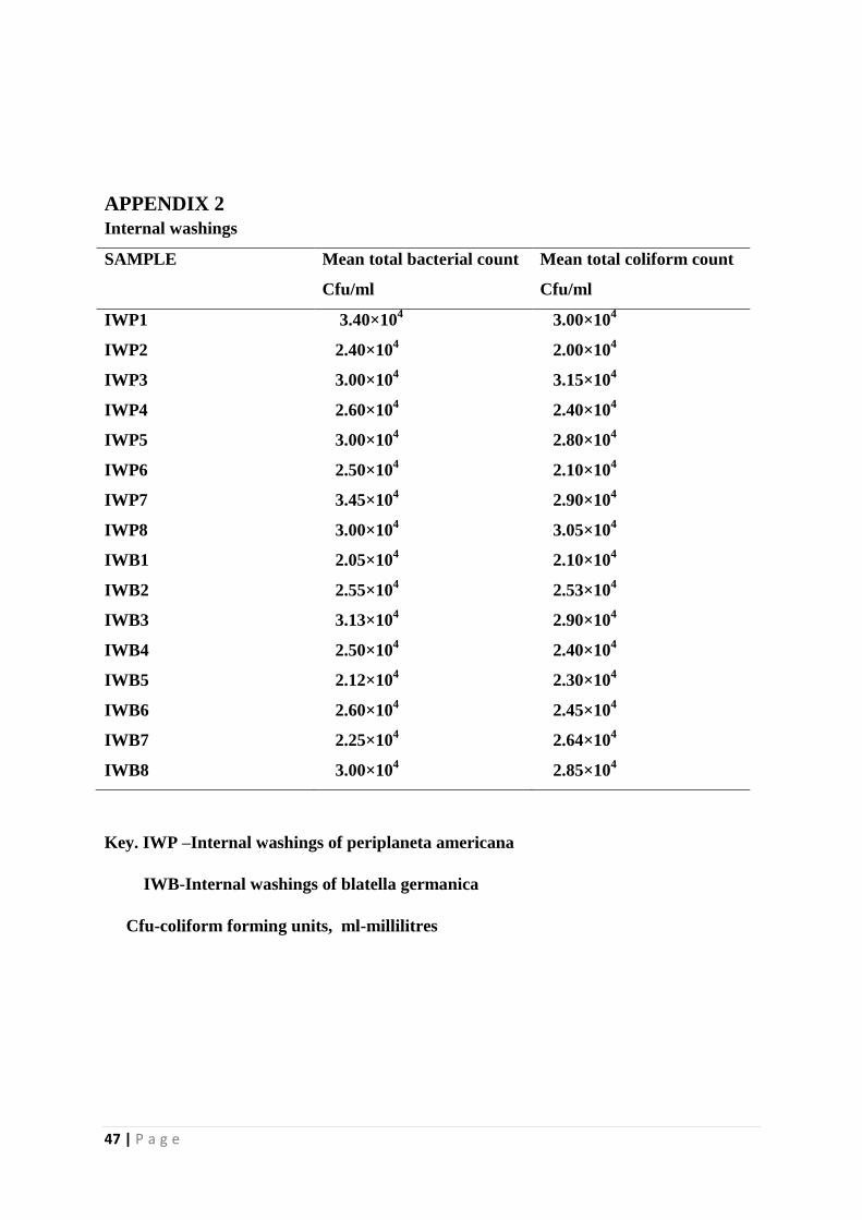
47 | P a g e
APPENDIX 2
Internal washings
SAMPLE Mean total bacterial count
Cfu/ml
Mean total coliform count
Cfu/ml
IWP1 3.40×10⁴ 3.00×10
4
IWP2 2.40×10⁴ 2.00×10
4
IWP3 3.00×10⁴ 3.15×10
4
IWP4 2.60×10⁴ 2.40×10
4
IWP5 3.00×10⁴ 2.80×10
4
IWP6 2.50×10⁴ 2.10×10
4
IWP7 3.45×10⁴ 2.90×10
4
IWP8 3.00×10⁴ 3.05×10
4
IWB1 2.05×10⁴ 2.10×10
4
IWB2 2.55×10⁴ 2.53×10
4
IWB3 3.13×10⁴ 2.90×10
4
IWB4 2.50×10⁴ 2.40×10
4
IWB5 2.12×10⁴ 2.30×10
4
IWB6 2.60×10⁴ 2.45×10
4
IWB7 2.25×10⁴ 2.64×10
4
IWB8 3.00×10⁴ 2.85×10
4
Key. IWP –Internal washings of periplaneta americana
IWB-Internal washings of blatella germanica
Cfu-coliform forming units, ml-millilitres
48 | P a g e
APPENDIX 3- MACROSCOPIC IDENTIFICATION(WANI, 2013)
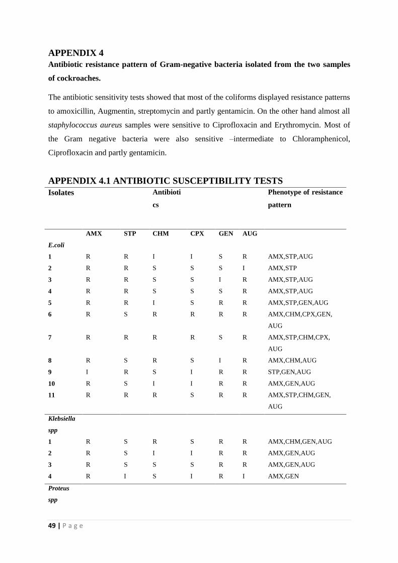
49 | P a g e
APPENDIX 4
Antibiotic resistance pattern of Gram-negative bacteria isolated from the two samples
of cockroaches.
The antibiotic sensitivity tests showed that most of the coliforms displayed resistance patterns
to amoxicillin, Augmentin, streptomycin and partly gentamicin. On the other hand almost all
staphylococcus aureus samples were sensitive to Ciprofloxacin and Erythromycin. Most of
the Gram negative bacteria were also sensitive –intermediate to Chloramphenicol,
Ciprofloxacin and partly gentamicin.
APPENDIX 4.1 ANTIBIOTIC SUSCEPTIBILITY TESTS
Isolates
Antibioti
cs
Phenotype of resistance
pattern
E.coli
AMX STP CHM CPX GEN AUG
1 R R I I S R AMX,STP,AUG
2 R R S S S I AMX,STP
3 R R S S I R AMX,STP,AUG
4 R R S S S R AMX,STP,AUG
5 R R I S R R AMX,STP,GEN,AUG
6 R S R R R R AMX,CHM,CPX,GEN,
AUG
7 R R R R S R AMX,STP,CHM,CPX,
AUG
8 R S R S I R AMX,CHM,AUG
9 I R S I R R STP,GEN,AUG
10 R S I I R R AMX,GEN,AUG
11 R R R S R R AMX,STP,CHM,GEN,
AUG
Klebsiella
spp
1 R S R S R R AMX,CHM,GEN,AUG
2 R S I I R R AMX,GEN,AUG
3 R S S S R R AMX,GEN,AUG
4 R I S I R I AMX,GEN
Proteus
spp
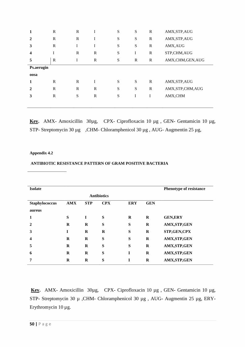
50 | P a g e
1 R R I S S R AMX,STP,AUG
2 R R I S S R AMX,STP,AUG
3 R I I S S R AMX,AUG
4 I R R S I R STP,CHM,AUG
5 R I R S R R AMX,CHM,GEN,AUG
Ps.aerugin
oosa
1 R R I S S R AMX,STP,AUG
2 R R R S S R AMX,STP,CHM,AUG
3 R S R S I I AMX,CHM
Key. AMX- Amoxicillin 30µg, CPX- Ciprofloxacin 10 µg , GEN- Gentamicin 10 µg,
STP- Streptomycin 30 µg ,CHM- Chloramphenicol 30 µg , AUG- Augmentin 25 µg,
Appendix 4.2
ANTIBIOTIC RESISTANCE PATTERN OF GRAM POSITIVE BACTERIA
Key. AMX- Amoxicillin 30µg, CPX- Ciprofloxacin 10 µg , GEN- Gentamicin 10 µg,
STP- Streptomycin 30 µ ,CHM- Chloramphenicol 30 µg , AUG- Augmentin 25 µg, ERY-
Erythromycin 10 µg.
Isolate
Antibiotics
Phenotype of resistance
Staphylococcus
aureus
AMX STP CPX ERY GEN
1 S I S R R GEN,ERY
2 R R S S R AMX,STP,GEN
3 I R R S R STP,GEN,CPX
4 R R S S R AMX,STP,GEN
5 R R S S R AMX,STP,GEN
6 R R S I R AMX,STP,GEN
7 R R S I R AMX,STP,GEN
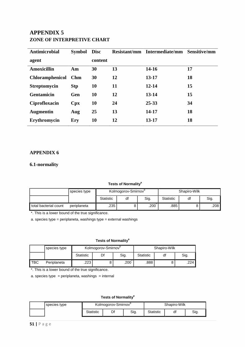
51 | P a g e
APPENDIX 5
ZONE OF INTERPRETIVE CHART
Antimicrobial
agent
Symbol Disc
content
Resistant/mm Intermediate/mm Sensitive/mm
Amoxicillin Am 30 13 14-16 17
Chloramphenicol Chm 30 12 13-17 18
Streptomycin Stp 10 11 12-14 15
Gentamicin Gen 10 12 13-14 15
Ciprofloxacin Cpx 10 24 25-33 34
Augmentin Aug 25 13 14-17 18
Erythromycin Ery 10 12 13-17 18
APPENDIX 6
6.1-normality
Tests of Normalitya
species type Kolmogorov-Smirnovb Shapiro-Wilk
Statistic df Sig. Statistic df Sig.
total bacterial count periplaneta .235 8 .200* .885 8 .208
*. This is a lower bound of the true significance.
a. species type = periplaneta, washings type = external washings
Tests of Normalitya
species type Kolmogorov-Smirnovb Shapiro-Wilk
Statistic Df Sig. Statistic df Sig.
TBC Periplaneta .223 8 .200* .888 8 .224
*. This is a lower bound of the true significance.
a. species type = periplaneta, washings = internal
Tests of Normalitya
species type Kolmogorov-Smirnovb Shapiro-Wilk
Statistic Df Sig. Statistic df Sig.

52 | P a g e
TBC blatella germanica .181 8 .200* .947 8 .685
*. This is a lower bound of the true significance.
a. species type = blatella germanica, washings = external
Tests of Normalitya
species type Kolmogorov-Smirnovb Shapiro-Wilk
Statistic Df Sig. Statistic df Sig.
TBC blatella germanica .195 8 .200* .915 8 .394
*. This is a lower bound of the true significance.
a. species type = blatella germanica, washings = internal
b. Lilliefors Significance Correction
6.2-homogeneity tests
Levene's Test of Equality of Error Variancesa
Dependent Variable: total bacterial count
F df1 df2 Sig.
2.341 3 28 .095
Tests the null hypothesis that the error variance
of the dependent variable is equal across groups.
a. Design: Intercept + species + washings +
species * washings
6.3 Two -way anova table
Tests of Between-Subjects Effects
Dependent Variable: TBC
Source Type III Sum of
Squares
Df Mean Square F Sig. Partial Eta
Squared
Corrected Model 107342109.375a 3 35780703.125 2.502 .080 .211
Intercept 29722267578.1
25
1 29722267578.1
25
2078.128 .000 .987
Species 84013203.125 1 84013203.125 5.874 .022 .173
Washings 22865703.125 1 22865703.125 1.599 .217 .054
species * washings 463203.125 1 463203.125 .032 .858 .001
Error 400467812.500 28 14302421.875
Total 30230077500.0
00
32
Corrected Total 507809921.875 31
a. R Squared = .211 (Adjusted R Squared = .127)
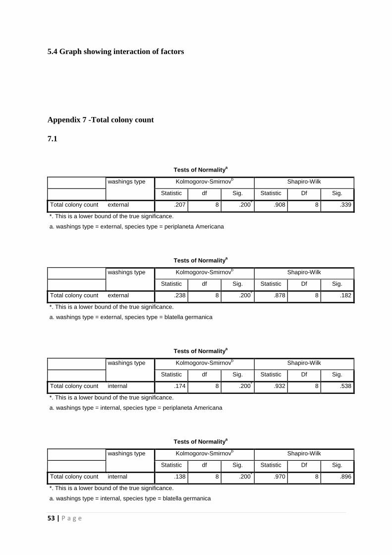
53 | P a g e
5.4 Graph showing interaction of factors
Appendix 7 -Total colony count
7.1
Tests of Normalitya
washings type Kolmogorov-Smirnovb Shapiro-Wilk
Statistic df Sig. Statistic Df Sig.
Total colony count external .207 8 .200* .908 8 .339
*. This is a lower bound of the true significance.
a. washings type = external, species type = periplaneta Americana
Tests of Normalitya
washings type Kolmogorov-Smirnovb Shapiro-Wilk
Statistic df Sig. Statistic Df Sig.
Total colony count external .238 8 .200* .878 8 .182
*. This is a lower bound of the true significance.
a. washings type = external, species type = blatella germanica
Tests of Normalitya
washings type Kolmogorov-Smirnovb Shapiro-Wilk
Statistic df Sig. Statistic Df Sig.
Total colony count internal .174 8 .200* .932 8 .538
*. This is a lower bound of the true significance.
a. washings type = internal, species type = periplaneta Americana
Tests of Normalitya
washings type Kolmogorov-Smirnovb Shapiro-Wilk
Statistic df Sig. Statistic Df Sig.
Total colony count internal .138 8 .200* .970 8 .896
*. This is a lower bound of the true significance.
a. washings type = internal, species type = blatella germanica
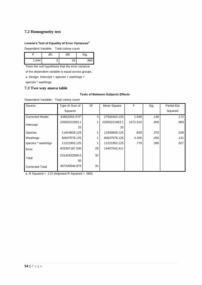
54 | P a g e
7.2 Homogeneity test
Levene's Test of Equality of Error Variancesa
Dependent Variable: Total colony count
F df1 df2 Sig.
1.044 3 28 .389
Tests the null hypothesis that the error variance
of the dependent variable is equal across groups.
a. Design: Intercept + species + washings +
species * washings
7.3 Two way anova table
Tests of Between-Subjects Effects
Dependent Variable: Total colony count
Source Type III Sum of
Squares
Df Mean Square F Sig. Partial Eta
Squared
Corrected Model 83803359.375a 3 27934453.125 1.939 .146 .172
Intercept 22655221953.1
25
1 22655221953.1
25
1572.510 .000 .983
Species 11943828.125 1 11943828.125 .829 .370 .029
Washings 60637578.125 1 60637578.125 4.209 .050 .131
species * washings 11221953.125 1 11221953.125 .779 .385 .027
Error 403397187.500 28 14407042.411
Total 23142422500.0
00
32
Corrected Total 487200546.875 31
a. R Squared = .172 (Adjusted R Squared = .083)