Medically important Bacteria and Fungi
51
Medically important Bacteria and Fungi Hira Mushtaq Lecturer, COBAM, UOP
-
Upload
khangminh22 -
Category
Documents
-
view
0 -
download
0
Transcript of Medically important Bacteria and Fungi

Medically important Bacteria and
Fungi
Hira Mushtaq
Lecturer, COBAM, UOP

Bacteria
Gram positive
Cocci
Staphylococcus aureus
Streptococcus pneumonia
Streptococcus pyogenes
Rods
Bacillus anthracis
Bacillus subtilis
Clostridium tetani
Clostridium botulinum
Gram negative
Cocci
Nesseria meningitis
Nesseria ghonorrhea
Rods
Salmonella typhimurium
Pseudomonas aeruginosa
Escherichia coli O157: H7

Gram Positive Cocci

Staphylococcus spp Family: Micrococcaceae
Genus: A. Staphylococcus- derived from Greek ―stapyle‖ (bunch of grapes)
Include major human pathogen and skin commensals
Staphylococcus divided into coagulase positive & coagulase negative categories
Common spp: S. aureus
S. epidermidis
S. saprophyticus
B. Micrococcus- skin commensal
Gram-positive spherical cells (0.5-1.5 mm) in singles, pairs, and clusters
Appear as ―bunches of grapes

Non motile
Non–spore-forming
Nonencapsulated
Catalase-producing
Primarily aerobic, some facultatively anaerobic
Inhibited by high bile salt concentration
S. aurues ß-hemolytic
Colony morphology:
buttery looking, cream or white colored
S. aureus


Virulence factors of S. aureus
Toxins:
Hemolysins (α, β, γ, δ) – lyse red blood cells
Leukocidin – lyses neutrophils and macrophages
Enterotoxin – induce gastrointestinal distress
Exfoliative toxin – separates the epidermis from the
dermis
Toxic shock syndrome toxin (TSST) - induces fever,
vomiting, shock, systemic organ damage

Epidemiology and pathogenesis
Present in most environments even humans
Readily isolated from fomites
Carriage is mostly in anterior nares, skin,
nasopharynx, intestine.
Predisposition to infection include: poor hygiene and
nutrition, tissue injury, preexisting primary infection,
diabetes, immunodeficiency.
Increase in community acquired methicillin
resistance - MRSA
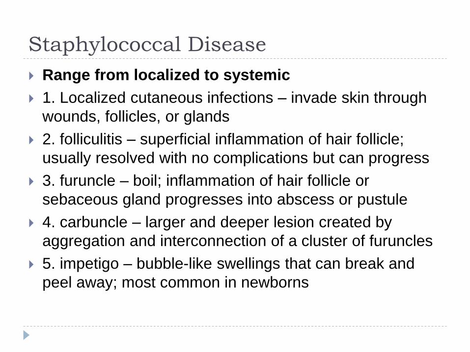
Staphylococcal Disease
Range from localized to systemic
1. Localized cutaneous infections – invade skin through
wounds, follicles, or glands
2. folliculitis – superficial inflammation of hair follicle;
usually resolved with no complications but can progress
3. furuncle – boil; inflammation of hair follicle or
sebaceous gland progresses into abscess or pustule
4. carbuncle – larger and deeper lesion created by
aggregation and interconnection of a cluster of furuncles
5. impetigo – bubble-like swellings that can break and
peel away; most common in newborns


Continu----
Systemic infections
osteomyelitis – infection is established in the
metaphysis; abscess forms
bacteremia - primary origin is bacteria from another
infected site or medical devices; endocarditis
possible

Continu----
Toxigenic disease
food intoxication – ingestion of heat stable
enterotoxins; gastrointestinal distress
staphylococcal scalded skin syndrome – toxin
induces bright red flush, blisters, then desquamation
of the epidermis
toxic shock syndrome – toxemia leading to shock
and organ failure

Clinical detection
Frequently isolated from pus, tissue exudates,
sputum, urine, and blood
Cultivation (MSA agar), catalase, biochemical
testing, coagulase
Identification of Staphylococcus in Samples

Treatment
95% have penicillinase and are resistant to penicillin
and ampicillin.
MRSA – methicillin-resistant S. aureus – carry
multiple resistance
Abscesses have to be surgically perforated.
Systemic infections require intensive lengthy therapy.

Other GPC
Catalase negative :
Streptococcus pneumonia (pneumonia)
streptococcus pyogenes (sore throat)
Streptococcus mutans (tooth infection)
Enteroccoccus feacalis (GIT + bacteremia)

Gram positive Rods

Family: Clostridiaceae
3–8 um long, thick, Gram-positive
Spore forming, rod shaped bacteria
Motile bacteria with flagella
strictly obligate anaerobic to aerotolerant
Occurrence naturally inhabit the soil and the
intestinal tracts of humans and animals.
Common species:
C. tatni (tetnus, nervous system disorder)
C. perfirengens (anaerobic cellulitis and gas gangrene)
C. botulinum (botulism, food poisoning)
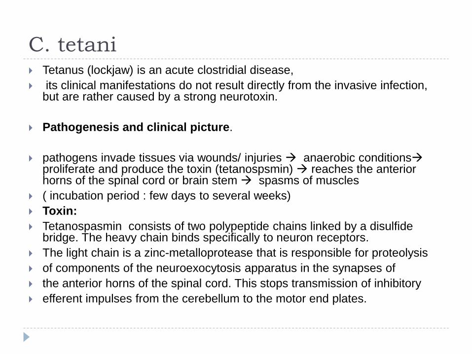
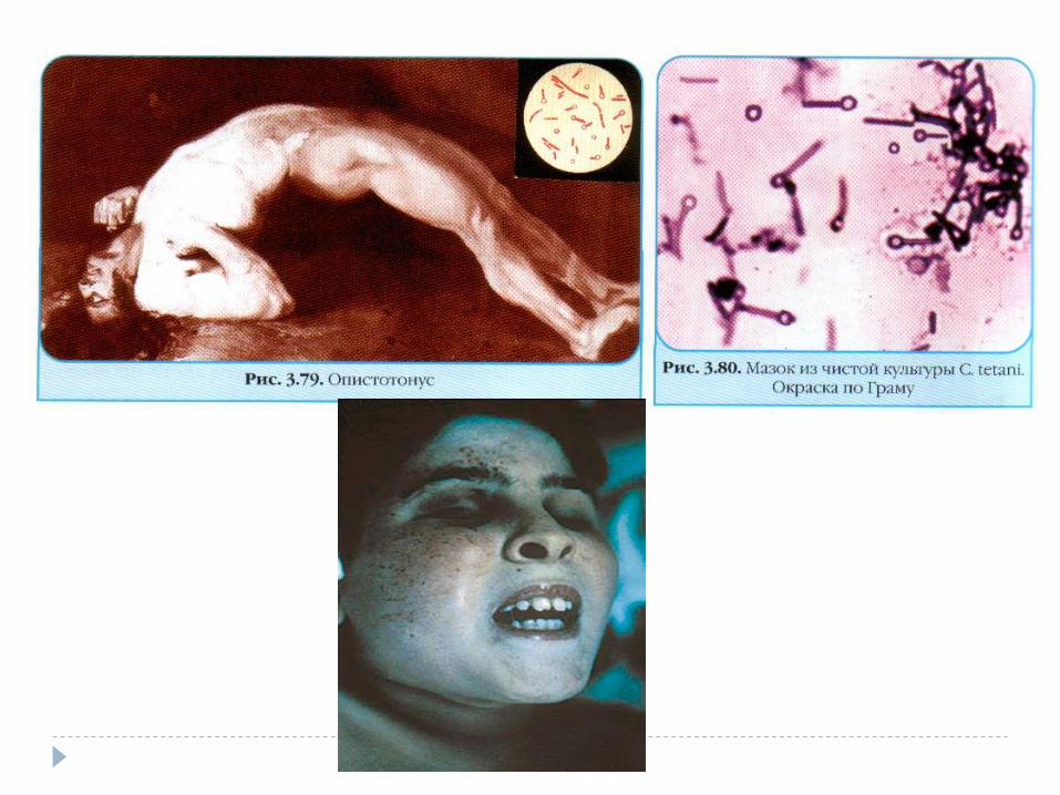
C. tetani Tetanus (lockjaw) is an acute clostridial disease,
its clinical manifestations do not result directly from the invasive infection, but are rather caused by a strong neurotoxin.
Pathogenesis and clinical picture.
pathogens invade tissues via wounds/ injuries anaerobic conditions proliferate and produce the toxin (tetanospsmin) reaches the anterior horns of the spinal cord or brain stem spasms of muscles
( incubation period : few days to several weeks)
Toxin:
Tetanospasmin consists of two polypeptide chains linked by a disulfide bridge. The heavy chain binds specifically to neuron receptors.
The light chain is a zinc-metalloprotease that is responsible for proteolysis
of components of the neuroexocytosis apparatus in the synapses of
the anterior horns of the spinal cord. This stops transmission of inhibitory
efferent impulses from the cerebellum to the motor end plates.


Vaccination & treatment
DPT (diphtheria, pertussis, tetanus)
tetanus toxoid
antigenic
TAT (tetnus antitoxin); Metronidazole (For more
serious wounds)

Gram negative rods

Enterobacteriaceae.
41 genera with hundreds of species
Gram-negative,
usually motile (peritrichous flagellation or swarming
movement)
facultative anaerobic rod bacteria
natural habitat is the intestinal tract of humans and
animals
Responsible for nosocomial diseases as well
0.5–1.5 um thick, and 24 um long
Generation time in vitro is 20–30 minutes

Several serovars
O antigens. Specific polysaccharide chains in the
lipopolysaccharide complex of the outer membrane.
H antigens. Flagellar antigens consisting of protein.
K antigens. Linear polymers of the outer membrane
built up of a repeated series of carbohydrate units
(sometimes proteins as well). They can cover the cell
densely.
F antigens. Antigens of protein attachment fimbriae.

Salmonella spp
Salmonella enterica with seven subspecies.
Typhoid salmonelloses: typhi and paratyphi A, B, and C
Salmonellae are taken up orally and the invasion pathway is through the intestinal tract, from where they enter lymphatic tissue, first spreading lymphogenously, then hematogenously.
1-3 weeks
Human carriers are the only source of infection
Enteric salmonelloses : develop when pathogens are taken up with food. The primary infection source is usually livestock. These relatively frequent infections remain restricted to the gastrointestinal tract.
1–2 days

Pathognesis

Pathogenesis

Diagnosis & Therapy.
Diagnosis by
stool culture
metabolic properties
treated with anti-infective agents (antibiotics) e.g.
aminopenicillins, 4-quinolones
slowing down intestinal activity (e.g., with
loperamide) and replacing fluid and electrolyte
losses orally as required

Control
providing training in hygienic practices for all food-
handling personnel in slaughterhouses,
food processing plants, and restaurants;
cooking and refrigerating foods adequately in food
processing plants, restaurants, and homes;
and expanding of governmental enteric disease
surveillance programs.

Bordetella pertussis
Gram negative,
aerobic,
encapsulated coccobacillus
the causative agent of pertussis or whooping cough.
bacterium is spread by airborne droplets; its
incubation period is 7–10 days on average (range 6–
20 days)
Humans are the only known reservoir for B.
pertussis

Pathogenesis

Whooping cough (pertussis) is a highly contagious respiratory tract infection. In many people, it's marked
by a severe hacking cough followed by a high-pitched intake of breath that sounds like "whoop."
Symptoms
usually mild at first and resemble those of a common cold:
Runny nose
Nasal congestion
Red, watery eyes
Fever
Cough
After a week or two, signs and symptoms worsen. Thick mucus accumulates inside your airways, causing uncontrollable coughing. Severe and prolonged coughing attacks may:
Provoke vomiting
Result in a red or blue face
Cause extreme fatigue
End with a high-pitched "whoop" sound during the next breath of air

Risk factors and prevention
infants who are younger than age 12 months who are unvaccinated or haven't received the full set of recommended vaccines have the
highest risk for severe complications and death.
the best way to prevent whooping cough is with the pertussis vaccine, which doctors often give in combination with vaccines against two other serious diseases — diphtheria and tetanus. Doctors recommend beginning vaccination during infancy.
The vaccine consists of a series of five injections, typically given to children at these ages:
2 months
4 months
6 months
15 to 18 months
4 to 6 years

Mycobacterium tuberculosis
Over a century ago Robert Koch identified
Mycobacterium tuberculosis as the causative agent
of tuberculosis
acid-fast rods, 0.4um wide, and 3–4 um long,
Endospore forming
non-motile.
can be stained with special agents (Ziehl-Neelsen
staining)
M. bovis and M. africanum can also causes TB.
obligate aerobes


Pathogenesis
Transmission: from other humans through droplet nuclei
(1-5 micron in diameter) and the respiratory Route
Transmission to humans from susceptible animal species
and their products (e.g., milk) is also possible
Depending on the environment, these tiny particles can
remain suspended in the air for several hours.
incubation period : about 4 to 12 weeks, and the disease
develops slowly.
symptoms : fever, fatigue, and weight loss.
A cough, which is characteristic of pulmonary involvement, may
result in expectoration of bloody sputum.


Treatment and control chemotherapy and chemoprophylaxis are carried
Recently, new multi-drug-resistant strains of tuberculosis (MDR-TB)
have developed and are spreading
infants and children, are vaccinated with bacille Calmette-Guérin (BCG) vaccine to prevent complications such as meningitis.

Diagnosis
requires microscopic and cultural identification of the
pathogen or pathogen-specific DNA
generation time of TB is approximately 12–18 hours,
so that cultures must be incubated for three to six or
eight weeks at 37 oC until proliferation becomes
macroscopically visible
Tuberculin skin reaction

Diagnosis
requires microscopic and cultural identification of the
pathogen or pathogen-specific DNA
generation time of TB is approximately 12–18 hours,
so that cultures must be incubated for three to six or
eight weeks at 37 8C until proliferation becomes
macroscopically visible
Tuberculin reaction

Gram negative Cocci

Neisseriaceae Gram-negative cocci often arranged in pairs (diplococci) with
adjacent sides flattened (like coffee beans) a diameter of approximately 1 um
Encapsulated
Non sporeforming
Aerobic
Nonmotile
Important human species
Neisseria gonorrhoeae sexually transmitted pathogen (urethritis, cervicitis)
Neisseria meningitides (meningitis, meningoencephalitis, arthritis,)
species normally colonize mucosal surfaces of oropharynx and nasopharynx and occasionally anogenital mucosal
membranes
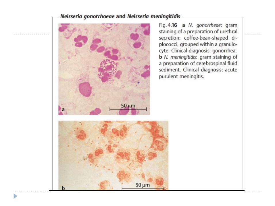

Neisseria meningitides
Gram negative cocci shaped
appears in kidney bean shape under the microscope .
It requires anaerobic environment with 5% CO2 and enriched media containing blood for growth
oxidase and catalase positive.
Serogroups A, B, C, D, etc. (a total of 12
Epidemiology: Humans only natural hosts
Person-to-person transmission by aerosolization of respiratory tract secretions in crowded conditions
Close contact with infectious person
Highest incidence in children younger than 5 years and particularly those younger than 1 year of age

Pathogenesis
Pili-mediated receptor-specific colonization of nonciliated cells of
nasopharynx
Engulfed by phagocytes
Antiphagocytic polysaccharide capsule does not allow
phagolysosome activity
allows systemic spread i.e. to soft tissues
(brains n ligaments)
Toxic effects mediated by hyperproduction of lipooligosaccharide
Causing meningitis, utheritis, arthritis etc.

Treatment
The antibiotic of choice is penicillin G.
Therapy has been obtained with third-generation
cephalosporins, e.g., cefotaxime or ceftriaxone.
It is important to start treatment as quickly as
possible to prevent delayed damage

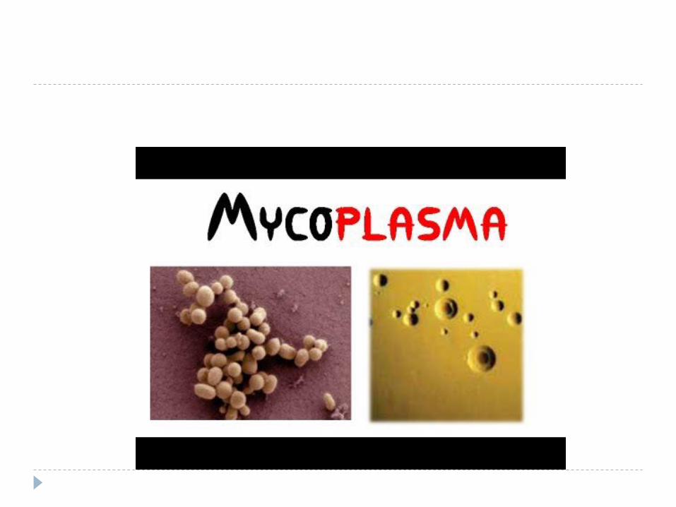
Mycoplasma

Mycoplasmataceae
Do not possess rigid cell walls for lack of a murein layer
Ploemorphic but most common form is coccoid cell with a diameter of 0.3–0.8 lm.
Long,
fungi like filaments grown on culture mediums with high osmotic pressure levels.
frequently causes pneumonias that run atypical courses, especially in young children.
10 -20% of pneumonias contracted outside of hospitals are caused by this pathogen
Common species:
Mycoplosama pneumnia
Ureoplasma
Infections of the respiratory organs or urinary tract.

Pathogenesis
transmitted by aerosol droplets cells attach
themselves to the epithelia of the trachea, bronchi,
and bronchioles destruction of the epithelial cells
infection develops into pneumonia with an
inflammatory exudate in the lumens of the bronchi
and bronchioles.
The incubation period is 10–20 days.
Symptoms : fever, headache, and a persistent
cough.
Treatment
The antibiotics of choice are tetracyclines and
macrolides
M. pneumoniae is found worldwide.
Humans are the only source of infection. The
pathogens are transmitted
by droplet infection during close contact. Infections
are frequently contracted
in families, schools, homes for children, work camps,
and military camps