High Pro-Inflammatory Cytokine Secretion and Loss of High Avidity Cross-Reactive Cytotoxic T-Cells...
12
High Pro-Inflammatory Cytokine Secretion and Loss of High Avidity Cross-Reactive Cytotoxic T-Cells during the Course of Secondary Dengue Virus Infection Tao Dong 1. *, Edward Moran 1. *, Nguyen Vinh Chau 3 , Cameron Simmons 2 , Kerstin Luhn 1 , Yanchun Peng 1 , Bridget Wills 2 , Nguyen Phuong Dung 2 , Le Thi Thu Thao 3 , Tran Tinh Hien 3 , Andrew McMichael 1 , Jeremy Farrar 2 , Sarah Rowland-Jones 1 1 MRC Human Immunology Unit, Weatherall Institute of Molecular Medicine, John Radcliffe Hospital, Oxford, United Kingdom, 2 Oxford University Clinical Research Unit, Hospital for Tropical Diseases, Ho Chi Minh City, Viet Nam, 3 Hospital for Tropical Diseases, Ho Chi Minh City, Viet Nam Background. Dengue is one of the most important human diseases transmitted by an arthropod vector and the incidence of dengue virus infection has been increasing – over half the world’s population now live in areas at risk of infection. Most infections are asymptomatic, but a subset of patients experience a potentially fatal shock syndrome characterised by plasma leakage. Severe forms of dengue are epidemiologically associated with repeated infection by more than one of the four dengue virus serotypes. Generally attributed to the phenomenon of antibody-dependent enhancement, recent observations indicate that T-cells may also influence disease phenotype. Methods and Findings. Virus-specific cytotoxic T lymphocytes (CTL) showing high level cross reactivity between dengue serotypes could be expanded from blood samples taken during the acute phase of secondary dengue infection. These could not be detected in convalescence when only CTL populations demonstrating significant serotype specificity were identified. Dengue cross-reactive CTL clones derived from these patients were of higher avidity than serotype-specific clones and produced much higher levels of both type 1 and certain type 2 cytokines, many previously implicated in dengue pathogenesis. Conclusion. Dengue serotype cross-reactive CTL clones showing high avidity for antigen produce higher levels of inflammatory cytokines than serotype-specific clones. That such cells cannot be expanded from convalescent samples suggests that they may be depleted, perhaps as a consequence of activation-induced cell death. Such high avidity cross-reactive memory CTL may produce inflammatory cytokines during the course of secondary infection, contributing to the pathogenesis of vascular leak. These cells appear to be subsequently deleted leaving a more serotype-specific memory CTL pool. Further studies are needed to relate these cellular observations to disease phenotype in a large group of patients. If confirmed they have significant implications for understanding the role of virus-specific CTL in pathogenesis of dengue disease. Citation: Dong T, Moran E, Vinh Chau N, Simmons C, Luhn K, et al (2007) High Pro-Inflammatory Cytokine Secretion and Loss of High Avidity Cross- Reactive Cytotoxic T-Cells during the Course of Secondary Dengue Virus Infection. PLoS ONE 2(12): e1192. doi:10.1371/journal.pone.0001192 INTRODUCTION Dengue is one of the most important human diseases transmitted by an arthropod vector (Aedes Aegypti) and the incidence of dengue virus infection has been increasing steadily throughout the world[1,2]. The dengue viruses belong to the Flavivirus genus and there are four distinct serotypes, DEN 1 to 4. Patients develop symptoms 5–7 days after the bite of an infected mosquito. This lasts 2–7 days corresponding with the time of peak viral load. Viral titres then fall and may be low or undetectable by the day of defervescence[3]. Most infections are asymptomatic but for those with symptoms clinical manifestations range from a mild febrile illness (Dengue fever, DF) to a potentially severe syndrome which may include haemorrhage and shock (Dengue haemorrhagic fever, DHF)[4,5]. The pathophysiology of DHF is poorly understood. The key pathological feature is increased vascular permeability with plasma leakage into the interstitial spaces associated with increased levels of vasoactive cytokines such as tumour necrosis factor alpha (TNF- a), Interferon gamma (IFN-c), Interleukin six (IL-6) and In- terleukin two (IL-2)[6–9]. Other implicated cytokines include IL- 10, IL-13 and IL-18[10–12]. Epidemiological and clinical studies have demonstrated a role for immunological, host genetic and viral factors in the pathogenesis of severe disease[1]. The majority of DHF cases occur in patients who have experienced a previous infection with a heterologous DEN serotype[4,13]. Infection with one DEN serotype generally fails to provide protective immunity against the other serotypes and sequential, heterotypic infection may lead to more severe disease. This phenomenon has been a substantial obstacle to dengue vaccine development because of the implica- tion that cross-reactive immune responses between DEN serotypes play a part in the pathogenesis of severe disease. The phenomenon of antibody-dependent enhancement (ADE)[14,15] is widely accepted as a good explanation of this link between immunological cross-reactivity and disease patho- genesis. The antibody response generated against one DEN serotype provides only transient protection against other serotypes. Later infection with heterologous virus is actually enhanced by the remaining antibody with increased viral uptake into Fc receptor bearing cells. Disease severity is thought to be a consequence of Academic Editor: Scott Halstead, Burns School of Medicine, United States of America Received February 5, 2007; Accepted October 19, 2007; Published December 5, 2007 Copyright: ß 2007 Dong et al. This is an open-access article distributed under the terms of the Creative Commons Attribution License, which permits unrestricted use, distribution, and reproduction in any medium, provided the original author and source are credited. Funding: This work was funded by the Medical Research Council, UK (T.D., Y.C.P., A.M., S.R.J.) and the Wellcome Trust (N.V.C, C.S., B.W., N.P.D., J.F.). K.L. has a long- term fellowship from the European Molecular Biology Organisation. E.M. is a Wellcome Trust Clinical Research Training fellow. Competing Interests: The authors have declared that no competing interests exist. * To whom correspondence should be addressed. E-mail: [email protected]. uk (TD); [email protected] (EM) . These authors contributed equally to this work. PLoS ONE | www.plosone.org 1 December 2007 | Issue 12 | e1192
-
Upload
independent -
Category
Documents
-
view
4 -
download
0
Transcript of High Pro-Inflammatory Cytokine Secretion and Loss of High Avidity Cross-Reactive Cytotoxic T-Cells...
High Pro-Inflammatory Cytokine Secretion and Loss ofHigh Avidity Cross-Reactive Cytotoxic T-Cells during theCourse of Secondary Dengue Virus InfectionTao Dong1.*, Edward Moran1.*, Nguyen Vinh Chau3, Cameron Simmons2, Kerstin Luhn1, Yanchun Peng1, Bridget Wills2, Nguyen PhuongDung2, Le Thi Thu Thao3, Tran Tinh Hien3, Andrew McMichael1, Jeremy Farrar2, Sarah Rowland-Jones1
1 MRC Human Immunology Unit, Weatherall Institute of Molecular Medicine, John Radcliffe Hospital, Oxford, United Kingdom, 2 Oxford UniversityClinical Research Unit, Hospital for Tropical Diseases, Ho Chi Minh City, Viet Nam, 3 Hospital for Tropical Diseases, Ho Chi Minh City, Viet Nam
Background. Dengue is one of the most important human diseases transmitted by an arthropod vector and the incidence ofdengue virus infection has been increasing – over half the world’s population now live in areas at risk of infection. Most infectionsare asymptomatic, but a subset of patients experience a potentially fatal shock syndrome characterised by plasma leakage. Severeforms of dengue are epidemiologically associated with repeated infection by more than one of the four dengue virus serotypes.Generally attributed to the phenomenon of antibody-dependent enhancement, recent observations indicate that T-cells may alsoinfluence disease phenotype. Methods and Findings. Virus-specific cytotoxic T lymphocytes (CTL) showing high level crossreactivity between dengue serotypes could be expanded from blood samples taken during the acute phase of secondary dengueinfection. These could not be detected in convalescence when only CTL populations demonstrating significant serotype specificitywere identified. Dengue cross-reactive CTL clones derived from these patients were of higher avidity than serotype-specific clonesand produced much higher levels of both type 1 and certain type 2 cytokines, many previously implicated in dengue pathogenesis.Conclusion. Dengue serotype cross-reactive CTL clones showing high avidity for antigen produce higher levels of inflammatorycytokines than serotype-specific clones. That such cells cannot be expanded from convalescent samples suggests that they may bedepleted, perhaps as a consequence of activation-induced cell death. Such high avidity cross-reactive memory CTL may produceinflammatory cytokines during the course of secondary infection, contributing to the pathogenesis of vascular leak. These cellsappear to be subsequently deleted leaving a more serotype-specific memory CTL pool. Further studies are needed to relate thesecellular observations to disease phenotype in a large group of patients. If confirmed they have significant implications forunderstanding the role of virus-specific CTL in pathogenesis of dengue disease.
Citation: Dong T, Moran E, Vinh Chau N, Simmons C, Luhn K, et al (2007) High Pro-Inflammatory Cytokine Secretion and Loss of High Avidity Cross-Reactive Cytotoxic T-Cells during the Course of Secondary Dengue Virus Infection. PLoS ONE 2(12): e1192. doi:10.1371/journal.pone.0001192
INTRODUCTIONDengue is one of the most important human diseases transmitted by
an arthropod vector (Aedes Aegypti) and the incidence of dengue virus
infection has been increasing steadily throughout the world[1,2].
The dengue viruses belong to the Flavivirus genus and there are four
distinct serotypes, DEN 1 to 4. Patients develop symptoms 5–7 days
after the bite of an infected mosquito. This lasts 2–7 days
corresponding with the time of peak viral load. Viral titres then
fall and may be low or undetectable by the day of defervescence[3].
Most infections are asymptomatic but for those with symptoms
clinical manifestations range from a mild febrile illness (Dengue
fever, DF) to a potentially severe syndrome which may include
haemorrhage and shock (Dengue haemorrhagic fever, DHF)[4,5].
The pathophysiology of DHF is poorly understood. The key
pathological feature is increased vascular permeability with plasma
leakage into the interstitial spaces associated with increased levels
of vasoactive cytokines such as tumour necrosis factor alpha (TNF-
a), Interferon gamma (IFN-c), Interleukin six (IL-6) and In-
terleukin two (IL-2)[6–9]. Other implicated cytokines include IL-
10, IL-13 and IL-18[10–12].
Epidemiological and clinical studies have demonstrated a role
for immunological, host genetic and viral factors in the
pathogenesis of severe disease[1]. The majority of DHF cases
occur in patients who have experienced a previous infection with
a heterologous DEN serotype[4,13]. Infection with one DEN
serotype generally fails to provide protective immunity against the
other serotypes and sequential, heterotypic infection may lead to
more severe disease. This phenomenon has been a substantial
obstacle to dengue vaccine development because of the implica-
tion that cross-reactive immune responses between DEN serotypes
play a part in the pathogenesis of severe disease.
The phenomenon of antibody-dependent enhancement
(ADE)[14,15] is widely accepted as a good explanation of this
link between immunological cross-reactivity and disease patho-
genesis. The antibody response generated against one DEN
serotype provides only transient protection against other serotypes.
Later infection with heterologous virus is actually enhanced by the
remaining antibody with increased viral uptake into Fc receptor
bearing cells. Disease severity is thought to be a consequence of
Academic Editor: Scott Halstead, Burns School of Medicine, United States ofAmerica
Received February 5, 2007; Accepted October 19, 2007; Published December 5,2007
Copyright: � 2007 Dong et al. This is an open-access article distributed underthe terms of the Creative Commons Attribution License, which permitsunrestricted use, distribution, and reproduction in any medium, provided theoriginal author and source are credited.
Funding: This work was funded by the Medical Research Council, UK (T.D., Y.C.P.,A.M., S.R.J.) and the Wellcome Trust (N.V.C, C.S., B.W., N.P.D., J.F.). K.L. has a long-term fellowship from the European Molecular Biology Organisation. E.M. isa Wellcome Trust Clinical Research Training fellow.
Competing Interests: The authors have declared that no competing interestsexist.
* To whom correspondence should be addressed. E-mail: [email protected] (TD); [email protected] (EM)
. These authors contributed equally to this work.
PLoS ONE | www.plosone.org 1 December 2007 | Issue 12 | e1192
the raised viral titre and the resulting pathology (whether mediated
directly by the virus itself or by immune responses, cellular or
otherwise) is therefore correspondingly more severe than that in
primary infection. There is evidence to support the hypothesis.
However ADE is neither a sufficient (estimates of rates of DHF in
those experiencing secondary infection range from 1.8–12%
patients[16,17]), nor an absolutely necessary precondition for the
development of severe disease (not every severe case occurs in
those experiencing secondary infection – although the over-
whelming majority do). Additional mechanisms are likely to be
involved to account for the complex clinical phenotype of dengue
disease.
It has been recognised for sometime that CTL populations are
capable of mediating significant immunopathology in viral
infections such as lymphocytic choriomeningitis virus[18]. There
is good evidence that the CTL response to a viral infection –
whether by a heterologous agent (such as one of the four dengue
viruses) or one unrelated to previous viral encounters – can be
modulated by the infection history of an individual in a manner
likely to contribute to disease severity[19].
Recent reports have linked cross-reactive cellular immune
responses to dengue virus with pathogenesis[20–22]. It has been
proposed that cross-reactive cytotoxic memory T-cells (CTL)
raised against a previous dengue strain and therefore activated in
the early stages of acute secondary infection would be less efficient
in eliminating newly encountered dengue virus serotypes and that
this could lead to increased virus replication and more severe
disease – an example of the immunological phenomenon ‘‘original
antigenic sin’’[21]. Others have demonstrated the complexity of
the response of serotype cross-reactive memory CTL to heterol-
ogous variant peptides and suggest that cross-reactive T-cells
might have altered cytokine profiles that could contribute to
induction of plasma leakage[20,23,24].
In this study we have examined the CD8 T-cell responses to
a previously identified immunodominant NS3 epitope presented
by HLA-A*11[21] (the most common HLA allele in Viet Nam) in
a cohort of Vietnamese dengue patients. We demonstrate a striking
difference in the serotype specificity of CD8 T-cells present in
patients with acute dengue virus infection compared with those in
convalescence. CTL recognising tetramers for the dengue NS3
epitope can be expanded by short-term stimulation with peptides
representing the dengue epitopes. Activated highly cross-reactive
cells are detected in PBMCs taken during acute infection but
cannot be detected in, or expanded from convalescent samples,
perhaps as a consequence of activation-induced cell death in
vivo[25,26]. The populations remaining in convalescence show
a much greater degree of serotype specificity. High avidity cross-
reactive CTL clones from these patients produce higher levels of
type 1 and certain type 2 cytokines than serotype specific clones.
Differential release of vasoactive and pro-inflammatory factors by
cross-reactive and serotype-specific CTL populations may con-
tribute to the pathogenesis of severe dengue disease. Larger clinical
studies are required to extend these observations: firstly to examine
how CTL cross-reactivity and phenotype relate to disease severity
and secondly to identify any differences in those experiencing
primary and secondary infection. Such findings would have
significant implications for dengue vaccine strategies.
METHODS
Patient samples and HLA typingBlood samples were collected from a cohort of over 300 adults and
children attending the Hospital for Tropical Diseases and Ho Chi
Minh City Children’s Hospital #1 in Ho Chi Minh city, Viet
Nam, with dengue virus infection of varying clinical disease severity.
Samples were collected on the day of admission (acute) and during
convalescence (one month after the first sample). Patients were
characterised demographically, clinically (using established clinical
criteria[27]), virologically (using RT-PCR) and serologically (IgM
and IgG on paired samples). Serological data suggested that the
majority (.99%) of the patients we studied were experiencing
secondary infections. The study protocol was approved by the
Scientific and Ethics committee at The Hospital for Tropical
Diseases and the Oxford Tropical Research Ethics committee.
Molecular HLA typing was performed on most study subjects using
amplification refractory mutation system PCR (ARMS-PCR) with
sequence specific primers, as previously described[28].
Dengue virus PCR and SerologyDengue virus RNA was isolated from acute plasma samples using
RNAgents (Promega, Wisconsin). RNA was reverse transcribed
and two rounds of PCR were performed using primers and
methods described previously[29]. In samples containing virus, the
PCR yielded DNA products, the unique sizes of which were
diagnostic for each dengue serotype.
Dengue infection was confirmed via serological testing of acute
and early convalescent plasma samples collected at least 3 days
apart using a commercial capture-IgM and IgG ELISA (Panbio,
Brisbane, Australia). The ELISA was performed and the results
interpreted according to the manufacturers instructions. This
ELISA assay has been validated as both sensitive and specific for
primary and secondary dengue infections[30].
Synthesis of peptide-HLA tetramersThe HLA molecule heavy chain cDNAs were modified by
substitution of the transmembrane and cytosolic regions with
a sequence encoding the BirA biotinylation enzyme recognition
site, as previously described[31]. These modified HLA heavy chains,
and ß2-microglobulin, were synthesized in a prokaryotic expression
system (pET, R&D Systems), purified from bacterial inclusion
bodies, solubilised in the presence of denaturant (4 M Urea) and
allowed to refold with the relevant peptide by dilution. Refolded
monomeric complexes were purified by FPLC and biotinylated
using BirA (Avidity), then combined with either phycoerythrin (PE)-
labelled, Quantum red-labelled (Sigma) or allophycocyanin (APC)-
labelled streptavidin (Molecular Probes) at a 4:1 molar ratio to form
tetrameric HLA/peptide complexes (‘‘tetramers’’). Tetramers in-
capable of binding the CD8 molecule were produced using site-
directed mutagenesis. The amino acids at positions 227 and 228 (the
alpha-3 domain of the MHC molecule) were altered from DT to
KA. This change abrogates CD8 binding[32]. These ‘‘CD8-null’’
monomers were conjugated as described above. The tetramers used
in these studies were as follows: HLA-A*1101 wild type and CD8-
null refolded with the previously described NS3 epitope, using three
different peptide variants representing the sequences from DEN1,
DEN2, and DEN3/4. Tetramers were conjugated with different
fluorochromes to allow co-staining of T-cell populations.
Antigens and antibodiesPeptides were synthesized by FMOC chemistry using a Zinnser
peptide synthesiser to .70% purity. Anti-CD8 and anti-CD38
(Peridinin chlorophyll protein, PerCP) antibodies were purchased
from Becton Dickinson (San Diego, California).
Cell surface stainingCell surface staining was carried out on freshly separated or
carefully thawed cryo-preserved PBMCs. Titrated tetramers (PE,
High Avidity CTL in Dengue
PLoS ONE | www.plosone.org 2 December 2007 | Issue 12 | e1192
APC or quantum-red conjugated) were added for 15 min at 37uC,
then the cells were incubated with CD8-APC and CD38 FITC
antibodies for 15 min at room temperature (RT) in some cases.
Cells were then washed and stored in Cell FixTM buffer (Becton
Dickinson) at 4uC until flow cytometry analysis was performed.
Samples were analyzed on a Becton Dickinson FACSCalibur.
Tetramer dissociation assayTo assess the rate of tetramer dissociation cells from a CTL clone
were incubated with PE-labelled tetramer for 40 minutes at 4uC.
The cells were washed twice and resuspended in 50 mL of buffer.
2 mL of this suspension was added to 100 mL of PBS and analysed
by flow cytometry. An excess of antibody known to block tetramer
binding (DK25, Dako) was then added to the remaining cell
suspension. 2 mL of this reaction was removed periodically, added
to 200 mL of PBS and analysed on a Cyan ADP flow cytometer.
The fraction of positive cells was defined as the percentage of cells
falling above a gate at which 90% of cells were positive at time 0.
Establishment of CTL lines and ClonesCTL lines were generated as previously described[33]. Briefly,
PBMCs were stimulated with specific epitope peptides at 2 mM
concentration, IL-2 was added on day 3, and the specificity of the
CTL lines were tested using CTL lysis assay or tetramer staining
on day 10 and day 20. CTL clones were established from PBMCs
or CTL lines by either limiting dilution or by using the appropriate
antigen-specific tetramers in combination with MACS anti-PE-
microbeads (Miltenyi Biotec). Briefly, tetramers were added to 3–5
million PBMCs, then incubated at 37uC for 20 minutes. The cells
were then washed with cold buffer and resuspended in 40 ml of anti-
PE beads, mixed well and incubated for 15 minutes at 4–8uC. The
cells were then washed and resuspended in 500 ml cold buffer, and
magnetic separation was performed according to the manufacturer’s
protocol (Miltenyi Biotec, Germany). Sorted tetramer positive cells
were counted and CTL cloning was performed by using a standard
protocol as previously described[33].
CTL lysis assaysCTL lysis assays were performed using standard 51Chromium
release assays[33]. Briefly, HLA class I matched B-cell lines (BCL)
were labelled with 51Chromium for 1 hour, then washed three
times: target cells were then divided and pulsed with peptides at
different concentrations. After another hour of incubation at 37uC,
the peptide solution was then washed off and cells were counted
and co-cultured with CTL clones at appropriate effector to target
(E:T) ratios in 96 well plates. The plates were incubated at 37uCfor 4 hours, supernatants were harvested and then counted for
radioactivity using a Beta-plate counter (WALLAC) Specific lysis
was calculated from the formula:
% lysis~ experimental counts{media controlð Þ=
detergent control{media controlð Þ|100%:
Cytokine beads assayIn parallel with the CTL lysis assays, we performed a duplicate set
of experiments using the scheme described above but without 51Cr
labelling of the BCL. Supernatant from the CTL and target cell
co-culture was harvested after overnight incubation, and cytokines
were measured by Luminex cytokine bead array analysis
according to the manufacturer’s instructions (Bio-Rad Laborato-
ries, USA).
RESULTS
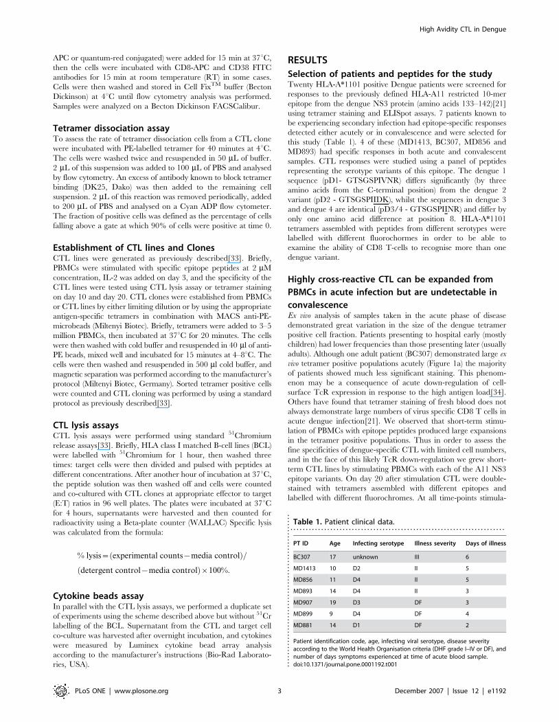
Selection of patients and peptides for the studyTwenty HLA-A*1101 positive Dengue patients were screened for
responses to the previously defined HLA-A11 restricted 10-mer
epitope from the dengue NS3 protein (amino acids 133–142)[21]
using tetramer staining and ELISpot assays. 7 patients known to
be experiencing secondary infection had epitope-specific responses
detected either acutely or in convalescence and were selected for
this study (Table 1). 4 of these (MD1413, BC307, MD856 and
MD893) had specific responses in both acute and convalescent
samples. CTL responses were studied using a panel of peptides
representing the serotype variants of this epitope. The dengue 1
sequence (pD1- GTSGSPIVNR) differs significantly (by three
amino acids from the C-terminal position) from the dengue 2
variant (pD2 - GTSGSPIIDK), whilst the sequences in dengue 3
and dengue 4 are identical (pD3/4 - GTSGSPIINR) and differ by
only one amino acid difference at position 8. HLA-A*1101
tetramers assembled with peptides from different serotypes were
labelled with different fluorochormes in order to be able to
examine the ability of CD8 T-cells to recognise more than one
dengue variant.
Highly cross-reactive CTL can be expanded from
PBMCs in acute infection but are undetectable in
convalescenceEx vivo analysis of samples taken in the acute phase of disease
demonstrated great variation in the size of the dengue tetramer
positive cell fraction. Patients presenting to hospital early (mostly
children) had lower frequencies than those presenting later (usually
adults). Although one adult patient (BC307) demonstrated large ex
vivo tetramer positive populations acutely (Figure 1a) the majority
of patients showed much less significant staining. This phenom-
enon may be a consequence of acute down-regulation of cell-
surface TcR expression in response to the high antigen load[34].
Others have found that tetramer staining of fresh blood does not
always demonstrate large numbers of virus specific CD8 T cells in
acute dengue infection[21]. We observed that short-term stimu-
lation of PBMCs with epitope peptides produced large expansions
in the tetramer positive populations. Thus in order to assess the
fine specificities of dengue-specific CTL with limited cell numbers,
and in the face of this likely TcR down-regulation we grew short-
term CTL lines by stimulating PBMCs with each of the A11 NS3
epitope variants. On day 20 after stimulation CTL were double-
stained with tetramers assembled with different epitopes and
labelled with different fluorochromes. At all time-points stimula-
Table 1. Patient clinical data.. . . . . . . . . . . . . . . . . . . . . . . . . . . . . . . . . . . . . . . . . . . . . . . . . . . . . . . . . . . . . . . . . . . . . .
PT ID Age Infecting serotype Illness severity Days of illness
BC307 17 unknown III 6
MD1413 10 D2 II 5
MD856 11 D4 II 5
MD893 14 D4 II 3
MD907 19 D3 DF 3
MD899 9 D4 DF 4
MD881 14 D1 DF 2
Patient identification code, age, infecting viral serotype, disease severityaccording to the World Health Organisation criteria (DHF grade I–IV or DF), andnumber of days symptoms experienced at time of acute blood sample.doi:10.1371/journal.pone.0001192.t001..
....
....
....
....
....
....
....
....
....
....
....
....
High Avidity CTL in Dengue
PLoS ONE | www.plosone.org 3 December 2007 | Issue 12 | e1192
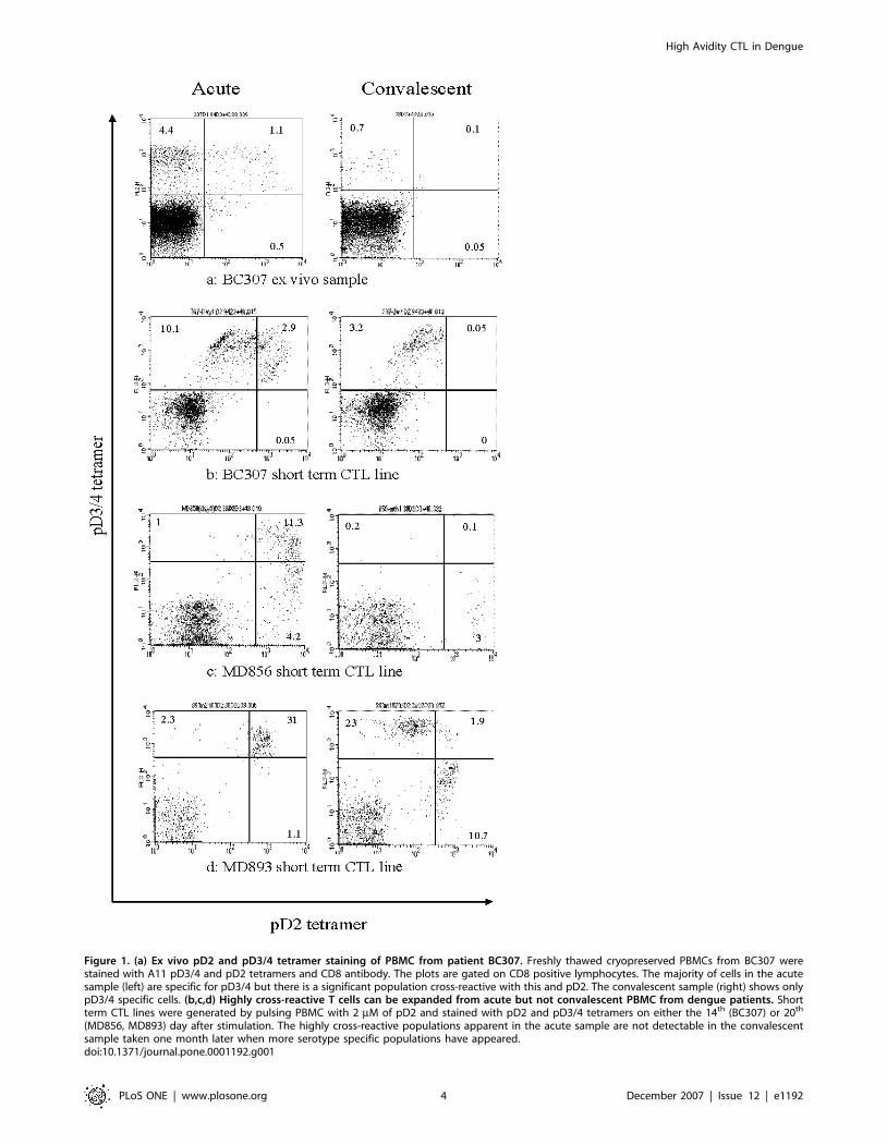
Figure 1. (a) Ex vivo pD2 and pD3/4 tetramer staining of PBMC from patient BC307. Freshly thawed cryopreserved PBMCs from BC307 werestained with A11 pD3/4 and pD2 tetramers and CD8 antibody. The plots are gated on CD8 positive lymphocytes. The majority of cells in the acutesample (left) are specific for pD3/4 but there is a significant population cross-reactive with this and pD2. The convalescent sample (right) shows onlypD3/4 specific cells. (b,c,d) Highly cross-reactive T cells can be expanded from acute but not convalescent PBMC from dengue patients. Shortterm CTL lines were generated by pulsing PBMC with 2 mM of pD2 and stained with pD2 and pD3/4 tetramers on either the 14th (BC307) or 20th
(MD856, MD893) day after stimulation. The highly cross-reactive populations apparent in the acute sample are not detectable in the convalescentsample taken one month later when more serotype specific populations have appeared.doi:10.1371/journal.pone.0001192.g001
High Avidity CTL in Dengue
PLoS ONE | www.plosone.org 4 December 2007 | Issue 12 | e1192

tion with the pD3/4 peptide resulted in the expansion of cells
mostly specific for pD3/4 (data not shown). However stimulation
of PBMCs from four patients (MD893, MD856, BC307, MD881)
with pD2 resulted in expansions of CTL binding both D2 and
D3/4 tetramers equally well. The pattern of staining differed
slightly in each patient but all 4 demonstrated expansions of highly
cross-reactive cells from acute samples. We considered such CTL
– those recognising both pD2 and pD3/4 – to be highly cross-
reactive given their equal recognition of both tetramers despite
significant sequence variation. CTL recognising pD1 usually
recognised pD3/4 – a reflection of their greater homology. Cells
showing high cross reactivity between pD2 and pD3/4 always
cross reacted with pD1. These highly cross-reactive CTL were
undetectable in, and could not be expanded from the convalescent
samples of patient MD893, MD856 and BC307 (Figure 1b,c,d) -
no convalescent sample was available for MD881. Only CTL
showing specificity for a single serotype or limited cross-reactivity
(i.e. strong binding of the tetramer folded with the peptide of one
serotype, weak binding of the other) remained. A similar pattern of
staining was observed ex vivo in samples from patient BC307 where
the high frequency of tetramer positive cells allowed direct analysis
without the need for in vitro expansion. Acutely approximately
20% of tetramer positive cells were cross-reactive, recognising both
the pD2 and pD3/4 tetramers. This population was not detectable
in the convalescent (day 21) sample (Figure 1a).
There were 2 patients in whom this phenomenon was not
observed: antigen specific cells could not be detected acutely in
patient MD899 but partially cross-reactive populations recognising
pD2 and pD3/4 were apparent by convalescence; patient MD907
had only serotype specific cells present acutely (pD2).
Highly cross-reactive CTL express higher levels of
CD38.CD38 is a marker of lymphocyte activation. Ex vivo staining of
PBMC from patient BC307 demonstrated equally high levels of
CD38 staining among all tetramer positive cells in the acute phase
(Figure 2a). By three weeks after the acute sample CD38
expression had fallen markedly. At this time point CD8 cells
recognising the two similar peptide variants, pD1 and pD3/4,
were present (partially cross-reactive – Figure 2b) but cells cross-
reactive between the more heterologous peptides, pD2 and pD3/4
could not be detected (highly cross-reactive cells – Figure 1a).
CD38 expression was greater on those T cells recognising both
pD1 and pD3/4 (57% CD38 high – Figure 2d) than upon partially
or non cross-reactive T cells (19% CD38 high – Figure 2c). This
suggests that highly cross-reactive T cell populations are more
activated than cells exhibiting low levels of cross-reactivity. It is not
clear why pD1-pD3/4 cross-reactive cells are present at 3 weeks
and pD2-pD3/4 cross-reactive cells are not. We could speculate
that this highly cross-reactive latter group experiences even higher
levels of activation resulting in activation induced cell death and
clonal deletion.
Cross-reactive CTL clones maintain cytolytic activity
at low peptide concentrationsWe generated CTL clones by limiting dilutions of short-term lines
derived from both acute and convalescent PBMC stimulated with
either pD2 or pD3/4 from patients BC307 and MD1413
(Figure 3a). We were able to generate both serotype specific and
highly cross-reactive CTL clones from acute samples, but only
serotype-specific clones or clones showing low-level cross-reactivity
could be grown from convalescent samples. Highly cross-reactive
clones (E5, 10B3, 9F5, 10H5, 8E9 and 9E5) and serotype specific
clones 10A4 and 3H9 (both recognising pD2) were grown from
short term lines derived from acute samples. Serotype specific
clones D9 (recognising pD3/4), partially cross-reactive clones C42
and C48 (both showing strongest recognition for pD3/4 and
differing recognition of pD1) were grown from lines derived from
convalescent samples. All highly cross-reactive clones maintained
high levels of cytolytic activity at low peptide concentrations with
all three peptide variants in a standard chromium release assay
(9F5, 10B3 and E5 are representative – Figure 3b). Partially cross-
reactive clone C48 showed good lytic activity against cells pulsed
with pD3/4 and pD1 but low activity against pD2 (Fig 3b). Clone
C42 showed good activity against pD3/4, low activity against pD1
and no recognition of pD2. Serotype specific clones showed
intermediate (D9, 10A4 and 3H9) activity against their cognate
peptides (pD3/4, pD2, pD2 respectively) but failed to recognise
target cells pulsed with the peptide variants (Fig 3c). All cross-
reactive or partially cross-reactive clones maintained lytic activity
against B-cells pulsed with peptide concentrations as low as
0.01 mM. Serotype specific clones showed no or negligible activity
at these levels.
Cross-reactive clones show greater avidity for pD3/4
peptide-MHCWe produced tetramers incapable of binding CD8 due to
a mutation in the a3 region of the heavy chain. Such tetramers
have been shown to reliably identify high avidity CTLs[35]. We
stained the clones with ‘‘CD8-null’’ tetramer folded with pD3/4.
Cross-reactive clones 9F5 and E5 bound this tetramer almost as
well as the wild-type tetramer (Figure 4a) whereas more serotype
specific clones C48 and D9 showed little or no binding (Figure 4b).
Other cross-reactive clones not described here in detail showed
similarly effective binding to the CD8-null tetramer (8E9, 9E5).
This implies that cross-reactive clones have a stronger peptide/
MHC-TCR interaction than serotype specific clones or those with
only low level cross-reactivity. The relative weakness of the
peptide/MHC-TCR interaction of a serotype specific clone
renders the MHC-CD8 interaction a requirement for tetramer
staining. This observation was confirmed by tetramer dissociation
assays. Wild-type pD3/4 tetramer dissociates more quickly from
DEN3/4 serotype-specific clone D9 than either cross-reactive
clone E5 or 9F5 (Figure 4d). This implies that D9’s TCR avidity
for tetramer is lower than its cross-reactive counterparts.
None of the clones generated from these patients showed any
significant binding to the pD2 CD8-null tetramer and dissociation
assays demonstrated that pD2 wild-type tetramer dissociates from
cross-reactive clones much more quickly than pD3/4. pD2
conforms to published A*1101 binding motifs. It would appear
that effective TCR binding is dependent upon the MHC-CD8
interaction to an extent not demonstrated by the other variants.
Cross-reactive CTL clones produce higher levels and
different patterns of cytokine release than serotype-
specific CTLClones were stimulated with B cells loaded with the relevant
peptide and the concentration of cytokines in the tissue culture
supernatant measured at 24 hours. Highly cross-reactive clones
E5, 9F5, 10H5 and 10B3 consistently produced higher levels of
TNFa, IFNc and GM-CSF than most serotype specific clones
(Figure 5a, b). IL-10 was produced in high levels by clone E5.
Other clones produced IL-10 in only very low levels if at all. The
clones from patient MD1413 produced IL-13, another type 2
cytokine, and again cross-reactive clones produced significantly
High Avidity CTL in Dengue
PLoS ONE | www.plosone.org 5 December 2007 | Issue 12 | e1192
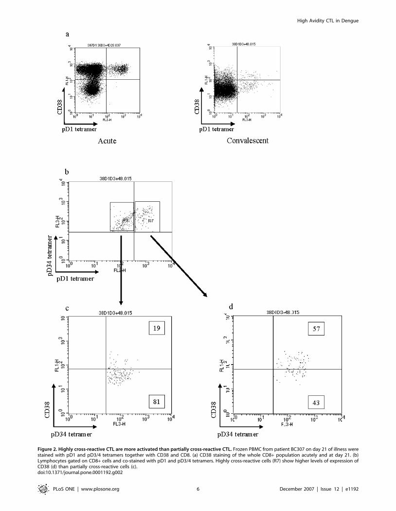
Figure 2. Highly cross-reactive CTL are more activated than partially cross-reactive CTL. Frozen PBMC from patient BC307 on day 21 of illness werestained with pD1 and pD3/4 tetramers together with CD38 and CD8. (a) CD38 staining of the whole CD8+ population acutely and at day 21. (b)Lymphocytes gated on CD8+ cells and co-stained with pD1 and pD3/4 tetramers. Highly cross-reactive cells (R7) show higher levels of expression ofCD38 (d) than partially cross-reactive cells (c).doi:10.1371/journal.pone.0001192.g002
High Avidity CTL in Dengue
PLoS ONE | www.plosone.org 6 December 2007 | Issue 12 | e1192
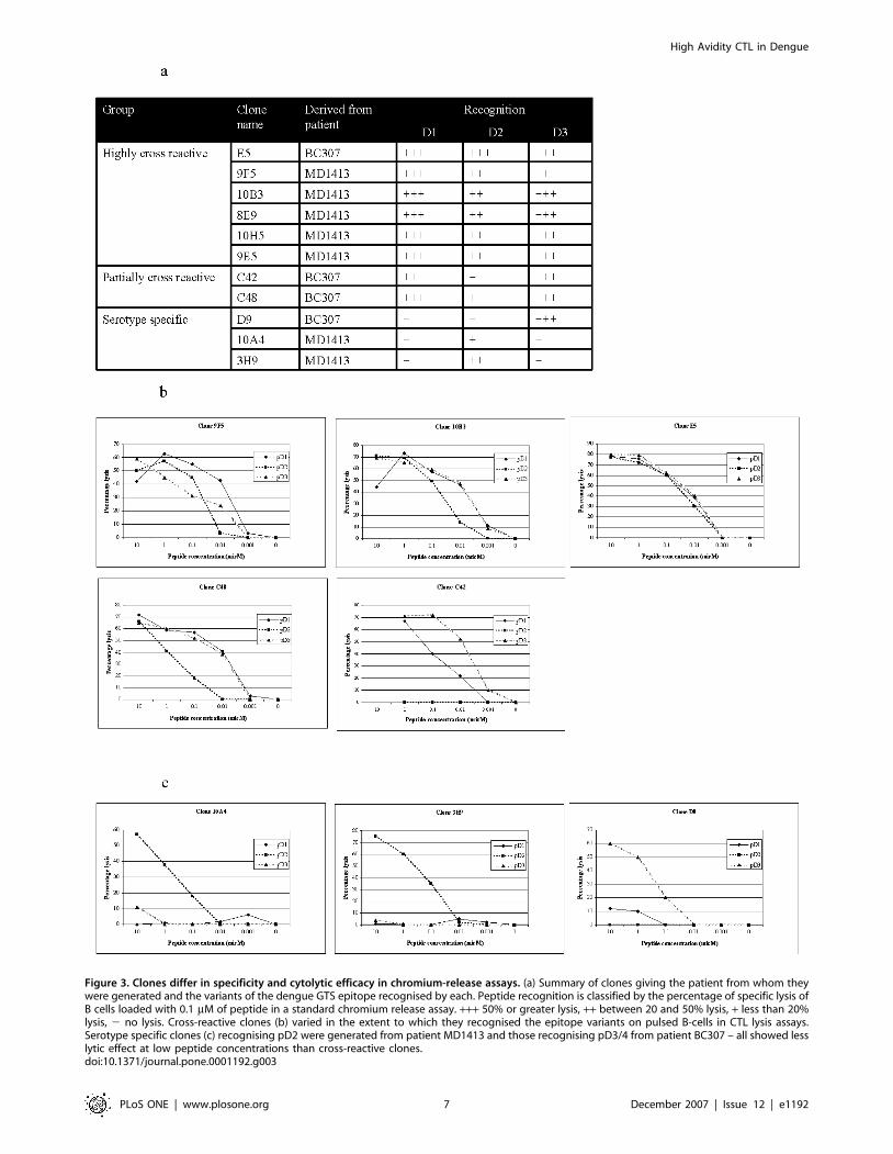
Figure 3. Clones differ in specificity and cytolytic efficacy in chromium-release assays. (a) Summary of clones giving the patient from whom theywere generated and the variants of the dengue GTS epitope recognised by each. Peptide recognition is classified by the percentage of specific lysis ofB cells loaded with 0.1 mM of peptide in a standard chromium release assay. +++ 50% or greater lysis, ++ between 20 and 50% lysis, + less than 20%lysis, 2 no lysis. Cross-reactive clones (b) varied in the extent to which they recognised the epitope variants on pulsed B-cells in CTL lysis assays.Serotype specific clones (c) recognising pD2 were generated from patient MD1413 and those recognising pD3/4 from patient BC307 – all showed lesslytic effect at low peptide concentrations than cross-reactive clones.doi:10.1371/journal.pone.0001192.g003
High Avidity CTL in Dengue
PLoS ONE | www.plosone.org 7 December 2007 | Issue 12 | e1192

Figure 4. Most cross-reactive clones show significantly better staining with CD8-null tetramers than serotype-specific clones. Clones werestained with CD8-null and wild-type A11 tetramers refolded with the DEN3 variant of the GTS epitope. Cross-reactive clones (a) generally showeda smaller drop in fluorescent intensity than DEN3 specific clones which showed much poorer staining with the CD8-null tetramer than with the wild-type (b). The exception was cross-reactive clone 10H5 which showed negligible binding to pD3/4 CD8-null tetramer. No clones showed significantbackground staining with an unrecognised tetramer (c). pD3/4 tetramer dissociates more rapidly from serotype specific clone D9 than cross-reactiveclones E5 or 9F5 (d) in a tetramer dissociation assay.doi:10.1371/journal.pone.0001192.g004
High Avidity CTL in Dengue
PLoS ONE | www.plosone.org 8 December 2007 | Issue 12 | e1192
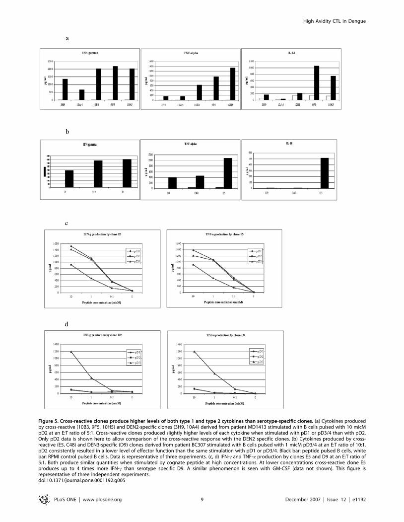
Figure 5. Cross-reactive clones produce higher levels of both type 1 and type 2 cytokines than serotype-specific clones. (a) Cytokines producedby cross-reactive (10B3, 9F5, 10H5) and DEN2-specific clones (3H9, 10A4) derived from patient MD1413 stimulated with B cells pulsed with 10 micMpD2 at an E:T ratio of 5:1. Cross-reactive clones produced slightly higher levels of each cytokine when stimulated with pD1 or pD3/4 than with pD2.Only pD2 data is shown here to allow comparison of the cross-reactive response with the DEN2 specific clones. (b) Cytokines produced by cross-reactive (E5, C48) and DEN3-specific (D9) clones derived from patient BC307 stimulated with B cells pulsed with 1 micM pD3/4 at an E:T ratio of 10:1.pD2 consistently resulted in a lower level of effector function than the same stimulation with pD1 or pD3/4. Black bar: peptide pulsed B cells, whitebar: RPMI control pulsed B cells. Data is representative of three experiments. (c, d) IFN-c and TNF-a production by clones E5 and D9 at an E:T ratio of5:1. Both produce similar quantities when stimulated by cognate peptide at high concentrations. At lower concentrations cross-reactive clone E5produces up to 4 times more IFN-c than serotype specific D9. A similar phenomenon is seen with GM-CSF (data not shown). This figure isrepresentative of three independent experiments.doi:10.1371/journal.pone.0001192.g005
High Avidity CTL in Dengue
PLoS ONE | www.plosone.org 9 December 2007 | Issue 12 | e1192
more. IFNc production by the clones correlated with their
cytolytic activity (compare Figure 5c/d and Figure 3). IL-4, IL-
16, IL-2, and IL-1b were produced at low levels only. For many
clones cytokine production was a better correlate of TCR avidity
than cytolytic activity. Despite similar lytic activity against pD3/4
(Figure 3) clones E5 and C48 differ in their CD8-null binding (E5
better than C48 – Figure 4a/b) and in the cytokine production (E5
better than C48 – Figure 5a). All cross-reactive clones produced
slightly less of each cytokine with pD2 stimulation. E5 is
representative of this phenomenon: despite showing equally
effective lytic activity to all peptide variants, it produced lower
levels of IFNc and TNFa with pD2 (Figure 5b). This is consistent
with the failure to bind the pD2 CD8-null tetramer.
Cytokine production therefore reflects a clone’s avidity for
peptide. Cross-reactive clones tend to be of higher avidity with
greater cytokine production than serotype specific clones.
DISCUSSIONWe have demonstrated that in some hospitalised Vietnamese
patients with acute dengue virus infection a population of activated
high avidity CTLs showing substantial cross-reactivity between all
four dengue serotypes can be detected. These high avidity cross-
reactive cells cannot be detected in convalescent samples,
presumably a consequence of activation-induced cell death[36],
leaving only populations that show a much greater degree of
serotype specificity. The phenomenon of apparent deletion of high
avidity CTLs has been observed in other clinical scenarios.
Molldren et al[25] studied the CTL response to the leukaemia-
associated self peptide PR1, an epitope found within proteinase 3.
High avidity CTL specific for PR1 were twice as effective at killing
chronic myelogenous leukaemia cells than low-avidity PR1-
specific CTLs[25]. These high-avidity CTLs were selectively
deleted by apoptosis if exposed to high PR1 peptide concentrations
or chronic myelogenous leukaemia cells overexpressing proteinase
3. Both low and high avidity PR1-specific CTLs could be detected
or expanded from healthy donors, but only low avidity CTLs
could be detected or expanded from newly diagnosed leukaemia
patients[25]. This process of selective clonal deletion may be
a result of clonal exhaustion similar to that observed in LCMV
infection[37]. Certainly tetramer positive lymphocytes from
patients with acute dengue show evidence of proliferating and
dying in large numbers[21]. This may reflect the high avidity
shown by many cross-reactive cells for their targets and the large
antigen load found just before defervescence.
Tetramer positive populations are often small in acute samples
stained ex vivo. We found that short term stimulation with pD2
produced large positive populations showing cross reactivity
between pD3/4 and pD2 tetramers. In contrast stimulation with
pD3/4 produced populations specific mostly for the pD3/4
tetramer in all donors with DEN3 or DEN4 infection. It has been
shown that high or low avidity CTLs can be elicited in vitro by
culturing the lymphocytes with low or high concentrations of
target antigen respectively[38–40]. All the cross-reactive clones
described here had a lower TCR avidity for pD2-MHC than
either pD1-MHC or pD3/4-MHC. Thus it is possible that due to
its higher avidity for the TCR the concentration of pD3/4 we used
for generating CTL lines preferentially expanded lower avidity
pD3/4 specific cells. In contrast although used in the same
concentration as pD3/4 in the generation of short term lines, the
lower avidity of pD2 for TCR resulted in the preferential
expansion of the high avidity cross-reactive T-cells.
All the clones we generated in this study produced both type 1
and type 2 cytokines: IFN-c, TNF-a, IL-13 and in some cases IL-
10. Many type 1 cytokines exhibit vasoactive properties and are
likely to participate in the pathogenesis of vascular leak. In
addition TNF-a can mediate activation-induced cell death in some
T cells [41] and has been implicated in peripheral T-cell
deletion[42,43]. Type 2 cytokines such as IL-13 and IL-10 have
been implicated in the pathogenesis of severe dengue[11,12]. IL-
10 can be produced by distinct CD4+ and CD8+ T cell
populations and has the ability to suppress T cell function[44,45].
It could be postulated that certain type 2 cytokines produced in
acute disease by a subset of highly cross-reactive CTL might exert
an inhibitory effect on dengue specific effector T cells.
Highly cross-reactive clones grown from acute samples pro-
duced high levels of TNFa, occasionally IL-10 and demonstrated
the greatest avidity for peptide-MHC as demonstrated by staining
with CD8-null tetramers and tetramer decay assays. Serotype
specific and partially cross-reactive clones produced much less
TNF-a, IFN-c and GM-CSF and were not shown to secrete any
IL-10. It has been suggested that the pattern of CD8 cell cytokine
production is epitope dependent[46,47] with high avidity T cell/
target interactions leading to greater production of TNFa or
IFNc[48–50].
In general killing ability correlated well with the TCR/pMHC
avidity demonstrated by CD8-null tetramers. We observed
variations in TCR avidity amongst clones that nonetheless
displayed very similar levels of cytolytic activity for a given peptide
concentration. The lytic activity of highly cross-reactive clone E5 is
very similar to partially cross-reactive clone C48 for a given
stimulation with pD3/4 (Figure 3). Yet E5 demonstrates much
higher avidity for peptide-MHC than C48 as demonstrated by
staining with CD8-null tetramers (Figure 4). The TCR/pMHC
interaction is only one component, albeit perhaps the most
significant, of a T cell’s avidity for its target. Others include the
TCR expression level, co-stimulatory molecule expression level
and the extracellular microenvironment. A recent review has
pointed out that T cells displaying all the characteristics of high
avidity interactions may nonetheless bear a TCR that is of
relatively low affinity[51]. Evidence of this complexity is apparent
in our study: whereas the vast majority of cross-reactive clones
producing high levels of cytokines showed high avidity TCR/
pMHC interactions independent of CD8, one (10H5) did not.
With that exception all cross-reactive clones showed good or
moderate binding to the CD8-null tetramer but not all produced
cytokines as vigorously as E5 or 9F5.
Discussion surrounds the precise roles of antibody and cellular
immunity in the pathogenesis of severe dengue. It is generally
accepted that antibody enhancement of secondary infection
facilitates viral infection of cells leading to high viraemia, antigen
loading of antigen-presenting cells[14,15] and a consequently
vigorous cellular immune response. However the observations
made here suggest that not only the magnitude, but the specific
‘‘contents’’ of the immune response could be of significance.
Memory CTL demonstrating both serotype-cross reactivity (a pre-
requisite for expansion from the memory pool in secondary
infection) and high avidity produce higher levels of inflammatory
cytokines than their lower-avidity peers. In vivo this may contribute
to the level of those vasoactive cytokines shown to have an
association with plasma leakage and disease severity. The failure to
expand these cells from convalescent samples suggests they are
absent or greatly reduced in number by this time point. This may
be a consequence of the large ADE-augmented antigen load
combined with the high avidity of the T cells leading to over-
activation, T cell exhaustion and cell death. Newly generated
serotype-specific CTLs therefore dominate. It is likely that
protective immunity is primarily antibody mediated, but it could
be postulated that a ‘‘lack of immunopathogenesis’’, as much as
High Avidity CTL in Dengue
PLoS ONE | www.plosone.org 10 December 2007 | Issue 12 | e1192
a degree of protection derives from these serotype-specific
populations in patients with repeated exposure to dengue virus.
This detailed cellular study involved too few patients to allow
firm conclusions to be drawn regarding disease aetiology.
Nonetheless these findings have potentially significant implications
for understanding the role of virus-specific CTL in immunity to
dengue virus infection and in the pathogenesis of severe dengue
disease. Further work is needed to extend them and relate the
presence or absence of such high-avidity cross-reactive cells to
clinical phenotype. In the hunt for a vaccine that produces pan-
serotype protective immunity without the risk of iatrogenic DHF it
is surely prudent to consider not only the nature of the antibody
response but the specificity, avidity and effector function of T cells
elicited by dengue vaccine candidates.
ACKNOWLEDGMENTS
Author Contributions
Conceived and designed the experiments: AM SR JF EM TD. Performed
the experiments: EM KL NV TD. Analyzed the data: EM NV TD.
Contributed reagents/materials/analysis tools: TH BW EM KL NV CS
NP LT TD. Wrote the paper: EM TD.
REFERENCES1. Rothman AL (2003) Immunology and immunopathogenesis of dengue disease.
Adv Virus Res 60: 397–419.
2. Guzman MG, Kouri G (2002) Dengue: an update. Lancet Infect Dis 2: 33–42.
3. Vaughn DW, Green S, Kalayanarooj S, Innis BL, Nimmannitya S, et al. (2000)
Dengue viremia titer, antibody response pattern, and virus serotype correlate
with disease severity. J Infect Dis 181: 2–9.
4. Rothman AL, Ennis FA (1999) Immunopathogenesis of Dengue hemorrhagic
fever. Virology 257: 1–6.
5. Vazquez S, Lemos G, Pupo M, Ganzon O, Palenzuela D, et al. (2003) Diagnosis
of dengue virus infection by the visual and simple AuBioDOT immunoglobulin
M capture system. Clin Diagn Lab Immunol 10: 1074–1077.
6. Kittigul L, Temprom W, Sujirarat D, Kittigul C (2000) Determination of tumor
necrosis factor-alpha levels in dengue virus infected patients by sensitive biotin-
streptavidin enzyme-linked immunosorbent assay. J Virol Methods 90: 51–57.
7. Kurane I, Innis BL, Nimmannitya S, Nisalak A, Meager A, et al. (1991)
Activation of T lymphocytes in dengue virus infections. High levels of soluble
interleukin 2 receptor, soluble CD4, soluble CD8, interleukin 2, and interferon-
gamma in sera of children with dengue. J Clin Invest 88: 1473–1480.
8. Hober D, Poli L, Roblin B, Gestas P, Chungue E, et al. (1993) Serum levels of
tumor necrosis factor-alpha (TNF-alpha), interleukin-6 (IL-6), and interleukin-1
beta (IL-1 beta) in dengue-infected patients. Am J Trop Med Hyg 48: 324–331.
9. Hober D, Nguyen TL, Shen L, Ha DQ, Huong VT, et al. (1998) Tumor
necrosis factor alpha levels in plasma and whole-blood culture in dengue-
infected patients: relationship between virus detection and pre-existing specific
antibodies. J Med Virol 54: 210–218.
10. Green S, Vaughn DW, Kalayanarooj S, Nimmannitya S, Suntayakorn S, et al.
(1999) Elevated plasma interleukin-10 levels in acute dengue correlate with
disease severity. J Med Virol 59: 329–334.
11. Perez AB, Garcia G, Sierra B, Alvarez M, Vazquez S, et al. (2004) IL-10 levels in
Dengue patients: some findings from the exceptional epidemiological conditions
in Cuba. J Med Virol 73: 230–234.
12. Mustafa AS, Elbishbishi EA, Agarwal R, Chaturvedi UC (2001) Elevated levels
of interleukin-13 and IL-18 in patients with dengue hemorrhagic fever. FEMS
Immunol Med Microbiol 30: 229–233.
13. Halstead SB, Nimmannitya S, Cohen SN (1970) Observations related to
pathogenesis of dengue hemorrhagic fever. IV. Relation of disease severity to
antibody response and virus recovered. Yale J Biol Med 42: 311–328.
14. Halstead SB (1989) Antibody, macrophages, dengue virus infection, shock, and
hemorrhage: a pathogenetic cascade. Rev Infect Dis 11 Suppl 4: S830–839.
15. Morens DM (1994) Antibody-dependent enhancement of infection and the
pathogenesis of viral disease. Clin Infect Dis 19: 500–512.
16. Burke DS, Nisalak A, Johnson DE, Scott RM (1988) A prospective study of
dengue infections in Bangkok. Am J Trop Med Hyg 38: 172–180.
17. Sangkawibha N, Rojanasuphot S, Ahandrik S, Viriyapongse S, Jatanasen S, et
al. (1984) Risk factors in dengue shock syndrome: a prospective epidemiologic
study in Rayong, Thailand. I. The 1980 outbreak. Am J Epidemiol 120:
653–669.
18. Klenerman P, Zinkernagel RM (1998) Original antigenic sin impairs cytotoxic T
lymphocyte responses to viruses bearing variant epitopes. Nature 394: 482–485.
19. Brehm MA, Selin LK, Welsh RM (2004) CD8 T cell responses to viral infections
in sequence. Cell Microbiol 6: 411–421.
20. Zivny J, DeFronzo M, Jarry W, Jameson J, Cruz J, et al. (1999) Partial agonist
effect influences the CTL response to a heterologous dengue virus serotype.
J Immunol 163: 2754–2760.
21. Mongkolsapaya J, Dejnirattisai W, Xu XN, Vasanawathana S,
Tangthawornchaikul N, et al. (2003) Original antigenic sin and apoptosis in
the pathogenesis of dengue hemorrhagic fever. Nat Med 9: 921–927.
22. Simmons CP, Dong T, Chau NV, Dung NT, Chau TN, et al. (2005) Early T-
cell responses to dengue virus epitopes in Vietnamese adults with secondary
dengue virus infections. J Virol 79: 5665–5675.
23. Rothman AL (2004) Dengue: defining protective versus pathologic immunity.
J Clin Invest 113: 946–951.
24. Bashyam HS, Green S, Rothman AL (2006) Dengue Virus-Reactive CD8+ T
Cells Display Quantitative and Qualitative Differences in Their Response toVariant Epitopes of Heterologous Viral Serotypes. J Immunol 176: 2817–2824.
25. Molldrem JJ, Lee PP, Kant S, Wieder E, Jiang W, et al. (2003) Chronic
myelogenous leukemia shapes host immunity by selective deletion of high-avidityleukemia-specific T cells. J Clin Invest 111: 639–647.
26. Snyder JT, Alexander-Miller MA, Berzofskyl JA, Belyakov IM (2003) Molecularmechanisms and biological significance of CTL avidity. Curr HIV Res 1:
287–294.
27. (1997) Dengue haemorrhagic fever: diagnosis, treatment, prevention andcontrol. Geneva, Switzerland: World Health Organization.
28. Bunce M, O’Neill CM, Barnardo MC, Krausa P, Browning MJ, et al. (1995)Phototyping: comprehensive DNA typing for HLA-A, B, C, DRB1, DRB3,
DRB4, DRB5 & DQB1 by PCR with 144 primer mixes utilizing sequence-specific primers (PCR-SSP). Tissue Antigens 46: 355–367.
29. Lanciotti RS, Calisher CH, Gubler DJ, Chang GJ, Vorndam AV (1992) Rapid
detection and typing of dengue viruses from clinical samples by using reversetranscriptase-polymerase chain reaction. J Clin Microbiol 30: 545–551.
30. Vaughn DW, Nisalak A, Solomon T, Kalayanarooj S, Nguyen MD, et al. (1999)Rapid serologic diagnosis of dengue virus infection using a commercial capture
ELISA that distinguishes primary and secondary infections. Am J Trop Med
Hyg 60: 693–698.
31. Altman JD, Moss PA, Goulder PJ, Barouch DH, McHeyzer-Williams MG, et al.
(1996) Phenotypic analysis of antigen-specific T lymphocytes. Science 274:94–96.
32. Xu XN, Purbhoo MA, Chen N, Mongkolsapaya J, Cox JH, et al. (2001) A novel
approach to antigen-specific deletion of CTL with minimal cellular activationusing alpha3 domain mutants of MHC class I/peptide complex. Immunity 14:
591–602.
33. Dong T, Boyd D, Rosenberg W, Alp N, Takiguchi M, et al. (1996) An HLA-
B35-restricted epitope modified at an anchor residue results in an antagonist
peptide. Eur J Immunol 26: 335–339.
34. Drake DR 3rd, Ream RM, Lawrence CW, Braciale TJ (2005) Transient loss of
MHC class I tetramer binding after CD8+ T cell activation reflects altered T celleffector function. J Immunol 175: 1507–1515.
35. Choi EM, Chen JL, Wooldridge L, Salio M, Lissina A, et al. (2003) High avidityantigen-specific CTL identified by CD8-independent tetramer staining.
J Immunol 171: 5116–5123.
36. Green DR, Droin N, Pinkoski M (2003) Activation-induced cell death in T cells.Immunol Rev 193: 70–81.
37. Gallimore A, Glithero A, Godkin A, Tissot AC, Pluckthun A, et al. (1998)Induction and exhaustion of lymphocytic choriomeningitis virus-specific
cytotoxic T lymphocytes visualized using soluble tetrameric major histocom-
patibility complex class I-peptide complexes. J Exp Med 187: 1383–1393.
38. Alexander-Miller MA, Derby MA, Sarin A, Henkart PA, Berzofsky JA (1998)
Supraoptimal peptide-major histocompatibility complex causes a decrease inbc1-2 levels and allows tumor necrosis factor alpha receptor II-mediated
apoptosis of cytotoxic T lymphocytes. J Exp Med 188: 1391–1399.
39. Zeh HJ 3rd, Perry-Lalley D, Dudley ME, Rosenberg SA, Yang JC (1999) Highavidity CTLs for two self-antigens demonstrate superior in vitro and in vivo
antitumor efficacy. J Immunol 162: 989–994.
40. Alexander-Miller MA, Leggatt GR, Berzofsky JA (1996) Selective expansion of
high- or low-avidity cytotoxic T lymphocytes and efficacy for adoptive
immunotherapy. Proc Natl Acad Sci U S A 93: 4102–4107.
41. Zheng L, Fisher G, Miller RE, Peschon J, Lynch DH, et al. (1995) Induction of
apoptosis in mature T cells by tumour necrosis factor. Nature 377: 348–351.
42. Speiser DE, Sebzda E, Ohteki T, Bachmann MF, Pfeffer K, et al. (1996) Tumor
necrosis factor receptor p55 mediates deletion of peripheral cytotoxic Tlymphocytes in vivo. Eur J Immunol 26: 3055–3060.
43. Sytwu HK, Liblau RS, McDevitt HO (1996) The roles of Fas/APO-1 (CD95)
and TNF in antigen-induced programmed cell death in T cell receptortransgenic mice. Immunity 5: 17–30.
44. Pestka S, Krause CD, Sarkar D, Walter MR, Shi Y, et al. (2004) Interleukin-10and related cytokines and receptors. Annu Rev Immunol 22: 929–979.
High Avidity CTL in Dengue
PLoS ONE | www.plosone.org 11 December 2007 | Issue 12 | e1192
45. Tanchot C, Guillaume S, Delon J, Bourgeois C, Franzke A, et al. (1998)
Modifications of CD8+ T cell function during in vivo memory or toleranceinduction. Immunity 8: 581–590.
46. Ma H, JA K (2000) Antigenic epitopes regulate the phenotype of CD8+ CTL
primed by exogenous antigens. J Immunol Jun 1: 5698–5703.47. Kan-Mitchell J BB, Wong-Staal F, Schaubert KL, Bajcz M, Bereta M (2004)
The HIV-1 HLA-A2-SLYNTVATL is a help-independent CTL epitope.J Immunol May 1: 5249–5261.
48. Ma H, K JA (2001) Peptide affinity for MHC influences the phenotype of
CD8(+) T cells primed in vivo. Cell Immunol Nov 25: 89–96.
49. Dong T, Stewart-Jones G, Chen N, Easterbrook P, Xu X, et al. (2004) HIV-
specific cytotoxic T cells from long-term survivors select a unique T cell receptor.
J Exp Med 200: 1547–1557.
50. Price DA, Sewell AK, Dong T, Tan R, Goulder PJ, et al. (1998) Antigen-specific
release of beta-chemokines by anti-HIV-1 cytotoxic T lymphocytes. Curr Biol 8:
355–358.
51. McKee MD, Roszkowski JJ, Nishimura MI (2005) T cell avidity and tumor
recognition: implications and therapeutic strategies. J Transl Med 3: 35.
High Avidity CTL in Dengue
PLoS ONE | www.plosone.org 12 December 2007 | Issue 12 | e1192