Synthesis and cytotoxic activity of some novel polycyclic γ-butyrolactones
Upload
khangminh22Category
view
0download
0
HAL Id: tel-03349523https://tel.archives-ouvertes.fr/tel-03349523
Submitted on 20 Sep 2021
HAL is a multi-disciplinary open accessarchive for the deposit and dissemination of sci-entific research documents, whether they are pub-lished or not. The documents may come fromteaching and research institutions in France orabroad, or from public or private research centers.
L’archive ouverte pluridisciplinaire HAL, estdestinée au dépôt et à la diffusion de documentsscientifiques de niveau recherche, publiés ou non,émanant des établissements d’enseignement et derecherche français ou étrangers, des laboratoirespublics ou privés.
The role of adenomatous polyposis coli in cytotoxic Tcell functions
Marie Juzans
To cite this version:Marie Juzans. The role of adenomatous polyposis coli in cytotoxic T cell functions. Immunology.Sorbonne Université, 2019. English. �NNT : 2019SORUS631�. �tel-03349523�

Sorbonne Université
École doctorale Physiologie, Physiopathologie et Thérapeutique Lymphocyte Cell Biology Unit, Institut Pasteur, INSERM U1221
The role of adenomatous polyposis coli in cytotoxic T cell
functions Implication for familial adenomatous polyposis and anti-tumor
immune responses
Par Marie Juzans
Thèse de doctorat en Immunologie
Dirigée par Vincenzo Di Bartolo et Andrés Alcover
Présentée et soutenue publiquement le 5 décembre 2019
Devant un jury composé de :
Dr. Robert Weil Président
Dr. Claire Hivroz Rapporteur
Dr. Fernando Sepulveda Rapporteur
Dr. Florence Niedergang Examinateur
Dr. Clotilde Randriamampita Examinateur
Dr. Vincenzo Di Bartolo Directeur de thèse
Pr. Andrés Alcover Co-directeur de thèse


A Boule et Poulout,
The research leading to these results was funded by a Ligue Nationale Contre le Cancer
Doctoral Fellowship, La Ligue Nationale Contre le Cancer Equipe Labellisée 2018, and
institutional grants from Institut Pasteur and INSERM.


1
Acknowledgements
I would like to first thank everyone who from near or far have supported and helped me during
these three years to complete this thesis.
Thank you first to all the member of the jury, for accepting to evaluate my work. I am very
honored that you all agreed to be there. Thank you, Robert Weil for having accepted to chair
this jury. I would like to express my sincere gratitude to Claire Hivroz for accepting to be
rapporteur and for reading this manuscript. Thank you, Fernando Sepulveda for also accepting
this role, but also for having followed my work during thesis committees making really helpful
suggestions each time. Thank you, Clotilde Randriamampita for taking a little of your time to
participate to my defense. Florence Niedergang, many thanks for accepting at the last minute
to be part of this jury.
Then I would like to thank Andrés Alcover and Vincenzo Di Bartolo, for giving me the
opportunity to work with them. Thank both of you for your trust, and for your unconditional
support and eternal optimism, even in the most difficult periods! Thank you, Vincenzo for
having always been there to answer to my “stupid questions”. Thank you, Andrés for teaching
me to consider that “It did not work” can also be “It worked, but not as we expected”.
Céline, merci merci merci mille fois ! Que ce soit pour t’être occupée des souris, m’avoir appris
de nombreux protocoles, ou juste par ta présence et ton soutien, rien n’aurait été possible sans
toi.
Thierry, merci beaucoup d’avoir tout de suite accepté de collaborer avec nous, et d’avoir
apporté un petit plus non négligeable à cette thèse.
I extend my sincere thanks to all current and former members of the lab, Daniel, Florence,
Elena, Jérôme… Marta, thank you for being here, always having good ideas or suggestions.
Thank you, Iratxe for teaching me microscopy, being always happy, and introducing me to the
3rd floor people, who I would also thank. Barbara, Thibaut and Juan Pablo, thank you for the
help and for the laugh!

2
An enormous thank you to all the beer hour team, Flo, les Alex, Mathieu…, for all the laugh,
the retreat in Athens, les Titres, always making everything better on Friday nights after a
difficult week. Merci également à Lucie et Rémi, toujours partants pour refaire le monde autour
d’UN verre. Et finalement merci à tous les membres du Clan Martin et des Cousines, qui n’ont
jamais rien compris à mes études et à mon sujet de thèse, mais qui m’ont toujours entourée,
soutenue et encouragée durant toutes ces années !
Pour finir, merci à Gene et Cricri pour avoir toujours été à cent pour cent derrière moi, ne
m’avoir jamais mis de barrière, et avoir fait de moi la personne que je suis aujourd’hui.
3
Summary
CD8 T cells form immunological synapses with antigen presenting cells to trigger their
activation and differentiation into cytotoxic T cells (CTLs). Immunological synapses also form
between CTLs and tumor cells, eliminating them.
Immunological synapse generation and functions are the result of T cell polarization toward the
antigen presenting cell or the tumor target cell. This depends on the orchestrated action of the
actin and microtubule cytoskeleton and of intracellular vesicle traffic. Actin cytoskeleton
intensively polymerizes at the synapse, and then is excluded from the center to form a ring at
the synapse periphery to stabilize it. Concomitantly, microtubules are repositioned at the
synapse, allowing the polarization of the centrosome and its docking to the plasma membrane,
defining a precise secretory domain in the synaptic cleft. Microtubules drive the polarized
vesicular transport of TCR and several signaling molecules to the synapse, ensuring T cell
activation. They also transport lytic granules in CTLs, ensuring their cytotoxic effector
functions. Polarized vesicle traffic and fusion at the synapse therefore depends on the interplay
between actin and microtubule cytoskeleton, but how this interplay is regulated is not
completely understood.
In this thesis work, we have identified the polarity regulator and tumor suppressor adenomatous
polyposis coli (Apc) as a key regulator of actin and microtubule cytoskeleton interplay in CTLs.
Thus, Apc tunes actin cytoskeleton dynamics, allowing the actin ring formation at the synapse
periphery. In addition, Apc regulates microtubule radial organization and centrosome
polarization. As a likely consequence, Apc controls synapse shape symmetry and stability.
Interestingly, Apc defects reduce early TCR signaling and nuclear translocation of the
transcription factor NFAT, with no significant impact in CTL differentiation and cytokine
production. Importantly, Apc modulates CTL cytotoxic activity, by allowing efficient lytic
granule targeting, dynamics, and fusion at the synapse plasma membrane.
This work unveils a novel regulatory role of Apc in cytotoxic T cell effector functions, through
its action as polarity regulator and cytoskeleton organizer. It provides further insight into the
potential impact of Apc mutations in anti-tumor immune response in familial adenomatous
polyposis. Apc mutations may thus cause a dual damage, primarily unbalancing epithelial

4
homeostasis, promoting the growth of epithelial adenomas, and, secondly, impairing CTL-
mediated anti-tumor immunity, and thus tumor elimination.
Keywords: adenomatous polyposis coli, immunological synapse, cytotoxic T cells, T cell
activation, cell polarity

5
Résumé
Les lymphocytes T CD8 forment des synapses immunologiques avec des cellules présentatrices
d’antigène afin d’initier leur activation et différenciation en lymphocytes T cytotoxiques
(CTLs). Les CTLs forment également des synapses avec les cellules tumorales afin de les
éliminer.
La génération d’une synapse immunologique et ses fonctions sont le résultat de la polarisation
du lymphocyte T vers la cellule présentatrice ou tumorale. Cette polarisation dépend de la
réorganisation du cytosquelette d’actine et du réseau de microtubules, mais aussi du trafic
vésiculaire intracellulaire. Le cytosquelette d’actine polymérise intensivement à la zone de
contact avec la cellule présentatrice ou tumorale, puis est exclu du centre de la synapse pour
former un anneau à sa périphérie, stabilisant ainsi la synapse. De façon concomitante, le réseau
de microtubules est repositionné à la synapse immunologique, permettant la polarisation du
centrosome et son positionnement très proche de la membrane, définissant un domaine de
sécrétion précis à la synapse. La réorganisation des microtubules permet le transport polarisé à
la synapse de vésicules contenant des TCR et des molécules de signalisation, assurant
l’activation des lymphocytes T. Les microtubules facilitent également l’adressage et fusion des
granules lytiques vers la synapse, assurant les fonctions effectrices cytotoxiques des CTLs. Les
mécanismes moléculaires régulant ces différents processus, et plus particulièrement les
interactions entre le cytosquelette d’actine et le réseau de microtubules, ne sont cependant pas
bien caractérisés.
Dans cette étude, nous avons identifié adénomatous polyposis coli (Apc) comme un régulateur
clé des interactions entre le cytosquelette d’actine et de microtubules dans les lymphocytes T
cytotoxiques. Apc est un régulateur de la polarité cellulaire et un suppresseur de tumeurs dont
la fonction dans les lymphocytes T CD8 n’était pas connue. Nous avons ainsi montré que Apc
contrôle la dynamique du cytosquelette d’actine permettant la formation d’un anneau à la
périphérie de la synapse. De plus, Apc contrôle l’organisation radiale des microtubules à la
synapse. En conséquence, Apc module la forme, la symétrie et la stabilité de la synapse
cytotoxique. De façon intéressante, des défauts de Apc réduisent la signalisation du TCR et
empêchent la translocation efficace dans le noyau du facteur de transcription NFAT. Cependant,
ces altérations n’induisent pas de défauts de différentiation ou de production de cytokines.
6
Néanmoins, Apc contrôle l’activité cytotoxique des lymphocytes T, facilitant l’adressage, la
dynamique, et la fusion des granules lytiques à la membrane synaptique.
Ces résultats révèlent donc un nouveau rôle de Apc dans la régulation des fonctions
cytotoxiques des lymphocytes T CD8, via son action de régulateur de polarité et d’organisateur
du cytosquelette. Ils suggèrent une altération de la réponse immune anti-tumorale des patients
atteints de polypose familiale. Les mutations de Apc pourraient donc d’une part, déséquilibrer
l’homéostasie de l’épithélium dans le colon, favorisant le développement d’adénomes, et
d’autre part, altérer la réponse cytotoxique des lymphocytes T CD8, diminuant leur capacité
d’élimination des tumeurs.
Mots clefs : adénomatous polyposis coli, synapse immunologique, lymphocytes T
cytotoxiques, activation des lymphocytes T, polarité cellulaire

7
Table of contents ACKNOWLEDGEMENTS __________________________________________________ 1 SUMMARY _______________________________________________________________ 3
RESUME _________________________________________________________________ 5 TABLE OF CONTENTS ____________________________________________________ 7
ABBREVIATIONS _________________________________________________________ 9 LIST OF FIGURES _______________________________________________________ 11
INTRODUCTION _________________________________________________________ 13 1. THE IMMUNE SYSTEM ____________________________________________________ 15
1.1. The innate immune system ____________________________________________ 15 1.2. The adaptive immune system __________________________________________ 16 1.3. Link between innate and adaptive ______________________________________ 17
2. T LYMPHOCYTES________________________________________________________ 18 2.1. T cell development __________________________________________________ 18 2.2. Secondary lymphoid organ infiltration___________________________________ 19 2.3. T cell activation ____________________________________________________ 20 2.4. Naive T cell differentiation into effector cell ______________________________ 22 2.5. CD8 T cell effector functions __________________________________________ 24
2.5.1. Lytic granule exocytosis __________________________________________ 24 2.5.2. Death-receptor pathway __________________________________________ 25 2.5.3. Cytokine secretion _______________________________________________ 26
2.6. Memory CD8 T cells ________________________________________________ 27 3. THE IMMUNOLOGICAL SYNAPSE ____________________________________________ 28
3.1. The immunological synapse formation __________________________________ 28 3.1.1 Cytoskeleton reorganization ________________________________________ 28 3.1.2 Polarized vesicular traffic to the immunological synapse _________________ 32 3.1.3. Molecular clustering at the immunological synapse _____________________ 33
3.2. T cell receptor signaling ______________________________________________ 37 3.3. The cytotoxic synapse _______________________________________________ 40
3.3.1. Cytoskeleton reorganization _______________________________________ 40 3.3.2. Lytic granule transport and fusion___________________________________ 42 3.3.3. Importance of forces for killing_____________________________________ 42
4. ANTI-TUMOR RESPONSE __________________________________________________ 44 4.1. Control of tumor development _________________________________________ 44 4.2. Tumor escape ______________________________________________________ 45
5. ADENOMATOUS POLYPOSIS COLI ___________________________________________ 47 5.1. Adenomatous polyposis coli structure and function ________________________ 47
5.1.1. Apc as a Wnt signaling pathway regulator ____________________________ 49 5.1.2. Apc as a polarity regulator ________________________________________ 49 5.1.3. Apc as a tumor suppressor_________________________________________ 50
5.2. Familial adenomatous polyposis and colorectal cancers _____________________ 52 5.3. Apc and the immune system __________________________________________ 53
OBJECTIVES ____________________________________________________________ 55
8
RESULTS ________________________________________________________________ 63
DISCUSSION ___________________________________________________________ 107 1. Does Apc regulates CD8 T cell synapse formation? _________________________ 109
1.1. Apc is expressed in CD8 T cells and tunes actin and microtubules cytoskeleton organization ________________________________________________________ 109 1.2. Apc regulates the immunological synapse stability and its maturation _______ 113
2. Are CD8 T cell activation and differentiation are modulated by Apc?___________ 114 2.1. Regulation of TCR signaling pathways by Apc _________________________ 114 2.2. Apc mutation is not sufficient to alter CD8 T cell differentiation into CTLs __ 115
3. What is the involvement of Apc in CTL effector functions? __________________ 117 3.1. CTL cytokine production and secretion are not regulated by Apc___________ 117 3.2. Apc regulates CTL cytotoxic activity ________________________________ 118 3.3. Apc tunes late events leading to cytotoxic granules release _______________ 120
4. Involvement for familial adenomatous polyposis and colorectal cancers _________ 122 4.1. Anti-tumor immune responses in ApcMin/+ mice ________________________ 122 4.2. Immune system characterization in familial adenomatous polyposis patients__ 123
CONCLUSION __________________________________________________________ 127
BIBLIOGRAPHY ________________________________________________________ 129

9
Abbreviations ALPS Autoimmune lymphoproliferative syndrome AP1 Activating protein 1 Apc Adenomatous polyposis coli Arp2/3 Actin related protein 2/3 Asef Apc-stimulated guanine exchange factor BCR B cell receptor CCL19 C-C motif chemiokine ligand 19 CCL21 C-C motif chemiokine ligand 21 CCR7 C-C motif chemiokine receptor type 7 Cdc42 Cell division control protein 42 homolog CTL Cytotoxic T cell CTLA-4 Cytotoxic T-lymphocyte antigen-4 DAG Diacyglycerol Dlg1 Mammalian homologue of the Drosophila Disc large protein 1 EB1 End binding protein 1 ELISA Enzyme-linked immunosorbent assay Erk1/2 Extracellular signal-regulated kinase 1/2 F-actin Actin filaments FADD Fas-associated death domain FAP Familial adenomatous polyposis GAPs GTPase-activating protein GEFs Guanine nucleotide exchange factors GTPases Guanosine triphosphatases HEV High endothelial venules HLH Hemophagocytic lymphohistiocytosis ICAM-1 Intercellular adhesion molecule 1 IFN Interferon Ikkβ I-kappa-B-kinase beta ILC Innate lymphoid cells IL Interleukin IP3 Inositol trisphosphate IQGAP-1 IQ motif containing GTPase activating protein 1 ITAMs Immune receptor tyrosine-based activation motifs JNK Jun kinase Lamp-1 Lysosomal-associated membrane protein 1 LAT Linker for activation of T cells

10
Lck Lymphocyte-specific protein tyrosine kinase LFA-1 Lymphocyte function-associated antigen 1 MAP kinase Mitogen-activated protein kinase MAPs Microtubule-associated proteins MHC Major histocompatibility complex MTOC Microtubule-organizing center NFAT Nuclear factor of activated T cells NFκB Nuclear factor kappa-B NK cell Natural killer cell NPFs Nucleation promoting factors PD-1 Programmed cell death-1 PI3K Phosphoinositide 3-kinase PIP2 Phosphatidylinositol (4,5)-bisphosphate PIP3 Phosphatidylinositol (3,4,5)-trisphosphate PKCz Protein kinase C z PLCg1 Phospholipase C-g1 pMHC Peptide bound to a major histocompatibility complex Rac1 Ras-related C3 botulinum toxin substrate 1 SEB Staphylococcal enterotoxin B shRNA Short hairpin RNA siRNA Small interfering RNA SLP76 SH2 domain-containing leukocyte protein of 76 kDa SMACs (c-, d-, p-) Supramolecular activation complexes (central, distal, peripheral) SNARE Soluble N-ethylmaleimide-sensitive factor attachment protein receptor TCR T cell receptor Th (1, 2, 17) Helper T cells type 1, 2 or 17 TIRF Total internal reflection fluorescence microscopy TNF Tumor necrosis factor Treg Regulatory T cell VCAM-1 Vascular cell adhesion molecule 1 VLA-4 Very late antigen 4 WASp Wiskott Aldrich syndrome protein WAVE WASp-family verprolin-homologous protein Zap70 z chain associated protein of 70 kDa

11
List of figures Figure 1: T cell journey from naive to effector cells - p19 Figure 2: The immunological synapse - p27 Figure 3: Polarized targeting of TCR and downstream signaling proteins at the immunological synapse - p33 Figure 4: The supramolecular activation clusters - p34 Figure 5: Cytoskeleton and signaling microcluster dynamics at the immunological synapse - p37 Figure 6: Main signaling pathways triggered by TCR-pMHC and CD28-CD80/86 engagement - p39 Figure 7: The cytotoxic synapse formation - p41 Figure 8: The three phases of tumor development - p45 Figure 9: Adenomatous polyposis coli structure and functions - p48 Figure 10: Adenomatous polyposis coli in intestinal epithelium - p51 Figure 11: Progression from colorectal polyp to cancer - p53 Figure 12: Systems to study immunological synapse formation - p60 Figure 13: Rupture force assay of cell-cell interaction in laminar flow chamber - p60 Figure 14: Physical basis of confocal and TIRF illumination - p61 Figure 15: Apc deficiency alters CD8 T cell immunological synapse formation - p112

12

13
Introduction

14

15
1. The immune system
The immune system is a highly develop network of organs (e.g. lymphoid organs), cells (e.g.
lymphocytes, monocytes…) and humoral factors (e.g. antibodies, cytokines, chemokines…),
which protects the organism against external or internal harm, while maintaining tolerance
toward self and environmental common elements (e.g. food). It allows the host to detect and
eliminate a diversity of pathogenic organisms, toxic substances, but also tumor cells.
Classically, the immune system has been divided in two categories: the innate immune system
and the adaptive immune system, although the boundaries between the two systems are not very
precise.
1.1. The innate immune system
Innate immune system acts as the first line of resistance against pathogen and is composed by
anatomical and physiological barriers, numerous immune cells, cytokines and chemokines that
they produce, and humoral components such as complement.
The innate immune system recognizes evolutionary conserved patterns (DNA, RNA,
glycoproteins…) found on pathogens but not on any self structure. Therefore, the repertoire of
innate cell receptors is limited, but still they can recognize a large variety of pathogens
displaying molecular patterns binding those receptors. Furthermore, the innate immune system
response is initiated really fast, as it starts to generate a protective inflammatory response within
minutes after pathogen exposure (Turvey and Broide, 2010).
Anatomical and physiological barriers include epithelial cells layers, secreted mucus,
mucociliary clearance mechanisms, low stomach pH and bacteriolytic lysozyme in tears or
saliva. Additionally, innate immune cells increase anatomical and physiological barrier
protection.
Innate immune cells, as all blood cells, derive from hematopoietic stem cells. Hematopoietic
stem cells are found in bone marrow, peripheral blood and placenta and are capable of self-
renewal and multilineage differentiation. They differentiate in multipotent progenitors, which

16
have lost their self-renewing capacity, and then further differentiate into common myeloid
progenitors or common lymphoid progenitors. Common myeloid progenitors give raise to red
blood cells, platelets, and innate immune cells including macrophages, dendritic cells, mast cell,
neutrophils and eosinophils. On the other hand, natural killer (NK) cells and the most recently
identified innate lymphoid cells (ILCs) differentiate from common lymphoid progenitors.
Altogether, these cells are key for pathogen clearance but also for adaptive immune system
activation (Seita and Weissman, 2010).
Indeed, macrophages, mast cells and neutrophils perform phagocytosis, i.e. they engulf and
absorb pathogens and infected or tumor cells, destroying them. In addition, macrophages
present phagocytosed antigens to adaptive immune cells. As macrophages, dendritic cells
internalize antigens from their environment and process them for presentation to adaptive
immune cells, thus triggering their activation (Guermonprez et al., 2002). ILCs produce a
plethora of cytokines to tune other immune cell responses (Eberl et al., 2015). Finally, NK cells
use combinations of activator and inhibitor receptors to target infected and tumor cells, leading
to their killing through the release of granules containing lytic proteins that induce target cell
apoptosis (Roda-Navarro, 2009).
1.2. The adaptive immune system
The adaptive immune system is only composed by cells, which as innate cells derive from
hematopoietic stem cells, and cytokines and antibodies that they produce. However, they derive
from common lymphoid progenitors that differentiate into T and B lymphocytes (Seita and
Weissman, 2010).
In opposition to the innate immune system, the adaptive immune system has a more restricted
recognition of pathogens. The repertoire of T cell receptors (TCRs) and B cell receptors
(BCRs), as well as their soluble counterpart immunoglobulins, is highly diverse due to somatic
gene rearrangements, making these proteins able to recognize very specific pathogen peptide
fragments or antigens. The adaptive immune response thus needs longer time to be initiated.
Indeed, T and B cells need first to recognize their cognate antigen presented by dendritic cells
or macrophages, leading to their activation and differentiation into effector cells, before they
generate a protective response (den Haan et al., 2014).
17
Once activated, B cells differentiate into plasma cells producing large amounts of antibodies,
which recognize antigens on pathogens or infected cells and drive humoral immune responses
(Nutt et al., 2015). On the other hand, T cells are divided into two main subsets, CD8 T cells
that display cytotoxic effector functions similarly to NK cells, and CD4 T cells that display
helper effector functions coordinating immune responses via cytokine secretion. CD4 T cells,
which are divided in different subsets, can positively or negatively regulate other immune cells.
Two major subsets have been identified: Th1 and Th2, which mostly induces cell-mediated
immunity and humoral, including antibodies, responses, respectively. However, several other
subsets have been characterized, such as Th17 which promotes inflammation and T regulator
(Treg) which suppresses immune responses (Caza and Landas, 2015).
Besides the specificity, another characteristic of the adaptive immune system is the production
of long-lived cells that have already encountered their specific antigen, allowing the
maintenance of an immunological memory. Memory cells persist and can rapidly reactivate to
achieve effector functions upon another encounter with their specific antigen, resulting in a
faster and more efficient response upon re-infection by the same pathogen.
1.3. Link between innate and adaptive
While the innate and adaptive immune responses are fundamentally different in their
mechanisms of action, and are often described separately, synergy between them is essential
for an intact, fully efficient immune response. They act together with the innate response being
the first line of host defense and the adaptive response becoming prominent after several days.
Innate cells send activator signals to the adaptive cells, which in turn amplify their responses
by recruiting innate cells, bringing about a complete immune response.
Furthermore, boundaries between the innate and adaptive immune systems are not fully strict,
since some T cell subsets, as gdT cells display characteristics of both innate and adaptive
immune cells (Hayday, 2019).

18
2. T lymphocytes
T lymphocytes are key cellular actors of adaptive immunity. Several T cell populations are
involved in all aspects of the immune response due to their wide range of functions, from the
positive or negative regulation and recruitment of other immune cells to the capacity to directly
kill infected or tumor cells.
2.1. T cell development
Whereas the majority of hematopoietic lineages mature in the bone marrow, T cell development
takes place in the thymus. This primary lymphoid organ is responsible for the generation and
selection of T cells and their diverse TCR repertoire.
During their progress through the thymus, T cells differentiate into subpopulations, each with
defined effector functions, the major ones being defined by their surface expression of CD4 or
CD8 co-receptors.
Cells that are initially double negative (DN), i.e. express neither CD4 nor CD8, undergo somatic
gene rearrangement of the genes coding for TCRs. This event marks their transition to double
positive (DP) cells that express both CD4 and CD8. DP cells then undergo positive selection
considering their recognition of the major histocompatibility complex (MHC), which present
specific antigen to TCRs during immune responses, and negative selection considering their
ability to recognize self-antigens (Germain, 2002).
Two types of MHCs have been described. Class I is expressed on all nucleated cells whereas
class II is found only on certain cells of the immune system, including macrophages, dendritic
cells and B cells. Class I MHC molecules present endogenous peptides while class II molecules
present exogenous peptides. DP that are selected on class I MHC molecules become CD4−
CD8+, and those that are selected on class II MHC molecules become CD4+ CD8− (Zuniga-
Pflucker, 2004).

19
These processes of selection allow the generation of T cell subpopulations with different
functions, but most importantly, ensure that only self-MHC-restricted and self-tolerant T cells
survive and leave the thymus.
2.2. Homing into secondary lymphoid organs
Mature CD4 and CD8 T cells exit the thymus as naive cells, which need to encounter antigen-
presenting cells displaying their specific antigen to be activated.
Naive T cells continuously circulate between the blood and lymph and the secondary lymphoid
organs (lymph nodes, Peyer’s patches and spleen) in search of cells presenting their specific
antigen.
Migrating naive T cells enter secondary lymphoid organs through the process of homing, via
high endothelial venules (HEV) through several step of cellular migration and adhesion.
Adhesion depends on the sequential activities of the L-selectin CD62L, the chemokine receptor
CCR7 and the integrin LFA-1 (lymphocyte function-associated antigen 1). Mucin-like
glycoproteins expressed on HEV endothelial cells interact with CD62L, allowing T cells to start
the extravasation process through the endothelial cell layer lining the blood vessels (Hemmerich
et al., 2001). Then, T cells undergo tethering and rolling steps, reducing their speed rate,
allowing them to detect chemokines as CCL19 and CCL21 through their receptor, CCR7.
Chemokine receptor signaling induces conformal changes in adhesion molecules like integrins
LFA-1 and VLA-4, which binds to intercellular adhesion molecule 1 (ICAM-1) and vascular
cell adhesion molecule 1 (VCAM-1), respectively, and results in T cells arrest within HEVs.
LFA-1 also triggers F-actin polymerization, leading to T cell flattening and crawling at the HEV
surface, allowing them to cross the endothelial barrier and home to secondary lymphoid organ
(von Andrian and Mempel, 2003).
Once in secondary lymphoid organs, T cells start scanning antigen presenting cell surface
searching for their specific antigen. If they fail to encounter it, they egress secondary lymphoid
organs and start again to circulate through the bloodstream or lymph.

20
2.3. T cell activation
In the rare cases where a naive T cell encounters an antigen presenting cell, generally dendritic
cells, displaying its specific antigen bound to an MHC II at its surface, T cell stops. It polarizes
toward the antigen presenting cell leading to the formation of a highly organized cell-cell
contact zone called the immunological synapse, which ensures efficient antigen recognition and
TCR signaling triggering leading to T cell activation, clonal expansion and differentiation.
Naive T cells require two essential signals for their activation and differentiation. Indeed,
antigen recognition by the TCR alone can promote tolerance, whereas co-stimuli with a second
signal help to develop an efficient immune response (Chen and Flies, 2013; Mueller et al.,
1989).
Signal 1 relies on the TCR engaging with the appropriate antigen-MHC (pMHC) complex and
provides the specificity to the response. The TCR is a glycosylated αβ heterodimer expressed
at the T cell surface. TCR α and β subunits have extracellular domains composed by variable
and constant immunoglobulin-like domains. They are associated with the CD3 complex,
formed by the γ, δ, ε, and ζ subunits, which are essential for the surface expression of the αβ
heterodimer and for TCR signaling, as they display signal transduction motifs named immune
receptor tyrosine-based activation motifs (ITAMs). Furthermore, TCR-CD3 complexes form
dynamic structures that are not stable at the plasma membrane but continuously traffic between
the plasma membrane and endosomal compartments (Alcover et al., 2018).
The strength of signal 1 is influenced by the duration of the TCR-pMHC interaction, but also
by the TCR affinity for the pMHC, the number of TCR-pMHC complexes formed, and the dose
of antigen (Bullock et al., 2000; Henrickson et al., 2008).
In addition to specific pMHC complexes, T cells receive a 2nd signal through co-stimulatory
molecules. Most of these proteins belong to the B7 family of immunoglobulin-like surface
proteins, and are recognized by the CD28 family expressed by T cells. CD28 interacts with
CD80 (B7.1) and CD86 (B7.2) expressed on the antigen-presenting cell surface. Another
member of this family is ICOS (CD278) which, in contrast to constitutively expressed CD28,
is upregulated upon T cell activation and interacts with ICOS ligand (ICOSL). Together, they
allow the induction of the required signal for optimal T cell activation and proliferation, and for
preventing T cell anergy (Acuto and Michel, 2003). However, the signal 2 also involves co-
inhibitory molecules fundamental for T cell tolerance and autoimmunity regulation, as the
21
cytotoxic T-lymphocyte antigen-4 (CTLA-4), which is expressed after activation, and also
interacts with CD80/86 but delivers inhibitory signals suppressing T cell proliferation
(Rowshanravan et al., 2018).
Nevertheless, co-stimulation is not always sufficient to induce T cell complete differentiation
into different subtypes, and an additional signal is often required. This signal corresponds to
the cytokine combination presents in the environment, which strongly depends on the nature of
the infection, and is key for the acquisition of effector functions (Mathieu et al., 2015; Raue et
al., 2013; Read et al., 2016).
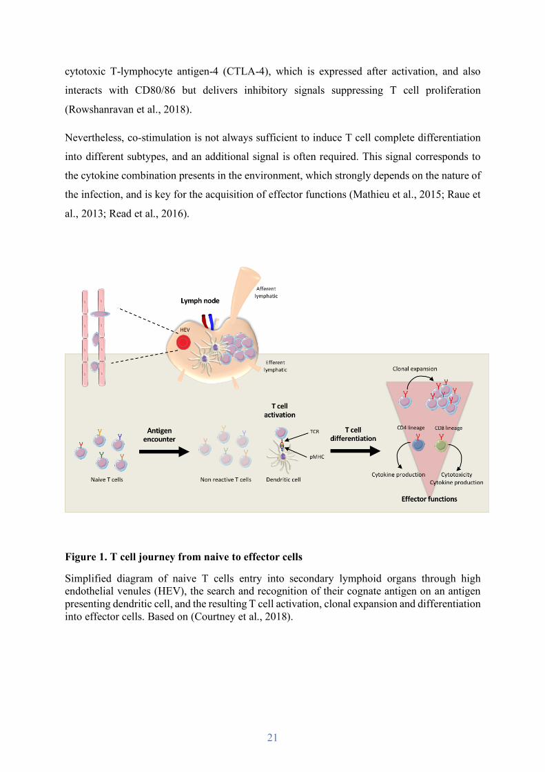
Figure 1. T cell journey from naive to effector cells
Simplified diagram of naive T cells entry into secondary lymphoid organs through high endothelial venules (HEV), the search and recognition of their cognate antigen on an antigen presenting dendritic cell, and the resulting T cell activation, clonal expansion and differentiation into effector cells. Based on (Courtney et al., 2018).

22
2.4. Naive T cell differentiation into effector cells
Upon activation, CD4 T cells differentiate into helper (Th) or T regulatory (Treg) T cells and
CD8 T cells into cytotoxic T cells (CTLs). Their surface expression patterns of adhesion
molecules and chemokine receptors change, allowing them to be guided to inflammatory sites
where they achieve their effector functions. Several molecules are thus commonly used to study
the journey of T cell through activation and differentiation.
- CD69 is a C-type lectin surface receptor. T cells rapidly express CD69 within a few hours
upon TCR stimulation, therefore CD69 has been used as a very early activation marker (Cibrian
and Sanchez-Madrid, 2017). CD69 regulates the differentiation of Treg cells as well as their
secretion of cytokines (Yu et al., 2018). Additionally, CD69 tunes T cell migration by inducing
the down-regulation of S1P1, a receptor required for lymphocyte egress from lymphoid organs.
Consequently, CD69 controls the retention of activated T cells in secondary lymphoid organs
(Shiow et al., 2006).
- CD25 is expressed within the first day upon TCR stimulation. CD25 is the alpha chain of the
interleukin-2 (IL-2) receptor, its expression leads to the formation of high affinity IL-2
receptors, together with IL-2b and gc subunits, that transduces the IL-2 signal required for T cell
survival and proliferation (Letourneau et al., 2009). In addition, CD25 expression, associated
with the expression of the FoxP3 transcription factor, allows to distinguish Tregs from others
CD4 T cell subsets.
- CD44 is a surface receptor that binds to the extracellular matrix, promoting cell adhesion and
migration. CD44 is up-regulated after TCR engagement and its expression is sustained on
effector cells as well as on memory cells allowing their extravasation to inflammatory sites
(DeGrendele et al., 1997; Nandi et al., 2004).
- CD62L, as previously mentioned (see 2.2.), is an L-selectin required for naive T cell homing
in secondary lymphoid organs. CD62L ligands are highly expressed in the HEVs and their
interaction initiates the extravasation process (Hemmerich et al., 2001). Upon T cell
differentiation into effector or memory cell, CD62L expression decreases, preventing T cells
from entering again into secondary lymphoid organs.

23
- CCR7 as presented previously is a chemokine receptor also involved in naive T cell homing
in secondary lymphoid organs. After activation, as for CD62L, T cells lose CCR7 expression
to present them from entering again in secondary lymphoid organs (Hauser and Legler, 2016).
- CD45 is a receptor protein tyrosine phosphatase key for TCR signaling (Desai et al., 1994). T
cells display several CD45 isoforms, which vary depending on the stage of T cell activation and
differentiation. Naive T cells express CD45RA. After activation, the extracellular domain of
CD45RA undergoes alternative splicing and is replaced by CD45RO (Rheinlander et al., 2018).
The analysis of the expression of several of these markers is used to distinguish naive, effector
and memory T cells. In human, CCR7 is often associated to CD45RA to make this distinction,
and particularly to sub-divide T cell memory populations. Indeed, naive T cells are CCR7+
CD45RA+, whereas central memory (TCM) cells are CCR7+ CD45RA- and effector memory
(TEM) are CCR7- CD45RA-, but can re-express CD45RA when they are highly differentiated
during persistent viral infection or chronic disease. TCM are mostly found in secondary
lymphoid organs and proliferate extensively, whereas TEM, which do not express CCR7, have
the capacity to migrate to inflammatory sites and have increased effector functions (Sallusto et
al., 2004).
In mice, CD62L is commonly associated with CD44. Naive T cells are CD62L+ CD44- whereas
TCM are CD62L+ CD44+ and TEM are CD62L- CD44+. TEM have thus the capacity to leave
secondary lymphoid organs and migrate to inflammatory sites.
24
2.5. CD8 T cell effector functions
CD4 T cells play an important role in coordinating adaptive immune responses. Indeed, there
are several T helper and regulatory subsets (see 1.2.), each one having specialized functions to
control and regulate, positively and negatively, CD8 T cells and B lymphocytes differentiation,
effector functions, and memory establishment (Yamane and Paul, 2013). Since this study has
been conducted on CD8 T cells only, CD4 T cell effector functions will not be described here.
Once activated in secondary lymphoid organs, CD8 T cells migrate to inflamed tissues where
they complete their differentiation into cytotoxic T cells (CTLs). There, they search for infected
or tumor cells expressing their specific pMHC. Upon antigen recognition and immunological
synapse formation, CTLs specifically kill target cells. The cytotoxic immune synapse requires
the engagement of only three to ten pMHC, its duration is short, and cells detach rapidly after,
allowing rapid elimination of pathogens (Purbhoo et al., 2004).
Three distinct mechanisms are involved in CTL effector functions. Two of them are
complementary pathways leading to target cell lysis: the granule exocytosis pathway and the
death-receptor pathway. The third one involves the release of different types of cytokines, as
numerous others immune cells.
2.5.1. Lytic granule exocytosis
Lytic granule release is the most important mechanism for infected or tumor cell elimination.
These granules are specialized secretory lysosomes released at the cytotoxic immune synapse
cleft formed between the CTL and the target cell. The synaptic cleft has thus been assumed to
optimize killing efficiency through high local concentration of lytic products and to prevent
other non-targeted cells to be exposed to cytotoxic proteins.
Lytic granules share molecular markers and traffic regulation with late endosomes and
lysosomes. They contain perforin, a pore forming protein, and granzymes, a family of serine
proteases, in both T cells and NK cells (Peters et al., 1991). Both perforin and granzymes are
poorly expressed in naive CD8 T cells, and are synthesized and stored in granules during CTL
differentiation (Olsen et al., 1990; Sanchez-Ruiz et al., 2011).

25
Perforin inserts in the target cell plasma membrane, oligomerizes and forms pores (Thiery and
Lieberman, 2014). Then, granzymes enter the target cell via these pores to trigger its apoptosis.
Therefore, granzyme capacity to induce target cell death is dependent on perforin as highlighted
in perforin knockout mice (Kagi et al., 1994; Walsh et al., 1994).
Several types of granzymes with different substrate specificity are expressed in CTLs.
Granzyme B has the strongest pro-apoptotic function, and acts through two mechanisms. First,
granzyme B can cleave its substrate after aspartate residues directly activating caspases, mostly
caspase 3, which results in rapid apoptosis. Granzyme B can also facilitate caspase activation
by cleaving the pro-apoptotic BH3 interacting death domain agonist Bid, inducing
mitochondrial damage and cytochrome C release (Afonina et al., 2010). Granzyme A activates
a slower mechanism of caspase-independent apoptosis by targeting the endoplasmic reticulum-
associated complex SET. Granzyme A and B act independently to induce apoptosis, and the
exact role of others granzymes is less known (Lieberman, 2003).
Defects in this cytotoxic pathway are associated with various disorders, particularly when
perforin production is affected as in hemophagocytic lymphohistiocytosis (HLH), a disorder
characterized by impaired granule-dependent cytotoxicity and uncontrolled T cell and
macrophage activation, resulting in hyper-inflammation (Osinska et al., 2014; Stepp et al.,
1999).
CTLs can kill multiple times in several hours, and thus need to be protected against their own
cytotoxicity. Surface expression of the lysosomal protease cathepsin B locally at the synapse
cleft could explain this self-protection (Balaji et al., 2002).
2.5.2. Death-receptor pathway
Upon CTL stimulation, Fas ligand (FasL) that is stored in intracellular granules is translocated
to the CTL surface at the cytotoxic immune synapse, and recognizes the cell death surface
receptor Fas expressed on target cells (Waring and Mullbacher, 1999). Fas, also known as
CD95, is a member of the tumor necrosis factor (TNF) receptor family. After activation, the
adapter molecule FADD (Fas-associated death domain) binds to Fas death domain recruiting
pro-caspase-8. Caspase-8 then initiates the caspase cascade leading to target cell apoptosis
(Micheau et al., 2002).
26
Defect in this cytotoxic pathway also lead to various disorders, as in autoimmune
lymphoproliferative syndrome (ALPS), characterized by defective lymphocyte homeostasis
and accumulation of autoreactive cells associated with Fas mutation (Rieux-Laucat et al.,
2003).
2.5.3. Cytokine secretion
Upon TCR signaling, CTLs produce cytokines as IFNγ and TNFα, and secrete them at the
synapse or in a multidirectional way (Huse et al., 2006; Kupfer et al., 1991).
IFNγ has multiple functions as it regulates more than 200 genes. IFNγ acts trough a cell surface
receptor whose ligation leads to Jak1 and Jak2 activation and subsequently to Stat1
phosphorylation which enters the nucleus and acts as a transcription factor (Schroder et al.,
2004). IFNγ signaling in target cells ultimately promotes MHC I presentation pathway,
amplifying their recognition by CTLs (Dolei et al., 1983). Furthermore, IFNγ is essential for
pathogen clearance as it directly inhibits viral replication, and stimulates several immune cell
types, as CD4 T cells, NK cells, or macrophages (Fensterl and Sen, 2009). This could also
enhance anti-tumor responses.
TNFα functions are also diverse. TNFα acts through the TNRF1 or TNFR2 receptors that differ
in their structure and in the signaling pathways they induce. Indeed, TNFR1 is almost
ubiquitously expressed whereas TNFR2 is mainly expressed by immune and endothelial cells.
TNFα binding to either receptors leads to the activation of the NFκB transcription factor and
MAP kinases (ERK, p38, JNK) (Hsu et al., 1995; Mehta et al., 2018; Rothe et al., 1995).
However, TNFR2 engagement promotes cell survival, whereas TNFR1 engagement can induce
either cell survival or cell death (Brenner et al., 2015). TNFα signaling can thus promotes T cell
activation and proliferation (Scheurich et al., 1987), and inflammatory response involving
several cell types. However, it can also lead to their apoptosis via caspase 8 activation (Micheau
and Tschopp, 2003).

27
2.6. Memory CD8 T cells
Upon pathogen clearance, most of the CTLs die within 1-2 weeks during the contraction phase
of the immune response. Only 5-10% are left as long-lived memory cells that are able to rapidly
induce a secondary response following re-exposure to the pathogen (Rocha and Tanchot, 2004;
Youngblood et al., 2017).
Memory cells present common properties with effector and naive cells, including pluripotency
and the ability to migrate in secondary lymphoid organs. In addition, cell expansion and re-
acquisition of effector functions are faster in secondary responses, compared to primary
responses, with much higher expansion.
In addition, several sub-populations of memory T cells displaying different properties have
been unveiled. For example, as mentioned previously, TCM are mostly found in secondary
lymphoid organs and proliferate extensively, whereas TEM migrate to inflammatory sites and
have increased effector functions.
Therefore, T cell journey from hematopoietic stem cells to CTLs is long and highly regulated.
Each step is crucial for efficient adaptive immune response. The impairment of anyone of them
could indeed result in defective pathogen clearance, tumor escape, or autoimmunity.

28
3. The immunological synapse
As mentioned above, when T cells recognize pMHC on antigen-presenting cells or target cells,
they form immunological synapses that allow the communication between the two cells and
ensure fully efficient antigen recognition and the triggering of the TCR and co-signaling
pathways, leading to T cell activation, proliferation, differentiation and the implementation of
effector functions.
The immunological synapse is the result of a massive T cell polarization process that reorients
the T cells toward the antigen-presenting cell or the target cell (see Figure 2). This process leads
to the reorganization of a variety of T cell components, including cytoskeleton, several
organelles, and the clustering of a plethora of receptors, signaling and adhesion molecules at
the immunological synapse (Soares et al., 2013b).
3.1. The immunological synapse formation
Immunological synapse formation starts with TCR engagement by pMHC. Initial TCR
signaling triggers T cell polarization toward the antigen presenting cell driven by cytoskeleton
reorganizations, which in turn tunes TCR signaling pathways leading to T cell activation,
differentiation, and cytokine production.
3.1.1 Cytoskeleton reorganization
Upon antigen recognition, T cells scanning their environment slow down and form stable
interactions with antigen-presenting cells in the case of naive T cells, or with target cells in the
case of CTLs. TCR engagement leads to a complete reorganization of T cell architecture in few
minutes, driven by dynamic rearrangements and interplay of the actin and microtubule
cytoskeleton.
29
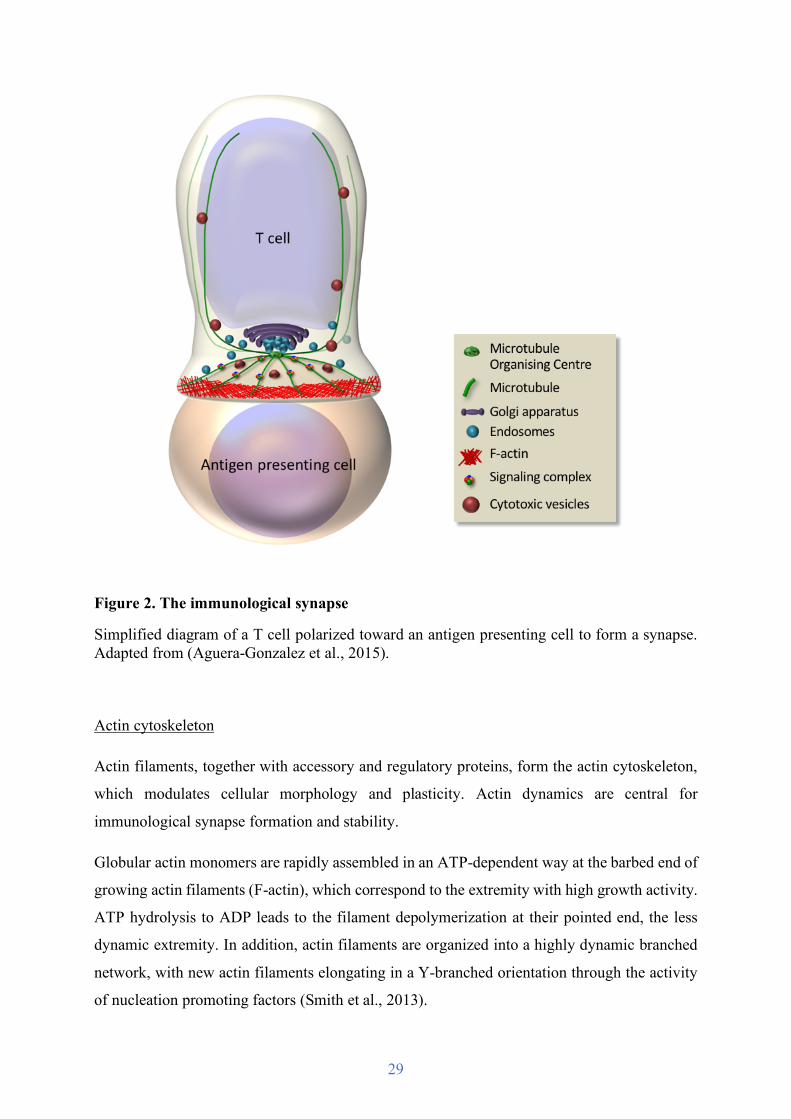
Figure 2. The immunological synapse
Simplified diagram of a T cell polarized toward an antigen presenting cell to form a synapse. Adapted from (Aguera-Gonzalez et al., 2015).
Actin cytoskeleton
Actin filaments, together with accessory and regulatory proteins, form the actin cytoskeleton,
which modulates cellular morphology and plasticity. Actin dynamics are central for
immunological synapse formation and stability.
Globular actin monomers are rapidly assembled in an ATP-dependent way at the barbed end of
growing actin filaments (F-actin), which correspond to the extremity with high growth activity.
ATP hydrolysis to ADP leads to the filament depolymerization at their pointed end, the less
dynamic extremity. In addition, actin filaments are organized into a highly dynamic branched
network, with new actin filaments elongating in a Y-branched orientation through the activity
of nucleation promoting factors (Smith et al., 2013).

30
Actin reorganization is mainly regulated by the activation of Rho-family small GTPases whose
best characterized members are Rac1, RhoA and Cdc42, and their downstream effector
molecules (Etienne-Manneville and Hall, 2002; Pollard, 2016). Rho GTPases constantly cycle
between an inactive, GDP-bound form, and an active, GTP-bound form, and bind effector
proteins involved in actin remodeling. The switch between the GDP- and GTP-bound forms is
regulated by guanine nucleotide exchange factors (GEFs) and GTPase-activating proteins
(GAPs), such as Vav1 and the Apc-stimulated guanine exchange factor (Asef) (Tybulewicz and
Henderson, 2009). When activated, Rho GTPases regulate the nucleation promoting factors
WASp (Wiskott Aldrich syndrome protein) and WAVE (WASp-family verprolin-homologous
protein), which in turn activate the Arp2/3 complex. The latter binds on the side of a pre-
existing linear actin filament and allows the nucleation and elongation of a new actin filament
in a Y-branch orientation (Goley and Welch, 2006; Rohatgi et al., 1999; Smith et al., 2013).
During immunological synapse formation, all these molecules are recruited at the cell contact
area and are regulated by TCR signaling pathways. Therefore, F-actin actively polarizes at
synapse. Once the synapse stabilized, F-actin clears from the center of the synapse leaving an
actin-rich peripheral ring (see Figure 2) (Bunnell et al., 2001; Ritter et al., 2015). F-actin
continuously pulls forces on the plasma membrane due to the molecular motor myosin II
contraction. Additionally, actin polymerization pushes on the membrane and drives retrograde
flow of the actin network. Together, these forces stabilize the actin cytoskeleton meshwork and
maintain the radial symmetry of the immunological synapse (Babich et al., 2012; Comrie and
Burkhardt, 2016).
Microtubule cytoskeleton
Microtubules contribute to cell shape determination, provide tracks for vesicle and organelle
intracellular traffic, and control cell division by allowing chromosomic segregation. As the actin
cytoskeleton, the microtubule network is highly reorganized at the immunological synapse.
Microtubules are formed by α/β tubulin heterodimers, which organize into tubular filaments
with β-tubulin being exposed at the highly dynamic plus end and α-tubulin at the minus end.
GTP-bound α/β heterodimers polymerize into microtubule filaments when hydrolyzed into
GDP-bound subunits. Microtubules are nucleated from the centrosome, from where they radiate
with their minus ends in and plus ends out, but also from the Golgi apparatus (Chabin-Brion et

31
al., 2001; Petry and Vale, 2015). Their nucleation, organization and stability are regulated by
microtubule-associated proteins (MAPs) such as γTuRC that serve as template for microtubule
formation, by post-translational modifications such as acetylation, and by microtubule plus end
tracking proteins including EB1, which recruits a range of different factors at the microtubule
tips. Altogether, these regulatory features promote microtubule polymerization and stability
(Nehlig et al., 2017; Roostalu and Surrey, 2017).
During synapse formation, microtubules are repositioned at the periphery of the T cell contact
area through the action of several proteins (see Figure 2). These include the microfilament liker
ezrin and the polarity regulators Dlg1 and adenomatous polyposis coli (Apc) that were all
shown necessary for microtubule radial organization at the synapse (Aguera-Gonzalez et al.,
2017; Bertrand et al., 2010; Lasserre et al., 2010). This process leads to the polarization of the
centrosome and its translocation toward the cell contact area (Geiger et al., 1982; Stinchcombe
et al., 2006). Numerous molecules have been shown necessary for centrosome polarization,
such as the molecular motor dynein, Rac1, IQGAP-1, PKCζ and formins (Combs et al., 2006;
Gomez et al., 2007; Stinchcombe et al., 2006). However, the exact mechanisms involved are
poorly understood.
Microtubules, by converging at the center of the immunological synapse, allow polarized
transport of vesicular components and organelles, such as the Golgi apparatus, and several
endosomes and secretory lysosomes as lytic granules (Bouchet et al., 2016; Soares et al.,
2013b).
Actin and microtubule cytoskeleton dynamics are generally described separately. However,
they interplay through various interacting proteins such as ezrin, Dlg1, IQGAP-1 or Apc
(Fukata et al., 2002; Lasserre et al., 2010; Nelson and Näthke, 2013). Interestingly, centrosome
translocation toward the center of the synapse occurs within a minute after F-actin clearance
(Ritter et al., 2015; Stinchcombe et al., 2006). This observation, together with numerous other
studies, suggests a critical interplay between the microtubule network and the actin cytoskeleton
at immunological synapses.
Defects in actin or microtubule cytoskeleton reorganization during immunological synapse
formation can lead to impaired T cell polarization, synapse architecture and/or T cell activation,
associated with numerous disorders, e.g. ARPC1B mutation-associated immunodeficiency,

32
chronic lymphocytic leukemia, Wiskott-Aldrich syndrome, or familial adenomatous polyposis
(Aguera-Gonzalez et al., 2017; Kallikourdis et al., 2015; Lasserre et al., 2010; Ramsay et al.,
2008; Randzavola et al., 2019).
3.1.2 Polarized vesicular traffic to the immunological synapse
Centrosome repositioning close to the synapse plasma membrane drives the polarized transport
along microtubules of several receptors and signaling molecules that are associated to
endosomal and Golgi tubulovesicular structures (see Figure 2). Polarized vesicle traffic ensures
their accumulation at the synapse, the formation of signaling complexes involved in TCR
signaling and T cell activation.
Vesicles are associated with microtubules that ensure their directional transport by means of
two molecular motors: toward the centrosome via dynein, and toward synapse periphery via
kinesin (Daniele et al., 2011; Kurowska et al., 2012; Schroer et al., 1989; Vale et al., 1985). In
addition, local actin polymerization generates forces for vesicle movement along microtubules
(Derivery et al., 2009; Zhang et al., 2017). Vesicles then accumulate into a pericentrosomal
compartment.
Trafficking involves several types of endosomal (e.g. early, recycling) and Golgi
compartments, and vesicle traffic regulators, such as Rab GTPases, transport proteins, or vesicle
fusion regulators (e.g. SNAREs) (see Figure 3). It concerns for instance TCR subunits, the
tyrosine kinase Lck as well as the adapter LAT, downstream effectors of the TCR signaling
pathway (Onnis et al., 2016). Those molecules travel back and forth between the plasma
membrane and endosomes in non-stimulated T cells, but are reoriented to the synapse upon
TCR engagement with pMHC (Blanchard et al., 2002; Bouchet et al., 2016; Bouchet et al.,
2017; Das et al., 2004; Ehrlich et al., 2002; Finetti et al., 2009; Larghi et al., 2013; Purbhoo et
al., 2010; Soares et al., 2013a; Zucchetti et al., 2019). In addition, polarized vesicular traffic is
crucial for T cell effector functions by allowing secretion of cytokines and lytic granules in the
synaptic cleft (Daniele et al., 2011; Kurowska et al., 2012; Sepulveda et al., 2015; Stinchcombe
et al., 2006).
33
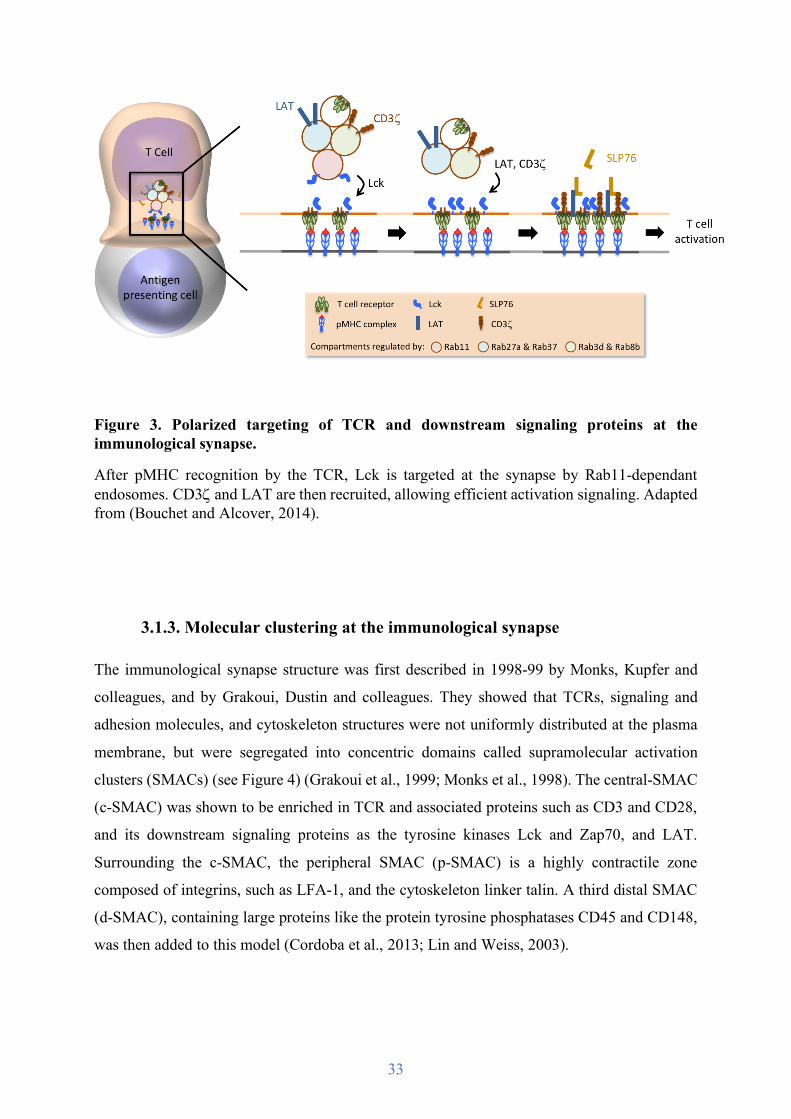
Figure 3. Polarized targeting of TCR and downstream signaling proteins at the immunological synapse.
After pMHC recognition by the TCR, Lck is targeted at the synapse by Rab11-dependant endosomes. CD3z and LAT are then recruited, allowing efficient activation signaling. Adapted from (Bouchet and Alcover, 2014).
3.1.3. Molecular clustering at the immunological synapse
The immunological synapse structure was first described in 1998-99 by Monks, Kupfer and
colleagues, and by Grakoui, Dustin and colleagues. They showed that TCRs, signaling and
adhesion molecules, and cytoskeleton structures were not uniformly distributed at the plasma
membrane, but were segregated into concentric domains called supramolecular activation
clusters (SMACs) (see Figure 4) (Grakoui et al., 1999; Monks et al., 1998). The central-SMAC
(c-SMAC) was shown to be enriched in TCR and associated proteins such as CD3 and CD28,
and its downstream signaling proteins as the tyrosine kinases Lck and Zap70, and LAT.
Surrounding the c-SMAC, the peripheral SMAC (p-SMAC) is a highly contractile zone
composed of integrins, such as LFA-1, and the cytoskeleton linker talin. A third distal SMAC
(d-SMAC), containing large proteins like the protein tyrosine phosphatases CD45 and CD148,
was then added to this model (Cordoba et al., 2013; Lin and Weiss, 2003).

34
Figure 4. The supramolecular activation clusters.
Simplified diagram of mature synapse organization in SMACs, and the molecules/ligands enriched in each zone.
This organization in concentric domains was initially thought to allow an optimal synapse
stability and thus T cell activation or effector functions. It is dictated, at least in part, by the size
of the ectodomains of the different molecules involved. The c-SMAC, responsible for TCR
signaling and the recycling of the molecules involved, contains molecules with short
ectodomains, therefore their interaction with their ligands on the antigen-presenting cell brings
the membrane of the two cells close to each other. The p-SMAC, which concentrates integrins
and their ligands, forms a ring of thigh adhesion between the two cells to stabilize the
immunological synapse. Conversely, molecules with larger ectodomains, such as the
phosphatase CD45, are excluded into the d-SMAC thus avoiding an inhibitory effect on the
TCR signaling pathway. Indeed, the artificial elongation of the p-MHC ectodomain, which
35
increases the distance between the two cells, prevents CD45 exclusion from the c-SMAC and
leads to reduce TCR signaling (Choudhuri et al., 2005).
Further studies, using phospho-specific antibodies revealing active signaling molecules,
showed that TCR signaling complexes appeared first in small clusters at the synapse periphery,
while the c-SMAC contained dephosphorylated molecules and correlated with inhibitory
signaling.
The use of TIRF live cell microscopy allowed to observe TCR and its downstream signaling
proteins forming submicron-scale molecular complexes, or microclusters, appearing within the
d-SMAC, then moving centripetally through the p-SMAC to the c-SMAC to form a mature
(e.g. concentrical) synapse. Signaling microclusters may disappear before reaching the c-
SMAC or coalesce in the c-SMAC, colocalizing with some lysosomal markers. This led to
propose that the immunological synapse p-SMAC and c-SMAC structures facilitate the balance
between signaling and degradation, and depend on antigen stimulatory quality. In addition,
micloclusters containing the co-stimulatory molecule CD28 and the inhibitory receptors CTL-
4 and PD-1 contribute to signaling regulation (Bunnell et al., 2002; Campi et al., 2005;
Cemerski et al., 2008; Cemerski et al., 2007; Lasserre and Alcover, 2010; Lee et al., 2003;
Varma et al., 2006; Yokosuka et al., 2008; Yokosuka et al., 2010; Yokosuka et al., 2005;
Yokosuka et al., 2012). More recently, the c-SMAC has been associated with the production
and accumulation of extracellular vesicles (ectosomes) containing TCR and CD40L that could
represent a means of T cell signal downregulation while producing vesicles capable to stimulate
antigen presenting cells (Choudhuri et al., 2014; Saliba et al., 2019).
The coordinated action of the actin cytoskeleton and the microtubule network is crucial for
microclusters dynamics and SMAC formation. Noteworthy, the d-SMAC corresponds to the
actin-rich peripheral ring and contains IQGAP-1 and ezrin that link the actin cytoskeleton and
the microtubules (Lasserre et al., 2010; Stinchcombe et al., 2006; Watanabe et al., 2004). It is
important to note that, concomitantly, TCR signaling regulates cytoskeleton dynamics, thus
coordinating the formation of mature synapse. Furthermore, actin cytoskeleton is required to
sustain TCR signaling (Valitutti et al., 1995).
As mentioned above, centrosome translocation toward the cell-cell contact area allows the
polarized transport of vesicles containing TCR, Lck and LAT to the synapse and the formation
of signaling complexes (Blanchard et al., 2002; Ehrlich et al., 2002; Finetti et al., 2009; Larghi
et al., 2013; Soares et al., 2013b). These proteins assemble in signaling microclusters in the d-
36
SMAC. Actin-induced forces driving retrograde flow, through its polymerization and myosin
II contraction, then allow the centripetal movement of the microclusters to the c-SMAC and of
LFA-1 to the p-SMAC (see Figure 5) (Campi et al., 2005; Comrie et al., 2015; Hashimoto-Tane
et al., 2011; Ilani et al., 2009; Nguyen et al., 2008). Microtubules support microcluster
centripetal movement involving the molecular motor dynein (Hashimoto-Tane et al., 2011;
Lasserre et al., 2010). Furthermore, actin clearence from the center of the synapse is the
initiating event for the c-SMAC formation (Bunnell et al., 2002; Ritter et al., 2015). Therefore,
blocking F-actin depolymerization reduces microcluster formation and impairs microcluster
and LFA-1 centripetal movement. This slows down their translocation to the p-SMAC,
decreasing TCR signaling and the adhesiveness of the synapse (Comrie et al., 2015; Comrie
and Burkhardt, 2016; Varma et al., 2006). Collectively, these data indicate that polarized
vesicular transport, actin and microtubule-mediated microcluster dynamics and force
generation are thus essential for controlling synapse formation and T cell activation.
Importantly, the p-SMAC and c-SMAC structures have been mostly observed in T cell
interacting with antigen presenting B cells or with lipid bilayer as surrogate synapses. In
contrast T cells interacting with dendritic cells form multifocal synapses (Brossard et al., 2005).
This indicates that depending on the quality of the antigen presenting cell, immunological
synapse structure and signaling may be different.
Therefore, the immunological synapse formation results from a dynamic T cell reorganization,
which allows a spatial and temporal molecular organization that tunes TCR signaling.
37
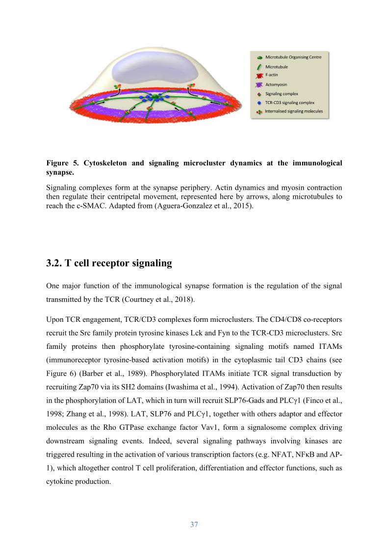
Figure 5. Cytoskeleton and signaling microcluster dynamics at the immunological synapse.
Signaling complexes form at the synapse periphery. Actin dynamics and myosin contraction then regulate their centripetal movement, represented here by arrows, along microtubules to reach the c-SMAC. Adapted from (Aguera-Gonzalez et al., 2015).
3.2. T cell receptor signaling
One major function of the immunological synapse formation is the regulation of the signal
transmitted by the TCR (Courtney et al., 2018).
Upon TCR engagement, TCR/CD3 complexes form microclusters. The CD4/CD8 co-receptors
recruit the Src family protein tyrosine kinases Lck and Fyn to the TCR-CD3 microclusters. Src
family proteins then phosphorylate tyrosine-containing signaling motifs named ITAMs
(immunoreceptor tyrosine-based activation motifs) in the cytoplasmic tail CD3 chains (see
Figure 6) (Barber et al., 1989). Phosphorylated ITAMs initiate TCR signal transduction by
recruiting Zap70 via its SH2 domains (Iwashima et al., 1994). Activation of Zap70 then results
in the phosphorylation of LAT, which in turn will recruit SLP76-Gads and PLCγ1 (Finco et al.,
1998; Zhang et al., 1998). LAT, SLP76 and PLCγ1, together with others adaptor and effector
molecules as the Rho GTPase exchange factor Vav1, form a signalosome complex driving
downstream signaling events. Indeed, several signaling pathways involving kinases are
triggered resulting in the activation of various transcription factors (e.g. NFAT, NFκB and AP-
1), which altogether control T cell proliferation, differentiation and effector functions, such as
cytokine production.
38
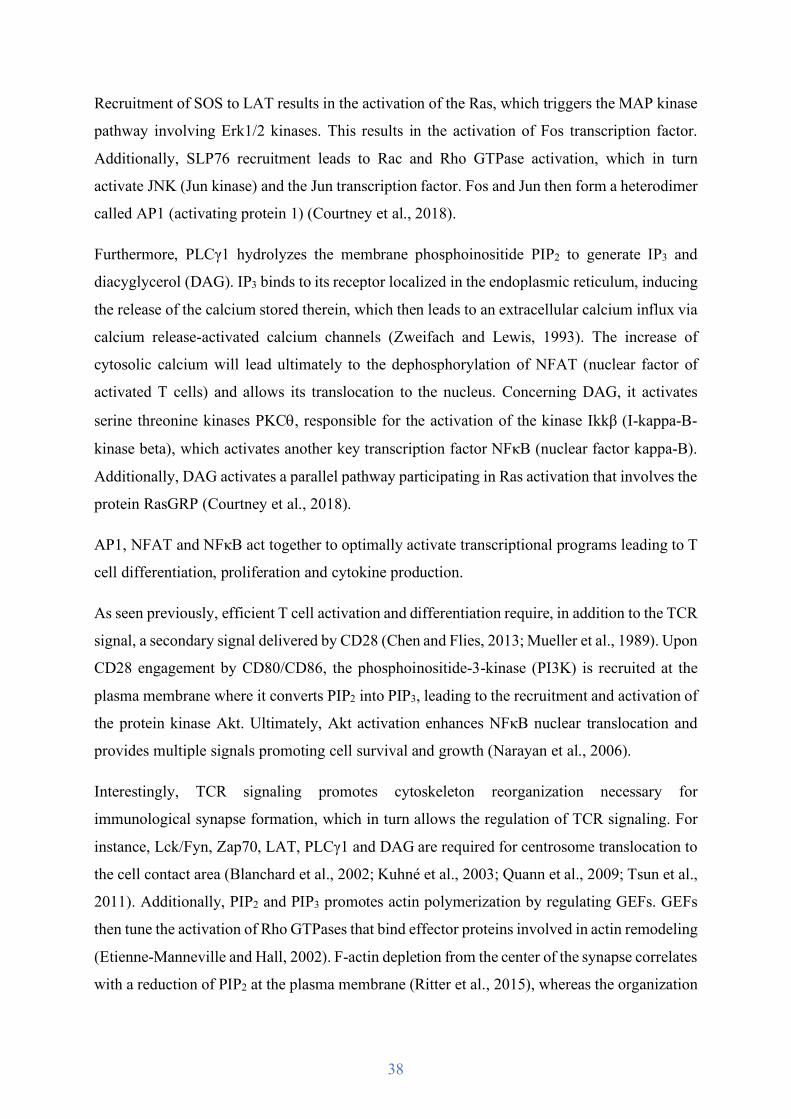
Recruitment of SOS to LAT results in the activation of the Ras, which triggers the MAP kinase
pathway involving Erk1/2 kinases. This results in the activation of Fos transcription factor.
Additionally, SLP76 recruitment leads to Rac and Rho GTPase activation, which in turn
activate JNK (Jun kinase) and the Jun transcription factor. Fos and Jun then form a heterodimer
called AP1 (activating protein 1) (Courtney et al., 2018).
Furthermore, PLCγ1 hydrolyzes the membrane phosphoinositide PIP2 to generate IP3 and
diacyglycerol (DAG). IP3 binds to its receptor localized in the endoplasmic reticulum, inducing
the release of the calcium stored therein, which then leads to an extracellular calcium influx via
calcium release-activated calcium channels (Zweifach and Lewis, 1993). The increase of
cytosolic calcium will lead ultimately to the dephosphorylation of NFAT (nuclear factor of
activated T cells) and allows its translocation to the nucleus. Concerning DAG, it activates
serine threonine kinases PKCq, responsible for the activation of the kinase Ikkβ (I-kappa-B-
kinase beta), which activates another key transcription factor NFκB (nuclear factor kappa-B).
Additionally, DAG activates a parallel pathway participating in Ras activation that involves the
protein RasGRP (Courtney et al., 2018).
AP1, NFAT and NFκB act together to optimally activate transcriptional programs leading to T
cell differentiation, proliferation and cytokine production.
As seen previously, efficient T cell activation and differentiation require, in addition to the TCR
signal, a secondary signal delivered by CD28 (Chen and Flies, 2013; Mueller et al., 1989). Upon
CD28 engagement by CD80/CD86, the phosphoinositide-3-kinase (PI3K) is recruited at the
plasma membrane where it converts PIP2 into PIP3, leading to the recruitment and activation of
the protein kinase Akt. Ultimately, Akt activation enhances NFκB nuclear translocation and
provides multiple signals promoting cell survival and growth (Narayan et al., 2006).
Interestingly, TCR signaling promotes cytoskeleton reorganization necessary for
immunological synapse formation, which in turn allows the regulation of TCR signaling. For
instance, Lck/Fyn, Zap70, LAT, PLCγ1 and DAG are required for centrosome translocation to
the cell contact area (Blanchard et al., 2002; Kuhné et al., 2003; Quann et al., 2009; Tsun et al.,
2011). Additionally, PIP2 and PIP3 promotes actin polymerization by regulating GEFs. GEFs
then tune the activation of Rho GTPases that bind effector proteins involved in actin remodeling
(Etienne-Manneville and Hall, 2002). F-actin depletion from the center of the synapse correlates
with a reduction of PIP2 at the plasma membrane (Ritter et al., 2015), whereas the organization
39
and maintenance of the actin-rich ring is controlled by the annular accumulation of PIP3 (Le
Floc'h et al., 2013).
In conclusion, interplay between T cell signaling events and cytoskeleton reorganization at the
immunological synapse leads to the activation of transcription factors that control gene involved
in T cell proliferation, differentiation, and effector function.
Figure 6. Main signaling pathways triggered by TCR-pMHC and CD28-CD80/86 engagement
Different signaling pathways activated upon TCR and co-receptor signalization, ultimately leading to transcription factor activation. From left to right, pathway color code is: blue for Rac/JNK pathway, green for Erk1/2/Fos pathway, violet for Calcium/NFAT pathway, and orange for PKCq /NF𝜅B pathway. (Aguera-Gonzalez et al., 2015).

40
3.3. The cytotoxic synapse
The immunological synapse formation and TCR signaling allow T cell activation, leading to
their proliferation and differentiation, but also to the triggering of their effector functions.
Indeed, the dynamic interplay between receptor signaling, polarized vesicle traffic and actin
and microtubule cytoskeleton rearrangements results in production and polarized secretion of
cytokines, membrane apposition allowing FasL-Fas interaction, and above all, lytic granule
release in the synaptic cleft.
3.3.1. Cytoskeleton reorganization
The cytotoxic synapse organization is similar to the one described previously (see Figure 7),
however the centrosome does not only translocate, but is docked at the plasma membrane.
Cytotoxic granules move along the microtubules in a dynein-mediated manner toward their
minus end to cluster around the moving centrosome, with which they polarized at the synapse
(Ritter et al., 2015). The centrosome thus defines a secretory domain localized within the p-
SMAC, next to the c-SMAC (Stinchcombe et al., 2001b; Stinchcombe et al., 2006). Its docking
ensures a precise site for lytic granule secretion, concentrating granules in the synaptic cleft,
but also promotes the delivery of FasL at the plasma membrane (Bossi and Griffiths, 1999).
Furthermore, F-actin depletion from the c- and p-SMAC to form an actin-rich ring allows the
centrosome docking and facilitates granules fusion at the plasma membrane. Conversely, actin
recovery terminates cytotoxic granule release (Ritter et al., 2015; Ritter et al., 2017).
Several studies have also shown that, in opposition to the stimulatory synapse, which leads to
T cell activation, the cytotoxic synapse does not require to be completely formed and stable to
be efficient. Indeed, CTLs do not need a strong antigen stimulation to induce target cell death,
compared to the one required for stimulatory synapse (Faroudi et al., 2003). Killing could
occurs with only three TCR-pMHC interactions, whereas stable synapse formation requires at
least ten interactions (Purbhoo et al., 2004). The formation of a mature synapse with typical
SMAC pattern may not always be necessary for efficient cytotoxic granules release (O'Keefe
and Gajewski, 2005). Hence, CTLs could kill multiple targets simultaneously, only polarizing
toward the target cell with the strongest antigen stimulus (Depoil et al., 2005). Lytic granule
translocation to the T cell-target cell contact area could thus be independent from the
centrosome repositioning (Bertrand et al., 2010; Wiedemann et al., 2006).

41
Figure 7. Formation of the cytotoxic synapse.
First the CTL recognizes a pMHC complex presented by a tumor or infected cell. Actin polymerizes at the synapse and rapidly clears the center, while lytic granules cluster around the centrosome and move toward the synapse. Granules fuse with the plasma membrane and perforin and granzymes are released in the synaptic cleft.
42
The importance of the F-actin dynamics for CTL effector functions has however not been
questioned as impairment of numerous of its regulators, such as PIP3, Arp2/3 or WASp, affects
killing efficiency (De Meester et al., 2010; Le Floc'h et al., 2013; Ramsay et al., 2008).
3.3.2. Lytic granule transport and fusion
Once at the synapse, lytic granules fuse with the plasma membrane, and perforin and granzymes
are released in the synaptic cleft. The study of human genetic disorders and mouse mutants
allowed to identified numerous proteins involved. Indeed, characterization of the molecular
defects leading to hemophagocytic lymphohistiocytosis (HLH) has contributed to understand
the different steps and their regulators required for lytic granules maturation and exocytosis (de
Saint Basile et al., 2010).
As highlighted above, HLH may result from defects in perforin (Stepp et al., 1999), but can
also result from SNARE protein and SNARE accessory factor deficiency, both in human and
mice. Therefore, deficient lysosomal trafficking regulator (LYST) alters granule maturation
(Barbosa et al., 1996; Nagle et al., 1996; Sepulveda et al., 2015), deficient AP-3 adaptor alters
lytic granule transport on microtubules to the synapse (Clark et al., 2003; Feng et al., 1999),
deficient Rab27a their docking (Kurowska et al., 2012; Ménasché et al., 2000; Stinchcombe et
al., 2001a), deficient Munc13-4 their priming (Feldmann et al., 2003; Ménager et al., 2007),
and deficient syntaxin-11 and Munc18-2 their fusion at the plasma membrane (zur Stadt et al.,
2009).
3.3.3. Importance of forces for killing
The interplay of pushing and pulling forces induced by the actin cytoskeleton described above
are not only important for immunological synapse formation, but also for CTL effector
functions. Indeed, the adaptation of single cell biophysical approaches (e.g. stimulatory
deformable micropillar arrays, stimulatory beads attached to micropipettes) has shown that
CTLs exert forces against antigen presenting surfaces with a spatiotemporal correlation
between the force exertion and lytic granule release (Basu et al., 2016; Tamzalit et al., 2019).
Forces applied to the synapse promote cytotoxicity by increasing the target cell membrane
tension, which in turn enhances the perforin pore forming activity.

43
Hence, the immunological synapse formation is the result of a massive dynamic polarization of
the T cell. This reorganization involves numerous cellular components as the actin cytoskeleton,
microtubule network, vesicular traffic or receptors, but most importantly, is regulated by the
interplay between them, coordinately regulated in space and time. Together they act to form a
stable synapse ensuring T cell activation, differentiation and effector functions necessary for
efficient adaptive immune response, and the elimination of pathogens and tumor cells.

44
4. Anti-tumor response
The involvement of the immune system in tumor development prevention and growth control
have been extensively studied, both in human and mouse.
Tumor development and resulting immune response occur in three steps: elimination,
equilibrium and escape (see Figure 8) (Dunn et al., 2004).
4.1. Control of tumor development
Tumors appear after cellular modifications such as mutations and chromosomal instability that
alter normal cell growth and survival, resulting in premalignant lesions. These lesions are
mostly eliminated through cell-intrinsic tumor suppression mechanisms activating apoptosis
and cell-cycle arrest (reviewed in (Lowe et al., 2004)) or by immunosurveillance processes.
Abnormal cells enter in a senescence state and secrete various cytokines inducing a pro-
inflammatory environment (Kuilman et al., 2008). These cytokines mediate the recruitment of
innate immune cells first, which leads to the killing of some senescent cells and consequent
release of tumor antigens. These antigens are presented by antigen presenting cells to mature
naive CD4 and CD8 T cells, driving their activation, proliferation and differentiation into
effector cells. The development of tumor-specific adaptive immunity then provides the capacity
to completely eliminate abnormal cells, in particular through CTL cytotoxic activity and IFNg
production (Kang et al., 2011; Ostroumov et al., 2018; Shankaran et al., 2001).
These mechanisms are remarkably efficient as on average cancers arise less than once in a
human lifetime, despite trillions of potential target cells. However, it happens that some
premalignant cells survive the elimination phase. If T cells and IFNg actions can contain tumor
development, an equilibrium state is achieved, sometime during several years (Dunn et al.,
2004; Koebel et al., 2007).
Malignant cells can however escape the immune control, and grow to form clinically detectable
tumors. Tumor escape is the result of immune-resistant cell selection over the time and the
addition of further genetic alterations in abnormal cells during the equilibrium phase, such as

45
the mutation of tumor suppressors like the polarity regulator adenomatous polyposis coli in
colorectal cancer or the transcription factor p53 that has been involved in 50% of cancers.
Figure 8. The three phases of tumor development.
Adapted from (Dunn et al., 2004).
4.2. Tumor escape
Escape and proliferation of tumor cells often is accompanied by the presence of an
immunosuppressive environment, which is less permissive for T cell recruitment, persistence,
and effector functions (Frey, 2015). Both T cell intrinsic and extrinsic factors have been shown
to inhibit T cell functions in tumors. Indeed, CTLs that have a central role in anti-tumor
immunity present impaired cytotoxic activity in the tumor bed.

46
Intrinsic factors mostly consist on the increased expression of checkpoint inhibitors at the T cell
surface when they interact with tumor cells. Checkpoint inhibitors are signaling receptors,
which during normal immune response control T cell over-activation to limit inflammation, but
restrict anti-tumor functions in cancer. Checkpoint inhibitors include T cell co-stimulatory
receptors such as CTLA-4, which is part of the CD28 family. It recognizes B7 family proteins
as CD80/86 and suppresses T cell proliferation by competing for ligand binding with CD28
(Chen, 2004; Rowshanravan et al., 2018). In addition, tumor-infiltrating CTLs have been shown
to form altered immunological synapses with tumor cells. The actin cytoskeleton does not
polymerize at the cell contact area, TCR and CD3 do not colocalize in microsclusters in the c-
SMAC, TCR early signaling is inhibited, and the centrosome and cytotoxic granules are not
translocated at the plasma membrane in CD8 T cells isolated from murine tumor models
(Radoja et al., 2001). Intrinsic factors therefore induce hypo-responsive T cells that are
exhausted, contributing to tumor progression.
Extrinsic factors occur via the recruitment of immunosuppressive cell populations such as
Tregs. These cells are critical for autoimmunity protection and regulation of inflammation and
immune responses. However, they may be an obstacle for anti-tumor CTL responses. Tregs are
characterized by the expression of CD25 and of the transcription factor Foxp3, but also the
inability to produce IL-2 (Hori et al., 2003). Tregs act through indirect and direct
immunosuppressive mechanisms (Miyara and Sakaguchi, 2007). They produce anti-
inflammatory cytokines such as IL-10 and TGF-b (Andersson et al., 2008), and consume IL-2
necessary for others T cell proliferation and survival (Pandiyan et al., 2007). They can also
display similar cytotoxic functions to CTLs, and eliminate B and T lymphocytes through
cytotoxic granule exocytosis and by inducing the death-receptor Fas pathway (Cao et al., 2007;
Grossman et al., 2004; Strauss et al., 2009; Zhao et al., 2006). Extrinsic factors therefore induce
anti-tumor effector cell apoptosis, also contributing to tumor progression.
Considering CTL and Treg functions, their number and ratio in tumors are crucial prognostic
factors for patient survival. CTL infiltration is thus considered as favorable and associated with
good prognosis, whereas Treg infiltration is generally associated with poor clinical outcome
(Galon et al., 2006). However, the balance between CTLs and Tregs need to be equilibrated as
inflammation can also promote tumor development particularly in colorectal cancers
(Mantovani et al., 2008).

47
5. Adenomatous polyposis coli
As describe above, T cell immune responses are controlled by the ability of the cell to polarize.
Indeed, the immunological synapse formation and thus the resulting T cell activation,
differentiation and effector functions that are crucial to control tumor development, depend on
the polarization of T cells toward antigen presenting cells and tumor target cells. Polarization
is also key to control migration, and therefore allowing T cells to enter secondary lymphoid
organs and to be recruited into tumors.
Cell polarity regulators are proteins controlling cellular asymmetric distribution of molecules,
the asymmetric organization specific membrane domains, and the enrichment of organelles or
the cytoskeleton at specific sites (Rodriguez-Boulan and Macara, 2014). Hence, polarity
regulators may be essential to ensure fully efficient T cell anti-tumor immune responses.
Several of them have been shown to control immunological synapse formation and T cell
activation. For instance, Dlg1 silencing perturbs T cell shape, microcluster formation, actin
polymerization, microtubule network organization and centrosome polarization, and NFAT-
driven gene transcription (Lasserre et al., 2010; Round et al., 2007; Round et al., 2005). In
addition, Scribble silencing perturbs synapse formation and TCR signaling molecule
recruitment at the synapse (Ludford-Menting et al., 2005). Interestingly, both Dlg1 and Scribble
interact with another polarity regulator called adenomatous polyposis coli (Apc), whose role
has never been characterized in CD8 T cells (How et al., 2019; Matsumine et al., 1996).
5.1. Adenomatous polyposis coli structure and function
Apc is a large 310-kDa multi-domain cytoplasmic protein (see Figure 9). Its central region
contains short peptide motifs that bind to the transcription factor regulator β-catenin, but
contains also three set of SAMP (Ser-Ala-Met-Pro) domains binding Axin to regulate β-catenin
(Rubinfeld et al., 1993). Apc amino-terminal portion contains a dimerization domain and an
armadillo repeat domain. The armadillo domains interact with numerous cytoskeleton
regulators as the actin and microtubule regulator IQGAP-1, the Rho GTPase Cdc42, the Rho
GTPase regulator Asef, or kinesin regulators (Jimbo et al., 2002; Kawasaki et al., 2000;
Sudhaharan et al., 2011; Watanabe et al., 2004). The carboxy-terminal portion also contains

48
domains binding cytoskeleton regulators as the microtubule plus end binding EB1 and Dlg1,
but also a basic domain directly interacting with microtubules (Munemitsu et al., 1994;
Nakamura et al., 2001). In addition, Apc has been shown to interact directly and indirectly with
nuclear pore and nuclear transport proteins, and apoptosis- or mitosis-related proteins (Nelson
and Näthke, 2013).
Therefore, due to its numerous interaction domains, Apc is a multi-functional protein involved
in a large range of cellular mechanisms.
Figure 9. Adenomatous polyposis coli structure and functions
(A) Schematic domain structure of Apc describing binding sites and interacting partners, and cellular functions they are involved in. (B) Apc truncated form resulting from Apc mutation. Based on (Aoki and Taketo, 2007).
Regulate proliferation and differentiation
Dimerizationdomain
Armadillo domains Basic domain
EB1 binding domain
Dlg1 binding domain
β-catenin and Axin binding domains
Binding to IQGAP-1, Cdc42, Asef and mDia
Binding to β-catenin
Binding to axin
Binding to microtubules
Cycline D and c-mycStimulate actinpolymerization
Chromosomalsegregation
Stabilize microtubules
Control of cellular polarization and migration
Full length adenomatous polyposis coli
C-terminally truncated adenomatous polyposis coli
A
B
49
5.1.1. Apc as a Wnt signaling pathway regulator
Apc is mostly known for its role in the Wnt signaling pathway, which plays essential roles
during embryonic development and in tissue homeostasis, by driving cell proliferation and
survival.
The Wnt signaling pathway regulates the level of the transcription factor β-catenin via a specific
destruction complex, which is composed by Apc, Axin, casein kinase 1 (CK1) and glycogen
synthase kinase 3 (GSK3). In absence of Wnt extracellular stimulus, this cytosolic destruction
complex initiates β-catenin ubiquitination and ultimately its proteasomal degradation. In
presence of Wnt stimulus, the destruction complex is inhibited via its recruitment to the plasma
membrane, and β-catenin is stabilized and translocates to the nucleus, thus inducing the
transcription of Wnt target genes, including c-myc, cyclin D and caspases. Increasing β-catenin
availability thus results in increased proliferation, but also decrease cell differentiation
(Munemitsu et al., 1995; Stamos and Weis, 2013).
5.1.2. Apc as a polarity regulator
Due to its numerous interaction domains with actin and microtubule cytoskeleton regulators
(see Figure 7), Apc has been involved in numerous cellular processes that require their
reorganization and cellular polarization, as cell directional migration and division, and is thus
classified as a polarity regulator protein.
The importance of Apc in cytoskeleton regulation is reflected by its subcellular localization.
Indeed, in migrating astrocytes and epithelial cells, Apc predominantly forms very small
clusters mainly localized at microtubule plus ends and plasma membrane, which correlates with
protrusive cell area and the leading edge (Etienne-Manneville et al., 2005; Näthke et al., 1996).
In highly polarized cells as intestinal epithelial cells, Apc is mostly detected at the basal surface,
where microtubule plus ends also are (Näthke et al., 1996). A more recent study from the
Alcover lab has also unveiled a similar localization in CD4 T cells, where Apc clusters appeared
enriched at the immunological synapse periphery, corresponding to microtubule plus end, but
were also observed along microtubules (Aguera-Gonzalez et al., 2017). Furthermore, Apc has
been observed in actin-rich leading edge of migrating epithelial cells (Watanabe et al., 2004).

50
Apc stabilizes the microtubule network in migrating fibroblasts and astrocytes, and ensures
directional migration. Loss of Apc results in decrease of cellular protrusion and microtubule
stability during cell migration (Etienne-Manneville, 2009; Etienne-Manneville and Hall, 2003).
In addition, Apc ensures microtubule network organization and centrosome polarization at the
immunological synapse of CD4 T cells (Aguera-Gonzalez et al., 2017). Furthermore, Apc loss
has been associated with defective mitotic spindle structure during cell division (Dikovskaya et
al., 2004). Finally, Apc truncation has been recently shown to induce the fragmentation of the
Golgi apparatus, involved in protein trafficking and microtubule nucleation, in colorectal
carcinoma cells (Kim et al., 2018).
Apc stimulates actin polymerization through its interactions with IQGAP-1 and Asef, but also
with the formin mDia (Juanes et al., 2017; Okada et al., 2010). Loss of Apc results in impaired
actin meshwork formation at the epithelial cell leading edge altering their directional migration
(Watanabe et al., 2004).
Therefore, Apc controls both actin and microtubules, regulating cell protrusion and migration
which require cellular polarization and the coordinated action of both cytoskeletons. As Dlg1
and IQGAP-1 (Fukata et al., 2002; Lasserre et al., 2010), Apc could tune the interplay between
actin and microtubule dynamics to establish cell polarity.
Finally, Apc binds to intermediate filaments and controls their reorganization during cell
migration (Sakamoto et al., 2013). Therefore, as a polarity regulator Apc is at the crossroads of
the main three cytoskeleton structures actin, microtubules, and intermediate filaments playing
key function in induced cell polarization.
5.1.3. Apc as a tumor suppressor
Due to the various cellular processes in which Apc is involved, Apc is also characterized as a
tumor suppressor, meaning that its expression in cells prevent them to become cancerous.
Indeed, its mutations, which usually lead to the carboxy-terminal region truncation, result in
colorectal cancers.
The intestinal and colon epithelia are composed by crypts and villi. Crypts contain intestinal
stem cells which differentiate into specialized epithelial cell types and migrate upward to the
villi in the intestines, or to the crypt collar in the colon. The toxic environment of the intestinal

51
tract requires the rapid elimination and renewal of these specialized cells, their differentiation
and migration are thus coordinated to ensure their exfoliation within 3-5 days.
Intestinal epithelium homeostasis is thus highly regulated by cellular differentiation,
proliferation and migration, all processes regulated by β-catenin and cytoskeleton, and therefore
Apc (Sansom et al., 2004). Loss of Apc results in increased cell proliferation, but reduced cell
differentiation. Additionally, impaired cytoskeleton dynamics result in decreased cell migration
(Kroboth et al., 2007; Watanabe et al., 2004). Stem cells cannot migrate to the villi or crypt
collar (Mahmoud et al., 1997; Sansom et al., 2004), ultimately impairing their elimination from
the toxic environment of the intestinal tract. In addition, loss of Apc leads to chromosome mis-
segregation promoting genetic instability, and spending longer time in a toxic environment
promotes additional mutation accumulation (Caldwell et al., 2007; Dikovskaya et al., 2007).
Altogether, these defects lead to the growth of intestinal and colon polyps with highly
tumorigenic risk (see Figure 10). Interestingly, restoration of Apc in established intestinal
tumors in mice drives rapid cell differentiation, tumor regression, and reestablishment of
epithelium homeostasis (Dow et al., 2015).
Hence, loss of Apc induces serious dysregulations in the processes regulating intestinal
epithelium homeostasis and has been shown as an initiating event in colorectal cancer
development.
Figure 10. Apc in intestinal epithelium
In wild-type tissue, stem cells (red) give rise to differentiating cells (yellow). Cells differentiate in several specialized epithelial cell types (green, brown, blue, pink), and migrate along the crypt-villus axis. There, cells are rapidly eliminated. When Apc is mutated, cells do not differentiate and fail to migrate to the villi. Adapted from (McCartney and Näthke, 2008).
Apc wild type Apc mutant

52
5.2. Familial adenomatous polyposis and colorectal cancers
More than 1 million people are diagnosed with colorectal cancer every year, and more than
600 000 die from this disease. Incidence strongly varies globally and is closely linked to
elements of the so-called ‘western lifestyle’. Incidence is higher in men than women and
strongly increases with age. Furthermore, numerous risk factors have been identified such as
heritable factors, smoking, alcohol consumption, high consumption of red meat, obesity,
diabetes, but also family history of colorectal cancer (Brenner et al., 2014).
Most of colorectal cancer are sporadic and develop over long periods, often more than 10 years,
through the formation of adenomas. These premalignant lesions then escape the equilibrium
state of immune control and transform into tumors, most of the time because of Apc mutation
and chromosomal instability which are seen in more than 70% of adenomas.
In addition to sporadic colorectal cancer, some hereditary forms have been observed,
corresponding to approximately 5% of all colorectal cancers (Jasperson et al., 2010). These
inherited forms of colorectal cancer are the result of autosomal dominant mutations of tumor
suppressor genes. 20% of them are due to Apc mutations.
Apc gene is located on chromosome 5q21 and spans 22 exons (Kinzler et al., 1991). More than
1 000 different mutation have been described according to the Cancer Genome Atlas, leading
to 95% of cases to missenses or premature stop codons resulting in the carboxyl-terminal region
truncation of the Apc protein. The most common truncations occur approximately between the
codons 1 200 and 1 500 (Rowan et al., 2000), generating Apc proteins lacking binding domains
to β-catenin, microtubules and several cytoskeleton regulators such as EB1 and Dlg1 (Zhang
and Shay, 2017).
Germ-line mutations of Apc induce the development of a syndrome called familial adenomatous
polyposis (FAP) characterized be the growth of thousands of polyps throughout the colon and
the rectum. The development of polyps is the result of the altered epithelial homeostasis
described above, and begins during childhood with significant increase during adolescence. In
half of the patients, polyps become adenomas by the age of 15 years, and in 95% by 35 years,
ultimately becoming tumors in 100% cases if left untreated (Petersen et al., 1991). However,
prophylactic surgery to remove the colon, and in some cases the rectum, is often performed
around 20-25 years old. In addition, when Apc mutation has been identified in a family, all first-
degree relatives are genetically tested, and prenatal or preimplantation testing is possible.

53
Most patients are asymptomatic for years until the adenomas are large and numerous, causing
rectal bleeding. Nonspecific symptoms have also been observed as abdominal pain, but also
osteomas, dental abnormalities, congenital hypertrophy of the retinal pigment epithelium, soft
tissue tumors, and extracolonic cancers (thyroid, liver, bile ducts and central nervous system)
(Half et al., 2009).
Figure 11. Progression from polyp to colorectal cancer
Adapted from www.hopkinscoloncancercenter.org.
5.3. Apc and the immune system
Apc involvement in familial adenomatous polyposis and colorectal cancers has been
extensively studied. However, most studies only concern how and why the epithelium is altered
to form premalignant lesions, without questioning the immune system involvement and if Apc
mutation could also alter the immunosurveillance process.
Some studies conducted in Apc mutant mice have shown altered control of inflammation by
Tregs. Indeed, Tregs have been shown to accumulate in lamina propria and adenomas at pre-
cancer stages, reducing others anti-tumor effector CD4 and CD8 T cell frequency. The
proportion of anti-inflammatory cells and pro-inflammatory cells is thus changed, unbalancing
the intestinal immune homeostasis. These Tregs, however, present impaired expression of
transcription factors key for their effector function regulation, as FoxP3 and Gata-3.
Consequently, their differentiation and production of anti-inflammatory cytokines as IL-10 are
decreased (Aguera-Gonzalez et al., 2017; Gounaris et al., 2009). Importantly, IL-10 production
Hyper-proliferation Adenoma Pre-cancerous polyp
Abnormal cell growth
Adenocarcinoma Cancer
Benign MalignantApcmutation p53mutation

54
by T cells or orally administered IL-10 reduces polyp formation in Apc mutant mice, while IL-
10 deficiency increases intestinal polyp formation and carcinoma (Chung et al., 2014; Dennis
et al., 2015).
Interestingly, some studies showed that Apc mutant Tregs start to produce the pro-inflammatory
cytokine IL-17 (Chae and Bothwell, 2015; Gounaris et al., 2009), usually secreted by Th17
cells, which have been associated with several autoimmune diseases (Ogura et al., 2008).
Furthermore, intestinal IL-17 production in Apc mutant mice has been shown to promote tumor
progression (Chae and Bothwell, 2015; Chae et al., 2010). Therefore, Apc mutant Tregs could
shift from a protective anti-inflammatory phenotype, required for colorectal tumor regression
(Erdman et al., 2005), to a detrimental pro-inflammatory phenotype. However, such increase in
IL-17 production was not observed by the Alcover lab (Aguera-Gonzalez et al., 2017).
In addition, loss of Apc results in reduction of mature naive T cells by rapidly inducing their
apoptosis once they egress the thymus (Wong et al., 2015). Furthermore, thymocyte-specific
Apc loss has been shown to result in an extensive thymic atrophy and interruption of T cell
development at the double negative stage (Gounari et al., 2005).
However, really few studies have questioned if Apc loss or mutation directly affects T cell
functions. Considering the altered inflammatory control in Apc mutant mice intestine, a recent
study from the Alcover lab has unveiled a molecular mechanism involved. As said previously,
Apc loss impairs microtubule organization at the immunological synapse and centrosome
reorientation toward the cell contact area in human CD4 T cells. It was shown that the
transcription factor NFAT, which is key in T cell activation, differentiation and cytokine
production, forms microclusters along microtubules. Altering microtubule network
reorganization impairs NFAT nuclear translocation and its transcriptional activity.
Furthermore, analysis of Apc mutant mice revealed that intestinal Tregs undergo altered
differentiation and produce lower amount of anti-inflammatory cytokine IL-10 (Aguera-
Gonzalez et al., 2017).
Altogether, these data suggest that Apc mutations have a dual impact on familial adenomatous
polyposis and colorectal cancer development, first, promoting polyp and adenoma formation
by unbalancing epithelial cell differentiation and migration, thus epithelial homeostasis, and,
second, impairing T cell anti-inflammatory responses which may favor development of pre-
cancerous lesions and tumor growth.

55
Objectives

56

57
Since 1984 when Norcross described the ability of T cells to form specific junctions with
antigen presenting cells, by analogy to the neuronal synapse (Norcross, 1984), numerous
laboratories, including the Alcover lab, have described the importance of the regulation of the
cellular remodeling for immunological synapse formation and T cell functions.
The Alcover lab has been investigating how the orchestrated actions of actin and microtubule
cytoskeleton, and of intracellular vesicle traffic, control immunological synapse organization,
T cell activation, proliferation, differentiation and effector functions, hallmarks of adaptive
immune responses. The laboratory has unveiled the key interplay between the actin and
microtubule cytoskeleton in regulating the synapse formation. Hence, a number of molecular
regulators of these processes have been characterized, including the membrane-microfilament
linker ezrin and the polarity regulator Dlg1 (Lasserre et al., 2010).
Apc could also regulate the interplay between actin and microtubule cytoskeleton, and therefore
be a critical regulator of T cell polarization and immunological synapse formation. In addition,
Apc has been identified for decades as responsible for colorectal cancers and familial
adenomatous polyposis, but little is known about its role in immune responses.
Previous data from the Alcover lab have for the first time unveiled a direct involvement of Apc
in T cell functions. Apc regulates microtubule repositioning at the synapse, altering TCR
signaling molecule microclusters formation and nuclear translocation of the transcription factor
NFAT (Aguera-Gonzalez et al., 2017). However, Apc involvement in actin dynamics have not
been described.
In this thesis work, we made the hypothesis that Apc defects could also impaired microtubule,
and probably actin cytoskeleton, reorganization at the synapse in CD8 T cells. Alteration of the
cytoskeleton dynamics and cell polarization would thus result in deficient and non-mature
immunological synapses. Consequently, we made the hypothesis that Apc defect could
impaired CD8 T cell activation, their differentiation into CTLs, and effector function
achievement. Studying Apc involvement in CD8 T cell functions could thus increase our
knowledge on the synapse formation and regulation, but also shed light on the effects of Apc
mutation in anti-tumor immunity exerted by CTLs. Our work may influence the current
understanding of colorectal cancer development in familial adenomatous polyposis, by

58
understanding why premalignant lesion forming in the rectum and colon of FAP patients are
not eliminated by the immune system.
The general objective of this thesis work was thus to investigate the impact of Apc deficiency
on anti-tumor cytotoxic T cell functions. We addressed the following questions:
I) Does Apc regulate CD8 T cell synapse formation?
II) Are CD8 T cell activation and differentiation modulated by Apc?
III) Is Apc involved in CTL effector functions?

59
Experimental procedures
To address these questions, two complementary models were used:
- Human CD8 T cells in which Apc expression was silenced using siRNA or shRNA,
- CD8 T cells from Apc mutant mice ApcMin/+
Apc-silenced cells are a good model to assess fundamental Apc involvement in CD8 T cell
morphology and functions. However, mice represent a closer physiopathological model to
investigate Apc-mediated effects of familial adenomatous polyposis on CD8 T cells.
The ApcMin/+ mice present a nonsense mutation resulting in Apc truncation at the codon 851
(Moser et al., 1990). A homozygote mutation is embryonically lethal, but heterozygote mutation
leads to the development of 20-100 polyps in the mouse intestinal tract. Therefore, these
mutants have been largely used as animal model to investigate the molecular bases of Apc-
mediated intestinal polyposis and carcinoma. However, in contrast to human familial
adenomatous polyposis where polyps predominantly appear in the colon, in Apc mutant mice,
polyp formation is favored in the small intestine (Zeineldin and Neufeld, 2013).
I) Does Apc regulate CD8 T cell synapse formation?
Apc tunes actin and microtubule cytoskeletons, and their reorganization is crucial for T cell
polarization toward an antigen presenting cell and immunological synapse formation.
Therefore, we first assessed by confocal microscopy if Apc loss would impaired cytoskeleton
polymerization and localization at the synapse. For this, we analyzed actin polymerization and
pattern, and microtubule network organization at the immunological synapse formed between
T cells and anti-CD3-coated coverslips (Figure 12). The coverslips act as surrogate antigen
presenting cells, stimulating T cells that form a flat readily observable “pseudo-synapse”
displaying higher spatial resolution.

60
Then, we analyzed synapse formation, morphology and T cell polarization after Apc loss in
conventional synapses formed with antigen presenting cells (Figure 12).
Figure 12. Systems to study immunological synapse formation
Diagrams of systems used to study immunological synapse by confocal microscopy. (A) Conventional synapse formed between a CTL and an anti-CD3 coated tumor cell. (B) Pseudo-synapse formed between a CD8 T cell or CTL and anti-CD3 coated glass surface. Adapted from (Aguera-Gonzalez et al., 2015).
Finally, we performed rupture force assays of cell-cell interactions. For this, we applied shear
laminar flow on CTLs and antigen presenting cells conjugated in a laminar flow chamber to
separate them (Figure 13). The lower the force used to detach conjugates is, the lower the
strength between the two cells is. This assay is therefore a read out of synapse stability and
adhesion.
Figure 13. Rupture force assay of cell-cell interaction in laminar flow chamber
Computer-controlled pump system Laminar flow chamber
CD8P815
Sample collector
Conventional synapse Pseudo-synapse
Anti-CD3 coated coverslip
A B

61
II) Are CD8 T cell activation and differentiation modulated by Apc?
The Alcover lab has unveiled Apc requirement for TCR downstream signaling molecule
centripetal movement and NFAT nuclear translocation in CD4 T cells, but also in their
differentiation into Tregs (Aguera-Gonzalez et al., 2017). We therefore investigated whether
Apc could modulate CD8 T cells activation by quantifying by Western blot the activation of
TCR partners after stimulation. In addition, ApcMin/+ mice CD8 T cell differentiation into CTLs
was assessed by quantifying by flow cytometry at several days post-stimulation the expression
of different makers commonly used to evaluate T cell differentiation, and described in the
introduction (e.g. CD25, CD62L, CD44).
Figure 14. Physical basis of confocal and TIRF illumination
(A) Confocal microscopy. Lasers induce illumination focused onto a defined spot, specifically excitating fluorophores at the spot. A pinhole inside the optical pathway cuts off out of focus signal. (B) TIRF microscopy. Illumination enters with an incidence angle at the sample-coverslip interface displaying different refractive indices. The laser beam is reflected off the interface and an evanescent field is generated on the opposite side of the interface, in the sample. Only fluorophores in the evanescent field are excited. Adapted from (Balagopalan et al., 2011).
A B Confocal microscopy TIRF microscopy

62
III) Is Apc involved in CTL effector functions?
Actin and microtubule reorganization are key for synapse formation, and therefore TCR
signaling transduction leading to CD8 T cell acquisition of effectors functions. In addition, they
are also key for the achievement of these effector functions.
Thus, we first assessed the killing efficiency of human Apc-silenced CTLs and ApcMin/+ CTLs
measuring the lactate dehydrogenase released by tumor target cells after incubation with CTLs,
which is a commonly used cytotoxicity assay.
Then, we evaluated the impact of Apc loss on the lytic granule exocytosis pathway. The CTL
cell surface expression of the Lamp1 luminal epitope CD107a, a marker contained in endosome
membrane and commonly used as a read out of the degranulation, was quantified by flow
cytometry upon contact with tumor target cells. In addition, we studied the lytic granule
dynamics, targeting and fusion at the pseudo-synapse formed with anti-CD3 coated coverslips
by live cell total internal reflection fluorescent (TIRF) microscopy. TIRF microscopy uses a
special mode of sample illumination to exclusively excite fluorophores near the adherent cell
surface (~100 nm), corresponding to the plasma membrane at the synapse (Figure 14). This
illumination mode is based on an evanescent field produced when light rays are totally
internally reflected at the cover glass/substrate interface, and which does not propagate deeper
into the sample (Poulter et al., 2015).
Finally, we evaluated the impact of Apc loss on CTL cytokine secretion by quantifying IFNg
and TNFa production and secretion by flow cytometry and ELISA.
The results of this thesis work are presented in the form of an article manuscript, which has
been submitted to Journal of Immunology.

63
Results

64

65
Adenomatous polyposis coli modulates actin and microtubule cytoskeleton at the
immunological synapse to tune cytotoxic T cell functions
Marie Juzans1,2, Céline Cuche1, Thierry Rose3, Marta Mastrogiovanni1,2, Pascal Bochet3,
Vincenzo Di Bartolo1*, Andrés Alcover1*
1. Lymphocyte Cell Biology Unit, Department of Immunology, Institut Pasteur, INSERM-
U1221, Ligue Nationale Contre le Cancer-Equipe Labellisée LIGUE 2018, F-75015 Paris,
France.
2. Sorbonne Université, Collège Doctoral, F-75005 Paris, France
3. Bioimage Analysis Unit, Department of Cell Biology and Infection, CNRS-UMR3691, F-
75015 Paris, France
* Corresponding authors with equal contribution as senior authors
Running Title: Adenomatous polyposis coli tunes cytotoxic T cell functions
Key words: ‘Adenomatous polyposis coli’; ‘cell polarity’; ‘T cell activation’; ‘immunological
synapse’; ‘cytotoxic T cells’, ‘CTL’
Correspondence
Vincenzo di Bartolo, PhD and Andrés Alcover, PhD
Lymphocyte Cell Biology Unit
Institut Pasteur
28, Rue du Dr Roux
75724 Paris Cedex 15
email: [email protected]
email: [email protected]

66
Abstract
Adenomatous polyposis coli (Apc) is a cell polarity regulator and a tumor suppressor
associated with familial adenomatous polyposis and colorectal cancer. Apc involvement in T
lymphocyte functions and anti-tumor immunity remains poorly understood. Investigating Apc-
depleted human CD8 T cells and CD8 T cells from ApcMin/+ mutant mice, we found that Apc
regulates actin and microtubule cytoskeleton remodeling at the immunological synapse,
controlling synapse morphology and stability, and lytic granule dynamics, including targeting
and fusion at the synapse. Ultimately, Apc tunes cytotoxic T cell activity leading to tumor cell
killing. Furthermore, Apc modulates early T cell receptor signaling and nuclear translocation
of the NFAT transcription factor with mild consequences on the expression of some
differentiation markers. In contrast, no differences in the production of effector cytokines were
observed. These results, together with our previous findings on Apc function in regulatory T
cells, indicate that Apc mutations may cause a dual damage, first unbalancing epithelial cell
differentiation and growth driving epithelial neoplasms, and, second, impairing T cell-mediated
anti-tumor immunity at several levels.

67
Introduction
T cell antigen receptors (TCR) recognize peptide antigens associated with major
histocompatibility complex molecules (MHC) on antigen-presenting cells. Antigen interaction
induces early TCR signaling leading to the T cell polarization towards antigen-presenting cells
and the formation of the immunological synapse at the T cell-antigen-presenting cell interface.
Immunological synapses are key to control T cell activation leading to T cell growth,
differentiation and cytokine production. They also control T cell effector functions like
polarized secretion of cytokines by helper (Th) and regulatory (Treg) T cells, and lytic granules
by cytotoxic T cells (CTLs) (1).
Immunological synapse generation and function depend on the orchestrated action of the
actin and microtubule cytoskeleton, and of intracellular vesicle traffic that polarize at the T cell
side of the immunological synapse. This drives the targeting and dynamic clustering of TCRs
and signaling molecules to optimally control T cell activation (2). Furthermore, the Golgi and
lytic granule intracellular traffic reorient towards the synapse, delivering directly T helper
cytokines to antigen-presenting B cells (3-5), or lytic granules to infected or tumor target cells.
Polarized secretion of lytic granules depends on the fine concomitant dynamic remodeling of
actin and microtubule cytoskeleton at the immunological synapse (6-9).
Cell polarity complexes are key to ensure stable cell polarity (10), as well as induced
polarization in migrating cells (11). Scribble, Dlg1 and PKCz polarity regulators were shown
to control lymphocyte migration, immunological synapse formation and T cell activation (12-
17). The polarity regulator and tumor suppressor Apc is known for its association with familial
adenomatous polyposis (FAP), human colorectal tumors, and intestinal carcinomas in mice (18-
21). Apc interacts with a variety of proteins, including transcription factor regulators as β-
catenin, polarity regulators as Dlg1 or Scribble, cytoskeleton regulators as Cdc42 or EB1,
nuclear pore and nuclear transport proteins, and apoptosis- or mitosis-related proteins (22, 23).
Apc mutations alter intestinal epithelium differentiation and induce tumor progression
in colorectal cancer patients and in mouse models (18-22, 24). The role of Apc in immune
responses is much less explored, in particular against tumors. However, altered intestinal
immune homeostasis and control of inflammation by regulatory T lymphocytes (Tregs) was
reported in Apc mutant mice (25-28). We have previously unveiled that Apc underpins various
molecular mechanisms controlling CD4 T cell functions (29). These include microtubule
network organization and centrosome polarization at the immunological synapse, and NFAT-
driven cytokine gene activation. Interestingly, Apc regulates NFATc2 nuclear translocation

68
upon T cell activation. Furthermore, NFATc2 forms microclusters associated with microtubules
and needs microtubules to translocate to the nucleus upon T cell activation. Finally, in ApcMin/+
mutant mice, lamina propria Tregs display lower capacity to differentiate and produce
cytokines, mainly IL-10 (29). This cytokine is central to regulate intestinal inflammation and
adenocarcinoma progression (30). These findings suggested that Apc mutations may also affect
immune cell functions and particularly the anti-inflammatory capacity of Tregs, which could
contribute to tumor development in patients carrying Apc mutations.
NFAT is involved in cytotoxic T cell differentiation and function (31). Furthermore,
microtubule-mediated lytic granule polarization and release at cytotoxic T cell synapses is
crucial for efficient tumor cell killing (7). Therefore, we hypothesized that Apc deficiency
might also affect cytotoxic T cell functions reducing their capacity to eliminate tumor cells and
contributing to tumor escape in Apc-dependent polyposis patients. To investigate this, we
studied Apc-silenced human primary CD8 T cells and CD8 T cells from ApcMin/+ mutant mice.
These heterozygous mice mutants have been largely used as an animal model to investigate the
molecular bases of Apc-mediated intestinal polyposis and carcinoma (19), and represent a more
suitable physio-pathological model to investigate Apc-mediated effects of inherited polyposis
than Apc-silenced cells.
Here, we show that Apc is involved in microtubule and actin cytoskeleton remodeling
at the immunological synapse of CD8 T cells. Furthermore, CD8 T cells from ApcMin/+ mice
displayed reduced TCR-mediated Erk kinase activation and NFAT nuclear translocation.
However mild or no effects were observed on some CD8 differentiation markers or production
of cytokines. Importantly, synapse stability, lytic granule dynamics and fusion at the synapse,
as well as cytotoxic activity against tumor cells were reduced in ex vivo differentiated CTLs
from ApcMin/+ mice or from Apc-silenced human CD8 T cells. These results reveal a novel role
of Apc in modulating cytotoxic T cell responses with potential consequences in anti-tumor
immunity.
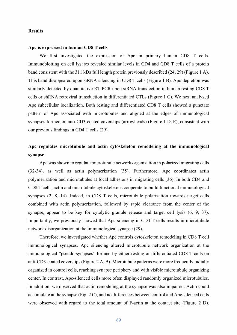
69
Results
Apc is expressed in human CD8 T cells
We first investigated the expression of Apc in primary human CD8 T cells.
Immunoblotting on cell lysates revealed similar levels in CD4 and CD8 T cells of a protein
band consistent with the 311 kDa full length protein previously described (24, 29) (Figure 1 A).
This band disappeared upon siRNA silencing in CD8 T cells (Figure 1 B). Apc depletion was
similarly detected by quantitative RT-PCR upon siRNA transfection in human resting CD8 T
cells or shRNA retroviral transduction in differentiated CTLs (Figure 1 C). We next analyzed
Apc subcellular localization. Both resting and differentiated CD8 T cells showed a punctate
pattern of Apc associated with microtubules and aligned at the edges of immunological
synapses formed on anti-CD3-coated coverslips (arrowheads) (Figure 1 D, E), consistent with
our previous findings in CD4 T cells (29).
Apc regulates microtubule and actin cytoskeleton remodeling at the immunological
synapse
Apc was shown to regulate microtubule network organization in polarized migrating cells
(32-34), as well as actin polymerization (35). Furthermore, Apc coordinates actin
polymerization and microtubules at focal adhesions in migrating cells (36). In both CD4 and
CD8 T cells, actin and microtubule cytoskeletons cooperate to build functional immunological
synapses (2, 8, 14). Indeed, in CD8 T cells, microtubule polarization towards target cells
combined with actin polymerization, followed by rapid clearance from the center of the
synapse, appear to be key for cytolytic granule release and target cell lysis (6, 9, 37).
Importantly, we previously showed that Apc silencing in CD4 T cells results in microtubule
network disorganization at the immunological synapse (29).
Therefore, we investigated whether Apc controls cytoskeleton remodeling in CD8 T cell
immunological synapses. Apc silencing altered microtubule network organization at the
immunological “pseudo-synapses” formed by either resting or differentiated CD8 T cells on
anti-CD3-coated coverslips (Figure 2 A, B). Microtubule patterns were more frequently radially
organized in control cells, reaching synapse periphery and with visible microtubule organizing
center. In contrast, Apc-silenced cells more often displayed randomly organized microtubules.
In addition, we observed that actin remodeling at the synapse was also impaired. Actin could
accumulate at the synapse (Fig. 2 C), and no differences between control and Apc-silenced cells
were observed with regard to the total amount of F-actin at the contact site (Figure 2 D).
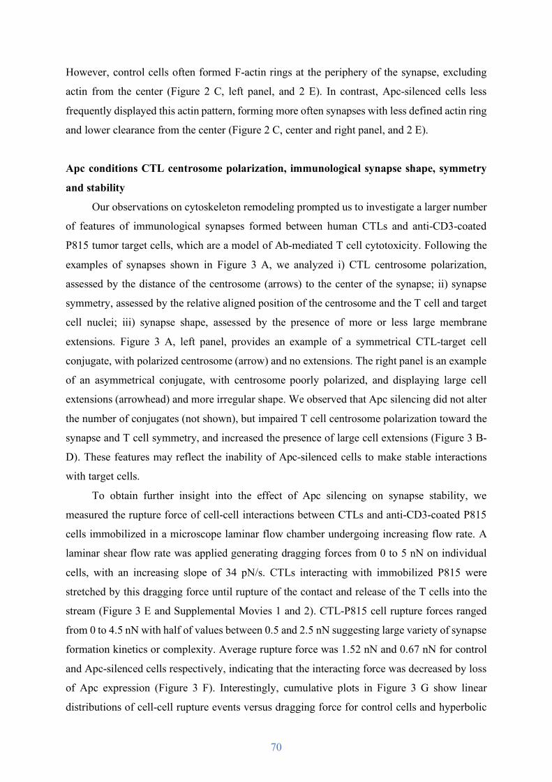
70
However, control cells often formed F-actin rings at the periphery of the synapse, excluding
actin from the center (Figure 2 C, left panel, and 2 E). In contrast, Apc-silenced cells less
frequently displayed this actin pattern, forming more often synapses with less defined actin ring
and lower clearance from the center (Figure 2 C, center and right panel, and 2 E).
Apc conditions CTL centrosome polarization, immunological synapse shape, symmetry
and stability
Our observations on cytoskeleton remodeling prompted us to investigate a larger number
of features of immunological synapses formed between human CTLs and anti-CD3-coated
P815 tumor target cells, which are a model of Ab-mediated T cell cytotoxicity. Following the
examples of synapses shown in Figure 3 A, we analyzed i) CTL centrosome polarization,
assessed by the distance of the centrosome (arrows) to the center of the synapse; ii) synapse
symmetry, assessed by the relative aligned position of the centrosome and the T cell and target
cell nuclei; iii) synapse shape, assessed by the presence of more or less large membrane
extensions. Figure 3 A, left panel, provides an example of a symmetrical CTL-target cell
conjugate, with polarized centrosome (arrow) and no extensions. The right panel is an example
of an asymmetrical conjugate, with centrosome poorly polarized, and displaying large cell
extensions (arrowhead) and more irregular shape. We observed that Apc silencing did not alter
the number of conjugates (not shown), but impaired T cell centrosome polarization toward the
synapse and T cell symmetry, and increased the presence of large cell extensions (Figure 3 B-
D). These features may reflect the inability of Apc-silenced cells to make stable interactions
with target cells.
To obtain further insight into the effect of Apc silencing on synapse stability, we
measured the rupture force of cell-cell interactions between CTLs and anti-CD3-coated P815
cells immobilized in a microscope laminar flow chamber undergoing increasing flow rate. A
laminar shear flow rate was applied generating dragging forces from 0 to 5 nN on individual
cells, with an increasing slope of 34 pN/s. CTLs interacting with immobilized P815 were
stretched by this dragging force until rupture of the contact and release of the T cells into the
stream (Figure 3 E and Supplemental Movies 1 and 2). CTL-P815 cell rupture forces ranged
from 0 to 4.5 nN with half of values between 0.5 and 2.5 nN suggesting large variety of synapse
formation kinetics or complexity. Average rupture force was 1.52 nN and 0.67 nN for control
and Apc-silenced cells respectively, indicating that the interacting force was decreased by loss
of Apc expression (Figure 3 F). Interestingly, cumulative plots in Figure 3 G show linear
distributions of cell-cell rupture events versus dragging force for control cells and hyperbolic
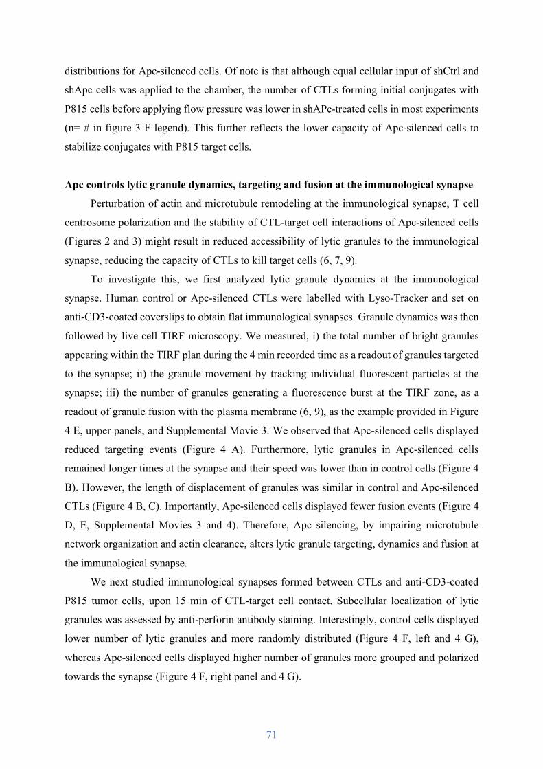
71
distributions for Apc-silenced cells. Of note is that although equal cellular input of shCtrl and
shApc cells was applied to the chamber, the number of CTLs forming initial conjugates with
P815 cells before applying flow pressure was lower in shAPc-treated cells in most experiments
(n= # in figure 3 F legend). This further reflects the lower capacity of Apc-silenced cells to
stabilize conjugates with P815 target cells.
Apc controls lytic granule dynamics, targeting and fusion at the immunological synapse
Perturbation of actin and microtubule remodeling at the immunological synapse, T cell
centrosome polarization and the stability of CTL-target cell interactions of Apc-silenced cells
(Figures 2 and 3) might result in reduced accessibility of lytic granules to the immunological
synapse, reducing the capacity of CTLs to kill target cells (6, 7, 9).
To investigate this, we first analyzed lytic granule dynamics at the immunological
synapse. Human control or Apc-silenced CTLs were labelled with Lyso-Tracker and set on
anti-CD3-coated coverslips to obtain flat immunological synapses. Granule dynamics was then
followed by live cell TIRF microscopy. We measured, i) the total number of bright granules
appearing within the TIRF plan during the 4 min recorded time as a readout of granules targeted
to the synapse; ii) the granule movement by tracking individual fluorescent particles at the
synapse; iii) the number of granules generating a fluorescence burst at the TIRF zone, as a
readout of granule fusion with the plasma membrane (6, 9), as the example provided in Figure
4 E, upper panels, and Supplemental Movie 3. We observed that Apc-silenced cells displayed
reduced targeting events (Figure 4 A). Furthermore, lytic granules in Apc-silenced cells
remained longer times at the synapse and their speed was lower than in control cells (Figure 4
B). However, the length of displacement of granules was similar in control and Apc-silenced
CTLs (Figure 4 B, C). Importantly, Apc-silenced cells displayed fewer fusion events (Figure 4
D, E, Supplemental Movies 3 and 4). Therefore, Apc silencing, by impairing microtubule
network organization and actin clearance, alters lytic granule targeting, dynamics and fusion at
the immunological synapse.
We next studied immunological synapses formed between CTLs and anti-CD3-coated
P815 tumor cells, upon 15 min of CTL-target cell contact. Subcellular localization of lytic
granules was assessed by anti-perforin antibody staining. Interestingly, control cells displayed
lower number of lytic granules and more randomly distributed (Figure 4 F, left and 4 G),
whereas Apc-silenced cells displayed higher number of granules more grouped and polarized
towards the synapse (Figure 4 F, right panel and 4 G).
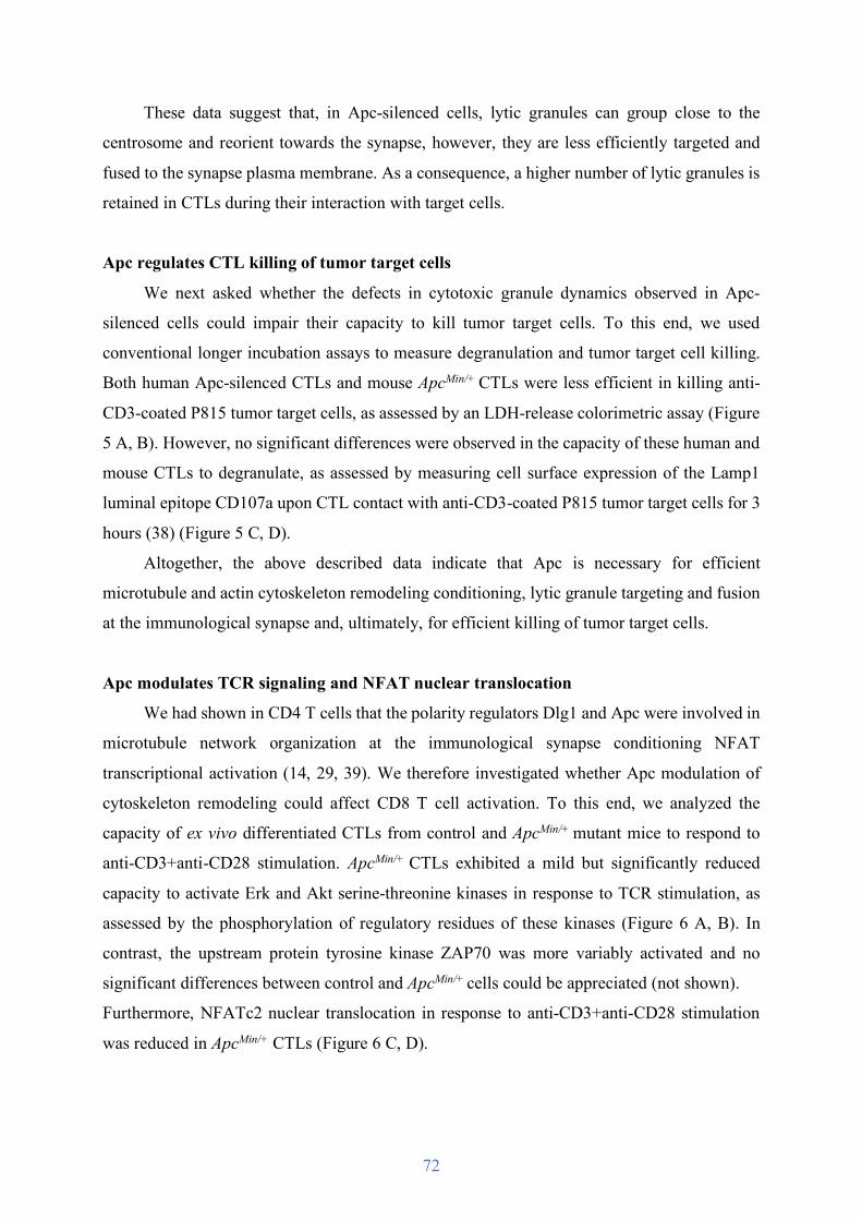
72
These data suggest that, in Apc-silenced cells, lytic granules can group close to the
centrosome and reorient towards the synapse, however, they are less efficiently targeted and
fused to the synapse plasma membrane. As a consequence, a higher number of lytic granules is
retained in CTLs during their interaction with target cells.
Apc regulates CTL killing of tumor target cells
We next asked whether the defects in cytotoxic granule dynamics observed in Apc-
silenced cells could impair their capacity to kill tumor target cells. To this end, we used
conventional longer incubation assays to measure degranulation and tumor target cell killing.
Both human Apc-silenced CTLs and mouse ApcMin/+ CTLs were less efficient in killing anti-
CD3-coated P815 tumor target cells, as assessed by an LDH-release colorimetric assay (Figure
5 A, B). However, no significant differences were observed in the capacity of these human and
mouse CTLs to degranulate, as assessed by measuring cell surface expression of the Lamp1
luminal epitope CD107a upon CTL contact with anti-CD3-coated P815 tumor target cells for 3
hours (38) (Figure 5 C, D).
Altogether, the above described data indicate that Apc is necessary for efficient
microtubule and actin cytoskeleton remodeling conditioning, lytic granule targeting and fusion
at the immunological synapse and, ultimately, for efficient killing of tumor target cells.
Apc modulates TCR signaling and NFAT nuclear translocation
We had shown in CD4 T cells that the polarity regulators Dlg1 and Apc were involved in
microtubule network organization at the immunological synapse conditioning NFAT
transcriptional activation (14, 29, 39). We therefore investigated whether Apc modulation of
cytoskeleton remodeling could affect CD8 T cell activation. To this end, we analyzed the
capacity of ex vivo differentiated CTLs from control and ApcMin/+ mutant mice to respond to
anti-CD3+anti-CD28 stimulation. ApcMin/+ CTLs exhibited a mild but significantly reduced
capacity to activate Erk and Akt serine-threonine kinases in response to TCR stimulation, as
assessed by the phosphorylation of regulatory residues of these kinases (Figure 6 A, B). In
contrast, the upstream protein tyrosine kinase ZAP70 was more variably activated and no
significant differences between control and ApcMin/+ cells could be appreciated (not shown).
Furthermore, NFATc2 nuclear translocation in response to anti-CD3+anti-CD28 stimulation
was reduced in ApcMin/+ CTLs (Figure 6 C, D).

73
Apc defects mildly alter ex vivo differentiation of CD8 T cells
The observed alteration in NFAT nuclear translocation prompted us to investigate
whether Apc defects could be associated with impaired CD8 T cell differentiation and/or
cytokine production. We therefore analyzed the capacity of ApcMin/+ CD8 T cells to differentiate
into CTLs ex vivo. Splenocytes and lymph node cells were stimulated with concanavalin A
(ConA) and IL-2, and the expression of the activation/differentiation molecules CD25,
granzyme B, CD44 and CD62L was assessed at 2, 4 and 6 days upon activation. Expression of
CD44 and CD62L adhesion molecules in CD8 T cells from ApcMin/+ mice appeared mildly
affected mainly at day 2. CD62L expression remained lower but differences were statistically
not significant, with high variability between animals (Figure 7 A-D). In contrast, no differences
were observed in CD25 and granzyme B expression (Figure 7 E-H). Interestingly, differences
in CD44 and CD62L expression were more readily observed in CD4 T cells with more
pronounced and longer effects (Supplemental Figure 1). Interestingly, the level and kinetics of
CD62L expression in CD4 T cells were different than in CD8 T cells.
These findings show that CD8 T cells from ApcMin/+ mice present reduced early TCR
activation events, such as Erk and Akt activation, and NFAT nuclear translocation. Finally,
minor differences in the expression of CD44 and CD62L differentiation markers in response to
ex vivo stimulation in cells from ApcMin/+ mice were observed in CD8 T cells, with more
significant differences in CD4 T cells.
Apc defects do not alter ex vivo CTL cytokine production
Since NFAT drives the transcription of a variety of T cell cytokines, we investigated the
capacity of Apc-silenced human CTLs and ApcMin/+ CTLs to produce effector cytokines. In spite
of the reduced capacity of ApcMin/+ CTLs to induce NFAT nuclear translocation, we did not
observe significant differences in the production of IL-2, IFNg or TNFa by differentiated CTLs
re-stimulated with anti-CD3+anti-CD28, either by intracellular staining and FACS analysis, or
by ELISA of cell culture supernatants (Figure 8 A, B). Similar results were found when
assessing cytokine production in cell culture supernatants of control and Apc-silenced human
CTLs by ELISA (Figure 8 C).
These findings indicate that Apc defects in both human and mouse CTLs do not affect
their capacity to produce effector CD8 cytokines under our ex vivo stimulation conditions.

74
Discussion
Altogether the data shown here unveil a novel role of Apc in cytotoxic T cell effector
functions. Apc defects in Apc-silenced human peripheral blood CD8 T cells or in ApcMin/+
mutant mice revealed the importance of Apc in cytoskeleton remodeling at the immunological
synapse. Both microtubules and actin reorganization appeared affected. Thus, the radial
organization of microtubules and centrosome polarization were perturbed, and F-actin ring
formation at the periphery of the synapse and F-actin exclusion from the center of the synapse
were impaired in Apc-defective T cells. Additionally, consistent with an impact on actin
cytoskeleton dynamics (40), synapse stability, shape and symmetry were also altered in Apc-
deficient cells.
Apc regulation of microtubule organization in CD8 T cells is consistent with the described
role of Apc as polarity regulator in other cell systems (23). Apc performs this function in
association with other regulators of cell polarity, including Cdc42, Scribble, Par6, PKCz and
Dlg1, as shown for instance in migrating astrocytes (34, 41). Furthermore, we have shown
before that Dlg1 and Apc control microtubule patterns, centrosome polarization and signaling
microcluster dynamics at the immunological synapse of CD4 T cells (14, 29). Although less
extensively studied, Apc may also regulate actin polymerization in synergy with its partner, the
formin family protein mDia (35). In addition, Apc regulates actin-microtubule crosstalk at focal
adhesions of migrating cells (36). Interestingly, molecular partners of Apc, as Cdc42 (34) and
formins (35), were shown to control actin remodeling and centrosome polarization at the
immunological synapse (42, 43). Therefore, Apc may collaborate with these cytoskeleton
regulators to collectively reorganize both cytoskeleton components at the CTL immunological
synapse, thus ensuring their interplay that is crucial for properly tuning T cell functions.
Concomitant centrosome polarization and F-actin exclusion from the center of the
immunological synapse has been shown to be key for lytic granule targeting and fusion at the
synapse, and efficient cytotoxic function against infected or tumor target cells (6, 7, 9, 37).
Therefore, impairment of both microtubule organization and centrosome polarization, together
with F-actin reorganization at the synapse in Apc-defective CTLs, may account for their altered
lytic granule dynamics, targeting and fusion at the synapse observed by live cell TIRF
microscopy. Apc-silenced cells could still concentrate lytic granules in the centrosomal area
and polarize them to a certain extent towards the immunological synapse, suggesting that there
is not a complete failure in granule transport on microtubules. Granules may be able to move
towards the centrosome and microtubule minus end, a movement mediated by dynein motors.

75
In contrast, granules do not properly reach the immunological synapse plasma membrane.
Disorganization of microtubule network at the immunological synapse likely prevents
microtubules from reaching the plasma membrane and, as a consequence, precludes centrosome
closing up to the synapse, which facilitates lytic granule docking and fusion (7). Kinesin-
mediated transport to the microtubule plus ends may also help granules to reach the synapse
plasma membrane (44, 45). As a likely consequence of inefficient lytic granule dynamics,
targeting and fusion, Apc defective CTLs killed tumor target cells less efficiently than control
cells. However, no difference in “degranulation” assessed by increase in Lamp1 (CD107a) cell
surface expression was detected. The apparent contradiction between the live cell TIRF
experiments and Lamp1 expression may be due to the different time of CTL-target cell contact
in these two assays, and/or their distinct sensitivities to discriminate differences. Lack of direct
correlation between degranulation and cytotoxicity has been reported in other experimental
setups (46).
Although significant, the differences in tumor cell killing activity between Apc-defective
CTLs and control cells were modest. This may due to the fact that ApcMin/+ mice are
heterozygous (homozygous mutation is embryonic lethal) (47) and may express residual levels
of Apc protein. Likewise, shRNA silencing in human primary CTLs is not fully efficient.
Hence, remaining Apc protein could account for the observed function. Furthermore, other
polarity regulators acting together with Apc may also compensate Apc defects. Finally, the T
cell activation conditions we used for CTL generation in vitro may compensate in part
cytotoxicity defects due to Apc, by increasing the expression of other proteins. This effect was
described for some forms of familial hemophagocytic lymphohistiocytosis due to syntaxin 11
or Munc18-2 mutations, where the addition of IL-2 used to induce T cell proliferation in vitro
was partially compensating the loss of effector function (48, 49). Importantly, subtle differences
in CTL cytotoxicity using in vitro assays may be predictive of more serious cytotoxic defects
in vivo leading to poor immune responses (50). Therefore, the small differences in cytotoxicity
we observed could have consequences for anti-tumor immunity in patients.
We have previously shown in CD4 T cells that Apc silencing leads to altered NFAT
nuclear translocation and reduced NFAT-driven transcriptional activation leading to impaired
IL-2 gene transcription. Furthermore, CD4 T cells from ApcMin/+ mice had reduced capacity to
produce IL-2, IL-4 and IFNg. Finally, regulatory T cells in these mutant mice showed impaired
differentiation and production of the anti-inflammatory cytokine IL-10 (29). We show here that
CTLs from ApcMin/+ mice also had an impaired capacity to translocate NFATc2 to the nucleus.
Interestingly, NFATc2 formed microclusters associated to microtubules in CD8 T cells (data
76
not shown), as we previously showed in CD4 T cells (29). However, the analysis of the
expression of a number of differentiation markers (e.g. CD25, granzyme B, CD44 and CD62L)
in response to ex vivo activation with concanavalin A and IL-2 revealed only mildly altered
CD44 and CD62L expression at day 2 in ApcMin/+ mice. Interestingly, under the same
stimulation conditions, the differences in expression of CD44 and CD62L between ApcMin/+ and
control T cells were more pronounced and significant in the CD4 T cell population. In addition,
Apc-silenced human and ApcMin/+ mouse ex vivo differentiated CTLs produced equal amount of
IL-2, TNFa and IFNg after re-stimulation with CD3/CD28 antibodies. Therefore, CD8 T cells
appear to be less sensitive to Apc defects than CD4 T cells when performing their activation
programs leading to differentiation and production of cytokines.
Altogether the data presented here show that Apc is involved in the regulation of CTL
effector functions driving target cell killing. Hence, Apc defects impair both actin and
microtubule cytoskeleton remodeling at the immunological synapse, centrosome polarization
and lytic granule dynamics, targeting and fusion at the synapse. However, CD8 T cell
differentiation and cytokine production appear much less dependent on Apc compared to what
has been observed in CD4 T cells (29). Thus, Apc defects may not significantly alter the
acquisition of effector functions in CD8 T cells, but impair the CTL capacities to kill tumor
cells and could therefore have an impact in anti-tumor immune responses in familial
adenomatous polyposis patients allowing easier tumor escape.

77
Materials and Methods
Human cell isolation, siRNA transfection, lentiviral infection, CTL generation and tumor
target cell culture
Human peripheral blood T cells from healthy volunteers were obtained from the French
Blood Bank Organization (Etablissement Français du Sang, EFS), or through the Institut
Pasteur Biological Resources Core Facility ICAReB (NSF 96-900 certified, from sampling to
distribution, reference BB-0033-00062/ICAReB platform/ Institut Pasteur, Paris,
France/BBMRI AO203/ 1 distribution/ access: 2016, May 19th, [BIORESOURCE]), under
CoSImmGEn protocol approved by the Committee of Protection of Persons, Ile de France-1
(No 2010-dec-12483). Informed consent was obtained from all donors.
Peripheral blood mononuclear cells (PBMCs) were isolated by centrifugation through
Ficoll-Hypaque. CD8 T cells were isolated from PBMCs using the MACS CD8 T Cell Isolation
Kit (Miltenyi Biotec) and maintained in human CD8 medium: RPMI 1640 + GlutaMAX-I
(Gibco) supplemented with 10% fetal bovine serum (FBS), 1 mM sodium pyruvate and
nonessential amino acids, 10 mM HEPES, 1% penicillin-streptomycin (v/v).
To generate CTLs, freshly isolated CD8 T cells were stimulated 2 days with coated anti-
CD3 (10 µg/mL; UCHT1 produced by A. Alcover), soluble anti-CD28 (7 µg/mL; Beckman
Coulter), and recombinant human IL-2 (100 U/mL; PeproTech) in human CD8 medium. Cells
were infected for 24 h with lentiviruses coding for control or Apc-specific shRNAs (10% v/v)
in human CD8 medium with IL-2, where fetal bovine serum (FBS) was replaced by human
serum, and then selected for 3 days with puromycin (3.9 µg/mL). Percentage of infected CTLs
was 70-85%, as assessed by GFP expression by FACS.
For siRNA experiments, small double stranded RNA oligonucleotides sequences used
were 5’-GAGAAUACGUCCACACCUU-3’ (siApc; GE Healthcare) for Apc depletion, as we
previously described (29). As control, 5’-UAGCGACUAAACACAUCAA-3’ (siGENOME
Non-Targeting siRNA #1; GE Healthcare) was used. 107 freshly isolated CD8 T cells were
transfected with 1 nmol of siCtrl or siApc using the Human T Cell Nucleofector kit and the
program U-14 on an Amaxa Nucleofector II (Lonza). Cells were then harvested in human CD8
medium without penicillin-streptomycin and used 72 h after transfection.
For shRNA experiments, lentiviruses were produced by HEK293T cells transfected with
the transient calcium phosphate DNA precipitation technique. Cells were transfected with
pCMV-deltaR8-2 and pCMV-env-VSV, together with a pLKO.1-puro-CMV-tGFP lentiviral
vector expressing or not (as negative control) a short hairpin (sh)RNA targeting Apc (5’-
78
GACTGTCCTTTCACCATATTT-3’) (Sigma-Aldrich). Forty-eight hours later, supernatant
was recovered and concentrated 40x by ultracentrifugation (26,000 rpm, 1.5 h, 4°C).
Lentiviruses stocks were stored at -80°C.
The P815 mouse mastocytoma cell line was used as tumor target cell. P815 cells were
maintained in DMEM medium supplemented with 10% FBS and 1% penicillin-streptomycin
(v/v). To make stimulatory target cells, P815 were pulsed with anti-human or mouse CD3
antibodies, as described below.
ApcMin/+ and wild type mice lymphocyte isolation and cell culture
Heterozygous C57BL/6J-ApcMin (ApcMin/+) mice and wild-type controls were purchased
from the Jackson Laboratory. Mice were housed and bred under pathogen-free conditions in
the Central Animal Facility of the Institut Pasteur. Protocols used had been approved by the
Ethical Committee for Animal Experimentation of the Institut Pasteur and by the French
Ministry of Research. Males and females were sacrificed at 10-12 weeks of age. Spleen and
lymph nodes were homogenized through a 70 µm filter. To generate CTLs, CD8 T cells were
purified using the MACS CD8a T Cell Isolation Kit (Miltenyi Biotec), and stimulated 2 days
with coated anti-CD3 (5 µg/mL; 145-2C11, eBiosciences), soluble anti-CD28 (2 µg/mL;
eBiosciences) and IL-2 (10 ng/mL; Miltenyi Biotec). Cells were then cultured for 5-6 days in
mouse CD8 medium: DMEM + GlutaMAX-I (Gibco) supplemented with 10% FBS, 1 mM
sodium pyruvate and nonessential amino acids, 50 µM β-mercaptoethanol, 10 mM HEPES, 1%
penicillin-streptomycin (v/v), in presence of IL-2 (10 ng/mL; Miltenyi Biotec).
Apc quantification
For Apc detection by Western blot, total cell extracts were obtained by lysing cells for 5
min on ice in lysis buffer (50 mM Tris pH 7.4, 100 mM NaCl, 0.5% Nonidet P-40, 5 mM
EDTA, 5 mM EGTA, 40 mM beta-glycerophosphate, 10 mM NaF, 10 mM Na4P2O7, 2 mM
ortho-vanadate and protease inhibitor cocktail). Cell lysates were cleared by spinning at 20,800
xg for 20 min at 4°C. Equal amount of protein content measured using the Bio-Rad protein
assay (Bio-Rad Laboratories) was loaded in NuPAGE 3-8% Tris-Acetate gels (ThermoFisher
Scientific). Proteins were transferred onto nitrocellulose membranes (LI-COR Biosciences) for
4 h using a Bio-Rad Mini Trans-Blot system in buffer containing 50 mM Tris, 380 mM Glycine,
20% ethanol, 0.1% SDS. Membranes were saturated with blocking buffer (Rockland
Immunochemicals) and incubated with anti-Apc (2 µg/mL; Ali 12-28, Abcam) and anti-ZAP70

79
(50 ng/mL; BD Biosciences) or anti-PLCg1 (1/1000; Cell Signaling) overnight at 4°C. They
were washed and then incubated with specific secondary antibodies conjugated with Alexa-
Fluor680 or DyLight800 (ThermoFisher Scientific) for 45 min. Near-infrared fluorescence was
imaged and quantified using the Odyssey Classic near-infrared imaging system (LI-COR
Biosciences) and Apc band intensity was normalized to control ZAP70 or PLCg1 band.
For Apc detection by retrotranscription quantitative PCR, total RNA was extracted using
the RNeasy Micro Plus Kit (Qiagen) following manufacturer’s instructions. cDNA was
obtained from 200 ng of total RNA using iScript cDNA synthesis kit (Bio-Rad Laboratories).
FastStart Universal SYBR Green PCR Master Mix (Roche) and ABI PRISM 7900HT Sequence
Detection system (Applied Biosystems) were used to quantify gene products. Quantitative PCR
were performed in triplicates, quantity values were calculated by the relative standard curve
method and normalized to the mRNA expression of the RLP13a housekeeping gene. Primer
sequences used were as follows:
Apc-F: 5’-CCAACAAGGCTACGCTATGC-3’/ R: 5’-TACATCTGCTCGCCAAGACA-3’
RLP13a-F: 5’- CATAGGAAGCTGGGAGCAAG-3’/
R: 5’- GCCCTCCAATCAGTCTTCTG-3’
Confocal microscopy, image post-treatment and quantitative image analysis
Coverslips were washed with HCl-ethanol 70% and coated with Poly-L-lysine 0.002% in
water (Sigma-Aldrich). For pseudo-synapse formation, coverslips were further coated with
anti-CD3 (10 µg/mL; UCHT1, BioLegend) overnight at 4°C. Coverslips were washed and
blocked for 30 min at 37°C with human CD8 medium. Human T cells were plated on coverslips
for 5 min at 37°C, and fixed with 4% paraformaldehyde for 13 min at room temperature. For
microtubule detection, cells were also incubated 20 min at -20°C in ice cold methanol. For
CTL-target cell immunological synapse formation, P815 cells, previously coated with anti-CD3
(20 µg/mL; OKT3 produced by A. Alcover) for 45 min at 4°C, were plated on poly-L-lysine-
coated coverslips for 15 min at 37°C. Coverslips were carefully washed with PBS, and human
CTLs were added at a 1:1 CTL-target ratio and incubated for 15 min at 37°C. Cells were then
fixed with 4% paraformaldehyde for 13 min at room temperature. Coverslips were washed in
PBS, and incubated 1 h in PBS with 1% bovine serum albumin (BSA) (v/v) to prevent
unspecific binding. Cells were then incubated 1 h at room temperature with PBS, 1% BSA,
0.1% Triton X-100 and anti-Apc (1/300; gift of I. Näthke, University of Dundee), anti-b-tubulin
(6.6 µg/mL; Merck Millipore), anti-pericentrin (1/100; Abcam), anti-perforin (10 µg/mL; BD

80
Biosciences). Coverslips were then incubated with the corresponding fluorescent-coupled
secondary Ab and Texas Red-X phalloidin (1/100; Invitrogen) for 45 min at room temperature.
After three washes in PBS with 1% BSA, coverslips were mounted on microscope slides using
ProLong Gold Antifade mounting medium with DAPI (Life Technologies).
Confocal images were acquired with an LSM 700 confocal microscope (Zeiss) using the
Plan-Apochromat 63x/1.40 NA objective. Optical confocal sections were acquired with ZEN
software (Zeiss) by intercalating green and red laser excitation to minimize channel cross talk.
All analyses were performed using Fiji software (51). For F-actin analysis, optical sections were
acquired at 1 µm intervals. Formation of the actin ring and phalloidin fluorescence intensity
analyses were performed on one confocal section at the cell-coverslip contact. For microtubule
pattern and synapses analyses, optical sections were acquired at 0.2 µm intervals and images
were treated by deconvolution with the Huygens Pro Software (Scientific Volume Imaging).
Microtubule pattern analysis was performed on projection of 4 confocal sections at the cell-
coverslip contact, and phenotypes were classified by blinded image observation by three
different investigators. Synapse morphology and cytotoxic granules localization were analyzed
in 3D projections, and classified as described for microtubule patterns. Centrosome localization
was estimated by measuring the distance between the anti-pericentrin staining and the CTL
plasma membrane at the center area of the immunological synapse. Quantification of cytotoxic
granules per cell was obtained using the TrackMate plugin for ImageJ developed by J.-Y.
Tinevez (Photonic Bio Imaging UTechS, Institut Pasteur) (52).
Rupture force assay of cell-cell interactions in laminar flow chamber
Glass surface (RS, France) of the flow chamber (Slide I0.1, Ibidi, Germany) was washed
using 5 chamber volumes (CV) of sulfuric acid (2 M; Merck-Sigma) then 5 CV of hydrogen
peroxide (33% Merck-Sigma), and rinsed with 10 CV of water then 10 CV of PBS. Bovine
fibronectin (10 µg/mL in PBS; Merck-Sigma) was incubated 1 h at room temperature and rinsed
with 20 CV of PBS. The chamber was equilibrated with DMEM (Gibco) at 37°C then loaded
with P815 cells for 15 min at 37°C. One CV of anti-CD3 (20 µg/mL final; OKT3 produced by
A. Alcover) in DMEM was carefully added, and cells were incubated for 15 min at 37°C.
Unbound cells and antibodies were cleared by flowing 5 CV of RPMI (37°C; Lonza). One CV
of human CTLs in RPMI 1% FBS (Lonza) was injected inside the chamber and incubated for
10 min at 37°C. A flow rate of PBS, increasing from 0 to 38.4 mL/min, was applied through
the chamber for 115 s using a syringe pump (SP210iW, WPI) controlled by a computer, and
81
synchronized with image acquisition (3 image/s, 1100 x 840 µm) using an inverted transmission
microscope (Observer D1, Zeiss) with a 10x/0.3 NA objective controlled by MicroManager
(53).
Image series were analyzed using ImageJ software (51) and the plugin CellCounter. P815
and CTLs were distinguished from their size, shape and nucleus/cell ratio. The flow rate value
at the cell-cell rupture event was used to compute the dragging force on the released cell
according to its size (mean diameter of 8 µm), shape (roughly spherical) and density (mean
value 1.20 kg/L). Calibration of dragging force was performed from sedimentation rate of cells
in the chamber (measured PBS density 1.0034 kg/L and dynamic viscosity 0.6998 mPa.s at
37°C) (54) and the theoretical flow speed versus wall distance according to Poiseuille solution
to Navier-Stokes formalism for a Reynolds’ number below 10 characterizing a laminar flow.
Live cell total internal reflection fluorescence (TIRF) microscopy
Glass bottom microwell dishes (MatTek Corporation) were washed with HCl-ethanol
70%. They were coated with Poly-L-lysine 0.002% in water (Sigma-Aldrich) 30 min at room
temperature, and then with anti-CD3 (10 µg/mL; UCHT1, BioLegend) overnight at 4°C.
Human CTLs were incubated with LysoTracker Deep Red (0.1 µM; Molecular Probes by Life
Technologies) for 45 min at 37°C to label cytotoxic granules. Cells were washed and
resuspended in human CD8 medium. 105 cells were dropped in a microwell, and once cells
were seeded, images were acquired in TIRF plan every 150 ms for 4 min. TIRF images were
acquired with an LSM 780-Elyra PS.1 confocal microscope (Zeiss) using an alpha Plan Apo
100x/1.46 N.A. oil immersion objective. Images were analyzed using the TrackMate plugin for
ImageJ software (52). A fluorescence threshold above 2x the mean granule fluorescence was
set, in order to select strong fluorescence events more likely closer to the plasma membrane.
Cytotoxicity Assay
At day 6-8 after initial stimulation, human or mouse CTLs were resuspended in RPMI
1640 + GlutaMAX-I (Gibco) 3% FCS, 10 mM HEPES, and mixed at the indicated ratios with
P815 target cells previously coated with anti-human-CD3 (6 µg/mL; UCHT1, Biolegend) or
anti-mouse-CD3 (6 µg/mL; 145-2C11, eBiosciences) for 45 min at 4°C. Mixed CTLs and target
cells were incubated 4 h at 37°C. The percentage of target cell lysis was measured using the
CytoTox 96 Non-Radioactive Cytotoxicity Assay (Promega) following manufacturer’s
82
instructions. Absorbance at 490 nm was measured using a PR2100 Microplate reader (Bio-
Rad).
Degranulation assay
At day 6-8 after initial stimulation, human or mouse CTLs were mixed at a 1:1 ratio with
P815 target cells previously coated with the indicated anti-CD3 concentration (OKT3 produced
by A. Alcover for human; 145-2C11, eBiosciences for mice) in presence of anti-CD107a-PE
(1/45, clone H4A3, Biolegend for human; 2.5 µg/mL, clone 1D4B, BD Biosciences for mice),
and incubated for 3 h at 37°C. Cells were stained with either anti-human CD8a-APC-Cy7 (1/25;
Biolegend) or anti-mouse CD8a-PerCP-Cy5.5 (1 µg/mL; BD Biosciences). Events were
acquired on a MACSQuant Analyzer (Miltenyi Biotech), analysis was performed using FlowJo
10 software (FlowJo, LLC). All samples were gated on forward and side scatter and for singlets.
Analysis of protein phosphorylation
Mouse T cells were incubated with 20 µg/mL of biotin-conjugated anti-CD3 (145-2C11,
eBioscience) and anti-CD28 (eBiosciences) antibodies for 30 min at 4°C. Cells were washed,
resuspend in Opti-Mem + GlutaMAX-I (Gibco), and placed 1 min at 37°C. Streptavidin (10
µg/mL; Sigma-Aldrich) was added and cells were incubated at 37°C for the indicated times.
Ice-cold PBS containing 2 mM ortho-vanadate and 0.05% sodium azide was added to stop cell
stimulation. Cells were lysed in ice-cold LBM buffer (20 mM Tris pH 7.4, 150 mM NaCl,
0.25% lauryl-b-maltoside, 50 mM NaF, 10 mM Na4P2O7, 1 mM EGTA, 2 mM ortho-vanadate,
1 mM MgCl2 and protease inhibitor cocktail) for 10 min on ice. Cell lysates were cleared by
spinning at 20,800 xg for 10 min at 4°C. Equal amount of protein content measured using the
Bio-Rad protein assay (Bio-Rad Laboratories) was loaded in NuPAGE 4-12% Bis-Tris gels
(ThermoFisher Scientific). Protein transfer was performed using the Trans-Blot Turbo system
(Bio-Rad Laboratories). Membranes were saturated with blocking buffer (Rockland
Immunochemicals) and incubated overnight at 4°C or 2 h at room temperature with anti-
phospho-Akt (pSer473; 1/1000; Cell Signaling Technology), anti-phospho-Erk1/2
(pThr202/Tyr204; 1/1000; Cell Signaling Technology), anti-GAPDH (6.6 µg/mL;
Calbiochem). Membranes were then incubated with specific secondary antibodies conjugated
with Alexa-Fluor680 or DyLight800 (ThermoFisher Scientific) for 45 min at room temperature.
Near-infrared fluorescence was recorded and quantified using an Odyssey Classic near-infrared
scanner (LI-COR Biosciences). Band intensity was normalized to control GAPDH. For a pooled
83
analysis of several experiments, normalized intensities were then divided by the mean
normalized intensity of the same experiment.
Nuclear NFAT detection
Nuclear NFAT was analyzed by Western blot analysis of cells fractionated for nucleus
and cytoplasm separation. Mouse T cells were activated as for analysis of protein
phosphorylation, for the indicated times. Cells were lysed in ice-cold low-salt lysis buffer (10
mM KCl, 10 mM HEPES, 0.1 mM EDTA, 0.1 mM EGTA, 1 mM DTT, 50 mM NaF, 10 mM
sodium pyrophosphate (Na4P2O7) and a protease inhibitor cocktail) for 15 min on ice. Detergent
Nonidet P-40 (Sigma-Aldrich) was added (0.45% v/v final) and cells were vortexed for 10 s to
break cytoplasmic membranes without altering nuclear membranes. Lysates were centrifuged
at 20,800 xg for 30 min at 4°C. Supernatants, corresponding to cytosolic fractions, were
recovered in ice-cold tubes. Pellets containing nuclei were washed once in PBS and
resuspended in ice-cold high-salt buffer (20 mM Tris-HCl pH 8.0, 1% SDS, 2 mM EDTA and
protease inhibitor cocktail), solubilized by sonication (3 x 10 s at 60% power) using a Vibracell
72434 (Bioblock Scientific), and then centrifuged at 20,800 xg for 30 min. Supernatants were
recovered as nuclear fractions. Equal amount of protein content measured using the Bio-Rad
protein assay (Bio-Rad Laboratories) was loaded in NuPAGE 3-8% Tris-Acetate gels
(ThermoFisher Scientific). Protein transfer was performed in a Bio-Rad Mini Trans-Blot
system. Membranes were saturated with blocking buffer (Rockland Immunochemicals) and
incubated with anti-NFAT1 (NFATc2) (0.25 µg/mL; BD Biosciences), anti-SLP76 (1/1000;
Thermo Fisher Scientific) or anti-Lamin B1 (1/2000; Abcam) overnight at 4°C. Then, they were
incubated with specific secondary antibodies conjugated with Alexa-Fluor680 or DyLight800
(ThermoFisher Scientific) for 45 min. Near-infrared fluorescence was imaged and quantified
using the Odyssey scanner as described above. NFAT band intensity was normalized to SLP76
cytoplasmic control or Lamin B1 nuclear control. For a pooled analysis of several experiments,
normalized intensities were then divided by the mean normalized intensity of the same
experiment.

84
Mouse T cell differentiation
Mouse cells freshly isolated from the spleen and lymph nodes were stimulated with
concanavalin A (5 µg/mL; Sigma) in mouse CD8 medium with IL-2 (10 ng/mL; Miltenyi
Biotec) for 2 days, and then cultured 4 additional days with IL-2. At day 0-2-4-6, cells were
incubated with anti-CD16/32 (10 µg/mL; Biolegend) to block Fc receptors and then stained
with Fixable Viability Stain 450 (25 ng/mL; BD Biosciences), anti-CD3-FITC (5 µg/mL; 145-
2C11, BD Biosciences), anti-CD4-APC-Vio770 (1.5 µg/mL; Miltenyi Biotec), anti-CD8-
PerCP-Cy5.5 (1 µg/mL; BD Biosciences), anti-CD25-PE-Cy7 (2 µg/mL; BD Biosciences),
anti-granzymeB-A647 (1/50; Biolegend), anti-CD44-APC (2 µg/mL; eBiosciences) and anti-
CD62L-RPE (2 µg/mL; eBiosciences). Events were acquired on a MACSQuant Analyzer
(Miltenyi Biotec) and analysis was performed using FlowJo 10 software (FlowJo, LLC). All
samples were gated on forward and side scatter, for singlets, and for live cells using Fixable
Viability Stain 450 (250 ng/mL; BD Biosciences).
Detection of cytokine production
IL-2 was removed from CTL culture and 20 h later cytokine production was assessed by
ELISA and flow cytometry, as follows:
For detection by ELISA, 3.105 human or mouse CTLs were restimulated at 37°C for 6 h
with PMA (phorbol 12-myristate 13-acetate) (50 ng/mL; Sigma-Aldrich) and calcium
ionophore A23187 (500 ng/mL; Sigma-Aldrich), or 20 h with coated anti-CD3 (5 µg/mL,
UCHT1, Biolegend for human; 145-2C11, 5 µg/mL, eBiosciences for mice) and soluble anti-
CD28 (5 µg/mL, Beckman Coulter for human; 2 µg/mL, eBiosciences for mice). Secreted IL-
2, TNFα and IFNγ levels were measured from culture supernatants with specific ELISA Duo-
set kits (R&D Systems) following manufacturer’s instructions.
For detection by flow cytometry, 105 mouse CTLs were restimulated as described above.
Two hours after the beginning of restimulation, brefeldin A (10 µg/mL, Sigma-Aldrich) was
added. Cells were fixed with 2% PFA for 15 min at room temperature, incubated with anti-
CD16/32 (10 µg/mL; Biolegend), and subsequently stained with anti-IL-2-RPE (4 µg/mL;
eBiosciences), anti-TNFα-FITC (1.2 µg/mL; Miltenyi biotech) and anti-IFNγ-PE-Cy7 (8
µg/mL; Biolegend) in presence of 0.05% saponin. Events were acquired on a MACSQuant
Analyzer (Miltenyi biotech), analysis was performed using FlowJo 10 software (FlowJo, LLC).
All samples were gated on forward and side scatter for singlets, and for live cells using Fixable
Viability Stain 450 (250 ng/mL; BD Biosciences).

85
Statistics
Statistical analyses were carried out using Prism software (GraphPad). Error bars in plots
represent the mean ± SEM. The p values are represented as follows: ****p < 0.0001, ***p <
0.001, **p < 0.01, *p < 0.05, NS p ≥ 0.05. The type of test used is mentioned in each figure
legend.

86
Acknowledgments
We thank J.Y. Tinevez, A. Salles and J. Fernandes from the Photonic Bio Imaging
UTechS microscopy core facility, Institut Pasteur, for technical support, and the ICAReB
Biological Resources core facility team, Institut Pasteur, for providing primary T cell samples.
We are grateful to I. Näthke for antibodies.
Disclosures The authors declare no competing financial interests.

87
References 1. Huse, M., E. J. Quann, and M. M. Davis. 2008. Shouts, whispers and the kiss of death: directional
secretion in T cells. Nat Immunol 9: 1105-1111. 2. Soares, H., R. Lasserre, and A. Alcover. 2013. Orchestrating cytoskeleton and intracellular vesicle
traffic to build functional immunological synapses. Immunol Rev 256: 118-132. 3. Kupfer, A., and G. Dennert. 1984. Reorientation of the microtubule-organizing center and the Golgi
apparatus in cloned cytotoxic lymphocytes triggered by binding to lysable target cells. J Immunol 133: 2762-2766.
4. Kupfer, A., T. R. Mosmann, and H. Kupfer. 1991. Polarized expression of cytokines in cell conjugates of helper T cells and splenic B cells. Proc Natl Acad Sci USA 88: 775-779.
5. Depoil, D., R. Zaru, M. Guiraud, A. Chauveau, J. Harriague, G. Bismuth, C. Utzny, S. Muller, and S. Valitutti. 2005. Immunological synapses are versatile structures enabling selective T cell polarization. Immunity 22: 185-194.
6. Ritter, A. T., Y. Asano, J. C. Stinchcombe, N. M. Dieckmann, B. C. Chen, C. Gawden-Bone, S. van Engelenburg, W. Legant, L. Gao, M. W. Davidson, E. Betzig, J. Lippincott-Schwartz, and G. M. Griffiths. 2015. Actin depletion initiates events leading to granule secretion at the immunological synapse. Immunity 42: 864-876.
7. Stinchcombe, J. C., E. Majorovits, G. Bossi, S. Fuller, and G. M. Griffiths. 2006. Centrosome polarization delivers secretory granules to the immunological synapse. Nature 443: 462-465.
8. Dieckmann, N. M., G. L. Frazer, Y. Asano, J. C. Stinchcombe, and G. M. Griffiths. 2016. The cytotoxic T lymphocyte immune synapse at a glance. J Cell Sci 129: 2881-2886.
9. Ritter, A. T., S. M. Kapnick, S. Murugesan, P. L. Schwartzberg, G. M. Griffiths, and J. Lippincott-Schwartz. 2017. Cortical actin recovery at the immunological synapse leads to termination of lytic granule secretion in cytotoxic T lymphocytes. Proc Natl Acad Sci U S A 114: E6585-E6594.
10. Rodriguez-Boulan, E., and I. G. Macara. 2014. Organization and execution of the epithelial polarity programme. Nat Rev Mol Cell Biol 15: 225-242.
11. Elric, J., and S. Etienne-Manneville. 2014. Centrosome positioning in polarized cells: common themes and variations. Exp Cell Res 328: 240-248.
12. Ludford-Menting, M. J., J. Oliaro, F. Sacirbegovic, E. T. Cheah, N. Pedersen, S. J. Thomas, A. Pasam, R. Iazzolino, L. E. Dow, N. J. Waterhouse, A. Murphy, S. Ellis, M. J. Smyth, M. H. Kershaw, P. K. Darcy, P. O. Humbert, and S. M. Russell. 2005. A network of PDZ-containing proteins regulates T cell polarity and morphology during migration and immunological synapse formation. Immunity 22: 737-748.
13. Real, E., S. Faure, E. Donnadieu, and J. Delon. 2007. Cutting edge: Atypical PKCs regulate T lymphocyte polarity and scanning behavior. J Immunol 179: 5649-5652.
14. Lasserre, R., S. Charrin, C. Cuche, A. Danckaert, M. I. Thoulouze, F. de Chaumont, T. Duong, N. Perrault, N. Varin-Blank, J. C. Olivo-Marin, S. Etienne-Manneville, M. Arpin, V. Di Bartolo, and A. Alcover. 2010. Ezrin tunes T-cell activation by controlling Dlg1 and microtubule positioning at the immunological synapse. Embo J 29 2301-2314.
15. Bertrand, F., M. Esquerre, A. E. Petit, M. Rodrigues, S. Duchez, J. Delon, and S. Valitutti. 2010. Activation of the ancestral polarity regulator protein kinase C zeta at the immunological synapse drives polarization of Th cell secretory machinery toward APCs. J Immunol 185: 2887-2894.
16. Xavier, R., S. Rabizadeh, K. Ishiguro, N. Andre, J. B. Ortiz, H. Wachtel, D. G. Morris, M. Lopez-Ilasaca, A. C. Shaw, W. Swat, and B. Seed. 2004. Discs large (Dlg1) complexes in lymphocyte activation. J Cell Biol 166: 173-178.
17. Round, J. L., L. A. Humphries, T. Tomassian, P. Mittelstadt, M. Zhang, and M. C. Miceli. 2007. Scaffold protein Dlgh1 coordinates alternative p38 kinase activation, directing T cell receptor signals toward NFAT but not NF-kappaB transcription factors. Nat Immunol 8: 154-161.
18. McCartney, B. M., and I. S. Nathke. 2008. Cell regulation by the Apc protein Apc as master regulator of epithelia. Curr Opin Cell Biol 20: 186-193.
19. Zeineldin, M., and K. L. Neufeld. 2013. More than two decades of Apc modeling in rodents. Biochim Biophys Acta 1836: 80-89.

88
20. Su, L. K., K. W. Kinzler, B. Vogelstein, A. C. Preisinger, A. R. Moser, C. Luongo, K. A. Gould, and W. F. Dove. 1992. Multiple intestinal neoplasia caused by a mutation in the murine homolog of the APC gene. Science 256: 668-670.
21. Moser, A. R., H. C. Pitot, and W. F. Dove. 1990. A dominant mutation that predisposes to multiple intestinal neoplasia in the mouse. Science 247: 322-324.
22. Nelson, S., and I. S. Nathke. 2013. Interactions and functions of the adenomatous polyposis coli (APC) protein at a glance. J Cell Sci 126: 873-877.
23. Etienne-Manneville, S. 2009. APC in cell migration. Adv Exp Med Biol 656: 30-40. 24. Beroud, C., and T. Soussi. 1996. APC gene: database of germline and somatic mutations in human
tumors and cell lines. Nucleic Acids Res 24: 121-124. 25. Gounaris, E., N. R. Blatner, K. Dennis, F. Magnusson, M. F. Gurish, T. B. Strom, P. Beckhove, F.
Gounari, and K. Khazaie. 2009. T-regulatory cells shift from a protective anti-inflammatory to a cancer-promoting proinflammatory phenotype in polyposis. Cancer Res 69: 5490-5497.
26. Chae, W. J., and A. L. Bothwell. 2015. Spontaneous Intestinal Tumorigenesis in Apc (/Min+) Mice Requires Altered T Cell Development with IL-17A. J Immunol Res 2015: 860106.
27. Akeus, P., V. Langenes, A. von Mentzer, U. Yrlid, A. Sjoling, P. Saksena, S. Raghavan, and M. Quiding-Jarbrink. 2014. Altered chemokine production and accumulation of regulatory T cells in intestinal adenomas of APC(Min/+) mice. Cancer immunology, immunotherapy : CII 63: 807-819.
28. Tanner, S. M., J. G. Daft, S. A. Hill, C. A. Martin, and R. G. Lorenz. 2016. Altered T-Cell Balance in Lymphoid Organs of a Mouse Model of Colorectal Cancer. J Histochem Cytochem 64: 753-767.
29. Aguera-Gonzalez, S., O. T. Burton, E. Vazquez-Chavez, C. Cuche, F. Herit, J. Bouchet, R. Lasserre, I. Del Rio-Iniguez, V. Di Bartolo, and A. Alcover. 2017. Adenomatous Polyposis Coli Defines Treg Differentiation and Anti-inflammatory Function through Microtubule-Mediated NFAT Localization. Cell Rep 21: 181-194.
30. Rubtsov, Y. P., J. P. Rasmussen, E. Y. Chi, J. Fontenot, L. Castelli, X. Ye, P. Treuting, L. Siewe, A. Roers, W. R. Henderson, Jr., W. Muller, and A. Y. Rudensky. 2008. Regulatory T cell-derived interleukin-10 limits inflammation at environmental interfaces. Immunity 28: 546-558.
31. Martinez, G. J., R. M. Pereira, T. Aijo, E. Y. Kim, F. Marangoni, M. E. Pipkin, S. Togher, V. Heissmeyer, Y. C. Zhang, S. Crotty, E. D. Lamperti, K. M. Ansel, T. R. Mempel, H. Lahdesmaki, P. G. Hogan, and A. Rao. 2015. The transcription factor NFAT promotes exhaustion of activated CD8(+) T cells. Immunity 42: 265-278.
32. Etienne-Manneville, S. 2013. Microtubules in cell migration. Annu Rev Cell Dev Biol 29: 471-499. 33. Etienne-Manneville, S., and A. Hall. 2003. Cdc42 regulates GSK3β and adenomatous polyposis coli
(APC) to control cell polarity. . Nature 421: 753-756. 34. Etienne-Manneville, S., J. B. Manneville, S. Nicholls, M. A. Ferenczi, and A. Hall. 2005. Cdc42 and
Par6-PKCzeta regulate the spatially localized association of Dlg1 and APC to control cell polarization. J Cell Biol 170: 895-901.
35. Okada, K., F. Bartolini, A. M. Deaconescu, J. B. Moseley, Z. Dogic, N. Grigorieff, G. G. Gundersen, and B. L. Goode. 2010. Adenomatous polyposis coli protein nucleates actin assembly and synergizes with the formin mDia1. J Cell Biol 189: 1087-1096.
36. Juanes, M. A., H. Bouguenina, J. A. Eskin, R. Jaiswal, A. Badache, and B. L. Goode. 2017. Adenomatous polyposis coli nucleates actin assembly to drive cell migration and microtubule-induced focal adhesion turnover. J Cell Biol 216: 2859-2875.
37. Le Floc'h, A., Y. Tanaka, N. S. Bantilan, G. Voisinne, G. Altan-Bonnet, Y. Fukui, and M. Huse. 2013. Annular PIP3 accumulation controls actin architecture and modulates cytotoxicity at the immunological synapse. J Exp Med 210: 2721-2737.
38. Bryceson, Y. T., C. Fauriat, J. M. Nunes, S. M. Wood, N. K. Bjorkstrom, E. O. Long, and H. G. Ljunggren. 2010. Functional analysis of human NK cells by flow cytometry. Methods in molecular biology 612: 335-352.
39. Lasserre, R., and A. Alcover. 2010. Cytoskeletal cross-talk in the control of T cell antigen receptor signaling. FEBS Lett 584: 4845-4850.
40. Bouchet, J., I. Del Rio-Iniguez, R. Lasserre, S. Aguera-Gonzalez, C. Cuche, A. Danckaert, M. W. McCaffrey, V. Di Bartolo, and A. Alcover. 2016. Rac1-Rab11-FIP3 regulatory hub coordinates vesicle traffic with actin remodeling and T-cell activation. EMBO J 35: 1160-1174.

89
41. Osmani, N., N. Vitale, J. P. Borj, and S. Etienne-Manneville. 2006. Scrib controls Cdc42 localization and activity to promote cell polarization during astrocyte migration. Curr Biol 6: 2395-2405.
42. Gomez, T. S., K. Kumar, R. B. Medeiros, Y. Shimizu, P. J. Leibson, and D. D. Billadeau. 2007. Formins regulate the actin-related protein 2/3 complex-independent polarization of the centrosome to the immunological synapse. Immunity 26: 177-190.
43. Stowers, L., D. Yelon, L. J. Berg, and J. Chant. 1995. Regulation of the polarization of T cells toward antigen-presenting cells by Ras-related GTPase Cdc42. Proc. Natl. Acad. Sci. USA 92: 5027-5031.
44. Burkhardt, J. K., J. M. McIlvain, Jr., M. P. Sheetz, and Y. Argon. 1993. Lytic granules from cytotoxic T cells exhibit kinesin-dependent motility on microtubules in vitro. J Cell Sci 104 ( Pt 1): 151-162.
45. Kurowska, M., N. Goudin, N. T. Nehme, M. Court, J. Garin, A. Fischer, G. de Saint Basile, and G. Menasche. 2012. Terminal transport of lytic granules to the immune synapse is mediated by the kinesin-1/Slp3/Rab27a complex. Blood 119: 3879-3889.
46. Wolint, P., M. R. Betts, R. A. Koup, and A. Oxenius. 2004. Immediate cytotoxicity but not degranulation distinguishes effector and memory subsets of CD8+ T cells. J Exp Med 199: 925-936.
47. Moser, A. R., A. R. Shoemaker, C. S. Connelly, L. Clipson, K. A. Gould, C. Luongo, W. F. Dove, P. H. Siggers, and R. L. Gardner. 1995. Homozygosity for the Min allele of Apc results in disruption of mouse development prior to gastrulation. Dev Dyn 203: 422-433.
48. Hackmann, Y., S. C. Graham, S. Ehl, S. Honing, K. Lehmberg, M. Arico, D. J. Owen, and G. M. Griffiths. 2013. Syntaxin binding mechanism and disease-causing mutations in Munc18-2. Proc Natl Acad Sci U S A 110: E4482-4491.
49. Bryceson, Y. T., E. Rudd, C. Zheng, J. Edner, D. Ma, S. M. Wood, A. G. Bechensteen, J. J. Boelens, T. Celkan, R. A. Farah, K. Hultenby, J. Winiarski, P. A. Roche, M. Nordenskjold, J. I. Henter, E. O. Long, and H. G. Ljunggren. 2007. Defective cytotoxic lymphocyte degranulation in syntaxin-11 deficient familial hemophagocytic lymphohistiocytosis 4 (FHL4) patients. Blood 110: 1906-1915.
50. Jessen, B., A. Maul-Pavicic, H. Ufheil, T. Vraetz, A. Enders, K. Lehmberg, A. Langler, U. Gross-Wieltsch, A. Bay, Z. Kaya, Y. T. Bryceson, E. Koscielniak, S. Badawy, G. Davies, M. Hufnagel, A. Schmitt-Graeff, P. Aichele, U. Zur Stadt, K. Schwarz, and S. Ehl. 2011. Subtle differences in CTL cytotoxicity determine susceptibility to hemophagocytic lymphohistiocytosis in mice and humans with Chediak-Higashi syndrome. Blood 118: 4620-4629.
51. Schindelin, J., I. Arganda-Carreras, E. Frise, V. Kaynig, M. Longair, T. Pietzsch, S. Preibisch, C. Rueden, S. Saalfeld, B. Schmid, J. Y. Tinevez, D. J. White, V. Hartenstein, K. Eliceiri, P. Tomancak, and A. Cardona. 2012. Fiji: an open-source platform for biological-image analysis. Nat Methods 9: 676-682.
52. Tinevez, J. Y., N. Perry, J. Schindelin, G. M. Hoopes, G. D. Reynolds, E. Laplantine, S. Y. Bednarek, S. L. Shorte, and K. W. Eliceiri. 2017. TrackMate: An open and extensible platform for single-particle tracking. Methods 115: 80-90.
53. Edelstein, A. D., M. A. Tsuchida, N. Amodaj, H. Pinkard, R. D. Vale, and N. Stuurman. 2014. Advanced methods of microscope control using muManager software. J Biol Methods 1.
54. Kamsma, D., P. Bochet, F. Oswald, N. Alblas, S. Goyard, G. J. L. Wuite, E. J. G. Peterman, and T. Rose. 2018. Single-Cell Acoustic Force Spectroscopy: Resolving Kinetics and Strength of T Cell Adhesion to Fibronectin. Cell Rep 24: 3008-3016.

90
Footnotes
1 VDB and AA contributed equally to this work as senior authors
2 Present address: TR, Lymphocyte Cell Biology Unit, Department of Immunology, Institut
Pasteur, Paris, France
3 This work was supported by grants from La Ligue Nationale Contre le Cancer, Equipe
Labellisée 2018, and institutional grants from the Institut Pasteur and INSERM. M.J. was
supported by a Ligue Nationale Contre le Cancer Doctoral Fellowship. M.M. is a scholar of
the Pasteur Paris University International Doctoral Program (PPU), supported by the Institut
Pasteur and the European Union’s Horizon 2020 Research and Innovation Programme under
the Marie Sklodowska-Curie grant agreement N°665807 (COFUND-PASTEURDOC).
4 Authors contribution. M.J.: designed, performed and analyzed experiments, developed
experimental models, and wrote the manuscript; C.C.: designed, performed and analyzed
experiments, developed experimental models, and provided technical and lab management
support; T.R.: designed, performed and analyzed experiments, and developed experimental
models. M.M.: performed and analyzed some experiments, provided technical support and
critical reading. P.B.: wrote software for rupture force flow experiments. V.D.B.: conceived
the project, designed and performed experiments, and wrote the manuscript. A.A.: conceived
the project, designed experiments, and wrote the manuscript.
5 Abbreviations used: Apc, Adenomatous polyposis coli. CV, chamber volume. INSERM,
Institut National de la Santé et de la Recherche Médicale.

91
Figure Legends Figure 1. Apc is expressed in CD8+ T cells.
(A) Expression of Apc in purified primary human CD8 and CD4 T cells. Cell lysates were
analyzed by Western blot using anti-Apc and anti-PLCg1 antibodies. The band corresponding
to Apc (~310 kDa) was quantified with respect to the PLCg1 band. Data are the mean ± SEM
of three donors.
(B) Expression of Apc detected by Western Blot in resting primary human CD8 T cells
transfected with control or Apc siRNA. Apc band intensity was normalized with respect to
ZAP70. Data are the mean ± SEM of two experiments.
(C) Expression of Apc detected by qRT-PCR in resting primary human CD8 T cells transfected
with control or Apc siRNA (top), or ex vivo differentiated CTLs infected with control or Apc
shRNA expressing retroviruses (bottom). Data are the mean ± SEM of three and five
experiments respectively (Mann-Whitney U test).
(D, E) Human resting CD8 T cells (D), or ex vivo differentiated CTLs (E) were spread on anti-
CD3-coated coverslips for 5 min to generate flat immunological synapses. Cells were then fixed
and stained with anti-b-tubulin antibody to reveal microtubules and with anti-Apc antibody.
Confocal optical sections were acquired at 0.2 µm intervals. Images were post-treated by
deconvolution. A 0.8 µm section at the cell-coverslip contact is shown. Right panels are zoomed
images of the framed cellular areas corresponding to the periphery of the synapse (1) and the
center of the cell (2). Arrowheads point to Apc puncta. Scale bar 5 µm.
Figure 2. Apc controls microtubule and actin cytoskeleton remodeling at the
immunological synapse.
(A, D) Human resting CD8 T cells were transfected with control or Apc siRNA.
(B, C, E) Human ex vivo differentiated CTLs were infected with retroviruses expressing control
or Apc shRNA and GFP.
Cells were stimulated on anti-CD3-coated coverslips for 5 min to generate flat immunological
synapses. Cells were then fixed and stained with anti-b-tubulin antibody to label microtubules
(A, B), or with phalloidin to label filamentous actin (F-actin) (C-E).
(A, B) Confocal optical sections were acquired at 0.2 µm intervals. Images were post-treated
by deconvolution. A 0.8 µm section at the cell-coverslip contact is shown. Microtubule network
organization patterns at the immunological synapse were ranked by visual observation of
unlabeled images by three independent investigators in two categories: pattern 1 (P1), presence
92
of a radial plane of microtubules, or pattern 2 (P2), non-radial organization. Data are the mean
± SEM of three siRNA experiments and two shRNA experiments in duplicates (Two-way
ANOVA). Scale bar 5 µm.
(C-E) Confocal optical sections were acquired at 1 µm intervals, a section at the cell-coverslip
contact is shown. F-actin organization patterns at the immunological synapse were ranked in
three categories depending on the pixel intensity plot across the synapse (lower panels): F-actin
ring + clearance, centrally depleted actin and accumulation at the periphery of the synapse;
intermediate, uneven depletion of F-actin at the center of the synapse; low F-actin ring or
clearance, F-actin all across the synapse. Analyses were performed on a 1 µm section at the
cell-coverslip contact. Data are the mean ± SEM of two siRNA experiments and three shRNA
experiments in duplicates (Two-way ANOVA). Scale bar 5 µm.
(D, E) Quantification of total F-actin (D) and F-actin phenotype (E) at the immunological
synapse in human resting CD8 T cells transfected with siCtrl or siApc (left panels) or in human
CTLs expressing shCtrl or shApc (right panels).
Figure 3. Apc controls centrosome polarization, and immunological synapse shape,
symmetry and stability.
(A) Control or Apc-silenced human ex vivo differentiated CTLs were incubated with anti-CD3
coated P815 for 15 min. Cells were then fixed and stained with phalloidin (red) to label F-actin,
and anti-pericentrin antibody to label the centrosome (white, arrows). Control and Apc shRNA
infected cells are GFP+ (green). Confocal optical sections were acquired at 0.2 µm intervals.
Images were post-treated by deconvolution. Conjugate 3D reconstructions are shown. Arrows
point to centrosomes and arrowheads to membrane extensions. Scale bar 5 µm.
(B) Centrosome distance relative to the synapse. Data are the mean ± SEM of two experiments
in duplicates (Unpaired t test).
(C, D) Morphological analysis of the synapses. Cell symmetry (C) and the presence of
protrusions (A, arrowhead, and D) were analyzed by visual observation of unlabeled images by
three independent investigators. Data are the mean ± SEM of four experiments in duplicates
(Unpaired t test).
(E-G) Control or Apc-silenced human CTLs were set to interact with anti-CD3-coated P815
tumor target cells previously adhered to a fibronectin-coated laminar flow chamber for 10 min
at 37 °C. Then, a laminar flow of PBS, increasing from 0 to 38.4 mL/min, was applied through
the chamber for 115 s, using a computer-driven syringe pump, and synchronized with image
acquisition (3 images/s).

93
(E) Representative images of supplemental movies 1 and 2, showing the sequential steps of
CTL detachment from the target cell. CTLs are distinguished from target cells by their size,
cytoplasm and light refringency. Top line, shCtrl and bottom line, shApc-treated cells. Force in
nN corresponding to each image is depicted.
(F, G) Quantification of rupture forces of interactions between CTLs and anti-CD3-coated P815
cells. Force was calculated as described in Methods. (F) four healthy donors (1-4), treated with
shCtrl (n= 684, 671, 1189, 1201) or with shApc (n= 576, 239, 124, 152) were analyzed.
Whiskers represents minimum and maximum values, while grey and black or red boxes
represent second and third quartile framing the median. Plots on the right show the combined
data from the four donors (Mann Whitney U test). The number of cells loaded in the chamber
and located in the field of view may vary from one experiment to another. In order to give the
same weight to each of the four experiments performed on different cell numbers in the
overall whisker-boxes shCtrl (n=3745) and shApc (n=1091), cells released per force interval
were scaled to a total number of 250 cells per experiment, 1000 cells after adding the four sets.
(G) Cumulative plots of cell-cell rupture events versus dragging force combining data from the
four donors. For each point, mean ± SEM are shown. Graphs show linear distributions for
control cells and hyperbolic distributions for Apc-silenced cells. See also supplemental movies
1 and 2.
Figure 4. Apc controls lytic granule dynamics, targeting and fusion at the immunological
synapse
(A-E) Control or Apc-silenced human ex vivo differentiated CTLs were incubated with Lyso-
Tracker to label lytic granules as part of the late endosomal-lysosomal compartment. Cells were
then set on anti-CD3-coated coverslips to generate flat immunological synapses. Lyso-Tracker+
granules were tracked using live cell TIRF microscopy, which allows to follow vesicles within
a 200 nm distance from the immunological synapse plasma membrane and therefore detects
lytic granule targeting and fusion events (fluorescence burst, arrowhead), as well as granule
dynamics at immunological synapse. TIRF images were acquired every 150 ms during 4 min.
Data are the mean ± SEM of three experiments.
(A) The number of lytic granules detected per synapse was used as a readout of granule
targeting events. See also Supplemental Movies 3, 4.
(B, C) Granule dynamics was estimated by tracking individual granules (C). Tracking duration,
average speed and displacement length were assessed (B).
94
(D, E) Among the detected lytic granules, some made a fluorescence burst, indicative of fusion
with the plasma membrane. The number of bursts was used as a readout of granule fusion
events. See also Supplemental Movies 3, 4.
(F) Control or Apc-silenced human ex vivo differentiated CTLs were incubated with anti-CD3
coated P815 for 15 min. Cells were then fixed and stained with phalloidin (red) and anti-perforin
(white) to label cytotoxic granules. Control and Apc shRNA infected cells are GFP+. Confocal
optical sections were acquired at 0.2 µm intervals. Images were post-treated by deconvolution.
Conjugate 3D reconstructions are shown. Scale bar 5 µm.
(G) Lytic granule localization and quantification. Cells were ranked in three categories
depending on granule patterns by blinded image analyses by three independent investigators.
Polarized: granules are grouped and localized close to the immunological synapse; grouped:
granules are grouped but far from the synapse; random: granules are randomly distributed
throughout the cell. The total number of lytic granules was quantified using the TrackMate
plugin on ImageJ. Data are mean ± SEM of three experiments in duplicates (Unpaired t test).
Figure 5. Apc controls CTL killing of tumor target cells
(A, B) Control or Apc-silenced human ex vivo differentiated CTLs (A), or ex vivo differentiated
CTLs from wild type or ApcMin/+ mice (B) were mixed with anti-human or mouse CD3-coated
P815 tumor target cells at the depicted effector CTL-target cell ratios, and incubated for 4 h.
Killing was assessed by a colorimetric assay, as described in Methods. Data are the mean ±
SEM of six shRNA experiments and twelve mice in duplicates (Two-way ANOVA).
(C, D) Human (C), or mouse (D) CTLs obtained as in A and B were incubated with P815 tumor
target cells coated with anti-human or anti-mouse CD3 at the depicted concentration. Anti
Lamp1-PE (CD107a) was added to the cells and incubated for 3 h. Lamp1 expression was
analyzed by flow cytometry. Representative FACS plots are displayed in left panels and the %
of cells overexpressing Lamp1 upon target cell incubation is shown in right panels. Data are
mean ± SEM of four shRNA experiments and six mice in duplicates (Two-way ANOVA).
Figure 6. Apc modulates early T cell activation and NFAT nuclear translocation.
Ex vivo differentiated CTLs from wild type or ApcMin/+ mutant mice were stimulated with anti-
CD3 and anti-CD28 antibodies for the indicated times. Cells were subsequently lysed and the
depicted signaling proteins were analyzed by immunoblotting.
(A, B) Quantification of Akt and Erk1/2 phosphorylation following CTLs stimulation. pAkt
and pErk1/2 band intensities were normalized with respect to GAPDH used as loading control.
95
Normalized intensities were then divided by the mean normalized intensity of the same
experiment before pooling data from multiple experiments. Data are mean ± SEM of seven
mice (Two-way ANOVA).
(C, D) Quantification of NFAT nuclear translocation following CTLs stimulation. Cell lysates
were lysed, separated into nuclear and cytoplasmic fractions and analyzed by immunoblotting.
NFAT band intensity was normalized with respect to lamin B1 used as loading control, and
then divided by the mean normalized intensity of the same mouse to normalize between
different experiments. Data are mean ± SEM of three mice (Two-way ANOVA).
Figure 7. Apc defects slightly affect CD8 T cell differentiation ex vivo
Wild type and ApcMin/+ mouse cells from spleen and lymph nodes were stimulated for two days
with concanavalin A. CD8 T cell differentiation was assessed at the depicted day after
stimulation by quantifying by flow cytometry the expression of the following activation-
differentiation markers: CD44, CD62L, CD25 and granzyme B (GzmB).
(A, C, E, G) Flow cytometry profiles of a representative experiment showing CD44, CD62L
and CD25, cell surface expression and intracellular granzyme B gating on CD3+ CD8+ CD4+
population.
(B, D, F, H) Quantification of the expression of CD44, CD62L, CD25 and granzyme B at the
indicated days. Each dot corresponds to one mouse (mean ± SEM; Unpaired t test).
Figure 8. Apc defects do not alter CTL cytokine production
(A, B) Ex vivo differentiated CTLs from wild type or ApcMin/+ mice were stimulated with anti-
CD3 and anti-CD28 antibodies for 20 h, or PMA and ionomycin for 6 h. The production of
TNFa, IFNg and IL-2 was analyzed by intracellular staining and FACS (A) or ELISA (B).
Percentage of cytokine-positive cells quantified by FACS. Data are mean ± SEM of 12 mice
(A). Cytokine secretion assessed by ELISA. One representative experiment with four mice is
shown out of three (B).
(C) Control or Apc-silenced human ex vivo differentiated CTLs were stimulated with anti-CD3
and anti-CD28 antibodies for 20 h, or PMA and ionomycin for 6 h. Cytokine production
detected by ELISA. Data are mean ± SEM of three experiments.
Supplemental figure 1. Apc defects slightly affect CD4 T cell differentiation ex vivo
Wild type and ApcMin/+ mouse cells from spleen and lymph nodes were stimulated for two days
with concanavalin A. CD4 T cell differentiation was assessed at the depicted day after
96
stimulation by quantifying by flow cytometry the expression of the following activation-
differentiation markers: CD25, CD44 and CD62L.
(A, C, E) Flow cytometry profiles of a representative experiment showing CD25, CD44 and
CD62L surface expression gating on CD3+ CD8+ CD4+ population.
(B, D, F) Quantification of the expression of CD25, CD44 and CD62L at the indicated days.
Each dot corresponds to one mouse (mean ± SEM; Unpaired t test).
Supplement Video 1, related to Figure 3 E, top panel.
Supplement Video 2, related to Figure 3 E, bottom panel
Supplement Video 3, related to Figure 4 E, top panel.
Supplement Video 4, related to Figure 4 E, bottom panel.

97
A
Figure 1
D
E
Apc (311 kDa)
PLC𝛾1
CD8 CD4 T cells B
shCtrl shApc shApc shApc0.0
0.3
0.6
0.9
1.2
Rel
ativ
e m
RN
A Ap
c/R
LP13
a
****
siCtrl siApc siApc siApc0.0
0.3
0.6
0.9
1.2
Rel
ativ
e m
RN
A Ap
c/R
LP13
a
**
C
siCtrl siApc0
20
40
60
80
100
Apc
cont
ent (
fold
indu
ctio
n)
Apc
Zap70
siCtrl
siApc
2 1 Apc β-tubulin
2 1
Apc β-tubulin
2
1
1 2
Rel
ativ
e m
RN
A Ap
c/R
LP13
a
**
siCtrl siApc
Rel
ativ
e m
RN
A Ap
c/R
LP13
a
shApc shCtrl
****
Nor
mal
ized
Apc
ban
d in
tens
ity
CD8 T cells CD4 T cells0.0
0.2
0.4
0.6
0.8
1.0
Nor
m. b
and
inte
nsity
CD8 T cells
Apc
cont
ent (
% s
iCtrl
) siCtrl siApc
Resting CD8 T cell
CTL
CD4 T cells

98
A
Figure 2
B
C F-actin ring + clearance Intermediate Low F-actin ring or clearance
D
shCtrl shApc0
15000
30000
45000
Pha
lloid
in fl
uore
scen
ce in
tens
ity (a
.u.)
siCtrl siApc0
20000
40000
60000
Pha
lloid
in fl
uore
scen
ce in
tens
ity (a
.u.)
Yes Intermediate No0
20
40
60
80
100
% c
ells
shCtrl
shApc**
Yes Intermediate No0
20
40
60
80
100
% c
ells
siCtrl
siApc**
Formation of actin ring +/- clearance Actin accumulation at the synapse E
P1 P20
20
40
60
80
100
% c
ells
siCtrl
siApc********
shApc shApc
siApc
P2
P1 P20
20
40
60
80
100
% c
ells
shCtrl
shApc
****
Resting CD8 T cells Resting CD8 T cells CTLs
siCtrl
P1
% c
ells
P1 P2
**** ****
P1 P20
20
40
60
80
100
% c
ells
siCtrl
siApc********
siCtrl (n=138) siApc (n=136)
shCtrl
P1
shApc
P2
% c
ells
P1 P2
** **
P1 P20
20
40
60
80
100
% c
ells
siCtrl
siApc********
shCtrl (n=54) shApc (n=55)
shCtrl
0 10 20 300
50
100
150
200
250
Distance accross synapse (µm)
Pix
el in
tens
ity (a
.u.)
0 10 20 300
50
100
150
200
250
Distance accross synapse (µm)
Pix
el in
tens
ity (a
.u.)
0 10 20 300
50
100
150
200
250
Pix
el in
tens
ity (a
.u.)
Distance accross synapse (µm)Distance accross synapse (μm)
Pixe
l int
ensi
ty (a
.u.)
Distance accross synapse (μm)
Pixe
l int
ensi
ty (a
.u.)
Distance accross synapse (μm)
Pixe
l int
ensi
ty (a
.u.)
Fluo
resc
ent i
nten
sity
(a.u
.)
siCtrl (n=86) siApc
(n=83) CTLs
shCtrl (n=85) shApc
(n=85)
Fluo
resc
ent i
nten
sity
(a.u
.)
% c
ells
Yes Intermediate No
**
P1 P20
20
40
60
80
100
% c
ells
siCtrl
siApc********
siCtrl (n=86) siApc (n=83) **
% c
ells
Yes Intermediate No P1 P2
0
20
40
60
80
100
% c
ells
siCtrl
siApc********
shCtrl (n=85) shApc (n=85)
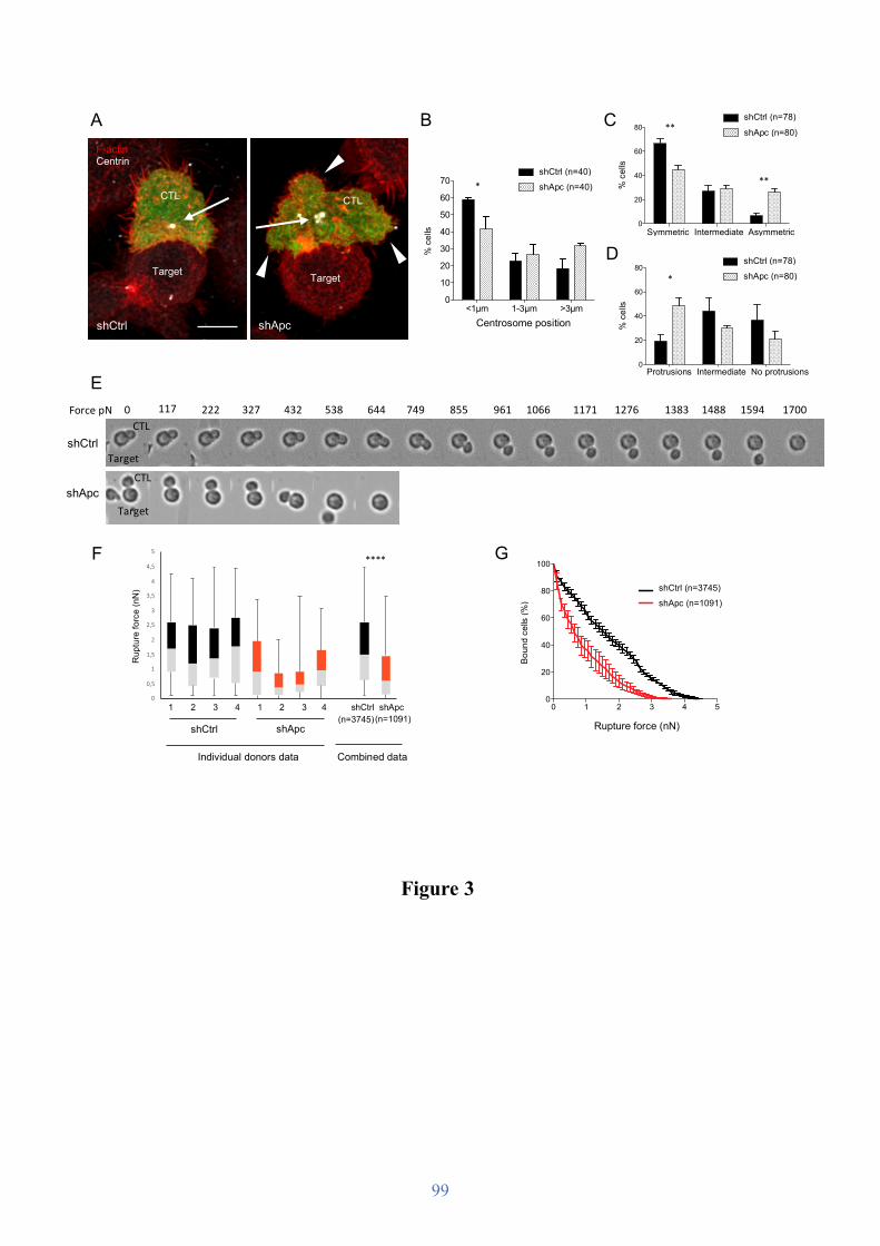
99
Figure 3
A
E
C
Symmetric Intermediate Asymmetric0
20
40
60
80
% c
ells
shCtrl
shApc**
**
D
B
<1µm 1-3µm >3µm0
10
20
30
40
50
60
70
% c
ells
shCtrl
shApc
Centrosome position
*
Protrusions Intermediate No protrusions0
20
40
60
80
% c
ells
shCtrl
shApc*
0
0,5
1
1,5
2
2,5
3
3,5
4
4,5
5
1 2 3 4 1 2 3 4 shCtrl shApc
Rupt
ure
forc
e (n
N)
1 2 3 4 1 2 3 4 shCtrl shApc shCtrl shApc
Rup
ture
forc
e (n
N)
(n=3745) (n=1091)
F G
Centrosome position
% c
ells
P1 P20
20
40
60
80
100
% c
ells
siCtrl
siApc********
shCtrl (n=40) shApc (n=40) %
cel
ls
Symmetric Intermediate Asymmetric P1 P2
0
20
40
60
80
100
% c
ells
siCtrl
siApc********
shCtrl (n=78) shApc (n=80)
*
**
**
% c
ells
Protrusions Intermediate No protrusions
*
P1 P20
20
40
60
80
100
% c
ells
siCtrl
siApc********
shCtrl (n=78) shApc (n=80)
F-actin Centrin
shCtrl
CTL
Target
CTL
Target
shApc
****
0 1 2 3 4 50
20
40
60
80
100
Rupture force (nN)
Boun
d ce
lls (%
)
shCtrl
shApc
shCtrl (n=3745) shApc (n=1091)
0 1 2 3 4 50
20
40
60
80
100
Rupture force (nN)
Boun
d ce
lls (%
)
shCtrl
shApc
Rupture force (nN)
Boun
d ce
lls (%
)
Individual donors data Combined data
0 117 222 327 432 538 644 749 855 961 1066 1171 1276 1383 1488 1594
shCtrl
shApc
CTL
Target CTL
Target
1700 Force pN

100
Figure 4
F
Polarized Grouped Random0
20
40
60
80
% c
ells
Granule localization
shCtrl
shApc
*
*
0 1-20 21-40 41-60 60+0
10
20
30
40
% c
ells
shCtrl
shApc*
*
Number of granules
G
shApc
CTL
Target
shCtrl shApc0
20
40
60
80
100
120
Num
ber o
f gra
nule
s pe
r cel
l *
Gra
nule
s/ce
ll A
shCtrl (n=19)
shApc (n=20)
shCtrl shApc0123456789
Fusi
on e
vent
s pe
r cel
l
**
Fusi
on e
vent
s/ce
ll
shCtrl
D E
shApc
shCtrl shApc0
30
60
90
120
150
180
210
Trac
king
dur
atio
n (s
ec)
****
shCtrl shApc0
1
2
3
4
5
6
7
8
Dis
plac
emen
t (µ
m)
ns
shCtrl shApc0
1
2
3
4
5
6
7
Med
ian
spee
d (µ
m/s
)
****B C
shCtrl
shApc
Gra
nule
trac
king
dur
atio
n (s
ec)
*
shCtrl (n=767)
shApc (n=524)
Gra
nule
med
ian
spee
d (µ
m/s
)
shCtrl (n=767)
shApc (n=524)
Gra
nule
dis
plac
emen
t (µm
)
shCtrl (n=767)
shApc (n=524)
**** **** ns
shCtrl (n=19)
shApc (n=20)
% c
ells
Polarized Grouped Random
*
*
Granule localization Number of granules P1 P2
0
20
40
60
80
100
% c
ells
siCtrl
siApc********
shCtrl (n=60) shApc (n=60)
* *
F-actin Perforin
shCtrl
CTL
Target

101
A
B
Figure 5
C
D
0 1 100
20
40
60
Anti-CD3 coated P815 (µg/mL)
% L
amp1
+ in
CD
8+ ce
lls
shCtrl
shApc
ns
0 2 200
20
40
60
Anti-CD3 coated P815 (µg/mL)
% L
amp1
+ in
CD
8+ ce
lls
WT
ApcMin/+
ns
shCtrl unstimulated shApc unstimulated shCtrl + P815 10 µg/mL anti-CD3 shApc + P815 10 µg/mL anti-CD3
Lamp1-PE
WT unst. Apc
Min/+ unst.
WT + P815 20 µg/mL anti-CD3 Apc
Min/+ + P815 20 µg/mL anti-CD3
Lamp1-PE
20:01 10:01 05:01 2.5:1 1.25:1 0.625:1 0.312:10
20
40
60
80
100
Effector:Target cell ratio
Cyt
otox
icity
(%)
shCtrl
shApc
***
27:1 09:01 03:01 01:01 0.333:010
20
40
60
80
100
Effector:Target cell ratio
Cyt
otox
icity
(%)
WT
ApcMin/+
****
Effector : Target cell ratio
Cyt
otox
icity
(%)
Effector : Target cell ratio
Cyt
otox
icity
(%)
***
****
20:01 10:01 05:01 2.5:1 1.25:1 0.625:1 0.312:10
20
40
60
80
100
Effector:Target cell ratio
Cyt
otox
icity
(%)
shCtrl
shApc
***
shCtrl shApc
20:01 10:01 05:01 2.5:1 1.25:1 0.625:1 0.312:10
20
40
60
80
100
Effector:Target cell ratio
Cyt
otox
icity
(%)
shCtrl
shApc
***
WT Apc
Min/+
Nor
mal
ized
to m
ode
Nor
mal
ized
to m
ode
Anti-CD3-coated P815 (μg/mL)
% L
amp1
+ in C
D8+ c
ells
P1 P20
20
40
60
80
100
% c
ells
siCtrl
siApc********
shCtrl shApc
Anti-CD3-coated P815 (μg/mL)
% L
amp1
+ in C
D8+ c
ells
P1 P20
20
40
60
80
100
% c
ells
siCtrl
siApc********
WT Apc
Min/+
20:1 10:1 5:1 2.5:1 1.25:1 0.6:1 0.3:1
27:1 9:1 3:1 1:1 0.3:1

102
A
Figure 6
0 2 4 6 8 100.0
0.6
1.2
1.8
Stimulation time (min)
Nor
m. b
and
inte
nsity
(a.u
.) *
0 2 4 6 8 100.0
0.6
1.2
1.8
Stimulation time (min)
Nor
m. b
and
inte
nsity
(a.u
.) WTApcMin/+
*
pAkt pErk1/2 B
C D
0 5 10 150.0
0.6
1.2
1.8
Stimulation time (min)
Nor
m. b
and
inte
nsity
(a.u
.) WTApcMin/+
****
Nuclear NFAT
NFAT
Lamin B1
WT ApcMin/+
CD3/CD28 XL (min): - 5 10 15 - 5 10 15
Akt pSer473
Erk1/2
GAPDH
CD3/CD28 XL (min): - 2 5 10 - 2 5 10 WT Apc
Min/+
p44 ➤ p42 ➤
Nor
m. b
and
inte
nsity
Stimulation time (min)
* *
Nor
m. b
and
inte
nsity
Stimulation time (min) 20:01 10:01 05:01 2.5:1 1.25:1 0.625:1 0.312:10
20
40
60
80
100
Effector:Target cell ratio
Cyt
otox
icity
(%)
shCtrl
shApc
***
WT Apc
Min/+
Nor
m. b
and
inte
nsity
Stimulation time (min) 20:01 10:01 05:01 2.5:1 1.25:1 0.625:1 0.312:10
20
40
60
80
100
Effector:Target cell ratio
Cyt
otox
icity
(%)
shCtrl
shApc
***
WT Apc
Min/+
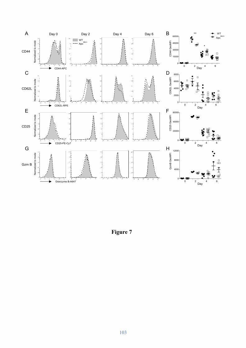
103
Figure 7
0
20000
40000
60000
CD
25 G
eoM
FI
0
4000
8000
12000
Gra
nzym
e B
Geo
MFI
B
D
F
Day 0 Day 4 Day 6
CD25
Gzm B
CD44
CD62L
Day 2 A
C
E
G
WT
ApcMin/
+W
T
ApcMin/
+W
T
ApcMin/
+W
T
ApcMin/
+
0
2000
4000
6000
8000
CD
62L
Geo
MFI
ns
0
15000
30000
45000
60000
CD
44 G
eoM
FI
**
*
0 2 4 6 Day
H
Nor
mal
ized
to m
ode
Nor
mal
ized
to m
ode
Nor
mal
ized
to m
ode
Nor
mal
ized
to m
ode
CD44-APC
CD62L-RPE
CD25-PE-Cy7
Granzyme B-A647
CD
44 G
eoM
FI
*
**
CD
62L
Geo
MFI
0 2 4 6 Day
CD
25 G
eoM
FI
0 2 4 6 Day
0 2 4 6 Day
Gzm
B G
eoM
FI
0 5 10 150.0
0.6
1.2
1.8
Stimulation time (min)
Nor
m. b
and
inte
nsity
(a.u
.) WTApcMin/+
****
WT Apc
Min/+
WT Apc
Min/+

104
Figure 8
Non-stimulatedPMA/Iono CD3/C280
2000
4000
6000
8000
TNFα
con
cent
ratio
n (p
g/m
L)
Non-stimulatedPMA/Iono CD3/C280
2000
4000
6000
IL-2
con
cent
ratio
n (p
g/m
L)
Non-stimulatedPMA/Iono CD3/C280
2000
4000
6000
8000
10000
IFN
y co
ncen
tratio
n (p
g/m
L)
C TNF𝛼 secretion IFN𝛾 secretion IL-2 secretion
Non-stimulatedPMA/Iono CD3/CD280
20
40
60
80
100
% IL
-2+
Non-stimulatedPMA/Iono CD3/CD280
20
40
60
80
100
% IF
Nγ+
Non-stimulatedPMA/Iono CD3/CD280
20
40
60
80
100
% T
NFα
+A
TNF𝛼+ in CD8
+ IFN𝛾+ in CD8
+ IL-2+ in CD8
+
Non-stimulatedPMA/Iono CD3/CD280
5000
10000
15000
TNFα
con
cent
ratio
n (p
g/m
L)
Non-stimulatedPMA/Iono CD3/CD280
200000
400000
600000IF
Nγ
conc
entra
tion
(pg/
mL)
Non-stimulatedPMA/Iono CD3/CD280
5000
10000
15000
20000
IL-2
con
cent
ratio
n (p
g/m
L) ns
B TNF𝛼 secretion IFN𝛾 secretion IL-2 secretion
% T
NF𝛼
+
Non-stim. PMA/iono CD3/CD28
% IF
N𝛾+
Non-stim. PMA/iono CD3/CD28
% IL
-2+
Non-stim. PMA/iono CD3/CD28
Non-stim. PMA/iono CD3/CD28
TNF𝛼
conc
entra
tion
(pg/
mL)
IFN𝛾
conc
entra
tion
(pg/
mL)
Non-stim. PMA/iono CD3/CD28
IL-2
con
cent
ratio
n (p
g/m
L)
Non-stim. PMA/iono CD3/CD28
TNF𝛼
conc
entra
tion
(pg/
mL)
Non-stim. PMA/iono CD3/CD28
IFN𝛾
conc
entra
tion
(pg/
mL)
Non-stim. PMA/iono CD3/CD28
IL-2
con
cent
ratio
n (p
g/m
L)
Non-stim. PMA/iono CD3/CD28 P1 P2
0
20
40
60
80
100
% c
ells
siCtrl
siApc********
shCtrl shApc
P1 P20
20
40
60
80
100
% c
ells
siCtrl
siApc********
WT Apc
Min/+
P1 P20
20
40
60
80
100
% c
ells
siCtrl
siApc********
WT Apc
Min/+

105
B
D
F
Day 0 Day 4 Day 6
CD25
CD44
CD62L
Day 2 A
C
E
0 2 4 6 Day
Nor
mal
ized
to m
ode
Nor
mal
ized
to m
ode
Nor
mal
ized
to m
ode
CD44-APC
CD62L-RPE
CD25-PE-Cy7
CD
44 G
eoM
FI
CD
62L
Geo
MFI
0 2 4 6 Day
CD
25 G
eoM
FI
0 2 4 6
Supplemental figure 1
0 2 4 60
10000
20000
30000
40000
50000
Day
CD
25 G
eoM
FI
0 2 4 60
15000
30000
45000
60000
Day
CD
44 G
eoM
FI
**
0 2 4 60
2000
4000
6000
8000
10000
Day
CD
62L
Geo
MFI
**
Day
WT Apc
Min/+

106

107
Discussion

108

109
The mechanisms regulating T cell polarization and the immunological synapse formation and
functions have been intensively investigated since the characterization of its molecular
composition and spatial organization 20 years ago (Grakoui et al., 1999; Monks et al., 1998).
While the involvement of the actin cytoskeleton, microtubule network, and polarized vesicular
traffic is well established, the interplay between them remained much less considered.
Numerous data pointed out Apc as a candidate regulator of the crosstalk between actin and
microtubules. Indeed, this polarity regulator interacts with several cytoskeleton interacting-
proteins, stabilizes microtubules and stimulates actin polymerization promoting polarization in
migrating cell (Kroboth et al., 2007; Okada et al., 2010; Watanabe et al., 2004).
Using Apc-silenced human CD8 T cells and T cells from a mouse model of familial
adenomatous polyposis (the ApcMin/+ strain), we have characterized the involvement of Apc in
CD8 T cells functions and anti-tumor immunity. We have unveiled the regulatory role of Apc
on CD8 T cell actin and microtubule cytoskeleton organization, and therefore synapse
formation and architecture. Hence, Apc defects resulted in reduce immunological synapse
stability and CTL killing activity, but interestingly only mildly impairs CD8 T cells activation
and differentiation into CTLs.
1. Does Apc regulate CD8 T cell synapse formation?
1.1. Apc is expressed in CD8 T cells and tunes actin and microtubules cytoskeleton
organization
To our knowledge, no studies have been conducted on the role of Apc in CD8 T cells. Thus,
we first investigated Apc expression and localization in primary human CD8 T cells. Apc
appears to have a similar localization in several cell types (e.g. T cells, fibroblasts, astrocytes).
Apc is associated with microtubules and localized in cellular areas with highly dynamic
cytoskeleton reorganization, such as the leading edge of migrating cells and the immunological
synapse periphery in T cells (Etienne-Manneville et al., 2005; Näthke et al., 1996; Watanabe et
al., 2004). Therefore, Apc subcellular localization in CD8 T cells is consistent with its
conserved role as cytoskeleton and cell polarity regulator (Nelson and Näthke, 2013).
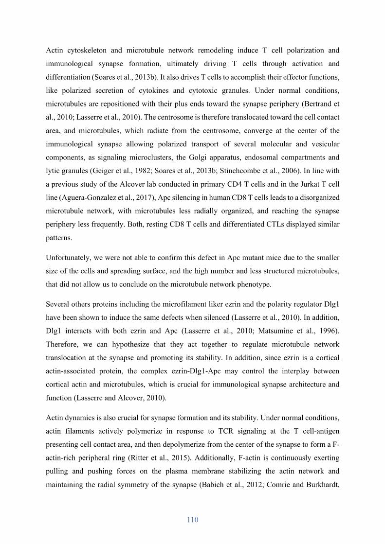
110
Actin cytoskeleton and microtubule network remodeling induce T cell polarization and
immunological synapse formation, ultimately driving T cells through activation and
differentiation (Soares et al., 2013b). It also drives T cells to accomplish their effector functions,
like polarized secretion of cytokines and cytotoxic granules. Under normal conditions,
microtubules are repositioned with their plus ends toward the synapse periphery (Bertrand et
al., 2010; Lasserre et al., 2010). The centrosome is therefore translocated toward the cell contact
area, and microtubules, which radiate from the centrosome, converge at the center of the
immunological synapse allowing polarized transport of several molecular and vesicular
components, as signaling microclusters, the Golgi apparatus, endosomal compartments and
lytic granules (Geiger et al., 1982; Soares et al., 2013b; Stinchcombe et al., 2006). In line with
a previous study of the Alcover lab conducted in primary CD4 T cells and in the Jurkat T cell
line (Aguera-Gonzalez et al., 2017), Apc silencing in human CD8 T cells leads to a disorganized
microtubule network, with microtubules less radially organized, and reaching the synapse
periphery less frequently. Both, resting CD8 T cells and differentiated CTLs displayed similar
patterns.
Unfortunately, we were not able to confirm this defect in Apc mutant mice due to the smaller
size of the cells and spreading surface, and the high number and less structured microtubules,
that did not allow us to conclude on the microtubule network phenotype.
Several others proteins including the microfilament liker ezrin and the polarity regulator Dlg1
have been shown to induce the same defects when silenced (Lasserre et al., 2010). In addition,
Dlg1 interacts with both ezrin and Apc (Lasserre et al., 2010; Matsumine et al., 1996).
Therefore, we can hypothesize that they act together to regulate microtubule network
translocation at the synapse and promoting its stability. In addition, since ezrin is a cortical
actin-associated protein, the complex ezrin-Dlg1-Apc may control the interplay between
cortical actin and microtubules, which is crucial for immunological synapse architecture and
function (Lasserre and Alcover, 2010).
Actin dynamics is also crucial for synapse formation and its stability. Under normal conditions,
actin filaments actively polymerize in response to TCR signaling at the T cell-antigen
presenting cell contact area, and then depolymerize from the center of the synapse to form a F-
actin-rich peripheral ring (Ritter et al., 2015). Additionally, F-actin is continuously exerting
pulling and pushing forces on the plasma membrane stabilizing the actin network and
maintaining the radial symmetry of the synapse (Babich et al., 2012; Comrie and Burkhardt,
111
2016). Apc silencing in human CD8 T cells did not affect actin accumulation and
polymerization at the synapse, as the total quantity of F-actin at the synapse was similar than in
control cells. However, actin clearance from the center of the synapse appeared to be impaired
as cells displayed less frequently an actin-rich ring at the synapse periphery.
Analyses conducted on mouse T cells revealed high variability in control and Apc mutant mice,
showing no clear tendency concerning F-actin accumulation at the synapse. In addition, no
difference was detected on actin ring formation (data not shown). This might be due to the fact
that ApcMin/+ mice are heterozygous and might express higher levels of Apc, displaying a less
clear phenotype.
Actin and microtubule cytoskeleton cooperate to build functional immunological synapses in T
cells (Soares et al., 2013b). Indeed, the microtubules and centrosome translocation toward the
synapse is occurring concomitantly to actin cytoskeleton remodeling. The interplay between
the two types of cytoskeleton is crucial for synapse formation and T cell functions, however,
the mechanisms underlying their crosstalk remain poorly understood. Apc interacts with both
actin and microtubule regulators (Nelson and Näthke, 2013). In addition, actin cytoskeleton and
microtubules share numerous others partners such as ezrin, Dlg1 or IQGAP-1, but also Cdc42
and formins that were shown to control actin remodeling and centrosome polarization at the
synapse (Fukata et al., 2002; Gomez et al., 2007; Lasserre et al., 2010; Matsumine et al., 1996;
Stowers et al., 1995). Interestingly, all these molecules are interacting partners of Apc (Nelson
and Näthke, 2013). Therefore, Apc and those molecules could form a complex regulating actin
and microtubule interplay during T cell polarization and synapse formation. This hypothesis is
also consistent with a recent study unveiling the role of Apc in coordinating microtubules and
actin polymerization at focal adhesions to control osteosarcoma cell migration (Juanes et al.,
2017).

112
Figure 15. Apc deficiency alters CD8 T cell immunological synapse formation.
Apc tunes CD8 T cell actin cytoskeleton and microtubules during synapse formation by promoting actin dynamics, microtubule stability, and centrosome polarization. Apc would thus stabilize the synapse, allowing its maturation and efficient lytic granule release in the synaptic cleft. However, if Apc deficiency induces granule release non-specifically at the synapse, as schematized on the left bottom panel, is still unknown.
113
1.2. Apc regulates the immunological synapse stability and its maturation
Alteration of the cytoskeleton dynamics can result in deficient or non-mature immunological
synapses, this is observed in several severe pathologies such as chronic lymphocytic leukemia
and Wiskott-Aldrich syndrome (Kallikourdis et al., 2015; Ramsay et al., 2008). As actin and
microtubule reorganization are altered in human Apc-silenced CD8 T cells stimulated by anti-
CD3 coated surfaces mimicking antigen presenting cells, we hypothesized that conventional
synapses formed with antigen presenting cell could present defects.
Considering that FAP patients and ApcMin/+ mutant mice are not immunodeficient, we did not
expect to observe a total impairment of synapse formation, and indeed, quantification of the
proportion of Apc-silenced CD8 T cells conjugated with antigen presenting cell revealed no
differences compared to control cells (data not shown). However, conjugated T cells displayed
large membrane extensions altering their radial symmetry and regularity, suggesting cellular
instability and defective cortical rigidity. In addition, the centrosome was more distant from the
cell contact area, hinting at an impaired CD8 T cell polarization toward the antigen presenting
cell (see Figure 15). It could also be interesting to study the Golgi apparatus polarization as it
is translocated at the synapse with the centrosome and is required for the proper recycling of
TCR signaling molecules and delivery of lytic granules (Kupfer et al., 1985; Roig-Martinez et
al., 2019).
These results therefore reflect the inability of Apc-silenced cells to stabilize their interactions
with target cells and form mature immunological synapses. Thus, we assessed T cell-target cell
interaction by the resistance of conjugates to resist an incremental shear laminar flow. The
measurement of the force range needed to detach CTLs from target cells using flow-induced
forces unveiled the lower adhesion strength of Apc-silenced CTLs compared to control cells.
Indeed, the rupture force for cell-cell interactions involving Apc-silenced cells was significantly
decreased.
It would be now interesting to characterized in more detail the Apc-dependent mechanisms
regulating CTL interactions. Indeed, lower adhesion forces could be the result of impaired actin
cytoskeleton dynamics and therefore synapse stability, suggested by the large membrane
protrusions and impaired formation of the actin-rich ring. An additional explanation could be
the alteration of molecules directly responsible for adhesion. Therefore, to complete this study,
it would be interesting to quantify the expression of several integrins such as LFA-1 and VLA-
4 at the synapse and their capacity to adhere to their ligands. Studying their localization may

114
also unveiled Apc involvement in synapse maturation and stability. Indeed, impairing the
microtubule network organization and the actin-rich ring formation at the synapse periphery
would impair the centripetal movement of membrane proteins, which results from the pushing
and pulling forces generated by actin polymerization and myosin II contraction. The SMAC
segregation would then not be optimal and the p-SMAC may not provide a sufficiently thigh
adhesion between CTLs and antigen presenting cells (Comrie et al., 2015; Yi et al., 2012).
Impaired centripetal flow would furthermore inhibit integrin activation, as their activation is
regulated through conformational changes induced by the actin cytoskeleton-generated forces
(Comrie et al., 2015; Martín-Cófreces et al., 2018). Therefore, it appears crucial to study Apc
involvement in actin-induced centripetal flow regulation and p- and c-SMAC formation.
2. Are CD8 T cell activation and differentiation modulated by Apc?
2.1. Regulation of TCR signaling pathways by Apc
Immunological synapse architecture and stability are crucial to trigger TCR signaling pathways,
ultimately resulting in the activation of transcription factors initiating CD8 T cell activation and
differentiation into CTLs.
Apc deficiency in CD4 T cells, which also impairs microtubule network remodeling at the
immunological synapse, alters nuclear translocation of the transcription factor NFATc2, as well
as its transcriptional regulation, without impairing the activation of TCR signaling molecules
and other transcription factor downstream of the TCR like NFkB (Aguera-Gonzalez et al.,
2017). Here, we found in ApcMin/+ CD8 T cells that some signaling molecules (e.g. Akt and
Erk1/2) were subtly but significantly less activated upon TCR engagement. In addition, lower
NFATc2 nuclear translocation was also observed.
Hence, Apc mutation in mouse CD8 T cells could affect different TCR signaling pathways
(Erk1/2/Fos, Akt/PKCθ, IP3/Ca+) that participate to the activation of several crucial
transcription factors (JNK/Fos, NFκB, NFAT) (see Figure 5). However, in CD4 T cells, NFκB
and Jun/Fos activation as well as calcium flux were unaffected. Thus, decreased NFAT activity
was likely the consequence of its defective microtubule-dependent nuclear translocation.
Indeed, NFATc2 was found forming microclusters associated with microtubules, and
115
microtubule depolymerization was inhibiting NFATc2 nuclear translocation and transcriptional
activity (Aguera-Gonzalez et al., 2017). These data therefore suggest that NFATc2 could be
similarly affected in CD8 T cells. Conversely, Apc mutation may somewhat alter early TCR
signaling in CD8 T cells but not in CD4. The reason of this apparent discrepancy is unknown
but it may stem in a different sensitivity of CD4 versus CD8 T cells to Apc mutation or
differences in the experimental model system used (Apc-silenced human CD4 T cells versus
Apc-mutant murine CD8).
Further insight into this question may be gained by studying the activation of other transcription
factors in CD8 T cells, or the transcription of their target genes. In addition, it could be
interesting to study microcluster formation and localization at the synapse, which are the earliest
events of TCR signaling pathways, and could reflect an alteration of the centripetal flow
generated by the actin cytoskeleton remodeling.
2.2. Apc mutation is not sufficient to alter CD8 T cell differentiation into CTLs
The observed TCR signaling and NFAT nuclear translocation defects did not appear to be
sufficient to significantly block or modify ApcMin/+ CD8 T cell differentiation into CTLs. In
contrast, previous work of the Alcover lab unveiled decreased differentiation of ApcMin/+ CD4
T cells into Tregs.
Apc mutant CD8 T cells expressed similar level of CD25 and granzyme B compared to control
cells along their ex vivo differentiation, suggesting that signal transduction and effector protein
synthesis are correctly induced in mutant cells under the experimental conditions used.
Nevertheless, mild changes in the expression of other maturation markers were observed in the
early phase of the differentiation process. This was the case for CD62L and CD44, which are
involved in cellular adhesion and regulate T cell entry and egress in secondary lymphoid organs
(Hemmerich et al., 2001; Nandi et al., 2004). These data suggest no effect of Apc loss on
terminal CD8 T cell differentiation, but some difference on the kinetics of this cellular program.
Experiments performed on CD8 T isolated specifically from the intestinal lamina propria did
not display more significant differences (data not shown), suggesting that this phenotype is not
dependent on tissue localization.

116
Therefore, it would be interesting to take advantage of the Apc mutant mouse model to perform
in vivo experiments to study naive T cell homing into secondary lymphoid organs, and the
recruitment of differentiating cells to inflammatory sites and tumors.
Interestingly under the same experimental differentiation conditions, CD4 T cells displayed
different kinetics of CD62L and more pronounced differences in CD44 and CD62L expression
between control and ApcMin/+ cells.
Altogether, these data suggest that altered T cell polarization and synapse formation only mildly
impact CD8 T cell activation and differentiation.
As described in the introduction of this thesis, immunological synapse formation induced by
TCR stimulation regulates the spatiotemporal molecular organization that tunes TCR signaling
and therefore T cell activation. Actin polymerization stabilizes the synapse to maintain TCR-
pMHC engagement, centrosome translocation toward the cell contact area favors the polarized
transport of vesicles containing TCR and its downstream signaling proteins (Blanchard et al.,
2002; Ehrlich et al., 2002; Hashimoto-Tane et al., 2011; Soares et al., 2013a), while actin-
induced retrograde flow and dynein drive the centripetal movement of microclusters along
microtubules and SMAC formation (Aguera-Gonzalez et al., 2017; Campi et al., 2005;
Hashimoto-Tane et al., 2011; Lasserre et al., 2010; Nguyen et al., 2008). Apc silencing in
human CD8 T cells impairs synapse stability, actin cytoskeleton dynamics, centrosome
polarization, and microtubule network repositioning at the synapse. Therefore, it is surprising
to detect only a slight reduction in TCR signaling, and mild changes in the expression of
differentiation markers that do not affect CD8 T cell activation and effector function
acquisition.
However, it is important to note that microscopy analyses and activation/differentiation
experiments have not been conducted in the same cellular models. Indeed, confocal microscopy
analysis of cytoskeleton reorganization and synapse formation and stability were performed on
human cells silenced with siRNA or shRNA, whereas functional experiments on activation and
differentiation were conducted with ApcMin/+ mouse cells. As mentioned previously, we tried to
investigate cytoskeleton defects in Apc mutant mice but the results on microtubule organization
and actin polymerization at the synapse were not conclusive. Human and mouse CD8 T cells
may have some functional differences, but the most likely explanation for these discrepancies
is that Apc silencing and Apc heterozygous mutation do not induce the same defects. Although
Apc silencing does not result in a complete knockdown (about 25% of residual protein may

117
persist in cells), the amount of wild-type protein left in murine mutant T cells may be higher,
and the expression of a truncated form of Apc could be sufficient to perform physiological
functions. Unfortunately, we were unable to measure the amount of residual protein in mouse
cells due to the lack of reactivity of the available anti-Apc antibodies.
3. Is Apc involved in CTL effector functions?
3.1. CTL cytokine production and secretion are not regulated by Apc
TCR signaling pathways ultimately induce the activation of transcription factors such as NFAT,
which is dephosphorylated following calcium entry in the cell, leading to its translocation to
the nucleus (Im and Rao, 2004). There, NFAT binds promoter or enhancer regions of DNA,
driving the expression of genes involved in T cell activation, differentiation, and cytokine
production. Cytokines dependent on NFAT include TNFα and IFNγ produced by CTLs during
immune responses (Campbell et al., 1996; Goldfeld et al., 1994).
The Alcover lab has previously shown the importance of Apc in CD4 T cell cytokine
production. Indeed, Apc-silenced human CD4 T cells displayed lower NFAT activity and IL-2
gene transcription. In addition, CD4 T cells from ApcMin/+ mice produced less IL-2, IL-4 and
IFNγ and lamina propria Tregs shown impaired differentiation and production of the anti-
inflammatory cytokine IL-10 (Aguera-Gonzalez et al., 2017). However, neither human Apc-
silenced nor mouse Apc-mutated CTLs presented any defect in IL-2, TNFα or IFNγ cytokine
production or secretion. Therefore, decreased NFAT nuclear translocation may not be sufficient
to impair CTL cytokine production under the experimental conditions used.
Considering the cytoskeleton organization defects we unveiled in CTL synapses, it is intriguing
that cytokine transport and exocytosis, regulated by actin and microtubule cytoskeletons, were
not affected. Indeed, upon TCR stimulation, they are released at the synapse (Kupfer et al.,
1991). As lytic granules, vesicles containing cytokines are transported through the Golgi
apparatus, which is associated with microtubules and localized in the pericentrosomal area,
therefore co-polarizing toward the immunological synapse (Huse et al., 2006; Huse et al., 2008;
Kupfer et al., 1994). In addition, the Hivroz laboratory reported the importance of actin
clearance from the synapse center in CD4 T cells to allow cytokine secretion. Indeed, Cdc42

118
silencing which inhibits actin-rich ring formation also impaired concentration of the vesicles
containing cytokines and their exocytosis (Chemin et al., 2012).
However, several studies have shown a second distinct secretion pathway from the one targeted
toward the synapse, that required cytoskeleton reorganization and dynamics. Indeed, cytokine
secretion can also be unpolarized and multidirectional, using different trafficking and secretory
processes (Huse et al., 2006; Sanderson et al., 2012). Therefore, it could be interesting to study
secretion localization in the experimental model we used to understand whether Apc may affect
polarized and/or non-polarized secretion pathways.
Together with the results from the ex vivo study of CD8 T cell activation and differentiation
presented above, this thesis work suggest that Apc plays a limited minor role in CD8 T cell
activation, differentiation into effector cells, and in cytokine production driven by NFAT
transcriptional activity. These observations appear at odds with previous finding of the
laboratory in CD4 T cells (Aguera-Gonzalez et al., 2017).
Hence, despite their common origin and the high apparent degree of similarity in some
processes, CD4 and CD8 T cells are still very different and their activation, differentiation, and
effector function acquisition do not undergo the same gene transcription processes (Sabins et
al., 2016; Seder and Ahmed, 2003; Whitmire et al., 2000). Therefore, our results indicate that
CD8 T cells are less sensitive to Apc defects than CD4 T cells, at least in their programs of
activation leading to differentiation and cytokine production.
3.2. Apc regulates CTL cytotoxic activity
Cytoskeleton reorganization at the cytotoxic synapse has been extensively studied. Actin
filaments actively polymerize at the cell contact area. Once the synapse is stabilized, F-actin
depolymerizes from the center of the synapse to form an actin-rich peripheral ring (Ritter et al.,
2013(Ritter et al., 2013). Microtubules are repositioned at the periphery of the cell contact area
leading to the centrosome polarization and docking at the plasma membrane (Aguera-Gonzalez
et al., 2017; Bertrand et al., 2010; Geiger et al., 1982; Hashimoto-Tane et al., 2011; Lasserre et
al., 2010; Stinchcombe et al., 2006). Lytic granules move along the microtubules to cluster
around the moving centrosome which defines a precise secretion site in the actin-poor region

119
of the synapse (Stinchcombe et al., 2001b; Stinchcombe et al., 2006). Finally, actin recovery
terminates lytic granule release (Ritter et al., 2015; Ritter et al., 2017). These cellular processes
are schematized on Figure 15, top panel. By confocal microscopy, we saw that human Apc-
silenced CTLs display impaired actin-rich ring formation, microtubule network radial
organization, and centrosome docking at the membrane. Consistent with the importance of these
cytoskeleton reorganization processes for CTL cytotoxicity, human Apc-silenced and Apc
mutant mouse CTLs presented decreased target cell killing activity.
Actin polymerization at the cell contact area, the formation of the actin-rich ring, and, as
recently shown, the formation of small actin-rich protrusions that extend into the target cell
surface apply forces at the CTL plasma membrane. These forces stabilize the synapse and
generate centripetal flow for SMAC formation, but also are exerted from the CTL to the target
cells (Husson et al., 2011; Tamzalit et al., 2019). In cytotoxic synapses, force exertion has been
correlated in space and time with lytic granule release. Applying tension on the target cell
membrane potentiates killing by enhancing the insertion of hydrophobic perforin in the target
cell plasma membrane, without changing the lytic granule exocytosis (Basu et al., 2016).
Here, the reduced actin-rich ring formation at the synapse periphery and the abnormal large
protrusion formation suggest impaired actin cytoskeleton dynamics in Apc-silenced CTLs. In
addition, the lower adhesion strength of CTLs to target cells indicates synapse instability and
reduced force engagement between the two cells.
Therefore, the decreased ability of Apc deficient CTLs to eliminate target cell could be the
result of lack of actin dynamics at the immunological synapse. Hence, it appears again
necessary to study Apc involvement in actin-induced centripetal flow by live high-resolution
microscopy, but also in the regulation of force exertion on target cells using, for instance,
micropillar deformation assay (Schoen et al., 2010).
However, although significant, the reduction of tumor cell killing in Apc-deficient CTLs was
surprisingly modest considering the importance of the cytoskeleton reorganization defects and
synapse instability unveiled. This could be due to the activation conditions we used in vitro to
generate CTLs. Indeed, IL-2 regulates a plethora of processes involved in T cell function
regulation, including gene transcription and cytoskeleton organization (Ross and Cantrell,
2018). Therefore, the addition of IL-2 could partially restore cytotoxic functions, as it was
described in some forms of hemophagocytic lymphohistiocytosis (Bryceson et al., 2007;
Marcenaro et al., 2006).

120
Another possibility is that our results support studies that are challenging the commonly
accepted synapse model described in the introduction. Indeed, several studies have shown that
the cytotoxic synapse, in opposition to the stimulatory synapse leading to T cell activation, does
not required to be completely formed and stable to be efficient. To induce target cell death,
CTLs do not need a strong antigen stimulus compared to the one required for stimulatory
synapse (Faroudi et al., 2003). Indeed, killing may be triggered by only three TCR-pMHC
interactions, whereas stimulatory synapses require at least ten interactions (Purbhoo et al.,
2004). In addition, lytic granule exocytosis may occur in absence of centrosome polarization
toward the targeted cell, and CTLs could kill simultaneously several targets (Bertrand et al.,
2013; Depoil et al., 2005; Wiedemann et al., 2006). This effect could be particularly
exacerbated in in vitro assays where T cells and target cells are forced to be together. Therefore,
despite Apc deficiency effects on immunological synapse stability and centrosome docking at
the cell contact area, it would less severely affect CTL killing activity in vitro.
3.3. Apc tunes late events leading to cytotoxic granules release
In order to study granule exocytosis, we have used several approaches that gave somehow
conflicting results, possibly due to difference in assay sensitivity and kinetics. Indeed, long and
strong stimulation as in the commonly used degranulation assay, where CD107a surface
expression is used as a readout of granule release, did not allow us to observe significant
differences in both human Apc-silenced and Apc mutant mouse CTLs compared to controls. In
contrast, shorter stimulation and analyses by real-time TIRF microscopy at single cell level
revealed significant differences in lytic granule dynamics, targeting and fusion at the synapse.
Indeed, after 15 min of cell contact, most of the CTLs have released their lytic granules under
our experimental conditions, whereas Apc-silenced cells still presented a high number of
cytoplasmic granules. Interestingly, those granules were grouped and polarized in a majority of
the cells, while the centrosome distance to the synapse remains longer. In addition, during the
first four minutes of stimulation on an activatory surface, less granules were seen reaching the
plasma membrane by TIRF microscopy. The speed of these granules was reduced, and fusion
events were decreased. Altogether, data obtained by fixed confocal and live TIRF microscopy
therefore suggest that defect of centrosome docking at the plasma membrane does not impact
121
granules ability to concentrate and polarize to a certain extent at the immunological synapse,
suggesting that the potential defect occurs at the last steps of granule targeting, docking and
fusion.
Alternatively, our data would support studies mentioned previously according to which CTLs
may also transport lytic granule and kill target cells independently from centrosome polarization
(Bertrand et al., 2013; Depoil et al., 2005; Wiedemann et al., 2006). Nevertheless, polarized
release may still increase the efficiency.
Impaired microtubule network organization could still reduce lytic granule transport and
movement close to the plasma membrane, since microtubules in Apc-silenced cells appear to
reach the plasma membrane with more difficulty. Furthermore, the remaining F-actin at the
center of the synapse may reduce the plasma membrane accessibility of lytic granules, acting
as a barrier that prevents efficient secretion, as previously seen by Ritter and colleges (Ritter et
al., 2017). Lytic granule fusion at the synapse is therefore less efficiently achieved.
Furthermore, we made the hypothesis that an unstable synapse and cytoskeleton defects could
lead to the exocytosis of granules in a non-polarized way or in a less restrictive synaptic cleft
(see Figure 15). This could be an alternative explanation to the conflicting results obtained by
Lamp1 surface expression quantification and TIRF microscopy, as granules would be released
but not specifically at the synapse. We may therefore observe lytic protein release in the cellular
environment and bystander killing. Unfortunately, we have not been able to confirm this
hypothesis yet as we have not found the optimal experimental conditions to induce specific but
not non-specific killing.
Hence, these data showed that Apc deficiency alter late steps of lytic granule targeting to the
synapse, consequently decreasing CTL killing efficiency.

122
4. Involvement for familial adenomatous polyposis and colorectal
cancers
This thesis work has unveiled for the first time the role of Apc in CD8 T cell functions, and
more precisely on immunological synapse formation and cytotoxic function regulation.
Furthermore, these data replaced in the context of FAP development can shed light on the
potential effect of Apc mutation in CTL anti-tumor immunity.
Indeed, so far little is known about Apc involvement in anti-tumor immune responses. Its role
in the epithelium and adenomas formation have been extensively studied, however why these
adenomas are not eliminated and can escape anti-tumor control is unclear.
4.1. Anti-tumor immune responses in ApcMin/+ mice
The Alcover lab, and others, have shown impaired ex and in vivo anti-inflammatory immune
response in Apc mutant mice due to Treg function alterations. Indeed, Tregs have been shown
to accumulate in the lamina propria and in adenomas at pre-cancerous stages, unbalancing the
intestinal immune homeostasis. However, intestinal Tregs have impaired expression of the
transcription factor FoxP3. Consequently, their differentiation and production of anti-
inflammatory cytokines as IL-10 are decreased (Aguera-Gonzalez et al., 2017; Gounaris et al.,
2009). According to others, their production of pro-inflammatory cytokines as IL-17 increases
(Aguera-Gonzalez et al., 2017; Chae and Bothwell, 2015; Gounaris et al., 2009). Therefore,
ApcMin/+ intestinal Tregs shift from a protective anti-inflammatory phenotype, required for
colorectal tumor regression, to a detrimental pro-inflammatory phenotype, unbalancing
intestinal immune homeostasis.
Unlike Tregs, we did not observe significant changes in ApcMin/+ CD8 T cell differentiation and
cytokine production. However, ex vivo differentiating CD8 T cells were displaying some subtle
changes in the expression of molecules involved in migration and recruitment to tumor site, as
CD62L and CD44. In addition, CTLs presented reduced ability to lyse tumor target cells.
Several in vivo experiments thus need to be performed to better characterize the altered CD8 T
cell functions due to Apc mutation in a mouse model of polyposis:
- Study CD8 T cell migration and homing, by injecting naive control or ApcMin/+ cells in control
mice, and then analyzing their localization in blood or secondary lymphoid organs.

123
- Study CTLs and others immune cell types recruitment and tumor infiltration, by implanting
solid tumor in control and ApcMin/+ mice, and then quantifying immune populations in the tumor.
- Study CD8 T cell ability to mount an anti-tumor response, by implanting solid tumor in control
and ApcMin/+ mice, then injecting tumor peptide to activate in vivo CD8 T cells, and finally
measuring tumor growth, and its infiltration, at different time points.
- Study CD8 T cells control of adenoma and tumor formation and their progression, by injecting
control or ApcMin/+ cells in ApcMin/+ mice, and then measuring adenoma and tumor number and
size in the intestines.
These experiments could confirm our ex vivo results and unveiled impaired tumor infiltration
and tumor cell elimination by CD8 T cells in anti-tumor immune response due to Apc mutation.
Together, studies conducted both on CD4 and CD8 T cells could therefore shed light on the
impaired anti-tumor immune responses occurring during adenoma and tumor formation in FAP.
Indeed, increased intestinal inflammation and decreased abnormal cell elimination would
together promote premalignant lesion development and then tumor escape.
4.2. Characterization of immune responses in familial adenomatous polyposis
patients
The ApcMin/+ mutant mouse is a very useful model to study familial adenomatous polyposis,
allowing to conduct numerous studies that would not be possible, or very difficult, with patients
such as isolation of different cell types from the lamina propria or in vivo homing analysis.
However, some differences exist between the pathological features in mice and humans, as
highlighted by the different localization of polyps in ApcMin/+ mice versus patients. Hence, it
may be argued that potential effects of Apc on immune cells may also be different between
human and mice.
Therefore, in addition to using human primary Apc-silenced cells and Apc-mutant mouse cells,
our laboratory recently started a study aiming to analyze the phenotype and function of immune
cells purified from blood of familial adenomatous polyposis patients carrying mutations in the
Apc gene.

124
This project is conducted in collaboration with the ICAReB bio-bank core facility at the Institut
Pasteur, managing the collection of blood samples from patients and healthy donors. FAP
patients carrying mutations in the Apc gene are being recruited with the help of the patients’
association Association Polyposes Familiales, as well as physicians specialists on polyposis. In
addition, we developed some of our experimental protocols with the collaboration of Milieu
Intérieur LabEx team, who set up standardized methods to investigate a large variety of
parameters of immune responses of a cohort of 1 000 healthy individuals (Hasan, 2019)
(http://www.milieuinterieur.fr/en ).
With this clinical study, we first aim to corroborate and extend the results obtained in this thesis
work with Apc-silenced human cells and ApcMin/+ cells. Therefore, we are analyzing
microtubule and actin organization in immunological synapses formed by CD8 T cells on anti-
CD3 coated stimulatory surfaces. In addition, we are assessing immunological synapse
formation, stability, and strength of adhesion. We are studying CD8 and CD4 T cell activation,
and differentiation, following a protocol similar to the one used for mouse cells. CD4 T cells
differentiation into Tregs is also assessed, in order to compare with results previously obtained
by our laboratory in mouse intestinal Tregs (Aguera-Gonzalez et al., 2017). Furthermore, CTL
effector functions including cytokine production, tumor cell lysis, and lytic granule release are
evaluated.
Additionally, we have extended our study to NK cells. Indeed, NK cells are innate cells that,
similar to CD8 T cells, are able to directly eliminate infected and tumor cells via the exocytosis
of lytic granules containing perforin and granzyme B. NK cell form immunological synapses
with infected or tumor cells without prior activation. These synapses do not involve the same
receptors and signaling pathways than T cells, however their architecture and organization are
similar, displaying polarization toward the target cell, and microtubules and centrosome
repositioning toward the cell contact area (Orange, 2008). In addition, F-actin polymerizes at
the synapse and is excluded from the secretion site (Brown et al., 2011; Rak et al., 2011). We
therefore hypothesized that Apc mutation could also alter NK cell cytotoxic functions,
contributing to impaired abnormal cell elimination, adenoma progression, and finally tumor
escape in FAP.
The second aim of this clinical study is to assess Apc involvement in T cell adhesion and
migration. Indeed, this study was focused on CD8 T cell functions that are regulated by
immunological synapse formation. However efficient T cell immune response also requires

125
recirculation of naive cells between the blood and lymph and secondary lymphoid organs in
search of cells presenting their cognate antigen, and recruitment of activated cells to
inflammation or tumor sites. Both of these processes are based on T cell ability to migrate.
Migration is a polarized cellular process that opposes a protrusive leading edge to a retracting
trailing edge. The leading edge, at the advancing front of the cell, is enriched in chemiokine
receptors and F-actin, providing sensitivity to guidance cues and protrusive forces for
directional migration. In contrast, the uropod, at the rear of the cell, contains the centrosome
and microtubules, providing contraction and retraction (Vicente-Manzanares et al., 2005).
In migrating cells, Apc is localized at microtubule plus ends and in actin-rich region, which
correlates with protrusive cell area and the leading edge (Etienne-Manneville et al., 2005;
Näthke et al., 1996; Watanabe et al., 2004). Apc silencing slows down fibroblast and astrocytes
during in vitro migration by preventing the actin meshwork formation in the leading edge,
destabilizing microtubules, and decreasing centrosome reorientation (Etienne-Manneville and
Hall, 2003; Kroboth et al., 2007; Watanabe et al., 2004). In addition, Apc mutation abrogates
in vivo cell migration from the crypts to the villi in the intestinal tract (Mahmoud et al., 1997;
Sansom et al., 2004).
Therefore, we hypothesized that Apc mutation could also alter T cell migration. To answer this
question, we are studying their migration toward chemiokine gradient in transwell assays or by
live microscopy. Additionally, using the rupture force assay, we are comparing adhesion
strength of healthy donor and patient cells on different substrates or extracellular matrix
components (e.g. fibronectin or VCAM-1).
Furthermore, we have chosen to extend our clinical study to other cell types. Indeed, our third
aim is to characterize the general landscape of immune responses in FAP patients. Therefore,
we are performing multiparametric whole blood immune cell phenotyping by flow cytometry,
and multiplex transcriptome and secreted proteome analyses after different types of innate and
adaptive stimulation.
Importantly, mutations carried by each patient will be analyzed to establish a correlation
between the type of mutation, Apc expression and functional alterations, as so far, no strong
correlation between genotype and phenotype has been highlighted.
This clinical study will allow us to characterize CD8 T cells, but also other immune cell type
responses in FAP patients. By unveiling alterations, we could shed light on impaired immune

126
processes, and therefore on why premalignant lesion forming in the rectum and colon of FAP
patients are not eliminated by the immune system.

127
Conclusion
My thesis work aimed to better understand how immunological synapse formation, CD8 T cell
activation and differentiation, and CTL effector functions could be regulated by the polarity
regulator Apc, with the ultimate purpose of better understanding potentially altered CD8 T cell
responses in familial adenomatous polyposis patients carrying Apc mutations.
Using Apc-silenced human CD8 T cells and T cells from a mouse model of familial
adenomatous polyposis ApcMin/+, we have first shown the regulatory role of Apc in CD8 T
cytoskeleton dynamics, and synapse symmetry and stability. Our results therefore suggest that
Apc reorganizes actin cytoskeleton and microtubule network at the CD8 T cell immunological
synapse, ensuring their interplay. We have also investigated Apc involvement in different steps
of CD8 T cell journey through immune response, as their activation, differentiation into CTLs,
and their effector functions. Interestingly, we have shown that Apc modulates CTL cytotoxic
activity, by allowing efficient lytic granule exocytosis, but is mildly involved in CD8 T cell
activation, differentiation and cytokine production. Unexpectedly, we have shown that Apc
deficiency affects differently CD4 and CD8 T cells.
Therefore, we propose that Apc regulates CD8 T cell cytotoxic functions through its function
of polarity regulator and cytoskeleton organizer.
Although our findings indicate that Apc is not absolutely required for the different processes
studied, it cannot be excluded that mild impairments of several processes results in defective
anti-tumor responses in vivo. Our results open numerous questions but confirm the relevance
of studying Apc regulatory role in CD8 T cell functions to better characterize FAP. In addition,
they underline the importance of characterizing Apc role in NK cell cytotoxic activity, as well
as in phagocytosis, which both depend on the orchestrated action of the actin and microtubule
cytoskeleton (Niedergang et al., 2016; Orange, 2008).
Therefore, the extension of this study through future in vivo experiments in mice, the analysis
of additional processes regulating T cell responses, such as their migration, and the ongoing
characterization of immune cell function in FAP patients will allow us to clarify the role of Apc
in anti-tumor responses. This might ultimately inspire future immunomodulatory approaches to
fight cancers and/or provide alternative treatments for patients.

128

129
Bibliography

130

131
Acuto, O., and Michel, F. (2003). CD28-mediated co-stimulation: a quantitative support for TCR signalling. Nat Rev Immunol 3, 939-951. Afonina, I.S., Cullen, S.P., and Martin, S.J. (2010). Cytotoxic and non-cytotoxic roles of the CTL/NK protease granzyme B. Immunol Rev 235, 105-116. Aguera-Gonzalez, S., Bouchet, J., and Alcover, A. (2015). Immunological Synapse. eLS John Wiley & Sons, Ltd: Chichester. Aguera-Gonzalez, S., Burton, O.T., Vazquez-Chavez, E., Cuche, C., Herit, F., Bouchet, J., Lasserre, R., Del Rio-Iniguez, I., Di Bartolo, V., and Alcover, A. (2017). Adenomatous Polyposis Coli Defines Treg Differentiation and Anti-inflammatory Function through Microtubule-Mediated NFAT Localization. Cell Rep 21, 181-194. Alcover, A., Alarcon, B., and Di Bartolo, V. (2018). Cell Biology of T Cell Receptor Expression and Regulation. Annu Rev Immunol 36, 103-125. Andersson, J., Tran, D.Q., Pesu, M., Davidson, T.S., Ramsey, H., O'Shea, J.J., and Shevach, E.M. (2008). CD4+ FoxP3+ regulatory T cells confer infectious tolerance in a TGF-beta-dependent manner. J Exp Med 205, 1975-1981. Aoki, K., and Taketo, M.M. (2007). Adenomatous polyposis coli (APC): a multi-functional tumor suppressor gene. J Cell Sci 120, 3327-3335. Babich, A., Li, S., O'Connor, R.S., Milone, M.C., Freedman, B.D., and Burkhardt, J.K. (2012). F-actin polymerization and retrograde flow drive sustained PLCgamma1 signaling during T cell activation. J Cell Biol 197, 775-787. Balagopalan, L., Sherman, E., Barr, V.A., and Samelson, L.E. (2011). Imaging techniques for assaying lymphocyte activation in action. Nat Rev Immunol 11, 21-33. Balaji, K.N., Schaschke, N., Machleidt, W., Catalfamo, M., and Henkart, P.A. (2002). Surface cathepsin B protects cytotoxic lymphocytes from self-destruction after degranulation. J Exp Med 196, 493-503. Barber, E.K., Dasgupta, J.D., Schlossman, S.F., Trevillyan, J.M., and Rudd, C.E. (1989). The CD4 and CD8 antigens are coupled to a protein-tyrosine kinase (p56lck) that phosphorylates the CD3 complex. Proc Natl Acad Sci U S A 86, 3277-3281. Barbosa, M.D., Nguyen, Q.A., Tchernev, V.T., Ashley, J.A., Detter, J.C., Blaydes, S.M., Brandt, S.J., Chotai, D., Hodgman, C., Solari, R.C., et al. (1996). Identification of the homologous beige and Chediak-Higashi syndrome genes. Nature 382, 262-265. Basu, R., Whitlock, B.M., Husson, J., Le Floc'h, A., Jin, W., Oyler-Yaniv, A., Dotiwala, F., Giannone, G., Hivroz, C., Biais, N., et al. (2016). Cytotoxic T Cells Use Mechanical Force to Potentiate Target Cell Killing. Cell 165, 100-110.

132
Bertrand, F., Esquerre, M., Petit, A.E., Rodrigues, M., Duchez, S., Delon, J., and Valitutti, S. (2010). Activation of the ancestral polarity regulator protein kinase C zeta at the immunological synapse drives polarization of Th cell secretory machinery toward APCs. J Immunol 185, 2887-2894. Bertrand, F., Müller, S., Roh, K.H., Laurent, C., Dupré, L., and Valitutti, S. (2013). An initial and rapid step of lytic granule secretion precedes microtubule organizing center polarization at the cytotoxic T lymphocyte/target cell synapse. Proc Natl Acad Sci U S A 110, 6073-6078. Blanchard, N., Di Bartolo, V., and Hivroz, C. (2002). In the immune synapse, ZAP-70 controls T cell polarization and recruitment of signaling proteins but not formation of the synaptic pattern. Immunity 17, 389-399. Bossi, G., and Griffiths, G.M. (1999). Degranulation plays an essential part in regulating cell surface expression of Fas ligand in T cells and natural killer cells. Nat Med 5, 90-96. Bouchet, J., and Alcover, A. (2014). [Immunological synapse is a dynamic signaling platform for T cell activation]. Med Sci (Paris) 30, 665-670. Bouchet, J., Del Río-Iñiguez, I., Lasserre, R., Agüera-Gonzalez, S., Cuche, C., Danckaert, A., McCaffrey, M.W., Di Bartolo, V., and Alcover, A. (2016). Rac1-Rab11-FIP3 regulatory hub coordinates vesicle traffic with actin remodeling and T-cell activation. EMBO J 35, 1160-1174. Bouchet, J., Del Río-Iñiguez, I., Vázquez-Chávez, E., Lasserre, R., Agüera-González, S., Cuche, C., McCaffrey, M.W., Di Bartolo, V., and Alcover, A. (2017). Rab11-FIP3 Regulation of Lck Endosomal Traffic Controls TCR Signal Transduction. J Immunol 198, 2967-2978. Brenner, D., Blaser, H., and Mak, T.W. (2015). Regulation of tumour necrosis factor signalling: live or let die. Nat Rev Immunol 15, 362-374. Brenner, H., Kloor, M., and Pox, C.P. (2014). Colorectal cancer. Lancet 383, 1490-1502. Brossard, C., Feuillet, V., Schmitt, A., Randriamampita, C., Romao, M., Raposo, G., and Trautmann, A. (2005). Multifocal structure of the T cell - dendritic cell synapse. Eur J Immunol 35, 1741-1753. Brown, A.C., Oddos, S., Dobbie, I.M., Alakoskela, J.M., Parton, R.M., Eissmann, P., Neil, M.A., Dunsby, C., French, P.M., Davis, I., et al. (2011). Remodelling of cortical actin where lytic granules dock at natural killer cell immune synapses revealed by super-resolution microscopy. PLoS Biol 9, e1001152. Bryceson, Y.T., Rudd, E., Zheng, C., Edner, J., Ma, D., Wood, S.M., Bechensteen, A.G., Boelens, J.J., Celkan, T., Farah, R.A., et al. (2007). Defective cytotoxic lymphocyte degranulation in syntaxin-11 deficient familial hemophagocytic lymphohistiocytosis 4 (FHL4) patients. Blood 110, 1906-1915. Bullock, T.N., Colella, T.A., and Engelhard, V.H. (2000). The density of peptides displayed by dendritic cells affects immune responses to human tyrosinase and gp100 in HLA-A2 transgenic mice. J Immunol 164, 2354-2361.

133
Bunnell, S.C., Hong, D.I., Kardon, J.R., Yamazaki, T., McGlade, C.J., Barr, V.A., and Samelson, L.E. (2002). T cell receptor ligation induces the formation of dynamically regulated signaling assemblies. J Cell Biol 158, 1263-1275. Bunnell, S.C., Kapoor, V., Trible, R.P., Zhang, W., and Samelson, L.E. (2001). Dynamic actin polymerization drives T cell receptor-induced spreading: a role for the signal transduction adaptor LAT. Immunity 14, 315-329. Caldwell, C.M., Green, R.A., and Kaplan, K.B. (2007). APC mutations lead to cytokinetic failures in vitro and tetraploid genotypes in Min mice. J Cell Biol 178, 1109-1120. Campbell, P.M., Pimm, J., Ramassar, V., and Halloran, P.F. (1996). Identification of a calcium-inducible, cyclosporine sensitive element in the IFN-gamma promoter that is a potential NFAT binding site. Transplantation 61, 933-939. Campi, G., Varma, R., and Dustin, M.L. (2005). Actin and agonist MHC-peptide complex-dependent T cell receptor microclusters as scaffolds for signaling. J Exp Med 202, 1031-1036. Cao, X., Cai, S.F., Fehniger, T.A., Song, J., Collins, L.I., Piwnica-Worms, D.R., and Ley, T.J. (2007). Granzyme B and perforin are important for regulatory T cell-mediated suppression of tumor clearance. Immunity 27, 635-646. Caza, T., and Landas, S. (2015). Functional and Phenotypic Plasticity of CD4(+) T Cell Subsets. Biomed Res Int 2015, 521957. Cemerski, S., Das, J., Giurisato, E., Markiewicz, M.A., Allen, P.M., Chakraborty, A.K., and Shaw, A.S. (2008). The balance between T cell receptor signaling and degradation at the center of the immunological synapse is determined by antigen quality. Immunity 29, 414-422. Cemerski, S., Das, J., Locasale, J., Arnold, P., Giurisato, E., Markiewicz, M.A., Fremont, D., Allen, P.M., Chakraborty, A.K., and Shaw, A.S. (2007). The stimulatory potency of T cell antigens is influenced by the formation of the immunological synapse. Immunity 26, 345-355. Chabin-Brion, K., Marceiller, J., Perez, F., Settegrana, C., Drechou, A., Durand, G., and Poüs, C. (2001). The Golgi complex is a microtubule-organizing organelle. Mol Biol Cell 12, 2047-2060. Chae, W.J., and Bothwell, A.L. (2015). Spontaneous Intestinal Tumorigenesis in Apc (/Min+) Mice Requires Altered T Cell Development with IL-17A. J Immunol Res 2015, 860106. Chae, W.J., Gibson, T.F., Zelterman, D., Hao, L., Henegariu, O., and Bothwell, A.L. (2010). Ablation of IL-17A abrogates progression of spontaneous intestinal tumorigenesis. Proc Natl Acad Sci U S A 107, 5540-5544. Chemin, K., Bohineust, A., Dogniaux, S., Tourret, M., Guégan, S., Miro, F., and Hivroz, C. (2012). Cytokine secretion by CD4+ T cells at the immunological synapse requires Cdc42-dependent local actin remodeling but not microtubule organizing center polarity. J Immunol 189, 2159-2168.

134
Chen, L. (2004). Co-inhibitory molecules of the B7-CD28 family in the control of T-cell immunity. Nat Rev Immunol 4, 336-347. Chen, L., and Flies, D.B. (2013). Molecular mechanisms of T cell co-stimulation and co-inhibition. Nat Rev Immunol 13, 227-242. Choudhuri, K., Llodrá, J., Roth, E.W., Tsai, J., Gordo, S., Wucherpfennig, K.W., Kam, L.C., Stokes, D.L., and Dustin, M.L. (2014). Polarized release of T-cell-receptor-enriched microvesicles at the immunological synapse. Nature 507, 118-123. Choudhuri, K., Wiseman, D., Brown, M.H., Gould, K., and van der Merwe, P.A. (2005). T-cell receptor triggering is critically dependent on the dimensions of its peptide-MHC ligand. Nature 436, 578-582. Chung, A.Y., Li, Q., Blair, S.J., De Jesus, M., Dennis, K.L., LeVea, C., Yao, J., Sun, Y., Conway, T.F., Virtuoso, L.P., et al. (2014). Oral interleukin-10 alleviates polyposis via neutralization of pathogenic T-regulatory cells. Cancer Res 74, 5377-5385. Cibrian, D., and Sanchez-Madrid, F. (2017). CD69: from activation marker to metabolic gatekeeper. Eur J Immunol 47, 946-953. Clark, R.H., Stinchcombe, J.C., Day, A., Blott, E., Booth, S., Bossi, G., Hamblin, T., Davies, E.G., and Griffiths, G.M. (2003). Adaptor protein 3-dependent microtubule-mediated movement of lytic granules to the immunological synapse. Nat Immunol 4, 1111-1120. Combs, J., Kim, S.J., Tan, S., Ligon, L.A., Holzbaur, E.L., Kuhn, J., and Poenie, M. (2006). Recruitment of dynein to the Jurkat immunological synapse. Proc Natl Acad Sci U S A 103, 14883-14888. Comrie, W.A., Babich, A., and Burkhardt, J.K. (2015). F-actin flow drives affinity maturation and spatial organization of LFA-1 at the immunological synapse. J Cell Biol 208, 475-491. Comrie, W.A., and Burkhardt, J.K. (2016). Action and Traction: Cytoskeletal Control of Receptor Triggering at the Immunological Synapse. Front Immunol 7, 68. Cordoba, S.P., Choudhuri, K., Zhang, H., Bridge, M., Basat, A.B., Dustin, M.L., and van der Merwe, P.A. (2013). The large ectodomains of CD45 and CD148 regulate their segregation from and inhibition of ligated T-cell receptor. Blood 121, 4295-4302. Courtney, A.H., Lo, W.L., and Weiss, A. (2018). TCR Signaling: Mechanisms of Initiation and Propagation. Trends Biochem Sci 43, 108-123. Daniele, T., Hackmann, Y., Ritter, A.T., Wenham, M., Booth, S., Bossi, G., Schintler, M., Auer-Grumbach, M., and Griffiths, G.M. (2011). A role for Rab7 in the movement of secretory granules in cytotoxic T lymphocytes. Traffic 12, 902-911. Das, V., Nal, B., Dujeancourt, A., Thoulouze, M.I., Galli, T., Roux, P., Dautry-Varsat, A., and Alcover, A. (2004). Activation-induced polarized recycling targets T cell antigen receptors to the immunological synapse; involvement of SNARE complexes. Immunity 20, 577-588.

135
De Meester, J., Calvez, R., Valitutti, S., and Dupre, L. (2010). The Wiskott-Aldrich syndrome protein regulates CTL cytotoxicity and is required for efficient killing of B cell lymphoma targets. J Leukoc Biol 88, 1031-1040. de Saint Basile, G., Menasche, G., and Fischer, A. (2010). Molecular mechanisms of biogenesis and exocytosis of cytotoxic granules. Nat Rev Immunol 10, 568-579. DeGrendele, H.C., Kosfiszer, M., Estess, P., and Siegelman, M.H. (1997). CD44 activation and associated primary adhesion is inducible via T cell receptor stimulation. J Immunol 159, 2549-2553. den Haan, J.M., Arens, R., and van Zelm, M.C. (2014). The activation of the adaptive immune system: cross-talk between antigen-presenting cells, T cells and B cells. Immunol Lett 162, 103-112. Dennis, K.L., Saadalla, A., Blatner, N.R., Wang, S., Venkateswaran, V., Gounari, F., Cheroutre, H., Weaver, C.T., Roers, A., Egilmez, N.K., et al. (2015). T-cell Expression of IL10 Is Essential for Tumor Immune Surveillance in the Small Intestine. Cancer Immunol Res 3, 806-814. Depoil, D., Zaru, R., Guiraud, M., Chauveau, A., Harriague, J., Bismuth, G., Utzny, C., Muller, S., and Valitutti, S. (2005). Immunological synapses are versatile structures enabling selective T cell polarization. Immunity 22, 185-194. Derivery, E., Sousa, C., Gautier, J.J., Lombard, B., Loew, D., and Gautreau, A. (2009). The Arp2/3 activator WASH controls the fission of endosomes through a large multiprotein complex. Dev Cell 17, 712-723. Desai, D.M., Sap, J., Silvennoinen, O., Schlessinger, J., and Weiss, A. (1994). The catalytic activity of the CD45 membrane-proximal phosphatase domain is required for TCR signaling and regulation. EMBO J 13, 4002-4010. Dikovskaya, D., Newton, I.P., and Näthke, I.S. (2004). The adenomatous polyposis coli protein is required for the formation of robust spindles formed in CSF Xenopus extracts. Mol Biol Cell 15, 2978-2991. Dikovskaya, D., Schiffmann, D., Newton, I.P., Oakley, A., Kroboth, K., Sansom, O., Jamieson, T.J., Meniel, V., Clarke, A., and Näthke, I.S. (2007). Loss of APC induces polyploidy as a result of a combination of defects in mitosis and apoptosis. J Cell Biol 176, 183-195. Dolei, A., Capobianchi, M.R., and Ameglio, F. (1983). Human interferon-gamma enhances the expression of class I and class II major histocompatibility complex products in neoplastic cells more effectively than interferon-alpha and interferon-beta. Infect Immun 40, 172-176. Dow, L.E., O'Rourke, K.P., Simon, J., Tschaharganeh, D.F., van Es, J.H., Clevers, H., and Lowe, S.W. (2015). Apc Restoration Promotes Cellular Differentiation and Reestablishes Crypt Homeostasis in Colorectal Cancer. Cell 161, 1539-1552. Dunn, G.P., Old, L.J., and Schreiber, R.D. (2004). The immunobiology of cancer immunosurveillance and immunoediting. Immunity 21, 137-148.

136
Eberl, G., Colonna, M., Di Santo, J.P., and McKenzie, A.N. (2015). Innate lymphoid cells. Innate lymphoid cells: a new paradigm in immunology. Science 348, aaa6566. Ehrlich, L.I., Ebert, P.J., Krummel, M.F., Weiss, A., and Davis, M.M. (2002). Dynamics of p56lck translocation to the T cell immunological synapse following agonist and antagonist stimulation. Immunity 17, 809-822. Erdman, S.E., Sohn, J.J., Rao, V.P., Nambiar, P.R., Ge, Z., Fox, J.G., and Schauer, D.B. (2005). CD4+CD25+ regulatory lymphocytes induce regression of intestinal tumors in ApcMin/+ mice. Cancer Res 65, 3998-4004. Etienne-Manneville, S. (2009). APC in cell migration. Adv Exp Med Biol 656, 30-40. Etienne-Manneville, S., and Hall, A. (2002). Rho GTPases in cell biology. Nature 420, 629-635. Etienne-Manneville, S., and Hall, A. (2003). Cdc42 regulates GSK-3beta and adenomatous polyposis coli to control cell polarity. Nature 421, 753-756. Etienne-Manneville, S., Manneville, J.B., Nicholls, S., Ferenczi, M.A., and Hall, A. (2005). Cdc42 and Par6-PKCzeta regulate the spatially localized association of Dlg1 and APC to control cell polarization. J Cell Biol 170, 895-901. Faroudi, M., Utzny, C., Salio, M., Cerundolo, V., Guiraud, M., Muller, S., and Valitutti, S. (2003). Lytic versus stimulatory synapse in cytotoxic T lymphocyte/target cell interaction: manifestation of a dual activation threshold. Proc Natl Acad Sci U S A 100, 14145-14150. Feldmann, J., Callebaut, I., Raposo, G., Certain, S., Bacq, D., Dumont, C., Lambert, N., Ouachee-Chardin, M., Chedeville, G., Tamary, H., et al. (2003). Munc13-4 is essential for cytolytic granules fusion and is mutated in a form of familial hemophagocytic lymphohistiocytosis (FHL3). Cell 115, 461-473. Feng, L., Seymour, A.B., Jiang, S., To, A., Peden, A.A., Novak, E.K., Zhen, L., Rusiniak, M.E., Eicher, E.M., Robinson, M.S., et al. (1999). The beta3A subunit gene (Ap3b1) of the AP-3 adaptor complex is altered in the mouse hypopigmentation mutant pearl, a model for Hermansky-Pudlak syndrome and night blindness. Hum Mol Genet 8, 323-330. Fensterl, V., and Sen, G.C. (2009). Interferons and viral infections. Biofactors 35, 14-20. Finco, T.S., Kadlecek, T., Zhang, W., Samelson, L.E., and Weiss, A. (1998). LAT is required for TCR-mediated activation of PLCgamma1 and the Ras pathway. Immunity 9, 617-626. Finetti, F., Paccani, S.R., Riparbelli, M.G., Giacomello, E., Perinetti, G., Pazour, G.J., Rosenbaum, J.L., and Baldari, C.T. (2009). Intraflagellar transport is required for polarized recycling of the TCR/CD3 complex to the immune synapse. Nat Cell Biol 11, 1332-1339. Frey, A.B. (2015). Suppression of T cell responses in the tumor microenvironment. Vaccine 33, 7393-7400.

137
Fukata, M., Watanabe, T., Noritake, J., Nakagawa, M., Yamaga, M., Kuroda, S., Matsuura, Y., Iwamatsu, A., Perez, F., and Kaibuchi, K. (2002). Rac1 and Cdc42 capture microtubules through IQGAP1 and CLIP-170. Cell 109, 873-885. Galon, J., Costes, A., Sanchez-Cabo, F., Kirilovsky, A., Mlecnik, B., Lagorce-Pagès, C., Tosolini, M., Camus, M., Berger, A., Wind, P., et al. (2006). Type, density, and location of immune cells within human colorectal tumors predict clinical outcome. Science 313, 1960-1964. Geiger, B., Rosen, D., and Berke, G. (1982). Spatial relationships of microtubule-organizing centers and the contact area of cytotoxic T lymphocytes and target cells. J Cell Biol 95, 137-143. Germain, R.N. (2002). T-cell development and the CD4-CD8 lineage decision. Nat Rev Immunol 2, 309-322. Goldfeld, A.E., Tsai, E., Kincaid, R., Belshaw, P.J., Schrieber, S.L., Strominger, J.L., and Rao, A. (1994). Calcineurin mediates human tumor necrosis factor alpha gene induction in stimulated T and B cells. J Exp Med 180, 763-768. Goley, E.D., and Welch, M.D. (2006). The ARP2/3 complex: an actin nucleator comes of age. Nat Rev Mol Cell Biol 7, 713-726. Gomez, T.S., Kumar, K., Medeiros, R.B., Shimizu, Y., Leibson, P.J., and Billadeau, D.D. (2007). Formins regulate the actin-related protein 2/3 complex-independent polarization of the centrosome to the immunological synapse. Immunity 26, 177-190. Gounari, F., Chang, R., Cowan, J., Guo, Z., Dose, M., Gounaris, E., and Khazaie, K. (2005). Loss of adenomatous polyposis coli gene function disrupts thymic development. Nat Immunol 6, 800-809. Gounaris, E., Blatner, N.R., Dennis, K., Magnusson, F., Gurish, M.F., Strom, T.B., Beckhove, P., Gounari, F., and Khazaie, K. (2009). T-regulatory cells shift from a protective anti-inflammatory to a cancer-promoting proinflammatory phenotype in polyposis. Cancer Res 69, 5490-5497. Grakoui, A., Bromley, S.K., Sumen, C., Davis, M.M., Shaw, A.S., Allen, P.M., and Dustin, M.L. (1999). The immunological synapse: a molecular machine controlling T cell activation. Science 285, 221-227. Grossman, W.J., Verbsky, J.W., Barchet, W., Colonna, M., Atkinson, J.P., and Ley, T.J. (2004). Human T regulatory cells can use the perforin pathway to cause autologous target cell death. Immunity 21, 589-601. Guermonprez, P., Valladeau, J., Zitvogel, L., Théry, C., and Amigorena, S. (2002). Antigen presentation and T cell stimulation by dendritic cells. Annu Rev Immunol 20, 621-667. Half, E., Bercovich, D., and Rozen, P. (2009). Familial adenomatous polyposis. Orphanet J Rare Dis 4, 22.
138
Hasan, M. (2019). [Milieu Intérieur: understanding healthy immune system heterogeneity to move along the path towards personalized medicine]. Med Sci (Paris) 35, 423-430. Hashimoto-Tane, A., Yokosuka, T., Sakata-Sogawa, K., Sakuma, M., Ishihara, C., Tokunaga, M., and Saito, T. (2011). Dynein-driven transport of T cell receptor microclusters regulates immune synapse formation and T cell activation. Immunity 34, 919-931. Hauser, M.A., and Legler, D.F. (2016). Common and biased signaling pathways of the chemokine receptor CCR7 elicited by its ligands CCL19 and CCL21 in leukocytes. J Leukoc Biol 99, 869-882. Hayday, A.C. (2019). γδ T Cell Update: Adaptate Orchestrators of Immune Surveillance. J Immunol 203, 311-320. Hemmerich, S., Bistrup, A., Singer, M.S., van Zante, A., Lee, J.K., Tsay, D., Peters, M., Carminati, J.L., Brennan, T.J., Carver-Moore, K., et al. (2001). Sulfation of L-selectin ligands by an HEV-restricted sulfotransferase regulates lymphocyte homing to lymph nodes. Immunity 15, 237-247. Henrickson, S.E., Mempel, T.R., Mazo, I.B., Liu, B., Artyomov, M.N., Zheng, H., Peixoto, A., Flynn, M.P., Senman, B., Junt, T., et al. (2008). T cell sensing of antigen dose governs interactive behavior with dendritic cells and sets a threshold for T cell activation. Nat Immunol 9, 282-291. Hori, S., Nomura, T., and Sakaguchi, S. (2003). Control of regulatory T cell development by the transcription factor Foxp3. Science 299, 1057-1061. How, J.Y., Caria, S., Humbert, P.O., and Kvansakul, M. (2019). Crystal structure of the human Scribble PDZ1 domain bound to the PDZ-binding motif of APC. FEBS Lett 593, 533-542. Hsu, H., Xiong, J., and Goeddel, D.V. (1995). The TNF receptor 1-associated protein TRADD signals cell death and NF-kappa B activation. Cell 81, 495-504. Huse, M., Lillemeier, B.F., Kuhns, M.S., Chen, D.S., and Davis, M.M. (2006). T cells use two directionally distinct pathways for cytokine secretion. Nat Immunol 7, 247-255. Huse, M., Quann, E.J., and Davis, M.M. (2008). Shouts, whispers and the kiss of death: directional secretion in T cells. Nat Immunol 9, 1105-1111. Husson, J., Chemin, K., Bohineust, A., Hivroz, C., and Henry, N. (2011). Force generation upon T cell receptor engagement. PLoS One 6, e19680. Ilani, T., Vasiliver-Shamis, G., Vardhana, S., Bretscher, A., and Dustin, M.L. (2009). T cell antigen receptor signaling and immunological synapse stability require myosin IIA. Nat Immunol 10, 531-539. Im, S.H., and Rao, A. (2004). Activation and deactivation of gene expression by Ca2+/calcineurin-NFAT-mediated signaling. Mol Cells 18, 1-9.

139
Iwashima, M., Irving, B.A., van Oers, N.S., Chan, A.C., and Weiss, A. (1994). Sequential interactions of the TCR with two distinct cytoplasmic tyrosine kinases. Science 263, 1136-1139. Jasperson, K.W., Tuohy, T.M., Neklason, D.W., and Burt, R.W. (2010). Hereditary and familial colon cancer. Gastroenterology 138, 2044-2058. Jimbo, T., Kawasaki, Y., Koyama, R., Sato, R., Takada, S., Haraguchi, K., and Akiyama, T. (2002). Identification of a link between the tumour suppressor APC and the kinesin superfamily. Nat Cell Biol 4, 323-327. Juanes, M.A., Bouguenina, H., Eskin, J.A., Jaiswal, R., Badache, A., and Goode, B.L. (2017). Adenomatous polyposis coli nucleates actin assembly to drive cell migration and microtubule-induced focal adhesion turnover. J Cell Biol 216, 2859-2875. Kagi, D., Ledermann, B., Burki, K., Seiler, P., Odermatt, B., Olsen, K.J., Podack, E.R., Zinkernagel, R.M., and Hengartner, H. (1994). Cytotoxicity mediated by T cells and natural killer cells is greatly impaired in perforin-deficient mice. Nature 369, 31-37. Kallikourdis, M., Viola, A., and Benvenuti, F. (2015). Human Immunodeficiencies Related to Defective APC/T Cell Interaction. Front Immunol 6, 433. Kang, T.W., Yevsa, T., Woller, N., Hoenicke, L., Wuestefeld, T., Dauch, D., Hohmeyer, A., Gereke, M., Rudalska, R., Potapova, A., et al. (2011). Senescence surveillance of pre-malignant hepatocytes limits liver cancer development. Nature 479, 547-551. Kawasaki, Y., Senda, T., Ishidate, T., Koyama, R., Morishita, T., Iwayama, Y., Higuchi, O., and Akiyama, T. (2000). Asef, a link between the tumor suppressor APC and G-protein signaling. Science 289, 1194-1197. Kim, S.B., Zhang, L., Yoon, J., Lee, J., Min, J., Li, W., Grishin, N.V., Moon, Y.A., Wright, W.E., and Shay, J.W. (2018). Truncated Adenomatous Polyposis Coli Mutation Induces Asef-Activated Golgi Fragmentation. Mol Cell Biol 38. Kinzler, K.W., Nilbert, M.C., Vogelstein, B., Bryan, T.M., Levy, D.B., Smith, K.J., Preisinger, A.C., Hamilton, S.R., Hedge, P., and Markham, A. (1991). Identification of a gene located at chromosome 5q21 that is mutated in colorectal cancers. Science 251, 1366-1370. Koebel, C.M., Vermi, W., Swann, J.B., Zerafa, N., Rodig, S.J., Old, L.J., Smyth, M.J., and Schreiber, R.D. (2007). Adaptive immunity maintains occult cancer in an equilibrium state. Nature 450, 903-907. Kroboth, K., Newton, I.P., Kita, K., Dikovskaya, D., Zumbrunn, J., Waterman-Storer, C.M., and Näthke, I.S. (2007). Lack of adenomatous polyposis coli protein correlates with a decrease in cell migration and overall changes in microtubule stability. Mol Biol Cell 18, 910-918. Kuhné, M.R., Lin, J., Yablonski, D., Mollenauer, M.N., Ehrlich, L.I., Huppa, J., Davis, M.M., and Weiss, A. (2003). Linker for activation of T cells, zeta-associated protein-70, and Src homology 2 domain-containing leukocyte protein-76 are required for TCR-induced microtubule-organizing center polarization. J Immunol 171, 860-866.

140
Kuilman, T., Michaloglou, C., Vredeveld, L.C., Douma, S., van Doorn, R., Desmet, C.J., Aarden, L.A., Mooi, W.J., and Peeper, D.S. (2008). Oncogene-induced senescence relayed by an interleukin-dependent inflammatory network. Cell 133, 1019-1031. Kupfer, A., Dennert, G., and Singer, S.J. (1985). The reorientation of the Golgi apparatus and the microtubule-organizing center in the cytotoxic effector cell is a prerequisite in the lysis of bound target cells. J Mol Cell Immunol 2, 37-49. Kupfer, A., Mosmann, T.R., and Kupfer, H. (1991). Polarized expression of cytokines in cell conjugates of helper T cells and splenic B cells. Proc Natl Acad Sci U S A 88, 775-779. Kupfer, H., Monks, C.R., and Kupfer, A. (1994). Small splenic B cells that bind to antigen-specific T helper (Th) cells and face the site of cytokine production in the Th cells selectively proliferate: immunofluorescence microscopic studies of Th-B antigen-presenting cell interactions. J Exp Med 179, 1507-1515. Kurowska, M., Goudin, N., Nehme, N.T., Court, M., Garin, J., Fischer, A., de Saint Basile, G., and Menasche, G. (2012). Terminal transport of lytic granules to the immune synapse is mediated by the kinesin-1/Slp3/Rab27a complex. Blood 119, 3879-3889. Larghi, P., Williamson, D.J., Carpier, J.M., Dogniaux, S., Chemin, K., Bohineust, A., Danglot, L., Gaus, K., Galli, T., and Hivroz, C. (2013). VAMP7 controls T cell activation by regulating the recruitment and phosphorylation of vesicular Lat at TCR-activation sites. Nat Immunol 14, 723-731. Lasserre, R., and Alcover, A. (2010). Cytoskeletal cross-talk in the control of T cell antigen receptor signaling. FEBS Lett 584, 4845-4850. Lasserre, R., Charrin, S., Cuche, C., Danckaert, A., Thoulouze, M.I., de Chaumont, F., Duong, T., Perrault, N., Varin-Blank, N., Olivo-Marin, J.C., et al. (2010). Ezrin tunes T-cell activation by controlling Dlg1 and microtubule positioning at the immunological synapse. EMBO J 29, 2301-2314. Le Floc'h, A., Tanaka, Y., Bantilan, N.S., Voisinne, G., Altan-Bonnet, G., Fukui, Y., and Huse, M. (2013). Annular PIP3 accumulation controls actin architecture and modulates cytotoxicity at the immunological synapse. J Exp Med 210, 2721-2737. Lee, K.H., Dinner, A.R., Tu, C., Campi, G., Raychaudhuri, S., Varma, R., Sims, T.N., Burack, W.R., Wu, H., Wang, J., et al. (2003). The immunological synapse balances T cell receptor signaling and degradation. Science 302, 1218-1222. Letourneau, S., Krieg, C., Pantaleo, G., and Boyman, O. (2009). IL-2- and CD25-dependent immunoregulatory mechanisms in the homeostasis of T-cell subsets. J Allergy Clin Immunol 123, 758-762. Lieberman, J. (2003). The ABCs of granule-mediated cytotoxicity: new weapons in the arsenal. Nat Rev Immunol 3, 361-370. Lin, J., and Weiss, A. (2003). The tyrosine phosphatase CD148 is excluded from the immunologic synapse and down-regulates prolonged T cell signaling. J Cell Biol 162, 673-682.

141
Lowe, S.W., Cepero, E., and Evan, G. (2004). Intrinsic tumour suppression. Nature 432, 307-315. Ludford-Menting, M.J., Oliaro, J., Sacirbegovic, F., Cheah, E.T., Pedersen, N., Thomas, S.J., Pasam, A., Iazzolino, R., Dow, L.E., Waterhouse, N.J., et al. (2005). A network of PDZ-containing proteins regulates T cell polarity and morphology during migration and immunological synapse formation. Immunity 22, 737-748. Mahmoud, N.N., Boolbol, S.K., Bilinski, R.T., Martucci, C., Chadburn, A., and Bertagnolli, M.M. (1997). Apc gene mutation is associated with a dominant-negative effect upon intestinal cell migration. Cancer Res 57, 5045-5050. Mantovani, A., Allavena, P., Sica, A., and Balkwill, F. (2008). Cancer-related inflammation. Nature 454, 436-444. Marcenaro, S., Gallo, F., Martini, S., Santoro, A., Griffiths, G.M., Aricó, M., Moretta, L., and Pende, D. (2006). Analysis of natural killer-cell function in familial hemophagocytic lymphohistiocytosis (FHL): defective CD107a surface expression heralds Munc13-4 defect and discriminates between genetic subtypes of the disease. Blood 108, 2316-2323. Martín-Cófreces, N.B., Vicente-Manzanares, M., and Sánchez-Madrid, F. (2018). Adhesive Interactions Delineate the Topography of the Immune Synapse. Front Cell Dev Biol 6, 149. Mathieu, C., Beltra, J.C., Charpentier, T., Bourbonnais, S., Di Santo, J.P., Lamarre, A., and Decaluwe, H. (2015). IL-2 and IL-15 regulate CD8+ memory T-cell differentiation but are dispensable for protective recall responses. Eur J Immunol 45, 3324-3338. Matsumine, A., Ogai, A., Senda, T., Okumura, N., Satoh, K., Baeg, G.H., Kawahara, T., Kobayashi, S., Okada, M., Toyoshima, K., et al. (1996). Binding of APC to the human homolog of the Drosophila discs large tumor suppressor protein. Science 272, 1020-1023. McCartney, B.M., and Näthke, I.S. (2008). Cell regulation by the Apc protein Apc as master regulator of epithelia. Curr Opin Cell Biol 20, 186-193. Mehta, A.K., Gracias, D.T., and Croft, M. (2018). TNF activity and T cells. Cytokine 101, 14-18. Micheau, O., Thome, M., Schneider, P., Holler, N., Tschopp, J., Nicholson, D.W., Briand, C., and Grutter, M.G. (2002). The long form of FLIP is an activator of caspase-8 at the Fas death-inducing signaling complex. J Biol Chem 277, 45162-45171. Micheau, O., and Tschopp, J. (2003). Induction of TNF receptor I-mediated apoptosis via two sequential signaling complexes. Cell 114, 181-190. Miyara, M., and Sakaguchi, S. (2007). Natural regulatory T cells: mechanisms of suppression. Trends Mol Med 13, 108-116. Monks, C.R., Freiberg, B.A., Kupfer, H., Sciaky, N., and Kupfer, A. (1998). Three-dimensional segregation of supramolecular activation clusters in T cells. Nature 395, 82-86.
142
Moser, A.R., Pitot, H.C., and Dove, W.F. (1990). A dominant mutation that predisposes to multiple intestinal neoplasia in the mouse. Science 247, 322-324. Mueller, D.L., Jenkins, M.K., and Schwartz, R.H. (1989). An accessory cell-derived costimulatory signal acts independently of protein kinase C activation to allow T cell proliferation and prevent the induction of unresponsiveness. J Immunol 142, 2617-2628. Munemitsu, S., Albert, I., Souza, B., Rubinfeld, B., and Polakis, P. (1995). Regulation of intracellular beta-catenin levels by the adenomatous polyposis coli (APC) tumor-suppressor protein. Proc Natl Acad Sci U S A 92, 3046-3050. Munemitsu, S., Souza, B., Müller, O., Albert, I., Rubinfeld, B., and Polakis, P. (1994). The APC gene product associates with microtubules in vivo and promotes their assembly in vitro. Cancer Res 54, 3676-3681. Ménager, M.M., Ménasché, G., Romao, M., Knapnougel, P., Ho, C.H., Garfa, M., Raposo, G., Feldmann, J., Fischer, A., and de Saint Basile, G. (2007). Secretory cytotoxic granule maturation and exocytosis require the effector protein hMunc13-4. Nat Immunol 8, 257-267. Ménasché, G., Pastural, E., Feldmann, J., Certain, S., Ersoy, F., Dupuis, S., Wulffraat, N., Bianchi, D., Fischer, A., Le Deist, F., et al. (2000). Mutations in RAB27A cause Griscelli syndrome associated with haemophagocytic syndrome. Nat Genet 25, 173-176. Nagle, D.L., Karim, M.A., Woolf, E.A., Holmgren, L., Bork, P., Misumi, D.J., McGrail, S.H., Dussault, B.J., Perou, C.M., Boissy, R.E., et al. (1996). Identification and mutation analysis of the complete gene for Chediak-Higashi syndrome. Nat Genet 14, 307-311. Nakamura, M., Zhou, X.Z., and Lu, K.P. (2001). Critical role for the EB1 and APC interaction in the regulation of microtubule polymerization. Curr Biol 11, 1062-1067. Nandi, A., Estess, P., and Siegelman, M. (2004). Bimolecular complex between rolling and firm adhesion receptors required for cell arrest; CD44 association with VLA-4 in T cell extravasation. Immunity 20, 455-465. Narayan, P., Holt, B., Tosti, R., and Kane, L.P. (2006). CARMA1 is required for Akt-mediated NF-kappaB activation in T cells. Mol Cell Biol 26, 2327-2336. Nehlig, A., Molina, A., Rodrigues-Ferreira, S., Honoré, S., and Nahmias, C. (2017). Regulation of end-binding protein EB1 in the control of microtubule dynamics. Cell Mol Life Sci 74, 2381-2393. Nelson, S., and Näthke, I.S. (2013). Interactions and functions of the adenomatous polyposis coli (APC) protein at a glance. J Cell Sci 126, 873-877. Nguyen, K., Sylvain, N.R., and Bunnell, S.C. (2008). T cell costimulation via the integrin VLA-4 inhibits the actin-dependent centralization of signaling microclusters containing the adaptor SLP-76. Immunity 28, 810-821. Niedergang, F., Di Bartolo, V., and Alcover, A. (2016). Comparative Anatomy of Phagocytic and Immunological Synapses. Front Immunol 7, 18.

143
Norcross, M.A. (1984). A synaptic basis for T-lymphocyte activation. Ann Immunol (Paris) 135D, 113-134. Nutt, S.L., Hodgkin, P.D., Tarlinton, D.M., and Corcoran, L.M. (2015). The generation of antibody-secreting plasma cells. Nat Rev Immunol 15, 160-171. Näthke, I.S., Adams, C.L., Polakis, P., Sellin, J.H., and Nelson, W.J. (1996). The adenomatous polyposis coli tumor suppressor protein localizes to plasma membrane sites involved in active cell migration. J Cell Biol 134, 165-179. O'Keefe, J.P., and Gajewski, T.F. (2005). Cutting edge: cytotoxic granule polarization and cytolysis can occur without central supramolecular activation cluster formation in CD8+ effector T cells. J Immunol 175, 5581-5585. Ogura, H., Murakami, M., Okuyama, Y., Tsuruoka, M., Kitabayashi, C., Kanamoto, M., Nishihara, M., Iwakura, Y., and Hirano, T. (2008). Interleukin-17 promotes autoimmunity by triggering a positive-feedback loop via interleukin-6 induction. Immunity 29, 628-636. Okada, K., Bartolini, F., Deaconescu, A.M., Moseley, J.B., Dogic, Z., Grigorieff, N., Gundersen, G.G., and Goode, B.L. (2010). Adenomatous polyposis coli protein nucleates actin assembly and synergizes with the formin mDia1. J Cell Biol 189, 1087-1096. Olsen, I., Bou-Gharios, G., and Abraham, D. (1990). The activation of resting lymphocytes is accompanied by the biogenesis of lysosomal organelles. Eur J Immunol 20, 2161-2170. Onnis, A., Finetti, F., and Baldari, C.T. (2016). Vesicular Trafficking to the Immune Synapse: How to Assemble Receptor-Tailored Pathways from a Basic Building Set. Front Immunol 7, 50. Orange, J.S. (2008). Formation and function of the lytic NK-cell immunological synapse. Nat Rev Immunol 8, 713-725. Osinska, I., Popko, K., and Demkow, U. (2014). Perforin: an important player in immune response. Cent Eur J Immunol 39, 109-115. Ostroumov, D., Fekete-Drimusz, N., Saborowski, M., Kühnel, F., and Woller, N. (2018). CD4 and CD8 T lymphocyte interplay in controlling tumor growth. Cell Mol Life Sci 75, 689-713. Pandiyan, P., Zheng, L., Ishihara, S., Reed, J., and Lenardo, M.J. (2007). CD4+CD25+Foxp3+ regulatory T cells induce cytokine deprivation-mediated apoptosis of effector CD4+ T cells. Nat Immunol 8, 1353-1362. Peters, P.J., Borst, J., Oorschot, V., Fukuda, M., Krähenbühl, O., Tschopp, J., Slot, J.W., and Geuze, H.J. (1991). Cytotoxic T lymphocyte granules are secretory lysosomes, containing both perforin and granzymes. J Exp Med 173, 1099-1109. Petersen, G.M., Slack, J., and Nakamura, Y. (1991). Screening guidelines and premorbid diagnosis of familial adenomatous polyposis using linkage. Gastroenterology 100, 1658-1664.

144
Petry, S., and Vale, R.D. (2015). Microtubule nucleation at the centrosome and beyond. Nat Cell Biol 17, 1089-1093. Pollard, T.D. (2016). Actin and Actin-Binding Proteins. Cold Spring Harb Perspect Biol 8. Poulter, N.S., Pitkeathly, W.T., Smith, P.J., and Rappoport, J.Z. (2015). The physical basis of total internal reflection fluorescence (TIRF) microscopy and its cellular applications. Methods Mol Biol 1251, 1-23. Purbhoo, M.A., Irvine, D.J., Huppa, J.B., and Davis, M.M. (2004). T cell killing does not require the formation of a stable mature immunological synapse. Nat Immunol 5, 524-530. Purbhoo, M.A., Liu, H., Oddos, S., Owen, D.M., Neil, M.A., Pageon, S.V., French, P.M., Rudd, C.E., and Davis, D.M. (2010). Dynamics of subsynaptic vesicles and surface microclusters at the immunological synapse. Sci Signal 3, ra36. Quann, E.J., Merino, E., Furuta, T., and Huse, M. (2009). Localized diacylglycerol drives the polarization of the microtubule-organizing center in T cells. Nat Immunol 10, 627-635. Radoja, S., Saio, M., Schaer, D., Koneru, M., Vukmanovic, S., and Frey, A.B. (2001). CD8(+) tumor-infiltrating T cells are deficient in perforin-mediated cytolytic activity due to defective microtubule-organizing center mobilization and lytic granule exocytosis. J Immunol 167, 5042-5051. Rak, G.D., Mace, E.M., Banerjee, P.P., Svitkina, T., and Orange, J.S. (2011). Natural killer cell lytic granule secretion occurs through a pervasive actin network at the immune synapse. PLoS Biol 9, e1001151. Ramsay, A.G., Johnson, A.J., Lee, A.M., Gorgun, G., Le Dieu, R., Blum, W., Byrd, J.C., and Gribben, J.G. (2008). Chronic lymphocytic leukemia T cells show impaired immunological synapse formation that can be reversed with an immunomodulating drug. J Clin Invest 118, 2427-2437. Randzavola, L.O., Strege, K., Juzans, M., Asano, Y., Stinchcombe, J.C., Gawden-Bone, C.M., Seaman, M.N., Kuijpers, T.W., and Griffiths, G.M. (2019). Loss of ARPC1B impairs cytotoxic T lymphocyte maintenance and cytolytic activity. J Clin Invest. Raue, H.P., Beadling, C., Haun, J., and Slifka, M.K. (2013). Cytokine-mediated programmed proliferation of virus-specific CD8(+) memory T cells. Immunity 38, 131-139. Read, K.A., Powell, M.D., McDonald, P.W., and Oestreich, K.J. (2016). IL-2, IL-7, and IL-15: Multistage regulators of CD4(+) T helper cell differentiation. Exp Hematol 44, 799-808. Rheinlander, A., Schraven, B., and Bommhardt, U. (2018). CD45 in human physiology and clinical medicine. Immunol Lett 196, 22-32. Rieux-Laucat, F., Le Deist, F., and Fischer, A. (2003). Autoimmune lymphoproliferative syndromes: genetic defects of apoptosis pathways. Cell Death Differ 10, 124-133. Ritter, A.T., Angus, K.L., and Griffiths, G.M. (2013). The role of the cytoskeleton at the immunological synapse. Immunol Rev 256, 107-117.
145
Ritter, A.T., Asano, Y., Stinchcombe, J.C., Dieckmann, N.M., Chen, B.C., Gawden-Bone, C., van Engelenburg, S., Legant, W., Gao, L., Davidson, M.W., et al. (2015). Actin depletion initiates events leading to granule secretion at the immunological synapse. Immunity 42, 864-876. Ritter, A.T., Kapnick, S.M., Murugesan, S., Schwartzberg, P.L., Griffiths, G.M., and Lippincott-Schwartz, J. (2017). Cortical actin recovery at the immunological synapse leads to termination of lytic granule secretion in cytotoxic T lymphocytes. Proc Natl Acad Sci U S A 114, E6585-E6594. Rocha, B., and Tanchot, C. (2004). CD8 T cell memory. Semin Immunol 16, 305-314. Roda-Navarro, P. (2009). Assembly and function of the natural killer cell immune synapse. Front Biosci (Landmark Ed) 14, 621-633. Rodriguez-Boulan, E., and Macara, I.G. (2014). Organization and execution of the epithelial polarity programme. Nat Rev Mol Cell Biol 15, 225-242. Rohatgi, R., Ma, L., Miki, H., Lopez, M., Kirchhausen, T., Takenawa, T., and Kirschner, M.W. (1999). The interaction between N-WASP and the Arp2/3 complex links Cdc42-dependent signals to actin assembly. Cell 97, 221-231. Roig-Martinez, M., Saavedra-Lopez, E., Casanova, P.V., Cribaro, G.P., and Barcia, C. (2019). The MTOC/Golgi Complex at the T-Cell Immunological Synapse. Results Probl Cell Differ 67, 223-231. Roostalu, J., and Surrey, T. (2017). Microtubule nucleation: beyond the template. Nat Rev Mol Cell Biol 18, 702-710. Ross, S.H., and Cantrell, D.A. (2018). Signaling and Function of Interleukin-2 in T Lymphocytes. Annu Rev Immunol 36, 411-433. Rothe, M., Sarma, V., Dixit, V.M., and Goeddel, D.V. (1995). TRAF2-mediated activation of NF-kappa B by TNF receptor 2 and CD40. Science 269, 1424-1427. Round, J.L., Humphries, L.A., Tomassian, T., Mittelstadt, P., Zhang, M., and Miceli, M.C. (2007). Scaffold protein Dlgh1 coordinates alternative p38 kinase activation, directing T cell receptor signals toward NFAT but not NF-kappaB transcription factors. Nat Immunol 8, 154-161. Round, J.L., Tomassian, T., Zhang, M., Patel, V., Schoenberger, S.P., and Miceli, M.C. (2005). Dlgh1 coordinates actin polymerization, synaptic T cell receptor and lipid raft aggregation, and effector function in T cells. J Exp Med 201, 419-430. Rowan, A.J., Lamlum, H., Ilyas, M., Wheeler, J., Straub, J., Papadopoulou, A., Bicknell, D., Bodmer, W.F., and Tomlinson, I.P. (2000). APC mutations in sporadic colorectal tumors: A mutational "hotspot" and interdependence of the "two hits". Proc Natl Acad Sci U S A 97, 3352-3357.

146
Rowshanravan, B., Halliday, N., and Sansom, D.M. (2018). CTLA-4: a moving target in immunotherapy. Blood 131, 58-67. Rubinfeld, B., Souza, B., Albert, I., Müller, O., Chamberlain, S.H., Masiarz, F.R., Munemitsu, S., and Polakis, P. (1993). Association of the APC gene product with beta-catenin. Science 262, 1731-1734. Sabins, N.C., Harman, B.C., Barone, L.R., Shen, S., and Santulli-Marotto, S. (2016). Differential Expression of Immune Checkpoint Modulators on In Vitro Primed CD4(+) and CD8(+) T Cells. Front Immunol 7, 221. Sakamoto, Y., Boëda, B., and Etienne-Manneville, S. (2013). APC binds intermediate filaments and is required for their reorganization during cell migration. J Cell Biol 200, 249-258. Saliba, D.G., Céspedes-Donoso, P.F., Bálint, Š., Compeer, E.B., Korobchevskaya, K., Valvo, S., Mayya, V., Kvalvaag, A., Peng, Y., Dong, T., et al. (2019). Composition and structure of synaptic ectosomes exporting antigen receptor linked to functional CD40 ligand from helper T cells. Elife 8. Sallusto, F., Geginat, J., and Lanzavecchia, A. (2004). Central memory and effector memory T cell subsets: function, generation, and maintenance. Annu Rev Immunol 22, 745-763. Sanchez-Ruiz, Y., Valitutti, S., and Dupre, L. (2011). Stepwise maturation of lytic granules during differentiation and activation of human CD8+ T lymphocytes. PLoS One 6, e27057. Sanderson, N.S., Puntel, M., Kroeger, K.M., Bondale, N.S., Swerdlow, M., Iranmanesh, N., Yagita, H., Ibrahim, A., Castro, M.G., and Lowenstein, P.R. (2012). Cytotoxic immunological synapses do not restrict the action of interferon-γ to antigenic target cells. Proc Natl Acad Sci U S A 109, 7835-7840. Sansom, O.J., Reed, K.R., Hayes, A.J., Ireland, H., Brinkmann, H., Newton, I.P., Batlle, E., Simon-Assmann, P., Clevers, H., Nathke, I.S., et al. (2004). Loss of Apc in vivo immediately perturbs Wnt signaling, differentiation, and migration. Genes Dev 18, 1385-1390. Scheurich, P., Thoma, B., Ucer, U., and Pfizenmaier, K. (1987). Immunoregulatory activity of recombinant human tumor necrosis factor (TNF)-alpha: induction of TNF receptors on human T cells and TNF-alpha-mediated enhancement of T cell responses. J Immunol 138, 1786-1790. Schoen, I., Hu, W., Klotzsch, E., and Vogel, V. (2010). Probing cellular traction forces by micropillar arrays: contribution of substrate warping to pillar deflection. Nano Lett 10, 1823-1830. Schroder, K., Hertzog, P.J., Ravasi, T., and Hume, D.A. (2004). Interferon-gamma: an overview of signals, mechanisms and functions. J Leukoc Biol 75, 163-189. Schroer, T.A., Steuer, E.R., and Sheetz, M.P. (1989). Cytoplasmic dynein is a minus end-directed motor for membranous organelles. Cell 56, 937-946. Seder, R.A., and Ahmed, R. (2003). Similarities and differences in CD4+ and CD8+ effector and memory T cell generation. Nat Immunol 4, 835-842.
147
Seita, J., and Weissman, I.L. (2010). Hematopoietic stem cell: self-renewal versus differentiation. Wiley Interdiscip Rev Syst Biol Med 2, 640-653. Sepulveda, F.E., Burgess, A., Heiligenstein, X., Goudin, N., Menager, M.M., Romao, M., Cote, M., Mahlaoui, N., Fischer, A., Raposo, G., et al. (2015). LYST controls the biogenesis of the endosomal compartment required for secretory lysosome function. Traffic 16, 191-203. Shankaran, V., Ikeda, H., Bruce, A.T., White, J.M., Swanson, P.E., Old, L.J., and Schreiber, R.D. (2001). IFNgamma and lymphocytes prevent primary tumour development and shape tumour immunogenicity. Nature 410, 1107-1111. S hiow, L.R., Rosen, D.B., Brdickova, N., Xu, Y., An, J., Lanier, L.L., Cyster, J.G., and Matloubian, M. (2006). CD69 acts downstream of interferon-alpha/beta to inhibit S1P1 and lymphocyte egress from lymphoid organs. Nature 440, 540-544. Smith, B.A., Daugherty-Clarke, K., Goode, B.L., and Gelles, J. (2013). Pathway of actin filament branch formation by Arp2/3 complex revealed by single-molecule imaging. Proc Natl Acad Sci U S A 110, 1285-1290. Soares, H., Henriques, R., Sachse, M., Ventimiglia, L., Alonso, M.A., Zimmer, C., Thoulouze, M.I., and Alcover, A. (2013a). Regulated vesicle fusion generates signaling nanoterritories that control T cell activation at the immunological synapse. J Exp Med 210, 2415-2433. Soares, H., Lasserre, R., and Alcover, A. (2013b). Orchestrating cytoskeleton and intracellular vesicle traffic to build functional immunological synapses. Immunol Rev 256, 118-132. Stamos, J.L., and Weis, W.I. (2013). The β-catenin destruction complex. Cold Spring Harb Perspect Biol 5, a007898. Stepp, S.E., Dufourcq-Lagelouse, R., Le Deist, F., Bhawan, S., Certain, S., Mathew, P.A., Henter, J.I., Bennett, M., Fischer, A., de Saint Basile, G., et al. (1999). Perforin gene defects in familial hemophagocytic lymphohistiocytosis. Science 286, 1957-1959. Stinchcombe, J.C., Barral, D.C., Mules, E.H., Booth, S., Hume, A.N., Machesky, L.M., Seabra, M.C., and Griffiths, G.M. (2001a). Rab27a is required for regulated secretion in cytotoxic T lymphocytes. J Cell Biol 152, 825-834. Stinchcombe, J.C., Bossi, G., Booth, S., and Griffiths, G.M. (2001b). The immunological synapse of CTL contains a secretory domain and membrane bridges. Immunity 15, 751-761. Stinchcombe, J.C., Majorovits, E., Bossi, G., Fuller, S., and Griffiths, G.M. (2006). Centrosome polarization delivers secretory granules to the immunological synapse. Nature 443, 462-465. Stowers, L., Yelon, D., Berg, L.J., and Chant, J. (1995). Regulation of the polarization of T cells toward antigen-presenting cells by Ras-related GTPase CDC42. Proc Natl Acad Sci U S A 92, 5027-5031. Strauss, L., Bergmann, C., and Whiteside, T.L. (2009). Human circulating CD4+CD25highFoxp3+ regulatory T cells kill autologous CD8+ but not CD4+ responder cells by Fas-mediated apoptosis. J Immunol 182, 1469-1480.
148
Sudhaharan, T., Goh, W.I., Sem, K.P., Lim, K.B., Bu, W., and Ahmed, S. (2011). Rho GTPase Cdc42 is a direct interacting partner of Adenomatous Polyposis Coli protein and can alter its cellular localization. PLoS One 6, e16603. Tamzalit, F., Wang, M.S., Jin, W., Tello-Lafoz, M., Boyko, V., Heddleston, J.M., Black, C.T., Kam, L.C., and Huse, M. (2019). Interfacial actin protrusions mechanically enhance killing by cytotoxic T cells. Sci Immunol 4. Thiery, J., and Lieberman, J. (2014). Perforin: a key pore-forming protein for immune control of viruses and cancer. Subcell Biochem 80, 197-220. Tsun, A., Qureshi, I., Stinchcombe, J.C., Jenkins, M.R., de la Roche, M., Kleczkowska, J., Zamoyska, R., and Griffiths, G.M. (2011). Centrosome docking at the immunological synapse is controlled by Lck signaling. J Cell Biol 192, 663-674. Turvey, S.E., and Broide, D.H. (2010). Innate immunity. J Allergy Clin Immunol 125, S24-32. Tybulewicz, V.L., and Henderson, R.B. (2009). Rho family GTPases and their regulators in lymphocytes. Nat Rev Immunol 9, 630-644. Vale, R.D., Reese, T.S., and Sheetz, M.P. (1985). Identification of a novel force-generating protein, kinesin, involved in microtubule-based motility. Cell 42, 39-50. Valitutti, S., Dessing, M., Aktories, K., Gallati, H., and Lanzavecchia, A. (1995). Sustained signaling leading to T cell activation results from prolonged T cell receptor occupancy. Role of T cell actin cytoskeleton. J Exp Med 181, 577-584. Varma, R., Campi, G., Yokosuka, T., Saito, T., and Dustin, M.L. (2006). T cell receptor-proximal signals are sustained in peripheral microclusters and terminated in the central supramolecular activation cluster. Immunity 25, 117-127. von Andrian, U.H., and Mempel, T.R. (2003). Homing and cellular traffic in lymph nodes. Nat Rev Immunol 3, 867-878. Walsh, C.M., Matloubian, M., Liu, C.C., Ueda, R., Kurahara, C.G., Christensen, J.L., Huang, M.T., Young, J.D., Ahmed, R., and Clark, W.R. (1994). Immune function in mice lacking the perforin gene. Proc Natl Acad Sci U S A 91, 10854-10858. Waring, P., and Mullbacher, A. (1999). Cell death induced by the Fas/Fas ligand pathway and its role in pathology. Immunol Cell Biol 77, 312-317. Watanabe, T., Wang, S., Noritake, J., Sato, K., Fukata, M., Takefuji, M., Nakagawa, M., Izumi, N., Akiyama, T., and Kaibuchi, K. (2004). Interaction with IQGAP1 links APC to Rac1, Cdc42, and actin filaments during cell polarization and migration. Dev Cell 7, 871-883. Whitmire, J.K., Murali-Krishna, K., Altman, J., and Ahmed, R. (2000). Antiviral CD4 and CD8 T-cell memory: differences in the size of the response and activation requirements. Philos Trans R Soc Lond B Biol Sci 355, 373-379.

149
Wiedemann, A., Depoil, D., Faroudi, M., and Valitutti, S. (2006). Cytotoxic T lymphocytes kill multiple targets simultaneously via spatiotemporal uncoupling of lytic and stimulatory synapses. Proc Natl Acad Sci U S A 103, 10985-10990. Wong, C., Chen, C., Wu, Q., Liu, Y., and Zheng, P. (2015). A critical role for the regulated wnt-myc pathway in naive T cell survival. J Immunol 194, 158-167. Yamane, H., and Paul, W.E. (2013). Early signaling events that underlie fate decisions of naive CD4(+) T cells toward distinct T-helper cell subsets. Immunol Rev 252, 12-23. Yi, J., Wu, X.S., Crites, T., and Hammer, J.A. (2012). Actin retrograde flow and actomyosin II arc contraction drive receptor cluster dynamics at the immunological synapse in Jurkat T cells. Mol Biol Cell 23, 834-852. Yokosuka, T., Kobayashi, W., Sakata-Sogawa, K., Takamatsu, M., Hashimoto-Tane, A., Dustin, M.L., Tokunaga, M., and Saito, T. (2008). Spatiotemporal regulation of T cell costimulation by TCR-CD28 microclusters and protein kinase C theta translocation. Immunity 29, 589-601. Yokosuka, T., Kobayashi, W., Takamatsu, M., Sakata-Sogawa, K., Zeng, H., Hashimoto-Tane, A., Yagita, H., Tokunaga, M., and Saito, T. (2010). Spatiotemporal basis of CTLA-4 costimulatory molecule-mediated negative regulation of T cell activation. Immunity 33, 326-339. Yokosuka, T., Sakata-Sogawa, K., Kobayashi, W., Hiroshima, M., Hashimoto-Tane, A., Tokunaga, M., Dustin, M.L., and Saito, T. (2005). Newly generated T cell receptor microclusters initiate and sustain T cell activation by recruitment of Zap70 and SLP-76. Nat Immunol 6, 1253-1262. Yokosuka, T., Takamatsu, M., Kobayashi-Imanishi, W., Hashimoto-Tane, A., Azuma, M., and Saito, T. (2012). Programmed cell death 1 forms negative costimulatory microclusters that directly inhibit T cell receptor signaling by recruiting phosphatase SHP2. J Exp Med 209, 1201-1217. Youngblood, B., Hale, J.S., Kissick, H.T., Ahn, E., Xu, X., Wieland, A., Araki, K., West, E.E., Ghoneim, H.E., Fan, Y., et al. (2017). Effector CD8 T cells dedifferentiate into long-lived memory cells. Nature 552, 404-409. Yu, L., Yang, F., Zhang, F., Guo, D., Li, L., Wang, X., Liang, T., Wang, J., Cai, Z., and Jin, H. (2018). CD69 enhances immunosuppressive function of regulatory T-cells and attenuates colitis by prompting IL-10 production. Cell Death Dis 9, 905. Zeineldin, M., and Neufeld, K.L. (2013). Understanding phenotypic variation in rodent models with germline Apc mutations. Cancer Res 73, 2389-2399. Zhang, L., and Shay, J.W. (2017). Multiple Roles of APC and its Therapeutic Implications in Colorectal Cancer. J Natl Cancer Inst 109.
150
Zhang, W., Sloan-Lancaster, J., Kitchen, J., Trible, R.P., and Samelson, L.E. (1998). LAT: the ZAP-70 tyrosine kinase substrate that links T cell receptor to cellular activation. Cell 92, 83-92. Zhang, Y., Shen, H., Liu, H., Feng, H., Liu, Y., Zhu, X., and Liu, X. (2017). Arp2/3 complex controls T cell homeostasis by maintaining surface TCR levels via regulating TCR. Sci Rep 7, 8952. Zhao, D.M., Thornton, A.M., DiPaolo, R.J., and Shevach, E.M. (2006). Activated CD4+CD25+ T cells selectively kill B lymphocytes. Blood 107, 3925-3932. Zucchetti, A.E., Bataille, L., Carpier, J.M., Dogniaux, S., San Roman-Jouve, M., Maurin, M., Stuck, M.W., Rios, R.M., Baldari, C.T., Pazour, G.J., et al. (2019). Tethering of vesicles to the Golgi by GMAP210 controls LAT delivery to the immune synapse. Nat Commun 10, 2864. Zuniga-Pflucker, J.C. (2004). T-cell development made simple. Nat Rev Immunol 4, 67-72. zur Stadt, U., Rohr, J., Seifert, W., Koch, F., Grieve, S., Pagel, J., Strauss, J., Kasper, B., Nurnberg, G., Becker, C., et al. (2009). Familial hemophagocytic lymphohistiocytosis type 5 (FHL-5) is caused by mutations in Munc18-2 and impaired binding to syntaxin 11. Am J Hum Genet 85, 482-492. Zweifach, A., and Lewis, R.S. (1993). Mitogen-regulated Ca2+ current of T lymphocytes is activated by depletion of intracellular Ca2+ stores. Proc Natl Acad Sci U S A 90, 6295-6299.
Copyright © 2022 FDOKUMEN