Structural characterization of chemically synthesized CdSe nanoparticles
Upload
putrabusinessschoolCategory
view
2download
0
© 2014 Namvar et al. This work is published by Dove Medical Press Limited, and licensed under Creative Commons Attribution – Non Commercial (unported, v3.0) License. The full terms of the License are available at http://creativecommons.org/licenses/by-nc/3.0/. Non-commercial uses of the work are permitted without any further
permission from Dove Medical Press Limited, provided the work is properly attributed. Permissions beyond the scope of the License are administered by Dove Medical Press Limited. Information on how to request permission may be found at: http://www.dovepress.com/permissions.php
International Journal of Nanomedicine 2014:9 2479–2488
International Journal of Nanomedicine Dovepress
submit your manuscript | www.dovepress.com
Dovepress 2479
O r I g I N a l r e s e a r c h
open access to scientific and medical research
Open access Full Text article
http://dx.doi.org/10.2147/IJN.S59661
Journal name: International Journal of NanomedicineJournal Designation: Original ResearchYear: 2014Volume: 9Running head verso: Namvar et alRunning head recto: Fe
3O
4 MNPs synthesized via seaweed aqueous extract
DOI: http://dx.doi.org/10.2147/IJN.S59661
cytotoxic effect of magnetic iron oxide nanoparticles synthesized via seaweed aqueous extract
Farideh Namvar1,2
heshu sulaiman rahman3,4
rosfarizan Mohamad1,5
Javad Baharara2
Mahnaz Mahdavi6
elaheh amini7
Max stanley chartrand8
swee Keong Yeap3
1Institute of Tropical Forestry and Forest Products, Universiti Putra Malaysia, selangor, Malaysia; 2research center for animal Development applied Biology, Mashhad Branch, Islamic azad University, Mashhad, Iran; 3Institute of Bioscience, 4Department of Microbiology and Pathology, Faculty of Veterinary Medicine, 5Department of Bioprocess Technology, Faculty of Biotechnology and Biomolecular sciences, Universiti Putra Malaysia, selangor, Malaysia; 6Department of chemistry, Faculty of science, Islamic azad University, shiraz Branch, shiraz, 7Kharazmi University, Tehran, Iran; 8Digicare Behavioral research, casa grande, aZ, Usa
correspondence: Farideh Namvar Institute of Tropical Forestry and Forest Products, Universiti Putra Malaysia, 43400 UPM serdang, selangor, Darul ehsan, Malaysia email [email protected]
heshu sulaiman rahman Department of Microbiology and Pathology, Faculty of Veterinary Medicine, Universiti Putra Malaysia, 43400 UPM serdang, selangor, Malaysia Tel +60 3 8946 3455 Fax +60 3 8946 1071 email [email protected]
Abstract: Magnetic iron oxide nanoparticles (Fe3O
4 MNPs) are among the most useful metal
nanoparticles for multiple applications across a broad spectrum in the biomedical field, includ-
ing the diagnosis and treatment of cancer. In previous work, we synthesized and characterized
Fe3O
4 MNPs using a simple, rapid, safe, efficient, one-step green method involving reduction of
ferric chloride solution using brown seaweed (Sargassum muticum) aqueous extract containing
hydroxyl, carboxyl, and amino functional groups mainly relevant to polysaccharides, which acts
as a potential stabilizer and metal reductant agent. The aim of this study was to evaluate the in
vitro cytotoxic activity and cellular effects of these Fe3O
4 MNPs. Their in vitro anticancer activity
was demonstrated in human cell lines for leukemia (Jurkat cells), breast cancer (MCF-7 cells),
cervical cancer (HeLa cells), and liver cancer (HepG2 cells). The cancer cells were treated with
different concentrations of Fe3O
4 MNPs, and an MTT (3-[4,5-dimethylthiazol-2-yl]-2,5 diphenyl
tetrazolium bromide) assay was used to test for cytotoxicity, resulting in an inhibitory concentra-
tion 50 (IC50
) value of 23.83±1.1 µg/mL (HepG2), 18.75±2.1 µg/mL (MCF-7), 12.5±1.7 µg/mL
(HeLa), and 6.4±2.3 µg/mL (Jurkat) 72 hours after treatment. Therefore, Jurkat cells were selected
for further investigation. The representative dot plots from flow cytometric analysis of apoptosis
showed that the percentages of cells in early apoptosis and late apoptosis were increased. Cell
cycle analysis showed a significant increase in accumulation of Fe3O
4 MNP-treated cells at
sub-G1 phase, confirming induction of apoptosis by Fe3O
4 MNPs. The Fe
3O
4 MNPs also activated
caspase-3 and caspase-9 in a time-response fashion. The nature of the biosynthesis and thera-
peutic potential of Fe3O
4 MNPs could pave the way for further research on the green synthesis
of therapeutic agents, particularly in nanomedicine, to assist in the treatment of cancer.
Keywords: green synthesis, seaweed water extract, anticancer effect, apoptosis
IntroductionNanoscience and nanotechnology have elegant potential across a broad spectrum of
cancer research, including diagnostic, monitoring, and therapeutic strategies, and pro-
vide novel approaches in these areas.1 Some nanocarriers, like liposomes, dendrimers,
micelles, carbon nanotubes, and nanoparticles, have been used to help in the diagnosis
and theranostics of certain types of cancers.2,3 The green approach to synthesis of
nanoparticles using plant materials, such as reducing and capping agents, could be
considered attractive in nanobiotechnology. When compared with mechanical strate-
gies, this technology is safe, simple, nontoxic, efficient, and environmentally friendly,
and also provides efficacious single-pot reactions without the need for additional
surfactants or capping agents.4
Due to their superparamagnetic behavior and surface modification properties,
iron oxides are considered to be capable candidates in cancer therapy.5 A novel
International Journal of Nanomedicine 2014:9submit your manuscript | www.dovepress.com
Dovepress
Dovepress
2480
Namvar et al
approach to biosynthesis of iron oxide magnetic nanoparticles
(Fe3O
4 MNPs) is to make nanoparticles using natural prod-
ucts, such as plant extracts, to reduce metal ions, which are
readily scalable and nontoxic compared with physical and
chemical methods.6
Synthesis of iron oxide in the presence of an oxidant
creates several forms of iron oxides including magnetite
(Fe3O
4) and hematite (a-Fe
2O
3). Application of magnetite
type iron oxide nanoparticles have been studied exten-
sively in recent years for their potential beneficial effects.7
The unique magnetic properties of iron oxide nanoparticles
mean decreased drug expenditure and drug administration,
and improved efficacy of diagnosis, targeting, and treatment
of tumors.8
The biocompatibility of Fe3O
4 MNPs makes them suitable
for biomedical application, such as in cellular therapy, tissue
repair, drug delivery, and magnetofection.9 Recent investiga-
tions have focused on developing methods that synthesize
magnetic nanoparticles with vast potential including size,
charge, stability, shape, and morphology.10 In general, due
to aggregation behavior, colloidal stability, and cytotoxicity,
surface coatings of the nanoparticles are considered critical
in nano research.11 The biodistribution of these nanoparticles
in vivo is greatly affected by their surface coating properties
and the influence of an external magnetic field. In addition,
a number of biocompatibility assays have been performed
for polysaccharide-coated magnetic nanoparticles, due to
concerns regarding the risks and benefits of magnetic nano-
particles and related technologies to human health and the
environment.
Using Fe3O
4 MNPs in the treatment of cancer, Ling et al
reported on superparamagnetic iron oxide nanocrystals
concurrently loaded with docetaxel, a chemotherapeutic
anticancer drug, and assessed their efficacy as an antican-
cer agent. The targeted nanoparticles were seen to have an
antiproliferative effect in PC3 prostate cancer cells on cyto-
toxicity assay. The IC50
value for iron nanoparticles loaded
with docetaxel was 1.46-fold and 1.57-fold lower than that
of docetaxel after 48 hours and 72 hours of treatment. Fur-
thermore, the nanoparticles alone did not have any significant
cytotoxic effect on PC3 cells, suggesting that a drug encap-
sulated in nanoparticles develops increased cytotoxicity in
a time-dependent and dose-dependent manner.12
Khan et al evaluated the cytotoxic effects of Fe3O
4 MNPs
in A549 human lung epithelial cancer cells and normal
IMR-90 lung fibroblasts. In their study, cancerous and
normal cells were exposed to various concentrations of
Fe3O
4 MNPs (1–100 µg/mL) for 24 hours and 48 hours. Their
findings showed that Fe3O
4 MNPs had significant cytotoxic
effects on A549 human lung cancer cells, but not on normal
IMR-90 human lung fibroblasts.13 Recent studies have also
confirmed the fact that bioactive compounds obtained from
macroalgae can increases the chances of discovery of novel
and versatile pharmaceutical agents with promise in cancer
research, diagnostics, and treatment.14
As mentioned in previous reports,15 seaweed is a subgroup
of macroalgae and an available food source in many coun-
tries, traditionally those in south-east Asia.16 Seaweed con-
tains a number of potentially biologically active ingredients,
including polysaccharides, proteins, lipids, vitamins, soluble
fiber, and minerals, with multiple medical applications in
cancer,17 inflammation,18 allergy,19 diabetes,20 thrombosis,21
and obesity (by bringing down the caloric value of the diet),22
and may be useful in the reduction of lipid absorption and risk
of cardiovascular disease,23 hypertension,24 and other degen-
erative diseases.25 These biomedical applications of seaweed
are mainly due to its functional groups, which act as cap-
ping agents in a green single-step process. Polysaccharides
are the main constituent of biopolymers in seaweed water
extract, and have been found to be strong stabilizers enabling
increased biocompatibility. Further, they confer chemical
functionality to nanostructures such as Fe3O
4 MNPs.26
In a previous study,27 we synthesized and characterized
Fe3O
4 MNPs using a brown seaweed (Sargassum muticum)
extract via the green method. The aim of this study was to
investigate the cytotoxic effects of Fe3O
4 MNPs prepared by
green biosynthesis on various human cancer cell lines using
a number of experimental methods.
Materials and methodsMaterialsSpecimens of the brown seaweed, Sargassum muticum, were
obtained from coastal waters in the Persian Gulf. All aqueous
solutions were made using distilled deionized water.
cell culturesAll cancer cell lines were purchased from the American Type
Culture Collection (ATCC, Rockville, MD, USA), main-
tained in Roswell Park Memorial Institute 1,640 medium
(ATCC) supplemented with L-glutamine 2 mM and 10%
fetal bovine serum (ATCC), with 100 U/mL penicillin and
100 µg/mL streptomycin (Sigma-Aldrich, St Louis, MO,
USA) according to the ATCC protocol, and cultured and
grown in 75 cm2 culture flasks (TPP Techno Plastic Products,
Trasadingen, Switzerland) at 37°C in an incubator with a
humidified atmosphere of 95% air and 5% CO2 (Binder

International Journal of Nanomedicine 2014:9 submit your manuscript | www.dovepress.com
Dovepress
Dovepress
2481
Fe3O4 MNPs synthesized via seaweed aqueous extract
GmbH, Tuttlingen, Germany). The cultures were frequently
examined under an inverted microscope (MICROS, Sankt
Veit an der Glan, Austria) for confluency and viability.
MethodsPreparation of S. muticum extractAs mentioned in our previous study,27 the seaweed specimens
were washed and stored at -20°C. To make the extract,
seaweed samples (about 1 g) were ground, freeze-dried,
and boiled with distilled deionized water (100 mL) in an
Erlenmeyer flask, with constant stirring for 15 minutes. The
extract was then cooled to room temperature, filtered, and
stored at -20°C before use.
Preparation and characterization of Fe3O4 MNPsFe
3O
4 MNPs (Figure 1) have been prepared and well char-
acterized previously by our group.27
cytotoxicity assay The antiproliferative effect of Fe
3O
4 MNPs on various cancer
cell lines and a normal Chang liver cell line was quantified
using a 3-(4,5-dimethylthiazol-2-yl)-2,5 diphenyl tetrazo-
lium bromide (MTT) kit (Sigma-Aldrich) according to the
standard method.28 Briefly, cells were allowed to grow in
a 75 cm2 cell culture flask (TPP Techno Plastic Products,
Trasadingen, Switzerland) until 95% confluent. The cells
were then seeded into each well of a 96-well microculture
plate (TPP Techno Plastic Products) at a concentration of
1×10⁵ cells/mL and treated with Fe3O
4 MNPs at various
concentrations. After incubation for 72 hours at 37°C in a 5%
CO2 incubator (Binder GmbH), 25 µL of a 5.5 mg/mL MTT
solution was added to each well, covered with aluminum foil,
and incubated for a further 3 hours in the dark. Immediately
afterwards, the medium was aspirated and the remaining
purple formazan was lysed with MTT solution. The assay
was performed in triplicate. The optical density was then
measured at 570 nm using an enzyme-linked immunosorbent
assay universal microplate reader (Bio Tek Instruments, Inc.,
VT, USA). The inhibitory concentration 50 (IC50
) value was
determined from the absorbance versus concentration curve.
Dimethyl sulfoxide 0.1% was used as the negative control.
acridine orange/propidium iodide assayFe
3O
4 MNP-induced death of Jurkat cells was investigated
using the acridine orange/propidium iodide double-staining
method according to the standard procedure29 followed
by examination under a fluorescence microscope (Leica,
Tokyo, Japan) with Leica Q Fluoro software (Leica)
installed. The Jurkat cells were plated at a concentration
of 1×10⁶ cells/mL in a 25 cm2 culture flask (TPP Techno
Plastic Products), exposed to Fe3O
4 MNPs at the IC
50, and
incubated at 37°C in a 5% CO2 incubator for 24, 48, and
72 hours. Next, the cells were centrifuged at 200× g (Uni-
versal 320R Centrifuge, Andreas Hettich GmbH & Co.KG,
Tuttlingen, Germany) for 10 minutes and the supernatant
was discarded. The cells were then washed twice with
phosphate-buffered saline after centrifuging at 200× g for
10 minutes to remove the remaining medium. About 10 µL
of the cell pellets were stained with a 10 µL fluorescent dye
mixture containing equal volumes (100 µg/mL) of acridine
orange and propidium iodide for 2 minutes. Approximately
10 µL of freshly stained cell suspension was placed onto a
glass slide, covered with a cover slip, and examined under
a fluorescent microscope.
Annexin V-fluorescein isothiocyanate assayJurkat cells (1×10⁶ cells/mL) were exposed to the nanopar-
ticles for 12, 24, and 48 hours, with untreated cells used
as controls. The cells were then collected and centrifuged
at 200× g (Universal 320R Centrifuge), (Andreas Hettich
Figure 1 Magnetic iron oxide nanoparticles synthesized via brown seaweed extract.Abbreviation: NPs; nanoparticles.
FeCI3
Green synthesis
Fe2O3-NPs Fe2O3-NPs
Fe2O3-NPs
Fe2O3-NPs
(25ºC)
Plant extract residue
International Journal of Nanomedicine 2014:9submit your manuscript | www.dovepress.com
Dovepress
Dovepress
2482
Namvar et al
GmbH & Co.KG) for 10 minutes to remove the medium.
The cell pellets were washed twice with 1 mL of ice-cold
phosphate-buffered saline, recentrifuged, and resuspended in
ice-cold 1× binding buffer. Exactly 500 µL of cell suspension
was transferred to a 5 mL culture tube (TPP Techno Plastic
Products), to which 5 µL of Annexin V-fluorescein isothio-
cyanate (FITC) conjugate and 10 µL of propidium iodide were
added. The cells were incubated for 15 minutes at room temper-
ature in the dark. Finally, flow cytometry was done under laser
emitting excitation light at 488 nm using a BD FACSCalibur™
flow cytometer equipped with an argon laser (BD Biosciences,
Franklin Lakes, NJ, USA). The data were analyzed using BD
CellQuest™ Pro software (BD Biosciences).
cell cycle assayFlow cytometry was used to investigate the cytotoxic-
ity of Fe3O
4 MNPs in Jurkat cells. Briefly, Jurkat cells
(2.0×10⁶ cells/mL) were cultured with the IC50
of Fe3O
4 MNPs
and incubated for 24, 48, and 72 hours. The cells were har-
vested by centrifugation at 1,500 rpm for 5 minutes and
washed with phosphate-buffered saline. Next, 600 µL of
70% ice-cold ethanol were added to the cell pellets, which
were then kept at -20°C overnight. A 1 mL volume of
phosphate-buffered saline was added and spun down at
1,500 rpm for 5 minutes to remove the ethanol. The cell
pellets were then washed twice with phosphate-buffered
saline, stained with staining buffer containing 0.1% Triton
X-100 ( Sigma-Aldrich), 10 mM ethylenediaminetet-
raacetic acid (Sigma-Aldrich), 50 µg/mL Ribonuclease
A ( Sigma-Aldrich), and 3 µg/mL propidium iodide (Sigma-
Aldrich), and incubated in the dark on ice for 30 minutes.
Flow cytometry was performed with laser emitting excita-
tion light at 488 nm using a FACSCalibur™ flow cytometer
equipped with an argon laser (BD Biosciences). The data
analysis was performed using CellQuest™ Pro software.
caspase-3 and caspase-9 assaysThe activity of caspase-3 and caspase-9 in Jurkat cells treated
with Fe3O
4 MNPs was determined after 24, 48, and 72 hours
using a colorimetric assay kit (BD Biosciences) according to
the manufacturer’s instructions without modification.
statistical analysisThe experiments were done in triplicate, and the results
are expressed as the mean ± standard deviation. The sta-
tistical analysis was done using IBM® SPSS® Statistics for
Windows version 20.0 software (IBM Corp., Armonk, NY,
USA). P-values 0.05 were considered to be statistically
significant.
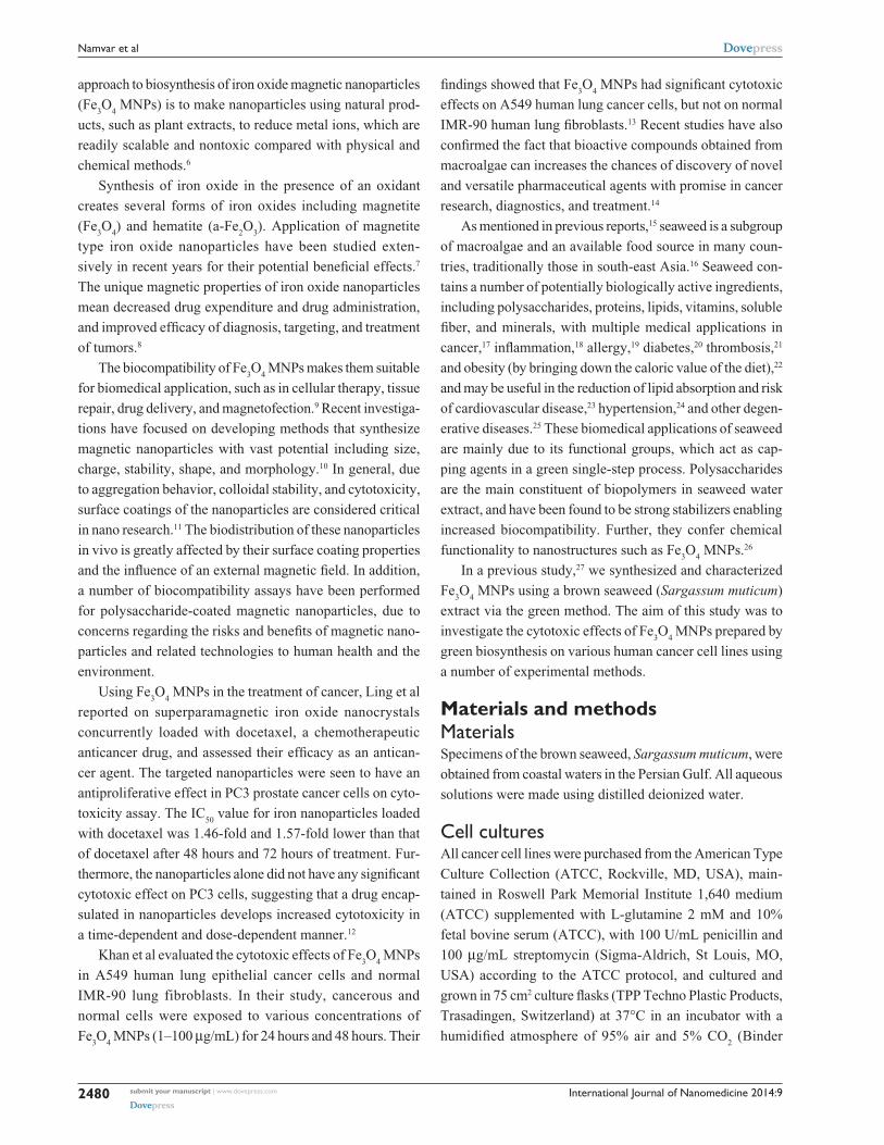
Results and discussionFe3O4 MNPs inhibit proliferation of human cancer cellsObservation of morphological changes in cells indicated
that Fe3O
4 MNPs inhibited proliferation of the various
cancer cell lines in a dose-dependent and time-dependent
manner (Figure 2A). No toxicity was seen in the normal
Chang liver cell line (Figure 2B). The IC50
values calcu-
lated for Fe3O
4 MNPs in the various cancer cell lines were
23.83±1.1 µg/mL (HepG2), 18.75±2.1 µg/mL (MCF-7),
12.5±1.7 µg/mL (HeLa), and 6.4±2.3 µg/mL (Jurkat) after
treatment for 72 hours. Therefore, Jurkat cells were selected
for further investigation.
It is relevant to the evaluation toxicity of metal nano-
particles against cancer cells as many studies have already
published. Coradeghini et al reported that BALB/C-3T3
mouse fibroblasts at higher doses than 50 µM for 72 hours of
exposure to gold nanoparticles with a thickness of 5 nm, as
compared with 15 nm, induced dose-dependent cell cytotoxi-
city. They reported that the size of gold nanoparticles is an
important indicator of potential application in the biomedical
field.30 Another recent study reported that different types
of iron oxide nanoparticles demonstrate diverse biological
activity as a result of versatile surface coating. Novotna et al
Figure 2 (A) The cytotoxic effect of Fe3O4 MNPs on various cancer cell lines after 72 hours of treatment evaluated by mitochondrial activity using the MTT assay. each point represents the mean value of three replicates. (B) The cytotoxic effect of Fe3O4 MNPs on a normal chang human liver cell line after 72 hours of treatment evaluated with MTT assay. each point represents the mean value of three replicates. Abbreviations: Fe3O4 MNPs, magnetic iron oxide nanoparticles; MTT, 3-(4,5-dimethylthiazol-2-yl)-2,5 diphenyl tetrazolium bromide.
Cel
l via
bilit
y (%
)
Fe3O4 MNP concentrations (μg/mL)
HepG2MCF-7HeLaJurkat
A
Cel
l via
bilit
y (%
)
Fe3O4 MNP concentration (μg/mL)
B
International Journal of Nanomedicine 2014:9 submit your manuscript | www.dovepress.com
Dovepress
Dovepress
2483
Fe3O4 MNPs synthesized via seaweed aqueous extract
examined the effect of superparamagnetic iron oxide
nanoparticles on human bone marrow mesenchymal
stromal cells from two donors. Their results showed only
hBMSCs-2 were sensitive to these nanoparticles; however,
increased oxidative injury to lipids, proteins, and DNA was
detected in cells from both donors.31
Schweigera et al reported on Fe3O
4 MNPs stabilized with
poly(ethyleneimine)-g-poly(ethylene glycol) and branched
poly(ethyleneimine). They also evaluated the cytotoxic
effects of polymer-coated iron oxide nanoparticles in
A549 epithelial adenocarcinoma cells. Their findings for cell
viability showed that poly(ethyleneimine) layered onto iron
oxide nanoparticles produced significant cytoxicity compared
to free poly(ethyleneimine) polymer.11
Quantification of apoptosis using acridine orange/propidium iodide double-stainingInduction of apoptosis is a useful strategy in the treatment of
cancer. Several cellular and molecularbiological features can
be demonstrated in apoptotic cells, including cell shrinkage,
DNA fragmentation, and activation of the caspase cascade.
The cytotoxicity of Fe3O
4 MNPs was evaluated by inhibi-
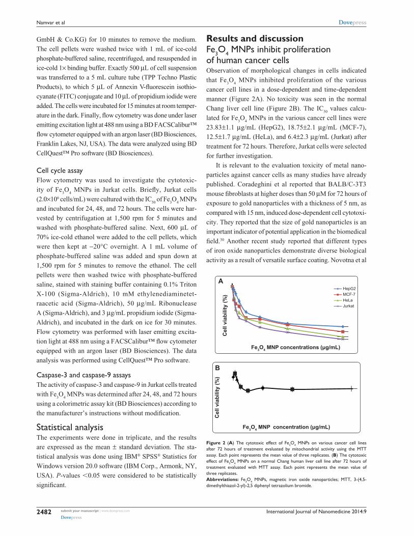
tion of cell growth. When the growth-inhibited cells were
stained with acridine orange/propidium iodide, apoptotic cell
death was observed in a time-dependent and dose-dependent
manner in all cultures (Figure 3).
Intact membranes exclude propidium iodide, but allow
uptake of acridine orange, which binds to double-stranded
DNA and fluoresces green under 488 nm excitation.
Untreated cells show diffuse green fluorescence, while
apoptotic cells (containing condensed chromatin material)
show clumps of intense green fluorescent spots within
the cell. Characteristic condensation patterns were observed
such as crescent shapes found at the nuclear periphery and
an increase in the number of round clumps.
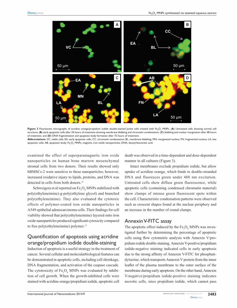
annexin V-FITc assayThe apoptotic effect induced by the Fe
3O
4 MNPs was inves-
tigated further by determining the percentage of apoptotic
cells using flow cytometric analysis with Annexin V/pro-
pidium iodide double-staining. Annexin V-positive/propidium
iodide-negative staining indicated cells in early apoptosis
due to the strong affinity of Annexin V-FITC for phosphati-
dylserine, which transports Annexin V protein from the inner
leaflet of the plasma membrane to the outer surface of the
membrane during early apoptosis. On the other hand, Annexin
V-negative/propidium iodide-positive staining indicates
necrotic cells, since propidium iodide, which cannot pass
Figure 3 Fluorescent micrographs of acridine orange/propidium iodide double-stained Jurkat cells treated with Fe3O4 MNPs. (A) Untreated cells showing normal cell structure, (B) early apoptotic cells after 24 hours of treatment showing membrane blebbing and chromatin condensation, (C) blebbing and nuclear margination after 48 hours of treatment, and (D) DNa fragmentation and apoptotic body formation after 72 hours of treatment. Abbreviations: Vc, viable cells; ea, early apoptotic cells; cc, chromatin condensation; Bl, membrane blebbing; MN, marginated nucleus; FN, fragmented nucleus; la, late apoptotic cells; aB, apoptotic body; Fe3O4 MNPs, magnetic iron oxide nanoparticles; DNa, deoxyribonucleic acid.
A B
VCEA
BL
CC
EA
LA BL FN
AB
LA
MN
50 μm50 μm
50 μm 50 μm
C D
BA
International Journal of Nanomedicine 2014:9submit your manuscript | www.dovepress.com
Dovepress
Dovepress
2484
Namvar et al
through an intact cell membrane, penetrates the compromised
membranes of cells that are dead or in late apoptosis, and
binds to nucleic acids. Meanwhile, Annexin V-negative/
propidium iodide-negative staining indicates viable cells
and Annexin V-positive/propidium iodide-positive staining
indicates cells in late apoptosis. The representative dot plots
for the flow cytometric analysis comparing untreated cells
and treated cells (at 12, 24, and 48 hours) showed an increase
in the percentages of treated cells in early apoptosis and late
Figure 4 Flow cytometric analysis of induction of apoptosis in Jurkat cells by Fe3O4 MNPs after staining with FITc-conjugated annexin V and propidium iodide. (A1–C1) Untreated (control) Jurkat cells after 12, 24, and 48 hours of incubation, respectively. (A2–C2) effects of 12, 24, and 48 hours of treatment with Fe3O4 MNPs, respectively. Abbreviations: Fe3O4 MNPs, magnetic iron oxide nanoparticles; Annexin V-FITC, annexin V-conjugated fluorescein isothiocyanate.
100 101 102 103 104100
101
102
103
104
100 101 102 103 104
100
101
102
103
104
100
101
102
103
104
100
101
102
103
104
A1
B1 B2
C1 C2
A2
Annexin V-FITC Annexin V-FITC
Annexin V-FITCAnnexin V-FITC
Annexin V-FITC Annexin V-FITC
100 101 102 103 104100
101
102
103
104
100 101 102 103 104
100
101
102
103
104
100 101 102 103 104
100 101 102 103 104
Prop
idiu
m io
dide
Prop
idiu
m io
dide
Prop
idiu
m io
dide
Prop
idiu
m io
dide
Prop
idiu
m io
dide
Prop
idiu
m io
dide
apoptosis (Figure 4 and Table 1). In addition, exposure to
Fe3O
4 MNPs resulted in a slight decrease in the amount of
viable cells at 12, 24, and 48 hours. These results suggest that
the antiproliferative effect of Fe3O
4 MNPs on Jurkat cells
results from induction of cell apoptosis.
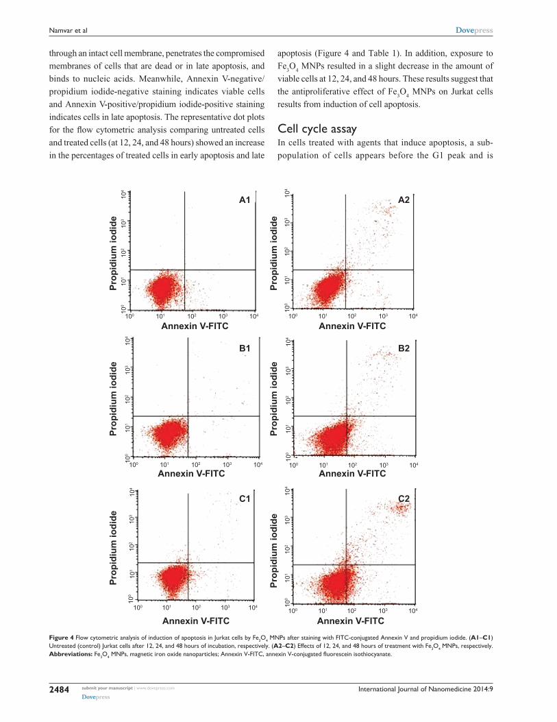
cell cycle assayIn cells treated with agents that induce apoptosis, a sub-
population of cells appears before the G1 peak and is
International Journal of Nanomedicine 2014:9 submit your manuscript | www.dovepress.com
Dovepress
Dovepress
2485
Fe3O4 MNPs synthesized via seaweed aqueous extract
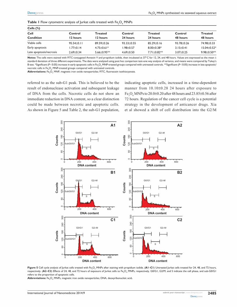
Table 1 Flow cytometric analysis of Jurkat cells treated with Fe3O4 MNPs
Cells (%)
Cell Condition
Control 12 hours
Treated 12 hours
Control 24 hours
Treated 24 hours
Control 48 hours
Treated 48 hours
Viable cells 95.54±0.11 89.59±0.26 93.33±0.55 85.29±0.16 93.78±0.26 74.98±0.33early apoptosis 1.77±0.14 4.75±0.61* 1.98±0.57 8.00±0.38* 3.15±0.41 15.04±0.52*late apoptosis/necrosis 2.69±0.34 5.66±0.95** 4.69±0.50 7.71±0.82** 3.07±0.25 9.98±0.50**
Notes: The cells were stained with FITc-conjugated annexin V and propidium iodide, then incubated at 37°c for 12, 24, and 48 hours. Values are expressed as the mean ± standard deviation of three different experiments. The data were analyzed using post hoc comparison test-one way analysis of variance, and means were compared by Tukey’s B test. *Significant (P0.05) increase in early apoptotic cells in Fe3O4 MNP-treated groups compared with untreated controls. **Significant (P0.05) increase in late apoptotic/necrotic cells in Fe3O4 MNP-treated groups compared with untreated controls. Abbreviations: Fe3O4 MNP, magnetic iron oxide nanoparticles; FITC, fluorescein isothiocyanate.
referred to as the sub-G1 peak. This is believed to be the
result of endonuclease activation and subsequent leakage
of DNA from the cells. Necrotic cells do not show an
immediate reduction in DNA content, so a clear distinction
could be made between necrotic and apoptotic cells.
As shown in Figure 5 and Table 2, the sub-G1 population,
indicating apoptotic cells, increased in a time-dependent
manner from 10.10±0.28 24 hours after exposure to
Fe3O
4 MNPs to 20.0±0.20 after 48 hours and 23.85±0.56 after
72 hours. Regulation of the cancer cell cycle is a potential
strategy in the development of anticancer drugs. Xia
et al showed a shift of cell distribution into the G2/M
Figure 5 cell cycle analysis of Jurkat cells treated with Fe3O4 MNPs after staining with propidium iodide. (A1–C1) Untreated Jurkat cells treated for 24, 48, and 72 hours, respectively. (A2–C2) effects of 24, 48, and 72 hours of exposure of Jurkat cells to Fe3O4 MNPs, respectively. g0/g1, g2/M, and s indicate the cell phase, and sub-g0/g1 refers to the proportion of apoptotic cells. Abbreviations: Fe3O4 MNPs, magnetic iron oxide nanoparticles; DNa, deoxyribonucleic acid.
060
120
180
240
300
0 200 400 600
060
120
180
240
300
0 200 400 600
060
120
180
240
300
0 200 400 600
060
120
180
240
300
0 200 400 600
060
120
180
240
300
0 200 400 600
060
120
180
240
300
0 200 400 600
SubG0/G1
SubG0/G1
SubG0/G1 SubG0/G1
SubG0/G1
SubG0/G1
G0/G1
G0/G1
G0/G1 G0/G1
G0/G1
G0/G1G2+M
G2+M
G2+M G2+M
G2+M
G2+M
S
S
S S
S
S
Cou
nts
Cou
nts
Cou
nts
Cou
nts
Cou
nts
Cou
nts
DNA content
DNA content DNA content
DNA content DNA content
DNA content
A1 A2
B1 B2
C1 C2

International Journal of Nanomedicine 2014:9submit your manuscript | www.dovepress.com
Dovepress
Dovepress
2486
Namvar et al
Figure 6 effect of treatment with Fe3O4 MNPs on caspase-3 and caspase-9 activity in Jurkat cells. The values represent the mean ± standard deviation of three independent experiments. Significant differences (P0.05) in caspase-3 and caspase-9 activity were found between the treated and control groups. Abbreviation: Fe3O4 MNPs, magnetic iron oxide nanoparticles.
24 h-
contr
ol
24 h-
treatm
ent
48 h-
contr
ol
48 h-
treatm
ent
72 h-
contr
ol
72 h-
treatm
ent
00.050.1
0.150.2
0.250.3
0.350.4
0.45
Opt
ical
den
sity
(490
nm
)
Caspase-9Caspase-3
Table 2 Flow cytometric analysis of Jurkat cells treated with Fe3O4 MNPs
Cells (%)
Cell cycle phases
Control 24 hours
Treated 24 hours
Control 48 hours
Treated 48 hours
Control 72 hours
Treated 72 hours
g0/g1 56.25±0.06 33.44±0.45 51.13±0.29 42.61±0.52 37.64±0.32 45.68±0.68g2/M 17.85±0.76 18.29±0.41 20.16±0.26 12.29±0.35 24.22±0.22 10.06±0.93synthesis 24.91±0.06 38.23±0.33 29.24±0.06 22.93±0.12 38.95±0.61 21.35±0.18sub g0/g1 0.98±0.23 10.10±0.28* 0.20±0.34 20.0±0.20* 0.02±0.46 23.85±0.56*
Notes: The cells were stained with propidium iodide and incubated at 37°c for 24, 48, and 72 hours. Values are expressed as the mean ± standard deviation of three different experiments. The data were analyzed using post hoc comparison test-one way analysis of variance, and means were compared by Tukey’s B test. *Significant (P0.05) increase of cells in sub-g0/g1 phase in Fe3O4 MNP-treated groups compared with untreated controls. Abbreviation: Fe3O4 MNP, magnetic iron oxide nanoparticles.
phase, which was enhanced by Fe3O
4 MNPs.32 Our data
indicate that the sub-G1 population, ie, apoptotic cells,
increased in a time-dependent manner, whereas other
researchers have reported cells arrested in G1/S and in
both G1/S and G2/M.32 These conflicting results sug-
gest that there may be fundamental differences between
cell types.
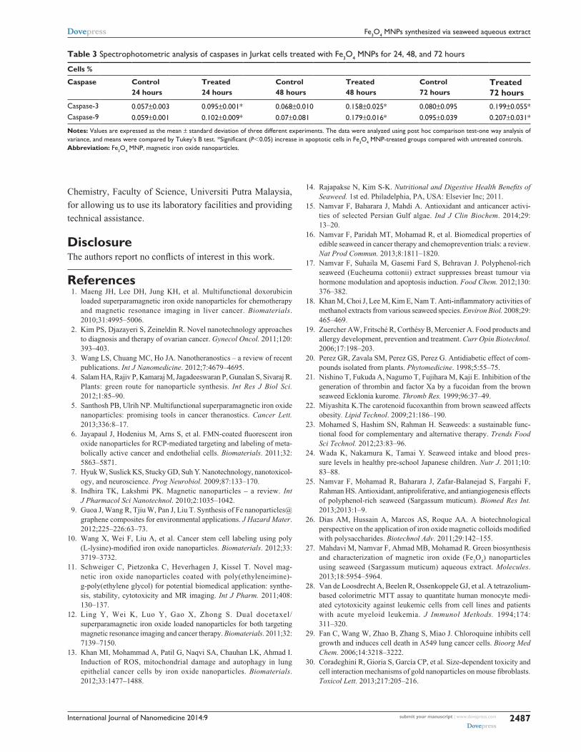
caspase assayThe apoptotic effect of Fe
3O
4 MNPs was also examined by
determining caspase-3 and caspase-9 activity relative to
varying concentrations of protein content for cells treated
with the IC50
of Fe3O
4 MNPs. Our results show that enzyme
activity increased in a time-dependent manner, as shown in
Figure 6 and Table 3.
Apoptosis is orchestrated by a family of cysteine
proteases known as caspases. The main effectors of
apoptosis encompass proteases from the caspase family,
which reside as latent precursors in most nucleated animal
cells. Fourteen mammalian caspases have been identified,
three of which (caspase-3, caspase-6, and caspase-7) are
thought to coordinate the execution phase of apoptosis by
cleaving multiple structural and repair proteins. Pathways to
caspase-3 activation have been identified, which are either
dependent on or independent of release of mitochondrial
cytochrome c and caspase-9 function. Caspase-3 activation
is the hallmark of apoptosis, and is necessary for apoptotic
chromatin condensation and DNA fragmentation in all cell
types examined. Thus, caspase-3 is essential for certain
processes associated with dismantling of the cell and the
formation of apoptotic bodies. However, it may also func-
tion before or at the stage when commitment to loss of
cell viability is made. As shown in Figure 6, Fe3O
4 MNPs
increased the activity of caspase-9 and caspase-3 in a time-
dependent manner. According to studies of caspase activ-
ity, several distinct pathways exist, resulting in induction
of apoptosis by Fe3O
4 MNPs. In our study, we noted that
Fe3O
4 MNPs activated caspase-3 and caspase-9 in a time-
response manner.
Conclusion Fe
3O
4 MNPs with different sizes and characteristics have
been developed and widely investigated in a number of bio-
medical applications. Stabilizing and preventing formation
of aggregation are critical for controlling chemical reac-
tions, which provide dependable, well-dispersed size and
consistency. In this study, we investigated the toxicity of
Fe3O
4 MNPs in human leukemia (Jurkat) cells. Our results
show that exposure of Jurkat cells to Fe3O
4 MNPs results
in significant cytotoxicity, with an apoptotic response, but
not in a normal Chang liver cell line, providing new oppor-
tunities for safe delivery of Fe3O
4 MNPs and application in
anticancer therapy.
AcknowledgmentThe authors are grateful to the staff of Universiti Putra Malaysia
for their help with this research, and to the Department of
International Journal of Nanomedicine 2014:9 submit your manuscript | www.dovepress.com
Dovepress
Dovepress
2487
Fe3O4 MNPs synthesized via seaweed aqueous extract
Table 3 spectrophotometric analysis of caspases in Jurkat cells treated with Fe3O4 MNPs for 24, 48, and 72 hours
Cells %
Caspase Control 24 hours
Treated 24 hours
Control 48 hours
Treated 48 hours
Control 72 hours
Treated 72 hours
caspase-3 0.057±0.003 0.095±0.001* 0.068±0.010 0.158±0.025* 0.080±0.095 0.199±0.055*caspase-9 0.059±0.001 0.102±0.009* 0.07±0.081 0.179±0.016* 0.095±0.039 0.207±0.031*
Notes: Values are expressed as the mean ± standard deviation of three different experiments. The data were analyzed using post hoc comparison test-one way analysis of variance, and means were compared by Tukey’s B test. *Significant (P0.05) increase in apoptotic cells in Fe3O4 MNP-treated groups compared with untreated controls. Abbreviation: Fe3O4 MNP, magnetic iron oxide nanoparticles.
14. Rajapakse N, Kim S-K. Nutritional and Digestive Health Benefits of Seaweed. 1st ed. Philadelphia, PA, USA: Elsevier Inc; 2011.
15. Namvar F, Baharara J, Mahdi A. Antioxidant and anticancer activi-ties of selected Persian Gulf algae. Ind J Clin Biochem. 2014;29: 13–20.
16. Namvar F, Paridah MT, Mohamad R, et al. Biomedical properties of edible seaweed in cancer therapy and chemoprevention trials: a review. Nat Prod Commun. 2013;8:1811–1820.
17. Namvar F, Suhaila M, Gasemi Fard S, Behravan J. Polyphenol-rich seaweed (Eucheuma cottonii) extract suppresses breast tumour via hormone modulation and apoptosis induction. Food Chem. 2012;130: 376–382.
18. Khan M, Choi J, Lee M, Kim E, Nam T. Anti-inflammatory activities of methanol extracts from various seaweed species. Environ Biol. 2008;29: 465–469.
19. Zuercher AW, Fritsché R, Corthésy B, Mercenier A. Food products and allergy development, prevention and treatment. Curr Opin Biotechnol. 2006;17:198–203.
20. Perez GR, Zavala SM, Perez GS, Perez G. Antidiabetic effect of com-pounds isolated from plants. Phytomedicine. 1998;5:55–75.
21. Nishino T, Fukuda A, Nagumo T, Fujihara M, Kaji E. Inhibition of the generation of thrombin and factor Xa by a fucoidan from the brown seaweed Ecklonia kurome. Thromb Res. 1999;96:37–49.
22. Miyashita K.The carotenoid fucoxanthin from brown seaweed affects obesity. Lipid Technol. 2009;21:186–190.
23. Mohamed S, Hashim SN, Rahman H. Seaweeds: a sustainable func-tional food for complementary and alternative therapy. Trends Food Sci Technol. 2012;23:83–96.
24. Wada K, Nakamura K, Tamai Y. Seaweed intake and blood pres-sure levels in healthy pre-school Japanese children. Nutr J. 2011;10: 83–88.
25. Namvar F, Mohamad R, Baharara J, Zafar-Balanejad S, Fargahi F, Rahman HS. Antioxidant, antiproliferative, and antiangiogenesis effects of polyphenol-rich seaweed (Sargassum muticum). Biomed Res Int. 2013;2013:1–9.
26. Dias AM, Hussain A, Marcos AS, Roque AA. A biotechnological perspective on the application of iron oxide magnetic colloids modified with polysaccharides. Biotechnol Adv. 2011;29:142–155.
27. Mahdavi M, Namvar F, Ahmad MB, Mohamad R. Green biosynthesis and characterization of magnetic iron oxide (Fe
3O
4) nanoparticles
using seaweed (Sargassum muticum) aqueous extract. Molecules. 2013;18:5954–5964.
28. Van de Loosdrecht A, Beelen R, Ossenkoppele GJ, et al. A tetrazolium- based colorimetric MTT assay to quantitate human monocyte medi-ated cytotoxicity against leukemic cells from cell lines and patients with acute myeloid leukemia. J Immunol Methods. 1994;174: 311–320.
29. Fan C, Wang W, Zhao B, Zhang S, Miao J. Chloroquine inhibits cell growth and induces cell death in A549 lung cancer cells. Bioorg Med Chem. 2006;14:3218–3222.
30. Coradeghini R, Gioria S, García CP, et al. Size-dependent toxicity and cell interaction mechanisms of gold nanoparticles on mouse fibroblasts. Toxicol Lett. 2013;217:205–216.
Chemistry, Faculty of Science, Universiti Putra Malaysia,
for allowing us to use its laboratory facilities and providing
technical assistance.
DisclosureThe authors report no conflicts of interest in this work.
References 1. Maeng JH, Lee DH, Jung KH, et al. Multifunctional doxorubicin
loaded superparamagnetic iron oxide nanoparticles for chemotherapy and magnetic resonance imaging in liver cancer. Biomaterials. 2010;31:4995–5006.
2. Kim PS, Djazayeri S, Zeineldin R. Novel nanotechnology approaches to diagnosis and therapy of ovarian cancer. Gynecol Oncol. 2011;120: 393–403.
3. Wang LS, Chuang MC, Ho JA. Nanotheranostics – a review of recent publications. Int J Nanomedicine. 2012;7:4679–4695.
4. Salam HA, Rajiv P, Kamaraj M, Jagadeeswaran P, Gunalan S, Sivaraj R. Plants: green route for nanoparticle synthesis. Int Res J Biol Sci. 2012;1:85–90.
5. Santhosh PB, Ulrih NP. Multifunctional superparamagnetic iron oxide nanoparticles: promising tools in cancer theranostics. Cancer Lett. 2013;336:8–17.
6. Jayapaul J, Hodenius M, Arns S, et al. FMN-coated fluorescent iron oxide nanoparticles for RCP-mediated targeting and labeling of meta-bolically active cancer and endothelial cells. Biomaterials. 2011;32: 5863–5871.
7. Hyuk W, Suslick KS, Stucky GD, Suh Y. Nanotechnology, nanotoxicol-ogy, and neuroscience. Prog Neurobiol. 2009;87:133–170.
8. Indhira TK, Lakshmi PK. Magnetic nanoparticles – a review. Int J Pharmacol Sci Nanotechnol. 2010;2:1035–1042.
9. Guoa J, Wang R, Tjiu W, Pan J, Liu T. Synthesis of Fe nanoparticles@graphene composites for environmental applications. J Hazard Mater. 2012;225–226:63–73.
10. Wang X, Wei F, Liu A, et al. Cancer stem cell labeling using poly (L-lysine)-modified iron oxide nanoparticles. Biomaterials. 2012;33: 3719–3732.
11. Schweiger C, Pietzonka C, Heverhagen J, Kissel T. Novel mag-netic iron oxide nanoparticles coated with poly(ethyleneimine)- g-poly(ethylene glycol) for potential biomedical application: synthe-sis, stability, cytotoxicity and MR imaging. Int J Pharm. 2011;408: 130–137.
12. Ling Y, Wei K, Luo Y, Gao X, Zhong S. Dual docetaxel/ superparamagnetic iron oxide loaded nanoparticles for both targeting magnetic resonance imaging and cancer therapy. Biomaterials. 2011;32: 7139–7150.
13. Khan MI, Mohammad A, Patil G, Naqvi SA, Chauhan LK, Ahmad I. Induction of ROS, mitochondrial damage and autophagy in lung epithelial cancer cells by iron oxide nanoparticles. Biomaterials. 2012;33:1477–1488.

International Journal of Nanomedicine 2014:9submit your manuscript | www.dovepress.com
Dovepress
Dovepress
International Journal of Nanomedicine
Publish your work in this journal
Submit your manuscript here: http://www.dovepress.com/international-journal-of-nanomedicine-journal
The International Journal of Nanomedicine is an international, peer-reviewed journal focusing on the application of nanotechnology in diagnostics, therapeutics, and drug delivery systems throughout the biomedical field. This journal is indexed on PubMed Central, MedLine, CAS, SciSearch®, Current Contents®/Clinical Medicine,
Journal Citation Reports/Science Edition, EMBase, Scopus and the Elsevier Bibliographic databases. The manuscript management system is completely online and includes a very quick and fair peer-review system, which is all easy to use. Visit http://www.dovepress.com/testimonials.php to read real quotes from published authors.
Dovepress
2488
Namvar et al
31. Novotna B, Jendelova P, Kapcalova M, et al. Oxidative damage to biological macromolecules in human bone marrow mesenchymal stromal cells labeled with various types of iron oxide nanoparticles. Toxicol Lett. 2012;210:53–63.
32. Xia G, Chen B, Ding J, et al. Effect of magnetic Fe3O
4 nanoparticles with
2-methoxyestradiol on the cell-cycle progression and apoptosis of myel-odysplastic syndrome cells. Int J Nanomedicine. 2011;6:1921–1927.
Copyright © 2022 FDOKUMEN