Characterization and biological activities of synthesized zinc ...
12
Research Article Mohammad Reza Karimzadeh, Sara Soltanian*, Mahboubeh Sheikhbahaei, and Neda Mohamadi Characterization and biological activities of synthesized zinc oxide nanoparticles using the extract of Acantholimon serotinum https://doi.org/10.1515/gps-2020-0058 received June 23, 2020; accepted September 27, 2020 Abstract: The present study reports the synthesis of ZnO- NPs using Acantholimon serotinum extracts followed by characterization and evaluation of biological activities. Field emission scanning electron microscope revealed irregular spherical morphology with a size in the range of 20–80 nm. The X-ray diffraction analysis confirmed the synthesis of highly pure ZnO NPs with a hexagonal shape and a crystalline size of 16.3 nm. The UV-Vis spectroscopy indicates the synthesis of ZnO-NPs. FT-IR confirmed the presence of phytocomponents in the plant extract, which was responsible for nanoparticle synthesis. According to MTT results, the biosynthesized ZnO-NPs showed cytotoxic effects on human colon cancer Caco-2 (IC 50 : 61 μg/mL), neuro- blastoma SH-SY5Y (IC 50 : 42 μg/mL), breast cancer MDA-MB- 231 (IC 50 : 24 μg/mL), and embryonic kidney HEK-293 (IC 50 : 60 μg/mL) cell lines. Significant reactive oxygen species (ROS) generation was measured by the DCFH-DA assay after 24 h incubation with ZnO-NPs (200 μg/mL). ZnO-NPs caused apoptotic and necrotic effects on cells, which was confirmed by Annexin V-PE/7-AAD staining and 6.8-fold increase in pro- apoptosis gene Bax and 178-fold decrease in anti -apoptosis gene Bcl-2. The well diffusion method did not show effective growth inhibition activities of the ZnO-NPs against bacteria. In conclusion, the ZnO-NPs induce cytotoxicity in cell lines through ROS generation and oxidative stress. Keywords: zinc oxide nanoparticles, Acantholimon sero- tinum, cytotoxicity, anti-bacterial activity, oxidative stress Abbreviations DCF 2,7-dichlorofluorescein DCFH-DA 2,7-dichlorofluorescein diacetate FTIR Fourier-transform infrared spectroscopy MTT 3-(4,5-dimethyl-2-thiazolyl)-2,5-diphenyl- 2H-tetrazolium bromide NPs nanoparticles PS phosphatidylserine ROS reactive oxygen species SEM scanning electron microscopy ZnO-NPs zinc oxide nanoparticles 1 Introduction Nanoparticles (NPs) have been introduced in many fields of biology, medicine, and material science and have been harnessed for application in diverse fields such as tissue engineering, drug design, and gene therapy to develop new therapeutic approaches over the last decade [1–3]. Metal NPs research has recently received special attention due to their unusual properties when compared with bulk metal [4]. For example, zinc oxide nanoparticles (ZnO- NPs) have become one of the most popular metal oxide NPs in industrial, pharmaceutical, and biological applica- tions [5]. Physical and chemical methods that are used for the synthesis of metal NPs such as ZnO-NPs are expensive and toxic. Green biosynthesis of ZnO-NPs is the use of reducing and capping agents obtained from plant material that eliminates the use of noxious chemicals with toxic effects [6,7]. Furthermore, many scientists have been at- tracted to use such resources for the biosynthesis of metal NPs due to the ease of production, diversity in size and Mohammad Reza Karimzadeh: Department of Medical Genetics, School of Medicine, Bam University of Medical Sciences, Bam, Iran * Corresponding author: Sara Soltanian, Department of Biology, Faculty of Science, Shahid Bahonar University of Kerman, Kerman, Iran, e-mail: [email protected], [email protected], tel: +98-34-31-32-2068; fax: +98-34-33-25-7432 Mahboubeh Sheikhbahaei: Department of Biology, Faculty of Science, Shahid Bahonar University of Kerman, Kerman, Iran Neda Mohamadi: Herbal and Traditional Medicines Research Center, Department of Pharmacogenosy, Kerman University of Medical Sciences, Kerman, Iran Green Processing and Synthesis 2020; 9: 722–733 Open Access. © 2020 Mohammad Reza Karimzadeh et al., published by De Gruyter. This work is licensed under the Creative Commons Attribution 4.0 International License.
-
Upload
khangminh22 -
Category
Documents
-
view
1 -
download
0
Transcript of Characterization and biological activities of synthesized zinc ...
Research Article
Mohammad Reza Karimzadeh, Sara Soltanian*, Mahboubeh Sheikhbahaei, andNeda Mohamadi
Characterization and biological activities ofsynthesized zinc oxide nanoparticles using theextract of Acantholimon serotinum
https://doi.org/10.1515/gps-2020-0058received June 23, 2020; accepted September 27, 2020
Abstract: The present study reports the synthesis of ZnO-NPs using Acantholimon serotinum extracts followed bycharacterization and evaluation of biological activities.Field emission scanning electron microscope revealedirregular spherical morphology with a size in the rangeof 20–80 nm. The X-ray diffraction analysis confirmed thesynthesis of highly pure ZnO NPs with a hexagonal shapeand a crystalline size of 16.3 nm. The UV-Vis spectroscopyindicates the synthesis of ZnO-NPs. FT-IR confirmed thepresence of phytocomponents in the plant extract, whichwas responsible for nanoparticle synthesis. According toMTT results, the biosynthesized ZnO-NPs showed cytotoxiceffectsonhumancoloncancerCaco-2(IC50:61 µg/mL),neuro-blastoma SH-SY5Y (IC50: 42 µg/mL), breast cancerMDA-MB-231 (IC50: 24 µg/mL), and embryonic kidney HEK-293 (IC50:60 µg/mL) cell lines. Significant reactive oxygen species(ROS) generationwasmeasured by the DCFH-DA assay after24 h incubation with ZnO-NPs (200 µg/mL). ZnO-NPs causedapoptotic and necrotic effects on cells, which was confirmedbyAnnexinV-PE/7-AADstainingand6.8-fold increase inpro-apoptosis gene Bax and 178-fold decrease in anti-apoptosisgene Bcl-2. The well diffusion method did not show effectivegrowth inhibition activities of the ZnO-NPs against bacteria.In conclusion, the ZnO-NPs induce cytotoxicity in cell linesthrough ROS generation and oxidative stress.
Keywords: zinc oxide nanoparticles, Acantholimon sero-tinum, cytotoxicity, anti-bacterial activity, oxidative stress
Abbreviations
DCF 2,7-dichlorofluoresceinDCFH-DA 2,7-dichlorofluorescein diacetateFTIR Fourier-transform infrared spectroscopyMTT 3-(4,5-dimethyl-2-thiazolyl)-2,5-diphenyl-
2H-tetrazolium bromideNPs nanoparticlesPS phosphatidylserineROS reactive oxygen speciesSEM scanning electron microscopyZnO-NPs zinc oxide nanoparticles
1 Introduction
Nanoparticles (NPs) have been introduced in many fieldsof biology, medicine, and material science and have beenharnessed for application in diverse fields such as tissueengineering, drug design, and gene therapy to developnew therapeutic approaches over the last decade [1–3].Metal NPs research has recently received special attentiondue to their unusual properties when compared with bulkmetal [4]. For example, zinc oxide nanoparticles (ZnO-NPs) have become one of the most popular metal oxideNPs in industrial, pharmaceutical, and biological applica-tions [5]. Physical and chemical methods that are used forthe synthesis of metal NPs such as ZnO-NPs are expensiveand toxic. Green biosynthesis of ZnO-NPs is the use ofreducing and capping agents obtained fromplantmaterialthat eliminates the use of noxious chemicals with toxiceffects [6,7]. Furthermore, many scientists have been at-tracted to use such resources for the biosynthesis of metalNPs due to the ease of production, diversity in size and
Mohammad Reza Karimzadeh: Department of Medical Genetics,School of Medicine, Bam University of Medical Sciences, Bam, Iran
* Corresponding author: Sara Soltanian, Department of Biology,Faculty of Science, Shahid Bahonar University of Kerman, Kerman,Iran, e-mail: [email protected], [email protected],tel: +98-34-31-32-2068; fax: +98-34-33-25-7432
Mahboubeh Sheikhbahaei: Department of Biology, Faculty ofScience, Shahid Bahonar University of Kerman, Kerman, IranNeda Mohamadi: Herbal and Traditional Medicines Research Center,Department of Pharmacogenosy, Kerman University of MedicalSciences, Kerman, Iran
Green Processing and Synthesis 2020; 9: 722–733
Open Access. © 2020 Mohammad Reza Karimzadeh et al., published by De Gruyter. This work is licensed under the Creative CommonsAttribution 4.0 International License.

shape, and enhanced biocompatibility of NPs relative toother methods. Until now, green synthesis of ZnO-NPsusing different plant extracts and their potential applica-tions in biology were reported [8,9]
Acantholimon Boiss is a genus in the family Plum-baginaceae composed of approximately 200 species thatmost of them are distributed in Irano-Turanian phytogeo-graphic region [10–13]. It was shown that plants from thePlumbaginaceous family contain secondary metabolitesincluding plumbagin, lignin, saponins, anthocyanin, qui-nines, alkaloids, simple phenolic, tannins, and flavonoidsthat are responsible for their biological effects [14,15].Until now, little attention has been given to the identifica-tion of phytochemicals and biological activities of thegenus Acantholimon [16]. Recently, cytotoxic, antioxi-dant, and antibacterial activities of three species of Acan-tholimon includingA. austro-iranicum, A. serotinum andA.chlorostegium were investigated by Soltanian et al. [17].
In this study, ZnO-NPs were green synthesized usingA. serotinummethanol extract for the first time. SynthesizedZnO-NPs were characterized by various techniques, andcytotoxic activity against several types of cancer and normalcell lines andantibacterial activity against twogram-positive(Enterococcus faecalis and Staphylococcus aureus) and twogram-negative (Pseudomonas aeruginosa and Escherichiacoli) bacteria were evaluated. In addition, this study showedthat theexposureof cells toZnO-NPs leads to reactiveoxygenspecies (ROS) generation, upregulation of Bax and down-regulation of Bcl-2, andfinally apoptosis/necrosis induction.
2 Materials and methods
2.1 Plant extraction procedure
Aerial parts of Acantholimon serotinum Rech.f.& Schiman-Czeika were collected from Baft to Khabr, Kerman Province,Southeast of Iran, identified according to the standard keys,and deposited in MIR herbarium under voucher number1968. Dry powdered plant material was extracted using amaceration technique with methanol 80% and then driedin the oven (Vacucell, Einrichtungen GmbH) at 40°C [17–19].
2.2 Biosynthesis of ZnO-NPs
The amount of 10 mL of plant extracts sample (100 µg/mLin distilled water) was added to 100mL of 0.1 M zincsulfate (ZnSO4) aqueous solution. An aqueous solution
of NaOH was gradually added into the solution underthe steady stirring until the pH of the solution reached to8 to attain smaller size particles [20]. The solution was kepton a magnetic stirrer at 60°C for 6 h. The yellowish-browncolor of the solution indicated the formation of the particles.The solution was centrifuged at 10,000 rpm for 10min, thesupernatant was discarded, and the pellet was washedusing deionized water 4–5 times; afterward, it was washedby ethanol around 3 times to remove organic impurities.After washing, the samples were dried in an oven at 50°C.
2.3 Characterizations of ZnO-NPs
The formation of ZnO-NPs was monitored using a UV-Vis spectrophotometer (PerkinElmer, Germany) at 300–600 nm. Field emission scanning electron microscopy(FESEM) (Quanta 200, USA) was used for examinationof the size, morphology, and distribution of synthesizedsamples. Crystallographic properties of ZnO NPs were ex-plored using the X-ray diffraction (XRD) technique within2θ = 10–90 using the XRD instrument (Rigaku, Ultima IV,Tokyo, Japan) with a Cu LFF λ = 1.540598 A as a radiationsource. The obtained pattern from the XRD was then ana-lyzed using X’Pert High Score Plus software, and the che-mical composition, crystalline structure, and size of theNP were identified. The size of the particles was calcu-lated using the Debye–Scherrer equation:
= /D λ β θ0.9 cos (1)
where D is the crystalline size, λ is the wavelength ofX-ray used, β is the full line width at the half maximum(FWHM) elevation of the main intensity peak, and θ is theBragg’s angle.
The NPs and plant extract were subjected to Fouriertransform infrared spectra (FT-IR) spectrometric analysisto specify the functional groups in the extract that may beresponsible for reducing ions to NPs.
2.4 Cell culture and cytotoxic activity
To examine the cytotoxic activity of ZnO-NPs, humancolon cancer (Caco-2), neuroblastoma (SH-SY5Y), breastcancer (MDA-MB-231), and embryonic kidney (HEK-293)cell lines were cultured with Dulbecco’s Modified Eagle’sMedium (DMEM) supplemented with 10% fetal bovineserum (FBS), 100 µg/mL penicillin and 100 µg/mL strep-tomycin and incubated at 37°C (5% CO2). The attachedcells were trypsinized for 3–5 min to get the individualcells. The cells were counted and distributed in a 96-well
Cytotoxicity of synthesized ZnO-NPs 723
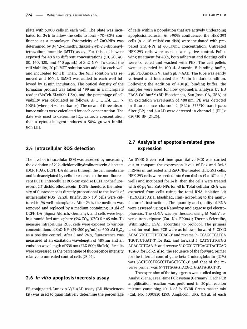
plate with 5,000 cells in each well. The plate was incu-bated for 24 h to allow the cells to form ∼70–80% con-fluence as a monolayer. Cytotoxicity of ZnO-NPs wasdetermined by 3-(4,5-dimethylthiazol-2-yl)-2,5-diphenyl-tetrazolium bromide (MTT) assay. For this, cells wereexposed for 48 h to different concentrations (10, 20, 40,80, 160, 320, and 640 µg/mL) of ZnO-NPs. To detect thecell viability, 20 µL MTT solution was added to each welland incubated for 3 h. Then, the MTT solution was re-moved and 100 µL DMSO was added to each well fol-lowed by 15 min incubation. The optical density of theformazan product was taken at 499 nm in a microplatereader (BioTek-ELx800, USA), and the percentage of cellviability was calculated as follows: Atreatment/Acontrol ×100% (where, A = absorbance). The mean of three absor-bance values were calculated for each concentration. Thedate was used to determine IC50 value, a concentrationthat a cytotoxic agent induces a 50% growth inhibi-tion [21].
2.5 Intracellular ROS detection
The level of intracellular ROS was assessed by measuringthe oxidation of 2′,7′-dichlorodihydrofluorescein diacetate(DCFH-DA). DCFH-DA diffuses through the cell membraneand is deacetylated by cellular esterase to the non-fluores-centDCFH. Intracellular ROS can oxidizeDCFH to thefluor-escent 2,7-dichlorofluorescein (DCF); therefore, the inten-sity of fluorescence is directly proportional to the levels ofintracellular ROS [22,23]. Briefly, 25 × 103 cells were cul-tured in 96-well microplates. After 24 h, the medium wasremoved and replaced by a medium containing 10 µM ofDCFH-DA (Sigma-Aldrich, Germany), and cells were keptin a humidified atmosphere (5% CO2, 37°C) for 45min. Tomeasure intracellular ROS, cells were exposed to variousconcentrations of ZnO-NPs (25–200 µg/mL) or 600 µMH2O2
as a positive control. After 3 and 24 h, fluorescence wasmeasured at an excitation wavelength of 485 nm and anemission wavelength of 538 nm (FLX 800; BioTek). Resultswere expressed as the percentage of fluorescence intensityrelative to untreated control cells [23,24].
2.6 In vitro apoptosis/necrosis assay
PE-conjugated-Annexin V/7-AAD assay (BD Bioscienceskit) was used to quantitatively determine the percentage
of cells within a population that are actively undergoingapoptosis/necrosis. At >90% confluence, the HEK-293cells (4 × 10⁵ cells/6 cm dish) were incubated with pre-pared ZnO-NPs at 60 µg/mL concentration. UntreatedHEK-293 cells were used as a negative control. Follo-wing treatment for 48 h, both adherent and floating cellswere collected and washed with PBS. The cell pelletswere suspended in 100 µL Annexin V binding buffer,5 µL PE-Annexin V, and 5 µL 7-AAD. The tube was gentlyvortexed and incubated for 15 min in dark condition.Following the addition of 400 µL binding buffer, thesamples were used for flow cytometric analysis by BDFACS Calibur™ (BD Biosciences, San Jose, CA, USA) atan excitation wavelength of 488 nm. PE was detectedin fluorescence channel 2 (FL2): 575/30 band passfilter (BP) and 7-AAD were detected in channel 3 (FL3):620/30 BP [25,26].
2.7 Analysis of apoptosis-related geneexpression
An SYBR Green real-time quantitative PCR was carriedout to compare the expression levels of Bax and Bcl-2mRNAs in untreated and ZnO-NPs-treated HEK-293 cells.HEK-293 cells were seeded into 6 cm dishes (5 × 10⁴ cells/well) and incubated for 24 h, then the cells were treatedwith 60 µg/mL ZnO-NPs for 48 h. Total cellular RNA wasextracted from cells using the total RNA isolation kit(DENAzist Asia, Mashhad, Iran) according to the manu-facturer’s instructions. The quantity and quality of RNAwere assessed using a Nanodrop and agarose gel electro-phoresis. The cDNA was synthesized using M-MuLV re-verse transcriptase (Cat. No. EP0441; Thermo Scientific,Wilmington, USA), according to protocol. The primersused for real-time PCR were as follows: forward 5′-CCCGAGAGGTCTTTTTCCGAG-3′ and reverse 5′- CCAGCCCATGATGGTTCTGAT-3′ for Bax, and forward 5′-CATGTGTGTGGAGAGCGTCAA-3′ and reverse 5′-GCCGGTTCAGGTACTCAGTCA-3′ for Bcl-2. Also, the sequence of the forward primerfor the internal control gene beta-2-microglobulin (β2M)was 5′-CTCCGTGGCCTTAGCTGTG-3′ and that of the re-verse primer was 5′-TTTGGAGTACGCTGGATAGCCT-3′.
The expressionof the target geneswas studiedusinganAnalytik Jena, a real-timePCR system (Germany). EachPCRamplification reaction was performed in 20 µL reactionmixture containing 10 µL of 2× SYBR Green master mix(Cat. No. 5000850-1250; Amplicon, UK), 0.5 µL of each
724 Mohammad Reza Karimzadeh et al.
primer (0.25 µM), 1 µL cDNA (50 ng), and 8 µL double-dis-tilledwater. After denaturation at 95°C for 15min, 40 cycleswere followed by 95°C for 15 s, 60°C for 20 s, and 72°C for10 s in PCR cycling condition. The amplification stage wasfollowed by amelting stage that temperaturewas increasedin steps of 1°C for 10 s from 61°C to 95°C.
A comparative threshold cycle method was used todetermine the relative expression level of the target gene.For this, the mean threshold cycle value of β2M as areference gene was subtracted from the mean thresholdcycle value of the target genes (Bax and Bcl-2) to obtainΔCT and fold change in the target gene of ZnO-NPs-treated cell relative to the untreated control sample wascalculated according to the following equation: Foldchange = 2(−ΔΔCT) where ΔΔCT is calculated by the fol-lowing equation: ΔCTtest sample − ΔCTcontrol sample [27,28].
2.8 Determination of antibacterial property
The antibacterial potential of synthesized ZnO-NPs wastested against Enterococcus faecalis (ATCC 29212), Staphy-lococcus aureus (ATCC 25838), Pseudomonas aeruginosa(ATCC 27853), and Escherichia coli (ATCC 11333) bacteriausing the agar well diffusion method. Briefly, the MullerHinton agar plates were inoculated with 1 mL (108 colony-forming units) of bacterial cultures using spread-plating.After drying the plates, wells of size 6mmhave beenmadeon Muller–Hinton agar plates using gel puncture. 100 µLof various concentrations (500, 1,000, 3,000 µg/mL) ofZnO-NPs was poured in each well. The culture plateswere incubated at 37°C for 24 h. After the growth period,the plates were removed and the antibacterial activitywas measured based on the inhibition zone (millimeters)around wells poured with the nanoparticle. Ciprofloxacin(25 mg/mL) and deionized water were used as a positiveand negative control, respectively. The experiment wasrepeated three times [29,30].
2.9 Statistical analysis
The one-way analysis of variance (ANOVA) was used todetermine whether there are any statistically significantdifferences between the means of control and treatments.The data were shown as mean ± SD and p < 0.05 acceptedas the minimum level of significance.
3 Results
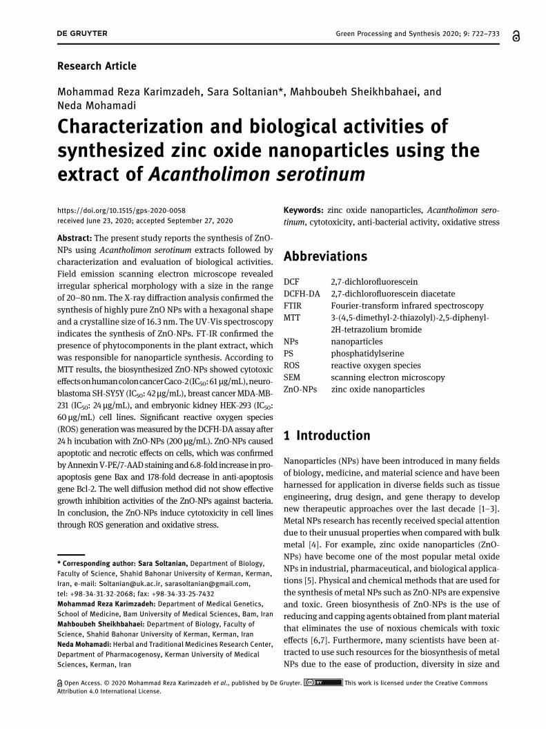
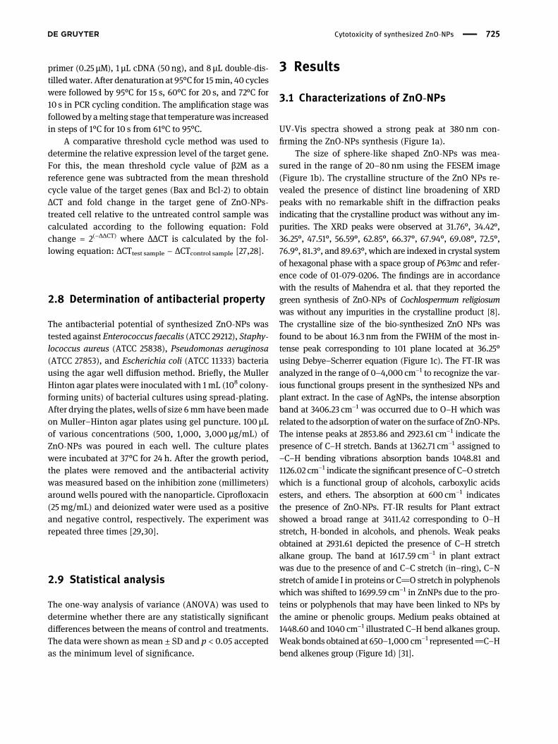
3.1 Characterizations of ZnO-NPs
UV-Vis spectra showed a strong peak at 380 nm con-firming the ZnO-NPs synthesis (Figure 1a).
The size of sphere-like shaped ZnO-NPs was mea-sured in the range of 20–80 nm using the FESEM image(Figure 1b). The crystalline structure of the ZnO NPs re-vealed the presence of distinct line broadening of XRDpeaks with no remarkable shift in the diffraction peaksindicating that the crystalline product was without any im-purities. The XRD peaks were observed at 31.76°, 34.42°,36.25°, 47.51°, 56.59°, 62.85°, 66.37°, 67.94°, 69.08°, 72.5°,76.9°, 81.3°, and 89.63°, which are indexed in crystal systemof hexagonal phase with a space group of P63mc and refer-ence code of 01-079-0206. The findings are in accordancewith the results of Mahendra et al. that they reported thegreen synthesis of ZnO-NPs of Cochlospermum religiosumwas without any impurities in the crystalline product [8].The crystalline size of the bio-synthesized ZnO NPs wasfound to be about 16.3 nm from the FWHM of the most in-tense peak corresponding to 101 plane located at 36.25°using Debye–Scherrer equation (Figure 1c). The FT-IR wasanalyzed in the range of 0–4,000 cm−1 to recognize the var-ious functional groups present in the synthesized NPs andplant extract. In the case of AgNPs, the intense absorptionband at 3406.23 cm−1 was occurred due to O–H which wasrelated to the adsorption ofwater on the surface of ZnO-NPs.The intense peaks at 2853.86 and 2923.61 cm−1 indicate thepresence of C–H stretch. Bands at 1362.71 cm−1 assigned to–C–H bending vibrations absorption bands 1048.81 and1126.02 cm−1 indicate the significant presence of C–O stretchwhich is a functional group of alcohols, carboxylic acidsesters, and ethers. The absorption at 600 cm−1 indicatesthe presence of ZnO-NPs. FT-IR results for Plant extractshowed a broad range at 3411.42 corresponding to O–Hstretch, H-bonded in alcohols, and phenols. Weak peaksobtained at 2931.61 depicted the presence of C–H stretchalkane group. The band at 1617.59 cm−1 in plant extractwas due to the presence of and C–C stretch (in–ring), C–Nstretch of amide I in proteins or C]O stretch in polyphenolswhich was shifted to 1699.59 cm−1 in ZnNPs due to the pro-teins or polyphenols that may have been linked to NPs bythe amine or phenolic groups. Medium peaks obtained at1448.60 and 1040 cm−1 illustrated C–H bend alkanes group.Weakbondsobtainedat 650–1,000 cm−1 represented]C–Hbend alkenes group (Figure 1d) [31].
Cytotoxicity of synthesized ZnO-NPs 725
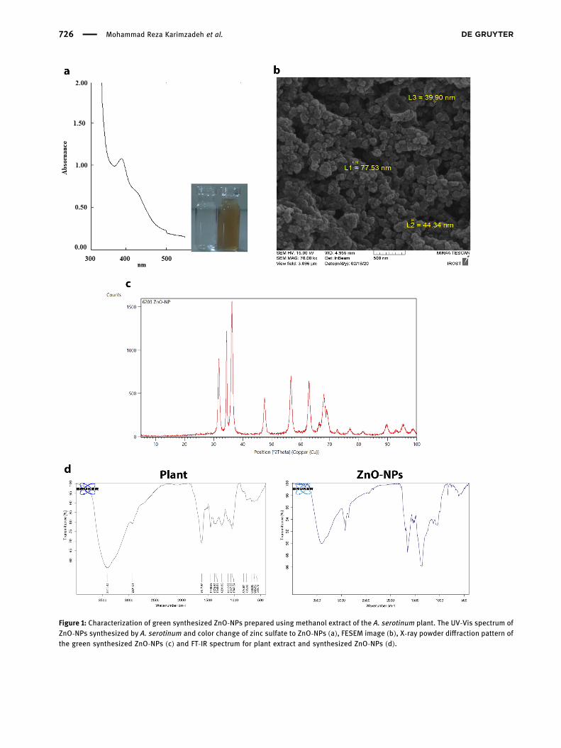
Figure 1: Characterization of green synthesized ZnO-NPs prepared using methanol extract of the A. serotinum plant. The UV-Vis spectrum ofZnO-NPs synthesized by A. serotinum and color change of zinc sulfate to ZnO-NPs (a), FESEM image (b), X-ray powder diffraction pattern ofthe green synthesized ZnO-NPs (c) and FT-IR spectrum for plant extract and synthesized ZnO-NPs (d).
726 Mohammad Reza Karimzadeh et al.
3.2 Determination of cytotoxic effect ofsynthesized ZnO-NPs
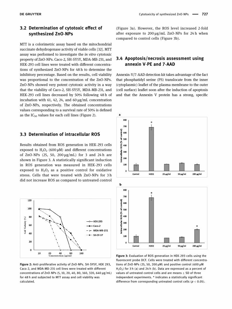
MTT is a colorimetric assay based on the mitochondrialsuccinate dehydrogenase activity of viable cells [32]. MTTassay was performed to investigate the in vitro cytotoxicproperty of ZnO-NPs. Caco-2, SH-SY5Y, MDA-MB-231, andHEK-293 cell lines were treated with different concentra-tions of synthesized ZnO-NPs for 48 h to determine theinhibitory percentage. Based on the results, cell viabilitywas proportional to the concentration of the ZnO-NPs.ZnO-NPs showed very potent cytotoxic activity in a waythat the viability of Caco-2, SH-SY5Y, MDA-MB-231, andHEK-293 cell lines decreased by 50% following 48 h ofincubation with 61, 42, 24, and 60 µg/mL concentrationof ZnO-NPs, respectively. The obtained concentrationsvalues corresponding to a survival rate of 50% is definedas the IC50 values for each cell lines (Figure 2).
3.3 Determination of intracellular ROS
Results obtained from ROS generation in HEK-293 cellsexposed to H2O2 (600 µM) and different concentrationsof ZnO-NPs (25, 50, 200 µg/mL) for 3 and 24 h areshown in Figure 3. A statistically significant inductionin ROS generation was measured in HEK-293 cellsexposed to H2O2 as a positive control for oxidativestress. Cells that were treated with ZnO-NPs for 3 hdid not increase ROS as compared to untreated control
(Figure 3a). However, the ROS level increased 2-foldafter exposure to 200 µg/mL ZnO-NPs for 24 h whencompared to control cells (Figure 3b).
3.4 Apoptosis/necrosis assessment usingannexin V-PE and 7-AAD
Annexin-V/7-AAD detection kit takes advantage of the factthat phosphatidyl serine (PS) translocate from the inner(cytoplasmic) leaflet of the plasma membrane to the outer(cell surface) leaflet soon after the induction of apoptosisand that the Annexin V protein has a strong, specific
Figure 2: Anti-proliferative activity of ZnO-NPs. SH-SY5Y, HEK 293,Caco-2, and MDA-MD-231 cell lines were treated with differentconcentrations of ZnO-NPs (5, 10, 20, 40, 80, 160, 320, 640 µg/mL)for 48 h and subjected to MTT assay and cell viability wascalculated.
Figure 3: Evaluation of ROS generation in HEK-293 cells using thefluorescent probe DCF. Cells were treated with different concentra-tions of ZnO-NPs (25, 50, 200 µM) and positive control (600 µMH2O2) for 3 h (a) and 24 h (b). Data are expressed as a percent ofvalues of untreated control cells and are means ± SD of threeindependent experiments. * indicates a statistically significantdifference from corresponding untreated control cells (p < 0.05).
Cytotoxicity of synthesized ZnO-NPs 727
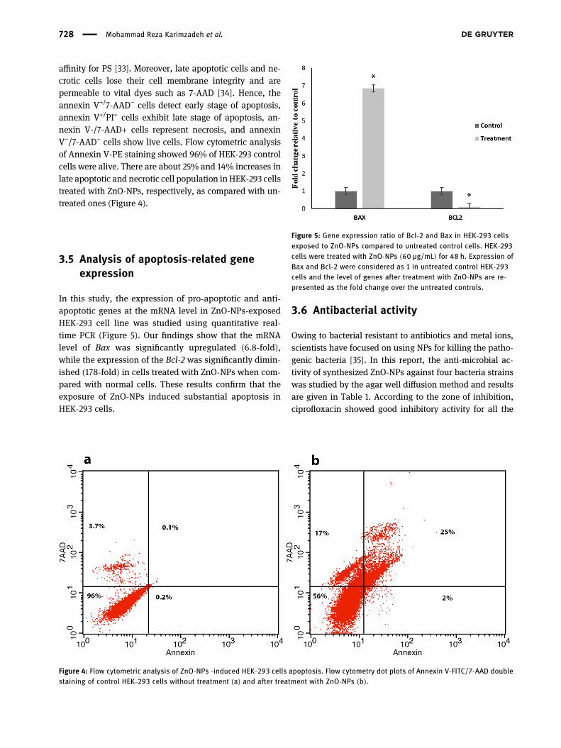
affinity for PS [33]. Moreover, late apoptotic cells and ne-crotic cells lose their cell membrane integrity and arepermeable to vital dyes such as 7-AAD [34]. Hence, theannexin V+/7-AAD− cells detect early stage of apoptosis,annexin V+/PI+ cells exhibit late stage of apoptosis, an-nexin V-/7-AAD+ cells represent necrosis, and annexinV−/7-AAD− cells show live cells. Flow cytometric analysisof Annexin V-PE staining showed 96% of HEK-293 controlcells were alive. There are about 25% and 14% increases inlate apoptotic and necrotic cell population inHEK-293 cellstreated with ZnO-NPs, respectively, as compared with un-treated ones (Figure 4).
3.5 Analysis of apoptosis-related geneexpression
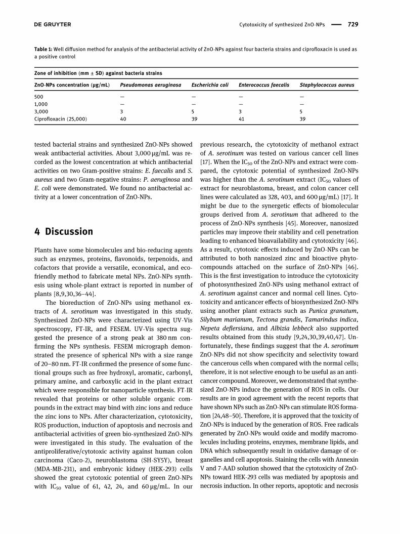
In this study, the expression of pro-apoptotic and anti-apoptotic genes at the mRNA level in ZnO-NPs-exposedHEK-293 cell line was studied using quantitative real-time PCR (Figure 5). Our findings show that the mRNAlevel of Bax was significantly upregulated (6.8-fold),while the expression of the Bcl-2 was significantly dimin-ished (178-fold) in cells treated with ZnO-NPs when com-pared with normal cells. These results confirm that theexposure of ZnO-NPs induced substantial apoptosis inHEK-293 cells.
3.6 Antibacterial activity
Owing to bacterial resistant to antibiotics and metal ions,scientists have focused on using NPs for killing the patho-genic bacteria [35]. In this report, the anti-microbial ac-tivity of synthesized ZnO-NPs against four bacteria strainswas studied by the agar well diffusion method and resultsare given in Table 1. According to the zone of inhibition,ciprofloxacin showed good inhibitory activity for all the
Figure 4: Flow cytometric analysis of ZnO-NPs -induced HEK-293 cells apoptosis. Flow cytometry dot plots of Annexin V-FITC/7-AAD doublestaining of control HEK-293 cells without treatment (a) and after treatment with ZnO-NPs (b).
Figure 5: Gene expression ratio of Bcl-2 and Bax in HEK-293 cellsexposed to ZnO-NPs compared to untreated control cells. HEK-293cells were treated with ZnO-NPs (60 µg/mL) for 48 h. Expression ofBax and Bcl-2 were considered as 1 in untreated control HEK-293cells and the level of genes after treatment with ZnO-NPs are re-presented as the fold change over the untreated controls.
728 Mohammad Reza Karimzadeh et al.
tested bacterial strains and synthesized ZnO-NPs showedweak antibacterial activities. About 3,000 µg/mL was re-corded as the lowest concentration at which antibacterialactivities on two Gram-positive strains: E. faecalis and S.aureus and two Gram-negative strains: P. aeruginosa andE. coli were demonstrated. We found no antibacterial ac-tivity at a lower concentration of ZnO-NPs.
4 Discussion
Plants have some biomolecules and bio-reducing agentssuch as enzymes, proteins, flavonoids, terpenoids, andcofactors that provide a versatile, economical, and eco-friendly method to fabricate metal NPs. ZnO-NPs synth-esis using whole-plant extract is reported in number ofplants [8,9,30,36–44].
The bioreduction of ZnO-NPs using methanol ex-tracts of A. serotinum was investigated in this study.Synthesized ZnO-NPs were characterized using UV-Visspectroscopy, FT-IR, and FESEM. UV-Vis spectra sug-gested the presence of a strong peak at 380 nm con-firming the NPs synthesis. FESEM micrograph demon-strated the presence of spherical NPs with a size rangeof 20–80 nm. FT-IR confirmed the presence of some func-tional groups such as free hydroxyl, aromatic, carbonyl,primary amine, and carboxylic acid in the plant extractwhich were responsible for nanoparticle synthesis. FT-IRrevealed that proteins or other soluble organic com-pounds in the extract may bind with zinc ions and reducethe zinc ions to NPs. After characterization, cytotoxicity,ROS production, induction of apoptosis and necrosis andantibacterial activities of green bio-synthesized ZnO-NPswere investigated in this study. The evaluation of theantiproliferative/cytotoxic activity against human coloncarcinoma (Caco-2), neuroblastoma (SH-SY5Y), breast(MDA-MB-231), and embryonic kidney (HEK-293) cellsshowed the great cytotoxic potential of green ZnO-NPswith IC50 value of 61, 42, 24, and 60 µg/mL. In our
previous research, the cytotoxicity of methanol extractof A. serotinum was tested on various cancer cell lines[17]. When the IC50 of the ZnO-NPs and extract were com-pared, the cytotoxic potential of synthesized ZnO-NPswas higher than the A. serotinum extract (IC50 values ofextract for neuroblastoma, breast, and colon cancer celllines were calculated as 328, 403, and 600 µg/mL) [17]. Itmight be due to the synergetic effects of biomoleculargroups derived from A. serotinum that adhered to theprocess of ZnO-NPs synthesis [45]. Moreover, nanosizedparticles may improve their stability and cell penetrationleading to enhanced bioavailability and cytotoxicity [46].As a result, cytotoxic effects induced by ZnO-NPs can beattributed to both nanosized zinc and bioactive phyto-compounds attached on the surface of ZnO-NPs [46].This is the first investigation to introduce the cytotoxicityof photosynthesized ZnO-NPs using methanol extract ofA. serotinum against cancer and normal cell lines. Cyto-toxicity and anticancer effects of biosynthesized ZnO-NPsusing another plant extracts such as Punica granatum,Silybum marianum, Tectona grandis, Tamarindus indica,Nepeta deflersiana, and Albizia lebbeck also supportedresults obtained from this study [9,24,30,39,40,47]. Un-fortunately, these findings suggest that the A. serotinumZnO-NPs did not show specificity and selectivity towardthe cancerous cells when compared with the normal cells;therefore, it is not selective enough to be useful as an anti-cancer compound.Moreover, we demonstrated that synthe-sized ZnO-NPs induce the generation of ROS in cells. Ourresults are in good agreement with the recent reports thathave shownNPs such as ZnO-NPs can stimulate ROS forma-tion [24,48–50]. Therefore, it is approved that the toxicity ofZnO-NPs is induced by the generation of ROS. Free radicalsgenerated by ZnO-NPs would oxide and modify macromo-lecules including proteins, enzymes, membrane lipids, andDNA which subsequently result in oxidative damage of or-ganelles and cell apoptosis. Staining the cells with AnnexinV and 7-AAD solution showed that the cytotoxicity of ZnO-NPs toward HEK-293 cells was mediated by apoptosis andnecrosis induction. In other reports, apoptotic and necrosis
Table 1:Well diffusion method for analysis of the antibacterial activity of ZnO-NPs against four bacteria strains and ciprofloxacin is used asa positive control
Zone of inhibition (mm ± SD) against bacteria strains
ZnO-NPs concentration (µg/mL) Pseudomonas aeruginosa Escherichia coli Enterococcus faecalis Staphylococcus aureus
500 — — — —1,000 — — — —3,000 3 5 3 5Ciprofloxacin (25,000) 40 39 41 39
Cytotoxicity of synthesized ZnO-NPs 729
induction was also observed after exposure of cells to plant-synthesized ZnO-NPs [48,50–52].
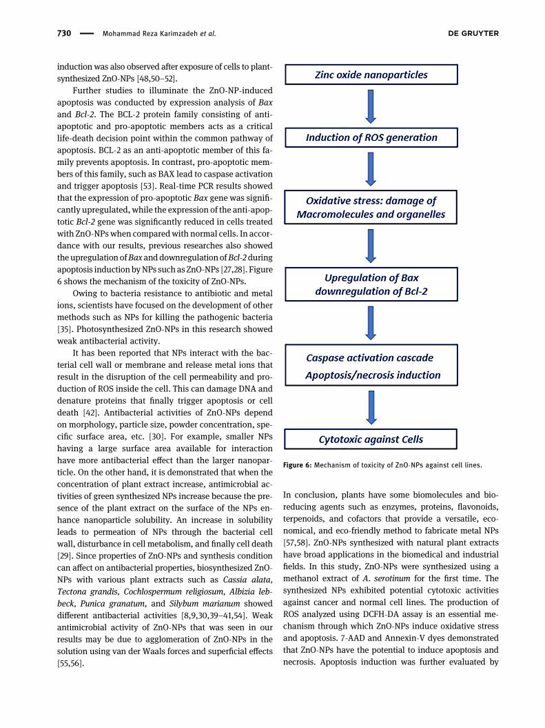
Further studies to illuminate the ZnO-NP-inducedapoptosis was conducted by expression analysis of Baxand Bcl-2. The BCL-2 protein family consisting of anti-apoptotic and pro-apoptotic members acts as a criticallife-death decision point within the common pathway ofapoptosis. BCL-2 as an anti-apoptotic member of this fa-mily prevents apoptosis. In contrast, pro-apoptotic mem-bers of this family, such as BAX lead to caspase activationand trigger apoptosis [53]. Real-time PCR results showedthat the expression of pro-apoptotic Bax gene was signifi-cantly upregulated, while the expression of the anti-apop-totic Bcl-2 gene was significantly reduced in cells treatedwith ZnO-NPswhen comparedwith normal cells. In accor-dance with our results, previous researches also showedtheupregulationofBaxanddownregulationofBcl-2duringapoptosis inductionbyNPs suchasZnO-NPs [27,28]. Figure6 shows the mechanism of the toxicity of ZnO-NPs.
Owing to bacteria resistance to antibiotic and metalions, scientists have focused on the development of othermethods such as NPs for killing the pathogenic bacteria[35]. Photosynthesized ZnO-NPs in this research showedweak antibacterial activity.
It has been reported that NPs interact with the bac-terial cell wall or membrane and release metal ions thatresult in the disruption of the cell permeability and pro-duction of ROS inside the cell. This can damage DNA anddenature proteins that finally trigger apoptosis or celldeath [42]. Antibacterial activities of ZnO-NPs dependon morphology, particle size, powder concentration, spe-cific surface area, etc. [30]. For example, smaller NPshaving a large surface area available for interactionhave more antibacterial effect than the larger nanopar-ticle. On the other hand, it is demonstrated that when theconcentration of plant extract increase, antimicrobial ac-tivities of green synthesized NPs increase because the pre-sence of the plant extract on the surface of the NPs en-hance nanoparticle solubility. An increase in solubilityleads to permeation of NPs through the bacterial cellwall, disturbance in cell metabolism, and finally cell death[29]. Since properties of ZnO-NPs and synthesis conditioncan affect on antibacterial properties, biosynthesized ZnO-NPs with various plant extracts such as Cassia alata,Tectona grandis, Cochlospermum religiosum, Albizia leb-beck, Punica granatum, and Silybum marianum showeddifferent antibacterial activities [8,9,30,39–41,54]. Weakantimicrobial activity of ZnO-NPs that was seen in ourresults may be due to agglomeration of ZnO-NPs in thesolution using van der Waals forces and superficial effects[55,56].
In conclusion, plants have some biomolecules and bio-reducing agents such as enzymes, proteins, flavonoids,terpenoids, and cofactors that provide a versatile, eco-nomical, and eco-friendly method to fabricate metal NPs[57,58]. ZnO-NPs synthesized with natural plant extractshave broad applications in the biomedical and industrialfields. In this study, ZnO-NPs were synthesized using amethanol extract of A. serotinum for the first time. Thesynthesized NPs exhibited potential cytotoxic activitiesagainst cancer and normal cell lines. The production ofROS analyzed using DCFH-DA assay is an essential me-chanism through which ZnO-NPs induce oxidative stressand apoptosis. 7-AAD and Annexin-V dyes demonstratedthat ZnO-NPs have the potential to induce apoptosis andnecrosis. Apoptosis induction was further evaluated by
Figure 6: Mechanism of toxicity of ZnO-NPs against cell lines.
730 Mohammad Reza Karimzadeh et al.
expression analysis of two important members of BCL-2family including Bax and Bcl-2. The antibacterial activityof the ZnO-NPs was examined on four bacterial stains, anditwasdiscovered that ZnO-NPs synthesizedbyA. serotinumhaveweakantibacterial activity.Hence, it is concluded thatthe synthesized ZnO-NPs show cytotoxicity by the genera-tion of ROS, leading to oxidative stress and eventually celldeath.
Acknowledgments: This work was supported by the BamUniversity of Medical Science, Bam, Iran, under Grant[number 98000048]. The authors thank Dr. MansourMirtadzadini at the Department of Biology, Faculty ofscience, Shahid Bahonar University of Kerman for plantcollection and identification.
Conflict of interest: The authors declare no conflict ofinterest.
References
[1] Hackenberg S, Scherzed A, Kessler M, Hummel S, Technau A,Froelich K, et al. Silver nanoparticles: evaluation of DNA da-mage, toxicity and functional impairment in human mesench-ymal stem cells. Toxicol Lett. 2011;201:27–33.
[2] Yokoyama K, Welchons D. The conjugation of amyloid betaprotein on the gold colloidal nanoparticles’ surfaces.Nanotechnology. 2007;18:105101.
[3] Oberdörster G, Oberdörster E, Oberdörster J. Nanotoxicology:an emerging discipline evolving from studies of ultrafine par-ticles. Environ Health Perspect. 2005;113:823–39.
[4] Kumar SV, Rajeshkumar S. Plant-based synthesis of nano-particles and their impact. Nanomaterials in plants, algae,and microorganisms. Concepts Controversies. 2018;3:33–57.
[5] Kumar P, Santhakumar K, Tatsugi J, Shin P-K, Ochiai S.Comparison of properties of polymer organic solar cells pre-pared using highly conductive modified PEDOT: PSS films byspin-and spray-coating methods. Jpn J Appl Phys.2013;53:01AB8.
[6] Mittal AK, Chisti Y, Banerjee UC. Synthesis of metallic nano-particles using plant extracts. Biotechnol Adv.2013;31:346–56.
[7] Abdel-Hameed E-SS. Total phenolic contents and free radicalscavenging activity of certain Egyptian Ficus species leafsamples. Food Chem. 2009;114:1271–7.
[8] Mahendra C, Murali M, Manasa G, Ponnamma P, Abhilash M,Lakshmeesha T, et al. Antibacterial and antimitotic potential ofbio-fabricated zinc oxide nanoparticles of Cochlospermumreligiosum (L.). Microb Pathogenesis. 2017;110:620–9.
[9] Sukri SNAM, Shameli K, Wong MM-T, Teow S-Y, Chew J,Ismail NA. Cytotoxicity and antibacterial activities of plant-
mediated synthesized zinc oxide (ZnO) nanoparticles usingPunica granatum (pomegranate) fruit peels extract. J MolStructure. 2019;1189:57–65.
[10] Bokhari M, Edmondson J. Acantholimon boiss. Flora Turk EastAegean Isl. 1982;7:478–502.
[11] Lashgari AP, Delazar A, Afshar FH, Parsa D. Contact toxicity andchemical composition of essential oil of Acantholimon scor-pius. Pharm Sci. 2016;22:138.
[12] Gazor R, Asgari M, Pasdaran A, Mohammadghasemi F, Nasiri E,Roushan ZA. Evaluation of hepatoprotective effect ofacantholimon gilliati eerial part methanolic extract. Iran JPharm Res: IJPR. 2017;16:135.
[13] Nasiri E, Naserirad S, Pasdaran LA, Gazor R,Mohammadghasemi F, Atrkar RZ. Hepatoprotective effect ofAcantholimon bracteatum (Girard) Boiss. on formaldehyde-induced liver injury in adult male mice. Res J Pharm.2016;3:55–61.
[14] Gunaherath GKB, Gunatilaka AL, Sultanbawa MUS,Balasubramaniam S. 1, 2 (3)-Tetrahydro-3,3′-biplumbagin: Anaphthalenone and other constituents from Plumbago zeyla-nica. Phytochemistry. 1983;22:1245–7.
[15] Pavela R. Efficacy of naphthoquinones as insecticides againstthe house fly, Musca domestica L. Ind Crop Products.2013;43:745–50.
[16] Nabi S, Baloch NU, Bashir S, Rabbani T, Al-Kahraman YM. Invitro antimicrobial, antitumor and cytotoxic activities and theirphytochemical analysis of methanolic extract and its fractionsof acantholimon longiscapum leaves. Int J Phytoph.2013;4:179–83.
[17] Soltanian S, Sheikhbahaei M, Mansour M, Behjat KK.Evaluation of anticancer, antioxidant and antibacterial prop-erties of methanol extract of three Acantholimon Boiss. spe-cies. Avicenna J Phytomed. 2020;10:641–52.
[18] Soltanian S, Sheikhbahaei M, Mohamadi N. Cytotoxicity eva-luation of methanol extracts of some medicinal plants on P19embryonal carcinoma cells. J Appl Pharm Sci. 2017;7:142–9.
[19] Soltanian S, Mohamadi N, Rajaei P, Khodami M,Mohammadi M. Phytochemical composition, and cytotoxic,antioxidant, and antibacterial activity of the essential oil andmethanol extract of Semenovia suffruticosa. Avicenna JPhytomed. 2019;9:143.
[20] Singh N, Haque FZ. Synthesis of zinc oxide nanoparticles withdifferent pH by aqueous solution growth technique. Optik.2016;127:174–7.
[21] Soltanian S, Sheikhbahaei M, Ziasistani M. Phytol down-regulates expression of some cancer stem cell markers anddecreases side population proportion in human embryoniccarcinoma NCCIT cells. Nutr Cancer. 2020;1–14.
[22] Marimoutou M, Le Sage F, Smadja J, d’Hellencourt CL,Gonthier M-P, Robert-Da, et al. Antioxidant polyphenol-richextracts from the medicinal plants Antirhea borbonica,Doratoxylon apetalum and Gouania mauritiana protect 3T3-L1preadipocytes against H2O2, TNFα and LPS inflammatorymediators by regulating the expression of superoxidedismutase and NF-κB genes. J Inflamm. 2015;12:10.
[23] Cui D, Liang T, Sun L, Meng L, Yang C, Wang L, et al. Greensynthesis of selenium nanoparticles with extract of hawthornfruit induced HepG2 cells apoptosis. Pharm Biol.2018;56:528–34.
Cytotoxicity of synthesized ZnO-NPs 731

[24] Al-Sheddi ES, Farshori NN, Al-Oqail MM, Al-Massarani SM,Saquib Q, Wahab R, et al. Anticancer potential of greensynthesized silver nanoparticles using extract of Nepeta de-flersiana against human cervical cancer cells (HeLA). BioinorgChem Appl. 2018;2:1–12.
[25] Soltanian S, Riahirad H, Pabarja A, Karimzadeh MR, Saeidi K.Kaempferol and docetaxel diminish side population and down-regulate some cancer stem cell markers in breast cancer cellline MCF-7. Biocell. 2017;41:33.
[26] Soltanian S, Riahirad H, Pabarja A, Jafari E, Khandani BK. Effectof Cinnamic acid and FOLFOX in diminishing side populationand downregulating cancer stem cell markers in colon cancercell line HT-29. DARU. J Pharm Sci. 2018;26:19–29.
[27] Baharara J, Namvar F, Ramezani T, Mousavi M, Mohamad R.Silver nanoparticles biosynthesized using Achillea bieber-steinii flower extract: apoptosis induction in MCF-7 cells viacaspase activation and regulation of Bax and Bcl-2 gene ex-pression. Molecules. 2015;20:2693–706.
[28] Salehi S, Shandiz SAS, Ghanbar F, Darvish MR, Ardestani MS,Mirzaie A, et al. Phytosynthesis of silver nanoparticles usingArtemisia marschalliana Sprengel aerial part extract and as-sessment of their antioxidant, anticancer, and antibacterialproperties. Int J Nanomed. 2016;11:1835.
[29] Shaik MR, Khan M, Kuniyil M, Al-Warthan A, Alkhathlan HZ,Siddiqui MRH, et al. Plant-extract-assisted green synthesis ofsilver nanoparticles using Origanum vulgare L. extract andtheir microbicidal activities. Sustainability. 2018;10:913.
[30] Senthilkumar N, NandhaKumar E, Priya P, Soni D, Vimalan M.Potheher IV. Synthesis, anti-bacterial, antiarthritic, anti-oxi-dant and in-vitro cytotoxicity activities of ZnO nanoparticlesusing leaf extract of Tectona Grandis (L.). N J Chem.2017;41:10347–56.
[31] Yuvakkumar R, Suresh J, Saravanakumar B, Nathanael AJ,Hong SI, Rajendran V. Rambutan peels promoted biomimeticsynthesis of bioinspired zinc oxide nanochains for biomedicalapplications. Spectrochimica Acta Part A: Mol BiomolSpectrosc. 2015;137:250–8.
[32] Stockert JC, Horobin RW, Colombo LL, Blázquez-Castro A.Tetrazolium salts and formazan products in cell biology: via-bility assessment, fluorescence imaging, and labeling per-spectives. Acta Histochemica. 2018;120:159–67.
[33] Van Engeland M, Nieland LJ, Ramaekers FC, Schutte B,Reutelingsperger CP. Annexin V-affinity assay: a review on anapoptosis detection system based on phosphatidylserine ex-posure. Cytometry: J Int Soc Anal Cytology. 1998;31:1–9.
[34] Zimmermann M, Meyer N. Annexin V/7-AAD staining in kera-tinocytes. Mammalian Cell Viability. 2011;3:57–63.
[35] Puišo J, Jonkuvienė D, Mačionienė I, Šalomskienė J, Jasutienė I,Kondrotas R. Biosynthesis of silver nanoparticles using lin-gonberry and cranberry juices and their antimicrobial activity.Colloids Surf B: Biointerfaces. 2014;121:214–21.
[36] Chouhan HS, Sharma A. Int J Green Herb Chem Int J.2014;3:425–33.
[37] Gnanasangeetha D, Thambavani DS. Biogenic production ofzinc oxide nanoparticles using Acalypha indica. J Chem, BiolPhys Sci (JCBPS). 2013;4:238.
[38] Manokari M, Shekhawat MS. Biogenesis of zinc oxide nano-particles using aqueous extracts of hemidesmusindicus (l.) r.Br Int J Res Stud Microbiol Biotechnol. 2015;1:20–4.
[39] Umar H, Kavaz D, Rizaner N. Biosynthesis of zinc oxide nano-particles using Albizia lebbeck stem bark, and evaluation of itsantimicrobial, antioxidant, and cytotoxic activities on humanbreast cancer cell lines. Int J Nanomed. 2019;14:87.
[40] Abbasi BH, Shah M, Hashmi SS, Nazir M, Naz S, Ahmad W,et al. Green bio-assisted synthesis, characterization and bio-logical evaluation of biocompatible ZnO NPs synthesized fromdifferent tissues of milk thistle (Silybum marianum).Nanomaterials. 2019;9:1171.
[41] Happy A, Soumya M, Kumar SV, Rajeshkumar S, Sheba RD,Lakshmi T, et al. Phyto-assisted synthesis of zinc oxide na-noparticles using Cassia alata and its antibacterial activityagainst Escherichia coli. Biochem Biophys Rep.2019;17:208–11.
[42] Sirelkhatim A, Mahmud S, Seeni A, Kaus NHM, Ann LC,Bakhori SKM, et al. Review on zinc oxide nanoparticles: anti-bacterial activity and toxicity mechanism. Nano-Micro Lett.2015;7:219–42.
[43] Doğan SŞ, Kocabaş A. Green synthesis of ZnO nanoparticleswith Veronica multifida and their antibiofilm activity. Hum &Exp Toxicol. 2019;39:319–27.
[44] Rauf MA, Oves M, Rehman FU, Khan AR, Husain N.Bougainvillea flower extract mediated zinc oxide’s nanoma-terials for antimicrobial and anticancer activity. BiomedPharmacother. 2019;116:108983.
[45] Ghanbar F, Mirzaie A, Ashrafi F, Noorbazargan H, Jalali MD,Salehi S, et al. Antioxidant, antibacterial and anticancerproperties of phyto-synthesised Artemisia quttensis Podlechextract mediated AgNPs. IET Nanobiotechnol. 2016;11:485–92.
[46] Mohanta YK, Panda SK, Jayabalan R, Sharma N, Bastia AK,Mohanta TK. Antimicrobial, antioxidant and cytotoxic activityof silver nanoparticles synthesized by leaf extract of Erythrinasuberosa (Roxb.). Front Mol Biosci. 2017;4:14.
[47] Prashanth G, Prashanth P, Bora U, Gadewar M,Nagabhushana B, Ananda S, et al. In vitro antibacterial andcytotoxicity studies of ZnO nanopowders prepared by com-bustion assisted facile green synthesis. Karbala Int J Mod Sci.2015;1:67–77.
[48] Wang C, Hu X, Gao Y, Ji Y. ZnO nanoparticles treatment inducesapoptosis by increasing intracellular ROS levels in LTEP-a-2cells. BioMed Res Int. 2015;5617:423287.
[49] Rosarin FS, Arulmozhi V, Nagarajan S, Mirunalini S.Antiproliferative effect of silver nanoparticles synthesizedusing amla on Hep2 cell line. Asian Pac J Tropical Med.2013;6:1–10.
[50] Bai D-P, Zhang X-F, Zhang G-L, Huang Y-F, Gurunathan S. Zincoxide nanoparticles induce apoptosis and autophagy inhuman ovarian cancer cells. Int J Nanomed. 2017;12:6521.
[51] Zare E, Pourseyedi S, Khatami M, Darezereshki E. Simplebiosynthesis of zinc oxide nanoparticles using nature’ssource, and it’s in vitro bio-activity. J Mol Structure.2017;1146:96–103.
[52] Saliani M, Jalal R, Goharshadi EK. Mechanism of oxidativestress involved in the toxicity of ZnO nanoparticles againsteukaryotic cells. Nanomed J. 2016;3:1–14.
[53] Tsujimoto Y. Role of Bcl-2 family proteins in apoptosis:apoptosomes or mitochondria? Genes Cell. 1998;3:697–707.
732 Mohammad Reza Karimzadeh et al.

[54] Ahmadi Shadmehri A, Namvar F, Miri H, Yaghmaei P, NakhaeiMoghaddam M. Assessment of antioxidant and antibacterialactivities of zinc oxide nanoparticles, graphene and graphenedecorated by zinc oxide nanoparticles. Int J Nano Dimens.2019;10:350–8.
[55] Yuan Y-G, Wang Y-H, Xing H-H, Gurunathan S. Quercetin-mediated synthesis of graphene oxide–silver nanoparticlenanocomposites: a suitable alternative nanotherapy for neuro-blastoma. Int J Nanomed. 2017;12:5819.
[56] Ogunyemi SO, Abdallah Y, Zhang M, Fouad H, Hong X,Ibrahim E, et al. Green synthesis of zinc oxide nanoparticles
using different plant extracts and their antibacterial activityagainst Xanthomonas oryzae pv. oryzae. Artif Cells, Nanomed,Biotechnol. 2019;47:341–52.
[57] Ovais M, Khalil AT, Islam NU, Ahmad I, Ayaz M, Saravanan M,et al. Role of plant phytochemicals and microbial enzymes inbiosynthesis of metallic nanoparticles. Appl MicrobiolBiotechnol. 2018;102:6799–814.
[58] Ovais M, Khalil AT, Ayaz M, Ahmad I, Nethi SK, Mukherjee S.Biosynthesis of metal nanoparticles via microbialenzymes: a mechanistic approach. Int J Mol Sci. 2018;19:4100.
Cytotoxicity of synthesized ZnO-NPs 733