Hollow polymeric (PLGA) nano capsules synthesized using ...
16
Materials Research Express PAPER Hollow polymeric (PLGA) nano capsules synthesized using solvent emulsion evaporation method for enhanced drug encapsulation and release efficiency To cite this article: Archana Raichur et al 2014 Mater. Res. Express 1 045407 View the article online for updates and enhancements. Related content Factorial design based preparation, optimization, characterization and in vitro drug release studies of olanzapine loaded PLGA nanoparticles Sarvesh Bohrey, Vibha Chourasiya and Archna Pandey - Nanoparticles with entrapped alpha- tocopherol: synthesis, characterization, and controlledrelease Imola Gabriela Zigoneanu, Carlos Ernesto Astete and Cristina Mirela Sabliov - Optimization of nanoparticles for cardiovascular tissue engineering Mohammad Izadifar, Michael E Kelly, Azita Haddadi et al. - Recent citations Pamela Krug et al - Preparation and properties of a temperature- and pH- responsive polypeptide hydrogel Shichao Zhao et al - A redox-sensitive polymer-drug conjugate for tumor therapy: synthesis, properties and performance Ke Song et al - This content was downloaded from IP address 128.210.126.199 on 12/01/2020 at 05:32
-
Upload
khangminh22 -
Category
Documents
-
view
2 -
download
0
Transcript of Hollow polymeric (PLGA) nano capsules synthesized using ...
Materials Research Express
PAPER
Hollow polymeric (PLGA) nano capsulessynthesized using solvent emulsion evaporationmethod for enhanced drug encapsulation andrelease efficiencyTo cite this article: Archana Raichur et al 2014 Mater. Res. Express 1 045407
View the article online for updates and enhancements.
Related contentFactorial design based preparation,optimization, characterization and in vitrodrug release studies of olanzapine loadedPLGA nanoparticlesSarvesh Bohrey, Vibha Chourasiya andArchna Pandey
-
Nanoparticles with entrapped alpha-tocopherol: synthesis, characterization,and controlledreleaseImola Gabriela Zigoneanu, Carlos ErnestoAstete and Cristina Mirela Sabliov
-
Optimization of nanoparticles forcardiovascular tissue engineeringMohammad Izadifar, Michael E Kelly, AzitaHaddadi et al.
-
Recent citationsPamela Krug et al-
Preparation and properties of atemperature- and pH- responsivepolypeptide hydrogelShichao Zhao et al
-
A redox-sensitive polymer-drug conjugatefor tumor therapy: synthesis, propertiesand performanceKe Song et al
-
This content was downloaded from IP address 128.210.126.199 on 12/01/2020 at 05:32
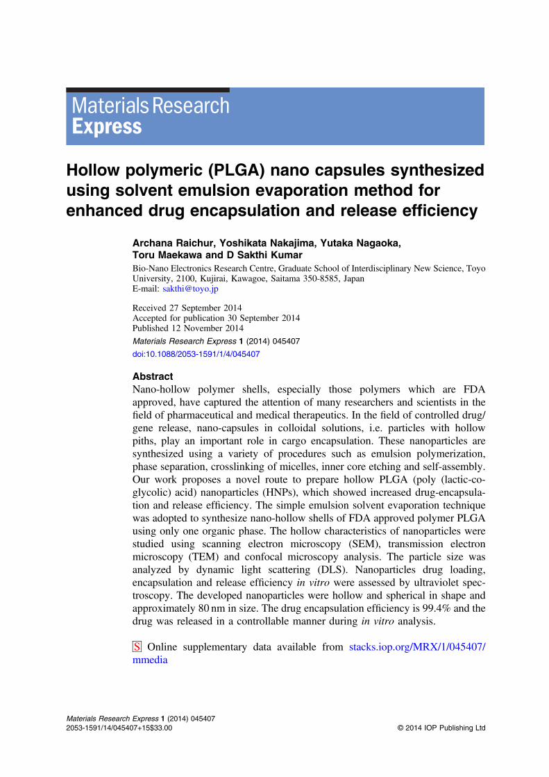
Hollow polymeric (PLGA) nano capsules synthesizedusing solvent emulsion evaporation method forenhanced drug encapsulation and release efficiency
Archana Raichur, Yoshikata Nakajima, Yutaka Nagaoka,Toru Maekawa and D Sakthi KumarBio-Nano Electronics Research Centre, Graduate School of Interdisciplinary New Science, ToyoUniversity, 2100, Kujirai, Kawagoe, Saitama 350-8585, JapanE-mail: [email protected]
Received 27 September 2014Accepted for publication 30 September 2014Published 12 November 2014
Materials Research Express 1 (2014) 045407
doi:10.1088/2053-1591/1/4/045407
AbstractNano-hollow polymer shells, especially those polymers which are FDAapproved, have captured the attention of many researchers and scientists in thefield of pharmaceutical and medical therapeutics. In the field of controlled drug/gene release, nano-capsules in colloidal solutions, i.e. particles with hollowpiths, play an important role in cargo encapsulation. These nanoparticles aresynthesized using a variety of procedures such as emulsion polymerization,phase separation, crosslinking of micelles, inner core etching and self-assembly.Our work proposes a novel route to prepare hollow PLGA (poly (lactic-co-glycolic) acid) nanoparticles (HNPs), which showed increased drug-encapsula-tion and release efficiency. The simple emulsion solvent evaporation techniquewas adopted to synthesize nano-hollow shells of FDA approved polymer PLGAusing only one organic phase. The hollow characteristics of nanoparticles werestudied using scanning electron microscopy (SEM), transmission electronmicroscopy (TEM) and confocal microscopy analysis. The particle size wasanalyzed by dynamic light scattering (DLS). Nanoparticles drug loading,encapsulation and release efficiency in vitro were assessed by ultraviolet spec-troscopy. The developed nanoparticles were hollow and spherical in shape andapproximately 80 nm in size. The drug encapsulation efficiency is 99.4% and thedrug was released in a controllable manner during in vitro analysis.
S Online supplementary data available from stacks.iop.org/MRX/1/045407/mmedia
Materials Research Express 1 (2014) 0454072053-1591/14/045407+15$33.00 © 2014 IOP Publishing Ltd
Keywords: nano hollow PLGA particles, solvent emulsion evaporation method,encapsulation and release efficiency
1. Introduction
Nanotechnology is a field in science which is rapidly flourishing and encouraging theinventions and synthesis of various types of nanomaterials. These nanomaterials have a widerange of applications in the fields of medicine, electronics and pharmaceuticals [1, 2].Nanomaterials used for therapeutic applications face some challenges, as they need to bebiocompatible, biodegradable, cost effective and highly efficient in drug loading. Also, thenanoparticles have to rescue themselves from the enzymatic actions and endosome reactionswithin the cells before they reach the specific cellular organelles for which they were designed[3]. The development in synthesis of polymeric nanoparticles may be one of the successfulattempts carried out in the field of gene, protein and drug delivery. The capacity of thesenanoparticles in holding and encapsulating the cargo still remains an insurmountablebarrier [4, 5].
Until now micro-hollow particles of polymers were synthesized and used for delivery ofdrug in cells [6, 7]. Chiang et al synthesized nanoparticles that are incorporated into polymericmicro-hollow particles for pulsatile drug release [8]. These micro particles can carry the cargo,however, there still remains the question of scalable release of specific medicinal moietiesinside the cytosol [8]. A major limitation of micro-particles is that they do not have the capacityto intrude through the cytosol and reach specific targets. This barricade could be overcome byusage of highly self-modified biocompatible nanoparticles.
Polymeric nano-particles have replaced micro-particles with relatively greater efficiency inencapsulating and delivery of the drug and gene [10]. However, to date evidence for‘endosomal escape’ has come either from cells treated with high concentrations of cargo abovethe therapeutic range or lack of co-localization to the pH-sensitivity [3]. To overcome thislimitation many other nanoparticles such as metal nanoparticles [9], silica nanoparticles [11],liposomes using layer-by-layer technique [12], multi-functionalization technique andencapsulation technique were widely adopted. These nanoparticles do show some limitationdue to their toxicity and release of cargo material instantaneously [8, 13]. Use of hollownanoparticles may be an option for conquering these limitations and disadvantages.
Hollow nanoparticles are said to have great efficiency for gene and drug release ascompared with non-hollow nanoparticles using targeted moiety due to their tailored porousstructured, high cargo loading and encapsulating efficiency, zero order drug release kinetics andmaterial reliability. Even though some hollow polymeric nanoparticles are efficient, not allpolymeric nanomaterials synthesized are biodegradable and biocompatible [14].
PLGA with 50:50 molecular weights is typically used in our experiment for synthesis ofhollow nanoshells. This polymer has successive monomeric units (of glycolic or lactic acid),which are linked together by ester linkages yielding a linear, amorphous aliphatic polyesterproduct, during polymerization [15]. This polymer when subjected to acidic environmentshowed controllable erosion and degradation. Also, PLGA is still widely used in single solventemulsion method giving highest yield of nanoparticles wherein physical parameters such assize, size distribution, morphology, surface modification and zeta potential were controlledsuccessfully [16]. The material used in our experiment was PLGA (poly (lactic-co-glycolic)
2
Mater. Res. Express 1 (2014) 045407 A Raichur et al
acid) which is FDA approved for clinical applications and therapeutic usage. It has excellentquality of biocompatibility and biodegradability too [8].
The various methods used for synthesis of nanoparticles to date include thermal annealingmethod, sol–gel process, crosslinking polymerization [17], inner core etching method [18, 19]and solvent emulsion method [18]. Of the above, simple single solvent emulsion evaporationmethod is undeniably the most cost effective method [20]. Therefore, we used this method forthe synthesis of hollow PLGA nanoparticles.
Kwon et al have reported the synthesis of PLGA nano half shells using water/oil emulsionsolvent method by adding poloxamer in organic phase, which is necessary for production ofnano half shells [21]. There are reports on the synthesis of polystyrene colloidal solution of halfshells nanoparticles [16, 17]. Due to obvious reasons such as incomplete encapsulation of drugand untimely drug release, nano half shells do not provide the support for gene and drugdelivery. The sealing and fusion of the nano half shells still remains a hurdle. Nano shells proveto be superior to nano-half shells in drug and gene delivery etc.
In this paper we report for the first time the synthesis of hollow (PLGA) nanoparticles,which release the drug in a controllable manner and may become a proficient vector to delivergene and drug in cell organelles. The synthesis of hollow PLGA nanoparticles by singleemulsion solvent method gave us an economic method of synthesis with enhanced drugencapsulation and release efficiency.
2. Experimental
2.1. Materials and methods
Poly (lactic-co-glycolic) acid (PLGA; 50:50, Mw 17 000∼ 70 000) and Rhodamine 6G (R6G)were purchased from Sigma Aldrich, USA. Polyvinyl alcohol (PVA) and ethyl acetate werepurchased from Kato Chemicals, Japan. Paclitaxel was purchased from Wako Ltd. Milli-Qwater was used throughout the experiment. The in vitro drug release was carried out at pH 7.4and 4.2 in phosphate-buffered solution (PBS).
2.2. Hollow nanoparticle synthesis (HNPs)
Hollow nanoparticles were prepared using the oil-in-water single solvent emulsion evaporationmethod, wherein the organic phase is ethyl acetate and these nanoparticles are dispersed in thePVA aqueous solution for stabilization. 50mg of PLGA was dissolved in 1ml of ethyl acetateand was rapidly stirred on a magnetic stirrer for complete dissolution in a beaker. This solutionwas vortexed for one hour. Concomitantly 5% (w/v) of 2ml of PVA aqueous solution wasprepared with the help of a magnetic stirrer keeping the stirrer temperature at 40 °C to make thesolution lukewarm and avoid boiling or overheating. The above vortexed PLGA solution wasadded drop-wise in previously prepared 1ml of PVA aqueous solution (the remaining 1ml ofPVA solution was cooled down to room temperature) keeping constant vortexing. After thecomplete addition of PLGA solution in 1ml of lukewarm PVA solution, the cold 1ml of PVAsolution was added instantly. The vortexing was continued for 5min, even after the wholeaddition of the cold PVA solution. After vortexing, this solution was sonicated with energyoutput of 40W for 2min to create a fine emulsion. This mixture was added to 50ml of 0.05%(w/v) PVA solution to form a colloidal solution. The colloidal solution was stirred using a
3
Mater. Res. Express 1 (2014) 045407 A Raichur et al
magnetic stirrer for complete evaporation of the organic component. Developed nanoshellsshowed good stabilization when dispersed in PVA solution and preserved at room temperature.
2.3. Non-hollow nanoparticle preparation (non HNPs)
Non-hollow PLGA nanoparticles were prepared by the same emulsion solvent evaporationmethod using 5ml of ethyl acetate as organic phase and dissolving 200mg of PLGA in it. Thissolution was added to 3% (w/v) of PVA solution and vortexed by keeping the vortexing atmaximum to obtain an emulsified solution. This solution was further dispersed in 0.01% of20ml of PVA solution to increase the stability of the nanoparticles. The whole colloidalsolution was stirred in room temperature for two hours.
The above two colloidal solutions containing HNPs and non-HNPs prepared werecentrifuged at 8000 rpm for 10min to collect the nanoparticles. These collected HNPs and non-HNPs were washed thrice using Milli-Q water; and then dispersed in water and centrifuged at8000 rpm for 10min. Nanoparticles were dried and further used for characterization.
2.4. Characterization of nanoparticles
The shape and morphological characters of polymer nanoparticles were analyzed by scanningelectron microscope (JSPM 7400 F, JEOL). 10mg nanoparticles were re-suspended in 100mlof distilled water and sonicated for 15min. From the above solution 10 μl drop was placed on asilicon wafer and dried at room temperature to get a uniform layer of particles.
The polymer nanoparticles were also investigated by TEM (JEM 2100, JEOL) operating at180 kV. The nanoparticles after washing were re-suspended in an equal amount of Milli-Qwater and 1–2 μl were dropped on the copper grid coated with carbon film. The sample wasdried at room temperature and used for observation. Also, in order to confirm the mean size ofthe PLGA hollow and non-hollow nanoparticle, particle size characterization based on dynamiclight scattering (DLS) was performed using Zeta-sizer.
For confocal microscopy study, freshly prepared hollow nano-particles were immersedinto the R6G aqueous solution having dye concentration 10−5M. Sufficient time was allowed toadsorb the dye molecules onto the polymer nano-particles to make PLGA/R6G nano-complex.100 μl of this solution was pipetted and dried on a confocal plate for examining the HNPs.
2.5. Drug encapsulation and loading
The drug, paclitaxel, was encapsulated in nanoparticles using emulsion solvent evaporationtechnique. For the synthesis of non-HNPs and HNPs we have used 200mg and 50mg of PLGA,respectively. The specific amount of paclitaxel ranging from 10, 20, 30 to 220 μgml−1 keepingstages of 10 μgml−1 in each step were added into ethyl acetate, which was suitably stirred toensure that all materials were dissolved. The synthesis of HNPS and non-HNPs was carried out,following the same above mentioned procedures. The formed oil-in-water (o/w) emulsion wasgently stirred at room temperature with the help of a magnetic stirrer for not less than four hoursto evaporate the organic solvent. The colloidal suspension was sonicated for 3mins and then thenanoparticles were separated at low speed centrifugation (3000 rpm, 5min). The sameprocedure was carried out until the maximum amount of paclitaxel was encapsulated. Thequantity of drug present is calculated according to UV-absorbance of paclitaxel present insupernatant at 227 nm using UV-Visible spectrometer by determining the quantity of drug [5].
4
Mater. Res. Express 1 (2014) 045407 A Raichur et al
The calculation of determining the concentration of drug was carried out according to theprocedure described in [5].
The percentage of drug entrapped was calculated from the amount of incorporated drug inthe nano-shell using the following equation:
=
× + ×
Drug loading efficiency [Amount of paclitaxel in nanoparticles]/
[Amount of paclitaxel loaded nanoparticles]100[Paclitaxel]/[Paclitaxel Polymer] 100 (1)
The drug encapsulation efficiency is calculated according to the following equation:
=
×
Drug encapsulation efficiency (Amount of encapsulation paclitaxel/Amount of paclitaxel used for nanoparticles preparation)
100 (2)
2.6. Drug release from nanoparticles
For conducting drug release study; 150 μgml−1 paclitaxel-encapsulated in hollow nanoparticleswas dispersed in 0.05% PVA solution and to get nearly mono-dispersed nanoparticles; theprepared nanoparticles were filtered through a syringe using 0.8 μm pore size Whatman filterunit GmbH, Germany. The colloidal solution was filtered to obtain nano-shells that are equal toor less than 800 nm in size. In the case of non-HNPs 53 μgml−1 paclitaxel-encapsulatedparticles were used. The above HNPs and non-HNPs were centrifuged at 3000 rpm for 10minsand dried. The total amount of 25mg of both HNPs and non-HNPs were collected anddispersed in 25ml of 7.4 pH PBS buffer solutions each. This solution was further divided in1ml aliquots in vials and placed in an orbital shaker kept at 120 rpmmin−1, maintained at 37 °C.The vials were taken out of the shaker and centrifuged at 8000 rpm for 10min and readingswere observed initially every two hours until the twelfth hour. After the twelfth hour, thereadings were taken after the time interval of twelve hours following the same procedure asabove. The experiment was performed in triplicate. The absorbance value of supernatant wastaken for analysis of paclitaxel concentration using UV-visible spectroscopy at 227 nm [5]. Thedrug release was calculated by using the equation:
=
−×
% Drug release [Amount of paclitaxel in supernatant]/
[Amount of total paclitaxel used for preparation ofdrug encapsulated nanoparticles]
100 (3)
The same procedure as above was carried out to compare the drug release of HNPs indifferent in vitro environments i.e. 7.4 and 4.2 pH. We have compared percentage of drugrelease to time where the observation was taken till 72 hrs.
3. Results and discussion
The unique feature of selecting 50:50 PLGA polymer are (a) approval by the US FDA for drugdelivery and (b) its high efficiency of biodegradability, drug biocompatibility, suitablebiodegradation kinetics, brilliant mechanical properties and ease of processing. Even though the
5
Mater. Res. Express 1 (2014) 045407 A Raichur et al
nanoparticles of PLGA are not hollow they have large surface area. They prove to be havinggreat ability to conjugate with multiple diagnostic and therapeutic agents. Thus, hollownanoparticles proved to be more efficient than the existing non-hollow PLGA nanoparticles indrug releasing and encapsulation activity.
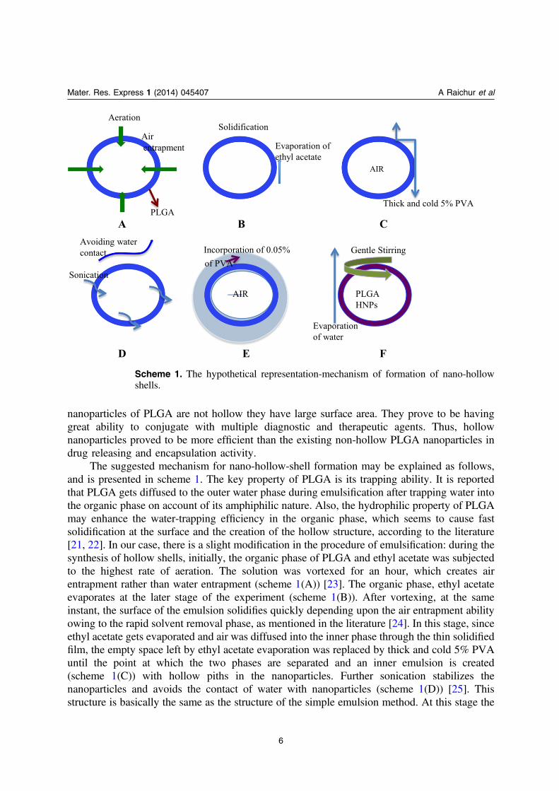
The suggested mechanism for nano-hollow-shell formation may be explained as follows,and is presented in scheme 1. The key property of PLGA is its trapping ability. It is reportedthat PLGA gets diffused to the outer water phase during emulsification after trapping water intothe organic phase on account of its amphiphilic nature. Also, the hydrophilic property of PLGAmay enhance the water-trapping efficiency in the organic phase, which seems to cause fastsolidification at the surface and the creation of the hollow structure, according to the literature[21, 22]. In our case, there is a slight modification in the procedure of emulsification: during thesynthesis of hollow shells, initially, the organic phase of PLGA and ethyl acetate was subjectedto the highest rate of aeration. The solution was vortexed for an hour, which creates airentrapment rather than water entrapment (scheme 1(A)) [23]. The organic phase, ethyl acetateevaporates at the later stage of the experiment (scheme 1(B)). After vortexing, at the sameinstant, the surface of the emulsion solidifies quickly depending upon the air entrapment abilityowing to the rapid solvent removal phase, as mentioned in the literature [24]. In this stage, sinceethyl acetate gets evaporated and air was diffused into the inner phase through the thin solidifiedfilm, the empty space left by ethyl acetate evaporation was replaced by thick and cold 5% PVAuntil the point at which the two phases are separated and an inner emulsion is created(scheme 1(C)) with hollow piths in the nanoparticles. Further sonication stabilizes thenanoparticles and avoids the contact of water with nanoparticles (scheme 1(D)) [25]. Thisstructure is basically the same as the structure of the simple emulsion method. At this stage the
Scheme 1. The hypothetical representation-mechanism of formation of nano-hollowshells.
6
Mater. Res. Express 1 (2014) 045407 A Raichur et al
incorporation of 0.05% of PVA stabilizes the nanoparticles leaving no passage for water to gushin [21] (scheme 1(E)). The sonication creates more stabilization and also addition of 0.05%PVA prevents particles from bursting out. Hence, instead of formation of nano-half-shells,nano-hollow-shells are created. There remains no space for water and it gets evaporated afterthe gentle stirring (scheme 1(F)). As the inner phase solidifies, the remaining ethyl acetate andwater evaporate from outer space. And finally nano-hollow structures are generated. TheseHNPs were characterized further using SEM, TEM and confocal microscopy.
3.1. Scanning electron microscopy (SEM)
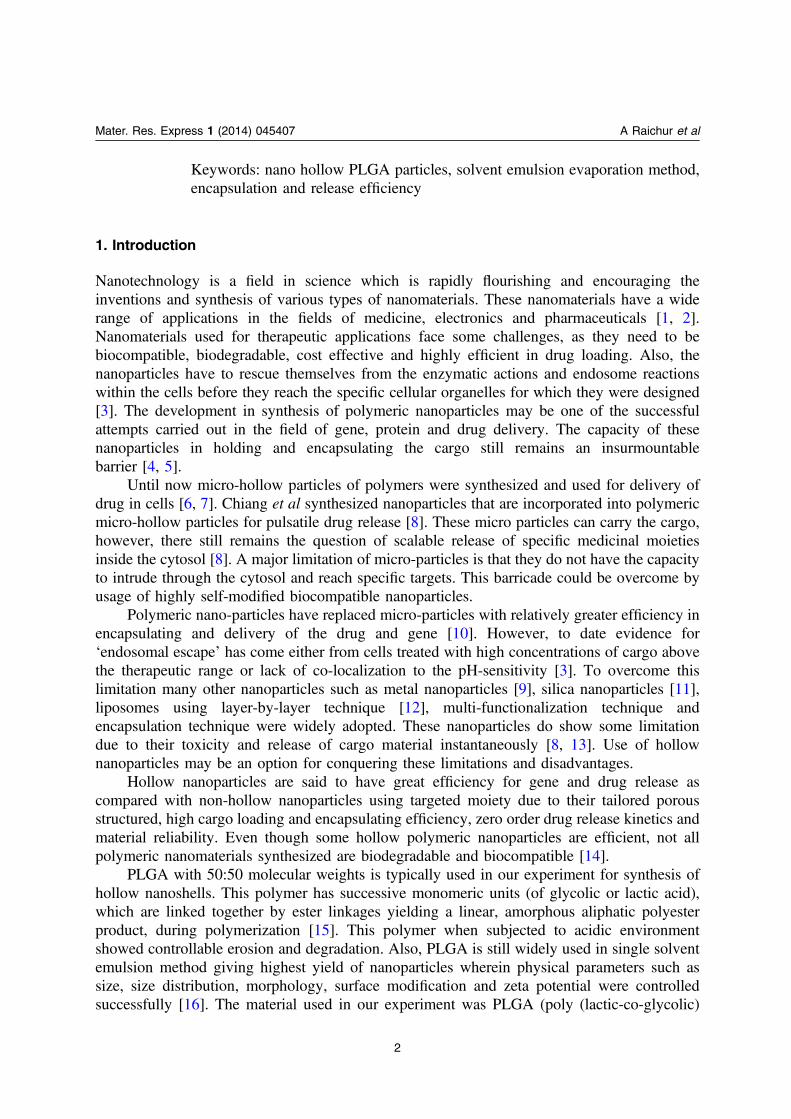
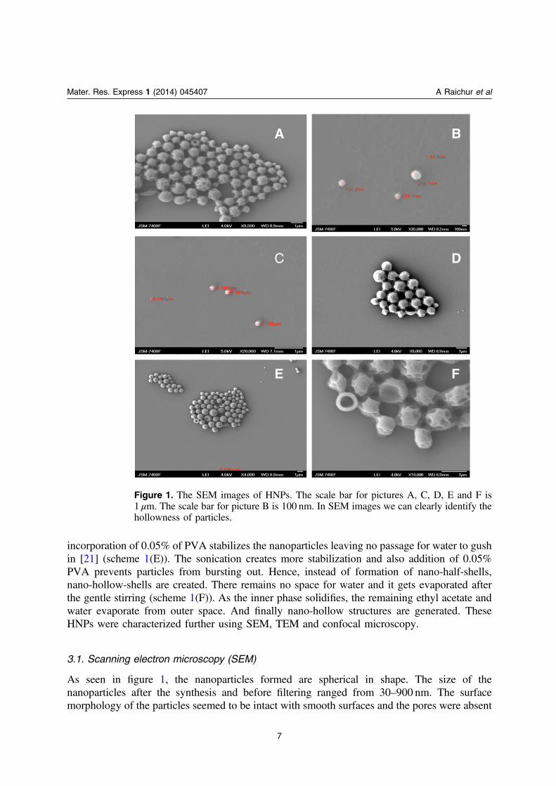
As seen in figure 1, the nanoparticles formed are spherical in shape. The size of thenanoparticles after the synthesis and before filtering ranged from 30–900 nm. The surfacemorphology of the particles seemed to be intact with smooth surfaces and the pores were absent
Figure 1. The SEM images of HNPs. The scale bar for pictures A, C, D, E and F is1 μm. The scale bar for picture B is 100 nm. In SEM images we can clearly identify thehollowness of particles.
7
Mater. Res. Express 1 (2014) 045407 A Raichur et al
on the surface, as seen in figure 1. This showed that even though the nanoparticles were washedfour times and dispersed in water they were not deformed and yet remained intact with less lossof stabilization. In the images in figure 1 we can observe the presence of some hollow structurestoo. This was due to over-sonication carried out in water prior to the preparation of the samplefor SEM. Over-sonication was carried out to crumble the hollow nanoparticles and capture thehollow characteristic of the nanoparticles.
There are adhered nanoparticles seen in figure 1, which may have happened due to thefollowing two reasons, namely, the sample may have very high quantity of redundant emulsifierand secondly the high temperature of electron beam irradiation melts the nano-shells at a greaterrate that makes the nano-hollow-shells get attached and shrink [21, 26]. However, the adhesiondid not have any significant impact on dispersal in PBS solution, the nanoparticles weresuspended stably in buffer solution.
Overall, nanoparticles could be filtered according to interest and used for further medicalapplications. Further, we successfully differentiated the characteristic hollow nature of thePLGA nanoparticles using TEM.
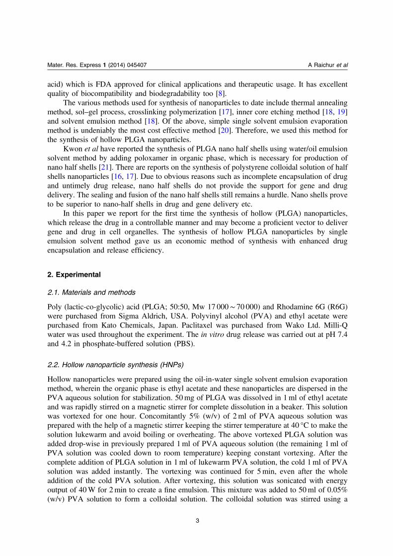
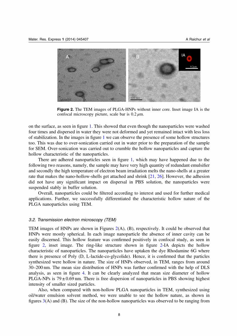
3.2. Transmission electron microscopy (TEM)
TEM images of HNPs are shown in Figures 2(A), (B), respectively. It could be observed thatHNPs were mostly spherical. In each image nanoparticle the absence of inner cavity can beeasily discerned. This hollow feature was confirmed positively in confocal study, as seen infigure 2, inset image. The ring-like structure shown in figure 2-IA depicts the hollowcharacteristic of nanoparticles. The nanoparticles have uptaken the dye Rhodamine 6G wherethere is presence of Poly (D, L-lactide-co-glycolide). Hence, it is confirmed that the particlessynthesized were hollow in nature. The size of HNPs observed, in TEM, ranges from around30–200 nm. The mean size distribution of HNPs was further confirmed with the help of DLSanalysis, as seen in figure 4. It can be clearly analyzed that mean size diameter of hollowPLGA-NPs is 79 ± 0.69 nm. There is free dispersion of nanoparticles in PBS showing highestintensity of smaller sized particles.
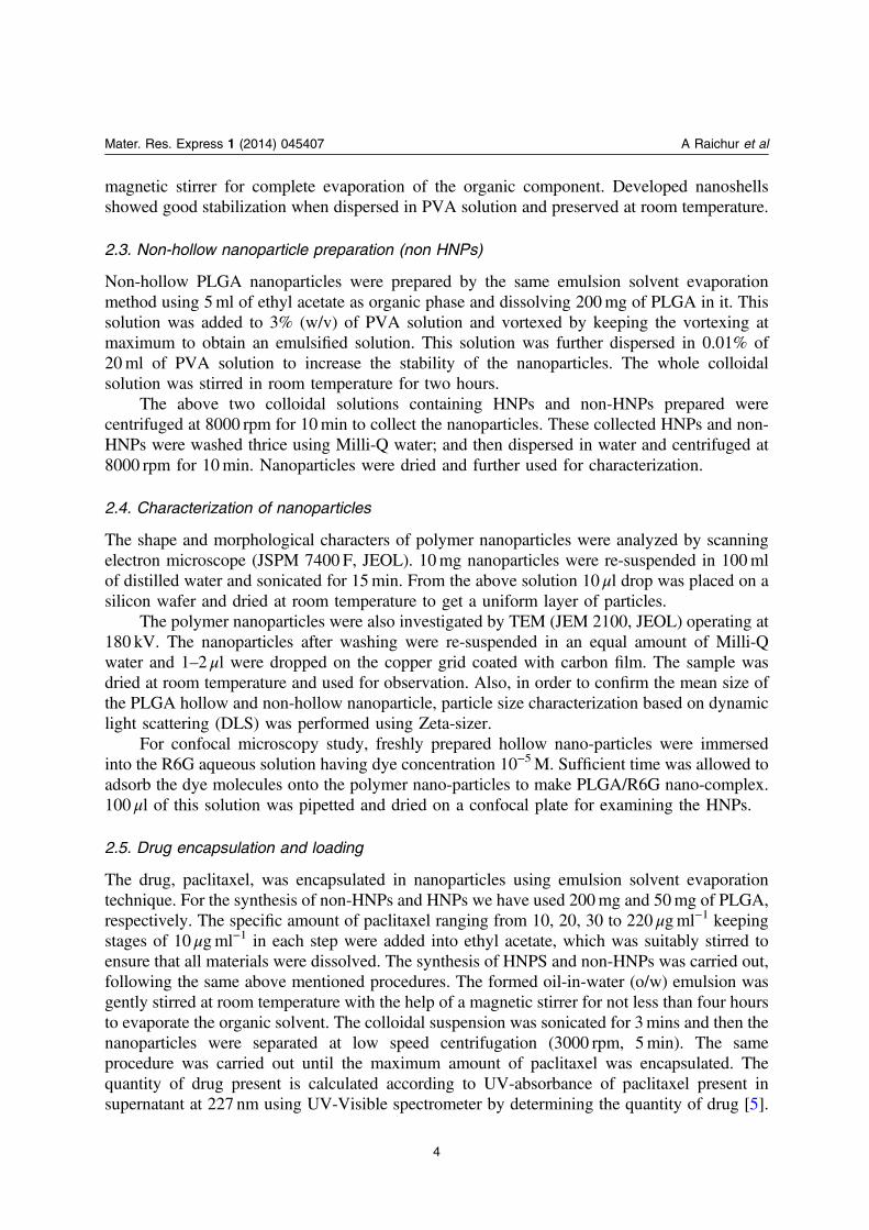
Also, when compared with non-hollow PLGA nanoparticles in TEM, synthesized usingoil/water emulsion solvent method, we were unable to see the hollow nature, as shown infigures 3(A) and (B). The size of the non-hollow nanoparticles was observed to be ranging from
Figure 2. The TEM images of PLGA-HNPs without inner core. Inset image IA is theconfocal microscopy picture, scale bar is 0.2 μm.
8
Mater. Res. Express 1 (2014) 045407 A Raichur et al
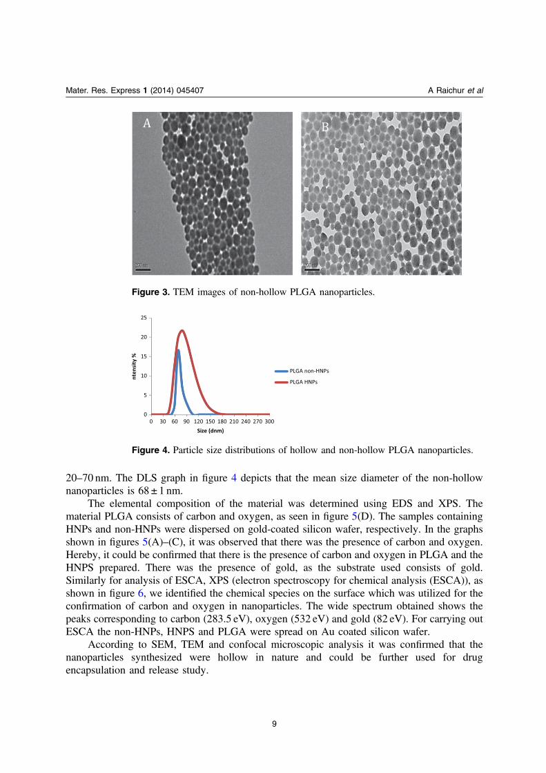
20–70 nm. The DLS graph in figure 4 depicts that the mean size diameter of the non-hollownanoparticles is 68 ± 1 nm.
The elemental composition of the material was determined using EDS and XPS. Thematerial PLGA consists of carbon and oxygen, as seen in figure 5(D). The samples containingHNPs and non-HNPs were dispersed on gold-coated silicon wafer, respectively. In the graphsshown in figures 5(A)–(C), it was observed that there was the presence of carbon and oxygen.Hereby, it could be confirmed that there is the presence of carbon and oxygen in PLGA and theHNPS prepared. There was the presence of gold, as the substrate used consists of gold.Similarly for analysis of ESCA, XPS (electron spectroscopy for chemical analysis (ESCA)), asshown in figure 6, we identified the chemical species on the surface which was utilized for theconfirmation of carbon and oxygen in nanoparticles. The wide spectrum obtained shows thepeaks corresponding to carbon (283.5 eV), oxygen (532 eV) and gold (82 eV). For carrying outESCA the non-HNPs, HNPS and PLGA were spread on Au coated silicon wafer.
According to SEM, TEM and confocal microscopic analysis it was confirmed that thenanoparticles synthesized were hollow in nature and could be further used for drugencapsulation and release study.
Figure 3. TEM images of non-hollow PLGA nanoparticles.
Figure 4. Particle size distributions of hollow and non-hollow PLGA nanoparticles.
9
Mater. Res. Express 1 (2014) 045407 A Raichur et al
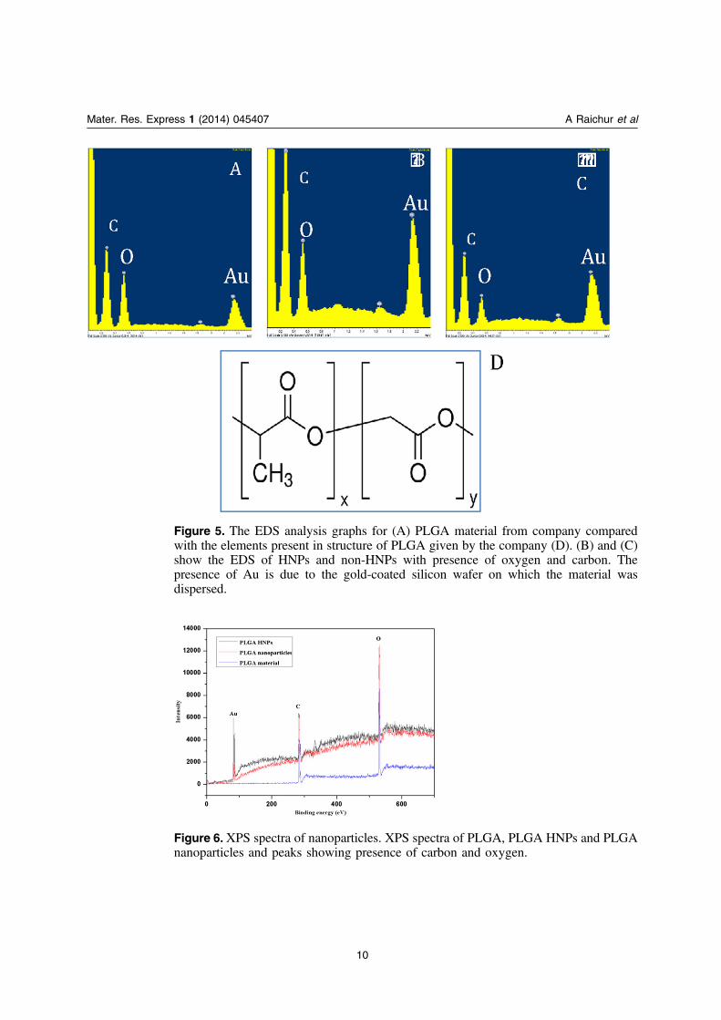
Figure 5. The EDS analysis graphs for (A) PLGA material from company comparedwith the elements present in structure of PLGA given by the company (D). (B) and (C)show the EDS of HNPs and non-HNPs with presence of oxygen and carbon. Thepresence of Au is due to the gold-coated silicon wafer on which the material wasdispersed.
Figure 6.XPS spectra of nanoparticles. XPS spectra of PLGA, PLGA HNPs and PLGAnanoparticles and peaks showing presence of carbon and oxygen.
10
Mater. Res. Express 1 (2014) 045407 A Raichur et al
3.3. Drug encapsulation and release efficiency
Nano-shells being hollow by nature, have the highest capacity to encapsulate drug compared tonon-hollow nanoparticles. In our work we used paclitaxel as the drug and carried on theexperiment for drug encapsulation and release efficiency studies.
Paclitaxel is widely used as a mitotic inhibitor in cancer chemotherapy. Direct oralincorporation of paclitaxel causes elimination of drug and has many limitations in clinicalapplication owing to its low solubility in water and pharmaceutical solvents [27, 28]. Therefore,polymeric nanoparticles such as PLGA hollow nanoparticles prove advantageous as idealvectors in delivering the drug safely in targeted cells.
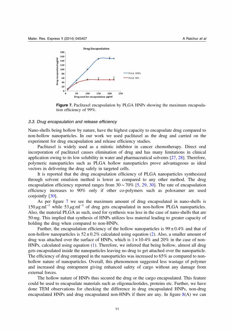
It is reported that the drug encapsulation efficiency of PLGA nanoparticles synthesizedthrough solvent emulsion method is lower as compared to any other method. The drugencapsulation efficiency reported ranges from 30∼ 70% [5, 29, 30]. The rate of encapsulationefficiency increases to 90% only if other co-polymers such as poloxamer are usedconjointly [30].
As per figure 7 we see the maximum amount of drug encapsulated in nano-shells is150 μgml−1 while 53 μgml−1 of drug gets encapsulated in non-hollow PLGA nanoparticles.Also, the material PLGA as such, used for synthesis was less in the case of nano-shells that are50mg. This implied that synthesis of HNPs utilizes less material leading to greater capacity ofholding the drug when compared to non-HNPs.
Further, the encapsulation efficiency of the hollow nanoparticles is 99 ± 0.4% and that ofnon-hollow nanoparticles is 52 ± 0.2% calculated using equation (2). Also, a smaller amount ofdrug was attached over the surface of HNPs, which is 1 × 10⁻⁴% and 20% in the case of non-HNPs, calculated using equation (1). Therefore, we inferred that being hollow, almost all druggets encapsulated inside the nanoparticles leaving no drug to get attached over the nanoparticle.The efficiency of drug entrapped in the nanoparticles was increased to 65% as compared to non-hollow nature of nanoparticles. Overall, this phenomenon suggested less wastage of polymerand increased drug entrapment giving enhanced safety of cargo without any damage fromexternal forces.
The hollow nature of HNPs thus secured the drug or the cargo encapsulated. This featurecould be used to encapsulate materials such as oligonucleotides, proteins etc. Further, we havedone TEM observations for checking the difference in drug encapsulated HNPs, non-drugencapsulated HNPs and drug encapsulated non-HNPs if there are any. In figure 8(A) we can
Figure 7. Paclitaxel encapsulation by PLGA HNPs showing the maximum encapsula-tion efficiency of 99%.
11
Mater. Res. Express 1 (2014) 045407 A Raichur et al
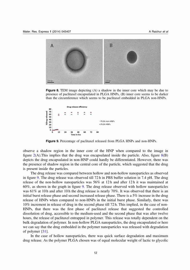
observe a shadow region in the inner core of the HNP when compared to the image infigure 2(A).This implies that the drug was encapsulated inside the particle. Also, figure 8(B)depicts the drug encapsulated in non-HNP could hardly be differentiated. However, there wasthe presence of shadow region in the central core of the particle, which suggested that the drugis present inside the particles.
The drug release was compared between hollow and non-hollow nanoparticles as observedin figure 9. The drug release was observed till 72 h in PBS buffer solution in 7.4 pH. The drugrelease of the non-hollow nanoparticles was 56% at 12 h and after 12 h it was maintained at60%, as shown in the graph in figure 9. The drug release observed with hollow nanoparticleswas 61% at 10 h and after 10 h the drug release is nearly 70%. It was observed that there is aninitial burst release phase and second increased release phase. There is a 5% increase in the drugrelease of HNPs when compared to non-HNPs in the initial burst phase. Similarly, there was10% increment in release of drug in the second phase till 72 h. This implied, in the case of non-HNPs, that there was the first phase of paclitaxel release that suggested the controlleddissolution of drug, accessible to the medium-used and the second phase that was after twelvehours, the release of paclitaxel entrapped in polymer. This release was totally dependent on thebulk degradation of polymer. In non-hollow PLGA nanoparticles, the drug encapsulated or herewe can say that the drug embedded in the polymer nanoparticles was released with degradationof polymer [31].
In the case of hollow nanoparticles, there was quick surface degradation and maximumdrug release. As the polymer PLGA chosen was of equal molecular weight of lactic to glycolic
Figure 8. TEM image depicting (A) a shadow in the inner core which may be due topresence of paclitaxel encapsulated in PLGA HNPs, (B) inner core seems to be darkerthan the circumference which seems to be paclitaxel embedded in PLGA non-HNPs.
Figure 9. Percentage of paclitaxel released from PLGA HNPs and non-HNPs.
12
Mater. Res. Express 1 (2014) 045407 A Raichur et al
acid, the degradation of polymer was processed as and when the surface became eroded. As aresult the maximum amount of paclitaxel was released. Being hollow, the drug release waspulsatile and nearly maximum. This characteristic difference between the quantity of drugrelease between HNPs and non-HNPs suggested that HNPs gave quick results of drug deliverywithin 12 h as compared to non-HNPs.
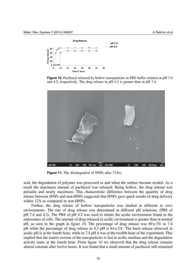
Further, the drug release of hollow nanoparticles was studied in different in vitroenvironments. The rate of drug release was determined in different pH solutions, (PBS ofpH 7.4 and 4.2). The PBS of pH 4.2 was used to mimic the acidic environment found in theendosomes of cells. The amount of drug released in acidic environment is greater than in normalpH, as seen in the graph in figure 10. The percentage of drug release was 69 ± 3% in 7.4pH while the percentage of drug release in 4.2 pH is 84 ± 2%. The burst release observed inacidic pH is at the fourth hour, while in 7.4 pH it was at the twelfth hour of the experiment. Thisimplied that the matrix erosion of the nanoparticles is fast in acidic medium and the degradationactivity starts at the fourth hour. From figure 10 we observed that the drug release remainsalmost constant after twelve hours. It was found that a small amount of paclitaxel still remained
Figure 10. Paclitaxel released by hollow nanoparticles in PBS buffer solution at pH 7.4and 4.2, respectively. The drug release in pH 4.2 is greater than in pH 7.4.
Figure 11. The disintegration of HNPs after 72 hrs.
13
Mater. Res. Express 1 (2014) 045407 A Raichur et al
inside the particles from the calculations using equation (3). The SEM images showed that theparticles disintegrate in buffer solution after 72 h, figure 11. The remaining paclitaxel wasreleased eventually as the nanoparticles were subjected to complete degradation. It can beinferred from the above observation that as time increases, degradation of HNPs also increasesgiving complete release of drug to the surrounding medium, here in our experiment it isPBS [32].
4. Conclusion
In summary, hollow polymer PLGA colloidal nano-shells were synthesized using simple oil-in-water emulsion solvent technique, which is economic, cost effective, and leads to high yield ofgood quality nanoparticles. This hollow structure results from subsequent events including airentrapment, fast solidification, and phase separation with instant stabilization. PLGA nano-hollow shells have the capacity to hold the cargo material for a long time wherein the materialcan remain safe. The cargo material gets released with constant rate. These nano capsules werepH sensitive, inferring that these particles could be used for drug or gene delivery at properspecific time delivery. Also, as these hollow shells are nanostructures with low densities andlarge surface area, they may be used as carriers for drug and gene delivery for anticancertreatment.
Acknowledgements
Archana Raichur acknowledges the Ministry of Education, Culture, Sports, Science andTechnology (MEXT), Japan for financial support under Monbukagakusho fellowship. Part ofthis study has been supported by a grant from the Programme of the Strategic ResearchFoundation at Private Universitites, S1101017, organized by MEXT, Japan since April 2012.Mr Keichi Hirakawa’s generous help in taking TEM pictures is profoundly acknowledged.
References
[1] Caruso F et al 2001 Adv. Mater. 13 11–22[2] Yin Y et al 2004 Science 304 711[3] Galleon J et al 2013 Nat. Biotechnol. 31 638–46[4] Santosa D M et al 2013 Nanotechnol. Biol. Med. 9 985–95[5] Ling Y and Huang Y 2008 Proceedings 19 (Berlin: Springer) pp 514–7
Kim M-J, Jang D-H, Lee Y-I, Jung H S, Lee H-J and Choa Y-H 2011 J. Nanosci. Nanotechnol. 11 889–93[6] Chang M-W, Stride E and Edirisinghe M 2010 J. R. Soc. Interface 7 S451–60[7] Zha L, Zhang Y, Yang W and Fu S 2002 Adv. Mater. 14 1090–2[8] Chiang W-L, Ke C-J, Liao Z-X, Chen S-Y, Chen F-R, Tsai C-Y, Xia Y and Sung H-W 2012 Small 8 3584–8[9] Zhang X, Chibli H, Mielke R and Nadeau J 2011 Bioconjugate Chem. 22 235–43
[10] Stevanović M M and Uskoković D P 2009 Curr. Nanosci. 5 1–14[11] Chen J-F, Ding H-M, Wang J-X and Shao L 2004 25 723–7[12] Jin H, Lovell J F, Chen J, Lin Q, Ding L, Ng K K, Pandey R K, Manoharan M, Zhang Z and Zheng G 2012
Bioconjugate Chem. 23 33−41
14
Mater. Res. Express 1 (2014) 045407 A Raichur et al
[13] Choi S W, Zhang Y and Xia Y 2010 Angew. Chem. 43 8076Choi S W, Zhang Y and Xia Y 2010 Angew. Chem. Int. Ed. 49 7904He H Y, Cao X and Lee L J 2004 J. Control. Release 95 391
[14] Li1 B, Yang X, Xia L, Majeed M I and Tan B Sci. Rep. 3 2128[15] Astete C E and Sabliov C M 2006 J. Biomater. Sci. Polym. Ed. 17 247–89[16] Lü J M, Wang X, Marin-Muller C, Wang H, Lin P H, Yao Q and Chen C 2009 Expert. Rev. Mol. Diagn. 9
325–41[17] Im S H and Jeong U 2005 Nature Mater. 4 671–9[18] Xu X and Asher S A 2004 J. Am. Chem. Soc. 126 7940–5[19] Lou X W (D), Archer L A and Yang Z 2008 Adv. Mater. 20 3987–4019[20] King T W and Patrick C W Jr 2000 J.Biomed. Mater. Res 51 383–90[21] Kwon S, Joo J-R, Lee W-K, Jeong Y and Choi J S 2009 Bull. Korean Chem. Soc. 30 486–8[22] Yang Y Y, Shi M, Goh S H, Moochhala S M, Ng S and Heller J 2003 J. Control. Release 88 201[23] Dalton A and Jurewicz I 2007 Nat. Nanotechnol. 2 339–40[24] Crotts G and Park T G 1995 J. Control. Release 35 91[25] Bihari1 P, Vippola M, Schultes S, Praetner M, Khandoga A G, Reichel C A, Coester C, Tuomi T,
Rehberg M and Krombach F 2008 Part. Fibre Toxicol. 5 14[26] Panyam J et al 2003 J. Control. Release 92 173–87[27] Gelderblom H, Verweij J, Nooter K and Cremophor E L 2001 Eur. J. Cancer 37 1590–8[28] Singla A K, Garg A and Aggarwal D 2002 Int. J. Pharm. 235 179–92[29] Santos D M et al 2013 Nanomedicine: Nanotechnol. Biol. Med. 9 985–95[30] Danhier F, Lecouturier N, Vroman B, Jérôme C, Marchand-Brynaert J, Feron O and Préat V 2009 J. Control.
Release 133 11–7[31] Corrigan O I and Li X 2009 Eur J. Pharmaceutical Sci. 37 477–85[32] Keum C-G, Noh Y-W, Baek J-S, Lim J-H, Hwang C-J, Na Y-G, Shin S-C and Cho C-W 2011 Int. J.
Nanomed. 6 2225–34
15
Mater. Res. Express 1 (2014) 045407 A Raichur et al