Preparation, characterization, in vitro drug release and biological studies of
Upload
khangminh22Category
view
0download
0
Optimization of in vitro test conditions for PLGA-based
drug delivery systems
Dissertation zur Erlangung des akademischen Grades des
Doktors der Naturwissenschaften (Dr. rer. nat.)
eingereicht im Fachbereich Biologie, Chemie, Pharmazie
der Freien Universität Berlin
vorgelegt von
ZOHA HANIF ZADEGAN
aus Germany
Berlin, 2017

II
1. Gutachter: Prof. Dr. Roland Bodmeier
2. Gutachter: Prof. Dr. Philippe Maincent
Tag der mündlichen Prüfung: 13.06.2017

III
To my Family

IV
Acknowledgements
During the work on my thesis at the Freie Universität Berlin, many people helped me with their
advice, patience and support. To all those, I want to express my thankfulness.
I owe my deepest gratitude to my supervisor Prof. Dr. Roland Bodmeier for his advice, guidance
and support throughout my graduate studies. I am very thankful to him for the opportunity to let
me work in his international research group as Ph. D. student.
I would also like to deeply thank Prof. Dr. Philippe Maincent for co-evaluating my thesis.
I would like to show my particular gratefulness to Dr. Luisa Duque for co-supervising my
doctoral study; certainly our regular discussions had great impact on my work.
I am indebted to all my colleagues for the supportive and friendly environment of our
workgroup; to Dr. Martin Körber, Dr. Werner Herrmann, Dr. Andrei Dashevsky, Dr. Mesut
Ciper, Dr. Burkhard Dickenhorst, Jia Deng , Chengcheng Zhao, Miriam Colombo, Dr. May
Darwich, Dr. Gaith M. Zoubari, Jelena Teodosic, Seyedreza Goldoozian and Benjamin Balzus;
to Dr. Luisa Duque, Dr. Kathrin Bürki, Dr. Agnieszka Solik, Dr. Rahul Sonawane, Dr. Fitsum
Sahle, Prutha Shrikrishna Gaitonde, Benjamin Balzusand and Lisa Bessßlich for proofreading
the parts of my dissertation; to Mrs. Gabriela Karsubke for her assistance with administrative
issues, to Stefan Walter, Eva Ewest and Andreas Krause for technical supports.
Finally, I would like to express my deepest gratitude to my parents Nahid and Ali, without them
I would not be where I am today and to my sister Hoda and her boyfriend Fabian for their love,
patience and ongoing support during my whole life and the last years, respectively.

Table of contents
1. Introduction ............................................................................................................................. 1
1.1 Parenteral controlled released drug delivery systems ........................................................... 2
1.1.1 Polymeric controlled release systems ............................................................................ 3
1.1.2 Biodegrdable polymers .................................................................................................. 3
1.2 Biodegradable polyesters based on lactic and glycolic acid ................................................. 6
1.2.1 Synthesis of PLGA ........................................................................................................ 6
1.2.2 Polymer erosion ............................................................................................................. 7
1.2.2.1 Surface erosion............................................................................................................ 8
1.2.2.2 Bulk erosion ................................................................................................................ 9
1.2.3 Degradation of PLGA .................................................................................................. 10
1.3 Release mechanisms of PLGA-based drug delivery systems ............................................. 10
1.3.1 Factors influencing drug release from PLGA-based drug delivery systems ............... 11
1.3.1.1 Water uptake ............................................................................................................. 11
1.3.1.2 Hydrolysis ................................................................................................................. 12
1.3.1.3 Erosion ...................................................................................................................... 13
1.4 PLGA-based drug delivery systems.................................................................................... 13
1.4.1 Biodegradable implants ............................................................................................... 14
1.4.2 Biodegradable microparticles ...................................................................................... 14
1.5 In vitro drug release testing for parenteral dosage form ..................................................... 18
1.5.1 Sample and separate Method ....................................................................................... 22
1.5.2 Flow-through method................................................................................................... 25
1.5.3 Dialysis method ........................................................................................................... 28
1.5.4 Accelerated release testing .......................................................................................... 35

Table of contents
VI
1.5.5 More realistic release test systems for parenteral products ......................................... 36
1.5.5.1 Subcutaneous injection site simulator (Scissor) ................................................... 37
1.5.5.2 Capillary bioreactor device ................................................................................... 38
1.5.5.3 Using agarose gel as a dissolution test .................................................................. 39
1.6 In vivo condition at the site of implantation........................................................................ 41
1.7 In vitro–in vivo correlation of parenteral controlled release drug delivery systems ........... 45
1.8 Application of the described release setup to implant ........................................................ 48
1.9 Objectives ........................................................................................................................... 51
2. Materials and Methods .......................................................................................................... 52
2.1 Materials ............................................................................................................................. 53
2.1.1 Model drugs ................................................................................................................. 53
2.1.2 Polymers ...................................................................................................................... 53
2.1.3 Other excipients ........................................................................................................... 54
2.2 Methods............................................................................................................................... 54
2.2.1 Preparation of PLGA implants using hot melt extrusion ............................................. 54
2.2.2 Preparation of PLGA implants using direct compression ........................................... 55
2.2.3 Restricted swelling device ........................................................................................... 55
2.2.4 Adsorption test of the restricted swelling device ......................................................... 55
2.2.5 Diffusion test of the restricted swelling device............................................................ 55
2.2.6 Drug extraction from implants ..................................................................................... 56
2.2.7 In vitro drug release study............................................................................................ 56
2.2.8 Degradation study and molecular weight determination ............................................. 57
2.2.9 In vito and ex vivo experiments .................................................................................... 57
2.2.10 Determination of tissue force ex vivo......................................................................... 58
2.2.11 Differential scanning calorimetry (DSC) ................................................................... 59

Table of contents
VII
2.2.12 Water content of the turkey breast ............................................................................. 59
2.2.13 Water uptake study .................................................................................................... 59
2.2.14 Diameter increase....................................................................................................... 60
2.2.15 Determination of implant morphology by optical macroscope ................................. 60
2.2.16 PLGA mass loss ......................................................................................................... 60
2.2.17 Determination of µpH using EPR .............................................................................. 61
3. Results and Discussion .......................................................................................................... 62
A. Investigation of the effect of tissue pressure on the shape, swelling and water uptake of
PLGA-based matrix implants and quantification of tissue pressure acting on the formulation
after implantation...................................................................................................................... 63
3.A.1 Background ................................................................................................................. 64
3.A.2 Evaluation of PLGA implants after implantation in turkey breast ............................. 65
3.A.2.1 Determination of the amount of water in the turkey breast ................................. 65
3.A.3 Ex vivo quantification of tissue pressure acting on PLGA-based matrix implants ..... 69
3.A.3.1 Method development............................................................................................ 69
3.A.3.2 Determination of the force during injection of water into balloon catheter ......... 69
3.A.3.3 Relationship between the injection pressure and the pressure needed to expand
the balloon catheter ........................................................................................................... 72
3.A.4 Conclusion .................................................................................................................. 77
B Design of a new in vitro test to mimic mechanical properties of the tissue for solid
parenteral DDS and to investigate the effect of restricted swelling on drug release from
PLGA-implants ......................................................................................................................... 79
3.B.1 Background ................................................................................................................. 80
3.B.2 Simulation of the physiological space in an in vitro test............................................. 80
3.2B.2.1 Air tight apparatus .............................................................................................. 80
3.B.2.2 Modified continuous flow method ....................................................................... 82

Table of contents
VIII
3.B.2.3 Modified sample and separate method ................................................................. 82
3.B.2.4 Restricted swelling device .................................................................................... 84
3.B.2.4.1 Absorption and diffusion test of the restricted swelling device ............ 84
3.B.3 The effect of restricted swelling on drug release from PLGA-based implants ........... 85
3.B.3.1 Diameter increase of implants prepared with PLGA of different moleculare
weights .............................................................................................................................. 85
3.B.3.2 Release of implants with different PLGA molecular weight ............................... 87
3.B.3.3 Release comparison of implants under restricted and unrestricted conditions .... 87
3.B.3.4 Determination of µpH of restricted and unrestricted implants using EPR ........... 94
3.B.4 Conclusion ................................................................................................................... 95
C Application of biphasic test model as a new biorelevant model for drug release from PLGA
implants and investigate its effect on risperidone release from PLGA-based implants ........... 97
3.C.1 Background ................................................................................................................. 98
3.C.2 Risperidone release in biphasic system ....................................................................... 99
3.C.3 Risperidone release from 5050 DLG 1A PLGA implants in monophasic system vs.
biphasic system ................................................................................................................... 100
3.C.4 Risperidone release from 503H implants in monophasic systrem vs. biphasic system
............................................................................................................................................. 102
3.C.4.1 Effect of drug loading ........................................................................................ 102
3.C.4.2 Effect of dissolution method .............................................................................. 103
3.C.5 Effect of amount of octanol and the ratio of octanol/phosphate buffer on the drug
release ................................................................................................................................. 106
3.C.6 Drug release from biphasic system containing olive oil ........................................... 107
3.C.7 Effect of incorporation of octanol in implants on drug release ................................. 110
3.C.8 Effect of octanol on glass transition temperature of two types of PLGA implants
containing 10% risperidone ................................................................................................ 113

Table of contents
IX
3.C.9 Changes in polymer molecular weight of octanol incorporated in implant containing
503H .................................................................................................................................... 114
3.C.10 Conclusion ............................................................................................................... 114
4. Summary .............................................................................................................................. 116
5. Zusammenfassung ............................................................................................................... 121
6. References ........................................................................................................................... 127

X

XI

1. Introduction

Introduction
2
1.1 Parenteral controlled release drug delivery systems
Morphine was the first official parenteral injected drug induced in 1867. Soon after, the number
of parenteral formulations has been increased dramatically. The intravenous, subcutaneous,
intramuscular, intraperitoneal, and intrathecal routs are examples of parenteral administration.
However, the major administration routes of parenteral controlled release systems are
subcutaneous and intramuscular. Products such as oil solutions (D. B. Larsen, Joergensen, Olsen,
Hansen, & Larsen, 2002; D. B. Larsen, Parshad, Fredholt, & Larsen, 2002), emulsions (Collins-
Gold, Lyons, & Bartholow, 1990; Florence & Whitehill, 1982), liposomes (Sharma & Sharma,
1997), micelles (Alkan-Onyuksel, Ramakrishnan, Chai, & Pezzuto, 1994), implants (Ueno,
Refojo, & Liu, 1982) and microparticles (Herrmann & Bodmeier, 1995) are identified as
parenteral controlled release drug delivery systems. In comparison to conventional oral dosage
forms these systems can maintain the drug in the desired therapeutic range for days, weeks,
months, and for some products, even years after one administration and offer several advantages
including:
o Increase of bioavailability: Biopharmaceuticals, such as proteins and peptides, are large
hydrophilic compounds administered via parenteral injections to circumvent their inherent
instability in the gastro-intestinal tract as well as their low permeability across biological
membranes (Frokjaer & Otzen, 2005). Another growing group of pharmaceuticals often requiring
administration by injection is low-molecular-weight hydrophobic drugs, which also have low oral
bioavailability (Christian Wischke & Schwendeman, 2008). Administration by injection leads to
discomfort for the patient.
o Long release period: The use of controlled release formulations enables the frequency of
injections to be reduced, which improve the patient’s compliance, especially those who require
daily or long-term treatment and reduce the need for follow-up care.
o Constant drug plasma concentration: Another advantage of controlled release formulations is that
they result in a more constant plasma concentration of the drug, which is better kept within the
therapeutic window. Frequent administrations often result in rises and falls in the concentration.
Too high a concentration can cause unwanted side effects, while too low a concentration results
in the loss of therapeutic effect. This means that lower total doses can be reduced with the
controlled release formulations (Johnson et al., 1996).

Introduction
3
o Localized delivery of drug: The product can be administrated directly at the site where drug
action is needed and hence systemic exposure of the drug can be reduced.
The major Issues during application of injectable drug delivery systems are pain and tissue
damage at the injection site, which decrease the patients’ compliance.
1.1.1 Polymeric controlled release systems
The development of polymeric controlled release system introduced a new concept in drug
administration. These systems are less complicated and with high stability. Encapsulation in the
polymer carrier eliminates the degradation of drugs; moreover, the release profile of the drugs
can be controlled by properly choosing polymers.
Polymers used in parenteral controlled release systems can be grouped into two main categories:
non-biodegradable and biodegradable polymers. The first polymeric controlled release devices is
a reservoir system based on non-biodegradable polymer silicone rubber (Folkman, Long, &
Rosenbaum, 1966). The disadvantages of such system lay in that firstly, surgical removal of
drug-depleted delivery systems of non-biodegradable polymers is difficult and painful and non-
removal may pose toxicological problems; secondly, although this diffusion controlled delivery
system is an excellent tool of modulating drug release, is largely dependent on polymer
permeability and drug characteristics. The basic mechanism in non-degradable devices being
diffusion, drugs which have either high molecular weight (7500) or poor solubility in polymer
are not amenable to diffusion controlled release (Jalil & Nixon, 2008; Sinha & Trehan, 2003). To
overcome these problems, biodegradable polymers for sustained release parenteral drug delivery
systems began to develop in early 1970s. When compared to non-biodegradable polymers, they
have the improved biocompatibility and are degraded in the body, This avoids the need for
surgical removal and thus improves the patient acceptance (Danckwerts & Fassihi, 2008).
1.1.2 Biodegradable polymers:
Biodegradable polymers commonly contain chemical linkages such as esters, anhydrides,
amides, peptides and glycosides. These polymers degrade in vivo either enzymatically or non-
enzymatically to biocompatible and non-toxic byproducts. These can be further metabolized or
excreted via normal physiological pathways. Biodegradable polymer not only have been

Introduction
4
extensively used in controlled delivery systems, but also extended to medical devices (Leenslag,
Pennings, Bos, Rozema, & Boering, 1987), wound dressing (Hubbell, 1996), and for fabricating
scaffolds in tissue engineering (F. Shi, Gross, & Rutherford, 1996).
Biodegradable polymers are calcified as natural or synthetic (Mishra et al., 2008). The
investigation of natural biodegradable polymer as drug carrier has been concentrated on proteins
and polysaccharides (Table 1.1) (Mohanty, Misra, & Hinrichsen, 2000). Natural biodegradable
polymers are attractive because they are natural products of living organisms, readily available,
relatively inexpensive and capable of multitude of chemical modifications (Sinha & Trehan,
2003).
Table 1.1 Nature biodegradable polymers
Proteins
Globulin, Gelatin, Collagen, Casein, Bovine
serum albumin, Human serum albumin
Polysaccharide
Starch, Cellulose, Chitosan, Dextran, Alginic
acid
Synthetic biodegradable polymers have gained more popularity than natural biodegradable
polymers. The major advantages of synthetic polymers include high purity of the product, more
predictable, uniformity and free of concerns of immunogenicity. Furthermore, synthetic
polymers provide with a wider range of mechanical properties and degradation rate. In the past
years, there are numerous biodegradable polymers synthesized. Most of these polymers contain
labile linkages in their backbone such as esters, orthoesters, anhydrides, carbonates, amides,
urethanes, etc. The synthesis, biodegradability, and application of these polymers have been well
reviewed (Table 1.2) (Gombotz & Pettit, 1995; J. H. Park, Ye, & Park, 2005; Winzenburg,

Introduction
5
Schmidt, Fuchs, & Kissel, 2004). Although some of the biodegradable materials have been
approved for the use in medical devices (Middleton & Tipton, 2000), biodegradable polymers
such as poly(lactic acid), poly(glycolic acid) and their copolymers (PLGA, Figure 1.2) (Holland,
Tighe, & Gould, 1986) are the most commonly used polymers as drug release controlling
matrices in approved parenteral products due to their excellent biocompatibility and
biodegradability and mechanical strength (J. M. Anderson & Shive, 2012; Athanasiou,
Niederauer, & Agrawal, 1996; Jain, 2000). In addition, other polymers such as poly(ε-
caprolactone and polyphosphoesters are being investigated for drug and gene delivery (Zhao,
Wang, Mao, & Leong, 2003). Poloxamers, copolymers of polyethylene oxide and
polyoxypropylene, are another interesting class which provided a wide range of applications in
pharmaceutical and biomedical field and were investigated for drug delivery (Kwon & Okano,
1999).
Table 1.2 Synthetic biodegradable polymers
Polyesters: Poly(glycolic acid), Poly(lactic acid),
Poly(lactic-co-glycolic acid), Poly(caprolactone)
Polyanhydrides
Polyorthoesters
Polyurethanes
Tyrosine-derived polycarbonates
Polyphosphazenes

Introduction
6
1.2 Biodegradable polyesters based on lactic and glycolic acid
1.2.1 Synthesis of PLGA
According to the biodegradable polymer classification, homopolymers poly(lactic acid) (PLA)
and poly(glycolic acid) (PGA) as well as mixtures thereof, poly(lactic-co-glycolic acid) (PLGA)
are categorized as synthetic, bulk eroding, linear, aliphatic poly(α-esters).
Polymers and copolymers of lactic and glycolic acids can be prepared in two ways: by a direct
polycondensation reaction of lactic acid and glycolic acid (Figure 1.1), resulting in polymers of
low molecular weight or by a ring opening polymerization of the cyclic diesters (1,4-dioxane-
2,5-diones) of glycolic acid and lactic acid (Figure 1.2). The ring opening polymerization yields
the polymers of high molecular weight (> 10,000 g/mol) and of better mechanical properties
(Gentile, Chiono, Carmagnola, & Hatton, 2014; Qian, Wohl, Crow, Macosko, & Hoye, 2011;
Silva, Cardoso, Silva, Freitas, & Sousa, 2015; N. Wang, Wu, Li, & Feng, 2000). Furthermore,
this method allows a better control of the molecular weight distribution (polydispersity) and the
end group functionality of the (co-)polymer (Jain, 2000).
Figure 1.1 Synthesis of poly(lactide) by direct polycondensation

Introduction
7
Figure 1.2 Synthesis of poly(lactide) by ring-opening polymerization
The selection of the reactants and the synthesis conditions will determine the physicochemical
properties of the resulting polyesters, such as hydrophilicity, mechanical strength, glass transition
and crystallinity (Gilding & Reed, 1979; Miller, Brady, & Cutright, 1977; Omelczuk &
McGinity, 1992).The characteristics, which can be used to describe the final polymers include
the weight or number averaged molecular weight, the polydispersity, the ratio of lactic and
glycolic acid monomers, the ratio of D- and L-lactic acid monomers and the endgroup
functionality. Although rarely specified, in random copolymers the segment length of
monomeric repeat units of a product is important since short block lengths avoid the formation of
crystalline domains in the polymer, which provides homogeneous controlled release matrices
(Shard, Clarke, & Davies, 2002).
1.2.2 Polymer erosion
The term erosion is used for the loss of material from the polymer bulk (Achim Göpferich,
1996). This process requires the degradation of the polymer into soluble oligomers or monomers,
but the focus is not on the single chain’s properties but on those of the bulk, e.g. mass, outer
dimension or mechanical stability. Two basic mechanisms are distinguished: Surface erosion and
bulk erosion (Figure 1.3). However, pure forms of the described erosion processes are rare.
Introduction
8
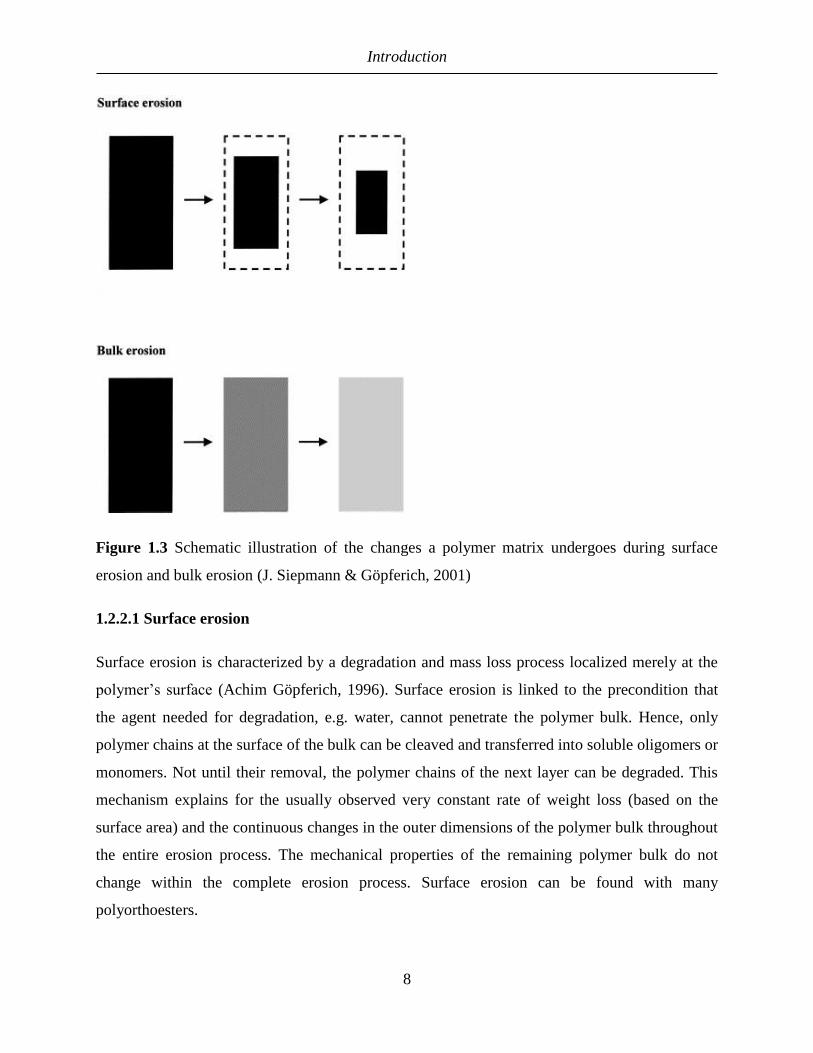
Figure 1.3 Schematic illustration of the changes a polymer matrix undergoes during surface
erosion and bulk erosion (J. Siepmann & Göpferich, 2001)
1.2.2.1 Surface erosion
Surface erosion is characterized by a degradation and mass loss process localized merely at the
polymer’s surface (Achim Göpferich, 1996). Surface erosion is linked to the precondition that
the agent needed for degradation, e.g. water, cannot penetrate the polymer bulk. Hence, only
polymer chains at the surface of the bulk can be cleaved and transferred into soluble oligomers or
monomers. Not until their removal, the polymer chains of the next layer can be degraded. This
mechanism explains for the usually observed very constant rate of weight loss (based on the
surface area) and the continuous changes in the outer dimensions of the polymer bulk throughout
the entire erosion process. The mechanical properties of the remaining polymer bulk do not
change within the complete erosion process. Surface erosion can be found with many
polyorthoesters.

Introduction
9
1.2.2.2 Bulk erosion
Bulk erosion is characterized by uniformly distributed degradation throughout the polymer bulk
(Achim Göpferich, 1996). Ideally, the probability of chain cleaving is evenly distributed within
the complete bulk. As long as the polymer chains are still insoluble, they cannot leave the bulk.
Every polymer showing bulk erosion therefore usually retains most of its mass over quite a long
time before finally showing a fast mass loss. This loss is correlated with the loss of the now
soluble shortened polymer chains that leave the bulk’s structure. Different to the mass, the
mechanical properties continuously change during the erosion process. Bulk erosion requires the
agent forcing chain degradation to penetrate faster into the bulk than a thought ‘front of
degradation’. In most biodegradable polymers, water is this agent as it allows hydrolytic
cleavage of the polymer chains. Thus, water permeability of the bulk determines whether a
polymer undergoes bulk erosion. The degradation rate of a bulk-eroding polymer is relatively
independent of its shape, as long as no additional factors are involved. As many polyesters show
autocanalization effects during their degradation, accelerating the further degradation, different
diffusion pathways for the leaving of formed acidic degradation products can change the
degradation behavior. This can explain e.g. the different reported degradation times of PLGA
scaffolds in vitro and in vivo as well as of samples of different sizes (Lichun Lu et al., 2000;
Vert, Li, & Garreau, 1992). It has also been shown, that the ratio between amorphous and
crystalline parts of semicrystalline polymers can change during erosion. As crystalline regions
within a polymer bulk usually have lower water permeability, their degradation rate tends to be
slower than that of amorphous parts. Therefore, the relative amount of crystalline regions within
the bulk will increase during degradation process, affecting e.g. the rate of the further
degradation and the bulk’s mechanical properties (Pitt, Chasalow, Hibionada, Klimas, &
Schindler, 1981). The same principle has to be applied to co-polymers with larger blocks (M.-H.
Huang, Li, Hutmacher, Coudane, & Vert, 2006).

Introduction
10
1.2.3 Degradation of PLGA
Polyesters like PLA and PLGA are bulk-eroding polymers and degrade by randomly hydrolysis
of the functional groups in an aqueous environment, without significant contribution of enzymes,
which requires the presence of water. The cleavage of ester bond linkages yields carboxylic end
groups and hydroxyl groups. The formed carboxylic groups then could catalyze and accelerate
the hydrolysis of other ester bonds, a phenomenon referred as autocatalysis. Degradation of the
polyesters leads to polymers of shorter chain length and below a critical molecular weight of
about 1050-1150 Da (T. G. Park, Yong Lee, & Sung Nam, 1998), the oligomers can dissolve in
the aqueous surrounding medium and diffuse out of the matrix. The end products of the
degradation are lactic acid (pKa 3.85) and glycolic acid (pKa 3.83), which are both non-toxic and
excreted via the lungs (after incorporation in the tricarboxylic acid cycle) or the urine.
1.3 Release mechanisms of PLGA-based drug delivery systems
There are three release mechanisms for drug molecules to be released from a PLGA-based DDS,
which are illustrated in Figure 1.4:
o diffusion through water-filled pores (diffusion controlled)
o diffusion through the polymer (diffusion controlled)
o due to polymer erosion (erosion controlled)
Figure 1.4 The release mechanisms: (A) diffusion through water-filled pores, (B) diffusion
through the polymer and (C) erosion (Fredenberg, Wahlgren, Reslow, & Axelsson, 2011)

Introduction
11
Diffusion through water-filled pores are the most common way of release, as the encapsulated
drug is a protein or a peptide, which are too large and too hydrophilic to be transported through
the polymer phase.
Diffusion through the polymer phase may occur when the drug is small and hydrophobic
(Raman, Berkland, Kim, & Pack, 2005). However, the drug must enter the water phase, either at
the surface or in the pores inside the DDS, before being released.
The encapsulated drug may also be released without any transport due to dissolution of the
polymer, i.e. erosion. Erosion also creates pores, thus increasing the rate of diffusion. However,
there is a difference between erosion leading to drug release without drug transport, and erosion
that increases the rate of drug transport. The latter has been reported as a release mechanism
countless times, at least after a lag period, which is often described as diffusion-controlled
release (Alexis, Venkatraman, Rath, & Boey, 2004; Goraltchouk, Scanga, Morshead, &
Shoichet, 2006; Lam, Duenas, Daugherty, Levin, & Cleland, 2000; L. Wang, Venkatraman, &
Kleiner, 2004)
In general describing the release mechanism of PLGA-based DDS is not sometimes problematic,
due to the complexity of the system it is not always clear which of the processes is dominating,
and in a chain of processes that leads to drug release it is not obvious which one is the rate-
determining process
1.3.1 Factors influencing drug release from PLGA-based drug delivery systems
1.3.1.1 Water uptake
Water is absorbed by the polymer immediately upon immersion in water or administration in
vivo. The rate of water absorption, or hydration of the DDS is rapid compared to drug release
(Batycky, Hanes, Langer, & Edwards, 1997; Blasi, D’Souza, Selmin, & DeLuca, 2005). Water
absorption is a pore-forming process. These pores are too small for drug transport during the
early stage of this process; however with increasing of the number and size of water-filled pores,
a porous connected network allowing drug release is formed (Mochizuki, Niikawa, Omura, &
Yamashita, 2008).

Introduction
12
1.3.1.2 Hydrolysis
Polymer degradation and hydrolysis starts immediately upon contact with water and subsequent
decrease in molecular weight. The polymer becomes less hydrophobic with decreasing molecular
weight, and at 1100 Da the oligomers become water soluble (T. G. Park, 1994).
Several factors, material but also formulation properties can affect the degradation kinetics. The
degradation rates of the polyester are influenced by parameters such as the initial weight average
molecular weight, the hydrophobicity (lactic acid > glycolic acid monomers), the degree of
crystallinity (e.g., increased in PGA and L-PLA) and the glass transition temperature of the
amorphous phase.
The hydrophobicity of capped PLGA, which is esterified with an alkyl alcohol, is higher than for
PLGA with free carboxyl groups (uncapped PLGA). Accordingly, uncapped PLGA degrades
faster than capped PLGA (Tracy et al., 1999). Another factor affecting the degradation of the
polyesters is the enantiomeric composition of the polymer (de Jong et al., 2001). Two
stereoisomeric forms of PLA are commercially available, optically active L-PLA and racemic
D,L-PLA. L-PLA is a semicrystalline material (isotactic), whereas D,L-PLA is amorphous
(Gilding & Reed, 1979). However, during hydrolysis degradation products of D,L-PLA can
crystallize and thus further degrade at a lower degradation rate (Li, Garreau, & Vert, 1990). The
decrease of the molecular weight of PLGA and PLA follows a pseudo-first order kinetic (A
Göpferich & Tessmar, 2002), which reflects a random chain scission process.
The pH has an important effect on the hydrolysis rate of polyesters. PLA and PLGA have a
stability optimum at pH 4 - 5 and are hydrolyzed under acid and base catalysis (de Jong et al.,
2001).
The cleavage of an ester bond linkage yields a hydroxyl and a carboxyl group and the formation
of carboxylic acids during degradation of the polyesters can accelerate the hydrolysis of other
ester bonds (Shenderova, Burke, & Schwendeman, 1999). This autocatalytic phenomenon is
known to cause heterogeneous degradation inside PLGA matrices (Li & McCarthy, 1999) i.e.
faster degradation at the center of the PLGA matrix than at the surface. This effect becomes
more pronounced with increasing dimensions of a DDS (Dunne, Corrigan, & Ramtoola, 2000) as

Introduction
13
the acid gradient increases, but heterogeneous degradation has also been reported in particles and
films with dimensions as small as 10 μm (T. G. Park, 1995). As consequence of autocatalysis,
bimodal molecular weight distributions can be found in size exclusion chromatograms of
samples from degradation studies.
1.3.1.3 Erosion
Hydrolysis of the polymer backbone is the initiation of the erosion process, which is a series of
events, including a decrease in the molecular weight, a decrease in glass transition temperature
with decreasing Mw, a loss of mechanical properties and finally, a loss of mass via the dissolution
of small polymer fragments (Lyu, Sparer, & Untereker, 2005).
Dissolution of polymer degradation products and erosion create pores. Small pores, formed by
water absorption consequently grow during polymer erosion, and eventually coalesce with
neighboring pores to form fewer, larger pores (Batycky et al., 1997). Small pores may also be
closed (Kang & Schwendeman, 2007). This phenomenon is related to the mobility of the
polymer chains, and their ability to rearrange (Yamaguchi et al., 2002). The mobility of polymer
chains depends on the glass transition temperature (Tg).
1.4 PLGA-based drug delivery systems
Biodegradable delivery systems based on PLGA can be in the form of solid implants,
microparticles or delivery systems that form in situ. Polymer based drug delivery systems can be
classified into two types: reservoir-based systems, and monolithic matrix systems (Figure 1.5).
In reservoir-based systems, the drug reservoir is enclosed within insoluble polymer. The drug
releases through the rate-controlling porous polymeric membrane. Monolithic matrix systems are
similar to reservoir-based systems, but in this case, the drug is dispersed or dissolved within a
polymer matrix. The drug release can be diffusion, swelling, and/or erosion controlled.
Compared to reservoir systems, matrix systems are easier to be manufactured because they are
homogeneous in nature and they are also safer since a mechanical defect of the reservoir device
rather than matrix device may cause dose dumping (X. Huang & Brazel, 2001). However, if

Introduction
14
polymer matrix is non-degradable, the constant release profile is difficult to be achieved with
matrix system (Fung & Saltzman, 1997).
Figure 1.5 Polymeric delivery systems; (A) Reservoir systems; (B) Matrix systems
1.4.1 Biodegradable implants
Four PLGA based implants delivering small peptides and low molecular weight drug are
available on the market (Table 1.3). The solid implants for controlled release of drugs are usually
cylindrical polymer matrices (rods), which can be on the millimeter to centimeter scale,
facilitating large loadings of active materials. More complicated three-dimensional implant
structures, such as tubes, scaffolds or other structural supports, which are of special interest for
tissue engineering applications, can be fabricated as well. Manufacturing techniques include
solvent casting (T. G. Park, Cohen, & Langer, 1992), extrusion (Zhang, Wyss, Pichora, Amsden,
& Goosen, 1993), melt compression, injection molding (Sundback, Hadlock, Cheney, & Vacanti,
2003), compression molding (Schliecker et al., 2004) and freeze drying (Hsu et al., 1996). The
major disadvantage of these delivery systems is their limited patient acceptance, due to the
required painful administration into the subcutaneous tissue by surgical intervention or insertion
using large-bore needles (trocar).
1.4.1 Biodegradable microparticles
Microparticles are spherical, polymeric carrier particles of a size between 1 and 1000 μm, which
contain drug either in form of a reservoir (microcapsules) or dissolved / dispersed in the polymer

Introduction
15
matrix (microspheres). PLGA microparticles can be prepared by different microencapsulation
techniques including solvent extraction/evaporation processes, phase separation (coacervation)
and spray drying (Jain, 2000). A choice of the technique depends on the nature of polymer, the
drug, the intended use and the duration of therapy. However, solvent evaporation with
emulsification is the most often used technique, at least in the laboratory scale, due to possibility
in adaption for drugs of different physicochemical properties (C Wischke & Schwendeman,
2012). The polymer solution containing the drug (in solution or dispersion) is emulsified in an
external phase. The internal solvent is removed by partition into the external phase and/or by
evaporation (O’Donnell & McGinity, 1997). Oil-in-water (O/W) and oil-in-oil (O/O) emulsion
techniques have been applied to produce microparticles using solvent evaporation. The
conventional O/W solvent evaporation is appropriate for lipophilic drugs, for instance steroids.
For water-soluble drugs, peptides and proteins, low encapsulation efficiency is frequently
observed. A double emulsion (W/O/W) technique has been introduced in order to circumvent the
problems relating to water-soluble substances (Jaraswekin, Prakongpan, & Bodmeier, 2007;
Schwach et al., 2003).The preparation of PLA/PLGA microparticles by coacervation is a
complex method in which the resulting microparticles frequently agglomerate since the method
lacks any stabilizers or emulsifiers (Jain, 2000). A drug in the form of a solution or particles is
dispersed into the polymer solution. Subsequently, the coacervation of the polymer is induced by
a phase separation inducing agent. Soft coacervate droplets are hardened using another
nonsolvent of the polymer, such as hexane. Large amounts of solvents are required in the
coacervation process, and residual solvents are a concern for this process. Compared to solvent
evaporation and coacervation, spray drying is more rapid, easier to scale up, and less dependent
on factors inherent in the drugs and polymers. In the spray drying method a PLA/PLGA solution
with a dissolved or dispersed drug is sprayed though the nozzle of a spray dryer to form
microparticles. Dichloromethane and ethyl acetate are useful to prepare the polymer solution.
The microparticles from this method are sometimes not spherical; the formation of fibers or
irregular-shaped particles could be found when using this technique (Jain, 2000; Schwach et al.,
2003).
Preferentially, microparticles have a size of less than 250 µm (J. H. Park et al., 2005), which
allow injection through smaller needles after re-dispersing them in a suitable aqueous medium.
The applicability of smaller needles reduces pain during administration and thus improves the

Introduction
16
patient comfort. The more convenient administration compared to implants makes microparticles
an attractive biodegradable drug delivery system. However, their manufacturing is technically
challenging.
Table 1.3 Examples of marketed PLGA-based drug delivery systems
Product Therapeutic Dosage form Company Indication Administration
rout
Arestin®
Minocycline
hydrochloride1
Microparticle OraPharma
Periodontal
disease
Subgingival
Atridox® Doxycycline
hyclate1
In situ forming
implant
Tolmar Chronic adult
periodontitis
Subgingival
Bydureon® Exenatide2 Microparticle Zeneca Type II
diabedes
mellitus
Subcutaneous
Decapeptyl®
Triptorelin
acetate2
Microparticle Ferring
Prostate cancer
Subcutaneous
Eligard®
Leuorolide
acetate2
In situ forming
implant
Astellas
Prostate cancer
Subcutaneous
Enantone®
Leuprolide
acetate2
Microparticle Takeda
Prostate
cancer,
endometriosis
Subcutaneous
Enantone®
Gyn
Leuprolide
acetate2
Microparticle Takeda
Prostate
cancer,
endometriosis
Subcutaneous

Introduction
17
Lupron®
Depot
Leuprolide
acetate2
Microparticle Abbvie
Prostate cancer
Intramuscular
Leuprone®
HEXAL®
Leuprolide
acetate2
Implant Hexal
Prostate cancer
Subcutaneous
Nutropin®
Depot
Somatropin,
recombinant
human growth
hormone3
Microparticle Genentech
Short stature
Subcutaneous
Ozurdex®
Dexamethasone1 Implant Allergan Macular
edema, retinal
vein occlusion,
uveitis
Intravitreal
Pamorelin®
LA
Triptorelin
embonate2
Microparticle Ipsen
Pharma
Prostate cancer
Intramuscular
Profact
Depot®
Buserelin
acetate2
Implant Sanofi-
Aventis
Prostate
cancer,
endometriosis
Subcutaneous
Risperdal®
Consta
Risperidone1
Microparticle Janssen/Alk
ermes
Schizophrenia
Intramuscular
Sandostatin®
LAR
Octreotide
acetate2
Microparticle Novartis
Acromegaly,
carcinod
Intramuscular

Introduction
18
syndrome
Somatuline®
LA
Lanreotide
acetate2
Microparticle Ipsen
Acromegaly
Intramuscular
Suprecur®
MP
Buserelin
acetate2
Microparticle Sanofi-
Aventis
Prostate cancer
Subcutaneous
TrelstarTM
Depot
Triptorelin
pamoate2
Microparticle Watson
Prostate cancer Intramuscular
Zoladex®
Goserelin
acetate2
Implant Astra
Zeneca
Prostate cancer
Subcutaneouse
1 low molecular weight drug
2 peptide
3 protein
1.5. In Vitro Drug Release Testing of Parenteral Dosage Forms
In vitro release studies are generally performed to accomplish one or more of the following aims
(Burgess, Hussain, Ingallinera, & Chen, 2002; L. Lachman, H. Lieberman, 1986):
o As an indirect measurement of drug availability, especially in preliminary stages of product
development
o Quality control to support batch release and to comply with specifications of batches proven to be
clinically and biologically effective
o Assess formulation factors and manufacturing methods that are likely to influence bioavailability
o Substantiation of label claim of the product
o As a compendial requirement

Introduction
19
Since the introduction of the rotating basket apparatus (USP 1) as the first standardized apparatus
for in vitro dissolution testing in the USP in 1970, dissolution testing has gone through major
changes, including the design of new apparatus and the introduction of more biorelevant testing
conditions.
An in vitro release profile reveals fundamental information on the structure (e.g., porosity) and
behavior of the formulation on a molecular level, possible interactions between drug and
polymer, and their influence on the rate and mechanism of drug release and model release data
(Washington, 1990). Over the last few years, regulatory activity in in vitro dissolution testing has
become even more important with regard to the establishment of in vitro-in vivo correlations
(IVIVC) and for the evaluation of scale-up and post-approval changes. Such information
facilitates a scientific and predictive approach to the design and development of sustained
delivery systems with desirable properties.
However, this evolution of methods has mainly focused on the oral route of administration.
Recently, the number of products that are delivered via non-oral routes of administration has
greatly increased the number of marketed products and the interest in controlled release
parenteral products has multiplied. The reasons for the delivery via alternative routes such as the
parenteral administration include advanced targeting strategies, as well as the increasing number
of new drug entities that cannot be successfully delivered via the oral route of administration due
to various reasons such as instability in the gastrointestinal tract, adverse reactions upon systemic
exposure, patient compliance, and accessibility to specific organs or local sites of the body, etc.
Although a sizable amount of research has focused on the parenteral controlled drug delivery
systems, very little attention has been devoted to the development of an in vitro release technique
(Seidlitz & Weitschies, 2012).
Unlike controlled release oral formulations, there are no regulatory standards for parenteral
controlled delivery systems. Also, the current United States Pharmacopeia (USP) apparatus for in
vitro release testing was designed mainly for oral and transdermal products and is not directly
applicable for parenteral products administered subcutaneously or intramuscularly. For example,
concerns with using USP apparatuses 1 (basket) and 2 (paddle) include sample containment,
large volume of media required for testing, and sampling procedure. USP apparatuses 5 (paddle

Introduction
20
over disc), 6 (cylinder), and 7 (reciprocating holder) were designed for the transdermal route and
do not offer any advantages for parenteral delivery systems such as microparticles. Additionally,
drawbacks of USP apparatuses 3 (reciprocating cylinder) and 4 (flow-through cell), designed for
extended-release oral dosage forms, include evaporation (reciprocating cylinder) and filter
blockage along with polymer migration leading to variable flow rates (flow-through cell).
The dosage forms applied to deliver these new drug entities are as diverse as the sites of delivery.
Depending on the intended therapeutic action, controlled release parenterals can be administered
intravenously, intramuscularly, subcutaneously or intra-articularly, can be implanted into tumor
tissue, ocular or peri-ocular tissue, teeth, bone, blood vessels or inserted into other natural
passages/ conduits such as the esophagus. The target of drug delivery can either be local
structures or the entire organism. The dosage forms include, but are not limited to, monoliths
such as rods, lipophilic solutions, disperse systems such as microspheres, nanoparticles,
liposomes, emulsions, and suspensions, in-situ forming gels or solids, cements, wafers, and
coated medical devices such as drug-eluting stents and drug-eluting pacing leads. Reviews have
been published on the technologies used for many of these dosage forms (Kreye, Siepmann, &
Siepmann, 2008; Packhaeuser, Schnieders, Oster, & Kissel, 2004; Y. Shi & Li, 2005; Yasukawa,
Ogura, Kimura, Sakurai, & Tabata, 2006).
From this representative but incomplete listing it is evident that specialized in vitro release test
systems are necessary to address the diversity of the dosage forms and their sites of application.
This diversity may be one of the reasons why currently there is no standard compendial
dissolution test method for controlled release parenterals in the United States Pharmacopeia
(USP), in the European Pharmacopoeia (Ph. Eur.) or in the Japanese Pharmacopoeia (JP). A
number of workshop reports have been published stating the need for regulatory guidance on this
issue and highlighting some of the approaches used at present (Brown et al., 2011; Burgess,
Crommelin, Hussain, & Chen, 2004; Martinez, Rathbone, Burgess, & Huynh, 2008; Siewert et
al., 2003).
The current methods that are used most often for in vitro dissolution testing of parenteral dosage
forms are mostly noncompendial, although they sometimes include USP apparatus designed for
other routes of administration. They are categorized into three general groups: sample and

Introduction
21
separate methods, dialysis membrane-based methods and flow-through or continuous flow
methods (Kastellorizios and Burgess 2012; Seidlitz and Weitschies 2012a) (Figure 1.6). The
impact of the experimental conditions used for drug release measurements from PLGA
parenteral depot systems have been reported in the literature, but not yet fully understood.
Figure 1.6 Basic principles of three different types of in vitro release sketched as examples of a
sample and separation method (S-S), a closed (A) and an open (B) continuous flow method (CF)
and a dialysis membrane-based technique (DMB) (C. Larsen, Larsen, Jensen, Yaghmur, &
Østergaard, 2009)

Introduction
22
1.5.1 Sample and separate method
Sample and separate method has been widely used as in vitro release testing for parenteral
formulations. When using this method, the formulation is typically placed in a vial, tube, or
beaker containing the release media. Media selection is based on drug solubility and stability
over the duration of the release study (e.g., phosphate buffer pH 7.4). The closed system is left at
constant temperature 37°C. Modifications of the basic technique to study drug release include
size of container, use of agitation, and sampling methods.
Container size: Container selection depends on the volume of dissolution media necessary to
maintain sink conditions without compromising the sensitivity of the assay for the activity being
studied. For example, in vitro release studies have been performed in tubes or vials when small
volumes (<10 mL) are used (Takada, Kurokawda, Miyazaki, Iwasa, & Ogawa, 1997; Volland,
Wolff, & Kissel, 1994; Yang, Chia, & Chung, 2000) and bottles or Erlenmeyer flasks when
larger volumes (100-400 mL) (Jeong et al., 2003; Liu, Kuo, Sung, & Hu, 2003) of media are
required.
Type, extent, and use of agitation: Once suspended in media, microparticles may be subjected
to continuous or intermittent agitation for the duration of the release study. Agitation of
microspheres using a paddle was reported to prevent aggregation of microspheres, which
significantly reduced the release rate from rifampicin microspheres (Bain, Munday, & Smith,
2008). Continuous agitation may be provided by using a magnetic stirrer at a fixed speed
(Negrın, Delgado, Llabrés, & Évora, 2001), wrist shaker rotating at 360° (Murty, Goodman,
Thanoo, & DeLuca, 2003), incubator shaker (Latha, Lal, Kumary, Sreekumar, & Jayakrishnan,
2000), shaking water bath (Kim & Burgess, 2008; Mi et al., 2003; Yen, Sung, Wang, & Yoa-Pu
Hu, 2001), tumbling end-over-end (Liggins & Burt, 2001), or high-speed stirring/revolution of
bottles (Latha et al., 2000). In some cases, the media contents were kept static during incubation
at 37°C (T. G. Park et al., 1998).
Sampling technique: Drug release is monitored at intermittent intervals by separating the
particles from the bulk media either by filtration or centrifugation. Filtration of media contents is
accomplished using membrane filters having a size that can filter polymer fragments followed by
analysis of supernatant (Liu et al., 2003; Yen et al., 2001). Centrifugation of media contents is
Introduction
23
also widely used and may be followed by sampling of the supernatant (Jiang, Woo, Kang, Singh,
& DeLuca, 2002; Lacasse et al., 1997; T. G. Park et al., 1998) or analysis of remaining drug in
the microspheres as with etoposide (Schaefer & Singh, 2002), peptides such as vapreotide, a
somatostatin analog (Blanco-Prıeto, Campanero, Besseghir, Heimgatner, & Gander, 2004),
leuprolide, a luteinizing hormone releasing hormone analog (Byung Ho Woo et al., 2002), and
thyrotropin- releasing hormone (TRH) (Toshiro, Hiroaki, Yusuke, Yasuaki, & Hajime, 1991),
because of instability in the release media. As an alternative to filtration or centrifugation of
microparticles, Bodmeier et al. (Bodmeier & McGinity, 1987) allowed the media contents to
settle before sampling the supernatant. The volume of supernatant withdrawn depends on drug
solubility and stability, assay sensitivity, and maintenance of sink conditions. For poorly water-
soluble drugs, such as paclitaxel, all of the release media (10 mL) was withdrawn at each
analysis followed by replacement with the exact volume sampled (Ruan & Feng, 2003). A
similar procedure was adopted for interferon-α where low loading (1.1%) necessitated the
removal of 3-ml supernatant from the release media (4 mL) (Diwan & Park, 2003). For drugs
such as amoxicillin, which are unstable in media, complete withdrawal of supernatant was
achieved by centrifugation followed by analysis of remaining native drug in microspheres and
supernatant (Kim & Burgess, 2008).
Buffer replacement: Buffer replacement is necessary to maintain sink conditions post sampling.
In some cases, total buffer replacement is necessary to prevent the accumulation of drug
degradation products in solution (Murty et al., 2003). For samples subjected to filtration, buffer
replacement is accomplished by ‘back-washing’ as reported by (Hickey, Kreutzer, Burgess, &
Moussy, 2002). For centrifuged samples, buffer replacement is generally followed by
resuspension of microparticles (Wei, Pettway, McCauley, & Ma, 2004).
Advantages and disadvantages: This technique provides a direct and reasonably accurate
assessment of in vitro release.
However, permanent aggregation of microspheres during filtration and/or centrifugation is a
major concern and may lead to lower release rates (Bain et al., 2008). To minimize effects of
agitation, surfactants have been used (D’Souza, Faraj, & DeLuca, 2005; Shameem, Lee, Deluca,
& Street, 1999) and/or intermittent shaking of media contents was performed (Ravivarapu, Lee,

Introduction
24
& DeLuca, 2000). Sampling is another major issue, especially when filtration or centrifugation is
used. Small-sized particles (<10 mm) lead to filter clogging when polymer degradation and
dissolution occur. Loss in volume because of filtration during sampling and buffer replacement is
a concern when the amount of release media is small. Sampling by filtration cannot be used with
drugs that bind to the filter. Centrifugation followed by analysis of the supernatant is an
alternative to filtration. However, time to sediment increases as the particles start degrading.
Also, redispersion of the degraded particles is difficult. Because release studies for these
extended release dosage forms could run into months, total buffer replacement is sometimes
necessary to maintain sink conditions. This is very difficult to accomplish if filtration or
centrifugation is used as the sampling technique.
Furthermore, the separation step has to be fast enough not to influence the release profile.
Alternatively, the microparticles could be recovered at periodic intervals and remaining drug
analyzed (Schaefer & Singh, 2002). This destructive technique requires a large amount of
microparticles and is not an attractive option to study release.
A comparison of the outcomes of dissolution testing using a sample and separate method with
and without agitation has been published (D’Souza & DeLuca, 2005). The results showed a
distinct deceleration of release in the unstirred setup and emphasized the need to establish
standardized and reproducible hydrodynamics. In a different study the impact of two different
types of agitation (horizontal shaking and stirring in an USP 2 paddle apparatus) on release from
microspheres was evaluated, revealing immense differences depending on the type of agitation
(Bain et al., 2008). Apparently, the shaking movement was not sufficient to prevent microsphere
aggregation and resulted in markedly slower release. Due to a lack of standardization of such
parameters, it is often difficult – if not impossible – to compare the results obtained with slightly
diverging sample and separate methods. In an adapted sample and separate method proposed
liposomes were embedded in a donor compartment, an agarose gel in the bottom of glass vials,
which was topped with a liposome-free agarose layer to separate the dosage form from the
acceptor media above. According to those authors, the method was suitable to permit the
perfusion of released proteins while retaining the liposomes (Peschka, Dennehy, & Szoka, 1998).

Introduction
25
1.5.2 Flow-through method
The idea of using flow-through methods for dissolution testing was introduced in the 1960s,
almost simultaneously by Baun et al. and Langenbucher (Baun & Walker, 1969; Langenbucher,
1969). This concept is represented in the USP, Ph.Eur., and JP as the flow-through cell (USP 4).
The chamber typically consists of a conical lower and a cylindrical upper part and is perfused by
dissolution media from bottom to top. The monographs of USP, Ph. Eur., and JP describe
different types of cells, a implant cell (diameter 22.6 mm) and a powder cell (diameter 12.0 mm).
The Ph. Eur. additionally describes a cell for lipophilic solid dosage forms such as suppositories,
which was designed to separate dissolved drug from the molten vehicle. Other noncompendial
cells have been introduced. The perfusion can either be performed in an open system, with fresh
media supplying the sample chamber the whole time, or in a closed loop of recirculating media.
These two options enable adaptation of media volume over a wide range to ensure sink
conditions. Violation of sink conditions inside the cell might however occur in spite of large
media volumes, if the release from the dosage forms very fast compared with the media
replacement in the cell as determined by the flow rate.
Pumps and flow rates: The flow-through cell is typically operated at a flow rate of 16 mL/min,
alternatively the monographs of the USP, the Ph. Eur. and JP suggest flow rates of 4 or 8
mL/min. It has to be kept in mind, though, that the flow rate of 16 mL/min was chosen to be
consistent with the compendial setups for the basket and paddle apparatus, so that approximately
one liter of dissolution media flows past the formulation in one hour, however the volume
typically used in apparatus 1 and 2, and does not necessarily represent biorelevant flow
conditions and volumes for parenterals (Iyer, Barr, & Karnes, 2006). Modifications of the USP
apparatus 4 have been used to assess drug release from parenteral formulations. A variety of
setups, pumps and flow rates have been reported in the literature and are stated below.
Constant flow of media is achieved by using a peristaltic (Cortesi, Esposito, Menegatto,
Gambari, & Nastruzzi, 1994; Vandelli, Rivasi, Guerra, Forni, & Arletti, 2001; Wagenaar &
Müller, 1994), syringe (Aubert-Pouëssel et al., 2004; Aubert-Pouëssel, Bibby, Venier-Julienne,
Hindré, & Benoît, 2002; Cheung, Kuba, Rauth, & Wu, 2004; Cortesi et al., 1994; Kılıçarslan &
Baykara, 2003; Yüksel, Dinç, Onur, & Baykara, 1998) or high-performance liquid

Introduction
26
chromatography (HPLC) (Longo & Goldberg, 1985) pump. In most cases, a lower flow rate
resulted in incomplete release probably because of slower rates of hydration and dissolution of
the polymer and drug, respectively. Conversely, cumulative release greater than 85% was
obtained with higher flow rates. Hydration of the polymer matrix is the most important factor
governing the release from microparticulate delivery systems. Once the polymer is hydrated,
drug release occurs as a result of a combination of diffusional and erosional processes. This
suggests that flow rate is an important parameter in the assessment of drug release when the
flow-through method is used. Another parameter to be considered is the volume of buffer, which
depends on drug solubility and assay sensitivity. In the event that buffer is being recirculated, it
is important that sink conditions be maintained by replacing part or the entire buffer.
In general, the lower conical part is filled with glass beads to avoid turbulence at the media inlet
as the diameter increases. The media outlet at the top of the cell is typically equipped with a filter
to prevent undissolved material from leaving the cell. In this context, dissolution testing of
formulations containing very small particles may pose a problem in the flow-through setups,
since filter resistance increases with pore size reduction, which may then lead to considerable
back-pressure inside the cells.
As with the sample and separate technique, media selection is based on drug solubility and
stability over the duration of the release study.
During the experiments the flow-through cell is placed in a water bath at 37°C and the samples
are withdrawn from the stirred media container (closed system) or collected at the media outlet
(open system).
A major limitation of the apparatus is that the implant is directly placed in the flow of the
medium. This is not a fully representation of the in vivo environment.
Advantages and disadvantages: The flow-through method attempts to simulate the in vivo
environment by constantly circulating a small volume of media through immobilized
microparticles to hydrate the particles and cause dissolution and diffusion of the drug. A major
advantage of this method is that samples can be continuously and conveniently sampled and
analyzed along with buffer replacement because of the automated process. Disadvantages with

Introduction
27
this procedure include variation in the flow rate due to clogging of the filter (because of polymer
degradation) leading to high-pressure buildup in the system. Also, low flow rates are achieved
with the types of filters used (membrane and ultrafilters) and seem to be responsible for low rate
and extent of drug release from microsphere formulations. Zolnik et al. studied the effect of
hydrodynamics inside the flow-through apparatus (Zolnik, Leary, & Burgess, 2006). The
pulsatile flow inside the flow-through cell was measured quantitatively using magnetic
resonance imaging (MRI). It was found that the flow field inside the dissolution cells was, at
most operating conditions, heterogeneous, rather than fully developed laminar flow, and
characterized by re-circulation and backward flow. A model implant was shown to be contacted
by a wide distribution of local velocities as a function of position and orientation in the flow cell.
The use of 1 mm beads acted as a distributor of the flow but did not suffice to ensure a fully
developed laminar flow profile, furthermore it was found that the conditions offering more
uniform flow profiles are operation at lower flow rates, using the wider cell, using ballotini and
placing the implant vertically.
Another possibly critical parameter in flow-through setups is the placement of the dosage form.
It has been shown for implants that the positioning may have an influence on the hydrodynamics
in the cells as well as on release (Morihara, Aoyagi, Kaniwa, Katori, & Kojim, 2002; Shiko,
Gladden, Sederman, Connolly, & Butler, 2011).
Rawat et al. investigated the suitability of the modified USP apparatus 4 for possible compendial
adaptation for drug release testing of microspheres (Rawat, Stippler, Shah, & Burgess, 2011).
The robustness and reproducibility of method was tested using commercially available
risperidone PLGA-based microparticles. Risperidone release was not affected by flow rate as
well as by minor variations in the method such as amount of microspheres, flow-through cell
size, and size of glass beads. However, the significant difference in release was observed by
slight variation in temperature.
Just recently a new in vitro release method for dispersed systems, such as nanosuspensions,
liposomes and emulsions, was introduced combining the use of a dialysis membrane mounted on
a custom made adapter with the flow-through cell (Bhardwaj & Burgess, 2010). The authors of
that study were able to show that the novel method was able to discriminate between three

Introduction
28
different liposome formulations. By contrast, no discrimination was possible with the dialysis
and reverse dialysis sac methods.
1.5.3 Dialysis Methods
Originally, the dialysis technique was used to study drug release from oily parenteral depot
solutions (D. B. Larsen, Joergensen, et al., 2002; D. H. Larsen, Fredholt, & Larsen, 2000;
Schultz, Møllgaard, Frokjaer, & Larsen, 1997) and suppositories (Lootvoet, Beyssac, Shiu,
Aiache, & Ritschel, 1992), particulate-based injectable formulations of poorly water-soluble
drugs (Parshad, Frydenvang, Liljefors, Cornett, & Larsen, 2003), and liposomes (Saarinen-
Savolainen, Järvinen, Taipale, & Urtti, 1997). More recently, this technique has been used to
study drug release from a variety of particulate systems for topical preparations (Parsaee,
Sarbolouki, & Parnianpour, 2002), oral suspensions (Bodmeier, Chen, Tyle, & Jarosz, 1991),
submicron emulsions (Levy & Benita, 1990), and intranasal (Martin, Bandi, Shulz, Roberts, &
Kompella, 2002) delivery. Other novel dosage forms where the dialysis technique has been used
include nanoparticles (Heiati, Tawashi, Shivers, & Phillips, 1997; Jeon, Jeong, Jang, Park, &
Nah, 2000; Leo, Cameroni, & Forni, 1999; Peracchia et al., 1997), implants (Dash, Haney, &
Garavalia, 1999), and micelles (La, Okano, & Kataoka, 1996).
The first reports on the use of dialysis methods for dissolution experiments were published in the
1960s for solid oral dosage forms (Barzilay & Hersey, 1968; Marlowe & Shangraw, 1967). In
those cases, the membranes were inserted into the dissolution setup to separate the sample from
undissolved formulation, e.g. granule particles and insoluble excipients. Dialysis methods
consists of a small donor compartment (5 – 8 mL) separated from a large acceptor compartment
(1000 mL) by a dialysis membrane, providing a driving force for drug transport to the outside
and maintaining sink conditions. Both chambers are filled with dissolution media, heated to 37°C
and the acceptor compartment is agitated. Common modes of agitation include a horizontal
shaker (Nastruzzi, Esposito, Cortesi, Gambari, & Menegatti, 2008; J Siepmann, Faisant, Akiki,
Richard, & Benoit, 2004) or using the USP paddle apparatus (Faisant, Siepmann, Oury, et al.,
2002; Faisant, Siepmann, & Benoit, 2002) under agitation. Media selection is based on drug
solubility and stability over the duration of the release study. Various modifications of the basic
technique have been employed to assess drug release and are described below.

Introduction
29
The most commonly reported setups utilize a dialysis bag (Figure 1.7) (Faisant, Siepmann,
Oury, et al., 2002; Faisant, Siepmann, & Benoit, 2002; Jeon et al., 2000; Lee et al., 2002; Leo et
al., 1999; Nastruzzi, Pastesini, et al., 2008; Nastruzzi, Esposito, et al., 2008; Peracchia et al.,
1997; Prabhu, Sullivan, & Betageri, 2008; J Siepmann et al., 2004; Juergen Siepmann, Faisant,
& Benoit, 2002; J. Wang, Wang, & Schwendeman, 2004) where dosage form is introduced into
the bag that is sealed and placed in a vessel containing buffer. Such setups may be unrealistic,
since the contact of the dosage form with aqueous media may influence the release rate
controlling principle, e.g. the dissolution of the polymer carrier as reported by Nie et al. (Nie,
Hsiao, Pan, & Yang, 2011).
As an alternative to the static placement of the donor compartment inside the acceptor
compartment, a rotating dialysis (Figure 1.7) cell model was introduced for parenterals based on
a model originally proposed for suppositories (C. Larsen et al., 2008; Pedersen, Østergaard,
Larsen, & Larsen, 2005).
In vitro release of calcitonin (MW 3600) from microspheres using both the sample and separate
method with agitation and dialysis bag (MWCO 12-14 kDa) showed complete release with both
methods, with release being slower with the dialysis technique but more reproducible (Prabhu et
al., 2008). In another report, the tube method showed slower release when compared to a dialysis
bag (MWCO12-14 kDa), which was selected to study the in vitro release of 125I-bovine
calcitonin from PLGA microspheres, as it offered more advantages over the tube method (Diaz,
Llabrés, & Évora, 1999). In vitro release of two proteins, carbonic anhydrase (MW 31 kDa) and
bovine serum albumin (MW 66 kDa) from PLGA microspheres from a ‘dialysis bag (MWCO
3.5 kDa)’ was compared to the sample and separate method (T. G. Park, Lu, & Crotts, 1995).
Both proteins were shown to be stable and active in the supernatant and microspheres when the
dialysis method was used. It was believed that the dialysis bags permitted a constant pH because
water-soluble oligomers, from polymer degradation, were removed, resulting in slower polymer
degradation and greater stability of the protein. In addition, dialysis bags simulated the in vivo
environment and retained sink conditions better than the tube method. The findings of these
studies, however, should be interpreted with caution. In the aforementioned studies, the volume
used for studying release from the tube method was equal to the volume added to the dialysis
bag. However, total volume of media used in the dialysis method 69 vs. 10 mL (Prabhu et al.,
Introduction
30
2008), 80 vs. 1 mL (Diaz et al., 1999), and 2000 vs. 4 mL (T. G. Park et al., 1995) was much
larger than with the tube method. The low volumes (1-10 mL) used with the tube method would
not be able to provide adequate buffer capacity, leading to build up of acidic degradation
products resulting in peptide/protein instability in the outer media and in the acidic
microenvironment of the microspheres.
The model parameters of dialysis method that can be varied include type/mode of agitation, ratio
between donor and acceptor cell volumes, and molecular mass cutoff value of the dialysis
membrane. However, the molecular mass cut-off value of the dialysis membrane and the
membrane surface area are key parameters when characterizing these models. MWCOs has a
board range, MWCOs selected for in vitro release studies should be high enough so that it
doesn’t limit the drug diffusion. In some cases, achievement of equilibration with the outer
media was slow owing to the small membrane surface area available for drug passage. Slow
equilibration limits an accurate analysis of initial drug levels in formulations where the burst
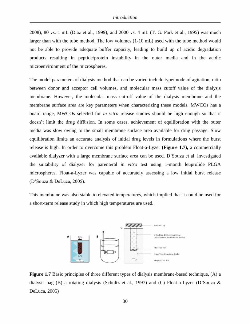
release is high. In order to overcome this problem Float-a-Lyzer (Figure 1.7), a commercially
available dialyzer with a large membrane surface area can be used. D’Souza et al. investigated
the suitability of dialyzer for parenteral in vitro test using 1-month leuprolide PLGA
microspheres. Float-a-Lyzer was capable of accurately assessing a low initial burst release
(D’Souza & DeLuca, 2005).
This membrane was also stable to elevated temperatures, which implied that it could be used for
a short-term release study in which high temperatures are used.
Figure 1.7 Basic principles of three different types of dialysis membrane-based technique, (A) a
dialysis bag (B) a rotating dialysis (Schultz et al., 1997) and (C) Float-a-Lyzer (D’Souza &
DeLuca, 2005)

Introduction
31
Advantages and disadvantages: The dialysis method is attractive because sampling and media
replacement are convenient because of physical separation of the microparticles from the outer
media by a dialyzing membrane.
A major problem with this method is a possible violation of sink conditions if diffusion through
the membrane is the rate determining step. To overcome this problem, a ‘reverse dialysis’
method has been proposed by Chidambaram et al. (Chidambaram & Burgess, 1999). In this
setup, the dosage form is placed directly in a large chamber containing the dissolution media and
the samples are withdrawn from microdialysis sacs immersed into the chamber, thus reversing
the volume ratio between donor and acceptor compartment.
Furthermore achievement of equilibration with the outer media is slow and would limit an
accurate analysis of initial drug levels in formulations where the burst effect is high (Janusz W
Kostanski & DeLuca, 2000). However, this issue was addressed by using dialysis bags (more
surface area) where initial release over 24 h was about 88% (J W Kostanski, Thanoo, & DeLuca,
2000).
Another disadvantage is that the time to equilibrate is prolonged if the bulk media is not stirred
(formation of unstirred water layer). In such situations, it is recommended that the outer media
be agitated to minimize unstirred water layer effects and to prevent accumulation of polymer
degradation products, especially when the formulation contains a protein (T. G. Park et al.,
1995). Also, this technique cannot be used if the drug binds to the polymer or membrane
(Kinget, Bontinck, & Herbots, 1979). However, because of the ease of sampling and the
possibility of total buffer replacement, this method seems to be an attractive option to study drug
release from microparticles and other particulate dosage forms.
Disadvantages with the dialysis technique are a cumbersome setup procedure for dialysis bags
(Diaz et al., 1999) and membrane at one end of a tube (Byung H Woo et al., 2001). This can be
addressed by using a commercially available dialyzer (D’Souza & DeLuca, 2005) having a large
surface area. Additionally, the regenerated cellulose membrane is stable up to 60°C, which
would allow its use in short-term elevated temperature studies (D’Souza et al., 2005; Shameem
et al., 1999). Also, the membrane may be washed and reused after each experiment, which would
render it cost effective.
Introduction
32
Several concerns are taken into consideration when perform a test for in vitro drug release of
parental controlled drug delivery system. These tests are often run over a long time period (e.g.,
several weeks to months). Due to a long duration of a drug release, the evaporation of the release
medium and the microbial contamination should be prevented. Suitable preservatives may be
added to prevent microbial contamination. Standard preservatives, including cetylammonium
bromide, benzalkonium chloride, parabens, phenol derivatives, mercury salts, and sodium azide
along with appropriate concentrations to be used, are listed in many pharmaceutical textbooks.
The selection has to be based on criteria such as compatibility with the active pharmaceutical as
well as other formulation ingredients and the pH of the test medium. Issues with these
compounds include their ionization properties, physicochemical interactions, or analytical
interferences. The composition of the medium should take into consideration the osmolarity, pH,
and buffer capacity of the fluids at the site of administration, which are usually assumed to
represent the condition of plasma or the physiological fluid but with lower buffer capacity.
It has to be mentioned that sink conditions do not always prevail at the site of administration in
vivo. Nevertheless, sink conditions should be used for standard in vitro dissolution testing to see
the release profile of the dosage form that would occur if no restrictions to distribution away
from the site of application were present (Burgess et al., 2002; D’Souza & DeLuca, 2006;
Washington, 1990). According to the USP, sink conditions are maintained when the volume of
medium equals at least three-times that required to form a saturated solution of the drug
substance. It has been suggested that the experimental concentration should be kept below 10%
of the concentration at saturation (Washington, 1990). Care must be taken when non-aqueous
solvents or solubilizing agents are added to the dissolution media to obtain sink conditions as the
release profiles may be greatly influenced by the concentration of the additives (Washington,
1990).
Moreover, the stability of active ingredients or drugs in the release medium is important. A drug
has to be stable in the in vitro condition at the determined temperature and at pH of the release
medium along the in vitro drug release test. However, the main challenges with this type of
dosage form are to determine the appropriate duration of the test and the times at which samples
are to be drawn in order to characterize the release profile adequately. While too long sampling

Introduction
33
interval can lead to misunderstand drug release manners from implants, excessively frequent
sampling is a waste of time.
Over the past decade, there have been attempts to compare in vitro test methods to study drug
release from parenteral drug delivery systems. Nastruzzi et al. studied the release of
bromocriptine mesylate from commercial Parlodel LA® microspheres using dialysis tubes and a
flow-through cell method and also compared the reproducibility between the two in vitro tests
(Nastruzzi, Esposito, et al., 2008). In the dialysis method, a dialysis tube containing 20 to 25 mg
microspheres was placed into 100 ml of 50 mM citrate buffer pH 3.5, whereas in the flow-
through cell method, 20 mg microspheres were packed in a column (45 × 9 mm) filled with 3 ml
of 50 mM citrate buffer pH 3.5 with a flow rate of 0.12 ml/min. Very different release rates were
obtained, although the overall shapes were similar. Greater release of drug with a longer time to
plateau occurred with the dialysis technique, whereas with the flow-through cell, the time to
reach the plateau was shorter, but the amount released was smaller.
Conti et al. assessed release from indomethacin PLA microspheres using the USP dissolution test
apparatus, rotating bottle apparatus, shaker incubator, and a recycling flow-through cell. The
following test conditions were employed to assess in vitro release in buffer (Conti, Genta,
Giunchedi, & Modena, 2008):
o USP XII paddle dissolution test apparatus: 1000 mL buffer at 100 and 200 rpm
o Rotating bottle apparatus: 100 mL buffer at 29 rpm
o Shaker incubator: 100 mL buffer at 60 and 120 strokes/min
o Recycling flow-through cell: 1000 mL buffer at a flow rate of 17 and 33 mL/min
For the indomethacin PLA microspheres, drug release was fastest with the recycling flow-
through cell with similar release profiles obtained using the USP dissolution XXII apparatus,
shaker incubator, and rotating bottle apparatus. Results from experiments with bromocriptine
mesylate and indomethacin microspheres were similar in that in vitro release with the flow-
through cell was faster. Studies on in vitro release of spray-dried rifampicin microspheres
formulated using a blend of R104 polymer (D,L-PLA, MW 2000) with R202H (D,L-PLA, MW
9000) were performed using bottles shaken horizontally in a water bath and with the USP
dissolution apparatus (Bain et al., 2008). Briefly, about 10 mg of microspheres was added to a

Introduction
34
bottle containing 100 ml dissolution medium with horizontal shaking at 1.5 Hz, whereas in the
USP XXII test method, release of 50 mg microspheres was assessed in 500 ml dissolution
medium agitated at 100 rpm at 37°C. The drug release was significantly faster with the USP
paddle apparatus. Because the ratio of microspheres to dissolution media was the same, faster
release with the USP paddle apparatus was attributed to the greater degree of agitation with the
paddle, which prevented the microspheres from forming aggregates at the base of the vessel.
When agitation was employed, the microspheres remained as individual particles and were
continually suspended in the media, resulting in faster release.
In recognition of the need for a standard in vitro release method, a series of national and
international workshops on quality assurance and performance of sustained and controlled
release parenterals have been conducted in recent years (Burgess et al., 2004, 2002; Siewert et
al., 2003). The issues addressed in addition to methodology were apparatus, outcomes,
parameters necessary for method development, and in vitro-in vivo correlation (IVIVC) for
sustained release parenteral dosage forms. The resulting publications included important
guidelines for novel or special dosage forms, including implants, injectable microparticles
formulations, and liposomes.
These delivery systems were categorized as those dosage forms requiring more work before a
method can be recommended. Some of the concerns in need of resolution included evaporation,
prevention of microbial contamination, osmolarity, pH, and buffer capacity of the media for
these extended release formulations, as the time to conduct in vitro studies would encompass
weeks or months.
For batch release testing and product development studies it is crucial to use the finished product
for the in vitro studies. With parenteral products, additional production steps such as sterilization
are often performed following the actual manufacture of the dosage form. These may influence
release behavior. For example, gamma sterilization may induce polymer degradation, leading to
changes in the average polymer molecular weight and the glass transition temperature and in turn
to changes in the release rate (Martinez et al., 2008). Besides changes in the polymer during
sterilization, the impact of the sterilization procedure on drug stability needs to be examined.

Introduction
35
Also, the procedure with which the dosage form is applied may have an influence on its release
profile. Consider, for example, the surface area that is formed after application of a depot
intramuscularly or subcutaneous injection (Hirano, Ichihashi, & Yamada, 1982; Weng Larsen &
Larsen, 2009). Even if such parameters will inevitably vary in vivo, the dissolution test design
should enable careful evaluation of their influence on release and relevant conditions should be
standardized in the in vitro setup. Some different approaches to deal with in-situ forming gels in
an in vitro test have been described briefly in a review by Larsen et al. (C. Larsen et al., 2009).
1.5.4 Accelerated release testing:
While real time in vitro release tests are necessary to gain a mechanistic understanding of drug
release and to help in formulation design, accelerated tests for controlled release parenterals are
essential for quality control purposes since real time release tests are of the order of weeks to
months (product specific). Currently, research is focused on shortening the time span of in vitro
release experiments with the aim of providing a quick and reliable method for assessing and
predicting drug release (Shameem et al., 1999; Siewert et al., 2003). Accelerated tests should be
predictive of real time tests and they should be designed as early as possible in the development
process in order to accrue sufficient data to underline the relationship between real time and
accelerated tests. For commercial dosage forms that release drug for 30- 90 days or even longer,
accelerated or short-term release provides the potential for conducting an in vitro release test in a
matter of days rather than months. Release testing of these dosage forms at 37°C would require
the addition of preservatives and impose certain limitations on the in vitro method, such as
stability and compatibility of the components of the release device, like tubings and membranes.
Therefore, a short-term release test might even be more reliable for quality-control purposes. In
addition, short-term studies can provide a rapid assessment of formulation and processing
variables that affect drug release from the delivery system, especially in the developmental
stages. These short-term studies can be performed by accelerating one or more conditions
employed in a real-time in vitro release study. There are several methods to accelerate release,
including elevated temperature, altering pH, use of surfactants, solvent. Upon acceleration the
mechanism of release may however change, as for example reported by Zolnik et al. (Zolnik et
al., 2006). When employing higher temperatures to accelerate release, particle aggregation may
pose a problem with disperse formulations (Shameem et al., 1999). Multiphasic release including

Introduction
36
an initial burst, which is often observed with modified release parenteral dosage forms, is often
impossible to adequately monitor in an accelerated release test. Therefore, an additional real-time
test of the initial phase of release is often advisable (Iyer et al., 2006; Martinez et al., 2008).
As with the real-time in vitro release study, the method should be simple, reproducible under the
conditions of study, inexpensive, and applicable to biodegradable microsphere formulations that
have varying duration of action in vivo.
1.5.5 More realistic release test systems for parenteral products:
Dissolution tests may be modified to include biorelevant conditions by adapting media
(including composition, volume and temperature, see above) or apparatus design. With regard to
specialized apparatus, systems have been introduced for solid oral dosage forms that simulate
certain aspects of physiological gastrointestinal passage, such as physicochemical and enzymatic
conditions, hydrodynamic shear stress or mechanical stress in the stomach and/or the
Intestine (Garbacz et al., 2008).
With parenteral products that are circulated through the vascular system with the blood, the
media volume and composition are most likely the main physiological factors to be reflected in
an in vitro test system. In this case, the apparatus may have less influence on release if
standardized and reproducible hydrodynamic conditions are achieved, and general principles of
dissolution testing are respected.
For parenteral products that are administered into tissue or confined spaces of the body, a
different situation may arise, in which the apparatus is also of greatest importance. Depending on
the surroundings, the release behavior of the dosage form and the physicochemical properties of
the released substances, sink conditions may not prevail locally at the site of release. Assuming a
diffusion layer model, this may lead to a deceleration of release from the dosage form due to a
decrease in concentration gradient. Also, the mass transport in living human tissue will be very
different compared with stirred media. The inclusion of basic physical properties representative
of the situation in vivo may be a first approach to adapting dissolution tests as it will be even
more complicated to include biological phenomena such as active transport or tissue reaction. In

Introduction
37
the following section, attempts to reflect such physical conditions in in vitro systems will be
summarized.
1.5.5.1 Subcutaneous Injection Site Simulator (Scissor):
Kinnunen et al. developed a novel in vitro system, termed Scissor (Subcutaneous Injection Site
Simulator) (Figure 1.8) (Kinnunen et al., 2015). Sirius Scissor is designed to mimic the stresses
a biopharmaceutical experiences when transitioning from formulation conditions to a
subcutaneous environment, in order to understand about the physical behavior of parenteral
drugs in the period immediately post-administration.
Scissor uses a dialysis-based injection chamber, which can incorporate various concentrations
and combinations of acellular extracellular matrix (ECM) components at the subcutaneous
injection site. This chamber is immersed in a container of a bicarbonate-based physiological
buffer that mimics the subcutaneous injection site and the infinite sink of the body. Such an
arrangement allows for real-time monitoring of the biopharmaceutical within the injection
chamber, and can be used to characterize physicochemical changes of the drug and its
interactions with ECM components. Movement of a biopharmaceutical from the injection
chamber to the infinite sink compartment simulates the drug migration from the injection site and
uptake by the blood and/or lymph capillaries. This system models environmental changes that a
biopharmaceutical could experience as it transitions from conditions of a drug product
formulation to the homeostatic state of the hypodermis following subcutaneous injection. Scissor
is designed to recreate, in an in vitro setting, dynamic events that could affect the fate of a
biopharmaceutical formulation delivered by subcutaneous injection in vivo. This transition
involves changes in pH, temperature, ionic components (with a loss of excipients), and transient
pressure changes due to volume introduction. The major drawback of this method is that it’s very
complicated and expensive. Furthermore, this method follow up factors that may affect the
bioavailability of biopharmaceutical after it is released from dosage form and does not consider
factors influencing dosage form and drug release from dosage form in vivo.

Introduction
38
Figure 1.8 Dynamic events in the hypodermis following the injection of a biopharmaceutical
formulation can be modeled using a modified dialysis system. A) Diagram of components and
events denoting the exchange of small molecules (salts, sugars, and other excipients), buffer
(transition to a bicarbonate-based environment), equilibration of temperature and pressure, and
potential interaction with extracellular matrix (ECM) components. B) Cartoon depicting
arrangement of positioned probes to continuously monitor pH inside and outside of the sample
injection chamber and positioning of light path (crenulated circle) in a large solute bath chamber
that emulates the infinite sink of the body. C) Cartoon depicting arrangement of injection
chamber/infinite sink compartment in a single-beam spectrophotometer that allows for
temperature control and stirring of the large bath contents. The numbering in B) and C) follows
the legend shown in C) (Kinnunen et al., 2015).
1.5.5.2 Capillary bioreactor device:
Iyer et al. introduced a method for release testing of a biodegradable monolithic rod implant
designed for subcutaneous implantation. This method is based on the idea of placing the implant,
as well as glass beads simulating barriers formed by cells, into the extra-capillary space of a
capillary bioreactor device (Figure 1.9) (Iyer, Barr, Dance, Coleman, & Karnes, 2007). The
capillary bioreactor included 50 polyether sulfone capillaries (diameter 0.5 µm, pore size not
reported), which were perfused by media at a flow rate of 1 ml/min. As the media was pumped
into the reactor, the extra-capillary space also filled with media. The system was operated in a
closed loop and the amount of drug released was determined from samples collected from the
media reservoir. Release with a modified flow-through cell which was used as a reference
method was faster, with an approximate doubling of the amount released after 90 days compared
with the capillary device. Those authors reported a linear one-to-one correlation of the results

Introduction
39
obtained using the capillary device with in vivo data. However, it has to be mentioned that even
though release was examined for 90 days only approximately 3% of the drug load had been
released during that time. The placement of the dosage form in between the capillaries and glass
beads is expected to be of great importance for this setup and slight changes in the arrangement
might lead to large deviations in the release behavior. Also, it remains to be investigated whether
glass beads are feasible of simulating properly the barriers formed by cells of the subcutaneous
tissue.
Figure 1.9 Capillary bioreactor device (left) top view with the implant positioned in the center
and (right) transverse section (Iyer, Barr, Dance, et al., 2007)
1.5.5.3 Using agarose gel as a dissolution test:
A different approach to simulate tissue implantation based on a hydrogel compartment has been
proposed for monolithic implants and microparticles (Allababidi & Shah, 1998; Hoang Thi et al.,
2010). The gels were chosen for their ability to mimic tissue according to their rheological
properties and water content, and used as acceptor compartments (Allababidi & Shah, 1998;
Hoffman, 2002). In comparison with stirred media, the convective transport in the hydrogel is
minimized but diffusion of small molecules is not hindered. This setup is thus more
representative of the situation in vivo. The embedding of the dosage form in the gels (agar or

Introduction
40
agarose) was slightly different in the different studies. In the case of the microparticles, a hole
was created in the center of the gel into which a suspension of the particles was added. The
monolithic implants were either placed in a hole in the gel and topped with an additional gel
layer or completely surrounded by gel, as depicted in Figure 1.10 (Allababidi & Shah, 1998;
Hoang Thi et al., 2010). This could be a very important difference, since in the first two settings
at least one surface of the drug containing compartment is in contact with a nondiffusible
compartment (apparatus wall and in case of the microparticles also the air above the particle
suspension). The change in contact area is expected to influence release. Upon implantation in
vivo all surfaces are expected to be in contact with tissue and therefore the complete surrounding
of the dosage form with the gel is the most favorable approach. In all setups samples of the
hydrogel were withdrawn at predetermined time points at various distances from the product and
drug content was determined. Thus, these methods also enabled the evaluation of spatial
distributions within the gels. In this context it should be mentioned that sink conditions should be
maintained in the overall system by providing a suitable amount of gel even though local
violations of sink conditions near the implant may occur in vitro as well as in vivo. Hoang Thi et
al. compared the results of their method, comprising agarose gels, to release testing in agitated
vials using an orbital shaker plate system at 100 rpm/min (Hoang Thi et al., 2010). Release from
all tested formulations was faster using the incubation method. The authors explained this
difference with the high concentration gradients at the interface between dosage form and media
as well as an observed fragmentation of the implant matrix in the agitated vials. (Gasmi et al.,
2016) employed an incubation method for reference. In that case, however, the release was
slower in agitated vials (horizontally shaken at 80 rpm/min) compared with the gel method.
Those authors attributed this deceleration of release to the prevention of local pH change by
acidic polymer fragments released in the stirred setup, whereas in the gel the pH underwent a
local decrease in pH resulting in autocatalysis of further polymer degradation. Allababidi et al.
on the other hand were not able to detect significant differences in cumulative release from their
model implant using their agar gel method and a sample and separate method used as a control
(horizontal shaker at 60 rpm/min) (Allababidi & Shah, 1998). Those results illustrated that the
outcomes of dissolution testing can be very different for quite similar methods depending on
which factors are controlling release from the dosage form.

Introduction
41
Figure 1.10 Schematic illustration of the experimental setup used for drug release measurements
into agarose gels (ci = drug concentration in the withdrawn gel sample; qi = drug amount in the
corresponding concentric ring) (Hoang Thi et al., 2010)
However, it has to be mentioned that even though the agar gel has a similar composition of
extracellular matrix, it cannot simulate the real environment of the dosage forms after
administration. Since the living tissues exhibit very different mass transport phenomena, in vivo
extracellular matrix consist of two phases: a gel phase containing primarily collagen, elastin, and
polysaccharides and a fluid phase made up of water and dissolved proteins in contrast to agar
gel, which consist only of a gel phase and doesn’t have any movement.
1.6 In vivo conditions at the site of implantation:
In general, when a product is administered into tissue, it will be in direct contact with the cells of
the respective tissue and interstitial fluid, the drug delivery system is in a lipophilic/hydrophilic
environment. After release from the dosage form the drug will be released into the extravascular
interstitial tissue fluid.

Introduction
42
In case of intramuscular or subcutaneous instillation, the drug is released into the extravascular
interstitial tissue fluid. The structural characteristics of interstitial space are similar in all tissues,
ultrastructural studies suggest that the interstitium is comprised of two phases: a gel phase
containing primarily collagen, elastin, and polysaccharides (e.g., hyaluronate,
glycosaminoglycans); and a fluid phase made up of water and dissolved proteins (Chary & Jain,
1989). The glycosaminoglycans are polyanionic polysaccharides that are charged at
physiological pH (7.4) and are bound covalently to a protein backbone to form immobilized
proteoglycans. The proteins present in the interstitial space are qualitatively the same as those
present in plasma, although quantitatively, they are present in lower concentrations. This results
in the interstitial colloid osmotic pressure (COP1) being less than that in plasma (Aukland &
Reed, 1993). Although the components of interstitium are principally the same in all tissues,
their relative amounts vary greatly (Aukland & Reed, 1993). It is believed that a network of
endogenous macromolecules effectively reduces the distribution volume such that the interstitial
space acts in a size exclusion manner, excluding very large molecules, and thereby affecting their
interstitial occupancy (Bert, Mathieson, & Pearce, 1982; Watson & Grodins, 1978).
The movement of fluid in tissues under normal and pathological conditions is generally
explained in terms of Starling's hypothesis. This hypothesis suggests that fluid is filtered at the
arterial end of a vessel and mainly reabsorbed at the venous end. This fluid movement
presumably leads to convective transport of macromolecules in the interstitium. The active agent
is released into the tissue fluid and has to traverse the interstitium to reach a blood capillary or a
lymphatic vessel by restricted diffusion in the gel phase and by convection and free diffusion in
the fluid phase or possibly via transporters until reaching a blood capillary or a lymphatic vessel.
Drug absorption from intramuscular and subcutaneous sites of depot injection may share some
gross common features (Medlicott, Waldron, & Foster, 2004). Absorptive processes that may
occur on injection into muscle or subcutaneous sites are presented in Figure 1.11
The size exclusion-like properties of the interstitium significantly reduce diffusion of plasma
proteins and other macromolecules in the extracellular matrix. Compared with the interstitial
fluid drainage by the lymph, the rate of filtration and reabsorption of fluid across the vascular
capillaries is ~ 10-fold higher. Thus, drug molecules (with molecular masses below ~ 2 kDa),
which are capable of entering blood and lymph capillaries at comparable rates, will be cleared

Introduction
43
predominantly by the blood vessels (Porter, Edwards, & Charman, 2001). On the other hand,
macromolecules above ~ 16 kDa as well as particulates are preferentially removed from the
tissue by the lymphatics (Zuidema, Kadir, Titulaer, & Oussoren, 1994). The contribution of
lymphatic uptake of macromolecules to the overall absorption process seems to be of most
importance in relation to the subcutaneous injection site because only a few lymph vessels are
located in muscle tissues (B. E. Ballard, 1968).
Figure 1.11 Possible pathways for absorption of drugs from controlled release parenteral dosage
forms at intramuscular or subcutaneous sites (C. Larsen et al., 2009)
Normal blood flow to subcutaneous tissue is 1.5–2.5 mL/100 g/min (Benet, 1990; Enevoldsen,
Simonsen, Stallknecht, Galbo, & Bulow, 2001) the blood flow to intramuscular tissue is higher.
However, tissue blood flow is subject to high variability such as exercise, pathology diurnal
changes, body position, etc. and may be of crucial importance to the resulting plasma profiles if
release from the dosage form is not the rate-limiting factor. Estimation of the various rate
constants would require an in vivo input rate, obtained through intravenous administration. In

Introduction
44
addition, an estimation of drug concentrations at the interstitial site, possibly involving
microdialysis studies would also provide useful information regarding disposition of the drug.
In sustained release parenterals, however, the ratelimiting step in the absorption is controlled by
the delivery system, thereby reducing potential inter-injection and interpatient variability
resulting from differences in injection site perfusion (Medlicott et al., 2004).
An additional barrier to diffusion is provided by cells, depending on the lipophilic character of
the drug, it may be partitioned into the adipose cells, and be released back into interstitial fluid as
a function of −kD, the re-distribution rate. This could lead to formation of a localized depot,
represented by the drug concentration, Ca, in the tissue.
Also, when administering parenteral dosage forms that deliver over very long periods of time,
changes of the surrounding tissue, as for example inflammation and formation of fibrous
capsules due to foreign body reaction, have to be taken into account (F. D. Anderson, Archer,
Harman, Leonard, & Wilborn, 1993).
The tissue reaction (intensity as well as duration of the inflammatory and wound healing
processes) is dependent on the size, shape, and chemical and physical properties of the
biomaterial (J. M. Anderson & Shive, 2012).
The host responds at the site of parenteral drug administration include acute inflammation,
chronic inflammation, granulation, foreign body reaction and fibrosis (Keselowsky et al., 2007).
The formation of fibrous capsules around drug delivery may take place over days to weeks and
for more long-acting depots, this event may contribute to the overall variability of in vivo drug
performance (F. D. Anderson et al., 1993). Also, the fact that microparticles < 10 µm may be
phagocytosed or engulfed by macrophages and foreign body giant cells in the inflammatory and
healing responses may affect variability.
Therefore, biological factors to consider when developing an in vitro release test in case of
intramuscular or subcutaneous instillation include:
o the intramuscular or subcutaneous environment, its configuration, foreign body response such as
immune system cells and encapsulation;

Introduction
45
o the in vivo release mechanism of the drug delivery system (e.g., diffusion, erosion or osmosis);
o in vivo uptake of the drug and
o the complex interaction between the host and the drug delivery system.
The biological environment surrounding the formulation is not static, and it may not be possible
to capture some of the biologically relevant reactions through the use of in vitro systems, such as
inflammation with consequent cellular recruitment and fibrosis capsule formation.
Also, when the formulation is liquid like oil vehicle or in situ implants, differences in the
interfacial area between the surface of the depot and the aqueous tissue fluid, which cause
variability in the overall apparent release rate, may arise from less predictable spreading (oils and
gels) or microparticles aggregation phenomena taking place at both subcutaneous and
intramuscular injection sites.
1.7 In vitro–in vivo correlation of parenteral controlled release drug delivery systems:
The development of suitable in vitro release models (for quality control as well as formulation
development purposes) is a critical activity, which, preferably, should be initiated in the early
depot design phase. These efforts should ideally lead to the establishment of an in vitro–in vivo
correlation (IVIVC) (C. Larsen et al., 2009). An IVIVC imparts in vivo validation to the in vitro
dissolution test, which can then be used as a surrogate for bioequivalence testing. In addition,
more meaningful dissolution specifications can be set using the concept of an IVIVC (Uppoor,
2001). A Guidance document was issued by the Food and Drug Administration in an effort to:
(a) reduce the regulatory burden by decreasing the number of biostudies needed to get approval
and maintain an extended release product on the market and (b) set dissolution specifications that
are more meaningful clinically. The ultimate goal is that demonstration of valid IVIVCs would
allow many of the biostudies that are generally required for major manufacturing changes to be
replaced by simple in vitro dissolution tests. The ideal approach to IVIVC modeling is to develop
one IVIVC model for the total plasma profile, but other approaches might also be pursued
(Byung H Woo et al., 2001). Importantly, the development of a true IVIVC requires that a
mathematical model describes the in vitro–in vivo relationship for two or more formulations
showing different release characteristics (Young, Farrell, & Shepard, 2005). When a meaningful
IVIVC has been established, it can be used as a surrogate for bioequivalence and for minimizing

Introduction
46
the number of bioequivalence studies to be performed during drug product development
(Uppoor, 2001). Four categories of correlations (A–D) have been described in the guidance. A
Level A correlation represents a point-to-point relationship, generally linear, between in vitro
dissolution rate and the in vivo input rate. A Level B correlation involves the principles of
statistical moments. The mean in vitro dissolution time is compared either to the mean residence
time or the mean in vivo dissolution time. This is not considered to be a point-to-point correlation
and because a number of different in vivo curves will produce similar mean residence time
(MRT) values, this cannot be considered discriminatory for different formulations. A Level C
IVIVC represents a single point relationship between a dissolution parameter (such as percent
dissolved at a particular time) and a pharmacokinetic parameter of interest, e.g. the area under
curve (AUC). However, it does not reflect the complete shape of the plasma concentration curve,
which is the critical factor that defines the performance of extended release products. A Level C
correlation, although useful to screen and rank-order formulations in animal models during drug
development, cannot be used for biowaivers or bioequivalence. The fourth category, D, is a
multiple Level C correlation and it represents a relationship between one or more
pharmacokinetic parameters, and the amount of drug dissolved, at multiple points of time on the
dissolution profile (Iyer et al., 2006).
At best, the in vitro model may mimic the in vivo conditions to such an extent that a level A
IVIVC can be established. This usually requires that drug release from the depot be the rate-
limiting step in the absorption process and that the drug release mechanism is the same in vitro
and in vivo. In this case, performance of in vitro release tests might be accepted instead of an in
vivo bioequivalence study (biowaiver) for the evaluation of minor changes in the composition or
manufacturing process of an already marketed product.
The lack of true IVIVCs can most likely be ascribed to barriers related to intrinsic drug
properties or the inability to design in vitro methods sufficiently capable of mimicking the in
vivo conditions at the injection site.
Often, it has been emphasized that in vitro release model development should be based on
knowledge of the in vivo drug release mechanism (Burgess et al., 2004, 2002; Martinez et al.,
2008). So far A lot of investigation has been done on the effect of the pH, ionic strength and

Introduction
47
temperature of the release medium, and agitation and flow rate (in continuous flow methods) on
the resulting drug release kinetics from PLGA-based formulations (Faisant, Akiki, Siepmann, &
Benoit, 2006; Rawat, Bhardwaj, & Burgess, 2012). However, very little is known on the impact
of in vivo conditions on the drug release mechanisms.
In general, with increasing duration of therapeutic activity, two or more in vivo events may
contribute to the overall drug release characteristics. Thus, it is suggested that the probability for
successful development of a suitable in vitro release model (for batch control and IVIVC), so to
speak, is inversely proportional to the intended duration of action of the formulation.
Although parenteral controlled drug delivery systems have been used for intramuscular and
subcutaneous administration for several decades, the in vivo drug release mechanism(s) are far
from fully elucidated.
In vivo phenomena which are difficult to simulate using an in vitro release model include exact
composition of tissue, non-predictable host responses, spreading and dispersion of the liquid
formulation (oil vehicle and in situ implant) at the injection site.
Zolnik et al. investigated two PLGA microsphere formulations, with different polymer molecular
weights to determine whether an in vitro and in vivo relationship could be established for
dexamethasone release (Zolnik & Burgess, 2008). A USP apparatus 4 was used for in vitro
testing. The in vivo release kinetics and pharmacodynamic effects of dexamethasone were
evaluated using a Sprague Dawley rat model. The in vitro release from both formulations
followed the typical triphasic profile of PLGA microspheres (initial burst release, followed by a
lag phase and a secondary zero-order phase). The in vivo release profiles differed in that the lag
phase was not observed and drug release rates were faster compared to the in vitro studies. It is
speculated that the lack of lag phase in vivo may be a result of different PLGA degradation
mechanisms in vivo as a consequence of the presence of enzymes as well as other in vivo factors
such as interstitial fluid volume, and local pH. A linear in vitro–in vivo relationship was
established after normalization of the time required to reach plateau for the in vitro and in vivo
data and the in vitro release data were predictive of the in vivo release.

Introduction
48
It was also reported that release rate from polylactic acid (PLA) intrascleral implant was in vivo
faster than in vitro. The faster in vivo versus in vitro release rate may have reflected the decrease
in pH inside the implant that occurred while it was imbedded in the eye. This drop in pH
enhanced the autocatalysis occurring at the center of the polymer matrix. Furthermore, a burst
release, which was seen in vitro, did not occur in vivo (Okabe et al., 2003).
The drug release of commercially available risperidone PLGA-based microparticles (Risperdal®
Consta) was investigated t in vitro and in vivo (Rawat et al., 2011). A modified USP apparatus 4
was used for in vitro testing. The in vivo release kinetics and pharmacodynamic effects of
risperidone were evaluated using human model and microspheres were placed in the
intramuscular space. The in vivo profile of commercial Risperdal® Consta microspheres differed
from the real-time in vitro profile and was faster initially and then slower after approximately 30
days. This effect is considered to be due to differences in the in vivo conditions such as small
interstitial volume, low pH and immune response.
Risperidone implant was investigated to determine whether an in vitro and in vivo relationship
could be established for drug release (Amann, Gandal, Lin, Liang, & Siegel, 2010; Rabin et al.,
2008). A sample and separate method was used for in vitro testing. The in vivo release kinetics
and pharmacodynamic effects of risperidone were evaluated using a Sprague Dawley rat model
and implants were placed in the subcutaneous space. A level B correlation yielded between in
vitro and in vivo data.
The authors confirmed that mass loss (microsphere weight) and PLGA molecular weight
decreased at a faster rate in vivo. It was hypnotized that the absorption of acidic oligomers in vivo
is faster, which were probably not as soluble under in vitro conditions. Faster enzymatic
degradation in vivo was also stated as a reason for higher mass loss (Heya et al., 1994; Tracy et
al., 1999).
1.8 Application of the described release setups to implants:
Implants are usually solid polymeric devices with a drug load and a release mechanism that
ensures the amount of drug being delivered per time unit throughout their residence time in the
body. They may be biodegradable and may require medical assistance for insertion and removal

Introduction
49
if necessary. They may either act locally or systemically. The residence time varies from days to
years. The difference from the medical devices is that the drug release, rather than the physical
effect, is their primary purpose.
The majority of the dissolution tests reported for implants have been performed using incubation
methods comparable with the sample and separate setup without subsequent filtration or
centrifugation (Ghalanbor, Körber, & Bodmeier, 2010, 2012, 2013). Advantages of this method
include ease of handling, the lack of need for specialized apparatus and the large number of
experiments that can be conducted simultaneously. Furthermore, when incubating implants in
closed vials or tubes, evaporation is not an issue and complete media changes can easily be
performed by transferring the implant to a new container. Care has to be taken, however, to
ensure that sink conditions are not violated at any time with these typically small volume setups.
The flow-through method also represents a feasible approach to dissolution testing of implants.
The most striking advantage of using the flow-through cell is that the flow conditions can be
adapted to the flow conditions present at the site of in vivo release. The impact of different flow
rates on release from implant has been studied. As already described above, the system can be
operated over a wide range of media volumes, which can easily be adapted with each experiment
to assure sink conditions. Noncompendial cells have been introduced: for example a low volume
implant cell with an inner diameter of 6 mm and a cell volume of 1 ml, which reduces the total
minimum volume of medium to approximately 15 mL (Looney & Cqrporation, 1996).
Evaporation can also be controlled by using sealed media containers.
As reported previously, the in vivo- in vitro correlations of parenteral controlled drug delivery
systems are usually poor. A major limitation of all methods for dissolution testing of implants is
that the dosage form is placed directly in the dissolution media. This does not represent the in
vivo environment, in which the implant is typically in contact with cells and tissue fluids. Until
now, the effect of mechanical properties of the tissue has not been studied and very little studies
are done about the effect of the lipophilic environment of the tissue on the formulation and drug
release from biodegradable implants.

Introduction
50

Introduction
51
1.9 Objectives
The purpose of this work was:
o to evaluate the swelling of the implant ex vivo and to quantify the tissue pressure acting
on PLGA-based matrix implants ex vivo
o to design a new in vitro test to mimic mechanical properties of the tissue for solid
parenteral DDS and to investigate the effect of restricted swelling on drug release from
PLGA-implants
o to apply biphasic release test model as a new biorelevant model for drug release from
PLGA-implants and investigate its effect on risperidone release from PLGA-based
implants

2. Materials and Methods

Materials and Methods
53
2.1 Materials
2.1.1 Model drugs
Risperidone (Wuxi Jida Pharmaceutical Co. Ltd, China); Theophylline (BASF AG,
Ludwigshafen, Germany) and Ibuprofen (BASF AG, Ludwigshafen, Germany).
Table 2.1 Solubility of model drugs in phosphate buffer pH 7.4 at 37°C
Drugs Solubility
(mg/mL)
Physicochemical
properties
Risperidone 0.334 Basic
Theophylline 12.700 Non-ionic
Ibuprofen 7.000 Acidic
2.1.2 Polymers
Poly (lactide-co-glycolide) (PLGA) Lakeshore 5050 DLG 1A, Lakeshore 5050 DLG 2A,
(Evonik, Kirschenallee, Darmstadt, Germany); PLGA Resomer® RG 502H, PLGA Resomer®
RG 503H (Boehringer Ingelheim Pharma GmbH & Co. KG, Ingelheim, Germany).
Table 2.2. Inherent viscosity of PLGA
Polymers Inherent viscosity (dl/g) (1)
5050 DLG 1A 0.05 – 0.15
5050 DLG 2A 0.15 – 0.25
PLGA RG 502H 0.16 – 0.24
PLGA RG 503H 0.32 – 0.44

Materials and Methods
54
(1) Obtained from the measurement of 0.1% solution in chloroform at 25°C and provided by
Boehringer Ingelheim Pharma GmbH & Co. KG, Ingelheim, Germany
2.1.3 Other Excipients
Potassium dihydrogen phosphate, disodium hydrogen phosphate, sodium hydroxide (NaOH),
sodium azide (Merck KGaA, Darmstadt, Germany); hydrochloric acid (HCl) (Carl Roth GmbH
& Co. KG, Karlsruhe, Germany), tetrahydrofuran (Carl Roth GmbH & Co. KG, Karlsruhe,
Germany); magnesium stearate (Baerlocher GmbH, Unterschleissheim, Germany), 1-octanol,
blue dextran (Sigma-Aldrich Chemie GmbH, Steinheim, Germany), olive oil (Lipoid GmbH,
Ludwigshafen, Germany), Flowlac® 100 (Meggle GmbH, Wasserburg, Germany), the spin label
4-(methylamino)-2-ethyl-5,5-dimethyl-4-pyridine- 2-yl-2,5-dihydro-1H-imidazol-1-oxyl (MEP)
(Magnettech GmbH, Berlin, Germany).
2.2 Methods
2.2.1 Preparation of PLGA implants using hot melt extrusion
For ex vivo and in vitro studies powder blends of PLGA (Lake shore 5050 DLG 2A) and the blue
dextran were manually mixed in a mortar with a pestle. Blue dextran was used in order to make
the implants visible and to extract them easier from the tissue. Cylindrical implants were
prepared by hot melt extrusion using a co-rotating conical twin screw extruder (HAAKE
MiniLab Rheomex CTW5, Thermo Electron) at 80°C and a 1.5 mm extrusion die. Cylindrical
matrices were obtained with a diameter of 1.5 – 2.5 mm. The implants were cut into 2.5 - 4 mm
length. All experiments were performed at least in triplicate.
For biphasic studies polymer blends of PLGA (Lake shore 5050 DLG 1A) and the model drug
risperidone were mixed in a 9:1 and 7:3 ratio w/w in a mortar with a pestle. 1 ml polypropylene
syringes (LUER LOK™, B-D®, Singapore) were filled with approximately 1g of properly mixed
formulation blends. The syringes were placed in a self-built die, fixed and heated at 105°C in an
oven for 10 min. The molten blends were extruded manually, producing cylindrical matrices
with diameters of 1.1 – 1.2 mm. The matrices were cut into 3-5 mm length.

Materials and Methods
55
2.2.2 Preparation of PLGA implants using direct compression
For risperidone, three different PLGA implants were prepared with polymer blends of PLGA
(Lake shore 5050 DLG 1A, 502H and 503H), whereas for ibuprofen and theophylline implants,
only PLGA 503H was used. In all cases, PLGA and drug were mixed in a 9:1 ratio w/w in a
mortar with a pestle. Magnesium stearate was added as lubricant to each blend (0.5% wt).
Implants were prepared by manually compressing the powder mixture into 8 mm flat faceted
implants with an instrumented single punch tableting machine (EK0, Korsch AG, Berlin,
Germany). The implants were characterized with regard to their dimensions and hardness
(Multicheck, Erweka GmbH, Heusenstamm, Germany). Additionally, implants containing only
drug (10%) and lactose as filler were prepared to study drug diffusion through the device.
Implants containing nitroxyl radical MEP and PLGA were prepared for EPR measurement. The
nitroxyl radical MEP was dissolved in ethanol, mixed in a mortar with a pestle with polymer, and
dried overnight, thus achieving a final concentration of 5 mmol/kg powder.
2.2.3 Restricted swelling device
A novel device was fabricated to prevent the swelling of formulations described results and
discussion in section 3.B.2.4.
2.2.4 Adsorption test of the restricted swelling device
A solution of the drug in distilled water (2.76%) was filtered three times through a glass filter of
the device. The concentration of the solution was determined using UV/VIS spectrophotometer
(Shimadzu UV HP 8453, Shimadzu Japan) before and after filtration.
2.2.5 Diffusion through the restricted swelling device
Implants containing only drug (10%) and Flowlac 100 used as filler were placed in the device
and the release study was carried out by the method previously described in section 2.2.7. Sink
conditions were maintained throughout the study time.

Materials and Methods
56
2.2.6 Drug extraction from implants
To quantitatively detect the actual drug loading of implants, 2 mL of 2M NaOH was added to
vials each containing an accurately weighed implant. After 24 hours when the implants were
completely dissolved, 3 mL of 2M HCl was added to the vials, in order to dissolve the drug. The
solution was analyzed using UV/VIS spectrophotometer (Shimadzu UV HP 8453, Shimadzu
Japan) with a Peltier thermostated cell holder (Agilent 8453, Agilent Technologies Inc., Palo
Alto, USA) equipped with UV-Chemstation biochemical analysis software at 277 nm, for
risperidone implants, and with single wavelength background correction at 400 nm.
2.2.7 In vitro drug release study
Implants prepared using direct compression were placed in the restricted swelling devices, which
were placed in the 120 mL custom-made glass bottles filled with phosphate buffer pH 7.4.
Unrestricted implants were placed directly in the 120 mL custom-made glass bottles filled with
110 mL phosphate buffer pH 7.4. The release study was carried out under sink conditions. These
bottles were incubated in a vertical position in the horizontal shaker (80 rpm, 37 °C; Gesellschaft
für Labortechnik mbH, Burgwedel, Germany). At predetermined time points, 5 mL samples were
withdrawn and replaced with fresh release medium. The drug amount in the release medium was
quantified UV-spectrophotometrically at 277 nm for risperidone, 222 nm for ibuprofen and 271
nm for theophylline (Shimadzu UV HP 8453, Shimadzu Japan).The test was performed in
triplicate (n = 3). The drug release of restricted implants was compared to that of unrestricted
ones.
Implants prepared using hot melt extrusion were placed in Duran® 50 mL glass bottles filled with
phosphate buffer pH 7.4. These bottles were incubated in a vertical position in the horizontal
shaker (80 rpm, 37 °C; Gesellschaft für Labortechnik mbH, Burgwedel, Germany). At
predefined sampling points, 5 mL of release sample was withdrawn and was replaced with fresh
release medium. Testing was performed in triplicate (n = 3). In monophasic system, 60 mL
phosphate buffer pH 7.4 and in biphasic system containing octanol, 45 mL phosphate buffer pH
7.4 and 15 mL 1-octanol were used as dissolution media. Drug concentrations in release samples
were quantified in both phases spectrophotometrically at 277 nm for risperidone (Shimadzu UV
HP 8453, Shimadzu Japan). For release study in biphasic system containing olive oil, at

Materials and Methods
57
predetermined times implants were taken out of the release medium and the remaining amount of
drug in implants was determined using extraction method.
2.2.8 Degradation study and molecular weight determination
At predetermined time points, implants were withdrawn from release medium, vacuum dried for
48 h, and dried formulations were incubated in 2 mL tetrahydrofuran for five hours. The turbid
samples were centrifuged at 17000 rpm for 15 minutes (HeraeusTM BiofugeTM, Thermo Fisher
Scientific Inc., Waltham, MA, USA) to remove the particles and the supernatant was analyzed
for the molecular weight distribution of the remaining polymer by gel permeation
chromatography. Gel permeation chromatography (GPC) analysis was carried out using
Shimadzu (Shimadzu, Tokyo, Japan) LD-10 liquid chromatograph equipped with degasser,
pump, auto-injector and column oven in combination with Viscotek triple detector (TDA-300,
Viscotek, Malvern Instruments Ltd., Malvern, UK) operated in double mode (differential
refractive index, viscosimetry). A column with a linear range from 1200 g/mol to 18,000 g/mol
(Mesopore 7.5 μm x 300 mm; Varian Inc., Darmstadt, Germany) and tetrahydrofuran were used
as stationary and mobile phases, respectively. The sample concentration was 20 mg/ml with the
corresponding injection volumes of 25 μl. Column and detector were operated at 30 °C and the
flow rate was 1 ml/min. A refractive index (RI) detector was used to determine the molecular
weights of PLGA, which was obtained from polystyrene standards with peak molecular weights
of 1,260 g/mol, 2,360 g/mol, 4,920 g/mol, 9,000 g/mol, 19,880 g/mol (Varian Inc., Darmstadt,
Germany). Data acquisition was performed using Omnisec software (Viscotek, Malvern
Instruments Ltd., Malvern, UK).
2.2.9 In vitro and ex vivo experiments
For the in vitro experiment, implants were incubated in 60 mL of 0.1 M phosphate buffer pH 7.4,
at 37°C. For the ex vivo experiments, implants were implanted into turkey breast, the small
incision in the tissue was closed using tissue glue and they were incubated in either 1 mL (to
avoid tissue dehydration) or in 60 mL (analogue to the in vitro release set-up) of 0.1 M
phosphate buffer pH 7.4 containing 1% sodium azide as preservative at 37°C. Samples were
taken at predetermined points for analysis. At each point, all implants were removed from old
tissue and implanted into a fresh turkey breast under ex vivo experiments. For both conditions,

Materials and Methods
58
samples were taken at predetermined points and the swelling, water uptake and shape changes of
the implants were assessed.
2.2.10 Determination of tissue force ex vivo
The tissue force applied to the implant ex vivo was determined with a balloon catheter in fresh
turkey breast. For this purpose, a 3 mL Luer Lock syringe was filled with water and connected to
a balloon catheter (OTW PTA balloon catheter). The plunger was placed in contact with the
probe of a texture analyzer (Stable Micro System®, Vienna Court, UK) and the injection force
was measured as a function of the plunger displacement (Figure 2.1). The displacement was set
to 10, 15, 17.5, 20 and 25 cm and the injection speed was 15 mm/min. To find the appropriate
test settings leading to a linear relationship between injection force and simulated tissue force (0
N, 10 N, 20 N), the force reached after a certain displacement was measured with the balloon
catheter in air (blank) and with a weight of 1 and 2 kg placed on the balloon. Once the
appropriate settings were determined, the balloon catheter was inserted in fresh turkey breast in
order to measure the force applied to the implants ex vivo. The injection force in this ex vivo
experiment (average of at least 3 measurements) was used to calculate the force applied to the
formulation ex vivo.
Figure 2.1 Illustration of the setup for injection force measurement

Materials and Methods
59
2.2.11 Differential scanning calorimetry (DSC)
DSC-studies (Mettler Toledo 822e) of implants after extrusion and after 1 day of release
(~10mg) were recorded using a heating rate of 20°C /min under nitrogen atmosphere from -20°C
to 80°C cooled to -20°C and heated again to 180°C at a rate of 5°C/min. Each thermogram
obtained was normalized for sample weight and the glass transition temperature was determined
using the Stare Software.
2.2.12 Water content of the turkey breast
Hydration and dehydration of tissue was studied by monitoring the water content of the turkey
breast after incubating it alone or in 60 mL of PBS at 37°C over a period of 5 days. Water uptake
of turkey breast meat was quantified by weight gain at predetermined time points (Equation 2.1).
Water uptake (%) = (𝑊𝑡𝑖− 𝑊0)
𝑊𝑜 × 100 (2.1)
Wti: weight wet at time ti
W0: initial weight
2.2.13 Water uptake study
Water uptake of the implants was determined by their weight gain during the incubation time
(Equation 2.2). At predetermined time points (ti), the implants under both, restricted and
unrestricted conditions, were taken out of the release medium, in in vitro and ex vivo studies the
implants were taken out of the incubation medium or extracted from the turkey breast meat and
weighed after the excess of water at the surface had been removed with filter paper. The wetted
samples were then dried under vacuum to constant weight. The water uptake study was done for
12 days for implants under restricted versus unrestricted condition, 11 days for the implants in
monophasic versus biphasic studies and 7 days for the implants in in vitro versus ex vivo studies,
thereafter handling of the formulations was not possible.
Water uptake (%) =(𝑤𝑒𝑖𝑔ℎ𝑡 𝑤𝑒𝑡−𝑤𝑒𝑖𝑔ℎ𝑡 𝑑𝑟𝑦)
𝑤𝑒𝑖𝑔ℎ𝑡 𝑑𝑟𝑦 × 100 (2.2)

Materials and Methods
60
2.2.14 Diameter increase
To study the swelling of the implants, the diameter of the implant was determined in its initial
dry form (D0) and at predetermined time points (Dti) during the incubation time. Implants were
taken out of the release medium and their diameter was determined using a macroscope. The
diameter growth was calculated as followed (Equation 2.3):
Diameter increase (%) =(𝐷𝑡𝑖− 𝐷0)
𝐷0× 100 (2.3)
To study the swelling of the implants, the surface area of the implant was determined in its initial
dry form (A0) and at predetermined time points (Ati) (Equation 2.4) over seven days. The
implants were removed from the incubation medium (in vitro) or turkey breast (ex vivo). The
diameter and length of implants were measured using a macroscope.
Surface area change (%) =(𝐴𝑡𝑖− 𝐴0)
𝐴0× 100 (2.4)
2.2.15 Determination of implant morphology by optical macroscope
Implant morphology, changes of the implant’s shape and size were investigated using a
macroscope (Inteq Informationstechnik GmbH, Berlin, Germany). The implants were observed
and size was measured at predetermined time points before incubation and during the drug
release study. The images were recorded by an image analysis software (EasyMeasure, Inteq
Informationstechnik GmbH, Berlin, Germany).
2.2.16 PLGA mass loss
To study the mass loss of the implants, the weight of the formulations was determined in its
initial dry form (W0) and then at predetermined time points (Wti) (Equation 2.5). The
formulations were removed from the release medium and dried in the vacuum oven to constant
weight (dry weight) (Heraeus oven VT 5042 EKP, Hanau, Germany coupled with a chemistry
hybrid pump, Vacuubrand GmbH, Wertheim, Germany). The mass loss study was done for 12
days for the implants under restricted versus unrestricted condition and 13 days for the implants
in in vitro versus ex vivo studies. Thereafter handling of the formulations was not possible.

Materials and Methods
61
Mass loss (%) =(𝑊𝑡𝑖− 𝑊0)
𝑊0× 100 (2.5)
W0: initial weight
Wti: dry weight at time ti
2.2.17 Determination of µpH using EPR
In order to prepare matrix implants for EPR imaging experiments, the nitroxyl radical MEP was
dissolved in ethanol and blended with polymer, thus achieving a final concentration of 5
mmol/kg powder. Implants were prepared as described in section 2.2.2.
At predetermined time intervals, restricted and unrestricted implants were withdrawn from the
incubation medium, carefully placed on a Teflon plate and EPR spectra were recorded. After the
measurements, the implants were placed back into the device and in the incubation medium.
After 16 days, no more solid signals were detectable indicating that the spin probes migrated
with the solvent outside the implants
The spin label 4-(Diethylamino)-2-ethyl-5,5-dimethyl-2-pyridine-4-yl-2,5-dihydro-1H-imidazol-
1-oxyl was chosen based on its pKa. Nitroxide spin probe was calibrated in citrate and phosphate
buffer solutions in order to cover a pH range between 2.2 and 7.6. EPR spectra were recorded as
the first derivative of the absorption signal, and the hyperfine splitting aN was measured as the
distance between the low-field (mI = +1) and central (mI = 0) lines. The measurements of the
hyperfine splitting constant aN of spin label with different pH values served as calibration curve.
A sigmoidal Boltzmann was used to fit the data. The determination of accurate pH was possible
in the range from 2.2 to 7 with this spin label
EPR spectra were recorded at a frequency of 9.4 GHz using an X-band ERS 220 spectrometer
(ZWG, Berlin, Germany) with a custom-made tomography extension, equipped with a
rectangular resonator. The EPR parameters used were as follows: microwave power, 2 mW;
modulation amplitude, 0.1 mT; scan width, 10 mT; maximum gradient, 4.27 T/m; scan time per
projection, 10 s; 95 projections with 512 points per projection; image matrix, 256 × 256 points.
Images were obtained from the recorded set of EPR spectra by means of deconvolution and
image reconstruction.

3. Results and Discussion

A
Investigation of the effect of tissue pressure on the shape, swelling and water
uptake of PLGA-based matrix implants and quantification of tissue pressure
acting on the formulation after implantation

Results and discussion
64
3.A.1 Background
Advances in the development of controlled release parenteral formulations were made during
recent years to overcome issues with patient acceptance and compliance related to multiple
injections in the treatment of some chronic diseases. Furthermore, sustained or controlled
parenteral delivery offers opportunities like reduced side effects as a result of long-lasting
constant drug plasma levels and localized delivery in the body (Burgess et al., 2004; Rawat et al.,
2012). Polymers used in parenteral controlled release systems are divided into two main
categories: non-biodegradable and biodegradable polymers. PLGA has been used to a wide
extent as biodegradable polymer in drug delivery systems. The reasons for the widespread use of
PLGA are its biodegradability, its biocompatibility, and the fact that PLGA have been approved
for parenteral use by regulatory authorities. A disadvantage of this polymer is that drug release
from formulations containing PLGA in vivo differs often from in vitro. In order to improve
IVIVC, in vivo factors, influencing PLGA formulations must take into consideration.
PLGAs absorb a large amount of water and are prone to swell. The degradation starts with the
water uptake. During degradation, the molecular weight of the polymer decreases and it becomes
more hydrophilic and polymer chains become more mobile, which further promotes polymer
swelling (Fredenberg et al., 2011).
However after implantation of a PLGA drug delivery system in the body, the formulation is in
contact with tissue cells (Iyer et al., 2006). The tissue applies a force on the formulation, which
could lead to restricted swelling of the implant and change of its shape. Furthermore, water
uptake in vivo could be decreased as well due to the limited amount of fluid, which leads to
restricted swelling and ultimately affects the drug release kinetics.
Although different methods have been used to determine the mechanical properties of both
muscle tissue and subcutaneous tissue (Aratow et al., 1993; Arda, Ciledag, Aktas, Aribas, &
Köse, 2011; R. E. Ballard et al., 1998; Crenshaw, Karlsson, Gerdle, & Friden, 1997; Hospital,
Imagine, Memorial, Jobe, & Angeles, 2010; H. T. Leong, Ng, Leung, & Fu, 2013; Samani &
Plewes, 2004; Winters et al., 2009; Yungher, Wininger, Barr, Craelius, & Threlkeld, 2011), still
only little is known about the force applied to the formulation after injection or implantation in
the body. It was found that shear elasticity of muscle tissue is two times higher than

Results and discussion
65
subcutaneous tissue and it increases significantly during active contraction (H. T. Leong, Ng,
Leung, & Fu, 2013). In this study muscle tissue was used. In muscle, large homogeneous regions
exist; such regions are in soft tissues very rare. Subcutaneous tissues are often very soft, have a
high deformability and are composed of heterogeneous regions (Samani, Bishop, Luginbuhl, &
Plewes, 2003). Therefore, the handling of subcutaneous tissue was problematic.
3.A.2 Evaluation of PLGA implants after implantation in turkey breast
Swelling is the increase of volume of material due to absorption of a solvent. Swelling is
dependent on the amount of liquid material that can be absorbed and the amount of space
available. In order to study these two factors, 3 experiments were designed. As control, in an in
vitro experiment implants were placed in release medium, where an excess of space and water
were available for swelling and implants were able to swell to their maximal degree. To decrease
the available space (which could also simulate in vivo condition/ mechanical properties of tissue)
implants were placed in fresh turkey breast (ex vivo experiment). To study how the amount of
water available for the polymeric matrix affects polymer swelling, turkey breast was incubated
with 60 mL phosphate buffer and with a minimal amount of phosphate (1 mL) buffer to avoid
tissue dehydration at 37°C. In order to make sure in both cases different amount of water was
available for implants, turkey breast was incubated with 60 mL phosphate buffer and without
buffer and the water uptake of tissue was studied first. The results obtained in these studies are
shown below.
3.A.2.1 Determination of the amount of water in the turkey breast
In order to determine the amount of water in the turkey breast when it was incubated at 37°C
with and without phosphate buffer, water uptake of the tissue was monitored over 5 days.
Water uptake in the turkey breast when incubated in 60 mL of phosphate buffer was ~20% after
24 hours, whereas in the absence of phosphate buffer it lost ~ 3% of its weight in this time
(Figure 3.A.1). In both experiments, the weight of the tissue then stayed constant until day 5.
Therefore, for the ex vivo experiment 1 mL of phosphate buffer was added in order to prevent the
weight loss of the turkey breast and to maintain the hydration state of the tissue.

Results and discussion
66
Figure 3.A.1 Water uptake of the turkey breast meat in phosphate buffer pH 7.4 at 37°C (n=3 +/-
SD)
During the in vitro experiment the implants swelled and their surface area increased linearly over
a period of 5 days. The percentage surface area increased up to 95% at day 5 (Figure 3.A.2).
Macroscopic pictures confirmed the implant swelling, its increase in size and the unchanged
cylindrical shape; nonetheless, the ends of the implant were deformed (Figure 3.A.3) because
they could freely swell in the liquid medium. In contrast, under ex vivo conditions, the swelling
was restricted. In both cases, the percentage surface area remained around 15% without
fluctuating considerably during the complete experiment and it increased only to a maximum of
approximately 23% (Figure 3.A.2). These differences could be related to the restricted space in
the turkey breast and the tissue pressure, which counteracted the expansion of the implants. The
implants remained still cylindrical during the first five days but their ends changed minimally
and rounded out. Afterwards they lost their cylindrical shape, hampering the determination of
their surface area (Figure 3.A.3). The reason for the complete deformation after day 5 was that
the molecular weight of the implant’ polymer decreased during degradation (Lu, Garcia, &
Mikos, 1999). As a consequence, the implants became soft during the erosion phase (Wu &
Ding, 2004, 2005) and the tissue pressure was high enough to prevent the implant’ swelling and
to enhance its deformation.

Results and discussion
67
Figure 3.A.2 Surface area of PLGA-implants ex vivo versus in vitro in phosphate buffer pH 7.4
at 37°C (n=3 +/- SD)
Figure 3.A.3 Macroscopic pictures of implants during in vitro and ex vivo incubation

Results and discussion
68
Under in vitro conditions, the implants took up water and swelled, the water uptake increased
linearly over a period of 7 days. The percentage surface area increased up to 107% at day 7
(Figure 3.A.4). Whereas in the case of the ex vivo experiment with 1 mL buffer, which correlates
better with the in vivo situation, the water uptake increased at a very slow rate and only to a
maximum of 20% at day 7 (Figure 3.A.4). The water uptake by the implant was low because
available water was mainly taken up by the tissue in order to maintain its internal moisture
content, and besides, the mechanical properties of the tissue prevented polymer swelling. Under
in vitro conditions and ex vivo with 1 mL buffer, the water uptake correlated with the observed
swelling behavior.
Ex vivo experiments with 60 mL buffer had a different behavior. There was no difference in
water uptake of implants for both ex vivo experiments until day 3. After day 3, the water uptake
of implants under in vitro conditions and ex vivo with 60 mL buffer increased at about the same
rate (Figure 3.A.4). Because the tissue contained higher amount of water under ex vivo
conditions with 60 mL buffer compared to ex vivo experiment with 1 mL buffer (Figure 3.A.1).
However, the water uptake in ex vivo experiment with 60 mL buffer was tendentially lower than
the one under in vitro conditions, since the fraction of water absorbed by the tissue was available
for the implant to swell; furthermore, the mechanical properties of the tissue could have also
played a role.
Figure 3.A.4 Water uptake of PLGA-implant ex vivo versus in vitro in phosphate buffer pH 7.4
at 37°C (n=3 +/- SD)

Results and discussion
69
3.A.3 Ex vivo quantification of tissue pressure acting on PLGA-based matrix
implants
Previous experiment showed a restricted swelling of implants under ex vivo conditions caused by
limited physiological space and the mechanical properties of the tissue. In this experiment, the
tissue force applied to the implant after implantation was determined with a balloon catheter and
turkey breast meat model.
3.A.3.1 Method development
Injection speed, type of syringe and the size of the balloon catheter were the investigated
parameters for the development of the method. With an injection speed of 15 mm/min, a 3-mL
Luer Lock syringe and an OTW PTA balloon catheter 6 mm x 60mm results with good
reproducibility were obtained (data not shown).
3.A.3.2 Determination of the force during injection of water into the balloon catheter
When the balloon catheter was exposed to air, the force which had to be applied to the syringe
plunger during the injection of different amounts of water was divided into the three following
phases: plunger-stopper breakloose force (PBF) followed by the dynamic glide force (DGF)
(Cilurzo et al., 2011), and resistance force thereafter. The initial (0-5 mm plunger displacement)
was related to the force required to initiate the movement of the plunger, which is the PBF
(Cilurzo et al., 2011). PBF reached the maximum force (Fmax) within 1 mm plunger displacement
and then decreased, indicating that the highest value of force was required to promote the
plunger motion (Cilurzo et al., 2011). This maximum value was followed by a plateau from 5 to
20 mm plunger displacement (second phase) indicating the sustained movement of the plunger to
expel the content of the syringe into the balloon catheter and to fill and expand it with a constant
force: the DGF (Cilurzo et al., 2011); i.e. once water started to flow through the needle and fill
the balloon, the force remained almost constant in the second part of the profile. During the third
part from 20 to 25 mm plunger displacement, the force increased rapidly because the balloon was
completely filled with water and expanded to its maximum value. Pressing more water increased
the pressure resistance of the balloon walls, which increased the force. In this phase pressure
resistance of the balloon walls was measured (Figure 3.A.5).

Results and discussion
70
When a weight (1 kg or 2 kg) was placed on the balloon catheter, the PBF increased with
increasing weight and the shape of the curve changed: the initial peak disappeared and the force
increased continuously as a function of plunger displacement (Figure 3.A.5).This was attributed
to the weight placed on the balloon, which counteracted the movement of the plunger in this
phase. However, DGF increased only slightly as a function of plunger displacement, indicating
that the force needed for sustained movement of the plunger and filling the balloon catheter,
increased only slightly. The reason for the minimal change of DGF might be that the force was
applied only from one direction and not from all directions like in an in vivo situation.
Consequently, the balloon catheter was able to expand to the sides.

Results and discussion
71
Figure 3.A.5 Force during injection of water into the balloon catheter as a function of plunger
displacement: (a) 10 mm (b) 15 mm (c) 17.5 mm (d) 20 mm (e) 25 mm (n=3 +/- SD)

Results and discussion
72
Once the balloon catheter is placed into the turkey breast, the force vs. plunger displacement
profile of the balloon catheter exhibited a different trend. The PBF disappeared completely and
the injection force increased exponentially during the plunger displacement (Figure 3.A.6). In
this case, the tissue surrounded completely the balloon catheter, contrary to the weights placed
on the balloon, which was unidirectional. Due to the elastic properties of the tissue, during the
expansion of the balloon, a resistance force toward the balloon walls was provided from all
directions, which increased with higher balloon expansion. This led to the exponential profile of
DGF vs. displacement (Figure 3.A.6). For 25 mm plunger displacement, the shape of the profile
remained unchanged however, the measured injection force was reduced and the resistance of the
balloon catheters walls decreased (Figure 3.A.6) most probably due to a weakening of the
balloon catheter walls throughout the experiments (i.e. same balloon catheter was used).
Figure 3.A.6 Injection force as a function of plunger displacement when the balloon catheter
was placed into the turkey breast (n=3 +/- SD)
3.A.3.3 Relationship between the injection pressure and the pressure needed to expand the
balloon catheter
The injection force while the balloon catheter was filled with water and expanded was measured
with the texture analyzer (Figure 3.A.6). The injection pressure was calculated taking into

Results and discussion
73
account the surface area of the syringe plunger A (m²) and the measured injection force F (N)
(Equation 3.1)
𝑃 =𝐹
𝐴 (3.1)
Furthermore, it is possible to calculate the pressure ex vivo using the Poiseuille equation
(Equation 3.2), which describes the pressure drop in an incompressible and Newtonian fluid in
laminar flow flowing through a narrow tube such as a hypodermic needle.
∆𝑃 (𝑝2 − 𝑝1) =8𝜂𝐿𝑄
𝜋𝑟4 (3.2)
Where ∆𝑃 (𝑃2 − 𝑃1 ) is the pressure difference between the beginning and the end of the tube
(Pa), 𝑄 is a flow rate (mm3/s), 𝜂 is a dynamic viscosity of the fluid (Pa s), 𝐿 is the length of the
tube (mm) and 𝑟 is the radius of the tube (mm). Parameters involved in the Poiseuille equation
are shown in Figure 3.A.7.
Figure 3.A.7 Illustration of syringe and some parameters related with injection force; (a)
component of syringe connected with a needle; (b) force applied on syringe plunger and (c)
pressure difference between the beginning and the end of needle (Rungseevijitprapa &
Bodmeier, 2009)

Results and discussion
74
Due to the complex shape of the syringe connected to the balloon catheter, the calculation of the
pressure ex vivo with the measurement of the injection pressure was not possible. It was possible
to measure the injection force applied on the syringe plunger to fill the balloon catheter with
water via a syringe. Thus, the force applied to the balloon catheter ex vivo could be quantified, if
a relationship between injection force and the force applied to the balloon catheter could be
established. In order to find a relationship between injection force and force applied to the
balloon catheter, the injection force was measured while the balloon catheter was in air and
under a known weight (1 kg and 2 kg). The measurement was run with different plunger
displacements, since different degree of expansion of the balloon catheter could be obtained. The
injection force reached after injecting a certain amount of water into the balloon catheter (at the
end of the plunger path) was plotted against the force applied to the balloon catheter, which was
0, 10 and 20 N, when the balloon catheter was in air, 1 kg and 2 kg weight was placed on the
balloon catheter, respectively. A linear relationship was found between the injection force and
the force applied to the balloon catheter for all tested plunger displacements and an
appropriate equation was determined for each plunger displacement (Figure 3.A.8). The
injection force increased linearly to the increase of the weight on the balloon catheter (Figure
3.A.8). The injection force of the 25 mm plunger displacement was higher, most likely because
at 25 mm plunger displacement, additional force was required to overcome the pressure
resistance of the balloon walls (Figure 3.A.8).
Figure 3.A.8 Correlation between injection force and the force applied to the balloon catheter
(n=3 +/- SD)

Results and discussion
75
After the appropriate equation was determined, the balloon catheter was inserted into a fresh
turkey breast and the injection force was measured with the same plunger displacements, in order
to measure the tissue force applied to the formulation ex vivo. The calculated force ex vivo was
between 5.7 – 37.9 N for a plunger displacement between 10 and 25 mm.
The force applied to the balloon catheter ex vivo increased with increasing plunger displacement
(Figure 3.A.9). This was caused by the mechanical properties of the tissue, which counteracted
the expansion of the balloon catheter, as explained before.
Figure 3.A.9 The calculated ex vivo force as a function of plunger displacement
For determination of the tissue elasticity, the measured force is usually plotted against
displacement (Samani et al., 2003). However, in this study the injection force was related to
plunger displacement, and the force ex vivo was related to the surface area of the balloon
catheter. In order to determine the relationship between the force applied to the balloon catheter
ex vivo and the surface area of the balloon catheter, the diameter of the balloon catheter was
determined under a microscope after it was filled with a certain amount of water and the surface
area was calculated.

Results and discussion
76
Table 3.A.1 Determination of the diameter of balloon catheter using a microscope
Amount of water filled into the
balloon catheter (mL)
Balloon diameter (mm) Calculated surface area
(cm2)
0.60 2.10 8.19
0.90 2.52 9.92
1.05 2.71 10.70
1.20 2.77 10.96
1.50 2.83 11.19
The force applied to the balloon catheter ex vivo increased exponentially with increasing surface
area of the balloon (Figure 3.A.10). This result is in agreement with the force-displacement data
obtained from soft tissue elastic modulus study reported elsewhere (H.-T. Leong et al., 2013;
Samani et al., 2003).
Figure 3.A.10 The calculated ex vivo force as a function of surface area of the balloon catheter

Results and discussion
77
The goal of this study was to find out the pressure ex vivo. The pressure is the force applied to
the surface of the balloon catheter, when it is placed into the turkey breast. The ex vivo force and
the surface area of the balloon catheter was calculated for different plunger displacements. Thus
it was possible to calculate the pressure ex vivo for different plunger displacements.
The pressure applied to the balloon catheter ex vivo increased with increasing plunger
displacement (Figure 3.A.11). That shows that although the tissue is flexible, the physiological
space for implant’ swelling is limited and if the formulation in the tissue expands the pressure
increases exponentially and counteracts the expansion of the formulation.
Figure 3.A.11 The calculated pressure ex vivo as a function of plunger displacement
3.A.4 Conclusions
Implants were incubated either in phosphate buffer (in vitro) or implanted in turkey breast (with
60 mL or with 1 mL additional buffer). The experiment showed that surface area and water
uptake of implants were decreased under ex vivo conditions compared to in vitro, which led to
restricted swelling. The surface area of implants was very similar for both ex vivo experiments.
There was also no difference in the water uptake of implants for both ex vivo experiments over
three days, but in the subsequent erosion phase, when the implants became soft, the water uptake
was higher in the ex vivo experiment with 60 mL phosphate buffer. It can be concluded that the

Results and discussion
78
mechanical properties of the tissue play a more important role for the restriction of swelling and
the shape change of implants in comparison to the reduced amount of water. However, the
amount of water in the matrix can influence polymer degradation and erosion and must also be
taken into consideration.
Furthermore, a new method - using a texture analyzer and a balloon catheter - was developed to
quantify the ex vivo force applied to an implanted object and to calculate the created pressure in
the tissue. It illustrated that the ex vivo force and pressure increased with increasing expansion of
the object, due to tissue resistance. That proves that although the tissue is flexible, if the
formulation in the tissue expands the mechanical properties of the tissue counteract the
expansion of the formulation.
This method could help to better predict the behavior of drug delivery systems and the drug
release after implantation and to understand differences observed between in vitro and in vivo
experiments

Results and discussion
79
B
Design of a new in vitro test to mimic mechanical properties of the tissue for
solid parenteral drug delivery systems and to investigate the effect of
restricted swelling on drug release from PLGA-implants

Results and discussion
80
3.B.1 Background
The release rate of the incorporated drug from the dosage forms is very important for the
performance of parenteral controlled drug delivery systems. However, no standard in vitro
release test methods have been established yet that correlate to in vivo drug release. Currently, a
large spectrum of different in vitro release models is applied to monitor drug release from
extended release parenteral dosage forms, including the use of sample and separate with or
without agitation, dialysis-based membranes, the USP apparatus 2 (paddle method), and the
modified USP apparatus 4 (flow-through cell method) (D’Souza & DeLuca, 2006). A major
drawback of almost all of these commonly used in vitro release tests is that the formulations are
exposed only to release medium, which does not mimic the physiological environment. The main
administration routes for controlled release parenteral formulations are subcutaneous and
intramuscular (J. M. Anderson & Shive, 2012; Katakam, Ravis, & Banga, 1997). When a PLGA-
based implant is completely surrounded only by release medium, it can swell in all directions,
differently to in vivo, where less water is available and the formulation is also in contact with
tissue cells (Iyer et al., 2006). In previous study, it was shown that the tissue applies a force on
the formulation, which leads to restricted swelling of the implant and change of its shape.
Furthermore, water uptake in vivo was also decreased due to the limited amount of fluid, which
leads to restricted swelling and ultimately affects the drug release kinetics. The aim of this study
was to simulate the limited physiological space in an in vitro test, and to investigate the effect of
restricted swelling on drug release from PLGA-based implants.
3.B.2 Simulation of the limited physiological space in an in vitro test
3.B.2.1 Air tight apparatus
In order to increase the air pressure, a glass bottle was equipped with a bike valve and a pump
was used to insert air into the glass bottle (Figure 3.B.1). The glass bottle could withstand the
pressure of 2 bars for 24 hours. The implant was placed in a test tube filled with 10 mL
phosphate buffer and this was placed in the glass bottle. It was incubated at 37°C after it was
pumped with air. Every day the implant was taken out to measure the swelling degree of the

Results and discussion
81
implants using light microscope. The advantage of this method was that the agitation in both case
under atmospheric condition and 2 bar of pressure was the same.
Figure 3.B.1 Schematic representation of the air tight apparatus
However, no significant difference in the swelling degree of implants at atmospheric pressure
(~1 bar) and under a higher pressure (~2 bar) was observed. The reason was that the pressure is
applied only to the surface of release medium and when PLGA swells, the volume of Implant
increased but the overall volume did not change (Figure 3.B.2).
Figure 3.B.2 Schematic representation of the swelling study under atmospheric conditions and 2
bar of pressure

Results and discussion
82
3.B.2.2 Modified continuous flow method
In this method, implant was set between two pieces of sponges, which were placed in a syringe
filter. The closed filter and release medium reservoir were placed in a 37°C water bath,
Phosphate buffer was pumped constantly through the system (Figure 3.B.3).
Figure 3.B.3 Schematic representation of the modified continuous flow method
After 1 day the shape of implant changed losing its cylindrical form, which was caused by the
pressure applied only from both sides. In addition, the pressure applied couldn’t be determined,
and the implant stuck to the sponge becoming impossible to study the implant swelling.
3.B.2.3 Modified sample and separate method
In this method, the implant was placed in a glass bottle field with phosphate buffer and
polystyrene or glass beads, which prevented the swelling. The glass bottle was incubated at 37°C
(Figure 3.B.4).

Results and discussion
83
Figure 3.B.4 Schematic representation of the modified sample and separation method
Glass beads and polystyrene beads were different in size and weight and could simulate different
pressure and agitation of the in vitro test.
Polystyrene beads could not prevent the swelling because they were floating and the free space
between them was too large. Glass beads were able to restrict the swelling. However, the
implants stuck to the beads (Figure 3.B.5).
Figure 3.B.5 Implant in absence and present of glass beads (left), observation of implant taken
out from the modified sample and separate method (right)
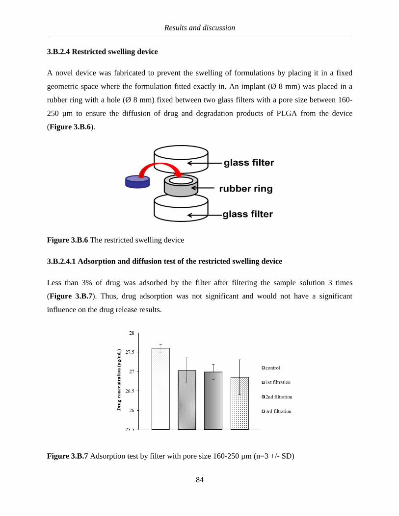
Results and discussion
84
3.B.2.4 Restricted swelling device
A novel device was fabricated to prevent the swelling of formulations by placing it in a fixed
geometric space where the formulation fitted exactly in. An implant (Ø 8 mm) was placed in a
rubber ring with a hole (Ø 8 mm) fixed between two glass filters with a pore size between 160-
250 µm to ensure the diffusion of drug and degradation products of PLGA from the device
(Figure 3.B.6).
Figure 3.B.6 The restricted swelling device
3.B.2.4.1 Adsorption and diffusion test of the restricted swelling device
Less than 3% of drug was adsorbed by the filter after filtering the sample solution 3 times
(Figure 3.B.7). Thus, drug adsorption was not significant and would not have a significant
influence on the drug release results.
Figure 3.B.7 Adsorption test by filter with pore size 160-250 µm (n=3 +/- SD)

Results and discussion
85
The pore size of the glass filter was between 160 and 250 µm, therefore complete diffusion of
drug out of the device was expected. However, the test showed that the diffusion rate of drug
through glass filter was slower than the control (Figure 3.B.8). Slower wettability of implant in
the device might have caused reduced diffusion rate. The speed of drug passage through all 3
devices was not the same (Figure 3.B.8): diffusion rate of one device was faster than the other
two devices. This could be explained by different distribution of pore size of filters.
Nevertheless, the diffusion rate was fast enough, so that the drug release was controlled by the
polymer matrix and not by the device.
Figure 3.B.8 Diffusion through glass filters with pore size 160-250 µm in phosphate buffer pH
7.4 at 37°C (n=3 +/- SD)
3.B.3 The effect of restricted swelling on drug release from PLGA-based
implants
3.B.3.1 Diameter increase of implants prepared with PLGA of different molecular weights
Very little is known about the effect of PLGA swelling on drug release. It is usually assumed that
the swelling effect of PLGA is not pronounced. However, Gasmi et al. suggested that the
swelling kinetics of the PLGA microparticles can play a decisive role in the control of drug
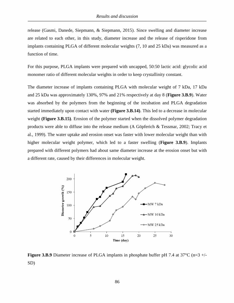
Results and discussion
86
release (Gasmi, Danede, Siepmann, & Siepmann, 2015). Since swelling and diameter increase
are related to each other, in this study, diameter increase and the release of risperidone from
implants containing PLGA of different molecular weights (7, 10 and 25 kDa) was measured as a
function of time.
For this purpose, PLGA implants were prepared with uncapped, 50:50 lactic acid: glycolic acid
monomer ratio of different molecular weights in order to keep crystallinity constant.
The diameter increase of implants containing PLGA with molecular weight of 7 kDa, 17 kDa
and 25 kDa was approximately 130%, 97% and 21% respectively at day 8 (Figure 3.B.9). Water
was absorbed by the polymers from the beginning of the incubation and PLGA degradation
started immediately upon contact with water (Figure 3.B.14). This led to a decrease in molecular
weight (Figure 3.B.15). Erosion of the polymer started when the dissolved polymer degradation
products were able to diffuse into the release medium (A Göpferich & Tessmar, 2002; Tracy et
al., 1999). The water uptake and erosion onset was faster with lower molecular weight than with
higher molecular weight polymer, which led to a faster swelling (Figure 3.B.9). Implants
prepared with different polymers had about same diameter increase at the erosion onset but with
a different rate, caused by their differences in molecular weight.
Figure 3.B.9 Diameter increase of PLGA implants in phosphate buffer pH 7.4 at 37°C (n=3 +/-
SD)

Results and discussion
87
3.B.3.2 Release of implants with different PLGA molecular weight
The release profile of risperidone (Figure 3.B.10) from implants containing PLGA showed a
typical multiphasic pattern like also seen with microparticles (Rawat et al., 2011).
As expected, there was no difference in drug release in the burst phase, since the drug loading for
all three formulations was the same. The higher the molecular weight of the PLGA was, the
longer was the lag phase (Figure 3.B.10). Implants containing PLGA with molecular weights of
7 kDa, 10 kDa and 25 kDa exhibited a lag phase until days 3, 6 and 11 respectively, because
PLGA with higher molecular weight required more time for the erosion to take place (Zolnik &
Burgess, 2008). In all cases, most of the risperidone was released during the polymer erosion
phase, its release rate during this phase decreased with increasing PLGA molecular weight.
Figure 3.B.10 Effect of Polymer molecular weight on risperidone release from PLGA implants
containing 10% w/w drug in phosphate buffer pH 7.4 at 37°C (n=3 +/−SD)
3.B.3.3 Release comparison of implant under restricted and unrestricted conditions
In order to study the effect of polymer swelling on risperidone release, drug release under
restricted and unrestricted conditions was performed.

Results and discussion
88
In both systems, the implants showed the same typical multiphasic release profile. As expected,
the drug release during diffusion and lag phase was slightly lower for restricted implants
compared to unrestricted, due to the lower wettability of implants in the device. However, the
difference was not pronounced, because risperidone has a low solubility and it’s release is
mainly driven by polymer erosion. Once the erosion phase started, i.e. day 3, 6 and 11 for PLGA
with molecular weight of 7 kDa, 10 kDa and 25 kDa respectively, the drug release rate in the
device was first faster and then became slower than the unrestricted implants. (Figure 3.B.11). In
all cases, the drug release slowed down after 80% drug released. A similar result was observed
by Rawat et al, who studied the in vivo and in vitro release profiles of commercially available
risperidone microparticles (Risperdal® Consta). The in vivo profile was initially faster than the
real-time in vitro profile and then slower after approximately 30 days (Rawat et al., 2012). They
assumed that a faster in vivo release during the lag phase is due to the accumulation of acidic
PLGA degradation products. Hence, the difference in drug release in the erosion phase can be
explained by the difference in polymer degradation, which may be caused by different water
uptake and swelling degree of the implants in the two systems.
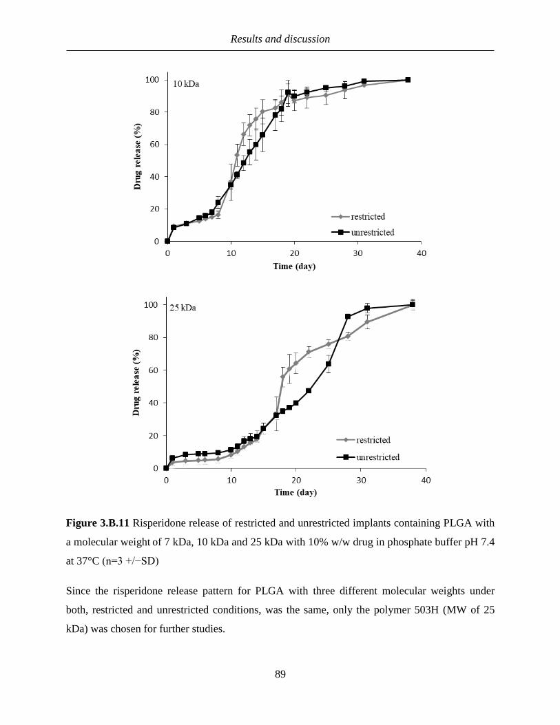
Results and discussion
89
Figure 3.B.11 Risperidone release of restricted and unrestricted implants containing PLGA with
a molecular weight of 7 kDa, 10 kDa and 25 kDa with 10% w/w drug in phosphate buffer pH 7.4
at 37°C (n=3 +/−SD)
Since the risperidone release pattern for PLGA with three different molecular weights under
both, restricted and unrestricted conditions, was the same, only the polymer 503H (MW of 25
kDa) was chosen for further studies.

Results and discussion
90
Risperidone is a basic drug and catalyze PLGA degradation (Souza, Faraj, Dorati, & Deluca,
2015), therefore an acidic drug and nonionic drug were chosen as model drugs as well.
Ibuprofen and theophylline both showed a triphasic release profile from PLGA implants
consisting of an initial burst release, a diffusion phase followed by a lag phase and an erosion-
controlled release phase thereafter (Figure 3.B.12 and 3.B.13).
The initial release of ibuprofen and theophylline was around two times higher from the
unrestricted implants than from the restricted ones. Furthermore, ibuprofen release from the
restricted implants occurred mainly during the erosion phase with 80% of drug being released,
whereas only 20% of drug was released during the diffusion and lag phase (until day 10). In
contrast, for unrestricted implants, approximately half of the drug content was released during
the diffusion phase and lag phase, and the rest was released during the erosion phase (Figure
3.B.12). In the case of theophylline release from the device only 5% of the drug was released
during the diffusion and the lag phases, while for the unrestricted implants 32% of drug was
released during these stages (Figure 3.B.13). In conclusion under restricted condition for
ibuprofen and theophylline, the release was mainly driven by PLGA erosion.
These differences could be attributed to the decreased swelling of implants in the device and
reduced water uptake, which led to the decreased drug release during the diffusion controlled
phase. This difference of drug release in the diffusion and lag phases were more pronounced for
ibuprofen and theophylline compared to risperidone due to their higher solubility. The enhanced
PLGA degradation for restricted implants allowed the formation of bigger pores and spaces from
where the drug was released, and thus, increased the release rate in the erosion phase in
comparison to the unrestricted polymer implants; these implants were able to swell and to create
water filled pores for drug diffusion, which increased the drug release in the diffusion controlled
phase.

Results and discussion
91
Figure 3.B.12 Ibuprofen release of restricted and unrestricted implants containing PLGA with a
molecular weight of 25 kDa with 10% w/w drug in phosphate buffer pH 7.4 at 37°C (n=3
+/−SD)
Figure 3.B.13 Theophylline release of restricted and unrestricted implants containing PLGA
with a molecular weight of 25 kDa with w/w 10% w/w drug in phosphate buffer pH 7.4 at 37°C
(n=3 +/−SD)

Results and discussion
92
The water uptake was approximately two fold higher under unrestricted than under restricted
condition at day 7. However, at day 12 the difference became more pronounced, being
approximately eight fold higher (Figure 3.B.14). This correlated with the results from the mass
loss study (Figure 3.B.14). The difference in mass loss was not pronounced until day 7, but it
became approximately three fold higher at day12 under restricted condition compared to the
unrestricted one. Since the swelling is dependent on the amount of water and free volume
available and under restricted swelling condition, the available volume is decreased, a decrease
in water uptake can be observed. Due to the decreased water uptake, the solubility and mobility
of the polymer degradation products could be decreased impairing their diffusion out of the
matrix. This could lead to faster polymer degradation in the erosion phase under restricted
condition.

Results and discussion
93
Figure 3.B.14 Comparison of water uptake and mass loss of restricted and unrestricted implants
containing 503H in phosphate buffer pH 7.4 at 37°C (n=2)
Since the difference in drug release between unrestricted implants and restricted implants was
observed in the erosion phase, the polymer degradation was monitored over this time period in
both systems and compared to each other.
The peak of the lognormal molecular weight distribution of PLGA (log Mp) decreased with
incubation time for restricted and unrestricted implants. In accordance with the mass loss study
(Figure 3.B.14), the change of the molecular weight was similar until day 7 under both restricted
and unrestricted conditions. However, at day 12, the polymer molecular weight of the implants in
the device was lower than the unrestricted implants and the difference in molecular weight
became more pronounced over the time (Figure 3.B.15). This means that the PLGA degradation
of implants under restricted condition became faster than under the unrestricted ones, which is in
agreement with the degradation of PLGA in vivo and in vitro described in literature (Grayson et
al., 2012; Kamei et al., 1992; Spenlehauer, Vert, Benoit, & Boddaert, 1989; Tracy et al., 1999).

Results and discussion
94
Figure 3.B.15 Changes in molecular weight of restricted and unrestricted implants containing
503H (n=2)
The surface area of the unrestricted implants increased due to the swelling. The implants in the
device were not able to take up water and swell, and thus, had a constant surface area. Due to the
higher surface area of the unrestricted implants, the degradation products diffused more easily
out of the matrix in the erosion phase. Because the water uptake of the implant in the device was
lower, the degradation products were less mobile, diffused more slowly out and accumulated
within the implants. This caused an acidic pH and accelerated the polymer erosion of the
implants in the device due to an autocatalytic effect like reported previously for PLGA 50:50
foams (Lichun Lu et al., 2000).
3.B.3.4 Determination of µpH of restricted and unrestricted implants using EPR
Non-destructive EPR spectroscopy was chosen as an established method to measure the μpH
values within the PLGA implants (Capancioni et al., 2003; C. Kroll, Mäder, Stößer, & Borchert,
1995; Christian Kroll & Herrmann, 2001; Siepe et al., 2006).
During the whole experiment, the µpH of restricted and unrestricted implants was acidic. The
µpH of unrestricted implants remained constant between 3.5 and 4.5 during the complete
experiment (Figure 3.B.16). In contrast, the µpH of implant placed in the device was reduced

Results and discussion
95
constantly up to pH 1.5 within 9 days and remained constant during the following 7 days. The
lowest accurate pH, which was possible to measure, was 2.2. Although the measured pH values
of implants in the device might not be very accurate, the comparative results between the two
different systems could be enough to explain their behavior. The degradation of PLGA and
production of oligomers reduced the µpH; if the diffusion of water through the matrix is
decreased by, e.g., physical barrier around the implant, they can accumulate causing a faster µpH
drop, and thus, accelerating the erosion and degradation of PLGA (Figure 3.B.16).
Figure 3.B.16 Determination of µpH in restricted and unrestricted implants containing 503H
(n=3 for restricted implants and day 7, n=1 for day 16 restricted and unrestricted implants)
3.B.4 Conclusion
A novel device was designed to simulate the restricted physiological space of the tissue and to
restrict the polymer swelling in order to study the effect of restricted swelling of polymer on drug
release with three model drugs of different ionization state.
For all three model drugs, basic, acidic and nonionic drugs, the burst release and diffusion
controlled drug release were reduced and lag phase was prolonged for implants in the device.
This was attributed to the decreased surface area of restricted implant and reduced water uptake
and swelling, which lead to a decreased drug release during the diffusion controlled phase. In the
Results and discussion
96
erosion phase, the release rate of drug was faster for the restricted implants due to the
accumulation of acidic degradation products from PLGA as a result of reduction of water uptake
and oligomers efflux, which caused higher autocatalysis and a faster PLGA degradation. Under
restricted condition, drug release was mainly controlled by the polymer erosion independent of
the nature of the drug (acidic, basic or neutral).
C
Application of biphasic test model as a new biorelevant model for drug release
from PLGA implants and investigate its effect on risperidone release from
PLGA-based implants

Results and discussion
98
3.C.1 Background
In order to optimize the drug release media, the effect of pH, buffer capacity, ionic composition,
osmolarity and for longer experiments, prevention of microbiological contamination have been
studied. Phosphate buffered saline pH 7.4 is the medium most commonly used for non-oral
dosage forms (Iyer et al., 2006). Unlike for the gastrointestinal passage standardized biorelevant
simulated body fluids have not been generally established for parenteral sites of application
(Diakidou, Vertzoni, Dressman, & Reppas, 2009; Jantratid, Janssen, Reppas, & Dressman,
2008). There have been, however, reports of using blood or blood components for dissolution
testing of parenteral dosage forms (Blanco-Prıeto et al., 1999; Kamberi et al., 2009; Sternberg et
al., 2007; G. X. Wang et al., 2010). Rarely, other additives from natural sources have been used
for the preparation of dissolution media, such as rat brain homogenate (Chen & Lu, 2008). Some
authors report the use of liquids referred to as simulated body fluids or for example simulated
synovial fluid, simulated tear fluid or simulated lung fluids (Chen & Lu, 2008; Colombo,
Monhemius, & Plant, 2008; Conzone, Brown, Day, & Ehrhardt, 2002; Kokubo & Takadama,
2006; Kortesuo et al., 2000; Nagarwal, Kumar, Dhanawat, & Pandit, 2011; G. X. Wang et al.,
2010), but these were mostly intended to evaluate issues other than drug release, such as bone-
bonding ability. Also, the use of a modification of Hank’s balanced salt solution has been
suggested to be biorelevant for subcutaneous implants (Iyer, Barr, & Karnes, 2007). In some
studies agar gel was used to simulate the extracellular matrix because of its rheological property
and high water content (Delplace et al., 2012; Hoang Thi et al., 2010; Nastruzzi, Esposito, et al.,
2008). Shuwisitkul et al. used for the first time a lipophilic liquid (LipofundinTM) as an
alternative release medium to mimic the in vivo subcutaneous condition, which resulted in less
water uptake, the deformation of the PLGA implant, and faster drug release (Shuwisitkul, 2011).
As mentioned before, in case of intramuscular or subcutaneous administration, the implant is
placed in the body in contact with muscle or adipose cells and extracellular fluids. Many studies
have been performed to simulate body fluid; however, the role of tissue cells on drug release has
not yet been studied. Depending on the lipophilic character of the drug, it may be partitioned into
the cells, and be released back into interstitial fluid, as a function of −kD, the re-distribution rate.
This could lead to formation of a localized depot in the tissue and affect the drug absorption and
release.

Results and discussion
99
Furthermore the implant is in contact with cell membranes, which might influence the drug
delivery system (Iyer et al., 2006). In previous chapter, the influence of the physiological space
of the tissue on drug release from PLGA implants was studied. In this study, a bicompartment
system was used to simulate the tissue and body fluid. The oil phase mimics the surrounding
tissue and cell membrane and the water phase mimics the body fluid. The biphasic release test
was used in oral application for drug with poor solubility, to maintain sink condition. However in
this study it is used to mimics the physiological environment/ surrounding tissue and to
investigate its effect on risperidone release from PLGA-based implants. Octanol and olive oil
were used as the oil phases.
3.C.2 Risperidone release in biphasic system
The release profile of risperidone from PLGA implants containing 5050 DLG 1A and 10%
risperidone in biphasic system is shown in Figure 3.C.1. The drug concentration was measured
in both aqueous and organic phase. The drug released in the aqueous phase over 15 days was less
than 2% while it increased in the octanol phase. This attributed mainly to the partitioning of the
drug into the oily phase due to higher o/w partition coefficient of risperidone (3.27). This is
supposed to mimic the absorption process of lipophilic drugs in the surrounding tissue.
Figure 3.C.1 Risperidone release from 5050 DLG 1A PLGA implant with 10% drug loading in
biphasic system at 37°C (n=3 +/−SD)

Results and discussion
100
3.C.3 Risperidone release from 5050 DLG 1A PLGA implant in monophasic
system vs. biphasic system
Risperidone release from implants in both systems showed triphasic drug release profile: an
initial release, a short diffusion and lag phase followed by an erosion-controlled release
thereafter.
In both monophasic and biphasic systems, there was an initial burst release of risperidone (6%)
from implants containing polymer 5050 DLG 1A and 10% w/w drug (Figure 3.C.2a). Biphasic
system resulted in a higher release rate in the erosion phase. Up to day 8 more than 90% drug
was released in biphasic system, while in monophasic system only 32% drug was released
(Figure 3.C.2a). Higher release rate in biphasic system can be explained by higher drug
solubility in biphasic system and the affinity of drug to octanol phase.
Since the drug release was governed predominantly by polymer erosion, octanol might influence
polymer degradation as well; therefore, water uptake and mass loss study were done. Water
uptake by the implants increased with time. At day 1 in both biphasic and monophasic systems
no difference was observed in water uptake. However, afterwards, the water uptake by the
biphasic system was more than 1.5 folds compared to the monophasic system (Figure 3.C.2b).
This indicates faster polymer degradation in biphasic system. Faster degradation leads to faster
decrease in molecular weight, polymer with lower molecular weight has higher hydrophilicity
and is able to absorb higher amount of water. The mass loss of the implants followed the same
trend (Figure 3.C.2c). The risperidone release accelerating effect of octanol correlated well with
the water uptake and mass loss. These showed a high evidence of faster polymer degradation in
biphasic system compared to monophasic system.

Results and discussion
101
Figure 3.C.2 (a) Release profile (n=3 +/−SD), (b) water uptake (n=1) and (C) mass loss (n=1) of
implant containing 5050 DLG 1A and 10% w/w risperidone in biphasic system and monophasic
system at 37°C

Results and discussion
102
3.C.4 Risperidone release from 503H implant in monophasic system vs.
biphasic system
The release of risperidone from the PLGA 503H implants in both systems was triphasic with an
initial burst, followed by a period of negligible risperidone release and a rapid release phase after
the first week (Fig.3.C.5 a and b). Thus, the release was governed by combination of drug
diffusion and polymer erosion mechanisms.
3.C.4.1 Effect of drug loading
In the monophasic system, no difference in the drug release rate between implants loaded with
10% and 30% w/w of the drug during the two initial release phases, burst and lag phases, was
observed (Figure 3.C.4 a). During the erosion phase the release rate of implants containing 30%
drug was slightly higher than implants with 10% drug (Figure 3.C.4 a). The reason behind this
was, risperidone enhanced polymer degradation due to its basic and nucleophilic character
(Selmin, Blasi, & DeLuca, 2012; Souza et al., 2015). In biphasic system, the release rate in initial
phase, lag phase and erosion phase was higher for implants containing 30% drug compared to
implants containing 10% drug (Figure 3.C.4 b). This could attribute to the high affinity of the
drug to octanol phase and partitioning of the drug into the oily phase.
Figure 3.C.4 Release profile of 503H implant containing 10% and 30% w/w risperidone in
monophasic system (left) and biphasic system (right) at 37°C (n=3 +/−SD)

Results and discussion
103
3.C.4.2 Effect of dissolution method
In the biphasic system the burst release was higher, the lag phase was shorter and the release rate
during erosion phase was higher compared to the monophasic system for both implants
containing 10% and 30% drug loading (Figure 3.C.5 a and b). For implants containing 30%
drug the difference became more pronounced than the one containing 10% drug. Higher burst
release of implants with 10% and 30% in biphasic system was attributed to the partition of
risperidone into the organic phase as soon as it was released (Figure 3.C.1). The biphasic test
permits a rapid removal of drug from the aqueous phase by partitioning into the organic phase,
so it could be assumed that the amount of drug in the organic phase represents the amount of
drug accumulated in the tissue in vivo. However, the shorter lag phase and higher release rate in
erosion phase for biphasic system indicated faster polymer erosion in biphasic system compared
to monophasic system.
Fig.3.C.5 Release profile of implant containing PLGA 503H and (a) 10% w/w (b) 30% w/w
risperidone in biphasic system and monophasic system at 37°C (n=3 +/−SD)
Apart from the polymer type, the environmental conditions such as pH, temperature, and
enzymes affect the kinetics of polymer degradation (Yoshioka, Kawazoe, Tateishi, & Chen,
2008). In order to find out the effect of octanol on drug release in erosion phase, the polymer
degradation was monitored over time in both systems and compared to each other. The peak of
the log-normal molecular weight distribution of PLGA (log Mp) decreased with incubation time
for both systems and there was no difference in change in molecular weight (Figure 3.C.6).
Higher drug release in erosion phase in biphasic system might therefore be associated with

Results and discussion
104
higher drug solubility in biphasic system, so it could leave the matrix faster after bigger pores
have formed as a consequence of polymer degradation. Furthermore, octanol could plasticize
PLGA, which leads to faster polymer degradation. Increase in drug release in erosion phase
might be also caused by increasing the solubility of PLGA degradation products by octanol
rather than its effect on polymer degradation rate.
Figure 3.C.6 Changes in MW of implants containing 503H in biphasic system and monophasic
system (n=2)
No difference was observed in water uptake study for implants until day 5 in both systems.
However, during erosion phase water uptake of implants in biphasic system increased two folds
compared to the monophasic system (Figure 3.C.7). Similar results were obtained with mass
loss study (Figure 3.C.8). The water uptake and mass loss studies indicated that octanol affect
the drug release in erosion phase.

Results and discussion
105
Figure 3.C.7 Water uptake of implants containing 503H in biphasic and monophasic system at
37°C (n=3 +/−SD),
Figure 3.C.8 Mass loss of implant containing 503H in biphasic and monophasic system (n= 1)
The glass transition temperature of risperidone implants decreased from 49°C to 46°C and 36°C
after 1 day of incubation in monophasic and biphasic systems, respectively. The decrease in glass
transition temperature in monophasic system was probably because of water absorption, which

Results and discussion
106
acts as plasticizer (Omelczuk & McGinity, 1992). The glass transition temperature of the implant
in biphasic system was much lower compared to the monophasic system, which might be
attributed to the additional plasticizing effect of octanol. The 13°C decrease in glass transition
temperature may have increased the molecular mobility and free volume in the polymeric
implant and, consequently increased the polymer erosion rate and drug diffusion through the
polymeric matrix.
3.C.5 Effect of the amount of octanol and the ratio of octanol/phosphate
buffer on drug release
The drug release from implants was independent of the amount of octanol and octanol/phosphate
buffer ratio (Figure 3.C.9 and 10). The reason for this could be that small amount of octanol
was enough to interact with polymer and plasticize it. With increasing the amount of octanol, it
formed a heterogeneous system and it cannot improve the plasticizing effect and decrease the
glass transition temperature any further.
Figure 3.C.9 Effect of different octanol/phosphate buffer pH 7.4 ratio on drug release at 37°C
(n=3 +/−SD)

Results and discussion
107
Figure 3.C.10 Effect of different amount of octanol on drug release with constant octanol/
phosphate buffer pH 7.4 ratio at 37°C (n=3 +/−SD)
3.C.6 Drug release from biphasic system containing olive oil
Adipocytes are characterized by the presence of a large triglyceride (TAG) rich lipid droplet
bounded by a monolayer of phospholipids. TAG is a neutral lipid consisting of a glycerol moiety
esterified with three fatty acids. In humans, the most abundant fatty acids esterified to TAG, in
order of decreasing amount, are palmitate (C16:0), stearate (C18:0), oleate (C18:1n9), and
linoleate (C18:2n6). These four fatty acids make up around 85% of all TAG (Yew Tan et al.,
2015). Olive oil has a similar fatty acid composition and was chosen as oil phase instead of
octanol to simulate the subcutaneous tissue better. However, it was not possible to determine the
amount of drug in both phases using UV spectrometry, therefore the remaining amount of drug
in the implants was extracted and quantified. The study was run for 20 days as after this time
point handling of the implants was not possible.
The drug release during diffusion and lag phase was similar in both systems. During the erosion
phase the drug release increased in the biphasic system (Figure 3.C.11). The water uptake and
mass loss were higher in the erosion phase as well (Figure 3.C.12 and 3.C.13). The drug
release, water uptake, and mass loss study using olive oil indicated faster drug release in erosion

Results and discussion
108
phase. This behavior was similar to the results obtained from biphasic system using octanol. The
polymer degradation wasn’t influenced by olive oil (Figure 3.C.14), which indicate that olive
oil, has most likely a plasticizing effect and increases the drug release by accelerating polymer
erosion. Furthermore, the drug solubility could also be increased in biphasic system using olive
oil due to the high hydrophobicity and high partition coefficient of risperidone. These studies
showed that octanol and olive oil have similar effect when used as oily phase.
Figure 3.C.11 Release profile of implant containing PLGA 503H and 10% w/w risperidone in
biphasic system with “olive oil” and monophasic system at 37°C (n=3 +/−SD),
Figure 3.C.12 Water uptake of implants containing 503H in biphasic system with “olive oil” and
monophasic system at 37°C (n=3 +/−SD),

Results and discussion
109
Figure 3.C.13 Mass loss of implants containing 503H in biphasic system with “olive oil” and
monophasic system at 37°C (n:1)
Figure 3.C.14 Changes in MW of implants containing 503H in biphasic system with “olive oil”
and monophasic system (n:2)

Results and discussion
110
3.C.7 Effect of incorporation of octanol in implants on drug release
Excipients used to adjust drug release from PLGA-based drug delivery systems are usually low
molecular weight salts such as magnesium hydroxide (Kang & Schwendeman, 2002) or
hydrophilic polymers such as PEG (Ghalanbor et al., 2010). The problem with these excipients is
that they do not stay in the matrix over the complete release time and diffuse out during diffusion
phase. Hence these excipients could change the burst release and lag phase, but they usually do
not have any influence on drug release in the erosion phase (Ghalanbor et al., 2010). Since
risperidone release was mainly erosion controlled and octanol influenced the polymer erosion,
octanol was incorporated into risperidone implants in order to adjust risperidone release.
Incorporation of 5% w/w octanol (based on polymer) into 5050 DLG 1A implants containing
10% w/w risperidone did not change the initial burst release, but increased the rate of risperidone
release in the erosion phase (Figure 3.C.15). The release approximated linear characteristics
against time without an uncontrolled burst. However, risperidone release profile did not change
with increasing the amount of octanol from 5% to 10% (Figure 3.C.15) and the drug release was
independent of the amount of octanol in the implant. This can be explained by the glass
transition temperature of 5050 DLG 1A. 5% octanol reduced the glass transition temperature, but
the glass transition temperature of this polymer was so low that increasing the amount of octanol
to 10% did not cause further reduction of the glass transition temperature of 5050 DLG 1A
(Figure3.C.17).
Figure 3.C.15 Effect of octanol level in 5050 DLG 1A implants on risperidone release in
phosphate buffer pH 7.4 at 37°C (n=3 +/−SD)
Results and discussion
111
As it was shown in Figure 3.B.10, the drug release with polymer 5050 DLG 1A was governed
mainly by the polymer erosion mechanism; to better understand the effect of octanol on
risperidone release in diffusion phase and lag phase, polymer 5050 DLG 1A was replaced by
PLGA 503H.
Incorporation of 5% and 10% w/w octanol (based on polymer) into PLGA 503H implants
containing 10% risperidone did not change drug release profile until day 3, which shows that
octanol did not influence the initial burst release. But it led to a shorter lag phase and increased
the rate of risperidone release in the erosion phase (Figure 3.C.16a). The release rate increased
with increasing amount of octanol during the erosion phase. These findings suggest that octanol
increase the release rate in erosion phase by decreasing the glass transition temperature (Figure
3.C.17). The effect of octanol on erosion correlated well with increased water uptake and mass
loss. There was no difference in water uptake with all three implants containing 0%, 5% and
10% octanol on day 1, but from day 5 the water uptake increased 2 folds and 4 folds for implants
containing 5% and 10% octanol respectively (Figure 3.C.16b). The difference became more
pronounced over the time. The mass loss of implants without octanol and the implant containing
5% octanol did not start until day 5, the mass loss of implants containing 10% octanol started
already at day 3 and the percentage of mass loss increased with increasing the amount of octanol
(Figure 3.C.16c). It was hypothesized that the degradation and/or erosion rate of the polymer
would increase in the presence of octanol due to its plasticizing effect. Furthermore, octanol with
a hydrophobic, long alkyl chain and a polar hydroxyl group membrane has an affinity to the
PLGA staying longer in the polymer, it does not diffuse out in contrast to hydrophilic excipients
like PEG (Ghalanbor et al., 2010).
Incorporation of 10% octanol into PLGA 503H implants resulted in release approximated the
linear characteristics without any uncontrolled burst effect (Figure 3.C.16a).
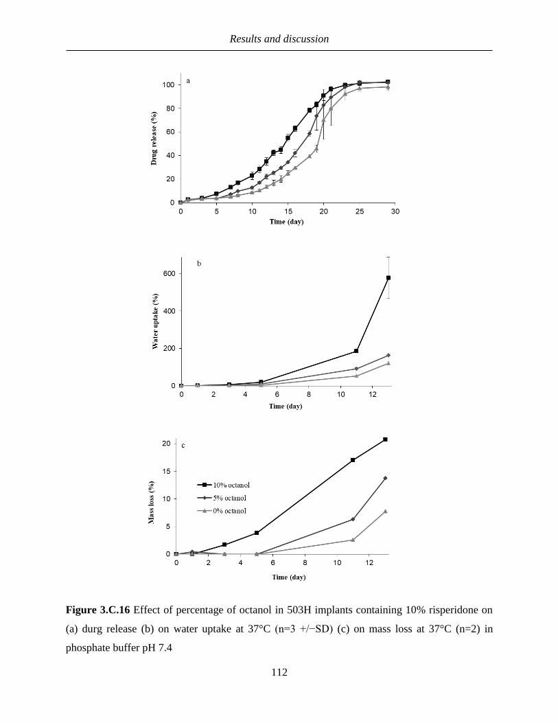
Results and discussion
112
Figure 3.C.16 Effect of percentage of octanol in 503H implants containing 10% risperidone on
(a) durg release (b) on water uptake at 37°C (n=3 +/−SD) (c) on mass loss at 37°C (n=2) in
phosphate buffer pH 7.4

Results and discussion
113
3.C.8 Effect of octanol on glass transition temperature of two types of PLGA
implants containing 10% risperidone
To prove the plasticizing effect of octanol on the PLGA polymer, the glass transition temperature
of two types of polymers (503H and 5050 DLG 1A) without octanol, with 5% and 10% octanol
was measured. In case of 503H implants 5% and 10% octanol decreased the glass transition
temperature by about 6°C and 12°C, respectively (Figure 3.C.17), which showed that there was
a linear relationship between decrease of glass transition temperature and the amount of octanol
incorporated into the implant. In case of 5050 DLG 1A implants, incorporation of 5% octanol
decreased the glass transition temperature by 12°C, but in contrast to implants containing PLGA
503H, increasing the amount of octanol to 10% did not cause further reduction of the glass
transition temperature of 5050 DLG 1A (Figure 3.C.17). An excipient can act as a plasticizer,
only when it exists in the same phase as the polymer. At a certain concentration the excipient
forms a heterogeneous system and it cannot act as a plasticizer and decrease the glass transition
temperature (Blasi et al., 2005). PLGA 503H, with molecular weight of 24 kDa, is a more
hydrophobic polymer than 5050 DLG 1A (Molecular weight 7 kDa); higher amount of octanol
can be absorbed into this polymer and plasticize it, and due to the higher affinity to the polymer
it stays longer in the polymer matrix.
Figure 3.C.17 Effect of percentage of octanol on glass transition temperature of PLGAs (n:2)

Results and discussion
114
3.C.9 Changes in polymer molecular weight of implant containing 503H and
octanol
As expected, there were no differences in degree of polymer degradation in implants with 5%
octanol or without octanol (Figure 3.C.18). However, the degradation of implants containing
10% octanol was slightly faster during the first 5 days (Figure 3.C.18). There were no
differences in molecular weight in the erosion phase (Figure 3.C.18). These results suggested
that octanol accelerate polymer erosion due to its plasticizing effect. In addition, it might have an
effect on oligomers solubility, which leave the matrix and were not detectable with GPC.
Figure 3.C.18 Effect of octanol on the changes in molecular weight of implants containing 503H
3.C.10 Conclusions
In biphasic system, risperidone was accumulated into the organic phase due to the high affinity
of the drug to the octanol phase. Furthermore, the drug release rate was higher in biphasic system
compared to monophasic. This was attributed to the plasticizing effect of octanol and increased
oligomers solubility, which accelerated polymer erosion without affecting the degradation. Olive
oil has the same effect as octanol. Biphasic release model could be potentially appropriate
biorelevant media for risperidone PLGA-based implant, since octanol and olive oil can simulate
lipid membrane of cells.
Results and discussion
115
Octanol could also be used as an excipient to adjust risperidone release in erosion, since it
influences the polymer erosion, without affecting burst release and lag phase. With PLGA 503H
implants it was possible to adjust the release rate by varying amount of octanol incorporated into
the implant. The release rate increased with increasing amount of octanol in implant during the
erosion phase. DSC data showed a linear relationship between depression of glass transition
temperature and the amount of octanol incorporated into the implant. Increased release rate was
also observed with 5050 DLG 1A implants, however risperidone release profile did not change
with increasing the amount of octanol, due to the low glass transition temperature of 5050 DLG
1A polymer. It must be taken into consideration that octanol is not FDA approved and cannot be
used as excipient in parenteral formulations, however excipients with similar structure, which are
FDA approved, could have the same effect as octanol and could be used to adjust risperidone
release in erosion phase.

4. Summary

Summary
117
The biorelevance of the in vitro conditions should be considered when designing dissolution or
release test methods for controlled release for orals and parenterals. Nevertheless, there are very
few reports on such test systems for parenteral dosage forms and most IVIVCs have been
achieved without considering physiologically relevant testing conditions in the in vitro system.
Usually the in vitro methods are developed when in vivo data is already available and are
optimized to match these data. Thus, IVIVC in this case is mainly understood as a tool for
quality control purposes, e.g. when dealing with post-approval issues, and the release methods
were adapted irrespective of the underlying physiologic conditions to achieve data correlation.
The approach to developing biorelevant test systems is different, even though these tests might
also be suitable for establishing IVIVC. Biorelevant test systems may be of outstanding value in
the preclinical phase of development providing a sound basis for dosage form optimization and
selection of the most promising candidates for clinical studies. As in vivo fate of formulation is
typically very complex and is furthermore subject to inter- and intra-individual variability, it
would be difficult for a biorelevant test system to simulate all in vivo conditions accurately.
The major drawback of almost all of the currently used in vitro release tests for controlled release
parenteral formulations is that the formulations are exposed only to release medium, which does
not mimic the physiological environment. When a PLGA-based implant is completely
surrounded only by release medium, it can absorb water and swell to its maximal degree,
differently to in vivo, where less water is available and the formulation is not only in contact with
body fluid, but also exposed to the tissue cells.
The purpose of this study was to evaluate the swelling of the implant ex vivo, to measure the
force applied to the formulation after implantation in the body and to calculate the created
pressure in the tissue. To design a new in vitro test to mimic mechanical properties of the tissue,
which may influence dosage form performance in a standardized and reproducible manner and to
study its effect on drug release. To assess biphasic system as biorelevant media and investigate
its effect on risperidone release from PLGA-based implants.
The shape, surface area and water uptake of PLGA-implants in ex vivo and in vitro experiments
was determined. Implants were incubated either in phosphate buffer (in vitro) or implanted in
turkey breast (with different amount of buffer). Surface area and water uptake of implants

Summary
118
increased continuously and they expanded notably and maintained their cylindrical shape under
in vitro conditions, whereas under ex vivo conditions their surface area increased to a lower
degree, they expanded only minimal and were deformed, indicating that the swelling was
suppressed. The water uptake of both ex vivo tests was lower as compared to in vitro tests. The
water uptake of implant under ex vivo test utilizing low amount of buffer increased at a very slow
rate. No difference in the water uptake of implants was observed for both ex vivo experiments
over 3 days, but in the subsequent erosion phase when the implants became soft, the water
uptake increased at about the same rate under the ex vivo experiment utilizing high amount of
phosphate buffer and in vitro experiment, as high amount of buffer was available for tissue to be
moistened. It can be concluded that the mechanical properties of the tissue and amount of water
available in vivo both play an important role for the restriction of swelling and the shape change
of implants.
A new method - using a texture analyzer and a balloon catheter - was established to quantify the
force applied to the formulation ex vivo. The force, reached after injecting certain amount of
water into the balloon catheter, was determined, when the balloon catheter was in air and under a
known weight. A linear relationship was found between the injection force and the force applied
to the balloon catheter and an appropriate equation was determined. The measurement was run
with different plunger displacements, since this leads to a different degree of expansion of the
balloon catheter. Afterwards the balloon catheter was inserted into a turkey breast and injection
force was measured. The tissue force applied to the formulation ex vivo was calculated with the
same plunger displacements using the appropriate equations. This measured force was used to
calculate the force applied to the formulation ex vivo. The force applied to the balloon catheter ex
vivo increased with increasing expansion of the balloon catheter. This is caused by the
mechanical properties of the tissue, which counteracts the expansion of the balloon catheter. This
indicated that the force created in the tissue after implanting an object should be taken into
consideration because it might influence the DDSs and therefore also the drug release.
For simulation of limited physiological space, a device was designed to prevent the swelling of
formulation. The acidic, basic and nonionic model drugs (e.g.,ibuprofen, risperidone and
theophylline) were used for this experiment. All PLGA-based implants showed a typical
multiphasic release profile: burst release, diffusion phase followed by lag phase and an erosion

Summary
119
controlled release phase. The risperidone release during diffusion and lag phase was slightly
lower for restricted implants compared to unrestricted, due to the lower wettability of implants in
the device. This difference in the diffusion and lag phase was more pronounced for ibuprofen
and theophylline compared to risperidone due to their higher solubility. From the beginning of
erosion phase, drug release rate under restricted conditions was higher than unrestricted implants
for all three drug models. Under restricted swelling conditions water uptake was decreased. The
decreased water uptake leads to the decreased solubility and mobility of the polymer degradation
products so that they could not diffuse out of the matrix. Furthermore, the surface area of the
unrestricted implants increased with increase in the swelling hence the degradation products
diffuse more easily out of the matrix under these conditions. At the same time the mass loss and
polymer degradation was determined and it was found out that the mass loss and polymer
degradation were both accelerated under restricted condition. The monitoring of µpH of implants
under restricted and non-restricted condition indicated that µpH of the later was between 3.5 and
4.5 during the complete time of follow-up. In contrast, the µpH of implant in device was reduced
constantly up to 1.5 within 9 days and remained constant for the following 7 days. As indicated
the accumulation of degradation products was higher in the implant in the device due to decrease
on surface area and decreased hydration. Oligomers stay in the matrix, causing a faster pH drop
and accelerated erosion.
Risperidone release from implants was studied in monophasic and biphasic systems (octanol:
water or olive oil: water). A triphasic drug release profile was found for both systems. In
biphasic system drug was accumulated into the organic phase due to its high affinity to the
octanol phase. In the biphasic system the burst release was higher, the lag phase was shorter and
the release rate during erosion phase was higher compared to the monophasic system indicating
faster polymer degradation in erosion phase in biphasic system. In order to find out the effect of
octanol on drug release in erosion phase, the water uptake, mass loss and polymer degradation
was monitored over time in both systems. Results confirmed higher water uptake and mass loss
in erosion phase; however, no difference was obtained from degradation study in both systems.
Accelerated effect in biphasic system was attributed to the plasticizing effect of octanol. Octanol
decreased polymer glass transition temperature, increasing the molecular mobility and free
volume in the implant, and consequently, increased the polymer erosion and drug diffusion
through the polymeric matrix. Octanol might also increase oligomers solubility, which

Summary
120
accelerated polymer erosion. Olive oil has a similar effect as octanol. Biphasic release model
could be potentially the appropriate biorelevant media for risperidone PLGA-based implant,
since octanol and olive oil both with a hydrophobic and hydrophilic group can simulate lipid
membrane of tissue cells.
The Effect of amount of octanol incorporated in implants was studied on risperidone release
from implants. With 503H-based implants it was possible to adjust the release rate with adjusting
the amount of octanol incorporated into the implant. The release rate increased with increasing
amount of octanol in implant during the erosion phase without affecting burst release and lag
phase. DSC data showed a linear relationship between glass transition temperature decrease and
the amount of octanol incorporated into the implant. Increased release rate was also observed
with 5050 DLG 1A implants. However, risperidone release profile didn’t change with increasing
the amount of octanol, due to the low glass transition temperature of 5050 DLG 1A polymer.
In conclusion, it may not be possible to simulate all of the biologically relevant reactions at the
site of administration through the use of single in vitro system, since the biological environment
surrounding the formulation is very complex and not static. However, it is still possible to
simulate single in vivo aspect in vitro, in order to investigate its effect on drug release and to
develop in vitro tests, which are more biorelevant than the current available in vitro tests.

5. Zusammenfassung

Zusammenfassung
122
Die Biorelevanz sollte bei der Entwicklung von In-Vitro-Methoden für orale und parenterale
Formulierungen mit kontrollierter Freisetzung berücksichtigt werden. Dennoch gibt es nur
wenige Berichte über solche Methoden für parenterale Formulierungen und die meisten IVIVCs
wurden ohne Berücksichtigung physiologisch relevanter Testbedingungen in In-vitro-Methoden
erreicht. In der Regel werden die In-Vitro-Methoden entwickelt, wenn In-Vivo Daten bereits
vorhanden sind und sie werden optimiert, um den In-Vivo Daten zu entsprechen. Somit wird eine
IVIVC in diesem Fall hauptsächlich zur Qualitätskontrolle benutzt und die
Freisetzungsmethoden wurden unabhängig von den zugrunde liegenden physiologischen
Bedingungen angepasst, um eine Datenkorrelation zu erreichen. Der Zweck für die Entwicklung
biorelevanter Testsysteme ist aber anders, obwohl diese Tests auch für die Etablierung von
IVIVC geeignet sind. Biorelevante Testsysteme können in der präklinischen Entwicklungsphase
von herausragender Bedeutung sein um eine solide Grundlage für die Optimierung der
Dosierungsform und die Auswahl der aussichtsreichsten Kandidaten für klinische Studien zu
liefern. Da die In-Vivo Vorgänge für die Formulierung typischerweise sehr komplex sind und
darüber hinaus einer inter- und intraindividuellen Variabilität unterliegen, wäre für ein
biorelevantes Testsystem schwierig, alle In-Vivo-Bedingungen genau zu simulieren. Der
Hauptnachteil von fast allen derzeit verwendeten In-vitro-Freisetzungstests für parenterale
Formulierungen mit kontrollierter Freisetzung besteht darin, dass die Formulierungen nur dem
Freisetzungsmedium ausgesetzt werden, was der physiologischen Umgebung nicht entspricht.
Wenn ein PLGA-basiertes Implantat durch das Freisetzungsmedium vollständig umgeben ist,
kann es unbegrenzt Wasser aufnehmen und bis zu seinem maximalen Ausmaß quellen, anders als
In-Vivo, wo weniger Wasser vorhanden ist und die Formulierung nicht nur in Kontakt mit
Körperflüssigkeiten ist, sondern auch zu den Gewebezellen exponiert ist.
Das Ziel dieser Studie war, die Implantatsquellung Ex-Vivo zu bewerten, um die auf die
Formulierung ausgeübte Kraft nach der Implantation im Körper zu messen und den erzeugten
Druck im Gewebe zu berechnen. Ein neuer standardisierter und reproduzierbarer In-Vitro-Test
sollte etabliert werden, der die mechanischen Eigenschaften des Gewebes auf die Arzneisform
simuliert und den Effekt auf die Arzneimittelfreisetzung untersucht. Das zweiphasen System
sollte als biorelevantes Medium beurteilt und sein Effekt auf die Risperidon-Freisetzung von
PLGA-basierten Implantaten untersucht werden.

Zusammenfassung
123
Die Form, die Oberfläche und die Wasseraufnahme von PLGA-Implantaten wurden in Ex-Vivo
und In-Vitro Experimenten untersucht. Die Implantate wurden entweder in Phosphatpuffer (In-
Vitro) inkubiert oder in Putenbrust implantiert (mit unterschiedlichen Puffermengen). Unter In-
Vitro Bedingungen hat sich die Oberflächenfläche und die Wasseraufnahme von Implantaten
kontinuierlich erhöht. Die Implantate expandierten deutlich und haben ihre zylindrische Form
beibehalten. Unter Ex-Vivo Bedingungen nahm die Oberfläche nur in einem geringeren Ausmaß
zu. Die Implantate expandierten nur minimal und wurden deformiert, was darauf hinweist, dass
die Quellung eingeschränkt wurde. Die Wasseraufnahme im Ex-Vivo-Test war im Vergleich zu
In-Vitro-Tests geringer.
Die Wasseraufnahme von Implantaten im Ex-Vivo-Test mit einer geringen Menge an Puffer
erhöhte sich mit einer sehr langsamen Geschwindigkeit. Es wurde kein Unterschied wurde in der
Wasseraufnahme von Implantaten für beide Ex-Vivo-Experimente über 3 Tage beobachtet,
jedoch erhöhte sich in der anschließenden Erosionsphase, in der die Implantate weicher wurden,
die Wasseraufnahme mit etwa der gleichen Geschwindigkeit im Ex-Vivo-Experiment unter
Verwendung einer hohen Menge an Phosphatpuffer wie im In-Vitro Experiment, da eine hohe
Menge an Puffer für das zu befeuchtende Gewebe zur Verfügung stand. Es kann gefolgert
werden, dass die mechanischen Eigenschaften des Gewebes und die Menge an Wasser, die In-
Vivo verfügbar ist, beide eine wichtige Rolle für die eingeschränkte Quellung und die
Deformierung von Implantaten spielen.
Eine neue Methode - mit einem Texturanalysator und einem Ballonkatheter - wurde etabliert, um
die auf die Formulierung ausgeübte Kraft Ex-Vivo zu quantifizieren. Die Kraft, die nach der
Injektion bestimmter Wassermenge in den Ballonkatheter erreicht wurde, wurde in Abhängigkeit
einer auf dem Ballonkatheter bekannten Gewicht bestimmt. Es wurde ein linearer
Zusammenhang zwischen der Injektionskraft und der Kraft, die auf den Ballonkatheter
angewendet wurde, ermittelt und eine Gleichung bestimmt. Die Messung wurde mit
unterschiedlichen Ausdehnungsgrad des Ballonkatheters durchgeführt. Danach wurde der
Ballonkatheter in eine Putenbrust eingeführt und die Injektionskraft gemessen. Die auf die
Formulierung aufgebrachte Gewebekraft Ex-Vivo wurde unter Verwendung der entsprechenden
Zusammenfassung
124
Gleichungen berechnet. Diese gemessene Kraft wurde verwendet, um die auf die Formulierung
aufgebrachte Kraft Ex-Vivo zu berechnen.
Die auf den Ballonkatheter ausgeübte Kraft nahm mit zunehmender Ausdehnung des
Ballonkatheters zu. Dies wird durch die mechanischen Eigenschaften des Gewebes verursacht,
die der Ausdehnung des Ballonkatheters entgegenwirken.
Dies zeigte, dass nach der Implantation eines Implants bestimmte Kraft vom Gewebe auf die
Implantate ausgeuebt wird, die in Betracht gezogen werden sollte, weil er das DDS und damit
auch die Arzneimittelfreisetzung beeinflussen könnte.
Um den begrenzten physiologischen Raum zu simulieren wurde eine Vorrichtung entworfen, die
die Quellung der Formulierung verhindert. Es wurde festgestellt, dass die Arzneistofffreisetzung
von der Vorrichtung nicht kontrolliert wurde. Die drei Arzneistoffe, Risperidone, Ibuprofen und
Theophyllin wurden in diesem Experiment verwendet. Alle PLGA-basierten Implantate zeigten
ein typisches multiphasisches Freisetzungsprofil: Burst-Freisetzung, Diffusionsphase, gefolgt
von einer Verzögerungsphase und einer erosionsgesteuerten Freisetzungsphase. Die Risperidon-
Freisetzung während der Diffusions- und Verzögerungsphase war niedriger für Implantate mit
eingeschränkter Quellung im Vergleich zu Implantaten mit uneingeschränkter Quellung,
aufgrund ihrer geringeren Hydratation in der Vorrichtung. Der Unterschied in der Diffusions-
und Verzögerungsphase war für Ibuprofen und Theophyllin, aufgrund ihrer höheren Löslichkeit
im Vergleich zu Risperidon, stärker ausgeprägt. Sobald die Erosionsphase erreicht wurde, die
Arzneistoff-Freisetzungsrate unter eingeschränkten Quellbedingungen höher als bei
uneingeschränkt gequollenen Implantaten für alle drei Arzneistoffmodelle.
Unter eingeschränkten Quellbedingungen wurde die Wasseraufnahme verringert. Die
verminderte Wasseraufnahme führt zu einer verminderten Löslichkeit und Diffusion der
Polymerabbauprodukte, so dass sie nicht aus der Matrix diffundieren konnten. Weiterhin erhöhte
sich die Oberfläche der uneingeschränkt gequollenen Implantaten mit zunehmender Quellung, so
dass die Abbauprodukte unter diesen Bedingungen leichter aus der Matrix diffundieren können.
Gleichzeitig wurde der Massenverlust und der Polymerabbau bestimmt und es wurde festgestellt,
dass der Massenverlust und der Polymerabbau unter eingeschränktem Quellzustand beschleunigt
wurden. Bei uneingeschränkter Quellung war der MikropH während der gesamten

Zusammenfassung
125
Experimentzeit zwischen 3,5 und 4,5. Im Gegensatz dazu reduzierte sich der MikropH der
Implantaten in der Vorrichtung innerhalb von 9 Tagen auf 1,5 und blieb für die folgenden 7 Tage
konstant. Wie angedeutet, war die Diffusion von Abbauprodukten aus der Implantat in der
Vorrichtung, aufgrund der reduzierte Oberfläche und verminderter Hydratation, verringert.
Oligomere bleiben in der Matrix, was einen schnellere Mikro-pH-Abnahme und eine
beschleunigte Erosion verursacht.
Die Risperidon-Freisetzung aus den Implantaten wurde in monophasischen und biphasischen
Systemen (Octanol: Wasser oder Olivenöl: Wasser) untersucht. Beide Systeme zeigten ein
triphasisches Freisetzungsprofil, welches vorher schon beschrieben wurde. Im biphasischen
System akkumulierte der Arzneistoff, aufgrund seiner hohen Affinität zur Octanolphase, in die
organische Phase. In dem biphasischen System war die Burst-Freisetzung höher, die
Verzögerungsphase kürzer und die Freisetzungsrate während der Erosionsphase höher im
Vergleich zu dem monophasischen System, was auf einen schnelleren Polymerabbau in der
Erosionsphase im biphasischen System hinweist. Um die Wirkung von Octanol auf die
Arzneistoff-Freisetzung und Erosionsphase zu untersuchen, wurde die Wasseraufnahme, der
Massenverlust und der Polymerabbau in Abhängigkeit von der Zeit in beiden Systemen
untersucht. Die Ergebnisse bestätigten eine höhere Wasseraufnahme und Massenverlust in der
Erosionsphase. Allerdings wurde es kein Unterschied in der Polymerabbaurate in beiden
Systemen beobachtet.
Die beschleunigte Wirkung im biphasischen System wurde durch den plastifizierenden Effekt
von Octanol verursacht. Octanol verringerte die Polymer-Glasübergangstemperatur, was die
molekulare Beweglichkeit und das freie Volumen im Implantat erhöht und folglich die
Polymererosion und die Arzneistoffdiffusion durch die Polymermatrix erhöht. Octanol könnte
auch die Löslichkeit der Oligomeren erhöht haben, was die Polymererosion zusätzlich
beschleunigt haben könnte. Olivenöl hatte eine ähnliche Wirkung wie Octanol. Das biphasische
Freisetzungsmodell könnte als biorelevantes Medium für das Risperidon-PLGA-basierte
Implantat geeignet sein, da Octanol und Olivenöl beide sowohl eine hydrophobe als auch eine
hydrophile Gruppe besitzen und somit die Lipidmembran der Gewebezellen simulieren können.

Zusammenfassung
126
Es wurde der Effekt der Menge an Octanol im Implantat auf die Arzneistoff-Freisetzung aus dem
Implantat untersucht. Mit 503H-basierten Implantaten war es möglich, die Freisetzungsrate mit
der Menge an Octanol, die im Implantat vorhandeln war, einzustellen. Die Freisetzungsrate
nahm mit zunehmender Menge an Octanol im Implantat während der Erosionsphase zu ohne die
Burst-Freisetzung und die Lag-Phase zu beeinflussen. DSC-Daten zeigten einen linearen
Zusammenhang zwischen der Abnahme der Glasübergangstemperatur und der Zunahme von
Octanol, das in das Implantat zugesetzt worden war. Eine erhöhte Freisetzungsrate wurde auch
bei 5050 DLG 1A Implantaten beobachtet. Das Risperidon-Freisetzungsprofil von 5050 DLG
1A-Polymer änderte sich im Gegensatz zum 503H-basierten Implantat nicht proportional mit der
Menge an Octanol aufgrund der niedrigen Glasübergangstemperatur.
Abschließend lässt sich festhalten, dass es wahrscheinlich nicht möglich ist, durch einen
einzelnen In-Vitro Test alle biologisch relevanten Reaktionen am Applikationsort zu simulieren,
da die biologische Umgebung, die die Formulierung umgibt, sehr komplex und nicht statisch ist.
Es ist jedoch möglich, einzelne In-Vivo-Aspekte, In-Vitro zu simulieren, um deren Effekte auf
die Arzneistofffreisetzung zu untersuchen und In-Vitro-Tests zu entwickeln, die biorelevanter als
die derzeitig verfügbaren In-Vitro-Tests sind.

6. References

References
128
Alexis, F., Venkatraman, S. S., Rath, S. K., & Boey, F. (2004). In vitro study of release
mechanisms of paclitaxel and rapamycin from drug-incorporated biodegradable stent
matrices. Journal of Controlled Release, 98(1), 67–74.
https://doi.org/10.1016/j.jconrel.2004.04.011
Alkan-Onyuksel, H., Ramakrishnan, S., Chai, H.-B., & Pezzuto, J. M. (1994). A Mixed Micellar
Formulation Suitable for the Parenteral Administration of Taxol. Pharmaceutical Research,
11(2), 206–212. https://doi.org/10.1023/A:1018943021705
Allababidi, S., & Shah, J. C. (1998). Kinetics and mechanism of release from glyceryl
monostearate-based implants: Evaluation of release in a gel simulating in vivo implantation.
Journal of Pharmaceutical Sciences, 87(6), 738–744. https://doi.org/10.1021/js9703986
Amann, L. C., Gandal, M. J., Lin, R., Liang, Y., & Siegel, S. J. (2010). In vitro-in vivo
correlations of scalable plga-risperidone implants for the treatment of schizophrenia.
Pharmaceutical Research, 27(8), 1730–1737. https://doi.org/10.1007/s11095-010-0152-4
Anderson, F. D., Archer, D. F., Harman, S. M., Leonard, R. J., & Wilborn, W. H. (1993). Tissue
Response to Bioerodible, Subcutaneous Drug Implants: A Possible Determinant of Drug
Absorption Kinetics. Pharmaceutical Research, 10(3), 369–380.
https://doi.org/10.1023/A:1018932104577
Anderson, J. M., & Shive, M. S. (2012). Biodegradation and biocompatibility of PLA and PLGA
microspheres. Advanced Drug Delivery Reviews, 64, 72–82.
https://doi.org/10.1016/j.addr.2012.09.004
Aratow, M., Ballard, R. E., Crenshaw, a G., Styf, J., Watenpaugh, D. E., Kahan, N. J., &
Hargens, a R. (1993). Intramuscular pressure and electromyography as indexes of force
during isokinetic exercise. Journal of Applied Physiology (Bethesda, Md. : 1985), 74(6),
2634–40. Retrieved from http://www.ncbi.nlm.nih.gov/pubmed/8365963
Arda, K., Ciledag, N., Aktas, E., Aribas, B. K., & Köse, K. (2011). Quantitative assessment of
normal soft-tissue elasticity using shear-wave ultrasound elastography. American Journal of
Roentgenology, 197(3), 532–536. https://doi.org/10.2214/AJR.10.5449

References
129
Athanasiou, K. A., Niederauer, G. G., & Agrawal, C. M. (1996). Sterilization, toxicity,
biocompatibility and clinical applications of polylactic acid/ polyglycolic acid copolymers.
Biomaterials, 17(2), 93–102. https://doi.org/10.1016/0142-9612(96)85754-1
Aubert-Pouëssel, A., Bibby, D. C., Venier-Julienne, M.-C., Hindré, F., & Benoît, J.-P. (2002). A
Novel in Vitro Delivery System for Assessing the Biological Integrity of Protein upon
Release from PLGA Microspheres. Pharmaceutical Research, 19(7), 1046–1051.
https://doi.org/10.1023/A:1016482809810
Aubert-Pouëssel, A., Venier-Julienne, M.-C., Clavreul, A., Sergent, M., Jollivet, C., Montero-
Menei, C. N., … Benoit, J.-P. (2004). In vitro study of GDNF release from biodegradable
PLGA microspheres. Journal of Controlled Release, 95(3), 463–475.
https://doi.org/10.1016/j.jconrel.2003.12.012
Aukland, K., & Reed, R. K. (1993). Interstitial-lymphatic mechanisms in the control of
extracellular fluid volume. Physiol Rev, 73(1), 1–78. Retrieved from
http://physrev.physiology.org/content/73/1/1
Bain, D. F., Munday, D. L., & Smith, A. (2008). Modulation of rifampicin release from spray-
dried microspheres using combinations of poly-(DL-lactide). Journal of
Microencapsulation, 16(3), 369–85. https://doi.org/10.1080/026520499289086
Ballard, B. E. (1968). Biopharmaceutical Considerations in Subcutaneous and Intramuscular
Drug Administration. Journal of Pharmaceutical Sciences, 57(3), 357–378.
https://doi.org/10.1002/jps.2600570301
Ballard, R. E., Watenpaugh, D. E., Breit, G. a, Murthy, G., Holley, D. C., & Hargens, a R.
(1998). Leg intramuscular pressures during locomotion in humans. Journal of Applied
Physiology (Bethesda, Md. : 1985), 84(6), 1976–1981.
Barzilay, R. B., & Hersey, J. A. (1968). Dissolution rate measurement by an automated dialysis
method. Journal of Pharmacy and Pharmacology, 20(S1), 232S–238S.
https://doi.org/10.1111/j.2042-7158.1968.tb09888.x
Batycky, R. P., Hanes, J., Langer, R., & Edwards, D. A. (1997). A Theoretical Model of Erosion
References
130
and Macromolecular Drug Release from Biodegrading Microspheres. Journal of
Pharmaceutical Sciences, 86(12), 1464–1477. https://doi.org/10.1021/js9604117
Baun, D. C., & Walker, G. C. (1969). Apparatus for Determining the Rate of Drug Release from
Solid Dosage Forms. Journal of Pharmaceutical Sciences, 58(5), 611–616.
https://doi.org/10.1002/jps.2600580523
Benet, L. Z. (1990). The effect of route of administration and distribution on drug action. Drugs
and the Pharmaceutical Sciences, 40, 181–207. Retrieved from
http://cat.inist.fr/?aModele=afficheN&cpsidt=19716079
Bert, J. L., Mathieson, J. M., & Pearce, R. H. (1982). The exclusion of human serum albumin by
human dermal collagenous fibres and within human dermis. Biochemical Journal, 201(2),
395–403. https://doi.org/10.1042/bj2010395
Bhardwaj, U., & Burgess, D. J. (2010). A novel USP apparatus 4 based release testing method
for dispersed systems. International Journal of Pharmaceutics, 388(1), 287–294.
https://doi.org/10.1016/j.ijpharm.2010.01.009
Blanco-Prıeto, M. J., Besseghir, K., Orsolini, P., Heimgartner, F., Deuschel, C., Merkle, H. P., …
Gander, B. (1999). Importance of the test medium for the release kinetics of a somatostatin
analogue from poly(d,l-lactide-co-glycolide) microspheres. International Journal of
Pharmaceutics, 184(2), 243–250. https://doi.org/10.1016/S0378-5173(99)00118-0
Blanco-Prıeto, M. J., Campanero, M. A., Besseghir, K., Heimgatner, F., & Gander, B. (2004).
Importance of single or blended polymer types for controlled in vitro release and plasma
levels of a somatostatin analogue entrapped in PLA/PLGA microspheres. Journal of
Controlled Release, 96(3), 437–448. https://doi.org/10.1016/j.jconrel.2004.02.015
Blasi, P., D’Souza, S. S., Selmin, F., & DeLuca, P. P. (2005). Plasticizing effect of water on
poly(lactide-co-glycolide). Journal of Controlled Release, 108(1), 1–9.
https://doi.org/10.1016/j.jconrel.2005.07.009
Bodmeier, R., Chen, H., Tyle, P., & Jarosz, P. (1991). Pseudoephedrine HCl microspheres
formulated into an oral suspension dosage form. Journal of Controlled Release, 15(1), 65–

References
131
77. https://doi.org/10.1016/0168-3659(91)90104-L
Bodmeier, R., & McGinity, J. W. (1987). The Preparation and Evaluation of Drug-Containing
Poly(dl-lactide) Microspheres Formed by the Solvent Evaporation Method. Pharmaceutical
Research, 4(6), 465–471. https://doi.org/10.1023/A:1016419303727
Brown, C. K., Friedel, H. D., Barker, A., Buhse, L. F., Keitel, S., Cecil, T. L., … Shah, V. P.
(2011). Meeting Report : FIP / AAPS Joint Workshop Report : Dissolution / In Vitro
Release Testing of Novel / Special Dosage Forms. AAPS PharmSciTech, 12(2), 51–64.
https://doi.org/10.1208/s12249-011-9634-x.Meeting
Burgess, D. J., Crommelin, D. J. A., Hussain, A. S., & Chen, M.-L. (2004). Assuring quality and
performance of sustained and controlled release parenterals: EUFEPS workshop report. The
AAPS Journal, 6(1), 100–11. https://doi.org/10.1208/ps060111
Burgess, D. J., Hussain, A. S., Ingallinera, T. S., & Chen, M.-L. (2002). Assuring Quality and
Performance of Sustained and Controlled Release Parenterals: AAPS Workshop Report,
Co-Sponsored by FDA and USP. Pharmaceutical Research, 19(11), 1761–1768.
https://doi.org/10.1023/A:1020730102176
Capancioni, S., Schwach-Abdellaoui, K., Kloeti, W., Herrmann, W., Brosig, H., Borchert, H. H.,
… Gurny, R. (2003). In vitro monitoring of poly(ortho ester) degradation by electron
paramagnetic resonance imaging. Macromolecules, 36(16), 6135–6141.
https://doi.org/10.1021/ma034365q
Chary, S. R., & Jain, R. K. (1989). Direct measurement of interstitial convection and diffusion of
albumin in normal and neoplastic tissues by fluorescence photobleaching. Proceedings of
the National Academy of Sciences of the United States of America, 86(14), 5385–9.
https://doi.org/10.1073/pnas.86.14.5385
Chen, W., & Lu, D. R. (2008). Carboplatin-loaded PLGA microspheres for intracerebral
injection: formulation and characterization. Journal of Microencapsulation, 16(5), 551–63.
https://doi.org/10.1080/026520499288753
Cheung, R. Y., Kuba, R., Rauth, A. M., & Wu, X. Y. (2004). A new approach to the in vivo and
References
132
in vitro investigation of drug release from locoregionally delivered microspheres. Journal of
Controlled Release, 100(1), 121–133. https://doi.org/10.1016/j.jconrel.2004.08.004
Chidambaram, N., & Burgess, D. J. (1999). A novel in vitro release method for submicron sized
dispersed systems. AAPS pharmSci, 1(3), E11. https://doi.org/10.1208/ps010311
Cilurzo, F., Selmin, F., Minghetti, P., Adami, M., Bertoni, E., Lauria, S., & Montanari, L.
(2011). Injectability evaluation: an open issue. AAPS PharmSciTech, 12(2), 604–9.
https://doi.org/10.1208/s12249-011-9625-y
Collins-Gold, L. C., Lyons, R. T., & Bartholow, L. C. (1990). Parenteral emulsions for drug
delivery. Advanced Drug Delivery Reviews, 5(3), 189–208. https://doi.org/10.1016/0169-
409X(90)90016-L
Colombo, C., Monhemius, A. J., & Plant, J. A. (2008). Platinum, palladium and rhodium release
from vehicle exhaust catalysts and road dust exposed to simulated lung fluids.
Ecotoxicology and Environmental Safety, 71(3), 722–730.
https://doi.org/10.1016/j.ecoenv.2007.11.011
Conti, B., Genta, I., Giunchedi, P., & Modena, T. (2008). Testing of “In Vitro” Dissolution
Behaviour of Microparticulate Drug Delivery Systems. Drug Development and Industrial
Pharmacy, 21(10), 1223–1233. https://doi.org/10.3109/03639049509026671
Conzone, S. D., Brown, R. F., Day, D. E., & Ehrhardt, G. J. (2002). In vitro and in vivo
dissolution behavior of a dysprosium lithium borate glass designed for the radiation
synovectomy treatment of rheumatoid arthritis. Journal of Biomedical Materials Research,
60(2), 260–8. https://doi.org/10.1002/jbm.10047
Cortesi, R., Esposito, E., Menegatto, E., Gambari, R., & Nastruzzi, C. (1994). Gelatin
microspheres as a new approach for the controlled delivery of synthetic oligonucleotides
and PCR-generated DNA fragments. International Journal of Pharmaceutics (Vol. 105).
Elsevier. https://doi.org/10.1016/0378-5173(94)90464-2
Crenshaw, A. G., Karlsson, S., Gerdle, B., & Friden, J. (1997). Differential responses in
intramuscular pressure and EMG fatigue indicators during low- vs. high-level isometric

References
133
contractions to fatigue. Acta Physiol Scand., 160, 353–361. https://doi.org/10.1046/j.1365-
201X.1997.00168.x
D’Souza, S. S., & DeLuca, P. P. (2005). Development of a dialysis in vitro release method for
biodegradable microspheres. AAPS PharmSciTech, 6(2), E323–E328.
https://doi.org/10.1208/pt060242
D’Souza, S. S., & DeLuca, P. P. (2006). Methods to assess in Vitro drug release from injectable
polymeric particulate systems. Pharmaceutical Research, 23(3), 460–474.
https://doi.org/10.1007/s11095-005-9397-8
D’Souza, S. S., Faraj, J. A., & DeLuca, P. P. (2005). A model-dependent approach to correlate
accelerated with real-time release from biodegradable microspheres. AAPS PharmSciTech,
6(4), E553--E564. https://doi.org/10.1208/pt060470
Danckwerts, M., & Fassihi, A. (2008). Implantable Controlled Release Drug Delivery Systems:
A Review. Drug Development and Industrial Pharmacy, 17(11), 1465–1502.
https://doi.org/10.3109/03639049109026629
Dash, A. K., Haney, P. W., & Garavalia, M. J. (1999). Development of an in vitro dissolution
method using microdialysis sampling technique for implantable drug delivery systems.
Journal of Pharmaceutical Sciences, 88(10), 1036–1040. https://doi.org/10.1021/js980480g
de Jong, S. ., Arias, E. ., Rijkers, D. T. ., van Nostrum, C. ., Kettenes-van den Bosch, J. ., &
Hennink, W. . (2001). New insights into the hydrolytic degradation of poly(lactic acid):
participation of the alcohol terminus. Polymer, 42(7), 2795–2802.
https://doi.org/10.1016/S0032-3861(00)00646-7
Delplace, C., Kreye, F., Klose, D., Danède, F., Descamps, M., Siepmann, J., & Siepmann, F.
(2012). Impact of the experimental conditions on drug release from parenteral depot
systems: From negligible to significant. International Journal of Pharmaceutics, 432(1–2),
11–22. https://doi.org/10.1016/j.ijpharm.2012.04.053
Diakidou, A., Vertzoni, M., Dressman, J., & Reppas, C. (2009). Estimation of intragastric drug
solubility in the fed state: comparison of various media with data in aspirates.

References
134
Biopharmaceutics & Drug Disposition, 30(6), 318–25. https://doi.org/10.1002/bdd.670
Diaz, R. ., Llabrés, M., & Évora, C. (1999). One-month sustained release microspheres of 125I-
bovine calcitonin: In vitro—in vivo studies. Journal of Controlled Release, 59(1), 55–62.
https://doi.org/10.1016/S0168-3659(98)00179-5
Diwan, M., & Park, T. G. (2003). Stabilization of recombinant interferon-α by pegylation for
encapsulation in PLGA microspheres. International Journal of Pharmaceutics, 252(1),
111–122. https://doi.org/10.1016/S0378-5173(02)00636-1
Dunne, M., Corrigan, O. I., & Ramtoola, Z. (2000). Influence of particle size and dissolution
conditions on the degradation properties of polylactide-co-glycolide particles. Biomaterials,
21(16), 1659–1668. https://doi.org/10.1016/S0142-9612(00)00040-5
Enevoldsen, L. H., Simonsen, L., Stallknecht, B., Galbo, H., & Bulow, J. (2001). In vivo human
lipolytic activity in preperitoneal and subdivisions of subcutaneous abdominal adipose
tissue. Am J Physiol Endocrinol Metab, 281(5), E1110-1114. Retrieved from
http://ajpendo.physiology.org/content/281/5/E1110
Faisant, N., Akiki, J., Siepmann, F., & Benoit, J. P. (2006). Effects of the type of release medium
on drug release from PLGA-based microparticles: Experiment and theory. International
Journal of Pharmaceutics, 314(2), 189–197. https://doi.org/10.1016/j.ijpharm.2005.07.030
Faisant, N., Siepmann, J., & Benoit, J. P. (2002). PLGA-based microparticles: elucidation of
mechanisms and a new, simple mathematical model quantifying drug release. European
Journal of Pharmaceutical Sciences, 15(4), 355–366. https://doi.org/10.1016/S0928-
0987(02)00023-4
Faisant, N., Siepmann, J., Oury, P., Laffineur, V., Bruna, E., Haffner, J., & Benoit, J. . (2002).
The effect of gamma-irradiation on drug release from bioerodible microparticles: a
quantitative treatment. International Journal of Pharmaceutics (Vol. 242).
https://doi.org/10.1016/S0378-5173(02)00188-6
Florence, A. T., & Whitehill, D. (1982). The formulation and stability of multiple emulsions.
International Journal of Pharmaceutics, 11(4), 277–308. https://doi.org/10.1016/0378-

References
135
5173(82)90080-1
Folkman, J., Long, D. M., & Rosenbaum, R. (1966). Silicone Rubber: A New Diffusion Property
Useful for General Anesthesia. Science, 154(3745), 148–149.
https://doi.org/10.1126/science.154.3745.148
Fredenberg, S., Wahlgren, M., Reslow, M., & Axelsson, A. (2011). The mechanisms of drug
release in poly(lactic-co-glycolic acid)-based drug delivery systems—A review.
International Journal of Pharmaceutics, 415(1), 34–52.
https://doi.org/10.1016/j.ijpharm.2011.05.049
Frokjaer, S., & Otzen, D. E. (2005). Protein drug stability: a formulation challenge. Nat Rev
Drug Discov, 4(4), 298–306. Retrieved from http://dx.doi.org/10.1038/nrd1695
Fung, L. K., & Saltzman, W. M. (1997). Polymeric implants for cancer chemotherapy. Advanced
Drug Delivery Reviews, 26(2), 209–230. https://doi.org/10.1016/S0169-409X(97)00036-7
Garbacz, G., Wedemeyer, R.-S., Nagel, S., Giessmann, T., Mönnikes, H., Wilson, C. G., …
Weitschies, W. (2008). Irregular absorption profiles observed from diclofenac extended
release implants can be predicted using a dissolution test apparatus that mimics in vivo
physical stresses. European Journal of Pharmaceutics and Biopharmaceutics, 70(2), 421–
428. https://doi.org/10.1016/j.ejpb.2008.05.029
Gasmi, H., Danede, F., Siepmann, J., & Siepmann, F. (2015). Does PLGA microparticle swelling
control drug release? New insight based on single particle swelling studies. Journal of
Controlled Release, 213, 120–127. https://doi.org/10.1016/j.jconrel.2015.06.039
Gasmi, H., Siepmann, F., Hamoudi, M. C., Danede, F., Verin, J., Willart, J.-F., & Siepmann, J.
(2016). Towards a better understanding of the different release phases from PLGA
microparticles: Dexamethasone-loaded systems. International Journal of Pharmaceutics,
514(1), 189–199. https://doi.org/10.1016/j.ijpharm.2016.08.032
Gentile, P., Chiono, V., Carmagnola, I., & Hatton, P. V. (2014). An overview of poly(lactic-co-
glycolic) acid (PLGA)-based biomaterials for bone tissue engineering. International
Journal of Molecular Sciences, 15(3), 3640–59. https://doi.org/10.3390/ijms15033640

References
136
Ghalanbor, Z., Körber, M., & Bodmeier, R. (2010). Improved Lysozyme Stability and Release
Properties of Poly(lactide-co-glycolide) Implants Prepared by Hot-Melt Extrusion.
Pharmaceutical Research, 27(2), 371–379. https://doi.org/10.1007/s11095-009-0033-x
Ghalanbor, Z., Körber, M., & Bodmeier, R. (2012). Protein release from poly(lactide-co-
glycolide) implants prepared by hot-melt extrusion: Thioester formation as a reason for
incomplete release. International Journal of Pharmaceutics, 438(1), 302–306.
https://doi.org/10.1016/j.ijpharm.2012.09.015
Ghalanbor, Z., Körber, M., & Bodmeier, R. (2013). Interdependency of protein-release
completeness and polymer degradation in PLGA-based implants. European Journal of
Pharmaceutics and Biopharmaceutics, 85(3), 624–630.
https://doi.org/10.1016/j.ejpb.2013.03.031
Gilding, D. K., & Reed, A. M. (1979). Biodegradable polymers for use in surgery—
polyglycolic/poly(actic acid) homo- and copolymers: 1. Polymer, 20(12), 1459–1464.
https://doi.org/10.1016/0032-3861(79)90009-0
Gombotz, W. R., & Pettit, D. K. (1995). Biodegradable Polymers for Protein and Peptide Drug
Delivery. Bioconjugate Chemistry, 6(4), 332–351. https://doi.org/10.1021/bc00034a002
Göpferich, A. (1996). Mechanisms of polymer degradation and erosion. Biomaterials, 17(2),
103–114. https://doi.org/10.1016/0142-9612(96)85755-3
Göpferich, A., & Tessmar, J. (2002). Polyanhydride degradation and erosion. Advanced Drug
Delivery Reviews, 54(7), 911–931. https://doi.org/10.1016/S0169-409X(02)00051-0
Goraltchouk, A., Scanga, V., Morshead, C. M., & Shoichet, M. S. (2006). Incorporation of
protein-eluting microspheres into biodegradable nerve guidance channels for controlled
release. Journal of Controlled Release, 110(2), 400–407.
https://doi.org/10.1016/j.jconrel.2005.10.019
Grayson, A. C. R., Voskerician, G., Lynn, A., Anderson, J. M., Cima, M. J., & Langer, R.
(2012). Differential degradation rates in vivo and in vitro of biocompatible poly(lactic acid)
and poly(glycolic acid) homo- and co-polymers for a polymeric drug-delivery microchip.
References
137
Journal of Biomaterials Science, Polymer Edition. Retrieved from
http://www.tandfonline.com/doi/abs/10.1163/1568562041959991#.WDuDB44Ssi4.mendele
y
Heiati, H., Tawashi, R., Shivers, R. R., & Phillips, N. C. (1997). Solid lipid nanoparticles as drug
carriers. I. Incorporation and retention of the lipophilic prodrug 3′-azido-3′-deoxythymidine
palmitate. International Journal of Pharmaceutics, 146(1), 123–131.
https://doi.org/10.1016/S0378-5173(96)04782-5
Herrmann, J., & Bodmeier, R. (1995). Somatostatin containing biodegradable microspheres
prepared by a modified solvent evaporation method based on W/O/W-multiple emulsions.
International Journal of Pharmaceutics, 126(1), 129–138. https://doi.org/10.1016/0378-
5173(95)04106-0
Heya, T., Toguchi, H., Mikura, Y., Nagai, A., Miura, Y., Futo, T., … Shimizu, H. (1994).
Controlled Release of Thyrotropin Releasing Hormone from Microspheres: Evaluation of
Release Profiles and Pharmacokinetics after Subcutaneous Administration. Journal of
Pharmaceutical Sciences, 83(6), 798–801. https://doi.org/10.1002/jps.2600830608
Hickey, T., Kreutzer, D., Burgess, D. ., & Moussy, F. (2002). Dexamethasone/PLGA
microspheres for continuous delivery of an anti-inflammatory drug for implantable medical
devices. Biomaterials, 23(7), 1649–1656. https://doi.org/10.1016/S0142-9612(01)00291-5
Hirano, K., Ichihashi, T., & Yamada, H. (1982). Studies on the Absorption of Practically Water-
Insoluble Drugs following Injection V: Subcutaneous Absorption in Rats from Solutions in
Water Immiscible Oils. Journal of Pharmaceutical Sciences, 71(5), 495–500.
https://doi.org/10.1002/jps.2600710505
Hoang Thi, T. H., Chai, F., Leprêtre, S., Blanchemain, N., Martel, B., Siepmann, F., … Flament,
M. P. (2010). Bone implants modified with cyclodextrin: Study of drug release in bulk fluid
and into agarose gel. International Journal of Pharmaceutics, 400(1–2), 74–85.
https://doi.org/10.1016/j.ijpharm.2010.08.035
Hoffman, A. S. (2002). Hydrogels for biomedical applications. Advanced Drug Delivery

References
138
Reviews, 54(1), 3–12. https://doi.org/10.1016/S0169-409X(01)00239-3
Holland, S. J., Tighe, B. J., & Gould, P. L. (1986). Polymers for biodegradable medical devices.
1. The potential of polyesters as controlled macromolecular release systems. Journal of
Controlled Release, 4(3), 155–180. https://doi.org/10.1016/0168-3659(86)90001-5
Hospital, H., Imagine, S., Memorial, N. W., Jobe, S., & Angeles, L. (2010). Breast fat :
ShearWave TM elasticity measurements, 34(9), 2010–2012.
Hsu, Y.-Y., Gresser, J. D., Trantolo, D. J., Lyons, C. M., Gangadharam, P. R. J., & Wise, D. L.
(1996). Low-density poly(dl-lactide-co-glycolide) foams for prolonged release of isoniazid.
Journal of Controlled Release, 40(3), 293–302. https://doi.org/10.1016/0168-
3659(95)00197-2
Huang, M.-H., Li, S., Hutmacher, D. W., Coudane, J., & Vert, M. (2006). Degradation
characteristics of poly(ε-caprolactone)-based copolymers and blends. Journal of Applied
Polymer Science, 102(2), 1681–1687. https://doi.org/10.1002/app.24196
Huang, X., & Brazel, C. S. (2001). On the importance and mechanisms of burst release in
matrix-controlled drug delivery systems. Journal of Controlled Release, 73(2), 121–136.
https://doi.org/10.1016/S0168-3659(01)00248-6
Hubbell, J. A. (1996). Hydrogel systems for barriers and local drug delivery in the control of
wound healing. Journal of Controlled Release, 39(2), 305–313.
https://doi.org/10.1016/0168-3659(95)00162-X
Iyer, S. S., Barr, W. H., Dance, M. E., Coleman, P. R., & Karnes, H. T. (2007). A “biorelevant”
system to investigate in vitro drug released from a naltrexone implant. International Journal
of Pharmaceutics, 340(1–2), 104–118. https://doi.org/10.1016/j.ijpharm.2007.03.032
Iyer, S. S., Barr, W. H., & Karnes, H. T. (2006). Profiling in vitro drug release from
subcutaneous implants: a review of current status and potential implications on drug product
development. Biopharmaceutics & Drug Disposition, 27(4), 157–70.
https://doi.org/10.1002/bdd.493

References
139
Iyer, S. S., Barr, W. H., & Karnes, H. T. (2007). Characterization of a potential medium for
“biorelevant” in vitro release testing of a naltrexone implant, employing a validated
stability-indicating HPLC method. Journal of Pharmaceutical and Biomedical Analysis,
43(3), 845–853. https://doi.org/10.1016/j.jpba.2006.08.023
Jain, R. A. (2000). The manufacturing techniques of various drug loaded biodegradable
poly(lactide-co-glycolide) (PLGA) devices. Biomaterials, 21(23), 2475–2490.
https://doi.org/10.1016/S0142-9612(00)00115-0
Jalil, R., & Nixon, J. R. (2008). Biodegradable poly(lactic acid) and poly(lactide-co-glycolide)
microcapsules: problems associated with preparative techniques and release properties.
Journal of Microencapsulation, 7(3), 297–325.
https://doi.org/10.3109/02652049009021842
Jantratid, E., Janssen, N., Reppas, C., & Dressman, J. B. (2008). Dissolution Media Simulating
Conditions in the Proximal Human Gastrointestinal Tract: An Update. Pharmaceutical
Research, 25(7), 1663. https://doi.org/10.1007/s11095-008-9569-4
Jaraswekin, S., Prakongpan, S., & Bodmeier, R. (2007). Effect of poly(lactide-co-glycolide)
molecular weight on the release of dexamethasone sodium phosphate from microparticles.
Journal of Microencapsulation, 24(2), 117–28.
https://doi.org/10.1080/02652040701233655
Jeon, H.-J., Jeong, Y.-I., Jang, M.-K., Park, Y.-H., & Nah, J.-W. (2000). Effect of solvent on the
preparation of surfactant-free poly(dl-lactide-co-glycolide) nanoparticles and norfloxacin
release characteristics. International Journal of Pharmaceutics, 207(1), 99–108.
https://doi.org/10.1016/S0378-5173(00)00537-8
Jeong, Y.-I., Song, J.-G., Kang, S.-S., Ryu, H.-H., Lee, Y.-H., Choi, C., … Jung, S. (2003).
Preparation of poly(dl-lactide-co-glycolide) microspheres encapsulating all-trans retinoic
acid. International Journal of Pharmaceutics, 259(1), 79–91.
https://doi.org/10.1016/S0378-5173(03)00207-2
Jiang, G., Woo, B. H., Kang, F., Singh, J., & DeLuca, P. P. (2002). Assessment of protein release

References
140
kinetics, stability and protein polymer interaction of lysozyme encapsulated poly(d,l-
lactide-co-glycolide) microspheres. Journal of Controlled Release, 79(1), 137–145.
https://doi.org/10.1016/S0168-3659(01)00533-8
Johnson, O. L., Cleland, J. L., Lee, H. J., Charnis, M., Duenas, E., Jaworowicz, W., … Putney, S.
D. (1996). A month-long effect from a single injection of microencapsulated human growth
hormone. Nat Med, 2(7), 795–799. Retrieved from http://dx.doi.org/10.1038/nm0796-795
Kamberi, M., Nayak, S., Myo-Min, K., Carter, T. P., Hancock, L., & Feder, D. (2009). A novel
accelerated in vitro release method for biodegradable coating of drug eluting stents: Insight
to the drug release mechanisms. European Journal of Pharmaceutical Sciences, 37(3), 217–
222. https://doi.org/10.1016/j.ejps.2009.02.009
Kamei, S., Inoue, Y., Okada, H., Yamada, M., Ogawa, Y., & Toguchi, H. (1992). New method
for analysis of biodegradable polyesters by high-performance liquid chromatography after
alkali hydrolysis. Biomaterials, 13(13), 953–958. https://doi.org/10.1016/0142-
9612(92)90120-D
Kang, J., & Schwendeman, S. P. (2002). Comparison of the effects of Mg(OH)2 and sucrose on
the stability of bovine serum albumin encapsulated in injectable poly(d,l-lactide-co-
glycolide) implants. Biomaterials, 23(1), 239–245. https://doi.org/10.1016/S0142-
9612(01)00101-6
Kang, J., & Schwendeman, S. P. (2007). Pore closing and opening in biodegradable polymers
and their effect on the controlled release of proteins. Molecular Pharmaceutics, 4(1), 104–
18. https://doi.org/10.1021/mp060041n
Kastellorizios, M., & Burgess, D. J. (2012). In Vitro Drug Release Testing and In Vivo/In Vitro
Correlation for Long Acting Implants and Injections. In J. C. Wright & D. J. Burgess (Eds.),
Long Acting Injections and Implants (pp. 475–503). Boston, MA: Springer US.
https://doi.org/10.1007/978-1-4614-0554-2_23
Katakam, M., Ravis, W. R., & Banga, A. K. (1997). Controlled release of human growth
hormone in rats following parenteral administration of poloxamer gels. Journal of
References
141
Controlled Release, 49(1), 21–26. https://doi.org/10.1016/S0168-3659(97)01648-9
Keselowsky, B. G., Bridges, A. W., Burns, K. L., Tate, C. C., Babensee, J. E., LaPlaca, M. C., &
García, A. J. (2007). Role of plasma fibronectin in the foreign body response to
biomaterials. Biomaterials, 28(25), 3626–3631.
https://doi.org/10.1016/j.biomaterials.2007.04.035
Kim, H., & Burgess, D. J. (2008). Effect of drug stability on the analysis of release data from
controlled release microspheres. Journal of Microencapsulation, 19(5), 631–40.
https://doi.org/10.1080/02652040210140698
Kinget, R., Bontinck, A.-M., & Herbots, H. (1979). Problems of dialysis techniques in the study
of macromolecule binding of drugs. International Journal of Pharmaceutics, 3(2), 65–72.
https://doi.org/10.1016/0378-5173(79)90066-8
Kinnunen, H. M., Sharma, V., Contreras-Rojas, L. R., Yu, Y., Alleman, C., Sreedhara, A., …
Mrsny, R. J. (2015). A novel in vitro method to model the fate of subcutaneously
administered biopharmaceuticals and associated formulation components. Journal of
Controlled Release, 214, 94–102. https://doi.org/10.1016/j.jconrel.2015.07.016
Kılıçarslan, M., & Baykara, T. (2003). The effect of the drug/polymer ratio on the properties of
the verapamil HCl loaded microspheres. International Journal of Pharmaceutics, 252(1),
99–109. https://doi.org/10.1016/S0378-5173(02)00630-0
Kokubo, T., & Takadama, H. (2006). How useful is SBF in predicting in vivo bone bioactivity?
Biomaterials, 27(15), 2907–2915. https://doi.org/10.1016/j.biomaterials.2006.01.017
Kortesuo, P., Ahola, M., Karlsson, S., Kangasniemi, I., Yli-Urpo, A., & Kiesvaara, J. (2000).
Silica xerogel as an implantable carrier for controlled drug delivery—evaluation of drug
distribution and tissue effects after implantation. Biomaterials, 21(2), 193–198.
https://doi.org/10.1016/S0142-9612(99)00148-9
Kostanski, J. W., & DeLuca, P. P. (2000). A novelin vitro release technique for peptide-
containing biodegradable microspheres. AAPS PharmSciTech, 1(1), 30–40.
https://doi.org/10.1208/pt010104

References
142
Kostanski, J. W., Thanoo, B. C., & DeLuca, P. P. (2000). Preparation, characterization, and in
vitro evaluation of 1- and 4-month controlled release orntide PLA and PLGA microspheres.
Pharmaceutical Development and Technology, 5(4), 585–96. https://doi.org/10.1081/PDT-
100102043
Kreye, F., Siepmann, F., & Siepmann, J. (2008). Lipid implants as drug delivery systems. Expert
Opinion on Drug Delivery, 5(3), 291–307. https://doi.org/10.1517/17425247.5.3.291
Kroll, C., & Herrmann, W. (2001). Influence of Drug Treatment on the Microacidity in Rat and
Human Skin — An In Vitro Electron Spin Resonance Imaging Study, 18(4), 525–530.
Kroll, C., Mäder, K., Stößer, R., & Borchert, H. H. (1995). Direct and continuous determination
of pH values in nontransparent w/o systems by means of EPR spectroscopy. European
Journal of Pharmaceutical Sciences, 3(1), 21–26. https://doi.org/10.1016/0928-
0987(94)00071-7
Kwon, G. S., & Okano, T. (1999). Soluble Self-Assembled Block Copolymers for Drug
Delivery. Pharmaceutical Research, 16(5), 597–600.
https://doi.org/10.1023/A:1011991617857
L. Lachman, H. Lieberman, and J. K. (1986). The Theory and Practice of Industrial Pharmacy.
La, S. B., Okano, T., & Kataoka, K. (1996). Preparation and characterization of the micelle-
forming polymeric drug indomethacin-incorporated poly(ethylene oxide)-poly(beta-benzyl
L-aspartate) block copolymer micelles. Journal of Pharmaceutical Sciences, 85(1), 85–90.
https://doi.org/10.1021/js950204r
Lacasse, F.-X., Hildgen, P., Pérodin, J., Escher, E., Phillips, N. C., & McMullen, J. N. (1997).
Improved Activity of a New Angiotensin Receptor Antagonist by an Injectable Spray-Dried
Polymer Microsphere Preparation. Pharmaceutical Research, 14(7), 887–891.
https://doi.org/10.1023/A:1012147700014
Lam, X. M., Duenas, E. T., Daugherty, A. L., Levin, N., & Cleland, J. L. (2000). Sustained
release of recombinant human insulin-like growth factor-I for treatment of diabetes. Journal
of Controlled Release, 67(2), 281–292. https://doi.org/10.1016/S0168-3659(00)00224-8

References
143
Langenbucher, F. (1969). In Vitro Assessment of Dissolution Kinetics: Description and
Evaluation of a Column-type Method. Journal of Pharmaceutical Sciences, 58(10), 1265–
1272. https://doi.org/10.1002/jps.2600581025
Larsen, C., Larsen, S. W., Jensen, H., Yaghmur, A., & Østergaard, J. (2009). Role of in vitro
release models in formulation development and quality control of parenteral depots. Expert
Opinion on Drug Delivery. Retrieved from
http://www.tandfonline.com/doi/abs/10.1517/17425240903307431#.WCn9qlsZIhk.mendele
y
Larsen, C., Østergaard, J., Larsen, S. W., Jensen, H., Jacobsen, S., Lindegaard, C., & Andersen,
P. H. (2008). Intra‐articular depot formulation principles: Role in the management of
postoperative pain and arthritic disorders. Journal of Pharmaceutical Sciences, 97(11),
4622–4654. https://doi.org/10.1002/jps.21346
Larsen, D. B., Joergensen, S., Olsen, N. V., Hansen, S. H., & Larsen, C. (2002). In vivo release
of bupivacaine from subcutaneously administered oily solution. Comparison with in vitro
release. Journal of Controlled Release, 81(1), 145–154. https://doi.org/10.1016/S0168-
3659(02)00055-X
Larsen, D. B., Parshad, H., Fredholt, K., & Larsen, C. (2002). Characteristics of drug substances
in oily solutions. Drug release rate, partitioning and solubility. International Journal of
Pharmaceutics, 232(1), 107–117. https://doi.org/10.1016/S0378-5173(01)00904-8
Larsen, D. H., Fredholt, K., & Larsen, C. (2000). Assessment of rate of drug release from oil
vehicle using a rotating dialysis cell. European Journal of Pharmaceutical Sciences, 11(3),
223–229. https://doi.org/10.1016/S0928-0987(00)00105-6
Latha, M. ., Lal, A. ., Kumary, T. ., Sreekumar, R., & Jayakrishnan, A. (2000). Progesterone
release from glutaraldehyde cross-linked casein microspheres: In vitro studies and in vivo
response in rabbits. Contraception, 61(5), 329–334. https://doi.org/10.1016/S0010-
7824(00)00113-X
Lee, W., Park, J., Yang, E. H., Suh, H., Kim, S. H., Chung, D. S., … Park, J. (2002).

References
144
Investigation of the factors influencing the release rates of cyclosporin A-loaded micro- and
nanoparticles prepared by high-pressure homogenizer. Journal of Controlled Release, 84(3),
115–123. https://doi.org/10.1016/S0168-3659(02)00239-0
Leenslag, J. W., Pennings, A. J., Bos, R. R. M., Rozema, F. R., & Boering, G. (1987).
Resorbable materials of poly(l-lactide): VII. In vivo and in vitro degradation. Biomaterials
(Vol. 8). Elsevier. https://doi.org/10.1016/0142-9612(87)90121-9
Leo, E., Cameroni, R., & Forni, F. (1999). Dynamic dialysis for the drug release evaluation from
doxorubicin–gelatin nanoparticle conjugates. International Journal of Pharmaceutics,
180(1), 23–30. https://doi.org/10.1016/S0378-5173(98)00401-3
Leong, H.-T., Ng, G. Y.-F., Leung, V. Y.-F., & Fu, S. N. (2013). Quantitative estimation of
muscle shear elastic modulus of the upper trapezius with supersonic shear imaging during
arm positioning. PloS One, 8(6), e67199. https://doi.org/10.1371/journal.pone.0067199
Leong, H. T., Ng, G. Y. fat, Leung, V. Y. fong, & Fu, S. N. (2013). Quantitative Estimation of
Muscle Shear Elastic Modulus of the Upper Trapezius with Supersonic Shear Imaging
during Arm Positioning. PLoS ONE, 8(6). https://doi.org/10.1371/journal.pone.0067199
Levy, M. Y., & Benita, S. (1990). Drug release from submicronized o/w emulsion: a new in vitro
kinetic evaluation model. International Journal of Pharmaceutics, 66(1), 29–37.
https://doi.org/10.1016/0378-5173(90)90381-D
Li, S., Garreau, H., & Vert, M. (1990). Structure-property relationships in the case of the
degradation of massive poly(α-hydroxy acids) in aqueous media. Journal of Materials
Science: Materials in Medicine, 1(4), 198–206. https://doi.org/10.1007/BF00701077
Li, S., & McCarthy, S. (1999). Further investigations on the hydrolytic degradation of poly (DL-
lactide). Biomaterials, 20(1), 35–44. https://doi.org/10.1016/S0142-9612(97)00226-3
Liggins, R. T., & Burt, H. M. (2001). Paclitaxel loaded poly(L-lactic acid) microspheres:
properties of microspheres made with low molecular weight polymers. International
Journal of Pharmaceutics, 222(1), 19–33. https://doi.org/10.1016/S0378-5173(01)00690-1

References
145
Liu, F.-I., Kuo, J. ., Sung, K. ., & Hu, O. Y. . (2003). Biodegradable polymeric microspheres for
nalbuphine prodrug controlled delivery: in vitro characterization and in vivo
pharmacokinetic studies. International Journal of Pharmaceutics, 257(1), 23–31.
https://doi.org/10.1016/S0378-5173(03)00110-8
Longo, W. E., & Goldberg, E. P. (1985). [2] Hydrophilic albumin microspheres. Methods in
Enzymology, 112, 18–26. https://doi.org/10.1016/S0076-6879(85)12004-5
Looney, T. J., & Cqrporation, S. (1996). Through Method ) Primer, 4, 3–5.
Lootvoet, G., Beyssac, E., Shiu, G. K., Aiache, J.-M., & Ritschel, W. A. (1992). Study on the
release of indomethacin from suppositories: in vitro-in vivo correlation. International
Journal of Pharmaceutics, 85(1), 113–120. https://doi.org/10.1016/0378-5173(92)90140-W
Lu, L., Garcia, C. A., & Mikos, A. G. (1999). In vitro degradation of thin poly(DL-lactic-co-
glycolic acid) films. Journal of Biomedical Materials Research, 46(2), 236–44.
https://doi.org/10.1002/(SICI)1097-4636(199908)46:2<236::AID-JBM13>3.0.CO;2-F
Lu, L., Peter, S. J., D. Lyman, M., Lai, H.-L., Leite, S. M., Tamada, J. A., … Mikos, A. G.
(2000). In vitro and in vivo degradation of porous poly(dl-lactic-co-glycolic acid) foams.
Biomaterials, 21(18), 1837–1845. https://doi.org/10.1016/S0142-9612(00)00047-8
Lyu, S., Sparer, R., & Untereker, D. (2005). Analytical solutions to mathematical models of the
surface and bulk erosion of solid polymers. Journal of Polymer Science Part B: Polymer
Physics, 43(4), 383–397. https://doi.org/10.1002/polb.20340
Marlowe, E., & Shangraw, R. F. (1967). Dissolution of Sodium Salicylate from Implant Matrices
Prepared by Wet Granulation and Direct Compression. Journal of Pharmaceutical Sciences,
56(4), 498–504. https://doi.org/10.1002/jps.2600560415
Martin, T. M., Bandi, N., Shulz, R., Roberts, C. B., & Kompella, U. B. (2002). Preparation of
budesonide and budesonide-PLA microparticles using supercritical fluid precipitation
technology. AAPS PharmSciTech, 3(3), 16–26. https://doi.org/10.1007/BF02830616
Martinez, M., Rathbone, M., Burgess, D., & Huynh, M. (2008). In vitro and in vivo

References
146
considerations associated with parenteral sustained release products: A review based upon
information presented and points expressed at the 2007 Controlled Release Society Annual
Meeting. Journal of Controlled Release, 129(2), 79–87.
https://doi.org/10.1016/j.jconrel.2008.04.004
Medlicott, N. J., Waldron, N. A., & Foster, T. P. (2004). Sustained release veterinary parenteral
products. Advanced Drug Delivery Reviews, 56(10), 1345–1365.
https://doi.org/10.1016/j.addr.2004.02.005
Mi, F.-L., Shyu, S.-S., Lin, Y.-M., Wu, Y.-B., Peng, C.-K., & Tsai, Y.-H. (2003). Chitin/PLGA
blend microspheres as a biodegradable drug delivery system: a new delivery system for
protein. Biomaterials, 24(27), 5023–5036. https://doi.org/10.1016/S0142-9612(03)00413-7
Middleton, J. C., & Tipton, A. J. (2000). Synthetic biodegradable polymers as orthopedic
devices. Biomaterials, 21(23), 2335–2346. https://doi.org/10.1016/S0142-9612(00)00101-0
Miller, R. A., Brady, J. M., & Cutright, D. E. (1977). Degradation rates of oral resorbable
implants (polylactates and polyglycolates): rate modification with changes in PLA/PGA
copolymer ratios. Journal of Biomedical Materials Research, 11(5), 711–9.
https://doi.org/10.1002/jbm.820110507
Mishra, N., Goyal, A. K., Khatri, K., Vaidya, B., Paliwal, R., Rai, S., … Vyas, S. P. (2008).
Biodegradable Polymer Based Particulate Carrier(s) for the Delivery of Proteins and
Peptides. Anti-Inflammatory & Anti-Allergy Agents in Medicinal Chemistry (Formerly Cu
Rrent Medicinal Chemistry - Anti-Inflammatory and Anti-Allergy Agents), 7(4), 240–251.
https://doi.org/10.2174/187152308786847816
Mochizuki, A., Niikawa, T., Omura, I., & Yamashita, S. (2008). Controlled release of argatroban
from PLA film—Effect of hydroxylesters as additives on enhancement of drug release.
Journal of Applied Polymer Science, 108(5), 3353–3360. https://doi.org/10.1002/app.27970
Mohanty, A. K., Misra, M., & Hinrichsen, G. (2000). Biofibres, biodegradable polymers and
biocomposites: An overview. Macromolecular Materials and Engineering, 276–277, 1–24.
https://doi.org/10.1002/(SICI)1439-2054(20000301)276:1<1::AID-MAME1>3.0.CO;2-W

References
147
Morihara, M., Aoyagi, N., Kaniwa, N., Katori, N., & Kojim, S. (2002). Hydrodynamic flows
around implants in different pharmacopeial dissolution tests. Drug Development and
Industrial Pharmacy, 28(6), 655–62. https://doi.org/10.1081/DDC-120003856
Murty, S. B., Goodman, J., Thanoo, B. C., & DeLuca, P. P. (2003). Identification of chemically
modified peptide from poly(D,L-lactide-co-glycolide) microspheres under in vitro release
conditions. AAPS PharmSciTech, 4(4), E50. https://doi.org/10.1208/pt040450
Nagarwal, R. C., Kumar, R., Dhanawat, M., & Pandit, J. K. (2011). Modified PLA nano in situ
gel: A potential ophthalmic drug delivery system. Colloids and Surfaces B: Biointerfaces,
86(1), 28–34. https://doi.org/10.1016/j.colsurfb.2011.03.023
Nastruzzi, C., Esposito, E., Cortesi, R., Gambari, R., & Menegatti, E. (2008). Kinetics of
bromocriptine release from microspheres: comparative analysis between different in vitro
models. Journal of Microencapsulation, 11(5), 565–74.
https://doi.org/10.3109/02652049409034995
Nastruzzi, C., Pastesini, C., Cortesi, R., Esposito, E., Gambari, R., & Menegatti, E. (2008).
Production and in vitro evaluation of gelatin microspheres containing an antitumour tetra-
amidine. Journal of Microencapsulation, 11(3), 249–60.
https://doi.org/10.3109/02652049409040454
Negrın, C. ., Delgado, A., Llabrés, M., & Évora, C. (2001). In vivo–in vitro study of
biodegradable methadone delivery systems. Biomaterials, 22(6), 563–570.
https://doi.org/10.1016/S0142-9612(00)00214-3
Nie, S., Hsiao, W. W., Pan, W., & Yang, Z. (2011). Thermoreversible pluronic?? F127-based
hydrogel containing liposomes for the controlled delivery of paclitaxel: In vitro drug
release, cell cytotoxicity, and uptake studies. International Journal of Nanomedicine, 6(1),
151–166. https://doi.org/10.2147/IJN.S15057
O’Donnell, P. B., & McGinity, J. W. (1997). Preparation of microspheres by the solvent
evaporation technique. Advanced Drug Delivery Reviews, 28(1), 25–42.
https://doi.org/10.1016/S0169-409X(97)00049-5
References
148
Okabe, J., Kimura, H., Kunou, N., Okabe, K., Kato, A., & Ogura, Y. (2003). Biodegradable
intrascleral implant for sustained intraocular delivery of betamethasone phosphate.
Investigative Ophthalmology and Visual Science, 44(2), 740–744.
https://doi.org/10.1167/iovs.02-0375
Omelczuk, M. O., & McGinity, J. W. (1992). The Influence of Polymer Glass Transition
Temperature and Molecular Weight on Drug Release from Implants Containing Poly(DL-
lactic Acid). Pharmaceutical Research, 9(1), 26–32.
https://doi.org/10.1023/A:1018967424392
Packhaeuser, C. ., Schnieders, J., Oster, C. ., & Kissel, T. (2004). In situ forming parenteral drug
delivery systems: an overview. European Journal of Pharmaceutics and Biopharmaceutics,
58(2), 445–455. https://doi.org/10.1016/j.ejpb.2004.03.003
Park, J. H., Ye, M., & Park, K. (2005). Biodegradable polymers for microencapsulation of drugs.
Molecules, 10(1), 146–161. https://doi.org/10.3390/10010146
Park, T. G. (1994). Degradation of poly(d,l-lactic acid) microspheres: effect of molecular weight.
Journal of Controlled Release, 30(2), 161–173. https://doi.org/10.1016/0168-
3659(94)90263-1
Park, T. G. (1995). Degradation of poly(lactic-co-glycolic acid) microspheres: effect of
copolymer composition. Biomaterials, 16(15), 1123–1130. https://doi.org/10.1016/0142-
9612(95)93575-X
Park, T. G., Cohen, S., & Langer, R. (1992). Poly(L-lactic acid)/Pluronic blends: characterization
of phase separation behavior, degradation, and morphology and use as protein-releasing
matrixes. Macromolecules, 25(1), 116–122. https://doi.org/10.1021/ma00027a019
Park, T. G., Lu, W., & Crotts, G. (1995). Importance of in vitro experimental conditions on
protein release kinetics, stability and polymer degradation in protein encapsulated poly (d,l-
lactic acid-co-glycolic acid) microspheres. Journal of Controlled Release, 33(2), 211–222.
https://doi.org/10.1016/0168-3659(94)00084-8
Park, T. G., Yong Lee, H., & Sung Nam, Y. (1998). A new preparation method for protein

References
149
loaded poly(d,l-lactic-co-glycolic acid) microspheres and protein release mechanism study.
Journal of Controlled Release, 55(2), 181–191. https://doi.org/10.1016/S0168-
3659(98)00050-9
Parsaee, S., Sarbolouki, M. N., & Parnianpour, M. (2002). In-vitro release of diclofenac
diethylammonium from lipid-based formulations. International Journal of Pharmaceutics,
241(1), 185–190. https://doi.org/10.1016/S0378-5173(02)00238-7
Parshad, H., Frydenvang, K., Liljefors, T., Cornett, C., & Larsen, C. (2003). Assessment of drug
salt release from solutions, suspensions and in situ suspensions using a rotating dialysis cell.
European Journal of Pharmaceutical Sciences, 19(4), 263–272.
https://doi.org/10.1016/S0928-0987(03)00119-2
Pedersen, B. T., Østergaard, J., Larsen, S. W., & Larsen, C. (2005). Characterization of the
rotating dialysis cell as an in vitro model potentially useful for simulation of the
pharmacokinetic fate of intra-articularly administered drugs. European Journal of
Pharmaceutical Sciences, 25(1), 73–79. https://doi.org/10.1016/j.ejps.2005.01.019
Peracchia, M. T., Gref, R., Minamitake, Y., Domb, A., Lotan, N., & Langer, R. (1997). PEG-
coated nanospheres from amphiphilic diblock and multiblock copolymers: Investigation of
their drug encapsulation and release characteristics. Journal of Controlled Release, 46(3),
223–231. https://doi.org/10.1016/S0168-3659(96)01597-0
Peschka, R., Dennehy, C., & Szoka, F. C. (1998). A simple in vitro model to study the release
kinetics of liposome encapsulated material. Journal of Controlled Release, 56(1–3), 41–51.
https://doi.org/10.1016/S0168-3659(98)00067-4
Pitt, C. G., Chasalow, F. I., Hibionada, Y. M., Klimas, D. M., & Schindler, A. (1981). Aliphatic
polyesters. I. The degradation of poly(ϵ-caprolactone) in vivo. Journal of Applied Polymer
Science, 26(11), 3779–3787. https://doi.org/10.1002/app.1981.070261124
Porter, C. J. ., Edwards, G. ., & Charman, S. . (2001). Lymphatic transport of proteins after s.c.
injection: implications of animal model selection. Advanced Drug Delivery Reviews, 50(1),
157–171. https://doi.org/10.1016/S0169-409X(01)00153-3

References
150
Prabhu, S., Sullivan, J. L., & Betageri, G. V. (2008). Comparative assessment of in vitro release
kinetics of calcitonin polypeptide from biodegradable microspheres. Drug Delivery, 9(3),
195–8. https://doi.org/10.1080/15227950290097633
Qian, H., Wohl, A. R., Crow, J. T., Macosko, C. W., & Hoye, T. R. (2011). A Strategy for
Control of “Random” Copolymerization of Lactide and Glycolide: Application to Synthesis
of PEG-b-PLGA Block Polymers Having Narrow Dispersity. Macromolecules, 44(18),
7132–7140. https://doi.org/10.1021/ma201169z
Rabin, C., Liang, Y., Ehrlichman, R. S., Budhian, A., Metzger, K. L., Majewski-Tiedeken, C., …
Siegel, S. J. (2008). In vitro and in vivo demonstration of risperidone implants in mice.
Schizophrenia Research, 98(1–3), 66–78. https://doi.org/10.1016/j.schres.2007.08.003
Raman, C., Berkland, C., Kim, K. (Kevin), & Pack, D. W. (2005). Modeling small-molecule
release from PLG microspheres: effects of polymer degradation and nonuniform drug
distribution. Journal of Controlled Release, 103(1), 149–158.
https://doi.org/10.1016/j.jconrel.2004.11.012
Ravivarapu, H. B., Lee, H., & DeLuca, P. P. (2000). Enhancing initial release of peptide from
poly(d,l-lactide-co-glycolide) (PLGA) microspheres by addition of a porosigen and
increasing drug load. Pharmaceutical Development and Technology, 5(2), 287–96.
https://doi.org/10.1081/PDT-100100543
Rawat, A., Bhardwaj, U., & Burgess, D. J. (2012). Comparison of in vitro–in vivo release of
Risperdal® Consta® microspheres. International Journal of Pharmaceutics, 434(1), 115–
121. https://doi.org/10.1016/j.ijpharm.2012.05.006
Rawat, A., Stippler, E., Shah, V. P., & Burgess, D. J. (2011). Validation of USP apparatus 4
method for microsphere in vitro release testing using Risperdal® Consta®. International
Journal of Pharmaceutics, 420(2), 198–205. https://doi.org/10.1016/j.ijpharm.2011.08.035
Ruan, G., & Feng, S.-S. (2003). Preparation and characterization of poly(lactic acid)–
poly(ethylene glycol)–poly(lactic acid) (PLA–PEG–PLA) microspheres for controlled
release of paclitaxel. Biomaterials, 24(27), 5037–5044. https://doi.org/10.1016/S0142-
References
151
9612(03)00419-8
Rungseevijitprapa, W., & Bodmeier, R. (2009). Injectability of biodegradable in situ forming
microparticle systems (ISM). European Journal of Pharmaceutical Sciences, 36(4–5), 524–
531. https://doi.org/10.1016/j.ejps.2008.12.003
Saarinen-Savolainen, P., Järvinen, T., Taipale, H., & Urtti, A. (1997). Method for evaluating
drug release from liposomes in sink conditions. International Journal of Pharmaceutics,
159(1), 27–33. https://doi.org/10.1016/S0378-5173(97)00264-0
Samani, A., Bishop, J., Luginbuhl, C., & Plewes, D. B. (2003). Measuring the elastic modulus of
ex vivo small tissue samples. Physics in Medicine and Biology, 48(14), 2183–2198.
https://doi.org/10.1088/0031-9155/48/14/310
Samani, A., & Plewes, D. (2004). A method to measure the hyperelastic parameters of ex vivo
breast tissue samples. Physics in Medicine and Biology, 49(18), 4395–4405.
https://doi.org/10.1088/0031-9155/49/18/014
Schaefer, M. J., & Singh, J. (2002). Effect of tricaprin on the physical characteristics and in vitro
release of etoposide from PLGA microspheres. Biomaterials, 23(16), 3465–3471.
https://doi.org/10.1016/S0142-9612(02)00053-4
Schliecker, G., Schmidt, C., Fuchs, S., Ehinger, A., Sandow, J., & Kissel, T. (2004). In vitro and
in vivo correlation of buserelin release from biodegradable implants using statistical
moment analysis. Journal of Controlled Release, 94(1), 25–37.
https://doi.org/10.1016/j.jconrel.2003.09.003
Schultz, K., Møllgaard, B., Frokjaer, S., & Larsen, C. (1997). Rotating dialysis cell as in vitro
release method for oily parenteral depot solutions. International Journal of Pharmaceutics,
157(2), 163–169. https://doi.org/10.1016/S0378-5173(97)00229-9
Schwach, G., Oudry, N., Delhomme, S., Lück, M., Lindner, H., & Gurny, R. (2003).
Biodegradable microparticles for sustained release of a new GnRH antagonist – part I:
screening commercial PLGA and formulation technologies. European Journal of
Pharmaceutics and Biopharmaceutics, 56(3), 327–336. https://doi.org/10.1016/S0939-

References
152
6411(03)00096-1
Seidlitz, A., & Weitschies, W. (2012). In-vitro dissolution methods for controlled release
parenterals and their applicability to drug-eluting stent testing. The Journal of Pharmacy
and Pharmacology, 64(7), 969–85. https://doi.org/10.1111/j.2042-7158.2011.01439.x
Selmin, F., Blasi, P., & DeLuca, P. P. (2012). Accelerated polymer biodegradation of risperidone
poly(D, L-lactide-co-glycolide) microspheres. AAPS PharmSciTech, 13(4), 1465–1472.
https://doi.org/10.1208/s12249-012-9874-4
Shameem, M., Lee, H., Deluca, P. P., & Street, R. (1999). A Short-term (Accelerated Release)
Approach to Evaluate Peptide Release from PLGA Depot Formulations Submitted: AAPS
Pharmsci, 1(3), 3–8. https://doi.org/10.1208/ps010307
Shard, A. G., Clarke, S., & Davies, M. C. (2002). Static SIMS analysis of random poly (lactic-
co-glycolic acid). Surface and Interface Analysis, 33(6), 528–532.
https://doi.org/10.1002/sia.1414
Sharma, A., & Sharma, U. S. (1997). Liposomes in drug delivery: Progress and limitations.
International Journal of Pharmaceutics, 154(2), 123–140. https://doi.org/10.1016/S0378-
5173(97)00135-X
Shenderova, A., Burke, T. G., & Schwendeman, S. P. (1999). The Acidic Microclimate in
Poly(lactide-co-glycolide) Microspheres Stabilizes Camptothecins. Pharmaceutical
Research, 16(2), 241–248. https://doi.org/10.1023/A:1018876308346
Shi, F., Gross, R. A., & Rutherford, D. R. (1996). Microbial Polyester Synthesis: Effects of
Poly(ethylene glycol) on Product Composition, Repeat Unit Sequence, and End Group
Structure †. Macromolecules, 29(1), 10–17. https://doi.org/10.1021/ma950707j
Shi, Y., & Li, L. C. (2005). Current advances in sustained-release systems for parenteral drug
delivery. Expert Opinion on Drug Delivery, 2(6), 1039–58.
https://doi.org/10.1517/17425247.2.6.1039
Shiko, G., Gladden, L. F., Sederman, A. J., Connolly, P. C., & Butler, J. M. (2011). MRI studies
References
153
of the hydrodynamics in a USP 4 dissolution testing cell. Journal of Pharmaceutical
Sciences, 100(3), 976–91. https://doi.org/10.1002/jps.22343
Shuwisitkul, D. (2011). Biodegradable Implants with Different Drug Release Profiles. Retrieved
from http://www.diss.fu-berlin.de/diss/receive/FUDISS_thesis_000000023117
Siepe, S., Herrmann, W., Borchert, H. H., Lueckel, B., Kramer, A., Ries, A., & Gurny, R.
(2006). Microenvironmental pH and microviscosity inside pH-controlled matrix implants:
An EPR imaging study. Journal of Controlled Release, 112(1), 72–78.
https://doi.org/10.1016/j.jconrel.2005.12.021
Siepmann, J., Faisant, N., Akiki, J., Richard, J., & Benoit, J. . (2004). Effect of the size of
biodegradable microparticles on drug release: experiment and theory. Journal of Controlled
Release, 96(1), 123–134. https://doi.org/10.1016/j.jconrel.2004.01.011
Siepmann, J., Faisant, N., & Benoit, J.-P. (2002). A New Mathematical Model Quantifying Drug
Release from Bioerodible Microparticles Using Monte Carlo Simulations. Pharmaceutical
Research, 19(12), 1885–1893. https://doi.org/10.1023/A:1021457911533
Siepmann, J., & Göpferich, A. (2001). Mathematical modeling of bioerodible, polymeric drug
delivery systems. Advanced Drug Delivery Reviews, 48(2), 229–247.
https://doi.org/10.1016/S0169-409X(01)00116-8
Siewert, M., Dressman, J., Brown, C., Shah, V., Aiache, J. M., Aoyagi, N., … Williams, R.
(2003). FIP/AAPS guidelines for dissolution/In vitro release testing of novel/special dosage
forms. Dissolution Technologies, 10(1), 6–15. https://doi.org/10.14227/DT100103P6
Silva, A. T. C. R., Cardoso, B. C. O., Silva, M. E. S. R. e, Freitas, R. F. S., & Sousa, R. G.
(2015). Synthesis, Characterization, and Study of PLGA Copolymer in Vitro Degradation.
Journal of Biomaterials and Nanobiotechnology, 6(1), 8–19.
https://doi.org/10.4236/jbnb.2015.61002
Sinha, V. R., & Trehan, A. (2003). Biodegradable microspheres for protein delivery. Journal of
Controlled Release, 90(3), 261–280. https://doi.org/10.1016/S0168-3659(03)00194-9

References
154
Souza, S. D., Faraj, J. A., Dorati, R., & Deluca, P. P. (2015). Enhanced Degradation of Lactide-
co-Glycolide Polymer with Basic Nucleophilic Drugs. Advances in Pharmaceutics, 2015(1).
https://doi.org/10.1155/2015/154239
Spenlehauer, G., Vert, M., Benoit, J. P., & Boddaert, A. (1989). In vitro and In vivo degradation
of poly(D,L lactide/glycolide) type microspheres made by solvent evaporation method.
Biomaterials, 10(8), 557–563. https://doi.org/10.1016/0142-9612(89)90063-X
Sternberg, K., Kramer, S., Nischan, C., Grabow, N., Langer, T., Hennighausen, G., & Schmitz,
K.-P. (2007). In vitro study of drug-eluting stent coatings based on poly(l-lactide)
incorporating cyclosporine A -- drug release, polymer degradation and mechanical integrity.
Journal of Materials Science: Materials in Medicine, 18(7), 1423.
https://doi.org/10.1007/s10856-007-0148-8
Sundback, C., Hadlock, T., Cheney, M., & Vacanti, J. (2003). Manufacture of porous polymer
nerve conduits by a novel low-pressure injection molding process. Biomaterials, 24(5),
819–830. https://doi.org/10.1016/S0142-9612(02)00409-X
Takada, S., Kurokawda, T., Miyazaki, K., Iwasa, S., & Ogawa, Y. (1997). Utilization of an
Amorphous Form of a Water-Soluble GPIIb/IIIa Antagonist for Controlled Release from
Biodegradable Microspheres. Pharmaceutical Research, 14(9), 1146–1150.
https://doi.org/10.1023/A:1012190304074
Toshiro, H., Hiroaki, O., Yusuke, T., Yasuaki, O., & Hajime, T. (1991). Effects of counteranion
of TRH and loading amount on control of TRH release from copoly(dl-lactic/glycolic acid)
microspheres prepared by an in-water drying method. International Journal of
Pharmaceutics, 69(1), 69–75. https://doi.org/10.1016/0378-5173(91)90088-6
Tracy, M. A., Ward, K. L., Firouzabadian, L., Wang, Y., Dong, N., Qian, R., & Zhang, Y.
(1999). Factors affecting the degradation rate of poly(lactide-co-glycolide) microspheres in
vivo and in vitro. Biomaterials, 20(11), 1057–1062. https://doi.org/10.1016/S0142-
9612(99)00002-2
Ueno, N., Refojo, M. F., & Liu, L. H. (1982). Controlled release rate of a lipophilic drug

References
155
(BCNU) from a refillable silicone rubber device. Journal of Biomedical Materials
Research, 16(5), 669–77. https://doi.org/10.1002/jbm.820160514
Uppoor, V. R. S. (2001). Regulatory perspectives on in vitro (dissolution)/in vivo
(bioavailability) correlations. Journal of Controlled Release, 72(1), 127–132.
https://doi.org/10.1016/S0168-3659(01)00268-1
Vandelli, M. A., Rivasi, F., Guerra, P., Forni, F., & Arletti, R. (2001). Gelatin microspheres
crosslinked with d,l-glyceraldehyde as a potential drug delivery system: preparation,
characterisation, in vitro and in vivo studies. International Journal of Pharmaceutics,
215(1), 175–184. https://doi.org/10.1016/S0378-5173(00)00681-5
Vert, M., Li, S., & Garreau, H. (1992). New insights on the degradation of bioresorbable
polymeric devices based on lactic and glycolic acids. Clinical Materials, 10(1), 3–8.
https://doi.org/10.1016/0267-6605(92)90077-7
Volland, C., Wolff, M., & Kissel, T. (1994). The influence of terminal gamma-sterilization on
captopril containing poly(d,l-lactide-co-glycolide) microspheres. Journal of Controlled
Release, 31(3), 293–305. https://doi.org/10.1016/0168-3659(94)90012-4
Wagenaar, B. W., & Müller, B. W. (1994). Piroxicam release from spray-dried biodegradable
microspheres. Biomaterials, 15(1), 49–54. https://doi.org/10.1016/0142-9612(94)90196-1
Wang, G. X., Luo, L. L., Yin, T. Y., Li, Y., Jiang, T., Ruan, C. G., … Guzman, R. (2010).
Ultrasonic atomization and subsequent desolvation for monoclonal antibody (mAb) to the
glycoprotein (GP) IIIa receptor into drug eluting stent. Journal of Microencapsulation,
27(2), 105–114. https://doi.org/10.3109/02652040903046798
Wang, J., Wang, B. M., & Schwendeman, S. P. (2004). Mechanistic evaluation of the glucose-
induced reduction in initial burst release of octreotide acetate from poly(d,l-lactide-co-
glycolide) microspheres. Biomaterials, 25(10), 1919–1927.
https://doi.org/10.1016/j.biomaterials.2003.08.019
Wang, L., Venkatraman, S., & Kleiner, L. (2004). Drug release from injectable depots: two
different in vitro mechanisms. Journal of Controlled Release, 99(2), 207–216.

References
156
https://doi.org/10.1016/j.jconrel.2004.06.021
Wang, N., Wu, X. S., Li, C., & Feng, M. F. (2000). Synthesis, characterization, biodegradation,
and drug delivery application of biodegradable lactic/glycolic acid polymers: I. Synthesis
and characterization. Journal of Biomaterials Science, Polymer Edition, 11(3), 301–318.
https://doi.org/10.1163/156856200743715
Washington, C. (1990). Drug release from microdisperse systems: a critical review. International
Journal of Pharmaceutics, 58(1), 1–12. https://doi.org/10.1016/0378-5173(90)90280-H
Watson, P. D., & Grodins, F. S. (1978). An analysis of the effects of the interstitial matrix on
plasma-lymph transport. Microvascular Research, 16(1), 19–41.
https://doi.org/10.1016/0026-2862(78)90042-0
Wei, G., Pettway, G. J., McCauley, L. K., & Ma, P. X. (2004). The release profiles and
bioactivity of parathyroid hormone from poly(lactic-co-glycolic acid) microspheres.
Biomaterials, 25(2), 345–352. https://doi.org/10.1016/S0142-9612(03)00528-3
Weng Larsen, S., & Larsen, C. (2009). Critical Factors Influencing the In Vivo Performance of
Long-acting Lipophilic Solutions---Impact on In Vitro Release Method Design. The AAPS
Journal, 11(4), 762–770. https://doi.org/10.1208/s12248-009-9153-9
Winters, T. M., Sepulveda, G. S., Cottler, P. S., Kaufman, K. R., Lieber, R. L., & Ward, S. R.
(2009). Correlation between isometric force and intramuscular pressure in rabbit tibialis
anterior muscle with an intact anterior compartment. Muscle and Nerve, 40(1), 79–85.
https://doi.org/10.1002/mus.21298
Winzenburg, G., Schmidt, C., Fuchs, S., & Kissel, T. (2004). Biodegradable polymers and their
potential use in parenteral veterinary drug delivery systems. Advanced Drug Delivery
Reviews, 56(10), 1453–1466. https://doi.org/10.1016/j.addr.2004.02.008
Wischke, C., & Schwendeman, S. P. (2008). Principles of encapsulating hydrophobic drugs in
PLA/PLGA microparticles. International Journal of Pharmaceutics, 364(2), 298–327.
https://doi.org/10.1016/j.ijpharm.2008.04.042

References
157
Wischke, C., & Schwendeman, S. P. (2012). Degradable Polymeric Carriers for Parenteral
Controlled Drug Delivery. In J. Siepmann, R. A. Siegel, & M. J. Rathbone (Eds.),
Fundamentals and Applications of Controlled Release Drug Delivery (pp. 171–228).
Boston, MA: Springer US. https://doi.org/10.1007/978-1-4614-0881-9_8
Woo, B. H., Kostanski, J. W., Gebrekidan, S., Dani, B. A., Thanoo, B. ., & DeLuca, P. P. (2001).
Preparation, characterization and in vivo evaluation of 120-day poly(d,l-lactide) leuprolide
microspheres. Journal of Controlled Release, 75(3), 307–315.
https://doi.org/10.1016/S0168-3659(01)00403-5
Woo, B. H., Na, K.-H., Dani, B. A., Jiang, G., Thanoo, B. C., & DeLuca, P. P. (2002). In Vitro
Characterization and in Vivo Testosterone Suppression of 6-Month Release Poly(D,L-
Lactide) Leuprolide Microspheres. Pharmaceutical Research, 19(4), 546–550.
https://doi.org/10.1023/A:1015168301339
Wu, L., & Ding, J. (2004). In vitro degradation of three-dimensional porous poly(d,l-lactide-co-
glycolide) scaffolds for tissue engineering. Biomaterials, 25(27), 5821–5830.
https://doi.org/10.1016/j.biomaterials.2004.01.038
Wu, L., & Ding, J. (2005). Effects of porosity and pore size on in vitro degradation of three-
dimensional porous poly(D,L-lactide-co-glycolide) scaffolds for tissue engineering. Journal
of Biomedical Materials Research. Part A, 75(4), 767–77.
https://doi.org/10.1002/jbm.a.30487
Yamaguchi, Y., Takenaga, M., Kitagawa, A., Ogawa, Y., Mizushima, Y., & Igarashi, R. (2002).
Insulin-loaded biodegradable PLGA microcapsules: initial burst release controlled by
hydrophilic additives. Journal of Controlled Release, 81(3), 235–249.
https://doi.org/10.1016/S0168-3659(02)00060-3
Yang, Y.-Y., Chia, H.-H., & Chung, T.-S. (2000). Effect of preparation temperature on the
characteristics and release profiles of PLGA microspheres containing protein fabricated by
double-emulsion solvent extraction/evaporation method. Journal of Controlled Release,
69(1), 81–96. https://doi.org/10.1016/S0168-3659(00)00291-1

References
158
Yasukawa, T., Ogura, Y., Kimura, H., Sakurai, E., & Tabata, Y. (2006). Drug delivery from
ocular implants. Expert Opinion on Drug Delivery, 3(2), 261–73.
https://doi.org/10.1517/17425247.3.2.261
Yen, S.-Y., Sung, K. ., Wang, J.-J., & Yoa-Pu Hu, O. (2001). Controlled release of nalbuphine
propionate from biodegradable microspheres: in vitro and in vivo studies. International
Journal of Pharmaceutics, 220(1), 91–99. https://doi.org/10.1016/S0378-5173(01)00649-4
Yew Tan, C., Virtue, S., Murfitt, S., Robert, L. D., Phua, Y. H., Dale, M., … Vidal-Puig, A.
(2015). Adipose tissue fatty acid chain length and mono-unsaturation increases with obesity
and insulin resistance. Scientific Reports, 5(November), 18366.
https://doi.org/10.1038/srep18366
Yoshioka, T., Kawazoe, N., Tateishi, T., & Chen, G. (2008). In vitro evaluation of
biodegradation of poly(lactic-co-glycolic acid) sponges. Biomaterials, 29(24–25), 3438–
3443. https://doi.org/10.1016/j.biomaterials.2008.04.011
Young, D., Farrell, C., & Shepard, T. (2005). In Vitro/In Vivo Correlation for Modified Release
Injectable Drug Delivery Systems. In Injectable Dispersed Systems (pp. 159–176). CRC
Press. https://doi.org/doi:10.1201/9780849350610.ch5
Yüksel, N., Dinç, E., Onur, F., & Baykara, T. (1998). Influence of swelling degree on release of
nicardipine hydrochloride from acrylic microspheres prepared by solvent evaporation
method. Pharmaceutical Development and Technology, 3(1), 115–21.
https://doi.org/10.3109/10837459809028485
Yungher, D. A., Wininger, M. T., Barr, J. B., Craelius, W., & Threlkeld, A. J. (2011). Surface
muscle pressure as a measure of active and passive behavior of muscles during gait.
Medical Engineering and Physics, 33(4), 464–471.
https://doi.org/10.1016/j.medengphy.2010.11.012
Zhang, X., Wyss, U. P., Pichora, D., Amsden, B., & Goosen, M. F. A. (1993). Controlled release
of albumin from biodegradable poly(DL-lactide) cylinders. Journal of Controlled Release,
25(1), 61–69. https://doi.org/10.1016/0168-3659(93)90095-M

References
159
Zhao, Z., Wang, J., Mao, H.-Q., & Leong, K. W. (2003). Polyphosphoesters in drug and gene
delivery. Advanced Drug Delivery Reviews, 55(4), 483–499. https://doi.org/10.1016/S0169-
409X(03)00040-1
Zolnik, B. S., & Burgess, D. J. (2008). Evaluation of in vivo–in vitro release of dexamethasone
from PLGA microspheres. Journal of Controlled Release, 127(2), 137–145.
https://doi.org/10.1016/j.jconrel.2008.01.004
Zolnik, B. S., Leary, P. E., & Burgess, D. J. (2006). Elevated temperature accelerated release
testing of PLGA microspheres. Journal of Controlled Release, 112(3), 293–300.
https://doi.org/10.1016/j.jconrel.2006.02.015
Zuidema, J., Kadir, F., Titulaer, H. A. C., & Oussoren, C. (1994). Release and absorption rates of
intramuscularly and subcutaneously injected pharmaceuticals (II). International Journal of
Pharmaceutics, 105(3), 189–207. https://doi.org/10.1016/0378-5173(94)90103-1
Copyright © 2022 FDOKUMEN