Immunocompetent 3D Model of Human Upper Airway for Disease Modeling and In Vitro Drug Evaluation
10
Immunocompetent 3D Model of Human Upper Airway for Disease Modeling and In Vitro Drug Evaluation Helen Harrington, † Paul Cato, † Fabian Salazar, † Malcolm Wilkinson, ‡ Alan Knox, § John W. Haycock, ⊥ Felicity Rose, ¶ Jon W. Aylott, ¶ and Amir M. Ghaemmaghami* ,† † Division of Immunology, School of Life Sciences, Faculty of Medicine & Health Sciences, University of Nottingham, Nottingham NG7 2UH, United Kingdom ‡ Kirkstall Ltd, Centurion Business Park, Templeborough, Rotherham, South Yorkshire S60 1FB, United Kingdom § Division of Respiratory Medicine, Nottingham City Hospital, School of Medicine, University of Nottingham, Nottingham NG5 1PB, United Kingdom ⊥ Kroto Research Institute, Dept. Materials Science & Engineering, University of Sheffield, Sheffield, South Yorkshire S3 7HQ, United Kingdom ¶ School of Pharmacy, University of Nottingham, Nottingham NG7 2RD, United Kingdom ABSTRACT: The development of more complex in vitro models for the assessment of novel drugs and chemicals is needed because of the limited biological relevance of animal models to humans as well as ethical considerations. Although some human-cell-based assays exist, they are usually 2D, consist of single cell type, and have limited cellular and functional representation of the native tissue. In this study, we have used biomimetic porous electrospun scaffolds to develop an immunocompetent 3D model of the human respiratory tract comprised of three key cell types present in upper airway epithelium. The three cell types, namely, epithelial cells (providing a physical barrier), fibroblasts (extracellular matrix production), and dendritic cells (immune sensing), were initially grown on individual scaffolds and then assembled into the 3D multicell tissue model. The epithelial layer was cultured at the air−liquid interface for up to four weeks, leading to formation of a functional barrier as evidenced by an increase in transepithelial electrical resistance (TEER) and tight junction formation. The response of epithelial cells to allergen exposure was monitored by quantifying changes in TEER readings and by assessment of cellular tight junctions using immunostaining. It was found that epithelial cells cocultured with fibroblasts formed a functional epithelial barrier at a quicker rate than single cultures of epithelial cells and that the recovery from allergen exposure was also more rapid. Also, our data show that dendritic cells within this model remain viable and responsive to external stimulation as evidenced by their migration within the 3D construct in response to allergen challenge. This model provides an easy to assemble and physiologically relevant 3D model of human airway epithelium that can be used for studies aiming at better understanding lung biology, the cross-talk between immune cells, and airborne allergens and pathogens as well as drug delivery. KEYWORDS: Lung, 3D scaffold, coculture, triculture, immune cells, electrospinning, dendritic cells, allergy ■ INTRODUCTION Respiratory diseases such as asthma are becoming increasingly prevalent, with reduced longevity and quality of life for those affected as well as causing an economic burden upon healthcare systems worldwide. 1 Consequently, there is a need to develop more effective therapies to prevent and treat respiratory diseases. Developing new therapies requires extensive testing to ensure efficacy and safety, which is both time-consuming and costly. Therapies that show promise during the first stage preclinical in vitro tests may be taken forward for further studies. For all new medications, regulatory authorities insist upon acquiring information from animal studies because the effect upon the whole body can be observed. However, the limited biological relevance of animal models to human diseases means that data obtained from such studies could not always be relied on. In vitro models of human tissues that are biomimetic and closely represent the functional properties of their respective tissues could enable better understanding of disease processes, hence providing more physiologically relevant platforms for identification of targets for therapy as well as testing the efficacy Special Issue: Engineered Biomimetic Tissue Platforms for in Vitro Drug Evaluation Received: January 12, 2014 Revised: March 9, 2014 Accepted: March 14, 2014 Article pubs.acs.org/molecularpharmaceutics © XXXX American Chemical Society A dx.doi.org/10.1021/mp5000295 | Mol. Pharmaceutics XXXX, XXX, XXX−XXX
-
Upload
independent -
Category
Documents
-
view
3 -
download
0
Transcript of Immunocompetent 3D Model of Human Upper Airway for Disease Modeling and In Vitro Drug Evaluation

Immunocompetent 3D Model of Human Upper Airway for DiseaseModeling and In Vitro Drug EvaluationHelen Harrington,† Paul Cato,† Fabian Salazar,† Malcolm Wilkinson,‡ Alan Knox,§ John W. Haycock,⊥
Felicity Rose,¶ Jon W. Aylott,¶ and Amir M. Ghaemmaghami*,†
†Division of Immunology, School of Life Sciences, Faculty of Medicine & Health Sciences, University of Nottingham, NottinghamNG7 2UH, United Kingdom‡Kirkstall Ltd, Centurion Business Park, Templeborough, Rotherham, South Yorkshire S60 1FB, United Kingdom§Division of Respiratory Medicine, Nottingham City Hospital, School of Medicine, University of Nottingham, Nottingham NG5 1PB,United Kingdom⊥Kroto Research Institute, Dept. Materials Science & Engineering, University of Sheffield, Sheffield, South Yorkshire S3 7HQ, UnitedKingdom¶School of Pharmacy, University of Nottingham, Nottingham NG7 2RD, United Kingdom
ABSTRACT: The development of more complex in vitromodels for the assessment of novel drugs and chemicals isneeded because of the limited biological relevance of animalmodels to humans as well as ethical considerations. Althoughsome human-cell-based assays exist, they are usually 2D,consist of single cell type, and have limited cellular andfunctional representation of the native tissue. In this study, wehave used biomimetic porous electrospun scaffolds to developan immunocompetent 3D model of the human respiratorytract comprised of three key cell types present in upper airwayepithelium. The three cell types, namely, epithelial cells (providing a physical barrier), fibroblasts (extracellular matrixproduction), and dendritic cells (immune sensing), were initially grown on individual scaffolds and then assembled into the 3Dmulticell tissue model. The epithelial layer was cultured at the air−liquid interface for up to four weeks, leading to formation of afunctional barrier as evidenced by an increase in transepithelial electrical resistance (TEER) and tight junction formation. Theresponse of epithelial cells to allergen exposure was monitored by quantifying changes in TEER readings and by assessment ofcellular tight junctions using immunostaining. It was found that epithelial cells cocultured with fibroblasts formed a functionalepithelial barrier at a quicker rate than single cultures of epithelial cells and that the recovery from allergen exposure was alsomore rapid. Also, our data show that dendritic cells within this model remain viable and responsive to external stimulation asevidenced by their migration within the 3D construct in response to allergen challenge. This model provides an easy to assembleand physiologically relevant 3D model of human airway epithelium that can be used for studies aiming at better understandinglung biology, the cross-talk between immune cells, and airborne allergens and pathogens as well as drug delivery.
KEYWORDS: Lung, 3D scaffold, coculture, triculture, immune cells, electrospinning, dendritic cells, allergy
■ INTRODUCTION
Respiratory diseases such as asthma are becoming increasinglyprevalent, with reduced longevity and quality of life for thoseaffected as well as causing an economic burden upon healthcaresystems worldwide.1 Consequently, there is a need to developmore effective therapies to prevent and treat respiratorydiseases. Developing new therapies requires extensive testingto ensure efficacy and safety, which is both time-consuming andcostly. Therapies that show promise during the first stagepreclinical in vitro tests may be taken forward for furtherstudies. For all new medications, regulatory authorities insistupon acquiring information from animal studies because theeffect upon the whole body can be observed. However, thelimited biological relevance of animal models to human diseases
means that data obtained from such studies could not always berelied on.In vitro models of human tissues that are biomimetic and
closely represent the functional properties of their respectivetissues could enable better understanding of disease processes,hence providing more physiologically relevant platforms foridentification of targets for therapy as well as testing the efficacy
Special Issue: Engineered Biomimetic Tissue Platforms for in VitroDrug Evaluation
Received: January 12, 2014Revised: March 9, 2014Accepted: March 14, 2014
Article
pubs.acs.org/molecularpharmaceutics
© XXXX American Chemical Society A dx.doi.org/10.1021/mp5000295 | Mol. Pharmaceutics XXXX, XXX, XXX−XXX
and safety of new drug leads. Using such in vitro models in drugdiscovery cycle could in turn substantially reduce the number ofdrug leads that need to be taken forward to preclinical studiesand, therefore, reducing the number of animals required forsuch experiments.2 In addition to providing scientificadvantages (e.g., identification of more efficacious targets fortherapy), using biomimetic in vitro tissue models also conformswith the “3Rs” principles of refinement, replacement, andreduction of animal experimentations in research whereverpossible.3
The respiratory system is constantly exposed to potentiallyharmful particles, allergens, and pathogens. To maintain sterilityof the lung the respiratory system has a series of defensemechanisms and the capability to respond to environmentalchallenges. Epithelial cells are the predominant cell type incontact with the air and as such the airway epithelium forms thefirst line of defense against airborne insults. Epithelial cells arestructurally arranged to form a continuous layer and are joinedvia protein junctions to create a paracellular barrier to shieldinterstitial tissue from the airway. As well as a physical barrier,the epithelium forms a chemical barrier via cellular secretions,for example, mucus that entraps infiltrating particles.Furthermore, contact with invading pathogens promptsepithelial cells to release lysozymes and phospholipase thatdestabilize bacterial membranes, defensins that have antimicro-bial activity, and surfactants that promote phagocytosis ofinvading particles.4
If the epithelial barrier is compromised, the epithelial cellsnot only change morphologically and functionally but alsocommunicate reciprocally via paracrine5 or contact-dependentsignaling with other cell types, such as underlying stromal andimmune cells including macrophages, DCs, lymphocytes,neutrophils, and mast cells.6,7 Summoning support fromunderlying cells can assist in restoring the epithelial barrier orinitiate an immune response through expression of adhesionmolecules and release of mediators including cytokines andchemokines.4 The synergistic interactions of cells within humanlung tissue remains largely understudied; in particular, few invitro lung models report the inclusion of immune cells that areessential for sensing cellular and environmental changes as wellas exerting a crucial role in the pathogenesis of lung diseases.The tissue engineering of lung models has largely focusedtoward engineering tracheal replacements due to the simplernature of this tissue.8 The robust architecture of the trachea canwithstand the decellularization process and subsequentrepopulation, whereas it proves difficult to repopulatedecellularized tissue from deeper within the lung that has amore complex construction. The specific structural and cellulararchitecture of complex lung tissue can be retained forexperimentation by using ex vivo tissue explants. These biopsysamples are practical for short-term experimentation, thoughinterindividual variability can have an impact upon the resultsand the availability of such tissue is limited. To allow high-throughput screening of samples and, particularly, longer-termexperiments, it is preferable to have a sustainable source ofreproducible tissue models.The use of commercially available two-dimensional (2D)
platforms upon which epithelial cells can be cultured at the air−liquid interface (ALI) is widely practiced. Although informationregarding cellular interaction can be identified using thesemethods, the 2D platforms fail to represent the cellulararrangement seen in vivo and, therefore, are not amenable todirect cell−cell interaction, thus only permitting observation of
paracrine interactions. The use of a 3D tissue equivalent isfavorable over 2D cell culture providing more in-vivo-likemorphology, function, and intercellular interactions enablinggreater resemblance to physiological conditions.9,10 Encapsulat-ing cells within synthetic or natural hydrogels has been widelyused for culturing cells in a 3D environment and providegreater cell−cell contact compared to culturing upon a solid 2Dsubstrate.11 In addition, hydrogels could provide a cellularmicroenvironment resembling the native extracellular matrix(ECM), hence supporting key functional properties of differentcell types.12,13 Although many cell types seem to thrive withinthe 3D environment of hydrogels, encapsulating epithelial cellswhose primary function is barrier formation could becounterintuitive. Therefore, other types of 3D matrix such asporous fiber sheets could be a favorable alternative for culturingepithelial cells, providing closer morphological resemblance tothe basement membrane in barrier tissues such as skin andrespiratory epithelium.14 Obviously, this does not preclude useof hydrogel based scaffolds with the optimal topography for 3Dculture of epithelial cells.Methods to create fibrous 3D platforms include phase
separation,15 electrospraying,16 or electrospinning.17 We showthat the ECM of lung tissue has a randomly arranged networkof nanometer-sized fibers, and as such, the electrospinningmethod proves a suitable choice to create a matrix that mimicsthis arrangement for culturing lung associated cells. The porousnetwork of polymer fibers that is produced can be tailored inmorphology and dimensions to mimic the native ECM of thecells being cultured. Electrospun scaffolds may be constructedfrom a plethora of materials. Although there has been somesuccess in the construction of pure protein electrospunscaffolds (e.g., collagen18 and fibrinogen19), poor structuralstrength limits their use. Synthetic polymers offer the choice ofwell-defined batches with a greater range of mechanical andchemical properties than those of natural materials. Further-more, synthetic polymers may adsorb ECM proteins in solutionor can be surface modified for enhanced cell attachment ifnecessary.14,20 Synthetic polymers PLA and PLGA are the moreextensively studied, having been explored for both in vitro21
and in vivo research.22 However, PLA and PLGA arebiodegradable and, in our preliminary experiments, werefound to become quite fragile and difficult to handle afterfew days of cell culture, hence proving unsuitable to supportlong-term cell cultures for the 3D lung model. Thus, use ofother nonbiodegradable and biologically nonfouling polymerssuch as poly(ethylene terephthalate) (PET), which wasreported to support cell culture, was considered.23
Subsequently, in this study, we have used electrospun fibersof PET to create a 3D model of airway epithelium, comprisingepithelial cells, dendritic cells, and fibroblasts, cultured at ALI.The model has been characterized with regards to its barrierfunction, responses to environmental stimuli, and migratoryproperties of the immune cells after allergen challenge.This model possesses reasonable cellular and structural
representation of the airway epithelium and is amenable to insitu monitoring, and as such, it presents an invaluable tool foracademic and pharmaceutical research within the fields of lungbiology, disease modeling, and drug discovery and delivery.
■ EXPERIMENTAL SECTION
Materials. All materials were purchased from Sigma-Aldrich,U.K., unless stated otherwise.
Molecular Pharmaceutics Article
dx.doi.org/10.1021/mp5000295 | Mol. Pharmaceutics XXXX, XXX, XXX−XXXB
Electrospinning Polyethylene Terephthalate Scaffold.Electrospun scaffolds were produced by dissolving polyethyleneterephthalate (PET) in 1:1 trifluoroacetic acid (TFA):dichloro-methane (DCM) (Fisher Chemicals, U.K.) to create a 10% (w/v) solution. The polymer solution was loaded into a syringe (20mL), and an 18 gauge needle (BD Falcon, U.K.) was attached.The syringe was securely fitted to a syringe pump-driver(Harvard Apparatus Ltd., U.K.). The needle tip was positioned15 cm from a grounded steel collector plate. The PET solutionwas delivered at a constant flow rate of 0.5 mL/hour at 14 kVfor 4 h. The scaffolds were air-dried in a fumehood for 24 h toallow residual solvent to evaporate.Propagation of Epithelial Cells and Fibroblasts. The
epithelial (Calu-3) and fibroblast (MRC-5) cell lines (LGCStandards cell, U.K.) were routinely cultured at 37 °C and 5%CO2 in DMEM-F12 Ham or MEM media, respectively. Bothculture media were supplemented with fetal calf serum (FCS)(10% (v/v)), L-glutamine solution (2 mM) (1% (v/v)), and anantibiotic/antimycotic solution (1% v/v) comprised ofpenicillin (10 000 units/mL), streptomycin sulfate (100 mg/mL), and amphotericin B (25 μg/mL).Generation of Dendritic Cells. Dendritic cells (DC) were
generated from peripheral blood monocytes as we havepreviously described. Briefly, peripheral blood mononuclearcells were isolated from human blood buffy coat (NationalBlood Transfusion Service, U.K.) using Histopaque densitygradient centrifugation. Monocytes were isolated using CD14+magnetic beads (Milteny Biotech, U.K.) to the purity of>98%.13,24 Purified monocytes were cultured with GM-CSF(50 ng/mL) and IL-4 (250 IU/ml) (R&D Systems) for 6 daysto generate immature DCs. DC phenotype was determined byflow cytometry after staining for cell surface markers includingCD11c, CD83, CD83, and HLA-DR.25
Assembly of Epithelial−Fibroblast Cocultures. Electro-spun scaffolds were cut to a size of 2 cm2 and sterilized byirradiating with ultraviolet (UV) light at a distance of 8 cm for15 min each side. The scaffolds were sterilely transferred to a 12well culture plate and a steel ring was placed on top to securethe scaffold before further sterilization in an antibiotic/antimycotic solution overnight (37 °C, 5% CO2). Thesterilizing solution was removed and the scaffold washed withPBS before submerging the scaffold in the appropriate cellculture media to precondition the scaffold. Calu-3 and MRC-5cells were inoculated inside of the steel ring onto separate PETscaffolds at a density of 3 × 10⁵ cells/scaffold (1 × 10⁶ cells/mLin 300 μL) and incubated for 72 h (37 °C, 5% CO2) (Figure1A).
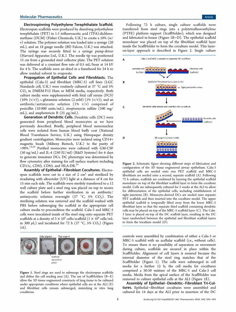
Following 72 h culture, single culture scaffolds weretransferred from steel rings into a polytetrafluoroethylene(PTFE) platform support (ScaffHolder), which was designedand fabricated in-house (Figure 1B−D). The epithelial scaffoldmonolayer was placed on top of the fibroblast scaffold layerinside the ScaffHolder to form the coculture model. This layer-on-layer approach is described in Figure 2. Single culture
controls were assembled by combination of either a Calu-3 orMRC-5 scaffold with an acellular scaffold (i.e., without cells).To ensure there is no possibility of separation or movementduring culture, scaffolds are secured in place within theScaffHolder. Alignment of cell layers is ensured because theinternal diameter of the steel ring matches that of theScaffHolder (Figure 1). The cells were submerged in cellmedia for a further 12 h; the cell media for coculturescomprised a 50:50 mixture of the MRC-5 and Calu-3 cellmedia. Media from the apical surface of the ScaffHolder wasremoved to culture epithelial cells at the ALI (Figure 1E).
Assembly of Epithelial−Dendritic−Fibroblast Tri-Cul-tures. Epithelial−fibroblast cocultures were assembled andcultured for 14 days at the ALI prior to insertion of the DC
Figure 1. Steel rings are used to submerge the electrospun scaffoldsand define the cell seeding area (A). The use of ScaffHolders (B−E)allow the 3D tissue engineered constructs of lung tissue to be culturedunder appropriate conditions where epithelial cells are at the ALI (E)and fibroblast cells remain submerged, mimicking in vitro lungconditions.
Figure 2. Schematic figure showing different steps of fabrication andconfiguration of the 3D tissue engineered airway epithelium. Calu-3epithelial cells are seeded onto one PET scaffold and MRC-5fibroblasts are seeded onto a second, separate scaffold (A). Following72 h culture, scaffolds are combined by layering the epithelial scaffoldmonolayer on top of the fibroblast scaffold layer to form the coculturemodel. Cells are subsequently cultured for 2 weeks at the ALI to allowfor differentiation of the epithelial cells, including establishment oftight junctions (B). Monocyte-derived DCs are seeded onto separatePET scaffolds and then inserted into the coculture model. The upperepithelial scaffold is temporally lifted away from the lower MRC-5fibroblast layer so that the separate third scaffold containing dendriticcells may be placed on-top of the MRC-5 scaffold layer (C). The Calu-3 layer is placed on-top of the DC scaffold layer, resulting in the DClayer sandwiched between the epithelial and fibroblast scaffold layersto form the triculture model (D).
Molecular Pharmaceutics Article
dx.doi.org/10.1021/mp5000295 | Mol. Pharmaceutics XXXX, XXX, XXX−XXXC

layer. The cell culture media composition remained as a 50:50mixture of Calu-3 and MRC-5 cell media.Immature DCs (Day 6) were prestained with Hoescht
nuclear stain (5 μg/mL) (Invitrogen, U.K.) and inoculatedonto PET electrospun scaffolds at a density of 2 × 10⁵ cells/scaffold and incubated for 24 h (37 °C, 5% CO2). The culturemedium was aspirated to remove DCs that had not attachedprior to insertion in between Calu-3 and MRC-5 layers in anestablished coculture model to form an immunocompetenttriculture model. A schematic describing the layer-on-layerapproach to assembling epithelial and fibroblast cocultures andsubsequent insertion of the DC layer is described in Figure 2.Stimulation of Triculture Model. Triculture models were
stimulated with house dust mite extract (HDM) (10 μg/mL)(GREER, U.S.A.) and lipopolysaccharide (LPS) (100 ng/mL)(Sigma-Aldrich, U.K.) or PBS control and incubated for 36 hprior to analysis. The triculture models were fixed with 4% (v/v) paraformaldehyde (Electron Microscopy Sciences, U.S.A.) inPBS, and the three scaffold layers were separated andimmunostained with pancytokeratin (epithelial cell marker).Scaffolds were then examined by confocal microscopy (LeicaSP2 confocal laser scanning microscope) (Leica Microsystems
Ltd., U.K.) with postvisualization performed using Volocitysoftware (Perkin-Elmer, U.K.).
Trans-Epithelial Electrical Resistance Measurements.Trans-epithelial electrical resistance (TEER) measurementswere performed across the epithelial cell monolayer of cellscultured at the ALI. Measurements were performed using anEVOM volt-ohm-meter and STX2 chopstick electrodes (WorldPrecision Instruments, U.K.). Prior to recording TEER,chopstick electrodes were sterilized (70% v/v ethanol indistilled water) and cell culture media was added to the upperchamber (500 μL) and lower chamber (1.5 mL total volume)and allowed to equilibrate for 30 min (37 °C, 5% CO2).Control measurements were performed using acellularscaffolds.
Scanning Electron Microscopy. Cellular samples of PETelectrospun scaffold were placed onto carbon-coated electronmicroscope stubs and sputter-coated with gold (5 min, BlazersSCD 030 Blazers Union Ltd., Liechtenstein) under an argonatmosphere (BOC, U.K.) prior to analysis. Samples wereimaged using SEM (Scanning Electron Microscopy) analysis(JEOL JMS-6060 LV microscope, JEOL Ltd., U.K.) operatingat an accelerating voltage of 10 kV. Cellular samples were fixed
Figure 3. Comparisons of scanning electron micrographs of decellularised lung tissue (A) with PET electrospun scaffold (B) showing morphologicalsimilarities. The fiber diameter of decellularised lung tissue and PET electrospun scaffolds were measured and show comparable dimensions (C).
Molecular Pharmaceutics Article
dx.doi.org/10.1021/mp5000295 | Mol. Pharmaceutics XXXX, XXX, XXX−XXXD

in 3% (v/v) glutaraldehyde overnight at 4 °C beforedehydration through an ascending series of ethanol concen-trations prior to SEM imaging.Scaffold Histology. Histology preparation was performed
by Nottingham University Advanced Microscopy Unit (AMU).Briefly, cellular scaffolds were fixed with 10% buffered formalin,excised, and embedded in paraffin. The paraffin embeddedblocks were then sectioned and stained with hematoxylin andeosin (H&E) before imaging.Immunocytochemistry. Scaffold samples were washed
with PBS prior to fixation with paraformaldehyde (4% (w/v))or methanol (100% (v/v)) for 15 min at room temperature(RT). Samples were washed in PBS (3 × 5 min each) beforebeing permeabilized using Triton X-100 (0.5% (v/v)) for 5 minat room temperature. Following a further wash in PBS (3 × 5min each), nonspecific antibody binding was blocked with goatserum (10% (v/v) in PBS for 5 min at room temperature.Samples were incubated with primary antibody (1:100)overnight at 4 °C. Primary antibodies used were Anti-Mucin5AC [45M1] (ab3649, AbCam, U.K.), mouse anti-ZO1(Invitrogen, U.K.), anti-fibronectin (ab 23750, AbCam, U.K.),Anti-Ki67 (ab15580, AbCam, U.K.), anti-collagen (ab34710,AbCam, U.K.), and pan-cytokeratin PK110 (SantaCruzBiotech, U.K.). Samples were then washed with PBS (3 × 5min each) and incubated with species-appropriate fluorescentlylabeled secondary antibodies (1:100) for 30 min at roomtemperature. Secondary antibodies included goat antimouseIgG Rhodamine Red X (Invitrogen, U.K.), goat antirabbit IgGFITC (Invitrogen, U.K.), and Alexa Fluor 488 goat antimouseIgM (μ chain) (Invitrogen, U.K.). Samples were washed in PBS(3 × 5 min each), incubated with Hoechst (5 μg/mL) for 5min at room temperature, and mounted using Fluoromountmounting medium. Immunostaining was observed using aconfocal microscope (Leica SP2 confocal laser scanningmicroscope, images processed with Leica confocal software)with postvisualization performed using Volocity software.Application of Papain. A papain (60U/mL) solution was
prepared with L-cysteine (5 mM) to reconstitute the cysteineactive site, and an aliquot (300 μL) was applied to the apicalsurface of Calu-3 cells. TEER measurements were performed tomonitor epithelial barrier integrity prior and post application.
■ RESULTSThe present study presents a multilayered 3D electrospun PETlung model capable of incorporating multiple cell types eachsupported upon their own individual electrospun layer. Theporous network of electrospun PET fibers can permit cellinteraction through both direct cell−cell contact and paracrinefactors within the 3D model. We report the incorporation oflung associated epithelial cells and fibroblasts and monocyte-derived DCs within the 3D model and determine how culturingthese cells together influences their behavior.Assembly of 3D Coculture Lung Model. SEM analysis of
decellularised human lung tissue (from healthy sections of lungtissue obtained from patients undergoing surgical procedures(Nottingham University Hospitals NHS Trust) after informedconsent and ethics approval) revealed a porous network ofnanometer-sized fibers (Figure 3A). Accordingly, we used anonbiodegradable polymer (namely, PET) to fabricate ananoscale porous scaffold (Figure 3B). The mean fiberdiameter of the PET scaffold (240 nm ±70) was similar tothe mean diameter of lung ECM (245 nm ±83) (Figure 3C).The average thickness of the PET scaffold, calculated using
histological sections, was 60 ± 10 μm. Owing to its robustphysical properties, the PET electrospun scaffold is capable ofwithstanding repeat handling allowing separation of individualcell layers for assessment following experimentation.The main structural cell types of the lung, epithelial, and
fibroblast cells were each cultured upon separate PET scaffolds.To incorporate an immune component into the model, atriculture system was formed by culturing DCs upon a thirdPET scaffold and inserting this between the epithelial andfibroblast layers of an established epithelial−fibroblast coculture(Figure 2).
Epithelial Barrier Formation and Integrity. Epithelialand fibroblast cells were each inoculated onto individual PETscaffold layers and cultured submerged in media to allow cellsto establish growth upon the scaffold, following which the cellinoculated scaffolds were assembled together within ourdeveloped ScaffHolders allowing physiologically relevantpositioning of the construct. Epithelial cells were culturedupon the uppermost layer allowing culture at ALI where theupper cell surface is in contact with the air. Culturing epithelialcells at the ALI mimics in vivo conditions by providing apical−basal polarity, which leads to full differentiation of epithelialcells and development of a functional barrier.26 The underlyingfibroblasts were positioned directly beneath the epithelialinoculated scaffold and remained submerged in media much asthey would in vivo.The production and maintenance of barrier integrity in
epithelial cells cultured upon PET electrospun scaffolds andpositioned at the ALI using ScaffHolders was assessed usingTEER measurements and permeability studies. Barrierformation was compared when epithelial cells were culturedalone (single culture) or with fibroblasts (coculture). TEER iswidely used to monitor barrier integrity, where an increase inresistance to flow of current is due to greater integrity of thebarrier, attributed to the formation of cellular tight junctions.27
An acellular control scaffold was monitored in order toreport a control value, as it is known that TEER valuesreportedly vary according to the material upon which theepithelial cells are cultured.28 In addition, a single culture ofMRC-5 fibroblasts was monitored as a control, as an increase inTEER value was not expected owing to MRC-5 fibroblastscharacteristically not forming tight junctions.Statistical analysis was performed using two-way ANOVA
with Sidaks̀ multiple comparison showed that TEER measure-ments of cocultures produced readings that were significantlygreater than those of epithelial cells cultured alone (Figure 4A).The TEER measurements show that after 14 days in culture
epithelial cells cultured alone, without fibroblasts, attainedmeasurements of ∼130 Ω cm2. The greatest increase was seenfor cocultures of epithelial and fibroblast cells attainingmeasurements of ∼200 Ω cm2. Thus, epithelial cells culturedtogether with fibroblasts appeared to achieve a confluentdifferentiated state earlier than single cultures of epithelial cellsas shown by earlier and greater increases in TEER measure-ments (Figure 4 A). The TEER values of the controls, anacellular scaffold and MRC-5 single culture, did not increase,maintaining baseline TEER measurements of ∼100 Ω cm2.Although human lung ex vivo TEER results are not available inthe literature, ex vivo rabbit airway epithelium has beenreported to be 260−320 Ω cm2 for the trachea29 and 266 Ωcm2 for the bronchus.30 Thus, the maximal TEER recorded inour 3D model are comparable to the data available for animalex vivo TEERs.
Molecular Pharmaceutics Article
dx.doi.org/10.1021/mp5000295 | Mol. Pharmaceutics XXXX, XXX, XXX−XXXE

The topography of the confluent epithelial cell layer wasobserved using SEM. The SEM micrographs of epithelial cellsfrom single culture, coculture, and fibroblasts from coculturesshowed what is thought to be the presence of ECM proteindeposition (Figure 4B).H&E staining of the epithelial layer from single and
cocultures indicated that single cultures formed a single thinlayer of cells, whereas the cocultured epithelial cells had a moredense and layered arrangement after 14 days at ALI (Figure 5).Furthermore, epithelial cells within cocultures appeared toproduce mucus earlier than single cultures as shown byimmunocytochemical staining (Figure 5).Immunocytochemical staining of the deposition of ECM
proteins indicated that the epithelial cells deposit collagen andfibronectin when cultured upon PET electrospun scaffolds(Figure 6). There appeared to be enhanced production of theseECM proteins from cocultured epithelial cells. The expressionof the cell proliferation marker Ki67 showed that epithelial cellsin coculture had lower levels of Ki67, possibly indicating theirtendency toward an earlier full differentiated state as alsoevidenced by an accelerated increase in TEER readings incocultures compared with single cultures of epithelial cells(Figure 7A). Fibroblast cell growth has not been adverselyaffected when positioned beneath the epithelial cell layer duringcoculture experimentation; the fibroblast cells are still presentand in a state of active growth as indicated by the production ofKi67 and have retained the ability to produce ECM proteinscollagen and fibronectin (Figure 6).
Epithelial Repair. The 3D lung model was cultured at theALI for 14 days before papain was applied to the epithelialsurface of single and cocultures. Papain is an allergen withcysteine protease activity and is known to disrupt tightjunctions in respiratory epithelium using its enzymatic activity,mimicking the action of other airborne allergens such as housedust mite, which also have a cysteine protease activity.31 Theapplication of papain on day 14 ALI was shown to disruptepithelial tight junctions in both single and coculture asdemonstrated by a sharp reduction in TEER values (Figure 7A)and disintegration of ZO1 protein visualized by immunostain-ing followed by confocal imaging (Figure 7B). The disruptionto tight junctions from coculture was thought to be less thanthat of single cultures, where almost no ZO1 could be observedpost papain. TEER measurements were performed for a further14 days to monitor the epithelial repair process, and further
Figure 4. The TEER measurements of cocultures are significantlyhigher than those of epithelial cells cultured alone. Error bars showstandard error of the mean. Statistical analysis was performed usingtwo-way ANOVA with Sidaks̀ multiple comparison with the differencebetween coculture and single Calu-3 culture having a p value <0.0001(A). SEM images of epithelial cells from single and coculture wherethe upper surface that has been imaged is thought to be predominantlyformed from ECM protein presence. The micrographs show thatepithelial cells from coculture have a smoother surface than those fromsingle culture (B).
Figure 5. After 14 days at the ALI. Histological staining shows thepresence of epithelial cells on the uppermost surface of the PETscaffold with a greater number of cells found in cocultures comparedto single cultures. Mucin production (red) was greater from coculturethan single culture, where cell nuclei are stained with DAPI (blue).
Figure 6. ECM staining following 14 days culture at the ALI shows thepresence of ECM proteins: collagen and fibronectin, predominantlyfound in epithelial and fibroblast cells and also Ki67, showing that thecells are in active growth. Epithelial cells in cocultures appear toexpress lower levels of Ki67 (compared to single cultures), indicatingtheir tendency toward full differentiation.
Molecular Pharmaceutics Article
dx.doi.org/10.1021/mp5000295 | Mol. Pharmaceutics XXXX, XXX, XXX−XXXF

examination of ZO1 expression was carried out. The cocultureswere observed to be recovering from the disruption withincreased TEERS and increased presence of ZO1 (Figure 7Aand B). However, TEER measurements did not appear torecover in single cultures during the same time frame andremained at approximately the same value as when the cellswere subjected to papain (Figure 7A). The confocal microscopyimages on day 15 (post papain) of single culture shows that thecell growth has been disrupted by papain and that tightjunctions are not present over a wide area of the sample. Thecells from single cultures recovered slightly as evidenced by thepresence of nondisrupted nuclei on day 28; however, thepresence of tight junctions remain absent (Figure 7B).DC Migration within the 3D Triculture Model. In a
proof-of-concept experiment, an immunocompetent triculturemodel comprising of epithelial, dendritic, and fibroblast layerswas constructed and DC migration within the modelmonitored. The triculture model was stimulated with HDMextract (a common airborne allergen) and toll-like 4 receptoragonist LPS (abundant in most airborne bacterial pathogens),and subsequently, the migration of DCs was assessed byconfocal microscopy. Epithelial cells were identified using
pancytokeratin and DCs had been prestained using the nuclearstain Hoescht.32 Assessment of the uppermost scaffold (bearingepithelial cells) revealed that after 36 h in the triculture, mostDCs seemed to have migrated to this layer and were present inclose proximity to the basal (in unstimulated samples) or apicalside (in stimulated samples) of the epithelial cell layer (Figure8A). The DCs migration was further confirmed by the fact that
DCs were found to be largely absent from the scaffold uponwhich they were originally inoculated (middle scaffold) (Figure8B). Typically, not many DCs could be observed in themajority of lower scaffolds (fibroblast layer) in either stimulatedor unstimulated samples (Figure 8C).
■ DISCUSSIONUsing electrospun porous fibers, we have engineered a 3Dtriculture of airway epithelial cells, dendritic cells, andfibroblasts at ALI creating a modular construct that mimicsthe cellular orientation and some functional properties ofhuman airway epithelium. The epithelium in vivo provides thefirst line of defense against environmental insults, and as such, itis capable of rapidly repairing any cellular damage caused.Epithelial cells in vivo are thought to respond to insult andinjury through a cascade of events of which there are threedistinct stages: dedifferentiation, proliferation, and differ-entiation.33 The first stage is the dedifferentiation of underlyingstromal cells, such as fibroblasts, that are initially exposed to theenvironment following injury to epithelial cells. The stromalcells are thought to migrate to the wound site to assist repair bydedifferentiation helping to cover the site of injury and assist inrestoring barrier integrity through proliferation.34 The final
Figure 7. Following the 14 days at the ALI, the enzyme papain wasapplied to the epithelial surface of single and cocultures, which wasshown to disrupt tight junctions demonstrated by the measurement ofTEERs (A) and by confocal imaging of ZO1 protein (B). Thedisruption to tight junctions from coculture was observed to be lessthan that of single cultures. TEER measurements were then performedfor a further 14 days to monitor the healing process. The cocultureswere observed to be recovering from the disruption with increase inTEERS and increase in the presence of ZO1. Single cultures did notappear to recover so well with TEER measurements remaining aroundthe same value as when the cells were subjected to papain. Statisticalanalysis was performed using two-way ANOVA with Sidaks̀ multiplecomparison, showing that the difference between coculture and singleCalu-3 culture that had been subjected to papain having a p value<0.0001.
Figure 8. Confocal microscopy images (×10 mag) of DC migration 36h after allergen stimulation in 3D tricoculture. The upper scaffoldcontained the Calu-3 epithelial layer (A), a middle layer to which DCswere seeded (B), and a lower scaffold containing MRC-5 (C). DCswere prestained with Hoescht nuclear stain (blue) and Calu-3 cellswere poststained with pancytokeratin (green). In single culture, DCsremained on the middle scaffold where they had been inoculated. Inthe triculture, most DCs migrated from middle scaffold (B) to upperscaffold (A). Upon treatment of the triculture with house dust miteextract (HDM) (10 μg/mL) and lipopolysaccharide (LPS) (100 ng/mL), DCs appeared to primarily migrate to the apical surface of theepithelial layer, whereas in unstimulated samples they appear to bemainly localized in the basal region of the epithelial layer (A).Experiments were performed in duplicates.
Molecular Pharmaceutics Article
dx.doi.org/10.1021/mp5000295 | Mol. Pharmaceutics XXXX, XXX, XXX−XXXG

stage in restoring barrier integrity is the differentiation of cellsinvolved in the repair process, including the restoration of fullfunction to epithelial cells, particularly their ability to formjunctional protein to “seal” the barrier.35
The exact mechanisms controlling cell fate during epitheliumrepair are not clear; however, it is thought that multipleparacrine factors and direct cell−cell contact are key parametersrequired to restore barrier integrity and function.36 It is,therefore, important to consider these factors when configuringtissue engineering strategies to create in vitro models ofepithelial tissue.36b
This study developed a 3D multicellular lung model that iscapable of forming a more physiologically relevant representa-tion of lung tissue than culturing cells alone or in 2D systems.Our studies corroborate the relevance of culturing multiple celltypes found in lung tissue together, as results demonstrate thatwhen epithelial cells were subjected to chemical insult (i.e., anenzymatically active allergen), they recovered earlier incocultures than single cultures. This supports the relevance ofcocultures as it is thought that the cells signal and respond inalliance.This study also assembled a triculture model in a proof-of-
concept experiment to examine whether DC migration couldoccur within the lung model following stimulation.A large number of DCs were found to migrate from the
scaffold in which they were initially inoculated (middle layer)through to the uppermost scaffold bearing the epithelial cells.This occurred both in the presence and absence of stimulationand may suggest that DCs were responding to epithelialsecretions.37 However, in samples stimulated with LPS andHDM, most DCs were located closer to the apical side of theepithelial barrier, whereas in unstimulated samples they weremainly located close to the basal side. These results prove thatthe 3D lung model is amenable to cell migration and that cell−cell contact is feasible. Individual scaffolds are typically 60 ± 10μm thick and the results of the DC migration studydemonstrate that scaffold thickness does not seem to impedeDC migration. The pattern of migration observed in thisexperiment substantiates DC migration in vivo, where uponactivation, DCs migrate to the epithelium and survey theimmune environment before migrating through the subepithe-lial compartment before traveling to lymph nodes.7,37,38 Thisenables future studies focused on inhaled drug delivery systemsas well as allergen and inhaled drugs uptake by DCs. We wouldestimate the triculture would be limited to one week due tomigration of DCs and their phenotypical/functional changesupon stimulation, which should provide enough time forperforming drug uptake/delivery experiments. Limitations to invitro models, particularly those generated from commerciallyavailable 2D inserts, are that the surface upon which cells growis planar, so cell−cell interaction is restricted. Cell interactionscan occur through the pores of the insert; however, these arethought to be constrained to paracrine interactions because thepore size of the substrate upon which the epithelial cells arecommonly cultured to allow barrier formation are too small,thus preventing cell migration. A 3D human skin triculturemodel comprising of keratinocytes, DCs, and fibroblasts hasbeen reported.14 This model shares similarities with the modeldeveloped in this work, including a physiologically relevantarrangement of the cell layers each supported on separate 3Dscaffolds and the establishment of a differentiated epithelial−fibroblast coculture prior to DC insertion. In the skin triculturemodel, the keratinocytes and fibroblasts were supported on 3D
microfiber-scale scaffolds; however, the DC layer was insertedinto the model in an agarose gel rather than on a fibrousscaffold. Migration of DCs in the skin model was reported fromthe agarose gel layer to only the fibroblast layer. Furthermore,the degree of migration appeared to be far less than thatobserved in the 3D model developed in this study. Hence,direct seeding of DCs on the porous scaffolds as opposed toencapsulation within a hydrogel seems to facilitate cellmigration.Epithelial cell lines such as Calu-3, the cell line we have used
in this study, are widely used as surrogates for primary cells.39
Despite many similarities between Calu-3 and primary cells,particularly in features like barrier formation and mucusproduction, there are also considerable functional differences(e.g., their cytokine profile). Therefore, use of cell linessomewhat limits the physiological relevance of the model andfuture efforts should focus on replacing cell lines with humanprimary cells.The 3D immunocompetent lung model presented in this
study successfully supports the culture of multiple cell types onelectrospun PET scaffolds that structurally resemble the nativelung ECM. The porous network of electrospun PET fibers canpermit cell interaction through both direct cell−cell contact andthrough paracrine factors within the 3D model as shown by theenhanced formation of a differentiated epithelial layer,enhanced epithelial repair, and migration of immune cellsincorporated within the model. Furthermore, the modularnature of our approach means that other relevant structural(e.g., smooth muscle cells) and immune cells (e.g., mast cells oreosinophils) could be included into the model with relativeease.Collectively, we believe this model possesses adequate
cellular and structural representation of the airway epitheliumand is amenable to in situ monitoring; as such, it presents aninvaluable tool for academic and pharmaceutical research withinthe fields of lung biology, disease modeling, and drug discoveryand delivery with the potential of reducing the need for someanimal experimentation in this area.
■ AUTHOR INFORMATION
Corresponding Author*A. M. Ghaemmaghami. Address: Division of Immunology,West Block, A Floor, Queen’s Medical Centre, University ofNottingham, NG7 2UH, United Kingdom. E-mail: [email protected].
NotesThe authors declare no competing financial interest.
■ ACKNOWLEDGMENTS
This work was jointly supported by a BBSRC IndustrialPartnership Award (IPA) and Kirkstall Ltd (http://kirkstall.org) (BB/H011293/1).
■ ABBREVIATIONS
ALI, air−liquid interface; DC, dendritic cells; DCM, dichloro-methane; ECM, extracellular matrix; FCS, fetal calf serum;HDM, house dust mite; LPS, lipopolysaccharide; PET,poly(ethylene terephthalate); PTFE, polytetrafluoroethylene;TEER, transepithelial electrical resistance; TFA, trifluoroaceticacid; UV, ultraviolet
Molecular Pharmaceutics Article
dx.doi.org/10.1021/mp5000295 | Mol. Pharmaceutics XXXX, XXX, XXX−XXXH

■ REFERENCES(1) Idoyaga, J.; Steinman, R. M. SnapShot: Dendritic Cells. Cell2011, 146 (4), 660−660 e2.(2) Seok, J.; Warren, H. S.; Cuenca, A. G.; Mindrinos, M. N.; Baker,H. V.; Xu, W.; Richards, D. R.; McDonald-Smith, G. P.; Gao, H.;Hennessy, L.; Finnerty, C. C.; Lopez, C. M.; Honari, S.; Moore, E. E.;Minei, J. P.; Cuschieri, J.; Bankey, P. E.; Johnson, J. L.; Sperry, J.;Nathens, A. B.; Billiar, T. R.; West, M. A.; Jeschke, M. G.; Klein, M. B.;Gamelli, R. L.; Gibran, N. S.; Brownstein, B. H.; Miller-Graziano, C.;Calvano, S. E.; Mason, P. H.; Cobb, J. P.; Rahme, L. G.; Lowry, S. F.;Maier, R. V.; Moldawer, L. L.; Herndon, D. N.; Davis, R. W.; Xiao, W.;Tompkins, R. G. Inflammation; Host Response to Injury, L. S. C. R.P., Genomic responses in mouse models poorly mimic humaninflammatory diseases. Proc. Natl. Acad. Sci. U.S.A. 2013, 110 (9),3507−12.(3) (a) Ghaemmaghami, A. M.; Hancock, M. J.; Harrington, H.; Kaji,H.; Khademhosseini, A. Biomimetic tissues on a chip for drugdiscovery. Drug Discovery Today 2012, 17 (3−4), 173−81. (b) Neuzi,P.; Giselbrecht, S.; Lange, K.; Huang, T. J.; Manz, A. Revisiting lab-on-a-chip technology for drug discovery. Nat. Rev. Drug discovery 2012, 11(8), 620−32.(4) Ryu, J. H.; Kim, C. H.; Yoon, J. H. Innate immune responses ofthe airway epithelium. Mol. Cells 2010, 30 (3), 173−83.(5) Zhang, S.; Smartt, H.; Holgate, S. T.; Roche, W. R. Growthfactors secreted by bronchial epithelial cells control myofibroblastproliferation: an in vitro co-culture model of airway remodeling inasthma. Lab. Invest.: J. Tech. Methods Pathol. 1999, 79 (4), 395−405.(6) (a) Bergeron, C.; Boulet, L. P. Structural changes in airwaydiseases: characteristics, mechanisms, consequences, and pharmaco-logic modulation. Chest 2006, 129 (4), 1068−87. (b) Hammad, H.;Lambrecht, B. N. Dendritic cells and airway epithelial cells at theinterface between innate and adaptive immune responses. Allergy2011, 66 (5), 579−87.(7) Hammad, H.; Chieppa, M.; Perros, F.; Willart, M. A.; Germain,R. N.; Lambrecht, B. N. House dust mite allergen induces asthma viaToll-like receptor 4 triggering of airway structural cells. Nat. Med.2009, 15 (4), 410−6.(8) Nichols, J. E.; Niles, J.; Riddle, M.; Vargas, G.; Schilagard, T.; Ma,L.; Edward, K.; La Francesca, S.; Sakamoto, J.; Vega, S.; Ogadegbe, M.;Mlcak, R.; Deyo, D.; Woodson, L.; McQuitty, C.; Lick, S.; Beckles, D.;Melo, E.; Cortiella, J. Production and assessment of decellularized pigand human lung scaffolds. Tissue Eng., Part A 2013, 19 (17−18),2045−62.(9) Carterson, A. J.; Honer zu Bentrup, K.; Ott, C. M.; Clarke, M. S.;Pierson, D. L.; Vanderburg, C. R.; Buchanan, K. L.; Nickerson, C. A.;Schurr, M. J. A549 lung epithelial cells grown as three-dimensionalaggregates: alternative tissue culture model for Pseudomonasaeruginosa pathogenesis. Infect. Immun. 2005, 73 (2), 1129−40.(10) Ravindran, S.; Song, Y.; George, A. Development of three-dimensional biomimetic scaffold to study epithelial-mesenchymalinteractions. Tissue Eng., Part A 2010, 16 (1), 327−42.(11) Liu, Y.; Rayatpisheh, S.; Chew, S. Y.; Chan-Park, M. B. Impactof endothelial cells on 3D cultured smooth muscle cells in abiomimetic hydrogel. ACS Appl. Mater. Interfaces 2012, 4 (3), 1378−87.(12) (a) Rimann, M.; Angres, B.; Patocchi-Tenzer, I.; Braum, S.;Graf-Hausner, U., Automation of 3D Cell Culture Using ChemicallyDefined Hydrogels. J. Lab. Autom. 2013; (b) DeVolder, R.; Kong, H.-J.Hydrogels for in vivo-like three-dimensional cellular studies. WileyInterdiscip. Rev.: Syst. Biol. Med. 2012, 4 (4), 351−365.(13) Garcia-Nieto, S.; Johal, R. K.; Shakesheff, K. M.; Emara, M.;Royer, P. J.; Chau, D. Y.; Shakib, F.; Ghaemmaghami, A. M. Lamininand fibronectin treatment leads to generation of dendritic cells withsuperior endocytic capacity. PLoS one 2010, 5 (4), e10123.(14) Chau, D. Y.; Johnson, C.; MacNeil, S.; Haycock, J. W.;Ghaemmaghami, A. M. The development of a 3D immunocompetentmodel of human skin. Biofabrication 2013, 5 (3), 035011.(15) Huang, Y. X.; Ren, J.; Chen, C.; Ren, T. B.; Zhou, X. Y.Preparation and properties of poly(lactide-co-glycolide) (PLGA)/
nano-hydroxyapatite (NHA) scaffolds by thermally induced phaseseparation and rabbit MSCs culture on scaffolds. J. Biomater. Appl.2008, 22 (5), 409−32.(16) Ekaputra, A. K.; Prestwich, G. D.; Cool, S. M.; Hutmacher, D.W. Combining electrospun scaffolds with electrosprayed hydrogelsleads to three-dimensional cellularization of hybrid constructs.Biomacromolecules 2008, 9 (8), 2097−103.(17) Harrington, H.; Rose, F. R. A. J.; Aylott, J. W.; Ghaemmaghami,A. M. Self-reporting Scaffolds for 3-Dimensional Cell Culture. J.Visualized Exp. 2013, No. 81, e50608.(18) (a) Matthews, J. A.; Wnek, G. E.; Simpson, D. G.; Bowlin, G. L.Electrospinning of collagen nanofibers. Biomacromolecules 2002, 3 (2),232−8. (b) Shih, Y. R.; Chen, C. N.; Tsai, S. W.; Wang, Y. J.; Lee, O.K. Growth of mesenchymal stem cells on electrospun type I collagennanofibers. Stem Cells 2006, 24 (11), 2391−7. (c) Rho, K. S.; Jeong,L.; Lee, G.; Seo, B. M.; Park, Y. J.; Hong, S. D.; Roh, S.; Cho, J. J.;Park, W. H.; Min, B. M. Electrospinning of collagen nanofibers: effectson the behavior of normal human keratinocytes and early-stage woundhealing. Biomaterials 2006, 27 (8), 1452−61.(19) Wnek, G. E.; Carr, M. E.; Simpson, D. G.; Bowlin, G. L.Electrospinning of Nanofiber Fibrinogen Structures. Nano Lett. 2002,3 (2), 213−216.(20) Intranuovo, F.; Howard, D.; White, L. J.; Johal, R. K.;Ghaemmaghami, A. M.; Favia, P.; Howdle, S. M.; Shakesheff, K. M.;Alexander, M. R. Uniform cell colonization of porous 3-D scaffoldsachieved using radial control of surface chemistry. Acta Biomater. 2011,7 (9), 3336−44.(21) (a) Cortiella, J.; Nichols, J. E.; Kojima, K.; Bonassar, L. J.;Dargon, P.; Roy, A. K.; Vacant, M. P.; Niles, J. A.; Vacanti, C. A.Tissue-engineered lung: an in vivo and in vitro comparison ofpolyglycolic acid and pluronic F-127 hydrogel/somatic lung progenitorcell constructs to support tissue growth. Tissue Eng. 2006, 12 (5),1213−25. (b) Bashur, C. A.; Dahlgren, L. A.; Goldstein, A. S. Effect offiber diameter and orientation on fibroblast morphology andproliferation on electrospun poly(D,L-lactic-co-glycolic acid) meshes.Biomaterials 2006, 27 (33), 5681−8. (c) Blackwood, K. A.; McKean,R.; Canton, I.; Freeman, C. O.; Franklin, K. L.; Cole, D.; Brook, I.;Farthing, P.; Rimmer, S.; Haycock, J. W.; Ryan, A. J.; MacNeil, S.Development of biodegradable electrospun scaffolds for dermalreplacement. Biomaterials 2008, 29 (21), 3091−104.(22) (a) Telemeco, T. A.; Ayres, C.; Bowlin, G. L.; Wnek, G. E.;Boland, E. D.; Cohen, N.; Baumgarten, C. M.; Mathews, J.; Simpson,D. G. Regulation of cellular infiltration into tissue engineering scaffoldscomposed of submicron diameter fibrils produced by electrospinning.Acta Biomater. 2005, 1 (4), 377−385. (b) Zhang, Y.; Yang, F.; Liu, K.;Shen, H.; Zhu, Y.; Zhang, W.; Liu, W.; Wang, S.; Cao, Y.; Zhou, G.The impact of PLGA scaffold orientation on in vitro cartilageregeneration. Biomaterials 2012, 33 (10), 2926−2935.(23) Ma, Z.; Kotaki, M.; Yong, T.; He, W.; Ramakrishna, S. Surfaceengineering of electrospun polyethylene terephthalate (PET) nano-fibers towards development of a new material for blood vesselengineering. Biomaterials 2005, 26 (15), 2527−2536.(24) Emara, M.; Royer, P. J.; Mahdavi, J.; Shakib, F.;Ghaemmaghami, A. M. Retagging identifies dendritic cell-specificintercellular adhesion molecule-3 (ICAM3)-grabbing non-integrin(DC-SIGN) protein as a novel receptor for a major allergen fromhouse dust mite. J. Biol. Chem. 2012, 287 (8), 5756−63.(25) Sharquie, I. K; Al-Ghouleh, A.; Fitton, P.; Clark, M. R.; Armour,K. L.; Sewell, H. F.; Shakib, F.; Ghaemmaghami, A. M. Aninvestigation into IgE-facilitated 752 allergen recognition andpresentation by human dendritic cells. BMC Immunol. 2013, 14(54), No. 10.1186/1471-2172-14-54.(26) (a) Kasper, M.; Singh, G. Epithelial lung cell marker: currenttools for cell typing. Histol. Histopathol. 1995, 10 (1), 155−69.(b) Pohl, C.; Hermanns, M. I.; Uboldi, C.; Bock, M.; Fuchs, S.; Dei-Anang, J.; Mayer, E.; Kehe, K.; Kummer, W.; Kirkpatrick, C. J. Barrierfunctions and paracellular integrity in human cell culture models of theproximal respiratory unit. Eur. J. Pharm. Biopharm. 2009, 72 (2), 339−49.
Molecular Pharmaceutics Article
dx.doi.org/10.1021/mp5000295 | Mol. Pharmaceutics XXXX, XXX, XXX−XXXI

(27) Zhang, M.; Kim, K. J.; Iyer, D.; Lin, Y.; Belisle, J.; McEnery, K.;Crandall, E. D.; Barnes, P. F. Effects of Mycobacterium tuberculosis onthe bioelectric properties of the alveolar epithelium. Infect. Immunol.1997, 65 (2), 692−8.(28) Nalayanda, D. D.; Puleo, C.; Fulton, W. B.; Sharpe, L. M.;Wang, T. H.; Abdullah, F. An open-access microfluidic model for lung-specific functional studies at an air-liquid interface. Biomed. Micro-devices 2009, 11 (5), 1081−9.(29) Bhat, M.; Toledo-Velasquez, D.; Wang, L.; Malanga, C.; Ma, J.H.; Rojanasakul, Y. Regulation of Tight Junction Permeability byCalcium Mediators and Cell Cytoskeleton in Rabbit TrachealEpithelium. Pharm. Res. 1993, 10 (7), 991−997.(30) Rojanasakul, Y.; Wang, L.-Y.; Bhat, M.; Glover, D.; Malanga, C.;Ma, J. H. The Transport Barrier of Epithelia: A Comparative Study onMembrane Permeability and Charge Selectivity in the Rabbit. Pharm.Res. 1992, 9 (8), 1029−1034.(31) Shakib, F.; Ghaemmaghami, A. M.; Sewell, H. F. The molecularbasis of allergenicity. Trends Immunol. 2008, 29 (12), 633−42.(32) Metzgeroth, G.; Mantz, C.; Kuhn, C.; Schultheis, B.; Hehlmann,R.; Hastka, J. Reliable identification of small cell lung cancer incytological specimens by immunocytology. Onkologie 2007, 30 (6),311−5.(33) Shimizu, T.; Nishihara, M.; Kawaguchi, S.; Sakakura, Y.Expression of phenotypic markers during regeneration of rat trachealepithelium following mechanical injury. Am. J. Respir. Cell Mol. Biol.1994, 11 (1), 85−94.(34) Davies, D. E. The role of the epithelium in airway remodeling inasthma. Proc. Am. Thorac. Soc. 2009, 6 (8), 678−82.(35) Stripp, B. R.; Reynolds, S. D. Maintenance and repair of thebronchiolar epithelium. Proc. Am. Thorac. Soc. 2008, 5 (3), 328−33.(36) (a) Knight, D. Epithelium-fibroblast interactions in response toairway inflammation. Immunol. Cell. Biol. 2001, 79 (2), 160−4.(b) Vrana, N. E.; Lavalle, P.; Dokmeci, M. R.; Dehghani, F.;Ghaemmaghami, A. M.; Khademhosseini, A. Engineering functionalepithelium for regenerative medicine and in vitro organ models: areview. Tissue Eng., Part B 2013, 19 (6), 529−43.(37) Banchereau, J.; Steinman, R. M. Dendritic cells and the controlof immunity. Nature 1998, 392 (6673), 245−52.(38) Cook, D. N.; Bottomly, K. Innate immune control of pulmonarydendritic cell trafficking. Proc. Am. Thorac. Soc. 2007, 4 (3), 234−9.(39) Ong, H. X.; Traini, D.; Young, P. M. Pharmaceuticalapplications of the Calu-3 lung epithelia cell line. Expert Opin. DrugDelivery 2013, 10 (9), 1287−302.
Molecular Pharmaceutics Article
dx.doi.org/10.1021/mp5000295 | Mol. Pharmaceutics XXXX, XXX, XXX−XXXJ