Computer-Aided Drug Design applied to marine drug discovery
357
Computer-Aided Drug Design applied to marine drug discovery Disseny de fàrmacs assistit per ordinador aplicat a la cerca de possibles fàrmacs marins Laura Llorach Parés Aquesta tesi doctoral està subjecta a la llicència Reconeixement- NoComercial – SenseObraDerivada 4.0. Espanya de Creative Commons. Esta tesis doctoral está sujeta a la licencia Reconocimiento - NoComercial – SinObraDerivada 4.0. España de Creative Commons. This doctoral thesis is licensed under the Creative Commons Attribution-NonCommercial- NoDerivs 4.0. Spain License.
-
Upload
khangminh22 -
Category
Documents
-
view
1 -
download
0
Transcript of Computer-Aided Drug Design applied to marine drug discovery

Computer-Aided Drug Design
applied to marine drug discovery
Disseny de fàrmacs assistit per ordinador aplicat a la cerca de possibles fàrmacs marins
Laura Llorach Parés
Aquesta tesi doctoral està subjecta a la llicència Reconeixement- NoComercial – SenseObraDerivada 4.0. Espanya de Creative Commons. Esta tesis doctoral está sujeta a la licencia Reconocimiento - NoComercial – SinObraDerivada 4.0. España de Creative Commons. This doctoral thesis is licensed under the Creative Commons Attribution-NonCommercial-NoDerivs 4.0. Spain License.

Computer-Aided Drug Design applied to marine drug
discovery
Doctoral Thesis
Laura Llorach Pares Barcelona, 2019


Programa de Doctorat en Biologia Marina
Departament de Biologia evolutiva, ecologia i ciències ambientals
Computer-Aided Drug Design applied to marine drug
discovery
Disseny de fàrmacs assistit per ordinador aplicat a la cerca de possibles fàrmacs marins
Memòria presentada per
Laura Llorach Pares
per optar al grau de
Doctora per la Universitat de Barcelona
Laura Llorach Pares
Barcelona, 2019
Vist i plau de la directora de la tesi
Dra. Conxita Avila Escartín
Catedràtica del departament de Biologia
evolutiva, ecologia i ciències ambientals
Facultat de Biologia Universitat de Barcelona
Vist i plau del co-director de la tesi
Dr. Melchor Sánchez Martínez
Director científic Mind The Byte.SL


“Who am I? Who am I?
I am Jean Valjean!”
Les Misérables, Victor Hugo


Agraïments / Acknowledgements
Portava temps pensant com escriure aquest capítol. Per una banda, pensava en aquelles
persones a les que volia agrair que haguessin format part d’aquest procés que és fer una tesi. I per
l’altra, em plantejava no fer agraïments i dedicar-li al meu fill, en Roger.
Com podeu veure, m’he acabat decantant per fer un agraïment general, ja que han sigut unes
quantes les persones a les que necessito dedicar unes paraules.
Voldria començar pel principi de la història i agrair a la Mire (Mireia Rodríguez), amiga i
companya d’aventures i viatges, que sense tu no hauria sigut prou valenta per llençar-me a fer la carrera
a Portugal. I mira, vist en perspectiva, va ser un gran esforç, però ha valgut la pena. A tú, Pepe (Josep
Ferrer), per dedicar tantes hores a fer-me entendre estadística i a la vegada, ensenyar-me a pensar.
Sense les teves classes no hauria tret la carrera ni seria qui sóc ara.
A l’Alfons Nonell, de MtB, per donar-me l’oportunitat de fer les pràctiques a la seva empresa.
Aquí va ser on va començar tot. Gràcies a les pràctiques del màster vaig començar a conèixer
l’apassionant món de la química computacional i el disseny de fàrmacs. També una menció a tots els
companys de MtB, tant amb els que hem compartit poc temps com aquells amb els que n’hem
compartit més, que durant aquests anys hem viscut canvis, alguns bons i altres no tant, experiències,
coneixements i alguna que altra cerveseta.
De MtB s’ha de fer una menció especial pel Mel, Melchor, encara recordo el primer dia que vaig
venir a l’oficina per fer una entrevista. I els primers dies... No entenc com vares poder tenir tanta
paciència i ensenyar-me tant i tan bé. De tu ho he aprés gairebé tot. Mentor i company. Et puc assegurar
que recordo perfectament apuntar-me a la llibreta “ls -ltr” per que no entenia res del que feia. Això de
Linux els primers dies va ser dur, molt dur. Després, amb el temps, he aprés a fer els meus scripts i bon
servei que t’han fet a vegades, eh?! Durant aquests anys hem treballat amb projectes molt interessants
on els dos hem après molt i, en el fons, crec que formavem un bon equip. Tots dos recordem i
recordarem, un projecte que ens va costar suor i sang, però entre els dos el vàrem aconseguir tirar
endavant amb perseverança i esforç. Com a co-director de tesis també t’he d’agrair l’esforç que has
hagut de fer aquests últims dies de correccions per que tot estigués a punt. Per tot, gràcies.
A la Conxita, una dona amb les idees clares i apassionada per l’Antàrtida i les molècules marines.
Gràcies al teu esperit investigador i al projecte BLUEBIO, vaig poder començar a conèixer les molècules
marines, primer durant les pràctiques del màster amb les quals vàrem fer el treball de final de màster i
després durant tot el doctorat, on aquestes molècules marines s’han acabat convertit en “les meves

molècules”. Crec que ens ho vàrem passar força bé en la nostra estada a Creta; congrés, platjeta i algun
imprevist que altre amb el tema aeroport. Ens va servir per coneixe’ns una miqueta més, ja que al fer un
doctoral industrial, no ens hem pogut conèixer com tocava. De totes maneres, sempre has estat allà i
ajudat en tot el que he necessitat. Conxita, un plaer haver-te tingut de directora de tesi!
Ara voldria agrair a la família el seu suport. Malgrat que crec que no han entès mai què estic
fent, han intentat respectar-ho i en aquests últims temps, on m’he dedicat a escriure, han ajudat en tot
el que han pogut. Als avis i àvies, gràcies per preguntar-me com tenia la feina i per intentar, una vegada i
un altre, entendre a que em dedico i que faig. Podeu seguir preguntant, us ho seguiré intentant explicar
el millor que puc.
Als meus pares, que tot i que crec que quan els hi vaig dir, primer, que faria la carrera a Portugal
mentre treballava, després, que amb 30 anys deixava una bona feina per començar un màster i després
que faria un doctorat mentre treballava, no varen ser els pares més feliços del món. Crec que per a ells,
això de prioritzar la formació acadèmica a una feina estable els hi va costar de pair, però amb el temps,
han vist que tan malament tampoc m’ha anat, no?! Us estimo, pares! Ja sabeu que sóc tossuda i que de
vegades pensem diferent, però al final, cada un hem de fer el nostre camí, i el meu camí no està tan
lluny del vostre.
Als meus sogres i sobretot a la meva sogra, que ha hagut de fer d’àvia durant moltes tardes per
que jo em pogués dedicar a la tesis. Suegri! Gràcies per tot. Per fer d’àvia, que t’encanta, per cuidar-nos
a tots, ajudar-nos a casa, fer-nos tants menjars i tant bons... Ets una gran àvia, una gran mare i una sogra
increïble!
A l’Oriol, el meu mariduky! Carinyu, que això ja s’acaba i tot tornarà a la normalitat. Sé que has
hagut d’aguantar el meu caràcter i mal humor, que puc arribar a ser molt pesada, ja ho sé. Que moltes
vegades voldries fer les teves “cosas jueguiles” i no pots. Però has de saber que ets un gran home, ple de
bondat i que treus el millor de mi. Tenir-te al costat es una sort i espero no perdre’t. Gràcies per fer de
marit i de pare tant bé. T’estimo!
Fill, a tú també t’estimo. No t’assabentes de gran cosa per què tot just ara faràs un any, i tú no
ho saps, però has fet gairebé la meitat d’un doctorat! Una part a la panxa de la mama i l’altre aquí al
meu costat. Ets el millor regal del món. Per tú, Roger, tota aquesta tesis i tot l’esforç, decisions i història
que hi ha al darrera, per què res és senzill, per què tot val la pena.
Finalment, a tota aquella gent que no ha tingut una relació directa amb la tesi però que han
estat sempre allà. Nenes, llop de neu, Patri. Gràcies pels ànims!

Contents General Introduction and Objectives ...................................................................................................... 1
General introduction ..................................................................................................................... 3
Biodiversity in marine ecosystems ....................................................................................... 3
Antarctic benthic ecosystems .............................................................................................. 4
Mediterranean benthic ecosystems..................................................................................... 9
Marine natural products.................................................................................................... 11
Computer-Aided Drug Design and Discovery ..................................................................... 12
Alzheimer’s Disease........................................................................................................... 25
Objectives of this thesis ............................................................................................................... 29
Supervisor’s Report ..................................................................................................................... 31
Section I. Target elucidation from a computational approach ............................................................. 33
Chapter 1. In silico studies to find new therapeutic indications for marine molecules. .............. 37
Abstract ............................................................................................................................ 38
Resum ............................................................................................................................... 39
Introduction ...................................................................................................................... 40
Results and discussion ....................................................................................................... 43
Materials and Methods ..................................................................................................... 58
Conclusions ....................................................................................................................... 61
Appendix ........................................................................................................................... 63
References ........................................................................................................................ 67
Section II. Elucidation of different pharmacophoric features of marine compounds and a precise in
silico binding study ............................................................................................................................... 71
Chapter 2. Computer-aided drug design applied to marine drug discovery: Meridianins as
Alzheimer’s disease therapeutic agents ...................................................................................... 75
Abstract ............................................................................................................................ 76
Resum ............................................................................................................................... 77
Introduction ...................................................................................................................... 78
Results .............................................................................................................................. 81
Discussion ......................................................................................................................... 90

Materials and Methods ..................................................................................................... 95
Conclusions ..................................................................................................................... 104
Appendix ......................................................................................................................... 106
References ...................................................................................................................... 113
Chapter 3. Kororamides, convolutamines, and indole derivatives as possible tau and dual
specificity kinases inhibitors for Alzheimer’s Disease ............................................................... 121
Abstract .......................................................................................................................... 122
Resum ............................................................................................................................. 123
Introduction .................................................................................................................... 124
Results and discussion ..................................................................................................... 126
Materials and Methods ................................................................................................... 149
Conclusions ..................................................................................................................... 154
Appendix ......................................................................................................................... 157
References ...................................................................................................................... 162
Section III. A combined computational and experimental study......................................................... 169
Chapter 4. Meridianins and lignarenones as potential GSK3β inhibitors and inductors of
structural synaptic plasticity ..................................................................................................... 173
Abstract .......................................................................................................................... 174
Resum ............................................................................................................................. 175
Introduction .................................................................................................................... 176
Results and discussion ..................................................................................................... 179
Materials and Methods ................................................................................................... 192
Conclusions ..................................................................................................................... 196
Appendix ......................................................................................................................... 198
References ...................................................................................................................... 200
General Discussion.............................................................................................................................. 207
General discussion ..................................................................................................................... 209
CADD potential in drug discovery .................................................................................... 210
Protein kinase inhibitors and MNPs pharmacophoric properties ..................................... 213
Pharmacokinetic properties to found hits from marine scaffolds ..................................... 216

Concluding remarks and future perspectives ................................................................... 217
Final Conclusions ................................................................................................................................ 219
Final conclusions ....................................................................................................................... 221
Catalan Summary................................................................................................................................ 223
Objectius d’aquesta tesi............................................................................................................. 225
Resultats ................................................................................................................................... 227
Discussió general ....................................................................................................................... 230
Conclusions finals ...................................................................................................................... 241
General References ............................................................................................................................. 243
General references .................................................................................................................... 245
Appendix I........................................................................................................................................... 269
Appendix II.......................................................................................................................................... 337


General Introduction and Objectives


General Introduction
3
General introduction
Biodiversity in marine ecosystems
Ecology is often described as the biology of the ecosystems. Margalef, in his book “Ecología”,
defined ecology as “the study of systems to a level at which individuals or whole organisms can be
considered elements of interaction among them or with a loosely organized environmental matrix”
(Margalef, 1974). An ecosystem comprehends a group of living organisms, or communities, in
conjunction with their abiotic components, all of them interacting as a system (Willis, 1997). The abiotic
factors are chemical or physical components of the environment, such as water, light, or temperature.
Ecosystems are controlled by internal factors, such as degradation and decomposition, or external
factors, such as climate or topology. Marine ecosystems are the largest on Earth, covering more than
70% of the surface of the planet (UNESCO, 2017). They are formed by oceans, seas and nearshore
systems, such as salt marshes and mudflats. Marine biodiversity is the result of life evolution for billions
of years and it is of great interest and value in many senses, but for a long time it has been
underestimated (Snelgrove, 2016). In fact, between one and two-thirds of marine species are
considered to be not yet described (Appeltans et al., 2012). Among these huge amounts of marine
species, as in terrestrial ecosystems, a wide net of interactions exists, generating all sorts of defences
and protective systems to survive (Faulkner & Ghiselin, 1983; Van Donk et al., 2011). Marine organisms
communicate through intra- and/or interspecific interactions, which are often regulated by natural
products (NPs) (infochemicals) and comprise what it is known as chemical ecology (Dayton et al., 1994;
Hay & Fenical, 1996). Chemodiversity is intrinsically related to biodiversity and is the result of constant
organism-to-organism interactive process, and thus, high biodiversity and ecological interactions are
linked to high chemical diversity (Barre, 2010; Núñez-Pons & Avila, 2015). The unique chemical diversity
of NPs, and marine natural products (MNPs) in particular, has been for long a major source of drug
candidates (Blunt et al., 2018a). Many marine invertebrates, due to the strong predation pressure,
possess unusual bioactive compounds that are essential for them to survive, playing important roles in
predatory and competitive interactions (Leal et al., 2012; Avila, 2016; Blunt et al., 2018a). Therefore,
with the aim to understand the pharmacological potential of natural compounds from marine benthic
invertebrates we develop here a computer-aided drug design study over a group of secondary
metabolites from selected marine organisms.
General Introduction
4
Antarctic benthic ecosystems
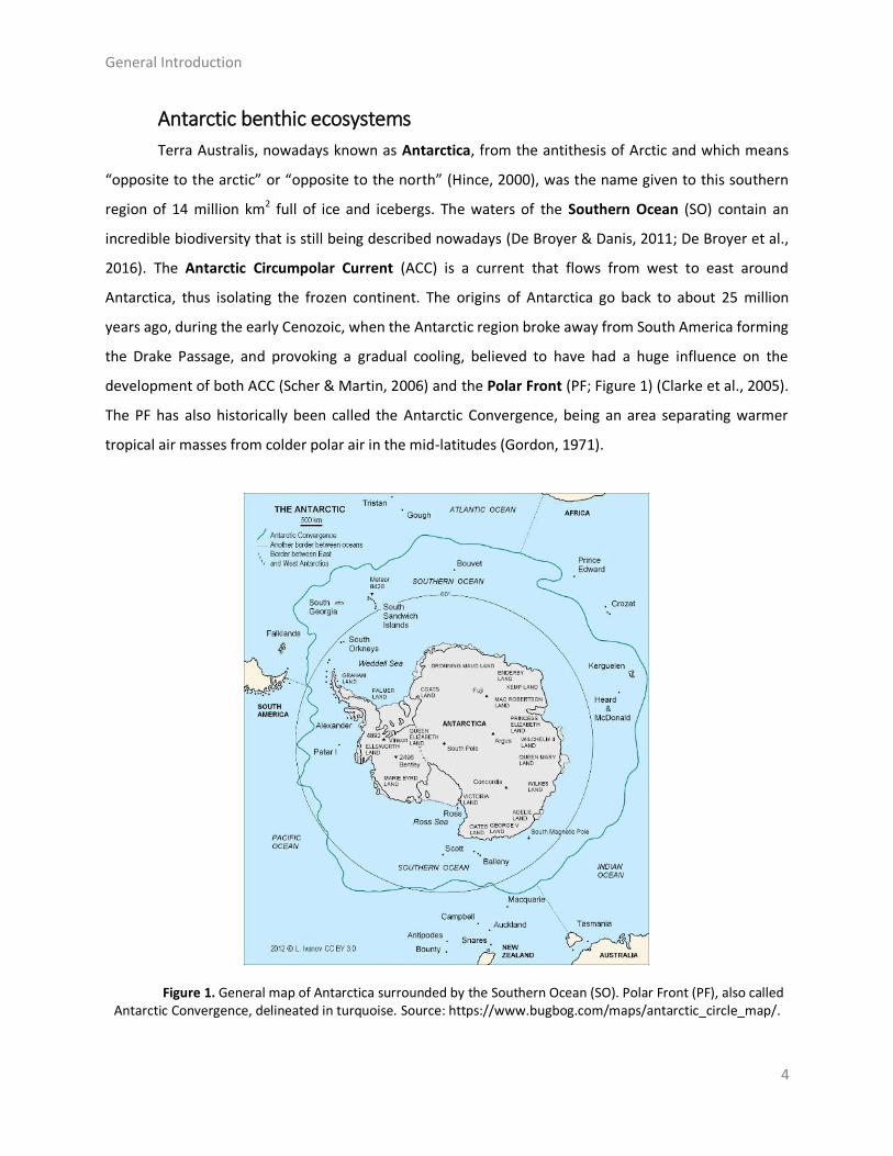
Terra Australis, nowadays known as Antarctica, from the antithesis of Arctic and which means
“opposite to the arctic” or “opposite to the north” (Hince, 2000), was the name given to this southern
region of 14 million km2 full of ice and icebergs. The waters of the Southern Ocean (SO) contain an
incredible biodiversity that is still being described nowadays (De Broyer & Danis, 2011; De Broyer et al.,
2016). The Antarctic Circumpolar Current (ACC) is a current that flows from west to east around
Antarctica, thus isolating the frozen continent. The origins of Antarctica go back to about 25 million
years ago, during the early Cenozoic, when the Antarctic region broke away from South America forming
the Drake Passage, and provoking a gradual cooling, believed to have had a huge influence on the
development of both ACC (Scher & Martin, 2006) and the Polar Front (PF; Figure 1) (Clarke et al., 2005).
The PF has also historically been called the Antarctic Convergence, being an area separating warmer
tropical air masses from colder polar air in the mid-latitudes (Gordon, 1971).
Figure 1. General map of Antarctica surrounded by the Southern Ocean (SO). Polar Front (PF), also called Antarctic Convergence, delineated in turquoise. Source: https://www.bugbog.com/maps/antarctic_circle_map/.

General Introduction
5
The PF constitutes a natural barrier that limits the exchange of cold and warm water, turning
this area into a distinctive biogeographical region. This particularity let the Antarctic region in isolation,
affecting the evolution of its fauna (Clarke & Crame, 1989). This is also associated with the high degree
of endemisms found in Antarctic marine ecosystems (Clarke & Johnston, 2003). With the aim of
classifying the SO biodiversity, the Register of Antarctic Marine Species (RAMS) published an accurated
list of more than 8100 species, where an 88% are benthic species (De Broyer et al., 2011). The benthic
community is exposed to considerable predatory pressure exerted by both macro- and micropredators
(McClintock & Baker; Oshel & Steele; Dayton et al., 1994; Figuerola et al., 2013; Moles et al., 2015).
Several studies demonstrated that most Antarctic benthic invertebrates present natural products in
their crude organic extracts that act as feeding repellents to avoid predation, thus using different
defensive chemical mechanisms (McClintock & Janssen, 1990; Amsler et al., 2001; Iken et al., 2002; Avila
et al., 2008; Koplovitz et al., 2009; Slattery, 2009; Moles et al., 2015). Overall, the organisms living in
Antarctic benthic ecosystems have developed very effective chemical defensive strategies, based on
secondary metabolites (natural products), which are crucial for species survival. Moreover, these
unique chemical compounds can potentially be further exploited for the development of new drugs,
considering its potential pharmacological properties (Núñez-Pons et al., 2015; Avila, 2016).
Natural products from Antarctic organisms have been reviewed several times recently (Avila et
al., 2008; Moles et al., 2015; Blunt et al., 2018a). From polar organisms such as sponges, cnidarians,
molluscs, bryozoans, and tunicates, a wide variety of biological compounds with diverse activities has
been isolated, such as antitumorals, anti-bacterials, and anti-inflammatories (Avila, 2016; Tian et al.,
2017). In this thesis I have worked with several compounds from diverse Antarctic benthic species, with
special attention to the compounds listed below.
Sponges are organisms full of pores and channels, allowing water circulation through them.
Their distribution is worldwide in all oceans, including tropical and polar regions with an approximate
number of 5.000-10.000 known species (Bergquist, 2001). Sponges (Porifera) are very effective
competitors for space, producing toxins and preventing other sessile organisms, such as ascidians, from
growing on top or nearby, being one of the most diverse sources of bioactive natural products known
(Proksch, 1994; Wang, 2006; Mehbub et al., 2014; Figueroa et al., 2015; Blunt et al., 2018a). Within the
phylum Porifera the most diverse class is Demosponges (Figure 2), including 76,2% of all described
species (WoRMS). The genera Latrunculia du Bocage, 1869, Dendrilla Lendenfeld, 1883, and Aplysilla
Schulze, 1878 belong to Demosponges.

General Introduction
6
Figure 2. The Antarctic Demosponge Latrunculia apicalis (Ridley & Dendy, 1886). Adapted from WoRMS (WoRMS).
In order to study the pharmacological properties of marine compounds, three secondary
metabolites isolated from diverse Antarctic sponges are included in this thesis (Chapter 1). These are
discorhabdin B, an alkaloid from the species Latrunculia apicalis Ridley & Dendy, 1886 (Yang et al.,
1995), dendrinolide, a diterpenoid form Dendrilla membranosa (Pallas, 1766) (Fontana et al., 1997), and
polyrhaphin A, another diterpenoid isolated from Aplysilla polyraphis Laubenfels, 1930 (Figure 3).
Discorhabdin B Dendrinolide Polyrhaphin A
Figure 3. Chemical structure of compounds from the sponges Latrunculia, Dendrilla and Aplysilla: discorhabdin B, dendrinolide, and polyrhaphin A.
Mollusca, with around 85.000 described species, is the second largest phylum of invertebrates
and the largest marine phylum, representing an enormous diversity of species; they can also live in
freshwater and terrestrial habitats (WoRMS; Rosenberg, 2014). The most diverse class is Gastropoda,
with around 70.000 species, some of them with commercial interest as human food sources. Marine
gastropods have been of great interest also for their astonishing natural products and amazing chemical
ecology. In fact, some drug leads from gastropods are currently in clinical trials, despite less than 1% of
the molluscan secondary metabolites have been investigated so far (Avila, 2006; Benkendorff, 2010).

General Introduction
7
Figure 4. The gorgeous Pteropod Clione limacina (Phipps, 1774) is shown on the left. Adapted from www.hiveminer.com. And the Heterobranch Bathydoris hodgsoni Eliot, 1907 is shown on the right. Photo by C. Avila.
With the aim of elucidating the pharmacological potential of molluscan secondary metabolites,
in this thesis I study two different species of gastropods, belonging to the genera Bathydoris Bergh, 1884
and Clione Pallas, 1774 (Figure 4). On Chapter 1, interesting results are reported about the
pharmacological potential of Hodgsonal, a drimane sesquiterpene isolated from the mantle extract of
the Antarctic heterobranch mollusc Bathydoris hodgsoni from the Weddell sea (Iken et al., 1998), and
also of Pteroenone, a defensive metabolite belonging to the polyketide family, isolated from the
pteropod Clione limacina, a shell-less pelagic mollusc collected in McMurdo Sound (Yoshida et al.,
1995)(Figure 5).
Hodgsonal Pteroenone Figure 5. Chemical structure of the secondary metabolites of the genera Bathydoris and Clione: Hodgsonal
and Pteroenone.
Sea cucumbers are echinoderms from the class Holothuroidea (Figure 6), with rugged skin and
long bodies. They can be found on the sea floor worldwide, with a number of described species of about
1.700. Sea cucumbers are known because of their defensive systems and thus, for the toxins they may
contain. Diverse chemical active compounds have been identified from these animals (Khotimchenko,
2018) and in this thesis, the pharmacological potential of a metabolite of the genus Staurocucumis is
analysed (Chapter 1). More precisely, Liouvilloside, a triterpeneglycoside isolated from the Antarctic sea

General Introduction
8
cucumber Staurocucumis liouvillei (Vaney, 1914) Ekman, 1927 collected near the sub-Antarctic Island of
Bouvet (South Atlantic Ocean) was studied here (Antonov et al., 2008).
Figure 6. The Holothuroidea Staurocucumis liouvillei (Vaney, 1914) Ekman, 1927 and the chemical structure of the triterpene glycoside, Liouvilloside (Antonov et al., 2008). Adapted from WoRMS (WoRMS).
Ascidians are marine animals living in all oceans, they are usually sessile ciliary-mucus filter
feeders, and comprise more than 2800 described species (Lambert, 2005). Ascidians or sea squirts
belong to the subphylum Tunicata of sac-like marine invertebrate filter feeders and are usually
cylindrical animals (Holland, 2016). Most ascidians’ metabolites have been isolated from whole-body
extractions but their complex organized body-plan and circulatory systems in comparison with other
sessile invertebrates, may allow them to encapsulate bioactive compounds to avoid toxicity (López-
Legentil et al., 2006; Núñez-Pons et al., 2012a). Within the class Ascidiacea, one of the most prolific
genus, with forty species described from the SO, is Aplidium Savigny, 1816 (Figure 7). From these
colonial genus several very interesting bioactive compounds have been obtained, including meridianins,
aplicyanins, and rossinones (Franco et al., 1998; Reyes et al., 2008; Appleton et al., 2009; Sísa et al.,
2009; Carbone et al., 2012; Núñez-Pons et al., 2012b).

General Introduction
9
Figure 7. Aplidium meridianum (Sluiter, 1906). Adapted from Maggioni and collaborators (Maggioni et al., 2018).
Along this thesis, I have studied nine different bioactive natural compounds of the genus
Aplidium, collected at the Eastern Weddell Sea, Antarctica (Núñez-Pons et al., 2012a), trying to elucidate
some of their pharmacological properties (Chapter 1). We selected the seven indole alkaloids
meridianins A-G, one brominated indole, aplicyanin, and one meroterpenoid, rossinone B (Figure 8).
Meridianin A R1 = OH, R2 = H, R3 = H, R4 = H
Meridianin B R1 = OH, R2 = H, R3 = Br, R4 = H Meridianin C R1 = H, R2 = Br, R3 = H, R4 = H Meridianin D R1 = H, R2 = H, R3 = Br, R4 = H Meridianin E R1 = OH, R2 = H, R3 = H, R4 = Br Meridianin F R1 = H, R2 = Br, R3 = Br, R4 = H Meridianin G R1 = H, R2 = H, R3 = H, R4 = H
Aplicyanin Rossinone B Figure 8. Chemical structure of the natural products of the genus Aplidium: meridianins A-G, aplicyanin,
and rossinone B.
Mediterranean benthic ecosystems
The Ancient Greeks once call it “The Great Sea” and around 6th centuries after the Romans, the
term Mare Mediterraneum, which means “in the middle of the land”, was first used to name the
Mediterranean Sea. It covers an approximate area of 2.5 million km2, and it is connected to the Atlantic

General Introduction
10
Ocean via a narrow strait of 14 km, called the Strait of Gibraltar. Cool waters from the Atlantic Ocean
enter through the Strait, and their low salinity turns the waters circulation westward along the North
African coasts till the Levantine Sea, where it starts to circulate eastwards along the Greek and South
Italian coasts (Millot & Taupier-Letage, 2005). Before exiting the Mediterranean Sea through the depths
of the Strait of Gibraltar, the seawater circulates along Italian, French and Spanish coasts (Millot, 1989).
It has been calculated that this circulation process in the Mediterranean Sea takes around 100 years,
which makes this sea very exposed to climate change (Millot, 1989). Overall, these low currents
favourably affect the biodiversity of the Mediterranean waters (MWs) building a stable and rich
ecosystem, estimated to contain between 4% and 18% of the world’s marine species (Bianchi & Morri,
2000) (Figure 9).
Figure 9. General map of the Mediterranean Sea and its water masses circulation. Source: https://www.greenprophet.com/2010/08/mediterranean-garbage-patch/mediterranean-temp-mao-2/.
From a chemical point of view, the bioactivity related to the natural products found in the
organisms living in the Mediterranean Sea is, by far, less studied than in the Atlantic or Pacific regions
(Uriz et al., 1991; Leal et al., 2012; Blunt et al., 2018a). As mentioned above, it is now known that marine
invertebrates are involved in a wide variety of interactions, most of them chemically mediated (Paul et
al., 2006; Egan et al., 2008; Puglisi et al., 2014). Due to this, as said, marine invertebrates are a potential

General Introduction
11
source of bioactive natural products which can act as protective defence against predators, and that
may be further investigated for therapeutic aims.
In this thesis I have also analysed two secondary metabolites from the genus Scaphander
Monfort, 1810, due to the pharmacological potential showed by gastropods (Carté, 1996; Pati et al.,
2015) (Figure 10). The Mediterranean Cephalaspidean Scaphander lignarius (Lineé 1758) is a marine
heterobranch gastropod mollusc inhabiting European coasts, from Iceland and Norway to the
Mediterranean Sea (Cutignano et al., 2012). In the particular case of this thesis, the mollusc was
collected in Blanes (Mediterranean coast of Catalonia) (Cutignano et al., 2008). S. lignarius typical
metabolites are the lignarenones, a family of phenyl containing polyketides, particularly, lignarenone A
and lignarenone B (Figure 10).
Lignarenone A Lignarenone B Figure 10. The Mediterranean heterobranch Scaphander lignarius (Lineé 1758) and the chemical structure
of its two secondary metabolites: lignarenone A and lignarenone B. Adapted from the Bluebio team.
Marine natural products
Historically, natural products (NPs) have been widely studied from diverse disciplines ranging
from ecology to pharmacy, where they are of capital importance. NPs exhibit a wide range of relevant
biochemical features, such as specific scaffolds and pharmacophoric patterns, which are an invaluable
source, for instance, for natural-product-inspired drug design and chemical synthesis, as well as for
other disciplines such as nutrition, playing an important role in chemical sciences, with a frequent
application in health too (J. Li & Vederas, 2009; Harvey et al., 2015; Rodrigues et al., 2016). Organic
compounds from terrestrial natural products (TNPs), originating from terrestrial plants,
microorganisms, vertebrates and invertebrates, have been extensively used in the past and present for
the treatment of many diseases, as well as used as templates for synthetic design (Chin et al., 2006; Dias
et al., 2012). Probably, two of the most famous examples are “aspirin”, acetylsalicylic acid which is an
anti-inflammatory agent isolated from the willow tree Salix alba L. 1753, and morphine, isolated from
Papaver somniferum, L., 1753 (Dias et al., 2012).

General Introduction
12
Due to their evolution and biodiversity, marine ecosystems are yielding more NPs than their
terrestrial counterparts (Tringali, 2012). Taking into account that marine species constitute nearly half of
the total planet biodiversity, this opens up the possibility to discover potential therapeutic agents from
marine NPs (Thakur et al., 2005; Baker, 2015). In the past 40 years, but specially in the last two decades,
the role of marine natural products (MNPs) in drug discovery has emerged as a hot research line
(Newman et al., 2000; Newman & Cragg, 2016; Molinski et al., 2009a; Baker, 2015). Due to the long
evolutionary processes and the specific conditions found on the seas and oceans, like the
Mediterranean but especially in Antarctica, together with the known capacity of NPs to bind proteins
(Breinbauer et al., 2002), MNPs represent a potentially huge source of therapeutic compounds with a
high potency and selectivity (Paterson & Anderson, 2005; Baker, 2015). The identification of NPs that
are capable of modulate protein functions in pathogenesis-related pathways, is one of the most
promising lines followed in drug discovery (Koehn & Carter, 2005; Folmer et al., 2008; Cragg et al.,
2009). Taking into account all these features, we may say that MNPs are optimized biologically active
metabolites which can be used as a template to design drug-like compounds (Paul et al., 2006; Puglisi et
al., 2014; Núñez-Pons et al., 2015; Prachayasittikul et al., 2015; Avila, 2016). Two recent examples of the
uses of MNPs to develop drugs for human health are ziconotide and trabectedin. Ziconotide
(commercialized as “Prialt”), approved by the United States Food and Drug Administration (FDA) in
2004, is used now for the treatment of chronic pain (Atanassoff, 2000; Reig & Abejón, 2009). More
recently, in 2007, PharmaMar launched to the market trabectedin (commercialized as “Yondelis”), the
first marine cancer medication approved by the European Medicines Agency (EMA) (European
Medicines Agency, 2007). In addition to these two, there are other antiviral, cytostatic or
antihyperlipidemic approved drugs and some others, around 20, are currently in clinical trials
(Lindequist, 2016), such as the recently described SYL1801 of Sylentis (PharmaMar), whose application
in eye drops could be a new therapeutic option for the treatment of retinal diseases characterized by
neovascular processes, as announced in May 2019.
Computer-Aided Drug Design and Discovery
The action of identifying new molecules with certain therapeutic activity is known as drug
discovery. The discovery and development of new drugs, for instance small molecules or peptides that
inhibit the function of a protein (biomolecular target) related to a particular pathological pathway, is a
complicated procedure that requires a lot of human and economic resources. In 2016, the cost of a new
General Introduction
13
molecular entity (NME), from the basic research until approved by the FDA as a final product, was
estimated to be 1-1.8$ billion and took around 12-15 years (Hughes et al., 2011; DiMasi et al., 2016).
Due to these high investments in terms of human and economic resources, the low efficiency of some
drugs, and the elevated failure rate in the drug discovery process, new methods and approaches have
been developed to try to solve or to reduce these problems (Ou-Yang et al., 2012). From the last
thirteen years, computer-aided drug design (CADD) has been settled down as one of the main effective
methods to tackle the aforementioned difficulties (Sliwoski et al., 2014). The use of computational
approaches allows the rapid exploration of the chemical space with the aim of finding novel lead
compounds and predict, for instance, if a given molecule can bind to a target, and if so, how strong will
be the binding (Katsila et al., 2016). CADD methods are powerful tools capable to complement
experimental approaches, such as high-throughput screening (HTS) techniques, reducing the number of
molecules to test, thus limiting the number of experiments to carry out, and as a consequence,
optimizing the use of research time and budget (Leelananda & Lindert, 2016). The use of CADD
techniques is time to time getting more attention, and today, CADD has become an effective and
indispensable tool in therapeutic development. This has happened because of the huge development
that this field has suffered respect to the initial times, including new and better tools and also stronger
pipelines that have been proved to work in several studies, due to the increasing knowledge of
biological structures, and also to the increasing computer power, among other reasons. CADD
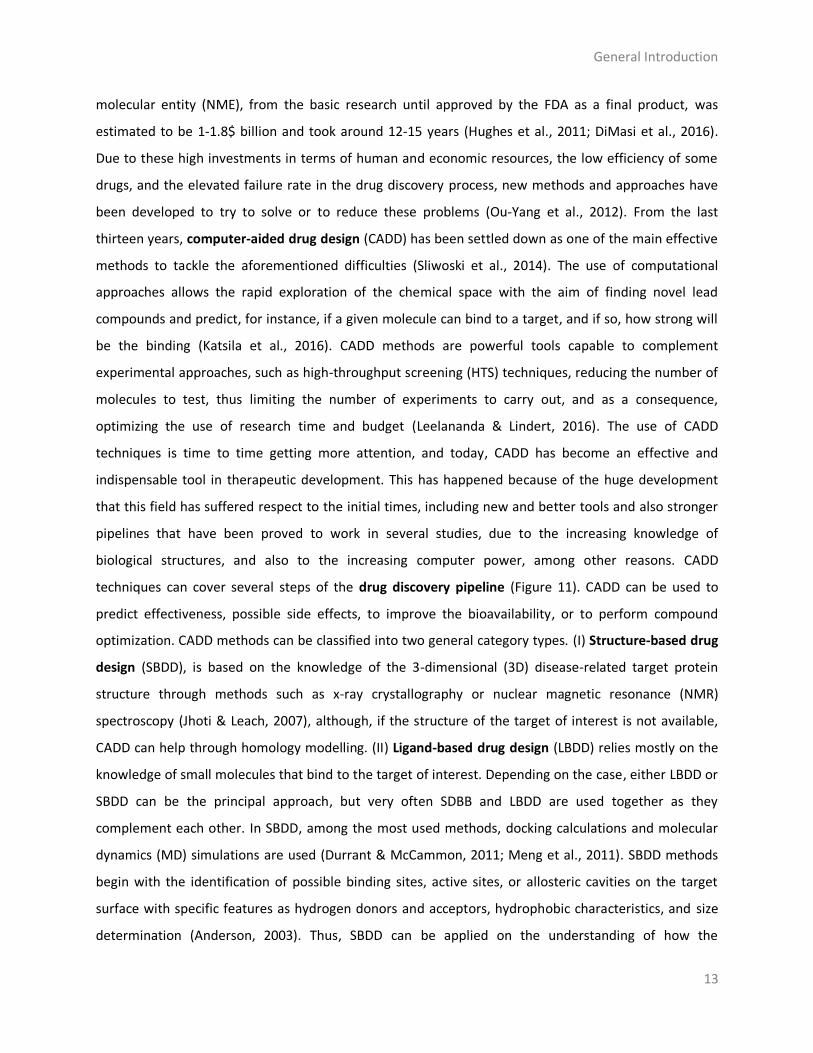
techniques can cover several steps of the drug discovery pipeline (Figure 11). CADD can be used to
predict effectiveness, possible side effects, to improve the bioavailability, or to perform compound
optimization. CADD methods can be classified into two general category types. (I) Structure-based drug
design (SBDD), is based on the knowledge of the 3-dimensional (3D) disease-related target protein
structure through methods such as x-ray crystallography or nuclear magnetic resonance (NMR)
spectroscopy (Jhoti & Leach, 2007), although, if the structure of the target of interest is not available,
CADD can help through homology modelling. (II) Ligand-based drug design (LBDD) relies mostly on the
knowledge of small molecules that bind to the target of interest. Depending on the case, either LBDD or
SBDD can be the principal approach, but very often SDBB and LBDD are used together as they
complement each other. In SBDD, among the most used methods, docking calculations and molecular
dynamics (MD) simulations are used (Durrant & McCammon, 2011; Meng et al., 2011). SBDD methods
begin with the identification of possible binding sites, active sites, or allosteric cavities on the target
surface with specific features as hydrogen donors and acceptors, hydrophobic characteristics, and size
determination (Anderson, 2003). Thus, SBDD can be applied on the understanding of how the
General Introduction
14
orientation or the pose of a given molecule could interact with a biological target, ultimately elucidating
the main pharmacophoric properties which exert a therapeutic effect. In LBDD, molecular similarity
approaches, quantitative structure-activity relationship (QSAR) techniques, or pharmacophore
modelling, are some of the most used techniques. LBDD methods, for instance, can be applied to
perform virtual screening (VS) in order to find analogue compounds to a molecule of interest, or to find
molecules that fulfill certain pharmacological, biological and/or toxicological properties, or to improve
compound features through hit to lead (H2L) optimization cycles to develop drug-like compounds
(Acharya et al., 2011; Lionta et al., 2014; Yu & Mackerell, 2017).
Figure 11. Schematic representation of a computer-aided drug design (CADD) pipeline. Original from the author.
It is always interesting to go a little bit deeper into all these computational techniques to
understand the magnitude of the evolution that this field has suffered during the last years. To start
with, and before describing any technique, it has to be noted that the protein structure determination is
fundamental in computational biology, computational chemistry, chemo/bioinformatics, and/or
computational biophysics fields. In CADD, specially for SBDD as commented above, having a good
determined structure is, usually, proportionally related to the results obtained, as the structure

General Introduction
15
information is key for understanding the interactions between small molecules and the protein, which is
an essential point on the drug discovery process (Reich & Webber, 1993).
Structure determination
One of the most successful and historically used approach for proteins and biological
macromolecules structure determination is the X-ray crystallography, where a trustworthy source of
protein needs to be available and purified until achieving a soluble material. Then, the protein must be
crystallized, and the crystal obtained has to diffract to sufficient resolution to be processed (Smyth &
Martin, 2000; Slabinski et al., 2007). NMR spectroscopy had his boom in the field of structural biology
on the 2000s, starting to play a major role in the determination of structures and dynamics of proteins,
and other biological macromolecules, since it allow determining protein conformations/ensembles at a
resolution better than 2 Å (Cavanagh et al., 1995; Cavalli et al., 2007). This determination consists in
several steps, including specialized techniques as quantum mechanical (QM) properties determination
(Elyashberg, 2015). The major advantage of NMR spectroscopy over X-ray crystallography is that the
determination can be done in solution, which allows for study of protein dynamics, and the difficulty of
fixing the protein in a crystal disappears. This is a substantial improvement in comparison with X-ray
crystallography. Cryogenic electron microscopy (cryo-EM) is planned to be the future of biological
macromolecules structure determination, as it allows its study in native conditions at near atomic
resolution while capturing multiple dynamic states (Murata & Wolf, 2018). In 2017, the Nobel Prize in
Chemistry was awarded to Dubochet, Frank and Henderson “for developing cryo-electron microscopy
for high-resolution structure determination of biomolecules in solution” (Dubochet, 2012; Henderson &
McMullan, 2013; Chen & Frank, 2016; Frank, 2016). Unlike X-ray crystallography and NMR spectroscopy,
cryo-EM requires a much smaller amount of sample and it allows to determine wide molecular mass
range of proteins, from kilo-Daltons (protein complexes) to mega-Daltons (virus particles) (Murata et al.,
2018). Nowadays this technique is becoming more used with time, producing better resolutions, better
resolved structures, but it is not the most used or common technique yet. According to the statistics of
the Protein Data Bank (PDB), a repository of information about the 3D structures of proteins, nucleic
acids and complex assemblies, 90% of the protein structures were resolved by X-ray crystallography,
12.000 (of more than 120.000) by NMR, while the number of structures resolved by Cryo-EM is 3947,
currently not comparable with the other two techniques. Nevertheless, in recent years, Cryo-EM is
suffering a very high growth in the number of protein structure diposits (Liu et al., 2014).

General Introduction
16
Having a resolved structure of the target of interest is the first step for using SBDD techniques.
However, it has to be taken into account that having a structure resolved, by any technique, does not
mean to have a perfect picture of the protein structure and being ready to start using computation over
it as a starting point for simulations. Usually, the structures present in the PDB do not fill the
computational needs. This may be due to high atomic resolutions that are translated in a bad
description of protein regions associated with non-natural, or lack, of secondary structure, or missing
atoms from the aminoacid sequence. Also, often, the resolved protein structure does not include the
region of interest. These “errors” can compromise the computational simulations, so they should be
solved or at least reduced as much as possible. In that sense, a popular computational method used to
alleviate this problem, when predicting the 3D coordinates of structures, is homology modelling (HM)
also known as template-based protein modelling. It is mainly used to obtain structures whose
coordinates are not available, or that are lacking some regions of interest. For other of the mentioned
errors, like the presence of missing atoms, there are software tools like PDBFixer (PDBFixer, 2019), that
help to fix protein structures. The principle behind HM is that evolutionary-related proteins often share
similar structures, and this is because it is well known that the protein structure remains more
conserved than the sequence during evolution (Lesk & Chothia, 1980; Illergård et al., 2009; Kaczanowski
& Zielenkiewicz, 2010). Exploiting this fact, homology modelling relies on the identification of one or
more known protein structures similar to the structure of a query sequence (or sequence of interest),
making an alignment of those structures and mapping the shared regions/residues between both the
query and the similar template. Using the retrieved information, a model is constructed and finally is
evaluated using different criteria such as Ramachandran angles, sequence similarity or sequence
coverage (Fiser, 2010).
Structure and ligand based applications
Once the 3D structure of a protein is known, finding its orthosteric pocket (active site) or
additional binding pockets (allosteric cavities or just binding regions on its surface) is the next important
step in SBDD. But before delving into this topic it is interesting to first explain LBDD, because it is
important to understand that the computational drug discovery process is not linear and methods and
techniques coming from structural or ligand sides, are highly complementary and their efficacy increases
when they are used together. As said above, given the case, maybe a SBDD or LBDD approach can be
better suited that a combination of both, but usually, mixing methods from both approaches increase
the probability of success, as can be seen on Chapters 1-4. Here, we describe the main methods used in

General Introduction
17
this thesis, coming from LBDD and SBDD. Further information is available, for instance, in Sliwoski et al
(2014) and Yu and Mackerell (2017).
Virtual screening (VS) is a computational technique used in drug discovery to search libraries of
small molecules in order to identify those structures which are most likely to bind to a drug target,
typically a protein receptor or enzyme. VS can be performed using structure or ligand-based techniques
(Sliwoski et al., 2014; Gimeno et al., 2019). One of the main techniques used in virtual (ligand)
screenings is molecular similarity (Willett, 2006; Eckert & Bajorath, 2007; Cereto-Massagué et al.,
2015). It is used to score and ranking molecules according to their likelihood to another molecule(s),
since it is a knowledge-driven approach which requires structural information of the bioactive ligand(s)
of interest. Another important variant is based on pharmacophore mapping. The International Union of
Pure and Applied Chemistry (IUPAC) defines a pharmacophore to be “an ensemble of steric and
electronic features that is necessary to ensure the optimal supramolecular interactions with a specific
biological target and to trigger (or block) its biological response”. It also constitutes a central unit or a
key scaffold of chemical compounds that should be preserved to design effective drugs (Wermuth et al.,
1998). In drug discovery, pharmacophore features are widely used for VS, de novo design and/or lead
optimization experiments (Yang, 2010).
The molecular-similarity VS method relies on the similarity-property principle, which states that
similar molecules should exhibit similar properties (Klopmand, 1992). This technique is usually employed
over large libraries and/or databases of compounds which contain diverse information associated to
each molecule, such as binding targets or distribution profiles. Because of that, these methods have
been highly used to elucidate the plausible targets, off-targets, or other pharmacological properties of
the studied compounds. This can be done by correlating the structural similarity with the possibility of
sharing a similar biological profile. This correlation is the idea behind the so-called Structure Activity
Relationship (SAR) principle, first introduced in 1865 (Crum-Brown & Fraser, 1868; Blake, 1884), that
derived into the so-called quantitative SAR (QSAR) methods. QSAR methods started to be used in the
pharmaceutical context as an attempt to correlate chemical structure (2D and/or 3D) with activity using
statistical approaches (Perkins et al., 2003). This was done with the aim of solving the problems they
encountered in the late 1990s, where some studies started to point out that poor pharmacokinetics (PK)
and toxicity predictions were an important cause of costly late-state failures in the drug development
process (Van de Waterbeemd & Gifford, 2003). Actually, these methods have now become a common
technique in the field. QSAR methods are now widely applied in drug discovery, especially on the study
of absorption, distribution, metabolism, excretion, and toxicity (ADMET) properties (Gola et al., 2006).
General Introduction
18
VS techniques based on ligands are powerful and widely used approaches, but there also exists
a structure-base counterpart, as commented before, which is the docking-based virtual screening. This
method allows the scanning of thousands of proteins to identify potential targets for a single molecule
or a library of compounds by using molecular docking calculations (see below) (Xu et al., 2018). Docking-
based screenings constitute an important computational tool for identifying new targets of existing
drugs and, especially, are highly valuable for predicting the bioactivity of a small molecule where the
protein target is still unknown (Lapillo et al., 2019). The usage of these techniques is clearly explained
and put in context in the following chapters (Chapters 1-4), where it is demonstrated how from a given
chemical compound (or a set of them) these techniques can be applied, for instance, to elucidate
possible targets (Toledo-Sherman & Chen, 2002; Shoichet, 2004), to determine their biological profile or
to find similar compounds (Varney et al., 1992; Shoichet, 2004).
The work done in this thesis, as can be observed in the following chapters, has been mainly
approached from a structural perspective, as it is mainly based on SBDD techniques (although LBDD
methods have also been important to achieve the thesis objectives). Probably, the two main methods
encompassed on SBDD are docking calculations and MD simulations. Because of that, and also as they
are mostly used here, we proceed to describe them in more detail. These methods can be applied in
different steps along the drug discovery pipeline, as seen in the following chapters.
Docking calculation process concerns the study and prediction of ligand conformation and
orientation (pose) within a target binding site (Kitchen et al., 2004). This calculation was first mentioned
in the early 1980s (Kuntz et al., 1982), and still today, is one of the most popular CADD tools used in drug
discovery (De Vivo & Cavalli, 2017). The docking protocol can be described as a multi-step process full of
complexity (Brooijmans & Kuntz, 2003), but basically involves two steps: the prediction of the binding
pose and the evaluation of its strength. The procedure begins with the application of docking algorithms
that facilitate the prediction of the best pose (including also ligand-target interactions) of a given small
molecule in the orthosteric site or other allosteric binding region of the protein; thereafter the binding
affinity of the protein-ligand complex is estimated (Meng et al., 2011).
Back to the history, the first explanation of binding was provided by Emil Fischer in 1894,
describing the specific action of an enzyme with single substrate using the lock and key analogy (Fischer,
1894) (Figure 12).

General Introduction
19
Figure 12. Fischer’s original “lock and key” model proposed in 1894 and Koshland “induce-fit” theory (Fischer, 1894; Koshland, 1963). E: Enzime. S: Substrate. Original from the author.
Later on, as this lock and key hypothesis did not take into account the flexible nature of the
protein, another theory was proposed, the so-called induce-fit theory, which refuses the idea that the
substrate only fits into the active site, and proposes a continuous change in the conformation of the
enzyme in response to the substrate binding (Koshland, 1963) (Figure 12). In agreement with this
theory, both ligands and protein receptors should be considered as flexible entities during docking.
However, probably the most used docking approach, the so-called classical or rigid docking, does not
take this into account. This variant only allows the ligand movement fixing the target conformations.
This represents a clear drawback mainly due to computer limitations resources, but also for the desire of
preserving a certain protein conformation. Anyway, in general, the lack of protein movement is
considered a limitation. To overcome this issue, in flexible docking the ligand and the receptor are
allowed to move. There are different variants of flexible docking based on the way the intrinsic protein
dynamics is incorporated into the equation. For instance, there are approaches where the receptor
remains rigid with the exception of the side chains of selected residues which are allowed to move or
even the receptor is fully flexible (Meng et al., 2011). Another approach to incorporate protein flexibility
could be the use of ensemble docking, which consists of the generation of different conformations of
the target experimentally (coming from NMR models or X-ray crystal structures), or computationally,
generally, obtained by MD simulations (Amaro et al., 2018). Over these ensembles, classical docking

General Introduction
20
experiments are performed; however, as different protein conformations are considered, the flexibility
is indirectly captured (Korb et al., 2012; De Vivo et al., 2017). Finally, a very useful method (explained in
the following paragraphs) is the post-processing of classical docking calculations by MD simulations (De
Vivo et al., 2016).
From the binding region perspective, there are two possible scenarios. 1) when the binding
pocket is previously known, and classical (rigid) and/or flexible docking calculations are performed over
it elucidating the preferential binding pose (Taylor et al., 2002) (Figure 13), and 2) when the binding
cavity is not known and the protein surface has to be explored with the aim of founding plausible
cavities (catalytic cavities, allosteric cavities or just binding regions where the ligand can be retained for
a certain period of time) where the molecule can bind and exert some activity (Hetényi & van der Spoel,
2006) (Figure 13). After the elucidation of all possible pockets, classical or flexible docking techniques
can be applied over them to determine which are the most favourable cavities and molecules poses. The
whole process is usually known as blind docking. A clear example of the application and utility of blind
docking calculations can be seen on Chapter 1. On Chapter 4, following the first approach of the blind
docking methods, an exploration of the cavities was performed with the aim of elucidate plausible
cavities. Besides, on Chapters 1-4 the important and crucial contribution of docking techniques on the
first steps of drug discovery process is shown.
Figure 13. Graphical representation of crystallographic human structure of glycogen synthase kinase beta (GSK3β) (Protein Data Bank ID (PDB) 6B8J) (Wagman et al., 2017). On the left GSK3β structure with its crystallographic ligand 65C placed in the adenosine triphosphate (ATP) active site and where classical docking can be performed over. On the right, the ATP cavity and also other five proposed allosteric cavities (1-5) found after performing a search of the protein surface (blind docking). Adapted from Llorach-Pares, et al. (2019).
ATP
1
4 3
2
5

General Introduction
21
In all variants of docking calculations, and once the cavity or cavities of binding are known,
predict the optimal placement for a molecule, given certain degrees of freedom, is by itself challenging,
as a high accuracy is needed to identify the best possible conformational pose of the ligand that fits
better the receptor structure. This step needs to be fast enough to allow the analysis of hundreds or
thousands of compounds in the same run, and it is conditioned for the high number of degrees of
freedom, which significantly increases the computation time and also the number of false-positive
results (Andrusier et al., 2008). The following and complementary step is the prediction of the biological
activity, in terms of binding energy by the use of scoring functions, and the subsequent evaluation of the
interactions between the small molecule and the target (Meng et al., 2011). Chapter 2 is a nice example
where the aforementioned step process is put in context and helps to understand the applicability of
binding interactions studies. To add more complexity to these methods, as said above, poor
crystallographic resolution of targets, implicit flexibility (Koshland, 1963), induced fit events (Tobi &
Bahar, 2005), and the water involvement on the target-molecule binding, make these type of
calculations a scientifically complex process (Kitchen et al., 2004).
All organisms are regulated by a correct protein function. A malfunction of this regulation can
result in some disease. Usually, the protein function is regulated by the binding of a substrate to the
orthosteric cavity (active site). However, there are cases where other additional/alternative pockets can
have this role. Allosteric regulation, an emerging concept in drug discovery in the last years (Abdel-
Magid, 2015), can control protein function by the binding of small molecules, or other entities like
peptides or even other proteins, to the target. It is used to be a single protein or protein complex, in a
cavity at some distance (until tens of Å) from the orthosteric site (Laskowski et al., 2009; Amaro, 2017).
However, a molecule binding to a cavity different from the principal one does not mean it is an allosteric
cavity, because this depends on the effect that the compound can exert over the protein. This effect, in
general, can be positive (activating), provoking an increase of the target protein activity, or negative
(inhibiting), causing a decrease of the protein activity (although the scenario can be, in some cases and
for certain proteins, a little bit more complex) (Tian et al., 2012; Morra & Colombo, 2018; Greener &
Sternberg, 2018). Also, the molecules that bind on it, do not need to be chemically similar to the natural
ligands as there is no competition between them (Laskowski et al., 2009). A nice example on how the
allosteric modulation can inhibit a protein function can be found in Chapter 4, where two marine
molecules are proposed to inhibit the activity of a kinase by binding to an allosteric pocket (called
substrate cavity).

General Introduction
22
Focusing on protein activity, all proteins are intrinsically dynamic/flexible entities (Kim et al.,
1993), and thus, its biological function/activity relies on their flexibility. To be more precise, the internal
motions of proteins result in conformational changes, which are at a time, essential for their functions
(Henzler-Wildman & Kern, 2007). The study of protein dynamic movements is necessary to understand
the structure-function relationship (Quan et al., 2014), that, in fact, could be reformulated as structure-
dynamics-function. Conformational changes on protein structures can be caused by protein-protein
binding, ligand binding or post-translational modifications (Teilum et al., 2009), which can directly affect
their function. Measuring, analysing, and understanding proteins dynamics and the associated
conformational changes, is a must. In this regard, MD simulations are a versatile and powerful
computational method widely used to obtain information on the time evolution of protein motions
(Karplus, 2002; Adcock & McCammon, 2006). More precisely, MD simulations allow the study of the
physical movement of atoms and molecules, ranging from simple systems of few atoms or just one small
chemical compound to more complex scenarios like proteins, or chemical compounds bound to
proteins. The atoms and molecules are allowed to interact for a fixed period of time, through the
integration on Newton’s laws of motion, constructing trajectories that allowed to describe the temporal
evolution of the particles of a given system, and thus, to observe its dynamic evolution. There are
several variants of this technique, some of them addressed to accelerate the dynamic process and span
the time-scale. In order to do that, an option is to apply an external force, like targeted (TMD) (Schlitter
et al., 1994), steered (SMD) (Suan & Khanh, 2013) or accelerated MD (AMD) (Hamelberg et al., 2004)
methods.
The first MD simulation of a protein was carried out in 1974 by Andrew McCammon and Martin
Karplus, and consisted in a 9.2 picoseconds (ps) trajectory of small globular protein, bovine pancreatic
trypsin inhibitor (BPTI), in vacuum (McCammon et al., 1977). More than ten years were needed to
report the simulation of the same protein but solvated in water (Levitt & Sharon, 1988). From that
moment, computational power has been growing quite fast, thus allowing the performance of more
complex simulations over time, that are also more “useful”, as they can help to solve more complex
problems related to diverse areas like biology, chemistry, or physics. Because of that, nowadays, the use
of these methods is very popular in different fields, as it happens in drug discovery. Regarding the use of
MD methods in drug discovery, its main advantage is that it allows to consider the structural
dynamics/flexibility of the proteins, alone and/or in complex, for instance, with ligands, other proteins,
or DNA. Unlike other static techniques, like rigid docking, this kind of simulations takes into account the

General Introduction
23
entropic effects and enables a more accurate estimation of the thermodynamics and kinetic association
of target-ligand complexes (De Vivo et al., 2016).
MD, in addition to the characterization of the structural landscape of a protein, or a protein
complex, and/or extracting conformational ensembles, is widely used, specially in drug discovery, to
understand the ligand-target binding and unbinding mechanisms (De Vivo et al., 2016). In relation to
that, one popular use of this technique is to post-processing docking calculations. As mentioned above,
classical docking (despite being a reasonable good technique to predict the optimal placement of a
ligand within a binding pocket, as it has a proven track record of success) has several limitations, as
classical docking does not consider protein flexibility and the scoring functions used to have accuracy
limitations. These limitations are usually translated into a bad description of the binding mode and the
associated binding energy, and thus a wrong ranking of the analysed compounds (Kitchen et al., 2004).
Flexible docking methods can improve the results of the rigid counterpart, but these variants still have a
strong dependency on the scoring function. In that sense, MD simulations can optimize the predicted
docking poses and also validate the stability of the docked complex (De Vivo et al., 2016; Aravindhan et
al., 2017). If the docking pose is not “good” enough, it could be possible to see how the ligand leaves the
binding site during the simulation (usually in hundreds of ps). This two-step protocol (docking+MD)
constitutes a good approach to solve docking drawbacks, thus allowing us the prediction of,
theoretically, more reliable protein-ligand binding modes (Alonso et al., 2006). The workflow combining
docking calculations (that can be used to screen large compound libraries filtering out a significant part)
and MD simulations (that despite being more computationally expensive, can be used efficiently, over
the best docking poses), has been extensively used in the literature (Alonso et al., 2006; Aravindhan et
al., 2017). Applying short post-processing MDs over hundreds of compounds is, nowadays, feasible in a
short period of time (around a week in a desktop GPU), which reinforces this approach, since it is fast
enough to be used regularly in any SBDD workflow. As a consequence of that, it is being ingreasingly
used.
There are different variations of MD simulations in addition to the classical version, which is
probably the most commonly employed. These variations can be used to understand the
binding/unbinding mechanism of a ligand over a target of interest. In that sense, Steered molecular
dynamics (SMD) is becoming a highly used method in drug discovery to describe the process of protein-
target binding, giving insights into the binding/unbinding mechanisms (Patel et al., 2014; De Vivo et al.,
2016, 2017). As explained before, external time-dependent forces are applied to the ligand in order to
accelerate the disassociation of the protein cavity, revealing the force needed to cause the rupture

General Introduction
24
between the ligand and the receptor (Isralewitz, Baudry, et al., 2001; Isralewitz, Gao, et al., 2001). These
forces can be theoretically correlated to the experimental residence time, and also, with its inhibitory
capacity (Potterton et al., 2019). Moreover, during this process, it is possible to estimate which
interactions are stronger and more necessary to keep the ligand bound.
From MD simulations, in general, a good deal of useful information can be extracted regarding
the dynamics and thermodynamics of the studied system. One of the properties that can be measured is
the binding free energy of target-ligand complexes. This energy is estimated, according to the
thermodynamic cycle shown in Figure 14, as by subtracting the free energies of the ligand and the
protein in aqueous solution to the free energy of the complex (protein-ligand) (Miller et al., 2012).
Figure 14. Thermodynamic cycle for free binding energy calculation. Adapted from Miller et.al (2012).
This calculation is done for each frame of the MD simulation and then averaged (Miller et al.,
2012), with the aim of taking into consideration all the dynamics of the system, in agreement with the

General Introduction
25
induced-fit theory mentioned before. To this purpose, there are several methods (each of them with its
advantages and drawbacks) with different accuracy and computational cost. Within them, the so-called
end-point techniques are a widely used option because of their good balance between accuracy,
computational cost and speed. Among them, the Molecular Mechanical/Generalized Born Surface Area
(MM/GBSA) method, is a popular technique that has been widely employed along this thesis, as it can
be seen in Chapters 1-4 (Kollman et al., 2000; Massova & Kollman, 2000). The binding energy resulting
from MM/GBSA is more realistic than the energy obtained from rigid docking calculations, because the
dynamic behaviour of the protein-ligand complexes can be taken into account (Mulakala &
Viswanadhan, 2013; Genheden & Ryde, 2015). Thus, a better ranking (based on the binding energy) of
the analysed compounds can be obtained, allowing for a better prioritization of them, although the
obtained binding energies can be far from being experimentally comparable.
Alzheimer’s Disease
Alzheimer’s disease (AD) is the most common cause of irreversible dementia worldwide,
representing 60-80% of the total cases. It is estimated to be over 45 million people globally. Its
prevalence grows constantly, mostly because of the progressive aging of the population and the long
asymptomatic initial stages of the pathology (Crous-Bou et al., 2017). In addition, limitations on current
treatments which may slightly improve the symptoms but do not cure the disease, do not help to reduce
the high incidence; thus, nowadays AD is one of the major world’s socioeconomic and health problems
(Citron, 2010). AD is a neurodegenerative disorder resulting in a gradual loss of cognitive function and
memory deterioration. Alzheimer’s pathologies are characterized by the presence of neurofibrillary
tangles (NFT), which are intraneuronal insoluble aggregations mainly composed of abnormal
phosphorylated tau protein, and senile plaques (SP), principally composed by beta-amyloid peptides
(Aβ). Tau protein was discovered in the 70s and it is responsible for the structural morphology of the
neurons by stabilizing the microtubules (Kosik, 1993). Tau binding is regulated by its phosphorylation
state, a regulated balance between tau kinase and phosphatase activities, which at a time is coordinated
by the action of some kinase proteins (Mandelkow et al., 1995). In pathological conditions, such as those
provoked by AD, the binding decreases and the neuronal microtubules lose their organization leading to
their aggregation and the formation of NFT (Billingsley & Kincaid, 1997; Kolarova et al., 2012) (Figure
15).

General Introduction
26
Figure 15. Microtubule-bounded by tau in health conditions and the hyperphosphorylation and consequently aggregation till the formation of neurofibrillary tangles (NFT) in Alzheimer’s Disease (AD). Source: Nature Reviews Drug Discovery 9, 387-398, 2010. doi:10.1038/nrd2896 (Citron, 2010) and Alzheimer’s news, 2014, Tau Protein Leads To Neuronal Death in Alzheimer’s by Patricia Inacio.
Aβ is a peptide of 40 or 42 aminoacids essentially involved in AD as a main component of the SP
found on Alzheimer’s patients brains (Hamley, 2012). The amyloid precursor protein (APP) is cut by two
proteases, beta (β) secretase (also known as beta-site APP cleaving enzyme 1 (BACE1)) and gamma (γ)
secretase to yield Aβ. In health conditions Aβ is found in a monomeric form, while in pathology
conditions it is generally believed that the formation of Aβ oligomers, which are toxic and cause a
synaptic dysfunction, starts to aggregate to finally form an amyloid plaque (Shankar et al., 2008; Zhao et
al., 2012) (Figure 16). The inhibition on the production of Aβ preventing APP cleaving, remains in the
central focus of the research to find a cure for AD, but, the function of APP is still controversial and not
well understood yet (Hiltunen et al., 2009). This should make us raise the need to first understand how
the pathology works, in order to further proceed in the design of drugs to treat AD.
General Introduction
27
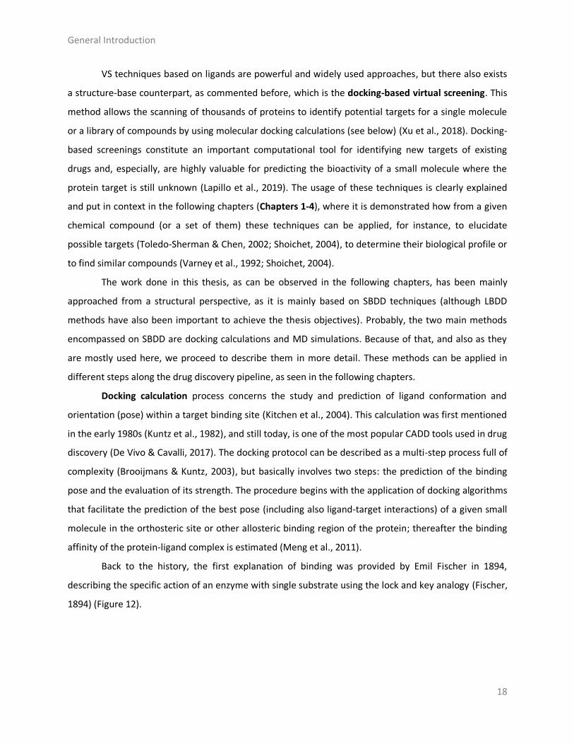
Figure 16. Graphical representation of the amyloid cascade theory where beta secretase (β-secretase, also known as beta-site APP cleaving enzyme 1 (BACE1)) and gamma secretase (γ-secretase) yield beta amyloid (Aβ) and its normal form in monomers, and after its aggregation, the formation of toxic oligomers and amyloid plaques directly linked to Alzheimer’s Disease (AD) takes place. Source: Nature Reviews Drug Discovery 9, 387-398, 2010. doi:10.1038/nrd2896 (Citron, 2010).
Regarding therapeutic approaches, the most direct approximation is concentrated on the
reduction of Aβ production by the inhibition of β-secretase/BACE1 activity, which is the responsible of
the proteolysis of APP, precursor of Aβ (Yan & Vassar, 2014) (Figure 16). However, the lack of promising
results has created reasonable doubts about the amyloid hypothesis, which never was generally
accepted (Doig et al., 2017; Kametani & Hasegawa, 2018). These doubts have ended up in the need to
look for new therapeutic options. A strategy oriented to reduce tau hyperphosphorylation and thus,
reducing the NFT formation, is conceptually more tempting. In addition to that, there is a general
consensus about its damaging effects (Citron, 2010). It is believed that the inhibition of specific tau
kinases could reduce the aggregation and now, it is considered a promising approach for the treatment
of AD (Martin et al., 2013; Tell & Hilgeroth, 2013; Llorach-Pares et al., 2019).
In this thesis, based upon the results obtained on the elucidation of possible targets from a set
of marine molecules, some of the compounds collected on expeditions to Antarctica and the
Mediterranean Sea from the BlueBio team (University of Barcelona), as well as other related molecules
described in the literature, in Chapter 1, we found it interesting to study the relation obtained between
meridianin A and the evolutionarily conserved group of dual specificity kinases cdc2-like kinases (CLKs).
In fact, one of its isoforms, CLK1, is known to be involved in the pathology of AD by the phosphorylation
of serine and arginine-rich (SR) proteins responsible for the regulation of the alternative splicing of
microtubule-associated tau (Jain et al., 2014). As a consequence of that, we decided to perform a deep
study evaluating the possible inhibitory activity of meridianins A-G, the whole family, against the
principal kinases involved in tau hyperphosphorylation and thus, AD pathology. Between these proteins,

General Introduction
28
glycogen synthase kinase-3 beta (GSK3β), a proline-directed serine/threonine kinase that
phosphorylates tau at different sites (specifically from 42 sites, 29 of have been found phosphorylated in
AD brains), is considered one of the main responsibles of tau phosphorylation (Wagner et al., 1996;
Hooper et al., 2008; Martin et al., 2013). Interestingly, the fact that more studies suggest a relation
between GSK3β and the production of Aβ, makes more relevant, if possible, the importance of targeting
GSK3β for the cure of AD (Hernández et al., 2010; Hernandez et al., 2012). Casein kinase 1 isoform delta
(CK1δ) also regulates microtubule dynamics through tau phosphorylation (at 46 sites, 25 are found in AD
brains) (Singh et al., 2002; G. Li et al., 2004; Hanger et al., 2007). In AD brains, CK1 is co-localized with
NFT and isoforms α, δ and ε, and their levels are increased respect to normal brains (Schwab et al.,
2000; Knippschild et al., 2005). The last kinase studied in this thesis was the dual specificity tyrosine
phosphorylation-regulated kinase isoform 1 A (DYRK1A). It was first related to the phosphorylation of
threonine 212, but today the list of phosphorylation sites rises up to eleven (Wegiel et al., 2011). There
are also insights that DYRK1A hyperphosphorylation is related to the inhibition of the ability of tau to
enable the microtubule assemble (Ryoo et al., 2007), and its involvement on the formation of NFT
(Wegiel et al., 2008).
The inhibition of these four targets is proposed to be key in the treatment of AD, as can be seen
in the literature, and during the last decades there have been several studies aiming to prove this (Bhat
et al., 2004; Perez et al., 2011; Jain et al., 2014; Branca et al., 2017). The link between NPs and MNPs on
the inhibition of kinases involved on AD, and even targeting the four aforementioned kinases, is not
new, as several examples can be found in the literature (Haefner, 2003; Liu et al., 2012). For instance,
indirubins or phenylmethylene hydantoins have been proposed to be inhibitors of the GSK3β (Meijer et
al., 2003; Khanfar et al., 2009). For the two dual specificity kinases, DYRK1A and CLK1, harmine, a well-
known alkaloid was reported to be a potent inhibitor (Göckler et al., 2009; Adayev et al., 2011; Grabher
et al., 2012). Also, lamellarins, pyrrole alkaloids isolated from different marine invertebrates, have been
predicted to be CK1δ inhibitors (Bharate et al., 2013).

Objectives
29
Objectives of this thesis
The main purpose of this thesis is to find possible therapeutic activities and to establish the
capability to modulate protein functions in pathogenesis-related pathways from marine molecules by
using different CADD tools and techniques. In other to achieve this goal, the present thesis is divided
into three sections which attempt to illustrate these achievements. In Section I, I shown how a
computational approach could improve the drug discovery pipeline (Chapter 1). Section II focuses on
the elucidation of different pharmacophoric features of marine compounds and a precise in silico
binding study that ends with the elucidation of the capability of different marine compounds to act as
inhibitors of tau kinases (GSK3β and CK1δ) and dual-specificity (DYRK1A and CLK1) protein kinases, all of
them related to AD, which constitute a promising starting point for the development of novel anti-AD
drugs (Chapter 2 and Chapter 3). Section III presents a computational study and an experimental
validation of the inhibitory activity of meridianins and lignarenones as possible GSK3β adenosine
triphosphate ATP and/or substrate inhibitors, which allows to propose them as drug-like candidates for
the treatment of AD pathologies (Chapter 4).
The specific objectives for each chapter are summarized below:
Chapter 1. In silico Studies to Find New Therapeutic Indications for Marine Molecules. The
main aims of this study are (I) to establish the possible therapeutic potential of a set of marine
molecules by using different computational techniques; (II) to predict and validate the marine
molecule-target complex binding, (III) to elucidate a list of possible targets, (IV) to evaluate the
adverse health effects by performing a preliminary toxicology prediction study; and (V) to
estimate the drug-like properties of each studied molecule.
Chapter 2. Computer-Aided Drug Design Applied to Marine Drug Discovery: Meridianins as
Alzheimer’s Disease Therapeutic Agents. In this chapter, the aims are (I) to highlight the power
of CADD techniques in marine molecules, and natural products in general, to find possible
therapeutic uses; (II) to evaluate and report the inhibitory activity found in the marine tunicate
Aplidium: Meridianins A-G, acting as ATP competitive inhibitors of GSK3β, CK1δ, DYRK1A, and
CLK1; (III) to evaluate the possible adverse health effects of meridianins by performing a
Objectives
30
preliminary pharmacokinetic study; and (IV) to analyse their pharmacological properties as well
as the effect that the presence of halogen atoms in their structure may have.
Chapter 3. Kororamides, Convolutamines, and Indole Derivatives as Possible Tau and Dual-
Specificity Kinase Inhibitors for Alzheimer’s Disease: A Computational Study. The objectives
are (I) to discover the possible therapeutic activity of kororamides and convolutamines against
AD by the inhibition of GSK3β, CK1δ, DYRK1A, and CLK1; (II) to determine the importance of the
indole scaffold for the inhibition of the four studied kinases and the importance and effect of
the halogen substituents; (III) to design new possible inhibitors of the four kinases starting from
meridianin and kororamide indole scaffolds; and (IV) to evaluate the adverse health effects of
kororamides, convolutamines and its derivatives by performing a preliminary ADMET study.
Chapter 4. Meridianins and lignarenones as potential GSK3β inhibitors and inductors of
structural synaptic plasticity. The aims here are (I) to elucidate the possible ATP and/or
substrate inhibitory activity of meridianins and lignarenone against GSK3β, a key target on the
AD pathway; (II) to explore druggable binding sites on GSK3β on the search of new allosteric
cavities; (III) to ascertain the pharmacokinetic properties; and (IV) to experimentally validate the
inhibitory activity of meridianins and lignarenones comparing Ser9 phosphorylation levels to
total levels of GSK3β as an indication of inhibition.

Supervisor’s Report
31
Supervisor’s Report
Dr. Conxita Avila, Director of the PhD Thesis entitled “Computer-Aided Drug Design applied to marine
drug discovery”, together with Dr. Melchor Sánchez Martínez as co-Director, certify that the dissertation
presented here is the result of the work carried out by Laura Llorach Parés under our guidance and
supervision. The contribution of the PhD candidate to each one of the manuscripts included in her
Thesis is detailed below:
Chapter 1. In silico Studies to Find New Therapeutic Indications for Marine Molecules.
Llorach-Pares, L; Nonell-Canals, A; Sanchez-Martinez, M; Avila, C.
(In prep)
LL-P: data analysis, carry out the experiments, results interpretation, and manuscript elaboration.
Chapter 2. Computer-Aided Drug Design Applied to Marine Drug Discovery: Meridianins as
Alzheimer’s Disease Therapeutic Agents.
Llorach-Pares, L; Nonell-Canals, A; Sanchez-Martinez, M; Avila, C.
Marine Drugs 15(12), 366 (2017)
5-Year Impact Factor: 4.379 (2017)
LL-P: data analysis, carry out the experiments, results interpretation, and manuscript elaboration.
Chapter 3. Kororamides, Convolutamines, and Indole Derivatives as Possible Tau and Dual-Specificity
Kinase Inhibitors for Alzheimer’s Disease: A Computational Study.
Llorach-Pares, L; Nonell-Canals, A; Avila, C; Sanchez-Martinez, M.
Marine Drugs 16(10), 386 (2018)
5-Year Impact Factor: 4.379 (2017)
LL-P: design of the computational study, data analysis, carry out the experiments, results interpretation,
and manuscript elaboration.

Supervisor’s Report
32
Chapter 4. Meridianins and lignarenones as potential GSK3β inhibitors and inductors of structural
synaptic plasticity.
Llorach-Pares, L; Rodriguez, E; Giralt, A; Nonell-Canals, A; Sanchez-Martinez, M; Alberch,J; Avila, C.
(In prep)
LL-P: design of the computational study, data analysis, carry out the computational experiments, results
interpretation, and manuscript elaboration.
From all the co-authors of the different chapters, ER has not been awarded a PhD degree
yet. We hereafter guarantee that none of the information contained in the chapter co-authored with
her will be used to elaborate any other part of someone else’s PhD thesis.
For all the above, we consider that the work developed by the PhD candidate grants her the
right to defend her Thesis in front of a scientific committee.
Barcelona, September 30th, 2019.
Dra. Conxita Avila Dr. Melchor Sánchez-Martínez

Supervisor’s Report
33
Section I
Target elucidation from a computational approach

Supervisor’s Report
34

Supervisor’s Report
35
Section I
Chapter 1
In silico studies to find new therapeutic indications for
marine molecules.

Supervisor’s Report
36

37
Chapter 1
In silico studies to find new therapeutic indications for marine molecules.
Laura Llorach-Pares 1,2, Alfons Nonell-Canals 1, Conxita Avila 2 and Melchor Sánchez-Martinez 1
1. Mind The Byte S.L., 08028 Barcelona, Catalonia.
2. Department of Evolutionary Biology, Ecology and Environmental Science, Faculty of Biology and
Biodiversity Research Institute (IRBio), Universitat de Barcelona, 08028 Barcelona, Catalonia.

Marine Drug Discovery
38
Abstract
Identifying small molecules that fit well into a binding cavity is one of the first steps in the drug
discovery pipeline. In this study, we try to elucidate a list of possible targets and the therapeutic
potential of a set of selected marine molecules employing different computational tools. Molecular
docking is one of the most common computer-aided drug design (CADD) tools which allow the study of
protein-ligand interactions, predicting at the same time the binding molecule orientation or pose.
Capturing protein motions is key to understand these molecule-target interactions, and Molecular
dynamics (MD) simulation is the best computational tool to do so. By the combination of these
computational tools and others, in this work we have established the link between a group of Antarctic
marine molecules and some neurodegenerative and cardiovascular pathologies. Moreover, we
evaluated the adverse health effects through toxicology predictions, and the drug-likeness properties of
this set of marine molecules, providing some insights of the prediction on marine molecule-target
complex binding. In this study, we exemplify how the use of computational tools can be applied on the
marine drug discovery field, establishing a pipeline to be followed on future studies.
Keywords: Marine natural products, Computer-aided drug design, Neurodegenerative diseases,
Molecular docking.

Marine Drug Discovery
39
Resum
Identificar petites molècules que s'adaptin bé a una cavitat activa és un dels primers passos a
seguir en el descobriment de fàrmacs. En aquest estudi pretenem dilucidar una llista de possibles
dianes, i el potencial terapèutic, d'un conjunt de molècules marines utilitzant diferents eines
computacionals. L'acoblament molecular és un dels instruments del disseny de fàrmacs assistit per
ordinador (DFAO) més comuns i que permet l'estudi de les interaccions proteïna-lligand, predint a la
vegada, tant l’orientació com la postura de la molècula acoblada. La captura dels moviments de les
proteïnes és clau per entendre aquestes interaccions proteïna-lligand, i la simulació de dinàmica
molecular (DM) és la millor eina computacional per fer-ho. Mitjançant la combinació d'aquestes eines
computacionals i d'altres, en aquest estudi hem pogut dilucidar el vincle entre un grup de molècules
marines i algunes patologies neurodegeneratives i cardiovasculars. A més, hem avaluat els possibles
efectes adversos en la salut mitjançant prediccions de toxicologia, i les propietats farmacològiques
d'aquest conjunt de molècules marines, proporcionant algunes idees sobre la predicció dels vincles
d’unió dels complexes molècules marines-proteïna. En aquest estudi exemplifiquem com es pot aplicar
l'ús d'eines computacionals en el camp del descobriment de fàrmacs marins, establint un procediment
que es pot seguir en futurs estudis.
Paraules clau: Productes Naturals Marins, Disseny de Fàrmacs Assistit per Ordinador, Acoblament
Molecular, Malalties Neurodegeneratives.

Marine Drug Discovery
40
Introduction
Molecular docking is a very popular computer-aided drug design (CADD) tool used in molecular
biology to evaluate ligand-target complementarity [1]. This method allows the study of protein-ligand
interactions at an atomic level and the prediction of preferred binding orientation or poses (binding
mode) of one molecule (typically a small organic compound) to another molecule (generally a biological
target such as a protein). Also, by the use of scoring functions, this powerful technique assesses and
predicts the binding affinity of the complex formed by a receptor and a ligand. So, essentially, docking
can be accomplished through two complementary steps: first, by sampling ligand conformations in the
active site of the protein and then, ranking these conformations using a scoring function [2]. Two types
of docking can be described, a) the classical (rigid) docking, where the crystallographic ligand’s pose and
the binding site of a given protein is established and can be used, and b) the blind docking, where the
binding site is not known, and it is first necessary to search the protein surface to find possible cavities
that lodge the active binding sites before performing the classical docking. Both methods are widely
used in the drug discovery process.
The first and critical step in small molecule drug design is the identification of ligands that fit
well into the binding pocket of the protein target [3]. Looking back at the history, in 1894, Emil Fischer
first postulated the specific action of an enzyme with single substrate using the lock and key analogy [4].
First reported dockings using this principle, where both ligand and receptor were treated as rigid
entities, were published at the end of 1982 [5]. As this lock-key hypothesis does not fully account for the
nature of the enzymatic actions, another theory has been proposed, called the induce-fit theory which
refuses that the substrate would do more than simply fit into the active site and assumes a continuous
change in the conformation and shape of the enzymes in response to substrate binding, in other words,
when a substrate binds to an enzyme, it will change its conformation [6]. After the introduction of this
principle, more accurate predictions of binding poses and binding affinities (minimum binding energies)
could be resolved. This fact can be traduced into a best candidates selection as active compounds, with
higher true positive rates of success, and also, at the same time, can considerably reduce expensive
experimental efforts.
From now on, the flexibility of enzymes has been considered, taking into account that is
extremely important because of these dynamic movements provide a mechanism for regulating
enzymatic activity. Consequently, and in agreement with the theory of the induce-fit, both, the ligands
and receptors (proteins) should be treated as flexible during docking. Due to computer limitations
resources, docking has been performed with flexible ligand and rigid receptor until today, and remains

Marine Drug Discovery
41
one of the most used and popular methods in drug discovery [2]. To deal with this drawback, and with
the aim of incorporating protein flexibility, many efforts have been made and new methods are now
being used to validate docking techniques, such as Molecular dynamics (MD) simulations which can
optimize the predicted binding mode, allowing the study of movements (so-called induce-fit effects) of
atoms along the time by integration of Newton’s equations of motions disclosing the adaptation of the
ligand to the target [7–10]. Recognizing the mechanisms of actions for the protein-ligand complexes
formation and understanding its binding, will help at the discovery, design and development of new
drugs. Protein-ligand interactions, thus, play an important role in many scientific areas and more
concretely, knowledge on these interactions is central for understanding biology at the macromolecular
level.
In this project, by the use of docking calculations and MD simulations, a chemical library of ten
marine molecules with marine origin was explored (Figure 1).
Aplicyanin Dendrinolide Discorhabdin-B Hodgsonal Meridianin-A
Polyrhaphin-A Pteroenone Rossinone Liouvilloside Pectinioside-B
Figure 1. Structures of the ten marine molecules selected for this study.
Specifically, several of these molecules were collected from benthic marine invertebrates from
the South Shetland Islands and the Weddell Sea (Antarctica), but also from some other areas of the
planet, like the Mediterranean Sea, and the Sea of Japan. Antarctica, in particular, because of its
biological and geographical characteristics, is considered a “biodiversity mine” where new species and
new marine molecules are constantly being discovered [11–13]. Oceans cover about 70% of the Earth’s
surface, and up to 80% of life inhabits in the marine ecosystem. Marine organisms constitute amazing
resources of enzymes and bioactive compounds. Marine natural products have always been a rich
source of drug-like compounds [14,15]. Biodiversity is related to chemodiversity as well, giving wider
opportunity for discovering novel therapeutics with novel mechanisms of action. Currently, there are
Marine Drug Discovery
42
seven marine-derived products approved as drugs by the United States Food and Drug Administration
(US FDA), supporting the importance of marine drug-like compounds [16].
The identification of natural products that are capable of modulating protein functions in
pathogenesis-related pathways in one of the most promising lines followed in drug discovery [17]. In the
present study, with the aim of knowing the possible therapeutic potential of a set of selected marine
molecules, we divided the study into four parts. First we aim to determine a list of possible targets. To
do so, two dimensions (2D) and three dimensions (3D) ligand-based virtual profiling (VP) software tools
were used. Once the targets were known, we decided to focus only on neurodegenerative and
cardiovascular pathologies. According to the “Health at a Glance 2013” report, in 2009, there were 14
million people estimated of suffering from dementia in the Organization for Economic Cooperation and
Development countries (OECD). The same study, considered that in 2011, cardiovascular diseases were
the main cause of mortality in most OECD countries representing 33% of all death [18]. To achieve the
relation between the targets found on the first step and relate them to the pathologies of interest, we
used DisGeNET, a database that integrates information on gene-disease associations [19]. The second
part is dedicated to toxicology prediction. Due to the importance on these predictions during drug
discovery, several computational toxicology tools are used to evaluate the adverse health effects of the
studied molecules. These software tools integrate information and data for a wide kind of sources, and
they allow developing predictive mathematical and computer-based models. Toxicology methods can be
used to reduce the dependence on experimental testing in general, and animal testing in particular, and
this means saving money and time [20]. Moreover, these techniques, given their inexpensiveness and
expeditious results, can be used in an early phase of drug discovery, before the synthesis of the
molecules, to prevent future problems and helping to rationally focus the drug development process.
Computational toxicity could be assessed based on ligand or target. In this study we performed both
kinds of predictions, in order to ensure as much as possible whether the studied molecules are toxic or
not. In the third part, a drug-likeness evaluation was carried out. Drug-likeness is a qualitative concept
used in drug design based on structural properties of compounds and it is used to estimate drug-like
properties of molecules. From molecular properties and structural features, it is possible to determine
whether a particular molecule is similar to a known drug [21,22]. The fourth and final part, is mainly
focused on the computational techniques previously mentioned as protein-ligand docking and blind
docking, which offer the capability to predict quickly and cheaply the binding mode and the affinity of a
ligand-receptor complex, and the induce-fit MD simulations and binding free energy calculations,
specifically, using reweighting techniques. These methods are popular approaches to estimate the free

Marine Drug Discovery
43
energy of the binding of small ligands, in these particular case marine molecules, to biological
macromolecules and proteins. They are typically based on MD simulations of the receptor-ligand
complexes and may be useful to improve the results of docking calculations or to understand observed
affinities and trends [23]. Despite the fact that MD simulations are more time-consuming approaches,
computing the free energies of the complex systems based on the thermodynamic principles are more
powerful methods. Also, a detailed analysis of data generated by MD simulations (key structural ligand-
target interactions, energy and temperature terms, root-mean-square deviation (RMSD) and fluctuation
(RMSF), and radius of gyration) are performed as this is a crucial step to understanding the nature of the
complex binding.
Results and discussion
Virtual Profiling
Molecular similarity is an important concept in drug discovery. Its principle is based on the
assumption that structurally similar molecules generally have similar properties, as they share similar
physical properties and biological functions. On drug discovery process, 2D similarity approaches are
widely used due to their simplicity, accuracy and efficiency [24]. Using Cabrakan software tool, we
performed 2D VP on the initial set of 10 molecules. At this point, two molecules, Liouvilloside and
Pectinioside-B, had to be discarded because in both cases no target was found for them. This means that
the database did not contain enough molecules similar to the query molecule. Sometimes, those
findings were consequence of the complex chemical structures that marine molecules have. As the
profiling experiments allow for the exploration of the molecular targets as well as their activities, the
remaining eight molecules were classified according to the activity shared and the protein families they
interact with.
Recently, the focus of these kind of studies has moved to 3D similarity methods. Those functions
are now gaining attention for their application in molecular target prediction. So, a 3D VP was
performed, by the use of Hurakan software tool, over the initial set of selected molecules. With this
technique, Liouvilloside and Pectinioside-B, had to be discarded because they were too large to be
analyzed by Hurakan, since this software can not load molecules with atomic masses higher that 900 Da
or more than 32 rotamers. At this point, Discorhabdin-B was discarded too because no targets were
found, probably due to the highly complex chemical nature of these marine molecules. The selection

Marine Drug Discovery
44
criteria of these software is different, and that is way there are molecules that have a suitable size for
Cabrakan but not for Hurakan.
Using DisGeNET data could be crossed and this allowed us to select those targets related to
cardiovascular and neurodegenerative diseases. Target selection was done following the criteria
explained in methods section. Importantly, we used all targets presenting more than three hits in both
of our VP analysis. By “hits” we mean that the database searching found at least three similarity
matches for the selected target. Therefore, at the end of the target selection process, targets associated
to neurodegenerative and cardiovascular pathologies, but also associated to other pathologies, were
included. From all the targets found, it was possible to relate 12 to neurodegenerative diseases, six to
cardiovascular diseases and nine to both neurological and cardiovascular pathologies. Six of them were
related with other disorders, specifically orphan diseases (digital clubbing, pituitary-dependent
Cushing’s disease, and mental retardation X-linked), peripheral nervous system disease, prostatic and
lung neoplasm (Figure 2).
Figure 2. Relation between targets and pathologies. Yellow: neurodegenerative diseases, Grey:
cardiovascular diseases, Orange; Neurodegenerative and cardiovascular pathologies, Purple: Other pathologies.
Therefore, after the two VPs for the seven remaining molecules, a total of 33 targets were
found. Some of them were shared between different molecules of the set. Specifically, there were 9
targets that interacted with more than one marine molecule. At the end, we had 75 marine molecule-
target complexes.
0 2 4 6 8 10 12
Neurodegenerative disease
Cardiovascular disease
Neurological and cardiovascularpathologies
Others

Marine Drug Discovery
45
Toxicology prediction
Due to the importance of toxicology studies during the process of drug discovery, the whole set
of 10 molecules was studied. Although three of them were already discarded, it was considered
important to include them in the prediction because it could give very interesting information on marine
molecules toxicology. The toxicology study based on ligand was focused on the study of carcinogenicity,
toxicity, mutagenicity, and skin sensitization, and was performed using VEGA software tool [25]. From all
the set of molecules, Liouvilloside showed no results in any model probably because it is too different
from the molecules in the models we tried. For the rest of the molecules, the results are as reported in
Figure 3.
Figure 3. The above graphics show the four different toxicology prediction models obtain using VEGA
software. The toxicity is divided into for models: carcinogenicity, toxicity, mutagenicity and skin sensitization.
Carcinogenicity: Six marine molecules with low probability and three with medium.
Toxicity: Seven molecules with low probability and two with medium.
Mutagenesis: No mutagenesis was found on four molecules while in five we found low
probabilities.
Skin sensitization: Six marine molecules were found with low probabilities, one with
medium and two with high.
Overall, the results obtained show that most marine molecules studied here had some
toxicology effects, even if low. These results may be easily explained by the fact that these marine
molecules come from marine invertebrates, such as sponges, molluscs, echinoderms, and tunicates,
which in fact, use toxic metabolites as chemical defenses against different species of fish and other small
animals, like amphipods [26]. There is a similar case in the literature, where didemnin B, a cyclic
depsipeptide isolated from a tunicate collected in the Caribbean Sea, was in clinical trials that had to be
suspended due to significant neuromuscular toxicity, thus highlighting the importance of these kind of
studies in preclinical drug discovery phases [27].

Marine Drug Discovery
46
In addition to ligand-based toxicity prediction, we also performed calculations based on target.
To that extent, The toxin and Toxin Target Database (T3DB), which contains toxins linked to their
corresponding targets, showing toxin-target association, was used as a reference database [28]. We
found that from the 10 studied molecules, none of them had a Tanimoto score higher than 0.65, what is
interpreted as no toxicity for any of the molecules of the set (Figure 4).
Figure 4. 2D Tanimoto based similarity results. The orange line represents the Tanimoto score (0.65).
The Tanimoto index used here is one of the best metrics for similarity calculations [29].
Performing these kind of analysis in early stages on the drug discovery process could help to predict
drug side effects and adverse effects, which is crucial for the efficiency, because the early identification
of any potential toxicity can save an enormous investment of money in a drug that will later be found
unfeasible [30].
Drug-likeness evaluation
Using this test we evaluate if the selected marine molecules share a similar behavior with known
drugs, by comparing their docking binding energy when they are docked to the targets of the study. To
do so, for each of the targets found on the VP step, we obtained a list of 190 drugs related. Results were
only obtained for 17 of the 33 set of targets, including six of the seven marine molecules tested,
excluding Pteroenone. Docking calculations with every drug-target and marine molecule-target were
performed against crystallographic structures or homology models. From the binding energies scores
0%
10%
20%
30%
40%
50%
60%
70%
80%
90%
100%
Tanimoto score

Marine Drug Discovery
47
obtained, those values lower than -6.5 kcal/mol were discarded and then, the remaining energies were
averaged separately (table 1). This is done, first by its link with the target, and then depending on the
type of docking performed, either crystallographic representation with ligand, or blind docking against
homology models.
Table 1. Summary of the results obtained performing docking simulations using the PDB structures and
blind docking simulations, using homology models (HM) with all the drugs found per target, and the
corresponding marine molecule. Drugs binding energies are an average of all the drug energies per target. To
avoid false positives, each docking calculation was performed twice (R0/R1). Energy values are on kcal/mol.
PDB - LIGAND DRUGS FOUND DRUGS MOLECULE
Binding Energy
R0/R1 Binding Energy
R0/R1
P11511 3EQM-ASD 5 -7,5 / -7,6 -8,1 / -82 3EQM-HEM 5 -8,4 / -8,4 -8,3 / -8,4
P09874 1UK0-FRM 3 -10,1 / -10,1 -8,2 / -8,2
O15530 2R7B-253 4 -10,0 / -10,0 -7,7 / -7,7
3QC4-MP7 4 -7,5 / -7,5 -8,1 / -7,6 P31749 3O96-IQO 2 -9,0 / -9,0 -8,6 / -8,6 P00491 1ULB-GUN 7 -7,5 / -7,5 -7,2 / -7,2 P15428 2GDZ-NAD 1 -11,9 / -11,8 -7,9 / -7,9 P00374 1MVS-DTM 15 -8,9 / -8,9 -7,7 / -7,7 P49841 3PUP-OS1 6 -9,2 / -9,4 -7,7 / -7,7 P00352 4WB9-NAI 3 -8,4 / -8,5 -8,8 / -8,5 P07550 4GBR-CAU 33 -8,0 / -8,1 -8,7 / -8,7
Q99714 1U7T-NAD 1 -10,1 / -10,1 -7,6 / -7,6 1U7T-TDT 1 -10,7 / -10,7 -7,7 / -7,7
Q07343 HM 16 -7,2 / -7,2 -8,6 / -8,6 P14867 HM 46 -6,7 / -6,8 -7,6 / -7,4 P24046 HM 1 -7,9 / -7,9 -7,5 / -7,6 P27815 HM 10 -7,3 / -7,3 -8,8 / -8,7 P46098 HM 10 -8,4 / -8,4 -8,1 / -8,1 Q08499 HM 3 -8,0 / -8,0 -9,2 / -9,1
Summarizing, a total of 392 docking calculations were performed and results (Table 1) show a
difference of ±3 kcal/mol between drug-target and marine molecule-target in those represented by
crystallographic structures, while for those represented by homology models, differences were ±2
kcal/mol. These results, supported by the representation of six molecules from the set of seven
(Pteroenone was previously excluded), allow us to suggest that the studied marine molecules behave,
and thus could act, as drugs.

Marine Drug Discovery
48
Virtual profiling validation
Docking calculations
Once the target and the molecule modeling were performed, it was possible to start doing
docking calculations to validate the stability of the complexes found at the first VP analyses. At this
point, over the 75 complexes (target-marine molecule), 166 dockings were performed. Only those
complexes with binding energies higher than -6.5 kcal/mol were selected. So, we ended up with 30
targets and 52 complexes. From these complexes and based on the different docking techniques used,
we obtained that, 32 of them were performed over crystallographic structures with ligand, 16 over
homology models, and 4 over crystallographic structures without ligands (Table A1, Table A2, Table A3).
Considering the binding energies obtained and after a visual analysis, the best ligand conformation-
target complex was selected.
Molecular Dynamics simulations
After the selection of the 54 best complexes and its respective selected poses, all of them were
then submitted to a short (1ns) MD simulation to post processing the docking poses, with the aim of
adding the target flexibility. This would allow to observe the induce-fit events coming from the
accommodation of the target to the ligand and vice versa, compared to the rigid docking procedures
where only the ligand is allowed to move, missing the protein flexibility, which is essential to carry out
their function [31,32]. After each MD simulation, a trajectory with the positions of the atoms comprising
the marine molecule-target complexes was generated as a function of the simulation time. As the aim of
this study was to find new indications for the set of marine molecules, from the results obtained, after a
visual analysis and based on the binding energies obtained, the best complex per molecules were
selected to perform a deeper study. At this point, Pteroenone, had to be discarded and was not further
analyzed, due to the fact that during the visual analysis of each simulation, we observed an artefact on
the protein and the system could not be validated. For Hodgsonal, Polyrhaphin-A and Dendrinolide only
one target was selected (P11511, P04798 and P16662, respectively), while for Rossinone two targets
(P15428 and P00352) were chosen, and three targets for Meridianin-A (Q9Y463, P15428 and P49759)
and Aplicyanin (O15530, P00491 and P31749) were selected.
To sum up, after these analyses, we focused on 11 marine molecule-target complexes (Table
A4). From these trajectories obtained after MD simulations, different features such as total energy
(Figure A1), potential energy (Figure A2), kinetic energy (Figure A3) and temperature, Radius of Gyration
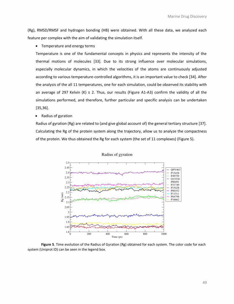
Marine Drug Discovery
49
(Rg), RMSD/RMSF and hydrogen bonding (HB) were obtained. With all these data, we analyzed each
feature per complex with the aim of validating the simulation itself.
Temperature and energy terms
Temperature is one of the fundamental concepts in physics and represents the intensity of the
thermal motions of molecules [33]. Due to its strong influence over molecular simulations,
especially molecular dynamics, in which the velocities of the atoms are continuously adjusted
according to various temperature-controlled algorithms, it is an important value to check [34]. After
the analysis of the all 11 temperatures, one for each simulation, could be observed its stability with
an average of 297 Kelvin (K) ± 2. Thus, our results (Figure A1-A3) confirm the validity of all the
simulations performed, and therefore, further particular and specific analysis can be undertaken
[35,36].
Radius of gyration
Radius of gyration (Rg) are related to (and give global account of) the general tertiary structure [37].
Calculating the Rg of the protein system along the trajectory, allow us to analyze the compactness
of the protein. We thus obtained the Rg for each system (the set of 11 complexes) (Figure 5).
Figure 5. Time evolution of the Radius of Gyration (Rg) obtained for each system. The color code for each
system (Uniprot ID) can be seen in the legend box.

Marine Drug Discovery
50
As a general trend, the decrease in the average value of Rg as a function of the simulation time
suggests contraction of the structure, what is translated into a gain in compactness. P11511 and
P04798 fluctuate in a constant way around 2.23-2.26 nm and 2.26-2.29 nm respectively, during the
whole simulation, thus indicating stability over time. These results contrast with P16662, where the
Rg oscillates from 2.22 to 2.28 nm, and Q9Y463 oscillates from 2.19 to 2.25 nm, which points out an
incompactness of the systems. P00352, despite seeming constant on time, shows the highest values
compared to the other 10 systems. P49759 and P31749 show a very similar and constant trend; at
the time step 0 ps, Rg values are around 2.1 nm and they increase slightly until, at time step 1000
ps, when they reach 2.13 and 2.15 nm. O15530 has lower values than the systems above and the Rg
fluctuates from 1.94 to 1.97 nm, indicating stability despite an abrupt steadily fluctuation can be
observed. P00491 is one of the most constant systems which fluctuates constantly at 1.88 nm. The
two last systems analyzed are P15428, once for Meridianin-A (red) and once for Rossinone
(maroon). Both systems have the lower Rg values, indicating stability and compactness.
Interestingly, despite the fact that at the time step 0 ps, Rg values are different, 1.84 and 1.85 nm,
from the 700 ps they merge and follow the same pattern till the end. These results give reliability
over all Rg analysis (Figure 5).
Root-mean-square deviation
The RMSD of atomic positions is the measure of the average distance between the atoms of
superimposed structures [38,39]. To check the stability of the simulations, the RMSDs of the Cα
atom was calculated and monitored over the course of simulation. In general, this superimposition
is performed among the structures extracted from the MD simulation and a reference structure
[40]. As a reference, we used the starting structure of each target, and the RMSD value was
obtained by comparing it with the structure (all atoms) obtained at each step of the trajectory. This
process was performed over the 11 remaining complexes (Figure 6).

Marine Drug Discovery
51
Figure 6. The progress and completion of the MD simulation processes were monitored by plotting a
graph of Root-mean-square deviation (RMSD) of the eleven systems obtained by comparing each structure at the
starting point and the structure (all atoms) obtained along the trajectory. The color code for each system (Uniprot
ID) can be seen in the legend box.
RMSD values increase from the beginning of the simulation to a certain moment in which it remains
constant (MD converged). For all the systems, the RMSD remains stable around average values of
0.15-0.25 nm over a considerably time period of the trajectory, indicating that the systems were
stable during the simulation (Figure 6). This is the expected behavior during an MD simulation as
RMSD is commonly used as an indicator of convergence of the structure towards an equilibrium
state.
Hydrogen bonding
The time-averaged number of hydrogen bonds (HBs) present on each marine molecule-target
complex was calculated from the MD trajectories. HBs were defined in such a way that the distance
between donor and acceptor was less that the cut-off distance of 3.5 Å (0.35 nm) and the angle
donor-H-acceptor was less than the cut-off angle of 20 degrees. Given that this is a dynamical
system, the number of HBs is not constant; in fact, these bonds are forming and breaking
constantly as the simulation runs. For this reason, we calculated the live time of these HBs
throughout the simulation. In this way, we obtained the occupancy of a particular interaction
during the 1ns simulation of each complex (Figure 7).
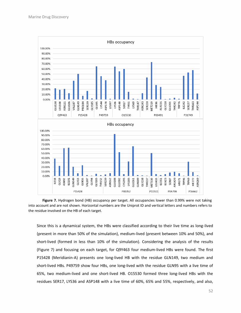
Marine Drug Discovery
52
Figure 7. Hydrogen bond (HB) occupancy per target. All occupancies lower than 0.99% were not taking
into account and are not shown. Horizontal numbers are the Uniprot ID and vertical letters and numbers refers to
the residue involved on the HB of each target.
Since this is a dynamical system, the HBs were classified according to their live time as long-lived
(present in more than 50% of the simulation), medium-lived (present between 10% and 50%), and
short-lived (formed in less than 10% of the simulation). Considering the analysis of the results
(Figure 7) and focusing on each target, for Q9Y463 four medium-lived HBs were found. The first
P15428 (Meridianin-A) presents one long-lived HB with the residue GLN149, two medium and
short-lived HBs. P49759 show four HBs, one long-lived with the residue GLN95 with a live time of
65%, two medium-lived and one short-lived HB. O15530 formed three long-lived HBs with the
residues SER17, LYS36 and ASP148 with a live time of 60%, 65% and 55%, respectively, and also,

Marine Drug Discovery
53
presents one medium-lived and two short-lived HBs. P00491 formed a total number of eight HBs.
Only one of them is a long-lived and is established with MET219, and there are four medium-lived
HBs and three short-lived. P31749 presents two long-lived HBs with the residues SER157 and
TRH163 with occupancy of 53% and 57%. Also, there are two more HBs but with a medium-lived
and one with short-lived. With eleven HBs, P15428 (Rossinone) is the target with more HBs formed
despite this only two of them, ILE91 and ASN92 are considered long-lived with occupancies of 62%;.
four medium-lived are also found, as well as five short-lived HBs. P00352 is the target with the
higher occupancy with a value of 93% with the residue GLU188, which means that this HBs is
almost maintained during all the MD simulation. Two other residues as GLU261 and GLU392 are
also considered long-lived HBs, but with 65% and 52% of occupancy. Only one medium-lived HB
was formed while six short-lived were present. P11511 had one long-lived HB with an occupancy of
51% and there was one short-lived HB. P04798 is the only target where any long or medium-lived
HB was founded, and the six residues found, were short-lived. P16662 has one medium-lived HB
and two short-lived HBs. As said, P15428 is studied twice, and in this case, the residues involved on
the formation of HBs are not the same in both cases, except GLN149. This is due to the size of the
binding cavity, which is quite large, favoring the different location of the molecules inside the cavity
while performing the docking calculations.
Root-mean-square fluctuation
When a dynamic system such as a protein fluctuates about some well-defined average position, the
RMSF of atomic positions can be calculated from the MD trajectory. That is, RMSF measures the
amplitude of atom motions during simulation (Figure 8) [41,42].

Marine Drug Discovery
54

Marine Drug Discovery
55
Figure 8. Root-mean-square fluctuation (RMSF) per residue (X-axis) values of each system along the MD
simulation. The highest fluctuations (> 0.25 nm) detected have been highlighted with orange circles.
Interestingly, after an exhaustive analysis of each system, the results from the RMSFs of each
aminoacid, highlight the flexible regions of the systems. RMSFs values higher than 0.25 nm (orange
circles Figure 8) are characteristic of aminoacids residues belonging to flexible regions. Eight out of
the 11 analyzed systems display RMSFs values higher than 0.25 nm, which means that remaining
there, both P15428 and P11511 are, in general, rigid structures. Q9Y463 has a peak of fluctuation
higher than 0.35 nm around residues 305-310. P49759 has fluctuation with values higher than 0.30
nm between residues 160-170. This system has another point of fluctuations around residues 40,
50, 195 and between 260 and 300, despite these has low values (< 0.25 nm). O15530 seems to be a
more rigid system, even though before the highest fluctuation observed on residues 52-58, two
other lower peaks can be observed indicating that there is a region between residues 25 to 60
which is more flexible than the rest of the protein. P00491 is also a system with low fluctuations
were only a fluctuation of 0.28 nm can be seen around residue 255. P31749 is the system with
more peaks of fluctuation and they are placed around residue 20, between 98 and 108 and around
297. Also, other lower peaks can be seen between residues 50 and 100, and again between 150 and
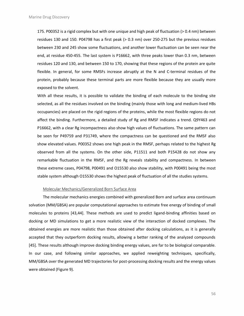
Marine Drug Discovery
56
175. P00352 is a rigid complex but with one unique and high peak of fluctuation (> 0.4 nm) between
residues 130 and 150. P04798 has a first peak (> 0.3 nm) over 250-275 but the previous residues
between 230 and 245 show some fluctuations, and another lower fluctuation can be seen near the
end, at residue 450-455. The last system is P16662, with three peaks lower than 0.3 nm, between
residues 120 and 130, and between 150 to 170, showing that these regions of the protein are quite
flexible. In general, for some RMSFs increase abruptly at the N and C-terminal residues of the
protein, probably because these terminal parts are more flexible because they are usually more
exposed to the solvent.
With all these results, it is possible to validate the binding of each molecule to the binding site
selected, as all the residues involved on the binding (mainly those with long and medium-lived HBs
occupancies) are placed on the rigid regions of the proteins, while the most flexible regions do not
affect the binding. Furthermore, a detailed study of Rg and RMSF indicates a trend. Q9Y463 and
P16662, with a clear Rg incompactness also show high values of fluctuations. The same pattern can
be seen for P49759 and P31749, where the compactness can be questioned and the RMSF also
show elevated values. P00352 shows one high peak in the RMSF, perhaps related to the highest Rg
observed from all the systems. On the other side, P11511 and both P15428 do not show any
remarkable fluctuation in the RMSF, and the Rg reveals stability and compactness. In between
these extreme cases, P04798, P00491 and O15530 also show stability, with P00491 being the most
stable system although O15530 shows the highest peak of fluctuation of all the studies systems.
Molecular Mechanics/Generalized Born Surface Area
The molecular mechanics energies combined with generalized Born and surface area continuum
solvation (MM/GBSA) are popular computational approaches to estimate free energy of binding of small
molecules to proteins [43,44]. These methods are used to predict ligand-binding affinities based on
docking or MD simulations to get a more realistic view of the interaction of docked complexes. The
obtained energies are more realistic than those obtained after docking calculations, as it is generally
accepted that they outperform docking results, allowing a better ranking of the analyzed compounds
[45]. These results although improve docking binding energy values, are far to be biological comparable.
In our case, and following similar approaches, we applied reweighting techniques, specifically,
MM/GBSA over the generated MD trajectories for post-processing docking results and the energy values
were obtained (Figure 9).

Marine Drug Discovery
57
Meridianin-A
Q9Y463 -41.7674 kcal/mol P15428 -38.3083 kcal/mol P49759 -37.9952 kcal/mol
Aplicyanin
O15530 -52.7520 kcal/mol P00491 –45.5407 kcal/mol P31749 -43.5072 kcal/mol
Rossinone Hodgsonal
P15428 -51.7908 kcal/mol P00352 -48.1930 kcal/mol P11511 -34.1795 kcal/mol
Polyrhaphin-A Dendrinolide
P04798 -42.6268 kcal/mol P16662 -25.6552 kcal/mol
Figure 9. Images of the binding mode of each marine molecule inside the binding cavity of the
corresponding target. Also, the summary of binding energy results after MD simulations with MM/GBSA
calculations are indicated here. All energies are in kcal/mol.
From the negative total binding energy found on all the studied systems, we conclude that all of
them are favorable complex in pure water. However, the computational results will not be equal to the

Marine Drug Discovery
58
real (experimental) binding free energy, because we did not estimate the (dis-favorable) entropy
contribution to binding, since this is a weak point of these methods. Meridianin-A, related with three
different targets, Q9Y463, P15428 and P49759, does not show any significant difference on the binding
energy that could suggest any selectivity over one target or another. Aplicyanin, also related to three
targets, O15530, P00491 and P31749, seems to show preference for O15530, which is a target related to
heart failure. Hodgsonal, Polyrhaphin-A, and Dendrinolide are only related with one target each,
P11511, P04798 and P16662, respectively. The complex formed by Dendrinolide and P16662 target
related to peripheral nervous system disease, is interesting because it is, by far, the lower binding
energy found. However, this target shows incompactness in the Rg analysis and also has elevated
fluctuations, which leads to think that this is the less favorable complex. Rossinone, is related with two
targets, P15428 and P00352, has good results on the qualitative analysis while no significant differences
can be observed on both targets. For P15428, target related to digital clubbing, the binding energy
obtained is the highest one and revealed a significant difference (13 kcal/mol) between the binding of
Rossinone or Meridianin-A (Figure 9).
Materials and Methods
Virtual Profiling
VP techniques able to automatically evaluate very large libraries of compounds using computer
programs to finding targets for an input (query) molecule. In other words, given an initial molecule and
by executing a similarity searching against a reference database (compound-target associations
database) are able to find similar molecules (restricted by a cut-off) and thus finding plausible target to
the input molecule. Basically, the way VP works is that if a molecule A is like a molecule B, the molecule
A should interact with the target of molecule B. For that purpose, we employed Cabrakan, a 2D ligand-
based VP software tool that compares molecules using 2D fingerprints and the assignment of biological
activity, and Hurakan, a 3D VP tool that compares molecules according to their similarity using methods
that take into account both physical and chemical properties of the molecules and their behavior with
the environment when acting on a target [46,47].

Marine Drug Discovery
59
Target selection
To relate the found targets with the specific selected pathologies we employed DisGeNET, a
database that integrates information on gene-diseases associations. From these data, we focused on
neurodegenerative and cardiovascular diseases [19].
Target modelling
From the selected targets, the search for 3D models was performed by exploring the Protein
Data Bank (RCSB PDB) [48]. For those targets without crystallographic structures available or showing
poor sequence representation (<30%), homology models were constructed by SWISS-MODEL, an
automated protein structure homology model software tool to generate models of targets from the
original human aminoacid sequence [49].
Toxicology prediction
Regarding ligand-based toxicology, the predictive models used here allow measuring Structure-
Activity Relationship (SAR). The SAR concept means that the biological activity of a chemical can be
related to its molecular structure, and when this is quantified, this relationship is known as QSAR. Here
we used VEGA software tool, which is integrated in different QSAR models that predict biological toxicity
at different levels [25]. The toxicity prediction depends on its reliability in a range that goes from 1 (low)
to 3 (high). In this study, to gain statistical significance and reliability, all the available models in VEGA
were employed. Because of that, the results of each category were averaged over all models used and
then the results were classified according to its probability of being toxic in the following terms; no
toxicity, low (<2), medium (2-2.75) or high (2.75-3). Regarding the toxicology based on target, T3DB was
used, and over it a 2D Tanimoto based similarity search was performed using an inhouse developed tool
[28].
Drug likeness evaluation
To evaluate the drug likeness of marine molecules, we use SuperTarget, a database which
provides drug-target relations [50]. Only those relations that come from the well-known database
DrugBank were selected [51]. Later, the 3D structure of those drugs selected was downloaded from
PubChem compound database [52].

Marine Drug Discovery
60
Docking calculations
Ligands were prepared to generate energetically minimized 3D coordinates and then docked
into the active site of each target using Itzamna software tool [53]. Kin software tool was also used to
perform blind docking calculations [54]. Best binding poses were determined and subsequently ranked
based on their calculated binding energies.
Molecular dynamics simulation
The MD simulations were performed using NAMD software through four sequential steps: First,
we run an energy minimization of the system, which is required to sort out any bad starting structures
caused during the generation of the system [55]. The second and third steps are part of an equilibration
simulation. The second step consists of simulating the system at NVT ensemble (at constant number of
particles (N), volume (V) and temperature (T)), with position restraints on the solute to get the
temperature at 300K. The third step consists of simulating at constant pressure (NPT) to fix the density
of the system while the system, is heated incrementally until reaching the desired temperature [56,57].
The fourth and last step consists of running the production simulation for sufficient time so that
property/phenomena of interest can be observed in required detail. In this study we performed short
(1ns) simulations with a time step of 2 fs. For the modelling of the protein targets we used Amber
ff99SB-ILDN, and for the modelling of the marine molecules, the General Amber Force Field (GAFF) set
of parameters was used [58,59]. Ligand GAFF parameters were obtained using Antechamber, whereas
the receptor structures were modelled using the leap module of Amber Tools [60,61].
Molecular dynamics analysis
Visual inspection of the trajectories and the HBs occupancies were performed using Visual
Molecular Dynamics (VMD) [62]. The quality assurance of the thermodynamic parameters (temperature,
potential, kinetic and total energy) and the review in terms of structure (Rg, RMSD and RMSF) were
performed using GROMACS quality assurance tool [63,64].
MM/Generalized Born Surface Area
MM/GBSA rescoring was performed using the MMPBSA python algorithm contained within the
Amber Tools suit. The snapshots generated at the end of MD simulations were used as input into the
post-simulation MM/GBSA calculation of binding free energies [65].

Marine Drug Discovery
61
Graphical representations
Graphical representations were prepared using PyMOL version 1.7 and the graphs are produced
using the program GRaphing, Advanced Computation and Exploration of data (xmgrace) version 5.1.22
[66,67]. 2D marine molecules were prepared using RDKit python library [68].
Conclusions
Molecular docking and the techniques that derive from it, as MD simulations and MM/GBSA
have demonstrated to be solid tools to be used during the process of drug discovery. Along this study,
we employed different computational tools to validate, even from different points of views, the results
obtained after each step of the process of elucidating the possible therapeutic potential of this set of
marine molecules. In the first step, after the VP techniques elucidated a list of possible targets related to
neurodegenerative and cardiovascular diseased, the marine molecule-target complexes were confirmed
using docking and blind docking calculations. Following that, the toxicology predictions based on ligand
and target revealed a tendency of these molecules to be toxic. Due to the importance of this kind of
studies during drug discovery, further studies are required to completely assess the safety of these
molecules. Maintaining the perspective and approach given to this study, and with the objective of
comparing the possible drug-likeness properties of these marine molecules, a total number of 392
docking calculations were performed over known drugs and marine molecules against the targets
selected on the first step and the results revealed that no significant differences were observed between
them. This indicates that all the molecules of the set could act as drugs. As previously explained here,
docking calculations have its limitations, and to give more reliability to our results, the induce-fit MD
simulations and MM/GBSA techniques were employed. A deep study of the time averaged structural
properties, such as Rg, RMSD, RMSF and HBs occupancies analyzed after the simulations allows us to
suggest that in general, the eleven systems studied can be validated, despite some of them, as Q9Y463,
P16662, P49759 and P31749, are less favorable than the others in terms of a qualitative analysis. In
terms of energy values, it is worth to mention that the lower energy was obtained for the complex
formed by Dendrinolide and P16662.
This study is a clear example on how the use of different CADD tools could help on the
elucidation of different marine molecules with a potential therapeutic activity, in this case against
cardiovascular, neurodegenerative, and some orphan diseases. Along this work, we identified, no only
the diseases but the targets and the regions of the target sequence, where the marine molecules bind,

Marine Drug Discovery
62
proving invaluable insights for further studies. Also, the introduction of the induce-fit MD simulation
events allowed an improvement of docking results and an exhaustive qualitative analysis of each system
could be performed. Finally, we believe that with our study, a general pipeline on the field of drug
discovery has been established, which can be applied on the elucidation of therapeutic agents from
marine molecules in future studies.
Acknowledgments: This research was partially supported by an Industrial Doctorate grant from
the Generalitat of Catalonia to L.L.-P (DI 2016-051).
Author Contributions: M.S.-M., L. L.-P- and C.A. conceived the study and designed the
experiments. L.L.-P. carried out the experiments whereas C.A. and M.S.-M. supervised them. All the
authors analyzed and discussed the results, as well as wrote the manuscript.
Funding: Industrial Doctorate grant from the Generalitat of Catalonia to LL-P (DI 2016-051).
Conflicts of Interest: The authors declare no conflict of interest.
Marine Drug Discovery
63
Appendix
Table A1. Summary of the results obtained performing docking simulations and Molecular Mechanics/Generalized Born Surface Area (MM/GBSA) calculations of marine molecules against each target (UniProt) with crystallographic structures (PDB) with ligand. To avoid false positives, each docking calculation was performed twice (R0/R1). All the energy values are in kcal/mol.
UniProt PDB - Ligand Docking MM/GBSA UniProt PDB - Ligand Docking MM/GBSA
Binding Energy
Binding Energy
Binding Energy
Binding Energy
R0/R1 R0/R1
Hodgsonal Aplicyanin
P11511 3EQM-ASD -8,1 / -8,2 -34,1795 P09874 1UK0-FRM -8,2 / -8,2 -32,3788 3EQM-HEM -8,3 / -8,4 -22,4488
O15530 2R7B-253 -7,7 / -7,7 -22,8350
P15428 2GDZ-NAD -6,9 / -6,9 -6,3093 3QC4-MP7 -8,1 / -8,1 -52,7520
Q99714 1U7T-NAD -7,2 / -7,2 -0,0562 P31749 3O96-IQO -8,6 / -8,6 -43,5072 2O23-NAD -6,7 / -6,7 -14,0999 P00491 1ULB-GUN -7,2 / -7,2 -45,5407
Meridianin-A Rossinone
P00374 1MVS-DTM -7,7 / -7,7 -28,9913 P00352 4WB9-NAI -8,8 / -8,5 -48,1930 P48730 4KBK-1QG -7,3 / -7,2 -34,4325 O00255 3U88-CHD -6,8 / -6,8 -25,5673
Q13976 3OGJ-CMP -6,7 / -6,7 -28,4003 P07550 4GBR-CAU -9,7 / -9,7 -43,2079 4QX5-CMP -7,9 / -7,9 -13,5921
Q99714 1U7T-TDT -8,9 / -9 -39,1248
P49841 3PUP-OS1 -7,7 / -7,7 -29,1106 1U7T-NAD -8,1 / -8 -40,7381
Q99714 1U7T-TDT -7,7 / -7,7 -25,4862 2O23-NAD -8,6 / -8,5 -39,0733 1U7T-NAD -7,5 / -7,4 -21,3118 P15428 2GDZ-NAD -9,2 / -9,3 -51,7908 2O23-NAD -7,6 / -7,6 -23,5035 P04637 5AB9-92O -6,4 / -6,5 -31,0615
Q13627 4AZE-3RA -8,2 / -8,2 -32,8447 P15428 2GDZ-NAD -8,4 / -8,4 -38,3083
Pteroenone
P15428 2GDZ-NAD -7 / -7,1 -30,7877 P07550 4GBR-CAU -7,7 / -7,6 -36,9769
Q99714 1U7T-TDT -6,5 / -6,5 -23,3863 2O23-NAD -6,5 / -6,4 -22,0587
Table A2. Summary of the results obtained performing blind docking simulations and Molecular Mechanics/Generalized Born Surface Area (MM/GBSA) calculations of marine molecules against each target (UniProt) represented by homology models. Pocket means the cavity chosen to perform MD simulations. To avoid false positives, each docking calculation was performed twice (R0/R1). All the energy values are in kcal/mol.
UniProt Pocket Docking MM/GBSA UniProt Pocket Docking MM/GBSA
Binding Energy
Binding Energy
Binding Energy
Binding Energy
R0/R1
R0/R1
Hodgsonal Rossinone
Q96KQ7 0 -7,8 / -7,9 -20,4079 Q96KQ7 1 -8,1 / -8,1 -26,9966
Meridianin-A Q07343 0 -8,6 / -8,6 -41,0540
P49759 0 -9,3 / -9,3 -37,9952 Q16236 0 -8,1 / -8 -36,1075 Q9Y463 0 -8,8 / -8,1 -41,7674 P27815 0 -8,8 / -8,7 -37,3115 Q96KQ7 0 -7,3 / -7,3 -19,5406 Q08499 0 -9,2 / -9,3 -32,3433
Aplicyanin Polyrhaphin-A
Q96KQ7 0 -7,5 / -7,5 -21,8578 P24046 0 -7,5 / -7,6 -27,6724 Q16236 0 -6,6 / -6,6 -22,3084 P46098 1 -8,1 / -8,1 -30,8251
Pteroenone P14867 0 -7,6 / -7,5 -14,0650
Q16236 0 -6,2 / -6,5 -15,8940 P04798 3 -9,5 / -9,2 -42,6268

Marine Drug Discovery
64
Table A3. Summary of the results obtained performing blind docking simulations and Molecular Mechanics/Generalized Born Surface Area (MM/GBSA) calculations of marine molecules against each target (UniProt) represented by crystallographic structures (PDB) without ligand. Pocket means the cavity chosen to perform MD simulations. To avoid false positives, each docking calculation was performed twice (R0/R1). All the energy values are in kcal/mol.
UniProt PDB Pocket Docking MM/GBSA UniProt PDB Pocket Docking MM/GBSA
Binding Energy
Binding Energy
Binding Energy
Binding Energy
R0/R1
R0/R1
Hodgsonal Rossinone
P04637 3Q01 1 -6,1 / -6,6 -11,3246 P04637 3Q01 1 -7 / -6,5 -23,5341 Meridianin-A Dendrinolide
Q13976 4KU8 0 -7,1 / -7,1 -19,9742 P16662 2O6L 0 -8,1 / -8,1 -25,6552
Table A4. Summary of the best affinities obtained after Molecular Mechanics/Generalized Born Surface Area (MM/GBSA) calculations of marine molecules against each target (UniProt) and also, pathologies listed by target. Each target can be represented by crystallographic structures with ligands, without ligands, or homology models (HM). To avoid false positive, each docking calculation was performed twice (R0/R1). All the energy values are in kcal/mol.
UniProt PDB-Ligand Docking MM/GBSA Pathologies
Binding Energy Binding Energy
R0/R1
Hodgsonal
P11511 3EQM-ASD -8.1 / -8,2 -34,1795 Autism Meridianin-A
Q9Y463 HM -8.1 / -8,8 -41,7674 Alzheimer P15428 2GDZ-NAD -8.4 / -8,4 -38,3083 Digital clubbing P49759 HM -9.3 / -9,3 -37,9952 Alzheimer
Aplicyanin
O15530 3QC4-MP7 -8.1 / -8,1 -52,7520 Heart failure P00491 1ULB-GUN -7.2 / -7,2 -45,5407 Alzheimer P31749 3O96-IQO -8,6 / -8.6 -43,5072 Cardiovascular, schizophrenia
Rossinone
P15428 2GDZ-NAD -9.2 / -9,3 -51,7908 Digital clubbing P00352 4WB9-NAI -8.5 / -8,8 -48,1930 Parkinson
Polyrhaphin-A
P04798 HM -9.2 / -9,5 -42,6268 Cardiovascular disease
Dendrinolide
P16662 2O6L -8.1 / -8,1 -25,6552 Peripheral nervous system disease

Marine Drug Discovery
65
Figure A1. Total energy during the progress of the MD simulation of the eleven target-marine molecule systems. The color code of each target can be seen in the legend box.
Figure A2. Potential energy during the progress of the MD simulation of the eleven target-marine molecule systems. The color code of each target can be seen in the legend box.

Marine Drug Discovery
66
Figure A3. Kinetic energy during the progress of the MD simulation of the eleven target-marine molecule systems. The color code of each target can be seen in the legend box.

Marine Drug Discovery
67
References
1. De Vivo, M.; Cavalli, A. Recent advances in dynamic docking for drug discovery. Wiley Interdiscip. Rev. Comput. Mol. Sci. 2017, 7, e1320.
2. Meng, X.-Y.; Zhang, H.-X.; Mezei, M.; Cui, M. Molecular docking: a powerful approach for structure-based drug discovery. Curr. Comput. Aided. Drug Des. 2011, 7, 146–57.
3. Jorgensen, W. L. The many roles of computation in drug discovery. Science 2004, 303, 1813–1818.
4. Fischer, E. No Title; 1894; Vol. 27. 5. Kuntz, I. D.; Blaney, J. M.; Oatley, S. J.; Langridge, R.; Ferrin, T. E. A geometric approach to
macromolecule-ligand interactions. J. Mol. Biol. 1982, 161, 269–288. 6. Koshland, D. E. Correlation of structure and function in enzyme action. Science 1963, 142, 1533–
41. 7. Aravindhan, G.; Coote, M. L.; Barakat, K. Molecular dynamics-driven drug discovery: leaping
forward with confidence. Drug Discov. Today 2017, 22, 249–269. 8. Lill, M. A. Efficient Incorporation of Protein Flexibility and Dynamics into Molecular Docking
Simulations. Biochemistry 2011, 50, 6157–6169. 9. Alonso, H.; Bliznyuk, A. A.; Gready, J. E. Combining docking and molecular dynamic simulations
in drug design. Med. Res. Rev. 2006, 26, 531–568. 10. Sherman, Woody; Day, Tyler; P. Jacobson, Matthew; A. Friesner, Richard and; Farid, Ramy. Novel
Procedure for Modeling Ligand/Receptor Induced Fit Effects. 2005. 11. Taboada, S.; Wiklund, H.; Glover, A. G.; Dahlgren, T. G.; Cristobo, J.; Avila, C. Two new Antarctic
Ophryotrocha (Annelida: Dorvilleidae) described from shallow-water whale bones. Polar Biol. 2013, 36, 1031–1045.
12. Taboada, S.; Junoy, J.; Andrade, S. C. S.; Giribet, G.; Cristobo, J.; Avila, C. On the identity of two Antarctic brooding nemerteans: redescription of Antarctonemertes valida (Bürger, 1893) and description of a new species in the genus Antarctonemertes Friedrich, 1955 (Nemertea, Hoplonemertea). Polar Biol. 2013, 36, 1415–1430.
13. Taboada, S.; Bas, M.; Avila, C. A new Parougia species (Annelida, Dorvilleidae) associated with eutrophic marine habitats in Antarctica. Polar Biol. 2015, 38, 517–527.
14. Rodrigues, T.; Reker, D.; Schneider, P.; Schneider, G. Counting on natural products for drug design. Nat. Chem. 2016, 8, 531–541.
15. W-H Li, J.; Vederas, J. C. Drug Discovery and Natural Products: End of an Era or an Endless Frontier? Science (80-. ). 2009, 325, 161–165.
16. Ebrahim, H.; El Sayed, K. Discovery of Novel Antiangiogenic Marine Natural Product Scaffolds. Mar. Drugs 2016, 14, 57.
17. Prachayasittikul, V.; Worachartcheewan, A.; Shoombuatong, W.; Songtawee, N.; Simeon, S.; Prachayasittikul, V.; Nantasenamat, C. Computer-Aided Drug Design of Bioactive Natural Products. Curr. Top. Med. Chem. 2015, 15, 1780–800.
18. Health at the glance-OCDE. 19. Pinero, J.; Queralt-Rosinach, N.; Bravo, A.; Deu-Pons, J.; Bauer-Mehren, A.; Baron, M.; Sanz, F.;
Furlong, L. I. DisGeNET: a discovery platform for the dynamical exploration of human diseases and their genes. Database 2015, 2015, bav028-bav028.
20. Muster, W.; Breidenbach, A.; Fischer, H.; Kirchner, S.; Müller, L.; Pähler, A. Computational toxicology in drug development. Drug Discov. Today 2008, 13, 303–310.
21. Nature Reviews Drug Discovery. Nature Publishing Group November 1, 2007, pp. 853–853. 22. Schneider, G. Prediction of Drug-Like Properties. 2013.

Marine Drug Discovery
68
23. Genheden, S.; Ryde, U. The MM/PBSA and MM/GBSA methods to estimate ligand-binding affinities. Expert Opin. Drug Discov. 2015, 10, 449–61.
24. Kumar, A.; Zhang, K. Y. J. Advances in the Development of Shape Similarity Methods and Their Application in Drug Discovery. Front. Chem. 2018, 6, 315.
25. VEGA-QSAR http://ceur-ws.org/Vol-1107/paper8.pdf (accessed May 13, 2016). 26. Proksch, P. Defensive roles for secondary metabolites from marine sponges and sponge-feeding
nudibranchs. Toxicon 1994, 32, 639–655. 27. Nuijen, B.; Bouma, M.; Manada, C.; Jimeno, J. M.; Schellens, J. H.; Bult, A.; Beijnen, J. H.
Pharmaceutical development of anticancer agents derived from marine sources. Anticancer. Drugs 2000, 11, 793–811.
28. Wishart, D.; Arndt, D.; Pon, A.; Sajed, T.; Guo, A. C.; Djoumbou, Y.; Knox, C.; Wilson, M.; Liang, Y.; Grant, J.; Liu, Y.; Goldansaz, S. A.; Rappaport, S. M. T3DB: the toxic exposome database. Nucleic Acids Res. 2015, 43, D928-34.
29. Bajusz, D.; Rácz, A.; Héberger, K. Why is Tanimoto index an appropriate choice for fingerprint-based similarity calculations? J. Cheminform. 2015, 7, 20.
30. Valerio, L. G. In silico toxicology for the pharmaceutical sciences. Toxicol. Appl. Pharmacol. 2009, 241, 356–370.
31. Tzeng, S.-R.; Kalodimos, C. G. Protein activity regulation by conformational entropy. Nature 2012, 488, 236–240.
32. Fenwick, R. B.; Orellana, L.; Esteban-Martín, S.; Orozco, M.; Salvatella, X. Correlated motions are a fundamental property of β-sheets. Nat. Commun. 2014, 5, 1–9.
33. Liu, B.; Xu, R.; He, X. Kinetic Energy-Based Temperature Computation in Non-Equilibrium Molecular Dynamics Simulation. 2009.
34. Crehuet, R.; Field, M. J. A temperature-dependent nudged-elastic-band algorithm. J. Chem. Phys. 2003, 118, 9563–9571.
35. Merz, P. T.; Shirts, M. R. Testing for physical validity in molecular simulations. PLoS One 2018, 13, e0202764.
36. Jepps, A; Ayton, J; Evans,E. Microscopic expressions for the thermodynamic temperature. Phys. Rev. E. Stat. Phys. Plasmas. Fluids. Relat. Interdiscip. Topics 2000, 62, 4757–63.
37. Pirolli, D.; Sciandra, F.; Bozzi, M.; Giardina, B.; Brancaccio, A.; Rosa, M. C. De Insights from Molecular Dynamics Simulations: Structural Basis for the V567D Mutation-Induced Instability of Zebrafish Alpha-Dystroglycan and Comparison with the Murine Model. PLoS One 2014, 9, e103866.
38. Kufareva, I.; Abagyan, R. Methods of protein structure comparison. Methods Mol. Biol. 2012, 857, 231–57.
39. Maiorov, V. N.; Crippen, G. M. Significance of Root-Mean-Square Deviation in Comparing Three-dimensional Structures of Globular Proteins. J. Mol. Biol. 1994, 235, 625–634.
40. Yu, W.; Mackerell, A. D. Computer-Aided Drug Design Methods. Methods Mol. Biol. 2017, 1520, 93–94.
41. Fuglebakk, E.; Echave, J.; Reuter, N. Measuring and comparing structural fluctuation patterns in large protein datasets. Bioinformatics 2012, 28, 2431–2440.
42. Bornot, A.; Etchebest, C.; de Brevern, A. G. Predicting protein flexibility through the prediction of local structures. Proteins 2011, 79, 839–52.
43. Steinbrecher, T.; Labahn, A. Towards Accurate Free Energy Calculations in Ligand Protein-Binding Studies. Curr. Med. Chem. 2010, 17, 767–785.
44. Murcko, M. A. Computational Methods to Predict Binding Free Energy in Ligand-Receptor Complexes. J. Med. Chem. 1995, 38, 4953–4967.

Marine Drug Discovery
69
45. Zhang, X.; Perez-Sanchez, H.; Lightstone, F. C. A Comprehensive Docking and MM/GBSA Rescoring Study of Ligand Recognition upon Binding Antithrombin. Curr. Top. Med. Chem. 2017, 17, 1631–1639.
46. Santamaria-Navarro, E.; Felix, E.; Nonell-Canals, A. Cabrakan https://www.mindthebyte.com/. 47. Santamaria-Navarro, E.; Nonell-Canals, A. Hurakan https://www.mindthebyte.com/. 48. Berman, H.; Henrick, K.; Nakamura, H.; Markley, J. L. The worldwide Protein Data Bank (wwPDB):
Ensuring a single, uniform archive of PDB data. Nucleic Acids Res. 2007, 35, 2006–2008. 49. Biasini, M.; Bienert, S.; Waterhouse, A.; Arnold, K.; Studer, G.; Schmidt, T.; Kiefer, F.; Cassarino,
T. G.; Bertoni, M.; Bordoli, L.; Schwede, T. SWISS-MODEL: Modelling protein tertiary and quaternary structure using evolutionary information. Nucleic Acids Res. 2014, 42, 252–258.
50. Günther, S.; Kuhn, M.; Dunkel, M.; Campillos, M.; Senger, C.; Petsalaki, E.; Ahmed, J.; Urdiales, E. G.; Gewiess, A.; Jensen, L. J.; Schneider, R.; Skoblo, R.; Russell, R. B.; Bourne, P. E.; Bork, P.; Preissner, R. SuperTarget and Matador: resources for exploring drug-target relationships. Nucleic Acids Res. 2008, 36, D919-22.
51. Law, V.; Knox, C.; Djoumbou, Y.; Jewison, T.; Guo, A. C.; Liu, Y.; MacIejewski, A.; Arndt, D.; Wilson, M.; Neveu, V.; Tang, A.; Gabriel, G.; Ly, C.; Adamjee, S.; Dame, Z. T.; Han, B.; Zhou, Y.; Wishart, D. S. DrugBank 4.0: Shedding new light on drug metabolism. Nucleic Acids Res. 2014, 42, 1–7.
52. Kim, S.; Thiessen, P. A.; Bolton, E. E.; Chen, J.; Fu, G.; Gindulyte, A.; Han, L.; He, J.; He, S.; Shoemaker, B. A.; Wang, J.; Yu, B.; Zhang, J.; Bryant, S. H. PubChem Substance and Compound databases. Nucleic Acids Res. 2016, 44, D1202–D1213.
53. Felix, E.; Santamaría-Navarro, E.; Sanchez-Martinez, M.; Nonell-Canals, A. Itzamna https://www.mindthebyte.com/.
54. Felix, E.; Nonell-Canals, A. Kin https://www.mindthebyte.com/. 55. Phillips, J. C.; Braun, R.; Wang, W.; Gumbart, J.; Tajkhorshid, E.; Villa, E.; Chipot, C.; Skeel, R. D.;
Kalé, L.; Schulten, K. Scalable molecular dynamics with NAMD. J. Comput. Chem. 2005, 26, 1781–1802.
56. Jorgensen, W. L.; Jenson, C. Temperature dependence of TIP3P, SPC, and TIP4P water from NPT Monte Carlo simulations: Seeking temperatures of maximum density. J. Comput. Chem. 1998, 19, 1179–1186.
57. Andersen, H. C. Rattle: A “velocity” version of the shake algorithm for molecular dynamics calculations. J. Comput. Phys. 1983, 52, 24–34.
58. Lindorff-Larsen, K.; Piana, S.; Palmo, K.; Maragakis, P.; Klepeis, J. L.; Dror, R. O.; Shaw, D. E. Improved side-chain torsion potentials for the Amber ff99SB protein force field. Proteins 2010, 78, 1950–8.
59. Wang, J.; Wolf, R. M.; Caldwell, J. W.; Kollman, P. A.; Case, D. A. Development and testing of a general amber force field. J. Comput. Chem. 2004, 25, 1157–1174.
60. Wang, J.; Wang, W.; Kollman, P. A.; Case, D. A. Antechamber, An Accessory Software Package For Molecular Mechanical Calculations. Natl. Institutes Heal.
61. Case, D. A.; Cheatham, T. E.; Darden, T.; Gohlke, H.; Luo, R.; Merz, K. M.; Onufriev, A.; Simmerling, C.; Wang, B.; Woods, R. J. The Amber biomolecular simulation programs. J. Comput. Chem. 2005, 26, 1668–88.
62. Humphrey, W.; Dalke, A.; Schulten, K. VMD: Visual molecular dynamics. J. Molec. Graph. 1996, 14, 33–38.
63. Hess, B.; Kutzner, C.; van der Spoel, D.; Lindahl, E. GROMACS 4: Algorithms for Highly Efficient, Load-Balanced, and Scalable Molecular Simulation. J. Chem. Theory Comput. 2008, 4, 435–447.

Marine Drug Discovery
70
64. Pronk, S.; Páll, S.; Schulz, R.; Larsson, P.; Bjelkmar, P.; Apostolov, R.; Shirts, M. R.; Smith, J. C.; Kasson, P. M.; Van Der Spoel, D.; Hess, B.; Lindahl, E. GROMACS 4.5: A high-throughput and highly parallel open source molecular simulation toolkit. Bioinformatics 2013, 29, 845–854.
65. Miller III, B. R.; McGee Jr., T. D.; Swails, J. M.; Homeyer, N.; Gohlke, H.; Roitberg, A. E. MMPBSA. py: An efficient program for end-state free energy calculations. J. Chem. Theory Comput. 2012, 3314–3321.
66. Yuan, S.; Chan, H. C. S.; Hu, Z. Using PyMOL as a platform for computational drug design. Wiley Interdiscip. Rev. Comput. Mol. Sci. 2017, 7, e1298.
67. 67.Grace User’s Guide (for Grace-5.1.22) http://plasma-gate.weizmann.ac.il/Grace/doc/UsersGuide.html (accessed Apr 26, 2019).
68. RDKit: Open-source cheminformatics.

Marine Drug Discovery
71
Section II
Elucidation of different pharmacophoric features of marine
compounds and a precise in silico binding study

Marine Drug Discovery
72

Marine Drug Discovery
73
Section II
Chapter 2
Computer-aided drug design applied to marine drug
discovery: Meridianins as Alzheimer’s disease therapeutic
agents

Marine Drug Discovery
74
75
Chapter 2
Computer-aided drug design applied to marine drug discovery: Meridianins as Alzheimer’s disease therapeutic agents
Laura Llorach-Pares 1,2, Alfons Nonell-Canals 2, Melchor Sánchez-Martínez2 and Conxita Avila1
1. Department of Evolutionary Biology, Ecology and Environmental Sciences, Faculty of Biology and
Biodiversity Research Institute (IRBio), Universitat de Barcelona, 08028 Barcelona, Catalonia.
2. Mind the Byte S.L., 08028 Barcelona, Catalonia, Spain.
Published in: Marine Drugs 15(12), 366 (2017) doi: 10.3390/md15120366.
Meridianins as Therapeutic Agents
76
Abstract
Computer-aided drug discovery/design (CADD) techniques allow the identification of natural
products that are capable of modulating protein functions in pathogenesis-related pathways,
constituting one of the most promising lines followed in drug discovery. In this paper, we
computationally evaluated and reported the inhibitory activity found in meridianins A–G, a group of
marine indole alkaloids isolated from the marine tunicate Aplidium, against various protein kinases
involved in Alzheimer’s disease (AD), a neurodegenerative pathology characterized by the presence of
neurofibrillary tangles (NFT). Balance splitting between tau kinase and phosphate activities caused tau
hyperphosphorylation and, thereby, its aggregation and NTF formation. Inhibition of specific kinases
involved in its phosphorylation pathway could be one of the key strategies to reverse tau
hyperphosphorylation and would represent an approach to develop drugs to palliate AD symptoms.
Meridianins bind to the adenosine triphosphate (ATP) binding site of certain protein kinases, acting as
ATP competitive inhibitors. These compounds show very promising scaffolds to design new drugs
against AD, which could act over tau protein kinases Glycogen synthase kinase-3 Beta (GSK3β) and
Casein kinase 1 delta (CK1δ, CK1D or KC1D), and dual specificity kinases as dual specificity tyrosine
phosphorylation regulated kinase 1 (DYRK1A) and cdc2-like kinases (CLK1). This work is aimed to
highlight the role of CADD techniques in marine drug discovery and to provide precise information
regarding the binding mode and strength of meridianins against several protein kinases that could help
in the future development of anti-AD drugs.
Keywords: Computer-aided drug discovery/design; meridianins; Alzheimer disease; protein kinases; tau
protein kinases; dual specificity kinases; marine natural products.

Meridianins as Therapeutic Agents
77
Resum
Les tècniques de descobriment o disseny de fàrmacs assistits per ordinador (DFAO) permeten la
identificació de productes naturals que són capaços de modular les funcions de proteïnes que estan
relacionades amb una determinada patologia, constituint una de les línies a seguir més prometedores
en el descobriment de fàrmacs. En aquest treball, hem avaluat computacionalment l'activitat inhibitòria
trobada en les meridianines A – G, un grup d'alcaloides indòlics marins aïllats del tunicat marí Aplidium,
contra diverses proteïnes quinases implicades en la malaltia d'Alzheimer (MA), una patologia
neurodegenerativa caracteritzada per la presència de cabdells neurofibril·lars. La pèrdua de l’estabilitat
entre les activitats de la tau quinasa i el fosfat és la causa de la hiperfosforilació de la tau i, per tant, la
seva agregació i formació de cabdells neurofibril·lars. La inhibició de quinases específiques implicades en
la seva via de fosforilació podria ser una de les estratègies clau per revertir la hiperfosforilació de la tau i
representaria una aproximació per desenvolupar fàrmacs per pal·liar els símptomes de la MA. Les
meridianines s’acoblen al lloc d'unió del trifosfat d'adenosina (ATP) de determinades proteïnes quinases,
actuant com a inhibidors competitius del ATP. Aquests compostos mostren esquelets molt prometedors
per dissenyar nous fàrmacs contra la MA, els quals podrien actuar sobre les proteïnes tau, com ara la
glycogen synthase kinase-3 beta (GSK3β) i la casein kinase 1 delta (CK1δ, CK1D o KC1D), i les quinases de
doble especificitat, com la dual specificity tyrosine phosphorylation regulated kinase 1 (DYRK1A) i les
quinases cdc2-like (CLK1). Aquest treball té com a objectiu destacar el paper de les tècniques de DFAO
en el descobriment de fàrmacs marins i proporcionar informació precisa sobre el mode i la força d’unió
de les meridianines contra diverses proteïnes quinases que podrien ajudar en el futur desenvolupament
de fàrmacs contra la MA.
Paraules clau: Disseny de fàrmacs assistit per ordinador; malaltia d’Alzheimer; proteïnes quinases,
proteïna quinasa tau, proteïnes de doble especificitat, productes naturals marins.

Meridianins as Therapeutic Agents
78
Introduction
Drug discovery is the process of identifying new molecules with a certain therapeutic activity.
This process is very expensive in terms of money and time. Translating basic research to the market
(going through drug discovery, preclinical and clinical studies) takes tens of years and costs billions of
dollars. The average cost to develop a new molecular entity is estimated to be $1.8 billion and requires
about 13.5 years [1]. However, the usage of computational techniques at various stages of the drug
discovery process could reduce that cost [2]. Hence, computer-aided drug discovery/design (CADD)
methods are becoming very popular and during the last three decades have played a major role in the
development of therapeutically important molecules [3,4]. CADD techniques cover several aspects of
the drug discovery pipeline, ranging from the selection of candidate molecules to the optimization of
lead compounds. For instance, virtual profiling (VP) methods can predict the biological profile as well as
mechanisms of action (MoA) of a certain molecule; molecular modelling techniques, such as docking and
molecular dynamics (MD), can predict ligand–target interactions in terms of binding mode and/or
binding strength, allowing discrimination between candidate compounds [5,6]; virtual screening (VS)
methods are able to find analogues (similar molecules) for a given compound(s) and/or build compound
libraries from an input molecule(s); hit to lead (H2L) optimization techniques are used to design new
molecules, improving an existing compound; absorption, distribution, metabolism, excretion and toxicity
(ADMET) prediction techniques are able to predict the physicochemical properties of a given compound,
i.e., information that can be coupled to H2L techniques in order to design better and safer drugs before
synthetizing them.
A common classification of these techniques is based on the nature of the input molecule. In this
sense, there are two general types of CADD approaches: structure-based drug design (SBDD) and ligand-
based drug design (LBDD). In SBDD, macromolecular three-dimensional (3D) target structures, usually
proteins, are analysed with the aim of identifying compounds that could interact (block, inhibit or
activate) with them. In LBDD, chemical compounds are analysed in order to, for instance, find chemical
analogues, explore their biological and/or toxicological profile, or improve their physicochemical and
pharmacological characteristics with the aim of developing drug-like compounds (Figure 1) [7,8].

Meridianins as Therapeutic Agents
79
Figure 1. Schematic representation of the computer-aided drug discovery/design (CADD) techniques
depicting a drug discovery pipeline. Original froom the author.
Historically, most new drugs have been designed from natural products (secondary metabolites)
and/or from compounds derived from them [9]. Natural products have thus been a rich source of
compounds for drug discovery, and often, feature biologically relevant molecular scaffolds and
pharmacophore patterns that have evolved as preferred ligand–protein binding motifs. The United
States Food and Drug Administration (US FDA) revealed that between 1981 and 2010, 34% of those
medicines approved were based on small molecules from natural products or direct derivatives of them
[10,11]. The identification of natural products that are capable of modulating protein functions in
pathogenesis-related pathways is one of the most promising lines followed in drug discovery [12].
Therefore, natural products constitute a huge source of inspiration in drug design [13].
An example is Alzheimer’s disease (AD), a neurodegenerative pathology that constitutes the
most common type of dementia (60–80% of the total cases), characterized by the presence of
neurofibrillary tangles (NFT) primarily composed of abnormal phosphorylated tau and senile plaques
(SP). Nowadays, despite its high incidence, there is still no specific treatment approved to cure this
disease. Tau phosphorylation is regulated by a balance between tau kinase and phosphate activities.
Splitting of this balance was considered to cause tau hyperphosphorylation and thereby its aggregation
and NTF formation [14,15]. Due to that fact, inhibition of specific tau kinases or kinases involved in tau

Meridianins as Therapeutic Agents
80
phosphorylation pathway, could be one of the key strategies to reverse tau phosphorylation and,
ultimately, fight AD [16].
The main relevant protein kinases involved in tau phosphorylation have been grouped into two
classes: tau protein kinases and dual specificity kinases. The first group contains proteins such as
glycogen synthase kinase-3 beta (GSK3β), that phosphorylates tau at different sites (specifically at 42
sites, 29 of them phosphorylated in AD brains) and casein kinase 1 delta (CK1δ), a non-proline-directed
protein kinase (non-PDPK) that regulates the microtubule dynamics through tau phosphorylation at 46
sites (25 of them phosphorylated in AD brains). The second group contains proteins such as dual
specificity tyrosine phosphorylation regulated kinase 1 (DYRK1) that self-catalyse their
autophosphorylation and behave as serine/threonine kinase that phosphorylates tau and the
transcription factor cyclic adenosine monophosphate-response element binding (cAMP-CREB), and an
evolutionarily conserved group of dual specificity kinases cdc2-like kinases (CLKs), which play an
important role in the regulation of ribonucleic acid RNA splicing and are involved in the pathology of AD
by phosphorylating the serine residues in arginine-rich (SR) proteins [14,15,17–19].
Among natural products, those of unexplored marine world origin are of great interest in the
discovery of novel chemical structures, since they harbour most of the biodiversity of the world [20,21].
For instance, compounds from marine invertebrates may possess interesting pharmacological activities.
Examples include Porifera, Cnidaria, Bryozoa, Mollusca and Tunicata [22,23]. However, although very
interesting and useful from a pharmacological point of view, obtaining these compounds is difficult,
both from technical and biological points of view; technically, because specimens have to be collected
by hand using scuba diving or by trawling (both expensive, logistically difficult, and time consuming), and
biologically, due to their marine habitats and due to the fact that they are usually unculturable [23]. All
these factors, together with the adequate implementation of the Nagoya Protocol and the bioavailability
of marine natural products, result in CADD contributions being highly relevant, since no biological
sample is needed to perform an in silico analysis [24]. This also alleviates some of the marine drug
discovery difficulties, such as the quantity of natural product necessary to be used in further clinical
studies.

Meridianins as Therapeutic Agents
81
Meridianin A R1 = OH, R2 = H, R3 = H, R4 = H
Meridianin B R1 = OH, R2 = H, R3 = Br, R4 = H
Meridianin C R1 = H, R2 = Br, R3 = H, R4 = H
Meridianin D R1 = H, R2 = H, R3 = Br, R4 = H
Meridianin E R1 = OH, R2 = H, R3 = H, R4 = Br
Meridianin F R1 = H, R2 = Br, R3 = Br, R4 = H
Meridianin G R1 = H, R2 = H, R3 = H, R4 = H
Figure 2. Structures of meridianins A–G.
To exemplify and highlight the power of CADD techniques in marine drug discovery, as part of an
ongoing study of bioactive marine molecules from benthic invertebrates, in this paper we evaluated and
reported the inhibitory activity found in meridianins A–G (Figure 2), a group of marine indole alkaloids
consisting of an indole framework connected to an aminopyrimidine ring, isolated from specimens of
the tunicate genus Aplidium, against various protein kinases involved in AD.
Results
Virtual Profiling
In a previous VP study (unpublished data not shown here), we observed that meridianins could
bind to diverse targets involved in different diseases associated with aging or neurodegenerative
pathologies, such as AD and Parkinson’s disease, cancer and cardiovascular diseases (Figure 3). The
found targets are of special interest as they are involved in several diseases that affect millions of people
worldwide, having a huge social incidence and also, in most cases, there is no cure for them. Regarding
AD, the most common disease in which meridianins could have a therapeutic role according to our
results (Figure 3), GSK3β, CK1δ, DYRK1A and CLK1 (four kinases involved in it) could be targeted. This
finding can be easily checked in the literature, confirming that meridianins can bind to these kinases.
Moreover, it can also be confirmed that the target analysis results are trustworthy, since not only is the
involvement of meridianins in AD disease found in the literature, but the role of meridianins as anti-
cancer agents can also be easily checked [25,26].
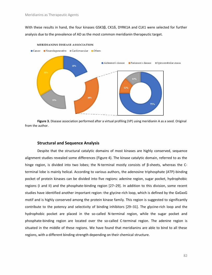
Meridianins as Therapeutic Agents
82
With these results in hand, the four kinases GSK3β, CK1δ, DYRK1A and CLK1 were selected for further
analysis due to the prevalence of AD as the most common meridianin therapeutic target.
Figure 3. Disease association performed after a virtual profiling (VP) using meridianin A as a seed. Original
from the author.
Structural and Sequence Analysis
Despite that the structural catalytic domains of most kinases are highly conserved, sequence
alignment studies revealed some differences (Figure 4). The kinase catalytic domain, referred to as the
hinge region, is divided into two lobes; the N-terminal mostly consists of β-sheets, whereas the C-
terminal lobe is mainly helical. According to various authors, the adenosine triphosphate (ATP)-binding
pocket of protein kinases can be divided into five regions: adenine region, sugar pocket, hydrophobic
regions (I and II) and the phosphate-binding region [27–29]. In addition to this division, some recent
studies have identified another important region: the glycine-rich loop, which is defined by the GxGxxG
motif and is highly conserved among the protein kinase family. This region is suggested to significantly
contribute to the potency and selectivity of binding inhibitors [29–31]. The glycine-rich loop and the
hydrophobic pocket are placed in the so-called N-terminal region, while the sugar pocket and
phosphate-binding region are located over the so-called C-terminal region. The adenine region is
situated in the middle of these regions. We have found that meridianins are able to bind to all these
regions, with a different binding strength depending on their chemical structure.

Meridianins as Therapeutic Agents
83
Figure 4. Amino acid sequence alignment of GSK3β, CK1δ, DYRK1A and CLK1. In the image, only the ATP-
binding pocket residues are shown. In blue, the key residues are conserved between all kinases. Green shows those conserved residues between tau protein kinases GSK3β and CK1δ, and red shows those conserved in dual specificity kinase DYRK1A and CLK1. Key residues refer to the residues implied in the binding of all the meridianins shared by the different targets and that are evolutionary conserved. The orange boxes represent the diverse region of the adenosine triphosphate (ATP) binding pocket. GRR: glycine-rich region; HP: hydrophobic pocket; AR: adenine region; SP: sugar pocket; PBP: phosphate binding pocket.
As explained above, we analysed two classes of protein kinases, specifically four members of
them. The core catalytic regions are conserved among all as they belong to the same enzyme subclass
(EC 2.7) and protein family (protein kinase). However, this protein family is divided into subfamilies:
serine-threonine protein kinases (EC 2.7.11), dual-specificity kinase (EC 2.7.12), protein-histidine kinases
(EC 2.7.13) and other protein kinases (EC 2.7.99). Thus, it seems logical that the binding site may be
more conserved among subfamilies, and even more so in lower classifications (sub-subfamilies such as
tau protein kinases (EC 2.7.11.26) and dual specificity kinase (EC 2.7.12.1)) than among the whole family.
Analysing our results, we have confirmed this trend. Several key residues (associated with the
substrate/inhibitor binding mode and/or conforming the pocket(s)) are conserved between the four
studied proteins (Figure 4), but a higher identity is observed by pairs. GSK3β and CK1δ share more
catalytic residues between them than with DYRK1A and CLK1, and vice versa. This observation agrees
with the finding of a common binding pattern between the four protein kinases plus another pattern per
each subfamily.

Meridianins as Therapeutic Agents
84
In silico Binding and Interaction Analysis
Meridianins bind to the ATP binding pocket of each of the selected targets, acting as ATP
competitive inhibitors. Binding energies obtained after docking and MD simulations (summarized in
Table A1) show a reasonably similar binding strength between the diverse meridianins and even among
the four kinases. Despite that fact, it could be observed that meridianin F tends to show higher energies
than the rest of the compounds. Moreover, in general, meridianins present better binding interaction
energies against CK1δ, DYRK1A and CLK1 than GSK3β. It must be said that these differences are hardly
noticeable and cannot constitute a unique and definitive prioritization tool.
The binding mode per meridianin and target (that slightly changes between each complex) is
summarized in Tables A2–A5. Comparing the interacting residues with the identified binders
(summarized in Table A6), it is clearly observed that meridianins could behave as inhibitors of the
analysed kinases. Moreover, analysing the observed binding mode together with the identified binders
and the conserved residues (Figure 4, Tables A6 and A7), as mentioned above, some patterns of the
general binding of meridianins to protein kinases could be extracted. It has to be highlighted that the
majority of the residues found in these patterns are identified as binders.
For tau protein kinases, GSK3β and CK1δ, 5 binding residues are shared between each of them,
whereas for dual specificity kinases, DYRK1A and CLK1, 12 are conserved. Moreover, there are four
residues conserved along the four analysed targets (Figure 4 and Table A7). Concretely, these residues
are an alanine and a lysine placed in the hydrophobic pocket, a leucine in the sugar pocket and an
aspartic acid in the phosphate binding region. Regarding tau protein kinases, there is also an isoleucine
shared by GSK3β and CK1δ. In the case of dual specificity kinases, there are eight other shared binders,
specifically, two phenylalanine, three valines, two glutamic acids and one leucine conserved and
identified as binders. Analysing the meridianin binding mode by focusing on the conserved amino acids
also identified as binders, we have found that two of them, A83 and K85 placed in the hydrophobic
pockets, are present in all meridianin binding modes over GSK3β and CK1δ (in the latter case, numbered
A36 and K38). For DYRK1A, three of the conserved residues are identified as key residues for the binding
of all meridianins, specifically V173, L241 and L294, in the same way as for CLK1 (in this case numbering
as V175, L244 and L295). In addition to these residues, others were found implicated in the general
binding of meridianins not conserved through all the targets (Table A6), specifically, for GSK3β I62, V70,
L132 and D200, for CK1δ I23, M82, L85, L135 and I148, and for DYRK1A K188, V222, F238, V306 and
D307. Finally, CLK1 residues L167 and A189 were identified as key meridianin binders.

Meridianins as Therapeutic Agents
85
Besides the above-mentioned residues, there are other important residues per meridianin and
target not present in the observed patterns that have a key role (Table A7), not dependent in a general
behaviour but dependent on the particular nature of each meridianin and target (Tables A2-A5).
Glycogen synthase kinase-3 Beta
Meridianins (Figure 5) tend to be placed within adenine (LDYV motif) and the hydrophobic
regions, formed by the conserved residues A83 and K85, in the catalytic cleft. The indole scaffold of the
meridianins is wrapped by N-terminal I62, F67, V70, A83, K85 and C-terminal T138, Q185, L188, D200
residues together with the LDYV motif in the hinge adenine region. Core interaction residues stabilize
meridianins by establishing hydrophobic contacts with I62, V70, A83, K85, L132, D200 and hydrogen
bonds with I62, K85, D200. The observed results further suggest that meridianins establish interactions
over the glycine-rich loop on GSK3β, defined by the GNGSFG motif, as well as with D200, a residue
present in the phosphate pocket. The fact that meridianins bind to I62, V70, A83, K85, L132, L188 and
D200, previously identified as binders, highlights meridianins inhibitory nature against GSK3β.
Figure 5. Meridianins A–G superposition over GSK3β. Labelled ligand-active site amino acid residues
involved in binding and the binding position of each meridianin models are enlarged. Original from the author.

Meridianins as Therapeutic Agents
86
Casein kinase 1 delta
All meridianin structures (Figure 6) share common interactions occupying the adenine region
formed by the MELL motif. Meridianins are stabilized in the hinge catalytic region, establishing hydrogen
bonds with A36, K38, M82, L85 and hydrophobic contacts with I23, K38, M82, L85, L135, and I148.
Interestingly, it has also been observed that the indole group of the higher ranked poses has additional
interactions with N-terminal I15, Y24, A36 and C-terminal D149 residues. It is important to remark that
meridianins bind to the previously identified binder residues I23, A36, K38, M82, L85, L135 and I148, a
fact that highlights meridianins inhibitory nature against CK1δ.
Figure 6. Meridianins A–G superposition over CK1δ. Labelled ligand-active site amino acid residues involved in binding and the binding position of each meridianin model are enlarged. Original from the author.
Dual specificity tyrosine phosphorylation regulated kinase 1
Meridianins are placed on the C-terminal region over the phosphate and sugar pockets as well
as the adenine motif FEML (Figure 7). Despite the fact that meridianins seem to interact with the N-
terminal residue V173 and the hydrophobic pocket residue K188, the rest of the key interactions are
established with residues placed over the C-terminal side. Meridianins establish hydrogen bonds with
K188, L241 and V307 as well as hydrophobic contacts with V173, K188, V222, F238, L241, L294, V306
and V307. Moreover, they perform π–cation and π–π stacking interactions with F238, which belongs to
the adenine motif. The inhibitory effect of meridianins against DYRK1A is confirmed by the fact that all

Meridianins as Therapeutic Agents
87
of them bind to V173, K188, V222, F238, L241, L294, V306 and V307, i.e., residues previously identified
as binders.
Figure 7. Meridianins A–G superposition over DYRK1A. Labelled ligand-active site amino acid residues involved in binding and the binding position of each meridianin model are enlarged. Original from the author.
Cdc2-like Kinases
Meridianin A–G conformations against CLK1 differ by pose, as can be observed in the
superimposition shown below (Figure 8). In fact, over this target is where meridianins displayed a more
different conformation between the family members. In general, all poses tend to be located near the
glycine rich loop and the hydrophobic pocket, interacting with the adenine motif FELL through L244 by a
hydrogen bond interaction. The different poses were well stabilized into the hinge catalytic pocket by
establishing hydrogen bonds interactions with L167 and L244 and hydrophobic contacts with L167,
V175, A189, L244 and L295, all of them previously identified as binders, a fact that underline their
inhibitory nature against CLK1.

Meridianins as Therapeutic Agents
88
Figure 8. Meridianins A–G superposition over CLK1. Labelled ligand-active site aminoacid residues involved in binding and the binding position of each meridianin model are enlarged. Original from the author.
Selectivity
Since the results of in silico binding showed good interactions against the four studied targets,
we wanted to know whether meridianins could be selective inhibitors of the studied protein families.
Thus, we conducted a selectivity test consisting of analysing the meridianins binding over eight kinases
(seven protein kinases and one non-protein kinase) with the aim of observing meridianin binding
preference. This test included meridianins and three compounds derived from them, previously
proposed as kinase inhibitors with a good selectivity for DYRK1A and CLK1 [25,32]. Our results show that
meridianins and the derived compounds are able to bind to all the studied protein kinases, suggesting
that they are not selective among them, although, for isocitrate dehydrogenase cytoplasmic (IDH1) and
cGMP-dependent protein kinase 1 (PRKG1), slightly lower binding energies can be observed. Moreover,
althought compunds 1-3 tend to better interact with DYRK1A and CLK1, large differences are not
observed in binding affinity between meridianins and their derived compounds (Table A8). In that sense,
the derived compounds show a selectivity for DYRK1A and CLK1 respect to GSK3β and CK1δ, but not to
all the tested kinases. Together, our results revealed the necessity to increase the selectivity of the
meridianins and their, herein analysed, derived compounds.

Meridianins as Therapeutic Agents
89
Pharmacokinetic Properties
Due to the importance of pharmacokinetics (PK) studies during drug discovery, the whole set of
meridianins and the three meridianins derived compounds were analysed, studying the ADMET
properties for each molecule (Tables A9- A11).
Absorption properties
In Caco-2 permeability, two different models were used as in the first one (ML model),
compounds 1 and 2 cannot be evaluated because they are out of the applicability domain (OAD). All the
analysed molecules have high permeability according to our proprietary model; while using pkCSM
meridianin G and compounds 2 and 3 show low permeability values, but they are almost considered as
high (>0.9). LogS values confirm good solubility in water and good bioavailability for each compound.
Intestinal absorption shows quite good percentages (absorbance >88%) for all the studied compounds,
as molecules showing values lower than 30% would be considered to be poorly absorbed. Both the P-
glycoprotein (Pgp) substrate and I/II inhibitor models show good concordance, and all of the studied
molecules have been predicted to be Pgp substrates, and any of them could act as an inhibitor. The last
absorption property studied was skin permeability, and results show values >−2.76, which means
reasonable low skin permeability.
Distribution properties
Log P values were calculated. The steady state volume of distribution (VDss) show by the studied
molecules is low, as all are above 2.81 L/Kg, Log VDss > 0.45. For plasma protein binding (PPB) property,
all the studied compounds have a probability of binding > 90%. Blood-brain barrier (BBB) permeability
results show poor permeability for all meridianins and the three derived compounds. Compounds with a
blood-brain permeability-surface area product (logPS) >−2 are considered to penetrate the central
nervous system (CNS), and in that sense, compounds 2 and 3 could be considered as penetrants as they
show slightly better results, i.e., logPS values of −1.88 and −1.99, respectively. However, they are on the
border, and the general tendency of all of them is to show poor penetration.
Metabolism properties
Cytochrome P450 interaction reveal that all the molecules in the studied sets are likely to be
metabolised. All of the analysed compounds are able to inhibit the CYP1A2 isoform. Besides meridianin
F and compounds 1 and 2 can also inhibit the CYP2C19 isoform, and compounds 1 and 3 the CYP3A4
isoform. Moreover, compound 2 can act as a substrate of the CYP3A4 isoform (Table A11).
Meridianins as Therapeutic Agents
90
Excretion properties
None of the analysed compounds is a substrate of organic cation transporter 2 (OCT2), which
means that non-clearance problems and adverse interactions with co-administrated OCT2 inhibitors are
expected. Moreover, total clearance was measured.
Toxicology properties
Regarding the maximum recommended tolerated dose (MRTD), our results show that only
meridianins A, B and E have high (greater than 0.477 log (mg/kg/day) MRTD values, which means that a
higher dose could be administrated, while the other compounds show lower values. AMES toxicity
predicts mutagenic and carcinogenic characteristics and the results reveal that only meridianins A, B and
E have no apparent toxicity. The human ether-a-go-go gene (hERG) I and II inhibitor method show that
any of the studied molecules is likely an hERG inhibitor. Hepatotoxicity results point out that meridianins
B and F may be associated with disrupted normal function of the liver. Skin sensitisation results show no
adverse effects for dermally applied products. In summary, based on all analysed compounds, only
meridianins A and E seem to be non-toxic and administrable with a possible high dose without
presenting adverse toxic effects.
Discussion
CADD techniques have an enormous potential in drug discovery, especially when they originate
from marine natural products, as they do not waste natural resources. As mentioned, there are
numerous different methodologies enclosed within the term CADD [2,4]. Usually the methodology is
chosen based on its applicability, advantages/drawbacks, previous studies in the field, and also the
expertise of the authors. In that sense, general methods such as docking, MD or ligand similarity
searches have been developed, as well as more specific techniques such as disease or target models
[33–44]. Each technique requires a specific input and gives a specific output, aiming to solve one step of
the drug discovery pipeline (Figure 1). However, although individual CADD methods can provide insight
and solve many questions, their power is their strength when combined, as we show here. With the
techniques employed in this study, we have mostly covered the drug discovery process able to be coped
computationally. The methodologies we show in this work, as well as the way and the order in which we
have used them, are addressed to cover a plausible general pipeline, which in our opinion is of general
interest regarding marine molecules discovery. In previous years, many resources have been invested in
biodiscovery (for instance, European funded projects such as PharmaSea, MaCuMBA, SeaBiotech,

Meridianins as Therapeutic Agents
91
BlueGenics or MicroB3) and some lead compounds have been designed, but a lot of information remains
stored [45–49]. Using CADD techniques, this information could be easily analysed and, potentially,
employed to find drug candidates. In summary, we have shown how starting from a molecule, we were
able to provide lead compounds (although in this case we provide insights to construct them instead of
fully designed compounds) against a certain disease. In that sense, and as we have commented above,
we exemplified the role of CADD tools applied to marine drug discovery in general, and in this particular
case, analysing the role of meridianins in AD, even more specifically, against four protein kinases
involved in its pathology.
The four protein kinases studied here were previously described by other authors as meridianin
targets [25,32,50,51]. This constitutes an excellent validation of our computational, blind, approach to
identify the biological profile of meridianins. However, although in the literature the possible anti-AD
activity of meridianins was reported and several compounds have been designed from them
[25,32,50,51], several aspects have not been taken into account and analysed, from a target-based
(structural) perspective, as we have done here.
A common observed feature of protein kinases inhibitors is that most of them usually interact
with the phosphate binding groove, in the innermost part of the pocket. This is a rich polar region, with
groups such as arginine or aspartate, that consequently can create hydrogen bonds with small
molecules acting as inhibitors [52]. We observed that meridianins also show this trend, supporting their
already mentioned general kinase inhibitory capacity. This, in addition to the fact that most of the
meridianin binding residues are previously described as binders of known inhibitors, as well as the
enzymatic assays that validated meridianin binding against the four studied kinases, also reinforce their
tau protein and dual specificity kinase inhibitory capacity. As mentioned above, to exert this inhibitory
capacity, meridianins show general binding trends against protein kinases in general and the studied
targets in particular, but also specific features related to the nature of each of the targets. The
understanding of these interactions (meridianin–target) and the identification of which of these
characteristics are the most important to obtain good interactions is key in the design of meridianin-
derived kinase inhibitors.
It was observed that for GSK3β, the best scored meridianins C, D, E, and F (Table A1) establish
hydrophobic contacts within the aminopyrimidine ring, revealing that this scaffold could be important in
having optimal interactions. This highlights the fact that the most important interactions between
GSK3β and meridianins were on the glycine rich loop and the hydrophobic and phosphate pockets. For
CK1δ, analysing our in silico binding results, we observed that for the best scored meridianins C, D and F

Meridianins as Therapeutic Agents
92
(Table A1), it seems that to increase the affinity of the ligand on this receptor, the aminopyrimidine
moiety should be oriented towards the top of the hydrophobic pocket at the N-terminal region. Also,
key interactions were observed in the adenine and sugar-phosphate pockets. Regarding DYRK1A,
meridianins mostly tend to be located over phosphate and sugar pockets as well as the adenine motif
FEML rather than the glycine rich loop. Best scored meridianins B, C, E, and F (Table A1), share similar
conformations but with different orientation with respect to the rest of the analysed meridianins, a fact
that could be exploited for future developments together with meridianins preferential placement over
the phosphate and sugar pocket. For CLK1, our molecular modelling studies have revealed that the best
interacting meridianins B, C, D and F (Table A1) tend to be located near the glycine rich loop and the
sugar pocket.
In general, the orientation of meridianin indole scaffolds differs from one complex to another.
Its preferential positioning is directed by hydrophobic interactions and steric effects, due to the
aminopyrimidine ring position. In some models, it occupies hydrophobic region I, similar to many potent
serine/threonine or tyrosine kinase inhibitors [27]. It must also be mentioned that for GSK3β and CLK1,
the preferred meridianin binding zones were located over the glycine rich loop
(N-terminal). Nevertheless, over CK1δ and DYRK1A, meridianins tend to be located over the sugar and
phosphate region (both over the C-terminal region), correlating this fact with the slightly highest
interacting energy observed after in silico binding experiments (Table A1). This could establish a new
insight into future development of inhibitors.
Another interesting feature observed with respect to the meridianin binding mode is the
presence of bromine. When present, interaction energies seem to be higher. The perfect example is
meridianin F, which has two Br at R2 and R3, and has the best interaction energies for each of the
studied targets with respect to the rest of meridianins. Emphasizing this issue, a pattern was observed
within the two classes of kinases. For CK1δ meridianins C (Br = R2), D (Br = R3) and F present the best
interaction energies. In GSK3β, meridianins D and F are among the three best interacting compounds.
On DYRK1A, meridianins B (Br = R3), C and F are three of the four best interacting compounds and in
CLK1, meridianins B, C, D and F are the ones that show the best energies. All these facts led us to
hypothesize that Br on R2 and R3 on meridianins could be synonymous with potency and has to be
taken into account for the design of new lead compounds against tau and dual-specificity kinases, in
particular, and protein kinases in general. Interestingly, the most promising meridianin-derived
compounds already designed (by Bharate and co-workers and Giraud and co-workers), are bromine-iodo
derivatives (compounds 2 and 3) and non-iodinated bromine analogues (compound 1) (Figure 9) [25,32].

Meridianins as Therapeutic Agents
93
This fact supports our hypothesis about the influence of Br in the potency of binding showed by
meridianins. According to our binding results, the derived compounds do not interact with target
kinases stronger than do the meridianins. Therefore, we hypothesized that to design more potent
inhibitors, the presence of Br atoms is key, but it is not enough. Playing with the different orientations
and binding residues implicated in the observed patterns in meridianins-kinase binding should be also
taken into account.
As protein kinases are a wide family of proteins involved in many cellular events, being selective
against the desired ones is key, probably even more important than having a potent inhibitor, to avoid
undesired effects. In that sense, our results show that both meridianins and the compounds reported by
Bharate and co-workers, as well as Giraud and co-workers, could bind to different protein kinases with a
similar strength [25,32]. In addition to that, the reported selectivity of the derived compounds for
DYRK1A and CLK1 respect to GSK3β and CK1δ is observed, but it is not extensible to all the tested
kinases. Going deeply into the results (Table A8), it could be observed that for IDH1 and PRKG1, the
binding energies are slightly lower in comparison with the other targets. This fact is very relevant and
could be explained because IDH1 is not a protein kinase. We put it in the pool of tested targets to see if
out of the studied family, some selectivity could be observed. Regarding PRKG1, despite that it is a
protein kinase member, the employed 3D structure contains an amino acid sequence that does not
cover the kinase region. It was included to see what happened if despite being a protein kinase family
member, the catalytic hinge region was not present. These findings allowed us to hypothesize that,
despite meridianins do not show specific selectivity against any of the protein kinases tested, they do
have a preferred binding to protein kinases. Moreover, this study validates the hypothesis that
meridianins can act as protein kinase inhibitors. However, the low selectivity observed with respect to
meridianins indicate that none of them is selective enough to properly act as AD therapeutic agent, even
if able to inhibit the desired kinases. Although they could be a good starting point to design new drugs
against AD, their selectivity should still be improved. To achieve that improvement, the presence of Br
atoms is not enough. A rational design based on the structural differences and binding patterns
observed along all meridianins should be carried out to obtain selective compounds that could have
options to become an anti-AD drug. In that sense, the analysed derived compounds constitute an
excellent example of how to improve meridianins to become therapeutic agents, but a new design is
needed to overcome broader selectivity issues.

Meridianins as Therapeutic Agents
94
Compound 1 R1=H, R2=H, R3=Br
Compound 2 R1=I, R2=Br, R3=H
Compound 3 R1=I, R2=H, R3=Br Figure 9. Structures of the three compounds. Selected from Bharate et al. Table 4—Compounds 68–70
and Giraud et al. Figure 2 —Compounds 30,33,34 [25,32].
Potency and selectivity are important characteristics of a drug, but fulfilling certain ADMET
requirements is also very important. The characterization of ADMET for the molecules being pursued as
potential drug candidates is essential, as clinical failures of about 50% of the drugs under investigation
are due to their inadequate ADMET attributes. In this regard, we have analysed the behaviour of all the
studied meridianins and also the three compounds designed by Giraud and co-workers to evaluate if the
implemented modifications improve the properties of the meridianins (Tables A9–A11) [25,32].
Meridianins and the three derived compounds show a potentially high, oral and intestinal,
absorbance as well as reasonable low skin permeability. Probably one of the most relevant findings is
that any of the studied compounds is able to cross the BBB by itself, which is essential for a drug that
should act in the brain. Good penetration was not shown in the CNS in general. In addition to CNS
entrance, the Pgp that seems to play a role in amyloid beta (Aβ) transport across the BBB and its
modulation (inhibition) has been designed as a mechanism to improve CNS pharmacotherapy [53–56].
Unfortunately, any of the studied compounds has been predicted as an inhibitor, but as a substrate,
which reinforces their inability to cross the gate into the CNS. Also, in relation to distribution properties,
high PPB probabilities were observed as well as a low VDss, which means these compounds will have a
lot of difficulties in diffusing or traversing cell membranes.
These compounds are also able to interact with cytochrom P450, acting as inhibitors and even
substrates of some isoforms, as described in the results. As it is well known that CYP450 drug
metabolism can induce clinical effects, these properties should be carefully analysed in order to design
lead compounds from the herein studied molecules [57]. Moreover, toxicology predictors show that the
studied molecules tend to have bad toxic effects, except meridianins A and E, for which no toxicity was
predicted and the maximum tolerated dose increases with respect to the rest of the studied
compounds.

Meridianins as Therapeutic Agents
95
Together, the obtained results suggest the necessity of performing a H2L optimization, in order
to improve the absorption, distribution, metabolism and toxicity of the studied compounds, as well as
their selectivity, with the aim of obtaining lead compounds able to become effective anti-AD drugs.
Materials and Methods
Virtual Profiling
VP techniques are computational tools aimed to elucidate the biological profile of a given
molecule, for instance, therapeutic indications or targets of a chemical compound could be estimated.
These techniques can be ligand- or target-based. Ligand-based approaches are able to automatically
evaluate very large libraries or databases of compounds containing diverse information, for example,
compound–target-bioactivities associations, using a chemical structure as a seed. As a result, similar
molecules (restricted by a cut-off) are found and for instance, plausible targets to the input molecule
selected. In this study, meridianin A was used as a seed. To run LBDD experiments, Cabrakan and
Hurakan (Mind the Byte SL, Barcelona, Spain) software tools were employed [58,59]. Cabrakan is a two-
dimensional (2D) ligand-based VP tool that compares molecules, through the use of 2D fingerprints,
over a reference database and the assignment of biological activity. It allows the identification of similar
chemical compounds (analogues) to the input molecule. Hurakan is a three-dimensional (3D) VP tool
that compares a query molecule with the structures present in a reference database using Comparative
Molecular Similarity Indices Analysis (CoMSIA) fields on a 3D grid. Hurakan can compare molecules
according to their relationship with their environment, thus obtaining biomimetic compounds with
different chemical structures. ChEMBL, which contains around 1,300,000 chemical compounds with
detailed information including target data, was employed as the reference database [60]. A target was
counted once when it appeared as both 2D and 3D hit during ligand-based VP experiments.
Here, we have employed similarity search based techniques, as they are simple, fast and
accurate. However, they have the limitation imposed by the reference database employed. If there are
no similar molecules to the input compound in the database, no results will be returned. This limitation
is shared with other LBDD techniques such as quantitative structure–activity relationship (QSAR) or
quantitative structure–property relationship (QSPR). The choice of these software tools and not another
ones is based basically on the deep knowledge we have about the algorithm, the database and their
performance.

Meridianins as Therapeutic Agents
96
Target-based approaches are able to, through knowledge of the 3D structures, evaluate huge
databases that contain cavity information of these structures and after a binding site identification,
docking calculations can be performed. As a result, the binding energy of every possible interaction is
returned, which allows the classification and prediction of the best targets. In this study, meridianin A
was used as a seed. Ixchel (Mind the Byte SL, Barcelona, Spain) is a structure-based VP tool that
performs docking calculations of a molecule (spatial data file (SDF) or simplified molecular input line
entry specification (SMILE) file) against an in-house developed database comprising almost 9000 protein
cavities (binding-sites) curated from Research Collaboratory for Structural Bioinformatics Protein Data
Bank (RCSB PDB) according to UniProt Knowledgebase (UniProtKB) human entries [61–63].
To run target (or virtual) profiling experiments related to SBDD, docking is the most used
technique. MD simulations or related techniques could be also employed, but they are much too
computationally expensive for these kinds of techniques, with docking the preferred option. There are
several variants of the docking techniques, but as we have commented for LBDD, the main limitation is
the reference database. In our case, we have selected a technique whose algorithm is well known and it
also incorporates a curated database of which we have a deep understanding. A deep knowledge of the
employed techniques is basic and based on that, we have selected Ixchel to run our experiments.
Structure Modelling
The meridianin structures were modelled from the 2D chemical structure published by Núñez-
Pons, Avila and co-workers [26]. The three meridianins derived compounds used for the selectivity test
were modelled from Giraud and co-workers and Bharate et al. [25,32].
Prior to any calculation, all the structures of the selected targets, for the binding and the
selectivity analysis, were modelled from their crystal structures available from the Protein Data Bank
(RCSB PDB). All of them represent human targets. As obtaining good structures is crucial, the best 3D
structures were selected; the structures and chains that cover the maximum amino acid region
sequence, in general, and the binding region of each of the selected targets in particular.
GSK3β was modelled from the crystallographic 3D structure with a PDB ID 3PUP that contains
the crystallographic ligand OS1. It is stored in the PDB database as a homodimer, but only chain B was
considered for further studies since GSK3β biological assembly is in monomeric form [31]. CK1δ was
modelled from the 3D crystallographic structure corresponding to the entry 4KBK that contains the
crystallographic ligand 1QG. Only chain B, since it is naturally a monomer, was considered for further
studies [64]. DYRK1A was modelled from the crystal 3D structure with a PDB ID 4AZE that contains the

Meridianins as Therapeutic Agents
97
crystallographic ligand 3RA. In the PDB database, we found 3 chains (A, B and C), but only chain A was
considered for further studies as DYRK1A biological assembly is in a monomeric form [52]. CLK1 was
modelled from the crystallographic 3D structure with a PDB code 2VAG with V25 as a crystallographic
ligand. As this protein is naturally a monomer, there is only one chain in the PDB database, so further
studies were performed against chain A [52].
To test selectivity, for all the PDB crystallographic structures selected, chain A was used in all
cases. Structures were modelled from their respective crystallographic 3D structure: Fibroblast growth
factor receptor 1 (FGFR1); 1AGW containing SU2 as a ligand, cAMP-dependent protein kinase catalytic
subunit alpha (PRKACA); 2GU8 containing 796 as a ligand, hexokinase-2 (HK2); 2NZT containing BG6 as a
ligand, dual specificity mitogen-activated protein kinase 1 (MAP2K1); 3DY7 containing ATP as a ligand,
phosphatidylinositol 4,5-bisphosphate 3-kinase catalytic subunit gamma isoform (PIK3CG); 3IBE
containing L64 as a ligand, PRKG1; 3OCP containing CMP as a ligand, serine/threonine-protein kinase N1
(PKN1); 4OTI containing MI1 as a ligand and one non protein-kinase IDH1; 4I3K containing NDP as a
ligand.
To test the binding of meridianins and their selectivity, molecular modelling experiments were
performed using the 3D structural models of meridianins A–G, and the models generated from the
crystallographic structures available in the PDB (PDB ID 3PUP, 4KBK, 4AZE and 2VAG, respectively) and
the PDB ID structures 1AGW, 2GU8, 2NZT, 3DY7, 3IBE, 3OCP, 4OTI and 4I3K, respectively.
Docking Calculations
Docking calculations constitute a simulation method, which predicts the preferred orientation of
one molecule (ligand) to a second (target). When only the movements of the first molecule are allowed,
the docking is considered classical or rigid; when both molecules are allowed to move, docking is
considered flexible. Generally, docking, without any other specification, refers to classical (rigid) docking
[7]. Docking, in the context of small-molecule drug discovery, concerns the study of binding process of
small molecules (ligands) and targets (proteins), i.e., a candidate binding mode (pose) is predicted when
ligand and receptor bind to each other. Scoring functions allow us to classify and rank, based on their
calculated binding energies, the most favourable pose. In that sense, flexible docking has advantages
over the rigid version of the technique. The dynamics is an intrinsic characteristic of proteins, necessary
to carry out any of their functions. Flexibility incorporation within the binding mode prediction is key to
obtain results capable of being correlated with experimental data. However, not all are advantages, as
the predicted binding energies could worsen. The inclusion of additional degrees of freedom to simulate
Meridianins as Therapeutic Agents
98
protein flexibility could increase the difficulty of accurately predicting the free energy of binding. This
complication could arise because more contributions to the free energy must be considered, for
instance, the interaction between flexible residues and the core of the protein, and typically, these
additional contributions also introduce additional inaccuracies [65].
Another option to add flexibility is the post-processing of docking results, which means, for
instance, docking validation and/or refinement by MD simulations. Rigid docking can predict the optimal
placement of a ligand within the binding site of a receptor, but not all the key interactions between the
ligand and receptor are usually depicted accurately. Hence, MD simulations can optimize the predicted
binding mode and also check the stability of the docked complex, as a bad docking pose will generate an
unstable MD trajectory, during which the ligand could even leave the binding site [34,36]. In this study,
we have employed a pipeline aimed to simulate a flexible docking protocol in a similar way to other
studies reported in the literature, in that we post-processed the obtained docking poses [66]. We
selected this approximation as this two-step protocol constitutes a (probably the most) practical and
convenient approach to address the docking problem [67]. It is in general less computational expensive
and provides the results that we need in an accurate way, comparable to “real” flexible docking
methodologies (such as ensemble-based or flexible induced-fit docking). In general, using MD as a post-
processing tool, a smaller fraction of the conformational space is usually covered, but without the
several limitations that affect sampling and scoring algorithms for docking.
All docking calculations were performed using Itzamna and Kin software tools (Mind the Byte SL,
Barcelona, Spain) [68,69] to perform classical and blind docking calculations, respectively. Itzamna is
used to carry out docking calculations and needs the structure of the molecule to dock, as well as the
cavity where it should be placed as an input. Kin is a software tool designed to perform blind docking
calculations. It involves a cavity search and a (best) cavity selection prior to performing the binding
calculation; a difference of Itzamna is that the docking cavity is given as an input to the calculation.
When the employed crystal structures were co-crystallized with a ligand, the cavity defined by the ligand
was employed.. As mentioned above, the modelled structures of the meridianins and the selected
targets were used. Two runs were carried out for each calculation to avoid false positives.
Results obtained from docking calculations were ranked based on their calculated binding
affinities, and the best poses summarized in Tables A1 and A8.

Meridianins as Therapeutic Agents
99
Molecular Dynamics Simulations
One of the principal tools in the computational studies of biomolecules are MD simulations, a
theoretical method for studying the physical movements of atoms and molecules. MD calculates the
time dependent behaviour of a molecular system, which means that atoms and molecules are allowed
to interact for a fix period of time, giving a view of the dynamic evolution of the system.
Short (1 nanosecond (ns)) MD simulations were performed using NAMD program version 2.11
over the best-docked complexes, which were selected based on ΔGbind [70]. The Amber ff99SB-ILDN
and the General Amber Force Field (GAFF) set of parameters were employed for modelling receptors
and ligands, respectively [71,72]. The election of these force-fields was based on the fact that both have
been extensively tested, being two of the most used for protein and protein-ligand simulations [71–74].
It has been shown that ff99SB-ILDN correlates consistently well with experimental data, and the GAFF
force-field can conveniently and quickly produce reasonable ligand (especially organic molecules)
parameters. Moreover, as amber force-fields, both are compatible, giving combined satisfactory results
in several studies. Ligand GAFF parameters were obtained using Antechamber, whereas the receptor
structures were modelled using the leap module of Amber Tools [75,76]. Simulations were carried out in
explicit solvent using the TIP3P water model with the imposition of periodic boundary conditions via a
cubic box [77]. Electrostatic interactions were calculated by the particle-mesh Ewald method using
constant pressure and temperature conditions. Each complex was solvated with a minimum distance of
10 Å from the surface of the complex to the edge of the box. Temperature was kept at 300 Kelvin (K)
using a Langevin Piston barostat. The time step employed was 2 femtoseconds (fs). Bond lengths to
hydrogens were constrained with the SHAKE algorithm [78]. Before production runs, the system was
energy minimized. Next, the solvent surrounding the protein was equilibrated at the target temperature
using harmonic position restraints on the heavy atoms. Finally, the system was submitted to a slow
heating-up phase, from 0 to 300 K. For the production run, all position restraints were removed.
Molecular Mechanics/Generalized Born Surface Area (MM/GBSA)
The so-called reweighting techniques are computational approaches to estimate the alchemical
free energy of interaction (ΔGbind) between small ligands and biological macromolecules. In the
literature, MM/GBSA is usually employed to estimate ligand-binding affinities based on docking or MD
simulations to get a more realistic view of the interaction of docked complexes. The obtained energies
are more realistic than the docking interaction values, allowing a better ranking of the analysed

Meridianins as Therapeutic Agents
100
compounds, although they cannot be biologically comparable. In our case and following similar
approaches, we applied reweighting techniques, specifically MM/GBSA, over the generated MD
trajectories for post-processing docking results [34,66,79].
MM/GBSA rescoring was performed using the MMPBSA python algorithm contained within the
Amber Tools suite [80]. The snapshots generated in the 1 ns MD simulation were imputed into the post-
simulation MM/GBSA calculation of binding free energy. MM/GBSA was chosen over other techniques
such as molecular mechanics/Poisson–Boltzmann surface area (MM/PBSA), linear interaction energy
(LIE), thermodynamics integration (TI) or free energy perturbation (FEP) because of its good balance
between accuracy and computational cost.
Rigorous thermodynamic pathway approaches, such as TI or FEP, provide more accurate
predicting binding free energies, whereas LIE, MM/GBSA and MM/PBSA constitute the so-called end-
point methods that in general are less accurate. Each of these methods has its own strengths and
limitations, and their computational requirements and speed are inversely correlated with their
accuracy. TI and FEP, which outperform end-point approaches, are very useful, especially for ranking
molecules inside a chemical series. Consequently, and regardless of their computational cost but given
the computational advances, these techniques are gradually being more frequently used in the drug
discovery pipeline, especially in guiding lead optimisation. However, in this study, our aim is not to
provide a detailed library of lead compounds, and thus we have employed a less rigorous, but very
popular approach in SBDD, alternative as the MMGBSA approach. The main problem of these
techniques could be that the efficacy of the method is usually system dependent. However, it is
generally accepted that they outperform docking results, so a better ranking of the analysed compounds
will be always obtained, although, as commented above, the obtained binding energies could be far
from being experimentally comparable.
Interaction Analysis
To analyse the key residues of the active site involved in the inhibitor binding, we examined the
obtained binding modes after molecular modelling studies with already known binders of each of the
targets. These binders (residues that have been revealed as necessary for the binding of known
substrates/inhibitors) were identified through an evidence-based interaction analysis. It was carried out
through a bibliographical search plus a database analysis. The bibliographical search was conducted
using several studies in which inhibitors against the selected kinases were identified describing each
compound binding mode [25,31,32,50–52,64,81–83]. The database search was done using an in-house,

Meridianins as Therapeutic Agents
101
recently constructed database. It was built by crossing ChEMBL and the RCSB PDB [62], and it contains
all PDB structures per UniProtKB ID with active compounds (by now there are only PDBS with
compounds not competing against cofactors). Moreover, the database also contains the residues to
which each active compound (per PDB) is bound. Thus, it allows the user, after docking or an MD
calculation, to easily check whether the analysed molecules behave as a binder.
Sequence Analysis
The four targets were aligned using the UniProtKB clustal omega interface from the amino acid
sequence associated with each UniProtKB entry.
Selectivity Analysis
Docking calculations of meridianins, as well as the three selected compounds (derived from
them and described in the literature), against twelve protein kinases were performed. These meridianins
derived molecules were obtained from the papers of Bharate et al. and Giraud et al. [25,32], and have
shown interesting inhibitory concentration (IC50) values in the micro and sub-micromolar range, and a
good selectivity for DYRK1A and CLK1. We selected them to see how the selectivity was taken into
account in the design of these compounds as they strongly resemble the original meridianins scaffolds
that we suspect are not selective enough. To test the selectivity, we choose seven protein kinases,
specifically, FGFR1, PRKACA, HK2, MAP2K1, PIK3CG, PRKG1 (for which the selected crystal structure do
not contain the catalytic hinge), PKN1 and one non-protein kinase, IDH1. Thus, we tested if the selected
compounds are selective between different protein kinases, belonging to different subfamilies, and
between protein and non-protein kinases. Moreover, we explored if without the catalytic hinge, binding
could be produced.
ADMET Properties Prediction
For the meridianins and the three derived compounds, ADMET properties prediction was carried
out using proprietary machine-learning (ML) models and the pkCSM webserver [84,85]. The proprietary
ML models covered logS (molecular aqueous coefficient), logP (octanol/water partition coefficient),
Caco2 permeability, BBB penetration and PPB. The first two models were generated by super vector
regression (SVR) techniques and the last three employed supper vector machines (SVM). For training
and testing the models, Chembl (logS, logP, Caco2) and Huuskonen (logS) datasets were employed, and

Meridianins as Therapeutic Agents
102
for BBB and PPB, the datasets described by Muehlbacher et al. and Zhu and coworkers [86–88]. The
pkCSM webserver allows the prediction of PK properties based on (I) compound general properties
(including molecular properties, toxicophores and pharmacophore) and (II) distance-based graph
signatures. Given an input molecule, both sources of information are used to train and test machine
learning-based predictors. The webserver is composed of 28 (not all employed in this work) regression
and classification ML models that have been generated and trained against 30 datasets (described at
Pires et al.) [85].
The use of proprietary models, some of which are also covered by pkCSM, is because these
methods, similar to other such as VS or VP, strongly rely on the employed reference dataset. As we have
a deeper knowledge of our methods, we prefer to use them when possible. Only for Caco2 did we
employ both models, ours and the pkCSM model, because for two compounds, our model is not good
enough to make a reliable prediction (they are out of the applicability domain as they are too different
with respect to the molecular fragments contained in the dataset employed to generate and train the
model. If less than 90% of the molecular fragments in that the input molecule can be discomposed are
not in the database, the prediction is not done). pkCSM predicted properties for all the compounds;
however, it does not indicate if a prediction is out of the applicability domain.
In summary, we have analysed 21 ADMET properties, 5 of which were studied with our
proprietary ML models and 17 with pkCSM. One of these properties, Caco2, was analysed twice using
both our proprietary model and the pkCSM model.
Absorption properties
Caco2 permeability, LogS, intestinal absorption (human), P-glycoprotein substrate, P-
glycoprotein I/II inhibitor and skin permeability. Caco-2 permeability is used to predict the absorption of
orally administered drugs. A high permeability is assessed when the predicted value is >0.90 for the
pkCSM model, or high (H), in the proprietary model. LogS reflects the solubility of the molecule in water
at 25 °C and also reflects the bioavailability of a given compound; it is represented by the logarithm of
the molar concentration (log mol/L). Intestinal absorption indicates the portion of compounds absorbed
through the human intestine; a molecule with an absorbance (intestinal absorption) of less than 30% is
considered to be poorly absorbed. Pgp acts as a biological barrier by extruding toxins and xenobiotics
out of cells, although it could have other, transport mediated, functions in certain tissues and organs.
The predictor assesses whether a given compound is likely to be a substrate of Pgp. Pgp I and II
inhibitors have significant PK implications for Pgp substrate, and the predictor will determine the
inhibitory effect of a given compound against Pgp I/II, which could have advantages that can be

Meridianins as Therapeutic Agents
103
exploited therapeutically, or result in contraindications. Skin permeability predicts if a given compound
is likely to be skin permeable (logKp > −2.5).
Distribution
LogP, VDss, PPB, BBB and CNS permeability. LogP allows us to estimate the distribution of a drug
within the body (lipophilicity). VDss, which is the theoretical volume that the total dose of a drug would
need to be uniformly distributed to give the same concentration as in blood and plasma, is considered
low if log VDss <−0.15 and high if >0.45 (the higher the VD, the greater the drug distribution in tissue
rather than plasma). PPB estimates the probability (>90% is considered high) that a given molecule binds
to a plasma protein, the less bound a drug is, the more efficiently it can traverse cell membranes or
diffuse. BBB permeability describes the ability of a drug to cross into the brain. The predictor describes
whether a compound is able to cross the BBB. CNS permeability measures blood brain permeability
surface-area (logPS), and it is similar to BBB but more direct, as it lacks the systemic distribution effects
that may distort brain penetration. Compounds with a logPS >−2 are considered to penetrate CNS, while
those with logPS <−3 are considered unable to penetrate.
Metabolism
Cytochrom P450 (CYP450) isoforms are important detoxification enzymes in the body and are
essential for the metabolism of many medications. Drugs can be inhibitors of CYP450, blocking its
metabolic activity, or can be metabolised (substrate) by them. CYP metabolism predictor assess whether
a given molecule is likely to be metabolised or not and act as inhibitor of specific isoforms of CYP450; a
specific inhibitor of CYP1A2, CYP2C19, CYP2C9, CYP2D6 and CYP3A4 and/or substrate of CYPD26 and
CYP3A4.
Excretion
Renal OCT2 substrate and Total Clearance. OCT2 is a renal uptake transporter that plays an
important role in disposition and renal clearance of drugs and endogenous compounds. The OCT2
substrate predictor indicates if a given molecule is likely to be an OCT2 substrate, which provides not
only clearance-related information but potential contraindications. Total clearance is related to
bioavailability and is also important for determining dosing rates to achieve steady-state concentrations,
and the predictor measures their value in log(mL/min/kg).
Toxicology
MRTD, AMES toxicity, hepatotoxicity, skin sensitization, hERG I/II inhibitors. MRTD provides an
estimated of the toxic dose threshold of chemicals in humans, and results less than or equal to 0.477

Meridianins as Therapeutic Agents
104
log(mg/kg/day) are considered low, and high when greater than 0.477 log(mg/kg/day). AMES toxicity
indicates if a compound could be mutagenic and therefore may act as a carcinogen. hERG I and II
inhibitor predictors determine if a given compound is likely to be a hERG I/II inhibitor as the inhibition of
potassium channels encoded by hERG could result in fatal pathologies (for instance it is the principal
cause of the development of acquiring long QT syndrome, fatal arrhythmia) and the withdrawal of many
substances from the pharmaceutical market. Hepatotoxicity predicts if a given molecule is likely to be
associated with disrupted normal function of the liver. Skin permeability predicts if a given compound is
likely to be associated with skin sensitisation.
Graphical Representations
Graphical representations of protein-ligand complexes were prepared using PyMOL version 1.7
[89] and PLIP version 1.3.0 [90].
Conclusions
Meridianins can be classified as kinase inhibitors and can be used as a starting point to design
and develop novel anti-AD drugs. It has been demonstrated, in silico and in vitro, that they are able to
bind specific tau (GSK3β and CK1δ) and dual-specificity (DYRK1A and CLK1) protein kinases. However,
they are not selective enough to constitute a therapeutic treatment against AD by themselves. In fact, as
they are demonstrated to be protein kinase inhibitors, they could probably inhibit several kinases
involved in different diseases[91]. In any case, they could serve as a starting scaffold to design new anti-
AD drugs. To achieve that, a rational design taking advantage of the differences found in the binding
patterns against different protein-kinases subfamilies, has to be carried out. In that sense, the presence
of Br on R2 and the R3 position over the meridianin indole scaffold could be synonymous with potency.
Besides, it seems that exploiting the C-terminal region (sugar and phosphate pocket) rather than the N-
terminal side, could increase the strength of the interactions exerted by meridianins, and probably the
potency shown by the designed compounds. However, although potency is important, and maintaining
the presence of Br seems to be fairly accomplished [25,32], the selectivity between protein-kinase
subfamilies is a crucial point to design proper anti-AD drugs, and even anti-cancer drugs. Meridianins are
not selective enough and should be improved to gain functionality and applicability. In addition, their
measured ADMET properties indicate that they should be optimized in order to become a drug or at
least a drug-lead compound. Therefore, the above-mentioned rational design in order to improve the
potency and selectivity of meridianins should include H2L optimization cycles. The showed toxicity

Meridianins as Therapeutic Agents
105
should be removed, and compounds interaction with Cytochrom P450 carefully analysed and, given the
case, eliminated or modulated. Moreover, their distribution properties should be improved, lowering
the PPB and VDss, to be able to diffuse and penetrate into cells easily. Besides, a mechanism to cross the
BBB should be found and in that sense, modifying each compound to be Pgp inhibitors could be a
possible strategy, although there are other mechanisms to overcome the BBB, including other protein
binding and nanodelivery, that could be also exploited [92–94].
Regarding meridianins specifically and CADD methods in general, we can conclude that these
techniques, despite their drawbacks, are very helpful in drug discovery, constituting a powerful tool that
could save time and money in experiments. Our study with meridianins is an example of this, since we
have been able to find plausible targets, that in the case of AD and cancer we have already validated
through the literature. The key role that these techniques could have in drug discovery is even higher for
the discovery and development of marine drugs, since no sample is needed to run these virtual
experiments. Moreover, since these methods could point out the best direction to follow and in which
targets expand the low sample amount that usually is available, these are crucial technologies to
maximize the success of marine prospection, as well as to protect biodiversity.
Acknowledgments: This research was partially supported by an Industrial Doctorate grant from
the Generalitat of Catalonia to L.L.-P (DI 2016-051).
Author Contributions: C.A. and M.S.-M. conceived the study and designed the experiments. L.L.-
P. carried out the experiments whereas A.N.-C., C.A. and M.S.-M. supervised them. All the authors
analyzed and discussed the results as well as wrote the manuscript.
Conflicts of Interest: The authors declare no conflict of interest.

Meridianins as Therapeutic Agents
106
Appendix
Table A1. Summary of classical rigid docking and Molecular Mechanics/Generalized Born Surface Area (MM/GBSA) calculations of the two best models selected per meridianins A–G. All energy values are kcal/mol.
GSK3β CK1δ DYRK1A CLK1
Binding Energy
(kcal/mol) MM/GBSA (kcal/mol)
Binding Energy
(kcal/mol)
MM/GBSA (kcal/mol)
Binding Energy
(kcal/mol)
MM/GBSA (kcal/mol)
Binding Energy
(kcal/mol)
MM/GBSA (kcal/mol)
R0/R1 R0/R1 R0/R1 R0/R1
A −7.3/−7.3 −26.43
A −6.9/−6.9 −32.25
A −7.4/−7.3 −28.00
A −8.9/−8.9 −27.49
−6.6/−6.1 −24.95 −6.8/−6.8 −30.10 −7.5/−7.4 −31.43 −7.8/−7.8 −30.70
B −7.3/−7.2 −29.11
B −6.4/−6.4 −35.06
B −7.7/−6.9 −37.38
B −8.5/−8.5 −34.14
−6.8/−6.7 −29.25 −5.6/−5.5 −32.30 −7.3/−7.9 −34.03 −8.0/−8.0 −30.38
C −7.6/−7.6 −28.54
C −6.9/−6.9 −38.85
C −8.2/−8.2 −31.95
C −8.5/−8.5 −33.31
−7.4/−7.5 −31.44 −6.9/−6.7 −35.84 −7.6/−7.6 −35.90 −8.1/−8.1 −34.92
D −7.7/−7.7 −31.19
D −7.0/−7.0 −38.69
D −7.9/−7.9 −29.47
D −8.6/−8.6 −33.58
−7.0/−6.9 −30.01 −6.8/−6.6 −38.06 −7.5/−7.6 −34.59 −8.1/−8.1 −35.90
E −7.3/−7.3 −31.26
E −7.0/−7.0 −35.20
E −7.5/−7.4 −35.62
E −9.0/−8.8 −26.39
−7.5/−7.5 −28.43 −7.0/−7.0 −34.97 −7.6/−7.4 −32.55 −7.9/−7.9 −31.63
F −7.9/−7.9 −35.18
F −7.2/−7.3 −38.55
F −8.0/−7.8 −39.99
F −8.7/−8.7 −37.71
−7.7/−7.9 −34.73 −7.1/−7.1 −38.93 −7.8/−7.7 −39.91 −8.5/−8.5 −37.61
G −7.3/−7.3 −24.04
G −6.8/−6.8 −31.92
G −8.1/−8.1 −30.17
G −9.1/−9.1 −27.95
−7.2/−7.2 −27.03 −6.9/−6.9 −32.94 −8.1/−8.1 −30.52 −8.7/−8.7 −29.88
To avoid false positives, each docking calculation was performed twice (R0 and R1).
Table A2. GSK3β residues that interacted with meridianins (each represented by letters A–G) after
molecular dynamics (MD) simulations. Those residues involved in all meridianin binding are bold and were considered important binding residues.
Residues A B C D E F G
I62 X X X X X X X F67
X
X X
X
V70 X X X X X X X A83 X X X X X X X K85 X X X X X X X E97 X
X X X X
V110 X X X
X X X L132 X X X X X X X D133 X
X
X X
Y134 X X X X
X
V135 X X X
X
X T138
X X
X
Q185
X
L188 X X X X X X
D200 X X X X X X X
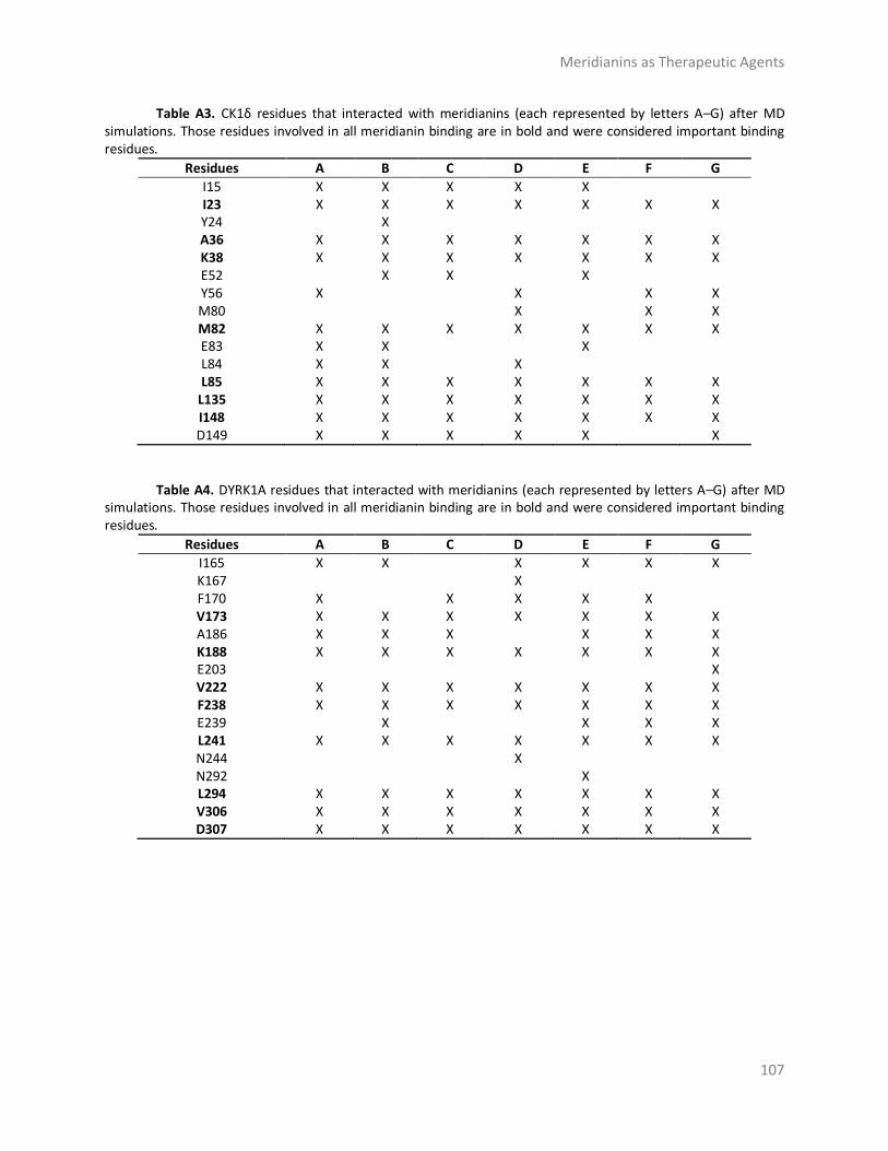
Meridianins as Therapeutic Agents
107
Table A3. CK1δ residues that interacted with meridianins (each represented by letters A–G) after MD simulations. Those residues involved in all meridianin binding are in bold and were considered important binding residues.
Residues A B C D E F G
I15 X X X X X
I23 X X X X X X X Y24
X
A36 X X X X X X X K38 X X X X X X X E52
X X
X
Y56 X
X
X X
M80
X
X X M82 X X X X X X X E83 X X
X
L84 X X
X
L85 X X X X X X X
L135 X X X X X X X I148 X X X X X X X D149 X X X X X
X
Table A4. DYRK1A residues that interacted with meridianins (each represented by letters A–G) after MD
simulations. Those residues involved in all meridianin binding are in bold and were considered important binding residues.
Residues A B C D E F G
I165 X X
X X X X K167
X
F170 X
X X X X
V173 X X X X X X X A186 X X X
X X X
K188 X X X X X X X E203
X
V222 X X X X X X X F238 X X X X X X X E239
X
X X X
L241 X X X X X X X N244
X
N292
X
L294 X X X X X X X V306 X X X X X X X D307 X X X X X X X

Meridianins as Therapeutic Agents
108
Table A5. CLK1 residues that interacted with meridianins (each represented by letters A–G) after MD simulations. Those residues involved in all meridianin binding are in bold and were considered important binding residues.
Residues A B C D E F G
L167 X X X X X X X F172 X X X
X X
V175 X X X X X X X A189 X X X X X X X K191 X X X X X
X
E206
X
X L210
X
V225 X X X X
X X
F241 X X X X
X X E242
X X X X
L243 X
X X
L244 X X X X X X X G245
X
S247
X
N293
X
L295 X X X X X X X V324 X X X X X
X
D325 X X X X X
X
Table A6. Binder columns represent those residues identified after a bibliographic and database research
and that interacted with other inhibitors. In shared columns are those residues involved with all meridianins binding per target. Residue number corresponds to each Protein Data Bank (PDB) number.
GSK3β CK1δ DYRK1A CLK1
Binders Shared Binders Shared Binders Shared Binders Shared
I62 I62 L167 L167 V70 V70 I23 I23 V173 V173 V175 V175 A83 A83 A36 A36 A189 A189 K85 K85 K38 K38 K188 K188 K191
V222 V222 V225 L132 L132 M82 M82 F238 F238 F241 V135 L85 L85 L241 L241 L244 L244 L188 L135 L135 L294 L294 L295 L295
I148 I148 V306 V306 V324 D200 D200 D149 D307 D307

Meridianins as Therapeutic Agents
109
Table A7. Residues involved in all meridianins binding to GSK3β, CK1δ, DYRK1A and CLK1. Residue number corresponds to each PDB number.
GSK3β CK1δ DYRK1A CLK1
I62 I15 I165 L167 K167
F67 F170 F172 V70 I23 V173 V175
Y24 A83 A36 A186 A189 K85 K38 K188 K191 E97
E52 E203 E206 Y56 L210 M80
V110 V222 V225 L132 M82 F238 F241 D133 E83 E239 E242 Y134 L84 L243 V135 L85 L241 L244 T138 N244 G245 Q185 S247
N292 N293 L188 L135 L294 L295
I148 V306 V324 D200 D149 D307 D325

Meridianins as Therapeutic Agents
110
Table A8. Summary of classical rigid docking of the best model selected per meridianin A–G and the derived compounds 1, 2 and 3, against others protein kinases and one non-kinase (IDH1).
GSK3β CK1δ DYRK1A CLK1 FGFR1 PRKACA
Binding Energy
Binding Energy
Binding Energy
Binding Energy
Binding Energy
Binding Energy
(kcal/mol) (kcal/mol) (kcal/mol) (kcal/mol) (kcal/mol) (kcal/mol)
R0/R1 R0/R1 R0/R1 R0/R1 R0/R1 R0/R1 A −7.3/−7.3 A −6.9/−6.9 A −7.4/−7.3 A −8.9/−8.9 A −7.1/−7.1 A −8.5/−8.5 B −7.3/−7.2 B −6.4/−6.4 B −7.7/−6.9 B −8.5/−8.5 B −6.7/−6.7 B −8.5/−8.5 C −7.6/−7.6 C −6.9/−6.9 C −8.2/−8.2 C −8.5/−8.5 C −7.1/−7.1 C −9.0/−9.0 D −7.7/−7.7 D −7.0/−7.0 D −7.9/−7.9 D −8.6/−8.6 D −7.1/−7.1 D −8.8/−8.8 E −7.3/−7.3 E −7.0/−7.0 E −7.5/−7.4 E −9.0/−8.8 E −7.4/−7.4 E −7.6/−7.6 F −7.9/−7.9 F −7.2/−7.3 F −8.0/−7.8 F −8.7/−8.7 F −7.3/−7.3 F −8.5/−8.5 G −7.3/−7.3 G −6.8/−6.8 G −8.1/−8.1 G −9.1/−9.1 G −7.1/−7.1 G −8.6/−8.6 1 −7.6/−7.6 1 −7.0/−7.0 1 −8.1/−8.1 1 −9.2/−9.2 1 −7.1/−7.1 1 −8.2/−8.2 2 −7.7/−7.7 2 −7.1/−7.1 2 −8.2/−8.2 2 −7.4/−7.4 2 −6.2/−6.2 2 −8.3/−8.3 3 −8.0/−8.0 3 −7.3/−7.3 3 −7.9/−7.9 3 −7.8/−7.8 3 −6.4/−6.4 3 −8.0/−8.0
HK2 MAP2K1 PIK3CG PRKG1 IDH1 PKN1
Binding Energy
Binding Energy
Binding Energy
Binding Energy
Binding Energy
Binding Energy
(kcal/mol) (kcal/mol) (kcal/mol) (kcal/mol) (kcal/mol) (kcal/mol)
R0/R1 R0/R1 R0/R1 R0/R1 R0/R1 R0/R1
A −7.1/−7.1 A −7.6/−7.6 A −6.8/−6.8 A −6.3/−6.3 A −5.8/−5.8 A −7.8/−7.8 B −6.6/−6.6 B −7.4/−7.4 B −7.3/−7.3 B −6.5/−6.5 B −6.3/−6.3 B −7.7/−7.7 C −7.0/−7.0 C −7.2/−7.2 C −7.0/−7.0 C −6.6/−6.6 C −5.8/−5.8 C −8.1/−8.1 D −6.6/−6.6 D −7.4/−7.4 D −7.8/−7.8 D −6.9/−6.9 D −5.8/−5.8 D −7.3/−7.3 E −6.8/−6.8 E −6.7/−6.7 E −7.1/−7.1 E −6.3/−6.3 E −5.6/−5.6 E −7.2/−7.2 F −6.9/−6.9 F −7.5/−7.5 F −7.2/−7.2 F −6.8/−6.8 F −6.0/−6.0 F −7.8/−7.8 G −6.9/−6.9 G −7.5/−7.5 G −7.3/−7.3 G −6.5/−6.5 G −6.3/−6.3 G −7.9/−7.9 1 −6.9/−6.9 1 −7.4/−7.4 1 −7.7/−7.7 1 −6.4/−6.4 1 −5.8/−5.8 1 −7.4/−7.4 2 −8.1/−8.1 2 −7.5/−7.5 2 −7.3/−7.3 2 −5.3/−5.3 2 −6.1/−6.1 2 −7.7/−7.7 3 −7.1/−7.1 3 −7.2/−7.2 3 −7.3/−7.3 3 −6.1/−6.1 3 −6.0/−6.0 3 −7.7/−7.7
All energy values are kcal/mol. To avoid false positives, each docking calculation was performed twice (R0 and R1).

Meridianins as Therapeutic Agents
111
Table A9. Summary of ADMET properties of meridianins (A to G) and the derived compounds extracted from the literature (1–3).
AB
SOR
PTI
ON
LogS Caco2
Permeability
Caco2 * Permeability
Intestinal
Absorption
Skin Permeability
A −4.18 A H A 0.99 A 93.38% A −2.76 B −5.02 B H B 1.07 B 92.22% B −2.76 C −5.55 C H C 0.95 C 91.77% C −2.92 D −5.55 D H D 0.95 D 92.715 D −2.91 E −5.04 E H E 0.98 E 90.98% E −2.74 F −6.16 F H F 0.98 F 91.49% F −2.92 G −4.51 G H G 0.86 G 93.44% G −2.90 1 −4.18 1 OAD 1 0.93 1 91.41% 1 −2.90 2 −5.02 2 OAD 2 0.8 2 89.89% 2 −2.884 3 −5.55 3 H 3 0.819 3 91.04% 3 −2.895
P-Glycoprotein substrate P-Glycoprotein I/II
Inhibitor
DIS
TRIB
UTI
ON
LogP BBB PPB
A Yes A No A 1.53 A No A >90% B Yes B No B 2.39 B No B >90% C Yes C No C 3.10 C No C >90% D Yes D No D 3.10 D No D >90% E Yes E No E 2.40 E No E >90% F Yes F No F 3.58 F No F >90% G Yes G No G 2.44 G No G <50% 1 Yes 1 No 1 3.40 1 No 1 >90% 2 Yes 2 No 2 3.40 2 No 2 >90% 3 Yes 3 No 3 3.10 3 No 3 >90%
VDss CNS
Permeability
MET
AB
OLI
SM
CYP450
Metabolism *
EXC
RET
ION
Total
Clearance
Renal OCT2
Substrate
A 0.25 A −2.92 A Yes A 0.57 A No B 0.24 B −2.92 B Yes B 0.30 B No C −0.06 C −2.81 C Yes C 0.09 C No D −0.01 D −2.82 D Yes D 0.14 D No E 0.22 E −2.93 E Yes E 0.15 E No F 0.07 F −2.82 F Yes F −0.19 F No G −0.10 G −2.12 G Yes G 0.71 G No 1 −0.02 1 −2.83 1 Yes 1 −0.07 1 No 2 −0.09 2 −1.88 2 Yes 2 −0.092 2 No 3 −0.09 3 −1.99 3 Yes 3 0.132 3 No
Caco2 permeability is calculated using proprietary ML model and Caco2 * with the pkCSM webserver, as explained in the methods section. CYP450 metabolism * specific values of interaction with different CYP450 isoforms are listed in Table A11. BBB: blood brain Barrier, PPB: protein-protein binding, VDss: steady state volume of distribution, CNS: central nervous system, OCT2: organic cation transported 2.

Meridianins as Therapeutic Agents
112
Table A10. Summary of toxicity properties of meridianins A–G and the three derived compounds extracted from the literatures (1–3).
MRTD AMES Toxicity hERG I/II Inhibition Hepatotoxicity Skin Sensitisation
A 0.503 A No A No A No A No B 0.584 B No B No B Yes B No C −0.107 C Yes C No C No C No D −0.095 D Yes D No D No D No E 0.589 E No E No E No E No F −0.088 F Yes F No F Yes F No G −0.086 G Yes G No G No G No 1 −0.068 1 Yes 1 No 1 No 1 No 2 −0.038 2 Yes 2 No 2 No 2 No 3 −0.058 3 Yes 3 No 3 No 3 No
MRTD: maximum recommended tolerated dose, hERG: human ether-a-go-go gene.
Table A11. Summary of specific values of interaction with different CYP450 isoforms properties of
meridianins A–G and the three derived compounds (1–3).
CYP2D6 Substrate
CYP3A4 Substrate
CYP1A2 Inhibitor
CYP2C19 Inhibitor
CYP2C9 Inhibitor
CYP2D6 Inhibitor
CYP3A4 Inhibitor
A No A No A Yes A No A No A No A No B No B No B Yes B No B No B No B No C No C No C Yes C No C No C No C No D No D No D Yes D No D No D No D No E No E No E Yes E No E No E No E No F No F No F Yes F Yes F No F No F No G No G No G Yes G No G No G No G No 1 No 1 No 1 Yes 1 Yes 1 No 1 No 1 Yes 2 No 2 Yes 2 Yes 2 Yes 2 No 2 No 2 No 3 No 3 No 3 Yes 3 No 3 No 3 No 3 Yes
CYP: Cytochrome

Meridianins as Therapeutic Agents
113
References
1. Paul, S.M.; Mytelka, D.S.; Dunwiddie, C.T.; Persinger, C.C.; Munos, B.H.; Lindborg, S.R.; Schacht,
A.L. How to improve R&D productivity: The pharmaceutical industry’s grand challenge. Nat. Rev.
Drug Discov. 2010, 9, 203.
2. Leelananda, S.P.; Lindert, S. Computational methods in drug discovery. Beilstein J. Org. Chem.
2016, 12, 2694–2718.
3. Ou-Yang, S.-S.; Lu, J.-Y.; Kong, X.-Q.; Liang, Z.-J.; Luo, C.; Jiang, H. Computational drug discovery.
Acta Pharmacol. Sin. 2012, 33, 1131–1140.
4. Sliwoski, G.; Kothiwale, S.; Meiler, J.; Lowe, E.W. Computational Methods in Drug Discovery.
Pharmacol. Rev. 2014, 66, 334–395.
5. Meng, X.-Y.; Zhang, H.-X.; Mezei, M.; Cui, M. Molecular docking: A powerful approach for
structure-based drug discovery. Curr. Comput. Aided Drug Des. 2011, 7, 146–157.
6. Durrant, J.D.; McCammon, J.A. Molecular dynamics simulations and drug discovery. BMC Biol.
2011, 9, 71.
7. Yu, W.; Mackerell, A.D. Computer-Aided Drug Design Methods. Methods Mol. Biol. 2017, 1520,
93–94.
8. Acharya, C.; Coop, A.; Polli, J.E.; Mackerell, A.D., Jr. Recent advances in ligand-based drug design:
relevance and utility of the conformationally sampled pharmacophore approach. Curr. Comput.
Aided Drug Des. 2011, 7, 10–22.
9. Li, J.W.-H.; Vederas, J.C. Drug Discovery and Natural Products: End of an Era or an Endless
Frontier? Science 2009, 325, 161–165.
10. Harvey, A.L.; Edrada-Ebel, R.; Quinn, R.J. The re-emergence of natural products for drug
discovery in the genomics era. Nat. Rev. Drug Discov. 2015, 14, 111–129.
11. Newman, D.J.; Cragg, G.M. Natural Products as Sources of New Drugs from 1981 to 2014. J. Nat.
Prod. 2016, 79, 629–661.
12. Prachayasittikul, V.; Worachartcheewan, A.; Shoombuatong, W.; Songtawee, N.; Simeon, S.;
Prachayasittikul, V.; Nantasenamat, C. Computer-Aided Drug Design of Bioactive Natural
Products. Curr. Top. Med. Chem. 2015, 15, 1780–1800.
13. Rodrigues, T.; Reker, D.; Schneider, P.; Schneider, G. Counting on natural products for drug
design. Nat. Chem. 2016, 8, 531–541.
14. Martin, L.; Latypova, X.; Wilson, C.M.; Magnaudeix, A.; Perrin, M.L.; Yardin, C.; Terro, F. Tau
protein kinases: Involvement in Alzheimer’s disease. Ageing Res. Rev. 2013, 12, 289–309.
15. Kolarova, M.; García-Sierra, F.; Bartos, A.; Ricny, J.; Ripova, D.; Ripova, D. Structure and
Pathology of Tau Protein in Alzheimer Disease. Int. J. Alzheimers Dis. 2012, 2012, 1–13.
16. Citron, M. Alzheimer’s disease: strategies for disease modification. Nat. Rev. Drug Discov. 2010,
9, 387–398.
17. Tell, V.; Hilgeroth, A. Recent developments of protein kinase inhibitors as potential AD

Meridianins as Therapeutic Agents
114
therapeutics. Front. Cell. Neurosci. 2013, 7, 189.
18. Dolan, P.J.; Johnson, G.V.W. The role of tau kinases in Alzheimer’s disease. Curr. Opin. Drug
Discov. Dev. 2010, 13, 595–603.
19. Lucke-Wold, B.P.; Turner, R.C.; Logsdon, A.F.; Simpkins, J.W.; Alkon, D.L.; Smith, K.E.; Chen, Y.-
W.; Tan, Z.; Huber, J.D.; Rosen, C.L. Common mechanisms of Alzheimer’s disease and ischemic
stroke: The role of protein kinase C in the progression of age-related neurodegeneration. J.
Alzheimers Dis. 2015, 43, 711–724.
20. Montaser, R.; Luesch, H. Marine natural products: A new wave of drugs? Future Med. Chem.
2011, 3,
1475–1489.
21. Blunt, J.W.; Copp, B.R.; Keyzers, R.A.; Munro, M.H.G.; Prinsep, M.R. Marine natural products.
Nat. Prod. Rep. 2017, 34, 235–294.
22. Kiuru, P.; D’Auria, M.; Muller, C.; Tammela, P.; Vuorela, H.; Yli-Kauhaluoma, J. Exploring Marine
Resources for Bioactive Compounds. Planta Med. 2014, 80, 1234–1246.
23. Molinski, T.F.; Dalisay, D.S.; Lievens, S.L.; Saludes, J.P. Drug development from marine natural
products. Nat. Rev. Drug Discov. 2009, 8, 69–85.
24. The Nagoya Protocol on Access and Benefit-Sharing. Available online:
https://www.cbd.int/abs/about/
default.shtml/ (accessed on 16 May 2017).
25. Bharate, S.B.; Yadav, R.R.; Battula, S.; Vishwakarma, R.A. Meridianins: Marine-Derived Potent
Kinase Inhibitors. Mini-Rev. Med. Chem. 2012, 12, 618–631.
26. Núñez-Pons, L.; Carbone, M.; Vázquez, J.; Rodríguez, J.; Nieto, R.M.; Varela, M.M.; Gavagnin, M.;
Avila, C. Natural Products from Antarctic Colonial Ascidians of the Genera Aplidium and
Synoicum: Variability and Defensive Role. Mar. Drugs 2012, 10, 1741–1764.
27. Traxler, P.; Furet, P. Strategies toward the Design of Novel and Selective Protein Tyrosine Kinase
Inhibitors. Pharmacol. Ther. 1999, 82, 195–206.
28. Huang, D.; Zhou, T.; Lafleur, K.; Nevado, C.; Caflisch, A. Kinase selectivity potential for inhibitors
targeting the ATP binding site: A network analysis. Bioinformatics 2010, 26, 198–204.
29. McGregor, M.J. A Pharmacophore Map of Small Molecule Protein Kinase Inhibitors. J. Chem. Inf.
Model. 2007, 47, 2374–2382.
30. Ebrahim, H.; El Sayed, K. Discovery of Novel Antiangiogenic Marine Natural Product Scaffolds.
Mar. Drugs 2016, 14, 57.
31. Feng, L.; Geisselbrecht, Y.; Blanck, S.; Wilbuer, A.; Atilla-Gokcumen, G.E.; Filippakopoulos, P.;
Kräling, K.; Celik, M.A.; Harms, K.; Maksimoska, J.; et al. Structurally sophisticated octahedral
metal complexes as highly selective protein kinase inhibitors. J. Am. Chem. Soc. 2011, 133,
5976–5986.
32. Giraud, F.; Alves, G.; Debiton, E.; Nauton, L.; Théry, V.; Durieu, E.; Ferandin, Y.; Lozach, O.;
Meijer, L.; Anizon, F.; et al. Synthesis, Protein Kinase Inhibitory Potencies, and in vitro

Meridianins as Therapeutic Agents
115
Antiproliferative Activities of Meridianin Derivatives. J. Med. Chem. 2011, 54, 4474–4489.
33. Wang, G.; Zhu, W. Molecular docking for drug discovery and development: A widely used
approach but far from perfect. Future Med. Chem. 2016, 8, 1707–1710.
34. De Vivo, M.; Masetti, M.; Bottegoni, G.; Cavalli, A. The Role of Molecular Dynamics and Related
Methods in Drug Discovery. J. Med. Chem. 2016, 59, 4035–4061.
35. De Vivo, M.; Cavalli, A. Recent advances in dynamic docking for drug discovery. Wiley
Interdiscip. Rev. Comput. Mol. Sci. 2017, 7, e1320.
36. Aravindhan, G.; Coote, M.L.; Barakat, K. Molecular dynamics-driven drug discovery: Leaping
forward with confidence. Drug Discov. Today 2017, 22, 249–269.
37. Pagadala, N.S.; Syed, K.; Tuszynski, J. Software for molecular docking: A review. Biophys. Rev. 2017,
9, 91–102.
38. Shin, W.-H.; Zhu, X.; Bures, M.; Kihara, D. Three-Dimensional Compound Comparison Methods
and Their Application in Drug Discovery. Molecules 2015, 20, 12841–12862.
39. Muegge, I.; Mukherjee, P. An overview of molecular fingerprint similarity search in virtual
screening. Expert Opin. Drug Discov. 2016, 11, 137–148.
40. Alonso, N.; Caamaño, O.; Romero-Duran, F.J.; Luan, F.; Cordeiro, M.N.D.S.; Yañez, M.; González-
Díaz, H.; García-Mera, X. Model for High-Throughput Screening of Multitarget Drugs in Chemical
Neurosciences: Synthesis, Assay, and Theoretic Study of Rasagiline Carbamates. ACS Chem.
Neurosci. 2013, 4, 1393–1403.
41. Simpraga, S.; Alvarez-Jimenez, R.; Mansvelder, H.D.; Van Gerven, J.M.A.; Groeneveld, G.J.; Poil,
S.-S.; Linkenkaer-Hansen, K. EEG machine learning for accurate detection of cholinergic
intervention and Alzheimer’s disease. Sci, Rep. 2017, 7, 5775.
42. Cummings, J.; Aisen, P.S.; DuBois, B.; Frölich, L.; Jack, C.R.; Jones, R.W.; Morris, J.C.; Raskin, J.;
Dowsett, S.A.; Scheltens, P. Drug development in Alzheimer’s disease: The path to 2025.
Alzheimers Res. Ther. 2016, 8, 39.
43. Solomon, K.A.; Sundararajan, S.; Abirami, V. QSAR Studies on N-aryl Derivative Activity Towards
Alzheimer’s Disease. Molecules 2009, 14, 1448–1455.
44. Gopi Mohan, C.; Gupta, S. QSAR Models towards Cholinesterase Inhibitors for the Treatment of
Alzheimer’s Disease. In Oncology: Breakthroughs in Research and Practice; IGI Global: Hershey,
PA, USA, 2016.
45. PharmaSea. Available online: http://www.pharma-sea.eu/ (accessed on 2 September 2017).
46. MaCuMBA. Available online: https://www.macumbaproject.eu/ (accessed on 2 September
2017).
47. SeaBiotech. Available online: http://spider.science.strath.ac.uk/seabiotech/index.php (accessed
on 2 September 2017).
48. BlueGenics. Available online: http://www.bluegenics.eu/cms/ (accessed on 2 September 2017).
49. MicroB3. Available online: https://www.microb3.eu/ (accessed on 2 September 2017).
50. Jain, P.; Karthikeyan, C.; Moorthy, N.S.; Waiker, D.; Jain, A.; Trivedi, P. Human CDC2-Like Kinase 1
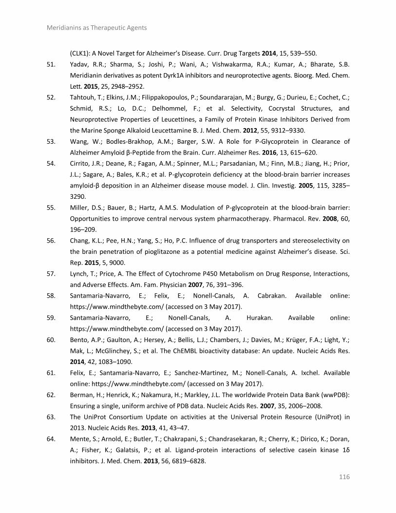
Meridianins as Therapeutic Agents
116
(CLK1): A Novel Target for Alzheimer’s Disease. Curr. Drug Targets 2014, 15, 539–550.
51. Yadav, R.R.; Sharma, S.; Joshi, P.; Wani, A.; Vishwakarma, R.A.; Kumar, A.; Bharate, S.B.
Meridianin derivatives as potent Dyrk1A inhibitors and neuroprotective agents. Bioorg. Med. Chem.
Lett. 2015, 25, 2948–2952.
52. Tahtouh, T.; Elkins, J.M.; Filippakopoulos, P.; Soundararajan, M.; Burgy, G.; Durieu, E.; Cochet, C.;
Schmid, R.S.; Lo, D.C.; Delhommel, F.; et al. Selectivity, Cocrystal Structures, and
Neuroprotective Properties of Leucettines, a Family of Protein Kinase Inhibitors Derived from
the Marine Sponge Alkaloid Leucettamine B. J. Med. Chem. 2012, 55, 9312–9330.
53. Wang, W.; Bodles-Brakhop, A.M.; Barger, S.W. A Role for P-Glycoprotein in Clearance of
Alzheimer Amyloid β-Peptide from the Brain. Curr. Alzheimer Res. 2016, 13, 615–620.
54. Cirrito, J.R.; Deane, R.; Fagan, A.M.; Spinner, M.L.; Parsadanian, M.; Finn, M.B.; Jiang, H.; Prior,
J.L.; Sagare, A.; Bales, K.R.; et al. P-glycoprotein deficiency at the blood-brain barrier increases
amyloid-β deposition in an Alzheimer disease mouse model. J. Clin. Investig. 2005, 115, 3285–
3290.
55. Miller, D.S.; Bauer, B.; Hartz, A.M.S. Modulation of P-glycoprotein at the blood-brain barrier:
Opportunities to improve central nervous system pharmacotherapy. Pharmacol. Rev. 2008, 60,
196–209.
56. Chang, K.L.; Pee, H.N.; Yang, S.; Ho, P.C. Influence of drug transporters and stereoselectivity on
the brain penetration of pioglitazone as a potential medicine against Alzheimer’s disease. Sci.
Rep. 2015, 5, 9000.
57. Lynch, T.; Price, A. The Effect of Cytochrome P450 Metabolism on Drug Response, Interactions,
and Adverse Effects. Am. Fam. Physician 2007, 76, 391–396.
58. Santamaria-Navarro, E.; Felix, E.; Nonell-Canals, A. Cabrakan. Available online:
https://www.mindthebyte.com/ (accessed on 3 May 2017).
59. Santamaria-Navarro, E.; Nonell-Canals, A. Hurakan. Available online:
https://www.mindthebyte.com/ (accessed on 3 May 2017).
60. Bento, A.P.; Gaulton, A.; Hersey, A.; Bellis, L.J.; Chambers, J.; Davies, M.; Krüger, F.A.; Light, Y.;
Mak, L.; McGlinchey, S.; et al. The ChEMBL bioactivity database: An update. Nucleic Acids Res.
2014, 42, 1083–1090.
61. Felix, E.; Santamaria-Navarro, E.; Sanchez-Martinez, M.; Nonell-Canals, A. Ixchel. Available
online: https://www.mindthebyte.com/ (accessed on 3 May 2017).
62. Berman, H.; Henrick, K.; Nakamura, H.; Markley, J.L. The worldwide Protein Data Bank (wwPDB):
Ensuring a single, uniform archive of PDB data. Nucleic Acids Res. 2007, 35, 2006–2008.
63. The UniProt Consortium Update on activities at the Universal Protein Resource (UniProt) in
2013. Nucleic Acids Res. 2013, 41, 43–47.
64. Mente, S.; Arnold, E.; Butler, T.; Chakrapani, S.; Chandrasekaran, R.; Cherry, K.; Dirico, K.; Doran,
A.; Fisher, K.; Galatsis, P.; et al. Ligand-protein interactions of selective casein kinase 1δ
inhibitors. J. Med. Chem. 2013, 56, 6819–6828.

Meridianins as Therapeutic Agents
117
65. Lill, M.A. Efficient Incorporation of Protein Flexibility and Dynamics into Molecular Docking
Simulations. Biochemistry 2011, 50, 6157–6169.
66. Genheden, S.; Ryde, U. The MM/PBSA and MM/GBSA methods to estimate ligand-binding
affinities. Expert Opin. Drug Discov. 2015, 441, 1–13.
67. Alonso, H.; Bliznyuk, A.A.; Gready, J.E. Combining docking and molecular dynamic simulations in
drug design. Med. Res. Rev. 2006, 26, 531–568.
68. Felix, E.; Santamaría-Navarro, E.; Sanchez-Martinez, M.; Nonell-Canals, A. Itzamna. Available
online: https://www.mindthebyte.com/ (accessed on 3 May 2017).
69. Felix, E.; Nonell-Canals, A. Kin. Available online: https://www.mindthebyte.com/ (accessed on 3
May 2017).
70. Phillips, J.C.; Braun, R.; Wang, W.; Gumbart, J.; Tajkhorshid, E.; Villa, E.; Chipot, C.; Skeel, R.D.;
Kalé, L.; Schulten, K. Scalable molecular dynamics with NAMD. J. Comput. Chem. 2005, 26,
1781–802.
71. Lindorff-Larsen, K.; Piana, S.; Palmo, K.; Maragakis, P.; Klepeis, J.L.; Dror, R.O.; Shaw, D.E.
Improved side-chain torsion potentials for the Amber ff99SB protein force field. Proteins 2010,
78, 1950–1958.
72. Wang, J.; Wolf, R.M.; Caldwell, J.W.; Kollman, P.A.; Case, D.A. Development and testing of a
general amber force field. J. Comput. Chem. 2004, 25, 1157–1174.
73. Martín-García, F.; Papaleo, E.; Gomez-Puertas, P.; Boomsma, W.; Lindorff-Larsen, K. Comparing
Molecular Dynamics Force Fields in the Essential Subspace. PLoS ONE 2015, 10, e0121114.
74. Lindorff-Larsen, K.; Maragakis, P.; Piana, S.; Eastwood, M.P.; Dror, R.O.; Shaw, D.E. Systematic
validation of protein force fields against experimental data. PLoS ONE 2012, 7, 1–6.
75. Wang, J.; Wang, W.; Kollman, P.A.; Case, D.A. Antechamber, An Accessory Software Package For
Molecular Mechanical Calculations. J. Comput. Chem. 2005, 25, 1157–1174.
76. Case, D.A.; Cheatham, T.E.; Darden, T.; Gohlke, H.; Luo, R.; Merz, K.M.; Onufriev, A.; Simmerling,
C.; Wang, B.; Woods, R.J. The Amber biomolecular simulation programs. J. Comput. Chem. 2005,
26, 1668–1688.
77. Jorgensen, W.L.; Jenson, C. Temperature dependence of TIP3P, SPC, and TIP4P water from NPT
Monte Carlo simulations: Seeking temperatures of maximum density. J. Comput. Chem. 1998,
19, 1179–1186.
78. Andersen, H.C. Rattle: A “velocity” version of the shake algorithm for molecular dynamics
calculations. J. Comput. Phys. 1983, 52, 24–34.
79. Rastelli, G.; Degliesposti, G.; Del Rio, A.; Sgobba, M. Binding Estimation after Refinement, a New
Automated Procedure for the Refinement and Rescoring of Docked Ligands in Virtual Screening.
Chem. Biol. Drug Des. 2009, 73, 283–286.
80. Miller, B.R., III; McGee, T.D., Jr.; Swails, J.M.; Homeyer, N.; Gohlke, H.; Roitberg, A.E. MMPBSA.
py: An efficient program for end-state free energy calculations. J. Chem. Theory Comput. 2012,
8, 3314–3321.

Meridianins as Therapeutic Agents
118
81. Mente, S.; Arnold, E.; Butler, T.; Chakrapani, S.; Chandrasekaran, R.; Cherry, K.; Dirico, K.; Doran,
A.; Fisher, K.; Galatsis, P.; et al. Ligand-protein interactions of selective casein kinase 1δ
inhibitors. J. Med. Chem. 2013, 56, 6819–6828.
82. Halekotte, J.; Witt, L.; Ianes, C.; Krüger, M.; Bührmann, M.; Rauh, D.; Pichlo, C.; Brunstein, E.;
Luxenburger, A.; Baumann, U.; et al. Optimized 4,5-Diarylimidazoles as Potent/Selective
Inhibitors of Protein Kinase CK1δ and Their Structural Relation to p38α MAPK. Molecules 2017,
22, 522.
83. Fedorov, O.; Huber, K.; Eisenreich, A.; Filippakopoulos, P.; King, O.; Bullock, A.N.; Szklarczyk, D.;
Jensen, L.J.; Fabbro, D.; Trappe, J.; et al. Specific CLK Inhibitors from a Novel Chemotype for
Regulation of Alternative Splicing. Chem. Biol. 2011, 18, 67–76.
84. Vidal, D.; Nonell-Canals, A. ADMET Models. Available online: https://www.mindthebyte.com/
(accessed on 3 May 2017).
85. Pires, D.E.V.; Blundell, T.L.; Ascher, D.B. pkCSM: Predicting small-molecule pharmacokinetic and
toxicity properties using graph-based signatures. J. Med. Chem. 2015, 58, 4066–4072.
86. Muehlbacher, M.; Spitzer, G.M.; Liedl, K.R.; Kornhuber, J. Qualitative prediction of blood–brain
barrier permeability on a large and refined dataset. J. Comput. Aided Mol. Des. 2011, 25, 1095–
1106.
87. Zhu, X.-W.; Sedykh, A.; Zhu, H.; Liu, S.-S.; Tropsha, A. The Use of Pseudo-Equilibrium Constant
Affords Improved QSAR Models of Human Plasma Protein Binding. Pharm. Res. 2013, 30, 1790–
1798.
88. Huuskonen, J. Estimation of Aqueous Solubility for a Diverse Set of Organic Compounds Based
on Molecular Topology. J. Chem. Inf. Comput. Sci. 2000, 40, 773–777.
89. Yuan, S.; Chan, H.C.S.; Hu, Z. Using PyMOL as a platform for computational drug design. Wiley
Interdiscip. Rev. Comput. Mol. Sci. 2017, 7, e1298.
90. Salentin, S.; Schreiber, S.; Haupt, V.J.; Adasme, M.F.; Schroeder, M. PLIP: Fully automated
protein–ligand interaction profiler. Nucleic Acids Res. 2015, 43, W443–W447.
91. Prudhomme, M.; Rossignol, E.; Youssef, A.; Anizon, F.; Moreau, P.; Fabbro, D.; Cohen, P.
Aminopyrimidylindoles structurally related to meridianins as kinase inhibitors. Cancer Res. 2014,
68.
92. Kingwell, K. Drug delivery: New targets for drug delivery across the BBB. Nat. Rev. Drug Discov.
2016, 15, 84–85.
93. Banks, W.A. From blood–brain barrier to blood–brain interface: New opportunities for CNS drug
delivery. Nat. Rev. Drug Discov. 2016, 15, 275–292.
94. Saraiva, C.; Praça, C.; Ferreira, R.; Santos, T.; Ferreira, L.; Bernardino, L. Nanoparticle-mediated
brain drug delivery: Overcoming blood–brain barrier to treat neurodegenerative diseases. J.
Control. Release 2016, 235, 34–47.
Meridianins as Therapeutic Agents
119
Section II
Chapter 3
Kororamides, convolutamines, and indole derivatives as
possible tau and dual-specificity kinase inhibitors for
Alzheimer’s disease: A computational study

Meridianins as Therapeutic Agents
120
121
Chapter 3
Kororamides, convolutamines, and indole derivatives as possible tau and dual specificity kinases inhibitors for Alzheimer’s Disease.
Laura Llorach-Pares 1,2, Alfons Nonell-Canals 2, Conxita Avila1,*, and Melchor Sánchez-Martínez2,*
1. Department of Evolutionary Biology, Ecology and Environmental Sciences, Faculty of Biology and
Biodiversity Research Institute (IRBio), Universitat de Barcelona, 08028 Barcelona, Catalonia.
2. Mind the Byte S.L., 08028 Barcelona, Catalonia, Spain.
Published in: Marine Drugs 16(10), 386 (2018) doi: 10.3390/md16100386

Kororamides, Convolutamines and Indole Derivatives
122
Abstract
Alzheimer’s disease (AD) is becoming one of the most disturbing health and socioeconomic
problems nowadays, as it is a neurodegenerative pathology with no treatment expected to grow further
due to population ageing. Current treatments for AD produce only a modest amelioration of symptoms,
although there is a constant ongoing research of new therapeutic strategies oriented to improve them,
and even to completely cure the disease. A principal feature of AD is the presence of neurofibrillary
tangles (NFT) induced by the aberrant phosphorylation of the microtubule-associated protein tau in the
brains of affected individuals. Glycogen synthase kinase-3 beta (GSK3β), casein kinase 1 delta (CK1δ),
dual specificity tyrosine phosphorylation regulated kinase 1A (DYRK1A), and dual specificity kinases
cdc2-like kinase 1 (CLK1) have been identified as the principal proteins involved in this process. Because
of that, the inhibition of these kinases has been proposed as a plausible therapeutic strategy to fight AD.
In this study, we computationally tested the inhibitory activity of different marine natural compounds,
as well as newly designed molecules from their scaffolds, over the mentioned protein kinases, finding
some new possible inhibitors with potential therapeutical application.
Keywords: Meridianins; Kororamide A-B; Convolutamine I-J; Indole scaffold; Computer-Aided Drug
Design; Alzheimer’s disease; GSK3β; CK1δ; DYRK1A; CLK1.

Kororamides, Convolutamines and Indole Derivatives
123
Resum
La malaltia d'Alzheimer (MA) s'està convertint en una de les malalties més inquietants i en un
problema socioeconòmic en l'actualitat, ja que és una patologia neurodegenerativa que no té
tractament, i s'espera que la seva afectació augmenti encara més a causa de l'envelliment de la
població. Els tractaments actuals per a la MA només produeixen una modesta millora dels símptomes,
tot i que hi ha una constant i permanent investigació de noves estratègies terapèutiques orientades a
millorar aquests símptomes, i fins i tot, per curar completament la malaltia. Una característica principal
de la MA és la presència de cabdells neurofibril·lars, induïts per una aberrant fosforilació de la proteïna
tau associada als microtúbuls, que es troben presents en el cervell dels individus afectats. La glycogen
synthase kinase-3 beta (GSK3β) i la casein kinase 1 delta (CK1δ, CK1D o KC1D), així com les quinases de
doble especificitat, com la dual specificity tyrosine phosphorylation regulated kinase 1 (DYRK1A) i les
quinases cdc2-like (CLK1), s'han identificat com les principals proteïnes implicades en aquest procés
d’hiperfosforilació. Per això, la inhibició d'aquestes quinases s'ha proposat com una estratègia
terapèutica plausible per combatre la MA. En aquest estudi, hem estudiat computacionalment l'activitat
inhibitòria de diferents compostos naturals d’origen marí, així com molècules dissenyades a partir dels
seus esquelets, sobre les esmentades proteïnes quinases, trobant alguns nous possibles inhibidors amb
potencial aplicació terapèutica.
Paraules clau: Meridianines; kororamide A-B; convolutamine I-J; esquelets indòlics; disseny de fàrmacs
assistit per ordinador; malaltia d’Alzheimer; GSK3β; CK1δ; DYRK1A; CLK1.

Kororamides, Convolutamines and Indole Derivatives
124
Introduction
Constituting about 2% of all human genes, protein kinases are an important family of enzymes
with a critical role in signal transduction pathway by modification of substrate activity. They are also
responsible to control different aspects of cell functions by its phosphorylation activity, which plays a
critical role in intracellular communication during development, and in the function of the nervous and
immune systems [1]. Due to that, kinases are related with many diseases such as Alzheimer’s Disease
(AD) or Amyotrophic Lateral Sclerosis (ALS), among others. AD, the neurodegenerative pathology that is
considered to represent the most common type of dementia (60-80% of the total cases), is
characterized by memory deterioration and modification of cognitive abilities. Alzheimer’s pathologies
are associated with the presence of senile plaques (SP), mainly composed by Beta-Amyloid (Aβ)
peptides, and neurofibrillary tangles (NFT), that are intraneuronal aggregations principally composed of
abnormal phosphorylated tau protein. Tau is a soluble microtubule-binding protein and is
hyperphosphorylated in AD. Tau phosphorylation is regulated by a balance between tau kinase and
phosphatase activities. Anti-phosphorylation strategies (kinase inhibitors) aim to inhibit these processes
of aggregation and the formation of NFT [2–4]. The above mentioned evidences may suggest that one of
the key strategies to prevent tau phosphorylation and thus, combat AD, could be the inhibition of the
protein kinases involved in the tau phosphorylation pathway [4].
Despite the catalytic subunits of many protein kinases are highly conserved, there are several
differences between them that allow to classify protein kinases into subfamilies: 1) Protein Kinases (EC
2.7.10); 2) Serine-Threonine protein kinases (EC 2.7.11); 3) Dual-specificity kinases (those acting on
Ser/Thr and Tyr residues) (EC 2.7.12); 4) Protein-histidine kinases (EC 2.7.13); 5) Protein-Arginine kinases
(EC 2.7.11.14) and 6) other protein kinases (EC 2.7.99), that can be also divided into sub-subfamilies,
such as tau protein kinase (EC 2.7.11.26) and dual specificity kinase (EC 2.7.12.1). The main relevant
protein kinases involved in tau phosphorylation belong to the sub-subfamilies tau protein kinase and
dual specificity kinases. glycogen synthase kinase-3 beta (GSK3β) and casein kinase 1 delta (CK1δ) are
tau protein kinases, while dual specificity tyrosine phosphorylation regulated kinase 1A (DYRK1A) and
cdc2-like kinase 1 (CLK1) are dual specificity kinases. Each of them has different roles regarding AD
pathology. For GSK3β several authors suggest its link between Aβ and tau pathology, and in AD patients
it has been co-localized with NFT. GSK3β is suggested to phosphorylate and hipper-phosphorylate tau,
while increasing the production of Aβ and mediating neuronal death. Phosphorylation of tau by GSK3β
occurs at 42 sites, where 29 of them are phosphorylated in AD brains. CK1δ is part of the non-proline-
directed protein kinase (non-PDPK) group inside the tau kinases and its levels are increased while is co-

Kororamides, Convolutamines and Indole Derivatives
125
localized with NFT. CK1δ has an important role on protein aggregation and regulates the microtubule
dynamics through tau phosphorylation at 46 sites, 25 of them phosphorylated in AD brains. DYRK1A
phosphorylates the amyloid precursor protein (APP) and tau proteins, thus increasing neuronal death
and the formation of aggregates. DYRK1A induces tau phosphorylation at serine 202, threonine 212, and
serine 404, sites that were found phosphorylated in AD brains. Finally, cdc2-like kinase 1 (CLK1), one of
the four isoforms conforming an evolutionary conserved group of dual specificity kinases, is related with
AD by phosphorylating the serine residues in arginine-rich (SR) proteins [2,3,5–15].
The natural-product-inspired design plays an important role in chemical science, as historically
natural products (NP) from diverse sources, such as plants or microbes, have been a rich source of
compounds [16–18]. NP are optimized biologically active metabolites which can be used as a template
to design drug-like compounds [16–18]. Evaluation of Food and Drug Administration (FDA) approved
new molecular entities (NMEs) reveals that NP and their derived compounds represent over one-third of
all NMEs [19], a percentage that is even higher regarding the active compounds in the central nervous
system (CNS) domain [20]. AD is not an exception, and several drug candidates have been developed
from natural sources against the different therapeutic targets identified to date [21–23]. In fact, few
reasonable selective and potent GSK3β, CK1δ, DYRK1A and CLK1 inhibitors have been described so far,
being most of them marine natural products or derived molecules from them [8,24–36].
Recently, it has been shown that meridianins, indole alkaloids from the marine tunicate
Aplidium from the Southern Ocean, could act as inhibitors of these four kinases, with possible inhibitors
being derived from them [24,29,34,37]. In addition to that, kororamide A-B, two brominated alkaloids
from the bryozoan Amathia tortuosa from Australia, showed a phenotypic signature on Parkinson's
disease [38]. Their structure resembles that of meridianins and because of that we decided to study
whether these compounds could also act as inhibitors of the four mentioned kinases, although as far as
we know this relation has never been established before. Following with this, and having into account
that marine indole alkaloid conform a large group of compounds with diverse biological activities that
make them attractive starting points for pharmaceutical development [39–41], we have designed here
several compounds starting from this well-known scaffold as a core element. Further, we modified the
structural features observed in meridianins and kororamides, as well as with the presence of halogen
substituents (present also in both chemical species), which has been revealed as key player to increase
activity over these four kinases [24,37,42,43].
To beefing up our initial assumption, we tested the indole scaffold and halogen substituents
effect on the inhibition of GSK3β, CK1δ, DYRK1A and CLK1. To determine the importance of the indole

Kororamides, Convolutamines and Indole Derivatives
126
scaffold for the inhibition of the four studied kinases we also screened the MarinLit database [44] to find
other possible marine compounds that were similar to meridianin F and kororamide A (which were the
best theoretical inhibitors of the four kinases) or at least incorporate the indole scaffold. Thereafter, we
analysed their binding behaviour against them. Moreover, and because of the importance of the
halogen substituents, we decided to investigate whether the halogen substituents are important respect
to the indole scaffold. To do that, we evaluated the inhibitory behaviour of convolutamine I-J, two
halogenated heterocyclic compounds (that do not present an indole scaffold) extracted also from the
bryozoan Amathia tortuosa and which are structurally and functionally related to kororamide A-B [38].
To sum up, with the general objective to contribute to the discovery of anti-AD drugs (protein
inhibitor/s to reduce or alleviate AD symptoms), the concrete aim of this study is threefold 1) to validate
if kororamide A-B and convolutamine I-J could act as novel inhibitors of the four studied kinases, 2) to
test the indole scaffold importance on the kinases inhibition, and 3) to design new possible inhibitors of
the four kinases starting from meridianin and kororamide indole scaffolds. To do so, a computational
study targeting the adenosine triphosphate (ATP) binding site of the aforementioned kinases has been
carried out by using computer-aided drug design (CADD) methods. CADD techniques are widely used in
(marine natural products) drug discovery, as they constitute an appropriate tool to rationally design and
develop new drug candidates, reducing the time and costs derived from their identification,
characterization and structure-optimization [45].
Results and discussion
New possible GSK3β, CK1δ, DYRK1A and CLK1 ATP competitive inhibitors
It is generally accepted that the ATP binding site of protein kinases, despite the fact that their
catalytic domains are highly conserved, still remains the most used cavity in (rational) drug design over
this family of proteins [46]. Protein kinases have two different lobes, the N-lobe that is mainly formed by
β-sheets, and the C-lobe formed by a helical structure. Between both lobes, the catalytic ATP cavity is
found, and it can be divided into five regions: Glycine-rich region (GRR), hydrophobic pocket (HP),
adenine region (AR), sugar pocket (SP), and the phosphate binding pocket (PBP) [46–48]. GRR and HP
are located at the N-terminal lobe, while SP and PBP are placed at the C-terminal lobe. AR is in the
middle of these regions, providing a link between them (Figure 1).
All five regions are quite evolutionary conserved between the kinases, but they are not identical
[37]. GRG is a highly conserved region with a GxGxFG motif (Table 1). The same occurs with the HP, as all

Kororamides, Convolutamines and Indole Derivatives
127
the four kinases have a VAIK motif, except DYRK1A with a Valine (V) residue instead of an Isoleucine (I).
On the contrary, the AR does not seem to have any conserved motif, while SP can be identified by the
PxNxL pattern. For the PBP, only the last Aspartate (D) residue seems to be conserved along the four
kinases.
Table 1. Summary of the ATP binding site regions of GSK3β, CK1δ, DYRK1A and CLK1. Five regions found inside the ATP cavity and their respective residues in a single letter code, as well as their sequence position that corresponds to each PDB file numbering.
Glycine-rich
Region
Hydrophobic
Adenine
Region
Sugar
Phosphate
Binding Pocket
GSK3β GNGSFG
63-68
VAIK
82-85
LDYV
132-135
PQNLL
184-188
LKLCD
196-200
CK1δ GSGSFG
16-21
VAIK
35-38
MELL
82-85
PDNFL
131-135
VYIID
145-149
DYRK1A GKGSFG
166-171
VAIK
184-187
FEML
238-241
PENIL
290-294
IKIVD
303-307
CLK1 GEGAFG
168-173
VAVK
188-191
FELL
241-244
PENIL
291-295
IKVVD
312-325
As explained previously, the kinases ATP binding site is the most exploited cavity as far as
inhibition is concerned. Several inhibitors have been reported in the past, being some of them marine
natural products, such as meridianins [28,49]. Most of them can bind to all these regions, with a
different binding strength depending on their chemical structure. Interestingly, a common feature
seems to be shared between the majority of them: the presence of an indole scaffold [8,25,26,30,31,33–
35].
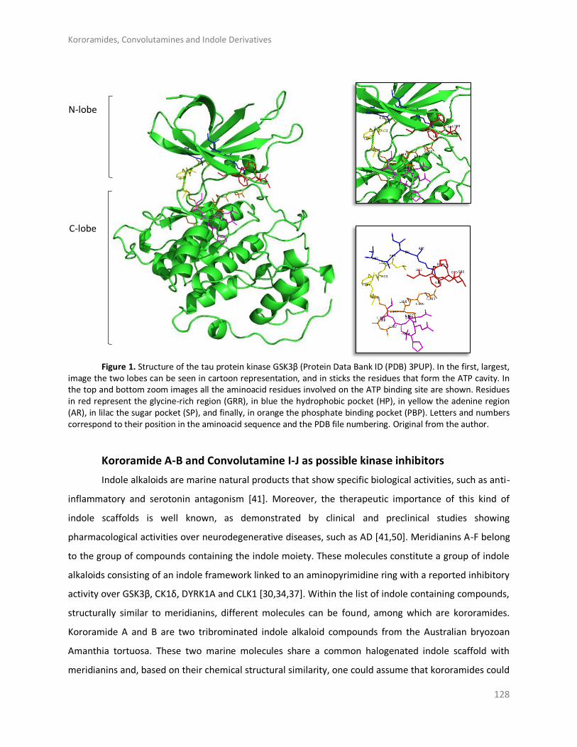
Kororamides, Convolutamines and Indole Derivatives
128
N-lobe
C-lobe
Figure 1. Structure of the tau protein kinase GSK3β (Protein Data Bank ID (PDB) 3PUP). In the first, largest,
image the two lobes can be seen in cartoon representation, and in sticks the residues that form the ATP cavity. In the top and bottom zoom images all the aminoacid residues involved on the ATP binding site are shown. Residues in red represent the glycine-rich region (GRR), in blue the hydrophobic pocket (HP), in yellow the adenine region (AR), in lilac the sugar pocket (SP), and finally, in orange the phosphate binding pocket (PBP). Letters and numbers correspond to their position in the aminoacid sequence and the PDB file numbering. Original from the author.
Kororamide A-B and Convolutamine I-J as possible kinase inhibitors
Indole alkaloids are marine natural products that show specific biological activities, such as anti-
inflammatory and serotonin antagonism [41]. Moreover, the therapeutic importance of this kind of
indole scaffolds is well known, as demonstrated by clinical and preclinical studies showing
pharmacological activities over neurodegenerative diseases, such as AD [41,50]. Meridianins A-F belong
to the group of compounds containing the indole moiety. These molecules constitute a group of indole
alkaloids consisting of an indole framework linked to an aminopyrimidine ring with a reported inhibitory
activity over GSK3β, CK1δ, DYRK1A and CLK1 [30,34,37]. Within the list of indole containing compounds,
structurally similar to meridianins, different molecules can be found, among which are kororamides.
Kororamide A and B are two tribrominated indole alkaloid compounds from the Australian bryozoan
Amanthia tortuosa. These two marine molecules share a common halogenated indole scaffold with
meridianins and, based on their chemical structural similarity, one could assume that kororamides could

Kororamides, Convolutamines and Indole Derivatives
129
have an inhibitory activity similar to meridianins. In the same study where kororamide B was identified,
three other compounds were also isolated, kororamide A and convolutamine I and J. The last two
compounds do not present an indole scaffold, but they are halogenated heterocyclic compounds as
other known kinase inhibitors [51–53] (Figure 2). To test this hypothesis, docking calculations and
Molecular Dynamics (MD) simulations were carried out to evaluate if kororamide A-B and
convolutamine I-J could behave as meridianins regarding GSK3β, CK1δ, DYRK1A and CLK1 binding, thus
indicating that they could be potential anti-AD therapeutic agents.
Figure 2. Chemical structures of convolutamine I, convolutamine J, kororamide A, kororamide B and meridianin F.
In previous studies the presence of halogen atoms was considered important to achieve a good
inhibitory activity over the four studied kinases [24,37]. In order to test whether the presence of a
halogenated indole scaffold, or just the presence of aromatic cycle substituted with halogen atoms
enhances a higher binding affinity against GSK3β, CK1δ, DYRK1A and CLK1, we analyse it by means of
docking and MD simulations. Thereafter, we compared the obtained results (Table 2) with the values
from kororamide A-B and convolutamine I-J with meridianin F, the most promising compound of the
chemical family (meridianin A-F) [37].

Kororamides, Convolutamines and Indole Derivatives
130
Table 2. Summary of classical rigid docking and Molecular Mechanics/Generalized Born Surface Area (MM/GBSA) calculations of the two best models selected per meridianin F (F), convolutamine I (I) and J (J), kororamide A (A) and B (B).
GSK3β CK1δ DYRK1A CLK1
Binding Energy
(kcal/mol)
Binding Energy
(kcal/mol)
Binding Energy
(kcal/mol)
Binding Energy
(kcal/mol)
Binding Energy
(kcal/mol)
Binding Energy
(kcal/mol)
Binding Energy
(kcal/mol)
Binding Energy
(kcal/mol)
R0/R1 R0/R1 R0/R1 R0/R1
F −7.9/−7.9 −35.18
F −7.2/−7.3 −38.55
F −8.0/−7.8 −39.99
F −8.7/−8.7 −37.71
−7.7/−7.9 −34.73 −7.1/−7.1 −38.93 −7.8/−7.7 −39.91 −8.5/−8.5 −37.61
I -5.6/-5.6 -23.08
I -5.0/-5.0 -3.19
I -5.6/-5.6 -26.52
I -5.8/-5.5 -33.23
-6.3/-6.3 -18.38 -5.4/-5.4 -11.26 -4.8/-4.8 -11.02 -5.8/-5.8 -31.93
J -6.7/-6.7 -31.58
J -6.2/-6.2 -37.76
J -7.4/-7.4 -31.35
J -6.0/-6.0 -21.47
-5.9/-5.9 -31.61 -5.8/-5.8 -28.91 -7.0/-7.0 -32.27 -4.6/-4.6 -24.37
A -8.3/-8.3 -34.88
A -8.0/-8.0 -35.48
A -8.2/-8.2 -32.94
A -6.7/-6.7 -37.46
-8.1/-8.1 -31.02 -7.4/-7.4 -33.94 -6.7/-6.7 -14.61 -2.9/-2.9 -38.93
B -9.1/-9.1 -31.80
B -8.1/-8.1 -28.68
B -7.7/-7.7 -23.83
B -4.4/-4.4 -28.71
-8.3/-8.3 -32.34 -6.6/-6.6 -35.53 -7.3/-7.3 -24.29 -4.0/-4.0 -22.96
To avoid false positives, each docking calculation was performed twice (R0/R1). All energy values are kcal/mol. For each target the first (left) column refers to the results of docking calculations while the second (right) column indicate the binding energy results obtained after MD calculations.
Our results indicate that all the analysed compounds could bind to the ATP binding pocket of
each of the mentioned kinases, thus theoretically acting as ATP competitive inhibitors (Figure 3). Binding
energies obtained after docking and MD simulations (Table 2) show that convolutamine J and
kororamide A tend to have higher energies than convolutamine I and kororamide B. To be more precise,
kororamide A shows better energies when bound against GSK3β, DYRK1A and CLK1, while
convolutamine J shows better energies over CK1δ. Comparing the energies obtained between the four
tribrominated metabolites found on the bryozoa Amathia tortuosa and meridianin F, we observe that
the last one has slightly better energies in all cases after MD. These energies do not allow us to discard
any of the compounds as an ATP competitive inhibitor, although we can prioritize kororamide A and
convolutamine J over kororamide B, and specially over convolutamine I. Besides, these results do not
allow us to discriminate between which structural feature influences most the binding strength against
the four studied kinases: the indole scaffold, the presence of halogen atoms, or the combination of both
features.
With the aim of performing a deeper analysis of the inhibitory behaviour of these compounds,
an interaction and binding mode analysis, of the best and prioritized compounds per target, was
performed. On the ATP catalytic cavity of GSK3β it is observed that key binders as I62, F67 and V70,
conforming the GRR or placed nearby, and Y138 and L188 placed at C-terminal lobe near the AR and
inside SP, respectively, are involved on the kororamide stabilization (Figure 3A). For CK1δ it is observed
that convolutamine J it is stabilized by interacting with several key binders as I23 that is placed near the

Kororamides, Convolutamines and Indole Derivatives
131
GRR and A36, M82 and I148 placed at HP, AR and PBP, respectively (Figure 3B). Looking at DYRK1A ATP
cavity, it is observed that kororamide A, at the N-terminal region, is interacting with I165 and V173, as
other known inhibitors like meridianin F or the co-crystal 3RA, both placed near the GRR. In the same
way, kororamide A is also stabilized by A185, which is found at the HP, and at the C-terminal zone it is
also interacting with E291 and L294, conforming PENIL motif, and the key binder V306 present at the
PBP. Finally, kororamide A is also stabilized by L241 and D244, placed near the AR (Figure 3C). Looking at
the ATP cavity of CLK1, it can be seen that on the N-terminal domine, L167, F172 and V175 at the GRR
and K191 at the HP, some of them known key binders, are interacting with kororamide A. Moreover, on
the C-lobe, kororamide A is interacting with F241 coming from the FELL adenin motif, E292 and L295 (a
known key binders), placed at the SP, and V324 found at the PBP (Figure 3D).
The binding mode of the best derivatives, as well as of the four brominated compounds studied,
per target pointed out that they are performing key contacts, according to our interaction analyses. This
fact together with the obtained binding energies, reinforce their capacity to behave as inhibitors for the
four analyzed kinases, in a similar way to meridianin F.
A) B)

Kororamides, Convolutamines and Indole Derivatives
132
C) D)
Figure 3. A) ATP cavity site of GSK3β (Protein Data Bank ID (PDB) 3PUP) with meridianin F (yellow), the co-crystallized OS1 inhibitor (green), and the best pose of kororamide A (magenta). B) ATP cavity site of CK1δ (Protein Data Bank ID (PDB) 4KBK) with meridianin F (yellow), the co-crystallized 1QG inhibitor (green), and the best pose of convolutamine J (magenta). C) ATP cavity site of DYRK1A (Protein Data Bank ID (PDB) 4AZE) with meridianin F (yellow), the co-crystallized 3RA inhibitor (green), and the best pose of kororamide A (magenta). D) ATP cavity site of CLK1 (Protein Data Bank ID (PDB) 2VAG) with meridianin F (yellow), the co-crystallized V25 inhibitor (green), and the best pose of kororamide A (magenta). Letters and numbers correspond to their position in the aminoacid sequence and the PDB file numbering. Original from the author.
Marine natural products and indole scaffold validation
With the aim of testing the importance of the indole scaffold as structural key feature on the
kinases ATP inhibitors and assuming the well-known Structure Activity Relationship (SAR) principle (i.e.
structurally similar compounds will have similar biological activities) a substructure search was
performed over the MarinLit database, a dataset that includes revised compounds from marine natural
products [44]. In that sense, similar compounds to meridianin F and kororamide A and the indole
scaffold were searched over this database. A list of 24 compounds was obtained, 18 compounds when
the indole scaffold was used as a seed, and three using meridianin F and kororamide A, respectively. The
list could contain more molecules if all the indole-containing compounds were selected. However, we
decided that this number is adequate to test if the indole scaffold with several, mostly minor, additions
is enough to have a theoretical inhibitory effect over the four kinases, or whether a complex structure
like meridianin F or kororamide A is necessary. Docking calculations were performed to analyse the
binding behaviour of all of them over GSK3β, CK1δ, DYRK1A, and CLK1 (Table 3).

Kororamides, Convolutamines and Indole Derivatives
133
Table 3. Summary of classical rigid docking calculations of the marine natural compounds found at MarinLit database after a substructure similarity search using a indole group, meridianin F and kororamide A as input molecules. To avoid false positives, each docking calculation was performed twice (R0/R1). All energy values are kcal/mol.
GSK3β CK1δ DYRK1A CLK1 GSK3β CK1δ DYRK1A CLK1
Binding Energy
Binding Energy
Binding Energy
Binding Energy
Binding Energy
Binding Energy
Binding Energy
Binding Energy
R0/R1 R0/R1 R0/R1 R0/R1 R0/R1 R0/R1 R0/R1 R0/R1
L17640 -6.4 / -6.4 -7.3 / -7.3 -7 / -7 -6 / -6 L4950 -9.1 / -9.1 -9.1 / -9.1 -9.1 / -9.1 -9.1 / -9.1 L1189 -6.8 / -6.8 -7.6 / -7.6 -7.2 / -7.2 -5.9 / -5.9 L4949 -8.7 / -8.7 -8.7 / -8.7 -8.7 / -8.7 -8.7 / -8.7
L34 -7.2 / -7.2 -8.1 / -8.1 -8.2 / -8.2 -6.9 / -6.9 L4951 -9 / -9 -9 / -9 -9 / -9 -9 / -9 L4080 -6.1 / -6.1 -6.9 / -6.9 -6.8 / -6.8 -6 / -6
L28238 -6.5 / -6.5 -7.8 / -7.8 -7.3 / -7.3 -5.8 / -5.8 L7472 -6.3 / -6.3 -7.1 / -7.1 -6.8 / -6.8 -6.2 / -6.2
L10723 -6.1 / -6.1 -6.7 / -6.7 -6.7 / -6.7 -5.2 / -5.2
L17639 -6.4 / -6.4 -7.1 / -7.1 -6.8 / -6.8 -5.6 / -5.6 L1192 -7.1 / -7.1 -7.6 / -7.6 -7.6 / -7.6 -5.9 / -5.9
L17641 -6.8 / -6.8 -7 / -7 -7 / -7 -5.7 / -5.7
L11375
-6.2 / -6.2 -6.7 / -6.7 -6.6 / -6.6 -5.4 / -5.4
L35 -7.3 / -7.3 -8 / -8 -8.1 / -8.1 -7.1 / -7.1 GSK3β CK1δ DYRK1A CLK1
L28804 -7.1 / -7.1 -7.6 / -7.6 -7.2 / -7.2 -5.8 / -5.8 Binding
Energy Binding Energy
Binding Energy
Binding Energy L4081 -6.4 / -6.4 -7 / -7 -6.8 / -6.8 -6.4 / -6.4
L29233 -8.5 / -8.5 -8.5 / -8.5 -9.3 / -9.3 -8.2 / -8.2 R0/R1 R0/R1 R0/R1 R0/R1
L24201 -10.6/-10.6 -8.8 / -8.8 -10.4/-10.4 -9.3 / -9.3 L9830 -7.4 / -7.4 -7.4 / -7.4 -7.4 / -7.4 -7.4 / -7.4 L25368 -9.7 / -9.7 -8.8 / -8.8 -10.3/-10.3 -8.9 / -8.9 L9831 -7.8 / -7.8 -7.8 / -7.8 -7.8 / -7.8 -7.8 / -7.8 L7473 -6.9 / -6.9 -7.3 / -7.3 -7.1 / -7.1 -6 / -6 L2330 -8.7 / -8.7 -8.7 / -8.7 -8.7 / -8.7 -8.7 / -8.7
The molecules names (Lxxxx) corresponds to the MarinLit entry code per each compound.
All those compounds with energies higher than -9.0 kcal/mol obtained in at least one of the
studied targets were considered promising compounds. In fact, after analysing their scaffold, a trend can
be seen because all of them have three or more aromatic rings and most of them have two indole
scaffolds (Figure A 1). Some interesting kinase inhibitors described in recent years corroborate this
finding, since they incorporate an indole moiety on their structures [24,25,28,54–56].
Moreover, looking at the top ranked compounds, it is easily observed that all of them have a
bromine (Br) substituent. Actually, all the 24 compounds have at least one Br atom, a differential
signature of marine compounds respect to terrestrial molecules. Unlike terrestrial species, many marine
organism produce halogenated metabolites [43]. This corroborates the proposed importance of the
indole scaffold on the kinases inhibition and seems to point out that the combination of an indole
scaffold with halogen substituents could be a good starting point to design new possible inhibitors of the

Kororamides, Convolutamines and Indole Derivatives
134
four kinases. This hypothesis is not an isolated fact as marine compounds with this moiety, different to
meridianin F and kororamide A, have shown inhibitory effects against some of the studied kinases
[24,28].
Indole derivatives
As mentioned above, the SAR hypothesis, widely used in drug discovery, has the premise that
structurally similar molecules have similar biological activities and thus similar biological targets. Several
known kinase inhibitors possess this moiety and some of them even present an halogenated version of
it. In a previous work, we showed that meridianin F, which has an halogenated indole scaffold was the
more active member of the family, highlighting the role of this moiety. Now, we have observed that
kororamide A and B, given the similarity to meridianin following the SAR principle, could be possible
inhibitors of these kinases. This fact is at least partially confirmed (further experiments are needed for a
complete validation) because of the in silico obtained binding energies over GSK3β, CK1δ, DYRK1A and
CLK1, reported above. All these facts together with the observed results at Table 3, made us hypothesize
that starting from an halogenated indole moiety and following structural features extracted from
meridianin F and kororamide A, we could design indole derivatives that could become kinase inhibitors.
Concretely, the indole group was used as a template for the design of a series of seven analogue
compounds with different fragments attached to the R3 position of the indole (compounds 1-7; Table 4)
and substituted with different combinations of halogen atoms at positions R1 and R2 (a-g combinations;
Table 4). Altogether, 49 compounds were designed.
Marine animals have demonstrated to be rich sources of halogenated metabolites and halogenated
compounds have a wide range of biological activities [42]. Most halogenated drugs contain fluorine (F),
followed by chlorine (Cl) and bromine (Br). Contrastingly, for marine-derived molecules, rather than
chlorine, the most prevalent halogen found is Br [57]. Halogenated molecules are interesting
therapeutic opportunities and it is estimated that one quarter of the total number of final compounds
synthesized have an insertion that involves halogens [58]. Halogenated ligands lead to more stable
complexes than non-halogenated ligands, and this is important to explain molecular recognition or to
planning an screening study [59]. Moreover, the capability of halogen atoms to improve oral absorption,
lipophilicity, blood brain barrier (BBB) permeability, metabolic and chemical stability, or even potency is
well known [59,60]. Therefore, the three mentioned halogen groups at R1 and R2 positions were
introduced and evaluated per compound (1-7 + a-g) with the aim of designing the best possible kinase
inhibitors (Table 4).

Kororamides, Convolutamines and Indole Derivatives
135
Table 4. Scheme of the chemical design. The indole scaffold of meridianin F and kororamide A was selected to
derive new compounds. More precisely, seven indole derivatives were designed (compound 1-7). The R3 position
was fulfilled with diverse structural elements mainly inspired on meridianin and kororamide structures. Compound
1 with a ketone group, compound 2 with an aromatic ring, compound 3 with a methylamine, compound 4 with
methyl group, compound 5 with methanol, compound 6 with an ethylamine and compound 7 with an ethyl-
methylamine. The R1 and R2 positions were completed with the permutation of Br, Cl and F halogen atoms (a-g)
over both positions. At the end 49 indole analogue compounds were designed.
meridianin F kororamide A
Indole
1 2 3
4 5 6 7
Compounds R1 R2
a Br Br
b F F
c Cl Cl
d Br F
e Br Cl
f F Br
g Cl Br

Kororamides, Convolutamines and Indole Derivatives
136
In silico binding and binding mode analysis of indole derivatives
To analyze the feasibility of the designed compounds as kinase inhibitors by an in silico binding
analysis, their binding mode and binding strength against GSK3β, CK1δ, DYRK1A and CLK1 were
analysed. To start with, docking experiments were performed. A total of 441 poses per target were
obtained from the 49 compounds of the set. Thereafter the binding behaviour of all the poses was
analysed, showing that the most populated binding region is, as expected, the ATP cavity. With all these
results in hand, best poses per target in terms of binding mode and binding energy were selected to
perform short MD simulations, for post-processing docking results. For some derivatives any pose was
considered for further studies, as the selection of best compounds was carried out considering not only
the binding energy but also the binding mode of each molecule, after an interaction analysis study. The
poses that did not present good interactions were discarded. Finally, 166 simulations were carried out,
corresponding to diverse poses belonging to 45 compounds for GSK3β, 30 for CK1δ , 46 in the case of
DYRK1A and 45 for CLK1. After MD simulations, the binding energies of the target-ligand complexes
were estimated by Molecular Mechanics/Generalized Born Surface Area (MM/GBSA) calculations. Table
5 summarizes the binding energies of the best indole derivatives, obtained after MD, per compound (1-
7) and target. The rest of the binding energies obtained per derivative and target are reported at Table A
1, Table A 2, Table A 3 and Table A 4, respectively.
Table 5. Summary of Molecular Mechanics/Generalized Born Surface Area (MM/GBSA) calculations of the best derived analogues over the four targets studied. Lowercase letters indicate the halogen substituent group (a-g), as described in Table.
GSK3β CK1δ DYRK1A CLK1
Binding Energy
Binding Energy
Binding Energy
Binding Energy
Compound 1 a -30.3141 a -35.4499 e -32.8862 g -30.3541
Compound 2 a -31.2458 e -37.8982 a -37.8422 a -34.1041
Compound 3 a -13.8779 g -28.7631 a -15.2733 f -20.4786
Compound 4 a -27.6481 e -28.6573 a -30.7518 e -28.3695
Compound 5 a -27.6534 e -28.5831 a -31.2535 c -29.4190
Compound 6 a -18.5779 a -26.4630 a -18.9387 g -30.7737
Compound 7 a -18.8955 a -18.4901 a -20.8203 g -25.4765
All energy values are kcal/mol.
As a general result, we observe that all the evaluated compounds present better binding
interaction energies against CK1δ, DYRK1A and CLK1 than GSK3β, as observed for meridianins [37]. Also,

Kororamides, Convolutamines and Indole Derivatives
137
as a general trend, compound 1 and 2 always show better energies than the rest of derivatives,
highlighting that the fragments (a ketone and an aromatic ring, respectively) introduced in the pyrrole
ring (R3 position) of the indole scaffold could have beneficial effects to achieve better inhibitory
activities over the ATP binding site of the four studied kinases. Finally, it must be remarked that the
designed compounds that do not work against the kinases are different for each one of them, thus
opening the door for exploiting these differences in the future to gain selectivity over the four analysed
kinases.
GSK3β
As said, the best docked complexes were selected to perform further analysis. For GSK3β 75
poses were chosen and over them MD simulations were performed. From the total studied set, and with
the aim of analysing the diverse derivatives, the best a-g combination for each of the 1-7 compounds
per target was selected. Over the seven best compounds found after MD simulations in terms of binding
energy, further analyses were performed, extracting some interesting features. Focusing on the halogen
substituents, the best compounds are always those that contain two Br atoms at R1 and R2 position,
reflecting the importance of Br substituents observed in previous studies [24,37].
A general pattern regarding the interactions performed by each of the seven best derived
compounds at the catalytic ATP binding site was observed. In general, I62, V70, A83, V110, L132, D133,
Y134, V135, Y138, and L188 are the most important aminoacids for their stabilization over the ATP
catalytic pocket (Figure 4). The NH indole group is essential to establish hydrogen bond interactions with
the carboxylic acid group (deprotonated under biological conditions) of D133 and/or V135. AR,
described by LDYV motif, accommodates the seven best compounds, all of them showing the same
binding mode/pose, stabilized by hydrophobic contacts. The indole group is wrapped by N-terminal I62
and V70 residues found near the GRR, together with A83 placed at the HP and C-terminal residues V110
and L188 present at the SP. As the binding mode analysis reveals, all the compounds have the same
binding mode, thus binding energy results and MD simulations were used with the aim of identifying
some differential features among them. MD analysis reveals that the indole scaffold is maintained
wrapped in the same position during all the simulation while the fragments introduced at R3 are more
flexible. A binding energy analysis showed that compound 2a has a slightly better energy than
compound 1a, although both could be considered good plausible options, as the binding energy
differences are around 1 kcal/mol, that seems to point out compound 2a as the best possible inhibitor.

Kororamides, Convolutamines and Indole Derivatives
138
Looking at the literature, our results show that the binding mode displayed by most of the
analysed compounds, specially by compound 2a, correlates with the binding mode of known inhibitors,
and also that the residues involved on it are key binders [35,61].
Figure 4. Superposition of the seven best compounds over GSK3β (PDB code: 3PUP) ATP cavity. The active site aminoacid residues involved in the binding of the best compounds and the binding position of each of them are enlarged. In the first enlarged panel the seven top compounds are represented, whereas in the right panel only compound 2a is shown. Original from the author.
CK1δ
For CK1δ, 97 docking poses were subjected to MD simulations. Thereafter, the seven best
compounds, in terms of binding energy were selected to be further analysed. Differently to GSK3β,
there is not a common binding mode shared by the seven derived analogues and there is not a specific
location of the halogens in the ATP binding site, that can be inferred from the observed binding modes.
Although a general pattern could not be observed, there are common features between the studied
derivatives than can be highlighted. There is a common behaviour between compound 1, 6, and 7, and
compounds 2, 4, and 5 (Figure 5). For the first group the best halogen composition is Br-Br (compound
1a, compound 6a and compound 7a), whereas for the second group, the best halogen composition is e
(Br-Cl), while for compound 3, that behaves differently to the rest of the compounds, is g (Cl-Br). In all
compounds a Br atom is present, which seems to indicate that this presence could be important to
increase the binding strength. In general, with few exceptions, the worst binding energies are obtained
when there is no Br atom present. This trend is also observed on the rest of kinases (Table A 1, Table A
2, Table A 3 and Table A 4). In addition, an accurate analysis of the most important residues involved on
the seven compounds binding mode, was performed. This analysis reveals that despite each compound

Kororamides, Convolutamines and Indole Derivatives
139
has a different binding pose, there are conserved interactions at the ATP catalytic cavity. According to
that, the most important residues on the binding of the seven compounds to CLK1δ are I23, A36, Y56,
L84, I148, and D149. All the seven derivatives are placed between the HP defined by A36 and the
residue I23 that is placed near the GRR, both zones located at the N-terminal region and L84, I149 and
D149 placed at the AR and PBP at the C-terminal domain. All the interactions observed between the
analogues and the residues are mainly hydrophobic contacts. Binding energies reveal that compound 2e
(Br-Cl) seems to be a slightly better inhibitor than compound 1a, although both can be considered good
options as the energy differences are around 2 kcal/mol.
Different studies have been addressed to find novel and potent CK1δ inhibitors in the last years.
Looking at them, it is easy to observe that the interactions made by of all these molecules are aligned,
validating it, with the binding mode of our proposed derivatives [27,32,33,36].
Figure 5. Superposition of the seven best compounds over CK1δ (PDB code: 4KBK) ATP cavity. The active site aminoacid residues involved in the binding of the best compounds and the binding position of each of them are enlarged. In the first enlarged panel the seven top compounds are represented, whereas in the right panel only compound 2e is shown. Original from the author.
DYRK1A
For DYRK1A, 72 docking poses were selected for further analysis. MD simulations were
performed over all of them, and thereafter the best compound per target, as for the rest of kinases, was
selected. Despite the indole derivatives tested do not shown a shared binding mode as GSK3β, it is more
conserved than for CK1δ. All compounds, except compound 3 that is oriented right upside down and
moreover shows the worst binding energy, shared the same placement at the ATP catalytic pocket
(Figure 6). Analysing the halogen composition of the best compounds it is observed that Br-Br, at R1-R2

Kororamides, Convolutamines and Indole Derivatives
140
positions, is the most common substituent; only compound 1 has a different combination (Br-Cl). As a
general conclusion, as with the other three kinases, the presence of at least one Br atom is important to
have a good binding affinity.
Looking forward to extract common patterns from the binding modes of the top seven
derivatives, it is clear that all these compounds placed at the catalytic ATP cavity are interacting with
I165 and V173, both residues delimitate the GRR, and A186 that is found at the HP, all of them located
at the N-terminal region. The important AR formed by a FEML motif also participates on each of the
seven bindings, being F238 and L241 the most important residues to stabilize the analysed derivatives.
At the C-terminal region, V306 and D307, present at the PBP, are also key binders. Interaction analysis
reveals that most of the interactions performed by the derived analogues were hydrophobic contacts.
For DYRK1A, after analysing the MD obtained results, it is observed again that compound 2a, is the best
derivative in terms of binding energy and binding mode. Interestingly, the observed binding patterns are
shared by most of the known inhibitors of this target, that could be found in the literature. Even more,
all of them are proposed as ATP competitive inhibitors like the derivatives we described here [25,26,34].
Figure 6. Superposition of the seven best compounds over DYRK1A (PDB code: 4AZE) ATP cavity. The active site aminoacid residues involved in the binding of the best compounds and the binding position of each of them are enlarged. In the first enlarged panel the seven top compounds are represented, whereas in the right panel only compound 2a is shown. Original from the author.
CLK1
For CLK1, 87 docking poses were selected for further analysis. All of them were subjected to MD
simulations selecting then the best one per target. A first binding mode observation reveals that a
common binding mode was found for compounds 1, 2, and 6 (Figure 7). These three compounds have
the best binding energy, and this could point out the importance of R1, R2 and R3 substituents to gain
inhibitory capacity. Compound 3, despite having a similar binding pose, does not show good energies.

Kororamides, Convolutamines and Indole Derivatives
141
The other compounds (4, 5, and 7) show slightly lower binding energies and a different binding mode,
even between them. Focused on the halogen groups, in this target there is not a clear trend, as the
seven best compounds show five different halogen substituents (a, c, e, f and g). Despite this fact, not
observed in the rest of studied kinases, a similar trend can be seen. Most of the seven top compounds
have a Br atom, except compound 5c. Moreover, in agreement with the rest of compounds the seven
top derivatives are mainly Br or Cl substituents on R1 or R2 position, with the exception of compound 3f.
This seems to suggest that, as for the other targets, all of the analysed halogen substituents
combinations could give good inhibitory results, but the presence of a Br is a key factor. In fact, for this
target, as seen for the other kinases, the compound 2a is the best one in terms of binding energy.
A detailed analysis of the displayed binding modes by each compound at the ATP cavity site,
reveals interesting shared patterns. On the N-terminal domain L167, F172, and V175 can be found at the
GRR, and A189 at the HP acting as key binders. Adenine motif FELL was also revealed important, in
particular F241 and L244. On the C-terminal region, residues E292 and L295 at the SP and V324 placed at
PBP are the most important aminoacids to stabilize the derived compounds over CLK1. As compound 2a,
the other best compounds tend to point their halogen groups between the AR and the HP, fact that
facilitate residues as F175 placed at the GRR and E292, or L295 placed at the opposite SP, surround and
fixed the indole scaffolds. Interestingly, the binding mode exposed here for the derived analogues in
general, and also for the best compound, 2a, in particular, is validated by other inhibitors reported in the
literature [30,31].
Figure 7. Superposition of the seven best compounds over CLK1 (PDB code: 2VAG) ATP cavity. The active site aminoacid residues involved in the binding of the seven best compounds and the binding position of each of them are enlarged. In the first enlarged panel the seven top compounds are represented, whereas in the right panel only compound 2a is shown. Original from the author.

Kororamides, Convolutamines and Indole Derivatives
142
The in silico binding studies performed over the four kinases indicate that the derivatives coming
from compound 2, 2a and 2e, located at the middle of the ATP binding cavity, seem to be the most
plausible ATP competitive inhibitors. However, other derivatives, especially for compound 1 should not
be discarded. In general, the presence of the benzene ring at position R3 could have a more positive
influence on compounds stabilization at the catalytic site than other substituents. Looking at the
literature, several inhibitors described for GSK3β, CK1δ, DYRK1A, and CLK1, as well as other members of
the protein kinase family, have aromatic rings in the terminal positions. Moreover, the analysis of the
effect of the halogen groups used as substituents at R1 and R2 positions pointed out that its presence
can influence the binding strength of the complex (ligand-target). In general, if at least one of the
substituents is a Br atom the binding energy is better. An interesting trend found here is that Br seems
to be the “best” halogen, followed by Cl and F, which in general give worst binding energies. This finding
is in line with what is observed in nature, since marine natural halogenated indole alkaloids contain
mostly bromine and chlorine, being the iodinated and fluorinated compounds less abundant [43].
Selectivity
One of the most important challenges on the design of novel kinase inhibitors is the lack of
selectivity over the ATP binding site, which is critical in clinical effectiveness of most drugs (Traxler &
Furet, 1999; Huang et al., 2010). Most kinase small-molecule inhibitors bind to the ATP catalytic cavity
near the AR and wrapped by GRR and HP on the T-lobe and SP and PBP at the C-lobe. The herein
performed study does not reveal any significat selectivity over the four kinases for any of the analysed
compounds, which could be easily observed looking at the obtained binding modes and energies.
However, analysing the residues involved on the binding and the regions occupied by the analogues,
some interesting trends that could be exploited in the future can be observed. Interestingly, regarding
the binding modes, the best binding energies were obtained on those compounds that are (partially)
placed at the PBP. This region, that is very exposed to the solvent and is not usually exploited to gain
binding affinity, can be useful to improve the inhibitors selectivity since it contains non-conserved
aminoacids [46].
Regarding the binding energy results per se, without having into account the binding mode,
remarkable significative differences are not observed. The best compound for each target (2a and 2e
respectively) comes from the same scaffold, being compound 2a the best theoretical inhibitor for three
of the four targets. If we analyse the binding energies of these top compounds, compounds 2a and 2e
over DYRK1A and CK1δ, respectively, show a better interaction energy, around 6 kcal/mol of difference,

Kororamides, Convolutamines and Indole Derivatives
143
respect to the binding energy of compound 2a over GSK3β and 3 kcal/mol over CLK1. However, although
a slight preference could be inferred from this, the binding of these four compounds to all the four
targets is possible with a reasonably good strength. In general, the main differences are observed
between the derived compounds 2 (mainly) and 1, which seem to have better energies than those
molecules coming from analogues 3 to 6, and specially respect to the molecules coming from analogue
compound 7 (Table 5). For GSK3β the best compounds coming from derivatives from 2 and 1 (1 kcal/mol
of difference between them) are displaying the best interaction energies, followed by those from
analogues 4 and 5 (around 4 kcal/mol of difference to compound 2a), and finally the worst compounds
come from analogues 3, 6 and 7 with differences around 13 to 17 kcal/mol respect compound 2a. In the
case of CK1δ, as for GSK3β, the top ranked compounds from analogues 2 and 1 (1.5 kcal/mol of
difference between them) have the best binding energies, followed by those from analogues 3, 4, 5 and
6 with differences around 9 to 11 kcal/mol respect compound 2e, and finally compound 7a with a
difference of around 19 kcal/mol respect to 2e. For DYRK1A, the best from compound 2 is the top
molecule in terms of interaction energy. Compound 1a shows a difference of around 5 kcal/mol,
whereas compounds 4 and 5 present differences between 6.5 and 7 kcal/mol, respectively, and
compounds coming from scaffolds 3, 6 and 7 between 17 and 22.5 kcal/mol. In the case of CLK1,
compound 2a has the better binding energies, followed by those from analogues 1 and 6 (differences
around 3 kcal/mol), molecules derived from compounds 4 and 5 (differences around 5 kcal/mol), and
finally those from analogues 3 and 7, that show differences around 8.5 to 13 kcal/mol respect to the
binding energy.
Looking to compounds 1-7 per target, it can be observed that for compounds 4 and 5 the
binding energy differences between the top a-g derivatives range between 2 and 4 kcal/mol between
the four kinases. For compounds 1, 2 and 7 the differences are higher, ranging between 2.5 and 7
kcal/mol, depending on the compound and target. Finally, for compounds derived from analogues 3 and
6 the differences are even higher, ranging between 8 and 15, and 4 and 12 kcal/mol, respectively. In
general, there is not any noticeable selectivity trend derived from the binding energy, although there
are some features that could be further exploited. For instance, for DYRK1A and CLK1 an aromatic ring
at R3 position is the best choice to gain activity over them, whereas for GSK3β and CK1δ a ketone group
at this position could also work, enhancing a way to design selective compounds at least for some of the
four kinases.

Kororamides, Convolutamines and Indole Derivatives
144
Exploring the effect of the halogen atoms over the binding strength, as said above, some general
trends could be observed but again, its presence does not give any clearly marked or significative
selectivity trend between targets. The presence of Br atoms seems to increase the binding strength
more than the presence of Cl of F, being in general Cl “better” than F to get good energetic results.
However, a possible selectivity feature could be observed due to compound 2e. Docking energy results
are similar for the four kinases, but it only performs good interactions for CK1δ. This is the reason why
MD simulation over this compound was only performed in complex with it, while for the other three
kinases it was not selected. Compound 2a gave good docking energies for all four targets but performed
good interactions only with GSK3β, DYRK1A and CLK1, so the fact of having a Br-Cl combination at R1-R2
plus an aromatic ring at R3 could be a sign of selectivity over CK1δ, although this should be further
explored, as other Cl combinations give good results for the other kinases (Table A 1, Table A 2, Table A
3 and Table A 4).
2a and 2e unbinding
To reinforce and validate the observed binding trends, as well as to find a differential feature
that could help to enhance the selectivity of future derived compounds over the four kinases, steered
molecular dynamics (SMD) simulations were performed. Since at the energy and binding mode level
there are no significant differences, we intended to see if there was some type of selectivity derived
from the protein structure that influences the facility/difficulty of unbinding of the most promising
inhibitory compounds 2a and 2e per target (Figure 8).
A)
B)

Kororamides, Convolutamines and Indole Derivatives
145
C)
D)
Figure 8. Exerted force in piconewtons (pN) needed to A) remove compound 2a (blue) from the GSK3β
ATP catalytic cavity, B) remove compound 2e (red) from the CK1δ ATP catalytic cavity, C) remove compound 2a (green) from the DYRK1A ATP catalytic cavity and remove compound 2a (orange) from CLK1 ATP catalytic cavity. The x axis represents the computational residence time in femtoseconds (fs).
At the beginning of each simulation (t = 0), the compound is in the bound state, placed inside
the ATP cavity interacting with the residues previously described. After 1 nanosecond (ns), at the four
kinases, the ligand is completely out of the cavity. In the case of GSK3β (Figure 6A) a force of around 400
pN (piconewtons) is needed to extract compound 2a from its catalytic cavity. The compound
dissociation from the target takes place at 200 femtoseconds (fs), moment when the force decrease
approach zero pN which means that the compound is out of the cavity. For CK1δ (Figure 7B) that hosts
the best compound in terms of binding energy, 2e, the necessary force to break the ligand-target
complex is higher than for the GSK3β complex, with forces that reach up to 600 pN. The ligand
unbinding takes place at a similar time than for GSK3β complex, around 200 ps although it takes slightly
more time. The dissociation of compound 2a from DYRK1A (Figure 7C) is done in two phases. A primary
rupture force seems to occur before 100 fs, and immediately afterwards the highest energy point can be
observed (around 500 pN), corresponding to the second break. A visual inspection of the SMD
confirmed that at this moment, the compound is still inside the ATP pocket. Over 200 fs, the force, after
a progressive decrease, arrives to zero pN. This progressive decline correlates with the progressive loose
of interactions during the way out of the compound from the catalytic cavity. For the CLK1-compound
2a complex (Figure 7C) a similar situation is observed. A primary rupture around 100 fs, moment when,
as for DYRK1A, the compound is still placed at the ATP binding site and it is not until later on 200 fs,
when a sudden drop in the energy can be observed, indicating the complete loss of interactions and
therefore, the leaving of the cavity. As a general trend, around 200ps 2a and 2e compounds leave the
catalytic pocket of the different kinases, requiring a different amount of force that is in line with the

Kororamides, Convolutamines and Indole Derivatives
146
observed binding energy. Usually, the better the binding energy, the higher the force needed to break
the complex and the longer the residence time. In that sense, the SMD results corroborate what has
been seen so far: CK1δ-2e and DYRK1A-compound 2a complexes that have the higher binding energies
also seem to have (slightly) longer residence times and require a higher force to take out their
respective ligands from their catalytic pockets. Thus, there is no feature that suggests a selectivity trend
derived from the unbinding process that could not be extracted from the binding energy results.
Compound 2a is more selective (it binds stronger and requires a higher effort to remove it) for DYRK1A
than for GSK3β and CLK1 but could bind to all of them. Compound 2e seems to be more difficult to
unbind that compound 2a, but this correlates with the higher binding energy it shows after MD.
Pharmacokinetic properties of kororamide A-B, convolutamine I-J and the designed derivatives
Due to the importance of pharmacokinetics (PK) and its impact on drug discovery process,
convolutamine I-J, kororamide A-B and the whole set of 49 analogues compounds were analysed,
studying their ADME/Tox features. The PK properties of the two best derived compounds 2a and 2e are
summarized on Table 6 and Table 7. The full set of derivatives were also analysed and results can be
found at Table A 5 (absorption and distribution) and Table A 6 (metabolism, excretion and toxicity).
The first PK property analysed was molecular weight, and all compounds show values under 500
Dalton (Da). The higher molecular mass was found for compound 2a with 351 Da, which is in good
agreement with the sizes that a small therapeutic molecule that should cross the BBB should have.
Absorption properties
Absorption describes the process by which drug candidates move from the point of
administration to the blood. LogS descriptor confirmed good solubility in water and good bioavailability
for each compound. The derivatives coming from compounds 2 show values that are between -5.1 and -
6.1, while for the rest of the derivatives, values go between -3 and -4. Caco-2 permeability revealed
medium to high values for all the compounds, except for kororamide A that was low. The compounds
that have a benzene at R3 position as well as the derivatives with F at R1 and R2 positions show
moderate permeability and should be optimized in the future. Regarding P-glycoprotein (Pgp) binding,
none of the compound was predicted to act over it. The interaction with Pgp has many pharmacological
implications that could result in pharmaceutical advantages or contraindications. For instance, Pgp
modulation has been suggested as a mechanism to improve CNS pharmacotherapy [62–65], but none of
the derivatives here seem to have this ability. On the other hand, intestinal absorption values higher

Kororamides, Convolutamines and Indole Derivatives
147
than 30% are considered well-absorbed compounds, and for all our set the obtained values were higher
than 89%. All these absorption results suggest good absorption properties for the 49 designed
derivatives, plus kororamides and convolutamines.
Distribution properties
Distribution describes the migration of a compound from the circulation to the extravascular
system. LogP values lower than 5 indicate that the compounds have an appropriate hydrophobicity and
permeability. In that sense, the derivatives coming from compound 2, as well as convolutamine J and
kororamide A have the highest values (≈ 4) while the rest of compounds are between 2 and 3. Opposite
to LogP behaviour, plasma-protein binding (PPB) and steady state volume of distribution (VDss) are not
showing as good tendencies for the best derivatives compounds coming from scaffold 1 and 2, and
Convolutamine J. Most of the analysed molecules showed medium to high PBP values (except
kororamide A and compounds 3b, 5b, 6b and 7b with low PPB values) indicating that a high percentage
of the administrated compounds will be found attached to proteins, affecting its diffusion and its
efficiency. As less bound a drug is to plasma proteins, the more efficient it is, as it can traverse cell
membranes or diffuse. Regarding VDss, derivatives from scaffolds 3, 6 and 7 and convolutamine I-J plus
kororamide A, have high VDss values (>0.45), while for the rest of compounds, distribution is low to
medium, in a close agreement with PBP results. BBB descriptors with results higher than > 0.3 reveal
good distribution to the brain, as they could pass the blood brain barrier. The highest values are found
for convolutamine I, kororamide A and the derivatives coming from scaffolds 2 and 4, as well as for
compounds 5b and 7b. However, it should be considered that most of the compounds not predicted to
cross BBB, have values near the threshold. In addition to BBB, Central nervous system (CNS)
permeability was measured. This seems to be a more precise measure than BBB, as it is a more direct
measurement [66]. Kororamides and convolutamines do not show good permeability values, whereas all
the derived compounds showed good results (>-2) allowing us to consider that most of the designed
compounds could penetrate the CNS, specially the compounds coming from scaffolds 2 and 4, among
which there are the two best candidates 2a and 2e.
Metabolism properties
Cytochrome (CYP) P450 is an important enzyme used to predict drug metabolism. Many drugs
could be deactivated or activated by CYP450, as cytochrome P450 enzymes that can be inhibited or
induced by drugs, resulting in clinically significant drug-drug interactions that can cause unanticipated
adverse reactions or therapeutic failures. Our results revealed that all the analysed compounds, except

Kororamides, Convolutamines and Indole Derivatives
148
kororamide B, are likely to be metabolised by CYP450, so their properties should be carefully analysed to
design lead compounds from the herein studied molecules [67,68].
Excretion properties
Regarding excretion properties, describing the transport of drugs into the urine or bile, good
results were obtained. It was found that only kororamide A and B tend to act as a substrate of the
organic cation transporter 2 (OCT2 or Solute carrier family 22 member 2, SLC22A2), which means that in
general, and for the two best derivatives 2a and 2e, non-clearance problems and adverse interactions
with co-administrated OCT2 inhibitors are expected.
Toxicity properties
During drug development, safety is always the most important issue, including a variety of
toxicities and adverse drug effects that should be evaluated in preclinical and clinical trial phases [69].
Between the measured properties, the inhibition of the potassium channels encoded by the human
ether-a-go-go gene (hERG) is basic. Our results indicate that none of the compounds seem to be toxic
due to hERG. In the same way, none of the designed derivatives is susceptible to be hepatotoxic.
However, convolutamine I and J as well as kororamide A tend to be hepatotoxic. Looking at AMES
toxicity, which predicts mutagenic and carcinogenic properties, our results revealed that the derivatives
from compound 2, as the top derivatives 2a and 2e, and kororamide B are predicted to be toxic, while
the rest of the set does not. Regarding the maximum recommended tolerated dose (MRTD), the four
brominated alkaloids as well as compounds coming from scaffolds 3, 6 and 7 showed low values/doses,
which is not the best scenario, whereas the rest of the compounds present good MRTD values.
Table 6. Summary of absorption and distribution properties of the two best compounds 2a and 2e found on the four studied kinases. BBB: blood brain barrier, PPB: protein-protein binding, VDss: steady state volume of distribution, CNS: central nervous system.
Absorption Distribution
Compound Mol
weight LogS P-Glycoprotein
Caco-2 permeability
Intestinal absorption
LogP BBB PPB VDss CNS
permeability
Compound 2a 351 -6.1 inactive Moderate 90.067 4.1 0.477 High 0.234 -0.894
Compound 2e 290.1 -5.7 inactive Moderate 91.036 3.8 0.508 High 0.076 -0.92
Table 7. Summary of metabolism, excretion and toxicity properties of the two best compounds 2a and 2e
found on the four studied kinases. CYP: cytochrome, OCT2: organic cation transporter 2, hERG: human ether-a-go-go gene, MRTD: maximum recommended tolerated dose.
Metabolism Excretion Toxicity
Compound CYP450 OCT2 substrate hERG MRTD AMES
toxicity Hepatotoxicity
Compound 2a Yes No <4.0 0.673 Yes No Compound 2e Yes No <4.0 0.641 Yes No

Kororamides, Convolutamines and Indole Derivatives
149
The well-known Lipinski’s rule of five, formulated in 1997 and that remains in force [70] was also
used in combination with the different ADME/Tox properties described above with the aim of
evaluate/determine druglikeness of the analysed compounds. To assess how druglike is a substance
based on Lipinski’s rules it is accepted that it should have 1) not more than five hydrogen bond donors
and 2) ten hydrogen bond acceptors, 3) a molecular mass less than 500 Da and 4) a LogP not greater
than 5. Focusing on the two best compounds (2a and 2e), both have one hydrogen donor and no
acceptors. Also, as seen in Table 6 and Table 7, the other Lipinski requirements are also met. Thus,
taking into consideration all the ADMET results described previously, these two compounds can be
proposed as good hit candidates, having into account that some properties, such as the possible
carcinogenesis and mutagenesis problems should be carefully addressed. In fact, absorption,
distribution metabolism, excretion, and toxicity properties should be more or less improved for all the
designed compounds, in a further Hit to lead (H2L) optimization process. Toxicity should be removed,
and compounds interaction with cytochrome P450 carefully analysed and, given the case, eliminated or
modulated. Moreover, Caco-2 permeability could be increased as well as their distribution properties
should be improved, lowering the PPB and VDss, to be able to diffuse and penetrate into cells easily.
Materials and Methods
Computational virtual screening
It is well known that there is a correlation between (chemical) structure and (biological) activity,
Structure Activity Relationship (SAR). This SAR is widely exploited in many aspects of the drug discovery
pipeline, ranging from compound screening to lead optimization processes, at experimental and
computational level. Herein, we have performed a 2D virtual screening search over MarinLit database
using its substructure search functionality. Using as an input meridianin F and kororamide A (the two
indole compounds that have shown a better binding strength against the four analysed kinases), as well
as the indole scaffold alone, a similarity search was performed over MarinLit obtaining a list of
compounds having an indole scaffold in their structure and/or being structurally similar to meridianin F
and/or kororamide A. The name and structure of the similar compounds could be found at Figure A 1.
Structure Modelling
Meridianin F structure was modelled from the 2-dimensions (2D) chemical structure published
by Núñez-Pons and co-workers [71]. Convolutamine J, I, and kororamide A and B, were modelled from

Kororamides, Convolutamines and Indole Derivatives
150
Dashti et al. [38]. Ligands were prepared to generate energetically minimized 3 dimensions (3D)
coordinates.
To do computational work, obtaining good structures to start with is crucial, so prior to any
calculation, the best structures of the four analysed targets were modelled from 3D crystal structures
extracted from the Protein Data Bank (RCSB PDB) [72]. Those structures represented human targets and
are the structures and chains that cover the maximum amino acid region sequence, in general, and the
binding region of each of the selected targets in particular. Since all the four kinases biological assembly
is in monomeric forms, GSK3β and CK1δ chain B and DYRK1A and CLK1 chain A were respectively
selected to perform further studies. To do so, due to the fact that the four studied targets have 3D
crystallographic structures, the ATP competitive inhibitors OS1 co-crystallized with GSK3β (PDB: 3PUP)
[35]; 1QG co-crystallized with CK1δ (PDB: 4KBK) [36], the crystal structures of DYRK1A in complex with
3RA (PDB: 4AZE) [26] and finally, V25 co-crystallized with CLK1 (PDB: 2VAG) [31] were used as a
template to perform rigid docking calculations using Itzamna (Mind the Byte.SL, Barcelona, Spain) [73].
Docking calculations
Docking calculations can identify small molecules (ligands) that fit well into the putative binding
pocket of a given protein (target) in an optimal way. Without any other specification, generally speaking,
docking refers to classical (rigid) docking where only the movement of the ligand is allowed [74]. This
kind of calculations allows to elucidate the candidate binding mode (pose) that is predicted when ligand
and receptor bind to each other, and scoring functions allows to classify and rank compounds based on
the binding energies obtained. Proteins are flexible entities, they move, and this dynamic is necessary to
carry out any protein function. Taking into account this flexibility regarding the binding mode prediction,
is key to obtain results capable of being experimentally correlated [75,76]. A good option to add
flexibility at the complex is the post-processing of docking results, that consist on docking validation
and/or refinement by MD simulations [77]. Rigid docking calculations can predict optimal ligand
placement at the binding site of a target, but not all the interactions between the ligand and the
receptor are usually depicted accurately. MD simulations can optimize the predicted binding mode,
allowing to observe the so-called induced fit events arising from the conformational adaptation of the
target to the ligand, and also check the stability of the docked complex, as a bad docking pose will
generate an unstable MD trajectory, during which the ligand could even leave the binding site [75,78]. In
this study, we have employed a pipeline aimed to simulate a flexible docking protocol in a similar way to
other studies reported in the literature, in that we post-processed the obtained docking poses [79].

Kororamides, Convolutamines and Indole Derivatives
151
Concretely, the energetically minimized ligand conformations were docked into the active site of the
four kinases studied; possible binding poses were determined and subsequently ranked based on their
calculated binding affinities and then post-processed using MD simulations. This two-step protocol
constitutes a (probably the most) practical and convenient approach to address the docking problem
[77]. It is in general less computationally expensive and provides the results that we need in an accurate
way, comparable to “real” flexible docking methodologies (such as ensemble-based or flexible induced-
fit docking). In general, using MD as a post-processing tool, a smaller fraction of the conformational
space is usually covered, but without the several limitations that affect sampling and scoring algorithms
for docking.
All docking calculations were performed using Itzamna software tool (Mind the Byte.SL,
Barcelona, Spain) [73]. Itzamna needs the structure of a molecule, or a set of compounds, to dock, as
well as the cavity where it should be placed as an input. When the used 3D crystal structures were co-
crystallized with a ligand, this cavity was employed. If it were not the case, the cavity was defined by the
residues that describe the cavity. Docking studies were performed between kororamide A-B,
convolutamine I-J and the set of 49 derived compounds against GSK3β, CK1δ, DYRK1A and CLK1. Two
runs were carried out for each calculation to avoid false positives.
Molecular dynamics simulations
MD simulations are one of the principal tools in the computational study of biomolecules as the
dynamic nature of proteins is a well-established phenomenon that these simulations can capture. It can
be described as a theoretical method for studying the physical movements of atoms and molecules with
the aim of exploring as much as possible the conformational states that proteins can adopt. MD
calculates the time dependent behaviour of a ligand-receptor complex; in other words, atoms and
molecules are allowed to interact for a fixed period of time, which gives a view of the dynamic evolution
of the system.
Short (1 ns) MD simulations were performed using NAMD program version 2.11 over the best-
docked complexes, which were selected based on Gibbs free energy (∆G bind) [80]. The Amber ff99SB-
ILDN and the General Amber Force Field (GAFF) set of parameters were employed for modelling
receptors and ligands, respectively [81,82]. The election of these force-fields was based on the fact that
both have been extensively tested, being two of the most used for protein and protein-ligand
simulations [81–84]. It has been shown that ff99SB-ILDN correlates consistently well with experimental
data, and the GAFF force-field can conveniently and quickly produce reasonable ligand (especially

Kororamides, Convolutamines and Indole Derivatives
152
organic molecules) parameters. Moreover, as amber force-fields, both are compatible, giving combined
satisfactory results in several studies. Ligand GAFF parameters were obtained using Antechamber,
whereas the receptor structures were modelled using the leap module of Amber Tools [85,86].
Simulations were carried out in explicit solvent using the TIP3P water model with the imposition of
periodic boundary conditions via a cubic box [87]. Electrostatic interactions were calculated by the
particle-mesh Ewald method using constant pressure and temperature conditions. Each complex was
solvated with a minimum distance of 10 Å from the surface of the complex to the edge of the box.
Temperature was kept at 300 Kelvin (K) using a Langevin Piston barostat. The time step employed was 2
fs. Bond lengths to hydrogens were constrained with the SHAKE algorithm [88]. Before production runs,
the system was energy minimized. Next, the solvent surrounding the protein was equilibrated at the
target temperature using harmonic position restraints on the heavy atoms. Finally, the system was
submitted to a slow heating-up phase, from 0 to 300 K. For the production run, all position restraints
were removed.
Molecular Mechanics/Generalized Born Surface Area
The molecular mechanics energies combined with generalized Born and surface area continuum
solvation (MM/GBSA) are popular computational approaches to estimate ∆G binding of small molecules
to proteins [79]. These methods are used to predict ligand-binding affinities based on docking or MD
simulations to get a more realistic view of the interaction of docked complexes. The obtained energies
are more realistic than those obtained after docking calculations, as it is generally accepted that they
outperform docking results, allowing a better ranking of the analysed compounds. It should be noted
that these results although improve docking binding energy values, are far to be biological comparable.
In our case and following similar approaches, we applied reweighting techniques, specifically MM/GBSA,
over the generated MD trajectories for post-processing docking results [78,79,89].
MM/GBSA rescoring was performing using the MMPBSA python algorithm contained within the
Amber Tools suite [90]. The snapshots generated at the end of MD simulations (1ns) were used as input
into the post-simulation MM/GBSA calculation of binding free energy.
Steered molecular dynamics
Steered molecular dynamics (SMD) is a simulation tool used to explore processes, which cannot
usually be achieved by standard MD simulation, such as ligand-protein unbinding or certain protein
conformational charges. Here, we have employed it to study ligand unbinding processes. In that sense,
in SMD simulations, a time-dependent external force is applied to the ligand, from an internal atom of

Kororamides, Convolutamines and Indole Derivatives
153
the protein, to facilitate its unbinding. For a given ligand bound to a target, it allows to establish a
theoretical correlation between unbinding forces and residence time, and in turn its inhibitory capacity;
the larger is the force and time needed to unbind a ligand from a receptor, the higher its binding affinity
[91–94].
SMD simulations were performed using NAMD version 2.11 [80]. Simulations of compounds 2a
and 2e over GSK3β, DYRK1A, CLK1 and CK1δ, respectively, were performed. The last frame obtained
from the post-processing MD simulations was used as an input. A harmonic constraint force constant of
4kcal/mol/Å with a constant velocity of 0.00002 Å/ns was applied. The time length for each simulation
was 1 ns, using a timestep of 2 fs, which was enough to observe the entire ligand unbinding process. The
rest of the parameters of the simulations were the same employed for MD simulations. The generated
trajectory was finally analysed using visual molecular dynamics (VMD) to extract the exerted force (pN)
per simulation frame [95].
Interaction analysis
To analyse the key residues of the active site involved in the inhibitor binding, we examined the
obtained binding modes after in silico binding studies, docking and/or MD simulations, with already
known binders of each one of the targets. The known binders are key residues that have been revealed
as necessary for the binding of known substrates/inhibitors and were identified through an evidence-
base interaction analysis carried out by a bibliographical search plus a database analysis. The
bibliographical search was conducted using several studies in which inhibitors against the selected
kinases were identified describing each compound binding mode [25–27,30–36,61]. The database
search was done using an in-house, recently constructed database. It was built by crossing ChEMBL and
the RCSB PDB [62], and it contains all PDB structures per UniProtKB ID with active compounds (by now
there are only PDBs with compounds not competing against cofactors). Moreover, the database also
contains the residues to which each active compound (per PDB) is bound [96,97]. This allows to easily
check whether the proposed derived compounds behave as a binder or not.
ADME/Tox properties prediction
ADME/Tox properties prediction were carried out using proprietary machine-learning (ML)
models and the pkCSM webserver [66,98]. The proprietary ML models covered logS (molecular aqueous
coefficient), logP (octanol/water partition coefficient), Pgp, caco-2 permeability, BBB penetration and
PPB. The first two models were generated by super vector regression (SVR) techniques, Pgp by Random
Forest, and the other three employing supper vector machines (SVM). For training and testing the logS,
Kororamides, Convolutamines and Indole Derivatives
154
logP, caco-2 and Pgp models, Chembl (logS, logP, caco-2 and Pgp) and Huuskonen (logS), Poongavanam
and co-workers (Pgp) and Sedykh et al. (Pgp) datasets were employed, and for BBB and PPB, the
datasets described by Muehlbacher et al. and Zhu and co-workers [99–103]. The pkCSM webserver
allows the prediction of PK properties based on (I) compound general properties (including molecular
properties, toxicophores and pharmacophores) and (II) distance-based graph signatures. Given an input
molecule, both sources of information are used to train and test machine learning-based predictors. The
webserver is composed of 28 (not all employed in this work) regression and classification ML models
that have been generated and trained against 30 datasets (described at Pires et al.) [66].
Graphical representations
Graphical representations of protein-ligand complexes were prepared using PyMOL version 1.7
and PLIP version 1.3.3 [104,105]. 2D ligand images were prepared using RDKit [106] python library and
SMD plots using matplotlib [107] and seaborn [108] python libraries.
Conclusions
Kororamide A-B and convolutamine I-J can act as tau (GSK3β and CK1δ) and dual specificity
(DYRK1A and CLK1) protein kinases inhibitors. Kororamide A-B are brominated indole alkaloids
structurally very similar to meridianins. Only having this fact into account and following the SAR
principle, a kororamides kinase inhibitory effect could be hypothesized, therefore the in silico binding
results we obtained were expected. These results corroborate the idea of that kororamides could be
kinase inhibitors with a therapeutic role in AD. Convolutamine I-J, which are not structurally similar to
meridianins or kororamides, but are brominated heterocyclic compounds like other known kinase
inhibitors, have also shown a plausible inhibitory capacity over GSK3β, CK1δ, DYRK1A and CLK1.
Altogether, the results highlight the role of the indole scaffold and the halogen substituents on these
kinases inhibition, being common features among all the compounds.
However, as happened with several other compounds acting over kinases, their main problem is
the selectivity. These compounds seem to be somehow selective for one of the kinases, and it is clear
which kinase is the preferred one to bind and which one is the belittled, but in general the obtained
energy differences are not enough to consider that these compounds are selective. Moreover, the four
brominated alkaloids should be optimized according to their ADMET properties. They have moderated
good absorption properties, but caco-2 permeability could be increased, especially for kororamide A, as
well as the distribution properties. Besides, the four compounds show a tendency to have toxicity

Kororamides, Convolutamines and Indole Derivatives
155
problems that should be carefully revised, in the same way as compounds interaction with cytochrome
P450, although kororamide B does not show this cytochrome interaction.
Through the inclusion of convolutamine into the analysis (as they are brominated but not indole
compounds), as well as the exploration of some indole-containing compounds from the MarinLit
database, we intended to disentangle whether the indole or the halogen substituents presence is the
most important feature to gain activity over the four kinases studied. However, the main conclusion
extracted is that individually both are equally important, and probably the best way to profit both
features is combining them into halogenated indole scaffolds.
Natural products possess a huge therapeutic potential, as reported here and in the mentioned
literature. Within natural products, those of unexplored marine origin are of great interest in the
discovery of novel chemical structures, since they harbour most of the biodiversity of the world
(Montaser & Luesch, 2011; Blunt et al., 2018b). Life started in the oceans and many organisms live only
there. Because of that, they should be successfully exploited in the future using sustainability criteria
and respecting biodiversity. All this makes computational CADD contributions very relevant, since no
biological sample is needed to perform an in silico analysis (Molinski et al., 2009b; Grosso et al., 2014;
Kiuru et al., 2014; Martins et al., 2014). Having all these facts into account and taking profit from the
scaffolds showed by meridianins and kororamides (examples of the importance of halogenated indole
scaffolds to gain kinase inhibitory activity) we designed 49 marine natural products derivatives.
Concretely, we performed a detailed computational study for the development of specific tau (GSK3β
and CK1δ) and dual specificity (DYRK1A and CLK1) protein kinases inhibitors, starting from marine
natural products, meridianin F and kororamide A, till the rational design of indole scaffolds derivatives as
possible ATP competitive kinase inhibitors for the treatment of AD. We illustrated how the indole
derivative compounds derived from scaffold 2 (an indole with an aromatic ring at R3 position and
halogen substituents at R1 and R2) in general and compounds 2a and 2e in particular, could be proposed
as good hit compounds to start a H2L optimization process. Altogether, it could be concluded that
kororamides, specially A, convolutamines, specially J, and compounds 2a and 2e could be possible ATP
competitive inhibitors against GSK3β, CK1δ, DYRK1A and CLK1. These results come from in silico
experiments and further in vitro and in vivo studies are required. Our results constitute a promising
starting point for the development of novel anti-AD drugs.

Kororamides, Convolutamines and Indole Derivatives
156
Acknowledgments: This research was partially supported by an Industrial Doctorate grant from
the Generalitat of Catalonia to L.L.-P (DI 2016-051). We want to thank John W. Blunt and Murray H. G.
Munro for their initial idea of building the MarinLit Database.
Author Contributions: M.S.-M., L.L.-P and C.A. conceived the study and designed the
experiments. L.L.-P. carried out the experiments whereas A.N.-C., C.A. and M.S.-M. supervised them. All
the authors analysed and discussed the results as well as wrote the manuscript.
Conflicts of Interest: The authors declare no conflict of interest.

Kororamides, Convolutamines and Indole Derivatives
157
Appendix
Figure A1. Structures of the 24 compounds found at MarinLit database after a similarity based
substructure search using meridianin F, kororamide A, and the indole scaffold as a seed.
Table A1. Summary of classical rigid docking calculations of the derived analogues compound set over the GSK3β and Molecular Mechanics/Generalized Born Surface Area (MM/GBSA) calculations of the best poses analogues compounds. Lowercase letters represent the employed halogen group (a-g).
GSK3β
1 2 3 4 5 6 7 Binding Energy R0/R1 a -6.9/-6.9 -8.1/-8.1 -5.9/-5.9 -6.5/-6.5 -6.3/-6.3 -6.2/-6.2 -6.6/-6.6
MM/GBSA -30.3141 -31.2458 -13.8779 -27.6481 -27.6534 -18.5779 -18.8955
Binding Energy R0/R1 b -6.8/-6.8 -7.9/-7.9 -5.1/-5.1 -6.6/-6.6 -5.6/-5.6 -5.8/-5.8 -5.9/-5.9 MM/GBSA -22.0902 -23.9910 -7.0321 -17.6371 -19.3959 -9.2248 -20.6117
Binding Energy R0/R1 c -6.8/-6.8 -8.1/-8.1 -5.8/-5.8 -6.3/-6.3 -6.3/-6.3 -6.2/-6.2 -6.7/-6.7 MM/GBSA -26.1345 -28.4927 -10.3167 -24.8857 -23.8207 -14.7674 -17.0307
Binding Energy R0/R1 d -7/-7 -8.1/-8.1 -5.2/-5.2 -6.8/-6.8 -6.5/-6.5 -6.1/-6.1 -6.7/-6.7 MM/GBSA -26.2805 -29.6158 -12.1225 -22.7717 -23.6754 -14.4347 -13.6539
Binding Energy R0/R1 e -7/-7 -8.1/-8.1 -6/-6 -6.7/-6.7 -5.8/-5.8 -6.1/-6.1 -6.7/-6.7 MM/GBSA -27.2898 -19.5564 -25.4497 -26.4593 -17.3314 -18.9577
Binding Energy R0/R1 f -6/-6 -8.2/-8.2 -5.8/-5.8 -6.3/-6.3 -6.2/-6.2 -6/-6 -6.4/-6.4 MM/GBSA -26.4517 -6.2475 -23.7315 -19.8909 -13.3104
Binding Energy R0/R1 g -6.2/-6.2 -8.2/-8.2 -5.8/-5.8 -6.5/-6.5 -6.2/-6.2 -6.3/-6.3 -5.9/-5.9 MM/GBSA -28.2864 -14.2501 -24.4026 -24.7124 -16.8986 -20.7272
To avoid false positives, each docking calculation was performed twice (R0/R1). All energy values are kcal/mol.

Kororamides, Convolutamines and Indole Derivatives
158
Table A2. Summary of classical rigid docking calculations of the derived analogues compound set over the CK1δ and Molecular Mechanics/Generalized Born Surface Area (MM/GBSA) calculations of the best poses analogues compounds. Lowercase letters represent the employed halogen group (a-g).
CK1δ
1 2 3 4 5 6 7 Binding Energy R0/R1 a -5.1/-5.1 -7.7/-7.7 -5.2/-5.2 -5.3/-5.3 -5.6/-5.6 -5.6/-5.6 -5.3/-5.3
MM/GBSA -35.4499 -3.7149 -26.5327 -26.4630 -18.4901
Binding Energy R0/R1 b -5.8/-5.8 -7.7/-7.7 -5.5/-5.5 -5.3/-5.3 -5.8/-5.8 -5.6/-5.6 -5.2/-5.2 MM/GBSA -24.0479 -30.2266 -21.2435 -6.4142 -11.6429
Binding Energy R0/R1 c -5.6/-5.6 -7.6/-7.6 -4.8/-4.8 -5.7/-5.7 -5.2/-5.2 -5.1/-5.1 -5.6/-5.6 MM/GBSA -29.9803 -19.4546 -22.8644 -26.4159 -12.0871
Binding Energy R0/R1 d -5.9/-5.9 -7.5/-7.5 -4.7/-4.7 -5.5/-5.5 -5.9/-5.9 -5.1/-5.1 -5.5/-5.5 MM/GBSA -19.8892 -25.5694
Binding Energy R0/R1 e -6.1/-6.1 -7.5/-7.5 -5.4/-5.4 -5.3/-5.3 -5.1/-5.1 -5.3/-5.3 -5.3/-5.3 MM/GBSA -37.8982 -28.6573 -28.5831 -16.2323
Binding Energy R0/R1 f -6.2/-6.2 -7.5/-7.5 -5.4/-5.4 -5.2/-5.2 -5.1/-5.1 -5.5/-5.5 -5.4/-5.4 MM/GBSA -34.6944 -22.6616 -26.3915 -13.6562 -15.4050
Binding Energy R0/R1 g -6.1/-6.1 -7.3/-7.3 -4.8/-4.8 -5.2/-5.2 -5.1/-5.1 -5.6/-5.6 -5.5/-5.5 MM/GBSA -33.2393 -28.7631 -26.0731 -28.0238
To avoid false positives, each docking calculation was performed twice (R0/R1). All energy values are kcal/mol.
Table A3. Summary of classical rigid docking calculations of the derived analogs compound set over the DYRK1A and Molecular Mechanics/Generalized Born Surface Area (MM/GBSA) calculations of the best poses analogs compounds. Lowercase letters represent the employed halogen group (a-g).
DYRK1A 1 2 3 4 5 6 7
Binding Energy R0/R1 a -6.2/-6.2 -8.6/-8.6 -5.9/-5.9 -6.8/-6.8 -6.7/-6.7 -5.7/-5.7 -6/-6 MM/GBSA -37.8422 -15.2733 -30.7518 -31.2535 -18.9387 -20.8203
Binding Energy R0/R1 b -6.5/-6.5 -8.5/-8.5 -7/-7 -7.2/-7.2 -6.6/-6.6 -7/-7 -7.3/-7.3 MM/GBSA -23.7829 -28.0642 -8.4887 -19.4730 -21.8802 -10.2503 -11.8981
Binding Energy R0/R1 c -6.3/-6.3 -8.6/-8.6 -5.9/-5.9 -7.2/-7.2 -6.5/-6.5 -6.9/-6.9 -7.3/-7.3 MM/GBSA -30.2004 -34.1231 -12.1852 -26.5473 -28.3000 -18.7332 -13.2158
Binding Energy R0/R1 d -5.9/-5.9 -8.6/-8.6 -6.7/-6.7 -6.6/-6.6 -6.3/-6.3 -6.5/-6.5 -6.6/-6.6 MM/GBSA -30.8597 -36.1125 -10.3667 -26.3748 -26.9602 -17.1653 -16.4510
Binding Energy R0/R1 e -6.4/-6.4 -8.6/-8.6 -6.5/-6.5 -6.6/-6.6 -6.4/-6.4 -6.6/-6.6 -6.4/-6.4 MM/GBSA -32.8862 -13.5197 -28.9038 -29.6780 -20.0635
Binding Energy R0/R1 f -6.2/-6.2 -8.6/-8.6 -5.8/-5.8 -6.8/-6.8 -6.3/-6.3 -6.4/-6.4 -6.9/-6.9 MM/GBSA -28.9419 -33.0823 -14.5027 -25.4579 -28.4081 -15.8583 -17.0209
Binding Energy R0/R1 g -6.7/-6.7 -8.7/-8.7 -5.8/-5.8 -6.8/-6.8 -6.7/-6.7 -6.1/-6.1 -6.9/-6.9 MM/GBSA -30.3186 -35.5805 -13.4928 -27.1510 -29.6010 -18.0146 -17.3973
To avoid false positives, each docking calculation was performed twice (R0/R1). All energy values are kcal/mol.

Kororamides, Convolutamines and Indole Derivatives
159
Table A4. Summary of classical rigid docking calculations of the derived analogs compound set over the CLK1 and Molecular Mechanics/Generalized Born Surface Area (MM/GBSA) calculations of the best poses analogs compounds. Lowercase letters represent the employed halogen group (a-g).
CLK1
1 2 3 4 5 6 7 Binding Energy R0/R1 a -7.5/-7.5 -8.8/-8.8 -6.4/-6.4 -6.8/-6.8 -6.2/-6.2 -6.4/-6.4 -6.9/-6.9
MM/GBSA -29.8546 -34.1041 -15.2943 -26.7584 -25.8120 -27.7489 -24.0832
Binding Energy R0/R1 b -7.1/-7.1 -8.4/-8.4 -7/-7 -5.6/-5.6 -7.5/-7.5 -6.6/-6.6 -7.2/-7.2 MM/GBSA -25.9089 -27.3711 -13.7919 -26.2326 -22.1539 -16.6984 -20.9149
Binding Energy R0/R1 c -7.6/-7.6 -9.1/-9.1 -7/-7 -7.3/-7.3 -6.4/-6.4 -6.6/-6.6 -6.4/-6.4 MM/GBSA -34.0221 -16.2165 -24.6708 -29.4190 -25.8368 -20.9436
Binding Energy R0/R1 d -7.6/-7.6 -8.4/-8.4 -6.8/-6.8 -7.8/-7.8 -6.3/-6.3 -6.9/-6.9 -7/-7 MM/GBSA -26.9398 -30.7361 -25.6581 -25.1797 -19.3712 -17.5727
Binding Energy R0/R1 e -6.8/-6.8 -8.9/-8.9 -6.9/-6.9 -7.5/-7.5 -5.9/-5.9 -7.1/-7.1 -7/-7 MM/GBSA -30.0891 -16.2097 -28.3695 -28.0697 -27.0478 -17.4985
Binding Energy R0/R1 f -7.5/-7.5 -8.6/-8.6 -6.7/-6.7 -7.5/-7.5 -6.2/-6.2 -6.4/-6.4 -6.7/-6.7 MM/GBSA -28.1471 -20.4786 -23.9274 -27.3596 -15.4231 -21.2829
Binding Energy R0/R1 g -7/-7 -8.9/-8.9 -6/-6 -7.4/-7.4 -6.8/-6.8 -6.4/-6.4 -6.8/-6.8 MM/GBSA -30.3541 -33.9082 -16.3122 -25.0002 -30.7737 -25.4765
To avoid false positives, each docking calculation was performed twice (R0/R1). All energy values are kcal/mol.

Kororamides, Convolutamines and Indole Derivatives
160
Table A5. Summary of absorption and distribution properties of the all set of 49 derived compounds and the four brominated alkaloids convolutamine I (I) and J (J), kororamide A (A) and B (B).
Absorption Distribution
Compound Mol
weight LogS P-Glycoprotein
Caco-2 permeability
Intestinal absorption
LogP BBB PPB VDss CNS
permeability Compound 1a 331 -4.8 inactive High 92.328 2.8 0.298 High 0.231 -1.832 Compound 1b 209.2 -3.4 inactive Moderate 94.522 2.6 0.186 Medium 0.131 -1.888 Compound 1c 242.1 -4.3 inactive High 92.462 2.9 0.279 High 0.37 -1.832 Compound 1d 270.1 -4 inactive High 93.463 2.6 0.152 High 0.249 -1.866 Compound 1e 286.6 -4.5 inactive High 92.395 2.8 0.278 High 0.385 -1.832 Compound 1f 270.1 -4 inactive High 93.49 2.6 0.152 High 0.256 -1.87 Compound 1g 286.6 -4.5 inactive High 92.395 2.8 0.278 High 0.385 -1.832 Compound 2a 351 -6.1 inactive Moderate 90.067 4.1 0.477 High 0.234 -0.894 Compound 2b 229.2 -5.1 inactive Moderate 92.006 3.4 0.539 High -0.081 -0.946 Compound 2c 262.1 -5.9 inactive Moderate 90.201 4.6 0.482 High 0.197 -0.894 Compound 2d 306.6 -6.1 inactive Moderate 90.134 4.4 0.48 High 0.215 -0.894 Compound 2e 290.1 -5.7 inactive Moderate 91.036 3.8 0.508 High 0.076 -0.92 Compound 2f 290.1 -5.7 inactive Moderate 91.721 3.8 0.708 High 0.051 -1.339 Compound 2g 306.6 -6.1 inactive Moderate 90.819 4.4 0.68 High 0.196 -1.313 Compound 3a 304 -4.7 inactive High 89.848 2.8 0.227 High 0.95 -1.961 Compound 3b 182.2 -3.4 inactive Moderate 91.82 2.7 0.375 Low 0.764 -2.017 Compound 3c 215.1 -4.2 inactive High 89.982 2.9 0.23 High 0.919 -1.961 Compound 3d 243.1 -4 inactive High 90.899 2.7 0.222 Medium 0.868 -1.999 Compound 3e 259.5 -4.4 inactive High 89.915 2.8 0.228 High 0.934 -1.961 Compound 3f 243.1 -4 inactive High 90.872 2.7 0.222 Medium 0.845 -1.995 Compound 3g 259.5 -4.4 inactive High 89.915 2.8 0.228 High 0.934 -1.961 Compound 4a 289 -4.9 inactive High 91.487 3.3 0.351 High 0.432 -1.66 Compound 4b 167.2 -3.4 inactive Moderate 93.459 3.2 0.437 Medium 0.248 -1.715 Compound 4c 200.1 -4.5 inactive High 91.621 3.6 0.357 High 0.401 -1.66 Compound 4d 228.1 -4.1 inactive High 92.538 3.2 0.382 Medium 0.344 -1.697 Compound 4e 244.5 -4.7 inactive High 91.554 3.5 0.354 High 0.416 -1.66 Compound 4f 228.1 -4.1 inactive High 92.511 3.2 0.382 Medium 0.32 -1.693 Compound 4g 244.5 -4.7 inactive High 91.554 3.5 0.354 High 0.416 -1.66 Compound 5a 305 -4.4 inactive High 89.763 3 0.284 High 0.253 -1.98 Compound 5b 183.2 -2.9 inactive Moderate 91.734 2.6 0.432 Low 0.086 -2.036 Compound 5c 216.1 -3.6 inactive High 89.897 2.7 0.287 High 0.223 -1.98 Compound 5d 244.1 -3.5 inactive High 90.814 2.7 0.279 Medium 0.169 -2.017 Compound 5e 260.5 -3.9 inactive High 89.83 2.9 0.286 High 0.238 -1.98 Compound 5f 244.1 -3.5 inactive High 90.786 2.7 0.279 Medium 0.143 -2.014 Compound 5g 260.5 -3.9 inactive High 89.83 2.9 0.286 High 0.238 -1.98 Compound 6a 318 -4.7 inactive High 90.757 2.7 0.146 High 1.061 -1.917 Compound 6b 196.2 -3.2 inactive Moderate 92.728 2.7 0.293 Low 0.867 -1.973 Compound 6c 229.1 -4.3 inactive High 90.891 3 0.148 Medium 1.031 -1.917 Compound 6d 257.1 -3.9 inactive High 91.78 2.6 0.14 Medium 0.956 -1.951 Compound 6e 273.6 -4.4 inactive High 90.824 2.8 0.147 Medium 1.046 -1.917 Compound 6f 257.1 -3.9 inactive High 91.808 2.6 0.14 Medium 0.978 -1.954 Compound 6g 273.6 -4.4 inactive High 90.824 2.8 0.147 Medium 1.046 -1.917 Compound 7a 332 -4.9 inactive High 92.246 3 0.191 High 1.141 -1.487 Compound 7b 210.2 -3.4 inactive Moderate 94.218 2.9 0.349 Low 0.994 -1.543 Compound 7c 243.1 -4.6 inactive High 92.38 3.3 0.225 Medium 1.11 -1.487 Compound 7d 271.1 -4.1 inactive High 93.297 2.9 0.258 Medium 1.084 -1.525 Compound 7e 287.6 -4.7 inactive High 92.313 3.2 0.208 High 1.125 -1.487 Compound 7f 271.1 -4.1 inactive High 93.27 2.9 0.258 Medium 1.055 -1.521 Compound 7g 287.6 -4.7 inactive High 92.313 3.2 0.208 High 1.125 -1.487
J 470 -4.4 inactive Moderate 90,483 4.4 0,386 High 0,868 -2,215 I 473 -4.3 inactive Moderate 91,515 3.9 0,193 High 1,474 -2,024 A 534.1 -4.3 inactive Low 90,979 4.6 0,316 Low 1,112 -2,449 B 535.1 -3.9 inactive Moderate 100 3.4 0,184 High 0,002 -2,93
BBB: blood brain barrier, PPB: protein-protein binding, VDss: steady state volume of distribution, CNS: central nervous system.

Kororamides, Convolutamines and Indole Derivatives
161
Table A6. Summary of metabolism, excretion and toxicity properties of the all set of 49 derived compounds and the four brominated alkaloids convolutamine I (I) and J (J), kororamide A (A) and B (B).
Metabolism Excretion Toxicity
Compound CYP450 OCT2
substrate hERG MRTD
AMES toxicity
Hepatotoxicity
Compound 1a Yes No <4.0 0.482 No No Compound 1b Yes No <4.0 0.666 No No Compound 1c Yes No <4.0 0.503 No No Compound 1d Yes No <4.0 0.45 No No Compound 1e Yes No <4.0 0.492 No No Compound 1f Yes No <4.0 0.574 No No Compound 1g Yes No <4.0 0.492 No No Compound 2a Yes No <4.0 0.673 Yes No Compound 2b Yes No <4.0 0.608 Yes No Compound 2c Yes No <4.0 0.671 Yes No Compound 2d Yes No <4.0 0.672 Yes No Compound 2e Yes No <4.0 0.641 Yes No Compound 2f Yes No <4.0 0.585 Yes No Compound 2g Yes No <4.0 0.616 Yes No Compound 3a Yes No <4.0 0.381 No No Compound 3b Yes No <4.0 0.512 No No Compound 3c Yes No <4.0 0.402 No No Compound 3d Yes No <4.0 0.455 No No Compound 3e Yes No <4.0 0.391 No No Compound 3f Yes No <4.0 0.303 No No Compound 3g Yes No <4.0 0.391 No No Compound 4a Yes No <4.0 0.525 No No Compound 4b Yes No <4.0 0.716 No No Compound 4c Yes No <4.0 0.544 No No Compound 4d Yes No <4.0 0.625 No No Compound 4e Yes No <4.0 0.534 No No Compound 4f Yes No <4.0 0.471 No No Compound 4g Yes No <4.0 0.534 No No Compound 5a Yes No <4.0 0.55 No No Compound 5b Yes No <4.0 0.678 No No Compound 5c Yes No <4.0 0.572 No No Compound 5d Yes No <4.0 0.627 No No Compound 5e Yes No <4.0 0.561 No No Compound 5f Yes No <4.0 0.47 No No Compound 5g Yes No <4.0 0.561 No No Compound 6a Yes No <4.0 0.376 No No Compound 6b Yes No <4.0 0.502 No No Compound 6c Yes No <4.0 0.397 No No Compound 6d Yes No <4.0 0.293 No No Compound 6e Yes No <4.0 0.387 No No Compound 6f Yes No <4.0 0.441 No No Compound 6g Yes No <4.0 0.387 No No Compound 7a Yes No <4.0 0.2 No No Compound 7b Yes No <4.0 0.311 No No Compound 7c Yes No <4.0 0.219 No No Compound 7d Yes No <4.0 0.259 No No Compound 7e Yes No <4.0 0.209 No No Compound 7f Yes No <4.0 0.117 No No Compound 7g Yes No <4.0 0.209 No No
I Yes Yes <4.0 0,029 No Yes J Yes Yes <4.0 -0,814 No Yes A Yes No <4.0 -0,599 No Yes B No No <4.0 0,405 Yes No
CYP: cytochrome, OCT2: organic cation transporter 2, hERG: human ether-a-go-go gene, MRTD: maximum recommended tolerated dose.

Kororamides, Convolutamines and Indole Derivatives
162
References
1. Manning, G.; Whyte, D. B.; Martinez, R.; Hunter, T.; Sudarsanam, S. The protein kinase complement of the human genome. Science 2002, 298, 1912–34, doi:10.1126/science.1075762.
2. Kolarova, M.; García-Sierra, F.; Bartos, A.; Ricny, J.; Ripova, D.; Ripova, D. Structure and Pathology of Tau Protein in Alzheimer Disease. Int. J. Alzheimers. Dis. 2012, 2012, 1–13, doi:10.1155/2012/731526.
3. Martin, L.; Latypova, X.; Wilson, C. M.; Magnaudeix, A.; Perrin, M.-L.; Yardin, C.; Terro, F. Tau protein kinases: Involvement in Alzheimer’s disease. Ageing Res. Rev. 2013, 12, 289–309, doi:10.1016/j.arr.2012.06.003.
4. Citron, M. Alzheimer’s disease: strategies for disease modification. Nat. Rev. Drug Discov. 2010, 9, 387–398, doi:10.1038/nrd2896.
5. Hernandez, F.; Lucas, J. J.; Avila, J. GSK3 and Tau: Two Convergence Points in Alzheimer’s Disease. J. Alzheimer’s Dis. 2012, 33, S141–S144, doi:10.3233/JAD-2012-129025.
6. Ishizawa, T.; Sahara, Narahiko Ishizawa, T.; Sahara, N.; Ishiguro, K.; Kersh, J.; Mcgowan, E.; Lewis, J.; Hutton, M.; Dickson, D. W.; Yen, S.-H. C.-L. of G. S. K.-3 with N. T. and G. D. in T. M. 2003; V. 163.; Ishiguro, K.; Kersh, J.; Mcgowan, E.; Lewis, J.; Hutton, M.; Dickson, D. W.; Yen, S.-H. Co-Localization of Glycogen Synthase Kinase-3 with Neurofibrillary Tangles and Granulovacuolar Degeneration in Transgenic Mice; 2003; Vol. 163;.
7. Ryoo, S.-R.; Kyeong Jeong, H.; Radnaabazar, C.; Yoo, J.-J.; Cho, H.-J.; Lee, H.-W.; Kim, I.-S.; Cheon, Y.-H.; Ahn, Y. S.; Chung, S.-H.; Song, W.-J. DYRK1A-mediated Hyperphosphorylation of Tau A functional link between down syndrome and alzheimer disease *. 2007, doi:10.1074/jbc.M707358200.
8. Tell, V.; Hilgeroth, A. Recent developments of protein kinase inhibitors as potential AD therapeutics. Front Cell Neurosci 2013, 1118, 5–1, doi:10.3389/fncel.2013.00189.
9. Dolan, P. J.; Johnson, G. V. W. The role of tau kinases in Alzheimer’s disease. Curr. Opin. Drug Discov. Devel. 2010, 13, 595–603.
10. Stotani, S.; Giordanetto, F.; Medda, F. DYRK1A inhibition as potential treatment for Alzheimer’s disease. Future Med. Chem. 2016, 8, 681–696, doi:10.4155/fmc-2016-0013.
11. Branca, C.; Shaw, D. M.; Belfiore, R.; Gokhale, V.; Shaw, A. Y.; Foley, C.; Smith, B.; Hulme, C.; Dunckley, T.; Meechoovet, B.; Caccamo, A.; Oddo, S. Dyrk1 inhibition improves Alzheimer’s disease-like pathology. Aging Cell 2017, 16, 1146–1154, doi:10.1111/acel.12648.
12. Li, G.; Yin, H.; Kuret, J. Casein kinase 1 delta phosphorylates tau and disrupts its binding to microtubules. J. Biol. Chem. 2004, 279, 15938–45, doi:10.1074/jbc.M314116200.
13. Hooper, C.; Killick, R.; Lovestone, S. The GSK3 hypothesis of Alzheimer’s disease. J. Neurochem. 2008, 104, 1433–9, doi:10.1111/j.1471-4159.2007.05194.x.
14. Llorens-Martín, M.; Jurado, J.; Hernández, F.; Avila, J. GSK-3β, a pivotal kinase in Alzheimer disease. Front. Mol. Neurosci. 2014, 7, 46, doi:10.3389/fnmol.2014.00046.
15. Hernández, F.; Gómez de Barreda, E.; Fuster-Matanzo, A.; Lucas, J. J.; Avila, J. GSK3: A possible link between beta amyloid peptide and tau protein. Exp. Neurol. 2010, 223, 322–325, doi:10.1016/j.expneurol.2009.09.011.
16. Rodrigues, T.; Reker, D.; Schneider, P.; Schneider, G. Counting on natural products for drug design. Nat. Chem. 2016, 8, 531–541, doi:10.1038/nchem.2479.
17. W-H Li, J.; Vederas, J. C. Drug Discovery and Natural Products: End of an Era or an Endless Frontier? Science (80-. ). 2009, 325, 161–165.
18. Harvey, A. L.; Edrada-Ebel, R.; Quinn, R. J. The re-emergence of natural products for drug discovery in the genomics era. Nat. Rev. Drug Discov. 2015, 14, 111–129, doi:10.1038/nrd4510.

Kororamides, Convolutamines and Indole Derivatives
163
19. Patridge, E.; Gareiss, P.; Kinch, M. S.; Hoyer, D. An analysis of FDA-approved drugs: natural products and their derivatives. Drug Discov. Today 2016, 21, 204–207, doi:10.1016/J.DRUDIS.2015.01.009.
20. Bharate, S. S.; Mignani, S.; Vishwakarma, R. A. Why Are the Majority of Active Compounds in the CNS Domain Natural Products? A Critical Analysis. J. Med. Chem. 2018, acs.jmedchem.7b01922, doi:10.1021/acs.jmedchem.7b01922.
21. Russo, P.; Kisialiou, A.; Lamonaca, P.; Moroni, R.; Prinzi, G.; Fini, M. New Drugs from Marine Organisms in Alzheimer’s Disease. Mar. Drugs 2015, 14, 5, doi:10.3390/md14010005.
22. Ansari, N.; Khodagholi, F. Natural Products as Promising Drug Candidates for the Treatment of Alzheimer’s Disease: Molecular Mechanism Aspect. Curr. Neuropharmacol. 2013, 11, 414–429, doi:10.2174/1570159X11311040005.
23. Dey, A.; Bhattacharya, R.; Mukherjee, A.; Pandey, D. K. Natural products against Alzheimer’s disease: Pharmaco-therapeutics and biotechnological interventions. Biotechnol. Adv. 2017, 35, 178–216, doi:10.1016/J.BIOTECHADV.2016.12.005.
24. B. Bharate, S.; R. Yadav, R.; Battula, S.; A. Vishwakarma, R. Meridianins: Marine-Derived Potent Kinase Inhibitors. Mini-Reviews Med. Chem. 2012, 12, 618–631, doi:10.2174/138955712800626728.
25. Meine, R.; Becker, W.; Falke, H.; Preu, L.; Loaëc, N.; Meijer, L.; Kunick, C. Indole-3-Carbonitriles as DYRK1A Inhibitors by Fragment-Based Drug Design. Molecules 2018, 23, 64, doi:10.3390/molecules23020064.
26. Tahtouh, T.; Elkins, J. M.; Filippakopoulos, P.; Soundararajan, M.; Burgy, G.; Durieu, E.; Cochet, C.; Schmid, R. S.; Lo, D. C.; Delhommel, F.; Oberholzer, A. E.; Pearl, L. H.; Carreaux, F.; Bazureau, J.-P.; Knapp, S.; Meijer, L. Selectivity, Cocrystal Structures, and Neuroprotective Properties of Leucettines, a Family of Protein Kinase Inhibitors Derived from the Marine Sponge Alkaloid Leucettamine B. J. Med. Chem. 2012, 55, 9312–9330, doi:10.1021/jm301034u.
27. Salado, I. G.; Redondo, M.; Bello, M. L.; Perez, C.; Liachko, N. F.; Kraemer, B. C.; Miguel, L.; Lecourtois, M.; Gil, C.; Martinez, A.; Perez, D. I. Protein Kinase CK-1 Inhibitors As New Potential Drugs for Amyotrophic Lateral Sclerosis. J. Med. Chem. 2014, 57, 2755–2772, doi:10.1021/jm500065f.
28. Eldar-Finkelman, H.; Martinez, A. GSK-3 Inhibitors: Preclinical and Clinical Focus on CNS. Front. Mol. Neurosci. 2011, 4, 32, doi:10.3389/fnmol.2011.00032.
29. Giraud, F.; Alves, G.; Debiton, E.; Nauton, L.; Th Ery, V.; Durieu, E.; Ferandin, Y.; Lozach, O.; Meijer, L.; Anizon, F.; Pereira, E.; Moreau, P. Synthesis, Protein Kinase Inhibitory Potencies, and in vitro Antiproliferative Activities of Meridianin Derivatives. J. Med. Chem 2011, 54, 4474–4489, doi:10.1021/jm200464w.
30. Jain, P.; Karthikeyan, C.; Moorthy, N. S.; Waiker, D.; Jain, A.; Trivedi, P. Human CDC2-Like Kinase 1 (CLK1): A Novel Target for Alzheimer’s Disease. Curr. Drug Targets 2014, 15, 539–550, doi:10.2174/1389450115666140226112321.
31. Fedorov, O.; Huber, K.; Eisenreich, A.; Filippakopoulos, P.; King, O.; Bullock, A. N.; Szklarczyk, D.; Jensen, L. J.; Fabbro, D.; Trappe, J.; Rauch, U.; Bracher, F.; Knapp, S. Specific CLK Inhibitors from a Novel Chemotype for Regulation of Alternative Splicing. Chem. Biol. 2011, 18, 67–76, doi:10.1016/j.chembiol.2010.11.009.
32. Halekotte, J.; Witt, L.; Ianes, C.; Krüger, M.; Bührmann, M.; Rauh, D.; Pichlo, C.; Brunstein, E.; Luxenburger, A.; Baumann, U.; Knippschild, U.; Bischof, J.; Peifer, C. Optimized 4,5-Diarylimidazoles as Potent/Selective Inhibitors of Protein Kinase CK1δ and Their Structural Relation to p38α MAPK. Molecules 2017, 22, 522, doi:10.3390/molecules22040522.
33. Cozza, G.; Gianoncelli, A.; Montopoli, M.; Caparrotta, L.; Venerando, A.; Meggio, F.; Pinna, L. A.; Zagotto, G.; Moro, S. Identification of novel protein kinase CK1 delta (CK1δ) inhibitors through

Kororamides, Convolutamines and Indole Derivatives
164
structure-based virtual screening. Bioorg. Med. Chem. Lett. 2008, 18, 5672–5675, doi:10.1016/J.BMCL.2008.08.072.
34. Yadav, R. R.; Sharma, S.; Joshi, P.; Wani, A.; Vishwakarma, R. A.; Kumar, A.; Bharate, S. B. Meridianin derivatives as potent Dyrk1A inhibitors and neuroprotective agents. Bioorg. Med. Chem. Lett. 2015, 25, 2948–2952, doi:10.1016/j.bmcl.2015.05.034.
35. Feng, L.; Geisselbrecht, Y.; Blanck, S.; Wilbuer, A.; Atilla-Gokcumen, G. E.; Filippakopoulos, P.; Kräling, K.; Celik, M. A.; Harms, K.; Maksimoska, J.; Marmorstein, R.; Frenking, G.; Knapp, S.; Essen, L.-O.; Meggers, E. Structurally sophisticated octahedral metal complexes as highly selective protein kinase inhibitors. J. Am. Chem. Soc. 2011, 133, 5976–86, doi:10.1021/ja1112996.
36. Mente, S.; Arnold, E.; Butler, T.; Chakrapani, S.; Chandrasekaran, R.; Cherry, K.; Dirico, K.; Doran, A.; Fisher, K.; Galatsis, P.; Green, M.; Hayward, M.; Humphrey, J.; Knafels, J.; Li, J.; Liu, S.; Marconi, M.; McDonald, S.; Ohren, J.; Paradis, V.; Sneed, B.; Walton, K.; Wager, T. Ligand-protein interactions of selective casein kinase 1 delta inhibitors. J. Med. Chem. 2013, doi:10.1021/jm4006324.
37. Llorach-Pares, L.; Nonell-Canals, A.; Sanchez-Martinez, M.; Avila, C. Computer-Aided Drug Design Applied to Marine Drug Discovery: Meridianins as Alzheimer’s Disease Therapeutic Agents. Mar. Drugs 2017, 15, 366, doi:10.3390/md15120366.
38. Dashti, Y.; Vial, M.-L.; Wood, S. A.; Mellick, G. D.; Roullier, C.; Quinn, R. J. Kororamide B, a brominated alkaloid from the bryozoan Amathia tortuosa and its effects on Parkinson’s disease cells. Tetrahedron 2015, 71, 7879–7884, doi:10.1016/J.TET.2015.08.017.
39. Netz, N.; Opatz, T. Marine Indole Alkaloids. Mar. Drugs 2015, 13, 4814–4914, doi:10.3390/md13084814.
40. Blunt, J. W.; Carroll, A. R.; Copp, B. R.; Davis, R. A.; Keyzers, R. A.; Prinsep, M. R. Marine natural products. Nat. Prod. Rep. 2018, 35, 8–53, doi:10.1039/c7np00052a.
41. Gul, W.; Hamann, M. T. Indole alkaloid marine natural products: An established source of cancer drug leads with considerable promise for the control of parasitic, neurological and other diseases. Life Sci. 2005, 78, 442–453, doi:10.1016/J.LFS.2005.09.007.
42. Gribble, G. W. Biological Activity of Recently Discovered Halogenated Marine Natural Products. Mar. Drugs 2015, 13, 4044–136, doi:10.3390/md13074044.
43. Pauletti, P. M.; Cintra, L. S.; Braguine, C. G.; da Silva Filho, A. A.; Silva, M. L. A. E.; Cunha, W. R.; Januário, A. H. Halogenated indole alkaloids from marine invertebrates. Mar. Drugs 2010, 8, 1526–49, doi:10.3390/md8051526.
44. Munro, M. H. G.; Blunt, J. W. MarinLit. 45. Acharya, C.; Coop, A.; Polli, J. E.; Mackerell, A. D.; Jr. Recent advances in ligand-based drug
design: relevance and utility of the conformationally sampled pharmacophore approach. Curr. Comput. Aided. Drug Des. 2011, 7, 10–22, doi:10.2174/157340911793743547.
46. Traxler, P.; Furet, P. Strategies toward the Design of Novel and Selective Protein Tyrosine Kinase Inhibitors. Pharmacol. Ther. 1999, 82, 195–206, doi:10.1016/S0163-7258(98)00044-8.
47. McGregor, M. J. A Pharmacophore Map of Small Molecule Protein Kinase Inhibitors. J. Chem. Inf. Model. 2007, 47, 2374–2382, doi:10.1021/ci700244t.
48. Huang, D.; Zhou, T.; Lafleur, K.; Nevado, C.; Caflisch, A. Kinase selectivity potential for inhibitors targeting the ATP binding site: a network analysis. Bioinformatics 2010, 26, 198–204, doi:10.1093/bioinformatics/btp650.
49. Jarhad, D. B.; Mashelkar, K. K.; Kim, H.-R.; Noh, M.; Jeong, L. S. Dual-Specificity Tyrosine Phosphorylation-Regulated Kinase 1A (DYRK1A) Inhibitors as Potential Therapeutics. J. Med. Chem. 2018, acs.jmedchem.8b00185, doi:10.1021/acs.jmedchem.8b00185.

Kororamides, Convolutamines and Indole Derivatives
165
50. Klein-Junior, L.; Santos Passos, C.; Moraes, A.; Wakui, V.; Konrath, E.; Nurisso, A.; Carrupt, P.-A.; Alves de Oliveira, C.; Kato, L.; Henriques, A. Indole Alkaloids and Semisynthetic Indole Derivatives as Multifunctional Scaffolds Aiming the Inhibition of Enzymes Related to Neurodegenerative Diseases – A Focus on Psychotria L. Genus. Curr. Top. Med. Chem. 2014, 14, 1056–1075, doi:10.2174/1568026614666140324142409.
51. Ren, P.; Liu, Y.; Li, L.; Chan, K.; Wilson, T. E. Heterocyclic kinase inhibitors 2008. 52. Almstetter, M. T.; Treml, A.; Koestler, R.; Yehia, N. Heterocyclic compounds as kinase inhibitors
2011. 53. Tremblay, J. L.; Kutok, D. G.; Winkler, V. J.; Palombella, Alfredo C. Castro, C. A.; Evans,
Somarajannair Janardanannair, A. L. T.; Liu, M. R. Heterocyclic compounds and uses thereof 2014.
54. An, W. F.; Germain, A. R.; Bishop, J. A.; Nag, P. P.; Metkar, S.; Ketterman, J.; Walk, M.; Weiwer, M.; Liu, X.; Patnaik, D.; Zhang, Y.-L.; Gale, J.; Zhao, W.; Kaya, T.; Barker, D.; Wagner, F. F.; Holson, E. B.; Dandapani, S.; Perez, J.; Munoz, B.; Palmer, M.; Pan, J. Q.; Haggarty, S. J.; Schreiber, S. L. Discovery of Potent and Highly Selective Inhibitors of GSK3b; National Center for Biotechnology Information (US), 2010;
55. Benek, O.; Hroch, L.; Aitken, L.; Gunn-Moore, F.; Vinklarova, L.; Kuca, K.; Perez, D. I.; Perez, C.; Martinez, A.; Fisar, Z.; Musilek, K. 1-(Benzo[ d ]thiazol-2-yl)-3-phenylureas as dual inhibitors of casein kinase 1 and ABAD enzymes for treatment of neurodegenerative disorders. J. Enzyme Inhib. Med. Chem. 2018, 33, 665–670, doi:10.1080/14756366.2018.1445736.
56. Coombs, T. C.; Tanega, C.; Shen, M.; Wang, J. L.; Auld, D. S.; Gerritz, S. W.; Schoenen, F. J.; Thomas, C. J.; Aubé, J. Small-molecule pyrimidine inhibitors of the cdc2-like (Clk) and dual specificity tyrosine phosphorylation-regulated (Dyrk) kinases: development of chemical probe ML315. Bioorg. Med. Chem. Lett. 2013, 23, 3654–61, doi:10.1016/j.bmcl.2013.02.096.
57. Fenical, W. Chapter 12 Natural Halogenated Organics. Elsevier Oceanogr. Ser. 1981, 31, 375–393, doi:10.1016/S0422-9894(08)70334-9.
58. Hernandes, M.; Cavalcanti, S. M.; Moreira, D. R.; de Azevedo Junior, W.; Leite, A. C. Halogen Atoms in the Modern Medicinal Chemistry: Hints for the Drug Design. Curr. Drug Targets 2010, 11, 303–314, doi:10.2174/138945010790711996.
59. Hernandes, M.; Cavalcanti, S. M.; Moreira, D. R.; de Azevedo Junior, W.; Leite, A. C. Halogen Atoms in the Modern Medicinal Chemistry: Hints for the Drug Design. Curr. Drug Targets 2010, 11, 303–314, doi:10.2174/138945010790711996.
60. Metrangolo, P.; Resnati, G. Chemistry. Halogen versus hydrogen. Science 2008, 321, 918–9, doi:10.1126/science.1162215.
61. Kramer, T.; Schmidt, B.; Lo Monte, F. Small-Molecule Inhibitors of GSK-3: Structural Insights and Their Application to Alzheimer’s Disease Models. Int. J. Alzheimers. Dis. 2012, 2012, 381029, doi:10.1155/2012/381029.
62. Wei, W.; Bodles-Brakhop, A. M.; Barger, S. W. A Role for P-Glycoprotein in Clearance of Alzheimer Amyloid β- Peptide from the Brain HHS Public Access. Curr Alzheimer Res 2016, 13, 615–620.
63. Miller, D. S.; Bauer, B.; Hartz, A. M. S. Modulation of P-Glycoprotein at the Blood-Brain Barrier: Opportunities to Improve Central Nervous System Pharmacotherapy. Pharmacol. Rev. 2008, 60, 196–209, doi:10.1124/pr.107.07109.
64. Chang, K. L.; Pee, H. N.; Yang, S.; Ho, P. C. Influence of drug transporters and stereoselectivity on the brain penetration of pioglitazone as a potential medicine against Alzheimer’s disease. Sci. Rep. 2015, 5, 9000, doi:10.1038/srep09000.
65. Cirrito, J. R.; Deane, R.; Fagan, A. M.; Spinner, M. L.; Parsadanian, M.; Finn, M. B.; Jiang, H.; Prior, J. L.; Sagare, A.; Bales, K. R.; Paul, S. M.; Zlokovic, B. V; Piwnica-Worms, D.; Holtzman, D. M. P-
Kororamides, Convolutamines and Indole Derivatives
166
glycoprotein deficiency at the blood-brain barrier increases amyloid- deposition in an Alzheimer disease mouse model. J. Clin. Invest. 2005, 115, 3285–3290, doi:10.1172/JCI25247.
66. Pires, D. E. V; Blundell, T. L.; Ascher, D. B. pkCSM: Predicting Small-Molecule Pharmacokinetic and Toxicity Properties Using Graph-Based Signatures. J. Med. Chem. 2015, 58, 4066–4072, doi:10.1021/acs.jmedchem.5b00104.
67. Lynch, T.; Price, A. The effect of cytochrome P450 metabolism on drug response, interactions, and adverse effects. Am. Fam. Physician 2007, 76, 391–396, doi:10.1046/j.1365-2125.1999.00073.x.
68. Bibi, Z. Role of cytochrome P450 in drug interactions. Nutr. Metab. (Lond). 2008, 5, 27, doi:10.1186/1743-7075-5-27.
69. Yang, H.; Sun, L.; Li, W.; Liu, G.; Tang, Y. In silico Prediction of Chemical Toxicity for Drug Design Using Machine Learning Methods and Structural Alerts. Front. Chem. 2018, 6, 1–12, doi:10.3389/fchem.2018.00030.
70. Lipinski, C. A.; Lombardo, F.; Dominy, B. W.; Feeney, P. J. Experimental and computational approaches to estimate solubility and permeability in drug discovery and development settings. Adv. Drug Deliv. Rev. 2012, 64, 4–17, doi:10.1016/j.addr.2012.09.019.
71. Núñez-Pons, L.; Carbone, M.; Vázquez, J.; Rodríguez, J.; Nieto, R. M.; Varela, M. M.; Gavagnin, M.; Avila, C. Natural Products from Antarctic Colonial Ascidians of the Genera Aplidium and Synoicum: Variability and Defensive Role. Mar. Drugs 2012, 10, 1741–1764, doi:10.3390/md10081741.
72. Berman, H.; Henrick, K.; Nakamura, H.; Markley, J. L. The worldwide Protein Data Bank (wwPDB): Ensuring a single, uniform archive of PDB data. Nucleic Acids Res. 2007, 35, 2006–2008, doi:10.1093/nar/gkl971.
73. Felix, E.; Santamaría-Navarro, E.; Sanchez-Martinez, M.; Nonell-Canals, A. Itzamna. 74. Yu, W.; Mackerell, A. D. Computer-Aided Drug Design Methods. Methods Mol. Biol. 2017, 1520,
93–94, doi:10.1007/978-1-4939-6634-9_5. 75. Ganesan, A.; Coote, M. L.; Barakat, K. Molecular dynamics-driven drug discovery: leaping
forward with confidence. Drug Discov. Today 2017, 22, doi:10.1016/j.drudis.2016.11.001. 76. Lill, M. A. Efficient incorporation of protein flexibility and dynamics into molecular docking
simulations. Biochemistry 2011, 50, 6157–6169, doi:10.1021/bi2004558.Efficient. 77. Alonso, H.; Bliznyuk, A. A.; Gready, J. E. Combining docking and molecular dynamic simulations
in drug design. Med. Res. Rev. 2006, 26, 531–568, doi:10.1002/med.20067. 78. de Vivo, M.; Masetti, M.; Bottegoni, G.; Cavalli, A. The Role of Molecular Dynamics and Related
Methods in Drug Discovery The Role of Molecular Dynamics and Related Methods in Drug Discovery. J. Med. Chem. 2016, doi:10.1021/acs.jmedchem.5b01684.
79. Genheden, S.; Ryde, U. The MM/PBSA and MM/GBSA methods to estimate ligand-binding affinities. Expert Opin. Drug Discov. 2015, 0441, 1–13, doi:10.1517/17460441.2015.1032936.
80. Phillips, J. C.; Braun, R.; Wang, W.; Gumbart, J.; Tajkhorshid, E.; Villa, E.; Chipot, C.; Skeel, R. D.; Kalé, L.; Schulten, K. Scalable molecular dynamics with NAMD. J. Comput. Chem. 2005, 26, 1781–1802, doi:10.1002/jcc.20289.
81. Lindorff-Larsen, K.; Piana, S.; Palmo, K.; Maragakis, P.; Klepeis, J. L.; Dror, R. O.; Shaw, D. E. Improved side-chain torsion potentials for the Amber ff99SB protein force field. Proteins 2010, 78, 1950–8, doi:10.1002/prot.22711.
82. Wang, J.; Wolf, R. M.; Caldwell, J. W.; Kollman, P. A.; Case, D. A. Development and testing of a general amber force field. J. Comput. Chem. 2004, 25, 1157–1174, doi:10.1002/jcc.20035.
83. Martín-García, F.; Papaleo, E.; Gomez-Puertas, P.; Boomsma, W.; Lindorff-Larsen, K. Comparing Molecular Dynamics Force Fields in the Essential Subspace. PLoS One 2015, 10, e0121114, doi:10.1371/journal.pone.0121114.

Kororamides, Convolutamines and Indole Derivatives
167
84. Lindorff-Larsen, K.; Maragakis, P.; Piana, S.; Eastwood, M. P.; Dror, R. O.; Shaw, D. E. Systematic validation of protein force fields against experimental data. PLoS One 2012, 7, 1–6, doi:10.1371/journal.pone.0032131.
85. Wang, J.; Wang, W.; Kollman, P. A.; Case, D. A. Antechamber, An Accessory Software Package For Molecular Mechanical Calculations. Natl. Institutes Heal.
86. Case, D. A.; Cheatham, T. E.; Darden, T.; Gohlke, H.; Luo, R.; Merz, K. M.; Onufriev, A.; Simmerling, C.; Wang, B.; Woods, R. J. The Amber biomolecular simulation programs. J. Comput. Chem. 2005, 26, 1668–88, doi:10.1002/jcc.20290.
87. Jorgensen, W. L.; Jenson, C. Temperature dependence of TIP3P, SPC, and TIP4P water from NPT Monte Carlo simulations: Seeking temperatures of maximum density. J. Comput. Chem. 1998, 19, 1179–1186, doi:10.1002/(SICI)1096-987X(19980730)19:10<1179::AID-JCC6>3.0.CO;2-J.
88. Andersen, H. C. Rattle: A “velocity” version of the shake algorithm for molecular dynamics calculations. J. Comput. Phys. 1983, 52, 24–34.
89. Rastelli, G.; Degliesposti, G.; Del Rio, A.; Sgobba, M. Binding estimation after refinement, a new automated procedure for the refinement and rescoring of docked ligands in virtual screening. Chem. Biol. Drug Des. 2009, 73, 283–286, doi:10.1111/j.1747-0285.2009.00780.x.
90. Miller III, B. R.; McGee Jr., T. D.; Swails, J. M.; Homeyer, N.; Gohlke, H.; Roitberg, A. E. MMPBSA. py: An efficient program for end-state free energy calculations. J. Chem. Theory Comput. 2012, 3314–3321.
91. Suan Li, M.; Khanh Mai, B. Steered Molecular Dynamics-A Promising Tool for Drug Design. Curr. Bioinform. 2012, 7, 342–351, doi:10.2174/157489312803901009.
92. Patel, J. S.; Berteotti, A.; Ronsisvalle, S.; Rocchia, W.; Cavalli, A. Steered Molecular Dynamics Simulations for Studying Protein–Ligand Interaction in Cyclin-Dependent Kinase 5. J. Chem. Inf. Model. 2014, 54, 470–480, doi:10.1021/ci4003574.
93. Izrailev, S.; Stepaniants, S.; Isralewitz, B.; Kosztin, D.; Lu, H.; Molnar, F.; Wriggers, W.; Schulten, K. Steered Molecular Dynamics. In; Springer, Berlin, Heidelberg, 1999; pp. 39–65.
94. Isralewitz, B.; Gao, M.; Schulten, K. Steered molecular dynamics and mechanical functions of proteins. Curr. Opin. Struct. Biol. 2001, 11, 224–230, doi:10.1016/S0959-440X(00)00194-9.
95. Humphrey, W.; Dalke, A.; Schulten, K. VMD: Visual molecular dynamics. J. Molec. Graph. 1996, 14, 33–38.
96. Bento, a. P.; Gaulton, A.; Hersey, A.; Bellis, L. J.; Chambers, J.; Davies, M.; Krüger, F. a.; Light, Y.; Mak, L.; McGlinchey, S.; Nowotka, M.; Papadatos, G.; Santos, R.; Overington, J. P. The ChEMBL bioactivity database: An update. Nucleic Acids Res. 2014, 42, 1083–1090, doi:10.1093/nar/gkt1031.
97. The UniProt Consortium Update on activities at the Universal Protein Resource (UniProt) in 2013. Nucleic Acids Res. 2013, 41, D43–7, doi:10.1093/nar/gks1068.
98. Vidal, D.; Nonell-Canals, A. ADMET models. 99. Huuskonen, J. Estimation of Aqueous Solubility for a Diverse Set of Organic Compounds Based
on Molecular Topology. 2000, doi:10.1021/CI9901338. 100. Muehlbacher, M.; Spitzer, G. M.; Liedl, K. R.; Kornhuber, J. Qualitative prediction of blood–brain
barrier permeability on a large and refined dataset. J. Comput. Aided. Mol. Des. 2011, 25, 1095–1106, doi:10.1007/s10822-011-9478-1.
101. Zhu, X.-W.; Sedykh, A.; Zhu, H.; Liu, S.-S.; Tropsha, A. The Use of Pseudo-Equilibrium Constant Affords Improved QSAR Models of Human Plasma Protein Binding. Pharm. Res. 2013, 30, 1790–1798, doi:10.1007/s11095-013-1023-6.
102. Poongavanam, V.; Haider, N.; Ecker, G. F. Fingerprint-based in silico models for the prediction of P-glycoprotein substrates and inhibitors. Bioorg. Med. Chem. 2012, 20, 5388–5395, doi:10.1016/J.BMC.2012.03.045.

Kororamides, Convolutamines and Indole Derivatives
168
103. Sedykh, A.; Fourches, D.; Duan, J.; Hucke, O.; Garneau, M.; Zhu, H.; Bonneau, P.; Tropsha, A. Human intestinal transporter database: QSAR modeling and virtual profiling of drug uptake, efflux and interactions., doi:10.1007/s11095-012-0935-x.
104. Yuan, S.; Chan, H. C. S.; Hu, Z. Using PyMOL as a platform for computational drug design. Wiley Interdiscip. Rev. Comput. Mol. Sci. 2017, 7, e1298, doi:10.1002/wcms.1298.
105. Salentin, S.; Schreiber, S.; Haupt, V. J.; Adasme, M. F.; Schroeder, M. PLIP: fully automated protein–ligand interaction profiler. Nucleic Acids Res. 2015, 43, W443–W447, doi:10.1093/nar/gkv315.
106. RDKit: Open-source cheminformatics. 107. Hunter, J. D. Matplotlib: A 2D Graphics Environment. Comput. Sci. Eng. 2007, 9, 90–95. 108. Waskom, M.; Botvinnik, O.; O’Kane, D.; Hobson, P.; Ostblom, J.; Lukauskas, S.; Gemperline, D. C.;
Augspurger, T.; Halchenko, Y.; Cole, J. B.; Warmenhoven, J.; Ruiter, J. de; Pye, C.; Hoyer, S.; Vanderplas, J.; Villalba, S.; Kunter, G.; Quintero, E.; Bachant, P.; Martin, M.; Meyer, K.; Miles, A.; Ram, Y.; Brunner, T.; Yarkoni, T.; Williams, M. L.; Evans, C.; Fitzgerald, C.; Brian; Qalieh, A. mwaskom/seaborn: v0.9.0 (July 2018). 2018, doi:10.5281/ZENODO.1313201.
109. Montaser, R.; Luesch, H. Marine natural products: a new wave of drugs? Future Med. Chem. 2011, 3, 1475–89, doi:10.4155/fmc.11.118.
110. Kiuru, P.; DʼAuria, M.; Muller, C.; Tammela, P.; Vuorela, H.; Yli-Kauhaluoma, J. Exploring Marine Resources for Bioactive Compounds. Planta Med. 2014, 80, 1234–1246, doi:10.1055/s-0034-1383001.
111. Grosso, C.; Valentão, P.; Ferreres, F.; Andrade, P. B. Review: Bioactive marine drugs and marine biomaterials for brain diseases. Mar. Drugs 2014, 12, 2539–2589, doi:10.3390/md12052539.
112. Martins, A.; Vieira, H.; Gaspar, H.; Santos, S. Marketed marine natural products in the pharmaceutical and cosmeceutical industries: Tips for success. Mar. Drugs 2014, 12, 1066–1101, doi:10.3390/md12021066.
113. Molinski, T. F.; Dalisay, D. S.; Lievens, S. L.; Saludes, J. P. Drug development from marine natural products. Nat. Rev. Drug Discov. 2009, 8, 69–85, doi:10.1038/nrd2487.

Kororamides, Convolutamines and Indole Derivatives
169
Section III
A combined computational and experimental study

Kororamides, Convolutamines and Indole Derivatives
170

Kororamides, Convolutamines and Indole Derivatives
171
Section III
Chapter 4
Meridianins and lignarenones as potential GSK3β inhibitors
and inductors of structural synaptic plasticity

Kororamides, Convolutamines and Indole Derivatives
172
173
Chapter 4
Meridianins and lignarenones as potential GSK3β inhibitors and inductors of structural synaptic plasticity
Laura Llorach-Pares 1,2, Ened Rodriguez-Urgelles 3,4,5, Alfons Nonell-Canals 2, Melchor Sanchez-Martinez 2*, Jordi Alberch 3,4,5 Albert Giralt 3,4,5* and Conxita Avila 1,
1. Department of Evolutionary Biology, Ecology and Environmental Sciences, Faculty of Biology and
Biodiversity Research Institute (IRBio), Universitat de Barcelona, 08028 Barcelona, Catalonia.
2. Mind the Byte S.L., 08028 Barcelona, Catalonia, Spain
3. Departament de Biomedicina, Facultat de Medicina, Institut de Neurociències, Universitat de
Barcelona, Barcelona, Spain.
4. Institut d'Investigacions Biomèdiques August Pi i Sunyer (IDIBAPS), Barcelona, Spain.
5. Centro de Investigación Biomédica en Red sobre Enfermedades Neurodegenerativas
(CIBERNED), Madrid, Spain.
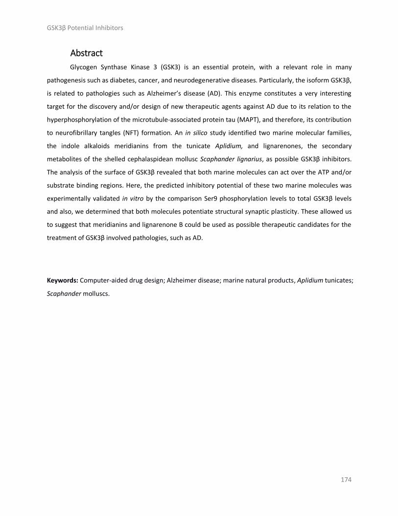
GSK3β Potential Inhibitors
174
Abstract
Glycogen Synthase Kinase 3 (GSK3) is an essential protein, with a relevant role in many
pathogenesis such as diabetes, cancer, and neurodegenerative diseases. Particularly, the isoform GSK3β,
is related to pathologies such as Alzheimer’s disease (AD). This enzyme constitutes a very interesting
target for the discovery and/or design of new therapeutic agents against AD due to its relation to the
hyperphosphorylation of the microtubule-associated protein tau (MAPT), and therefore, its contribution
to neurofibrillary tangles (NFT) formation. An in silico study identified two marine molecular families,
the indole alkaloids meridianins from the tunicate Aplidium, and lignarenones, the secondary
metabolites of the shelled cephalaspidean mollusc Scaphander lignarius, as possible GSK3β inhibitors.
The analysis of the surface of GSK3β revealed that both marine molecules can act over the ATP and/or
substrate binding regions. Here, the predicted inhibitory potential of these two marine molecules was
experimentally validated in vitro by the comparison Ser9 phosphorylation levels to total GSK3β levels
and also, we determined that both molecules potentiate structural synaptic plasticity. These allowed us
to suggest that meridianins and lignarenone B could be used as possible therapeutic candidates for the
treatment of GSK3β involved pathologies, such as AD.
Keywords: Computer-aided drug design; Alzheimer disease; marine natural products, Aplidium tunicates;
Scaphander molluscs.

GSK3β Potential Inhibitors
175
Resum
La glycogen synthase kinase-3 (GSK3) és una proteïna essencial, amb un paper rellevant en
moltes patologies com ara la diabetis, el càncer i les malalties neurodegeneratives. En particular, la
isoforma GSK3β està relacionada amb patologies com la malaltia d'Alzheimer (MA). Aquest enzim
constitueix un objectiu molt interessant per al descobriment i/o disseny de nous agents terapèutics
contra la MA a causa de la seva relació amb la hiperfosforilació de la proteïna tau associada als
microtúbuls, i per tant, la seva contribució en la formació de cabdells neurofibril·lars. Els estudis in silico
van identificar dues famílies de molècules marines, els alcaloides indòlics meridianines, del tunicat
Aplidium, i les lignarenones, metabòlits secundaris del mol·lusc cefalaspidi amb closca Scaphander
lignarius, com a possibles inhibidors GSK3β. L'anàlisi de la superfície de GSK3β revelà que les dues
molècules marines poden actuar sobre les regions d'unió del trifosfat d'adenosina (ATP) i/o del substrat.
El predit potencial inhibitori d'aquestes dues molècules marines va ser validat experimentalment in vitro
mitjançant la comparació dels nivells de fosforilació de Ser9 i els nivells totals de GSK3β, a la vegada que
es va poder observar que les dues molècules potencien la plasticitat sinàptica estructural. Aquests fets
ens permeten suggerir que les meridianines i la lignarenone B podrien ser utilitzats com a possibles
candidats terapèutics per al tractament de GSK3β implicada en patologies com ara la MA.
Paraules clau: Disseny de fàrmacs assistit per ordinador, Malaltia d’Alzheimer, Productes naturals
marins, Tunicats Aplidium, Mol·luscs Scaphander.

GSK3β Potential Inhibitors
176
Introduction
Glycogen Synthase Kinase 3 (GSK3) is one of the best known kinases and it has been widely
studied since in 1992, when its key contribution to the abnormal phosphorylation of the microtubule-
binding protein tau (MAPT) in the process thought to cause neurofibrillary tangles (NFT) formation in
Alzheimer’s disease (AD), was first discovered [1,2]. The most consistent AD disease manifestations are
extracellular senile plaques composed by amyloid-β (Aβ) proteins and NFT, mainly formed by
hyperphosphorylated tau protein. In the last years, GSK3 has emerged as a potential drug target. GSK3 is
a ubiquitous serine (Ser)/threonine (Thr) protein kinase widely expressed in many types of cells and
tissues, and particularly abundant in the brain. GSK3 is involved in a diversity of processes and pathways,
catalysing the transfer of a phosphate group from adenosine triphosphate (ATP) to Ser and Thr
aminoacid residues of target substrates. GSK3 was considered interesting because its wide involvement
in diseases and cellular processes, but also for its unconventional characteristics for being a kinase. GSK3
is constitutively active, its substrates usually need to be pre-phosphorylated by another kinase, and it is
inhibited, rather than activated, in response to stimulation of the insulin and Wnt pathways [3–5]. There
are two highly conserved isoforms of GSK3, that is GSK3α and GSK3β, sharing an overall identity of 84%,
and a 98% of sequence identity on their catalytic domains. Due to the historical relation of the isoform β
with several neurological diseases as well as aging processes or diabetes, it has received more attention
than GSK3α. GSK3β, widely present in the brain, is associated with several neurodegenerative diseases,
including Parkinson’s disease (PD), AD, and Huntington’s disease (HD) [6–8].
The predominant hypothesis in AD suggest that the activity of phosphatases and kinases, in
particular GSK3β, is affected by the amyloid peptides. All these changes result in an increase of protein
Tau phosphorylation. Changes in kinase activity are an intrinsic aspect of the pathological problem in AD,
as they negatively affect, even interrupting, synaptic signals essential for learning and memory [9]. GSK3
activity can be regulated by serine 9/21 phosphorylation and by Thr phosphorylation at residues 216
and 279, although this last phosphorylation is less common. GSK3 is usually phosphorylated at different
sites, but the regulatory outcomes of this remain unclear [3].
In AD, GSK3β is commonly regulated by inhibitory phosphorylation on Ser9, located at the N-
terminal tail. The deregulation of this process results in a GSK3β permanent abnormal activation which
then, hyperphosphorylate tau leading to its aggregation [7,10–12]. From a drug development
perspective, the strategies aimed to target GSK3β are oriented towards reducing tau
hyperphosphorylation by its inhibition. Thus, this is considered a promising therapeutic avenue for AD,
even more now that the amyloid hypothesis, that was never universally accepted, has not given too

GSK3β Potential Inhibitors
177
promising results [13,14]. Although it has been the predominant target and therapeutic hypothesis in
the last years, as corroborated by its predominant presence in the 112 molecules tested in clinical trials
from Phase I to III in 2018 [15,16]. Inhibition of tau aggregation is conceptually more tempting, because
there seems to be a wider general consensus about its damaging effects [17]. In that sense, GSK3β is
related to the amyloid hypothesis as the main therapeutic target, where Aβ fibrillar forms, are a possible
therapeutic alternative. Significant efforts have been made in the past years to design new potent and
selective GSK3 inhibitors, acting over the ATP catalytic pocket or over other allosteric cavities [18].
However, most of the obtained compounds have been considered as hits or starting points, but have not
advanced to the clinic because of administration, distribution, metabolism, excretion and toxicity
(ADMET) problems [19]. In fact, some of the early GSK3β inhibitors that entered into clinical trials failed
for toxicity problems or because off-target interactions, among other reasons [20,21]. Thus, there is still
an opportunity to develop better and safer GSK3β inhibitors, and marine natural products could play a
key role on this [22–27].
Bioactive natural products, from animals, plants, fungi, or microorganisms, are a source of
inspiration and play an important role in the discovery and design of new drugs [28–30]. Also, these
biological active metabolites can be used as a template to design drug-like compounds. One of the most
promising lines in the drug discovery pipeline is the identification of natural products capable of
modulating protein functions in pathogenesis-related pathways [31]. Within natural products, those
coming from the still under explored marine world are of great interest since they shelter most of the
biodiversity of the world [32,33]. An assessment of all Food and Drug Administration (FDA) approved
new molecular entities (NMEs) reveals that natural products and their derivatives represent over 38% of
all NMEs [34]. In other words, by the end of 2013 the FDA approved 547 natural products [34]. AD is a
pathology where several natural products have been proposed as drug candidates, covering different
therapeutic activities [22–24]. Indeed, in the last few years, an increasing number of possible GSK3β
inhibitors have been reported from marine invertebrates, as hymenialdisine isolated from a marine
sponge Axinella [35], the natural phenylmethylene hydantoin (PMH) isolated from the Red sea sponge
Hemimycale arabica [36], or even meridianins and its derivatives from tunicates, revealing the potential
of marine natural products as GSK3β potential therapeutic agents [25–27,37]. These compounds can
and should be a starting point to develop new bioinspired drugs against GSK3β. World oceans and its
coasts are exceptionally rich in species diversity and its exploration offers unique life forms full of
structurally diverse organisms and biological compounds which can also be tested as therapeutic agents

GSK3β Potential Inhibitors
178
[38]. The study of some of these marine natural products revealed their potential inhibitory function,
that can be further exploited and also developed as marketable drugs [39,40].
In a previous study of our group, aimed to unraveling the potential biological activities of a set of
selected small marine molecules, two of them were found particularly interesting as potential
therapeutic agents against GSK3β: meridianin A and lignarenone B [41]. Meridianins are a family of
marine indole alkaloids isolated from the cold waters of Antarctica. The natural products of this ascidian
consist of an indole framework linked to an aminopyrimidine ring, among other compounds. In contrast,
lignarenones, isolated from a mollusc in the temperate waters of the Mediterranean Sea, are two
phenyl conjugated trienones, also classified as polyketides (Figure 1). These compounds have been
isolated from specimens of the tunicate genus Aplidium and the shelled cephalaspidean mollusc
Scaphander lignarius, respectively [42,43].
Supporting this finding, a large number of heterocyclic inhibitors of GSK3 have been identified in
the past few years and they can be classified as ATP-competitive and non-ATP-competitive. The largest
group is that of compounds acting in the ATP catalytic site, i.e. competing against ATP, including
pyrazolopyrimidines, benzimidazoles, pyridinones, pyrimidines, indolylmaleimides, imidazopyridines,
oxadiazoles, or pyrazines among others [31]. Inhibitors that do not compete with ATP, acting over
allosteric cavities, include for instance thiadiazolidinones (TDZDs), halomethylketoneS (HMK), or 5-
imino-1,2,4-thiadiazole (ITZDs) derivatives [18,44,45]. In addition to the aforementioned groups of
inhibitors, in the last few years, a growing number of GSK3 inhibitors have been reported from marine
invertebrates, such as hymenialdisine, indirubines, manzamines, isoflavones or the linear
furanosesquiterpenes palinurin and tricantin [25,27,46]. Interestingly, the chemical structures of most of
these marine inhibitors have an hereocyclic scaffold, similar to the indole scaffold of meridianins (Figure
1). However, not all of them are heterocyclic compounds; for example, palinurin and tricantin, are
sesquiterpenes which present a linear structure, together with aromatic cycles in the terminal part,
similar to lignarenones (Figure 1). Also, it has to be mentioned that these compounds have been
synthesized in the past and this is relevant for the development of new derivatives from both
meridianins and lignarenones [47–50].

GSK3β Potential Inhibitors
179
Meridianin A R1 = OH, R2 = H, R3 = H, R4 = H
Meridianin B R1 = OH, R2 = H, R3 = Br, R4 = H
Meridianin C R1 = H, R2 = Br, R3 = H, R4 = H
Meridianin D R1 = H, R2 = H, R3 = Br, R4 = H Lignarenone A
Meridianin E R1 = OH, R2 = H, R3 = H, R4 = Br
Meridianin F R1 = H, R2 = Br, R3 = Br, R4 = H
Meridianin G R1 = H, R2 = H, R3 = H, R4 = H Lignarenone B
Figure 1. Structure of meridianins A-G and lignarenones A-B, natural compounds isolated from marine organisms.
In this study, we analyze and report the possible inhibitory activity of meridianins and
lignarenones. In the case of the lignarenones, due to the high structural similarity between both (A and
B) species, almost identical from a computational point of view, computational studies were performed
only over lignarenone B. We aimed to show whether these compounds were able to inhibit GSK3β
activity. To do so, a first computational approach using docking calculations and molecular dynamics
(MD) simulations was performed with the aim of elucidating the ability of meridianins and lignarenones
to act as ATP competitive or non-ATP-competitive inhibitors, and finding possible allosteric binding
cavities if it was the case. Moreover, an experimental validation of the inhibitory activity of both
molecular families was performed by Dr. Alberch group, interested in identifying new therapeutic
targets to develop new treatments for neurodegenerative diseases.
Results and discussion
Exploring druggable binding sites on GSK3β.
In this study, with the objective of identifying novel allosteric binding sites, using fpocket [51] to
analyse the GSK3β surface, we obtained 15 plausible cavities (Figure 2). Previous search of
crystallographic structures of GSK3β from the Protein Data Bank (PDB) [52] resulted in more than
thirteen different GSK3β complexes available with a good resolution (lower than 2.5 Å). After a careful
check of all of them, we selected the 6B8J crystal structure, representing human GSK3β together with
CHIR99021, a selective inhibitor, released in 2017 [53]. This new PDB was not used in previously
published works where similar studies were performed [18]. GSK3β ATP catalytic pocket has been widely
explored for decades, often showing the specificity problems characteristic of protein kinases. Allosteric

GSK3β Potential Inhibitors
180
cavities open the possibility of designing inhibitors without these inconveniences. The search of
druggable cavities over the protein surface, that could be an allosteric pocket or a place where a
molecule can be retained, for instance, affecting the efficacy of a given drug, is becoming more common
nowadays. There are several software tools designed to do that, based on different principles like
geometrical, probe and/or energy-based algorithms. Detection, comparison, and analyses of ligand
binding pockets is a key step in structure-based drug design [54]. One of the main issues associated with
pocket finding is druggability prediction, as it is important to avoid failing during the drug discovery
process and to focus the efforts of the process on the discovery of cavity sites that can offer a better
prospect. The concept of druggability adds a new dimension to pocket finding, as it evaluates the
likelihood that small drug-like molecules can bind a given cavity, and thus a concrete target; in some
cases, even evaluating if the binding could have sufficient potency to alter the protein activity [55].
Pocket Score
1 30.818
2 19.206
3 15.124
4 11.417
5 10.933
6 8.099
7 6.028
8 5.240
9 5.011
10 4.868
11 4.825
12 4.015
13 3.990
14 3.168
15 2.800
Figure 2. Cavities found (orange) by fpocket and plot on surface of GSK3β (green). Numbers correspond to
the ranking given by fpocket (1 is the best, and 15 the worst). In the list the score values of each pocket are shown.
Noworse details regarding the scoring function of fpocket can be found at Le Guilloux, et al. (2009).
Most of the pockets found here were previously described by Palomo et al. (2011) over a
different crystal structure, thus supporting the output of the performed cavity detection. Interestingly,
looking at the obtained ranked pocket list (Figure 2), and the corresponding images, the best cavity,
number 1, is not the well-known ATP-binding site, which is number 2. Instead, the best cavity
corresponds to the substrate binding site. The finding of the substrate pocket as a plausible drug-binding
1 2
4
7
8
9
10
0
11 14
12 3
5
6
15
13

GSK3β Potential Inhibitors
181
cavity is supported by literature data. For instance, Manzamine A, a β-carboline alkaloid isolated from
the marine sponges Haliclona and Acanthostrongylophora, or its derivatives, can bind to GSK3β near the
activation pocket formed by the key residues ARG96, ARG180 and LYS205 [56]. These residues are
located at the predicted cavity 1, the substrate pocket. Moreover, Zefirov et al. published in 2010 a
study confirming the interaction of manzamine A with this pocket [57]. Therefore, despite the
druggability score of the substrate pocket being lower than that obtained for the ATP cavity, our results
are in agreement with the literature, reinforcing the election of pocket 1 as a good binding site for small
molecules. It is worth mentioning that the fpocket ranking is based in a general score calculated from
different properties, including druggability (see methods section and Le Guilloux et al., 2009 for further
details) [51]. Thus, the pocket with the highest druggability may not be the “best” pocket, as seen here,
because the ATP-pocket is more druggable than the substrate pocket, but the rest of the properties
placed it in the second place. Anyway, both pockets are good enough to host drug-like compounds.
Due to the interest in developing allosteric inhibitors and the high number of cavities found on
the surface of GSK3β, it is relevant to study the capacity of meridianins and lignarenones to act as
allosteric inhibitors as well as ATP-competitive inhibitors. From the 15 cavities detected by fpocket, we
decided to focus only on the ATP cavity and the substrate binding pocket since, due to its druggability
and structural properties. As said, they are the two most suitable cavities to host a small molecule
inhibitor. We also showed the location of the pockets and the amino acids involved in each cavity
(Figure 3).
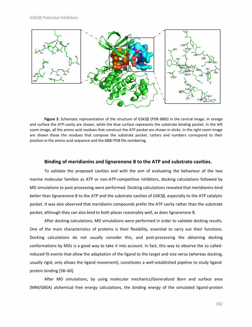
GSK3β Potential Inhibitors
182
Figure 3. Schematic representation of the structure of GSK3β (PDB 6B8J) in the central image. In orange
and surface the ATP-cavity are shown, while the blue surface represents the substrate binding pocket. In the left
zoom image, all the amino acid residues that construct the ATP pocket are shown in sticks. In the right zoom image
are shown those the residues that compose the substrate pocket. Letters and numbers correspond to their
position in the amino acid sequence and the 6B8J PDB file numbering.
Binding of meridianins and lignarenone B to the ATP and substrate cavities.
To validate the proposed cavities and with the aim of evaluating the behaviour of the two
marine molecular families as ATP or non-ATP-competitive inhibitors, docking calculations followed by
MD simulations to post-processing were performed. Docking calculations revealed that meridianins bind
better than lignarenone B to the ATP and the substrate cavities of GSK3β, especially to the ATP catalytic
pocket. It was also observed that meridianin compounds prefer the ATP cavity rather than the substrate
pocket, although they can also bind to both places reasonably well, as does lignarenone B.
After docking calculations, MD simulations were performed in order to validate docking results.
One of the main characteristics of proteins is their flexibility, essential to carry out their functions.
Docking calculations do not usually consider this, and post-processing the obtaining docking
conformations by MDs is a good way to take it into account. In fact, this way to observe the so called-
induced fit events that allow the adaptation of the ligand to the target and vice versa (whereas docking,
usually rigid, only allows the ligand movement), constitutes a well-established pipeline to study ligand-
protein binding [58–60].
After MD simulations, by using molecular mechanics/Generalized Born and surface area
(MM/GBSA) alchemical free energy calculations, the binding energy of the simulated ligand-protein

GSK3β Potential Inhibitors
183
complexes was estimated, ranking the compounds according to it. The obtained energy ranking is more
realistic that the one obtained from docking, as the dynamic nature of the protein and the solvent
environment are added to the equation (although the obtained energies are far to be directly
comparable to those obtained experimentally) [61]. The observed trend at docking results is confirmed
after MD, being all the best energies corresponding to the molecules binding over the ATP cavity, which
are at least 6 kcal/mol higher than those obtained on the substrate pocket (although most are around
10 kcal/mol and on the particular case of meridianin F, reaching a 18 kcal/mol difference). We obtained
the binding energies after each calculation (Table 1).
Table 1. Summary of the binding energy results after docking calculations and after 1 ns MD simulations
with molecular mechanics/generalised born surface area MM/GBSA calculations. All energy values are in kcal/mol.
Substrate cavity ATP cavity
Binding Energy MM/GBSA Binding Energy MM/GBSA
(kcal/mol) (kcal/mol) (kcal/mol) (kcal/mol)
R0/R1 R0/R1
Meridianin A -6.2/-6.2 -22.39 -7.8/-7.8 -28.68
Meridianin B -6.4/-6.4 -23.60 -7.4/-7.4 -34.23
Meridianin C -6.5/-6.5 -17.48 -8.0/-8.0 -32.18
Meridianin D -6.1/-6.2 -25.59 -7.6/-7.5 -35.53
Meridianin E -6.4/-6.4 -23.86 -7.6/-7.6 -28.18
Meridianin F -6.7/-6.6 -23.49 -8.1/-8.2 -41.75
Meridianin G -6.8/-6.8 -16.89 -7.9/-7.9 -29.88
Lignarenone B -6.0/-6.0 -18.59 -6.7/-6.7 -28.79
The binding mode of the complexes (GSK3β - marine molecule), when the compounds are
bound to the ATP and the substrate binding pocket, were found to be very stable. This stability was
evaluated qualitatively by carefully visualizing the generated trajectories and complexes, and it was also
assessed quantitatively by the analysis of the hydrogen bonds (HBs) present on each complex along the
MD trajectory, as well as by analysing the temperature, kinetic analysis, and root-mean-square deviation
(RMSD), as seen in Appendix. Since MDs are dynamic processes, the number of HBs is not constant; they
can be continuously forming and breaking or be stable, depending on the system under study. In the
case of GSK3β bound to meridianins A-G and lignarenone B, we found nine important HBs at the ATP
cavity. These HBs are established with residues F67, V70, K85, D133, V135, R141, Q185, C199, and D200.
Most of the listed aminoacidic residues are configuring the ATP binding pocket (Figure 4) [20,25,26,57].
For the substrate binding pocket, we found 16 important HBs. These were formed with F67, K85, K86,

GSK3β Potential Inhibitors
184
L88, Q89, F93, K94, N95, R96, R180, G202, S203, A204, K205, E211, and P212. All these residues are part
of the substrate pocket (Figure 6) [46]. In addition to the HBs, we also found that both, meridianins and
lignarenone B, can interact with I62, F67, V70, A83, K85, E97, L132, T138, L188, and D200 via
hydrophobic contacts on the ATP cavity and on the substrate pocket. Meridianins establish hydrophobic
contacts with D90, F93, K94, N95, R96, K205, and I217, while lignarenone B does with D90, F93, N95,
and R96. Also, two salt bridges were established on this pocket with residues D90 and E211 when
meridianins are bound.
Figure 417. Representation of the two binding cavities ATP and substrate in surface and the binding mode
of the marine molecules. On the left, the ATP pocket with all the meridianins and lignarenone B. On the right the
substrate pocket also with all the meridianins and lignarenone B. Both images represent the last frame after MD
simulation. Meridianin A-G colours: Peach, blue, tan, orange, pink, cyan and yellow. Lignarenone colour: green.
In the ATP cavity, meridianins A-G and lignarenone B are placed on a very similar way, while on
the substrate binding pocket the molecular placement of each compound varies with respect to the
other, although with some shared features (Figure 4). One fact that could explain this pattern is the
different dimension of the pockets. While the substrate pocket has a volume of 1808.60 Å3, the ATP
pocket represents a quarter of its volume 404.38 Å3, and thus, due to the size differences between
meridianins and lignarenones in a reduced space the position should be different.
The docking post-processing of MDs allows the observation of induced fit events, as mentioned
above. The existence or the magnitude of these events can be measured in different ways. For example,
qualitatively looking how the binding site slightly changes its conformation, or measuring how the

GSK3β Potential Inhibitors
185
interaction that stabilizes the ligand over the protein changes with the MD, or analysing the RMSF of the
ligand-target complexes along MDs, i.e. measuring the amplitude of atom motions during the MD
trajectory, and elucidating the flexible regions of the protein [62,63]. In this particular case, we analysed
the RMSF of GSK3β when meridianins and lignarenone B are bound over the ATP or substrate pockets
(Figure 5), obtaining homologous results. We found six peaks of fluctuations (corresponding to six
different protein regions) with high mobility respect to the baseline. The first one is placed on residues
49-50, the second and the highest one between residues 91 to 94, the third involved the residue 124,
the fourth involved residues 148 and 150, the fifth fluctuation occurs on residue 209, and the sixth is
localized on residues 290 and 292.
In the RMSF, when the marine molecules are bound to the substrate pocket, one more
fluctuation on residue 66 can be observed, while in the ATP fluctuation analysis, focusing on this
residue, despite the RMSF values are not zero (in fact they are a little bit higher than 0.20 nm) they do
not reach the amplitude of others (going from 0.25 to 0.40/0.50 nm, depending on the molecule bound).
Only when meridianin G and lignarenone B are bound, fluctuations reach the 0.25 nm on this 66 residue.
Another pattern that has been detected is the fact that when lignarenone B is bound to any of
both pockets, the fluctuation of GSK3β is in general higher than when meridianins bound, and this could
be related to the structure (size, linearity, etc.) of lignarenone B, which could provoke larger changes in
the protein conformation to allow an optimal placement, improving the poses obtained during the
docking process.
On the other hand, residue K94, present on the substrate pocket and a key component of the
cavity, which establishes HBs with the marine molecules during the binding, is highly fluctuating. This
can be explained because this residue is placed on the loop of the N-lobe, a very flexible region, which is
very exposed to the solvent during the MD simulation [64]. Also, substrate recognition requires GSK3β
residues, F67, Q89, and N95, which facilitate the precise positioning of the substrate within the
substrate binding pocket, and provide an insight into the substrate binding and specificity [65]. To do so,
the flexibility of the loop is necessary, and this is translated into high RMSF values of these residues and
those next to them, as seen on peak 2 and the peak on the residue 66 during the binding of the
meridianins and lignarenone B to the substrate pocket (Figure 5).
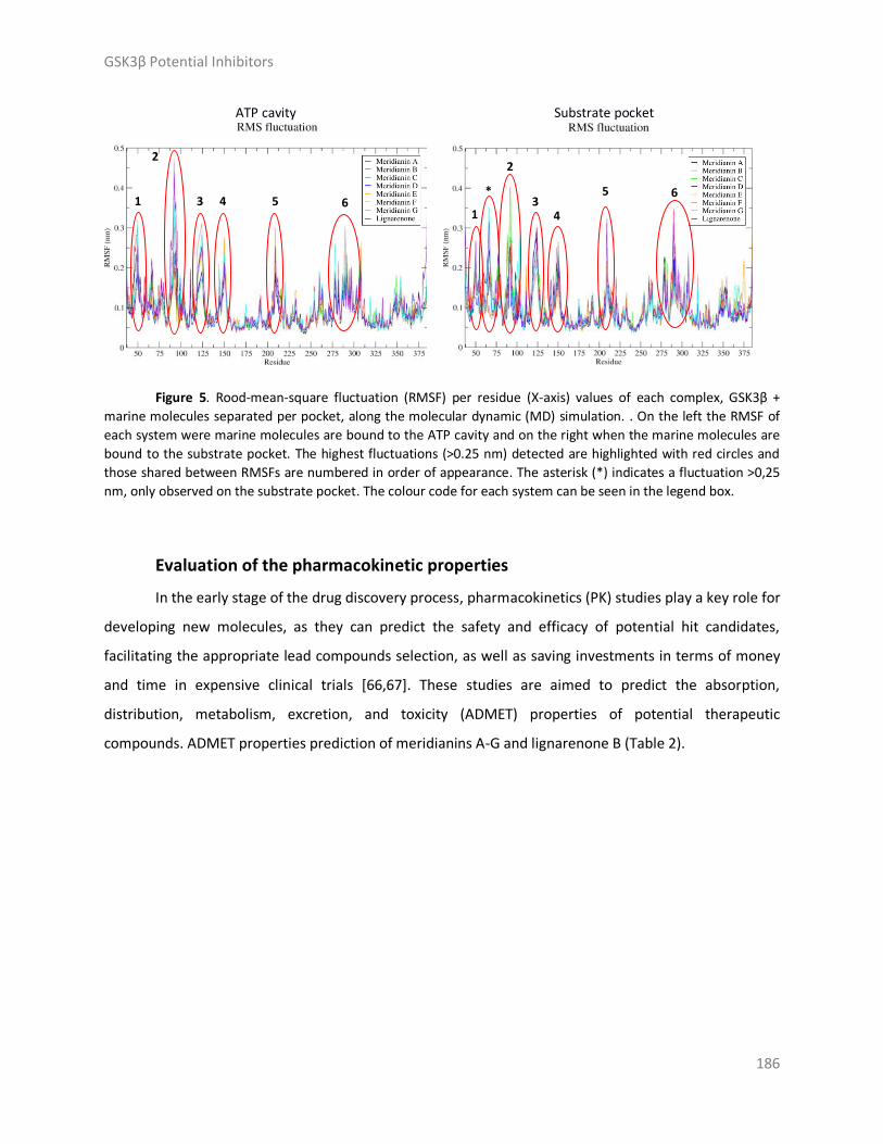
GSK3β Potential Inhibitors
186
ATP cavity Substrate pocket
Figure 5. Rood-mean-square fluctuation (RMSF) per residue (X-axis) values of each complex, GSK3β +
marine molecules separated per pocket, along the molecular dynamic (MD) simulation. . On the left the RMSF of
each system were marine molecules are bound to the ATP cavity and on the right when the marine molecules are
bound to the substrate pocket. The highest fluctuations (>0.25 nm) detected are highlighted with red circles and
those shared between RMSFs are numbered in order of appearance. The asterisk (*) indicates a fluctuation >0,25
nm, only observed on the substrate pocket. The colour code for each system can be seen in the legend box.
Evaluation of the pharmacokinetic properties
In the early stage of the drug discovery process, pharmacokinetics (PK) studies play a key role for
developing new molecules, as they can predict the safety and efficacy of potential hit candidates,
facilitating the appropriate lead compounds selection, as well as saving investments in terms of money
and time in expensive clinical trials [66,67]. These studies are aimed to predict the absorption,
distribution, metabolism, excretion, and toxicity (ADMET) properties of potential therapeutic
compounds. ADMET properties prediction of meridianins A-G and lignarenone B (Table 2).
* 6 5
4 3
2
1
6 5 4 3
2
1

GSK3β Potential Inhibitors
187
Table 2. Summary of absorption, distribution, metabolisim, excretion and toxicity properties of
meridianins and lignarenone B. Pgp: P-Glycoprotein, BBB: blood-brain barrier, PPB: plasma-protein binding,
CYP450: cytochrom P450, OCT2: organic cation transporter 2, hERG: human ether-a-go-go gene.
Absorption Distribution Metabolism Excretion Toxicity
Mol
Weight logS Pgp Caco2 logP BBB PPB CYP450 OCT2
Substrate hERG
Meridianin A 226.2 -4.2 inactive High 1.5 NO High Yes No <4.0
Meridianin B 305.1 -5.0 inactive High 2.4 NO High Yes No <4.0
Meridianin C 289.1 -5.6 inactive High 3.1 NO High Yes No <4.0
Meridianin D 289.1 -5.6 inactive High 3.1 NO High Yes No <4.0
Meridianin E 305.1 -5.0 inactive High 2.4 NO High Yes No <4.0
Meridianin F 368.0 -6.2 inactive High 3.6 NO High Yes No <4.0
Meridianin G 210.2 -4.5 inactive High 2.4 NO High Yes No <4.0
Lignarenone B 212.3 -3.2 inactive Low 3.6 NO High Yes No <4.0
In general, all the tested compounds should be optimized to improve some of their properties.
According to the results, they can be considered hits, but there is still work to do before they become
suitable lead compounds. Meridianins, as shown before, present some solubility problems (logS > 5
indicate not too much solubility), especially meridianin F, as well as shows difficulties to penetrate the
blood brain barrier (BBB) [25], a problem that is also shared by lignarenone B. This is also in agreement
with the obtained logP values (where values lower than 5 indicate that the compounds have an
appropriate hydrophobicity and permeability behavior). However, to become drugs penetrating the
central nervous system (CNS), molecules should have a logP around 2 [68]. Some meridianins are almost
there, but most of our compounds are far of this optimal value. Another issue is that all the studied
compounds present high plasma-protein binding (PPB) probability, indicating that a high percentage of
the administrated compound will be found attached to plasma proteins, affecting their diffusion and
efficiency (Table 2).
On the other hand, all of the compounds seem to be permeable, especially meridianins,
according to Caco2 results (Table 2). Regarding P-glycoprotein (Pgp) binding, none of our compounds
was predicted to act over it. The interaction with Pgp has many pharmacological implications that could
result in pharmaceutical advantages or contraindications. For instance, Pgp modulation has been
suggested as a mechanism to improve CNS pharmacotherapy, but it also plays a major role in the
multidrug resistance (MDR) phenomenon in cancer cells, depending on whether binding happens as a
substrate or as an inhibitor, and also on the isoform that the compound binds to [69–72]. Thus, none of
the molecules tested here seems to interact with Pgp, avoiding possible beneficial but also detrimental
effects.

GSK3β Potential Inhibitors
188
These compounds are able to interact with cytochrom P450 (CYP450), as described in the results
(Table 2) and CYP450 drug metabolism can induce clinical effects. On the contrary, any molecule is a
substrate of organic cation transporter 2 (OCT2), which means that non-clearance problems and adverse
interactions with co-administrated OCT2 inhibitors are expected. Moreover, as another positive point,
none of the compounds is an inhibitor of the potassium channels encoded by the human ether-a-go-go
gene (hERG). hERG inhibition can lead to fatal pathologies, such as cardiac diseases, because it is the
principal cause of the development of acquiring long QT syndrome, fatal arrhythmia, for example [73].
The absence of inhibition of hERG is a good and safe property of both meridianins and lignarenones.
Meridianins and lignarenones differentially increased pGSK3β Ser9, but not total
GSK3β levels in vitro
In order to confirm the predicted interaction between meridianins and lignarenone B towards
GSK3β, we used different doses of both marine molecules, at different times, to pharmacologically
inhibit GSK3β, and comparing Ser9 phosphorylation levels to total levels of GSK3β as an indication of
inhibition. Primary cortical cultures of neurons were treated with vehicle, meridianins, and lignarenones
(500 nM) or meridianins and lignarenones (10μM) for two time points, 15 and 60 min. Western blot
analysis was used to determine protein expression levels of GSK3β and pGSK3β Ser9, which is an
inhibitory phosphorylation site.
A one-way ANOVA analysis indicated that meridianins treatment significantly increased pGSK3β
levels, both after 15 min (F2, 30 = 4.189, p = 0.024) and after 60 min (F2,27 = 6.892, p = 0.0038). Specifically,
post hoc analysis revealed that at 60 min both doses, 500 nM (p < 0.05) and 10μM (p < 0.01) significantly
increased pGSK3β levels. Similarly, post hoc analysis after 15 min of treatment indicated that the dose of
10μM (p < 0.01), but not the dose of 500nM, exerted significant effects on pGSK3β levels (Figure 6).
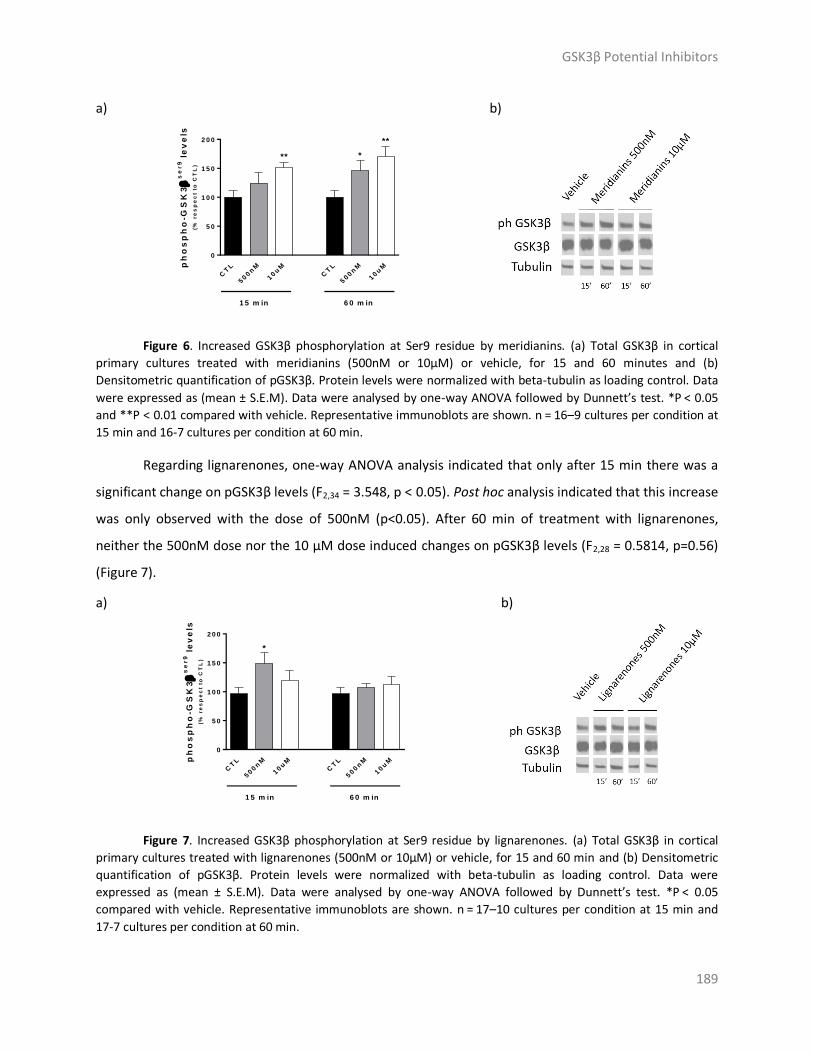
GSK3β Potential Inhibitors
189
a) b)
ph
os
ph
o-G
SK
3
se
r9 l
ev
els
(% r
es
pe
ct
to C
TL
)
CT
L
500n
M
10u
MC
TL
500n
M
10u
M
0
5 0
1 0 0
1 5 0
2 0 0
**
**
*
1 5 m in 6 0 m in
Figure 6. Increased GSK3β phosphorylation at Ser9 residue by meridianins. (a) Total GSK3β in cortical
primary cultures treated with meridianins (500nM or 10μM) or vehicle, for 15 and 60 minutes and (b)
Densitometric quantification of pGSK3β. Protein levels were normalized with beta-tubulin as loading control. Data
were expressed as (mean ± S.E.M). Data were analysed by one-way ANOVA followed by Dunnett’s test. *P < 0.05
and **P < 0.01 compared with vehicle. Representative immunoblots are shown. n = 16–9 cultures per condition at
15 min and 16-7 cultures per condition at 60 min.
Regarding lignarenones, one-way ANOVA analysis indicated that only after 15 min there was a
significant change on pGSK3β levels (F2,34 = 3.548, p < 0.05). Post hoc analysis indicated that this increase
was only observed with the dose of 500nM (p<0.05). After 60 min of treatment with lignarenones,
neither the 500nM dose nor the 10 μM dose induced changes on pGSK3β levels (F2,28 = 0.5814, p=0.56)
(Figure 7).
a) b)
ph
os
ph
o-G
SK
3
se
r9 l
ev
els
(% r
es
pe
ct
to C
TL
)
CT
L
500n
M
10u
MC
TL
500n
M
10u
M
0
5 0
1 0 0
1 5 0
2 0 0
1 5 m in 6 0 m in
*
Figure 7. Increased GSK3β phosphorylation at Ser9 residue by lignarenones. (a) Total GSK3β in cortical
primary cultures treated with lignarenones (500nM or 10μM) or vehicle, for 15 and 60 min and (b) Densitometric
quantification of pGSK3β. Protein levels were normalized with beta-tubulin as loading control. Data were
expressed as (mean ± S.E.M). Data were analysed by one-way ANOVA followed by Dunnett’s test. *P < 0.05
compared with vehicle. Representative immunoblots are shown. n = 17–10 cultures per condition at 15 min and
17-7 cultures per condition at 60 min.
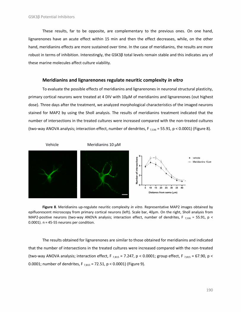
GSK3β Potential Inhibitors
190
These results, far to be opposite, are complementary to the previous ones. On one hand,
lignarenones have an acute effect within 15 min and then the effect decreases, while, on the other
hand, meridianins effects are more sustained over time. In the case of meridianins, the results are more
robust in terms of inhibition. Interestingly, the GSK3β total levels remain stable and this indicates any of
these marine molecules affect culture viability.
Meridianins and lignarenones regulate neuritic complexity in vitro
To evaluate the possible effects of meridianins and lignarenones in neuronal structural plasticity,
primary cortical neurons were treated at 4 DIV with 10μM of meridianins and lignarenones (out highest
dose). Three days after the treatment, we analyzed morphological characteristics of the imaged neurons
stained for MAP2 by using the Sholl analysis. The results of meridianins treatment indicated that the
number of intersections in the treated cultures were increased compared with the non-treated cultures
(two-way ANOVA analysis; interaction effect, number of dendrites, F 7,536 = 55.91, p < 0.0001) (Figure 8).
Vehicle Meridianins 10 µM
Figure 8. Meridianins up-regulate neuritic complexity in vitro. Representative MAP2 images obtained by
epifluorescent microscopy from primary cortical neurons (left). Scale bar, 40μm. On the right, Sholl analysis from
MAP2-positive neurons (two-way ANOVA analysis; interaction effect, number of dendrites, F 7,536 = 55.91, p <
0.0001). n = 45-55 neurons per condition.
The results obtained for lignarenones are similar to those obtained for meridianins and indicated
that the number of intersections in the treated cultures were increased compared with the non-treated
(two-way ANOVA analysis; interaction effect, F 7,815 = 7.247, p < 0.0001; group effect, F 7,815 = 67.90, p <
0.0001; number of dendrites, F 7,815 = 72.51, p < 0.0001) (Figure 9).

GSK3β Potential Inhibitors
191
Vehicle Lignarenones 10 µM
Figure 9. Lignarenones up-regulate neuritic complexity in vitro. Representative MAP2 images obtained by
epifluorescent microscopy from primary cortical neurons (left). Scale bar, 40μm. On the right, Sholl analysis from
MAP2-positive neurons (two-way ANOVA analysis; interaction effect, F 7,815 = 7.247, p < 0.0001; group effect, F 7,815
= 67.90, p < 0.0001; number of dendrites, F 7,815 = 72.51, p < 0.0001). n = 45-55 neurons per condition.
In both cases, positive results are obtained because an increase of the neurite outgrowth is
clearly observed. These results are in agreement with the previous ones, reported above.
Effect of meridianins and lignarenones on neuronal viability
To elucidate pharmacological effects of meridianins and lignarenones treatments, we then
analysed the cell viability on primary cortical cultures treated at 4 DIV at 10μM (out highest dose). Even
if some previous works did mention the toxicity of this kind of marine molecules at neuronal level
(Llorach-Pares et al., 2017), our results showed that neither meridianins nor lignarenones induced
changes in cell density in our primary cultures, meaning that cells were viable under the experimental
conditions evaluated (one-way ANOVA analysis, F 2,13 = 1.600, p=0.2392) (Figure 10) [25,74].

GSK3β Potential Inhibitors
192
Vehicle
Lignarenones
10 µM
Meridianins
10 µM
Figure 10. Cell viability is not affected by the highest doses of both meridianins and lignarenones used in
the experiments. Representative DAPI images obtained by epifluorescent microscopy from primary cortical
neurons in the control treatment (right). Scale bar, 50μm. Cell count by DAPI staining shows no significant
differences between groups (One-way ANOVA analysis, F 2,13 = 1.600, p=0.2392) (left). Data were expressed as
(mean ± S.E.M). n = 10 fields counted/6 coverslips per condition.
Materials and Methods
Computational
Target selection and modelling
From all the available structures of human GSK3β in the Protein Data Bank (PDB), the 3-
dimensions (3D) crystallographic structure 6B8J with the co-crystalized ligand 65C was selected, and
thus the protein structure modelling from it [53,75]. The meridianins and lignarenone B structures were

GSK3β Potential Inhibitors
193
modelled from the 2D chemical structure previously published by Nuñez-Pons and co-workers and
Cutignano et al., respectively [43,76].
Cavity search
Fpocket software, a protein pocket prediction algorithm was used to identify different cavities
on the surface of GSK3β [77]. Fpocket uses the standard PDB files, without ligands or water molecules,
as input, and using alpha spheres predicts the possible binding pockets over the protein surface, ranking
them according to a scoring that involves the druggability, the size of the pocket, and its Surface
Accessible Surface Area (SASA), among other properties [77].
Docking calculations
Docking calculations were performed over the two top ranked pockets in the cavity search: the
ATP and the substrate pocket. The modelled protein structures and the marine molecules were
employed as input of the calculations performed over each of the two cavities using Itzamna software
tool [78]. More details of this approach can be found at Llorach-Pares et al. (2017 and 2018) [25,26], and
Chapters 2-3 of this thesis.
Molecular dynamics simulation
Short MD simulations (1ns), using NAMD software version 2.11, were performed over the top
ranked conformations obtained after docking, which were selected based on free binding energy, to
post-processing them [79]. Each ligand-target complex was protonated at physiological pH 7.4 and then
placed into a TIP3P water cubic box, imposing periodic boundary conditions, in which Na+ and Cl- ions
were added to neutralize the charge of the system [80]. Electrostatic interactions were calculated by the
particle-mesh Ewald method using constant pressure and temperature conditions. Each complex was
solvated with a minimum distance of 10Å from the surface of the complex to the edge of the simulation
box. The temperature was maintained at 300 K using a Langevin thermostat, and the pressure was
maintained at 1 atm using a Langevin Piston barostat. The time step employed was 2 fs. Bond lengths to
hydrogens were constrained with the SHAKE algorithm [81]. Before production runs, the structure was
energy minimized followed by a slow heating-up phase using harmonic position restraints on the heavy
atoms of the protein. Subsequently, the system was energy minimized until volume equilibration,
followed by the production run without restraints. The Amber ff99SB-ILDN and the General Amber Force
Field (GAFF) set of parameters were used to model the target and the marine molecules, respectively

GSK3β Potential Inhibitors
194
[82,83]. Ligand GAFF parameters were obtained using Antechamber, whereas the receptor structures
were modelled using the leap module of Amber Tools [84,85].
Molecular dynamics analysis
Visual inspection to each trajectory and the HBs occupancies analysis was performed using
Visual Molecular Dynamics (VMD) software [86]. Thermodynamic (temperature, potential, kinetic and
total energy) and structural (Rg, RMSD and RMSF) analysis were performed using GROMACS simulation
package [87,88].
Molecular Mechanics Generalized Born Surface Area
After performing MD simulations to estimate the binding free energy of GSK3β – marine
molecules complexes, Molecular Mechanics Generalized Born Surface Area (MM/GBSA) reweighting
techniques were employed [89]. These techniques are widely used and outperform docking results
because they are employed using the MD output trajectory as an input, thus taking into account the
dynamic behaviour of the protein-ligand complexes [59,90]. MM/GBSA rescoring was performed using
the MMPBSA python algorithm contained within the Amber Tools suit [91].
ADMET prediction
ADMETer, a software tool containing supper vector regression (SVR) and supper vector machine
(SVM) ADMET machine-learning (ML) predictive models, was used to assess the ADMET properties of
meridianin A-G and lignarenone B [25,92]. More precisely, the models measured logS (molecular
aqueous coefficient), logP (octanol/water partition coefficient), Caco2 permeability, blood-brain barrier
penetration (BBB), plasma-protein binding (PPB), P-glycoprotein binding (Pgp), Cytocrom P450
(CYPC450), organic cation transporter 2 (OCT2), and hERG mediated toxicity properties.
Graphical representations
Graphical representations were prepared using PyMOL version 1.7 and XMGRACE version 5.1.22
[93,94]. 2D images of marine molecules were prepared using RDKit python library [95].
Experimental
Cortical and striatal primary cultures
Primary cortical neuronal cultures were performed as previously described in the literature [96].
Cortex from E17.5 WT mouse embryos were dissected and gently dissociated with a fire-polished
Pasteur pipette. Cells were seeded (50,000 cells/cm2 for immunochemical staining and

GSK3β Potential Inhibitors
195
800,000 cells/cm2 for Western blot analysis) onto 24mm culture or 60 mm culture plates pre-coated
with 0.1 mg/mL poly-d-lysine (Sigma Chemical Co., St. Louis, MO) and cultured in Neurobasal medium
supplemented with B27 (Gibco, Paisley, Scotland, UK, 50x) and GlutaMAX (Gibco, 100x) at 37 °C in a
humidified atmosphere containing 5% CO2. For biochemical assay, 60 mm culture dishes were treated 7
days in vitro (DIV) with vehicle (PBS), meridianins (10 µM), and lignarenones (10 µM). 15 or 60 min after
treatment, cells were washed with cold PBS and lysed for WB analysis. For immunocytochemical staining
and morphology analyses, 24mm culture dishes were treated during 4 days in vitro (DIV) with vehicle
(PBS), meridianins (10 µM), and lignarenones (10 µM), and assessed 3 days after.
Immunoblot analysis
Cell samples were collected in cold lysis buffer containing 50 mM Tris base (pH 7.5), 10mM
EDTA, 1% Triton X-100, and supplemented with 1 mM sodium orthovanadate, 1mM
phenylmethylsulfonyl fluoride, 1 mg/ml leupeptin and 1 mg/ml aprotinin. Samples were centrifuged at
32,000 g for 15 min and the supernatants collected. Following determination of the protein contents by
Detergent-Compatible Protein Assay (Bio-Rad, Hercules, CA, USA), protein extracts (15 µg) were mixed
with 5 × SDS sample buffer, boiled for 5 min, resolved on 15% SDS–PAGE and transferred to
nitrocellulose membranes (Whatman Schleicher & Schuell, Keene, NH, USA). After incubation (1 h) in
blocking buffer containing 10% non-fat powdered milk in Tris buffered saline-Tween (TBS-T) (50 mM
Tris–HCl, 150 mM NaCl, pH 7.4, 0.05% Tween 20), membranes were blotted overnight at 4 °C with
primary antibodies. Antibodies used for immunoblot analysis were: GSK3β (1:1000; Cell Signaling,
#9315), phosphoGSK3β at Ser9 (1:1000; Cell Signaling, #9336xz), and α-Tubulin (1:40,000; Sigma-Aldrich,
T9026). The membranes were then rinsed three times with TBS-T and incubated with horseradish
peroxidase-conjugated secondary antibody for 1 h at room temperature. After washing for 30 min with
TBS-T, the membranes were developed using the enhanced chemilluminescence ECL kit (Santa Cruz
Biotechnology). The Gel-Pro densitometry program (Gel-Pro Analyzer for Windows, version 4.0.00.001)
was used to quantify the different immunoreactive bands relative to the intensity of the α-tubulin or
phospho GSK3Beta band in the same membranes within a linear range of detection for the ECL reagent.
Immunocytochemical staining
Immunochemical staining was performed following standard protocols available [97]. Briefly,
primary cortical neuronal cultures were fixed at 7 days in vitro (DIV) in 4 % paraformaldehyde for 10
min. After fixation, cells were washed with PBS, incubated 15 min with 0,1M Glycine in PBS, used to
block unreacted aldehydes after fixation, which can cause an increase in background fluorescence. After

GSK3β Potential Inhibitors
196
washes with PBS, cells were permeabilized and blocked during 1h at room temperature with PBS
containing 0,3% Triton X-100, 1% bovine serum albumin and 1% donkey normal serum. Then, cells were
incubated overnight at 4ºC in 0.1 M PBS with 5 % normal horse serum with the proper primary antibody
added. MAP2 (1:1000, Sigma-Aldrich, M1406) primary antibodies was used. After primary antibody
incubation, cultures were washed with PBS and incubated 2h at room temperature with Alexa Fluor 488-
conjugated AffiniPure donkey anti-mouse (1:100; Jackson Immunoresearch Laboratories, Inc., West
Grove, PA). Then, coverslips mounted with Fluoromount containing DAPI onto the surface of a slide after
washes with PBS. Immunofluorescence images were taken using an Olympus BX60 epifluorescence
microscope, using a 20X objective.
Imaging and analysis
The in vitro Sholl analysis was performed with the freeware ImageJ (ImageJ, RRID:SCR 003070).
We evaluated 45–55 neurons, all of them MAP2-positive from one primary cortical culture. To estimate
the density of dendritic spines, 31–41 dendrites from MAP2-positive neurons (1 or 2 dendrites/neuron)
from 3 different cultures were counted.
Statistical analysis
Statistical analysis was performed using one-way ANOVA with the Dunnett’s post hoc test as
appropriate and indicated in the figure legends. Data analysis and graphs were created using Graphpad
Prism Software version 6.0. A 95% confidence interval was used and values of p<0.05 were considered
as statistically significant. Data is expressed as mean ± S.E.M.
Conclusions
In this study we have shown that the marine natural products meridianins and lignarenone B are
capable of inhibiting the activity of GSK3β through an ATP competitive and non-competitive, allosteric,
mechanism, although it seems that they are preferentially ATP-competitive inhibitors.
Docking and MD studies elucidated the binding mode of each of the studied compounds over
GSK3β. The inhibition can be caused by meridianins or lignarenone B occupying the ATP or the substrate
pockets of GSK3β, or even by the simultaneous binding on the ATP and/or the substrate active site.
Experimental analysis confirmed the GSK3β inhibition predicted in silico, as a consequence of growing
Ser9 phosphorylation levels. During the inhibitory process the GSK3β total levels remain stable,
suggesting that neither meridianins nor lignarenone B affect their viability. Moreover, the neurite
outgrowth increased, supporting the no affection of the structural plasticity. However, even with the

GSK3β Potential Inhibitors
197
inhibitory behaviour confirmation, further studies are needed to completely depict the binding mode of
meridianins and lignarenone B.
Results obtained from both computational and experimental studies allow us to suggest that
meridianins and, to a lesser extent, lignarenone B, may inhibit GSK3β. Therefore, these compounds
could be considered as hits, constituting a starting point to develop new future potential therapeutic
agents for the treatment of AD. However, they should be optimized because they are not showing good
absorption and distribution profiles yet. The solubility should be improved as well as the lipophilicity and
the BBB permeability, which are key issues here. Once inside the cells, these compounds have shown
good inhibitory profiles and also good permeability towards the cellular membrane, but nevertheless,
they should be able to penetrate into the brain. Several strategies can be employed for this, ranging
from a proper modification of the chemical structure to improving the nanodelivery, including also the
possibility of becoming Pgp or other protein binders that may facilitate their penetrance [98–100].
Our results from in silico and in vitro experiments constitute a promising starting point for the
development of novel anti-AD drugs and further studies should be devoted to improve the specific
characteristics of the studied marine compounds.
Acknowledgments: This research was partially supported by an Industrial Doctorate grant from
the Generalitat of Catalonia to L.L.-P (DI 2016-051), and the BLUEBIO (CTM2016-78901/ANT) project
from the Spanish government. This work is a contribution to the AntEco programme (SCAR). We thank
Carlos Jiménez, Laura Núñez-Pons, and Jaime Rodríguez for support with meridianins, and Josep Maria
Viñas and his “Estelada” fishing ship and crew for their help in collecting the Scaphander samples.
Author Contributions: L.LL.-P., A.G., M.S.-M., J.A. and C.A. conceived the study and designed the
experiments. L.LL.-P., E.R.-U, and A.G. carried out the experiments. M.S.-M., A.N.-C., C.A. and J.A.
supervised the experiments. All the authors analysed and discussed the results and elaborated the
manuscript.
Conflicts of Interest: The authors declare no conflict of interest.

GSK3β Potential Inhibitors
198
Appendix
The physics of the all simulations has to be checked before any particular MD analysis. Also, due
to its importance and its influence along the simulation, an important value to be checked after an MD is
the temperature, a fundamental concept in physics which represents the intensity of the thermal
motions of molecules [101]. In our study, after the analysis of all the systems, the stability of the
temperature was validated with an average of 297 K ± 3. Energies, total, potential, and kinetic, must also
be taken into account as they physically validate the simulations [102,103]. At this point, the
thermodynamic properties of all the complexes could be confirmed and the structural analysis could
start. Radius of gyration (Rg) which allow the analysis of the compactness of the protein and is related to
the tertiary structure, were analysed when each marine molecule was bound at the ATP and at the
substrate binding pocket (Figure A1) [45]. Root-mean-square deviation (RMSD) is used to validate the
stability along the simulations and measures the average distance between the atoms of superimposed
structures extracted from the MD simulations and a reference structure (Figure A2) [104–106]. The
behaviour of each simulation was checked obtaining positive results.
Figure A1. Time evolution of the Radius of gyration (Rg) obtained for each system. O the left, the systems with each marine molecule bound to the ATP cavity, and on the right, when the marine molecules are bound to the substrate pocket. The colour code for each system can be seen in the legend box.

GSK3β Potential Inhibitors
199
Figure A2. Time evolution of the root-mean-square deviation (RMSD) of the backbone atoms during the molecular dynamics (MD) simulation with respect to the initial structure of GSK3β. On the left, the RMSD of each system where marine molecules are bound to the ATP cavity, and on the right when the marine molecules are bound to the substrate pocket. The colour code for each system can be seen in the legend box.

GSK3β Potential Inhibitors
200
References
1. Hanger, D.P.; Hughes, K.; Woodgett, J.R.; Brion, J.-P.; Anderton, B.H. Glycogen synthase kinase-3 induces Alzheimer’s disease-like phosphorylation of tau: Generation of paired helical filament epitopes and neuronal localisation of the kinase. Neurosci. Lett. 1992, 147, 58–62.
2. Mandelkow, E.-M.; Drewes, G.; Biernat, J.; Gustke, N.; Van Lint, J.; Vandenheede, J.R.; Mandelkow, E. Glycogen synthase kinase-3 and the Alzheimer-like state of microtubule-associated protein tau. FEBS Lett. 1992, 314, 315–321.
3. Hooper, C.; Killick, R.; Lovestone, S. The GSK3 hypothesis of Alzheimer’s disease. J. Neurochem. 2008, 104, 1433–9.
4. Ding, V.W.; Chen, R.H.; McCormick, F. Differential regulation of glycogen synthase kinase 3beta by insulin and Wnt signaling. J. Biol. Chem. 2000, 275, 32475–81.
5. Palsgaard, J.; Emanuelli, B.; Winnay, J.N.; Sumara, G.; Karsenty, G.; Kahn, C.R. Cross-talk between insulin and Wnt signaling in preadipocytes: role of Wnt co-receptor low density lipoprotein receptor-related protein-5 (LRP5). J. Biol. Chem. 2012, 287, 12016–26.
6. Li, D.-W.; Liu, Z.-Q.; Chen, W.; Yao, M.; Li, G.-R. Association of glycogen synthase kinase-3β with Parkinson’s disease. Mol. Med. Rep. 2014, 9, 2043–50.
7. Llorens-Martín, M.; Jurado, J.; Hernández, F.; Avila, J. GSK-3β, a pivotal kinase in Alzheimer disease. Front. Mol. Neurosci. 2014, 7, 46.
8. Lim, N.K.H.; Hung, L.W.; Pang, T.Y.; Mclean, C.A.; Liddell, J.R.; Hilton, J.B.; Li, Q.-X.; White, A.R.; Hannan, A.J.; Crouch, P.J. Localized changes to glycogen synthase kinase-3 and collapsin response mediator protein-2 in the Huntington’s disease affected brain. Hum. Mol. Genet. 2014, 23, 4051–4063.
9. Kremer, A.; Louis, J. V; Jaworski, T.; Van Leuven, F. GSK3 and Alzheimer’s Disease: Facts and Fiction…. Front. Mol. Neurosci. 2011, 4, 17.
10. Goedert, M.; Klug, A.; Crowther, R.A. Tau protein, the paired helical filament and Alzheimer’s disease. J. Alzheimers. Dis. 2006, 9, 195–207.
11. Martin, L.; Latypova, X.; Wilson, C.M.; Magnaudeix, A.; Perrin, M.-L.; Yardin, C.; Terro, F. Tau protein kinases: Involvement in Alzheimer’s disease. Ageing Res. Rev. 2013, 12, 289–309.
12. Kolarova, M.; García-Sierra, F.; Bartos, A.; Ricny, J.; Ripova, D.; Ripova, D. Structure and Pathology of Tau Protein in Alzheimer Disease. Int. J. Alzheimers. Dis. 2012, 2012, 1–13.
13. Kametani, F.; Hasegawa, M. Reconsideration of Amyloid Hypothesis and Tau Hypothesis in Alzheimer’s Disease. Front. Neurosci. 2018, 12, 25.
14. Doig, A.J.; del Castillo-Frias, M.P.; Berthoumieu, O.; Tarus, B.; Nasica-Labouze, J.; Sterpone, F.; Nguyen, P.H.; Hooper, N.M.; Faller, P.; Derreumaux, P. Why Is Research on Amyloid-β Failing to Give New Drugs for Alzheimer’s Disease? ACS Chem. Neurosci. 2017, 8, 1435–1437.
15. del Ser, T.; Steinwachs, K.C.; Gertz, H.J.; Andrés, M. V.; Gómez-Carrillo, B.; Medina, M.; Vericat, J.A.; Redondo, P.; Fleet, D.; León, T. Treatment of Alzheimer’s Disease with the GSK-3 Inhibitor Tideglusib: A Pilot Study. J. Alzheimer’s Dis. 2012, 33, 205–215.
16. Cummings, J.; Lee, G.; Ritter, A.; Zhong, K. Alzheimer’s disease drug development pipeline: 2018. Alzheimer’s Dement. (New York, N. Y.) 2018, 4, 195–214.
17. Citron, M. Alzheimer’s disease: strategies for disease modification. Nat. Rev. Drug Discov. 2010, 9, 387–398.
18. Palomo, V.; Soteras, I.; Perez, D.I.; Perez, C.; Gil, C.; Eugenia Campillo, N.; Martinez, A. Exploring the Binding Sites of Glycogen Synthase Kinase 3. Identification and Characterization of Allosteric Modulation Cavities. J. Med. Chem. 2011, 54, 8461–8470.
19. Walz, A.; Ugolkov, A.; Chandra, S.; Kozikowski, A.; Carneiro, B.A.; O’Halloran, T. V; Giles, F.J.; Billadeau, D.D.; Mazar, A.P. Molecular Pathways: Revisiting Glycogen Synthase Kinase-3β as a

GSK3β Potential Inhibitors
201
Target for the Treatment of Cancer. Clin. Cancer Res. 2017, 23, 1891–1897. 20. Pandey, M.K.; DeGrado, T.R. Glycogen Synthase Kinase-3 (GSK-3)-Targeted Therapy and Imaging.
Theranostics 2016, 6, 571–93. 21. Martinez, A.; Gil, C.; Perez, D.I. Glycogen Synthase Kinase 3 Inhibitors in the Next Horizon for
Alzheimer’s Disease Treatment. Int. J. Alzheimers. Dis. 2011, 2011, 1–7. 22. Russo, P.; Kisialiou, A.; Lamonaca, P.; Moroni, R.; Prinzi, G.; Fini, M. New Drugs from Marine
Organisms in Alzheimer’s Disease. Mar. Drugs 2015, 14, 5. 23. Ansari, N.; Khodagholi, F. Natural Products as Promising Drug Candidates for the Treatment of
Alzheimer’s Disease: Molecular Mechanism Aspect. Curr. Neuropharmacol. 2013, 11, 414–429. 24. Dey, A.; Bhattacharya, R.; Mukherjee, A.; Pandey, D.K. Natural products against Alzheimer’s
disease: Pharmaco-therapeutics and biotechnological interventions. Biotechnol. Adv. 2017, 35, 178–216.
25. Llorach-Pares, L.; Nonell-Canals, A.; Sanchez-Martinez, M.; Avila, C. Computer-Aided Drug Design Applied to Marine Drug Discovery: Meridianins as Alzheimer’s Disease Therapeutic Agents. Mar. Drugs 2017, 15, 366.
26. Llorach-Pares, L.; Nonell-Canals, A.; Avila, C.; Sanchez-Martinez, M. Kororamides, Convolutamines, and Indole Derivatives as Possible Tau and Dual-Specificity Kinase Inhibitors for Alzheimer’s Disease: A Computational Study. Mar. Drugs 2018, 16, 386.
27. Eldar-Finkelman, H.; Martinez, A. GSK-3 Inhibitors: Preclinical and Clinical Focus on CNS. Front. Mol. Neurosci. 2011, 4, 32.
28. Rodrigues, T.; Reker, D.; Schneider, P.; Schneider, G. Counting on natural products for drug design. Nat. Chem. 2016, 8, 531–541.
29. W-H Li, J.; Vederas, J.C. Drug Discovery and Natural Products: End of an Era or an Endless Frontier? Science (80-. ). 2009, 325, 161–165.
30. Harvey, A.L.; Edrada-Ebel, R.; Quinn, R.J. The re-emergence of natural products for drug discovery in the genomics era. Nat. Rev. Drug Discov. 2015, 14, 111–129.
31. Prachayasittikul, V.; Worachartcheewan, A.; Shoombuatong, W.; Songtawee, N.; Simeon, S.; Prachayasittikul, V.; Nantasenamat, C. Computer-Aided Drug Design of Bioactive Natural Products. Curr. Top. Med. Chem. 2015, 15, 1780–800.
32. Montaser, R.; Luesch, H. Marine natural products: a new wave of drugs? Future Med. Chem. 2011, 3, 1475–1489.
33. Blunt, J.W.; Copp, B.R.; Hu, W.-P.; Munro, M.H.G.; Northcote, P.T.; Prinsep, M.R.; Grabley, S.; Yoon, H.D.; Chang, Y.-C.; Hong, Y.-K.; et al. Marine natural products. Nat. Prod. Rep. 2007, 24, 31.
34. Patridge, E.; Gareiss, P.; Kinch, M.S.; Hoyer, D. An analysis of FDA-approved drugs: natural products and their derivatives. Drug Discov. Today 2016, 21, 204–207.
35. Meijer, L.; Thunnissen, A.-M.; White, A.; Garnier, M.; Nikolic, M.; Tsai, L.-H.; Walter, J.; Cleverley, K.; Salinas, P.; Wu, Y.-Z.; et al. Inhibition of cyclin-dependent kinases, GSK-3β and CK1 by hymenialdisine, a marine sponge constituent. Chem. Biol. 2000, 7, 51–63.
36. Khanfar, M.A.; Asal, B.A.; Mudit, M.; Kaddoumi, A.; El Sayed, K.A. The marine natural-derived inhibitors of glycogen synthase kinase-3beta phenylmethylene hydantoins: In vitro and in vivo activities and pharmacophore modeling. Bioorg. Med. Chem. 2009, 17, 6032–9.
37. Bharate, S.B.; Yadav, R.; Battula, S.; Vishwakarma, R.. Meridianins: Marine-Derived Potent Kinase Inhibitors. Mini-Reviews Med. Chem. 2012, 12, 618–631.
38. Gerwick, W.H.; Moore, B.S. Lessons from the Past and Charting the Future of Marine Natural Products Drug Discovery and Chemical Biology. Chem. Biol. 2012, 19, 85–98.
39. Haefner, B. Drugs from the deep: marine natural products as drug candidates. Drug Discov. Today 2003, 8, 536–544.
40. Molinski, T.F.; Dalisay, D.S.; Lievens, S.L.; Saludes, J.P. Drug development from marine natural

GSK3β Potential Inhibitors
202
products. Nat. Rev. Drug Discov. 2009, 8, 69–85. 41. Llorach-Pares, L.; Nonell-Canals, A.; Sanchez-Martínez, M.; Avila, C. In silico studies to find new
therapeutic indications for marine molecules. Molecular docking in marine drug discovery and design. prep.
42. Núñez-Pons, L.; Forestieri, R.; Nieto, R.M.; Varela, M.; Nappo, M.; Rodríguez, J.; Jiménez, C.; Castelluccio, F.; Carbone, M.; Ramos-Espla, A.; et al. Chemical defenses of tunicates of the genus Aplidium from the Weddell Sea (Antarctica). Polar Biol. 2010, 33, 1319–1329.
43. Cutignano, A.; Avila, C.; Domenech-Coll, A.; d’Ippolito, G.; Cimino, G.; Fontana, A. First Biosynthetic Evidence on the Phenyl-Containing Polyketides of the Marine Mollusc Scaphander lignarius. Org. Lett. 2008, 10, 2963–2966.
44. Martinez, A.; Alonso, M.; Castro, A.; Concepción Pérez, A.; Moreno, F.J. First Non-ATP Competitive Glycogen Synthase Kinase 3 β (GSK-3β) Inhibitors: Thiadiazolidinones (TDZD) as Potential Drugs for the Treatment of Alzheimer’s Disease. 2002.
45. Palomo, V.; Perez, D.I.; Roca, C.; Anderson, C.; Rodríguez-Muela, N.; Perez, C.; Morales-Garcia, J.A.; Reyes, J.A.; Campillo, N.E.; Perez-Castillo, A.M.; et al. Subtly Modulating Glycogen Synthase Kinase 3 β: Allosteric Inhibitor Development and Their Potential for the Treatment of Chronic Diseases. J. Med. Chem. 2017, 60, 4983–5001.
46. Bidon-Chanal, A.; Fuertes, A.; Alonso, D.; Pérez, D.I.; Martínez, A.; Luque, F.J.; Medina, M. Evidence for a new binding mode to GSK-3: Allosteric regulation by the marine compound palinurin. Eur. J. Med. Chem. 2013, 60, 479–489.
47. Borer, B.C.; Taylor, R.J.K. ChemInform Abstract: The Synthesis of Lignarenone A and Lignarenone B, Metabolites of the Mediterranean Opisthobranch Mollusc Scaphander lignarius. ChemInform 1990, 21, no-no.
48. Bell, P.T.; Donaldson, W.A. Synthesis of Lignarenone B. J. Nat. Prod. 1992, 55, 1669–1671. 49. Tibiletti, F.; Simonetti, M.; Nicholas, K.M.; Palmisano, G.; Parravicini, M.; Imbesi, F.; Tollari, S.;
Penoni, A. One-pot synthesis of meridianins and meridianin analogues via indolization of nitrosoarenes. Tetrahedron 2010, 66, 1280–1288.
50. Walker, S.R.; Czyz, M.L.; Morris, J.C. Concise syntheses of meridianins and meriolins using a catalytic domino amino-palladation reaction. Org. Lett. 2014, 16, 708–711.
51. Le Guilloux, V.; Schmidtke, P.; Tuffery, P. Fpocket: an open source platform for ligand pocket detection. BMC Bioinformatics 2009, 10, 168.
52. Burley, S.K.; Berman, H.M.; Bhikadiya, C.; Bi, C.; Chen, L.; Di Costanzo, L.; Christie, C.; Dalenberg, K.; Duarte, J.M.; Dutta, S.; et al. RCSB Protein Data Bank: biological macromolecular structures enabling research and education in fundamental biology, biomedicine, biotechnology and energy. Nucleic Acids Res. 2019, 47, D464–D474.
53. Wagman, A.S.; Boyce, R.S.; Brown, S.P.; Fang, E.; Goff, D.; Jansen, J.M.; Le, V.P.; Levine, B.H.; Ng, S.C.; Ni, Z.-J.; et al. Synthesis, Binding Mode, and Antihyperglycemic Activity of Potent and Selective (5-Imidazol-2-yl-4-phenylpyrimidin-2-yl)[2-(2-pyridylamino)ethyl]amine Inhibitors of Glycogen Synthase Kinase 3. J. Med. Chem. 2017, 60, 8482–8514.
54. Pérot, S.; Sperandio, O.; Miteva, M.A.; Camproux, A.-C.; Villoutreix, B.O. Druggable pockets and binding site centric chemical space: a paradigm shift in drug discovery. Drug Discov. Today 2010, 15, 656–667.
55. Schmidtke, P.; Barril, X. Understanding and predicting druggability. A high-throughput method for detection of drug binding sites. J Med Chem 2010, 53, 5858–5867.
56. Hamann, M.; Alonso, D.; Martín-Aparicio, E.; Fuertes, A.; Pérez-Puerto, M.J.; Castro, A.; Morales, S.; Navarro, M.L.; del Monte-Millán, M.; Medina, M.; et al. Glycogen Synthase Kinase-3 (GSK-3) Inhibitory Activity and Structure–Activity Relationship (SAR) Studies of the Manzamine Alkaloids. Potential for Alzheimer’s Disease. J. Nat. Prod. 2007, 70, 1397–1405.

GSK3β Potential Inhibitors
203
57. Osolodkin, D.I.; Palyulin, V.A.; Zefirov, N.S. Glycogen Synthase Kinase 3 as an Anticancer Drug Target: Novel Experimental Findings and Trends in the Design of Inhibitors. Curr. Pharm. Des. 2013, 19, 665–679.
58. Alonso, H.; Bliznyuk, A.A.; Gready, J.E. Combining docking and molecular dynamic simulations in drug design. Med. Res. Rev. 2006, 26, 531–568.
59. Vivo, M. De; Masetti, M.; Bottegoni, G.; Cavalli, A. The Role of Molecular Dynamics and Related Methods in Drug Discovery. J. Med. Chem. 2016.
60. De Vivo, M.; Cavalli, A. Recent advances in dynamic docking for drug discovery. Wiley Interdiscip. Rev. Comput. Mol. Sci. 2017, 7, e1320.
61. Zhang, X.; Perez-Sanchez, H.; Lightstone, F.C. A Comprehensive Docking and MM/GBSA Rescoring Study of Ligand Recognition upon Binding Antithrombin. Curr. Top. Med. Chem. 2017, 17, 1631–1639.
62. Fuglebakk, E.; Echave, J.; Reuter, N. Measuring and comparing structural fluctuation patterns in large protein datasets. Bioinformatics 2012, 28, 2431–2440.
63. Bornot, A.; Etchebest, C.; de Brevern, A.G. Predicting protein flexibility through the prediction of local structures. Proteins 2011, 79, 839–52.
64. Kempner, E.S. Movable lobes and flexible loops in proteins. Structural deformations that control biochemical activity. FEBS Lett. 1993, 326, 4–10.
65. Ilouz, R.; Kowalsman, N.; Eisenstein, M.; Eldar-Finkelman, H. Identification of novel glycogen synthase kinase-3beta substrate-interacting residues suggests a common mechanism for substrate recognition. J. Biol. Chem. 2006, 281, 30621–30.
66. Ruiz-Garcia, A.; Bermejo, M.; Moss, A.; Casabo, V.G. Pharmacokinetics in Drug Discovery. J. Pharm. Sci. 2008, 97, 654–690.
67. Alqahtani, S. In silico ADME-Tox modeling: progress and prospects. Expert Opin. Drug Metab. Toxicol. 2017, 13, 1147–1158.
68. Hansch, C.; Björkroth, J.P.; Leo, A. Hydrophobicity and central nervous system agents: on the principle of minimal hydrophobicity in drug design. J. Pharm. Sci. 1987, 76, 663–87.
69. Wang, W.; Bodles-Brakhop, A.M.; Barger, S.W. A Role for P-Glycoprotein in Clearance of Alzheimer Amyloid β -Peptide from the Brain. Curr. Alzheimer Res. 2016, 13, 615–20.
70. Miller, D.S.; Bauer, B.; Hartz, A.M.S. Modulation of P-glycoprotein at the blood-brain barrier: opportunities to improve central nervous system pharmacotherapy. Pharmacol. Rev. 2008, 60, 196–209.
71. Chang, K.L.; Pee, H.N.; Yang, S.; Ho, P.C. Influence of drug transporters and stereoselectivity on the brain penetration of pioglitazone as a potential medicine against Alzheimer’s disease. Sci. Rep. 2015, 5, 9000.
72. Cirrito, J.R.; Deane, R.; Fagan, A.M.; Spinner, M.L.; Parsadanian, M.; Finn, M.B.; Jiang, H.; Prior, J.L.; Sagare, A.; Bales, K.R.; et al. P-glycoprotein deficiency at the blood-brain barrier increases amyloid-beta deposition in an Alzheimer disease mouse model. J. Clin. Invest. 2005, 115, 3285–90.
73. Roden, D.M.; Viswanathan, P.C. Genetics of acquired long QT syndrome. J. Clin. Invest. 2005, 115, 2025–32.
74. Vasskog, T.; Andersen, J.H.; Hansen, E.; Svenson, J. Characterization and Cytotoxicity Studies of the Rare 21:4 n-7 Acid and Other Polyunsaturated Fatty Acids from the Marine Opisthobranch Scaphander lignarius, Isolated Using Bioassay Guided Fractionation. Mar. Drugs 2012, 10, 2676.
75. Desaphy, J.; Bret, G.; Rognan, D.; Kellenberger, E. sc-PDB: a 3D-database of ligandable binding sites--10 years on. Nucleic Acids Res. 2014, 43, D399--D404.
76. Núñez-Pons, L.; Carbone, M.; Vázquez, J.; Rodríguez, J.; Nieto, R.M.; Varela, M.M.; Gavagnin, M.; Avila, C.; Núñez-Pons, L.; Carbone, M.; et al. Natural Products from Antarctic Colonial Ascidians of

GSK3β Potential Inhibitors
204
the Genera Aplidium and Synoicum: Variability and Defensive Role. Mar. Drugs 2012, 10, 1741–1764.
77. Schmidtke, P.; Le Guilloux, V.; Maupetit, J.; Tuffeery, P. fpocket: Online tools for protein ensemble pocket detection and tracking. Nucleic Acids Res. 2010, 38.
78. Felix, E.; Santamaría-Navarro, E.; Sanchez-Martinez, M.; Nonell-Canals, A. Itzamna Available online: https://www.mindthebyte.com/.
79. Phillips, J.C.; Braun, R.; Wang, W.; Gumbart, J.; Tajkhorshid, E.; Villa, E.; Chipot, C.; Skeel, R.D.; Kalé, L.; Schulten, K. Scalable molecular dynamics with NAMD. J. Comput. Chem. 2005, 26, 1781–1802.
80. Jorgensen, W.L.; Jenson, C. Temperature dependence of TIP3P, SPC, and TIP4P water from NPT Monte Carlo simulations: Seeking temperatures of maximum density. J. Comput. Chem. 1998, 19, 1179–1186.
81. Andersen, H.C. Rattle: A “velocity” version of the shake algorithm for molecular dynamics calculations. J. Comput. Phys. 1983, 52, 24–34.
82. Lindorff-Larsen, K.; Piana, S.; Palmo, K.; Maragakis, P.; Klepeis, J.L.; Dror, R.O.; Shaw, D.E. Improved side-chain torsion potentials for the Amber ff99SB protein force field. Proteins 2010, 78, 1950–8.
83. Wang, J.; Wolf, R.M.; Caldwell, J.W.; Kollman, P.A.; Case, D.A. Development and testing of a general amber force field. J. Comput. Chem. 2004, 25, 1157–1174.
84. Wang, J.; Wang, W.; Kollman, P.A.; Case, D.A. Antechamber, An Accessory Software Package For Molecular Mechanical Calculations. Natl. Institutes Heal.
85. Case, D.A.; Cheatham, T.E.; Darden, T.; Gohlke, H.; Luo, R.; Merz, K.M.; Onufriev, A.; Simmerling, C.; Wang, B.; Woods, R.J. The Amber biomolecular simulation programs. J. Comput. Chem. 2005, 26, 1668–88.
86. Humphrey, W.; Dalke, A.; Schulten, K. VMD: Visual molecular dynamics. J. Molec. Graph. 1996, 14, 33–38.
87. Hess, B.; Kutzner, C.; van der Spoel, D.; Lindahl, E. GROMACS 4: Algorithms for Highly Efficient, Load-Balanced, and Scalable Molecular Simulation. J. Chem. Theory Comput. 2008, 4, 435–447.
88. Pronk, S.; Páll, S.; Schulz, R.; Larsson, P.; Bjelkmar, P.; Apostolov, R.; Shirts, M.R.; Smith, J.C.; Kasson, P.M.; Van Der Spoel, D.; et al. GROMACS 4.5: A high-throughput and highly parallel open source molecular simulation toolkit. Bioinformatics 2013, 29, 845–854.
89. Genheden, S.; Ryde, U. The MM/PBSA and MM/GBSA methods to estimate ligand-binding affinities. Expert Opin. Drug Discov. 2015, 10, 449–61.
90. Rastelli, G.; Degliesposti, G.; Del Rio, A.; Sgobba, M. Binding Estimation after Refinement, a New Automated Procedure for the Refinement and Rescoring of Docked Ligands in Virtual Screening. Chem. Biol. Drug Des. 2009, 73, 283–286.
91. Miller III, B.R.; McGee Jr., T.D.; Swails, J.M.; Homeyer, N.; Gohlke, H.; Roitberg, A.E. MMPBSA. py: An efficient program for end-state free energy calculations. J. Chem. Theory Comput. 2012, 3314–3321.
92. Vidal, D.; Nonell-Canals, A. ADMET models Available online: https://www.mindthebyte.com/. 93. Yuan, S.; Chan, H.C.S.; Hu, Z. Using PyMOL as a platform for computational drug design. Wiley
Interdiscip. Rev. Comput. Mol. Sci. 2017, 7, e1298. 94. Grace User’s Guide (for Grace-5.1.22) Available online: http://plasma-
gate.weizmann.ac.il/Grace/doc/UsersGuide.html (accessed on Apr 26, 2019). 95. RDKit: Open-source cheminformatics. 96. Anglada-Huguet, M.; Vidal-Sancho, L.; Giralt, A.; García-Díaz Barriga, G.; Xifró, X.; Alberch, J.
Prostaglandin E2 EP2 activation reduces memory decline in R6/1 mouse model of Huntington’s disease by the induction of BDNF-dependent synaptic plasticity. Neurobiol. Dis. 2016, 95, 22–34.
GSK3β Potential Inhibitors
205
97. Xifró, X.; García-Martínez, J.M.; del Toro, D.; Alberch, J.; Pérez-Navarro, E. Calcineurin is involved in the early activation of NMDA-mediated cell death in mutant huntingtin knock-in striatal cells. J. Neurochem. 2008, 105, 1596–1612.
98. Kingwell, K. Drug delivery: New targets for drug delivery across the BBB. Nat. Rev. Drug Discov. 2016, 15, 84–85.
99. Banks, W.A. From blood–brain barrier to blood–brain interface: new opportunities for CNS drug delivery. Nat. Rev. Drug Discov. 2016, 15, 275–292.
100. Saraiva, C.; Praça, C.; Ferreira, R.; Santos, T.; Ferreira, L.; Bernardino, L. Nanoparticle-mediated brain drug delivery: Overcoming blood–brain barrier to treat neurodegenerative diseases. J. Med. Chem. 2016.
101. Liu, B.; Xu, R.; He, X. Kinetic Energy-Based Temperature Computation in Non-Equilibrium Molecular Dynamics Simulation. 2009.
102. Merz, P.T.; Shirts, M.R. Testing for physical validity in molecular simulations. PLoS One 2018, 13, e0202764.
103. Jepps; Ayton; Evans Microscopic expressions for the thermodynamic temperature. Phys. Rev. E. Stat. Phys. Plasmas. Fluids. Relat. Interdiscip. Topics 2000, 62, 4757–63.
104. Kufareva, I.; Abagyan, R. Methods of protein structure comparison. Methods Mol. Biol. 2012, 857, 231–57.
105. Maiorov, V.N.; Crippen, G.M. Significance of Root-Mean-Square Deviation in Comparing Three-dimensional Structures of Globular Proteins. J. Mol. Biol. 1994, 235, 625–634.
106. Yu, W.; Mackerell, A.D. Computer-Aided Drug Design Methods. Methods Mol. Biol. 2017, 1520, 93–94.

206

GSK3β Potential Inhibitors
207
General Discussion

GSK3β Potential Inhibitors
208

General Discussion
209
General discussion
Marine benthic invertebrates possess very interesting natural products, with a huge potential
for drug discovery (Leal et al., 2012; Avila, 2016; Blunt et al., 2018a). In particular, organisms from
underexplored areas, such as Antarctica, hide an enormous arsenal of chemodiversity that is starting to
be untapped only recently (Barre, 2010; Núñez-Pons et al., 2015; Blunt et al., 2018a). One of the main
problems of these studies is to decide which potential bioactivity or therapeutic use can a new
compound acquire, since the limited amount of material usually prevents a wide bioactivity screening.
For these reasons, using computational methods is paramount to decide the potential activities to
check, given a particular molecular structure. The use of these computational techniques and therefore,
the results obtained on the study of the possible therapeutic activity and protein function modulation
capability in pathogenesis-related pathways of MNPs from Antarctic and Mediterranean benthic
communities, allow covering different steps of the drug discovery pipeline. In fact, along this thesis, I
used many different CADD tools and techniques, which were also comparatively studied and
implemented. This thesis highlights the utility of CADD in the process of drug development, giving
valuable insights about the process itself (Chapter 1), about the possible pharmacological properties of a
group of MNPs (Chapter 2 and 3), and about allosteric modulation as a possible solution in the search
for a treatment of AD (Chapter 4). Additionally, our studies on natural products from ascidians provide
new evidences on the potential therapeutic condition of these secondary metabolites against a group
of kinases related with the tau hyperphosphorylation on Alzheimer’s pathology (Chapter 2, 3 and 4).
Two of the most important contribution of this thesis are first, the applicability of an established
pipeline step procedure, on the in silico drug discovery process of MNPs, and second, the computational
elucidation and subsequent in vitro validation of the inhibitory activity of meridianin A-G and
lignarenone B against GSK3β.
Here, I discuss the most important findings, which are organized in four sections, providing a
comprehensive and general overview of the results obtained along these years in the drug discovery
field. The most relevant insights about the results obtained within a general perspective are
commented. Recommendations and limitations are treated too on every section, while future
perspectives are exposed on the last section of this chapter.

General Discussion
210
CADD potential in drug discovery
Development of new drugs is a complex process, and according to that, computational
approaches are versatile tools which facilitate and accelerate the drug design and development
(Prachayasittikul et al., 2015). In my opinion, the best definition of CADD, from an structural point of
view, was that by Baig et al., who defined CADD as the methods used with the aim of simulating
interactions between receptors and drugs in order to determine binding affinities (Baig et al., 2017).
The utilization of these methods on MNP is not new (Medina-Franco, 2013; Pereira & Aires-de-Sousa,
2018), but the potential and usefulness of CADD techniques applied to MNP has become, in general,
more clear along this thesis. One of the main advantages here is to avoid wasting natural resources,
which often is not valued enough in the literature. Collecting samples, which are exclusively present in
marine habitats, requires huge efforts from an economic, human, technical, and biological perspective,
and moreover most of these organisms are usually unculturable (Molinski et al., 2009a). Therefore, the
contribution of CADD techniques is very relevant, since no samples are required for any computational
analysis. Moreover, CADD provides certain knowledge about the chemistry of the molecules which is
unreachable only through in vitro experiments, reducing the cost and time, and improving the quality of
the research (Macalino et al., 2015).
There are several examples of successful CADD application in different areas and diverse
pathologies (Marshall, 1987; Propst & Perun, 1989; Song et al., 2009; Talele et al., 2010; Das, 2017). In
this thesis, we focussed on the discovery and design of new compounds for the treatment of AD.
It is an evidence that CADD applications, as any computational prediction, have limitations.
Most CADD techniques, such the employed in this thesis as, docking, QSAR, VS, VP, pharmacophore
modelling, or MD, are based on pre-defined algorithms and scripts, which restrict their confidence and
performance. Algorithms updates and high experimental data validations are key aspects to overcome
these limitations and to improve accuracy in CADD predictions.
The most common failures on drug discovery rely on the wrong prediction of ligand-protein
complexes. This “misdocked” predictions can be caused by different aspects but the most probable is
the erroneous energy determination or prediction for ligand, protein, or both structures (Verkhivker et
al., 2000; Ramírez & Caballero, 2016, 2018). The most frequent validation to overcome these drawbacks,
is the reproducibility of the correct bound complex (redocking) and the assignment of correct scores
values to the best dockings, to establish some correlations between the poses, the known natural
ligands, if it is the case, and the measured affinities of the lead (Korb et al., 2012; Talevi, 2018). Along

General Discussion
211
this thesis, all the docking experiments were performed at least twice, following the redocking principle.
Whit the aim to reduce failure and validate docking predictions, the most typically, extensively, and
routine approach is the use of MD simulations, which are used as post-processing techniques, and allow
the understanding of the protein motions and the exploration of the conformational landscape (Karplus
& Kuriyan, 2005; Sakano et al., 2016). Despite MD simulations are accurate methods, they also present
drawbacks. Their major limitations are the time scale and the required refinement of the force fields
(Durrant et al., 2011). MD simulations are usually performed in the order of nanoseconds (ns) to
microseconds (µs). Nevertheless, to detect biological conformational changes on the protein folding,
dynamics in the order of milliseconds (ms) are needed (Leelananda et al., 2016). Simulating at this time
scale is very time consuming a huge computational time is required. Several methods have been
recently introduced on the field with the aim to better explore the conformational space and with
restricted time, such as accelerated MD (aMD), metadynamics, replica exchange MD (REMD), and
umbrella sampling, among others (Bernardi et al., 2015). However, depending on the study, much
smaller time steps are enough. As a docking post-processing tool, few ns or even ps are necessary. In the
literature there are different streams that postulate that certain combination of time steps and number
of replicas in better than others (Hou et al., 2011; Genheden et al., 2015; Sun et al., 2018). After several
tests, in this thesis we decided to employ MD simulations of 1ns of duration. This duration is enough to
see how a bad docking pose leaves the binding pocket, in fact, a few hundreds of ps are sufficient. Based
on these short simulations, binding free energies and/or binding affinities are estimated. These values
can be later used to rank the analysed compounds, helping on the elucidation of the best fit compound
to a specific target. Different approaches can be used to infer the binding energy, despite nowadays the
most accurate technique seems to be free energy perturbation (FEP), and thermodynamic integration
(TI). Along this thesis MM/GBSA calculations are used due to their good balance between accuracy,
computational power and time, to compute the binding free energies. This methodology has been
widely used for decades and its successful prediction of binding energies and good ranking between
compounds is more than demonstrated (Mulakala et al., 2013; Zhang et al., 2017).
Another major limitation in the in silico drug discovery field is the absence of the target
structure, which is not yet experimentally solved. This fact does not allow the exploration of all the
proteome (Barril, 2017), although there are techniques like HM used to alleviate this kind of problems.
However, despite the advances in structure determination techniques, both experimentally and
computationally, nowadays there are a lot of proteins that can not be explored computationally (Barril,
General Discussion
212
2017). Due to this fact, among others, we have to highlight that we still do not known and are far to
understand the etiology of most diseases (Gonzalez & Kann, 2012).
Furthermore, ADMET prediction tools also need to be reviewed, despite the continuous
improvements and the evolution undergone during the last decades. Solid experimental data are
needed because good data is the basis of any good model. The most effective, well established and cost-
effective approach to decrease the failure on later stages on the drug discovery process before the
molecule is synthetized, and increase safety issues, is to apply computational ADMET properties
prediction in early stages (Clark, 2005). Computational ADMET prediction helps a lot to reduce
experimental bad ADMET prediction, responsibles of drug failure in later stage of the drug discovery
process, but with better model the help will be much higher. Most ADMET methods are based on the
similarity principle (which means similar compound, similar behaviour), but today it is known that toxic
effects can be caused by interactions with other proteins, where the predicted lead compound binds to
a similar catalytic cavity but not to the predicted target, this fact is known as off-target (Rudmann,
2013).
To select the best predictive ADMET parameter models, it is crucial to select the right
mathematical approach, the best molecular descriptors for a particular ADMET property, and the
appropriate size of the set of experimental data related to a particular ADMET property, for the correct
validation of the model (Van de Waterbeemd et al., 2003). Along this thesis, Chapters 1-4, there is a
section describing the PK properties of each molecule studied at every stage. In Chapter 1 the toxicology
prediction was assessed using “classical” QSAR models, but nevertheless, in Chapters 2-4, due to the
great boom in the last years and its relevance, machine learning (ML) based QSAR methods, by using
ADMETer and pkCSM software tools (Pires et al., 2015; Vidal & Nonell-Canals, 2017). ML is based on the
construction of computational models that can improve discovery and decision making from high-
quality data (Vamathevan et al., 2019). Moreover, ML can be applied to develop models to predict
chemical properties as absorption, distribution, metabolism, excretion, and toxicity (ADMET) (Heikamp
& Bajorath, 2014; Lavecchia, 2015). ADMETer is a software tool containing support vector machines
(SVMs) models for the prediction of Caco2 permeability, BBB, and PPB, as well as, support vector
regression (SVR) algorithms for the estimation of LogP and LogS. In recent years, due to their accurate
and consistent prediction, SVMs and to a less extent, SVR, have become increasingly popular in the drug
discovery field, and are supervised ML algorithms for classification and regression-based prediction of
property values as ADMET properties (Hou et al.; Clark, 2005; Shen et al., 2010). The advantages of
these predictive models to determine ADMET properties is due to the fact that are highly trustful, but as

General Discussion
213
any computational model, there are also some limitations. Probably, the main limitation of ML models is
the applicability domain, as they rely on the available data sets to generate and train appropriate
models, so the prediction only occurs within known frameworks of the training data (Vamathevan et al.,
2019).
Protein kinase inhibitors and MNPs pharmacophoric properties
The human kinome, all the catalogued protein kinases encoded by the human genome, includes
a total of 518 proteins divided into seven subfamilies (Manning et al., 2002). Considering the study
carried out on Chapter 1, where we were able to identify the capability of a group of marine molecules
to bind proteins related to various pathologies. Concretely, we see a trend that point out that the MNPs
studies had for targeting protein kinases, large family of signalling enzymes, as proposed also by other
authors (Marston, 2011; Skropeta et al., 2011). From these initial data, our results support the
hypothesis of the potential of MNPs to act as inhibitors of protein kinases. In that sense, in our study
(Chapter 1), we founds that these protein kinases were bound by aplicyanin and meridianin A, two
molecules that shared a very similar scaffold. Despite the link between meridianins and protein kinases
is not new (Giraud et al., 2011; Bharate et al., 2012), an approach based on structural target perspective
was never been carried out before. Hence, a first validation of the uses and applicability of CADD
applied to MNPs was performed here. In order to evaluate the real capability of meridianin A-G, a
computational study was performed along Chapter 2 and 3. The decision to continue the study with the
indole alkaloid meridianin and not with aplicyanin was based on the existing amount of compound
sample. Since we planned a further in vitro validation of the computational results, it was necessary to
have a small amount of sample.
Interaction of meridianins A-G with GSK3β, CK1δ, DYRK1A, and CLK1, was consistently observed
in all cases. Meridianins tend to bind at the ATP cavity, more concretely, at the phosphate binding
groove, which is a polar region with the existence of arginine and aspartate, two aminoacids that
facilitate the creation of HBs with small molecules or inhibitors. This supported the idea that the
preferred binding zone of some small inhibitors is the phosphate binding groove (Tahtouh et al., 2012),
deeper in the ATP cavity. This is in agreement with the results obtained on Chapter 2, where a detailed
study of the binding mode of each meridianin, after docking calculations and MD simulations, validated
this binding mode, supporting the capacity of meridianins to act as kinase inhibitors. However, a
preferred position or orientation pattern for the meridianins on each of the studied bindings cannot be

General Discussion
214
established. This could be due to the different hydrophobic interactions established between each of
the seven meridianins and the respective kinase proteins. This particularity could be further studied and
analysed with the aim of finding some selectivity between kinases or even as a starting point when
designing new inhibitors. Another revealing feature on the study of the inhibitory activity of meridianins
was elucidated on Chapter 2, where the presence of bromine on its chemical structure was found to be
key. A trend on the energies obtained after MM/GBSA calculations revealed a correlation between the
higher values obtained and the presence of bromine on meridianins indole scaffold. Considering the
results obtained so far and with the idea to start providing some light into this issue, in Chapter 3 we
designed different indole scaffolds derivatives, inspired by meridianins and kororamides. Their
therapeutic relevance (Gul & Hamann, 2005; Klein-Junior et al., 2014) and the presence of halogen
atoms (Pauletti et al., 2010; Gribble, 2015) at R2 and R3, was also revealed to be very important to
increase the activity and potency on the design of new inhibitors against the studied kinases (Chapter 2
and 3). Three halogenated atoms, bromine, chlorine, and fluorine were tested, as they are used to
increase therapeutic resilience and also they are the most commonly used halogen atoms on drug
design (Sirimulla et al., 2013; Filgueira et al., 2014). The studies performed on Chapter 3, improved our
understanding of the transcendental role of the indole scaffold, and also, how bromine atoms enhance
the inhibitory capacity. In addition to that, our results pointed out that the introduction of an aromatic
ring to the indole scaffold helps to stronger binding to the aforementioned kinases. The best designed
derivative on Chapter 3 incorporate it. It fact, from all the created indole derivatives, this is, by far, the
most chemically similar compound to meridianins. This leads us back to the hypothesis of the possible
inhibitory activity of meridianins itself. In that chapter, we also tried to discern which one of the two
features, the indole scaffold or the halogen substituents was more important in terms of binding energy
and binding mode, but it was clear that the combination of both indole scaffold plus brominate atoms is
the option that gives better results. Chapters 2 and 3 provide a deep overall picture of the binding mode
and mechanisms of action of meridianins, but also other compounds, such as kororamide A,
convolutamine J, and two indole derivative compounds 2a and 2e could be proposed as possible ATP-
competitive kinase inhibitors to develop new anti-AD drugs. All these data highlight the great usefulness
of computational techniques, since from the chemical structure of a marine molecule (meridianins) we
managed to propose at least, two other marine compounds and two newly designed structures as
possible inhibitors of GSK3β, CK1δ, DYRK1A and CLK1.
As expected, though, not all are positive results. The biggest challenge is the lack of selectivity
of these molecules to the four protein kinases. In the performed studies we were not able to assure any
General Discussion
215
preference for one molecule to one kinase. Selectivity between kinases is an essential feature that
needs to be amended on the further designing of appropriate anti-AD drugs (Davis et al., 2011). In that
sense, we found differences between the orthosteric pockets of the kinases that can be exploited in the
future.
Kinase inhibitors can target directly the active ATP-binding site or may be redirected to allosteric
sites (B. Li et al., 2004). Due to the high structure conservation in the ATP binding site throughout the
protein family, getting specificity between kinases by binding it, could be particularly difficult. With the
aim to solve this inconvenient, on Chapter 4, we performed a search over GSK3β looking for new
druggable cavities. The results obtained pointed out the substrate binding pocket, a pocket places at
the same N-loop, a few Å far from the ATP cavity, as a very good cavity that could lodge small inhibitor
molecules. This is in line with other studies (Palomo et al., 2011; Bidon-Chanal et al., 2013), which
reinforce the results and the employed methodology. Looking at the literature, there is a hypothesis
stating that binding on an allosteric cavity could also inhibit the activity of GSK3β (Palomo et al., 2011,
2017), having a direct impact on the reduction of tau hyperphosphorylation, and thus reverberate on
the reduction of NFT formation and AD symptoms. As an allosteric pocket can give to our molecules
selectivity, we decide to test the hypothesis. Since all the results we obtained so far pointed out that
meridianins are the best compounds among all the MNPs studied, we selected them to perform a deep
study trying to validate the aforementioned hypothesis. Also, lignarenone B was included in the study
due to its structural similarity to other non-ATP natural inhibitor of GSK3β (Bidon-Chanal et al., 2013).
The results obtained suggested a better binding over the ATP catalytic cavity, instead of into the
substrate binding pocket. Different reasons may explain these results, ranging from the large size of the
substrate pocket compared to the ATP cavity, to the fact that the substrate pocket is placed in a loop,
what is translated to high fluctuations due to the solvent exposure (Nilmeier et al., 2011). From an
experimental validation carried out, the inhibitory activity of both molecules was evaluated comparing
serine 9 phosphorylation and total GSK3β levels. In any of the assays was the viability of GSBK3β
compromised, breaking the stigma of their toxicological potential (Chapter 1), and even neurite
outgrowth increased. However, we could not verify in silico or in vitro whether the inhibition occurs at
the binding of the ATP catalytic cavity, at the substrate pocket, or even at both. The existence of bivalent
inhibitors is not new (Roskoski, 2016), and it is a very interesting feature that could be exploited in the
future.

General Discussion
216
Pharmacokinetic properties to found hits from marine scaffolds
From the set of molecules studied, those that come from the database and the literature as well
as the new compounds designed along this thesis, require further careful, individual analysis and
pharmacokinetics properties improvement. They can be considered hits but are far from being leads,
among other reasons, because of their pharmacokinetics properties.
As a general trend absorption properties, should be improved, especially according to P-
Glycoprotein (Pgp) substrate binding. However, the results are not extremely bad, we can consider them
moderate good absorption results, but to make them optimal, an improvement is needed. Regarding
distribution properties, the studied compounds showed high PPB probabilities as well as low VDs, which
means these compounds will have a lot of difficulties in diffusing or traversing cell membranes.
Moreover, most of the molecules are not supposed to cross the blood brain barrier (BBB) and penetrate
the CNS, with the exception of the designed compounds 2a and 2e (Chapter 4). As anti-AD drugs, this is
an important requisite. The inability to penetrate the central nervous system (CNS), could be alleviated
at the same time that absorption properties, as PgP binding (acting as a substrate not an inhibitor that
has the opposite effect) is related to the ability to penetrate the CNS (Sadeque et al., 2000; Breedveld et
al., 2006). Moreover, a part of the obvious chemical structure modification to improve the penetration
properties, there exist the possibility to facilitate the BBB penetration, for instance, by nanocarriers,
making use of nanodelivery techniques (Banks, 2016; Kingwell, 2016; Saraiva et al., 2016). Metabolic
properties analysis revealed that the analysed compounds are likely to be metabolised by CYP450,
which could results in adverse reactions or therapeutic failures, so a careful analysis is needed to obtain
safer hit or leads. The excretion properties showed good results. Only few compounds seems to interact
with OCT2, and that the compounds that interact are in the border-line of the model to be classified as
interacting or non-interacting compounds, so non-clearance problems and adverse interactions with co-
administrered OCT2 inhibitors are expected. Regarding toxicology properties, none of the compounds
studied indicated human ether-a-go-go gene (hERG) inhibition. However, despite of that, some
hepatotoxicity, mutagenic, and carcinogenicity propensities were detected. Those toxic properties need
to be removed further on.
To sum up, the obtained results suggest the necessity to performing H2L optimization cycles, in
order to improve the absorption, distribution, metabolism and toxicity of the studied compounds, as
well as their selectivity according to the binding results commented before, with the aim of obtaining
lead compounds able to become effective anti-AD drugs.
General Discussion
217
To perform H2L optimization cycles, new experiments will be required. These experiments
guided by medicinal chemistry experts together with computational tools, will be a good
option/workflow to employ in order to improve all of these properties. Medicinal chemistry plays an
important role on the H2L optimization process (Hoffer et al., 2018). Due to structural modifications
guided by medicinal chemistry, optimizing compound properties could be possible, providing the
desired efficacy, at acceptable dose, improving its ADME properties, and minimizing any toxicological
feature (Hann & Keser, 2012). Computational chemistry is potent by itself, but wet lab experiments are
always needed. The combination of both dry and wet lab techniques is the most powerful and efficient
approach.
Concluding remarks and future prespectives
The main conclusion of this thesis is that marine molecules can be used as therapeutic agents
against several diseases, especially AD, and CADD techniques can help develop this research line. We
have focused especially in AD, but in fact both out results (Chapter 1) as well as the literature, clearly
indicate that this can be exerted to other diseases. Along this thesis we exemplified the potential of
CADD applied to marine drug discovery. Both Antarctic and Mediterranean benthic organisms should be
considered to be rich sources of MNPs capable to modulate pathogenesis-related pathways. This is not
something new, it is a fact widely accepted, but there are very few studies attempting to systematically
elucidate the biological profile of a relatively big set of marine compounds. Probably extending the
analysis performed in Chapter1 to a large dataset, as the whole MarineLit database, will help to get
knowledge about the possible bioactivities of marine drugs. Probably it would increase even more the
usage of MNPs as source of inspiration in drug design.
In this thesis, we also clearly pointed out the benefit and usefulness of computational methods
to elucidate the pharmacological potential of MNPs and also to find hits. Although powerful methods,
alone are not completely conclusive, when mixed with wet-lab experiments, as in Chapter 4, they
constitute a strong pipeline, as they have predictive capabilities.

218

General Discussion
219
Final Conclusions

220

Final Conclusions
221
Final conclusions
From the studies carried out during this thesis we can conclude that:
1. Merdianins, Lignarenones, Kororamides and dirivatives 2a, and 2e, can be considered hit
compounds to fight AD through the inhibition of GSK3β, CK1δ, DYRK1A, and CLK1 protein
kinases.
2. Indole scaffolds with halogen substituents constitute a good starting point to design hits and
lead compounds against the aforementioned kinases.
3. CADD techniques are interesting predictive tools that can help the biological profile elucidation
of MNPs as well as to identify them as hits compounds for a particular target and diseases. In
combination with wet-lab experiments they are even more useful.
4. The utilization of VP techniques in this thesis allowed for the elucidation of a list of possible
targets, relating them to a particular disease, such as cardiovascular and neurodegenerative
pathologies, and also, to identify the binding cavity where the marine molecules could exert
their activity.
5. The evaluation of drug-likeness properties of our compounds by comparing their binding
energies constitutes an interesting step that can be established as another computational
validation of the proposed inhibitory activity for other marine molecules.
6. Detailed binding mode study elucidated new insights into the small molecules binding on GSK3β,
CK1δ, DYRK1A and CLK1, allowing the determination of the interactions established between
each marine molecule-target complex, and thus, proved its future applicability on the discovery
and design of new leads or hits.
7. Molecules such as meridianins A-G are proposed as possible inhibitors over the specific tau
kinase GSK3β, CK1δ, and dual-specificity kinases, as DYRK1A and CLK1. This constitutes a
promising starting point for the development of novel anti-AD drugs.
8. The natural marine compounds analysed here, or its derivatives, are a very interesting source of
inspiration for the discovery of novel leads with potent therapeutic activity against protein
kinases involved in the AD pathway.
9. The presence of two bromine atoms at the R2 and R3 positions on the indole scaffold of
meridianins was revealed to be synonymous of potency. In this sense, and following a rational
design, a couple of new designed brominated lead compounds (2a and 2e), were established to

Final Conclusions
222
exert the best inhibitory activity, which supports them as a good scaffold to start an
optimization process.
10. Other brominated molecules, such as kororamide A-B, with two brominated atoms on its indole
scaffold and, perhaps also convolutamine I-J, heterocyclic compounds with three bromine
atoms, showed some inhibitory activity against protein kinases too, supporting their potential as
anti-AD therapeutic agents.
11. The potential of CADD on the elucidation of meridianins A-G and lignarenones as possible
inhibitors of the specific tau kinase GSK3β was validated by an experimental study, and
supported the hypothesis that meridianins and in less maner, lignarenone, could be used on the
treatment of Alzheimer’s pathologies reducing the pathological hyperphosphorylation of tau
and thus reducing the NFT formation.

Final Conclusions
223
Catalan Summary

224

Objectius
225
Objectius d’aquesta tesi
L'objectiu principal d'aquesta tesi és dilucidar la possible activitat terapèutica i la capacitat per
modular les funcions de proteïnes que estan relacionades amb una determinada patologia de les
molècules marines mitjançant l'ús de diferents eines i tècniques de disseny de fàrmacs assistit per
ordinador (DFAO),. D’acord amb aquest l'objectiu, la present tesi es divideix en tres seccions on
s’intenta il·lustrar l’assoliment d’aquests. En la Secció I, poso en rellevància com un enfocament
computacional podria millorar el pipeline de descobriment de fàrmacs (Capítol 1). La Secció II, es
centra en la dilucidació de les diferents característiques farmacofòriques dels compostos marins i en
un precís estudi d’unió in silico, que acaba amb la dilucidació de la capacitat de diferents compostos
marins per actuar com a inhibidors de les proteïnes tau quinases (GSK3β i CK1δ) i de les proteïnes
quinases de doble especificitat (DYRK1A i CLK1), totes elles relacionades amb la malaltia d’Alzheimer
(MA), el que representa un prometedor punt de partida per al desenvolupament de nous fàrmacs contra
la MA (Capítol 2 i Capítol 3). La Secció III, presenta un estudi computacional i una validació
experimental de l'activitat inhibitòria de les meridianines i les lignarenones com a possibles inhibidors
de la GSK3β mitjançant la seva unió a les cavitats del trifosfat d'adenosina (ATP) i/o del substrat, la qual
cosa permet proposar-los com a fàrmacs candidats per al tractament de la MA (Capítol 4).
Els objectius específics de cada capítol es resumeixen a continuació:
Capítol 1. Estudis in silico per trobar noves indicacions terapèutiques per a molècules marines.
L'objectiu principal d'aquest estudi és (I) establir el possible potencial terapèutic de diverses
molècules marines mitjançant l’ús de diferents tècniques computacionals, (II) predir i validar el
mode d’unió, la forma en la qual interaccionen, molècula marina-diana, (III) dilucidar una llista
de possibles dianes, (IV) avaluar els seus efectes adversos en la salut mitjançant un estudi
preliminar de predicció de toxicologia; i (V) estimar les propietats com a fàrmacs de cadascuna
de les molècules estudiades.
Capítol 2. Disseny de fàrmacs assistit per ordinador aplicat a la cerca de possibles fàrmacs
marins: Meridianines com a agents terapèutics de la malaltia d'Alzheimer. El nostre objectiu és
(I) ressaltar el poder de les tècniques de DFAO en molècules marines i productes naturals en
general, alhora de trobar-hi possibles usos terapèutics; (II) avaluar i informar de l'activitat

Objectius
226
inhibitòria trobada en el tunicat marí Aplidum: Meridianines A-G, actuant com a inhibidors
competitius de l'ATP en GSK3β, CK1δ, DYRK1A, i CLK1; (III) avaluar els seus possibles efectes
adversos en la salut mitjançant un estudi preliminar de predicció de propietats
farmacocinètiques (PK); i (IV) analitzar les seves propietats farmacològiques així com l’acció
d’àtoms halògens en la seva estructura.
Capítol 3. Kororamides, Convolutamines, i derivats indòlics com a possibles inhibidors de les
proteïnes tau quinases i de les quinases de doble especificitat per a la malaltia d'Alzheimer: un
estudi computacional. L'objectiu és (I) determinar la possible acció terapèutica de kororamides i
convolutamines contra la MA mitjançant la inhibició de GSK3β, CK1δ, DYRK1A, i CLK1; (II)
determinar la importància de l’esquelet indòlic en la inhibició de les quatre quinases estudiades i
la importància/efecte dels substituents halògens; (III) dissenyar nous possibles inhibidors de les
quatre quinases a partir dels esquelets indòlics de meridianines i kororamides; i (IV) avaluar els
efectes adversos de kororamides, convolutamides i els seus derivats en la salut mitjançant un
estudi d’administració, distribució, metabolisme, extreció i toxicitat (ADMET).
Capítol 4. Meridianines i lignarenones com a potencials inhibidors de GSK3β i inductors de la
plasticitat sinàptica neuronal. El propòsit aquí és (I) dilucidar la possible activitat inhibitòria de
meridianines i lignarenone sobre les cavitats de l’ATP i/o del substrat en la GSK3β, una diana
clau involucrada en la ruta de la MA; (II) explorar llocs d'unió farmacològics en GSK3β a la
recerca de noves cavitats al·lostèriques; (III) predir les propietats farmacològiques; i (IV) validar
experimentalment l'activitat inhibitòria de les meridianines i les lignarenones comparant els
nivells de fosforilació de la Ser9 i els nivells totals de GSK3β com a indicador de la inhibició.

Resultats
227
Resultats
A continuació es presenta un resum clar i concís dels principals resultats obtinguts en cada un
dels articles científics publicats en aquesta tesi.
Capítol 1. Estudis in silico per trobar noves indicacions terapèutiques per a molècules marines.
Identificar petites molècules que s'adaptin bé a una cavitat activa és un dels primers passos a
seguir en el descobriment de fàrmacs. En aquest estudi pretenem dilucidar una llista de possibles
dianes, i el potencial terapèutic, d'un conjunt de molècules marines utilitzant diferents eines
computacionals. L'acoblament molecular és un dels instruments del disseny de fàrmacs assistit per
ordinador (DFAO) més comuns i que permet l'estudi de les interaccions proteïna-lligand, predint a la
vegada, tant l’orientació com la postura de la molècula acoblada. La captura dels moviments de les
proteïnes és clau per entendre aquestes interaccions proteïna-lligand, i la simulació de dinàmica
molecular (DM) és la millor eina computacional per fer-ho. Mitjançant la combinació d'aquestes eines
computacionals i d'altres, en aquest estudi hem pogut dilucidar el vincle entre un grup de molècules
marines i algunes patologies neurodegeneratives i cardiovasculars. A més, hem avaluat els possibles
efectes adversos en la salut mitjançant prediccions de toxicologia, i les propietats farmacològiques
d'aquest conjunt de molècules marines, proporcionant algunes idees sobre la predicció dels vincles
d’unió dels complexes molècules marines-proteïna. En aquest estudi exemplifiquem com es pot aplicar
l'ús d'eines computacionals en el camp del descobriment de fàrmacs marins, establint un procediment
que es pot seguir en futurs estudis.
Capítol 2. Disseny de fàrmacs assistit per ordinador aplicat a la cerca de possibles fàrmacs marins: Meridianines com a agents terapèutics de la malaltia d'Alzheimer.
Les tècniques de descobriment o disseny de fàrmacs assistits per ordinador (DFAO) permeten la
identificació de productes naturals que són capaços de modular les funcions de proteïnes que estan
relacionades amb una determinada patologia, constituint una de les línies a seguir més prometedores
en el descobriment de fàrmacs. En aquest treball, hem avaluat computacionalment l'activitat inhibitòria
trobada en les meridianines A – G, un grup d'alcaloides indòlics marins aïllats del tunicat marí Aplidium,
Resultats
228
contra diverses proteïnes quinases implicades en la malaltia d'Alzheimer (MA), una patologia
neurodegenerativa caracteritzada per la presència de cabdells neurofibril·lars. La pèrdua de l’estabilitat
entre les activitats de la tau quinasa i el fosfat és la causa de la hiperfosforilació de la tau i, per tant, la
seva agregació i formació de cabdells neurofibril·lars. La inhibició de quinases específiques implicades en
la seva via de fosforilació podria ser una de les estratègies clau per revertir la hiperfosforilació de la tau i
representaria una aproximació per desenvolupar fàrmacs per pal·liar els símptomes de la MA. Les
meridianines s’acoblen al lloc d'unió del trifosfat d'adenosina (ATP) de determinades proteïnes quinases,
actuant com a inhibidors competitius del ATP. Aquests compostos mostren esquelets molt prometedors
per dissenyar nous fàrmacs contra la MA, els quals podrien actuar sobre les proteïnes tau, com ara la
glycogen synthase kinase-3 beta (GSK3β) i la casein kinase 1 delta (CK1δ, CK1D o KC1D), i les quinases de
doble especificitat, com la dual specificity tyrosine phosphorylation regulated kinase 1 (DYRK1A) i les
quinases cdc2-like (CLK1). Aquest treball té com a objectiu destacar el paper de les tècniques de DFAO
en el descobriment de fàrmacs marins i proporcionar informació precisa sobre el mode i la força d’unió
de les meridianines contra diverses proteïnes quinases que podrien ajudar en el futur desenvolupament
de fàrmacs contra la MA.
Capítol 3. Kororamides, Convolutamines, i derivats indòlics com a possibles inhibidors de les proteïnes tau quinases i de les quinases de doble especificitat per a la malaltia d'Alzheimer: un estudi computacional.
La malaltia d'Alzheimer (MA) s'està convertint en una de les malalties més inquietants i en un
problema socioeconòmic en l'actualitat, ja que és una patologia neurodegenerativa que no té
tractament, i s'espera que la seva afectació augmenti encara més a causa de l'envelliment de la
població. Els tractaments actuals per a la MA només produeixen una modesta millora dels símptomes,
tot i que hi ha una constant i permanent investigació de noves estratègies terapèutiques orientades a
millorar aquests símptomes, i fins i tot, per curar completament la malaltia. Una característica principal
de la MA és la presència de cabdells neurofibril·lars, induïts per una aberrant fosforilació de la proteïna
tau associada als microtúbuls, que es troben presents en el cervell dels individus afectats. La glycogen
synthase kinase-3 beta (GSK3β) i la casein kinase 1 delta (CK1δ, CK1D o KC1D), així com les quinases de
doble especificitat, com la dual specificity tyrosine phosphorylation regulated kinase 1 (DYRK1A) i les
quinases cdc2-like (CLK1), s'han identificat com les principals proteïnes implicades en aquest procés
d’hiperfosforilació. Per això, la inhibició d'aquestes quinases s'ha proposat com una estratègia
terapèutica plausible per combatre la MA. En aquest estudi, hem estudiat computacionalment l'activitat

Resultats
229
inhibitòria de diferents compostos naturals d’origen marí, així com molècules dissenyades a partir dels
seus esquelets, sobre les esmentades proteïnes quinases, trobant alguns nous possibles inhibidors amb
potencial aplicació terapèutica.
Capítol 4. Meridianines i lignarenones com a potencials inhibidors de GSK3β i inductors de la plasticitat sinàptica neuronal.
La glycogen synthase kinase-3 (GSK3) és una proteïna essencial, amb un paper rellevant en
moltes patologies com ara la diabetis, el càncer i les malalties neurodegeneratives. En particular, la
isoforma GSK3β està relacionada amb patologies com la malaltia d'Alzheimer (MA). Aquest enzim
constitueix un objectiu molt interessant per al descobriment i/o disseny de nous agents terapèutics
contra la MA a causa de la seva relació amb la hiperfosforilació de la proteïna tau associada als
microtúbuls, i per tant, la seva contribució en la formació de cabdells neurofibril·lars. Els estudis in silico
van identificar dues famílies de molècules marines, els alcaloides indòlics meridianines, del tunicat
Aplidium, i les lignarenones, metabòlits secundaris del mol·lusc cefalaspidi amb closca Scaphander
lignarius, com a possibles inhibidors GSK3β. L'anàlisi de la superfície de GSK3β revelà que les dues
molècules marines poden actuar sobre les regions d'unió del trifosfat d'adenosina (ATP) i/o del substrat.
El predit potencial inhibitori d'aquestes dues molècules marines va ser validat experimentalment in vitro
mitjançant la comparació dels nivells de fosforilació de Ser9 i els nivells totals de GSK3β, a la vegada que
es va poder observar que les dues molècules potencien la plasticitat sinàptica estructural. Aquests fets
ens permeten suggerir que les meridianines i la lignarenone B podrien ser utilitzats com a possibles
candidats terapèutics per al tractament de GSK3β implicada en patologies com ara la MA.

Discussió General
230
Discussió general
Els invertebrats bentònics marins posseeixen productes naturals molt interessants, amb un
potencial enorme per al descobriment de (Leal et al., 2012; Avila, 2016; Blunt et al., 2018a). En
particular, els organismes d'àrees poc explorades, com l'Antàrtida, amaguen un enorme arsenal de
quimiodiversitat que recentment ha començat a ser explotada (Barre, 2010; Núñez-Pons et al., 2015;
Blunt et al., 2018a). Un dels problemes principals d'aquests estudis és predir quina bioactivitat o
potencial ús terapèutic pot tenir cada nou compost, ja que la limitada quantitat de material sol evitar un
ampli cribratge de bioactivitat. Per aquestes raons, l'ús de mètodes computacionals és primordial per
predir les potencials activitats donada una estructura molecular determinada. L'ús d'aquestes tècniques
computacionals i, per tant, els resultats obtinguts en l'estudi sobre la possible activitat terapèutica i la
capacitat de modulació de la funció proteica en vies relacionades amb patologies dels PNMs
recol·lectats en les comunitats bentònic de l’Antàrtida i el Mediterrani, permeten cobrir diferents passos
del protocol de descobriment de fàrmacs. De fet, al llarg d'aquesta tesi, he utilitzat moltes eines i
tècniques de DFAO, que també s'han estudiat i implementat de manera comparativa. Aquesta tesi posa
de manifest la utilitat del DFAO en el procés de desenvolupament de fàrmacs, donant valuosos
coneixements sobre el propi procés (capítol 1), sobre les possibles propietats farmacològiques d'un grup
de PNMs (capítol 2 i 3), i sobre modulació al·lostèrica com a possible solució en la recerca d'un
tractament per la MA (capítol 4). A més, els nostres estudis sobre productes naturals d'ascidiacis
aporten noves evidències sobre la possible condició terapèutica d'aquests metabòlits secundaris contra
un grup de quinases relacionats amb la hiperfosforilació de la tau en la patologia d'Alzheimer (capítol 2,
3 i 4). Dues de les contribucions més importants d'aquesta tesi són, en primer lloc, l'aplicabilitat d'un
protocol establert per passos en el procés de descobriment de fàrmacs in silico dels PNMs, i en segon
lloc, l'elucidació computacional i posterior validació in vitro de l’activitat inhibitòria de meridianines A-
G i lignarenone B contra GSK3β. Aquí, discutim les troballes més importants, que s'organitzen en quatre
seccions, proporcionant una visió global i general dels resultats obtinguts al llarg d'aquests anys en el
camp del descobriment de fàrmacs. Es comenten les perspectives més rellevants sobre els resultats
obtinguts dins d'una perspectiva general. Les recomanacions i limitacions es tracten també en cada
secció, mentre que perspectives futures estan exposades a l'última secció d'aquest capítol.

Discussió General
231
Potencial de DFAO en el descobriment de fàrmacs
El desenvolupament de nous fàrmacs és un procés complex, i segons això, els enfocaments
computacionals són eines versàtils que faciliten i acceleren el disseny i desenvolupament de fàrmacs
(Prachayasittikul et al., 2015). En la meva opinió, la millor definició de DFAO, des d'un punt de vista
estructural, va ser feta per Baig et al., qui definia DFAO com els mètodes utilitzats amb l'objectiu de
simular interaccions entre receptors i fàrmacs per tal de determinar afinitats vinculants (Baig et al. ,
2017). La utilització d'aquests mètodes amb PNM no és nova (Medina-Franco, 2013; Pereira & Aires-de-
Sousa, 2018), però la potencialitat i utilitat de les tècniques DFAO aplicades a PNM s'ha vist ressaltada al
llarg d'aquesta tesi. Un dels principals avantatges aquí és evitar el malbaratament de recursos naturals,
que sovint no es valora prou en la literatura. La recollida de mostres, que són exclusivament presents en
els hàbitats marins, requereix enormes esforços des d'una perspectiva econòmica, humana, tècnica i
biològica, i, a més, la majoria d'aquests organismes solen ser no cultivables (Molinski et al., 2009a). Per
tant, la contribució de les tècniques de DFAO és molt rellevant, ja que no es requereixen mostres per a
cap anàlisi computacional. A més, el DFAO proporciona certs coneixements sobre la química de les
molècules al qual no es pot accedir a través d'experiments in vitro, reduint el cost i el temps, i millorant
la qualitat de la recerca (Macalino et al., 2015).
Hi ha diversos exemples d'aplicació de DFAO exitoses en diferents àrees i patologies (Marshall,
1987; Propst & Perun, 1989; Song et al., 2009; Talele et al., 2010; Das, 2017). En aquesta tesi, ens vam
centrar en el descobriment i disseny de nous compostos per al tractament de la MA.
És una evidència que les aplicacions DFAO, com qualsevol predicció computacional, tenen
limitacions. La majoria de les tècniques de DFAO, com les emprades en aquesta tesi, ja siguin,
acoblament molecular, QSAR, VS, VP, modelització de farmacòfor, o DM, es basen en algorismes i scripts
predefinits, que restringeixen la seva confiança i rendiment. Les actualitzacions d'algoritmes i un
increment de les validacions de dades experimentals són aspectes clau per superar aquestes limitacions
i per millorar la precisió en les prediccions del DFAO.
Els fracassos més comuns en el descobriment de fàrmacs depenen de la predicció incorrecta
dels complexos de lligand-proteïna. Aquesta errònia predicció pot ser causada per diferents aspectes,
però el més probable és l’errònia determinació de l'energia o la mala predicció de lligand, proteïnes, o
ambdues estructures (Verkhivker et al., 2000; Ramírez & Caballero, 2016, 2018). La validació més
freqüent per superar aquests inconvenients, és la reproductibilitat correcta del complex lligand-
proteïna (reacoblament) i l'assignació de valors correctes als millors acoblaments, per establir algunes
correlacions entre les postures, els lligands coneguts, i si és el cas, i les afinitats mesurades del “lead”
Discussió General
232
(Korb et al., 2012; Talevi, 2018). Al llarg d'aquesta tesi, tots els experiments d'acoblament es van
realitzar com a mínim dues vegades, seguint el principi de reacoblament. Amb l'objectiu de reduir el
fracàs i la validació de les prediccions d'acoblament, l'enfocament més habitual, extensiu i rutinari és l'ús
de simulacions de DM, que s'utilitzen com a tècniques de post-processament, i permeten comprendre
els moviments de proteïnes i l'exploració de l’espai conformacional (Karplus & Kuriyan, 2005; Sakano et
al., 2016). Malgrat que les simulacions de DM són mètodes precisos, també presenten inconvenients.
Les seves principals limitacions són l'escala de temps i el refinament requerit dels camps de força
(Durrant et al., 2011). Les simulacions de DM normalment es duen a terme en l'ordre de nanosegons
(ns) a microsegons (μs). No obstant això, per detectar canvis conformacionals biològics sobre el
plegament de proteïnes, es necessiten dinàmiques en l'ordre de mil·lisegons (ms) (Leelananda et al.,
2016). Simular en aquesta escala de temps és consumir molt temps i requereix d’un gran cost
computacional. S'han introduït, recentment, diversos mètodes en el camp amb l'objectiu d'explorar
millor l'espai conformacional i amb temps restringit, com ara DM accelerada (aDM), metadynamics,
rèplica d'intercanvi DM (REMD), i integració termodinàmica, entre d'altres (Bernardi et al., 2015). No
obstant això, depenent de l'estudi, amb passos de temps molt més petits es suficient. Com a eina de
post-processament d'acoblament, pocs ns o fins i tot ps són necessaris. En la literatura hi ha diferents
corrents que postulen que una certa combinació de passos de temps i nombre de rèpliques és millor que
d'altres (Hou et al., 2011; Genheden et al., 2015; Sun et al., 2018). Després de diverses proves, en
aquesta tesi vam decidir emprar simulacions de DM de 1ns de durada. Aquesta durada és suficient per
veure com una postura d’unió dolenta deixa la cavitat, de fet, uns quants centenars de ps són suficients.
A partir d'aquestes simulacions curtes, s'estimen les energies lliures d’unió i/o les afinitats. Aquests
valors es poden utilitzar posteriorment per classificar els compostos analitzats, ajudant en l'elucidació
del millor compost d’acord a un objectiu específic. Es poden utilizar diferents enfocaments per inferir
l'energia d’unió, tot i que avui en dia la tècnica més precisa sembla ser la pertorbació de l'energia lliure
(FEP), i la integració termodinàmica (TI). Al llarg d'aquesta tesi es fan servir càlculs MM/GBSA pel seu
bon equilibri entre exactitud, potència computacional i temps, per calcular les energies lliures d’unió.
Aquesta metodologia ha estat àmpliament utilitzada durant dècades i la seva predicció exitosa
d'energies d’unió i la bona classificació entre compostos està més que demostrada (Mulakala et al.,
2013; Zhang et al., 2017).
Una altra limitació important en el camp de descobriment de fàrmacs in silico és l'absència de
l'estructura d’unió, que encara no està resolta experimentalment. Aquest fet no permet l'exploració de
tot el proteoma (Barril, 2017), encara que hi ha tècniques com els models d’homologia que es poden

Discussió General
233
utilitzar per reduir aquest tipus de problemes. No obstant això, malgrat els avenços en les tècniques de
determinació de l'estructura, tant experimentalment com computacionalment, avui en dia hi ha una
gran quantitat de proteïnes que no es poden explorar computacionalment (Barril, 2017). A causa
d'aquest fet, entre d'altres, hem de destacar que encara no sabem i estem lluny d'entendre l'etiologia
de la majoria de malalties (Gonzalez & Kann, 2012).
A més, les eines de predicció d’ADMET també han de ser revisades, malgrat les contínues
millores i l'evolució que s'ha experimentat durant les darreres dècades. Calen dades experimentals
sòlides perquè unes bones dades són la base de qualsevol bon model. L'enfocament més eficaç, ben
establert i rendible per disminuir el fracàs en etapes posteriors en el procés de descobriment de fàrmacs
abans que la molècula es sintetitzi, i augmenti els problemes de seguretat, és aplicar la predicció de
propietats de d’ADMET computacional en les primers etapes (Clark, 2005). La predicció d’ADMET
computacional ajuda molt a reduir la predicció de males d’ADMET experimentals, responsables del
fracàs de fàrmacs en posteriors etapes del procés de descoberta de fàrmacs, però amb millors models la
seva contribució serà molt més alta. La majoria dels mètodes d’ADMET es basen en el principi de
similitud (que significa compostos similars, similar comportament), però avui en dia se sap que els
efectes tòxics poden ser causats per interaccions amb altres proteïnes, on el compost “lead” previst
s'uneix a una cavitat catalítica similar, però no a la diana prevista, aquest fet es coneix com off-target
(Rudmann, 2013).
Per seleccionar els millors models de paràmetres d’ADMET predictius, és crucial seleccionar
l'enfocament matemàtic adequat, els millors descriptors moleculars per a una particular propietat
d’ADMET, i la mida apropiada del conjunt de dades experimentals relacionades amb aquesta propietat,
per a la correcta validació del model (van de WaterbeeDM et al., 2003). Al llarg d'aquesta tesi, capítols
1-4, hi ha un apartat que descriu les propietats PK de cada molècula estudiada en cada etapa. En el
capítol 1, la predicció de toxicologia va ser avaluada utilitzant models QSAR "clàssics", però no obstant
això, en els capítols 2-4, a causa del gran auge dels últims anys i la seva rellevància, models machine
learning (ML) basats en mètodes QSAR, es varen emprar mitjançant l'ús d'eines de programari ADMETer
i pkCSM (Pires et al., 2015; Vidal & Nonell-Canals, 2017). ML es basa en la construcció de models
computacionals que poden millorar la presa de decisions a partir de dades d'alta qualitat (Vamathevan
et al., 2019). D'altra banda, es pot aplicar ML per desenvolupar models per predir les propietats
químiques com l'absorció, distribució, metabolisme, excreció i toxicitat (ADMET) (Heikamp & Bajorath,
2014; Lavecchia, 2015). ADMETer és una eina de programari que conté suport de màquines vectorials
(SVMs) per a la predicció de la permeabilitat Caco2, BBB, i PPB, així com, els algoritmes de suport de

Discussió General
234
regressió vectorial (SVR) per a l'estimació de la LogP i LogS. En els últims anys, a causa de la seva
predicció precisa i consistent, SVMs i en menor mesura, SVR, s'han tornat cada vegada més populars en
el camp del descobriment de fàrmacs. Els avantatges d'aquests models predictius per a determinar les
propietats d’ADMET es deuen al fet que són molt confiables, però com qualsevol model computacional,
també hi ha algunes limitacions. Probablement, la principal limitació dels models ML és el domini de
l'aplicabilitat, ja que depenen dels conjunts de dades disponibles per generar i entrenar els models
apropiats, de manera que la predicció només es produeix en els marcs coneguts de les dades
d'entrenament (Vamathevan et al., 2019).
Inhibidors de les proteïnes kinases i propietats farmacofòriques del PNMs
El kinoma humà, totes les proteïnes quinases catalogades codificats pel genoma humà, inclou un
total de 518 proteïnes dividides en set subfamílies (Manning et al., 2002). Tenint en compte l'estudi
realitzat en el capítol 1, hem pogut identificar la capacitat d'un grup de molècules marines per vincular-
se a proteïnes relacionades amb diverses patologies. Concretament, veiem una tendència que assenyala
que els PNMs tendeixen a vincular-se amb proteïnes quinases, la gran família d'enzims de senyalització,
tal com es proposat també per altres autors (Marston, 2011; Skropeta et al., 2011). A partir d'aquestes
dades inicials, els nostres resultats suporten la hipòtesi del potencial dels PNMs per actuar com a
inhibidors de les proteïnes quinases. En aquest sentit, en el nostre estudi (capítol 1), es fundamenta la
vinculació aquestes proteïnes quinases amb aplicyanina i meridianina A, dues molècules que
comparteixen un esquelet molt similar. Malgrat el vincle entre meridianines i proteïnes quinases no és
nou (Giraud et al., 2011; Bharate et al., 2012), mai s'ha dut a terme una aproximació basada en una
perspectiva estructural. Per tant, es va realitzar una primera validació dels usos i aplicabilitat del DFAO
aplicat al PNMs. Per tal d'avaluar la capacitat real de meridianines A-G, es va dur a terme un estudi
computacional al llarg del capítols 2 i 3. La decisió de continuar l'estudi amb l'alcaloide indòlic
meridianina i no amb aplicyanina es basava en la quantitat existent de mostra del compost. Atès que
vam planificar una posterior validació in vitro dels resultats computacionals, calia tenir una petita
quantitat de mostra.
La interacció de les meridianines A-G amb GSK3β, CK1δ, DYRK1A i CLK1, es va observar
sistemàticament en tots els casos. Les meridianines tendeixen a unir-se a la cavitat de l'ATP, més
concretament, al solc fosfatat, que és una regió polar amb l'existència d'arginina i aspartat, dos

Discussió General
235
aminoàcids que faciliten la creació de ponts d’hidrogen amb molècules petites o inhibidors. Això donava
suport a la idea que la zona d'unió preferida d'alguns inhibidors petits és el solc fosfatat (Tahtouh et al.,
2012), situat a la part més profunda en la cavitat de l'ATP. Això concorda amb els resultats obtinguts en
el capítol 2, on un estudi detallat de la manera d’unió de cada meridianina, després de càlculs
d’acoblament i les simulacions MD, va validar aquest mode d’unió, donant suport a la capacitat de les
meridianines per actuar com inhibidors de les proteïnes quinases. No obstant això, no es pot establir
una posició preferida o un patró d'orientació per les meridianines en cadascuna de les unions
estudiades. Això podria ser degut a les diferents interaccions hidrofòbiques establertes entre cada una
de les set meridianines i les proteïnes quinases respectives. Aquesta particularitat pot ser estudiada i
analitzada amb l'objectiu de trobar certa selectivitat entre quinases o fins i tot com a punt de partida en
el disseny de nous inhibidors. Un altre tret revelador en l'estudi de l'activitat inhibitòria de les
meridianines va ser elucidat en el capítol 2, on la presència de brom en la seva estructura química es va
trobar que era clau. Una tendència a les energies obtingudes després de càlculs MM/GBSA va revelar
una correlació entre els valors més alts obtinguts i la presència de brom en l’esquelet indòlic de les
meridianines. Tenint en compte els resultats obtinguts fins ara i amb la idea de començar a donar llum a
aquesta qüestió, en el capítol 3 hem dissenyat diferents derivats a partir dels esquelets indòlics,
inspirats en meridianines i kororamides. La seva rellevància terapèutica (Gul & Hamann, 2005; Klein-
Junior et al., 2014) i la presència d'àtoms halògens (Pauletti et al., 2010; Gribble, 2015) a les posicions R2
i R3, també es va revelar molt important per incrementar l'activitat i potència en el disseny de nous
inhibidors enfront de les quinases estudiades (capítol 2 i 3). Es van provar tres àtoms halogenats, brom,
clor i fluor, ja que s'utilitzen per augmentar la resiliència terapèutica i també són els àtoms d'halogen
més comúnment utilitzats en el disseny de fàrmacs (Sirimulla et al., 2013; Filgueira et al., 2014). Els
estudis realitzats en el capítol 3, van millorar la nostra comprensió del paper transcendental de
l’esquelet indòlic, i també, com els àtoms de brom potencien la capacitat inhibidora. A més d'això, els
nostres resultats van assenyalar que la introducció d'un anell aromàtic a l’esquelet indòlic ajuda a la unió
més forta a les esmentades quinases. El millors compostos dissenyats en el capítol 3, derivats de
l’esquelet indòlic, l’incorporen (2a i 2e). De tots els derivats indòlics creats, aquests són de lluny, els
compostos químicament més similars a les meridianines. Això ens porta de nou a la hipòtesi de la
possible activitat inhibitòria de les meridianines. En aquest capítol, també es va tractar de discernir si
una de les dues característiques, l’esquelet indòlic o el substitut d'halogen podia ser més important en
termes d'energia d’unió i de manera d’unió, però es va evidenciar que la combinació de l’esquelet
indòlic més els àtoms de brom és l'opció que dóna millors resultats. Els capítols 2 i 3 proporcionen una

Discussió General
236
imatge global profunda de la forma d’unió i els mecanismes d'acció de les meridianines, però també
altres compostos, com ara kororamide A, convolutamine J, i dos compostos derivats indòlics 2a i 2e que
podrien ser proposats com a possibles inhibidors de quinases competitius de l’ATP per desenvolupar
nous fàrmacs contra la MA. Totes aquestes dades destaquen la gran utilitat de les tècniques
computacionals, ja que a partir de l'estructura química d'una molècula marina (meridianines) hem
aconseguit proposar com a mínim altres dos compostos marins i dues estructures de nova construcció
com a possibles inhibidors de GSK3β, CK1δ, DYRK1A i CLK1.
Com s'esperava, però, no tots són resultats positius. El desafiament més gran és la manca de
selectivitat d'aquestes molècules per les quatre quinases. En els estudis realitzats no vam poder
assegurar cap preferència per una molècula a una quinasa. La selectivitat entre quinases és una
característica essencial que ha de ser modificada en el disseny de fàrmacs apropiats per a la MA (Davis
et al., 2011). En aquest sentit, trobem diferències entre les cavitats ortostèriques de les quinases que es
poden explotar en el futur.
Els inhibidors de quinases poden dirigir-se directament al lloc d'unió de l’ATP o pot ser redirigit a
llocs al·lostèrics (B. li et al., 2004). A causa de l'alta conservació de estructura en el lloc d'unió de l'ATP
al llarg de la família de proteïnes, obtenir l'especificitat entre les quinases per la seva unió, podria ser
particularment difícil. Amb l'objectiu de resoldre aquest inconvenient, en el capítol 4, vam realitzar una
cerca sobre GSK3β buscant noves cavitats farmacològiques. Els resultats obtinguts van assenyalar la
cavita d’unió del substracte, una cavitat en el mateix N-loop, a uns pocs Å de distància de la cavitat de
l’ATP, com una molt bona cavitat que podria encabir molècules petites amb caràcter inhibitori. Això està
en consonància amb altres estudis (Palomo et al., 2011; Bidon-Chanal et al., 2013), que reforcen els
resultats i la metodologia emprada. En la literatura, hi ha una hipòtesi que indica que la unió en una
cavitat al·lostèrica també podria inhibir l'activitat de GSK3β (Palomo et al., 2011, 2017), tenint un
impacte directe en la reducció de la hiperfosforilació de la tau, i per tant resultaría en la reducció de la
formació de NFT i així en els símptomes de la MA. Com una cavitat al·lostèrica pot donar a les nostres
molècules selectivitat, decidim provar la hipòtesi. Atès que tots els resultats obtinguts fins ara van
assenyalar que les meridianines són els millors compostos entre tots els PNMs estudiats, els vam
seleccionar per dur a terme un estudi detallat intentant validar la hipòtesi esmentada. A més, la
lignarenone B va ser inclosa en l'estudi a causa de la seva similitud estructural amb un altre inhibidor
natural no competitiu de l’ATP en la GSK3β (Bidon-Chanal et al., 2013). Els resultats obtinguts van
suggerir un millor lligam sobre la cavitat catalítica de l'ATP, en lloc d'entrar en la cavitat d’unió del
substrat. Diferents raons poden explicar aquests resultats, que van des del gran volum de la cavitat del

Discussió General
237
substrat en comparació amb la cavitat ATP, fins al fet que la cavitat del substrat es col·loca en un loop,
fet que es tradueix en elevades fluctuacions a causa de l'exposició al dissolvent (Nilmeier et al. , 2011). A
partir d'una validació experimental, l'activitat inhibidora de les dues molècules va ser avaluada
comparant la fosforilació de la Serina 9 i els nivells totals de GSK3β. En cap dels assajos la viabilitat de
GSBK3β no es va veure compromesa, trencant l'estigma del seu potencial toxicològic (capítol 1), i fins i
tot el creixement de neurites es va veure augmentat. No obstant això, no podríem verificar in silico o in
vitro si la inhibició es produeix en la unió a la cavitat catalítica de l'ATP, a la cavaitat del substrat, o fins i
tot en ambdues. L'existència d'inhibidors bivalents no és nova (Roskoski, 2016), i és una característica
molt interessant que podria ser explotada en el futur.
Propietats farmacocinètiques per trobar hits d’esquelets de molecules marines
A partir del conjunt de molècules estudiades, les que provenen de la base de dades i la
literatura, així com els nous compostos dissenyats al llarg d'aquesta tesi, requereixen més cura, anàlisi
individual i millora de les propietats farmacocinètiques. Poden considerar-se “hits”, però estan lluny de
ser “leads” potencials, entre altres raons, per les seves propietats farmacocinètiques.
Com tendència general les propietats d'absorció s'han de millorar, especialment segons l’unió
de substrat P-glicoproteïna (PgP). No obstant això, els resultats no són dolents, podent-los considerar
moderadament bons, però per fer-los òptims, es necessita una millora. Pel que fa a les propietats de
distribució, els compostos estudiats van mostrar probabilitats altes de PPB, així com de baixa VDs, el
que significa que aquests compostos tindran una gran quantitat de dificultats en la difusió o la travessa
de les membranes cel·lulars. D'altra banda, la majoria de les molècules no se suposa que puguin creuar
la barrera hematoencefàlica (BBB) i penetrar en el SNC, amb l'excepció dels compostos dissenyats 2a i
2e (capítol 4). Com a medicaments conta la MA, aquest és un requisit important. La incapacitat per
penetrar en el sistema nerviós central (SNC), podria ser reduida al mateix temps que les propietats
d'absorció, com l’unió al PgP (actuant com un substrat i no un inhibidor que té l'efecte contrari) es
relaciona amb la capacitat de penetrar en el SNC (Sadeque et al., 2000; Breedveld et al., 2006). A més a
més, a part de la evident millora en l’estructura química per millorar-ne les propietats de penetració,
existeix la possibilitat de facilitar la penetració de BBB, per exemple, per nanocarriers, fent ús de
tècniques de nanodelivery (Bancs, 2016; Kingwell, 2016; Saraiva et al., 2016). L'anàlisi de les propietats
metabòliques va revelar que els compostos analitzats són susceptibles de ser metabolitzats per CYP450,

Discussió General
238
la qual cosa podria resultar en reaccions adverses o errades terapèutiques, de manera que es necessita
un anàlisi acurat per obtenir “hits” o “leads” més segurs. Les propietats de l'excreció van mostrar bons
resultats. Només uns pocs compostos semblen interactuar amb OCT2, i els compostos que interactuen
estan en el llindar del model per ser classificats com a compostos d'interacció o no-interacció, de
manera que s’esperen problemes de no eliminació i d’interaccions adverses amb la co-administració
inhibidors de OCT2. Pel que fa a les propietats toxicològiques, cap dels compostos estudiats indicava la
inhibició de gens d'èter humà (hERG). No obstant això, s'han detectat algunes propensions
d'hepatotoxicitat, mutagènica i carcinogenicitat. Aquestes propietats tòxiques han de ser eliminades
més endavant.
En resum, els resultats obtinguts suggereixen la necessitat de realitzar cicles d'optimització H2L,
per tal de millorar l'absorció, distribució, metabolisme i toxicitat dels compostos estudiats, així com la
seva selectivitat segons els resultats d’unió comentats abans, amb l'objectiu d'obtenir compostos “lead”
capaç de ser medicaments eficaços contra la MA.
Per dur a terme cicles d'optimització H2L, es requereix de nous experiments. Aquests
experiments guiats per experts en química medicinal juntament amb eines computacionals, seran una
bona opció de treball a emprar per tal de millorar totes aquestes propietats. La química medicinal juga
un paper important en el procés d'optimització H2L (Hoffer et al., 2018). A causa de modificacions
estructurals guiades per la química medicinal, podria ser possible l'optimització de les propietats dels
compostes, proporcionant l'eficàcia desitjada, a dosis acceptables, millorant les seves propietats ADME,
i minimitzant qualsevol característica toxicològica (Hann & Keser, 2012). La química computacional és
potent per si mateix, però els experiments de laboratori són sempre necessaris. La combinació de
tècniques de laboratori i in silico és l'enfocament més potent i eficient.
Observacions finals i prespectives futures
La conclusió principal d'aquesta tesi és que les molècules marines poden ser utilitzades com a
agents terapèutics contra diverses malalties, especialment la MD, i tècniques de DFAO poden ajudar a
desenvolupar aquesta línia de recerca. Ens hem centrat especialment en la MA, però de fet tant els
resultats (capítol 1) com la literatura, indiquen clarament que això pot ser exercit en altres malalties. Al
llarg d'aquesta tesi exemplificàvem el potencial de DFAO aplicat al descobriment de fàrmacs marins.
Tant els organismes bentònics antàrtics com els mediterranis han de ser considerats com a fonts riques
de PNMs capaços de modular les vies relacionades amb patologies. Això no és una cosa nova, és un fet

Discussió General
239
àmpliament acceptat, però hi ha molt pocs estudis que intentin elucidar sistemàticament el perfil
biològic d'un conjunt relativament gran de compostos marins. Probablement estenent l'anàlisi realitzat
en el Chapter1 a un gran conjunt de dades, com tota la base de dades MarineLit, ajudarà a obtenir
coneixement sobre les possibles bioactivitats de fàrmacs marins. Probablement augmentaria encara
més l'ús de PNMs com a font d'inspiració en el disseny de fàrmacs.
En aquesta tesi, també es va assenyalar clarament el benefici i la utilitat dels mètodes
computacionals per elucidar el potencial farmacològic dels PNMs i també per trobar “hits”. Encara que
son mètodes potents, per si sols no són completament concloents, quan es barregen amb experiments
de laboratori, com en el capítol 4, constitueixen una protocol complert, ja que tenen capacitats
predictives.

Discussió General
240

Conclusions Finals
241
Conclusions finals
A partir dels estudis fets durant aquesta tesi, podem concluir que:
1. Merdianines, Lignarenones, Kororamides i dirivatives 2a i 2e, es poden considerar compostos
“hits” per combatre la MA a través de la inhibició de les GSK3β, CK1δ, DYRK1A, i CLK1 proteïnes
quinases.
2. Els esquelets indòlics amb els substitutiius halogenats, constitueixen un bon punt de partida per
dissenyar hits i compostos de “leads” contra les esmentades quinases.
3. Les tècniques de DFAO són eines predictives interessants que poden ajudar a dilucidació del
perfil biològic dels PNMs, així com identificar-los com a compostos “hits” per a una diana
concreta o malaltia particular. En combinació amb experiments de laboratori són encara més
útils.
4. La utilització de tècniques de VP en aquesta tesi va permetre l'elucidació d'una llista de possibles
dianes, relacionant-les amb una determinada malaltia, com are les patologies cardiovasculars i
neurodegeneratives, i també, identificar la cavitat d’unió on les molècules marines podrien
exercir la seva activitat.
5. L'avaluació de les propietats de semblança a altres fàrmacs dels nostres compostos comparant
les seves energies d’unió, constitueix un pas interessant que es pot establir com una altra
validació computacional de l'activitat inhibitòria proposada per a altres molècules marines.
6. L’estudi detallat del mode d’unió dilucida noves idees sobre l’unió de molècules petites en
GSK3β, CK1δ, DYRK1A i CLK1, permetent la determinació de les interaccions establertes entre
cada molècula marina i la seva diana, i per tant, demostrar la seva futura aplicabilitat en el
descobriment i disseny de nous potencials “hits” o “leads”.
7. Molècules com les meridianines a-G son proposades com a possibles inhibidors de les proteïnes
tau específiques, GSK3β i CK1δ, i les quinases de doble especificitat com DYRK1A i CLK1. Aquest
fet constitueix un punt de partida prometedor per al desenvolupament de nous fàrmacs contra
la MA.
8. Els compostos naturals marins analitzats aquí, i els seus derivats, són una font d'inspiració molt
interessant per al descobriment de nous “hits” potencials amb una potent activitat terapèutica
contra proteïnes quinases implicats en la MA.

Conclusions Finals
242
9. La presència de dos àtoms de brom a les posicions de R2 i de R3 en l’esquelet de les
meridianines, va ser revelat com a sinònim de potència. En aquest sentit, i seguint un disseny
racional, es van establir un parell de nous compostos brominats (2a i 2e) interessant, capaços
d’exercir la millor activitat inhibidora, fet que els valida per considerar-los un bon esquelet per
iniciar un procés d'optimització.
10. Altres molècules bromades, com ara kororamides A-B, amb dos àtoms de brom en el seu
esquelet indòlic i, potser també convolutamines I-J, compostos heterocíclics amb tres àtoms de
brom, van mostrar una activitat inhibitòria contra les proteïna quinases, donant suport a les
seves potencialitats com a agents terapèutics contra la MA.
11. El potencial del DFAO sobre l'elucidació de les meridianines A-G i lignarenones com possibles
inhibidors tau quinasa GSK3β va ser validat per un estudi experimental, i va recolzar la hipòtesi
que les meridianines i en menor mesura, la lignarenone, podrien ser utilitzats pel tractament de
les patologies d'Alzheimer, reduint la hiperfosforilació patològica provocada per la tau i reduint
així la formació de l'NFT.

Conclusions Finals
243
General References

Conclusions Finals
244

General References
245
General references Abdel-Magid, A. F. (2015). Allosteric Modulators: An Emerging Concept in Drug Discovery. ACS Medicinal
Chemistry Letters, 6(2), 104–107. https://doi.org/10.1021/ml5005365
Acharya, C., Coop, A., Polli, J. E., Mackerell, A. D., & Jr. (2011). Recent advances in ligand-based drug
design: relevance and utility of the conformationally sampled pharmacophore approach. Current
Computer-Aided Drug Design, 7(1), 10–22. https://doi.org/10.2174/157340911793743547
Adayev, T., Wegiel, J., & Hwang, Y.-W. (2011). Harmine is an ATP-competitive inhibitor for dual-
specificity tyrosine phosphorylation-regulated kinase 1A (Dyrk1A). Archives of Biochemistry and
Biophysics, 507(2), 212–218. https://doi.org/10.1016/j.abb.2010.12.024
Adcock, S. A., & McCammon, J. A. (2006). Molecular Dynamics: Survey of Methods for Simulating the
Activity of Proteins. Chemical Reviews, 21062(5), 1589–1615.
Alonso, H., Bliznyuk, A. A., & Gready, J. E. (2006). Combining docking and molecular dynamic simulations
in drug design. Medicinal Research Reviews, 26(5), 531–568. https://doi.org/10.1002/med.20067
Amaro, R. E. (2017, September 27). Toward Understanding “the Ways” of Allosteric Drugs. ACS Central
Science, Vol. 3, pp. 925–926. https://doi.org/10.1021/acscentsci.7b00396
Amaro, R. E., Baudry, J., Chodera, J., Demir, Ö., McCammon, J. A., Miao, Y., & Smith, J. C. (2018, May 22).
Ensemble Docking in Drug Discovery. Biophysical Journal, Vol. 114, pp. 2271–2278.
https://doi.org/10.1016/j.bpj.2018.02.038
Amsler, C. D., McClintock, J. B., & Baker, B. J. (2001). Secondary Metabolites as Mediators of Trophic
Interactions Among Antarctic Marine Organisms. Integrative and Comparative Biology, 41(1), 17–
26. https://doi.org/10.1668/0003-1569(2001)041[0017:SMAMOT]2.0.CO;2
Anderson, A. C. (2003, September 1). The process of structure-based drug design. Chemistry and
Biology, Vol. 10, pp. 787–797. https://doi.org/10.1016/j.chembiol.2003.09.002
Andrusier, N., Mashiach, E., Nussinov, R., & Wolfson, H. J. (2008, November 1). Principles of flexible
protein-protein docking. Proteins: Structure, Function and Genetics, Vol. 73, pp. 271–289.
https://doi.org/10.1002/prot.22170
Antonov, A. S., Avilov, S. A., Kalinovsky, A. I., Anastyuk, S. D., Dmitrenok, P. S., Evtushenko, E. V., …
Stonik, V. A. (2008). Triterpene Glycosides from Antarctic Sea Cucumbers. 1. Structure of
Liouvillosides A 1 , A 2 , A 3 , B 1 , and B 2 from the Sea Cucumber Staurocucumis liouvillei : New
Procedure for Separation of Highly Polar Glycoside Fractions and Taxonomic Revision. Journal of
Natural Products, 71(10), 1677–1685. https://doi.org/10.1021/np800173c
Appeltans, W., Ahyong, S. T., Anderson, G., Angel, M. V., Artois, T., Bailly, N., … Costello, M. J. (2012).

General References
246
The Magnitude of Global Marine Species Diversity. Current Biology, 22(23), 2189–2202.
https://doi.org/10.1016/J.CUB.2012.09.036
Appleton, D. R., Chuen, C. S., Berridge, M. V., Webb, V. L., & Copp, B. R. (2009). Rossinones A and B,
biologically active meroterpenoids from the antarctic ascidian, Aplidium species. Journal of Organic
Chemistry, 74(23), 9195–9198. https://doi.org/10.1021/jo901846j
Aravindhan, G., Coote, M. L., & Barakat, K. (2017). Molecular dynamics-driven drug discovery: leaping
forward with confidence. Drug Discovery Today, 22(2), 249–269.
https://doi.org/10.1016/J.DRUDIS.2016.11.001
Atanassoff, P. (2000). Ziconotide, a new n-type calcium channel blocker, administered intrathecally for
acute postoperative pain*1, *2. Regional Anesthesia and Pain Medicine, 25(3), 274–278.
https://doi.org/10.1016/S1098-7339(00)90010-5
Avila, C. (2006). Molluscan Natural Products as Biological Models: Chemical Ecology, Histology, and
Laboratory Culture. https://doi.org/10.1007/978-3-540-30880-5_1
Avila, C. (2016). Ecological and Pharmacological Activities of Antarctic Marine Natural Products. Planta
Medica, 82(9–10), 767–774. https://doi.org/10.1055/s-0042-105652
Avila, C., Taboada, S., & Núñez-Pons, L. (2008). Antarctic marine chemical ecology: what is next? Marine
Ecology, 29(1), 1–71. https://doi.org/10.1111/j.1439-0485.2007.00215.x
Baig, M. H., Ahmad, K., Rabbani, G., Danishuddin, M., & Choi, I. (2017). Computer Aided Drug Design and
its Application to the Development of Potential Drugs for Neurodegenerative Disorders. Current
Neuropharmacology, 16(6), 740–748. https://doi.org/10.2174/1570159x15666171016163510
Baker, B. J. (Bill J. . (2015). Marine biomedicine : from beach to bedside.
Banks, W. A. (2016). From blood–brain barrier to blood–brain interface: new opportunities for CNS drug
delivery. Nature Reviews Drug Discovery, 15(4), 275–292. https://doi.org/10.1038/nrd.2015.21
Barre, S. La. (2010). Marine biodiversity and chemodiversity: a tale of many different stories, with a dicey
outcome.
Barril, X. (2017, October 3). Computer-aided drug design: time to play with novel chemical matter.
Expert Opinion on Drug Discovery, 12(10), 977–980.
https://doi.org/10.1080/17460441.2017.1362386
Benkendorff, K. (2010). Molluscan biological and chemical diversity: secondary metabolites and
medicinal resources produced by marine molluscs. Biological Reviews of the Cambridge
Philosophical Society, 85(4), 757–775. https://doi.org/10.1111/j.1469-185X.2010.00124.x
Bergquist, P. R. (2001). Porifera (Sponges). In Encyclopedia of Life Sciences.

General References
247
https://doi.org/10.1038/npg.els.0001582
Bernardi, R. C., Melo, M. C. R., & Schulten, K. (2015). Enhanced sampling techniques in molecular
dynamics simulations of biological systems ☆. https://doi.org/10.1016/j.bbagen.2014.10.019
Bharate, S. B., Sawant, S. D., Singh, P. P., & Vishwakarma, R. A. (2013). Kinase Inhibitors of Marine Origin.
Chemical Reviews, 113(8), 6761–6815. https://doi.org/10.1021/cr300410v
Bharate, S. B., Yadav, R., Battula, S., & Vishwakarma, R. . (2012). Meridianins: Marine-Derived Potent
Kinase Inhibitors. Mini-Reviews in Medicinal Chemistry, 12(7), 618–631.
https://doi.org/10.2174/138955712800626728
Bhat, R. V., Budd Haeberlein, S. L., & Avila, J. (2004). Glycogen synthase kinase 3: a drug target for CNS
therapies. Journal of Neurochemistry, 89(6), 1313–1317. https://doi.org/10.1111/j.1471-
4159.2004.02422.x
Bianchi, C. N., & Morri, C. (2000). Marine Biodiversity of the Mediterranean Sea: Situation, Problems and
Prospects for Future Research. Marine Pollution Bulletin, 40(5), 367–376.
https://doi.org/10.1016/S0025-326X(00)00027-8
Bidon-Chanal, A., Fuertes, A., Alonso, D., Pérez, D. I., Martínez, A., Luque, F. J., & Medina, M. (2013).
Evidence for a new binding mode to GSK-3: Allosteric regulation by the marine compound
palinurin. European Journal of Medicinal Chemistry, 60, 479–489.
https://doi.org/10.1016/j.ejmech.2012.12.014
Billingsley, M. L., & Kincaid, R. L. (1997). Regulated phosphorylation and dephosphorylation of tau
protein: effects on microtubule interaction, intracellular trafficking and neurodegeneration. The
Biochemical Journal, 323 ( Pt 3)(Pt 3), 577–591. https://doi.org/10.1042/bj3230577
Blake, J. (1884). On the connection between Physiological Action and Chemical Constitution. The Journal
of Physiology, 5(1), 35–44. Retrieved from http://www.ncbi.nlm.nih.gov/pubmed/16991361
Blunt, J. W., Carroll, A. R., Copp, B. R., Davis, R. A., Keyzers, R. A., & Prinsep, M. R. (2018a). Marine
natural products. Natural Product Reports, 35(1), 8–53. https://doi.org/10.1039/C7NP00052A
Blunt, J. W., Carroll, A. R., Copp, B. R., Davis, R. A., Keyzers, R. A., & Prinsep, M. R. (2018b). Marine
natural products. Natural Product Reports, 35(1), 8–53. https://doi.org/10.1039/c7np00052a
Branca, C., Shaw, D. M., Belfiore, R., Gokhale, V., Shaw, A. Y., Foley, C., … Oddo, S. (2017). Dyrk1
inhibition improves Alzheimer’s disease-like pathology. Aging Cell, 16(5), 1146–1154.
https://doi.org/10.1111/acel.12648
Breedveld, P., Beijnen, J. H., & Schellens, J. H. M. (2006). Use of P-glycoprotein and BCRP inhibitors to
improve oral bioavailability and CNS penetration of anticancer drugs. Trends in Pharmacological
General References
248
Sciences, 27(1), 17–24. https://doi.org/10.1016/J.TIPS.2005.11.009
Breinbauer, R., Vetter, I. R., & Waldmann, H. (2002). From Protein Domains to Drug
Candidates—Natural Products as Guiding Principles in the Design and Synthesis of Compound
Libraries. Angewandte Chemie International Edition, 41(16), 2878. https://doi.org/10.1002/1521-
3773(20020816)41:16<2878::AID-ANIE2878>3.0.CO;2-B
Brooijmans, N., & Kuntz, I. D. (2003). Molecular Recognition and Docking Algorithms. Annual Review of
Biophysics and Biomolecular Structure, 32(1), 335–373.
https://doi.org/10.1146/annurev.biophys.32.110601.142532
Carbone, M., Núñez-Pons, L., Paone, M., Castelluccio, F., Avila, C., & Gavagnin, M. (2012). Rossinone-
related meroterpenes from the Antarctic ascidian Aplidium fuegiense. Tetrahedron, 68(18), 3541–
3544. https://doi.org/10.1016/j.tet.2012.03.013
Carté, B. (1996). Biomedical Potential of Marine Natural Products. BioScience, 46(4), 271–286.
https://doi.org/10.2307/1312834
Cavalli, A., Salvatella, X., Dobson, C. M., & Vendruscolo, M. (2007). Protein structure determination from
NMR chemical shifts. Proceedings of the National Academy of Sciences of the United States of
America, 104(23), 9615–9620. https://doi.org/10.1073/pnas.0610313104
Cavanagh, J., Fairbrother, W. J., Palmer, I., & Skelton, N. J. (1995). Protein NMR spectroscopy : principles
and practice. Retrieved from
https://books.google.es/books?hl=en&lr=&id=85rYGWiBJ1kC&oi=fnd&pg=PP1&dq=NMR+spectros
copy+proteins&ots=L5lqzn1Wxu&sig=k3TqIs7ZVxcguH6oXOsY4uJQ_C8#v=onepage&q=NMR
spectroscopy proteins&f=false
Cereto-Massagué, A., Ojeda, M. J., Valls, C., Mulero, M., Garcia-Vallvé, S., & Pujadas, G. (2015).
Molecular fingerprint similarity search in virtual screening. Methods, 71, 58–63.
https://doi.org/10.1016/J.YMETH.2014.08.005
Chen, B., & Frank, J. (2016). Two promising future developments of cryo-EM: capturing short-lived states
and mapping a continuum of states of a macromolecule. Microscopy, 65(1), 69–79.
https://doi.org/10.1093/jmicro/dfv344
Chin, Y., Balunas, M. J., Chai, H. B., & Al., E. (2006). Drug discovery from natural sources. AAPS, 8(2),
E239–E253. https://doi.org/https://doi.org/10.1007/BF02854894
Citron, M. (2010). Alzheimer’s disease: strategies for disease modification. Nature Reviews Drug
Discovery, 9(5), 387–398. https://doi.org/10.1038/nrd2896
Clark, D. E. (2005). Chapter 10 Computational Prediction of ADMET Properties: Recent Developments
General References
249
and Future Challenges. Annual Reports in Computational Chemistry, Vol. 1, pp. 133–151.
https://doi.org/10.1016/S1574-1400(05)01010-8
Clarke, A., Barnes, D. K. A., & Hodgson, D. A. (2005). How isolated is Antarctica? Trends in Ecology &
Evolution, 20(1), 1–3. https://doi.org/10.1016/J.TREE.2004.10.004
Clarke, A., & Crame, J. A. (1989). The origin of the Southern Ocean marine fauna. Geological Society,
London, Special Publications, 47(1), 253–268. https://doi.org/10.1144/GSL.SP.1989.047.01.19
Clarke, A., & Johnston, N. M. (2003). Antarctic marine benthic diversity. 55–57.
https://doi.org/10.1201/9780203180570-8
Cragg, G. M., Grothaus, P. G., & Newman, D. J. (2009). Impact of natural products on developing new
anti-cancer agents. Chemical Reviews, 109(7), 3012–3043. https://doi.org/10.1021/cr900019j
Crous-Bou, M., Minguillón, C., Gramunt, N., & Molinuevo, J. L. (2017). Alzheimer’s disease prevention:
from risk factors to early intervention. Alzheimer’s Research & Therapy, 9(1), 71.
https://doi.org/10.1186/s13195-017-0297-z
Crum-Brown, A., & Fraser, T. R. (1868). On the Connection between Chemical Constitution and
Physiological Action; with special reference to the Physiological Action of the Salts of the
Ammonium Bases derived from Strychnia, Brucia, Thebaia, Codeia, Morphia, and Nicotia. J Anat
Physiol, 2(2), 224–242.
Cutignano, A., Avila, C., Domenech-Coll, A., d’Ippolito, G., Cimino, G., & Fontana, A. (2008). First
Biosynthetic Evidence on the Phenyl-Containing Polyketides of the Marine Mollusc Scaphander
lignarius. Organic Letters, 10(14), 2963–2966. https://doi.org/10.1021/ol800877f
Cutignano, A., Avila, C., Rosica, A., Romano, G., Laratta, B., Domenech-Coll, A., … Fontana, A. (2012).
Biosynthesis and Cellular Localization of Functional Polyketides in the Gastropod Mollusc
Scaphander lignarius. ChemBioChem, 13(12), 1759–1766. https://doi.org/10.1002/cbic.201200287
Das, P. S. (2017). A REVIEW ON COMPUTER AIDED DRUG DESIGN IN DRUG DISCOVERY. World Journal of
Pharmacy and Pharmaceutical Sciences, 279–291. https://doi.org/10.20959/wjpps20177-9450
Davis, M. I., Hunt, J. P., Herrgard, S., Ciceri, P., Wodicka, L. M., Pallares, G., … Zarrinkar, P. P. (2011).
Comprehensive analysis of kinase inhibitor selectivity. Nature Biotechnology, 29(11), 1046–1051.
https://doi.org/10.1038/nbt.1990
Dayton, P. K., Mordida, B. J., & Bacon, F. (1994). Polar Marine Communities. American Zoologist, Vol. 34,
pp. 90–99. https://doi.org/10.2307/3883821
De Broyer, C., & Danis, B. (2011). How many species in the Southern Ocean? Towards a dynamic
inventory of the Antarctic marine species. Deep Sea Research Part II: Topical Studies in

General References
250
Oceanography, 58(1–2), 5–17. https://doi.org/10.1016/J.DSR2.2010.10.007
De Broyer, C., Koubbi, P., Griffiths, H. ., Raymond, B., C., U. D., A.P., V. de P., … Y., R.-C. (2016). Bio-
geographic Atlas of the Southern Ocean. Scientia Marina, 80(1), 135–136.
https://doi.org/10.3989/scimar.2016.80n1135
De Vivo, M., & Cavalli, A. (2017). Recent advances in dynamic docking for drug discovery. Wiley
Interdisciplinary Reviews: Computational Molecular Science, 7(6), e1320.
https://doi.org/10.1002/wcms.1320
De Vivo, M., Masetti, M., Bottegoni, G., & Cavalli, A. (2016). The Role of Molecular Dynamics and Related
Methods in Drug Discovery. Journal of Medicinal Chemistry.
https://doi.org/10.1021/acs.jmedchem.5b01684
Dias, D. A., Urban, S., & Roessner, U. (2012, April 16). A Historical overview of natural products in drug
discovery. Metabolites, Vol. 2, pp. 303–336. https://doi.org/10.3390/metabo2020303
DiMasi, J. A., Grabowski, H. G., & Hansen, R. W. (2016). Innovation in the pharmaceutical industry: New
estimates of R&D costs. Journal of Health Economics, 47, 20–33.
https://doi.org/10.1016/j.jhealeco.2016.01.012
Doig, A. J., del Castillo-Frias, M. P., Berthoumieu, O., Tarus, B., Nasica-Labouze, J., Sterpone, F., …
Derreumaux, P. (2017). Why Is Research on Amyloid-β Failing to Give New Drugs for Alzheimer’s
Disease? ACS Chemical Neuroscience, 8(7), 1435–1437.
https://doi.org/10.1021/acschemneuro.7b00188
Dubochet, J. (2012). Cryo-EM-the first thirty years. Journal of Microscopy, 245(3), 221–224.
https://doi.org/10.1111/j.1365-2818.2011.03569.x
Durrant, J. D., & McCammon, J. A. (2011). Molecular dynamics simulations and drug discovery. BMC
Biology, 9, 71. https://doi.org/10.1186/1741-7007-9-71
Eckert, H., & Bajorath, J. (2007). Molecular similarity analysis in virtual screening: foundations,
limitations and novel approaches. Drug Discovery Today, 12(5–6), 225–233.
https://doi.org/10.1016/j.drudis.2007.01.011
Egan, S., Thomas, T., & Kjelleberg, S. (2008). Unlocking the diversity and biotechnological potential of
marine surface associated microbial communities. Current Opinion in Microbiology, 11(3), 219–
225. https://doi.org/10.1016/J.MIB.2008.04.001
Elyashberg, M. (2015, June 1). Identification and structure elucidation by NMR spectroscopy. TrAC -
Trends in Analytical Chemistry, Vol. 69, pp. 88–97. https://doi.org/10.1016/j.trac.2015.02.014
European Medicines Agency. (2007). Yondelis | European Medicines Agency. Retrieved July 30, 2019,

General References
251
from https://www.ema.europa.eu/en/medicines/human/EPAR/yondelis
Faulkner, D., & Ghiselin, M. (1983). Chemical defense and evolutionary ecology of dorid nudibranchs and
some other opisthobranch gastropods. Marine Ecology Progress Series, 13, 295–301.
https://doi.org/10.3354/meps013295
Figueroa, L., Jiménez, C., Rodríguez, J., Areche, C., Chávez, R., Henríquez, M., … Vaca, I. (2015). 3-
Nitroasterric Acid Derivatives from an Antarctic Sponge-Derived Pseudogymnoascus sp. Fungus.
Journal of Natural Products, 78(4), 919–923. https://doi.org/10.1021/np500906k
Figuerola, B., Núñez-Pons, L., Moles, J., & Avila, C. (2013). Feeding repellence in Antarctic bryozoans.
Naturwissenschaften, 100(11), 1069–1081. https://doi.org/10.1007/s00114-013-1112-8
Filgueira, W., Azevedo, D., Cristina, A., & Leite, L. (2014). Halogen Atoms in the Modern Medicinal
Chemistry: Hints for the Drug Design SAnDReS. Statistical Analysis of Docking Results and Scoring
Functions View project Development of synthetic larvicidals for Aedes aegypty View project.
https://doi.org/10.2174/138945010790711996
Fischer, E. (1894). Einfluss der Configuration auf die Wirkung der Enzyme (Vol. 27).
Fiser, A. (2010). Template-based protein structure modeling. Methods in Molecular Biology (Clifton,
N.J.), 673, 73–94. https://doi.org/10.1007/978-1-60761-842-3_6
Folmer, F., Jaspars, M., Dicato, M., & Diederich, M. (2008, February 1). Marine natural products as
targeted modulators of the transcription factor NF-κB. Biochemical Pharmacology, Vol. 75, pp.
603–617. https://doi.org/10.1016/j.bcp.2007.07.044
Fontana, A., Scognamiglio, G., & Cimino, G. (1997). Dendrinolide, a New Degraded Diterpenoid from the
Antarctic Sponge Dendrilla membranosa. https://doi.org/10.1021/NP960712W
Franco, L. H., Joffé, E. B. de K., Puricelli, L., Tatian, M., Seldes, A. M., & Palermo, J. A. (1998). Indole
Alkaloids from the Tunicate Aplidium m eridianum. Journal of Natural Products, 61(9), 1130–1132.
https://doi.org/10.1021/np970493u
Frank, J. (2016). Generalized single-particle cryo-EM – a historical perspective. Microscopy, 65(1), 3–8.
https://doi.org/10.1093/jmicro/dfv358
Genheden, S., & Ryde, U. (2015). The MM/PBSA and MM/GBSA methods to estimate ligand-binding
affinities. Expert Opinion on Drug Discovery, 10(5), 449–461.
https://doi.org/10.1517/17460441.2015.1032936
Gimeno, A., Ojeda-Montes, M., Tomás-Hernández, S., Cereto-Massagué, A., Beltrán-Debón, R., Mulero,
M., … Garcia-Vallvé, S. (2019). The Light and Dark Sides of Virtual Screening: What Is There to
Know? International Journal of Molecular Sciences, 20(6), 1375.

General References
252
https://doi.org/10.3390/ijms20061375
Giraud, F., Alves, G., Debiton, E., Nauton, L., Th Ery, V., Durieu, E., … Moreau, P. (2011). Synthesis,
Protein Kinase Inhibitory Potencies, and in Vitro Antiproliferative Activities of Meridianin
Derivatives. J. Med. Chem, 54, 4474–4489. https://doi.org/10.1021/jm200464w
Göckler, N., Jofre, G., Papadopoulos, C., Soppa, U., Tejedor, F. J., & Becker, W. (2009). Harmine
specifically inhibits protein kinase DYRK1A and interferes with neurite formation. FEBS Journal,
276(21), 6324–6337. https://doi.org/10.1111/j.1742-4658.2009.07346.x
Gola, J., Obrezanova, O., Champness, E., & Segall, M. (2006). ADMET Property Prediction: The State of
the Art and Current Challenges. QSAR & Combinatorial Science, 25(12), 1172–1180.
https://doi.org/10.1002/qsar.200610093
Gonzalez, M. W., & Kann, M. G. (2012). Chapter 4: Protein Interactions and Disease. PLoS Computational
Biology, 8(12). https://doi.org/10.1371/journal.pcbi.1002819
Gordon, A. L. (1971). Antarctic polar front zone. https://doi.org/10.1029/AR015p0205
Grabher, P., Durieu, E., Kouloura, E., Halabalaki, M., Skaltsounis, L., Meijer, L., … Potterat, O. (2012).
Library-based Discovery of DYRK1A/CLK1 Inhibitors from Natural Product Extracts. Planta Medica,
78(10), 951–956. https://doi.org/10.1055/s-0031-1298625
Greener, J. G., & Sternberg, M. J. (2018, June 1). Structure-based prediction of protein allostery. Current
Opinion in Structural Biology, Vol. 50, pp. 1–8. https://doi.org/10.1016/j.sbi.2017.10.002
Gribble, G. W. (2015). Biological Activity of Recently Discovered Halogenated Marine Natural Products.
Marine Drugs, 13(7), 4044–4136. https://doi.org/10.3390/md13074044
Grosso, C., Valentão, P., Ferreres, F., & Andrade, P. B. (2014). Review: Bioactive marine drugs and
marine biomaterials for brain diseases. Marine Drugs, 12(5), 2539–2589.
https://doi.org/10.3390/md12052539
Gul, W., & Hamann, M. T. (2005). Indole alkaloid marine natural products: An established source of
cancer drug leads with considerable promise for the control of parasitic, neurological and other
diseases. Life Sciences, 78(5), 442–453. https://doi.org/10.1016/J.LFS.2005.09.007
Haefner, B. (2003). Drugs from the deep: marine natural products as drug candidates. Drug Discovery
Today, 8(12), 536–544. https://doi.org/10.1016/S1359-6446(03)02713-2
Hamelberg, D., Mongan, J., & McCammon, J. A. (2004). Accelerated molecular dynamics: A promising
and efficient simulation method for biomolecules. Journal of Chemical Physics, 120(24), 11919–
11929.
Hamley, I. W. (2012). The Amyloid Beta Peptide: A Chemist’s Perspective. Role in Alzheimer’s and

General References
253
Fibrillization. Chemical Reviews, 112(10), 5147–5192. https://doi.org/10.1021/cr3000994
Hanger, D. P., Byers, H. L., Wray, S., Leung, K. Y., Saxton, M. J., Seereeram, A., … Anderton, B. H. (2007).
Novel phosphorylation sites in Tau from Alzheimer brain support a role for casein kinase 1 in
disease pathogenesis. Journal of Biological Chemistry. https://doi.org/10.1074/jbc.M703269200
Hann, M. M., & Keser, G. M. (2012, May). Finding the sweet spot: The role of nature and nurture in
medicinal chemistry. Nature Reviews Drug Discovery, Vol. 11, pp. 355–365.
https://doi.org/10.1038/nrd3701
Harvey, A. L., Edrada-Ebel, R., & Quinn, R. J. (2015). The re-emergence of natural products for drug
discovery in the genomics era. Nature Reviews Drug Discovery, 14(2), 111–129.
https://doi.org/10.1038/nrd4510
Hay, M., & Fenical, W. (1996). Chemical Ecology and Marine Biodiversity: Insights and Products from the
Sea. Oceanography, 9(1), 10–20. https://doi.org/10.5670/oceanog.1996.21
Heikamp, K., & Bajorath, J. (2014, January). Support vector machines for drug discovery. Expert Opinion
on Drug Discovery, Vol. 9, pp. 93–104. https://doi.org/10.1517/17460441.2014.866943
Henderson, R., & McMullan, G. (2013). Problems in obtaining perfect images by single-particle electron
cryomicroscopy of biological structures in amorphous ice. Microscopy, 62(1), 43–50.
https://doi.org/10.1093/jmicro/dfs094
Henzler-Wildman, K., & Kern, D. (2007). Dynamic personalities of proteins. Nature, 450(7172), 964–972.
Hernández, F., Gómez de Barreda, E., Fuster-Matanzo, A., Lucas, J. J., & Avila, J. (2010). GSK3: A possible
link between beta amyloid peptide and tau protein. Experimental Neurology, 223(2), 322–325.
https://doi.org/10.1016/j.expneurol.2009.09.011
Hernandez, F., Lucas, J. J., & Avila, J. (2012). GSK3 and Tau: Two Convergence Points in Alzheimer’s
Disease. Journal of Alzheimer’s Disease, 33(s1), S141–S144. https://doi.org/10.3233/JAD-2012-
129025
Hetényi, C., & van der Spoel, D. (2006). Blind docking of drug-sized compounds to proteins with up to a
thousand residues. FEBS Letters, 580(5), 1447–1450.
https://doi.org/10.1016/J.FEBSLET.2006.01.074
Hiltunen, M., van Groen, T., & Jolkkonen, J. (2009). Functional Roles of Amyloid-β Protein Precursor and
Amyloid-β Peptides: Evidence from Experimental Studies. Journal of Alzheimer’s Disease, 18(2),
401–412. https://doi.org/10.3233/JAD-2009-1154
Hince, B. (2000). The Antarctic dictionary : a complete guide to Antarctic English. Retrieved from
https://www.publish.csiro.au/book/2536/

General References
254
Hoffer, L., Muller, C., Roche, P., & Morelli, X. (2018, September 1). Chemistry-driven Hit-to-lead
Optimization Guided by Structure-based Approaches. Molecular Informatics, Vol. 37.
https://doi.org/10.1002/minf.201800059
Holland, L. Z. (2016, February 22). Tunicates. Current Biology, Vol. 26, pp. R146–R152.
https://doi.org/10.1016/j.cub.2015.12.024
Hooper, C., Killick, R., & Lovestone, S. (2008). The GSK3 hypothesis of Alzheimer’s disease. Journal of
Neurochemistry, 104(6), 1433–1439. https://doi.org/10.1111/j.1471-4159.2007.05194.x
Hou, T., Wang, J., & Li, Y. ADME evaluation in drug discovery. 8. The prediction of human intestinal
absorption by a support vector machine. Journal of Chemical Information and Modeling, 47(6),
2408–2415. https://doi.org/10.1021/ci7002076
Hou, T., Wang, J., Li, Y., & Wang, W. (2011). Assessing the performance of the MM/PBSA and MM/GBSA
methods. 1. The accuracy of binding free energy calculations based on molecular dynamics
simulations. Journal of Chemical Information and Modeling, 51(1), 69–82.
https://doi.org/10.1021/ci100275a
Huang, D., Zhou, T., Lafleur, K., Nevado, C., & Caflisch, A. (2010). Kinase selectivity potential for
inhibitors targeting the ATP binding site: a network analysis. Bioinformatics, 26(2), 198–204.
https://doi.org/10.1093/bioinformatics/btp650
Hughes, J. P., Rees, S., Kalindjian, S. B., & Philpott, K. L. (2011). Principles of early drug discovery. British
Journal of Pharmacology, 162(6), 1239–1249. https://doi.org/10.1111/j.1476-5381.2010.01127.x
Iken, K., Avila, C., Ciavatta, M. L., Fontana, A., & Cimino, G. (1998). Hodgsonal, a new drimane
sesquiterpene from the mantle of the Antarctic nudibranch Bathydoris hodgsoni. Tetrahedron
Letters, 39(31), 5635–5638. https://doi.org/10.1016/S0040-4039(98)01095-8
Iken, K., Avila, C., Fontana, A., & Gavagnin, M. (2002). Chemical ecology and origin of defensive
compounds in the Antarctic nudibranch Austrodoris kerguelenensis (Opisthobranchia:
Gastropoda). Marine Biology, 141(1), 101–109. https://doi.org/10.1007/s00227-002-0816-7
Illergård, K., Ardell, D. H., & Elofsson, A. (2009). Structure is three to ten times more conserved than
sequence-A study of structural response in protein cores. Proteins: Structure, Function, and
Bioinformatics, 77(3), 499–508. https://doi.org/10.1002/prot.22458
Isralewitz, B., Baudry, J., Gullingsrud, J., Kosztin, D., & Schulten, K. (2001). Steered molecular dynamics
investigations of protein function. Journal of Molecular Graphics and Modelling, 19(1), 13–25.
Isralewitz, B., Gao, M., & Schulten, K. (2001). Steered molecular dynamics and mechanical functions of
proteins. Current Opinion in Structural Biology, 11(2), 224–230. https://doi.org/10.1016/S0959-

General References
255
440X(00)00194-9
Jain, P., Karthikeyan, C., Moorthy, N. S., Waiker, D., Jain, A., & Trivedi, P. (2014). Human CDC2-Like
Kinase 1 (CLK1): A Novel Target for Alzheimer’s Disease. Current Drug Targets, 15(5), 539–550.
https://doi.org/10.2174/1389450115666140226112321
Jhoti, H., & Leach, A. R. (2007). Structure-based drug discovery. Springer.
Kaczanowski, S., & Zielenkiewicz, P. (2010). Why similar protein sequences encode similar three-
dimensional structures? Theoretical Chemistry Accounts, 125(3–6), 643–650.
https://doi.org/10.1007/s00214-009-0656-3
Kametani, F., & Hasegawa, M. (2018). Reconsideration of Amyloid Hypothesis and Tau Hypothesis in
Alzheimer’s Disease. Frontiers in Neuroscience, 12, 25. https://doi.org/10.3389/fnins.2018.00025
Karplus, M. (2002). Molecular Dynamics Simulations of Biomolecules.
https://doi.org/10.1021/AR020082R
Karplus, M., & Kuriyan, J. (2005). Molecular dynamics and protein function. Proceedings of the National
Academy of Sciences of the United States of America, 102(19), 6679–6685.
https://doi.org/10.1073/pnas.0408930102
Katsila, T., Spyroulias, G. A., Patrinos, G. P., & Matsoukas, M.-T. (2016). Computational approaches in
target identification and drug discovery. Computational and Structural Biotechnology Journal, 14,
177. https://doi.org/10.1016/J.CSBJ.2016.04.004
Khanfar, M. A., Asal, B. A., Mudit, M., Kaddoumi, A., & El Sayed, K. A. (2009). The marine natural-derived
inhibitors of glycogen synthase kinase-3β phenylmethylene hydantoins: In vitro and in vivo
activities and pharmacophore modeling. Bioorganic & Medicinal Chemistry, 17(16), 6032–6039.
https://doi.org/10.1016/J.BMC.2009.06.054
Khotimchenko, Y. (2018, May 2). Pharmacological potential of sea cucumbers. International Journal of
Molecular Sciences, Vol. 19. https://doi.org/10.3390/ijms19051342
Kim, K. S., Fuchs, J. A., & Woodward, C. K. (1993). Hydrogen exchange identifies native-state motional
domains important in protein folding. Biochemistry, 32(37), 9600–9608.
https://doi.org/10.1021/bi00088a012
Kingwell, K. (2016). Drug delivery: New targets for drug delivery across the BBB. Nature Reviews Drug
Discovery, 15(2), 84–85. https://doi.org/10.1038/nrd.2016.14
Kitchen, D. B., Decornez, H., Furr, J. R., & Bajorath, J. (2004). Docking and scoring in virtual screening for
drug discovery: methods and applications. Nature Reviews Drug Discovery, 3(11), 935–949.
https://doi.org/10.1038/nrd1549

General References
256
Kiuru, P., DʼAuria, M., Muller, C., Tammela, P., Vuorela, H., & Yli-Kauhaluoma, J. (2014). Exploring Marine
Resources for Bioactive Compounds. Planta Medica, 80(14), 1234–1246. https://doi.org/10.1055/s-
0034-1383001
Klein-Junior, L., Santos Passos, C., Moraes, A., Wakui, V., Konrath, E., Nurisso, A., … Henriques, A. (2014).
Indole Alkaloids and Semisynthetic Indole Derivatives as Multifunctional Scaffolds Aiming the
Inhibition of Enzymes Related to Neurodegenerative Diseases – A Focus on Psychotria L. Genus.
Current Topics in Medicinal Chemistry, 14(8), 1056–1075.
https://doi.org/10.2174/1568026614666140324142409
Klopmand, G. (1992). Concepts and applications of molecular similarity, by Mark A. Johnson and Gerald
M. Maggiora, eds., John Wiley & Sons, New York, 1990, 393 pp. Price: $65.00. Journal of
Computational Chemistry, 13(4), 539–540. https://doi.org/10.1002/jcc.540130415
Knippschild, U., Gocht, A., Wolff, S., Huber, N., Löhler, J., & Stöter, M. (2005). The casein kinase 1 family:
participation in multiple cellular processes in eukaryotes. Cellular Signalling, 17(6), 675–689.
https://doi.org/10.1016/J.CELLSIG.2004.12.011
Koehn, F. E., & Carter, G. T. (2005). The evolving role of natural products in drug discovery. Nature
Reviews Drug Discovery, 4(3), 206–220. https://doi.org/10.1038/nrd1657
Kolarova, M., García-Sierra, F., Bartos, A., Ricny, J., Ripova, D., & Ripova, D. (2012). Structure and
Pathology of Tau Protein in Alzheimer Disease. International Journal of Alzheimer’s Disease, 2012,
1–13. https://doi.org/10.1155/2012/731526
Kollman, P. A., Massova, I., Reyes, C., Kuhn, B., Huo, S., Chong, L., … Cheatham, T. E. (2000). Calculating
Structures and Free Energies of Complex Molecules: Combining Molecular Mechanics and
Continuum Models. https://doi.org/10.1021/AR000033J
Koplovitz, G., McClintock, J., Amsler, C., & Baker, B. (2009). Palatability and chemical anti-predatory
defenses in common ascidians from the Antarctic Peninsula. Aquatic Biology, 7(1–2), 81–92.
https://doi.org/10.3354/ab00188
Korb, O., Olsson, T. S. G., Bowden, S. J., Hall, R. J., Verdonk, M. L., Liebeschuetz, J. W., & Cole, J. C.
(2012). Potential and limitations of ensemble docking. Journal of Chemical Information and
Modeling, 52(5), 1262–1274. https://doi.org/10.1021/ci2005934
Koshland, D. E. (1963). Correlation of structure and function in enzyme action. Science (New York, N.Y.),
142(3599), 1533–1541. https://doi.org/10.1126/SCIENCE.142.3599.1533
Kosik, K. S. (1993). The Molecular and Cellular Biology of Tau. Brain Pathology, 3(1), 39–43.
https://doi.org/10.1111/j.1750-3639.1993.tb00724.x

General References
257
Kuntz, I. D., Blaney, J. M., Oatley, S. J., Langridge, R., & Ferrin, T. E. (1982). A geometric approach to
macromolecule-ligand interactions. Journal of Molecular Biology, 161(2), 269–288.
https://doi.org/10.1016/0022-2836(82)90153-X
Lambert, G. (2005). Ecology and natural history of the protochordates. Canadian Journal of Zoology,
83(1), 34–50. https://doi.org/10.1139/z04-156
Lapillo, M., Tuccinardi, T., Martinelli, A., Macchia, M., Giordano, A., & Poli, G. (2019). Extensive reliability
evaluation of docking-based target-fishing strategies. International Journal of Molecular Sciences,
20(5). https://doi.org/10.3390/ijms20051023
Laskowski, R. A., Gerick, F., & Thornton, J. M. (2009). The structural basis of allosteric regulation in
proteins. FEBS Letters, 583(11), 1692–1698. https://doi.org/10.1016/J.FEBSLET.2009.03.019
Lavecchia, A. (2015). Machine-learning approaches in drug discovery: Methods and applications. Drug
Discovery Today, Vol. 20, pp. 318–331. https://doi.org/10.1016/j.drudis.2014.10.012
Leal, M. C., Puga, J., Serôdio, J., Gomes, N. C. M., & Calado, R. (2012). Trends in the Discovery of New
Marine Natural Products from Invertebrates over the Last Two Decades – Where and What Are We
Bioprospecting? PLoS ONE, 7(1), e30580. https://doi.org/10.1371/journal.pone.0030580
Leelananda, S. P., & Lindert, S. (2016). Computational methods in drug discovery. Beilstein Journal of
Organic Chemistry, 12(1), 2694–2718. https://doi.org/10.3762/bjoc.12.267
Lesk, A. M., & Chothia, C. (1980). How different amino acid sequences determine similar protein
structures: The structure and evolutionary dynamics of the globins. Journal of Molecular Biology,
136(3), 225–270. https://doi.org/10.1016/0022-2836(80)90373-3
Levitt, M., & Sharon, R. (1988). Accurate simulation of protein dynamics in solution. Proceedings of the
National Academy of Sciences of the United States of America, 85(20), 7557–7561.
https://doi.org/10.1073/pnas.85.20.7557
Li, B., Liu, Y., Uno, T., & Gray, N. (2004). Creating Chemical Diversity to Target Protein Kinases.
Combinatorial Chemistry & High Throughput Screening, 7(5), 453–472.
https://doi.org/10.2174/1386207043328580
Li, G., Yin, H., & Kuret, J. (2004). Casein kinase 1 delta phosphorylates tau and disrupts its binding to
microtubules. The Journal of Biological Chemistry, 279(16), 15938–15945.
https://doi.org/10.1074/jbc.M314116200
Li, J., & Vederas, J. C. (2009). Drug Discovery and Natural Products: End of an Era or an Endless Frontier?
Science, 325, 161–165.
Lindequist, U. (2016). Marine-derived pharmaceuticals - challenges and opportunities. Biomolecules and

General References
258
Therapeutics, 24(6), 561–571. https://doi.org/10.4062/biomolther.2016.181
Lionta, E., Spyrou, G., Vassilatis, D., & Cournia, Z. (2014). Structure-Based Virtual Screening for Drug
Discovery: Principles, Applications and Recent Advances. Current Topics in Medicinal Chemistry,
14(16), 1923–1938. https://doi.org/10.2174/1568026614666140929124445
Liu, J., Hu, Y., Waller, D. L., Wang, J., & Liu, Q. (2012). Natural products as kinase inhibitors. Natural
Product Reports, 29(3), 392. https://doi.org/10.1039/c2np00097k
Liu, Li, Y., Han, L., Li, J., Liu, J., Zhao, Z., … Wang, R. (2014). PDB-wide collection of binding data: current
status of the PDBbind database. Bioinformatics, 31(3), 405–412.
https://doi.org/10.1093/bioinformatics/btu626
Llorach-Pares, L., Nonell-Canals, A., Sanchez-Martinez, M., & Avila, C. (2017). Computer-Aided Drug
Design Applied to Marine Drug Discovery: Meridianins as Alzheimer’s Disease Therapeutic Agents.
Marine Drugs, 15(12), 366. https://doi.org/10.3390/md15120366
Llorach-Pares, L., Rodriguez, E., Giralt, A., Nonell-Canals, A., Sanchez-Martinez, M., Alberch, J., & Avila, C.
(2019). Meridianins and lignarenones as potential GSK3β inhibitors. A combined computational
and experimental study. In Prep.
López-Legentil, S., Turon, X., & Schupp, P. (2006). Chemical and physical defenses against predators in
Cystodytes (Ascidiacea). Journal of Experimental Marine Biology and Ecology, 332(1), 27–36.
https://doi.org/10.1016/j.jembe.2005.11.002
Macalino, S. J. Y., Gosu, V., Hong, S., & Choi, S. (2015, September 22). Role of computer-aided drug
design in modern drug discovery. Archives of Pharmacal Research, Vol. 38, pp. 1686–1701.
https://doi.org/10.1007/s12272-015-0640-5
Maggioni, T., Taverna, A., Reyna, P. B., Alurralde, G., Rimondino, C., & Tatián, M. (2018). Deep-sea
ascidians (Chordata, Tunicata) from the SW Atlantic: Species richness with descriptions of two new
species. Zootaxa, 4526(1), 001–028. https://doi.org/10.11646/zootaxa.4526.1.1
Mandelkow, E.-M., Biernat, J., Drewes, G., Gustke, N., Trinczek, B., & Mandelkow, E. (1995). Tau
domains, phosphorylation, and interactions with microtubules. Neurobiology of Aging, 16(3), 355–
362. https://doi.org/10.1016/0197-4580(95)00025-A
Manning, G., Whyte, D. B., Martinez, R., Hunter, T., & Sudarsanam, S. (2002). The protein kinase
complement of the human genome. Science (New York, N.Y.), 298(5600), 1912–1934.
https://doi.org/10.1126/science.1075762
Margalef, R. (1974). Ecologia. Omega.
Marshall, G. (1987). Computed-aided drug design. Annual Review of Pharmacology and Toxicology, Vol.

General References
259
27, 193–213. https://doi.org/10.1146/annurev.pa.27.040187.001205
Marston, A. (2011). Natural Products as a Source of Protein Kinase Activators and Inhibitors. Current
Topics in Medicinal Chemistry, 11(11), 1333–1339. https://doi.org/10.2174/156802611795589575
Martin, L., Latypova, X., Wilson, C. M., Magnaudeix, A., Perrin, M.-L., Yardin, C., & Terro, F. (2013). Tau
protein kinases: Involvement in Alzheimer’s disease. Ageing Research Reviews, 12(1), 289–309.
https://doi.org/10.1016/j.arr.2012.06.003
Martins, A., Vieira, H., Gaspar, H., & Santos, S. (2014). Marketed marine natural products in the
pharmaceutical and cosmeceutical industries: Tips for success. Marine Drugs, 12(2), 1066–1101.
https://doi.org/10.3390/md12021066
Massova, I., & Kollman, P. A. (2000). Combined molecular mechanical and continuum solvent approach
(MM-PBSA/GBSA) to predict ligand binding. Perspectives in Drug Discovery and Design, 18(1), 113–
135. https://doi.org/10.1023/A:1008763014207
McCammon, J. A., Gelin, B. R., & Karplus, M. (1977). A Review of the Chemical Ecology of Antarctic
Marine Invertebrates. American Zoologist, Vol. 37, pp. 329–342. https://doi.org/10.2307/3884014
McClintock, J. B., & Janssen, J. (1990). Pteropod abduction as a chemical defence in a pelagic antarctic
amphipod. Nature, 346(6283), 462–464. https://doi.org/10.1038/346462a0
Medina-Franco, J. L. (2013). Advances in computational approaches for drug discovery based on natural
products. Revista Latinoamericana de Química, 41(2), 95–110. Retrieved from
http://www.scielo.org.mx/scielo.php?script=sci_arttext&pid=S0370-59432013000200003
Mehbub, M. F., Lei, J., Franco, C., & Zhang, W. (2014). Marine sponge derived natural products between
2001 and 2010: trends and opportunities for discovery of bioactives. Marine Drugs, 12(8), 4539–
4577. https://doi.org/10.3390/md12084539
Meijer, L., Skaltsounis, A.-L., Magiatis, P., Polychronopoulos, P., Knockaert, M., Leost, M., … Greengard,
P. (2003). GSK-3-Selective Inhibitors Derived from Tyrian Purple Indirubins. Chemistry & Biology,
10(12), 1255–1266. https://doi.org/10.1016/J.CHEMBIOL.2003.11.010
Meng, X.-Y., Zhang, H.-X., Mezei, M., & Cui, M. (2011). Molecular docking: a powerful approach for
structure-based drug discovery. Current Computer-Aided Drug Design, 7(2), 146–157.
https://doi.org/10.2174/157340911795677602
Miller, B. R., McGee, T. D., Swails, J. M., Homeyer, N., Gohlke, H., & Roitberg, A. E. (2012). MMPBSA. py:
An efficient program for end-state free energy calculations. Journal of Chemical Theory and
Computation, (8), 3314–3321.
Millot, C. (1989). La circulation générale en Méditerranée occidentale : Aperçu de nos connaissances et

General References
260
projets d’études. Annales de Géographie, 98(549), 497–515.
https://doi.org/10.3406/geo.1989.20925
Millot, & Taupier-Letage, I. (2005). Circulation in the Mediterranean Sea.
https://doi.org/10.1007/b107143
Moles, J., Núñez-Pons, L., Taboada, S., Figuerola, B., Cristobo, J., & Avila, C. (2015). Anti-predatory
chemical defences in Antarctic benthic fauna. Marine Biology, 162(9), 1813–1821.
https://doi.org/10.1007/s00227-015-2714-9
Molinski, T. F., Dalisay, D. S., Lievens, S. L., & Saludes, J. P. (2009a). Drug development from marine
natural products. Nature Reviews. Drug Discovery, 8(1), 69–85. https://doi.org/10.1038/nrd2487
Molinski, T. F., Dalisay, D. S., Lievens, S. L., & Saludes, J. P. (2009b). Drug development from marine
natural products. Nature Reviews Drug Discovery, 8(1), 69–85. https://doi.org/10.1038/nrd2487
Montaser, R., & Luesch, H. (2011). Marine natural products: a new wave of drugs? Future Medicinal
Chemistry, 3(12), 1475–1489. https://doi.org/10.4155/fmc.11.118
Morra, G., & Colombo, G. (2018). Understanding Allostery to Design New Drugs.
https://doi.org/10.1002/9783527806836.ch11
Mulakala, C., & Viswanadhan, V. N. (2013). Could MM-GBSA be accurate enough for calculation of
absolute protein/ligand binding free energies? Journal of Molecular Graphics and Modelling, 46,
41–51. https://doi.org/10.1016/j.jmgm.2013.09.005
Murata, K., & Wolf, M. (2018). Cryo-electron microscopy for structural analysis of dynamic biological
macromolecules. Biochimica et Biophysica Acta (BBA) - General Subjects, 1862(2), 324–334.
https://doi.org/10.1016/J.BBAGEN.2017.07.020
Newman, D. J., & Cragg, G. M. (2016). Natural Products as Sources of New Drugs from 1981 to 2014.
Journal of Natural Products, 79(3), 629–661. https://doi.org/10.1021/acs.jnatprod.5b01055
Newman, D. J., Cragg, G. M., & Snader, K. M. (2000). The influence of natural products upon drug
discovery. Natural Product Reports, 17(3), 215–234. https://doi.org/10.1039/a902202c
Nilmeier, J., Hua, L., Coutsias, E. A., & Jacobson, M. P. (2011). Assessing protein loop flexibility by
hierarchical Monte Carlo sampling. Journal of Chemical Theory and Computation, 7(5), 1564–1574.
https://doi.org/10.1021/ct1006696
Núñez-Pons, L., & Avila, C. (2015). Natural products mediating ecological interactions in Antarctic
benthic communities: a mini-review of the known molecules. Natural Product Reports, 32(7),
1114–1130. https://doi.org/10.1039/c4np00150h
Núñez-Pons, L., Carbone, M., Vázquez, J., Rodríguez, J., Nieto, R. M., Varela, M. M., … Avila, C. (2012a).

General References
261
Natural products from Antarctic colonial ascidians of the genera Aplidium and Synoicum: variability
and defensive role. Marine Drugs, 10(8), 1741–1764. https://doi.org/10.3390/md10081741
Núñez-Pons, L., Carbone, M., Vázquez, J., Rodríguez, J., Nieto, R. M., Varela, M. M., … Avila, C. (2012b).
Natural Products from Antarctic Colonial Ascidians of the Genera Aplidium and Synoicum:
Variability and Defensive Role. Marine Drugs, 10(12), 1741–1764.
https://doi.org/10.3390/md10081741
Oshel, P. E., & Steele, D. H. Amphipod Paramphithoe hystrix: a micropredator on the sponge Haliclona
ventilabrum. Marine Ecology Progress Series, Vol. 23, pp. 307–309.
https://doi.org/10.2307/24816000
Ou-Yang, S.-S., Lu, J.-Y., Kong, X.-Q., Liang, Z.-J., Luo, C., & Jiang, H. (2012). Computational drug
discovery. Acta Pharmacologica Sinica, 33(9), 1131–1140. https://doi.org/10.1038/aps.2012.109
Palomo, V., Perez, D. I., Roca, C., Anderson, C., Rodríguez-Muela, N., Perez, C., … Martinez, A. (2017).
Subtly Modulating Glycogen Synthase Kinase 3 β: Allosteric Inhibitor Development and Their
Potential for the Treatment of Chronic Diseases. Journal of Medicinal Chemistry, 60(12), 4983–
5001. https://doi.org/10.1021/acs.jmedchem.7b00395
Palomo, V., Soteras, I., Perez, D. I., Perez, C., Gil, C., Eugenia Campillo, N., & Martinez, A. (2011).
Exploring the Binding Sites of Glycogen Synthase Kinase 3. Identification and Characterization of
Allosteric Modulation Cavities. Journal of Medicinal Chemistry, 54(24), 8461–8470.
https://doi.org/10.1021/jm200996g
Patel, J. S., Berteotti, A., Ronsisvalle, S., Rocchia, W., & Cavalli, A. (2014). Steered molecular dynamics
simulations for studying protein-ligand interaction in cyclin-dependent kinase 5. Journal of
Chemical Information and Modeling, 54(2), 470–480. https://doi.org/10.1021/ci4003574
Paterson, I., & Anderson, E. A. (2005). CHEMISTRY: The Renaissance of Natural Products as Drug
Candidates. Science, 310(5747), 451–453. https://doi.org/10.1126/science.1116364
Pati, P., Kumar Sahu, B., & Panigrahy, R. C. (2015). Marine molluscs as a potential drug cabinet: an
overview. In Indian Journal of Geo-Marine Science (Vol. 44).
Paul, V. J., Puglisi, M. P., & Ritson-Williams, R. (2006). Marine chemical ecology. Natural Product Reports,
23(2), 153. https://doi.org/10.1039/b404735b
Pauletti, P. M., Cintra, L. S., Braguine, C. G., da Silva Filho, A. A., Silva, M. L. A. E., Cunha, W. R., &
Januário, A. H. (2010). Halogenated indole alkaloids from marine invertebrates. Marine Drugs, 8(5),
1526–1549. https://doi.org/10.3390/md8051526
PDBFixer. (2019). PDBFixer. Retrieved from https://github.com/pandegroup/pdbfixer

General References
262
Pereira, F., & Aires-de-Sousa, J. (2018). Computational Methodologies in the Exploration of Marine
Natural Product Leads. Marine Drugs, 16(7). https://doi.org/10.3390/md16070236
Perez, D. I., Gil, C., & Martinez, A. (2011). Protein kinases CK1 and CK2 as new targets for
neurodegenerative diseases. Medicinal Research Reviews, 31(6), 924–954.
https://doi.org/10.1002/med.20207
Perkins, R., Fang, H., Tong, W., & Welsh, W. J. (2003). Quantitative structure-activity relationship
methods: perspectives on drug discovery and toxicology. Environmental Toxicology and Chemistry,
22(8), 1666–1679. Retrieved from http://www.ncbi.nlm.nih.gov/pubmed/12924569
Pires, D. E. V., Blundell, T. L., & Ascher, D. B. (2015). pkCSM: Predicting small-molecule pharmacokinetic
and toxicity properties using graph-based signatures. Journal of Medicinal Chemistry, 58(9), 4066–
4072. https://doi.org/10.1021/acs.jmedchem.5b00104
Potterton, A., Husseini, F. S., Southey, M. W. Y., Bodkin, M. J., Heifetz, A., Coveney, P. V., & Townsend-
Nicholson, A. (2019). Ensemble-Based Steered Molecular Dynamics Predicts Relative Residence
Time of A 2A Receptor Binders. Journal of Chemical Theory and Computation, 15(5), 3316–3330.
https://doi.org/10.1021/acs.jctc.8b01270
Prachayasittikul, V., Worachartcheewan, A., Shoombuatong, W., Songtawee, N., Simeon, S.,
Prachayasittikul, V., & Nantasenamat, C. (2015). Computer-Aided Drug Design of Bioactive Natural
Products. Current Topics in Medicinal Chemistry, 15(18), 1780–1800.
https://doi.org/10.2174/1568026615666150506151101
Proksch, P. (1994). Defensive roles for secondary metabolites from marine sponges and sponge-feeding
nudibranchs. Toxicon : Official Journal of the International Society on Toxinology, 32(6), 639–655.
https://doi.org/10.1016/0041-0101(94)90334-4
Propst, C., & Perun, T. (1989). Computer-Aided Drug Design: Methods and Applications (1st ed.).
Retrieved from https://dl.acm.org/citation.cfm?id=575797
Puglisi, M. P., Sneed, J. M., Sharp, K. H., Ritson-Williams, R., & Paul, V. J. (2014, November 1). Marine
chemical ecology in benthic environments. Natural Product Reports, Vol. 31, pp. 1510–1553.
https://doi.org/10.1039/c4np00017j
Quan, Y., Sang, P., Tao, Y., Fu, Y. X., Zhang, K. Q., Xie, Y. H., & Liu, S. Q. (2014). Protein dynamics and
motions in relation to their functions: Several case studies and the underlying mechanisms. Journal
of Biomolecular Structure and Dynamics, 32(3), 372–393.
https://doi.org/10.1080/07391102.2013.770372
Ramírez, D., & Caballero, J. (2016). Is It Reliable to Use Common Molecular Docking Methods for

General References
263
Comparing the Binding Affinities of Enantiomer Pairs for Their Protein Target? International Journal
of Molecular Sciences, 17(4), 525. https://doi.org/10.3390/ijms17040525
Ramírez, D., & Caballero, J. (2018). Is It Reliable to Take the Molecular Docking Top Scoring Position as
the Best Solution without Considering Available Structural Data? Molecules, 23(5), 1038.
https://doi.org/10.3390/molecules23051038
Reich, S. H., & Webber, S. E. (1993). Structure-based drug design (SBDD): Every structure tells a story...
Perspectives in Drug Discovery and Design, 1(2), 371–390. https://doi.org/10.1007/BF02174536
Reig, E., & Abejón, D. (2009). Intrathecal Non-Opioid Analgesics for the Control of Pain.
Neuromodulation, 467–481. https://doi.org/10.1016/B978-0-12-374248-3.00036-7
Reyes, F., Fernández, R., Rodríguez, A., Francesch, A., Taboada, S., Ávila, C., & Cuevas, C. (2008).
Aplicyanins A-F, new cytotoxic bromoindole derivatives from the marine tunicate Aplidium
cyaneum. Tetrahedron, 64(22), 5119–5123. https://doi.org/10.1016/j.tet.2008.03.060
Rodrigues, T., Reker, D., Schneider, P., & Schneider, G. (2016). Counting on natural products for drug
design. Nature Chemistry, 8(6), 531–541. https://doi.org/10.1038/nchem.2479
Rosenberg, G. (2014). A New Critical Estimate of Named Species-Level Diversity of the Recent Mollusca*.
American Malacological Bulletin, 32(2), 308. https://doi.org/10.4003/006.032.0204
Roskoski, R. (2016). Classification of small molecule protein kinase inhibitors based upon the structures
of their drug-enzyme complexes. Pharmacological Research, 103, 26–48.
https://doi.org/10.1016/J.PHRS.2015.10.021
Rudmann, D. G. (2013). On-target and Off-target-based Toxicologic Effects. Toxicologic Pathology, 41(2),
310–314. https://doi.org/10.1177/0192623312464311
Ryoo, S.-R., Jeong, H. K., Radnaabazar, C., Yoo, J.-J., Cho, H.-J., Lee, H.-W., … Song, W.-J. (2007). DYRK1A-
mediated hyperphosphorylation of Tau. A functional link between Down syndrome and Alzheimer
disease. The Journal of Biological Chemistry, 282(48), 34850–34857.
https://doi.org/10.1074/jbc.M707358200
Sadeque, A., Wandel, C., He, H., Shah, S., & Wood, A. J. J. (2000). Increased drug delivery to the brain by
P-glycoprotein inhibition. Clinical Pharmacology & Therapeutics, 68(3), 231–237.
https://doi.org/10.1067/mcp.2000.109156
Sakano, T., Mahamood, M. I., Yamashita, T., & Fujitani, H. (2016). Molecular dynamics analysis to
evaluate docking pose prediction. Biophysics and Physicobiology, 13, 181–194.
https://doi.org/10.2142/biophysico.13.0_181
Saraiva, C., Praça, C., Ferreira, R., Santos, T., Ferreira, L., & Bernardino, L. (2016). Nanoparticle-mediated

General References
264
brain drug delivery: Overcoming blood–brain barrier to treat neurodegenerative diseases. Journal
of Medicinal Chemistry. https://doi.org/10.1016/j.jconrel.2016.05.044
Scher, H. D., & Martin, E. E. (2006). Timing and climatic consequences of the opening of Drake Passage.
Science (New York, N.Y.), 312(5772), 428–430. https://doi.org/10.1126/science.1120044
Schlitter, J., Engels, M., & Krüger, P. (1994). Targeted molecular dynamics: A new approach for searching
pathways of conformational transitions. Journal of Molecular Graphics, 12(2), 84–89.
https://doi.org/10.1016/0263-7855(94)80072-3
Schwab, C., DeMaggio, A. J., Ghoshal, N., Binder, L. I., Kuret, J., & McGeer, P. L. (2000). Casein kinase 1
delta is associated with pathological accumulation of tau in several neurodegenerative diseases.
Neurobiology of Aging, 21(4), 503–510. https://doi.org/10.1016/S0197-4580(00)00110-X
Shankar, G. M., Li, S., Mehta, T. H., Garcia-Munoz, A., Shepardson, N. E., Smith, I., … Selkoe, D. J. (2008).
Amyloid-β protein dimers isolated directly from Alzheimer’s brains impair synaptic plasticity and
memory. Nature Medicine, 14(8), 837–842. https://doi.org/10.1038/nm1782
Shen, J., Cheng, F., Xu, Y., Li, W., & Tang, Y. (2010). Estimation of ADME properties with substructure
pattern recognition. Journal of Chemical Information and Modeling, 50(6), 1034–1041.
https://doi.org/10.1021/ci100104j
Shoichet, B. K. (2004). Virtual screening of chemical libraries. Nature, 432(7019), 862–865.
https://doi.org/10.1038/nature03197
Singh, T. J., Grundke-Iqbal, I., & Iqbal, K. (2002). Phosphorylation of τ Protein by Casein Kinase-1
Converts It to an Abnormal Alzheimer-Like State. Journal of Neurochemistry, 64(3), 1420–1423.
https://doi.org/10.1046/j.1471-4159.1995.64031420.x
Sirimulla, S., Bailey, J. B., Vegesna, R., & Narayan, M. (2013, November 25). Halogen interactions in
protein-ligand complexes: Implications of halogen bonding for rational drug design. Journal of
Chemical Information and Modeling, Vol. 53, pp. 2781–2791. https://doi.org/10.1021/ci400257k
Sísa, M., Pla, D., Altuna, M., Francesch, A., Cuevas, C., Albericio, F., & Alvarez, M. (2009). Total Synthesis
and Antiproliferative Activity Screening of (±)-Aplicyanins A, B and E and Related Analogues †.
Journal of Medicinal Chemistry, 52(20), 6217–6223. https://doi.org/10.1021/jm900544z
Skropeta, D., Pastro, N., & Zivanovic, A. (2011, October). Kinase inhibitors from marine sponges. Marine
Drugs, Vol. 9, pp. 2131–2154. https://doi.org/10.3390/md9102131
Slabinski, L., Jaroszewski, L., Rodrigues, A. P. C., Rychlewski, L., Wilson, I. A., Lesley, S. A., & Godzik, A.
(2007). The challenge of protein structure determination--lessons from structural genomics.
Protein Science : A Publication of the Protein Society, 16(11), 2472–2482.

General References
265
https://doi.org/10.1110/ps.073037907
Slattery, M. (2009). Bioactive compounds from echinoderms. In Echinoderms: Durham (pp. 591–600).
https://doi.org/10.1201/9780203869543-c88
Sliwoski, G., Kothiwale, S., Meiler, J., & Lowe, E. W. (2014). Computational Methods in Drug Discovery.
PHARMACOLOGICAL REVIEWS Pharmacol Rev, 66, 334–395.
https://doi.org/10.1124/pr.112.007336
Smyth, M. S., & Martin, J. H. (2000). x ray crystallography. Molecular Pathology : MP, 53(1), 8–14.
https://doi.org/10.1136/mp.53.1.8
Snelgrove, P. (2016). An Ocean of Discovery: Biodiversity Beyond the Census of Marine Life. Planta
Medica, 82(09/10), 790–799. https://doi.org/10.1055/s-0042-103934
Song, C. M., Lim, S. J., & Tong, J. C. (2009). Recent advances in computer-aided drug design. Briefings in
Bioinformatics, Vol. 10, pp. 579–591. https://doi.org/10.1093/bib/bbp023
Suan, M., & Khanh, B. (2013). Steered Molecular Dynamics-A Promising Tool for Drug Design. Current
Bioinformatics, 7(4), 342–351. https://doi.org/10.2174/157489312803901009
Sun, H., Duan, L., Chen, F., Liu, H., Wang, Z., Pan, P., … Hou, T. (2018). Assessing the performance of
MM/PBSA and MM/GBSA methods. 7. Entropy effects on the performance of end-point binding
free energy calculation approaches. Physical Chemistry Chemical Physics, 20(21), 14450–14460.
https://doi.org/10.1039/C7CP07623A
Tahtouh, T., Elkins, J. M., Filippakopoulos, P., Soundararajan, M., Burgy, G., Durieu, E., … Meijer, L.
(2012). Selectivity, Cocrystal Structures, and Neuroprotective Properties of Leucettines, a Family of
Protein Kinase Inhibitors Derived from the Marine Sponge Alkaloid Leucettamine B. Journal of
Medicinal Chemistry, 55(21), 9312–9330. https://doi.org/10.1021/jm301034u
Talele, T., Khedkar, S., & Rigby, A. (2010). Successful Applications of Computer Aided Drug Discovery:
Moving Drugs from Concept to the Clinic. Current Topics in Medicinal Chemistry, 10(1), 127–141.
https://doi.org/10.2174/156802610790232251
Talevi, A. (2018). Computer-Aided Drug Design: An Overview. https://doi.org/10.1007/978-1-4939-7756-
7_1
Taylor, R. D., Jewsbury, P. J., & Essex, J. W. (2002). A review of protein-small molecule docking methods.
Journal of Computer-Aided Molecular Design, 16(3), 151–166.
https://doi.org/10.1023/A:1020155510718
Teilum, K., Olsen, J. G., & Kragelund, B. B. (2009). Functional aspects of protein flexibility. Cellular and
Molecular Life Sciences, 66(14), 2231–2247. https://doi.org/10.1007/s00018-009-0014-6

General References
266
Tell, V., & Hilgeroth, A. (2013). Recent developments of protein kinase inhibitors as potential AD
therapeutics. Front Cell Neurosci, 1118(16), 5–1. https://doi.org/10.3389/fncel.2013.00189
Thakur, N., Thakur, A., & Müller, W. (2005). Marine natural products in drug discovery. Natural Product
Radiance, 4, 471–477.
Tian, F., Lv, Y., & Yang, L. (2012). Structure-based prediction of protein-protein binding affinity with
consideration of allosteric effect. Amino Acids, 43(2), 531–543. https://doi.org/10.1007/s00726-
011-1101-1
Tian, Li, Y. L., & Zhao, F. C. (2017, March 1). Secondary metabolites from polar organisms. Marine Drugs,
Vol. 15. https://doi.org/10.3390/md15030028
Tobi, D., & Bahar, I. (2005). Structural changes involved in protein binding correlate with intrinsic
motions of proteins in the unbound state. Proceedings of the National Academy of Sciences of the
United States of America, 102(52), 18908–18913. https://doi.org/10.1073/pnas.0507603102
Toledo-Sherman, L. M., & Chen, D. (2002). High-throughput virtual screening for drug discovery in
parallel. Current Opinion in Drug Discovery & Development, 5(3), 414–421. Retrieved from
http://www.ncbi.nlm.nih.gov/pubmed/12058617
Traxler, P., & Furet, P. (1999). Strategies toward the Design of Novel and Selective Protein Tyrosine
Kinase Inhibitors. Pharmacology & Therapeutics, 82(2), 195–206. https://doi.org/10.1016/S0163-
7258(98)00044-8
Tringali, C. (2012). Bioactive compounds from natural sources : natural products as lead compounds in
drug discovery. CRC Press.
UNESCO. (2017). Facts and figures on marine biodiversity | United Nations Educational, Scientific and
Cultural Organization. Retrieved September 14, 2019, from
http://www.unesco.org/new/en/natural-sciences/ioc-oceans/focus-areas/rio-20-ocean/blueprint-
for-the-future-we-want/marine-biodiversity/facts-and-figures-on-marine-biodiversity/
Uriz, M. J., Martin, D., Turon, X., Ballesteros, E., Hughes, R., & Acebal, C. (1991). An approach to the
ecological significance of chemically mediated bioactivity in Mediterranean benthic communities.
Marine Ecology Progress Series, 70, 175–188. https://doi.org/10.2307/24816773
Vamathevan, J., Clark, D., Czodrowski, P., Dunham, I., Ferran, E., Lee, G., … Zhao, S. (2019, June 1).
Applications of machine learning in drug discovery and development. Nature Reviews Drug
Discovery, Vol. 18, pp. 463–477. https://doi.org/10.1038/s41573-019-0024-5
Van de Waterbeemd, H., & Gifford, E. (2003). ADMET in silico modelling: towards prediction paradise?
Nature Reviews Drug Discovery, 2(3), 192–204. https://doi.org/10.1038/nrd1032

General References
267
Van Donk, E., Ianora, A., & Vos, M. (2011). Induced defences in marine and freshwater phytoplankton: a
review. Hydrobiologia, 668(1), 3–19. https://doi.org/10.1007/s10750-010-0395-4
Varney, M. D., Marzoni, G. P., Palmer, C. L., Deal, J. G., Webber, S., Welsh, K. M., … Morse, C. A. (1992).
Crystal-structure-based design and synthesis of benz[cd]indole-containing inhibitors of thymidylate
synthase. Journal of Medicinal Chemistry, 35(4), 663–676. https://doi.org/10.1021/jm00082a006
Verkhivker, G. M., Bouzida, D., Gehlhaar, D. K., Rejto, P. A., Arthurs, S., Colson, A. B., … Rose, P. W.
(2000). Deciphering common failures in molecular docking of ligand-protein complexes. Journal of
Computer-Aided Molecular Design, 14(8), 731–751. Retrieved from
http://www.ncbi.nlm.nih.gov/pubmed/11131967
Vidal, D., & Nonell-Canals, A. (2017). ADMET models. Retrieved from https://www.mindthebyte.com/
Wagman, A. S., Boyce, R. S., Brown, S. P., Fang, E., Goff, D., Jansen, J. M., … Bussiere, D. E. (2017).
Synthesis, Binding Mode, and Antihyperglycemic Activity of Potent and Selective (5-Imidazol-2-yl-4-
phenylpyrimidin-2-yl)[2-(2-pyridylamino)ethyl]amine Inhibitors of Glycogen Synthase Kinase 3.
Journal of Medicinal Chemistry, 60(20), 8482–8514.
https://doi.org/10.1021/acs.jmedchem.7b00922
Wagner, U., Utton, M., Gallo, J. M., & Miller, C. C. (1996). Cellular phosphorylation of tau by GSK-3 beta
influences tau binding to microtubules and microtubule organisation. Journal of Cell Science,
109(6).
Wang, G. (2006). Diversity and biotechnological potential of the sponge-associated microbial consortia.
Journal of Industrial Microbiology & Biotechnology, 33(7), 545–551.
https://doi.org/10.1007/s10295-006-0123-2
Wegiel, J., Dowjat, K., Kaczmarski, W., Kuchna, I., Nowicki, K., Frackowiak, J., … Hwang, Y.-W. (2008). The
role of overexpressed DYRK1A protein in the early onset of neurofibrillary degeneration in Down
syndrome. Acta Neuropathologica, 116(4), 391–407. https://doi.org/10.1007/s00401-008-0419-6
Wegiel, J., Gong, C.-X., & Hwang, Y.-W. (2011). The role of DYRK1A in neurodegenerative diseases. FEBS
Journal, 278(2), 236–245. https://doi.org/10.1111/j.1742-4658.2010.07955.x
Wermuth, C. G., Ganellin, C. R., Lindberg, P., & Mitscher, L. A. (1998). Glossary of terms used in
medicinal chemistry (IUPAC Recommendations 1998). Pure and Applied Chemistry, 70(5), 1129–
1143. https://doi.org/10.1351/pac199870051129
Willett, P. (2006). Similarity-based virtual screening using 2D fingerprints. Drug Discovery Today, 11(23–
24), 1046–1053. https://doi.org/10.1016/j.drudis.2006.10.005
Willis, A. J. (1997). Forum. Functional Ecology, 11(2), 268–271. https://doi.org/10.1111/j.1365-

General References
268
2435.1997.00081.x
WoRMS. World Register of Marine Species. Retrieved September 2, 2019, from
http://www.marinespecies.org/
Xu, X., Huang, M., & Zou, X. (2018). Docking-based inverse virtual screening: methods, applications, and
challenges. Biophysics Reports, 4(1), 1–16. https://doi.org/10.1007/s41048-017-0045-8
Yan, R., & Vassar, R. (2014). Targeting the β secretase BACE1 for Alzheimer’s disease therapy. The
Lancet. Neurology, 13(3), 319–329. https://doi.org/10.1016/S1474-4422(13)70276-X
Yang. (2010). Pharmacophore modeling and applications in drug discovery: challenges and recent
advances. Drug Discovery Today, 15(11–12), 444–450.
https://doi.org/10.1016/J.DRUDIS.2010.03.013
Yang, A., Baker, B. J., Grimwade, J., Leonard, A., & McClintock, J. B. (1995). Discorhabdin Alkaloids from
the Antarctic Sponge Latrunculia apicalis. Journal of Natural Products, 58(10), 1596–1599.
https://doi.org/10.1021/np50124a020
Yoshida, W. Y., Bryan, P. J., Baker, B. J., & McClintock, J. B. (1995). Pteroenone: A Defensive Metabolite
of the Abducted Antarctic Pteropod Clione antarctica. The Journal of Organic Chemistry, 60(3),
780–782. https://doi.org/10.1021/jo00108a057
Yu, W., & Mackerell, A. D. (2017). Computer-Aided Drug Design Methods. Methods in Molecular Biology,
1520, 93–94. https://doi.org/10.1007/978-1-4939-6634-9_5
Zhang, X., Perez-Sanchez, H., & C. Lightstone, F. (2017). A Comprehensive Docking and MM/GBSA
Rescoring Study of Ligand Recognition upon Binding Antithrombin. Current Topics in Medicinal
Chemistry, 17(14), 1631–1639. https://doi.org/10.2174/1568026616666161117112604

269
Appendix I
A copy of the two chapters of this thesis that are already
published, Chapter 2 and Chapter 3, are included below in
their original format

General References
270
General References
271
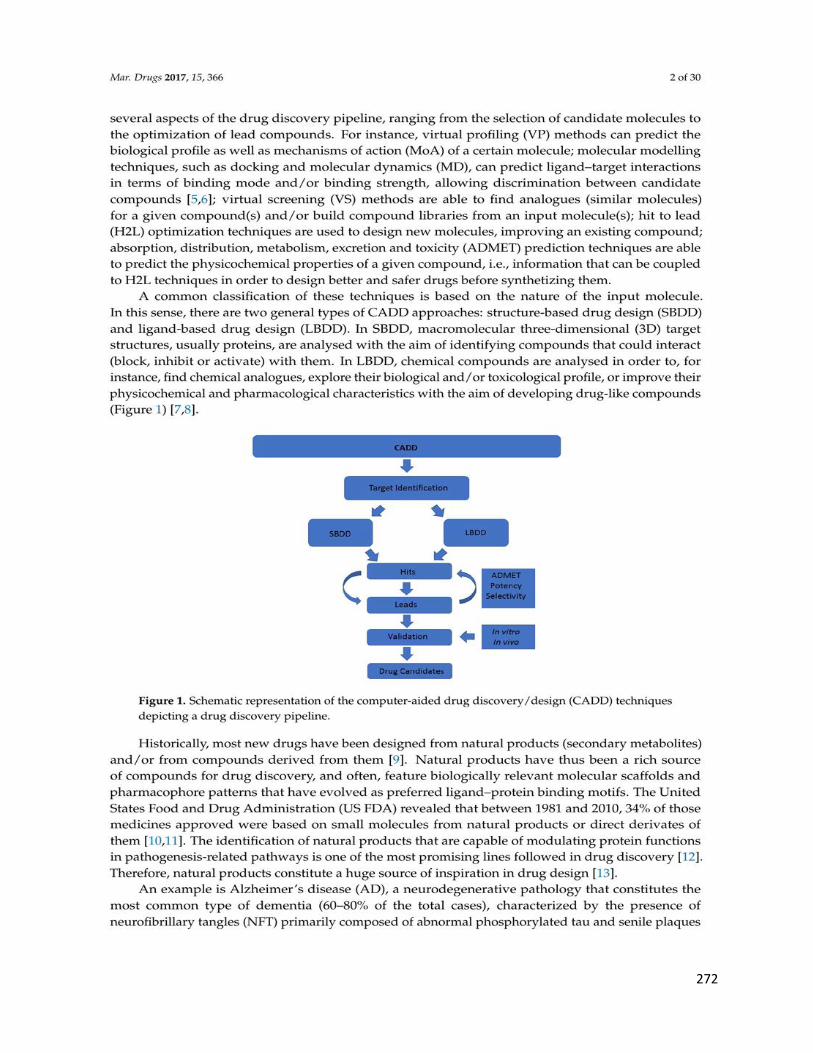
272

273

274

275

276

277

278

279

280

281

282

283
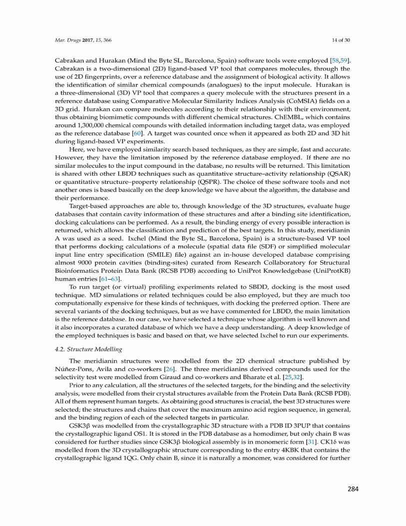
284

285

286

287

288

289

290

291

292

293

294

295

296

297

298

299

300

301

302

303

304

305

306

307

308

309

310

311

312

313

314

315

316

317

318

319

320

321

322
323

324

325

326

327

328

329

330

331

332

333

334

335
Appendix II
Other publications not related to this thesis. The first page is
included below in their original format

336
337

338

339

340

341

342

343

344