Evaluation of current copper bioavailability tools for soft freshwaters in Sweden
Upload
independentCategory
view
4download
0
Biochemical Pharmacology xxx (2009) xxx–xxx
1
2
3
4
5
6
789
10
11
1213
14151617181920212223
G Model
BCP 10317 1–9
RE
CTE
D P
RO
OFDesign of curcumin-loaded PLGA nanoparticles formulation with
enhanced cellular uptake, and increased bioactivity in vitro and superiorbioavailability in vivo
Preeta Anand a, Harish B. Nair b, Bokyung Sung a, Ajaikumar B. Kunnumakkara a, Vivek R. Yadav a,Rajeshwar R. Tekmal b, Bharat B. Aggarwal a,*a Cytokine Research Laboratory, Department of Experimental Therapeutics, The University of Texas M.D. Anderson Cancer Center, 1515 Holcombe Boulevard, Box 143, Houston, TX
77030, USAb Department of Obstetrics and Gynecology, University of Texas Health Science Center, 7703 Floyd Curl Drive, San Antonio, TX 78229-3900, USA
A R T I C L E I N F O
Article history:
Received 23 June 2009
Accepted 1 September 2009
Available online xxx
Keywords:
Nanoparticles
Apoptosis
Inflammation
TNF-alpha
A B S T R A C T
Curcumin, a yellow pigment present in the spice turmeric (Curcuma longa), has been linked with
antioxidant, anti-inflammatory, antiproliferative, anticancer, antidiabetic, antirheumatic, and antiviral
effects, but its optimum potential is limited by its lack of solubility in aqueous solvents and poor oral
bioavailability. We employed a polymer-based nanoparticle approach to improve bioavailability.
Curcumin was encapsulated with 97.5% efficiency in biodegradable nanoparticulate formulation based
on poly (lactide-co-glycolide) (PLGA) and a stabilizer polyethylene glycol (PEG)-5000. Dynamic laser
light scattering and transmission electron microscopy indicated a particle diameter of 80.9 nm. This
curcumin, renamed from hereon ‘‘as curcumin (NP)’’, was characterized for its biological activity. In vitro
curcumin (NP) exhibited very rapid (2 h vs >72 h) and more efficient cellular uptake than curcumin.
Estrase staining revealed that curcumin (NP) was at least as potent as or more potent than curcumin in
inducing apoptosis of leukemic cells and in suppressing proliferation of various tumor cell lines. When
examined by electrophoretic gel shift mobility assay, curcumin (NP) was more active than curcumin in
inhibiting TNF-induced NF-kB activation and in suppression of NF-kB-regulated proteins involved in cell
proliferation (cyclin D1), invasion (MMP-9), and angiogenesis (VEGF). In mice, curcumin (NP) was more
bioavailable and had a longer half-life than curcumin. Overall we demonstrate that curcumin-loaded
PLGA nanoparticles formulation has enhanced cellular uptake, and increased bioactivity in vitro and
superior bioavailability in vivo over curcumin.
� 2009 Published by Elsevier Inc.
Contents lists available at ScienceDirect
Biochemical Pharmacology
journal homepage: www.e lsev ier .com/ locate /b iochempharm
24252627282930313233343536
UNC
OR
1. Introduction
The search for safe, affordable, and efficient agents forprevention and treatment of chronic diseases has led researchersto look in the pharmacopoeia of traditional medicines (TM) forpotential therapeutics. Lack of knowledge about the activechemical entity in TM and its mechanism of action has, however,hindered the addition of many TM to modern allopathic medicines.This is not the case with curcumin, a yellow pigment in the spiceturmeric (Curcuma longa), first isolated in 1815. Its chemicalstructure, first determined in 1910, is diferuloylmethane [1,7-bis(4-hydroxy-3-methoxyphenyl)-1,6-heptadiene-3,5-dione] [1].
3738394041
Abbreviations: NF-kB, nuclear factor-kappaB; TNF, tumor necrosis factor; VEGF,
vascular endothelial growth factor.
* Corresponding author. Tel.: +1 713 794 1817; fax: +1 713 606 3399.
E-mail address: [email protected] (B.B. Aggarwal).
Please cite this article in press as: Anand P, et al. Design of curcuminuptake, and increased bioactivity in vitro and superior bioavailability i
0006-2952/$ – see front matter � 2009 Published by Elsevier Inc.
doi:10.1016/j.bcp.2009.09.003
Although the anti-inflammatory activity of turmeric and its rolein treatment of various diseases including wound-healing,ulcers, and arthritis is well described in Ayurveda (a scienceof long life), the work from our laboratory and others hasindicated that these activities may be due to the curcuminpresent in turmeric [2–4]. Extensive research within the last twodecades has revealed that curcumin exhibits antioxidant, anti-inflammatory, anti-survival, antiproliferative, anti-invasive andantiangiogenic activity [5]. These effects are mediated in partthrough the downregulation of various transcription factorsincluding nuclear factor (NF)-kB [6,7], activator protein (AP)-1[8], hypoxia inducible factor (HIF)-1a [9], and beta-catenin [10].This leads to the downregulation of various proteins involved incell proliferation (e.g., cyclin D1), invasion (e.g., matrixmetalloproteinase-9; MMP-9) and angiogenesis (e.g., vascularendothelial growth factor; VEGF). Animal studies have revealedthat curcumin can prevent carcinogen-induced tumorigenesisand inhibit growth of implanted human tumors [3,11]. Such
-loaded PLGA nanoparticles formulation with enhanced cellularn vivo. Biochem Pharmacol (2009), doi:10.1016/j.bcp.2009.09.003
42434445464748495051525354555657585960616263646566676869707172
73
74
757677787980818283848586878889909192
93
949596979899
100101102
103104
105
106107108109110111112113114115
116
117118119120121122123
124
125126127128129130131132133134135
136
137138139140141142143144145146
147
148149150151152153
154
155156
P. Anand et al. / Biochemical Pharmacology xxx (2009) xxx–xxx2
G Model
BCP 10317 1–9
UN
CO
RR
E
studies have led to clinical trials of curcumin in patients withcolon cancer [12,13], familial adenomatous polyposis (FAP) [14],pancreatic cancer [15], and multiple myeloma [16].
Unfortunately, these studies have revealed that one of themajor problems with curcumin is its oral bioavailability in vivo.Traditionally, turmeric is delivered orally as an emulsion in oil ormilk; perhaps because of the hydrophobic nature of its bioactiveconstituents such as curcumin and turmeric oil. Curcumin hasindeed been shown to interact with phospholipids [17–20],surfactants [21], proteins [22], and cyclodextrin [23]. Variousmethods have been tried to enhance curcumin delivery, includingits incorporation into liposomes [24,25] and into phospholipidvesicles [26]. The latter was used to deliver curcumin via theintravenous route to bone morrow and splenic macrophages.Another way to solve the problem of lack of water solubility andpoor oral bioavailability, is polymer-based nanoparticles [27]. Thisapproach has been used to deliver natural products such ascoenzyme Q10 [28], estradiol [29], and ellagic acid [30] andchemotherapeutic agents such as paclitaxel [31], and doxorubicin[32]. In fact a nanoparticle formulation of paclitaxel in whichserum albumin is included as a carrier (Abraxane) has beenapproved for the treatment of breast cancer [33].
Nanoparticle approach has been recently explored for curcuminas well [34]. We have recently shown that silk fibroin-derivedcurcumin nanoparticles exhibit higher efficacy against breastcancer cells [35]. To further characterize the curcumin nanopar-ticles, in the present report we describe a biodegradable curcuminnanoparticulate formulation based on poly (lactide-co-glycolide)(PLGA) and a stabilizer polyethylene glycol (PEG)-5000 that exhibitenhanced cellular uptake, and increased bioactivity in vitro andsuperior bioavailability in vivo than curcumin.
2. Materials and methods
2.1. Reagents
Curcumin was obtained from Sabinsa Corporation (Piscataway,NJ). Poly (lactide-co-glycolide) (PLGA)–PEG was obtained fromLactal Absorbable Polymers, AL. Ammonium persulphate (APS),ferrous ammonium sulphate (FAS), and N,N0-methylene bisacrylamide (MBA) were obtained from Sigma Chemicals (St. Louis,MO). TEMED was purchased from Invitrogen (Carlsbad, CA). Fetalbovine serum (FBS) was supplied by Atlanta Biologicals (Law-renceville, GA). Antibodies against cyclin D1 and matrix mellato-proteinase (MMP)-9 were obtained from Santa Cruz Biotechnology(Santa Cruz, CA). Bacteria-derived human recombinant tumornecrosis factor (TNF), purified to homogeneity with a specificactivity of 5 � 107 U/mg, was provided by Genentech (SanFrancisco, CA). Penicillin, streptomycin, Iscove’s modified Dulbec-co’s medium, Dulbecco’s modified eagle’s medium, RPMI 1640medium and Live/Dead viability/cytotoxicity kit were obtainedfrom Invitrogen (Grand Island, NY). An anti-vascular endothelialgrowth factor (VEGF) antibody was purchased from ThermoScientific (Fremont, CA).
2.2. Cell lines
The cell lines KBM-5 (human chronic myeloid leukemia), Jurkat(human T cell leukemia), DU145 (prostate carcinoma), MDA-MB-231 (breast adenocarcinoma), HCT116 (human colon adenocarci-noma), and SEG-1 (human esophageal adenocarcinoma) wereobtained from the American Type Culture Collection (ATCC;Manassas, VA). KBM-5 cells were cultured in Iscove’s modifiedDulbecco’s medium with 15% FBS; Jurkat and DU145 was culturedin RPMI 1640; and HCT116, MDA-MB-231 and SEG-1 cells werecultured in Dulbecco’s modified Eagle’s medium supplemented
Please cite this article in press as: Anand P, et al. Design of curcuminuptake, and increased bioactivity in vitro and superior bioavailability i
CTE
D P
RO
OF
with 10% FBS. Culture media were supplemented with 100 U/mLpenicillin and 100 mg/mL streptomycin.
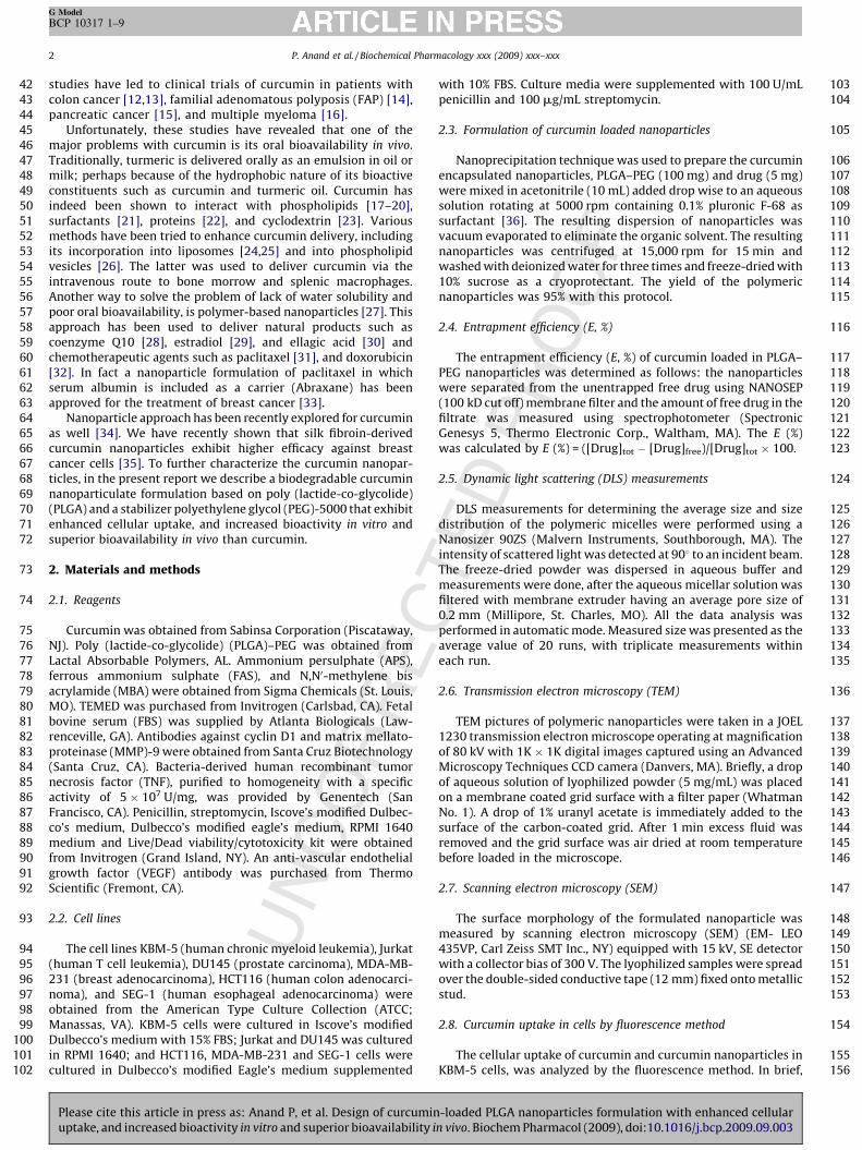
2.3. Formulation of curcumin loaded nanoparticles
Nanoprecipitation technique was used to prepare the curcuminencapsulated nanoparticles, PLGA–PEG (100 mg) and drug (5 mg)were mixed in acetonitrile (10 mL) added drop wise to an aqueoussolution rotating at 5000 rpm containing 0.1% pluronic F-68 assurfactant [36]. The resulting dispersion of nanoparticles wasvacuum evaporated to eliminate the organic solvent. The resultingnanoparticles was centrifuged at 15,000 rpm for 15 min andwashed with deionized water for three times and freeze-dried with10% sucrose as a cryoprotectant. The yield of the polymericnanoparticles was 95% with this protocol.
2.4. Entrapment efficiency (E, %)
The entrapment efficiency (E, %) of curcumin loaded in PLGA–PEG nanoparticles was determined as follows: the nanoparticleswere separated from the unentrapped free drug using NANOSEP(100 kD cut off) membrane filter and the amount of free drug in thefiltrate was measured using spectrophotometer (SpectronicGenesys 5, Thermo Electronic Corp., Waltham, MA). The E (%)was calculated by E (%) = ([Drug]tot � [Drug]free)/[Drug]tot � 100.
2.5. Dynamic light scattering (DLS) measurements
DLS measurements for determining the average size and sizedistribution of the polymeric micelles were performed using aNanosizer 90ZS (Malvern Instruments, Southborough, MA). Theintensity of scattered light was detected at 908 to an incident beam.The freeze-dried powder was dispersed in aqueous buffer andmeasurements were done, after the aqueous micellar solution wasfiltered with membrane extruder having an average pore size of0.2 mm (Millipore, St. Charles, MO). All the data analysis wasperformed in automatic mode. Measured size was presented as theaverage value of 20 runs, with triplicate measurements withineach run.
2.6. Transmission electron microscopy (TEM)
TEM pictures of polymeric nanoparticles were taken in a JOEL1230 transmission electron microscope operating at magnificationof 80 kV with 1K � 1K digital images captured using an AdvancedMicroscopy Techniques CCD camera (Danvers, MA). Briefly, a dropof aqueous solution of lyophilized powder (5 mg/mL) was placedon a membrane coated grid surface with a filter paper (WhatmanNo. 1). A drop of 1% uranyl acetate is immediately added to thesurface of the carbon-coated grid. After 1 min excess fluid wasremoved and the grid surface was air dried at room temperaturebefore loaded in the microscope.
2.7. Scanning electron microscopy (SEM)
The surface morphology of the formulated nanoparticle wasmeasured by scanning electron microscopy (SEM) (EM- LEO435VP, Carl Zeiss SMT Inc., NY) equipped with 15 kV, SE detectorwith a collector bias of 300 V. The lyophilized samples were spreadover the double-sided conductive tape (12 mm) fixed onto metallicstud.
2.8. Curcumin uptake in cells by fluorescence method
The cellular uptake of curcumin and curcumin nanoparticles inKBM-5 cells, was analyzed by the fluorescence method. In brief,
-loaded PLGA nanoparticles formulation with enhanced cellularn vivo. Biochem Pharmacol (2009), doi:10.1016/j.bcp.2009.09.003

157158159160161162163164165166167168
169
170171172173174175176177178179180
181182
183
184185186187188189190191192193194195196197198199
200
201202
P. Anand et al. / Biochemical Pharmacology xxx (2009) xxx–xxx 3
G Model
BCP 10317 1–9
cells were incubated with Hoechst dye (50 ng/mL; blue fluores-cence) for 30 min, and then washed two times with PBS. Thewashed cells were resuspended in media and then incubated withcurcumin and curcumin (NP) for different time intervals. Cellswere then examined under a fluorescence microscope (Labophot-2; Nikon, Tokyo, Japan), and images were captured using aPhotometrics Coolsnap CF color camera (Nikon, Tokyo, Japan). Forvehicle control, cells were incubated with Hoechst dye (50 ng/mL;blue fluorescence) for 30 min, and then washed two times withPBS. The washed cells were resuspended in media and thenincubated with 0.01% DMSO (vehicle for curcumin) or PBS for30 min.
2.9. Apoptosis assay
To compare the apoptotic effects of curcumin and curcumin(NP) in KBM-5 cells, we used a Live/Dead assay kit (Invitrogen,Carlsbad, CA), which determines intracellular esterase activityand plasma membrane integrity. Calcein, a polyanionic, greenfluorescent dye, is retained within live cells, and a redfluorescent ethidium homodimer dye enters cells throughdamaged membranes and binds to nucleic acids but is excludedby the intact plasma membranes of live cells. In brief, cells (5000per well) were incubated with curcumin and curcumin (NP) for24 h. Cells were then stained with assay reagents for 30 min atambient temperature. Cell viability was determined under a
UN
CO
RR
EC
Fig. 1. (A) Curcumin (NP) morphology by scanning electron microscopy (SEM) (left
nanoparticle size: the analysis done by photon correlation spectroscopy.
Please cite this article in press as: Anand P, et al. Design of curcuminuptake, and increased bioactivity in vitro and superior bioavailability i
PR
OO
F
fluorescence microscope by counting live (green) and dead (red)cells.
2.10. Electrophoretic mobility shift assay
To assess NF-kB activation, we isolated nuclei from cells andcarried out electrophoretic mobility shift assays (EMSAs) essen-tially as previously described. In brief, nuclear extracts preparedfrom cancer cells (1 � 106 mL�1) were incubated with 32P-end-labeled 45-mer double-stranded NF-kB oligonucleotide (4 mg ofprotein with 16 fmol of DNA) from the HIV long terminal repeat(50-TTGTTACAAGGGACTTTC CGCTG GGGACTTTC CAGGGA GGCGTGG-30; boldface indicates NF-kB binding sites) for 15 min at 37 8C.The resulting DNA–protein complex was separated from freeoligonucleotides on 6.6% native polyacrylamide gels. A double-stranded mutant oligonucleotide (50-TTGTTACAACTCACTTTCCGCTGCTCACTTTC CAGGGAGG CGTGG-30) was used to evaluatethe specificity of binding of NF-kB to DNA. The dried gels werevisualized, and radioactive bands were quantitated using aPhosphorimager (Molecular Dynamics, Sunnyvale, CA) and theImageQuant software.
2.11. Western blot analysis
Cancer cells were incubated on ice for 30 min in 0.5 mL of ice-cold whole-cell lysate buffer (10% NP-40, 5 mM NaCl, 1 mM
TED
panel) and transmission electron microscopy (TEM) (right panel). (B) Curcumin
-loaded PLGA nanoparticles formulation with enhanced cellularn vivo. Biochem Pharmacol (2009), doi:10.1016/j.bcp.2009.09.003

203204205206207208209
210
211212213214215216217
218
219220221222223224225226227228
229
230231
P. Anand et al. / Biochemical Pharmacology xxx (2009) xxx–xxx4
G Model
BCP 10317 1–9
HEPES, 0.1 mM EGTA, 0.5 mM EDTA, 0.1 mM phenylmethylsul-phonyl fluoride, 0.2 mM sodium orthovanadate, 1 mM NaF,2 mg/mL aprotinin, and 2 mg/mL leupeptin). Proteins were thenfractionated by SDS-polyacrylamide gel electrophoresis, elec-trotransferred to nitrocellulose membranes, blotted with eachantibody, and detected by enhanced chemiluminescence (GEHealthcare, Piscataway, NJ).
2.12. Bioavailability of curcumin and curcumin nanoparticles
The mice were divided into two groups (2 mice in eachgroup), group 1 received curcumin and group 2 receivedcurcumin-loaded nanoparticles. Curcumin or curcumin nano-particles were given intravenously (2.5 mg/kg) to each mouse,the blood was collected at different time intervals, serum wasseparated, and the concentration was determined by HPLCanalysis.
UN
CO
RR
E
Fig. 2. (A) Cellular uptake of curcumin and curcumin (NP). KBM-5 cells were incuba
resuspended in media and then incubated with 10 mM curcumin or curcumin (NP). T
fluorescence) was monitored by fluorescence microscopy as described in Section 2. (B)
(50 ng/mL) for 30 min. KBM-5 (1 � 106) cells were incubated with 10 mM curcumin or c
The cells were harvested at different time intervals and the cellular uptake was determ
Please cite this article in press as: Anand P, et al. Design of curcuminuptake, and increased bioactivity in vitro and superior bioavailability i
OF
3. Results
The focus of this study was to prepare and characterize thecurcumin nanoparticles using various methods, with a goal toenhance its bioavailability without loss of biological activity. Thesenanoparticles were examined for their cellular uptake, for theirability to induce apoptosis and suppress proliferation of tumor cells,for their suppression of NF-kB and NF-kB-regulated gene products,and for in vivo bioavailability. For most studies, human leukemiaKBM-5 cells were used, as the effects of curcumin on these cells havebeen well described [7]. Throughout the studies, the effects ofcurcumin (NP) were compared with those of native curcumin.
3.1. Preparation and characteristics of curcumin (NP)
The morphology of curcumin (NP) is shown in Fig. 1A. PLGA–PEG associated curcumin had 97.5% encapsulation efficiency and
CTE
D P
RO
ted with Hoechst (50 ng/mL) for 30 min, and then washed two times with PBS,
he cells were harvested at different time intervals and the cellular uptake (green
Disappearance of curcumin and curcumin (NP) from the cells stained with Hoechst
urcumin (NP) for 30 min, washed two times with PBS and again incubated at 37 8C.
ined by fluorescence microscopy as described in Section 2.
-loaded PLGA nanoparticles formulation with enhanced cellularn vivo. Biochem Pharmacol (2009), doi:10.1016/j.bcp.2009.09.003

PR
OO
F232233234
235
236237238
239240241242243244245246247
Fig. 2. (Continued ).
P. Anand et al. / Biochemical Pharmacology xxx (2009) xxx–xxx 5
G Model
BCP 10317 1–9
contained 4 mg curcumin/mg of nanoparticles. Photon correlationspectroscopy showed that the mean diameter of curcumin (NP)was 80.9 nm (Fig. 1B).
3.2. Curcumin (NP) exhibits enhanced cellular uptake
We next investigated the cellular uptake of curcumin andcurcumin nanoparticles. For this human chronic myeloid leukemiaKBM-5 cells were incubated with curcumin or curcumin (NP) at
UN
CO
RR
EC
Fig. 3. (A) Curcumin (NP) can induce apoptosis of tumor cells. KBM-5 (5000 cells/well) cells
The cells were harvested and stained with Live/Dead assay reagent as per the manufactures
cells. Human leukemia (KBM-5 and Jurkat), prostate (DU145), breast (MDA-MB-231), c
concentrations of either curcumin or curcumin (NP) in for 72 h and then examined for a
Please cite this article in press as: Anand P, et al. Design of curcuminuptake, and increased bioactivity in vitro and superior bioavailability i
TED10 mM concentration. The cells were harvested at different time
intervals and the cellular uptake was determined by fluorescencemicroscopy. Furthermore, to avoid the background effect fromhigh intensity of fluorescence by curcumin, cells were also stainedwith Hoechst dye (blue fluorescence). The results showed thatcurcumin (NP) was taken up as early as 5 min after exposure andreached maximum at 30 min (Fig. 2A). In contrast, earliest uptakeof curcumin occurred at 30 min and did not reach maximum evenat 180 min (Fig. 2A).
were incubated with curcumin or curcumin (NP) in indicated concentrations for 24 h.
protocol as described in Section 2. (B) Curcumin (NP) can inhibit proliferation of tumor
olon (HCT116) and esophageal (SEG-1) cancer cells were incubated with different
poptosis by MTT method. All results were expressed with vehicle control as 100%.
-loaded PLGA nanoparticles formulation with enhanced cellularn vivo. Biochem Pharmacol (2009), doi:10.1016/j.bcp.2009.09.003

OR
RE
CTE
D P
RO
OF
248249250251252253254255256
257
258259
260261262263
264
265266267268269270271
Fig. 3. (Continued ).
P. Anand et al. / Biochemical Pharmacology xxx (2009) xxx–xxx6
G Model
BCP 10317 1–9
UN
C
We also investigated the disappearance of curcumin (NP) fromcells after removing the compounds from the medium. For thiscells were incubated with curcumin or curcumin (NP) at 10 mMconcentration for 30 min, washed two times with PBS, andincubated for indicated times at 37 8C. The cells were harvestedat different time intervals and the cellular uptake was determinedby fluorescence microscopy. Interestingly, we found that curcumin(NP) quickly disappeared whereas curcumin disappeared slowly(Fig. 2B).
3.3. Curcumin (NP) induces apoptosis of tumor cells
Next, we investigated the ability of curcumin (NP) to induceapoptosis in human cancer cells. When human KBM-5 cells were
Please cite this article in press as: Anand P, et al. Design of curcuminuptake, and increased bioactivity in vitro and superior bioavailability i
incubated with different concentrations of curcumin or curcumin(NP) for 24 h, esterase staining showed a dose-related apoptoticresponse (Fig. 3A). At 25 mM curcumin (NP) was somewhat moreactive than curcumin.
3.4. Curcumin (NP) inhibits proliferation of tumor cells
Human leukemia (KBM-5 and Jurkat), prostate (DU145),breast (MDA-MB-231), colon (HCT116) and esophageal (SEG-1)cancer cells were incubated with different concentrations ofeither curcumin or curcumin (NP) for 72 h and then examinedfor apoptosis by MTT method. The results in Fig. 3B show thatcurcumin (NP) suppressed cell proliferation dose-dependentlyat least as potently as native curcumin.
-loaded PLGA nanoparticles formulation with enhanced cellularn vivo. Biochem Pharmacol (2009), doi:10.1016/j.bcp.2009.09.003

CO
RR
EC
OO
F
272
273274275276277278279280281282283
284285
286287
288289290291292293294295
296
297298299300301Fig. 4. (A) Curcumin or curcumin (NP) do not induce NF-kB activation in KBM-5
cells. KBM-5 (2 � 106) cells were treated with indicated concentrations of curcumin
or curcumin (NP) for 4 h. Nuclear extracts were prepared and the NF-kB activity
was examined by EMSA. (B and C) Curcumin (NP) is more potent than regular
curcumin in inhibiting TNF-induced activation of NF-kB. KBM-5 (2 � 106) cells
were treated with indicated concentrations of curcumin or curcumin (NP) for 4 h.
The cells were then incubated with TNF 0.1 nM for 30 min and analyzed for NF-kB
activity by EMSA. The cytotoxicity was examined using trypan blue exclusion
method.
Fig. 5. Curcumin (NP) is more potent than curcumin in inhibiting TNF-induced
expression of NF-kB-regulated genes. KBM-5 (1 � 106) cells were co-incubated
with TNF (1 nM) and curcumin or curcumin (NP) (10 mM) for indicated time
intervals. The cells were harvested and the expression of cyclin D1, MMP-9, and
VEGF analyzed by western blot. b-Actin was used as a loading control.
Fig. 6. Bioavailability of curcumin and curcumin (NP). The mice were divided into
two groups (6 mice in each group), group 1 was given curcumin and group 2 was
given curcumin (NP). Curcumin and curcumin (NP) were administered
intravenously (2.5 mg/kg) and the blood was collected at different time
intervals. Serum was separated and the concentration of curcumin and
curcumin (NP) were determined by HPLC analysis.
P. Anand et al. / Biochemical Pharmacology xxx (2009) xxx–xxx 7
G Model
BCP 10317 1–9
UN3.5. Curcumin (NP) suppresses NF-kB activation
Suppression of NF-kB is one of the major activities assigned tocurcumin. Whether curcumin (NP) by itself can induce NF-kB, wasexamined. KBM-5 cells were treated with the indicated concen-trations of curcumin (NP) for 4 h, and nuclear extracts wereprepared and analyzed for NF-kB activity by EMSA. Neithercurcumin nor curcumin (NP) alone activated NF-kB (Fig. 4A). TNF-induced activation of NF-kB, however, was inhibited by curcuminin a dose-dependent manner (Fig. 4B), which was also the case forcurcumin (NP) (Fig. 4C). Curcumin (NP) was more potent thancurcumin in this effect. Under the conditions, curcuminor curcumin (NP) had no significant effect on cell viability,
Please cite this article in press as: Anand P, et al. Design of curcuminuptake, and increased bioactivity in vitro and superior bioavailability i
TED
PR
indicating that suppression of NF-kB activation was not due to lossof cell viability.
3.6. Curcumin (NP) inhibits the expression of NF-kB-regulated gene
products
To compare the potency of curcumin (NP) and curcumin ininhibiting TNF-induced expression of NF-kB-regulated geneproducts, we co-incubated KBM-5 cells with TNF and curcuminor with TNF and curcumin (NP) for different time intervals andthen examined for cyclin D1 (cell proliferative), MMP-9 (invasion)and VEGF (angiogenesis) gene products. As shown in Fig. 5,curcumin (NP) suppressed the expression of all these geneproducts more potently than curcumin.
3.7. Curcumin (NP) is more bioavailable than curcumin
One of the major reasons for making curcumin (NP) to examineits effect on bioavailability in vivo. For this, mice were intrave-nously administered either curcumin or curcumin (NP) (2.5 mg/kg), blood was collected at different time intervals, and curcuminconcentration was determined by HPLC analysis. Results in Fig. 6
-loaded PLGA nanoparticles formulation with enhanced cellularn vivo. Biochem Pharmacol (2009), doi:10.1016/j.bcp.2009.09.003

302303304305
306
307308309310311312313314315316317318319320321322323324325326327328329330331332333334335336337338339340341342343344345346347348349350351352353354355356357358359360361362363364365
366367368369370371372373374375376377378379380
381
382383384385386
387
388389390391392393394395396397398399400401402403404405406407408409410411412413414415416417418419420421422423424425426427428429430431432433434435436437438439
P. Anand et al. / Biochemical Pharmacology xxx (2009) xxx–xxx8
G Model
BCP 10317 1–9
UN
CO
RR
E
clearly show serum levels of curcumin were almost twice as high inthe case of curcumin (NP) administration as it was curcuminadministration. In addition, half-life of curcumin (NP) wassubstantially longer than that of curcumin.
4. Discussion
Curcumin, a yellow pigment from turmeric, has been associatedwith antioxidant, anti-inflammatory, antiproliferative, anticancer,antiangiogenic, and antidiabetic activities. However poor watersolubility and limited oral bioavailability are the major roadblocksin its development as a therapeutic for cancer and other chronicdiseases. In the current report we prepared a biodegradablenanoparticle formulation of curcumin based on poly (lactide-co-glycolide) technique using polyethylene glycol (PEG-5000) as astabilizer and tested its bioavailability and effects on cells growth.This method has been used to encapsulate a wide variety ofhydrophobic drugs including coenzyme Q [28], taxol [31,37],estradiol [29], ellagic acid [30], and camptothecin [32]. Our resultsare in agreement with Bisht et al. [34] who made nanoparticles ofcurcumin. However, these authors reported very limited informa-tion about its biological attributes in vitro and no information on itsbioavailability in vivo.
We found that curcumin (NP) had much higher cellular uptakein vitro than that of curcumin. Its rate of disappearance from thecells, however, was very similar. Increased cellular uptake ofcurcumin (NP) is in agreement with Bisht et al.’s report [34]. Wealso found that curcumin (NP) was more potent in inducingapoptosis in tumor cells. One possible cause for higher activitycould be higher cellular uptake of curcumin (NP).
Curcumin (NP) inhibited the growth of a wide variety of tumorcells in a dose-dependent manner. This effect was comparable withthat of curcumin. Why curcumin (NP) did not show significantlyhigher efficacy than curcumin is not clear. Because apoptosis wasexamined during a short time (24 h) and antiproliferative effectsduring a long time (72 h), it is possible that in short-term assayscurcumin (NP) is more active than curcumin but not in long termassays, perhaps due to the differential cellular uptake.
Curcumin (NP) was more active than curcumin in suppressingNF-kB activation. This difference again could have been due todifferential uptake. Bisht et al. [34] did not examine TNF-inducibleNF-kB but showed that curcumin (NP) could suppress constitutiveNF-kB in pancreatic cancer cells.
We found that curcumin (NP) was also more active thancurcumin in suppressing the expression of TNF-regulated expres-sion of cyclin D1, MMP-9 and VEGF; most likely again due toenhanced cellular uptake. These results imply that curcumin (NP)may be superior to curcumin as an antitumor, anti-invasive andantiangiogenic agent. The results are in agreement with a reportthat curcumin (NP) downregulates IL-6, another NF-kB-regulatedgene product [34].
Finally, our results also show that in animals curcumin (NP) ismore bioavailable and has a substantially longer half-life. Inagreement with Bisht et al. [34], we found that curcumin (NP) wasnot toxic to the animals. Formation of polymeric micelles ofcurcumin with copolymer micelles of methoxy poly(ethyleneoxide)-b-poly(epsilon-caprolactone) (MePEO-b-PCL), has beenshown to increase its half-life by 162-fold [38]. These results arein agreement with a report that showed that curcumin formulatedwith phosphatidyl choline (PC), when given to animals, had a peakplasma levels and area under the plasma concentration time curve(AUC) values were higher than the unformulated curcumin [20].They also showed that liver levels of curcumin were higher afteradministration of PC–curcumin as compared to unformulatedcurcumin. In contrast, curcumin concentrations in the gastro-intestinal mucosa after ingestion of PC–curcumin were somewhat
Please cite this article in press as: Anand P, et al. Design of curcuminuptake, and increased bioactivity in vitro and superior bioavailability i
CTE
D P
RO
OF
lower than those observed after administration of unformulatedcurcumin. Curcumin has been conjugated with numerous carriersincluding phospholipids [18], cyclodextrin [23], phosphatidylcholine [20], and liposomes [25,39], but very little informationis available about its biological activities. It has been shown thatbinding states of curcumin to all these carriers is typical ofamphipathic drugs [40]. More studies are now needed toinvestigate the in vivo efficacy of curcumin (NP) against varioustypes of cancer. Although curcumin is pharmacologically quite safein human [1,11,41], the efficacy of curcumin (NP) remains to bedetermined. Polymers such as albumin-bound paclitaxel (Abrax-ane) have been approved for human use for the treatment of cancer[33]. Overall, our results suggest that curcumin (NP) is likely tohave great potential as a therapeutic, but more studies arerequired.
Acknowledgements
Dr. Aggarwal is the Ransom Horne, Jr., Professor of CancerResearch. This work was supported by a grant from a programproject grant from National Institutes of Health (NIH CA-124787-01A2), and a grant from Center for Targeted Therapy of TheUniversity of Texas M.D. Anderson Cancer Center.
References
[1] Aggarwal BB, Sung B. Pharmacological basis for the role of curcumin in chronicdiseases: an age-old spice with modern targets. Trends in PharmacologicalSciences 2009;30:85–94.
[2] Shishodia S, Sethi G, Aggarwal BB. Curcumin: getting back to the roots. Annalsof the New York Academy of Sciences 2005;1056:206–17.
[3] Goel A, Kunnumakkara AB, Aggarwal BB. Curcumin as ‘‘Curecumin’’: fromkitchen to clinic. Biochemical Pharmacology 2008;75:787–809.
[4] Strimpakos AS, Sharma RA. Curcumin: preventive and therapeutic propertiesin laboratory studies and clinical trials. Antioxidants & Redox Signaling2008;10:511–45.
[5] Kunnumakkara AB, Anand P, Aggarwal BB. Curcumin inhibits proliferation,invasion, angiogenesis and metastasis of different cancers through interactionwith multiple cell signaling proteins. Cancer Letters 2008;269:199–225.
[6] Singh S, Aggarwal BB. Activation of transcription factor NF-kappa B is sup-pressed by curcumin (diferuloylmethane) [corrected]. The Journal of BiologicalChemistry 1995;270:24995–5000.
[7] Aggarwal S, Ichikawa H, Takada Y, Sandur SK, Shishodia S, Aggarwal BB.Curcumin (diferuloylmethane) down-regulates expression of cell proliferationand antiapoptotic and metastatic gene products through suppression ofIkappaBalpha kinase and Akt activation. Molecular Pharmacology2006;69:195–206.
[8] Huang TS, Lee SC, Lin JK. Suppression of c-Jun/AP-1 activation by an inhibitor oftumor promotion in mouse fibroblast cells. Proceedings of the NationalAcademy of Sciences of the United States of America 1991;88:5292–6.
[9] Bae MK, Kim SH, Jeong JW, Lee YM, Kim HS, Kim SR, et al. Curcumin inhibitshypoxia-induced angiogenesis via down-regulation of HIF-1. OncologyReports 2006;15:1557–62.
[10] Jaiswal AS, Marlow BP, Gupta N, Narayan S. Beta-catenin-mediated transacti-vation and cell-cell adhesion pathways are important in curcumin (diferuyl-methane)-induced growth arrest and apoptosis in colon cancer cells.Oncogene 2002;21:8414–27.
[11] Anand P, Sundaram C, Jhurani S, Kunnumakkara AB, Aggarwal BB. Curcuminand cancer: an ‘‘old-age’’ disease with an ‘‘age-old’’ solution. Cancer Letters2008;267:133–64.
[12] Sharma RA, Euden SA, Platton SL, Cooke DN, Shafayat A, Hewitt HR, et al. PhaseI clinical trial of oral curcumin: biomarkers of systemic activity and compli-ance. Clinical Cancer Research 2004;10:6847–54.
[13] Sharma RA, McLelland HR, Hill KA, Ireson CR, Euden SA, Manson MM, et al.Pharmacodynamic and pharmacokinetic study of oral Curcuma extract inpatients with colorectal cancer. Clinical Cancer Research 2001;7:1894–900.
[14] Cruz-Correa M, Shoskes DA, Sanchez P, Zhao R, Hylind LM, Wexner SD, et al.Combination treatment with curcumin and quercetin of adenomas in familialadenomatous polyposis. Clinics in Gastroenterology and Hepatology2006;4:1035–8.
[15] Dhillon N, Aggarwal BB, Newman RA, Wolff RA, Kunnumakkara AB, Abbruzz-ese JL, et al. Phase II trial of curcumin in patients with advanced pancreaticcancer. Clinical Cancer Research 2008;14:4491–9.
[16] Bhardwaj A, Sethi G, Vadhan-Raj S, Bueso-Ramos C, Takada Y, Gaur U, et al.Resveratrol inhibits proliferation, induces apoptosis, and overcomes chemore-sistance through down-regulation of STAT3 and nuclear factor-kappaB-regu-lated antiapoptotic and cell survival gene products in human multiplemyeloma cells. Blood 2007;109:2293–302.
-loaded PLGA nanoparticles formulation with enhanced cellularn vivo. Biochem Pharmacol (2009), doi:10.1016/j.bcp.2009.09.003

440441442443444445446447448449450451452453454455456457458459460461462463464465466467468469470471472473474475476477478479480481482
483484485486487488489490491492493494495496497498499500501502503504505506507508509510511512513514515516517518519520521522523524525
526
P. Anand et al. / Biochemical Pharmacology xxx (2009) xxx–xxx 9
G Model
BCP 10317 1–9
[17] Began G, Sudharshan E, Udaya Sankar K, Appu Rao AG. Interaction of curcuminwith phosphatidylcholine: a spectrofluorometric study. Journal of Agriculturaland Food Chemistry 1999;47:4992–7.
[18] Maiti K, Mukherjee K, Gantait A, Saha BP, Mukherjee PK. Curcumin-phos-pholipid complex: preparation, therapeutic evaluation and pharmacoki-netic study in rats. International Journal of Pharmaceutics 2007;330:155–63.
[19] Kunwar A, Barik A, Pandey R, Priyadarsini KI. Transport of liposomaland albumin loaded curcumin to living cells: an absorption and fluore-scence spectroscopic study. Biochimica et Biophysica Acta 2006;1760:1513–20.
[20] Marczylo TH, Verschoyle RD, Cooke DN, Morazzoni P, Steward WP, Gescher AJ.Comparison of systemic availability of curcumin with that of curcumin for-mulated with phosphatidylcholine. Cancer Chemotherapy and Pharmacology2007;60:171–7.
[21] Tonnesen HH. Solubility, chemical and photochemical stability of curcumin insurfactant solutions. Studies of curcumin and curcuminoids, XXVIII. DiePharmazie 2002;57:820–4.
[22] Kumar V, Lewis SA, Mutalik S, Shenoy DB, Venkatesh, Udupa N. Biodegradablemicrospheres of curcumin for treatment of inflammation. Indian Journal ofPhysiology and Pharmacology 2002;46:209–17.
[23] Salmaso S, Bersani S, Semenzato A, Caliceti P. New cyclodextrin bioconjugatesfor active tumour targeting. Journal of Drug Targeting 2007;15:379–90.
[24] Li L, Braiteh FS, Kurzrock R. Liposome-encapsulated curcumin: in vitro and invivo effects on proliferation, apoptosis, signaling, and angiogenesis. Cancer2005;104:1322–31.
[25] Takahashi M, Kitamoto D, Imura T, Oku H, Takara K, Wada K. Characterizationand bioavailability of liposomes containing a ukon extract. Bioscience Bio-technology and Biochemistry 2008;72:1199–205.
[26] Sou K, Inenaga S, Takeoka S, Tsuchida E. Loading of curcumin into macro-phages using lipid-based nanoparticles. International Journal of Pharmaceu-tics 2008;352:287–93.
[27] Cho K, Wang X, Nie S, Chen ZG, Shin DM. Therapeutic nanoparticles for drugdelivery in cancer. Clinical Cancer Research 2008;14:1310–6.
[28] Ankola DD, Viswanad B, Bhardwaj V, Ramarao P, Kumar MN. Development ofpotent oral nanoparticulate formulation of coenzyme Q10 for treatment ofhypertension: can the simple nutritional supplements be used as first linetherapeutic agents for prophylaxis/therapy? European Journal of Pharmaceu-tics and Biopharmaceutics 2007;67:361–9.
[29] Hariharan S, Bhardwaj V, Bala I, Sitterberg J, Bakowsky U, Ravi Kumar MN.Design of estradiol loaded PLGA nanoparticulate formulations: a potential oraldelivery system for hormone therapy. Pharmaceutical Research 2006;23:184–95.
UN
CO
RR
EC
Please cite this article in press as: Anand P, et al. Design of curcuminuptake, and increased bioactivity in vitro and superior bioavailability i
ED
PR
OO
F
[30] Bala I, Bhardwaj V, Hariharan S, Kharade SV, Roy N, Ravi Kumar MN. Sustainedrelease nanoparticulate formulation containing antioxidant-ellagic acid aspotential prophylaxis system for oral administration. Journal of Drug Target-ing 2006;14:27–34.
[31] Mu L, Feng SS. A novel controlled release formulation for the anticancer drugpaclitaxel (Taxol): PLGA nanoparticles containing vitamin E TPGS. Journal ofControlled Release 2003;86:33–48.
[32] Vasey PA, Kaye SB, Morrison R, Twelves C, Wilson P, Duncan R, et al.Phase I clinical and pharmacokinetic study of PK1 [N-(2-hydroxypropyl)-methacrylamide copolymer doxorubicin]: first member of a new classof chemotherapeutic agents–drug–polymer conjugates. Cancer ResearchCampaign Phase I/II Committee. Clinical Cancer Research 1999;5:83–94.
[33] Gradishar WJ, Tjulandin S, Davidson N, Shaw H, Desai N, Bhar P, et al. Phase IIItrial of nanoparticle albumin-bound paclitaxel compared with polyethylatedcastor oil-based paclitaxel in women with breast cancer. Journal of ClinicalOncology 2005;23:7794–803.
[34] Bisht S, Feldmann G, Soni S, Ravi R, Karikar C, Maitra A, et al. Polymericnanoparticle-encapsulated curcumin (‘‘nanocurcumin’’): a novel strategy forhuman cancer therapy. Journal of Nanobiotechnology 2007;5:3.
[35] Gupta V, Aseh A, Rios CN, Aggarwal BB, Mathur AB. Fabrication and char-acterization of silk fibroin-derived curcumin nanoparticles for cancer therapy.International Journal of Nanomedicine 2009;4:115–22.
[36] Fessi H, Puisieux F, Devissaquet JP, Ammoury N, Benita S. Nanocapsule for-mation by interfacial polymer deposition following solvent displacement.International Journal of Pharmaceutics 1989;55:R1–4.
[37] Kim TY, Kim DW, Chung JY, Shin SG, Kim SC, Heo DS, et al. Phase I andpharmacokinetic study of Genexol-PM, a cremophor-free, polymeric micelle-formulated paclitaxel, in patients with advanced malignancies. Clinical CancerResearch 2004;10:3708–16.
[38] Ma Z, Shayeganpour A, Brocks DR, Lavasanifar A, Samuel J. High-performanceliquid chromatography analysis of curcumin in rat plasma: application topharmacokinetics of polymeric micellar formulation of curcumin. BiomedicalChromatography 2007;21:546–52.
[39] Thangapazham RL, Puri A, Tele S, Blumenthal R, Maheshwari RK. Evaluation ofa nanotechnology-based carrier for delivery of curcumin in prostate cancercells. International Journal of Oncology 2008;32:1119–23.
[40] Sun Y, Lee CC, Hung WC, Chen FY, Lee MT, Huang HW. The bound states ofamphipathic drugs in lipid bilayers: study of curcumin. Biophysical Journal2008;95:2318–24.
[41] Aggarwal BB, Harikumar KB. Potential therapeutic effects of curcumin, theanti-inflammatory agent, against neurodegenerative, cardiovascular, pulmon-ary, metabolic, autoimmune and neoplastic diseases. The International Journalof Biochemistry & Cell Biology 2009;41:40–59.
T-loaded PLGA nanoparticles formulation with enhanced cellularn vivo. Biochem Pharmacol (2009), doi:10.1016/j.bcp.2009.09.003
Copyright © 2022 FDOKUMEN