Output Costs, Currency Crises and Interest Rate Defence of a Peg
Upload
independentCategory
view
2download
0
European Journal of Pharmaceutical Sciences xxx (2011) xxx–xxx
Contents lists available at SciVerse ScienceDirect
European Journal of Pharmaceutical Sciences
journal homepage: www.elsevier .com/ locate/e jps
Protein-loaded PLGA–PEG–PLGA microspheres: A tool for cell therapy
Van-Thanh Tran a,b,1, Jean-Pierre Karam a,b, Xavier Garric c, Jean Coudane c, Jean-Pierre Benoît a,b,Claudia N. Montero-Menei a,b, Marie-Claire Venier-Julienne a,b,⇑a LUNAM Université, Ingénierie de la Vectorisation Particulaire, F-49933 Angers Cedex, Franceb INSERM U646, F-49933 Angers, Francec Institut des biomolécules Max Mousseron, CNRS UMR 5247, F-34093 Montpellier, France
a r t i c l e i n f o a b s t r a c t
Article history:Received 27 July 2011Received in revised form 25 October 2011Accepted 31 October 2011Available online xxxx
Keywords:PLGA–PEG–PLGALysozymePoloxamer 188Controlled protein releaseCell adhesion
0928-0987/$ - see front matter � 2011 Elsevier B.V. Adoi:10.1016/j.ejps.2011.10.030
⇑ Corresponding author at: LUNAM Université, InParticulaire, F-49933 Angers Cedex, France. Tel.: +3735853.
E-mail address: [email protected] (M.-C. Veni1 Current address: Department of Galenic Pharmacy, S
of Medicine and Pharmacy at Ho Chi Minh City, 70000
Please cite this article in press as: Tran, V.-T.,doi:10.1016/j.ejps.2011.10.030
A promising strategy to repair injured organs is possible by delivering a growth factor via poly-(D,L lac-tide-co-glycolide) (PLGA) microspheres; the latter are coated with adhesion molecules that serve as asupport for cell delivery. At present, PLGA is not the optimal choice of polymer because of poor or incom-plete protein release. The use of a more hydrophilic PLGA–PEG–PLGA (A–B–A) copolymer increases thedegree of protein release. In this work, the impact of different combinations of (B) and (A) segmentson the protein-release profile has been investigated. Continuous-release profiles, with no lag phases,were observed. The triblock ABA with a low molecular weight of PEG and a high molecular weight ofPLGA showed an interesting release pattern with a small burst (<10% in 48 h) followed by sustained, pro-tein release over 36 days. Incomplete protein release was found to be due to various causes: proteinadsorption, protein aggregation and protein denaturation under acidic conditions. Interestingly, cell via-bility and cell adhesion on microspheres coated with fibronectin highlight the interest of these polymersfor tissue engineering applications.
� 2011 Elsevier B.V. All rights reserved.
1. Introduction
Cell therapy carried out by grafting autologous or non-autolo-gous cells is a promising strategy to repair injured organs (Delcroixet al., 2009; Dennis et al., 2007). However, the survival and func-tional state of the cells after transplantation still need to be im-proved (Delcroix et al., 2009; Tabata, 2000). Growth anddifferentiation factors may improve cell survival, cell differentia-tion and affect the immediate environment, thus enhancing graftintegration. Nevertheless, the delivery of these factors still remainsa technological challenge due to their fragile structure and theirshort half-life after administration. To overcome these difficulties,the growth factors can be protected in biodegradable microparti-cles which offer controlled and sustained release after administra-tion (Aubert-Pouëssel et al., 2004; D’Aurizio et al., 2011).
Our group has demonstrated the interest of a tissue engineeringsystem named Pharmacologically Active Microcarriers (PAMs) toimprove grafting in the host tissue. These biodegradable particlesmade with poly-(D,L-lactic-co-glycolic acid) (PLGA) with anadapted size (60 lm), presenting a biomimetic surface of cell adhe-
ll rights reserved.
génierie de la Vectorisation3 241 735855; fax: +33 241
er-Julienne).chool of Pharmacy, UniversityHo Chi Minh City, Viet Nam.
et al. Protein-loaded PLGA–PE
sion/extracellular matrix molecules, served as a support for celladministration and the programmed delivery of an appropriateprotein. The combined effect of the 3D, biomimetic surface andthe delivered growth factor increased cell survival and differentia-tion of the transported cells and also enhanced the regenerativepotential of stem cells (Bouffi et al., 2010; Delcroix et al., 2011;Tatard et al., 2004, 2005a). A PLGA polymer was first chosen be-cause of its biodegradable and non-toxic nature (Fournier et al.,2003). However, low and incomplete protein release from PLGAmicrospheres are related to protein instability during the releaseperiod (Determan et al., 2006; Fu et al., 2000). Our group and oth-ers have shown that by introducing hydrophilic segmentspoly(ethylene glycol) (PEG) into hydrophobic polyesters, PLGA,protein release from PLGA–PEG–PLGA (ABA) triblock copolymermicrospheres was enhanced (Kissel et al., 2002; Paillard-Giteauet al., 2010). Due to the presence of PEG segments, cross-linkedbiodegradable hydrogel formed upon contact with water thusfavouring protein release (Li and Kissel, 1993). The PEG segment it-self also promotes the stability of proteins (Kissel et al., 1996). Inthis regard, Kissel et al. (2002) demonstrate a complete release oflysozyme from PLGA–PEG–PLGA with a high protein loading of 5%.
Tissue engineering approaches combining 3D biomimetic sys-tems and the sustained release of therapeutic factors represent atechnological improvement for cell therapy studies. Accuratedelivery of therapeutic proteins at physiological levels requireslow encapsulation loading of these otherwise expensive proteins
G–PLGA microspheres: A tool for cell therapy. Eur. J. Pharm. Sci. (2011),

2 V.-T. Tran et al. / European Journal of Pharmaceutical Sciences xxx (2011) xxx–xxx
(VEGF, CNTF, GDNF) (Aubert-Pouëssel et al., 2004; Bertram et al.,2009; Boerckel et al., 2011), which accentuates protein destabilisa-tion following polymer–protein interaction. However, most of re-search in the literature was focused at high protein loading(more than 3%) (Kissel et al., 2002) or focused on the in vivo bio-application where the reason of incomplete protein release wasnot clarified (Chen and Hu, 2011). It is therefore essential to inves-tigate protein release from ABA copolymers with low loading (<1%)in order to elucidate protein behaviour during a long release peri-od. Furthermore, although the co-precipitation of the stabilizeragent with the protein presents particular interest to stabilizethe protein without affecting the burst effect (Paillard-Giteauet al., 2010), the association of the co-precipitation with the sys-tematic study of the polymers was not yet evaluated. In the presentstudy, the impact of ABA copolymer composition and molecularweight on the release profile and protein stability were studied.Lysozyme was used as a model protein because it is representativeof the physical and chemical properties (isoelectric point andmolecular weight) and the adsorption behaviour of therapeuticgrowths factors such as NGF, TGF-b3 and NT3 (Aubert-Pouësselet al., 2002; Paillard-Giteau et al., 2010).
The adsorption of fibronectin on ABA copolymers was also eval-uated to assess the capability of creating an appropriate 3D biomi-metic surface with these copolymers. Fibronectin was used as abioadhesive substance for this biomimetic surface because it en-hances the attachment of various stem cells in vitro and affectstheir behaviour such as survival, migration and proliferation (Delc-roix et al., 2009). Cell adhesion and cell survival on the micro-spheres with a fibronectin biomimetic surface were studied. Thegoal of this study is to follow the stability of the protein releasedfrom different PLGA–PEG–PLGA triblock copolymers and to createa biomimetic 3D surface by fibronectin adsorption in order to im-prove a tissue-engineering approach.
2. Materials and methods
2.1. Materials
Lysozyme (chicken egg white) and its substrate Micrococcus lys-odeikticus, glycofurol (tetraglycol or a-[(tetrahydro-2-furanyl)methyl]-x-hydroxy-poly(oxy-1,2-ethanediyl)), fibronectin, di-methyl sulfoxide (DMSO), bovine serum albumin (BSA), BSA-FITCand dextran-FITC were obtained from Sigma-Aldrich (Saint Quen-tin Fallavier, France). Polyvinyl alcohol (Mowiol� 4-88) was ob-tained from Kuraray Specialities Europe (Frankfurt, Germany).Pluronic F68 was kindly supplied by BASF (Levallois-Perret,France). Culture mediums, penicillin, streptomycin and trypsinwere obtained from Invitrogen (Cergy Pontoise, France). Uncapped(free carboxylic acid group at the terminal end) PLGA37.5/25 (Mn14,000 Da) was provided by Phusis (Saint-Ismier, France). DL-Lac-tide and glycolide were obtained from Purac (Gorinchem, TheNetherlands) and poly(ethylene oxide) from Fluka.
2.2. Methods
2.2.1. Polymer synthesis and characterisationA series of nine PLGA–PEG–PLGA copolymers were synthesised
from the combination of various molecular weights of PLGA seg-ments (20, 30 and 40 kDa) and PEG segments (4, 12 and 20 kDa).The polymers are indicated as PLGxPEGy, in which ‘‘x’’ and ‘‘y’’ arethe PLGA and PEG segment molecular weights in kDa, respectively.
The triblock copolymer PLGA–PEG–PLGA (ABA copolymer) wasprepared by ring-opening polymerisation of DL-lactide and glyco-lide using PEG as an initiator, and stannous octoate [Sn(Oct)2] asa catalyst (Garric et al., 2008). Briefly, precise amounts of various
Please cite this article in press as: Tran, V.-T., et al. Protein-loaded PLGA–PEdoi:10.1016/j.ejps.2011.10.030
PEG, DL-lactide and glycolide were mixed and introduced into100 mL round-bottom flasks with the catalyst. The mixtures wereheated to 140 �C and degassed by vacuum-nitrogen purge cyclesin order to remove the moisture and the oxygen, inhibitors of thispolymerisation. Following this, the flasks were sealed under dy-namic vacuum at 10�3 mbar and the polymerisation was allowedto proceed at 140 �C under constant agitation. After 5 days, theproducts were recovered by dissolution in methylene chlorideand then precipitated by adding the same volume of ethanol. Final-ly, the polymer was filtered and dried overnight under reducedpressure, up to constant weight.
The ABA copolymer was characterised by 1H NMR spectra andsize exclusion chromatography (SEC). The molecular weight of thePLGA block was determined by using the integration ratio of reso-nance of PEG blocks at 3.6 ppm and PLGA blocks at 4.76 ppm inthe 1H NMR spectra. The molecular weights of the copolymers weredetermined by SEC using Waters Inc. equipment fitted with a Plgel5 lm mixed-C (60 cm) column as the stationary phase and a Waters410 refractometric detector, eluted with DMF at 1 mL min�1. Typi-cally, samples were dissolved in DMF at 10 mg/mL and filtered onPTFE filter Millex�-FH (pore size 0.45 lm) from Millipore Corpora-tion, prior to 20 lL of the solution of polymer being injected. TheMn and Mw were expressed according to calibration againstpoly(styrene) standards.
Differential scanning calorimetry (DSC) was performed with aMettler Toledo Star System (Mettler-Toledo, Viroflay, France). Sam-ples (10 mg) were placed in a sealed aluminium crucible; theywere first heated from 25 to 80 �C, then thermograms covering arange from �50 to 100 �C were recorded at a heating rate of10 �C min�1. The Tg of the polymer were determined thanks tothe DSC technique. The PLGA 37.5/25 with the known Tg
(42.5 �C) was used as control.For analysis of the swelling properties of polymers, the relative
water-uptake of polymer film was measured: polymer film wasprepared by solvent casting; 10 mg of polymer were dissolved inDMSO, poured onto a glass dish and subsequently air-dried. Poly-mer films were immersed in distilled water at room temperatureto obtain the wet weight. After 2 days of incubation, the relativewater-uptake was calculated as a percentage of water absorbedinto the polymer films against initial dry polymer film weight.The relative water-uptake of polymer films was repeated threetimes and the values were presented as mean ± SD.
2.2.2. Microsphere preparation and characterisationMicrospheres were prepared using an s/o/w emulsion solvent
evaporation–extraction process adapted from Giteau et al. (2008):45 lL of an NaCl 0.3 M solution containing 900 lg of protein and9 mg of Poloxamer 188 was added to 1.04 g glycofurol to form a1 mL suspension. After 30 min (at 5 �C), the protein particles wererecovered by centrifugation (10,000g, 5 �C, 30 min). They were thencarefully dispersed in the organic solution (2 mL; 3:1 methylenechloride:acetone) containing 150 mg of polymer. This organic sus-pension was emulsified in a poly(vinyl alcohol) aqueous solution(90 mL, 4%w/v) maintained at room temperature and mechanicallystirred at 550 rpm for 1 min (Heidolph RZR 2041, Merck Eurolab,Paris, France). After the addition of 100 mL of deionised water andstirring for 10 min, 500 mL of deionised water was added to theresulting o/w emulsion and stirred at 300 rpm for 20 min to extractthe organic solvent. The suspension was sieved through a 125 lmstainless mesh and then recovered by sieving through a polypropyl-ene 37 lm filter. Microspheres were washed with 500 mL of deion-ised water and then freeze-dried before storage at �20 �C.
To track the protein during the in vitro release, lysozyme-FITCwas encapsulated into PLG40PEG4 microspheres with the same pro-cess. Lysozyme-FITC was prepared as defined by Bezemer et al.(2000).
G–PLGA microspheres: A tool for cell therapy. Eur. J. Pharm. Sci. (2011),

V.-T. Tran et al. / European Journal of Pharmaceutical Sciences xxx (2011) xxx–xxx 3
To assess the pH level in the microspheres during the in vitro re-lease, 1.5 mg of dextran-FITC was added to the aqueous proteinsolution before the precipitation step.
The surface morphology of the microparticles was investigatedby scanning electron microscopy (SEM, JSM 6310F, JEOL, Paris,France). Freeze-dried microparticles were mounted onto metalstubs using double-sided adhesive tape, vacuum-coated with afilm of carbon using a MED 020 (Bal-Tec, Balzers, Lichtenstein).
The average particle size and size distribution were determinedusing a Coulter Multisizer (Coultronics, Margency, France). Themicroparticles were suspended in isotonic saline solution and son-icated for a few minutes prior to analysis. The mean particle sizesare expressed as volume distributions.
The protein encapsulation yield was determined consideringthe biologically active entrapped protein as previously reported(Aubert-Pouëssel et al., 2002; Giteau et al., 2008). Lysozyme-loaded microspheres (10 mg, 3 batches) were dissolved in 0.9 mLDMSO in a 5 mL PTFE tube. After 1 h, 3 mL of 0.01 M HCl wereadded. The solution was left to stand for one more hour and wasthen incubated with a M. lysodeikticus suspension for lysozymeactivity determination. The amount of active protein was calcu-lated using a standard curve.
2.2.3. In vitro release studyThe in vitro release profile of lysozyme from the microspheres
was determined by adding 500 lL of 0.05 M TRIS–HCl buffer, pH7.4, containing 0.1%w/v BSA and 0.09%w/v NaCl to 10 mg of micro-spheres, into the centrifugation tubes. The tubes were closed andincubated in a shaken water bath (37 �C, 125 rpm). At determinedintervals, the tubes were centrifuged for 5 min at 2800g to collectthe supernatant and to analyse the pH, lysozyme and poloxamerrelease. The supernatant was then replaced by fresh buffer.
2.2.3.1. Assessment of protein integrity. To follow the behaviour ofunreleased protein during the in vitro study, lysozyme-FITC encap-sulated into microspheres was observed by fluorescent micros-copy. At determined time-points, microspheres were collected bycentrifugation (5 min, 2800g) to assess the bioactivity of remaininglysozyme in the matrix.
The aggregation of remaining lysozyme in the microsphereswas investigated by sodium dodecyl sulphate–polyacrylamide gelelectrophoresis (SDS–PAGE). The microspheres were dissolved inDMSO (500 lL). After 1 h, 1 mL of 0.01 N HCl was added and thetubes were slowly vortexed for one more hour. The tubes werecentrifuged at 13,400g for 5 min and 1 mL of the supernatantwas saved for freeze-drying. The freeze-dried protein was then dis-solved in 50 lL of 0.05 M Tris–HCl buffer. Fifteen microlitre of ex-tracted lysozyme were diluted with SDS buffer (1/1 w/w) andsubjected to electrophoresis on a 10% SDS–polyacrylamide gelunder reducing (with b-mercaptoethanol) and non-reducing con-ditions. Electrophoresis was performed using a Bio-Rad Mini-Pro-tean electrophoresis system at a constant voltage (80 V). Gelswere stained with 0.1% Coomassie blue solution and then pro-cessed with an aqueous solution of 10% methanol and 10% aceticacid. The molecular weights of the detected bands were comparedto standard molecular weights. Standard lysozyme treated underthe same condition was used as a control.
2.2.3.2. Poloxamer release. The amount of encapsulated poloxamerwas determined by a colorimetric method based on the formationof a coloured complex between polyethylene oxide segments ofpoloxamer and cobalt(II) thiocyanate (Paillard-Giteau et al.,2010). Briefly, 10 mg of microspheres were digested with 400 lLof NaOH 0.2 N for 8 h (until the solution became limpid) and neu-tralised with 400 lL of HCl 0.2 N. This solution was lyophilised; thefreeze-dried poloxamer was dissolved in 40 lL of Tris HCl 0.05 M.
Please cite this article in press as: Tran, V.-T., et al. Protein-loaded PLGA–PEdoi:10.1016/j.ejps.2011.10.030
Six hundred microlitre of cobalt thiocyanate reagent, 100 lL ofethyl acetate and 200 lL of absolute ethanol were then addedand the mixture was centrifuged (1 h, 13,360g). The sedimentwas washed with ethanol and subsequently dissolved in 1 mL ofacetone. The absorbance was measured at 624 nm. The quantityof poloxamer was compared to a standard curve. A control (micro-spheres without poloxamer) was carried out to consider the poly-ethylene oxide segment of the ABA copolymer.
To quantify the released poloxamer, release buffers were col-lected and lyophilised; the freeze-dried poloxamer was quantifiedas described above.
2.2.3.3. Polymer degradation. During an in vitro release study, themicrospheres were collected, washed and freeze-dried at deter-mined time intervals.
DSC measurements were carried out to determine the varia-tions of polymer Tg.
The molecular weights of the polymers were determined bySEC. To avoid interference with BSA and lysozyme, the micro-spheres were dissolved in 4 mL of dichloromethane; 4 mL of dis-tilled water was then added to extracted water-solublesubstances under shaking for 8 h. The dichloromethane phasewas then recovered and the solvent was removed under vacuum.The polymer was then dissolved in DMF at 5 mg/mL, which wasanalysed by SEC as described in polymer characterisation.
2.2.3.4. pH measurement inside and outside the microspheres. To as-sess the buffer effect of the release medium, a release study of fourtypes of ABA copolymers was achieved in 1 mL of the same buffersolution and replaced with fresh buffer at 2-day intervals. The mi-cro-pH in the microspheres was assessed as described by Shender-ova et al. (1999). Briefly, 1% w/w of dextran-FITC loadedmicrospheres was incubated under in vitro release conditions, typ-ically 1 mL of releasing buffer containing 100 lg dextran-FITC at37 �C under shaking. The release buffer was totally replaced every2 days to stabilise the pH of the release medium. The microparticleswere observed by confocal microscopy (Olympus Light MicroscopeFluoview FU300, Paris, France). The excitation wavelength was488 nm and the emission filter was 515 nm. To investigate thepH-dependence of fluorescence intensity, images of probe solutionsat 0.1 mg/mL at pH 2, 5 and 7.4 were taken. The black and whiteimages were transformed into colour with Adobe PhotoShop soft-ware (Adobe Systems Incorporated, CA). The indexed colour modewas selected and the level of grey from black and white was trans-formed into colours from violet to red with a spectrum colour table.
2.2.3.5. Lysozyme stability study. To assess the biological stability oflysozyme, a full factorial experimental design (23) was used tostudy the effect of three additives on lysozyme stability at pH 2,3, 4 and 5 over a period of 8 days. The concentration was deter-mined by considering the in vitro conditions: lysozyme 0.12 mg/mL, BSA 1 mg/mL, Poloxamer 2 mg/mL, NaCl 0.88 mg/mL.
2.2.4. Biomimetic surface, cell adhesion and viability2.2.4.1. Biomimetic coating. Films of polymer were incubated in a10 lg/mL solution of fibronectin for 1.5 h at 37 �C. The coated poly-mer films were washed three times with PBS. The fibronectin coat-ing was determined by immunofluorescence. Briefly, a saturationstep with a PBS solution containing 4% BSA for 60 min at room tem-perature was performed. Polymer films were washed three timeswith PBS, followed by incubation with a monoclonal mouse, anti-human, fibronectin antibody (1:100) overnight at 4 �C. The sectionswere then washed with PBS and incubated with a biotinylatedhorse, anti-mouse, IgG antibody (1:200) for 60 min, washed inPBS and incubated with Streptavidin-fluoprobe 547 (1:500) for40 min Isotype control was also performed.
G–PLGA microspheres: A tool for cell therapy. Eur. J. Pharm. Sci. (2011),

4 V.-T. Tran et al. / European Journal of Pharmaceutical Sciences xxx (2011) xxx–xxx
Fibronectin adsorption was also evaluated on ABA microspheresto ensure the similarity of the protein coating between the 2D and3D adsorption model. Fibronectin-coated microspheres were pre-pared in the same conditions as polymer films.
Streptavidin-fluoprobe was visualised by using a Axioskop 2Zeiss microscope and was analysed using METAMORPH software.
2.2.4.2. Cell adhesion. Human marrow stromal cells (MSC) were iso-lated and cultured as previously described (D’Ippolito et al., 2004).Briefly, cells from the stromal vascular fraction of bone marrow wereseeded on 1.25 ng/cm2 of a fibronectin-like coated flask at 100 cells/cm2 and cultured for several passages in DMEM supplemented with3% FBS, 100 U/mL penicillin, 100 lg/mL streptomycin, in a humidi-fied atmosphere of 3% O2, 5% CO2 at 37 �C. The media were changedtwice a week. When reaching 50–60% confluency, the cells were de-tached with 0.5 mM EDTA/0.05% trypsin for 5 min at 37 �C.
Microparticles (0.5 mg) were coated at 37 �C in a 5% CO2 incuba-tor with 18 lg/cm2 of fibronectin-like material in a serum-freemedium under 15 rpm continuous rotation for 1.5 h. The coatedmicroparticles (PAM) were immediately washed three times withdouble-distilled water supplemented with 200 U/mL penicillinand 200 lg/mL streptomycin after the centrifugation steps,freeze-dried and then stored at 4 �C before use. They were thenincubated with 1.2 � 105 cells in DMEM with 3% FBS, 100 U/mLpenicillin and 100 lg/mL streptomycin for 4 h without any agita-tion in 24 Ultra low attachment plates (Costar). After centrifugation(2 min, 1000 rpm) to remove non-adherent cells, the percentage ofmicrocarriers conveying MSC was evaluated by CyQuant� cell pro-liferation kit (Invitrogen) according to the manufacturer’s indica-tions. After subtracting the absorbance of the microcarriers, thenumber of adherent cells on the ABA microspheres was calculatedusing a standard curve.
3. Results
3.1. Polymer characterisation
Characteristics of the nine synthesised ABA copolymers are re-ported in Table 1. The molecular weights of the copolymers weremeasured by 1H NMR and were found to be close to the theoreticalmolecular weights. SEC data show that the polydispersity index formost of the copolymers are between 1.5 and 2. These values arecommonly obtained with this polymerisation method comparedto controlled radical polymerisation.
As expected, polymer Tg values increased with the molecularweight of PLGA segments and decreased with the molecular weightof PEG segments, PEG segments plasticise the material (Table 1). TheTg of the nine studied polymers were all under the in vitro tempera-ture (37 �C); the rubbery form of these copolymer matrices couldthus contribute to favour protein diffusion through this carriersystem.
The percentage of water uptake in the polymer films is shownin Table 1. The hydrophobicity of the PLGA segment limitedwater-uptake. With the small hydrophilic PEG segment (4 kDa),water-uptake is low (<60%) regardless of the size of PLGA seg-ments. By increasing the PEG segment to 20 kDa, the water-uptakeincreases up to 254%. The ‘‘anti-water-uptake’’ effect of PLGA wasmore pronounced with long PEG segments. This will significantlyinfluence the characteristics of lysozyme-loaded microspheres.
3.2. ABA microspheres characterisation
Microsphere diameters were in the range of [61–89] ± [19–24] lm. A variation of lysozyme encapsulation efficiency is shownin Table 1 and the microsphere surface observed by scanning
Please cite this article in press as: Tran, V.-T., et al. Protein-loaded PLGA–PEdoi:10.1016/j.ejps.2011.10.030
electron microscopy is shown in Fig. 1. Microspheres based on thesmallest PEG segments and the longest PLGA segments, copolymer(PLG40PEG4), showed the highest level of encapsulation efficiency(70%) and presented a smooth surface, whereas the microsphereswith the longest chains of PEG and shortest chains of PLGA(PLG20PEG20) showed the lowest level of encapsulation efficiencywith the highest level of porosity. The PEG segment thus increasedthe porosity of the microspheres and reduced encapsulation effi-ciency. In order to comprehend the impact of polymer compositionon protein release, taking into account encapsulation efficiency andparticle structure, these formulations were used without an optimi-sation step.
Fig. 2 shows the in vitro lysozyme release profiles over 50 daysas a function of the molecular weights of PEG. At 48 h, lysozyme re-lease increased dramatically from 8% for PLG40PEG4 to 28% forPLG20PEG20. The molecular weight of PEG was the most influentfactor on protein release compared to the PLGA segment. Increas-ing the molecular weight of the PEG segment increased the quan-tity of protein released at the initial stage and thus achieved highprotein release levels at the plateau. After a burst, PLG20PEG20had released 52% of lysozyme by Day 8 and reached 61% at the pla-teau. In contrast, PLG40PEG4 presents an interesting release profilewith a small burst; only 8% was initially released within 48 h, fol-lowed by a continuous, sustained release to achieve 31% by Day 45.
3.2.1. Bioactivity of lysozyme in the microspheresAfter the screening step, PLG40PEG4 appears to be a good can-
didate with a high encapsulation efficiency, a low burst effect and agood continuous-release profile over 36 days. However, there isstill 70% of unreleased protein, so the lysozyme state and environ-ment within the microspheres were investigated in the PLG40PEG4microspheres. Toward this aim, the in vitro release of lysozyme-FITC was studied (Fig. 3A). Although the microspheres were de-graded into large blocks of polymer by Day 43, the protein still re-mained in the polymer matrix. The quantification of activelysozyme remaining in the PLG40PEG4 matrix was achieved. AtDay 8 and Day 22, 4 and 48% of lysozyme was denaturated respec-tively and remaining lysozyme was totally inactive by Day 43.Lysozyme gradually lost its biological activity within the matrixduring the in vitro incubation period (Fig. 3B).
Poloxamer has the capacity to protect lysozyme from adsorp-tion onto hydrophobic PLGA segments (Paillard-Giteau et al.,2010). Poloxamer encapsulated into PLG40PEG4 was quantifiedat 0.3% w/w (30 lg poloxamer/10 mg microspheres). Due to itshigh hydrophilicity, more than 50% of poloxamer leaked out intothe release buffer after 48 h of incubation and 91% after 1 week.The anti-adsorption effect of poloxamer was thus limited to1 week.
The protein conformation was studied by SDS–PAGE (Fig. 4).After 2 days under in vitro release conditions, only a single bandof lysozyme was observed. By Day 8, a slightly stained, secondband of protein between the 25 and 37 kDa reference bands wasobserved. This band matched dimer lysozyme aggregation, whichwas dissociated under reducing conditions (Fig. 4.2). On Day 22,a similar amount of lysozyme aggregations, resistant to denaturat-ing conditions, was observed.
3.2.2. Polymer degradation and protein denaturation under acidicconditions
The in vitro degradation study of the PLG40PEG4 microspheresshowed that the molecular weight of the copolymer decreased dra-matically from 89 to 62 kDa by Day 2, to 17 kDa by Day 8 and final-ly to 5 kDa by Day 29 (Fig. 5A). On the contrary, the Tg ofPLG40PEG4 microspheres increased from 26 to 35 �C by Day 8and decreased gradually to 28 �C by Day 29 (Fig. 5B). The Tg in-crease during the first week suggests that the PLGA was no longer
G–PLGA microspheres: A tool for cell therapy. Eur. J. Pharm. Sci. (2011),
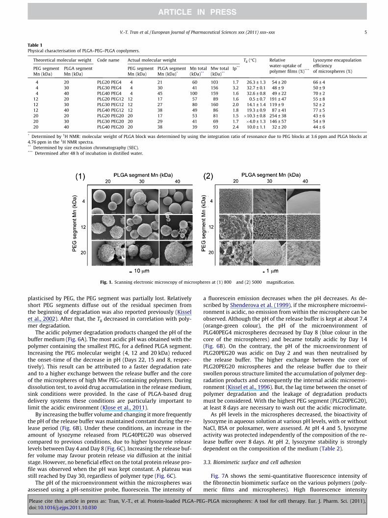
Table 1Physical characterisation of PLGA–PEG–PLGA copolymers.
Theoretical molecular weight Code name Actual molecular weight Tg (�C) Relativewater-uptake ofpolymer films (%)***
Lysozyme encapsulationefficiencyof microspheres (%)
PEG segmentMn (kDa)
PLGA segmentMn (kDa)
PEG segmentMn (kDa)
PLGA segmentMn (kDa)*
Mn total(kDa)**
Mw total(kDa)**
Ip**
4 20 PLG20 PEG4 4 21 60 103 1.7 26.3 ± 1.3 54 ± 20 66 ± 44 30 PLG30 PEG4 4 30 41 156 3.2 32.7 ± 0.1 48 ± 9 50 ± 94 40 PLG40 PEG4 4 45 100 159 1.6 32.6 ± 0.8 49 ± 22 70 ± 2
12 20 PLG20 PEG12 12 17 57 89 1.6 0.5 ± 0.7 191 ± 47 55 ± 812 30 PLG30 PEG12 12 27 80 160 2.0 14.1 ± 1.4 119 ± 9 52 ± 212 40 PLG40 PEG12 12 38 49 86 1.8 19.3 ± 0.9 87 ± 41 77 ± 520 20 PLG20 PEG20 20 17 53 81 1.5 �10.3 ± 0.8 254 ± 38 43 ± 620 30 PLG30 PEG20 20 29 41 69 1.7 �4.0 ± 1.3 146 ± 57 54 ± 920 40 PLG40 PEG20 20 38 39 93 2.4 10.0 ± 1.1 32 ± 20 44 ± 6
* Determined by 1H NMR: molecular weight of PLGA block was determined by using the integration ratio of resonance due to PEG blocks at 3.6 ppm and PLGA blocks at4.76 ppm in the 1H NMR spectra.** Determined by size exclusion chromatography (SEC).*** Determined after 48 h of incubation in distilled water.
Fig. 1. Scanning electronic microscopy of microspheres at (1) 800� and (2) 5000� magnification.
V.-T. Tran et al. / European Journal of Pharmaceutical Sciences xxx (2011) xxx–xxx 5
plasticised by PEG, the PEG segment was partially lost. Relativelyshort PEG segments diffuse out of the residual specimen fromthe beginning of degradation was also reported previously (Kisselet al., 2002). After that, the Tg decreased in correlation with poly-mer degradation.
The acidic polymer degradation products changed the pH of thebuffer medium (Fig. 6A). The most acidic pH was obtained with thepolymer containing the smallest PEG, for a defined PLGA segment.Increasing the PEG molecular weight (4, 12 and 20 kDa) reducedthe onset-time of the decrease in pH (Days 22, 15 and 8, respec-tively). This result can be attributed to a faster degradation rateand to a higher exchange between the release buffer and the coreof the microspheres of high Mw PEG-containing polymers. Duringdissolution test, to avoid drug accumulation in the release medium,sink conditions were provided. In the case of PLGA-based drugdelivery systems these conditions are particularly important tolimit the acidic environment (Klose et al., 2011).
By increasing the buffer volume and changing it more frequentlythe pH of the release buffer was maintained constant during the re-lease period (Fig. 6B). Under these conditions, an increase in theamount of lysozyme released from PLG40PEG20 was observedcompared to previous conditions, due to higher lysozyme releaselevels between Day 4 and Day 8 (Fig. 6C). Increasing the release buf-fer volume may favour protein release via diffusion at the initialstage. However, no beneficial effect on the total protein release pro-file was observed when the pH was kept constant. A plateau wasstill reached by Day 30, regardless of polymer type (Fig. 6C).
The pH of the microenvironment within the microspheres wasassessed using a pH-sensitive probe, fluorescein. The intensity of
Please cite this article in press as: Tran, V.-T., et al. Protein-loaded PLGA–PEdoi:10.1016/j.ejps.2011.10.030
a fluorescein emission decreases when the pH decreases. As de-scribed by Shenderova et al. (1999), if the microsphere microenvi-ronment is acidic, no emission from within the microsphere can beobserved. Although the pH of the release buffer is kept at about 7.4(orange-green colour), the pH of the microenvironment ofPLG40PEG4 microspheres decreased by Day 8 (blue colour in thecore of the microspheres) and became totally acidic by Day 14(Fig. 6B). On the contrary, the pH of the microenvironment ofPLG20PEG20 was acidic on Day 2 and was then neutralised bythe release buffer. The higher exchange between the core ofPLG20PEG20 microspheres and the release buffer due to theirswollen porous structure limited the accumulation of polymer deg-radation products and consequently the internal acidic microenvi-ronment (Kissel et al., 1996). But, the lag time between the onset ofpolymer degradation and the leakage of degradation productsmust be considered. With the highest PEG segment (PLG20PEG20),at least 8 days are necessary to wash out the acidic microclimate.
As pH levels in the microspheres decreased, the bioactivity oflysozyme in aqueous solution at various pH levels, with or withoutNaCl, BSA or poloxamer, were assessed. At pH 4 and 5, lysozymeactivity was protected independently of the composition of the re-lease buffer over 8 days. At pH 2, lysozyme stability is stronglydependent on the composition of the medium (Table 2).
3.3. Biomimetic surface and cell adhesion
Fig. 7A shows the semi-quantitative fluorescence intensity ofthe fibronectin biomimetic surface on the various polymers (poly-meric films and microspheres). High fluorescence intensity
G–PLGA microspheres: A tool for cell therapy. Eur. J. Pharm. Sci. (2011),

Fig. 2. In vitro release profile of lysozyme (mean ± SD) from ABA microspheres(n = 3 for each point) (error bar <1%).
Fig. 4. SDS–PAGE of lysozyme remaining in the PLG40PEG4, PLG40PEG20,PLG30PEG4 and PLG30PEG20 microspheres during in vitro release period undernon-reducing conditions (1) and reducing conditions (2) on Days 2, 8 and 22.
6 V.-T. Tran et al. / European Journal of Pharmaceutical Sciences xxx (2011) xxx–xxx
indicates high fibronectin adsorption on the polymer films studied.As PLGA 37.5/25 has been used to formulate the PAMs in previousstudies (Bouffi et al., 2010; Tatard et al., 2007b), fluorescence
Fig. 3. (A) Optical and fluorescence images of lysozyme-FITC (represented by the bright zand 43 and (B) bioactivity of lysozyme in the PLG40PEG4 microspheres during the in vi
Please cite this article in press as: Tran, V.-T., et al. Protein-loaded PLGA–PEdoi:10.1016/j.ejps.2011.10.030
intensity measured from fibronectin-coated PLGA 37.5/25 wasset at 1000 as a reference. For polymers containing 4 and 12 kDaPEG chains, high levels of fibronectin adsorption was found withhigh molecular weights of PLGA whereas low fibronectin adsorp-tion was found with small PLGAs (PLG20PEG12). Highly hydro-philic polymers (PEG 20 kDa) show weak fibronectin adsorptionregardless of the molecular weight of the PLGA segments. Fibro-nectin adsorbed on microspheres showed the same tendency ason polymeric films. Fibronectin-coated PLG40PEG4 microspheresshowed the best fibronectin adsorption (Fig. 7A.2).
PLG40PEG4 and PLG20PEG20 were selected to study the marrowstromal cell (MSC) behaviour on ABA microspheres and comparedto PLGA 37.5/25 microspheres used as a reference. After 4 h, the celladhesion on microspheres with a fibronectin biomimetic surfacewas determined (Fig. 7B). MSCs showed good adherence (around90%) onto the fibronectin-coated ABA microspheres regardless oftheir composition and of the quality of their biomimetic surface.Furthermore, all the polymers (PLG40PEG4, PLG20PEG20 andPLG37.5/25) showed good compatibility with MSC cells. Cell viabil-ity after one week of culture with the three different microspheres
one) remaining in PLG40PEG4 microspheres during in vitro release on Days 0, 2, 8, 22tro release period.
G–PLGA microspheres: A tool for cell therapy. Eur. J. Pharm. Sci. (2011),

Table 2Biostability of lysozyme in acidic medium (pH 2) with or without NaCl, BSA,Poloxamer 188 after 8 days of incubation.
Experiment BSA (mg/mL)
Poloxamer 188(mg/mL)
NaCl (mg/mL)
Activelysozyme (%)
1 0 0 0 101 ± 72 1 0 0 109 ± 33 0 2 0 93 ± 64 1 2 0 76 ± 145 0 0 0.88 66 ± 36 1 0 0.88 110 ± 17 0 2 0.88 86 ± 68 1 2 0.88 50 ± 2
Fig. 6. pH of release buffer: (A) in 500 lL of Tris–HCl 0.05 M, with 7 day intervalremoval, (B) in 1000 lL of Tris–HCl0.05 M with 2 day interval removal, and (C) thelysozyme release profile under (B) conditions. (B) Confocal microscopy of thePLG40PEG4 and PLG20PEG20 microspheres containing the pH-sensitive fluorescentprobe after 3 h, 2 days, 8 days and 14 days during in vitro release (1000 lL Tris–HCl0.05 M, 37 �C, with 2-day interval replacement conditions). The colour of 0.1 mg/mLfluorescein solution images at pH 2, 5 and 7.4 are shown for comparison.
Fig. 5. Molecular weight (A) and Tg changes (B) of PLG40PEG4 during in vitro release studies.
V.-T. Tran et al. / European Journal of Pharmaceutical Sciences xxx (2011) xxx–xxx 7
Please cite this article in press as: Tran, V.-T., et al. Protein-loaded PLGA–PEdoi:10.1016/j.ejps.2011.10.030
was similar to the control cells cultured in a standard well (data notshown).
4. Discussion
Tissue engineering is a rapidly expanding field that has gainedmomentum with the therapeutic possibilities offered by stem cells.Indeed, tissue engineering strategies such as 3D-biomimetic sur-faces and the prolonged delivery of growth factors may overcomethe major limitations in the use of stem cells which are their lowsurvival rate and differentiation capacity after transplantation. Atherapeutic tool (PAMs) combining these two strategies has beendeveloped in our laboratory and has shown its potential to enhancethe survival, differentiation and regenerative capacities of stemcells and to repair lesioned tissues (Delcroix et al., 2011; Tatardet al., 2004, 2005a,b, 2007a,b). In order to underline these strate-gies, it is essential to improve protein release from copolymermicrospheres, which remains a technological challenge due tothe special conformation of proteins. Our study thus focused onthe impact of polymer composition on protein release to compre-hend the chronology of protein destabilisation during the releasestep.
PLGA is known for its biodegradability and biocompatibility(Fournier et al., 2003) whereas non-toxic hydrophilic PEG is knownfor its stabilising effect on proteins (Kissel et al., 1996; Singh et al.,2008; Wade and Weller, 1994). PLGA–PEG–PLGA copolymers werethus chosen to fit these two features. These copolymers wereshown to increase drug release by increasing the degradation rate(Li et al., 2000; Youxin and Kissel, 1993) due to their rapid swellingproperties (Zange et al., 1997). However, the impact of the molec-ular weight of PLGA and PEG segments was not studied in the lit-erature. To investigate the effect of ABA composition on the proteinrelease rate, nine polymers were prepared with combinations from20, 30, and 40 kDa for PLGA segments and 4, 12, 20 kDa for PEGsegments.
G–PLGA microspheres: A tool for cell therapy. Eur. J. Pharm. Sci. (2011),

Fig. 7. (A1) fluorescence intensity of fibronectin-coated polymer films incubated with streptavidin–rhodamine and (A2) fluorescence microscopy of fibronectine-coatedPLG40PEG4 and PLG20PEG20 microspheres incubated with streptavidin–rhodamine. The bright zone represents the fluorescence of rhodamine. (B1) Illustration of cellsadhered to the coated PLG20PEG20, PLG40PEG4 and PLGA 37.5/25 microspheres after 4 h incubation with MSC cells and (B2) their optical microscopy observation (the barscale represents 20 lm).
8 V.-T. Tran et al. / European Journal of Pharmaceutical Sciences xxx (2011) xxx–xxx
Microspheres were prepared with similar protein loadings andwere sieved to minimise the number of parameters that can affectdrug release (Liggins and Burt, 2004; Tran et al., 2011). It has beenstated that the vacuum drying of ABA microspheres during lyophil-isation step can induce a porous surface due to the rapid evapora-tion of water (Morlock et al., 1998). We found that micro-porescould be formed before vacuum drying by microphase separation.During the solvent extraction, the hydrophobic PLGA segmentsshrank whereas the hydrophilic PEG segments had more affinitywith the outer-water phase and moved out to the surface, leadingto the formation of porous-structured microspheres. The PEG seg-ments dominated the porosity of these microspheres.
The molecular weights of PEG and PLGA segments presented animportant effect on protein encapsulation efficiency. For the20 kDa PEG segments, the encapsulation yield was dramaticallylow. The ABA copolymers with the longest segment of PLGA andthe shortest PEG segment (PLG40PEG4 and PLG40PEG12) had thehighest encapsulation efficiency. The water uptake level into thenascent microspheres increased with PEG segment size leadingto protein loss.
The ABA composition also affects the initial stage of in vitro re-lease (48 h). Even if no significant differences were detected be-tween the Tg of the raw polymer and the microspheres, the Tg
Please cite this article in press as: Tran, V.-T., et al. Protein-loaded PLGA–PEdoi:10.1016/j.ejps.2011.10.030
level was below the in vitro study temperature (37 �C), thereforethe diffusion of the protein was facilitated out of the rubberymicrospheres. Moreover, by increasing the length of hydrophilicsegments water uptake by the polymer matrix is promoted, thusfavouring the diffusion of the protein towards the outer phase dur-ing the hardening process and in vitro release.
After 48 h, lysozyme release profiles were continuous up to Day8 (PLG40PEG20) or Day 36 (PLG40PEG4) before reaching a plateau.We focused on PLG40PEG4 microspheres to elucidate the mecha-nism of lysozyme instability because it reached a satisfactoryhydrophilic-hydrophobic balance. During the first week, althoughPEG segments gradually leaked out from the ABA copolymers,the microenvironment was still neutral. However, at the sametime, the poloxamer gradually ran out, thereby limiting its anti-adsorption protective effect (Paillard-Giteau et al., 2010), and per-mitting lysozyme to partially adsorb onto the PLGA segment sur-faces. It is known that, when a protein is adsorbed on ahydrophobic surface, its conformation slowly changes to maximisethe number of exposed hydrophobic regions in contact with thehydrophobic surface (Andrade and Hlady, 1987). During this pro-cess, a fraction of lysozyme with exposed hydrophobic regionscould make contact with each other and form dimeric aggrega-tions. By Day 8, dimeric aggregations were dissociated by
G–PLGA microspheres: A tool for cell therapy. Eur. J. Pharm. Sci. (2011),

V.-T. Tran et al. / European Journal of Pharmaceutical Sciences xxx (2011) xxx–xxx 9
b-mercaptoethanol implying that disulphide bonds were involvedin the lysozyme polymerisation. By Day 22, dimeric aggregationswere no longer dissociated by b-mercaptoethanol, so another typeof covalent aggregation must be involved in the protein aggrega-tion mechanism at the latter period. From this point of view, thepresence of poloxamer in the microsphere is necessary to protectthe protein from adsorption and aggregation. However, the mono-band of lysozyme was always observed, so lysozyme aggregationcould not completely explain the incomplete release of protein.
At a later period, the plateau of protein release correlated intime with the drop of pH in the release medium: from pH 7 topH < 5. This phenomenon suggests that the incomplete releasewas partially due to the instability of lysozyme after a long incuba-tion time at 37 �C in the acidic conditions within the microspheres.Although Tris buffer could neutralise the acidity of the release buf-fer, the pH of the release buffer showed a lag in time with themicroenvironment in the microspheres , which began to be acidicfrom Day 8 onwards. At this time, poloxamer was no longer pres-ent, BSA and NaCl diffused into the microspheres from the releasebuffer creating BSA micro-zones in the microsphere altering theprotein microenvironment. These microenvironments correspondthe experiment five and six in the Table 2. At pH 2, the study oflysozyme stability in solution versus medium composition showedthat without poloxamer lysozyme keeps its biological activity inthe BSA micro-zones associated to NaCl, and lost 34% of its biolog-ical activity in the BSA-free micro-zones. These results underlinethe role of the microenvironment on the protein stability particu-larly at low pH.
To our knowledge, this is the first time that the cause of incom-plete protein release from ABA copolymers has been clarified. Var-ious reasons for protein instability accumulated within ABAmicrospheres can be thus listed as a function of time: proteinadsorption, protein aggregation and protein denaturation underacidic conditions. The comprehension of the underlying mecha-nisms leading to incomplete release will now allow us to overcomethese hurdles and create, invent, strategies to attain a completeand sustained release of proteins. One possibility is to optimisethe protein microenvironment by retaining poloxamer into thematrix during pH drop.
Further bioconducive characteristics of the microspheres, the3D biomimetic surface, was also investigated. A first screening ofthe fibronectin surface of the different microspheres showed thatPEG segments present at the surface of the microspheres limitedfibronectin adsorption. PEG is well known to minimise the adsorp-tion of plasmatic protein by steric repulsion and there is a directcorrelation between the PEG chain-length with the minimisationof protein adsorption (Vonarbourg et al., 2006). In this regard, withthe shortest PEG segments, fibronectin was mainly adsorbed to thehydrophobic PLGA segments. For the PLG30PEG12, we supposethat there is no dominant effect from either the PLGA or PEG seg-ments on fibronectin adsorption due to chain length. Therefore, inthis case, the rough surface of this polymer probably favoured theentrapment of fibronectin on its surface.
The majority of the cells adhered onto the fibronectin-coatedmicrospheres independent of the amount of fibronectin adsorbedon their surface. The hydrophilic surface facilitates cell adhesion,and thus more than 90% of MSCs adhered to the PLG20PEG20 witha slight fibronectin-coating. Furthermore, it is known that porousmicrospheres are suitable for the adherence of cells (Senumaet al., 1999). PLG20PEG20, with its high hydrophilic surface andpore morphology, hence has a good level of MSC cell adsorption.Nevertheless, several studies with neural stem cells have demon-strated the interest of fibronectin on cell differentiation (Tatardet al., 2005b; Wijelath et al., 2004). PLG40PEG4 microspheres pre-senting a biomimetic fibronectin surface thus appear to be moreinteresting than PLG20PEG20 microspheres for tissue-engineering
Please cite this article in press as: Tran, V.-T., et al. Protein-loaded PLGA–PEdoi:10.1016/j.ejps.2011.10.030
approaches. Finally, in the present study, human MSC adhered ontomicrospheres were still observed after one week of incubation,confirming that all the ABA polymers used in this study showeda good degree of compatibility with cells, as already reported fora mouse cell line (43, 44).
5. Conclusion
A series of biodegradable ABA triblock copolymers were synthe-sized by varying simultaneously the Mn of PEG and PLGA segmentsand they were used to prepare PAMs. The introduction ofhydrophilic polyoxyethylene B block domains in PLGA chains in-duced a rapid rate of water uptake and a continuous-release profile.However, higher PEG block copolymer showed shorter time releaseand was not suitable for the adsorption of the biomimetic surface.The PLG40PEG4 copolymer appeared to be the best candidate poly-mer and showed the best hydrophilic-lipophilic balance leading tothe best encapsulation efficiency (77%), low initial release, and con-tinuous release up to 36 days. Furthermore, the PLG40PEG4 polymerpresented a homogeneous fibronectin biomimetic surface and goodMSC biocompatibility. Incomplete release due to the time-limitedprotection of poloxamer, and denaturation of proteins under acidicconditions, was clarified. This study highlights the impact of PEGand PLGA size segments on the acidification rate both in the polymermatrix and in the release medium concurrently to the protein degra-dation profile. This systematic study has never been reported.
More importantly, this present study emphasises the necessityto both adjust the polymer hydrophilic-hydrophobic balance tothe intended protein release profile and to maintain the stabilizeragent within the polymer matrix close to the protein as long aspossible. Optimisation of the type (PPO/PEO ratio) and proportionof poloxamer co-precipitated with the protein is now in progress.
This work sets the ground for other studies with therapeuticproteins loaded in PLG40PEG4 microspheres for tissue engineeringapplications.
Acknowledgements
The authors would like to thank the ‘Service Commun d’Imagerieet de Microscopie d’Angers’ for the confocal microscopy experi-ments. We would also like to thank Pr. J.-L. Courthaudon and Dr.G. Larcher for their precious scientific advice. We are also gratefulto the French ‘Ministère de l’Education Nationale et de la Recherche’for financial support.
References
Andrade, J.D., Hlady, V., 1987. Protein adsorption and materials biocompatibility: atutorial reviews and suggested hypotheses. Adv. Polym. Sci. 79, 1–63.
Aubert-Pouëssel, A., Bibby, D.C., Venier-Julienne, M.C., Hindré, F., Benoit, J.P., 2002. Anovel in vitro delivery system for assessing the biological integrity of proteinupon release from PLGA microspheres. Pharm. Res. 19, 1046–1051.
Aubert-Pouëssel, A., Venier-Julienne, M.-C., Clavreul, A., Sergent, M., Jollivet, C.,Montero-Menei, C.N., Garcion, E., Bibby, D.C., Menei, P., Benoit, J.-P., 2004. Invitro study of GDNF release from biodegradable PLGA microspheres. J.Controlled Release 95, 463–475.
Bertram, J.P., Jay, S.M., Hynes, S.R., Robinson, R., Criscione, J.M., Lavik, E.B., 2009.Functionalized poly(lactic-co-glycolic acid) enhances drug delivery andprovides chemical moieties for surface engineering while preservingbiocompatibility. Acta Biomater. 5, 2860–2871.
Bezemer, J.M., Radersma, R., Grijpma, D.W., Dijkstra, P.J., Van Blitterswijk, C.A.,Feijen, J., 2000. Microspheres for protein delivery prepared from amphiphilicmultiblock copolymers1. Influence of preparation techniques on particlecharacteristics and protein delivery. J. Controlled Release 67, 233–248.
Boerckel, J.D., Kolambkar, Y.M., Dupont, K.M., Uhrig, B.A., Phelps, E.A., Stevens, H.Y.,Garcia, A.J., Guldberg, R.E., 2011. Effects of protein dose and delivery system onBMP-mediated bone regeneration. Biomaterials 32, 5241–5251.
Bouffi, C., Thomas, O., Bony, C., Giteau, A., Venier-Julienne, M.-C., Jorgensen, C.,Montero-Menei, C., Noël, D., 2010. The role of pharmacologically activemicrocarriers releasing TGF-b3 in cartilage formation in vivo by mesenchymalstem cells. Biomaterials 31, 6485–6493.
G–PLGA microspheres: A tool for cell therapy. Eur. J. Pharm. Sci. (2011),

10 V.-T. Tran et al. / European Journal of Pharmaceutical Sciences xxx (2011) xxx–xxx
Chen, W., Hu, S., 2011. Suitable carriers for encapsulation and distribution ofendostar: comparison of endostar-loaded particulate carriers. Int. J. Nanomed.6, 1535–1541.
D’Aurizio, E., van Nostrum, C.F., van Steenbergen, M.J., Sozio, P., Siepmann, F.,Siepmann, J., Hennink, W.E., Di Stefano, A., 2011. Preparation andcharacterization of poly(lactic-co-glycolic acid) microspheres loaded with alabile antiparkinson prodrug. Int. J. Pharm. 409, 289–296.
Delcroix, G.J.-R., Schiller, P.C., Benoit, J.-P., Montero-Menei, C.N., 2009. Adult celltherapy for brain neuronal damages and the role of tissue engineering.Biomaterials 31, 2105–2120.
Delcroix, G.J., Garbayo, E., Sindji, L., Thomas, O., Vanpouille-Box, C., Schiller, P.C.,Montero-Menei, C.N., 2011. The therapeutic potential of human multipotentmesenchymal stromal cells combined with pharmacologically activemicrocarriers transplanted in hemi-parkinsonian rats. Biomaterials 32, 1560–1573.
Dennis, J.E., Esterly, K., Awadallah, A., Parrish, C.R., Poynter, G.M., Goltry, K.L., 2007.Clinical-scale expansion of a mixed population of bone marrow-derived stemand progenitor cells for potential use in bone tissue regeneration. Stem Cells 25,2575–2582.
Determan, A.S., Wilson, J.H., Kipper, M.J., Wannemuehler, M.J., Narasimhan, B.,2006. Protein stability in the presence of polymer degradation products:Consequences for controlled release formulations. Biomaterials 27, 3312–3320.
D’Ippolito, G., Diabira, S., Howard, G.A., Menei, P., Roos, B.A., Schiller, P.C., 2004.Marrow-isolated adult multilineage inducible (MIAMI) cells, a uniquepopulation of postnatal young and old human cells with extensive expansionand differentiation potential. J. Cell Sci. 117, 2971–2981.
Fournier, E., Passirani, C., Montero-Menei, C.N., Benoit, J.P., 2003. Biocompatibility ofimplantable synthetic polymeric drug carriers: focus on brain biocompatibility.Biomaterials 24, 3311–3331.
Fu, K., Klibanov, A.M., Langer, R., 2000. Protein stability in controlled-releasesystems. Nat. Biotechnol. 18, 24–25.
Garric, X., Garreau, H., Vert, M., Molès, J.-P., 2008. Behaviors of keratinocytes andfibroblasts on films of PLA-PEO-PLA triblock copolymers with various PLAsegment lengths. J. Mater. Sci.: Mater. Med. 19, 1645–1651.
Giteau, A., Venier-Julienne, M.-C., Marchal, S., Courthaudon, J.-L., Sergent, M.,Montero-Menei, C., Verdier, J.-M., Benoit, J.-P., 2008. Reversible proteinprecipitation to ensure stability during encapsulation within PLGAmicrospheres. Eur. J. Pharm. Biopharm. 70, 127–136.
Kissel, T., Li, Y.X., Volland, C., Görich, S., Koneberg, R., 1996. Parenteral proteindelivery systems using biodegradable polyesters of ABA block structure,containing hydrophobic poly(lactide-co-glycolide) A blocks and hydrophilicpoly(ethylene oxide) B blocks. J. Controlled Release 39, 315–326.
Kissel, T., Li, Y., Unger, F., 2002. ABA-triblock copolymers from biodegradablepolyester A-blocks and hydrophilic poly(ethylene oxide) B-blocks as acandidate for in situ forming hydrogel delivery systems for proteins. Adv.Drug Deliv. Rev. 54, 99–134.
Klose, D., Delplace, C., Siepmann, J., 2011. Unintended potential impact of perfectsink conditions on PLGA degradation in microparticles. Int. J. Pharm. 404, 75–82.
Li, Y.X., Kissel, T., 1993. Synthesis and properties of biodegradable ABA triblockcopolymers consisting of poly(L-lactic acid) or poly(L-lactic-co-glycolic acid) A-blocks attached to central poly(oxyethylene) B-blocks. J. Controlled Release 27,247–257.
Li, X., Deng, X., Yuan, M., Xiong, C., Huang, Z., Zhang, Y., Jia, J., 2000. In vitrodegradation and release profiles of poly-DL-lactide-poly(ethylene glycol)microspheres with entrapped proteins. J. Appl. Polym. Sci. 78, 140–148.
Please cite this article in press as: Tran, V.-T., et al. Protein-loaded PLGA–PEdoi:10.1016/j.ejps.2011.10.030
Liggins, R.T., Burt, H.M., 2004. Paclitaxel loaded poly(L-lactic acid) (PLLA)microspheres: II. The effect of processing parameters on microspheremorphology and drug release kinetics. Int. J. Pharm. 281, 103–106.
Morlock, M., Kissel, T., Li, Y.X., Koll, H., Winter, G., 1998. Erythropoietin loadedmicrospheres prepared from biodegradable LPLG-PEO-LPLG triblockcopolymers: protein stabilization and in-vitro release properties. J. ControlRelease 56, 105–115.
Paillard-Giteau, A., Tran, V.T., Thomas, O., Garric, X., Coudane, J., Marchal, S.,Chourpa, I., Benoit, J.P., Montero-Menei, C.N., Venier-Julienne, M.C., 2010. Effectof various additives and polymers on lysozyme release from PLGA microspheresprepared by an s/o/w emulsion technique. Eur. J. Pharm. Biophys. 75, 128–136.
Senuma, Y., Franceschin, S., Hilborn, J.G., Tissières, P., Bisson, I., Frey, P., 1999.Bioresorbable microspheres by spinning disk atomization as injectable cellcarrier: from preparation to in vitro evaluation. Biomaterials 21, 1135–1144.
Shenderova, A., Burke, T.G., Schwendeman, S.P., 1999. The acidic microclimate inpoly(lactide-co-glycolide) microspheres stabilizes camptothecins. Pharm. Res.16, 241–243.
Singh, R., Singh, S., Lillard, J.W., 2008. Past, present, and future technologies for oraldelivery of therapeutic proteins. J. Pharm. Sci. 97, 2497–2523.
Tabata, Y., 2000. The importance of drug delivery systems in tissue engineering.Pharm. Sci. Tech. Today 3, 80–89.
Tatard, V.M., Venier-Julienne, M.C., Benoit, J.P., Menei, P., Montero-Menei, C.N.,2004. In vivo evaluation of pharmacologically active microcarriers releasingnerve growth factor and conveying PC12 cells. Cell Transplant. 13, 573–583.
Tatard, V.M., Menei, P., Benoit, J.P., Montero-Menei, C.N., 2005a. Combiningpolymeric devices and stem cells for the treatment of neurological disorders:a promising therapeutic approach. Curr. Drug Targets 6, 81–96.
Tatard, V.M., Venier-Julienne, M.C., Saulnier, P., Prechter, E., Benoit, J.P., Menei, P.,Montero-Menei, C.N., 2005b. Pharmacologically active microcarriers: a tool forcell therapy. Biomaterials 26, 3727–3737.
Tatard, V.M., D’Ippolito, G., Diabira, S., Valeyev, A., Hackman, J., McCarthy, M.,Bouckenooghe, T., Menei, P., Montero-Menei, C.N., Schiller, P.C., 2007a.Neurotrophin-directed differentiation of human adult marrow stromal cells todopaminergic-like neurons. Bone 40, 360–373.
Tatard, V.M., Sindji, L., Branton, J.G., Aubert-Pouëssel, A., Colleau, J., Benoit, J.-P.,Montero-Menei, C.N., 2007b. Pharmacologically active microcarriers releasingglial cell line – derived neurotrophic factor: survival and differentiation ofembryonic dopaminergic neurons after grafting in hemiparkinsonian rats.Biomaterials 28, 1978–1988.
Tran, V.T., Benoit, J.P., Venier-Julienne, M.C., 2011. Why and how to preparebiodegradable, monodispersed, polymeric microparticles in the field ofpharmacy? Int. J. Pharm. 407, 1–11.
Vonarbourg, A., Passirani, C., Saulnier, P., Benoit, J.P., 2006. Parameters influencingthe stealthiness of colloidal drug delivery systems. Biomaterials 27, 4356–4373.
Wade, A., Weller, P.J., 1994. Handbook of Pharmaceutical Excipients, 2nd ed. ThePhamaceutical Press and American Pharmaceutical Association, Washington,London.
Wijelath, E.S., Rahman, S., Murray, J., Patel, Y., Savidge, G., Sobel, M., 2004.Fibronectin promotes VEGF-induced CD34+ cell differentiation into endothelialcells. J. Vasc. Surg. 39, 655–660.
Youxin, L., Kissel, T., 1993. Synthesis and properties of biodegradable ABA triblockcopolymers consisting of poly(L-lactic acid) or poly(L-lactic-co-glycolic acid) A-blocks attached to central poly(oxyethylene) B-blocks. J. Controlled Release 27,247–257.
Zange, R., Li, Y., Kissel, T., 1997. In vitro degradation study and in vitrobiocompatibility testing of PEO containing ABA triblock copolymers. Proc.Controlled Release Soc., pp. 511–512.
G–PLGA microspheres: A tool for cell therapy. Eur. J. Pharm. Sci. (2011),
Copyright © 2022 FDOKUMEN