Evaluation of chondrogenesis within PEGT: PBT scaffolds with high PEG content
7
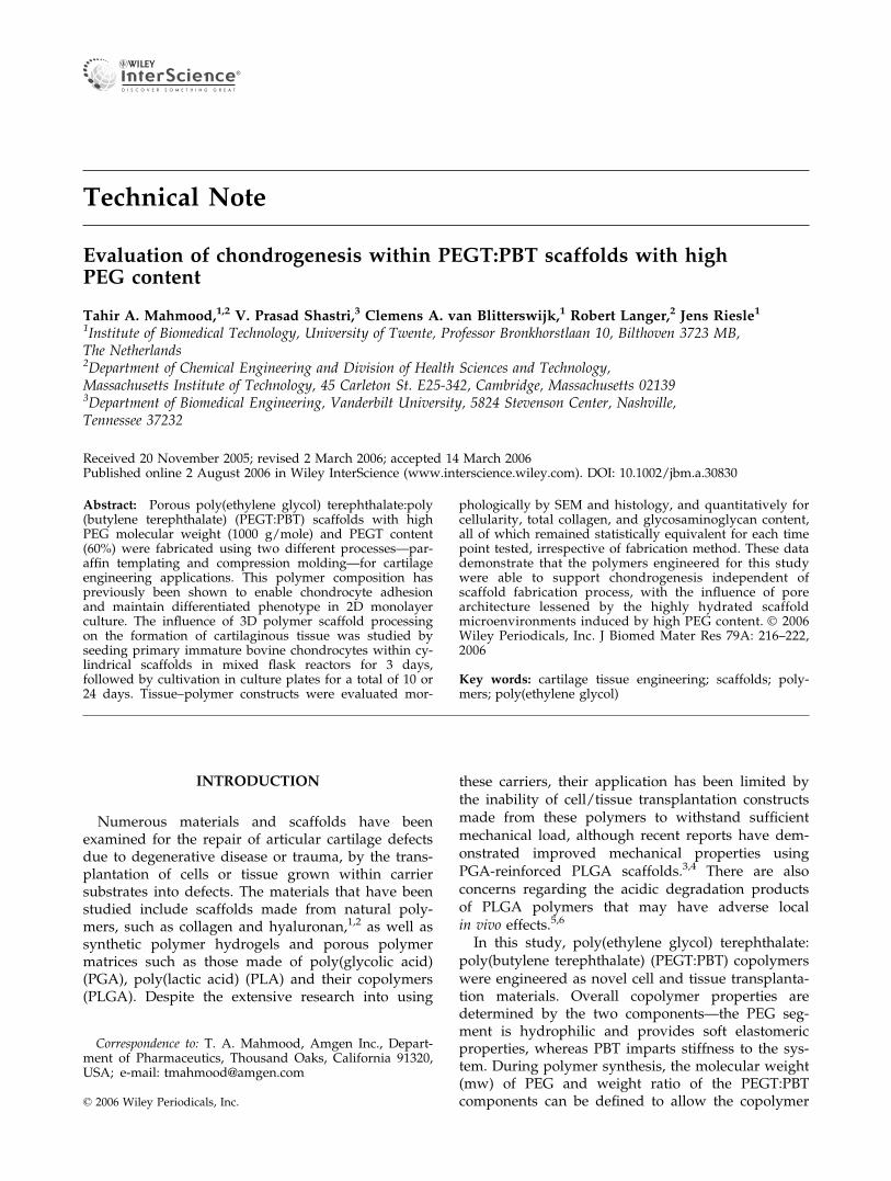
Technical Note Evaluation of chondrogenesis within PEGT:PBT scaffolds with high PEG content Tahir A. Mahmood, 1,2 V. Prasad Shastri, 3 Clemens A. van Blitterswijk, 1 Robert Langer, 2 Jens Riesle 1 1 Institute of Biomedical Technology, University of Twente, Professor Bronkhorstlaan 10, Bilthoven 3723 MB, The Netherlands 2 Department of Chemical Engineering and Division of Health Sciences and Technology, Massachusetts Institute of Technology, 45 Carleton St. E25-342, Cambridge, Massachusetts 02139 3 Department of Biomedical Engineering, Vanderbilt University, 5824 Stevenson Center, Nashville, Tennessee 37232 Received 20 November 2005; revised 2 March 2006; accepted 14 March 2006 Published online 2 August 2006 in Wiley InterScience (www.interscience.wiley.com). DOI: 10.1002/jbm.a.30830 Abstract: Porous poly(ethylene glycol) terephthalate:poly (butylene terephthalate) (PEGT:PBT) scaffolds with high PEG molecular weight (1000 g/mole) and PEGT content (60%) were fabricated using two different processes—par- affin templating and compression molding—for cartilage engineering applications. This polymer composition has previously been shown to enable chondrocyte adhesion and maintain differentiated phenotype in 2D monolayer culture. The influence of 3D polymer scaffold processing on the formation of cartilaginous tissue was studied by seeding primary immature bovine chondrocytes within cy- lindrical scaffolds in mixed flask reactors for 3 days, followed by cultivation in culture plates for a total of 10 or 24 days. Tissue–polymer constructs were evaluated mor- phologically by SEM and histology, and quantitatively for cellularity, total collagen, and glycosaminoglycan content, all of which remained statistically equivalent for each time point tested, irrespective of fabrication method. These data demonstrate that the polymers engineered for this study were able to support chondrogenesis independent of scaffold fabrication process, with the influence of pore architecture lessened by the highly hydrated scaffold microenvironments induced by high PEG content. Ó 2006 Wiley Periodicals, Inc. J Biomed Mater Res 79A: 216–222, 2006 Key words: cartilage tissue engineering; scaffolds; poly- mers; poly(ethylene glycol) INTRODUCTION Numerous materials and scaffolds have been examined for the repair of articular cartilage defects due to degenerative disease or trauma, by the trans- plantation of cells or tissue grown within carrier substrates into defects. The materials that have been studied include scaffolds made from natural poly- mers, such as collagen and hyaluronan, 1,2 as well as synthetic polymer hydrogels and porous polymer matrices such as those made of poly(glycolic acid) (PGA), poly(lactic acid) (PLA) and their copolymers (PLGA). Despite the extensive research into using these carriers, their application has been limited by the inability of cell/tissue transplantation constructs made from these polymers to withstand sufficient mechanical load, although recent reports have dem- onstrated improved mechanical properties using PGA-reinforced PLGA scaffolds. 3,4 There are also concerns regarding the acidic degradation products of PLGA polymers that may have adverse local in vivo effects. 5,6 In this study, poly(ethylene glycol) terephthalate: poly(butylene terephthalate) (PEGT:PBT) copolymers were engineered as novel cell and tissue transplanta- tion materials. Overall copolymer properties are determined by the two components—the PEG seg- ment is hydrophilic and provides soft elastomeric properties, whereas PBT imparts stiffness to the sys- tem. During polymer synthesis, the molecular weight (mw) of PEG and weight ratio of the PEGT:PBT components can be defined to allow the copolymer Correspondence to: T. A. Mahmood, Amgen Inc., Depart- ment of Pharmaceutics, Thousand Oaks, California 91320, USA; e-mail: [email protected] ' 2006 Wiley Periodicals, Inc.
-
Upload
uni-freiburg -
Category
Documents
-
view
0 -
download
0
Transcript of Evaluation of chondrogenesis within PEGT: PBT scaffolds with high PEG content
Technical Note
Evaluation of chondrogenesis within PEGT:PBT scaffolds with highPEG content
Tahir A. Mahmood,1,2 V. Prasad Shastri,3 Clemens A. van Blitterswijk,1 Robert Langer,2 Jens Riesle11Institute of Biomedical Technology, University of Twente, Professor Bronkhorstlaan 10, Bilthoven 3723 MB,The Netherlands2Department of Chemical Engineering and Division of Health Sciences and Technology,Massachusetts Institute of Technology, 45 Carleton St. E25-342, Cambridge, Massachusetts 021393Department of Biomedical Engineering, Vanderbilt University, 5824 Stevenson Center, Nashville,Tennessee 37232
Received 20 November 2005; revised 2 March 2006; accepted 14 March 2006Published online 2 August 2006 in Wiley InterScience (www.interscience.wiley.com). DOI: 10.1002/jbm.a.30830
Abstract: Porous poly(ethylene glycol) terephthalate:poly(butylene terephthalate) (PEGT:PBT) scaffolds with highPEG molecular weight (1000 g/mole) and PEGT content(60%) were fabricated using two different processes—par-affin templating and compression molding—for cartilageengineering applications. This polymer composition haspreviously been shown to enable chondrocyte adhesionand maintain differentiated phenotype in 2D monolayerculture. The influence of 3D polymer scaffold processingon the formation of cartilaginous tissue was studied byseeding primary immature bovine chondrocytes within cy-lindrical scaffolds in mixed flask reactors for 3 days,followed by cultivation in culture plates for a total of 10 or24 days. Tissue–polymer constructs were evaluated mor-
phologically by SEM and histology, and quantitatively forcellularity, total collagen, and glycosaminoglycan content,all of which remained statistically equivalent for each timepoint tested, irrespective of fabrication method. These datademonstrate that the polymers engineered for this studywere able to support chondrogenesis independent ofscaffold fabrication process, with the influence of porearchitecture lessened by the highly hydrated scaffoldmicroenvironments induced by high PEG content. � 2006Wiley Periodicals, Inc. J Biomed Mater Res 79A: 216–222,2006
Key words: cartilage tissue engineering; scaffolds; poly-mers; poly(ethylene glycol)
INTRODUCTION
Numerous materials and scaffolds have beenexamined for the repair of articular cartilage defectsdue to degenerative disease or trauma, by the trans-plantation of cells or tissue grown within carriersubstrates into defects. The materials that have beenstudied include scaffolds made from natural poly-mers, such as collagen and hyaluronan,1,2 as well assynthetic polymer hydrogels and porous polymermatrices such as those made of poly(glycolic acid)(PGA), poly(lactic acid) (PLA) and their copolymers(PLGA). Despite the extensive research into using
these carriers, their application has been limited bythe inability of cell/tissue transplantation constructsmade from these polymers to withstand sufficientmechanical load, although recent reports have dem-onstrated improved mechanical properties usingPGA-reinforced PLGA scaffolds.3,4 There are alsoconcerns regarding the acidic degradation productsof PLGA polymers that may have adverse localin vivo effects.5,6
In this study, poly(ethylene glycol) terephthalate:poly(butylene terephthalate) (PEGT:PBT) copolymerswere engineered as novel cell and tissue transplanta-tion materials. Overall copolymer properties aredetermined by the two components—the PEG seg-ment is hydrophilic and provides soft elastomericproperties, whereas PBT imparts stiffness to the sys-tem. During polymer synthesis, the molecular weight(mw) of PEG and weight ratio of the PEGT:PBTcomponents can be defined to allow the copolymer
Correspondence to: T. A. Mahmood, Amgen Inc., Depart-ment of Pharmaceutics, Thousand Oaks, California 91320,USA; e-mail: [email protected]
' 2006 Wiley Periodicals, Inc.

to be tailored for desired mechanical and surfaceproperties. For example, reducing PEG mw from1000 to 300 g/mole and PEGT wt % from 70 to 55%resulted in increase in tensile strength (�) from 5.3 to15.3 MPa, and in tensile modulus (E) from 33.9 to187.5 MPa, respectively.7
Various PEGT:PBT compositions have been stud-ied for their ability to support chondrocyte attach-ment, growth, and phenotype in 2D monolayer con-ditions.8–10 It was observed from these studies that abalance of hydrophilic and hydrophobic segments isneeded for chondrocyte attachment, while maintain-ing chondrogenic phenotype. Specifically, substrateswith high PEG mw (>1000 g/mole) and PEGT wt(>70%) supported the differentiated phenotype, butwere not conducive to cell attachment in numberssufficient for practical application. Conversely, lowPEG mw (300 g/mole) and PEGT wt (<55%) enabledcell attachment, but resulted in chondrocyte dediffer-entiation to fibroblastic phenotype. Of the range ofcompositions evaluated, the composition with 1000 g/mole PEG and a PEGT:PBT molar ratio of 60:40 wasshown to support chondrocyte adhesion and presentedinterfacial material-cell conditions that maintained thedifferentiated chondrocyte phenotype in 2D mono-layer conditions, and was thus selected as the compo-sition for studying 3D cartilaginous tissue formation.Furthermore, an orthopedic medical device made fromPEGT:PBT copolymers has been approved by the USFood and Drug Administration,11 which could facili-tate the development of tissue engineered productsbased on these polymers.
Porous scaffolds for cell/tissue transplantationwere fabricated from the high PEG content copoly-mer by two techniques: paraffin templating (PT) andcompression molding (CM). Initial studies havedemonstrated promise for the use of these scaffold-ing techniques for tissue engineering, although theinfluence of polymer composition on tissue forma-tion within each scaffold type has yet to be fully elu-cidated. In the CM process, polymer resin is meltedaround salt crystals inside a mold under high pres-sure, and the salt leached out using water to form aporous structure.12 In PT, the paraffin porogen isleached out using an aliphatic hydrocarbon solvent(porogen solvent; nonsolvent for polymer) during poly-mer precipitation to create porous foam blocks.13
Articular cartilage extracellular matrix (ECM) con-tains type II collagen, proteoglycan macromoleculeswith glycosaminoglycan (GAG) side chains, as wellas other matrix molecules. The tissue is able to with-stand compression loading due to interaction betweencollagen network and GAG, which is able to sequesterwater within the matrix.2 To enhance the potential ofintegrating engineered tissue with ECM at the defectsite, a strategy is to transplant biologic-material con-structs of immature tissue. This could lead to im-
proved integration with the host ECM, as further tis-sue development will occur in vivo within the lesion.
The objective of this study was to evaluate and com-pare in vitro chondrogenesis within high PEG contentporous scaffolds fabricated by PT and CM techniques.
MATERIALS AND METHODS
Polymer synthesis
PEGT:PBT segmented copolymer resin was prepared asdescribed previously.14 Briefly, synthesis occurred by two-step condensation in the presence of titanium tetrabutoxide(Merck, Darmstadt, Germany) as catalyst (0.1 wt %). Vita-min E (Sigma, Uithoorn, The Netherlands) was added toprevent oxidation. The resulting copolymer can be furtherprocessed into various geometrical configurations such asfilms or porous scaffolds, providing a model class of sub-strates for studying cell–material interactions (Fig. 1a).
Scaffold processing and fabrication
The different formulations of the PEGT:PBT copolymersystem are indicated as follows: a-PEGT b:c, where a is themolecular weight of PEG (g/mole), b is the weight per-centage of PEGT, and c is the weight percentage of PBT. Inthis study, 1000-PEGT 60:40 was used.
PT scaffolds were produced by dissolving the polymerresin in methylene chloride and dispersed with spherical andirregular paraffin particles of 1 mm and 425–500 lmmean di-ameter, respectively, to yield a polymer–solvent–porogenputty. The theoretical average pore size was calculated to be613 lm. The putty was packed densely into a Teflon mold,and the paraffin porogen extracted in hexane at 45–508C for20 min to yield a porous block of polymer foam (Fig. 1b). Thefoam block was dried under vacuum to remove any residualsolvent.
CM scaffolds were fabricated into blocks by injectionmolding PEGT:PBT polymer resin with salt crystal inclu-sions at 2208C for 10 min under 20,000 lbs/in.2 pressure.These blocks were air-cooled for 20 min and immersed indemineralized H2O to leach out the salt crystals and create400–600 lm pores (Fig. 1b).
Cylindrical scaffolds of 4 mm diameter and 4 mm thick-ness were cored out from blocks of both fabrication groups,cleaned with 70% ethanol and demineralized H2O, andgamma-sterilized under vacuum. Scaffolds were immersedovernight in culture medium prior to seeding to reach equi-librium polymer swelling.
Chondrocyte isolation and culturing
Articular cartilage was harvested from the patellar groovesof young bovine calves. Chondrocytes were isolated by type IIcollagenase (Worthington Biochemical, Lakewood, NJ) diges-tion for 16 h and transferred to a well defined culture mediumcontaining DMEM, 10% fetal bovine serum, penicillin/strep-tomycin (Gibco, NJ), ascorbic acid phosphate, nonessentialamino acids, and L-proline (Sigma, St. Louis, MO).
PEGT:PBT SCAFFOLDS WITH HIGH PEG CONTENT 217
Journal of Biomedical Materials Research Part A DOI 10.1002/jbm.a

Chondrocytes were seeded at a density of 3 million cellsper scaffold in mixed flask systems, as described in detailpreviously.15,16 Scaffolds were maintained in the mixed flasksfor 3 days, after which they were transferred to 6-well plates(BD Falcon, Amsterdam, NL) on an orbital shaker for theremaining cultivation periods. Samples from each scaffoldgroup were harvested for histology at day 10 and for electronmicroscopy at day 24. Constructs were also harvested at days10 and 24 for total cell number, collagen, and GAG content.
Histology
Histological sections were prepared by first fixing thecell–polymer constructs in 2.5% gluteraldehyde and 10%formalin for 7 days. Specimens were then dehydrated over1 week in a graded ethanol series, followed by embedding inglycol methacrylate and sectioning to a thickness of 5 lm ona standard microtome (Microm, Walldorf, Germany). The
sections were stained by safranin O/fast green for sulfatedGAG, and nuclei counterstained with hematoxylin.
Scanning electron microscopy
To examine cell–polymer construct morphology in higherdetail, samples harvested at day 24 were fixed in Karnov-sky’s fixative, dried at the critical point of CO2 and sputter-coated with a 12 nm layer of gold (Cressington Scientific,Watford, UK). Specimens were examined by scanning elec-tron microscopy (SEM) (Phillips ESEM, Eindhoven, TheNetherlands).
Quantitative GAG and collagen analyses
Constructs (n ¼ 3) were harvested at day 10 and 24 anddigested overnight at 568C in solution containing protein-ase K, pepstatin A and iodoacetamide (Sigma). Digests
Figure 1. (a) Chemical structure of segmented poly(ether ester) [PEGT:PBT] copolymers, formed by polycondensationpolymerization of hydrophilic PEG-containing segments and hydrophobic PBT segments. (b) Schematic outline of the stepsinvolved in fabricating CM and PT scaffolds. PT scaffolds were made by dissolving the polymer resin in methylene chlo-ride and dispersed with spherical and irregular paraffin porogen particles. The resulting viscous solution/putty waspacked in a Teflon mold and porogen extracted at 45–508C for 20 min in hexane. The foam block was dried under vacuumto remove residual solvent. CM scaffolds were fabricated into blocks by injection molding PEGT:PBT polymer resin withsalt crystals (porogen) at 2208C for 10 min under 20,000 lbs/in.2 pressure. Blocks were air cooled for 20 min and immersedin demineralized H2O to leach out the salt porogen. Cylindrical scaffolds used for these studies were cored out fromblocks of each fabrication group, as needed.
218 MAHMOOD ET AL.
Journal of Biomedical Materials Research Part A DOI 10.1002/jbm.a

Figure 2. SEMs of high PEG content PEGT:PBT scaffolds fabricated by CM (Fig. 2a) and PT (Fig. 2b). The surface to-pographies that resulted from the different techniques are visible at this magnification. Histology of construct cross sectionsat day 10 demonstrated interconnected GAG-rich tissue formation within scaffolds [Figs. 2(c,d)]; scale bars ¼ 2 mm. Highmagnification SEM examination at day 24 revealed dense ECM-like morphology with rounded chondrocytes [Figs. 2(e,f)].[Color figure can be viewed in the online issue, which is available at www.interscience.wiley.com.]
PEGT:PBT SCAFFOLDS WITH HIGH PEG CONTENT 219
Journal of Biomedical Materials Research Part A DOI 10.1002/jbm.a

were evaluated for cellularity, GAG, and total collagencontent.
For determining cell numbers, quantitation of total DNAwere performed by Cyquant dye kit (Molecular Probes,Leiden, The Netherlands) using a spectrofluorometer(Perkin Elmer, IL). Cell numbers were calculated from thetotal DNA content by normalizing to 7.7 � 10�12 g DNAper bovine chondrocyte.17
GAG content was obtained by labeling with dimethyl-methylene blue dye (Sigma-Aldrich) and measuring colorintensity by a spectrophotometer (Bio-Tek Instruments,Neufahrn, Germany).18
Total collagen was determined by measuring theamount of hydroxyproline in each construct. Aliquots ofproteinase K digests to be evaluated for hydroxyprolinewere hydrolyzed in 6N HCl at 1108C for 16 h. The hydro-lyzate was assayed for hydroxyproline using methods thathave been described in detail elsewhere, with a hydroxy-proline content of 13% of collagen used to calculate finalcollagen quantities.19
Data obtained from quantitative assays were analyzedby student’s t-test. p < 0.05 was used to determine statisti-cal significance.
RESULTS
Histology and scanning electron microscopy
The pore structures of scaffolds produced by PTand CM are shown in Figure 2. Pores in CM scaf-folds were generally cuboidal (Fig. 2a), whereas the PTprocess resulted in an irregular pore structure (Fig. 2b).
At day 10, histology revealed red positive stainingfor safranin O within the pores of constructs fabri-cated by both processes [Figs. 2(c,d)]. Observation ofsections from both scaffold types showed regions ofcartilaginous tissue interconnecting between pores.
Scanning electron micrographs of constructs culti-vated for 24 days revealed a rich fibrillar extracellu-lar-like matrix (ECM), within which chondrocyteswere embedded [Figs. 2(e,f)]. In the PT constructs,the dense ECM lacunae contained cells with sphe-roid morphology.
In some histology sections, artifacts such as gapswere seen. Repeated sectioning from multiple sam-ples has demonstrated that these areas contained tis-sue that separated from the polymer scaffold andembedding material during sectioning.
Quantitative constituent analyses (Table 1)
Cellularity evaluated by DNA assay
At day 10, PT and CM constructs contained an aver-age 12.6 million and 13.5 million cells per construct, re-spectively. From 10 to 24 days cultivation, both scaffoldgroups displayed significant increases in cellularity(PT: p < 0.036, CM: p < 0.032). At day 24, PT and CM
constructs contained 16.9 million and 20.7 million cells,respectively. At each time point, there was no signifi-cant difference in cell number between scaffold types.
GAG content
At day 10, constructs contained 2.4 mg (PT) and2.2 mg GAG per construct (CM). Mean GAG contentremained statistically the same for both scaffoldtypes between 10 and 24 days. At day 24, meanGAG content within each PT and CM construct was2.2 and 2.1 mg, respectively.
Collagen content
At day 10 of culture, PT constructs contained 22.8 mgcollagen, whereas CM constructs contained 21.7 mg. Atday 24, PT and CM constructs contained 51 and 52.5 mgcollagen, respectively. Although there were no signifi-cant differences in collagen content between both scaf-fold types at either time point, the amount of collagenincreased significantly in each scaffold type during10–24 days culture (PT, p < 0.001; CM, p < 0.0035).
DISCUSSION
Scaffolds fabricated from 1000-PEGT 60:40 poly-mers using both PT and CM processes enabled theinfiltration of cells within the scaffold interior, and
TABLE IQuantitative Evaluation of Cell Number, GAG (mg), andTotal Collagen Content (mg) Per Construct in PT and
CM Scaffolds at 10 and 24 Days
Day 10 Day 24
Average St. Dev Average St. Dev
Cell number (106)PT 12.6 1.7 16.9 0.9CM 13.5 1.1 20.7 0.8
GAG (mg)PT 2.4 0.3 2.2 0.5CM 2.2 0.3 2.1 0.2
Collagen (mg)PT 22.8 4.8 51.0 1.8CM 21.7 2.2 52.5 5.1
Cell number: Both scaffold types exhibited a significantincrease in cellularity from 10 to 24 days (PT, p < 0.036;CM, p < 0.032). At each time point, however, there was nosignificant difference in the number of cells in each scaf-fold type. GAG: There were no significant differences inGAG between both scaffold types at either time point, orbetween each scaffold group from 10 to 24 days cultiva-tion. Collagen: Although there were no differences in totalcollagen content in both scaffold types at either time point,the amount of collagen increased significantly in each scaf-fold type from 10 to 24 days cultivation (PT, p < 0.001;CM, p < 0.0035).
220 MAHMOOD ET AL.
Journal of Biomedical Materials Research Part A DOI 10.1002/jbm.a

supported chondrogenesis. Theoretical porosities ofscaffolds (determined by mass–volume) produced byboth fabrication methods were similar (CM: 78%, PT:80%), which are very similar to the porosities deter-mined using micro-computed tomography20 andmercury porosimetry.13
There were no significant differences in the num-ber of cells, total collagen, or GAG content withinscaffolds produced from this high PEG content com-position. This may in part be due to the relativelyhigh hydrophilicity of the polymer compositiontested—the mass of 1000-PEGT 60:40 polymer hasbeen shown in our laboratory to increase 1.8 timesupon reaching equilibrium in water. Polymers withlong chain PEG and high PEG content are likely tosequester water molecules, providing an environmentsimilar to the hydrated conditions found in hyalinecartilage. A correlation between PEG mw, PEGT:PBTratio and molecule diffusivity has been previously de-monstrated, due to differential effects of hydration onthe 50–100 A mesh size of the hydrogel-like polymerphase.21 Thus, it follows that the highly hydratedconditions of the 1000-PEGT 60:40 copolymer mayenable greater nutrient diffusion into scaffold interi-ors, thereby supporting chondrogenesis throughoutthe volume of scaffolds, as evidenced by histology,SEM, and supporting quantitative data.
It has been reported earlier that cartilaginous tis-sue formation within scaffolds fabricated fromhydrophobic polymer compositions (300 mw PEG,55% PEGT) was dependent on the method of fabrica-tion.22 In those studies, only PT supported in vitrochondrogenesis, as chondrocytes seeded on CM scaf-folds were unable to infiltrate the interior of the scaf-folds, remained at the periphery and exhibited a de-differentiated fibroblastic morphology. When lookedat together with the earlier results, the data pre-sented here support the notion that 3D pore architec-ture may be more important in scaffolds with lesschondro-supportive chemistry. Our results also sup-port a previous study that showed that a hydrophilicpolymer with dense arrangement of high molecularweight PEG, as well as a baseline pore interconnec-tivity, are prerequisite to support chondrogenesis instatic in vitro cultivation.20
Cartilaginous tissue formation has been reportedin porous scaffolds with smaller pore diameters of174 lm and 115–335 lm.23,24 This indicates that themean pore size in our PT and CM scaffolds waslarge enough to allow chondrocyte infiltration andsubsequent matrix formation. Using GAG producedper cell as a comparative, positive biomarker by whichto compare with the extensively studied PGA-basedpolymer scaffolds, the PEGT:PBT constructs evaluatedin this study demonstrated up to 3-fold greater GAGper cell than PGA at similar time points.25 Highmagnification SEM at 24 days revealed dense ECM
and rounded cellular morphology typical for chon-drocytes.
In conclusion, we have shown that scaffolds madefrom 1000 g/mole PEG and 60:40 PEGT:PBT ratiocan be produced with different pore architecturesbut similar porosities using PT and CM techniques.These data demonstrate that the high PEG contentPEGT:PBT copolymer polymer composition engi-neered for this study was able to support chondro-genesis independent of scaffold fabrication process.
References
1. Woodfield TB, Bezemer JM, Pieper JS, van Blitterswijk CA,Riesle J. Scaffolds for tissue engineering of cartilage. Crit RevEukaryot Gene Expr 2002;12:209–236.
2. Hunziker EB. Articular cartilage repair: Basic science andclinical progress. A review of the current status and pros-pects. Osteoarthritis Cartilage 2002;10:432–463.
3. Slivka MA, Leatherbury NC, Kieswetter K, Niederauer GG.Porous, resorbable, fiber-reinforced scaffolds tailored for articu-lar cartilage repair. Tissue Eng 2001;7:767–780.
4. Niederauer GG, Slivka MA, Leatherbury NC, Korvick DL,Harroff HH, Ehler WC, Dunn CJ, Kieswetter K. Evaluation ofmultiphase implants for repair of focal osteochondral defectsin goats. Biomaterials 2000;21:2561–2574.
5. Spain TL, Agrawal CM, Athanasiou KA. New technique toextend the useful life of a biodegradable cartilage implant.Tissue Eng 1998;4:343–352.
6. Athanasiou KA, Agrawal CM, Barber FA, Burkhart SS.Orthopaedic applications for PLA-PGA biodegradable poly-mers. Arthroscopy 1998;14:726–737.
7. Sakkers RJ, de Wijn JR, Dalmeyer RA, van Blitterswijk CA,Brand R. Evaluation of copolymers of polyethylene oxide andpolybutylene terephthalate (polyactive): Mechanical behav-iour. J Mater Sci Mater Med 1998;9:375–379.
8. Mahmood TA, de Jong R, Riesle J, Langer R, van BlitterswijkCA. Adhesion-mediated signal transduction in human articu-lar chondrocytes: The influence of biomaterial chemistry andtenascin-C. Exp Cell Res 2004;301:179–188.
9. Mahmood TA, Miot S, Martin I, Riesle J, Langer R, vanBlitterswijk CA. Modulation of cell phenotype for tissue engi-neering by designing the biologic-copolymer carrier interface.2006. Submitted for publication.
10. Papadaki M, Mahmood T, Gupta P, Claase MB, Grijpma DW,Riesle J, van Blitterswijk CA, Langer R. The different behav-iors of skeletal muscle cells and chondrocytes on PEGT/PBTblock copolymers are related to the surface properties of thesubstrate. J Biomed Mater Res 2001;54:47–58.
11. U.S. Food and Drug Administration Approval. Notice Num-ber K010840. 2001.
12. Malda J, Woodfield TB, van der Vloodt F, Wilson C, MartensDE, Tramper J, van Blitterswijk CA, Riesle J. The effect ofPEGT/PBT scaffold architecture on the composition of tissueengineered cartilage. Biomaterials 2005;26:63–72.
13. Shastri VP, Martin I, Langer R. Macroporous polymer foamsby hydrocarbon templating. Proc Natl Acad Sci USA 2000;97:1970–1975.
14. Deschamps AA, Grijpma DW, Feijen J. Poly(ethylene oxide)/poly(butylene terephthalate) segmented block copolymers:The effect of copolymer composition on physical propertiesand degradation behavior. Polymer 2001;42:9335–9345.
15. Freed LE, Hollander AP, Martin I, Barry JR, Langer R, Vunjak-Novakovic G. Chondrogenesis in a cell-polymer-bioreactor sys-tem. Exp Cell Res 1998;240:58–65.
PEGT:PBT SCAFFOLDS WITH HIGH PEG CONTENT 221
Journal of Biomedical Materials Research Part A DOI 10.1002/jbm.a

16. Freed LE, Vunjak-Novakovic G, Langer R. Cultivation of cell-polymer cartilage implants in bioreactors. J Cell Biochem 1993;51:257–264.
17. Kim YJ, Sah RL, Doong JY, Grodzinsky AJ. Fluorometricassay of DNA in cartilage explants using hoechst 33258. AnalBiochem 1988;174:168–176.
18. Farndale RW, Buttle DJ, Barrett AJ. Improved quantitation anddiscrimination of sulphated glycosaminoglycans by use ofdimethylmethylene blue. Biochim Biophys Acta 1986;883:173–177.
19. Morales TI, Woessner JF, Howell DS, Marsh JM, LeMaire WJ.A microassay for the direct demonstration of collagenolyticactivity in Graafian follicles of the rat. Biochim Biophys Acta1978;524:428–434.
20. Miot S, Woodfield T, Daniels AU, Suetterlin R, Peterschmitt I,Heberer M, van Blitterswijk CA, Riesle J, Martin I. Effects ofscaffold composition and architecture on human nasal chon-drocyte redifferentiation and cartilaginous matrix deposition.Biomaterials 2005;26:2479–2489.
21. Bezemer JM, Grijpma DW, Dijkstra PJ, van Blitterswijk CA,Feijen J. A controlled release system for proteins based onpoly(ether ester) block-copolymers: Polymer network charac-terization. J Control Release 1999;62:393–405.
22. Mahmood TA, Shastri VP, Van Blitterswijk CA, Langer R,Riesle J. Tissue engineering of bovine articular cartilage withinporous poly(ether ester) copolymer scaffolds with differentstructures. Tissue Eng 2005;11:1244–1253.
23. Miralles G, Baudoin R, Dumas D, Baptiste D, Hubert P,Stoltz JF, Dellacherie E, Mainard D, Netter P, Payan E. Sodiumalginate sponges with or without hyaluronate: In vitro engi-neering of cartilage. J Biomed Mater Res 2001;57:268–278.
24. LiVecchi AB, Tombes RM, LaBerge M. In vitro chondrocyte col-lagen deposition within porous HDPE: Substrate microstructureand wettability effects. J Biomed Mater Res 1994;28:839–850.
25. Freed LE, Grande DA, Lingbin Z, Emmanual J, Marquis JC,Langer R. Joint resurfacing using allograft chondrocytes andsynthetic biodegradable polymer scaffolds. J Biomed MaterRes 1994;28:891–899.
222 MAHMOOD ET AL.
Journal of Biomedical Materials Research Part A DOI 10.1002/jbm.a