Mimicking the Arterial Microenvironment with PEG-PC to ...
197
University of Massachusetts Amherst University of Massachusetts Amherst ScholarWorks@UMass Amherst ScholarWorks@UMass Amherst Doctoral Dissertations Dissertations and Theses August 2015 Mimicking the Arterial Microenvironment with PEG-PC to Mimicking the Arterial Microenvironment with PEG-PC to Investigate the Roles of Physicochemical Stimuli in SMC Investigate the Roles of Physicochemical Stimuli in SMC Phenotype and Behavior Phenotype and Behavior William G. Herrick University of Massachusetts Amherst Follow this and additional works at: https://scholarworks.umass.edu/dissertations_2 Part of the Biochemical and Biomolecular Engineering Commons, Biological Factors Commons, Biology and Biomimetic Materials Commons, Biomaterials Commons, Cancer Biology Commons, Cardiovascular Diseases Commons, Cardiovascular System Commons, Cell Biology Commons, Mechanics of Materials Commons, Molecular, Cellular, and Tissue Engineering Commons, Neoplasms Commons, Polymer and Organic Materials Commons, Polymer Science Commons, and the Tissues Commons Recommended Citation Recommended Citation Herrick, William G., "Mimicking the Arterial Microenvironment with PEG-PC to Investigate the Roles of Physicochemical Stimuli in SMC Phenotype and Behavior" (2015). Doctoral Dissertations. 367. https://doi.org/10.7275/6857738.0 https://scholarworks.umass.edu/dissertations_2/367 This Open Access Dissertation is brought to you for free and open access by the Dissertations and Theses at ScholarWorks@UMass Amherst. It has been accepted for inclusion in Doctoral Dissertations by an authorized administrator of ScholarWorks@UMass Amherst. For more information, please contact [email protected].
-
Upload
khangminh22 -
Category
Documents
-
view
1 -
download
0
Transcript of Mimicking the Arterial Microenvironment with PEG-PC to ...

University of Massachusetts Amherst University of Massachusetts Amherst
ScholarWorks@UMass Amherst ScholarWorks@UMass Amherst
Doctoral Dissertations Dissertations and Theses
August 2015
Mimicking the Arterial Microenvironment with PEG-PC to Mimicking the Arterial Microenvironment with PEG-PC to
Investigate the Roles of Physicochemical Stimuli in SMC Investigate the Roles of Physicochemical Stimuli in SMC
Phenotype and Behavior Phenotype and Behavior
William G. Herrick University of Massachusetts Amherst
Follow this and additional works at: https://scholarworks.umass.edu/dissertations_2
Part of the Biochemical and Biomolecular Engineering Commons, Biological Factors Commons,
Biology and Biomimetic Materials Commons, Biomaterials Commons, Cancer Biology Commons,
Cardiovascular Diseases Commons, Cardiovascular System Commons, Cell Biology Commons,
Mechanics of Materials Commons, Molecular, Cellular, and Tissue Engineering Commons, Neoplasms
Commons, Polymer and Organic Materials Commons, Polymer Science Commons, and the Tissues
Commons
Recommended Citation Recommended Citation Herrick, William G., "Mimicking the Arterial Microenvironment with PEG-PC to Investigate the Roles of Physicochemical Stimuli in SMC Phenotype and Behavior" (2015). Doctoral Dissertations. 367. https://doi.org/10.7275/6857738.0 https://scholarworks.umass.edu/dissertations_2/367
This Open Access Dissertation is brought to you for free and open access by the Dissertations and Theses at ScholarWorks@UMass Amherst. It has been accepted for inclusion in Doctoral Dissertations by an authorized administrator of ScholarWorks@UMass Amherst. For more information, please contact [email protected].

MIMICKING THE ARTERIAL MICROENVIRONMENT WITH PEG-PC TO
INVESTIGATE THE ROLES OF PHYSICOCHEMICAL STIMULI IN SMC
PHENOTYPE AND BEHAVIOR
A Dissertation Presented
by
WILLIAM GERARD HERRICK
Submitted to the Graduate School of the
University of Massachusetts Amherst in partial fulfillment
of the requirements for the degree of
DOCTOR OF PHILOSOPHY
May 2015
Chemical Engineering

© Copyright by William Gerard Herrick 2015
All Rights Reserved
Portions of Chapter 1 Copyright © 1985 Wolters Kluwer Health
Portions of Chapter 2 and 3 Copyright © 2013 American Chemical Society

MIMICKING THE ARTERIAL MICROENVIRONMENT WITH PEG-PC TO
INVESTIGATE THE ROLES OF PHYSICOCHEMICAL STIMULI ON SMC
PHENOTYPE AND BEHAVIOR
A Dissertation Presented
By
WILLIAM G. HERRICK
Approved as to style and content by:
___________________________________________
Shelly R. Peyton, Chair
___________________________________________
Susan C. Roberts, Member
___________________________________________
Juan Anguita, Member
__________________________________________
John Collura – Interim Department Head
Chemical Engineering

DEDICATION
To my parents, Jane Marie Herrick and William Donald Herrick
For always supporting and believing in me
And to my brother, Corey Donald Herrick
For teaching me that life isn’t always so easy, but we don’t give up

v
ACKNOWLEDGEMENTS
I must first acknowledge how grateful I am to my graduate advisor, Dr. Shelly
Peyton, for taking a chance on me during a difficult and uncertain time of my life. As Dr.
Peyton’s first graduate student, it has been enlightening and inspiring to experience, and
be a part of, starting up and growing the lab to the successful research group we are now.
Dr. Peyton has been a fantastic advisor because she has always been reliable, honest, and
constructive in our discussions, and always willing to help when she was busy with her
own important work. I am certain that I am a better scientist because of the insights,
knowledge, and experience that Dr. Peyton imparts on all her students, and I am also certain
that her talents and knowledge will guarantee her continued success in academia.
I am also thankful to my colleagues and friends in Dr. Peyton’s research group,
especially Thuy Nguyen, Lauren Barney, Lauren Jansen, Alyssa Schwartz, and Elizabeth
Brooks. We have all had many great discussions regarding research and life over the years,
and I believe I am a better scientist, and better person, as a result. I am also grateful for
their assistance with my lab work, such as when someone (often Thuy) would help me out
by changing the media on an experiment while I was out of town, and the effort they all
put forth during hours of group meeting discussions, practice talks, and paper edits. I am
especially grateful to Thuy Nguyen for his constant willingness to lend me a hand with
experiments, his always-positive outlook, and his hard work on our PEG-PC paper. I
believe every single person in the Peyton group has a long, successful career ahead of them,
and I hope we will not lose touch as I move on with my own career.
I am grateful to Dr. Susan Roberts for her kindness and the very hard work she put
into the highly successful Institute of Cellular Engineering, from which I was very thankful

vi
to be awarded 2 years of funding and a graduate certificate. I am also thankful for her
efforts to promote both diversity, and especially professional development of graduate
students at UMass through the creation of numerous seminars and career panels to educate
the graduate student community. I attended many of these seminars and lunches and gained
invaluable insights from experienced professionals that I will carry with me throughout my
career. I must also acknowledge the fantastic work of Shana Passonno, first as program
manager of ICE and currently as the Director of the Office of Professional Development
in the Graduate School. Shana has always impressed me with her kindness and
approachability, and I greatly respect how hard she has worked to create successful career
development programs that I have, and continue, to benefit from. She is also always willing
to use the connections she has built up to help students like myself in their job searches,
and I am sincerely grateful for her assistance.
I would also like to thank Dr. Juan Anguita, because though we may not have met
very many times, every meeting with Dr. Anguita was enlightening and informative.
Without the knowledge that I gained from my discussions with him, I do not think my work
would be where it is today.
I also acknowledge the hard work of the Chemical Engineering departmental staff,
especially that of Bobbie Bassett, Marie Wallace, Amity Lee-Bradley, Anshalee Guarnieri
and Lauren O’Brien. It is the hard work of these people that has ensured we get paid and
reimbursed on time, we are able to purchase the supplies we need for research, and that we
all successfully traverse the mire of graduate school paper work.
I am very grateful for the friendships I have formed during my time at UMass,
especially the “Gobias Industries” bar trivia team originally consisting of myself, Nicole

vii
Labbe, Ray Sehgal, Tim Hanly and Charley Swofford. I am particularly grateful to Ray
Sehgal, who was about the best roommate I could have had for 5 years, and for the many
hours we spent discussing weightlifting and fitness and hitting the weights hard at the
recreation center. I am also very thankful to Nicole Labbe, who has put up with my
annoying chatter for the past 6 years and yet still remains a close friend with whom I can
confide in. I am also thankful to my class (Whitney Stoppel, Jin Yang, Ryan Reeves, Ray
Sehgal, Charley Swofford, Tim Hanly and Joe White) for the help and support we all
provided each other throughout the years. Unfortunately, there are too many other people
to list that I would like to thank, from the 4 summers of softball and various other aspects
of my life in the Valley, so I will stop here.
I am incredibly grateful to my girlfriend for the past year, Francesca Tomaino, for
always being supportive, even when that meant I had to work late hours and she was left
home alone. Being with her has taught me so much about life and what it takes to be happy,
and I sincerely believe I am a better person now because of her. Significantly, I do not
know that I would be in the position I am right now if not for her emotional and practical
support over the past several months, which has enabled me to focus on completing this
dissertation research.
Finally, I am extremely grateful to my family for always being emotionally, and
occasionally financially, supportive. They have never questioned my decision to go to
graduate school, and they have never been anything but extremely proud of my goals and
achievements. From my mother always sending me home with meals for the next few days
or giving me gas money so I can visit home more often, to my dad never hesitating to help
me with a big move or to spend a dozen hours fixing my car because I couldn’t afford a

viii
mechanic, they have always done everything in their ability to help me achieve success and
happiness.

ix
ABSTRACT
MIMICKING THE ARTERIAL MICROENVIRONMENT WITH PEG-PC TO
INVESTIGATE THE ROLES OF PHYSICOCHEMICAL STIMULI IN SMC
PHENOTYPE AND BEHAVIOR
MAY 2015
WILLIAM GERARD HERRICK, B.S., THE JOHNS HOPKINS UNIVERSITY
Ph.D., UNIVERSITY OF MASSACHUSETTS AMHERST
Directed by: Assistant Professor Shelly R. Peyton
The goal of this dissertation was to parse the roles of physical, mechanical and
chemical cues in the phenotype plasticity of smooth muscle cells (SMCs) in
atherosclerosis. We first developed and characterized a novel synthetic hydrogel with
desirable traits for studying mechanotransduction in vitro. This hydrogel, PEG-PC, is a co-
polymer of poly(ethylene glycol) and phosphorylcholine with an incredible range of
Young’s moduli (~1 kPa - 9 MPa) that enables reproduction of nearly any tissue stiffness,
exceptional optical and anti-fouling properties, and support for covalent attachment of
extracellular matrix (ECM) proteins. To our knowledge, this combination of mechanical
range, low price, and ease-of-use is unmatched by any other hydrogel. We further used
PEG-PC to evaluate the impact of substrate stiffness on the proliferation and adhesion
properties of three cancer cell lines in 2D, from which we conclude that
mechanotransduction is cell type-dependent and differences in focal adhesion-mediated
signaling affect proliferation outcomes.

x
With PEG-PC as a substrate, we then designed a complex in vitro model to
recapitulate characteristic changes in the surrounding microenvironment that SMCs
experience during the progression of atherosclerosis. These changes include the
composition of the ECM, the availability of soluble factors, and the surrounding
mechanical environment. Our findings point to ECM composition as the primary regulator
of SMC behaviors and characteristics, in part by modulating the effects of soluble factors.
Unexpectedly, changes in substrate stiffness had a relatively modest effect. In spite of large
ECM-directed differences in proliferation and motility, we did not find that these behaviors
are inversely related to SMC marker expression, nor was marker expression substantially
dependent on ECM composition despite being regulated by focal adhesion kinase
signaling. Finally, our findings suggest that the transition from a migratory to a
proliferative phenotype in atherosclerosis is mediated by the changing ECM composition,
and we propose hypothetical, integrin-driven models to explain this switch. From these
conclusions, we emphasize the importance of increasing the complexity of in vitro models
to carefully match critical features of the in vivo microenvironments. We expect this
approach to produce physiologically relevant behaviors, and in doing so we may identify
novel, context-dependent therapeutic targets.
xi
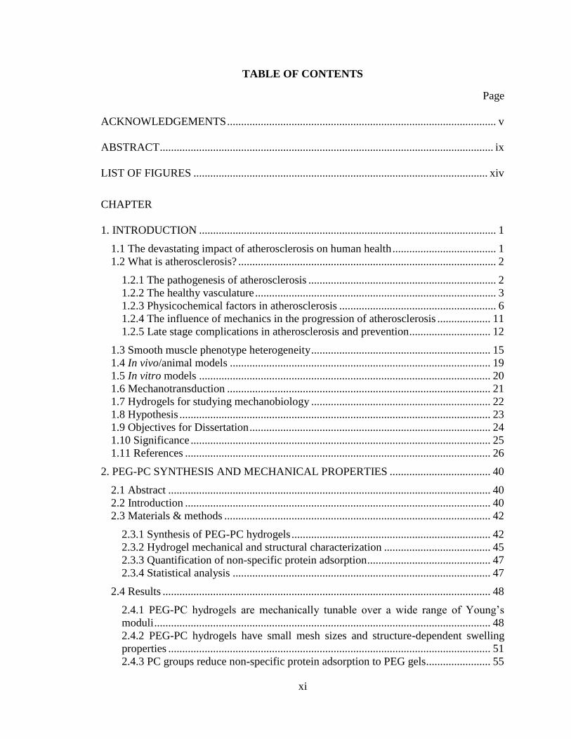
TABLE OF CONTENTS
Page
ACKNOWLEDGEMENTS ................................................................................................ v
ABSTRACT ....................................................................................................................... ix
LIST OF FIGURES ......................................................................................................... xiv
CHAPTER
1. INTRODUCTION .......................................................................................................... 1
1.1 The devastating impact of atherosclerosis on human health ..................................... 1
1.2 What is atherosclerosis? ............................................................................................ 2
1.2.1 The pathogenesis of atherosclerosis ................................................................... 2 1.2.2 The healthy vasculature ...................................................................................... 3 1.2.3 Physicochemical factors in atherosclerosis ........................................................ 6
1.2.4 The influence of mechanics in the progression of atherosclerosis ................... 11 1.2.5 Late stage complications in atherosclerosis and prevention ............................. 12
1.3 Smooth muscle phenotype heterogeneity ................................................................ 15 1.4 In vivo/animal models ............................................................................................. 19 1.5 In vitro models ........................................................................................................ 20
1.6 Mechanotransduction .............................................................................................. 21
1.7 Hydrogels for studying mechanobiology ................................................................ 22 1.8 Hypothesis ............................................................................................................... 23 1.9 Objectives for Dissertation ...................................................................................... 24
1.10 Significance ........................................................................................................... 25 1.11 References ............................................................................................................. 26
2. PEG-PC SYNTHESIS AND MECHANICAL PROPERTIES .................................... 40
2.1 Abstract ................................................................................................................... 40 2.2 Introduction ............................................................................................................. 40 2.3 Materials & methods ............................................................................................... 42
2.3.1 Synthesis of PEG-PC hydrogels ....................................................................... 42
2.3.2 Hydrogel mechanical and structural characterization ...................................... 45 2.3.3 Quantification of non-specific protein adsorption ............................................ 47 2.3.4 Statistical analysis ............................................................................................ 47
2.4 Results ..................................................................................................................... 48
2.4.1 PEG-PC hydrogels are mechanically tunable over a wide range of Young’s
moduli ........................................................................................................................ 48 2.4.2 PEG-PC hydrogels have small mesh sizes and structure-dependent swelling
properties ................................................................................................................... 51 2.4.3 PC groups reduce non-specific protein adsorption to PEG gels ....................... 55

xii
2.4.4 PEG-PC hydrogels are optically transparent .................................................... 58
2.5 Discussion ............................................................................................................... 58 2.6 Conclusion ............................................................................................................... 63 2.7 References ............................................................................................................... 64
3. CANCER CELL MECHANOTRANSDUCTION VARIES WITH CELL TYPE ...... 72
3.1 Abstract ................................................................................................................... 72 3.2 Introduction ............................................................................................................. 72 3.3 Materials & methods ............................................................................................... 74
3.3.1 Cell culture ....................................................................................................... 74
3.3.2 Synthesis of PEG-PC hydrogels ....................................................................... 75 3.3.3 Protein functionalization .................................................................................. 77
3.3.4 Quantification of protein coupling ................................................................... 78 3.3.5 Focal adhesion quantification and imaging ...................................................... 78 3.3.6 Cell proliferation .............................................................................................. 79 3.3.7 Statistical analysis ............................................................................................ 80
3.4 Results ..................................................................................................................... 80
3.4.1 Protein surface density can be controlled with sulfo-SANPAH ...................... 80
3.4.2 Cells adhere and spread to protein-coupled PEG-PC surfaces ......................... 81 3.4.3 Substrate modulus controls focal adhesion maturity ........................................ 83 3.4.4 The effect of substrate modulus on proliferation is cell type-specific ............. 85
3.5 Discussion ............................................................................................................... 87
3.6 Conclusions ............................................................................................................. 89 3.7 References ............................................................................................................... 89
4. EXTRACELLULAR MATRIX COMPOSITION IS THE MAJOR DRIVER OF SMC
PHENOTYPE AND BEHAVIORS .................................................................................. 92
4.1 Abstract ................................................................................................................... 92
4.2 Introduction ............................................................................................................. 93 4.3 Materials & methods ............................................................................................... 96
4.3.1 Cell culture ....................................................................................................... 96 4.3.2 PEG-PC hydrogel polymerization and contact mechanics measurements ....... 96 4.3.3 Protein functionalization to hydrogel surfaces ................................................. 98 4.3.4 Cell proliferation assays ................................................................................... 99
4.3.5 Cell migration assays ...................................................................................... 100 4.3.6 Cell invasion assays ........................................................................................ 100 4.3.7 SMC marker Western blotting ........................................................................ 101
4.3.8 Immunofluorescent staining of HASMCs ...................................................... 103 4.3.9 Reverse transcriptase PCR to confirm smoothelin expression ....................... 104 4.3.10 Characterization of HASMC morphology.................................................... 105 4.3.11 Cell signaling time course with MAGPIX multiplex analysis ..................... 105 4.3.12 Kinase inhibition experiments ...................................................................... 107 4.3.13 Statistical analysis ........................................................................................ 107
4.4 Results ................................................................................................................... 108

xiii
4.4.1 PEG-PC mechanical properties and arterial microenvironments ................... 108 4.4.2 alamarBlue accurately measures differences in HASMC number ................. 110 4.4.3 Proliferation is primarily regulated by ECM composition ............................. 111 4.4.4 ECM composition is the most significant driver of 2D and 3D motility ....... 113
4.4.5 SMC size and morphology is primarily dictated by ECM composition ........ 115 4.4.6 Soluble factors and modulus significantly affect marker expression ............. 117 4.4.7 FAK controls SMC plasticity, in part through PI3K/Akt ............................... 119
4.5 Discussion ............................................................................................................. 123 4.6 Conclusion ............................................................................................................. 136
4.7 Future work & considerations ............................................................................... 136
4.7.1 Validate the proposed signaling models ......................................................... 137
4.7.2 Extend in vitro model to recapitulate more complex features of the in vivo
microenvironment.................................................................................................... 140
4.8 References ............................................................................................................. 146
BIBLIOGRAPHY ........................................................................................................... 157

xiv
LIST OF FIGURES
Figure Page
1.1: Illustration of a normal and blocked artery ................................................................. 3
1.2: Illustration of the arrangement of SMCs and ECM proteins in the media .................. 5
1.3: The current obesity epidemic began around the same time that low-fat dietary
guidelines were issued by the US Government ................................................................ 15
2.1: Young’s modulus example calculation...................................................................... 46
2.2: PEG-PC hydrogels are mechanically tunable over four orders of magnitude ........... 49
2.3: The effect of varying [PEGDMA]:[PC] with constant polymer fraction .................. 50
2.4: Free radical initiator affects PEG-PC modulus at low [PEGDMA] .......................... 51
2.5: NMR spectra of PEG-PC hydrogels .......................................................................... 53
2.6: PEG-PC hydrogels are linearly elastic up to 0.3 M PEGDMA ................................. 54
2.7: Swelling is enhanced by PC comonomer .................................................................. 55
2.8: PEG-PC hydrogels are non-fouling, and optically transparent at low PEGDMA
concentrations ................................................................................................................... 57
3.1: Control of PEG-PC protein surface concentration with sulfo-SANPAH .................. 81
3.2: Modulus and integrin-binding on PEG-PC gels modulates cell morphology ........... 82
3.3: PEG-PC modulus control of focal adhesions is cell-specific .................................... 84
3.4: PEG-PC modulus affects cellular proliferation ......................................................... 87
4.1: in situ PEG-PC contact mechanics testing .............................................................. 109
4.2: Description of in vitro model and general experimental design .............................. 110
4.3: alamarBlue produces an excellent cell number standard curve ............................... 111
4.4: SMC proliferation is modulated by stiffness, ECM, and soluble factors ................ 113
4.5: SMC motility is modulated by ECM and soluble factors ........................................ 115
4.6: SMC adhesion and morphology is dictated by ECM composition ......................... 117
4.7: SMC marker expression is more dependent on soluble factors than ECM ............. 119
4.8: Cell signaling time courses ...................................................................................... 121
4.9: Effects of kinase inhibition on SMC marker expression and proliferation ............. 123
4.10: SMC morphology is dictated by ECM composition and soluble factors .............. 126
4.11: Proposed integrin-mediated signaling mechanisms on BaM ECM ....................... 133
4.12: Proposed integrin-mediated signaling mechanisms on InF ECM ......................... 135

1
CHAPTER 1
INTRODUCTION
1.1 The devastating impact of atherosclerosis on human health
Since at least 1900, the leading cause of non-infectious death nearly every year in
the Western world has been heart disease or atherosclerosis-related [1]. In 2010 alone there
were 596,577 deaths attributed to heart disease, of which 62.9% are attributed to ischemic
heart disease and 5.6% are attributed to hypertensive disease, both most commonly caused
by atherosclerosis. Examined further, 21% of these heart disease-related deaths were due
to myocardial infarction, 42% due to ‘other forms of chronic ischemic heart disease,’ and
30.3% due to ‘other heart diseases’ such as acute endocarditis, heart failure and ‘all other
types of heart disease’ [2]. Furthermore, the picture worsens if we consider cerebrovascular
diseases (128,932 deaths in 2009) and aortic aneurysm (10,073 deaths), other common
results of atherosclerotic plaque disruption. Altogether this represents 738,852 deaths or
29.2% of all deaths in the United States in 2011, a figure which matches the World Health
Organization’s figures for atherosclerosis-related deaths worldwide in 2008 [3]. In
contrast, all forms of malignant neoplasms (cancer) combined accounted for 576,691
deaths or 22.3% in the United States in 2011.
Heart disease and cancer are major public health problems that need considerable
research and work to address. The scope of the problem is even greater when we consider
that the rates of obesity [4] and type II diabetes [5] in the United States have risen steadily
since 1990 – current estimates are that 20% of children [6] and 36% of adults [4] are obese.
Both conditions are believed to be interrelated and are significant risk factors for heart
disease and cancer. This is not especially surprising given the large number of similarities

2
between atherosclerosis and many types of cancer, including hyperproliferative cells and
fibrotic tissues.
1.2 What is atherosclerosis?
1.2.1 The pathogenesis of atherosclerosis
Atherosclerosis, literally “hardening of the arteries,” is characterized by the
development of atherosclerotic plaques, also known as an atheroma. The disease earns its
name from the progressive stiffening or hardening of the arteries affected by plaque
formation, in particular when the plaques become fibrotic and calcified. Plaques begin as
small, fatty lumps or streaks on the arterial wall, but under the wrong conditions these fatty
streaks coalesce into larger fatty deposits and develop into plaques which protrude into the
bloodstream and occlude blood flow (Fig. 1.1) [7]. Occlusion of blood flow from plaque
protrusion can lead to hypertension and eventually ischemia, which has potentially life-
threatening consequences. Plaques can form anywhere in the large arteries, but are most
problematic when they grow uncontrollably in the cerebral arteries (increasing risk for
stroke) and coronary arteries (increasing risk for myocardial infarction) [7]. They can also
develop in the aorta, increasing risk for type III aortic dissection [8], and abdominal aortic
aneurysms [9]. Atherosclerosis is also a major cause of peripheral arterial disease [10], a
leading cause of lower limb amputation in diabetic patients [11].

3
Figure 1.1: Illustration of a normal and blocked artery
"Blausen 0052 Artery NormalvPartially-BlockedVessel" by BruceBlaus - Own work.
Licensed under Creative Commons Attribution 3.0 via Wikimedia Commons -
http://commons.wikimedia.org/wiki/File:Blausen_0052_Artery_NormalvPartially-
BlockedVessel.png#mediaviewer/File:Blausen_0052_Artery_NormalvPartially-
BlockedVessel.png
1.2.2 The healthy vasculature
The medium- and large-sized elastic arteries affected by atherosclerosis are
comprised of 3 distinct tissue layers (Fig. 1.2). The outermost layer, the adventitia, is
largely acellular and composed primarily of connective tissues, especially collagen. Its
function is to anchor arteries to the bones and organs of the body and keep them in place.
The innermost layer, the intima, is in contact with flowing blood and is composed of a
single layer of endothelial cells attached to a thin basement membrane of connective
tissues. The middle layer, the media, is responsible for imparting the elastic stretch-recoil
properties that allow the vascular wall to retain mechanical integrity under pulsatile
blood flow. These properties are imparted by smooth muscle cells (SMCs) arranged in
concentric layers sandwiched between connective tissues and thick sheets of elastin [7,12].

4
These SMCs are in a ‘contractile’ phenotype, so-called because of their robust actomyosin
contractile apparatus that imparts resilience to mechanical stretch and strain.
The extracellular matrix proteins comprising the connective tissues are crucial to
the unique mechanical properties of large arteries, but they also affect cell phenotype and
behavior through integrin signaling and sequestration of growth factors. In an undamaged
media, SMCs are in immediate contact with a thin layer of basement membrane (or
reticular lamina; the correct nomenclature is unclear) comprised largely of laminin and the
non-fibrillar type IV collagen. Both proteins are major components of Matrigel™, a protein
mixture purified from Engelbreth-Holm-Swarm mouse sarcoma, which is commonly used
for cell culture and known to promote differentiated phenotypes in vitro [13,14]. Indeed,
the combination of laminin and collagen IV has been shown to promote expression of
contractile markers in SMCs [15,16], but evidence points to laminins having the greatest
impact [16–18], especially when the cells are exposed to cyclic stretch [19]. Their effects
are most likely mediated by integrin signaling, but there may be other mechanisms at work.
Collagen IV has also been implicated in phenotype maintenance of smooth muscle cells,
but this finding has not been confirmed in other studies [18]. Regardless, collagen IV does
make up about 50% of the basement membrane and forms suprastructures with laminins
[20]. It may have anti-proliferative effects, in part, by binding and sequestering growth
factors including PDGF and FGF [21].

5
Figure 1.2: Illustration of the arrangement of SMCs and ECM proteins in the media
A detailed depiction of the healthy arterial media. E = elastin fibers, F = collagen I fibers,
M = basement membrane and collagen III mesh, Ce = SMC, C = circumferential cut, L =
longitudinal cut. Figure adapted with permission from [12], Copyright © 1985 Wolters
Kluwer Health.
Immediately adjacent to the thin basement membrane layer resides a mesh-like
network of collagen III fibrils (Fig. 1.2) [12], which is surrounded by thick sheets of elastic
laminae composed of crosslinked elastin protein. Finally, thick bundles of collagen I are
interspersed between the elastic laminae. These bundles of SMCs and connective tissues,
collectively termed “lamellar units,” are primarily responsible for the elasticity and
mechanical integrity of large arteries [22]. It is unclear if SMCs in the media are directly
adhered to the collagen networks, but fibrillar forms of both types of collagen and elastin
fibers have been demonstrated to promote the contractile phenotype in SMCs in vitro [18].

6
1.2.3 Physicochemical factors in atherosclerosis
While it is not known with certainty how atherosclerosis starts, it is widely believed
that vascular inflammation is the most likely initiator of plaque formation. The
inflammatory response leading to atherosclerosis is initiated, in part, by low density
lipoproteins (LDLs) and very low density lipoproteins (VLDLs) embedding in the vascular
subendothelial space [7]. Atherosclerosis results when vascular inflammation is chronic,
and the persistent inflammation seems likely to be the result of a combination of factors.
Implicated pro-inflammatory factors include those mentioned above, plus oxidized low
density lipoproteins (oxLDLs), advanced glycation end-products (AGEs) [23], and
inflammatory factors such as interleukins and interferons [24].
Regardless of the initial cause, fatty streaks form from the embedding of LDL and
VLDL particles and their uptake by invading monocytes [7]. In the subendothelial space,
monocytes differentiate into macrophages and impact plaque progression in multiple ways:
they secrete growth factors and cytokines that ‘activate’ endothelial cells, which, in turn,
also produce growth factors and cytokines; they express scavenger receptor A to increase
the uptake of LDL particles [25]; they express matrix metalloproteinases (MMPs) and
migrate towards the intima, digesting the connective tissues in their path; they increase
uptake of LDL particles and can eventually turn into fat-laden ‘foam cells’ which apoptose
at a higher rate and fill the subendothelial space with extracellular fat deposits [7]; and they
may also secrete disordered fragments of elastin and collagen [26,27].
All of these factors serve to irreversibly damage the arterial wall and contribute to
phenotype switching of SMCs in the media. SMCs respond to the soluble growth factors
and cytokines – which have either diffused there or are secreted by invading macrophages

7
– by expressing MMPs [28], digesting their surrounding matrix and migrating towards the
developing plaque [7]. These atherosclerosis-associated SMCs have dedifferentiated into
the so-called ‘synthetic’ phenotype, which is similar to that of myofibroblasts. In some
plaques, SMCs proliferate rapidly and secrete excess collagen, fibronectin, and elastin
fragments that contribute to plaque size and form a fibrotic cap, which is the type plaque
from which atherosclerosis derives its name [7]. However, they may also apoptose and/or
take up LDL particles and become foam cells, much like macrophages, which may
contribute to a softer plaque that is believed to be more prone to rupture [29]. Of course,
the degradation of the medial connective tissues during their migration to the plaque results
in the loss of mechanical integrity, which along with occlusion of blood flow will
contribute to hypertension [30].
The atherosclerotic plaque is a heterogeneous environment with dozens of soluble
factors identified in vitro as having potential roles in disease progression. These soluble
chemicals are believed to, on the whole, increase the rates proliferation of resident cells, as
well as their uptake of LDL particles and synthesis of extracellular matrix proteins and
lipids [31,32]. The majority of these soluble factors are classified as cytokines with roles
in modulating the immune response at sites of vascular inflammation. Interleukins, secreted
peptide cytokines, are one such class of immunomodulators produced by macrophages and
endothelial cells. Interleukin-1s (IL-1), actually a family of 4 proteins, are believed to
prolong the inflammatory response by inducing resident endothelial cells to produce more
cytokines (IL-6, IL-8, monocyte chemoattractant protein, tumor necrosis factor), by
upregulating MMP expression, and by promoting transendothelial migration of leukocytes
and monocytes by upregulating ICAM-1 and VCAM-1 [33]. Interleukin-8 has similar

8
positive feedback effects on inflammation and can simultaneously activate several
signaling pathways associated with cytoskeleton dynamics, protein synthesis and
enhancement of growth factor signaling [34]. Interleukin-6 also has pro-inflammatory
effects and is produced by immune cells and SMCs in plaques [35]. Tumor necrosis factor
alpha (TNFα) is another cytokine produced by macrophages and endothelial cells
implicated in proliferation, cell survival and production of pro-inflammatory eicosanoids
and interleukins [36].
Growth factors are another major promoter of atherosclerotic lesion formation
through their effects on cell proliferation, survival and migration. The most potent growth
factor to SMCs in vitro, platelet derived growth factor BB (PDGF-BB), is released in
lesions by endothelial cells, macrophages and SMCs. It has potent proliferative and
migratory effects on synthetic phenotype SMCs, but little effect on contractile SMCs [37].
Proliferation is induced by promoting splicing of histone deacetylase 7 (HDAC7) [38] and
migration by promoting phosphatidylinositol turnover and calcium signaling [37,39].
Prostanoids, a subclass of eicosanoids, are another class of soluble factors with
roles in atherosclerosis. These include thromboxane, a molecule involved in thrombosis by
promoting platelet aggregation, and a variety of prostaglandins with effects on
inflammation, proliferation and platelet aggregation (i.e. prostacyclin I2, prostaglandin E2)
[40]. However, whether prostaglandin E2 is atheroprotective or promotes atherosclerosis
is unclear, and its effects are concentration-dependent [41].
The downregulation of certain atheroprotective compounds, for instance, nitric
oxide (NO), is also implicated in atherogenesis. NO is normally produced by the
endothelial cells of the intima, which upregulate expression of endothelial nitric oxide

9
synthase (eNOS) under laminar flow conditions in vitro [42]. This latter observation may
explain why atherosclerotic plaques are more commonly found near bends, turns and
bifurcations in the arterial tree [42], where laminar flow is disturbed. NO is known to
inhibit migration and proliferation of vascular smooth muscle cells in vitro, possibly
implicating defective NO production in their aberrant behavior during atherosclerosis
[43,44].
Transforming growth factor beta (TGFβ) is another factor believed to protect
against atherosclerosis, possibly through its role in reversing the wound repair process [45].
Patients with advanced atherosclerosis have been observed to have reduced serum levels
of TGFβ [46], but concentrations in plaques are not necessarily altered. Instead, SMCs in
atherosclerotic lesions in humans are found to have reduced levels of the type II TGFβ
receptor [45], expression of which is required for functionality of the type I receptor. This
alteration in the ratio between the type I and II TGFβ receptors with disease and aging may
explain, in part, the different responses to TGFβ by young and old SMCs and other
differential, context-dependent effects [47–52].
Besides these soluble factors, it is becoming increasingly clear that the extracellular
matrix components in contact with cells can have an impact on disease progression. SMCs
involved in atherosclerosis must degrade and migrate through basement membrane
proteins and fibrous collagens and elastin, and it is highly likely that the changing
environment affects SMC phenotype and behavior. While the basement membrane proteins
are observed to have protective effects in vitro, the effects of the collagens and other
proteins are definitely context-dependent.

10
Although the effects of collagen III on SMC phenotype have not been extensively
studied, it has been reported that fragmentation of collagen III with UV irradiation
increases proliferation and expression of ICAM-1 and VCAM-1, while simultaneously
downregulating β1 integrins and the focal adhesion-associated proteins vinculin, talin, and
the intermediate filament vimentin [53]. The synthesis of collagen III by synthetic
phenotype SMCs increases when treated with TGFβ, IL-1 or PDGF, however, interferon-
gamma reduces synthesis, even with co-treatment with TGFβ [54].
Similar to collagen III, monomeric collagen I has been shown to promote
proliferation and suppress synthesis of extracellular matrix proteins, actin-binding proteins
and signaling molecules [55,56]. The effects of collagen I on SMCs in vitro is rather similar
to the effects of collagen III [57], and it is found in abundance in atherosclerotic lesions,
especially within the SMC-laden fibrous cap [58]. SMCs have been shown to increase
collagen synthesis in response to many factors commonly found in lesions, especially
PDGF-BB, endothelin-1, IL-1, homocysteine, TGFβ and mechanical stretch [58].
However, despite the typical association of collagen I and III with the synthetic phenotype,
other studies have found that polymerized or fibrilized collagens can help induce the
contractile phenotype, in some instances [55,56]. If this effect is relevant in vivo, it is
presumably reversed when the SMCs are induced to degrade their environment and invade
a plaque.
A major extracellular matrix protein found in plaques, fibronectin, is initially laid-
down with fibrin as a provisional matrix during the wound repair response [59]. However,
since the wound repair process is disrupted in atherosclerosis, the fibronectin deposits are
not necessarily cleared out and replaced with appropriately-structured collagen fibers. The

11
abundant fibronectin in plaques is believed to help promote the proliferative, synthetic
phenotype in SMCs, which is supported by in vitro studies [60].
The soluble and physical factors discussed above are just a small subset of the
dozens or hundreds of chemicals and proteins that may have significant effects in the onset
and progression of atherosclerosis. Not discussed in detail but still of importance are
molecules such as various proteoglycans (especially chondroitin sulfate and heparan
sulfates), which have roles in sequestration of growth factors and as co-receptors [61], as
well as LDL and oxidized LDL particles [7], advanced glycation end products which may
additionally stiffen plaques [23], and insulin and insulin-like growth factors (but the role
of IGF-I is debated) [31] that are frequently elevated in patients with atherosclerosis. All
of these factors and likely many more are important to understanding atherosclerosis, but
the heterogeneity of plaques suggest that they may not all be important in every instance.
1.2.4 The influence of mechanics in the progression of atherosclerosis
Given their role in mechanical integrity of the arterial wall, the effects of various
mechanical stimuli on SMCs is an extensively studied area. As mentioned previously,
cyclic stretch can promote the contractile SMC phenotype in vitro [19], but it could also
be implicated in promoting atherosclerosis by inducing production of elastin and collagen
I [62]. Another effect of blood flow is reflected in how certain portions of the arterial tree
are prone to plaque development due to low fluid shear stress, which has been found to
induce downregulation of eNOS in endothelial cells of the intima and thereby reduce
diffusion of atheroprotective NO to medial SMCs [42]. Disrupted blood flow mechanics
may have other effects, for instance from transmural interstitial flow due to a damaged,

12
leaky endothelium, which may potentially contribute to MMP-1 expression and SMC
migration [63].
In vitro studies have revealed that differences in static mechanics, typically in the
form of hydrogels with varying stiffnesses, may also impact SMC phenotype and
behaviors. On 2D substrates, SMC proliferation tends to increase with substrate modulus,
but so does expression of contractile phenotype markers, and migration is also modulus-
dependent [64,65]. In 3D cell cultures, migration is reported to be strongly dependent on
mesh size [66], and once again proliferation and contractile phenotype markers increase
with modulus [67]. Some of these findings are unintuitive given how atherosclerotic
plaques tend to stiffen, but other studies find that mesh size is more important than modulus
[68], and it is very possible that SMC phenotype in atherosclerosis is not as well understood
as believed.
1.2.5 Late stage complications in atherosclerosis and prevention
Further complications arise in the late stages of atherosclerosis, especially fibrosis
and calcification. As discussed above, atherosclerotic plaque composition can vary greatly,
and in some cases SMCs arranged near the luminal surface of the plaque produce excessive
amounts of extracellular matrix proteins forming a fibrous cap [7]. Cells in other plaques
can differentiate into osteoblast-like cells that secrete bone-related proteins including
osteopontin [69] and calcium salts in the form of hydroxyapatite [70]. Fibrosis and/or
calcification of plaques can further harden the artery wall, but a stiffer wall may actually
stabilize the plaque and prevent a deadly rupture event [71,72].

13
Due to the potentially life-threatening consequences of rupture, much effort has
been spent on therapies to improve stability [73,74] and on ways of non-invasively
evaluating stability in at-risk patients [75]. Plaque rupture is a serious complication of
atherosclerosis that can cause death or brain damage. When a rupture occurs, the
underlying collagenous tissues are exposed to the blood stream and the coagulation cascade
is rapidly induced to attempt to patch the rupture with the formation of a clot (thrombosis)
[7]. However, if the clot embolizes from the site of rupture, the effects are often severe.
The most common life-threatening consequence of embolism occurs with atherosclerosis
of the coronary arteries (myocardial infarction), with atherosclerosis of the cerebral arteries
being the second most common occurrence (stroke).
Current efforts to treat atherosclerosis focus on prevention through dietary
intervention and exercise, with an emphasis on low fat foods and ‘heart healthy’ grains.
However, this view is being increasingly questioned [76–79] in light of considerable
evidence that low fat diets may actually be more harmful than helpful. In fact, our current
obesity epidemic began at about the same time the US Senate Select Committee on
Nutrition and Human Needs issued low fat dietary guidelines in 1977 (Fig. 1.3), and obesity
is a significant risk factor for atherosclerosis, diabetes, cancer and related diseases.
Since there has been limited success from dietary interventions, statins have
become the first line of defense in prevention for patients with pre-existing heart disease,
or at high risk of developing it. Statins, including the blockbuster drug Lipitor [80], are a
class of drugs approved for inhibition of 3-hydroxy-3-methyl-glutaryl-Coenzyme A
reductase (HMG-CoA reductase), a rate-controlling enzyme in the cholesterol synthesis
pathway [81]; However, statins have since been shown to have other effects that can

14
explain their efficacy, including inhibition of both Rho kinase [82,83] and the uptake of
oxidized LDL particle [84]. There is also some evidence that statins may be contributing
to the incidence of dementia in elderly patients [85], but the connection is considered
tenuous. Statins have been proven effective in reducing the rate of myocardial infarction
in patients with pre-existing heart disease, but their efficacy at primary prevention was less
certain until the last couple years [86–89]. There are also surgical options for secondary
prevention, including stents which mechanically widen an occluded artery [90].
Unfortunately, while stent implantation may be life-saving in many cases, the stent itself
damages the artery wall and induces inflammation and restenosis.

15
Figure 1.3: The current obesity epidemic began around the same time that low-fat dietary
guidelines were issued by the US Government
Source: National Center for Health Statistics (US). Health, United States, 2008: With
Special Feature on the Health of Young Adults. Hyattsville (MD): National Center for
Health Statistics (US); 2009 Mar. Chartbook.
1.3 Smooth muscle phenotype heterogeneity
Of note is the phenotypic heterogeneity of SMCs in the arterial wall – they do not
exist strictly in a ‘contractile’ or ‘synthetic’ phenotype, but rather along a spectrum
characterized by the expression of certain marker proteins, and are capable of

16
transdifferentiation under the appropriate conditions [91]. The inherent heterogeneity can
partly be explained by the fact that vascular SMCs are derived from multiple embryonic
origins, but this is an incomplete explanation [92]. The picture is complicated further by
observations that, in some cases, a portion of the SMCs present in atherosclerotic plaques
are actually derived from circulating bone marrow-derived progenitor cells [93], but this is
controversial [94,95]. It remains likely, however, that the vast majority of the SMCs in
atherosclerotic plaques originate from the arterial media.
The marker proteins expressed by contractile SMCs are, not surprisingly, typically
associated with the actomyosin contractile apparatus, and have roles in regulating
contractility. With the exception of smoothelin [96], most SMC markers can also be found
in myofibroblasts in some, but not all, cases, which further supports the idea of a phenotype
spectrum. The earliest expressed SMC marker, which is also considered the primary
marker of myofibroblasts, is α-smooth muscle actin (α-SMA), an actin isoform that makes
up a large portion of the contractile apparatus in smooth muscle. However, α-SMA is a
necessary, but insufficient marker of the contractile SMC phenotype in vitro due to
similarities between myofibroblasts and the synthetic SMC phenotype.
Mid-stage markers of the contractile phenotype include caldesmon, calponin and
smooth muscle myosin heavy chain (SM-MHC). Caldesmon is an actin-binding protein
with 2 distinct isoforms, heavy and light (h-CaD and l-CaD), with h-CaD being expressed
in SMCs and l-CaD in all tissues, including SMCs. In fact, when SMCs dedifferentiate into
the myofibroblast-like synthetic phenotype in vitro, they convert to predominantly
expressing the l-CaD isoform. Interestingly, caldesmon is part of the cytoskeleton due to it
binding and stabilizing actin filaments, but the heavy isoform has additional functionality

17
by inhibiting myosin ATPase activity, giving it a role in the contractile apparatus in smooth
muscle. This difference may be explained by the only structural difference between the
light and heavy isoforms, a short, charged spacer linking the actin-binding domain and the
myosin-binding domain, which could provide the necessary space for interacting with
myosin in SMCs [97].
Similarly, calponin is part of both the contractile apparatus and the cytoskeleton,
with actin-binding and myosin ATPase-inhibiting domains, and 2 major isoforms, basic
(h1) and acidic (h2) [98]. Like caldesmon, the basic isoform is more strongly expressed in
smooth muscle cells, and expression is converted upon dedifferentiation to the synthetic
phenotype. Ironically, calponin itself only has a single calponin homology (CH) domain
but does not bind actin through this domain, while a doublet of this domain in other proteins
(e.g. α-actinin and filamin, and many more) is responsible for actin binding [99]. Instead,
a regulatory domain is responsible for actin binding, while yet another domain interacts
with and inhibits the ATPase-activity of myosin II. The CH domain enables interaction
with other contractile proteins like tropomyosin, Ca2+/calmodulin, as well as the signaling
protein ERK [100]. While calponin itself is not a Ca2+-dependent, as the name would
suggest, it binds Ca2+/calmodulin, which phosphorylates calponin and inhibits its myosin-
binding activity to modulate contractility [101].
Smooth muscle myosin heavy chain is a major functional protein of the contractile
apparatus, but not a member of the cytoskeleton. It is the smooth muscle-specific isoform
of the heavy chains of myosin II, with two major splice variants, SM1 and SM2. Non-
muscle cells express non-muscle MHC (NM-MHC), and upon dedifferentiation to the
synthetic phenotype, SMCs shift expression from SM- to NM-MHC [102]. It is likely that

18
this smooth muscle-specific isoform is required for interactions with the other smooth
muscle contractile proteins, like calponin and caldesmon, and these interactions impart
unique contractile properties to smooth muscle.
The latest SMC marker recognized, smoothelin, is also purported to be the only
truly exclusive SMC marker [96]. It has 2 isoforms, a shorter smoothelin-A and longer
smoothelin-B, which are predominantly found in visceral and vascular smooth muscle,
respectively [103]. It is an actin binding protein, interacting with filamentous actin through
CH domains [104], and evidence from knock-out studies in mice points to a role in the
contractile apparatus [105]. However, the complete function of smoothelin remains
unknown.
With the exception of smoothelin-B (and only weakly for smoothelin-A),
transcription of SMC marker genes is regulated by serum response factor (SRF) and an
SRF co-activator, myocardin, binding to CArG boxes in contractile marker gene promoters
[106]. MicroRNAs have garnered attention in cardiac and smooth muscle research in the
past several years, with the finding they have profound effects on the regulation of cellular
phenotype. In particular, expression of the microRNAs miR-143 and miR-145 in SMCs is
induced by myocardin/SRF, forming a positive feedback loop due to the effects of miR-
143/145 on suppressors of myocardin expression or activity; they also suppress non-
myocardin SRF co-activators that would otherwise enable SRF-binding to CArG boxes on
genes known to promote dedifferentiation to the synthetic phenotype [107]. MicroRNA-1
is also positively regulated by myocardin [108] and inhibits SMC proliferation, whereas
the role of microRNA-133 is less certain [109] because it positively affects some SMC
markers (SM-MHC), but decreases expression of others (calponin, transgelin-2 and α-

19
SMA). However, it has an overall positive effect by reducing SMC migration and
proliferation. One thing is clear: the study of SMC phenotype regulation is extremely
complex, and it is further complicated and confounded by differences in contexts and even
the species of cells used in various studies.
1.4 In vivo/animal models
Researchers have developed several animal models for inducing (or resisting)
plaque formation reminiscent of that found in humans, most frequently in mice, rabbits and
rats. These models are very important for corroborating in vitro data and testing potential
drugs and treatments for reversing or preventing atherosclerosis.
The earliest work with animal models and atherosclerosis took place primarily in
Russia in the early 1900’s [110], with a focus on the dietary causes of fatty streak formation
in rabbits. However, the use of rabbits as an animal model of human atherosclerosis is
specious, because they are exclusively herbivorous in nature and regulate lipids very
differently from humans [111].
In recent years, mice have seen much more frequent use in studies of
atherosclerosis, especially the apoE-/- knockout mouse. These mice spontaneously forms
advanced lesions in the aortic root, as well as in other portions of the arterial tree with a
high cholesterol diet. Other transgenic mice have been developed as models dyslipidemia
(the SR-BI knockout), familial hypercholesterolemia (the LDLR-/- mouse), and for features
of metabolic syndrome (db/db, ob/ob mice) [111].
Other less-frequently used animal models include rats and hamsters, and the difficult-
to-use nonhuman primates and pigs. Rats are, however, frequently used in a balloon injury
20
model of restenosis, a pathological process with many similarities to atherosclerosis
[112,113]. Certain primates (e.g. chimpanzees, rhesus monkeys) and pigs are possibly the
ideal animal models for human atherosclerosis, owing to their similar diets, circulatory
systems and lipid regulation. Unfortunately, they are also large, expensive to care for, have
long lifespans and time to maturation, and their use in biomedical research is ethically
challenging.
1.5 In vitro models
There are numerous reports of in vitro model systems designed to mimic one or
more features of the vasculature. These models are used to investigate SMC phenotype and
behaviors, including proliferation and migration in response to growth factors and ECM
proteins, and the expression of contractile SMC markers. To reflect the in vivo reality, SMC
differentiation is reported to be enhanced by coculture with endothelial cells [114–116],
which most likely induces differentiation and quiescence due the secretion of soluble
factors by endothelial cells, most prominently, NO. In order to model changes with arterial
stiffness with aging and disease, several groups have used mechanically-tunable hydrogels
[64,67,68,117–122]. These reports investigate static mechanical effects on proliferation
and cell area, which are positively affected, as well as 2D and 3D migration, and marker
expression. Another group has reported that culturing SMCs on an NO-generating
polymeric biomaterial [123] reduces proliferation, which recapitulates some features of
SMC/endothelial cell coculture.
There have also been numerous reports on more mechanically complex model
systems. For instance, the application of cyclic stretch or strain is used as a model of the

21
mechanical environment produced by pulsatile blood flow [19,62,119,124,125], and
various SMC behaviors are reportedly affected, including secretion of ECM proteins and
cytokines, proliferation, and cell shape. However, there is disagreement in these literature
reports on whether or not cyclic strain is beneficial to SMC phenotype, which is likely a
consequence of the specific contexts studied. Other systems model the effects of a
damaged, leaky vascular endothelium on the pathogenesis of atherosclerosis by inducing
interstitial fluid flow through SMCs encapsulated in collagen I gels [126–129], with
findings of significant effects on SMC expression and secretion of MMPs, leading to
subsequent degradation and invasion of the collagen gel matrix.
1.6 Mechanotransduction
Generally, cells are able to sense their mechanical environment predominantly
through integrin-linked focal adhesion structures [130]. These structures are formed on the
cytoplasmic side of integrin clusters and involve dozens of scaffolding and signaling
proteins. Cells are able to mechanosense through focal adhesions due to the presence of
so-called mechanosensitive proteins such as talin [131]. These proteins are
mechanosensitive because they are tethered to both the membrane-anchored adhesion as
well as actin stress fibers and, as actomyosin contraction pulls on the adhesions, the
proteins are physically unraveled, exposing binding sites for various signaling molecules
[131]. Typically, cells seemed to try to balance the internal cytoskeletal tension with the
tension at integrin-ECM connections [132], and so a stiffer substrate will result in greater
contraction and pull those proteins open to expose signal protein binding sites. For these
22
reasons, substrate modulus is a major factor that should be controlled in cell biology
experiments.
1.7 Hydrogels for studying mechanobiology
As in atherosclerosis, a wide variety of diseases, tissues and individual cell types
are affected by mechanical cues from the microenvironment. Mechanical stimuli, which
may include tissue modulus, shear stress, and cyclic stretch, are documented to influence
cell migration [64,65,133], differentiation [67,134,135], proliferation [136], and other
events [137]. The study of mechanobiology benefits from the development of mechanically
tunable biomaterials for in vitro cell experiments. The biomaterials most commonly used
are hydrogels, including synthetic polymers, such as polyacrylamide (PAA) [120,138,139],
and poly(ethylene glycol) (PEG) [67], and biologically derived gels, such as alginate [140],
MatrigelTM [141], fibrin [142], and type I collagen [143]. Of these, biological hydrogels are
the easiest to adapt, as they are commercially available, and inherently contain the integrin-
binding and proteolytic domains cells encounter in vivo. However, increasing the
concentrations of these proteins in order to increase the gel modulus also increases the
density of integrin-binding domains, decreases the porosity, and alters degradability [144].
These parameters cannot be easily decoupled, making it difficult to determine which ECM
property most significantly affects cell behavior. PAA was the first popularized synthetic
hydrogel for cellular mechanobiology [139], in part because one can independently tune
the integrin-binding and bulk modulus. However, it is not entirely resistant to protein
adsorption [145], and cannot be used to study three dimensional (3D) cell behavior.

23
PEG-based gels are inexpensive, have independently tunable integrin-binding, bulk
modulus, and proteolytic domains [117,146,147], and can be used to study cell behavior in
3D with appropriate modifications [148–150]. However, the mechanical range of PEG-
based gels is limited, typically in the range of 0.5-5 kPa when crosslinked with
proteolytically degradable groups [67], or 20-500 kPa with diacrylate or dimethacrylate
free radical crosslinking [117,151]. PEG and PAA-based hydrogels have been used
extensively to study mechanobiology, for example to demonstrate that cells sense the
rigidity of their substrate and respond dynamically to changes in tension [133], or that
substrate modulus affects stem cell lineage specification [134].
1.8 Hypothesis
To date, biomedical research has generally taken a reductionist approach and used
simple in vitro models, and in vivo animal models that are often of questionable relevance
to human physiology and disease. The primary aim of this dissertation work is to begin to
bridge the gap between these types of studies by designing a complex in vitro model that
captures several major features of the in vivo vasculature to study SMC phenotype in the
context of atherosclerosis. Of course, this approach relies completely on the foundational
work of the studies that precede it, and thus those works are absolutely critical despite any
shortcomings they may have. The expectation is that piecing together that information to
more closely match the in vivo microenvironment will reveal interactions, phenotypes, and
behaviors that may be unobtainable with the simpler models. Prior works are also essential
in helping us understand, interpret, and corroborate the data and observations from a more
complex model. Altogether, the fundamental hypothesis of this dissertation is that, with

24
previous works as a guide, we can design a complex in vitro model that recapitulates crucial
elements of the in vivo cellular microenvironment to reproduce physiologically-relevant
phenomena in vitro. Finally, these phenomena can be examined in depth to parse the
integration of physical, mechanical, and chemical signals and reveal potential regulatory
pathways and pathway interactions that may not otherwise be observed in simpler in vitro
models, and with this new information seek novel drug targets and therapies.
1.9 Objectives for Dissertation
To test the hypothesis proposed in the prior section, I set forth the following
objectives for this dissertation work:
1. Design and characterize a superior hydrogel to study mechanobiology, with
features:
a. Non-fouling
b. Can be made very soft or stiff, within a physiologically relevant range
c. Inexpensive, simple to make
d. Supports attachment of full-length proteins
e. Optically transparent
2. Demonstrate that the hydrogel is appropriate for cell culture studies, with
requirements:
a. Cells must be able to ‘feel’ differences in modulus/stiffness
b. Must demonstrate that stiffness can affect cell behavior
3. Design a complex in vitro model for SMC phenotype, with features:
a. ECM proteins commonly associated with intact vascular media and with
the atherosclerotic plaque.
b. Soluble factors reported relevant to atherosclerosis or maintenance of
SMC phenotype.
c. Soft and stiff substrates to simulate the stiffening and fibrosis frequently
associated with the development of plaques.

25
4. Use this in vitro model to parse the effects of the model features on relevant SMC
phenotypes:
a. Marker expression
b. Proliferation
c. Migration
d. Invasion
5. Finally, probe signaling pathways predicted to be involved in these behaviors,
with a focus on integrins, soluble factors, and how they activate interacting
signaling pathways.
1.10 Significance
While there are certainly studies that use relatively complex in vitro model systems,
the majority of biomedical research is performed on glass or polystyrene, and the effects
of extracellular matrix proteins and other mechanical effects are often neglected. This
approach has led to a huge amount of disagreement within the literature, and the vast
majority of potential therapeutics or treatments that are successfully tested in animal
models sadly do not translate to humans. We believe that the valuable information gained
from these studies, however, can be used to design increasingly complex in vitro models
that may vastly improve the odds of making discoveries that are directly applicable to
human health and disease. The aim of this dissertation work was to apply this approach to
the study of SMC phenotype in the context of atherosclerosis. Through the course of this
dissertation research, we have developed and characterized an exceptional synthetic
hydrogel for mechanobiology research. We then used this hydrogel platform to study the
effect of substrate modulus on the proliferation and mechanotransduction properties of
three cancer cell types, the first ever mechanobiology report of this type with these cells.
Once proven suitable, we then used this hydrogel as one element of complexity in in vitro

26
models of the vascular microenvironment. Along with this static mechanical feature, we
also introduced two mixtures of healthy- and disease-associated ECM proteins and 2
medium supplements containing soluble factors associated with the healthy and disease
states. By recapitulating several elements of in vivo complexity, we have recreated
physiologically-relevant behaviors in vitro and provide evidence to reconcile some
disputes in the literature and dispute some widely-held notions regarding SMC phenotype.
We also propose a detailed, literature-supported hypothetical mechanism by which ECM
binding controls SMC behaviors, with potentially widespread implications in many other
areas, including cancer. While additional work will be required before we can fully
understand these results and confirm our proposed mechanisms, we believe this work will
be of great interest to any investigators interested in SMC phenotype and the role of the
microenvironment on disease states and cell behaviors.
1.11 References
[1] D.S. Jones, S.H. Podolsky, J.A. Greene, The burden of disease and the changing
task of medicine., N. Engl. J. Med. 366 (2012) 2333–8.
doi:10.1056/NEJMp1113569.
[2] CDC, National Vital Statistic Reports, Deaths: Final Data for 2011, 2014.
[3] World Health Organization, World Health Statistics 2008, 2008.
[4] C.L. Ogden, M.D. Carroll, B.K. Kit, K.M. Flegal, Prevalence of obesity in the
United States, 2009-2010., NCHS Data Brief. (2012) 1–8.
[5] E.W. Gregg, B.L. Cadwell, Y.J. Cheng, C.C. Cowie, D.E. Williams, L. Geiss, et al.,
Trends in the prevalence and ratio of diagnosed to undiagnosed diabetes according
to obesity levels in the U.S., Diabetes Care. 27 (2004) 2806–12.

27
[6] C.L. Ogden, M.D. Carroll, L.R. Curtin, M.M. Lamb, K.M. Flegal, Prevalence of
high body mass index in US children and adolescents, 2007-2008., JAMA. 303
(2010) 242–9. doi:10.1001/jama.2009.2012.
[7] V. Kumar, R.S. Cotran, S.L. Robbins, Robbins Pathologic Basis of Disease, 5th ed.,
W.B. Saunders, Philadelpha, 1999.
[8] M. DeBakey, E. Crawford, H. Garrett, A. Beall, J. Howell, Surgical considerations
in the treatment of aneurysms of the thoraco-abdominal aorta., Ann. Surg. 162
(1965) 650–661.
[9] J. Golledge, J. Muller, A. Daugherty, P. Norman, Abdominal aortic aneurysm:
pathogenesis and implications for management., Arterioscler. Thromb. Vasc. Biol.
26 (2006) 2605–13. doi:10.1161/01.ATV.0000245819.32762.cb.
[10] F.N. Ali, T.L. Carman, Medical management for chronic atherosclerotic peripheral
arterial disease, Drugs. 72 (2012) 2073–2085.
[11] N. Schaper, G. Andros, J. Apelqvist, K. Bakker, J. Lammer, M. Lepantalo, et al.,
Specific guidelines for the diagnosis and treatment of peripheral arterial disease in
a patient with diabetes and ulceration of the foot 2011, Diabetes. Metab. Res. Rev.
28 (2012) 236–237. doi:10.1002/dmrr.
[12] J.M. Clark, S. Glagov, Transmural organization of the arterial media. The lamellar
unit revisited., Arterioscler. Thromb. Vasc. Biol. 5 (1985) 19–34.
[13] R. Spinazzi, L. Petrelli, D. Guidolin, G. Carraro, V. Casale, C. Tortorella, et al., In
vitro culture on Matrigel favors the long-term maintenance of rat zona glomerulosa-
cell differentiated phenotype., Int. J. Mol. Med. 17 (2006) 1101–1110.
[14] M.M. Marques, M.D. Martins, C.M. França, Effect of Matrigel on adenoid cystic
carcinoma cell line differentiation, Int. J. Exp. Pathol. 87 (2006) 405–410.
doi:10.1111/j.1365-2613.2006.00498.x.
[15] J. Thyberg, A. Hultgfirdh-Nilsson, Cell Tissue Fibronectin and the basement
membrane components laminin and collagen type IV influence the phenotypic
properties of subcultured rat aortic smooth muscle cells differently, Cell Tissue Res.
276 (1994) 263–271.
[16] J. Thyberg, K. Blomgren, J. Roy, P.K. Tran, U. Hedin, Phenotypic Modulation of
Smooth Muscle Cells after Arterial Injury Is Associated with Changes in the
Distribution of Laminin and Fibronectin, J. Histochem. Cytochem. 45 (1997) 837–
846. doi:10.1177/002215549704500608.

28
[17] I. Hayward, K. Bridle, G. Campbell, P. Underwood, J. Campbell, Effect of
extracellular matrix proteins on vascular smooth muscle cell phenotype, Cell Biol.
Int. 19 (1995) 727–734.
[18] M.J. Barnes, R.W. Farndale, Collagens and atherosclerosis., Exp. Gerontol. 34
(1999) 513–25.
[19] K.G. Birukov, V.P.E. Shirinsky, O. V Stepanova, V.A. Tkachuk, A.W.A. Hahn, T.J.
Resink, et al., Stretch affects phenotype and proliferation of vascular smooth muscle
cells., Mol. Cell. Biochem. 144 (1995) 131–9.
[20] V.S. LeBleu, B. Macdonald, R. Kalluri, Structure and function of basement
membranes., Exp. Biol. Med. 232 (2007) 1121–9. doi:10.3181/0703-MR-72.
[21] R.A. Brekken, E.H. Sage, SPARC, a matricellular protein: at the crossroads of cell–
matrix communication, Matrix Biol. 19 (2001) 815–827.
[22] K. Midwood, J. Schwarzbauer, Elastic fibers: building bridges between cells and
their matrix, Curr. Biol. 12 (2002) 279–281.
[23] G. Basta, A.M. Schmidt, R. De Caterina, Advanced glycation end products and
vascular inflammation: implications for accelerated atherosclerosis in diabetes.,
Cardiovasc. Res. 63 (2004) 582–92. doi:10.1016/j.cardiores.2004.05.001.
[24] A. Tedgui, Z. Mallat, Cytokines in Atherosclerosis : Pathogenic and Regulatory
Pathways, Physiol. Rev. 86 (2006) 515–581. doi:10.1152/physrev.00024.2005.
[25] H. Suzuki, T. Kodama, A role for macrophage scavenger receptors in atherosclerosis
and susceptibility to infection, Nature. 386 (1997) 292–296.
[26] A. Krettek, G.K. Sukhova, P. Libby, Elastogenesis in human arterial disease: a role
for macrophages in disordered elastin synthesis., Arterioscler. Thromb. Vasc. Biol.
23 (2003) 582–7. doi:10.1161/01.ATV.0000064372.78561.A5.
[27] I. Gonçalves, M.P.S. Ares, A. Moberg, J. Moses, F. To, J. Montan, et al., Elastin-
and Collagen-Rich Human Carotid Plaques Have Increased Levels of the Cysteine
Protease Inhibitor Cystatin C, J. Vasc. Res. 45 (2008) 395–401.
[28] H. Yanagi, Y. Sasaguri, K. Sugama, M. Morlmatsu, H. Nagase, Production of tissue
collagenase (matrix metalloproteinase 1) by human aortic smooth muscle cells in
response to platelet-derived growth factor, Atherosclerosis. 91 (1991) 207–216.
[29] M.M. Kockx, A.G. Herman, Apoptosis in atherosclerosis: beneficial or
detrimental?, Cardiovasc. Res. 45 (2000) 736–46.

29
[30] M.F. O’Rourke, W.W. Nichols, Aortic diameter, aortic stiffness, and wave
reflection increase with age and isolated systolic hypertension., Hypertension. 45
(2005) 652–8. doi:10.1161/01.HYP.0000153793.84859.b8.
[31] R.W. Stout, Overview of the association between insulin and atherosclerosis,
Metabolism. 34 (1985) 7–12.
[32] P. Libby, P.M. Ridker, A. Maseri, Inflammation and Atherosclerosis, Circulation.
105 (2002) 1135–1143. doi:10.1161/hc0902.104353.
[33] J. Von der Thüsen, J. Kuiper, T. Van Berkel, E. Biessen, Interleukins in
atherosclerosis: molecular pathways and therapeutic potential, Pharmacol. Rev. 55
(2003) 133–166. doi:10.1124/pr.55.1.5.133.
[34] D.J.J. Waugh, C. Wilson, The interleukin-8 pathway in cancer., Clin. Cancer Res.
14 (2008) 6735–41. doi:10.1158/1078-0432.CCR-07-4843.
[35] H. Loppnow, P. Libby, Proliferating or interleukin 1-activated human vascular
smooth muscle cells secrete copious interleukin 6., J. Clin. Invest. 85 (1990) 731–8.
doi:10.1172/JCI114498.
[36] S. Jovinge, M.P.S. Ares, B. Kallin, J. Nilsson, Human Monocytes/Macrophages
Release TNF- in Response to Ox-LDL, Arterioscler. Thromb. Vasc. Biol. 16 (1996)
1573–1579. doi:10.1161/01.ATV.16.12.1573.
[37] K.E. Bornfeldt, Intracellular Signaling in Arterial Smooth Muscle Migration versus
Proliferation., Trends Cardiovasc. Med. 6 (1996) 143–51. doi:10.1016/1050-
1738(96)00038-2.
[38] B. Zhou, A. Margariti, L. Zeng, O. Habi, Q. Xiao, D. Martin, et al., Splicing of
histone deacetylase 7 modulates smooth muscle cell proliferation and neointima
formation through nuclear β-catenin translocation., Arterioscler. Thromb. Vasc.
Biol. 31 (2011) 2676–84. doi:10.1161/ATVBAHA.111.230888.
[39] C. Wei, X. Wang, M. Zheng, H. Cheng, Calcium gradients underlying cell
migration., Curr. Opin. Cell Biol. (2011) 6–13. doi:10.1016/j.ceb.2011.12.002.
[40] C.L. Bos, D.J. Richel, T. Ritsema, M.P. Peppelenbosch, H.H. Versteeg, Prostanoids
and prostanoid receptors in signal transduction., Int. J. Biochem. Cell Biol. 36
(2004) 1187–205. doi:10.1016/j.biocel.2003.08.006.
[41] J.E. Fabre, M. Nguyen, K. Athirakul, K. Coggins, J.D. McNeish, S. Austin, et al.,
Activation of the murine EP3 receptor for PGE2 inhibits cAMP production and
promotes platelet aggregation., J. Clin. Invest. 107 (2001) 603–10.
doi:10.1172/JCI10881.

30
[42] K.S. Cunningham, A.I. Gotlieb, The role of shear stress in the pathogenesis of
atherosclerosis., Lab. Investig. 85 (2005) 9–23. doi:10.1038/labinvest.3700215.
[43] D.L. Mooradian, T.C. Hutsell, L.K. Keefer, Nitric oxide (NO) donor molecules:
effect of NO release rate on vascular smooth muscle cell proliferation in vitro., J.
Cardiovasc. Pharmacol. 25 (1995) 674–678.
[44] R. Sarkar, E.G. Meinberg, J.C. Stanley, D. Gordon, R. Clinton Webb, Nitric Oxide
Reversibly Inhibits the Migration of Cultured Vascular Smooth Muscle Cells , Circ.
Res. . 78 (1996) 225–230. doi:10.1161/01.RES.78.2.225.
[45] T.A. McCaffrey, TGF-betas and TGF-beta receptors in atherosclerosis, Cytokine
Growth Factor Rev. 11 (2000) 103–114.
[46] D.J. Grainger, P.R. Kemp, J.C. Metcalfe, A.C. Liu, R.M. Lawn, N.R. Williams, et
al., The serum concentration of active transforming growth factor-[beta] is severely
depressed in advanced atherosclerosis, Nat Med. 1 (1995) 74–79.
[47] X. Shi, L.-W. Guo, S.M. Seedial, Y. Si, B. Wang, T. Takayama, et al., TGF-β/Smad3
inhibit vascular smooth muscle cell apoptosis through an autocrine signaling
mechanism involving VEGF-A., Cell Death Dis. 5 (2014) e1317.
doi:10.1038/cddis.2014.282.
[48] N. Kalinina, A. Agrotis, Y. Antropova, O. Ilyinskaya, V. Smirnov, E. Tararak, et
al., Smad expression in human atherosclerotic lesions: evidence for impaired TGF-
beta/Smad signaling in smooth muscle cells of fibrofatty lesions., Arterioscler.
Thromb. Vasc. Biol. 24 (2004) 1391–6.
doi:10.1161/01.ATV.0000133605.89421.79.
[49] G.A. Stouffer, G.K. Owens, TGF-beta promotes proliferation of cultured SMC via
both PDGF-AA-dependent and PDGF-AA-independent mechanisms., J. Clin.
Invest. 93 (1994) 2048–55. doi:10.1172/JCI117199.
[50] L. V Goodman, R.A. Majack, Vascular smooth muscle cells express distinct
transforming growth factor-beta receptor phenotypes as a function of cell density in
culture, J. Biol. Chem. 264 (1989) 5241–5244.
[51] S. Bjorkerud, Effects of transforming growth factor-beta 1 on human arterial smooth
muscle cells in vitro, Arterioscler. Thromb. Vasc. Biol. 11 (1991) 892–902.
doi:10.1161/01.ATV.11.4.892.
[52] M. Hneino, L. Bouazza, G. Bricca, J.Y. Li, D. Langlois, Density-dependent shift of
transforming growth factor-beta-1 from inhibition to stimulation of vascular smooth
muscle cell growth is based on unconventional regulation of proliferation, apoptosis
and contact inhibition., J. Vasc. Res. 46 (2009) 85–97. doi:10.1159/000142612.

31
[53] L. Bačáková, V. Lisá, L. Kubínová, J. Wilhelm, J. Novotná, A. Eckhart, et al.,
Ultraviolet light-irradiated collagen III modulates expression of cytoskeletal and
surface adhesion molecules in rat aortic smooth muscle cells in vitro, Virchows
Arch. 440 (2002) 50–62. doi:10.1007/s004280100463.
[54] E.P. Amento, N. Ehsani, H. Palmer, P. Libby, Cytokines and growth factors
positively and negatively regulate interstitial collagen gene expression in human
vascular smooth muscle cells. , Arterioscler. Thromb. Vasc. Biol. . 11 (1991) 1223–
1230. doi:10.1161/01.ATV.11.5.1223.
[55] H. Koyama, E.W. Raines, K.E. Bornfeldt, J.M. Roberts, R. Ross, Arterial Smooth
Muscle Proliferation through Regulation of Cdk2 Inhibitors, Cell. 87 (1996) 1069–
1078.
[56] T. Ichii, H. Koyama, S. Tanaka, S. Kim, A. Shioi, Y. Okuno, et al., Fibrillar collagen
specifically regulates human vascular smooth muscle cell genes involved in cellular
responses and the pericellular matrix environment., Circ. Res. 88 (2001) 460–7.
[57] N. Sakata, K. Kawamura, S. Takebayashi, Effects of collagen matrix on proliferation
and differentiation of vascular smooth muscle cells in vitro, Exp. Mol. Pathol. 52
(1990) 179–191.
[58] M.D. Rekhter, Collagen synthesis in atherosclerosis: too much and not enough.,
Cardiovasc. Res. 41 (1999) 376–84.
[59] K.S. Midwood, L.V. Williams, J.E. Schwarzbauer, Tissue repair and the dynamics
of the extracellular matrix., Int. J. Biochem. Cell Biol. 36 (2004) 1031–7.
doi:10.1016/j.biocel.2003.12.003.
[60] M.K. Magnusson, D.F. Mosher, Fibronectin : Structure, Assembly, and
Cardiovascular Implications, Arterioscler. Thromb. Vasc. Biol. 18 (1998) 1363–
1370. doi:10.1161/01.ATV.18.9.1363.
[61] S. Cattaruzza, R. Perris, Approaching the Proteoglycome: Molecular Interactions of
Proteoglycans and Their Functional Output, Macromol. Biosci. 6 (2006) 667–680.
doi:10.1002/mabi.200600100.
[62] B.S. Kim, J. Nikolovski, J. Bonadio, D.J. Mooney, Cyclic mechanical strain
regulates the development of engineered smooth muscle tissue., Nat. Biotechnol. 17
(1999) 979–83. doi:10.1038/13671.
[63] S. Tada, J.M. Tarbell, Interstitial flow through the internal elastic lamina affects
shear stress on arterial smooth muscle cells., Am. J. Physiol. Heart Circ. Physiol.
278 (2000) H1589–97.

32
[64] S.R. Peyton, A.J. Putnam, Extracellular matrix rigidity governs smooth muscle cell
motility in a biphasic fashion., J. Cell. Physiol. 204 (2005) 198–209.
doi:10.1002/jcp.20274.
[65] J.Y. Wong, A. Velasco, P. Rajagopalan, Q. Pham, Directed Movement of Vascular
Smooth Muscle Cells on Gradient-Compliant Hydrogels, Langmuir. 19 (2003)
1908–1913.
[66] D. Dikovsky, H. Bianco-Peled, D. Seliktar, The effect of structural alterations of
PEG-fibrinogen hydrogel scaffolds on 3-D cellular morphology and cellular
migration., Biomaterials. 27 (2006) 1496–506.
doi:10.1016/j.biomaterials.2005.09.038.
[67] S.R. Peyton, P.D. Kim, C.M. Ghajar, D. Seliktar, A.J. Putnam, The effects of matrix
stiffness and RhoA on the phenotypic plasticity of smooth muscle cells in a 3-D
biosynthetic hydrogel system., Biomaterials. 29 (2008) 2597–607.
doi:10.1016/j.biomaterials.2008.02.005.
[68] D.J. Munoz-Pinto, A.S. Bulick, M.S. Hahn, Uncoupled investigation of scaffold
modulus and mesh size on smooth muscle cell behavior., J. Biomed. Mater. Res. A.
90 (2009) 303–16. doi:10.1002/jbm.a.32492.
[69] L. Demer, K. Watson, K. Boström, Mechanism of calcification in atherosclerosis,
Trends Cardiovasc. Med. (1994).
[70] F. Parhami, Y. Tintut, J.K. Patel, N. Mody, A. Hemmat, L.L. Demer, Regulation of
vascular calcification in atherosclerosis., Z. Kardiol. 90 (2001) 27–30.
[71] N. Alexopoulos, P. Raggi, Calcification in atherosclerosis, Nat. Rev. Cardiol. 6
(2009) 681–688.
[72] B. Halvorsen, K. Otterdal, T.B. Dahl, M. Skjelland, L. Gullestad, E. Øie, et al.,
Atherosclerotic plaque stability--what determines the fate of a plaque?, Prog.
Cardiovasc. Dis. 51 (2008) 183–94. doi:10.1016/j.pcad.2008.09.001.
[73] L.G. Bucciarelli, T. Wendt, W. Qu, Y. Lu, E. Lalla, L.L. Rong, et al., RAGE
Blockade Stabilizes Established Atherosclerosis in Diabetic Apolipoprotein E-Null
Mice, Circulation. 106 (2002) 2827–2835.
doi:10.1161/01.CIR.0000039325.03698.36.
[74] G. Stoll, M. Bendszus, Inflammation and atherosclerosis: novel insights into plaque
formation and destabilization., Stroke. 37 (2006) 1923–32.
doi:10.1161/01.STR.0000226901.34927.10.

33
[75] B. Kietselaer, C. Reutelingsperger, G. Heidendal, M. Daemen, W. Mess, L. Hofstra,
Noninvasive detection of plaque instability with use of radiolabeled annexin A5 in
patients with carotid-artery atherosclerosis, N. Engl. J. Med. 350 (2004) 1472–1473.
[76] G. Taubes, Good calories, bad calories: challenging the conventional wisdom on
diet, weight control, and disease, 1st ed., Kopf, New York, 2007.
[77] W. Davis, Wheat Belly, 1st ed., Rodale Books, Emmaus, 2011.
[78] S. Liu, J. Manson, Dietary carbohydrates, physical inactivity, obesity, and
the’metabolic syndrome'as predictors of coronary heart disease, Curr. Opin. Lipidol.
12 (2001) 395–404.
[79] J.S. Volek, S.D. Phinney, C.E. Forsythe, E.E. Quann, R.J. Wood, M.J. Puglisi, et
al., Carbohydrate restriction has a more favorable impact on the metabolic syndrome
than a low fat diet., Lipids. 44 (2009) 297–309. doi:10.1007/s11745-008-3274-2.
[80] A. Press, Lipitor becomes world’s top-selling drug, Crain’s New York Bus. (2011).
http://www.crainsnewyork.com/article/20111228/HEALTH_CARE/111229902
(accessed November 22, 2012).
[81] A.P. Lea, D. McTavish, Atorvastatin. A review of its pharmacology and therapeutic
potential in the management of hyperlipidaemias., Drugs. 53 (1997) 828–847.
[82] M. Rupérez, R. Rodrigues-Díez, L.M. Blanco-Colio, E. Sánchez-López, J.
Rodríguez-Vita, V. Esteban, et al., HMG-CoA reductase inhibitors decrease
angiotensin II-induced vascular fibrosis: role of RhoA/ROCK and MAPK
pathways., Hypertension. 50 (2007) 377–83.
doi:10.1161/HYPERTENSIONAHA.107.091264.
[83] A. Nohria, A. Prsic, P.-Y. Liu, R. Okamoto, M.A. Creager, A. Selwyn, et al., Statins
inhibit Rho kinase activity in patients with atherosclerosis., Atherosclerosis. 205
(2009) 517–21. doi:10.1016/j.atherosclerosis.2008.12.023.
[84] D.Y. Li, H.J. Chen, J.L. Mehta, Statins inhibit oxidized-LDL-mediated LOX-1
expression, uptake of oxidized-LDL and reduction in PKB phosphorylation.,
Cardiovasc. Res. 52 (2001) 130–5.
[85] L.H. Kuller, Statins and dementia., Curr. Atheroscler. Rep. 9 (2007) 154–61.
[86] F. Taylor, K. Ward, T. Moore, M. Burke, G. Davey Smith, J. Casas, et al., Statins
for the primary prevention of cardiovascular disease, Cochrane Libr. (2012).

34
[87] J. Conly, F. Clement, M. Tonelli, B. Hemmelgarn, S. Klarenbach, A. Lloyd, et al.,
Cost-effectiveness of the use of low- and high-potency statins in people at low
cardiovascular risk., Can. Med. Assoc. J. 183 (2011) E1180–8.
doi:10.1503/cmaj.101281.
[88] K. Ray, S. Seshasai, S. Erqou, P. Sever, J. Jukema, I. Ford, et al., Statins and All-
Cause Mortality in High-Risk Primary Prevention, Arch Intern Med. 170 (2010)
1024–1031.
[89] F. Taylor, M. Huffman, A. Macedo, T. Moore, M. Burke, G. Davey Smith, et al.,
Statins for the primary prevention of cardiovascular disease, Cochrane Database
Syst. Rev. (2013).
[90] A. V Finn, G. Nakazawa, M. Joner, F.D. Kolodgie, E.K. Mont, H.K. Gold, et al.,
Vascular responses to drug eluting stents: importance of delayed healing.,
Arterioscler. Thromb. Vasc. Biol. 27 (2007) 1500–10.
doi:10.1161/ATVBAHA.107.144220.
[91] S.S.M. Rensen, P.A.F.M. Doevendans, G.J.J.M. van Eys, Regulation and
characteristics of vascular smooth muscle cell phenotypic diversity., Neth. Heart J.
15 (2007) 100–8.
[92] T. Yoshida, G.K. Owens, Molecular determinants of vascular smooth muscle cell
diversity., Circ. Res. 96 (2005) 280–91.
doi:10.1161/01.RES.0000155951.62152.2e.
[93] M. Sata, K. Tanaka, R. Nagai, Origin of smooth muscle progenitor cells: different
conclusions from different models., Circulation. 107 (2003) e106–7.
doi:10.1161/01.CIR.0000067689.02650.98.
[94] J.F. Bentzon, C. Weile, C.S. Sondergaard, J. Hindkjaer, M. Kassem, E. Falk, Smooth
muscle cells in atherosclerosis originate from the local vessel wall and not
circulating progenitor cells in ApoE knockout mice., Arterioscler. Thromb. Vasc.
Biol. 26 (2006) 2696–702. doi:10.1161/01.ATV.0000247243.48542.9d.
[95] A. Schober, C. Weber, Bone marrow-derived smooth muscle cells are breaking bad
in atherogenesis., Arterioscler. Thromb. Vasc. Biol. 31 (2011) 1258–9.
doi:10.1161/ATVBAHA.111.226001.
[96] G.J. van Eys, P.M. Niessen, S.S. Rensen, Smoothelin in vascular smooth muscle
cells., Trends Cardiovasc. Med. 17 (2007) 26–30. doi:10.1016/j.tcm.2006.11.001.
[97] C.A. Wang, Caldesmon and the regulation of cytoskeletal functions, Adv Exp Med
Biol. 644 (2010) 250–272.

35
[98] J.-P. Jin, M.P. Walsh, M.E. Resek, G.A. McMartin, Expression and epitopic
conservation of calponin in different smooth muscles and during development,
Biochem. Cell Biol. 74 (1996) 187–196. doi:10.1139/o96-019.
[99] M. Gimona, R. Mital, The single CH domain of calponin is neither sufficient nor
necessary for F-actin binding., J. Cell Sci. 111 (1998) 1813–21.
[100] S.J. Winder, B.G. Allen, E.D. Fraser, H.M. Kang, G.J. Kargacin, M.P. Walsh,
Calponin phosphorylation in vitro and in intact muscle., Biochem. J. 296 (1993)
827–36.
[101] F.L. Wills, W.D. McCubbin, C.M. Kay, Characterization of the smooth muscle
calponin and calmodulin complex., Biochemistry. 32 (1993) 2321–8.
[102] B.J. Holycross, R.S. Blank, M.M. Thompson, M.J. Peach, G.K. Owens, Platelet-
derived growth factor-BB-induced suppression of smooth muscle cell
differentiation, Circ. Res. 71 (1992) 1525–1532. doi:10.1161/01.RES.71.6.1525.
[103] X.H.T. Wehrens, B. Mies, M. Gimona, F.C.S. Ramaekers, G.J.J.M. Van Eys, J.V.
Small, Localization of smoothelin in avian smooth muscle and identification of a
vascular-specific isoform, FEBS Lett. 405 (1997) 315–320. doi:10.1016/S0014-
5793(97)00207-X.
[104] P. Niessen, S. Clément, L. Fontao, C. Chaponnier, B. Teunissen, S. Rensen, et al.,
Biochemical evidence for interaction between smoothelin and filamentous actin,
Exp. Cell Res. 292 (2004) 170–178. doi:10.1016/j.yexcr.2003.09.005.
[105] P. Niessen, S. Rensen, J. van Deursen, J. De Man, A. De Laet, J.-M. Vanderwinden,
et al., Smoothelin-A is essential for functional intestinal smooth muscle contractility
in mice., Gastroenterology. 129 (2005) 1592–601.
doi:10.1053/j.gastro.2005.08.018.
[106] K.L. Du, H.S. Ip, J. Li, M. Chen, F. Dandre, W. Yu, et al., Myocardin Is a Critical
Serum Response Factor Cofactor in the Transcriptional Program Regulating Smooth
Muscle Cell Differentiation, Mol. Cell. Biol. 23 (2003) 2425–2437.
doi:10.1128/MCB.23.7.2425.
[107] A.Y. Rangrez, Z.A. Massy, V. Metzinger-Le Meuth, L. Metzinger, miR-143 and
miR-145: molecular keys to switch the phenotype of vascular smooth muscle cells.,
Circ. Cardiovasc. Genet. 4 (2011) 197–205.
doi:10.1161/CIRCGENETICS.110.958702.
[108] J. Chen, H. Yin, Y. Jiang, S.K. Radhakrishnan, Z.-P. Huang, J. Li, et al., Induction
of microRNA-1 by myocardin in smooth muscle cells inhibits cell proliferation.,
Arterioscler. Thromb. Vasc. Biol. 31 (2011) 368–75.
doi:10.1161/ATVBAHA.110.218149.

36
[109] D. Torella, C. Iaconetti, D. Catalucci, G.M. Ellison, A. Leone, C.D. Waring, et al.,
MicroRNA-133 controls vascular smooth muscle cell phenotypic switch in vitro and
vascular remodeling in vivo., Circ. Res. 109 (2011) 880–93.
doi:10.1161/CIRCRESAHA.111.240150.
[110] N. Anitschkow, S. Chalatow, On experimental cholesterin steatosis and its
significance in the origin of some pathological processes, Cent. Fur Allg. Pathol.
Und Pathol. Anat. XXIV (1913) 1–9. doi:10.1161/01.ATV.3.2.178.
[111] J.C. Russell, S.D. Proctor, Small animal models of cardiovascular disease: tools for
the study of the roles of metabolic syndrome, dyslipidemia, and atherosclerosis.,
Cardiovasc. Pathol. 15 (2006) 318–30. doi:10.1016/j.carpath.2006.09.001.
[112] R. Riessen, T.N. Wight, C. Pastore, C. Henley, J.M. Isner, Distribution of
Hyaluronan During Extracellular Matrix Remodeling in Human Restenotic Arteries
and Balloon-Injured Rat Carotid Arteries, Circulation. 93 (1996) 1141–1147.
doi:10.1161/01.CIR.93.6.1141.
[113] C. Indolfi, G. Esposito, E. Di Lorenzo, A. Rapacciuolo, A. Feliciello, A. Porcellini,
et al., Smooth Muscle Cell Proliferation Is Proportional to the Degree of Balloon
Injury in a Rat Model of Angioplasty, Circulation. 92 (1995) 1230–1235.
doi:10.1161/01.CIR.92.5.1230.
[114] S. Chaterji, K. Park, A. Panitch, Scaffold-free in vitro arterial mimetics: the
importance of smooth muscle-endothelium contact., Tissue Eng. Part A. 16 (2010)
1901–12. doi:10.1089/ten.TEA.2009.0271.
[115] M.F. Fillinger, L.N. Sampson, J.L. Cronenwett, R.J. Powell, R.J. Wagner, Coculture
of endothelial cells and smooth muscle cells in bilayer and conditioned media
models., J. Surg. Res. 67 (1997) 169–78. doi:10.1006/jsre.1996.4978.
[116] Y. Liu, S. Rayatpisheh, S.Y. Chew, M.B. Chan-Park, Impact of endothelial cells on
3D cultured smooth muscle cells in a biomimetic hydrogel., ACS Appl. Mater.
Interfaces. 4 (2012) 1378–87. doi:10.1021/am201648f.
[117] S.R. Peyton, C.B. Raub, V.P. Keschrumrus, A.J. Putnam, The use of poly(ethylene
glycol) hydrogels to investigate the impact of ECM chemistry and mechanics on
smooth muscle cells., Biomaterials. 27 (2006) 4881–93.
doi:10.1016/j.biomaterials.2006.05.012.
[118] X.Q. Brown, E. Bartolak-Suki, C. Williams, M.L. Walker, V.M. Weaver, J.Y.
Wong, Effect of substrate stiffness and PDGF on the behavior of vascular smooth
muscle cells: implications for atherosclerosis., J. Cell. Physiol. 225 (2010) 115–22.
doi:10.1002/jcp.22202.

37
[119] R.G. Thakar, Q. Cheng, S. Patel, J. Chu, M. Nasir, D. Liepmann, et al., Cell-shape
regulation of smooth muscle cell proliferation., Biophys. J. 96 (2009) 3423–32.
doi:10.1016/j.bpj.2008.11.074.
[120] A. Engler, L. Bacakova, C. Newman, A. Hategan, M. Griffin, D. Discher, Substrate
compliance versus ligand density in cell on gel responses, Biophys. J. 86 (2004)
617–28. doi:10.1016/S0006-3495(04)74140-5.
[121] L. Lin, R.E. Marchant, J. Zhu, K. Kottke-Marchant, Extracellular Matrix-Mimetic
Poly (ethylene glycol) Hydrogels Engineered to Regulate Smooth Muscle Cell
Proliferation in 3D., Acta Biomater. (2014). doi:10.1016/j.actbio.2014.08.025.
[122] A.A. Ucuzian, L.P. Brewster, A.T. East, Y. Pang, A.A. Gassman, H.P. Greisler,
Characterization of the chemotactic and mitogenic response of SMCs to PDGF-BB
and FGF-2 in fibrin hydrogels., J. Biomed. Mater. Res. A. 94 (2010) 988–96.
doi:10.1002/jbm.a.32786.
[123] K.S. Bohl, J.L. West, Nitric oxide-generating polymers reduce platelet adhesion and
smooth muscle cell proliferation., Biomaterials. 21 (2000) 2273–8.
[124] A. Zampetaki, Z. Zhang, Y. Hu, Q. Xu, Biomechanical stress induces IL-6
expression in smooth muscle cells via Ras/Rac1-p38 MAPK-NF-kappaB signaling
pathways., Am. J. Physiol. Heart Circ. Physiol. 288 (2005) H2946–54.
doi:10.1152/ajpheart.00919.2004.
[125] C. Li, Q. Xu, Mechanical stress-initiated signal transduction in vascular smooth
muscle cells in vitro and in vivo., Cell. Signal. 19 (2007) 881–91.
doi:10.1016/j.cellsig.2007.01.004.
[126] S. Wang, J.M. Tarbell, Effect of Fluid Flow on Smooth Muscle Cells in a 3-
Dimensional Collagen Gel Model, Arterioscler. Thromb. Vasc. Biol. 20 (2000)
2220–2225. doi:10.1161/01.ATV.20.10.2220.
[127] Z. Shi, X. Ji, D.E. Berardi, H. Qazi, J.M. Tarbell, Interstitial flow induces MMP-1
expression and vascular SMC migration in collagen I gels via an ERK1/2-dependent
and c-Jun-mediated mechanism., Am. J. Physiol. Heart Circ. Physiol. 298 (2010)
H127–35. doi:10.1152/ajpheart.00732.2009.
[128] Z.-D. Shi, H. Wang, J.M. Tarbell, Heparan sulfate proteoglycans mediate interstitial
flow mechanotransduction regulating MMP-13 expression and cell motility via
FAK-ERK in 3D collagen., PLoS One. 6 (2011) e15956.
doi:10.1371/journal.pone.0015956.
[129] Z. Shi, X. Ji, H. Qazi, J.M. Tarbell, Interstitial flow promotes vascular fibroblast ,
myofibroblast , and smooth muscle cell motility in 3-D collagen I via upregulation
of MMP-1, (2009). doi:10.1152/ajpheart.00369.2009.

38
[130] L.H. Romer, K.G. Birukov, J.G.N. Garcia, Focal adhesions: paradigm for a
signaling nexus., Circ. Res. 98 (2006) 606–16.
doi:10.1161/01.RES.0000207408.31270.db.
[131] U.S. Schwarz, T. Erdmann, I.B. Bischofs, Focal adhesions as mechanosensors: the
two-spring model., BioSystems. 83 (2006) 225–32.
doi:10.1016/j.biosystems.2005.05.019.
[132] D.E. Ingber, Tensegrity and mechanotransduction., J. Bodyw. Mov. Ther. 12 (2008)
198–200. doi:10.1016/j.jbmt.2008.04.038.
[133] C.M. Lo, H.B. Wang, M. Dembo, Y.L. Wang, Cell movement is guided by the
rigidity of the substrate, Biophys. J. 79 (2000) 144–152. doi:10.1016/S0006-
3495(00)76279-5.
[134] A.J. Engler, S. Sen, H.L. Sweeney, D.E. Discher, Matrix elasticity directs stem cell
lineage specification, Cell. 126 (2006) 677–89. doi:10.1016/j.cell.2006.06.044.
[135] K. Saha, A.J. Keung, E.F. Irwin, Y. Li, L. Little, D. V Schaffer, et al., Substrate
modulus directs neural stem cell behavior, Biophys. J. 95 (2008) 4426–38.
doi:10.1529/biophysj.108.132217.
[136] A.J. Engler, M.A. Griffin, S. Sen, C.G. Bonnemann, H.L. Sweeney, D.E. Discher,
Myotubes differentiate optimally on substrates with tissue-like stiffness:
pathological implications for soft or stiff microenvironments, J. Cell Biol. 166
(2004) 877–887.
[137] S.R. Peyton, C.M. Ghajar, C.B. Khatiwala, A.J. Putnam, The emergence of ECM
mechanics and cytoskeletal tension as important regulators of cell function, Cell
Biochem. Biophys. 47 (2007) 300–320. doi:10.1007/s12013-007-0004-y.
[138] R.J. Pelham, Y.-L. Wang, Cell locomotion and focal adhesions are regulated by
substrate flexibility, Proc Natl Acad Sci USA. 94 (1997) 13661–13665.
[139] R.J. Pelham Jr., Y.L. Wang, Cell locomotion and focal adhesions are regulated by
the mechanical properties of the substrate, Biol. Bull. 194 (1998) 348–350.
[140] J.A. Rowley, G. Madlambayan, D.J. Mooney, Alginate hydrogels as synthetic
extracellular matrix materials, Biomaterials. 20 (1999) 45–53.
[141] C.F. Deroanne, C.M. Lapiere, B. V Nusgens, In vitro tubulogenesis of endothelial
cells by relaxation of the coupling extracellular matrix-cytoskeleton, Cardiovasc.
Res. 49 (2001) 647–658.

39
[142] C.M. Ghajar, X. Chen, J.W. Harris, V. Suresh, C.C.W. Hughes, N.L. Jeon, et al.,
The effect of matrix density on the regulation of 3-D capillary morphogenesis.,
Biophys. J. 94 (2008) 1930–41. doi:10.1529/biophysj.107.120774.
[143] M.A. Wozniak, R. Desai, P.A. Solski, C.J. Der, P.J. Keely, ROCK-generated
contractility regulates breast epithelial cell differentiation in response to the physical
properties of a three-dimensional collagen matrix, J. Cell Biol. 163 (2003) 583–595.
[144] M.H. Zaman, L.M. Trapani, A.L. Sieminski, A. Siemeski, D. Mackellar, H. Gong,
et al., Migration of tumor cells in 3D matrices is governed by matrix stiffness along
with cell-matrix adhesion and proteolysis, Proc. Natl. Acad. Sci. U. S. A. 103 (2006)
10889–10894. doi:10.1073/pnas.0604460103.
[145] M.T. Thompson, M.C. Berg, I.S. Tobias, J.A. Lichter, M.F. Rubner, K.J. Van Vliet,
Biochemical functionalization of polymeric cell substrata can alter mechanical
compliance, Biomacromolecules. 7 (2006) 1990–1995. doi:Doi
10.1021/Bm060146b.
[146] D.L. Hern, J.A. Hubbell, Incorporation of adhesion peptides into nonadhesive
hydrogels useful for tissue resurfacing, J. Biomed. Mater. Res. 39 (1998) 266–276.
[147] S.J. Bryant, K.S. Anseth, Controlling the spatial distribution of ECM components in
degradable PEG hydrogels for tissue engineering cartilage, J Biomed Mater Res A.
64 (2003) 70–79.
[148] A.N. Stachowiak, D.J. Irvine, Inverse opal hydrogel-collagen composite scaffolds
as a supportive microenvironment for immune cell migration, J. Biomed. Mater.
Res. 85 (2008) 815–828. doi:10.1002/jbm.a.31661.
[149] S.-H. Lee, J.J. Moon, J.S. Miller, J.L. West, Poly(ethylene glycol) hydrogels
conjugated with a collagenase-sensitive fluorogenic substrate to visualize
collagenase activity during three-dimensional cell migration, Biomaterials. 28
(2007) 3163–3170. doi:10.1016/j.biomaterials.2007.03.004.
[150] M. Gonen-Wadmany, L. Oss-Ronen, D. Seliktar, Protein-polymer conjugates for
forming photopolymerizable biomimetic hydrogels for tissue engineering,
Biomaterials. 28 (2007) 3876–3886.
[151] J.S. Temenoff, K.A. Athanasiou, R.G. LeBaron, A.G. Mikos, Effect of
poly(ethylene glycol) molecular weight on tensile and swelling properties of
oligo(poly(ethylene glycol) fumarate) hydrogels for cartilage tissue engineering, J.
Biomed. Mater. Res. 59 (2002) 429–437.

40
CHAPTER 2
PEG-PC SYNTHESIS AND MECHANICAL PROPERTIES
2.1 Abstract
This chapter will detail the development, synthesis and mechanical characterization
of a novel hydrogel with significant utility in mechanobiology and disease research. The
goal of this work was to create a novel, tunable hydrogel with exceptional anti-fouling
properties to control the type and presentation of extracellular matrix proteins. This was
achieved by copolymerizing the well-characterized PEG-dimethacrylate hydrogel with a
methacrylated phosphorylcholine (PC) monomer. Phosphorylcholine was chosen because
it is a very hydrophilic zwitterion, which imparts the ability to resist protein fouling. We
find that free radical-copolymerized PEG and PC (‘PEG-PC’) has a mechanical range of 1
kPa to 10 MPa Young’s modulus, which matches or exceeds any previously reported
hydrogel, and average mesh sizes that are smaller than typical growth factors or other
soluble factors (≤ 5 nm). PEG-PC also has reduced fouling compared to PEG itself, which
we show is [PC]-dependent. Finally, we characterize the optical clarity of PEG-PC on
coverslips and find that it is completely transparent from approximately 1 kPa to 1 MPa
Young’s modulus.
2.2 Introduction
The development of tunable biomaterials has driven rapid progress in tissue
engineering, either to facilitate or enhance the natural wound healing process, or to act as
functional replacement tissues in the human body. Biomaterial platforms have been
developed both for eventual in vivo application, as well as for in vitro model systems for

41
studying complex biological processes, including cell migration, adhesion, proliferation,
and differentiation. Information from these in vitro studies drives the design of implantable
scaffolds, and promotes understanding of complex biology in environments more
physiologically relevant than traditional plastic and glass surfaces. Studying cellular
phenomenology and signaling mechanisms in microenvironments that capture key features
of the in vivo extracellular matrix (ECM) is more efficient and cost-effective than animal
studies, and may prove to be more predictive of human clinical outcome.
Mechanical cues from materials and the microenvironment, such as substrate
modulus, shear stress, and stretch, influence cell migration [1–3], differentiation [4–6], and
proliferation [7] (for review, see ref. [8]). Mechanotransduction, the translation of a
mechanical cue from the ECM into a biochemical one, is mediated by focal adhesions [9].
There have been many studies on the effects of substrate modulus on cell spreading [1,10–
16], focal adhesion properties [1,11,15,17], proliferation [4,6,11,18–20], migration [1–
3,21,22], and phenotype and differentiation [4,5,20,23–25] with several materials
platforms [1,12–14,18,20,26–29]. Materials most commonly used to investigate
mechanobiology are hydrogels, including synthetic polymers, such as polyacrylamide
(PAA) [30–32], and poly(ethylene glycol) (PEG) [4], and biologically derived gels, such
as alginate [33], MatrigelTM [34], fibrin [35], hyaluronic acid [14,20,29] and type I collagen
[36]. Of these, biological hydrogels are the easiest to adapt, as they are commercially
available, and inherently contain the integrin-binding and proteolytic domains cells
naturally encounter in vivo. However, manipulating the concentrations of these proteins in
order to control the gel modulus simultaneously alters the density of integrin-binding
domains, porosity, and degradability [22]. These parameters cannot be decoupled easily,

42
making it difficult to determine which ECM property most significantly affects cell
behavior.
With respect to synthetic hydrogels, PAA was the first popularized platform for
cellular mechanobiology [32], in part because it was the first system to demonstrate
independent tuning of cell adhesion and modulus. While PAA is an important material, it
has limitations, such as a narrow mechanical range, and it is not suitable for three-
dimensional (3D) studies. PEG-based gels are inexpensive, have independently tunable cell
adhesion, bulk modulus, and proteolytic domains [11,37,38], and can be used to study cell
behavior in 3D [39–41]. However, the mechanical range of PEG-based gels is limited,
typically in the range of 0.5-5 kPa when crosslinked with proteolytically degradable groups
[4], or 20-500 kPa with diacrylate or dimethacrylate crosslinking [11,42]. We sought to
improve on this limited range of Young’s moduli, by combining the commonly used
PEGDMA crosslinker with an inexpensive, extremely hydrophilic zwitterion. We report
here the synthesis of a hybrid polymer hydrogel, which combines PEG-dimethacrylate
(PEGDMA), with the zwitterionic comonomer 2-methacryloyloxyethyl phosphorylcholine
(PC). To our knowledge, these “PEG-PC” hydrogels have a mechanical range that matches
or exceeds any previously reported hydrogel system, and, importantly, they are simple,
inexpensive to produce, and retain optical clarity over most of their mechanical range.
2.3 Materials & methods
2.3.1 Synthesis of PEG-PC hydrogels
In most cases, PEG-PC polymer hydrogel precursor solutions were prepared by
mixing PEGDMA (average Mn 750, Sigma-Aldrich, St. Louis, MO), varied between final

43
concentrations of 7.4 mM and 0.7 M (0.5-55 wt.%, see Figure 2.2E), and 0.6 M (17 wt.%)
2-methacryloyloxyethyl phosphorylcholine (MPC; Sigma-Aldrich) in phosphate buffered
saline (PBS). Solutions were degassed for 30 seconds with nitrogen, and sterilized with a
0.2 µm syringe filter (Thermo Fisher Scientific, Waltham, MA). Depending on the desired
format, we have used two different free radical initiators for polymerization. To cure under
UV light, 0.8 wt.% Irgacure 2959 (from a 20 wt.% stock in 70% ethanol; BASF,
Ludwigshafen, Germany) was added, and gel formation was induced with a Spectroline
High-Intensity UV Lamp with 365 nm light (Model #SB-100P, Westbury, NY), at
approximately 3.5 inches from the gel for 7 minutes. To polymerize hydrogels without UV
light, we added 0.05 wt.% ammonium persulfate (APS, from a 20 wt.% stock in water;
Bio-Rad Laboratories, Hercules, CA) and 0.125 vol.% tetramethylethylenediamine
(TEMED, Bio-Rad Laboratories) and polymerized gels under nitrogen for 10 minutes.
We also performed some experiments with different concentrations of MPC: 1) for
mechanical testing, we UV polymerized hydrogels with MPC:PEGDMA molar ratios of
13.9, 7.23 and 2.59 at a constant 20.4 wt.% total polymer (600/43.1, 510/70.5 and 348/135
mM MPC/PEGDMA, respectively), and 2) for comparison of swelling and mechanical
properties between molecular weight-matched MPC and a PEG-methacrylate (PEGMA)
comonomers, we UV polymerized hydrogels with constant [PEGDMA] (135 mM) and
concentrations of MPC or PEGMA of approximately 160, 300 and 600 mM. Due to the
poor solubility of PEGMA in water, these latter hydrogels were polymerized in 70%
ethanol.
To make thin PEG-PC hydrogels with even heights suitable for microscopy, 75 µL
aliquots of PEG-PC solution was cured between chemically modified 18 mm glass
44
coverslips. Methacrylate silanized coverslips served as the base, and covalently attached to
the hydrogel during polymerization, whereas hydrophobic coverslips could be easily
removed from the final, hydrophilic hydrogels. To create the methacrylate coverslips, slips
were treated with O2 plasma for 10 minutes, reacted in 200 mL of 95% ethanol with 2
vol.% 3-(trimethoxysilyl) propyl methacrylate (adjusted to pH 5.0 with glacial acetic acid;
Thermo Fisher Scientific) for 2 minutes with shaking, washed three times in 200 proof
ethanol, and finally dried at 120°C for 15 minutes[43]. Hydrophobic coverslips were made
with coverslips submerged in Sigmacote (Sigma-Aldrich), shaken for 20 minutes, washed
3 times with 200 proof ethanol, and dried under vacuum. Following polymerization, the
Sigmacote cover slips were removed from the gel surface with fine forceps, and the gels
were allowed to swell in sterile PBS for 24 hours. Prepared coverslips were stored in foil
in a desiccator at room temperature.
To synthesize PEG-PC hydrogels for high throughput applications, 96-well
microplates with glass bottoms (no. 1.5 coverslip glass; In Vitro Scientific, Sunnyvale, CA)
were treated with O2 plasma for 10 minutes, reacted with 100 µL/well of 2 vol.% 3-
(trimethoxysilyl) propyl methacrylate (Sigma-Aldrich) in 95% ethanol (to pH 5.0 with
glacial acetic acid; Thermo Fisher Scientific) for 2 minutes, with shaking, then rinsed 3x
with 200 proof ethanol, and dried at 40°C for 30 minutes. To polymerize hydrogels in
wells, pre-polymer solutions were prepared with APS and TEMED polymerization as
described above, and added to the 96-well plate at 40 µL/well. To remove O2 in the air that
would inhibit polymerization, plates were placed in a vacuum oven at room temperature,
pure N2 gas flushed through the chamber for 5 minutes, and then the chamber sealed for

45
10 minutes to complete the reaction. Finally, hydrogels were swelled overnight with 100
µL/well PBS.
2.3.2 Hydrogel mechanical and structural characterization
PEG-PC hydrogel cylinders for mechanical compression testing were formed in
Teflon molds (5 mm height, 5.5 mm diameter) and swelled in PBS for 48 hours. Post-
swelling, hydrogel dimensions were measured with digital calipers, and mechanical
compression tests were performed with a TA Instruments (New Castle, DE) AR-2000
rheometer using a 2 µm/second strain rate. The Young’s modulus (E) for each hydrogel
was calculated by plotting the measured normal force between 0 and 4% strain, and
dividing the slope of the best-fit linear regression by the hydrogel cross-sectional area (see
Fig. 2.1 for a representative stress-strain curve and example calculation). The Young’s
modulus was calculated from ≥ 4 samples at each PEGDMA concentration.

46
Figure 2.1: Young’s modulus example calculation
Young’s moduli were calculated by uniaxially compressing a cylindrical hydrogel and
plotting the measured normal force versus strain from 0 to 4%. The modulus is then
computed as the slope of the stress-strain curve divided by the cross sectional area of the
hydrogel cylinder.
To determine an approximate average mesh size as a function of PEGDMA
crosslinker content with constant PC content, hydrogels were swelled in PBS for 48 hours
then weighed, fully lyophilized under vacuum and moderate heating, and weighed again.
The average mesh sizes, 𝜉, of the PEG-PC hydrogels were determined as a function of
PEGDMA crosslinker concentration according to the Flory theory, as modified by Canal
and Peppas [44]:
𝜉 = υ2,s
−13(r̅2)1/2
where υ2,s is the swollen volume fraction of polymer and (r̅2)1/2 is the average end-to-end
distance of the PEGDMA crosslinker.
To detect unreacted methacrylate groups, NMR was performed on PEG-PC
samples polymerized with APS and TEMED in microcentrifuge tubes with a Bruker
Spectrospin DPX300 (Bruker Corporation, Billerica, MA). D2O was used as the solvent.

47
To quantitatively measure the observed transparency of PEG-PC versus PEGDMA,
hydrogels were made on cover slips, as described above, from 135 mM to 0.7 M PEGDMA
with and without 0.6 M PC. After 48 hours of swelling in PBS, the optical densities of
hydrogels in clean PBS were measured at 450, 490, 572, 630 and 750 nm with a BioTek
ELx800 absorbance microplate reader (BioTek, Winooski, VT).
2.3.3 Quantification of non-specific protein adsorption
We quantified protein adsorption to the hydrogels with an indirect ELISA [45].
Hydrogels were polymerized in 96-well plates, swelled in PBS overnight, and incubated
with 10 mg/mL bovine serum albumin (BSA; Sigma-Aldrich) for 20 hours at 37°C. The
gels were washed 5x with PBS, incubated with primary antibody to BSA (Life
Technologies, Carlsbad, CA) in PBS for 90 minutes, washed again 5x with PBS, and
incubated with secondary antibody conjugated to horseradish peroxidase (HRP; Abcam,
Cambridge, UK) for 90 minutes. After washing 5x with PBS, the gels were incubated with
0.1 mg/mL 3,3’,5,5’-tetramethylbenzidine (TMB; Sigma-Aldrich) and 0.06% hydrogen
peroxide (Thermo Fisher Scientific) in 0.1 M sodium acetate (pH 5.5; Sigma-Aldrich) for
1 hour at room temperature with shaking. At 1 hour, an equal volume of 1 M sulfuric acid
(Sigma-Aldrich) was added to each well, and the absorbance at 450 nm was measured with
a BioTek ELx800 absorbance microplate reader (BioTek).
2.3.4 Statistical analysis
Statistical analysis was performed using Prism v5.04 (GraphPad Software, La Jolla,
CA). Data are reported as mean ± standard deviation, unless otherwise noted. Statistical

48
significance of the difference between pairs of means was evaluated by computing P-values
with unpaired Student's t-tests (with Welch's correction as necessary). P ≤ 0.05 is denoted
with *, ≤ 0.01 with **, ≤ 0.001 with *** and ≤ 0.0001 with ****; P > 0.05 is considered
not significant ('ns').
2.4 Results
2.4.1 PEG-PC hydrogels are mechanically tunable over a wide range of Young’s
moduli
We synthesized hydrogels from varying concentrations of PEGDMA with constant
PC, and characterized physical properties important for a viable cell culture platform (Fig.
2.2). We quantified the Young's moduli of photopolymerized PEG-PC hydrogels with 0.6
M PC (20 wt.%) in PBS and PEGDMA ranging from 7.4 to 700 mM (0.5 – 55 wt.%) via
compression testing (Fig. 2.1, 2.2B). As expected, we found that hydrogel modulus
increases with crosslinker concentration from 0.9 ± 0.2 kPa at 7.4 mM PEGDMA to 9300
± 900 kPa at 0.7 M PEGDMA. This mechanical characterization demonstrates a tunable
range of Young’s moduli that spans four orders of magnitude, from 900 Pa to nearly 10
MPa. We attribute this wide range of moduli to the inclusion of the PC zwitterion, enabling
gelation at very low concentrations of PEGDMA (7.4 mM, or 0.5 wt.%). This range of
Young’s moduli spans the reported moduli of nearly any tissue type, including brain, tumor
tissue, skin, cartilage, and some areas of bone[46].

49
Figure 2.2: PEG-PC hydrogels are mechanically tunable over four orders of magnitude
(a) Schematic of PEG-PC hydrogel structure, which were prepared by free-radical
polymerization to form PEGDMA-crosslinked linear PC polymers that entangle and form
gels at very low [PEGDMA] concentrations. At very high [PEGDMA], the network
structure is dominated by PEGDMA with sparsely distributed PCs. (b) Young’s modulus,
E, of PEG-PC hydrogels as a function of [PEGDMA] from 7.4 mM to 0.7 M. PC is 20
wt.% (0.6 M). Error bars are SDs. Each adjacent pair is significantly different with p <
0.001 or better. (c) PEG-PC swelling behavior in PBS, which is maintained even at very
high [PEGDMA]. (d) The average mesh sizes of PEG-PC hydrogels (PC at 0.6 M) as a
function of [PEGDMA]. Error bars are SDs (N ≥ 4). (e) Table detailing all mechanical
testing results.
In a single-polymer hydrogel, mesh size and modulus are generally coupled with
the total polymer mass fraction. Due to the interesting 2-regime effects we have observed,
we also examined the effect on Young’s modulus of the molar ratio of [PEGDMA]:[PC]

50
in hydrogels with constant total polymer fraction (Fig. 2.3). We find that the Young’s
modulus is linearly and positively associated with [PEGDMA], but negatively associated
with [PC] (R2 = 0.9997 and 0.9979, respectively; data not shown), and the ratio of
[PEGDMA]:[PC] is positively and linearly associated (R2 = 0.9873). This result may be
explained by 2 mechanisms (or a combination of both): 1) as PC content increases, so does
swelling (to be discussed further, below), which results in larger mesh sizes and lower
moduli, or 2) increasing PEGDMA increases the number of reactable groups, which creates
a more tightly crosslinked polymer network with smaller mesh sizes and higher moduli.
0.0 0.1 0.2 0.3 0.40
50
100
150
200
250
[PEGDMA]:[PC]
Yo
un
g's
M
od
ulu
s
(kP
a)
Figure 2.3: The effect of varying [PEGDMA]:[PC] with constant polymer fraction
PEG-PC gels with varied ratios of PEGDMA:PC and constant polymer mass fraction were
polymerized and tested with mechanical compression. We find a positive and linear
association (R2 = 0.9873) with Young’s modulus and [PEGDMA]:[PC] that is likely due
to a combination of the effects of PC on swelling and PEGDMA on crosslinking density.
In the mechanobiology field, laboratories typically use either APS and TEMED, or
Irgacure, as free-radical initiators of hydrogel polymerization. We observed minor
differences in Young’s modulus when comparing hydrogels made with one of these two
initiators at the same PEGDMA concentration (Fig. 2.4). The Young's modulus of PEG-

51
PC hydrogels at 0.6 M PC and 7.4 mM PEGDMA polymerized with APS was 5.9 kPa,
compared to 900 Pa when polymerized with Irgacure. This trend was consistent at low
concentrations of PEGDMA, but differences diminish with increasing PEGDMA content.
The mesh size with these same conditions are 4.1 ± 0.1 and 5.3 ± 0.4 nm, respectively.
These data indicate that the differences in properties at low [PEGDMA] are likely due to
differences in reaction rate or efficiency between the two free radical initiators.
0.007 0.029 0.043 0.0470
10
20
30
40
50
60 IRG APS
[PEGDMA] (M)
Yo
un
g's
M
od
ulu
s (k
Pa
)
Figure 2.4: Free radical initiator affects PEG-PC modulus at low [PEGDMA]
PEG-PC cylinders of varying [PEGDMA] were polymerized with APS and TEMED and
the Young’s modulus determined from mechanical compression testing. The difference in
hydrogel modulus between polymerization with Irgacure (IRG) and APS/TEMED (APS)
narrows as [PEGDMA] increases, likely due to differences in reaction rate or efficiency.
Figure adapted with permission from [47], Copyright © 2013 American Chemical Society.
2.4.2 PEG-PC hydrogels have small mesh sizes and structure-dependent swelling
properties
The average mesh sizes (𝜉) of PEG-PC hydrogels from 7.4 mM to 0.7 M PEGDMA
ranged from 5.3 ± 0.4 nm at 7.4 mM crosslinker to 0.95 ± 0.01 nm at 0.7 M crosslinker

52
(Fig. 2.2D). These sizes approximate values reported for other common synthetic
hydrogels such as PEG-diacrylate (𝜉 ~ 1.4 - 7 nm) [48], poly(vinyl alcohol) (𝜉 ~ 4 - 32 nm)
[44], or poly(2-hydroxyethyl methacrylate) (𝜉 ~ 1.6 - 2.4 nm) [44]. Significantly, all but
the softest PEG-PC has mesh sizes smaller than the predicted hydrodynamic radius of
common growth factors, such as FGF-1 and -2, PDGF-BB, EGF and TGFβ1 (Rhyd ~4.4 –
5.1 nm, predicted from light scattering data and molecular weights [49]), which we expect
will prevent their artificial sequestration in the substrate. While the mesh size of 135 mM
PEGDMA hydrogels is only 0.5-fold higher than 135 mM PEG-PC (3.2 nm vs 1.8 nm,
respectively), the Young's modulus is 10-fold lower (55 kPa vs 500 kPa, respectively [50]),
demonstrating the profound effect of the methacrylic PC polymers on the hydrogel
mechanical properties, and their impact on the overall gel volume fraction.
Interestingly, the expected strong correlation between mesh size and Young’s
modulus was found only over a partial range of crosslinker concentrations (Fig. 2.2B, D).
There were two different regimes where mesh size and Young’s modulus strongly
correlated: from 7.4 to 135 mM PEGDMA (Pearson’s R = -0.8383, p < 0.05), and from 0.3
to 0.7 M PEGDMA (Pearson’s R = -0.9572, p < 0.05). Interestingly, these behaviors
separate where the weight fraction of PC and PEGDMA are equal in the gel. This finding
suggests a change in the fundamental structure of the hydrogel, from one dominated by a
PEGDMA-crosslinked methacrylic PC polymer to one dominated by a PEGDMA-
crosslinked PEG polymer with sparsely distributed PC pendants (Fig. 2.2A). To investigate
whether these regime changes were due to incomplete methacrylate conversion, we
collected NMR spectra on the PEG-PC hydrogels. These spectra (Fig. 2.5) indicate
complete methacrylate conversion for PEG-PC hydrogels up to 0.4 M PEGDMA. While it

53
is not possible to directly quantify the methacrylate conversion from these spectra, the post-
swelling dry gel masses for these gels are 99% or more of the theoretical polymer masses
(data not shown), implying that methacrylate conversion is very high at all concentrations
of PEG.
Figure 2.5: NMR spectra of PEG-PC hydrogels
1H NMR spectra of 5 hydrogel samples prepared with varying concentrations of PEGDMA
crosslinker. As PEGDMA, and presumably the crosslink density increases, some residual
monomer is noted at 5.5 and 6.0 ppm (vinyl protons). Figure adapted with permission from
[47], Copyright © 2013 American Chemical Society.
Given the very high moduli and small mesh sizes of highly crosslinked PEG-PC
hydrogels, we speculated that these gels may not swell water typical of PEG gels.
Surprisingly, from swelling data, we ascertained that PEG-PC does in fact swell PBS at all
crosslinker concentrations (Fig. 2.2C), and we again observed two different regimes of

54
swelling behavior, separated at the point where the gels undergo a change between a PC
and PEG-dominated structure. In examining the stress-strain curves from compression
testing, we found that the hydrogels behaved as linear elastic gels at low strain in PC-
dominated networks, and non-linearity was observed above this regime change (Fig. 2.6).
Recent studies have implied that cells may be able to sense these types of differences in
network structure [51]. In contrast to these studies, however, we observed non-linearity at
only the highest moduli, outside the mechanical range reported to be discernable by most
cell types (E ≤ 500 kPa).
Figure 2.6: PEG-PC hydrogels are linearly elastic up to 0.3 M PEGDMA
At low strain, PEG-PC behaves as a linearly elastic material in the PC-dominated regime
([PEGDMA] < 0.3 M), but in the PEGDMA-dominated regime the stress-strain curves are
slightly nonlinear. There is evidence that cells feel nonlinear elastic materials differently
than linear elastic materials, but the mechanical range of PEG-PC (up to 500 kPa) that is
linear elastic covers the vast majority of tissue types. Figure adapted with permission from
[47], Copyright © 2013 American Chemical Society.
We also compared the effect of PC on swelling properties with a weight-matched,
non-zwitterionic methacrylated comonomer, PEGMA (MN ~ 300) with constant
[PEGDMA]. These results (Fig. 2.7), presented here as the percent increase in swelling
with PC compared to PEGMA, show a linear relationship (R2 = 0.9746) between

55
comonomer concentration and increased swelling. These data further support the theory
that hydrophilic PC groups enhance hydrogel swelling and thereby enable the incredible
range of mechanical properties we have reported with PEG-PC.
0 5 10 15 200
5
10
15
Comonomer (wt.%)
% In
cre
ase in
Sw
ellin
g
wit
h
PC
Co
mo
no
mer
Figure 2.7: Swelling is enhanced by PC comonomer
The role of PC on PEG-PC swelling properties was investigated by comparing against a
non-zwitterionic, methacrylated comonomer of similar molecular weight, PEGMA, with
varying concentrations and constant [PEGDMA]. The PC-induced increase in swelling
over PEGMA linearly increases with comonomer concentration, confirming that PC
groups in the hydrogel increase swelling, presumably because of their zwitterionic
structure and hydrophilicity.
2.4.3 PC groups reduce non-specific protein adsorption to PEG gels
PEG is an amphiphilic polymer that is resistant to non-specific protein adsorption.
We hypothesized that incorporation of extremely hydrophilic PC groups would further
enhance resistance to protein adsorption. We tested this by adsorbing BSA to PEGDMA
(0.145 M) and PEG-PC (0.6 M PC, 0.054 M PEGDMA) hydrogels that had the same
Young’s modulus, and, after extensive washing, measuring the adsorbed protein with an
indirect ELISA. As expected, the PEGDMA hydrogels had small, but detectable levels of
56
BSA on the surface, whereas no BSA was detectable on PEG-PC surfaces (Fig. 2.8A), nor
on polyacrylamide gels (data not shown).
We also quantified the effect of PC content in hydrogels on protein adsorption by
measuring BSA adsorption on hydrogels with constant PEGDMA (0.084 M) and varied
PC concentration (0.15, 0.3 and 0.6 M). As expected, BSA levels decreased with increasing
PC content (Fig. 2.8B), with half the signal at 0.6 M PC as at 0.15 M. Interestingly, BSA
was detected on PEG-PC hydrogels with 0.084 M PEGDMA, but not on the hydrogels with
0.054 M PEGDMA. This suggests that adsorption increases when the ratio of
PEGDMA:PC increases, further supporting our hypothesis that PC groups decrease non-
specific protein adsorption.

57
Figure 2.8: PEG-PC hydrogels are non-fouling, and optically transparent at low
PEGDMA concentrations
(a) Comparison of adsorption of BSA to modulus-matched PEGDMA (0.145 M) and PEG-
PC (0.6 M PC and 0.054 M PEGDMA) hydrogels measured by ELISA with TMB as
detection substrate. No BSA was detected (compared to negative controls) on the PEG-PC
hydrogel. (b) Adsorbed BSA was measured as a function of PC concentration with constant
[PEGDMA] (0.084 M). Adsorption decreases with increasing PC content, demonstrating
the ability of PC to prevent fouling. (c) The optical density of PEG-PC (left) and PEGDMA
(right) hydrogels on coverslips in PBS was measured at several wavelengths. The optical
density of PEG-PC hydrogels are lower than PEGDMA hydrogels at 135 mM and 0.3 M,
but the opposite is true at 0.5 and 0.7 M. Error bars are standard deviations (N ≥ 2). (d)
Visual comparison of the optical transparencies of PEG and PEG-PC hydrogels at 0.135,
0.3, 0.5 and 0.7 M PEGDMA. PEG-PC hydrogels are 0.6 M PC. Figure adapted with
permission from [47], Copyright © 2013 American Chemical Society.

58
2.4.4 PEG-PC hydrogels are optically transparent
For a hydrogel platform to be particularly useful for microscopy, the optical clarity
(and refractive index) would ideally be close to that of glass or plastic substrates. Both PEG
and PAA hydrogels become opaque at high crosslinker concentrations. These PEG-PC
hydrogels are completely transparent at all but the very highest cross-linker concentrations
of (Fig. 2.8C, D), with opacity emerging when the PEGDMA content reaches 40 wt.%.
Optical properties were characterized after polymerizing hydrogels on coverslips, then
measuring optical density (OD) at several different wavelengths (Fig. 2.8C). PEG-PC
hydrogels are completely transparent (having ODs equal to the polystyrene surface) across
all wavelengths up to 0.3 M PEGDMA, only becoming opaque at 0.7 M crosslinker. In
contrast, PEGDMA-only hydrogels are opaque at lower crosslinker concentrations. At
crosslinker concentrations higher than typically used in the literature, the PEGMDA
hydrogels are nearly as transparent as PEG-PC gels.
2.5 Discussion
An ideal biomaterial for studying cell-material interactions should be easily tunable
across physical properties relevant to the cell type and tissue of interest. Controllable
mechanical properties in biomaterials are extremely valuable for understanding cell
behavior in heart disease [52,53] and cancer [54], as cell phenotype [4,5,7,55] and stem
cell differentiation [5,56,57] are sensitive to the modulus of the microenvironment. In vivo,
a vascular SMC in a healthy arterial media experiences a microenvironment stiffness
between 2 and 10 kPa [58,59], but in an atherosclerotic plaque, the substrate could have
stiffness from tens [60,61] to hundreds of kPa [62]. During metastasis, a cancer cell must

59
migrate from a stiff, fibrotic tumor environment through more compliant interstitial tissues
and blood vessel walls into the blood stream [63]. Matrix crosslinking and stiffness have
profound effects on tumor malignancy [64] and cancer cell metastasis [63]. Other ideal
features of hydrogels for mechanobiology include independently tunable stiffness and
ligand density, hydrophilicity for reduction of non-specific protein adsorption, and optical
clarity for modern quantitative microscopy techniques.
Poly(ethylene glycol) (PEG) is a well characterized amphiphilic polymer widely
used as a hydrogel crosslinker in biological applications due to its biocompatibility,
mechanical tunability, and anti-fouling properties. However, while it is possible to lower
the Young’s moduli by adding enzymatic degradation sites to the PEG monomers,
PEGDMA hydrogels (MN ~750) cannot polymerize at concentrations ≤ 5 wt.%, limiting
the range of moduli obtainable. Furthermore, PEGDMA hydrogels at most tissue-relevant
moduli are opaque (Fig. 2.8C, D), reducing their attractiveness for high-resolution
microscopy.
In the present study, we describe and characterize a new PEG-PC hydrogel that
improves upon all these properties. PEG-PC hydrogels are composed of a PEGDMA
crosslinker, co-polymerized with the PC zwitterion. PC-containing phospholipids are a
major component of biological membranes, and PC-polymers exploit biocompatible
moieties in synthetic constructs [65]. PC-containing hydrogels and PC-coated surfaces
have been exploited for their anti-fouling properties and biocompatibility [65] for use in
applications such as contact lenses [66], cell encapsulation [67], and drug delivery [68].
There have been two prior reports of a similar polyMPC hydrogel crosslinked with ethylene
glycol dimethacrylate and other crosslinkers [69,70]. However, these authors used a much
60
higher concentration of PC (2.5 M versus 0.6 M) and very short crosslinkers in comparison
to PEG-PC, and the mechanical properties of the materials were not reported.
Hydrogels which incorporate other zwitterion co-monomers have been studied, in
particular sulfobetaines such as 1-(3-sulphopropyl)-2-vinyl-pyridinium-betaine [71,72]
and N-(3-sulfopropyl)-N-(methacryloxyethyl)-N,N-dimethylammonium betaine [73], but
not in cell culture contexts. Another approach to create hydrophilic hydrogels includes the
incorporation of hyaluronic acid [14,16,20,29,74]. However, hyaluronic acid has some
very undesirable properties, including polydispersity, very high cost, and the requirement
of functionalization with organic chemistry. Plus, it is known to affect cell behavior
through several different cell-surface receptors [75], which may be undesirable except in
instances where the biological effects of hyaluronic acid are specifically being studied. On
the other hand, PC has the advantages of being relatively inexpensive (especially compared
to hyaluronic acid), biocompatible without affecting cell behavior, and simple integration
into any methacrylate-functionalized hydrogel.
We have successfully polymerized PEG-PC hydrogels using UV polymerization
with as little as 7.4 mM (0.5 wt.%) PEGDMA crosslinker, and up to 0.3 M crosslinker
without diminishing optical clarity (Fig. 2.8C, D). In contrast, PEGDMA hydrogels
without PC comonomer cannot polymerize below approximately 70 mM crosslinker, and
they are not optically transparent below 0.5 M. Polymerization at such a low crosslinker
concentration imparts PEG-PC hydrogels with a wider range of mechanical properties: the
Young’s moduli of PEG-PC ranges from 900 Pa with 7.4 mM crosslinker to nearly 10 MPa
with 0.7 M crosslinker, which is more compliant at the low end of crosslinker concentration
than is possible with PEGDMA hydrogels. This range of stiffnesses is greater than

61
obtainable with PEGDMA gels, and covers a biologically-relevant span that enables
reproduction of nearly any biological tissue, e.g. liver (5-55 kPa) [76–78], spinal cord (89
kPa), thyroid (9 kPa), breast tumor (4 kPa), carotid artery (90 kPa), and articular cartilage
(950 kPa) [46]. While bone typically has a modulus from 10-20 GPa [79], PEG-PC
hydrogels with moduli from 1 – 10 MPa may prove useful for studying osteoclasts and
other bone-associated cell types. We have also demonstrated the polymerization of PEG-
PC with APS and TEMED free radical initiation, and find that differences in mechanical
properties diminish with increasing crosslinker concentration (Fig. 2.4), most likely an
effect of different crosslinker concentration-dependent reaction rates or efficiency.
In addition to the exceptional mechanical range and optical properties, we have also
demonstrated through BSA adsorption experiments that PEG-PC adsorbs less protein than
a PEGDMA hydrogel of the same modulus (Fig. 2.8A), and the amount of adsorbed protein
is inversely related to the PC content of the hydrogel (Fig. 2.8B). These results further
confirm the non-fouling properties of PC, which enable a greater degree of control over
protein presentation than PEGDMA hydrogels.
From our characterization, we conclude that PEG-PC hydrogels outperform PEG
hydrogels as a platform for mechanobiology, and this four order-of-magnitude range of
Young’s moduli exceeds nearly every previously reported polymer or hydrogel reported
for cell culture use, including the recent report of a more tunable version of PDMS [28].
We also discovered that the 135 mM crosslinker hydrogels had mesh sizes half that of a
135 mM PEGDMA gel without PC, whereas the Young's modulus is nearly ten times
higher [50], implying the inherent structural changes outlined in Figure 2.2. Two reports
have demonstrated that hyaluronic acid hydrogels have a similarly wide tunable

62
mechanical range [14,16], however, they are significantly more expensive to prepare than
PEG-PC gels, require a longer preparation time, and hyaluronic acid has inherent
bioactivity that can confound interpretation of results.
We propose two distinct phenomena at low and high concentrations of crosslinker
to explain the remarkable mechanical properties of PEG-PC. At less than 70 mM (5 wt.%)
crosslinker, PEGDMA does not form a gel. However, integration of the PC polymer allows
polymerization with as low as 7.4 mM PEGDMA (0.5 wt.%). This is likely due to the
overall increase in polymer mass from the PC, which is then crosslinked by the bifunctional
PEGs (Fig. 2.2A). Secondly, although PC is charge-neutral in solution due to an
intramolecular salt bridge, the PC groups can form dipoles, and it is conceivable that
entropic forces (increased water ordering around the PC zwitterions) extend the PEG
chains, permitting hydrogel formation at low crosslinker concentration. We have
polymerized PEG-PC hydrogels in both water and PBS, and found no differences in the
resulting mechanical properties (data not shown), confirming that the mechanical
properties of the gels are maintained in the presence of biologically relevant solvents and
that salts do not affect polymer conformations. Regardless of the precise interactions
responsible, the resulting hydrogels are far more compliant than with PEGDMA alone, due
to the lower overall crosslinking density and increased swelling.
At concentrations of PEGDMA above 135 mM, we observed a large change in the
trends of mechanical properties as a function of crosslinker concentration. We
hypothesized these effects result from a fundamental change in the structure of the
hydrogel, specifically by PEGDMA becoming the dominant hydrogel component, as
illustrated in the two extremes of very low and very high PEGDMA concentration in Figure

63
2.2. In this regime, where there is more PEGDMA mass per volume than PC, the
dependency of both mesh size and Young’s modulus on crosslinker concentration is much
weaker than in the low PEGDMA regime.
The proposed ability to form two very different polymer structures with the same
two components may also help explain the impressive mechanical range that PEG-PC
hydrogels achieve. The structural differences responsible for the regime transition just
described may be why we consistently observed linear stress-strain curves in the PC >
PEGDMA regime and small portions of nonlinearity (up to 1 or 2% strain) in the PEGDMA
> PC regime (Fig. 2.6). This has potential cellular significance due to the recent report that
cell phenotype is sensitive to whether a material is linearly elastic or not [51]. However,
we could not directly compare results, as we observed non-linearity at low strain in high
modulus gels, whereas these authors observed non-linearity at high strain in low modulus
gels, implying inherently different networks. Our results on the effect of varying [PC] with
constant total polymer fraction, and those comparing swelling properties of PEGDMA gels
copolymerized with varying amounts of PC and PEGMA, further confirm the
hydrophilicity of PC and its impact on modulus and water uptake. These characteristics
lend further support to the hypothesis that entropic forces from PC may be extending
PEGDMA polymer chains and thereby increasing mesh size and decreasing modulus, an
effect which is presumably diminished in the PEGDMA > PC regime.
2.6 Conclusion
We have developed a new class of hydrogels by combining the tunability and
versatility of PEG hydrogels with biomimetic comonomer, PC. These hydrogels have an

64
extremely wide, 4 order of magnitude range of mechanical properties, with the additional
advantages of being inexpensive, simple to synthesize, and optically clear. PEG-PC gels
can be used to study mechanobiology across many different cell types, and we encourage
others to adapt this system to study cellular responses to ECM modulus.
2.7 References
[1] S.R. Peyton, A.J. Putnam, Extracellular matrix rigidity governs smooth muscle cell
motility in a biphasic fashion., J. Cell. Physiol. 204 (2005) 198–209.
doi:10.1002/jcp.20274.
[2] J.Y. Wong, A. Velasco, P. Rajagopalan, Q. Pham, Directed Movement of Vascular
Smooth Muscle Cells on Gradient-Compliant Hydrogels, Langmuir. 19 (2003)
1908–1913.
[3] C.M. Lo, H.B. Wang, M. Dembo, Y.L. Wang, Cell movement is guided by the
rigidity of the substrate, Biophys. J. 79 (2000) 144–152. doi:10.1016/S0006-
3495(00)76279-5.
[4] S.R. Peyton, P.D. Kim, C.M. Ghajar, D. Seliktar, A.J. Putnam, The effects of matrix
stiffness and RhoA on the phenotypic plasticity of smooth muscle cells in a 3-D
biosynthetic hydrogel system., Biomaterials. 29 (2008) 2597–607.
doi:10.1016/j.biomaterials.2008.02.005.
[5] A.J. Engler, S. Sen, H.L. Sweeney, D.E. Discher, Matrix elasticity directs stem cell
lineage specification, Cell. 126 (2006) 677–89. doi:10.1016/j.cell.2006.06.044.
[6] K. Saha, A.J. Keung, E.F. Irwin, Y. Li, L. Little, D. V Schaffer, et al., Substrate
modulus directs neural stem cell behavior, Biophys. J. 95 (2008) 4426–38.
doi:10.1529/biophysj.108.132217.
[7] A.J. Engler, M.A. Griffin, S. Sen, C.G. Bonnemann, H.L. Sweeney, D.E. Discher,
Myotubes differentiate optimally on substrates with tissue-like stiffness:
pathological implications for soft or stiff microenvironments, J. Cell Biol. 166
(2004) 877–887.
[8] S.R. Peyton, C.M. Ghajar, C.B. Khatiwala, A.J. Putnam, The emergence of ECM
mechanics and cytoskeletal tension as important regulators of cell function, Cell
Biochem. Biophys. 47 (2007) 300–320. doi:10.1007/s12013-007-0004-y.

65
[9] A.W. Orr, B.P. Helmke, B.R. Blackman, M.A. Schwartz, Mechanisms of
mechanotransduction., Dev. Cell. 10 (2006) 11–20.
doi:10.1016/j.devcel.2005.12.006.
[10] C.R. Nuttelman, D.J. Mortisen, S.M. Henry, K.S. Anseth, Attachment of fibronectin
to poly(vinyl alcohol) hydrogels promotes NIH3T3 cell adhesion, proliferation, and
migration., J. Biomed. Mater. Res. 57 (2001) 217–23.
[11] S.R. Peyton, C.B. Raub, V.P. Keschrumrus, A.J. Putnam, The use of poly(ethylene
glycol) hydrogels to investigate the impact of ECM chemistry and mechanics on
smooth muscle cells., Biomaterials. 27 (2006) 4881–93.
doi:10.1016/j.biomaterials.2006.05.012.
[12] C. Adelöw, T. Segura, J.A. Hubbell, P. Frey, The effect of enzymatically degradable
poly(ethylene glycol) hydrogels on smooth muscle cell phenotype., Biomaterials. 29
(2008) 314–26. doi:10.1016/j.biomaterials.2007.09.036.
[13] S. Ouasti, R. Donno, F. Cellesi, M.J. Sherratt, G. Terenghi, N. Tirelli, Network
connectivity, mechanical properties and cell adhesion for hyaluronic acid/PEG
hydrogels., Biomaterials. 32 (2011) 6456–70.
doi:10.1016/j.biomaterials.2011.05.044.
[14] B. Ananthanarayanan, Y. Kim, S. Kumar, Elucidating the mechanobiology of
malignant brain tumors using a brain matrix-mimetic hyaluronic acid hydrogel
platform., Biomaterials. 32 (2011) 7913–23.
doi:10.1016/j.biomaterials.2011.07.005.
[15] S. Weng, J. Fu, Synergistic regulation of cell function by matrix rigidity and
adhesive pattern., Biomaterials. 32 (2011) 9584–93.
doi:10.1016/j.biomaterials.2011.09.006.
[16] R.A. Marklein, J.A. Burdick, Spatially controlled hydrogel mechanics to modulate
stem cell interactions, Soft Matter. 6 (2010) 136. doi:10.1039/b916933d.
[17] A. Diener, B. Nebe, F. Lüthen, P. Becker, U. Beck, H.G. Neumann, et al., Control
of focal adhesion dynamics by material surface characteristics., Biomaterials. 26
(2005) 383–92. doi:10.1016/j.biomaterials.2004.02.038.
[18] X.Q. Brown, K. Ookawa, J.Y. Wong, Evaluation of polydimethylsiloxane scaffolds
with physiologically-relevant elastic moduli: interplay of substrate mechanics and
surface chemistry effects on vascular smooth muscle cell response., Biomaterials.
26 (2005) 3123–9. doi:10.1016/j.biomaterials.2004.08.009.
[19] N.D. Leipzig, M.S. Shoichet, The effect of substrate stiffness on adult neural stem
cell behavior, Biomaterials. 30 (2009) 6867–78.
doi:10.1016/j.biomaterials.2009.09.002.

66
[20] S. Khetan, M. Guvendiren, W.R. Legant, D.M. Cohen, C.S. Chen, J.A. Burdick,
Degradation-mediated cellular traction directs stem cell fate in covalently
crosslinked three-dimensional hydrogels., Nat. Mater. 12 (2013) 1–8.
doi:10.1038/nmat3586.
[21] G.P. Raeber, M.P. Lutolf, J.A. Hubbell, Molecularly engineered PEG hydrogels: a
novel model system for proteolytically mediated cell migration., Biophys. J. 89
(2005) 1374–88. doi:10.1529/biophysj.104.050682.
[22] M.H. Zaman, L.M. Trapani, A.L. Sieminski, A. Siemeski, D. Mackellar, H. Gong,
et al., Migration of tumor cells in 3D matrices is governed by matrix stiffness along
with cell-matrix adhesion and proteolysis, Proc. Natl. Acad. Sci. U. S. A. 103 (2006)
10889–10894. doi:10.1073/pnas.0604460103.
[23] D.J. Munoz-Pinto, A.S. Bulick, M.S. Hahn, Uncoupled investigation of scaffold
modulus and mesh size on smooth muscle cell behavior., J. Biomed. Mater. Res. A.
90 (2009) 303–16. doi:10.1002/jbm.a.32492.
[24] J.P. Winer, P.A. Janmey, M.E. McCormick, M. Funaki, Bone marrow-derived
human mesenchymal stem cells become quiescent on soft substrates but remain
responsive to chemical or mechanical stimuli., Tissue Eng. Part A. 15 (2009) 147–
54. doi:10.1089/ten.tea.2007.0388.
[25] X.Q. Brown, E. Bartolak-Suki, C. Williams, M.L. Walker, V.M. Weaver, J.Y.
Wong, Effect of substrate stiffness and PDGF on the behavior of vascular smooth
muscle cells: implications for atherosclerosis., J. Cell. Physiol. 225 (2010) 115–22.
doi:10.1002/jcp.22202.
[26] O. Hartman, C. Zhang, E.L. Adams, M.C. Farach-Carson, N.J. Petrelli, B.D. Chase,
et al., Microfabricated electrospun collagen membranes for 3-D cancer models and
drug screening applications., Biomacromolecules. 10 (2009) 2019–32.
doi:10.1021/bm8012764.
[27] M.B. Browning, T. Wilems, M. Hahn, E. Cosgriff-Hernandez, Compositional
control of poly(ethylene glycol) hydrogel modulus independent of mesh size., J.
Biomed. Mater. Res. A. 98 (2011) 268–73. doi:10.1002/jbm.a.33109.
[28] R.N. Palchesko, L. Zhang, Y. Sun, A.W. Feinberg, Development of
polydimethylsiloxane substrates with tunable elastic modulus to study cell
mechanobiology in muscle and nerve., PLoS One. 7 (2012) e51499.
doi:10.1371/journal.pone.0051499.
[29] F. Rehfeldt, A.E.X. Brown, M. Raab, S. Cai, A.L. Zajac, A. Zemel, et al., Hyaluronic
acid matrices show matrix stiffness in 2D and 3D dictates cytoskeletal order and
myosin-II phosphorylation within stem cells., Integr. Biol. 4 (2012) 422–30.
doi:10.1039/c2ib00150k.

67
[30] A. Engler, L. Bacakova, C. Newman, A. Hategan, M. Griffin, D. Discher, Substrate
compliance versus ligand density in cell on gel responses, Biophys. J. 86 (2004)
617–28. doi:10.1016/S0006-3495(04)74140-5.
[31] R.J. Pelham, Y.-L. Wang, Cell locomotion and focal adhesions are regulated by
substrate flexibility, Proc Natl Acad Sci USA. 94 (1997) 13661–13665.
[32] R.J. Pelham Jr., Y.L. Wang, Cell locomotion and focal adhesions are regulated by
the mechanical properties of the substrate, Biol. Bull. 194 (1998) 348–350.
[33] J.A. Rowley, G. Madlambayan, D.J. Mooney, Alginate hydrogels as synthetic
extracellular matrix materials, Biomaterials. 20 (1999) 45–53.
[34] C.F. Deroanne, C.M. Lapiere, B. V Nusgens, In vitro tubulogenesis of endothelial
cells by relaxation of the coupling extracellular matrix-cytoskeleton, Cardiovasc.
Res. 49 (2001) 647–658.
[35] C.M. Ghajar, X. Chen, J.W. Harris, V. Suresh, C.C.W. Hughes, N.L. Jeon, et al.,
The effect of matrix density on the regulation of 3-D capillary morphogenesis.,
Biophys. J. 94 (2008) 1930–41. doi:10.1529/biophysj.107.120774.
[36] M.A. Wozniak, R. Desai, P.A. Solski, C.J. Der, P.J. Keely, ROCK-generated
contractility regulates breast epithelial cell differentiation in response to the physical
properties of a three-dimensional collagen matrix, J. Cell Biol. 163 (2003) 583–595.
[37] D.L. Hern, J.A. Hubbell, Incorporation of adhesion peptides into nonadhesive
hydrogels useful for tissue resurfacing, J. Biomed. Mater. Res. 39 (1998) 266–276.
[38] S.J. Bryant, K.S. Anseth, Controlling the spatial distribution of ECM components in
degradable PEG hydrogels for tissue engineering cartilage, J Biomed Mater Res A.
64 (2003) 70–79.
[39] A.N. Stachowiak, D.J. Irvine, Inverse opal hydrogel-collagen composite scaffolds
as a supportive microenvironment for immune cell migration, J. Biomed. Mater.
Res. 85 (2008) 815–828. doi:10.1002/jbm.a.31661.
[40] S.-H. Lee, J.J. Moon, J.S. Miller, J.L. West, Poly(ethylene glycol) hydrogels
conjugated with a collagenase-sensitive fluorogenic substrate to visualize
collagenase activity during three-dimensional cell migration, Biomaterials. 28
(2007) 3163–3170. doi:10.1016/j.biomaterials.2007.03.004.
[41] M. Gonen-Wadmany, L. Oss-Ronen, D. Seliktar, Protein-polymer conjugates for
forming photopolymerizable biomimetic hydrogels for tissue engineering,
Biomaterials. 28 (2007) 3876–3886.

68
[42] J.S. Temenoff, K.A. Athanasiou, R.G. LeBaron, A.G. Mikos, Effect of
poly(ethylene glycol) molecular weight on tensile and swelling properties of
oligo(poly(ethylene glycol) fumarate) hydrogels for cartilage tissue engineering, J.
Biomed. Mater. Res. 59 (2002) 429–437.
[43] V.A. Liu, S.N. Bhatia, Three-Dimensional Photopatterning of Hydrogels Containing
Living Cells, Biomed. Microdevices. 4 (2002) 257–266.
[44] T. Canal, N.A. Peppas, Correlation between mesh size and equilibrium degree of
swelling of polymeric networks, J. Biomed. Mater. Res. A. 23 (1989) 1183–1193.
[45] Y. Chang, T. Cheng, Y. Shih, K. Lee, J. Lai, Biofouling-resistance expanded
poly(tetrafluoroethylene) membrane with a hydrogel-like layer of surface-
immobilized poly(ethylene glycol) methacrylate for human plasma protein
repulsions, J. Membr. Sci. 323 (2008) 77–84. doi:10.1016/j.memsci.2008.06.023.
[46] I. Levental, P.C. Georges, P.A. Janmey, Soft biological materials and their impact
on cell function, Soft Matter. 3 (2007) 299. doi:10.1039/b610522j.
[47] W.G. Herrick, T. V Nguyen, M. Sleiman, S. McRae, T.S. Emrick, S.R. Peyton,
PEG-phosphorylcholine hydrogels as tunable and versatile platforms for
mechanobiology., Biomacromolecules. 14 (2013) 2294–304.
doi:10.1021/bm400418g.
[48] G.M. Cruise, D.S. Scharp, J.A. Hubbell, Characterization of permeability and
network structure of interfacially photopolymerized poly(ethylene glycol) diacrylate
hydrogels., Biomaterials. 19 (1998) 1287–94.
[49] Ulf Nobbmann, Characterization down to Nanometers: Light Scattering from
Proteins and Micelles, in: F. Case, P. Alexandridis (Eds.), Mesoscale Phenom. Fluid
Syst., American Chemical Society, Washington, DC, 2003: pp. 44–59.
doi:10.1021/bk-2003-0861.
[50] S.R. Peyton, Z.I. Kalcioglu, J.C. Cohen, A.P. Runkle, K.J. Van Vliet, D.A.
Lauffenburger, et al., Marrow-derived stem cell motility in 3D synthetic scaffold is
governed by geometry along with adhesivity and stiffness., Biotechnol. Bioeng. 108
(2011) 1181–93. doi:10.1002/bit.23027.
[51] J.P. Winer, S. Oake, P.A. Janmey, Non-linear elasticity of extracellular matrices
enables contractile cells to communicate local position and orientation., PLoS One.
4 (2009) e6382. doi:10.1371/journal.pone.0006382.
[52] S. Laurent, P. Boutouyrie, P. Lacolley, Structural and genetic bases of arterial
stiffness., Hypertension. 45 (2005) 1050–5.
doi:10.1161/01.HYP.0000164580.39991.3d.

69
[53] C. Hahn, M.A. Schwartz, Mechanotransduction in vascular physiology and
atherogenesis., Nat. Rev. Mol. Cell Biol. 10 (2009) 53–62. doi:10.1038/nrm2596.
[54] M. Makale, Cellular mechanobiology and cancer metastasis., Birth Defects Res.,
Part C. 81 (2007) 329–43. doi:10.1002/bdrc.20110.
[55] R.G. Thakar, Q. Cheng, S. Patel, J. Chu, M. Nasir, D. Liepmann, et al., Cell-shape
regulation of smooth muscle cell proliferation., Biophys. J. 96 (2009) 3423–32.
doi:10.1016/j.bpj.2008.11.074.
[56] J. Du, X. Chen, X. Liang, G. Zhang, J. Xu, L. He, et al., Integrin activation and
internalization on soft ECM as a mechanism of induction of stem cell differentiation
by ECM elasticity, Proc. Natl. Acad. Sci. U. S. A. 108 (2011) 9466–9471.
doi:10.1073/pnas.1106467108/-
/DCSupplemental.www.pnas.org/cgi/doi/10.1073/pnas.1106467108.
[57] J.S. Park, J.S. Chu, A.D. Tsou, R. Diop, Z. Tang, A. Wang, et al., The effect of
matrix stiffness on the differentiation of mesenchymal stem cells in response to
TGF-β., Biomaterials. 32 (2011) 3921–30. doi:10.1016/j.biomaterials.2011.02.019.
[58] A.J. Engler, L. Richert, J.Y. Wong, C. Picart, D.E. Discher, Surface probe
measurements of the elasticity of sectioned tissue, thin gels and polyelectrolyte
multilayer films: Correlations between substrate stiffness and cell adhesion, Surf.
Sci. 570 (2004) 142–154. doi:10.1016/j.susc.2004.06.179.
[59] J. Peloquin, J. Huynh, R.M. Williams, C.A. Reinhart-King, Indentation
measurements of the subendothelial matrix in bovine carotid arteries., J. Biomech.
44 (2011) 815–21. doi:10.1016/j.jbiomech.2010.12.018.
[60] P. Tracqui, A. Broisat, J. Toczek, N. Mesnier, J. Ohayon, L. Riou, Mapping
elasticity moduli of atherosclerotic plaque in situ via atomic force microscopy., J.
Struct. Biol. 174 (2011) 115–23. doi:10.1016/j.jsb.2011.01.010.
[61] T. Moriwaki, T. Oie, K. Takamizawa, Y. Murayama, T. Fukuda, S. Omata, et al.,
Variations in local elastic modulus along the length of the aorta as observed by use
of a scanning haptic microscope (SHM)., J. Artif. Organs. 14 (2011) 276–283.
doi:10.1007/s10047-011-0596-2.
[62] Y.C. Fung, Biomechanics: Mechanical Properties of Living Tissues, 2nd ed.,
Springer, 1993.
[63] S. Kumar, V.M. Weaver, Mechanics, malignancy, and metastasis: the force journey
of a tumor cell., Cancer Metastasis Rev. 28 (2009) 113–27. doi:10.1007/s10555-
008-9173-4.

70
[64] K.R. Levental, H. Yu, L. Kass, J.N. Lakins, M. Egeblad, J.T. Erler, et al., Matrix
crosslinking forces tumor progression by enhancing integrin signaling., Cell. 139
(2009) 891–906. doi:10.1016/j.cell.2009.10.027.
[65] A. Lewis, Phosphorylcholine-based polymers and their use in the prevention of
biofouling., Colloids Surf., B. 18 (2000) 261–275.
[66] T. Shimizu, T. Goda, N. Minoura, M. Takai, K. Ishihara, Super-hydrophilic silicone
hydrogels with interpenetrating poly(2-methacryloyloxyethyl phosphorylcholine)
networks., Biomaterials. 31 (2010) 3274–80.
doi:10.1016/j.biomaterials.2010.01.026.
[67] Y. Xu, K. Jang, T. Konno, K. Ishihara, K. Mawatari, T. Kitamori, The biological
performance of cell-containing phospholipid polymer hydrogels in bulk and
microscale form., Biomaterials. 31 (2010) 8839–46.
doi:10.1016/j.biomaterials.2010.07.106.
[68] X. Chen, S. McRae, S. Parelkar, T. Emrick, Polymeric phosphorylcholine-
camptothecin conjugates prepared by controlled free radical polymerization and
click chemistry., Bioconjugate Chem. 20 (2009) 2331–41. doi:10.1021/bc900339x.
[69] Y. Kiritoshi, K. Ishihara, Preparation of cross-linked biocompatible poly(2-
methacryloyloxyethyl phosphorylcholine) gel and its strange swelling behavior in
water/ethanol mixture., J. Biomater. Sci., Polym. Ed. 13 (2002) 213–24.
[70] Y. Kiritoshi, K. Ishihara, Synthesis of hydrophilic cross-linker having
phosphorylcholine-like linkage for improvement of hydrogel properties, Polymer
(Guildf). 45 (2004) 7499–7504. doi:10.1016/j.polymer.2004.09.014.
[71] W. Xue, M.B. Huglin, B. Liao, Observations on the swelling characteristics of the
zwitterionic hydrogel of poly(1-3-sulfopropyl)-2-vinyl-pyridinium-betaine
hydrogel, Eur. Polym. J. 42 (2006) 3015–3023.
doi:10.1016/j.eurpolymj.2006.07.015.
[72] W. Xue, M.B. Huglin, B. Liao, T.G.J. Jones, Swelling behaviour of crosslinked
hydrogels based on (2-hydroxyethyl methacrylate) with a zwitterionic comonomer
(1-3-sulfopropyl-2-vinyl-pyridinium-betaine), Eur. Polym. J. 43 (2007) 915–927.
doi:10.1016/j.eurpolymj.2006.12.004.
[73] Z. Zhang, T. Chao, S. Jiang, Physical, chemical, and chemical-physical double
network of zwitterionic hydrogels., J. Phys. Chem. B. 112 (2008) 5327–32.
doi:10.1021/jp710683w.
[74] J.L. Young, A.J. Engler, Hydrogels with time-dependent material properties
enhance cardiomyocyte differentiation in vitro., Biomaterials. 32 (2011) 1002–9.
doi:10.1016/j.biomaterials.2010.10.020.

71
[75] J. Necas, L. Bartosikova, P. Brauner, J. Kolar, Hyaluronic acid (hyaluronan): a
review, Vet. Med. (Prague, Czech Repub.). 53 (2008) 397–411.
[76] R. Masuzaki, R. Tateishi, H. Yoshida, T. Sato, T. Ohki, T. Goto, et al., Assessing
liver tumor stiffness by transient elastography., Hepatol. Int. 1 (2007) 394–7.
doi:10.1007/s12072-007-9012-7.
[77] N. Ganne-Carrié, M. Ziol, V. de Ledinghen, C. Douvin, P. Marcellin, L. Castera, et
al., Accuracy of liver stiffness measurement for the diagnosis of cirrhosis in patients
with chronic liver diseases., Hepatol. (Baltimore, MD, U. S.). 44 (2006) 1511–7.
doi:10.1002/hep.21420.
[78] F. Vizzutti, U. Arena, R.G. Romanelli, L. Rega, M. Foschi, S. Colagrande, et al.,
Liver stiffness measurement predicts severe portal hypertension in patients with
HCV-related cirrhosis., Hepatol. (Baltimore, MD, U. S.). 45 (2007) 1290–7.
doi:10.1002/hep.21665.
[79] J.Y. Rho, L. Kuhn-Spearing, P. Zioupos, Mechanical properties and the hierarchical
structure of bone., Med. Eng. Phys. 20 (1998) 92–102.

72
CHAPTER 3
CANCER CELL MECHANOTRANSDUCTION VARIES WITH CELL TYPE
3.1 Abstract
In the following chapter, I will demonstrate the utility of PEG-PC hydrogels for use
in in vitro disease research. This work is a proof-of-concept study to show that PEG-PC
hydrogels can be surface-functionalized with full-length extracellular matrix proteins and
this promotes cell attachment and adhesion, and to investigate the impact of substrate
modulus on proliferation and focal adhesion properties of multiple cancer cell lines
(HEP3B, MB-MDA-231, SkBr3). This work also extensively utilizes PEG-PC hydrogels
synthesized in a 96-well microplate format for high-throughput screening and other in vitro
assays. The findings herein are the first reported study on the impact that substrate modulus
has on the proliferation of these cancer cell lines, which has relevance to understanding the
roles of tumor stiffening and fibrosis in vivo.
3.2 Introduction
In modern medicine, there is a somewhat narrow-minded focus on the importance
of genetics in understanding development and disease, and for treating disease. While the
revolutions in technology that enabled genetic research are technically impressive and
critically important, the hyping of the importance of genetics has led many researchers
towards the belief that only genetics matter. This is a troubling trend, but it is one which is
slowly being pushed back against by work in the field of mechanobiology. It is troubling
because the medical community has long known that mechanics has an important role in
development and disease [1], and if biomedical researchers continue to ignore or downplay

73
the importance of the physical microenvironment we may miss opportunities to develop
life-saving cures and treatments.
The revelation that mechanics is vitally important in human physiology may be
traced back to Wolff’s Law from the late 1800’s, which essentially states that bone
responds to the mechanical load placed upon it through an adaptive response that increases
the density of the internal trabeculae [2]. Now, we know that mechanotransduction – the
ability of cells to sense and respond to changes in their mechanical environment – is a
factor in a very large list of diseases, including many of the most costly diseases such as
atherosclerosis, cancer (and metastasis), hypertension, pulmonary fibrosis, irritable bowel
syndrome, osteoporosis, and sexual dysfunction (plus many more [1]). Diseases may be
caused, or modulated by, defects in the conversion of mechanical signals to chemical
signals (e.g. several muscular dystrophies [3], atrial fibrillation [4], osteoporosis [5]),
changes in extracellular matrix composition (ECM) or structure (e.g. some muscular
dystrophies [6], Marfan’s syndrome [7], cancer [8,9]), or alterations to the contractile
apparatus within cells (e.g. some types of deafness and blindness, cardiomyopathy [10],
sexual dysfunction [11]). While many of the mechanically-related causes or consequences
of disease may have a genetic basis, we may be able to discover and develop treatments,
or even cures, for many diseases if we can address the underlying mechanical dysfunction,
without the need for gene therapy.
A lot of work in the field of mechanobiology has focused on the use of 2D
polymeric biomaterials to modulate static mechanics. This approach is based on the
observation that integrin-ECM interactions, and the focal adhesion signaling complexes
they engender, are the primary means of transducing mechanical signals from the exterior

74
of the cell into internal signaling. Due to the existence of many different types of ECM
proteins and integrin heterodimers, exactly how, and what signals are transduced in any
given context is highly complex and depends on cell type (and species), the tertiary
structure of the ECM proteins, and potential interactions between soluble factors and
integrins and/or ECM proteins. Thus, due in part to the ease with which many cell types
are cultured on 2D surfaces, many groups have developed and used tunable polymeric
biomaterials to modulate substrate stiffness, especially hydrogels. These materials enable
researchers to examine how changes in stiffness may modulate disease states in different
microenvironment contexts (e.g. ECM proteins, soluble factors).
Here, we demonstrate the use of our novel PEG-PC hydrogel in a mechanobiology
study to examine the influence of substrate stiffness on the proliferation and focal adhesion
properties of multiple cancer cell lines. We find that the dependence of proliferation on
stiffness varies across cell types, including one cell line which actually proliferates less on
stiff substrates. These cell-specific effects are likely due to differences in expression of
integrins, focal adhesion-associated signaling proteins, and/or contractile apparatus
proteins.
3.3 Materials & methods
3.3.1 Cell culture
All cells and culture media reagents were purchased from Life Technologies,
Carlsbad, CA, unless otherwise noted. Human aortic smooth muscle cells (HASMCs) were
cultured in Dulbecco’s modified Eagle’s medium (DMEM) supplemented with 1%
penicillin-streptomycin (P/S) and smooth muscle growth supplement (SMGS). Human

75
hepatocellular carcinoma cells (HEP3Bs, American Type Culture Collection, Manassas,
VA), were cultured in modified Eagle’s medium (MEM) supplemented with 1% P/S and
10% FBS. Human SkBr3 and MDA-MB-231 (231) breast carcinoma cell lines were
generous gifts from Shannon Hughes at the Massachusetts Institute of Technology and
Sallie Schneider at the Pioneer Valley Life Sciences Institute, respectively. Both SkBr3
and 231 cell lines were cultured in DMEM supplemented with 1% P/S and 10% FBS.
3.3.2 Synthesis of PEG-PC hydrogels
PEG-PC polymer hydrogel precursor solutions were prepared by mixing PEGDMA
(average Mn 750, Sigma-Aldrich, St. Louis, MO), to final concentrations of 29, 47, 84 and
122 mM (2.2 – 9.2 wt.%, see Figure 2.2E), and 0.6 M (17 wt.%) 2-methacryloyloxyethyl
phosphorylcholine (MPC; Sigma-Aldrich) in phosphate buffered saline (PBS). Solutions
were degassed for 30 seconds with nitrogen, and sterilized with a 0.2 µm syringe filter
(Thermo Fisher Scientific, Waltham, MA). Hydrogels were polymerized by adding 0.05
wt.% ammonium persulfate (APS, from a 20 wt.% stock in water; Bio-Rad Laboratories,
Hercules, CA) and 0.125 vol.% tetramethylethylenediamine (TEMED, Bio-Rad
Laboratories) and cured in an O2-free atmosphere.
To make thin PEG-PC hydrogels with even heights suitable for microscopy, 75 µL
aliquots of PEG-PC solution was cured between chemically modified 18 mm glass
coverslips with APS and TEMED polymerization, on the bench at room temperature.
Methacrylate silanized coverslips served as the base, and covalently attached to the
hydrogel during polymerization, whereas hydrophobic coverslips could be easily removed
from the final, hydrophilic hydrogels. To create the methacrylate coverslips, slips were

76
treated with O2 plasma for 10 minutes, reacted in 200 mL of 95% ethanol with 2 vol.% 3-
(trimethoxysilyl) propyl methacrylate (adjusted to pH 5.0 with glacial acetic acid; Thermo
Fisher Scientific) for 2 minutes with shaking, washed three times in 200 proof ethanol, and
finally dried at 120°C for 15 minutes[12]. Hydrophobic coverslips were made with
coverslips submerged in Sigmacote (Sigma-Aldrich), shaken for 20 minutes, washed 3
times with 200 proof ethanol, and dried under vacuum. Following polymerization, the
Sigmacote cover slips were removed from the gel surface with fine forceps, and the gels
were allowed to swell in sterile PBS for 24 hours. Prepared coverslips were stored in foil
in a dessicator at room temperature.
To synthesize PEG-PC hydrogels for high throughput applications, 96-well
microplates with glass bottoms (no. 1.5 coverslip glass; In Vitro Scientific, Sunnyvale, CA)
were treated with O2 plasma for 10 minutes, reacted with 100 µL/well of 2 vol.% 3-
(trimethoxysilyl) propyl methacrylate (Sigma-Aldrich) in 95% ethanol (to pH 5.0 with
glacial acetic acid; Thermo Fisher Scientific) for 2 minutes, with shaking, then rinsed 3x
with 200 proof ethanol, and dried at 40°C for 30 minutes. To polymerize hydrogels in
wells, pre-polymer solutions were prepared with APS and TEMED polymerization as
described above, and added to the 96-well plate at 40 µL/well. Oxygen was purged by
placing plates in a vacuum oven at room temperature and flushing pure N2 gas through the
chamber for 5 minutes, then sealing the chamber for 10 minutes to complete the reaction.
Finally, hydrogels were swelled overnight with 100 µL/well PBS.

77
3.3.3 Protein functionalization
To facilitate cell adhesion to the gels, proteins were covalently attached to the
hydrogel surfaces. Hydrated gels on coverslips were transferred to 12-well tissue culture
dishes and treated twice with sulfo-SANPAH (0.3 mg/mL in pH 8.5 HEPES buffer;
ProteoChem, Denver, CO) under UV light for 15 minutes. The gels were washed by twice
pipetting sterile PBS directly over the surface and shaking for 10 seconds, followed
immediately by incubation with protein cocktails. For gels in 96-well plates, 100 µL of 0.6
mg/mL sulfo-SANPAH was added to each well and the plates exposed to UV light for 20
minutes and then briefly washed three times with HEPES buffer. To demonstrate
versatility, we also showed that acrylate-poly(ethylene glycol)-succinimidyl valerate
(PEG-SVA; Laysan Bio, Arab, AL) can crosslink proteins by adding it to the PEG-PC pre-
hydrogel solution at 0.11 wt.%. This method incorporates an amine reactive group into the
bulk of the hydrogel instead of isolating the reaction at the surface.
After reacting the gel surfaces with the heterobifunctional crosslinker, hydrogels
presented a highly amine-reactive functional group for covalent linkage to a variety of
integrin-binding proteins. We made two different mixtures of integrin-binding proteins
(protein “cocktails”), which consisted of either integrin-binding ECM proteins that are
found in typical basement membranes (70% collagen III, 15% collagen IV and 15%
laminin at 10 µg/cm2), or in inflammation and wound healing (50% collagen I and 50%
fibronectin at 10 µg/cm2). The proteins used were type I collagen (rat tail) and laminin
(mouse) (both from Life Technologies), recombinant human collagen III (FibroGen, San
Francisco, CA), recombinant human collagen IV (Neuromics, Edina, MN), and human
plasma fibronectin (EMD Millipore, Billerica, MA). Protein cocktails were made in sterile

78
PBS and adjusted to pH 3 to prevent collagen gelation. Post-protein reaction, hydrogels
were washed 3x over an hour in sterile PBS with shaking, and then UV sterilized for 60
minutes before cell seeding.
3.3.4 Quantification of protein coupling
We quantified protein coupling to the hydrogels with an indirect ELISA[13].
Hydrogels were polymerized on coverslips, swelled in PBS overnight, and coupled with
varying concentrations of recombinant human collagen III (Fibrogen, San Francisco, CA)
with sulfo-SANPAH, as described above. The gels were washed 5x with PBS, incubated
with primary antibody to collagen III (clone 1E7; Santa Cruz Biotechnology, Dallas, TX)
in PBS, at 37°C for 90 minutes, washed again 5x with PBS, and incubated with secondary
antibody conjugated to horseradish peroxidase (HRP; Abcam, Cambridge, UK) at 37°C for
90 minutes. After washing 5x with PBS, the gels were incubated with 0.1 mg/mL 3,3’,5,5’-
tetramethylbenzidine (TMB; Sigma-Aldrich) and 0.06% hydrogen peroxide (Thermo
Fisher Scientific) in 0.1 M sodium acetate (pH 5.5; Sigma-Aldrich) for 40 minutes at room
temperature with shaking. Immediately, an equal volume of 1 M sulfuric acid (Sigma) was
added to each well, and the absorbance at 450 nm was measured with a BioTek ELx800
absorbance microplate reader (BioTek, Winooski, VT).
3.3.5 Focal adhesion quantification and imaging
All cell lines were seeded in serum-free DMEM (with the exception of HEP3Bs,
which were seeded in MEM) on 18, 26, 165 and 400 kPa PEG-PC hydrogels coupled with
10 µg/cm2 collagen I. After 48 hours, cells were rinsed 2x with warm PBS, fixed in fresh

79
4% formaldehyde, and blocked with AbDil (2% BSA in Tris-buffered saline with 0.1%
Triton X-100, TBS-T). Vinculin was immunofluorescently labeled with a monoclonal
mouse anti-vinculin antibody (Sigma-Aldrich) and an anti-mouse FITC secondary
antibody (Jackson ImmunoResearch Laboratories, West Grove, PA). F-actin was
fluorescently labeled with Alexa Fluor 555-conjugated phalloidin (Life Technologies), and
cell nuclei were labeled with DAPI (MP Biomedicals, Santa Ana, CA). Antibody
incubations were performed for 1 hour in AbDil, in the dark, and cells were thoroughly
washed between labeling steps with TBS-T. Each sample was equilibrated with ProLong
Gold antifade reagent (Life Technologies) for 5 minutes before imaging. Images were
taken with a 63x oil immersion objective, on a Zeiss Axio Observer Z1 microscope (Carl
Zeiss, Oberkochen, Germany), and ImageJ [14] was used to adjust the brightness and
contrast for clarity in figures. With unmodified images, ImageJ's built-in measurement
functions were used to quantify focal adhesion area and circularity. A minimum of 42 focal
adhesions were manually traced from a minimum of 8 cells per condition.
3.3.6 Cell proliferation
In 96-well glass-bottomed plates, PEG-PC hydrogels with Young’s moduli of 18,
26, 165 and 400 kPa were polymerized with APS and TEMED. Sulfo-SANPAH was used
to attach collagen I at 10 µg/cm2, as described above. 231s and SkBr3s were seeded at
6,000 cells/well in serum-free DMEM with 1% P/S; HEP3B cells were seeded at 6,000
cells/well in MEM media with 10% FBS and 1% P/S. In all cases, the media was exchanged
for serum-containing media (10% FBS) 24 hours after seeding and was replenished every
two days. Five days after seeding, the MTS-based assay CellTiter96 AQueous (Promega,

80
Madison, WI) was added to each well at 20 µL/well to indirectly measure cell proliferation
via mitochondrial redox activity. After 4 hours of incubation, the absorbance was read at
490 nm with a BioTek ELx800 (BioTeK) absorbance microplate reader. In order to
demonstrate how cell number changes as a function of hydrogel modulus, the data is
presented as a fold-increase in absorbance compared to the softest (18 kPa) hydrogel tested.
3.3.7 Statistical analysis
Statistical analysis was performed using Prism v5.04 (GraphPad Software, La Jolla,
CA). Data are reported as mean ± standard deviation, unless otherwise noted. Statistical
significance of the difference between pairs of means was evaluated by computing P-values
with unpaired Student's t-tests (with Welch's correction as necessary). When one-way
ANOVAs were performed, the statistical significance of pairwise differences was
determined with the Tukey post-test. P ≤ 0.05 is denoted with *, ≤ 0.01 with **, ≤ 0.001
with *** and ≤ 0.0001 with ****; P > 0.05 is considered not significant ('ns').
3.4 Results
3.4.1 Protein surface density can be controlled with sulfo-SANPAH
To demonstrate that sulfo-SANPAH coupling of proteins to PEG-PC is robust and
controllable, we coupled PEG-PC hydrogels (84 mM PEGDMA) with 5, 10 and 20 µg/cm2
(theoretical) of collagen III and indirectly measured the relative concentrations with an
ELISA assay (Fig. 3.1). We found that the ELISA signal is linearly proportional to the
theoretical concentration of collagen III protein (R2 = 0.9622). This result confirms the

81
coupling of proteins to PEG-PC with sulfo-SANPAH, and that this process can be precisely
controlled to achieve different surface concentrations of proteins.
0 5 10 15 200.0
0.5
1.0
1.5
2.0
Collagen III (g/cm2)
Ab
so
rban
ce
(a.u
.)
Figure 3.1: Control of PEG-PC protein surface concentration with sulfo-SANPAH
With an ELISA assay, we demonstrate that the surface concentration of proteins coupled
to PEG-PC with sulfo-SANPAH is controllable, and linearly proportional to protein
concentrations in solution.
3.4.2 Cells adhere and spread to protein-coupled PEG-PC surfaces
We covalently attached integrin-binding ECM proteins to the surface of PEG-PC
hydrogels with two methods: sulfo-SANPAH and PEG-SVA. Both methods enabled cell
attachment and spreading on the PEG-PC surfaces (Fig. 3.2A) with multiple cell types (Fig.
3.2B), and different protein mixtures (Fig. 3.2C). As shown in Figure 3.2A, no cell
attachment or spreading occurs on PEG-PC surfaces without sulfo-SANPAH or PEG-SVA
modification, demonstrating again that PEG-PC hydrogels are non-fouling, and useful for
studies in which parsing the roles of integrin-binding vs. mechanical properties is desired.
With coupling, however, extensive cell spreading was observed consistently for all cell
types we tested (examples shown are left: smooth muscle, right: liver carcinoma).

82
Figure 3.2: Modulus and integrin-binding on PEG-PC gels modulates cell morphology
(a) (Left) HASMCs on PEG-PC (84 mM PEGDMA) treated with (top) or without (bottom)
sulfo-SANPAH and collagen I at 10 µg/cm2, scale bar is 100 µm. (Right) HEP3B cells on
PEG-PC (22 mM PEGDMA) with (top) or without (bottom) PEG-succinimidyl valerate
(PEG-SVA) and 65% collagen III, 23% collagen I and 2% fibronectin at 5 µg/cm2, scale
bar is 200 µm. (b) i - ii: HASMCs on PEG-PC (compliant = 15 and stiff = 84 mM
PEGDMA, 3 and 170 kPa, respectively, except for SkBr3 for which stiff = 0.15 M
PEGDMA and 400 kPa) with collagen I. Vinculin = green, F-actin = red, and DNA = blue.
iii – iv: HEP3Bs. v - vi: MDA-MB-231s, and vii - viii: SkBr3s. Scale bar is 20 µm. (c)
HASMCs on basement membrane-like ECM (70% collagen III, 15% each collagen IV and
laminin at 10 µg/cm2 total) and on inflammatory ECM (50% each fibronectin and collagen
I at 10 µg/cm2 total). Scale bar is 20 µm. Figure adapted with permission from [15],
Copyright © 2013 American Chemical Society.

83
3.4.3 Substrate modulus controls focal adhesion maturity
Going further, we demonstrated that cells sense differences in the stiffness of PEG-
PC hydrogels coupled with 10 µg/cm2 collagen I by performing experiments with three
human cell types: liver carcinoma (HEP3B) and two breast cancer cell lines (SkBr3 and
MDA-MB-231) (Fig. 3.2B). After 48 hours of culture, cells were fixed and stained for actin
(red), DNA (blue) and vinculin (green). Vinculin staining was used to identify focal
adhesions [16], which were counted and analyzed with ImageJ (Fig. 3.3A). We manually
traced focal adhesions at 4 stiffnesses to quantify the number of focal adhesions visible per
cell (Fig. 3.3B), the average focal adhesion area (Fig. 3.3C) and elongation (Fig. 3.3D).
Focal adhesion circularity was quantified with ImageJ as a measure of adhesion maturity,
with elongation (circularity-1) being associated with increased adhesion stability (Fig.
3.3D) [17].

84
Figure 3.3: PEG-PC modulus control of focal adhesions is cell-specific
(a) Example of image thresholding and focal adhesion tracing performed in ImageJ of an
SkBr3 cell with labeled vinculin. (b) Average number of focal adhesions (FA) counted per
cell as a function of PEG-PC modulus with 10 µg/cm2 collagen I. HEP3B cells had
significantly fewer focal adhesions on the 165 and 400 kPa PEG-PC gels than on the 18
kPa gels, as measured with one-way ANOVA and the Tukey post-test for significance. (c)
Average focal adhesion area on 18 – 400 kPa PEG-PC with 10 µg/cm2 collagen I. (d)
Average focal adhesion elongation (departure from circularity) on 18 – 400 kPa PEG-PC
with 10 µg/cm2 collagen I. SkBr3 focal adhesion elongation decreased by 37% from 18 to
400 kPa. Figure adapted with permission from [15], Copyright © 2013 American Chemical
Society.
We observed that focal adhesion area decreased with Young’s modulus in SkBr3
and 231 cells (Fig. 3.3C; areas on the stiffest PEG-PC were reduced approximately 37 and
45% from the softer PEG-PC hydrogels, p < 0.05 and p < 0.01, respectively), and SkBr3
elongation also decreased with increasing stiffness. Interestingly, HEP3B focal adhesion
85
area is biphasic (Fig. 3.3C; area on 26 kPa PEG-PC is 65% larger than on 18 and 165 kPa,
p < 0.01), and they were also the only cell line to have a significantly different number of
focal adhesions per cell. Both HEP3Bs and MDA-MB-231s had very few cells with actin
stress fibers, but many cells had prevalent, actin-rich filopodia (Fig. 3.2B iii-iv, v-vi).
These results demonstrate the ability of PEG-PC hydrogel mechanical properties to
modulate cytoskeletal organization, and revealed that mechanosensitivity is cell line-
specific.
3.4.4 The effect of substrate modulus on proliferation is cell type-specific
We investigated the role of substrate stiffness on the proliferation of three types of
cancer cells. HEP3Bs, SkBr3s and 231s were each seeded on PEG-PC gels across a range
of modulus with 10 µg/cm2 collagen I. We found that 231 proliferation increases by
approximately 25-35% (over the reading on 18 kPa hydrogels), for hydrogels between 18
and 26 kPa, before saturating (Fig. 3.4A). HEP3Bs were most sensitive to substrate
modulus, with average proliferation increasing about 60% from 18 kPa to 26 kPa, and then
another 60% from 26 kPa to 165 kPa (Fig. 3.4C). Finally, SkBr3s proliferation decreased
by 25% between 26 kPa and 165 kPa, and another 25% between 165 kPa and 400 kPa (Fig.
3.4B).
We connect these proliferation results with our analysis of focal adhesions using
simple statistical methods. For instance, HEP3B cells proliferated substantially more on
stiff substrates, but they had fewer focal adhesions on these substrates; in contrast, SkBr3
cells proliferate less on the stiffer substrates, with smaller focal adhesions. Finally, 231 cell
proliferation increased marginally with stiffness, and their focal adhesions were also

86
minimally affected by substrate modulus. These three contrasting trends imply differing
degrees of interconnectedness of adhesion, contractile and proliferation signaling pathways
in these cell types. Although the mechanisms of this behavior are outside the scope of this
particular study, we quantified the correlations, if any, between focal adhesion number,
area, elongation, and cell proliferation for each cell type. Using the Spearman correlation
coefficient we find that SkBr3 focal adhesion area and elongation are strongly correlated
with proliferation (R = 1 and 0.8, respectively), and HEP3B focal adhesion number is
inversely correlated with proliferation (R = -0.8).

87
Figure 3.4: PEG-PC modulus affects cellular proliferation
HEP3B (●), MDA-MB-231 (■), and SkBr3 (▲) cells were grown on PEG-PC from 18 to
400 kPa for 5 days and their proliferation quantified with CellTiter96. Results are presented
as arbitrary absorbance units normalized to the 18 kPa condition for each cell type (reported
as “1”). 231s and SkBr3s display minor dependence of proliferation on substrate modulus,
but with opposing trends. However, HEP3B proliferation is strongly and positively
affected by substrate modulus. Error bars are standard error, with N=4, for 231s, N=3 for
SkBr3s and N=2 for HEP3Bs. Figure adapted with permission from [15], Copyright ©
2013 American Chemical Society.
3.5 Discussion
In the mechanobiology field, researchers are interested in the availability of
biocompatible materials with tunable mechanical properties that can probe how cells sense
mechanical forces, and the role that mechanical forces may have in disease and
development. Moreover, materials for these applications must be capable of being
functionalized with adhesive ligands, be they full-length ECM proteins or adhesive peptide
sequences. In this work, we have investigated the suitability of PEG-PC as a hydrogel for
these applications. In addition to the exceptional mechanical properties described in
Chapter 2, we have shown here that full-length proteins can be coupled to PEG-PC surfaces
with sulfo-SANPAH and UV light, or alternatively by the addition of PEG-SVA to the
hydrogel bulk polymer (Fig. 3.2A). The attachment of ECM proteins supports cell adhesion

88
and spreading on PEG-PC (Fig. 3.2), and the amount of protein coupled with sulfo-
SANPAH can be controlled by adjusting the protein concentration in solution during the
reaction (Fig. 3.1).
As proof of concept in mechanobiology, we cultured several cell lines (HASMCs,
HEP3Bs, and several breast cancer cell lines: SkBr3 and 231 shown here, BT-549 and
others not shown) on PEG-PC gels from 3 to 400 kPa Young's moduli functionalized with
collagen I, and quantified the effect of substrate stiffness on cell morphology and focal
adhesion number, size and elongation. The relationship between focal adhesion properties
and substrate modulus was cell type-dependent (Fig. 3.3B-D), as was the relation between
focal adhesion properties and proliferation. Mechanobiology reports with these cell lines
is limited, but one group reported a decrease in the relative adhesivity of 231s with
increasing stiffness [18]. This report supports our findings that focal adhesion area
decreases with stiffness, but a direct comparison cannot be drawn due to experimental
differences.
Comparing our focal adhesion quantification with proliferation has revealed cell
type-specific differences in mechanotransduction. These different trends highlight how cell
type-specific differences in mechanotransduction machinery reported elsewhere (integrins,
adhesion proteins, actin, myosin and many more [19,20]) can result in very different
mechanosensing properties and subsequent changes in pathway signaling. To our
knowledge, this is the first report on the effects of 2D substrate stiffness on proliferation in
these cell types. Other, previously available mechanotransduction data on these cell lines
is limited, and our results may guide future research. We propose that PEG-PC hydrogels

89
are ideal for studies of mechanobiology or mechanotransduction, development, and
stiffness-associated disease states such as cancer and atherosclerosis.
3.6 Conclusions
We have shown that PEG-PC can be functionalized, with control, to support cell
adhesion and spreading in 2D, and demonstrated the mechanosensing of 3 cancer cell lines
over a range of ~380 kPa Young’s modulus. All 3 cell lines had varying responses to
changes in stiffness, as reflected in their proliferation rates and focal adhesion shape, size
and number. These results, plus PEG-PC’s exceptional mechanical, optical and fouling
properties, prove it to be an excellent choice for future studies in these areas.
3.7 References
[1] D. Ingber, Mechanobiology and diseases of mechanotransduction, Ann. Med. 35
(2003) 564–577. doi:10.1080/07853890310016333.
[2] C. Ruff, B. Holt, E. Trinkaus, Who’s afraid of the big bad Wolff?: “Wolff’s law”
and bone functional adaptation., Am. J. Phys. Anthropol. 129 (2006) 484–98.
doi:10.1002/ajpa.20371.
[3] H.J. Spence, Y.-J. Chen, S.J. Winder, Muscular dystrophies, the cytoskeleton and
cell adhesion., Bioessays. 24 (2002) 542–52. doi:10.1002/bies.10098.
[4] F. Bode, F. Sachs, M.R. Franz, Tarantula peptide inhibits atrial fibrillation., Nature.
409 (2001) 35–6. doi:10.1038/35051165.
[5] H. Anderson, An antagonist of osteoclast integrins prevents experimental
osteoporosis., J. Clin. Invest. 99 (1997) 11772.
[6] D.-A.M. Pillers, J.B. Kempton, N.M. Duncan, J. Pang, S.J. Dwinnell, D.R. Trune,
Hearing loss in the laminin-deficient dy mouse model of congenital muscular
dystrophy., Mol. Genet. Metab. 76 (2002) 217–24.

90
[7] S. Westaby, Aortic dissection in Marfan’s syndrome, Ann. Thorac. Surg. 67 (1999)
1861–1863. doi:10.1016/S0003-4975(99)00430-0.
[8] M. Sternlicht, M. Bissell, Z. Werb, The matrix metalloproteinase stromelysin-1 acts
as a natural mammary tumor promoter, Oncogene. 19 (2000) 1102–1113.
doi:10.1038/sj.onc.1203347.The.
[9] D.E. Ingber, Cancer as a disease of epithelial-mesenchymal interactions and
extracellular matrix regulation., Differentiation. 70 (2002) 547–60.
doi:10.1046/j.1432-0436.2002.700908.x.
[10] M.J. Redowicz, Myosins and pathology: genetics and biology., Acta Biochim. Pol.
49 (2002) 789–804. doi:024904789.
[11] R.J. Levin, The physiology of sexual arousal in the human female: a recreational
and procreational synthesis., Arch. Sex. Behav. 31 (2002) 405–11.
[12] V.A. Liu, S.N. Bhatia, Three-Dimensional Photopatterning of Hydrogels Containing
Living Cells, Biomed. Microdevices. 4 (2002) 257–266.
[13] Y. Chang, T. Cheng, Y. Shih, K. Lee, J. Lai, Biofouling-resistance expanded
poly(tetrafluoroethylene) membrane with a hydrogel-like layer of surface-
immobilized poly(ethylene glycol) methacrylate for human plasma protein
repulsions, J. Membr. Sci. 323 (2008) 77–84. doi:10.1016/j.memsci.2008.06.023.
[14] C.A. Schneider, W.S. Rasband, K.W. Eliceiri, NIH Image to ImageJ: 25 years of
image analysis, Nat. Methods. 9 (2012) 671–675. doi:10.1038/nmeth.2089.
[15] W.G. Herrick, T. V Nguyen, M. Sleiman, S. McRae, T.S. Emrick, S.R. Peyton,
PEG-phosphorylcholine hydrogels as tunable and versatile platforms for
mechanobiology., Biomacromolecules. 14 (2013) 2294–304.
doi:10.1021/bm400418g.
[16] B. Geiger, K.T. Tokuyasu, A.H. Dutton, S.J. Singer, Vinculin, an intracellular
protein localized at specialized sites where microfilament bundles terminate at cell
membranes., Proc. Natl. Acad. Sci. U. S. A. 77 (1980) 4127–31.
[17] R. Zaidel-Bar, M. Cohen, L. Addadi, B. Geiger, Hierarchical assembly of cell-
matrix adhesion complexes., Biochem. Soc. Trans. 32 (2004) 416–420.
doi:10.1042/BST0320416.
[18] R.W. Tilghman, C.R. Cowan, J.D. Mih, Y. Koryakina, D. Gioeli, J.K. Slack-Davis,
et al., Matrix rigidity regulates cancer cell growth and cellular phenotype., PLoS
One. 5 (2010) e12905. doi:10.1371/journal.pone.0012905.

91
[19] Z. Mostafavi-Pour, J.A. Askari, S.J. Parkinson, P.J. Parker, T.T.C. Ng, M.J.
Humphries, Integrin-specific signaling pathways controlling focal adhesion
formation and cell migration., J. Cell Biol. 161 (2003) 155–67.
doi:10.1083/jcb.200210176.
[20] P.C. Georges, P.A. Janmey, Cell type-specific response to growth on soft materials.,
J. Appl. Physiol. 98 (2005) 1547–53. doi:10.1152/japplphysiol.01121.2004.

92
CHAPTER 4
EXTRACELLULAR MATRIX COMPOSITION IS THE MAJOR DRIVER OF
SMC PHENOTYPE AND BEHAVIORS
4.1 Abstract
The aim of the work described in this chapter is to gain a deeper understanding of
how the changing microenvironment affects smooth muscle cell (SMC) phenotype and
behaviors in atherosclerosis. We have designed a complex in vitro model to recapitulate
characteristic changes in the composition of the surrounding extracellular matrix (ECM),
soluble factors, and vessel wall stiffness that SMCs undergo in this disease. We found that
ECM composition is the primary regulator of cell behaviors, and modulates the effects of
soluble factors, whereas stiffness had an unexpectedly modest role. Surprisingly, in direct
conflict with previous studies, we did not find that proliferation and migration are inversely
related to SMC marker expression. Cell signaling studies revealed that marker expression
is mediated chiefly via focal adhesion kinase (FAK) signaling, with additional evidence of
ECM-specific differences in their regulation downstream of FAK. Finally, our findings
suggests that longstanding disagreement regarding the distinction between proliferative
and migratory phenotypes in SMCs is well-explained by the changing ECM composition,
and we propose hypothetical, integrin-driven models to explain this switchover. From these
results we conclude the importance of increasing the complexity of in vitro models to
carefully match critical features of in vivo microenvironments, which will induce
physiological behaviors and aid in identification of novel drug targets and treatments.

93
4.2 Introduction
Heart disease is the leading cause of mortality worldwide, accounting for over 30%
of all deaths [1]. Smooth muscle cells (SMCs) play an important role in atherosclerosis,
the pathological condition that leads to most cerebrovascular- and cardiovascular disease-
related deaths. These normally quiescent, immobilized cells are induced to migrate from
the arterial media to the developing plaque where they may rapidly proliferate and
contribute to plaque volume and instability. During this invasion process, SMCs are
subjected to significant changes in their local microenvironment, including the
composition of the surrounding extracellular matrix (ECM), the availability of soluble
factors, and the stiffness of the vessel wall.
Due to their role in arterial mechanics, SMCs – along with fibroblasts, a closely
related cell type – have been the subject of many studies in the fields of
mechanotransduction and biomechanics. Of major interest to these fields are the effects of
substrate compliance and ECM proteins on aberrant SMC behavior in atherosclerosis,
particularly proliferation and migration. These characteristics are typically tested with
biocompatible hydrogels [2–5] having tunable mechanical properties and the ability to be
coupled with adhesive ECM proteins. With these in vitro model systems, some common
trends have emerged. Typically, proliferation is positively correlated with substrate
compliance in SMCs [6,7] and other cells [8], but reports vary based on the material and
cell type [9,10]. ECM proteins and other adhesive ligands are also capable of regulating
proliferation and migration of SMCs [2,11] and other cell types, and of modulating the
effects of soluble growth factors (GFs) and cytokines [4,12,13].

94
However, many of these in vitro studies do not reproduce, or in some cases, conflict
with conclusions derived from in vivo studies. For example, SMCs reside on a spectrum of
phenotypes, yet only two phenotypes are typically described: in a healthy, intact arterial
media, SMCs are differentiated and ‘contractile,’ whereas proliferative SMCs in an
atherosclerotic lesion are in the dedifferentiated, ‘synthetic’ phenotype. Further, SMC
proliferation and marker expression (and therefore differentiation) are typically regarded
to be inversely related [14–17], but conflicting reports [18–21] raise serious questions
regarding the true diversity of SMC phenotypes and the interrelation between the
extracellular environment, phenotype, and pathological behaviors. Despite the
comparatively simplistic designs of the in vitro models used, these prior studies have
nevertheless revealed substantial and vital information concerning the effects of local
stimuli on cellular phenotype and behaviors that are the foundation of the work described
here.
Given that the arterial microenvironment is much more complex and heterogeneous
than a typical in vitro model, we hypothesize that some – and possibly many – conflicts
between in vitro and in vivo findings are due to the loss of signaling complexity that occurs
when a complex in vivo microenvironment is reduced and simplified for in vitro studies.
We believe that conflicts in the literature such as these, as well as the high failure rate of
drug candidates in pre-clinical and phase I clinical trials, can potentially be explained by
significant differences between human biology, in vitro models, and animal models.
In response, we investigated the diversity of SMC phenotypes and behaviors with
a polymeric biomaterial system designed to recapitulate not only multiple major features
(physical, chemical, and mechanical) of the in vivo microenvironment, but also the changes

95
in these features that SMCs experience during invasion of a plaque. We used poly(ethylene
glycol) dimethacrylate-phosphorylcholine (PEG-PC) hydrogels [9,22], described in
Chapter 2, as a tunable biomaterial substrate, and coupled them with mixtures of integrin-
binding ECM proteins representative of the changing arterial microenvironment. We
forced SMCs into the contractile phenotype with a series of media changes, and then
quantified their plasticity toward the synthetic phenotype and pathological cell behaviors
as modulated by substrate stiffness, ECM/integrin binding, and soluble factors.
Foremost, our results show that ECM/integrin binding is the most significant factor
in regulating SMC behaviors and modulating the effects of soluble factors. Our data also
strongly suggests that literature conflicts regarding the distinction between migratory and
proliferative phenotypes may be best explained by the changes in the surrounding ECM
composition, which is natively captured in in vivo studies, but not in simpler in vitro
models. Strikingly, many of our results do not agree with some widely-accepted notions
about SMC phenotype in vitro, most notably our finding that SMC proliferation is not
inversely correlated with contractile marker expression in our system, nor is marker
expression related to SMC morphology. We also found that marker expression is primarily
regulated by focal adhesion kinase (FAK) signaling through phosphatidylinositol 3-kinase
(PI3K) and Akt. Finally, we present a detailed, hypothetical model that fits very well with
observations from this and many other reports, and describes a plausible mechanism by
which integrin binding controls the switch from a migratory to proliferative phenotype.
This study represents an early step towards closer replication of the in vivo
microenvironment, and we hope our work will inspire others to carefully consider the
effects of numerous relevant stimuli on SMC behavior, with any tunable platform.

96
4.3 Materials & methods
4.3.1 Cell culture
All cells and culture media reagents were purchased from Life Technologies,
Carlsbad, CA, unless otherwise noted. Human aortic smooth muscle cells (HASMCs) from
passages 2 through 8 were routinely cultured in Medium 231 supplemented with 1%
penicillin-streptomycin (P/S) and smooth muscle growth supplement (SMGS; contains
4.9% FBS, 2 ng/mL human basic fibroblast growth factor, 0.5 ng/mL human epidermal
growth factor, 5 ng/mL heparin, 2 µg/mL recombinant human insulin-like growth factor-I,
and 0.2 µg/mL bovine serum albumin). 1% P/S was used in all media types. Experiments
were performed by trypsinizing confluent HASMCs on tissue culture plastic, resuspending
in Dulbecco’s modified Eagle’s medium (DMEM) and 10% fetal bovine serum (FBS) and
seeding on ECM-coated PEG-PC samples. Cells were synched into the contractile
phenotype 24 hours post-seeding by briefly washing with serum-free DMEM and then
treating with serum-free DMEM for 48 hours, then stimulating with 2.5 ng/mL
recombinant human TGFβ1 (R&D Systems, Minneapolis, MN) in serum-free DMEM for
an additional 48 hours. Finally, media was changed to DMEM supplemented with either
SMGS (‘growth medium’), or smooth muscle differentiation supplement (SMDS; contains
30 µg/mL heparin and 1% FBS; ‘differentiation medium’) for 48-96 hours, depending on
the experiment.
4.3.2 PEG-PC hydrogel polymerization and contact mechanics measurements
As a base for 2D PEG-PC hydrogels, 15 mm no. 2 glass coverslips (Thermo Fisher
Scientific, Waltham, MA) were treated with a UV Ozone ProCleaner for 10 minutes

97
(BioForce Nanosciences, Las Vegas, NV), and reacted with 50 mL of 95% ethanol
(adjusted to pH 5.0 with glacial acetic acid; Thermo Fisher Scientific) with 2 vol.% 3-
(trimethoxysilyl) propyl methacrylate (Sigma-Aldrich, St. Louis, MO) for 2 minutes with
shaking, washed three times in 200 proof ethanol, and dried at 120C for 20 minutes [23].
Prepared coverslips were wrapped in aluminum foil and stored in a desiccator at room
temperature.
PEG-PC polymer hydrogel precursor solutions were prepared by mixing PEGDMA
(average Mn 750, Sigma-Aldrich), between final concentrations of 14.7 mM and 0.135 M
(1.1 - 10.1 wt.%), and 0.6 M (17 wt.%) 2-methacryloyloxyethyl phosphorylcholine (Sigma-
Aldrich; Fig. 4.1) in phosphate buffered saline (PBS). Solutions were sterilized with a 0.2
µm syringe filter (Thermo Fisher Scientific) and degassed for 30 seconds with grade 5.0
nitrogen gas. Free-radical polymerization was induced with the addition of 0.125 vol.%
tetramethylethylenediamine (TEMED, Bio-Rad Laboratories, Hercules, CA) and 0.05
wt.% ammonium persulfate (APS, from a 20 wt.% stock in water; Bio-Rad Laboratories),
then 50 µL aliquots of polymer solution were pipetted onto methacrylate-silanized
coverslips and topped with untreated coverslips. These reactions were performed on the
bench at room temperature, with empirically-determined optimal polymerization times of
22, 20, 19 and 18 minutes for 14.7, 43.1, 83.8 and 135 mM PEGDMA, respectively. At the
specified times, the gels were immediately moved to 6-well plates (3 gels/well) and
covered with PBS. The next day, top coverslips were carefully removed with fine forceps.
To quantify the elastic moduli of the hydrogels directly on the coverslips, a piezo-
controlled linear actuator (Burleigh Inchworm Nanopositioner) was used to bring a
polystyrene-coated steel cylindrical probe (cross-sectional radius ~ 0.75 mm) into contact

98
with a sample. Upon contact and reproach, the relative displacement, δ, and resulting force,
P, were measured with a custom-designed load cell (Fig. 4.1a). In addition, the contact
radius, a, established between the probe and the gel was confirmed to equal the radius of
the probe with a Zeiss Axiovert 200M microscope (Carl Zeiss, Oberkochen, Germany).
Tests were conducted at a crosshead velocity of 0.5 μm/s and the total displacement for
each test was fixed at 30 μm.
To quantify the elastic modulus, the compliance, 𝐶 =𝜕𝛿
𝜕𝑃, was determined from the
linear force-displacement curves (Fig. 4.1a), and the modulus, E, determined by [24]:
𝐸 =(1 − 𝜈2)
2𝐶𝑎{ 1 + 1.33 (
𝑎
ℎ) + 1.33 (
𝑎
ℎ)
3
}−1
where 𝑎
ℎ is the ratio of the contact radius to the thickness of the hydrogel. Fully
swollen hydrogels are often considered to be in compressible under relevant time scales of
loading [25], such that the Poisson ratio, v, is typically in the range of 0.4-0.5. We present
the results in Figure 4.1 as both an effective modulus, 𝐸∗ =𝐸
1−ν2, and as a Young’s
modulus, E, with an estimated Poisson’s ratio of 0.5.
4.3.3 Protein functionalization to hydrogel surfaces
Two protein cocktails were prepared in PBS (Fig. 4.2): ‘BaM’ (basement
membrane representation) containing 50% (w/v) collagen IV (recombinant human; Sigma-
Aldrich) and 50% laminin (mouse), and ‘InF’ (inflammatory ECM representation)
containing 50% monomeric collagen I (rat tail) and 50% fibronectin (human plasma; EMD
Millipore, Billerica, MA). Glacial acetic acid was added to the InF cocktail at 0.14 vol.%
to prevent collagen fibril formation.

99
Hydrated gels were treated with sulfo-SANPAH (0.3 mg/mL in pH 8.5 HEPES
buffer; ProteoChem, Denver, CO) under UV light for 10 minutes at ~3 in. They were then
washed with PBS 2x and incubated with 75 µL of cocktail for a 3 µg/cm2 theoretical surface
concentration. After 20 h, gels were transferred to fresh wells, washed 3x over 30 min in
PBS, and UV sterilized for at least 30 min prior to cell seeding.
4.3.4 Cell proliferation assays
We used the common alamarBlue (resazurin [26]) assay to quantify relative cell
populations. An alamarBlue stock solution was made by adding resazurin sodium salt
(Sigma-Aldrich) to 0.1 wt.% in PBS and then sterile-filtering with a 0.2 µm filter (Thermo
Fisher Scientific). The assay was performed by replacing the media on samples with a
solution of 10% alamarBlue stock and 90% DMEM containing 10% FBS and 1% P/S. The
sample plates were then incubated at 37°C/5% CO2 for 2 hours, 2 x 100 µL aliquots were
transferred to 96-well plates and fluorescence measured with a Spectramax M5 microplate
reader (Molecular Devices, Sunnyvale, CA) using 550 nm (excitation) and 585 nm
(emission) wavelengths. To assess the impact of soluble factors, ECM composition and
stiffness on SMC proliferation, HASMCs were seeded at approximately 5,600 cells/cm2
on samples with Young’s moduli from approximately 40 to 94 kPa in 24-well tissue culture
plates. 24 hours post-seeding, initial cell adhesion was assessed on a set of technical
replicates with the alamarBlue assay. The final cell proportions of the remaining technical
replicates were assayed following phenotype syncing and 96 hours of treatment with either
differentiation or growth medium, and we have reported these results as the fold-difference
between the first and final days with 5-6 independent biological replicates per condition.

100
4.3.5 Cell migration assays
To assess the impact of soluble factors, ECM composition and stiffness on SMC
2D motility, cells were sparsely seeded on samples in 12-well plates, synched towards the
contractile phenotype, then immobilized in the wells of 24-well plates with 5-minute epoxy
(Devcon, Danvers, MA) and either growth or differentiation medium pipetted into the
wells. Cell motility was observed with a Zeiss Axio Observer Z1 (Carl Zeiss) inverted
microscope (5x objective, brightfield; images taken for 12 hours at 15 minute intervals).
Cell positions were manually tracked with ImageJ [27] using the Manual Tracking plugin
[28], and we calculated the average velocity, displacement, and chemotactic index of each
tracked cell by fitting to a random walk model in MATLAB with code provided by Aaron
Meyers. We have reported the results as the population average velocities and chemotactic
indices for each condition, from 3-5 independent biological replicates and a minimum of
100 cells per condition.
4.3.6 Cell invasion assays
To assess the impact of soluble factors, ECM composition and stiffness on SMC
invasion into collagen I gels, we performed experiments in 24-well plates, as described
above, with 40 and 94 kPa Young’s modulus PEG-PC, but immediately after phenotype
syncing we polymerized collagen I gels on each sample. This was performed by mixing
either differentiation or growth medium 1:20 with sterile 1 M NaOH (Thermo Fisher
Scientific), and mixing this solution 1:2 with 3 mg/mL type I collagen (rat tail; Life
Technologies) for a 2 mg/mL collagen I solution. All solutions were pre-chilled and kept
on ice. This well-mixed solution was then pipetted over each sample (300 µL/well) and the

101
plates incubated at 37°C/5% CO2 for 5 minutes. After polymerization, 600 µL of warm
differentiation or growth medium was slowly pipetted onto each collagen I gel and the
plates returned to the incubator. After 48 hours, the media solutions were very carefully
aspirated off the gels and replaced with fresh media. After another 48 hours, the samples
were imaged with a Zeiss Axio Observer Z1 inverted microscope, by taking images with
30 µm Z-slices at 5x and a 10x objective in brightfield. The approximate depth of invasion
into the collagen gels was determined manually by counting the number of Z-slices from
the surface of the PEG-PC gel to the focal plane of invaded cells. We report these results
as the average cell invasion depth, with a minimum of 3 independent biological replicates
and 140 cells per condition.
4.3.7 SMC marker Western blotting
To assess the impact of soluble factors, ECM composition and stiffness on SMC
marker expression, we seeded HASMCs at approximately 11,000 cells/cm2 on samples in
24-well plates and cultured them as described in the preceding sections. Note that, to
eliminate the possibility that differences in expression are due to differences in time in
culture, for all media-type comparisons the samples were maintained in culture for the
same length of time, i.e. in the comparison between serum-starved and TGFβ1 treated
samples, the TGFβ1 treated samples were serum-starved for 48 hours and TGFβ1 treated
for 48 hours, whereas the serum starved samples were in serum-free media for 96 hours.
At various time points, samples were briefly rinsed with ice-cold PBS, and immediately
lysed, on ice, for 5 minutes with freshly-prepared, ice-cold Tris-Triton lysis buffer
containing 10 mM Tris, pH 7.4, 100 mM NaCl, 1 mM EDTA, 1 mM EGTA, 1% Triton X-

102
100, 10% glycerol, 0.1% SDS, 0.5% deoxycholate and freshly-added protease inhibitors
(cOmplete Mini EDTA-free protease inhibitor tablets, Roche Applied Sciences, Penzberg,
Germany). To increase total protein concentration, lysis buffer volumes were kept low (<
100 µL/well) and/or 2-3 replicates of each condition were pooled with the same aliquot of
lysis buffer. If not to be used that day, lysates were snap frozen in liquid nitrogen and stored
at -80°C.
The protein concentrations of freshly prepared lysates, or previously-frozen lysates
thawed on ice, were measured with the Pierce BCA protein assay in a microplate format
(Thermo Fisher Scientific) using a BioTek ELx800 absorbance microplate reader (BioTek,
Winooski, VT), and the sample protein concentrations normalized by dilution with ice-cold
Tris-Triton buffer containing freshly-added protease inhibitors. Each sample, typically
containing 6-10 µg total protein, was then mixed with Laemmli sample buffer (4x; Boston
BioProducts, Ashland, MA), heated in boiling water for 5 minutes, loaded on 4-20% SDS-
PAGE gels (10 well, 50 µL/lane Mini-Protean® TGX precast gels, Bio-Rad Laboratories)
along with a lane for the Precision Plus Protein Kaleidoscope protein standard (Bio-Rad),
and run at 200 V for 30 minutes in SDS-PAGE running buffer (25 mM Tris, 190 mM
glycine, 0.1% SDS).
The proteins were then transferred to PVDF membranes in Western blot transfer
buffer (25 mM Tris, 190 mM glycine, 20% methanol) at 110 V for 90 minutes on ice. After
transfer, blots were blocked for 2 hours at room temperature, with agitation, using either 5
wt.% bovine serum albumin (BSA; Sigma-Aldrich) in Tris-buffered saline with 0.1 vol.%
Triton X-100 (TBS-T) for calponin detection, or in TBS-T containing 5 wt.% nonfat milk
for smoothelin detection. After blocking, the blots were incubated overnight at 4°C, with

103
agitation, in BSA blocking buffer containing primary antibody to calponin (clone EP798Y,
1:10000; Abcam, Cambridge, MA), or in milk blocking buffer with primary antibody to
smoothelin-B (clone H-300, 1:200; Santa Cruz Biotechnology, Dallas, TX). The next day,
the blots were washed 5x with TBS-T, then incubated with HRP-conjugated secondary
antibody (anti-rabbit polyclonal, Abcam) in blocking buffer for 2 hours at room
temperature, with agitation, then washed 5x with TBS-T. Finally, detection was performed
with enhanced chemiluminescence (ECL) by mixing equal volumes of a solution
containing 2.5 mM luminol (3-aminophthalic hydrazide, 250 mM stock in DMSO; Thermo
Fisher Scientific) and 0.396 mM p-coumaric acid (90 mM stock in DMSO, Thermo Fisher
Scientific) in 100 mM Tris buffer, pH 8.6, and a solution containing 0.018% hydrogen
peroxide (Thermo Fisher Scientific) in 100 mM Tris buffer, pH 8.6, incubating blots in this
solution for exactly 1 minute and then imaging with a G:BOX (Syngene, Frederick, MD).
Finally, the calponin blots were washed 3x with TBS-T and probed for cyclophilin B as an
internal control (primary Ab from Thermo Fisher Scientific, 1:10000) with the same
procedure.
4.3.8 Immunofluorescent staining of HASMCs
To fix cells for immunofluorescent staining immediately following phenotype
syncing, as well as immediately following phenotype syncing plus 48 hours of treatment
with differentiation or growth media, samples were rinsed 2x with warm PBS, incubated
in fresh 4% formaldehyde for 20 minutes, and blocked with AbDil (2% BSA in Tris-
buffered saline with 0.1% Triton X-100, TBS-T) for 1 hour. Calponin was
immunofluorescently labeled with a primary antibody to calponin (clone EP798Y ; Abcam)

104
and an anti-mouse FITC secondary antibody (Jackson ImmunoResearch Laboratories), or
smoothelin was labeled with a primary antibody to smoothelin-B (clone H-300; Santa Cruz
Biotechnology) and an anti-rabbit FITC secondary antibody (Jackson ImmunoResearch
Laboratories). F-actin was fluorescently labeled with Alexa Fluor 555-conjugated
phalloidin (Life Technologies), and cell nuclei were labeled with DAPI (MP Biomedicals).
Antibody incubations were performed for 1 hour in AbDil, in the dark, and cells were
thoroughly washed between labeling steps with TBS-T. Each sample was equilibrated with
ProLong Gold antifade reagent (Life Technologies) for 5 minutes before imaging. Images
were taken with a 63x oil immersion objective, on a Zeiss Axio Observer Z1 microscope
(Carl Zeiss).
4.3.9 Reverse transcriptase PCR to confirm smoothelin expression
We confirmed the expression of smoothelin mRNA with reverse transcriptase PCR
(RT-PCR). HASMCs were grown to confluence on tissue culture plastic in complete
growth medium, total RNA collected with the GenElute Mammalian Total RNA Miniprep
Kit (Sigma-Aldrich), and the concentration of RNA measured with a NanoDrop ND-1000
(Thermo Fisher Scientific). We then mixed 1 µg of total RNA with RNasin Plus (Promega),
oligo(dTs) and RNase-free water according to the manufacturer’s instructions, denatured
secondary structures with heating at 70°C for 5 minutes, chilled on ice for 5 minutes, and
finally added a reverse transcriptase master mix (1x reaction buffer, RNasin Plus, dNTPs
and RevertAid Reverse Transcriptase, Thermo Fisher Scientific) to the total RNA and
incubated at 42°C. After 60 minutes, the reaction was heat inactivated at 70°C for 10
minutes. The reaction product was amplified with standard PCR using JumpStart Taq DNA

105
polymerase (Sigma-Aldrich) and primers designed against the SMTN-B gene (accession
Y13492, forward: CACTCATGTCACCAGCTTCAG, reverse:
CTCTGATCCAGCATCTTGTCC). Finally, the PCR reaction product was separated on
an agarose gel along with a nucleotide ladder (New England BioLabs, Ipswich, MA) and
visualized with an InGenius UV gel imager (Syngene).
4.3.10 Characterization of HASMC morphology
To assess the impact of soluble factors, ECM composition and stiffness on
spreading area and morphology, HASMCs were sparsely seeded on ECM-coated PEG-PC
samples and, after 48 hours of treatment with the specified media type, washed with warm
PBS and fixed for 20 minutes with warm 4% formaldehyde in PBS, at room temperature.
The fixed samples were then washed 3x with PBS, then incubated with Cellomics Whole
Cell Stain Blue (Thermo Fisher Scientific) according to the manufacturer’s instructions,
for 90 minutes. The samples were again washed 3x with PBS, then imaged with a Zeiss
Axio Observer Z1 inverted microscope with a DAPI filter and a 10x objective. Finally,
isolated cells were manually traced in ImageJ, and the areas, aspect ratios (AR) and other
properties measured with the built-in “Measurement” tool. We report these results as cell
averages from a minimum of 3 independent biological replicates and 100 cells per
condition.
4.3.11 Cell signaling time course with MAGPIX multiplex analysis
To assess the impact of soluble factors, ECM composition and stiffness on several
major intracellular signaling pathways, sample lysates were collected during 3 separate

106
time courses (Fig. 4.8) and analyzed using a MAGPIX instrument (Luminex Corporation,
Austin, TX) and a 9-plex magnetic bead cell signaling multiplex assay kit (48-680MAG;
Merck Millipore, Billerica, MA) to simultaneously measure the activities of the following
phospho-proteins: CREB (pS133), ERK (pT185/pY187), NFκB (pS536), JNK
(pT183/pY185), p38 (pT180/pY182), p70 S6K (pT412), STAT3 (pS727), STAT5A/B
(pY694/699), and Akt (pS473). Lysis was performed with MAGPIX lysis buffer,
containing 50 mM Tris-Cl, pH 7.1, 1% NP-40, 10% glycerol, and 150 mM NaCl, with
freshly-added phosphatase inhibitors (Phosphatase Inhibitor Cocktail II, 2x working
concentration; Boston BioProducts) and protease inhibitors (cOmplete Mini, EDTA-free),
plus (all from Thermo Fisher Scientific) additional sodium pyrophosphate (1:100 from 0.1
M stock), β-glycerophosphate (1:40 from 1 M stock), phenylmethanesulfonyl fluoride
(1:500 from 0.5 M stock), leupeptin (1:1000 from 10 mg/mL stock) and pepstatin A
(1:1000 from 5 mg/mL stock). To minimize protease and phosphatase activity, lysis and
sample handling was performed in a 4°C environment. At each time point, sample plates
were rinsed briefly with ice-cold PBS, put on ice and lysed with buffer for 5 minutes.
Sample lysates were then pipetted over the gel surface several times, collected in chilled
microcentrifuge tubes, snap frozen in liquid nitrogen, and stored at -80°C. On the day of
each MAGPIX experiment, samples were thawed on ice, protein levels quantified with the
Pierce BCA assay as described above, and concentrations normalized with fresh lysis
buffer. The MAGPIX assays were then performed in accordance with the kit
manufacturer’s instructions. We report these results as the average net mean fluorescence
intensity (MFI) of 2-4 biological replicates.

107
4.3.12 Kinase inhibition experiments
To assess how inhibition of 4 kinases of interest modulate SMC marker expression,
we seeded HASMCs on 40 and 94 kPa ECM-coated PEG-PC samples at approximately
11,000 cells/cm2, as described above. After 48 hours of serum-starvation, kinase inhibitors
were co-administered during TGFβ1 treatment. We inhibited Akt with the allosteric
inhibitor sc-66 [29] at 1 µg/mL (20 mg/mL stock in DMSO; Abcam), FAK with FAK
inhibitor 14 [30] at 100 µM (100 mM stock in DMSO; Merck Millipore), and PI3K with
LY294002 at 50 µM (50 mM stock in DMSO; Merck Millipore). With the exception of
FAK inhibitor 14, inhibitors were present during the first hour after the media change, and
then replaced with inhibitor-free media. FAK inhibitor 14, however, was kept in the media
during the entire 48 hours. After 48 hours, samples were lysed with Tris-Triton buffer and
Western blotted as described above.
Proliferation experiments in differentiation medium were performed as described
above with inhibitors to Akt or ERK (FR180204 at 20 µM final concentration; Sigma-
Aldrich) administered during the first hour of differentiation medium stimulation, then
additionally for one hour during the medium change at 48 hours.
4.3.13 Statistical analysis
Statistical analysis was performed using Prism v5.04 (GraphPad Software, La Jolla,
CA). Data are reported as mean ± standard error, unless otherwise noted. Statistical
significance of the difference between pairs of means was evaluated by computing P-values
with unpaired Student's t-tests. P ≤ 0.05 is denoted with *, ≤ 0.01 with **, ≤ 0.001 with
*** and ≤ 0.0001 with ****; P > 0.05 is considered not significant ('ns').

108
4.4 Results
4.4.1 PEG-PC mechanical properties and in vitro microenvironments
We used indentation to quantify the effective modulus of samples that were
prepared on glass cover slips identically to those used in biological experiments. As
expected for a linear elastic material loaded with a flat punch geometry, the relationship
between force and displacement is linear for all compositions tested (Fig. 4.1a).
Interestingly, the PEG-PC mechanical properties measured in this way were rather
different than those described in Chapter 2 [9]. It is unexpected that different testing
methods would produce such large differences, so we suspect the softer gels may be due
to the APS and TEMED curing, differences in curing times (which were optimized for
adhesion to cover slips), or surface effects from curing between cover slips. There was no
significant dependence on displacement rate as observed from the load-displacement
response at different crosshead velocities (v = 0.1 µm/s and 0.5 µm/s, not shown). For ease
of comparison to the more commonly used Young’s modulus, we estimated a Poisson ratio
of 0.5 to translate these effective moduli to Young’s moduli.

109
Figure 4.1: in situ PEG-PC contact mechanics testing
(a) PEG-PC hydrogel elastic modulus on glass coverslips was measured with contact
mechanics by bringing a cylindrical probe into contact with the gel surface and measuring
the force, P, versus displacement, δ. Data is plotted with compressive force and
displacement values being positive. (b) The effective modulus, E*, was measured over a
range of [PEGDMA] concentrations from a minimum of three samples per condition and
used to calculate a Young’s modulus, E, using an estimated Poisson’s ratio of 0.5.
Calculated and estimated moduli are plotted versus [PEGDMA]. (c) Tabulated PEG-PC
data.
To create a biomaterial environment that captured key biochemical and physical
environments, we coupled the tunable mechanics of the PEG-PC gel system with ECM
proteins representative of the proteins to which SMCs are attached in a healthy medial layer

110
(basement membrane, or BaM) or those representative of an inflammatory plaque
(inflammatory or InF) (Fig 4.2a). These insoluble cues were combined with soluble factors
also commonly found in these two scenarios. Unless otherwise noted, in all experiments
we also ‘synced’ cell populations towards the contractile phenotype with a series of media
changes prior to stimulation with growth or differentiation mediums (Fig. 4.2b).
Figure 4.2: Description of in vitro model and general experimental design
(a) Two ECM protein mixtures representative of the major integrin-binding adhesive
proteins found in an intact arterial media (BaM) and an atherosclerotic plaque (InF) were
used in combination with two mixtures of soluble factors (differentiation and growth
medias) and PEG-PC substrates with Young’s moduli 40-94 kPa to model the changes that
typically occur during the progression of atherosclerosis. (b) Time line of phenotype
syncing protocol performed for all experiments (unless otherwise noted).
4.4.2 alamarBlue accurately measures differences in HASMC number
We confirmed that the alamarBlue assay is suitable for use in this cell type by
seeding serial dilutions of cells on plastic and collagen III-coated PEG-PC and performing
the alamarBlue assay after 24 hours of adhesion. We report that, with a 2 hour incubation,

111
this assay produces an excellent standard curve with respect to the number of seeded cells,
on both PEG-PC and tissue culture plastic (Fig. 4.3), linear regression R2 = 0.9970 and
0.9869, respectively). In the same experiment, we also compared against the popular, and
much more expensive, CellTiter96 AQueous One (Promega) assay, but got subpar results
(R2 = 0.8788 and 0.7711 for plastic and gels, respectively. Data not shown). We conclude
that the very low cost and excellent linearity of the alamarBlue assay in the range of cell
numbers used with these proliferation experiments make it a suitable choice for this cell
type.
0 10 20 30 40 500
5000
10000
15000
20000
TCPSPEG-PC + collagen III
Cells Seeded (x1,000)
Net
Flu
ore
scen
ce (
a.u
.)
Figure 4.3: alamarBlue produces an excellent cell number standard curve
We demonstrated the linearity of the alamarBlue assay on TCPS and PEG-PC by seeding
serial dilutions of HASMCs and performing the alamarBlue assay 24 hours later.
Representative example shown. Note that it is not possible to compute an actual cell
number without creating a standard curve for every proliferation assay and assuming that
all seeded cells adhere and do not proliferate.
4.4.3 Proliferation is primarily regulated by ECM composition
We quantified proliferation of contractile SMCs on gels from 40 to 94 kPa, after 4
days of culture in growth and differentiation media (Fig .4.4). Substrate modulus had a

112
positive effect on cell proliferation in differentiation medium, and with the BaM
configuration in growth medium. This effect was most pronounced with the combination
of the BaM ECM and differentiation medium (Fig. 4.4b). Substrate modulus had a less
pronounced effect on proliferation on the InF configuration with growth medium, and
played no significant role with differentiation medium. We infer that the ECM-specific
responses are mediated by differences in integrin activation, in agreement with a previous
study comparing fibronectin, collagen, and laminin-coated gels [31]. Heparin is well
known to inhibit the effects of certain GFs on SMC proliferation [32,33], further supporting
this hypothesis. However, the several soluble factors in the growth medium are apparently
capable of overriding any growth inhibitory effects mediated by the BaM configuration,
resulting in no differences in proliferation with this condition. Furthermore, modulus has a
large effect on cell population from 40 to 60 kPa (with the exception of InF ECM in
differentiation medium), but the effect is not further enhanced with increasing modulus.

113
Figure 4.4: SMC proliferation is modulated by stiffness, ECM, and soluble factors
SMC proliferation was measured as a function of hydrogel modulus (x-axis) on both ECM
configurations (BaM, ●, and InF, ■). The results are reported here as the fold-change in
cell populations between day 1 (24 hours post-seeding) and day 9 (post-syncing and 96
hour stimulation with growth (a) or differentiation (b) medias). In (a), colored *’s denote
the difference in proliferation is significantly different than the softest condition. In (b),
black *’s indicate a significant difference in proliferation between ECM compositions at
that stiffness, and blue *’s indicate significant difference from stiffer BaM conditions.
Error bars are ± SEM with N = 5-6 independent biological replicates.
4.4.4 ECM composition is the most significant driver of 2D and 3D motility
As models of the early stages of SMC invasion into a developing plaque, we
evaluated both the 2D random migration and 3D invasiveness of SMCs as modulated by
ECM composition, soluble factors, and stiffness (Fig. 4.5). We found that ECM
composition and soluble factors have a very significant impact on average cell speeds and
migration directionality (Fig. 4.5a-b). SMC speed has been reported as biphasic with
respect to substrate modulus [5], which we did not observe in our initial experiments;
however, after introducing an even softer condition (11 kPa), we found that cells on BaM
in differentiation medium migrated much slower than the other stiffnesses, demonstrating
that the biphasic regime for BaM and differentiation medium was not spanned in our
original experiments. Unexpectedly, the average speed on BaM is significantly higher than

114
on InF with either medium supplement, but the chemotactic index on InF is higher. This
latter result indicates that cells on BaM move erratically and non-directionally. Growth
medium generally increased both random migration speed and the chemotactic index on
both ECM configurations.
Given these surprising results on 2D surfaces, we hypothesized that integrin
binding was significantly influencing cell adhesion, and could therefore impact 3D
invasion as well. With a collagen gel invasion assay, we found that invasion depth is greater
with SMCs from the BaM ECM in growth medium, with a modest increase in invasion
depth on the soft versus stiff substrates (Fig. 4.5c-d). Invasion depth in differentiation
medium was overall shallow, but greater for the soft BaM condition. Given the observed
weaker adhesion/spreading on BaM, it is possible that invasion depth is greater due to ease
of detachment; however, since the SMCs are given 4 days to invade the collagen matrix,
BaM may also induce phenotypic changes that affect the SMCs long after leaving the BaM
surface.

115
Figure 4.5: SMC motility is modulated by ECM and soluble factors
SMC average migration speed (a) and chemotactic index (b) during stimulation with
growth and differentiation mediums (solid and dashed lines, respectively) was quantified
as a function of Young’s modulus (x-axis) on both ECM configurations (BaM, ●, and InF,
■). Error bars are ± SEM with N > 100 cells and at least 3 independent biological replicates.
Statistical significance is not labeled because all discernible differences have p-values <
0.05 or better. In a collagen gel invasion assay, SMCs were seeded on the softest (c) and
stiffest (d) conditions with BaM and InF ECM configurations (blue and red, respectively)
and with both growth and differentiation mediums as chemoattractants (x-axis). Tukey box
plot with N > 100 cells per condition and 3-4 independent biological replicates, except for
2 replicates on soft BaM in differentiation medium.
4.4.5 SMC size and morphology is primarily dictated by ECM composition
Since SMC phenotype is reported to be correlated with shape and size [34], we
quantified cell morphology on our various surfaces (Fig. 4.6). We found that cells are
approximately 2-3-fold larger in area on InF compared to BaM (Fig. 4.6a), consistent with

116
a synthetic phenotype on these inflammatory proteins. Average cell aspect ratio (AR) was
used to evaluate the ‘spindle’-like shape of SMCs, with high ARs indicating an elongated,
spindle-like morphology, indicative of the contractile phenotype. We found that the
average cell AR has little dependence on modulus or ECM composition after syncing (Fig.
4.6b), which is further validation that this priming step does chemically “sync” the
otherwise heterogeneous cells into a consistent, differentiated cell population, even though
they are seeded onto different stiffness gels with different integrin-binding conditions.
Remarkably, we observed that the average AR of cells on BaM in growth medium increases
with substrate modulus, and it is higher overall than on InF with either growth or
differentiation media stimulation (Fig. 4.6b). Thus, while aspect ratios are synced during
1 treatment, the contractile phenotype appears to be better maintained, at least with
respect to cell morphology, on stiff gels with basement membrane proteins, regardless of
the ensuing medium conditions.

117
Figure 4.6: SMC adhesion and morphology is dictated by ECM composition
(a,b) Cell spreading area (a) and aspect ratio (b) were quantified with manual tracing and
ImageJ’s ‘Measurement’ tool as a function of hydrogel modulus (x-axis) and ECM
composition (BaM, ●, and InF, ■) after phenotype syncing (light colored dashed lines) and
stimulation with growth or differentiation mediums for 48 hours (solid and dashed lines,
respectively). Error bars are ± SEM with N > 100 cells and 3 independent biological
replicates. Statistical significance is not labeled because all discernible differences have p-
values < 0.05 or better. (c) Immunofluorescent staining of vinculin (green), actin stress
fibers (orange), and nuclei (DAPI) of SMCs on the softest and stiffest substrates with both
ECM compositions in growth medium after 48 hours of culture. Scale bar is 50 µm.
4.4.6 Soluble factors and modulus significantly affect marker expression
To connect SMC motility and proliferation to prototypical markers of SMC
differentiation, we evaluated the expression of calponin and smoothelin via Western
blotting (Fig. 4.7a). Synthetic SMCs secrete ECM and are classically characterized by the
downregulation of several contractile phenotype-associated marker proteins (caldesmon,

118
calponin, SM-MHC, smoothelin and more [35–37]). These proteins interact with the actin
cytoskeleton and have roles in regulating actomyosin contractility, and are therefore
involved in mechanotransduction. In agreement with previous reports [38], we found that
treatment with TGFβ1 during the syncing steps increased expression of both calponin and
smoothelin after serum starvation. The differentiation medium supplement (SMDS) is
intended to promote SMC differentiation, while the growth medium induces proliferation
and dedifferentiation. In agreement with these expectations, we found that, relative to
TGFβ1 treatment, calponin expression is modestly up- and down-regulated by
differentiation and growth medium, respectively (Fig. 4.7a, top). Smoothelin expression,
however, increases with differentiation and further increases with growth medium (Fig.
4.7a, bottom). Calponin expression increases on stiff substrates and InF with differentiation
medium, but is not affected by stiffness in the other contexts, whereas smoothelin
expression is enhanced on stiff substrates with TGFβ1 and differentiation medium, or with
growth medium on InF. Any effect of stiffness was most pronounced on InF, and
expression of both markers was greater on InF than BaM on soft substrates, but otherwise
there was little-to-no difference in expression between the integrin-binding conditions.
These results, in combination with our proliferation and migration results (Figs. 4.4, 4.5),
directly contradict the notion that SMC marker expression and behaviors indicative of the
synthetic phenotype are inversely related.
Given the reported scarcity of smoothelin in cultured SMCs [35], we visualized
actin fibers and calponin or smoothelin with immunofluorescence (Fig. 4.7b). These results
confirmed antibody specificity for stress-fiber localized proteins, and also that smoothelin
co-localizes to the nucleus [39]. We further confirmed expression of the smoothelin-B

119
mRNA with reverse transcriptase PCR in SMCs grown on TCPS in FBS-containing growth
medium (Fig. 4.7c).
Figure 4.7: SMC marker expression is more dependent on soluble factors than ECM
(a) Representative Western blots for calponin (top) and smoothelin (bottom) after each
syncing step and stimulation with growth or differentiation mediums. SS: Serum-starve,
TGF
growth medium. Internal control bands (labeled ‘i’) for normalization are cyclophilin B.
(b) Immunofluorescent staining of calponin (top) and smoothelin (bottom) in green with
actin (red) and DAPI (blue). Scale bar is 20 µm. (c) Reverse transcriptase PCR to confirm
expression of SMTN-B mRNA.
4.4.7 FAK controls SMC plasticity, in part through PI3K/Akt
To understand the possible signaling mechanisms driving the phenotypic diversity
of the SMCs we had observed thus far, we quantified the activation of several major
signaling proteins during adhesion, TGFβ1 treatment, and stimulation with differentiation
medium, with a phospho-protein multiplex assay (Fig. 4.8). We observed characteristic
spikes in phosphorylation of ERK and Akt on each modulus tested, and phosphorylation

120
was generally higher on the InF configuration. We detected little to no phosphorylation of
1 stimulation (not shown), and again ERK peaked at early time points
and was enhanced by the InF configuration. Interestingly, ERK showed higher
phosphorylation on the stiffer hydrogel during TGFβ1 stimulation (Fig. 4.8b). Finally, we
saw opposing trends in Akt and ERK phosphorylation during stimulation with
differentiation medium (Fig. 4.8c). Akt activity was initially low, suggesting that
differentiation medium does not directly activate Akt, but by 24 hours the phosphorylation
level was significantly greater, with a positive influence from stiffness and the InF ECM.
However, ERK had the characteristic early spike in activity which subsides to a baseline
after approximately 1 hour, and activity was once again greater on the InF ECM without
being affected by substrate stiffness.

121
Figure 4.8: Cell signaling time courses
(a-c) Phosphorylation of Akt and ERK, when occurred, are plotted as a function of time
(x- 1 stimulation (b), and
stimulation with differentiation medium (c). Error bars are ± SEM from N = 2-4
independent biological replicates.

122
We then performed experiments with inhibitors to signaling proteins that are
associated both with Akt and integrin activity to draw a connection between our observed
SMC phenotypes and ECM composition (Fig. 4.9). We inhibited FAK, PI3K and Akt with
small molecule pharmacological inhibitors, and found that FAK was the strongest upstream
driver of SMC differentiation on all conditions. In fact, inhibition of FAK completely
abrogated expression of both calponin and smoothelin (Fig. 4.9a). However, while
inhibition of PI3K or Akt reduced expression of calponin, it had little to no effect on
smoothelin expression. Interestingly, though we did not find any substantial differences in
expression between the two ECM configurations, it is of note that cells on InF remained
well-spread and appeared relatively normal over 48 hours, but cells on the BaM
composition underwent rounding and detachment, prohibiting us from obtaining data for
the 40 kPa BaM condition with Akt or FAK inhibitors. These results imply that regulation
of smoothelin and calponin expression diverges downstream of activated FAK, though it
appears that PI3K and Akt may still have role. We also performed proliferation
experiments with ERK and Akt inhibitors in differentiation medium, but Akt inhibition for
96 hours caused total cell loss through detachment and/or apoptosis on both ECM
configurations, and the effects of ERK inhibition were inconsistent and differences were
statistically significant (Fig. 4.9b). Despite this, we still suspect a role for ERK in mediating
SMC proliferation in our model, but we speculate that the inhibitor was ineffective because
the working concentration was not verified and optimized. In addition, there are few or no
reports of FR180204 being used with SMCs, suggesting we may have had different results
with the more commonly-used inhibitor PD98059.

123
Figure 4.9: Effects of kinase inhibition on SMC marker expression and proliferation
(a) During the final phenotype syncing step (TGFβ treatment for 48 hours), inhibitors to
PI3K (50 µM LY294002), Akt (1 µg/mL sc-66), and FAK (100 µM inhibitor 14) were co-
administered for the first hour (with the exception of FAK inhibitor 14, which was present
for the duration), and after 48 hours lysates were collected and subsequently Western
blotted for calponin and smoothelin. Internal control bands are cyclophilin B. Note that
Akt and FAK inhibition for 48 hours resulted in complete loss of cells on the soft BaM
condition and thus we were unable to examine expression under those conditions. (b)
Proliferation experiments with differentiation medium were performed as described
previously, with the addition of inhibitors to Akt (1 µg/mL sc-66) and ERK (20 µM
FR180204). Error bars are ± SEM with N = 3 independent biological replicates.
4.5 Discussion
SMCs are observed to rapidly convert from the contractile to the synthetic
phenotype when removed from the in vivo microenvironment and cultured on plastic. This
phenotype plasticity has been heavily studied due to similarities between the synthetic
phenotype observed in vitro and that of the SMCs found in atherosclerotic plaques. The

124
presence of synthetic SMCs in atherosclerosis is a maladaptive consequence of the
phenotype plasticity necessary for SMCs during development and wound healing, which
must be capable of phenotype switching for maintaining vascular wall integrity
(contractile) and for vascular wall remodeling during development, growth, and normal
wound repair (synthetic). Inspired by the role of SMCs in vascular mechanics, many groups
have investigated the effects of various types of mechanical stimuli on SMC phenotype,
including oscillatory flow [40], stretch [41–43], interstitial flow [44,45], and laminar versus
nonlaminar flow [46]. However, most previous in vitro studies of SMC phenotype use
relatively simple models, and we believe many new insights may be gained by trying to
more closely match the in vivo microenvironment with more complex in vitro models.
With this approach in mind, we have designed a 2D biomaterial system that reflects
several major, physicochemical changes in the in vivo extracellular microenvironment that
are purported to occur during the progression of atherosclerosis and restenosis. Our
observations demonstrate that interplay between the various components of a complex
microenvironment can modulate phenotype and behavior in complicated, unexpected
ways. In fact, many assumptions from the SMC literature on/in other in vitro environments
do not hold here, which we suspect is due the focus on single physicochemical cues, instead
of the combinatorial approach we have taken here.
One of the major failed assumptions revealed by our data is that expression of SMC
marker proteins is not inversely related to proliferation as commonly reported
[15,17,20,47]. In fact, expression of calponin, and smoothelin to a lesser extent, was overall
greater with inflammatory than basement membrane proteins on soft substrates. In contrast,
proliferation was significantly greater on InF than BaM in differentiation medium, and
125
unchanged in growth medium (Figs. 4.4, 4.7). This data suggests that the signaling
pathways regulating SMC proliferation and marker expression are not strictly antagonistic,
or possibly even coupled at all. However, the proliferation trends in differentiation medium
indicate that the effects of soluble factors on proliferation in these contexts are modulated
by integrin activity [48,49].
In contrast, soluble factors had unexpected effects on marker expression that were
only modestly modulated by ECM composition – applying TGFβ1 to serum deprived cells
significantly increased expression of both markers, as expected [38,50], and calponin was
upregulated and downregulated by differentiation and growth medias, respectively, as
expected. However, smoothelin expression was slightly increased with either supplement,
suggesting differences in regulation. Stiffness consistently positively affected expression
of both proteins, which is counterintuitive but in agreement with prior reports [6,7,51].
In agreement with expected effects of these ECM proteins on SMC phenotype, we
found that both supplements induced a contractile, spindle-shaped morphology on BaM
and a large, rhomboidal shape associated with the synthetic phenotype [52,53] on InF (Fig.
4.6). Remarkably, these traits did not correspond to differences in expression of calponin
or smoothelin, despite their purported status as components of the SMC contractile
apparatus. Though a spindle shape is typically associated with contractile phenotype and
marker expression, they are not strictly coupled [54], which our data corroborates.
Conversely, we have frequently observed the so-called “hill-and-valley” pattern of cell
adhesion with TGFβ1 treatment on the BaM composition (Fig. 4.10c-d) which is usually
associated with SMCs cultured in vitro, and hence the synthetic phenotype. Other groups
have reported similar results with TGFβ1 treatment on plastic [55] and with prolonged

126
serum-deprivation [56]. Lending further support to veracity of our results is the similarity
of SMC morphologies on BaM treated with growth or differentiation medium to those
observed on Matrigel [57], which is comprised primarily of collagen IV and laminin.
Figure 4.10: SMC morphology is dictated by ECM composition and soluble factors
Despite little or no difference in contractile marker expression between the BaM and InF
ECMs, we observed major differences in morphologies after stimulation with growth or
differentiation medium, including a spindle-like shape (high AR, Fig. 4.6b) on BaM (a, 94
kPa BaM after growth medium stimulation), and rhomboidal morphologies on InF (b, 80
kPa InF after differentiation medium stimulation). Conversely, while differences in AR
were negligible immediately following phenotype syncing, we consistently observed the
synthetic phenotype-associated “hill-and-valley” morphology on the BaM ECM (c), but
not on InF (d, both on 94 kPa substrates). Scale bars are 200 µm.
Of equal interest is the observation that migration and invasion characteristics are
disconnected from SMC marker expression. However, upon further reflection, the BaM

127
and growth medium condition models the process of detachment from the basement
membrane and migration towards chemoattractants in a developing plaque (Fig. 4.5c-d).
Thus, we speculate that SMCs in this model may be reflective of an invasive phenotype
characteristic of the early stages of atherosclerosis. Finally, our initial experiments showed
that the effect of stiffness on 2D migration speed did not follow the previously reported
biphasic trend in vitro [5]. We initially explained this by our combinatorial protein
approach, as this previous study was done on fibronectin alone, and collagen IV has been
previously reported to strongly enhance SMC motility relative to the other ECM proteins
used here [58]. We investigated this further by performing additional migration
experiments with BaM on 11 kPa PEG-PC in differentiation medium, and possibly
identified the range of elastic moduli over which SMCs migrate biphasically in this
microenvironment.
These and our proliferation results are counter to the oft-repeated notions that SMC
behaviors associated with atherosclerosis (proliferation, migration, a rhomboidal
morphology) are inversely related to SMC marker expression, but from our work and other
reports we now know that the relationship between marker expression and phenotype is
highly complex. Numerous factors are likely to affect these characteristics, including
species of origin, cell density, cell age, and anatomical origin. For instance, coincident
proliferation and contractile marker expression has been reported in neonatal rat SMCs
[59], in developmental contexts [60–62], in rat SMCs of different ages and from intimal
hyperplasia [54], and even in human SMCs exposed to TGFβ1 in vitro [17]. Conversely,
while synthetic SMCs in advanced atherosclerotic or restenotic lesions express very low
levels of marker proteins [63], and SMCs in lesions generated by the commonly used rat

128
balloon-injury model are proliferative [64], this does not necessarily imply they are
proliferative in lesions in humans [65].
Synthetic SMCs are also typically found to be more migratory [34] and invasive,
but we find large differences in migration speed and invasion depth between the ECM
compositions, despite there being little difference in differentiation marker expression. We
conclude from this series of experiments that the relationship between differentiation
markers and phenotypic characteristics is highly dependent on the physicochemical
context, and it would be unwise to consider the previously reported correlations between
markers and behaviors as hard and fast rules.
Measurement of several signaling protein activity levels over time implicates Akt
and ERK1/2 signaling pathways in regulating these phenomena, the activities of which are
overall greater on InF than BaM (Fig. 4.8). Increased stiffness also enhanced signaling
under certain conditions, but relatively modestly. We were surprised to not detect any p38
or JNK activity, which have reported roles in SMC migration [66,67], proliferation [67],
and the response to TGFβ1 [50,68]. However, these studies are themselves contradictory
[66,67], and so we propose that differences in cell types and contexts explain the lack of
p38 and JNK activity in our model.
This analysis was followed up with kinase inhibition experiments to identify
potential connections between cell signaling pathways and integrins/focal adhesion
complexes. The effects of Akt and FAK inhibitors were similar on soft BaM during TGFβ1
treatment, with total loss of cells by 48 hours, but PI3K inhibition only caused significant
rounding. Cell rounding was also dramatic with all inhibitors on stiffer BaM, but cells were
not completely detached. Remarkably, inhibitors had little visible effect with InF during

129
TGFβ1 treatment, with the interesting exception that FAK inhibition dramatically reduced
cell-cell contacts (not shown).
We also measured the effect of kinase inhibition during TGFβ1 treatment on marker
expression and, despite apparently significant effects on the actin cytoskeleton and/or
adhesion structures, were surprised to find little relation between markers and
detachment/rounding. The most intriguing finding is that FAK inhibition completely
abrogates expression of both markers, implicating FAK as a key regulator of contractile
marker expression in 2D. Furthermore, expression of calponin and smoothelin appear to
be regulated by distinct, but possibly overlapping signaling pathways that diverge
downstream of FAK. This conclusion is derived from the finding that smoothelin is
modestly affected by PI3K inhibition, but barely affected by Akt inhibition, whereas
calponin is strongly downregulated by inhibition of either on BaM. On InF, however,
calponin is more strongly regulated via Akt than PI3K, which suggests that Akt may be
activated through integrin-linked kinase (ILK) independently of, or in addition to PI3K
[69].
The likely explanation of how integrin binding in our system regulated stiffness
sensitivity and sensitivity to the medium conditions is the crosstalk between integrin
binding and colocalized growth factor receptors (GFRs). Numerous studies (for review,
see [70]) have reported a concomitant increase in migration and proliferation after
treatment with soluble factors or mechanical stimulation, and our data agrees with this
notion. However, it has long been hypothesized that migration and proliferation of
synthetic SMCs in atherosclerosis are temporally distinct phenomena [71,72]. We believe
this seeming contradiction between in vitro data and physiological intuition is an

130
illustrative example of the limitations necessarily imposed by simpler models. Our data
indicates that, as the ECM surrounding SMCs changes from a basement membrane
composition to that found in a plaque, there is a switch from migratory behavior to a high
rate of proliferation. This data altogether is in vitro evidence that proliferation and
migration may be inversely related with respect to particular ECM compositions, a
conclusion that fits well with our understanding of the pathology of atherosclerosis.
From our data and previous work, we propose hypothetical signaling mechanisms
in Figures 4.11 and 4.12 that may explain the impact of ECM composition on SMC
phenotype and behaviors, and how these processes are modulated by soluble factors. As
described above, our inhibitor experiments indicate that FAK regulates the expression of
SMC markers, and suggest that calponin and smoothelin regulation may diverge
downstream of FAK. These experiments also indicate that calponin expression may be
mediated primarily via PI3K/Akt on BaM and ILK/Akt on InF, whereas smoothelin may
be regulated via disparate, or possibly partially overlapping pathways. We additionally
propose that activation of different integrin heterodimers – most likely αVβ3 on InF and
α2β1 on BaM – well explains the physiologically relevant phenomena we observed. The
much greater spreading area and slower migration of SMCs on InF, as well as a higher
prevalence of stress fibers and vinculin-rich focal adhesions, is an indicator of significant
FAK autophosphorylation [73], which in turn recruits and activates c-Src and enhances
overall FAK signaling [74]. In fact, β3 integrin itself provides Src homology 2 (SH2)
binding domains for direct association of c-Src independent of actin and focal adhesion
assembly [75]. The higher prevalence of these signaling moieties on InF may lead to greater
proliferation by accumulated signaling through the c-Src/ERK1/2 and/or FAK/ILK/Akt

131
pathways, and greater spreading through FAK-mediated enhancement of RhoA/ROCK and
stress fiber formation. In contrast, while α2β1 integrin activation on BaM may generate
many of the same signaling motifs [48], the weaker adhesion we observed suggests less
FAK/c-Src signaling, resulting in dampened proliferation.
We also propose a mechanism to explain our observed ECM-controlled switch from
a migratory to a proliferative phenotype. Prior work with fibroblasts revealed that these
cells are motile and proliferative at low and high concentrations of PDGF, respectively, a
change that appears to be mediated by switching between clathrin-mediated endocytosis
(CME) and raft/caveolin-mediated endocytosis (RME) of the PDGF receptor [76]. We
hypothesize that changes in integrin activation could facilitate a similar mechanism in
SMCs. In fact, SMC binding to fibrillar collagen via α2 integrin has been shown to inhibit
cholesterol biosynthesis [77], which impairs RME and would likely inhibit proliferation.
Heparin has also been shown to induce it’s anti-proliferative effects, in part, via inhibition
of RME and ligand-less EGFR activation [78], which may be partially responsible for the
suppressed proliferation of SMCs on InF in differentiation medium. Other evidence for this
hypothesis in the literature include a report that insulin signaling in primary adipocytes is
RME-dependent [79], as is the mitogenic effect of PDGF-BB on human bladder SMCs
[80].
This proposal also fits well with the finding that migration is random and non-
directional on BaM, because CME is responsible for rapid, localized recycling of GFRs at
the plasma membrane that enables a fast cellular response to transient microgradients of
chemoattractants [81]. The adhesion properties on BaM also suggest faster turnover of
FAs, which is likely to support rapid migration on soft substrates. This latter mechanism

132
would seem to also apply to α2β1 integrin binding to monomeric collagen I on InF, but this
is likely mitigated by the additional presence of fibronectin, which has been found to
dramatically increase phosphorylation of paxillin [82], decrease migration, and increase
spreading [83].
Of potentially major significance is that the association between RME and
mitogenic signaling provides yet another plausible mechanism by which statins exert their
atheroprotective effects. Statins are specific inhibitors of HMG-CoA reductase, a rate-
controlling enzyme in the cholesterol biosynthetic pathway, and therefore reduce
cholesterol concentrations in the plasma membrane and inhibit receptor signaling by
inhibiting RME [84]. Another side effect of disrupting this pathway is impairment of
protein prenylation, which is a requirement for plasma membrane localization of Ras
superfamily members [85], and is likely one way how statins inhibit proliferation (Ras)
and disrupt actin dynamics/migration (Rho) [86].

133
Figure 4.11: Proposed integrin-mediated signaling mechanisms on BaM ECM
On the BaM ECM, we predict that specific signaling motifs driven by integrin-binding –
with α2β1 integrin as a prime candidate – induce a migratory and invasive phenotype with
low proliferative capacity. We hypothesize that a migratory phenotype is favored on BaM
due to weak adhesion, high focal adhesion turnover, and CME enabling a rapid response
to chemoattractant microgradients. Conversely, proliferation is poorly supported due to a
paucity of robust actin stress fibers, the favoring of CME over RME, and possible
inhibition of cholesterol biosynthesis and protein prenylation.
Soluble factors play a role in these phenomena by activating integrin-associated
GFRs, which further enhance mitogenic signaling via c-Src and/or Akt. Despite the relative

134
scarcity of soluble factors in differentiation medium, proliferation is greater on InF due
possibly to overall greater basal activation of mitogenic signaling induced by ligand-free
association of integrins and GFRs. Other possible contributory factors include the
induction of autocrine GF production by heparin [87] or collagen I [88], supported by our
finding that Akt activity increased after 24 hours in differentiation medium on the stiff
condition. Proliferation is lower on BaM, however, due to a lack of many of these effects
and/or potential sequestration of TGFβ1 in the pericellular matrix by collagen IV [89],
which has been shown to potentiate the GF inhibition of heparin [90]. Finally, despite the
lack of significance between the fold-change in cell populations, we believe that SMCs on
InF in growth medium may actually proliferate more than on BaM due to the mechanisms
described above, but this is obfuscated due to greater spreading and attachment of SMCs
on InF, resulting in faster saturation of the surface and cell-cell contact inhibition.

135
Figure 4.12: Proposed integrin-mediated signaling mechanisms on InF ECM
On the InF ECM, we predict that specific signaling motifs driven by integrin-binding –
with αvβ3 integrin as a prime candidate – induce a proliferative phenotype with low
migratory capacity. We hypothesize that a proliferative phenotype is favored on InF due to
strong adhesion, a robust actin cytoskeleton, and a combination of integrin-GFR
association and RME enabling a concentrated response to GFR activation. Conversely,
migration is poorly supported due to low focal adhesion turnover.

136
4.6 Conclusion
We present here a simple in vitro biomaterial system that can parse out the
complexity of smooth muscle cell phenotype and signaling while under multi-factorial
exposure to differences in substrate modulus, integrin binding, and soluble factor
stimulation. The novelty in our approach was initially syncing cell populations on multiple
combinations of substrate stiffness and ECM composition, and then probing their
susceptibility to the synthetic phenotype when dosed with different soluble factor cocktails.
This simple change from previous methods allowed us to see the true phenotypic diversity
of SMCs, and helped resolve some contradictions that exist between in vitro and in vivo
studies. Coupled with our signaling analysis, we demonstrated that integrin binding is the
most significant driver of SMC phenotype, and it dictated SMC stiffness sensing and
sensitivity to soluble factors. This was modulated primarily by FAK phosphorylation, and
we propose FAK’s downstream targets are regulated by the integrin heterodimer bound
and its ability to recruit GFRs and Src into the focal adhesion complex. In sum, our results
demonstrate the need to examine any biophysical cue in proper context, and studies of
mechanosensing in isolation (including our own) may result in incomplete conclusions.
4.7 Future work & considerations
In the prior discussion we touched upon many areas of potential interest to explore
in the future. To elaborate on these suggestions, we propose two equally important
directions: 1) Validate the proposed signaling models with the current in vitro platform
using advanced cell biology techniques, and 2) Add additional complexity to the in vitro

137
platform to identify other potentially important mechanisms. Next, we will discuss the pros
and cons of each and potential methods for moving forward in these directions.
4.7.1 Validate the proposed signaling models
Since the results in this current work are very interesting and reveal an array of
SMC behaviors and phenotypes rarely observed in vitro, it is important to validate the
proposed mechanisms behind the significant differences in proliferation, motility, and cell
morphology reported here. Perhaps the most crucial component of our hypothetical
signaling model is the role of CME and RME in promoting migration and proliferation,
respectively. Their roles may be partially confirmed or denied through the use of inhibitors
to each type of endocytosis, including siRNAs towards proteins involved in the processes
[76], small molecule drugs [91], and statins to inhibit RME by depleting cholesterol in the
plasma membrane [92]. Of course, the choice of inhibitor is important, as non-specific
effects could confound interpretation of the results, and thus siRNAs against proteins or
enzymes validated to be directly involved in vesicle formation may be the best choices.
Other options to further validate the significance of these endocytic pathways include
immunofluorescent staining and, in the case of RME, detergent extraction and separation
of cytosolic, membrane, and lipid raft fractions and subsequent immunoblotting for
proteins of interest i.e. integrins and growth factor receptors. We can then use these tools
to test our model predictions that inhibiting CME will increase the chemotactic index and
decrease the speed of SMCs on the BaM composition, and inhibiting RME will decrease
proliferation on InF. While it would be necessary to carefully avoid confounding effects
because statins can inhibit SMC proliferation independent of their effects on cholesterol

138
biosynthesis [93], it would be of great interest to the medical field if we were to find that
statins also inhibit proliferation of SMCs by inhibiting RME.
We must also elucidate the integrin activity patterns on each ECM composition to
confirm our suspicions that α2β1 and αVβ3 are major regulators of the behaviors on BaM
and InF, respectively. This can be investigated with a combination of Western blotting,
immunofluorescent staining, and integrin inhibition experiments. Immunofluorescent
staining is likely the most straightforward method, because it permits direct visualization
of integrins in the plasma membrane, whereas Western blotting will inform us of integrin
expression, but not activation. Immunofluorescent staining does have the disadvantage of
not being so easily and rigorously quantified. Integrin blocking or inhibition can also be
useful, but it is important to know how the method of inhibition acts on the targeted
integrin. Together, however, these methods would likely be sufficient to confirm or deny
involvement of our integrins of interest in these phenomena.
In addition to differences in integrin expression, we suspect there is a role for Src
and EGF receptor (and/or other GFRs) transactivation in these phenomena, which is itself
likely modulated by integrin expression patterns and endocytosis. EGFR transactivation
via Src is a somewhat common theme in SMC and cancer research, and integrin modulation
of this type of activity could certainly help explain the differences in proliferation we
observed. While it would likely be best to investigate this possibility by Western blotting
phosphorylated and total forms of EGFR and Src during different time points of TGFβ1 or
stimulation with growth or differentiation medium, it is difficult to get sufficient protein
yields from samples in the formats used to successfully visualize phosphoproteins via
Western blot. This could be alleviated with the use of very sensitive secondary antibodies

139
(i.e. with infrared emitting conjugates), but another approach is, again, immunofluorescent
staining and co-localization. If either method shows a difference in these activities, the
application of inhibitors to EGFR (or other GFR) or Src, or transfection with siRNAs,
should then be used to confirm that these are the signaling proteins responsible for the
observed behaviors.
Another potential mechanism of interest that could help explain our proliferation
and signaling data is the autocrine production of GFs or cytokines such as VEGFs, PDGFs,
TNFα, or several interleukins-of-interest. This type of phenomena has been observed in
SMCs treated with TGFβ1 [94], and it is possible that integrin-ECM interactions could
modulate this process. With the wide availability of multiplex panels for GFs and
cytokines, the MAGPIX instrument is an excellent tool to study this type of phenomena,
and assaying non-phosphorylated proteins in cell culture supernatants is fast and easy. If it
were discovered that one ECM composition does in fact promote secretion of relevant
soluble factors, we would next test different integrin inhibitors to see if the secretion is
reduced, add and remove ECM proteins to replicate or eliminate the effect, or alter the
ECM concentration to check for ‘dose’-dependency of the effect.
Similarly, given that the BaM composition promotes deeper collagen gel invasion
with growth medium stimulation, we hypothesize that this combination of factors is
inducing synthesis, secretion and/or activation of one or more matrix metalloproteinases
(MMPs), the class of enzymes primarily responsible for degrading ECM proteins during
3D cell migration. Since MMPs are synthesized in an inactive form, it is necessary to test
for MMP activity, with gelatin zymography for some MMPs, or through the use of

140
cleavage-activated fluorophores and other reporter molecules, with findings confirmed
with the use of integrin or MMP inhibitors.
4.7.2 Extend in vitro model to recapitulate more complex features of the in vivo
microenvironment
Having demonstrated that greater complexity can yield physiologically-relevant
behaviors in vitro, a natural next step is to further extend the complexity of our in vitro
model to replicate additional features of the in vivo microenvironment. One simple change
is the addition of softer and stiffer PEG-PC substrates to cover a wider range of measured
arterial stiffnesses. We originally chose PEG-PC compositions that ranged from ~10 – 500
kPa as measured by uniaxial compression testing (see Chapter 2), but the indentation
mechanical testing method employed here to measure the effective elastic modulus on glass
cover slips provided dramatically different Young’s moduli of ~40 – 94 kPa. While this is
unexpected, and it is unclear as to why the new testing or polymerization methods yielded
such different results, it may explain why stiffness had a much more modest effect than
anticipated. Nevertheless, since we found that ECM is a major driver of SMC behaviors,
and substrate stiffness is sensed through integrins and focal adhesions, it could be
informative to further examine the effects of stiffness on SMC behavior and phenotype by
repeating the more interesting experiments – especially proliferation in differentiation
medium and 2D migration – on softer and stiffer PEG-PC substrates (~10 kPa and ~150-
500 kPa, respectively) and characterizing the changes in focal adhesion and cytoskeletal
properties. These experiments may yield additional information regarding the role of

141
integrins and the cytoskeleton in mediating the phenomena we examined, and may provide
additional supporting evidence of our endocytosis hypothesis.
Since there are hundreds of physical and chemical factors implicated in
atherosclerosis, additional complexity could easily be introduced in the form of a huge
number of potential soluble factors and relevant ECM proteins. In the microenvironment
of an intact media there are layers of collagen III in a mesh-like network, layered bundles
of collagen I fibers, and sheets of elastic laminae (see Chapter 1, Fig. 1.2). However, it is
unclear if SMCs are actively bound to these other ECM proteins via integrins, or if they
are merely in proximity. Regardless, including these features in our BaM model may be
technically infeasible, as we have tried unsuccessfully to covalently attach collagen I and
III fibers to PEG-PC with sulfo-SANPAH. However, it would likely be trivial to extend
our 3D invasion model to account for these additional layers of ECM by phenotype syncing
SMCs on the BaM ECM composition and polymerizing a layer of collagen III directly on
top, followed by a layer of collagen I. Ideally, polymerization conditions (i.e. pH,
temperature, monomer concentration, and lysyl oxidase concentration) would be tested and
optimized to produce a fibrous network that closely matches the properties found in the in
vivo vascular media including fiber diameter, crosslinking density, and possibly even fiber
alignment. With this more advanced model we could determine if collagen III and fiber
morphology impairs or promotes SMC invasion. Even better would be if we could tweak
the BaM microenvironment to achieve greater confluency and more closely-match the high
density of SMC packing in the arterial media, possibly by using a higher surface density
of the BaM ECM proteins. Additionally, it would more physiologically relevant if SMCs

142
on BaM could be induced to produce their own basement membrane proteins on their apical
surface if they do not already, which would first be tested with immunofluorescent staining.
While it is a major part of the arterial media, elastin fiber polymerization and
crosslinking into elastic laminae is a complicated multi-step process [95] and it is unclear
how, or if, it is possible to synthesize or induce production of native-like elastic laminae in
vitro. However, it would be interesting to test if any of our experimental conditions induce
elastin production and crosslinking, which could be confirmed by detecting elastin and
markers for elastin crosslinking, desmosine and isodesmosine, with an ELISA or
immunofluorescent staining.
Additionally, since disordered, fragmented elastin is reportedly found in
atherosclerotic plaques, we could further investigate the role of elastin by testing the effects
of different elastin forms – tropoelastin, native soluble elastin, and the cleavage product
elastin-derived peptide [96] – on the various behaviors and phenotypes observed with our
in vitro models. We could also easily introduce other common soluble factors such as
interleukins, tenascin C, TNFα, numerous proteoglycans, and nitric oxide to investigate
their involvement and modulation of pathological behaviors. Of particular interest are the
soluble factors secreted by endothelial cells, macrophages, and foam cells, which are
hypothesized to produce the chemoattractants that induce SMC migration towards a
plaque. One approach to this is to use endothelial- or macrophage-conditioned media in
place of, or as a supplement to, the other medias used in our studies. However, it is
preferable to have a convenient way to evaluate the soluble factors in the conditioned
medium, and the conditions in which the cells are grown will certainly have an effect on
the soluble factors they produce, both of which could hamper this approach.

143
Of course, an overabundance of complexity could quickly become detrimental,
because it may be impossible or infeasible to perform every experimental iteration
necessary to parse out the relative contributions and effects of each additional feature.
Luckily, advancements in technology are driving down the costs of experimental lab work
and making it possible to study very complex biological systems faster, better and more
easily than ever before, for instance Luminex’s xMAP technology. In conjunction with
biostatistics, high-throughput ‘-omics’ techniques also enable the examination of hundreds,
or even thousands, of targets of interest.
Proteomic techniques, commonly performed with a combination of 2D
electrophoresis and mass spectroscopy, have identified numerous proteins that are
differentially regulated in atherosclerotic plaques compared to a healthy media [97], as well
as in comparisons between plaques histologically-identified as stable or unstable [98].
However, most (but not all [99,100]) proteomics studies related to atherosclerosis use
sectioned tissues from animals or human patients, therefore including proteins produced
by any resident cell type, including immune cells and endothelial cells, and cannot provide
any specific information regarding the roles of SMCs. The same is typical of transcriptome
studies, which have identified many genes associated with atherosclerosis [101,102], but
the mRNA transcripts could be produced by any of the resident cells.
While the types of studies described above are very useful for identifying potential
markers of varying disease states, we are more interested in applying these techniques with
our complex in vitro model system to get a ‘big picture’ view of how specific element(s)
of the microenvironment regulate gene transcription and protein expression in SMCs and
subsequently modulate their behavior. In such an approach it would be appropriate to first

144
consider how various combinations of environmental stimuli modulate the transcriptome,
in part because it is much easier to measure the mRNA levels of thousands of genes with
RNA-seq technology than it is to quantify the levels of the same number of proteins with
mass spectroscopy. While not strictly necessary, in the case of SMCs it would be beneficial
to perform a whole-transcriptome analysis with sequencing because several microRNAs
are associated with the contractile and synthetic phenotypes [103–105]. With this data we
can deduce potential regulatory pathways and identify proteins of interest that may have
never been considered a priori, then follow up with proteomics techniques to confirm and
evaluate the expression of any interesting proteins identified from the transcriptome
analysis. Finally, any interesting findings can be further confirmed with inhibitor studies
and low-throughput techniques including Western blots and qRT-PCR, as necessary.
In addition to extending the essential components of our current in vitro model,
there are other areas of interest that could be examined with some significant changes to
our model. One of these areas of interest is dynamic mechanical forces, especially
oscillatory stretch like that which SMCs experience from pulsatile blood flow. However,
it is unclear how, or if, PEG-PC could be adapted for use in the “Flexcell” devices
commonly used for such studies, or otherwise how the in vitro model could be modified to
exert these forces on SMCs. The addition of fluid flow, or oscillatory shear stress, is also
of interest. While technically it is the endothelial cells of the vascular intima that are
primarily subjected to fluid flow, this is still of interest with SMCs because they may
experience these forces following endothelial denudation, as can occur with stent
implantation or balloon angioplasty. These features can and have been modeled in
microfluidic devices, but with limited physiological complexity [106]. It would

145
undoubtedly be technically challenging, but it should be feasible to introduce the
complexity of our in vitro system into a microfluidic model.
Another extremely interesting feature that would be relatively straightforward to
integrate with our in vitro model is co-culture with endothelial cells, which has previously
been shown to profoundly affect SMC phenotype [107–109]. This could be achieved
indirectly with endothelial cell conditioned media, or by seeding endothelial cells directly
on top of confluent SMCs. However, since endothelial cells and SMCs in healthy arteries
are physically separated by connective tissue (mostly collagen), it would be more relevant
for SMCs and endothelial cells to be separated by a thin layer of polymerized collagen, or
possibly a synthetic, degradable hydrogel such as PEG-4-maleimide [110]. Ideally, fluid
flow would also be introduced. The more complex 3D invasion model incorporating both
collagen I and III described above might be suitable for this purpose, with vascular
endothelial cells seeded on top of the collagen I gel. This would certainly be technically
challenging, but if feasible in practice it could ultimately prove to be a useful in vitro model
of the pathogenesis of atherosclerosis. For instance, with such an in vitro model we could
introduce oxidized LDL particles and monocytes to a subconfluent layer of endothelial
cells and see if SMCs are induced to invade and form an artificial plaque. Of course, to be
truly useful, systems of this complexity must be designed carefully, with reproducibility
and ease-of-use established as a high priority from the start.

146
4.8 References
[1] World Health Organization, World Health Statistics 2008, 2008.
[2] B. Mann, J. West, Cell adhesion peptides alter smooth muscle cell adhesion,
proliferation, migration, and matrix protein synthesis on modified surfaces and in
polymer scaffolds, J. Biomed. Mater. Res. 60 (2002) 86–93.
doi:10.1002/jbm.10042.
[3] J.Y. Wong, A. Velasco, P. Rajagopalan, Q. Pham, Directed Movement of Vascular
Smooth Muscle Cells on Gradient-Compliant Hydrogels, Langmuir. 19 (2003)
1908–1913.
[4] J.P. Stegemann, R.M. Nerem, Altered response of vascular smooth muscle cells to
exogenous biochemical stimulation in two- and three-dimensional culture, Exp. Cell
Res. 283 (2003) 146–155. doi:10.1016/S0014-4827(02)00041-1.
[5] S.R. Peyton, A.J. Putnam, Extracellular matrix rigidity governs smooth muscle cell
motility in a biphasic fashion., J. Cell. Physiol. 204 (2005) 198–209.
doi:10.1002/jcp.20274.
[6] S.R. Peyton, C.B. Raub, V.P. Keschrumrus, A.J. Putnam, The use of poly(ethylene
glycol) hydrogels to investigate the impact of ECM chemistry and mechanics on
smooth muscle cells., Biomaterials. 27 (2006) 4881–93.
doi:10.1016/j.biomaterials.2006.05.012.
[7] S.R. Peyton, P.D. Kim, C.M. Ghajar, D. Seliktar, A.J. Putnam, The effects of matrix
stiffness and RhoA on the phenotypic plasticity of smooth muscle cells in a 3-D
biosynthetic hydrogel system., Biomaterials. 29 (2008) 2597–607.
doi:10.1016/j.biomaterials.2008.02.005.
[8] E. Hadjipanayi, V. Mudera, R.A. Brown, Close dependence of fibroblast
proliferation on collagen scaffold matrix stiffness, J. Tissue Eng. Regen. Med. 3
(2009) 77–84.
[9] W.G. Herrick, T. V Nguyen, M. Sleiman, S. McRae, T.S. Emrick, S.R. Peyton,
PEG-phosphorylcholine hydrogels as tunable and versatile platforms for
mechanobiology., Biomacromolecules. 14 (2013) 2294–304.
doi:10.1021/bm400418g.
[10] X.Q. Brown, K. Ookawa, J.Y. Wong, Evaluation of polydimethylsiloxane scaffolds
with physiologically-relevant elastic moduli: interplay of substrate mechanics and
surface chemistry effects on vascular smooth muscle cell response., Biomaterials.
26 (2005) 3123–9. doi:10.1016/j.biomaterials.2004.08.009.

147
[11] H. Bramfeldt, P. Vermette, Enhanced smooth muscle cell adhesion and proliferation
on protein-modified polycaprolactone-based copolymers., J. Biomed. Mater. Res.
A. 88 (2009) 520–30. doi:10.1002/jbm.a.31889.
[12] B. Zheng, C. Duan, D. Clemmons, The effect of extracellular matrix proteins on
porcine smooth muscle cell insulin-like growth factor (IGF) binding protein-5
synthesis and responsiveness to IGF-I, J. Biol. Chem. 273 (1998) 8994–9000.
[13] A.A. Ucuzian, L.P. Brewster, A.T. East, Y. Pang, A.A. Gassman, H.P. Greisler,
Characterization of the chemotactic and mitogenic response of SMCs to PDGF-BB
and FGF-2 in fibrin hydrogels., J. Biomed. Mater. Res. A. 94 (2010) 988–96.
doi:10.1002/jbm.a.32786.
[14] M.H. Corjay, R.S. Blank, G.K. Owens, Platelet-derived growth factor-induced
destabilization of smooth muscle alpha-actin mRNA., J. Cell. Physiol. 145 (1990)
391–7. doi:10.1002/jcp.1041450302.
[15] R.S. Blank, G.K. Owens, Platelet-derived growth factor regulates actin isoform
expression and growth state in cultured rat aortic smooth muscle cells., J. Cell.
Physiol. 142 (1990) 635–42. doi:10.1002/jcp.1041420325.
[16] G. Fager, G.K. Hansson, A.M. Gown, D.M. Larson, O. Skalli, G. Bondjers, Human
arterial smooth muscle cells in culture: Inverse relationship between proliferation
and expression of contractile proteins, Vitr. Cell. Dev. Biol. 25 (1989) 511–520.
doi:10.1007/BF02623563.
[17] S. Bjorkerud, Effects of transforming growth factor-beta 1 on human arterial smooth
muscle cells in vitro, Arterioscler. Thromb. Vasc. Biol. 11 (1991) 892–902.
doi:10.1161/01.ATV.11.4.892.
[18] B.J. Holycross, R.S. Blank, M.M. Thompson, M.J. Peach, G.K. Owens, Platelet-
derived growth factor-BB-induced suppression of smooth muscle cell
differentiation, Circ. Res. 71 (1992) 1525–1532. doi:10.1161/01.RES.71.6.1525.
[19] A. Desmouliere, L. Rubbia-Brandt, G. Gabbiani, Modulation of actin isoform
expression in cultured arterial smooth muscle cells by heparin and culture
conditions, Arterioscler. Thromb. Vasc. Biol. 11 (1991) 244–253.
doi:10.1161/01.ATV.11.2.244.
[20] M.H. Corjay, M.M. Thompson, K.R. Lynch, G.K. Owens, Differential effect of
platelet-derived growth factor- versus serum-induced growth on smooth muscle
alpha-actin and nonmuscle beta-actin mRNA expression in cultured rat aortic
smooth muscle cells., J. Biol. Chem. 264 (1989) 10501–6.

148
[21] M.F. Janat, W.S. Argraves, G. Liau, Regulation of vascular smooth muscle cell
integrin expression by transforming growth factor beta1 and by platelet-derived
growth factor-BB., J. Cell. Physiol. 151 (1992) 588–95.
doi:10.1002/jcp.1041510319.
[22] T. V Nguyen, M. Sleiman, T. Moriarty, W.G. Herrick, S.R. Peyton, Sorafenib
resistance and JNK signaling in carcinoma during extracellular matrix stiffening.,
Biomaterials. 35 (2014) 5749–59. doi:10.1016/j.biomaterials.2014.03.058.
[23] V.A. Liu, S.N. Bhatia, Three-Dimensional Photopatterning of Hydrogels Containing
Living Cells, Biomed. Microdevices. 4 (2002) 257–266.
[24] K.R. Shull, D. Ahn, W.-L. Chen, C.M. Flanigan, A.J. Crosby, Axisymmetric
adhesion tests of soft materials, Macromol. Chem. Phys. 199 (1998) 489–511.
doi:10.1002/macp.1998.021990402.
[25] K.S. Anseth, C.N. Bowman, L. Brannon-Peppas, Mechanical properties of
hydrogels and their experimental determination, Biomaterials. 17 (1996) 1647–
1657. doi:10.1016/0142-9612(96)87644-7.
[26] J. O’Brien, I. Wilson, T. Orton, F. Pognan, Investigation of the Alamar Blue
(resazurin) fluorescent dye for the assessment of mammalian cell cytotoxicity., Eur.
J. Biochem. 267 (2000) 5421–6.
[27] C.A. Schneider, W.S. Rasband, K.W. Eliceiri, NIH Image to ImageJ: 25 years of
image analysis, Nat. Methods. 9 (2012) 671–675. doi:10.1038/nmeth.2089.
[28] F.P. Cordelières, Manual Tracking: a semi-automated tool to keep a track of
movements in 2 and 3D, in: Am. Soc. Cell Biol. 46th Annu. Meet., 2006.
[29] H. Jo, P. Lo, Y. Li, F. Loison, S. Green, J. Wang, et al., Deactivation of Akt by a
small molecule inhibitor targeting pleckstrin homology domain and facilitating Akt
ubiquitination, (2011). doi:10.1073/pnas.1019062108/-
/DCSupplemental.www.pnas.org/cgi/doi/10.1073/pnas.1019062108.
[30] V.M. Golubovskaya, C. Nyberg, M. Zheng, F. Kweh, A. Magis, D. Ostrov, et al., A
small molecule inhibitor, 1, 2, 4, 5-benzenetetraamine tetrahydrochloride, targeting
the y397 site of focal adhesion kinase decreases tumor growth, J. Med. Chem. 51
(2008) 7405–7416.
[31] O. V Sazonova, B.C. Isenberg, J. Herrmann, K.L. Lee, A. Purwada, A.D. Valentine,
et al., Extracellular matrix presentation modulates vascular smooth muscle cell
mechanotransduction., Matrix Biol. 41 (2015) 36–43.
doi:10.1016/j.matbio.2014.11.001.

149
[32] J.J. Castellot, L.A. Pukac, B.L. Caleb, T.C. Wright, M.J. Karnovsky, Heparin
selectively inhibits a protein kinase C-dependent mechanism of cell cycle
progression in calf aortic smooth muscle cells., J. Cell Biol. 109 (1989) 3147–55.
[33] V. Lindner, N.E. Olson, A.W. Clowes, M.A. Reidy, Inhibition of smooth muscle
cell proliferation in injured rat arteries. Interaction of heparin with basic fibroblast
growth factor., J. Clin. Invest. 90 (1992) 2044–9. doi:10.1172/JCI116085.
[34] S. Li, S. Sims, Y. Jiao, L.H. Chow, J.G. Pickering, Evidence From a Novel Human
Cell Clone That Adult Vascular Smooth Muscle Cells Can Convert Reversibly
Between Noncontractile and Contractile Phenotypes, Circ. Res. 85 (1999) 338–348.
doi:10.1161/01.RES.85.4.338.
[35] G.J. van Eys, P.M. Niessen, S.S. Rensen, Smoothelin in vascular smooth muscle
cells., Trends Cardiovasc. Med. 17 (2007) 26–30. doi:10.1016/j.tcm.2006.11.001.
[36] S.J. Winder, B.G. Allen, E.D. Fraser, H.M. Kang, G.J. Kargacin, M.P. Walsh,
Calponin phosphorylation in vitro and in intact muscle., Biochem. J. 296 (1993)
827–36.
[37] N.F. Worth, B.E. Rolfe, J. Song, G.R. Campbell, Vascular smooth muscle cell
phenotypic modulation in culture is associated with reorganisation of contractile and
cytoskeletal proteins., Cell Motil. Cytoskeleton. 49 (2001) 130–45.
doi:10.1002/cm.1027.
[38] M.B. Hautmann, C.S. Madsen, G.K. Owens, A transforming growth factor beta
(TGFbeta) control element drives TGFbeta-induced stimulation of smooth muscle
alpha-actin gene expression in concert with two CArG elements., J. Biol. Chem. 272
(1997) 10948–56.
[39] S. Stephan, S.G. Ball, M. Williamson, D. V Bax, A. Lomas, C.A. Shuttleworth, et
al., Cell-matrix biology in vascular tissue engineering., J. Anat. 209 (2006) 495–
502. doi:10.1111/j.1469-7580.2006.00633.x.
[40] M. Baguneid, D. Murray, H.J. Salacinski, B. Fuller, G. Hamilton, M. Walker, et al.,
Shear-stress preconditioning and tissue-engineering-based paradigms for generating
arterial substitutes., Biotechnol. Appl. Biochem. 39 (2004) 151–7.
doi:10.1042/BA20030148.
[41] R.G. Thakar, Q. Cheng, S. Patel, J. Chu, M. Nasir, D. Liepmann, et al., Cell-shape
regulation of smooth muscle cell proliferation., Biophys. J. 96 (2009) 3423–32.
doi:10.1016/j.bpj.2008.11.074.
[42] K.G. Birukov, V.P.E. Shirinsky, O. V Stepanova, V.A. Tkachuk, A.W.A. Hahn, T.J.
Resink, et al., Stretch affects phenotype and proliferation of vascular smooth muscle
cells., Mol. Cell. Biochem. 144 (1995) 131–9.

150
[43] A.T. Halka, N.J. Turner, A. Carter, J. Ghosh, M.O. Murphy, J.P. Kirton, et al., The
effects of stretch on vascular smooth muscle cell phenotype in vitro., Cardiovasc.
Pathol. 17 (2008) 98–102. doi:10.1016/j.carpath.2007.03.001.
[44] Z.-D. Shi, H. Wang, J.M. Tarbell, Heparan sulfate proteoglycans mediate interstitial
flow mechanotransduction regulating MMP-13 expression and cell motility via
FAK-ERK in 3D collagen., PLoS One. 6 (2011) e15956.
doi:10.1371/journal.pone.0015956.
[45] Z. Shi, X. Ji, D.E. Berardi, H. Qazi, J.M. Tarbell, Interstitial flow induces MMP-1
expression and vascular SMC migration in collagen I gels via an ERK1/2-dependent
and c-Jun-mediated mechanism., Am. J. Physiol. Heart Circ. Physiol. 298 (2010)
H127–35. doi:10.1152/ajpheart.00732.2009.
[46] H. Asada, J. Paszkowiak, D. Teso, K. Alvi, A. Thorisson, J.C. Frattini, et al.,
Sustained orbital shear stress stimulates smooth muscle cell proliferation via the
extracellular signal-regulated protein kinase 1/2 pathway., J. Vasc. Surg. 42 (2005)
772–80. doi:10.1016/j.jvs.2005.05.046.
[47] A. Engler, L. Bacakova, C. Newman, A. Hategan, M. Griffin, D. Discher, Substrate
compliance versus ligand density in cell on gel responses, Biophys. J. 86 (2004)
617–28. doi:10.1016/S0006-3495(04)74140-5.
[48] S.T. Hollenbeck, H. Itoh, O. Louie, P.L. Faries, B. Liu, K.C. Kent, Type I collagen
synergistically enhances PDGF-induced smooth muscle cell proliferation through
pp60src-dependent crosstalk between the alpha2beta1 integrin and PDGFbeta
receptor., Biochem. Biophys. Res. Commun. 325 (2004) 328–37.
doi:10.1016/j.bbrc.2004.10.031.
[49] T. Ishigaki, K. Imanaka-Yoshida, N. Shimojo, S. Matsushima, W. Taki, T. Yoshida,
Tenascin-C enhances crosstalk signaling of integrin αvβ3/PDGFR-β complex by
SRC recruitment promoting PDGF-induced proliferation and migration in smooth
muscle cells., J. Cell. Physiol. 226 (2011) 2617–24. doi:10.1002/jcp.22614.
[50] S.-C. Lien, S. Usami, S. Chien, J.-J. Chiu, Phosphatidylinositol 3-kinase/Akt
pathway is involved in transforming growth factor-beta1-induced phenotypic
modulation of 10T1/2 cells to smooth muscle cells., Cell. Signal. 18 (2006) 1270–
8. doi:10.1016/j.cellsig.2005.10.013.
[51] D.J. Munoz-Pinto, A.S. Bulick, M.S. Hahn, Uncoupled investigation of scaffold
modulus and mesh size on smooth muscle cell behavior., J. Biomed. Mater. Res. A.
90 (2009) 303–16. doi:10.1002/jbm.a.32492.
[52] E.M. Rzucidlo, K.A. Martin, R.J. Powell, Regulation of vascular smooth muscle cell
differentiation., J. Vasc. Surg. 45 Suppl A (2007) A25–32.
doi:10.1016/j.jvs.2007.03.001.

151
[53] U. Hedin, B.A. Bottger, E. Forsberg, S. Johansoon, J. Thyberg, Diverse effects of
fibronectin and laminin on phenotypic properties of cultured arterial smooth muscle
cells, J. Cell Biol. 107 (1988) 307–319. doi:10.1083/jcb.107.1.307.
[54] A. Orlandi, P. Ropraz, G. Gabbiani, Proliferative activity and alpha-smooth muscle
actin expression in cultured rat aortic smooth muscle cells are differently modulated
by transforming growth factor-beta1 and heparin, Exp. Cell Res. 214 (1994) 528–
536.
[55] R. Majack, Beta-type transforming growth factor specifies organizational behavior
in vascular smooth muscle cell cultures., J. Cell Biol. 105 (1987) 465–471.
[56] X. Ma, Y. Wang, N.L. Stephens, Serum deprivation induces a unique
hypercontractile phenotype of cultured smooth muscle cells, Am J Physiol Cell
Physiol. 274 (1998) C1206–C1214.
[57] X. Li, P. Tsai, E. Wieder, A. Kribben, V. Van Putten, R.W. Schrier, et al., Vascular
smooth muscle cells grown on Matrigel. A model of the contractile phenotype with
decreased activation of mitogen-activated protein kinase., J. Biol. Chem. 269 (1994)
19653–19658.
[58] P.R. Nelson, S. Yamamura, K.C. Kent, Extracellular matrix proteins are potent
agonists of human smooth muscle cell migration., J. Vasc. Surg. 24 (1996) 25–32.
[59] E. Wilson, Q. Mai, K. Sudhir, R.H. Weiss, H.E. Ives, Mechanical strain induces
growth of vascular smooth muscle cells via autocrine action of PDGF., J. Cell Biol.
123 (1993) 741–7.
[60] S.H. Lee, J.E. Hungerford, C.D. Little, M.L. Iruela-Arispe, Proliferation and
differentiation of smooth muscle cell precursors occurs simultaneously during the
development of the vessel wall., Dev. Dyn. 209 (1997) 342–52.
doi:10.1002/(SICI)1097-0177(199708)209:4<342::AID-AJA2>3.0.CO;2-I.
[61] C.L. Cook, M.C. Weiser, P.E. Schwartz, C.L. Jones, R.A. Majack, Developmentally
timed expression of an embryonic growth phenotype in vascular smooth muscle
cells, Circ. Res. 74 (1994) 189–196. doi:10.1161/01.RES.74.2.189.
[62] Y. Tina Au, R. Kenagy, M. Clowes, A. Clowes, Mechanism of inhibition by heparin
of vascular smooth muscle cell proliferation and migration, Pathophysiol. Haemost.
Thromb. 23 (1993) 177–182.
[63] J.N. Wilcox, Analysis of local gene expression in human atherosclerotic plaques by
in situ hybridization., Trends Cardiovasc. Med. 1 (1991) 17–24. doi:10.1016/1050-
1738(91)90054-I.

152
[64] A.W. Clowes, M.A. Reidy, M.M. Clowes, Kinetics of cellular proliferation after
arterial injury. I. Smooth muscle growth in the absence of endothelium., Lab. Invest.
49 (1983) 327–33.
[65] E.R. O’Brien, C.E. Alpers, D.K. Stewart, M. Ferguson, N. Tran, D. Gordon, et al.,
Proliferation in primary and restenotic coronary atherectomy tissue. Implications for
antiproliferative therapy, Circ. Res. 73 (1993) 223–231.
doi:10.1161/01.RES.73.2.223.
[66] M.M. Kavurma, L.M. Khachigian, ERK, JNK, and p38 MAP kinases differentially
regulate proliferation and migration of phenotypically distinct smooth muscle cell
subtypes., J. Cell. Biochem. 89 (2003) 289–300. doi:10.1002/jcb.10497.
[67] Y. Zhan, S. Kim, Y. Izumi, Y. Izumiya, T. Nakao, H. Miyazaki, et al., Role of JNK,
p38, and ERK in platelet-derived growth factor-induced vascular proliferation,
migration, and gene expression., Arterioscler. Thromb. Vasc. Biol. 23 (2003) 795–
801. doi:10.1161/01.ATV.0000066132.32063.F2.
[68] R.A. Deaton, C. Su, T.G. Valencia, S.R. Grant, Transforming growth factor-beta1-
induced expression of smooth muscle marker genes involves activation of PKN and
p38 MAPK., J. Biol. Chem. 280 (2005) 31172–81. doi:10.1074/jbc.M504774200.
[69] B. Ho, M.P. Bendeck, Integrin linked kinase (ILK) expression and function in
vascular smooth muscle cells., Cell Adh. Migr. 3 (2009) 174–6.
[70] S.S.M. Rensen, P.A.F.M. Doevendans, G.J.J.M. van Eys, Regulation and
characteristics of vascular smooth muscle cell phenotypic diversity., Neth. Heart J.
15 (2007) 100–8.
[71] K.E. Bornfeldt, Intracellular Signaling in Arterial Smooth Muscle Migration versus
Proliferation., Trends Cardiovasc. Med. 6 (1996) 143–51. doi:10.1016/1050-
1738(96)00038-2.
[72] S.F. Louis, P. Zahradka, Vascular smooth muscle cell motility: From migration to
invasion., Exp. Clin. Cardiol. 15 (2010) e75–85.
[73] D.M. Pirone, W.F. Liu, S.A. Ruiz, L. Gao, S. Raghavan, C. a Lemmon, et al., An
inhibitory role for FAK in regulating proliferation: a link between limited adhesion
and RhoA-ROCK signaling., J. Cell Biol. 174 (2006) 277–88.
doi:10.1083/jcb.200510062.
[74] S.K. Mitra, D.D. Schlaepfer, Integrin-regulated FAK-Src signaling in normal and
cancer cells., Curr. Opin. Cell Biol. 18 (2006) 516–23.
doi:10.1016/j.ceb.2006.08.011.

153
[75] E.G. Arias-Salgado, S. Lizano, S. Sarkar, J.S. Brugge, M.H. Ginsberg, S.J. Shattil,
Src kinase activation by direct interaction with the integrin beta cytoplasmic
domain., Proc. Natl. Acad. Sci. U. S. A. 100 (2003) 13298–302.
doi:10.1073/pnas.2336149100.
[76] A. De Donatis, G. Comito, F. Buricchi, M.C. Vinci, A. Parenti, A. Caselli, et al.,
Proliferation versus migration in platelet-derived growth factor signaling: the key
role of endocytosis., J. Biol. Chem. 283 (2008) 19948–56.
doi:10.1074/jbc.M709428200.
[77] N. Ferri, E. Roncalli, L. Arnaboldi, S. Fenu, O. Andrukhova, S. Aharinejad, et al.,
Fibrillar collagen inhibits cholesterol biosynthesis in human aortic smooth muscle
cells., Arterioscler. Thromb. Vasc. Biol. 29 (2009) 1631–7.
doi:10.1161/ATVBAHA.109.187807.
[78] Y. Liu, L. Song, D. Templeton, Heparin suppresses lipid raft‐mediated signaling and
ligand‐independent EGF receptor activation, J. Cell. Physiol. (2007) 205–212.
doi:10.1002/JCP.
[79] S. Fagerholm, U. Ortegren, M. Karlsson, I. Ruishalme, P. Strålfors, Rapid insulin-
dependent endocytosis of the insulin receptor by caveolae in primary adipocytes.,
PLoS One. 4 (2009) e5985. doi:10.1371/journal.pone.0005985.
[80] M. Stehr, R.M. Adam, J. Khoury, L. Zhuang, K.R. Solomon, C.A. Peters, et al.,
Platelet derived growth factor-BB is a potent mitogen for rat ureteral and human
bladder smooth muscle cells: dependence on lipid rafts for cell signaling., J. Urol.
169 (2003) 1165–70. doi:10.1097/01.ju.0000041501.01323.b9.
[81] A. De Donatis, F. Ranaldi, P. Cirri, Reciprocal control of cell proliferation and
migration., Cell Commun. Signal. 8 (2010) 20. doi:10.1186/1478-811X-8-20.
[82] S. Bellis, J. Perrotta, M. Curtis, C. Turner, Adhesion of fibroblasts to fibronectin
stimulates both serine and tyrosine phosphorylation of paxillin, Biochem. J. 381
(1997) 375–381.
[83] N. Abou Zeid, A.-M. Vallés, B. Boyer, Serine phosphorylation regulates paxillin
turnover during cell migration., Cell Commun. Signal. 4 (2006) 8.
doi:10.1186/1478-811X-4-8.
[84] J. Sánchez-Wandelmer, A. Dávalos, E. Herrera, M. Giera, S. Cano, G. de la Peña,
et al., Inhibition of cholesterol biosynthesis disrupts lipid raft/caveolae and affects
insulin receptor activation in 3T3-L1 preadipocytes., Biochim. Biophys. Acta. 1788
(2009) 1731–9. doi:10.1016/j.bbamem.2009.05.002.

154
[85] P.J. Roberts, N. Mitin, P.J. Keller, E.J. Chenette, J.P. Madigan, R.O. Currin, et al.,
Rho Family GTPase modification and dependence on CAAX motif-signaled
posttranslational modification., J. Biol. Chem. 283 (2008) 25150–63.
doi:10.1074/jbc.M800882200.
[86] M. Rupérez, R. Rodrigues-Díez, L.M. Blanco-Colio, E. Sánchez-López, J.
Rodríguez-Vita, V. Esteban, et al., HMG-CoA reductase inhibitors decrease
angiotensin II-induced vascular fibrosis: role of RhoA/ROCK and MAPK
pathways., Hypertension. 50 (2007) 377–83.
doi:10.1161/HYPERTENSIONAHA.107.091264.
[87] A. Skaletz-Rorowski, A. Schmidt, G. Breithardt, E. Buddecke, Heparin-Induced
Overexpression of Basic Fibroblast Growth Factor, Basic Fibroblast Growth Factor
Receptor, and Cell-Associated Proteoheparan Sulfate in Cultured Coronary Smooth
Muscle Cells, Arterioscler. Thromb. Vasc. Biol. 16 (1996) 1063–1069.
doi:10.1161/01.ATV.16.8.1063.
[88] J.I. Jones, T. Prevette, A. Gockerman, D.R. Clemmons, Ligand occupancy of the
alpha-V-beta3 integrin is necessary for smooth muscle cells to migrate in response
to insulin-like growth factor., Proc. Natl. Acad. Sci. U. S. A. 93 (1996) 2482–7.
[89] L. Macri, D. Silverstein, R. a F. Clark, Growth factor binding to the pericellular
matrix and its importance in tissue engineering., Adv. Drug Deliv. Rev. 59 (2007)
1366–81. doi:10.1016/j.addr.2007.08.015.
[90] T.A. McCaffrey, D.J. Falcone, C.F. Brayton, L.A. Agarwal, F.G. Welt, B.B.
Weksler, Transforming growth factor-beta activity is potentiated by heparin via
dissociation of the transforming growth factor-beta/alpha 2-macroglobulin inactive
complex., J. Cell Biol. 109 (1989) 441–8.
[91] J. Rejman, A. Bragonzi, M. Conese, Role of clathrin- and caveolae-mediated
endocytosis in gene transfer mediated by lipo- and polyplexes., Mol. Ther. 12 (2005)
468–74. doi:10.1016/j.ymthe.2005.03.038.
[92] Y.C. Li, M.J. Park, S.-K. Ye, C.-W. Kim, Y.-N. Kim, Elevated levels of cholesterol-
rich lipid rafts in cancer cells are correlated with apoptosis sensitivity induced by
cholesterol-depleting agents., Am. J. Pathol. 168 (2006) 1107–18; quiz 1404–5.
doi:10.2353/ajpath.2006.050959.
[93] M. Li, Y. Liu, H. Shi, Y. Zhang, G. Wang, J. Xu, et al., Statins inhibit pulmonary
artery smooth muscle cell proliferation by upregulation of HO-1 and p21WAF1.,
Naunyn. Schmiedebergs. Arch. Pharmacol. 385 (2012) 961–8. doi:10.1007/s00210-
012-0768-5.

155
[94] X. Shi, L.-W. Guo, S.M. Seedial, Y. Si, B. Wang, T. Takayama, et al., TGF-β/Smad3
inhibit vascular smooth muscle cell apoptosis through an autocrine signaling
mechanism involving VEGF-A., Cell Death Dis. 5 (2014) e1317.
doi:10.1038/cddis.2014.282.
[95] J. Xu, G.-P. Shi, Vascular wall extracellular matrix proteins and vascular diseases.,
Biochim. Biophys. Acta. 1842 (2014) 2106–2119.
doi:10.1016/j.bbadis.2014.07.008.
[96] L. Robert, Cell-elastin interaction and signaling., Pathol. Biol. 53 (2005) 399–404.
doi:10.1016/j.patbio.2004.12.020.
[97] L.E. Viiri, L.E. Full, T.J. Navin, S. Begum, A. Didangelos, N. Astola, et al., Smooth
muscle cells in human atherosclerosis: proteomic profiling reveals differences in
expression of Annexin A1 and mitochondrial proteins in carotid disease., J. Mol.
Cell. Cardiol. 54 (2013) 65–72. doi:10.1016/j.yjmcc.2012.11.002.
[98] A.J. Lepedda, A. Cigliano, G.M. Cherchi, R. Spirito, M. Maggioni, F. Carta, et al.,
A proteomic approach to differentiate histologically classified stable and unstable
plaques from human carotid arteries., Atherosclerosis. 203 (2009) 112–8.
doi:10.1016/j.atherosclerosis.2008.07.001.
[99] D.L. MacLellan, H. Steen, R.M. Adam, M. Garlick, D. Zurakowski, S.P. Gygi, et
al., A quantitative proteomic analysis of growth factor-induced compositional
changes in lipid rafts of human smooth muscle cells., Proteomics. 5 (2005) 4733–
42. doi:10.1002/pmic.200500044.
[100] U. Mayr, M. Mayr, X. Yin, S. Begum, E. Tarelli, R. Wait, et al., Proteomic dataset
of mouse aortic smooth muscle cells., Proteomics. 5 (2005) 4546–57.
doi:10.1002/pmic.200402045.
[101] D. Seo, T. Wang, H. Dressman, E.E. Herderick, E.S. Iversen, C. Dong, et al., Gene
expression phenotypes of atherosclerosis., Arterioscler. Thromb. Vasc. Biol. 24
(2004) 1922–7. doi:10.1161/01.ATV.0000141358.65242.1f.
[102] K. Sikorski, J. Wesoly, H.A.R. Bluyssen, Data mining of atherosclerotic plaque
transcriptomes predicts STAT1-dependent inflammatory signal integration in
vascular disease., Int. J. Mol. Sci. 15 (2014) 14313–31. doi:10.3390/ijms150814313.
[103] L. Elia, M. Quintavalle, J. Zhang, R. Contu, L. Cossu, M.V.G. Latronico, et al., The
knockout of miR-143 and -145 alters smooth muscle cell maintenance and vascular
homeostasis in mice: correlates with human disease., Cell Death Differ. 16 (2009)
1590–8. doi:10.1038/cdd.2009.153.

156
[104] J. Chen, H. Yin, Y. Jiang, S.K. Radhakrishnan, Z.-P. Huang, J. Li, et al., Induction
of microRNA-1 by myocardin in smooth muscle cells inhibits cell proliferation.,
Arterioscler. Thromb. Vasc. Biol. 31 (2011) 368–75.
doi:10.1161/ATVBAHA.110.218149.
[105] F. Lovren, Y. Pan, A. Quan, K.K. Singh, P.C. Shukla, N. Gupta, et al., MicroRNA-
145 targeted therapy reduces atherosclerosis., Circulation. 126 (2012) S81–90.
doi:10.1161/CIRCULATIONAHA.111.084186.
[106] K.H.K. Wong, J.M. Chan, R.D. Kamm, J. Tien, Microfluidic models of vascular
functions., Annu. Rev. Biomed. Eng. 14 (2012) 205–30. doi:10.1146/annurev-
bioeng-071811-150052.
[107] M.F. Fillinger, L.N. Sampson, J.L. Cronenwett, R.J. Powell, R.J. Wagner, Coculture
of endothelial cells and smooth muscle cells in bilayer and conditioned media
models., J. Surg. Res. 67 (1997) 169–78. doi:10.1006/jsre.1996.4978.
[108] D.J. Brown, E.M. Rzucidlo, B.L. Merenick, R.J. Wagner, K.A. Martin, R.J. Powell,
Endothelial cell activation of the smooth muscle cell phosphoinositide 3-kinase/Akt
pathway promotes differentiation., J. Vasc. Surg. 41 (2005) 509–16.
doi:10.1016/j.jvs.2004.12.024.
[109] S. Chaterji, K. Park, A. Panitch, Scaffold-free in vitro arterial mimetics: the
importance of smooth muscle-endothelium contact., Tissue Eng. Part A. 16 (2010)
1901–12. doi:10.1089/ten.TEA.2009.0271.
[110] E.A. Phelps, N.O. Enemchukwu, V.F. Fiore, J.C. Sy, N. Murthy, T.A. Sulchek, et
al., Maleimide cross-linked bioactive PEG hydrogel exhibits improved reaction
kinetics and cross-linking for cell encapsulation and in situ delivery., Adv. Mater.
24 (2012) 64–70. doi:10.1002/adma.201103574.

157
BIBLIOGRAPHY
N. Abou Zeid, A.-M. Vallés, B. Boyer, Serine phosphorylation regulates paxillin turnover
during cell migration., Cell Commun. Signal. 4 (2006) 8. doi:10.1186/1478-811X-4-8.
C. Adelöw, T. Segura, J.A. Hubbell, P. Frey, The effect of enzymatically degradable
poly(ethylene glycol) hydrogels on smooth muscle cell phenotype., Biomaterials. 29
(2008) 314–26. doi:10.1016/j.biomaterials.2007.09.036.
N. Alexopoulos, P. Raggi, Calcification in atherosclerosis, Nat. Rev. Cardiol. 6 (2009)
681–688.
F.N. Ali, T.L. Carman, Medical management for chronic atherosclerotic peripheral arterial
disease, Drugs. 72 (2012) 2073–2085.
E.P. Amento, N. Ehsani, H. Palmer, P. Libby, Cytokines and growth factors positively and
negatively regulate interstitial collagen gene expression in human vascular smooth muscle
cells. , Arterioscler. Thromb. Vasc. Biol. . 11 (1991) 1223–1230.
doi:10.1161/01.ATV.11.5.1223.
B. Ananthanarayanan, Y. Kim, S. Kumar, Elucidating the mechanobiology of malignant
brain tumors using a brain matrix-mimetic hyaluronic acid hydrogel platform.,
Biomaterials. 32 (2011) 7913–23. doi:10.1016/j.biomaterials.2011.07.005.
H. Anderson, An antagonist of osteoclast integrins prevents experimental osteoporosis.,
J. Clin. Invest. 99 (1997) 11772.
N. Anitschkow, S. Chalatow, On experimental cholesterin steatosis and its significance in
the origin of some pathological processes, Cent. Fur Allg. Pathol. Und Pathol. Anat. XXIV
(1913) 1–9. doi:10.1161/01.ATV.3.2.178.
K.S. Anseth, C.N. Bowman, L. Brannon-Peppas, Mechanical properties of hydrogels and
their experimental determination, Biomaterials. 17 (1996) 1647–1657. doi:10.1016/0142-
9612(96)87644-7.
E.G. Arias-Salgado, S. Lizano, S. Sarkar, J.S. Brugge, M.H. Ginsberg, S.J. Shattil, Src
kinase activation by direct interaction with the integrin beta cytoplasmic domain., Proc.
Natl. Acad. Sci. U. S. A. 100 (2003) 13298–302. doi:10.1073/pnas.2336149100.
H. Asada, J. Paszkowiak, D. Teso, K. Alvi, A. Thorisson, J.C. Frattini, et al., Sustained
orbital shear stress stimulates smooth muscle cell proliferation via the extracellular signal-
regulated protein kinase 1/2 pathway., J. Vasc. Surg. 42 (2005) 772–80.
doi:10.1016/j.jvs.2005.05.046.

158
L. Bačáková, V. Lisá, L. Kubínová, J. Wilhelm, J. Novotná, A. Eckhart, et al., Ultraviolet
light-irradiated collagen III modulates expression of cytoskeletal and surface adhesion
molecules in rat aortic smooth muscle cells in vitro, Virchows Arch. 440 (2002) 50–62.
doi:10.1007/s004280100463.
M. Baguneid, D. Murray, H.J. Salacinski, B. Fuller, G. Hamilton, M. Walker, et al., Shear-
stress preconditioning and tissue-engineering-based paradigms for generating arterial
substitutes., Biotechnol. Appl. Biochem. 39 (2004) 151–7. doi:10.1042/BA20030148.
M.J. Barnes, R.W. Farndale, Collagens and atherosclerosis., Exp. Gerontol. 34 (1999)
513–25.
G. Basta, A.M. Schmidt, R. De Caterina, Advanced glycation end products and vascular
inflammation: implications for accelerated atherosclerosis in diabetes., Cardiovasc. Res.
63 (2004) 582–92. doi:10.1016/j.cardiores.2004.05.001.
S. Bellis, J. Perrotta, M. Curtis, C. Turner, Adhesion of fibroblasts to fibronectin stimulates
both serine and tyrosine phosphorylation of paxillin, Biochem. J. 381 (1997) 375–381.
J.F. Bentzon, C. Weile, C.S. Sondergaard, J. Hindkjaer, M. Kassem, E. Falk, Smooth
muscle cells in atherosclerosis originate from the local vessel wall and not circulating
progenitor cells in ApoE knockout mice., Arterioscler. Thromb. Vasc. Biol. 26 (2006)
2696–702. doi:10.1161/01.ATV.0000247243.48542.9d.
K.G. Birukov, V.P.E. Shirinsky, O. V Stepanova, V.A. Tkachuk, A.W.A. Hahn, T.J.
Resink, et al., Stretch affects phenotype and proliferation of vascular smooth muscle cells.,
Mol. Cell. Biochem. 144 (1995) 131–9.
S. Bjorkerud, Effects of transforming growth factor-beta 1 on human arterial smooth
muscle cells in vitro, Arterioscler. Thromb. Vasc. Biol. 11 (1991) 892–902.
doi:10.1161/01.ATV.11.4.892.
R.S. Blank, G.K. Owens, Platelet-derived growth factor regulates actin isoform expression
and growth state in cultured rat aortic smooth muscle cells., J. Cell. Physiol. 142 (1990)
635–42. doi:10.1002/jcp.1041420325.
F. Bode, F. Sachs, M.R. Franz, Tarantula peptide inhibits atrial fibrillation., Nature. 409
(2001) 35–6. doi:10.1038/35051165.
K.S. Bohl, J.L. West, Nitric oxide-generating polymers reduce platelet adhesion and
smooth muscle cell proliferation., Biomaterials. 21 (2000) 2273–8.
K.E. Bornfeldt, Intracellular Signaling in Arterial Smooth Muscle Migration versus
Proliferation., Trends Cardiovasc. Med. 6 (1996) 143–51. doi:10.1016/1050-
1738(96)00038-2.

159
C.L. Bos, D.J. Richel, T. Ritsema, M.P. Peppelenbosch, H.H. Versteeg, Prostanoids and
prostanoid receptors in signal transduction., Int. J. Biochem. Cell Biol. 36 (2004) 1187–
205. doi:10.1016/j.biocel.2003.08.006.
H. Bramfeldt, P. Vermette, Enhanced smooth muscle cell adhesion and proliferation on
protein-modified polycaprolactone-based copolymers., J. Biomed. Mater. Res. A. 88
(2009) 520–30. doi:10.1002/jbm.a.31889.
R.A. Brekken, E.H. Sage, SPARC, a matricellular protein: at the crossroads of cell–matrix
communication, Matrix Biol. 19 (2001) 815–827.
D.J. Brown, E.M. Rzucidlo, B.L. Merenick, R.J. Wagner, K.A. Martin, R.J. Powell,
Endothelial cell activation of the smooth muscle cell phosphoinositide 3-kinase/Akt
pathway promotes differentiation., J. Vasc. Surg. 41 (2005) 509–16.
doi:10.1016/j.jvs.2004.12.024.
X.Q. Brown, E. Bartolak-Suki, C. Williams, M.L. Walker, V.M. Weaver, J.Y. Wong,
Effect of substrate stiffness and PDGF on the behavior of vascular smooth muscle cells:
implications for atherosclerosis., J. Cell. Physiol. 225 (2010) 115–22.
doi:10.1002/jcp.22202.
X.Q. Brown, K. Ookawa, J.Y. Wong, Evaluation of polydimethylsiloxane scaffolds with
physiologically-relevant elastic moduli: interplay of substrate mechanics and surface
chemistry effects on vascular smooth muscle cell response., Biomaterials. 26 (2005) 3123–
9. doi:10.1016/j.biomaterials.2004.08.009.
M.B. Browning, T. Wilems, M. Hahn, E. Cosgriff-Hernandez, Compositional control of
poly(ethylene glycol) hydrogel modulus independent of mesh size., J. Biomed. Mater. Res.
A. 98 (2011) 268–73. doi:10.1002/jbm.a.33109.
S.J. Bryant, K.S. Anseth, Controlling the spatial distribution of ECM components in
degradable PEG hydrogels for tissue engineering cartilage, J Biomed Mater Res A. 64
(2003) 70–79.
L.G. Bucciarelli, T. Wendt, W. Qu, Y. Lu, E. Lalla, L.L. Rong, et al., RAGE Blockade
Stabilizes Established Atherosclerosis in Diabetic Apolipoprotein E-Null Mice,
Circulation. 106 (2002) 2827–2835. doi:10.1161/01.CIR.0000039325.03698.36.
T. Canal, N.A. Peppas, Correlation between mesh size and equilibrium degree of swelling
of polymeric networks, J. Biomed. Mater. Res. A. 23 (1989) 1183–1193.
J.J. Castellot, L.A. Pukac, B.L. Caleb, T.C. Wright, M.J. Karnovsky, Heparin selectively
inhibits a protein kinase C-dependent mechanism of cell cycle progression in calf aortic
smooth muscle cells., J. Cell Biol. 109 (1989) 3147–55.

160
S. Cattaruzza, R. Perris, Approaching the Proteoglycome: Molecular Interactions of
Proteoglycans and Their Functional Output, Macromol. Biosci. 6 (2006) 667–680.
doi:10.1002/mabi.200600100.
CDC, National Vital Statistic Reports, Deaths: Final Data for 2011, 2014.
Y. Chang, T. Cheng, Y. Shih, K. Lee, J. Lai, Biofouling-resistance expanded
poly(tetrafluoroethylene) membrane with a hydrogel-like layer of surface-immobilized
poly(ethylene glycol) methacrylate for human plasma protein repulsions, J. Membr. Sci.
323 (2008) 77–84. doi:10.1016/j.memsci.2008.06.023.
S. Chaterji, K. Park, A. Panitch, Scaffold-free in vitro arterial mimetics: the importance of
smooth muscle-endothelium contact., Tissue Eng. Part A. 16 (2010) 1901–12.
doi:10.1089/ten.TEA.2009.0271.
J. Chen, H. Yin, Y. Jiang, S.K. Radhakrishnan, Z.-P. Huang, J. Li, et al., Induction of
microRNA-1 by myocardin in smooth muscle cells inhibits cell proliferation., Arterioscler.
Thromb. Vasc. Biol. 31 (2011) 368–75. doi:10.1161/ATVBAHA.110.218149.
X. Chen, S. McRae, S. Parelkar, T. Emrick, Polymeric phosphorylcholine-camptothecin
conjugates prepared by controlled free radical polymerization and click chemistry.,
Bioconjugate Chem. 20 (2009) 2331–41. doi:10.1021/bc900339x.
J.M. Clark, S. Glagov, Transmural organization of the arterial media. The lamellar unit
revisited., Arterioscler. Thromb. Vasc. Biol. 5 (1985) 19–34.
A.W. Clowes, M.A. Reidy, M.M. Clowes, Kinetics of cellular proliferation after arterial
injury. I. Smooth muscle growth in the absence of endothelium., Lab. Invest. 49 (1983)
327–33.
J. Conly, F. Clement, M. Tonelli, B. Hemmelgarn, S. Klarenbach, A. Lloyd, et al., Cost-
effectiveness of the use of low- and high-potency statins in people at low cardiovascular
risk., Can. Med. Assoc. J. 183 (2011) E1180–8. doi:10.1503/cmaj.101281.
C.L. Cook, M.C. Weiser, P.E. Schwartz, C.L. Jones, R.A. Majack, Developmentally timed
expression of an embryonic growth phenotype in vascular smooth muscle cells, Circ. Res.
74 (1994) 189–196. doi:10.1161/01.RES.74.2.189.
F.P. Cordelières, Manual Tracking: a semi-automated tool to keep a track of movements
in 2 and 3D, in: Am. Soc. Cell Biol. 46th Annu. Meet., 2006.
M.H. Corjay, R.S. Blank, G.K. Owens, Platelet-derived growth factor-induced
destabilization of smooth muscle alpha-actin mRNA., J. Cell. Physiol. 145 (1990) 391–7.
doi:10.1002/jcp.1041450302.

161
M.H. Corjay, M.M. Thompson, K.R. Lynch, G.K. Owens, Differential effect of platelet-
derived growth factor- versus serum-induced growth on smooth muscle alpha-actin and
nonmuscle beta-actin mRNA expression in cultured rat aortic smooth muscle cells., J. Biol.
Chem. 264 (1989) 10501–6.
G.M. Cruise, D.S. Scharp, J.A. Hubbell, Characterization of permeability and network
structure of interfacially photopolymerized poly(ethylene glycol) diacrylate hydrogels.,
Biomaterials. 19 (1998) 1287–94.
K.S. Cunningham, A.I. Gotlieb, The role of shear stress in the pathogenesis of
atherosclerosis., Lab. Investig. 85 (2005) 9–23. doi:10.1038/labinvest.3700215.
W. Davis, Wheat Belly, 1st ed., Rodale Books, Emmaus, 2011.
R.A. Deaton, C. Su, T.G. Valencia, S.R. Grant, Transforming growth factor-beta1-induced
expression of smooth muscle marker genes involves activation of PKN and p38 MAPK.,
J. Biol. Chem. 280 (2005) 31172–81. doi:10.1074/jbc.M504774200.
M. DeBakey, E. Crawford, H. Garrett, A. Beall, J. Howell, Surgical considerations in the
treatment of aneurysms of the thoraco-abdominal aorta., Ann. Surg. 162 (1965) 650–661.
L. Demer, K. Watson, K. Boström, Mechanism of calcification in atherosclerosis, Trends
Cardiovasc. Med. (1994).
C.F. Deroanne, C.M. Lapiere, B. V Nusgens, In vitro tubulogenesis of endothelial cells by
relaxation of the coupling extracellular matrix-cytoskeleton, Cardiovasc. Res. 49 (2001)
647–658.
A. Desmouliere, L. Rubbia-Brandt, G. Gabbiani, Modulation of actin isoform expression
in cultured arterial smooth muscle cells by heparin and culture conditions, Arterioscler.
Thromb. Vasc. Biol. 11 (1991) 244–253. doi:10.1161/01.ATV.11.2.244.
A. Diener, B. Nebe, F. Lüthen, P. Becker, U. Beck, H.G. Neumann, et al., Control of focal
adhesion dynamics by material surface characteristics., Biomaterials. 26 (2005) 383–92.
doi:10.1016/j.biomaterials.2004.02.038.
D. Dikovsky, H. Bianco-Peled, D. Seliktar, The effect of structural alterations of PEG-
fibrinogen hydrogel scaffolds on 3-D cellular morphology and cellular migration.,
Biomaterials. 27 (2006) 1496–506. doi:10.1016/j.biomaterials.2005.09.038.
A. De Donatis, G. Comito, F. Buricchi, M.C. Vinci, A. Parenti, A. Caselli, et al.,
Proliferation versus migration in platelet-derived growth factor signaling: the key role of
endocytosis., J. Biol. Chem. 283 (2008) 19948–56. doi:10.1074/jbc.M709428200.
A. De Donatis, F. Ranaldi, P. Cirri, Reciprocal control of cell proliferation and migration.,
Cell Commun. Signal. 8 (2010) 20. doi:10.1186/1478-811X-8-20.

162
J. Du, X. Chen, X. Liang, G. Zhang, J. Xu, L. He, et al., Integrin activation and
internalization on soft ECM as a mechanism of induction of stem cell differentiation by
ECM elasticity, Proc. Natl. Acad. Sci. U. S. A. 108 (2011) 9466–9471.
doi:10.1073/pnas.1106467108/-
/DCSupplemental.www.pnas.org/cgi/doi/10.1073/pnas.1106467108.
K.L. Du, H.S. Ip, J. Li, M. Chen, F. Dandre, W. Yu, et al., Myocardin Is a Critical Serum
Response Factor Cofactor in the Transcriptional Program Regulating Smooth Muscle Cell
Differentiation, Mol. Cell. Biol. 23 (2003) 2425–2437. doi:10.1128/MCB.23.7.2425.
L. Elia, M. Quintavalle, J. Zhang, R. Contu, L. Cossu, M.V.G. Latronico, et al., The
knockout of miR-143 and -145 alters smooth muscle cell maintenance and vascular
homeostasis in mice: correlates with human disease., Cell Death Differ. 16 (2009) 1590–
8. doi:10.1038/cdd.2009.153.
A.J. Engler, M.A. Griffin, S. Sen, C.G. Bonnemann, H.L. Sweeney, D.E. Discher,
Myotubes differentiate optimally on substrates with tissue-like stiffness: pathological
implications for soft or stiff microenvironments, J. Cell Biol. 166 (2004) 877–887.
A. Engler, L. Bacakova, C. Newman, A. Hategan, M. Griffin, D. Discher, Substrate
compliance versus ligand density in cell on gel responses, Biophys. J. 86 (2004) 617–28.
doi:10.1016/S0006-3495(04)74140-5.
A.J. Engler, S. Sen, H.L. Sweeney, D.E. Discher, Matrix elasticity directs stem cell lineage
specification, Cell. 126 (2006) 677–89. doi:10.1016/j.cell.2006.06.044.
A.J. Engler, L. Richert, J.Y. Wong, C. Picart, D.E. Discher, Surface probe measurements
of the elasticity of sectioned tissue, thin gels and polyelectrolyte multilayer films:
Correlations between substrate stiffness and cell adhesion, Surf. Sci. 570 (2004) 142–154.
doi:10.1016/j.susc.2004.06.179.
G.J. van Eys, P.M. Niessen, S.S. Rensen, Smoothelin in vascular smooth muscle cells.,
Trends Cardiovasc. Med. 17 (2007) 26–30. doi:10.1016/j.tcm.2006.11.001.
J.E. Fabre, M. Nguyen, K. Athirakul, K. Coggins, J.D. McNeish, S. Austin, et al.,
Activation of the murine EP3 receptor for PGE2 inhibits cAMP production and promotes
platelet aggregation., J. Clin. Invest. 107 (2001) 603–10. doi:10.1172/JCI10881.
G. Fager, G.K. Hansson, A.M. Gown, D.M. Larson, O. Skalli, G. Bondjers, Human arterial
smooth muscle cells in culture: Inverse relationship between proliferation and expression
of contractile proteins, Vitr. Cell. Dev. Biol. 25 (1989) 511–520.
doi:10.1007/BF02623563.
S. Fagerholm, U. Ortegren, M. Karlsson, I. Ruishalme, P. Strålfors, Rapid insulin-
dependent endocytosis of the insulin receptor by caveolae in primary adipocytes., PLoS
One. 4 (2009) e5985. doi:10.1371/journal.pone.0005985.

163
N. Ferri, E. Roncalli, L. Arnaboldi, S. Fenu, O. Andrukhova, S. Aharinejad, et al., Fibrillar
collagen inhibits cholesterol biosynthesis in human aortic smooth muscle cells.,
Arterioscler. Thromb. Vasc. Biol. 29 (2009) 1631–7.
doi:10.1161/ATVBAHA.109.187807.
M.F. Fillinger, L.N. Sampson, J.L. Cronenwett, R.J. Powell, R.J. Wagner, Coculture of
endothelial cells and smooth muscle cells in bilayer and conditioned media models., J.
Surg. Res. 67 (1997) 169–78. doi:10.1006/jsre.1996.4978.
A. V Finn, G. Nakazawa, M. Joner, F.D. Kolodgie, E.K. Mont, H.K. Gold, et al., Vascular
responses to drug eluting stents: importance of delayed healing., Arterioscler. Thromb.
Vasc. Biol. 27 (2007) 1500–10. doi:10.1161/ATVBAHA.107.144220.
Y.C. Fung, Biomechanics: Mechanical Properties of Living Tissues, 2nd ed., Springer,
1993.
N. Ganne-Carrié, M. Ziol, V. de Ledinghen, C. Douvin, P. Marcellin, L. Castera, et al.,
Accuracy of liver stiffness measurement for the diagnosis of cirrhosis in patients with
chronic liver diseases., Hepatol. (Baltimore, MD, U. S.). 44 (2006) 1511–7.
doi:10.1002/hep.21420.
B. Geiger, K.T. Tokuyasu, A.H. Dutton, S.J. Singer, Vinculin, an intracellular protein
localized at specialized sites where microfilament bundles terminate at cell membranes.,
Proc. Natl. Acad. Sci. U. S. A. 77 (1980) 4127–31.
P.C. Georges, P.A. Janmey, Cell type-specific response to growth on soft materials., J.
Appl. Physiol. 98 (2005) 1547–53. doi:10.1152/japplphysiol.01121.2004.
C.M. Ghajar, X. Chen, J.W. Harris, V. Suresh, C.C.W. Hughes, N.L. Jeon, et al., The effect
of matrix density on the regulation of 3-D capillary morphogenesis., Biophys. J. 94 (2008)
1930–41. doi:10.1529/biophysj.107.120774.
M. Gimona, R. Mital, The single CH domain of calponin is neither sufficient nor necessary
for F-actin binding., J. Cell Sci. 111 (1998) 1813–21.
J. Golledge, J. Muller, A. Daugherty, P. Norman, Abdominal aortic aneurysm:
pathogenesis and implications for management., Arterioscler. Thromb. Vasc. Biol. 26
(2006) 2605–13. doi:10.1161/01.ATV.0000245819.32762.cb.
V.M. Golubovskaya, C. Nyberg, M. Zheng, F. Kweh, A. Magis, D. Ostrov, et al., A small
molecule inhibitor, 1, 2, 4, 5-benzenetetraamine tetrahydrochloride, targeting the y397 site
of focal adhesion kinase decreases tumor growth, J. Med. Chem. 51 (2008) 7405–7416.
I. Gonçalves, M.P.S. Ares, A. Moberg, J. Moses, F. To, J. Montan, et al., Elastin- and
Collagen-Rich Human Carotid Plaques Have Increased Levels of the Cysteine Protease
Inhibitor Cystatin C, J. Vasc. Res. 45 (2008) 395–401.

164
M. Gonen-Wadmany, L. Oss-Ronen, D. Seliktar, Protein-polymer conjugates for forming
photopolymerizable biomimetic hydrogels for tissue engineering, Biomaterials. 28 (2007)
3876–3886.
L. V Goodman, R.A. Majack, Vascular smooth muscle cells express distinct transforming
growth factor-beta receptor phenotypes as a function of cell density in culture, J. Biol.
Chem. 264 (1989) 5241–5244.
D.J. Grainger, P.R. Kemp, J.C. Metcalfe, A.C. Liu, R.M. Lawn, N.R. Williams, et al., The
serum concentration of active transforming growth factor-[beta] is severely depressed in
advanced atherosclerosis, Nat Med. 1 (1995) 74–79.
E.W. Gregg, B.L. Cadwell, Y.J. Cheng, C.C. Cowie, D.E. Williams, L. Geiss, et al., Trends
in the prevalence and ratio of diagnosed to undiagnosed diabetes according to obesity levels
in the U.S., Diabetes Care. 27 (2004) 2806–12.
E. Hadjipanayi, V. Mudera, R.A. Brown, Close dependence of fibroblast proliferation on
collagen scaffold matrix stiffness, J. Tissue Eng. Regen. Med. 3 (2009) 77–84.
C. Hahn, M.A. Schwartz, Mechanotransduction in vascular physiology and atherogenesis.,
Nat. Rev. Mol. Cell Biol. 10 (2009) 53–62. doi:10.1038/nrm2596.
A.T. Halka, N.J. Turner, A. Carter, J. Ghosh, M.O. Murphy, J.P. Kirton, et al., The effects
of stretch on vascular smooth muscle cell phenotype in vitro., Cardiovasc. Pathol. 17
(2008) 98–102. doi:10.1016/j.carpath.2007.03.001.
B. Halvorsen, K. Otterdal, T.B. Dahl, M. Skjelland, L. Gullestad, E. Øie, et al.,
Atherosclerotic plaque stability--what determines the fate of a plaque?, Prog. Cardiovasc.
Dis. 51 (2008) 183–94. doi:10.1016/j.pcad.2008.09.001.
O. Hartman, C. Zhang, E.L. Adams, M.C. Farach-Carson, N.J. Petrelli, B.D. Chase, et al.,
Microfabricated electrospun collagen membranes for 3-D cancer models and drug
screening applications., Biomacromolecules. 10 (2009) 2019–32.
doi:10.1021/bm8012764.
M.B. Hautmann, C.S. Madsen, G.K. Owens, A transforming growth factor beta (TGFbeta)
control element drives TGFbeta-induced stimulation of smooth muscle alpha-actin gene
expression in concert with two CArG elements., J. Biol. Chem. 272 (1997) 10948–56.
I. Hayward, K. Bridle, G. Campbell, P. Underwood, J. Campbell, Effect of extracellular
matrix proteins on vascular smooth muscle cell phenotype, Cell Biol. Int. 19 (1995) 727–
734.
U. Hedin, B.A. Bottger, E. Forsberg, S. Johansoon, J. Thyberg, Diverse effects of
fibronectin and laminin on phenotypic properties of cultured arterial smooth muscle cells,
J. Cell Biol. 107 (1988) 307–319. doi:10.1083/jcb.107.1.307.

165
D.L. Hern, J.A. Hubbell, Incorporation of adhesion peptides into nonadhesive hydrogels
useful for tissue resurfacing, J. Biomed. Mater. Res. 39 (1998) 266–276.
W.G. Herrick, T. V Nguyen, M. Sleiman, S. McRae, T.S. Emrick, S.R. Peyton, PEG-
phosphorylcholine hydrogels as tunable and versatile platforms for mechanobiology.,
Biomacromolecules. 14 (2013) 2294–304. doi:10.1021/bm400418g.
M. Hneino, L. Bouazza, G. Bricca, J.Y. Li, D. Langlois, Density-dependent shift of
transforming growth factor-beta-1 from inhibition to stimulation of vascular smooth
muscle cell growth is based on unconventional regulation of proliferation, apoptosis and
contact inhibition., J. Vasc. Res. 46 (2009) 85–97. doi:10.1159/000142612.
B. Ho, M.P. Bendeck, Integrin linked kinase (ILK) expression and function in vascular
smooth muscle cells., Cell Adh. Migr. 3 (2009) 174–6.
S.T. Hollenbeck, H. Itoh, O. Louie, P.L. Faries, B. Liu, K.C. Kent, Type I collagen
synergistically enhances PDGF-induced smooth muscle cell proliferation through pp60src-
dependent crosstalk between the alpha2beta1 integrin and PDGFbeta receptor., Biochem.
Biophys. Res. Commun. 325 (2004) 328–37. doi:10.1016/j.bbrc.2004.10.031.
B.J. Holycross, R.S. Blank, M.M. Thompson, M.J. Peach, G.K. Owens, Platelet-derived
growth factor-BB-induced suppression of smooth muscle cell differentiation, Circ. Res. 71
(1992) 1525–1532. doi:10.1161/01.RES.71.6.1525.
T. Ichii, H. Koyama, S. Tanaka, S. Kim, A. Shioi, Y. Okuno, et al., Fibrillar collagen
specifically regulates human vascular smooth muscle cell genes involved in cellular
responses and the pericellular matrix environment., Circ. Res. 88 (2001) 460–7.
C. Indolfi, G. Esposito, E. Di Lorenzo, A. Rapacciuolo, A. Feliciello, A. Porcellini, et al.,
Smooth Muscle Cell Proliferation Is Proportional to the Degree of Balloon Injury in a Rat
Model of Angioplasty, Circulation. 92 (1995) 1230–1235. doi:10.1161/01.CIR.92.5.1230.
D. Ingber, Mechanobiology and diseases of mechanotransduction, Ann. Med. 35 (2003)
564–577. doi:10.1080/07853890310016333.
D.E. Ingber, Tensegrity and mechanotransduction., J. Bodyw. Mov. Ther. 12 (2008) 198–
200. doi:10.1016/j.jbmt.2008.04.038.
D.E. Ingber, Cancer as a disease of epithelial-mesenchymal interactions and extracellular
matrix regulation., Differentiation. 70 (2002) 547–60. doi:10.1046/j.1432-
0436.2002.700908.x.
T. Ishigaki, K. Imanaka-Yoshida, N. Shimojo, S. Matsushima, W. Taki, T. Yoshida,
Tenascin-C enhances crosstalk signaling of integrin αvβ3/PDGFR-β complex by SRC
recruitment promoting PDGF-induced proliferation and migration in smooth muscle cells.,
J. Cell. Physiol. 226 (2011) 2617–24. doi:10.1002/jcp.22614.

166
M.F. Janat, W.S. Argraves, G. Liau, Regulation of vascular smooth muscle cell integrin
expression by transforming growth factor beta1 and by platelet-derived growth factor-BB.,
J. Cell. Physiol. 151 (1992) 588–95. doi:10.1002/jcp.1041510319.
J.-P. Jin, M.P. Walsh, M.E. Resek, G.A. McMartin, Expression and epitopic conservation
of calponin in different smooth muscles and during development, Biochem. Cell Biol. 74
(1996) 187–196. doi:10.1139/o96-019.
H. Jo, P. Lo, Y. Li, F. Loison, S. Green, J. Wang, et al., Deactivation of Akt by a small
molecule inhibitor targeting pleckstrin homology domain and facilitating Akt
ubiquitination, (2011). doi:10.1073/pnas.1019062108/-
/DCSupplemental.www.pnas.org/cgi/doi/10.1073/pnas.1019062108.
D.S. Jones, S.H. Podolsky, J.A. Greene, The burden of disease and the changing task of
medicine., N. Engl. J. Med. 366 (2012) 2333–8. doi:10.1056/NEJMp1113569.
J.I. Jones, T. Prevette, A. Gockerman, D.R. Clemmons, Ligand occupancy of the alpha-
V-beta3 integrin is necessary for smooth muscle cells to migrate in response to insulin-like
growth factor., Proc. Natl. Acad. Sci. U. S. A. 93 (1996) 2482–7.
S. Jovinge, M.P.S. Ares, B. Kallin, J. Nilsson, Human Monocytes/Macrophages Release
TNF- in Response to Ox-LDL, Arterioscler. Thromb. Vasc. Biol. 16 (1996) 1573–1579.
doi:10.1161/01.ATV.16.12.1573.
N. Kalinina, A. Agrotis, Y. Antropova, O. Ilyinskaya, V. Smirnov, E. Tararak, et al., Smad
expression in human atherosclerotic lesions: evidence for impaired TGF-beta/Smad
signaling in smooth muscle cells of fibrofatty lesions., Arterioscler. Thromb. Vasc. Biol.
24 (2004) 1391–6. doi:10.1161/01.ATV.0000133605.89421.79.
M.M. Kavurma, L.M. Khachigian, ERK, JNK, and p38 MAP kinases differentially
regulate proliferation and migration of phenotypically distinct smooth muscle cell
subtypes., J. Cell. Biochem. 89 (2003) 289–300. doi:10.1002/jcb.10497.
S. Khetan, M. Guvendiren, W.R. Legant, D.M. Cohen, C.S. Chen, J.A. Burdick,
Degradation-mediated cellular traction directs stem cell fate in covalently crosslinked
three-dimensional hydrogels., Nat. Mater. 12 (2013) 1–8. doi:10.1038/nmat3586.
B. Kietselaer, C. Reutelingsperger, G. Heidendal, M. Daemen, W. Mess, L. Hofstra,
Noninvasive detection of plaque instability with use of radiolabeled annexin A5 in patients
with carotid-artery atherosclerosis, N. Engl. J. Med. 350 (2004) 1472–1473.
B.S. Kim, J. Nikolovski, J. Bonadio, D.J. Mooney, Cyclic mechanical strain regulates the
development of engineered smooth muscle tissue., Nat. Biotechnol. 17 (1999) 979–83.
doi:10.1038/13671.

167
Y. Kiritoshi, K. Ishihara, Preparation of cross-linked biocompatible poly(2-
methacryloyloxyethyl phosphorylcholine) gel and its strange swelling behavior in
water/ethanol mixture., J. Biomater. Sci., Polym. Ed. 13 (2002) 213–24.
Y. Kiritoshi, K. Ishihara, Synthesis of hydrophilic cross-linker having phosphorylcholine-
like linkage for improvement of hydrogel properties, Polymer (Guildf). 45 (2004) 7499–
7504. doi:10.1016/j.polymer.2004.09.014.
M.M. Kockx, A.G. Herman, Apoptosis in atherosclerosis: beneficial or detrimental?,
Cardiovasc. Res. 45 (2000) 736–46.
H. Koyama, E.W. Raines, K.E. Bornfeldt, J.M. Roberts, R. Ross, Arterial Smooth Muscle
Proliferation through Regulation of Cdk2 Inhibitors, Cell. 87 (1996) 1069–1078.
A. Krettek, G.K. Sukhova, P. Libby, Elastogenesis in human arterial disease: a role for
macrophages in disordered elastin synthesis., Arterioscler. Thromb. Vasc. Biol. 23 (2003)
582–7. doi:10.1161/01.ATV.0000064372.78561.A5.
L.H. Kuller, Statins and dementia., Curr. Atheroscler. Rep. 9 (2007) 154–61.
S. Kumar, V.M. Weaver, Mechanics, malignancy, and metastasis: the force journey of a
tumor cell., Cancer Metastasis Rev. 28 (2009) 113–27. doi:10.1007/s10555-008-9173-4.
V. Kumar, R.S. Cotran, S.L. Robbins, Robbins Pathologic Basis of Disease, 5th ed., W.B.
Saunders, Philadelpha, 1999.
S. Laurent, P. Boutouyrie, P. Lacolley, Structural and genetic bases of arterial stiffness.,
Hypertension. 45 (2005) 1050–5. doi:10.1161/01.HYP.0000164580.39991.3d.
A.P. Lea, D. McTavish, Atorvastatin. A review of its pharmacology and therapeutic
potential in the management of hyperlipidaemias., Drugs. 53 (1997) 828–847.
V.S. LeBleu, B. Macdonald, R. Kalluri, Structure and function of basement membranes.,
Exp. Biol. Med. 232 (2007) 1121–9. doi:10.3181/0703-MR-72.
S.H. Lee, J.E. Hungerford, C.D. Little, M.L. Iruela-Arispe, Proliferation and
differentiation of smooth muscle cell precursors occurs simultaneously during the
development of the vessel wall., Dev. Dyn. 209 (1997) 342–52. doi:10.1002/(SICI)1097-
0177(199708)209:4<342::AID-AJA2>3.0.CO;2-I.
S.-H. Lee, J.J. Moon, J.S. Miller, J.L. West, Poly(ethylene glycol) hydrogels conjugated
with a collagenase-sensitive fluorogenic substrate to visualize collagenase activity during
three-dimensional cell migration, Biomaterials. 28 (2007) 3163–3170.
doi:10.1016/j.biomaterials.2007.03.004.

168
N.D. Leipzig, M.S. Shoichet, The effect of substrate stiffness on adult neural stem cell
behavior, Biomaterials. 30 (2009) 6867–78. doi:10.1016/j.biomaterials.2009.09.002.
A.J. Lepedda, A. Cigliano, G.M. Cherchi, R. Spirito, M. Maggioni, F. Carta, et al., A
proteomic approach to differentiate histologically classified stable and unstable plaques
from human carotid arteries., Atherosclerosis. 203 (2009) 112–8.
doi:10.1016/j.atherosclerosis.2008.07.001.
I. Levental, P.C. Georges, P.A. Janmey, Soft biological materials and their impact on cell
function, Soft Matter. 3 (2007) 299. doi:10.1039/b610522j.
K.R. Levental, H. Yu, L. Kass, J.N. Lakins, M. Egeblad, J.T. Erler, et al., Matrix
crosslinking forces tumor progression by enhancing integrin signaling., Cell. 139 (2009)
891–906. doi:10.1016/j.cell.2009.10.027.
R.J. Levin, The physiology of sexual arousal in the human female: a recreational and
procreational synthesis., Arch. Sex. Behav. 31 (2002) 405–11.
A. Lewis, Phosphorylcholine-based polymers and their use in the prevention of
biofouling., Colloids Surf., B. 18 (2000) 261–275.
C. Li, Q. Xu, Mechanical stress-initiated signal transduction in vascular smooth muscle
cells in vitro and in vivo., Cell. Signal. 19 (2007) 881–91.
doi:10.1016/j.cellsig.2007.01.004.
D.Y. Li, H.J. Chen, J.L. Mehta, Statins inhibit oxidized-LDL-mediated LOX-1 expression,
uptake of oxidized-LDL and reduction in PKB phosphorylation., Cardiovasc. Res. 52
(2001) 130–5.
M. Li, Y. Liu, H. Shi, Y. Zhang, G. Wang, J. Xu, et al., Statins inhibit pulmonary artery
smooth muscle cell proliferation by upregulation of HO-1 and p21WAF1., Naunyn.
Schmiedebergs. Arch. Pharmacol. 385 (2012) 961–8. doi:10.1007/s00210-012-0768-5.
S. Li, S. Sims, Y. Jiao, L.H. Chow, J.G. Pickering, Evidence From a Novel Human Cell
Clone That Adult Vascular Smooth Muscle Cells Can Convert Reversibly Between
Noncontractile and Contractile Phenotypes, Circ. Res. 85 (1999) 338–348.
doi:10.1161/01.RES.85.4.338.
X. Li, P. Tsai, E. Wieder, A. Kribben, V. Van Putten, R.W. Schrier, et al., Vascular smooth
muscle cells grown on Matrigel. A model of the contractile phenotype with decreased
activation of mitogen-activated protein kinase., J. Biol. Chem. 269 (1994) 19653–19658.
Y.C. Li, M.J. Park, S.-K. Ye, C.-W. Kim, Y.-N. Kim, Elevated levels of cholesterol-rich
lipid rafts in cancer cells are correlated with apoptosis sensitivity induced by cholesterol-
depleting agents., Am. J. Pathol. 168 (2006) 1107–18; quiz 1404–5.
doi:10.2353/ajpath.2006.050959.

169
P. Libby, P.M. Ridker, A. Maseri, Inflammation and Atherosclerosis, Circulation. 105
(2002) 1135–1143. doi:10.1161/hc0902.104353.
S.-C. Lien, S. Usami, S. Chien, J.-J. Chiu, Phosphatidylinositol 3-kinase/Akt pathway is
involved in transforming growth factor-beta1-induced phenotypic modulation of 10T1/2
cells to smooth muscle cells., Cell. Signal. 18 (2006) 1270–8.
doi:10.1016/j.cellsig.2005.10.013.
L. Lin, R.E. Marchant, J. Zhu, K. Kottke-Marchant, Extracellular Matrix-Mimetic Poly
(ethylene glycol) Hydrogels Engineered to Regulate Smooth Muscle Cell Proliferation in
3D., Acta Biomater. (2014). doi:10.1016/j.actbio.2014.08.025.
V. Lindner, N.E. Olson, A.W. Clowes, M.A. Reidy, Inhibition of smooth muscle cell
proliferation in injured rat arteries. Interaction of heparin with basic fibroblast growth
factor., J. Clin. Invest. 90 (1992) 2044–9. doi:10.1172/JCI116085.
S. Liu, J. Manson, Dietary carbohydrates, physical inactivity, obesity, and the’metabolic
syndrome'as predictors of coronary heart disease, Curr. Opin. Lipidol. 12 (2001) 395–404.
V.A. Liu, S.N. Bhatia, Three-Dimensional Photopatterning of Hydrogels Containing
Living Cells, Biomed. Microdevices. 4 (2002) 257–266.
Y. Liu, L. Song, D. Templeton, Heparin suppresses lipid raft‐mediated signaling and
ligand‐independent EGF receptor activation, J. Cell. Physiol. (2007) 205–212.
doi:10.1002/JCP.
Y. Liu, S. Rayatpisheh, S.Y. Chew, M.B. Chan-Park, Impact of endothelial cells on 3D
cultured smooth muscle cells in a biomimetic hydrogel., ACS Appl. Mater. Interfaces. 4
(2012) 1378–87. doi:10.1021/am201648f.
C.M. Lo, H.B. Wang, M. Dembo, Y.L. Wang, Cell movement is guided by the rigidity of
the substrate, Biophys. J. 79 (2000) 144–152. doi:10.1016/S0006-3495(00)76279-5.
H. Loppnow, P. Libby, Proliferating or interleukin 1-activated human vascular smooth
muscle cells secrete copious interleukin 6., J. Clin. Invest. 85 (1990) 731–8.
doi:10.1172/JCI114498.
S.F. Louis, P. Zahradka, Vascular smooth muscle cell motility: From migration to
invasion., Exp. Clin. Cardiol. 15 (2010) e75–85.
F. Lovren, Y. Pan, A. Quan, K.K. Singh, P.C. Shukla, N. Gupta, et al., MicroRNA-145
targeted therapy reduces atherosclerosis., Circulation. 126 (2012) S81–90.
doi:10.1161/CIRCULATIONAHA.111.084186.

170
X. Ma, Y. Wang, N.L. Stephens, Serum deprivation induces a unique hypercontractile
phenotype of cultured smooth muscle cells, Am J Physiol Cell Physiol. 274 (1998) C1206–
C1214.
D.L. MacLellan, H. Steen, R.M. Adam, M. Garlick, D. Zurakowski, S.P. Gygi, et al., A
quantitative proteomic analysis of growth factor-induced compositional changes in lipid
rafts of human smooth muscle cells., Proteomics. 5 (2005) 4733–42.
doi:10.1002/pmic.200500044.
L. Macri, D. Silverstein, R. a F. Clark, Growth factor binding to the pericellular matrix
and its importance in tissue engineering., Adv. Drug Deliv. Rev. 59 (2007) 1366–81.
doi:10.1016/j.addr.2007.08.015.
M.K. Magnusson, D.F. Mosher, Fibronectin : Structure, Assembly, and Cardiovascular
Implications, Arterioscler. Thromb. Vasc. Biol. 18 (1998) 1363–1370.
doi:10.1161/01.ATV.18.9.1363.
R. Majack, Beta-type transforming growth factor specifies organizational behavior in
vascular smooth muscle cell cultures., J. Cell Biol. 105 (1987) 465–471.
M. Makale, Cellular mechanobiology and cancer metastasis., Birth Defects Res., Part C.
81 (2007) 329–43. doi:10.1002/bdrc.20110.
B. Mann, J. West, Cell adhesion peptides alter smooth muscle cell adhesion, proliferation,
migration, and matrix protein synthesis on modified surfaces and in polymer scaffolds, J.
Biomed. Mater. Res. 60 (2002) 86–93. doi:10.1002/jbm.10042.
R.A. Marklein, J.A. Burdick, Spatially controlled hydrogel mechanics to modulate stem
cell interactions, Soft Matter. 6 (2010) 136. doi:10.1039/b916933d.
M.M. Marques, M.D. Martins, C.M. França, Effect of Matrigel on adenoid cystic
carcinoma cell line differentiation, Int. J. Exp. Pathol. 87 (2006) 405–410.
doi:10.1111/j.1365-2613.2006.00498.x.
R. Masuzaki, R. Tateishi, H. Yoshida, T. Sato, T. Ohki, T. Goto, et al., Assessing liver
tumor stiffness by transient elastography., Hepatol. Int. 1 (2007) 394–7.
doi:10.1007/s12072-007-9012-7.
U. Mayr, M. Mayr, X. Yin, S. Begum, E. Tarelli, R. Wait, et al., Proteomic dataset of
mouse aortic smooth muscle cells., Proteomics. 5 (2005) 4546–57.
doi:10.1002/pmic.200402045.
T.A. McCaffrey, D.J. Falcone, C.F. Brayton, L.A. Agarwal, F.G. Welt, B.B. Weksler,
Transforming growth factor-beta activity is potentiated by heparin via dissociation of the
transforming growth factor-beta/alpha 2-macroglobulin inactive complex., J. Cell Biol.
109 (1989) 441–8.
171
T.A. McCaffrey, TGF-betas and TGF-beta receptors in atherosclerosis, Cytokine Growth
Factor Rev. 11 (2000) 103–114.
K.S. Midwood, L.V. Williams, J.E. Schwarzbauer, Tissue repair and the dynamics of the
extracellular matrix., Int. J. Biochem. Cell Biol. 36 (2004) 1031–7.
doi:10.1016/j.biocel.2003.12.003.
K. Midwood, J. Schwarzbauer, Elastic fibers: building bridges between cells and their
matrix, Curr. Biol. 12 (2002) 279–281.
S.K. Mitra, D.D. Schlaepfer, Integrin-regulated FAK-Src signaling in normal and cancer
cells., Curr. Opin. Cell Biol. 18 (2006) 516–23. doi:10.1016/j.ceb.2006.08.011.
D.L. Mooradian, T.C. Hutsell, L.K. Keefer, Nitric oxide (NO) donor molecules: effect of
NO release rate on vascular smooth muscle cell proliferation in vitro., J. Cardiovasc.
Pharmacol. 25 (1995) 674–678.
T. Moriwaki, T. Oie, K. Takamizawa, Y. Murayama, T. Fukuda, S. Omata, et al.,
Variations in local elastic modulus along the length of the aorta as observed by use of a
scanning haptic microscope (SHM)., J. Artif. Organs. 14 (2011) 276–283.
doi:10.1007/s10047-011-0596-2.
Z. Mostafavi-Pour, J.A. Askari, S.J. Parkinson, P.J. Parker, T.T.C. Ng, M.J. Humphries,
Integrin-specific signaling pathways controlling focal adhesion formation and cell
migration., J. Cell Biol. 161 (2003) 155–67. doi:10.1083/jcb.200210176.
D.J. Munoz-Pinto, A.S. Bulick, M.S. Hahn, Uncoupled investigation of scaffold modulus
and mesh size on smooth muscle cell behavior., J. Biomed. Mater. Res. A. 90 (2009) 303–
16. doi:10.1002/jbm.a.32492.
J. Necas, L. Bartosikova, P. Brauner, J. Kolar, Hyaluronic acid (hyaluronan): a review,
Vet. Med. (Prague, Czech Repub.). 53 (2008) 397–411.
P.R. Nelson, S. Yamamura, K.C. Kent, Extracellular matrix proteins are potent agonists
of human smooth muscle cell migration., J. Vasc. Surg. 24 (1996) 25–32.
T. V Nguyen, M. Sleiman, T. Moriarty, W.G. Herrick, S.R. Peyton, Sorafenib resistance
and JNK signaling in carcinoma during extracellular matrix stiffening., Biomaterials. 35
(2014) 5749–59. doi:10.1016/j.biomaterials.2014.03.058.
P. Niessen, S. Clément, L. Fontao, C. Chaponnier, B. Teunissen, S. Rensen, et al.,
Biochemical evidence for interaction between smoothelin and filamentous actin, Exp. Cell
Res. 292 (2004) 170–178. doi:10.1016/j.yexcr.2003.09.005.

172
P. Niessen, S. Rensen, J. van Deursen, J. De Man, A. De Laet, J.-M. Vanderwinden, et al.,
Smoothelin-A is essential for functional intestinal smooth muscle contractility in mice.,
Gastroenterology. 129 (2005) 1592–601. doi:10.1053/j.gastro.2005.08.018.
A. Nohria, A. Prsic, P.-Y. Liu, R. Okamoto, M.A. Creager, A. Selwyn, et al., Statins inhibit
Rho kinase activity in patients with atherosclerosis., Atherosclerosis. 205 (2009) 517–21.
doi:10.1016/j.atherosclerosis.2008.12.023.
C.R. Nuttelman, D.J. Mortisen, S.M. Henry, K.S. Anseth, Attachment of fibronectin to
poly(vinyl alcohol) hydrogels promotes NIH3T3 cell adhesion, proliferation, and
migration., J. Biomed. Mater. Res. 57 (2001) 217–23.
E.R. O’Brien, C.E. Alpers, D.K. Stewart, M. Ferguson, N. Tran, D. Gordon, et al.,
Proliferation in primary and restenotic coronary atherectomy tissue. Implications for
antiproliferative therapy, Circ. Res. 73 (1993) 223–231. doi:10.1161/01.RES.73.2.223.
J. O’Brien, I. Wilson, T. Orton, F. Pognan, Investigation of the Alamar Blue (resazurin)
fluorescent dye for the assessment of mammalian cell cytotoxicity., Eur. J. Biochem. 267
(2000) 5421–6.
M.F. O’Rourke, W.W. Nichols, Aortic diameter, aortic stiffness, and wave reflection
increase with age and isolated systolic hypertension., Hypertension. 45 (2005) 652–8.
doi:10.1161/01.HYP.0000153793.84859.b8.
C.L. Ogden, M.D. Carroll, L.R. Curtin, M.M. Lamb, K.M. Flegal, Prevalence of high body
mass index in US children and adolescents, 2007-2008., JAMA. 303 (2010) 242–9.
doi:10.1001/jama.2009.2012.
C.L. Ogden, M.D. Carroll, B.K. Kit, K.M. Flegal, Prevalence of obesity in the United
States, 2009-2010., NCHS Data Brief. (2012) 1–8.
A. Orlandi, P. Ropraz, G. Gabbiani, Proliferative activity and alpha-smooth muscle actin
expression in cultured rat aortic smooth muscle cells are differently modulated by
transforming growth factor-beta1 and heparin, Exp. Cell Res. 214 (1994) 528–536.
A.W. Orr, B.P. Helmke, B.R. Blackman, M.A. Schwartz, Mechanisms of
mechanotransduction., Dev. Cell. 10 (2006) 11–20. doi:10.1016/j.devcel.2005.12.006.
S. Ouasti, R. Donno, F. Cellesi, M.J. Sherratt, G. Terenghi, N. Tirelli, Network
connectivity, mechanical properties and cell adhesion for hyaluronic acid/PEG hydrogels.,
Biomaterials. 32 (2011) 6456–70. doi:10.1016/j.biomaterials.2011.05.044.
R.N. Palchesko, L. Zhang, Y. Sun, A.W. Feinberg, Development of polydimethylsiloxane
substrates with tunable elastic modulus to study cell mechanobiology in muscle and nerve.,
PLoS One. 7 (2012) e51499. doi:10.1371/journal.pone.0051499.

173
F. Parhami, Y. Tintut, J.K. Patel, N. Mody, A. Hemmat, L.L. Demer, Regulation of
vascular calcification in atherosclerosis., Z. Kardiol. 90 (2001) 27–30.
J.S. Park, J.S. Chu, A.D. Tsou, R. Diop, Z. Tang, A. Wang, et al., The effect of matrix
stiffness on the differentiation of mesenchymal stem cells in response to TGF-β.,
Biomaterials. 32 (2011) 3921–30. doi:10.1016/j.biomaterials.2011.02.019.
R.J. Pelham Jr., Y.L. Wang, Cell locomotion and focal adhesions are regulated by the
mechanical properties of the substrate, Biol. Bull. 194 (1998) 348–350.
R.J. Pelham, Y.-L. Wang, Cell locomotion and focal adhesions are regulated by substrate
flexibility, Proc Natl Acad Sci USA. 94 (1997) 13661–13665.
J. Peloquin, J. Huynh, R.M. Williams, C.A. Reinhart-King, Indentation measurements of
the subendothelial matrix in bovine carotid arteries., J. Biomech. 44 (2011) 815–21.
doi:10.1016/j.jbiomech.2010.12.018.
S.R. Peyton, Z.I. Kalcioglu, J.C. Cohen, A.P. Runkle, K.J. Van Vliet, D.A. Lauffenburger,
et al., Marrow-derived stem cell motility in 3D synthetic scaffold is governed by geometry
along with adhesivity and stiffness., Biotechnol. Bioeng. 108 (2011) 1181–93.
doi:10.1002/bit.23027.
S.R. Peyton, P.D. Kim, C.M. Ghajar, D. Seliktar, A.J. Putnam, The effects of matrix
stiffness and RhoA on the phenotypic plasticity of smooth muscle cells in a 3-D
biosynthetic hydrogel system., Biomaterials. 29 (2008) 2597–607.
doi:10.1016/j.biomaterials.2008.02.005.
S.R. Peyton, A.J. Putnam, Extracellular matrix rigidity governs smooth muscle cell
motility in a biphasic fashion., J. Cell. Physiol. 204 (2005) 198–209.
doi:10.1002/jcp.20274.
S.R. Peyton, C.B. Raub, V.P. Keschrumrus, A.J. Putnam, The use of poly(ethylene glycol)
hydrogels to investigate the impact of ECM chemistry and mechanics on smooth muscle
cells., Biomaterials. 27 (2006) 4881–93. doi:10.1016/j.biomaterials.2006.05.012.
S.R. Peyton, C.M. Ghajar, C.B. Khatiwala, A.J. Putnam, The emergence of ECM
mechanics and cytoskeletal tension as important regulators of cell function, Cell Biochem.
Biophys. 47 (2007) 300–320. doi:10.1007/s12013-007-0004-y.
E.A. Phelps, N.O. Enemchukwu, V.F. Fiore, J.C. Sy, N. Murthy, T.A. Sulchek, et al.,
Maleimide cross-linked bioactive PEG hydrogel exhibits improved reaction kinetics and
cross-linking for cell encapsulation and in situ delivery., Adv. Mater. 24 (2012) 64–70.
doi:10.1002/adma.201103574.

174
D.-A.M. Pillers, J.B. Kempton, N.M. Duncan, J. Pang, S.J. Dwinnell, D.R. Trune, Hearing
loss in the laminin-deficient dy mouse model of congenital muscular dystrophy., Mol.
Genet. Metab. 76 (2002) 217–24.
D.M. Pirone, W.F. Liu, S.A. Ruiz, L. Gao, S. Raghavan, C. a Lemmon, et al., An inhibitory
role for FAK in regulating proliferation: a link between limited adhesion and RhoA-ROCK
signaling., J. Cell Biol. 174 (2006) 277–88. doi:10.1083/jcb.200510062.
A. Press, Lipitor becomes world’s top-selling drug, Crain’s New York Bus. (2011).
http://www.crainsnewyork.com/article/20111228/HEALTH_CARE/111229902 (accessed
November 22, 2012).
G.P. Raeber, M.P. Lutolf, J.A. Hubbell, Molecularly engineered PEG hydrogels: a novel
model system for proteolytically mediated cell migration., Biophys. J. 89 (2005) 1374–88.
doi:10.1529/biophysj.104.050682.
A.Y. Rangrez, Z.A. Massy, V. Metzinger-Le Meuth, L. Metzinger, miR-143 and miR-145:
molecular keys to switch the phenotype of vascular smooth muscle cells., Circ. Cardiovasc.
Genet. 4 (2011) 197–205. doi:10.1161/CIRCGENETICS.110.958702.
K. Ray, S. Seshasai, S. Erqou, P. Sever, J. Jukema, I. Ford, et al., Statins and All-Cause
Mortality in High-Risk Primary Prevention, Arch Intern Med. 170 (2010) 1024–1031.
M.J. Redowicz, Myosins and pathology: genetics and biology., Acta Biochim. Pol. 49
(2002) 789–804. doi:024904789.
F. Rehfeldt, A.E.X. Brown, M. Raab, S. Cai, A.L. Zajac, A. Zemel, et al., Hyaluronic acid
matrices show matrix stiffness in 2D and 3D dictates cytoskeletal order and myosin-II
phosphorylation within stem cells., Integr. Biol. 4 (2012) 422–30.
doi:10.1039/c2ib00150k.
J. Rejman, A. Bragonzi, M. Conese, Role of clathrin- and caveolae-mediated endocytosis
in gene transfer mediated by lipo- and polyplexes., Mol. Ther. 12 (2005) 468–74.
doi:10.1016/j.ymthe.2005.03.038.
M.D. Rekhter, Collagen synthesis in atherosclerosis: too much and not enough.,
Cardiovasc. Res. 41 (1999) 376–84.
S.S.M. Rensen, P.A.F.M. Doevendans, G.J.J.M. van Eys, Regulation and characteristics
of vascular smooth muscle cell phenotypic diversity., Neth. Heart J. 15 (2007) 100–8.
J.Y. Rho, L. Kuhn-Spearing, P. Zioupos, Mechanical properties and the hierarchical
structure of bone., Med. Eng. Phys. 20 (1998) 92–102.

175
R. Riessen, T.N. Wight, C. Pastore, C. Henley, J.M. Isner, Distribution of Hyaluronan
During Extracellular Matrix Remodeling in Human Restenotic Arteries and Balloon-
Injured Rat Carotid Arteries, Circulation. 93 (1996) 1141–1147.
doi:10.1161/01.CIR.93.6.1141.
L. Robert, Cell-elastin interaction and signaling., Pathol. Biol. 53 (2005) 399–404.
doi:10.1016/j.patbio.2004.12.020.
P.J. Roberts, N. Mitin, P.J. Keller, E.J. Chenette, J.P. Madigan, R.O. Currin, et al., Rho
Family GTPase modification and dependence on CAAX motif-signaled posttranslational
modification., J. Biol. Chem. 283 (2008) 25150–63. doi:10.1074/jbc.M800882200.
L.H. Romer, K.G. Birukov, J.G.N. Garcia, Focal adhesions: paradigm for a signaling
nexus., Circ. Res. 98 (2006) 606–16. doi:10.1161/01.RES.0000207408.31270.db.
J.A. Rowley, G. Madlambayan, D.J. Mooney, Alginate hydrogels as synthetic extracellular
matrix materials, Biomaterials. 20 (1999) 45–53.
C. Ruff, B. Holt, E. Trinkaus, Who’s afraid of the big bad Wolff?: “Wolff’s law” and bone
functional adaptation., Am. J. Phys. Anthropol. 129 (2006) 484–98.
doi:10.1002/ajpa.20371.
M. Rupérez, R. Rodrigues-Díez, L.M. Blanco-Colio, E. Sánchez-López, J. Rodríguez-Vita,
V. Esteban, et al., HMG-CoA reductase inhibitors decrease angiotensin II-induced
vascular fibrosis: role of RhoA/ROCK and MAPK pathways., Hypertension. 50 (2007)
377–83. doi:10.1161/HYPERTENSIONAHA.107.091264.
J.C. Russell, S.D. Proctor, Small animal models of cardiovascular disease: tools for the
study of the roles of metabolic syndrome, dyslipidemia, and atherosclerosis., Cardiovasc.
Pathol. 15 (2006) 318–30. doi:10.1016/j.carpath.2006.09.001.
E.M. Rzucidlo, K.A. Martin, R.J. Powell, Regulation of vascular smooth muscle cell
differentiation., J. Vasc. Surg. 45 Suppl A (2007) A25–32. doi:10.1016/j.jvs.2007.03.001.
K. Saha, A.J. Keung, E.F. Irwin, Y. Li, L. Little, D. V Schaffer, et al., Substrate modulus
directs neural stem cell behavior, Biophys. J. 95 (2008) 4426–38.
doi:10.1529/biophysj.108.132217.
N. Sakata, K. Kawamura, S. Takebayashi, Effects of collagen matrix on proliferation and
differentiation of vascular smooth muscle cells in vitro, Exp. Mol. Pathol. 52 (1990) 179–
191.
J. Sánchez-Wandelmer, A. Dávalos, E. Herrera, M. Giera, S. Cano, G. de la Peña, et al.,
Inhibition of cholesterol biosynthesis disrupts lipid raft/caveolae and affects insulin
receptor activation in 3T3-L1 preadipocytes., Biochim. Biophys. Acta. 1788 (2009) 1731–
9. doi:10.1016/j.bbamem.2009.05.002.

176
R. Sarkar, E.G. Meinberg, J.C. Stanley, D. Gordon, R. Clinton Webb, Nitric Oxide
Reversibly Inhibits the Migration of Cultured Vascular Smooth Muscle Cells , Circ. Res. .
78 (1996) 225–230. doi:10.1161/01.RES.78.2.225.
M. Sata, K. Tanaka, R. Nagai, Origin of smooth muscle progenitor cells: different
conclusions from different models., Circulation. 107 (2003) e106–7.
doi:10.1161/01.CIR.0000067689.02650.98.
O. V Sazonova, B.C. Isenberg, J. Herrmann, K.L. Lee, A. Purwada, A.D. Valentine, et al.,
Extracellular matrix presentation modulates vascular smooth muscle cell
mechanotransduction., Matrix Biol. 41 (2015) 36–43. doi:10.1016/j.matbio.2014.11.001.
N. Schaper, G. Andros, J. Apelqvist, K. Bakker, J. Lammer, M. Lepantalo, et al., Specific
guidelines for the diagnosis and treatment of peripheral arterial disease in a patient with
diabetes and ulceration of the foot 2011, Diabetes. Metab. Res. Rev. 28 (2012) 236–237.
doi:10.1002/dmrr.
C.A. Schneider, W.S. Rasband, K.W. Eliceiri, NIH Image to ImageJ: 25 years of image
analysis, Nat. Methods. 9 (2012) 671–675. doi:10.1038/nmeth.2089.
A. Schober, C. Weber, Bone marrow-derived smooth muscle cells are breaking bad in
atherogenesis., Arterioscler. Thromb. Vasc. Biol. 31 (2011) 1258–9.
doi:10.1161/ATVBAHA.111.226001.
U.S. Schwarz, T. Erdmann, I.B. Bischofs, Focal adhesions as mechanosensors: the two-
spring model., BioSystems. 83 (2006) 225–32. doi:10.1016/j.biosystems.2005.05.019.
D. Seo, T. Wang, H. Dressman, E.E. Herderick, E.S. Iversen, C. Dong, et al., Gene
expression phenotypes of atherosclerosis., Arterioscler. Thromb. Vasc. Biol. 24 (2004)
1922–7. doi:10.1161/01.ATV.0000141358.65242.1f.
X. Shi, L.-W. Guo, S.M. Seedial, Y. Si, B. Wang, T. Takayama, et al., TGF-β/Smad3
inhibit vascular smooth muscle cell apoptosis through an autocrine signaling mechanism
involving VEGF-A., Cell Death Dis. 5 (2014) e1317. doi:10.1038/cddis.2014.282.
Z. Shi, X. Ji, D.E. Berardi, H. Qazi, J.M. Tarbell, Interstitial flow induces MMP-1
expression and vascular SMC migration in collagen I gels via an ERK1/2-dependent and
c-Jun-mediated mechanism., Am. J. Physiol. Heart Circ. Physiol. 298 (2010) H127–35.
doi:10.1152/ajpheart.00732.2009.
Z. Shi, X. Ji, H. Qazi, J.M. Tarbell, Interstitial flow promotes vascular fibroblast ,
myofibroblast , and smooth muscle cell motility in 3-D collagen I via upregulation of
MMP-1, (2009). doi:10.1152/ajpheart.00369.2009.

177
Z.-D. Shi, H. Wang, J.M. Tarbell, Heparan sulfate proteoglycans mediate interstitial flow
mechanotransduction regulating MMP-13 expression and cell motility via FAK-ERK in
3D collagen., PLoS One. 6 (2011) e15956. doi:10.1371/journal.pone.0015956.
T. Shimizu, T. Goda, N. Minoura, M. Takai, K. Ishihara, Super-hydrophilic silicone
hydrogels with interpenetrating poly(2-methacryloyloxyethyl phosphorylcholine)
networks., Biomaterials. 31 (2010) 3274–80. doi:10.1016/j.biomaterials.2010.01.026.
K.R. Shull, D. Ahn, W.-L. Chen, C.M. Flanigan, A.J. Crosby, Axisymmetric adhesion
tests of soft materials, Macromol. Chem. Phys. 199 (1998) 489–511.
doi:10.1002/macp.1998.021990402.
K. Sikorski, J. Wesoly, H.A.R. Bluyssen, Data mining of atherosclerotic plaque
transcriptomes predicts STAT1-dependent inflammatory signal integration in vascular
disease., Int. J. Mol. Sci. 15 (2014) 14313–31. doi:10.3390/ijms150814313.
A. Skaletz-Rorowski, A. Schmidt, G. Breithardt, E. Buddecke, Heparin-Induced
Overexpression of Basic Fibroblast Growth Factor, Basic Fibroblast Growth Factor
Receptor, and Cell-Associated Proteoheparan Sulfate in Cultured Coronary Smooth
Muscle Cells, Arterioscler. Thromb. Vasc. Biol. 16 (1996) 1063–1069.
doi:10.1161/01.ATV.16.8.1063.
H.J. Spence, Y.-J. Chen, S.J. Winder, Muscular dystrophies, the cytoskeleton and cell
adhesion., Bioessays. 24 (2002) 542–52. doi:10.1002/bies.10098.
R. Spinazzi, L. Petrelli, D. Guidolin, G. Carraro, V. Casale, C. Tortorella, et al., In vitro
culture on Matrigel favors the long-term maintenance of rat zona glomerulosa-cell
differentiated phenotype., Int. J. Mol. Med. 17 (2006) 1101–1110.
A.N. Stachowiak, D.J. Irvine, Inverse opal hydrogel-collagen composite scaffolds as a
supportive microenvironment for immune cell migration, J. Biomed. Mater. Res. 85 (2008)
815–828. doi:10.1002/jbm.a.31661.
J.P. Stegemann, R.M. Nerem, Altered response of vascular smooth muscle cells to
exogenous biochemical stimulation in two- and three-dimensional culture, Exp. Cell Res.
283 (2003) 146–155. doi:10.1016/S0014-4827(02)00041-1.
M. Stehr, R.M. Adam, J. Khoury, L. Zhuang, K.R. Solomon, C.A. Peters, et al., Platelet
derived growth factor-BB is a potent mitogen for rat ureteral and human bladder smooth
muscle cells: dependence on lipid rafts for cell signaling., J. Urol. 169 (2003) 1165–70.
doi:10.1097/01.ju.0000041501.01323.b9.
S. Stephan, S.G. Ball, M. Williamson, D. V Bax, A. Lomas, C.A. Shuttleworth, et al., Cell-
matrix biology in vascular tissue engineering., J. Anat. 209 (2006) 495–502.
doi:10.1111/j.1469-7580.2006.00633.x.

178
M. Sternlicht, M. Bissell, Z. Werb, The matrix metalloproteinase stromelysin-1 acts as a
natural mammary tumor promoter, Oncogene. 19 (2000) 1102–1113.
doi:10.1038/sj.onc.1203347.The.
G. Stoll, M. Bendszus, Inflammation and atherosclerosis: novel insights into plaque
formation and destabilization., Stroke. 37 (2006) 1923–32.
doi:10.1161/01.STR.0000226901.34927.10.
G.A. Stouffer, G.K. Owens, TGF-beta promotes proliferation of cultured SMC via both
PDGF-AA-dependent and PDGF-AA-independent mechanisms., J. Clin. Invest. 93 (1994)
2048–55. doi:10.1172/JCI117199.
R.W. Stout, Overview of the association between insulin and atherosclerosis, Metabolism.
34 (1985) 7–12.
H. Suzuki, T. Kodama, A role for macrophage scavenger receptors in atherosclerosis and
susceptibility to infection, Nature. 386 (1997) 292–296.
S. Tada, J.M. Tarbell, Interstitial flow through the internal elastic lamina affects shear
stress on arterial smooth muscle cells., Am. J. Physiol. Heart Circ. Physiol. 278 (2000)
H1589–97.
G. Taubes, Good calories, bad calories: challenging the conventional wisdom on diet,
weight control, and disease, 1st ed., Kopf, New York, 2007.
F. Taylor, M. Huffman, A. Macedo, T. Moore, M. Burke, G. Davey Smith, et al., Statins
for the primary prevention of cardiovascular disease, Cochrane Database Syst. Rev. (2013).
F. Taylor, K. Ward, T. Moore, M. Burke, G. Davey Smith, J. Casas, et al., Statins for the
primary prevention of cardiovascular disease, Cochrane Libr. (2012).
A. Tedgui, Z. Mallat, Cytokines in Atherosclerosis : Pathogenic and Regulatory Pathways,
Physiol. Rev. 86 (2006) 515–581. doi:10.1152/physrev.00024.2005.
J.S. Temenoff, K.A. Athanasiou, R.G. LeBaron, A.G. Mikos, Effect of poly(ethylene
glycol) molecular weight on tensile and swelling properties of oligo(poly(ethylene glycol)
fumarate) hydrogels for cartilage tissue engineering, J. Biomed. Mater. Res. 59 (2002)
429–437.
R.G. Thakar, Q. Cheng, S. Patel, J. Chu, M. Nasir, D. Liepmann, et al., Cell-shape
regulation of smooth muscle cell proliferation., Biophys. J. 96 (2009) 3423–32.
doi:10.1016/j.bpj.2008.11.074.
M.T. Thompson, M.C. Berg, I.S. Tobias, J.A. Lichter, M.F. Rubner, K.J. Van Vliet,
Biochemical functionalization of polymeric cell substrata can alter mechanical compliance,
Biomacromolecules. 7 (2006) 1990–1995. doi:Doi 10.1021/Bm060146b.

179
J. Von der Thüsen, J. Kuiper, T. Van Berkel, E. Biessen, Interleukins in atherosclerosis:
molecular pathways and therapeutic potential, Pharmacol. Rev. 55 (2003) 133–166.
doi:10.1124/pr.55.1.5.133.
J. Thyberg, K. Blomgren, J. Roy, P.K. Tran, U. Hedin, Phenotypic Modulation of Smooth
Muscle Cells after Arterial Injury Is Associated with Changes in the Distribution of
Laminin and Fibronectin, J. Histochem. Cytochem. 45 (1997) 837–846.
doi:10.1177/002215549704500608.
J. Thyberg, A. Hultgfirdh-Nilsson, Cell Tissue Fibronectin and the basement membrane
components laminin and collagen type IV influence the phenotypic properties of
subcultured rat aortic smooth muscle cells differently, Cell Tissue Res. 276 (1994) 263–
271.
R.W. Tilghman, C.R. Cowan, J.D. Mih, Y. Koryakina, D. Gioeli, J.K. Slack-Davis, et al.,
Matrix rigidity regulates cancer cell growth and cellular phenotype., PLoS One. 5 (2010)
e12905. doi:10.1371/journal.pone.0012905.
Y. Tina Au, R. Kenagy, M. Clowes, A. Clowes, Mechanism of inhibition by heparin of
vascular smooth muscle cell proliferation and migration, Pathophysiol. Haemost. Thromb.
23 (1993) 177–182.
D. Torella, C. Iaconetti, D. Catalucci, G.M. Ellison, A. Leone, C.D. Waring, et al.,
MicroRNA-133 controls vascular smooth muscle cell phenotypic switch in vitro and
vascular remodeling in vivo., Circ. Res. 109 (2011) 880–93.
doi:10.1161/CIRCRESAHA.111.240150.
P. Tracqui, A. Broisat, J. Toczek, N. Mesnier, J. Ohayon, L. Riou, Mapping elasticity
moduli of atherosclerotic plaque in situ via atomic force microscopy., J. Struct. Biol. 174
(2011) 115–23. doi:10.1016/j.jsb.2011.01.010.
A.A. Ucuzian, L.P. Brewster, A.T. East, Y. Pang, A.A. Gassman, H.P. Greisler,
Characterization of the chemotactic and mitogenic response of SMCs to PDGF-BB and
FGF-2 in fibrin hydrogels., J. Biomed. Mater. Res. A. 94 (2010) 988–96.
doi:10.1002/jbm.a.32786.
Ulf Nobbmann, Characterization down to Nanometers: Light Scattering from Proteins and
Micelles, in: F. Case, P. Alexandridis (Eds.), Mesoscale Phenom. Fluid Syst., American
Chemical Society, Washington, DC, 2003: pp. 44–59. doi:10.1021/bk-2003-0861.
L.E. Viiri, L.E. Full, T.J. Navin, S. Begum, A. Didangelos, N. Astola, et al., Smooth
muscle cells in human atherosclerosis: proteomic profiling reveals differences in
expression of Annexin A1 and mitochondrial proteins in carotid disease., J. Mol. Cell.
Cardiol. 54 (2013) 65–72. doi:10.1016/j.yjmcc.2012.11.002.

180
F. Vizzutti, U. Arena, R.G. Romanelli, L. Rega, M. Foschi, S. Colagrande, et al., Liver
stiffness measurement predicts severe portal hypertension in patients with HCV-related
cirrhosis., Hepatol. (Baltimore, MD, U. S.). 45 (2007) 1290–7. doi:10.1002/hep.21665.
J.S. Volek, S.D. Phinney, C.E. Forsythe, E.E. Quann, R.J. Wood, M.J. Puglisi, et al.,
Carbohydrate restriction has a more favorable impact on the metabolic syndrome than a
low fat diet., Lipids. 44 (2009) 297–309. doi:10.1007/s11745-008-3274-2.
C.A. Wang, Caldesmon and the regulation of cytoskeletal functions, Adv Exp Med Biol.
644 (2010) 250–272.
S. Wang, J.M. Tarbell, Effect of Fluid Flow on Smooth Muscle Cells in a 3-Dimensional
Collagen Gel Model, Arterioscler. Thromb. Vasc. Biol. 20 (2000) 2220–2225.
doi:10.1161/01.ATV.20.10.2220.
D.J.J. Waugh, C. Wilson, The interleukin-8 pathway in cancer., Clin. Cancer Res. 14
(2008) 6735–41. doi:10.1158/1078-0432.CCR-07-4843.
X.H.T. Wehrens, B. Mies, M. Gimona, F.C.S. Ramaekers, G.J.J.M. Van Eys, J.V. Small,
Localization of smoothelin in avian smooth muscle and identification of a vascular-specific
isoform, FEBS Lett. 405 (1997) 315–320. doi:10.1016/S0014-5793(97)00207-X.
C. Wei, X. Wang, M. Zheng, H. Cheng, Calcium gradients underlying cell migration.,
Curr. Opin. Cell Biol. (2011) 6–13. doi:10.1016/j.ceb.2011.12.002.
S. Weng, J. Fu, Synergistic regulation of cell function by matrix rigidity and adhesive
pattern., Biomaterials. 32 (2011) 9584–93. doi:10.1016/j.biomaterials.2011.09.006.
S. Westaby, Aortic dissection in Marfan’s syndrome, Ann. Thorac. Surg. 67 (1999) 1861–
1863. doi:10.1016/S0003-4975(99)00430-0.
J.N. Wilcox, Analysis of local gene expression in human atherosclerotic plaques by in situ
hybridization., Trends Cardiovasc. Med. 1 (1991) 17–24. doi:10.1016/1050-
1738(91)90054-I.
F.L. Wills, W.D. McCubbin, C.M. Kay, Characterization of the smooth muscle calponin
and calmodulin complex., Biochemistry. 32 (1993) 2321–8.
E. Wilson, Q. Mai, K. Sudhir, R.H. Weiss, H.E. Ives, Mechanical strain induces growth of
vascular smooth muscle cells via autocrine action of PDGF., J. Cell Biol. 123 (1993) 741–
7.
S.J. Winder, B.G. Allen, E.D. Fraser, H.M. Kang, G.J. Kargacin, M.P. Walsh, Calponin
phosphorylation in vitro and in intact muscle., Biochem. J. 296 (1993) 827–36.

181
J.P. Winer, P.A. Janmey, M.E. McCormick, M. Funaki, Bone marrow-derived human
mesenchymal stem cells become quiescent on soft substrates but remain responsive to
chemical or mechanical stimuli., Tissue Eng. Part A. 15 (2009) 147–54.
doi:10.1089/ten.tea.2007.0388.
J.P. Winer, S. Oake, P.A. Janmey, Non-linear elasticity of extracellular matrices enables
contractile cells to communicate local position and orientation., PLoS One. 4 (2009) e6382.
doi:10.1371/journal.pone.0006382.
J.Y. Wong, A. Velasco, P. Rajagopalan, Q. Pham, Directed Movement of Vascular Smooth
Muscle Cells on Gradient-Compliant Hydrogels, Langmuir. 19 (2003) 1908–1913.
K.H.K. Wong, J.M. Chan, R.D. Kamm, J. Tien, Microfluidic models of vascular
functions., Annu. Rev. Biomed. Eng. 14 (2012) 205–30. doi:10.1146/annurev-bioeng-
071811-150052.
World Health Organization, World Health Statistics 2008, 2008.
N.F. Worth, B.E. Rolfe, J. Song, G.R. Campbell, Vascular smooth muscle cell phenotypic
modulation in culture is associated with reorganisation of contractile and cytoskeletal
proteins., Cell Motil. Cytoskeleton. 49 (2001) 130–45. doi:10.1002/cm.1027.
M.A. Wozniak, R. Desai, P.A. Solski, C.J. Der, P.J. Keely, ROCK-generated contractility
regulates breast epithelial cell differentiation in response to the physical properties of a
three-dimensional collagen matrix, J. Cell Biol. 163 (2003) 583–595.
J. Xu, G.-P. Shi, Vascular wall extracellular matrix proteins and vascular diseases.,
Biochim. Biophys. Acta. 1842 (2014) 2106–2119. doi:10.1016/j.bbadis.2014.07.008.
Y. Xu, K. Jang, T. Konno, K. Ishihara, K. Mawatari, T. Kitamori, The biological
performance of cell-containing phospholipid polymer hydrogels in bulk and microscale
form., Biomaterials. 31 (2010) 8839–46. doi:10.1016/j.biomaterials.2010.07.106.
W. Xue, M.B. Huglin, B. Liao, Observations on the swelling characteristics of the
zwitterionic hydrogel of poly(1-3-sulfopropyl)-2-vinyl-pyridinium-betaine hydrogel, Eur.
Polym. J. 42 (2006) 3015–3023. doi:10.1016/j.eurpolymj.2006.07.015.
W. Xue, M.B. Huglin, B. Liao, T.G.J. Jones, Swelling behaviour of crosslinked hydrogels
based on (2-hydroxyethyl methacrylate) with a zwitterionic comonomer (1-3-sulfopropyl-
2-vinyl-pyridinium-betaine), Eur. Polym. J. 43 (2007) 915–927.
doi:10.1016/j.eurpolymj.2006.12.004.
H. Yanagi, Y. Sasaguri, K. Sugama, M. Morlmatsu, H. Nagase, Production of tissue
collagenase (matrix metalloproteinase 1) by human aortic smooth muscle cells in response
to platelet-derived growth factor, Atherosclerosis. 91 (1991) 207–216.

182
T. Yoshida, G.K. Owens, Molecular determinants of vascular smooth muscle cell
diversity., Circ. Res. 96 (2005) 280–91. doi:10.1161/01.RES.0000155951.62152.2e.
J.L. Young, A.J. Engler, Hydrogels with time-dependent material properties enhance
cardiomyocyte differentiation in vitro., Biomaterials. 32 (2011) 1002–9.
doi:10.1016/j.biomaterials.2010.10.020.
R. Zaidel-Bar, M. Cohen, L. Addadi, B. Geiger, Hierarchical assembly of cell-matrix
adhesion complexes., Biochem. Soc. Trans. 32 (2004) 416–420.
doi:10.1042/BST0320416.
M.H. Zaman, L.M. Trapani, A.L. Sieminski, A. Siemeski, D. Mackellar, H. Gong, et al.,
Migration of tumor cells in 3D matrices is governed by matrix stiffness along with cell-
matrix adhesion and proteolysis, Proc. Natl. Acad. Sci. U. S. A. 103 (2006) 10889–10894.
doi:10.1073/pnas.0604460103.
A. Zampetaki, Z. Zhang, Y. Hu, Q. Xu, Biomechanical stress induces IL-6 expression in
smooth muscle cells via Ras/Rac1-p38 MAPK-NF-kappaB signaling pathways., Am. J.
Physiol. Heart Circ. Physiol. 288 (2005) H2946–54. doi:10.1152/ajpheart.00919.2004.
Y. Zhan, S. Kim, Y. Izumi, Y. Izumiya, T. Nakao, H. Miyazaki, et al., Role of JNK, p38,
and ERK in platelet-derived growth factor-induced vascular proliferation, migration, and
gene expression., Arterioscler. Thromb. Vasc. Biol. 23 (2003) 795–801.
doi:10.1161/01.ATV.0000066132.32063.F2.
Z. Zhang, T. Chao, S. Jiang, Physical, chemical, and chemical-physical double network of
zwitterionic hydrogels., J. Phys. Chem. B. 112 (2008) 5327–32. doi:10.1021/jp710683w.
B. Zheng, C. Duan, D. Clemmons, The effect of extracellular matrix proteins on porcine
smooth muscle cell insulin-like growth factor (IGF) binding protein-5 synthesis and
responsiveness to IGF-I, J. Biol. Chem. 273 (1998) 8994–9000.
B. Zhou, A. Margariti, L. Zeng, O. Habi, Q. Xiao, D. Martin, et al., Splicing of histone
deacetylase 7 modulates smooth muscle cell proliferation and neointima formation through
nuclear β-catenin translocation., Arterioscler. Thromb. Vasc. Biol. 31 (2011) 2676–84.
doi:10.1161/ATVBAHA.111.230888.