Mimicking hydrogenases: From biomimetics to artificial enzymes
63
HAL Id: hal-01069167 https://hal.archives-ouvertes.fr/hal-01069167 Submitted on 28 May 2018 HAL is a multi-disciplinary open access archive for the deposit and dissemination of sci- entific research documents, whether they are pub- lished or not. The documents may come from teaching and research institutions in France or abroad, or from public or private research centers. L’archive ouverte pluridisciplinaire HAL, est destinée au dépôt et à la diffusion de documents scientifiques de niveau recherche, publiés ou non, émanant des établissements d’enseignement et de recherche français ou étrangers, des laboratoires publics ou privés. Mimicking hydrogenases: From biomimetics to artificial enzymes T. R. Simmons, Gustav Berggren, Marine Bacchi, Marc Fontecave, Vincent Artero To cite this version: T. R. Simmons, Gustav Berggren, Marine Bacchi, Marc Fontecave, Vincent Artero. Mimicking hy- drogenases: From biomimetics to artificial enzymes. Coordination Chemistry Reviews, Elsevier, 2014, 270-271, pp.127-150. 10.1016/j.ccr.2013.12.018. hal-01069167
-
Upload
khangminh22 -
Category
Documents
-
view
1 -
download
0
Transcript of Mimicking hydrogenases: From biomimetics to artificial enzymes

HAL Id: hal-01069167https://hal.archives-ouvertes.fr/hal-01069167
Submitted on 28 May 2018
HAL is a multi-disciplinary open accessarchive for the deposit and dissemination of sci-entific research documents, whether they are pub-lished or not. The documents may come fromteaching and research institutions in France orabroad, or from public or private research centers.
L’archive ouverte pluridisciplinaire HAL, estdestinée au dépôt et à la diffusion de documentsscientifiques de niveau recherche, publiés ou non,émanant des établissements d’enseignement et derecherche français ou étrangers, des laboratoirespublics ou privés.
Mimicking hydrogenases: From biomimetics to artificialenzymes
T. R. Simmons, Gustav Berggren, Marine Bacchi, Marc Fontecave, VincentArtero
To cite this version:T. R. Simmons, Gustav Berggren, Marine Bacchi, Marc Fontecave, Vincent Artero. Mimicking hy-drogenases: From biomimetics to artificial enzymes. Coordination Chemistry Reviews, Elsevier, 2014,270-271, pp.127-150. �10.1016/j.ccr.2013.12.018�. �hal-01069167�

1
Mimicking Hydrogenases: from Biomimetics to Artificial Enzymes
Trevor R Simmons,1#
Gustav Berggren,1,2#
Marine Bacchi,1 Marc Fontecave,
1, 3 and Vincent Artero
1*
1 Laboratoire de Chimie et Biologie des Métaux (CEA / Université Grenoble 1 / CNRS), 17 rue des Martyrs, F-
38054 Grenoble cedex 9, France
2 Department of Biochemistry and Biophysics, Stockholm University, Svante Arrhenius väg 16, SE-106 91
Stockholm, Sweden.
3 Collège de France, 11 place Marcelin-Berthelot, F-75231 Paris cedex 5, France.
# These two authors have equally contributed to the work
* Corresponding author; [email protected]
Keywords : hydrogenase, biomimetic chemistry, bioinspired chemistry, artificial enzyme, biohybrids,
photocatalysis
Abstract
Over the last 15 years, a plethora of research has provided major insights into the structure and
function of hydrogenase enzymes. This has led to the important development of chemical models that
mimic the inorganic enzymatic co-factors, which in turn has further contributed to the understanding
of the specific molecular features of these natural systems that facilitate such large and robust enzyme
activities. More recently, efforts have been made to generate guest-host models and artificial
hydrogenases, through the incorporation of transition metal-catalysts (guests) into various hosts. This
adds a new layer of complexity to hydrogenase-like catalytic systems that allows for better tuning of
their activity through manipulation of both the first (the guest) and the second (the host) coordination
sphere. Herein we review the aforementioned advances achieved during the last 15 years, in the field
of inorganic biomimetic hydrogenase chemistry. After a brief presentation of the enzymes themselves,

2
as well as the early bioinspired catalysts, we review the more recent systems constructed as models for
the hydrogenase enzymes, with a specific focus on the various strategies employed for incorporating
of synthetic models into supramolecular frameworks and polypeptidic/protein scaffolds, and critically
discuss the advantages of such an elaborate approach, with regard to the catalytic performances.
1. Introduction
Hydrogenases, one of the rare families of organometallic biomolecules, are unique
catalysts for both the production of hydrogen from water and its oxidation back to water [1].
While hydrogen is often referred to as one of the post-oil fuels [2], most technological
solutions developed so far, such as photoelectrochemical cells or proton-exchange-membrane
fuel-cells and electrolyzers, are based on the powerful catalytic properties of platinum metal.
Interestingly, hydrogenases have been shown to rival platinum, working at the
thermodynamic equilibrium and with high catalytic rates, whilst only employing first row
transition metals, nickel and/or iron, as the metal centers of their active sites [3]. As a
consequence their use as substitutes for platinum in future technological devices, holds
promise for the development of a sustainable and economically viable H2 economy [4].
Unfortunately these enzymes are in general, highly sensitive to oxygen, which represents a
major obstacle for their incorporation into technological devices. Intense research is currently
devoted to the understanding of the mechanism of enzyme inactivation by O2 [5, 6] and to the
detailed characterization of the few O2-tolerant hydrogenases, which interestingly catalyze
hydrogen cycling in the presence of oxygen [7, 8], thanks to specific iron-sulfur clusters [9]
shuttling electrons between the surface and the active site. This aspect is not discussed is this
review article, but it is important to note that irreversible oxidative degradation of
hydrogenase, is not due to the intrinsic oxygen sensitivity of their dinuclear active site itself.
The design of active site analogues, through the biomimetic approach, is thus relevant for the
preparation of stable and noble-metal free catalysts for H2 evolution and uptake. In this
review article we will first describe how mimics of the active sites of hydrogenases have been
developed and progressively refined during the last fifteen years. We will then discuss the
various approaches followed to improve the activity of these mimics, through a precise
control of their immediate environment by means of supramolecular chemistry, as well as the
alternative biosynthetic approach which has recently produced bio-hybrid systems consisting
of synthetic mimics of hydrogenase active sites associated with peptides or accommodated
within protein cavities. Not only has this opened up new avenues for the development of H2-

3
evolving systems with enhanced activity, it has also recently provided a novel and exciting
route for the direct and facile activation of native [FeFe] hydrogenases [10, 11].
2. Hydrogenases
Characterization of certain living organisms, such as archaea, bacteria, cyanobacteria and
algae, has led to the exciting discovery that hydrogen can be either produced or utilised as a
source of low-potential electrons within living cells participating in a global H2 cycle [12].
Bacteria such as Ralstonia eutropha (a facultative chemolithoautotrophic organism) provide a
good example of this as they are able to use hydrogen as their sole source of energy [13].
Another example comes from micro-algaea such as Chlamydomonas reinhardtii, which under
certain conditions is able to use sunlight to transiently drive the reverse reaction, i.e.
extracting electrons from water and using them to reduce protons into hydrogen [14]. Finally,
methanogens such as Methanobacterium thermoautotrophicum are able to exploit the
reducing power of H2 to produce CH4 from CO2 [15].
This chemical activity is made possible through the expression of fascinating
metalloenzymes called hydrogenases [13, 16, 17]. There are two classes of hydrogen-
metabolizing enzymes, the [NiFe]- and [FeFe]-hydrogenases, which catalyse these reactions
without any overpotential [18] and at very high rates (one molecule of hydrogenase produces
between 1500 to 20000 molecules of H2 per second at pH 7 and 37 °C in water) [3, 19, 20]. A
third class, [Fe]-hydrogenase or Hmd (Hydrogen-forming methylene-tetrahydromethanopterin
dehydrogenase), is only found in archaea methanogens and requires the use of a hydride
acceptor/donor substrate to react with or produce H2.
2.1. Structure and activity of [FeFe]-hydrogenases
Detailed information regarding the structure of this class of enzymes is available from
the X-ray structures of the [FeFe]-hydrogenases, in particular from Clostridium pasteurianum
and Desulfovibrio desulfuricans originally solved at 1.8 Å and 1.6 Å resolution, respectively
[21, 22]. In all [FeFe]-hydrogenases the dinuclear Fe centre ([FeFe]) shares a cysteine ligand

4
with a standard [4Fe-4S] cluster, thus forming the buried so-called H-cluster, shown in Figure
1. The cysteine bridging the [4Fe-4S] and the di-iron sub-clusters is the only protein ligated to
the second sub-cluster. A combination of X-ray crystallographic [21, 22] and infrared
spectroscopic investigations [23, 24] has established that each Fe centre features one CN–and
one CO ligand, both in a terminal binding mode, with an additional CO in a bridging mode
between both Fe atoms, in the oxidized Hox redox state of the H-cluster (Figure 2). These
diatomic molecules are strong -acid ligands to Fe, which undergo metal-to-ligand back
bonding that stabilizes the low Fe oxidation states. Accordingly, the Hox state can be
described as a low spin S=1/2 state with a Fe(II)/Fe(I) pair [25, 26] and a cubane cluster in the
oxidized and diamagnetic [4Fe-4S]2+
state [27]. Several H-bonds to the CN– ligands are
important, as they stabilize a specific orientation of the di-iron subcluster in the active site.
Figure 1. Schematic representation (left) and X-ray crystal structure (right) of the ‘H-cluster’
in the Hox state, found in [FeFe]-hydrogenase, (PDB code 3C8Y with modification of the
bridgehead atom from oxygen to nitrogen).
The coordination sphere is completed by a dithiolate ligand, whose composition has
been a matter of debate. It has been proposed, on the basis of the electron density from X-ray
crystallographic data, to be −SCH2XCH2S
− (xdt), with X being either nitrogen (an NH group,
adt) oxygen or carbon (an ether or methylene group in odt and pdt, respectively). Because of
the potential role of an amine group in proton trafficking, the first alternative was considered
as the most likely [28]. Further support for this hypothesis was provided by analysis of 14
N
nuclear quadrupole and hyperfine interactions of the H-cluster determined by advanced EPR
spectroscopy [29-31]. However, this conundrum was finally solved by Berggren et al.[10],

5
who showed that upon maturation of apo-HydA with complexes bearing the 3 alternative
bridgeheads, only the NH bridged hybrid enzyme gave spectroscopic data and catalytic
activity in line with the naturally occurring [FeFe]-hydrogenase.
In [FeFe]-hydrogenases from green-algae including Chlamydomonas reinhardtii,
Scenedesmus obliquus and Chlorella fusca, the protein only contains the catalytic H-cluster.
These enzymes thus represent the simplest forms of [FeFe]-hydrogenases yet identified.
However, [FeFe]-hydrogenases more generally contain accessory [4Fe-4S] and [2Fe-2S]
ferredoxin-like clusters that function as electron-transfer centres, electronically connecting the
active site to the protein surface. These redox partners can either provide electrons for
hydrogen generation or capture them for use as reducing power by the cell.
Combination of crystallographic and spectroscopic data has provided a rather clear
view of the enzymatic mechanism (Figure 2). In the Hox state, the distal Fe is in an octahedral
coordination environment, with two thiolates from the bridging ligand, two CO ligands (one
terminal and one bridging), a terminal CN– ligand and what is thought to be an exchangeable
OHn ligand (n = 1, 2) [13, 32]. Upon one-electron reduction, the Hred state is generated, with
the cubane subcluster still in the oxidized state, the di-iron subcluster in the Fe(I)/Fe(I) state,
with the distal iron in a square pyramidal geometry bearing a free coordination site [33]. The
formation of Hred from Hox is a proton-coupled electron transfer process but the location of the
proton is still not identified. Very recently a third paramagnetic redox state, Hsred (‘super-
reduced’) has been experimentally observed and characterized by EPR and FTIR
spectroscopy [34]. Hsred contains one more electron than Hred, which is located on the [4Fe-
4S] cluster, and is characterized by a Fe(I)/Fe(I)/[4Fe-4S]+ configuration. It is suggested that
Hsred forms part of the catalytic cycle as an additional intermediate [34, 35].
6
Figure 2. Postulated catalytic mechanism for reversible H+ reduction by [FeFe]-
hydrogenases, including the recently characterised Hsred state. The location of the proton in
the Hred and Hsred state is undetermined. Adapted from ref [34].
It was thus proposed, on this basis, that H2 oxidation, catalysed by [Fe-Fe]-
hydrogenases, proceeds via initial binding of H2 to the exchangeable/free coordination site of
Hox, followed by heterolytic cleavage of the H-H bond assisted by the bridgehead amine
function, and finally transfer of two individual electrons from the Fe-bound hydride to the
[4Fe-4S] cluster and the distal Fe of the [FeFe] subsite. Ejection of the protons and the
electrons from Hsred regenerates Hox to complete the catalytic cycle. Each step is reversible,
and as such proton reduction to H2 is proposed to use the same catalytic steps and
intermediates, but in the opposite direction.

7
2.2. Structure and activity of [NiFe]-hydrogenases
Structural characterisation of [NiFe]-hydrogenases from Desulfovibrio gigas, D.
fructosovorans, D. vulgaris and D. desulfuricans, revealed that these enzymes consist of two
subunits (Figure 3) [36, 37], The small unit contains three aligned iron-sulphur clusters, a
[4Fe-4S] cluster located 13 Å away from the active site, a distal [4Fe-4S] cluster close to the
surface of the protein, and a [3Fe-4S] cluster half-way between the two [4Fe-4S] units.
Together they form part of an electron transfer pathway between a redox protein partner at the
surface and the hetero-bimetallic Ni-Fe active centre, which lies buried within the large
subunit. In the as-isolated oxidized, and thus inactive form, the Ni(III) ion features a strongly
distorted square pyramidal geometry. The four thiolate ligands are provided by cysteinate
residues, two of which are terminal and two that are bridging between the nickel and iron
centres. The Fe(II) ion is further ligated by two cyanide ions and one carbon monoxide ligand,
as confirmed by FTIR spectroscopic studies. The CO ligand is situated in a hydrophobic
pocket while the CN– ligands interact with the protein through hydrogen bonds. In the
oxidized forms such as the Ni-A and Ni-B states, an oxygenated ligand (peroxide or
hydroxide respectively) forms a third bridge between nickel and iron [38]. These inactive
states can be converted into the active species Ni-SI (vide infra), upon a one-electron
reduction.
Figure 3. Schematic representation (left) and X-ray crystal structure (right) of the [NiFe]-
hydrogenase (PDB code 1WUJ) active site in the inactive Ni-A (O = X = HOO–) and Ni-B (O
= X = HO–) state.

8
Several H2 uptake mechanisms have been proposed for [NiFe] hydrogenases but a
consensus has yet to be reached [39]. One possible mechanism (Figure 4) involves an initial
so-called Ni-SI form, containing a Ni(II) species with only two cysteinate bridges. H2 reacts
with Ni-SI to produce an EPR-silent form called Ni-R. Three possible structures have been
proposed for the Ni-R state. The first two involve H2 binding to nickel or iron, with a
preference for the former based on the following considerations: (i) the H2 transport channel
is directly connected to the nickel centre rather than to iron [40]; (ii) exogenous CO, a
competitive inhibitor of H2, has been shown to bind to nickel [41-43] and (iii) long-elusive H2
binding to NiII centres has recently been documented [44]. It is also possible that heterolytic
cleavage of H2 via proton abstraction, presumably by a terminal cysteinate ligand of Ni [45]
occurs at the Ni-R stage [46]. Subsequent one-electron oxidation generates the paramagnetic
Ni-C state [47]. As shown by ENDOR and HYSCORE spectroscopy [48-51], the Ni-C state
contains a bridging hydride between nickel and iron. A second one-electron oxidation is
followed by release of the former hydride ligand as a proton, regenerating the Ni-SI form and
thus closing the catalytic cycle. Throughout this cycle the iron centre remains in the low-spin
Fe(II) configuration, a state favoured by the high-field CO and CN– ligands, whilst the nickel
centre switches between the Ni(III) (in Ni-C) and Ni(II) (in Ni-SI and Ni-R) oxidation states.
As with the [FeFe]-hydrogenase, it is assumed that H2 production occurs through the same
pathways in the reverse direction.

9
Figure 4. Postulated catalytic mechanism for reversible H2 oxidation by [NiFe]-
hydrogenases, with the three proposed structures for structurally uncharacterised Ni-R state
including two possible modes of H2 binding shown in red (Ni-H2) and blue (Fe-H2) (dashed
box). Adapted from ref [52].
2.3 Structure and activity of [Fe]-hydrogenases
Hydrogenases from the third class, which are only found in methanogens, had long
been thought to be purely bio-organic catalysts, and as such were initially called metal-free
hydrogenases [15]. It is now well known that these enzymes in fact contain an organometallic
mono-iron active site, and as a consequence are now referred to as either [Fe]-hydrogenase
(not to be mistaken with Fe-only hydrogenase, the previous name for [FeFe]-hydrogenase),
iron-sulphur cluster-free hydrogenase or Hmd for Hydrogen-forming methylene-
tetrahydromethanopterin dehydrogenase. This last denomination correctly accounts for the
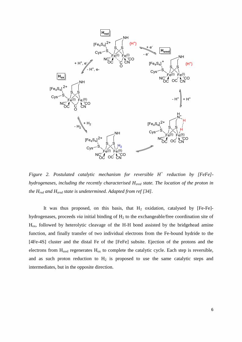
activity of this class of hydrogenases that heterolytically split H2 into a hydride (H–) and a
proton, with the hydride transferred to the carbocation containing substrate, methenyl-
tetrahydromethanopterin (methenyl-H4MPT+), (Figure 5), yielding methylene-
tetrahydromethanopterin (methylene-H4MPT). In contrast to [FeFe]- and [NiFe]-
10
hydrogenases, to which they are not phylogeneticaly related, [Fe]-hydrogenases do not
catalyse the splitting of H2 into protons and electrons. In addition, they do not catalyse a H/D
exchange reaction at a significant rate unless methylene-H4MPT or methenyl-H4MPT+
is
present [53] .
Figure 5. Schematic of the reversible heterolytic splitting of H2 by Hmd, showing a hydride
stereospecifically transferred into the pro-R site of methenyl-H4MPT+ to yield methylene-
H4MPT plus a proton.
Structural determination of the active site has been achieved in three successive steps
by the group of Shima and Thauer. First, a low molecular mass cofactor could be isolated and
was shown to be associated with one tightly bound iron ion [54]. Both the enzyme and the
isolated cofactor were found to be light-sensitive. The structure of the light-inactivated
cofactor was shown in 2004, to be a 2-pyridone derivative bearing a pendant carboxylate
function [55]. In a second step, IR spectroscopy revealed that an iron dicarbonyl moiety was
bound to the cofactor in its active state [56]. Third, a crystal structure could be obtained
showing that iron was bound to the nitrogen atom of the 2-pyridone cofactor [57].
Coordination of a thiolate function of the C176 cysteine residue was later confirmed [58]
along with the cis-coordination of two carbonyl ligands. A fuller picture of the iron
coordination sphere was eventually achieved with the crystal structure of the C176A mutated
[Fe]-hydrogenase (Figure 6). In the crystal structure exogenous dithiothreitol was found to
act as a bidentate ligand towards the iron centre, replacing both the thiolate function (C176 in
the native enzyme) and a labile coordination site, where H2 is likely activated. The
coordination sphere of Fe is completed by an acyl (-C(=O)-R) ligand, a reduced form of the
carboxylate residue found in the light-inactivated cofactor [59]. The site for H2 binding has
11
recently been identified through the crystal structure determination of an isocyanide-inhibited
form of the [Fe]-hydrogenase, since kinetic studies have suggested that H2 and isocyanide
bind to the same ligation site [60]. The structure showed that this site is located trans to the
acyl ligand [61]. In addition, the strong interaction observed between the bound isocyanide
molecule and the pyridinol hydroxylate group (deprotonated form) in the crystal structure,
suggests that this group also affects the chemistry of Fe-bound H2, perhaps acting as a
“pendant” base, as discussed earlier for adt2–
and terminal cysteine ligands in the enzymatic
mechanism of [FeFe] and [NiFe]-hydrogenases.
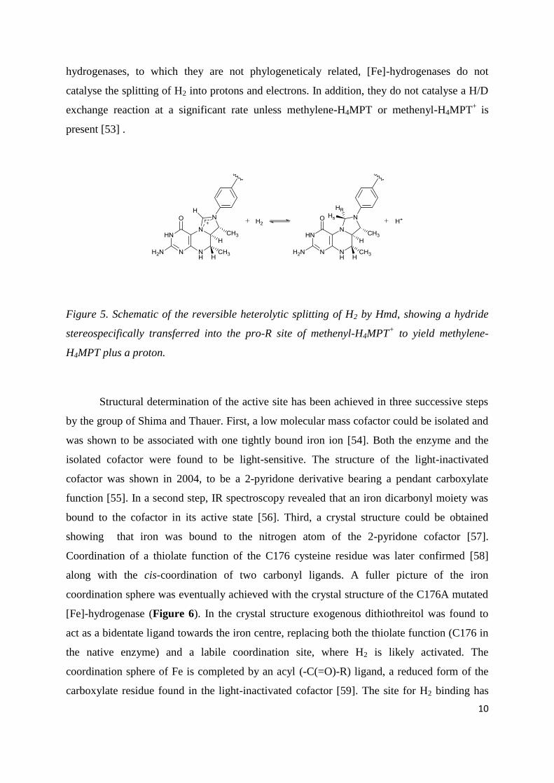
Figure 6. Schematic representation (left) of the [Fe]-hydrogenase active site with the Hmd
cofactor, and the X-ray crystal structure (right) of the mono-iron complex obtained from the
cysteine 176 mutant (PDB code 3H65).
The iron centre remains EPR-silent throughout the enzymatic cycle and is now thought to be a
low spin Fe(II) centre acting as a Lewis base for H2 coordination and activation. The general
catalytic mechanism is now understood as follows: first, methenyl-H4MPT+ binds to the
enzyme, which induces a conformational change from an open to a closed form of the
enzyme. In this latter form, the carbocationic site of the substrate is located close to the iron
atom. Hydrogen then binds to the iron atom of the active site trans to the acyl ligand.
Heterolytic cleavage of the H2 molecule likely occurs at this point, and whilst the intimate
mechanism of this step remains unclear, it is believed that the coordinated thiolate ligand acts

12
as a base in the process, a hypothesis made by comparison with heterolytic H2 cleavage at the
active site of [NiFe]-hydrogenase [62]. The oxygen atom of 2-pyridone, which exhibits
phenolate character in one of its mesomeric forms, is another possible proton acceptor site
during H2 cleavage [63]. The substrate is the final acceptor of the hydride ligand while the
proton is rapidly exchanged with bulk water.
3. Biomimetic catalysts
3.1. Models of the active site of [FeFe]-hydrogenases
3.1.1. First generation of [FeFe]-mimics
Long before the structural elements of [FeFe]-hydrogenases’ active subsite were
known, the preparation of analogous dithiolate-bridged hexacarbonyl diiron complexes was
reported by Reihlen et al., describing the synthesis of [(μ-SEt)2Fe2(CO)6] (1) as early as 1929
[64].
Upon structural elucidation of the H-cluster in 1998-1999 [21, 22], the striking
similarity of the 2Fe subsite to the propanedithiolate (pdt) bridged diiron complex [(μ-
pdt)Fe2(CO)6] (2) previously reported by Seyferth [65], no doubt inspired the three
independent groups of Pickett, Rauchfuss and Darensbourg, to replace two CO with two
cyanide ligands to give the water-soluble dianion, [(μ-pdt)Fe2(CO)4(CN)2]2–
(3) (Figure 7)
[66-68].
Figure 7. Synthetic scheme for the preparation of [(μ-SEt)2Fe2(CO)6] (1) [64], and the
precursor diiron hexacarbonyl complex (2) utilised en-route to the first true [FeFe]-model,
[(μ-pdt)Fe2(CO)4(CN)2]2–
(3) [66-68].

13
Although the identity of the bridgehead atom of the natural subsite has now been
confirmed as a secondary amine [10], the ambiguity of its identity at that time led to the
preparation of related [2Fe]-units bearing adt (azadithiolate = [(SCH2)2NR]2-
) (5-6) [69] and
odt (oxodithiolate = [(SCH2)2O]2-
) (7-8) [69-71] derivatives of the pdt bridge (Figure 8).
Figure 8. Scheme for the preparation of [FeFe]-subsite analogues bearing naturally relevant
adt (5-6) and alternative odt (7-8) bridging groups, with their alternate bridgeheads
highlighted in blue and red respectively.
These early models appear to have had a profound effect on the chemical community, as over
the last two decades a plethora of wide ranging [FeFe]-hydrogenase mimics have been
reported, with over 300 novel systems described [33, 72].
Much of this work has focused on preparing subsites that mimic the natural enzyme’s
key features such as a bridging CO [73-77], formation of mixed valent Fe(II)-Fe(I) systems
reproducing the ‘rotated state’ of the enzyme [78-85], isolation of stable, biologically relevant
terminal hydride species [86-88] and attachment to a synthetic Fe4S4 cubane, to give a full H-

14
cluster model (28, Figure 18) [89]. However, many of the compounds that are closest to
resembling the natural subsite in terms of valency and oxidation state of the iron metal centre,
make use of abiological phosphine or carbene ligands. Furthermore they tend to
electrocatalytically reduce protons at large overpotentials, cycling through the Fe(I)-Fe(I)/
Fe(I)-Fe(0) levels as opposed to the natural Fe(II)-Fe(I) [33, 72].
3.1.2. Second generation models: modification of the bridging group
Whilst the interest in [FeFe]-model systems continues unabated, with close to 100 new papers
in the last 5 years alone, the vast majority of this work focuses on exploratory alteration of the
bridging group, with limited success in overcoming key issues, such as large overpotential
requirements for H+
reduction, intrinsic for many [FeFe]-mimics. Nonetheless, interesting
systems bearing novel features have been developed, which may benefit the design of any
future catalytic systems.
The catalogue of bridgehead groups has been extended by recent work to include Se [90, 91]
and Si [92-94] bridgeheads, whilst the bridging sulphur atoms have been replaced with Te in
isolation [95-97] or combination with other chalcogens [98].
The effect of using higher group chalcogens to replace the bridging dithiolates has recently
been explored by groups including Hou and Weigand, which demonstrate contrasting effects
of this modification on electrocatalytic H+ reduction. Hou et al. describe the synthesis of an
[FeFe]-mimic containing a phenyl amine, diselenolate bridge [(µ-
(SeCH2)2NC6H4CH3)Fe2(CO)6] which is an active catalyst for H+ reduction using p-
toluenesulfonic acid (HOTs) [99]. When compared to its dithiolate bridge derivative, the
reduction potentials for the Fe(I)-Fe(I) to Fe(I)-Fe(0) process are almost identical at –1.50 V
vs. Ag/AgCl, yet the diselenolate bridged species shows a slightly larger increase in current,
indicative of higher catalytic activity. Contrastingly, when Weigand and co-workers carried
out a comparative study of water soluble dithiolate and diselenolate bridged species [(µ-
(ECH2)2CH2R)Fe2(CO)6] (R = tetra-O-acetyl-β-D-glucopyranoside, E = S/Se) in acetic acid
and water, the diselenolate species shows lower catalytic activity towards H2 evolution than

15
its the dithiolate equivalent, although it does exhibit improved robustness in water under
reductive conditions, relative to its dithiolate derivative.
One strategy for designing diiron model systems with redox properties closer to the
thermodynamic potential for H+ reduction is inclusion of a suitably substituted aromatic
dithiolato bridgehead, as highlighted by a recent study by Felton and co-workers [100]. The
[(µ-bdt)Fe2(CO)6] (bdt = benzenedithiolate, [(S)2C6H4]2-
) type diiron system, bearing benzene
rings highly substituted with electron withdrawing groups such as chloride were prepared,
analogous to earlier work from the Ott group [101]. The catalytic potentials and efficiencies
of these and related systems were compared and it appeared that increasing the number of
electron-withdrawing groups lowers the potential for catalysis (up to 150 mV) but
compromises the catalytic efficiency in terms of turnover frequency.
3.1.3. Variation of ligands: from cyanide to abiological phosphines and/or carbenes
Whilst the first generation of cyanide containing [FeFe]-mimics fulfilled a structural
requisite, it soon became apparent that use of cyanide outside of the protective protein
environment presented difficulties, somewhat highlighted by the fact that new reports of
cyanide ligated systems are in very short supply. Single substitution of a CO from 2 with
cyanide gives an electron rich diiron complex that can be protonated, although the preferred
site is the CN– ligand itself [102]. Disubstitution gives the highly reactive dianion 3 which
upon protonation results in sub-stoichiometric H2 evolution with subsequent decomposition of
the compound [102, 103].
Since this early work, much use has been made of surrogate ligands, which are said to
approach the electron donating properties of cyanide, but without the inherent complications,
including biologically relevant thioether groups, isocyanides, and to a larger extent more
abiological ligands such as carbenes, amines, and phosphines [33]. Importantly, the use of
these alternative ligands has allowed for key aspects of the natural subsite to be replicated,
such as isolation of a terminal hydride species, discussed in section 3.1.6.
Another important feature of the natural enzyme diiron site is the H2O molecule or hydroxide
ion, coordinated to the Fe distal to the [4Fe-4S] cubane, which is believed to play an

16
important protective role in the Hoxair
state of the enzyme [13, 32]. Surprisingly, this feature
has only very recently been replicated by Liu and co-workers by addition of a phenol
functionality connected to the bridge group of a diiron hexacarbonyl system to give [(μ-
SCH2)2CMe(CH2-o-C6H4OH)Fe2(CO)6] (9) [104, 105]. Upon deprotonation of the phenol
group of 9 with NaH, the phenolate oxygen coordinates to an iron centre with release of a CO
ligand to give the pentacarbonyl species [(μ-SCH2)2CMe(CH2-o-C6H4O)Fe2(CO)5]−
(10,
Figure 9).
Figure 9. Scheme for the intra-molecular coordination of the deprotonated phenol group of 9
to give [(μ-SCH2)2CMe(CH2-o-C6H4O)Fe2(CO)5]− (10), as a structural model for the Hox
air
and Hox states of the [FeFe]-hydrogenase.
3.1.4. Bridging CO, mixed valence state and rotated structures
To date, there is still only one example of a diiron complex with bridging CO, solely
supported by cyanide ligation [73]. This species slowly converts back to an all terminal CO
system.
The first examples of isolated stable complexes containing a bridging CO ligand were
achieved utilising electron rich (relative to CO) isocyanides (MeCN), or phosphine/phosphite
in isolation or combination with CN, in diferrous systems [33]. More recent examples of
similar systems include work by Talarmin and co-workers, in which 2 is coordinated by a
chelating dicarbene substrate to give [(μ-pdt)Fe2(CO)4(κ2-IMe-CH2-IMe)] (IMe = 1-
methylimidazol-2-ylidene) [77]. The effect of this ligand on the ability of the complex to bind
varying substrates (CO, MeCN, P(OMe)3) was probed under electrochemical oxidation
conditions. In the presence of P(OMe)3 the diferrous, bridging CO, [(μ-pdt)(μ-
CO)Fe2(CO)2(P(OMe)3)2(κ2-IMe-CH2-IMe)]2+
compound is produced.

17
Mixed valence models have relied upon ligation with phosphines and/or carbenes to give
compounds that stabilize oxidised Fe(II)-Fe(I) systems in non-coordinating solvents, through
a combination of enhanced electron density at Fe, and steric effects imparted by the bulky
ligands [33]. This has allowed for the fabrication of model systems with a ‘rotated state’ in
which an Fe centre assumes a square-pyramidal geometry, with a free coordination site for
substrate binding, which neatly mimics the Hox/Hox-CO states of the natural enzyme.
Ott and co-workers have recently demonstrated that substitution of two CO ligands of 2 for a
chelating diphosphine ligand with an electron deficient carborane bridgehead, gives the diiron
unit [(µ-pdt)Fe2(CO)4(BC)] (BC = 1,2-bisdiphenylphosphine-1,2-o-carborane) (11, Figure
10), which has sufficient electron density to stabilise the Fe(II)-Fe(I) state. This mixed valent
species was formed through electrochemical oxidation and was found to be stable on a
minutes timescale, resulting in a reversible oxidation process in the CV, and allowing for its
spectroscopic characterization via EPR and HYSCORE [106].
Figure 10. X-ray crystal structure [A] of the [FeFe]-model (11) that features a chelating
diphosphine borane ligand, and the EPR spectrum of the Fe(II)Fe(I) species [B] formed
through electrochemical oxidation. Reproduced with permission from reference [106].
3.1.5. Proton relays

18
Protonation of the [FeFe]-subsite is generally believed to occur at the free coordination site of
the distal Fe in the Hox state of the enzyme, however other possible sites for protonation have
been suggested [88, 107-109], such as the Fe-Fe bond, the amino bridge, the cyanide ligands
and the dithiolate bridges, all with precedents in the literature [33, 110]. Indeed, in a very
recent report by Liu et al. [111] the novel diiron mimic [(µ,κ2-bdt)(µ-PPh2)Fe2(CO)5]
– is
prepared, which features a bridging phosphine ligand. Protonation with an excess of
trifluoromethanesulfonic acid (HOTf) produces a doubly protonated species, bearing a µ-
hydride and a protonated thiolate, characterized by X-ray crystallography.
Similar results were reported by Ott and co-workers, showing that inclusion of ligands
bearing varying amine functionalities (12-14), allows for the directional control of the initial
protonation site (Figure 11), with generation of either an ammonium salt (15) or µ-H species
(16), depending on the nature of the amine group, or formation of a SH+ species (17) when
the basic amine group is omitted from the chelating diphosphine ligand [107].
Figure 11. A series of [FeFe]-models bearing chelating diphosphine ligands with varying
bridgeheads groups (12-14), and the protonated products (15-17) all featuring unique sites of
protonation, coloured for clarity.
19
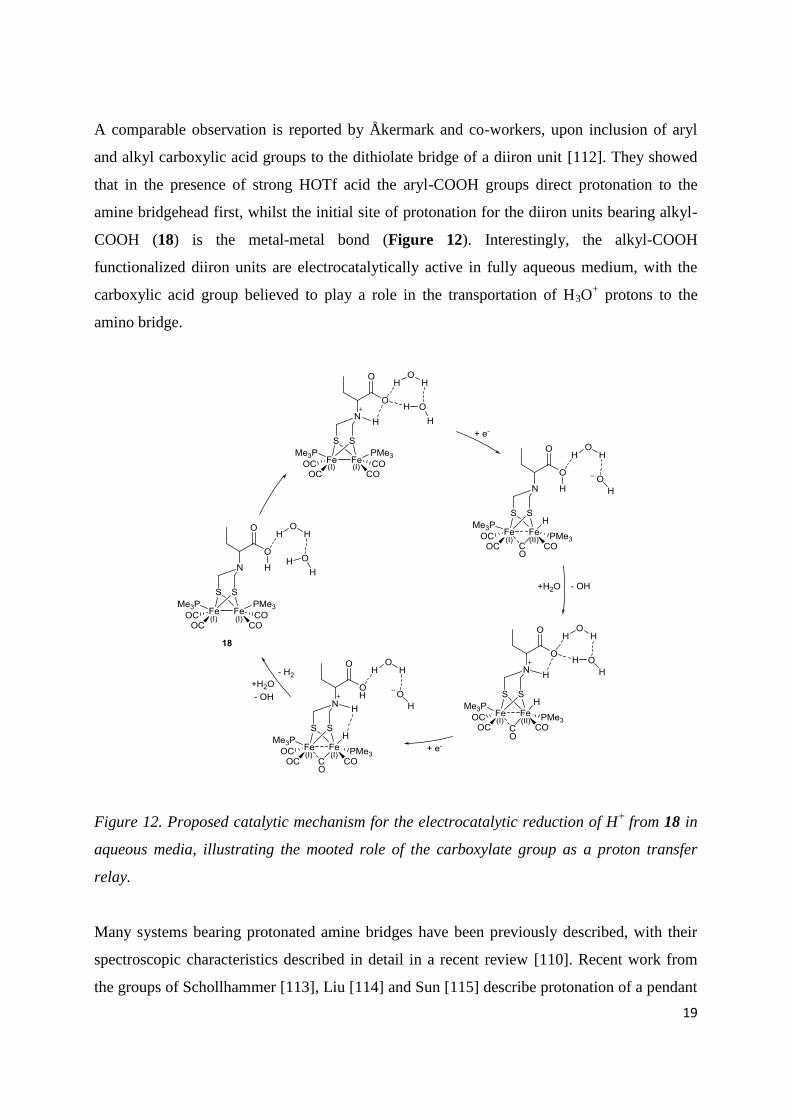
A comparable observation is reported by Åkermark and co-workers, upon inclusion of aryl
and alkyl carboxylic acid groups to the dithiolate bridge of a diiron unit [112]. They showed
that in the presence of strong HOTf acid the aryl-COOH groups direct protonation to the
amine bridgehead first, whilst the initial site of protonation for the diiron units bearing alkyl-
COOH (18) is the metal-metal bond (Figure 12). Interestingly, the alkyl-COOH
functionalized diiron units are electrocatalytically active in fully aqueous medium, with the
carboxylic acid group believed to play a role in the transportation of H3O+ protons to the
amino bridge.
Figure 12. Proposed catalytic mechanism for the electrocatalytic reduction of H+
from 18 in
aqueous media, illustrating the mooted role of the carboxylate group as a proton transfer
relay.
Many systems bearing protonated amine bridges have been previously described, with their
spectroscopic characteristics described in detail in a recent review [110]. Recent work from
the groups of Schollhammer [113], Liu [114] and Sun [115] describe protonation of a pendant

20
basic functionality incorporated into phosphine ligands, with their potential as proton shuttles
discussed. A good example of such proton transfer behaviour is the description of an
unsymmetrically substituted diphosphine diiron unit by Talarmin and co-workers [113], in
which two CO ligands from 2 are substituted with a chelating di-phosphine ligand, bearing a
NMe bridgehead to give [(µ-pdt)Fe2(CO)4({PPh2CH2}NMe)] (19) . Protonation with an
excess of HBF4.Et2O in acetone leads to protonation of the NMe group in axial position (20).
However, upon dissolving the protonated species in CH2Cl2, the compound isomerizes,
altering the arrangement of the phosphine ligand from basal-apical to the basal-basal form
whilst concomitantly forming the bridging hydride species 21 (Figure 13). Although the
method of proton delivery is not discussed in detail, it could be suggested that rotation about
the chelating ligand allows the protonated NMe group within close enough proximity of the
metal-metal bond to form the thermodynamically favoured product via proton transfer.
Figure 13. Scheme for the protonation of 19 to yield the ammonium salt 20, and its
subsequent isomerization that results in delivery of the proton to the metal-metal bond, to
form the bridging hydride 21.
Of course the natural system functions reversibly and thus a proton relay would be required
not only for delivery of a proton but also for its dispatch following H2 oxidation, activity
which can been probed by catalytic exchange of H/D. Whilst there are reports of catalytic
exchange of H/D from -hydride diiron mimics void of pendant base functionality, these
generally required pre-treatment, via photolysis, to open a coordination site of an Fe centre
[103, 116, 117] yielding slow rates of exchange.
A recent study by Sun and co-workers clearly demonstrates the importance of the basic
functionality for catalytic H/D exchange [118]. Complex 2 is treated with a chelating
diphosphine containing an N-nPr bridgehead (nPr = CH2CH2CH3) to give, amongst other
products, the unsymmetrically substituted complex [(μ-pdt)Fe2(CO)4({PPh2CH2}2N-nPr)].

21
Upon treatment with two equivalents of HBF4.Et2O in CH2Cl2, the μ-hydride, ammonium salt
species [(μ-H)(μ-pdt)Fe2(CO)4({PPh2CH2}2NH-nPr)]2+
(22) is quantitatively formed. When
this hydride species was treated with 10 equivalents of CH3COOD in CH2Cl2, the 1H NMR
signal for the μ-hydride at δ –13.0 ppm was rapidly lost, whilst a new high field signal
attributed to the μ-deuteride was observed in the 2H NMR. Contrastingly, when the same
experiment was carried out on the equivalent diiron unit that lacks a basic bridgehead ([(μ-
H)(μ-pdt)Fe2(CO)4({PPh2CH2}2CH2)]), the μ-hydride signal at δ –12.8 ppm remained
unaltered. Additionally, the latter compound could not be deprotonated even with 20
equivalents of aniline base, yet the hydride from 22 could be removed by H2O alone. A
mechanism for H/D exchange by this system has been discussed, with a schematic
representation shown in Figure 14.
Figure 14. Scheme for the reversible exchange of protons and deuterons in 22, facilitated by
the basic amine bridgehead of the diphosphine ligand.
If it appears logical that the pendant amine group of the H-cluster functions as a proton relay,
perhaps less obvious is its possible role in the protection of an uncoordinated site of the diiron
unit. Rauchfuss and co-workers have recently shown that the inclusion of an amino
bridgehead helps to stabilise the two electron oxidized state of the triphosphine substituted
diiron unit [(μ-(SCH2)2NBn)Fe2(CO)3(dppv)(PMe3)] (23) (Bn = Benzyl, C6H5CH2, dppv =
diphenylphosphine vinyl, P(Ph2)2CHCHP(Ph2)2) [119]. Electrochemical oxidation of this
[FeFe]-mimic occurs at mild potential, relative to the pdt-bridged equivalent, whilst also
showing good reversibility on the cyclic voltammetry timescale. The stabilisation of this 32 e–
complex is due to the coordination, following rotation about the chelating diphosphine ligand,

22
of the NBn bridgehead group to the coordinatively unsaturated iron centre to give 24,
characterized by X-ray crystallography (Figure 15). This result suggests that the bridging
amine unit could also function as a protective ligand in the natural enzyme, in a similar vein
to the role of the H2O/OH- ligand in the Hox state.
Figure 15. Scheme for the reversible two electron oxidation of 23, resulting in the breaking of
the metal-metal bond, stabilised by the coordination of a solvent molecule to one Fe centre
and coordination of the nitrogen atom of the bridgehead to the second to yield 24.
Another interesting finding from this work is that the ammonium equivalent of this
compound, [(μ-(SCH2)2NHBn)Fe2(CO)3(dppv)(PMe3)]+, can function as an efficient hydrogen
atom donor in the presence of the H-atom abstracting agent TEMPO (2,2,6,6-
tetramethylpiperidin-1-oxyl), to yield the mixed valent Fe(II)-Fe(I) species, suggesting that
these types of mimics, and by extension the natural system, may function through a PCET
mechanism.
3.1.6. Hydrides: bridging vs. terminal binding modes
Hydride derivatives of dithiolate-bridged diiron clusters pre-date the crystal structure
determination of the natural subsite of [FeFe]-hydrogenase by over 20 years, when Poilblanc
and co-workers reported the protonation of symmetrically substituted diphosphine [(μ-H)(μ-
SMe)2Fe2(CO)4(PPhMe2)2]+ to yield a bridging hydride species [120]. Whilst the majority of
hydride models are bridging in nature [33], and active electrocatalysts for H+ reduction,
comparative catalytic studies on terminal hydride derivatives are needed if we are to better
understand the bias, if any, the natural system has towards a particular mode (bridging versus
terminal) of metal hydride, and the specific reasons for such a preference.

23
A crystal structure of a diiron unit bearing a terminal hydride was first reported by Rauchfuss
and co-workers in 2005 [86], although this was achieved by addition of a hydride ligand from
LiAlH4/NaBH4 to a tetraphosphine ligated diferrous complex (25) to give [(µ-edt)(µ-
CO)HFe2(CO)(PMe3)4(NCMe)]2+
(26, Figure 16) (edt2–
= ethanedithiolate).
Figure 16. Scheme for the formation of the first isolated diiron unit to bear a terminal hydride
(26), through hydride addition to the tetraphosphine species 25, and the thermodynamically
stable bridging hydride equivalent 27.
This work was followed by the groups of Schollhammer [121], Rauchfuss [122] and Hogarth
[123], who reported that upon protonation of phosphine substituted [FeFe]-mimics, at low
temperatures, it was possible to detect 1H-NMR signals characteristic of terminally bound
hydride ligands. The isolation and characterisation of a stable terminal hydride diiron unit via
protonation was finally achieved by Rauchfuss and co-workers in 2012 with the low
temperature synthesis and isolation of the doubly protonated diiron unit [(µ-
adtH)Fe2H(CO)2(dppv)2]2+
(Figure 17) [87]. The crystal structure replicates the naturally
relevant bridging CO. It also demonstrates a strikingly short distance between the terminal
hydride and equatorial amino proton of 1.88(7) Å, indicating significant dihydrogen bonding.
It should be noted that, upon warming the reaction solution to room temperature, the terminal
hydride species isomerizes to give the bridging derivative, as confirmed by 1H NMR.

24
Figure 17: ORTEP representation of [(µ-adtH)Fe2H(CO)2(dppv)2]2+
with thermal ellipsoids
drawn at 50% probability. Phenyl hydrogen atoms, counter-ions and solvent of crystallization
omitted for clarity. Reproduced with permission from reference [87].
It has previously been suggested that the presence of a pendant base group such as the adt
bridge would kinetically favour terminal hydride species [124] and that the overpotential
would be smaller in comparison to bridging hydride species. This notion is given credence by
the reduction potential recorded for [(µ-adtH)Fe2H(CO)2(dppv)2]2+
, which is anodically
shifted by 150 mV, relative to its bridging hydride derivative. The advantages of a terminal
hydride are further evidenced by the relatively large TOF (5000 s–1
) calculated for the singly
protonated terminal hydride species [(µ-adt)Fe2H(CO)2(dppv)2]2+
during electrochemical
reduction in the presence of trifluoroacetic acid in CH2Cl2, again compared to the bridging
hydride derivative (20 s–1
).
When considering the advantages of a terminal hydride species over a bridging hydride, such
as their milder reduction potentials and their proximity to the mooted proton transfer group, it
is tempting to suggest that the natural enzyme would likely favour such a catalytic
intermediate [125].
3.1.7. H-cluster models: towards the complete catalytic package
The first attempts to introduce an electron transfer relay, a key component of the natural
enzyme (see 2.1 above), were achieved by Tard et al. by inclusion of a biologically relevant
cubane [Fe4S4]-cluster, bridged to a [2Fe3S]-model via a sulphur ligand to yield the H-cluster
model system depicted in Figure 18 [89]. Whilst this system is functional as an

25
electrocatalyst for proton reduction, it is at the cost of large overpotentials, whilst also lacking
the ability to perform the reverse process.
Figure 18. Schematic representation of the H-cluster model 28 synthesized by Tard et al. [89].
When considering the amount of work that has been published on [FeFe]-mimics it is
somewhat surprising that very little attention has been given to the introduction of an electron
transfer relay, to replicate [4Fe-4S] cluster function, particularly as understanding the cluster’s
role in enabling catalysis may be key to achieving truly functional model systems. The recent
detection and characterisation of the Hsred oxidation state (Figure 2) by the groups of Lubitz
and King will likely inspire many groups in the field to pursue this endeavour [34, 35].
There are a handful of reports of diiron units bearing additional redox active moieties,
however almost all of the reported systems show no significant overpotential decrease or
catalytic efficiency increase. Models include the attachment of pendant ferrocene (Fc) groups
[126-129], a diphosphine functionalised fullerene [130], non-innocent 2,3-
bis(diphenylphosphine) maleic anhydride (BMA) [131], and bipy ligands (bipy = 2,2’-
bipyridine) [132]. Unfortunately, only the fullerene model exhibits any electronic
communication between the second and first redox coordination spheres, with the only other
functionality observed being that of an electrochemical internal standard [126-128].
In fact, the only example of an [FeFe]-mimic that bears a functional electron transfer relay is
the complex [µ-{(SCH2)2NBn}Fe2(CO)3(FcP*)(dppv)] (29) (FcP* = Cp*Fe(C5Me4CH2PEt2))
(Figure 19) recently reported by Camara and Rauchfuss [133]. This is also the first example
of a model [FeFe]-system that performs catalytic H2 oxidation.
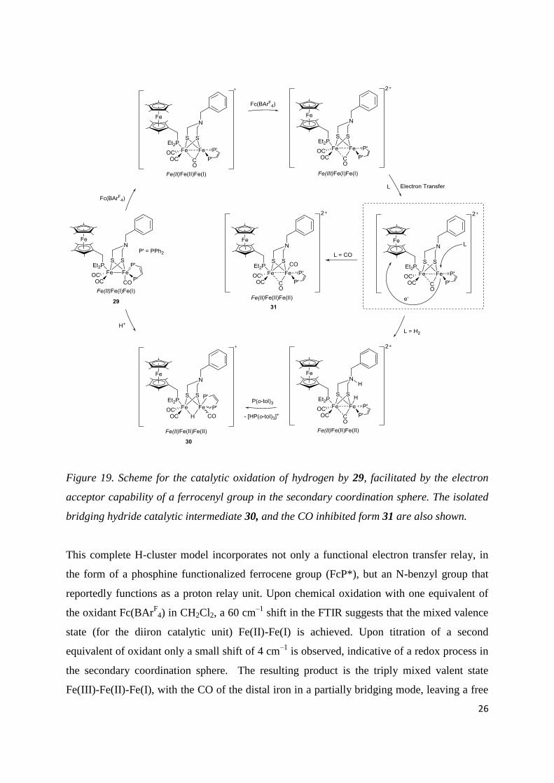
26
Figure 19. Scheme for the catalytic oxidation of hydrogen by 29, facilitated by the electron
acceptor capability of a ferrocenyl group in the secondary coordination sphere. The isolated
bridging hydride catalytic intermediate 30, and the CO inhibited form 31 are also shown.
This complete H-cluster model incorporates not only a functional electron transfer relay, in
the form of a phosphine functionalized ferrocene group (FcP*), but an N-benzyl group that
reportedly functions as a proton relay unit. Upon chemical oxidation with one equivalent of
the oxidant Fc(BArF
4) in CH2Cl2, a 60 cm–1
shift in the FTIR suggests that the mixed valence
state (for the diiron catalytic unit) Fe(II)-Fe(I) is achieved. Upon titration of a second
equivalent of oxidant only a small shift of 4 cm–1
is observed, indicative of a redox process in
the secondary coordination sphere. The resulting product is the triply mixed valent state
Fe(III)-Fe(II)-Fe(I), with the CO of the distal iron in a partially bridging mode, leaving a free

27
coordination site in the apical position. Bubbling of H2 through a CH2Cl2 solution of the
complex in the presence of 6 equivalents of base (P(o-tolyl)3) and excess of oxidant (4 equiv.
of Fc(BArF
4)) led to 4 turnovers of H2 oxidation over a 5 hour period.
It is important to note that unlike previous doubly oxidized diiron systems, the diferrous state
for the catalytic subunit is not achieved. Instead the second oxidation step likely results in the
loss of an electron from the ferrocene electron relay to yield a ferrocenium moiety. It has
previously been shown that the binding of a H– ligand to [FeFe]-model systems can be
energetically biased towards a diferrous system [119], although there is no evidence for an
unsaturated diferrous state in the enzyme. It is suggested rather that nature would proceed
with activation of H2 via a proton coupled electron transfer (PCET) mechanism, with the
[4Fe-4S] cubane providing the oxidative power required (Figure 2). Indeed, these results
suggest that H2 binding, its heterolytic cleavage and transfer of an electron to the pendant
ferrocenium occur concomitantly, as strengthened by a recent DFT study on the same system
[134].Although at first glance, the inclusion of an electron transfer relay would appear
intrinsic to this model systems ability to catalytically oxidize H2, Sun and coworkers [135]
have subsequently shown that under similar experimental conditions, their previously reported
model system bearing only a proton transfer relay (22 Figure 14) is also active for catalytic
H2 oxidation, and at a slightly increased rate.
Whilst these systems no doubt represent a dramatic breakthrough in modelling the
bidirectional functionality of the [FeFe]-hydrogenase enzyme, their catalytic rates for H2
oxidation of 10–4
s–1
are several orders of magnitude lower than the 28000 s–1
of the natural
system [1]. It is vital that these types of challenges are overcome if we are to develop efficient
catalytic systems, which are adequate for incorporation into future practical technological
devices [136].
3.2. Models of the active site of [NiFe]-hydrogenases

28
Although several mimics of the active site of [NiFe]-hydrogenase were reported
shortly after the structure of the natural subsite had been determined (32-33, Figure 20) [137],
construction of models with greater structural relevance, that also replicate the active site’s
functionality, has long been a challenge for chemists, due in no small part to its complexity.
Up to 2009, a large number of mimics had been reported [33, 39, 138], some of them
featuring the all-sulphur coordination sphere around nickel, the {Ni(µ-SR)2Fe} core, and CN–
/CO ligands bound to Fe (34-37). However, none of these structural mimics were shown to be
catalytically active.
Figure 20. Selected early [NiFe]-models, with compounds 34-37 featuring the biologically
relevant {Ni(µ-SR)2Fe} core, coloured in red.
3.2.1. Nickel-ruthenium mimics
The use of organometallic ruthenium moieties as surrogates for the {Fe(II)(CN)2(CO)}
fragment is justified by the lack of redox change at the low-spin Fe(II) centre of native
enzymes during catalysis, suggesting that the role of the carbonyl and cyanide ligands found
at the active site may be restricted to the modulation of the electronics of the iron centre and/

29
or the stabilization of intermediates such as hydride or dihydrogen derivatives. In respect to
this latter described role, ruthenium complexes are widely used as catalysts for hydrogenation
or hydrogen-transfer reactions [139] and Ru(II) can easily accommodate both hard and soft
ligands including dihydrogen or a hydride, a required property for a metal to replicate the Fe
function during catalysis by NiFe hydrogenases. This approach allowed Oudart and co-
workers from our group to prepare active [NiRu] catalysts for H2 evolution, through
combination of the weakly active Ni complex, [Ni(xbsms)] [140, 141] with {Ru(CO)2Cl2}
(38-39), {Ru(CO)3Cl}+ (40) and {RuCl(p-cymene)2}
+ (41-42) moieties (Figure 21) [142-
144]. It also made it possible to derive a structure-function relationship that resulted in
significant improvement of nickel-ruthenium H2-evolving catalyst performance: using the
infraredCO frequency as a probe for the electron density on the metal centres, the same
authors concluded that the more electron-rich the metal centre, the better the catalyst, as far as
overpotential requirement is concerned. Canaguier and co-workers from our group followed
this trend, with substitution of {Ru(arene)}2+
or {Ru(CO)2Cl}+
for more electron-rich
organometallic moieties such as {RuCp}+ (43-45) (Cp
– = cyclopentadienyl anion) fragments
(Figure 21). This approach was successful as it was possible to gain 200 mV under the same
assay conditions using [Ni(xbsms)RuCp] compounds, relative to the former [NiRu]-catalysts
[141]. The introduction of the electron-enriched {RuCp*}+ moiety (46-48) (Cp*
– =
pentametylcyclopentadienyl anion) finally yielded the most active catalyst in the series (46)
with both a relatively low overpotential requirement (620 mV) and a high turnover frequency
[145]. In addition, the steric protection provided by the bulky Cp*– ligand resulted in an
increased stability upon cycling. The group of S. Ogo in Japan used a similar strategy to
produce a dinuclear nickel-ruthenium compound (49, Figure 21) bearing a
hexamethylbenzene ligand capable of catalytically oxidizing H2 in the presence of Cu(II) ions
as the oxidizing agents [146]. It was shown that the activation of H2 by the dinuclear catalyst
involves the formation of an intermediate containing a hydride ligand (H–) bridging the two
metal centres [147], a geometry very similar to that found in the catalytically competent Ni-C
state of NiFe hydrogenases [48]. Similar bridging hydride intermediates have also been
proposed through DFT calculations for the reverse process, i.e. hydrogen evolution [141,
148]. In a recent study, our group revisited the mechanism for H2 evolution catalyzed by this
series of compounds, and highlighted the possibility that H2 is produced via a PCET
mechanism from just such a bridging hydride ligand [149].

30
Figure 21. Selected [NiRu] complexes as functional models of the [NiFe]-hydrogenase.
3.2.2. Nickel-iron functional mimics
The next milestone was the design of functional models only containing first-row transition
metals. Seven such systems (compounds 50-55 and 57, Figure 22) were reported as
exhibiting catalytic activity, albeit restricted to H2 evolution [150-153], once again with a
bridging hydride species as the active intermediate.
In 2009 Rauchfuss and coworkers prepared a dinuclear Ni-Fe hydride compound through
the apparently facile protonation of [(dppe)Ni(µ-pdt)Fe(CO)3], initially reported by Schröder
[154], with HBF4 to yield [(dppe)Ni(µ-H)(µ-pdt)Fe(CO)3]+ (50) [150]. Other derivatives

31
could be obtained through the substitution of two CO ligands with phosphites (51),
phosphines (52-53) [151] or diphosphines [150], requiring photochemical activation in the
latter case. All of these dinuclear bridging hydride derivatives proved electrocatalytically
competent for hydrogen evolution from trifluoroacetic acid (TFA) in a CH2Cl2 solution. The
absence of any thermodynamical data for acid-base constants in this solvent makes the
determination of the overpotential requirement quite difficult. A 1 V estimation for the
overpotential requirement for H2 evolution from TFA catalysed by 50 could be obtained
from comparison of H2 evolution in CH2Cl2 and DMF at a platinum electrode [152].
Measurements were further made from freshly prepared CH3CN solutions of phosphine
derivatives (51-53), despite their low stability in this solvent, and yielded lower overpotential
values in the 260-430 mV range [151].
Canaguier and coworkers from our group reported another functional Ni-Fe mimic in 2010
(54), by coordination of [Ni(xbsms)] to a cyclopentadienyl Fe(II) moiety, originated from the
[CpFe(CO)2(thf)]+
precursor. The two thiolate ligands of the xbsms2–
ligand bridge the nickel
and the iron centres and the Fe coordination sphere is completed by a single CO ligand.
Complex 54 was shown to catalyse H2 evolution from TFA in DMF with a 730 mV
overpotential requirement, with subsequent DFT calculations proposing that possible
formation of a bridging hydride could occur through protonation at the CO ligand in the one-
electron reduced state, followed by rearrangement and elimination of the CO ligand.

32
Figure 22. Schematic representation of the majority of structurally relevant [NiFe]-mimics,
active for electrocatalytic proton reduction (50-55), and for the bidirectional conversion of
protons and electrons into dihydrogen (58). Also shown are the only known [NiMn] systems
(56, 57), also active for electrocatalytic proton reduction.
A similar approach allowed for the synthesis of a Ni-Mn compound (56,), with a similar
structure and requiring an overpotential of 860 mV to evolve H2 from TFA in DMF [155,
156]. A second series of Ni-Mn compounds has been reported by L.-C. Song, two of which
(57) proved active for H2 evolution from acetic acid in CH3CN [157] with low catalytic
current enhancement but reduced overpotential requirement (~480 mV).
Using an analogous strategy, Weber and coworkers from the Lubitz group reacted the
[Ni(xbsms)] complex with [Fe(CO)3(bda)] (bda = benzylidene acetone) to obtain 55; a
[NiFe]-model with only three sulphur ligands on the Ni centre, and a thioether group
coordinated to the Fe atom. In this highly structurally distorted compound, a CO ligand was
found to bridge between both metals. Interestingly, in the presence of HBF4, protonation
occurred at the terminal thiolate ligand of the nickel centre, a feature reminiscent of the active

33
site of NiFe hydrogenases. Both un-protonated and protonated compounds proved active for
H2 evolution from TFA in CH3CN with overpotential requirements of 540-570 mV [153].
Recently, Ogo and co-workers have described a nickel-iron mimic able to mediate both
hydrogen evolution and oxidation, thus reproducing for the first time at a binuclear core the
bidirectional activity of the [NiFe]-hydrogenases [158]. The novelty of this mimic, which is
likely responsible for its functionality, is the use of three triethylphosphite (P(OEt)3) ligands
to modulate the electronic properties of the iron centre, so as to promote coordination of H2 as
a first step towards its activation. Heterolytic splitting of H2 is promoted in the presence of
methanolate, a strong base that captures a proton whilst a hydride ligand remains coordinated
to the iron centre, in a terminal binding mode (Figure 22, 58). This hydride species can be
oxidized by methylviologen (MV2+
), thus at a mild potential. Release of a proton regenerates
the starting compound, completing the catalytic cycle. The net reaction is the two-electron
oxidation of molecular hydrogen with formation of two protons. It is however important to
note that the nickel-iron compound reported by Ogo and co-workers achieves only a single
turn-over with a 12% yield, although a better yield (45%) could be measured when using a
stronger oxidant such as the ferrocenium ion. The system also operates at energies far from
the thermodynamic equilibrium, as evidenced by the requirement of a strong base to activate
H2, while the natural process operates in water at neutral pH. Similarly, a strong acid is
required to produce H2 from the hydride species. So whilst this system shows promise as a
catalyst for hydrogen evolution, this will be at the expense of quite large overpotentials as
noted with other Ni-Fe mimics [150, 152, 153]. Comparison of two Ni-Fe and Ni-Ru
compounds with the same set of ligands [152] neatly highlights the benefit of Ru over Fe in
terms of overpotential requirement for catalysis [156, 159], consistent with the fact that noble
metals are currently preferred as catalysts in technological devices. Nonetheless, this novel
nickel-iron compound performs a single turnover reaction both for hydrogen oxidation and
evolution, behaviour so far restricted to a single series of mononuclear nickel catalysts [160-
162] that have been the subject of a previous detailed review [163].
Structural similarity of these systems with the active site of [NiFe] hydrogenases is
obvious. Nevertheless, comparison to the enzyme should be made with caution, with regard to
the elucidation of the natural catalytic mechanism. An issue of particular concern is the

34
binding mode of the hydride intermediate, i.e. bridging vs. terminal, with the latter option
providing the additional question as to which metal site accommodates the hydride. While a
terminal hydride ligand bound to a mononuclear iron centre has been recently reported as an
active catalyst for H2 oxidation [164], most characterised [147, 150, 151] or computed [141,
148, 152, 165] dinuclear hydride derivatives, based on a {Ni(II)(µ-SR)2M} core with M a
low-spin d6 metal centre, feature a bridging structure in contrast to the terminally bound
hydride recently described by Ogo, rendering any association between terminal/bridging
binding modes and H2 evolution/activation futile. Even if a terminally Fe-bound hydride is
associated with H2 activation, it is not in accordance with the observation of a bridging
hydride in the Ni-C state of the enzyme [48]. That being said, the production of a bridging
hydride derivative that does replicate the Ni(III)-Fe(II) electronic structure of the Ni-C state
remains elusive. It could thus be argued that the Ni(II)-Fe(II) centres of Ogo’s hydride
derivative (58) would better reproduce the structure of the Ni-R state of the enzyme, but no
definitive data exists as to the nature of the additional ligand (H2, H–…) nor to its binding
mode to the {Ni(II)(µ-SR)2Fe} core in this particular state.
Very recent work by Manor and Rauchfuss describes the synthesis of two [(dxpe)Ni(µ-
pdt)Fe(CO)n(CNBArF
3)2]¯ compounds, 59 and 60 (x = phenyl or cyclohexenyl, and BAr
F3 =
B(C6F5)3) (Figure 23) [166], which represent the closest models of biomimetic ligation at the
Fe centres. This was made possible by utilising isocyanides ligands CNBArF
3, which not only
protect the Fe centre from chemical attack through steric bulk but also provide the anionic
character of the subsite found in the natural enzyme. Furthermore, compounds 59 and 60 are
capable of heterolytically splitting H2 to yield the bridging hydride species 61 and 62
respectively, both of which show hydridic character, evidenced through 1H NMR and FTIR
spectroscopy. In line with the recent work of Ogo [158], [Et4N][(dppe)Ni(µ-pdt)(µ-
H)Fe(CO)(CNBArF
3)2] (61) exhibits bidirectional behaviour, capable of electrocatalytic H2
oxidation in the presence of the strong base DBU (diazabicycloundecene) and H2 evolution in
the presence of HCl, although this latter process only occurs on a stoichiometric level.
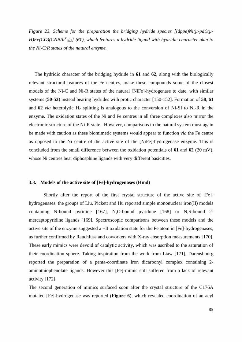
35
Figure 23. Scheme for the preparation the bridging hydride species [(dppe)Ni(µ-pdt)(µ-
H)Fe(CO)(CNBArF
3)2] (61), which features a hydride ligand with hydridic character akin to
the Ni-C/R states of the natural enzyme.
The hydridic character of the bridging hydride in 61 and 62, along with the biologically
relevant structural features of the Fe centres, make these compounds some of the closest
models of the Ni-C and Ni-R states of the natural [NiFe]-hydrogenase to date, with similar
systems (50-53) instead bearing hydrides with protic character [150-152]. Formation of 58, 61
and 62 via heterolytic H2 splitting is analogous to the conversion of Ni-SI to Ni-R in the
enzyme. The oxidation states of the Ni and Fe centres in all three complexes also mirror the
electronic structure of the Ni-R state. However, comparisons to the natural system must again
be made with caution as these biomimetic systems would appear to function via the Fe centre
as opposed to the Ni centre of the active site of the [NiFe]-hydrogenase enzyme. This is
concluded from the small difference between the oxidation potentials of 61 and 62 (20 mV),
whose Ni centres bear diphosphine ligands with very different basicities.
3.3. Models of the active site of [Fe]-hydrogenases (Hmd)
Shortly after the report of the first crystal structure of the active site of [Fe]-
hydrogenases, the groups of Liu, Pickett and Hu reported simple mononuclear iron(II) models
containing N-bound pyridine [167], N,O-bound pyridone [168] or N,S-bound 2-
mercaptopyridine ligands [169]. Spectroscopic comparisons between these models and the
active site of the enzyme suggested a +II oxidation state for the Fe atom in [Fe]-hydrogenases,
as further confirmed by Rauchfuss and coworkers with X-ray absorption measurements [170].
These early mimics were devoid of catalytic activity, which was ascribed to the saturation of
their coordination sphere. Taking inspiration from the work from Liaw [171], Darensbourg
reported the preparation of a penta-coordinate iron dicarbonyl complex containing 2-
aminothiophenolate ligands. However this [Fe]-mimic still suffered from a lack of relevant
activity [172].
The second generation of mimics surfaced soon after the crystal structure of the C176A
mutated [Fe]-hydrogenase was reported (Figure 6), which revealed coordination of an acyl

36
ligand at the active site. The first coordinatively unsaturated dicarbonyl-acyl-iron complex
was reported in another context by Holland [173], followed by reports of several other
mononuclear iron carbonyl complexes with acyl ligands by the groups of Hu [174] and
Rauchfuss [175, 176]. More relevant mimics of the active site of [Fe]-hydrogenase were
reported more recently, in which the acyl functionality is part of either an acylmethylpyridine
ligand [177], a 2-acylmethyl-6-methoxypyridine ligand [178-180] or 2-acylmethyl-6-
hydroxymethylpyridine ligand [181]. In this context, the approach taken by Pickett and
coworkers, in which the construction of an [Fe]-mimic is achieved using a pyridine-
carbamoyl ligand (63), is of particular interest as it results in a mono iron complex whose
metrical data is remarkably close to that of the active site found in the enzyme (Figure 24)
[182, 183]. To date however the closest model of this active site, which reproduces the exact
square-pyramidal iron coordination sphere of the natural system (64), was reported by the
group of Hu [184], although their model lacked the naturally occurring acylmethylpyridone
ligand.
Figure 24. Schematic representation of the closest structural mimics to date, of the [Fe]-
hydrogenase active site.
This issue was somewhat remedied by recent work of the Song group (Figure 25) [185],
though following deprotection of the pyridinol with TFA, a bi-metallic species spontaneously
forms (65). Unfortunately, despite the continued efforts of multiple research groups, none of

37
the synthetic mimics of the active site of [Fe]-hydrogenase have so far shown any relevant
activity for hydrogen activation or hydride transfer functionality.
Figure 25. Scheme for the preparation of the unstable intermediate shown in brackets, which
represents one of the closest [Fe]-hydrogenase structural models, and it’s thermodynamically
stable compound 65.
4. Supramolecular control
As outlined above, biomimetic systems inspired by the [FeFe] and [NiFe] catalytic co-
factors found in the active site of hydrogenases (Figures 1 and 3), have struggled to live up to
the expectations arising from the extreme efficiency observed for these enzymes. The
discrepancy between the reactivity of the enzymatic system and these synthetic small
molecule models, can be attributed to two important factors; i) the need of the latter to operate
in non-aqueous solvents, ii) the absence of a protein environment, important for the
modulation of the catalytic activity via outer coordination sphere interactions (Figure 26)
[186].

38
Figure 26. X-ray crystal structure of the CpI [FeFe]-hydrogenase (left, PDB code 3C8Y
with a modified active site to display an NH bridgehead) with the active site (right) and the
with the surrounding amino-acid residues important for catalytic activity and active site
stability.
Indeed this point has recently been exquisitely illustrated by our recent work, where upon
insertion of complex 6 into apo-[FeFe]-hydrogenase, either directly [11] or via the HydF
maturase [10] (see section 4.6), a fully active [FeFe]-hydrogenase enzyme is generated,
demonstrating that the protein cavity transforms this otherwise inactive and fragile complex in
aqueous medium, into a highly efficient and robust catalyst [10].
Consequently, numerous approaches are currently being developed in attempts to mimic
the influence of the proteic environment, such as the incorporation of these molecular
complexes into supramolecular structures including polymers, micelles, gels and peptidic
scaffolds.
4.1. Incorporation of biomimetic catalysts in gels and resins

39
Incorporation of molecular catalysts into a gel or resin support offers a number of potential
advantages, as it provides the possibility to tailor the binding environment as well as
facilitating the incorporation of such systems into technological devices.
Figure 27. The diiron complex employed for incorporation, [(-pdt)Fe2(CO)4(PMe3)2] [A],
and the stacking of the Fmoc-LL hydrogel [B].
Frederix et al. have studied the incorporation of [(-pdt)Fe2(CO)4(PMe3)2] into an Fmoc-LL
hydrogel (Fmoc-LL = N-(fluorenyl-9-methoxycarbonyl) protected Leu-Leu), a low molecular
weight hydrogelator that forms three dimensional networks in water at low concentrations
(Figure 27). The organometallic complex was introduced, and stabilized, in the translucent
gel via non-covalent interactions. FTIR spectroscopy indicated an environment intermediate
between strictly non-polar (heptane) and polar (MeOH). Interestingly, while [(-
pdt)Fe2(CO)4(PMe3)2] has been reported to be unstable in aqueous solution, it was found to be
stable for up to two weeks in the gel environment. However no data on the catalytic activity
of this system was reported [187].
Two systems in which TentagelTM
resin beads are used to anchor Ni [188] and FeFe [189]
complexes respectively have been reported by the group of Darensbourg. A CGC
(CysGlyCys) peptide fragment was covalently attached to the resin via an amide linkage to
provide a binding site for Ni ions, and indeed the tripeptide was found to be capable of
coordinating Ni ions in a NiN2S2 fashion. NiRh and NiW (66) heterobimetallic centers could

40
also be obtained by binding W(CO)5 or Rh(CO)2 via the thiolate ligands of the Ni(CGC)
complex (Figure 28) [188]. These results hint at the intriguing possibility of generating
[NiFe] mimics by reaction of appropriate organometallic iron species with the resin bound
NiN2S2 moiety.
H
H H
HN
NO
SMmt
NN
O
O
SMmt
O
2-
HN C
O
S
N N
S
O
O
NH
O
Ni
2-
CO
O
O
O O
H
S
N
N
S
O N O
Ni
WC
C
C
O
CN C
O
CF3COOH:HSiiPr3
CH2Cl2
Ni(acac)2, KOH
CH2Cl2
(THF)W(CO)5
CH2Cl2
ATR-IR
19671850
1917
2000 1900 1800
1
2000 1900 1800
1
H
H H
HN
NO
SMmt
NN
O
O
SMmt
O
2-
HN C
O
S
N N
S
O
O
NH
O
Ni
2-
CO
O
O
O O
H
S
N
N
S
O N O
Ni
WC
C
C
O
CN C
O
CF3COOH:HSiiPr3
CH2Cl2
Ni(acac)2, KOH
CH2Cl2
(THF)W(CO)5
CH2Cl2
ATR-IR
19671850
1917
2000 1900 1800
1
2000 1900 1800
1
Figure 28. The resin bound [Ni(CGC)]W(CO)5]2-
system 66 and its ATR-FTIR spectrum.
A study on the covalent attachment of carboxy-functionalized [FeFe]-hydrogenase mimics to
the same type of resin was reported in 2009 [189]. Three different linkers were used, in which
the carboxylate-group was introduced either on the bridgehead of the dithiolate motif (67), by
replacement of a CO ligand with the phosphine ligand P(C2H4COOH)3 (68), or as two
separate thiolate bridging groups (69) (Figure 29). Once attached to the resin (70-72) the
complexes could be further modified analogously to their solution state complexes, by
introduction of electron rich ligands such as PMe3 and CN−. Surprisingly, attaching the
complexes to the Tentagel resin was found to have a negative effect on their stability, both
with regards to acidic conditions as well as photo-induced loss of the CO ligands, making this
specific resin-complex combination an unlikely candidate for further catalytic studies.

41
Figure 29. Schematic representation of three carboxy-functionalized [FeFe]-hydrogenase
model complexes, before (67-69) and after (70-72) covalent attachment to the TentagelTM
resin beads.
4.2. Cyclodextrines
A series of dinuclear iron complexes in which the bridgehead nitrogen atom has been
functionalized to incorporate an aryl sulfonate group has been reported by the group of
Darensbourg [190, 191]. To a certain degree, the presence of the anionic sulfonate group
alone increases the solubility of the complex in water but, more importantly, allows for
inclusion of the complex into β-cyclodextrins. Cyclodextrins are cyclic oligosaccharides,
consisting of 6-8 glucose units (denoted α-, β- and γ-cyclodextrins respectively), generating a

42
hydrophobic cavity with a hydrophilic rim (Figure 30). The adducts formed between β-
cyclodextrins and five [FeFe]-hydrogenase mimics [(µ-(SCH2)2NC6H4SO3)Fe2(CO)6]−
(73),
[(µ-(SCH2)2NC6H4SO3)Fe2(CO)5(P(OMe)3)]−, [(µ-(SCH2)2NC6H4SO3)Fe2(CO)5(PTA)]
− (PTA
= 1,3,5-Triaza-7-phosphaadamantane), [(µ-(SCH2)2NC6H4SO3)Fe2(CO)5(PPh3)]−
and [(µ-
(SCH2)2NC6H4SO3)Fe2(CO)4(PMe3)2]− were investigated. The structure of the supramolecular
assemblies were studied in solution by NMR but also in the solid state by X-ray
crystallography, making this class of guest-host complexes one of the few discussed herein
for which crystal structures have been reported. It was found that whilst these cyclodextrins
improved the stability of the complexes in aqueous buffer, it impaired their electrocatalytic
performance with regards to proton reduction, requiring relatively large cathodic potentials
compared to the free complexes [190, 191].
Figure 30. Schematic representation of 73 encapsulated in a cyclodextrin unit [A], and the
structure of the α- (n=0), β- (n=1) and γ- (n=2) cyclodextrin units. Adapted from [190].
This class of guest-host complexes has been further studied by Sun and co-workers under
photocatalytic conditions. Using the organic dyes Eosin Y (EY) or Rose Bengal (RB) as
photosensitizers, the photocatalytic properties of 73 were studied at pH 10 in the presence of
TEA (triethylamine) as an electron donor. Indeed the addition of either β- or γ-cyclodextrins
was found to have a positive effect on both quantum efficiency as well as catalyst stability,
resulting in a nine fold increase in TON (mol H2∙mol catalyst–1
) from 8 to 75 when combining
the above mentioned diiron catalyst with EY in the presence of a tenfold excess of γ-
cyclodextrins [192].

43
4.3 Micelles
Sodium dodecyl sulfate (SDS) micelles are used to simulate cell membranes, in the sense
that they form lipid layers. They are, however, single layer systems featuring a negatively
charged surface and a hydrophobic interior. The effect of incorporating water insoluble
[FeFe]-hydrogenase mimics into the hydrophobic pocket of aqueous SDS micelles has been
studied by the groups of Wu [193, 194] and Gloaguen [195, 196]. The complexes [(-
(SCH2)2NPh)Fe2(CO)6] and [(-(SCH2)2NBn)Fe2(CO)6] were studied in combination with
Re-based photosensitizers by photo-physical/-catalytic methods. Although the micelle does
indeed promote water solubility for both diiron complexes, only traces of H2 are evolved upon
visible light irradiation, in the presence of ascorbic acid as a sacrificial electron donor. The
complex [(-(SCH2)2NBn)Fe2(CO)6] gives the largest maximum TON of only 0.13, and as
such neither system can be considered truly catalytic [193, 194]. Gloaguen and co-workers
have studied the effects of SDS on [(µ-bdt)Fe2(CO)6] and its P(OMe)3 substituted analogue
[(µ-bdt)Fe2(CO)4(P(OMe)3)2]) using electrochemical techniques. [(µ-bdt)Fe2(CO)6] is a slow
but robust catalyst for H2 evolution in organic solvents [197, 198]. In the presence of SDS, the
complexes not only become water soluble, but also highly efficient catalysts. Electrocatalytic
H2 evolution was observed at −0.7 V vs. NHE in a pH 3.3 aqueous solution; corresponding to
a 0.5 V overpotential. The catalytic capacity of the system was also verified using bulk
electrochemistry, with 52 turnovers achieved during the first hour of electrolysis performed at
−0.66 V vs. NHE at pH 3, and with no noticeable degradation of the catalyst [196]. The
trimethylphosphite-substituted complex [(µ-bdt)Fe2(CO)4(P(OMe)3)2] was found to have
similar electrochemical properties. Analytical electrochemistry indicated relatively fast H2
production, with an overpotential requirement of ~300 mV, although the compound degrades
within minutes under bulk electrolysis conditions in the presence of acetic acid [195]. In a
follow-up study, micellar solutions of [(µ-bdt)Fe2(CO)6] were evaluated for H2 evolution in
fully aqueous solution under photocatalytic conditions with Eosin Y and rose bengal as
photosensitizers and triethylamine as the sacrificial donor, with significant stability
enhancement due to the inclusion into SDS micelles [199].

44
4.4. Dendrimers
An alternative approach to the stabilization of a [(-S)2Fe2(CO)6] core (4, Figure 8) in a
hydrophobic environment has been reported by Yang and Li and co-workers [200]. By
covalent attachment of the sulfide ligands of the organometallic core to a Fréchet-type
dendritic moiety, consisting of aryl-benzyl ethers, 4 was encapsulated in a hydrophobic
pocket inside the dendrimer to yield Hy-G4 (74) (Figure 31).
Figure 31. Structure of the dendritic hydrogenase mimic Hy-G4 (74), featuring a [(-
S)2Fe2(CO)6] core.
Photocatalytic hydrogen production was performed in an acetone/water mixture (9:1) in
the presence of TEA as sacrificial electron donor with [Ir(ppy)2(bpy)]+ as the photosensitizer.
The TOF and total TON of the system was found to improve with the size of the dendritic
structure and for the largest structure (n = 4, where n denotes the number of benzyl ethers in
one branch, Figure 31) the system had a relatively impressive TON of 22200, with an initial
TOF of 7240 h–1
(TOF = mol H2∙mol catalyst-1
∙h–1
) during the first hour of the reaction.

45
4.5 Poly acrylic acid
Wu and co-workers have exchanged a CO ligand of 2 for an isocyanide ligand, to introduce
further functionality to the system. In the original study an isocyanide oligoether ligand
(CNR', Figure 32) was introduced to 2 [201] to yield [(-pdt)Fe2(CO)5(CNR')] (75). The
oligoether ligand allowed for photocatalytic studies under strictly aqueous conditions and
hydrogen evolution was observed using nanocrystal quantum dots, CdTe, stabilized by 3-
mercapto-propionic acid (MPA-CdTe), as a photosensitizer and ascorbic acid as an electron
and proton donor. The system was found to be stable during 10 hours of continuous
irradiation of visible light, during which time it yielded a TON of up to 500 with a constant
TOF of 50.
The group of Wu has continued to explore this type of systems and recently reported a
remarkable photocatalytic system in which an alternative di-phenyl isocyanide linker (R,
Figure 32) was employed to graft the di-iron complex to the anionic polymer poly acrylic
acid (PAA) via an amide linkage (76) [202]. The successful incorporation of the complex was
verified using NMR, FTIR, UV/Vis and inductively coupled plasma-atomic emission
spectrometry (ICP-AES). As expected, the PAA framework provided improved water
solubility and allowed for photocatalytic studies in aqueous solution. In the presence of 0.1 M
ascorbic acid (at an initial pH of 4.0) and CdSe quantum dots, employed as photosensitizers,
76 was found to produce H2 under irradiation of 430 nm light for 8h, during which time it
produced a total of 27,000 turn-overs with an initial TOF of 3.6 s–1
. It should be noted that
the observed efficiency is likely attributable not only to improved catalytic efficiency of the
di-iron unit, but also to some extent the interaction between the PAA and the CdSe
photosensitizer. As the latter interaction prevents aggregation of the nanoparticles as well as
ensuring close contact between the sensitizer and the catalyst. A similar improvement of the
photophysical properties of the system is also observed in the case of photocatalytic systems
employing dendritic moieties (vide supra).

46
Figure 32. [(-pdt)Fe2(CO)5(CNR/R')] isocyanide complexes employed by Wu and co-
workers, linked to a water soluble oligoether ligand (R) (75), and a PAA polymer (R' ) (76).
4.6 Artificial maturation of [FeFe]-hydrogenase: the crucial role of the outer
coordination sphere.
During the biosynthesis, or maturation of [FeFe]-hydrogenases, a pre-catalyst is first
assembled onto the HydF protein, and from there delivered to the apo-form of HydA [203] for
final maturation of the latter [204-207]. As a consequence it is anticipated that HydF contains
a well-designed cavity for binding an {Fe2(CO)x(CN)y} type complex. HydF actually contains
a [4Fe-4S] cluster in which only three Fe atoms are ligated by a cysteine residue while the
fourth one features a free coordination site, which has been shown to be accessible by
nitrogen-based ligands such as imidazole [208]. Our group recently explored the possibility
of exploiting this property of HydF to introduce synthetic complexes into the assembly line of
the enzyme maturation machinery. The synthetic complexes chosen are some of the closest

47
mimics of the catalytic subunit of [FeFe]-hydrogenases reported to date, all featuring CN− and
CO ligands as well as a bridging dithiolate ligand, differing only in the nature of the
bridgehead atom (3, 6 and 8) (Figure 8).
A novel coordination mode was observed in the 3/6/8-HydF hybrid proteins facilitated by the
presence of a [4Fe-4S] cluster, in which only three Fe atoms are ligated by a cysteine residue
whilst the fourth one features a free coordination site, shown to be accessible by nitrogen-
based ligands such as imidazole [208]. This ‘free’ site allowed for the direct association
between the dinuclear synthetic complex and the [4Fe-4S] cluster as observed in the active
site of [FeFe] hydrogenases, albeit via a bridging cyanide ligand instead of a cysteine derived
thiolate ligand (Figure 33). This was demonstrated through a set of spectroscopic (UV-
visible, FTIR, EPR) techniques and related DFT computations for 3-HydF. Indeed, these
synthetically constructed hybrids of HydF were found to be capable of mimicking the
reactivity of native HydF, i.e. transferring their di-iron complexes into apo-HydA, as shown
by protein FTIR. Remarkably, the complex featuring a nitrogen bridgehead (6-HydF)
generated a fully active enzyme [10], and more remarkable still, is that subsequent
experiments illustrated that such a biomimetic activation pathway also occurs in the absence
of the HydF maturase, albeit under different conditions [11]. These results finally settle the
debate on the bridgehead atom’s true identity [209, 210]. Furthermore, they illustrate the
crucial importance of the protein environment in transforming this class of complexes into
active catalysts.
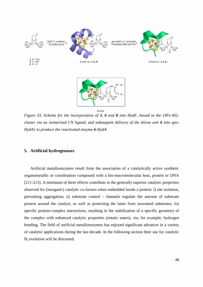
48
Figure 33. Scheme for the incorporation of 3, 6 and 8 into HydF, bound to the [4Fe-4S]-
cluster via an isomerised CN ligand, and subsequent delivery of the diiron unit 6 into apo-
HydA1 to produce the reactivated enzyme 6-HydA.
5. Artificial hydrogenases
Artificial metalloenzymes result from the association of a catalytically active synthetic
organometallic or coordination compound with a bio-macromolecular host, protein or DNA
[211-213]. A minimum of three effects contribute to the generally superior catalytic properties
observed for (inorganic) catalytic co-factors when embedded inside a protein: i) site isolation,
preventing aggregation; ii) substrate control - channels regulate the amount of substrate
present around the catalyst, as well as protecting the latter from unwanted substrates; iii)
specific protein-complex interactions, resulting in the stabilization of a specific geometry of
the complex with enhanced catalytic properties (entatic states), via, for example, hydrogen
bonding. The field of artificial metalloenzymes has enjoyed significant advances in a variety
of catalytic applications during the last decade. In the following section their use for catalytic
H2 evolution will be discussed.

49
5.1. Ni-based artificial hydrogenases
Mononuclear Ni complexes based on diphosphine PR
2NR’
2 ligands, developed by D.
Dubois [163], are among the most efficient bioinspired catalysts for H2 production and
uptake. The synthetic versatility of this class of compounds has allowed Shaw and coworkers
to prepare a series of complexes (77-79) using diphosphine ligands functionalised through
peptidic bonds with amino acids and amino acid esters in order to provide the Ni centre with a
protein-like outer-coordination sphere and evaluate the impact of that environment,
particularly with regards to the catalytic performances [214-216] (Figure 34). Interestingly
these new complexes display excellent catalytic activities for the reduction of protons to H2
with high catalytic rates and moderate overpotentials. Significant variations of the catalytic
activities as the result of varying the amino acid in the dipeptide, and thus the size and the
charge of the side chains, were observed. Bulky ligands increase the overpotential
requirement for H2 evolution. The catalytic performances were found superior when charged
(basic > acidic) amino-acids are introduced with regards to those with neutral side chains.
Catalysts bearing ester-protected ligands also proved less efficient. This is interpretated in
terms of the capability of the functional groups of the ligands to concentrate water and protons
near the active site and to facilitate proton transfer. This approach thus provides a unique
opportunity to understand the contribution that the outer-coordination sphere can have in
tuning the catalytic activity of this important class of hydrogenase mimics and opens the
possibility to develop closer structural enzyme mimics with larger peptides. Recently a new
amino-acid derivative (79) with glycine residues directly attached on the amine function of
the ligand was reported. Upon aging in water, auto-reduction of the Ni(II) into a Ni(0)
complex (80) occurs. A potential mechanism via decarboxylation of one glycine moiety is
proposed to account for this reaction. Importantly, these compounds proved as very efficient
catalysts for both hydrogen evolution in water over a wide range of pHs. A maximum
turnovers frequencies of 33 s–1
and minimum overpotential requirement of 40 mV is
observed for hydrogen oxidation at pH 0.5 [217].

50
Figure 34. Schematic of a selected of highly active mono-nuclear Ni complexes bearing
PR
2NR’
2 type ligands, in which R’ has been modified to feature amino acid or peptide
functionalities (77-80).
Jones showed that it was possible to synthesize hetero-bimetallic Ni-Mn (Mn = Ru, Fe2, Mo2
and W2) complexes starting from the peptide-Ni complex 81, made with the heptapeptide Ala-
Cys-Asp-Leu-Pro-Cys-Gly, a sequence present in nickel-superoxide dismutase containing the
two cysteines that chelate the Ni ion in the active site of the enzyme (Figure 35) [218]. It has
been demonstrated that the cysteinyl thiolates of that metallopeptide were indeed able to
exchange labile ligands of organometallic complexes. The reaction thus generates polynuclear
species with two cysteinyl thiolate bridges between Ni and either a single Ru (82), two Fe
(83), two Mo or two W (84) organometallic units. However, these new and rather interesting
hetero-bimetallic compounds have not been evaluated for their catalytic activity thus far.

51
Figure 35. The four [NiM] systems prepared by Jones and co-workers [170], from the Ni
complex in which the Ni ion is chelated by the two sulphurs of the heptapeptide Ala-Cys-Asp-
Leu-Pro-Cys-Gly (81).
5.2. Artificial hydrogenases based on synthetic diiron complexes
Synthetic di-iron complexes mimicking the active site of [FeFe]-hydrogenases have also been
incorporated into peptides and proteins. The general concept of this approach is to covalently
attach the complexes to a polypeptide backbone via the bridging dithiolate moiety. This can
be done by either taking advantage of two appropriately placed cysteine derived thiolate
ligands, or by introducing synthetic amino acids containing a propyl bridged dithiolate ligand
(Figure 36).

52
Figure 36. Representation of the different way used to covalently attach diiron cores to a
peptide backbone (wiggly lines indicate peptidic linkages).
Two representative examples have been reported by Jones and co-workers using de novo
designed oligo peptides. By reacting Fe3(CO)12 with an alanine rich, 36 amino-acids long,
peptide featuring a CXXC sequence, in which the two cysteine residues are correctly
positioned to establish a dithiolate bridge, a new species was obtained [219]. Based on a
combination of mass spectrometry, FTIR, circular dichroism (CD) and UV/Visible
spectroscopy measurements, the formation of a [(µ-Scys)2Fe2(CO)6] (85) complex, bound to
one face of the α-helical peptide and structurally analogous with the active site of [FeFe]-
hydrogenases was suggested (Figure 36). An alternative synthetic route employing an
artificial amino acid was reported in 2011. An oligopeptide incorporating a lysine (Trp-Ala-
Ser-Lys-Leu-Pro-Ser-Gly) was synthesized, to which a propane dithiolate ligand was linked
via the lysine moiety by means of amide coupling. The formation of the corresponding [(µ-
{SCH2}2CCONHR)Fe2(CO)6] complex (86, R = Trp-Ala-Ser-Lys-Leu-Pro-Ser-Gly) (Figure
36) and its subsequent modification to form the di-phosphine analogue, (µ-
{SCH2}2CHCONHR)Fe2(CO)4(PMe3)2], was confirmed by FTIR [220].
Such a synthetic artificial amino acid containing a 1,3-dithiol side chain, was incorporated
into an alanine-rich 19-mer peptide, known to provide a helical structure. The dithiolate
served as double bridge to anchor the di-iron unit of 87 in the helical peptide (Figure 36).
The resulting hybrid system features a pH dependent irreversible reduction process at −1.1 V
vs. RHE and was shown to catalyse light-driven H2 evolution at pH 4.5 in aqueous conditions
and in the presence of [Ru(bpy)3]2+
as the photosensitizer and ascorbate as the sacrificial
electron donor, resulting in a total TON of 84 (with regards to the catalyst) in 2.3 h [221].

53
Hayashi and coworkers demonstrated that hybrid systems could be prepared simply by
treating apo-cytochrome c (cytc) with Fe2(CO)9 [222]. Mass spectrometry and FTIR
spectroscopy indicated a [(µ-Scys)2Fe2(CO)6] structure (85) similar to the one obtained by A.
K. Jones [219, 220]_ENREF_36. This biohybrid displayed catalytic activity for hydrogen
production from water (82 turnovers in 2 h at pH 4.7) when irradiated in the presence of
[Ru(bipy)3]2+
and ascorbate. Much lower activity was obtained when cytochrome c was
replaced by a cytc-derived heptapeptide fragment containing the Cys-X-X-Cys sequence,
illustrating the importance of a complete protein environment.
In another study, an octadecapeptide fragment derived from cytochrome c556 and containing
a Cys-X-X-Cys-His sequence was used to attach both the di-iron carbonyl cluster to cysteines
and a Ru-based photosensitizer, [Ru(tpy)(bpy)(His)]2+
, to histidine [223]. In fact, direct
coordination of the Ru complex within the protein cavity was found to be crucial for
photocatalysis. Visible light irradiation of the [FeFe][Ru]-peptide in the presence of ascorbate
resulted in catalytic H2 evolution with a total TON of 8 in 2h, at which point H2 production
reached a plateau. However, when the reaction was performed under similar conditions using
the [FeFe]-peptide and free [Ru(tpy)(bpy)(Im)]2+
as the photosensitizer, no H2 production was
observed.
Conclusion
During the last decade, biomimetic chemistry has proven very effective for the development
of models of the structure of the active sites of hydrogenase enzymes, and the current
synthetic capabilities allow for the replication of almost all the key features of the natural
active subsites. Noticeable advances in the recent years include isolation of a terminal
hydrides by Rauchfuss and co-workers in 2012 [87, 88] and the preparation of the first [FeFe]
hydrogenase mimic capable of catalytic H2 oxidation. Remarkably this latter complex, [μ-
{(SCH2)2NBn}Fe2(CO)3(FcP*)(dppv)], is also the only system containing a functional
electron transfer relay [133]. Two Ni-Fe mimics capable of non-catalytic bidirectional H2
oxidation have also recently reported by Ogo [158] and Rauchfuss [166]. Unfortunately,
whilst these sophisticated models can provide valuable insights into the intimate mechanisms
of the natural active sites, there remains a notable absence of model systems that are capable

54
of achieving similar levels of catalytic activity as the hydrogenase enzymes, with most
operating at rates several orders of magnitude lower.
By contrast, controlling the outer sphere environment of these mimics, through incorporation
into micelles or dendrimers, has allowed for a significant improvement of the activity and
stability of model systems in more naturally relevant media. Indeed, such elegant guest-host
systems are capable of several thousand turn-overs by photolysis, conditions traditionally too
harsh for homogenous catalytic systems to withstand. It is important to note that direct
comparison of these catalytic systems is somewhat hampered by the wide and varied
photocatalytic conditions employed, nevertheless, these results clearly demonstrate the
relative strength of the guest-host approach [200, 202].
This strategy has been successfully extended to produce elegantly designed artificial
hydrogenases based on synthetic biomimics, incorporated into peptidic or proteic frameworks.
In general, these artificial hydrogenases are more stable and active for H2 evolution in water
than the parent biomimetic complexes.
Finally artificial activation of a hydrogenase enzyme has been achieved through the
introduction of a synthetic organometallic moiety into an apo-[FeFe]-hydrogenase [10], via
the chaperone protein HydF, which yields a fully active enzyme. This work has allowed for
final confirmation of the nature of the bridgehead atom of the bridging dithiolate ligand as
that of a nitrogen, but more importantly, it rather effortlessly highlights the essential role that
the protein cavity plays in transforming the typically fragile (in aqueous media) catalytically
inactive complex [(µ-adt)Fe2(CO)4(CN)2]2-
(6) into a robust and highly efficient catalytic
machine.
Acknowledgments
This work was supported by the French National Research Agency (ANR, NiFe–Cat ANR–
10–BLAN–711 and Labex program ARCANE, ANR-11-LABX-0003-01) and the FCH Joint
Undertaking (ArtipHyction project, grant n. 303435 and Nano-Cat project, grant n. 325239) .
GB gratefully acknowledges Bengt Lundqvist minnesfond, FORMAS (contract number 213-
2010-563) and the Swedish royal academy of sciences.
References and notes

55
[1] M. Frey, ChemBioChem, 3 (2002) 153. [2] N. Armaroli and V. Balzani, ChemSusChem, 4 (2011) 21. [3] A.K. Jones, E. Sillery, S.P.J. Albracht and F.A. Armstrong, Chem. Commun., (2002) 866. [4] J.A. Cracknell, K.A. Vincent and F.A. Armstrong, Chem. Rev., 108 (2008) 2439. [5] S.T. Stripp, G. Goldet, C. Brandmayr, O. Sanganas, K.A. Vincent, M. Haumann, F.A. Armstrong and T. Happe, Proc. Natl. Acad. Sci. USA, 106 (2009) 17331. [6] A. Abou Hamdan, B. Burlat, O. Gutierrez-Sanz, P.P. Liebgott, C. Baffert, A.L. De Lacey, M. Rousset, B. Guigliarelli, C. Leger and S. Dementin, Nature Chemical Biology, 9 (2013) 15. [7] J. Fritsch, P. Scheerer, S. Frielingsdorf, S. Kroschinsky, B. Friedrich, O. Lenz and C.M.T. Spahn, Nature, 479 (2011) 249. [8] Y. Shomura, K.S. Yoon, H. Nishihara and Y. Higuchi, Nature, 479 (2011) 253. [9] K. Grubel and P.L. Holland, Angew. Chem. Int. Ed., 51 (2012) 3308. [10] G. Berggren, A. Adamska, C. Lambertz, T.R. Simmons, J. Esselborn, M. Atta, S. Gambarelli, J.M. Mouesca, E. Reijerse, W. Lubitz, T. Happe, V. Artero and M. Fontecave, Nature, 499 (2013) 66. [11] J. Esselborn, C. Lambertz, A. Adamska-Venkatesh, T. Simmons, G. Berggren, J. Noth, J. Siebel, A. Hemschemeier, V. Artero, E. Reijerse, M. Fontecave, W. Lubitz and T. Happe, Nat Chem Biol, advance online publication (2013). [12] R.K. Thauer, Eur. J. Inorg. Chem., 2011 919. [13] K.A. Vincent, A. Parkin and F.A. Armstrong, Chem. Rev., 107 (2007) 4366. [14] A. Magnuson, M. Anderlund, O. Johansson, P. Lindblad, R. Lomoth, T. Polivka, S. Ott, K. Stensjö, S. Styring, V. Sundström and L. Hammarström, Acc. Chem. Res., 42 (2009) 1899. [15] R.K. Thauer, A.R. Klein and G.C. Hartmann, Chem. Rev., 96 (1996) 3031. [16] J.C. Fontecilla-Camps, A. Volbeda, C. Cavazza and Y. Nicolet, Chem. Rev., 107 (2007) 4273. [17] R.K. Thauer, A.K. Kaster, M. Goenrich, M. Schick, T. Hiromoto and S. Shima, Annual Review of Biochemistry, Vol 79, 79 (2010) 507. [18] the extra energy required for the reaction over that defined by the standard potential of the H+/H2 redox couple; in other words, the difference between the potential that needs to be applied to the system to allow it to function and the standard potential of the H+/H2 redox couple [19] H.R. Pershad, J.L.C. Duff, H.A. Heering, E.C. Duin, S.P.J. Albracht and F.A. Armstrong, Biochemistry, 38 (1999) 8992. [20] C. Madden, M.D. Vaughn, I. Diez-Perez, K.A. Brown, P.W. King, D. Gust, A.L. Moore and T.A. Moore, J. Am. Chem. Soc., 134 (2012) 1577. [21] J.W. Peters, W.N. Lanzilotta, B.J. Lemon and L.C. Seefeldt, Science, 282 (1998) 1853. [22] Y. Nicolet, C. Piras, P. Legrand, C.E. Hatchikian and J.C. Fontecilla-Camps, Structure, 7 (1999) 13. [23] Z.J. Chen, B.J. Lemon, S. Huang, D.J. Swartz, J.W. Peters and K.A. Bagley, Biochemistry, 41 (2002) 2036. [24] W. Roseboom, A.L. De Lacey, V.M. Fernandez, E.C. Hatchikian and S.P.J. Albracht, J. Biol. Inorg. Chem., 11 (2006) 102. [25] A.L. De Lacey, C. Stadler, C. Cavazza, E.C. Hatchikian and V.M. Fernandez, J. Am. Chem. Soc., 122 (2000) 11232. [26] P.E.M. Siegbahn, J.W. Tye and M.B. Hall, Chem. Rev., 107 (2007) 4414. [27] C.V. Popescu and E. Munck, J. Am. Chem. Soc., 121 (1999) 7877. [28] Y. Nicolet, A.L. de Lacey, X. Vernede, V.M. Fernandez, E.C. Hatchikian and J.C. Fontecilla-Camps, J. Am. Chem. Soc., 123 (2001) 1596. [29] A. Silakov, B. Wenk, E. Reijerse and W. Lubitz, Physical Chemistry Chemical Physics, 11 (2009) 6592. [30] W. Lubitz, E. Reijerse and M. van Gastel, Chem. Rev., 107 (2007) 4331.

56
[31] Ö.F. Erdem, L. Schwartz, M. Stein, A. Silakov, S. Kaur-Ghumaan, P. Huang, S. Ott, E.J. Reijerse and W. Lubitz, Angewandte Chemie International Edition, 50 (2011) 1439. [32] K.A. Vincent, A. Parkin, O. Lenz, S.P.J. Albracht, J.C. Fontecilla-Camps, R. Cammack, B. Friedrich and F.A. Armstrong, J. Am. Chem. Soc., 127 (2005) 18179. [33] C. Tard and C.J. Pickett, Chem. Rev., 109 (2009) 2245. [34] A. Adamska, A. Silakov, C. Lambertz, O. Rüdiger, T. Happe, E. Reijerse and W. Lubitz, Angew. Chem. Int. Ed., 51 (2012) 11458. [35] D.W. Mulder, M.W. Ratzloff, E.M. Shepard, A.S. Byer, S.M. Noone, J.W. Peters, J.B. Broderick and P.W. King, J. Am. Chem. Soc., 135 (2013) 6921. [36] A. Volbeda, M.-H. Charon, C. Piras, E.C. Hatchikian, M. Frey and J.C. Fontecilla-Camps, Nature, 373 (1995) 580. [37] A. Volbeda, E. Garcin, C. Piras, A.L. de Lacey, V.M. Fernandez, E.C. Hatchikian, M. Frey and J.C. Fontecilla-Camps, Journal of the American Chemical Society, 118 (1996) 12989. [38] A. Volbeda, L. Martin, C. Cavazza, M. Matho, B.W. Faber, W. Roseboom, S.P.J. Albracht, E. Garcin, M. Rousset and J.C. Fontecilla-Camps, J. Biol. Inorg. Chem., 10 (2005) 239. [39] S. Canaguier, V. Artero and M. Fontecave, Dalton Trans., (2008) 315. [40] Y. Montet, P. Amara, A. Volbeda, X. Vernede, E.C. Hatchikian, M.J. Field, M. Frey and J.C. FontecillaCamps, Nature Structural Biology, 4 (1997) 523. [41] C. Leger, S. Dementin, P. Bertrand, M. Rousset and B. Guigliarelli, J. Am. Chem. Soc., 126 (2004) 12162. [42] F. Leroux, S. Dementin, B. Burlatt, L. Cournac, A. Volbeda, S. Champ, L. Martin, B. Guigliarelli, P. Bertrand, J. Fontecilla-Camps, M. Rousset and C. Leger, Proc. Natl. Acad. Sci. USA, 105 (2008) 11188. [43] H. Ogata, Y. Mizoguchi, N. Mizuno, K. Miki, S. Adachi, N. Yasuoka, T. Yagi, O. Yamauchi, S. Hirota and Y. Higuchi, J. Am. Chem. Soc., 124 (2002) 11628. [44] C. Tsay and J.C. Peters, Chem. Sci., 3 (2012) 1313. [45] S. Niu and N.B. Hall, Inorg. Chem., 40 (2001) 6201. [46] T. Krämer, M. Kampa, W. Lubitz, M. van Gastel and F. Neese, ChemBioChem, 14 (2013) 1898. [47] J.P. Whitehead, R.J. Gurbiel, C. Bagyinka, B.M. Hoffman and M.J. Maroney, J. Am. Chem. Soc., 115 (1993) 5629. [48] M. Brecht, M. van Gastel, T. Buhrke, B. Friedrich and W. Lubitz, J. Am. Chem. Soc., 125 (2003) 13075. [49] S. Foerster, M. van Gastel, M. Brecht and W. Lubitz, J. Biol. Inorg. Chem., 10 (2005) 51. [50] W. Lubitz, M. Brecht, S. Foerster, M. Stein, Y. Higuchi, T. Buhrke and B. Friedrich, Epr in the 21st Century: Basics and Applications to Material, Life and Earth Sciences, (2002) 437. [51] M.E. Pandelia, P. Infossi, M. Stein, M.T. Giudici-Orticoni and W. Lubitz, Chem. Commun., 48 (2012) 823. [52] T.R. Simmons and V. Artero, Angew. Chem. Int. Ed., 52 (2013) 6143. [53] S. Vogt, E.J. Lyon, S. Shima and R.K. Thauer, J. Biol. Inorg. Chem., 13 (2008) 97. [54] G. Buurman, S. Shima and R.K. Thauer, FEBS Lett., 485 (2000) 200. [55] S. Shima, E.J. Lyon, M.S. Sordel-Klippert, M. Kauss, J. Kahnt, R.K. Thauer, K. Steinbach, X.L. Xie, L. Verdier and C. Griesinger, Angew. Chem. Int. Ed., 43 (2004) 2547. [56] E.J. Lyon, S. Shima, R. Boecher, R.K. Thauer, F.W. Grevels, E. Bill, W. Roseboom and S.P. Albracht, J. Am. Chem. Soc., 126 (2004) 14239. [57] S. Shima, O. Pilak, S. Vogt, M. Schick, M.S. Stagni, W. Meyer-Klaucke, E. Warkentin, R.K. Thauer and U. Ermler, Science, 321 (2008) 572. [58] M. Korbas, S. Vogt, W. Meyer-Klaucke, E. Bill, E.J. Lyon, R.K. Thauer and S. Shima, J. Biol. Chem., 281 (2006) 30804. [59] T. Hiromoto, K. Ataka, O. Pilak, S. Vogt, M.S. Stagni, W. Meyer-Klaucke, E. Warkentin, R.K. Thauer, S. Shima and U. Ermler, FEBS Lett., 583 (2009) 585. [60] S. Shima and K. Ataka, FEBS Lett., 585 (2011) 353.

57
[61] H. Tamura, M. Salomone-Stagni, T. Fujishiro, E. Warkentin, W. Meyer-Klaucke, U. Ermler and S. Shima, Angew. Chem. Int. Ed., 52 (2013) 9656. [62] T. Hiromoto, E. Warkentin, J. Moll, U. Ermler and S. Shima, Angew. Chem. Int. Ed., 48 (2009) 6457. [63] X.Z. Yang and M.B. Hall, J. Am. Chem. Soc., 131 (2009) 10901. [64] H. Reihlen, A. Gruhl and G. Hessling, Liebigs Ann. Chem., 472 (1929) 268. [65] D. Seyferth, G.B. Womack, M.K. Gallagher, M. Cowie, B.W. Hames, J.P. Fackler and A.M. Mazany, Organometallics, 6 (1987) 283. [66] A. Le Cloirec, S.P. Best, S. Borg, S.C. Davies, D.J. Evans, D.L. Hughes and C.J. Pickett, Chem. Commun., (1999) 2285. [67] M. Schmidt, S.M. Contakes and T.B. Rauchfuss, J. Am. Chem. Soc., 121 (1999) 9736. [68] E.J. Lyon, I.P. Georgakaki, J.H. Reibenspies and M.Y. Darensbourg, Angew. Chem. Int. Ed., 38 (1999) 3178. [69] J.D. Lawrence, H.X. Li, T.B. Rauchfuss, M. Benard and M.M. Rohmer, Angew. Chem. Int. Ed., 40 (2001) 1768. [70] L.C. Song, Z.Y. Yang, H.Z. Bian and Q.M. Hu, Organometallics, 23 (2004) 3082. [71] L.C. Song, Z.Y. Yang, H.Z. Bian, Y. Liu, H.T. Wang, X.F. Liu and Q.M. Hu, Organometallics, 24 (2005) 6126. [72] J.F. Capon, F. Gloaguen, F.Y. Petillon, P. Schollhammer and J. Talarmin, Coord. Chem. Rev., 253 (2009) 1476. [73] M. Razavet, S.C. Davies, D.L. Hughes and C.J. Pickett, Chem. Commun., (2001) 847. [74] J.I. van der Vlugt, T.B. Rauchfuss and S.R. Wilson, Chem. Eur. J., 12 (2006) 90. [75] C.A. Boyke, T.B. Rauchfuss, S.R. Wilson, M.M. Rohmer and M. Benard, J. Am. Chem. Soc., 126 (2004) 15151. [76] C.A. Boyke, J.I. van der Vlugt, T.B. Rauchfuss, S.R. Wilson, G. Zampella and L. De Gioia, J. Am. Chem. Soc., 127 (2005) 11010. [77] D. Chouffai, G. Zampella, J.F. Capon, L. De Gioia, A. Le Goff, F.Y. Petillon, P. Schollhammer and J. Talarmin, Organometallics, 31 (2012) 1082. [78] A.K. Justice, M.J. Nilges, T.B. Rauchfuss, S.R. Wilson, L. De Gioia and G. Zampella, J. Am. Chem. Soc., 130 (2008) 5293. [79] A.K. Justice, L. De Gioia, M.J. Nilges, T.B. Rauchfuss, S.R. Wilson and G. Zampella, Inorg. Chem., 47 (2008) 7405. [80] A.K. Justice, T.B. Rauchfuss and S.R. Wilson, Angew. Chem. Int. Ed., 46 (2007) 6152. [81] T.B. Liu and M.Y. Darensbourg, J. Am. Chem. Soc., 129 (2007) 7008. [82] C.M. Thomas, T.B. Liu, M.B. Hall and M.Y. Darensbourg, Inorg. Chem., 47 (2008) 7009. [83] M.L. Singleton, N. Bhuvanesh, J.H. Reibenspies and M.Y. Darensbourg, Angew. Chem. Int. Ed., 47 (2008) 9492. [84] W. Wang, T.B. Rauchfuss, C.E. Moore, A.L. Rheingold, L. De Gioia and G. Zampella, Chemistry – A European Journal, 19 (2013) 15476. [85] S. Munery, J.-F. Capon, L. De Gioia, C. Elleouet, C. Greco, F.Y. Pétillon, P. Schollhammer, J. Talarmin and G. Zampella, Chemistry – A European Journal, 19 (2013) 15458. [86] J.I. van der Vlugt, T.B. Rauchfuss, C.M. Whaley and S.R. Wilson, J. Am. Chem. Soc., 127 (2005) 16012. [87] M.E. Carroll, B.E. Barton, T.B. Rauchfuss and P.J. Carroll, J. Am. Chem. Soc., 134 (2012) 18843. [88] R. Zaffaroni, T.B. Rauchfuss, D.L. Gray, L. De Gioia and G. Zampella, J. Am. Chem. Soc., 134 (2012) 19260. [89] C. Tard, X.M. Liu, S.K. Ibrahim, M. Bruschi, L. De Gioia, S.C. Davies, X. Yang, L.S. Wang, G. Sawers and C.J. Pickett, Nature, 433 (2005) 610. [90] M.K. Harb, T. Niksch, J. Windhager, H. Gorls, R. Holze, L.T. Lockett, N. Okumura, D.H. Evans, R.S. Glass, D.L. Lichtenberger, M. El-Khateeb and W. Weigand, Organometallics, 28 (2009) 1039.

58
[91] M.K. Harb, J. Windhager, A. Daraosheh, H. Gorls, L.T. Lockett, N. Okumura, D.H. Evans, R.S. Glass, D.L. Lichtenberger, M. El-Khateeb and W. Weigand, Eur. J. Inorg. Chem., (2009) 3414. [92] T. Kuckmann, F. Schodel, I. Sanger, M. Bolte, M. Wagner and H.W. Lerner, Eur. J. Inorg. Chem., (2010) 468. [93] U.P. Apfel, D. Troegel, Y. Halpin, S. Tschierlei, U. Uhlemann, H. Gorls, M. Schmitt, J. Popp, P. Dunne, M. Venkatesan, M. Coey, M. Rudolph, J.G. Vos, R. Tacke and W. Weigand, Inorg. Chem., 49 (2010) 10117. [94] U.P. Apfel, Y. Halpin, H. Gorls, J.G. Vos and W. Weigand, Eur. J. Inorg. Chem., (2011) 581. [95] M.K. Harb, U.P. Apfel, J. Kubel, H. Gorls, G.A.N. Felton, T. Sakamoto, D.H. Evans, R.S. Glass, D.L. Lichtenberger, M. El-Khateeb and W. Weigand, Organometallics, 28 (2009) 6666. [96] U.P. Apfel, H. Gorls, G.A.N. Felton, D.H. Evans, R.S. Glass, D.L. Lichtenberger and W. Weigand, Helv. Chim. Acta, 95 (2012) 2168. [97] L.C. Song, Q.L. Li, Z.H. Feng, X.J. Sun, Z.J. Xie and H.B. Song, Dalton Trans., 42 (2013) 1612. [98] M.K. Harb, H. Gorls, T. Sakamoto, G.A.N. Felton, D.H. Evans, R.S. Glass, D.L. Lichtenberger, M. El-khateeb and W. Weigand, Eur. J. Inorg. Chem., (2010) 3976. [99] S. Gao, J. Fan, S. Sun, X. Peng, X. Zhao and J. Hou, Dalton Trans., 0 (2008) 2128. [100] E.S. Donovan, J.J. McCormick, G.S. Nichol and G.A.N. Felton, Organometallics, 31 (2012) 8067. [101] L. Schwartz, P.S. Singh, L. Eriksson, R. Lomoth and S. Ott, C. R. Chim., 11 (2008) 875. [102] F. Gloaguen, J.D. Lawrence and T.B. Rauchfuss, J. Am. Chem. Soc., 123 (2001) 9476. [103] X. Zhao, I.P. Georgakaki, M.L. Miller, J.C. Yarbrough and M.Y. Darensbourg, J. Am. Chem. Soc., 123 (2001) 9710. [104] W. Zhong, Y. Tang, G. Zampella, X.F. Wang, X.L. Yang, B. Hu, J.A. Wang, Z.Y. Xiao, Z.H. Wei, H.W. Chen, L. De Gioia and X.M. Liu, Inorg. Chem. Commun., 13 (2010) 1089. [105] Y. Tang, Z.H. Wei, W. Zhong and X.M. Liu, Eur. J. Inorg. Chem., (2011) 1112. [106] M. Karnahl, S. Tschierlei, O.F. Erdem, S. Pullen, M.P. Santoni, E.J. Reijerse, W. Lubitz and S. Ott, Dalton Trans., 41 (2012) 12468. [107] S. Ezzaher, A. Gogoll, C. Bruhn and S. Ott, Chem. Commun., 46 (2010) 5775. [108] W.B. Dong, M. Wang, X.Y. Liu, K. Jin, G.H. Li, F.J. Wang and L.C. Sun, Chem. Commun., (2006) 305. [109] C.P. Liu, J.N.T. Peck, J.A. Wright, C.J. Pickett and M.B. Hall, Eur. J. Inorg. Chem., (2011) 1080. [110] S. Tschierlei, S. Ott and R. Lomoth, Energy Environ. Sci., 4 (2011) 2340. [111] Y.C. Liu, K.T. Chu, R.L. Jhang, G.H. Lee and M.H. Chiang, Chem. Commun., 49 (2013) 4743. [112] W.M. Gao, J.L. Sun, T. Akermark, M.R. Li, L. Eriksson, L.C. Sun and B. Akermark, Chem. Eur. J., 16 (2010) 2537. [113] S. Ezzaher, J.F. Capon, F. Gloaguen, F.Y. Petillon, P. Schollhammer, J. Talarmin and N. Kervarec, Inorg. Chem., 48 (2009) 2. [114] Y.W. Wang, Z.M. Li, X.H. Zeng, X.F. Wang, C.X. Zhan, Y.Q. Liu, X.R. Zeng, Q.Y. Luo and X.M. Liu, New J. Chem., 33 (2009) 1780. [115] P. Li, M. Wang, L. Chen, J.H. Liu, Z.B. Zhaoa and L.C. Sun, Dalton Trans., (2009) 1919. [116] X. Zhao, I.P. Georgakaki, M.L. Miller, R. Mejia-Rodriguez, C.Y. Chiang and M.Y. Darensbourg, Inorg. Chem., 41 (2002) 3917. [117] I.P. Georgakaki, M.L. Miller and M.Y. Darensbourg, Inorg. Chem., 42 (2003) 2489. [118] N. Wang, M. Wang, J.H. Liu, K. Jin, L. Chen and L.C. Sun, Inorg. Chem., 48 (2009) 11551. [119] M.T. Olsen, T.B. Rauchfuss and S.R. Wilson, J. Am. Chem. Soc., 132 (2010) 17733. [120] K. Fauvel, R. Mathieu and R. Poilblanc, Inorg. Chem., 15 (1976) 976. [121] S. Ezzaher, J.F. Capon, F. Gloaguen, F.Y. Petillon, P. Schollhammer and J. Talarmin, Inorg. Chem., 46 (2007) 3426. [122] B.E. Barton and T.B. Rauchfuss, Inorg. Chem., 47 (2008) 2261. [123] F.I. Adam, G. Hogarth, S.E. Kabir and D. Richards, C. R. Chim., 11 (2008) 890. [124] B.E. Barton, M.T. Olsen and T.B. Rauchfuss, J. Am. Chem. Soc., 130 (2008) 16834. [125] B.E. Barton, M.T. Olsen and T.B. Rauchfuss, Curr. Opin. Biotechnol., 21 (2010) 292.
59
[126] X.H. Zeng, Z.M. Li, Z.Y. Xiao, Y.W. Wang and X.M. Liu, Electrochem. Commun., 12 (2010) 342. [127] J. Zhao, Z.H. Wei, X.H. Zeng and X.M. Liu, Dalton Trans., 41 (2012) 11125. [128] C. Gimbert-Surinach, M. Bhadbhade and S.B. Colbran, Organometallics, 31 (2012) 3480. [129] Y.-C. Liu, C.-H. Lee, G.-H. Lee and M.-H. Chiang, Eur. J. Inorg. Chem., 2011 (2011) 1155. [130] Y.-C. Liu, T.-H. Yen, Y.-J. Tseng, C.-H. Hu, G.-H. Lee and M.-H. Chiang, Inorg. Chem., 51 (2012) 5997. [131] Y.T. Si, K. Charreteur, J.F. Capon, F. Gloaguen, F.Y. Petillon, P. Schollhammer and J. Talarmin, J. Inorg. Biochem., 104 (2010) 1038. [132] S. Roy, T.L. Groy and A.K. Jones, Dalton Trans., 42 (2013) 3843. [133] J.M. Camara and T.B. Rauchfuss, Nature Chemistry, 4 (2012) 26. [134] C. Greco, Inorg. Chem., 52 (2013) 1901. [135] N. Wang, M. Wang, Y. Wang, D. Zheng, H. Han, M.S.G. Ahlquist and L. Sun, J. Am. Chem. Soc., 135 (2013) 13688. [136] J.A. Wright and C.J. Pickett, Chemcatchem, 4 (2012) 1723. [137] C.H. Lai, J.H. Reibenspies and M.Y. Darensbourg, Angew. Chem. Int. Ed., 35 (1996) 2390. [138] Z.L. Li, Y. Ohki and K. Tatsumi, J. Am. Chem. Soc., 127 (2005) 8950. [139] T. Naota, H. Takaya and S.I. Murahashi, Chem. Rev., 98 (1998) 2599. [140] J.A.W. Verhagen, D.D. Ellis, M. Lutz, A.L. Spek and E. Bouwman, Dalton Trans., (2002) 1275. [141] S. Canaguier, L. Vaccaro, V. Artero, R. Ostermann, J. Pécaut, M.J. Field and M. Fontecave, Chem. Eur. J., 15 (2009) 9350. [142] Y. Oudart, V. Artero, J. Pécaut and M. Fontecave, Inorg. Chem., 45 (2006) 4334. [143] Y. Oudart, V. Artero, J. Pécaut, C. Lebrun and M. Fontecave, Eur. J. Inorg. Chem., (2007) 2613. [144] Y. Oudart, V. Artero, L. Norel, C. Train, J. Pécaut and M. Fontecave, J. Organomet. Chem., 694 (2009) 2866. [145] S. Canaguier, M. Fontecave and V. Artero, Eur. J. Inorg. Chem., (2011) 1094. [146] T. Matsumoto, B. Kure and S. Ogo, Chem. Lett., 37 (2008) 970. [147] S. Ogo, R. Kabe, K. Uehara, B. Kure, T. Nishimura, S.C. Menon, R. Harada, S. Fukuzumi, Y. Higuchi, T. Ohhara, T. Tamada and R. Kuroki, Science, 316 (2007) 585. [148] L. Vaccaro, V. Artero, S. Canaguier, M. Fontecave and M.J. Field, Dalton Trans., 39 (2010) 3043. [149] S. Canaguier, V. Fourmond, C.U. Perotto, J. Fize, J. Pecaut, M. Fontecave, M.J. Field and V. Artero, Chem. Commun., 49 (2013) 5004. [150] B.E. Barton, C.M. Whaley, T.B. Rauchfuss and D.L. Gray, J. Am. Chem. Soc., 131 (2009) 6942. [151] B.E. Barton and T.B. Rauchfuss, J. Am. Chem. Soc., 132 (2010) 14877. [152] S. Canaguier, M. Field, Y. Oudart, J. Pecaut, M. Fontecave and V. Artero, Chem. Commun., 46 (2010) 5876. [153] K. Weber, T. Krämer, H.S. Shafaat, T. Weyhermüller, E. Bill, M. van Gastel, F. Neese and W. Lubitz, J. Am. Chem. Soc., 134 (2012) 20745. [154] W.F. Zhu, A.C. Marr, Q. Wang, F. Neese, D.J.E. Spencer, A.J. Blake, P.A. Cooke, C. Wilson and M. Schroder, Proc. Natl. Acad. Sci. USA, 102 (2005) 18280. [155] V. Fourmond, S. Canaguier, B. Golly, M.J. Field, M. Fontecave and V. Artero, Energy Environ. Sci., 4 (2011) 2417. [156] V. Fourmond, P.A. Jacques, M. Fontecave and V. Artero, Inorg. Chem., 49 (2010) 10338. [157] L.-C. Song, J.-P. Li, Z.-J. Xie and H.-B. Song, Inorg. Chem., 52 (2013) 11618. [158] S. Ogo, K. Ichikawa, T. Kishima, T. Matsumoto, H. Nakai, K. Kusaka and T. Ohhara, Science, 339 (2013) 682. [159] F.A. Armstrong and J. Hirst, Proc. Natl. Acad. Sci. USA, 108 (2011) 14049. [160] A.D. Wilson, R.H. Newell, M.J. McNevin, J.T. Muckerman, M.R. DuBois and D.L. DuBois, J. Am. Chem. Soc., 128 (2006) 358.

60
[161] S.E. Smith, J.Y. Yang, D.L. DuBois and R.M. Bullock, Angew. Chem. Int. Ed., 51 (2012) 3152. [162] A. Le Goff, V. Artero, B. Jousselme, P.D. Tran, N. Guillet, R. Metaye, A. Fihri, S. Palacin and M. Fontecave, Science, 326 (2009) 1384. [163] M.R. DuBois and D.L. DuBois, Chem. Soc. Rev., 38 (2009) 62. [164] T. Liu, D.L. DuBois and R.M. Bullock, Nat Chem, 5 (2013) 228. [165] V. Fourmond, S. Canaguier, B. Golly, M.J. Field, M. Fontecave and V. Artero, Energy Environ. Sci., 4 (2011) 2417. [166] B.C. Manor and T.B. Rauchfuss, J. Am. Chem. Soc., 135, (2013) 11895. [167] X.F. Wang, Z.M. Li, X.R. Zeng, Q.Y. Luo, D.J. Evans, C.J. Pickett and X.M. Liu, Chem. Commun., (2008) 3555. [168] B.V. Obrist, D.F. Chen, A. Ahrens, V. Schunemann, R. Scopelliti and X.L. Hu, Inorg. Chem., 48 (2009) 3514. [169] D.F. Chen, A. Ahrens-Botzong, V. Schuemann, R. Scopelliti and X.L. Hu, Inorg. Chem., 50 (2011) 5249. [170] M. Salomone-Stagni, F. Stellato, C.M. Whaley, S. Vogt, S. Morante, S. Shima, T.B. Rauchfuss and W. Meyer-Klaucke, Dalton Trans., 39 (2010) 3057. [171] W.F. Liaw, N.H. Lee, C.H. Chen, C.M. Lee, G.H. Lee and S.M. Peng, J. Am. Chem. Soc., 122 (2000) 488. [172] T.B. Liu, B. Li, C.V. Popescu, A. Bilko, L.M. Perez, M.B. Hall and M.Y. Darensbourg, Chem. Eur. J., 16 (2010) 3083. [173] J.M. Smith, R.J. Lachicotte and P.L. Holland, Organometallics, 21 (2002) 4808. [174] D.F. Chen, R. Scopelliti and X.L. Hu, J. Am. Chem. Soc., 132 (2010) 928. [175] A.M. Royer, T.B. Rauchfuss and D.L. Gray, Organometallics, 28 (2009) 3618. [176] A.M. Royer, M. Salomone-Stagni, T.B. Rauchfuss and W. Meyer-Klaucke, J. Am. Chem. Soc., 132 (2010) 16997. [177] D.F. Chen, R. Scopelliti and X.L. Hu, Angew. Chem. Int. Ed., 49 (2010) 7512. [178] B.W. Hu, D.F. Chen and X.L. Hu, Chem. Eur. J., 18 (2012) 11528. [179] B.W. Hu, D.F. Chen and X.L. Hu, Chem. Eur. J., 19 (2013) 6221. [180] D.F. Chen, R. Scopelliti and X.L. Hu, Angew. Chem. Int. Ed., 50 (2011) 5670. [181] L.C. Song, Z.J. Xie, M.M. Wang, G.Y. Zhao and H.B. Song, Inorg. Chem., 51 (2012) 7466. [182] P.J. Turrell, A.D. Hill, S.K. Ibrahim, J.A. Wright and C.J. Pickett, Dalton Trans., 42 (2013) 8140. [183] P.J. Turrell, J.A. Wright, J.N.T. Peck, V.S. Oganesyan and C.J. Pickett, Angew. Chem. Int. Ed., 49 (2010) 7508. [184] D.F. Chen, R. Scopelliti and X.L. Hu, Angew. Chem. Int. Ed., 51 (2012) 1919. [185] L.C. Song, G.Y. Zhao, Z.J. Xie and J.W. Zhang, Organometallics, 32 (2013) 2509. [186] P. Knorzer, A. Silakov, C.E. Foster, F.A. Armstrong, W. Lubitz and T. Happe, J. Biol. Chem., 287 (2012) 1489. [187] P.W.J.M. Frederix, R. Kania, J.A. Wright, D.A. Lamprou, R.V. Ulijn, C.J. Pickett and N.T. Hunt, Dalton Transactions, 41 (2012) 13112. [188] K.N. Green, S.P. Jeffery, J.H. Reibenspies and M.Y. Darensbourg, Journal of the American Chemical Society, 128 (2006) 6493. [189] K.N. Green, J.L. Hess, C.M. Thomas and M.Y. Darensbourg, Dalton Transactions, 0 (2009) 4344. [190] M.L. Singleton, D.J. Crouthers, R.P. Duttweiler, J.H. Reibenspies and M.Y. Darensbourg, Inorganic Chemistry, 50 (2011) 5015. [191] M.L. Singleton, J.H. Reibenspies and M.Y. Darensbourg, Journal of the American Chemical Society, 132 (2010) 8870. [192] X. Li, M. Wang, D. Zheng, K. Han, J. Dong and L. Sun, Energy Environ. Sci., 5 (2012) 8220. [193] H.-Y. Wang, W.-G. Wang, G. Si, F. Wang, C.-H. Tung and L.-Z. Wu, Langmuir, 26 (2010) 9766. [194] F. Wang, W.-G. Wang, H.-Y. Wang, G. Si, C.-H. Tung and L.-Z. Wu, ACS Catalysis, 2 (2012) 407.

61
[195] F. Quentel, G. Passard and F. Gloaguen, Chemistry – A European Journal, 18 (2012) 13473. [196] F. Quentel, G. Passard and F. Gloaguen, Energy & Environmental Science, 5 (2012) 7757. [197] J.-F. Capon, F. Gloaguen, P. Schollhammer and J. Talarmin, Journal of Electroanalytical Chemistry, 595 (2006) 47. [198] J.-F. Capon, F. Gloaguen, P. Schollhammer and J. Talarmin, Journal of Electroanalytical Chemistry, 566 (2004) 241. [199] C. Orain, F. Quentel and F. Gloaguen, ChemSusChem, (2013) n/a. [200] T. Yu, Y. Zeng, J. Chen, Y.-Y. Li, G. Yang and Y. Li, Angewandte Chemie International Edition, 52 (2013) 5631. [201] F. Wang, W.-G. Wang, X.-J. Wang, H.-Y. Wang, C.-H. Tung and L.-Z. Wu, Angew. Chem. Int. Ed., 50 (2011) 3193. [202] F. Wang, W.-J. Liang, J.-X. Jian, C.-B. Li, B. Chen, C.-H. Tung and L.-Z. Wu, Angew. Chem. Int. Ed., (2013) n/a. [203] apo-HydA refers to a form the [FeFe] hydrogenase enzyme incorporating its [4Fe4S] cluster but lacking its dinuclear catalytic subunit [204] David W. Mulder, Eric M. Shepard, Jonathan E. Meuser, N. Joshi, Paul W. King, Matthew C. Posewitz, Joan B. Broderick and John W. Peters, Structure (London, England : 1993), 19 (2011) 1038. [205] E.M. Shepard, S.E. McGlynn, A.L. Bueling, C.S. Grady-Smith, S.J. George, M.A. Winslow, S.P. Cramer, J.W. Peters and J.B. Broderick, Proceedings of the National Academy of Sciences, 107 (2010) 10448. [206] D.W. Mulder, E.S. Boyd, R. Sarma, R.K. Lange, J.A. Endrizzi, J.B. Broderick and J.W. Peters, Nature, 465 (2010) 248. [207] S.E. McGlynn, E.M. Shepard, M.A. Winslow, A.V. Naumov, K.S. Duschene, M.C. Posewitz, W.E. Broderick, J.B. Broderick and J.W. Peters, FEBS Letters, 582 (2008) 2183. [208] X. Brazzolotto, J.K. Rubach, J. Gaillard, S. Gambarelli, M. Atta and M. Fontecave, Journal of Biological Chemistry, 281 (2006) 769. [209] D. Schilter and T.B. Rauchfuss, Angew. Chem. Int. Ed., (2013) n/a. [210] U.-P. Apfel and W. Weigand, ChemBioChem, (2013) n/a. [211] J. Steinreiber and T.R. Ward, Coordination Chemistry Reviews, 252 (2008) 751. [212] Y. Lu, N. Yeung, N. Sieracki and N.M. Marshall, Nature, 460 (2009) 855. [213] F. Rosati and G. Roelfes, ChemCatChem, 2 (2010) 916. [214] A. Jain, S. Lense, J.C. Linehan, S. Raugei, H. Cho, D.L. DuBois and W.J. Shaw, Inorg. Chem., 50 (2011) 4073. [215] A. Jain, M.L. Reback, M.L. Lindstrom, C.E. Thogerson, M.L. Helm, A.M. Appel and W.J. Shaw, Inorg. Chem., 51 (2012) 6592. [216] M.L. Reback, B. Ginovska-Pangovska, M.H. Ho, A. Jain, T.C. Squier, S. Raugei, J.A.S. Roberts and W.J. Shaw, Chem. Eur. J., 19 (2013) 1928. [217] A. Dutta, S. Lense, J. Hou, M.H. Engelhard, J.A.S. Roberts and W.J. Shaw, J. Am. Chem. Soc., 135 (2013) 18490. [218] A. Dutta, G.A. Hamilton, H.E. Hartnett and A.K. Jones, Inorganic Chemistry, 51 (2012) 9580. [219] A.K. Jones, B.R. Lichtenstein, A. Dutta, G. Gordon and P.L. Dutton, Journal of the American Chemical Society, 129 (2007) 14844. [220] S. Roy, S. Shinde, G.A. Hamilton, H.E. Hartnett and A.K. Jones, European Journal of Inorganic Chemistry, 2011 (2011) 1050. [221] A. Roy, C. Madden and G. Ghirlanda, Chemical Communications, 48 (2012) 9816. [222] Y. Sano, A. Onoda and T. Hayashi, Chemical Communications, 47 (2011) 8229. [223] Y. Sano, A. Onoda and T. Hayashi, Journal of Inorganic Biochemistry, 108 (2012) 159.

62