Mimicking Interacting Relativistic Theories with Stationary Pulses of Light
Upload
independentCategory
view
1download
0
Rational Design, Structure, and Biological Evaluation of Cyclic Peptides Mimicking theVascular Endothelial Growth Factor
Victor Goncalves,†,‡,§ Benoit Gautier,†,‡,§ Pascale Coric,|,⊥ Serge Bouaziz,|,⊥ Christine Lenoir,‡,§ Christiane Garbay,‡,§
Michel Vidal,*,‡,§ and Nicolas Inguimbert*,‡,§
UniVersiteParis Descartes, UFR biome´dicale, Laboratoire de Pharmacochimie Mole´culaire et Cellulaire, 45 rue des Saints Pe`res,Paris, F-75006, France, INSERM U648, 45 rue des Saints Pe`res, Paris, F-75006, France, UniVersiteParis Descartes, UFR de pharmacie,Laboratoire de Pharmacologie Chimique et Ge´netique, 4, aVenue de l’ObserVatoire, Paris, F-75006, France, and INSERM U640,Paris, F-75006, France
ReceiVed June 15, 2007
Angiogenesis is the development of a novel vascular network from a pre-existing structure. Blockingangiogenesis is an attractive strategy to inhibit tumor growth and metastasis formation. Based on structuraland mutagenesis data, we have developed novel cyclic peptides that mimic, simultaneously, two regions ofthe VEGF crucial for the interaction with the VEGF receptors. The peptides, displaying the best affinity forVEGF receptor 1 on a competition assay, inhibited endothelial cell transduction pathway, migration, andcapillary-like tubes formation. The specificity of these peptides for VEGF receptors was demonstrated bymicroscopy using a fluorescent peptide derivative. The resolution of the structure of some cyclic peptidesby NMR and molecular modeling has allowed the identification of various factors accounting for theirinhibitory activity. Taken together, these results validate the selection of these two regions as targets todevelop molecules able to disturb the development of cancer and angiogenesis-associated diseases.
Introduction
Angiogenesis is the new blood vessel formation from a pre-existing endothelial structure. During embryonic development,endothelial cells are particularly active, while during adult lifethey are usually limited to physiological processes such aswound healing and menstrual cycle. The deregulation ofangiogenesis is involved in several pathologies including cancer,ischemic, and inflammatory diseases (atherosclerosis, rheuma-toid arthritis, or age-related macular degeneration).1,2 Conse-quently, the research for drugs able to disrupt abnormalangiogenesis constitutes an essential research field.3,4
Angiogenesis is tuned by pro-angiogenic and antiangiogenicfactors, and the shift from this equilibrium leads to pathologicalangiogenesis. Among these factors, the vascular endothelialgrowth factor (VEGF,a also called VEGF-A) plays a major rolein angiogenesis.5 VEGF and its structural homologues, VEGF-B, -C, -D, -E, and -F and PlGF (placenta growth factor), arehomodimeric glycoproteins that belong to the cystine knotprotein class (Figure 1). There are at least eight known humanisoforms of VEGF-A resulting from the alternative RNAsplicing. The predominant isoform in humans is named VEGF165,according to its length in amino acids. The pro-angiogenicfunction of VEGF165 is mediated through binding to two tyrosinekinase receptors, VEGF receptor 1 (VEGF-R1, Flt-1) and VEGFreceptor 2 (VEGF-R2, KDR).6 These receptors are located atthe surface of endothelial cells and various tumor cell types.VEGF165 binds to VEGF-R1 (10-30 pM), with higher affinity
compared to VEGF-R2 (100-700 pM).7 Different biologicalroles have been suggested for these two receptors. VEGF-R2is the major mediator of the mitogenic, angiogenic, and
* To whom correspondence should be addressed. Dr. Nicolas Inguimbertand Pr. Michel Vidal, Laboratoire de Pharmacochimie Mole´culaire etCellulaire, Universite´ Paris Descartes, 45 rue des Saints Pe`res, FR-75006 Paris, France. Tel.:+33.1.42.86.21.26 (N.I. and M.V.). Fax:+33.1.42.86.40.82 (N.I. and M.V.). E-mail: [email protected] (N.I.); [email protected] (M.V.).
† Victor Goncalves and Benoit Gautier participated equally to this work.‡ UniversiteParis Descartes, UFR biome´dicale.§ INSERM U648.| UniversiteParis Descartes, UFR de pharmacie.⊥ INSERM U640.
a Abbreviations: Acm, acetamidomethyl; AcOH, acetic acid; COSY,correlation spectroscopy; DCM, dichloromethane; DIPEA,N,N′-diisopro-pylethylamine; DMF, dimethylformamide; DMSO, dimethylsulfoxide; DQF-COSY, double quantum filtered-correlation spectroscopy; ECD, extracellulardomain; EDT, ethanedithiol; EDTA, ethylenediaminetetraacetic acid; EGTA,ethylene glycol-bis(2-aminoethylether)-N,N,N′,N′-tetraacetic acid; Flt-1, fms-like tyrosine kinase-1; ERK, extracellular signal-regulated kinase; FBS,foetal bovine serum; Fmoc, fluorenylmethyloxycarbonyl; HBTU, O-benzotriazole-N,N,N′,N′-tetramethyl-uronium-hexafluoro-phosphate; Hca,L-(7-hydroxycoumar-4-yl)alanine; hF, homophenylalanine; HMP resin,4-(hydroxymethyl)phenoxymethyl-copoly(styrene-1%divinylbenzene) resin;HNTG, hepes-NaCl-triton-glycerol; HOBt,N-hydroxybenzotriazole; HU-VEC, human umbilical vein endothelial cell; Ig, immunoglobulin; KDR,kinase domain-containing receptor; MAPK, mitogen-activated proteinkinase; NMP, 1-methyl-2-pyrrolidinone; NOE, nuclear Overhauser effect;NOESY, nuclear Overhauser enhancement spectroscopy; PBS, phosphatebuffer saline; Rink amide MBHA, 4-(2′,4′-dimethoxyphenyl-Fmoc-ami-nomethyl)-phenoxyacetamido-methylbenzhydryl amine; rmsd, root meansquare deviation; SDS, sodium dodecyl sulphate; SPPS, solid-phase peptidesynthesis; TBS, tris-HCl buffer saline; TBST, tris-HCl buffer saline tween20; TFA, trifluoroacetic acid; TIPS, triisopropylsilane; TOCSY, totalcorrelated spectroscopy; VEGF, vascular endothelial growth factor; VEGF-R, vascular endothelial growth factor receptor; VEGF-R1 d2, vascularendothelial growth factor receptor 1 domain 2.
Figure 1. X-ray structure of VEGF8-109 in complex with VEGF-R1d2.
5135J. Med. Chem.2007,50, 5135-5146
10.1021/jm0706970 CCC: $37.00 © 2007 American Chemical SocietyPublished on Web 09/27/2007

permeability-enhancing effects of VEGF165. In contrast, VEGF-R1 is a negative regulator of angiogenesis during early develop-ment, but plays an important role in angiogenesis underpathological conditions.8,9 Recent studies have shown that bothare necessary for human tumor growth and metastasis forma-tion.10,11 Another tyrosine kinase receptor, VEGF receptor 3,responsible for lymphangiogenesis, has been proved to be alsoinvolved in tumor angiogenesis and growth.12 More recently,neuropilin-1 (NP-1), a nontyrosine kinase receptor of VEGF165
has been identified on endothelial and tumor cells. NP-1 actsas a co-receptor, enhancing the activity of VEGF-R2.13 Con-sidering the central role displayed by the system VEGF165-VEGFreceptors in pathological angiogenesis, these proteins constituteattractive and well-established targets for the development ofantiangiogenic therapies aiming to disrupt their interaction. Oneprominent example is the humanized recombinant monoclonalantibody called bevacizumab. It has been approved as atherapeutic antiangiogenic agent, in particular, for treatment ofmetastatic colorectal and non-small cell lung cancer.14 Most ofchemical approaches are constituted by the inhibition of theVEGF receptors tyrosine kinase activity and several compoundsare currently under clinical evaluation.15 Nevertheless, most ofthem are not specific and inhibit other tyrosine kinase receptors.Other methods inhibiting the VEGF activity are to producesoluble VEGF-R1 or VEGF-R2, acting as a VEGF trap,16 VEGFreceptor specific ribozyme,17 and RNA aptamers against VEGF.18
On the other hand, several peptides binding to the extracellulardomain (ECD) of the receptor constituted by seven immuno-globulin (Ig)-like domains have been reported to modulateVEGF-dependent angiogenesis. A large number of them exhibitantagonist behavior and only few acts as agonists. Most of theseantagonists were discovered either by phage display screeningor combinatorial peptide libraries.19,20
Despite the numerous reports dealing with the characterizationof the VEGF/VEGF receptors interface, the rational design ofpeptides modulating this interaction is relatively rare. A firstapproach, developed by Jia et al.,21 consisted in testing theaffinity for the VEGF receptors of 12-amino acid peptidescorresponding to fragments of VEGF189. Among identifiedantagonist peptides, the most effective one was based on thesequence 11-22 (HHEVVKFMDVYQ-NH2) of VEGF189 andexhibited an IC50 of 42µM on a competition binding assay withVEGF-I125. Other rational strategies for the discovery of VEGFreceptor ligands are based on the knowledge of the structure ofthe interface between VEGF and VEGF-R1,2. These strategieswere made possible thanks to the availability of deletion studiesperformed on the ECD of VEGF-R1,22-27 of crystallographicstructures of VEGF165 alone28 or in complex with VEGF-R1domain 222 (VEGF-R1 d2), as well as the structure of PlGF29
in interaction with VEGF-R1 d2. In addition, site-directedmutagenesis experiments allowed the identification of VEGF165
binding determinants with VEGF-R1 and VEGF-R2.28,30
These studies showed that VEGF binds as an antiparallelhomodimer to two copies of VEGF receptors and that the ECD2 is mainly responsible for VEGF binding. The surface ofinteraction is represented by about 65% for the monomer 1 and35% for the monomer 2 of the VEGF, respectively (Figure 1).The fragments of the monomer 2 directly in contact with thereceptor are theâ-sheet 46-48 and the loop 79-91. Thesequence 79-93 was used as a model by Zilberberg et al. todesign a 17-amino acid cyclic peptide, cyclo-VEGI (head-to-tail c[DFPQIMRIKPHQGQHIGE]-NH2),31,32 which proved tobe an antagonist of VEGF receptors with an IC50 of 12 µMmeasured on BAE cells. On the other hand, the VEGF165
monomer 1 (65% of the surface of contact) interacts mainlywith the receptors through two fragments: the N-terminalR-helix 16-27 and the loop 61-68 (Figure 2). D’Andrea et al.proposed a peptide mimicking theR-helix 16-27 region,33 andaccording to structural and mutagenesis data, they suggestedthat the affinity of this fragment relied essentially on theinteraction of the residues F17, M18, Y21, Q22, and Y25 withthe receptors. After forcing the domain to adopt a helicalstructure in solution, surprisingly, the peptide (Ac-KVKFMD-VYQRSYCHP-NH2) revealed to act as an agonist of bothVEGF-R1 and VEGF-R2.
Up to now, the loop 61-68 (CNDEGLEC) has never beenused to design inhibitors. Yet, the X-ray structure of VEGF8-109
with VEGF-R1 d2 showed that the residue D63 participates inthe unique direct charge-mediated interaction existing betweenthe ligand and it receptors (with R224 of VEGF-R1). Moreover,the alanine-scan analysis of VEGF mutants highlighted that theregion 63-67 of VEGF constitutes a hot-spot for VEGF-R1binding, as confirmed by the 30-fold decrease of affinityobserved for the mutations D63A/E64A/E67A.30
Herein, we report the rational design of the first cyclicpeptides based on the structure of the region 61-68, theiroptimization and the NMR study of two active and inactivepeptides accounting for their different activity. Furthermore, afluorescent peptide was synthesized to verify the specificity ofthe interaction on endothelial cells. Finally, the antagonistactivity of the most active peptides was verified on differentcell assays.
Results and Discussion
Testing of the Peptide Fragments 61-68 and 16-27 asPotential VEGF Receptor Antagonists.In the first instance,we searched to define if the VEGF sequences 61-68 and 16-27 could constitute templates for the design of VEGF receptorsantagonists. In this purpose, we synthesized, first, the linearpeptide1 (SNDEGLES), based on the sequence of the region61-68, by standardNR-Fmoc chemistry on HMP resin.34 Toavoid oxidation issues, the cysteines 61 and 68 were replacedby two serines. Couplings were carried out with in situ-activatingreagents (HBTU, HOBt in the presence of DIPEA) to generateHOBt esters (Table 1).
Furthermore, in the VEGF, the region C61-C68 adopts apseudocyclic structure, with only 5 Å separating the thiolfunctions of the N- and C-terminal cysteines (Figure 2).Therefore, we searched to mimic the active conformation bysynthesizing a cyclic peptide constrained by a disulfide bridge.
Figure 2. X-ray structure of VEGF fragments 16-27 (magenta) and61-68 (gray) in complex with VEGF-R1 d2. The colored surfacerepresents the electrostatic potential of VEGF-R1 d2 surface.
5136 Journal of Medicinal Chemistry, 2007, Vol. 50, No. 21 GoncalVes et al.

Cysteines were introduced asS-acetamidomethyl (Acm)-protected amino acids, because treatment of such residues byiodine results in simultaneous removal of the sulfhydrylprotecting groups and disulfide bond formation (Scheme 1).Preliminary experiments demonstrated that iodine oxidation ofthe peptide containing a cysteine residue with a freeNR-aminofunction was sluggish, thus we chose to perform aN-acylationof C61 with acetic anhydride before oxidizing the peptide.Moreover, a systemic issue in oxidizing cysteine-containingpeptides is the formation of dimer and polymer products dueto cross-reaction between peptide chains. To limit this problem,the cyclization was performed on resin (substitution: 0.6 mmol/g) with an excess of iodine in DMF.35 In this way, each peptidechain supported by the resin was relatively separated from theothers, favoring intrachain resin-bound reaction by takingadvantage of the pseudodilution phenomenon.36 Final peptide2 (Ac-c[CNDEGLEC]-NH2) was obtained after cleavage anddeprotection of the resin, with thioanisole and triisopropylsilane(TIPS) as scavengers.
As references for screening assays, we also synthesized thepeptide3 (KFMDVYQRSYCH-NH2), which corresponds to thesequence of theR-helix 16-27, and the peptideSP5.237
identified by phage-display library screening and described inthe literature as a VEGF-R1 antagonist.
Peptides affinity for VEGF-R1 was evaluated by a chemi-luminescent assay relying on competition between the testedcompound and biotinylated VEGF165 for binding to recombinantreceptor 138 (Table 1). The linear peptide1 appeared as a weakligand of VEGF-R1 with 24% inhibition of VEGF165 bindingat 400µM. As expected, the cyclization of this peptide led toa small increase of its binding properties with 40% inhibitionobserved at the same concentration for the peptide2. The peptide3, mimicking theR-helix, was slightly more potent (47% ofinhibition at 400µM), while SP5.2was clearly active with anIC50 of 28µM. Unlabeled VEGF165 was used as internal controland exhibited in this assay an IC50 of 387 ( 60 pM. Takentogether, the results obtained for the peptide2 and3 confirm
the accuracy of choosing the sequence 61-68 as a templatefor developing antagonists of the VEGF-VEGF receptorsinteraction.
Optimization of the Peptide 2 by Rational Design of NewCyclic Peptides.Considering the weak affinity of the peptide2 for the receptor 1, we searched to improve its activity bytargeting simultaneously the domains of interaction 61-68 and16-27. Indeed, mutagenesis data show28 that alanine-substitu-tion of the three aromatic residues F17, Y21, and Y25 of VEGFleads to a decrease of the affinity of the mutant compared tothe wild type protein. As the loop 61-68 is in close proximitywith the aromatic residues Y21 and Y25, we hypothesized thatthe amino acids C61 and N62, not essential for the interaction,could be advantageously replaced by two aromatic amino acids.Therefore, the cyclic scaffold of the 61-68 region wasconserved as well as the determinant residues for interaction(D63, E64, and E67) and two new aromatic residues wereintroduced to mimic the position of the tyrosines in theR-helix(Figure 2). Given the fact that the cysteines needed to besuppressed, we replaced the previous disulfide bridge by alactam cyclization between the free amino-function of theN-terminal aromatic residue and the side-chain carboxylic acidfunction of a glutamic acid introduced in place of C68. Inaddition to limit the backbone flexibility, this modification tendsto increase the biological stability of the cyclic peptides. Basedon this general scaffold, we synthesized a series of peptidesdiffering by the nature of the amino acids used in position 61,62, and 66, and the length of the cyclic backbone.
As previously, we chose to perform cyclization with thepeptides anchored on resin to prevent formation of byproducts.The synthesis was carried out on Rink amide MBHA resin usingFmoc chemistry (Scheme 2). Amino acids were introduced withacid-labile protecting groups, except for the C-terminal glutamicacid whose side chain was protected by an allyl group. Oncethe elongation was completed, the allyl protecting group wasremoved by Pd(0)-catalyzed allyl transfer39 using Pd(PPh3)4 inCHCl3/AcOH/NMP (37:2:1) under argon atmosphere. TheNR-Fmoc group was then eliminated by piperidine treatment inNMP, and the lactam cyclization was performed overnight usingtraditional reagents, HBTU/HOBt/DIPEA in NMP/DCM. The
Table 1. Sequence and Inhibitory Potency of the Peptides1-3 andSP5.2on VEGF-R1
entry sequence activity at 400µMa
1 SNDEGLES 24.3( 3.92 Ac-c[CNDEGLEC]-NH2 39.8( 1.33 KFMDVYQRSYCH-NH2 47.3( 6.5SP5.2 NGYEIEWYSWVTHGMY-NH2 99.2( 0.4
a Activity corresponds to the percentage of biotinylated VEGF165
displaced by the indicated concentration of peptide on VEGFR-1. In thisassay, VEGF165 displayed an IC50 of 387 ( 60 pM.
Scheme 1.Synthesis of the Peptide2
Scheme 2.Synthesis of the Peptide7
Cyclic Peptides Mimicking Endothelial Growth Factor Journal of Medicinal Chemistry, 2007, Vol. 50, No. 215137

remaining side chain protecting groups were then removed, andthe peptide was cleaved from the resin under acidic conditions.
We first synthesized the peptide4, which possesses twohomophenylalanines as aromatic amino acids. Indeed, based ondocking studies, it appeared necessary to introduce a relativelylong side chain on these positions to mimic the distant locationof the original Y21 and Y25. Unfortunately, once tested on ourcompetition assay, the peptide4 revealed to be inactive onVEGF-R1 at 100µM (Table 2). We searched if we couldimprove the affinity for the receptor by restricting the flexibilityof the cyclic backbone. With this aim, we constructed the peptide5 where E67 was suppressed. This peptide appeared as a weakligand of VEGF-R1 with 35% inhibition of VEGF165 bindingat 100 µM. Based on these results, we proposed that theinactivity of the peptide4 may not be necessarily related to theexcessive length of the backbone but rather to the too distantposition of the aromatic rings carried by the homophenylala-nines. Thus, we synthesized the peptide6, which bears twophenylalanines in place of the homophenylalanines and whoseE67 was conserved in the backbone. This peptide proved to beas active as the peptide5.
Therefore, we chose to conserve this eight-amino acidbackbone and to study the effect of the nature of the aromaticamino acids on the affinity of the cyclic peptides. Wesynthesized the peptides7 and8 whose aromatic residues were,respectively, YY and YW. The inherent idea was that the peptide7 could mimic the Y21 and Y25 amino acids present inVEGF165, while the peptide8 contains the two amino acids W21and Y25 found in the PlGF. The peptide7 appeared to be themost active one on VEGF-R1 displacement assay, with an IC50
of 40 µM, whereas the peptide8 was unable to compete withbiotinylated VEGF165 binding even at 100µM. We followedthe structure-activity relationship study by testing the affinityof the peptide9 with the same sequence as that for peptide7,except the first tyrosine residue, which was replaced by aglutamic acid because in the VEGF the Y25 is close to the sidechain amino function of K171 from VEGF-R1 d2. Thismodification was supposed to introduce a supplementaryelectrostatic interaction between the carboxylate function of theglutamic acid and the K171 amino function, but unfortunately,the peptide9 showed to be inactive on VEGF-R1. A final assayresulted in producing the peptide10 bearing two tyrosines asaromatic residues and where L66 was replaced by anothertyrosine with the aim to establish a newπ-stacking interactionwith the nearby Y199 within the receptor. Although conservinga weak affinity to VEGF-R1, the peptide10 was less activethan the previous peptide4.
To sum up this structure-activity relationship study, theresults obtained above confirmed our rational approach becausethe introduction of aromatic amino acids in the initial cyclicscaffold led to a strong improvement of the peptide affinity,compared to the isolated fragments2 and3. However, cyclic
peptides affinity seems to be strongly dependent on the natureof the aromatic amino acids present on their sequence, becausemodifications of steric hindrance can induce loss of the activity(see the peptide7 compared to the peptide8). A comparison ofthe peptides7 (tyrosine-tyrosine) and10 (glutamic acid-tyrosine) tends to prove that the affinity is dependent on thepresence of two aromatic residues at these positions, probablyto establish the hydrophobic interactions originally existingbetween Y21 and Y25 and the residues L221, L204, and F172of the receptors. Moreover, the presence of an H-bond donor/acceptor function on the cycle of the second aromatic residueseems important for interacting with the amino function of K171.
To identify the factors favoring the binding of the cyclopep-tides on the VEGF-R1 and how this binding could perturb theinteraction between the VEGF and its receptor VEGF-R1, thepeptides4 and7 have been studied by NMR and the peptide7,which was shown to have the highest affinity, was docked onVEGF-R1 to compare the formation of the complexes.
1D and 2D Proton NMR Experiments on the CyclicPeptides 4 and 7.The 1D proton NMR spectra performed onthe two cyclic peptides4 and7 show very well-resolved spectrawith no overlaps, making the assignment easy to complete
Table 2. Sequence and Inhibitory Potency of the Peptides4-12 on VEGF-R1
entry sequencea activity at 100µMb IC50c (µM)
4 c[homoPhe-homoPhe-Asp-Glu-Gly-Leu-Glu-Glu]-NH2 0.0( 2.6 N/D5 c[homoPhe-homoPhe-Asp-Glu-Gly-Leu-Glu]-NH2 34.9( 0.1 N/D6 c[Phe-Phe-Asp-Glu-Gly-Leu-Glu-Glu]-NH2 37.9( 6.6 N/D7 c[Tyr-Tyr-Asp-Glu-Gly-Leu-Glu-Glu]-NH2 57.1( 2.3 39.9( 16.68 c[Tyr-Trp-Asp-Glu-Gly-Leu-Glu-Glu]-NH2 4.8( 4.3 N/D9 c[Glu-Tyr-Asp-Glu-Gly-Leu-Glu-Glu]-NH2 0.6( 7.0 N/D
10 c[Tyr-Tyr-Asp-Glu-Gly-Tyr-Glu-Glu]-NH2 16.6( 5.2 N/D11 c[Tyr-Hca-Asp-Glu-Gly-Leu-Glu-Glu]-NH2 1.7( 3.9 N/D12 c[homoPhe-Hca-Glu-Gly-Leu-Glu-Glu]-NH2 87.3( 1.4 19.3( 6.8
a homoPhe, homophenylalanine; Hca, L-(7-hydroxycoumar-4-yl)alanine.b Activity corresponds to the percentage of biotinylated VEGF165 displaced bythe indicated concentration of peptide on VEGF-R1.c N/D ) not determined.
Table 3. NMR Chemical Shifts of the Peptides4 and7a
chemical shifts (ppm)
residues NH RH âH others
(A) Peptide4 in 10% DMSO-d6, 10% D2O,and 80% H2O at 293 K, pH 4.1
hF1 8.08 4.25 1.94 γCH2 2.13, 2.71;εH 7.27;ηH 7.26;úH 7.35
hF2 8.45 4.03 2.08 γCH2 2.63, 2.72;εH 7.24;ηH 7.22;úH 7.32
D3 8.57 4.47 2.82, 2.89E4 8.15 4.27 1.94 γCH2 2.16, 2.34G5 8.12 3.73, 3.95L6 7.90 4.33 1.63 γCH 1.56;δCH3 0.84E7 8.30 4.22 1.97 γCH2 2.11, 2.41E8 8.22 4.22 â1 1.94 γ1CH2 2.13, 2.4;
γ2CH2 7.18, 7.48
(B) Peptide7 in 10% DMSO-d6, 10% D2O,and 80% H2O at 293 K, pH 4.24
Y1 8.05 4.45 2.72, 2.9 δH 7.00;εH 6.77Y2 8.25 4.3 2.94, 3.03 δH 7.05;εH 6.80D3 8.35 4.45 2.67, 2.78E4 8.14 4.26 1.95, 2.16 γCH2 2.37G5 8.18 3.82, 3.94L6 7.96 4.32 1.65 γCH 1.58;δCH3 0.84E7 8.39 4.21 1.99, 2.11 γCH2 2.41E8 8.09 4.11 â11.82, 1.94 γ1CH2 2.26;
γ2CH2 7.11,7.39
a Peptides4 and7 were prepared in 10% D2O/H2O and 10% DMSO atpH 4.1 and 4.24, respectively. These values were referenced to the watersignal at 4.8 ppm and were determined from 2D1H TOCSY spectra recordedat 293 K and 600 MHz.
5138 Journal of Medicinal Chemistry, 2007, Vol. 50, No. 21 GoncalVes et al.

(Table 3). In particular, the amide regions (Figure 3A,B) arewell-resolved for both peptides4 (7.90 to 8.57 ppm) and7 (7.96to 8.39 ppm). The aromatic region is less well-resolved for thepeptide4 (7.22 to 7.35 ppm) compared to peptide7 (6.77 to7.05 ppm; Table 3). The 2D proton NMR spectra allowed us toidentify the different spin systems thanks to the TOCSY andDQF-COSY experiments. Those spin systems have beensequentially correlated through the nuclear Overhauser enhance-ment spectroscopy (NOESY) experiment that gives the correla-tion between amino acids through space (Figure 3A,B). For eachpeptide, the NOESY experiment has confirmed that the peptideis cyclic by observing a distance proximity between the hF1-(HN) and the E8(HR) in the peptide4 (red cross in Figure 3A)and between the Y1(HN) and the E8(HR) at a less extent in thepeptide7 (red cross Figure 3B). This particular correlation canbe evidenced only by increasing the scale of the spectra (Figure3B). It is important to note that the chemical shift spreading ismore significant for the peptide4 compared to the peptide7 in the amide region as well as for the HR region (Figure3A,B). Because the majority of the amino acids are identicalwithin the two molecules except residues 1 and 2, we concludethat the two tyrosines replacing the two homophenylalaninesproduce an effect on the global conformation of peptide7.
NMR Analysis and NOE Restraints. DQF-COSY andTOCSY experiments were used to identify the spin systems,and NOESY experiments were used for sequential and long-range distance assignment.40 No major overlaps occur in theamide or aliphatic regions, and characteristic intraresidual andsequential connectivities have been identified for all residuesof the peptides4 and7 (Figure 3). The analysis of the NOEsallows to state that the two molecules adopt a differentconformation because the number of NOEs identified on thepeptide4 (100) is more important than the one observed forpeptide7 (89). Of these distances, 62 are intraresidual, 31 aresequential, and 7 are medium range (|i - j| e 4) for the peptide4, while 63, 25, and 1 were found for peptide7. While the
number of intraresidual constraints is equivalent for bothpeptides, the number of medium range constraints (|i - j| e 4)is higher for the peptide4 than for peptide7. These mediumrange constraints are undoubtedly at the origin of the differentfolds adopted by the two peptides4 and7. It is unexpected that
Figure 3. 1H NMR spectra representing the correlations between HR protons (vertical 1D spectra) and amide protons (horizontal 1D spectra) ina NOESY experiment (2D spectra), with a 250 ms mixing time. Spectra were recorded in a solvent mixture H2O/D2O/DMSO-d6 (80:10:10), at pH4.1 and 4.24, respectively, for the two cyclic peptides4 (A) and7 (B), at a temperature of 293 K. Intraresidual and sequential correlations betweenNH(i + 1) and HR(i) protons can be followed from residue Y1 or hF1 to E8. Red crosses show the correlation between the first (Y1 or hF1) andthe last (E8) residues in the cyclic peptide.
Table 4. Structural Statistics for the Peptides4 and7
peptide4 peptide7
(a) Restraints for Calculationtotal NOE restraints 100 89intraresidue 62 63sequential
(|i - j| ) 1)31 25
medium range(|i - j| e 4)
7 1
(b) Structure Statisticsrmsdbonds (Å) (2.04-3.34)× 10-3 (1.94-2.45)× 10-3
bond angles (°) 0.51-0.6 0.49-0.53improper torsions (°) 0.3-0.34 0.26-0.28NOE restraints (Å) (3.09-4.9)× 10-2 (3.7-4.94)× 10-2
(c) Final Energies (kcal/mol)E total 21.2-26.39 19.84-22.3E bonds 0.56-1.49 0.49-0.77E angles 9.45-13.22 8.22-9.8E improper angles 1.03-1.385 0.75-0.87E van der Waals 2.3-3.3 1.33-2.99E NOE 4.79-12.02 6.12-10.87
(d) Ramachandran Plotresidues in most
favorable regions (%)20 38
residues in additionalallowed regions (%)
56 42
residues in generouslyallowed regions (%)
24 16
residues in disallowedregions (%)
0 4
(e) Atomic rmsd (Å) on the Backbone Atomspairwise 0.26( 0.15 0.58( 0.44to mean structure 0.25( 0.12 0.47( 0.37
Cyclic Peptides Mimicking Endothelial Growth Factor Journal of Medicinal Chemistry, 2007, Vol. 50, No. 215139

the replacement of two homophenylalanines by two tyrosinesimplicates such a modification on the backbone atoms and onside chain orientation. These data make us to conclude that thepresence of one tyrosine or both of them is necessary to obtainan adequate conformation, providing an efficient inhibitoryactivity for peptide 7. Moreover, the different number ofrestraints will have a consequence on the convergence of thestructures (Table 4).
Structural Analysis of the Cyclic Peptides 4 and 7.The3D structure calculations were performed using X-PLOR.41,42
Out of 200 calculated structures, for each peptide, 10 wereselected according to their low overall energy and their weaknumber of distance restraint violations. None of the structuresexhibited an NOE violation greater than 0.2 Å and all of themrevealed a good covalent geometry, with no bond or angleviolations. The Ramachandran plots show that 100% of the
residues are found in the allowed regions for the peptide4, while4% of the residues (over 10 structures) were located in adisallowed region (Table 4d). The superimposition of the 10lowest energy structures on their backbone atoms shows a goodconvergence of the structures for both peptides4 and7 (Figure4A,B). The pairwise rmsd has been calculated for each familyof structures and evaluated, respectively, to 0.26( 0.15 Å and0.58 ( 0.44 Å on the backbone atoms from residues 1 to 8.The same measure performed relatively to the averagedstructure, on the whole backbone atoms, gave a rmsd of 0.25( 0.12 Å and 0.47( 0.37 Å for the peptides4 and 7,respectively (Figure 4A,B). Input data for structural calculationsand statistics are listed in Table 4.
According to the structural statistics, the final energies, andthe Ramachandran plot results (Table 4), the two structurespossess a good quality with a better convergence of thestructures in the case of peptide4 compared to peptide7 (Figure4A,B). These results could be due to a higher flexibility ofpeptide7 and could explain why peptide7 would be more active,by having a higher capacity to adapt to the VEGF-R1 target,compared to peptide4. The substitution of the two homophe-nylalanines by the tyrosines could account for the highercapacity of peptide7 to adapt on the target VEGF-R1.
The amino acid side chains seem to point outside of the cyclicpeptides except for residues E4 in peptide4 and L6 in peptide7, which orients to the inside of the cycle. hF2 and Y2 aredirected to the outside of the molecule while the hF1 and Y1are folded under and above the plan of the cycle of peptide4and peptide7, respectively (Figure 4A,B). The folding of thehF1 under the plan of the cycle could prevent the interaction ofpeptide4 with the receptor VEGF-R1, while the folding of Y1above the cycle does not prohibit the interaction with thereceptor VEGF-R1 but rather could stabilize this interaction.
The rmsd between the averaged structure of peptide4 andthe VEGF fragments 61-68 or 63-67 gives, respectively, avalue of 2.68 Å and 2.43 Å on backbone atoms. The samecalculations between peptide7 and VEGF fragments 61-68and 63-67 were evaluated to 1.69 Å and 1.59 Å, respectively(Figure 4C,D). These results show a better convergence betweenpeptide7 and the fragment VEGF compared to peptide4. Thesedata could account for the higher affinity for the VEGF-R1observed for peptide7 compared to peptide4.
Analysis of the Intramolecular Hydrogen Bonds withinVEGF and the Peptides 4 and 7.To bring an explanation
Figure 4. Comparison of the 3D structures of the cyclic peptides4and 7. (A and B) Superimposition on the backbone atoms of the 10best structures of the cyclic peptides4 (cyan) and7 (green), in linerepresentation with the averaged structure in the stick representation.(C and D) Superimposition, on the backbone atoms, of the averagedstructures of the cyclic peptide4 (cyan) and7 (green) with the structureof the fragment VEGF(61-68) (magenta) in the stick representation.
Figure 5. Superimposition of the average cyclic peptide7 (green) with the VEGF fragment 61-68 (magenta) in the complex with VEGF-R1,which the surface is colored according the electrostatic properties of the amino acids. (A) The complex between VEGF-R1 and peptide7 has notbeen energy minimized. (B) VEGF(61-68) and VEGF(16-27) were removed and the complex formed between the cyclic peptide7 and VEGF-R1was minimized without introducing NMR distance restraints. A slipping of the peptide7 over VEGF-R1 is observed, allowing Y2 of peptide7 toreplace Y21 of VEGF 16-27.
5140 Journal of Medicinal Chemistry, 2007, Vol. 50, No. 21 GoncalVes et al.
concerning the different affinity and activity observed for peptide7 and peptide4 regarding VEGF-R1, the hydrogen bond networkhas been investigated for each peptide. Peptide4 does not haveany intramolecular hydrogen bond, while peptide7 displays fourintramolecular hydrogen bonds established as follows: Y1(ηH)/E4(Oε1), Y2(HN)/E8(Oε1), G5(HN)/E4(Oε1), and E7(HN)/E7-(Oε1). Only two intramolecular hydrogen bonds were foundwithin VEGF fragment 61-68: L66(HN)/D63(O) and G65-(HN)/D63(Oδ1). It appears that, resulting from the formationof the cyclic peptide, the hydrogen bonds have been modifiedin peptide7. In spite of that, peptide7 and VEGF 61-68 showa similar backbone conformation, in particular, if the corre-sponding fragments VEGF 63-67 and peptide7(3-7) arecompared.
Docking of Peptide 7 on the VEGF-R1 Receptor.Finally,to compare the relative affinities of peptide7 and VEGFconcerning VEGF-R1 and to identify the factors necessary forthe interaction, peptide7 has been docked on the VEGF-R1receptor and the complex has been submitted to energyminimization using the X-PLOR program. During the minimi-zation, the VEGF-R1 d2 atom position was maintained fixedwith the “constraints-fix” statement in X-PLOR, while the atomsof peptide7 were free to move, and the constraints withinpeptide7, identified by NMR, have not been used. The resultsshow that peptide7, docked on the VEGF-R1, slips on thesurface of the VEGF-R1 receptor after minimization (Figure5A,B), forms three hydrogen bonds with VEGF-R1, K171(Hú1)/E7(Oε1), H223(Hδ1)/D3(Oδ1), and R224(ηH21)/E4(Oε2), andstabilizes this interaction (Figure 6A). The same analysisperformed on the complex formed between the correspondingVEGF fragment 61-68 and VEGF-R1 shows that only twohydrogen bonds exist: R224(ηH22)/D63(Oδ1) and R224(Nε)/D63(Hδ2) (Figure 6B). The distribution of the hydrogen bonds,within the complex VEGF-R1/peptide7, could explain itsaffinity for the VEGF-R1 and the inhibitory properties of thiscyclic peptide. All together, these data explain why the cyclicpeptide7 has such an efficient inhibitory activity compared topeptide4 and, thus, how it could disturb the interaction betweenVEGF-R1 and VEGF by adapting on the receptor as a result ofthe formation of these hydrogen bonds. Moreover, Y2 in peptide7 seems to disturb the interaction of theR-helix 16-27 withVEGF-R1, by taking the place of Y21 of VEGF (Figure5 B). Finally, the interaction between peptide7 and VEGF-R1is mediated by hydrophobic contacts between Y2 of peptide7with L204 of VEGF-R1. This hydrophobic interaction wasalready observed in the complex VEGF/VEGF-R1 (Figure6A,B).
The difference between peptide4 and peptide7 concerningtheir capacity to inhibit the interaction between VEGF and itsreceptor VEGF-R1 is clearly related to their conformation andtheir ability to adapt on the target VEGF-R1. First, the peptide4 family has a better convergence than peptide7, with lowerpairwise rmsd calculated on the whole backbone atoms, assum-ing that peptide4 is less flexible than peptide7. Moreover, thelarger number of medium range NOEs found for peptide4 couldcontribute to the rigidity of peptide4 comparatively to peptide7. These results show that the flexibility of the peptide iscertainly necessary for an interaction with the VEGF-R1receptor.
Second, the conformation of peptide7 is closer to VEGFfragment 61-68 than peptide4, with a rmsd of 1.69 Å and2.68 Å, respectively, calculated on the whole backbone atoms.This result confirms that the interaction with VEGF-R1 needsa flexibility of the peptide to adapt its target and inhibit theinteraction with its original ligand.
Third, the presence of the tyrosines instead of the homophe-nylalanines seems to be of first consequence for the inhibitoryactivity of the peptide7. While the presence of the homophe-nylalanines in peptide4 does not provide the inhibition of theinteraction between VEGF and VEGF-R1, their replacement bytyrosines in peptide7 supplies the inhibition. Peptide7 disturbsnot only the interaction of the domain VEGF 61-68 withVEGF-R1, but also the interaction of theR-helix 16-27 withVEGF-R1 because Y2 of peptide7 prevents the contact of Y21within VEGF(16-27) with the receptor VEGF-R1 by takingits place. This interaction seems to be directed by the hydro-phobic properties of the aromatic cycle of Y21 able to interactwith L204 of VEGF-R1 and be reinforced by the ability of Y2-(OH) to form a hydrogen bond.
Finally, another tyrosine, Y25, inside the VEGF segment 16-27, seems to be important for the interaction with the VEGF-R1 receptor. The optimization of the inhibitory properties ofpeptide7 could go through a modification of its backbone toforce the Y1 to locate on VEGF-R1 instead of Y25 of VEGF-(16-27) and to destabilize further the interaction betweenVEGF-R1 and VEGF, increasing the inhibitory activity ofpeptide7.
Microscopy Imaging of a Fluorescent Cyclic Peptide.Afterhaving demonstrated the binding properties of the cyclic peptidesto VEGF-R1, we attempted to verify on a cellular model whetherthese peptides were specific to the VEGF receptors and, in thispurpose, we developed a fluorescent-tagged peptide. However,because of the relative sensibility of our peptides to modifica-tions of their sequence, it appeared that the use of common
Figure 6. Comparison of the hydrogen bond network (in dotted lines) within the complexes formed between (A) VEGF-R1 and peptide7 and (B)VEGF-R1 and VEGF(61-68). The receptor VEGF-R1 is represented as a blue trace and amino acid sidechains involved in hydrogen bonds (K171,H223, and R224) are in stick representation. The peptide7, in stick representation is colored in green and shows the amino acids implicated inhydrogen bonds (D3, E4, and E7). The domains of VEGF, (16-27) and (61-68), are colored in magenta.
Cyclic Peptides Mimicking Endothelial Growth Factor Journal of Medicinal Chemistry, 2007, Vol. 50, No. 215141
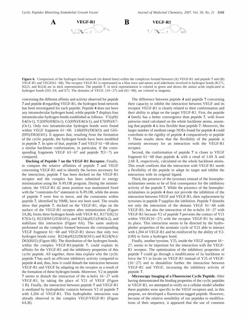
voluminous fluorescent dyes, such as the fluorescein introducedby El-Mousawi et al. forSP5.2labeling,37 might lead to a lossof activity. Thereby, we chose to insert in the peptide, aL-(7-hydroxycoumar-4-yl)alanine (Hca), which is a fluorescent aminoacid derivative developed by Brun et al.43 This fluorophore isparticularly well suited as it constitutes a good isostere oftyrosine or tryptophan. In addition, it is available as anNR-Fmoc-protected amino acid allowing its introduction in thesequence by classical SPPS. First, we synthesized the compound11 based on the sequence of peptide7, where an Hca wasincorporated in place of the second tyrosine. This peptide didnot displace VEGF binding even at 100µM. By contrast, thepeptide12 (aromatic residues: hF-Hca) showed to be the mostactive peptide of the series, exhibiting an IC50 of 19.3 ( 6.8µM. Thus, we used the peptide12 to perform biphotonicmicroscopy experiments on human umbilical vein endothelialcells (HUVEC). First, we verified that no autofluorescenceoccurred in the absence of the peptide12. Then, after treatmentby 100 µM of peptide12 over a period of 30 min, a strongblue fluorescence was observed around the cells, demonstratingthe binding properties of12 on HUVEC. The incubation ofHUVEC with the peptide12 and an excess of VEGF duringthe same time period induced disappearance of the fluorescence.These results highlight the VEGF receptor-specific binding of12on HUVEC (Figure 7) and, by extension, the VEGF receptor-specificity of the series of cyclic peptides. Moreover, it allowsus to consider this peptide as a valuable pharmacological toolfor studies on the cellular becoming VEGF receptors orinternalization of VEGF receptors.
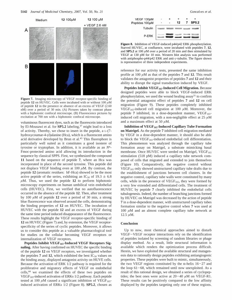
Peptides Inhibit VEGF165-Induced VEGF Receptors Sig-naling. After having confirmed on HUVEC the specific bindingof the peptide12 on VEGF receptors, we investigated whetherthe peptides7 and12, which exhibited the best IC50 values onthe binding assay, displayed antagonist activity on HUVE cells.Because the activation of ERK 1/2 pathway is required for theproliferative and migratory effects of VEGF on endothelialcells,44 we examined the effects of these two peptides onVEGF165-induced activation of ERKs 1/2 pathway. Each peptidetested at 100µM caused a significant inhibition of VEGF165-induced activation of ERKs 1/2 (Figure 8).SP5.2, chosen as
reference for our activity tests, presented the same inhibitionprofile at 100µM as that of the peptides7 and12. This resultvalidates the antagonist properties of peptides7 and12and theirability to disrupt the signal transduction induced by VEGF.
Peptides Inhibit VEGF165-Induced Cell Migration. Becausedesigned peptides were able to block VEGF-induced ERKphosphorylation, we used the wound healing assay31 to confirmthe potential antagonist effect of peptides7 and 12 on cellmigration (Figure 9). These peptides completely inhibitedVEGF165-induced cell migration at 100µM. Moreover, thepeptide7 inhibited, in a dose-dependent manner, VEGF165-induced cell migration, with a non-negligible effect at 25µMand a maximum effect at 50µM.
Inhibition of VEGF 165-Induced Capillary Tube Formationon Matrigel. As the peptide7 inhibited cell migration mediatedby VEGF in a dose-dependent manner, it should also be ableto block the VEGF165-induced endothelial cell differentiation.This phenomenon was analyzed through the capillary tubeformation assay on Matrigel, a substrate mimicking basalmembrane. Once HUVEC were seeded on Matrigel, VEGF165
stimulation (518 pM) induced a capillary tube network com-posed of cells that migrated and extended to join themselves(Figure 10). Comparatively, the negative control withoutVEGF165 only showed unstructured capillary tube drafts due tothe establishment of junctions between cell clusters. In thenegative control, capillary tube walls were constituted by manycells, while in the presence of VEGF165, they were formed bya very few extended and differentiated cells. The treatment ofHUVEC by peptide7 clearly inhibited the endothelial cellstubulogenesis. Indeed, the number of branching junctions formedby HUVEC on Matrigel was decreased by the action of peptide7 in a dose-dependent manner, with unstructured capillary tubesformation similar to the negative control when7 was used at100 µM and an almost complete capillary tube network at12.5 µM.
Conclusion
Up to now, most chemical approaches aimed to disturbVEGF-VEGF receptor interactions rely on the identificationof peptides isolated by screening of random libraries or phagedisplay method. As a result, little structural information isavailable which renders the optimization process difficult.Herein, we have exploited the available structural and mutagen-esis data to rationally design peptides exhibiting antiangiogenicproperties. These peptides were built to mimic, simultaneously,the two VEGF regions constituted by theR-helix 16-27 andthe loop 61-68, which remained until now unexploited. As aresult of this rational design, we obtained a series of cyclopep-tides; the best ones were active at 20-40 µM on VEGF-R1.These results can be positively compared to the low affinitydisplayed by the peptides targeting only one of these regions.
Figure 7. Imaging microscopy of VEGF receptor-specific binding ofpeptide12 on HUVEC. Cells were incubated with or without 100µMof peptide12 in the presence or absence of an excess of VEGF (2.60nM) over a period of 30 min. (A) Pictures taken by contrast phasewith a biphotonic confocal microscope. (B) Fluorescence pictures byexcitation at 700 nm with a biphotonic confocal microscope.
Figure 8. Inhibition of VEGF-induced p44/p42 ERK phosphorylation.Starved HUVEC, at confluence, were incubated with peptides7, 12,andSP5.2at 100µM over a period of 20 min and then stimulated byVEGF at 130 pM for 10 min. Western blot analysis was performedwith antiphospho-p44/p42 ERK and anti-γ-tubulin. The figure shownis representative of three independent experiments.
5142 Journal of Medicinal Chemistry, 2007, Vol. 50, No. 21 GoncalVes et al.

Moreover, the synthesis of a cyclic peptide incorporating thefluorescent 7-(hydroxycoumar-4-yl)alanine allowed us to verifythat these peptides were specific to the VEGF receptors presentat the surface of endothelial cells.
The resolution of the structure of the cyclic peptides4 and7by nuclear magnetic resonance (NMR) and molecular modelingusing NMR restraints has permitted the obtainment of variousinformation explaining the inhibitory activity of peptide7.Following the determination of their 3D structure, their con-formation has been compared with the one of the VEGFfragment used as a model to design the cyclic peptide inhibitors.The comparison of their isolated structure and superimposedwith the VEGF(61-68) fragment, in interaction with the VEGF-R1 receptor, may possibly explain the difference in thebiological activity of each peptide. The docking of peptide7,which exhibited the higher inhibitory activity with VEGF-R1and the minimization of the complex, let us propose that theinhibitory activity of peptide7 is facilitated by its flexibilityby interactions taking place within the complex and stabilizedby hydrogen bonds between peptide7 and VEGF-R1. Finally,the peptide7 exhibited antagonist activity on endothelial cells.Notably, it was able to inhibit several events implied inangiogenesis processes such as signal transduction, migration,and differentiation on Matrigel. The evaluation of the antitu-moral and antiangiogenic activities of these peptides, in vivo,is in progress.
All together, these results illustrate how specific inhibitorscan be rationally designed through the structure/function studyof the complex and how the inhibitor could be optimized byinspection of its interaction with VEGF-R1. These data couldhelp to propose new molecules targeting the complex formedbetween VEGF and VEGF-R1.
Materials and MethodsGeneral. Rink amide MBHA resin and HMP resin were
purchased from Novabiochem. HBTU, HOBt, DMAP, DCC, andDIPEA were from Applied Biosystems. Pd(PPh3)4 was from Fluka.All amino acids, from Novabiochem or Bachem, wereNR-terminal-protected by Fmoc and their side chains were protected asfollows: Arg(N-Pmc), Asn(N-Trt), Asp(O-All), Asp(O-t-Bu), Cys-(S-Trt), Cys(S-Acm), Gln(N-Trt), Glu(O-All), Glu(O-t-Bu), His-(N-Trt), Lys(N-Boc), Ser(O-tBu), Thr(O-tBu), Trp(N-Boc), andTyr(O-tBu). Fmoc-Hca-OH was kindly provided by Dr. Emman-uelle Braud (University Paris Descartes, France). Peptide synthesissolvents and acetonitrile for HPLC were analytical grade and wereacquired from commercial sources and used without furtherpurification.
General Procedure for Peptide Synthesis.All peptides weresynthesized by Merrifield stepwise solid-phase synthesis on anApplied Biosystems 433A automated peptide synthesizer usingstandard scale (0.25 mmol)FastMocchemistry. Coupling reactionswere performed using Fmoc amino acids (4 equiv), activated withHBTU (4 equiv), and HOBt (4 equiv) in the presence of DIPEA(8 equiv) for 1 h. Fmoc removal was effected by treating the resinwith 20% piperidine in NMP for 15 min. Peptides were cleavedfrom resin with simultaneous removal of side chain protectinggroups by treatment with 15 mL of TFA/water/TIPS (95:2.5:2.5v/v; Method A), TFA/thioanisole/water (94:5:1 v/v; Method B), orTFA/water/EDT/TIPS (94:2.5:2.5:1 v/v; Method C) for 3 h atroomtemperature. The filtrate from the cleavage reaction was evaporated,precipitated in cold diethyl oxide, collected by centrifugation, andlyophilized.
Peptides Purification and Analysis. Crude peptides werepurified by semipreparative RP-HPLC on a Nucleosil C18 column(Vydac, 5µm, 10× 250 mm) with a gradient program (solvent Ais water with 0.1% TFA and solvent B is 70% acetonitrile aqueoussolution with 0.09% TFA) at a flow rate of 2 mL/min with UVdetection at 214 and 254 nm. Fractions were analyzed by RP-HPLCon a Nucleosil C18 column (Vydac, 5µm, 4.6× 250 mm) at a
Figure 9. Effect of the peptides on VEGF-induced cell migration in a wound healing assay. Confluent monolayers of starved HUVEC werewounded using a tip, and a set of photos was taken at this time. Then HUVEC were treated with 130 pM of VEGF alone or with the peptides atthe indicated concentrations and a second set of photos was taken 6 h later. In the control, HUVEC were incubated with medium 2% FBS. Thephotos shown are representative of three independent experiments.
Figure 10. Representative microphotographs of HUVEC on Matrigel showing the effects of peptide7 on capillary tube formation. HUVEC weretreated with 518 pM of VEGF alone or with peptide7 at the indicated concentrations. Photos were taken after 14 h with a camera at 40× originalmagnification. The photos shown are representative of three independent experiments.
Cyclic Peptides Mimicking Endothelial Growth Factor Journal of Medicinal Chemistry, 2007, Vol. 50, No. 215143

flow rate of 1 mL/min and the pure fractions were collected andlyophilized to yield the final peptides as highly purified (>95%)white or off-white solids. The identity of peptides was checked byelectrospray mass spectrometry (MS) or by high-resolution elec-trospray mass spectrometry (HRMS).
Peptide SNDEGLES (1).Synthesis was performed followingthe general procedure on a HMP resin (substitution: 1.14 mmol/g,218 mg). Cleavage and deprotection from the resin was realizedwith method A. Yield after purification, 6.4%; MSm/z calcd forC32H51N9O18, 849.3; found, 850.7 [M+ H]+; tR ) 8.2 min (10-30% of solvent B in 20 min, purity 99%).
Peptide Ac-c[CNDEGLEC]-NH2 (2). Synthesis was performedon a Rink amide MBHA resin (substitution: 0.61 mmol/g, 410 mg)with cysteines introduced as Fmoc-Cys(Acm)-OH protected aminoacids. The peptide was synthesized following the general procedure,and theNR-terminal amino function was capped by a 20 mintreatment with 10 mL of an acetic anhydride mixture (0.5 M aceticanhydride, 0.125 M DIPEA, and 0.015 M HOBt in NMP). Thenthe resin was suspended in 150 mL of DMF and 50 mL of a solutionof iodine in DMF (10 equiv, 632 mg) were added dropwise over30 min. The solution was stirred vigorously over a period of 5 h atroom temperature. The solution was filtered and the resin waswashed with 100 mL of DMF and DCM. Deprotection and cleavageof the resin were realized according to method B. Yield afterpurification, 3.6%; MSm/zcalcd for C34H52N10O16S2, 920.3; found,921.7 [M + H]+; tR ) 5.2 min (20-80% of solvent B in 30 min,purity 97%).
Peptide KFMDVYQRSYCH-NH 2 (3).Synthesis was performedon a Rink amide MBHA resin (substitution: 0.61 mmol/g, 410 mg)following the general procedure. Deprotection and cleavage of theresin were realized according to method C. Yield after purification,3.0%; MSm/z calcd for C70H102N20O18S2, 1574.7; found, 1576.3[M + H]+; tR ) 10.5 min (20-60% of solvent B in 20 min, purity98%).
Peptide NGYEIEWYSWVTHGMY-NH 2 (SP5.2).Synthesiswas performed on a Rink amide MBHA resin (substitution: 0.61mmol/g, 410 mg) following the general procedure. Deprotectionand cleavage of the resin were realized according to method C.Yield after purification, 1.0%; MSm/z calcd for C96H124N22O26S,2032.9; found, 2034.1 [M+ H]+; tR ) 20.2 min (20-80% ofsolvent B in 30 min, purity 91.3%).
General Procedure for Synthesis of Cyclic Peptides 4-12.The linear sequence of amino acids was assembled on Rink amideMBHA resin (substitution: 0.70 mmol/g, 350 mg) according tothe general method for peptide synthesis, except that the C-terminalresidue was a Fmoc-Glu(O-allyl)-OH and the terminalNR-Fmocprotecting group was conserved at the end of the synthesis. Afterassembling the peptide, theO-allyl protecting group on the glutamicacid was removed by the following protocol: the resin was swollenin 30 mL of DCM/AcOH/NMP (37:2:1 v/v) and the suspensionwas bubbled with argon for 30 min. Then 870 mg of Pd(PPh3)4 (3equiv) were added and the solution was stirred for 3 h at roomtemperature under an argon atmosphere. The suspension was filteredand the resin was washed with a solution of 0.5% DIPEA in NMP(50 mL), a solution of diethyldithiocarbamate (0.5 g in 100 mLNMP), DMF (2 × 10 mL), and DCM (2× 10 mL). The Fmocgroup was then removed with 20% piperidine in NMP (30 min atroom temperature) and the resin was washed with NMP. Next thepeptidyl resins were stirred overnight with a solution of couplingreagents (8 mL NMP+ 0.25 mL 2 M DIPEA in NMP + 2.5 mL100 mM HBTU in NMP) at room temperature. The resin waswashed with NMP and DCM, and the deprotection and cleavagewas performed with method A.
Peptide c[hFhFDEGLEE]-NH2 (4). Yield, 17.0%; HRMSm/zcalcd for C47H63N9O15Na, 1016.4311; found, 1016.4339 [M+ Na]+;tR ) 22.1 min (10-90% of solvent B in 30 min, purity97.6%).
Peptide c[hFhFDEGLE]-NH2 (5). Yield, 6.9%; HRMSm/zcalcd for C42H56N8O12Na, 887.3915; found, 887.3927 [M+ Na]+;tR ) 24.5 min (10-90% of solvent B in 30 min, purity 93%).
Peptide c[FFDEGLEE]-NH2 (6). Yield, 9.2%; HRMSm/zcalcdfor C45H59N9O15Na, 988.4028; found, 988.4036 [M+ Na]+; tR )19.4 min (10-90% of solvent B in 30 min, purity 94%).
Peptide c[YYDEGLEE]-NH 2 (7). Yield, 13.2%; HRMSm/zcalcd for C45H59N9O17Na, 1020.3927; found, 1020.3915 [M+ Na]+;tR ) 14.0 min (10-90% of solvent B in 30 min, purity 98%).
Peptide c[YWDEGLEE]-NH 2 (8). Yield, 8.2%; MSm/z calcdfor C47H60N10O16, 1020.4; found, 1021.6 [M+ H]+; tR ) 17.04min (10-90% of solvent B in 30 min, purity 90%).
Peptide c[EYDEGLEE]-NH2 (9). Yield, 16.8%; MSm/z calcdfor C41H57N9O18, 963.4; found, 986.5 [M+ Na]+; tR ) 12.47 min(10-90% of solvent B in 30 min, purity 97%).
Peptide c[YYDEGYEE]-NH2 (10).Yield, 7.5%; MSm/z calcdfor C48H57N9O18, 1047.4; found, 1070.3 [M+ Na]+; tR ) 7.9 min(20-80% of solvent B in 30 min, purity 99%).
Peptide c[YHcaDEGLEE]-NH2 (11). Yield, 1.0%; MS m/zcalcd for C49H60N8O17, 1065.0; found, 1088.5 [M+ Na]+; tR )13.4 min (20-50% of solvent B in 30 min, purity 98%).
Peptide c[hFHcaDEGLEE]-NH2 (12). Yield, 1.0%; MSm/zcalcd for C49H61N9O18, 1064.1; found, 1064.2 [M+ H]+; tR ) 18.06min (10-90% of solvent B in 30 min, purity 95%).
Chemiluminescent Competition Assay on VEGF-R1.Theassay was performed as previously described by Goncalves et al.38
Briefly, a fixed amount of biotinylated VEGF165 (131 pM) wasincubated with the tested peptide in the presence of recombinanthuman VEGF-R1 adsorbed on a 96-well microplate. The biotiny-lated VEGF165 remaining after wash steps was detected bychemiluminescence thanks to HRP-conjugated streptavidin.
NMR Experiments and Data Processing.Samples for NMRexperiments were prepared by dissolving 1 mg of the cyclic peptidein the solvent mixture H2O/D2O/DMSO-d6 (80:10:10). The pH ofthe sample was set to 4.1 and 4.24 for the peptides4 and 7,respectively. NMR experiments were carried out on an AvanceBruker 600 MHz proton spectrometer. Total COSY, TOCSY,45 witha mixing time of 70 ms, double quantum filtered spectroscopy,DQF-COSY46,47and NOESY48,49at five different mixing times (50,100, 150, 200, and 250 ms) were performed on the samples. Two-dimensional spectra were acquired at 293 K, with 2048 real pointsin t2, a spectral width of 6010 Hz and 512 t1 increments. Thetransmitter frequency was set to the water signal. The solventresonance was suppressed using a 3-9-19 pulse sequence50 or bysaturation of the water signal during the relaxation delay of 1.6 sbetween free induction decays. For all experiments, the temperaturewas controlled externally using a temperature control system (BCU05 Bruker). All data were processed using XWINNMR software(Bruker). Aπ/6 phase-shifted sine bell window function was appliedand data were zero filled once prior to Fourier transformation inboth dimensions t1 and t2. The final size of the frequency domainmatrices was 2048 points inω1 and ω2 dimensions. For allexperiments,1H frequency scale was directly referenced to water.The data were then analyzed with the Felix program (Accelrys,San Diego, CA).
Structure Calculation of the Cyclic Peptides. The inputdistance restraints were deduced from the measurement of the NOEintensities in the NOESY data sets at five mixing times, at thetemperature of 293 K, by integration of the peaks into distancesby a R-6 dependency and a tolerance of 20% to take into accountthe errors of integration. The distances were calibrated using thedistance between theâ1/â2 protons of glutamate residues of 1.76Å measured in peptide7 andâ1/â2 protons of the aspartic residuefor the peptide4. No dihedral torsion angle restraints or hydrogenbonds were introduced during the calculations. The structuredetermination was done using standard protocols of distancegeometry and simulated annealing to embed and optimize initialstructures and performed with X-PLOR 3.8441,42 on an SGI O2R12000 workstation. These structures were optimized further withrestrained molecular dynamics simulations, which were carried outin vacuo with a distance-dependent dielectric constant. The dynam-ics were initiated at 5 K and the temperature was increased graduallyto 1000 K in 5.0 ps and then equilibrated for 1.0 ps. The forceconstants for the distance restraints were kept at 2.0 kcal‚mol-1‚Å-2
5144 Journal of Medicinal Chemistry, 2007, Vol. 50, No. 21 GoncalVes et al.

during these stages and were subsequently scaled up to a final valueof 30 kcal‚mol-1‚Å-2 over 6.0 ps. The system was then allowed toevolve for 20.0 ps at 1000 K and then slow-cooled to 300 K in14.0 ps and equilibrated for 10.0 ps. The coordinates saved every0.5 ps in the last 4.0 ps were averaged. The resulting structureswere submitted to a conjugate gradient minimization until a finalgradient of 0.1 kcal‚mol-1‚Å-2 was reached. Out of 200 calculatedstructures, the 10 best structures were selected based on the criteriaof acceptable covalent geometry, low distance restraint violations,and favorable nonbonded energy. All dynamics were carried outwith a time step of 1.0 fs. The quality of the structures was evaluatedwith PROCHECK51 and INSIGHT II (Accelrys) software was usedto visualize the structures. The quality of the structures wasmonitored by pairwise root-mean-squared deviation (rmsd) valuesand listed in Table 2. The final refined structures of the cyclicpeptides in solution exhibit no distance violations larger than 0.2Å (Table 2). Input restraints statistics are listed in Table 1.
Docking of Peptide 7 on VEGF-R1.To discover the factorsfavoring the binding of peptide7 on VEGF-R1 and how this bindingcould perturb the interaction between the VEGF and its receptor,VEGF-R1, peptide7 has been docked on VEGF-R1. In a first step,peptide7 has been superimposed with the VEGF(61-68) fragment,then the VEGF(61-68) has been removed, and the complex formedbetween VEGF-R1 and peptide7 has been minimized in a secondstep. The coordinates of VEGF-R1 have not been allowed to moveduring the minimization and were maintained fixed with the“constraints-fix-statement” in X-PLOR, while the atoms of peptide7 have been free to move and to adapt to VEGF-R1. The NOEconstraints identified previously within peptide7 have not beenemployed during energy minimization. The obtained structure ofthe complex and the position of peptide7 on VEGF-R1 have beenanalyzed, and the hydrogen bond network has been identifiedbetween peptide7 and VEGF-R1.
Cell Line and Culture. HUVEC were obtained as a gift fromDr. Catherine Boisson-Vidal (U765 INSERM) and were culturedin 150 cm2 plastic flasks, coated by a solution of 0.5% gelatin(BioChemika, Sigma), in M199 containing 20% fetal bovine serum(FBS). Cells were incubated at 37°C in a humidified atmosphereof 5% CO2 in air, and medium was changed every two days.Experiments were conducted on HUVEC that had gone throughone to five passages.
Imaging of the Binding between the Peptide 12 and the VEGFReceptors.HUVEC were grown on six-well plates coated by asolution of 0.5% gelatine containing a slide. Preconfluent HUVECwere incubated with12 and VEGF165 at indicated concentrationsfor 6 h. After this time, cells were washed three times with PBSand fixed with 4% formaldehyde. Cells were washed three timeswith PBS, and slides were mounted using mounting mediumVectashield Hard set (Vector Laboratories, Burlingame). Thebinding between12 and HUVEC was analyzed with a biphotonicconfocal microscope (Leica TCS SP2 AOBS, laser 2 photons:Tsunami, Spectra Physics; Institut Jacques Monod, France). Experi-ments were repeated two times with identical results.
Western Blot Analysis.Experiments were realized in six-wellplates coated with a solution of 0.5% gelatin. HUVEC, atconfluence, were starved in 2% FBS-supplemented M199 overnight,followed by 5 h in 0% FBS-supplemented M199. HUVEC weretreated for 20 min by compounds at the indicated concentrationsin 0% FBS-supplemented M199 and then stimulated by VEGF165
at 130 pM during 10 min. Cells were washed three times with ice-cold PBS, and the reaction was terminated by the addition of 100µL of ice-cold lysis buffer HNTG : Hepes 50 mM, NaCl 150 mM,glycerol 10%, Triton 100 1%, EGTA 1 mM, MgCl2 1 mM, Na3-VO4 1 mM, NaF 1 mM, and one protease inhibitor cocktail tablet(Complete, Roche). Equivalent amounts of proteins from each lysatewere resolved in 12% SDS-polyacrylamide gel and then transferredonto nitrocellulose membranes (Bio-Rad Laboratories). The trans-blotted membrane was incubated with antiphosphorylated p44-42ERK MAPK antibody (Cell Signaling Technology, Beverly, MA,Etats-Unis; 1:1000) and with anti-γ-tubuline (Santa Cruz Biotech-nology, Californie, Etats-Unis; 1:1000) in Tris-HCl buffered saline
(TBS) containing 0.1% Tween 20 (TBST) and 5% FBS at 4°Covernight. After treatment with the primary antibody, the membranewas washed three times with TBST, then incubated with anti-rabbitIgG-horseradish peroxidase conjugates (Amersham Biosciences,Buckinghamshire, Angleterre; 1:6000) for 1 h atroom temperature,and washed three times with TBST. The immunoblots werevisualized by enhanced chemiluminescence (Amersham Bio-sciences).
Wound Healing Assay.HUVEC were seeded in six-well platescoated with a solution of 0.5% gelatin and were allowed to growto confluence. Complete medium was replaced by medium contain-ing 2% FBS, and incubation was continued overnight. A linearwound was drawn in the monolayer of cells. A set of digital photoswas taken of each wound with a camera (Digital Sight, DS-L1,Nikon, Japon) at 100× original magnification. The wells werewashed with PBS, and medium containing 130 pM VEGF165 inthe presence or absence of peptides at 100µM was added. After 6h, a second set of photos was taken at the same place in the sameconditions.
Capillary Tube Formation on Matrigel (In Vitro AngiogenesisAssay).The formation of capillary tube-like structures by HUVECwas analyzed on 96-well plates coated with growth factor reducedMATRIGEL matrix (BD Biosciences, U.S.A.). Matrigel was thawedat 4 °C. Then 75µL/well of Matrigel was distributed and allowedto solidify at 37°C over a period of 1 h. HUVEC were seeded onthe polymerized Matrigel (2× 10⁴ cells/well) in medium containing5% FBS, with 518 pM of VEGF in the presence or absence of7 atthe indicated concentrations. The plate was incubated at 37°C,and tube formation was observed after 14 h under a phase-contrastmicroscope. Digital pictures were then taken with a camera (DigitalSight, DS-L1, Nikon, Japon) at 40× original magnification.
Acknowledgment. Financial support for this work wasprovided by the Ligue nationale contre le cancer and by theUniversity Paris Descartes Bonus-Qualite´-Recherche grant. Theauthors thank Dr. W.-Q. Liu for mass spectral data, Dr. C.Boisson-Vidal and I. Galy-Fauroux for providing HUVE cells,and C. Piolot and C. Chamot for their support for biphotonicmicroscopy experiments.
Supporting Information Available: 1H NMR spectra of com-pounds4 and7 in DMSO-d6; table of purity for the target com-pounds 1-12 and SP5.2; and HPLC analysis for the targetcompounds1-12 and SP5.2. This material is available free ofcharge via the Internet at http://pubs.acs.org.
References(1) Ferrara, N. Vascular endothelial growth factor: Basic science and
clinical progress.Endocr. ReV. 2004, 25, 581-611.(2) Carmeliet, P.; Jain, R. K. Angiogenesis in cancer and other diseases.
Nature2000, 407, 249-257.(3) Ferrara, N.; Kerbel, R. S. Angiogenesis as a therapeutic target.Nature
2005, 438, 967-974.(4) Kerbel, R. S. Antiangiogenic therapy: a universal chemosensitization
strategy for cancer?Science2006, 312, 1171-1175.(5) Ferrara, N.; Gerber, H. P. The role of vascular endothelial growth
factor in angiogenesis.Acta Haematol.2001, 106, 148-156.(6) Olsson, A. K.; Dimberg, A.; Kreuger, J.; Claesson-Welsh, L. VEGF
receptor signalling - in control of vascular function.Nat. ReV. Mol.Cell. Biol. 2006, 7, 359-371.
(7) Roskoski, R., Jr. Vascular endothelial growth factor (VEGF) signalingin tumor progression.Crit. ReV. Oncol. Hematol.2007, 62, 179-213.
(8) Takahashi, H.; Shibuya, M. The vascular endothelial growth factor(VEGF)/VEGF receptor system and its role under physiological andpathological conditions.Clin. Sci. (London) 2005, 109, 227-241.
(9) Luttun, A.; Autiero, M.; Tjwa, M.; Carmeliet, P. Genetic dissectionof tumor angiogenesis: Are PlGF and VEGFR-1 novel anticancertargets?Biochim. Biophys. Acta2004, 1654, 79-94.
(10) Karashima, T.; Inoue, K.; Fukata, S.; Iiyama, T.; Kurabayashi, A.;Kawada, C.; Shuin, T. Blockade of the vascular endothelial growthfactor-receptor 2 pathway inhibits the growth of human renal cellcarcinoma, RBM1-IT4, in the kidney but not in the bone of nudemice. Int. J. Oncol.2007, 30, 937-45.
Cyclic Peptides Mimicking Endothelial Growth Factor Journal of Medicinal Chemistry, 2007, Vol. 50, No. 215145

(11) Gille, J.; Heidenreich, R.; Pinter, A.; Schmitz, J.; Boehme, B.; Hicklin,D. J.; Henschler, R.; Breier, G. Simultaneous blockade of VEGFR-1and VEGFR-2 activation is necessary to efficiently inhibit experi-mental melanoma growth and metastasis formation.Int. J. Cancer.2007, 120, 1899-1908.
(12) Laakkonen, P.; Waltari, M.; Holopainen, T.; Takahashi, T.; Pytowski,B.; Steiner, P.; Hicklin, D.; Persaud, K.; Tonra, J. R.; Witte, L.;Alitalo, K. Vascular endothelial growth factor receptor 3 is involvedin tumor angiogenesis and growth.Cancer Res.2007, 67, 593-599.
(13) Soker, S.; Miao, H. Q.; Nomi, M.; Takashima, S.; Klagsbrun, M.VEGF165 mediates formation of complexes containing VEGFR-2and neuropilin-1 that enhance VEGF165-receptor binding.J. Cell.Biochem.2002, 85, 357-368.
(14) Sandler, A.; Gray, R.; Perry, M. C.; Brahmer, J.; Schiller, J. H.;Dowlati, A.; Lilenbaum, R.; Johnson, D. H. Paclitaxel-carboplatinalone or with bevacizumab for non-small-cell lung cancer.N. Engl.J. Med.2006, 355, 2542-2550.
(15) Sun, S.; Schiller, J. H. Angiogenesis inhibitors in the treatment oflung cancer.Crit. ReV. Oncol. Hematol.2007, 62, 93-104.
(16) Holash, J.; Davis, S.; Papadopoulos, N.; Croll, S. D.; Ho, L.; Russell,M.; Boland, P.; Leidich, R.; Hylton, D.; Burova, E.; Ioffe, E.; Huang,T.; Radziejewski, C.; Bailey, K.; Fandl, J. P.; Daly, T.; Wiegand, S.J.; Yancopoulos, G. D.; Rudge, J. S. VEGF-Trap: A VEGF blockerwith potent antitumor effects.Proc. Natl. Acad. Sci. U.S.A.2002,99, 11393-11398.
(17) Parry, T. J.; Cushman, C.; Gallegos, A. M.; Agrawal, A. B.;Richardson, M.; Andrews, L. E.; Maloney, L.; Mokler, V. R.;Wincott, F. E.; Pavco, P. A. Bioactivity of antiangiogenic ribozymestargeting Flt-1 and KDR mRNA.Nucleic Acids Res.1999, 27, 2569-2577.
(18) Gragoudas, E. S.; Adamis, A. P.; Cunningham, E. T., Jr.; Feinsod,M.; Guyer, D. R. Pegaptanib for neovascular age-related maculardegeneration.N. Engl. J. Med.2004, 351, 2805-2816.
(19) Goncalves, V. G. B.; Lenoir, C.; Garbay, C.; Vidal, M.; Inguimbert,N. Peptides as antagonists of the VEGF receptors.Pharmachem2006,15-19.
(20) D’Andrea, L. D.; Del Gatto, A.; Pedone, C.; Benedetti, E. Peptide-based molecules in angiogenesis.Chem. Biol. Drug. Des.2006, 67,115-126.
(21) Jia, H.; Jezequel, S.; Lohr, M.; Shaikh, S.; Davis, D.; Soker, S.;Selwood, D.; Zachary, I. Peptides encoded by exon 6 of VEGF inhibitendothelial cell biological responses and angiogenesis induced byVEGF. Biochem. Biophys. Res. Commun.2001, 283, 164-173.
(22) Wiesmann, C.; Fuh, G.; Christinger, H. W.; Eigenbrot, C.; Wells, J.A.; de Vos, A. M. Crystal structure at 1.7 A resolution of VEGF incomplex with domain 2 of the Flt-1 receptor.Cell 1997, 91, 695-704.
(23) Davis-Smyth, T.; Chen, H.; Park, J.; Presta, L. G.; Ferrara, N. Thesecond immunoglobulin-like domain of the VEGF tyrosine kinasereceptor Flt-1 determines ligand binding and may initiate a signaltransduction cascade.Emb. J.1996, 15, 4919-4927.
(24) Barleon, B.; Totzke, F.; Herzog, C.; Blanke, S.; Kremmer, E.;Siemeister, G.; Marme, D.; Martiny-Baron, G. Mapping of the sitesfor ligand binding and receptor dimerization at the extracellulardomain of the vascular endothelial growth factor receptor FLT-1.J.Biol. Chem.1997, 272, 10382-10388.
(25) Cunningham, S. A.; Stephan, C. C.; Arrate, M. P.; Ayer, K. G.; Brock,T. A. Identification of the extracellular domains of Flt-1 that mediateligand interactions.Biochem. Biophys. Res. Commun.1997, 231,596-599.
(26) Fuh, G.; Li, B.; Crowley, C.; Cunningham, B.; Wells, J. A.Requirements for binding and signaling of the kinase domain receptorfor vascular endothelial growth factor.J. Biol. Chem.1998, 273,11197-11204.
(27) Herley, M. T.; Yu, Y.; Whitney, R. G.; Sato, J. D. Characterizationof the VEGF binding site on the Flt-1 receptor.Biochem. Biophys.Res. Commun.1999, 262, 731-738.
(28) Muller, Y. A.; Li, B.; Christinger, H. W.; Wells, J. A.; Cunningham,B. C.; de Vos, A. M. Vascular endothelial growth factor: Crystalstructure and functional mapping of the kinase domain receptorbinding site.Proc. Natl. Acad. Sci. U.S.A.1997, 94, 7192-7197.
(29) Christinger, H. W.; Fuh, G.; de Vos, A. M.; Wiesmann, C. The crystalstructure of placental growth factor in complex with domain 2 ofvascular endothelial growth factor receptor-1.J. Biol. Chem.2004,279, 10382-10388.
(30) Keyt, B. A.; Nguyen, H. V.; Berleau, L. T.; Duarte, C. M.; Park, J.;Chen, H.; Ferrara, N. Identification of vascular endothelial growthfactor determinants for binding KDR and FLT-1 receptors. Generationof receptor-selective VEGF variants by site-directed mutagenesis.J.Biol. Chem.1996, 271, 5638-5646.
(31) Zilberberg, L.; Shinkaruk, S.; Lequin, O.; Rousseau, B.; Hagedorn,M.; Costa, F.; Caronzolo, D.; Balke, M.; Canron, X.; Convert, O.;Lain, G.; Gionnet, K.; Goncalves, M.; Bayle, M.; Bello, L.;Chassaing, G.; Deleris, G.; Bikfalvi, A. Structure and inhibitoryeffects on angiogenesis and tumor development of a new vascularendothelial growth inhibitor.J. Biol. Chem.2003, 278, 35564-35573.
(32) Berthelot, T.; Goncalves, M.; Lain, G.; Estieu-Gionnet, K.; Deleris,G. New strategy towards the efficient solid phase synthesis ofcyclopeptides.Tetrahedron2006, 62, 1124-1130.
(33) D’Andrea, L. D.; Iaccarino, G.; Fattorusso, R.; Sorriento, D.;Carannante, C.; Capasso, D.; Trimarco, B.; Pedone, C. Targetingangiogenesis: Structural characterization and biological propertiesof a de novo engineered VEGF mimicking peptide.Proc. Natl. Acad.Sci. U.S.A.2005, 102, 14215-14220.
(34) Chang, C. D.; Meienhofer, J. Solid-phase peptide-synthesis using mildbase cleavage of nalpha-fluorenylmethyloxycarbonylamino acids,exemplified by a synthesis of dihydrosomatostatin.Int. J. Pept.Protein Res.1978, 11, 246-249.
(35) Joseph, C. G.; Wang, X. S.; Scott, J. W.; Bauzo, R. M.; Xiang, Z.;Richards, N. G.; Haskell-Luevano, C. Stereochemical studies of themonocyclic agouti-related protein (103-122) Arg-Phe-Phe resi-dues: Conversion of a melanocortin-4 receptor antagonist into anagonist and results in the discovery of a potent and selectivemelanocortin-1 agonist.J. Med. Chem.2004, 47, 6702-6710.
(36) Mazur, S.; Jayalekshmy, P. Chemistry of polymer-boundortho-benzynesFrequency of encounter between substituents on cross-linked polystyrenes.J. Am. Chem. Soc.1979, 101, 677-683.
(37) El-Mousawi, M.; Tchistiakova, L.; Yurchenko, L.; Pietrzynski, G.;Moreno, M.; Stanimirovic, D.; Ahmad, D.; Alakhov, V. A vascularendothelial growth factor high affinity receptor 1-specific peptidewith antiangiogenic activity identified using a phage display peptidelibrary. J. Biol. Chem.2003, 278, 46681-46691.
(38) Goncalves, V.; Gautier, B.; Garbay, C.; Vidal, M.; Inguimbert, N.Development of a chemiluminescent screening assay for detectionof vascular endothelial growth factor receptor 1 ligands.Anal.Biochem.2007, 366, 108-110.
(39) Alcaro, M. C.; Sabatino, G.; Uziel, J.; Chelli, M.; Ginanneschi, M.;Rovero, P.; Papini, A. M. On-resin head-to-tail cyclization ofcyclotetrapeptides: Optimization of crucial parameters.J. Pept. Sci.2004, 10, 218-228.
(40) Nilges, M.; Clore, G. M.; Gronenborn, A. M. Determination of3-dimensional structures of proteins from interproton distance databy hybrid distance geometry-dynamical simulated annealing calcula-tions.FEBS Lett.1988, 229, 317-324.
(41) Laskowski, R. A.; Rullmann, J. A. C.; MacArthur, M. W.; Kaptein,R.; Thornton, J. M. AQUA and PROCHECK-NMR: Programs forchecking the quality of protein structures solved by NMR.J. Biomol.NMR 1996, 8, 477-486.
(42) Brunger, A. T. X-PLOR Software Manual, version 3.1; YaleUniversity Press: New Haven, CT, 1992.
(43) Brun, M. P.; Bischoff, L.; Garbay, C. A very short route toenantiomerically pure coumarin-bearing fluorescent amino acids.Angew. Chem., Int. Ed.2004, 43, 3432-3436.
(44) Cross, M. J.; Claesson-Welsh, L. FGF and VEGF function inangiogenesis: Signalling pathways, biological responses and thera-peutic inhibition.Trends Pharmacol. Sci.2001, 22, 201-207.
(45) Griesinger, C.; Otting, G.; Wuthrich, K.; Ernst, R. R. Clean TOCSYfor H-1 spin system-identification in macromolecules.J. Am. Chem.Soc.1988, 110, 7870-7872.
(46) Rance, M.; Sorensen, O. W.; Bodenhausen, G.; Wagner, G.; Ernst,R. R.; Wuthrich, K. Improved spectral resolution in COSY H-1-NMR spectra of proteins via double quantum filtering.Biochem.Biophys. Res. Commun.1983, 117, 479-485.
(47) Marion, D.; Wuthrich, K. Application of phase sensitive two-dimensional correlated spectroscopy (COSY) for measurements ofH-1-H-1 spin-spin coupling-constants in proteins.Biochem. Bio-phys. Res. Commun.1983, 113, 967-974.
(48) Jeener, J.; Meier, B. H.; Bachmann, P.; Ernst, R. R. Investigation ofexchange processes by 2-dimensional NMR spectroscopy.J. Chem.Phys.1979, 71, 4546-4553.
(49) Kumar, A.; Ernst, R. R.; Wuthrich, K. A two-dimensional nuclearOverhauser enhancement (2D NOE) experiment for the elucidationof complete proton-proton cross-relaxation networks in biologicalmacromolecules.Biochem. Biophys. Res. Commun.1980, 95, 1-6.
(50) Piotto, M.; Saudek, V.; Sklenar, V. Gradient-tailored excitation forsingle-quantum NMR-spectroscopy of aqueous solutions.J. Biomol.NMR 1992, 2, 661-665.
(51) Wuthrich, K. NMR of Proteins and Nucleic Acids; Wiley-Inter-science: New York, 1986.
JM0706970
5146 Journal of Medicinal Chemistry, 2007, Vol. 50, No. 21 GoncalVes et al.
Copyright © 2022 FDOKUMEN