Microneme protein 5 regulates the activity of Toxoplasma subtilisin 1 by mimicking a subtilisin...
12
Microneme Protein 5 Regulates the Activity of Toxoplasma Subtilisin 1 by Mimicking a Subtilisin Prodomain * Received for publication, June 8, 2012, and in revised form, August 13, 2012 Published, JBC Papers in Press, August 15, 2012, DOI 10.1074/jbc.M112.389825 Savvas Saouros ‡1 , Zhicheng Dou §1 , Maud Henry ‡ , Jan Marchant ‡ , Vern B. Carruthers §2 , and Stephen Matthews ‡3 From the ‡ Division of Molecular Biosciences, Imperial College London, South Kensington Campus, London SW7 2AZ, United Kingdom and the § Department of Microbiology and Immunology, University of Michigan Medical School, Ann Arbor, Michigan 48109 Background: TgSUB1 is a subtilisin protease that trims invasion proteins on the surface of Toxoplasma gondii. Results: TgMIC5 suppresses TgSUB1 activity and structurally mimics a subtilisin prodomain, suggesting a mechanism for inhibition. Conclusion: The C-terminal region of TgMIC5 is responsible for inhibition of TgSUB1. Significance: We identify a novel subtilisin propeptide mimic. Toxoplasma gondii is the model parasite of the phylum Api- complexa, which contains obligate intracellular parasites of medical and veterinary importance. Apicomplexans invade host cells by a multistep process involving the secretion of adhesive microneme protein (MIC) complexes. The subtilisin protease TgSUB1 trims several MICs on the parasite surface to activate gliding motility and host invasion. Although a previous study showed that expression of the secretory protein TgMIC5 sup- presses TgSUB1 activity, the mechanism was unknown. Here, we solve the three-dimensional structure of TgMIC5 by nuclear magnetic resonance (NMR), revealing that it mimics a subtilisin prodomain including a flexible C-terminal peptide that may insert into the subtilisin active site. We show that TgMIC5 is an almost 50-fold more potent inhibitor of TgSUB1 activity than the small molecule inhibitor N-[N-(N-acetyl-L-leucyl)-L-leucyl]- L-norleucine (ALLN). Moreover, we demonstrate that TgMIC5 is retained on the parasite plasma membrane via its physical interaction with the membrane-anchored TgSUB1. Toxoplasma gondii is an obligate intracellular parasite belonging to the phylum Apicomplexa and is the causative agent of toxoplasmosis. Toxoplasma is able to invade almost any nucleated cell and is prevalent among human populations, with a worldwide infection rate of up to 30%. Infection in humans occurs primarily following consumption of under- cooked infected meat or contact with feces from infected domestic cats. In immunocompromised individuals, Toxo- plasma reactivates from a semidormant state to cause acute diseases including blindness (1) or potentially fatal encephalitis (2, 3). Infection in pregnant women can result in a range of fetal birth defects or death (4). Due to its remarkably high infection rate, T. gondii constitutes the third most common cause of food-related death in both the United States (5) and France (6) after Salmonella and Listeria. T. gondii actively invades host cells during infection. Host cell entry requires the sequential release of proteins from secretory organelles named micronemes and rhoptries (7). Microneme proteins (MICs) 4 contribute to host cell attach- ment and provide a link to the parasite actomyosin motor that drives cell entry (8, 9). Many MICs contain lectin domains that bind specific carbohydrate ligands on the surface of host cells, providing a key attachment point for the parasite (10 –14). Adhesive MICs are assembled into heteromeric complexes as they pass through the secretory pathway. Several proteolytic molecular checkpoints occur throughout their assembly and transit to the micronemes. Proteolytic processing of MICs also occurs after their release onto the parasite surface, and this is essential for successful invasion. TgSUB1 is a glycosylphos- phatidylinositol (GPI)-anchored microneme protein that shows subtilisin-like serine protease activity. Lagal et al. (15) recently showed that TgSUB1 mediates the necessary surface processing of certain MICs for efficient host recognition and invasion, such as TgMIC2-M2AP and MIC4. Using gene knock-out and proteome profiling Brydges et al. (16) showed that the protease activity of TgSUB1 is negatively regulated by another microneme protein, TgMIC5. TgMIC5 is expressed as a preproprotein, which is proteolytically processed to a proprotein by the signal peptidase before being further processed in a post-Golgi compartment to the mature protein (17). Although Brydges et al. did not determine the precise nature of TgSUB1 regulation by TgMIC5, they suggested two possible mechanisms. First, they proposed that TgMIC5 could inhibit TgSUB1 activity directly, possibly by binding to and occluding the active site. Second, they proposed that TgMIC5 associates with the substrates of TgSUB1 on the parasite sur- face, thereby protecting them from overdigestion by TgSUB1. The latter model was favored in part because TgMIC5 showed no amino acid sequence similarity to any protease inhibitor. * This work was supported, in whole or in part, by National Institutes of Health Grant AI063263 (to V. B. C.). This work was also supported by Medical Research Council Grants G0800038 (to S. M.) and an American Heart Asso- ciation postdoctoral fellowship (to Z. D.). 1 Both authors contributed equally to this work. 2 To whom correspondence may be addressed. E-mail: [email protected]. 3 To whom correspondence may be addressed. E-mail: s.j.matthews@imperial. ac.uk. 4 The abbreviations used are: MIC, microneme; ALLN, N-[N-(N-acetyl-L-leucyl)- L-leucyl]-L-norleucine; ESA, excreted-secreted antigen; GPI, glycosylphos- phatidylinositol; PDB, Protein Data Bank. THE JOURNAL OF BIOLOGICAL CHEMISTRY VOL. 287, NO. 43, pp. 36029 –36040, October 19, 2012 © 2012 by The American Society for Biochemistry and Molecular Biology, Inc. Published in the U.S.A. OCTOBER 19, 2012 • VOLUME 287 • NUMBER 43 JOURNAL OF BIOLOGICAL CHEMISTRY 36029 at University of Michigan, on April 22, 2013 www.jbc.org Downloaded from
-
Upload
independent -
Category
Documents
-
view
0 -
download
0
Transcript of Microneme protein 5 regulates the activity of Toxoplasma subtilisin 1 by mimicking a subtilisin...

Microneme Protein 5 Regulates the Activity of ToxoplasmaSubtilisin 1 by Mimicking a Subtilisin Prodomain*
Received for publication, June 8, 2012, and in revised form, August 13, 2012 Published, JBC Papers in Press, August 15, 2012, DOI 10.1074/jbc.M112.389825
Savvas Saouros‡1
, Zhicheng Dou§1
, Maud Henry‡, Jan Marchant
‡, Vern B. Carruthers
§2, and Stephen Matthews
‡3
From the‡
Division of Molecular Biosciences, Imperial College London, South Kensington Campus, London SW7 2AZ,
United Kingdom and the§
Department of Microbiology and Immunology, University of Michigan Medical School,
Ann Arbor, Michigan 48109
Background: TgSUB1 is a subtilisin protease that trims invasion proteins on the surface of Toxoplasma gondii.Results: TgMIC5 suppresses TgSUB1 activity and structurally mimics a subtilisin prodomain, suggesting a mechanism forinhibition.Conclusion: The C-terminal region of TgMIC5 is responsible for inhibition of TgSUB1.Significance:We identify a novel subtilisin propeptide mimic.
Toxoplasma gondii is the model parasite of the phylum Api-complexa, which contains obligate intracellular parasites ofmedical and veterinary importance. Apicomplexans invade hostcells by a multistep process involving the secretion of adhesivemicroneme protein (MIC) complexes. The subtilisin proteaseTgSUB1 trims several MICs on the parasite surface to activategliding motility and host invasion. Although a previous studyshowed that expression of the secretory protein TgMIC5 sup-presses TgSUB1 activity, the mechanism was unknown. Here,we solve the three-dimensional structure of TgMIC5 by nuclearmagnetic resonance (NMR), revealing that it mimics a subtilisinprodomain including a flexible C-terminal peptide that mayinsert into the subtilisin active site. We show that TgMIC5 is analmost 50-fold more potent inhibitor of TgSUB1 activity thanthe smallmolecule inhibitorN-[N-(N-acetyl-L-leucyl)-L-leucyl]-L-norleucine (ALLN). Moreover, we demonstrate that TgMIC5is retained on the parasite plasma membrane via its physicalinteraction with the membrane-anchored TgSUB1.
Toxoplasma gondii is an obligate intracellular parasitebelonging to the phylum Apicomplexa and is the causativeagent of toxoplasmosis. Toxoplasma is able to invade almostany nucleated cell and is prevalent among human populations,with a worldwide infection rate of up to 30%. Infection inhumans occurs primarily following consumption of under-cooked infected meat or contact with feces from infecteddomestic cats. In immunocompromised individuals, Toxo-plasma reactivates from a semidormant state to cause acutediseases including blindness (1) or potentially fatal encephalitis(2, 3). Infection in pregnant women can result in a range of fetalbirth defects or death (4). Due to its remarkably high infectionrate, T. gondii constitutes the third most common cause of
food-related death in both the United States (5) and France (6)after Salmonella and Listeria.T. gondii actively invades host cells during infection. Host
cell entry requires the sequential release of proteins fromsecretory organelles named micronemes and rhoptries (7).Microneme proteins (MICs)4 contribute to host cell attach-ment and provide a link to the parasite actomyosin motor thatdrives cell entry (8, 9). Many MICs contain lectin domains thatbind specific carbohydrate ligands on the surface of host cells,providing a key attachment point for the parasite (10–14).Adhesive MICs are assembled into heteromeric complexes asthey pass through the secretory pathway. Several proteolyticmolecular checkpoints occur throughout their assembly andtransit to the micronemes. Proteolytic processing of MICs alsooccurs after their release onto the parasite surface, and this isessential for successful invasion. TgSUB1 is a glycosylphos-phatidylinositol (GPI)-anchored microneme protein thatshows subtilisin-like serine protease activity. Lagal et al. (15)recently showed that TgSUB1 mediates the necessary surfaceprocessing of certain MICs for efficient host recognition andinvasion, such as TgMIC2-M2AP and MIC4.Using gene knock-out and proteome profiling Brydges et al.
(16) showed that the protease activity of TgSUB1 is negativelyregulated by another microneme protein, TgMIC5. TgMIC5 isexpressed as a preproprotein,which is proteolytically processedto a proprotein by the signal peptidase before being furtherprocessed in a post-Golgi compartment to the mature protein(17). Although Brydges et al. did not determine the precisenature of TgSUB1 regulation by TgMIC5, they suggested twopossible mechanisms. First, they proposed that TgMIC5 couldinhibit TgSUB1 activity directly, possibly by binding to andoccluding the active site. Second, they proposed that TgMIC5associates with the substrates of TgSUB1 on the parasite sur-face, thereby protecting them from overdigestion by TgSUB1.The latter model was favored in part because TgMIC5 showedno amino acid sequence similarity to any protease inhibitor.
* This work was supported, in whole or in part, by National Institutes of HealthGrant AI063263 (to V. B. C.). This work was also supported by MedicalResearch Council Grants G0800038 (to S. M.) and an American Heart Asso-ciation postdoctoral fellowship (to Z. D.).
1 Both authors contributed equally to this work.2 To whom correspondence may be addressed. E-mail: [email protected] To whom correspondence may be addressed. E-mail: s.j.matthews@imperial.
ac.uk.
4 The abbreviations used are: MIC, microneme; ALLN, N-[N-(N-acetyl-L-leucyl)-L-leucyl]-L-norleucine; ESA, excreted-secreted antigen; GPI, glycosylphos-phatidylinositol; PDB, Protein Data Bank.
THE JOURNAL OF BIOLOGICAL CHEMISTRY VOL. 287, NO. 43, pp. 36029 –36040, October 19, 2012© 2012 by The American Society for Biochemistry and Molecular Biology, Inc. Published in the U.S.A.
OCTOBER 19, 2012 • VOLUME 287 • NUMBER 43 JOURNAL OF BIOLOGICAL CHEMISTRY 36029
at University of M
ichigan, on April 22, 2013w
ww
.jbc.orgD
ownloaded from

Immunoprecipitation experiments failed to identify partners ofTgMIC5, thus limiting further mechanistic insight of its role inregulating TgSUB1 proteolysis (16).Combining atomic resolution studies with data from bio-
chemical and parasite studies, we reveal that TgMIC5 is a sub-tilisin propeptide mimic and a potent inhibitor of TgSUB1.Wediscuss the implications of this inhibitory activity in the contextof infection byT. gondii. Our work could also provide the inspi-ration for novel therapeutics design to target parasite proteases.
EXPERIMENTAL PROCEDURES
Parasite Culture—The RH strain of T. gondii, which is a typeI strain that is highly pathogenic for all laboratory mice, wasused in all experiments. Parasite tachyzoites (RH, RHDmic5,and RHDsub1) were grown in human foreskin fibroblast cellsand harvested as described previously (18).Cloning, Expression, and Purification of Recombinant
TgMIC5 from Escherichia coli—Wild type (WT) MIC5 wasexpressed using the pTYB2 plasmid (New England Biolabs) inBL21 (DE3) E. coli strain (Stratagene) in Luria Bertani mediumsupplemented with 50mg/ml carbenicillin. The protein expres-sion was induced with 500 mM isopropyl b-D-thiogalactopyra-noside for 3 h at 37 °C. All subsequent purification steps wereperformed at 4 °C, and protease inhibitors were absent. Thecells were lysed using the Cell Disruptor TS5 (Constant CellDisruption Systems) coupled to a Haake TC200 chiller(Thermo Electron Corporation). WTMIC5 fusion protein waspurified by affinity chromatography using chitin resin (NewEngland Biolabs). The resin was equilibrated with 10 bed vol-umes of column buffer (20 mMHEPES, pH 7.5, 500 mMNaCl, 1mM EDTA). The bound intein tag was cleaved using a cleav-age buffer containing 20 mM HEPES, pH 8.0, 500 mM NaCl, 1mM EDTA, 50 mM dithiothreitol (DTT). The chitin resincolumn was flushed with 3 bed volumes of cleavage bufferand allowed to sit for 16 h. Pure WT MIC5 was recovered inthe flow-through.To facilitate the production of mutant and truncated pro-
teins, WT MIC5 was cloned onto pET30 Xa/LIC plasmid(Novagen). The mutated proteins were generated using theQuikChangeTM Site-directed Mutagenesis kit (Agilent Tech-nologies). All five mutated/truncated proteins were expressedusing a pET30 Xa/LIC plasmid in BL21 (DE3) E. coli strain inLuria Bertani medium supplemented with 40 mg/ml kanamy-cin. The cultures were initially grown at 37 °C and followed by atemperature adjustment for 1 h at 18 °C before inducing with500 mM isopropyl b-D-thiogalactopyranoside for 12 h. Themutated and truncated proteins were purified using a nickel-nitrilotriacetic acid resin (Qiagen). The resin was equilibratedwith 10 bed volumes of binding buffer (20 mM Tris-HCl, pH 8,500 mM NaCl, 5 mM imidazole). Bound proteins were washedwith 10 bed volumes of binding buffer, wash buffer (20 mMTris-HCl, pH 8, 500 mM NaCl, 20 mM imidazole) and elutedwith 10 bed volumes of elution buffer (20 mM Tris-HCl, pH 8,500 mM NaCl, 250 mM imidazole).
Size exclusion chromatography was carried out using anAKTA system (Amersham Biosciences) in combination with apreparative SuperdexTM 75 column. All samples wereexchanged into identical buffer conditions (25mMKH2PO4, pH
7.2, 150mMNaCl). The protein fractions were pooled together,concentrated to ;500 mM, and exchanged into an NMR buffer(25 mM KH2PO4, pH 7.2, 100 mM NaCl). 15N,13C-labeled sam-ples of WTMIC5 were produced in minimal medium contain-ing 0.07% 15NH4Cl and 0.2% 13C6-glucose.Solution Structure Determination of TgMIC5—Backbone
and side chain assignmentswere completed using our in-house,semiautomated assignment algorithms and standard triple-res-onance assignment methodology (19). Ha and Hb assignmentswere obtained using HBHA (CBCACO)NH. The side chainassignments were completed using HCCH total correlation(TOCSY) spectroscopy and (H)CC(CO)NHTOCSY. Three-di-mensional 1H-15N/13C NOESY-HSQC (mixing time 100 ms at800MHz) experiments provided the distance restraints used inthe final structure calculation. The ARIA protocol (20) wasused for completion of the NOE assignment and structure cal-culation. The frequency window tolerance for assigning NOEswas 60.04 ppm and 60.06 ppm for direct and indirect protondimensions and 60.6 ppm for both nitrogen and carbondimensions. The ARIA parameters p, Tv, and Nv were set todefault values. 144 dihedral angle restraints derived fromTALOS were also implemented (21). The 10 lowest energystructures had no NOE violations .0.5 Å and dihedral angleviolations .5º. Although structure calculations readily con-verged without the introduction of manual assignments, a sys-tematic check of automatically assigned NOEs was carried out.The structural statistics are presented in Table 1. The hetero-nuclear 1H-15N NOE data were measured as described previ-ously (22).Inhibition Assay of TgSUB1Activity—Filter-purified RH par-
asites were washed three times with ice-cold D1 medium (Dul-becco’s modified Eagle’s medium (DMEM) containing 10 mMHEPES, pH 7.0, 1% fetal bovine serum (FBS), and 2 mM L-glu-tamine). Parasites were resuspended at 33 108ml21 in D1, and90 ml of parasite resuspension was pipetted into the wells of a96-well round bottom plate containing 10 ml of inhibitors(recombinant TgMIC5 or ALLN) and incubated for 20 min at37 °C. Parasites were pelleted twice at 4 °C by centrifugation.Supernatants were analyzed by quantitative immunoblotting asdescribed below. Recombinant WT and mutant TgMIC5 pro-teins were tested at concentrations of 8.2 3 1023 mM, 2.5 31022 mM, 7.43 1022 mM, 2.23 1021 mM, 6.73 1021 mM, 2.0mM,6.0 mM, and 18.0 mM. ALLN was tested at concentrations of4.6 3 1022 mM, 1.4 3 1021 mM, 4.1 3 1021 mM, 1.2 mM, 3.7 mM,11.1 mM, 33.3 mM, and 100.0 mM. Intensities of the TgMIC450band were quantified with LI-COR image studio software andplotted against the corresponding concentrations of inhibitorsusing Prism 5 software to determine the half-maximal inhibi-tory concentrations (IC50). Due to the low ratio of TgMIC450 toTgMIC470, the optimal laser power for detecting TgMIC450oversaturated the TgMIC470 band, as indicated by the whiteregion in its middle. Lysozyme was included as a negative con-trol because its size and globular nature are similar to TgMIC5.SDS-PAGE and Immunoblotting—Parasite excreted-se-
creted antigen (ESA) fractions or cell lysates were mixed with53 SDS-PAGE loading buffer containing 2% b-mercaptoetha-nol (final concentration) and boiled for 5 min before resolvingon 10% or 12.5% minigels. For qualitative blots, proteins were
MIC5 Inhibition of SUB1 from Toxoplasma gondii
36030 JOURNAL OF BIOLOGICAL CHEMISTRY VOLUME 287 • NUMBER 43 • OCTOBER 19, 2012
at University of M
ichigan, on April 22, 2013w
ww
.jbc.orgD
ownloaded from
semidry-transferred to polyvinylidene fluoride (PVDF) mem-brane (Millipore), blocked with phosphate-buffered saline(PBS) containing 1% skim milk, 0.05% Tween 20, and 0.05%Triton X-114, and incubated with primary antibodies. Goatanti-mouse or anti-rabbit IgGs conjugated with horseradishperoxidase (HRP)were used as secondary antibodies. Immuno-blots were developed with SuperSignal West Pico chemilumi-nescence reagent (Pierce) and exposed to x-ray film. For quan-titative blots, proteins were semidry-transferred to PVDF-FLmembrane (Millipore). Blots were treated with blocking buffer(LI-COR Biosciences) and sequentially probed with rabbitpolyclonal anti-TgMIC4 (23) and goat anti-rabbit IgG conju-gated with IRDye 800CW (LI-COR Biosciences). Blots wereair-dried and imaged by LI-COR Odyssey CLx instrument.Immunolocalization Assays—To assess the surface distribu-
tion of proteins during invasion, tachyzoites were filter-puri-fied, washed, and resuspended in “Endo” buffer (44.7 mMK2SO4, 10 mM MgSO4, 106 mM sucrose, 5 mM glucose, 20 mMTris-H2SO4, 3.5 mg/ml bovine serum albumin (BSA), pH 8.2, apotassium-rich buffer that arrests parasite motility) (24) at 3 3107 ml21 at room temperature. Two hundred microliters ofparasite resuspension was added to overnight-grown humanforeskin fibroblast cells in 8-well chamber slides. The parasiteswere allowed to settle for 15 min at room temperature beforethe medium was replaced with prewarmed invasion medium(DMEM with 10 mM HEPES, pH 7.0, 3% FBS, and 2 mM L-glu-tamine) containing 1% ethanol. Parasites were allowed toinvade for 2 min at 37 °C prior to fixation with 4% paraformal-dehyde for 20 min. Parasites were stained with mouse mono-
clonal (mAb) B3.90 anti-TgAMA1 (25), rabbit polyclonal anti-TgMIC5 (17), or rabbit anti-Plasmodium falciparum SUB1(PfSUB1). To assess the subcellular distribution TgMIC5 andTgSUB1 within parasites, these proteins were stained with theaforementioned antibodies along with TgAMA1 in overnightreplicated parasites permeabilized with 0.1% Triton X-100.Mowiol was used as the mounting medium. Images were cap-tured digitally with an AxioCAMMRm camera equipped ZeissAxiovert Observer Z1 inverted fluorescence microscope atroom temperature with a 1003, 1.3 NA oil objective lens andprocessed using Zeiss Axiovision 4.3 software. Adobe Photo-shop and Illustrator CS5 programs were used to assemble thefinal images.Analysis of Surface Protein Abundance by Flow Cytometry—
RH, RHDmic5, and RHDsub1 parasites were harvested andresuspended in the invasion medium at 3 3 107 ml21 at roomtemperature. Parasites were induced with invasion mediumcontaining 1% ethanol for 2 min to stimulate release of ESAs,followed by the addition of an equal volume of 8% paraformal-dehyde and the incubation at room temperature for 20 min.Fixed parasites were pelleted and blocked with 10% FBS in PBSfor 10min and immunostainedwith rabbit anti-PfSUB1 and ratanti-TgMIC5. Goat anti-rabbit IgG antibody conjugated withfluorescein isothiocyanate (FITC) and goat anti-rat IgG anti-body conjugated with phycoerythrin were used as secondaryantibodies. Intensities of FITC and phycoerythrin dyes weremeasured using a BD FACSCanto flow cytometer and recordedusing BD FACSDiva software. Quantification was performedand plotted using FlowJo software version 9.1.
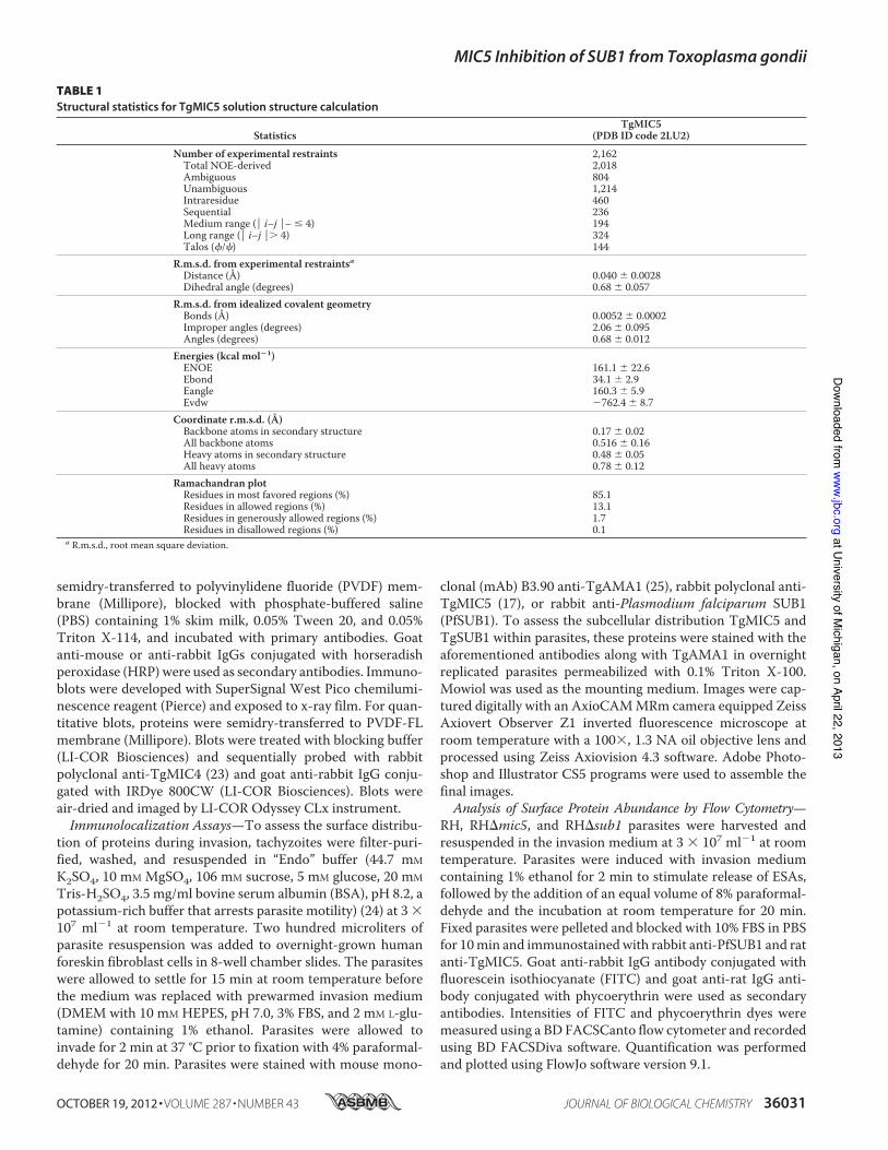
TABLE 1Structural statistics for TgMIC5 solution structure calculation
StatisticsTgMIC5
(PDB ID code 2LU2)Number of experimental restraints 2,162Total NOE-derived 2,018Ambiguous 804Unambiguous 1,214Intraresidue 460Sequential 236Medium range (Pi–jP– # 4) 194Long range (Pi–jP. 4) 324Talos (f/c) 144
R.m.s.d. from experimental restraintsaDistance (Å) 0.040 6 0.0028Dihedral angle (degrees) 0.68 6 0.057
R.m.s.d. from idealized covalent geometryBonds (Å) 0.0052 6 0.0002Improper angles (degrees) 2.06 6 0.095Angles (degrees) 0.68 6 0.012
Energies (kcal mol21)ENOE 161.1 6 22.6Ebond 34.1 6 2.9Eangle 160.3 6 5.9Evdw 2762.4 6 8.7
Coordinate r.m.s.d. (Å)Backbone atoms in secondary structure 0.17 6 0.02All backbone atoms 0.516 6 0.16Heavy atoms in secondary structure 0.48 6 0.05All heavy atoms 0.78 6 0.12
Ramachandran plotResidues in most favored regions (%) 85.1Residues in allowed regions (%) 13.1Residues in generously allowed regions (%) 1.7Residues in disallowed regions (%) 0.1
a R.m.s.d., root mean square deviation.
MIC5 Inhibition of SUB1 from Toxoplasma gondii
OCTOBER 19, 2012 • VOLUME 287 • NUMBER 43 JOURNAL OF BIOLOGICAL CHEMISTRY 36031
at University of M
ichigan, on April 22, 2013w
ww
.jbc.orgD
ownloaded from

RESULTS
The Solution Structure of TgMIC5—Sequence analysisrevealed that TgMIC5 bears no significant sequence similarity(E value . 0.001) with any structurally characterized protein.To provide insight to the mechanism of TgMIC5 regulation ofTgSUB1 processing, we determined the solution structure ofrecombinant TgMIC5 using heteronuclear multidimensionalNMR spectroscopy. After completion of backbone assignmentsit became apparent that the relaxation properties of the first 54amino acids were characteristic of high dynamics and displayedno evidence for stable secondary structure. After their removal,the NMR spectra of the globular domain remained unchanged.The NMR side chain resonance assignments and full structurecalculations were continued using this construct. The structurerevealed an open-sandwich antiparallel-a/antiparallel-b fold,comprising two a-helices, a1 and a2, and a four-strandedb-sheet, b1–b4 (Fig. 1, A–C). The heteronuclear 15N NOEshowed that NOE values for the C-terminal six residues aresignificantly lower than for themain globular domain (Fig. 1D).Regions that undergo internal motion faster than overall tum-bling showed a decreasedNOE intensity relative to the average.This indicates that the C-terminal peptide is flexible (Fig. 1E).
TgMIC5 Is Structurally Similar to Protease Prodomains—Asearch for protein structure similarities using programs DALI(26) and PDBeFold (27) revealed a similarity to the prodomainsof zinc carboxypeptidases (Z-score 5 5.8 for PDB ID code2BOA; Fig. 2A) (28–30). Prodomains can serve as intramolec-ular chaperones as well as transient inhibitors to prevent pre-mature enzymatic activity. For these metalloproteinases, theprodomains are N-terminal extensions that function as a gate-keeper by preventing substrate access to the catalytic site. Theglobular part of the prodomain contacts the entrance to theactive site. Activation occurs through proteolysis of the con-necting helical linker, which makes further interactions withthe enzymatic domain and contains the cleavage site for activa-tion. TgMIC5 does not possess these features, but is terminatedwith a sequence that displays significant flexibility in the solu-tion structure (Fig. 1D). This region also possesses three hydro-phobic residues (DSEVKLA; Fig. 1E) together with fourcharged groups (Asp, Glu, Lys, and C terminus) that could con-tribute to a protein-protein interaction site. Further manualanalysis of similar structures revealed that the structure ofTgMIC5 is also shared by prodomains of the subtilisin-like fam-ily of proteases (Z-score 5 3.4 for PDB ID code 3CNQ; Fig. 2B)
FIGURE 1. Solution structure of TgMIC5. A, ensemble of 10 lowest energy structures. Individual structures are shown as Ca traces. B and C, representations ofthe average structure following refinement in water. D, 1H-15N heteronuculear NOE data for TgMIC5. The distribution of 15N heteronuclear NOE across thesequence of TgMIC5 is shown. Lower NOE values indicate faster internal motions relative to the rest of the protein (22). E, electrostatic surface of a represent-ative TgMIC5. Inset shows a sketch of the C-terminal flexible region with hydrophobic residues highlighted.
MIC5 Inhibition of SUB1 from Toxoplasma gondii
36032 JOURNAL OF BIOLOGICAL CHEMISTRY VOLUME 287 • NUMBER 43 • OCTOBER 19, 2012
at University of M
ichigan, on April 22, 2013w
ww
.jbc.orgD
ownloaded from
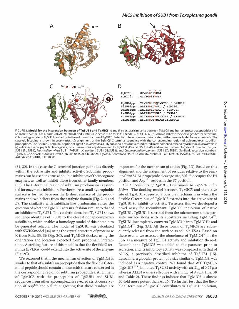
(31, 32). In this case the C-terminal junction point lies directlywithin the active site and inhibits activity. Subtilisin prodo-mains can be used in trans as soluble inhibitors of their cognateenzymes, as well as inhibit those from other family members(33). The C-terminal region of subtilisin prodomains is essen-tial for enzymatic inhibition. Furthermore, a small hydrophobicsurface is formed between the b-sheet surface of the prodo-mains and two helices from the catalytic domain (Fig. 2, A andB). The similarity with subtilisin-like prodomains raises thequestion of whether TgMIC5 acts in a fashion similar to that ofan inhibitor ofTgSUB1.The catalytic domain ofTgSUB1 showssequence identities of ;30% to the closest nonapicomplexansubtilisins, which enables a homology model of this domain tobe generated reliably. The model of TgSUB1 was calculatedwith SWISSmodel (34) using the crystal structure of proteinaseK from Refs. 35, 36 (Fig. 2C), and TgMIC5 docked using theorientation and location expected from prodomain interac-tions. A striking feature of this model is that the flexible C ter-minus (EVLKA) could extend into the active site of the enzyme(Fig. 2C).We reasoned that if the mechanism of action of TgMIC5 is
similar to that of a subtilisin propeptide then the flexible C-ter-minal peptide should contain amino acids that are conserved inthe corresponding region of subtilisin propeptides. Alignmentof TgMIC5 with the propeptides of TgSUB1 and SUB1sequences from other apicomplexans revealed strict conserva-tion of Asp132 and Val135, suggesting that these residues are
important for the mechanism of action (Fig. 2D). Based on thisalignment and the assignment of residues relative to the Plas-modium SUB1 propeptide cleavage site, Val135 occupies the P4position and Asp132 resides in the P7 position.The C Terminus of TgMIC5 Contributes to TgSUB1 Inhi-
bition—The docking model between TgMIC5 and the activesite of TgSUB1 suggested a possible mechanism in which theflexible C terminus of TgMIC5 extends into the active site ofTgSUB1 to inhibit its activity. To assess this we developed anovel assay for recombinant TgMIC5 inhibition of nativeTgSUB1. TgSUB1 is secreted from the micronemes to the par-asite surface along with its substrates including TgMIC470.TgSUB1 incompletely converts TgMIC470 into TgMIC450 andTgMIC420 (Fig. 3A). All three forms of TgMIC4 are subse-quently released from the surface as soluble ESAs. Based onthese events we assessed the abundance of TgMIC450 in theESA as a measure of TgSUB1 activity and inhibition thereof.Recombinant TgMIC5 was added to the parasites prior tosecretion, and its inhibitory activity was compared with that ofALLN, a previously described inhibitor of TgSUB1 (15).Lysozyme, a globular protein of a size similar to TgMIC5, wasincluded as a negative control. We found that WT TgMIC5(TgMIC5WT) inhibitedTgSUB1 activitywith an IC50 of 0.22mMwhereas ALLNwas less effective with an IC50 of 9.9 mM (Fig. 3Band Table 2). These findings indicate that TgMIC5 is almost50-fold more potent than ALLN. To further test that the flexi-ble C terminus of TgMIC5 contributes to TgSUB1 inhibition,
FIGURE 2. Model for the interaction between of TgSUB1 and TgMIC5. A and B, structural similarity between TgMIC5 and human procarboxypeptidase A4(Z-score 5 5.8 for PDB ID code 2BOA) (28, 30) (A), and subtilisin (Z-score 5 3.4 for PDB ID code 3CNQ) (31, 32) (B). Arrows indicate the cleavage sites for activation.C, homology model of TgSUB1 docked onto the solution structure of TgMIC5. Potential interaction motif is indicated with conserved side chains as red balls. Thecatalytic histidine is shown in yellow sticks. D, alignment of the TgMIC5 C-terminal sequence with the corresponding region of apicomplexan subtilisinpropeptides. The flexible C-terminal peptide of TgMIC5 is underlined. Fully conserved residues are indicated in emboldened red and by asterisks. A forward slash
(/) indicates the propeptide cleavage site, which was empirically determined for TgSUB1 (45) and PfSUB1 (46) and implied by homology for Plasmodium berghei
SUB1 (PbSUB1), Plasmodium vivax SUB1 (PvSUB1) N. caninum SUB1 (NcSUB1), and Cryptosporidium parvum SUB1 (CpSUB1). GenBank accession numbers:TgMIC5, CAA70921; putative NcMIC5, NCLIV_068520, CBZ56428; TgSUB1, AAK94670; PfSUB1, CAA05627; PbSUB1, XP_679126; PvSUB1, ACT76164; NcSUB1,AAF04257; CpSUB1, CAD98301.
MIC5 Inhibition of SUB1 from Toxoplasma gondii
OCTOBER 19, 2012 • VOLUME 287 • NUMBER 43 JOURNAL OF BIOLOGICAL CHEMISTRY 36033
at University of M
ichigan, on April 22, 2013w
ww
.jbc.orgD
ownloaded from

we generated a mutant TgMIC5 protein (TgMIC5D131–138)lacking the last 8 amino acids. We also created an N-terminallytruncated TgMIC5 mutant (TgMIC5D1–54) lacking theunstructured first 54 amino acids as a control. TgMIC5D131–138
(IC50 4.4 mM) was 20-fold less potent than TgMIC5WT whereasTgMIC5D1–54 showed no loss of inhibitory activity (Fig. 3C andTable 2). These findings are consistent with the model that theflexible C-terminal TgMIC5 participates in the mechanism ofTgSUB1 inhibition.
To test the putative role of the strictly conserved Asp132 andVal135 in protease inhibition, we generated alanine substitutionmutants at each of these positions. The TgMIC5V135A mutantshowed a modest 2-fold decrease in inhibitory activity com-pared with theWT protein (Fig. 3D and Table 2). However, theTgMIC5D132A mutant exhibited an IC50 of 4.6 mM, which issimilar to that of the poorly active C-terminally truncatedTgMIC5D131–138mutant. This 23-fold loss of inhibitory activitysuggests that TgMIC5 Asp132 is a key contributor to the mech-anism of TgSUB1 inhibition. Collectively, the C terminus ofTgMIC5 plays a critical role in blocking TgSUB1 activity, andAsp132 within this fragment contributes significantly to themechanism of inhibition.TgMIC5 Inhibition of TgSUB1Activity Also Affects Processing
of Other Secreted Micronemal Substrates—To test whether theinhibition of TgSUB1 by TgMIC5WT also affects other sub-strates on the parasite surface, we evaluated the proteolytictrimming of other microneme proteins including TgMIC2,TgM2AP, and TgPLP1. We also assessed TgSUB1 self-shed-ding from the parasite surface. The assay was performed using0.67mMTgMIC5, which is slightly higher than the IC90 value of
FIGURE 3. The C terminus of TgMIC5 plays a critical role in inhibition of TgSUB1. A, schematic illustrates TgMIC4 processing on the parasite surface byTgSUB1 protease. The six PAN/Apple domains of TgMIC4 are numbered from the N terminus, with cleavage by TgSUB1 occurring between domains 4 and 5 toproduce an N-terminal fragment termed TgMIC450. B, TgMIC5 inhibitory activity for TgSUB1 was quantified. Recombinant TgMIC5 at the indicated concentra-tions was added to live tachyzoites expressing surface TgSUB1, which coverts TgMIC470 to TgMIC450 before being shed into the medium as an ESA product. ESAproducts were collected from the supernatant after centrifugation, separated by 12.5% SDS-PAGE and immunoblotted with rabbit anti-TgMIC4 and goatanti-rabbit IgG conjugated with IRDye 800CW. The inhibition efficacy of TgMIC5WT was compared with ALLN by infrared laser scanning (LI-COR Biosciences) tomeasure the intensity of TgMIC450 in the ESA. Lysosome was included as a negative control protein. C, to assess the functional region within TgMIC5 proteincontributing to inhibition of TgSUB1 activity, 54 (D1–54) and 8 amino (D131–138) acids were deleted from N and C termini of WT TgMIC5, respectively. Thesetruncation mutants were tested for inhibitory activity according to the description in B. D, to further identify the key residue(s) in the C terminus of TgMIC5 forits inhibition ability, Asp132 and Val135 were replaced with alanine to create TgMIC5D132A and TgMIC5V135A. Inhibitory activity was measured according to B.Quantitative data in B–D are from at least three independent experiments each. Error bars are S.E.
TABLE 2Half-maximal inhibitory concentrations (IC
50) of TgSUB1 inhibitors
InhibitorIC50 (mM)
(mean 6 S.E.)Change relativeto TgMIC5WT
-foldLysozyme NDa NDTgMIC5WT 0.22 6 0.02 1.0ALLN 9.9 6 0.2 45TgMIC5D131–138 4.4 6 1.2 20TgMIC5D1–54 0.27 6 0.06 1.2TgMIC5D132A 4.6 6 0.9 21TgMIC5V135A 0.41 6 0.06 1.9TgMIC5D132A/V135A 1.9 6 0.2 8.6
a ND, not determined.
MIC5 Inhibition of SUB1 from Toxoplasma gondii
36034 JOURNAL OF BIOLOGICAL CHEMISTRY VOLUME 287 • NUMBER 43 • OCTOBER 19, 2012
at University of M
ichigan, on April 22, 2013w
ww
.jbc.orgD
ownloaded from

TgMIC5WT (0.42 mM). TgMIC5D131–138 was included as acontrol.The abundance andmigration of secretedTgAMA1were not
affected by TgMIC5WT or TgMIC5D131–138 (Fig. 4), consistentwith it not being a substrate of TgSUB1 (15). TgMIC2 is shedfrom the parasite surface as a mixture of species including a100-kDa form with an intact N terminus and a 95-kDa formresulting from N-terminal trimming by TgSUB1. TgMIC5WT
inhibited trimming to the 95-kDa species whereas the C-termi-nally truncated TgMIC5D131–138 mutant failed to disrupt trim-ming (Fig. 4). TgM2AP forms a complex with TgMIC2 and isprocessed by TgSUB1 into four species termed TgM2AP-1, -2,-3, and -4 (37). TgM2AP-1 is thought to be processed byTgSUB1 prior to or coincident with nascent secretion from themicronemes based on its resistance to inhibition by ALLN (15).Consistent with this, TgMIC5WT strongly inhibited the pro-duction of TgM2AP-2, -3, and -4 in the ESA and resulted in theaccumulation of TgM2AP-1 and the other unprocessed speciesproTgM2AP (pTgM2AP) mature M2AP (mM2AP). In con-trast, the C-terminally truncated TgMIC5D131–138 showedminimal inhibitory activity, slightly affecting the production ofTgM2AP-2, -3, and -4. Similarly, mature TgPLP1 migrating at130 kDawas not cleaved byTgSUB1 into a 100-kDa species (15)upon addition of TgMIC5WT but was processed in the presenceof the C-terminally truncated TgMIC5 mutant.TgSUB1 is stored in the micronemes as a 90-kDa GPI-an-
chored species (TgSUB190), which is secreted onto the surfacewhere it encounters its substrates. TgSUB190 is released from
the surface into the ESA as 82- and 70-kDa species (TgSUB182and TgSUB170). Binder et al. generated an rabbit anti-peptideantibody (RaTgSUB1) against a C-terminal antigenic peptideimmediately adjacent to its GPI anchor (38). This antibody onlyrecognizes TgSUB190 and the larger precursor forms whereas arabbit polyclonal antibody against the catalytic domain ofPfSUB1 (RaPfSUB1) recognizes all of the species based on theantigenic similarity between TgSUB1 and PfSUB1. When theESA fractionwas incubatedwith PBS orC-terminally truncatedTgMIC5D131–138, TgSUB190 was quantitatively processed to amixture of TgSUB182 and TgSUB170, which migrated near oneanother (Fig. 4). Exogenously added TgMIC5WT inhibitedprocessing to TgSUB170, resulting in exclusive detection of theTgSUB190 and TgSUB182 (Fig. 4). RaTgSUB1 recognizedTgSUB190, indicating this species in the ESA has an intactC-terminal epitope recognized by this antibody. Altogether,our findings indicate that TgMIC5 inhibits TgSUB1 processingof micronemal substrates and itself.TgMIC5 Physically Interacts with TgSUB1—The docking
model predicted that the C terminus of TgMIC5 interacts withthe active site of TgSUB1 to act as the inhibitor. We tried toimmunoprecipitate the endogenous TgMIC5-TgSUB1 com-plex fromRHparasite ESA and cell lysate, but these effortswereunsuccessful, indicating a fragile interaction between TgMIC5and TgSUB1. To compensate for a weak interaction, weincubated 0.67 mM recombinant N-terminally His6-taggedTgMIC5WT with ESA containing TgSUB1. As controls, theN-terminally His6-tagged TgMIC5D131–138 and superoxide dis-mutase were also incubated with ESA at the same concentra-tion. When we used a polyclonal anti-PfSUB1 antibody toimmunoprecipitate TgSUB1 and probed it with a monoclonalanti-His antibody, TgMIC5WT was detected more readily thanthe C-terminally truncated mutant TgMIC5D131–138 (Fig. 5A).Superoxide dismutase did not interact with TgSUB1, asexpected. However, when we attempted to perform the recip-
FIGURE 4. TgMIC5 inhibition of TgSUB1 activity also impairs processing
of other secreted micronemal substrates. PBS, recombinant TgMIC5WT, orrecombinant TgMIC5D131–138 was incubated with live tachyzoites expressingsurface TgSUB1. ESA products were collected and immunoblotted for theindicated antigens. TgAMA1 was included as the negative control and anindicator of loading consistency.
FIGURE 5. TgMIC5 physically interacts with TgSUB1 protein. A, recombi-nant TgMIC5WT, recombinant TgMIC5D131–138, or an irrelevant protein (super-oxide dismutase) carrying His6 tags at their N termini was incubated with theESA from RH parasites to test for a molecular interaction between TgMIC5 andTgSUB1. A polyclonal antibody against PfSUB1 (aPfSUB1) was used to immu-noprecipitate (IP) TgSUB1, and a monoclonal antibody recognizing the His6tag (aHis tag) was used to detect recombinant protein associated withTgSUB1 by immunoblotting (IB). B, recombinant TgMIC5WT or recombinantTgMIC5D131–138 was incubated with RH parasites before collecting ESA forimmunoprecipitation with aHis tag and detection with aPfSUB1.
MIC5 Inhibition of SUB1 from Toxoplasma gondii
OCTOBER 19, 2012 • VOLUME 287 • NUMBER 43 JOURNAL OF BIOLOGICAL CHEMISTRY 36035
at University of M
ichigan, on April 22, 2013w
ww
.jbc.orgD
ownloaded from

MIC5 Inhibition of SUB1 from Toxoplasma gondii
36036 JOURNAL OF BIOLOGICAL CHEMISTRY VOLUME 287 • NUMBER 43 • OCTOBER 19, 2012
at University of M
ichigan, on April 22, 2013w
ww
.jbc.orgD
ownloaded from

rocal experiment, we failed to detect the TgSUB1 on the blotunder the conditions used. Interestingly, when recombinantTgMIC5WT and TgMIC5131–138 were incubated with live par-asites during secretion, TgMIC5WT interacted with TgSUB190in the ESA. In contrast, TgSUB190 was not detected in associa-tion with TgMIC5131–138, and instead an apparent weak asso-ciation of this truncation mutant with TgSUB182 andTgSUB170 was detected (Fig. 5B). These results are consistentwith TgMIC5 interacting physically with TgSUB1 to regulateits activity, and the findings suggest that the TgMIC5 C-termi-nal domain contributes to the interaction.TgMIC5 Is Associated with the Parasite Surface via Its Inter-
action with TgSUB1—TgMIC5 resides on the parasite surfaceduring cell invasion despite not possessing amembrane anchor(16, 17), raising the question of how TgMIC5 is retained on theparasite surface. Our finding that TgMIC5 interacts with andinhibits TgSUB1 suggests a possible mechanism of surfaceretention via TgSUB1. To test this, we first determinedwhetherthe expression and intracellular trafficking of each protein areindependent of the other based on analysis of RHDsub1 andRHDmic5mutants (Fig. 6,A–C). TgMIC5 expression and local-ization were normal in RHDsub1 as assessed by immunofluo-rescence co-localization with TgAMA1, a marker for micro-nemes (Fig. 6A), and immunoblotting (Fig. 6B). Similarly,TgSUB1 expression and localization in the micronemes werenormal in RHDmic5 parasites (Fig. 6, B and C). To assess theirinterdependence for surface expression during cell invasion, westained invading RHDmic5 and RHDsub1 parasites for TgSUB1orTgMIC5, respectively, andTgAMA1. TgSUB1 expression onthe surface of RHDmic5 parasites wasmoderately reduced (Fig.6D), suggesting that TgMIC5 influences the surface abundanceof TgSUB1. More strikingly, RHDsub1 parasites were com-pletely negative for surface TgMIC5 (Fig. 6E), indicating thatTgSUB1 is required for TgMIC5 retention on the parasite sur-face. We further quantified the abundance of TgMIC5 orTgSUB1 proteins on the surface of fixed RHDsub1 andRHDmic5 mutants by flow cytometry after inducing micro-neme secretion by treatment with 1% ethanol (39). Consistentwith the invasion findings, the intensity of TgSUB1 on the sur-face of RHDmic5 parasites was ;40% lower than that of RHparasites, and no TgMIC5 was detected on the surface ofRHDsub1 parasites (Fig. 6, F and G).
We also assessed the abundance of secreted TgMIC5 orTgSUB1 in the ESA derived from RHDsub1 or RHDmic5 para-sites. Similar amounts of TgMIC5 were released to the ESAbetweenRHandRHDsub1parasites (Fig. 6H), whereasTgSUB1was not detectable in the ESA of RHDmic5 (Fig. 6H). Because
the lack of TgSUB1 in the ESA is more severe than the reduc-tion of TgSUB1 on the parasite surface, TgSUB1 appears to beunstable and is possibly degraded upon its release into the ESAin the absence of TgMIC5. Altogether, our results suggest thatthe expression of TgSUB1 is partially dependent on TgMIC5and that whereas TgMIC5 expression and localization are inde-pendent of TgSUB1, its surface localization is dependententirely on the presence of TgSUB1.
DISCUSSION
TgSUB1 proteolytically processes several Toxoplasma pro-teins that are secreted from the micronemes on the parasitesurface. This processing has been termed “trimming” becauseinmost cases it results in the clipping of small peptides from thetermini ofMIC proteins (40). Although the role of surface trim-ming is not fully understood, the findings of two studies suggestthat trimming enhances the adhesive function ofT. gondiiMICproteins. Barragan et al. showed that an N-terminally trimmedspecies of TgMIC2 bindsmore readily to intercellular adhesionmolecule-1 (ICAM-1) than the untrimmed species (41). T. gon-dii uses ICAM-1 as a host surface receptor for paracellular(between cell) transmigration across epithelial cell layers dur-ing infection. More recently, Lagal et al. (15) reported that thefailure of TgSUB1 knock-out parasites to processMIC proteinsincluding TgMIC2, TgM2AP, and TgMIC4 corresponded withdiminished parasite attachment and invasion of host cells.TgSUB1 knock-out parasites also displayed aberrant glidingmotility and attenuated virulence in mice. A distinct role inimmune modulation has also been recently proposed forTgSUB1 processing of TgMIC4, which generates a galactose-binding product (TgMIC420) from the C terminus of TgMIC4(42). Earlier work revealed a function for TgMIC5 in regulatingTgSUB1 activity (16), but the molecular mechanism of regula-tion remained unclear. Our current findings strongly suggestthat TgMIC5 suppresses TgSUB1 activity directly by mimick-ing the inhibitory mechanism of a subtilisin propeptide.The NMR solution structure of amino acid residues 54–132
fromTgMIC5 revealed a globular domain resembling the struc-ture of propeptides from certainmetalloproteinases and subtil-isins. Typically, these fold into a compact structure with a four-stranded b-sheet and two a-helices. Although prodomainstend to be unstructured in isolation and their stability is oftenlinked to the folding of the catalytic domain, the prodomainfrom PfSUB1 has been shown to be autonomously folded andinteracts tightly with the mature enzyme (43). TgMIC5 is alsoable to fold spontaneously, and its structural similarity to sub-tilisin prodomains suggests that it likely interacts with the
FIGURE 6. TgMIC5 is associated with the parasite surface via its interaction with TgSUB1. A, TgMIC5 traffics normally to the micronemes in RHDsub1
mutant. Overnight replicated parasites were fixed and stained with rabbit anti-TgMIC5 and mouse monoclonal antibody B3.90 anti-TgAMA1, which weredetected with species-specific secondary antibodies conjugated to Alexa Fluor 488 or Alexa Fluor 594. Scale bars, 2 mm. B, immunoblots of parasite lysatesprobed with the indicated antibodies show normal expression of TgMIC5 in RHDsub1 mutant parasites and diminished expression of TgSUB1 in RHDmic5
parasites. TgActin was detected as a loading control. C, TgSUB1 traffics normally to the micronemes in RHDmic5 parasites. Parasites were stained with rabbitanti-PfSUB1 and monoclonal B3.90 anti-TgAMA1. Scale bars, 2 mm. D, TgSUB1 is displayed on the surface of RHDmic5 parasites during invasion albeit withdiminished intensity. Scale bars, 2 mm. E, TgMIC5 requires TgSUB1 expression for retention on the parasite surface during cell invasion. Parasites werepulse-invaded into human foreskin fibroblast cells as described under “Experimental Procedures,” fixed, and stained with antibodies to the indicated proteins.F, abundance of TgMIC5 and TgSUB1 on the parasite surface was measured by flow cytometry. Microneme secretion was stimulated with ethanol, and parasiteswere fixed and stained with rabbit anti-PfSUB1 and rat anti-TgMIC5, which were detected with species-specific secondary antibodies conjugated with FITC orphycoerythrin. G, flow cytometry relative intensity analysis showed the absence of TgMIC5 protein on the RHDsub1 mutant surface, whereas the amount ofTgSUB1 on the RHDmic5 mutant surface is ;60% of that on the surface of RH parasites. The intensity of staining for RH parasites was set to 100%. H,immunoblots of ESA show WT and mutant parasites detected with the indicated antibodies.
MIC5 Inhibition of SUB1 from Toxoplasma gondii
OCTOBER 19, 2012 • VOLUME 287 • NUMBER 43 JOURNAL OF BIOLOGICAL CHEMISTRY 36037
at University of M
ichigan, on April 22, 2013w
ww
.jbc.orgD
ownloaded from

mature enzyme in a similar fashion.We confirmed the presenceof a direct interaction between TgMIC5 and TgSUB1 (Fig. 5)andderived a homologymodel for the complex based on knownprodomain-subtilisin structures (Fig. 2C). Intriguingly, ourmodel places the flexible C-terminal peptide (amino acids 132–138) of TgMIC5within reach of the active site, and its extensionpositions the C terminus directly over the catalytic triad (Fig.2C). In this scenario, the C-terminal region of TgMIC5 wouldblock substrate access to the TgSUB1 active site groove.NCLIV_068520 from Neospora caninum is the only likelyorthologue of TgMIC5, possessing 55% sequence identity (Fig.2D). Comparison of the C-terminal sequences of TgMIC5,NCLIV_068520 and known prodomains of apicomplexan sub-tilisins reveals two conserved residues, namely a valine at P4and aspartate at P7. Although the P4 (Val) residue has beenshown to be crucial for substrate recognition of PfSUB1 (44), itis not essential for the inhibition properties of its propeptide(43). However, removal of the C-terminal 11 residues from thePfSUB1 prodomain does reduce potency. The P4 position isalso valine in TgMIC5 (Fig. 2D), and consistent with theseresults, replacement with alanine is not substantially deleteri-ous to its inhibitory properties. We have, however, identified aconserved aspartate residue at P7 and showed that a pointmutation at this position significantly reduced the potency ofTgSUB1 inhibition. This residue lies at the proposed interfaceof the TgMIC5-TgSUB1 complex (Fig. 2C). Furthermore, ourNMR dynamics measurements indicate that Asp132 forms ahinge point at the end of the globular domain, which wouldallow for the correct positioning of the flexible C terminuswithin the active site groove.We showed using a novel live cell assay that recombinant
TgMIC5 inhibits parasite-derived TgSUB1 processing ofTgMIC2, TgM2AP, TgMIC4, and TgPLP1. This finding sup-ports a model wherein TgMIC5 regulates the activity ofTgSUB1, permitting an optimal level of proteolysis and pre-cluding the degradation of off targets or the hyperprocessing ofits normal substrates. Consistent with this model, TgGRA1 isless abundant in the ESA and appears to be degraded followingsecretion from TgMIC5-deficient parasites (16). TgGRA1 isnot normally a substrate of TgSUB1 because it is secreted afterparasite invasion into the parasitophorous vacuole, a site whereTgSUB1 is not found. Several other unidentified proteins alsoseem to be degraded upon secretion from TgMIC5-deficientparasites. Additionally, whereas TgSUB1 processes TgMIC4into TgMIC450 and TgMIC420 in WT parasites, it hyperpro-cesses TgMIC4 into a new species, TgMIC418, in the absence ofTgMIC5 (16). That TgMIC5 parasites do not appear to have aninvasion or virulence phenotype might be the result of offset-ting effects: higher efficiency trimming of the major adhesinTgMIC2 promotes parasite attachment and invasion whereashyperprocessing or degradation of other substrates diminishestheir functionality.Interestingly, our findings indicate that TgMIC5 also affects
the processing and stability of TgSUB1 itself. TgSUB190 is theGPI-anchored species in the micronemes and presumably onthe parasite surface. Recombinant TgMIC5 inhibited the proc-essing of TgSUB190 to the TgSUB170 soluble form in the ESA.This implies that TgSUB1 activity is required for self-shedding
from the parasite surface and the release of TgSUB170 into theESA.TgMIC5 inhibition also resulted in the abnormal presenceTgSUB190 into the ESA. It remains unclear in this situationwhether the TgSUB190 ESA form is still GPI-anchored or if it isshed from the membrane by a phospholipase or another prote-ase. If it is shed by another protease then the cleavage site mustbe immediately adjacent to the GPI anchor because theTgSUB190 ESA form still contains the C-terminal proximalepitope recognized by RaTgSUB1. That the abundance of theTgSUB182-soluble form in the ESAwas not affected byTgMIC5inhibition of TgSUB1 activity suggests that it is shed by anothersurface protease.We also found that TgSUB1 ismoderately lessabundant on the surface of RHDmic5 parasites and essentiallyabsent from the ESA of these parasites. Both of these findingscan be explained by the enhanced activity of TgSUB1, resultingin increased self-shedding from the parasite surface and self-degradation in the ESA.
It was previously unclear how TgMIC5 remained associatedwith theparasite surfaceduringcell invasionbecauseananalysis ofits primary structure did not suggest a transmembrane domain orlipid anchor. Consistent with the absence of intrinsic membraneassociation, the heterologous expression of TgMIC5 in mamma-lian cells resulted in its secretion into the medium as a solubleprotein, with no discernable retention on the plasma membrane(16). Our findings strongly suggest that TgMIC5 protein isretained on the parasite plasmamembrane via its interactionwithTgSUB1. Nonetheless, despite the multiple lines of evidence link-ing TgMIC5 to TgSUB1, the interaction between these proteinsappears to be somewhat fragile based on the difficulties of captur-ing the endogenous TgMIC5-TgSUB1 complex from parasitelysates and ESA. The addition of excess recombinant TgMIC5WT
to RH parasite ESA compensated for the apparent low affinitybetween TgMIC5 and TgSUB1, resulting in the detection of aninteraction complex. The modest interaction between these pro-teins likely favors a balanced regulation of TgSUB1, thereby per-mitting an appropriate level of processing to ensure efficient cellinvasion without adverse proteolysis. Accordingly, our findingsreveal anovelmechanismofproteolytic regulationbyapropeptidemimic that fine tunes proteolysis during infection.
Acknowledgment—We thankDr. Pete Simpson for support and usefuldiscussion.
REFERENCES1. Rothova, A. (2003) Ocular manifestations of toxoplasmosis. Curr. Opin.
Ophthalmol. 14, 384–3882. Richards, F. O., Jr., Kovacs, J. A., and Luft, B. J. (1995) Preventing toxoplas-
mic encephalitis in persons infectedwith human immunodeficiency virus.Clin. Infect. Dis. 21, S49-S56
3. Sell, M., Klingebiel, R., Di Iorio, G., and Sampaolo, S. (2005) Primary cer-ebral toxoplasmosis: a rare case of ventriculitis and hydrocephalus inAIDS. Clin. Neuropathol. 24, 106–111
4. Carruthers, V. B. (2002) Host cell invasion by the opportunistic pathogenToxoplasma gondii. Acta Trop. 81, 111–122
5. Mead, P. S., Slutsker, L., Dietz, V., McCaig, L. F., Bresee, J. S., Shapiro, C.,Griffin, P.M., andTauxe, R. V. (1999) Food-related illness and death in theUnited States. Emerg. Infect. Dis. 5, 607–625
6. Vaillant, V., de Valk, H., Baron, E., Ancelle, T., Colin, P., Delmas, M. C.,Dufour, B., Pouillot, R., Le Strat, Y., Weinbreck, P., Jougla, E., and Desen-
MIC5 Inhibition of SUB1 from Toxoplasma gondii
36038 JOURNAL OF BIOLOGICAL CHEMISTRY VOLUME 287 • NUMBER 43 • OCTOBER 19, 2012
at University of M
ichigan, on April 22, 2013w
ww
.jbc.orgD
ownloaded from

clos, J. C. (2005) Foodborne infections in France. Foodborne Pathog. Dis. 2,221–232
7. Carruthers, V. B., and Boothroyd, J. C. (2007) Curr. Opin. Microbiol. 10,83–89
8. Sibley, L. D. (2011) Invasion and intracellular survival by protozoan para-sites. Immunol. Rev. 240, 72–91
9. Soldati-Favre, D. (2008) Molecular dissection of host cell invasion by theapicomplexans: the glideosome. Parasite 15, 197–205
10. Lai, L., Bumstead, J., Liu, Y., Garnett, J., Campanero-Rhodes, M. A., Blake,D. P., Palma, A. S., Chai,W., Ferguson, D. J., Simpson, P., Feizi, T., Tomley,F.M., andMatthews, S. (2011) The role of sialyl glycan recognition in hosttissue tropism of the avian parasite Eimeria tenella. PloS Pathog. 7,e1002296
11. Blumenschein, T. M., Friedrich, N., Childs, R. A., Saouros, S., Carpenter,E. P., Campanero-Rhodes, M. A., Simpson, P., Chai,W., Koutroukides, T.,Blackman, M. J., Feizi, T., Soldati-Favre, D., and Matthews, S. (2007)Atomic resolution insight into host cell recognition by Toxoplasma gon-dii. EMBO J. 26, 2808–2820
12. Friedrich, N., Matthews, S., and Soldati-Favre, D. (2010) Sialic acids: keydeterminants for invasion by the Apicomplexa. Int. J. Parasitol. 40,1145–1154
13. Friedrich, N., Santos, J. M., Liu, Y., Palma, A. S., Leon, E., Saouros, S., Kiso,M., Blackman, M. J., Matthews, S., Feizi, T., and Soldati-Favre, D. (2010)Members of a novel protein family containingmicroneme adhesive repeatdomains act as sialic acid-binding lectins during host cell invasion byapicomplexan parasites. J. Biol. Chem. 285, 2064–2076
14. Saouros, S., Edwards-Jones, B., Reiss,M., Sawmynaden, K., Cota, E., Simp-son, P., Dowse, T. J., Jäkle, U., Ramboarina, S., Shivarattan, T., Matthews,S., and Soldati-Favre, D. (2005) A novel galectin-like domain from Toxo-plasma gondii micronemal protein 1 assists the folding, assembly, andtransport of a cell adhesion complex. J. Biol. Chem. 280, 38583–38591
15. Lagal, V., Binder, E.M., Huynh,M.H., Kafsack, B. F., Harris, P. K., Diez, R.,Chen, D., Cole, R. N., Carruthers, V. B., and Kim, K. (2010) Toxoplasmagondii protease TgSUB1 is required for cell surface processing ofmicrone-mal adhesive complexes and efficient adhesion of tachyzoites.Cell.Micro-biol. 12, 1792–1808
16. Brydges, S. D., Zhou, X. W., Huynh, M. H., Harper, J. M., Mital, J., Ad-jogble, K. D., Däubener, W., Ward, G. E., and Carruthers, V. B. (2006)Targeted deletion ofMIC5 enhances trimming proteolysis ofToxoplasmainvasion proteins. Eukaryot. Cell 5, 2174–2183
17. Brydges, S. D., Sherman, G. D., Nockemann, S., Loyens, A., Däubener,W.,Dubremetz, J. F., and Carruthers, V. B. (2000) Molecular characterizationof TgMIC5, a proteolytically processed antigen secreted from the mi-cronemes of Toxoplasma gondii.Mol. Biochem. Parasitol. 111, 51–66
18. Harper, J. M., Huynh, M. H., Coppens, I., Parussini, F., Moreno, S., andCarruthers, V. B. (2006) A cleavable propeptide influences Toxoplasmainfection by facilitating the trafficking and secretion of the TgMIC2-M2AP invasion complex.Mol. Biol. Cell 17, 4551–4563
19. Marchant, J., Sawmynaden, K., Saouros, S., Simpson, P., andMatthews, S.(2008) Complete resonance assignment of the first and second apple do-mains of MIC4 from Toxoplasma gondii, using a new NMRView-basedassignment aid. Biomol. NMR Assign. 2, 119–121
20. Rieping, W., Habeck, M., Bardiaux, B., Bernard, A., Malliavin, T. E., andNilges, M. (2007) ARIA2: automated NOE assignment and data integra-tion in NMR structure calculation. Bioinformatics 23, 381–382
21. Shen, Y., Delaglio, F., Cornilescu, G., and Bax, A. (2009) TALOS1: a hy-brid method for predicting protein backbone torsion angles from NMRchemical shifts. J. Biomol. NMR 44, 213–223
22. Farrow, N. A., Muhandiram, R., Singer, A. U., Pascal, S. M., Kay, C. M.,Gish, G., Shoelson, S. E., Pawson, T., Forman-Kay, J. D., and Kay, L. E.(1994) Backbone dynamics of a free and phosphopeptide-complexed Srchomology 2 domain studied by 15N NMR relaxation. Biochemistry 33,5984–6003
23. Brecht, S., Carruthers, V. B., Ferguson, D. J., Giddings, O. K., Wang, G.,Jakle, U., Harper, J.M., Sibley, L. D., and Soldati, D. (2001) The toxoplasmamicronemal proteinMIC4 is an adhesin composed of six conserved appledomains. J. Biol. Chem. 276, 4119–4127
24. Endo, T., Tokuda, H., Yagita, K., and Koyama, T. (1987) Effects of extra-
cellular potassium on acid release and motility initiation in Toxoplasmagondii. J. Protozool. 34, 291–295
25. Donahue, C. G., Carruthers, V. B., Gilk, S. D., andWard, G. E. (2000) TheToxoplasma homolog of Plasmodium apical membrane antigen-1(AMA-1) is a microneme protein secreted in response to elevated intra-cellular calcium levels.Mol. Biochem. Parasitol. 111, 15–30
26. Holm, L., and Sander, C. (1995) DALI: a network tool for protein structurecomparison. Trends Biochem. Sci. 20, 478–480
27. Krissinel, E., andHenrick, K. (2004) Secondary-structurematching (SSM),a new tool for fast protein structure alignment in three dimensions. ActaCrystallogr. D Biol. Crystallogr. 60, 2256–2268
28. García-Castellanos, R., Bonet-Figueredo, R., Pallarés, I., Ventura, S.,Avilés, F. X., Vendrell, J., and Gomis-Rütha, F. X. (2005) Detailed molec-ular comparison between the inhibition mode of A/B-type carboxypepti-dases in the zymogen state and by the endogenous inhibitor latexin. Cell.Mol. Life Sci. 62, 1996–2014
29. Marx, P. F., Brondijk, T. H., Plug, T., Romijn, R. A., Hemrika, W., Meijers,J. C., and Huizinga, E. G. (2008) Crystal structures of TAFI elucidate theinactivationmechanismof activatedTAFI: a novelmechanism for enzymeautoregulation. Blood 112, 2803–2809
30. Vendrell, J., Querol, E., and Avilés, F. X. (2000) Metallocarboxypeptidasesand their protein inhibitors: structure, function and biomedical proper-ties. Biochim. Biophys. Acta 1477, 284–298
31. Nakagawa, M., Ueyama, M., Tsuruta, H., Uno, T., Kanamaru, K., Mikami,B., and Yamagata, H. (2010) Functional analysis of the cucumisin propep-tide as a potent inhibitor of its mature enzyme. J. Biol. Chem. 285,29797–29807
32. Ruan, B., London, V., Fisher, K. E., Gallagher, D. T., and Bryan, P. N. (2008)Engineering substrate preference in subtilisin: structural and kinetic anal-ysis of a specificity mutant. Biochemistry 47, 6628–6636
33. Fugère, M., Limperis, P. C., Beaulieu-Audy, V., Gagnon, F., Lavigne, P.,Klarskov, K., Leduc, R., and Day, R. (2002) Inhibitory potency and speci-ficity of subtilase-like pro-protein convertase (SPC) prodomains. J. Biol.Chem. 277, 7648–7656
34. Arnold, K., Bordoli, L., Kopp, J., and Schwede, T. (2006) The SWISS-MODEL workspace: a web-based environment for protein structure ho-mology modelling. Bioinformatics 22, 195–201
35. Helland, R., Larsen, A. N., Smalås, A. O., and Willassen, N. P. (2006) The1.8 Å crystal structure of a proteinase K-like enzyme from a psychrotrophSerratia species. FEBS J. 273, 61–71
36. Schwede, T., Kopp, J., Guex, N., and Peitsch, M. C. (2003) SWISS-MOD-EL: an automated protein homology-modeling server. Nucleic Acids Res.31, 3381–3385
37. Zhou, X. W., Blackman, M. J., Howell, S. A., and Carruthers, V. B. (2004)Proteomic analysis of cleavage events reveals a dynamic two-step mecha-nism for proteolysis of a key parasite adhesive complex. Mol. Cell. Pro-teomics 3, 565–576
38. Binder, E.M., Lagal, V., andKim, K. (2008) The prodomain ofToxoplasmagondii GPI-anchored subtilase TgSUB1 mediates its targeting to mi-cronemes. Traffic 9, 1485–1496
39. Carruthers, V. B., Moreno, S. N., and Sibley, L. D. (1999) Ethanol andacetaldehyde elevate intracellular [Ca21] and stimulate microneme dis-charge in Toxoplasma gondii. Biochem. J. 342, 379–386
40. Carruthers, V. B., and Blackman,M. J. (2005) A new release on life: emerg-ing concepts in proteolysis and parasite invasion. Mol. Microbiol. 55,1617–1630
41. Barragan, A., Brossier, F., and Sibley, L. D. (2005) Transepithelial migra-tion ofToxoplasma gondii involves an interaction of intercellular adhesionmolecule 1 (ICAM-1) with the parasite adhesin MIC2. Cell. Microbiol. 7,561–568
42. Marchant, J., Cowper, B., Liu, Y., Lai, L., Pinzan, C., Marq, J. B., Friedrich,N., Sawmynaden, K., Liew, L., Chai, W., Childs, R. A., Saouros, S., Simp-son, P., Roque Barreira,M.C., Feizi, T., Soldati-Favre, D., andMatthews, S.(2012) Galactose recognition by the apicomplexan parasite Toxoplasmagondii. J. Biol. Chem. 287, 16720–16733
43. Jean, L., Hackett, F., Martin, S. R., and Blackman, M. J. (2003) Functionalcharacterization of the propeptide of Plasmodium falciparum subtilisin-like protease-1. J. Biol. Chem. 278, 28572–28579
MIC5 Inhibition of SUB1 from Toxoplasma gondii
OCTOBER 19, 2012 • VOLUME 287 • NUMBER 43 JOURNAL OF BIOLOGICAL CHEMISTRY 36039
at University of M
ichigan, on April 22, 2013w
ww
.jbc.orgD
ownloaded from

44. Silmon deMonerri, N. C., Flynn, H. R., Campos, M. G., Hackett, F., Kous-sis, K., Withers-Martinez, C., Skehel, J. M., and Blackman, M. J. (2011)Global identification of multiple substrates for Plasmodium falciparumSUB1, an essential malarial processing protease. Infect. Immun. 79,1086–1097
45. Miller, S. A., Binder, E. M., Blackman, M. J., Carruthers, V. B., and Kim, K.
(2001) A conserved subtilisin-like protein TgSUB1 in microneme organ-elles of Toxoplasma gondii. J. Biol. Chem. 276, 45341–45348
46. Sajid, M., Withers-Martinez, C., and Blackman, M. J. (2000) Matura-tion and specificity of Plasmodium falciparum subtilisin-like prote-ase-1, a malaria merozoite subtilisin-like serine protease. J. Biol. Chem.275, 631–641
MIC5 Inhibition of SUB1 from Toxoplasma gondii
36040 JOURNAL OF BIOLOGICAL CHEMISTRY VOLUME 287 • NUMBER 43 • OCTOBER 19, 2012
at University of M
ichigan, on April 22, 2013w
ww
.jbc.orgD
ownloaded from