Galactose recognition by the apicomplexan parasite Toxoplasma gondii
The cathepsin L of Toxoplasma gondii (TgCPL) and itsendogenous macromolecular inhibitor, toxostatin✰
Robert Huanga, Xuchu Quea, Ken Hiratab, Linda S. Brinenc, Ji Hyun Leec, ElizabethHanselld, Juan Engeld, Mohammed Sajidd, and Sharon Reeda,b,*
aDepartment of Medicine, University of California, San Diego, San Diego, California 92103
bDepartment of Pathology, University of California, San Diego, San Diego, California 92103
cDepartments of Cellular and Molecular Pharmacology, University of California, San Francisco, SanFrancisco, CA 94143
dSandler Center for Basic Research in Parasitic Diseases, University of California, San Francisco, SanFrancisco, CA 94143
AbstractT. gondii is an obligate intracellular parasite of all vertebrates, including man. Successful invasionand replication requires the synchronized release of parasite proteins, many of which requireproteolytic processing. Unlike most parasites, T. gondii has a limited number of Clan CA, family C1cysteine proteinases with one cathepsin B (TgCPB), one cathepsin L (TgCPL) and three cathepsinCs (TgCPC1, 2, 3). Previously, we characterized toxopain, the only cathepsin B enzyme, whichlocalizes to the rhoptry organelle. Two cathepsin Cs are trafficked through dense granules to theparasitophorous vacuole where they degrade peptides. We now report the cloning, expression, andmodeling of the sole cathepsin L gene and the identification of two new endogenous inhibitors.TgCPL differs from human cathepsin L with a pH optimum of 6.5 and its substrate preference forleucine (vs. phenylalanine) in the P2 position. This distinct preference is explained by homologymodeling, which reveals a non-canonical aspartic acid (Asp 216) at the base of the predicted activesite S2 pocket, which limits substrate access. To further our understanding of the regulation ofcathepsins in T. gondii, we identified two genes encoding endogenous cysteine proteinase inhibitors(ICPs or toxostatins), which are active against both TgCPB and TgCPL in the nanomolar range. Overexpression of toxostatin-1 significantly decreased overall cysteine proteinase activity in parasitelysates, but had no detectable effect on invasion or intracellular multiplication. These findingsprovide important insights into the proteolytic cascades of T. gondii and their endogenous control.
KeywordsToxoplasma gondii; cysteine proteinases; cathepsin L; inhibitor of cysteine proteinases
✰Note: Nucleotide sequence data reported in this paper are available in the DDBJ, EMBL and GenBank™ databases under the accessionnumbers EF452500, EF452501, and EU304362Corresponding Author: Tel: 619-543-6146; fax: 619-543-6614; email: [email protected]'s Disclaimer: This is a PDF file of an unedited manuscript that has been accepted for publication. As a service to our customerswe are providing this early version of the manuscript. The manuscript will undergo copyediting, typesetting, and review of the resultingproof before it is published in its final citable form. Please note that during the production process errors may be discovered which couldaffect the content, and all legal disclaimers that apply to the journal pertain.
NIH Public AccessAuthor ManuscriptMol Biochem Parasitol. Author manuscript; available in PMC 2010 March 1.
Published in final edited form as:Mol Biochem Parasitol. 2009 March ; 164(1): 86–94. doi:10.1016/j.molbiopara.2008.11.012.
NIH
-PA Author Manuscript
NIH
-PA Author Manuscript
NIH
-PA Author Manuscript
1. IntroductionToxoplasma gondii is an obligate intracellular protozoan parasite that can invade and replicatein any nucleated cell of multiple vertebrate hosts, including humans [1–3]. Toxoplasmosiscauses a range of manifestations from asymptomatic to fatal infection. Primary infection of thefetus, which occurs in approximately 1 in 1,000 live births, causes devastating, and often fataldisease [4]. Reactivation of latent toxoplasmosis most often manifests as toxoplasmaencephalitis in AIDS patients. Without treatment, toxoplasma encephalitis is uniformly fatalin this population [5].
Invasion by T. gondii is regulated by the sequential release of a set of unique apical complexorganelles: micronemes, rhoptries, and dense granules [1]. The majority of these key proteinsrequire proteolytic processing. Cysteine proteinases are likely candidates as they are involvedin host cell invasion and/or replication in a number of other Apicomplexa parasites such asPlasmodium [6–7] and Cryptosporidium [8]. These proteinases also appear to be crucial in theprocess of invasion of toxoplasma. Unlike most protozoa, T. gondii has a limited number ofClan CA, family C1 cysteine proteinases with only one cathepsin B (TgCPB), one cathepsinL (TgCPL), and three cathepsin C’s (TgCPC 1, 2, and 3) [9]. We have shown that the cathepsinB, TgCPB, is essential to the invasion and replication of Toxoplasma as specific inhibitors orantisense to TgCPB blocked the invasion of host cells and caused abnormal rhoptrymorphology [10]. Inhibition of TgCPB also limited in vivo infection in a chick embryo modelof disseminated toxoplasmosis [11]. The cathepsin Cs are key for intracellular survival of theparasite and degrade peptides within the parasitophorous vacuole [12]. We now report the firstexpression and characterization of active Toxoplasma gondii cathepsin L.
The intracellular control of proteolytic activity within a protozoan is critical. The activity ofcysteine proteinases of higher eukaryotes is controlled by a number of endogenous inhibitors,including cystatins and α2-Macroglobulin. No genes homologous to cystatins have beendetected in protozoa, but several protozoa, including T. cruzi [13], T. brucei [14],Leishmania [15], E. histolytica [16], and P. falciparum [17] synthesize endogenous inhibitorswith a novel conserved structure, called Inhibitor of Cysteine Proteinases or ICP. Relatedproteins have also been identified in bacteria but are absent in higher eukaryotes [18,19]. Thestructure of the L. mexicana ICP [15] and chagasin [20,21] were recently described and havea unique immunoglobulin-like fold. ICPs may inhibit parasite cysteine proteinases as in T.cruzi [13] and T. brucei [14] or host proteinases as in Leishmania [15]. We now report theidentification of genes encoding two cysteine protease inhibitors, toxostatin-1 and 2, whichinhibit T. gondii cathepsin L and B in the nanomolar range. Further understanding of theinteractions of toxoplasma cathepsins and these endogenous inhibitors should shed light ontheir role in the pathogenesis of toxoplasmosis.
2. Materials and methods2.1. Toxoplasma cultures
Primary human foreskin fibroblasts (HFF) were cultured in Dulbecco's modified Eagle'smedium (MEM) supplemented with 10% fetal calf serum (FCS) (Irvine Scientific, Irvine, CA)and penicillin and streptomycin (50 µg/ml) and maintained subsequently in the same mediumwith 2% FCS. T. gondii RH tachyzoites were maintained by serial passage in HFF monolayersin MEM supplemented with 10% FCS and 20 µg/ml gentamicin solution at 37°C in a humid5% CO2 atmosphere.
Huang et al. Page 2
Mol Biochem Parasitol. Author manuscript; available in PMC 2010 March 1.
NIH
-PA Author Manuscript
NIH
-PA Author Manuscript
NIH
-PA Author Manuscript
2.2. Isolation of the TgCPL Gene from a Toxoplasma cDNA LibraryDNA primers were synthesized based upon the partial cathepsin L sequence submitted inGenbank by Hansner et. al [22] (AF184984.1) to amplify a truncated 501 base pair fragmentfrom genomic DNA (TgCPL5: 5-’CAGGGGCAGTGCGGGAGGTGTTGGGC-3’ andTGCPL3: 5’-CCAGGTGTTTTT-GACGAT-CCAATAG-3’). The PCR derived probe wasradiolabeled with P32 dCTP with DNA polymerase I (Promega) and used to screen the cDNARH(EP) T. gondii bacteriophage lambda library (NIH AIDS Research and Reference ReagentProgram). Positive spots, confirmed using duplicate filters, were cored from the agar platesand re-suspended in SM buffer. Positive phage were subjected to another round of screening,the phagemid rescued, and the DNA sequenced as previously described [23]. The completesequence is in GenBank under accession number EU304362.
2.3. Expression of recombinant TgCPL and TgCPB in PichiaDNA primers were designed to amplify the full-length pro-mature TgCPL (5’- GAA TTC ATGGAC AGC AGC GAG ACG CAC TAC-3’ and 5’-GCG GCC GCT CAC ATC ACG GGGAAA GAC GCA TCT-3’) or truncated pro-mature protein (5’-GAA TTC TCG TTC CTC ATTCAG TGG CAG GGC-3’ and 5’-GCG GCC GCT CAC ATC ACG GGG AAA GAC GCATCT-3’) from the purified TgCPL cDNA library phagemid. Primers for pro-mature TgCPB(5’-CTC GAG AAA AGA ACC CCG GAC GAC TCG TTG TTT CCG CTT-3’) and (3’-GCGGCC GCC TAC ATT TCT CTC TCC TCT TCT GC-5’) were used to amplify the gene fromtotal cell cDNA. Cloning of the sequences into the pPiczαA plasmid (Invitrogen),electroporation into X-33 cells, selection of proteinase expressing clones, and purification wereperformed as previously described [24].
2.4. Antibody production and immunoblotsPolyclonal antibody to TgCPL was produced by immunizing rabbits three times with 100 µgof recombinant protein mixed with Titermax Gold Adjuvant® (Sigma). The TgCPL antiserumwas affinity-purified by adsorption and desorption to recombinant rTgCPL expressed in E.coli or Pichia on nitrocellulose membranes [10]. The specificity of the TgCPL antiserum wasconfirmed by immunoblots containing rTgCPL (100–500 ng) and toxoplasma lysate (1.7 ×10⁷ tachyzoites) and detected with rabbit anti-rTgCPL anti-serum (1:1000) and goat-anti-rabbitIgG horseradish peroxidase (HRP, 1:10,000) using SuperSignal™ (Pierce) [10].
Antibodies were produced to toxostatin-1 in rabbits by the same procedure as above. Additionalantibodies were also produced by immunizing Rhode Island Red chickens with gelpurifiedTgICP1 in Freund’s complete adjuvant, followed by monthly boosting in Freund’s incompletefor 5 months (Robert Sargeant Antibodies, Ramona, CA). IgY was purified from egg yolks bysolubilization in a 1:1.5:2 ratio of egg yolk:PBS: chloroform, centrifugation at 3000 rpm for30 minutes, precipitation with 12% PEG6000, and resuspended in TBS, 0.5% Tween, 1mMEDTA. Monospecific antibody was generated by affinity-purification with rTgICP1 onimmunoblots as detailed above [10]. For toxostatin blots, recombinant protein or parasitelysates were electrophoresed, blotted, and detected with polyclonal rabbit or chicken antibodyagainst toxostatin-1 or Au-1 monoclonal antibody (1 µg/ml, Covance Research Products) andprobed with goat anti-chicken, rabbit, or mouse-HRP (Zymed), followed bychemiluminescence detection (Super Signal,Pierce).
2.5. Activity and substrate specificity of TgCPLProteinase activity was measured based on the liberation of the fluorescent leaving group, 4-amino-7-methylcoumarin (AMC), from synthetic peptide substrates to determine the preferredcleavage of the P1 and P2 sites as previously described [10]. Recombinant TgCPL wasactivated by pre-incubating with 5mM DTT (dithiothreitol) for 10 minutes in a substrate buffer
Huang et al. Page 3
Mol Biochem Parasitol. Author manuscript; available in PMC 2010 March 1.
NIH
-PA Author Manuscript
NIH
-PA Author Manuscript
NIH
-PA Author Manuscript
of 50 mM sodium citrate, 2mM EDTA. 0.005% Triton X-100 at pH 6.5. The Michaelis constant(Km) of TgCPL for the synthetic substrates Z-Arg-Arg-AMC, Z-Phe-Arg-AMC, and Z-Lys-Arg was measured using increasing concentrations of synthetic peptide substrates (2.0 to 150µM) and determined using the Enzfitter software (Biosoft, Cambridge, United Kingdom).
Native TgCPL was immunoprecipitated from tachyzoite lysates (1.5 × 10⁹) with 1 µgmonospecific rabbit anti-TgCPL followed by binding to protein A/G agarose (Santa CruzBiotechnology), and resuspended in sample buffer or by binding to FK-29C, a biotinylatedinhibitor (10 µM, MP Bioproducts) followed by strepavidin agarose. Native and recombinantTgCPL were electrophoresed on a 4–20% gradient SDS gel. The N-terminal peptide sequenceof recombinant TgCPL was obtained from the protein band on the gel, while the native proteinwas transferred to a 0.45 µM polyvinylidenedifluoride membrane (Immobilon, MilliporeCorp., Bedford, MA), the mature proteinase band stained with Coomassie blue, excised, andsequenced by Edman degradation in an Applied Biosystems Procise Liquid Pulse ProteinSequenator at the Stanford University Protein and Nucleic Acids Facility.
The pH optimum of TgCPL was determined by comparing the cleavage of the preferred peptidesubstrate, Z-KQKLR-AMC, in Na2HPO4/citric acid buffer with pH’s ranging from 5.0–8.0.
2.6. Homology modeling of TgCPLHomology modeling of the mature domain of Toxoplasma gondii cathepsin L was perfomedusing human cathepsin K (PDB ID 1U9X) as a template. The sequence of human cathepsin Kwas found to be the most similar to that of Toxoplasma gondii cathepsin L based on BLASTsearching. ClustalW, with a blosum matrix and penalties of 10.0 for open gaps and 0.05 forgap extensions [25] was used to perform sequence alignment. A three-dimensional model ofTgCPL was generated using the program Modeller [26].
2.7. Immunofluorescent and electron microscopyConfluent human foreskin fibroblasts on Labtek II slides were infected with T. gondiitachyzoites (50,000 per well) for 24 hrs at 37°C, washed, fixed with 4% paraformaldehyde andpermeabilized as previously described [10]. Slides were incubated with rabbit anti-TgCPL(1:500), anti-ROP2,3,4 monoclonal antibody (1:500, a kind gift of Jean Francois Dubremetz),dense granule GRA3 monoclonal antibodies (1:500, from Dr. Vern Carruthers), anti-microneme antibody at 1:500 (AMA-1, from Drs. Peter Bradley and John Boothroyd), ormonoclonal (1:100, Sigma) or rabbit polyclonal antibody to au-1 (1:200, QED, San Diego,CA), and detected with goat-anti-rabbit IgG (1:200, Alexa 594) or goat anti-mouse IgG (Alexa488).
Immunoelectron microscopy was performed on infected monolayers fixed in 2%paraformaldehyde, 0.1% glutaraldehyde, 0.1M cacodylate buffer, pH 7.4 and cryoprotectedwith 20% polyvinyl pyrrolidone (Sigma) in 2.3M sucrose as previously described [27].Sections were incubated with affinity purified rabbit anti-TgCPL at a 1:50 dilution followedby goat anti-rabbit conjugated with 10 nm gold (Ted Pella Inc, Ca.) at a 1:50 dilution for 60minutes. The sections were then stained with oxalate uranyl acetate and embedded in 1.5%methyl cellulose (Sigma, Mo.), 0.3% aqueous uranyl acetate (Ted Pella Inc., Ca.), andexamined with a Philips Tecnai 10 electron microscope.
2.8. Cloning and identification of the toxostatin genesThe T. gondii Genome Database (http://toxoDB.org) was searched for the signature motifs(NPTTGY and/or V/I-X5-G-X8-VRPW) to the chagasin and cystatin family using the BLASTand protein motif program. The putative ICP family genes were identified from EST and cDNAdatabase (TgTwin_Scan). Total cellular RNA from T. gondii was isolated using RNAzol
Huang et al. Page 4
Mol Biochem Parasitol. Author manuscript; available in PMC 2010 March 1.
NIH
-PA Author Manuscript
NIH
-PA Author Manuscript
NIH
-PA Author Manuscript
reagent (Invitrogen) and transcribed into cDNAs using Superscipt II reverse transcriptase andoligo(dT) primer. Toxostatin-1 and 2 were amplified from T. gondii RH cDNA and cloned intopQE80L (Qiagen) respectively using primers based on the sequence data fromTgTwin_Scan_6575 and 7478 of http://toxodb.org. An alignment of the toxostatins with thechagasin protein family was prepared using ClustalW program. The toxostatin sequences weredeposited in GenBankTM under accession numbers EF452500 and EF452501. The predictedcleavage site for the signal peptidase was determined according to Von Heijne [ 28].
2.9. Heterologous expression and purification of recombinant toxostatinsThe toxostatin coding sequences (without the N-terminal signal region) were amplified fromT. gondii cDNA with primers incorporating SacI and HindIII restriction sites and an N-terminalhistidine tag (TgICP1-SacI: 5′-ATA GAG CTC TGC CCG AGC GCG TGC GTC CAC-3′ andTgICP1-HindIII: 5′ - AAT AAG CTT GTC CGT TGC ATG AAT ATG GAC CAC-3′;TgICP2-BamHI: 5’-GATA GGA TCC AGG CAA GGT ACG TCG CCG CGC GCT-3’ andTgICP2-HindIII: 5’-GAAT AAG CTT GTC GAA GTG TAC GAG AGC GAC GAA G-3’).The cDNA fragments were digested with SacI and HindIII, inserted into the linearized vector,pQE80L (Promega), and transformed into E. coli JM109 with ampicillin (100 µg/ml) selection.Selected clones were characterized by restriction mapping and sequenced.
For protein expression, E. coli JM109 was induced with 1 mM isopropyl-d-thiogalactopyranoside (IPTG) for 4 h, and the recombinant proteins purified by nickel affinitychromatography with imidazole as previously described [10]. Protein purity and concentrationwere estimated by Coomassie blue staining and immunoblots with His-tag antibody or anti-toxostatin antibodies. Toxostatins were purified to apparent homogeneity (>90%) by SDS–PAGE analysis.
Inhibition of cysteine protease activity was measured by pre-incubation of cathepsin L orcathepsin B with rToxostatin-1 at various dilutions for 30 min at room temperature in 100 mMsodium phosphate, pH 6.0, containing 2 mM EDTA and 1 mM DTT, and subsequent additionof 8 µM Z-Phe-Arg-AMC (for TgCPL) or Z-Arg-Arg-AMC (for TgCPB). The IC50 wascalculated as the concentration of inhibitor resulting in 50% inhibition of proteinase activitycompared with non-inhibited controls.
2.10. Over-expression of toxostatin-1The pminiHXGPRT-gra1 vector (NIH AIDS Research and Reference Reagent Program)containing a strong promoter of T. gondii gra1 gene with C-terminal AU1 epitope tag, wasused to drive the overexpression of toxostatin-1. The coding sequence of toxostatin-1wasamplified by PCR from T. gondii strain RH cDNA with BglII and AvrII incorporated intoprimers. The PCR amplicon was digested with BglII and AvrII and ligated in frame into thelinearized vector. Plasmid DNA was purified from transformed E. coli clone using a Maxiprepkit (Qiagen) and sequenced. Tachyzoites of T. gondii ΔHXGPRT strain were electroporatedwith 50 µg of plasmid DNA (pminiHXGPRT-gra1-TgICP1-au1) and transfectants identifiedthrough MPA/X selection (25 ug/ml mycophenolic acid + 50 ug/ml xanthine).
Tachyzoites (5 × 10⁵ control or pminiHXGPRT-gra1-TgICP1) were added to fibroblastmonolayers in chamber slides and invasion determined by acridine orange staining at 2 h andintracellular multiplication at 24 h as previously described [10].
Huang et al. Page 5
Mol Biochem Parasitol. Author manuscript; available in PMC 2010 March 1.
NIH
-PA Author Manuscript
NIH
-PA Author Manuscript
NIH
-PA Author Manuscript
3. Results3.1. Cloning of TgCPL
The sequence of proTgCPL was obtained by screening the Toxoplasma gondii RH(EP) λ cDNAwith a PCR derived DNA probe based upon a previously submitted truncated T. gondiicathepsin L Genbank sequence. Clone TgCPL53 encoded a predicted 421 amino acid zymogen,which includes an 1197 bp 3’ c-terminal extension and a 378 bp 5’ non-coding region. Analysisof the deduced open reading frame revealed the classic cathepsin L amino acid motifs ERFNIN,KNFD, and SPV. The full-length zymogen consists of a deduced 221 amino acid matureprotein, 125 amino acid pro-region, and a 75 amino acid pre-region. A 23 amino acid potentialtransmembrane domain spans the pre-pro region (SOSUI Signal Program:http://sosui.proteome.bio.tuat.ac.jp). Two potential N-glycosylation sites were predictedutilizing the YinOYang 1.2 software algorithm (http://www.cbs.dtu.dk). BlastP analysisindicated homology with other protozoal cathepsin L-like genes, including 55% deduced aminoacid identity with Sarcocystis muris, 44% with C. parvum, 38% with Falcipain-3, 36% withFalcipain-2, and 24% with TgCPB (Figure 1).
3.2. Purification and activity of rTgCPLPichia pastoris has proven to be a useful system for expressing active recombinant cathepsins[24], which are usually lethal to bacteria. We took advantage of expression of the TgCPL andB as proenzymes with an α-mating factor fusion, which promotes secretion into the media.After concentration and purification by anion exchange column using FPLC, a single bandwith Mr ~32 kD was detected (Figure 2). Since the predicted Mr of mature TgCPL is 24 kD,this band is consistent with partially processed pro-mature rTgCPL or aberrant electrophoreticmotility as seen with cruzain from T. cruzi [29]. To confirm this, we obtained the N-terminalsequence of the active, recombinant enzyme: LAGVDWRSR, confirming that the cleavageoccurred at the predicted site (see double arrow, Figure 1). The N-terminus of the native enzymewas blocked, but since it has the same electrophoretic mobility as the recombinant enzyme,we assume that the endogenous mature proteinase is similarly processed and lacks thetransmembrane domain.
rTgCPL has the greatest affinity for leucine in the P2 site with a Km of 7 ± 0.7 µM (Table 1),while the preferred substrate of human cathepsin L, Z-FR-AMC, had a Km of 15.7 + 1.0 µM.Homology modeling revealed the molecular basis for this substrate specificity (see 3.3).rTgCPL was active in broad pH range from 4 to 8, but maximal at pH 6.5 (Figure 3).
3.3. Homology modeling of TgCPLThe sequence of the mature domain of Toxoplasma gondii cathepsin L showed 50% identitywith human cathepsin K. Based on a BLAST search of sequences that have known three-dimensional structures, human cathepsin K showed the highest degree of similarity and wastherefore used as a modeling template. Comparing the model of TgCPL with the three-dimensional structure of human cathepsin L (PDB ID 1CJL or 1CS8) revealed significantdifferences in the area of the enzyme active site. In the model of TgCPL, an aspartic acid (Asp216) is present in the bottom of the pocket (Figure 4). In human cathepsin L, the comparableposition is filled by an alanine (Ala 214). The presence of an inflexible aspartic acid in thepocket of TgCPL likely is the basis for the observed substrate preference for leucine in P2 vs.the usual preference of cathepsin Ls for phenylalanine as the larger phenyalanine cannot beaccommodated (Figure 5) [30]. The remainder of the S2 region maintains some similarity. Bothenzymes have a methionine in the S2 pocket region (Toxoplasma Met 74 and human Met 70).Futhermore, modeling of the Toxoplasma enzyme shows Leu 163 in this cleft while the humanenzyme has Met 161 in the comparable position.
Huang et al. Page 6
Mol Biochem Parasitol. Author manuscript; available in PMC 2010 March 1.
NIH
-PA Author Manuscript
NIH
-PA Author Manuscript
NIH
-PA Author Manuscript
The apparent shape of the space available in the S3 regions of the two enzymes is visiblydifferent, with the human enzyme having more room than the Toxoplasma enzyme. In addition,the TgCPL’s S3 region contains more abundant opportunity for charge-stabilizing interactionswith Glu 64, Glu 73, Gln 67 and the main chain carbonyl group of Gly 65 lining the availablesurface of the S3 area. In contrast, human cathepsin L shows Tyr 72, Leu 69, Glu 63 and themain chain carbonyl of Gly 61 in comparable positions. The difference in the relative depthand breadth of this region in the two enzymes is particularly obvious around Toxoplasma Gly65/human Gly61, however there are no sequence inserts or deletions in this region that accountfor the predicted difference of the S3 pocket.
3.4. Localization of TgCPLWe produced a polyclonal antibody, which reacts with native TgCPL, for localization withinthe tachyzoite. TgCPL was found primarily in the apical end of the tachyzoite (Figure 6A), butdid not co-localize with any apical organelles, including rhoptries, dense granules, ormicronemes (data not shown). Electron microscopy confirmed that TgCPL localized to a smallvesicle population (Figure 6B). Although TgCPL has a putative transmembrane domain, it iscleaved from the mature enzyme (Figure 1), and no membrane localization was detected.
3.5. Analysis of the toxostatin genesWe queried the T. gondii genome database for signature motifs homologous to that of chagasin,a cysteine protease inhibitor first described in T. cruzi [13]. We identified two putative chagasinfamily genes, toxostatin-1 and 2. The derived amino acid sequences consist of 177 and 258amino acid residues with calculated molecular masses (without signal peptides) of Mr 17 kDaand 25 kDa, respectively. Toxostatin-1 and 2 show only low homology to each other (28%identity and 42% similarity) with a 15% gap. Only toxostatin-2 contains a 48 aa N-terminalextension and a 51 aa internal insertion resembling serine proteinase inhibitors (serpins) fromplants. According to an alignment of selected sequences using the CLUSTAL W algorithm,the overall sequence homology of toxostatin-2 to the chagasin family of T. cruzi, E. histolytica,Leishmania major or Plasmodium is less than 15%. However, the amino acid sequence oftoxostatin-1 has about 22% identical (49% similarity) to a C-terminal domain of an ICP-likeinhibitor sequence from P. berghei (Figure 7).
Expression levels of toxostatin-1 and 2 mRNA differed in parasite developmental stages basedon SAGE and EST data. Toxostatin-1 is expressed in high level with more than 110 EST tagsfound from all stages including tachyzoites, bradyzoites and sporozoites while toxostatin-2 isexpressed in relatively low levels with 7 EST tags found only from tachyzoite cDNA(http://www.toxodb.org/toxo/home.jsp.)
3.6. Purification and activity of recombinant toxostatin-1We over expressed the toxostatins heterologously in E. coli, resulting in approximately 45%of total cellular proteins (data not shown). The recombinant proteins were primarily solubleand were purified by nickel affinity chromotography to 95% homogeneity. The recombinantproteins have apparent molecular weights of 17 kDa and 25 kDa respectively (Figure 8A),similar to the theoretical value calculated from its predicted amino acid sequence.
To investigate the function of the toxostatins, purified recombinant toxostatin-1 was used totest inhibitory activity against cathepsins from T. gondii. Recombinant toxostatin-1 effectivelyinhibited the peptidase activity of rTgCPL and rTgCPB in the nanomolar range (IC50=24.0nM for rTgCPL and 31.4 nM for rTgCPB). Toxostatin-1 also inhibited human cathepsin L(IC50=9.9 nM), more efficiently than human cathepsin B (IC50=146.5 nM).
Huang et al. Page 7
Mol Biochem Parasitol. Author manuscript; available in PMC 2010 March 1.
NIH
-PA Author Manuscript
NIH
-PA Author Manuscript
NIH
-PA Author Manuscript

The role of toxostatin-1 in T. gondii was further tested by over-expression of toxostatin-1 withan au-1 tag under control of the strong gra1 promoter (Figure 8B). Localization studies ofepitope tagged toxostatin-1 revealed signal throughout the cell, likely reflecting aberranttrafficking from over expression (data not shown). The cathepsin activity (both cathepsin Band L) was reduced by >90% in toxostatin-1 transfected tachyzoite lysates. To determine ifinhibition of the cathepsin activity affected invasion or intracellular multiplication, monolayerswere infected with wild type RH or toxostatin-1-over expressing strains. Neither the amountof invasion at 2 h or multiplication at 24 hr was affected (data not shown).
DiscussionToxoplasmosis remains a major cause of congenital infection and causes serious complicationsin infected, immunocompromised hosts. While standard treatment is generally accessible andrelatively inexpensive, the frequency of adverse events from sulfa drug therapy, some of whichcan prove fatal, necessitates the search for safe alternatives. Parasitic cysteine proteinases arekey enzymes encompassing a broad-range of biological functions including evasion of the hostimmune defenses, host cell/tissue invasion, and proteolytic processing of precursor proteins.These enzymes have been targeted for chemotherapy using synthetic peptide inhibitors, as hostcathepsin homologues are biochemically and structurally distinct and have greater redundancythan parasite cathepsins.
Cysteine proteinases are also critical enzymes in the invasion and replication of severalApicomplexa protozoa, including Toxoplasma gondii [10], Eimeria [31], and Plasmodia [6,32,33]. Toxoplasma is unique among protozoa with only a limited number of Clan CA, familyC1 cathepsins, including one Cathepsin B [10], one L, and three Cs [12].
The TgCPL protein is synthesized as a zymogen consisting of a 75 amino acid presequence, a124 amino acid pro-sequence, and a 222 amino acid mature region (Figure 1). TgCPL containsthe highly conserved ERFNIN amino acid motif within the pro-region, which is characteristicof cathepsin L-like cysteine proteinases. A BlastP homology search of TgCPL resulted in thehighest match to the cathepsin L gene from the apicomplexan Sarcocystis muris (55% deducedamino acid identity), which has been reported to be secreted into the parasitophorous vacuolefrom the dense granules [22]. Previous studies of a human cathepsin L demonstrated thenecessity of a carboxy-terminal amino acid motif (S-X-P-X-V) for protein secretion [34].TgCPL contains a similar motif (S-F-P-V-M) at the carboxy-terminal. Interestingly, whenChauhan et. al. removed the second non-specific amino acid from the pentamer motif, theyfound that human cathepsin L was still secreted (S-Y-P-V). Proteomics data did not indicatethe presence of TgCPL from secreted T. gondii proteins, which included several dense granuleproteins of unknown function [35].
A potential transmembrane domain was predicted spanning the 3’ end of the pre-region andthe 5’ end of the pro-region of TgCPL using the SOSUI prediction program [36], but this iscleaved from the mature enzyme, which would explain the lack of membrane localization byfluorescent or immunoelectron microscopy (Figure 6). The potential transmembrane domainwithin the P. falciparum genes falcipain-2 and falcipain-3 is responsible for the proper targetingto the plasmodium plasma membrane through the endoplasmic reticulum and secretorypathway [37]. Additionally, a unique bipartite motif from both the cytoplasmic and luminalportions of the falcipain-2 prodomain has been demonstrated to be essential for targeting thecathepsin into the food vacuole [37]. There is no homology to the same region in TgCPL, andthe role, if any, of the TgCPL transmembrane domain and pro-region as a trafficking regulatoris currently unknown.
Huang et al. Page 8
Mol Biochem Parasitol. Author manuscript; available in PMC 2010 March 1.
NIH
-PA Author Manuscript
NIH
-PA Author Manuscript
NIH
-PA Author Manuscript
The function of the 1307bp TgCPL 3’UTR is also unknown. In other eukaryotes, the 3’ UTRis capable of binding proteins, including endonucleases, to regulate transcription levels. In twohuman cathepsin B genes, protein binding within the 3’ UTR has been shown to stabilize thestem-loop structure [38]. In the closely related protozoan, Plasmodium falciparum, geneexpression can be upregulated through elements within the 3’ flanking sequences [39];however, equivalent regulatory sequences are not present in the TgCPL 3’ UTR. Additionally,miRNA’s can inhibit translation by binding to the 3’UTR in higher eukaryotes [40]. Cis-regulated regions in the 5’ UTR’s of the enolase genes [41] and the nucleoside triphosphatehydrolase gene [42] of T. gondii have been previously reported. No evidence has beendefinitively presented linking the binding of protein, presence of regulatory elements, ormiRNA in 3’ UTR gene regulation of T. gondii.
The preferred substrate of most cathepsin Ls, including human, is for Phe in the P2 position.TgCPL is unusual in its preference for Leu in the P2 position (Table 1). These differences insubstrate preferences can be explained by homology modeling (Figure 4 and Figure 5). Theaspartic acid side chain at the base of the TgCPL S2 pocket is less flexible and cannot rotateout of the pocket to accommodate large incoming groups, such as Phe. Leucine, in contrast, isshorter and more flexible. Limited binding of Phe likely occurs through interactions with thehydrophobic walls of the pocket, which provide enough stabilizing interactions toaccommodate partial insertion. In the human enzyme, the small and hydrophobic alaninemoiety can readily accommodate Phe, Val or Leu at P2.
Steric considerations for substrate preferences can be illustrated by making use of knownstructures of another papain super family cysteine protease, cruzain [29], in complex with smallmolecule inhibitors. These complex structures provide a template for the comparison ofpotential binding of substrates to TgCPL. Superimposition of the three-dimensional structuresof different inhibitor-bound complexes of cruzain on the model structure of TgCPL allow forthe approximation of the positioning of various moieties in the toxo enzyme’s active site region.Superimposition of cruzain bound to an inhibitor (PDB ID 1F2A) [29] with Phe in the P2position (rms deviation of protein superimposition 0.952 Å) on the TgCPL model clearlyillustrates that Phe is too large of a side chain to sit in this pocket. Steric clash is evident withAsp 216 at the base of the pocket (Figure 5A). In contrast, superimposition of cruzain boundto an inhibitor with Leu in the P2 position (PDB ID 1EWP) (rms deviation of proteinsuperimposition = 0.965 Å) reveals the more ready accommodation of a Leu side chain. Similarprobing of the structure of human cathepsin L reveals that there is indeed adequate space forPhe and Leu to fit in the S2 pocket with an alanine at the base (Figure 5B).
Protein modeling predicts that the mature region of TgCPL should be exposed to theextracellular space, raising the possibility of its potential role in host cell invasion and/orimmunoevasion [28]. While immunofluorescent imaging did not show membrane-associatedstaining, antigen presentation could be a transient event dictated by the stage of infection.Carruther’s group found that cysteine proteinase inhibitors blocked microneme proteinsecretion [43], and TgCPL may be linked to microneme protein processing (V. Carruthers,personal communication).
Roles for cathepsin L-like cysteine proteinases in Apicomplexa have been best defined inPlasmodium. Falcipain-2 and 3 are critical for hemoglobin degradation in the food vacuolesof P. falciparum [6]. Disruption of falcipain-3 is lethal, while knock-out of falcipain-2 resultsin accumulation of hemoglobin in food vacuoles [32]. Disruption of falcipain-1, reduced oocystproduction by 70–90%, suggesting an important function of this cysteine proteinase in theparasite’s development in the mosquito midgut [7]. The cathepsin L gene inCryptosporidium is the earliest known lineage in the cathepsin L-like family [44], but the genehas not been cloned or further characterized.
Huang et al. Page 9
Mol Biochem Parasitol. Author manuscript; available in PMC 2010 March 1.
NIH
-PA Author Manuscript
NIH
-PA Author Manuscript
NIH
-PA Author Manuscript
In evaluating potential inhibitors of TgCPL, we identified two proteinaceous inhibitors ofcysteine proteinases (ICPs). Functional homology has been identified between proteinasepeptidase inhibitors of both protozoa (Trypanosoma cruzi [13], T. brucei [14], Leishmania[15], P. falciparum [17]) and bacteria (Pseudomonas aeruginosa [18,19]) with a universalinhibition of clan CA, family C1 cysteine peptidases, despite low sequence similarities betweenthese inhibitors [19]. Comparison of the toxostatin sequence with those of several ICPinhibitors from other organisms (Figure 7) shows that these proteins typically contain threeregions with conserved sequence elements: NPTTGY(F)xW at positions 24–34, GxGG atpositions 59–62 and LxYxRPW(F) at positions 80–86, respectively. The first motif of cruzipainhas been shown to interact with the catalytic cysteine of the target protease [21]. This wasconfirmed in a recent mutagenesis study of chagasin in which the L2 loop, containing theNPPTGY motif, was critical to inhibit its native target, cruzain [45]. The key inhibitory residuesmay depend on the target enzyme, however, as mutants in Loop 4 were critical for leishmanalICP interactions with papain [46], while the chagasin L6 loop with LxYxRPW was importantto inhibit human cathepsin L [45]. Neither toxostatin nor falstatin have the typical first motif(NPTTG) of chagasin. Instead, toxostatin and falstatin contain a conserved element of GxGYxW(F/L) at the position of first motif. Toxostatins also lack the second motif but contain thethird motif similar to the chicken cystatin and rat kininogen. The co-crystallization of chagasinwith falcipain-2 [21] and NMR structure with human cathepsin L [20] confirmed theimmunoglobulin folds and tripartite binding, suggesting an evolutionarily conserved scaffold[21]. We found that toxostatins were active against multiple cathepsins, including human,similar to the findings with chagasin [13] and falstatin [17], suggesting that the tripartite bindingmay be more important for their broad inhibitory activity than specific residues.
Based on their broad specificity, ICPs may inhibit either parasite or host cathepsins.Interestingly, chagasin [13], the leishmanial ICP [15], and falstatin [17] are down-regulated atthe time of highest expression of clan CA cysteine proteinases. In contrast, mRNA fortoxostatin-1 was highest in tachyzoites. Excess chagasin disrupts in vitro invasion of T. cruzi[47], while disruption of leishmanial ICP limits infection in mice [15]. Despite significantinhibition of overall cathepsin activity by the over expression of toxostatin-1, we could notdemonstrate an effect on host cell invasion or parasite multiplication in vitro. This may be dueto the lack of colocalization of the over expressed toxostatin-1 in the same subcellularcompartments as TgCPB or L. Alternatively, toxostatin-1 may act on a host cathepsin or beinvolved in intracellular inhibition of TgCPL or TgCPB and not play a significant role ininvasion. Further understanding of the mechanism of action of the toxostatins may not onlyclarify the role of TgCPL in the pathogenesis of toxoplasmosis, but may identify uniquesequences, which lead to better inhibitor design.
AbbreviationsICP, Inhibitor of Cysteine Proteinase; rTgCPB, recombinant Toxoplasma gondii cathepsin B;TgCPC, Toxoplasma gondii cathepsin C; TgCPL, Toxoplasma gondii cathepsin L.
AcknowledgementsThis work was supported by NIAID (AI41093 S.R., AI35707 J.E.), the University of California University-wide AIDSResearch Program (ID04-SD-079, S.R.), the Rockefeller Brothers Fund (S.R.), and the UCSD Center for AIDSResearch (X.Q.). We thank Drs. Fran Gillin and Charles Davis for their helpful comments, and Ivy Hsieh for hertechnical EM support.
References1. Black MW, Boothroyd JC. Lytic cycle of Toxoplasma gondii. Microbiol Mol Biol Rev 2000;64:607–
623. [PubMed: 10974128]
Huang et al. Page 10
Mol Biochem Parasitol. Author manuscript; available in PMC 2010 March 1.
NIH
-PA Author Manuscript
NIH
-PA Author Manuscript
NIH
-PA Author Manuscript
2. Dobrowolski JM, Sibley LD. Toxoplasma invasion of mammalian cells is powered by the actincytoskeleton of the parasite. Cell 1996;84:933–939. [PubMed: 8601316]
3. Montoya JG, Liesenfeld O. Toxoplasmosis. Lancet 2004;363:1965–1973. [PubMed: 15194258]4. McAuley J, Boyer KM, Patel D, et al. Early and longitudinal evaluations of treated infants and children
and untreated historical patients with congenital toxoplasmosis: the Chicago Collaborative TreatmentTrial. Clin Infect Dis 1994;18:38–72. [PubMed: 8054436]
5. Luft BJ, Remington JS. Toxoplasmic encephalitis in AIDS. Clin Infect Dis 1992;15:211–222.[PubMed: 1520757]
6. Dahl EL, Rosenthal PJ. Biosynthesis, localization, and processing of falcipain cysteine proteases ofPlasmodium falciparum. Mol Biochem Parasitol 2005;139:205–212. [PubMed: 15664655]
7. Eksi S, Czesny B, Greenbaum DC, Bogyo M, Williamson KC. Targeted disruption of Plasmodiumfalciparum cysteine protease, falcipain 1, reduces oocyst production, not erythrocytic stage growth.Mol Microbiol 2004;53:243–250. [PubMed: 15225318]
8. Ojcius DM, Perfettini JL, Bonnin A, Laurent F. Caspase-dependent apoptosis during infection withCryptosporidium parvum. Microbes Infect 1999;1:1163–1168. [PubMed: 10580271]
9. Kissinger JC, Gajiria B, Li L, Paulsen IT, Roos DS. ToxoDB: accessing the Toxoplasma gondii genome.Nucleic Acids Res 2003;31:234–236. [PubMed: 12519989]
10. Que X, Ngo H, Lawton J, et al. The cathepsin B of Toxoplasma gondii, Toxopain-1, is critical forparasite invasion and rhoptry protein processing. J. Biol Chem 2002;277:25791–25797. [PubMed:12000756]
11. Que X, Wunderlich A, Joiner KA, Reed SL. Toxopain-1 is critical for infection in a novel chickenembryo model of congenital toxoplasmosis. Infect Immun 2004;72:2915–2921. [PubMed:15102804]
12. Que X, Engel JC, Ferguson D, Wunderlich A, Tomavo S, Reed SL. Cathepsin Cs are key for theintracellular survival of the protozoan parasite, Toxoplasma gondii. J Biol Chem 2007;282:4994–5003. [PubMed: 17164247]
13. Monteiro AC, Abrahamson M, Lima AP, Vannier-Santos MA, Scharfstein J. Identification,characterization and localization of chagasin, a tight-binding cysteine protease inhibitor inTrypanosoma cruzi. J Cell Sci 2001;114:3933–3942. [PubMed: 11719560]
14. Santos CC, Coombs GH, Lima AP, Mottram JC. Role of the Trypanosoma brucei natural cysteinepeptidase inhibitor ICP in differentiation and virulence. Molec Microbiol 2007;66:991–1002.[PubMed: 17944830]
15. Besteiro S, Coombs GH, Mottram JC. A potential role for ICP, a leishmanial inhibitor of cysteinepeptidases, in the interaction between host and parasite. Molec Microbiol 2004;54:1224–1236.[PubMed: 15554964]
16. Riekenberg S, Witjes B, Saric M, Bruchhaus I, Scholze H. Identification of EhICP1, a chagasin-likecysteine protease inhibitor of Entamoeba histolytica. FEBS Lett 2005;579:1573–1578. [PubMed:15757643]
17. Pandey K, Singh N, Arastu-Kapur S, Bogyo M, Rosenthal PJ. Falstatin, a cysteine protease inhibitorof Plasmodium falciparum, facilitates erythrocyte invasion. PLoS Pathogens 2006;2:1031–1041.
18. Rigden DJ, Mosolov VV, Galperin MY. Sequence conservation in the chagasin family suggests acommon trend in cytsteine proteinase binding by unrelated protein inhibitors. Protein Sci2002;11:1971–1977. [PubMed: 12142451]
19. Sanderson SJ, Westrop GD, Scharfstein J, Mottram JC, Coombs GH. Functional conservation of anatural cysteine peptidase inhibitor in protozoan and bacterial pathogens. FEBS Lett 2003;542:12–16. [PubMed: 12729889]
20. Ljunggren A, Redzynia I, Alvarez-Fernandez M, et al. Crystal structure of the parasite proteaseinhibitor chagasin in complex with a host target cysteine protease. J Mol Biol 2007;371:137–153.[PubMed: 17561110]
21. Wang SX, Pandey KC, Scharfstein J, et al. The structure of chagasin in complex with a cysteineprotease clarifies the binding mode and evolution of an inhibitor family. Structure 2007;15:535–543.[PubMed: 17502099]
Huang et al. Page 11
Mol Biochem Parasitol. Author manuscript; available in PMC 2010 March 1.
NIH
-PA Author Manuscript
NIH
-PA Author Manuscript
NIH
-PA Author Manuscript

22. Hansner T, Freyer B, Mehlhorn H, Ruger W. Isolation and characterization of a cDNA clone encodinga thiol proteinase of Sarcocystis muris cyst merozoites (Apicomplexa). Parasitol Res 1999;85:749–757. [PubMed: 10431744]
23. Reed SL, Bouvier J, Pollack AS, et al. Cloning of a virulence factor of Entamoeba histolytica:pathogenic strains possess a unique cysteine proteinase gene. J Clin Invest 1993;91:1532–1540.[PubMed: 8473498]
24. Que X, Kim SH, Sajid M, et al. A surface amebic cysteine proteinase inactivates Interleukin-18. InfectImmun 2003;71:1274–1280. [PubMed: 12595442]
25. Thompson JD, Higgins DG, Gibson TJ. CLUSTAL W: improving the sensitivity of progressivemultiple sequence alignment through sequence weighting, position-specific gap penalties and weightmatrix choice. Nucleic Acids Res 1994;22:4673–4680. [PubMed: 7984417]
26. Sali A, Blundell TL. Comparative protein modelling by satisfaction of spatial restraints. J Mol Biol1993;234:779–815. [PubMed: 8254673]
27. Peters PJ. Cryo-immunogold Electron Microscopy. Current Protocols in Cell Biology 1999:4.7.1–4.7.12.
28. Emanuelsson O, Nielsen H, Brunak S, von HG. Predicting subcellular localization of proteins basedon their N-terminal amino acid sequence. J Mol Biol 2000;300:1005–1016. [PubMed: 10891285]
29. McGrath ME, Eakin AE, Engel JC, McKerrow JH, Craik CS, Fletterick RJ. The crystal structure ofcruzain: a therapeutic target for Chagas disease. J Mol Biol 1995;247:251–259. [PubMed: 7707373]
30. Brinen LS, Hansell E, Cheng J, Roush WR, McKerrow JH, Fletterick RJ. A target within the target:probing cruzain's P1' site to define structural determinants for the Chagas' disease protease. Structure2000 Aug;:831–840. [PubMed: 10997902]
31. Fuller AL, McDougald LR. Reduction in cell entry of Eimeria tenella (Coccidia) sporozoites byprotease inhibitors, and partial characterization of proteolytic activity associated with intactsporozoites and merozoites. J Parasitol 1990;76:464–467. [PubMed: 2199652]
32. Sijwali PS, Koo J, Singh N, Rosenthal PJ. Gene disruptions demonstrate independent roles for thefour falcipain cysteine proteases of Plasmodium falciparum. Mol Biochem Parasitol 2006;150:96–106. [PubMed: 16890302]
33. Sijwali PS, Rosenthal PJ. Gene disruption confirms a critical role for the cysteine protease falcipain-2hemoglobin hydrolysis by Plasmodium falciparum. Proc Natl Acad Sci USA 2004;101:4384–4389.[PubMed: 15070727]
34. Chauhan SS, Ray D, Kane SE, Willingham MC, Gottesman MM. Involvement of carboxy-terminalamino acids in secretion of human lysosomal protease cathepsin L. Biochem 1998;37:8584–8594.[PubMed: 9622510]
35. Zhou XW, Kafsack BF, Cole RN, Beckett P, Shen RF, Carruthers VB. The opportunistic pathogenToxoplasma gondii deploys a diverse legion of invasion and survival proteins. J Biol Chem2005;280:34233–34244. [PubMed: 16002397]
36. Hirokawa T, Boon-Chieng S, Mitaku S. SOSUI: classification and secondary structure predictionsystem for membrane proteins. Bioinformatics 1998;14:378-3. [PubMed: 9632836]
37. Subramanian S, Sijwali PS, Rosenthal PJ. Falcipain cysteine proteases require bipartite motifs fortrafficking to the Plasmodium falciparum food vacuole. J Biol Chem 2007;282:24961–24969.[PubMed: 17565983]
38. Tam SW, Cote-Paulino LR, Peak DA, Sheahan K, Murnane MJ. Human cathepsin B-encodingcDNAs: sequence variations in the 3'-untranslated region. Gene 1994;139:171–176. [PubMed:7509303]
39. Oguariri RM, Dunn JM, Golightly LM. 3' gene regulatory elements required for expression of thePlasmodium falciparum developmental protein, Pfs25. Mol Biochem Parasitol 2006;146:163–172.[PubMed: 16439031]
40. Ullu E, Tschudi C, Chakraborty T. RNA interference in protozoan parasites. Cell Microbiol2004;6:509–519. [PubMed: 15104593]
41. Kibe MK, Coppin A, Dendouga N, et al. Transcriptional regulation of two stage-specifically expressedgenes in the protozoan parasite Toxoplasma gondii. Nucleic Acids Res 2005;33:1722–1736.[PubMed: 15784612]
Huang et al. Page 12
Mol Biochem Parasitol. Author manuscript; available in PMC 2010 March 1.
NIH
-PA Author Manuscript
NIH
-PA Author Manuscript
NIH
-PA Author Manuscript
42. Nakaar V, Bermudes D, Peck KR, Joiner KA. Upstream elements required for expression ofnucleoside triphosphate hydrolase genes of Toxoplasma gondii. Mol Biochem Parasitol1998;92:229–239. [PubMed: 9657328]
43. Teo CF, Zhou XW, Bogyo M, Carruthers VB. Cysteine protease inhibitors block Toxoplasmagondii microneme secretion and cell invasion. Antimicrob Agents Chemo 2007;51:679–688.
44. Sajid M, McKerrow JH. Cysteine proteases of parasitic organisms. Mol Biochem Parasitol2002;120:1–21. [PubMed: 11849701]
45. dos Reis FCG, Smith BP, Santos CC, et al. The role of conserved residues of chagasin in the inhibitionof cysteine peptidases. FEBS Lett 2008;582:485–490. [PubMed: 18201565]
46. Smith BO, Picken NMC, Westrop GD, et al. The structure of Leishmania mexicana ICP providesevidence for convergent evolution of cysteine peptidase inhibitors. J Biol Chem 2006;281:5821–5828. [PubMed: 16407198]
47. Santos CC, Sant'Anna C, Terres A, Cunha-e Silva NL, Scharfstein J, Lima APC. Chagasin, theendogenous cysteine protease inhibitor of Trypanosoma cruzi, modulates parasite differentiation andinvasion of mammalian cells. J Cell Sci 2005;118:901–915. [PubMed: 15713748]
Huang et al. Page 13
Mol Biochem Parasitol. Author manuscript; available in PMC 2010 March 1.
NIH
-PA Author Manuscript
NIH
-PA Author Manuscript
NIH
-PA Author Manuscript
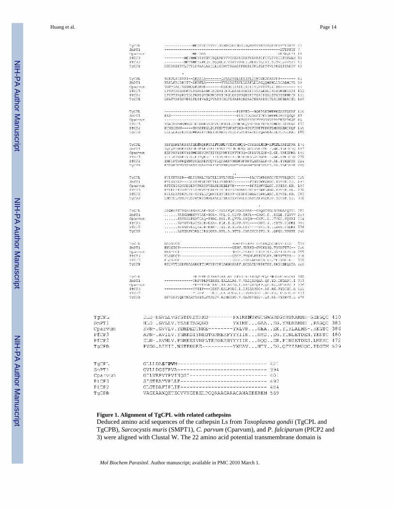
Figure 1. Alignment of TgCPL with related cathepsinsDeduced amino acid sequences of the cathepsin Ls from Toxoplasma gondii (TgCPL andTgCPB), Sarcocystis muris (SMPT1), C. parvum (Cparvum), and P. falciparum (PfCP2 and3) were aligned with Clustal W. The 22 amino acid potential transmembrane domain is
Huang et al. Page 14
Mol Biochem Parasitol. Author manuscript; available in PMC 2010 March 1.
NIH
-PA Author Manuscript
NIH
-PA Author Manuscript
NIH
-PA Author Manuscript
underlined (59–82). The predicted pre/pro region cleavage site is marked with a single arrowand the pro/mature site with a double arrow. Conserved motifs (ERFNIN, KNFD, and SPV)are italicized and bold. The active site cysteine, histidine, and asparagine are shown in bold(229, 351, and 387). The potential N-glycosylation sites are marked with stars (*, positions 56and 266).
Huang et al. Page 15
Mol Biochem Parasitol. Author manuscript; available in PMC 2010 March 1.
NIH
-PA Author Manuscript
NIH
-PA Author Manuscript
NIH
-PA Author Manuscript
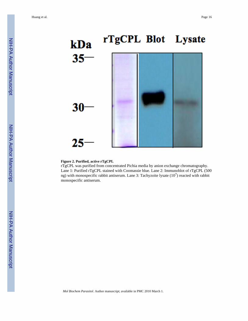
Figure 2. Purified, active rTgCPLrTgCPL was purified from concentrated Pichia media by anion exchange chromatography.Lane 1: Purified rTgCPL stained with Coomassie blue. Lane 2: Immunoblot of rTgCPL (500ng) with monospecific rabbit antiserum. Lane 3: Tachyzoite lysate (107) reacted with rabbitmonospecific antiserum.
Huang et al. Page 16
Mol Biochem Parasitol. Author manuscript; available in PMC 2010 March 1.
NIH
-PA Author Manuscript
NIH
-PA Author Manuscript
NIH
-PA Author Manuscript
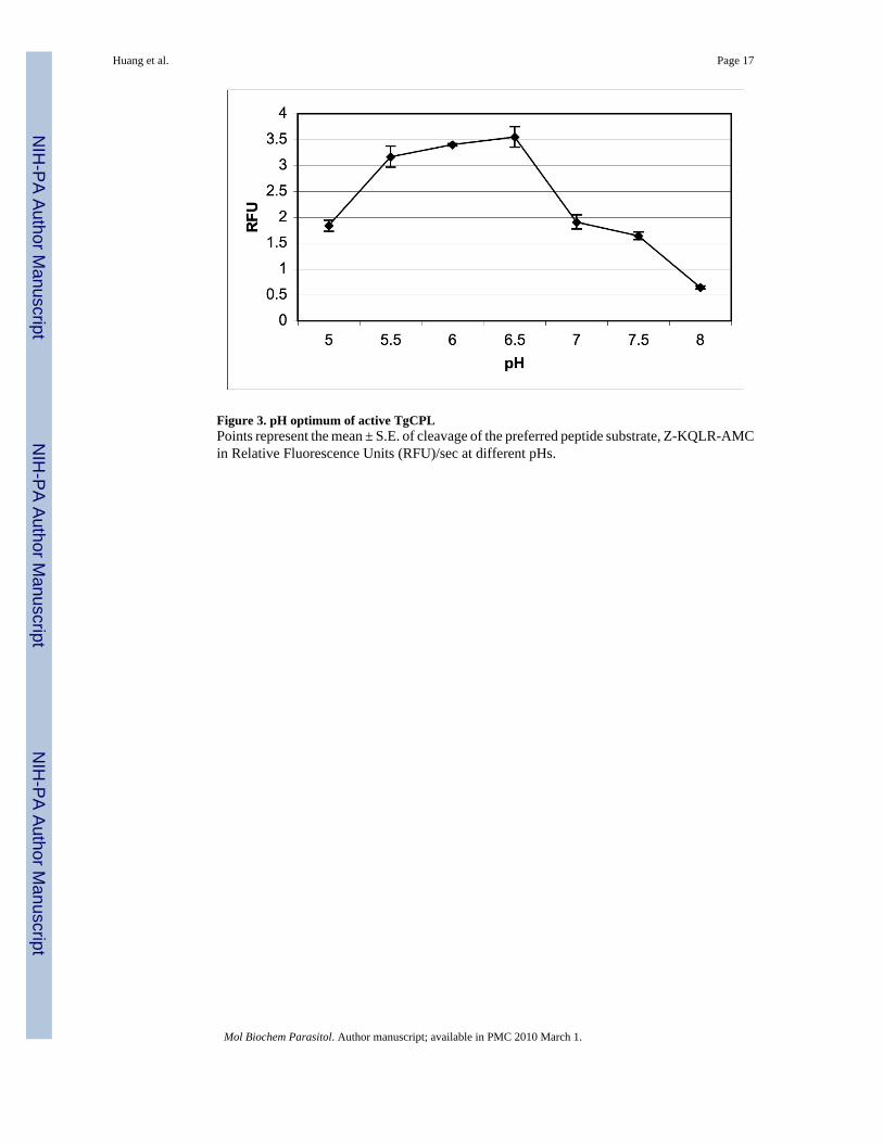
Figure 3. pH optimum of active TgCPLPoints represent the mean ± S.E. of cleavage of the preferred peptide substrate, Z-KQLR-AMCin Relative Fluorescence Units (RFU)/sec at different pHs.
Huang et al. Page 17
Mol Biochem Parasitol. Author manuscript; available in PMC 2010 March 1.
NIH
-PA Author Manuscript
NIH
-PA Author Manuscript
NIH
-PA Author Manuscript

Figure 4. Structure of the active site pocket of TgCPL superimposed on the structure of humancathepsin LThe aspartic acid (Asp 216) can be seen at the base of the S2 pocket in TgCPL which is analanine (Ala 214) in the human cathepsin.
Huang et al. Page 18
Mol Biochem Parasitol. Author manuscript; available in PMC 2010 March 1.
NIH
-PA Author Manuscript
NIH
-PA Author Manuscript
NIH
-PA Author Manuscript
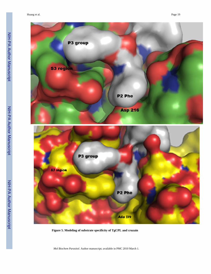
Figure 5. Modeling of substrate specificity of TgCPL and cruzain
Huang et al. Page 19
Mol Biochem Parasitol. Author manuscript; available in PMC 2010 March 1.
NIH
-PA Author Manuscript
NIH
-PA Author Manuscript
NIH
-PA Author Manuscript
A. Phenylalanine in the P2 position of a substrate shows poor fit in active site pocket of TgCPL.B. Accommodation of leucine in the P2 position of substrates in cruzain with alanine in the S2site.
Huang et al. Page 20
Mol Biochem Parasitol. Author manuscript; available in PMC 2010 March 1.
NIH
-PA Author Manuscript
NIH
-PA Author Manuscript
NIH
-PA Author Manuscript

Figure 6. Localization of TgCPLA. Localization of TgCPL (red) with insert of DIC. B. Immunoelectron micrograph of TgCPLin radiolucent vesicle (arrow). Size bar (0.5 microns) is shown below.
Huang et al. Page 21
Mol Biochem Parasitol. Author manuscript; available in PMC 2010 March 1.
NIH
-PA Author Manuscript
NIH
-PA Author Manuscript
NIH
-PA Author Manuscript
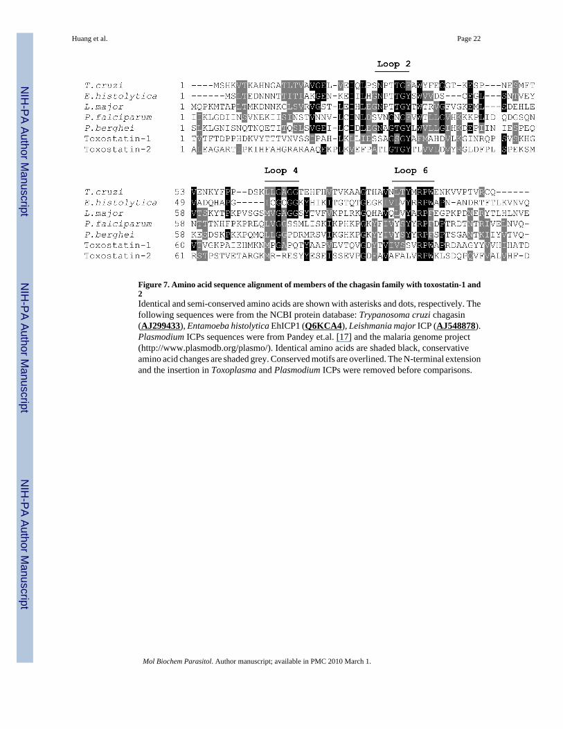
Figure 7. Amino acid sequence alignment of members of the chagasin family with toxostatin-1 and2Identical and semi-conserved amino acids are shown with asterisks and dots, respectively. Thefollowing sequences were from the NCBI protein database: Trypanosoma cruzi chagasin(AJ299433), Entamoeba histolytica EhICP1 (Q6KCA4), Leishmania major ICP (AJ548878).Plasmodium ICPs sequences were from Pandey et.al. [17] and the malaria genome project(http://www.plasmodb.org/plasmo/). Identical amino acids are shaded black, conservativeamino acid changes are shaded grey. Conserved motifs are overlined. The N-terminal extensionand the insertion in Toxoplasma and Plasmodium ICPs were removed before comparisons.
Huang et al. Page 22
Mol Biochem Parasitol. Author manuscript; available in PMC 2010 March 1.
NIH
-PA Author Manuscript
NIH
-PA Author Manuscript
NIH
-PA Author Manuscript

Figure 8. Recombinant and over expressed toxostatinsA. Purified, recombinant toxostatin-1 and 2 are stained with Coomassie blue. B. Immunoblotof lysates of wild type (RH) and tachyzoites over-expressing toxostatin-1 (Toxo-1++) detectedwith anti-AU-1 to the epitope tag.
Huang et al. Page 23
Mol Biochem Parasitol. Author manuscript; available in PMC 2010 March 1.
NIH
-PA Author Manuscript
NIH
-PA Author Manuscript
NIH
-PA Author Manuscript

NIH
-PA Author Manuscript
NIH
-PA Author Manuscript
NIH
-PA Author Manuscript
Huang et al. Page 24
Table 1Enzyme Kinetics of TgCPL
Substrate Km (uM) Vmax (RFU/sec)
LR 7.0 ± 0.7 120.6 ± 3.6
FR 15.7 ± 1.0 148.0 ± 4.4
RR 17.6 ± 1.2 14.8 ± 0.3
Mol Biochem Parasitol. Author manuscript; available in PMC 2010 March 1.
Copyright © 2022 FDOKUMEN