Post-LIA glacier changes along a latitudinal transect in the Central Italian Alps
Upload
independentCategory
view
0download
0
LT
CHa
b
c
d
e
f
g
a
ARRA
KTGASMBAAB
m
0h
Veterinary Parasitology 194 (2013) 9– 15
Contents lists available at SciVerse ScienceDirect
Veterinary Parasitology
jo u rn al hom epa ge : www.elsev ier .com/ locate /vetpar
atitudinal variability in the seroprevalence of antibodies againstoxoplasma gondii in non-migrant and Arctic migratory geese
ecilia A.M. Sandströma,b,∗, Anita G.J. Bumab, Bethany J. Hoyec,d, Jouke Propa,enk van der Jeugde, Berend Voslamber f, Jesper Madseng, Maarten J.J.E. Loonena
University of Groningen, Arctic Centre, PO Box 716, 9700 AS Groningen, The NetherlandsUniversity of Groningen, Ocean Ecosystems, Nijenborgh 7, 9747 AG Groningen, The NetherlandsNetherlands Institute of Ecology, Department of Animal Ecology, PO Box 50, 6700 AB Wageningen, The NetherlandsDepartment of Ecology and Evolutionary Biology, University of Colorado, Ramaley N122, CB334, Boulder, CO 80309, USADutch Centre for Avian Migration & Demography, Netherlands Institute of Ecology, PO Box 50, 6700 AB Wageningen, The NetherlandsSovon, Dutch Centre for Field Ornithology, PO Box 6521, 6503 GA Nijmegen, The NetherlandsArctic Research Centre, Department of Bioscience Aarhus University, Universitetsparken, Building 1110, DK-8000 Aarhus C, Denmark
r t i c l e i n f o
rticle history:eceived 6 July 2012eceived in revised form 2 December 2012ccepted 13 December 2012
eywords:oxoplasma gondiieesercticeroreversionodified agglutination test
ranta leucopsisnser brachyrhynchusnser anserranta canadensis
a b s t r a c t
Toxoplasma gondii is an intracellular coccidian parasite found worldwide and is knownto infect virtually all warm-blooded animals. It requires a cat (family Felidae) to com-plete its full life cycle. Despite the absence of wild felids on the Arctic archipelago ofSvalbard, T. gondii has been found in resident predators such as the arctic fox and polarbear. It has therefore been suggested that T. gondii may enter this ecosystem via migra-tory birds. The objective of this study was to identify locations where goose populationsmay become infected with T. gondii, and to investigate the dynamics of T. gondii spe-cific antibodies. Single blood samples of both adults and juveniles were collected fromselected goose species (Anser anser, A. brachyrhynchus, Branta canadensis, B. leucopsis) atArctic brood-rearing areas in Russia and on Svalbard, and temperate wintering grounds inthe Netherlands and Denmark (migratory populations) as well as temperate brood-rearinggrounds (the Netherlands, non-migratory populations). A modified agglutination test wasused on serum, for detection of antibodies against T. gondii. Occasional repeated annualsampling of individual adults was performed to determine the antibody dynamics. Adultswere found seropositive at all locations (Arctic and temperate, brood-rearing and winter-ing grounds) with low seroprevalence in brood-rearing birds on temperate grounds. As nojuvenile geese were found seropositive at any brood-rearing location, but nine month oldgeese were found seropositive during spring migration we conclude that geese, irrespec-tive of species and migration, encounter T. gondii infection in wintering areas. In re-sampledbirds on Svalbard significant seroreversion was observed, with 42% of seropositive adultsshowing no detectable antibodies after 12 months, while the proportion of seroconversionwas only 3%. Modelled variation of seroprevalence with field data on antibody longevity andparasite transmission suggests seroprevalence of a population within a range of 5.2–19.9%,
in line with measured values. The high occurrence of seroreversion compared to the lowoccurrence of seroconverexplains the absence oftion in antibody titre. Theintroduce T. gondii to the
∗ Corresponding author at: University of Groningen, Arctic Centre, PO Box 716obile: +31 65 597 6246.
E-mail addresses: [email protected], [email protected] (C.A.M
304-4017/$ – see front matter © 2013 Elsevier B.V. All rights reserved.ttp://dx.doi.org/10.1016/j.vetpar.2012.12.027
sion hampers analysis of species- or site-specific patterns, but
an increase in seroprevalence with age and the observed varia-se findings imply that even though infection rate is low, adultshigh Arctic ecosystem following infection in temperate regions.© 2013 Elsevier B.V. All rights reserved.
, 9700 AS Groningen, The Netherlands. Tel.: +31 50 363 6056;
. Sandström).

rinary P
10 C.A.M. Sandström et al. / Vete1. Introduction
Infectious diseases represent a significant threat to bothhuman and animal populations. As a consequence, it isof great relevance to understand the infection dynamicsand distribution of important zoonotic pathogens (Altizeret al., 2011). Toxoplasma gondii is a globally distributed coc-cidian protozoan (Dubey, 2010). Infection with T. gondiiis one of the most common parasitic infections of warm-blooded animals worldwide, including humans (Dubey andBeattie, 1988; Tenter et al., 2000). A wide range of mammalsand birds can serve as intermediate hosts, where asexualreproduction and tissue cyst formation occur. Intermedi-ate hosts can be infected by ingestion of oocysts or tissuecysts, and in some cases by placental transmission. Sex-ual reproduction can only happen in the intestines of thedefinitive host and results in infective oocysts being shedwith their faeces. Oocysts are essential for the transmissionto a non-carnivorous host and are only shed by domesticcats and other felines (Dubey, 2010). As the cat populationhas developed parallel to the human population, there is astrong potential for T. gondii transmission in rural settings(Amendoeira et al., 2003). Oocysts have been found both inwater and in soil samples around human dwellings (Weigelet al., 1999; Dubey, 2010) and may enter the marine envi-ronment through freshwater runoff or via sewage systems(Lindsay et al., 2003; Conrad et al., 2005). Here, the oocystscan travel long distances via physical and biological pro-cesses, the latter including ingestion by marine mammalsor accumulation in filter feeding fish and bivalves (Arktushet al., 2003; Fayer et al., 2004; Miller et al., 2008; Massieet al., 2010).
Polar regions are isolated both by their extreme envi-ronment and remote position. Nevertheless, both in Arctic(Prestrud et al., 2007; Oksanen et al., 2009; Jensen et al.,2010) and sub-Antarctic (Afonso et al., 2007) regions indi-viduals seropositive with T. gondii have been found. Clearly,T. gondii is found in areas not inhabited by its definitivehost. For example, in the high Arctic Svalbard archipelago(78–81◦N, 10–30◦E), including the main island Spitsbergen,no wild felines are present and domestic cats are prohib-ited. Yet, T. gondii infection has been observed in residenttop predators such as Arctic foxes (Vulpes lagopus) and Polarbears (Ursus maritimus) (Prestrud et al., 2007). Whetherthe initial infection is a result of oocysts transported viaocean currents or tissue cysts from migratory animals isunknown.
Ecosystems are connected via seasonal migrations(reviewed in Altizer et al., 2011). Along the flyway migra-tory birds may transport infectious disease agents (Bradleyet al., 2005; Altizer et al., 2011) and Prestrud et al. (2007,2008a, 2008b) suggested that T. gondii is brought to theArctic by migratory birds. In support of this notion, 7%of migratory barnacle geese (Branta leucopsis) on Svalbardwere found seropositive, whereas no resident herbivoressuch as Svalbard reindeer (Rangifer tarandus platyrhynchus)(n = 390) or sibling voles (Microtus epiroticus) (n = 361) were
found seropositive, suggesting that T. gondii oocysts in theterrestrial ecosystem are not an important mode of trans-mission on Svalbard (Prestrud et al., 2007). In the samestudy, foxes captured at sites devoid of goose coloniesarasitology 194 (2013) 9– 15
showed lower seroprevalence than foxes captured closeto goose colonies (Prestrud et al., 2007). In addition, asthe Svalbard goose populations have doubled over the lastdecades (Fox et al., 2010), so has the prevalence of anti-bodies to T. gondii in Svalbard’s polar bears (Oksanen et al.,2009; Jensen et al., 2010).
The objective of this study was to determine T. gondiiseroprevalence in goose populations at various locationsin order to assess the role of migratory birds as vectorof T. gondii to isolated Arctic ecosystems. As juvenilesare infection naïve at birth and limited in their habitatexposure, they were specifically targeted to determinethe area of infection. To this end, we sampled adults andjuveniles of two arctic migratory goose species; the bar-nacle goose (Branta leucopsis) and the pink-footed goose(Anser brachyrhynchus), at Arctic breeding and temperatewintering grounds. To expand the sampling of the temper-ate environment, resident Dutch populations of barnacle,Canada (B. canadensis) and greylag geese (A. anser) wereincluded, sampled during the brood-rearing period.
Our main assumption was that the likelihood of infec-tion with T. gondii is high in areas with suspected highdensities of cats, and that infection results in increased spe-cific antibody levels in the blood. Both adult and juvenilegeese would consequently show higher seroprevalence attemperate, compared to Arctic, locations. Therefore, thefollowing hypotheses were tested: (i) in arctic areas onlyadults are seropositive; (ii) in temperate areas both adultsand juveniles are seropositive, and both show a higher titreof antibodies in the blood: (iii) the proportion of seroposi-tive individuals increases with age.
2. Materials and methods
Blood samples were collected between 2006 and 2010at four locations: Svalbard (1), Nenets Autonomous OkrugNW Russia (2), Denmark (3) and the Netherlands (4) (Fig. 1).In the Arctic, birds were sampled on Spitsbergen, the west-ern island of Svalbard (79◦N/12◦E) and in NW Russia atTobseda (68◦N/52◦E) and Kolguev (69◦N/49◦E). In Denmarkall birds were sampled during spring staging at Vest StadilFjord (58◦N/8◦E). In the Netherlands birds were sampled inthe provinces of Groningen, Friesland, Gelderland, Noordand Zuid Holland (52◦N/4◦E – 53◦N/6◦E) during summerand in Friesland (53◦N/6◦E) during winter staging (Table 1).
In total four species of wild geese were investigated:barnacle goose (n = 1543), pink-footed goose (n = 787),greylag goose (n = 266) and Canada goose (n = 79) (Table 1).In the Arctic, barnacle geese were sampled from popula-tions using two different flyways; those migrating fromArctic Russia to the Netherlands and those migrating fromSvalbard to Scotland (Black et al., 2007). A second speciessampled in the Arctic was the pink-footed goose migrat-ing from Svalbard to Denmark–the Netherlands–Belgium(Madsen et al., 1999). The pink-footed goose is sharinghabitat with both the earlier mentioned migratory barnaclegoose populations. The migratory and non-migratory pop-
ulations have overlapping winter habitats though do notfully mix during winter staging (van der Jeugd et al., 2001).Both juvenile and adult birds were sampled at all loca-tions (Table 1). Juvenile birds caught in Denmark were a
C.A.M. Sandström et al. / Veterinary Parasitology 194 (2013) 9– 15 11
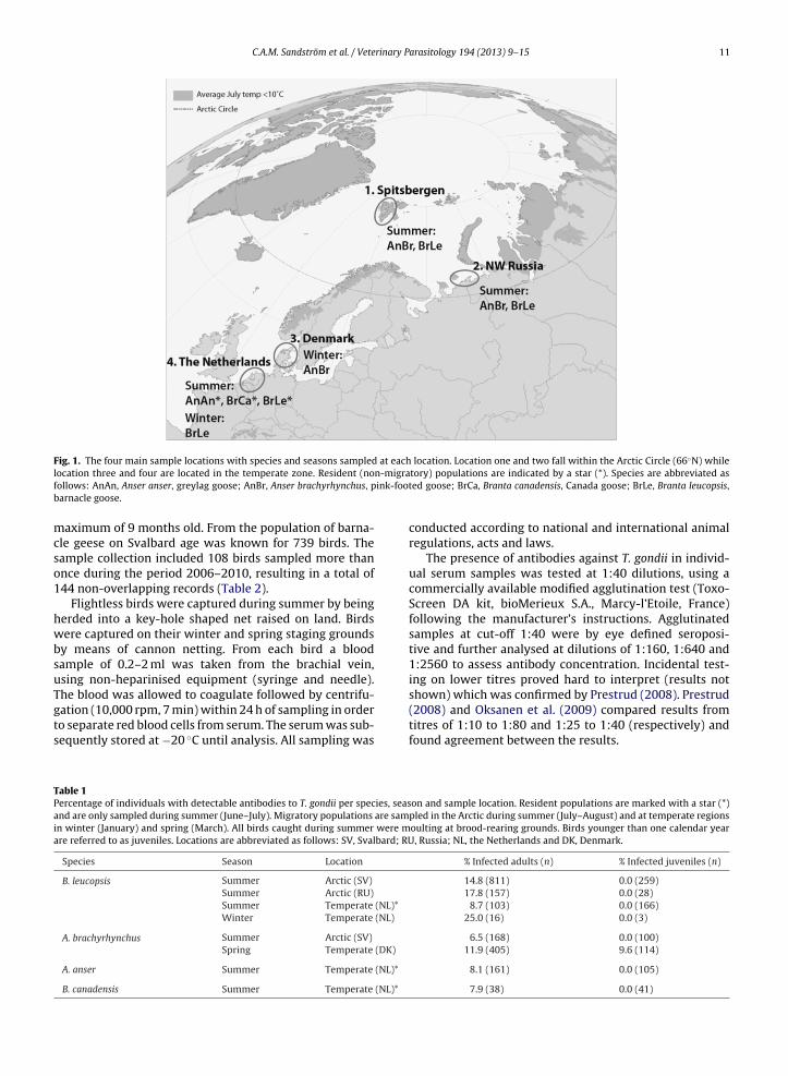
Fig. 1. The four main sample locations with species and seasons sampled at each location. Location one and two fall within the Arctic Circle (66◦N) whilel n-migraf ink-footb
mcso1
hwbsuTgts
TPaia
ocation three and four are located in the temperate zone. Resident (noollows: AnAn, Anser anser, greylag goose; AnBr, Anser brachyrhynchus, parnacle goose.
aximum of 9 months old. From the population of barna-le geese on Svalbard age was known for 739 birds. Theample collection included 108 birds sampled more thannce during the period 2006–2010, resulting in a total of44 non-overlapping records (Table 2).
Flightless birds were captured during summer by beingerded into a key-hole shaped net raised on land. Birdsere captured on their winter and spring staging grounds
y means of cannon netting. From each bird a bloodample of 0.2–2 ml was taken from the brachial vein,sing non-heparinised equipment (syringe and needle).he blood was allowed to coagulate followed by centrifu-
ation (10,000 rpm, 7 min) within 24 h of sampling in ordero separate red blood cells from serum. The serum was sub-equently stored at −20 ◦C until analysis. All sampling wasable 1ercentage of individuals with detectable antibodies to T. gondii per species, seasnd are only sampled during summer (June–July). Migratory populations are samn winter (January) and spring (March). All birds caught during summer were mre referred to as juveniles. Locations are abbreviated as follows: SV, Svalbard; RU
Species Season Location
B. leucopsis Summer Arctic (SV)
Summer Arctic (RU)Summer Temperate (NL)*
Winter Temperate (NL)
A. brachyrhynchus Summer Arctic (SV)
Spring Temperate (DK)
A. anser Summer Temperate (NL)*
B. canadensis Summer Temperate (NL)*
tory) populations are indicated by a star (*). Species are abbreviated ased goose; BrCa, Branta canadensis, Canada goose; BrLe, Branta leucopsis,
conducted according to national and international animalregulations, acts and laws.
The presence of antibodies against T. gondii in individ-ual serum samples was tested at 1:40 dilutions, using acommercially available modified agglutination test (Toxo-Screen DA kit, bioMerieux S.A., Marcy-l’Etoile, France)following the manufacturer’s instructions. Agglutinatedsamples at cut-off 1:40 were by eye defined seroposi-tive and further analysed at dilutions of 1:160, 1:640 and1:2560 to assess antibody concentration. Incidental test-ing on lower titres proved hard to interpret (results notshown) which was confirmed by Prestrud (2008). Prestrud
(2008) and Oksanen et al. (2009) compared results fromtitres of 1:10 to 1:80 and 1:25 to 1:40 (respectively) andfound agreement between the results.on and sample location. Resident populations are marked with a star (*)pled in the Arctic during summer (July–August) and at temperate regionsoulting at brood-rearing grounds. Birds younger than one calendar year, Russia; NL, the Netherlands and DK, Denmark.
% Infected adults (n) % Infected juveniles (n)
14.8 (811) 0.0 (259)17.8 (157) 0.0 (28)
8.7 (103) 0.0 (166)25.0 (16) 0.0 (3)
6.5 (168) 0.0 (100)11.9 (405) 9.6 (114)
8.1 (161) 0.0 (105)
7.9 (38) 0.0 (41)

12 C.A.M. Sandström et al. / Veterinary Parasitology 194 (2013) 9– 15
Table 2Conversion and reversion in individuals sampled at two different time intervals. In 144 cases an individual was re-sampled 1, 2, 3 or 4 years later (t = y).Only non-overlapping periods were considered.
Transition Time interval (Year) N samples N samples Calculated proportionNegative → positive Negative at t = 0 Positive at t = y Converting (�)
0 → 1 64 2 0.03130 → 2 39 3 0.04800 → 3 15 1 0.03350 → 4 2 0 n.a.
Transition Time interval (Year) N samples N samples Calculated proportionPositive → negative Positive at t = 0 Negative at t = y Reverting (ı)
0 → 1 12 5 0.4167
0 → 2 100 → 3 2
Using re-caught individuals the annual proportion ofseroconverting individuals (prop. �), and the proportionseroreverting individuals (prop. ı) were calculated (here-after � and ı respectively). These values were used inan iteration predicting seroprevalence (�) in a populationof 1000 individuals after 20 years. Together with ran-dom generated numbers it was decided if seroconversionhas occurred, and if so, if seroreversion would occur thefollowing year. Repeatedly sampled individuals from non-overlapping periods longer than one year gave additionalvalues for � and ı. Using these additional values for � andı, the variation within the parameters resulted in a rangeof potential � .
The differences in seroprevalence between variousgroups (site, age, species, and gender) were tested usinga chi-square test. A Fisher’s exact test was includedwhen sample size in one of the observed groups wasbelow 5. Non-parametric tests were used to compare theantibody concentration between groups, Mann–WhitneyU-test comparing two groups and a Kruskal–Wallis ANOVAtest comparing three or more groups. Binary logistic regres-sions were used to estimate the effect of age on theseroprevalence in the population. All statistical tests wereperformed using SPSS, version 16 (SPSS INC., Chicago, IL,USA).
3. Results
All juveniles sampled on brood-rearing grounds werefound seronegative, (n = 699) (Table 1). At the Arctic loca-tions (Svalbard and Russia) 10.4% of all adults (n = 1136)were seropositive. A similar seroprevalence was foundduring summer at temperate sites: 8.3% of all adults(n = 302) were seropositive. The seroprevalence variedfor different species and seasonal groups, ranging from6.5% in pink-footed geese on Arctic breeding grounds to25% in migratory barnacle geese on wintering groundsin the Netherlands (Table 1). On Svalbard the seropreva-lence of pink-footed geese was significantly lower thanof barnacle geese (X2 = 8.170, p = 0.006). Between thedifferent populations of barnacle geese there was no sig-
nificant difference in seroprevalence found for the variouslocations. When following goose populations from Arc-tic grounds to temperate wintering and spring grounds,the seroprevalence for pink-footed geese during spring5 0.29951 0.2160
staging was higher than during brood rearing (tested one-sided 6.5–11.9% X2 = 3.617, p = 0.036) while this increasewas non-significant for adult barnacle geese (17.8–25.0%X2 = 0.495) (Table 1). Of the 114 nine month old pink-footedgoose juveniles sampled on spring staging grounds inDenmark, 11 were seropositive. Within the spring stagingpopulation, no difference was found comparing juveniles(9.6%) to adults (11.9%) (X2 = 0.265, p = 0.622) (Table 1).
Re-sampled individuals over a period of one to threeyears revealed the proportion of seroreversion (ı) and sero-conversion (�) (Table 2). For adults sampled negative at t = 0(n = 64) � was calculated to 3.1% over one year while ı wasmore than ten times higher, at 41.7%. Other time intervalsbetween repeatedly sampled individuals gave additionalindependent estimates for � and ı (Table 2). In all cases,seroreversion was much higher than seroconversion. Thesefindings were integrated in a simple model. This model esti-mated combinations of � and ı resulting in given valuesof seroprevalence (�) (Fig. 2A) or in observed transitions(seroreversion or seroconversion) in � over a defined timeinterval of one, two or three years (Fig. 2B). Both simula-tions combined suggest values of � ranging from 2.5% to5% and ı ranging from 20% to 45%, giving a span of � inthe population between 5.2% and 19.9%. We measured a �range of 6.5% to 17.7% in the sampled population, with ahighest value of 25% for a small sample size of 16.
The variance of antibody concentration within anindividual showed that no seronegative individuals hadantibody titres higher than 1:160 the following or previousyear. The individuals with the highest measured concen-tration the first year (seropositive at dilution 1:640 at t = 0)stayed seropositive the following year (1:160 at t = 1). Aseropositive individual (1:40 at t = 0) showed the highesttitre measured of 1:2560 the following year. No individualsmaintained the minimum threshold level of 1:40 betweenthe years.
To investigate the effect of age, individuals weregrouped into four age classes. Number of positive indi-viduals and total sample size were (npositive/ntotal sample):class < 1, 0/158 = 0; class 1–5, 1/24 = 4.2%; class 6–10,3/40 = 7.5%; class > 10: 16/122 = 13.1%. There was a sig-
nificant increase in seroprevalence over age (X2 = 25.276,p = 0.000). This increase was not significant if only adultswere considered. The number of seropositive individu-als per age class was modelled by randomly assigning
C.A.M. Sandström et al. / Veterinary Parasitology 194 (2013) 9– 15 13
Fig. 2. (A) and (B) Modelled combinations of delta (ı) and lambda (�) resulting ina given time period of 1, 2, or 3 years, were each line shows conversion (a–c) or re0.200 to 0.450, calculated values for � range from 5.2% to 19.9%. Input-values are
Fig. 3. Modelled numbers of infected individuals per age groups. Linesrepresent individuals in a population of 1000 individuals in a given ageclass (left y-axis). Bars show average numbers of infection per onceiaa
aapssts4(snib
4
g
nfected individuals (right y-axis). Individuals were in each age classssigned to seroconversion and/or – reversion based on random numbersnd measured population values. The iteration is run 100 times.
nnual seroconversion (3.1%) to seronegative individualsnd seroreversion (41.7%) to seropositive individuals in aopulation starting with 1000 naive individuals. The modelhowed that the number of individuals which have beeneropositive over their life time increases with age whilehe number of seropositive individuals in a given age classtabilized for values of gamma (Fig. 3). At an age of 20 years,66 individuals of 1000 have at least once been infectedFig. 3, dotted line), nevertheless only 69 individuals wereeropositive at the same age (Fig. 3, solid line). The averageumbers of infections per infected individual was found to
ncrease from 1.00 to 1.33 times over a 20 year period (Fig. 3ars). The results are based on 100 iterations.
. Discussion
We hypothesized that both adult and juvenileeese would show higher seroprevalence at temperate,
: a set value of seroprevalence (�) (panel A); an observed transition overversion (d–f) (panel B). In the range of values for �: 0.025 to 0.050 and ı:
presented in Table 2.
compared to Arctic, locations. We expected no positivejuveniles in the Arctic. If only one Arctic juvenile had beenconfirmed seropositive, the marine infection pathwaywould have to be considered as relevant transport route ofT. gondii to the high Arctic. Instead, the chance of samplinga seropositive juvenile on temperate breeding groundswas expected to be high due to felines shedding oocystson the grasslands.
The absence of seropositive juveniles on temperatebreeding grounds seemed counterintuitive. However, theexposure time for goslings to become infected before beingsampled on the breeding grounds was on average only 35days. With the observed annual proportion of seroconvert-ing individuals of 3.1% and assuming a constant chance ofinfection over a year, the chance of sampling a seroposi-tive gosling would be 1 out of 336 while our sample size ofjuveniles is only 312 individuals (see Table 1). In addition,the environment that flightless birds encounter is possi-bly less contaminated with oocysts than the environmentvisited by flying birds. Due to their exposure and vulner-ability to predation, flightless birds inhabit areas with alower predator encounter risk (Madsen and Mortensen,1987; Kahlert et al., 1996). Such an area is often close towater, where cats are supposedly less likely to hunt. Sys-tematic goose counts in the Netherlands during 2005–2011showed that while only 1 cat was observed close to awater body, 142 cats were seen on grasslands (Voslam-ber, unpublished data). The observer counted all geese andmammals weekly in a predefined area of 1500 Ha, subdi-vided in fields of settlements, grasslands and shore land.During winter when the geese can fly, they forage on grass-lands where they may be subjected to a higher infectionrisk.
Seroprevalence did not increase with age of testedindividuals when juveniles were excluded. Rate of (re-)infection is seemingly low in relation to seroreversion. Our
results suggest that the species investigated here undergoa more rapid seroreversion than previously known. Theeffect of seroreversion on immunity needs to be unrav-elled before definite statements can be made about the
rinary P
14 C.A.M. Sandström et al. / Vetestatus of infection. As the exact relation between immunityand seroprevalence is unknown, in this study the status ofinfection is based on seropositive individuals.
Based on field values of infection and antibody dynamicsa seroprevalence of 7% was calculated in the Arctic barnaclegoose population. A value of 7% for seroprevalence is inthe lower end found in our study, though it fits very wellwith previous work of Prestrud et al. (2007) in the samepopulation. When integrating measured values from yearswith more than one winter between sample occasions therange of possible seroconversion and reversion increasedand so did the variation of stable seroprevalence (from 5.2%to 19.9%), matching measured field values well.
Infection risk might be considerable higher than the3.1% measured over a full year in barnacle geese. Juvenilepink-footed geese acquired seroprevalence of 9.6% over alifetime of nine months. The observed values of seropreva-lence in adult pink-footed geese (11.9%) would then relyon an almost twice as high species-specific seroreversionof 71%. However with the variation in sample sizes andpotential species specific values of seroreversion, furtherinterpretation would become speculative.
As the antibody titre in the blood is expected to increasewhen an individual is (re-)infected, we expected higherantibody titre on temperate grounds, especially duringwinter. However our data did not support this. On the otherhand, within a migrating population a trend for higher anti-body titres on temperate grounds than on Arctic groundswas found both over the migration route within a species(summer vs. winter) as well as when comparing Arcticbreeders to temperate breeders. Additionally, of all posi-tive birds at temperate brood-rearing grounds 24% werepositive at the highest titre (1:2560) compared to only 6%at arctic grounds. The largest fraction (35%) of birds at Arcticbreeding grounds was positive at the lowest antibody titre(1:40). The different patterns observed in the concentrationtitres support our hypothesis that infection is occurring intemperate regions.
The average number of infection events per infectedindividual increases from 1.0 time for a one year old toa maximum of 1.3 times for an individual of 20 years.Re-infection would probably result in higher antibody con-centrations with a slightly lower sero reversion rate. Wehave no significant evidence for this statement but thatcould result in a somewhat higher prevalence with age,which is hinted at in the non-significant highest value ofseroprevalence in the age class older than 10 years.
Identification of the source of infection is vital for under-standing how the parasite is infecting new ecosystems.The observed trend towards increased seroprevalence aswell as antibody titre on wintering grounds suggestedthat the wintering grounds are a source of (re-)infection.However, geographical differences were not always sig-nificant, which can be explained by the large variationin sample numbers in combination with small differ-ences in seroprevalence. For barnacle geese, we calculatedthe required sample size for the wintering population to
obtain significant differences at 25% seroprevalence withthe empiric results from the three summersampled sites.The required sample sizes were n = 1108 for Arctic Rus-sia at a seroprevalence of 17.8%, n = 52 for Arctic Svalbardarasitology 194 (2013) 9– 15
at a seroprevalence of 14.8% and n = 20 for non-migratorygeese in the Netherlands at a seroprevalence of 8.7%. Thisclearly shows the sensitivity for sample size in combina-tion with observed differences. Within the same flywayfrom Arctic Russia to the Netherlands, sample size forsignificant results would be extremely large. Neverthe-less, if we expected a seasonal difference over the flyway,for pink-footed geese the seroprevalence in the Arcticwas significantly lower than on winter and spring staginggrounds. When considering variability within one season(brood-rearing) there was always a difference betweenArctic and temperate regions. Surprisingly, the seropreva-lence for Arctic breeding barnacle geese was significantlyhigher than the Dutch breeding equivalent. On the con-trary, pink-footed geese breeding on Svalbard had thelowest seroprevalence measured which corroborate thehypothesis of northern regions carrying a lower disease risk(Piersma, 1997).
More evenly represented sampling populations wouldincrease the rigidity to this study. The majority of the fieldcampaigns were aimed on summer populations. However,an increased number of winter staging birds from both theNetherlands and Scotland would have contributed to theunderstanding for the seasonal variation of seroprevalence,antibody dynamics and the possible infection location inthe light of the annual cycle.
In conclusion, infection with T. gondii is likely to happenon temperate grounds during the winter period when thebirds are able to fly. Infected birds transport the parasite toArctic breeding grounds, and if predated the parasite canenter the ecosystem. As no naive Arctic birds (juveniles)were found seropositive in the Arctic we have no supportfor an alternative transmission pathway of T. gondii to thehigh Arctic. We found the proportion of individuals serore-verting over a time interval of one year being > 40%, whilethe proportion seroconverting was a magnitude lower.Using an iteration based on values from individuals sam-pled in multiple years we predicted the expected level ofseroprevalence in a population which corresponded wellwithin the range of measured values.
This study advances our understanding of ecologicaldrivers behind the occurrence of spatial and temporal vari-ation of T. gondii within two naturally defined geographicalareas. However, future studies should focus on achiev-ing a full picture of the flyway to determine the antibodydynamics. In general, juveniles must be sampled in greaternumbers to directly link site of infection with environment.
Conflict of interest
There are no known personal relationships withother people or organizations that could inappropriatelyinfluence this work. To the best of the author’s knowledgethere are no conflicts of interest.
Acknowledgements
This study was supported through the BIRDHEALTHprogramme within the International Polar Year by theNetherlands Organisation for Scientific Research (NWO;grant 851.40.071 and 854.40.073) and by funding provided

rinary P
bgcaSios
R
A
A
A
A
B
B
C
D
D
F
F
J
C.A.M. Sandström et al. / Vete
y the University of Groningen. Permits were obtained foroose catching and blood sampling from national ringingentres, animal experimentation boards, nature protectiongencies and land owners. We would like to thank Fritsteenhuisen (Fig. 1) and Dick Visser (Figs. 2 and 3) formproving the figures, Anouk Goedknegt assisted with lab-ratory analyses. We are thankful to many volunteers andtudents who helped catching and sampling birds.
eferences
fonso, E., Thuilliez, P., Pontier, D., Gilot-Fromont, E., 2007. Toxoplasmosisin prey species and consequences for prevalence in feral cats: not allprey species are equal. Parasitology 134, 1963–1971.
ltizer, S., Bartel, R., Han, B.A., 2011. Animal migration and infectiousdisease risk. Science 331, 296–302.
mendoeira, M.R.R., Sobral, C.A.Q., Teva, A., de Lima, J.N., Klein, C.H., 2003.Inquérito sorológico para a infecc ão por Toxoplasma gondii em amerín-dos isolados, Matos Grosso. Rev. Soc. Bras. Med. Trop. 36, 671–676 (inPortuguese with English abstract).
rktush, K.D., Miller, M.A., Leutenegger, C.M., Gardner, I.A., Packham,A.E., Heckeroth, A.R., Tenter, A.M., Barr, B.C., Conrad, P.C., 2003.Molecular and bioassay-based detection of Toxoplasma gondii oocystuptake by mussels (Mytilus galloprovincialis). Int. J. Parasitol. 33,1087–1097.
lack, J.M., Prop, J., Larsson, K., 2007. Wild Goose Dilemmas: PopulationConsequences of Individual Decisions in Barnacle Geese. Branta Press,Groningen, p. 254.
radley, M.J., Kutz, S.J., Jenkins, E., O’Hara, T.M., 2005. The potential impactof climate change on infectious diseases of Arctic fauna. Int. J. Circum-polar Health 64, 468–477.
onrad, P.A., Miller, M.A., Kreuder, C., James, E.R., Mazet, J., Dabritz,H., Jessup, D.A., Frances Gulland, Grigg, M.E., 2005. Transmission ofToxoplasma: clues from the study of sea otters as sentinels of Toxo-plasma gondii flow into the marine environment. Int. J. Parasitol. 35,1135–1168.
ubey, J.P., 2010. Toxoplasmosis of Animals and Humans. CRC Press, BocaRaton, 313 pp.
ubey, J.P., Beattie, C.P., 1988. Toxoplasmosis of Animals and Man. CRCPress, Boca Raton, 220 pp.
ayer, R., Dubey, J.P., Lindsay, D.S., 2004. Zoonotic protozoa: from land tosea. Trends Parasitol. 20, 531–536.
ox, A.D., Ebbinge, B.S., Mitchell, C., Heinickle, T., Aarvak, T., Colhoun, K.,Clausen, P., Dereliev, S., Faragó, S., Koffijberg, K., Kruckenberg, H., Loo-nen, M.J.J.E., Madsen, J., Mooij, J., Musil, P., Nilsson, L., Pihl, S., van derJeugd, H., 2010. Current estimates of goose population sizes in west-ern Europe, a gap analysis and an assessment of trends. Ornis Svecicia
20, 115–127.ensen, S.K., Aars, J., Lydersen, C., Kovacs, K.M., Åsbakk, K., 2010. The preva-lence of Toxoplasma gondii in polar bears and their marine mammalprey: evidence for a marine transmission pathway? Polar Biol. 33,599–606.
arasitology 194 (2013) 9– 15 15
Kahlert, J., Fox, A.D., Etteruo, H., 1996. Nocturnal feeding in moulting grey-lag geese Anser anser – an anti-predator response? Ardea 84, 15–22.
Lindsay, D.S., Collins, M.V., Mitchell, S.M., Cole, R.A., Flick, G.J., Wetch, C.N.,Lindquist, A., Dubey, J.P., 2003. Sporulation and survival of Toxoplasmagondii oocysts in seawater. J. Eukaryot. Microbiol. 50, 687–688.
Madsen, J., Mortensen, C.E., 1987. Habitat exploitation and interspecificcompetition of moulting geese in East Greenland. Ibis 129, 25–44.
Madsen, J., Kuijken, E., Meire, P., Cottaar, F., Haitjema, T., Nico-laisen, P.I., Bønes, T. & Mehlum, F. 1999: Pink-footed Goose Anserbrachyrhynchus: Svalbard. Pp. 82-93. in: Madsen, J., Cracknell, G. &Fox, A.D. (eds) Goose Populations of the Western Palearctic. A reviewof status and distribution. Wetlands International Publication No. 48.Wetlands International, Wageningen, the Netherlands. National Envi-ronmental Research Institute, Rønde, Denmark. 344 pp.
Massie, G.L., Ware, M.W., Villegas, E.N., Black, M.W., 2010. Uptake andtransmission of Toxoplasma gondii oocysts by migratory, filter-feedingfish. Vet. Parasitol. 169, 296–303.
Miller, M.A., Miller, W.A., Conrad, P.A., James, E.R., Mellis, A.C., Leuteneg-ger, C.M., Dabritz, H.A., Packham, A.E., Paradies, D., Harries, H., Ames, J.,Jessup, D.A., Worcester, K., Grigg, M.E., 2008. Type X Toxoplasma gondiiin wild mussel and terrestrial carnivores from coastal California: newlinkages between terrestrial mammals, runoff and toxoplasmosis insea otters. Int. J. Parasitol. 38, 1319–1328.
Oksanen, A., Åsbakk, K., Prestrud, K.W., Aars, J., Derocher, A.E., Tryland,M., Wiig, Ø., Dubey, J.P., Sonne, C., Dietz, R., Andersen, M., Born, E.W.,2009. Prevalence of antibodies against Toxoplasma gondii in polarbears (Ursus maritimus) from Svalbard and East Greenland. J. Parasitol.95, 89–94.
Piersma, T., 1997. Do global patterns of habitat use and migration strate-gies co-evolve with relative investments in immunocompetence dueto spatial variation in parasite pressure? Oikos 80, 623–631.
Prestrud, K.W., 2008. Toxoplasma gondii in the high Arctic archipelago ofSvalbard. PhD thesis. Norwegian School of Veterinary Science, Oslo,Norway.
Prestrud, K.W., Åsbakk, K., Fuglei, E., Mørk, T., Stein, A., Ropstad, E., Try-land, M., Gabrielsen, G.W., Lydersen, C., Kovacs, K.M., Loonen, M.J.J.E.,Sagerup, K., Oksanen, A., 2007. Serosurvey for Toxoplasma gondii in arc-tic foxes and possible sources of infection in the high Arctic of Svalbard.Vet. Parasitol. 150, 6–12.
Prestrud, K.W., Dubey, J.P., Åsbakk, K., Fuglei, E., Su, C., 2008a. First isolateof Toxoplasma gondii from arctic fox (Vulpes lagopus) from Svalbard.Vet. Parasitol. 151, 110–114.
Prestrud, K.W., Åsbakk, K., Mørk, T., Fuglei, E., Tryland, M., Su, C., 2008b.Direct high-resolution genotyping of Toxoplasma gondii in arctic foxes(Vulpes lagopus) in the remote arctic Svalbard archipelago revealswidespread clonal Type II lineage. Vet. Parasitol. 158, 121–128.
Tenter, A.M., Heckeroth, A.R., Weiss, L.M., 2000. Toxoplasma gondii: fromanimals to humans. Int. J. Parasitol. 30, 1217–1258.
van der Jeugd, H.P., Olthoff, M.P., Stahl, J., 2001. Breeding range translatesinto staging site choice: Baltic and Arctic Barnacle geese Branta leu-
copsis use different habitats at a Dutch Wadden Sea island. Ardea 89,253–265.Weigel, R.M., Dubey, J.P., Dyer, D., Siegel, A.M., 1999. Risk factors for infec-tion with Toxoplasma gondii for residents and workers on swine farmsin Illinois, USA. Am. J. Trop. Med. Hyg. 60, 793–798.
Copyright © 2022 FDOKUMEN


![HvFT1 (VrnH3) drives latitudinal adaptation in Spanish barleys [2011]](https://static.fdokumen.com/doc/165x107/6334165aa1ced1126c0a28cd/hvft1-vrnh3-drives-latitudinal-adaptation-in-spanish-barleys-2011.jpg)